Laser-Therapy.US
Start New Query

An Hour of Light and Sound a Day Might Keep Alzheimer’s at Bay

Angus Chen - Scientific American March 14, 2019 (Publication) 4521
This is an summary article about the work of Shannon Macauley, a neuroscientist at Wake Forest School of Medicine who found that light and sound has a siginicant impact on the Alzheimer's
View Resource

There is no cure for Alzheimer’s disease. Although a few drugs manage temporarily certain cognitive symptoms of the illness, none can stop or meaningfully slow its progression. “We really don’t have much to offer people,” says Shannon Macauley, a neuroscientist at Wake Forest School of Medicine. Virtually all new treatments have failed in clinical trials. But new research is looking beyond drugs to see what relief might come from a simple LED light and a speaker. Bathing patients in flashing light and pulsing sounds both tuned to a frequency of 40 hertz might reverse key signs of Alzheimer’s in the brain, according to a paper published in Cell on Thursday. “I think it’s an absolutely fascinating paper to be honest,” says Macauley, who was not involved in this work. “It’s a very provocative idea. It’s noninvasive and easy and low cost, potentially, so if it were to come to fruition in humans—that’s fabulous.” Still, all this is a big if, Macauley acknowledges. The work was done in mice with genetic alterations that doomed them to develop key symptoms and pathology of Alzheimer’s disease. One batch of mice formed neurofibrillary tangles inside their neurons—dysfunctional knots of a protein called tau that can lead to the cell’s death. Another batch of the mice developed amyloid beta plaques—sticky heaps of protein that dam the flow of communication between neurons. All the mice also had a third hallmark of the disease—irregular brain activity in the gamma range of brain waves that oscillate between 30 and 100 times a second. In 2015 neuroscientist Li-Huei Tsai, director at The Picower Institute for Learning and Memory at Massachusetts Institute of Technology, was working on an experiment to manipulate that brain activity by flashing a white light at these mice. Like light strobes, our brains flicker. Brain waves are generated when large groups of neurons oscillate on and off together. Neurons encode our thoughts and actions and senses in this rhythmic electrical flutter. So when Tsai tuned her light to flash 40 times a second, or 40 hertz, and flickered it at the mice, their brains flickered back—generating gamma waves at a corresponding 40 hertz. Then, something unexpected happened. When Tsai dissected the mice brains afterward, the amount of amyloid plaques and tau tangles in the mice that saw the light had plummeted. “It was the most remarkable thing,” Tsai says. “The light flicker stimulation triggers a tremendous microglia response. These are the brain’s immune cells that clear cell debris and toxic waste including amyloid. They’re impaired in Alzheimer’s disease, but [the light] seems to restore their abilities.” When Tsai dissected the mice brains afterward, the amount of amyloid plaques and tau tangles in the mice that saw the light had plummeted. “It was the most remarkable thing,” Tsai says. “The light flicker stimulation triggers a tremendous microglia response. These are the brain’s immune cells that clear cell debris and toxic waste including amyloid. They’re impaired in Alzheimer’s disease, but [the light] seems to restore their abilities.” This clearing-out process only happened in the visual cortex where the brain processes light information. To get these effects to penetrate deeper into the brain, she added a clicking sound like a dolphin’s chirrup that also had a 40-hertz frequency. When the mice sat in a room with both the flashing light and the droning sound for an hour day, seven days in a row, amyloid plaques and tau tangles began falling in not just the audio and visual cortices but the prefrontal cortex and the hippocampus as well. “This was one of the big jumps in the new paper,” Macauley says. “These are the learning and memory centers of the brain. And there was about a 40 or 50 percent decrease in amyloid and tau levels. It’s an absolutely impressive feat.” That showed when Tsai put the mice through a set of cognitive tests. In one, where the mice were given a familiar and an unfamiliar object to explore, mice that didn’t get the treatment acted as though they’d never seen the familiar object. “That shows some memory problems,” Tsai says. Mice that saw the light and heard the sound spent about two thirds of the time that untreated mice did examining the familiar object. “It was unbelievable,” Tsai says. “This is the first time we’ve seen that this noninvasive stimulation can improve cognitive function. It’s not a drug or an antibody or anything, it’s just light and sound.” One possible explanation for this is brains with Alzheimer’s have irregular, often hyperactive, neurons, says Jorge Palop, a neurologist at the University of California, San Francisco, who did not work on the study. By providing the brains with a steady and regular beat, the repeating light and sound might work as a kind of metronome for brain activity. “This could be like resetting the mice every day and correcting some of this abnormal activity that they have,” he says. “Then downstream of that are all these beneficial effects.” All of this is still at the level of speculation. Researchers simply do not know why these brain waves, specifically ones rising from light and sound stimulation at 40 hertz and no other frequencies, can lead to a reversal of Alzheimer’s disease symptoms. “That’s a mystery,” says Terrence Town, a neuroscientist, at the University of Southern California who was not involved with the work. It’s also not clear if these beneficial effects would appear or if 40 hertz is the “magic” frequency in humans, he says. Tsai is already working on answering those questions. In human studies underway at Cognito Therapeutics, a start-up she founded with her colleague Ed Boyden, she says light and sound seem to increase gamma waves in healthy participants without negative side effects. “Nobody gets sick or even complains about it,” Tsai says. “But to see a [therapeutic] effect in humans, you’ll have to wait a long time. If this approach has an impact, the experiment could easily take five years to have some conclusive answer.”


Original Source: https://www.scientificamerican.com/article/an-hour-of-light-and-sound-a-day-might-keep-alzheimers-at-bay/

Can near-infrared energy reach the brain for treatment of TBI? - Video abstract [78182]

Larry D. Morries, Theodore A. Henderson MD, PhD - 2015 (Video) 4323
This research was done under the supervision of NASA and seems to be some of the most independent research comparing therapy laser parameters.
View Resource

This video was created to support their published research. The authors did research using several lasers and slices of a sheep’s brain to try and determine the best parameter for treating TBI (Traumatic Brain Injury) with a desired fluency of 0.9 to 15 joules/cm2 at a depth of 2 cm. They state that getting the energy through the skull is especially difficult so they test multiple options so test the transfer rate. They started out using a continuous output split 980/810nm system (the only company that makes that type of split system, 80% of the power at 980nm and 20% of the power at 810nm, is LiteCure with their LightForce series). The result was less than 1/2% of the energy reached a depth of 2cm. Then they switched to pulsing and got an increase in the energy transfer. When they switched to a 810nm-only 15 watt system with pulsing the transfer rate increased to 16% of the output energy reached the target depth.

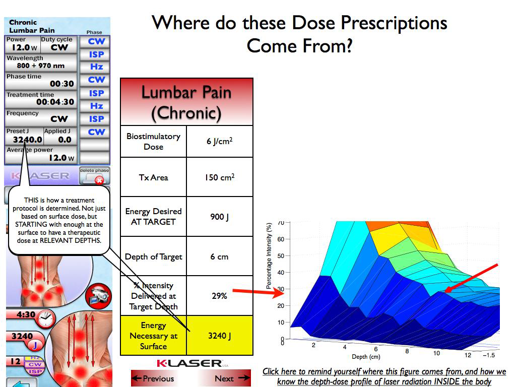
 Here are some rough numbers to review the feasibility of using this system for treatment. If the duty cycle is 70%, the system will deliver 1.68 joules per second at a depth 2cm (15wattS*70%*16%). To get 5 joules/cm2 over 15 x 15 cm treatment area would require a total of 1125 joules at depth. This would take 23 minutes.

This research shows that only class 4 systems can delivery the level of power needed for this kind of therapy in a typical rushed doctor's office. A class 3b system with 1 watt would take 4 - 5 hours per treatment to get the same dosage.

The original research publication is titled " Treatments for traumatic brain injury with emphasis on transcranial near-infrared laser phototherapy"


video length: (9:18)


Original Source: https://www.youtube.com/watch?v=iZbP2IVekh0

Review of transcranial photobiomodulation for major depressive disorder: targeting brain metabolism, inflammation, oxidative stress, and neurogenesis

Paolo Cassano; Samuel R. Petrie; Michael R. Hamblin; Theodore A. Henderson; Dan V. Iosifescu; - Neurophotonics, 3(3), 031404 (2016). doi:10.1117/1.NPh.3.3.031404 March 4, 2016 (Publication) 4471
This study shows some of the most detailed parameters (power, wavelenght, dosage) for working with the brain and seems to be unbiased because of the diverse background of authors..
View Resource

Abstract
We examined the use of near-infrared and red radiation (photobiomodulation, PBM) for treating major depressive disorder (MDD). While still experimental, preliminary data on the use of PBM for brain disorders are promising. PBM is low-cost with potential for wide dissemination; further research on PBM is sorely needed. We found clinical and preclinical studies via PubMed search (2015), using the following keywords: “near-infrared radiation,” “NIR,” “low-level light therapy,” “low-level laser therapy,” or “LLLT” plus “depression.” We chose clinically focused studies and excluded studies involving near-infrared spectroscopy. In addition, we used PubMed to find articles that examine the link between PBM and relevant biological processes including metabolism, inflammation, oxidative stress, and neurogenesis. Studies suggest the processes aforementioned are potentially effective targets for PBM to treat depression. There is also clinical preliminary evidence suggesting the efficacy of PBM in treating MDD, and comorbid anxiety disorders, suicidal ideation, and traumatic brain injury. Based on the data collected to date, PBM appears to be a promising treatment for depression that is safe and well-tolerated. However, large randomized controlled trials are still needed to establish the safety and effectiveness of this new treatment for MDD.

1.

Introduction

Infrared (IR) light is ubiquitously present to most life on the earth. Of the total amount of solar energy reaching the human skin, 54% is IR and 30% is IR type A—near-infrared—(NIR; with a wavelength range of 760 to 1440 nm),1 which penetrates through the human skin and reaches deeply into tissue, depending on wavelength and energy.2

NIR is used to treat a variety of conditions such as muscle pain,3 wounds,4 neuropathic pain,5 and headache.6 NIR is also used for wellness and lifestyle purposes such as for cosmetic improvement in peri-orbital wrinkles.7,8 The clinical use of NIR light applied in NIR-spectroscopy dates from the mid-1980s, when it was used for monitoring of the brain in the neonate and the fetus.9

The use of transcranial phototherapy for treating brain disorders started with its application to acute stroke. Numerous preclinical animal studies1011.12 suggested that the application of NIR laser (810 nm) to the head at various times (hours) after induction of an acute stroke had beneficial effects on subsequent neurological performance and reduced lesion size. Evidence was obtained for the anti-inflammatory, anti-apoptotic, and proneurogenesis effects in the brain stimulated by this approach.13,14 These promising animal studies led to the conduction of a series of clinical trials called NeuroThera Effectiveness and Safety Trials (NEST). All together there were three large studies conducted in 1410 stroke patients [NEST-1 (n=120" role="presentation">n=120

), NEST-2 (n=660" role="presentation">n=660), NEST-3 (n=630" role="presentation">n=630

)] that demonstrated that NIR light delivered transcranially with a class-IV laser is safe, with no significant differences in rates of adverse events with NIR, when compared to sham exposure.1516.17 Other preclinical studies and clinical trials have suggested that transcranial photobiomodulation (PBM: laser or light emitting diodes—LED) is safe and effective for acute1819.20.21.22 and chronic2324.25 traumatic brain injury (TBI) and has beneficial effects on neurodegenerative diseases (Alzheimer’s and Parkinson’s).26,27

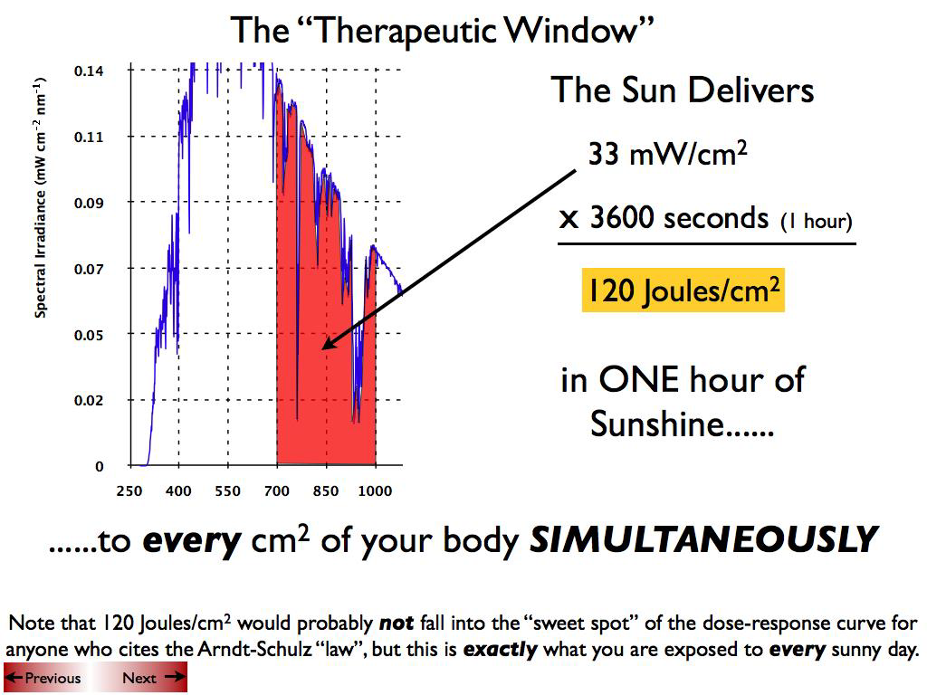
For the transcranial treatment of major depressive disorder (MDD), both PBM LEDs and lasers have been experimentally tested, although PBM is not FDA-approved for the treatment of MDD. Certain forms of PBM treatment are also referred to as low-level light therapy (LLLT), since it utilizes light at a low power (0.1 to 0.5 W output at the source) to avoid any heating of tissue. The irradiance of the PBM medical devices (or power density) typically ranges from 1 to 10 times the NIR irradiance from sunlight on the skin (33.6  mW/cm2" role="presentation">33.6mW/cm2

at the zenith). However, most PBM medical devices only deliver light energy at one or two selected wavelengths, as opposed to the whole spectrum of IR that is contained in sunlight. To our knowledge and to this date, transcranial PBM treatment has not caused any retinal injury—one of the most likely postulated adverse events, although care is taken routinely in such studies to protect the eyes with goggles or eye covers.28

In this review, we will first discuss the mechanisms of action by which NIR and red light (PBM) might improve symptoms of depression, and then present the clinical evidence for their use as a treatment for MDD and other comorbid psychiatric syndromes.

2.

Methods

We found clinical and preclinical studies via PubMed search (December 15, 2015), using the following keywords: “near-infrared radiation,” “NIR,” “low-level light therapy,” “low-level laser therapy,” or “LLLT” plus “depression.” We chose studies that had a clinical focus, and we excluded studies involving NIR spectroscopy. We also located studies using the references from the articles found in the PubMed search. As the searched literature encompassed different conditions and disorders frequently comorbid with depression, a specific section of this review was devoted to the effect of PBM on psychiatric comorbidity. In the latter section, the following conditions were included, based on available literature: TBI, anxiety and post-traumatic stress syndromes, insomnia, and suicidal ideation. The literature search for the use of PBM to treat comorbid conditions was neither systematic nor extensive, but rather a secondary focus of this review. The information is presented in an organized fashion to allow the reader to easily grasp the potential applications of PBM for the treatment of depression and of its comorbid conditions. To attain this goal, the authors have allowed a margin of redundancy, by distributing different information derived from any given publication in separate sections of this review. To avoid an artificial inflation of the extant literature on the chosen topic, we referenced the main authors—and when appropriate their affiliation—when referring to the same articles more than once. The reader will find a table summarizing the six key clinical articles reviewed, also to avoid unintended inflation of the literature. The six clinical reports included in this review where extracted from a pool of 58 articles, that were originally identified with the literature search.

In addition, we used PubMed to find articles that examined the link between PBM and each of the various biological processes including metabolism, inflammation, oxidative stress, and neurogenesis.

3.

Targeting Brain Metabolism

Multiple studies have reported regional and global hypometabolism in MDD, which could be related (either causally or consequentially) to the neurobiology of mood disorders.2930.31.32 Positron emission tomography studies have shown abnormalities in glucose consumption rates and in blood flow in several brain regions of subjects with major depression.33 Moreover, metabolic abnormalities in the anterior cingulate, the amygdala-hippocampus complex, the dorsolateral prefrontal cortex (DLPFC), and inferior parietal cortex seem to improve after antidepressant treatment or after recovery.3435.36

With phosphorus magnetic resonance spectroscopy (P31-MRS" role="presentation">31P-MRS

), the baseline pool of nucleotide triphosphate (NTP)—a product of the cellular utilization of glucose and a marker of the cellular energy availability—was low in subjects who subsequently responded to antidepressant treatment.32 Iosifescu et al.32 also demonstrated for the first time with P31-MRS" role="presentation">31P-MRS a correlation between treatment response (to a regimen that combined antidepressants and triiodothyronine) and restoration of a higher NTP pool (with compensatory decrease in phosphocreatine) in the anterior cingulate cortex. This study suggests a pathway to antidepressant response based on restoration of a high cellular energy state. In fact, phosphocreatine represents a long-term storage depot of energy, while NTP and ATP are energy-rich molecules that are readily available to the cell. The same authors replicated the aforementioned findings in MDD subjects treated with standard antidepressants (Iosifescu et al., unpublished). In this cohort, P31-MRS" role="presentation">31P-MRS

metabolite changes were noted in brain-only voxels of responders, but not in nonresponders to antidepressants.

In experimental and animal models, PBM (NIR and red light) noninvasively delivers energy to the cytochrome c oxidase and by stimulating the mitochondrial respiratory chain leads to increased ATP production (see Fig. 1).3738.39 A study of the effects of NIR on patients with MDD found that a single session of NIR led to a marginally significant increase in regional cerebral blood flow.40 Whether the observed changes in cerebral blood flow resulted from fundamental changes in neuronal metabolism or changes in vascular tone remain to be clarified. Given the correlation of both hypometabolism and abnormal cerebral blood flow with MDD, the beneficial effect of NIR on brain metabolism is one potential mechanism for its antidepressant effect.

Fig. 1

Cellular targets of NIR radiation mechanisms of transcranial NIR for psychiatric disease. The NIR photons are absorbed by cytochrome c oxidase in the mitochondrial respiratory chain. This mitochondrial stimulation increases production of ATP but also activates signaling pathways by a brief burst of ROS. This signaling activates antioxidant defenses reducing overall oxidative stress. Proinflammatory cytokines and neuroinflammation are reduced. Neurotrophins such as brain-derived neurotrophic factor are upregulated, which in turn activate synaptogenesis (formation of new connections between existing neurons) and neurogenesis (formation of new neurons from neural stem cells).

NPH_3_3_031404_f001.png

4.

Targeting Inflammation

Animal and clinical research suggests that the inflammatory arm of the immune system contributes to MDD. Post-mortem gene expression profiling on tissue samples from Brodmann area 10 (BA10—prefrontal cortex) have shown that MDD is characterized by increased inflammation and apoptosis.41 In a case-control study, Simon et al.42 found that antidepressant-naive MDD subjects had significant elevations in the following cytokines and chemokines when compared to healthy controls: MIP-1α" role="presentation">MIP-1α

, IL-1α" role="presentation">IL-1α, IL-1β" role="presentation">IL-1β, IL-6, IL-8, IL-10, Eotaxin, GM-CSF, and IFNγ" role="presentation">IFNγ

. Although IL-10 is an anti-inflammatory cytokine, the results suggested that the elevated levels of this IL-10 were likely induced in response to the overall elevation of proinflammatory cytokine levels. In a review of the research on inflammation in MDD, Raison et al.43 proposed that proinflammatory cytokines might cause brain abnormalities that are characteristic of MDD. Indeed, animal research has shown that IL-1 mediates chronic depression in mice by suppressing hippocampal neurogenesis.44

One proinflammatory cytokine that may be of particular relevance to depression is CSF IL-6 (IL6 measured in cerebrospinal fluid). In a recent report, patients with MDD had significantly higher CSF IL-6 levels compared to healthy controls; CSF IL-6 levels were significantly higher than in the serum, and there was no significant correlation between CSF and serum IL-6 levels.45 These findings are consistent with a prior report showing a positive correlation between CSF IL-6 levels and the severity of depression and suicide attempts, with the strongest correlation found in violent suicide attempters.46 One report in a smaller sample of depressed patients has shown that CSF IL-647 was lower or comparable to healthy controls.

NIR light and red light (600 to 1600 nm) decreased synovial IL-6 gene expression (decreased mRNA levels) in a rat model of rheumatoid arthritis.48 In another study, NIR (810 nm) used as a treatment for pain in patients with rheumatoid arthritis decreased production of the following proinflammatory cytokines: TNF-α" role="presentation">TNF-α

, IL-1β" role="presentation">IL-1β

, and IL-8.49 Khuman et al.50 showed that transcranial NIR improved cognitive function and reduced neuroinflammation as measured by Iba1+ activated microglia in brain sections from mice that had suffered a TBI. Finally, NIR (970 nm) has been found to be an effective treatment for inflammatory-type acne.51 In summary, it is reasonable to predict that transcranial NIR treatment would likewise have an anti-inflammatory effect in patients suffering from MDD.

5.

Targeting Oxidative Stress

Research has demonstrated a correlation between MDD and vulnerability to oxidative stress.52 For example, depression-induced rats show a significant decrease in glutathione peroxidase (GSH-Px) activity in the cortex.53 Glutathione (GSH) is the most abundant and one of the important antioxidants in the brain; GSH-Px enzymes protect against oxidative stress via reducing hydroperoxides and scavenging free radicals.54 GSH also appears reduced in the brains of MDD subjects.55 Additionally, a study by Sarandol et al.52 demonstrated that MDD patients have higher levels of malondialdehyde, a toxic molecule and a biomarker of oxidative stress.56 Moreover, depressed patients have more red blood cell (RBC) oxidation compared to healthy controls.52 In the same study, the authors found a significant positive correlation between RBC superoxide dismutase (SOD) activity and depression severity. SOD serves to catalyze the removal of the toxic superoxide radical.57 Thus, elevated SOD activity in depressed patients might indicate higher levels of oxidative stress. Finally, catalase activity and nitric oxide (NO) levels have also been shown to be lower in depressed patients than in healthy controls.58 Catalase is an enzyme that protects cells against damaging reactive oxygen species (ROS) via degradation of hydrogen peroxide to water and oxygen.59 NO has protective effects against cell damage, which are likely due to its pleiotropic functions in regulating antioxidant enzymes and many other aspects of cell metabolism.60,61

Oxidative stress may be an effective target for antidepressant treatments. However, successful treatments for MDD vary in regard to their protective effects against oxidative stress.52,53,62 Animal research suggests that PBM may have beneficial effects on oxidative stress. In a rat model of traumatized muscle, NIR (904 nm) blocked the release of harmful ROS and the activation of the transcription factor, nuclear factor κB (NF-κB), both induced by muscle trauma. Trauma activates NF-κB by destroying a specific protein inhibitor of NF-κB called IκB, and this destruction was inhibited by NIR light. Furthermore, NIR reduced the associated overexpression of the inducible form of nitric oxide synthase (iNOS) and reduced the production of collagen.63 This regulation of iNOS is important because excessive levels of iNOS can lead to formation of large amounts of NO that combine with superoxide radicals to form the damaging species peroxynitrite, and can interfere with the protective benefits of other forms of NO synthase.64 These findings suggest that NIR protects against oxidative stress induced by trauma. Finally, an in vitro study of the effects of red light and NIR (700 to 2000 nm) on human RBCs found that NIR significantly protected RBCs against oxidation.65


Original Source: https://www.spiedigitallibrary.org/journals/Neurophotonics/volume-3/issue-03/031404/Review-of-transcranial-photobiomodulation-for-major-depressive-disorder--targeting/10.1117/1.NPh.3.3.031404.full?SSO=1

Treatments for Traumatic Brain Injury With Emphasis on Transcranial Near-Infrared Laser Phototherapy

Larry D Morries, Paolo Cassano, Theodore A Henderson, - This article was published in Neuropsychiatric Disease and Treatment, 20 August 2015 (Publication) 4406
This exceptional research indicated prefered wavelenghts and dosages for treating patients with traumatic brain injuries. The found some surprising results.
View Resource

Abstract:

Traumatic brain injury (TBI) is a growing health concern affecting civilians and military personnel. In this review, treatments for the chronic TBI patient are discussed, including pharmaceuticals, nutraceuticals, cognitive therapy, and hyperbaric oxygen therapy. All available literature suggests a marginal benefit with prolonged treatment courses. An emerging modality of treatment is near-infrared (NIR) light, which has benefit in animal models of stroke, spinal cord injury, optic nerve injury, and TBI, and in human trials for stroke and TBI. The extant literature is confounded by variable degrees of efficacy and a bewildering array of treatment parameters. Some data indicate that diodes emitting low-level NIR energy often have failed to demonstrate therapeutic efficacy, perhaps due to failing to deliver sufficient radiant energy to the necessary depth. As part of this review, we present a retrospective case series using high-power NIR laser phototherapy with a Class IV laser to treat TBI. We demonstrate greater clinical efficacy with higher fluence, in contrast to the bimodal model of efficacy previously proposed. In ten patients with chronic TBI (average time since injury 9.3 years) given ten treatments over the course of 2 months using a high-power NIR laser (13.2 W/0.89 cm2 at 810 nm or 9 W/0.89 cm2 at 810 nm and 980 nm), symptoms of headache, sleep disturbance, cognition, mood dysregulation, anxiety, and irritability improved. Symptoms were monitored by depression scales and a novel patient diary system specifically designed for this study. NIR light in the power range of 10-15 W at 810 nm and 980 nm can safely and effectively treat chronic symptoms of TBI. The clinical benefit and effects of infrared phototherapy on mitochondrial function and secondary molecular events are discussed in the context of adequate radiant energy penetration. Keywords: infrared, traumatic brain injury, TBI, transcranial infrared light therapy, transcranial laser therapy 

INTRODUCTION

Traumatic brain injury (TBI) has recently moved into the limelight due to the recognition of its impact on professional athletes and military personnel. Yet, TBI is neither a new problem nor limited to those two populations. The Centers for Disease Control and Prevention estimated that 1.5 million Americans sustained TBI annually in 2000.1 As of 2006, the estimates had risen to 1.7 million brain injuries annually.2,3 Undoubtedly, these point prevalence proportions will increase as military personnel return home,4 and the problem of repeated mild TBI (mTBI) becomes more recognized in sports.5 Current estimates of the prevalence of TBI among veterans range from 9.6%6 to 20%,7 with an estimated total of more than 300,000 cases of TBI among military personnel since 2000.4 The current estimates of the combined number of sportsrelated concussions and brain injuries in the US are 1.6-3.8 million annually.8-10 TBI results in a wide spectrum of neurological, psychiatric, cognitive, and emotional consequences. In part, the variation is related to the severity of the injury (mild, moderate, severe TBI), which is stratified based on Glasgow Coma score, periods of unconsciousness, and degrees of amnesia. Furthermore, the diversity of sequalae can be related to the areas of the brain that are injured, the severity of the injury (highly variable within the classification of “mild” and “moderate”), and the evolution of the injury over time due to neuroinflammatory processes.11,12 Additional mechanisms thought to underlie the damage of TBI include decreased mitochondrial function, calcium and magnesium dysregulation, excitotoxicity, disruption of neural networks, free radicalinduced damage, excessive nitric oxide, ischemia, and damage to the blood-brain barrier. Together, these can contribute to a progression of the damage over time. Patients with TBI can experience headache, visual disturbances, dizziness, cognitive impairment, loss of  executive skills, memory impairment, fatigue, impulsivity, impaired judgment, emotional outbursts, anxiety, and depression.3,13-23 The situation can be further clouded by secondary and/ or comorbid posttraumatic stress disorder (PTSD), depression, and anxiety,17-25 which can have symptoms that overlap with those described above and appear to be increasingly likely with repetitive concussive or subconcussive brain injury.5,24,26

TREATMENTS FOR TBI

Pharmacological treatments Pharmacological treatment largely targets the neuropsychiatric sequalae of TBI, rather than providing any means of healing or repairing injury. In general, pharmacological treatment is focused on the modulation of major neurotransmitter systems – dopaminergic, serotonergic, noradrenergic, acetylcholinergic, and glutaminergic.20 Disruption of the major neurotransmitter pathways may result from direct injury or excitotoxicity and other cytotoxic mechanisms. The treatment of depression secondary to TBI is often approached with serotonin reuptake inhibitors. Several studies have examined the benefit of sertraline in post- TBI depression.27-29 Other serotonin reuptake inhibitors also have been examined. Tricyclic antidepressants appear to have some use in the treatment of post-TBI depression, although cautious dose titration is required. Patients with TBI are at greater vulnerability to sedation and cholinergic side effects of confusion and memory impairment. With serotonergic agents other than sertraline, cognitive effects also have been reported.30 Similarly, lithium may be a less desirable agent in this  population due to sedation and cognitive impairment. Patients with TBI may respond at lower doses and lower blood levels than expected. Modulation of the dopaminergic system may improve alertness, attention, and cognitive processing speed. The stimulants are most commonly used for this purpose. Methylphenidate facilitates the release of dopamine and slows its reuptake. Dextroamphetamine strongly inhibits reuptake of dopamine, slows down the breakdown of dopamine by monoamine oxidase, and somewhat increases the release of dopamine. These subtle differences are sometimes imperceptible to the patient, but at other times, a patient will do best on one or the other stimulant. Increasing dopamine in the reticular activating system leads to enhanced arousal. Increasing dopamine within the frontal cortex and the striatum leads to enhanced processing speed and attention. Some evidence suggests that the stimulants may enhance neuronal recovery after injury.31-33 There are numerous potential side effects with stimulants, including abnormal heart rhythms, decreased seizure threshold, and death, but these severe side effects are extremely rare. The most common side effects with stimulants are decreased appetite, stomach upset, and headache. These are most severe at the beginning of treatment and improve over time for most patients. Insomnia is another common side effect, which may be more frequent in those with a TBI. Amantadine and bromocriptine may also increase dopamine. Studies of these agents have shown reduced abulia, anergia, and anhedonia in those with TBI.34,35 Amantadine may cause confusion, hallucinations, and hypotension. Small studies have suggested some benefits of bromocriptine in cognitive function.36,37 Arousal-enhancing agents also have found a use in the treatment of the neurocognitive sequalae of TBI. Modafinil is the oldest form of these medications, and armodafinil is an isomer of modafinil with longer activity and less side effects. These medications help to increase alertness and wakefulness. The precise mechanism of action of odafinil is unclear. It appears to increase histamine in parts of the brain involved in controlling the sleep-wake cycle; however, knock-out mice that lack histamine receptors still show increased wakefulness with modafinil.38,39 The picture is also murky  for modafinil’s effect on orexins, which are wakefulness molecules in the hypothalamus.40 Modafinil has been shown to weakly bind to the dopamine transporter – like the stimulants,41 and dopamine transporter knock-out mice show no response to modafinil.42 A number of research studies have examined the benefit of these agents in fatigue associated with multiple sclerosis, TBI, cancer, and other conditions. Cognitive and memory impairments after TBI may reflect disruption of cholinergic function. The impact of anticholinergic agents on cognitive function of those with TBI supports this contention. Donepezil is the safest and most widely used of the cholinesterase inhibitors. Several easonably large studies have shown improved memory and cognitive function.43-45 Donepezil has benefits in memory and cognition even several years after injury.45,46  Anticonvulsants are often prescribed initially after a TBI due to heightened risk for seizures. Post-TBI mania or mood lability may respond well to anticonvulsants, such as carbamazepine or sodium valproate. They are also often used to treat aggression after TBI. The anticonvulsant agent, topiramate, has been shown to adversely affect cognitive function in the TBI patients.47 While insomnia is a significant issue for patients with TBI, affecting between 15% and 84% (mean of 40%),3,13,19,21,23,48,49 little has been published on the treatment of this aspect of TBI. Benzodiazepines may  be effective but carry a risk of disinhibition. Kemp et al48 found that commonly used sleep aid, melatonin, was not effective. Antidepressants, including serotonin reuptake inhibitors and tricyclic antidepressants, are not effective in resolving insomnia in this population.49 No single agent has emerged as a good solution for this symptom. Cognitive rehabilitation Cognitive rehabilitation now takes many forms and is often individualized to the particular needs of the patients. Protocols have been devised to remediate cognitive difficulties often encountered in those with TBI, such as impaired concentration, executive dysfunction,  inattention, visual disturbances, memory dysfunction, and impaired language function. They range from simple strategies (using a planner to aid memory and organization) to specific protocols targeting particular cognitive functions (eg, short-term memory) that can be monitored with sequential neuropsychological testing. These interventions have been extensively reviewed elsewhere.50,51 Comprehensive programs which include psychotherapy and social skills components have been shown to have greater efficacy.50,52,53 Overall, reports of benefits have been mixed.54,55 Behavioral therapies Behavioral remediation strategies to eliminate problematic  behaviors following TBI have met with mixed success, most often in terms of the poor generalization of specific skills to the outside world. Behavioral deficits that create difficulties for those with TBI and their families include poor hygiene, decline in tidying/cleaning habits, social withdrawal, reduced social comprehension, impaired memory, and poor organization. Behavioral excesses that create difficulties for those with TBI and their families include aggression, sleep disruption, and perseverations. These have been reviewed elsewhere.56 Nutritional supplements Nutritional supplements, herbs, and nootropics have been utilized for many years and are increasingly popular among the patient populations. There remains little clinical research on many of these agents, perhaps reflecting a lack of funding more than a lack of efficacy. Acetyl-l-carnitine is an ester of l-carnitine and is thought to protect brain cells after injury when glucose metabolic pathways are compromised. During this period, acetyll- carnitine supports alternative ketogenic pathways for metabolism.57 It is also believed to enhance cholinergic function. While there are several clinical studies on patients with Alzheimer’s disease and preclinical data on animal models of TBI, the clinical literature on TBI remains sparse. Ginkgo biloba is a natural product of the tree by the same name. It has been shown to improve membrane fluidity and increase resistance to free-radical damage. It provides some subtle benefits to cognitive function in clinical studies of stroke, dementia, aging, and hypoxia damage.58 It has not been systematically studied in TBI but is used extensively in clinic, often in combination with meclofenoxate which is an avid scavenger of free radicals.59 S-Adenosylmethionine (SAMe) is a nutritional supplement which improves cell membrane fluidity and promotes the production of glutathione, an antioxidant. The benefit of SAMe has been assessed in a single clinical study of TBI.60 Patients receiving SAMe had a 77% improvement in clinical scores of post-concussive symptoms. Citicholine provides a source of choline which can cross the bloodbrain barrier. It has been used extensively in Europe and Japan as a treatment for TBI, stroke, and dementia. However, two large US studies failed to demonstrate significant benefit.61,62 Piracetam and the related oxiracetam and phenylpiracetam have shown some promise as nootropic agents. In one double-blind, placebo-controlled study, piracetam improved several symptoms of postconcussive syndrome, including headache and vertigo.63 More recent clinical studies have shown marginal benefit.64 Huperzine-A, an extract of Japanese club moss, is a natural acetylcholinesterase inhibitor. It may serve as a natural alternative to donepezil, rivastigmine, or galantamine. Galantamine warrants special mention as it appears to also modulate nicotinic  eceptors and appears to have more persistent benefit in the treatment of Alzheimer’s disease. It appears to modulate neuroimmune responses, in addition to its effects on acetylcholinesterase.65 Cerebrolysin is a polypeptide that purportedly mimics the actions of neurotrophic factors.66,67 Studies have shown that it can reduce beta amyloid and phosphorylated tau protein accumulation. It may promote neurogenesis, synapse formation, and functional recovery.66 In animal models of acute TBI, cerebrolysin-treated rats had more surviving neurons in the area of impact and showed greater functional recovery.67 In a clinical trial of acute TBI, patients were recruited within 24 hours of injury and treated for 3 months with daily intravenous infusion of cerebrolysin. At 3 months, those receiving cerebrolysin performed significantly better on the Cognitive Abilities Screening Instrument.68 It remains unclear if cerebrolysin provides long-term nootropic benefit. The elevation of free radicals in TBI suggests that antioxidants should be beneficial. Clinical trials of pharmacological antioxidants over the past 30 years have not yielded a useful agent in acute TBI.69 Agents, such as tirilazad70 and polyethylene glycol- onjugated superoxide dismutase, have failed to show benefit in acute TBI. Omega-3 fatty acids may enhance brain repair and recovery, based on animal and clinical studies.71 Similarly, vitamin D may offer neuroprotective and restorative benefits72 in the acute TBI setting. In chronic TBI, vitamin D and omega-3 fatty acids may work synergistically, as they both may reduce neuroinflammation, apoptosis, and oxidative stress.73 Other nutritional supplements have been recommended, but prolonged therapy is necessary to possibly see benefits in TBI. A 6-month trial of ginkgo, vinpocetine, acetyl-lcarnitine, huperzine, alpha-lipoic acid, n-acetyl-cysteine, multivitamins, and over 5 g of omega-3 fatty acids daily yielded improved performance in cognitive testing and increased perfusion (function) in single-photon emission computed tomography (SPECT) scan.74 Long-term use of dietary flavanols may improve cognition in mTBI.75 Transcranial magnetic stimulation Transcranial magnetic stimulation (TMS) has shown some promise in animal models of TBI.76 However, a Cochrane review of the clinical application of TMS for depression noted no difference between repetitive TMS (rTMS) and sham rTMS using the Beck Depression Inventory (BDI) or the Hamilton Depression Rating Scale, except during the initial 2-week period.77 The application of TMS in the post-TBI patients is limited by the risk of seizure induction.78 Hyperbaric oxygen Hyperbaric oxygen treatment has been explored as a treatment for TBI.79-91 Hyperbaric oxygen therapy is neither a benign treatment, given the concerns of oxygen toxicity,79 nor a clear treatment in that the placebo condition of moderate hyperbaric room air also effectively improves cognitive function.80,81 The most carefully performed study compared a group in a cross-over design with an interval of both null treatment and hyperbaric oxygen at 100% oxygen and 1.5 atm.82 The study described improvement in many of the symptoms associated with persistent TBI including headache, tinnitus, vision disturbance, memory dysfunction, and impaired cognitive function. Cognitive testing also showed improvement in attention, information processing speed, and a battery of cognitive tests. In an uncontrolled case series of 16 subjects, Harch et al83 demonstrated that an abbreviated series of hyperbaric treatments using 100% oxygen at 1.5 atm could mitigate subjective symptoms of TBI (eg, headache, sleep disruption, irritability), improve cognitive testing scores, and improve cortical function based on SPECT imaging.83 A study of a higher dose (2.4 atm) did not reveal any significant benefit of hyperbaric oxygen therapy compared to a sham-control group treated with 1.3 atm,84 and this result has been extended and confirmed by a related group.85 However, this may reflect an inverse dose- esponse curve, rather than an absence of benefit, in that the low-dose sham group demonstrated significant changes in cognitive testing and symptom frequency.86 Hyperbaric oxygen remains a controversial area in both acute TBI86-89 and chronic TBI.82,83,85,86,90,91 Physical exercise High-energy activities and exercise programs completed through a health club facility or comprehensive rehabilitation program should focus on the same parameters of an age-adjusted and diagnosis-specific program for aerobic conditioning – flexibility, stabilization, and strength. Though it appears safe and is an accepted intervention for TBI, there is a need for further welldesigned studies.92 Exercise was a part of a 6-month study of lifestyle changes described above which yielded improved function based on cognitive testing and perfusion SPECT scans.74

A NEW TREATMENT FOR TBI

Unfortunately, little has been found to reverse the damage of TBI or repetitive concussion which is the root cause of residual cognitive and psychological impairment following TBI.20,93 One potential avenue of treatment for TBI is infrared light, which has shown promising data in a number of applications. Near-infrared (NIR) light has been investigated for its ability to modulate intracellular mechanisms related to healing. The application of NIR light by low-power laser or by light-emitting diode (LED) is also known as laser phototherapy94 or near-infrared photobiomodulation.92 NIR irradiation can facilitate wound healing,95,96 promote muscle repair,95 and stimulate angiogenesis.95,96 NIR phototherapy has been studied and applied clinically in a wide array of ailments, including skin ulcers,97 osteoarthritis,98 peripheral nerve injury,95,96 low back pain,99 myocardial infarction,100 and stem cell induction.101 The finding that NIR light passes relatively efficiently through bone has spurred interest in its application to treating disorders of the brain. Over the past decade, transcranial near-infrared light therapy (NILT)102 has been studied in animal models to understand its ability to repair damaged or dysfunctional brain tissue resulting from stroke and TBI. The first published study of NILT for TBI in humans described two cases of chronic mTBI with significant disability.103 Each patient was treated with an LED device delivering low-level low-level light therapy (LLLT) in the red and NIR range for 6-10 minutes per area daily for several months. Both patients had marked neuropsychological improvement after a minimum of 7-9 months of LLLT treatment. The precise mechanisms underlying photobiomodulation and its therapeutic benefits are not fully understood. The purported effects of NIR are illustrated in Figure 1. Light in the wavelength range of 600-1,200 nm has significant photobiomodulation capability.104 Current data most strongly support that absorption of NIR photons by cytochrome c oxidase in the mitochondrial respiratory chain is the key initiating event in photobiomodulation.95,96,104,105 This induces an increase in cytochrome c oxidase activity which in turn increases adenosine triphosphate (ATP) production. Such an increase in ATP in wounded or underperfused cells may be sufficient to activate cells in areas of injury or metabolic derangement.106 Data from numerous tissue culture and animal studies point to the importance of several secondary molecular and cellular events. For example, NIR photonic energy can modulate reactive oxygen species,95,96,102 activate mitochondrial DNA replication,95,96 increase early-response genes,95 increase growth factor expression, induce cell proliferation, and alter nitric oxide levels.95,96,102 These mechanisms are more fully described in the companion paper.105 When examined in the specific model of neural tissue injury, NIR phototherapy can lead to demonstrable neural repair and recovery. For example, LLLT of a power density of 0.9-36 J/cm2 applied at 24 hours poststroke in a rodent model yielded a 32% reduction in neurological deficits, as well as histochemical evidence of neuron proliferation and migration.106-108 LLLT had similar benefits in a rodent model of TBI.96,109-111 Interestingly, these cellular changes evolved over a period of days after light exposure and persisted for considerably longer than the interval of actual NIR exposure. These findings are consistent with a progressive regeneration cascade set in motion by the NIR light exposure. NILT in stroke NILT, predominately in the form of LLLT, has been investigated in laboratory models of stroke. LLLT applied in a single dose to an ischemic stroke model appeared to induce expression of the growth factor transforming growth factor – beta 1 and suppress the production of peroxynitrite.112 In a rat model of middle cerebral artery occlusion, LLLT at a dose of 0.5-7.5 mW/ cm2 using continuous wavelength light at 808 nm was administered at 24 hours after the acute stroke.108,113 This single application was estimated to deliver 1.8 J/cm2 in total to the cortex surface and resulted in demonstrable neurological improvement. Functional changes were not manifested until approximately 2 weeks after the single treatment. While there was no significant change in the size of the stroke lesion, histochemical evidence of neurogenesis and migrating neurons108 indicate that a cascade of secondary processes was initiated by NILT. A rabbit model of stroke utilizing injection of a blood clot embolus also demonstrated benefit from LLLT.102,114,115 Herein, 808 nm light was applied with an LED delivering 7.5 mW/cm2 and an estimated 0.9-2.6 J/cm2 to the cortical surface. Cortical ATP levels were increased, indicative of increased mitochondrial activity.114 Significant behavioral recovery was also noted; however, neither ATP increased nor neurological function changed at doses less than 0.3-0.7 J/cm2.114,115 At higher doses of 0.9-15 J/cm2, neurological improvement was seen.114,115 The clinical trials of NILT in acute stroke, the Neuro- Thera Effectiveness and Safety Trials 1, 2, and 3 (NEST- 1,-2, -3), were conducted between 2006 and 2009. The Phase II clinical trial (NEST-1) involved 120 patients in a double-blind, placebo- ontrolled study of the effects of NILT within 24 hours of ischemic stroke.116,117 Approximately 60% of the patients experienced clinical benefit, and the safety profile was very good. Thus, NEST-2, a Phase III clinical trial, was undertaken in 2007. A total of 660 patients were enrolled.118 Somewhat surprisingly, the study did not demonstrate statistical clinical improvement using a different outcome measure.119 Post hoc analysis revealed that a portion of the patients who were moderately affected and/or had strokes limited to the cerebral cortex did realize clinically and statistically significant improvement.102 The NEST-3 trial was halted midpoint when it failed to demonstrate statistical benefit on futility analysis.120 A key factor in the interpretation of the results of NEST-3 is that, different from NEST-1, all types of stroke were included as opposed to just cortical strokes. Continuous laser light has a limited depth of penetration (#1 cm into brain tissue) which likely prevents an effect on deeper brain matter. Therefore, the lack of significant benefits from NIR phototherapy in NEST-3 could be related to the fact that ischemic penumbra was not reached by the light (Luis DeTaboada, personal communication, January 2015). While pulsed NIR was not used in the NEST-3 study, it is estimated that pulsed NIR could penetrate up to 3 cm in depth from the cortical surface, therefore possibly extending the therapeutic target to deeper strokes (Luis DeTaboada, personal communication, January 2015). Figure 1 Hypothesized mechanism of action of NiR light therapy. Notes: NiR light (600-980 nm) penetrates tissue to variable depths depending on wavelength, the tissue involved, coherence, and time. A fraction of the photonic energy reaches the mitochondria and is absorbed by cytochrome c oxidase. This activates increased ATP production, increases production of ROS and RNS, and possibly increases NO. Downstream events include increased early-response genes (c-fos and c-jun) and activation of NF-?B, which in turn induces increased transcription of gene products leading to synaptogenesis, neurogenesis, and increased production of inflammatory mediators and growth factors. Abbreviations: NiR, near-infrared; ATP, adenosine triphosphate; ROS, reactive oxygen species; RNS, reactive nitrogen species; NO, nitric oxide; NF-?B, nuclear factor kappa B. NILT in TBi Oron et al109 conducted the first animal studies of NILT for TBI. They found that a single application of NIR light at 808 nm from a 200 mW emitter at 4 hours post-injury resulted in a significant reduction in lesion size by 5 days.109 To date, several groups have studied NILT in animal models, and this material has previously been reviewed.95,121-123 Single applications of 800-810 nm NIR light within 4 hours of injury have been shown to improve neurological function significantly.110,124-126 The same dose of NIR light at 6 hours was less effective125 and at 8 hours had no appreciable benefit.125 NIR photonic energy at other wavelengths was less effective. Wu et al110 examined red light (670 nm) at 4 hours and found a similar improvement in neurological function; however, 730 nm and 980 nm had no neurological benefit. Similar data for lesion volume have been reported. A single dose of 800-810 nm NIR light (fluence of 36 J/cm2) yielded an approximate 50% reduction in the volume of the lesion at 3-4 weeks110,111,124-126 and a possible reduction in the initial spread of neurological injury, based on the marked reduction in lesion volume found at 5 days post-injury.109 Repeated NIR phototherapy treatments appear to have some benefit, but the frequency and number of treatments are critical factors. While a single NIR light application had benefit, daily applications for 3 days yielded much greater neurological benefit126,127 with smaller lesion size,126 fewer degenerating neurons,126 more proliferating cells,126 and greater levels of brain-derived neurotrophic factor (BDNF)127 compared to a single treatment in a mouse model. In contrast, daily treatment for 7 days128 or 14 days126 showed no difference from controls. NIR energy densities in the range of 0.9-36 J/cm2 resulted in significant biochemical and behavioral changes.109-111,124-127 Pulsing of NIR light appears to yield a greater neurological response but only within certain parameters. Pulsing at 10 Hz yielded greater neurological improvement and a significant reduction in lesion size compared to either continuous-wave or pulsed NIR at 100 Hz.111 In the mouse model of moderate TBI, NILT (800-810 nm) improved learning and memory (Morris water maze performance),128 as well as behaviors associated with depression and anxiety (immobility during tail suspension).111,124 The finding that NILT brought about a smaller lesion in the rodent model of TBI compared to untreated mice suggests that decreased apoptosis, reduced spreading lesion penumbra, and/or neurogenesis are induced by NILT. Indeed, NILT can decrease BAX expression, a pro-apoptosis gene,129 increase expression of BCL-2, an anti-apoptosis gene,129 increase nerve growth factor,95 increase BDNF,127 decrease inflammatory markers,130 and decrease numbers of degenerating neurons.126 Together, these mechanisms may reduce the enlargement of the initial lesion during the first day following the lesion.109 Moreover, increased BDNF and nerve growth factor may contribute to synaptogenesis as shown by increased levels of synapsin-1,127 and neurogenesis, as shown by increased numbers of proliferating cells.127 In a double-blind study in healthy volunteers, NILT was beneficial – compared to sham – in memory and attention.131 In this study, the authors shed only one application of NIR light to the right forehead, targeting the right frontal pole of the cerebral cortex (Brodmann’s area 9 and 10). The device was a Class IV laser CG-5000 (Cell Gen Therapeutics, Dallas, TX, USA), and the parameters were as follows: wavelength 1,064 nm, irradiance 250 mW/cm2, fluence 60 J/cm2, and time 4 minutes per site (two sites).131 The subjects who received the NIR treatment had better attention after 2 weeks, measured by the psychomotor vigilance test. They also had better delayed visual memory at the Delayed Match-to-Sample test. This is the only published controlled trial assessing the impact of NILT on cognition; however, other reports have shown the therapeutic effects of NILT in small numbers of TBI patients. In a two-case report in TBI patients,103 NILT (870 nm) improved sustained attention, memory, and executive functions. Both patients were treated with an instrument with three separate LED cluster heads. The parameters used for the treatment were the following: NIR wavelength 870 nm and 633 nm (red light), irradiance 2.2-25.8 mW/cm2, fluence 13.3 J/cm2, and time 10 minutes per site.103 The same group reported on a cohort of eleven subjects with persistent cognitive dysfunction and treated with a similar NILT protocol for chronic mTBI.132 The eleven subjects received NILT with a device with three LED cluster heads (Model 1100; MedX Health, Toronto, ON, Canada). The parameters used for the treatment were the following: NIR wavelength 870 nm and 633 nm (red light), irradiance 22.2 mW/cm2, fluence 13 J/cm2, and approximate time 10 minutes per site. The NIR light was applied three times per week for 6 weeks (18 sessions), on eleven sites for 10 minutes per site (the total duration of each session was 20 minutes).132 The sites on the skull were chosen on the midline, and bilaterally on frontal, parietal, and temporal areas. At the follow-up neuropsychological testing, NILT had a powerful effect on attention, inhibition, and inhibition switching in the Stroop task, and similarly improved verbal learning and memory, as well as enhanced long-delay free recall on the California Verbal Learning Test. Eight subjects, from the same cohort, were identified as having mild, moderate, or severe depression based on the BDI-II total score (range: 15-34).132 The three cases, who entered the study with only mild depression, remained the same after NILT treatment. Results for the five cases with moderate-severe depression were as follows: two moderate cases improved to mild/minimal depression 8 weeks after the end of NILT series, and one severe case improved to moderate depression. Two moderate or severe depression cases remained the same after 8 weeks of follow-up from the last NILT session.132 Dose response and photonic penetration A prevailing theory in photobiomodulation postulates that a bimodal response curve exists for the biological effects of NIR light.95 The so-called Arndt-Schulz curve (a fundamental principle in homeopathic medicine) is frequently used to describe this biphasic dose response. Some data indicate that low levels of light have a much better effect on stimulating and repairing tissues than higher levels of light. Laboratory studies of cells in culture have demonstrated a bimodal dose response to light exposure in lymphocytes133 and fibroblasts.134,135 For example, Chen et al135 found that a range of 0.03-0.3 J/cm2 was beneficial in activating transcription factors in culture, while 3-30 J/cm2 inhibited the activation of these factors. In contrast, an order-of-magnitude greater dose (2 J/cm2) was best at activating fibroblasts in a superficial wound model.136 Furthermore, an order-ofmagnitude greater dose (30 J/cm2) proved to be best in a rodent joint inflammation model.137 Thus, a dosedependent effect for many biological responses to NIR light has been demonstrated,95,137-139 but the critical parameter is dose at the level of the target tissue, rather than at the surface.137,140 The amount of energy that reaches a volume of tissue at depth is determined by the attenuation of the photonic energy as it passes through the overlying tissue. For example, only 2.45% of the energy from a 980 nm laser emitter penetrates to the level of the peroneal nerve.140 Nevertheless, the biphasic dose response does not appear to be universally true. In primary microglial cell culture, a dose-dependent response to NIR was demonstrated with no detrimental effects at doses as high as 30 J/cm2.141 So a critical question in the use of NILT is that of radiant energy penetration. In particular, some authors have challenged the efficacy of low-power LEDs used in LLLT.142-144 In laboratory studies, LLLT radiant energy is almost entirely absorbed in the first 1 mm of skin.145,146 In two unrelated studies, LLLT diode devices proved to be ineffective in the treatment of diabetic neuropathy,142,144 in contrast with prior reports.147 Similarly, laboratory studies of NILT using LLLT transcranially have not consistently yielded positive results. For example, in a rat model of TBI, Giacci et al148 found no benefit from daily 30-minute irradiation with either 670 nm or 830 nm 0.5 W LED emitters for a period of 7 days. Doses at the skin surface were 28.4 J/cm2 and 22.6 J/cm2, respectively.148 Similarly, treatment of a rat model of contusive spinal cord injury with LLLT (830 nm at 22.6 J/cm2 or 670 nm at 28.4 J/cm2) for 30 minutes per day for 5 days resulted in no significant functional improvement and no reduction in lesion size, despite delivering 2.6 J/cm2 to the spinal cord.148 Lapchak102 reported that the physical parameters of NILT in the clinical trials for the treatment of stroke utilized in the NEST-1 and NEST-2 trials116-120 may have delivered insufficient energy to cortical tissues to be effective. Therein, NIR light of 808 nm wavelength with infrared energy densities of 0.9 J/cm2 was applied to the human scalp for a total of 40 minutes with applications at multiple sites during that time.116,118 Recall that animal models of both stroke and TBI suggest that NIR energy densities in the range of 0.9-36 J/cm2 resulted in significant biochemical and behavioral changes.96,106-115,125-127 The concern raised from the NEST studies102 is that current clinical trials testing the effectiveness of lowenergy NIR diodes to treat TBI may yield negative or inaccurate efficacy data, not because of a failure of infrared light to invoke a change but due to a dose error. Doses that are effective when directly applied to cells in a Petri dish149,150 or to 3-5 mm thick rodent brains96,109-111,125,126,128 may be insufficient to penetrate 2-4 cm into the human brain. In a companion paper, our own studies of photonic energy penetration are detailed.105 To summarize, the laboratory tissue studies showed that 0.5 W LED emitters did not penetrate the 2 mm thickness of human skin. No detectable energy from 0.5 W LED NIR light emitters could be detected penetrating a similar thickness (1-2 mm) of sheep skin or 3 cm thick section containing sheep skin, skull, and brain. In contrast, 11% of the photonic energy from a 10 W 810/980 nm coherent NIR laser penetrated 2 mm of human skin. Similarly, 17% of the photonic energy from a 15 W 810 nm coherent NIR laser penetrated the same distance.105 Energy from these more powerful NIR emitters could be detected penetrating 3 cm of sheep skin, skull, and brain with 0.4% of the 10 W 810/980 nm NIR laser’s energy reaching the depth of 3 cm and 2.9% of the 15 W 810 nm NIR laser’s energy traversing the same distance.105 Anders also has demonstrated penetration of 808 nm light to 40 mm in the brain using a 5 W laser emitter (JJ Anders, personal communication, January 2015). Prompted by the mixed results in the literature and the observations by Lapchak,102 Franzen-Korzendorfer et al,144 Wan et al151 and Lavery et al142 we have been utilizing relatively high-power (10- 5 W) lasers at the wavelengths of 810 nm and 980 nm in the clinic to treat patients with TBI. Clinically, the patients have shown excellent responses with resolution of many of their long-standing symptoms of TBI or post-concussive syndrome. Below is a retrospective series of such patients to illustrate the extent and character of response to this modality. Methods Patients in the case series were sequentially treated patients at a clinic which is engaged in ongoing NILT for a number of clinical conditions. The risks, benefits, and current state of research on the use of NILT were explained to each patient. Each patient consented to treatment. Institutional Review Board approval was obtained in a post hoc review, noting that the risk-benefit ratio was acceptable. Between March 16, 2011 and February 20, 2013, sequential new referrals for chronic mild-to-moderate TBI were evaluated for treatment and selected for NILT using Class IV lasers, either the LT1000 (LiteCure, Newark, DE, USA), a 10 W adjustable NIR laser emitter with wavelengths of 810/980 nm capable of delivering continuous or pulsed NIR light, or the Diowave 810 (Diowave, Riviera Beach, FL, USA), an adjustable NIR emitter up to 15 W with a wavelength of 810 nm capable of delivering continuous or pulsed NIR energy. Demographics and laser treatment settings are detailed in Table 1. The fluence delivered to the skin of patients ranged from 55 J/cm2 to 81 J/cm2. No other treatment modalities (medications, exercise regimen, supplements) were added, discontinued, or changed while receiving NILT. Symptoms were monitored clinically. A baseline Quick Inventory of Depressive Symptomatology Self-Report (QIDS-SR)152 was completed for all patients, and the BDI153 was administered to seven of the ten patients before and after the course of treatment. In addition, each patient was instructed on how to create and maintain a patient and spousal diary of symptoms and subjective progress. Each of six patients received a single series of ten treatments with the LT1000 Class IV laser. Three additional patients each received a single series of 20 treatments with the LT1000 Class IV laser. One patient was treated with the Diowave 810 nm Class IV laser device in a series of 20 treatments. The patients and treating clinician wore protective eyewear. There were no incidents of burns or thermal discomfort (Figure 2). The impact of high-watt NILT While the patient group represented a diverse mix (Table 1 presents demographics), some notable commonalities of symptoms emerged. Over 90% of the patients had complaints of anxiety, depression, irritability, and insomnia. Other symptoms included headache (60%), suicidal ideation (50%), cognitive difficulties (50%), attention problems (50%), short-term memory problems (40%), loss of libido (30%), substance abuse (20%), fatigue (20%), and panic attacks (20%). Six of the patients were unemployed prior to treatment. Three of the patients were experiencing severe marital difficulties. All carried or had a confirmed diagnosis of TBI, but other comorbid diagnoses included PTSD, major depressive disorder, generalized anxiety disorder, bipolar disorder, and attention deficit/hyperactivity disorder. The patients’ baseline scores on the BDI were 25.3±12.1 (moderate depression range), and baseline scores on the QIDS-SR were 12.9±4.6 (moderate depression range). During NILT treatments, skin temperature increased no more than 3°C with rapid cooling after removal of the NIR light. A continuous sweeping motion was utilized to minimize skin heating and cover a larger area. After a course of ten treatments of NILT (20 treatments in four patients), each patient experienced significant clinical improvement with resolution of many of their symptoms (Table 2). In addition, the BDI scores dropped to 12±6.5 (nondepressed range). This represented a significant decrease (P,0.01, Student’s t-test, one-tailed, Microsoft Excel). The QIDS-SR scores after treatment were 2.2±2.3 (nondepressed range), and the difference from baseline was highly significant (P,0.00001, Student’s t-test, one-tailed). Patients noted improvement in cognitive function, mood, anxiety, and sleep. None of the patients continued to have suicidal thoughts (50% at baseline). Other symptoms, such as anxiety and irritability, were markedly improved. Most notable were the nonquantifiable changes in patients’ lives. Patients reported improved cognitive ability and a desire to return to meaningful work. Five of the six unemployed patients have returned to work. The two patients who were Iraq/ Afghanistan veterans have found new careers in highly skilled trades. The patients with marital difficulties have reconciled and were purchasing homes or otherwise solidifying their marriages. The clinical change can be attributed to NILT because no changes in medications, supplements, or exercise regimen were permitted during the course of NILT treatment. All patients in the case series experienced significant clinical improvement which supports the conjecture that high-power NIR laser delivers sufficient energy to the human brain for photobiomodulation to occur. Insomnia and suicidal ideation, common symptoms in those with TBI or post-concussive syndromes,3,17-20,24,25 resolved in 100% of cases. Headache, another common symptom for patients following a TBI,6,14,15,23 was reduced or resolved in the six patients so afflicted. Symptoms such as anxiety,14,15,21,24 depression,21,24,25,27-29 and irritability resolved or were dramatically reduced in all patients. Cognitive function appeared to improve based on return to work or improved work performance, although cognitive tests were not performed. The quality of life dramatically improved in all cases, based on the observations of the patients, their family members, and the treating clinician. At follow-up intervals of 6-7 months post-treatment, patients have reported continued improvements in symptoms. The precise areas of brain injury were not elucidated in Figure 2 Treatment parameters per individual, based on area of the skull treated. Notes: Dimensions varied per head/skull size and hair line. Treatment was warm and comfortable for each patient. There were no incidences of discomfort. Areas treated were (A) temporal- ilateral, (B) frontal, and in patients 1-3, 5, and 6 (B) frontal only. Table 1: Infrared light treatment parameters for each of the ten patients in the case series Patient Area treated Sex Mechanism of TBI Interval since TBI Wavelength of Dosage per area Duration before treatment NIR-PT dual wave Scanning technique per area pulsed 10 Hz 1 B, bilateral frontal Male Concussive blast 2 years 810 and 980 nm 2,700 J 10 minutes Fluence – 20.45 J/cm2 2 areas Area – 132 cm2 10 visits 2 B, bilateral frontal Female MVA 18 years 810 and 980 nm 2,400 J 9 minutes Fluence – 18 J/cm2 2 areas Area – 133 cm2 10 visits 3 B, bilateral frontal Female MVA 5 years 810 and 980 nm 2,400 J 8 minutes Abuse Fluence – 18.3 J/ cm2 2 areas Area – 131 cm2 10 visits 4 A–B, bilateral frontal, left temporal Female MVA x2 8 years and 13 years 810 and 980 nm 2,400 J 8 minutes Fluence – 18.3 J/cm2 3 areas Area – 131 cm2 10 visits 5 B, bilateral frontal Male Vietnam Veteran 20+ years 810 and 980 nm 3,000 J 10 minutes Concussion Fluence – 28.3 J/cm2 2 areas Child abuse Area – 106 cm2 10 visits 6 B, bilateral frontal Male Concussion 5+ years 810 and 980 nm 2,400 J 12 minutes Fluence – 14.8 J/cm2 2 areas Area – 162 cm2 10 visits 7 B–A, bilateral frontal, left temporal Male Afghanistan, Iraqi Disability 810 and 980 nm 3,000 J 10 minutes Disability due to TBI 2 years Fluence – 22.7 J/cm2 3 areas Area – 132 cm2 20 visits  B–A, bilateral frontal, bilateral temporal Female Hypoxic encephalopathy Childbirth-related 810 and 980 nm 2,700 J 9 minutes injury, 8 years Fluence – 27.8 J/cm2 3 areas Area – 97 cm2 20 visits 9 B–A, bilateral frontal, bilateral temporal Male MVA-TBI Numerous episodes 810 and 980 nm 3,000 J 10 minutes Concussions Fluence – 22.72 J/cm2 3 areas Area – 132 cm2 20 visits 10 B–A, bilateral frontal, left temporal Female Bicycle vs car >30 days 810 nm single 2,700 J 9 minutes Concussion, amnesia, LOC wavelength – Fluence – 17.1 J/cm2 3 areas different device Area – 158 cm2 20 visits Note: All safety precautions were followed, including metal protective eyewear (laser eye protection). Abbreviation: LOC, loss of consciousness; MvA, motor vehicle accident; TBi, traumatic brain injury. the majority of these cases, so a correlation of symptoms changes and cortical function changes cannot be made; however, perfusion SPECT imaging in other patients has shown significant increases in perfusion in injured areas of the brain and overall improved cortical function following similar courses of high-watt NILT.154 One concern that has been expressed about high-watt NIR lasers is the risk of tissue heating.155 We explored this issue in our companion paper on NIR penetration.105 Temperature change was 1°C-3°C at the skin surface using continuous-wave NIR lasers in the range of 10-15 W. Using pulsed settings, the high-powered lasers showed no significant temperature change in tissue samples. The temperature change on human skin was 1°C or less in the in vivo penetration studies while maintaining continuous movement of the laser probe head.105 Clinically, patients in this case series reported only slight warming of the skin, but no discomfort, using the continuous motion technique. Laboratory studies have largely focused on treatment of acute brain injury. The processes involved in the benefits of NIR light in chronic TBI as seen in this clinical case series may be quite distinct. Nevertheless, Schiffer et al156 found that a single application of LLLT at 810 nm and 250 mW to the forehead over 8 minutes reduced depression and anxiety symptoms in ten patients for approximately 2 weeks. Similarly, the small case series by Naeser et al103 demonstrated some benefit using NIR light, albeit at very low power levels over a prolonged course of several months with only transient benefit. Together with our clinical data, these findings suggest that at least some of the photobiomodulatory effects of NIR energy likely do occur in chronic neurological conditions. Prior presentations on NILT for the treatment of TBI or stroke in humans have focused on getting photonic energy through the skull to the cortex surface which traverses a distance of about 6-10 mm; however, this model is flawed in that the distance to the areas of damage may be far greater. In other words, the cortex immediately subjacent to a portion of the skull may be 10 mm from the surface, but the NIR light energy may need to penetrate 3-7 cm to reach areas of damage. Much of the cortical surface is actually lining the walls and floors of sulci, rather than immediately subjacent to the skull. Analysis of NIR spectroscopy reveals that light propagation through varying media with irregular boundaries is subject to high levels of scatter.157 In addition, review of the neuroimaging literature on TBI has revealed that the most common areas injured in TBI are the orbitofrontal cortex (at the ventral surface of the frontal lobe) and the anterior and medial temporal lobes.158 It is not anatomically possible to position an NIR light emitter immediately exterior to the skull overlying these areas. Indeed, the orbitofrontal cortex positioned immediately above the eyes can only be reached from the forehead by angling the light emitter. Similarly, the temporal lobes are separated from the surface by epidermis, dermis, subcutaneous fat, subcutaneous blood vessels, accessory head of the temporalis muscle, connective tissue, temporalis muscle, skull, and dura mater.159 Each of these structures has different absorption and refraction properties, and each interface between different materials also creates a barrier to transmission of photonic energy.157 Blood flowing in the subcutaneous vessels is believed to create a unique barrier to transmission.160 In summary, effectively targeting the areas most commonly injured in TBI with sufficient photonic energy to initiate reparative processes represents a significant challenge in NILT. This appears to have been overcome with the high-power laser protocol presented here and in a related paper.154 As yet, the mechanism of action of NILT in treating TBI is not entirely clear. Moreover, the neurological benefits are not immediately apparent. Rather, a delay of 1-4 weeks was noted, consistent with a progressive regeneration cascade set in motion by the NILT.96,103,105 ,107,109,121,122,124,127,135 Similarly, most of the patients in the present case series did not notice benefits immediately or within the first few treatments. Instead, they reported benefits emerging over an interval of weeks, and in some cases, continuing after completion of the course of NILT. In addition, the clinical improvement reported by the patients in the above case series is more profound than that reported by patients treated with LLLT or low-powered lasers.103 In fact, we observed that among seven subjects with documented moderate depression, per BDI scores, four had an antidepressant response (≥50% decrease of depression severity). In contrast, Naeser et al132 reported that out of eight subjects with TBI and comorbid depression, only three had a significant improvement in their depressive symptoms (37.5%). Our results may be due to the greater penetration of more powerful, coherent, and pulsed NIR light from a laser source. A unique outcome measure was developed for this protocol (Morries and Henderson, unpublished data, 2015). A patient diary and separate spousal diary provided a weekly update of patient’s response in his or her home environment. This novel approach to capturing the patient treatment experience provided the patient and family with tangible and pertinent documentation of the clinical response. While time consuming, the experiences recorded in these diaries proved to be valuable clinical tools to the treating clinicians.

CONCLUSION

To date, there has been little progress in developing effective treatments for chronic mild-to-moderate TBI or repetitive concussions. This area of need has become even more pressing with the return of veterans from military conflicts in Iraq and Afghanistan4,6,7,16,17,19,161 and the recognition of the magnitude of sport-related TBI.5,8-10 In addition, the dramatic growth in the geriatric population with attendant proprioceptive dysfunction has resulted in a rising incidence of fall-related TBI.162 NILT has shown promise as a tool for the treatment of TBI. A critical issue is to assure that adequate photonic energy reaches the injured areas of the brain. The use of high-wattage lasers, as we have demonstrated, results in marked clinical improvement in patients with chronic TBI. Moreover, symptoms consistent with PTSD, anxiety, and/or depression also improved considerably or resolved in this group of patients. Further work in the use of highwattage NILT in the treatment of TBI, depression, and other neurological disorders is encouraged.

ACKNOWLEDGMENTS

The authors would like to acknowledge the technical assistance of Mr Charles Vorwaller (Aspen Lasers) and Lite Cure Corporation. The authors also acknowledge the contribution of Ms. Taylor Tuteur in the artistic creation of Figure 1.

DISCLOSURE

Dr. Larry D Morries is the CEO of Neuro-Laser Foundation, a nonprofit foundation. He has a private practice in Lakewood, CO. Theodore A Henderson is the president of The Synaptic Space, a medical consulting firm. He is Table 2 NiLT case series with demographics, symptoms, and treatment response

PRETREATMENT POSTTREATMENT

Patient # Sex Occupation Mechanism of TBI Diagnoses Sleep Symptoms Suicidal BDI Sleep Symptoms Suicidal BDI 1 M Veteran, Blast – 5 years; TBI, PTSD, MDD Primary and H, S, I, D, X, L, A, M, + – Resolved None, back No – unemployed Iraqi middle C, SL with spouse, insomnia working 2 F Nurse, MVA – 8 years TBI, PTSD Middle and H, F, I, X, C, A, STM, L, + 18 Resolved A and HA – No 15 unemployed terminal HA, SL but mild, insomnia return to work 3 F Unemployed Assault and TBI, PTSD, MDD, Primary and D, X, P, M, L, HA, S, + 23 Resolved HA – mild, No – MVA, 5 years GAD, ADHD middle insomnia, SA, C, N, STM back with Prior nightmares spouse, no SA, working 4 F Unemployed MVA – 3 years, TBI, PTSD, MDD Primary and D, X, HA, I, M, SA, S, N + 23 Resolved None, marriage No 17 assault middle insomnia, improved, numerous violent nightmares no SA, working 5 M Veteran, Blast – 20+ years TBI, MDD, GAD Primary and D, X, I, S, SL + 18 Resolved None No 1 unemployed 1960s; Vietnam middle insomnia 6 M executive Trauma – TBI, GAD, MDD Primary D, X, I, P, HA, A, S – – Resolved HA, X, and P – No – chronic insomnia but improved 7 M Veteran, Multiple blasts TBI, MDD, GAD Primary and S, D, I, X, C, A, S, STM, – 22 Resolved HA and C – No 16 disability (>12); Afghan middle HA mild, new and Iraqi wars insomnia career 8 F Student Childbirth TBI, learning Primary D, I, X, C, A, SL, F, STM – 16 Resolved, STM improved, No 7 disorder insomnia no bads reading .20% dream more animated 9 F Sales MVA and TBI, LOC Primary and HA, SL, N, D, I, X, H, A – 29 Resolved Mild HA, No 9 sports TBI middle insomnia, job nightmares promotion 10 F Physicist Recent car– TBI, LOC, amnesia Primary and D, I, X, neck, knee pain – 51 Resolved No loss No 19 bicycle middle of skills, accident insomnia maintain intellectual job Notes: Demographics for each of the ten patients in this case study is presented. Also presented is their history of mechanism of injury, diagnosis, and related symptoms. Changes in anxiety levels, sleep patterns, depression, and suicidal ideation were important symptoms and outcomes to track. Patients were instructed for no medication changes, with their primary treatment provider’s approval. Cognitive difficulties, attention problems, and short-term memory difficulties were by patient interpretation of their symptomatic improvement and patient diary changes. Symptom occurrence % was as follows: Anxiety – 100%, Depression – 90%, Irritability – 90%, Primary And Middle Insomnia – 90%, Headache – 60%, Sadness – 60%, Suicidal Ideation – 50%, Cognitive Difficulties – 50%, Attention Problems – 50%, Short-Term Memory Problems – 40%, Marital Difficulties – 30%, Loss Of Libido – 30%, Substance Abuse – 20%, Fatigue – 20%, Panic Attacks – 20%. Abbreviations: NILT: Near-Infrared Light Therapy, TBI: Traumatic Brain Injury, PTSD: Post-traumatic Stress Disorder, MDD: Major Depressive Disorder, GAD: General Anxiety Disorder, ADHD: Attention Deficit/Hyperactivity Disorder, H: Hyperarousal, S: Sadness, I: Irritability, D: Depression, X: Anxiety, L: Loss Of Libido, A: Attention Problems, M: Marital Difficulties, C: Cognitive Problems, SL: Sleep Issues, F: Fatigue, STM: Short- erm Memory Problems, HA: Headache, P: Panic Attacks, SA: Substance Abuse, N: Nightmares, BDI: Beck Depression Inventory, LOC: Loss of Consciousness, MVA: Motor Vehicle Accident. the president of Dr. Theodore Henderson, Inc., a clinical service firm. He is the co-owner of Neuro-Luminance, a clinical service organization. He is the president of the International Society of Applied Neuroimaging. He is the CFO of the Neuro-Laser Foundation, a nonprofit foundation. Dr. Paolo Cassano received funding from the Brain and Behavior Research Foundation; Photothera Inc and from the Dupont Warren Fellowship (Harvard Medical School) to conduct research on NIR light for the treatment of major depressive disorder.

ABOUT THE AUTHORS:

Larry D. Morries, DC brings a distinguished 30-year career studying and treating the brain and body through his private practice based in Lakewood, Colorado. As Neuro-Laser Foundation’s co-founder, his chiropractic expertise is complemented with extensive study of near infrared-light therapy applications, clinical radiology, clinical neurology and sports injury and rehabilitation. In practice since 1973, Dr. Morries has contributed extensively to both chiropractic and medical professions throughout his career. He is a recognized expert often called upon for review services, treatment utilizations, and documentation presentations. In recent years, he has guided the Colorado State of Colorado Workers Compensation Board with a review of treatment guidelines for Chronic Pain, and Complex Regional Pain Syndrome, Shoulder Pain, Low Back Pain, Traumatic Brain Injury, and was asked to present in 2016 on Thoracic Outlet Syndrome.

Other professional involvement include:

• Colorado Chiropractic Association, Board member, President in 1982, Chairman in 1984

• Colorado Chiropractic Society, Vice President and Secretary in 1995-2004

• Colorado Chiropractic Journal Club, Chairman,since 2008

Dr. Morries has continued his study of the human body and brain with postgraduate work in Neurodiagnostic testing at the American Academy of Neurology, and Harvard Medical School-Massachusetts General Hospital. He is also educated on Spinal Mechanics at Chicago Rehabilitation Institute. He earned his Doctorate in Chiropractic from Logan Chiropractic College, with recognition as Student Clinical Director, Teaching Assistant in Radiology. Dr. Morries is most proud of his research papers and awards, in America Academy of Pain Medicine, Sciatic and Suprascapular Nerve Blocks with Dr. Steve Gulevich, MD. He was asked to share two Poster presentations at the North American Laser Foundation in 2011on Low Back Pain, plus Polyneuropathy treatment with Laser (NIR) therapy. His Podium Presentation and publication on Hip dysplasia, in American Board of Chiropractic Sports Physicians®. Additionally, he has given presentations abroad at State of Chiropractic Research, Foundation of Chiropractic Education and Research, in Bournemouth England and Vancouver, BC, Canada. Dr. Theodore Henderson has extensive training and experience to the practice of Psychiatry. He trained in Psychiatry at the prestigious Barnes/Jewish Hospitals at Washington University in St. Louis. Dr. Henderson completed a fellowship in Child & Adolescent Psychiatry at the University of Colorado. He also has training in Radiology, Nuclear Medicine, and the genetics of psychiatry. He established his private practice in Centennial Colorado in July of 2000. Dr. Henderson brings a unique blend of expertise in psychopharmacology, neurobiology, and an understanding of human nature to the practice of psychiatry. Dr. Henderson attended medical school at Saint Louis University School of Medicine. While in medical school, he began studying heart pathology under Dr. Vernon Fischer. He earned an American Heart Association Medical Student Research Fellowship. With this fellowship, he spent one year at the University of Washington studying the pathology of atherosclerosis. In 1991, Dr. Henderson founded the Child Abuse Prevention Task Force at Saint Louis University. This program taught children, parents, and teachers about child sexual abuse and how to prevent it. Each year, this program reached over 8,000 children throughout the metro St. Louis area, primarily in the poor inner-city schools. The program was awarded numerous awards, including a Saint Louis University Community Service Award, Commendations from the school districts, and an award from the American Medical Student Association. Dr. Henderson was nominated for a Student Life Leadership Award and earned a Departmental Award from the Department of Community and Family Medicine. He also received a Weis Humanitarian Award recognizing outstanding humanitarian care as a medical student. Dr. Henderson wrote a training manual on this program that was implemented at other medical schools and he cowrote a book chapter in the book, A Parent’s & Teacher’s Handbook on Identifying and Preventing Child Abuse (1998). During graduate school and medical school, Dr. Henderson published numerous research studies. He published 9 articles and 27 abstracts about his research in brain development. He also published a book chapter on brain development in collaboration with his research professor, Dr. Mark Jacquin. His research focused on the role of neural growth factors and impulse activity on the development of brain organization. He collaborated with leading researchers, including Drs. Thomas Woolsey, Eugene Johnson, and Thomas Rhoades. While a medical student, Dr. Henderson wrote two research grants (as part of program project grants). Both were funded. He continued conducting research at Saint Louis University and Washington University throughout his residencies. Dr. Henderson trained for one year in Radiology, focusing on neuroimaging and pediatrics. With this strong base, he then undertook a residency in Psychiatry at Washington University’s program at Barnes/Jewish Hospitals in St. Louis. His residency included extended training in general pediatrics at St. Louis Children’s Hospital. In 1997, He was awarded the National Institute of Mental Health Outstanding Resident Award for his ongoing work in child abuse prevention and his neurobiological research while a resident. Dr. Henderson completed a residency in Adult (or General) Psychiatry and then undertook a fellowship in Child Psychiatry at the University of Colorado. This included additional specialization in Autism and Autism Spectrum Disorders. He compl


Effect of autologous mesenchymal stem cells induced by low level laser therapy on cardiogenesis in the infarcted area following myocardial infarction in rats

Hana Tuby1, Tali Yaakobi1, Lidya Maltz1, Yaakov Delarea2, Orit Sagi-Assif2, Uri Oron1* - (Publication) 4467
This study showed rats that were give a heart attack and then treated with the laser on their shins saw a 55% reduction in infarction size in the heart showing that the stem cells released from the bone migrated to the heart.
View Resource

 1Department of Zoology, The George S. Wise Faculty of Life Sciences, Tel-Aviv University, Tel-Aviv, Israel

2Department of Cell Biology and Immunology, The George S. Wise Faculty of Life Sciences, Tel-Aviv University, Tel-Aviv, Israel

Email: *oronu@post.tau.ac.il

Received 27 May 2013; revised 29 June 2013; accepted 16 July 2013

ABSTRACT

In this study, we investigated the hypothesis that photo- biostimulation by low-energy laser therapy (LLLT) applied to the bone marrow (BM) of myocardial in- farcted rats may attenuate the scarring processes that follow myocardial infarction (MI). Wistar rats under- went experimental MI. LLLT (Ga-Al-As diode laser) was applied to the BM of the exposed tibia at differ- ent time intervals post-MI (4 hrs, 48 hrs and 5 days). Sham-operated infarcted rats served as control. In- farct size was significantly reduced (55%) in the la- ser-treated rats as compared to the control non-treat- ed rats, at 2 weeks post-MI. A significant 3-fold in- crease was observed in the density of desmin immu- nopositive stained cells 14 days post-MI in the infarc- ted area of the laser-treated rats as compared to the non-laser-treated controls. The electron microscopy from the control infarcted rat hearts revealed a typi- cal interphase area between the intact myocardium and the infarcted area, with conspicuous fibroblasts with collagen deposition dispersed among them. In rats that were laser treated (to BM), the interphase zone demonstrated cells with different intracellular struc- tures. There was also a significant increase in the per- centage of c-kit positive cells and macrophages in the circulating blood of the laser treated rats as compar- ed to control non treated ones. In the majority of the cells clusters of myofibrils anchored to well-developed Z-lines and structures resembling the morphological characteristics of mature intact cardiomyocytes were evident. In conclusion, LLLT to the BM of rats post- MI induces cardiogenesis mainly at the borders of the infarcted area in the heart.

Keywords: Low-Level Laser Therapy; Myocardial Infarction; Macrophage; Desmin; Ultrastructure; c-Kit Positive Cells

1. INTRODUCTION

Regenerative capacity and mitotic activity in the heart are confined mainly to the lower vertebrates [1]. Amputation of ~20% of the zebrafish’s ventricular myocardium re- sulted in full regeneration without scarring [2]. In am- phibians, heart injury was associated with increased cell proliferation of myocytes and enhanced regeneration [3]. The adult mammalian heart was traditionally considered to be a post-mitotic organ with terminally differentiated cardiac myocytes. However, this dogma has recently been challenged by several studies and reviews [4-8]. These studies have suggested that cardiac myocytes are replaced throughout the lifespan even in the human heart, and that myocytes can regenerate from resident cardiac progenitor cells (CPC) as well as from bone marrow (BM). Studies in human infarcted hearts have shown evidence of cytoki- nesis of cells in the heart and evidence of cardiac stem cells that are activated in response to ischemic injury. This growth response is attenuated in chronic heart fail- ure [9]. Some studies have reported that cardiac myocyt- es can be derived from BM; specifically, side population precursor cells following induction of myocardial infarc- tion (MI) by left anterior descending artery (LAD) liga- tion [10-12]. Contradicting these findings, other laborato- ries using genetic markers have reported that lineage ne- gative, c-kit+ BM cells did not differentiate into cardio- myocytes [13]. It was also suggested that BM-derived stem cells may stimulate the small population of stem cells in the ischemic heart to proliferate and differentiate to enhance cardiac repair post-MI [14]. In a recent study transient regenerative potential in the mouse heart was demonstrated during the neonatal period [15].

Low-level laser therapy (LLLT) has been found to modulate various biological processes [16,17], such as increasing mitochondrial respiration and ATP synthesis [18], facilitating wound healing and promoting the proc- ess of skeletal muscle regeneration and angiogenesis [19- 21]. In an experimental model of the infarcted heart in rats and dogs, it was demonstrated that LLLT application directly to the infarcted area in the heart at optimal power parameters significantly reduced scar tissue formation [22-24]. This phenomenon was partially attributed to a significant elevation in ATP content, heat shock proteins, vascular endothelial growth factor (VEGF), inducible ni- tric oxide (NO) synthase, and angiogenesis in the ischemic zone of the laser-irradiated rats, as compared to non- irradiated rats [25].

The effect of photobiostimulation on stem cells or pro- genitor cells has not been extensively studied. LLLT ap- plication to normal human neural progenitor cells signi- ficantly increases ATP production in these cells [26]. LLLT delivery to MSCs and cardiac stem cells in vitro caused a significant enhancement in their proliferation rate [27,28]. LLLT has also been shown to increase the proliferation rate of adipose-derived stem cells in vitro [29]. Recently, we demonstrated that LLLT application to autologous BM could induce mesenchymal stem cells (MSCs) in the BM to proliferate and cause their recruit- ment and specific homing in on the infarcted rat heart and not on other organs [30,31]. The laser treatment to the BM also caused a marked and statistically significant reduction of 79% in the scarring and ventricular dilata- tion followed MI as compared to infarcted non-laser- treated rats. The aim of the present study was to investi- gate the possibility that induction of stem cells in the BM of rats by LLLT could also affect cardiogenesis in the in- farcted rat heart.

2. MATERIALS AND METHODS

2.1. Experimental Procedures

A total of 21 Wistar male rats, weighing 200 - 250 gr, that underwent ligation of the LAD artery to induce MI, were used as described by us previously [23]. All the ex- perimental procedures were approved by the animal care committee of Tel-Aviv University. Briefly, rats were anes- thetized with Avertin (1 ml/100 g body weight I.P.) and the lungs were ventilated. Thoractomy was performed by invasion of the intercostals muscles between the 5th and 6th rib to expose the heart. The LAD artery was occluded 2 mm from the origin with 5-0 polypropylene thread (Ethicon Inc., Cincinnati, OH). Following LAD artery occlusion the chest muscles and skin were sutured and the rats were ventilated until they woke up. The infarcted rats were divided randomly into two groups. In one group LLLT was applied directly to the BM 4 hrs, 48 hrs and 5 days post-MI (see below). The second group was non-laser-treated (the rat’s bone was exposed for the same duration as the laser-treated group but the laser was not turned on). Food and water were supplied ad libitum. Rats were sacrificed 14 days post-MI.

2.2. Laser Application

After induction of MI rats were randomly assigned to a laser-treated or control non-laser-treated group. A diode (Ga-Al-As) laser, wavelength 804 nm with a tunable po- wer output of maximum of 400 mW (Lasotronic Inc., Zug, Switzerland) for application to the BM was used. The laser device was equipped with a metal-backed glass fiber optic (1.5 mm diameter). An infrared viewer (Laso- tronic Inc. Zug, Switzerland) and infrared-sensitive de-tecting card (Newport, Inc., Irvine, CA) were used to de- termine the infrared irradiation area. Laser application was done by a 10 mm longitudinal cut in the skin above the medial aspect, and further delicate cleaning of the bone surface was carried out. The tip of the fiber optic (1.5 mm diameter) was placed perpendicularly to the center of the exposed medial aspect of the tibia and power den- sity of 10 mW/cm2 was applied to the BM. The laser was applied for a duration of 100 sec (energy density 1.0 J/cm2). Left or right exposed tibias were chosen at random for LLLT application. In sham-operated infarcted rats that served as control the tibias were exposed and the fi- ber optic was placed as described above but the laser beam was not turned on.

2.3. Histology and Electron Microscopy

A defined cross-section sample (2 mm thick) from the central part of the infarcted area was taken from all hearts for histology. Eight micron paraffin sections were pre- pared from the tissue samples of each heart. Infarct size was determined using Masson’s trichrome staining as described by us previously [23]. Three observers, blinded to control or laser-treated rats, analyzed infarct size. Six microscopic slides from the infarcted area of each heart were chosen at random for determination of infarct size. Infarct size was expressed as the percentage of the total infarcted area relative to the total area of the left ventri- cle (LV) in each section, using image analysis software Sigma Scan Pro (Sigma, St. Louis, MO).

For electron microscopy three tissue samples from each of the control and laser-irradiated rat hearts were taken from the interphase zone between the infarcted and non-infarcted tissue by macroscopic examination. Fixa- tion was performed in 3.5% glutaraldehyde in 0.1 M ca- codylate buffer for 24 hrs followed by embedment in Epon-812. Semi-thin sections (1 micron) were prepared in order to localize the interphase zone. Thin sections were then prepared and stained with uranyl acetate and lead citrate followed by examination with a Jeol electron microscope.

2.4. Immunohistochemistry

The total number of cells immunostained for desmin (bone marrow cells or newly formed) in the infarcted area were determined using a desmin kit (Zytomed Laboratory, Ber- lin, Germany). The procedure was performed at room temperature with anti-mouse (dilution 1:25 - 1:50) primary antibody for 60 min. Following washing, slides were in- cubated with HRP secondary antibody for mouse for 30 min followed by DAB Chromogen system (Covance Inc., Dedham). Slides were rinsed again in wash buffer, stain- ed in Hematoxylin for nuclei detection, mounted and viewed using a Zeiss microscope equipped with a camera and video screen. The total number of desmin immuno- stained cells within the infarcted area was counted and their density expressed as the percentage of the total area of the infarct using SigmaPro software.

2.5. Flow Cytometry Analysis

Blood samples were taken 2 and 7 days post-IR injury for fluorescence-activated cell sorting (FACS) analysis. 100 μl of blood were mixed with different antibodies: anti-mouse CD117 (c-kit) PE (eBioscience San Diego, USA) and rat IgG2b isotype control PE (eBioscience San Diego, USA) and anti-rat macrophage marker PE (eBio- science San Diego, USA) and mouse IgG2a K isotype control PE (eBioscience San Diego, USA), were used for the FACS analysis according to the manufacturer’s guide- lines. Forty five min post incubation of the whole fresh blood with the relevant antibodies, 2 ml of Fix/Lyse so- lution (eBioscience, San Diego, USA) was added. After mixture the suspended cells were left for 60 min in the dark at room temperature. Centrifugation was performed for 10 min, supernatant was removed and washing of the pellet was performed with 2 ml of Flow Cytometry Stain- ing Buffer Solution (eBioscience, San Diego, California, USA). After another centrifugation for 10 minutes the supernatant was decanted. The pellet containing mono- nucleated cells was resuspended in 200 μl of flow stain buffer for FACS analysis.

2.6. Statistical Analysis

The SigmaStat 2.0 (Sigma, St. Luis, USA) software was used for statistical analysis. Tests were performed first for normality distribution, followed by parametric (stu- dent’s t-test) test.

3. RESULTS

Application of LLLT to the infarcted heart caused a sig- nificant (p = 0.049) reduction of 55% in infarct size as compared to control. The present of macrophages and c- kit positive cells in the blood was determined by FACS analysis (Figure 1). It was found that at 5 days post MI there was a statistical significant 2-fold higher concentra- tion of macrophages and significant 1.4-fold higher c-kit positive cells (mesenchymal cells) in the laser treated rats as compared to the infarcted non laser treated rats. Des- min immunostaining of histological sections of the in- farcted zone from laser-treated rats demonstrated a higher density of positively stained cells than in the non laser-treated ones (Figures 2-4). In the interphase zone, cells extending from the myocardium towards the in 

Figure 1. Percent (out of total mononucleated cells) of macro- phages and c-kit positive cells in blood of control and laser treated rats (to the bone marrow) 5 days post MI as revealed by FACS analysis. The results are mean ± S.E.M of 15 rats at each group. Statistical significance *p < 0.05; **p < 0.01.

Figure 2. Representative desmin immunostained light micro- graphs of the infarcted zone of non-laser-treated rats (a, c) and laser-treated rats (to the bone marrow at 4 and 48 hrs and 5 days) (b, d) taken 2 weeks post-MI. Note that the zone in the control non-laser-treated rats contains mainly collageneous mate- rial with a few desmin immunopositive cells in the infarcted area (a, c); while in the laser-treated rats the zone displays posi- tive desmin staining in extended outgrowths (arrow) from the myocardium (MC) in (b), and in the cytoplasm of many cells in the infarcted area in (d). IF, Infarcted area. Bar = 50 μm.

farcted area showed higher immunostaining for desmin in the laser-treated rat hearts as compared to the control non-treated ones (Figure 2). The cell density of desmin immune-positive cells was also determined quantitatively in histological sections of both the infarcted laser-treated rats and infarcted non-laser-treated rats. The cell density was significantly (p < 0.01) 3-fold higher in the infarcted area of the laser-treated rats as compared to the non-la- ser-treated controls (Figure 4).

The electron micrographs of all samples taken from the control non-laser-treated infarcted rat hearts revealed a typical interphase area between intact and infarcted heart (Figure 5(a)). Adjacent to the non-ischemic intact myocardium there were conspicuous fibroblasts with col- lagen deposition dispersed among them (Figure 5(a)). In all samples taken from the laser-irradiated hearts the in- terphase zone between intact and infarcted area demon- strated different characteristics to those of the non-laser- treated infarcted rat hearts. Cells with newly-formed or- ganized contractile myofilaments dispersed in the cyto- plasm were detected in groups of several cells (Figure 5(b)). In these cells numerous mitochondria, clusters of ribosomes, and conspicuous clusters of contractile pro- teins were evident in the cytoplasm (Figures 6-8). Some cells contained dispersed contractile myofilaments in the cytoplasm that were still in an early stage of organization (Figure 6). The organization of newly-formed contractile myofilaments in the cytoplasm was observed in various

Figure 3. Representative desmin immunostained light micro- graphs of the interphase of the infarcted zone of laser-treated rats. Note that desmin positively stained cross-sections of myo- fibers (arrows) intermingled in the infarcted zone in (a). In (b) immunopositively stained cross-sections of myofibers (arrow) are visible in the infarcted area (IF). In (c) newly-formed car- diomyocytes (NC) are seen, with the desmin immunostaining mainly confined to the Z-line. Bar = 50 μm.

Figure 4. Density of desmin positively stained area (relative to total area) in the infarcted areas of control (non-laser-treated) and laser-treated (to the bone marrow) rats at 14 days post-MI. Results are mean+ S.E.M from 6 - 8 rats in each group. **p < 0.01.

Figure 5. Electron micrographs of typical interphase zone be- tween myocardium and infarcted area of control non-laser- treated (a) and laser-treated (b) to bone marrow rats. Note intact myocardium (MY) and adjacent fibroblast (FB) in the infarcted area surrounded by collagen (CL) deposition in (a). In (b) sev- eral newly-formed cardiomyocytes (marked with asterix) with conspicuous well-organized myofilaments (MF) in their cyto- plasm are evident adjacent to blood capillaries (CA). EN, En- dothelial cell.

degrees of maturation in those cells. In some cells the myofilaments were dispersed in the cytoplasm and in others they were organized in clusters anchored to well- developed Z-lines (Figure 7(a)). In certain cells the myo- filaments were organized parallel to the longitudinal di- rection of the cells, resembling the morphological char- acteristics of mature intact cardiomyocytes (Figure 7(b)). Some of the cells were also seen in a process of forma- tion of typical intercalated disc between them (Figure 9).

4. DISCUSSION AND CONCLUSION

The most significant outcome of this study was the ap- pearance of newly-formed cardiomyocytes following laser treatment to the BM, as indicated by light and electron microscopy. There was a 3-fold increase in the density of

Figure 6. Electron micrographs of most probably newly-formed cardiomyocytes at an early stage of organization of contractile myofilaments. Note myofilaments (MF) in the cytoplasm. M, Mitochondrion. Bar = 1 μm.

Figure 7. Electron micrographs of most probably newly-formed cardiomyocytes with early (a) and late (b) stages of the organi- zation of the contractile myofilaments in the cytoplasm. Note contractile myofilaments that are dispersed (DMF) in the cyto- plasm with a few organized in clusters anchored to Z-lines (Z) in (a). In (b) myofilaments (MF) are organized in parallel to the longitudinal axis of the cardiomyocyte, resembling their orga- nization in mature cardiomyocyte. N, Nucleus. Bar = 1 μm.

desmin immunostained cells in the infarcted rat hearts that had been laser treated. Desmin is a protein found in the cytoplasm of developing myocytes and cardiomyo- cytes [32]. The significantly higher occurrence of des- min-positive cells in the infarcted area of the laser- treated hearts may indicate the synthesis of new contrac- tile proteins in the developing new cardiomyocytes, re- sembling the process that takes place during embryonic development. The ultrastructural features of the cells in the interphase between the intact myocardium and the

Figure 8. Electron micrographs of typical interphase zone be- tween myocardium and infarcted area of laser-treated infarcted rat heart. Note numerous mitochondria (M) in the cytoplasm of the cardiomyocytes in (a) and (b). Also note organized contrac- tile myofilament with well-developed Z-lines (Z), some dis- persed myofilaments and clusters of ribosomes (R). Bar = 1 μm.

Figure 9. Electron micrographs of typical intercalated disk formation in the interphase region of the infarcted heart of la- ser-treated rats. Formation of intercalated disks (ID) between cells (marked with asterix) is evident. Note that the most proba- bly newly-formed cardiomyocytes contain clusters of myofila- ments (MF) in the cytoplasm that are conspicuous in their obli- que or cross-sections (arrows). Bar = 1 μm.

infarcted myocardium of the laser-treated rats, as shown in this study, clearly resemble the characteristics of car- diomyocytes during embryonic development of the heart [33]. Furthermore, the clusters of ribosomes and the nu- merous clusters of mitochondria in the cytoplasm of these cells may characterize cells that are active in the synthe- sis of proteins. It was previously demonstrated that direct LLLT to the infarcted hearts of rats, dogs and pigs caus- ed a significant reduction of scarring post-MI [23,24]. It was suggested that part of this reduction could be ex- plained by the regenerative response that takes place in the interphase zone [24].

The results of the present study indicate that the LLLT

applied to autologous BM attenuates the concentration of macrophages and MSC in the circulating blood. We have previously shown that LLLT application to the BM of infarcted rats caused a 2 fold enhancement in the rate of proliferation of MSC in the BM [30]. Those cells that most probably leave the BM to the circulating blood in- deed show a significant elevation of their concentration (as reveled by the FACS analysis in the present paper) at 5 days post MI. Consequently these cells probably home in on the infarcted heart, and even migrate specifically to the infarcted area [30]. These cells may induce cardiac stem cells to differentiate to newly-formed cardiomyo- cytes, as suggested previously by Hatzistergos et al. [14]. Indeed, it was found that endogenous c-kit+ cardiac stem cells were increased by 20-fold in the rat infarcted heart compared to control, following transcardial injection of BM-derived MSCs [14]. Such induction may be enabled due to paracrine secretion of various growth factors by the laser-stimulated MSC that originated from the BM. The possibility that paracrine secretion occurs in im- planted stem cells during cell therapy to the heart post- MI has been suggested previously [34]. Another mecha- nism that may take place after homing of stem cells to the infarcted heart of the laser-stimulated rats is that these cells continue to proliferate in the appropriate mi-lieu of the interphase zone in the infarcted heart and then differentiate to cardiomyocytes [30].

Another possible mechanism that maybe associates with the reduction of infarct size is the significant increase in the concentration of macrophages in the circulation fol- lowing LLLT to the BM as revealed from the FACS analysis in the present study. These findings corroborate with studies indicating that macrophages activity in the infarcted area at early stages post MI cause reduction of scarring post MI [35,36]. Thus, it could be postulated that more macrophages that will eventually home in the infarcted area from the circulating blood in the laser treated rats will also contribute to the reduction of scar- ring.

Although the findings of the present study do not in- dicate the extent of regenerative capacity of the rat in- farcted heart post-laser-irradiation, they do reveal a shift from practically no cardiomyocytes in the tissue samples taken from the non-laser-treated hearts, to the presence of newly-formed cardiomyocytes in all the electron mi- croscope sections taken from the hearts of rats that are laser-treated to the BM.

In conclusion, to the best of our knowledge, this is the first study to demonstrate the appearance of newly-form- ed cardiomyocytes in the infarcted area following LLLT to autologous BM in the infarcted rat heart. The mecha- nisms associated with this phenomenon remain to be elu- cidated in further studies.

5. ACKNOWLEDGEMENTS

This study was partially supported by the Elizabeth and Nicholas Shle- zak Super-center for Cardiac Research and Medical Engineering. The authors wish to acknowledge N. Paz for editing the manuscript and V. Wexler for helping with preparation of the figures.

REFERENCES

[1]

Rumyantsev, P.P. (1977) Interrelations of the prolifera- tion and differentiation processes during cardiac myoge- nesis and regeneration. International Review of Cytology, 51, 186-273. doi:10.1016/S0074-7696(08)60228-4

[2]

Poss, K.D., Wilson, L.G. and Keating, M.T. (2002) Heart regeneration in zebrafish. Science, 298, 2188-2190. doi:10.1126/science.1077857

[3]

Rumyantsev, P.P. (1973) Post-injury DNA synthesis, mi- tosis and ultrastructural reorganization of adult frog car- diac myocytes. An electron microscopic-autoradiographic study. Z Zellforsch Mikrosk Anat, 139, 431-50. doi:10.1007/BF00306596

[4]

Barnett, P. and van den Hoff, M.J.B. (2011) Cardiac re- generation: Different cells same goal. Medical & Biologi- cal Engineering & Computing, 49, 723-732. doi:10.1007/s11517-011-0776-5

[5]

Bollini, S., Smart. N. and Riley, P.R. (2011) Resident car- diac progenitor cells: At the heart of regeneration. Jour- nal of Molecular and Cellular Cardiology, 50, 296-303. doi:10.1016/j.yjmcc.2010.07.006

[6]

Choi, W.Y. and Poss, K.D. (2012) Cardiac regeneration. Current Topics in Developmental Biology, 100, 319-343. doi:10.1016/B978-0-12-387786-4.00010-5

[7]

Laflamme, M.A. and Murry, C.E. (2011) Heart regenera- tion. Nature, 473, 326-335. doi:10.1038/nature10147

[8]

Steinhauser, M.L. and Lee, R.T. (2011) Regeneration of the heart. EMBO Molecular Medicine, 3, 701-712. doi:10.1002/emmm.201100175

[9]

Urbanek, K., Torella, D., Sheikh, F., De Angelis, A., Nur- zynska, D., Silvestri, F., Beltrami, C.A., Bussani, R., Bel- trami, A.P., Quaini, F., Bolli, R., Leri, A., Kajstura. J. and Anversa, P. (2005) Myocardial regeneration by activation of multipotent cardiac stem cells in ischemic heart failure. Proceedings of the National Academy of Sciences of the USA, 102, 8692-8697. doi:10.1073/pnas.0500169102

[10]

Bittner, R.E., Schofer, C., Weipoltshammer, K., Ivanova, S., Streubel, B., Hauser, E., Freilinger, M., Höger, H., Elbe- Bürger, A. and Wachtler, F. (1999) Recruitment of bone- marrow-derived cells by skeletal and cardiac muscle in adult dystrophic mdx mice. Anatomy and Embryology, 199, 391-396. doi:10.1007/s004290050237

[11]

Jackson, K.A., Majka, S.M., Wand, H., Pocius, J., Hart- ley, C.J., Majesky, M.W., Entman, M.L., Michael, L.H., Hirschi, K.K. and Goodell, M.A. (2001) Regeneration of ischemic cardiac muscle and vascular endothelium by adult stem cells. The Journal of Clinical Investigation, 107, 1395-1402. doi:10.1172/JCI12150

[12]

Pfister, O., Mouquet, F., Jain, M., Summer, R., Helmes, M., Fine, A., Colucci, W.S. and Liao, R. (2005) CD3- but not CD31+ cardiac side population cells exhibit functio- nal cardiomyogenic differentiation. Circulation Research, 97, 52-61. doi:10.1161/01.RES.0000173297.53793.fa

[13]

Balsam, L.B., Wagers, A.J., Christensen, J.L., Kofidis, T., Weissman, I.L. and Robbins, R.C. (2004) Haematopoietic stem cells adopt mature haematopoietic fates in ischaemic myocardium. Nature, 428, 668-673. doi:10.1038/nature02460

[14]

Hatzistergos, K.E., Quevedo, H., Oskouei. B.N., Hu, Q., Feigenbaum, G.S., Margitich, I.S., Mazhari, R., Boyle, A.J., Zambrano, J.P., Rodriguez, J.E., Dulce, R., Pattany, P.M., Valdes, D., Revilla, C., Heldman, A.W., McNiece, I. and Hare, J.M. (2010) Bone marrow mesenchymal stem cells stimulate cardiac stem cell proliferation and differ- entiation. Circulation Research, 107, 913-922. doi:10.1161/CIRCRESAHA.110.222703

[15]

Porrello, E.R., Mahmoud, A.I., Simpson, E., Hill, J.A., Richardson, J.A., Olson, E.N. and Sadek, H.A. (2011) Transient regenerative potential of the neonatal mouse heart. Science, 331, 1078-1080. doi:10.1126/science.1200708

[16]

Conlan, M.J., Rapley, J.W. and Cobb, C.M. (1996) Bio- stimulation of wound healing by low energy laser irradia- tion. Journal of Clinical Periodontology, 23, 492-496. doi:10.1111/j.1600-051X.1996.tb00580.x

[17]

Karu, T. (2007) Ten lectures on basic science of laser photherapy. Prima Books, Gragesberg.

[18]

Karu, T. (2010) Mitochondrial mechanisms of photobio- modulation in context of new data about multiple roles of ATP. Photomedicine and Laser Surgery, 28, 159-160. doi:10.1089/pho.2010.2789

[19]

Bibikova, A. and Oron, U. (1993) Promotion of muscle regeneration in the toad (Bufo viridis) gastrocnemius mu- scle by low energy laser irradiation. Anatomical Record, 235, 374-380. doi:10.1002/ar.1092350306

[20]

Bibikova, A., Belkin, A. and Oron, U. (1994) Enhancement of angiogenesis in regenerating gastrocnemius muscle of the toad (Bufo viridis) by low energy laser irradiation. Anatomy and Embryology, 190, 597-602. doi:10.1007/BF00190110

[21]

Oron, U. (2006) Photoengineering of tissue repair in ske- letal and cardiac muscles. Photomedicine and Laser Sur- gery, 24, 111-120. doi:10.1089/pho.2006.24.111

[22]

Yaakobi, T., Shoshani, Y., Levkovitz, S., Rubin, O., Ben- Haim, S.A. and Oron, U. (2001) Long term effect of low energy laser irradiation on infarction and reperfusion in- jury in the rat heart. Journal of Applied Physiology, 90, 2411-2441.

[23]

Oron, U., Yaakobi, T., Oron, A., Hayam, G., Gepstein, L., Wolf, T., Rubin, O. and Ben Haim, S.A. (2001a) Attenu- ation of the formation of scar tissue in rats and dogs post myocardial infarction by low energy laser irradiation. La- sers in Surgery and Medicine, 28, 204-211. doi:10.1002/lsm.1039

[24]

Oron, U., Yaakobi, T., Oron, A., Mordechovitz, D., Shof- ti, R., Hayam, G., Dror, U., Gepstein, L., Wolf, T., Hau- denschild, C. and Ben Haim, S.A. (2001b) Low energy la- ser irradiation reduces formation of scar tissue following myocardial infarction in dogs. Circulation, 103, 296-301.doi:10.1161/01.CIR.103.2.296

[25]

Tuby, H., Maltz, L. and Oron, U. (2006) Modulations of VEGF and iNOS in the rat heart by low energy laser irra- diation are associated with cardioprotection and enhanced angiogenesis. Lasers in Surgery and Medicine, 38, 682- 688. doi:10.1002/lsm.20377

[26]

Oron, U., Ilic, S., De Taboada, L. and Streeter, J. (2007) Ga-As (808 nm) laser irradiation enhance ATP produc- tion in human neuronal cells in culture. Photomedicine and Laser Surgery, 25, 180-182. doi:10.1089/pho.2007.2064

[27]

Tuby, H., Maltz, L. and Oron, U. (2007) Low-level laser irradiation (LLLI) promotes proliferation of mesenchy- mal and cardiac stem cells in culture. Lasers in Surgery and Medicine, 39, 373-378. doi:10.1002/lsm.20492

[28]

Li, W.T., Leu, Y.C. and Wu, J.L. (2010) Red-light light- emitting diode irradiation increases the proliferation and osteogenic differentiation of rat bone marrow mesenchy- mal stem cells. Photomedicine and Laser Surgery, 28, S-157-S-165. doi:10.1089/pho.2009.2540

[29]

Mvula, B., Moore, T.J. and Abrahamse, H. (2010) Effect of low-level laser irradiation and epidermal growth factor on adult human adipose-derived stem cells. Lasers in Medical Science, 25, 33-39. doi:10.1007/s10103-008-0636-1

[30]

Tuby, H., Maltz, L. and Oron, U. (2011) Induction of au- tologous mesenchymal stem cells in the bone marrow by low-level laser therapy has profound beneficial effects on the infarcted rat heart. Lasers in Surgery and Medicine, 43, 401-409. doi:10.1002/lsm.21063

[31]

Oron, U. (2011) Light therapy and stem cells: A thera- peutic intervention of the future. Journal of Interventio- nal Cardiology, 3, 627-629.

[32]

Toma, C., Pittenger, M.F., Cahill, K.S., Byrne, B.J. and Kessler, P.D. (2002) Human mesenchymal stem cells dif- ferentiate to a cardiomyocyte phenotype in the adult mu- rine heart. Circulation, 105, 93-98. doi:10.1161/hc0102.101442

[33]

Oron, U. and Mandelberg, M. (1985) Focal regeneration in the rat myocardium following cold injury. Cell Tissue Research, 241, 459-463. doi:10.1007/BF00217194

[34]

Mummery, C.L., Davis, R.P. and Krieger, J.E. (2010) Challenges in using stem cells for cardiac repair. Science Translational Medicine, 14, 1-5.

[35]

van Amerongen, M.J., Harmsen, M.C., van Rooijen, N., Petersen, A.H. and van Luyn, M.J. (2007) Macrophage dep- letion impairs wound healing and increases left ventricu- lar remodeling after myocardial injury in mice. American Journal of Pathology, 170, 1093-1103. doi:10.2353/ajpath.2007.060547

[36]

Okazaki, T., Ebihara, S., Asada, M., Yamanda, S., Saijo, Y., Shiraishi, Y., Ebihara, T., Niu, K., Mei, H., Arai, H. and Yambe, T. (2007) Macrophage colony-stimulating factor improves cardiac function after ischemic injury by induc- ing vascular endothelial growth factor production and sur- vival of cardiomyocytes. American Journal of Pathology, 171, 1093-1103. doi:10.2353/ajpath.2007.061191


Original Source: http://www.scirp.org/journal/jbise

Influence of Low Level Laser Radiation on Migration of Stem Cells

Levon Gasparyan, Grigory Brill, Anu Makela - (Publication) 4468
This study showed a 26% increase in stem cell when they uses red and IR lasers continuous wave.
View Resource

 Abstract: The long term effects of low level laser therapy can involve treatment mechanisms connected with activation of stem cells.

In the current study migration of stem cells was tested under the influence of laser light alone as well as in case of combined influence of light and stromal cell-derived factor-1α (SDF-1α). This cytokine plays a role in lymphocyte trafficking, hematopoietic progenitor cell and stem cell homing.

To investigate the light influence on stem cells, we analyzed factor-dependent cell-Patersen (FDCP)-mix multipotent progenitor cells.

Migration of the stem cell line was tested using Transwell system (Corning, NY) under influence of red diode laser (λ=659.6 nm, 19.5 mW) or infrared diode laser (λ=958 nm, 36 mW) during 15 min at continuous wave, as well as in case of applying 150 ng/ml SDF-1α.

Group 1 cells were a group of control, group 2 cells received only red light irradiation, while group 3 cells had IR light irradiation. Group 4 cells were treated with 150 ng/ml SDF-1α. Group 5 cells were irradiated with red laser light in addition to 150 ng/ml SDF-1α, and group 6 cells by IR light and 150 ng/ml SDF-1α.

The count of migrated cells was 1496,5±409 (100%) in case of control. Red and IR laser light increased migration activity of stem cells up to 1892±283 (126%) and 2255,5±510 (151%) accordingly. Influence of SDF-1α was more significant, than effects of light irradiation alone 3365,5±489 (225%). Combined effects of light irradiation and SDF-1α were significantly stronger 5813±1199 (388%) for SDF-1α and red laser light, and 6391,5±540 (427%) for SDF-1α and IR laser light irradiation.

Preliminary study results showed that laser light irradiation can activate stem cell migration in vitro. The results are more reliable in the case of combined application of light and SDF-1α. These results are giving ground to consider that stem cell reactions to light irradiation can be one of the factors of light therapy.

Key words: low level laser irradiation, low level laser therapy, stem cells, SDF-1, stromal cell-derived factor-1

INTRODUCTION

More than 30 years ago first reports about biological effects of low doses of laser light were presented. Currently low level laser therapy (LLLT) is successfully applied in the treatment of numerous diseases and pathological conditions. LLLT exhibits positive effects for the treatment of disorders, having in common failure of blood supply with development of acute or chronic tissue hypoxia, different level of destruction of tissues, following decreased regenerative abilities of tissues and organs, defects in immune system, and altered cell metabolism. At the same time some important mechanisms of influence of laser light on the body are still far to be fully understood [1 - 8].

Recent studies discovered important role of bone marrow hematopoetic stem cell (HSCs) for naturally occurred recovery and regeneration processes, following tissue hypoxia and injury. The three clinically important steps in this natural process are mobilization of stem cells from the bone marrow, homing of these cells to the site of injury, and differentiation of the stem cell into a functional cell of the injured tissue [9]. Different methods of stem cell therapy, the treatment method, based on mobilization and transplantation of stem cells, proves to be effective method of therapy for different disorders.

We proposed a hypothesis that wide range of positive effects following laser therapy can be connected to increased activity of stem cells in damaged tissues. To test that, we examined in vitro the influence of laser light on migration of stem cells in absence and in presence of stromal cell-derived factor-1 (SDF-1), a potent chemoattractor for lymphocytes, monocytes, HSCs, which plays a critical role in the stem cell migration towards areas of tissue injury and hypoxia.

MATERIALS AND METHODS

To investigate the light influence on stem cells, we analyzed factor-dependent cell-Patersen (FDCP)-mix multipotent progenitor cells. The FDCP-mix stem cell line was maintained in ISCOVE’S medium supplemented with 20% horse serum and penicillin/streptomycin in the presence of 20 ng/ml IL-3. The cells were supplied with fresh medium each 5 days. Migration of the stem cell line was tested using Transwell system (Corning, NY). The cells were washed with PBS once and re-suspended in the medium containing 0.1% BSA (2x10⁶/ml). Then, 600 μl of the mixture was irradiated by red diode laser (λ=659.6 nm, 19.5 mW) or infrared diode laser (λ=958 nm, 36 mW) during 15 min at continuous wave. Next, 100 μl of the mixture (2x10⁵ cells) was seeded into upper chambers of the Transwell system, and the filters were placed into the wells containing 600? μl of the medium with or without 150 ng/ml SDF-1α. The plate was incubated for 4 h (37°C, 5% CO2, humidified atmosphere), after which the cells were collected and counted by a FACS sorter (Beckton Dickinson) during 1 min. All samples were performed in duplicate.

Group 1 cells are control group, group 2 cells received only red light irradiation, while group 3 cells – only IR light irradiation. Group 4 cells were treated with 150 ng/ml SDF-1α. Group 5 cells were irradiated with red laser light in addition to 150 ng/ml SDF-1α, and group 6 cells – IR light and 150 ng/ml SDF-1α.

RESULTS

Small amount of stem cells can migrate without SDF-1α or laser light influence. The count of migrated cells in control group was 1496,5±409 (Fig). This amount was considered as 100%. Red and IR laser light at the above mentioned dosage and methods of irradiation increased migration activity of stem cells up to 1892±283 (126%) and 2255,5±510 (151%) accordingly. Influence of SDF-1α was more noticeable, than effects of red or IR laser light irradiation alone - 3365,5±489 (225%). It is important to stress attention on the finding, that rate of stem cell migration towards the filter and SDF-1α containing medium was much higher after laser irradiation of cells - 5813±1199 (388%) for red laser light, and 6391,5±540 (427%) for IR laser light irradiation.

DISCUSSION

The main scientific result of this study is the fact, that red and infrared laser light irradiation can activate migration of stem cells in vitro. Moreover, red and IR laser radiation can up-regulate the rate of stem cell migration towards higher SDF-1α gradient.

How to explain the direct effects of mobility of stem cells in vitro under red and IR laser light irradiation, and use this fact for better understanding the wide range of therapeutic effects of laser therapy?

Modern medical science has accepted that every pathologic condition or disease should be treated according to its clinical stage and symptoms, considering its pathogenesis and etiology. Similar treatment methods can be applied only for the treatment of different diseases, having common pathogenesis.

Not very many examples of successful application of the similar or close therapy method for the treatment of different pathologies are known in modern medicine. Steroid hormone therapy is one of such cases.

Another illustration of successful application of the similar treatment techniques for treatment of different disorders is stem cell therapy, a novel treatment method, which is still under development. Growing data suggests, that transplanted stem cell can successfully and for long period of time improve heart myocardial contractility and other heart functions after myocardial infarction, can support neoangiogenesis in areas of tissue infarction and damage, can replace several cell types in tissues, including β-cells in diabetes models, neurons, cardiomyocytes, hematopoetic cells of different lineages and so on, as well as be useful in the treatment of atherosclerosis [9].

The main principle of stem cell therapy is the idea of replacement of damaged and dead cells in injured tissues and organs with new healthy ones. It is known, that severe stress, tissue hypoxia and damage mobilizes some hematopoetic stem cells (HSCs) from bone marrow to peripheral bloodstream. After that HSCs can migrate towards hypoxic tissues and reach them. Finally they can start to proliferate to the cells types, typical for that damaged tissues. HSCs in the tissues are also able to produce several cytokines, chemokines, growthfactors, improve survival of damaged cells and limit apoptosis. As a result of some tissue regeneration, improvement in the function of a damaged organ can be achieved. Similar and even stronger regeneration and treatment effects can be displayed after transplantation of fetal or adult HSCs to recipient [10-12].

Low laser light irradiation is one other example of application of the same factor for the treatment of number of disorders, which, at first glance, have nothing or very little in common in their pathogenesis. Laser light can accelerate wound and burn healing, improve condition of patients after myocardial infarction and stroke, can support hematopoiesis of bone marrow after X-ray radiation or during cancer chemotherapy, can help for the treatment of diabetic angiopathy and neuropathy, as well as reduce atherosclerotic plaque formation. In cellular and tissue level LLLT exhibits positive effects for the treatment of disorders, having in common failure of blood supply with development of acute or chronic tissue hypoxia, different level of destruction of tissues, following with decreased regenerative abilities of cells, as well as altered cell metabolism [6, 7, 13, 14].

One can see that the therapeutic applications of LLLT and stem cell therapy are very close. So, earlier we proposed the hypotheses that one of the mechanisms of light therapy includes acceleration of tissue repair due to better mobilization of stem cells to the spot of injury after laser light irradiation [15]. That process should include several phases, including activation of stem cell migration towards area of tissue damage and hypoxia.

Stem cells are being investigated for their potential use in regenerative medicine. Stem cells share the following two defining characteristics: the capacity to differentiate into a spectrum of different cell types and the capacity to renew themselves [16]. The biological principle that underlies stem cell therapy is tissue-directed differentiation. For example, adult stem cells isolated from liver tissue and re-injected into liver become hepatocytes, whereas the same cells injected into myocardium become myocytes. [17] Stem cells have been engrafted into a broad spectrum of tissues, including regenerating bone, neural tissue, dystrophic skeletal muscle, and injured skeletal muscle. [18]. Myocardial regeneration is perhaps the most widely studied and debated example of stem cell plasticity. The most promising results have been obtained after transplantation and mobilization of bone marrow cells to the area of infarction.

The three clinically important steps in this natural process are mobilization of stem cells from the bone marrow, homing of these cells to the site of injury, and differentiation of the stem cell into a functional cell of the injured tissue [19].

Stem cell repair of cardiac and vascular tissue is a naturally occurring process after injury [20, 21] Circulating CD34+ mononuclear cell counts and plasma levels of endothelial growth factor are significantly increased in patients with acute myocardial infarction, peaking on day 7 after onset [22]. Due to limitations of the naturally occurring repair process after myocardium infarction and other injuries or pathologies several stem cell transplantation strategies were proposed and tested.

At present, however, enthusiasm for the therapeutic potential of strategies of stem cell transplantation is limited by certain practical considerations. For example, the number of stem cells, required for injection for the treatment of myocardial infarction, can be harvested approximately from 6 l of donor blood [23].

Other important limitation for autologous bone marrow stem/progenitor cell mobilization is a recent finding, that circulating endothelial progenitor cells in patients with coronary heart disease are impaired with respect to number and functional activity. Moreover, Heeschen et al [24] reported that regeneration and functional ability of bone marrow-derived mononuclear cells (BM-MNCs) in patients with chronic ischemic cardiomyopathy (ICMP) are also limited. In spite of the fact that, the number of BM-MNCs isolated from bone marrow aspirates of 18 patients with ICMP and 8 healthy subjects s did not differ, the colony-forming capacity of BM-MNCs from patients with ICMP was significantly lower compared with BM-MNCs from healthy controls. Likewise, the migratory response to SDF-1 and vascular endothelial growth factor (VEGF) was significantly reduced in BM-MNCs derived from patients with ICMP compared with BM-MNCs from healthy controls. The reduced neovascularization capacity in vivo of BM-MNCs derived from patients with ICMP closely correlated with the in vitro assessment of SDF-1-induced migration and colony-forming capacity.

The need for development of new methods for mobilization, as well as for homing of stem cells to the site of injury is therefore evident.

Several growth factors, chemokines and cytokines are involved in the regulation of stem cell mobilization, homing and differentiation. Stromal cell-derived factor-1 (SDF-1) is one of them. SDF-1 is a chemokine playing an important role in the trafficking of hematopoietic stem cells. SDF-1 is expressed on stromal cells of various tissues. CXCR4 is the only known receptor for SDF-1 [25]. SDF-1/CXCR4 interaction is reported to play an important physiological role during embryogenesis in hematopoiesis, vascular development, cardiogenesis, and cerebellar development [26-28].

Recently, several investigators have reported that CD34+ cells, classically considered to be hematopoietic stem cells, expressed CXCR4, and that SDF-1 could induce CD34+ cell migration in vitro [29]. Accordingly, SDF-1 is considered as one of the key regulators of hematopoietic stem cell trafficking between the peripheral circulation and bone marrow. SDF-1 has also been shown to effect CD34+ cell proliferation and mobilization and to induce angiogenesis in vivo [30 -32].

Hattori et al [31] reported that plasma elevation of SDF-1 induced mobilization of mature and immature hematopoietic progenitors and stem cells, including endothelial progenitor cells (EPCs). However, application of granulocyte colony-stimulating factor (G-CSF) for stem cell mobilization is widely accepted nowadays.

Yamaguchi et al [23] studied the effects of SDF-1 on migration and accumulation of EPCs. SDF-1 induced EPCs migration in a dose dependent manner in vitro. The magnitude of migration was similar to that induced by VEGF. Authors also reported that locally (in hind-limb ischemic muscle of experimental animals) administered SDF-1 could augment the local accumulation of transplanted EPCs from peripheral blood, thereby resulting in enhanced neovascularization. As a result, cell transplantation not only improved neovascularization but also reduced adverse biological consequences such as limb necrosis and auto-amputation in the mouse ischemic hind-limb model. These studies also disclosed that systemic EPCs transplantation improved myocardial neovascularization and cardiac function corresponding to reduced left ventricular scarring. Authors concluded that, at least under the experimental conditions used in the study, the effect of SDF-1 on neovascularization appears to result primarily from its ability to enhance the recruitment and incorporation of transplanted EPCs.

Damas at al. [33] reported that SDF-1α, at least in high concentrations, may mediate anti-inflammatory and matrix-stabilizing effects in unstable angina. These effects may promote plaque stabilization, and therapeutic intervention that enhances SDF-1 α activity could potentially be beneficial in acute coronary syndromes. Authors demonstrated significantly altered SDF-1/CXCR4 expression in patients with angina, with particularly marked changes in those with unstable disease, with low SDF-1 levels in plasma and altered expression of its corresponding receptor on peripheral blood mononuclear cells (PBMC). In contrast to the raised plasma levels of inflammatory chemokines in patients with angina plasma levels of SDF-1 and the surface expression of its corresponding receptor (CXCR4) on PBMC appear to be down-regulated in these patients. Thus, although persistent inflammation may involve up-regulation of inflammatory chemokines, recent studies suggest that inflammatory cytokines (eg, TNF-α and IL-1) may decrease the expression of SDF-1 and CXCR4.

Future progress of stem therapy techniques probably will include development of incubation methods for enhancement stem cell mobility and homing ability, as well as for faster proliferation into desire tissue cells. Increasing migration abilities will help to achieve better and faster results.

The ability of laser light to activate migration and mobility of different cells is well known. It was noticed, that irradiation of sperm cells in vitro can increase their mobility and fertility [34]. Moreover, this effect is more pronounced in case of damaged cells with low mobility rate. This gives a ground to assume that laser light irradiation in certain dosage and condition can improve functional abilities of cells. Future experiments are required to ascertain if stem cells respond to the laser light the same way.

The main finding on this study is that red and IR laser light can stimulate stem cell migration in vitro, and especially increase migration towards SDF-1α gradient. Stem cell ability to migrate towards tissues with higher SDF-1 concentration is one of the key mechanisms of stem cell homing. These results are giving ground to speculate that activation of stem cell migration can be one of the mechanisms of low level laser therapy. Taking into consideration that the combined of SDF-1 and laser irradiation had the strongest effect on stem cell homing, it would be reasonable to assume that this combination could be used in not only increasing the activity of stem cells but also in determining the main area of stem cell mobilization and homing. The current study did not aim to study the mechanisms of increased migration ability, which will be study in the future. But it is possible to suggest following explanation: laser irradiation can change the metabolism of stem cells, increase ATP production and so increase the migration, as well as up-regulate CXCR4 receptor expression or syntheses de novo. More studies are required to test if the laser light irradiation in vivo is able to make homing of transplanted stem cells to the area of damage more efficient, to check the influence of laser light on the mobilization rate of stem cells from bone marrow, to investigate if laser light can enhance functional abilities of stem cells. These studies would be desirable for better understanding of the mechanisms of laser therapy and for development of more effective methods of stem cell therapy.

References

1. Tuner J. and Hode L. Low Level Laser Therapy: Clinical Practice and Scientific Background, Prima Books, Grängesberg, Sweden, 1999.

2. Karu T. The Science of Low Power Laser Therapy, Gordon & Breach, London, 1998.

3. Baxter G.D. Therapeutic Lasers: Theory and Practice, Churchill Livingstone, London, 1994.

4. Simunovic Z., Ed. Lasers in Medicine and Dentistry, Vitgraf, Rijeka (Croatia), 2000.

5. Zhukov B.N. and Lysov N.A. Laser irradiation in experimental and clinical angiology (in Russian), Samara (Russia), 1996.

6. Kozlov V.I., et al. Bases of laser physio- and reflexo-therapy (in Russian), Zdorovje, Samara (Russia), 1993.

7. Paleev N.R. Ed. Phototherapy (in Russian), Meditsina, Moscow (Russia), 2001.

8. Skobelkin O. K. Ed. Application of low-intensive lasers in clinical practice (in Russian). Moscow, 1997.

9. Forrester J, Price M, Makkar R. Stem Cell Repair of Infarcted Myocardium. An Overview for Clinicians. Circulation. 2003;108:1139–1145.

10. Orlic D., Hill J., Arai A. Stem Cells for Myocardial Regeneration Circulation Research. 2002;91:1092.

11. Hodgson D., Behfar A., Zingman L.V., Kane G.C., Perez-Terzic C., Alekseev A.E., Puceat M., and Terzic A. Stable benefit of embryonic stem cell therapy in myocardial infarction. Am J Physiol Heart Circ Physiol, August 1, 2004; 287(2): H471 - H479.

12. Ozbaran M., Omay S. B., Nalbantgil S., Kultursay H., Kumanlioglu K., Nart D., and Pektok E. Autologous peripheral stem cell transplantation in patients with congestive heart failure due to ischemic heart disease. Eur. J. Cardiothorac. Surg., March 1, 2004; 25(3): 342 - 350.

13. Brill A.G., Shenkman B., Brill G.E. et al. Blood irradiation by He-Ne laser induces a decrease in platelet responses to physiological agonists and an increase in platelet cyclic GMP. Platelets. 2000. Vol. 11. P. 87-93.

14. Mester A. Biostimulative effect in wound healing by continuous wave 820 nm laser diode. Lasers in Med Science, abstract issue July 1988, No. 289.

15. Gasparyan L.V. Stem cells and therapeutic effect of light irradiation (in Russian). Collection of abstracts of the 10th International Conference of Quantum Medicine, Moscow, 2003, pp. 43-44.

16. Graf T. Differentiation plasticity of hematopoietic cells. Blood. 2002;99:3089–3101.

17. Malouf NN, Coleman WB, Girsham JW, et al. Adult-derived stem cells from the liver become myocytes in the heart in vivo. Am J Pathol. 2001;158:1929–1935.

18. Donovan PJ, Gearhart J. The end of the beginning for pluripotent stem cells. Nature. 2001;414:92–97.

19. Forrester J, Price M, Makkar R. Stem Cell Repair of Infarcted Myocardium. An Overview for Clinicians. Circulation. 2003;108:1139–1145.

20. Beltrami AP, Urbanek K, Kajstura J, et al. Evidence that human cardiac myocytes divide after myocardial infarction. N Engl J Med. 2001;344:1750–1757.

21. Gill M, Dias S, Hattori K, et al. Vascular trauma induces rapid but transient mobilization of VEGFR2(+)AC133(+) endothelial precursor cells. Circ Res. 2001;88:167–174.

22. Shintani S, Murohara T, Ikeda H, et al. Mobilization of endothelial progenitor cells in patients with acute myocardial infarction. Circulation. 2001;103:2776–2779.

23. Yamaguchi J, Kusano K, Masuo O, at al. Stromal Cell–Derived Factor-1 Effects on Ex Vivo Expanded Endothelial Progenitor Cell Recruitment for Ischemic Neovascularization. Circulation. 2003;107: 1322–1328.

24. Heeschen C, Lehmann R, Honold J, Assmus B, Aicher A, Walter DH, Martin H, Zeiher AM, Dimmeler S. Profoundly reduced neovascularization capacity of bone marrow mononuclear cells derived from patients with chronic ischemic heart disease. Circulation. 2004;109(13):1615-22.

25. Bleul CC, Farzan M, Choe H, et al. The lymphocyte chemoattractant SDF-1 is a ligand for LESTR/fusin and blocks HIV-1 entry. Nature. 1996;382:829–833.

26. Nagasawa T, Hirota S, Tachibana K, et al. Defects of B-cell lymphopoiesis and bone-marrow myelopoiesis in mice lacking the CXC chemokine PBSF/SDF-1. Nature. 1996;382:635–638.

27. Tachibana K, Hirota S, Iizasa H, et al. The chemokine receptor CXCR4 is essential for vascularization of the gastrointestinal tract. Nature. 1998; 393:591–594.

28. Zou YR, Kottmann AH, Kuroda M, et al. Function of the chemokine receptor CXCR4 in haematopoiesis and in cerebellar development. Nature. 1998;393:595–599.

29. Mohle R, Bautz F, Rafii S, et al. The chemokine receptor CXCR-4 is expressed on CD34+ hematopoietic progenitors and leukemic cells and mediates transendothelial migration induced by stromal cell-derived factor-1. Blood. 1998;91:4523–4530.

30. Lataillade JJ, Clay D, Dupuy C, et al. Chemokine SDF-1 enhances circulating CD34+ cell proliferation in synergy with cytokines: possible role in progenitor survival. Blood. 2000;95:756–768.

31. Hattori K, Heissig B, Tashiro K, et al. Plasma elevation of stromal cell-derived factor-1 induces mobilization of mature and immature hematopoietic progenitor and stem cells. Blood. 2001;97:3354–3360.

32. Salcedo R, Wasserman K, Young HA, et al. Vascular endothelial growth factor and basic fibroblast growth factor induce expression of CXCR4 on human endothelial cells: in vivo neovascularization induced by stromal derived factor-1α. Am J Pathol. 1999;154:1125–1135.

33. Damas J, Wæhre T, Yndestad A, et al. Stromal Cell–Derived Factor-1a in Unstable Angina. Circulation. 2002;106:36-42.

34. Pyrikova S.I et al. Effect of laser exposure on human seminal fluid (in Russian). Clinical and laboratory diagnosis. 1998;5:15-16.


Photobiomodulation for Traumatic Brain Injury and Stroke

Michael R Hamblin - J Neurosci Res. Author manuscript; available in PMC 2018 Oct 1. (Publication) 4533
This study compare wavelength and pulsing frequencies to find the highest efficacy. It shows how much better 810nm (fig 3)and 10Hz (fig 4) are superior for TBI.
View Resource

There is a notable lack of therapeutic alternatives for what is fast becoming a global epidemic of traumatic brain injury (TBI). Photobiomodulation (PBM) employs red or near-infrared (NIR) light (600-1100nm) to stimulate healing, protect tissue from dying, increase mitochondrial function, improve blood flow and tissue oxygenation. PBM can also act to reduce swelling, increase antioxidants, decrease inflammation, protect against apoptosis, and modulate microglial activation state. All these mechanisms of action strongly suggest that PBM delivered to the head should be beneficial in cases of both acute and chronic TBI. Most reports have used NIR light either from lasers or from light-emitting diodes (LEDs). Many studies in small animal models of acute TBI have found positive effects on neurological function, learning and memory, and reduced inflammation and cell death, in the brain. There is evidence that PBM can help the brain to repair itself by stimulating neurogenesis, upregulating BDNF synthesis, and encouraging synaptogenesis. In healthy human volunteers (including students and healthy elderly women) PBM has been shown to increase regional cerebral blood flow, tissue oxygenation and improve memory, mood and cognitive function. Clinical studies have been conducted in patients suffering from the chronic effects of TBI. There have been reports of improvements in executive function, working memory, and improved sleep. Functional magnetic resonance imaging has shown modulation of activation in intrinsic brain networks likely to be damaged in TBI (default mode network and salience network).

Keywords: photobiomodulation, low-level laser therapy, traumatic brain injury, stroke, chromophores, animal studies, clinical trials, human studies

Go to:

1. Introduction

Photobiomodulation (PBM) formerly known as low-level laser (light) therapy (LLLT) is approaching its 50th anniversary, after being discovered by Endre Mester working in Hungary in 1967 (Hamblin et al. 2016). Originally thought to be a property of red lasers (600-700 nm), PBM has broadened to include near-infrared (NIR) wavelengths 760-1200 nm, and even blue and green wavelengths. Moreover the advent of inexpensive and safe light emitting diodes (LEDs) has supplanted the use of expensive lasers in many indications. The better tissue penetration properties of NIR light, together with its good efficacy, has made it the most popular wavelength range overall. The best-known medical applications of PBM have been for indications such as stimulation of wound healing (Hopkins et al. 2004; Kovacs et al. 1974), reduction of pain and inflammation in orthopedic and musculoskeletal conditions (Aimbire et al. 2006; Gam et al. 1993), and mitigation of cancer therapy side-effects (Zecha et al. 2016a; Zecha et al. 2016b). However in recent years there has been growing interest in the use of PBM in various brain disorders (Hamblin 2016b; Hennessy and Hamblin 2016; Naeser and Hamblin 2011; Naeser and Hamblin 2015). The almost complete lack of any adverse side-effects of PBM, coupled with growing disillusion with pharmaceutical drugs that affect brain function, have combined together to suggest an alternative physical therapy approach to improving brain function.

Traumatic brain injury (TBI) is caused by some type of trauma to the head, often resulting from road traffic accidents, assaults, falls, sports injuries, or blast injuries suffered in military conflict. TBI is classified as mild (loss of consciousness 0-30 minutes; altered mental state <24 hours; post-trauma amnesia <1 day); moderate (loss of consciousness 30 minutes to 24 hours; altered mental state >24 hours; post-trauma amnesia >1-7 days), or severe (loss of consciousness >24 hours; altered mental state >24 hours; post-trauma amnesia >7 days) (Blennow et al. 2016). There are three cases of TBI sustained each minute in the US (Faul et al. 2010). Repeated mild episodes of TBI (also known as concussions) even without loss of consciousness, may have devastating cumulative effects (Kamins and Giza 2016). Chronic traumatic encephalopathy is a recently recognized condition resulting from repeated head trauma, found in boxers, football players, and military personnel (McKee et al. 2016; Safinia et al. 2016). There is presently no accepted treatment for TBI, although some investigational approaches are being tested in both the acute (neuroprotection) and chronic (neurorehabilitation) settings (Loane and Faden 2010). One of these novel approaches is PBM or LLLT (Hamblin 2016a; Hamblin 2016b; Huang et al. 2012; Thunshelle and Hamblin 2016).

Go to:

2. Mechanisms of action

Uncertainties about the mechanism of action of PBM at the molecular and cellular levels, have undoubtedly held back its acceptance in the wider biomedical community. However in recent years substantial progress has been made in this regard (de Freitas and Hamblin 2016). In the following section the state-of-the-art knowledge about the mechanisms of PBM is summarized. Figure 1 shows a graphical representation of the cellular and molecular mechanisms of PBM.

An external file that holds a picture, illustration, etc.
Object name is nihms911264f1.jpg

Figure 1

Molecular mechanisms of tPBM

Light passes through the scalp and skull, where depending on the wavelength it is absorbed by two different chromophores. Red and NIR (up to 940nm) is primarily absorbed by cytochrome c oxidase in the mitochondrial respiratory chain of the cortical neurons. Longer wavelength NIR light (980nm, 1064nm) is primarily absorbed by heat and light-sensitive transient receptor potential ion channels. In both cases cell signaling and messenger molecules are upregulated as a result of stimulated mitochondrial activity, including reactive oxygen species (ROS), and adenosine triphosphate (ATP). hv is light, TRPV is transient receptor potential vanilloid (ion channels).

2.1 Chromophores

The first law of photobiology states that a photon must be absorbed by some molecule within the tissue to have any biological effect. The identity of these chromophores has been the subject of much scientific investigation and speculation. Largely due to the efforts of Tiina Karu in Russia, the enzyme cytochrome c oxidase (CCO) has been identified as a major chromophore of red/NIR light (Karu 1999; Karu and Kolyakov 2005; Karu et al. 2004a; Karu et al. 2004b). CCO is unit IV in the mitochondrial respiratory chain and has absorption peaks reaching well into the NIR spectral region (up to 900 nm) as well as in the red and blue regions. The most discussed hypothesis to explain exactly how photon absorption can stimulate the activity of CCO involves the photodissociation of inhibitory nitric oxide (NO) that can bind to the copper and heme centers in the enzyme and prevent oxygen from gaining access to the active sites (Lane 2006). In experimental models (such as isolated mitochondria) oxygen consumption and ATP production are increased, and the mitochondrial membrane potential is raised (Passarella et al. 1984).

A less well-appreciated mechanism involves light and heat-gated ion channels. These cation ion channels are thought to be members of the transient receptor potential (TRP) superfamily consisting of over 28 distinct members organized into six subfamilies, based on their primary amino acid structures (Caterina and Pang 2016). TRPV (vanilloid sub-family) members including TRPV1 (capsaicin receptor) have been shown to be activated by various wavelengths of light including green, red and NIR.

2.2 Cellular mechanisms

After the primary photon absorption event occurs, whether that the photons are absorbed by CCO, or by TRP ion channels a series of secondary events occurs. One of these events is the generation of reactive oxygen species (ROS), which are thought to be produced inside the mitochondria due to an increase in electron transport, and a rise in the mitochondrial membrane potential above the baseline levels (Suski et al. 2012). It should be noted that mitochondrial ROS can be produced when MMP is raised above normal, and also when ROS is reduced below normal. It is thought that the ROS produced when MMP is lowered (mitochondrial dysfunction) are more damaging than ROS produced when MMP is raised (mitochondrial stimulation). Nitric oxide is produced after PBM (Hamblin 2008), possibly by photodissociation from CCO where it inhibits oxygen consumption and electron transport (Lane 2006). Cyclic adenosine monophosphate (cAMP) (Gao and Xing 2009) and intracellular calcium are increased (Alexandratou et al. 2002). Many of these secondary mediators in the signaling pathways triggered by PBM, can induce activation of transcription factors, that go on to upregulate or downregulate expression levels of a large number of genes. One of the best-known transcription factors is NF-kB that can regulate expression of over one hundred genes including proteins with antioxidant, anti-apoptotic, pro-proliferation, and pro-migration functions. PBM (810 nm 3J/cm2) was shown to activate NF-kB in mouse embryonic fibroblasts via ROS production (Chen et al. 2011a). Since NF-kB is known to be a pro-inflammatory transcription factor, it might be thought that PBM would be pro-inflammatory. However it was shown that NF-KB was decreased in already activated (treated with Toll-like receptor ligands) inflammatory dendritic cells by PBM (810 nm 3J/cm2) (Chen et al. 2011b).

2.3 Tissue mechanisms

The changes in expression levels of proteins involved in antioxidant and redox-regulation, anti-apoptotic and pro-survival, cellular proliferation, etc mean that distinct changes in tissue homeostasis, healing and regeneration can be expected after PBM. For instance, structural proteins such as collagen are newly synthesized in order to repair tissue damage (Tatmatsu-Rocha et al. 2016). Cells at risk of dying in tissue that has been subjected to ischemic or other insults are protected (Sussai et al. 2010). Stem cells are activated to leave their niche, proliferate and differentiate (Oron and Oron 2016; Zhang et al. 2016). Pain and inflammation are reduced (Chow et al. 2009). Blood flow is increased (Samoilova et al. 2008) (possibly as a result of the release of NO (Mitchell and Mack 2013)), which also stimulates lymphatic drainage thereby reducing edema (Dirican et al. 2011).

2.4 Brain specific mechanisms

In addition to the foregoing, there are some PBM tissue mechanisms that are specific to the brain. One of the most important is an increase in cerebral blood flow often reported after transcranial photobiomodualtion (tPBM) (Salgado et al. 2015), leading to increased tissue oxygenation, and more oxidized CCO as measured by NIR spectroscopy (Rojas and Gonzalez-Lima 2013). tPBM has been shown to reduce activated microglia in the brains of TBI mice as measured by IBA1 (ionized calcium-binding adapter molecule-1) expression thus demonstrating reduced neuroinflammation (Khuman et al. 2012). tPBM has been shown to increase neurogenesis (formation of new brain cells derived from neuroprogenitor cells) (Xuan et al. 2014), and synaptogenesis (formation of new connections between existing brain cells) (Xuan et al. 2015) both in TBI mice. Figure 2 shows a graphical representation of a variety of these brain-specific tissue mechanisms.

An external file that holds a picture, illustration, etc.
Object name is nihms911264f2.jpg

Figure 2

Brain-specific mechanisms of tPBM

The gene transcription process described in Figure 1 can lead to decreases in neuronal apoptosis and excitotoxicity and lessening of inflammation and reduction of edema due to increased lymphatic flow, which together with protective factors such as antioxidants, will all help to reduce progressive brain damage. Increases in angiogenesis, expression of neurotrophins leading to activation of neural progenitor cells and more cell migration, and increased synaptogenesis may all contribute to the brain repairing itself from damage sustained in the trauma. AUC is area under the curve.

Go to:

3. Transcranial photobiomodulation

Transcranial PBM is a growing approach to many different brain disorders that may be classified as sudden onset (stroke, TBI, global ischemia), neurodegenerative (Alzheimer's, Parkinson's, dementia), or psychiatric (depression, anxiety, posttraumatic stress disorder)(Hamblin 2016b; Hennessy and Hamblin 2016; Thunshelle and Hamblin 2016). In the following section some issues concerning where the light should be delivered, and the effects of PBM on uninjured mice and humans are addressed.

3.1 Light penetration

Several laboratories working in the field of tissue optics, have investigated the penetration of light of different wavelengths though the scalp and the skull, and to what depths into the parenchyma of the brain this light can penetrate. Answering the question “can light shone on the head sufficiently penetrate to reach the brain?” is difficult. The main reason is that at present it is unclear exactly what threshold of power density is necessary (expressed in mW/cm2) at some depth inside the brain to have a biological effect. There clearly must be a minimum value below which, the light can be delivered for an infinite time without having any effect, but whether this threshold is in the region of μW/cm2 or mW/cm2 is unknown at present.

Haeussinger et al. estimated that the mean penetration depth (5% remaining intensity) of NIR light through the scalp and skull was 23.6 + 0:7 mm (Haeussinger et al. 2011). Other studies have found comparable results with some variations depending on the precise location on the head and the precise wavelength studied (Okada and Delpy 2003; Strangman et al. 2014).

Jagdeo et al. (Jagdeo et al. 2012) used human cadaver heads (skull with intact soft tissue) to measure penetration of 830 nm light, and found that penetration depended on the anatomical region of the skull (0.9% at the temporal region, 2.1% at the frontal region, and 11.7% at the occipital region). Tedord et al. (Tedford et al. 2015) also used human cadaver heads to compare penetration of 660 nm, 808 nm, and 940 nm light. They found that 808 nm light penetrated best, and could reach a depth in the brain of 40–50 mm. Lapchak et al. compared the transmission of 810 nm light through the skulls (no soft tissue) of four different species, and found the mouse skull transmitted 40%, while for rat it was 21%, for rabbit it was 11.3 and for the human skull it was only 4.2% (Lapchak et al. 2015). Pitzschke and colleagues compared penetration of 670 nm and 810 nm light into the brain when delivered by a transcranial or a transphenoidal approach, and found that the best combination was 810 nm delivered transphenoidally (Pitzschke et al. 2015). Yaroslavsky et al. examined light penetration of different wavelengths through different parts of the brain tissue (white brain matter, gray brain matter, cerebellum, and brainstem tissues, pons, thalamus). Best penetration was found with wavelengths between 1000 and 1100 nm (Yaroslavsky et al. 2002). Henderson and Morries found that between 0.45% and 2.90% of 810 nm or 980 nm light penetrated through 3 cm of scalp, skull and brain tissue in ex vivo lamb heads (Henderson and Morries 2015a).

3.2 Local vs systemic effects of light

It is possible that the beneficial effects of PBM on the brain cannot be entirely explained by penetration of light through the scalp and skull into the brain itself, at a sufficient intensity to have an effect on the brain cells. The surface power density that can be safely applied to the head, is limited by heating of the skin. Perceptible heating of the skin starts to be felt when the power density is over about 500 mW/cm2, and can become severe at 1 W/cm2.

There has been one study that explicitly addressed whether direct transcranial PBM or indirect PBM is best for the brain. In a study of PBM for Parkinson's disease in a mouse model, Mitrofanis and colleagues compared the direct delivery of light to the mouse head, and they also covered up the head with aluminum foil so that the light was delivered to the remainder of the mouse body. They found that there was a highly beneficial effect on brain histology with light delivered to the head, but nevertheless there was also a statistically significant although less pronounced benefit (referred to as an “abscopal effect”) when the head was shielded from light. Moreover Oron and co-workers (Farfara et al. 2015) have shown that delivering NIR light to the mouse tibia (using either surface illumination or a fiber optic) resulted in improvements in memory and spatial learning in a transgenic mouse model of Alzheimer's disease. They proposed the mechanism involved PBM stimulating c-kit-positive mesenchymal stem cells (MSCs) that were normally resident in autologous bone marrow. These MSCs were proposed to be able to infiltrate the brain, and clear β-amyloid plaques (Oron and Oron 2016). It should be noted in general that the calvarial bone marrow of the skull contains substantial numbers of stem cells (Iwashita et al. 2003).

3.3 PBM for brain in uninjured animals

Several laboratories have reported that shining light onto the head of uninjured healthy mice or rats can improve various cognitive and emotional parameters. The first study reported that exposure of the middle aged (12 months) CD1 female mice to 1072 nm LED arrays (Michalikova et al. 2008) produced improved performance in a 3D maze compared to sham treated age-matched controls. Gonzalez-Lima and coworkers (Gonzalez-Lima and Barrett 2014) showed that transcranial PBM (9 mW/cm2 with a 660 nm LED array) delivered to rats induced dose-dependent increases in oxygen consumption (5% after 1 J/cm2 and 16% after 5 J/cm2) [113]. They also found that tPBM reduced fear renewal and prevented the reemergence of the extinguished conditioned fear-responses (Rojas et al. 2012).

3.4 PBM for enhancement of brain function in uninjured human volunteers

Gonzalez-Lima et al delivered transcranial PBM (1064 nm laser, 60 J/cm2 at 250 mW/cm2) to the forehead in uninjured human volunteers in a placebo-controlled, randomized study. The goal was to improve performance of cognitive tasks related to the prefrontal cortex, including a psychomotor vigilance task (PVT), a delayed match-to-sample (DMS) memory task, and improved mood as measured by the positive and negative affect schedule (PANAS-X) (Barrett and Gonzalez-Lima 2013). Subsequent studies in uninjured humans showed that tPBM with 1064 nm laser could improve performance in the Wisconsin Card Sorting Task (considered the gold standard test for executive function) (Blanco et al. 2015). They also showed that tPBM to the right forehead (but not the left forehead) could improve attention bias modification (ABM) in humans with depression (Disner et al. 2016).

Salgado et al. applied transcranial LED to enhance cerebral blood flow in healthy elderly women, as measured by transcranial Doppler ultrasound (TCD) of the right and left middle cerebral artery and basilar artery. Twenty-five non-institutionalized elderly women (mean age 72 years), with cognitive status > 24, were assessed using TCD before and after transcranial LED therapy. tPBM (627 nm, 70 mW/cm2, 10 J/cm2) was performed at four points of the frontal and parietal region for 30 s each twice a week for 4 weeks. There was a significant increase in the systolic and diastolic velocity of the left middle cerebral artery (25 and 30%, respectively) and the basilar artery (up to 17 and 25%), as well as a decrease in the pulsatility index and resistance index values of the three cerebral arteries analyzed (Salgado et al. 2015).

3.5 PBM for acute stroke

Transcranial PBM delivered to the head, has been investigated as a possible treatment for acute stroke (Lapchak 2010). Animal models such as rats and rabbits, were first used as laboratory models, and these animals had experimental strokes induced by a variety of methods and were then treated with light (usually 810 nm laser) within 24 h of stroke onset (Lampl 2007). In these studies intervention by tLLLT within 24 h had meaningful beneficial effects.

Treatment of acute stroke in human patients was then addressed in a series of three clinical trials called “Neurothera Effectiveness and Safety Trials” (NEST-1 (Lampl et al. 2007), NEST-2 (Huisa et al. 2013), and NEST-3 (Zivin et al. 2014)). The protocol used an 810 nm laser applied to the shaved head (20 separate points in the 10/20 EEG system) within 24 h of patients suffering an ischemic stroke. The first study, NEST-1, enrolled 120 patients between the ages of 40 to 85 years of age and found a significantly improved outcome (p < 0.05 real vs sham, NIH Stroke Severity Scale) 5 days after a single laser treatment had been administered (Lampl et al. 2007). This significantly improved status was still present 90 days post-stroke in 70% of the PBM patients (but only 51% of the sham patients). The second clinical trial, NEST-2, enrolled 660 patients, aged 40 to 90, who were randomly assigned to one of two groups (331 to PBM, 327 to sham) (Zivin et al. 2009). Significant improvements (p < 0.04) were found in the moderate and moderate-severe (but not for the severe) stroke patients. The last clinical trial, NEST-3, was planned for 1000 patients enrolled, but the study was prematurely terminated by the DSMB for futility (an expected lack of statistical significance) (Lapchak and Boitano 2016). Many commentators have asked how tPBM could work so well in the first trial, yet fail in the third trial. Insufficient light penetration, too long an interval between stroke onset and PBM, inappropriate stroke severity measurement scale, use of only one single tPBM treatment, and failure to illuminate different specific areas of the brain for individual patients, have all been suggested as contributory reasons (Hamblin 2016b). It is undoubtedly the case that the failure of NEST-3 has cast a cloud over the whole application of PBMT for TBI as well as for stroke. Many commentators have asked “Why are you testing PBMT for TBI, if it has been shown not to work for stroke?” The failure of the investigators not to take into account the anatomical location of the stroke (and also whether it was deep or superficial) was also likely to have played a role in the failure of NEST-3. It is logical that light should be applied to the same side of the head where the lesion was located, not both sides of the head (Naeser et al. 2012). In my opinion the use of a single application of PBMT also bore some of the responsibility. Although a single application of PBM to the head works very well for experimental animals (mice, rats, rabbits) who have suffered a stroke or a TBI, the same may not apply to humans.

Go to:

4. Animal studies of PBM in acute TBI models

4.1 Studies from the Oron laboratory

Oron's group was the first (Oron et al. 2007) to demonstrate that a single exposure of the head of a mouse a few hours after creation of a TBI lesion using a NIR laser (808 nm) could improve neurological performance and reduce the size of the brain lesion. A weight-drop device was used to induce a closed-head TBI in the mice. An 808 nm diode laser with two energy densities calculated at the surface of the brain (1.2-2.4 J/cm2 delivered by 2 minutes of irradiation with 200mW laser power to the scalp) was delivered to the head 4 hours after TBI was induced. Neurobehavioral function was assessed by the neurological severity score (NSS). There was no significant difference between the control and laser-treated group in NSS between the power densities (10 vs 20 mW/cm2), and no significant difference at early time points (24 and 48 hours) post TBI. However, there was a significant improvement (27% lower NSS score) in the PBM group at times between 5 days and 4 weeks. The laser treated group also showed a smaller loss of cortical tissue than the sham group (Oron et al.). In another study (Oron et al. 2012) they varied the pulse parameters (CW, 100Hz, or 600Hz) and tested whether the tPBM was equally effective when delivered at 4, 6, or 8 hours post-TBI. They first established that a calculated dose to the cortical surface of 1.2 J/cm2 of 808nm laser at 200mW applied to the head, was more effective when delivered at 6 hours post TBI than at 8 hours. They then selected an even shorter time post-TBI (4 hours) and compared CW with 100Hz and 600Hz. At 56 days, more mice in the 100Hz group (compared to the CW and 600 Hz groups) had fully recovered. The 600Hz group had lower NSS scores than the CW and 100Hz groups up to 20 days. Magnetic resonance imaging (MRI) analysis demonstrated significantly smaller lesion volumes in PBM-treated mice compared to controls.

4.2 Studies from the Hamblin laboratory

Wu et al. (Wu et al. 2012) first explored the effect of varying the laser wavelengths of PBM had on closed-head TBI in mice. Mice were randomly assigned to a PBM treatment group with a particular wavelength, or to a sham treatment group as a control. Closed-head injury (CHI) was induced via a weight- drop apparatus. To analyze the severity of the TBI, the neurological severity score (NSS) was measured and recorded. The injured mice were then treated with varying wavelengths of laser light (665, 730, 810 or 980 nm) at an energy density of 36 J/cm2 directed onto the scalp at 4 hours post-TBI. The 665 nm and 810 nm laser groups showed significant improvement in NSS when compared to the control group between days 5 to 28. By contrast, the 730 nm and 980 nm laser groups did not show any significant improvement in NSS (Wu et al. 2012) (Figure 3). The tissue chromophore cytochrome c oxidase (CCO) is proposed to be responsible for the underlying photon absorption process that underlies many PBM effects. CCO has absorption bands around 665 nm and 810 nm while it has a low absorption region at the wavelength of 730 nm (Karu et al.). It should be noted that this particular study (Wu et al. 2012) found that the 980 nm did not produce the same positive effects as the 665 nm and 810 nm wavelengths did; nevertheless previous studies did find that the 980 nm wavelength was an active one for PBM (Anders et al. 2014). Wu et al. suggested that these dissimilar results may be due to differences in the energy density, irradiance etc. between the other studies and the Wu study (Wu et al. 2012). In particular a much lower dose of 980 nm might have been effective had it been tested (Wang et al. 2016). Ando et al. (Ando et al. 2011) next used the 810 nm wavelength produced by a Ga-Al-As diode laser delivered at parameters used in the Wu study, and varied the pulse modes of the laser. These modes consisted of either pulsed wave at 10 Hz or at 100 Hz (50% duty cycle) or continuous wave laser. They used a different mouse model of TBI induced with a controlled cortical impact device directly inflicting a lesion on the cortex via an open craniotomy. A single treatment with a power density of 50 mW/m2 and an energy density of 36 J/cm2 (duration of 12 minutes) was given via tLLLT to the closed head in mice at 4 hours post CCI. At 48 hours to 28 days post TBI, all laser treated groups had significant decreases in the measured neurological severity score (NSS) when compared to the controls. Although all laser treated groups had similar NSS improvement rates up to day 7, the PW 10 Hz group began to show even greater improvement beyond this point as seen in Figure 4. At day 28, the forced swim test for depression and anxiety was used and showed a significant decrease in the immobility time for the PW 10 Hz group. In the tail suspension test, which measures depression and anxiety, there was also a significant decrease in the immobility time at day 28, and also at day 1, in the PW 10 Hz group.

An external file that holds a picture, illustration, etc.
Object name is nihms911264f3.jpg

Figure 3

Effect of different laser wavelengths of tPBM in closed-head TBI in mice

(A) Sham-treated control versus 665 nm laser. (B) Sham-treated control versus 730 nm laser. (C) Sham-treated control versus 810 nm laser. (D) Sham-treated control versus 980 nm laser. Points are means of 8–12 mice and bars are SD. *P < 0.05; **P < 0.01; ***P < 0.001 (one-way ANOVA). Reprinted with permission from (Wu et al. 2012)

An external file that holds a picture, illustration, etc.
Object name is nihms911264f4.jpg

Figure 4

Effects of pulsing in tPBM for CCI-TBI in mice

(A) Time course of neurological severity score (NSS) of mice with TBI receiving either control (no laser-treatment), or 810 nm laser (36 J/cm2 delivered at 50 mW/cm2 with a spot size of 0.78 cm2 in either CW, PW 10 Hz or PW 100 Hz modes. Results are expressed as mean +/- S.E.M ***P < 0.001 vs. the other conditions. (B) Mean areas under the NSS-time curves in the two-dimensional coordinate system over the 28-day study for the 4 groups of mice. Results are means +/- SD (n = 10). Reprinted from (Ando et al. 2011) (open access).

Studies using immunofluorescence staining of sections cut from mouse brains showed that tPBM increased neuroprogenitor cells (incorporating BrdU) in the dentate gyrus (DG) of the hippocampus and the subventricular zone (SVZ) at 7 days after the treatment (Xuan et al. 2014). The neurotrophin known as brain derived neurotrophic factor (BDNF) was also increased in the DG and SVZ at 7 days, while the protein marker (synapsin-1) for synaptogenesis and neuroplasticity was increased in the cortex at 28 days but not in the DG, SVZ or in any location at 7 days (Xuan et al. 2015). Learning and memory as measured by the Morris water maze was also improved by tPBM (Xuan et al. 2014).

4.3 Studies from the Wu laboratory

Zhang et al. (Zhang et al. 2014) first showed that secondary brain injury occurred to a worse degree in mice that had been genetically engineered to lack “Immediate Early Response” gene X-1 (IEX-1). When these mice were exposed to a gentle head impact (thought to closely resemble mild TBI in humans) they had a worse NSS than uninjured mice with the same TBI. Exposure of IEX-1 knockout mice to PBM (150 mW/cm2, 4 min, and 36 J/cm2) delivered at 4 hours post injury, restored the NSS to almost baseline levels, suppressed proinflammatory cytokine expression of interleukin (IL)-Iβ and IL-6, but upregulated TNF-α. The original lack of IEX-1 decreased ATP production, but exposing the injured brain to LLLT elevated ATP production back to near normal levels.

Dong et al. (Dong et al. 2015) asked whether the beneficial effects of PBM on TBI in mice could be enhanced by combining PBM with administration of metabolic substrates such as pyruvate and/or lactate. The goal was to even further improve mitochondrial function in the brain. This combinatorial treatment was able to reverse memory and learning deficits in TBI injured mice back to normal levels as well as leaving the hippocampal region completely protected from tissue loss; a stark contrast to control TBI mice that exhibited severe tissue loss from secondary brain injury.

4.4 Studies from the Whalen laboratory

Khuman et al (Khuman et al. 2012) delivered PBM (800nm) either directly to the injured brain tissue (through the craniotomy) or transcranially in mice beginning 60-80 min after CCI TBI. At a dose of 60J/cm2 (500mW/cm2) the mice showed increased performance in the Morris water maze (latency to the hidden platform, p<0.05, and probe trial, p<0.01) compared to non-treated controls. When PBM was delivered via open craniotomy there was reduced microgliosis at 48h (IbA-1+ cells, p<0.05). Little or no effect of tPBM on post-injury cognitive function was observed using lower or higher doses, a 4-h administration time point or 60J/cm2 at 7-days post-TBI.

4.5 Studies from the Whelan laboratory

Quirk et al (Quirk et al. 2012) studied Sprague-Dawley rats who had received a severe CCI TBI and were divided into three groups: real TBI, sham surgery, and anesthetization only. Each group received either real or sham PBM consisting of 670nm LED treatments of 15J/cm2, 50mW/cm2, 5min, given two times per day for 3 days (chemical analysis) or 10 days (behavioral analysis using a TruScan nose-poke device). Significant differences in task entries, repeat entries, and task errors were seen in the TBI rats treated with PBM vs untreated TBI mice, and in sham surgery mice treated with PBM vs untreated sham surgery mice. A statistically significant decrease was found in the pro-apoptotic marker Bax, and increases in the anti-apoptotic marker Bcl-2 and reduced glutathione (GSH) levels in tPBM TBI mice.

4.6 Studies from the Marques laboratory

Moreira et al used a different model of TBI (Moreira et al. 2009). Wistar rats received a craniotomy and a copper probe cooled in liquid nitrogen was applied to the surface of the brain to create a standardized cryogenic injury. They treated the rats with either a 780nm or 660nm laser at one of two different doses (3J/cm2 or 5J/cm2) twice (once immediately after the injury and again 3 hours later). Rats were sacrificed 6h and 24h after the injury. The 780nm laser was better at reducing levels of pro-inflammatory cytokines (TNFα, IL1β, IL6) particularly at early timepoints (Moreira et al. 2009). In a follow-up study using 3 J/cm2 (Moreira et al. 2011) these workers reported on the healing of the injuries in these rats at timepoints 6h, 1, 7 and 14 days after the last irradiation. Cryogenic injury created focal lesions in the cortex characterized by necrosis, edema, hemorrhage and inflammatory infiltrate. The most striking findings were: PBM-treated lesions showed less tissue loss than control lesions at 6h. During the first 24h the amount of viable neurons was significantly higher in the PBM groups. PBM reduced the amount of GFAP (glial fibrillary acidic protein, a marker of astrogliosis) and the numbers of leukocytes and lymphocytes, thus demonstrating its anti-inflammatory effect.

Go to:

5. Patients with chronic TBI

The majority of studies of PBM for TBI in laboratory animals have been conducted in the acute setting, while the majority of human studies of PBM for TBI have been conducted in patients who have suffered head injuries at various times in the past (sometimes quite a long time ago).

5.1 Naeser case reports

In 2011 Naeser, Saltmarche et al., published the first report describing two chronic, TBI cases treated with tPBM (Naeser et al. 2011). A 500 mW CW LED source (mixture of 660 nm red and 870 nm NIR LEDs) with a power density of 22.2 mW/cm2 (area of 22.48 cm2), was applied all over the head, for 10 minutes at each placement location (13.3 J/cm2). In the first case study the patient reported that she could concentrate on tasks for a longer period of time (the time able to work at a computer increased from 20 minutes to 3 hours). She had a better ability to remember what she read, decreased sensitivity when receiving haircuts in the spots where PBM was applied, and improved mathematical skills after undergoing PBM. The second patient had statistically significant improvements compared to prior neuropsychological tests after 9 months of treatment. The patient had a 2 standard deviation (SD) increase on tests of inhibition and inhibition accuracy (9th percentile to 63rd percentile on the Stroop test for executive function and a 1 SD increase on the Wechsler Memory scale test for the logical memory test (83rd percentile to 99th percentile) (Naeser et al. 2011).

5.2 Naeser case series

Naeser et al then went on to report a case series containing a further eleven patients (Naeser et al. 2014). This was an open protocol study that examined whether scalp application of red and NIR LED could improve cognition in patients with chronic, mild TBI (mTBI). This study enrolled 11 participants ranging in age from 26 to 62 years (6 males, 5 females) who suffered from persistent cognitive dysfunction after mTBI. The injuries in the participants had been caused by motor vehicle accidents, sports related events and for one participant, an improvised explosive device (IED) blast. tPBM consisted of 18 sessions (Monday, Wednesday, and Friday for 6 weeks) and was started anywhere from 10 months to 8 years post-TBI. A total of 11 LED cluster heads (5.25 cm in diameter, 500 mW, 22.2 mW/cm2, 13 J/cm2) were applied for 10 minutes per set (5 or 6 LED placements per set, Set A and then Set B, in each session). Neuropsychological testing was performed pre-LED application and 1 week, 1 month and 2 months after the final treatment. They found that there was a significant positive linear trend for the Stroop Test for executive function, in trial 3 inhibition (p = 0.004); Stroop, trial 4 inhibition switching (p = 0.003); California Verbal Learning Test (CVLT)-II, total trials 1-5 (p = 0.003); CVLT-II, long delay free recall (p = 0.006). Improved sleep and fewer post-traumatic stress disorder (PTSD) symptoms, if present beforehand, were observed after treatment. Participants and family members also reported better social function and a better ability to perform interpersonal and occupational activities. Although these results were significant, the authors suggested that further placebo-controlled studies would be needed to ensure the reliability of this approach (Naeser et al. 2014).

Naeser has proposed (Naeser et al. 2016; Naeser et al. 2014) that specific scalp placements of the LED cluster heads may affect specific cortical nodes in the intrinsic networks of the brain, such as the default mode network (DMN), the salience network (SN), and the central executive network (CEN). These intrinsic networks are often dysregulated after TBI (Sharp et al. 2014). Naeser proposed that the specific areas of the head to receive light, to target cortical nodes in these networks were as follows:

  1. For the DMN, placement of the LED cluster head on the midline of face, centered on the upper forehead and the front hairline, targeted the left and right mesial prefrontal cortex; and on a midline, scalp location half-way between the occipital protuberance and the vertex of the head, targeted the precuneus; and on left and right LED placements superior to the tip of each ear and posterior to each ear, targeted the inferior parietal cortex/angular gyrus areas.

  2. For the SN, placement of LED cluster heads on the left and right temple areas, to target the anterior insula (but due to depth of insula, unknown if the photons reached the target); midline of the vertex of the head, to target the left and right presupplementary motor areas; and the LED cluster head placed on the midline of face, centered on the upper forehead and the front hairline, also targeted the left and right dorsal anterior cingulate cortex.

  3. For the CEN, left and right scalp LED placements immediately posterior to the front hairline (on a line directly superior from the pupils of the eyes), targeted the dorso-lateral prefrontal cortex areas; and the left and right LED placements superior to the tip of each ear and posterior to each ear, also targeted the posterolateral inferior parietal cortex/angular gyrus areas (also treated as part of the DMN).


Further studies from Naeser and colleagues (Naeser et al. 2016) tested an intranasal LED (iLED) device. Two small iLEDs (one red and the other NIR) were clipped into each nostril and used at the same time for 25 min. The parameters were as follows: red, 633nm, 8mW CW, 1 cm2, energy density 12 J/cm2 (25 min); NIR 810nm, 14.2mW, pulsed 10Hz, 1cm2, 21.3J/cm2. The first mTBI participant (24-year old female) who had sustained four sports-related concussions (two during snowboarding and two during field hockey), received iLED PBM three times per week for 6 weeks. Significant improvements were observed in tasks measuring executive function and verbal memory as well as attention and verbal fluency. At 1 week after the 18th iLED treatment, the average total time asleep had increased by 61 min per night and her sleep efficiency (total sleep time divided by total time in bed) had increased by 11%. At 12 weeks after the last iLED treatment, she was able to discontinue all sleep medications that she had previously been using. The second, mTBI participant who received the intranasal only, LED treatment series is a 49 Yr. M (non-Veteran) who sustained mTBI in a MVA, 30 years prior to receiving the intranasal LED treatment series. He showed significant improvement on the Controlled Oral Word Association-FAS Test post- the iLED treatment series, improving by +1.3 SD and +1.5 SD at 1 and 2 months post- the 18th iLED treatment. His sleep data indicated he was already a good sleeper, at entry.

5.3 Bogdanova and Naeser studies

Bogdanova reported (Bogdanova et al. 2014) a case report of two patients (1 female) with moderate TBI (medical records and clinical evaluation) and persistent cognitive dysfunction (as measured by neuropsychological tests of executive function and memory). Patients received 18 sessions of transcranial LED therapy (3×/week for 6 weeks) using the mixed red/NIR cluster described above (Naeser et al. 2011).

Standardized neuropsychological tests for executive function, memory, depression, PTSD and sleep measures (PSQI, actigraphy) were administered to participants pre-(T1), mid-(T2), and one week (T3) post-PBM treatment. Both PBM treated cases (P1 and P2) showed marked improvement in sleep (actigraphy total sleep) 1 week post-LED treatment (T3), as compared to pre-treatment (T1). P1 also improved in executive function, verbal memory, and sleep efficiency; while P2 significantly improved on measures of PTSD (PCL-M) and depression. No adverse events were reported.

5.4 Studies from Henderson and Morries

Henderson and Morries (Henderson and Morries 2015b) used a high-power NIR laser (10-15 W at 810 and 980 nm) and applied it to the head to treat a patient with moderate TBI. The patient received 20 NIR applications over a 2-month period. They carried out anatomical magnetic resonance imaging (MRI) and perfusion single-photon emission computed tomography (SPECT). The patient showed decreased depression, anxiety, headache, and insomnia, whereas cognition and quality of life improved, accompanied by changes in the SPECT imaging.

They next reported (Morries et al. 2015) a series of ten patients with chronic TBI (average time since injury 9.3 years) where each patient received ten treatments over the course of 2 months using a high-power NIR laser (13.2 W/0.89 cm2 equivalent to 14.6 W/cm2 at 810nm; or 9 W/0.89 cm2 equivalent to 10.11 W/cm2 at 980nm). A continuous sweeping motion over the forehead was utilized to minimize skin heating and cover a larger area. Skin temperature increased no more than 3°C. Overall symptoms of headache, sleep disturbance, cognition, mood dysregulation, anxiety, and irritability improved. Symptoms were monitored by depression scales and a novel patient diary system specifically designed for this study. These authors have proposed that high power lasers are preferable for tPBM treatments because the photons can better reach the brain (Henderson and Morries 2015a).

5.5 Case study from Nawashiro

Nawashiro et al (Nawashiro et al. 2012) treated a single patient who had suffered a severe TBI. The patient survived but was left in a persistent vegetative state for 8 months after the accident. He showed no spontaneous movement of limbs and a CT scan of the head 8 months after the accident showed a focal low-density area in the right frontal lobe. The device had 23 individual 850nm LEDs (13mW each; total power 299mW, total area 57cm2). A treatment time of 30 min per session delivered 20.5 J/cm2 over the left and right forehead areas repeated twice daily (6h apart), for 73 days. Five days after beginning the PBM (after 10 treatments), the patient began to spontaneously move his left arm and hand, which had not occurred during the previous 8 months. Single-photon emission computed tomography with N-isopropyl-[123I]p-iodoamphetamine (IMP-SPECT) was performed twice. The IMP-SPECT scans showed a focal increase (20% higher) in cerebral blood flow in the uninjured left anterior frontal lobe 30 min after the last (146th) PBM treatment, compared to before PBM began.

Go to:

6. Conclusion and future prospects

As was mentioned above, one of the most important questions to be answered when contemplating clinical treatment of TBI patients with tPBM, is what is the best time to administer the treatment? All the available reports of studies using PBM in laboratory animal models of TBI and stroke, and also in patients treated for stroke, have been in the acute phase where the overall goal of the intervention can be best described as neuroprotection. Not only that but there are several reports (Lapchak et al. 2007; Oron et al. 2012) that PBM for both TBI and stroke is most effective when it is delivered as soon as possible after the actual event (head impact or ischemic stroke). The protocols for the series of NEST clinical trials specified that patients should be treated with PBM within 24 hours of the stroke occurring. By contrast, all the clinical trials of PBM for patients with TBI, that have so far been carried out, have been with chronic TBI, after varying periods of time having elapsed after the original head injury, sometimes as long as 8 years. Although it would be generally supposed that tPBM would be effective when delivered to acute TBI patients, this has not yet been actually tested. If tPBM were to be used for acute TBI patients, then presumably the PBM should be delivered perhaps beginning at 4 to 6 hours post-TBI, for a limited number of times after the injury; perhaps once a day for 7 days?

The dosimetry and optimum delivery apparatus of tPBM is still uncertain. Although there is some consensus that wavelengths in the region of 800-900nm will penetrate the scalp and skull, other workers have used longer NIR wavelengths, 980nm, 1064nm, or 1072nm. Pulsing or CW is another unresolved question. The exact locations on the head that should receive the light are still unknown. Naeser has proposed (Naeser et al. 2016) some interesting considerations regarding the scalp placements of the tLEDS, and their effect on various intrinsic cortical networks of the brain. Targeted LED placements could promote better neuromodulation (activation/deactivation) in specific cortical nodes. It is possible that communication between nodes within one single network, and/or across networks could be improved. Moreover preliminary data indicate that intranasal, red plus near-infrared LEDs can also benefit TBI patients, although the degree to which light incident on the nasal mucosa, and possibly delivered transsphenoidally (Pitzschke et al. 2015) can penetrate directly into the brain, remains to be determined.

An advantage of intranasal and/or transcranial LED PBM therapy is that it can be performed in the home, for long-term use (Naeser et al. 2011). Also, 5 chronic, mild to moderately-severe dementia cases recently showed significant improvement on the Mini-Mental State Examination (p<0.003), and on the Alzheimer's Disease Assessment Scale-Cognitive subscale (p<0.023) after 12 weeks of daily, at-home, intranasal, near-infrared LED PBM treatments (810nm, pulsed at 10 Hz), and once-a-week in-office, tLED treatments applied to the cortical nodes of the Default Mode Network (Saltmarche et al. 2017). Anecdotally, there was also improved sleep, fewer angry outbursts, and less wandering. When all LED treatments were withdrawn after 12 weeks of active LED PBM treatment, there was precipitous decline in cognition and behavior. Thus, at-home, long-term use of iLED plus tLED PBM offers a potential therapy to mitigate the sequelae of Alzheimer's disease and possibly other neurodegenerative disorders, as well as TBI and stroke.

One highly distressing aspect of TBI symptomatology that has not so far been addressed by PBM, is that of post-traumatic epilepsy (PTE). TBI is the most significant cause of symptomatic epilepsy in people from 15 to 24 years of age. The frontal and temporal lobes are the most frequently affected regions, but imaging (MRI) often fails to show the precise cause. During PTE seizures there is an abnormal electrical discharge in the brain, with staring and unresponsiveness, stiffening or shaking of the body, legs, arms or head; strange sounds, tastes, visual images, feelings or smells; inability to speak or understand, etc (Cotter et al. 2017). Epilepsy has traditionally been considered to be a contra-indication for PBMT (Navratil and Kymplova 2002). However the knowledge that has recently been gained concerning the beneficial effects of PBMT on the damaged brain, suggests that this view may need to be critically revisited.

Moreover there is also potential of tPBM to treat a wide range of brain disorders only loosely associated with TBI, including Parkinson's disease (Purushothuman et al. 2013), depression, anxiety, post-traumatic stress disorder, autism spectrum disorder and so on (Hamblin 2016b).

The ongoing and accelerating clinical research efforts in testing PBM for TBI, are expected to lead to the answering of many of these questions in the coming years.


Original Source: https://www.ncbi.nlm.nih.gov/pmc/articles/PMC5803455/

Effect of pulsing in low-level light therapy.

Hashmi JT1, Huang YY, Sharma SK, Kurup DB, De Taboada L, Carroll JD, Hamblin MR. - Lasers Surg Med. 2010 Aug;42(6):450-66. doi: 10.1002/lsm.20950. (Publication) 2004
This is one of the most complete review of pulsed lasers versus continuous wave lasers. They also try to determine if there is a best pulsing frequency.
View Resource

INTRODUCTION

Since the introduction of low-level laser (light) therapy in 1967, over two hundred randomized, double-blinded, and placebo-controlled phase III clinical trials have been published from over a dozen countries. Whereas there is some degree of consensus as to the best wavelengths of light and acceptable dosages to be used, there is no agreement on whether continuous wave (CW) or pulsed wave (PW) light is more suitable for the various applications of LLLT. This review will raise (but not necessarily answer) several questions. How does pulsed light differ from CW on the cellular and molecular level, and how is the outcome of LLLT affected? If pulsing is more efficacious, then at what pulse parameters is the optimal outcome achieved? In particular, what is the ideal pulse repetition rate or frequency to use?

PULSE PARAMETERS AND LIGHT SOURCES

There are five parameters that could be specified for pulsed light sources. The pulse width or duration or ON time (PD) and the pulse Interval or OFF time (PI) are measured in seconds. Pulse repetition rate or frequency (F) is measured in Hz. The duty cycle (DC) is a unitless fractional number or %. The peak power and average power are measured in Watts.

Pulse duration, pulse repetition rate, and duty cycle are related by the simple equation:

DC=F×PD


Peak power is a measure of light intensity during the pulse duration, and related to the average power (measured in Watts) by:

Average power=Peak power×F×PD


Alternatively,

Peak power=Average powerDC


In all cases, it is necessary to specify any two out of three of: PD, F, and DC, and either the peak or average power for the pulse parameters to be fully defined.

Figure 1 graphically shows the relationship between peak power and pulse duration.

Fig. 1

Fig. 1

Conceptual diagram comparing the structure of CW with pulsed light of various pulse durations.

Go to:

TYPES OF PULSED LIGHT SOURCES

Five major types of pulsed lasers (or other light sources) are commonly utilized: (1) Q-switched, (2) Gain-switched, (3) Mode-locked, (4) Superpulsed, and (5) Chopped or gated. Each utilizes a different mechanism to generate light in a pulsed as opposed to continuous manner, and vary in terms of pulse repetition rates, energies, and durations. However the first three classes of “truly” pulsed lasers mentioned above are in general not used for LLLT; instead superpulsed or gated lasers are mainly used. The concept of super-pulsing was originally developed for the carbon dioxide laser used in high power tissue ablative procedures. The idea was that by generating relatively short pulses (µsecond) the laser media could be excited to higher levels than those normally allowed in CW mode where heat dissipation constraints limit the maximum amounts of energy that can be used to excite the lasing media. With the original carbon dioxide superpulsed lasers, the short pulses would confine the thermal energy in the tissue (by making the pulse duration less than the thermal diffusion time) reducing collateral thermal damage to normal tissue.

Another type of laser that particularly benefited from super-pulsing is the gallium-arsenide (GaAs) diode laser. This laser has a wavelength in the region of 904-nm and pulse duration usually in the range of 100–200 nanoseconds. Another semiconductor laser amenable to superpulsing is the indium-gallium-arsenide (In-Ga-As) diode laser. It emits light at a similar wavelength (904–905-nm) as the GaAs diode laser, producing very brief pulses (200 nanoseconds) of high frequencies (in the range of kilohertz). These pulses are of very high peak powers (1–50 W) and an average power of 60 mW. Theoretically, the super-pulsed GaAs and In-Ga-As lasers allow for deep penetration without the unwelcome effects of CW (such as thermal damage), as well as allowing for shorter treatment times.

The other major class of pulsed light sources used in LLLT are simply CW lasers (usually diode lasers) that have a pulsed power supply generated by a laser driver containing a pulse generator. This technology is described as “chopped” or “gated.” It is also equally feasible to use pulse generator technology to pulse LEDs or LED arrays [1].

WHY COULD PULSING BE IMPORTANT IN LLLT?

Pulsed light offers numerous potential benefits. Because there are “quench periods” (pulse OFF times) following the pulse ON times, pulsed lasers can generate less tissue heating. In instances where it is desirable to deliver light to deeper tissues increased powers are needed to provide adequate energy at the target tissue. This increased power can cause tissue heating at the surface layers and in this instance pulsed light could be very useful. Whereas CW causes an increase in temperature of the intervening and target tissues or organ, pulsed light has been shown to cause no measurable change in the temperature of the irradiated area for the same delivered energy density. Anders et al. administered pulsed light to pig craniums, and found no significant change in temperature of the scalp or skull tissue (J.J. Anders, personal communication). Ilic et al. [2] found that pulsed light (peak power densities of 750 mW/cm2) administered for 120 seconds produced no neurological or tissue damage, whereas an equal power density delivered by CW (for the same number of seconds) caused marked neurological deficits.

Aside from safety advantages, pulsed light might simply be more effective than CW. The “quench period” (pulse OFF times) reduces tissue heating, thereby allowing the use of potentially much higher peak power densities than those that could be safely used in CW. For example, when CW power densities at the skin of ≥2 W/cm2 are used, doubling the CW power density would only marginally increase the treatment depth while potentially significantly increasing the risk of thermal damage; in contrast, peak powers of ≥5 W/cm2 pulsed using appropriate ON and OFF times might produce little, or no tissue heating. The higher peak powers that can be safely used by pulsing light can overcome tissue heating problems and improve the ability of the laser to penetrate deep tissues achieving greater treatment depths.

There may be other biological reasons for the improved efficacy of pulsed light (PW) over CW. The majority of the pulsed light sources used for LLLT have frequencies in the 2.5–10,000 Hz range and pulse durations are commonly in the range of a few millisecond. This observation suggests that if there is a biological explanation of the improved effects of pulsed light it is either due to some fundamental frequency that exists in biological systems in the range of tens to hundreds of Hz, or alternatively due to some biological process that has a time scale of a few milliseconds. Two possibilities for what these biological processes could actually be occur to us. Firstly, it is known that mammalian brains have waves that have specific frequencies [3]. Electroencephalography studies have identified four distinct classes of brain waves [4,5]. Alpha waves (8–13 Hz) occur in adults who have their eyes closed or who are relaxed [6]. Beta waves (14–40 Hz) mainly occur in adults who are awake, alert or focused [7]. Delta waves (1–3 Hz) occur mainly in infants, adults in deep sleep, or adults with brain tumors [8]. Theta waves (4–7 Hz) occur mainly in children ages 2–5 years old and in adults in the twilight state between sleeping and waking or in meditation [9]. The possibility of resonance occurring between the frequency of the light pulses and the frequency of the brain waves may explain some of the results with transcranial LLLT using pulsed light.

Secondly, there are several lines of evidence that ion channels are involved in the subcellular effects of LLLT. Some channels permit the passage of ions based solely on their charge of positive (cationic) or negative (anionic) while others are selective for specific species of ion, such as sodium or potassium. These ions move through the channel pore single file nearly as quickly as the ions move through free fluid. In some ion channels, passage through the pore is governed by a “gate,” which may be opened or closed by chemical or electrical signals, temperature, or mechanical force, depending on the variety of channel. Ion channels are especially prominent components of the nervous system. Voltage-activated ion channels underlie the nerve impulse and while transmitter-activated or ligand-gated channels mediate conduction across the synapses.

There is a lot of literature on the kinetics of various classes of ion channels but in broad summary it can be claimed that the time scale or kinetics for opening and closing of ion channels is of the order of a few milliseconds. For instance Gilboa et al. [10] used pulses having a width 10 milliseconds and a period of 40 milliseconds (25 Hz). Other reports on diverse types of ion channels have given kinetics with timescales of 160 milliseconds [11], 3 milliseconds [12] and one paper giving three values of 0.1, 4 and 100 milliseconds [13]. Potassium and calcium ion channels in the mitochondria and the sarcolemma may be involved in the cellular response to LLLT [1416].

Thirdly there is the possibility that one mechanism of action of LLLT on a cellular level is the photodissociation of nitric oxide from a protein binding site (heme or copper center) such as those found in cyctochrome c oxidase [17]. If this process occurs it is likely that the NO would rebind to the same site even in the presence of continuous light. Therefore if the light was pulsed multiple photodissociation events could occur, while in CW mode the number of dissociations may be much smaller.

PENETRATION DEPTH

The most important parameter that governs the depth of penetration of laser light into tissue is wavelength. Both the absorption and scattering coefficients of living tissues are higher at lower wavelength so near-infrared light penetrates more deeply that red and so on. It is often claimed that pulsed lasers penetrate more deeply into tissue than CW lasers with the same average power. Why exactly should this be so? Let us suppose that at a certain wavelength (for instance 810-nm) the depth of tissue at which the intensity of a laser is reduced to 10% of its value at the surface of the skin is 1-cm. Therefore if we are using a laser with a power density (irradiance) of 100 mW/cm2 at the skin, the power density remaining at 1 cm below the skin is 10 mW/cm2 and at 2-cm deep is 1 mW/cm2. Now let us suppose that a certain threshold power density (minimum number of photons per unit area per unit time) at the target tissue is necessary to have a biological effect and that this value is 10 mW/cm2. The effective penetration depth at CW may be said to be 1-cm. Now let us suppose that the laser is instead pulsed with a 10-milliseconds pulse duration at a frequency of 1 Hz (DC = 1 Hz×0.010 seconds = 0.010) and the same average power. The peak power and peak power densities are now 100 times higher (peak power = average power/DC = average power×100). With a peak power density of 10 W/cm2 at the skin, the tissue depth—at which this peak power density is attenuated to the threshold level of 10 mW/cm2—is now 3-cm rather than 1-cm in CW mode. But what we have to consider is that the laser is only on for 1% of the time so the total fluence delivered to the 3-cm depth in pulsed mode is 100 times less than that delivered to 1-cm depth in CW mode. However it would be possible to increase the illumination time by a factor of 100 to reach the supposed threshold of fluence as well as the threshold of power at the 3-cm depth. In reality the increase in effective penetration depth obtained with pulsed lasers is more modest than simple calculations might suggest. Many applications of LLLT do not require deep penetration such as tendinopathies and joint pain.

Similarly, deep penetration is often not required to alleviate joint pain. The target tissue in such cases is the synovia; with the exception of back, neck, and hip, most joints have readily accessible synovia. Bjordal et al. [19] conducted a review of literature and concluded that “superpulsed” lasers (904 nm) were not significantly more effective than CW lasers (810–830 nm); both types of laser achieved similar results, but half the energy was needed to be used for superpulsed lasers. On the other hand, deeper penetrance is needed to reach back, neck, and hip joints. If power densities greater than a few mW/cm2 are to be safely delivered to target tissues >5 cm below the skin, it appears likely that this can only be done by using pulsed lasers. It is postulated that successful LLLT treatments in such joints bring benefit not by reaching the deep target tissue but by inhibiting superficial nociceptors. In other words, they bring relief primarily by attenuating pain perception, as opposed to decreasing inflammation. Does deeper penetration via pulsed lasers offer any significant benefit over CW? It is quite possible that a relatively higher fluence is necessary to attenuate pain, whereas a lower fluence decreases inflammation. If this is indeed the case, for musculo-skeletal applications achieving higher doses at the level of the target tissue may not be ideal. Further studies must be done to confirm this hypothesis, as well as to determine if there is any real benefit to the deeper penetration attained by pulsed lasers. Muscles such as the biceps and rectus femoris are not small organs, and have quite deep target tissue. Yet, various studies have shown significant improvement with CW lasers and CW LED. It remains to be seen whether or not pulsed lasers offer any additional advantage. Similarly, depression [20] and stroke studies [21] using LLLT have demonstrated that CW LED’s and CW lasers (respectively) produce a beneficial therapeutic effect. There are reports from Anders’ laboratory that fluences as low as 0.1–0.2 J/cm2 may be optimal for cells in the brain [22]. However, further studies must be done to determine whether pulsed light, with higher peak power densities deeper into the brain tissues, might increase the effectiveness of these therapies.

STUDIES COMPARING CW AND PW

In this review thirty-three studies involving pulsed LLLT were examined. Of these studies, nine of them directly compared continuous wave (CW) with pulsed wave (PW) light, as recorded in Table 1. Six of these nine studies found PW to be more effective than CW. One study comparing CW and PW found both modes of operation to be equally effective, with no statistically significant difference between the two. Only two of the nine articles reported better results with CW than PW, although in both of these studies PW treated subjects were found to have better outcomes than placebo groups. One of the recurring limitations of the papers in this review was that like for like irradiation parameters were not used. For instance, Gigo-Benato et al. [23] found CW superior to PW in nerve regeneration, but is this because of the mode of operation (CW or PW) or because the CW laser used 808 nm and the pulsed laser used 905 nm?

TABLE 1

TABLE 1

Studies Comparing CW and PW

Of the six studies that found PW to be more effective than CW, four of them involved the use of LLLT to cure the following pathologies in vivo: wound healing, pain, and ischemic stroke. The two remaining studies reported pulsing to be beneficial in vitro; in the first such study, PW promoted bone stimulation more so than CW. The other in vitro study comparing CW and PW found the latter mode of operation better able to penetrate through melanin filters, indicating that pulsing may be beneficial in reaching deep target tissue in dark-skinned patients.

In the wound healing study, Kymplova et al. [24] used a large sample size of women to study the effects of phototherapy on wound repair following surgical episiotomies (one of the most common surgical procedures in women). A pulsed laser emitted light (wavelength of 670 nm) at various frequencies (10, 25, and 50 Hz). The pulsed laser promoted wound repair and healing more so than the CW light source.

In the pain study, Sushko et al. [25] investigated the role of pulsed LLLT to attenuate pain in white male mice. The same wavelength of light was used as in Kymplova et al.’s study (670 nm), with the frequencies of 10, 600, and 8,000 Hz. Both modes of delivery (CW and PW) reduced the behavioral manifestations of somatic pain as compared to controls, but pulsed light (10 and 8,000 Hz in particular) was more effective.

The two studies involving pulsed LLLT and stroke were both done by Lapchak et al. [26]. Ischemic strokes were induced in rabbits, and a pulsed laser with a wavelength of 808 nm was used. In the first study, two frequencies of pulsed light were used (100 and 1,000 Hz), both of which reduced neurological deficits more so than CW. Accordingly, pulsed LLLT may play a major role in the management of stroke patients. Lapchak et al.’s second study attempted to prove the hypothesis that LLLT’s neuroprotective effect following stroke was a result of enhanced mitochondrial energy production (increased ATP synthesis) [27]. As with the previous study, LLLT was administered following stroke induction. CW radiation raised cortical ATP levels but was unable to bring them back to baseline. PW radiation, on the other hand, not only mitigated the effects of stroke on cortical ATP levels, but was able to raise cortical ATP levels to higher than those found in healthy rabbits (those in which stroke was not induced). This study provides valuable insight into one of the potential cellular and molecular mechanisms behind the enhanced neurogenesis (and improved clinical outcomes) observed in subjects receiving transcranial LLLT following stroke.

One of the nine studies reviewed found CW and PW to be equally effective in the promotion of wound healing. This study compared the effects of a CW laser (632.8 nm) and a PW laser (904 nm) on the promotion of wound healing in rabbits. Both lasers improved tensile strength during wound healing, but did not significantly improve wound-healing rates. A combined laser (CW+PW) was also tested. All three of the laser regimens improved tensile strength to a similar extent.

As mentioned earlier, there were nine studies that compared CW and PW, only two of which found CW to be more effective. These two studies involved wound healing and nerve regeneration respectively. Al-Watban and Zhang [28] study involved rats that were inflicted with aseptic wounds. The rats were divided into three groups: a control group, those irradiated with continuous wave light, and those irradiated with pulsed light at various repetition rates (100, 200, 300, 400, and 500 Hz). Of the pulse repetition rates administered, 100 Hz was the most efficacious and 500 Hz the least. Both CW and PW (635 nm) promoted wound healing, but CW was more efficacious. These results conflict with earlier studies that found pulsed light to be more beneficial in the promotion of wound healing. However, it should be noted that the difference between CW and PW treated subjects was small (a relative wound healing rate of 4.81 as compared to 4.32).

The second study that found CW to be more effective than PW involved nerve regeneration. There were three articles involving nerve regeneration, all of which found pulsed LLLT to be ineffective, as discussed in the section below entitled “Studies Involving Nerve Conduction and Regeneration.” Of these three, only Gigo-Benato et al. [23] compared CW (808 nm) and PW (905 nm). This study involved rats in which the left median nerve was completely transected and then repaired by end-to-end neurorrhaphy. The CW laser (808 nm) promoted faster nerve and muscle recovery than the pulsed laser (905 nm). However, Gigo-Benato also tested a combination of the CW and pulsed lasers, finding this to be the most effective of all. In other words, seven of the nine studies comparing CW and PW found pulsing to play a beneficial role. Only one of the nine studies found no role of PW, and even in this study the benefit of CW over PW was minimal.

STUDIES INVOLVING THE USE OF COMBINED LASERS (CW+PW)

We reviewed three studies, as recorded in Table 2, which investigated the role of a combined laser (using both CW and PW). Of these, only Gigo-Benato’s study compared the combined laser to stand alone CW or PW. This study has been discussed in the above section: the combined laser was found to be effective in stimulating nerve regeneration, more so than CW or PW alone.

TABLE 2

TABLE 2

Studies Involving the Use of Combined Lasers (CW + PW)

The two other studies used a combined laser (CW and PW) to administer laser acupuncture, along with Transcutaneous Electrical Nerve Stimulation (TENS), to patients with symptoms of pain. Naeser et al. [29] administered this “triple therapy” to patients suffering from carpal tunnel syndrome (CTS). Eleven patients with mild-to-moderate symptoms of CTS were selected, all of who had failed to respond to standard medical or surgical treatment regimens. Subjects were divided into two groups, one of which received sham irradiation and the other that received a combined treatment of LLLT (CW and pulsed) and TENS. As compared to controls, the treated group experienced statistically significant improvement and remained stable for 1–3 years. The results of this study are promising, and indicate a possible role of LLLT and TENS in the conservative management of CTS.

Ceccherelli et al. [30] administered laser acupuncture to patients suffering from myofascial pain. In this double-blinded placebo controlled trial, patients received either the same “triple therapy” as in the Naeser et al. study (CW, PW, and TENS) or sham irradiation, every other day over the course of 24 days. Results were encouraging, with the treatment group experiencing a significant improvement in symptoms, both immediately after the treatment regimen and at a 3-month follow up visit.

In both preceding studies, the combined regimen of CW, PW, and TENS was compared to untreated controls, and found to be effective. However, neither study compared CW and PW or administered CW, PW, or TENS individually. As such, it is difficult to determine whether standalone CW or PW would have produced similar results, or if the combined regimen (along with TENS) was necessary.

STUDIES EVALUATING THE USE OF PULSED LASERS

Of the 33 studies reviewed, 21 of them compared PW treated subjects with untreated controls, as reported in Table 3. Of these, fourteen studies found pulsed LLLT to be effective, whereas seven of them found PW treated subjects to have no benefit over untreated controls. Only one study found PW to have a worse outcome than controls. Of the fourteen studies that found pulsed LLLT to be effective, seven involved the promotion of wound healing, four involved the attenuation of pain, two involved the promotion of bone and cartilage growth respectively, and one involved the treatment of a very rare condition (hyperphagic syndrome caused by traumatic brain injury). Of the seven studies that found no benefit to pulsed light, three involved the promotion of nerve conduction, two involved the promotion of nerve regeneration, and the remaining two involved the attenuation of pain.

TABLE 3

TABLE 3

Studies Evaluating the Use of Pulsed Lasers

Studies Comparing Various Pulse Repetition Rates

If pulsed LLLT is effective (or ineffective), then what pulse repetition rates are to be used (or avoided)? Ten of the 33 articles reviewed tested and compared various repetition rates, as reported in Table 4. Four of these studies involved the use of pulsed LLLT to promote wound healing. Longo et al. [31] used the pulse repetition rates of 1,500 and 3,000 Hz, and found only the latter setting to promote wound healing. Korolev et al. [32] similarly used two pulse repetition rates, 500 and 3,000 Hz. In this case, both were found to be effective but 500 Hz was more so. Al-Watban and Zhang [28] compared five different pulse repetition rates (100, 200, 300, 400 and 500 Hz), finding 100 Hz to be the most effective and 500 Hz the least. el Sayed and Dyson [33] compared four different pulse repetition rates (2.5, 20, 292, and 20,000 Hz), and found only the two middle values (20 and 292 Hz) beneficial. The more effective pulse repetition rates in these four studies were very disparate, including 20, 100, 292, 500, and 3,000 Hz (a range of 20–3,000 Hz).

TABLE 4

TABLE 4

Studies Comparing Various Pulse Repetition Rates

Two studies compared the role of various pulse repetition rates in the attenuation of pain. Ponnudurai et al. [34] used laser photobiostimulation to decrease pain levels in rats, and investigated the effect of using various pulsing frequencies (4, 60, and 200 Hz). The rat tail-flick test was utilized, and tail-flick latencies were measured at five intervals between 30 minutes and 7 days following irradiation. The pulsing frequency of 4 Hz increased pain threshold rapidly but very transiently, whereas 60 Hz produced a delayed but longer lasting effect. On the other hand, 200 Hz failed to produce any hypoalgesic effect whatsoever. Sushko et al. [25] conducted a similar experiment, using mice instead of rats. The center of pain was irradiated (610–910 nm) for 10 minutes with either CW or pulsed light (10, 600, and 8,000 Hz). Both modes of delivery (CW and pulsed) reduced the behavioral manifestations of somatic pain as compared to controls, but pulsed light was more effective. In particular, 10 and 8,000 Hz produced the best effect. The more effective pulse repetition rates from these two studies (involving pain attenuation) included 4, 10, 60, and 8,000 Hz (a range of 4–8,000 Hz), and the less effective pulse repetition rates included 200 and 600 Hz.

Lapchak et al. [26] not only compared CW and PW, but also pulsed light at two different repetition rates, P1 (1,000 Hz) and P2 (100 Hz). Ischemic strokes were induced in rabbits, and the neuroprotective effects of LLLT were assessed via behavioral analysis 48 hours post-stroke. Both P1 (1,000 Hz) and P2 (100 Hz) produced a similar effect (superior to CW).

Rezvani et al. [35] studied the use of low level light therapy to prevent X-ray induced late dermal necrosis. An X-ray dose of 23.4 Gy is known to invariably cause dermal necrosis after 10–16 weeks. This dose was delivered to pigs, which were then treated with LLLT for several weeks using various wavelengths (660, 820, 880, and 950 nm) pulsed at either 2.5 or 5,000 Hz. Light pulsed at 2.5 Hz did not reduce the incidence of dermal necrosis. On the other hand, light pulsed at 5,000 Hz significantly reduced (P = 0.001) the incidence to 52% when given 6–16 weeks after irradiation.

Of the 10 articles reviewed that compared various pulse repetition rates, two of them involved in vitro experiments. Brondon et al. [36] undertook a study to determine if pulsing light would overcome the filtering effects of melanin. Melanin filters were placed in front of human HEP-2 cells, which were then irradiated for 72 hours (670 nm wavelength) with either CW or pulsed light at various repetition rates (6, 18, 36, 100, and 600 Hz). Both cell proliferation and oxidative burst activity, were increased in the group treated with pulsed light, indicating that pulsed light is indeed better able to penetrate melanin rich skin. Specifically, cell proliferation was maximal at 100 Hz at 48 and 72 hours (n = 4, P≤0.05), and oxidative burst was maximal at 600 Hz (n = 4, P≤0.05).

Ueda and Shimizu [37] studied the effects of pulsed low-level light on bone formation in vitro. Osteoblast-like cells were isolated from fetal rat calvariae; one group was not irradiated at all, another was irradiated with continuous wave light, and the third group with pulsed light at three repetition rates (1, 2, and 8 Hz). As compared to the control group, both CW and PW light resulted in increased cellular proliferation, bone nodule formation, alkaline phosphatase (ALP) gene expression, and ALP activity. Pulsed light at 2 Hz stimulated these factors the most.

Out of all 10 articles that compared various pulse repetition rates, the following pulse repetition rates were found to be beneficial: 2, 10, 20, 100, 292, 500, 600, 1,000, 3,000, 5,000, and 8,000 Hz. In this wide range of frequencies (2–8,000 Hz), no particular frequencies stood out as being particularly more or less useful than others.

STUDIED INVOLVING WOUND HEALING

Ten studies out of the 33 involved LLLT’s role in the promotion of wound healing, as recorded in Table 5. Only two of these studies compared CW and PW. Kymplova et al. [24] found pulsed LLLT to promote wound healing over CW, whereas Al-Watban and Zhang [28] found CW to be slightly more effective than PW. Both studies used light of a similar wavelength (670 vs. 635 nm), although the pulse repetition rates used by Kymplova et al. were lower (10–50 Hz vs. 100–500 Hz in Al-Watban et al.’s study). The energy densities applied were also different (2 J/cm2 vs. 1 J/cm2).

TABLE 5

TABLE 5

Studied Involving Wound Healing

Every study reviewed found pulsed LLLT effective in promoting wound healing (as compared to untreated controls), including the Al-Watban et al. study. Six of these studies used light in the wavelength range of 820–956 nm, and four in the range of 632.8–670 nm. Once again, a wide range of frequencies were used (2.5–20,000 Hz), most of which were found to promote wound healing. (Tested frequencies included 2.5, 5, 8.58, 10, 15.6, 20, 25, 31.2, 50, 78, 80, 287, 292, 500, 700, 3,000, 4,672, 9,000, and 20,000 Hz). Most of these articles also reported energy densities, usually in the range of 1–2 J/cm2.

STUDIES INVOLVING NERVE CONDUCTION AND REGENERATION

We reviewed three articles evaluating the role of pulsed LLLT in the promotion of nerve conduction, and another three involving nerve regeneration, as reported in Table 6. Unlike the studies involving wound healing where positive outcomes were reported, all six of these studies reported negative outcomes with pulsed light. Five of these studies found PW to have no statistically significant effect on outcome, whereas one of them found PW to have a deleterious effect. There was no study that directly compared CW and PW in regards to nerve conduction. Walsh et al. [38] conducted a study with 32 human volunteers to determine if pulsed LLLT would influence nerve conduction in the superficial radial nerve. Action potentials were measured pre- and post-irradiation (at 5, 10, and 15 minutes). No significant difference was appreciated between control and treatment groups, indicating that LLLT with those particular pulsing parameters and dosimetry had no specific neurophysiologic effects on nerve conduction. Bagis et al. [39] and Comelekoglu et al. [40] obtained similar negative results using frog nerves. Walsh et al. used a wavelength of 820 nm, whereas Bagis et al. used a 904 nm laser. All three studies tested pulse repetition rates within the range of 1–128 Hz.

TABLE 6

TABLE 6

Studies Involving Nerve Conduction and Regeneration

Similarly, the nerve regeneration studies reviewed reported negative outcomes. Chen et al. [41] found PW to have a counterproductive effect, reducing nerve regeneration as compared to untreated controls. Only one study compared CW with PW, and found the former to be superior to the latter. However, the combined laser (CW+PW) was superior to CW alone, indicating that there might in fact be a role of pulsing in nerve regeneration.

STUDIES INVOLVING PAIN ATTENUATION

Nine of the thirty-three studies involved pulsed LLLT’s role in the attenuation of pain, as reported in Table 7. Of these, only one of them directly compared CW and PW. This study was conducted by Sushko et al. [25] and found that although both CW and PW decreased pain levels, PW was more effective. This study also determined that pulse repetition rates of 10 and 8,000 Hz were more effective than 600 Hz. Ponnudurai et al. [34] similarly compared various pulse repetition rates (4, 60, and 200 Hz). A rapid but transient analgesic effect was exhibited with 4 Hz, whereas a delayed but longer lasting effect was achieved with 60 Hz. On the other hand, 200 Hz failed to produce any analgesic effect whatsoever.

TABLE 7

TABLE 7

Studies Involving Pain Attenuation

Two of the studies used a combined laser (CW+PW) along with TENS; both found the combined regimen to be effective. The five remaining studies compared pulsed LLLT with untreated controls. Three of these studies found pulsed LLLT to be effective, whereas two did not. Of the nine total studies on pain attenuation, seven found pulsed LLLT to be effective in its role of attenuating pain. Only two studies found no statistically significant effect. However, it should be noted that both of these involved pain of a different nature than commonly tested in pulsed LLLT studies. The first of these was by Craig et al. [42] and involved the use of pulsed LLLT to relieve the symptoms of delayed-onset muscle soreness (DOMS). DOMS refers to the feeling of pain and muscle stiffness that can result 1–3 days after intense sporting activity such as weightlifting. This pain is duller in quality than that tested in the other studies. The second study that showed no benefit to pulsed LLLT, published by de Bie et al. [43], involved the treatment of lateral ankle sprains.

STUDIES INVOLVING ISCHEMIC STROKE

Table 8 records the two studies that involved pulsed LLLT and stroke. In the first study, PW but not CW decreased neurological deficits when delivered six hours post-stroke. Two pulse repetition rates were tested (100 and 1,000 Hz) and found to be equally effective. On the other hand, both CW and PW produced no benefit if delivered 12 hours post-stroke, indicating that timely administration of LLLT is essential.

TABLE 8

TABLE 8

Studies Involving Stroke

The second study investigated the possible mechanisms behind the neuroprotective effect of LLLT. It was postulated that LLLT enhances mitochondrial energy production (and ATP synthesis), which allows for enhanced neurogenesis. This hypothesis was tested using the rabbit small clot embolic stroke model (RSCEM). Four groups of rabbits were used: (1) a naïve control group which was neither embolized or irradiated, (2) a placebo group which was embolized and sham irradiated, (3) an embolized group which was irradiated with CW (808 nm), and (4) an embolized group which was irradiated with pulsed light (808 nm) at two different frequencies. Forty-five percent less cortical ATP was measured in the second group (placebo) as compared to the first (naïve), confirming the hypothesis that ischemic strokes decrease cortical mitochondrial energy. All laser irradiated groups were able to mitigate this effect. CW radiation managed to raise the cortical ATP levels by 41%, whereas PW administration raised these levels by over 150%. Surprisingly, this was even higher than the cortical ATP content measured in naïve rabbits that had never suffered stroke.

OTHER APPLICATIONS OF PULSED MODALITIES IN BIOMEDICINE

Many of the modalities of treatment employed in biomedicine and physical therapy are used in pulsed format [44]. Electricity, electromagnetic fields and ultrasound are applied with particular pulse structures. It may be possible to gain some insight into the effect of pulsing structures in LLLT by a brief review of the other pulsed modalities. Transcutaneous electrical neural stimulation (TENS) is the application of pulses of electric current to the skin [45]. This application stimulates the brain and has been used for the treatment of various psychological and neurological conditions, including Parkinson’s, epilepsy, chronic pain, depression, and neuromuscular rehabilitation. Frequencies usually fall between 5 and 25 Hz, but may range from 2 to 80 Hz [46]. Deep brain stimulation (DBS) is a surgical treatment involving the implantation of a brain pacemaker, a medical device that sends electrical impulses to specific parts of the brain. DBS has the potential to provide substantial benefit to patients suffering from a variety of neurological conditions, including epilepsy, Parkinson’s disease, dystonia, Tourette’s syndrome, and depression [47]. The Food and Drug Administration (FDA) approved DBS at 130 Hz as a treatment for essential tremor in 1997, for Parkinson’s disease in 2002, and dystonia in 2003. Pulsed electromagnetic field (PEMF) therapy has been used for a wide range of conditions, including bone healing and regeneration [48], osteoporosis [49], arthritis [50] wound healing and pain [51], carpal tunnel syndrome [52], spinal cord injury [53], nerve regeneration [54], soft tissue injuries [55], and cancer [56]. Frequencies used for these conditions range from 1 Hz (“low”) to 200 Hz (“high”). Transcranial magnetic stimulation (TMS) is a noninvasive method used to excite neurons in the brain. Weak electric currents are induced by butterfly coils positioned above the head. TMS has been approved for the treatment of resistant depression in several countries and is under investigation for migraine [57], aphasia [58], and tinnitus [59]. Low-intensity pulsed ultrasound (LIPUS) utilizes a non-thermal mechanism of action, which can be used to promote bone healing by inducing the expression of growth factors and prostaglandins, which stimulate osteoblasts, chondrocytes and fibroblasts [60].

CONCLUSION

There has been remarkably little information available in the peer-reviewed literature on the rationale for using pulsed lasers or pulsed light in LLLT rather than CW. Moreover there is no consensus on the effects of different frequencies and pulse parameters on the physiology and therapeutic response of the various disease states that are often treated with laser therapy. This has allowed manufacturers to claim advantages of pulsing without hard evidence to back up their claims.

CW light is the gold standard and has been used for all LLLT applications. However, this review of the literature indicates that overall pulsed light may be superior to CW light with everything else being equal. This seemed to be particularly true for wound healing and post-stroke management. On the other hand, PW as a solo treatment may be less beneficial than CW in patients requiring nerve regeneration. This could possibly be explained by the mechanism of action LLLT that can either cause cell stimulation or cell inhibition or both stimulation and inhibition at the same time on different cell types. It is possible that stimulation in neurons is desired to promote neurogenesis following stroke (increased mitochondrial synthesis of ATP results in more energy for neurons to regenerate themselves), whereas inhibition of inflammatory cells, inhibition of immune response or inhibition of the glial scar may also occur at the same time. The logic in favor of PW is that cells may need periods of rest, without which they can no longer be stimulated further.

Considering that the biology of LLLT is known to be complex, it is likely that there may several optimal sets of pulse parameters and that these may relate to the specific wavelengths and chromophores and may well also be affected by other optical properties of tissues.

It was impossible to draw any meaningful correlations between pulse frequency and pathological condition, due to the wide-ranging and disparate data. As for other pulse parameters, these were in general poorly and inconsistentl


Intro: Low level light (or laser) therapy (LLLT) is a rapidly growing modality used in physical therapy, chiropractic, sports medicine and increasingly in mainstream medicine. LLLT is used to increase wound healing and tissue regeneration, to relieve pain and inflammation, to prevent tissue death, to mitigate degeneration in many neurological indications. While some agreement has emerged on the best wavelengths of light and a range of acceptable dosages to be used (irradiance and fluence), there is no agreement on whether continuous wave or pulsed light is best and on what factors govern the pulse parameters to be chosen.

Background: Low level light (or laser) therapy (LLLT) is a rapidly growing modality used in physical therapy, chiropractic, sports medicine and increasingly in mainstream medicine. LLLT is used to increase wound healing and tissue regeneration, to relieve pain and inflammation, to prevent tissue death, to mitigate degeneration in many neurological indications. While some agreement has emerged on the best wavelengths of light and a range of acceptable dosages to be used (irradiance and fluence), there is no agreement on whether continuous wave or pulsed light is best and on what factors govern the pulse parameters to be chosen.

Abstract: Abstract BACKGROUND AND OBJECTIVE: Low level light (or laser) therapy (LLLT) is a rapidly growing modality used in physical therapy, chiropractic, sports medicine and increasingly in mainstream medicine. LLLT is used to increase wound healing and tissue regeneration, to relieve pain and inflammation, to prevent tissue death, to mitigate degeneration in many neurological indications. While some agreement has emerged on the best wavelengths of light and a range of acceptable dosages to be used (irradiance and fluence), there is no agreement on whether continuous wave or pulsed light is best and on what factors govern the pulse parameters to be chosen. STUDY DESIGN/MATERIALS AND METHODS: The published peer-reviewed literature was reviewed between 1970 and 2010. RESULTS: The basic molecular and cellular mechanisms of LLLT are discussed. The type of pulsed light sources available and the parameters that govern their pulse structure are outlined. Studies that have compared continuous wave and pulsed light in both animals and patients are reviewed. Frequencies used in other pulsed modalities used in physical therapy and biomedicine are compared to those used in LLLT. CONCLUSION: There is some evidence that pulsed light does have effects that are different from those of continuous wave light. However further work is needed to define these effects for different disease conditions and pulse structures. (c) 2010 Wiley-Liss, Inc.

Methods: The published peer-reviewed literature was reviewed between 1970 and 2010.

Results: The basic molecular and cellular mechanisms of LLLT are discussed. The type of pulsed light sources available and the parameters that govern their pulse structure are outlined. Studies that have compared continuous wave and pulsed light in both animals and patients are reviewed. Frequencies used in other pulsed modalities used in physical therapy and biomedicine are compared to those used in LLLT.

Conclusions: There is some evidence that pulsed light does have effects that are different from those of continuous wave light. However further work is needed to define these effects for different disease conditions and pulse structures.

Original Source: http://www.ncbi.nlm.nih.gov/pubmed/20662021

Effect of low level laser therapy (830 nm) with different therapy regimes on the process of tissue repair in partial lesion calcaneous tendon.

Oliveira FS1, Pinfildi CE, Parizoto NA, Liebano RE, Bossini PS, Garcia EB, Ferreira LM. - Lasers Surg Med. 2009 Apr;41(4):271-6. doi: 10.1002/lsm.20760. (Publication) 2534
Double blind study shows that laser group had 99% recovery to pre-injured levels and the non-laser group showed extensive cellular damage under a microscope.
View Resource

There were 5 groups of rats in the study. Groups 2 through 5 were injured in a controlled operation. Groups 3 through 5 were given different dosage of laser therapy.

Group 1: No damage
Group 2: Damaged and then area treated with placebo
Group 3: Damaged and given non-optimal therapy
Group 4: Damaged and optimal dosage
Group 5: Damaged and non-optimal therapy

Any laser therapy showed a significant improvement over no laser and the optimal laser (Group 4 with a total dosage of 4 j/cm^2) showed no difference from the uninjured tissue. The image below show the group 2 and group 4 tissue under a polarization microscope.

study results


Intro: Calcaneous tendon is one of the most damaged tendons, and its healing may last from weeks to months to be completed. In the search after speeding tendon repair, low intensity laser therapy has shown favorable effect. To assess the effect of low intensity laser therapy on the process of tissue repair in calcaneous tendon after undergoing a partial lesion.

Background: Calcaneous tendon is one of the most damaged tendons, and its healing may last from weeks to months to be completed. In the search after speeding tendon repair, low intensity laser therapy has shown favorable effect. To assess the effect of low intensity laser therapy on the process of tissue repair in calcaneous tendon after undergoing a partial lesion.

Abstract: Abstract BACKGROUND AND OBJECTIVE: Calcaneous tendon is one of the most damaged tendons, and its healing may last from weeks to months to be completed. In the search after speeding tendon repair, low intensity laser therapy has shown favorable effect. To assess the effect of low intensity laser therapy on the process of tissue repair in calcaneous tendon after undergoing a partial lesion. STUDY DESIGN/MATERIALS AND METHODS: Experimentally controlled randomized single blind study. Sixty male rats were used randomly and were assigned to five groups containing 12 animals each one; 42 out of 60 underwent lesion caused by dropping a 186 g weight over their Achilles tendon from a 20 cm height. In Group 1 (standard control), animals did not suffer the lesion nor underwent laser therapy; in Group 2 (control), animals suffered the lesion but did not undergo laser therapy; in Groups 3, 4, and 5, animals suffered lesion and underwent laser therapy for 3, 5, and 7 days, respectively. Animals which suffered lesion were sacrificed on the 8th day after the lesion and assessed by polarization microscopy to analyze the degree of collagen fibers organization. RESULTS: Both experimental and standard control Groups presented significant values when compared with the control Groups, and there was no significant difference when Groups 1 and 4 were compared; the same occurred between Groups 3 and 5. CONCLUSION: Low intensity laser therapy was effective in the improvement of collagen fibers organization of the calcaneous tendon after undergoing a partial lesion.

Methods: Experimentally controlled randomized single blind study. Sixty male rats were used randomly and were assigned to five groups containing 12 animals each one; 42 out of 60 underwent lesion caused by dropping a 186 g weight over their Achilles tendon from a 20 cm height. In Group 1 (standard control), animals did not suffer the lesion nor underwent laser therapy; in Group 2 (control), animals suffered the lesion but did not undergo laser therapy; in Groups 3, 4, and 5, animals suffered lesion and underwent laser therapy for 3, 5, and 7 days, respectively. Animals which suffered lesion were sacrificed on the 8th day after the lesion and assessed by polarization microscopy to analyze the degree of collagen fibers organization.

Results: Both experimental and standard control Groups presented significant values when compared with the control Groups, and there was no significant difference when Groups 1 and 4 were compared; the same occurred between Groups 3 and 5.

Conclusions: Low intensity laser therapy was effective in the improvement of collagen fibers organization of the calcaneous tendon after undergoing a partial lesion.

Original Source: http://www.ncbi.nlm.nih.gov/pubmed/19347936

Shining light on the head: Photobiomodulation for brain disorders

Michael R. Hamblin - 10.1016/j.bbacli.2016.09.002 (Publication) 4466
This is 27 pages of independent analysis of how photobiomodulation effects the brain. Covers wavelengths, dosage, pulsing, depths and underlying reactions. Amazing.
View Resource

Photobiomodulation (PBM) describes the use of red or near-infrared light to stimulate, heal, regenerate, and protect tissue that has either been injured, is degenerating, or else is at risk of dying. One of the organ systems of the human body that is most necessary to life, and whose optimum functioning is most worried about by humankind in general, is the brain. The brain suffers from many different disorders that can be classified into three broad groupings: traumatic events (stroke, traumatic brain injury, and global ischemia), degenerative diseases (dementia, Alzheimer's and Parkinson's), and psychiatric disorders (depression, anxiety, post traumatic stress disorder). There is some evidence that all these seemingly diverse conditions can be beneficially affected by applying light to the head. There is even the possibility that PBM could be used for cognitive enhancement in normal healthy people. In this transcranial PBM (tPBM) application, near-infrared (NIR) light is often applied to the forehead because of the better penetration (no hair, longer wavelength). Some workers have used lasers, but recently the introduction of inexpensive light emitting diode (LED) arrays has allowed the development of light emitting helmets or “brain caps”. This review will cover the mechanisms of action of photobiomodulation to the brain, and summarize some of the key pre-clinical studies and clinical trials that have been undertaken for diverse brain disorders.

Keywords: Photobiomodulation, Low level laser (light) therapy, Ischemic stroke, Traumatic brain injury, Alzheimer's disease, Parkinson's disease, Major depression, Cognitive enhancement

Go to:

Graphical abstract

Image 2

Go to:

1. Introduction

Photobiomodulation (PBM) as it is known today (the beneficial health benefits of light therapy had been known for some time before), was accidently discovered in 1967, when Endre Mester from Hungary attempted to repeat an experiment recently published by McGuff in Boston, USA [1]. McGuff had used a beam from the recently discovered ruby laser [2], to destroy a cancerous tumor that had been experimentally implanted into a laboratory rat. However (unbeknownst to Mester) the ruby laser that had been built for him, was only a tiny fraction of the power of the laser that had previously been used by McGuff. However, instead of curing the experimental tumors with his low-powered laser, Mester succeeded in stimulating hair regrowth and wound healing in the rats, in the sites where the tumors had been implanted [3], [4]. This discovery led to a series of papers describing what Mester called “laser biostimulation”, and soon became known as “low level laser therapy” (LLLT) [5], [6], [7].

LLLT was initially primarily studied for stimulation of wound healing, and reduction of pain and inflammation in various orthopedic conditions such as tendonitis, neck pain, and carpal tunnel syndrome [8]. The advent of light emitting diodes (LED) led to LLLT being renamed as “low level light therapy”, as it became more accepted that the use of coherent lasers was not absolutely necessary, and a second renaming occurred recently [9] when the term PBM was adopted due to uncertainties in the exact meaning of “low level”.

Go to:

2. Mechanisms of action of photobiomodulation

2.1. Mitochondria and cytochrome c oxidase

The most well studied mechanism of action of PBM centers around cytochrome c oxidase (CCO), which is unit four of the mitochondrial respiratory chain, responsible for the final reduction of oxygen to water using the electrons generated from glucose metabolism [10]. The theory is that CCO enzyme activity may be inhibited by nitric oxide (NO) (especially in hypoxic or damaged cells). This inhibitory NO can be dissociated by photons of light that are absorbed by CCO (which contains two heme and two copper centers with different absorption spectra) [11]. These absorption peaks are mainly in the red (600–700 nm) and near-infrared (760–940 nm) spectral regions. When NO is dissociated, the mitochondrial membrane potential is increased, more oxygen is consumed, more glucose is metabolized and more ATP is produced by the mitochondria.

2.2. Reactive oxygen species, nitric oxide, blood flow

It has been shown that there is a brief increase in reactive oxygen species (ROS) produced in the mitochondria when they absorb the photons delivered during PBM. The idea is that this burst of ROS may trigger some mitochondrial signaling pathways leading to cytoprotective, anti-oxidant and anti-apoptotic effects in the cells [12]. The NO that is released by photodissociation acts as a vasodilator as well as a dilator of lymphatic flow. Moreover NO is also a potent signaling molecule and can activate a number of beneficial cellular pathways [13]. Fig. 2 illustrates these mechanisms.

Fig. 2

Fig. 2

Tissue specific processes that occur after PBM and benefit a range of brain disorders. BDNF, brain-derived neurotrophic factor; LLLT, low level light therapy; NGF, nerve growth factor; NT-3, neurotrophin 3; PBM, photobiomodulation; SOD, superoxide dismutase. ...

2.3. Light sensitive ion channels and calcium

It is quite clear that there must be some other type of photoacceptor, in addition to CCO, as is clearly demonstrated by the fact that wavelengths substantially longer than the red/NIR wavelengths discussed above, can also produce beneficial effects is some biological scenarios. Wavelengths such as 980 nm [14], [15], 1064 nm laser [16], and 1072 nm LED [17], and even broad band IR light [18] have all been reported to carry out PBM type effects. Although the photoacceptor for these wavelengths has by no means been conclusively identified, the leading hypothesis is that it is primarily water (perhaps nanostructured water) located in heat or light sensitive ion channels. Clear changes in intracellular calcium can be observed, that could be explained by light-mediated opening of calcium ion channels, such as members of the transient receptor potential (TRP) super-family [19]. TRP describes a large family of ion channels typified by TRPV1, recently identified as the biological receptor for capsaicin (the active ingredient in hot chili peppers) [20]. The biological roles of TRP channels are multifarious, but many TRP channels are involved in heat sensing and thermoregulation [21].

2.4. Signaling mediators and activation of transcription factors

Most authors suggest that the beneficial effects of tPBM on the brain can be explained by increases in cerebral blood flow, greater oxygen availability and oxygen consumption, improved ATP production and mitochondrial activity [22], [23], [24]. However there are many reports that a brief exposure to light (especially in the case of experimental animals that have suffered some kind of acute injury or traumatic insult) can have effects lasting days, weeks or even months [25]. This long-lasting effect of light can only be explained by activation of signaling pathways and transcription factors that cause changes in protein expression that last for some considerable time. The effects of PBM on stimulating mitochondrial activity and blood flow is of itself, unlikely to explain long-lasting effects. A recent review listed no less than fourteen different transcription factors and signaling mediators, that have been reported to be activated after light exposure [10].

Fig. 1 illustrates two of the most important molecular photoreceptors or chromophores (cytochrome c oxidase and heat-gated ion channels) inside neuronal cells that absorb photons that penetrate into the brain. The signaling pathways and activation of transcription factors lead to the eventual effects of PBM in the brain.

Fig. 1

Fig. 1

Molecular and intracellular mechanisms of transcranial low level laser (light) or photobiomodulation. AP1, activator protein 1; ATP, adenosine triphosphate; Ca2 +, calcium ions; cAMP, cyclic adenosine monophosphate; NF-kB, nuclear factor kappa ...

Fig. 2 illustrates some more tissue specific mechanisms that lead on from the initial photon absorption effects explained in Fig. 1. A wide variety of processes can occur that can benefit a correspondingly wide range of brain disorders. These processes can be divided into short-term stimulation (ATP, blood flow, lymphatic flow, cerebral oxygenation, less edema). Another group of processes center around neuroprotection (upregulation of anti-apoptotic proteins, less excitotoxity, more antioxidants, less inflammation). Finally a group of processes that can be grouped under “help the brain to repair itself” (neurotrophins, neurogenesis and synaptogenesis).

2.5. Biphasic dose response and effect of coherence

The biphasic dose response (otherwise known as hormesis, and reviewed extensively by Calabrese et al. [26]) is a fundamental biological law describing how different biological systems can be activated or stimulated by low doses of any physical insult or chemical substance, no matter how toxic or damaging this insult may be in large doses. The most well studied example of hormesis is that of ionizing radiation, where protective mechanisms are induced by very low exposures, that can not only protect against subsequent large doses of ionizing radiation, but can even have beneficial effects against diseases such as cancer using whole body irradiation [27].

There are many reports of PBM following a biphasic dose response (sometimes called obeying the Arndt-Schulz curve [28], [29]. A low dose of light is beneficial, but raising the dose produces progressively less benefit until eventually a damaging effect can be produced at very high light [30]. It is often said in this context that “more does not mean more”.

Another question that arises in the field of PBM is whether the coherent monochromatic lasers that were used in the original discovery of the effect, and whose use continued for many years, are superior to the rather recent introduction of LEDs, that are non-coherent and have a wider band-spread (generally 30 nm full-width half-maximum). Although there are one or two authors who continue to believe that coherent lasers are superior [31], most commentators feel that other parameters such as wavelength, power density, energy density and total energy are the most important determinants of efficacy [8].

Go to:

3. Tissue optics, direct versus systemic effects, light sources

3.1. Light penetration into the brain

Due to the growing interest in PBM of the brain, several tissue optics laboratories have investigated the penetration of light of different wavelengths through the scalp and the skull, and to what depths into the brain this light can penetrate. This is an intriguing question to consider, because at present it is unclear exactly what threshold of power density in mW/cm2 is required in the b5rain to have a biological effect. There clearly must be a minimum value below which the light can be delivered for an infinite time without doing anything, but whether this is in the region of μW/cm2 or mW/cm2 is unknown at present.

Functional near-infrared spectroscopy (fNIRS) using 700–900 nm light has been established as a brain imaging technique that can be compared to functional magnetic resonance imaging (fMRI) [32]. Haeussinger et al. estimated that the mean penetration depth (5% remaining intensity) of NIR light through the scalp and skull was 23:6 + 0:7 mm [33]. Other studies have found comparable results with variations depending on the precise location on the head and wavelength [34], [35].

Jagdeo et al. [36] used human cadaver heads (skull with intact soft tissue) to measure penetration of 830 nm light, and found that penetration depended on the anatomical region of the skull (0.9% at the temporal region, 2.1% at the frontal region, and 11.7% at the occipital region). Red light (633 nm) hardly penetrated at all. Tedord et al. [37] also used human cadaver heads to compare penetration of 660 nm, 808 nm, and 940 nm light. They found that 808 nm light was best and could reach a depth in the brain of 40–50 mm. Lapchak et al. compared the transmission of 810 nm light through the skulls of four different species, and found mouse transmitted 40%, while for rat it was 21%, rabbit it was 11.3 and for human skulls it was only 4.2% [38]. Pitzschke and colleagues compared penetration of 670 nm and 810 nm light into the brain when delivered by a transcranial or a transphenoidal approach, and found that the best combination was 810 nm delivered transphenoidally [39]. In a subsequent study these authors compared the effects of storage and processing (frozen or formalin-fixed) on the tissue optical properties of rabbit heads [40]. Yaroslavsky et al. examined light penetration of different wavelengths through different parts of the brain tissue (white brain matter, gray brain matter, cerebellum, and brainstem tissues, pons, thalamus). Best penetration was found with wavelengths between 1000 and 1100 nm [41].

Henderson and Morries found that between 0.45% and 2.90% of 810 nm or 980 nm light penetrated through 3 cm of scalp, skull and brain tissue in ex vivo lamb heads [42].

3.2. Systemic effects

It is in fact very likely that the beneficial effects of PBM on the brain cannot be entirely explained by penetration of photons through the scalp and skull into the brain itself. There have been some studies that have explicitly addressed this exact issue. In a study of PBM for Parkinson's disease in a mouse model [43]. Mitrofanis and colleagues compared delivering light to the mouse head, and also covered up the head with aluminum foil so that they delivered light to the remainder of the mouse body. They found that there was a highly beneficial effect on neurocognitive behavior with irradiation to the head, but nevertheless there was also a statistically significant (although less pronounced benefit, referred to by these authors as an ‘abscopal effect”) when the head was shielded from light [44]. Moreover Oron and co-workers [45] have shown that delivering NIR light to the mouse tibia (using either surface illumination or a fiber optic) resulted in improvement in a transgenic mouse model of Alzheimer's disease (AD). Light was delivered weekly for 2 months, starting at 4 months of age (progressive stage of AD). They showed improved cognitive capacity and spatial learning, as compared to sham-treated AD mice. They proposed that the mechanism of this effect was to stimulate c-kit-positive mesenchymal stem cells (MSCs) in autologous bone marrow (BM) to enhance the capacity of MSCs to infiltrate the brain, and clear β-amyloid plaques [46]. It should be noted that the calvarial bone marrow of the skull contains substantial numbers of stem cells [47].

3.3. Laser acupuncture

Laser acupuncture is often used as an alternative or as an addition to traditional Chinese acupuncture using needles [48]. Many of the applications of laser acupuncture have been for conditions that affect the brain [49] such as Alzheimer's disease [50] and autism [51] that have all been investigated in animal models. Moreover laser acupuncture has been tested clinically [52].

3.4. Light sources

A wide array of different light sources (lasers and LEDs) have been employed for tPBM. One of the most controversial questions which remains to be conclusively settled, is whether a coherent monochromatic laser is superior to non-coherent LEDs typically having a 30 nm band-pass (full width half maximum). Although wavelengths in the NIR region (800–1100 nm) have been the most often used, red wavelengths have sometimes been used either alone, or in combination with NIR. Power levels have also varied markedly from Class IV lasers with total power outputs in the region of 10 W [53], to lasers with more modest power levels (circa 1 W). LEDs can also have widely varying total power levels depending on the size of the array and the number and power of the individual diodes. Power densities can also vary quite substantially from the Photothera laser [54] and other class IV lasers , which required active cooling (~ 700 mW/cm2) to LEDs in the region of 10–30 mW/cm2.

3.5. Usefulness of animal models when testing tPBM for brain disorders

One question that is always asked in biomedical research, is how closely do the laboratory models of disease (which are usually mice or rats) mimic the human disease for which new treatments are being sought? This is no less critical a question when the areas being studied include brain disorders and neurology. There now exist a plethora of transgenic mouse models of neurological disease [55], [56]. However in the present case, where the proposed treatment is almost completely free of any safety concerns, or any reported adverse side effects, it can be validly questioned as to why the use of laboratory animal models should be encouraged. Animal models undoubtedly have disadvantages such as failure to replicate all the biological pathways found in human disease, difficulty in accurately measuring varied forms of cognitive performance, small size of mice and rats compared to humans, short lifespan affecting the development of age related diseases, and lack of lifestyle factors that adversely affect human diseases. Nevertheless, small animal models are less expensive, and require much less time and effort to obtain results than human clinical trials, so it is likely they will continue to be used to test tPBM for the foreseeable future.

Go to:

4. PBM for stroke

4.1. Animal models

Perhaps the most well-investigated application of PBM to the brain, lies in its possible use as a treatment for acute stroke [57]. Animal models such as rats and rabbits, were first used as laboratory models, and these animals had experimental strokes induced by a variety of methods and were then treated with light (usually 810 nm laser) within 24 h of stroke onset [58]. In these studies intervention by tLLLT within 24 h had meaningful beneficial effects. For the rat models, stroke was induced by middle cerebral artery occlusion (MCAO) via an insertion of a filament into the carotid artery or via craniotomy [59], [60]. Stroke induction in the “rabbit small clot embolic model” (RSCEM) was by injection of a preparation of small blood clots (made from blood taken from a second donor rabbit) into a catheter placed in the right internal carotid artery [61]. These studies and the treatments and results are listed in Table 1.

Table 1

Table 1

Reports of transcranial LLLT used for stroke in animal models.

CW, continuous wave; LLLT, low level light therapy; MCAO, middle cerebral artery occlusion; NOS, nitric oxide synthase; RSCEM, rabbit small clot embolic model; TGFβ1, transforming growth factor β1.

4.2. Clinical trials for acute stroke

Treatment of acute stroke was addressed in a series of three clinical trials called “Neurothera Effectiveness and Safety Trials” (NEST-1 [65], NEST-2 [66], and NEST-3 [67]) using an 810 nm laser applied to the shaved head within 24 h of patients suffering an ischemic stroke. The first study, NEST-1, enrolled 120 patients between the ages of 40 to 85 years of age with a diagnosis of ischemic stroke involving a neurological deficit that could be measured. The purpose of this first clinical trial was to demonstrate the safety and effectiveness of laser therapy for stroke within 24 h [65]. tPBM significantly improved outcome in human stroke patients, when applied at ~ 18 h post-stroke, over the entire surface of the head (20 points in the 10/20 EEG system) regardless of stroke [65]. Only one laser treatment was administered, and 5 days later, there was significantly greater improvement in the Real- but not in the Sham-treated group (p < 0.05, NIH Stroke Severity Scale). This significantly greater improvement was still present at 90 days post-stroke, where 70% of the patients treated with Real-LLLT had a successful outcome, while only 51% of Sham-controls did. The second clinical trial, NEST-2, enrolled 660 patients, aged 40 to 90, who were randomly assigned to one of two groups (331 to LLLT, 327 to sham) [68]. Beneficial results (p < 0.04) were found for the moderate and moderate-severe (but not for the severe) stroke patients, who received the Real laser protocol [68]. These results suggested that the overall severity of the individual stroke should be taken into consideration in future studies, and very severe patients are unlikely to recover with any kind of treatment. The last clinical trial, NEST-3, was planned for 1000 patients enrolled. Patients in this study were not to receive tissue plasminogen activator, but the study was prematurely terminated by the DSMB for futility (an expected lack of statistical significance) [67]. NEST-1 was considered successful, even though as a phase 1 trial, it was not designed to show efficacy. NEST-2 was partially successful when the patients were stratified, to exclude very severe strokes or strokes deep within the brain [66]. There has been considerable discussion in the scientific literature on precisely why the NEST-3 trial failed [69]. Many commentators have wondered how could tPBM work so well in the first trial, in a sub-group in the second trial, and fail in the third trial. Lapchak's opinion is that the much thicker skull of humans compared to that of the other animals discussed above (mouse, rat and rabbit), meant that therapeutically effective amounts of light were unlikely to reach the brain [69]. Moreover the time between the occurrence of a stroke and initiation of the PBMT may be an important factor. There are reports in the literature that neuroprotection must be administered as soon as possible after a stroke [70], [71]. Furthermore, stroke trials in particular should adhere to the RIGOR (rigorous research) guidelines and STAIR (stroke therapy academic industry roundtable) criteria [72]. Other contributory causes to the failure of NEST-3 may have been included the decision to use only one single tPBM treatment, instead of a series of treatments. Moreover, the optimum brain areas to be treated in acute stroke remain to be determined. It is possible that certain areas of the brain that have sustained ischemic damage should be preferentially illuminated and not others.

4.3. Chronic stroke

Somewhat surprisingly, there have not as yet been many trials of PBM for rehabilitation of stroke patients with only the occasional report to date. Naeser reported in an abstract the use of tPBM to treat chronic aphasia in post-stroke patients [73]. Boonswang et al. [74] reported a single patient case in which PBM was used in conjunction with physical therapy to rehabilitate chronic stroke damage. However the findings that PBM can stimulate synaptogenesis in mice with TBI, does suggest that tPBM may have particular benefits in rehabilitation of stroke patients. Norman Doidge, in Toronto, Canada has described the use of PBM as a component of a neuroplasticity approach to rehabilitate chronic stroke patients [75].

Go to:

5. PBM for traumatic brain injury (TBI)

5.1. Mouse and rat models

There have been a number of studies looking at the effects of PBM in animal models of TBI. Oron's group was the first [76] to demonstrate that a single exposure of the mouse head to a NIR laser (808 nm) a few hours after creation of a TBI lesion could improve neurological performance and reduce the size of the brain lesion. A weight-drop device was used to induce a closed-head injury in the mice. An 808 nm diode laser with two energy densities (1.2–2.4 J/cm2 over 2 min of irradiation with 10 and 20 mW/cm2) was delivered to the head 4 h after TBI was induced. Neurobehavioral function was assessed by the neurological severity score (NSS). There were no significant difference in NSS between the power densities (10 vs 20 mW/cm2) or significant differentiation between the control and laser treated group at early time points (24 and 48 h) post TBI. However, there was a significant improvement (27% lower NSS score) in the PBM group at times of 5 days to 4 weeks. The laser treated group also showed a smaller loss of cortical tissue than the sham group [76].

Hamblin's laboratory then went on (in a series of papers [76]) to show that 810 nm laser (and 660 nm laser) could benefit experimental TBI both in a closed head weight drop model [77], and also in controlled cortical impact model in mice [25]. Wu et al. [77] explored the effect that varying the laser wavelengths of LLLT had on closed-head TBI in mice. Mice were randomly assigned to LLLT treated group or to sham group as a control. Closed-head injury (CHI) was induced via a weight drop apparatus. To analyze the severity of the TBI, the neurological severity score (NSS) was measured and recorded. The injured mice were then treated with varying wavelengths of laser (665, 730, 810 or 980 nm) at an energy level of 36 J/cm2 at 4 h directed onto the scalp. The 665 nm and 810 nm groups showed significant improvement in NSS when compared to the control group at day 5 to day 28. Results are shown in Fig. 3. Conversely, the 730 and 980 nm groups did not show a significant improvement in NSS and these wavelengths did not produce similar beneficial effects as in the 665 nm and 810 nm LLLT groups [77]. The tissue chromophore cytochrome c oxidase (CCO) is proposed to be responsible for the underlying mechanism that produces the many PBM effects that are the byproduct of LLLT. COO has absorption bands around 665 nm and 810 nm while it has low absorption bands at the wavelength of 730 nm [78]. It should be noted that this particular study found that the 980 nm did not produce the same positive effects as the 665 nm and 810 nm wavelengths did; nevertheless previous studies did find that the 980 nm wavelength was an active one for LLLT. Wu et al. proposed that these dissimilar results may be due to the variance in the energy level, irradiance, etc. between the other studies and this particular study [77].

Fig. 3

Fig. 3

tPBM for TBI in a mouse model. Mice received a closed head injury and 4 hours later a single exposure of the head to one of four different lasers (36 J/cm2 delivered at 150 mW/cm2 over 4 min with spot size 1-cm diameter) ...

Ando et al. [25] used the 810 nm wavelength laser parameters from the previous study and varied the pulse modes of the laser in a mouse model of TBI. These modes consisted of either pulsed wave at 10 Hz or at 100 Hz (50% duty cycle) or continuous wave laser. For the mice, TBI was induced with a controlled cortical impact device via open craniotomy. A single treatment with an 810 nm Ga-Al-As diode laser with a power density of 50 mW/m2 and an energy density of 36 J/cm2 was given via tLLLT to the closed head in mice for a duration of 12 min at 4 h post CCI. At 48 h to 28 days post TBI, all laser treated groups had significant decreases in the measured neurological severity score (NSS) when compared to the control (Fig. 4A). Although all laser treated groups had similar NSS improvement rates up to day 7, the PW 10 Hz group began to show greater improvement beyond this point as seen in Fig. 4. At day 28, the forced swim test for depression and anxiety was used and showed a significant decrease in the immobility time for the PW 10 Hz group. In the tail suspension test which measures depression and anxiety, there was also a significant decrease in the immobility time at day 28, and this time also at day 1, in the PW 10 Hz group.

Fig. 4

Fig. 4

tPBM for controlled cortical impact TBI in a mouse model. (A) Mice received a single exposure (810 nm laser, 36 J/cm2 delivered at 50 mW/cm2 over 12 min) [121]. (B) Mice received 3 daily exposures starting 4 h post-TBI ...

Studies using immunofluorescence of mouse brains showed that tPBM increased neuroprogenitor cells in the dentate gyrus (DG) and subventricular zone at 7 days after the treatment [79]. The neurotrophin called brain derived neurotrophic factor (BDNF) was also increased in the DG and SVZ at 7 days , while the marker (synapsin-1) for synaptogenesis and neuroplasticity was increased in the cortex at 28 days but not in the DG, SVZ or at 7 days [80] (Fig. 4B). Learning and memory as measured by the Morris water maze was also improved by tPBM [81]. Whalen's laboratory [82] and Whelan's laboratory [83] also successfully demonstrated therapeutic benefits of tPBM for TBI in mice and rats respectively.

Zhang et al. [84] showed that secondary brain injury occurred to a worse degree in mice that had been genetically engineered to lack “Immediate Early Response” gene X-1 (IEX-1) when exposed to a gentle head impact (this injury is thought to closely resemble mild TBI in humans). Exposing IEX-1 knockout mice to LLLT 4 h post injury, suppressed proinflammatory cytokine expression of interleukin (IL)-Iβ and IL-6, but upregulated TNF-α. The lack of IEX-1 decreased ATP production, but exposing the injured brain to LLLT elevated ATP production back to near normal levels.

Dong et al. [85] even further improved the beneficial effects of PBM on TBI in mice, by combining the treatment with metabolic substrates such as pyruvate and/or lactate. The goal was to even further improve mitochondrial function. This combinatorial treatment was able to reverse memory and learning deficits in TBI mice back to normal levels, as well as leaving the hippocampal region completely protected from tissue loss; a stark contrast to that found in control TBI mice that exhibited severe tissue loss from secondary brain injury.

5.2. TBI in humans

Margaret Naeser and collaborators have tested PBM in human subjects who had suffered TBI in the past [86]. Many sufferers from severe or even moderate TBI, have very long lasting and even life-changing sequelae (headaches, cognitive impairment, and difficulty sleeping) that prevent them working or living any kind or normal life. These individuals may have been high achievers before the accident that caused damage to their brain [87]. Initially Naeser published a report [88] describing two cases she treated with PBM applied to the forehead twice a week. A 500 mW continuous wave LED source (mixture of 660 nm red and 830 nm NIR LEDs) with a power density of 22.2 mW/cm2 (area of 22.48 cm2), was applied to the forehead for a typical duration of 10 min (13.3 J/cm2). In the first case study the patient reported that she could concentrate on tasks for a longer period of time (the time able to work at a computer increased from 30 min to 3 h). She had a better ability to remember what she read, decreased sensitivity when receiving haircuts in the spots where LLLT was applied, and improved mathematical skills after undergoing LLLT. The second patient had statistically significant improvements compared to prior neuropsychological tests after 9 months of treatment. The patient had a 2 standard deviation (SD) increase on tests of inhibition and inhibition accuracy (9th percentile to 63rd percentile on the Stroop test for executive function and a 1 SD increase on the Wechsler Memory scale test for the logical memory test (83rd percentile to 99th percentile) [89].

Naeser et al. then went on to report a case series of a further eleven patients [90]. This was an open protocol study that examined whether scalp application of red and near infrared (NIR) light could improve cognition in patients with chronic, mild traumatic brain injury (mTBI). This study had 11 participants ranging in age from 26 to 62 (6 males, 5 females) who suffered from persistent cognitive dysfunction after mTBI. The participants' injuries were caused by motor vehicle accidents, sports related events and for one participant, an improvised explosive device (IED) blast. tLLLT consisted of 18 sessions (Monday, Wednesday, and Friday for 6 weeks) and commenced anywhere from 10 months to 8 years post-TBI. A total of 11 LED clusters (5.25 cm in diameter, 500 mW, 22.2 mW/cm2, 13 J/cm2) were applied for about 10 min per session (5 or 6 LED placements per set, Set A and then Set B, in each session). Neuropsychological testing was performed pre-LED application and 1 week, 1 month and 2 months after the final treatment. Naeser and colleagues found that there was a significant positive linear trend observed for the Stroop Test for executive function, in trial 2 inhibition (p = 0.004); Stroop, trial 4 inhibition switching (p = 0.003); California Verbal Learning Test (CVLT)-II, total trials 1–5 (p = 0.003); CVLT-II, long delay free recall (p = 0.006). Improved sleep and fewer post-traumatic stress disorder (PTSD) symptoms, if present beforehand, were observed after treatment. Participants and family members also reported better social function and a better ability to perform interpersonal and occupational activities. Although these results were significant, further placebo-controlled studies will be needed to ensure the reliability of this these data [90].

Henderson and Morries [91] used a high-power NIR laser (10–15 W at 810 and 980 nm) applied to the head to treat a patient with moderate TBI. The patient received 20 NIR applications over a 2-month period. They carried out anatomical magnetic resonance imaging (MRI) and perfusion single-photon emission computed tomography (SPECT). The patient showed decreased depression, anxiety, headache, and insomnia, whereas cognition and quality of life improved, accompanied by changes in the SPECT imaging.

Go to:

6. PBM for Alzheimer's disease (AD)

6.1. Animal models

There was a convincing study [92] carried out in an AβPP transgenic mouse of AD. tPBM (810 nm laser) was administered at different doses 3 times/week for 6 months starting at 3 months of age. The numbers of Aβ plaques were significantly reduced in the brain with administration of tPBM in a dose-dependent fashion. tPBM mitigated the behavioral effects seen with advanced amyloid deposition and reduced the expression of inflammatory markers in the transgenic mice. In addition, TLT showed an increase in ATP levels, mitochondrial function, and c-fos expression suggesting that there was an overall improvement in neurological function.

6.2. Humans

There has been a group of investigators in Northern England who have used a helmet built with 1072 nm LEDs to treat AD, but somewhat surprisingly no peer-reviewed publications have described this approach [93]. However a small pilot study (19 patients) that took the form of a randomized placebo-controlled trial investigated the effect of the Vielight Neuro system (see Fig. 5A) (a combination of tPBM and intranasal PBM) on patients with dementia and mild cognitive impairment [94]. This was a controlled single blind pilot study in humans to investigate the effects of PBM on memory and cognition. 19 participants with impaired memory/cognition were randomized into active and sham treatments over 12 weeks with a 4-week no-treatment follow-up period. They were assessed with MMSE and ADAS-cog scales. The protocol involved in-clinic use of a combined transcranial-intranasal PBM device; and at-home use of an intranasal-only PBM device and participants/ caregivers noted daily experiences in a journal. Active participants with moderate to severe impairment (MMSE scores 5–24) showed significant improvements (5-points MMSE score) after 12 weeks. There was also a significant improvement in ADAS-cog scores (see Fig. 5B). They also reported better sleep, fewer angry outbursts and decreased anxiety and wandering. Declines were noted during the 4-week no-treatment follow-up period. Participants with mild impairment to normal (MMSE scores of 25 to 30) in both the active and sham sub-groups showed improvements. No related adverse events were reported.

Fig. 5

Fig. 5

tPBM for Alzheimer's disease. (A) Nineteen patients were randomized to receive real or sham tPBM (810 nm LED, 24.6 J/cm2 at 41 mW/cm2). (B) Significant decline in ADAS-cog (improved cognitive performance) in real but not sham (unpublished ...

An interesting paper from Russia [95] described the use of intravascular PBM to treat 89 patients with AD who received PBM (46 patients) or standard treatment with memantine and rivastigmine (43 patients). The PBM consisted of threading a fiber-optic through a cathéter in the fémoral artery and advancing it to the distal site of the anterior and middle cerebral arteries and delivering 20 mW of red laser for 20–40 min. The PBM group had improvement in cerebral microcirculation leading to permanent (from 1 to 7 years) reduction in dementia and cognitive recovery.

Go to:

7. Parkinson's disease

The majority of studies on PBM for Parkinson's disease have been in animal models and have come from the laboratory of John Mitrofanis in Australia [96]. Two basic models of Parkinson's disease were used. The first employed administration of the small molecule (MPTP or 1-methyl-4-phenyl-1,2,3,6-tetrahydropyridine) to mice [97]. MPTP was discovered as an impurity in an illegal recreational drug to cause Parkinson's like symptoms (loss of substantia nigra cells) in young people who had taken this drug [98]. Mice were treated with tPBM (670-nm LED, 40 mW/cm2, 3.6 J/cm2) 15 min after each MPTP injection repeated 4 times over 30 h. There were significantly more (35%–45%) dopaminergic cells in the brains of the tPBM treated mice [97]. A subsequent study showed similar results in a chronic mouse model of MPTP-induced Parkinson's disease [99]. They repeated their studies in another mouse model of Parkinson's disease, the tau transgenic mouse strain (K3) that has a progressive degeneration of dopaminergic cells in the substantia nigra pars compacta (SNc) [100]. They went on to test a surgically implanted intracranial fiber designed to deliver either 670 nm LED (0.16 mW) or 670 nm laser (67 mW) into the lateral ventricle of the brain in MPTP-treated mice [101]. Both low power LED and high power laser were effective in preserving SNc cells, but the laser was considered to be unsuitable for long-term use (6 days) due to excessive heat production. As mentioned above, these authors also reported a protective effect of abscopal light exposure (head shielded) in this mouse model [43]. Recently this group has tested their implanted fiber approach in a model of Parkinson's disease in adult Macaque monkeys treated with MPTP [102]. Clinical evaluation of Parkinson's symptoms (posture, general activity, bradykinesia, and facial expression) in the monkeys were improved at low doses of light (24 J or 35 J) compared to high doses (125 J) [103].

The only clinical report of PBM for Parkinson's disease in humans was an abstract presented in 2010 [104]


Original Source: https://www.ncbi.nlm.nih.gov/pmc/articles/PMC5066074/

LLLT for Traumatic Brain Injury (TBI)

Michael Hamblin - 2013 (Video) 4322
Dr Hamblin is associated with Harvard and the Wellman center. He is a distinguished author and spokeman for all type of medical lasers but he is also associated with Thor.
View Resource

Dr. Hamblin discusses the use of low level laser therapy for all type of brain injuries. He is an expert in all type of light healing (see below). He has performed much of his research on rats. He claims several key points:

  • 10 Hertz is the preferred pulsing frequency for the brain
  • 810nm is the preferred wavelength for cell interaction.
  • The best treatment period is about 7 sessions but  less than 14 days of  treatments. Going to longer term treatments seems to reduce the effectiveness.

This video is mainly about TBI but the principals are universal. Dr Hamblin is associated with Thor laser so there is some potential for bias but he is also assocated with Harvard and the Wellman Centre. Introduction to Low Level Laser Therapy (LLLT) for Traumatic Brain Injury (TBI) by Mike Hamblin. Wellman Centre for Photomedicine, Harvard Medical School.


video length: (6:18)

This video is restricted for minors.


Original Source: https://www.youtube.com/watch?v=sl5T1Lw0B5o

Laser-induced regeneration of cartilage

Emil N. Sobol, Olga I. Baum, Anatoliy B. Shekhter, Anna Guller, Andrey V. Baskov - J. of Biomedical Optics, 16(8), 080902 (2011) (Publication) 4501
This article shows results in laser regeneration of joints and spine disks cartilages and discuss some future applications of lasers in regenerative medicine
View Resource

 Abstract

Laser radiation provides a means to control the fields of temperature and thermo mechanical stress, mass transfer, and modification of fine structure of the cartilage matrix. The aim of this outlook paper is to review physical and biological aspects of laser-induced regeneration of cartilage and to discuss the possibilities and prospects of its clinical applications. The problems and the pathways of tissue regeneration, the types and features of cartilage will be introduced first. Then we will review various actual and prospective approaches for cartilage repair; consider possible mechanisms of laser-induced regeneration. Finally, we present the results in laser regeneration of joints and spine disks cartilages and discuss some future applications of lasers in regenerative medicine.

1.

Introduction

Cartilage is a kind of highly specialized connective tissue. The structural variety of the cartilage provides its unique biomechanical capacity to bear different kinds of static and dynamic loads over a wide range of intensity. Biological role of cartilage structures stems from their critical significance for growth and development as well as for all kinds of body movements. The exceptional importance of cartilage elements for individual survival is, probably, due to mechanisms of natural selection, resulting in limited reparative potential of this tissue. Scanty cellular sources and low metabolic rate along with avascularity of cartilage contribute to its decreased regeneration ability. As a result of these strong limitations, the injuries of cartilage caused by inflammation, traumas, degeneration, and aging usually become chronic and recalcitrant to any kind of medical treatment. In the USA, according to tentative estimations, the prevalence of all forms of arthritis has been calculated in order of 40 million people; and the annual medical care costs were about 65 billion USD.1Degenerative spine diseases are a major cause of back pain that deteriorates the quality of life of patients and often leads to disability. Direct and indirect medical expenses are estimated as more than 90 billion per year.2

High prevalence and incidence, as well as the social and economic significance of cartilage pathology, attract great interest to this problem. Considerable efforts have been devoted to study various approaches to restore cartilage structures and to stimulate intrinsic capabilities of the tissue to regeneration. There are several treatment modalities of cartilage restoration suggested for clinical use (see Ref. 3 and referred literature): 1. surgical techniques; 2. controllable cell delivery to the lesion; and 3. tissue engineering applications of biodegradable materials (scaffolds) with cell-seeding and modification of cartilage reparative response by different growth factors and cytokines. Although there is a wealth of information regarding the substitution of lost cartilage by the mentioned approaches, the problem of cartilage repair is still unsolved. The long term results show no completed cartilage regeneration; in many cases, the new growing tissue materially differs from the well organized original cartilage. The reasons of insufficient cartilage reparation are connected with its structural and functional organization and with the difficulties of the precise control of the external physical and chemical effects.45 Regeneration of cartilage may be realized in accordance with the natural genetic program of the cells. The efficacy of any approach aimed to control the regeneration process depends on the solution of three tasks: 1. the ability to reproduce the normal cell differentiation sequence from the progenitor cells to mature chondrocytes, 2. stimulation of the specific subpopulations of the resident cells to proliferation and/or new matrix production, and (c) achievement of adequate spatial organization of the new growing tissue. Probably, the most important feature of the laser-based treatment is the involvement and activation of the intrinsic mechanisms of cartilage repair. Many papers are devoted to the effect of low-intensive lasers on cartilage functional state and reparative ability. However, the effectiveness, as well as the placebo-versus-treatment ratio for low level laser therapy, is still under considerable dispute. A more detailed discussion of this issue may be found elsewhere.6 This paper is mainly limited with a consideration of the effect of nonablative laser radiation on the cartilaginous cells through their matrix microenvironment to provide natural and optimal conditions for regeneration. Wide ranges of wavelengths, precise localization of the irradiated area, and temporal and spatial modulation of laser radiation are the main advantages of the laser technologies, which may result in specific tissue response. In particular, the laser-induced modification of the cartilage extracellular matrix (ECM) seems to be of great significance in view of some new data on the developmental roles of the matrix molecules and mechanical loads. Although the evidence of laser irradiation morphogenetic effects is still largely circumstantial, we consider the available observations to address some possible perspectives of the controlled regeneration of cartilage using nonablative laser treatment. So, the aim of this paper is to review physical and biological aspects of laser-induced regeneration of cartilage, to discuss the possibilities and prospects of its clinical applications. The problems and the ways of tissue regeneration and the types and features of cartilage will be introduced first. Then we will review various actual and prospective approaches to cartilage repair, consider possible mechanisms of laser-induced regeneration, present the results in laser regeneration of joints and spine disks cartilages, and finally, discuss some future medical applications of laser regeneration.

2.

Cartilage as a Subject of Regeneration

There are a number of detailed reviews describing the structure and vital functions of cartilages.378 The main components of cartilage are cells (chondrocytes) and ECM consisting of water (70 to 80%), collagens, proteoglycans (PGs), hyaluronic acid (HA), and glycoproteins (GP). The PGs consist of glycosaminoglycanes (chondroitin sulphate and keratan sulphate) linked to the core-protein, which, in turn, is bound with HA threads interweaving between collagen fibrils (Fig. 1). PGs have a lot of negative charged groups; and the electrical neutrality of cartilage is due to the presence of positive ions (K+, Na+, H+, Ca2+, Mg2+). There are three types of cartilage tissue: hyaline cartilage (costal, nasal septum, articular cartilage of the joints), fibrous cartilage (annulus fibrosis of the spine disks, Eustachian tube), and elastic cartilage (auricle, epiglottis). Hyaline cartilage first forms in embryos and later transforms into other types of cartilage and bone tissues. The distinguishing features of the ECM of hyaline cartilage are having a very high content of glycosaminoglycanes and the prevalence of collagen type II fibrils.910111213 Fibrous cartilage is characterized by predominance of collagen type I.141516 Matrix of the elastic cartilage possesses elastic fibers. Nasal and some other cartilages are covered with a perichondrium playing an important role in nutrition and growth of the avascular tissue. Articular cartilage has no perichondrium; it gets nutrition from synovial liquid and subchondral bone. An articular cartilage surface is covered by a cell-free lamina splendens (LS) consisting mainly of the HA and phospholipids.17 An important structural and metabolic unit of articular cartilage is a chondron.11 It includes a chondrocyte and its pericellular matrix (PM) bordered with a pericellular capsule (PC). The chondron is surrounded by territorial and interterritorial matrices. The chondrons and their matrix environment have different mechanical properties.311 The PM is enriched with HA, sulphated PGs, biglycan, and GPs, including link protein and laminin. The PC is predominantly composed of compact thin fibrils of collagen type VI and fibronectin. It is suggested that the PM and PC provide hydrodynamic protection for the chondrocyte against pressure loading and take a part in control of spatial and temporal distribution of newly synthesized macromolecules as well as in the cell-matrix interaction.11 Territorial and interterritorial matrices are characterized by different degrees of the PGs maturity and with a different proportion of the chondroitin sulphate and keratan sulphate. The heteropolymeric fibrils of collagen types II, IX, and XI (HCF) emerging in the territorial matrice become the major load-bearing element in the interterritorial matrice.16 These fibrils are in charge of the tissue protection against multidirectional tensions.

Fig. 1

Cartilage components and structure. PG –proteolycanes; HA – hyaluronic acid, GP – glycoproteins; M – morphogenes; R -molecular receptors of chondrocyte's membrane; (+) ions (K+, Na+, H+, Ca2+, Mg2+).

080902_1_1.jpg

A number of molecules that possess signal roles in morphogenetic processes, including chondrogenesis from embryonic development to regeneration, may interact with the receptors of the cellular membrane of chondrocyte. Binding of such morphogenes to the membrane receptors triggers various intracellular signaling cascades to result in regulation of the expression of genes. Hydrostatic pressures and fluid flows as well as multidirectional tensions contribute to tissue water displacement leading to changes of local concentrations of ions and morphogens. The GP molecules (integrins, fibronectin, laminin, etc.) distributing over the ECM serve as important mediators of the signaling molecules. They play an important role in the cell-matrix interactions and operate on the growth of cartilage tissue.

Hyaline cartilage has a zonal structure:111819 the superficial layer contains fibroblast-like chondrocytes of type I. It is characterized by a decreased level of the PG aggregates (aggrecanes) and by a high content of small leucine-rich PGs (decorin and biglycan). The cells in the middle layer are chondrocytes of type II. They form multicellular clones and keep a certain ability of proliferation. A smaller subpopulation of the middle layer cells is presented by the chondrocytes type III covered with lacunas. These nonproliferating cells are also presented in the deep layer of cartilage. Type IV cells belong to a degrading cell group. Chondrocytes synthesize and degrade all components of cartilage matrix through specialized enzymes (prolyl hydroxylase, lysil oxidase, collagenases, aggrecanases etc.).1620 Metabolic activity of the chondrocytes in cartilage is controlled by hormones, various cytokines, growth factors, and vitamins (A, C, and D).21222324 Ultimately, the biosynthetic and catabolic activities of cartilage cells, as well as the kinetics of the cellular population are governed by the local concentrations of the humoral and insoluble morphogens near the external membranes of chondrocytes.

The main mechanism of cartilage nutrition is diffusion of water carrying low-molecular substances (ions, glucose, amino acids, etc.). As the chondrocytes kinetics are under conditions of hypoxia, their metabolism is generally realized by the anaerobic glycolysis pathway. That, in combination with the chondrocytes paucity, determines a low level of cartilage metabolism. Half life period is three or four years for aggrecans, and about 10 years for collagen.25 All types of cartilage, especially articular cartage and intervertebral disks, have low repair potential. There is a lot of literature on this topic. 262728293031323334353637 Extra-articular cartilage is usually repaired by the means of proliferation and chondrogenic differentiation of the perichondrial cells. The defects of hyaline cartilage and the extensive defects of costal and auricular cartilages are usually filled up with fibrous connective tissue or fibrous cartilage, which both do not have adequate functional properties; that determines persistent attempts to find new possibilities for cartilage regeneration. The healing of cartilage defects can be improved with mechanical stimulation, intra-articular application of HA, hormone therapy,38394041 and also with the use of osteochondral or cartilaginous implants, in particular together with cultivated chondrocytes.4243 One of the current leading approaches is in vitro growth of the tissue engineering constructs followed with their implantation into cartilage lesion. Autologous chondrocyte implantation (ACI) resulted in the formation of hyaline-like tissue with a quite stable clinical outcome.4445 But according to the histological data, only 39% of the defects treated with ACI were filled with hyaline cartilage, while 43% were filled with fibrocartilage, and 18% did not show any healing response at all.46

Regeneration process is associated with embryonic chondrogenesis mechanisms and partial dedifferentiation of mature cells. Figure 2 shows possible pathways of regeneration-related dedifferentiation of the cells in cartilage. Mesenchymal stem cells (MSC) can differentiate into cartilage cells of various types, including immature and mature chondrocytes, and notochordal and chondrocyte-like cells of the intervertebral disks. These processes are under multilevel control of signaling molecules and mechanical factors. Our main hypothesis is that differentiation and dedifferentiation of cartilage cells, as well as their metabolic activity, may be controlled by direct action of laser radiation on the cells and through laser-induced modification of the ECM.

Fig. 2

Differentiation of cartilage cells and possible pathways of their regeneration-related dedifferentiation. 1 – MSC, 2 – pre-chondrocytes, 3 – early chondrocytes (chondroblasts), 4 – columnar chondrocytes, 5 – hypertrophic chondrocytes, 6 – chondrocytes of fibrous cartilage, 7 – chondrocytes of hyaline cartilage, 8 – differentiation pathways, 9 – pathways of limited dedifferentiation, 10 – additional pathways of cellular differentiation (following the enchondral osteogenesis differentiation mechanism).

080902_1_2.jpg

3.

Targets for Laser Effect. Possible Types of Cartilage Response on Laser Radiation

To discuss possible ways of using lasers for cartilage regeneration, it is important to know what effect laser parameters have on (a) different types of the cells; (b) different components of the ECM; (c) signaling molecules produced by the cells and accumulated in the ECM; (d) intercellular and cell-matrix interactions; (e) differentiation and dedifferentiation of the cells, their migration and biosynthesis activity. Feasible pathways promoting cartilage regeneration include: 1. additional cellular supply from bone marrow and blood; 2. biosynthesis amplification of the ECM components, 3. stimulation of the motility of mature chondrocytes, and 4. activation of resident adult stem cells toward their proliferation, differentiation, and ECM production. The main reasons of the low regeneration potential of cartilage are advanced differentiation of the resident chondrocytes and relatively slow metabolism of the tissue. The nonablative laser radiation may provide controllable thermal and mechanical effects (as on the cells, as on the matrix) resulting in activation of the cellular biosynthesis. In particular, nonuniform laser heating of cartilage induces heterogeneous thermal expansion, stress, and also the movement of the interstitial water and ions (see Fig. 3 and Sec. 4).

Fig. 3

Targets and mechanisms of the laser-induced regeneration of cartilage. Thin straight arrows show the direct laser influence on the components of cartilage. White thick arrows represent the most important biological responses to laser action. Dark thick arrows show the processes leading directly to regeneration.

080902_1_3.jpg

One of the major obstacles for regeneration of cartilage, including partial-thickness defect of articular cartilage, is its avascularity, which hampers the progenitor cell movement from the blood and marrow to the damaged areas of the tissue. Preventing an entry of unspecialized cells and diminishing the rate of cartilage repair that slow regeneration, nevertheless, may have its good point, as it may potentially result in the growth of well organized tissue of the hyaline cartilage. Rapid repair of the full-thickness cartilage defects usually leads to undesirable growth of fibrous connective tissue or fibrous cartilage due to the impact of blood and bone-marrow-derived cells. It can be better understood by the following analogy. It is known that skin wound healing resulting in a fibrous scar is going through emergency regeneration due to swift proliferation of unspecialized fibroblasts. Their sources are the precursor cells coming into the wound via blood. These cells have nonspecific genetic program and form scar.47 In a similar manner, the bone-marrow cells coming to the full-thickness defect of articular cartilage differentiate into the fibroblasts of nonspecific connective tissue or into the chondrocytes of the fibrous cartilage. This provides quick filling of the defect, but fails in functionality of the novel tissue. One of the possible ways to promote growth of the hyaline cartilage in the full-thickness defects of articular cartilage plates can be laser-induced coagulation of the bottom of the defect. This may prevent access of unspecialized precursor cells from the blood or bone-marrow in order to develop more specific, i.e., hyaline cartilage.

It is known that in the course of embryogenesis, the hyaline cartilage forms in the zones undergoing compression load (articular cartilage), whereas, the fibrous cartilage (meniscus, annulus fibrosis of the intervertebral disk) usually develops in the stretched or torsioned zones. Spatial and temporal modulation of laser radiation allows controlling the actual distribution of stretched and compressed zones in cartilage. The mechanical loads are important factors governing an orchestra of chondrogenesis, including the processes of cellular differentiation. Therefore, the nonablative laser treatment may play a triggering role in the differentiation of immature cartilage cells. Laser radiation may probably be responsible for the reverse process of dedifferentiation of the mature chondrocytes leading to the recovery of their ability to divide. Existing natural pathways of cells dedifferentiation (see Fig. 2) open possibilities for tissue correction, in particular, replacement of abnormally grown fibrous tissue by hyaline cartilage possessing adequate mechanical and functional properties (Fig. 3).

Laser radiation can also be used to stimulate proliferation and acquiring the specialized phenotype by resident stem cells or MSC coming through synovial liquid in order to promote their transformation into mature hyaline-like chondrocytes. This approach is critically significant for healing of the partial-thickness defects of articular cartilage. At the same time, as the cellular population in full-thickness cartilage defect is highly heterogenic, laser irradiation may effect the proliferation of different kinds of cells. Thereafter, the additional controlling factor of the ECM architecture should be taken into account. Laser modification of the fine structure of ECM does not change its general organization. This provides natural environments for chondrocytes and promotes restoration of the hyaline type cartilage. One of the important factors is cell movement velocity, which correlates with the alignment of the matrix fibrillar components.4849 Nonablative laser irradiation allows structure modification and diffusion properties of ECM. This may support cell movement and favor tissue regeneration.

Laser-induced growth of hyaline cartilage in elastic cartilage was established in the course of in vivoexperiments on laser reshaping of porcine ears.50 The effects of laser irradiation on gene expression of chondrocytes and collagen of ECM have been studied for rabbit septal cartilage using laser settings typical for laser reshaping procedure.51 It was shown that laser irradiation of cartilage does not result in the detection of collagen type I. Only collagen type II was observed after laser irradiation in the corresponding cell culture in vitro. This fact indicates that cartilage cellular response to nonablative laser irradiation differs from the reaction of conventional wound healing. Laser irradiation of cartilage can leave intact collagen and preserve general matrix architecture, which favors chondrocyte survival and promotes new tissue growth. Evidence of hyaline cartilage development in laser-irradiated intervertebral disks was revealed in the animal experiments (see Sec. 5.2). The advantage of the laser effect on chondrocytes proliferation compared to other thermal, mechanical, and chemical effects was demonstrated in Ref. 52. No evidence of chondrocyte DNA replication was observed in tissues heated using nonlaser methods, grown in TGF-β-contained media, or mechanically traumatized. In contrast, for laser irradiated chondrocytes, flow cytometry provided evidence that laser irradiation causes a proliferative response in cho...


Original Source: https://www.spiedigitallibrary.org/journals/journal-of-biomedical-optics/volume-16/issue-08/080902/Laser-induced-regeneration-of-cartilage/10.1117/1.3614565.full?SSO=1

Photobiomodulation: Lasers vs Light Emitting Diodes?

Vladimir Heiskanen and Michael R. Hamblin - Photochem Photobiol Sci. Author manuscript; available in PMC 2019 Aug 8. (Publication) 4522
This is one of the most detailed summary articles discusses the LEDs versus Laser for therapy.
View Resource
Distinct wavelengths of light have been known to have various biological effects on humans. Ultraviolet-B radiation promotes vitamin D synthesis and visible light has important effects on circadian rhythm entrainment and alertness. For more than three thousand years, sunlight has been used as a medical treatment for a variety of diseases by the ancient Egyptians, Indian Ayurveda and traditional Chinese medicine, but it is only since the invention of the electric light in the latter part of the 19th century, that an alternative has emerged. Since the beginning of the 21st century, over 2000 PubMed-indexed scientific articles have also been published focusing on the various physiological effects of red light and near-infrared radiation. These wavelengths of light have been shown to penetrate through human tissues and to locally (and possibly systemically) affect cellular metabolism, cellular signaling, inflammatory processes and growth factor production. This treatment is nowadays called “photobiomodulation therapy” (PBM), but it has also had more than 60 other names in the scientific literature; “low-level laser therapy” (LLLT) has been the most commonly used term. The reasons to prefer the use of “PBM” over “LLLT” are twofold [1]. Firstly PBM does not imply that a laser is necessary for the therapeutic benefits to occur. Secondly PBM implies that the therapeutic effects could in some circumstances be due to inhibition effects, as well as to the more usual stimulation effects. Table 1 illustrates various medical conditions (or their animal models), for which PBM has already been investigated, in animals and/or clinical human studies. These indications include a multitude of diseases of brain, bone, eyes, internal organs, connective tissue, skin and muscles. Most of the published results have been positive. More than 40 clinical studies are currently underway based on information currently available in the ClinicalTrials.gov database. Photobiomodulation (PBM) is a treatment method based on research findings showing that irradiation with certain wavelengths of red or near-infrared light has been shown to produce a range of physiological effects in cells, tissues, animals and humans. Scientific research into PBM was initially started in the late 1960s by utilizing the newly invented (1960) lasers, and the therapy rapidly became known as ‘low-level laser therapy”. It was mainly used for wound healing and reduction of pain and inflammation. Despite other light sources being available during the first 40 years of PBM research, lasers remained by far the most commonly employed device, and in fact, some authors insisted that lasers were essential to the therapeutic benefit. Collimated, coherent, highly monochromatic beams with the possibility of high power densities were considered preferable. However in recent years, non-coherent light sources such as light-emitting diodes (LEDs) and broad-band lamps have become common. Advantages of LEDs include no laser safety considerations, ease of home use, ability to irradiate a large area of tissue at once, possibility of wearable devices, and much lower cost per mW. LED photobiomodulation is here to stay. Keywords: photobiomodulation therapy, low-level laser (light) therapy, light emitting diodes, mechanisms: medical indications.
Original Source: https://www.ncbi.nlm.nih.gov/pmc/articles/PMC6091542/

Low-level laser (light) therapy (LLLT) in skin: stimulating, healing, restoring

Pinar Avci, MD, Asheesh Gupta, PhD, Magesh Sadasivam, MTech, Daniela Vecchio, PhD, Zeev Pam, MD, Nadav Pam, MD, and Michael R Hamblin, PhD - 2014 (Publication) 4403
This study covers LLLT use on skin rejuvenation, Acne, Protoprotection, Herpes Lesions, Vitilago, Burns, Depigmentation, scars and Psoraisis
View Resource

Low-level laser (light) therapy (LLLT) is a fast-growing technology used to treat a multitude of conditions that require stimulation of healing, relief of pain and inflammation, and restoration of function. Although the skin is the organ that is naturally exposed to light more than any other organ, it still responds well to red and near-infrared wavelengths. The photons are absorbed by mitochondrial chromophores in skin cells. Consequently electron transport, adenosine triphosphate (ATP) nitric oxide release, blood flow, reactive oxygen species increase and diverse signaling pathways get activated. Stem cells can be activated allowing increased tissue repair and healing. In dermatology, LLLT has beneficial effects on wrinkles, acne scars, hypertrophic scars, and healing of burns. LLLT can reduce UV damage both as a treatment and as a prophylaxis. In pigmentary disorders such as vitiligo, LLLT can increase pigmentation by stimulating melanocyte proliferation and reduce depigmentation by inhibiting autoimmunity. Inflammatory diseases such as psoriasis and acne can also benefit. The non-invasive nature and almost complete absence of side-effects encourages further testing in dermatology.

Keywords: Acne, Dermatology, Herpes, Laser, LLLT, Low level laser therapy, Phototherapy, Skin disease, Skin Rejuvenation, Pigmentation, Vitiligo

Increasingly, non-invasive therapies for skin disease and skin rejuvenation are used, especially in Western countries where relatively high disposable incomes are combined with the desire for an ideal appearance fostered by societal pressures. Although the skin is the organ that is naturally most exposed to light, it still responds well to red and near-infrared wavelengths delivered at the correct parameters with therapeutic intent. Low-level laser therapy (LLLT) was discovered in the late 1960s, but only in recent times has it been widely applied in dermatology. The introduction of light emitting diode (LED) devices has reduced many of the concerns formerly associated with lasers, such as expense, safety concerns and the need for trained personnel to operate them. In fact, many LED devices are designed for home use and are widely sold on the internet. This review will cover the use of LLLT as possibly the ultimate non-invasive approach to treating the skin.

Low-Level Laser (Light) Therapy and Its Mechanism of Action

LLLT, phototherapy or photobiomodulation refers to the use of photons at a non-thermal irradiance to alter biological activity. LLLT uses either coherent light sources (lasers) or non-coherent light sources consisting of filtered lamps or light-emitting diodes (LED) or, on occasion, a combination of both. The main medical applications of LLLT are reducing pain and inflammation, augmenting tissue repair and promoting regeneration of different tissues and nerves, and preventing tissue damage in situations where it is likely to occur.1,2 In the last few decades, non-ablative laser therapies have been used increasingly for the aesthetic treatment of fine wrinkles, photoaged skin and scars, a process known as photorejuvenation (Table 1). More recently, this approach has also been used for inflammatory acne (Table 1).3 LLLT involves exposing cells or tissue to low-levels of red and near infrared (NIR) light. This process is referred to as ‘low-level’ because the energy or power densities employed are low compared to other forms of laser therapy such as ablation, cutting, and thermally coagulating tissue. Recently, medical treatment with LLLT at various intensities has been found to stimulate or inhibit an assortment of cellular processes.4

Table 1
Examples of LLLT Devices for Dermatological Applications

The mechanism associated with the cellular photobiostimulation by LLLT is not yet fully understood. From observation, it appears that LLLT has a wide range of effects at the molecular, cellular, and tissue levels. The basic biological mechanism behind the effects of LLLT is thought to be through absorption of red and NIR light by mitochondrial chromophores, in particular cytochrome c oxidase (CCO) which is contained in the respiratory chain located within the mitochondria,57 and perhaps also by photoacceptors in the plasma membrane of cells. Consequently a cascade of events occur in the mitochondria, leading to biostimulation of various processes (Figure 1).8 Absorption spectra obtained for CCO in different oxidation states were recorded and found to be very similar to the action spectra for biological responses to the light.5 It is hypothesized that this absorption of light energy may cause photodissociation of inhibitory nitric oxide from CCO9 leading to enhancement of enzyme activity,10 electron transport,11 mitochondrial respiration and adenosine triphosphate (ATP) production (Figure 1).1214 In turn, LLLT alters the cellular redox state which induces the activation of numerous intracellular signaling pathways, and alters the affinity of transcription factors concerned with cell proliferation, survival, tissue repair and regeneration (Figure 1).2,5,6,15,16

Figure 1
Mechanism of action of LLLT.

Although LLLT is now used to treat a wide variety of ailments, it remains somewhat controversial as a therapy for 2 principle reasons. First, there are uncertainties about the fundamental molecular and cellular mechanisms responsible for transducing signals from the photons incident on the cells to the biological effects that take place in the irradiated tissue. Second, there are significant variations in terms of dosimetry parameters: wavelength, irradiance or power density, pulse structure, coherence, polarization, energy, fluence, irradiation time, contact vs non-contact application, and repetition regimen. Lower dosimetric parameters can result in reduced effectiveness of the treatment and higher ones can lead to tissue damage.1 This illustrates the concept of the biphasic dose response that has been reported to operate in LLLT 1,18,19. Many of the published studies on LLLT include negative results. It is possibly because of an inappropriate choice of light source and dosage. It may also be due to inappropriate preparation of the patient’s skin before application of LLLT, such as: lack of removal of makeup and oily debris, which can interfere with the penetration of the light source, and failure to account for skin pigmentation.17 Inappropriate maintenance of the LLLT equipment can reduce its performance and interfere with clinical results as well. It is important to consider that there is an optimal dose of light for any particular application.

Laser radiation or non-coherent light has a wavelength and radiant exposure dependent capability to alter cellular behavior in the absence of significant heating.20 Phototherapy employs light with wavelengths between 390–1,100 nm and can be continuous wave or pulsed. In normal circumstances, it uses relatively low fluences (0.04–50 J/cm2) and power densities (< 100 mW/cm2).21 Wavelengths in the range of 390 nm to 600 nm are used to treat superficial tissue, and longer wavelengths in the range of 600nm to 1,100nm, which penetrate further, are used to treat deeper-seated tissues (Figure 2).4 Wavelengths in the range 700 nm to 750 nm have been found to have limited biochemical activity and are therefore not often used.1 Various light sources used in LLLT include inert gas lasers and semiconductor laser diodes such as helium neon (HeNe; 633 nm), ruby (694 nm), argon (488 and 514 nm), krypton (521, 530, 568, 647 nm), gallium arsenide (GaAs; > 760 nm, with a common example of 904 nm), and gallium aluminum arsenide (GaAlAs; 612–870 nm).17 A wide range of LED semiconductors are available at lower wavelengths, whose medium contains the elements indium, phosphide and nitride. One question that has not yet been conclusively answered is whether there is any advantage to using coherent laser light over non-coherent LED light.22 While some medical practitioners treat deep tissue lesions using focused lasers in “points”, in dermatology the use of LEDs is becoming increasingly common due to the relatively large areas of tissue that require irradiation.

Figure 2
Tissue penetration depths of various wavelengths.

LLLT for Skin Rejuvenation

Skin starts showing its first signs of aging in the late 20s to early 30s and it usually presents with wrinkles, dyspigmentation, telangiectasia, and loss of elasticity. Common histologic and molecular-level features are reduction in the amount of collagen, fragmentation of collagen fibers, elastotic degeneration of elastic fibers, upregulation of matrix metalloproteinases (MMPs), especially MMP-1 and MMP-2, dilated and tortuous dermal vessels, and atrophy and disorientation of the epidermis.23,24 Both chronological and environmental influences are responsible for the aging process of skin; however photodamage seems to be one of the most important causes of these changes.

Several modalities have been developed in order to reverse the dermal and epidermal signs of photo- and chronological aging. The main concept of most of these modalities is removing the epidermis and inducing a controlled form of skin wounding in order to promote collagen biosynthesis and dermal matrix remodeling. The most commonly used interventions as of today are retinoic acid (a vitamin A derivative), dermabrasion, chemical peels, and ablative laser resurfacing with carbon dioxide (CO2) or erbium: yttrium-aluminum-garnet (Er:YAG) lasers or a combination of these wavelengths.2527 However, these procedures require intensive post-treatment care, prolonged downtime and may lead to complications such as long-lasting erythema, pain, infection, bleedings, oozing, burns, hyper- or hypopigmentation and scarring.28,29 These limitations created a need for the development of alternative rejuvenation procedures that were safer, more effective, had fewer side effects and minimum postoperative care and downtime, which in turn led to the emergence of non-ablative rejuvenation technologies.3032 Non-ablative skin rejuvenation aims to improve photoaged and aging skin without destroying the epidermis.31,32 Irregular pigmentation and telangiectasia can be treated with intense pulsed light sources (IPL), 532 nm potassium-titanyl-phosphate lasers (KTP), and high-dose 585/595 nm pulsed dye lasers (PDL)33. Wrinkle reduction and skin tightening through thermal injury to the dermis (photothermolysis) can be achieved by other IPL sources (ie, low-dose 589/595 nm PDLs, 1064 & 1320 nm neodymium:yttrium-aluminum-garnet lasers, (Nd:YAG) 1450 nm diode lasers, and 1540 nm erbium fiber lasers).33

LED which is a novel light source for non-thermal, non-ablative skin rejuvenation has been shown to be effective for improving wrinkles and skin laxity (Figure 3).3440 It is not a new phenomenon since the first reports of LLLT effects on increased collagen go back to 1987. Studies by Abergel et al. and Yu et al. reported an increase in production of pro-collagen, collagen, basic fibroblast growth factors (bFGF) and proliferation of fibroblasts after exposure to low-energy laser irradiation in vitro and in vivo animal models (Figure 4).41,42 Furthermore, LLLT was already known to increase microcirculation, vascular perfusion in the skin, alter platelet-derived growth factor (PDGF), transforming growth factor (TGF-β1) and inhibit apoptosis (Figure 4).1,43,44 Lee et al. investigated the histologic and ultrastructural changes following a combination of 830 nm, 55 mW/cm2, 66 J/cm2 and 633 nm, 105 mW/cm2, 126 J/cm2 LED phototherapy and observed alteration in the status of MMPs and their tissue inhibitors (TIMPs).33 Furthermore, mRNA levels of IL-1β, TNF-α, ICAM-1, and connexin 43 (Cx43) were increased following LED phototherapy whereas IL-6 levels were decreased (Figure 4) 33. Finally, an increase in the amount of collagen was demonstrated in the post-treatment specimens 33. Pro-inflammatory cytokines IL-1β and TNF-α are thought to be recruited to heal the intentionally formed photothermally-mediated wounds associated with laser treatments, and this cascade of wound healing consequently contributes to new collagen synthesis.33 LED therapy may induce this wound healing process through non-thermal and atraumatic induction of a subclinical ‘quasi-wound’, even without any actual thermal damage which could cause complications as in some other laser treatments.33 TIMPs inhibit MMP activities, so another possible mechanism for the increased collagen could be through the induction of TIMPs (Figure 4). When these observations are put together, it is possible that increased production of IL-1β and TNF-α might have induced MMPs in the early response to LED therapy. This may clear the photodamaged collagen fragments to enable biosynthesis of new collagen fibers. Later on, an increase in the amount of TIMPs might protect the newly synthesized collagen from proteolytic degradation by MMPs.33 Furthermore, increased expression of Cx43 may possibly enhance cell-to-cell communication between dermal components, especially the fibroblasts, and enhance the cellular responses to the photobiostimulation effects from LED treatment, in order to produce new collagen in a larger area which even includes the non-irradiated regions.33 In a clinical study performed by Weiss et al., 300 patients received LED therapy (590 nm, 0.10 J/cm2) alone, and 600 patients received LED therapy in combination with a thermal-based photorejuvenation procedure. Among patients who received LED photorejuvenation alone, 90% reported that they observed a softening of skin texture and a reduction in roughness and fine lines ranging from a significant reduction to sometimes subtle changes.36 Moreover, patients receiving a thermal photorejuvenation laser with or without additional LED photomodulation (n = 152) reported a prominent reduction in post-treatment erythema and an overall impression of increased efficacy with the additional LED treatment.36,45 This reduction in post-treatment erythema could be attributed to anti-inflammatory effects of LLLT.40 Using different pulse sequence parameters, a multicenter clinical trial was conducted, with 90 patients receiving 8 LED treatments over 4 weeks.37,4648 The outcome of this study showed very favorable results, with over 90% of patients improving by at least one Fitzpatrick photoaging category and 65% of patients demonstrating global improvement in facial texture, fine lines, background erythema, and pigmentation. The results peaked at 4 to 6 months following completion of 8 treatments. Markedly increased collagen in the papillary dermis and reduced MMP-1 were common findings. Barolet et al.’s study is also consistent with the previously mentioned studies. They used a 3-D model of tissue-engineered human reconstructed skin to investigate the potential of 660 nm, 50 mW/cm, 4 J/cm2 LED in modulating collagen and MMP-1 and results showed upregulation of collagen and down-regulation MMP-1 in vitro.40 A split-face, single-blinded clinical study was then carried out to assess the results of this light treatment on skin texture and appearance of individuals with aged/photoaged skin.40 Following 12 LED treatments, profilometry quantification demonstrated that while more than 90% of individuals had a reduction in rhytid depth and surface roughness, 87% of the individuals reported that they have experienced a reduction in the Fitzpatrick wrinkling severity score.40

Figure 3
Examples of LLLT devices in dermatology for home and clinical use.
Figure 4
Possible mechanism of actions for LLLT’s effects on skin rejuvenation.

LLLT for Acne

Pathogenesis of acne vulgaris has not yet been clarified, however current consensus is that it involves four main events: follicular hyperconification, increased sebum secretion effected by the androgenic hormone secretions, colonization of Propionibacterium acnes and inflammation.49 P. acnes plays a key role by acting on triglycerides and releasing its cytokines which in turn trigger inflammatory reactions and alter infundibular keratinization.49 Current treatments for acne vulgaris include topical and oral medications such as topical antibiotics, topical retinoids, benzoyl peroxide, alpha hydroxy acids, salicylic acid, or azaleic acid. In severe cases, systemic antibiotics such as tetracycline and doxycycline, oral retinoids, and some hormones are indicated.50 Medications work by counteracting microcomedone formation, sebum production, P. acnes, and inflammation.50 Despite many options that are available for treatment of acne vulgaris, many patients still respond inadequately to treatment or experience some adverse effects.

Phototherapy (light, lasers, and photodynamic therapy) has been proposed as an alternative therapeutic modality to treat acne vulgaris and was proposed to have less side effects compared to other treatment options.51 Exposure of sunlight was reported to be highly effective for treatment of acne with efficacy up to 70%.52 The sunlight decreases the androgenic hormones in the sebaceous gland, but the unwanted effect of exposure to UVA and UVB limits sunlight for the treatment of acne. Recently, phototherapy with visible light (mainly blue light, red light or combination of both) started being used in the treatment of acne (Figure 3).52 One mechanism of action of phototherapy for acne is through the absorption of light (specifically blue light) by porphyrins that have been produced by P. acnes as a part of its normal metabolism, and that act as endogenous photosensitizers. 49,53 This process causes a photochemical reaction and forms reactive free radicals and Singlet oxygen species which in turn lead to bacterial destruction (Figure 5).49,53 Red light is known to penetrate deeper in tissues when compared to blue light.50 It has been demonstrated that red light can affect the sebum secretion of sebaceous glands and change keratinocyte behavior.54 Furthermore, red light might also exert its effects by modulating cytokines from macrophages and other cells, which in turn could reduce inflammation.51,54

Figure 5
Illustration of acne treatment with red and blue light.

Several studies reported that LLLT in the red to near infrared spectral range (630–1000 nm) and nonthermal power (less than 200 mW) alone or in combination with other treatment modalities (mainly blue light), is effective for treatment of acne vulgaris.17,49,52,54,55 One of these studies demonstrated significant reduction in active acne lesions after 12 sessions of treatment using 630-nm red spectrum LLLT with a fluence of 12 J/cm2 twice a week for 12 sessions in conjunction with 2% topical clindamycin; however the same study showed no significant effects when a 890 nm laser was used.50 A few studies also showed that the combination of blue light and red light have synergistic effects in acne treatment.49,5456 It is proposed that synergistic effects of mixed light is due to synergy between the anti-bacterial and anti-inflammatory effect of blue and red light respectively (Figure 5).49,56 It is also worth mentioning that in most studies improvement in inflammatory lesions were higher than the improvement in comedones.49,56

LLLT for Photoprotection

It is widely accepted that the UV range (< 400 nm) exposure is responsible for almost all damaging photo-induced effects on human skin.5759 Some proposed mechanisms for UV induced skin damage are collagen breakdown, formation of free radicals, inhibition of DNA repair, and inhibition of the immune system.5759 Existing solutions to prevent UV induced damaging effects are based on minimizing the amount of UV irradiation that reaches the skin, which is achieved by either avoidance of sun exposure or by use of sunscreens. However sometimes sun avoidance might be hard to implement, especially for the people involved in outdoor occupations or leisure activities. On the other hand, the photoprotective efficacy of topical sunscreens have their own limitations as well, which include decreased efficacy following water exposure or perspiration, spectral limitations, possible toxic effects of nanoparticles that are contained by most sunscreens,60 user allergies, and compliance.

It has recently been suggested that infrared (IR) exposure might have protective effects against UV-induced skin damage mainly by triggering protective/repair responses to UV irradiation. In the natural environment, visible and IR solar wavelengths predominate in the morning and UVB and UVA are maximal around noon which suggest that mammalians already possess a natural mechanism which, in reaction to morning IR radiation, prepares the skin for upcoming potentially damaging UV radiation at noon.61 However, opposing views also exist, such as Krutmann’s study demonstrating IR-induced disturbance of the electron flow of the mitochondrial electron transport chain which leads to inadequate energy production in dermal fibroblasts.62 Schroeder’s report is another example stating that IR alters the collagen equilibrium of the dermal extracellular matrix by leading to an increased expression of the collagen-degrading enzyme MMP-1, and by decreasing the de novo synthesis of the collagen itself.59 As previously mentioned, the same light source may have opposite effects on the same tissue depending on the parameters used and these conflicting views are probably due to the biphasic effects of light.18,19

Menezes et al. demonstrated that non-coherent near infrared radiation (NIR) (700–2,000 nm) generated a strong cellular defense against solar UV cytotoxicity in the absence of rising skin temperature and it was assumed to be a long-lasting (at least 24 hours) and cumulative phenomenon.63 Following this study, Frank et al. proposed that IR irradiation prepares cells to resist UVB-induced damage by affecting the mitochondrial apoptotic pathway.64 IR pre-irradiation of human fibroblasts was shown to inhibit UVB activation of caspase-9 and -3, partially release of cytochrome c and Smac/Diablo, decrease pro-apoptotic (ie, Bax) and increase anti-apoptotic proteins (ie, Bcl-2 or Bcl-xL).64 The results suggested that IR inhibited UVB-induced apoptosis by modulating the Bcl2/Bax balance, pointing to a role of p53, a sensor of gene integrity involved in cell apoptosis and repair mechanisms. In a further study, Frank et al. studied more specifically the role of the p53 cell signaling pathway in the prevention of UVB toxicity.64 The response to IR irradiation was shown to be p53 dependent which further suggests that IR irradiation prepares cells to resist and/or to repair further UVB-induced DNA damage. Finally, the IR induction of defense mechanisms was supported by Applegate et al. who reported that the protective protein, ferritin, normally involved in skin repair (scavenger of Fe2+ otherwise available for oxidative reactions) was induced by IR radiation.65

In an in vitro study, it was reported that an increase dermal fibroblast procollagen secretion reduces metalloproteinases (MMP) or collagenase production following non-thermal non-coherent deep red visible LED exposures (660 nm, sequential pulsing mode).40 These results correlated with significant clinical improvement of rhytids in vivo.40 In a subsequent in vivo pilot study, effect of this wavelength in 3 healthy subjects using a minimal erythemal dose (MED) method adapted from sunscreen SPF determination has been investigated.61 The results showed that LED therapy was effective, achieving a significant response in the reduction of the erythema induced by UVB.61 Following this pilot study a further investigation has been performed to find out in vivo aspects of this phenomenon. Effects of non-thermal, non-coherent 660 nm LED pulsed treatments in providing enhanced skin resistance prior to upcoming UV damage was investigated in a group of subjects with normal fair skin and patients presenting polymorphous light eruption (PLE). Results suggested that LED based therapy prior to UV exposure provided significant dose-related protection against UVB induced erythema. A significant reduction in UVB induced erythema reaction was observed in at least one occasion in 85% of subjects as well as in the patients suffering from PLE. Furthermore, a sun protection factor SPF-15-like effect and a reduction in post-inflammatory hyperpigmentation were observed. An in vitro study by Yu et al. revealed that HeNe laser irradiation stimulated an increase in nerve growth factor (NGF) release from cultured keratinocytes and its gene expression.66 NGF is a major paracrine maintenance factor for melanocyte survival in skin.67 It was shown that NGF can protect melanocytes from UV-induced apoptosis by upregulating BCL-2 levels in the cells.68 Therefore, an increase in NGF production induced by HeNe laser treatment may provide another explanation for the photoprotective effects of LLLT.

LLLT for Herpes Virus Lesions

One of the most common virus infections is caused by herpes simplex virus (HSV). HSV is chronic and lasts one’s entire life. The exposure of the host to several kinds of physical or emotional stresses such as fever, exposure to UV light, and immune suppression causes virus reactivation and migration through sensory nerves to skin and mucosa, localizing particularly on the basal epithelium of the lips and the perioral area.69 Up to 60% of sufferers will experience a prodromic stage, after which the lesions develop through stages of erythema, papule, vesicle, ulcer and crust, until healing is achieved. It is accompanied by pain, burning, itching, or tingling at the site where the blisters form. Immune responses to HSV infection involve: macrophages, Langerhans cells, natural killer cells, lymphocyte-mediated delayed-type hypersensitivity, and cytotoxicity.70

While several anti-viral drugs such as acyclovir and valacyclovir are used to control recurrent herpes outbreaks, only limited reduction in the lesions’ healing time has been observed.69 Furthermore, development of drug-resistant HSV strains is of increasing significance especially in immunocompromised patients.70 Therefore, new therapy modalities that can shorten the recurrent episodes and cause prominent reduction of related pain and inflammation are necessary.

LLLT has been suggested as an alternative to current medications for accelerated healing, reducing symptoms and influencing the length of the recurrence period.69,71,72 Among 50 patients with recurrent perioral herpes simplex infection (at least once per month for more than 6 months), when LLLT (690 nm, 80 mW/cm2, 48 J/cm2) was applied daily for 2 weeks during recurrence-free periods it was shown to decrease the frequency of herpes labialis episodes.73 In another study with similar irradiation parameters (647 nm, 50 mW/cm2, 4.5 J/cm2), investigators achieved a significant prolongation of remission intervals from 30 to 73 days in patients with recurrent herpes simplex infection.74 Interestingly, patients with labial herpes infection showed better results than those with genital infection. However, irradiation did not effect established HSV latency in a murine model.75

Even though mechanism of action is still not clear, an indirect effect of LLLT on cellular and humoral components of the immune system involved in antiviral responses rather than a direct virus-inactivating effect was proposed.76 Inoue et al. investigated tuberculin reactions at the bilateral sites of the backs of sensitized guinea pigs. They applied a single low-power laser irradiation dose at a fluence of 3.6 J per cm2on one side and compared it to contralateral non-irradiated sites.77 Interestingly, following irradiation, tuberculin reaction was suppressed not only at the irradiated site but also at the contralateral nonirradiated site. It is worth to note that this phenomenon was observed when mononuclear cells were dominant in the perivascular cellular infiltration. Based on their results, they have suggested a possible systemic inhibitory effect of LLLT on delayed hypersensitivity reactions.77 Activation and proliferation of lymphocytes7881 and macrophages82 as well as the synthesis and expression of cytokines83,84 following low intensities of red and NIR light have been reported by several investigators. The question of whether these effects of LLLT have any influence on HSV infection remains to be determined.

LLLT for Vitiligo

Vitiligo is an acquired pigmentary disorder characterized by depigmentation of the skin and hair. The underlying mechanism of how the functional melanocytes disappear from the involved skin is still under investigation. However, findings suggest that regardless of the pathogenetic mechanism involved, keratinocytes, fibroblasts, melanoblasts and melanocytes may all be involved in both the depigmentation and also the repigmentation processes of vitiligo.66,8589 Therefore, stimulation of these epidermal and dermal cells may be a possible treatment option. Due to the obscure pathogenesis of the disease, treatment of vitiligo has generally been unsatisfactory. Current existing therapies that induce varying degrees of repigmentation in patients with vitiligo are topical corticosteroids, phototherapy, and photochemotherapy (PUVA).89 In 1982, a group of investigators found that low energy laser irradiation had effects on defective biosynthesis of catecholamine in certain dermatological conditions including scleroderma and vitiligo.90,91 Later on, one of the investigators from the same group reported that after 6–8 months of treating 18 vitiglio patients with low-energy HeNe laser (632 nm, 25 mW/cm2 ) therapy, marked repigmentation was observed in 64% of the patients and some follicular repigmentation was observed in the remaining 34%.91 Since then, LLLT has been suggested as an alternative effective treatment option for patients with vitiligo.66,88,89

Segmental-type vitiligo is associated with a dysfunction of the sympathetic nerves in the affected skin and it is relatively resistant to conventional therapies.66 Based on the previous reports stating that HeNe laser irradiation leads to improvement in nerve injury9294 and LLLT induces repigmentation responses,95,96 it was proposed that the HeNe laser might be a potential treatment modality for treatment of segmental type vitiligo. 66 When the HeNe laser light was administered locally (3 J/cm2, 1.0 mW, 632.8 nm), marked perilesional and perifollicular repigmentation (> 50%) was observed in 60% of patients with successive treatments. Both NGF and bFGF stimulate melanocyte migration and deficiencies of these mediators may participate in the development of vitiligo.86,97,98 In the same study, when cultured keratinocytes and fibroblasts were irradiated with 0.5–1.5 J per cm2 HeNe laser, significant increase in bFGF release both from keratinocytes and fibroblasts as well as a significant increase in NGF release from keratinocytes was reported. 66 Additionally, the medium from HeNe laser irradiated keratinocytes stimulated [3H]thymidine uptake and proliferation of cultured melanocytes. Another study by Lan et al. demonstrated that the HeNe laser (632.8 nm, 1 J/cm2 and 10 mW) stimulates melanocyte proliferation through enhanced α2β1 integrin expression88 and induces melanocyte growth through upregulation of the expression of phosphorylated cyclic-AMP response element binding protein (CREB) which is an important regulator of melanocyte growth.88 ECM molecules are also important elements of the pigmentation process due to their regulatory roles for physiological functions of pigment cells including morphology, migration, tyrosinase activity and proliferation.

LLLT for Producing Depigmentation

Most studies carried out for vitiligo show the stimulatory effects of LLLT on pigmentation; however in a previously mentioned study, while testing effects of blue and red laser for acne treatment, an interesting and unexpected result was found for the first time.49 Combining both blue (415 +−5 nm, irradiance 40 mW/cm2, 48 J/cm2 ) and red (633 +- 6 nm, 80 mW/cm2, 96 J/cm2 ) light produced an overall decrease in the melanin level. Instrumental measurement results showed that melanin level increased by 6.7 (the median of differences between the melanin level before and after one treatment session) after blue light irradiation without a statistical significance (P > .1), whereas it decreased by 15.5 with statistical significance (P < .005) after red light irradiation. This finding may have some relationship with the laser’s brightening effect of the skin tone, which 14 out of 24 patients spontaneously reported after the treatment period. However as of today, no other studies investigated or reported similar decrease in melanin levels following red light irradiation. Considering that different parameters are used for vitiligo and acne treatment, different effects of red light on the same tissue might be due to the biphasic effects of LLLT.18,19

LLLT for Hypertrophic Scars and Keloids

Hypertrophic scars and keloids are benign skin tumors that usually form following surgery, trauma, or acne and are difficult to eradicate. Fibroblastic proliferation and excess collagen deposits are the 2 main characteristics105 and imbalance between rates of collagen biosynthesis and degradation superimposed on the individual’s genetic predisposition have been implicated in their pathogenesis. A wide range of surgical (eg, cryotherapy, excision), non-surgical (e.g., pharmacological, mechanical pressure, silicone gel dressings), and laser therapies (CO2, pulsed dye, fractional ablative, and non-ablative lasers) have been tested with variable success, however until now, an optimal treatment of these lesions remains to be found.106108 It has recently been proposed that poor regulation of interleukin (IL)-6 signaling pathways and transforming growth factor beta-I (TGF-βI) expression have a significant role in this process and thus inhibition of the IL-6 pathway and/or TGF-βI could be a potential therapeutic target.106,107,109111 Based on the reports demonstrating the effects of LLLT on decreasing IL-6 mRNA levels,33 modulation of PDGF, TGF-β, interleukins such as IL-13 and IL- 15, MMPs, which are all also associated with abnormal wound healing,112,113 it was proposed to be an alternative therapy to existing treatment options. The use of LLLT as a prophylactic method to alter the wound healing process to avoid or attenuate the formation of hypertrophic scars or keloids has been investigated by Barolet and Boucher in 3 cases studies, where following scar revision by surgery or CO2 laser ablation on bilateral areas, a single scar was treated daily by the patient at home with NIR - LED 805 nm at 30 mW/cm2 and 27 J/cm2.112 The first patient had pre-auricular linear keloids bilaterally post-face lift procedure and surgical scar revision/excision had been performed. The second patient had hypertrophic scars on the chest bilaterally post-acne when the CO2 laser was used for resurfacing. The third patient had hypertrophic scars on the back bilaterally post-excision and again the CO2 laser was used for resurfacing. As a result, significant improvements on the NIR-LED treated vs the control scar were seen in all efficacy measures and moreover no significant treatment-related adverse effects were reported.112

LLLT for Burns

In a clinical study by Weiss et al. 10 patients received LED treatment (590 nm) for acute sunburn using a once-or twice-daily treatment regimen for 3 days, treating only half of the affected anatomic area.36 Decreased symptoms of burning, redness, swelling, and peeling were reported. One patient received LED treatment twice daily for 3 days only on half of his back, and other half was left untreated.36 When compared with the untreated side, decreased MMP-1 was demonstrated on the LED-treated side through immunofluorescence staining. Moreover, RT-PCR gene expression analysis showed a significant decrease in MMP-1 gene expression on the LED-treated side at both 4 and 24 hours post–UV injury compared with the untreated side. Other significant changes were also noted with LED treatment related to inflammation and dermal matrix composition 4 days post–ultraviolet (UV) exposure.36

One of the main complications of receiving laser treatment is burns which may be devastating for the patient. LED was suggested as a treatment modality for facilitating faster healing. A group of 9 patients who had a variety of second-degree burns from nonablative laser devices were given LED therapy once a day for 1 week and according to both the patient and the physician, healing occurred 50% faster.36 Also the same investigators conducted a pilot study, where one forearm was injured by a CO2 laser using a computer pattern generator to deliver the identical treatment to both test sites. Both sites received daily dressing changes using a non-stick dressing and Polysporin ointment, but one site also received additional LED treatment.36 As a result, when compared to the untreated control site, accelerated reepithelialization was observed in the LED-treated site 36.

LLLT for Psoriasis

More recently LLLT has been considered for treatment of plaque psoriasis. A recent preliminary study investigated the efficacy of a combination of 830 nm (near infrared) and 630 nm (visible red light) to treat recalcitrant psoriasis using LED irradiation. All patients with psoriasis resistant to conventional therapy were enrolled and were treated sequentially with 830 nm and 630 nm wavelengths in 2 20-min sessions with 48 hours between session for 4 or 5 weeks. The results showed no adverse side effects and a resolution of psoriasis.114 The limitation of this study was the small number of patients enrolled, however the results observed encourage future investigations for use of LLLT in treating psoriasis.

Conclusion

LLLT appears to have a wide range of applications of use in dermatology, especially in indications where stimulation of healing, reduction of inflammation, reduction of cell death and skin rejuvenation are required. The application of LLLT to disorders of pigmentation may work both ways by producing both repigmentation of vitiligo, and depigmentation of hyperpigmented lesions depending on the dosimetric parameters. The introduction of LED array-based devices has simplified the application to large areas of skin. There is no agreement as yet on several important parameters particularly whether red, NIR, or a combination of both wavelengths is optimal for any particular application. There is a credibility gap that needs to be overcome before LLLT is routinely applied in every dermatologist’s office.


Original Source: https://www.ncbi.nlm.nih.gov/pmc/articles/PMC4126803/

The effect of low-level laser in knee osteoarthritis: a double-blind, randomized, placebo-controlled trial.

Hegedus B1, Viharos L, Gervain M, Gálfi M. - Photomed Laser Surg. 2009 Aug;27(4):577-84. doi: 10.1089/pho.2008.2297. (Publication) 2462
This double-blind study showed a significant improvement using LLLT. The dosage was relatively low at 6 J /point using a CW 50mW system at 830nm.
View Resource
Intro: Low-level laser therapy (LLLT) is thought to have an analgesic effect as well as a biomodulatory effect on microcirculation. This study was designed to examine the pain-relieving effect of LLLT and possible microcirculatory changes measured by thermography in patients with knee osteoarthritis (KOA).

Background: Low-level laser therapy (LLLT) is thought to have an analgesic effect as well as a biomodulatory effect on microcirculation. This study was designed to examine the pain-relieving effect of LLLT and possible microcirculatory changes measured by thermography in patients with knee osteoarthritis (KOA).

Abstract: Abstract INTRODUCTION: Low-level laser therapy (LLLT) is thought to have an analgesic effect as well as a biomodulatory effect on microcirculation. This study was designed to examine the pain-relieving effect of LLLT and possible microcirculatory changes measured by thermography in patients with knee osteoarthritis (KOA). MATERIALS AND METHODS: Patients with mild or moderate KOA were randomized to receive either LLLT or placebo LLLT. Treatments were delivered twice a week over a period of 4 wk with a diode laser (wavelength 830 nm, continuous wave, power 50 mW) in skin contact at a dose of 6 J/point. The placebo control group was treated with an ineffective probe (power 0.5 mW) of the same appearance. Before examinations and immediately, 2 wk, and 2 mo after completing the therapy, thermography was performed (bilateral comparative thermograph by AGA infrared camera); joint flexion, circumference, and pressure sensitivity were measured; and the visual analogue scale was recorded. RESULTS: In the group treated with active LLLT, a significant improvement was found in pain (before treatment [BT]: 5.75; 2 mo after treatment : 1.18); circumference (BT: 40.45; AT: 39.86); pressure sensitivity (BT: 2.33; AT: 0.77); and flexion (BT: 105.83; AT: 122.94). In the placebo group, changes in joint flexion and pain were not significant. Thermographic measurements showed at least a 0.5 degrees C increase in temperature--and thus an improvement in circulation compared to the initial values. In the placebo group, these changes did not occur. CONCLUSION: Our results show that LLLT reduces pain in KOA and improves microcirculation in the irradiated area.

Methods: Patients with mild or moderate KOA were randomized to receive either LLLT or placebo LLLT. Treatments were delivered twice a week over a period of 4 wk with a diode laser (wavelength 830 nm, continuous wave, power 50 mW) in skin contact at a dose of 6 J/point. The placebo control group was treated with an ineffective probe (power 0.5 mW) of the same appearance. Before examinations and immediately, 2 wk, and 2 mo after completing the therapy, thermography was performed (bilateral comparative thermograph by AGA infrared camera); joint flexion, circumference, and pressure sensitivity were measured; and the visual analogue scale was recorded.

Results: In the group treated with active LLLT, a significant improvement was found in pain (before treatment [BT]: 5.75; 2 mo after treatment : 1.18); circumference (BT: 40.45; AT: 39.86); pressure sensitivity (BT: 2.33; AT: 0.77); and flexion (BT: 105.83; AT: 122.94). In the placebo group, changes in joint flexion and pain were not significant. Thermographic measurements showed at least a 0.5 degrees C increase in temperature--and thus an improvement in circulation compared to the initial values. In the placebo group, these changes did not occur.

Conclusions: Our results show that LLLT reduces pain in KOA and improves microcirculation in the irradiated area.

Original Source: http://www.ncbi.nlm.nih.gov/pubmed/19530911

Role of low-level laser therapy in neurorehabilitation.

Hashmi JT1, Huang YY, Osmani BZ, Sharma SK, Naeser MA, Hamblin MR. - PM R. 2010 Dec;2(12 Suppl 2):S292-305. doi: 10.1016/j.pmrj.2010.10.013. (Publication) 1764
This summary publication goes into depth the mechanisms involved in LLLT with detailed sections on stroke, TBI and degenerative nervous system disease.
View Resource

Abstract

This year marks the 50th anniversary of the discovery of the laser. The development of lasers for medical use, which became known as low-level laser therapy (LLLT) or photobiomodulation, followed in 1967. In recent years, LLLT has become an increasingly mainstream modality, especially in the areas of physical medicine and rehabilitation. At first used mainly for wound healing and pain relief, the medical applications of LLLT have broadened to include diseases such as stroke, myocardial infarction, and degenerative or traumatic brain disorders. This review will cover the mechanisms of LLLT that operate both on a cellular and a tissue level. Mitochondria are thought to be the principal photoreceptors, and increased adenosine triphosphate, reactive oxygen species, intracellular calcium, and release of nitric oxide are the initial events. Activation of transcription factors then leads to expression of many protective, anti-apoptotic, anti-oxidant, and pro-proliferation gene products. Animal studies and human clinical trials of LLLT for indications with relevance to neurology, such as stroke, traumatic brain injury, degenerative brain disease, spinal cord injury, and peripheral nerve regeneration, will be covered.

INTRODUCTION

It was not long after the discovery of the first lasers (the ruby laser in 1960 and the helium-neon [HeNe] laser in 1961) that they began to be used in medical applications. In 1967, Endre Mester in Hungary noticed the ability of the HeNe laser to increase hair growth [1] and stimulate wound healing in mice [2], and, shortly afterward, he began to use lasers to treat patients with nonhealing skin ulcers [3]. Since those early days, the use of low-power lasers (as opposed to high-power lasers that can destroy tissue by a photothermal effect) has steadily increased in diverse areas of medical practice that require healing, prevention of tissue death, pain relief, reduction of inflammation, and regenerative medicine. Some of the different organ systems, diseases, and injuries that have been effectively treated with low-level laser therapy (LLLT) are schematically shown in Figure 1.

Figure 1
Diagram of the various medical applications of low-level light therapy.

Nevertheless, this modality, which is variously known as LLLT or photobiomodulation, remains controversial. The reasons for this lack of general acceptance among both the medical community and the general public at large are 2-fold. First, widespread uncertainty and confusion exists about the mechanisms of action of LLLT at the molecular, cellular, and tissue levels. Second, a large number of parameters (eg, wavelength, fluence, irradiance, treatment timing and repetition, pulsing, and polarization) can be chosen in designing LLLT protocols. Furthermore, a biphasic dose response exists in laser therapy [4], which describes the observation that increasing the overall “dose” of the laser either by increasing the power density or by increasing the illumination time may have a counter-productive effect compared with the benefit obtained with lower doses. Taken together, these considerations may explain why a number of negative studies have been published; however, this should not be taken to imply that LLLT in general does not work but rather that the laser parameters used in those particular studies were ineffective.

In recent years, the development of light-emitting diodes (LEDs) as alternative light sources for LLLT has added to the confusion. These devices produce light with wavelengths similar to those of lasers, but they have broader output peaks (ie, they are less monochromatic) and lack the coherence that is a particular feature of laser light. LEDs have the advantage of being significantly less expensive than laser diodes (by a factor of approximately 100 on a milliwatt basis), and the LLLT community is engaged in a vigorous ongoing debate about their respective benefits.

This review covers the mechanisms that are thought to operate at molecular and cellular levels in LLLT. Many of the most compelling applications of LLLT are in the field of neurology (both central and peripheral). Many serious brain diseases and injuries can be successfully treated with noninvasive transcranial laser therapy. Furthermore, in the peripheral nervous system, LLLT can be used effectively for nerve regeneration and pain relief.

CELLULAR AND MOLECULAR MECHANISMS OF LLLT

LLLT uses low-powered laser light in the range of 1-1000 mW, at wavelengths from 632-1064 nm, to stimulate a biological response. These lasers emit no heat, sound, or vibration. Instead of generating a thermal effect, LLLT acts by inducing a photochemical reaction in the cell, a process referred to as biostimulation or photobiomodulation. Photo-biology works on the principle that, when light hits certain molecules called chromophores, the photon energy causes electrons to be excited and jump from low-energy orbits to higher-energy orbits. In nature, this stored energy can be used by the system to perform various cellular tasks, such as photosynthesis and photomorphogenesis. Numerous examples of chromophores exist in nature, such as chlorophyll in plants, bacteriochlorophyll in blue-green algae, flavoproteins, and hemoglobin found in red blood cells. The respective colors of chromophores are determined by the part of the spectrum of light they absorb: chlorophyll is green, flavoprotein is yellow, and hemoglobin is red [5].

Mitochondria are considered the power generators of the eukaryotic cell, converting oxygen and nutrients through the oxidative phosphorylation process and electron transport chain into adenosine triphosphate (ATP), as shown in Figure 2. The basic idea behind cellular respiration is that high-energy electrons are passed from electron carriers, such as reduced nicotinamide adenine dinucleotide (NADH) and the reduced form of flavin adenine dinucleotide (FADH2), through a series of transmembrane complexes (including cytochrome c oxidase [CCO]) to the final electron acceptor, generating a proton gradient. The gradient is used by FOF1 ATP synthase to produce ATP. Various in vitro experiments, such as those that use rat liver isolates, found that cellular respiration was upregulated when mitochondria were exposed to an HeNe laser or other forms of illumination. Laser irradiation caused an increase in mitochondrial products (such as ATP [6], NADH, protein, ribonucleic acid [RNA] [7]) and a reciprocal augmentation in oxygen consumption. A similar effect is produced when tissue that contains mitochondria is exposed to low-level radiation. Visible and near-infrared (NIR) light is absorbed by the organelle, and an upregulation of cellular respiration is observed [8].

Figure 2
Illustration of mitochondrion, as well as of the electron transport chain and oxidative metabolism.

Once it was observed that LLLT's mechanism of action is at the level of the mitochondria, it remained to be determined what specific structure within the mitochondria acted as the chromophore. Four membrane-bound complexes have been identified in mitochondria, each constituting an extremely complex transmembrane structure embedded in the inner membrane. Complex IV, also known as CCO, is a large transmembrane protein complex found in mitochondria, which is a component of the respiratory electron transport chain (Figure 3). CCO appears to absorb the same spectrum of light as that observed for the action spectra for the biological response to light in the NIR range. Thus it is reasonable to assume that CCO acts as an important chromophore in LLLT [9]. CCO consists of 2 copper centers and 2 heme-iron centers that are capable of absorbing light over a wide range, including NIR.

Figure 3
Complex IV (cytochrome c oxidase) is the principal chromophore involved in low-level light therapy. It has 2 copper centers and 2 heme prosthetic groups. Cytochrome c is oxidized and oxygen is reduced to water during respiration.

The next reasonable question to consider is: What action does CCO modulate once it absorbs the energy from light? On the cellular level, LLLT may cause photodissociation of nitric oxide (NO) from CCO. In a stressed cell, NO produced by mitochondrial NO synthase displaces oxygen from CCO, which results in a downregulation of cellular respiration and a subsequent decrease in the production of energy-storing compounds, such as ATP. By dissociating NO from CCO, LLLT prevents the displacement of oxygen from CCO and thereby promotes unhindered cellular respiration [10] (see Figure 4). Increased CCO enzyme activity can be measured [11]; increased ATP production [12] and increased electron transport [13] also have been reported. The basic idea behind cellular respiration is that high-energy electrons are passed from electron carriers, such as NADH and FADH2, through a series of transmembrane complexes (including CCO) to the final electron acceptor. Increased cellular ATP produced by LLLT may contribute to the positive effects, both by raising cellular energy levels and by upregulating the cyclic AMP molecule (biochemically formed from ATP) that is involved in many signaling pathways.

Figure 4
Nitric oxide can bind to copper (or heme) centers in cytochrome c oxidase and inhibit respiration. The nitric oxide may be photodissociated by absorption of red or near infrared light, allowing oxygen to return and sharply increasing respiration and adenosine ...

Oxygen acts as the final electron acceptor and is, in the process, converted to water. Part of the oxygen that is metabolized produces reactive oxygen species (ROS) as a natural by-product. ROS (eg, superoxide and hydrogen peroxide) are chemically active molecules that play an important role in cell signaling, regulation of cell cycle progression, enzyme activation, and nucleic acid and protein synthesis [14]. Because LLLT promotes the metabolism of oxygen, it also acts to increase ROS production. In turn, ROS activates certain redox-sensitive transcription factors such as nuclear factor-κB [NF-κB] and activator protein 1, which leads to the upregulation of various stimulatory and protective genes. The ultimate effect of LLLT is likely to be produced by transcription factor activation, which modulates the host's downstream cellular and tissue responses (see Figure 5).

Figure 5
Diagram that illustrates the mechanism of low-level light therapy (LLLT) on the cellular and molecular level. Near infrared light, absorbed by the mitochondria, causes upregulation of the cellular respiratory chain. A host of downstream cellular responses ...

Almost certainly, other mechanisms through which LLLT produces its effects are at play in addition to the one just described. For example, NO is a potent vasodilator via its effect on cyclic guanine monophosphate production. Cyclic guanine monophosphate is also involved in many other signaling pathways. LLLT may cause the photodissociation of NO from intracellular stores (ie, nitrosylated forms of both hemoglobin and myoglobin, in addition to CCO) [15]. LLLT promotes the synthesis of deoxyribonucleic acid (DNA) and RNA [16] and increases the production of proteins [17]. It also modulates enzymatic activity [18], affects intracellular and extracellular pH [17,18], and accelerates cell metabolism [18,19]. The expression of multiple genes related to cellular proliferation, migration, and the production of cytokines and growth factors also have been shown to be stimulated by low-level light [20].

Light is a powerful force and has a myriad of effects. The specific mechanisms of action may vary among various clinical applications of LLL and will be discussed in the respective sections below. Furthermore, in spite of a great number of studies that explored how LLLT works, the exact mechanism of action remains to be fully elucidated.

STROKE

Transcranial LLLT (808 nm) has significantly improved recovery after ischemic stroke in rats when they received one treatment 24 hours after sustaining a stroke [21,22]. Stroke was induced in rats by 2 different methods: (1) permanent occlusion of the middle cerebral artery through a craniotomy or (2) insertion of a filament. The laser was used transcranially on the exposed (shaved skin) skull by placing the tip of the 4-mm diameter fiber optic onto the skin at 2 locations on the head (3 mm dorsal to the eye and 2 mm anterior to the ear) on the contralateral hemisphere to the stroke. These locations had been determined from prior measurements to be sufficient to illuminate 1 brain hemisphere as a result of dispersion of the laser beam by the skin and the skull. Results of previous studies had shown that LLLT of the contralateral, or both hemispheres, demonstrated no difference in functional outcome [23]. An NIR gallium arsenic diode laser was used transcranially to illuminate the hemisphere contralateral to the stroke at a power density of 7.5 mW/cm2 to the brain tissue [22]. In both models of stroke, the neurologic deficits at 3 weeks after stroke were significantly reduced (by 32%) (P < .01) in the laser-treated rats compared with control subjects.

In this study, the number of newly formed neuronal cells, assessed by double immunoreactivity to bromodeoxyuridine and tubulin isotype III, as well as migrating cells (double Cortin immunoreactivity), was significantly elevated in the subventricular zone of the hemisphere ipsilateral to the induction of stroke when treated by LLLT [21,22]. No significant difference in the stroke lesion area was found between control and laser-irradiated rats. The researchers suggested that an underlying mechanism for the functional benefit after LLLT in this study was possible induction of neurogenesis. Results of other studies also suggested that, because improvement in neurologic outcome may not be evident for 2-4 weeks in the poststroke rat model, delayed benefits may in part be due to induction of neurogenesis and migration of neurons [24,25]. In addition, transcranial LLLT may prevent apoptosis and improve outcomes by exerting a neuroprotective effect, although these exact mechanisms are poorly understood [26].

Other studies in rat and rabbit models also have observed that transcranial LLLT improves functional outcome after stroke [25,27,28]. A recent rabbit study combined transcranial LLLT with thrombolytic therapy by using tissue plasminogen activator, with no increase in bleeding and good safety [29].

In the aforementioned studies, it has long been hypothesized that increased mitochondrial function (ie, increased ATP production) in brain cells irradiated with NIR LLLT was one of the major mechanisms involved with the beneficial behavioral effects observed after LLLT treatment. A recent animal study with rabbits has shown a direct relationship between the level of cortical fluence (energy density) delivered (in J/cm2) and cortical ATP content in embolized rabbits [30]. Five minutes after embolization (right carotid), the rabbits were exposed to 2 minutes of NIR transcranial LLLT with use of an 808-nm laser source (continuous wave [CW] or pulsed wave [PW] at 100 Hz or at 1000 Hz on the skin surface, posterior to bregma at midline). Three hours after embolization, the cerebral cortex was excised and processed for measurement of ATP content. Embolization decreased cortical ATP content in ischemic cortex by 45% compared with naive rabbits.A linear relationship up to 4.5 J/cm2 in fluence delivered, was observed for the relationship between cortical fluence (in J/cm2) verus percent increase in cortical ATP content (over sham-treated embolized rabbits). This linear relationship was observed with a power density of 7.5 mW/cm2 CW (0.9 J/cm2), where an increase of 41% in cortical ATP was observed; and with a power density of 37.5 mW/cm2 PW (100 Hz, 4.5 J/cm2), where an increase of 157% in cortical ATP was observed. An increase in cortical ATP of 221% was observed with fluence of 31.5 J/cm2, delivered with a power density of 262.5 mW/cm2 PW, 1000 Hz. This suggests that a near-plateau effect was present regarding the fluence level delivered above 4.5 J/cm2. It was surprising, however, that the increased cortical ATP levels of 157% and 221%, were higher than those measured in naive rabbits that had never suffered stroke. Because the authors observed that the PW modes (100 Hz and 1000 Hz) were more effective than the CW mode to increase cortical ATP, they hypothesized that in future stroke studies in animals and in humans, even greater improvement in clinical rating scores might be achieved by optimizing the method of NIR transcranial LLLT delivery, including the length of treatment and the mode of treatment (PW).

Transcranial LLLT has been shown to significantly improve outcome in acute human stroke patients when applied approximately 18 hours after the stroke occurs over the entire surface of the head (20 points in the 10/20 electroencephalographic system), regardless of the stroke location [31]. Only one LLLT treatment was administered, and, 5 days later, significantly greater improvement was found in the real-treated group but not in the sham-treated group (P < .05, National Institutes of Health Stroke Severity Scale). This significantly greater improvement was still present 90 days after –the stroke occurred, at which time 70% of the patients treated with real LLLT had a successful outcome compared with only 51% of control subjects. An NIR (808 nm) laser was used, which delivered a fluence of 0.9 J/cm2 over the entire surface (2 minutes per each of the 20 points; power density of 7.5 mW/cm2).

In a second, similar study with the same transcranial LLLT protocol, an additional 658 acute stroke patients were randomly assigned to receive real or sham treatments of transcranial LLLT. Similar significant beneficial results (P < .04) were observed for the patients who had a moderate or moderate to severe stroke (n = 434) and received the real laser protocol but not for the patients who had a severe stroke [32]. When all 656 cases were included in the data analysis (including the severe stroke cases), no significant real versus sham LLLT effect was seen. When data for both stroke studies were pooled (n = 778 [120 plus 658]) [31,32], a highly significant beneficial effect was seen for the real transcranial LLLT group (P = .003) compared with those who received the sham laser treatment [33].

Lampl et al [31] wrote that “Although the mechanism of action of infrared laser therapy for stroke is not completely understood . . . infrared laser therapy is a physical process that can produce biochemical changes at the tissue level. The putative mechanism . . . involves stimulation of ATP formation by mitochondria and may also involve prevention of apoptosis in the ischemic penumbra and enhancement of neurorecovery mechanisms.”

To date, no studies have been conducted to examine transcranial LLLT treatment of chronic stroke patients. Naeser et al [34] studied the application of LLLT-laser acupuncture (instead of needles) to stimulate acupuncture points on the body in chronic stroke patients with paralysis. Seven stroke patients (range, 48-71 years; 5 men) were treated, 5 of whom had single left hemisphere stroke, and 2 of whom had single right hemisphere stroke. Five patients were treated for hemiplegia, including severely reduced or no voluntary isolated finger movement, and 2 patients had hand paresis only. Six of the 7 patients received laser acupuncture during the chronic phase after the stroke had occurred (10 months to 6.5 years after stroke onset), clearly beyond the spontaneous recovery phase, which is considered to be up to 6 months after the stroke occurs [35,36]. The patients served as their own controls; no sham LLLT was administered. One patient (who had hand paresis) received LLLT during the acute phase after the stroke occurred (1 month after the stroke occurred). The patients did not receive any physical therapy or occupational therapy treatments while participating in this study.

A 20-mW gallium aluminum arsenide (780 nm) NIR CW laser with a 1-mm-diameter aperture was used (Unilaser, Copenhagen, Denmark). (At the time of this study, more powerful red or NIR lasers were not yet available.) Treatment consisted of stimulation of shallow acupuncture points (located on the hands and face) for 20 seconds per point (51 J/cm2). Deeper acupuncture points (located on the arms and legs) were treated for 40 seconds per point (103 J/cm2). Acupuncture points were treated on both the paralyzed side (arm, leg, and/or face) and on the nonparalyzed side by using primarily acupuncture meridians of the large intestine, triple warmer, gall bladder, liver, small intestine, and stomach [34]. The patients were treated 2-3 times per week for 3-4 months. They received a total of 20, 40, or 60 treatments (based on patient availability and transportation). Within a few days before the first treatment and a few days after the last treatment, physical therapy and/or occupational therapy testing was performed by therapists blinded to the acupuncture treatment program to which the patient had been assigned: LLLT, real or sham needle, or no acupuncture. Overall, 5 of 7 of the patients (71.4%) showed improvement.

The 2 patients who showed no improvement had severe paralysis. We have observed that severity of paralysis and potential for improvement after LLLT-laser acupuncture (or needle acupuncture) is related to lesion location on chronic computed tomography (CT) scan acquired at least 3 months poststroke onset. Patients with lesion in more than half of the “periventricular white matter area” (PVWM) (adjacent to the body of the lateral ventricle, superior to the posterior limb, internal capsule), an area containing multiple efferent and afferent pathways (eg, thalamocortical, occipitofrontal, pathways from SMA/cingulate gyrus to the body of caudate, medial subcallosal fasciculus, and others), had severe paralysis which did not improve following LLLT-laser acupuncture (or needle) acupuncture treatments [34,37,38]. This area is diagrammed in Figure 6. The CT scan for a chronic stroke patient who had good response after LLLT-laser acupuncture treatments [34,37,38]. This area is diagrammed in Figure 7.

Figure 6
Location of periventricular white matter (PVWM) area (black arrow), adjacent to the body of the lateral ventricle, located immediately superior to the posterior limb, internal capsule (computed tomography slice angulation, coronal and axial views). An ...
Figure 7
(a.) Computed tomography (CT) scan of a 65-year-old woman obtained 5 months after stroke onset shows sparing of the most posterior portion of the periventricular white matter (PVWM) (white arrow), that is, likely sparing of some of the leg fibers. This ...

The 3 chronic stroke patients with hemiplegia who showed improvement after LLLT had an increase of 11%-28% in isolated, active range of motion for shoulder abduction, knee flexion, and/or knee extension (mean, 15.8%; SD, 7.1). This percentage increase after LLLT-laser acupuncture was similar to that observed after a series of 20 or 40 needle acupuncture treatments [37,38]. The person with hand paresis who was treated with LLLT at 33 months after stroke onset showed an increase of 2-6 lb in grip strength, 3-jaw chuck, tip pinch, and lateral pinch in the affected hand. These results are similar to those obtained with needle acupuncture [39]. These findings are intriguing and suggest that some recovery of motor function can occur with needle acupuncture or LLLT acupuncture applied to body acupuncture points in chronic stroke patients.

A reduction in hand spasticity also has been observed when chronic stroke patients are treated with a combination of red-beam laser applied to hand acupuncture points plus microamps transcutaneous electrical nerve stimulation (TENS). Figure 8 shows an immediate reduction in hand spasticity after the first hand treatment when LLLT-laser acupuncture and microamps TENS were used with 2 chronic stroke patients. This LLLT and microamps TENS hand treatment program also may be used with patients who have hand spasticity related to other etiologies, including, for example, traumatic brain injury (TBI), “stiff man syndrome,” and spinal cord injury (SCI) (personal observation, M.A.N., 2001). Similar to red and NIR LLLT, microamps TENS increases ATP levels when applied to the skin [40]. However, Cheng et al [40] observed that when stronger milliamps TENS was used (eg, similar to conventional TENS), the ATP levels were decreased. Hence when microamps TENS is used (as shown in Figure 8) [41], it is advisable to keep the sensation below threshold for the patient to increase ATP (not decrease ATP).

Figure 8
(a.) Before the first low-level laser therapy (LLLT) and microamps transcutaneous electrical nerve stimulation (TENS) acupuncture treatment. It was 1.5 years after stroke onset and the patient still had right hand spasticity and was unable to extend her ...

TRAUMATIC BRAIN INJURY

Each year in the United States, more than 1.4 million new cases of TBI occur, and more than 80,000 persons are left with permanent disability [42]. Mild TBI (mTBI) from single and multiple events is the most frequent type of head injury experienced by military personnel deployed to Iraq and Afghanistan [43]. TBI is known to cause damage that ranges from observable to microscopic throughout the gray and white matter of the brain. Diffuse axonal injury [44] is often observed in the anterior corona radiata and frontotemporal regions [45]. Two regions highly susceptible to damage within the frontal lobes are the prefrontal cortex and the anterior cingulate gyrus. Cognitive processing problems result from tissue damage and inefficient cellular function in these brain regions. The prefrontal cortex is involved with maintaining, monitoring, and manipulating information in working memory [46] and particularly in sustained attention [47,48].

In the first reported study of the use of transcranial LLLT to treat traumatic brain injury, an animal model was used [49]. Mice were subjected to closed-head injury (CHI) by using a weight-drop procedure, and 4 hours after CHI, either sham or real NIR LLLT (808 nm) was administered transcranially. The control group received no laser therapy (n = 8); the laser-treated group (n = 16) received 1 transcranial LLLT treatment by using a 200-mW, 808-nm NIR laser with a 3-mm-diameter probe tip (Photothera Inc, Carlsbad, CA). Either 10 or 20 mW/cm2 was administered. A single point was treated on the skull (a skin incision was made) that was located 4 mm caudal to the coronal suture line on the midline. The point was treated for 2 minutes (1.2-2.4 J/cm2). At 24 and 48 hours after CHI, no significant difference in motor behavior was seen between mice in the laser-treated and control groups. After 5 days, the motor behavior was significantly better (P < .05) in the laser-treated group; in addition, the neurobehavioral scores were 26%-27% better (lower scores indicated better motor behavior). At 28 days after CHI, the brain-tissue volume was examined for mice in each group. The mean lesion size of 1.4% in the laser-treated group (SD 0.1) was significantly smaller (P < .001) than in the control group (12.1%, SD 1.3). No difference in lesion size or behavior was observed in the mice treated with 10 mW/cm2 and those treated with 20 mW/cm2. The researchers suggested various possible mechanisms, including an increase in ATP, total antioxidants, angiogenesis, neurogenesis, heat shock proteins content, and an antiapoptotic effect, similar to observations reported after LLLT treatment of ischemic heart skeletal muscles [50-54].

Moreira et al [55] conducted a study in 2009 using phototherapy with low-intensity lasers and observed the effect on local and systemic immunomodulation after cryogenic brain injury in rats. Brain and blood samples were analyzed by enzyme-linked immunosorbent assay for the production of cytokines interleukin (IL)-6 , IL-10, IL-1b, and tumor necrosis factor (TNF)-α. The study concluded that laser phototherapy could positively affect the balance of IL-1b, TNF-α, and IL-6 in rats and thereby prevent cell death after TBI.

Wu et al [56] reported another mouse study of LLLT mediated by transcranial laser therapy. A nonfocal (diffuse) TBI was produced by a CHI caused by a calibrated weight-drop device. A neurologic severity score for each mouse was determined based on 10 standardized performance tests (involving beam balancing and maze exiting) administered at specified times. Mice with a neurologic severity score of 7-8 (moderately severe brain injury) were used in the study. Mice were given a single treatment to the top of the head with 36 J/cm2 of a 665-nm, 810-nm, or 980-nm laser 4 hours after the closed head TBI. Both 665-nm and 810-nm lasers were highly effective in improving the neurologic performance of the mice during the succeeding 4 weeks. The 980-nm wavelength was ineffective (negative control). We believe that this difference in results can be explained by the absorption spectrum of the different chromophores; CCO has peaks at 660 nm and 810 nm, whereas water has a peak at 980 nm.

In humans, 2 persons with chronic mTBI recently have been reported to have improved cognition after a series of treatments with transcranial, red, and NIR LEDs [57,58]. The LED cluster heads were applied to the forehead and scalp areas (the hair was not shaved off but was parted underneath each 2-inch-diameter LED cluster head). Each cluster head had 61 diodes (9 red 633-nm diodes and 52 NIR 870-nm diodes). Each diode was 12-15 mW, and the total power output was 500 mW. The LED cluster heads were applied to bilateral frontal, parietal, and temporal areas and to the mid-sagittal suture line.

Each LED cluster head was applied for 10 minutes per placement. With the device used here (parameters described above), 1 joule per cm2 (J/cm2) energy density was produced during every 45 seconds of exposure time. The energy density dose at the forehead-scalp was 13.3 J/ cm2; the power density was 22.2 mW/cm2 (±20%). The power density refers to the mW of power applied per cm2. The ± refers to the range of fluctuation (plus or minus 20%) on the power density per cm2. This power density is well below that used in other transcranial laser or LED studies to treat acute stroke cases or severe depression cases (225 mW/cm2) [59]. It is estimated that only approximately 3% of the photons delivered to the forehead-scalp surface will reach 1 cm, to the cortex [60]. The dose of 13.3 J/cm2 per placement area was estimated to deliver only 0.4 J/cm2 to the brain cortex. No sensation of heat or pain was reported during the LED application to the skin or scalp. These LED cluster heads (MedX Health Corp, Mississauga, Ontario, Canada) are approved by the U.S. Food and Drug Administration for treatment of musculoskeletal pain; they were used off-label for treatment of cognition in the mTBI cases. No potential existed for ocular damage because the LEDs produce noncoherent light. These LED cluster heads also have been approved by the Food and Drug Administration for home treatment.

A 66-year-old woman (case 1) began transcranial LED treatments 7 years after a motor vehicle–related TBI. Before LED treatment, she could focus on her computer for only 20 minutes. After 8 weekly LED treatments, her focused computer time increased to 3 hours. She has treated herself nightly at home for 5.5 years, with transcranial LED. She maintains her improved cognition at age 72 years.

Case 2 involved a 52-year-old retired, high-ranking female military officer who had a history of multiple TBIs. Her brain MRI showed frontoparietal atrophy. She was medically disabled for 5 months before beginning nightly transcranial LED treatments at home (see Figure 9, A and B). After 4 months of nightly LED treatments, she returned to work full time as an executive consultant for an international technology consulting firm and discontinued medical disability. Neuropsychological tests performed after 9 months of transcranial LED showed significant improvement in cognition (see Figure 9, C). After LED treatments, she improved on tests of executive function (inhibition and inhibition accuracy, +2 SD) and on memory (immediate and delayed recall +1, +2 SD). The improvement of +1 or +2 standard deviations on her scores refers to the degree of improvement on her scores after 9 months of LED treatments (versus before LED treatments). The SDs are provided with the test materials, and they are based on the published norms for each test.

Figure 9
(a.) Red and near-infrared (NIR) light-emitting diode (LED) cluster head (2-inch diameter) for transcranial LED treatments. (b.) Sample placement location on right forehead for one of the LED cluster heads during transcranial LED treatment. (c.) Graph ...

Both patients with TBI reported that they needed to continue with home treatments. If they stop treatment for 1 or 2 weeks, then their cognitive problems started to return. Both patients with TBI reported improved sleep. The second patient with TBI reported a decrease in her posttraumatic stress disorder symptoms after a few months of using the transcranial LEDs, and Schiffer et al [59] also reported a reduction in posttraumatic stress disorder symptoms in 3 of 10 patients with major depression who were treated with transcranial LED.

Several possible mechanisms may be associated with the improved cognition in the mTBI cases treated with transcranial LEDs [58]. Mitochondria display a significant amount of dysfunction after TBI [61-63]. The primary mechanism for improvement posited in one study with human acute stroke patients was an increase in ATP, with photons being used by CCO in the mitochondria to increase ATP, especially in the cortex [64].

An increase in ATP after red and/or NIR LED treatments in patients with chronic TBI would have beneficial effects, including an increase in cellular respiration and oxygenation. Oxidative stress plays a role in the damage present after TBI [65]. One hypothesis is that LLLT produces low levels of ROS in mitochondria of illuminated cells and that these ROS cause NF-κB activation via the redox sensitive sensor enzyme protein kinase D1, which results in upregulation of the mitochondrial superoxide dismutase [66]. A single exposure of LLLT-LED in vitro with fibroblasts has been observed to increase NF-κB in the short term [67]. In stimulated dendritic cells in the longer term, however, NF-κB and pro-inflammatory cytokines were reduced [68]. Thus, in the long term, repeated LED treatments are hypothesized to decrease inflammation (less NF-κB) and upregulate gene products that are cytoprotective, such as superoxide dismutase, glutathione peroxidase, and heat shock protein 70 [54,69]. It is hypothesized that an overall protective response occurs with repeated LED treatments and that major ROS-mediated damage and chronic inflammation that occur in the brain after TBI may actually be reduced.

Acupuncture points located on the scalp were treated with the red-NIR LEDs [57]. This includes points along the Governing Vessel (GV) acupuncture meridian, located on the midline of the skull (including, in part, the mid-sagittal suture line). Some acupuncture points located on the GV meridian have been used historically to help treat patients in coma [70] and stroke [71], for example, GV 16 (inferior to occipital protuberance), GV 20 (vertex), and GV 24 (near center-front hairline); these points were treated in both patients with TBI reported in this study.

Transcranial red-NIR LED may have irradiated the blood via the valveless, emissary veins located on the scalp surface but interconnecting with veins in the superior sagittal sinus (M. Dyson, oral personal communication, June 2009). If red-NIR photons penetrate deeply enough to reach the cortex, then it also is possible they are entering small vessels located between the arachnoid and the pia mater, including those that supply arterial blood to superficial areas of the cortex. Direct in vitro blood irradiation with a red-beam laser has been observed to improve erythrocyte deformability (flexibility) and rheology [72,73]. A beneficial effect from direct-laser blood irradiation in vivo has been observed during stenting procedures where a low-level, red-beam laser (10 mW, 650 nm) was used, with the beam placed directly into a coronary artery [74]. The restenosis rate was reduced and no adverse effects or complications were noted. Thus blood irradiation at the scalp may have affected local intracerebral blood and circulation; however; whether this effect occurred is unknown and would require further study.

An increase in regional cerebral blood flow may have occurred, specifically to the frontal lobes. The second TBI case showed significant improvement on objective, neuro-psychological testing for executive function (inhibition) after administration of LED. These results suggest improved function in the prefrontal cortex and anterior cingulate gyrus regions. Significant improvement on “inhibition” on the Stroop test particularly suggests improved function of the medial prefrontal cortex, anterior cingulate gyrus area [75]. It is possible that this medial prefrontal cortex area could have been treated with NIR photons, especially when the LED cluster head was placed over the midline, front hairline area. The dorsolateral prefrontal cortex also was likely irradiated when the LEDs were placed on the left and right high-frontal areas of the scalp. Increased regional cerebral blood flow also could have occurred in frontal pole areas with the TBI cases, as was observed in the recent transcranial LED study to treat major depression [59]. Additional controlled studies with real and sham transcranial LLLT and LED are recommended to investigate whether these methods can be applied to improve cognition and reduce symptom severity in persons with acute and chronic TBI. The LED technology is not expensive ($1400 for a single LED cluster head and approximately $4000 to $5000 for a unit with 3 LED cluster heads). The transcranial LED treatment protocol can be used in the home.

DEGENERATIVE CENTRAL NERVOUS SYSTEM DISEASE

The positive effects of transcranial laser therapy on stroke and TBI have led to early investigations into whether LLLT may have benefits for persons with degenerative brain disorders, which are a rapidly growing affliction of the world's aging population. Moges et al [76] tested whether LLLT had a role to play in treating familial amyotrophic lateral sclerosis (FALS), which is a neurodegenerative disease characterized by progressive loss of motor neurons and death. Mitochondrial dysfunction and oxidative stress play an important role in motor neuron loss in ALS. The study combined LLLT (with use of an 810-nm diode laser with 140-mW output power targeting a 1.4-cm2 spot area for 120 seconds using 12 J/cm2 energy density) and riboflavin to test the survival of motor neurons in a mouse model of FALS. Motor function (determined with use of the Rota rod test) was significantly improved in the LLLT group in the early stage of the disease. Immunohistochemical expression of the astrocyte marker glial fibrillary acidic protein was significantly reduced in the cervical and lumbar enlargements of the spinal cord as a result of LLLT.

Trimmer et al [77] carried out preliminary studies that may have relevance to Parkinson disease (PD). Mitochondria supply the ATP needed to support axonal transport, which contributes to many other cellular functions essential for the survival of neuronal cells. Furthermore, mitochondria in PD tissues are metabolically and functi


Original Source: http://www.ncbi.nlm.nih.gov/pubmed/21172691

Near-infrared light increases ATP, extends lifespan and improves mobility in aged Drosophila melanogaster

Rana Begum 1 , Karin Calaza 2 , Jaimie Hoh Kam 1 , Thomas E. Salt 1 , Chris Hogg 3 and Glen Jeffery - Royal Society Publishing (Publication) 4509
PBM increased the average lifespan and mobility of fruit flies. Although they all died at 12 weeks, treating the flies with PBM significantly increase the average healthspan.
View Resource

Near-infrared light increases ATP, extends lifespan and improves mobility in aged Drosophila melanogaster Rana Begum 1 , Karin Calaza 2 , Jaimie Hoh Kam 1 , Thomas E. Salt 1 , Chris Hogg 3 and Glen Jeffery Institute of Ophthalmology, University College London, London EC1V 9EL, UK 2 Program of Neuroscience, Institute de Biologia, Universidade Federal Fluminense, Rio de Janeiro 24210130, Brazil 3 Moorfields Eye Hospital, London EC1V 2PD, UK Ageing is an irreversible cellular decline partly driven by failing mitochondrial integrity. Mitochondria accumulate DNA mutations and reduce ATP production necessary for cellular metabolism. This is associated with inflammation. Near-infrared exposure increases retinal ATP in old mice via cytochrome c oxidase absorption and reduces inflammation. Here, we expose fruitflies daily to 670 nm radiation, revealing elevated ATP and reduced inflam- mation with age. Critically, there was a significant increase in average lifespan: 100–175% more flies survived into old age following 670 nm exposure and these had significantly improved mobility. This may be a simple route to extending lifespan and improving function in old age. 1. Introduction Mitochondria provide cellular energy via adenosine triphosphate (ATP). But, their DNA (mtDNA) suffers from progressive mutations resulting in reduced ATP production, which is thought to run concomitantly with an increase in pro-inflammatory reactive oxygen species (ROS) [1,2]. Hence, hallmarks of ageing are reduced cellular energy and progressive systemic inflammation. Meta- bolic demand also plays a role as tissues and organisms with high metabolic rates generally suffer from rapid ageing [3,4]. The retina has the greatest metabolic demand in the body [5], but ATP decline in the central nervous system can be significantly improved by near-infrared/infrared light (NIR/IR, [6]). Specific wavelengths in this range are absorbed by cytochrome c oxidase in mitochondrial respiration, improving its efficiency [7–10]. These wavelengths improve mito- chondrial membrane potentials, significantly reduce inflammation and reduce macrophage numbers with brief exposures of around 60–90 s repeated over approximately a week [11,12]. NIR/IR also reduces experimental pathology when insult impacts on mitochondrial function, as in experimental Parkinson’s disease, where NIR significantly reduces cell death in the substantia nigra [13]. However, NIR/IR studies have largely used light for short periods and their impact on lifespan has not been assessed [7,11,12]. If NIR improves mitochondrial function we predict it may extend life. The fly has been used here because of its relatively short life [14]. Hence, we ask if long-term exposure to 670 nm in Drosophila melanogaster can increase lifespan and improve function in old age. 2. Material and methods Drosophila melanogaster were used. Hatched male flies were housed on 12/12 light cycle at 258C within a season. Half were exposed to 670 nm for 20 min per day at & 2015 The Authors. Published by the Royal Society under the terms of the Creative Commons Attribution License http://creativecommons.org/licenses/by/4.0/, which permits unrestricted use, provided the original author and source are credited. on March 18, 2015http://rsbl.royalsocietypublishing.org/Downloaded from 40 mW cm 22 in clear plastic 50 cm 3 (28 mm wide) containers, illuminating flies from either side, which were counted weekly. Room illumination was 2 mW cm 22 . 670 nm energies were approximately 100 times lower than indirect sunlight, consistent with earlier studies [7]. Light devices were built by C. H. Elec- tronics UK and contained 50 670 nm LEDS over 20 cm 2 . Six independent replicates were used in lifespan experiments (n ¼ 620 flies). ATP, inflammation and mobility were assessed at seven weeks, when ATP and mobility are known to decline [15]. ATP was measured by luciferin–luciferase assay (Enliten w ATP Assay System, Promega). Flies were killed with liquid nitrogen, transferred to 2.5% trichoroacetic acid (TCA), then homogenized at 48C. Supernatant was collected and the TCA was neutralized with 1 M Tris–acetate buffer (pH 7.75, final TCA concentration 0.0625%); 10 ml of neutralized solution was added to 100 ml of luci- ferin–luciferase in fresh buffer. ATP was measured using an Orion microplate luminometer (Berthold Detection Systems GmbH) and data normalized to fly numbers. Tissues were homogenized in 2% sodium dodecyl sulfate (SDS) with protease inhibitor cocktail for Western blot (Roche Diagnos- tics), and centrifuged; the supernatant was pipetted out, separated with 10% SDS–PAGE and electrophoretically transferred onto nylon membranes. Immunoblotting was undertaken for complement component C3 (Cappel, MP Biomedicals), which is highly conserved [16]. Protein was quantified by densitometric X-ray scanning and values were normalized to a-tubulin. Fly mobility assessment was as Bjedov et al. [14]. Flies were placed in 100 ml clear cylinders (seven flies per trial), tapped to the bottom and then videoed, the last two steps repeated three times. Using the videos, the number of flies above the 50 ml mark (9 cm from the bottom) was counted after 1 min. Individual flies were traced, with absolute distance travelled measured. Data w er e analysed with GraphPad P RISM v. 5 and sta t is ti cal analysis was undertaken using Mann –Whitne y U non-parametric and log-rank tests. 3. Results (a) ATP levels are elevated and systemic inflammation reduced Whole body ATP declines with age only after appr o xima tely seven weeks [14], when ATP was measured here. A TP concen- trations wer e significantly grea t er, by approxima tely 80%, in 670 nm exposed animals compared with unexposed (figure 1a, Mann–Whitney test p ¼ 0.028). At seven weeks, W estern blots wer e undertaken for inflammatory marker complement com- ponent C3. This was reduced in 670 nm exposed flies compared with controls (figure 1b). Hence, 670 nm radiation elevates ATP and reduces inflammation. (b) Lifespan increases Fly numbers in experimental and control groups were similar in the two weeks post-hatching. From week 3, fly deaths were greater in controls than 670 nm exposed flies and they remained so at each time point until week 11–12, when all flies were dead in both groups. This difference was significant (figure 2, log-rank test p ¼ 0.008). The progressive mean percentage increase in 670 nm flies alive over controls is given in figure 2b. Group differences accelerated from week 4, when 10% extra 670 nm treated flies were alive compared with controls, to approximately 50% extra when the control population had halved. By the time the control population was reduced by 80%, at week 8, more than 100% extra 670 nm treated flies remained alive. Subsequently, group differences reached almost 180% before declining to zero in both groups at week 11–12. Hence, 670 nm did not extend absolute lifespan. (c) Aged mobility increases Mobility of 670 nm treated and control flies was measured at seven weeks. Significantly more 670 nm treated flies climbed above the 50 ml level (9 cm) and significantly more travelled a greater distance than controls (Mann–Whitney test p ¼ 0.028, p ¼ 0.014, respectively). Twice as many 670 nm flies climbed above 50 ml (9 cm) compared with controls and these travelled twice the distance in 1 min compared with controls (figure 2c,d). Hence, 670 nm exposure significantly improves both lifespan and mobility. 4. Discussion Drosophila melanogaster has been widely used in lifespan studies as they are short lived and their genomic sequence is relatively well understood [14,17], hence their adoption experimentally here to extend lifespan. Our results reveal [ATP] (nM)/fly ATP level(a) C3 expression (Western blot)(b) control absolute intensity 670 control 670 control C3 ~ 110 kDa a-tubulin 55 kDa 670 * 0 0 500 1000 200 400 600 Figure 1. Exposur e to 670 nm radiation increases ATP in aged flies and reduces inflammati on. (a) Sev en week old flies exposed to 670 nm had a significant incr ease in whole body ATP compared with controls, p ¼ 0.028. n ¼ 25 flies per group. (b) Whole body inflammation (C3) was measured in seven week flies using W estern blot. This was reduced in 670 nm exposed flies by approxima tely 15%. Here, flies were pooled within groups as C3 protein levels were lo w in individuals. Hence there are no error bars. n ¼ 15 flies per group. (Online version in colour.) rsbl.royalsocietypublishing.org Biol. Lett. 11: 20150073 2 on March 18, 2015http://rsbl.royalsocietypublishing.org/Downloaded from that when flies are exposed to 670 nm radiation they have reduced inflammation, improved ATP, improved mobility and extended average lifespans. These data are consistent with the majority of studies undertaken using 670 nm on mammals, showing reduced inflammation in experimental models and in ageing, and improved ATP levels [6,7]. How- ever, it would be difficult to undertake lifespan experiments in mice as the light would not penetrate the entire body as it does in flies and hence its influence would not be systemic. There are many factors and pathways in ageing, and nine candidate hallmarks have been suggested, which may be separate, but also are likely to have interactions [1]. Mito- chondrial function is one. Previously, mitochondrial function and ageing were viewed within a framework of pro- gressive mtDNA mutations/deletions resulting in reduced ATP and increased ROS. The balance of these factors was seen as a driver in the mitochondrial theory of ageing [18]. However, evidence has undermined the role of ROS in ageing [19,20]. Hence, some mutant mice have reduced life- span as a result of mtDNA mutations/deletions not associated with increased ROS [21,22]. Further, increased ROS can prolong lifespan in yeast and Caenorhabditis elegans [22,23], and in mammals it does not accelerate ageing [20]. These data are reviewed by Lopez-Otin et al. [1], who argue that low ROS may activate compensatory mechanisms and not directly contribute to ageing. Such data may undermine the ROS element in Harman’s mitochondrial theory [18]. If correct, it places greater potential emphasis on ATP in ageing. NIR has been successful in treating induced pathology [7] and ageing, particularly in the retina, where progressive age- related inflammation is marked owing to high metabolic rate [11,12]. These wavelengths penetrate deeply and 670 nm trans-illuminated our flies at 40 mW cm 22 . In relation to this, it may be significant that, while old domestic incandescent light- ing contained significant NIR elements, none is present in modern strip lighting or energy-saving domestic lighting [12]. The absence of these wavelengths from artificial lighting may have long-term consequences. As longer wavelengths penetrate deeply, this may be of significance not only for the ageing eye, but also potentially for other tissues. Ethics statement. Fly research is free of legal ethical constraint. Data accessibility. All data are presented in the manuscript. Acknowledgement. We thank Iris Salecker, Giovanna Vinti and Tobi Weinrrich for technical assistance. Author contributions. G.J. designed experiments and wrote the manu- script. All authors undertook the experiments and approved the final version of the manuscript. R.B. analysed the data. Funding statement. Supported by the Rosetrees Trust UK. K.C. was a research fellow from CAPES Brazil (proc. 18134/12-2). Competing interests. We have no competing interests. References 1. Lopez-Otin C, Blasco MA, Partridge L, Serrano M, Kroemer G. 2013 The hallmarks of aging. Cell 153, 1194–1217. (doi:10.1016/ j.cell.2013.05.039) 2. Balaban RS, Nemoto S, Finkel T. 2005 Mitochondrial, oxidants and aging. Cell 120, 483–495. (doi:10.1016/j.cell.2005.02.001) 3. Speakman JR. 2005 Body size, energy, metabolism and lifespan. J. Exp. Biol. 208, 1717–1730. (doi:10. 1242/jeb.01556) 4. Wang Z, Ying Z, Bosy-Westphal A, Zhang J, Schautz B, Later W, Heymsfield SB, Mu¨ller MJ. 2010 Specific metabolic rates of major organs and tissues across adulthood: evolution by mechanistic model of resting expenditure. Am. J. Clin. Nutr. 92, 1369– 1377. (doi:10.3945/ajcn.2010.29885) 5. Yu DY, Cringle SJ. 2001 Oxygen distribution and consumption within the retina in vascularized and avascular retinas and in animal models of disease. Prog. Retin. Eye Res. 20, 175–208. (doi:10.1016/ S1350-9462(00)00027-6) 0 0 10 20 30 40 50 60 70 80 100 (a) (c) (d ) (b) % survival over controls n = 620 flies fly survival curves for 670 nm (–) and control (–) 90 123456 time (weeks) % survival 78910 0 0 100 200 300 123456 no. weeks % increase over control control distance travelled in 1 min 670 control 670 * 0 20 60 40 80 100 distance (mm) fly climbing (>90 mm) * 0 20 60 40 80 % >90 mm in 1 min 78910 11 12 Figure 2. Lifespan and mobility. (a) Fly numbers at progressive weeks in groups exposed to 670 nm supplemented light each day (red line) and controls (black line). Curves are averages for six independent experiments with a minimum of 40 flies per group in each experiment. Fly death rates separated between three and six weeks with fewer flies dying in 670 nm exposed animals. Reduction in the two population followed similar patterns from six weeks but with the 670 nm exposed group having greater numbers at any point until week 12. In all replicates, there was no indication that 670 nm increased absolute lifespan beyond weeks 11 – 12. Differences between the two groups were statistically significant ( p ¼ 0.008). (b) Inset: percentage increase of 670 nm exposed flies alive at pro- gressive weeks. (c) Seven week old 670 nm exposed flies were more active than controls. (d) Mobility measures the percentage of flies that climbed above 90 mm in a clear 100 ml cylinder. (d) This was filmed and then the distance travelled by each fly was measured in each group. In both cases, the 670 nm exposed flies where significantly more mobile. There were 21 flies in each group in each condition. (Online verion in colour.) rsbl.royalsocietypublishing.org Biol. Lett. 11: 20150073 3 on March 18, 2015http://rsbl.royalsocietypublishing.org/Downloaded from 6. Gkotsi D, Begum R, Salt T, Lascaratos G, Hogg C, Chau KY, Schapira AH, Jeffery G. 2014 Recharging mitochondrial batteries in old eyes. Near infra-red increases ATP. Exp. Eye Res. 122, 50 –53. (doi:10. 1016/j.exer.2014.02.023) 7. Fitzgerald M et al . 2013 Red/near-infrared irradiation therapy for treatment of central nervous system injuries and disorders. Rev. Neurosci. 24, 205–226. (doi:10.1515/revneuro-2012-0086) 8. Wilson M, Greenwood C. 1970 The long-wavelength absortion band of cytochrome c oxidase. Biochem. J. 116, 17 –18. 9. Karu TI, Pyatibrat LV, Kolyakov SF, Afanasyeva NI. 2005 Absorption measurements of cell monolayer relevant to phototherapy: reduction of cytochrome c oxidase under near IR radiation. J. Photochem. Photobiol. 81, 98– 106. (doi:10.1016/j.jphotobiol.2005.07.002) 10. Cooper CE, Springett R. 1997 Measurement of cytochrome oxidase and mitochondrial energetics by near-infrared spectroscopy. Phil. Trans. R. Soc. Lond. B 352, 669– 676. (doi:10.1098/rstb.1997.0048) 11. Kokkinopoulos I, Colman A, Hogg C, Heckenlively J, Jeffery G. 2013 Age-related inflammation is reduced by 670 nm light via increased mitochondrial membrane potential. Neurobiol. Aging 34, 602– 609. (doi:10.1016/j.neurobiolaging.2012.04.014) 12. Begum R, Powner MB, Hudson N, Hogg C, Jeffery G. 2013 Treatment with 670 nm up regulates cytochrome C oxidase expression and reduces inflammation in an age-related macular degeneration model. PLoS ONE 8, e57828. (doi:10. 1371/journal.pone.0057828) 13. Purushothuman S, Nandasena C, Johnstone DM, Stone J, Mitrofanis J. 2013 The impact of near- infrared light on dopaminergic cell survival in a transgenic mouse model of parkinsonism. Brain Res. 1535, 61 –70. (doi:10.1016/j.brainres.2013.08.047) 14. Bjedov I, Toivonen JM, Kerr F, Slack C, Foley A, Partridge L. 2010 Mechanisms of life span extension by rampamycin in the fruit fly Drosophila melanogaster. Cell Metab. 11, 35–46. (doi:10.1016/ j.cmet.2009.11.010) 15. Vernace VA, Arnaud L, Schmidt-Glenewinkel T, Figueiredo-Pereira ME. 2007 Aging perturbs 26S proteasome assembly in Drosophila melanogaster . FASEB J. 21, 2672–2682. (doi:10.1096/fj.06- 6751com) 16. Nonaka M, Kimura A. 2006 Genomic view of the evolution of the complement system. Immunogenetics 58, 701– 713. (doi:10.1007/ s00251-006-0142-1) 17. Celniker SE, Rubin GM. 2003 The Drosophila melanogaster genome. Annu. Rev. Genomics Hum. Genet. 4, 89–117. (doi:10.1146/annurev.genom.4. 070802.110323) 18. Harman D. 1981 The ageing process. Proc. Natl Acad. Sci. USA 78, 7124 –7128. (doi:10.1073/pnas. 78.11.7124) 19. Edgar D et al. 2009 Random point mutations with major effects on protein coding genes are the driving force behind premature aging in mtDNA mutator mice. Cell Metab. 10, 131–138. (doi:10. 1016/j.cmet.2009.06.010) 20. Hiona A et al. 2010 Mitcohondrial DNA mutations induce mitochondrial dysfunction, apoptosis and sarcopenia in skeletal muscle of mitochondrial mutator mice. PLoS ONE 5, e11468. (doi:10.1371/ journal.pone.0011468) 21. Doonan R, McElwee JJ, Matthijssens F, Walker GA, Houthoofd K, Back P, Matscheski A, Vanfleteren JR, Gems D. 2008 Against the oxidative damage theory of aging: superoxide disumatases protect against oxidative stress but have little or no effect on life span in Caenorhabditis elegans. Genes Dev. 22, 3236–3241. (doi:10.1101/gad. 504808) 22. Mesquita A et al. 2010 Caloric restriction or catalase inactivation extends yeast chronological lifespan by inducing H 2 O 2 and superoxide dismutase activity. Proc. Natl Acad. Sci. USA 107, 15 123–15 128. (doi:10.1073/pnas.1004432107) 23. Zhang Y et al. 2009 Mice deficient in both Mn superoxide dismutase and glutathione peroxidase-1 have increased oxidative damage and a greater incidence of pathology but no reduction in longevity. J. Gerontol. A Biol. Sci. Med. Sci. 64, 1212– 1220. (doi:10.1093/gerona/glp132) rsbl.royalsocietypublishing.org Biol. Lett. 11: 20150073 4 on March 18, 2015http://rsbl.royalsocietypublishing.org/Downloaded from


Original Source: https://www.researchgate.net/publication/273781783_Near-infrared_light_increases_ATP_extends_lifespan_and_improves_mobility_in_aged_Drosophila_melanogaster

TheraLazr Cold Laser Treatment of Parkinson Tremor

Dr. Stephen Riner D.C. - YouTube 2012 (Video) 4321
Dr. Riner uses LLLT to treat a patient with Parkinsons, the video shows the progression of the treatment
View Resource

This video shows before and after treatment footage of a patients with advanced Parkinsons. Treatment lasts about 2 weeks. Dr Riner is using the brain and neurostim setting on the brain, C5 Nerve Root and the Ulnar nerve in the elbow.

The TheraLazr is the prototype for the Avant LZ30 series of lasers.


video length: (2:03)


Original Source: https://www.youtube.com/watch?v=yOoV1AJ83-M

Effects of pulsing of light on the dentinogenesis of dental pulp stem cells in vitro

Hong Bae Kim1, Ku Youn Baik2, Hoon Seonwoo3, Kyoung-Je Jang1, Myung Chul Lee1, Pill-Hoon Choung4 & Jong Hoon Chung - September, 2018 (Publication) 4525
This study cover some interesting info about pulsing frequencies.
View Resource

We clearly showed that pulsing of LPL was more efficient in hDPSC differentiation than continuous wave irradiation. In our experiment, CW mode light did not induce significant changes in hDPSC states, which may be ascribed to the weak sub-mW level power of light we used. However, PW mode irradiation of the same power light induced significant changes in CMP and ALP activity. 30% duty cycle and 300–3000Hz pulse frequencies showed the highest effects on hDPSC function. Tough the mechanism is not clearly known, high production of intracellular ROS and the activation of TGF-β1 signaling pathway should be related to this pulsing mode enhanced hDPSC-dentinogenic differentiation.


Original Source: https://www.ncbi.nlm.nih.gov/pmc/articles/PMC5795010/

Effect of NASA light-emitting diode irradiation on wound healing

Whelan HT1, Smits RL Jr, Buchman EV, Whelan NT, Turner SG, Margolis DA, Cevenini V, Stinson H, Ignatius R, Martin T, Cwiklinski J, Philippi AF, Graf WR, Hodgson B, Gould L, Kane M, Chen G, Caviness J. - J Clin Laser Med Surg. 2001 Dec;19(6):305-14. (Publication) 4497
Study showed increases in growth of 155-171% of normal human epithelial cells and an improvment of greater than 40% in musculoskeletal training injuries in Navy SEAL
View Resource

OBJECTIVE:

The purpose of this study was to assess the effects of hyperbaric oxygen (HBO) and near-infrared light therapy on wound healing.

BACKGROUND DATA:

Light-emitting diodes (LED), originally developed for NASA plant growth experiments in space show promise for delivering light deep into tissues of the body to promote wound healing and human tissue growth. In this paper, we review and present our new data of LED treatment on cells grown in culture, on ischemic and diabetic wounds in rat models, and on acute and chronic wounds in humans.

MATERIALS AND METHODS:

In vitro and in vivo (animal and human) studies utilized a variety of LED wavelength, power intensity, and energy density parameters to begin to identify conditions for each biological tissue that are optimal for biostimulation.

RESULTS:

LED produced in vitro increases of cell growth of 140-200% in mouse-derived fibroblasts, rat-derived osteoblasts, and rat-derived skeletal muscle cells, and increases in growth of 155-171% of normal human epithelial cells. Wound size decreased up to 36% in conjunction with HBO in ischemic rat models. LED produced improvement of greater than 40% in musculoskeletal training injuries in Navy SEAL team members, and decreased wound healing time in crew members aboard a U.S. Naval submarine. LED produced a 47% reduction in pain of children suffering from oral mucositis.

CONCLUSION:

We believe that the use of NASA LED for light therapy alone, and in conjunction with hyperbaric oxygen, will greatly enhance the natural wound healing process, and more quickly return the patient to a preinjury/illness level of activity. This work is supported and managed through the NASA Marshall Space Flight Center-SBIR Program.

Read more at: https://pdfs.semanticscholar.org/1f5b/0a4ce02a9c58dfd8531552fd2d2e2f3e701e.pdf

Original Source: https://www.ncbi.nlm.nih.gov/pubmed/11776448

Comparison of Therapeutic Effects between Pulsed and Continuous Wave 810-nm Wavelength Laser Irradiation for Traumatic Brain Injury in Mice

Takahiro Ando1,2, Weijun Xuan1,3,4, Tao Xu1,3,5, Tianhong Dai1,3, Sulbha K. Sharma1 , Gitika B. Kharkwal1,3, Ying-Ying Huang1,3,6, Qiuhe Wu1,3,7, Michael J. Whalen8 , Shunichi Sato9 , Minoru Obara2 , Michael R. Hamblin1,3,10* - (Publication) 4526
This study highlights the efficacy of 810nm wavelength.
View Resource

The 810-nm laser pulsed at 10-Hz was the most effective judged by improvement in NSS and body weight although the other laser regimens were also effective. The brain lesion volume of mice treated with 10-Hz pulsed-laser irradiation was significantly lower than control group at 15-days and 4-weeks post-TBI. Moreover, we found an antidepressant effect of LLLT at 4-weeks as shown by forced swim and tail suspension tests. Conclusion: The therapeutic effect of LLLT for TBI with an 810-nm laser was more effective at 10-Hz pulse frequency than at CW and 100-Hz. This finding may provide a new insight into biological mechanisms of LLLT.


Original Source: https://www.ncbi.nlm.nih.gov/pmc/articles/PMC3196530/

A Practical Handbook: Laser Acupuncture

Volkmar Kreisel and Michael Weber - (Book) 4319
View Resource

This book is like a bible for laser acupuncture. It is the most detailed book on the subject that we have been able to find. It can be a little hard to get out because the publisher is in Germany. Dr. Weber operates a large clinic in Germany where he treat a wide variety of conditions. He also does training classes for acupuncturist and is a leader in the field of laser acupuncture. In addition to having a detailed explanation of how lasers stimulate the body, her provide some great general guidelines on the use of lasers and his book includes beautifully detailed protocols. Chapters in the book include 3 major sections: High-Tech Acupuncture with Laser Light, Practical Guidelines and Treatment Concepts. Within the treatment concepts are group of protocols for Orthopedics, Neurology, Psychosomatic disorders, Throat, Nose and Ear, Internal Medicine, Dermatology, Pediatrics, Gynecology ,Dental Medicine and Ophthalmology.


Original Source: http://www.coldlasers.org/lllt-books/

Are all the negative lllt studies really negative?

Tunér-Hode - 1998 (Publication) 4385
View Resource

This is an excerpt from the book "Low Level Laser Therapy" by Tunér-Hode, chapter 13. You will find the excerpt at the link below or here. This excerpt talks about how not all negative LLLT studies can necessarily say that LLLT does not work. The main problem being that the dose or wavelength was incorrect for the attempted treatment, leaving reasearchers with less than satisfactory results, in some cases laser parameters were not even recorded. While we must take negative studies seriously, it can be seen that once the majority of them have been examined that the attempted LLLT was simply being done incorrectly. You will find the excerpt broken up thusly:

  • Are all the negative lllt studies really negative?
  • "I heard it through the grapewine"
  • Positive from negative
  • Negative from negative
  • Important parameters
    • A. Wavelength 
    • B. Dose
    • C. Power density
  • Typical traditional laser instruments
  • Dose development 
  • Pitfalls
    • 1. Low outputs 
    • 2. Inclusion criteria 
    • 3. Lack of proper control groups
    • 4. Therapeutic technique
    • 5. Systemic effects
    • 6. Tissue condition
    • 7. Power density
    • 8. Mixed parameters
    • 9. The influence of ambient light
    • 10. Premature conclusions
    • 11. Meta-analyses
  • Confusion between groups


Original Source: http://www.laser.nu/lllt/LLLT_critic_on_critics.htm

“Quantum Leap” in Photobiomodulation Therapy Ushers in a New Generation of Light-Based Treatments for Cancer and Other Complex Diseases: Perspective and Mini-Review

Luis Santana-Blank, MD, Elizabeth Rodríguez-Santana, MD, Karin E. Santana-Rodríguez, BS, and Heberto Reyes, MD - Photomedicine and Laser Surgery (Publication) 4490
A broad article, but follow the references located at the original document to learn more about more applicable topics.
View Resource

Abstract

Objective: Set within the context of the 2015 International Year of Light and Light-Based Technologies,and of a growing and aging world population with ever-rising healthcare needs, this perspective and mini-review focuses on photobiomodulation (PBM) therapy as an emerging, cost-effective, treatment option for cancer (i.e., solid tumors) and other complex diseases, particularly, of the eye (e.g., age-related macular degeneration, diabetic retinopathy, glaucoma, retinitis pigmentosa) and the central nervous system (e.g., Alzheimer's and Parkinson's disease). Background data: Over the last decades, primary and secondary mechanisms of PBM have been revealed. These include oxygen-dependent and oxygen-independent structural and functional action pathways. Signal and target characteristics determine biological outcome, which is optimal (or even positive) only within a given set of parameters. Methods: This study was a perspective and nonsystematic literature mini-review. Results: Studies support what we describe as a paradigm shift or “quantum leap” in the understanding and use of light and its interaction with water and other relevant photo-cceptors to restore physiologic function. Conclusions: Based on existing evidence, it is argued that PBM therapy can raise the standard of care and improve the quality of life of patients for a fraction of the cost of many current approaches. PBM therapy can, therefore,benefit large, vulnerable population groups, including the elderly and the poor, whilehaving a major impact on medical practice and public finances.

Go to:

Introduction

The United Nations declared 2015 to be the International Year of Light and Light-Based Technologies (IYL 2015) in recognition of the vital role of light-based systems in our daily lives, and their growing importance to meeting the world's challenges in areas as diverse as energy, education, telecommunication, agriculture, and health.1 Although our perception of light is often limited to the visible band of the electromagnetic (EM) spectrum,2 both lower and shorter wavelengths are increasingly used in new medical technologies3 including soft, injectable, and bioresorbable electronics.4 Described as an imperative cross-cutting discipline of in the twenty-first century, light science has already revolutionized the physical sciences and industry. The control of light at the nanoscale has unveiled a plethora of phenomena, leading to powerful new applications and setting high expectations for years to come.5 In particular, light's ability to control materials and transport coded signals forms the bases for many new photonic devices and systems, wherein photons act as tailor-made EM energy packets that can perform various functions.

Here, we describe a paradigm shift or “quantum leap” in the understanding and use of light and its interaction with water and other relevant photoacceptors to control biologic function in medicine through photobiomodulation (PBM) therapy. We propose that progress will lead to the imminent inception of PBM therapy as a mainstream treatment for multiple complex diseases, including solid tumors, as well as neurodegenerative diseases (NDs) of the eye and central nervous system (CNS)6–10 (Fig. 1). PBM therapy can raise the standard of care and improve the quality of life of patients at a fraction of the cost of many current approaches. Thus, a “quantum leap” in PBM therapy will benefit large and vulnerable population groups, including the elderly and the poor, while having a major impact on medical practice and public finances.11 This is particularly important because the high price of drug therapies, which can reach hundreds of thousands of dollars per year,12 as well as a growing and aging world population, are putting a severe strain on family and public finances around the world.13An external file that holds a picture, illustration, etc.
Object name is fig-1.jpg

FIG. 1.

Flow chart illustrating fields of light-based technologies, highlighting photobiomodulation (PBM) therapy applied to complex diseases as a quantum leap in medical therapeutics.

Go to:

Origin, Trajectory and Myriad Relationships in PBM's “Quantum Leap” in Medicine

Concurrent with progress in PBM therapy, a long history of discoveries has put medicine at the brink of a revolution in the use of light–water interactions for the treatment of complex diseases.7,8,10,14 Long ago, Albert Szent-Gyorgyi postulated that water was at the core of energy transfer in biological systems (i.e., quantum biology), and that that explained how energy from biomolecules could be translated into free energy for cells.15–17 Ling further elaborated on the physical state of water in living cells,18 and proposed on theoretical grounds that ordered layers of water could extend infinitely under ideal conditions.19,20Later, Huber proposed a structural basis of light energy and electron transfer in biology.21 More recently, Zewail and others showed that, with rapid laser techniques, it is possible to “see” how atoms in a molecule move during a chemical reaction.22 Light science has now reached microscales at the limit of recordable physical observation (e.g., resonant intermolecular transfer of vibrational energy in water at −100 fs)23,24showing, for example, the memory of persistent correlations in water structures within 50 fs, which is important in stabilizing biological systems.25 These and other tremendous achievements have changed our view of water, from a merely passive medium to an integral active player in the physiology of life, and have opened the gates to both direct measurement and control of physiological processes via light–water interaction.

Go to:

State of the Art in PBM

In 2016, PBM therapy will be added to the MeSH database as an entry term for records spanning five decades of research.26 As argued by Anders et al., this is a key step, as it distinguishes PBM therapy from light-based devices used for heating of tissues, such as near infrared (NIR) lamps or other applications that rely on thermal effects for all or part of their mechanisms of action.26 In contrast, PBM therapy employs low-level monochromatic or quasimonochromatic light, currently from visible blue (400 nm) to far-infrared (FIR 3200 nm), to induce nonthermal (≤0.01°C) photochemical and photophysical effects. Nonlinear processes through which PBM therapy can stimulate or inhibit; that is, modulate, physiological activity depend upon signal-to-noise rate and target cell/tissue parameters.27–29 Thus, signal and target characteristics determine biological outcome, which is optimal (or even positive) only within a narrow set of parameters.13

Over the last decades, primary and secondary mechanisms of PBM at the tissue, cellular, and molecular levels have been revealed. These include two major structural and functional action pathways. The first, or classic, action pathway relates to oxygen-dependent mechanisms operated by oxidation-reduction enzymes of the respiratory chain, particularly cytochrome c oxidase (CcO), which is partly responsible for light energy absorption and transfer to cells and tissues.30 This pathway is associated to cofactors, pigments, metals, and proteins that act as key redox centers within the body's bioenergetic rack mechanism described by Huber.21 Nitric oxide (NO), as a first-level player, also has an activation and modulation role in the oxygen-dependent pathway.31–33

The second, or oxygen-independent, action pathway centers on the vital role of water not only as the prevalent medium of life but as an active molecule, capable of absorbing radiant energy (e.g., IR light) and transporting/transducing it along extended biological surfaces, from bulk water to confined water in nanoscopic tissue and cell spaces. Light–water dynamics precede/coexist with the classic oxygen-dependent action pathway and complement and facilitate energy transfer for increased adenosine triphosphate (ATP) production.29,34,35 As a point of comparison, correlated internal electron- and proton-transfer reactions have been tracked in real time into the oxidized enzyme (CcO), revealing an overall real time of 3.46 ms.36 This relay is slower by several orders of magnitude than total energy transport through water dynamics from bulk liquid water to confined spaces.34

Oxygen-independent light–water interactions may further power and modulate molecular signaling pathways and gene transcription factors via multiple nonmetabolic pathways.10,35 For examle, the energy of the drive force wave of an infrared pulsed laser device (IPLD) used in our group's previous studies (NIR 0.27 eV) is within the range of the strength of hydrogen bonds,29,37 and the IPLD carrier wave oscillates at a frequency (3x 10e6 Hz) that enters in vibrational resonance with the rate of electron transfer through the DNA double helix.29,37 Theoretical evidence suggests that these wave properties promote the activation of open state dynamics,38,39 allowing the activation of complex chaotic dynamics as well as the regulation of DNA replication and transcription, because the existence of open states in one place of the chain can influence the dynamics of other distant open states.29,34,35 Resulting effects match reported reductions in the frequency of chromosome aberrations induced by that low-energy laser irradiation,40 as well as theoretical,38,39 experimental,27,28 and clinical studies.41–48 These and other oxygen-independent PBM effects are channeled through metabolic control levels to regulate the energy-dependent path from the genotype to the phenotype.49,50

Go to:

Light–Water Interactions and the Quantum Leap in PBM

We propose that the key to understanding and controlling the biophysics and biochemistry of higher-order organisms stems from their dual aqueous and energy-dependent nature. Water represents 70% by mass of an adult human body, or nearly 99% of total molecules by number, given water's low molecular weight. In addition, high-order organisms, including humans, can be represented as complex electrochemical (semiconducting) systems that comprise a vast array of energy-sensitive materials and machinery, such as ion pumps (e.g., chemically driven electron pumping through molecular wires, such as the D pathway in CcO),34 molecular motors (e.g., ATP synthase and Brownian biomotors), transistors-capacitors (e.g., cell membrane), liquid crystals (e.g., membrane structure), and rechargeable electrolytic biological batteries (e.g., hydrophilic interface in cells/tissues). Life system's double nature, whose two main structural and functional pillars are energy and water joined to biomolecules, has, therefore, tremendous consequences for life and health.

Water's permittivity, calculated considering the system as a plane capacitor, is generally high. Therefore, radiant energy can penetrate and be absorbed by tissues to provide powerful tools in medicine.51 One example is the exclusion zone (EZ) described by Pollack.52 High-energy EZ water forms along hydrophilic surfaces (e.g., tissue interfaces) in response to radiant energy.53 Remarkably, EZ water can separate and store electrical charges, and can release up to 70% of such charges when it is perturbed, such as by injury-induced redox potentials.54 We have argued that supplied energy can power and modulate cellular work and signaling pathways, even when the metabolic energy pathway has been compromised, steering cells toward or away from programmed cell death.34 EZ water may, thus, act as an electrolytic bio-battery,35 which can efficiently and selectively transfer energy to sites expressing redox injury potentials, as found in cancer and other complex diseases, triggering reparative and regenerative mechanisms that can lead to restoring homeostasis/homeokinesis and, ultimately, health.29,34,35

Experimentally, IR energy absorption by water has been recently modeled in a porcine model, confirming that absorption depends upon fluence and wavelength. Further, the higher the concentration of water in tissues, the higher IR energy absorption will be.55 This is consistent with controlled clinical studies in solid tumors and complex ophthalmic and neurologic diseases,9,46,56 as well as molecular, biochemical, biophysical, and metabolic mechanistic support for a quantum leap in medical therapeutics based on the simple, but powerful, idea that properly tailored light can power and modulate physiologically reparative mechanisms.30,57–62

Go to:

Cancer and Tumor Microenvironments

The bases of our understanding of cancer are constantly being questioned and revised, leading to new treatment goals. In a paradigm-changing editorial, Prendergast recently argued that “disorders in microenvironment and peripheral systems that control cancer might increasingly be viewed as primary rather than secondary factors in the root nature of cancer as a clinical disease.” This constitutes “a crucial and radical distinction from prevailing thought, since it implies that cancer may be a symptom of an underlying clinical disorder, rather than the root problem itself that needs to be addressed.” 6,63

Prendergast further suggested that “effective treatment of cancer may not necessarily entail understanding or addressing this complexity, but mastering the use of tissue or systemic systems that have the inherent ability to do so.” Hence, a common thread linking emerging perspectives in oncology and PBM therapy may well be the restitution of tissue homeostasis-homeokinesis via light-energy supplementation, a microenvironment effect that comprises and extends the Warburg effect previously discussed by our group.57,64–67

Go to:

Photobiomodulation and Cancer

As far back as 1964–1966, McGuff et al. showed 64,65 that “laser energy has a selective effect on certain malignant tumors, resulting in their progressive regression and ultimate dissolution.” Following years of controversy,66,67 editorials by Karu68 and Lanzafame11,69 now stress evidence supporting the potential anticancer effects of PBM.11,68,69 New data confirm that PBM under certain parameters is safe for use in cancer patients.60 This is in accord with clinical results from our group using the abovementioned proof of concept IPLD.4,44

A phase I trial in patients with advanced neoplasias demonstrated that the IPLD studied was safe for clinical use and improved performance status and quality of life.41 Antitumor activity was observed in 88.23% of patients with 10 years of follow-up.41

In that series, T2-weighted MRI data showed increased water content of tumor heterogeneities42,44 preceding tumor-volume reduction and a therapeutic anticancer effect.42,44 Structural, kinetic, and thermodynamic implications of these changes in water dynamics have been analyzed at the tissue, cell, and interstitial levels.27 In conjunction, selective activation of programmed cellular death [i.e., apoptosis, necrosis, and anoikis (cell death by loss of cell adhesion)] and cytomorphologic modification (e.g., reduced size, increased roundness, increased vacuoles) were documented in neoplastic cells, but not in peripheral tissues.8,42 Modulation of cluster of differentiation (CD)4 CD45RA+, CD25 activated, tumor necrosis factor alpha (TNF-α), and soluble interleukin (IL)-2 receptor (sIL-2R) was further documented.43These hallmark results, supported by independent data,70–72 demonstrate that PBM therapy can modulate antitumor effects,6,8 in sharp contrast with long-held views.45,73,74 This evidence is also consistent with growing experimental and clinical reports from multiple other authors.60,75–82

Go to:

PBM and Ophthalmic and Neurodegenerative Disorders

Recent evidence underscores common mechanisms between cancer and NDs of the eye and CNS. Research suggests that oxidative proteome damage may be the most likely cause of aging and age-related maladies such as cancer and other complex diseases, including NDs.83 Findings also show “common mechanisms of onset,” with a focus on genes such as DJ-1 and Myc-Modulator 1 (MM-1) and signaling pathways that contribute to the onset and pathogenesis of cancer and NDs such as retinitis pigmentosa (RP), Parkinson disease (PD), and cerebellar atrophy.”84 Finally, both disease groups are profoundly energetic in nature, featuring prominent deterioration of metabolic energy pathways.10

External light energy supplementation has been shown to generate neuroprotective, vasoprotective, baroprotective, immunomodulatory, and regenerative effects (Fig. 2). 47 We have documented that such effects may be activated and modulated locally and/or remotely via oxygen-dependent and oxygen-independent pathways that can encompass extended biologic surfaces and may even reach avascular eye tissues (i.e., cornea, lens, aqueous humor, and vitreous) noninvasively. Although a full elucidation of involved mechanisms escapes the scope of this perspective and mini-review, a very brief discussion of results from multiple authors is given subsequently.

An external file that holds a picture, illustration, etc.
Object name is fig-2.jpg

FIG. 2.

Electromagnetic (light) energy supplementation based on water–light interactions. Upper left side shows classic oxygen (O2) dependent pathways by which light energy generates adenosine triphosphate (ATP)/ guanosine-5′-triphosphate (GTP) and other high-energy molecules. Upper right side shows O2 independent pathways by which photoinduced, nonlinear, oscillations in water provide energy for cellular work, signaling, and gene transcription. Top center shows interfacial exclusion zone (EZ) water, which acts as a selective rechargeable electrolytic bio-battery. Together, these pathways activate and modulate physiologically reparative mechanisms which, at appropriate irradiation parameters, can generate neuroprotective, vasoprotective, baroprotective, immunomodulator, and regenerative effects locally and remotely, promoting homeostasis/homeokinesis through the coupling and synchronization of biophysical, biochemical, biomechanical, and hydrodynamic oscillators, as guided by the second law of thermodynamics. Arrows point to the sequence and direction of events. (Updated from reference 47. Authors retained copyright.)

PBM has shown promise in the treatment of diabetic retinopathy (DR),85,86 age-related macular degeneration (AMD),46 glaucoma,47 RP,87 Stargardt disease,88 Leber's hereditary optic neuropathy,89 Alzheimer's disease (AD), and PD, 90,91 among other conditions.89 Strikingly, although each of these NDs has different etiologies and pathogeneses, “they frequently induce a set of cell signals that lead to well-established and similar morphological and functional changes, including programmed cell death. Furthermore, oxidative stress, activation of apoptotic pathways and inflammatory response, are common features in all these diseases.”92


Remarkably, PBM can modulate apoptosis as well as necrosis.42,45,47 PBM can also be both pro-oxidant in the short term, but antioxidant in the long term,93 thus modulating reactive oxygen species (ROS) generation. We also found clinical evidence of immune regulatory effects over inflammation during treatment of solid tumors with the IPLD, a NIR diode laser pulsed at a frequency of 3 MHz.43 These results are in agreement with the regulating role of the vagal reflex on the inflammatory reflex reported by Tracey, using an electronic device that stimulated nerves to treat inflammation.71,72

In addition, PBM has been shown to protect against retinal dysfunction and photoreceptor cell death in rodent models of retinal injury and retinal degeneration.94 PBM has been further reported to attenuate oxidative stress and inflammation in primary astrocytes induced by amyloid β peptide (Aβ),95 and to reduce Aβ-induced apoptosis,96 which is thought to play a major role in AD. Nevertheless, it has been argued that red to NIR light cannot be transmitted through the scalp to the brain more than a few centimeters,97 which makes it nearly impossible to noninvasively treat AD with PBM 98 using conventional (direct) delivery systems/methods. Similarly, although an absence of adverse effects from 670 and 830 nm PBM applied to the retina in Sprague Dawley albino rats has been reported,94 extreme care must be taken to avoid photodamage of the eye99 from direct PBM procedures.

Conversely, we published an interventional case report of a patient with bilateral geographic atrophic AMD (gaAMD) and associated neurologic disease treated noninvasively, indirectly, and at a distance (i.e., remotely) from ocular structures and the CNS with the above-referenced IPLD/photo-infrared pulsed bio-modulation (PIPBM).46 Results showed neurologic improvement, transitory color vision, enhanced visual acuity, full-field electroretinogram (ERG) modifications toward a normal rhythm, drusen mobilization, decreased lens opacity, and lower intraocular pressure (IOP), in accord with a retrospective noncomparative data analysis from the phase I trial of patients with advanced cancer treated with the IPLD,41 which showed statistically significant evidence of a therapeutic hypotensor effect over IOP,47 and they are consistent with the positive neurological evolution of two trial patients.

Moreover, although trial participants did not develop media opacity, one pre-existing incipient cataract in the right eye of a patient (transitional meningioma) became denser and slightly smaller 3 months post-treatment, and remained unchanged 1 year post-treatment. The left eye lens of the same patient was unaffected. Although the finding could be part of the natural history of the cataract, we stressed that possible deterministic effects related to the initial metabolic or biochemical state of lens opacities should be studied.41

In accordance with the what was described, a robust body of evidence suggests that protein misfolding, insolubility, and aggregation are at the root of both cataracts and other diseases including AD, PD, and Huntington's disease,100 and that external EM energy (light) supplementation can have reparative effect on protein misfolding, activating and modulating metabolic control levels of protein folding/unfolding.10,34In addition, PBM effects on targets such as heat shock proteins (α crystalline), enzymes of the antioxidative system, Na+-K+-ATPase, Ca +2-ATPase, aquaporins (AQPs), and ion pumps have been referred to as part of mechanisms that could have influenced the response observed in the lens on the cases studied.46 We further proposed that, among other effects, PBM can stimulate and/or substitute ATP production via water dynamics, which is vital for the activation and inactivation kinetics in phototransduction.46 PBM can also affect the synthesis of molecules in a liquid crystalline (LC) state (e.g., self-assembly of lipids, water, and other biomolecules such as proteins and sterols, which are sensitive to temperature and/or electric fields) If confirmed, the latter may have multidisciplinary applications in medicine and biology in areas such as photovision, in which LCs are essential functional components.28

A first rapid communication referring to the retina and optic nerve additionally showed first evidence of EZ water as a selective rechargeable bio-battery applicable to PBM, suggesting a new understanding of the eye's energetic environment, which may have deep implications in ocular physiology as well as in the pathophysiology, diagnosis, and treatment of blinding diseases using light-based therapies.48 Therefore, as a promising alternative to drug therapies,101 or in combination with other treatments, PBM therapy may be developed into a viable therapeutic approach with multidisciplinary applications in ophthalmology and neuroscience,46 inducing and modulating physiologically reparative and regenerative effects that can favor homeostasis/homeokinesis27–29 through the coupling and synchronization of biophysical, biochemical, biomechanical, and hydrodynamic oscillators, as guided by thermodynamics.

Go to:

Treatment Costs and Availability

At the 2015 American Society of Clinical Oncology (ASCO) annual meeting, Dr. Leonard Saltz, chief of gastrointestinal oncology at Memorial Sloan Kettering Cancer Center, discussed the high cost of cancer drugs. He argued that “the unsustainably high prices of cancer drugs is a big problem, and it's our problem,” citing as examples the cost of nivolumab ($28.78/mg) and ipilimumab ($157.46/mg), which is “approximately 4000 times the cost of gold.”102 Previously, >100 oncologists had protested the high price of cancer drugs, also calling them economically “unsustainable.” They noted that, of 12 cancer drugs approved in 2012, 11 were priced > $100,000 per year,103 with multiple drugs often being required for extended periods. Such high prices and their impact on families, governments, and society at large are leading some to propose that cost should be considered a “financial toxicity” to be assessed with other toxicities when treatments are considered by doctors and patients.104 In contrast, although it has been estimated the cost of developing new drug therapies can run up to USD $1.3–$1.7 billion,105,106 the development cost of new photonics devices can be substantially lower, which can lower therapy costs and increase treatment availability. For the same reasons, PBM can also offer a noninvasive and cost-effective therapeutic option for patients with NDs of the retina, brain, and beyond.5685

Go to:

Conclusions

The celebration of the IYL 2015 by the United Nations1 is a fitting time to announce what we describe here as a “quantum leap” in PBM therapy. It is also a good opportunity to ensure that policy makers and the medical community become aware of and embrace the immense potential of light-based medical technologies, especially PBM therapy, as an emerging treatment option for cancer and other complex diseases.107 Although not all tissues respond to PBM therapy,69 in vitro and in vivo xenografts and evidence from clinical studies does suggest that it is time to begin considering PBM therapy as a potential drug equivalent.11,108 In addition, PBM therapy may have minimal or no adverse effects, improve quality of life and functional status and raise the current standard of care for many cancer patients when used alone or in combination with other therapies.9 PBM therapy further represents a novel hope for the treatment of numerous eye and neurologic diseases. And as stated, PBM may be developed at a lower cost than many current treatments,8,10 which can help meet the healthcare needs of an increasing and aging world population. As such, this perspective and mini-review focuses on the large potential tangible contributions of light-based therapies for large demographic segments of the population, such as aging “baby boomers” who are expected to face a higher incidence of diseases such as cancer, AMD, DR, glaucoma, RP, AD, and PD, as well as other neurologic diseases in the next 15 years. In light of the growing costs of drugs and their impact on developed and developing countries, we propose that PBM therapy may offer a novel, safe, and effective therapy choice that would be more accessible to large vulnerable groups, such as the poor and the elderly.

Concurrently with the United Nations' declaration of 2015 as the year of light and light-based technologies, PBM therapy stands at the brink of delivering a new generation of treatments for complex diseases. New PBM therapies will preserve quality of life and raise standard of care in an efficient and cost-efficient manner. This will particularly benefit the most vulnerable demographic sectors, such as the elderly and the poor, and reduce the strain of growing healthcare costs in both industrialized and developing countries. We propose that such developments and their imminent impact represent a paradigm shift or “quantum leap” in PBM therapy and medicine at large.

Go to:

Acknowledgments

We thank Jesús Alberto Santana-Rodríguez for reviewing and editing this article, and Luis Rafael Santana-Rodríguez for design and technical support. This study was supported by Fundalas, Foundation for Interdisciplinary Research and Development.

Go to:

Author Disclosure Statement

No competing financial interests exist.

Go to:

References


Original Source: https://www.ncbi.nlm.nih.gov/pmc/articles/PMC4782038/

Laser Phototherapy Clinical Practice and Scientific Background

Lars Hode and Jan Tunér - 2014 (Book) 4328
This book is one of the most comprehensive resources for European style laser therapy.
View Resource

 This book covers an astonishing amount of information in its near thousand pages, everthing from basic laser physics to dental, and veteranary useage. Here are some of its contents:

  • Basic Laser Physics
    • physics
    • energy
    • radiation
    • wavelength and frequency
    • photon energy
    • the elecromagnetic spectrum
    • the optical reigon
    • radiation risks
    • can electromagnetic radiation cause cancer
    • protective mechanisms
    • light
    • the optical spectrum
    • light sources
    • various sources of radiation
    • natural sources of radiation
    • man-made light sources
    • the light emmiting diode (LED)
    • flash lamps
    • the laser
    • laser design
    • practical lasers
    • the properties of laser light coherence
    • interference
    • laser beam characteristics
    • polarisation
    • output power
    • continuous and pulsed lasers
    • the peak power value
    • average power output
    • power density
    • light distribution
    • beam divergence
    • collimation
    • risk of eye injury
    • decisive factors in the risk of eye injury
    • the laser instrument
    • properties of some laser types
    • description of common surgical laser types
    • the CO2 laser (carbon dioxide laser)
    • carbon dioxide lasers in surgery
    • carbon dioxide lasers in dental applications
    • the Nd:YAG laser
    • Nd:YAG lasers in surgery
    • Nd:YAG lasers in dentistry
    • erbium lasers in dentistry
    • "strong" diode lasers in dentistry
    • the KTP laser
    • Q-switching
  • Theraputic Lasers
    • the first generation 1975-85
    • the second generation 1985-95
    • the third generation 1995-2005
    • the fourth generation 2005 and onwards
    • what is a good laser therapy instrument
    • the basic instrument
    • sales tricks
    • high power-low power
    • laser or LED
    • high or low price
    • penetration of light into tissue
    • "a story of a young scientist"
    • the wavelength
    • how deep does light penetrate into tissue?
  • Biostimulation
    • history
    • a few words on mechanisms
    • photoreceptors
    • what parameters to use
    • laser parameters
    • whitch wavelength?
    • output power
    • average output power
    • power density
    • energy density
    • the dose
    • treatment dose
    • calculation of doses
    • dose ranges
    • calculation of treatment time for a desired dose
    • "reay reckoner"
    • dose per point
    • pulsed or continuous light
    • pulse repetition rate (PRP)
    • patient parameters
    • treatment area
    • treatment intervals
    • pre- or postoperative treatment
    • treatment method parameters
    • local treatment
    • shallow problems
    • deeper problems
    • treating inside the body
    • systemic treatments
    • acccupuncture
    • trigger points
    • spinal processes
    • dermatome
    • blood irradiation
    • irradiation of lymph nodes
    • irradiation of ganglions
    • combo treatment
    • interaction with medication
    • other considerations
    • what about collimation?
    • depth of penetration, greatest active depth
    • factors that reduce penetration
    • tissue compression
    • how deep does the light penetrate?
    • laser light irradiation through clothes
    • the importance of tissue and cell condition
    • the importance of ambient light
    • in vitro/ in vivo
    • laser therapy with high output lasers
    • laser therapy with carbon dioxide lasers
    • laser therapy with Nd:YAG lasers
    • laser therapy with ruby lasers
    • laser therapy with Er:YAG lasers
    • laser therapy with surgical diode lasers
    • risks and side effects
    • the importance of correct diagnose
    • cancer
    • cytogentic effects?
    • a false picture of health
    • tiredness
    • pain reaction
    • do high doses of laser therapy damage tissue?
    • is it only an effect of temperature?
    • protection against radiation injury
    • how to measure effects of laser therapy
    • thermography
    • magnetic resonance imaging
    • high resolution digitized ultrasound B-scan
    • tensile strength
    • other objective methods
    • does it have to be a laser?
    • FDA (Food and Drug Administration)
    • how well documented?
    • confused?
    • the funding research
    • as time goes by
  • Medical indications
    • who and what can be treated?
    • acne
    • allergy
    • antibiotic resistance
    • arteriosclerosis
    • arthritis
    • asthma
    • blood preservation
    • blood pressure
    • bone regeneration
    • burning mouth syndrome
    • cancer
    • cardiac conditions
    • carpal tunnel syndrome
    • cerebral palsy
    • crural and venous ulcers
    • delayed onset muscular soreness (DOMS)
    • depression, psychosomatic problems
    • diabetes
    • duodenal/gastric ulcer
    • epicondylitis
    • erythema multiform major
    • fibrositis/fribomyalgia
    • headache/migraine
    • heamorrhoids
    • herpes simplex
    • immune system modulation
    • inflammation
    • inner ear conditions
    • laryngitis
    • lichen
    • low back pain
    • mastitis
    • microcirculation
    • morbus sluder
    • mucositis
    • muscle regeneration
    • mycosis
    • nerve conduction
    • nerve regeneration and function
    • oedema
    • ophthalmic problems
    • pain
    • periostitis
    • plantar fasciitis
    • salivary glands
    • sinuitis
    • spinal cord injuries
    • snake bites
    • sports injuries
    • stem cells
    • stroke, irradiation of the brain
    • tendinopathies
    • tinnitus, vertigo, meniere's disease
    • tonsillitis
    • trigeminal neuralgia
    • thrombophlebitis
    • tuberculosis
    • urology
    • warts
    • wiplash-assosiated dissorders
    • vitiligo
    • womens' health
    • wound healing
    • zoster
    • idications in the pipeline
    • alzheimer's disease
    • botox failures
    • cellulites
    • cholesterol reduction
    • complex reigonal pain syndrom (CRPS)
    • eczema
    • erectile dysfunction
    • familiar amyotrophic lateral sclerosis (FALS)
    • glomerulonephritis
    • obesity
    • orofacial granulomatosis
    • Parkinson's disease
    • post-mestrual stress
    • pemphigus vulgaris
    • sleeping disorders
    • withdrawal periods
    • wrinkles
    • consumer lasers
  • Dental LPT
    • the dental laser literature
    • on which patients can LPT be used?
    • dental indications
    • alveolitis
    • anaesthetics
    • aphthae
    • bleeding
    • bisphosphonate related osteonecrosis of the jaw
    • caries
    • dentitio dificilis (pericoronitis)
    • endodontics
    • extraction
    • gingivitus
    • herpes zoster
    • hypersensitive dentine
    • implantology
    • leukoplakia
    • lingua geographica (glossitis)
    • lip wounds
    • nausea
    • nerve injury
    • orthodontics
    • mild dental pain
    • paediatric dental treatment
    • periodontics
    • prosthetics
    • root fractures
    • secondary dentine formations
    • temperature caveats
    • toemporo-mandibular disorders (TMD)
    • TMD and endodontics
    • other dental laser applications
    • dental pohoto dynamic therapy
    • composite curing
    • deminerallisation
    • tooth bleaching
    • caries detection
    • lasers as a diagnostic tool
    • case reports
  • Non Coherent Light Sources
  • Veterinary Use
    • case reports
  • Contra Idications
    • pacemakers
    • pregnancy
    • epilepsy
    • thyroid gland
    • children
    • cancer
    • haemophilia
    • irradiation of the brain
    • radiation therapy patients
    • diabetes
    • tatoos
    • light sensitivity
  • Coherence
    • the role of coherence in laser phototherapy
    • itroduction
    • summary
  • Dose and Intensity
    • basics about energy
    • output power
    • power density
    • the laser beam
    • the laser probe
    • pulsed lasers
    • energy density
    • treatment dose
    • the dose does not demend on the intensity
    • dose per point
    • more about treatment technique
  • The Mechanisms
    • are biostimulative effects laser specific?
    • is it possible to prove that laser therapy doesn't work?
    • comparisons between coherent and non-coherent light
    • what is the importance of the length of coherence
    • hode's hamburger
    • hode's big burger
    • abrahamson's apple
    • moonlight
    • how deep does light penetrate tissue?
    • bright light phototherapy
    • similarities and differences
    • possible primary mechanisms
    • polarisation effects
    • what characterises the light in a laser speckle
    • porphyrins and polarised light
    • cell cultures and tissue have different optical properties
    • tthe effect of heat development in the tissue
    • macroscopic heating
    • the microscopic heat effect
    • mechanical forces
    • excitation effects
    • primary reactions due to excitation
    • secondary reactions due to cell signaling
    • flourescence-luminescence
    • multi-photon effects
    • llasting effects in tissue
    • non-linear optical effects
    • opto-acoustic waves
    • secondary mechanisms
    • effects on pain
    • effects on blood circulation
    • stimulatory and regulatory mechanisms
    • effects on the immune system
    • other interesting possibilities
    • summary of mechanisms
    • diagnostics with therapeutic lasers
    • photodynamic therapy - PDT
    • other medical uses of lasers
  • A Guide for Scientific Work
    • methodology of a trial
    • parameters
    • technical parameters
    • treatment parameters
    • medical parameters
    • closer description of the technical parameters
    • name of instrument (producer)
    • laser type and wavelength
    • laser beam characteristics
    • number of sources
    • beam delivery system
    • output power
    • power density at probe aperture
    • calibration of the instrument
    • closer description of the treatment parameters
    • treatment area
    • dose: energy density
    • dose per treatment and total dose
    • intensity: power density
    • treatment method
    • treatment distance (spot size), type of movement, scanning
    • sites of treatment
    • number of treatment sessions
    • frequency of treatment sessions
    • closer description of the medical parameters
    • description of the problem to be treated
    • patients (number, age, sex)
    • exclusion criteria
    • inclusion criteria
    • condition of patient
    • pre-, parallel-, or post-medication
    • treated with other methods before
    • drop-out rates
    • follow up
    • outcome measures
    • statistical analysis
    • economy
    • gallium-alluminium and all that
    • recommendations of WALT - the world assosiation for laser therapy
  • The Laser Phototherapy Literature
    • the importance of reporting all laser parameters - even in the abstract
    • diclofenac, dexamethasone or laser phototherapy?
    • another pithole in LPT research
    • database of abstracts of reviews of effects (DARE)
    • the wound healing contradiction
    • wikipedia
    • poor documentation - compared to what?
    • LPT equipment and the future
    • english language books od LPT:
    • books in other languages, with ISBN
    • laser phototherapy journals
    • information for your patient

Original Source: http://www.coldlasers.org/lllt-books/

A Practical Handbook Laser Acupuncture Successful Treatment Concepts

Volkmar Kreisel and Michael Weber - 2012 (Book) 4330
This book is our top recommendation for any acupuncture style cold laser treatment protocols. It is beautifully written and illustrated.
View Resource

High-Tech Acupuncture with Laser Light

  • an equisite light therapy
  • biostimulation
    • light can heal
    • primary stimulation effects
    • secondary simulation effects
    • is ther optimum stimulation
  • laser acupuncture
    • high-tech and tradition
    • laser ear acupuncture
    • is there an optimum dose
  • resonance therapy
    • what is resonance therapy
    • explanatory models
    • laser frequencies and those who discovered them
    • resonance theapy on the ear
  • additional methods and synergisms
    • suplementary acupuncture methods
    • special applications
    • synergisms
  • laser types
    • laser types by wavelength
    • laser types by type of signal
    • laser types by form of application
    • laser classes

Practical Guidelines

  • point localization
  • selecting the frequency
  • recommended doses
    • laser acupuncture: doses and treatment time with laser pen and laser needle
    • area therapy: dose and treatment time with laser shower and dermaspot
  • important information regarding therapy plans
  • containdications and side effects

Treatment Concepts

  • orthopedics
    • achillodynia
    • arthitis, idiopathic juvenile (pediatric rheumatism)
    • arthritis, rheumatoid (chronic polyarthritis)
    • arthritis urica (gouty arthritis)
    • aseptic osteonecrosis
    • bakers cyst (popliteal cyst)
    • slipped disk (spinal disk herniation)
    • bursitis
    • chrondophathia patellae
    • coxarthosis (arthosis of th hip)
    • CRPS (complex regional pain syndrome, Sudeck's disease, reflex dystrophy)
    • epicondylitis humeri
    • exostosis (bony outgrowth)
    • heel spur (calcaneal spur)
    • fibromyalgia
    • gonarthosis (arthosis of the knee joint)
    • hallux valgus (hallux rigidus, bunion)
    • cervical spine syndrome
    • sacroiliac joint blockage (SIJ blockage)
    • capsular ligament injury
    • lumbosciatica (sciatica syndrome, irritation of the nerve root)
    • lymphatic edema, postoperative
    • metataralgia
    • muscle fiber rupture (traumatic myopathy)
    • Myofascial pain syndrome
    • shoulder-arm syndrome
    • spinal canal stenosis
    • wound healing disorder
  • neurology
    • carple tunnel syndrome CTS (median nerve compression syndrome)
    • cephalgia
    • facial paresis
    • migraine
    • multiple sclerosis MS (encephalomyelitis disseminata)
    • paresis (incomplete paralysis)
    • Parkinson's syndrome (Parkinson's disease)
    • phantom pain
    • polyneuropathy
    • restless leg syndrome RLS
    • transient ischemic attack TIA (stroke)
  • psychosomatic disorders
    • anorexia nervosa
    • burnout syndrome
    • depression
    • jet lag (dysrhythmia)
    • concentration disorders
    • addictions - alcohol abuse
    • addictions - nicotine abuse
  • throat, nose and ear
    • otitis media (inflammation of the middle ear)
    • parotitis
    • acute sinusitis
    • chronic sinusitis
    • tinnitus
    • tonsillitus (angina tonsillaris)
  • internal medicine
    • allergic disorders - basic laser desensitization
    • allergic disorders - allergic exanthema
    • allergic disorders - hay fever
    • allergic disorders - food allergies
    • angiopathies - chronic venous insufficiency CVI
    • angiopathies - hemorrhoids
    • angiopathies - raynaud's disease
    • angiopathies - thrombophlebitis
    • gastrointestinal disorders - ulcerative colitis
    • gastrointestinal disorders - gastritis
    • gastrointestinal disorders - hepatitis
    • gastrointestinal disorders - crohn's disease
    • lung disorders - bronchial asthma
    • lung disorders - acute bronchitis
    • lung disorders - chronic bronchitis
    • lung disorders - COPD (chronic obstructive pulmonary disease)
    • metabolic disorders - diabetes mellitus
  • dematology
    • acne (acne simplex)
    • atopic eczema / neurodermatitis
    • hyperhidrosis
    • psoriasis
    • seborrjeic eczema
  • pediatrics
    • adenoids (adenoid vegetations, polps, palatine tonsil)
    • attention deficit hyperactivity syndrome ADHS
    • attention deficit syndrome (concentration disorder)
    • abdominal pain, functional
    • chronic bronchitits
    • three months' colic (regulation disorder / infant crying)
    • enuresis nocturna (bedwetting)
    • whooping cough (petussis)
    • tympanic effusion (tubal catarrh)
    • obesity (adipositas)
    • underweight (growth disorder)
    • cerebral paresis (cerbral palsy)
  • gynaecology
    • mastitis (inflammation of the mammary glands)
    • PMS (postmenstral syndrome)
    • morning sickness (hyperemesis gravidarium)
  • dental medicine
    • stomatitis/gingivitis/aphtea
    • tooth extractions
    • bleeding gums
    • toothache
  • ophthalmology
    • age-related macular degeneration AMD
    • central serous chorioretinopathy (central serous retinitis)
    • glaucoma
    • conjuctivitis
    • retinitis pigmentosa
    • dry eyes (sicca syndrome)

Original Source: http://www.coldlasers.org/lllt-books/

How Not To Promote Laser Therapy

Jan Tunér, DDS Lars Hode, DrSci (Swedish Laser Medical Society) Peter A Jenkins, MBA (Australia n Medical Laser Association) - (Publication) 4365
The following publication is from the WALT website where they debunk some of questionable marketing of lasers. Click on the link to see the original article.
View Resource

We can talk to the cells, but  we must learn their language.”
Tiina Karu

This challenging statement has been met with enthusiasm as well as with incredulity. Taking command of the cells by the use of light is still
not part of mainstream medicine, in spite of strong scientific evidence. It is now obvious that we can indeed talk to the cells even though we are still rather poor in understanding their language The skepticism about this method has many explanations. In this article, we will focus on one of them – marketing tricks.

The collected evidence about the many advantages of laser phototherapy is rapidly increasing. The knowledge about the basic mechanisms as well as about the optimal dosage intervals has improved dramatically in recent years. It should be easy to sell laser equipment to all kinds of therapists just using the available scientific knowledge – which is truly amazing in and of itself. But this is not always the case. Too many manufacturers deliver poor equipment and training, and too many of them use sales gimmicks in order to make their equipment look unique. It is not that the devices they’re selling are incapable of producing therapeutic effects. They are; even a $10 lecture pointer has some therapeutic potential. It is that they are simply NOT capable of delivering upon many (in some cases, most) of the claims that are made about them, whether those claims be about the range of treatable indications, therapeutic outcomes, depth of penetration, speed of treatment, method of application, or patented waveforms, etc.. Such sales techniques and outright dishonesty are confusing for consumers and risk draining the therapy of the credibility it deserves. Let us look at some examples!

“An extraordinary claim requires extraordinary proof.”
Marcello Truzzi


Sales trick 1: Soliton waves
One laser manufacturer in the USA claims that their lasers produce “soliton waves” by “piggy-backing one wavelength upon another”, and that these “penetrate deeper into the body than is possible with any other type of laser”. This sounds impressive and unique, but it is a sales trick, no more, no less. No therapeutic laser on the market produces solitons. And, even if it were possible and financially viable to do so, what evidence is there to support this manufacturer’s claims of therapeutic benefit?

Sales trick 2: Scalar waves
The husband-and-wife “inventors” of the Scalar Wave Laser claim to have developed the “most advanced low level laser technology with state of the art quantum scalar waves” that supposedly employs a “unique approach to accessing the quantum neutral unified field state” to “dissolve cellular memory, normalize body systems, optimize anti-aging capabilities, and activate the glands and higher dimensional subtle body that yogis and mystics have tapped into throughout the ages”.
This is, of course, a complete fabrication, a crackpot theory. No laser equipment designed for laser phototherapy is producing scalar waves and again, even if such waves existed, there is no evidence whatsoever that they should have a positive or negative effect of cell functions.

Penetration
For many indications, some degree of light penetration through tissue is an advantage. The penetration of laser light into different types of tissue is surprisingly poorly investigated, but enough is certainly known to refute the claims of some manufacturers. There are two extremes oft found in the marketing claims, one that photons can penetrate clothes and even the entire body at very low powers, the other that very high power output is needed to reach very deep-lying targets. Both claims are characterized by gross exaggeration, demonstrating either complete ignorance or deliberate misapplication of the science of optics.

Sales trick 3: Treating through clothes
One particular manufacturer claims that their device, emitting a very low intensity thin line of red laser light, can be used to treat patients effectively through their clothing. Yet it is obvious to anyone who wears a shirt in the sun that clothes are a very effective blocker of light. And the skin barrier in itself reduces the amount of light going below the dermis. A simple experiment on the penetration of 650 nm 20 mW red laser light through different types of textiles can be watched on the following Youtube presentation:

http://www.youtube.com/watch?v=MkGJvvWD1vw

Representatives of this company also claim that these photons go right through our bodies. Whilst it is possible for very high-energy particles such as neutrinos and for x-rays, being very different waves, to penetrate through our bodies, the low energy photons produced by therapeutic lasers are physically incapable of penetrating through that much tissue.
Recent research is hinting that low power and long exposure is better than high power and short time for tissue regeneration, and, seemingly underlining this statement, this same company has presented research papers showing success using their lasers in the clinical setting (without clothes).
Serious users of this approach report treatment times in excess of 15-20 minutes, which may produce a systemic effect by irradiating blood through superficial blood vessels. Well enough, but this does not involve photons penetrating the body, and certainly will not work through clothes. Mixing science with pseudoscience is pseudoscience.

Sales trick 4
: Class IV laser therapy
The international system of laser classification is concerned only with the risk for eye injury and, at higher powers, skin damage. It has nothing at all to do with suitability for laser treatment, nor does it mean a generational change nor ensure any improvement in efficacy. Many different parameters are considered in eye risk evaluation (laser wavelength, beam diameter, beam divergence, exposure time, pulsing vs continuous emission, type of pulsing and more). Actually there are Class I lasers that are higher powered than many Class IV instruments! So, there is no sense in or reason for, other than deception, the term “Class IV laser therapy”.
For example, some manufacturers claim that their Class IV lasers (e.g. 10-60 W, 980 nm laser) offer superb penetration through tissue (from 6-to-9 inches according to one manufacturer), and that the so-called “weak” class IIIB lasers (e.g. 500 mW, 808 nm laser) hardly penetrate the surface skin barrier at all. However, in the chosen example below, the very opposite is the truth! Due primarily to its absorption by water in the tissue, 980 nm penetrates less than 808 nm, and this is not compensated by the higher power. At around 808 nm we actually have the best penetration into tissue, and increasing power only increases the depth of penetration marginally. With the higher superficial absorbance of the 980 nm laser there will be considerable heating, and, while heat is fine for many conditions, it is not of what photomedicine is constituted. The picture to the left supposedly illustrates the superiority of a Class IV laser. Although the illustrations and explanations vary, there is more than one laser company using the same flawed argument to promote high-powered lasers.
It is also interesting to note the use of the term “Class IV technology”. There is no specific “technology” that enables a manufacturer to choose a laser emitter that produces more than 500 mW, thus the term “Class IV technology” is simply used to infer a differential benefit that does not exist. Apart from power, the only differences between Class IIIB and IV lasers are the potential hazards and, usually, the price.
For more detailed information about the penetration of laser light, we recommend that you read our article “Penetration of light” in Laser World (www.laser.nu).


Sales trick 5: Claimed output vs. actual output
Two recent papers have considered the same thing:
The power of therapeutic lasers in use. Both studies are from Brazil and the outcome is alarming, although don't think that this is a problem only in Brazil! Certainly, many laser manufacturers are responsible and are producing equipment of a high standard. But too many are not! Read the abstracts below, and take heed!
Photomed Laser Surg. 2009;27(4):633-639. Radiant power determination of low-level laser therapy equipment and characterization of its clinical use procedures.
Guirro RR, Weis LC.
Department of Biomechanics, Medicine and Rehabilitation of the Locomotor System, School of Medicine of Ribeirão Preto, University São Paulo, Ribeirão Preto, SP, Brazil. rguirro@fmrp.usp.br


The main objectives of this study were to characterize low-level laser therapy (LLLT) and the physical therapy clinical procedures for its use. There are few scientific studies that characterize the calibration of LLLT equipment. Forty lasers at 36 physical therapy clinics were selected. The equipment was characterized through data collected from the owner manuals, direct consultation with the manufacturers, and a questionnaire answered by the users.
A digital potency analyzer was used to calibrate released mean potency. Qualitative data were presented throughout the descriptive statistics and quantitative data were analyzedby the Wilcoxon/Kruskal-Wallis and Fisher tests (significance, p < 0.05).

RESULTS: The laser equipment was either GaAs (70.5%) or HeNe (23.5%), and 60% was analog and acquired over 5 years ago. The majority of the equipment was used 10-15 times per week and the most frequent density level used was 2 to 4 J/cm(2). Protective goggles were available in only 19.4% of the clinics evaluated. The association between the analyzed categories demonstrated that a lower mean potency was correlated both with equipment acquired over 5 years ago and analog technology. The determined mean potency was lower than the one
claimed by the manufacturer (p < 0.05). In 30 cases, the analyzed equipment presented a potency between 3 microW and 5.6 mW; in three cases, the potency was >25 mW; and in seven cases, potency was nonexistent. CONCLUSION: The analyzed equipment was out-dated and periodical maintenance was not conducted, which was reflected in the low irradiated potency.

Other laser Our laser
Rev Bras Fisioter. 2010;14(4):303-308. Calibration of low-level laser therapy equipment.Fukuda TY, Jesus JF, Santos MG, Cazarini Junior C, Tanji MM, Plapler H. Physical Therapy Sector, Irmandade Santa Casa de Misericórdia de São Paulo (ISCMSP), São Paulo (SP), Brazil. tfukuda10@yahoo.com.br


Despite the increase in the use of low-level laser therapy (LLLT), there is still a lack of consensus in the literature regarding how often the equipment must be calibrated. For the evaluation, a LaserCheck power meter designed to calibrate continuous equipment was used. The power meter was programmed with data related to the laser's wavelength to gauge the real average power being emitted. The LLLT devices were evaluated in two ways: first with the device cooled down and then with the device warmed up for 10 minutes. For each condition, three tests were performed. The laser probe was aligned with the power meter, which provided the real average power being emitted by the LLLT device. All of the data and information related to the laser application were collected with the use of a questionnaire filled in by the supervising therapists. RESULTS: The 60 devices evaluated showed deficit in real average power in the cooled-down and warmed-up condition. The statistical analysis (ANOVA) showed a significant decrease (p<0.05) in the real average power measured in relation to the manufacturer's average power. On average, the most common dose in  the clinics was 4 J/cm², and the most desired effects were healing and anti-inflammatory effects.
According to the World Association for Laser Therapy (WALT), 1 to 4 J of final energy are necessary to achieve these effects, however only one device was able to reach the recommended therapeutic window.

CONCLUSION: The LLLT devices showed a deficit in real average power that emphasized a lack of order in the application of this tool. The present study also showed the need for periodical calibration of LLLT equipment and a better technical knowledge of the therapists involved.

Pulsing
There are principally two types of pulsing in laser phototherapy – chopped (switched) or super pulsed. A chopped beam is a continuous beam that is electronically (or mechanically) switched between on and off. During the moments when it is on it has typically the same output power as in continuous mode, but as it is not on all the time, the average output power is less than when it is continuous. The average power is a function of the continuous wave power and the duty cycle (the ratio of the “on” time of the beam to the total emission (“on” + “off”) time, usually expressed as a percentage). Typical laser types are most of the gas lasers (such as the HeNe laser) and all semiconductor (diode) lasers (except the GaAs laser).
The GaAs laser was the first semiconductor laser in the world. In order to generate laser light, the current density in the GaAs semiconductor crystal had to be extremely high. As a consequence of the high electric current the output power of this semiconductor laser is very high. Typical peak power is in the order of many watts. However, when an electric current is conducted through a material heat is generated, and with the necessary high current in this laser the crystal will burn up immediately unless the time of current conduction is extremely short, i.e., super-pulsed GaAs lasers cannot work continuously. The maximal pulse time for this laser is in the order of 100 to 200 nanoseconds and, after each such pulse, a long cooling time is needed, usually about a thousand times longer than said pulse time. This form of pulsing is called super pulsing and, although the peak power is very high, the average output of super-pulsed lasers is comparatively low. Typically
the GaAs laser produces its maximum emission at 904 nm.


Sales trick 6: The 904 nm trick
Restating the above, even though the peak power of the super-pulsed GaAs laser may be very high, it lasts for an extremely short time compared to the pulse cycle, resulting in an average output power that is usually a thousand times lower than the peak power. For clinical use, it is the average Power that counts. The energy (dose) delivered from pulsed lasers is always the average output power multiplied by the exposure time. The average power is the important output of the laser.
Some manufacturers preferto label these lasers as “very strong” and state only the peak power which then can be in the order of 100 watts. This sounds impressive, but typically these lasers emit 10-100 mW average power, and this is what counts for the treatment. The GaAs lasers are quite useful in physiotherapy, but care has to be taken.
In some super-pulsed lasers the average output changes with the set pulse frequency, so that low pulse repetition rates deliver very low average outputs. This means that with such lasers, with low frequency settings, the treatment time may be impractically long in order to deliver a reasonable dose. One manufacturer, for example, promotes its super-pulsed lasers as having 25,000 mW or 50,000 mW of power, and offers the user a small number of preset ‘programs’ which, essentially, only adjust the pulse frequency and, therefore, the average output power. One of these ‘programs’ sets a frequency of 5 Hz. To calculate the average power one must only know the Peak Power, the Pulse Frequency and the Pulse Duration. As mentioned previously, the pulse duration (i.e., the ‘width’ of each pulse of energy) of most GaAs devices is 100-200 nanoseconds (0.0000001 – 0.0000002 sec). If we use the manufacturer’s ‘highest’ power option (50,000 mW), select their 5 Hz program, and assume the longest possible pulse duration (0.0000002 sec) for our calculation, we arrive at an Average Output Power of only 0.050 mW, or fifty millionths of one Watt. With this very low average power it will take twenty thousand seconds (5.6 hours) for this manufacturer’s laser to deliver one Joule. Impractically long, perhaps? Other super-pulsed lasers employ “pulse trains”, which enable the average output to be maintained at a constant level over all frequencies. The importance of checking upon this is obvious when it comes to acquiring a GaAs laser.

Sales trick 7: False super pulsing
One manufacturer claims that its dual-wavelength (800 nm and 970 nm) high-powered Class IV laser has better penetration due touts ‘Intense Super Pulse’ emission. However, these diode lasers are not super pulsed, they are “chopped”, and chopping does not offer increased penetration. In this case chopping the output simply reduces the tissue-heating effect of the high power laser by both reducing the average power and also allowing time for the tissue to thermally relax (i.e., dissipate heat) between each pulse of light.

Frequencies
The biological differences between super-pulsed and chopped emissions are likely to be fundamental. Is pulsing then of interest? The in vitro studies by e.g. Tiina Karu clearly show that the type of pulsing is of importance. However, in these situations one type of cell and one type of reaction is studied. In the clinical situation, many types of cells are irradiated and a multitude of events happen. So is pulsing then of any clinical importance? The answer is that we do not know.
This is well presented in the recent literature review by Hashmi et al, http://www.ncbi.nlm.nih.gov/pubmed/20662021
Some lasers are pulsed to allow for heat dissipation, but that has nothing to do with biostimulation. Chopping is an option in some continuous lasers and users should be aware of the fact that suggested pulse repetition rates are only setting options; we do not know if the different pulse repetition rates provide different biological results. Many “recommended” frequencies employed in therapeutic lasers are, in fact, carried over from other fields and modalities, especially electrical stimulation. Nogier’s frequencies, for example, are often incorporated into laser therapy protocols for both humans and animals; yet their original application was in humans only, specifically auricular therapy delivered by electrical stimulation. Due largely to the impact of pulse frequency upon the average power of the first
therapeutic diode laser, the GaAs, Nogier’s original frequencies (there are seven, ranging from 1.14
Hz to 146 Hz) are even presented at a higher “harmonic” so as to achieve a higher average output power, further increasing the disparity between their original intended application and their current use. Despite this, and the fact that there have been no studies undertaken to compare or confirm the efficacy of the original or higher-harmonic laser-delivered frequencies in humans or animals, these and other frequencies are provided as an integral part of many different therapeutic laser devices and their pre-programmed protocols.

Sales trick 8: Pre-programmed machines
There are many variations of so called pre-programmed lasers on the market. Some offer ‘starter’ protocols that employ simple variations of power, frequency and time, making these parameters known to the user and even affording them the option of changing them as their knowledge and
experience improves. Others, however, provide the user with nothing more than a choice of letters or numbers that represent different “proprietary programs”, ensuring that the user is kept completely in the dark as to what they’re actually doing. Such programs may consist of various frequencies and exposure times, often in automatically-changing combinations of such; for instance, 20 seconds of 500 Hz + 40 second of 120 Hz + 10 second of 1500 Hz. The user is informed only that that “program” is supposed to be the best for e.g. headache, and that another program and time/frequency combination is the best for arthritis, etc. The buyer of such an instrument trusts that the constructor of the instrument knows that this is a fact. However, there are no such optimal time/frequency combinations scientifically proved to be better than others. Also - how can a setting for “arthritis”, for example, be the same for a finger joint as well as for a knee? Who can verify the pulse repetition rates recommended? Such preset protocols will generate nothing more than vaguely satisfactory outcomes, at best; neither what your patients expect of you, nor what you should expect of a clinical tool that has, most likely, cost you thousands of dollars.
One particular manufacturer has corrupted the use of the terms ‘Optical Window’ and ‘Therapeutic Window’, well-known to many within the phototherapy field, to label their preset programs as so-called ‘Therapeutic Optical Windows’ that, supposedly, deliver optimal combinations of the many different parameters that influence clinical outcomes. As an exercise, let’s consider the various device and treatment parameters and patient characteristics that affect variations in phototherapy outcomes, and determine how many iterations of these must be clinically tested and validated before one could claim, with even a hint of honesty, to have determined the optimal “Therapeutic Optical Windows” for even a handful of indications.
First we take the various parameters of, say, a switched continuous wave device (e.g., output power, spot size, wavelength, pulse frequency, duty cycle). Then we add the irradiation duration, treatment technique, number of points to be treated or the area of affected tissue, and the target tissue depth. Next, toss in a handful of such patient characteristics as skin colour and tissue type and whether their condition is acute, sub-acute and chronic. Finally, consider some desirable clinical outcomes such as analgesia, reduction of inflammation, enhanced tissue repair and/or nerve tissue regeneration. Although this gives us a very simplified set of factors, we are still left with potentially billions of combinations of variables that must be subjected to clinical testing in order to support this manufacturer’s claims. In forty-something years of research into phototherapy, by hundreds of researchers, we have barely even scratched the surface in terms of determining upper and lower activity thresholds of irradiation duration and intensity, and yet we’re now supposed to believe that one company only has considered and tested every possible iteration and distilled them into nine optimal “Optical Therapeutic Windows”? Even the most credulous among us must baulk at that ...
We recommend, instead, availing yourself of high-quality research published peer-reviewed journals, informative manuals and qualified seminars, rather than automatic settings. Use palpation, your own physiologic knowledge, your patients’ feedback and your experience to guide you in your choice of parameters.

High power – low power
There are two extremes on the market – those promoting very low power output and those promoting very high power output. Which is best?
The answer is: none of them. There is no “one size fits all” laser. Each one has its limitation. There is an increased awareness about the necessity to deliver fairly low doses over longer time to optimize anti-inflammatory results (Castano et al 2007, http://www.ncbi.nlm.nih.gov/pubmed/17659584as one example). This means that, at least for healing processes, low power over long time is more effective than high power over short time, even if the total energy is the same. The same goes for stimulation of cell proliferation. For temporary analgesia of painful conditions, high power over short time can give a better momentary effect, subject to certain minimum-time and maximum-power thresholds. The optimal dose windows for musculo-skeletal indications, based upon the current scientific evidence, can be found at www.walt.nu
Conclusion: very high powered lasers are useful for treating large areas in short time and to obtain pain inhibition, but seemingly less effective for basic cell stimulation. And they do not penetrate much deeper due to the high output– in fact, the very act of making a high power laser ‘safe’ for long-duration exposures may make it less capable of penetrating as deeply as a lower-powered laser that can e.g. be applied in contact and with slight pressure to the skin. All types of medical lasers are useful within their own limitations, but the very high powered lasers are still lacking scientific documentation in spite of their increasing popularity with salesmen and their less-informed customers.
And – N.B. – high power does not mean that a laser instrument has to be in laser class IV. Let us assume that the probe has 10 laser diodes, placed at
some distance from each other, each having an output of 450 mW, i.e. class III. This instrument is then a less-hazardous (by definition) class III instrument with an output of 4.5 W (4,500 mW).

Laser or LED
You will find many different configurations of phototherapy instruments in the market, some offering laser output only, some offering only LEDs , and – excluding LEDs that are provided for indication only – other devices combining both lasers and LEDs as active therapeutic components.  The two latter types are sometimes deceptively called “laser” with no reference made to other emitter types; this is inaccurate, at best. Often the buyer is unaware of the distinction, thinking they have bought a true laser device. The primary reason for replacing laser sources with LED sources,
or to add such, is not that LEDs are better or more efficient, but simply that they are cheaper to buy and to drive electrically. Although LED instruments can also elicit good clinical results, they are not lasers and it is technically and ethically incorrect to call them such; doing so serves only to benefit the manufacturer and/or marketer of the device, not the purchaser.

High or low price
If you are in the process of buying a laser instrument without experience of the market, you are vulnerable to the sweet arguments of the salesmen.
One aspect is the price. Is high price indicating high quality and good treatment results? No. Not necessarily the opposite either. We can recommend that you acquire a power meter (separate or built-in). Also find out the service level of the company – what happens when it breaks?

Bottom line
Laser phototherapy is a wonderful tool in medicine and useful for just about any medical practitioner. The scientific evidence is considerable but differs from one indication to the other.
What is already known is sufficient for piquing the interest of anyone with an open mind. So why use sales tricks when the plain truth is good enough?


Original Source: http://www.laser.nu/lllt/pdf/Confounders.pdf

Performance Chiropractic and Wellness: The Complete A-Z Manual for Low Level Laser Therapy 5th edition

Jerome Rerucha B.S., C.S.C.S., D.C. - 2015 (Book) 4332
Dr Rerucha is on the cutting edge at documenting how different pulsing frequencies can be used for different stilulatory effects. He works mainly with Erchonia.
View Resource

The Biological Basics of Low Level Laser Light Therapy

  • summary
  • introduction
  • Alexander Gurwitsch: cells emit light
  • non-linear dynamics
  • introducing quantum physics
  • itroduction to quantum biology
  • quantum coherence in biology
  • biological coherence and the sensitivity of living systems
  • Fritz Albert Popp: biophotons
  • Guenther Albreecht-Buehler: cells respont to light
  • Mae-Wan Ho: visualizing coherence
  • conclusions

Therapeutic Laser Applications

  • how does low level laser therapy work?
  • what are the advantages over other modes of therapy?
  • cliniclal use of low level laser therapy
  • abstract submitted to laser and surgury medicine
    • background and objective
    • methods
    • results
    • conclusion
    • safety considerations
    • eye considerations
    • pace makers and other implanted devices
    • pregnancy
    • excessive toxicity
    • preface to treatment section

Nerver Roots

  • flexion and extension
  • lateral flexion
  • rotation
  • MRT (muscle response testing) through ROM of cervical spine
  • shoulder
  • neurological level
    • C5
    • C6
    • C7
    • C8
    • T1
    • S1
    • L5
    • L4
    • L3
    • L3-L5
    • L2-L4
    • L1-L3
  • low back

Top Ten Laser Protocols

  • organ / glands / tissue
  • acute injury (shock)
  • pain
  • lymphatic protocol
  • detox protocol
  • immune protocol
  • hormone protocol
  • basic cranial nerve
  • tissue memory
  • trauma preparation protocol

A-Z Laser Protocols

  • abdominal cramping
  • abdominal inflammation/pain
  • abrasions
  • abscess
  • achilles tear / strain (partial only; not rupture)
  • acidosis (hyperacidity
  • acid reflux
  • acne
  • acute injury
  • adenoids
  • (ADD) atention deficit disorder and hyperactivity disorder (ADHD)
  • Addiction
  • addison's disease
  • adhesions
  • adhesive capsulitis
  • adrenal
  • aids
  • allergies
  • alopecia
  • alpha waves
  • alzheimer's
  • amenorrhea
  • amoebas
  • amyotrophic lateral sclerosis / lou gehrig's disease / motor neuron
  • amnesia
  • anemia
  • anger
  • angina
  • anosmia (loss of smell)
  • anxiety appendicitis
  • arrhythmias
  • arteries / arteriosclerosis
  • arthritis
  • asthma
  • ataxia
  • athlete's foot
  • atrophy
  • backache / back pain
  • bacteria
  • bed sores
  • bedwetting
  • bell's palsy
  • beta waves
  • bites
  • bladder
  • bleeding gums
  • bloating
  • blood pressure (high)
  • blood pressure (low)
  • blood sugar balance
  • boils
  • bone
  • bowel
  • bradycardia
  • brain
  • breast augmentation
  • bronchitis
  • bruises
  • buerger's disease
  • bunions
  • burns
  • burns (second degree)
  • bursitis
  • calcium deposits or formations
  • candida
  • canker sores
  • capsulitis
  • carpal tunnel syndrome
  • cartilage
  • cataracts
  • chemical peels / resurfacing
  • chest pain
  • chicken pox (herpes zoster / varicella)
  • cholecystitis
  • cholelithiasis
  • chronic fatigue
  • chronic pain
  • circulation
  • cirrhosis
  • cold sores (herpes simplex 1)
  • colds and flu
  • colitis
  • concussion
  • confusion
  • congestion
  • congestive heart falure (CHF)
  • conjunctivitis (pink eye)
  • costipation
  • cramps (muscle)
  • cranial nerves (general)
  • cranial nerves VIII
  • crepitus
  • crohn's disease
  • cuts
  • cushing's syndrome
  • cytomegalovirus (herpes syndrome V)
  • deer tick
  • delta waves
  • depression
  • dermatitis
  • detoxification
  •  diabetes
  • diabetic neuropathy
  • diabetic ulcers
  • digestion
  • dim vision
  • disc herniation
  • dizziness
  • dupuytren's contracture
  • dyslexia
  • ear ache
  • ear infection
  • eczema
  • edema
  • emotional stress
  • emphysema
  • emulsification of fat
  • endometriosis
  • epistaxis
  • epstein - barr virus
  • esophagitis
  • exercise recovery
  • eye conditions
  • facet syndrome
  • facial paralysis
  • fever
  • fever blisters
  • fibromyalgia
  • flu
  • food intolerance
  • food poisoning
  • foot fungus
  • fracture
  • fungus
  • gait
  • gallbladder (general)
  • gallbladder (stones)
  • ganglion cyst
  • general musculoskeletal
  • gerd
  • gingivitis
  • glaucoma
  • goiter
  • gout
  • gums
  • headache
  • heart
  • heartburn
  • hearing difficulty
  • hemorrhoids
  • hepatitis A
  • hepatitis B
  • hepatitis C
  • hernia
  • herpes simplex
  • herpes zoster (chickenpox / varicella)
  • HIV
  • hives
  • hoarseness
  • hormone balance
  • hot flashes
  • human papilloma virus (HPV)
  • hyperactivity
  • hyper/hypo-tension
  • hyper/hypo-thyroid
  • hyper/hypo-gycemia
  • impotence
  • immune enhancement
  • incontinence
  • indigestion
  • infection
  • inflammatory bowel disease
  • inflammation
  • influenza
  • injuries
  • insect bites
  • irritable bowel syndrome
  • ischemia
  • jaundice
  • joints
  • keloid
  • kidney
  • kidey stones
  • large intestine
  • laryngitis
  • ligament
  • liposuction
  • liver (balace and support)
  • loss of smell (anosmia)
  • loss of taste
  • low back pain
  • lungs
  • lyme disease
  • lymphadentis
  • lymphatic
  • macular degeneration
  • memory problems
  • meniere's disease
  • meniscus sprain (grade 1)
  • menopause
  • mensturation
  • mental fatigue
  • meridian balance 15
  • migraine
  • motion sickness
  • multiple sclerosis
  • muscle
  • muscle spasm
  • myocardial inrarction
  • nerve root
  • neurogenic inflammation
  • neuropathy
  • nervousness
  • nose bleed
  • numbness
  • nystagmus
  • ocular motility disorders
  • ocular nerve
  • olfactory nerve
  • osgood-schlatter disease
  • otitis
  • pain
  • pain (chronic)
  • pain (general)
  • injury related pain (localized)
  • pain (acute injury)
  • pancreas
  • parasite
  • parasympathetic facilitazation
  • paresthesia (numbness)
  • periodontal disease
  • pink eye (conjunctivitis)
  • plantar fasciitis
  • pneumonia
  • polycystic kidney diseases
  • polycystic ovary
  • post operative scar revision
  • post operative wound healing / pain
  • post traumatic stress disorder (PTSD)
  • postnasal drip
  • premenstral syndrome (PMS)
  • pre set head PL-touch
  • pre-op
  • prostate
  • psoriasis
  • punctures
  • rash
  • reflex sympathetic dystrophy (RSD)
  • renal problems
  • respiratory problems
  • restless leg syndrome
  • retinitis pigmentosa
  • rheumatism
  • ringworm
  • road rash
  • scar tissue
  • sciatica
  • sedation
  • seizures
  • shingles
  • sinusitis
  • skin
  • sleep apnea
  • small intesine
  • smell - lack of
  • sore throat
  • soreness
  • spasm
  • spider veins
  • spleen
  • sprains
  • spurs
  • standars (neurological) setting
  • stanard (up-regulation) setting
  • staph infection
  • stings
  • stomach ulcer
  • strep infections
  • stress
  • stroke
  • sty
  • subluxation
  • sunburns
  • swimmer's ear
  • swollen ankles
  • sympathetic calming
  • tachycardia
  • taste - lack of
  • teeth
  • tendonmyopathy (tendonitis)
  • tension headaches
  • theta waves
  • thoratic outlet syndrome
  • throat
  • thrush
  • thyroid (hyper)
  • thyroid (hypo)
  • tinnitus
  • TMJ
  • toenail fungus
  • tonsilitis
  • toothache
  • ulcer
  • ulcerative colotis
  • up-regulation
  • urinary tract infection
  • varicose veins
  • veins
  • venereal warts
  • viral infections
  • voice
  • vomiting
  • water retention
  • watery discharge from eye
  • warts
  • wounds
  • yeast

Original Source: http://www.coldlasers.org/lllt-books/

Mechanisms and applications of the anti-inflammatory effects of photobiomodulation

Michael R Hamblin - PMC 2017 Jul 24 (Publication) 4519
Chronic diseases of the modern age involving systemic inflammation such as type II diabetes, obesity, Alzheimer's disease, cardiovascular disease and endothelial dysfunction are again worth investigating in the context of PBM.
View Resource

Abstract

Photobiomodulation (PBM) also known as low-level level laser therapy is the use of red and near-infrared light to stimulate healing, relieve pain, and reduce inflammation. The primary chromophores have been identified as cytochrome c oxidase in mitochondria, and calcium ion channels (possibly mediated by light absorption by opsins). Secondary effects of photon absorption include increases in ATP, a brief burst of reactive oxygen species, an increase in nitric oxide, and modulation of calcium levels. Tertiary effects include activation of a wide range of transcription factors leading to improved cell survival, increased proliferation and migration, and new protein synthesis. There is a pronounced biphasic dose response whereby low levels of light have stimulating effects, while high levels of light have inhibitory effects. It has been found that PBM can produce ROS in normal cells, but when used in oxidatively stressed cells or in animal models of disease, ROS levels are lowered. PBM is able to up-regulate anti-oxidant defenses and reduce oxidative stress. It was shown that PBM can activate NF-kB in normal quiescent cells, however in activated inflammatory cells, inflammatory markers were decreased. One of the most reproducible effects of PBM is an overall reduction in inflammation, which is particularly important for disorders of the joints, traumatic injuries, lung disorders, and in the brain. PBM has been shown to reduce markers of M1 phenotype in activated macrophages. Many reports have shown reductions in reactive nitrogen species and prostaglandins in various animal models. PBM can reduce inflammation in the brain, abdominal fat, wounds, lungs, spinal cord.

2.1. Cytochrome c oxidase in mitochondria

Cytochrome c oxidase (CCO) is unit IV in the mitochondrial electron transport chain. It transfers one electron (from each of four cytochrome c molecules), to a single oxygen molecule, producing two molecules of water. At the same time the four protons required, are translocated across the mitochondrial membrane, producing a proton gradient that the ATP synthase enzyme needs to synthesize ATP. CCO has two heme centers (a and a3) and two copper centers (CuA and CuB). Each of these metal centers can exist in an oxidized or a reduced state, and these have different absorption spectra, meaning CCO can absorb light well into the NIR region (up to 950 nm) [9]. Tiina Karu from Russia was the first to suggest [10,11], that the action spectrum of PBM effects matched the absorption spectrum of CCO, and this observation was confirmed by Wong-Riley et al in Wisconsin [12]. The assumption that CCO is a main target of PBM also explains the wide use of red/NIR wavelengths as these longer wavelengths have much better tissue penetration than say blue or green light which are better absorbed by hemoglobin. The most popular theory to explain exactly why photon absorption by CCO could led to increase of the enzyme activity, increased oxygen consumption, and increased ATP production is based on photodissociation of inhibitory nitric oxide (NO) [13]. Since NO is non-covalently bound to the heme and Cu centers and competitively blocks oxygen at a ratio of 1:10, a relatively low energy photon can kick out the NO and allow a lot of respiration to take place [14].

2.2. Light gated ion channels and opsins

More recently it has become apparent that another class of photoreceptors, must be involved in transducing cellular signals, particularly responding to blue and green light. Thee photoreceptors have been proposed to be members of the family of light-sensitive G-protein coupled receptors known as opsins (OPN). Opsins function by photoisomerization of a cis-retinal co-factor leading to a conformational change in the protein. The most well known opsin is rhodopsin (OPN1), which is responsible for mediating vision in the rod and cone photoreceptor cells in the mammalian retina. There are other members of the opsin family (OPN2-5), which are expressed in many other tissues of the body including the brain [15]. One of the best-defined signaling events that occurs after light-activation of opsins, is the opening of light-gated ion channels such as members of the transient receptor potential (TRP) family of calcium channels [16]. TRP channels are now known to be pleiotropic cellular sensors mediating the response to a wide range of external stimuli (heat, cold, pressure, taste, smell), and involved in many different cellular processes [17]. Activation of TRP causes non-selective permeabilization (mainly of the plasma membrane) to calcium, sodium and magnesium [18]. It is now known that TRP channel proteins are conserved throughout evolution and are found in most organisms, tissues, and cell-types. The TRP channel superfamily is now classified into seven related subfamilies: TRPC, TRPM, TRPV, TRPA, TRPP, TRPML, and TRPN [19]. Light-sensitive ion channels are based on an opsin chromophore (isomerization of a cis-retinal molecule to the trans configuration) as illustrated in Drusophila photoreceptors [20].

We have shown that blue or green light (but not red or 810 nm NIR) increased intracellular calcium in adipose derived stem cells, that could be blocked by ion channel inhibitors [5].

2.3. Flavins and flavoproteins

There is another well-known family of biological chromophores called cryptochromes. These proteins have some sequence similarity to photolyases [21], which are blue light responsive enzymes that repair DNA damage in bacteria caused by UV exposure [22]. Cryptochromes rely on a flavin (flavin adenine dinucleotide, FAD) or a pterin (5,10-methenyltetrahydrofolic acid) to actually absorb the light (again usually blue or green). Cryptochromes have been studied mainly in plants and insects. Recent evidence has emerged that mammalian cryptochromes are important in regulation of the circadian clock. It is thought that human cryptochromes (CRY1 and CRY2) send signals via part of the optic nerve to the suprachiasmatic nucleus (SCN) in the brain, which is the master regulator of the CLOCK system to entrain biological responses to the light-dark cycle [23]. However the situation is complicated because retinal ganglion cells containing melanopsin (OPN4) are also involved in photoentrainment [24]. Studies are still ongoing to investigate this redundancy [25].

It should be emphasized that compared to CCO and mitochondria, evidence is still emerging concerning the extent to which opsins, cryptochomes and light-gated ion channels (which may be widely expressed in many different cell types) could be responsible for PBM effects. If their role is significant it is likely to be in the blue and green spectral regions. Further research will be necessary to explore their role in anti-inflammatory effects, wound healing and tissue regeneration.

2.4. Water as a chromophore and heat-gated ion channels

Since the biological effects of light continue to be observed, as the wavelength increases in the infra-red region (>1000 nm), beyond those known to be absorbed by CCO, it is now thought likely that an alternative chromophore must be responsible. The obvious candidate for this alternative chromophore is water molecules whose absorption spectrum has peaks at 980 nm, and also at most wavelengths longer than 1200 nm. Moreover, water is by the far the most prevalent molecule in biological tissue (particularly considering its low molecule weight = 18). At present the proposed mechanism involves selective absorption of IR photons by structured water layers (also known as interfacial water) [26] or water clusters [27], at power levels that are insufficient to cause any detectable bulk-heating of the tissue. A small increase in vibrational energy by a water cluster formed in or on a sensitive protein such as a heat-gated ion channel, could be sufficient to perturb the tertiary protein structure thus opening the channel and allowing modulation of intracellular calcium levels [28]. Pollack has shown that interfacial water can undergo charge separation when it absorbs visible or NIR light [29]. This charge separation (equivalent to localized pH changes) could affect the conformation of proteins [30]. It has also been suggested that PBM could reduce the viscosity of interfacial water within the mitochondria, and allow the F0F1 ATP synthase, which rotates as a nanomotor to turn faster [31]. It should be noted here that the first regulatory approvals of PBM were gained as a 510 K device “equivalent to an non-heating IR lamp” [32]. While the involvement of water as a chromophore may still be considered hypothetical it is difficult to think of another explanation for the beneficial of PBM at wavelengths between 1000 nm all the way to 10,000 nm (carbon dioxide laser).

3.1. PBM increases ROS in normal cells

When PBM stimulates CCO activity in normal healthy cells, the resulting increase in mitochondrial membrane potential (MMP) above normal baseline levels, leads to a brief and rather modest increase in generation of reactive oxygen species (ROS) [33]. However this brief burst of ROS caused by 3 J/cm2 of 810 nm laser (Figure 2A) was shown to be sufficient to activate the redox-sensitive transcription factor, NF-kB in embryonic fibroblasts [34] (Figure 2B). Addition of the anti-oxidant N-acetyl-cysteine to the cells could block the NK-kB activation (Figure 2C), but not the increase in cellular ATP caused by the mitochondrial stimulation (Figure 2D). In primary cultured cortical neurons [35], 810 nm laser produced a biphasic dose response in ATP production (Figure 3A) and MMP (Figure 3B) with a maximum at 3 J/cm2. At a high dose (30 J/cm2) the MMP was actually lowered below baseline. Interestingly the dose-response curve between fluence (J/cm2) and ROS production showed two different maxima (Figure 3C). One of these maxima occurred at 3 J/cm2 where the MMP showed its maximum increase. The second maximum in ROS production occurred at 30 J/cm2 where the MMP had been reduced below baseline. At a value between these two fluences (10 J/cm2) a dose at which the MMP was approximately back to baseline, there was not much ROS generation. These data are very good examples of the “biphasic dose response” or “Arndt-Schulz curve” which is often discussed in the PBM literature [7,8].

Thus it appears that ROS can be generated within mitochondria when the MMP is increased above normal values and also when it is decreased below normal values. It remains to be seen whether these two kinds of PBM-generated ROS are identical or not. One intriguing possibility is that whether the ROS generated by PBM is beneficial or detrimental may depend on the rate at which it is generated. If superoxide is generated in mitochondria at a rate that allows superoxide dismutase (SOD) to detoxify it to hydrogen peroxide, then the uncharged H2O2 can diffuse out of the mitochondria to activate beneficial signaling pathways, while if superoxide is generated at a rate or at levels beyond the ability of SOD to deal with it, then the charged superoxide may build up inside mitochondria and damage them.

3.2. PBM reduces ROS in oxidative stressed cells and tissues

Notwithstanding, the ability of PBM to produce a burst of ROS in normal cells, it is well-accepted that PBM when as a treatment for tissue injury or muscle damage is able to reduce markers of oxidative stress [36,37,38]. How can these apparently contradictory findings be reconciled? A study attempted to answer this question [39]. Primary cultured cortical neurons were treated with one of three different interventions, all of which were chosen from literature methods of artificially inducing oxidative stress in cell culture. The first was cobalt chloride (CoCl2), which is used as a mimetic for hypoxia and works by a Fenton reaction producing hydroxyl radicals [40]. The second was direct treatment with hydrogen peroxide. The third was treatment with the mitochondrial complex I inhibitor, rotenone [41]. All three of these different treatments increased the intracellular mitochondrial ROS as judged by Cell-Rox Red (Figure 4A), and at the same time lowered the MMP as measured by tetramethyl-rhodamine methyl ester (TMRM) (Figure 4B). PBM (3 J/cm2 of 810 nm laser) raised the MMP back towards baseline, while simultaneously reducing the generation of ROS in oxidatively stressed cells (while slightly increasing ROS in normal cells). In control cells (no oxidative stress), PBM increased MMP above baseline and still produced a modest increase in ROS.

Since most laboratory studies of PBM as a therapy have looked at various animal models of disease or injury, it is not surprising that most workers have measured reduction in tissue markers of oxidative stress (TBARS) after PBM [36,42]. There have been a lot of studies looking at muscles. In humans, especially in athletes, high-level exercise produces effects in muscles characterized by delayed-onset muscle soreness, markers of muscle damage (creatine kinase), inflammation and oxidative stress.

One cellular study by Macedo et al [43] used muscle cells isolated from muscular dystrophy mice (mdx LA 24) and found that 5 J/cm2 of 830 nm increased the expression levels of myosin heavy chain, and intracellular [Ca2+]i. PBM decreased H2O2 production and 4-HNE levels and also GSH levels and GR and SOD activities. The mdx cells showed significant increase in the TNF-α and NFκB levels, which were reduced by PBM.

While it is highly likely that the effects of PBM in modulating ROS are involved in the anti-inflammatory effects of PBM, it would be dangerous to conclude that that is the only explanation. Other signaling pathways (nitric oxide, cyclic AMP, calcium) are also likely to be involved in reduction of inflammation.

As mentioned above we found [34] that PBM (3 J/cm2 of 810 nm laser) activated NF-kB in embryonic fibroblasts isolated from mice that had been genetically engineered to express firefly luciferase under control of an NF-kB promoter. Although it is well-known that NF-kB functions as a pro-inflammatory transcription factor, but on the other hand it is also well known that in clinical practice or in laboratory animal studies) PBM has a profound anti-inflammatory effect in vivo. This gives rise to another apparent contradiction that must be satisfactorily resolved.

4.2. PBM reduces levels of pro-inflammatory cytokines in activated inflammatory cells

Part of the answer to the apparent contradiction highlighted above, was addressed in a subsequent paper [44]. We isolated primary bone marrow-derived dendritic cells (DCs) from the mouse femur and cultured them with GM-CSF. When these cells were activated with the classical toll-like receptor (TLR) agonists, LPS (TLR4) and CpG oligodeoxynucleotide (TLR9), they showed upregulation of cell-surface markers of activation and maturation such as MHC class II, CD86 and CD11c as measured by flow cytometry. Moreover IL12 was secreted by CpG-stimulated DCs. PBM (0.3 or 3 J/cm2 of 810 nm laser) reduced all the markers of activation and also the IL12 secretion. Figure 5.

Yamaura et al [45] tested PBM (810 nm, 5 or 25 J/cm2) on synoviocytes isolated from rheumatoid arthritis patients. They applied PBM before or after addition of tumor necrosis factor-α (TNF-α). mRNA and protein levels of TNF-α and interleukins (IL)-1beta, and IL-8 were reduced (especially by 25 J/cm2).

Hwang et al [46] incubated human annulus fibrosus cells with conditioned medium obtained from macrophages (THP-1 cells) containing proinflammatory cytokines IL1β, IL6, IL8 and TNF-α. They compared 405, 532 and 650 nm at doses up to 1.6 J/cm2. They found that all wavelengths reduced IL8 expression and 405 nm also reduced IL6.

The “Super-Lizer” is a Japanese device that emits linear polarized infrared light. Imaoka et al [47] tested it against a rat model of rheumatoid arthritis involving immunizing the rats with bovine type II collagen, after which they develop autoimmune inflammation in multiple joints. The found reductions in IL20 expression in histological sections taken from the PBM-treated joints and also in human rheumatoid fibroblast-like synoviocyte (MH7A) stimulated with IL1β.

Lim et al [48] studied human gingival fibroblasts (HGF) treated with lipopolysaccharides (LPS) isolated from Porphyromonas gingivalis. They used PBM mediated by a 635 nm LED and irradiated the cells + LPS directly or indirectly (transferring medium from PBM treated cells to other cells with LPS). Both direct and indirect protocols showed reductions in inflammatory markers (cyclooxygenase-2 (COX2), prostaglandin E2 (PGE2), granulocyte colony-stimulating factor (GCSF), regulated on activated normal T-cell expressed and secreted (RANTES), and CXCL11). In the indirect irradiation group, phosphorylation of C-Raf and Erk1/2 increased. In another study [49] the same group used a similar system (direct PBM on HGF + LPS) and showed that 635 nm PBM reduced IL6, IL8, p38 phosphorylation, and increased JNK phosphorylation. They explained the activation of JNK by the growth promoting effects of PBM. Sakurai et al reported [50] similar findings using HGF treated with Campylobacter rectus LPS and PBM (830 nm up to 6.3 J/cm2) to reduce levels of COX2 and PGE2. In another study [51] the same group showed a reduction in IL1β in the same system.

4.3. Effects of PBM on macrophage phenotype

Another very interesting property of PBM is its ability to change the phenotype of activated cells of the monocyte or macrophage lineage. These cells can display two very different phenotypes depending on which pathological situation the cells are faced with. The M1 phenotype (classically activated) applies to macrophages that are faced with a situation in which bacteria or other pathogens need to be killed, or alternatively tumor cells need to be destroyed. Inducible nitric oxide synthase is a hallmark of the M1 phenotype and nitric oxide secretion is often measured. On the other hand the M2 phenotype (alternatively activated) applies to macrophages that are involved in disposal of cellular or protein debris and stimulation of healing by angiogenesis. The M2 phenotype produces arginase, an enzyme that inhibits NO production and allows them to produce ornithine, a precursor of hydroxyproline and polyamines [52]. The markers of these two phenotypes of activated macrophage have some aspects in common, but also show many aspects that are very different [53]. It should be noted that this concept of M1 and M2 activation states, applies to other specialized macrophage type cells that are resident in different tissues, such as microglia in brain [54], alveolar macrophages in lung [55], Kuppfer cells in liver [56], etc.

Fernandes et al used J774 macrophage-like cells activated with interferon-γ and LPS to produce a MI phenotype and compared 660 nm and 780 nm laser. They found that both wavelengths reduced TNF-α, COX-2 and iNOS expression, with the 780 nm being somewhat better [57]. Silva et al used RAW264.7 macrophages to test two wavelengths (660 nm and 808 nm) at a range of fluences (11-214 J/cm2) [58]. They found increases in NO release with 660 nm at the higher fluences. von Leden et al carried out an interesting study looking at the effects of PBM on microglia and their interaction with cortical neurons [59]. They used both primary microglia isolated from mouse brains and the BV2 mouse microglial cell line and compared four fluences (0.2, 4, 10, and 30 J/cm2, at 808 nm. Fluences between 4 and 30 J/cm2 induced expression of M1 markers in microglia. Markers of the M2 phenotype, including CD206 and TIMP1, were observed at lower energy densities of 0.2–10 J/cm2. In addition, co-culture of PBM or control-treated microglia with primary neuronal cultures demonstrated a dose-dependent effect of PBM on microglial-induced neuronal growth and neurite extension. This suggests that the benefits of PBM on neuroinflammation may be more pronounced at lower overall doses. The same group went on to show that M1 activated macrophages receiving PBM (660 nm laser) showed significant decreases in CCL3, CXCL2 and TNFα mRNA expression 4 h after irradiation [60]. However, 24 h after irradiation, M1 macrophages showed increased expression of CXCL2 and TNFα genes. M1 activated macrophages irradiated with 780 nm showed a significant decrease in CCL3 gene expression 4h after irradiation. These data could explain the anti-inflammatory effects of LLLT in wound repair.

This section will cover some of the most important medical indications where PBM has been shown in laboratory studies to be effective (at least partly) by its pronounced anti-inflammatory effects. Figure 6 shows a graphical summary of the anti-inflammatory applications of PBM in experimental animal models.

5.1. Wound healing

Many papers have demonstrated the efficacy of PBM in stimulating wound healing. In animal models these studies have generally been on acute wounds [61], while in clinical trials they are often been concerned with chronic non-healing wounds such as diabetic ulcers [62]. Gupta et al [63] tested PBM using a superpulsed 904 nm laser on burn wounds in rats. They found faster healing, reduced inflammation (histology), decreased expression of TNF-α and NF-kB, and up-regulated expression of VEGF, FGFR-1, HSP-60, HSP-90, HIF-1α and matrix metalloproteinases-2 and 9 compared to controls. It is intriguing to speculate that the effects of PBM on wound healing (especially the use of for chronic non-healing wounds) could involve both pro-inflammatory effects and anti-inflammatory effects. This seemingly contradictory statement may be possible due to the recent discovery of resolvins and protectins, which are multifunctional lipid mediators derived from omega-3 polyunsaturated fatty acids [64]. If resolvins were produced as a result of the brief acute inflammation induced by application of PBM to chronic wounds, then it has been already shown that resolvins can hasten the healing of diabetic wounds in mice [65]. Resolvins have been shown to reduce tumor necrosis factor-α, interleukin-1β, and neutrophil platelet-endothelial cell adhesion molecule-1 in a mouse burn wound model [66].

5.2. Arthritis

In humans, arthritis is most often caused by a degenerative process occurring in osteoarthritis, or an autoimmune process occurring in rheumatoid arthritis. Both are characterized by pronounced inflammatory changes in the joint and even systemically. Different animal models are produced to mimic these diseases, but a common approach is to inject the sterile preparation of yeast cell walls known as zymosan into the knee joints of rats.

Castano et al [67] used this zymosan-induced arthritis model to study the effects of two different fluences of 810 nm laser (3 and 30 J/cm2) delivered at two different power densities (5 and 50 mW/cm2). PBM was delivered once a day for 5 days commencing after zymosan injection, and the swelling in the knee was measured daily. Prostagladin E2 (PGE2) was measured in the serum. They found that 3 out of the 4 sets of parameters were approximately equally effective in reducing swelling and PGE2, but the ineffective set of parameters was 3 J/cm2 delivered at 50 mW/cm2 which only took 1 min of illumination time. The conclusion was, that the illumination time was important in PBM, and if that time was too short, then the treatment could be ineffective.

Moriyama et al [68] used a transgenic mouse strain (FVB/N-Tg(iNOS-luc) that had been engineered to express luciferase under control of the inducible nitric oxide synthase promoter, to allow bioluminescence imaging of PBM of the zymosal-induced arthritis model in mice knees. They compared the same fluence of 635, 660, 690, and 905 nm (CW0 and 905 nm (short pulse). Animals younger than 15 weeks showed mostly reduction of iNOS expression, while older animals showed increased iNOS expression. Pulsed 905 nm also increased iNOS expression.

Pallotta et al [69] used a model where carageenan was injected into the rat knee and tested 810 nm laser at 1, 3, 6 or 10 J/cm2. Rats were sacrificed after 6 or 12 hours and the joint tissue removed. PBM was able to significantly inhibit the total number of leukocytes, as well as the myeloperoxidase activity. Vascular extravasation was significantly inhibited at the higher dose of energy of 10 J. Gene expression of both COX-1 and 2 were significantly enhanced by laser irradiation while PGE2 production was inhibited. These apparently contradictory results require more study to fully explain.

5.3. Muscles

One of the most robust applications of PBM, is its effects on muscles [70,71]. PBM can potentiate muscular performance especially when applied to the muscles 3 hours before exercise [72]. PBM can also make exercise-training regimens more effective. It is not therefore surprising that PBM can also help to heal muscle injuries, not to mention reducing muscle pain and soreness after excessive exercise. Many of the animal studies that have been done have looked at markers of inflammation and oxidative stress in muscle tissue removed from sacrificed animals. For instance, Silveira et al [73] caused a traumatic muscle injury by a single blunt-impact to the rat gastrocnemius muscle. PBM (850 nm, 3 or 5 J/cm2) was initiated 2, 12, and 24  h after muscle trauma, and repeated for five days. The locomotion and muscle function was improved by PBM. TBARS, protein carbonyls, superoxide dismutase, glutathione peroxidase, and catalase, were increased after muscle injury, these increases were prevented by PBM. PBM prevented increases in IL-6 and IL-10 and reversed the trauma-induced reduction in BDNF and VEGF.

5.4. Inflammatory pain

There have been many studies that have looked at the effects of PBM on pain in animal models. Some studies have looked at sensitivity to pain [74] using the von Frey filaments (a graded set of fibers of increasing stiffness and when the animal feels the pressure it withdraws its foot [75]).

Some studies have looked at animal models of neuropathic pain such as the “spared nerve injury” [76]. This involves ligating two out of three branches of the sciatic nerve in rats and causes long lasting (>6 months) mechanical allodynia [77]. Kobelia Ketz et al found improvements in pain scores with PBM (980  nm applied to affected hind paw 1 W, 20 s, 41 cm above skin, power density 43.25  mW/cm2, dose 20 J). They also found lower expression of the proinflammatory marker (Iba1) in microglia in the dorsal root ganglion, gracile nucleus, dorsal column and dorsal horn. The M1/M2 balance of the macrophage phenotype was switched from M1 to M2 by PBM, as judged by relative staining with anti-CD86 (M1) and anti-CD206 (M2).

Martins et al looked at the effect of PBM on a model of inflammatory pain [42]. This involved injecting complete Freund's adjuvant (CFA) into the mouse paw, and produces hyperalgesia and elevated cytokine levels (TNF-α, IL-1β, IL-10). They found that LEDT (950-nm, 80 mW/cm2, 1, 2 or 4 J/cm2) applied to the plantar aspect of the right hind limb, reduced pain, increased the levels of IL-10 prevented TBARS increase in both acute and chronic phases, reduced protein carbonyl levels and increased SOD and CAT activity in the acute phase only.

5.5. Lung inflammation

Aimbire and his laboratory in Brazil have carried out several studies on the use of PBM to reduce acute lung inflammation (ALI) in various animal models. In a mouse model of lung inflammation caused either by inhalation of lipolysaccharide or intranasal administration of TNFα they analyzed the bronchoalveolar lavage fluid (BALF). PBM (660 nm, 4.5 J/cm2) was administered to the skin over the right upper bronchus 15 min after ALI induction. PBM attenuated the neutrophil influx and lowered TNFα in BALF. In alveolar macrophages, PBM increased cAMP and reduced TNFα mRNA.

They also studied a different model of ALI caused by intestinal ischemia and reperfusion (I/R), that produces an analogue of acute respiratory distress syndrome (ARDS) [78]. Rats were subjected to superior mesenteric artery occlusion (45 min) and received PBM (660 nm, 7.5 J/cm2) carried out by irradiating the rats on the skin over the right upper bronchus for 15 and 30 min, and rats were euthanized 30 min, 2, or 4 h later. PBM reduced lung edema, myeloperoxisdase activity, TNF-α and iNOS, LLLT increased IL-10 in the lungs of animals subjected to I/R.

A third animal model was related to asthma [79]. Mice were sensitized to ovalbumin (OVA), and then challenged by a single 15-min exposure to aerosolized OVA. PBM was applied as above (660 nm, 30 mW, 5.4 J). Bronchial hyper-responsiveness (as measured by dose response curves to acetylcholine) was reduced by PBM as well as reductions in eosinophils and eotaxin. PBM also diminished expression of intracellular adhesion molecule and Th2 cytokines, as well as signal transducer and activator of transduction 6 (STAT6) levels in lungs from challenged mice. Recently Rigonato-Oliveira et al. presented a study that concluded that the reduced lung inflammation and the positive effects of PBM on the airways appear to be mediated by increased secretion of the anti-inflammatory cytokine IL-10, and reduction of mucus in the airway [80].

5.6. Traumatic brain injury

In recent years the use of PBM as a treatment for traumatic brain injury [81,82], and other brain disorders including stroke, neurodegenerative diseases and even psychiatric disorders has increased markedly [83]. It is thought that the actions of NIR light shone on the head and penetrating into the brain are multi-factorial, but one clear effect is the anti-inflammatory action of transcranial PBM. This was shown by a series of mouse experiments conducted by Khuman et al [84]. They used the controlled cortical impact model of TBI and delivered PBM (800  nm) was applied directly to the contused parenchyma or transcranially in mice beginning 60–80 min after CCI. Injured mice treated with 60 J/cm2 (500  mW/cm2 × 2  min) had improved latency to the hidden platform and probe trial performance in the Morris water maze. PBM in open craniotomy mice reduced the number of activated microglia in the brain at 48  h (21.8 ± 2.3 versus 39.2 ± 4.2 IbA-1 + cells/field).

5.7. Spinal cord injury

Spinal cord injury (SCI) is another promising area of central nervous system injury that could be benefited by PBM. Veronez et al [85] used a rat model of SCI involving a contusion produced by a mechanical impactor (between the ninth and tenth thoracic vertebrae), with a pressure of 150 kdyn. Three different doses of PBM (808-nm laser) were tested: 500 J/cm2, 750 J/cm2 and 1000 J/cm2 delivered daily for seven days. Functional preformance and tactile sensitivity were improved after PBM, at 1000 J/cm2. PBM at 750 and 1000 J/cm2 reduced the lesion volume and also reduced markers of inflammation (lower CD-68 protein expression).

5.8. Autoimmune diseases

Experimental autoimmune encephalomyelitis (EAE) is the most commonly studied animal model of multiple sclerosis (MS), a chronic autoimmune demyelinating disorder of the central nervous system. Immunomodulatory and immunosuppressive therapies currently approved for the treatment of MS slow disease progression, but do not prevent it. Lyons et al [86] studied a mouse model of EAE involving immunization with myelin oligodendrocyte glycoprotein (MOG35-55). They treated the female C57BL/6 mice with PBM (670 nm) for several days in different regimens. In addition to improved muscular function, they found down-regulation of inducible nitric oxide synthase (iNOS) gene expression in the spinal cords of mice as well as an up-regulation of the Bcl-2 anti-apoptosis gene, an increased Bcl-2:Bax ratio, and reduced apoptosis within the spinal cord of animals over the course of disease. 670 nm light therapy failed to ameliorate MOG-induced EAE in mice deficient in iNOS, confirming a role for remediation of nitrosative stress in the amelioration of MOG-induced EAE by 670 nm mediated photobiomodulation.

5.9. Abdominal fat

Yoshimura et al [87] looked at a mouse model of obesity and type 2 diabetes [87]. Four weeks old male adult C57BL/6 mice were fed a hypercaloric high-fat diet (40% calories derived from fat) for eight weeks to induce obesity and hyperglycemia. Over a period of four weeks mice were exposed to six irradiation sessions using an 843 nm LED (5.7 J cm−2, 19 mW cm−2). Non-irradiated control mice had areas of inflammation in their abdominal fat almost five times greater than the PBM group. The PBM group had significantly lower blood glucose levels 24 hours after the last session.

Amongst the many hundreds of reports of clinical applications of PBMT, we will highlight a few here, which seem to be especially relevant to inflammation, and inflammatory disorders.

6.1. Achilles tendinopathy

Bjordal et al in Norway carried out a randomized, placebo controlled trial of PBM (904 nm, 5.4 J per point, 20 mW/cm2) for activated Achilles tendinitis [88]. In addition to clinical assessment, they used microdialysis measurement of peritendinous prostaglandin E2 concentrations. Doppler ultrasonography measurements at baseline showed minor inflammation shown by increased intratendinous blood flow, and a measurable resistive index. PGE2 concentrations were significantly reduced with PBM vs placebo. The pressure pain threshold also increased significantly.

6.2. Thyroiditis

Chavantes and Chammas in Brazil have studied PBM for chronic autoimmune thyroiditis. An initial pilot trial [89] used 10 applications of PBM (830 nm, 50 mW, 38–108 J/cm2), twice a week, using either the punctual technique (8 patients) or the sweep technique (7 patients). Patients required a lower dosage of levothyroxine, and showed an increased echogenicity by ultrasound. The next study [90] was a randomized, placebo-controlled trial of 43 patients with a 9-month follow-up. In addition to improved thyroid function they found reduced autoimmunity evidenced by lower thyroid peroxidase antibodies (TPOAb), and thyroglobulin antibodies (TgAb). A third study [91] used color Doppler ultrasound to show improved normal vascualrization in the thyroid parenchyma. Finally [92] they showed a statistically significant increase in serum TGF-β1 levels 30 days post-intervention in the PBM group, thus confirming the anti-inflammatory effect. Recently a long-term follow up study of these thyroiditis patients (6 years later) was presented showing that PBM was safe in the long term and demonstrated lasting benefits [93].

6.3. Muscles

PBM for muscles aims to benefit athletic performance and training, to reduce delayed onset muscle soreness (DOMS), as well as to ameliorate signs of muscle damage (creatine kinase) after intense or prolonged exercise. Moreover PBM can also be used to treat frank muscle damage caused by muscle strains or trauma. The International Olympic Committee and the World Anti-Doping Agency cannot ban light therapy for athletes considering (1) the intensity is similar to sunlight, and (2) there is no forensic test for light exposure. There have been several clinical trials carried out in Brazil in athletes such as elite runners [94], volleyball players [95] and rugby players [96]. Ferraresi et al conducted a case-controlled study in a pair of identical twins [97]. They used a flexible LED array (850 nm, 75 J, 15 sec) applied to both quadriceps femoris muscles (real to one twin and sham to the other) immediately after each strength training session (3 times/wk for 12 weeks) consisting of leg press and leg extension exercises with load of 80% and 50% of the 1-repetition maximum test, respectively. PBM increased the maximal load in exercise and reduced fatigue, creatine kinase, and visual analog scale (DOMS) compared to sham. Muscle biopsies were taken before and after the training program and showed that PBM decreased inflammatory markers such as interleukin 1β and muscle atrophy (myostatin). Protein synthesis (mammalian target of rapamycin) and oxidative stress defense (SOD2, mitochondrial superoxide dismutase) were up-regulated.

6.4. Psoriasis

Psoriasis is a chronic autoimmune skin disease. Psoriasis is characterized by the abnormally excessive and rapid growth of keratinocytes (instead of being replaced every 28–30 days as in normal skin, in psoriatic skin they are replaced every 3–5 days). This hyperproliferation is caused by an inflammatory cascade in the dermis involving dendritic cells, macrophages, and T cells secreting TNF-α, IL-1β, IL-6, IL-17, IL-22, and IL-36γ [98]. PBM has been used for psoriasis because of its anti-inflammatory effects, which is a different approach from UV phototherapy which tends to kill circulating T-cells. Ablon [99] tested PBM using LEDs (830 nm, 60 J/cm2 and 633 nm, 126 J/cm2) in two 20-min sessions over 4 or 5 weeks, with 48 h between sessions in 9 patients with chronic treatment-resistant psoriasis. Clearance rates at the end of the follow-up period ranged from 60% to 100%. Satisfaction was universally very high.

Choi et al [100] tested PBM in case report of a patient with another inflammatory skin disease called acrodermatitis continua, who also had a 10-yr history of plaque psoriasis on her knees and elbows. As she was pregnant and not suited for pharmacological therapy, she received treatment with PBM (broad-band polarized light, 480–3,400 nm, 10 J/cm2). In two weeks (after only 4 treatments), the clinical resolution was impressive and no pustules were found. Topical methylprednisolone aceponate steroid cream was switched to a moisturizer, and she was treated twice or once a week with PBM until a healthy baby was delivered.

6.5. Arthritis

As can be seen from the animal studies section, arthritis is one of the most important clinical indications for PBM [101,102]. The two most common forms of arthritis are osteoarthritis (degenerative joint disease that mostly affects the fingers, knees, and hips) and rheumatoid arthritis (autoimmune joint inflammation that often affects the hands and feet). Osteoarthritis (OA) affects more than 3.8% of the population while rheumatoid arthritis (RA) affects about 0.24%. Both types have been successfully treated with PBM. Cochrane systematic reviews found for good evidence for its effectiveness in RA [103], and some evidence in the case of OA [104]. Most clinical studies have used pain scales and range of movement scores to test the effectiveness, rather than measures of inflammation which are difficult to carry out in human subjects.

Barabas and coworkers [105] made an attempt by testing PBM on ex vivo samples of synovial tissue surgically removed from patients receiving knee joint replacement. Synovial membrane samples received exposure to PBM (810 nm, 448 mW, 25 J/cm2, 1 cm2 area). PBM caused an increase in mitochondrial heat shock protein 1 60 kD, and decreases in calpain small subunit 1, tubulin alpha-1C, beta 2,vimentin variant 3, annexin A1, annexin A5, cofilin 1,transgelin, and collagen type VI alpha 2 chain precursor all significantly decreased compared to the control

6.6. Alopecia areata

Alopecia areata (AA) is one of the three common types of hair loss, the other two being androgenetic alopecia (AGA, male pattern baldness) and chemotherapy induced alopecia. AA is a common autoimmune disease resulting from damage caused to the hair follicles (HFs) by T cells. Evidence of autoantibodies to anagen stage HF structures is found in affected humans and experimental mouse models. Biopsy specimens from affected individuals demonstrate a characteristic peri- and intrafollicular inflammatory infiltrate around anagen-stage HFs consisting of activated CD4 and CD8 T lymphocytes [106]. PBM is an excellent treatment for hair loss in general and AGA in particular [107,108]. Yamazaki et al [109] reported the use of the “Super-Lizer” delivering linear-polarized light between 600–1600 nm at a power of 1.26 W to the areas of hair loss on the scalp (4-s pulses delivered at 1-s intervals for 3 min every 1 or 2 weeks until hair growth was observed). Regrowth of vellus hairs was achieved on more than 50% ofthe involved areas in all 15 cases. The frequency of irradiation until regrowth ranged from one to 14 times and the duration of SL treatment was 2 weeks to 5 months.

7. Conclusion and Future Studies

The clinical applications of PBM have been increasing apace in recent years. The recent adoption of inexpensive large area LED arrays, that have replaced costly, small area laser beams with a risk of eye damage, has accelerated this increase in popularity. Advances in understanding of PBM mechanisms of action at a molecular and cellular level, have provided a scientific rationale for its use for multiple diseases. Many patients have become disillusioned with traditional pharmaceutical approaches to a range of chronic conditions, with their accompanying distressing side-effects and have turned to complementary and alternative medicine for more natural remedies. PBM has an almost complete lack of reported adverse effects, provided the parameters are understood at least at a basic level. The remarkable range of medical benefits provided by PBM, has led some to suggest that it may be “too good to be true”. However one of the most general benefits of PBM that has recently emerged, is its pronounced anti-inflammatory effects. While the exact cellular signaling pathways responsible for this anti-inflammatory action are not yet completely understood, it is becoming clear that both local and systemic mechanisms are operating. The local reduction of edema, and reductions in markers of oxidative stress and pro-inflammatory cytokines are well established. However there also appears to be a systemic effect whereby light delivered to the body, can positively benefit distant tissues and organs.

There is a lot of scope for further work on PBM and inflammation. The intriguing benefits of PBM on some autoimmune diseases, suggests that this area may present a fertile area for researchers. There may be some overlap between the ability of PBM to activate and mobilize stem cells and progenitor cells, and its anti-inflammatory action, considering that one of the main benefits of exogenous stem cell therapy has been found to be its anti-inflammatory effect. The versatile benefits of PBM on the brain and the central nervous system, encourages further study of its ability to reduce neuroinflammation. Chronic diseases of the modern age involving systemic inflammation such as type II diabetes, obesity, Alzheimer's disease, cardiovascular disease and endothelial dysfunction are again worth investigating in the context of PBM.


Original Source: https://www.ncbi.nlm.nih.gov/pmc/articles/PMC5523874/

When is the best moment to apply photobiomodulation therapy (PBMT) when associated to a treatmill endurance-training program? A randomized, triple-blinded, placebo-controlled clinical trial.

Eduardo Foschini MirandaShaiane Silva TomazoniPaulo Roberto Vicente de PaivaHenrique Dantas PintoDenis SmithLarissa Aline SantosPaulo de Tarso Camillo de CarvalhoErnesto Cesar Pinto Leal-Junior - Lasers in Medical Science May 2018 (Publication) 4483
A studying showing the benefits of using LEDT before and after a cardio workout.
View Resource


Abstract

Photobiomodulation therapy (PBMT) employing low-level laser therapy (LLLT) and/or light emitting diode therapy (LEDT) has emerged as an electrophysical intervention that could be associated with aerobic training to enhance beneficial effects of aerobic exercise. However, the best moment to perform irradiation with PBMT in aerobic training has not been elucidated. The aim of this study was to assess the effects of PBMT applied before and/or after each training session and to evaluate outcomes of the endurance-training program associated with PBMT. Seventy-seven healthy volunteers completed the treadmill-training protocol performed for 12 weeks, with 3 sessions per week. PBMT was performed before and/or after each training session (17 sites on each lower limb, using a cluster of 12 diodes: 4 × 905 nm super-pulsed laser diodes, 4 × 875 nm infrared LEDs, and 4 × 640 nm red LEDs, dose of 30 J per site). Volunteers were randomized in four groups according to the treatment they would receive before and after each training session: PBMT before + PBMT after, PBMT before + placebo after, placebo before + PBMT after, and placebo before + placebo after. Assessments were performed before the start of the protocol and after 4, 8, and 12 weeks of training. Primary outcome was time until exhaustion; secondary outcome measures were oxygen uptake and body fat. PBMT applied before and after aerobic exercise training sessions (PBMT before + PBMT after group) significantly increased (p < 0.05) the percentage of change of time until exhaustion and oxygen uptake compared to the group treated with placebo before and after aerobic exercise training sessions (placebo before + placebo after group) at 4th, 8th, and 12th week. PBMT applied before and after aerobic exercise training sessions (PBMT before + PBMT after group) also significantly improved (p < 0.05) the percentage of change of body fat compared to the group treated with placebo before and after aerobic exercise training sessions (placebo before + placebo after group) at 8th and 12th week. PBMT applied before and after sessions of aerobic training during 12 weeks can increase the time-to-exhaustion and oxygen uptake and also decrease the body fat in healthy volunteers when compared to placebo irradiation before and after exercise sessions. Our outcomes show that PBMT applied before and after endurance-training exercise sessions lead to improvement of endurance three times faster than exercise only.

Introduction

Physical activity is recommended and beneficial for both asymptomatic persons and individuals with chronic diseases [1, 2]. Aerobic endurance is considered a useful tool for the assessment of physical fitness and the detection of changes in aerobic fitness resulting from systematic training [3].

Regular aerobic exercise has various beneficial metabolic, vascular, and cardiorespiratory effects [4]. Additionally, it decreases body fat and increases muscle mass, muscle strength, and bone density [5]. Moreover, it improves self-esteem and physical and mental health and reduces the incidence of anxiety and depression [4, 6].

Various ergogenic agents, such as whey protein [7], caffeine [8], creatine [9], and neuromuscular electrical stimulation [10], are currently used to increase the benefits of aerobic training. Photobiomodulation therapy (PBMT) has emerged as an electrophysical intervention that could be associated with aerobic training to enhance beneficial effects of aerobic exercise, since several studies used PBMT to improve physical performance when associated with different kinds of exercise [11, 12, 13, 14].

Several studies have recently used PBMT to improve muscle performance during aerobic activities in healthy adults [15, 16, 17, 18] and postmenopausal women [19, 20]. However, to the best of our knowledge, the best moment to perform irradiation with PBMT in aerobic training has not been yet elucidated.

For instance, the current literature shows that the application of PBMT before progressive aerobic exercise has ergogenic effects and acutely increases the time until exhaustion, covered distance, and pulmonary ventilation and decreases the score of dyspnea during progressive cardiopulmonary test [15]. In addition, PBMT irradiation performed prior to aerobic exercises improves the exercise performance by decreasing the exercise-induced oxidative stress and muscle damage [18] and increasing the oxygen extraction by peripheral muscles [16]. When performed during aerobic training sessions, PBMT improves the quadriceps power and reduces the peripheral fatigue in postmenopausal women [19, 20]. Additionally, when applied after the sessions of endurance-training program, PBMT leads to a greater fatigue reduction than endurance training without PBMT irradiation [17].

Therefore, the optimal moment to perform PBMT in aerobic training is still open to discussion. With this perspective in mind, we aimed to assess the effects of PBMT applied at different time points (before and/or after) of each training session and its potential effects on the outcomes of an endurance-training program (aerobic exercise).

Materials and methods

Study design and protocol

We performed a triple-blind (assessors, therapists, and volunteers), placebo-controlled, randomized clinical trial. The study was conducted in the Laboratory of Phototherapy in Sports and Exercise.

Ethical aspects

All participants signed informed consent prior to enrollment and the study was approved by the research ethics committee of Nove de Julho University (process 553.831) and registered at Clinical Trials.gov (NCT02874976).

Sample

The sample size was calculated assuming a type I error of 0.05 and a type II error of 0.2, based on previous study [21], and the primary established outcome was the time until exhaustion.

Inclusion and exclusion criteria

We recruited 96 healthy volunteers (48 men and 48 women) between 18 and 35 years of age and without training or involvement in a regular exercise program (i.e., exercise more than once per week) [22, 23]. Volunteers were excluded if they had any skeletal muscle injury, used any nutritional supplement or pharmacologic agent, presented with signs or symptoms of any disease (i.e., neurologic, inflammatory, pulmonary, metabolic, oncologic), or had a history of cardiac arrest that might limit performance of high-intensity exercises. Volunteers that were unable to attend a minimum rate of 80% of the training sessions and volunteers with immune diseases that require continuous use of anti-inflammatory drugs were also excluded.

Randomization and blinding procedures

Volunteers were distributed in four experimental groups (24 volunteers in each group) through a simple drawing of lots (A, B, C, or D) that determined the moment they would receive active and/or placebo PBMT treatment:
  • PBMT + PBMT: volunteers were treated with active PBMT before and after each training session.

  • PBMT + placebo: volunteers were treated with active PBMT before and placebo PBMT after each training session.

  • Placebo + PBMT: volunteers were treated with placebo PBMT before and active PBMT after each training session.

  • Placebo + placebo: volunteers were treated with placebo PBMT before and after each training session.

Randomization labels were created by using a randomization table at a central office where a series of sealed, opaque, and numbered envelopes ensured confidentiality. The researcher who programmed the PBMT device (manufactured by Multi Radiance Medical™, Solon, OH, USA) based on the randomization results was not involved in any other procedure of the study. He was instructed not to inform the participants or other researchers of the PBMT program (active or placebo). None of the researchers involved in aerobic endurance-training assessments and data collection knew which program corresponded to active or placebo PBMT.

Identical PBMT devices were used in both programs (active or placebo) by a researcher who was not involved in any phase of the projected data collection to ensure the study blinding. All displays and sounds emitted were identical regardless of the selected program. The active PBMT treatment did not demonstrate discernable amounts of heat [24].

Therefore, volunteers were unable to differentiate between active or placebo treatments. All volunteers were required to wear opaque goggles during treatments to safety and to maintain the triple-blind design.

Procedures

The study included three sessions of aerobic endurance training per week performed over 12 weeks, and each session lasted 30 min; the load for each exercise session (treadmill speed) progressed constantly in order to keep subjects’ heart rate between 70 and 80% from maximum heart rate. The assessments were conducted before the start of the training protocol and after 4, 8, and 12 weeks of training. A summary of the study design is presented in Fig. 1.
Fig. 1

CONSORT flowchart

Cardiopulmonary exercise test

Participants performed a standardized progressive cardiopulmonary exercise test on a treadmill with a fixed inclination of 1% until exhaustion. They began the test with a 3-min warm-up at a velocity of 3 km/h. Next, the treadmill velocity was increased by 1 km/h at 1-min intervals until the velocity of 16 km/h was reached. Participants were instructed to use hand signals to request termination of the test at any time. A 3-min recovery phase at a velocity of 6 km/h was allowed after each test [18]. During testing, we monitored the rates of oxygen uptake (VO2), carbon dioxide production measured with a VO 2000 gas analyzer (Inbrasport, Indústria Brasileira de Equipamentos Médico-Desportivos LTDA, Porto Alegre, RS, Brazil), total time until exhaustion, and heart rate measured with a digital electrocardiograph (Medical Graphs Ergomet, São Paulo, SP, Brazil).

These data were used to evaluate the performance of participants during progressive cardiopulmonary exercise testing, because this test is currently the most widely used in the literature for this purpose [25]. The entire test was monitored by electrocardiogram and blood pressure measurement. If any abnormal heart rate or blood pressure changes were observed or if the test was terminated prematurely on request, the test was stopped, and the volunteer’s data were deleted.

Body composition assessment

Body composition was assessed by the same technician (blinded to volunteer’s allocation in different experimental groups) using the procedures established by ISAK [26]. Measurements of height, body mass, and skinfolds were used to establish the percentage of fat [26].

Aerobic training protocol

Aerobic treadmill training, associated or not with PBMT, was performed three times a week for 12 weeks, each session lasting 30 min, with training intensity kept between 70 and 80% of maximum heart rate [27]; changes in running speed (training load) were constantly performed to achieve the 70–80% heart rate.

Training was interrupted based on the criteria established by the guidelines of the American Heart Association. Training intensity was monitored by a heart rate monitor manufactured by Polar®.

Photobiomodulation therapy

PBMT was applied employing MR4 Laser Therapy Systems outfitted with LaserShower 50 4D emitters (both manufactured by Multi Radiance Medical, Solon, OH, USA). The cluster style emitter contains 12 diodes composing of four super-pulsed laser diodes (905 nm, 0.3125 mW average power, and 12.5 W peak power for each diode), four red LED diodes (640 nm, 15 mW average power for each diode), and four infrared LEDs diodes (875 nm, 17.5 mW average power for each diode).

The cluster probe was selected due to the available coverage area and to reduce the number of sites needing treatment. Treatment was applied in direct contact with the skin with a slight applied overpressure to nine sites on extensor muscles of the knee (Fig. 2a), six sites on knee flexors of the knee, and two sites on the calf (Fig. 2b) of both lower limbs [15, 28]. To ensure blinding, the device emitted the same sounds and regardless of the programmed mode (active or placebo). The researcher, who was blinded to randomization and the programming of PBMT device, performed the PBMT.
Fig. 2

a Treatment sites at knee extensor muscles. b Treatment sites at knee flexor and ankle plantar flexor muscles

PBMT parameters and irradiation sites were selected based upon previous positive outcomes demonstrated with the same family of device [13, 15, 28, 29]. Table 1 provides a full description of the PBMT parameters. The volunteers received PBMT or placebo from 5 to 10 min before and/or after aerobic training sessions.


Statistical analysis

The obtained results were tested for their normality through the Shapiro-Wilk test. Since the data showed a normal distribution, two-way ANOVA test with Bonferroni post hoc analysis was applied. The data were described as mean values with the respective standard deviations and both absolute and percentage values were analyzed. Graphical data are described as mean and standard errors of mean (SEM). The level of statistical significance was p < 0.05.

Results

After data collection, we analyzed the results of 77 volunteers of both genders (PBMT + PBMT: 18 volunteers; PBMT + placebo: 21 volunteers; placebo + PBMT: 18 volunteers; and placebo + placebo: 20 volunteers) that had completed the aerobic training protocol after 12 weeks (Fig. 1). None of the recruited volunteers were excluded due abnormal heart rate or blood pressure during the execution of procedures of this study. The characteristics of the volunteers are summarized in Table 2.


As shown in Table 2, no statistically significant differences (p > 0.05) were found for anthropometric variables and baseline data among the different experimental study groups.

Table 3 shows all results of cardiopulmonary progressive test in absolute values for different variables analyzed in all experimental groups of this study. We observed a statistically significant improvement in oxygen uptake when PBMT was performed before and after training sessions (PBMT + PBMT group), comparing baseline values vs 4-, 8-, and 12-week values (p < 0.001). The same was observed for pulmonary ventilation, comparing baseline values vs 8- and 12-week values (p = 0.0018 and p = 0.003, respectively), and for time until exhaustion, comparing baseline values vs 4-, 8-, and 12-week values (p < 0.001).
Table 3

Progressive endurance test variables


Baseline

4 weeks

8 weeks

12 weeks

VO2 (mL/kg/min)

PBMT + PBMT

35.8 ± 9.5

40.2 ± 10.2*

41.5 ± 10.4*

42.5 ± 11.2*

PBMT + Placebo

34.8 ± 7.0

37.6 ± 7.0

38.6 ± 8.0

38.2 ± 7.0

Placebo + PBMT

35.2 ± 8.9

36.6 ± 8.1

38.6 ± 8.3

38.5 ± 8.3

Placebo + placebo

36.2 ± 7.7

36.8 ± 8.0

37.6 ± 7.5

38.4 ± 10.1

VCO2 (mL/kg/min)

PBMT + PBMT

38.7 ± 7.0

40.4 ± 8.6

41.3 ± 7.8

41.4 ± 8.7

PBMT + placebo

38.,5 ± 7.8

39.5 ± 6.6

41.7 ± 7.9

41.9 ± 6.8

Placebo + PBMT

38.5 ± 9.5

38.2 ± 9.5

41.5 ± 8.4

40.7 ± 9.6

Placebo + placebo

38.8 ± 10.6

40.7 ± 9.4

43.1 ± 13.4

40.9 ± 10.5

VE (mL/kg/min)

PBMT + PBMT

73.6 ± 22.8

77.9 ± 21.5

83.5 ± 24.5*

85.3 ± 22.5*

PBMT + Placebo

70.6 ± 20.3

71.0 ± 23.1

78.1 ± 23.0

77.2 ± 22.1

Placebo + PBMT

66.2 ± 25.3

70.6 ± 24.2

73.9 ± 20.6

73.4 ± 20.7

Placebo + placebo

69.9 ± 17.9

70.8 ± 18.8

70.3 ± 22.4

77.1 ± 18.3

Time until exhaustion (s)

PBMT + PBMT

681.5 ± 111.9

752.1 ± 111.7*

787.7 ± 114.2*

808.5 ± 124.5*

PBMT + placebo

698.7 ± 131.1

739.3 ± 142.2

773.4 ± 165.9

792.1 ± 186.9

Placebo + PBMT

693.1 ± 106.9

738.4 ± 116.6

766.1 ± 121.0

797.0 ± 139.0

Placebo + placebo

699.5 ± 137.3

720.2 ± 150.0

741.3 ± 154.3*

766.1 ± 159.8*

Data is expressed in average and standard deviation (±)

VO 2 oxygen uptake, VCO 2 carbon dioxide production, VE pulmonary ventilation

*Statistically significant difference compared to baseline (p < 0.05)

Furthermore, PBMT applied before and after each aerobic exercise training session (PBMT + PBMT group) significantly increased (p < 0.05) the percentage change of oxygen consumption and time-to-exhaustion compared to the group treated with placebo before and after each aerobic exercise training session (placebo + placebo group) from 4th to 12th week. Similarly, PBMT applied before and after each aerobic exercise training session (PBMT + PBMT group) significantly improved (p < 0.05) the percentage change of body fat compared to group treated with placebo before and after each aerobic exercise training session (placebo + placebo group). The outcomes are summarized in Figs. 3, 4, and 5, respectively.

Fig. 3

Percentage of change in time-to-exhaustion. The data are presented in mean and SEM. Letter a indicates statistical significance between PBMT + PBMT and placebo + placebo (p < 0.05)

Fig. 4

Percentage of change in maximum oxygen uptake. The data are presented in mean and SEM. Letter a indicates statistical significance between PBMT + PBMT and placebo + placebo (p < 0.05)

Fig. 5

Percentage of change in body fat. The data are presented in mean and SEM. Letter a indicates statistical significance between PBMT + PBMT and placebo + placebo (p < 0.05)

Discussion

To the best of our knowledge, this is the first study aiming to test the optimal moment to perform PBMT in an aerobic training protocol (before, after, or before and after training). Few studies have assessed chronic effects of PBMT [17, 20, 21]; however, PBMT has been applied at different moments (before, after, or during exercise) of the aerobic training program. Briefly, we observed that the combination of super-pulsed lasers and LEDs applied before and after exercise sessions increased the oxygen uptake, time-to-exhaustion, and reduced body fat in healthy sedentary volunteers after 12 weeks of aerobic training.

Paolillo et al. [20] investigated the effects of PBMT applied during the sessions of aerobic training on the treadmill in 20 postmenopausal women. The training was performed twice a week for 3 months, with an intensity of 85–90% of maximum heart rate. The volunteers received LED therapy with 850 nm, 31 mW/cm2, 30 min irradiation, and 14,400 J applied bilaterally to the tight regions. PBMT increased the exercise tolerance time when compared to the control group. These data corroborate with the results of our study, however, we used different light sources and wavelengths simultaneously (4 × 905 nm super-pulsed lasers, 4 × 875 nm infrared LEDs, and 4 × 640 nm red LEDs) to irradiate the volunteers and we found an increase in exercise tolerance of 13.4%. The magnitude of the difference in outcomes between studies might be related to the used irradiation protocol (in our study, the volunteers were irradiated before and after the aerobic training sessions, while Paolillo et al. [20] irradiated volunteers during the training sessions).

The same authors [21] also investigated the effects of PBMT (infrared LEDs—850 nm) when applied during treadmill training in 45 postmenopausal women. The training was performed twice a week for 6 months, and each training session lasted 45 min. The authors found a significant increase in exercise tolerance, and metabolic equivalents, and a longer duration of Bruce test. In our study, the association of PBMT before and after sessions of the aerobic training program was able to increase the oxygen consumption (with 18.7%) and time-to-exhaustion (with 13.4%) and improve the percentage of change of body fat (with 13.9%) after only 12 weeks of aerobic training.

Duarte et al. [30] evaluated the effects of PBMT (808 nm) associated with aerobic and resistance training performed three times a week for 16 weeks in obese women. The authors found a significant decrease in the percentage of fat and in neck and waist circumference. It is important to highlight that in our study, we observed statistically significant improvement in the percentage of change of body fat (13.9%) after only 12 weeks of aerobic training when associated with PBMT before and after the training sessions. We believe that the association of PBMT before and after training was able to enhance the performance and the tolerance of the volunteers during the aerobic training protocol, favoring the reduction of the body fat at the end of the 12 weeks of training.

It is interesting how outcomes in the fourth week for PBMT + PBMT group were similar to those of placebo + placebo group (or exercise alone) in the 12th week. This means that PBMT with optimal irradiation protocol (before and after exercise training sessions) can increase the endurance capacity of volunteers three times faster than exercise alone.

Regarding the mechanisms of the observed effects, we strongly believe that mitochondrial activity modulation is the key mechanism, despite the fact that our study only focused on clinical and functional aspects and not on mechanisms. Hayworth et al. [31] demonstrated that the activity of cytochrome c oxidase is enhanced by PBMT with a single wavelength in skeletal muscle fibers of rats. More recently, Albuquerque-Pontes et al. [32] showed that PBMT with different wavelengths (660, 830, or 905 nm) was able to increase the expression of cytochrome c oxidase in the intact skeletal muscle tissue in different time windows (5 min to 24 h after irradiation), which means that the muscle metabolism can be improved through the action of PBMT. These findings help us to explain the increase in performance observed by the use of PBMT associated with an aerobic training protocol and provide the rationale for the concurrent use of different wavelengths at the same time, which can represent a therapeutic advantage in various clinical situations.

In fact, different studies have shown that the concurrent use of different light sources and wavelengths enhances muscular performance [13, 14, 15, 28, 29, 33] decreases pain [34


Original Source: https://link-springer-com.colorado.idm.oclc.org/article/10.1007%2Fs10103-017-2396-2

Low-level light emitting diode therapy promotes long-term functional recovery after experimental stroke in mice

Hae In Lee, Sae-Won Lee, Nam Gyun Kim, Kyoung-Jun Park, Byung Tae Choi, Yong-Il Shin - Wiley Online Journal/ 02 May 2017 (Publication) 4482
View Resource

Abstract

We aimed to investigate the effects of low-level light emitting diode therapy (LED-T) on the long-term functional outcomes after cerebral ischemia, and the optimal timing of LED-T initiation for achieving suitable functional recovery. Focal cerebral ischemia was induced in mice via photothrombosis. These mice were assigned to a sham-operated (control), ischemic (vehicle), or LED-T group [initiation immediately (acute), 4?days (subacute) or 10?days (delayed) after ischemia, followed by once-daily treatment for 7?days]. Behavioral outcomes were assessed 21 and 28?days post-ischemia, and histopathological analysis was performed 28?days post-ischemia. The acute and subacute LED-T groups showed a significant improvement in motor function up to 28?days post-ischemia, although no brain atrophy recovery was noted. We observed proliferating cells (BrdU+) in the ischemic brain, and significant increases in BrdU+/GFAP+, BrdU+/DCX+, BrdU+/NeuN+, and CD31+ cells in the subacute LED-T group. However, the BrdU+/Iba?1+ cell count was reduced in the subacute LED-T group. Furthermore, the brain-derived neurotrophic factor (BDNF) was significantly upregulated in the subacute LED-T group. We concluded that LED-T administered during the subacute stage had a positive impact on the long-term functional outcome, probably via neuron and astrocyte proliferation, blood vessel reconstruction, and increased BDNF expression.


Introduction

Stroke is one of the major causes of serious and long?term disability 1, and >50?% of stroke survivors develop hemiparesis 6?months after stroke 2. However, the single available treatment for cerebral ischemia is only effective when administered within 4.5?h after cerebral ischemia 3. Moreover, no effective neuroprotective approach has been established for cases after ischemic brain injuries. The extent of disability caused by the cerebral ischemia gradually increases with time, and hence, appropriate therapeutic interventions should be developed to recover brain tissue damage and function.

During the chronic stroke phase, neurorestorative treatments are designed to enhance brain remodeling and neuroplasticity. Neurorestoration following a stroke is achieved by enhancing neurogenesis and angiogenesis, which consequently promotes functional recovery 4. In particular, neurogenesis promotes plasticity, restores neuronal signals, and stimulates myelination 5. Angiogenesis increases blood flow and support to create a hospitable environment for resident brain cells 6. Furthermore, microvessels and neuroblasts mutually support each other through the release of neurotrophic factors and closely function to mediate brain remodeling processes by reducing neuronal degeneration, promoting neuronal plasticity 7, and modulating glial responses 8. Brain?derived neurotrophic factor (BDNF) plays an important role in neurogenesis, proliferation, and neuronal survival 9, 10. Hence, it is important to determine which therapeutic intervention is more effective in facilitating neurorestoration and functional recovery via neurogenesis, angiogenesis, and BDNF upregulation following brain damage.

Low?level light therapy (LLLT) is a promising modality for the treatment of various conditions, including stroke, myocardial infarction, spinal cord injury, degenerative disorders, and traumatic brain injury 11, 12. LLLT has been found to exert significant biological effects in cell cultures, as well as in?vivo, in animal models and in clinical settings 13. LLLT exerts potent anti?inflammatory, anti?edema, and pro?angiogenetic effects 14, 15, and can facilitate wound healing by stimulating the proliferation of dividing cells 11, 15. The beneficial effects of LLLT on new synaptic connections have been shown to contribute to neural repair processes during the reorganization of brain tissues 14, 16. We recently reported that light?emitting diode therapy (LED?T) exerts neuroprotective effects against acute brain injury after focal cerebral ischemia 17, 18. Pre?conditioning or immediate treatment with LED?T after an ischemic insult can be effective against acute brain injury by aiding in anti?inflammation, blood?brain barrier protection, and neuroprotection 17, 18.

Although the neuroprotective effects of LED?T against acute brain injury (termed as “short?term effects”) have been previously reported, the effects of LED?T against late ischemic brain injury (long?term effects) remain unclear. In the present study, we evaluated the effects of LED?T on long?term functional outcomes following cerebral ischemia, along with the optimal timing of LED?T initiation for functional recovery by using a photothrombotic cortical ischemic mouse model. We performed behavior tests and histopathological analyses to assess neurorepair and functional recovery, and examined the optimal therapeutic time window and mechanisms underlying the long?term functional outcomes with LED?T.

2 Materials and methods

2.1 Animals

All experiments were performed in accordance with the guidelines of the Pusan National University?Institutional Animal Care and Use Committee on ethical procedures and scientific care, following approval by the institutional review board of Pusan National University (approval number: PNU?2015?1041). Adult male C57BL/6J mice (6?weeks, 20–25?g) were housed under diurnal lighting conditions with free access to food and tap water, with a 12?h light/dark cycle. The mice were adapted to these conditions for at least 7?days prior to the experiments, and were then assigned to a specific group after collecting baseline measurements. The mice were assigned to the following 5?groups (Figure?1): control, sham?operated mice (n=14); vehicle, cerebral ischemia induction without LED?T (n=14); acute LED?T, mice that received LED?T immediately post?ischemia, followed by once?daily treatments for 7 consecutive days (n=14); subacute LED?T, mice that received LED?T 4 days post?ischemia, followed by once?daily treatments for 7 consecutive days (n=14); delayed LED?T, mice that received LED?T 10 days post?ischemia, followed by once?daily treatments for 7 consecutive days (n=14). Computer?generated randomization was conducted by SigmaPlot 11.2 (Systat Software Inc, San Jose, CA) for allocating to control, vehicle, acute LED?T, subacute LED?T or delayed LED?T groups. After getting the random number by computer?generated randomization, C57/BL6J male mice were allocated in a blinded fashion.

Experimental design and time line. (A) Baseline data were collected 1 day before cerebral ischemia induction in the sham, vehicle, and LED?T groups. The LED therapy group was further sub?divided into 3 groups (acute LED?T, subacute LED?T, and delayed LED?T), wherein LED?T was initiated at different time points and was continued for 7?days consecutively. Behavioral tests were conducted on days?21 and 28. On day?28, all the mice were sacrificed for histological examination. (B) After photothrombosis with illumination, the mice received LED?T for 7 consecutive days, although the therapy was initiated at different time points for the various therapy groups.


2.2 Experimental model of photothrombotic stroke

Focal cerebral ischemia was induced via photothrombosis, as previously described 17. Briefly, mice were anesthetized using face mask?delivered 2?% isoflurane, and were maintained on 1.5?% air with 80?% N2O and 20?% O2. For the surgery, the head of the mouse was fixed in a stereotactic frame (David Kopf Instruments, Tujunga, CA), and the bregma and lambda points were identified following a middle scalp incision. A photochemical dye, Rose Bengal (Sigma?Aldrich, St. Louis, MO), was administrated intraperitoneally (10?mg/ml in saline) 5?min prior to illumination. The exposed intact skull was then illuminated with a fiber optic bundle of a KL6000 LED cold light source (Carl Zeiss, Jena, Germany) using a micromanipulator for 15?min (Figure?1B). Thereafter, the surgical wound was sutured and the mice were allowed to recover.

2.3 Low?level light emitting diode therapy

Mice received treatment using a skin?adhesive LED light source (Color Seven Co., Seoul, Korea) as previously reported 18. For LED?T, a device with a peak wavelength of 610?nm (orange color) was placed on the skin at 2 concurrent locations on the head (the right midpoint of the parietal bone and the posterior midline of the seventh cervical vertebra) using double?sided tape (Figure?1B). The mice received treatment from the light source, which was set to 2.0?J/cm2 (1.7?mW/cm2×20?min), once a day for 7 consecutive days; the treatments commenced at different time points. The vehicle group was kept under isoflurane anesthesia without any LED treatment.

Effects of LED?T on behavioral function and brain atrophy. (A, B) Behavioral tests were conducted at different time points; before ischemia and day 21 and 28 post?ischemia. The wire?grip test (A) and rotarod test (B) were performed in all the groups (n=14 in each group). Data are expressed as mean±SEM. ###P<0.001 vs. the Control; *P<0.05, **P<0.01, ***P<0.001 vs. the Vehicle group. (C?E) Post?stroke brain atrophy was evaluated. (C) Quantitation of brain atrophy showed that there is no significant difference between the vehicle (Veh) and LED?T groups (n=4 in each group). #P<0.05 vs. the Control group. (D) Representative whole brain images after cerebral ischemia on day?28. (E) Image of H&E staining. Each region was located at 2.80?mm, 2.34?mm, and 1.98?mm from the bregma. Scale bar=1?cm.

2.4 Bromodeoxyuridine (BrdU) labeling

Bromodeoxyuridine (BrdU; Sigma?Aldrich, St. Louis, MO) was dissolved in 0.9?% saline and administered intraperitoneally (50?mg/kg). To analyze cell proliferation, all animals received BrdU injections once a day for 5 successive days after ischemia. On day 28 after cerebral ischemia, the animals were deeply anesthetized and transcardially perfused.

2.5 Behavior tests

A wire?grip test to evaluate vestibular motor function was conducted with the mouse placed on a metal wire (length: 45?cm) suspended across 2 upright poles (height: 45?cm). The mice were scored based on the manner in which they held onto the wire for 60?s and traversed the wire. The wire?grip score was quantified using a 5?point scale: grade 0, inability to remain on the wire for ≥30?s; grade?1, failure to hold on to the wire with the fore paws and hind paws together; grade?2, grasping of the wire with the fore and hind paws, but not the tail; grade?3, grasping of the wire using the tail, along with the fore and hind paws; and grade?4, movement along the wire on all 4 paws along with the tail. Rotarod test was performed by using a rotarod apparatus (Panlab S.L.U., Barcelona, Spain), in order to evaluate motor coordination and equilibrium. The rotarod speed was increased from 4 to 40?rpm during adaptation trials. After then, each mouse was placed on the rotating rod. Five trials were performed per day at a speed of 18?rpm for 3?min. The data are presented as the average of 5 recorded values.

2.6 Determination of brain atrophy

Brain atrophy was estimated via Hematoxylin and eosin (H&E) staining. In brief, mice were anesthetized with sodium thiopental, and perfused with cold PBS followed by 4?% paraformaldehyde (PFA), after which the brains were removed. Fixed brains were embedded in paraffin, serially sectioned (5?μm), and stained with H&E. The tissue slides were mounted in the mounting medium (Vector Laboratories, Burlingame, CA, USA). The areas of the contralateral and ipsilateral hemisphere were analyzed with the iSolution analysis software (Image & Microscope Technology, Vancouver, Canada).

2.7 Immunohistochemistry

Mice were anesthetized with sodium thiopental, perfused with cold PBS followed by 4 % PFA and the brains were removed. The brains further fixed in 4?% PFA at 4?°C for 24?h, followed by cryoprotection in 30?% sucrose for 72?h at 4?°C. Next, the isolated brains were frozen in an optical cutting temperature medium for frozen tissue specimens (Sakura Finetek, Torrance, CA) and stored at −80?°C until examined. The frozen brain sections (20?μm) were incubated with blocking buffer (1xPBS/5?% normal goat serum/0.3?% Triton X?100) for 1?h at room temperature. The specific primary antibodies were incubated overnight at 4?°C. BrdU (1:500; OBT0030GAbD, Serotec, Oxford, UK), GFAP (1:100; MAB360, Milipore Corporation, Billerica, MA, USA), Iba?1 (1:200; 019–19741, Wako, Pure Chemical Industries, Osaka, Japan), NeuN (1:500; MAB377, Milipore Corporation), doublecortin (DCX, 1:200; sc?8066, Santa Cruz Biotecnology, Santa Cruz, CA, USA), CD31 (1:100; 550274, BD Biosciences, San Jose, CA, USA), mBNDF (1:500; NB100?98682, Novus Biologicals, Littleton, CO). The sections were then incubated with fluorescent conjugated secondary antibodies (Thermo, Waltham, MA, USA) and DAPI (Invitrogen Corporation, Carlsbad, CA, USA) for 2?h at room temperature. Sequently, slides were washed and cover?slipped with mounting medium (Vector Laboratories, Inc). Titled images (0.36?mm2/field) of each section were capture with a laser scanning confocal microscope (Carl Zeiss, Inc., Jena, Germany) and morphological analysis and quantification of positive cells were countered using a iSolution analysis software (Image & Microscope Technology, Vancouver, Canada). Blood vessel staining with CD31 was measured as the integrated optical density (IOD) of CD31 positive cells. The IOD and counted cells were captured from 3?fields (0.36?mm2/field) per 3?predefined areas per adjacent 3?brain sections from each mouse were analyzed.

2.8 Statistical analysis

The data are expressed as the means ± SEM. Statistical comparisons were performed using the SigmaStat statistical program version 11.2 (Systat Software, SanJose, CA, USA). Data were analyzed statistically using one?way ANOVA or one?way repeated ANOVA followed by Student?Newman?Keuls test. A P<0.3 was considered statistically significant.

Effect of LED?T on astrocyte proliferation in cerebral ischemic cortex. (A) Immunofluorescence staining for BrdU (green) and GFAP (red) in the ipsilateral side of cerebral cortex at 28 days after ischemia. Scale bar=50 μm. (B) BrdU (green) and GFAP (red) in the subacute LED?T group. Scale bar=50 μm. (C) Quantitation of BrdU+ or BrdU+/GFAP+ cells in the cerebral cortex. The number of proliferating astrocytes, marked by BrdU+/GFAP+, was significantly greater in the subacute LED?T group (n=4 in each group). #P<0.05 vs. the Control; *P<0.05 vs. the Vehicle group.

3 Results

3.1 LED?T improves functional recovery after cerebral ischemia

In order to determine whether LED?T can promote functional recovery after cerebral ischemia, we examined the wire?grip test and rotarod tests 21?days and 28?days post?ischemia (Figure?2A and 2B). The wire?grip test for vestibular motor function indicated lower values in the vehicle group than in the control group, but higher values in the acute and subacute LED?T groups than in the vehicle group. In particular, the acute LED?T group showed a significant improvement at 21?and 28?days post?ischemia, whereas the subacute LED?T group showed a significant improvement at 28?days after cerebral ischemia (Figure?2A). The motor coordination observed on the rotarod test also showed a similar pattern (Figure?2B). The acute and subacute LED?T group indicated marked functional recovery at 21 and 28?days after cerebral ischemia (Figure?2B). We also measured the body weight of all mice during experiments, and we observed that the body weight between groups was not significantly altered (data not shown).

Effect of LED?T on microglial proliferation. (A, B) Immunofluorescence staining for BrdU (green) and Iba?1 (red) in the cerebral cortex of ipsilateral side in the subacute LED?T group at 28 days after ischemia. Scale bar=50 μm. (B) Enlarged view. Arrow; BrdU+/Iba?1+ cell. (C) Quantitation of BrdU+ or BrdU+/Iba?1+ cells in the cerebral cortex. The subacute LED?T group exhibited a significant decrease in the proliferating microglia, in comparison with the Vehicle group (n=4 in each group). ###P<0.001 vs. the Control; *P<0.05 vs. the Vehicle group.

Next, we evaluated whether post?stroke brain atrophy was affected by LED?T (Figure?2C–2E). At 28?days after cerebral ischemia, apparent atrophy in the ischemic cortex was observed on gross photographs of the whole brain and in brain sections stained with H&E. Moreover, we found that the ipsilateral volume was significantly reduced following brain injury, and that LED?T did not restore the lesion volume (Figure?2C–5E).

Effect of LED?T on neuronal cell proliferation. (A) Immunofluorescence staining for BrdU (green) and DCX (red) in the ipsilateral cerebral cortex in the subacute LED?T group. Scale bar=50 μm. (B) The number of BrdU+/DCX+ cells was greater in the subacute LED?T group (n=4 in each group). #P<0.05 vs. the Control; *P<0.05, ***P<0.001 vs. the Vehicle group.

3.2 LED?T regulates the proliferation of astrocytes and microglia after cerebral ischemia

To evaluate the effect of LED?T on the proliferation of glial cells, brain tissues (28?days post?ischemia) were stained for the astrocyte marker GFAP and microglia marker Iba?1 (Figure?3 and 4). BrdU+ cells were detected in the cerebral cortex, and the subacute LED?T group showed a significantly higher number of BrdU+ cells in the ipsilateral hemisphere than did the vehicle group. Moreover, the number of proliferating astrocytes (BrdU+/GFAP+ cells) was significantly greater in the vehicle group than in the control group, and these counts were even greater in the acute, subacute, and delayed LED?T groups (Figure?3C). With regard to the proliferation of microglia (BrdU+/Iba?1+ cells), the vehicle group exhibited a significantly higher number of BrdU+/Iba?1+ cells than did the control. In contrast, BrdU+/Iba?1+ cells were significantly lower in the subacute LED?T group than in the vehicle group at 28?days after cerebral ischemia (Figure?6).

Effect of LED?T on the number of NeuN+ cells after cerebral ischemia. (A) Immunofluorescence staining for BrdU (green) and NeuN (red) in the ipsilateral cerebral cortex. Scale bar=50 μm. (B) The number of mature neuron (Brdu+/NeuN+) cells was significantly greater following subacute LED?T treatment (n=4 in each group). #P<0.05 vs. the Control; **P<0.01 vs. the Vehicle group.

3.3 LED?T promotes the proliferation and differentiation of neuronal cells

To evaluate the influence of LED?T on the proliferation and differentiation of neuronal cells (Figure 5 and 6), we counted the BrdU+/DCX+ (an immature neuronal cell marker) and BrdU+/NeuN+ (a mature neuronal cell marker) cells in the cerebral cortex. We found that both BrdU+/DCX+ cells and BrdU+/NeuN+ cells were present in the ipsilateral cortex, and that the numbers of these cells were significantly greater in the subacute LED?T group than in the vehicle group. These results suggest that subacute LED?T can increase the number of newly formed neuroblasts and enhance their differentiation towards neurons.

Effect of LED?T on microvessels after cerebral ischemia. (A) Immunofluorescence staining for CD31 (an endothelial cell marker) with DAPI (blue) in the ipsilateral cerebral cortex. Scale bar=50 μm. (B) Enlarged view. (C) The integrated optical density (IOD) for CD31+ immunofluorescence was significantly greater after subacute LED?T (n=4 in each group). *P<0.05 vs. the Vehicle group.

3.4 LED?T promotes CD31?postive cells in cerebral ischemic cortex

To examine whether LED?T also affects the formation of blood vessels in the cerebral cortex after ischemia, we measured the levels of blood vessel with specific marker CD31 at the peri?infarct region (Figure?7A and B). The numbers of CD31+ cells were significantly greater in the subacute LED?T group than in the vehicle group, indicating that subacute LED?T can facilitate blood vessel reconstruction in the ischemic area (Figure?7C).

Effect of LED?T on the mBDNF expression after cerebral ischemia. (A) mBDNF expression (red) in the cerebral cortex of ipsilateral side in the subacute LED?T group on day 28 post?ischemia. Scale bar=50 μm. (B) Quantitative graph for the mBDNF+ cells (n=4 in each group). ###P<0.001 vs. the Control; *P<0.05 vs. the Vehicle group.

3.5 LED?T upregulates the BDNF level in the post–ischemic cerebral cortex

As the subacute LED?T group exhibited marked increases in proliferating neuronal cells, we examined whether LED?T could regulate the levels of BDNF, a well?known neurotrophic factor 10, in the cerebral cortex (Figure?8). We found a lower number of BDNF+ cells in the ipsilateral cortex in the vehicle group at 28?days post?ischemia, which was significantly increased following subacute LED?T.

4 Discussion

In this study undertaken to examine the effects of LED?T on long?term functional outcomes post?ischemia, we observed that, in addition to its known neuroprotective effects during the acute phase of experimental stroke, the initiation of LED?T during the subacute stage following cerebral ischemia has a positive impact on the long?term (28?days) functional outcome, and leads to the proliferation of neurons and astrocytes and facilitation of blood vessel reconstruction. In the present study, we showed that subacute LED?T enhances the expression of BDNF, which is known to be involved in the repair/plasticity processes 9, and could thus possibly mediate the above?mentioned effects.

In our study, we found that LED?T has a long?term protective effect against late cerebral injuries at 21 and 28?days after focal cerebral ischemia in mice, which has not been reported previously. Moreover, we have recently described that pre?conditioning or immediate treatment with LED?T after an ischemic insult exerts neuroprotective effects against acute brain injury following focal cerebral ischemia 17, 18. However, it is unclear whether these neuroprotective effects observed in acute phase experiments (1 day or 3 days post?ischemia) will persist in the chronic phase (28 days post?ischemia). Moreover, it is important to identify the therapeutic interventions that ameliorate the chronic responses secondary to the acute injury. We found that the acute and subacute LED?T groups had significantly improved motor function, whereas brain atrophy did not recover following LED?T (Figure?2).

Stroke recovery involves heterogeneous processes, and there are many factors, including brain structure, brain damage, and therapeutic intervention, that can affect the functional outcome 19, 20. Although structural damage was observed in the ipsilesional M1 of patients with subcortical stroke and significant motor recovery, the structurally impaired M1 region retained the potential for functional reorganization 21. Hence, the observation of the repair process, including the proliferation and differentiation of neurons and glia, as well as the reconstruction of blood vessels, in the ipsilateral lesion of the acute LED?T and subacute LED?T groups is vital, even though the structural damage remains unaffected by LED?T.

The efficacy of therapeutic interventions after stroke is time?dependent 22, 23. The interventions initiated 5 or 7 days after ischemic brain injury significantly improved the functional recovery and increased the structural plasticity; however, these beneficial effects were not observed in delayed rehabilitation initiated 30 days after stroke 24. We found that the acute and subacute LED?T groups had significantly improved motor functions, consistent with previous reports, whereas the delayed LED?T groups did not (Figure?2), thus indicating that the first 10 days after a stroke may represent a critical period during which the brain is most responsive to rehabilitation therapy. Moreover, it appeared that the delayed LED?T group did not yield a sufficient long?term outcome, and hence, the initiation of delayed?stage therapeutic interventions may limit functional recovery (Figure?2). Therefore, we suggest that the optimal timing of the initiation of LED?T is important for achieving suitable long?term outcomes, and that there may be positive effects on neurovascular remodeling during the subacute stage of cerebral ischemia, including long?term effects at the behavioral and structural level.

The role of reactive astrocytes after stroke remains controversial 25. The astrocytic inflammatory response to stroke aggravates the ischemic lesion during the acute phase 26. However, astrocyte activation, as evidenced by the astrocyte marker GFAP, may also contribute to functional recovery 27, 28. Astrocytes can support neurons by secreting neurotrophic factors 29, controlling brain homoeostasis, and creating a microenvironment for successful brain remodeling. Hence, reactive astrocytes may potentially play both detrimental and beneficial roles under certain temporal conditions after stroke. Furthermore, we found that the numbers of BrdU+/GFAP+ cells were significantly increased in the cerebral cortex in the acute, subacute, and delayed LED?T groups at 28?days post?ischemia (Figure?3). We have previously shown that pretreatment with LED?T markedly reduced the numbers of Iba?1? and GFAP?positive cells, as well as the levels of inflammatory mediators 24 h after cerebral ischemia 17. These results suggest that LED?T may have dual effects in attenuating inflammation via astrocyte count reduction during the acute phase and in promoting neural repair and functional recovery via astrocyte proliferation during the chronic phase after ischemic brain injury. Microglia are among the first cells to respond to brain damage and serve as potent modulators of repair and regeneration 30 by releasing destructive pro?inflammatory mediators. The subacute LED?T group exhibited significant suppression of BrdU+/Iba?1+ cells in comparison with the vehicle group (Figure?4), thus suggesting that the suppressive microgliosis induced by subacute LED?T may contribute to post?ischemic recovery.

During the recovery period after cerebral ischemia, we found that LED?T enhanced neurogenesis (Figure?5 and 6). Neurogenesis plays a pivotal role in the recovery from cerebral ischemia 5. In particular, we found that the subacute LED?T group had an increased number of BrdU+/DCX+ and BrdU+/NeuN+ cells, which suggests that LED?T may stimulate neurogenesis or play beneficial roles in brain repair (Figure?5 and 6). Similarly, angiogenesis, wherein vessels are newly formed from existing vessels, also contributes to recovery after cerebral ischemia 6. In the subacute LED?T group, the number of CD31+ cells increased in the cerebral cortex in comparison with that in the vehicle group (Figure?7). These findings indicate that subacute LED?T may facilitate new vessel formation during ischemic recovery. Thus, our results show strong evidence that neurovascular networks were remodeled by subacute LED?T.

Neuroprotection is associated with the BDNF level 10, and hence, we attempted to assess whether BDNF expression is involved in the effects of LED?T. BDNF is an attractive target in the molecular signaling pathways that regulate neuronal survival and dendritic growth during cerebral remodeling 9. BDNF modulates the dendritic structure and promotes the synaptic regulation and axonal plasticity associated with sensorimotor recovery 31. Greater BDNF expression was observed in the subacute LED?T group than in the vehicle group (Figure?8), which suggests that the upregulation of BDNF may represent an important step in the facilitation of brain repair by subacute LED?T.

In conclusion, our study helped identify the effects of LED?T during the subacute stage, as well as the underlying mechanisms, in an experimental animal model of ischemic stroke, particularly with regard to the improvements in the long?term functional outcome, through neuron and astrocyte proliferation, blood vessel reconstruction, and BDNF expression increase. Overall, these findings suggest that LED?T is a promising candidate as a neurorestorative therapy after stroke.

References

1D. Mukherjee, C. G. Patil, World Neurosurg 76, S85–90 (2011).


2M. Kelly-Hayes, A. Beiser, C.?S. Kase, A. Scaramucci, R.?B. D′Agostino, P.?A. Wolf, Journal of Stroke and Cerebrovascular Diseases: the Official Journal of National Stroke Association 12, 119–26 (2003).


3U. Dirnagl, C. Iadecola, M. A. Moskowitz, Trends in Neurosciences 22, 391–7 (1999).


4D. M. Hermann, M. Chopp, Lancet Neurology 11, 369–80 (2012).


5J. Chen, P. Venkat, A. Zacharek, M. Chopp, Frontiers in Human Neuroscience 8, 382 (2014).


Original Source: https://onlinelibrary-wiley-com.colorado.idm.oclc.org/doi/full/10.1002/jbio.201700038

No Cure from LiteCure

Jan Tunér, Mar 22, 2014 - Annals of Laser Therapy Research (Publication) 4402
This article from Jan Tuner talks about LiteCure's horrible marketing, how 980nm is really bad and how Class 4 lasers are too powerful and often misused. They recommend 905nm for deep penetration.
View Resource

More Lies and Subterfuge from the World of Class IV Laser Therapy

By Jan Tunér

The US laser manufacturer LiteCure (a.k.a. Companion/Pegasus for veterinary version) belongs to a group of laser manufacturers that confuse customers and let consumers pay a high price for something that they do not need. LaserAnnals has previously addressed the so-called Class IV lasers for LPT in general and in a few cases mentioned this particular culprit LiteCure. In this article, we will make a closer check on the credibility and ethics of this company.

Marketing is generally a way of stretching the truth or at least highlighting potential benefits of a product without mentioning the drawbacks. Not very ethical but more or less what consumers expect. Sheer lying is a bit different, and LiteCure uses blatant lies in its marketing. Let us see the first lie:

Lie #1. LiteCure originally claimed that 980 nm has a much better penetration than 808 nm, and that the very high output of their lasers improves the penetration. The illustration below is from their early attempts at marketing the supposed benefits of their device:

pegasus_old1

Anyone with some basic knowledge about tissue optics knows that 980 nm has a poor penetration due to absorption by water and lipids, and that 808 nm (the illustration actually states 880 nm, but this is not a commonly-used laser wavelength so we assume this was another error…) actually is in an optical window where penetration through skin is optimal. Using very high power with 980 nm doesn’t increase penetration considerably, but instead causes more light to be absorbed superficially more quickly, leading to heat generation. And LPT is not based upon heat but upon stimulation!

Knowledgeable scientists, experienced clinicians and other manufacturers were quick to criticise, however, and to call LiteCure out on this lie, and over time LiteCure has responded by adding the deeper-penetrating 810 nm wavelength to their products, and by modifying the image, as follows:

pegasus_new1

Although a step in the right direction, even this illustration is still misleading and, basically, incorrect: The effective depth of laser irradiation does not increase over time.

Further to that, the “effortless” non-contact technique causes considerable energy loss by reflection and backscatter – together, remittance, which has been measured at upwards of 80% from bare skin (Al Watban, 1996) – and up to 100% energy loss due to absorption within animal hair/fur.  This is hardly “efficient”!

The truth is the opposite to what their sales claims try to tell: A 0.5 W 808-810 nm Class 3B laser actually has a superior ability to penetrate into the body, whereas a 10.0 W 980 nm Class 4 has limited ability and also causes more problems with regards to heat generation. And, as the lower-powered Class 3B device may be applied in contact with the skin directly over the pathological tissue, and held steady for the necessary time to deliver the appropriate amount of energy, it is also significantly more efficient, accurate and safe.

The problem is that their consumer group is rather ignorant about LPT basics and swallow the bait. Fortunately for LiteCure, very high energies are bio-inhibitory and have a temporary pain relieving effect. This is an impressing effect when demonstrated. The downside of the procedure is that the needed reduction of an inflammatory process in inhibited and so is the body’s ability to regenerate itself. This is what is called “a sales trick”.

Lie #2. In its advertising material the LiteCure company writes: “World renowned Laser Therapy Experts, Jan Tunér and Lars Hode have indicated the advantages of high power laser therapy. The (research) literature supports the hypothesis that higher power density yields better clinical results.”

This is similar to the way the devil reads the bible. The above conclusion follows a part of our book where the remarkably low powered lasers on the Canadian market in the ‘90s is discussed. The vast majority of the lasers used were HeNe 1-2 mW and GaAlAs 5-30 mW. So the 400 mW lasers that had just arrived on the market at that time seemed to have a new potential – and they had.

Continued reading of our book reveals that high energies probably will have a better effect on pain conditions but probably not on superficial conditions such as wound healing. In fact, the discussion following the text about “high power” strongly modulates their usefulness.

This text appeared initially in the 2002 book “Low level laser therapy – clinical practice and scientific background”. In following versions of this book, the text has been modified and becomes more critical of extreme energies. And believe me, the next one will be even more critical, to avoid any misunderstandings.

Read my lips: Tunér and Hode do not recommend 15 W Class IV lasers, not even 5 W!”  

An appropriately configured and applied Class 3B device can do all that we need, and if you want to reach deep targets the 904 nm superpulsed GaAs is the best tool!

LiteCure type of science

Recently a LiteCure research paper on fibromylaglia (FM) was published:

Panton L, Simonavice E, Williams K, Mojock C, Kim JS, Kingsley JD, McMillan V, Mathis R. Effects of Class IV laser therapy on fibromyalgia impact and function in women with fibromyalgia. J Altern Complement Med. 2013 May;19(5):445-52.

FM is a devastating condition and LPT is probably a viable option to use, especially since other therapies are rather ineffective and life-long intake of painkillers not a viable option, with the side effects in mind. The study by Panton is obviously performed by a competent team of medical experts, but it seems they have “been taken for a ride” by the LiteCure company. The overall effect of the laser treatment was modest, but had some effects.

So let us have a look on this paper…

For the laser group, treatment was rendered utilizing a LCT-1000 (LiteCure LLC, Newark, DE) solid-state GaAlAs laser delivering a continuous-wave, dual-wavelength laser with 20% 810 nm, and 80% 980nm at 10 W. Each 56.45 cm2 treatment point was treated with laser at 10.63 J/cm2 and warm air utilizing a grid scanning technique to avoid overheating tissue. Participants were instructed to expect some warmth but that the treatment should not burn and to provide verbal cues if the treatment spots became excessively warm. Each treatment point was treated for exactly 60 seconds for a total of 600 J per point, for a total daily treatment dose of 4200 J. The dual wavelength was used for two reasons: (1) this is what is commercially available and (2) two wavelengths allow for treatment in patients with different skin colours since different melanin concentrations will absorb light differently. Both wavelengths are in the accepted therapeutic window. The sham treatment consisted of 60 seconds of warm air alone over the seven tender points.

Now, let us try to make some sense about this study:

a. The cause of FM is not known, but it is manifested by painful bodily points. If pain were a separate biological unit, smashing it with a sledge hammer might be useful. But there is probably more to it, like peripheral neural sensitisation and inflammation. 600 J (!) is given to each point and this is a very high and quite inhibitive energy. And a “point” is declared to be 56.45 cm2. This is rather an area. But by spreading out the light over a large area, the dose becomes 10.63 J/cm2. Such a dose appears to be reasonable, but the energy is not.

b. The paper says: Like the IIIB lasers, recently developed Class IV therapeutic lasers use diffuse light at wavelengths in a therapeutic window that allow penetration of the light deep into the tissue. True, but these lasers do not penetrate deeper than the Class IIIB/3B lasers, so this is a deliberately misleading statement. Further, Class IV/4 therapeutic lasers are not exactly “recently developed”: The defocused beams of Class IV/4 surgical lasers have been used for therapy for equally as long as Class IIIB/3B devices. And the first commercially-available dedicated Class IV/4 therapeutic lasers came on the market in Europe during the ‘90s – which, of course, contradicts the claims by LiteCure and others that Class IV/4 laser therapy is new improvement of Class IIIB/3B. As they are now, these earlier Class IV/4 therapeutic lasers  were very expensive and inefficient, and proved no more effective than the already-available lower-powered lasers, so their use did not flourish until the marketing machine took hold in the USA.

c. The paper says: This development has led to the use of Class IV lasers to treat a variety of conditions including skin lesions(24,25), acute soft-tissue injuries (26), and chronic pain syndromes (27) such as FM. In fact, the references 24-27 are not related to the use of “Class IV” LPT lasers at all! This is a technique used often by LiteCure and other marketers of high-powered Class IV therapeutic lasers, banking on the fact that the casual reader will not follow through and actually read the referenced studies.

d. The paper says: There are only a few studies that have used laser therapy to treat pain (16,17,27,37,38). What about 125 published RCTs? If changed to “FM pain”, this is a more valid statement. And one of the most frequently quoted papers on FM and LPT (Gür et al.) used 2 J per point and with better results.

e. The paper says: Studies suggest that Class IV lasers have a beneficial analgesic and anti-inflammatory effect in humans (47-50). No, they don’t! All four papers to which they’ve referred are on Class 3B!

f. Previous studies on FM and LPT have been using considerably lower energies, so the reason for increasing these by a factor 100 seems to have but one background: To prove the superiority of the manufacturer’s product. However, the clinical outcome of this paper was not better than those where is Class 3B lasers have been used.

And let’s address another niggling falsehood: There is no such thing as “Class IV technology”!! 499 mW is Class 3B, 501 mW is Class IV. This is no “technology”. Laser classification is simply related to the relative risk posed by the power, wavelength and distribution of the laser emission!

The manufacturers of the Class IV lasers used in LPT have sponsored a small number of clinical studies. They all contain considerable flaws and even lies and are far from convincing. But they do contribute to the general confusion and are an obstacle in the general acceptance of laser phototherapy.

As mentioned previously, a typical trick of the Class IV vendor is to make reference to Class 3B papers, with proper documentation of their own products lacking. This was the old trick of LED vendors in the ’90s. The LEDs have, in the meantime, created their own scientific groundwork and do not have to use sales tricks any longer.

You can stop reading here, but if you like, here is the actual text from the book that is supposed to recommend Class IV lasers:

Stronger = better?

The power output of therapeutic lasers has increased radically during the nineties. McKibbin reports that there were about 1800 therapeutic laser units in Canada in 1990. 22% of them were HeNe lasers with an output of 1 mW or less, 35% HeNe lasers with 1-2 mW, 13% 830 nm units with an output up to 5 mW, 3% 830 nm units with an output up to 30 mW, 26% GaAs units with an output of 5 mW or less, and 1% units in the 760-780 range nm with an output up to 30 mW.

Now in 2009, the situation is quite different. HeNe units are being replaced by stronger InGaAlP lasers up to 500 mW, GaAlAs units of 7 000 mW are on the market, and GaAs units of 100 mW and more are available.

Even though it is possible to attain some effects with a 1-2 mW laser, there is no doubt that with a laser 100 times stronger, it is much easier to achieve biostimulating effects, at least if one intends to use treatment periods of the same length. Power density is also very important!

The authors used to have certain misgivings about an “inflation” with respect to the output power of therapeutic lasers. One misgiving was, and still is, the obvious risk of eye damage. The need for protective glasses has previously been exaggerated, but is now becoming more important. Another misgiving is the lack of research in the field of “high-power” therapeutic lasers. So far, insufficient data have been published on these powerful lasers. For the moment, we must rely primarily on our own clinical experience. That experience, however, is so encouraging that it cannot be ignored, even with the lack of scientific support. It would appear that “high-powered” therapeutic lasers will be able to further expand the scope of laser therapy, especially in pain therapy.

The doses previously recommended for laser therapy still hold true, in a way. However, much of what we know about dosage is based upon wound healing studies. This is the field in which both stimulating and inhibiting doses have generally been observed. But a wound is superficial, and the superficial tissue will absorb most of the laser energy. So treating a condition in the inner ear through the bone behind the ear is quite a different matter. The dense bone behind the ear absorbs some 90% of the light energy. Skin and blood absorb another 5%. Thus, 100 J in contact mode means only some 5 J or less in the inner ear. For pain and inflammation in large joints, such as the knee, quite a few joules may be required on the surface before the actual target receives the energy needed.

Using the same amount of energy but with different energy densities will not necessarily trigger the same biological response. Kim [545] used 1.2 J in plastic and aesthetic surgery. The energy was delivered either by a 1000 mW or a 60 mW 830 nm laser (1000 mW × 1.2 sec or 60 mW × 200 sec). Both were effective, but the 60 mW laser was more effective in the initial period of wound healing, while the 1000 mW laser was more effective in the late period.

Are strong lasers better than weaker ones?

YES and NO. Output power should not be too low for its purpose. If the power is too low, it causes unnecessarily long treatment time in order to achieve the required total dose (see more about the dose in the next chapter). Also, if output power is too low, it could result in the power density being too low which is an important parameter in treatment. Nor should output power be too high for its purpose. If the power is too high, the light could burn tanned, coloured skin, tattoos or skin with dark hair. Furthermore, in most countries, there is a power limit of 500 mW (= 0.5 watt), above which the laser may be a Class 4 laser. If so, it usually means that it requires oversight by an MD or DDS, more safety measures, and significantly more regulatory control. Also, if the power is too high, it can result in unintentionally high doses which can give less good treatment results than necessary (see the Arndt-Schulz curve in the next chapter). And finally, time is also an important treatment parameter. Administering a certain number of joules over a certain area using a certain laser power during a certain time, may not give the same result as using a ten times stronger laser during one tenth of the time with unchanged optical configuration. Another way to say this is that the rule of reciprocity is not valid. Some laser companies claim that a Class 4 laser ‘by default’ is better than a Class 3B laser (4 is higher than 3, so it has to be better… right?). This is simply not true. The classification of lasers is a measure of eye hazard, nothing else. While defocused Class 4 lasers may well be used successfully in laser therapy, this does not have anything to do with the laser classification.


Original Source: http://www.laserannals.com/2014/03/22/no-cure-from-litecure/

MGH-led study shows light therapy is safe, modulates brain repair, and may benefit patients with moderate traumatic brain injury

- (Publication) 4518
“Light therapy is safe and has measurable effects in the brain. Light therapy could become the first widely-accepted treatment for moderate traumatic brain injury”
View Resource

Light therapy is safe and has measurable effects in the brain, according to a pioneering study by researchers from the Wellman Center for Photomedicine at Massachusetts General Hospital (MGH). Senior investigators Rajiv Gupta, MD, PhD, director of the Ultra-High Resolution Volume CT Lab at MGH and Benjamin Vakoc, PhD, at the Wellman Center led the study, which was supported by a grant from the Department of Defense (DOD) and published in JAMA Network Open September 14th. This study is one of the first, if not the first, prospective, randomized, interventional clinical trials of near-infrared, low-level light therapy (LLLT) in patients who recently suffered a moderate brain injury. If further trials support these findings, light therapy could become the first widely-accepted treatment for this type of injury. TBI is the leading cause of traumatic injury worldwide, and an estimated 69 million people experience such an injury every year. However, there are no treatments for this condition yet, largely because the underlying biological mechanisms are not well understood and it is so challenging to do studies with actual patients in the acute stage of trauma. “The Gulf War put TBI in the headlines,” says Gupta, “because body armor had been greatly improved by then. But there were still brain injuries caused by the shock waves from high powered explosives.” For a variety of reasons, the number of TBIs has increased around the globe since then, but effective treatments are still sorely needed. For this study, a special helmet had to be designed specifically to deliver the therapy, an undertaking that required a mix of medical, engineering and physics expertise. This multidisciplinary team included Gupta, a neuroradiologist, Vakoc, an applied physicist, and others specializing in the development and translation of optical instrumentation to the clinic and biologic laboratories. Both Gupta and Vakoc are also associate professors at Harvard Medical School. “For this study, we designed a practical, near-infrared treatment based on Wellman Center research and working directly with DOD on the vexing problem of TBI, a condition faced by so many,” says Rox Anderson, MD, the center’s director. Another challenge was optimizing the wavelength of the near-infrared LLLT. “Nobody knows how much light you need to get the optimal effect,” explains Lynn Drake, MD, one of the study co-authors and director of business development at the Wellman Center. “We tried to optimize the wavelength, dosing, timing of delivery, and length of exposure.” This was done through a series of pre-clinical experiments led by Anderson. These included multiple preclinical studies led by Michael Hamblin, PhD. Anderson and Hamblin are both co-authors on this paper. Near-infrared LLLT has already been considered for multiple uses, but to date, few if any studies of this technology have been tested and none in patients with TBI. It has been studied in stroke patients and Wellman basic laboratory research suggests it is neuroprotective through a mechanism mediated by specialized intracellular organs called mitochondria. It took several years of research at Wellman to understand the basic mechanism prior to the clinical trial. The randomized clinical trial included 68 patients with moderate traumatic brain injury who were divided into two groups. One group received LLLT, via the special helmet, which delivered the light. Patients in the control group wore the helmet for the same amount of time, but did not receive the treatment. The helmet was designed by Vakoc’s team at Wellman. During the study, the subjects’ brains were tested for neuroreactivity using quantitative magnetic resonance imaging (MRI) metrics and the subjects also underwent neurocognitive function assessment. MRI was performed in the acute (within 72 hours of the injury), early subacute (2-3 weeks), and late subacute (approximately three months) stages of recovery. Clinical assessments were performed during each visit and at six months, using the Rivermead Post-Concussion Questionnaire, with each item assessed on a five-point scale. Twenty-eight patients completed at least one LLLT session and none reported any adverse reactions. In addition, the researchers found that they could measure the effects of transcranial LLLT on the brain. The MRI studies showed statistically significant differences in the integrity of myelin surrounding the neurons of treated patients versus the control group. Both these findings support follow-up trials, especially since there are no other treatments for these patients. The study also showed the light does impact the cells. While it is well established that cells have light receptors, “going into this trial, we had several unanswered questions such as whether the light would go through the scalp and skull, whether the dose was sufficient, and whether it would be enough to engage the neural substrates responsible for repair after TBI,” says Gupta. It’s important to note, he adds, that for this initial study, the researchers focused on patients with moderate traumatic brain injury. That helped to ensure their study could have statistically significant findings because patients in this category are more likely to demonstrate a measurable effect. “It would be much more difficult to see such changes in patients with mild injuries and it is quite likely that in patients with severe brain injuries the effect of light therapy would be confounded by other comorbidities of severe trauma,” says Gupta. He adds that researchers are still very early in the development of this therapy, and it is not known if it could be applied to other types of brain injury, such as chronic traumatic encephalopathy (CTE), which has received a lot of public attention over the last few years. CTE is a progressive degenerative disease associated with a history of repetitive brain trauma such as that experienced by certain types of athletes, most notably football players. This study opens up many possibilities for broader use of photomedicine. “Transcranial LED therapy is a promising area of research, with potential to help various brain disorders where therapies are limited,” says Margaret Naeser, PhD, a prominent researcher in photomedicine and research professor of Neurology at Boston University School of Medicine. She was not affiliated with this particular study. This research was partially supported by grants from Air Force contract FA8650-17-C-9113; Army USAMRAA Joint Warfighter Medical Research Program, contract W81XWH-15-C-0052; and Congressionally Directed Medical Research Program W81XWH-13-2-0067. About the Massachusetts General Hospital Massachusetts General Hospital, founded in 1811, is the original and largest teaching hospital of Harvard Medical School. The MGH Research Institute conducts the largest hospital-based research program in the nation, with an annual research budget of more than $1 billion and comprises more than 8,500 researchers working across more than 30 institutes, centers and departments. In August 2020 the MGH was named #6 in the nation by U.S. News & World Report in its list of "America’s Best Hospitals."


Original Source: https://www.massgeneral.org/news/press-release/Mgh-led-study-shows-light-therapy-is-safe-modulates-brain-repair-and-may-benefit-patients-with-moderate-traumatic-brain-injury

Explore Laser Acupuncture’s Role - Chapter 9

Wen-Long Hu, Yu-Chiang Hung and I-Ling Hung - (Publication) 4401
This publication is a summary of some of the most effective acupuncture studies.
View Resource
2. Laser acupuncture vs. acupuncture
Instead of using a metal acupuncture needle, LA uses red or near-infrared light with a
wavelength
between
600 and 1000 nm and power between
5 and 500 mW. It is also referred
to as low level laser therapy (LLLT), with 0.1–0.5 J/cm
2
being deposited per acupoint, or
1–4 J/cm
2
per Ashi point. Because of the low absorption of laser light by human skin in
the given wavelength
range, it has been hypothesized
that laser light can penetrate
deeply
into the tissue, where it may have a photobiostimulation effect [4].
LA combines the advantages of traditional Chinese acupuncture and modern laser
medicine. However, in some ways, laser energy may be more suitable than the tradition?
al needle for stimulating the oscillating energy field of the meridian system. The correct
frequency modulation of the monochromatic laser beam energizes the meridian and
acupuncture point and thereby improves the oscillation of the meridian’s own frequency.
In addition, some patients may avoid acupuncture because of fear of pain and LA may be
less invasive, less painful, and safer than traditional acupuncture. It may also be an
improvement
over traditional
moxibustion,
because
it uses similar energy levels but avoids
harmful effects related to smoke and heat (Table 1).
3. Review of the clinical literature
3.1. Pain
Assessment of the methods and findings of clinical trials on LA is confounded by the lack
of detail in some studies in the literature. It is also noteworthy that all studies reporting
negative results (no significant benefit of LA compared with control or sham conditions)
lacked details regarding treatment parameters, such as laser power or dose [5].
Treatment
Instrument
Invasiveness
Sensations
Pain
Traditional
acupuncture
Needle
Invasive
Soreness, numbness,
expansion, or pain
Painful
Laser acupuncture
Low-level laser
Non-invasive
None or slight
warmth (if > 10 J/cm
2
)
Painless
Table 1.
Comparison of traditional and laser acupuncture
Acupuncture in Modern Medicine
206
3.1.1. Low back pain
Glazov used multiple regression analysis to identify which baseline characteristics predicted
pain changes in the immediate, short, and intermediate terms. They found that higher pain
scores at baseline predicted greater pain relief after LA for chronic non-specific low back pain.
Adjusted analysis suggested a clinically significant effect on pain with LA compared to sham
treatment (P < 0.05), at short term follow-up only [
6]. Later, Glazov et al. performed a random?
ized controlled trial comparing the effects of laser acupuncture (LA) and sham laser treatment
in reducing pain and disability in adults with chronic non-specific low back pain. Their results
showed no effect of LA in reducing pain using infrared light at 0.2 J/point [
7]. A possible reason
for this result is dose dependency issue [8].
On the other hand, Fiore et al. [
9] examined the short-term effects of high-intensity laser
therapy versus ultrasound therapy for the treatment of low back pain in a randomized
controlled trial without placebo control group. At the end of a 3-week intervention period with
5 treatments per week, participants in the laser therapy group showed a significantly greater
decrease in pain and an improvement in related disability compared with the ultrasound
group. This study suggests that laser therapy is a promising treatment option for the rehabil?
itation of low back pain.
3.1.2. Myofascial pain
LA has been used to treat myofascial pain in the masticatory musculature and trapezius
muscles.
Some trials focusing
on the ability of LA to relieve myofascial
pain found negative
or contradictory results, which may reflect the poor methodologies used before 2001 [10].
In recent years, randomized controlled trials revealed significant decreases in pain during
both rest and activity, and an increase in pain threshold in patients treated with LA
compared to those treated with needle-acupuncture and placebo, respectively. Although
no definite conclusions can be drawn due to the low number of participants (n = 11), LA
may be a good treatment option for patients wanting a noninvasive, complementary
therapy [11]. LA appears to be an effective form of acupuncture for the management of
these conditions [12].
3.1.3. Temporomandibular dysfunction
The effectiveness of LLLT for the control of pain in patients with temporomandibular
disorder
(TMD) has been evaluated
in studies
with various
research
designs.
In one study,
LA was applied to acupuncture points in TMD patients, and the results showed signifi?
cant pain reduction and improvement in the electromyographic behavior of masseter
muscles
in maximal
habitual
occlusion
after treatment,
but no significant
improvement
was
observed in mandibular movement. This shows that LA may have an effect for control?
ling pain in TMD patients [13, 14].
Explore Laser Acupuncture’s Role
http://dx.doi.org/10.5772/55092
207
3.1.4. Lateral epicondylitis
LLLT is a conservative treatment for lateral epicondylitis (LE). A recent systemic review
evaluated therapeutic effects of LLLT in treating LE in terms of pain, grip strength, range
of motion (ROM), and weight tests. The results revealed that applying LLLT on myofas?
cial trigger points is an effective
means for pain reduction
and also led to increases
in grip
force, ROM, and weight test [15]. However, most of the reviewed studies, which were all
several
decades
old, showed
no significant
differences
between
the laser treatment
and the
placebo groups [16]. This may be related to dose or wavelength effects. LLLT at optimal
doses of 0.5–7.2 J at 904 nm and possibly
632 nm administered
directly
to the lateral elbow
tendon insertions, has been shown to offer short-term pain relief and reduce LE disabili?
ty. No serious side effects were reported in that study. This finding contradicts the
conclusions of the previously mentioned review [16], which failed to assess treatment
procedure, wavelength, or optimal dose [17].
3.1.5. Knee osteoarthritis
Laser acupuncture has been widely used in treating knee osteoarthritis. A study by the Western
Ontario and McMaster Universities (WOMAC) found a significant decrease in pain score in
knee osteoarthritis patients treated with LA compared sham treatment [
18]. Yurtkuran et al.
used a range of pain indices to investigate the effects and minimum effective dose of LA in
treating this condition. Although they found some pain reduction in the placebo group,
statistically significant reduction in knee circumference was observed only in the laser group.
They concluded that LA was effective in reducing periarticular swelling. They also noticed
different effects for different knee joint acupoints. The most common acupoints they used were
Dubi (ST35) and Neixiyan (EX-LE 4) [19].
3.1.6. Headache
Several randomized controlled trials have found LA to be an effective treatment for headache,
especially in children, including both migraine and chronic tension-type headaches [
20, 21].
LA was shown to decrease headache intensity, duration of attacks, as well as number of
headache days per month.
3.2. Other conditions
3.2.1. Obesity
LA has been reported to be effective in weight control. Wozniak (2003) compared the efficacy
of a low-calorie diet with and without concurrent LA in reducing visceral obesity in postme?
nopausal women. They observed a significantly greater drop in body weight, body mass index,
and waist-to-hip ratio with the combined treatment [
22]. More recently, Hu et al. evaluated
the therapeutic effects of LA in subjects with simple obesity using a non-restrictive diet
protocol. Significant reductions in body weight and body mass index were seen after 4 weeks
Acupuncture in Modern Medicine
208
of LA treatment. Moreover, patient compliance was high, since this is a comfortable and non-
restrictive diet protocol [23].
3.2.2. Carpal tunnel syndrome
In patients with carpal tunnel syndrome (CTS), lower-level-laser therapy plus microamperes
transcutaneous electric nerve stimulation (TENS) applied to acupuncture points has been
shown to significantly reduce pain. In this study, both subjective (McGill Pain Questionnaire
scores) and objective (sensory and motor latencies and Phalen and Tinel signs) measurements
revealed that this combined approach was effective in treating CTS [
24]. Branco examined the
effects of various conservative treatments on CTS patients, including LA, TENS, needle
acupuncture, and Chinese herbal medicine formulas, pain reductions were significantly
greater with the LA compared to the other treatments, including some patients failed to have
symptom relief after surgical release. Suggested mechanisms of pain reduction they proposed
included increased adenosine triphosphate (ATP) at the cellular level, decreased inflamma?
tion, and temporarily increased serotonin level [25].
3.2.3. Postoperative vomiting
Postoperative nausea and vomiting (PONV) are frequent side effects of general anesthesia in
children. In 1998, LA was shown to reduce PONV in children after strabismus surgery. In this
study, laser stimulation of PC6 occurred 15 min before administration anesthesia and again
15 min after arriving in the recovery room. In the laser stimulation group, the incidence of
vomiting was significantly lower (25%) than that in the placebo group (85%) [
26]. In another
study, Butkovic et al. compared the effectiveness of LA to that of metoclopramide in preventing
PONV in children after sevoflurane anesthesia. There were no statistically significant differ?
ences between the LA and metoclopramide groups in occurrence or timing of vomiting (P <
0.001). They concluded that LA is equally as effective as metoclopramide in preventing PONV
in children [27].
3.2.4. Smoking cessation
A prospective observational 2 year study showed that the Smokex-Pro method (Table 2)
is an effective
aid in smoking
cessation.
The treatment
was well tolerated
and showed
only
mild and temporary side effects. Additional advantages included lower cost and shorter
treatment duration compared to other smoking cessation programs. However, controlled
clinical
trials are still needed
to confirm
the results of this study and to refine the treatment
for maximum efficacy [28].
3.2.5. Alcohol addiction
Auricular
acupuncture
has been used in the treatment
of alcohol
addiction
for many years.
Zalewska-Kaszubska
et al. performed
a study aimed at intensifying
this method
by adding
Explore Laser Acupuncture’s Role
http://dx.doi.org/10.5772/55092
209
laser biostimulation for treatment of patients with alcohol dependence syndrome. The
patients
received
a single helium-neon
laser neck biostimulation
and 10 auricular
acupunc?
ture treatments with an argon laser. After 2 months of treatment, improvements in Beck
Depression Inventory-Fast Screen (BDI-FS) scores and increases in beta-endorphin level
were observed. These results suggest that laser therapy can be useful as an adjunct
treatment for alcoholism [29].
3.2.6. Asthmatic children
It has been shown that LA significantly decreases mean weekly PFV as a measurement of
bronchial hyper reactivity in asthmatic children. In this study there were no significant effects
on FEV1, quality of life, or additional medication [
30]. Another randomized control trial
concluded that a single LA treatment offers no protection against exercise induced broncho?
constriction in pediatric and adolescent patients [31].
3.2.7. Neurogenic pruritus
Stellon described successful LA treatment in a 6-year-old girl with neurogenic pruritus of the
abdomen, which was the first case report on LA treatment for this condition. The main
advantage of using low energy laser light to stimulate points, as opposed to using acupuncture
needles, was that it causes little or no sensation during the treatment which is particularly
useful in pediatric population [32].
3.2.8. Depression
Depression is a major public health problem, and there is some evidence supporting the
effectiveness of needle acupuncture in its treatment. Quah-Smith et al. investigated the effects
of LA in treating depression patients and found that Depression Inventory scores decrease
from baseline by 16.1 points in the intervention group and by only 6.8 points in the sham control
group (P < 0.001). However, the beneficial effect was short-lived. The LA was well tolerated,
with transient fatigue being the most common adverse effect [
33]. Additional controlled
clinical trials are needed to confirm the results of this study and to refine the treatment for
maximum efficacy.
3.2.9. Nocturnal enuresis
LA therapy has been shown to be significantly more effective in reducing bed-wetting in
primary monosymptomatic nocturnal enuresis patients compared to placebo treatment. In
this study, they found no significant
effects on maximal
voided volume
(first morning
void
excluded),
maximal
morning
voided volume,
voiding
frequency,
enuresis
frequency
before
and after treatment, or nocturnal urine production after LA treatment. However, they did
find significant increases in average daytime voided volume compared to the placebo
group. They concluded LA was a safe but inefficient treatment for these patients [34].
However, a different study showed subtle effects on bladder reservoir function after LA
Acupuncture in Modern Medicine
210
treatment [35]. In comparison with pharmacological therapy using desmopressin, another
study showed no statistically significant differences in success rates between desmopres?
sin therapy and LA. LA is an alternative, noninvasive, painless, cost-effective, and short-
term therapy for children with primary nocturnal enuresis, normal bladder function, and
high nighttime urine production [36].
3.2.10. Autonomic nervous activity of night shift workers
Many studies have demonstrated high levels of affective disorders in night-shift workers.
Wu et al. examined the impact of LA on autonomic nervous system (ANS) and heart-
rate variability (HRV) of such workers. Compared with a placebo group, the treatment
group had a statistically significant improvement in high-frequency (HF) HRV, low-
frequency (LF) HRV, and LF:HF HRV ratio. This suggests LA may increase vagal activi?
ty and suppression of cardiac sympathetic nerves and could be used to help patients who
have circadian rhythm disorders [37].
3.2.11. Whiplash injuries
Following introduction of compulsory seat belt use in cars, whiplash injuries of the cervical
spine have become commonplace. Current treatment approaches resolve symptoms within a
short time in most cases, but a small proportion of patients still develop persistent health
problems. Aigner et al. combined LA with cervical collar treatment and medication (parace?
tamol and chlormezanone) to treat whiplash patients. They did not observe significantly
greater improvements in any outcome measure at any time for LA compared to the other
treatment protocols [38].
3.2.12. Postural instability
It is well known that the risk of falling is high among elderly people. Bergamaschi compared
the effectiveness of auriculopuncture, ultralow-power LA, and placebo treatment in improv?
ing postural control in an elderly population. Balance performance was measured on a force
platform before and after treatment. Although the small sample size did not allow reliable
statistical analysis, the observed balance improvements were remarkable, and some differen?
ces were observed between the two kinds of stimulation. They found that both LA and
auriculopuncture reduced nociceptive interference, thus improving postural control [39].
3.2.13. Peripheral artery disease
Cardiovascular disease is the most common cause of death in humans. The use of acupuncture
as a complementary and alternative treatment for cardiovascular disease has been suggested
in both humans and animals. Possible advantages of using acupuncture are the low cost of
treatment and the low risk of collateral damage when used in combination with other medical
treatments. In 2010, a trial was performed to compare the effects of traditional acupuncture
and LA on arterial pressure and peripheral circulation of the inferior limbs in patients with
Explore Laser Acupuncture’s Role
http://dx.doi.org/10.5772/55092
211
circulatory deficiencies. The results showed that only those treated with LA exhibited a
significant increase in systolic pressure in their lower limbs, with a resulting improvement in
Revascularization Index. This suggests that different stimuli on acupuncture points generate
different variations of peripheral resistance in the lower limbs [40].
3.2.14. Cerebral cortical and subcortical activations
As recent studies have demonstrated, acupuncture can elicit activity in specific areas of the
brain. Siedentopf et al. investigated the cerebral effects of LA at both GB43 acupoints with
functional magnetic resonance imaging. Their results showed that LA produced significant,
predominantly ipsilateral, brain activation within the thalamus, nucleus subthalamicus,
nucleus ruber, the brainstem, and the Brodmann areas 40 and 22. No significant brain activa?
tions were observed within the placebo group. The fact that the observed effects were primarily
ipsilateral supports the assumption that LA is mediated by meridians, since they do not cross
to the other side of the body [41].
4. Discussion and conclusions
LA is a non-invasive technique involving the stimulation of traditional acupoints with low-
intensity, non-thermal laser irradiation. Its clinical application is widespread even though its
mechanisms are not well understood. LLLT can decrease inflammation by reducing the levels
of biochemical markers (prostaglandin E2, messenger ribonucleic acid cyclooxygenase-2,
IL-1β, TNF-α
), neutrophil influx, oxidative stress, edema, and hemorrhaging [
42]. Analgesia
induced by laser phototherapy is mediated by peripheral opioid receptors [
43], however LA
has both local and distant analgesic effects that may be mediated by different mechanisms.
LA combines the positive effects of traditional acupuncture with LLLT. Compared with
needle-based methods for manipulating Qi, LA has the advantages of being non-invasive and
aseptic. Moreover, it is painless and safe because no heat is generated during the procedure,
and it is more effective in some medical condition and requires less time than needle-based
acupuncture [
23]. No side effects or complications resulting from LA have been reported in
any study so far.
We have presented evidence supporting the use of LA in the treatment of various types of
acute and chronic pain, postoperative nausea and vomiting, nocturnal enuresis, alcohol
addiction, smoking cessation, obesity, and cerebral cortical activation (Table 2). Inappropriate
selection of points and frequencies, insufficient energy and therapeutic sessions will lead to a
failure in laser acupuncture therapy. Further studies are needed to better define optimal
treatment parameters, including wavelength, dose, and intensity, and to maximize the
physiological benefit and cost effectiveness of treatment (
Figure 1)
. In conclusion, LA integra?
tes traditional acupuncture with LLLT, and is effective in treating many conditions. In the
hands of an experienced physician, LA can be an effective alternative treatment method in modern medicine.
 
Study
Subjects
Design
Parameters
Acupoints
Intervention
Results
Fiore et
al, 2011
[9]
Low back
pain
n=30
RCT
1064 nm, 760
mJ/cm
2
, pw
Trigger points
5 times/ week
for 3 weeks
Greater decrease in
pain and an
improvement of
related disability
Katsoulis
et al,
2010
[11]
Myofascial
pain
n=108
NR,
blind
control
study
690 nm, 40
mW, 40–60 J,
15 min
ST6, SI18, SI3, LI4
Twice a week
for 3 weeks
Pain reduction >
50%
Hotta et
al, 2010
[13]
TMD
n=10
Case
series
GaAlAs laser,
780 nm; 70
mW, 35 J/cm
2
LI4, HT3, ST6, ST7
Once a week
for ten
sessions
Improve painful
symptoms and
electromyographic
activities of
masseter muscles
Mazzetto
et al,
2010
[14]
TMD
n=40
RCT,
double-
blind
GaAlAs laser,
830 nm, 40
mW, 5 J/cm²
Affected condyle
lateral pole:
superior,
anterior,
posterior,
posterior-inferior
Twice a week
for 4 weeks
Immediate decrease
of painful symptoms
and increased range
of mandibular
movements
Ren et al,
2010
[18]
Knee OA
n=41
RCT
Semiconductor
laser, 650 nm,
36 mW; and
CO
2
laser, 1006
nm, 200mW, 40
Hz; 2 min
ST35 and EX-LE 4
Thrice a week
for 2 weeks,
then twice a
week for
4weeks
Improve the pain,
stiffness and
functional limitation
of knee
Yurtkura
n et al,
2007
[19]
Knee OA
n=52
RCT
904 nm, 10
mW/cm
2
, 4
mW, 0.48 J, 120
s
SP 9
5 times per
week for 2
weeks
Reducing
periarticular
swelling
Gottschli
ng et al,
2008
[20]
Headache
n=43
RCT
30 mW, 830
nm, cw, 3.8
W/cm
2
, 0.9 J/
point, 30 s
Frontal: LI4, ST36;
lateral: TE5,
GB34; occipital:
SI3, BL60;
holocephalic:
GV20
Once a week
over 4 weeks
Headache
decreased
Ebnesha
hidi et al,
2005
[21]
Tension
Headache
n=50
RCT
GaAsAl laser,
830nm, 39
mW/cm
2
, 1.3 J/
point, 43s
LU7, LI4, GB14,
GB20 bilaterally
Thrice a week
for 10 sessions
Improvement for
headache intensity,
median duration of
attacks, and median
number of days
with headache per
month
Explore Laser Acupuncture’s Role
http://dx.doi.org/10.5772/55092
213
Study
Subjects
Design
Parameters
Acupoints
Intervention
Results
Wozniak
et al,
2003
[22]
Viseral
obesity after
menopause
n= 74
RCT
900 Hz, 24 mW,
0.1 J, 10–15 s/
point, 150 s
CV12, ST36, ST25,
LR3, PC6, HT7;
auriculotherapy:
55, 87
Twice a week
with low-
calorie diet for
6 weeks
Higher efficacy in
lowering body
weight, body mass
index and waist-to-
hip-ratio
Hu et al,
2010
[23]
Simple
obesity
n=95 (M/F
22/73)
Case
series
GaAlAs laser,
785 nm, 50
mW, 0.25 J/
point
Stomach,
Hunger, ST25,
ST28, ST40, SP15,
CV9
Thrice a week
with
non-restrictive
diet for 4
weeks
Reduce body weight
and body mass
index
Naeser et
al, 2002
[24]
CTS
n=11
RCT
632.8 nm, cw,
15 mW on
shallow
acupoints; 904
nm, pulsed, 9.4
W on deeper
points
PC7
35 to 45 min,
thrice a week
for 3-4wk
Significant
decreases in MPQ
score, median nerve
sensory latency, and
Phalen and Tinel
signs
Branco et
al, 1999
[25]
CTS
n=36
open
protocol
study
HeNe laser, 670
nm, cw, 5 mW,
1-7 J/point,
TENS; 904 nm,
pw, 10 W, 1 J/
point and/or
needle
acupuncture
PC7, 8, 9; LU9, 10,
11; LI1, 4; TE1, 5;
SI1
Thrice a week
for 4-5 weeks
33 of 36 hands
(91.6%) no pain, or
pain reduced by
more than 50%
Schlager
et al,
1998
[26]
PONV
n=91
RCT
670 nm, 10
mW, cw, 30 s
PC6
15 min before
induction of
anaesthesia
and 15 min
after arriving
in the recovery
room
Lower incidence of
PONV (25%)
Butkovic
et al,
2005
[27]
PONV
n=120
RCT
GaAlAs laser,
780 nm, 20
mW, cw, 1 J/
point, 60 s
PC6
15 min before
induction
of anesthesia
Equally effective as
metoclopramide in
preventing PONV
Breivogel
et al,
2011
[28]
Smoking
cessation
n=156
prospect
ive
observat
ional
study
Smokex-Pro
method
(electric
stimulation and
laser at 650 nm,
1.5 mW, 10 Hz)
Defined regions
in the ear and
nose
For 24 months
Long-term
abstinence rates
were 49.3% (1 year)
and 47.95% (2
years)
Acupuncture in Modern Medicine
214
Study
Subjects
Design
Parameters
Acupoints
Intervention
Results
Zalewska
-
Kaszubsk
a et al,
2004
[29]
Alcohol
addiction
n=53
Case
series
HeNe laser
632.8 nm, 25
mW (neck);
Argon laser,
514 nm, 100
mW, 10 s (ear)
Neck and
auricular
acupoints: 82, 83,
87, 51, 55
For 20 days
Improved BDI-FS
and increase in
beta-endorphin
level
Stockert
et al,
2007
[30]
Asthmatic
children
n=17
RCT
670 nm,10 mW,
20 s/point up to
16 points +
probiotics
LU1, 5, 7, 9, 11;
LI4, 6, 19, 20;
BL13, 17, 18, 20,
21, 23; ST13, 25,
36, 40, 44; SP3, 6,
9, 10; HT3, 5, 7;
SI3; KI3, 6, 8, 27;
PC6; TE5, 15;
GB3, 34, 40, 41;
LR2, 3, 8, 13; CV4,
6, 9, 17, 21; GV4,
13
For 10 weeks
Decreased mean
weekly Peak flow
variability and days
of acute febrile
infections
Quah-
Smith et
al, 2005
[33]
Depression
n=30
RCT
100 mW, 0.5 J,
5 s
LR14, CV15,
CV14, HT7, LR8
Twice weekly
for 4 weeks
then weekly
for a further 4
weeks
BDI scores fell from
baseline by 16.1
points in the test
group and by 6.8
points in the control
group
Karaman
et al,
2011
[34]
Nocturnal
enuresis in
childern,
n=91
RCT,
single
blind
635~670 nm, <
5 mW, 1 min/
point
CV3, 4, 6, and
bilateral SP6,
ST36
Thrice a week
for 4 weeks
Decreased the mean
number of weekly
bed-wetting
episodes
Radvansk
a et al,
2011
[35]
Monosympt
omatic
nocturnal
enuresis
n=31
RCT,
single-
blind
670 nm, 20 s
GV20, HT7, ST36,
SP6, LR3, KI3,
CV3, CV4, BL23,
GV4
Thrice a week
in the
first 2 weeks,
then Twice a
week in the
next 3 weeks
Increase in average
daytime voided
volume
Wu et al,
2009
[37]
Healthy
night shift
worker
n=45
Case
series
830 nm, 60mW,
9.7 J/cm
2
, 10
min
PC6
Single
treatment
Increase vagal
activity and
suppression of
cardiac sympathetic
nerves
Bergama
schi et al,
2011
[39]
Postural
instability
n= 34
prelimin
ary
report
0.03 mW,
100Hz, 0.3 mJ/
point
BL60, KI3,
Auriculotherapy
zones
Single
treatment
Reduce nociceptive
interference and
improve postural
control
Explore Laser Acupuncture’s Role
http://dx.doi.org/10.5772/55092
215
Study
Subjects
Design
Parameters
Acupoints
Intervention
Results
Cunha et
al, 2010
[40]
Peripheral
artery
disease
n=40
RCT
AlGaAs laser,
650 nm, 2.4
J/cm
2
10 acupoints
Single
treatment
Increase in systolic
pressure of lower
limbs, improvement
in Revascularization
Index
Siedento
pf et al,
2005
[41]
Healthy
male
n=22
RCT
10 mW, 670
nm, cw
GB43
Time series
RARARARAR
(on: A/off: R)
Significant brain
activations within
the thalamus,
nucleus
subthalamicus,
nucleus ruber,
brainstem,
Brodmann areas 40
and 22
Aigner et
al, 2006
[38]
Whiplash
injury
n=45
RCT
HeNe laser,
632.8 nm, cw, 5
mW, 0.075 J/
cm
2
, 15 s
B10, B40, G20,
G34, TE5, SI6,
LG14; ear points
29, 37, 41, 55
Thrice a week
for three
weeks
No statistically
significant
advantage in the
acute or chronic
phase
Table 2.
Summary table of clinical researches into laser acupuncture
BDI-FS: Beck Depression Inventory–Fast Screen, CTS: carpal tunnel syndrome, cw: continuous
wave, MPQ: McGill Pain Questionnaire, NR: non-randomized, OA: osteoarthritis, PONV:
Postoperative nausea and vomiting, pw: pulsed wave, RCT: randomized controlled trial, TMD:
temporomandibular disorders.
Figure 1.
Graph of the therapeutic effects of laser acupuncture divided into psychological (pink) and physiological
(dark blue) effects. The latter is determined by the shown factors
Acupuncture in Modern Medicine
216
Author details
Wen-Long Hu
1,2,3,4
, Yu-Chiang Hung
1,2
and I-Ling Hung
1
1 Department of TCM, Kaohsiung Chang Gung Memorial Hospital, Taiwan
2 Chang Gung University College of Medicine, Taiwan
3 Kaohsiung Medical University College of Medicine, Taiwan
4 Fooyin University College of Nursing, Taiwan
References
[1] Hill, S. Letter: Acupuncture research in the USSR. American Journal of Chinese Med?
icine (1976). , 4(2), 204-5.
[2] Miklánek, J, & Kriz, V. st experiences with the utilization of lasers for acupuncture.
The Akuplas device. Fysiatrický a reumatologický vestník (1978). , 56(1), 36-40.
[3] Whittaker, P. Laser acupuncture: past, present, and future. Lasers in Medical Science
(2004). , 19(2), 69-80.
[4] Cigna Medical Coverage Policy: Low-Level Laser Therapy. (2012).
http://
www.cigna.com/assets/docs/health-care-professionals/coverage_positions/
mm_0115_coveragepositioncriteria_lowlevel_laser_therapy.pdf,.
[5] Baxter, G. D, Bleakley, C, & Mcdonough, S. Clinical effectiveness of laser acupunc?
ture: a systematic review. Journal of Acupuncture and Meridian Studies (2008). , 1(2),
65-82.
[6] Glazov, G. The influence of baseline characteristics on response to a laser acupunc?
ture intervention: an exploratory analysis. Acupuncture in Medicine (2010). , 28(1),
6-11.
[7] Glazov, G, Schattner, P, Lopez, D, & Shandley, K. Laser acupuncture for chronic non-
specific low back pain: a controlled clinical trial. Acupuncture in Medicine (2009). ,
27(3), 94-100.
[8] Baxter, G. D. Laser acupuncture: effectiveness depends upon dosage. Acupuncture in
Medicine (2009).
[9] Fiore, P, Panza, F, Cassatella, G, Russo, A, Frisardi, V, Solfrizzi, V, & Ranieri, M. Di
Teo L, Santamato A. Short-term effects of high-intensity laser therapy versus ultra?
sound therapy in the treatment of low back pain: a randomized controlled trial. Eu?
ropean Journal of Physical and Rehabilitation Medicine (2011). , 47(3), 367-373.
Explore Laser Acupuncture’s Role
http://dx.doi.org/10.5772/55092
217
[10] Fargas-babjak, A. Acupuncture, transcutaneous electrical nerve stimulation, and la?
ser therapy in chronic pain. The Clinical Journal of Pain (2001). , 17(4), 105-13.
[11] Katsoulis, J, Ausfeld-hafter, B, Windecker-gétaz, I, Katsoulis, K, Blagojevic, N, &
Mericske-stern, R. Laser acupuncture for myofascial pain of the masticatory muscles.
A controlled pilot study. Schweizer Monatsschrift für Zahnmedizin (2010). , 120(3),
213-25.
[12] Baxter, G. D, Bleakley, C, & Mcdonough, S. Clinical effectiveness of laser acupunc?
ture: a systematic review. Journal of Acupuncture and Meridian Studies (2008). , 1(2),
65-82.
[13] Hotta, P. T, Hotta, T. H, Bataglion, C, & Bataglion, S. A. de Souza Coronatto EA, Siés?
sere S, Regalo SC. Emg analysis after laser acupuncture in patients with temporo?
mandibular dysfunction (TMD). Implications for practice. Complementary Therapies
in Clinical Practice (2010). , 16(3), 158-60.
[14] Mazzetto, M. O, Carrasco, T. G, & Bidinelo, E. F. de Andrade Pizzo RC, Mazzetto RG.
Low intensity laser application in temporomandibular disorders: a phase I double-
blind study. The Journal of Craniomandibular Practice (2007). , 25(3), 186-92.
[15] Chang, W. D, Wu, J. H, Yang, W. J, & Jiang, J. A. Therapeutic effects of low-level laser
on lateral epicondylitis from differential interventions of Chinese-Western medicine:
systematic review. Photomedicine and Laser Surgery (2010). , 28(3), 327-36.
[16] Haker, E, & Lundeberg, T. Laser treatment applied to acupuncture points in lateral
humeral epicondylalgia. A double-blind study. Pain. (1990). , 43(2), 243-7.
[17] Bjordal, J. M, Lopes-martins, R. A, Joensen, J, Couppe, C, Ljunggren, A. E, Stergiou?
las, A, & Johnson, M. I. A systematic review with procedural assessments and meta-
analysis of low level laser therapy in lateral elbow tendinopathy (tennis elbow). BMC
Musculoskeletal Disorders (2008).
[18] Ren, X. M, Wang, M, Shen, X. Y, Wang, L. Z, & Zhao, L. Clinical observation on acu?
point irradiation with combined laser or red light on patients with knee osteoarthritis
of yang deficiency and cold coagulation type. Zhongguo Zhen Jiu (2010). , 30(12),
977-81.
[19] Yurtkuran, M, Alp, A, Konur, S, Ozçakir, S, & Bingol, U. Laser acupuncture in knee
osteoarthritis: a double-blind, randomized controlled study. Photomedicine and La?
ser Surgery. (2007). , 25(1), 14-20.
[20] Gottschling, S, Meyer, S, Gribova, I, Distler, L, Berrang, J, Gortner, L, Graf, N, &
Shamdeen, M. G. Laser acupuncture in children with headache: a double-blind,
randomized, bicenter, placebo-controlled trial. Pain. (2008). , 137(2), 405-12.
[21] Ebneshahidi, N. S, Heshmatipour, M, Moghaddami, A, & Eghtesadi-araghi, P. The
effects of laser acupuncture on chronic tension headache--a randomized controlled
trial. Acupuncture in Medicine (2005). , 23(1), 13-8.
Acupuncture in Modern Medicine
218
[22] Wozniak, P, Stachowiak, G, Piêta-doliñska, A, & Oszukowski, P. Laser acupuncture
and low-calorie diet during visceral obesity therapy after menopause. Acta Obstetri?
cia et Gynecologica Scandinavica (2003). , 82(1), 69-73.
[23] Hu, W. L, Chang, C. H, & Hung, Y. C. Clinical observations on laser acupuncture in
simple obesity therapy. The American Journal of Chinese Medicine (2010). , 38(5),
861-7.
[24] Naeser, M. A, Hahn, K. A, Lieberman, B. E, & Branco, K. F. Carpal tunnel syndrome
pain treated with low-level laser and microamperes transcutaneous electric nerve
stimulation: A controlled study. Archives of Physical Medicine and Rehabilitation
(2002). , 83(7), 978-88.
[25] Branco, K, & Naeser, M. A. Carpal tunnel syndrome: clinical outcome after low-level
laser acupuncture, microamps transcutaneous electrical nerve stimulation, and other
alternative therapies--an open protocol study. The Journal of Alternative and Com?
plementary Medicine. (1999). , 5(1), 5-26.
[26] Schlager, A, Offer, T, & Baldissera, I. Laser stimulation of acupuncture point reduces
postoperative vomiting in children undergoing strabismus surgery. British journal of
anaesthesia (1998). , 6.
[27] Butkovic, D, Toljan, S, Matolic, M, Kralik, S, & Radesiæ, L. Comparison of laser acu?
puncture and metoclopramide in PONV prevention in children. Pediatric Anesthesia
(2005). , 15(1), 37-40.
[28] Breivogel, B, Vuthaj, B, Krumm, B, Hummel, J, Cornell, D, & Diehl, A. Photoelectric
stimulation of defined ear points (Smokex-Pro method) as an aid for smoking cessa?
tion: a prospective observational 2-year study with 156 smokers in a primary care
setting. European Addiction Research (2011). , 17(6), 292-301.
[29] Zalewska-kaszubska, J, & Obzejta, D. Use of low-energy laser as adjunct treatment of
alcohol addiction. Lasers in medical science (2004). , 19(2), 100-4.
[30] Stockert, K, Schneider, B, Porenta, G, Rath, R, Nissel, H, & Eichler, I. Laser acupunc?
ture and probiotics in school age children with asthma: a randomized, placebo-con?
trolled pilot study of therapy guided by principles of Traditional Chinese Medicine.
Pediatric Allergy and Immunology (2007). , 18(2), 160-6.
[31] Gruber, W, Eber, E, Malle-scheid, D, Pfleger, A, Weinhandl, E, Dorfer, L, & Zach, M.
S. Laser acupuncture in children and adolescents with exercise induced asthma.
Thorax. (2002). , 57(3), 222-5.
[32] Stellon, A. The use of laser acupuncture for the treatment of neurogenic pruritus in a
child--a case history. Acupuncture in Medicine (2005). , 23(1), 31-3.
[33] Quah-smith, J. I, Tang, W. M, & Russell, J. Laser acupuncture for mild to moderate
depression in a primary care setting-a randomized controlled trial. Acupuncture in
Medicine (2005). , 23(3), 103-11.
Explore Laser Acupuncture’s Role
http://dx.doi.org/10.5772/55092
219
[34] Karaman, M. I, Koca, O, Küçük, E. V, Öztürk, M, Güne, M, & Kaya, C. Laser acu?
puncture therapy for primary monosymptomatic nocturnal enuresis. The journal of
Urology (2011). , 185(5), 1852-6.
[35] Radvanska, E, Kamperis, K, Kleif, A, Kovács, L, & Rittig, S. Effect of laser acupunc?
ture for monosymptomatic nocturnal enuresis on bladder reservoir function and noc?
turnal urine output. The journal of Urology (2011). , 185(5), 1857-61.
[36] Radmayr, C, Schlager, A, Studen, M, & Bartsch, G. Prospective randomized trial us?
ing laser acupuncture versus desmopressin in the treatment of nocturnal enuresis.
European Urology (2001). , 40(2), 201-5.
[37] Wu, J. H, Chen, H. Y, Chang, Y. J, Wu, H. C, Chang, W. D, Chu, Y. J, & Jiang, J. A.
Study of autonomic nervous activity of night shift workers treated with laser acu?
puncture. Photomedicine and laser surgery (2009). , 27(2), 273-9.
[38] Aigner, N, Fialka, C, Radda, C, & Vecsei, V. Adjuvant laser acupuncture in the treat?
ment of whiplash injuries: a prospective, randomized placebo-controlled trial. Wie?
ner Klinische Wochenschrift (2006).
[39] Bergamaschi, M, Ferrari, G, Gallamini, M, & Scoppa, F. Laser acupuncture and auric?
ulotherapy in postural instability--a preliminary report. Journal of Acupuncture and
Meridian Studies (2011). , 4(1), 69-74.
[40] Cunha, R. G, Rodrigues, K. C, Salvador, M, & Zangaro, R. A. Effectiveness of Laser
treatment at acupuncture sites compared to traditional acupuncture in the treatment
of peripheral artery disease. Engineering in Medicine and Biology Society (EMBC),
2010 Annual International Conference of the IEEE (2010). , 2010, 1262-5.
[41] Siedentopf, C. M, Koppelstaetter, F, Haala, I. A, Haid, V, Rhomberg, P, Ischebeck, A,
Buchberger, W, Felber, S, Schlager, A, & Golaszewski, S. M. Laser acupuncture in?
duced specific cerebral cortical and subcortical activations in humans. Lasers in Med?
ical Sciense (2005). , 20(2), 68-73.
[42] Bjordal, J. M, Johnson, M. I, Iversen, V, Aimbire, F, & Lopes-martins, R. A. Photora?
diation in acute pain: a systematic review of possible mechanisms of action and clini?
cal effects in randomized placebo-controlled trials. Photomedicine and Laser Surgery
(2006). , 2, 158-68.
[43] Serra, A. P, & Ashmawi, H. A. Influence of Naloxone and Methysergide on the Anal?
gesic Effects of Low-Level Laser in an Experimental Pain Model. Revista Brasileira de
Anestesiologia (2010). , 60(3), 302-310.


Original Source: http://cdn.intechopen.com/pdfs-wm/43315.pdf

Short-term Efficacy Comparison of High-intensity and Low-intensity Laser Therapy in the Treatment of Lateral Epicondylitis: A Randomized Double-blind Clinical Study

Ercan KAYDOK1, Banu ORDAHAN2, Sezin SOLUM3, Ali Yavuz KARAHAN - (Publication) 4530
Both groups also showed significant improvement in grip strength, QDASH score, and the SF-36 physical component score. However, the HILT group had significantly better outcomes in these functional scores than the LILT group.
View Resource

Objectives: This study aims to evaluate and compare the short-term efficacies of high-intensity laser therapy (HILT) and low-intensity laser therapy (LILT) in the treatment of lateral epicondylitis (LE).

Patients and methods: Sixty patients (16 males, 44 females; mean age 44.2±9.3 years; range, 18 to 65 years) with unilateral elbow pain were randomized into two groups as 30 patients treated with HILT (9 males and 21 females) and 30 patients treated with LILT (7 males and 23 females). The HILT (1,064 nm) and LILT (904 nm) were administered three times a week for three weeks, and each treatment was combined with an epicondylitis bandage. A visual analog scale (VAS), quick Disabilities of the Arm, Shoulder, and Hand (QDASH) questionnaire, Medical Outcomes Study Questionnaire Short Form 36 Health Survey (SF-36), and hand grip strength test were used to evaluate the patients before and three weeks after treatment.

Results: The two groups had similar demographic characteristics, including age, sex, occupation, and body mass index (p>0.05). There were no statistically significant differences between the two groups in terms of the pretreatment VAS, QDASH, hand grip strength, and SF-36 scores (p>0.05). After three weeks, both groups showed significant improvements in all of the parameters (p<0.05). However, in the HILT group, the QDASH, hand grip strength, and SF-36 physical component summary (PCS) scores showed superior improvement compared to the LILT group (p<0.05).

Conclusion: Each treatment modality was found to be effective and safe for the short-term treatment of LE. However, the HILT exhibited more significant effects on the hand grip strength, QDASH, and SF-36 PCS scores than the LILT.

Citation: Kaydok E, Ordahan B, Solum S, Karahan AY. Short-Term Efficacy Comparison of High-Intensity and Low-Intensity Laser Therapy in the Treatment of Lateral Epicondylitis: A Randomized Double-Blind Clinical Study. Arch Rheumatol 2020;35(1):60-67.
Conflict of Interest

The authors declared no conflicts of interest with respect to the authorship and/or publication of this article.
Financial Disclosure

The authors received no financial support for the research and/or authorship of this article.


Original Source: https://archivesofrheumatology.org/abstract/1062

Secret to renewed teeth? Lasers show gleam of hope

David Mooney,Harold Slavkin, Karen Weintraub - USA Today May 28, 2014 (Publication) 4325
This article from USA Today talks about repairing tooth enamel on rats teeth and the implication of laser therapy.
View Resource

The following is an except from a USA today article. Please visit the link at the bottom so see the entire article.

What if a cavity could fill itself, a broken tooth regrow? That's the promise of work published today in the journal Science Translational Medicine.

By shining light from a low-powered laser – about the brightness of a sunlit day – researchers were able to turn on a natural healing program and regrow dentin, the material inside a tooth. So far, they can only do this in rodents, but they could receive approval to test it in people within a year.

If it succeeds, the approach might also work for regrowing heart tissue, fighting inflammation and repairing bone and wounds, the researchers say.

"There's potential for this to be broadly useful," said David Mooney, the Harvard University bioengineer, who was the paper's senior author.

The promise is fantastic, said Harold Slavkin, a molecular biologist and professor of dentistry at the Ostrow School of Dentistry at the University of Southern California in Los Angeles. By mimicking a process already found in nature, Mooney's work has the potential to eventually transform medical care, enabling people someday to regrow their own livers, hearts or kidneys, he said.

"Twenty or 30 years from now people may say, 'Isn't it ridiculous that they used to transplant organs from one person to the other,'" Slavkin said.

Co-author Praveen Arany, a dentist and pathologist, said he got interested in the potential healing power of light after hearing anecdotes about light's ability to repair wounds and regrow hair. Laser light at very low frequencies does nothing, and at higher frequencies is commonly used to cut and cauterize tissue, so the dose of light has to be carefully delivered, said Arany, who initiated the research while a student in Mooney's lab.


He spent years carefully calibrating light levels to discover an optimal dose.

At appropriate levels, the light appears to trigger a chemical reaction that releases reactive oxygen species, a potentially damaging type of molecule.

In response to the reactive oxygen, the body's natural healing process activates a protein called Transforming Growth Factor (or TGF)-beta, which plays crucial roles in embryonic development, wound healing and the immune system. The TGF-beta stimulates production of new dentin, the material at the center of the tooth.

Arany and Mooney demonstrated that they can trigger this cascade of events and produce dentin by shining a low-powered laser on a rodent's tooth.

What they can't do yet is stimulate an entire tooth to regrow – the new dentin lacks the structure of a tooth, Mooney said. But Arany, now with the National Institute of Dental and Craniofacial Research, is hopeful of finding a way to get the body to rebuild structures, too.

"If we can figure out a way of activating those (processes), that would be really cool," he said.

Anne George, an endowed professor at the University of Illinois at Chicago, College of Dentistry, praised the work as impressive and important.

"If it works in a clinical trial setting, I think it will be great," she said.


Original Source: http://www.usatoday.com/story/news/nation/2014/05/28/lasers-regrow-teeth/9608231/

RJ-LLLT in paralyzed dogs prevents surgery...

- 2014 (Video) 4344
In this video you will see before and after LLLT on 3 different paralyzed dogs, made by RJ Lasers could be biased
View Resource

This short video shows 3 before and after videos of paralized dogs who recieved LLLT and were able to walk again.

video length: (1:25) 


Original Source: https://www.youtube.com/watch?v=McfHhMNo13I

Photobiomodulation (blue and green light) encourages osteoblastic-differentiation of human adipose-derived stem cells: role of intracellular calcium and light-gated ion channels.

- (Publication) 4493
View Resource

Abstract

Human adipose-derived stem cells (hASCs) have the potential to differentiate into several different cell types including osteoblasts. Photobiomodulation (PBM) or low level laser therapy (LLLT) using red or near-infrared wavelengths has been reported to have effects on both proliferation and osteogenic differentiation of stem cells. We examined the effects of delivering four different wavelengths (420 nm, 540 nm, 660 nm, 810 nm) at the same dose (3 J/cm2) five times (every two days) on hASCs cultured in osteogenic medium over three weeks. We measured expression of the following transcription factors by RT-PCR: RUNX2, osterix, and the osteoblast protein, osteocalcin. The 420 nm and 540 nm wavelengths were more effective in stimulating osteoblast differentiation compared to 660 nm and 810 nm. Intracellular calcium was higher after 420 nm and 540 nm, and could be inhibited by capsazepine and SKF96365, which also inhibited osteogenic differentiation. We hypothesize that activation of light-gated calcium ion channels by blue and green light could explain our results.

Human adipose-derived stem cells (hASCs) have emerged as a popular and versatile tool in the field of regenerative medicine1. Adipose tissue is usually isolated in the form of fat removed during liposuction procedures. This tissue represents an abundant and accessible source of adult stem cells that can be purified from the lipoaspirate, with the ability to differentiate along multiple lineage pathways2. hASCs have been shown to be very similar (in terms of markers expressed on their surface and in their differentiation potential) to bone marrow-derived mesenchymal stem cells (BMDMSC)3.

Many surgical and orthopedic procedures require the reconstruction of significant defects in bone, which are beyond the already excellent capacity of natural bone to heal, because they are too large4. Autologous bone graft which is usually harvested from the iliac crest, is considered to be the gold standard material for bone regeneration in orthopedic surgery5. However the autologous bone graft procedure has limitations including donor site morbidity, limited amounts, and a requirement for a second surgical procedure.

To overcome these limitations, researchers have proposed the use of HADSC to provide a source of cells that can differentiate and proliferate into osteogenic cells (osteoblasts) under the influence of the appropriate molecular signals6. These signals can be partly provided by an appropriate scaffold with the correct properties: a three-dimensional structure, a composition consisting of polymers (e.g. poly-lactic-co-glycolic acid), proteins (e.g. collagen) and minerals (e.g. hydroxyapatite)7. In addition to the correct scaffold exogenous growth factors are often added into the mix. These growth factors may contain bone morphogenic proteins (BMPs) which are members of the TGF-α superfamily, as well as osteopontin, fibronectin tenascin, and bone sialoprotein8.

Nevertheless, despite much information that is known about how to induce these hASCs to differentiate into osteoblasts9,10, it is always desirable to find additional inexpensive and harmless interventions that could accelerate the process, and increase the yield of the desired bone cells. Such a method which certainly qualifies as inexpensive and harmless is photobiomodulation (PBM), also known as low level laser therapy (LLLT)11. PBM has been used for several years as a specific way of stimulating various types of stem cells to proliferate and differentiate12. Several studies have examined this process in vitro13,14,15,16,17,18,19,20,21,22. There have been some in vivo studies that have for instance tried seeding spheroids made of BMDMSC onto excisional wounds in mice and irradiate them or not with light23,24. Other studies have used a laser to irradiate the tibias of mice with the aim of mobilizing stem cells from the bone marrow that could then migrate and repair a heart attack25,26, or reverse ischemic kidney injury27.

Despite many publications shining light on hASCs and BMDMSC in vitro, it is still rather unclear what are the optimum wavelengths for this process and what are the most appropriate doses of light. The wavelengths that are generally used in PBM, to some extent depend on what specific chromophores inside the cells are proposed to be targeted. For the best-established cellular chromophore within the mitochondria, namely cytochrome c oxidase, it is reasonably well accepted that either red light (630 nm–670 nm) or near-infrared light (780 nm–940 nm) will have positive effects, provided the dose employed is kept within the stimulatory range (a few J/cm2). However, evidence is emerging that ion channels within cells can also respond to light, but the optimum wavelengths for this effect are unknown. In the present study we compared four different wavelengths (blue 420 nm, green 540 nm, red 660 nm and near infrared 810 nm) all delivered at the same fluence (3 J/cm2) on the osteogenic differentiation of hASCs in vitro.

Material and Methods

Cell culture

Human adipose-derived stem cells (hASCs) were purchased from ScienCell Company (San Diego, CA, USA). All materials were purchased from Sigma-Aldrich (St. Louis, MO, USA) unless noted otherwise. Fetal bovine serum (FBS) was purchased from Atlanta Biologicals (Flowery Branch, GA, USA). Proliferation medium (PM) is composed of Dulbecco’s modified Eagle medium (DMEM, Gibco BRL, Grand Island, NJ, USA) containing 10% fetal bovine serum, 100 IU/ml penicillin/streptomycin. Osteogenic differentiation medium (OM) is composed of high glucose Dulbecco’s modified Eagle medium (DMEM) containing 10% fetal bovine serum, 100 IU/ml penicillin/streptomycin, 100 nM dexamethasone, 0.2 mM ascorbic acid, and 10 mM β-glycerophosphate.

Photobiomodulation and pharmacological compounds

The cells were irradiated by 4 different wavelengths of photobiomodulation (420, 540, 660, 810 nm) at the dose of (3 J/cm2) five times (every two days) on hASCs cultured in osteogenic medium for three weeks. The different light sources are listed in Table 1. The chemicals were added into the culture medium 10 min before photobiomodulation. Table 2 shows the time course of photobiomodulation on cells cultured in OM and gene expression measurement.

Table 1

Light sources and parameters.
Wavelength 410–430 nm 525–555 nm 660 nm 810 nm
Type LED array Filtered lamp Diode laser Diode laser
Manufacture OMNILUX, CA LumaCare™ Lamp, CA Arroyo Instruments, LLC, CA, USA Opto Power Corp., Tucson, AZ, USA
Models D35PN EL 1600 Model LC-122 Medical 5305 TECSource, 5 A/12 V, 4308 LaserSource, 8 A Model D030-MM-FCTS/B
Mode CW CW CW CW
Fluence rate (mW/cm2) 16 16 16 16
Fluence (J/cm2) 3 3 3 3
Time of irradiation (s) 188 188 188 188
Spot size (cm2) 4 4 4 4

The fluence rate was adjusted by changing the distance between the laser and the cell culture dish. The cell culture plates were covered with aluminum-foil, spot size was defined by the size of window in the aluminum-foil. CW, continuous-wave.

Table 2

Application of photobiomodulation on cells cultured in OM and gene expression time course.
Days in OM (day) Application of PBM (time) Gene expression
0 1  
2 2  
4 3  
6 4  
7   RUNX2
8 5  
14   RUNX2
21   RUNX2, OCN, and OSX

Capsazepine (CPZ) is a selective inhibitor of transient receptor potential vanilloid 1 (TRPV1) channel, and SKF96365 (SKF) is a non-selective transient receptor potential canonical (TRPC) inhibitor. CPZ and SKF were dissolved in DMSO at a concentration of 10 mM and a final concentration of 5 uM was used for the experiments.

RNA Extraction, Reverse Transcription, and Quantitative RT-PCR

In order to evaluate the effects of different wavelengths of photobiomodulation and TRP channel inhibitors on osteogenic markers, quantitative PCR was performed. Total cellular RNAs were isolated with RNeasy Mini Kit (QIAGEN, Valencia, CA) and used for High-Capacity RNA-to-cDNA™ Kit System (Applied Biosystems, Foster City, CA). Quantification of all gene transcripts was performed by real-time polymerase chain reaction (RT-PCR) using a SYBR Green kit (Roche Diagnostics Ltd, Lewes, UK). GAPDH was used as an internal control. The primers used are listed in Table 3.

Table 3

The primers for qPCR Analysis.
  Forward primer Reverse primer
ALP ATGGGATGGGTGTCTCCACA CCACGAAGGGGAACTTGTC
RUNX2 CCGCCTCAGTGATTTAGGGC GGGTCTGTAATCTGACTCTGTCC
OCN CACTCCTCGCCCTATTGGC CCCTCCTGCTTGGACACAAAG
OSX AGCAGCAGTAGCAGAAGCA CAGCAGTCCCATAGGCATC
GAPDH GGTCACCAGGGCTGCTTTTA GGATCTCGCTCCTGGAAGATG

Sulforhodamine B colorimetric assay

In order to find a suitable drug concentration of the TRP inhibitor, we measured the cell proliferation by Sulforhodamine B colorimetric assay which measures amount of cellular protein and does not rely on mitochondrial activity. Briefly, cells were seeded at 3,000 per well in a 96-well plate and culture for one day. After stimulating by drugs, cells were fixed by 10% (wt/vol) trichloroacetic acid for 30 min and stained by 0.057% SRB solution for 30 min. After washing by 1% (vol/vol) acetic acid, the samples were dissolved in 10 nM Tris base solution, and OD was measured at 510 nm.

Intracellular calcium assay

To monitor the changes in the intracellular calcium concentration, hASCs in osteogenic medium were pretreated with 1 μM Fluo-4 AM for 1 hour before photobiomodulation. Then different wavelengths of photobiomodulation were applied and confocal images were taken immediately.

Alizarin red S (AR-S) staining and mineralization assays

To detect osteogenic differentiation, the hASCs were seeded in 6-well plates and cultured with osteogenic medium (OM) for 14 or 21 days then used for mineralization testing. For qualitative testing, plates were washed three times with PBS, hASCs were fixed with 95% ethanol, then stained with 0.5% alizarin red stain for one hour. After staining, the cells were washed with distilled deionized water. Positive stained cells were then detected with an optical microscope. For quantitative detection, the stained samples were solubilized by 100 mM cetylpyridinium chloride to dissolve the calcium-bound AR-S and then the solution was transferred to 96-well plate, 100 microliters per well, and the absorbance was measured at 562 nm. The experiment was repeated three times.

Statistical analysis

All data were performed in triplicate with n = 6/8 for each sample. Software SPSS 19.0 (SPSS Inc., Chicago, IL, USA) was used to perform one-way ANOVA with Tukey’s post-hoc test to evaluate the statistical significance of all results (p < 0.05). For multiple comparisons, Bonferroni was used in all the experiments. The 2^delta delta Ct method was used in relative gene expression studies.

Results

RUNX2, OCN, OSX expression in culture after 420 nm, 540 nm, 660 nm and 810 nm photobiomodulation

An analysis for evaluating the mRNA levels of RUNX2, OCN, and OSX was performed with or without photobiomodulation (PBM). The expression of RUNX2 demonstrated that hASCs differentiate into osteoblasts in culture. For RUNX2 gene expression, we examined mRNA level at 7 days, 14 days and 21 days. PBM was used every two days, so for 7 days group PBM was used 4 times, while 14 days and 21 days groups we used PBM 5 times. We found that the RUNX2 level of the green light group at all three time points were higher than red, near infrared and OM groups. The blue light group was higher than red light, near-infrared and OM group at 7 days (Fig. 1A). For OSX gene expression, the green and blue PBM groups had better effects than the red, near infrared and OM groups at 21 days (Fig. 1B). For OSX gene expression, at 21 days, we found that the green light PBM group was better than the red and OM groups, and the blue light group was better than OM group (Fig. 1C).

An external file that holds a picture, illustration, etc.
Object name is srep33719-f1.jpg

Quantitative evaluation of mRNA levels via real-time PCR of RUNX2 (A), OSX (B) and OCN (C) after 4 different wavelengths (420, 540, 660 and 810 nm) PBM. Data are expressed as mean ± SD. Experiments were carried out using two dishes each in three experiments (n = 6). #p < 0.05, ##p < 0.01, ###p < 0.001.

The activation of 420 nm and 540 nm to promote osteogenic differentiation could be abrogated by TRPV1 and TRPC channel inhibitors

We performed Alizarin red (AR-S) staining as a mineralization assay in osteogenic medium with or without addition of TRP channel antagonists CPZ(5 μM) and SKF(5 μM) incubating for 10 minutes before photobiomodulation. There was a significant difference between OM and 420 nm, 540 nm, 810 nm groups. ***(P < 0.001) for 420 nm and 540 nm groups, and *(P < 0.05) for 810 nm group. There was no significant difference between the OM and 660 nm groups. Compared with 810 nm group, 420 nm (#P < 0.05) and 540 nm (###P < 0.001) had better effects in the ARS assay (Fig. 2A–C). The increase in the mineralization level in response to 420 nm and 540 nm groups was abrogated by the TRP channel antagonists CPZ and SKF (Fig. 2A–D). These results imply that TRP calcium channels play a role in blue and green light-enhancement of osteoblast differentiation. The AR staining after red light (660 nm) was partially abrogated by the TRP inhibitors. NIR light-mediated enhancement of osteogenic differentiation was not abrogated by TRP inhibitors, and therefore appears to occur via a different mechanism.

An external file that holds a picture, illustration, etc.
Object name is srep33719-f2.jpg

(A) Alizarin red stain was added into cell cultures in osteogenic medium after photobiomodulation at a dose of 3 J/cm2 five times (every two days) with or without CPZ or SKF pretreatment. The alizarin red staining was measured after 21 days to determine the level of mineralization. Pre-incubation with CPZ (5 μM) and SKF (5 μM) for 10 minutes before photobiomodulation reduced the effect of photobiomodulation in 420 nm and 540 nm groups, to a lesser extent in the 660 nm group, but not in the 810 nm group. (B) Images of alizarin red staining taken by microscope. A higher intensity of alizarin red after 420 nm and 540 nm groups, while the intensity of 420 nm and 540 nm +CPZ/SKF groups was similar to the control group. (C,D) Quantitative evaluation of calcium deposits using Alizarin red staining. hASCs were treated or not with the TRP channel inhibitors CPZ (5 μM) and SKF (5 μM) for 10 minutes before each application of photobiomodulation. Data are expressed as mean ± SD. Experiments have been carried out for 3 times (n = 8). *,#P < 0.05, ***,###P < 0.001.

420 nm and 540 nm photobiomodulation increase osteogenic relative gene expression through TRP/calcium signaling pathway

The expression of osteogenic genes Runx2, OCN and OSX could be regulated by intracellular calcium, which could in turn be elevated by blue and green light. In order to investigate whether intracellular calcium was elevated by blue and green light, hASCs were pretreated with CPZ (5 μM) or SKF (5 μM) 10 min before photobiomodulation. Fluo-4 was used as a fluorescent indicator to measure calcium levels immediately after light and RT-PCR was used to measure osteogenic gene expression after 21 days. We found that 540 nm laser irradiation at 3 J/cm2 gave the highest increase in intracellular calcium concentration followed by 420 nm. 660 nm and 810 nm wavelengths did not significantly increase calcium (Fig. 3A). The increase in calcium occurred within 1 min after cessation of 540 nm illumination (Fig. 3B).

An external file that holds a picture, illustration, etc.
Object name is srep33719-f3.jpg
CPZ and SKF blocked the increase of intracellular calcium in hASCs cultured in OM caused by 420 nm or 540 nm.

(A) Effects of four different wavelengths on intracellular calcium measured immediately. (B) Time course of intracellular calcium after 540 nm with or without CPZ or SKF. (C) Quantitative analysis for intracellular calcium with or without CPZ (5 μM) or SKF (5 μM) pretreated before photobiomodulation using all four wavelengths.

The increase of intracellular calcium in response to 420 nm and 540 nm groups was abrogated by TRP channel antagonists CPZ and SKF (Fig. 3B,C). SKF also reduced calcium in hASCs in OM alone (no light) but this was not significant. In 660 nm and 810 nm groups there were no significant differences between photobiomodulation group and CPZ or SKF pre-treated groups with intracellular calcium (Fig. 3C).

Incubation with CPZ (5  μM) or SKF (5 μM) before each individual application of 420 nm and 540 nm photobiomodulation delivered 5 times over 21 days, significantly decreased RUNX2, OSX, and OCN expression levels as compared to the control group (OM alone) (Fig. 4A–C). In the 660 nm and 810 nm groups the relative gene expression levels showed no differences in the CPZ or SKF pretreated groups compared to OM alone (Data not shown).

An external file that holds a picture, illustration, etc.
Object name is srep33719-f4.jpg
Effects of TRP inhibitors on osteogenic gene expression stimulated by 420 nm or 540 nm photobiomodulation.

(A) Quantitative analysis for gene expression level of RUNX2. The data of RUNX2 expression are shown at day 21; data at days 7 and 14 are not shown. (B) Quantitative analysis for gene expression level of OSX at day 21. (C) Quantitative analysis for gene expression level of OCN at day 21. Data represent means ± SD of the number of determinations (n = 4 or 6, *P <0 .05, **P < 0.01, ***P < 0.001).

Discussion

The present study has found some interesting and surprising results related to the effects of four different wavelengths in promoting osteogenic differentiation of hASCs. Other previous studies using photobiomodulation for stem cell differentiation have mainly used red light (and occasionally NIR light) to promote osteogenic differentiation of various kinds of stem cells including hASCs. Abramovitch-Gottlib et al. used a HeNe laser (632.8 nm) to promote osteogenic differentiation of a mouse MSC cell line growing on a three-dimensional (3D) coralline biomatrix13. Peng et al. used red LEDs (620 nm) to promote osteogenic differentiation of primary rat BMDMSC and measured up-regulation of various osteoblast related genes28. Li et al. performed similar studies on primary rat BMDMSC using a 630 nm LED array29. Soleimani and coworkers22 used the NIR wavelength (810 nm laser) to promote osteogenic differentiation of hASCs. All these previous reports used comparable fluences (a few J/cm2), and often repeated the light irradiation several times over the entire course of the experiment.

Since we originally expected the red (660 nm) and NIR (810 nm) wavelengths to have the most pronounced effect on stimulating the osteogenic differentiation of hASCs, we were somewhat surprised to find that the blue (420 nm) and the green (540 nm) wavelengths in fact had much better effects on this differentiation process, when compared to the red and NIR wavelengths. It is reasonably well established11 that red and NIR light activates cytochrome c oxidase (CCO, unit 4 in the mitochondrial respiratory chain). This CCO activation is proposed to occur by displacing inhibitory nitric oxide30, and the consequent increased activity of CCO increases mitochondrial membrane potential thus allowing the mitochondria to produce more ATP. The particular effect of red and NIR light in promoting stem cell differentiation, is proposed to be due to shifting the metabolic profile from glycolysis to oxidative phosphorylation due to the increased mitochondrial number and activity induced by the light exposure. It is known that this metabolic switch (glycolysis to oxidative phosphorylation) is a key factor in stem cell osteogenic differentiation31. Moreover photobiomodulation can also cause a brief production of reactive oxygen species (ROS)32, and ROS production has also been shown to be involved in stem cell differentiation33.

RUNX-2 is now recognized as one of the most important osteogenic differentiation transcription factor. Osteocalcin (OCN) is non-collagenous protein which found specific in bone, and is also considered to be a marker of osteoblast differentiation during bone metabolism process. Osterix (OSX) is an important transcription factor in the end stage of osteoblast. differentiation which determines the expression of a variety of osteoblast markers. And OSX has essential effects in bone formation which maybe a downstream transcription factor of RUNX-2.

The ability of TRP channel inhibitors such as CPZ and SKF to abrogate the response of hASCs to blue and green light suggested that light-gated ion channels (as opposed to mitochondrial stimulation) may be involved in this response.

In recent years there has been an enormous amount of interest in light-gated ion channels34. Light-gated channelrhodopsin cation channels (originally isolated from chlorophyte algae) have transformed neuroscience research through their use as membrane-depolarizing optogenetic tools for targeted photoactivation of the firing of neurons35. A recent report described the isolation of light-gated anion channels with faster kinetics than channelrhodopsin, triggered at less than one-thousandth of the light intensity36. The chromophore in channelrhodopsin relies on cis-trans isomerizarion of a retinaldehyde molecule producing reversible alteration of the tertiary protein structure. The action spectra of the family of chennelrhodopsins mainly shows peaks in the blue-green spectral region, although variants are now known with peaks ranging all the way from 436 nm to 587 nm37.

The superfamily of ion channels known as transient receptor potential (TRP) channels was originally discovered as a light-gated calcium channel in a Drosophila mutant that was defective in visual transduction38. TRPs are non-selective cation channels with six transmembrane domains, and have now expanded into a huge superfamily of seven different sub-classes based on sequence homology39, members of which are present in almost all known life forms40. The vanilloid TRP sub-class (TRPV) was identified as including the receptor (TRPV1) specific for capsaicin (active ingredient in hot chilli peppers) originally found in the dorsal root ganglia41. TRPVs have now been shown to have a multitude of biological functions, including perception of pain, pressure and heat, and are involved in several brain functions42.

Wang et al.43 studied activation of the TRPV1 channel that had been exogenously expressed in Xenopus oocytes by red (637 nm) and green (532 nm) laser light. They found (in agreement with their previous study in mast cells44) that red laser activated TRPV1, but also discovered that green laser produced an even more pronounced activation. Laser activation in mast cells was abrogated by SKF and ruthenium red (a broad-spectrum inhibitor of mammalian ion channels). Gu et45 showed that green (532 nm) light activated TRPV1 expressed in Xenopus oocytes, but this activation did not occur with blue (406 nm) or with red (637 nm) light. Although TRPV channels are not yet generally accepted to be light-gated ion channels, a recent report suggests that thermosensitive TRPV1 and TRPV4 channels are expressed in the pineal photoreceptor cells of a teleost fish, where they modulate melatonin secretion in vitro46.

Melanopsin was identified as a photoreceptor molecule expressed in intrinsically photosensitive retinal ganglion cells in mammalian organisms (including humans)47. Melanopsin is responsible for regulating circadian rhythms48, and the melanopsin chromophore also relies on isomerization of 11-cis retinal (with a peak at 479 nm) producing a rise in intracellular calcium49. Melanopsin has been targeted by various therapeutic devices that use bright white or blue light shone in the face to treat jet-lag, seasonal affective disorder, insomnia and depression50,51,52. In 1998 Campbell and Murphy53 proposed that bright light delivered to the back of the knees could have similar effects on circadian rhythms, as when shone in the eyes, but this study was later challenged54.

The fact that SKF was more effective than CPZ in abrogating the effects of green and blue light in our hASCs differentiation system, suggests that TRPV1 may not be the main (or indeed the only) light gated ion channel operating in these hASCs.

Further work is needed to investigate in more detail the mechanism of action of different wavelengths of photobiomodulation on various different types of ion channels. So far this response has only been shown to naturally occur in mast cells and now in hASCs. How many other cell types also respond in this manner to blue or green light? It will no doubt be pointed out that since the transmission of blue and green light by tissue is very limited, therapeutic applications of blue and green light will be doomed to failure. However the Philips Company has introduced a blue light patch called “BlueTouch” for relief of back pain (https://www.philips.co.uk/c-p/PR3082_00/bluetouch-bluetouch-pain-relief-patch/overview) although we cannot trace any peer-reviewed publications supporting its efficacy. Could this device be operating via activation of light-gated ion channels? Moreover many therapeutic applications of stem cells require expansion and differentiation protocols to be carried out in vitro before introduction into the site of injury or disease, and it may be possible to use different wavelengths of light for these two different purposes. In other words, use red/NIR light for expansion and proliferation of stem cells, and use blue/green light for differentiation into progenitor cells.


Original Source: https://www.ncbi.nlm.nih.gov/pubmed/27650508

Low level light emitting diode (LED) therapy suppresses inflammasome-mediated brain damage in experimental ischemic stroke

dHae In Lee Sae, Won Lee Nam Gyun Kim Kyoung Jun Park Byung Tae Choi Yong Shin Hwa Kyoung Shin - Wiley-VCH Verlag GmbH & Co. 06 February 2017 (Publication) 4481
View Resource

Abstract

Use of photostimulation including low?level light emitting diode (LED) therapy has broadened greatly in recent years because it is compact, portable, and easy to use. Here, the effects of photostimulation by LED (610 nm) therapy on ischemic brain damage was investigated in mice in which treatment started after a stroke in a clinically relevant setting. The mice underwent LED therapy (20 min) twice a day for 3 days, commencing at 4 hours post?ischemia. LED therapy group generated a significantly smaller infarct size and improvements in neurological function based on neurologic test score. LED therapy profoundly reduced neuroinflammatory responses including neutrophil infiltration and microglia activation in the ischemic cortex. LED therapy also decreased cell death and attenuated the NLRP3 inflammasome, in accordance with down?regulation of pro?inflammatory cytokines IL?1β and IL?18 in the ischemic brain. Moreover, the mice with post?ischemic LED therapy showed suppressed TLR?2 levels, MAPK signaling and NF?kB activation. These findings suggest that by suppressing the inflammasome, LED therapy can attenuate neuroinflammatory responses and tissue damage following ischemic stroke. Therapeutic interventions targeting the inflammasome via photostimulation with LED may be a novel approach to ameliorate brain injury following ischemic stroke.


Effect of post?ischemic low?level light emitting diode therapy (LED?T) on infarct reduction was mediated by inflammasome suppression.


Introduction

Ischemic stroke, a cerebrovascular insult, is the most common cause of physical disabilities worldwide. However, the only FDA approved treatment is tissue plasminogen activator that must be administered up to 4.5 hours after stroke onset 1. Therefore, identifying new stroke therapeutics would address a significant unmet medical need. Ischemic stroke initiates a complex cascade of events that leads to focal brain damage, and in which inflammation plays a significant role 2. The inflammatory response includes activation of resident microglia and production of pro?inflammatory cytokines 3, followed rapidly by infiltration and accumulation of neutrophils and monocytes/macrophages in microvessels and ischemic cerebral parenchyma 4.

Inflammatory mechanisms that contribute to cell death in cerebral ischemia are mediated by a multi?protein complex called the inflammasome 5-9. The key component is NOD?like receptor pyridine domain?containing (NLRP) protein, which initiates inflammasome activation when bound by its ligand. More specifically, the NLRP1 and NLRP3 inflammasomes are cytosolic complexes containing NLRP1 or NLRP3 receptor protein, ASC (apoptosis?associated speck?like protein containing a caspase recruitment domain), X?linked inhibitor of apoptosis (XIAP), precursor caspase?1 and/or precursor caspase?11 10. First, toll?like receptors (TLRs) trigger mitogen activated protein kinase (MAPK) signaling pathways and nuclear factor kappa?B (NF?kB) activation, which regulate NLRP3 expression 11-13 and induce formation of the inflammasome. Its activation then cleaves pro?caspase?1 into the bioactive form, which then induces production of active IL?1β and IL?18; ultimately, this induces pyroptosis, a type of inflammatory cell death 5-9, 14, 15. Therefore, targeting components in the inflammasome pathways may offer a new therapeutic strategy for the treatment of ischemic stroke.

Recently low?level light therapy has gained attraction in treating neurological and psychological disorders because it is relatively cheap, non?invasive, and safe 16-20. Low?level light therapy has been used neurotherapeutically because it can penetrate the scalp and skull 21. In addition, low?level light therapy can modulate a wide range of cellular processes via absorption of light energy via chromophores or photoreceptors in the mitochondria 22. The photochemistry hypothesis is a widely accepted to explain the induction of photobiological effects such as increasing energy in the form of ATP, generating reactive oxygen species (ROS) and nitric oxide, and modification of intracellular organelle membrane activity; these then lead to activation of downstream signaling pathways and transcription factors 23, 24. Transcranial near?infrared light therapy was shown to reduce ischemic brain damage in rabbit acute ischemic stroke 25. Light therapy (710 nm) showed neuroprotection in rat experimental stroke models 26, 27, and has shown clinical promise when tissue regeneration and prevention of tissue damage are required 23. Furthermore, low?level laser light (800 nm) improves cognitive deficits and modulates neuroinflammation after traumatic brain injury 28, 29, and low?level laser therapy (632.8 nm) suppresses microglia activation in BV2 microglial cells 29.

While the use of low?level light therapy mostly involves red and near?infrared light, low?power light emitting diode (LED) using visible light is attractive because LEDs are safer, generating negligible heat at the targeted tissue surface. In addition, LEDs are more affordable, compact/portable, and easier to use. Therefore, we investigated whether acute LED therapy using visible light (orange; 610 nm; see Figure 1 for details on the apparatus) could suppress ischemic brain damage in a focal cerebral ischemia mouse model, using clinically relevant post?stroke parameters.

Experimental scheme of the low?level light emitting diode (LED) therapy. (A) The technical characteristics of the skin?adherent low?level light emitting diode probe. (B) The mice underwent LED therapy (20 min) twice a day for 3 days commencing at 4 h post?ischemia. The control group was kept under isoflurane anesthesia for 20 min without LED application.

Materials and methods

General surgical preparation

All animal experiments were conducted in accordance with the guidelines of the Pusan National University?Institutional Animal Care and Use Committee (PNU?IACUC) on their ethical procedures and scientific care, and were approved by the PNU?IACUC in Pusan National University (Approval Number PNU?2014?0646). Male mice (C57BL/6J, 20–25 g) were housed under diurnal lighting conditions and allowed food and tap water ad libitum. Anesthesia was achieved by face mask?delivered isoflurane (2% induction and 1.5% maintenance, in 80% N2O and 20% O2). Rectal temperature was maintained at 36.5–37.5 °C using a Panlab thermostatically controlled heating mat (Harvard Apparatus, Holliston, MA).

Low?level light emitting diodes (LED) therapy

A skin?adherent LED probe (Color Seven Co., Seoul, Korea) was used for LED therapy with the following technical characteristics: peak wavelength, 610 nm (orange color); power intensity, 1.7 mW/cm2; energy density, 2.0 J/cm2 (Figure 1A). Light stimulation was applied by placing the probes (spot size, 4?mm diameter) onto the skin via double?sided tape at two locations on the head (the right midpoint of the parietal bone and the posterior midline of the seventh cervical vertebra) concurrently (Figure 1B). The mice underwent LED therapy (20 min) twice a day for 3 days, commencing at 4 h after the ischemic insult. The control group was kept under isoflurane anesthesia for 20 min without LED (Figure 1B). Experimental drugs including a TLR2 agonist (Pam2CSK4; 50 µg/kg, Invivogen, San Diego, CA) 30, NLRP3 agonist (MSU crystals; 10 mg/kg, Invivogen) 31, and NLRP3 antagonist (MCC950; 10 mg/kg, Sigma, St. Louis, MO) 32 were intraperitoneally administered to mice 30 min before LED therapy. Control mice were administered PBS.

Focal cerebral ischemia

Focal cerebral ischemia was induced by photothrombosis of the cortical microvessels 33. The advantages of this model are simple animal preparation, no craniotomy or mechanical manipulation of cerebral blood vessels or parenchyma, and easily reproducible lesion size and location. Briefly, photochemical dye Rose Bengal (Sigma?Aldrich, St. Louis, MO; 0.1 ml of a 10 mg/ml solution in sterile saline) was injected intraperitoneally so that it entered the blood stream 5 min before illumination. When brain is illuminated by a Cold?light source CL 6000 LED (Carl Zeiss, Jena, Germany), the dye becomes activated and induces endothelial damage with platelet activation and thrombosis, resulting in local blood flow interruption 34. The mice were placed in a stereotaxic frame (David Kopf Instruments, Tujunga, CA) for illumination, the skull was exposed, and bregma and lambda identified. A fiber optic bundle of a cold light source with a 4 mm aperture was centered 2.4 mm laterally from the bregma using a micromanipulator located over the sensorimotor cortex. The brain was illuminated for 15 min, the surgical wound was sutured, and the mice were allowed to recover from anesthesia (Figure 1B).

Infarct volume

Mice were deeply anesthetized with sodium thiopental 72 h after ischemic insults, and the brains were removed. The cerebral infarct size was determined on 2,3,5?triphenyltetrazolium chloride (TTC)?stained, 2?mm?thick brain sections. Infarction areas were quantified using the iSolution full image analysis software (Image & Microscope Technology, Vancouver, Canada). To account for and eliminate the effects of swelling/edema, the infarction volume was calculated using an indirect measurement in which the volumes of each section were summed according to the following formula: contralateral hemisphere (mm3) – undamaged ipsilateral hemisphere (mm3).

Neurological score

Neurological deficit was scored in each mouse at 72 h after ischemic insult in a blinded fashion according to the following graded scoring system: 0 = no deficit; 1 = forelimb weakness and torso turning to the ipsilateral side when held by the tail; 2 = circling to the affected side; 3 = unable to bear weight on the affected side; and 4 = no spontaneous locomotor activity or barrel rolling 35.

Western blotting

Mice were deeply anesthetized with sodium thiopental 72 h after the induction of ischemia, and then perfused transcardially with cold PBS. Brain cortices were subsequently collected and total protein was isolated according to the standard methods. Samples were separated by 12% sodium dodecyl sulfate?polyacrylamide gel electrophoresis, and transferred onto a polyvinylidene difluoride (PVDF) membrane (Amersham Biosciences, Piscataway, NJ). Immunoblot analysis was performed with the specific primary antibodies followed by secondary antibody conjugated with horseradish peroxidase: TLR?2 (1 : 1000; sc?16237), TLR?4 (1 : 1000; sc?293072), NF?κB p65 (1 : 1000; sc?109), ASC (1 : 1000; sc?22514?R), precursor IL?1β (1 : 500; sc?7884), mature IL?1β (1 : 500, sc?7884), precursor IL?18 (1 : 500; sc?7954), mature IL?18 (1 : 500; sc?7954, Santa Cruz Biotechnology, Dallas, TX), p38 (1 : 1000; 9212S), p?p38 (1 : 1000; 9212S), JNK (1 : 1000; 9251S), p?JNK (1 : 1000; 9251S), ERK (1 : 1000; 4695), p?ERK (1 : 1000; 4695, Cell signaling, Danvers, MA), NLRP1 (1 : 1000; NBP1?54899), NLRP3 (1 : 1000; NBP1?77080), XIAP (1 : 1000; NB100?56183), cleaved caspase?1 (1 : 500; NBP1?45433), pro?caspase?1 (1 : 500; NBP1?45433), cleaved caspase?11 (1 : 500; NBP1?45453), pro?caspase?11 (1 : 500; NBP1?45453, Novus Biologicals, Littleton, CO), myeloperoxidase (MPO, 1 : 1000; af3667, R&D systems, Minneapolis, MN). The intensity of chemiluminescence was measured using an ImageQuant LAS 4000 apparatus (GE Healthcare Life Sciences, Uppsala, Sweden). The membrane was then stripped and incubated with anti?β?actin (1 : 2000; A5316, Sigma) or anti?Lamin B (1 : 1000; sc?3740, Santa Cruz Biotechnology) antibodies as an internal control.

TUNEL analysis and PI staining

Neuronal death was evaluated by TUNEL analysis and propidium iodide (PI) staining. Mice were perfused transcardially with cold PBS prior to processing of tissue. The frozen brains were cut to a thickness of 8 μm using a CM 3050 cryostat (Leica Microsystems, Wetzlar, Germany), and the TUNEL assay was performed using a DeadEndTM Fluorometric TUNEL System kit (Promega Corporation, Madison, WI). For PI staining, brain sections were incubated with PI (50 μg/ml). After mounting using a fluorescent mounting medium (Vector Laboratories, Inc., Burlingame, CA), images were obtained with a fluorescence microscope (Axio Imager M1, Carl Zeiss). TUNEL(+)/PI(+) cells were counted blindly from three fields per three predefined areas per three adjacent brain sections from each mouse.

Immunohistochemical staining

Seventy two hours after focal cerebral ischemia, mice were deeply anesthetized with sodium thiopental and subsequently perfused transcardially with cold PBS followed by 4% paraformaldehyde for fixation. Each mouse brain was removed and further fixed in 4% paraformaldehyde at 4 °C for 24 h, followed by cryoprotection in 30% sucrose for 72 h at 4 °C. Next, the isolated brains were frozen in an optimal cutting temperature medium for frozen tissue specimens (Sakura Finetek, Torrance, CA) and stored at –80 °C until examined. The frozen brains were cut to a thickness of 14 μm using a CM 3050 cryostat (Leica Microsystems), and the sections were pretreated with 0.1% H2O2 for 20 min, incubated with blocking buffer (CAS block; Invitrogen Corporation, Carlsbad, CA), and subsequently incubated with primary antibodies against Iba?1 (1 : 200; 019?19741, Wako, Pure Chemical Industries, Osaka, Japan) at 4 °C overnight. The sections were then incubated with biotinylated secondary antibody (1 : 500; BA?1000, Vector Laboratories, Inc.) for 2 h. After several washing, sections were incubated in an avidin?biotinylated peroxidase complex (ABC) reagent (Vectastain ABC kit, Vector Laboratories Inc.) and visualized using a diaminobenzidine (DAB) solution (Vector Laboratories Inc.). All samples were visualized using a light microscope (Carl Zeiss, Jena, Germany). For immunofluorescence staining, the brain sections were immunostained with primary antibodies against MPO (1 : 300; af3667, R&D systems), Iba?1 (1 : 200; 019?19741, Wako, Pure Chemical Industries) or CD68 (1 : 500; MCA1957GA, AbD Serotec, Oxford, UK) at 4 °C overnight. The samples were incubated with FITC? (1 : 500; FI?1000, FI?5000) or Texas Red?conjugated secondary antibodies (1 : 500; TI?9400, Vector Laboratories, Inc.) for 2 h in the dark. The images of each section were captured with a fluorescence microscope (Axio Imager M1, Carl Zeiss) and morphological analysis and quantification of positive cells was conducted using the iSolution analysis software (Image & Microscope Technology). For quantification of positive cells, at least three randomly selected fields (0.36 mm2/field) in the peri?infarct area were examined and averaged. The MPO(+) or Iba?1(+)/CD68(+) cells from three fields per three adjacent brain sections from each mouse were counted.

Data analysis

Quantification of band intensity was performed by Image J software (NIH, Bethesda, MD, USA) and normalized to the intensity of internal control. Data are expressed as the means ± the SEM. Differences between two groups were determined using the unpaired t?test; and comparing more than two groups was determined by one?way analysis of variance (ANOVA) followed by Student?Newman?Keuls test. A P < 0.05 was considered statistically significant.

Results

LED therapy reduced brain damage when administered after stroke onset

We first evaluated whether post?ischemic treatment with LED therapy could improve tissue and functional outcomes following focal cerebral ischemia (Figures 1 and 2). As shown in Figure 2A, TTC staining revealed that LED therapy significantly reduced infarct volume relative to controls when measured 72 h after ischemic brain injury (37.0 ± 5.0 mm3 vs. 58.0 ± 7.0 mm3, LED therapy and control, respectively, P < 0.05; Figure 2A and B). Thus, acute LED therapy treatment reduces the spread of ischemic damage. As with infarct volume, ischemia?induced neurological deficits were significantly attenuated in the LED?treated mice (P < 0.05; Figure 2C and Supplementary file), as measured by a scored evaluation of neurologic function (a lower score represents less deficit; see Supplementary file). Together, these findings indicated that acute post?ischemic LED therapy improved tissue?level markers of ischemic damage, and neurological function, in a focal cerebral ischemic mouse model.

Post?ischemic LED therapy improved tissue and functional outcome in a mouse model of ischemic stroke. (A, B) LED therapy (LED?T) reduced infarct volume compared with the control mice (Con). At 72 h after photothrombotic cortical ischemia, brains were removed and brain sections were sequentially obtained. Coronal brain sections (2?mm?thick) were stained with 2,3,5?triphenyltetrazolium chloride (TTC). White indicates the infarct area (A). Quantification of the infarct volume (B) was analyzed using the iSolution full image analysis software (N = 9, * P < 0.05 vs. control group). (C) LED therapy improved neurologic function after cerebral ischemia. Neurological deficit was evaluated 72 h after cerebral ischemia in a blinded fashion followed by neurological score (0 means no deficit. The lower score represents less deficit). Data are expressed as the means ± SEM (N = 9). * P < 0.05 when compared with the control group (Con).

LED therapy attenuated post?ischemic neuroinflammatory responses

We investigated whether LED therapy modulated ischemia?related neuroinflammation by immunofluorescence staining and western blotting for myeloperoxidase (MPO; Figure 3A–D), a marker of neutrophil infiltration. MPO demonstrated fewer neutrophils in the cortical region after LED treatment (P < 0.01; Figure 3B and C). Western blotting revealed that LED therapy significantly reduced MPO protein levels compared to the control group (P < 0.01; Figure 3D). We next examined microglial activation in the ischemic cortex using Iba?1 (marker protein expressed in both quiescent and active microglia) 36, 37 and CD68 (active microglia marker) 37 using immunohistochemial staining (Figure 3E–G). Iba?1/CD68 double positive cells indicated the active microglia. Iba?1(+)/CD68(+) cells in the ischemic cortex were significantly decreased in the LED therapy group relative to the control group (P < 0.05; Figure 3E and F). Morphology of Iba?1(+) microglia could be more clearly observed in Figure 3G. LED therapy remarkably reduced the Iba?1 immunoreactivies in the penumbra region of the cerebral cortex (Figure 3G). These findings suggest that neuroinflammation such as neutrophil infiltration and microglia activation after ischemic brain injury was effectively rescued by LED therapy.

Treatment with LED therapy reduced neuroinflammatory responses after ischemic stroke. (A) The coronal section illustrates the infarct region (grey) and the red rectangle indicates the imaging field. (B) LED therapy (LED?T) reduced neutrophil infiltration. Immunofluorescence staining for MPO (green), a marker for neutrophil infiltration, in control and LED?treated mouse brains 72 h after focal cerebral ischemia. (Scale bar = 100 μm). (C) Quantification graph of MPO(+) cells. (D) Western blot using ipsilateral protein showed that LED therapy decreased the expression of MPO in ischemic brain (top). The quantification graph of MPO immunoblots is shown (bottom, N = 3, ** P < 0.01 vs. control group). (E) Immunofluorescence staining for activated microglial marker Iba?1 (green) and CD68 (red) in the ischemic cortex. Fewer Iba?1(+)/CD68(+) cells (yellow) were observed in the LED therapy group. Scale bar = 100 μm. (F) Quantification graph of Iba?1(+)/CD68(+) cells. Data are expressed as the means ± SEM (N = 4). * P < 0.05 when compared with the control group (Con). (G) Iba?1 immunoreactivities were decreased in the penumbra region of the cerebral cortex with LED therapy. Magnification = ×100. The scale bar = 50 µm.

LED therapy attenuated neural cell death and inflammasome activity after ischemic brain injury

We further evaluated the effects of LED treatment on neural cell death after focal cerebral ischemia (Figures 4A and 4B). Fewer TUNEL(+)/PI(+) cells (apoptotic cells) were observed in the ischemic cortex of the LED therapy group (P < 0.05; Figure 4A and B). We next examined the effects of LED therapy on levels of inflammasome components in brain tissue ipsilateral to the lesion 72 h after ischemic insult. (Figure 4C). NLRP3 was significantly decreased in the LED therapy group relative to the control group (P < 0.05; Figure 4C). Moreover, LED therapy significantly reduced the levels of cleaved caspase?1 and ?11 (Figure 4D), as well as mature IL?1β and IL?18 in ischemic brain tissue (Figure 4E).

Post?ischemic treatment of LED therapy promoted neural cell survival in ischemic stroke through inflammasome suppression. (A) Representative photomicrographs for TUNEL (green) and PI (red). Fewer TUNEL(+)/PI(+) cells were observed in the LED therapy group (LED?T). Scale bar = 100 μm. (B) Quantification graph of TUNEL(+)/PI(+) cells in the ischemic cortex. Data are expressed as the means ± SEM (N = 4). * P < 0.05 when compared with the control group (Con). (CE) Post?ischemic LED treatment decreases NLRP3 expression and inflammasome activity in ipsilateral side after ischemic stroke. (C) Among inflammasome component proteins such as NLRP1, NLRP3, ASC and XIAP, LED therapy decreases the level of NLRP3 (left). Quantification graph of immunoblots (right, N = 5, * P < 0.05 vs. control). (D, E) The level of activated inflammasome proteins such as cleaved?caspase?1 and cleaved?caspase?11 and maturation of IL?1β and IL?18 was investigated in ipsilateral brain tissues of C57BL/6J mice following focal cerebral ischemia. Data are expressed as the means ± SEM (N = 4 or 5). * P < 0.05, ** P < 0.01 when compared with the control group (Con).

The LED therapeutic effect on infarct reduction was mediated by NLRP3 in vivo. NLRP3 antagonist (MCC950; 10 mg/kg) or NLRP3 agonist (MSU crystals; 10 mg/kg) was administered intraperitoneally injection to mice 30 min before LED therapy. (A) After photothrombotic cortical ischemia, coronal brain sections (2 mm?thick) were stained with TTC. Blue triangle indicates the infarct area. (B) Quantification of the infarct volume was analyzed (N = 5 ∼ 7 each, means ± SEM). * P < 0.05 vs. control group (Con), ## P < 0.01, when compared with the LED?T group (one?way ANOVA). (C) LED treatment attenuates ischemic brain damage via reduction of NLRP3 level. MCC950 alone, NLRP3 inhibitor, reduced ischemic brain damage. In contrast, MSU crystal (NLRP3 agonist) blocks the LED?T effect on brain damage reduction.

We next investigated whether NLRP3 mediated the in vivo reduction of infarct volume described above (Figure 5). As seen in Figure 5, monotherapy with MCC950, a potent inhibitor of NLRP3 32 reduced infarct volume to sizes similar to LED therapy (Figure 5A and B), although the effect was not statistically significant. In contrast, an NLRP3 agonist (MSU crystals) 31 combined with LED therapy significantly inhibited the reductive effect of LED therapy effect on infarct volume (P < 0.01) (Figure 5A–C). These results indicate that post?ischemic LED therapy decreased ischemic brain damage, possibly by NLRP3?mediated inflammasome suppression.

Post?ischemic LED therapy reduced TLR?2 and triggered MAP kinase (MAPK) and NF?kB inactivation

Activation of TLRs primes NLRP3?mediated inflammasome activation, and thus cell death 38, 39, therefore, we determined expression levels of TLR?2 and TLR?4 (Figure 6A). TLR2 and TLR4 stimulation lead to priming of NLRP3 40, 41. LED therapy significantly reduced TLR?2, but not TLR?4, protein levels in the ischemic cortex (Figure 6A). We also examined MAPKs and NF?kB (Figure 6B and C) protein, as these are components of the TLR pathways. LED therapy significantly attenuated the levels of p?JNK and p?ERK, and significantly reduced translocation of the NF?κB p65 protein subunit into the nucleus, relative to the control group (P < 0.05; Figure 6B and C). These data suggest that LED therapy is capable of decreasing TLR?2?mediated signaling induced by ischemic insult.

Post?ischemic LED treatment reduced TLR?2 expression, phosphorylation of MAPKs, and NF?κB activation in a mouse ischemic stroke model. (A) TLR?2 expression in ipsilateral brain tissues was reduced in LED therapy group (LED?T). N = 5, * P < 0.05 vs. control group (Con). (B) Levels of phosphorylated p38, JNK and ERK in ipsilateral brain tissues of C57BL/6J mice following focal cerebral ischemia. LED therapy suppressed the phospho?JNK and phospho?ERK (N = 5, * P < 0.05 vs. Con). (C) Western blot analysis using ipsilateral brain tissues shows that nuclear localization of NF?kB was decreased by LED treatment. Data are expressed as the means ± SEM (N = 5, * P < 0.05 vs. Con).

Finally, we analyzed whether in vivo infarct volume reduction by LED therapy was mediated by TLR2 (Figure 7). LED therapy significantly reduced infarct volume compared to controls, but when co?treated with the TLR2 agonist Pam2CSK4 30 and LED therapy, the reduction in infarct volume was significantly inhibited (P < 0.001) (Figure 7B). These findings suggested that TLR2 mediated post?ischemic improvements by LED therapy.

Infarct volume reduction by LED therapy was mediated by TLR2 in vivo. TLR2 agonist (Pam2CSK4; 50 µg/kg) was administered intraperitoneal injection to mice 30 min before LED therapy. (A) Representative photographs of coronal brain section with TTC staining. Blue triangle indicates the infarct area. (B) Quantification of the infarct volume was analyzed (N = 5 each, means ± SEM). ** P < 0.01 vs. control group (Con), ### P < 0.001, when compared with the LED?T group (one?way ANOVA). (C) LED treatment attenuates ischemic brain damage via reduction of TLR2 level. Pam2CSK4, TLR2 agonist, blocks the LED effect.

Discussion

These studies determined that post?ischemic LED therapy reduced infarct volume in a focal cerebral ischemia mouse model. We found that LED therapy suppressed neuroinflammation and neural cell death in the ischemic cortex via TLR2?mediated activation and the NLRP3 inflammasome; and that this activation was in turn mediated through MAPK and NF?kB pathways (Figure 8). Notably, we also found improvement in neurological scores after LED therapy.

Schematic model for neuroprotection by LED therapy after ischemic stroke injury.

Interest in low?level light therapy is rapidly growing as new data on its effects are reported 21. Previous reports have demonstrated benefits including rescue of cognitive impairment and other deficits associated with chronic neurological conditions 16-20. Low?level light therapy (633 nm and 870 nm together) has improved cognition in patients with traumatic brain injury 17. Low?level light therapy also improved memory in normal adult rats 19 and middle?aged mice 42. It has been reported that near?infrared light therapy decreases depression in human subjects 16 and improves locomotor activity in rats with traumatic brain injury 18 and mice with Parkinson's disease 20. Moreover, low?level light therapy using near?infrared has reduced ischemic brain damage in experimentally induced stroke in rabbits 25, and showed neuroprotection effect in experimental stroke of rats 26, 27. It was previously reported that low?level light therapy is also effective in a pre?conditioning mode on pain, heart attack, wound healing, central nervous system and so on 43. We recently reported the preventive effect of LED therapy on ischemic brain injury of mouse 44. Since low?level light therapy is economical and has few side effects, it is applicable for clinical prevention, and not just the treatment of the cerebral ischemic disease. While the low?level light therapy mostly focused on red and near?infrared, we are interested in using low?power LED with visible light because LED using visible light are more affordable, compact/portable, and easier to use. Our results were obtained using LED therapy (610 nm orange light) applied twice a day for 3 days, commencing at 4 h after the ischemic event (Figure 2, Supplementary File), and observed the underlying mechanisms of ischemic damage reduction.

For application of light therapy, longer red/near?infrared wavelengths are much better at penetrating tissue than shorter blue/green wavelengths, therefore red and near?infrared lights are preferred clinically. There are few studies to evaluate the transmission rate of radiation in the skull 45, 46. Radiation (emitted in the 600–800 nm spectrum) can penetrate about 1 cm into the skull of human cadavers 45. Jagdeo et al. observed that 600–800 nm radiation range can penetrate soft tissues, bone, and brain parenchyma in cadavers preserved in formalin 46. Although we can suggest the penetration possibility of 610 nm light into human skull from these reports, but we don't know exactly whether our LED parameters are experimentally arrived at the target sites in human. Further investigation needed to clarify this issue.

Ischemic stroke initiates a complex cascade of pathogenetic events that lead to focal brain damage, and inflammation is a major contributor 2. Abulafia et al. 5 described a novel inflammatory mechanism through which the inflammasome contributes to neuronal cell death in cerebral ischemia 5. NLRP3 is known for its role in inflammasome formation, creating multi?protein complexes with ASC and XIAP that are critical for caspase?1 and ?11 activation, and subsequent active IL?1β/IL?18 production 10. During cerebral ischemic injury, there is increased expression of inflammasome components such as NLRP1, NLRP3, ASC, and pro?caspase?1 and ?11 5. While most innate signaling receptors have a relatively restricted ligand spectrum, NLRP3 can be activated by diverse entities such as infectious microorganisms, microbial products, dying cell fragments, and small molecule immune activators 47, 48. It has been suggested that the major role of NLRP3 inflammasomes is in


Original Source: https://onlinelibrary-wiley-com.colorado.idm.oclc.org/doi/full/10.1002/jbio.201600244

Light-emitting diode therapy in exercise-trained mice increases muscle performance, cytochrome c oxidase activity, ATP and cell proliferation

Cleber Ferraresi, Nivaldo Antonio Parizotto, Marcelo Victor Pires de Sousa, Beatriz Kaippert, Ying?Ying Huang, Tomoharu Koiso, Vanderlei Salvador Bagnato, Michael R. Hamblin - Wiley Online Library/ 09-01-2015 (Publication) 4485
This research showed that the light group had significantly more ATP concentration than the control group.
View Resource

Abstract

Light-emitting diode therapy (LEDT) applied over the leg, gluteus and lower-back muscles of mice using a LED cluster (630 nm and 850 nm, 80 mW/cm2, 7.2 J/cm2) increased muscle performance (repetitive climbing of a ladder carrying a water-filled tube attached to the tail), ATP and mitochondrial metabolism; oxidative stress and proliferative myocyte markers in mice subjected to acute and progressive strength training. Six bi-daily training sessions LEDT-After and LEDT-Before-After regimens more than doubled muscle performance and increased ATP more than tenfold. The effectiveness of LEDT on improving muscle performance and recovery suggest applicability for high performance sports and in training programs.


Positioning of the mice and light-emitting diode therapy (LEDT) applied on mouse legs, gluteus and lower-back muscles without contact.

Introduction

Low-level laser (light) therapy has several applications in medicine such as treatment of pain 1, 2, tendinopathies 3 and acceleration of tissue repair 2, 4. Since the 1960s when the first laser (Light Amplification by Stimulated Emission of Radiation) devices were constructed, many applications of this therapy and its mechanisms of action have been investigated around the world 5.

Light therapy can be delivered by different light sources such as diode lasers or light emitting diodes (LEDs). These light sources differ in monochromaticity and coherence, since diode lasers are coherent with a tiny spectral bandwidth and less divergence of the light beams compared to the light emitted by LEDs 5. The spectral regions generally used for light therapy range between red (600 nm) to near infrared (1,000 nm) with total power in range of 1 mW–500 mW and power density (irradiance) in the range of range 1 mW–5 W/cm2 5. These lasers and LEDs are considered to produce equivalent effects on the tissue if the dose of light delivered/applied is in accordance with the possible biphasic dose?response previously reported 5-7. The light?tissue interaction depends on light absorption by specific structures in the cells that are known as chromophores 8-11.

Recently light therapy using lasers and LEDs has been used to increase muscle performance in exercises involving strength 12 or fatigue resistance 13-15; and light therapy may have a role to play in preparing athletes competing in high performance sports. Recent reviews have reported positive effects of light therapy on muscle performance, highlighting protection from exercise?induced muscle damage 16; an increased number of repetitions in maximum exertion tests 17; increased workload, torque and muscle fatigue resistance in training programs; as well as an overview of the main possible mechanisms of action of the light therapy on muscle tissue 18.

Several biological factors govern success or optimum performance in sports that involve high?intensity exercise, or alternatively involve endurance exercise, that both require muscle adaptation during pre?competition training programs. Among these factors are the depletion of the energy supply for muscle contraction which comprises adenosine triphosphate (ATP) and glycogen; accumulation of possibly deleterious metabolites from energy metabolism such as lactate, adenosine diphosphate (ADP), adenosine monophosphate (AMP), ions Ca2+ and H+; production of reactive oxygen species (ROS) 19-22; and the recovery process from microlesions or muscle damage 23. Light therapy seems to be able to benefit all these ”limitations” since its mechanism of action involves the improvement of mitochondrial metabolism and increased ATP synthesis 24, 25 owing to increased activity of cytochrome c oxidase (COX) in the electron transport chain (ETC) 9, 25, 26; reduction of reactive oxygen species (ROS) or improvement of oxidative stress defense 27, 28; and can stimulate faster muscle repair due to an increased proliferation and differentiation of muscle cells 29.

Experimental and clinical trials with different methodologies have reported the benefits of light therapy on muscle performance when applied before 15, 30, 31 or after exercise 12, 13, 32. However there is no consensus about the best time regimen for use of light therapy 18. The best wavelength (red or infrared) to stimulate muscle cells and increase muscle performance is also unclear.

In the current study we used an experimental model of mice exercising on a ladder similar to that reported in a previous study 33, in order to simulate a clinical strength training program that would allow us to identify which light therapy regimen would be better to increase muscle performance. Four different regimens of light therapy were applied to the mouse leg, gluteus and lower?back muscles during a training program: sham; before; before?after; and after each training session. Light therapy was delivered from LEDs (LEDT) with two simultaneous wavelengths (red and infrared). Assessment of muscle performance (load, number of repetitions, muscle work and power), markers of cellular energy and metabolism (ATP, glycogen and COX), oxidative stress markers (protein carbonyls, glutathione, catalase activity, lipid peroxidation, protein thiols) and muscle cell proliferation (BrdU – 5?bromo?2′?deoxyuridine) and adult myonuclei (DAPI – 4′,6?diamidino?2?phenylindole) were carried out.

Materials and methods

Animals

This study was performed with 8 week?old male Balb/c mice, weighing on average 22.22 g (SEM 0.24), housed at five mice per cage and kept on a 12 hour light 12 hour dark cycle. The 22 animals were provided by Charles River Inc and were provided with water and fed ad libitum at the animal facility of Massachusetts General Hospital. All procedures were approved by the IACUC of Massachusetts General Hospital (protocol #2014N000055) and met the guidelines of the National Institutes of Health.

Experimental groups

Twenty?two animals were randomly allocated into 4 exercise groups with 5 animals in each group, and 2 animals were allocated into an ”absolute” control group:

  • LEDT?Sham group: animals were treated with sham LEDT (LEDT device in placebo mode) over both legs, gluteus and lower?back muscles 5 minutes before each training session on ladder.

  • LEDT?Before: animals were treated with real LEDT over both legs, gluteus and lower?back muscles 5 minutes before each training session on ladder.

  • LEDT?Before?After: animals were treated with real LEDT over both legs, gluteus and lower?back muscles 5 minutes before and 5 minutes after each training session on ladder.

  • LEDT?After: animals were treated with real LEDT over both legs, gluteus and lower?back muscles 5 minutes after each training session on ladder.

  • Control: animals were not subjected to any LEDT or exercise or muscle performance assessment.

Ladder

An inclined ladder (80°) with dimensions of 100 cm × 9 cm (length and width, respectively) with bars spaced at 0.5 cm intervals was used in this study as reported in a previous study 33 (Figure 1).

Figure 1

Open in figure viewerPowerPoint

Ladder. Inclined ladder (80°) with 100 cm × 9 cm (length and width, respectively) used for the training program and muscle performance assessments. Falcon tube filled with water and attached to the mouse tail.

Load

A Falcon tube (50 ml) was filled with measured volumes of water and weighed using a precise scale. The target load was achieved adding or removing water from the tube and then this tube was attached to the mouse tail using adhesive tape (Figure 1). All loads were calculated in grams.

Procedures

The schedule of the various exercise procedures is described in Table 1.

Table 1. Schedule for exercise procedures

Day

Procedure

# repetitions

Load

Day 1

Familiarization

4 × 10 = 40

zero

Day 2

3RM baseline

3

Starting at 2 × BWa

Day 3

Training 1

5 × 10 = 50

0.8 × 3RMb

Day 5

Training 2

5 × 10 = 50

0.9 × 3RM

Day 7

Training 3

5 × 10 = 50

1.0 × 3RM

Day 9

Training 4

5 × 10 = 50

1.1 × 3RM

Day 11

Training 5

5 × 10 = 50

1.2 × 3RM

Day 13

Training 6

5 × 10 = 50

1.3 × 3RM

Day 14

3RM final

3

Starting at 3 × BW

  • a : body weight
  • b : average load carried during 3RM baseline measurement

Familiarization with ladder?climbing

All experimental groups, except Control group, were familiarized with climbing the ladder one day before the start of muscle performance assessment and training. The familiarization procedure was 4 sets of 10 climbs on the ladder (repetitions) with rest periods of 2 minutes between individual sets. No load was attached to the mouse tail during this procedure.

Three repetitions maximum load (3RM)

This test was the first evaluation of muscle performance and was set as the average of the maximum load carried by each animal during 3 consecutive full climbs of the inclined ladder (3RM). Slight pressure with tweezers was applied on mouse tail if the animal stopped during a climb. The test was stopped when mice were not able to climb or lost their grip on the ladder due to failure of concentric muscle contraction. The first attempt included a load corresponding to 200% of the individual mouse body weight. A maximum of 3 climb attempts was applied. If a mouse finished the climb the load was increased by 10% for the next climb, while if the mouse failed to finish a climb, the load was decreased by 10% for the next climb. The 3RM evaluation was performed twice; the first time was 24 h after familiarization procedure (baseline) and the second time was 24 h after the last training session (final).

Acute strength training protocol

After 24 h from initial 3RM baseline assessment, all experimental groups, except Control, were subjected to 6 training sessions carried out on alternate days (every 48 h). Each training session consisted of 5 sets of 10 repetitions (climbs) on the ladder with a rest period of 2 minutes between each set. If the animal could not complete a set or failed during a climb, the distance climbed (in cm) was measured and the rest period was started immediately. During some repetitions, a slight pressure on the mouse tail was performed with tweezers to stimulate the animal to climb and complete the exercise. If after three applications of gentle pressures the mouse could not resume climbing, and stopped or lost its grip on the ladder, the set of repetitions was stopped and the rest interval was started.

The number of repetitions in each set was measured as well as the time spent to complete the exercise. These data were used to calculate the muscle work and muscle power in each training session. The load of each training session was progressively increased and calculated as percentages of the 3RM (in grams) measured at baseline as follows: first training (80%), second training (90%), third training (100%), fourth training (110%), fifth training (120%) and sixth training (130%).

Light?emitting diode therapy (LEDT)

A non?commercial cluster of 40 LEDs (20 red – 630 ± 10 nm; 20 infrared – 850 ± 20 nm) with diameter of 76 mm was used in this study. A complete description of the LEDT parameters is presented in Table 2. The optical power reaching the surface of the mouse skin was measured with an optical energy meter PM100D Thorlabs® fitted with a sensor S142C (area of 1.13 cm2). All mice (except mice in Control) were shaved and fixed on a plastic plate using adhesive tapes. Afterwards, in accordance with experimental group, these animals were treated with LEDT over both legs, gluteus and lower?back muscles at a distance of 45 mm (without contact) (Figure 2). Irradiation lasted 90 s per session with fixed parameters as described in Table 1. LEDT placebo had no energy (0 J) and no power (0 mW) applied over the targeted muscles. The light dose was based on the possible biphasic dose response reported previously 5, 6. Moreover, dual wavelengths were chosen to function at the same time in this study based on specificities of the chromophores in the cells and therefore optimizing the effects of the light therapy (LEDT) by a double band of absorption 8-11.

Figure 2

Open in figure viewerPowerPoint

LEDT. Positioning of the mice and light?emitting diode therapy (LEDT) applied on mouse legs, gluteus and lower?back muscles without contact.


Muscle performance

The 3RM test was the first evaluation for muscle performance. This test measured the maximum load (in grams) carried by each animal during 3 consecutive full climbs on the inclined ladder.

During each training session the load, number of repetitions (rep), distance climbed and time spent to complete each repetition were recorded. These data were used to calculate muscle work and power.

Although the ladder had a total length of 100 cm available the maximum distance available to climb was set at 70 cm in order to avoid the load touching the floor. Thereby the muscle work was calculated as follows:

Work (J) = mgh

where ”m” is mass of the load (grams converted to kilogram) in each training session plus mouse body mass (values converted to kilogram); ”g” is acceleration due to gravity and ”h” is the distance climbed (converted to meters). Results were obtained in Joules (J) and presented as average ± standard error of mean (SEM) for each group at each training session.

Muscle power was calculated from results of muscle work (J) and time spent (s) to perform all repetitions of each set at all training sessions as follows:

Power (mW) = J/s

where ”J” is Joule and represents the muscle work performed and ”s” is time in seconds. Result were obtained in milliwatts (mW) and presented as average ± standard error of mean (SEM) per each group at each training session.

Muscular ATP

The gastrocnemius muscle from one leg of each animal was used for analysis of muscular ATP. Muscle samples were thawed in ice for 5 min, homogenized at a proportion of 3–4 mg of tissue to 500 µl of 10% perchloric acid (HClO4) following procedures previously published 34. Afterwards, an aliquot of 10 µl of the muscle homogenate plus 40 µl of CellTiter Glo Luminescent Cell Viability Assay mix (Promega), totaling 50 µl, were placed in the well microplate (CostarTM 96?Well White Clear?Bottom Plates). Luminescence signals were measured in a SpectraMax M5 Multi?Mode Microplate Reader (Molecular Devices, Sunnyvale, CA) with integration time of 5 s to increase low signals 34. A standard curve was prepared using ATP standard (Sigma) according to manufacturer's guidelines and then ATP concentration was calculated in nanomol (nmol) per milligram (mg) of protein. An aliquot of muscle homogenate was used to quantify the total protein by QuantiProTM BCA Assay kit (Sigma?Aldrich) following manufacturer's guidelines.

Muscular glycogen

Quadriceps femoris muscles were thawed in ice for 30 min and muscular glycogen was measured in 50 mg of quadriceps femoris tissue homogenized with 6 N NaOH at a proportion of 50 mg/ml. A standard curve was prepared using absolute ethanol (100%), K2SO4 (10%), phenol (4.1%) and 1 mM of glucose (2%) according to Dubois et al. 35. Optical density was read at 480 nm in spectrophotometer (EvolutionTM 300 UV?Vis, software VISPRO – Thermo Scientific). Data were normalized per mg of muscle tissue.

Oxidative stress markers

Protein carbonyl: Quadriceps femoris muscles were homogenized in deionized water (dH2O) at a proportion of 10 mg/200 µl. Protein carbonyl content was quantified using Protein Carbonyl Content Assay kit (Biovision) with the colorimetric method and following manufacturer's guidelines. All results were normalized per total protein quantified by QuantiProTM BCA Assay kit (Sigma?Aldrich) following manufacturer's guidelines.

Glutathione: Quadriceps femoris muscles were homogenized in 100 mM ice cold phosphate buffer (pH = 7.4) at a proportion of 10 mg/250 µl. Phosphate buffer was prepared with dibasic (Na2HPO4) and monobasic (NaH2PO4) sodium phosphate at equal proportions. Total and oxidized glutathione analysis was carried out with Glutathione Colorimetric Assay kit (ARBOR Assays) following manufacturer's guidelines. In addition, all results were normalized per total protein of the samples using QuantiProTM BCA Assay kit (Sigma?Aldrich) following manufacturer's guidelines.

Catalase activity: Quadriceps femoris muscles were homogenized in cold assay buffer provided in a Catalase Activity Assay kit (Biovision) at a proportion of 50 mg/100 µl. This analysis used the colorimetric method and followed manufacture's guidelines.

Lipid peroxidation using TBARS (Thiobarbituric Acid Reactive Substances): Quadriceps femoris muscles were homogenized with RIPA Buffer (Sigma?Aldrich) at a proportion of 25 mg/250 µl. Next, TBARS Colorimetric Assay kit (Cayman Chemical) was used following manufacturer's guidelines.

Protein Thiols: Quadriceps femoris muscles were homogenized in ice cold 100 mM phosphate buffer at a proportion of 10 mg/250 µl. Next, a Fluorescent Protein Thiol Detectiont kit (ARBOR Assays) was used following manufacturer's guidelines. In addition, all results were normalized per total protein quantified by QuantiProTM BCA Assay kit (Sigma?Aldrich) following manufacturer's guidelines.

Immunofluorescence analyses

5?bromo?2′?deoxyuridine (BrdU): BrdU reagent (Sigma?Aldrich) was diluted in saline solution (PBS) at a concentration of 10 mg/ml. Next, during the last 8 days of the experiment all animals (including Control group) received a single daily intra peritoneal injection (50 mg/kg) of BrdU. Mice were anesthetized and submitted to surgical procedures described previously. Gastrocnemius muscles were embedded in paraffin, cut in axial slices of 5 µm thickness from the muscle belly region by a microtome and mounted on slides for immunohistochemical procedures. Briefly, slides were deparaffinized with graded ethanol and then passed through antigen retrieval solution in a water bath pre?heated at 98 °C for 30 min. Afterwards slides were washed and incubated for 15 min at room temperature with 0.1% Triton X?100 TBS for cell membrane permeabilization, washed again and incubated for 30 min in protein blocking solution consisting of 3% BSA (Bovine Serum Albumin – Sigma) and 10% goat serum in TBS. Next, slides were immunostained with sheep anti?BrdU (Ab1893 – Abcam, Cambridge, MA) at 1 : 50 working concentration and selected anti?sheep (Alexa Fluor® 647 – Invitrogen) fluorescent secondary antibody matched to the primary antibody to stain at 1 : 200 working concentration. Finally, slides were cover?slipped with mounting media containing DAPI (4′,6?diamidino?2?phenylindole) (Invitrogen). Cells positively stained for BrdU were imaged using confocal microscope (Olympus America Inc. Center Valley, PA, USA) from three random fields. BrdU and DAPI staining were quantified using software Image J (NIH, Bethesda, MD).

Cytochrome c oxidase subunit IV (COX IV): Gastrocnemius muscles were subjected to the same procedures described for BrdU staining. Slides were immunostained with rabbit anti?COX IV (Cell Signaling Technology®) at 1 : 500 working concentration and selected anti?rabbit (Alexa Fluor® 680 – Invitrogen) secondary antibody matched with primary antibody to stain at 1 : 200 working concentration. Cells positively stained for COX IV were imaged using confocal microscopy as above and then the red channel of the exported images was changed to yellow.

Statistical analysis

Shapiro?Wilk's W test verified the normal distribution of the data. All experimental groups subjected to training protocols were compared at each training session for number of repetitions, muscle work and muscle power using one?way analysis of variance (ANOVA) and Tukey HSD post?hoc test. The load of 3RM among these same groups was compared by Two?way ANOVA with repeated measures (baseline versus final) and Tukey HSD post?hoc test. For muscular ATP, glycogen, oxidative stress markers and immunofluorescence stains, all experimental groups were compared by one?way ANOVA and Tukey's HSD post?hoc test. Significance was set at p < 0.05.


Results

Muscle performance

3RM: The final load 3RM was significantly higher (p < 0.05) in all experimental groups at the end of the experiment period compared to baseline. The final load of LEDT?After (92.28 g, SEM 0.82) was higher than LEDT?Sham (59.58 g, SEM 5.28; p < 0.001) and LEDT?Before (78.98 g, SEM 1.96; p = 0.020). In addition, LEDT?Sham had a significantly lower final load (p < 0.001) compared to LEDT?Before as well as LEDT?Before/After (83.91 g, SEM 1.49) (Figure 4A).

Figure 4

Open in figure viewerPowerPoint

Muscle performance (n = 5 animals per group). (A) Baseline and Final test of 3 repetitions maximum (3RM) measuring the total load carried by mice during this test. * statistical significance (p < 0.05) comparing the final 3RM load between groups. (B) Number of repetitions or climbs performed by each group treated with different regimens of LEDT during the progressive training program. (C) Muscle power developed by each group treated with different regimens of LEDT during the progressive training program. (D) Muscle work developed by each group treated with different regimens of LEDT during the progressive training program. * statistical significance (p < 0.05) compared to LEDT?Sham. # statistical significance (p < 0.05) compared to LEDT?After. & statistical significance (p < 0.05) compared to LEDT?Before. Abbreviations: LEDT = light?emitting diode therapy; LEDT?Sham (Sham – S) = LEDT placebo (LEDT device in placebo mode) on muscles immediately before (5 minutes) each training session on ladder; LEDT?Before (Before – B) = LEDT applied on muscles immediately before (5 minutes) each training session on ladder; LEDT?Before?After (Before?After – A?B) = LEDT applied on muscles immediately before (5 minutes) and immediately after (5 minutes) each training session on ladder; LEDT?After (After – A) = LEDT applied on muscles immediately after (5 minutes) each training session on ladder. The load of 3RM at baseline versus final was analyzed by Two?way analysis of variance (ANOVA) with repeated measures. Number of repetitions, muscle work and power were analyzed by One?way ANOVA.

Number of repetitions: There were significantly differences (p < 0.05) between all groups in each training session (Figure 4B). At 80% of 3RM (first session): animals in LEDT?Before and LEDT?Before?After groups performed more repetitions compared to animals in LEDT?Sham and LEDT?After (p < 0.01) groups. At 90% of 3RM (second session): animals in LED?Sham group performed fewer repetitions than animals in LEDT?Before, LEDT?Before?After and LEDT?After groups (p < 0.001). At 100% of 3RM (third session): animals in LEDT?Sham group performed fewer repetitions compared to animals in LEDT?Before (p = 0.014), LED?Before?After (p = 0.010) and LEDT?After (p = 0.002) groups. At 110% of 3RM (fourth session): animals in LEDT?Sham group performed fewer repetitions than animals in LEDT?Before?After (p = 0.013) and LEDT?After (p = 0.009) groups. At 120% of 3RM (fifth session): animals in LEDT?After group performed more repetitions than animals in LEDT?Before (p = 0.022) and LEDT?Sham (p < 0.001) groups. In addition, animals in LEDT?Sham performed fewer repetitions than animals in LEDT?Before (p = 0.022), LEDT?Before?After and LEDT?After (p < 0.001) groups. At 130% of 3RM (sixth session): animals in LEDT?Before?After and LEDT?After groups performed more repetitions than animals in LEDT?Sham (p < 0.001) and LEDT?Before (p < 0.01) groups.

Muscle Power: At 80% of 3RM there were no significant differences among all groups (p > 0.05). At 90% of 3RM: animals in LEDT?Sham group had lower muscle power compared to animals in LEDT?Before, LEDT?Before?After and LEDT?After (p < 0.01) groups. At 100% of 3RM: animals in LEDT?Sham group had lower muscle power than animals in LEDT?Before?After (p = 0.025) and LEDT?After (p = 0.007) groups. At 110% of 3RM: animals in LEDT?Before?After group developed more muscle power than animals in LEDT?Sham (p < 0.001) and LEDT?Before (p = 0.013) groups. In addition, animals in LEDT?After group had more muscle power than animals in LEDT?Sham (p = 0.002) group. At 120% of 3RM: animals in LEDT?Before?After and LEDT?After groups developed more muscle power than animals in LEDT?Sham and LEDT?Before (p < 0.001) groups. At 130% of 3RM: animals in LEDT?Before?After group developed more muscle power than animals in LEDT?Sham and LEDT?Before (p < 0.001) as well as LEDT?After (p = 0.001) groups. In addition, animals in LEDT?After group had more muscle power than animals in LEDT?Sham (p < 0.001) and LEDT?Before (p = 0.004) groups. Finally, animals in LEDT?Before group had major muscle power than animals in LEDT?Sham (p = 0.020) group (Figure 4C).

Muscle Work: Similar to results presented in Figure 4B, at 80% of 3RM only animals in LEDT?Before and LEDT?Before?After groups performed more muscle work compared to LEDT?Sham (p < 0.05) group (Figure 4D). At 90% of 3RM: animals in LEDT?Sham group performed less muscle work than animals in LEDT?Before, LEDT?Before?After and LEDT?After (p < 0.001) groups. These results were similar at 100% of 3RM (p < 0.001). At 110% of 3RM: animals in LEDT?Sham group had lower muscle work compared to animals in LEDT?Before?After (p = 0.015) and LEDT?After (p = 0.011) groups. At 120% of 3RM: animals in LEDT?Sham group performed lower muscle work compared to animals in LEDT?Before (p = 0.027) and LEDT?Before?After and LEDT?After (p < 0.001) groups. In addition, animals in LEDT?After group performed more muscle work than animals in LEDT?Before (p = 0.026) group. At 130% of 3RM: animals in LEDT?Before?After and LEDT?After groups performed more muscle work than animals in LEDT?Sham (p < 0.001) and LEDT?Before (p < 0.01) groups (Figure 4D).

Muscle ATP content

Animals in LEDT?After group had significantly (p < 0.001) more ATP concentration (1,367.64 nmol/ mg protein, SEM 105.30) compared to animals in LEDT?Sham (15.85 nmol/mg protein, SEM 5.14), LEDT?Before (81.00 nmol/ mg protein, SEM 10.11), LEDT?Before?After (687.62 nmol/ mg protein, SEM 11.76) and Control (17.53 nmol/mg protein, SEM 7.47) groups. In addition, animals in LEDT?Before?After group had also major contents of ATP compared to animals in LEDT?Before, LEDT?Sham and Control (p < 0.001) groups (Figure 5A).


Figure 5

Open in figure viewerPowerPoint

Muscular ATP and glycogen contents (n = 5 animals per group). (A) Adenosine triphosphate (ATP) contents in gastrocnemius muscle after the training program. (B) Glycogen contents in quadriceps femoris muscles after the training program. * statistical significance (p < 0.05). Abbreviations: LEDT = light?emitting diode therapy; LEDT?Sham (Sham – S) = LEDT placebo (LEDT device in placebo mode) on muscles immediately before (5 minutes) each training session on ladder; LEDT?Before (Before – B) = LEDT applied on muscles immediately before (5 minutes) each training session on ladder; LEDT?Before?After (Before?After – A?B) = LEDT applied on muscles immediately before (5 minutes) and immediately after (5 minutes) each training session on ladder; LEDT?After (After – A) = LEDT applied on muscles immediately after (5 minutes) each training session on ladder. Control (C) = no exercise or muscle performance assessment. Comparisons among all groups were conducted using One?way analysis of variance (ANOVA).


Muscle glycogen content

Animals in LEDT?After (137.76 nmol/mg tissue, SEM 11.40) and LEDT?Before?After (144.44 nmol/ mg tissue, SEM 16.23) groups had significantly higher concentrations of glycogen in quadriceps femoris muscles (p < 0.001) compared to animals in LEDT?Sham (31.36 nmol/mg tissue, SEM 7.45), LEDT?Before (52.76 nmol/mg tissue, SEM 6.53) and Control (58.78 nmol/ mg tissue, SEM 7.17) groups (Figure 5B).

Oxidative stress markers

Total glutathione: Animals in Control group (1.33 µM/µg protein, SEM 0.11) had a significantly higher concentration of total glutathione compared to animals in LEDT?Sham (0.097 µM/µg protein, SEM 0.046; p = 0.005) and LEDT?Before (1.00 µM/µg protein, SEM 0.02; p = 0.010) groups (Figure 6A).

Figure 6

Open in figure viewerPowerPoint

Oxidative stress markers (n = 5 animals per group) in quadriceps femoris muscles. (A) Total Glutathione (reduced glutathione – GSH). (B) Oxidized Glutathione (GSSG). (C) Protein Carbonyl. (D) Catalase activity. (E) Lipid peroxidation using TBARS (Thiobarbituric Acid Reactive Substances). (F) Protein Thiol. * statistical significance (p < 0.05). Abbreviations: LEDT = light?emitting diode therapy; LEDT?Sham (Sham – S) = LEDT placebo (LEDT device in placebo mode) on muscles immediately before (5 minutes) each training session on ladder; LEDT?Before (Before – B) = LEDT applied on muscles immediately before (5 minutes) each training session on ladder; LEDT?Before?After (Before?After – A?B) = LEDT applied on muscles immediately before (5 minutes) and immediately after (5 minutes) each training session on ladder; LEDT?After (After – A) = LEDT applied on muscles immediately after (5 minutes) each training session on ladder. Control (C) = no exercise or muscle performance assessment. Comparisons among all groups were conducted using One?way analysis of variance (ANOVA).

Oxidized glutathione: Animals in LEDT?Sham group (0.005 µM/µg protein, SEM 0.001) had significantly minor concentration of glutathione oxidized compared to animals in LEDT?Before (0.20 µM/µg protein, SEM 0.002; p = 0.015), LEDT?Before?After (0.035 µM/µg protein, SEM 0.003; p < 0.001), LEDT?After (0.041 µM/µg protein, SEM 0.003; p < 0.001) and Control (0.027 µM/µg protein, SEM 0.007; p = 0.006) groups. In addition, animals in LEDT?Before group had significantly minor concentration of oxidized glutathione compared to animals in LEDT?After (p < 0.001) and LEDT?Before?After (p = 0.024) groups (Figure 6B).

Protein carbonyl: Animals in LEDT?After group (1.40 nmol/µg protein, SEM 0.15) had significantly lower concentrations of protein carbonyls compared to animals in LEDT?Sham (6.31 nmol/µg protein, SEM 1.09; p = 0.030), LEDT?Before (6.81 nmol/µg protein, SEM 1.21; p = 0.040) and LEDT?Before?After (8.27 nmol/µg protein, SEM 2.35; p = 0.008) groups (Figure 6C).

Catalase activity: Animals in LEDT?Sham group (2.11 nmol/min/ml, SEM 0.10) had significantly lower catalase activity (p < 0.01) compared to animals in LEDT?Before?After (4.33 nmol/min/ml, SEM 0.62), LEDT?After (4.22 nmol/min/ml, SEM 0.37) and Control (4.47 nmol/min/ml, SEM 0.52) groups (Figure 6D).

Lipid peroxidation using TBARS: There were no significant differences between any of the groups (p > 0.05) assessed. Animals in Control group had a concentration of 21.29 µM (SEM 1.13); animals in LEDT?Sham had 21.12 µM (SEM 2.86); animals in LEDT?Before had 23.87 µM (SEM 1.13); animals in LEDT?Before?After had 19.19 µM (SEM 1.01) and animals in LEDT?After had 19.55 µM (SEM 1.24) (Figure 6E).

Protein Thiols: There were no sig


Original Source: https://onlinelibrary-wiley-com.colorado.idm.oclc.org/doi/full/10.1002/jbio.201400087

A Preliminary Study of the Safety of Red Light Phototherapy of Tissues Harboring Cancer

- Photomedicine and Laser Surgery (Publication) 4491
This study anaylizes the effect of whole-body LLLT on tissues harboring cancer and concluded that suggests that LLLT at these parameters may be safe even when malignant lesions are present.
View Resource

Abstract

Objective: Red light phototherapy is known to stimulate cell proliferation in wound healing. This study investigated whether low-level light therapy (LLLT) would promote tumor growth when pre-existing malignancy is present. Background data: LLLT has been increasingly used for numerous conditions, but its use in cancer patients, including the treatment of lymphedema or various unrelated comorbidities, has been withheld by practitioners because of the fear that LLLT might result in initiation or promotion of metastatic lesions or new primary tumors. There has been little scientific study of oncologic outcomes after use of LLLT in cancer patients. Methods: A standard SKH mouse nonmelanoma UV-induced skin cancer model was used after visible squamous cell carcinomas were present, to study the effects of LLLT on tumor growth. The red light group (n=8) received automated full body 670 nm LLLT delivered twice a day at 5 J/cm2 using an LED source. The control group (n=8) was handled similarly, but did not receive LLLT. Measurements on 330 tumors were conducted for 37 consecutive days, while the animals received daily LLLT. Results: Daily tumor measurements demonstrated no measurable effect of LLLT on tumor growth. Conclusions: This experiment suggests that LLLT at these parameters may be safe even when malignant lesions are present. Further studies on the effects of photoirradiation on neoplasms are warranted.

Introduction

Low-level light therapy (LLLT) is being used increasingly for the treatment of a variety of conditions including trauma, wound healing, arthritis, musculoskeletal disorders, and dental and cosmetic applications.14 The current therapeutic approach is to be cautious of potential harmful effects from the use of LLLT in patients with cancer. Its use for the management of lymphedema and other complications in cancer patients has been withheld because of the fear that LLLT might promote metastasis.5,6. This approach is summarized by the review of Hawkins et al., which stated that “LLLT should be avoided or given with special caution in…patients with cancer if there is any doubt of a recurrence of metastases.…Although LLLT has not induced cancer in any of the reported studies, the precise reactions of existing tumors to LLLT are unknown.”6 There is little scientific evidence available as regards oncologic outcomes and local responses to LLLT in cancer patients. Although it is unlikely that LLLT would induce de novo cancer development as there is no evidence that LLLT causes DNA damage, its effects on cellular proliferation have been the empiric basis for withholding treatment in cancer patients.

Red light is known to have a mitogenic effect based on its ability to activate cell division at certain spectral and dose ranges in vitro.79 We are aware only of two studies on the effects of LLLT on cancer.10,11 Revazova demonstrated the acceleration of tumor growth by 633 nm laser irradiation at 3.5 J/cm2 three times per week for 2 weeks in a model of human gastric adenocarcinoma transplanted into immunodeficient athymic nude mice.11 This suggests that LLLT is indeed capable of activating tumor growth under conditions that exclude immune resistance. In another study, the irradiation of squamous cell carcinomas (SCC) in the hamster cheek pouch with 660 nm light at 56 J/cm2 and a 3 mm spot caused significant progression of the severity of SCC as judged by histology.10 The bulk of literature on the topic of LLLT and cancer does not address the question of LLLT effects on tumor growth.

The present study investigated the potential promotion of tumor growth by LLLT cause by the stimulation of cellular proliferation in cancerous cells. A standard nonmelanoma mouse skin cancer model was used to test the effect of automated full body photoirradiation twice a day at 670 nm and at an energy density 2.5 J/cm2 on tumor growth in already developed lesions.

We hypothesized that the systemic effects of phototherapy with red light might offset activation of cell division observed in vitro.

Discussion

The use of phototherapy in the treatment of cancer patients has been controversial. Current recommendations suggest that therapy should be carefully considered and used cautiously in patients with cancer, and that treatment in areas bearing tumors should be avoided. This empiric advice is based on our current knowledge of the experimental acceleration of cellular proliferation and stimulation of wound healing and tissue repair as demonstrated in both animal models and clinical scenarios.1618

There have been few studies that have investigated the influence of LLLT on tumors and tumor growth. The hamster cheek pouch DMBA-induced oral SCC has been recently investigated by Monteiro et al.10 The authors treated the oral cavity with 660 nm LLLT after induction of tumors. Histological evaluation demonstrated an increase in the progression and severity of SCC.10

Liebow et al. had also demonstrated an apparent stimulation of tumor induction and growth after CO2 laser incisions were created in cheek pouch tissue that had been transformed as a result of DMBA painting.19,20 Both the Montiero and Liebow investigations involved manipulations of tissue that had been manipulated into a transformed field as a result of DMBA induction. This process inevitably results in tumor formation and it is well known that scalpel incisions and other perturbations of the epithelium can stimulate tumor induction. It is also well known that these tumors are dependent upon epidermal growth factor (EGF) for growth.21 Saliva contains significant concentrations of EGF and other growth factors and cytokines. Inflammation results in consumption and degradation of these growth factors, and processes that reduce or modulate the inflammatory response similarly affect tumor development in these tissues. CO2 laser use results in a reduction and delay in the inflammatory response.2225 This particular laser is capable of inducing heat shock proteins by a mechanism similar to that observed in modification of wound healing and scar formation in laser-assisted-scar-healing (LASH) in humans.26,27 Similarly, phototherapy at 660 nm is known to reduce inflammation.28

Both of these studies demonstrate that the local milieu is important in the induction and proliferation of malignant lesions. However, it would not be appropriate to make generalizations about all types of cancers based on this very specific model and tumor system.

The model

We chose a model that can produce a large number of malignant cutaneous lesions economically and automatically (Fig. 2), provides a way to irradiate them with red light automatically (Fig. 3), and allows us to monitor the growth of these tumors daily. This experimental model (Fig. 1) induces spontaneous and genetically heterogeneous nonmelanoma skin cancers on the backs of hairless mice after UV damage. The induction of cancer by UV exposure is a random process and involves a combination of randomly induced mutations in multiple genes per tumor. The tumors produced by this model are heterogeneous, which is more representative of a wider range of clinically observed cancers as contrasted to models that use genetically homogeneous cancer cell lines. Although nonmelanoma skin cancer is not as deadly as other cancers in humans, it is a true cancer genetically and functionally and therefore with the effects of red light, LLLT in the presence of these neoplasms is relevant to the potential effects of red light therapy on other types of cancer.

The advantage of SKH-1 mouse cancer model is that the cells producing cancer in the overwhelming majority are epidermal keratinocytes, that is the fast-dividing keratinocytes of the lowest layer of epidermis, which is very thin in mice, less than 0.05 mm. Therefore, the tumors grow on the surface of the skin and a very minor part of each tumor is below the surface.2933 Early investigations using the SKH-1 model documented the high degree of histologic similarity in the numerous cutaneous malignant lesions produced in this model.2933 In addition, the high throughput method of periodic photographing the tumors and measuring their diameter on the photographs, a well-established method of measurement, fosters the analysis of hundreds of tumors longitudinally, which is not possible with other end-point methods, such as histology. The majority of the research studies utilizing this SKH-1 cancer model use the size of the visible tumor as a function of time as a measure of tumor proliferation.12,3441

The sensitivity of the model to detect small therapeutic effects is limited by the fact that the tumors in treatment and control groups are by their nature different genetically, as each tumor is a result of random mutagenic events. Although this difference is of no significance, because of large numbers of medium-sized tumors in both groups, the individual random mutations resulting in the induction of small numbers of large fast growing tumors potentially affected the overall statistical results. This limitation can be overcome in the future by increasing the number of mice treated or by measuring baseline growth rates for each tumor before the beginning of red light therapy, and then comparing the growth rate of each tumor before and after beginning the therapy.

Automation and human interventions

We have developed a new method that uses a well-characterized animal model for the study of the effects of LLLT on cancer. The advantage of this model is that the setup minimizes the human factor, both in influencing mouse behavior and in data analysis. The mice are irradiated automatically and the tumors are sized across time using image morphometry blinded to treatment, thus excluding human bias.

Evidence supporting the safety of red light

This study aimed to maximize the healing and activation effects while avoiding the inhibitory effects of red light. We selected the red light dose and fluence very conservatively based on our prior studies on wound healing.13 Treated mice received two irradiations per day at 8 mW/cm2 fluence for 312 sec per session, resulting in a total dose density of 2.5 J/cm2 per session (5 J/cm2 per day). This regimen is in general agreement with the one used by Erdle et al.14 Erdle et al. used the same red light source and mouse strain (SKH-1), measured incisional wound healing, and demonstrated the high efficiency of chronic daily treatment at a dose of 3.6 J/cm2 (either 450 sec at 8 mW/cm2 or 37 min at 1.6 mW/cm2).

This study documented the absence of strong positive or negative effects of LLLT on tumor growth in this model and red light treatment parameters. Prior studies using the same red LLLT system demonstrated that these parameters stimulate wound healing.13 The present study provides some evidence that phototherapy at these parameters should not be empirically contraindicated in the treatment of patients with cancer. Our qualitative observations of improvement in skin quality at early time points, and relief of sickness behavior at later stages of the investigation, are also suggestive of the fact that the light was capable of producing beneficial effects for the whole animal despite the presence of tumors. It should be recognized, however, that the present study delivers, essentially, whole body therapy to the affected individual, rather than treating a specific area.

The small but statistically significant decrease in tumor area observed on days 16–23 demonstrates the ability of our model to detect small changes in tumor volume because of the low degree of random histotype variability in the model and the high number of examined tumors and time points. An additional explanation as to why red light was beneficial at days 16–23 may be the stimulation of antitumor immune activity or, perhaps, a local photodynamic effect as a result of red light activation of endogenous porphyrins present in tumors in and around areas of spontaneous hemorrhage and necrosis. Red light treatment was qualitatively observed to relieve sickness behavior, which suggests that there was an improved host response and increased antitumor immunity; at least until the tumor burden overwhelmed these effects. Future studies directed at studying these immune effects would be helpful in determining the biological basis for these observations.

Targets of the red light

Important factors to consider are: what tissues were reached by the red light during whole body phototherapy as was the case in this study, and which chromophores are absorbing the light. Because the mice have hairless fair skin, the light was not shielded by hair or melanin. The necrotic tissue covering some of the tumors might have shielded some tumor cells from the red light and/or may have generated local photodynamic effects caused by interaction with endogenous porphyrins. Much of the light likely did penetrate deeper in the mouse, potentially stimulating lymphatic vessels, lymph nodes, internal organs such as the spleen, and, possibly, even the bone marrow. It is likely that both actively dividing tumor cells and immune cells including white blood cells; immune cells infiltrating the skin such as mast cells, dendritic cells, neutrophils, and other, lymphatic vessels and nodes; bone marrow; and, possibly, spleen were absorbing and being activated by the light treatments.

As this study suggests that the outcome of red light therapy depends upon competition between possible activation of tumor growth on the one hand, and improvement of systemic antitumor immune response on the other, future studies should address the issue of local versus systemic red light therapy. Treatment was systemic in this case because of whole-body photoirradiation. Specific studies would be helpful, particularly if treatment can be isolated and directed solely to healthy tissues, both tumor-bearing and healthy tissue, or tumors alone.

Conclusions

The present study failed to demonstrate a harmful effect of whole-body red LLLT on tumor growth in an experimental model of UV-induced SCC. There was a transient and small reduction in relative tumor area in the treatment group compared with controls. This study suggests that LLLT should not be withheld from cancer patients on an empiric basis. Further investigations designed to build upon these observations and determine the mechanism for the host–tumor responses noted during the early treatment phase are warranted.


Original Source: https://www.ncbi.nlm.nih.gov/pmc/articles/PMC3423866/

A randomised, placebo controlled trial of low level laser therapy for activated Achilles tendinitis with microdialysis measurement of peritendinous prostaglandin E2 concentrations.

Bjordal JM1, Lopes-Martins RA, Iversen VV. - Br J Sports Med. 2006 Jan;40(1):76-80; discussion 76-80. (Publication) 3429
This is a double blind study of LLLT on patients with bilateral Achilles tendinitis
View Resource

In conclusion, the results of this study indicate that LLLT can be used to reduce inflammatory musculoskeletal pain, using an Irradia AB laser, at a dosage of 1.8 J on each of three 0.5 cm2 points along the Achilles tendon (for a total of 5.4 J for the session), with the laser emitting a 904 nm wavelength. a power density of 20 mW/cm2, at a frequency of 5,000 Hz, with an average power of 10 mW for each of the three diodes, peak power of 10W for each diode,  treating for only one session. 


Intro: Low level laser therapy (LLLT) has gained increasing popularity in the management of tendinopathy and arthritis. Results from in vitro and in vivo studies have suggested that inflammatory modulation is one of several possible biological mechanisms of LLLT action.

Background: Low level laser therapy (LLLT) has gained increasing popularity in the management of tendinopathy and arthritis. Results from in vitro and in vivo studies have suggested that inflammatory modulation is one of several possible biological mechanisms of LLLT action.

Abstract: Abstract BACKGROUND: Low level laser therapy (LLLT) has gained increasing popularity in the management of tendinopathy and arthritis. Results from in vitro and in vivo studies have suggested that inflammatory modulation is one of several possible biological mechanisms of LLLT action. OBJECTIVE: To investigate in situ if LLLT has an anti-inflammatory effect on activated tendinitis of the human Achilles tendon. SUBJECTS: Seven patients with bilateral Achilles tendinitis (14 tendons) who had aggravated symptoms produced by pain inducing activity immediately before the study. METHOD: Infrared (904 nm wavelength) LLLT (5.4 J per point, power density 20 mW/cm2) and placebo LLLT (0 J) were administered to both Achilles tendons in random blinded order. RESULTS: Ultrasonography Doppler measurements at baseline showed minor inflammation through increased intratendinous blood flow in all 14 tendons and measurable resistive index in eight tendons of 0.91 (95% confidence interval 0.87 to 0.95). Prostaglandin E2 concentrations were significantly reduced 75, 90, and 105 minutes after active LLLT compared with concentrations before treatment (p = 0.026) and after placebo LLLT (p = 0.009). Pressure pain threshold had increased significantly (p = 0.012) after active LLLT compared with placebo LLLT: the mean difference in the change between the groups was 0.40 kg/cm2 (95% confidence interval 0.10 to 0.70). CONCLUSION: LLLT at a dose of 5.4 J per point can reduce inflammation and pain in activated Achilles tendinitis. LLLT may therefore have potential in the management of diseases with an inflammatory component.

Methods: To investigate in situ if LLLT has an anti-inflammatory effect on activated tendinitis of the human Achilles tendon.

Results: Seven patients with bilateral Achilles tendinitis (14 tendons) who had aggravated symptoms produced by pain inducing activity immediately before the study.

Conclusions: Infrared (904 nm wavelength) LLLT (5.4 J per point, power density 20 mW/cm2) and placebo LLLT (0 J) were administered to both Achilles tendons in random blinded order.

Original Source: http://www.ncbi.nlm.nih.gov/pubmed/16371497

Treatment of Neurodegeneration: Integrating Photobiomodulation and Neurofeedback in Alzheimer's Dementia and Parkinson's: A Review

Marvin H Berman, Trent W Nichols - (Publication) 4529
View Resource
Objective: A review of photobiomodulation (PBM) in Alzheimer's dementia is submitted. The addition of PBM in neurodegenerative diseases is a dual modality that is at present gaining traction as it is safe, antiviral, and anti-inflammatory for treating neurodegeneration with photons that stimulate mitochondria increasing adenosine triphosphate and proteasomes increasing misfolded protein removal. Neurofeedback provides neural plasticity with an increase in brain-derived nerve factor mRNA and an increase in dendrite production and density in the hippocampus coupled with overall growth in dendrites, density, and neuronal survival. Background: Alzheimer's disease pathophysiology is the accumulation of hyperphosphorylated tau protein neurofibrillary tangles and subsequently amyloid-beta plaques. PBM and neurobiofeedback (NBF)address the multiple gene expression and upregulation of multiple pathogenic pathway inflammation, reactive oxidative stress, mitochondrial disorders, insulin resistance, methylation defects, regulation of neuroprotective factors, and regional hypoperfusion of the brain. There is no human evidence to suggest a clinical therapeutic benefit from using consistent light sources while significantly increasing safety concerns. Methods: A PBM test with early- to mid-Alzheimer's was reported in 2017, consisting of a double-blind, placebo-controlled trial in a small pilot group of early- to mid-dementia subjects under Institutional Review Board (IRB)-approved Food and Drug Administration (FDA) Clinical Trial. Results: PBM-treated subjects showed that active treatment subjects tended to show greater improvement in the functioning of the executive: clock drawing, immediate recall, practical memory, and visual attention and task switching (Trails A&B). A larger study using the CerebroLite helmet in Temple Texas again of subjects in a double-blind, placebo-controlled IRB-approved FDA Clinical Trial demonstrated gain in memory and cognition by increased clock drawing. Conclusions: Next-generation trials with the Cognitolite for Parkinson's disease subjects will incorporate the insights regarding significant bilateral occipital hypocoherence deficits gained from the quantitative EEG analyses. Future applications will integrate noninvasive stimulation delivery, including full-body and transcranial and infrared light with pulsed electromagnetic frequencies.
Original Source: https://pubmed.ncbi.nlm.nih.gov/31647776/

Biphasic Dose Response in Low Level Light Therapy – An Update

Ying-Ying Huang, Sulbha K Sharma, Michael R Hamblin - Published online 2011 Sep 2. doi: 10.2203/dose-response.11-009.Hamblin (Publication) 4506
This research talks about the controversial bi-phasic response from light and laser therapy.
View Resource

Low-level laser (light) therapy (LLLT) has been known since 1967 but still remains controversial due to incomplete understanding of the basic mechanisms and the selection of inappropriate dosimetric parameters that led to negative studies. The biphasic dose-response or Arndt-Schulz curve in LLLT has been shown both in vitro studies and in animal experiments. This review will provide an update to our previous (Huang et al. 2009) coverage of this topic. In vitro mediators of LLLT such as adenosine triphosphate (ATP) and mitochondrial membrane potential show biphasic patterns, while others such as mitochondrial reactive oxygen species show a triphasic dose-response with two distinct peaks. The Janus nature of reactive oxygen species (ROS) that may act as a beneficial signaling molecule at low concentrations and a harmful cytotoxic agent at high concentrations, may partly explain the observed responses in vivo. Transcranial LLLT for traumatic brain injury (TBI) in mice shows a distinct biphasic pattern with peaks in beneficial neurological effects observed when the number of treatments is varied, and when the energy density of an individual treatment is varied. Further understanding of the extent to which biphasic dose responses apply in LLLT will be necessary to optimize clinical treatments.

Keywords: low level laser therapy, photobiomodulation, biphasic dose response, reactive oxygen species, nitric oxide, traumatic brain injury

Go to:

INTRODUCTION

Low level laser (light) therapy (LLLT) employs visible (generally red) or near-infrared light generated from a laser or light emitting diode (LED) system to treat diverse injuries or pathologies in humans or animals. The light is typically of narrow spectral width between 600nm – 1000nm. The fluence (energy density) used is generally between 1 and 20 J/cm2 while the irradiance (power density) can vary widely depending on the actual light source and spot size; values from 5 to 50 mW/cm2 are common for stimulation and healing, while much higher irradiances (up to W/cm2) can be used for nerve inhibition and pain relief. LLLT is typically used to promote tissue regeneration, reduce swelling and inflammation and relieve pain and is often applied to the injury for 30 seconds to a few minutes or so, a few times a week for several weeks. Unlike other medical laser procedures, LLLT is not an ablative or thermal mechanism, but rather a photochemical effect comparable to photosynthesis in plants whereby the light is absorbed and exerts a chemical change.

Within a decade of the introduction of LLLT in the 1970s it was realized that more does not necessarily mean better. The demonstration of the biphasic dose response curve in LLLT has been hampered by disagreement about exactly what constitutes a “dose”. Many practitioners concentrate on fluence as the principle metric of dose, while others prefer irradiance or illumination time. The use of very small spot sizes by some practitioners has led to the assertion that they delivered hundreds of mW/cm2 from a 50 mW laser. While this statement is mathematically correct it can give the impression that much higher doses of light were given than actually were delivered.

Two years ago we reviewed (Huang et al. 2009) the biphasic dose response in LLLT and found many reports in the literature concerning biphasic dose responses observed in cell cultures, some in animal experiments but no clinical reports. We now believe that the time is right to revisit this interesting topic for two reasons. Firstly because we have found more instances in our laboratory both in vitro with cultured cortical neurons, and in vivo with LLLT of traumatic brain injuries in mouse models. Secondly because advances have been made in mechanistic understanding of how LLLT works at a cellular level that may explain why a little light may be beneficial and at the same time a lot of light might be harmful.

Go to:

MECHANISMS OF LOW LEVEL LIGHT THERAPY

Basic photobiophysics and photochemistry

According to the First Law of Photochemistry, the photons of light must be absorbed by some molecular photoacceptors or chromophores for photochemistry to occur (Sutherland 2002).The mechanism of LLLT at the cellular level has been attributed to the absorption of monochromatic visible and near infrared (NIR) radiation by components of the cellular respiratory chain (Karu 1989). Phototherapy is characterized by its ability to induce photobiological processes in cells. The effective tissue penetration of light and the specific wavelength of light absorbed by photoacceptors are two of the major parameters to be considered in light therapy. In tissue there is an “optical window” that runs approximately from 650 nm to 1200 nm where the effective tissue penetration of light is maximized. Therefore the use of LLLT in animals and patients almost exclusively involves red and near-infrared light (600–1100-nm) (Karu and Afanas’eva 1995). The action spectrum (a plot of biological effect against wavelength) shows which specific wavelengths of light are most effectively used for biological endpoints as well as for further investigations into cellular mechanisms of phototherapy (Karu and Kolyakov 2005). Fluence (J/cm2) is often referred to as “dose”, though many authors and practitioners of LLLT also refer to energy (Joules) as dose. Not only is this confusing to the novice student of LLLT but it also assumes that the product of power and time (and more importantly power density and time) is the goal rather than the right combination of individual values. This lack of reciprocity has been shown many times before and since our first paper on biphasic dose response and several more authors have reported finding these effects since. Examples of recently published “dose-rate” effects are also reviewed later in this article.

Mitochondrial Respiration and Cytochrome c oxidase

Mitochondria play an important role in energy generation and metabolism and are involved in current research about the mechanism of LLLT effects. The absorption of monochromatic visible and NIR radiation by components of the cellular respiratory chain has been considered as the primary mechanism of LLLT at the cellular level (Karu 1989). Cytochrome c oxidase (Cco) is proposed to be the primary photoacceptor for the red-NIR light range in mammalian cells. Absorption spectra obtained for biological responses to light were found to be very similar to the absorption spectra of Cco in different oxidation states (Karu and Kolyakov 2005).LLLT on isolated mitochondria increased proton electrochemical potential, ATP synthesis (Passarella et al. 1984), increased RNA and protein synthesis (Greco et al. 1989) and increases in oxygen consumption, mitochondrial membrane potential, and enhanced synthesis of NADH and ATP.

ROS release and Redox signaling pathway

Mitochondria are an important source of reactive oxygen species (ROS) within most mammalian cells. Mitochondrial ROS may act as a modulatable redox signal, reversibly affecting the activity of a range of functions in the mitochondria, cytosol and nucleus. ROS are very small molecules that include oxygen ions such as superoxide, free radicals such as hydroxyl radical, hydrogen peroxide, and organic peroxides. ROS are highly reactive with biological molecules such as proteins, nucleic acids and unsaturated lipids. ROS are also involved in the signaling pathways from mitochondria to nuclei. It is thought that cells have ROS or redox sensors whose function is to detect potentially harmful levels of ROS that may cause cell damage, and then induce expression of anti-oxidant defenses such as superoxide dismutase and catalase.

LLLT was reported to produce a shift in overall cell redox potential in the direction of greater oxidation (Karu 1999) and increased ROS generation and cell redox activity have been demonstrated (Lubart et al. 2005). These cytosolic responses may in turn induce transcriptional changes. Several transcription factors are regulated by changes in cellular redox state, but the most important one is nuclear factor κB (NF-κB). Figure 1 graphically illustrates some of the intracellular signaling pathways that are proposed to occur after LLLT.

An external file that holds a picture, illustration, etc.
Object name is drp-09-602f1.jpg

FIG. 1.

Schematic depiction of the cellular signaling pathways triggered by LLLT. After photons are absorbed by chromophores in the mitochondria, respiration and ATP is increased but in addition signaling molecules such as reactive oxygen species (ROS) and nitric oxide (NO) are also produced.

NO release and NO signaling

There have been reports of the production and/or release of NO from cells after in vitro LLLT. It is possible that the delivery of low fluences of red/NIR light produces a small amount of NO from mitochondria by dissociation from intracellular stores (Shiva and Gladwin 2009), such as nitrosothiols (Borutaite et al. 2000), NO bound to hemoglobin or myoglobin (Lohr et al. 2009; Zhang et al. 2009) or by dissociation of NO from Cco (Lane 2006) as depicted in Figure 2. A second mechanism for NO production is by light-mediated increase of the nitrite reductase activity of cytochrome c oxidase (Lane 2006). A third possibility is that light can cause increase of the activity of an isoform of nitric oxide synthase (Poyton and Ball 2011), possibly by increasing intracellular calcium levels. This low concentration of NO produced by illumination is proposed to be beneficial through cell-signaling pathways (Ball et al. 2011).

An external file that holds a picture, illustration, etc.
Object name is drp-09-602f2.jpg

FIG. 2.

One possible theory that can explain the simultaneous increase in respiration an production of nitric oxide is the photodissociation of bound NO that is inhibiting cytochrome c oxidase by displacing oxygen.

Go to:

BIPHASIC DOSE RESPONSES IN LLLT

Many reports of biphasic dose responses in LLLT were reviewed in our previous contribution and for convenience we have assembled these reports into Tables. Table 1 lists reports on cultured cells in vitro, Table 2 lists those reports in animal models in vivo, while Table 3 contains the only report of biphasic dose response in clinical studies.

TABLE 1.

Biphasic dose response studies of LLLT in vitro.

Year Cells Laser characteristics Fluence Irradiance Reference
1978 Lymphocytes in vitro   “threshold phenomenon”   Mester et al. 1978
1990 Macrophage cell lines (U-937) 820nm Laser; 120mW/cm2; 2.4J/ cm2 to 9.6J/cm2 Cell proliferation: Maximum at 7.2J/cm2 least at 9.6J/cm2   Bolton et al. 1990
1991 Macrophage cell lines (U-937) 820nm Laser; 2.4J/cm2 or 7.2J/cm2; 400mW/ cm2 or 800mW/ cm2   cell proliferation increased at 400mW/ cm2; Cell viability reduced at 800mW/cm2 Bolton et al. 1991
1994 Human oral mucosal fibroblast cells 812nm laser; 4.5mW/cm2; Cell proliferation peak at 0.45 J/cm2; less at 1.422J/cm2   Loevschall and Arenholt-Bindslev 1994
2001 Chinese hamster ovary and human fibroblast cells He-Ne laser;1.25 mW/cm2; 0.06 to 0.6J/cm2 Cell proliferation peak at 0.18 J/cm2; less at 0.6J/cm2.   al-Watban and Andres 2001
2003 human fibroblast cells 628nm LED; 11.46 mW/cm2; 0, 0.44, 0.88, 2.00, 4.40, and 8.68 J /cm2 Cell proliferation maximum at 0.88 J/cm2; reduced at 8.68 J/cm2   Zhang et al. 2003
2005 Human HEP-2 and murine L-929 cell lines 670 nm LED; 5 J/cm2 per treatment; Total 50J/cm2/day; 1 to 4 treatments/day Cell proliferation bigger at 2 treatments/day   Brondon et al. 2005
2005 Hela cells wavelength range of 580–860 nm DNA synthesis rate maximum at 0.1 J/cm2 with 0.8 mW/cm2   Karu and Kolyakov 2005
2005 Wounded fibroblasts 632.8nm laser; 2mW/cm2; 0.5, 2.5, 5.0 or 10.0 J/cm2 Cell proliferation maximum at a single dose of 2.5J/cm2; Cellular damage at 10J/cm2   Hawkins and Abrahamse 2005
2006 Wounded fibroblasts 632.8nm laser; 5.0 J/ cm2 or 16J/ cm2 Cell proliferation and cell viability increased at 5 J/cm2; decreased at 10 and 16 J/cm2   Hawkins and Abrahamse 2006a
2006 Wounded fibroblasts 632.8nm laser; 5.0 J/cm2 or 16J/cm2 Cell migration and proliferation increased at a single dose of 5.0 J/cm2 and two or three doses of 2.5 J/cm2; inhibited at 16 J/cm2   Hawkins and Abrahamse 2006b
2007 Human Neural Progenitor Cells (NHNPCs) 810nm; 0.2J/ cm2; 50mW/cm2 and 100mW/ cm2   Neurite outgrowth greater at 50mW/cm2; less at 100mW/cm2 Anders et al. 2007
2009 Rheumatoid arthritis synoviocytes 810nm laser_1, 3, 5, 10, 20 and 50 J/cm2 Cell proliferation increased at 5 J/cm2 (16.7 mW/cm2); Lower at 50 J/cm2   Yamaura et al. 2009
2009 Mouse embryonic fibroblasts 810nm laser; 0.003,0.03,0.3,3 or 30J/cm2 NF-κB activation maximum at 0.3 J/cm2; decreased at 3 J/cm2 and 30 J/cm2   Chen et al. 2009

Open in a separate window

TABLE 2.

Biphasic dose response studies of LLLT in vivo (animal models).

Year Tissue Laser characteristics Fluence Irradiance Reference
1979 wound closure He-Ne laser4 J/cm2   Wound healing best at 45 mW/cm2; least at 12.4 mW/cm2 Ginsbach 1979
2001 Induced heart attacks in rats 810 nm laser; 2.5 to 20mW/cm2 ;   Reductions of infarct size maximum at 5mW/cm2
Lower effects both at 2.5mW/cm2 and 20mW/cm2
Oron et al. 2001
2005 Mouse pleurisy induced by Carrageenan 650nm laser; 2.5 mW in 0.08 cm2; 3 J/cm2, 7.5 J/cm2, and 15 J/cm2 Inflammatory cell migration reduction most at 7.5 J/cm2; Less at 3 and 15 J/cm2   Lopes-Martins et al. 2005
2007 Healing of pressure ulcers in mice 670nm LED; 5 J/cm2 at 0.7, 2, 8 or 40mW/cm2   Healing significant improved only at 8mW/cm2;Less at 0.7, 2, and 40 mW/cm2 Lanzafame et al. 2007
2007 Full thickness dorsal excisional wound in BALB/c mice a single exposure from 635, 670, 720 or 820nm filtered lamp; 1, 2, 10 and 50 J/cm2; 100 mW/cm2 10, 20, 100 and 500 seconds Healing effect best at 2 J/cm2 for 635nm light; worse at 50 J/cm2 for most wavelengths compared to no treatment 820nm was the best wavelength Demidova-Rice et al. 2007
2007 Inflammatory arthritis induced by zymosan in rats 810-nm laser; 3 and 30 J/cm2; 5 mW/cm2 and 50 mW/cm2 30 J/cm2 was better than 3 J/cm2 at 50mW/cm2 3 J/cm2 has effective at 5mW/cm2 but not 50mW/cm2 Castano et al. 2007

Open in a separate window

TABLE 3.

Biphasic dose response studies of LLLT in clinical studies.

Year Patients Laser characteristics Fluence Irradiance Reference
1997 Patients with post herpetic neuralgia of the facial type 830nm lasers; 60mW laser and 150mW laser; irradiance point at 4mm in diameter   Pain reduction greater at 150mW laser; less at 60mW laser when exposure to the same time. Hashimoto et al. 1997

Figure 3 shows a 3D depiction of the Arndt Schulz model to illustrate a possible dose “sweet spot” at the target tissue. This graph suggests that insufficient power density or too short a time will have no effect on the pathology, that too much power density and / or time may have inhibitory effects and that there may be an optimal balance between power density and time that produces a maximal beneficial effect. There even may be a (low) power density for which infinite irradiation time would only have positive effects and no inhibitory effect. We believe that the absolute figures will be different at different wavelengths, tissue types, redox states, and may be affected further by different pulse parameters.

An external file that holds a picture, illustration, etc.
Object name is drp-09-602f3.jpg

FIG. 3.

Three-dimensional model of the Arndt-Schulz curve illustrating how either irradiance or illumination time (fluence) can have biphasic dose response effects in LLLT.

Go to:

CURRENT BIPHASIC DOSE RESPONSE STUDIES IN LLLT

In this section we cover the new reports of biphasic dose responses in LLLT that have been published in the last two years since our previous review.

In an oral mucositis hamster model Lopes and coworkers (Lopes et al. 2009) delivered 660-nm laser at two different irradiances (55 mW/cm2 for 16 seconds per point or 155 mW/cm2 for 6 seconds per point). Both regimens delivered 0.9 J/cm2 per point. On day 7, 11 and 15 the authors reported reduced severity of clinical mucositis and lower levels of COX-2 staining in the 55 mW/cm2 group and that the 155 mW/cm2 had no significant differences when compared with controls. This data is summarized in Figure 4.

An external file that holds a picture, illustration, etc.
Object name is drp-09-602f4.jpg

FIG. 4.

Mean grading of oral mucositis (OM) in a hamster cheek pouch model treated with 0.9 J/cm2 of 660-nm laser at two different irradiances (55 mW/cm2 for 16 seconds per point or 155 mW/cm2 for 6 seconds per point). Graph redrawn from data contained in (Lopes, Plapler et al. 2009).

Gal et al (Gal et al. 2009) compared the effects of delivering 5 J/cm2 of 670-nm laser at different power densities on wound tensile strength in a rat model. They found (Figure 5) that 670 nm laser achieved a significant effect using 4mW/cm2 applied for 1,250 seconds (20 mins 50 seconds) but that this effect was lost if the same 5J/cm2 fluence was delivered at 15 mW/cm2 for 333 seconds (5 mins 33 seconds).

An external file that holds a picture, illustration, etc.
Object name is drp-09-602f5.jpg

FIG. 5.

Mean wound tensile strength obtained after delivering 5 J/cm2 of 670-nm laser at different power densities (4mW/cm2 applied for 1,250 seconds or 15 mW/cm2 for 333 seconds). Graph redrawn from data contained in (Gal, Mokry et al. 2009).

(Skopin and Molitor 2009) studied the effects of different influences of 980 nm laser on a human fibroblast in vitro model of wound healing. A small pipette was used to induce a wound in fibroblast cell cultures, which were exposed to a range of laser doses (1.5–66 J/cm2). Exposure to low- and medium-dose laser light accelerated cell growth, whereas high-intensity light negated the beneficial effects of laser exposure as shown in Figure 6.

An external file that holds a picture, illustration, etc.
Object name is drp-09-602f6.jpg

FIG. 6.

Mean percentage of healing induced in a scratch wounded culture of human fibroblasts using different fluences (constant time, increasing irradiance) of 980-nm laser. Graph redrawn from data contained in (Gal, Mokry et al. 2009).

(Prabhu et al. 2010) performed a dose response study by applying a 7 mW HeNe (632.8-nm) laser with a power density of 4 mW/cm2 to 15×15 mm excisional wounds on Swiss albino mice for a range of irradiation times from 249 seconds (4.15 mins) up to 2,290 seconds (41.46 mins). As Figure 7 shows, there was a clear biphasic response (including a possible inhibitory effect) with changes in irradiation time and therefore fluence.

An external file that holds a picture, illustration, etc.
Object name is drp-09-602f7.jpg

FIG. 7.

Mean area under the curve of wound area over time in a mouse excisional wound healing model treated with a 7 mW (power density of 4 mW/cm2) HeNe (632.8-nm) laser for times ranging from 249 to 2,290 seconds. Graph redrawn from data contained in (Prabhu, Rao et al. 2010).

Go to:

BIPHASIC LLLT DOSE RESPONSE STUDIES IN CULTURED NEURONS AND TRAUMATIC BRAIN INJURY MODELS IN MICE

LLLT studies on cultured cortical neurons

In order to elucidate the mechanism responsible for the beneficial effect reported by LLLT for brain related disorders, we carried out studies to look into effects of 810 nm laser on different cellular signaling molecules in primary cortical neurons. The primary cortical neurons were isolated from brains taken from embryonic mice. We irradiated the neurons with different fluences of 0.03, 0.3, 3, 10 or 30 J/cm2 delivered at a constant irradiance of 25 mW/cm2, and subsequently the intracellular levels of ROS, mitochondrial membrane potential (MMP) and ATP was measured. The changes in mitochondrial function were studied in terms of ATP and MMP. Low-level light was found to induce a significant increase in ATP and MMP at lower fluences and a decrease at higher fluence. ROS was induced significantly by light at all light doses but there was a distinctive pattern of a double peak with the first peak coinciding with the other peaks of ATP and MMP at 3 J/cm2 (Figure 8). However in contrast to ATP and MMP there was a second larger rise in ROS at 30 J/cm2 that coincided with the reduction in MMP below baseline. The results of the this study suggested that LLLT at lower fluences is capable of inducing mediators of cell signaling process which in turn may be responsible for the biomodulatory effects of the low level laser. Conversely at higher fluences beneficial mediators are reduced but potentially harmful mediators are increased. Thus this study offered an explanation for the biphasic dose response induced by LLLT.

An external file that holds a picture, illustration, etc.
Object name is drp-09-602f8.jpg

FIG. 8.

Mean expression levels of reactive oxygen species (ROS, measured by MitoSox red fluorescence), mitochondrial membrane potential (MMP, measured by red/green fluorescence ration of JC1 dye) and ATP (measured by firefly luciferase assay) in primary mouse cortical neurons treated with various fluences of 810-laser delivered at 25 mW/cm2 over times varying from 1.2 to 1200 seconds.

LLLT in a mouse model of traumatic brain injury

We have been studying the effect of transcranial laser (810-nm) on mouse models of traumatic brain injury. The model involves a controlled cortical impact using a pneumatic piston device through a craniotomy followed by closure of the head. This injury can be adjusted in severity to produce a neurological severity score (NSS based on a panel of standardized behavioral tests) of 7–8 on a scale of 0 (normal mice) to 10 (severe brain injury that causes death). The basic finding was that delivering a single dose of 36 J/cm2 810-nm laser delivered at 50 mW/cm2 (12 minutes illumination time) in a spot of 1-cm diameter centered on the top of the mouse head at a time point of 4 hours post-TBI was highly effective in ameliorating the neurological symptoms suffered by the mice (Figure 9A). When we delivered 10 times as much 810-nm laser (360 J/cm2 at 500 mW/cm2) also taking 12 minutes the beneficial effect totally disappeared, and at early time points (1–6 days) the high fluence appeared to be worse than no treatment (Figure 9B).

An external file that holds a picture, illustration, etc.
Object name is drp-09-602f9.jpg

Open in a separate window

FIG. 9.

Transcranial laser therapy (36 J/cm2 of 810-nm laser delivered at 50 mW/cm2 (12 minutes illumination time) in a spot of 1-cm diameter centered on the top of the mouse head) was used to treat mice with controlled cortical impact TBI four hours after injury. (A) Significant improvement in neurological severity score continuing for 4 weeks after a single treatment. (B) Delivering ten times more light by increasing irradiance tenfold (500 mW/cm2) loses all therapeutic benefit, and produces worse performance soon after laser. (C) Repeating beneficial laser treatment daily for 14 days loses benefit in performance after 5 days.

When we repeated the effective laser treatments 14 times (36 J/cm2 delivered at 50-mW/cm2 once a day for 14 days starting 4 hours post-TB) we found a very interesting result (Figure 9C). For the first 4 days the improvement in NSS in the repeated laser group was marginally better than the single treatment. However on day 5 the gradual improvement ceased and as the laser was repeated the NSS got closer to that of untreated TBI mice until at day 14 it actually crossed over. Although the differences were not statistically significant it appeared that from day 16 until day 28 the mice that received 14 laser treatments did worse than those that received no treatment at all.

Go to:

POSSIBLE EXPLANATIONS FOR BIPHASIC DOSE RESPONSE IN LLLT

The triphasic dose response we have observed for ROS production in cultured cortical neurons (see Fig 7) suggests an explanation for the biphasic dose response. The hypothesis is that there are two kinds of ROS. Good ROS are produced at fairly low fluences of light. The reason for the production of good ROS is likely to be connected with stimulation of mitochondrial electron transport as shown by increases in MMP and increases in ATP production. These good ROS can initiate beneficial cell signaling pathwas leading to activation of redox sensitive transcription factors such as NF-κB (Chandel et al. 2000; Groeger et al. 2009). NF-κB activation induces expression of a large number of gene products related to cell proliferation and survival (Karin and Lin 2002; Brea-Calvo et al. 2009). As the fluence of light is increased the beneficial ROS production in the mitochondria decreases in tandem with reductions in MMP and a drop-off in ATP production. Then when even more light is delivered there is a second peak in ROS production, which we will call bad ROS. Bad ROS can damage the mitochondria leading to a drop in MMP below baseline levels and presumably can lead to initiation of apoptosis by the mitochondrial pathway including cytochrome c release. It remains to be seen whether the good and bad ROS are identical species and just differ in amount, or whether they are chemically different species. For instance it may be hypothesized that the good ROS consists mainly of superoxide while the bad ROS consists of more damaging ROS such as hydroxyl radicals and peroxynitrite. In Figure 7 we used just one type of fluorescent ROS indicator (mitoSOX red), which is commonly supposed to be specific for superoxide but will likely also be activated by hydroxyl radicals and peroxynitrite.

There have been several studies showing that relatively high doses of light can induce apoptosis in various cell types via ROS-mediated signaling pathways (Huang et al. 2011). Meanwhile, there is an important proapoptotic signaling pathway has been identified which involv


Original Source: https://www.ncbi.nlm.nih.gov/pmc/articles/PMC3315174/

Low-level laser therapy ameliorates disesase progression in a mouse model of multiple sclerosis.

Elaine D. Goncalves, Priscila S. Souiza, Vicente Lieberknecht, Giulia S. P. Fidelis, Rafael I. Barbosa, Paulo C. L. Silveria, Ricardo A. de Pinho, Rafael C. Dutra - Taylor & Francis Online 12/2015 (Publication) 4484
View Resource

Multiple sclerosis (MS) is an autoimmune demyelinating inflammatory disease characterized by recurrent episodes of T cell-mediated immune attack on central nervous system (CNS) myelin, leading to axon damage and progressive disability. The existing therapies for MS are only partially effective and are associated with undesirable side effects. Low-level laser therapy (LLLT) has been clinically used to treat inflammation, and to induce tissue healing and repair processes. However, there are no reports about the effects and mechanisms of LLLT in experimental autoimmune encephalomyelitis (EAE), an established model of MS. Here, we report the effects and underlying mechanisms of action of LLLT (AlGaInP, 660 nm and GaAs, 904 nm) irradiated on the spinal cord during EAE development. EAE was induced in female C57BL/6 mice by immunization with MOG35–55 peptide emulsified in complete Freund’s adjuvant. Our results showed that LLLT consistently reduced the clinical score of EAE and delayed the disease onset, and also prevented weight loss induced by immunization. Furthermore, these beneficial effects of LLLT seem to be associated with the down-regulation of NO levels in the CNS, although the treatment with LLLT failed to inhibit lipid peroxidation and restore antioxidant defense during EAE. Finally, histological analysis showed that LLLT blocked neuroinflammation through a reduction of inflammatory cells in the CNS, especially lymphocytes, as well as preventing demyelination in the spinal cord after EAE induction. Together, our results suggest the use of LLLT as a therapeutic application during autoimmune neuroinflammatory responses, such as MS.

Introduction

Multiple sclerosis (MS) is an inflammatory chronic autoimmune and neurodegenerative disorder of the human central nervous system (CNS), in which encephalitogenic Th1 and Th17 lymphocytes induce a response against components of myelin [1–3 Sospedra, M., and R. Martin. 2005. Immunology of multiple sclerosis. Ann. Rev. Immunol. 23: 683747
Steinman, L. 2007. A brief history of T(H)17, the first major revision in the T(H)1/T(H)2 hypothesis of T cell-mediated tissue damage. Nat. Med. 13: 139145
Goverman, J. 2009. Autoimmune T cell responses in the central nervous system. Nat. Rev. Immunol. 9: 393407 
]. Inflammation, gliosis and axonal injury are additional prominent neuropathological characteristics, as is the clinical evolution from intermittent attacks to slow, steady progressive worsening [4 Ransohoff, R. M., D. A. Hafler, and C. F. Lucchinetti. 2015. Multiple sclerosis – a quiet revolution. Nat. Rev. Neurol. 11: 134142[Crossref], [PubMed], [Web of Science ®], [Google Scholar]]. Moreover, some evidence points to an important role for nitric oxide (NO) in the pathogenesis of MS and to its contribution to the various facets of the disorder, including inflammation, oligodendrocytes injury, changes in synaptic transmission, axonal degeneration and neuronal death [5 Smith, K. J., and H. Lassmann. 2002. The role of nitric oxide in multiple sclerosis. Lancet Neurol. 1: 232241[Crossref], [PubMed], [Web of Science ®], [Google Scholar]].

Patients with MS typically present between the ages of 20 and 40 years, with affected women outnumbering men 2:1, and the progressive phase of disease manifests at any time between 5 and 35 years after onset [4 Ransohoff, R. M., D. A. Hafler, and C. F. Lucchinetti. 2015. Multiple sclerosis – a quiet revolution. Nat. Rev. Neurol. 11: 134142[Crossref], [PubMed], [Web of Science ®], [Google Scholar]]. MS causes a multitude of symptoms, including visual disturbances, spasticity, weakness, impairment of walking, coordination difficulties, tremor/ataxia, sensory problems and bladder disturbances [1 Sospedra, M., and R. Martin. 2005. Immunology of multiple sclerosis. Ann. Rev. Immunol. 23: 683747[Crossref], [PubMed], [Web of Science ®], [Google Scholar],6 McFarland, H. F., and R. Martin. 2007. Multiple sclerosis: a complicated picture of autoimmunity. Nat. Immunol. 8: 913919[Crossref], [PubMed], [Web of Science ®], [Google Scholar]]. Moreover, “invisible” symptoms, such as fatigue, neuropathic pain and cognitive deficits, are also common [7–9 Shi, J., C. B. Zhao, T. L. Vollmer, et al. 2008. APOE epsilon 4 allele is associated with cognitive impairment in patients with multiple sclerosis. Neurology 70: 185190
Rao, S. M., G. J. Leo, L. Bernardin, and F. Unverzagt. 1991. Cognitive dysfunction in multiple sclerosis. I. Frequency, patterns, and prediction. Neurology 41: 685691
Rao, S. M. 1995. Neuropsychology of multiple sclerosis. Curr. Opin. Neurol. 8: 216220 
] and are detectable even before a definitive diagnosis of MS is made. These sensorial, cognitive and emotional symptoms related to MS strongly impact on family, social and work activities, as well as their quality of life [10 Engel, C., B. Greim, and U. K. Zettl. 2007. Diagnostics of cognitive dysfunctions in multiple sclerosis. J. Neurol. 254: II30II34[Crossref], [PubMed], [Web of Science ®], [Google Scholar]]. MS manifests in several forms, like as: (i) clinically isolated syndrome (CIS) is the first manifestation of MS-like signs and symptoms, usually followed by another attack at which a clinical diagnosis of MS is made; (ii) relapsing remitting MS (RRMS), which is characterized by sudden relapses punctuated by short- or long-term remissions; (iii) secondary progressive MS (SPMS), which has a progressive course resulting in severe, irreversible debilitation and (iv) primary progressive MS (PPMS), which is a progressive type of MS without an initial relapsing and remitting period [11 Confavreux, C., and S. Vukusic. 2006. Natural history of multiple sclerosis: a unifying concept. Brain J. Neurol. 129: 606616[Crossref], [Web of Science ®], [Google Scholar]]. The economic cost of MS associated with relapses and subsequent disability is considerable. For instance, a multicenter study initially carried out in five European countries examined the costs associated with MS, and the annual cost for those with expanded disability status scale (EDSS) ≤3 ranged from E 13.534 to E 22.561 increasing to E 28.524–E 43.984 for EDSS 4–6.5 and E 39.592–E 65.395 for EDSS ≥7 [12 Karampampa, K., A. Gustavsson, C. Miltenburger, and B. Eckert. 2012. Treatment experience, burden and unmet needs (TRIBUNE) in MS study: results from five European countries. Mult. Scler. 18: 715[Crossref], [PubMed], [Web of Science ®], [Google Scholar]], and loss of earnings was the biggest contributor to indirect costs [13 O'Connell, K., S. B. Kelly, E. Fogarty, et al. 2014. Economic costs associated with an MS relapse. Mult. Scler. Relat. Disord. 3: 678683[Crossref], [PubMed], [Web of Science ®], [Google Scholar]]. In this context, phototherapy, especially laser, has been widely used in research of different tissues, such as tendons, nerves, skin tissue, bones, muscles and CNS [14–18 Baroni, B. M., R. Rodrigues, B. B. Freire, et al. 2015. Effect of low-level laser therapy on muscle adaptation to knee extensor eccentric training. Eur. J. Appl. Physiol. 115: 639647
Barbosa, R. I., A. M. Marcolino, R. R. de Jesus Guirro, et al. 2010. Comparative effects of wavelengths of low-power laser in regeneration of sciatic nerve in rats following crushing lesion. Lasers Med. Sci. 25: 423430
Batista, J. D., S. Sargenti-Neto, P. Dechichi, et al. 2015. Low-level laser therapy on bone repair: is there any effect outside the irradiated field? Lasers Med. Sci. 30: 15691574
Allahverdi, A., D. Sharifi, M. A. Takhtfooladi, et al. 2015. Evaluation of low-level laser therapy, platelet-rich plasma, and their combination on the healing of Achilles tendon in rabbits. Lasers Med. Sci. 30: 13051313
Hartzell, T. L., R. Rubinstein, and M. Herman. 2012. Therapeutic modalities – an updated review for the hand surgeon. J. Hand Surg. 37: 597621 
].

Low-level laser therapy (LLLT) has been considered as an adjuvant clinical treatment [19 Carrasco, T. G., M. O. Mazzetto, R. G. Mazzetto, and W. MestrinerJr. 2008. Low intensity laser therapy in temporomandibular disorder: a phase II double-blind study. Cranio 26: 274281[PubMed], [Google Scholar],20 Gavish, L., L. S. Perez, P. Reissman, and S. D. Gertz. 2008. Irradiation with 780 nm diode laser attenuates inflammatory cytokines but upregulates nitric oxide in lipopolysaccharide-stimulated macrophages: implications for the prevention of aneurysm progression. Lasers Surg. Med. 40: 371378[Crossref], [PubMed], [Web of Science ®], [Google Scholar]], and its photomodulating, analgesic and direct interference effects on the neuroinflammatory process have drawn the attention of many researchers. LLLT can modulate a broad-spectrum of cellular processes, including: (i) protection from cell and tissue death; (ii) stimulation of healing and repair of injuries and (iii) reduction of pain, swelling and inflammation [21 Chung, H., T. Dai, S. K. Sharma, et al. 2012. The nuts and bolts of low-level laser (light) therapy. Ann. Biomed. Eng. 40: 516533[Crossref], [PubMed], [Web of Science ®], [Google Scholar]]. It has been reported that the effects of laser irradiation – called photobiomodulation – are related to light fluence [22 Wang, F., T. S. Chen, D. Xing, et al. 2005. Measuring dynamics of caspase-3 activity in living cells using FRET technique during apoptosis induced by high fluence low-power laser irradiation. Lasers Surg. Med. 36: 27[Crossref], [PubMed], [Web of Science ®], [Google Scholar]]. Evidence suggests that red or near-infra-red light (at wavelengths that can penetrate tissue) is absorbed by mitochondrial chromophores, especially cytochrome c oxidase, leading to increased cellular respiration and ATP formation, and modulation of oxidative stress and NO production that together lead to the activation of signaling pathways and gene transcription [23 Chen, A. C., P. R. Arany, Y. Y. Huang, et al. 2011. Low-level laser therapy activates NF-kB via generation of reactive oxygen species in mouse embryonic fibroblasts. PLoS One 6: e22453[Crossref], [PubMed], [Web of Science ®], [Google Scholar]]. The effect of LLLT on the brain has also been extensively investigated. Transcranially applied LLLT has been shown to have beneficial effects on Alzheimer’s disease (AD) mouse models, and on rats and rabbits post-stroke [24–26 Oron, A., U. Oron, J. Chen, et al. 2006. Low-level laser therapy applied transcranially to rats after induction of stroke significantly reduces long-term neurological deficits. Stroke 37: 26202624
De Taboada, L., J. Yu, S. El-Amouri, et al. 2011. Transcranial laser therapy attenuates amyloid-beta peptide neuropathology in amyloid-beta protein precursor transgenic mice. J. Alzheimers Dis. 23: 521535
Farfara, D., H. Tuby, D. Trudler, et al. 2015. Low-level laser therapy ameliorates disease progression in a mouse model of Alzheimer's disease. J. Mol. Neurosci. 55: 430436 
]. Furthermore, LLLT-regulated microglial function through Src kinase – a non-receptor tyrosine kinase that is activated by oxidative events [27 Song, S., F. Zhou, and W. R. Chen. 2012. Low-level laser therapy regulates microglial function through Src-mediated signaling pathways: implications for neurodegenerative diseases. J. Neuroinflamm. 9: 219[Crossref], [PubMed], [Web of Science ®], [Google Scholar]] – and reduced long-term neurological deficits after traumatic brain injury (TBI) [28 Xuan, W., F. Vatansever, L. Huang, et al. 2013. Transcranial low-level laser therapy improves neurological performance in traumatic brain injury in mice: effect of treatment repetition regimen. PLoS One 8: e53454[Crossref], [PubMed], [Web of Science ®], [Google Scholar],29 Oron, A., U. Oron, J. Streeter, et al. 2012. Near infrared transcranial laser therapy applied at various modes to mice following traumatic brain injury significantly reduces long-term neurological deficits. J. Neurotrauma 29: 401407[Crossref], [PubMed], [Web of Science ®], [Google Scholar]]. However, there have been no reports about the effects of irradiation on the autoimmune neuroinflammatory diseases, such as MS. Therefore, in the present study, we investigated the therapeutic potential of LLLT on experimental autoimmune encephalomyelitis (EAE) disease progression, an established model of MS. Most importantly, we attempted to elucidate some of the mechanisms through which LLLT modulates the pro-inflammatory environment of CNS.

Methods

Experimental animals

Experiments were conducted using female C57BL/6 mice (6–10 weeks of age). The mice were kept in groups of four to six animals per cage, maintained under controlled temperature (22 ± 1 °C) with a 12-h light/dark cycle (lights on at 07:00 h), and were given free access to food and water. All procedures used in the present study followed the Guide for the Care and Use of Laboratory Animals (NIH publication no. 85–23) and were approved by the Animal Ethics Committee of the Universidade Federal de Santa Catarina (CEUA-UFSC, protocol number PP00956) and Universidade do Extremo Sul Catarinense (CEUA-UNESC, protocol number 042/2014-1).

EAE induction and clinical evaluation

Active EAE was induced by subcutaneous immunization with 200 µg of myelin oligodendrocytes glycoprotein (MOG) peptide, amino acids 35–55 and 500 µg Mycobacterium tuberculosis extract H37Ra in complete Freund’s adjuvant oil, as previously described [30 Stromnes, I. M., and J. M. Goverman. 2006. Active induction of experimental allergic encephalomyelitis. Nat. Protoc. 1: 18101819[Crossref], [PubMed], [Web of Science ®], [Google Scholar]]. All animals were also injected intraperitoneally on days 0 and 2 with 300 ng of Pertussis toxin. Non-immunized (naive) and EAE non-irradiated animals were used as controls. Mice were monitored and scored daily for clinical disease severity according to the standard 0–5 EAE grading scale: 0, unaffected; 1, tail limpness; 2, failure to right upon attempt to roll over; 3, partial paralysis; 4, complete paralysis and 5, moribund. The mean of the clinical scores and body weight (a parameter of health) of all mice within a given treatment group was determined daily, thereby yielding the mean clinical score and body weight change for that treatment group.

Laser treatment

The animals were randomly divided into four groups: (I) not immunized and untreated – naïve group; (II) immunized and untreated – EAE group; (III) immunized and treated with AlGaInP LLLT (660 nm) and (IV) immunized and treated with GaAs LLLT (904 nm). A two-laser diode (Ibramed™, São Paulo, Brazil) was used with the following parameters: (i) 660-nm wavelength (AsGaInP), mean power of 30 mW, continuous regime and beam area of 0.06 cm2. The laser irradiation was delivered with a fluency of 10 J/cm2 and energy of 0.6 J, with exposure time of 20 s for each position; (ii) 904-nm wavelength (GaAs), peak power of 70 W, pulsed regime (time of pulse 60 ns) and beam area of 0.10 cm2. The laser irradiation was delivered with a fluency of 3 J/cm2. The animals were irradiated during 30 days (starting on day 0 until day 30 post-immunization), with a total of six position of irradiation per day – laser radiation was timed to contact in six points located 0.5 cm distance between the points. The laser focus was positioned on the spinal cord at an angle of 90° to the skin according to a contact-point technique, and the gauging of the laser emission was conducted before and after completion of the experiments.

Biochemical assays

Nitric oxide

NO release was quantified using the Griess assay [31 Pang, Q., X. Hu, X. Li, et al. 2015. Behavioral impairments and changes of nitric oxide and inducible nitric oxide synthase in the brains of molarless KM mice. Behav. Brain Res. 278: 411416[Crossref], [PubMed], [Web of Science ®], [Google Scholar]]. After EAE induction, the production of NO was determined by an assay for nitrite. Eight mice of each group were euthanized, and the inguinal lymph nodes, spinal cords and spleen were extracted. The lymph node, spinal cord and spleen were rapidly separated on an ice plate and weighed. The samples were incubated with Griess reagent (1% sulfanilamide in 0.1 mol/L HCl and 0.1% N-(1-naphthyl) ethylenediamine dihydrochloride) at room temperature for 10 min, and optical density of the assay samples was measured spectrophotometrically at 540 nm.

Oxidative damage to lipids

The levels of 2-thiobarbituric acid-reactive species (TBARS) are expressed as malondialdehyde (MDA) equivalents, as previously described [32 Draper, H. H., and M. Hadley. 1990. Malondialdehyde determination as index of lipid peroxidation. Methods Enzymol. 186: 421431[Crossref], [PubMed], [Web of Science ®], [Google Scholar]]. Briefly, spinal cord and inguinal lymph nodes were mixed with 1 mL of 10% trichloroacetic acid and 1 mL of 0.67% thiobarbituric acid. Subsequently, the mixture was heated in a boiling water bath for 15 min. The amount of TBARS was determined by measuring absorbance at 532 nm, and the results are given in nanomoles of TBARS per milligram of protein.

Glutathione (GSH) levels

Samples of spinal cord were collected and maintained at −80 °C for at least 48 h. The sample was homogenized with 200 μL of 0.02 M EDTA. The homogenate was mixed with 25 μL of 10% trichloroacetic acid, and was homogenized three times over 15 min, followed by centrifugation (15 min× 1500g × 4 °C). The supernatant was added to 200 μL of 0.2 M TRIS buffer, pH 7.4 and 500 μM DTNB. Color development resulting from the reaction between DTNB and thiols reached a maximum in 5 min and was stable for more than 30 min. Absorbance was read at 412 nm after 10 min. A standard GSH curve was formed. The results are expressed as GSH per mg of protein [33 Borghi, S. M., A. C. Zarpelon, F. A. Pinho-Ribeiro, et al. 2014. Role of TNF-alpha/TNFR1 in intense acute swimming-induced delayed onset muscle soreness in mice. Physiol. Behav. 128: 277287[Crossref], [PubMed], [Web of Science ®], [Google Scholar]].

Histopathological examination and assessment

For histopathological analysis, 30 days after EAE induction, animals were sacrificed and each portion of the lumbar spinal cord (L3–L5) was removed and fixed immediately in 10% neutral formalin buffer [formalin:phosphate buffer (0.01 M, pH 7.4) = 1:1] for 24 h. The spinal cord portions were subsequently processed by routine paraffin embedding, sectioned (5 -μm thickness) and mounted on glass slides. A deparaffinization protocol was carried out through a xylene-free method as previously described [34 Falkeholm, L., C. A. Grant, A. Magnusson, and E. Moller. 2001. Xylene-free method for histological preparation: a multicentre evaluation. Lab. Invest. 81: 12131221[Crossref], [PubMed], [Web of Science ®], [Google Scholar]]. Hematoxylin–eosin (H&E)- or luxol fast blue (LFB)-stained slides were observed for immune cell infiltration and demyelination area, respectively. The settings used for image acquisition were identical for both control and experimental tissues, and representative images are presented. Four ocular fields per section (six to nine mice per group) were captured and a threshold optical density that best discriminated the nuclear staining of inflammatory cells (hematoxylin-eosin) or myelin (luxol fast blue) was obtained using NIH ImageJ 1.36 b imaging software (NIH, Bethesda, MD) and applied to all experimental groups The total pixel intensity was determined, and the data are expressed as optical density (O.D.).

ELISA assay

Spinal cord segments were homogenized in phosphate buffer containing 0.05% Tween® 20, 0.1 mM phenylmethylsulphonyl fluoride, 0.1 mM benzethonium chloride, 10 mM EDTA and 20 UI aprotinin A. The homogenate was centrifuged at 3000g for 10 min, and the supernatants were stored at −70 °C until further analysis. IFN-γ, IL-17 and IL-1β levels were estimated with ELISA kits from R&D Systems (Minneapolis, MN) according to the manufacturer’s recommendations.

Drugs and reagents

Pertussis toxin, phosphate-buffered saline (PBS) and complete Freund’s adjuvant oil were all purchased from Sigma Chemical Co. (St. Louis, MO). The MOG35–55 peptide (MEVGWYRSPFSRVVHLYRNGK) was obtained from EZBiolab (Carmel, IN) and the M. tuberculosis extract H37Ra from Difco Laboratories (Detroit, MI). The anti-mouse-IL-17, IFN-γ, IL-1β DuoSet kits were obtained from R&D Systems (Minneapolis, MN). Other reagents were all of analytical grade and were obtained from different commercial sources.

Statistical analysis

Results are presented as means ± SEM of measurements made on 6–9 mice per group per experiment, and are representative of one/two independent experiments without overlapping samples by evaluating the reproducibility of these results. One-way ANOVA followed by the Newman–Keuls test was used to compare the groups at each time-point when the parameters were measured at different times after the EAE induction. p values less than 0.05 (p < 0.05) were considered significant. The statistical analyses were performed using GraphPad Prism 4 Software (GraphPad Software Inc., San Diego, CA).

Results

LLLT alleviates symptoms and delays disease onset in EAE mice

C57BL/6 mice immunized with MOG35–55 developed EAE clinical symptoms after 7 days and reached a maximum mean clinical on day 30, when the incidence of clinical EAE was 100% and the average score was around 3.5 ± 0.5 (Figure 1A and Table 1). To test the prophylactic efficacy of laser during EAE, treatment starts from day 0 of induction. Compared with the untreated EAE group, AlGaInP 10 J/cm2 or GaAs 3 J/cm2 treatment significantly delayed disease onset (p < 0.001; Table 1) and decreased disease severity as measured by the mean maximal clinical score (2.0 ± 0.2 and 2.5 ± 0.5, respectively), with inhibition of 68 ± 2% (AlGaInP 10 J/cm2, Figure 1A and B) and 54 ± 5% (GaAs 3 J/cm2) (p < 0.0001; F = 48.05), based on the area under the curve (AUC), compared with the EAE-untreated group (Figure 1A and B; Table 1).

Figure 1. Low-level laser therapy attenuates the EAE disease process in C57BL/6 mice. Active EAE was induced in C57BL/6 mice by immunization with MOG35–55 on day 0. The clinical score (A), area under the curve (AUC) (B), body weight change (C) and delta (Δ) body weight gain or loss at the peak of disease (day 30 post-induction) (D) were evaluated in the naive group, the control group (EAE), in mice pre-treated with AlGaInP 10 J/cm2 (660 nm) and in mice pre-treated with GaAs 3 J/cm2 (904 nm), 30 days after immunization. The clinical symptoms were scored every day in a blinded manner and are expressed as the mean clinical score or as the AUC. Data points are presented as the mean ± SEM. Values of ##p < 0.001 versus naive group and **p < 0.001 versus EAE group (one-way ANOVA followed by post-hoc Newman–Keuls).


As previously described, animals with EAE tend to have a reduced body weight as a result of anorexia and deficient fluid uptake, which fit well with the severity of the clinical score [35 Mix, E., H. Meyer-Rienecker, and U. K. Zettl. 2008. Animal models of multiple sclerosis for the development and validation of novel therapies – potential and limitations. J. Neurol. 255: 714[Crossref], [PubMed], [Web of Science ®], [Google Scholar]]. Next, we evaluated whether LLLT prevents the body weight change that is induced by EAE in mice. As expected, after EAE induction, a significant body weight loss was observed in the EAE mice compared with the naïve group (Figure 1C and D). Interestingly, a significant body weight gain was found in the EAE plus AlGaInP 10 J/cm2 (10 ± 2.5%; Figure 1D) group and the EAE plus GaAs 3 J/cm2 group (11 ± 3.0%; Figure 1D) (p < 0.01; F = 6.3) when compared with the EAE group.

LLLT down-regulates NO levels in the CNS and peripheral lymphoid tissue without affecting lipid peroxidation or the antioxidant defense during EAE

Excess amounts of NO are harmful for CNS function and are implicated in the pathophysiology of many neurologic diseases, such as MS, and the EAE model, in which NO is overproduced, mainly by innate immunity cells, such as macrophages and microglia [36–38 Ghasemi, M., and A. Fatemi. 2014. Pathologic role of glial nitric oxide in adult and pediatric neuroinflammatory diseases. Neurosci. Biobehav. Rev. 45: 168182
Das, U. N. 2012. Is multiple sclerosis a proresolution deficiency disorder? Nutrition 28: 951958
Miller, E. 2012. Multiple sclerosis. Adv. Exp. Med. Biol. 724: 222238 
]. Thus, we investigated the effect of LLLT on the level of NO in the CNS and secondary lymphoid tissue of EAE-treated and untreated animals. In agreement with clinical signs, the concentration of NO in the spinal cord of the EAE mice was significantly increased (52 ± 25 µmol/mg of protein) compared with the control animals (Figure 2A). In contrast, both AlGaInP 10 J/cm2 and GaAs 3 J/cm2 treatment down-regulated the NO level in the CNS of the EAE-treated animals, with a mean of 10 ± 5 and 15 ± 10 µmol/mg of protein, respectively (Figure 2A; p < 0.01; F = 7.15). Moreover, this upregulation was attenuated with LLLT (AlGaInP 10 J/cm2 and GaAs 3 J/cm2 treatment) in the spleen tissue after EAE induction (p < 0.05 and p < 0.01 versus the healthy group; Figure 2C). However, compared with the untreated EAE group, LLLT did not significantly modulate NO in the lymph node (Figure 2B). In addition, LLLT failed to inhibit lipid peroxidation (Figure 3A and B; p < 0.08; F = 2.80 and p < 0.7; F = 0.38) or to restore the antioxidant defense (Figure 3C and D; p < 0.31; F = 1.28 and p < 0.45; F = 0.91) after EAE induction in the spinal cord and lymph node, respectively.

Figure 2. Low-level laser therapy selectively inhibits NO level in the CNS and peripheral lymphoid tissue of EAE mice. Active EAE was induced in the C57BL/6 mice with MOG35–55/CFA. The spinal lumbar cords (A), inguinal lymph nodes (B) and spleen (C) were obtained from the naive group, the control group (EAE), from mice pre-treated with AlGaInP 10 J/cm2 (660 nm) and from mice pre-treated with GaAs 3 J/cm2 (904 nm), 30 days after immunization. The NO production was analyzed using the Griess assay. Data are presented as means ± SEM of 6–9 mice per group and are representative of two independent experiments. #p < 0.05 versus naïve group and **p < 0.001 versus EAE group (one-way ANOVA with Newman–Keuls post-hoc test).

Figure 3. Low-level laser therapy ameliorates EAE without affecting lipid peroxidation or the antioxidant defense. Animals were immunized with MOG35–55 peptide/CFA and pertussis toxin. Lumbar spinal cord and inguinal lymph node samples were collected from the naive group, the control group (EAE), from mice pre-treated with AlGaInP 10 J/cm2 (660 nm) and from mice pre-treated with GaAs 3 J/cm2 (904 nm), 30 days after EAE induction for the determination of TBARS (panels A and B) and GSH (panels C and D) levels, respectively. Results are presented as means ± SEM of 6–9 mice/group, and are representative of two separate experiments.

LLLT limits the infiltration of immune cells to the CNS

The hallmark of EAE disease is the infiltration of inflammatory cells into the CNS, leading to neuronal and oligodendrocyte damage [39 Bogie, J. F., P. Stinissen, and J. J. Hendriks. 2014. Macrophage subsets and microglia in multiple sclerosis. Acta Neuropathol. 128: 191213[Crossref], [PubMed], [Web of Science ®], [Google Scholar]]. Therefore, we aimed to determine the effect of LLLT on the infiltration of inflammatory cells into the CNS after EAE induction. As shown in Figure 4, no inflammatory foci were detected in the naïve lumbar spinal cord; however, the untreated EAE mice showed profound infiltration of immune cells into the CNS, particularly in the white matter region (Figure 4A and B). Interestingly, treatment with AlGaInP 10 J/cm2 significantly reduced the infiltration of these inflammatory cells into the CNS (Figure 4A and B; p < 0.02; F = 4.36). In contrast, treatment with GaAs 3 J/cm2 only resulted in a moderate inhibition (Figure 4).

Figure 4. Low-level laser therapy blocks infiltration of mononuclear cells into the CNS during EAE pathology. Active EAE was induced in the C57BL/6 mice with MOG35–55/CFA plus pertussis toxin. At the peak of disease (day 30), animals were killed and the lumbar spinal cords from the naive group, the control group (EAE), from mice pre-treated with AlGaInP 10 J/cm2 (660 nm) and from mice pre-treated with GaAs 3 J/cm2 (904 nm) were harvested for infiltration studies. Infiltration of mononuclear cells into spinal cords sections was examined by H&E staining (A), with magnification ×40, ×100 and ×400. Graphical representation of the inflammatory cells evaluated in the lumbar spinal cord (B). Specifically, four alternate 5 -µm sections (six to nine animals/group) of the white matter of the lumbar spinal cord were obtained between L4 and L6. Detail: inflammatory foci in the white matter after EAE induction. Data are presented as means ± SEM. #p < 0.05 versus naïve group and *p < 0.05 versus EAE group (one-way ANOVA with Newman–Keuls post-hoc test).

LLLT reduces the demyelination area

To investigate whether clinical improvement was accompanied by decreased neuropathology, we examined the demyelination area in longitudinal sections of the lumbar region of spinal cords by LFB staining 30 days post-immunization. Histological analysis of the spinal cord tissue sections from the healthy control mice showed an intact myelin sheath (Figure 5), whereas typical demyelination was observed in the EAE mice (Figure 5A and B). Again, AlGaInP 10 J/cm2 treatment remarkably attenuated CNS demyelination in the EAE mice (Figure 5A and B), while GaAs 3 J/cm2 failed to inhibit the demyelination area induced by EAE (Figure 5A and B). These data suggest the clinical relevance of LLLT, especially AlGaInP 10 J/cm2, in reducing EAE severity.

Figure 5. Low-level laser therapy inhibits CNS demyelination during EAE development. Active EAE was induced in the C57BL/6 mice with MOG35–55/CFA plus pertussis toxin. At the peak of disease (day 30), animals were killed and the lumbar spinal cords from the naive group, the control group (EAE), from mice pre-treated with AlGaInP 10 J/cm2 (660 nm) and from mice pre-treated with GaAs 3 J/cm2 (904 nm) were harvested for demyelination studies. Demyelination areas in spinal cord sections were examined by luxol fast blue (LFB) staining (A), with magnification ×40 and ×100. Graphical representation of the CNS demyelination in lumbar spinal cord (B). Specifically, four alternate 5 -µm sections (six to nine animals/group) of the white matter of the lumbar spinal cord were obtained between L4 and L6. Detail: CNS demyelination in the white matter after EAE induction. Data are presented as means ± SEM. #p < 0.05 versus naïve group and *p < 0.05 versus EAE group (one-way ANOVA with Newman–Keuls post-hoc test).

LLLT attenuated production of pro-inflammatory cytokines during EAE pathology

To initiate CNS inflammation, myelin-specific T cells, especially Th17 and Th1 subsets, must be activated in the periphery, gain access to the CNS and then be reactivated by central APCs presenting self-antigen, initiating a cascade of events, including the secretion of cytokines/chemokines, which recruit macrophages to the sites of T-cell activation [3 Goverman, J. 2009. Autoimmune T cell responses in the central nervous system. Nat. Rev. Immunol. 9: 393407[Crossref], [PubMed], [Web of Science ®], [Google Scholar]]. Moreover, pro-inflammatory mediators secreted by macrophages/microglia, such as IL-1β, are important for both perpetuating inflammation and contributing to CNS tissue damage in EAE [40 Kuchroo, V. K., A. C. Anderson, H. Waldner, et al. 2002. T cell response in experimental autoimmune encephalomyelitis (EAE): role of self and cross-reactive antigens in shaping, tuning, and regulating the autopathogenic T cell repertoire. Ann. Rev. Immunol. 20: 101123[Crossref], [PubMed], [Web of Science ®], [Google Scholar]]. Here, pronounced increase in IL-17, IFN-γ and IL-1β levels was observed in the spinal cord after EAE-immunization (Figure 6). AlGaInP 10 J/cm2 and GaAs 3 J/cm2 treatment markedly inhibited the upregulation of IL-17 (Figure 6A), IFN-γ (Figure 6B) and IL-1β (Figure 6C) in the CNS after EAE induction.

Figure 6. Low-level laser therapy inhibits production of pro-inflammatory cytokines during EAE pathology. The spinal cord was extracted and processed to estimate the levels of IL-17 (A), IFN-γ (B) and IL-1β (C) by ELISA in the naive group, the control group (EAE), from mice pre-treated with AlGaInP 10 J/cm2 (660 nm) and from mice pre-treated with GaAs 3 J/cm2 (904 nm). Data are presented as means ± SEM of 6–9 mice per group. #p < 0.05 and ##p < 0.01 versus naïve group; *p < 0.05 and **p < 0.001 versus EAE group (one-way ANOVA with Newman–Keuls post-hoc test).

Discussion

MS is the prototypic autoimmune inflammatory disorder of the CNS for which no cure is presently known. T cells have a pivotal role in orchestrating the complex cascade of events in MS, which include chronic inflammation, primary demyelination and axonal damage. The adverse events associated with the widely used IFN-β, glatiramer acetate, fingolimod, and, more recently, dimethyl fumarate justifying the search for alternative and less detrimental therapies.

Herein, we show that LLLT reduced the clinical score of EAE and delayed the disease onset through down-regulation of NO levels in the CNS and peripheral lymphoid tissue. Interestingly, a significant body weight gain was found in the EAE plus AlGaInP group and the EAE plus GaAs group, when compared with the EAE group, which could be due to the modulation of leptin levels. In fact, recently, Burduli demonstrated that the combined treatment by means of low-intensity laser irradiation is accompanied by the normalization of the plasma leptin level, suppression of the inflammatory process and a significant improvement of the quality of life of the patients suffering from rheumatoid arthritis [41 Burduli, N. N., and N. M. Burduli. 2015. [The influence of intravenous laser irradiation of the blood on the dynamics of leptin levels and the quality of life of the patients presenting with rheumatoid arthritis]. Vopr. Kurortol. Fizioter. Lech. Fiz. Kult. 92: 1113[PubMed], [Google Scholar]]. Therefore, further experiments are required to confirm whether or not LLLT modulates the leptin pathway during the development of EAE. In addition, these beneficial effects of LLLT seem to be associated with a block of the entry of the inflammatory cells (especially lymphocytes) into the CNS, as well as immune cell migration, the demyelinating process and production of pro-inflammatory cytokines, after EAE induction (see proposed scheme in Figure 7). These results are in accord with studies in rodent models demonstrating that LLLT: (i) improves cognitive functions in the progressive stages of a mouse model of AD [26 Farfara, D., H. Tuby, D. Trudler, et al. 2015. Low-level laser therapy ameliorates disease progression in a mouse model of Alzheimer's disease. J. Mol. Neurosci. 55: 430436[Crossref], [PubMed], [Web of Science ®], [Google Scholar]]; (ii) recovers short- and long-term (56 days) neurobehavioral functions and reduces brain lesion volume after TBI [29 Oron, A., U. Oron, J. Streeter, et al. 2012. Near infrared transcranial laser therapy applied at various modes to mice following traumatic brain injury significantly reduces long-term neurological deficits. J. Neurotrauma 29: 401407[Crossref], [PubMed], [Web of Science ®], [Google Scholar]] and (iii) reduces the delayed-type hypersensitivity reaction to ovalbumin by down-regulation of pro-inflammatory mediators [42 Oliveira, R. G., A. P. Ferreira, A. J. Cortes, et al. 2013. Low-level laser reduces the production of TNF-alpha, IFN-gamma, and IL-10 induced by OVA. Lasers Med. Sci. 28: 15191525[Crossref], [PubMed], [Web of Science ®], [Google Scholar],43 de Oliveira, R. G., F. M. Aarestrup, C. Miranda, et al. 2010. Low-level laser therapy reduces delayed hypersensitivity reaction to ovalbumin in Balb/C mice. Photomed. Laser Surg. 28: 773777[Crossref], [PubMed], [Web of Science ®], [Google Scholar]]. Taken together, these studies demonstrate the biological effects of LLLT with different parameters, confirming the ample therapeutic window of LLLT in different clinical conditions. In the literature, there are a large number of experimental studies with LLLT, although few parameters are described in detail, which results in the comparison and consequent understanding of the mechanisms involved being difficult. In the present study, we used two wavelengths – 660 and 904 nm. In agreement with our data, Enwemeka reported that only 30% of published papers using LLLT reveal consistent information to determine the dose, or even reported inaccurate data [44 Enwemeka, C. S. 2008. Standard parameters in laser phototherapy. Photomed. Laser Surg. 26: 411[Crossref], [PubMed], [Web of Science ®], [Google Scholar]]. Thus, further studies are required to verify the effectiveness of LLLT in MS.

Figure 7. Schematic representation of low-level laser therapy (LLLT) anti-inflammatory and immunosuppressive effects in an experimental model of MS. Preventive treatment with LLLT during the induction phase of EAE, an experimental model of MS, inhibits development and progression of disease, besides neuroinflammation and demyelinating process in the CNS. Together, LLLT immunomodulatory correlates to inhibition of NO and cytokines levels in the spinal cord after EAE induction. LLLT, low-level laser therapy; EAE, experimental autoimmune encephalomyelitis; MS, multiple sclerosis; BBB, blood–brain barrier; CNS, central nervous systems; MOG, myelin oligodendrocytes glycoprotein; Th cell, T helper lymphocyte.

It has been suggested that LLLT may affect the inflammatory system, although the basis for the immunosuppressive effects of LLLT is still unknown. It is possible that LLLT irradiation changes RNA expression at the level of mRNA or protein synthesis of pro-inflammatory mediators, such as IL-2, TNF-α, IFN-γ, ICAM-1 and COX-2, as well as enhancing peripheral endogenous opioid in rats [45–47 Mafra de Lima, F., M. S. Costa, R. Albertini, et al. 2009. Low level laser therapy (LLLT): attenuation of cholinergic hyperreactivity, beta(2)-adrenergic hyporesponsiveness and TNF-alpha mRNA expression in rat bronchi segments in E. coli lipopolysaccharide-induced airway inflammation by a NF-kappaB dependent mechanism. Lasers Surg. Med. 41: 6874
Hagiwara, S., H. Iwasaka, A. Hasegawa, and T. Noguchi. 2008. Pre-Irradiation of blood by gallium aluminum arsenide (830 nm) low-level laser enhances peripheral endogenous opioid analgesia in rats. Anesth. Analg. 107: 10581063
Matsumoto, M. A., R. V. Ferino, G. F. Monteleone, and D. A. Ribeiro. 2009. Low-level laser therapy modulates cyclo-oxygenase-2 expression during bone repair in rats. Lasers Med. Sci. 24: 195201 
], from immune cells. In fact, recently, Mozzati et al. demonstrated that superpulsed laser irradiation blocked down-regulation of IL-1β, IL-6, IL-10 and COX-2, and that this was associated with a reduction in the inflammatory process after tooth extraction [48 Mozzati, M., G. Martinasso, N. Cocero, et al. 2011. Influence of superpulsed laser therapy on healing processes following tooth extraction. Photomed. Laser Surg. 29: 565571[Crossref], [PubMed], [Web of Science ®], [Google Scholar]]. Specific wavelengths of light trigger different inflammatory pathways of immune cells, such as antigen-presenting cells (APCs, e.g. macrophages) [49 Dube, A., H. Bansal, and P. K. Gupta. 2003. Modulation of macrophage structure and function by low level He–Ne laser irradiation. Photochem. Photobiol. Sci. 2: 851855[Crossref], [PubMed], [Web of Science ®], [Google Scholar]], which leads to increased infiltration into the tissues. The ability of macrophages to act as phagocytes is also modulated by the application of LLLT [49 Dube, A., H. Bansal, and P. K. Gupta. 2003. Modulation of macrophage structure and function by low level He–Ne laser irradiation. Photochem. Photobiol. Sci. 2: 851855[Crossref], [PubMed], [Web of Science ®], [Google Scholar]]. The ability of LLLT to drain lymphatic cells can be explained by the direct effects of laser light on the production of cytokines, because laser light can penetrate to 50 mm below the tissue surface [50 Uebelhoer, N. S., and E. V. Ross. 2008. Introduction. Update on lasers. Semin. Cutan. Med. Surg. 27: 221226[Crossref], [PubMed], [Web of Science ®], [Google Scholar]]. Accumulated evidence now suggests that in the induction phase of EAE and MS disease (day 0–day 7), encephalitogenic T cells in the periphery become activated by a viral or another infectious antigen [1 Sospedra, M., and R. Martin. 2005. Immunology of multiple sclerosis. Ann. Rev. Immunol. 23: 683747[Crossref], [PubMed], [Web of Science ®], [Google Scholar]]. Here, we hypothesize that LLLT applied during the induction phase of EAE increased phagocytic activity, and thus reduced antigen presentation in draining lymphatic cells and consistently inhibited activation of encephalitogenic Th1 and Th17 cells during the presentation of myelin antigens in peripheral lymphoid organs. Consequently, these cells failed to differentiate, proliferate and migrate to the CNS effectively, an effect that abrogated the development of EAE. In agreement with our data, Farfara et al. showed that laser-induced CD11b-positive phagocytotic monocyte cells were associated with a significant reduction of brain amyloid load following a short period of treatment [26 Farfara, D., H. Tuby, D. Trudler, et al. 2015. Low-level laser therapy ameliorates disease progression in a mouse model of Alzheimer's disease. J. Mol. Neurosci. 55: 430436[Crossref], [PubMed], [Web of Science ®], [Google Scholar]].

After peripheral activation, CD4+ T cells effectively enter the subarachnoid space by crossing the blood-cerebrospinal fluid (CSF) barrier in either the choroid plexus or the meningeal venules [2 Steinman, L. 2007. A brief history of T(H)17, the first major revision in the T(H)1/T(H)2 hypothesis of T cell-mediated tissue damage. Nat. Med. 13: 139145[Crossref], [PubMed], [Web of Science ®], [Google Scholar],3 Goverman, J. 2009. Autoimmune T cell responses in the central nervous system. Nat. Rev. Immunol. 9: 393407[Crossref], [PubMed], [Web of Science ®], [Google Scholar]]. Moreover, inside the CNS, the T cells are re-activated by MHC class II-expressing microglia, which express myelin epitopes [3 Goverman, J. 2009. Autoimmune T cell responses in the central nervous system. Nat. Rev. Immunol. 9: 393407[Crossref], [PubMed], [Web of Science ®], [Google Scholar]]. These T cells are capable of producing pro-inflammatory mediators, such as cytokines and reactive oxygen species (ROS). The level of ROS, especially NO, is enhanced in MS [51 Koch, M., G. S. Ramsaransing, A. V. Arutjunyan, et al. 2006. Oxidative stress in serum and peripheral blood leukocytes in patients with different disease courses of multiple sclerosis. J. Neurol. 253: 483487[Crossref], [PubMed], [Web of Science ®], [Google Scholar]] and consequently causes increased permeability of the blood–brain barrier (BBB) [52 Kuhlmann, C. R., R. Tamaki, M. Gamerdinger, et al. 2007. Inhibition of the myosin light chain kinase prevents hypoxia-induced blood–brain barrier disruption. J. Neurochem. 102: 501507[Crossref], [PubMed], [Web of Science ®], [Google Scholar]]. Similarly, opening of the BBB and oxidative stress are known to be involved in the pathogenesis of EAE, the animal model of MS [53 van Horssen, J., G. Schreibelt, J. Drexhage, et al. 2008. Severe oxidative damage in multiple sclerosis lesions coincides with enhanced antioxidant enzyme expression. Free Radic. Biol. Med. 45: 17291737[Crossref], [PubMed], [Web of Science ®], [Google Scholar]]. In this study, we extended and enriched these findings by demonstrating that LLLT inhibited the NO level in the CNS and peripheral lymphoid tissue, especially, spleen after EAE induction. On the other hand, LLLT did not modulate the production of NO in the inguinal lymph nodes, which could be due to either the EAE mice having less NO-producing cells or to a decreased NO-producing capability on a per cell basis in the regional lymph nodes. Thus, future studies will need to clarify this hypothesis, as well as to investigate whether LLLT could modulate, directly, these cells in lymph nodes after EAE induction. Interestingly, the beneficial effect of LLLT can be partially explained based on the rapid elevation of ATP content, as previously demonstrated after laser irradiation in the ischemic heart [54 Oron, U., T. Yaakobi, A. Oron, et al. 2001. Low-energy laser irradiation reduces formation of scar tissue after myocardial infarction in rats and dogs. Circulation 103: 296301[Crossref], [PubMed], [Web of Science ®], [Google Scholar]]. Furthermore, increases in total antioxidants, angiogenesis, heat-shock protein content and anti-apoptotic activity following LLLT were previously found for ischemic heart and skeletal muscles [29 Oron, A., U. Oron, J. Streeter, et al. 2012. Near infrared transcranial laser therapy applied at various modes to mice following traumatic brain injury significantly reduces long-term neurological deficits. J. Neurotrauma 29: 401407[Crossref], [PubMed], [Web of Science ®], [Google Scholar],54 Oron, U., T. Yaakobi, A. Oron, et al. 2001. Low-energy laser irradiation reduces formation of scar tissue after myocardial infarction in rats and dogs. Circulation 103: 296301[Crossref], [PubMed], [Web of Science ®], [Google Scholar],55 Avni, D., S. Levkovitz, L. Maltz, and U. Oron. 2005. Protection of skeletal muscles from ischemic injury: low-level laser therapy increases antioxidant activity. Photomed. Laser Surg. 23: 273277[Crossref], [PubMed], [Web of Science ®], [Google Scholar]], and can be suggested as possible processes that are also attenuated by LLLT in the EAE model.

Additionally, much attention has been paid to therapeutic strategies aimed at controlling microglia-mediated neurotoxicity. Recently, it has been debated whether He–Ne (632.8 nm) LLLT can activate a number of signaling pathways, including MAPK/ERK, Src, Akt and RTK/PKCs signaling pathways [56 Zhang, J., D. Xing, and X. Gao. 2008. Low-power laser irradiation activates Src tyrosine kinase through reactive oxygen species-mediated signaling pathway. J. Cell. Physiol. 217: 518528[Crossref], [PubMed], [Web of Science ®], [Google Scholar],57 Zhang, L., D. Xing, X. Gao, and S. Wu. 2009. Low-power laser irradiation promotes cell proliferation by activating PI3K/Akt pathway. J. Cell. Physiol. 219: 553562[Crossref], [PubMed], [Web of Science ®], [Google Scholar]]. A study by Song et al. employed a microglial activation model (BV2 cells plus lipopolysaccharide) and evaluated the LLLT-induced neuroprotective effect. They found that LLLT prevents Toll-like receptor (TLR)-mediated pro-inflammatory responses in microglia, characterized by down-regulation of pro-inflammatory cytokine expression and NO production [27 Song, S., F. Zhou, and W. R. Chen. 2012. Low-level laser therapy regulates microglial function through Src-mediated signaling pathways: implications for neurodegenerative diseases. J. Neuroinflamm. 9: 219[Crossref], [PubMed], [Web of Science ®], [Google Scholar]]. Results reported here indicate, for the first time to our knowledge, that LLLT preventive treatment produced a marked reduction in inflammatory cell recruitment into the spinal cord and effectively prevented demyelination areas in the EAE mice. According to our data, the neuroinflammatory process results in neuronal injury that may impair function in the CNS, and these results suggest a neuroprotective effect of LLLT, which can be observed in terms of reduced EAE development and severity of clinical scores.

The present study also indicates a possible preferable mode of laser use for LLLT application after EAE immunization. The 660-nm wavelength (AsGaInP 10 J/cm2) in continuous-pulse mode demonstrated a better outcome in the percentage of mice showing complete recovery compared with the 904-nm wavelength (GaAs 3 J/cm2). In agreement with our data, Oron et al. (LLLT 808 nm, GaAlAs) described the superiority of the 100-Hz laser compared to the 600-Hz frequency after closed-head injury (CHI), and suggested that this difference may be associated with a resonance effect between pulsed light and brain waves (such as α-waves and θ-waves) [29 Oron, A., U. Oron, J. Streeter, et al. 2012. Near infrared transcranial laser therapy applied at various modes to mice following traumatic brain injury significantly reduces long-term neurological deficits. J. Neurotrauma 29: 401407[Crossref], [PubMed], [Web of Science ®], [Google Scholar]]. In addition, there is a higher elevation in ATP content in the rabbit brain when laser energy was applied in the 100-Hz mode compared with 600 Hz [58 Lapchak, P. A., and L. De Taboada. 2010. Transcranial near infrared laser treatment (NILT) increases cortical adenosine-5′-triphosphate (ATP) content following embolic strokes in rabbits. Brain Res. 1306: 100105[Crossref], [PubMed], [Web of Science ®], [Google Scholar]]. Thus, we can propose that the AsGaInP 10-J/cm2 mode is perhaps the preferable mode with which to obtain a beneficial effect after autoimmune and neurodegenerative diseases, such as MS. Finally, in agreement with our data, Hudson et al. used the LLLT at 808 and 980 nm (1 W/cm2), which was projected through bovine tissue samples ranging in thickness from 18 to 95 mm and power density measurements were taken for each wavelength at the various depths. Thus, the authors concluded that 808 nm of light penetrates as much as 54% deeper than 980 nm light in bovine tissue, although we have not found any data with another tissue, such as bone, skin, nerves or MS [59 Hudson, D. E., D. O. Hudson, J. M. Wininger, and B. D. Richardson. 2013. Penetration of laser light at 808 and 980 nm in bovine tissue samples. Photomed. Laser Surg. 31: 163168[Crossref], [PubMed], [Web of Science ®], [Google Scholar]]. Moreover, Byrnes et al. showed that LLLT at 810 nm can penetrate deep into the body and promote neuronal regeneration and functional recovery for spinal cord injury (SCI) [60 Byrnes, K. R., R. W. Waynant, I. K. Ilev, et al. 2005. Light promotes regeneration and functional recovery and alters the immune response after spinal cord injury. Lasers Surg. Med. 36: 171185[Crossref], [PubMed], [Web of Science ®], [Google Scholar]]. The noninvasive nature and almost complete absence of side effects encourage further studies in neuroscience. Usually every laser and light source has a therapeutic window, narrow or wide for a particular lesion or condition. In laser treatment side effects can be divided into: (i) immediate effects, which occur either immediately or within a few minutes or hours of laser treatment, occasionally related to improper technique and rarely related to an accident; (ii) late effects, excess fluence leads to epidermal erythema, superficial burn or deep dermal burn with incident scarring depending on the extent of injury and pigmentary changes and (iii) sequelae, which rarely occurs. Importantly, these adverse effects can be prevented or minimised by proper patient and lesion selection, proper parameter selection, test shots and stepping down on fluence [61 Patil, U. A., and L. D. Dhami. 2008. Overview of lasers. Indian J. Plastic Surg. 41: S101S113[Crossref], [PubMed], [Google Scholar]].

In summary, the present study indicates that LLLT applied daily post-EAE induction to C57BL/6 mice markedly inhibits clinical signs, neuroinflammation and oxidative damage induced by encephalitogenic T lymphocytes and microglia in the CNS. Thus, LLLT may be a promising non-pharmacological disease-modifying therapy for the treatment of autoimmune conditions, such as MS.

References

  1. Sospedra, M., and R. Martin. 2005. Immunology of multiple sclerosis. Ann. Rev. Immunol. 23: 683747 ,  OpenURL University of Colorado at Boulder Libraries
  2. Steinman, L. 2007. A brief history of T(H)17, the first major revision in the T(H)1/T(H)2 hypothesis of T cell-mediated tissue damage. Nat. Med. 13: 139145 ,  OpenURL University of Colorado at Boulder Libraries
  3. Goverman, J. 2009. Autoimmune T cell responses in the central nervous system. Nat. Rev. Immunol. 9: 393407 ,  OpenURL University of Colorado at Boulder Libraries
  4. Ransohoff, R. M., D. A. Hafler, and C. F. Lucchinetti. 2015. Multiple sclerosis – a quiet revolution. Nat. Rev. Neurol. 11: 134142 ,  OpenURL University of Colorado at Boulder Libraries
  5. Smith, K. J., and H. Lassmann. 2002. The role of nitric oxide in multiple sclerosis. Lancet Neurol. 1: 232241 ,  OpenURL University of Colorado at Boulder Libraries
  6. McFarland, H. F., and R. Martin. 2007. Multiple sclerosis: a complicated picture of autoimmunity. Nat. Immunol. 8: 913919 ,  OpenURL University of Colorado at Boulder Libraries
  7. Shi, J., C. B. Zhao, T. L. Vollmer, et al. 2008. APOE epsilon 4 allele is associated with cognitive impairment in patients with multiple sclerosis. Neurology 70: 185190 ,  OpenURL University of Colorado at Boulder Libraries
  8. Rao, S. M., G. J. Leo, L. Bernardin, and F. Unverzagt. 1991. Cognitive dysfunction in multiple sclerosis. I. Frequency, patterns, and prediction. Neurology 41: 685691 ,  OpenURL University of Colorado at Boulder Libraries
  9. Rao, S. M. 1995. Neuropsychology of multiple sclerosis. Curr. Opin. Neurol. 8: 216220 ,  OpenURL University of Colorado at Boulder Libraries
  10. Engel, C., B. Greim, and U. K. Zettl. 2007. Diagnostics of cognitive dysfunctions in multiple sclerosis. J. Neurol. 254: II30II34 ,  OpenURL University of Colorado at Boulder Libraries
  11. Confavreux, C., and S. Vukusic. 2006. Natural history of multiple sclerosis: a unifying concept. Brain J. Neurol. 129: 606616 ,  OpenURL University of Colorado at Boulder Libraries
  12. Karampampa, K., A. Gustavsson, C. Miltenburger, and B. Eckert. 2012. Treatment experience, burden and unmet needs (TRIBUNE) in MS study: results from five European countries. Mult. Scler. 18: 715 ,  OpenURL University of Colorado at Boulder Libraries
  13. O'Connell, K., S. B. Kelly, E. Fogarty, et al. 2014. Economic costs associated with an MS relapse. Mult. Scler. Relat. Disord. 3: 678683 ,  OpenURL University of Colorado at Boulder Libraries
  14. Baroni, B. M., R. Rodrigues, B. B. Freire, et al. 2015. Effect of low-level laser therapy on muscle adaptation to knee extensor eccentric training. Eur. J. Appl. Physiol. 115: 639647 ,  OpenURL University of Colorado at Boulder Libraries
  15. Barbosa, R. I., A. M. Marcolino, R. R. de Jesus Guirro, et al. 2010. Comparative effects of wavelengths of low-power laser in regeneration of sciatic nerve in rats following crushing lesion. Lasers Med. Sci. 25: 423430 ,  OpenURL University of Colorado at Boulder Libraries
  16. Batista, J. D., S. Sargenti-Neto, P. Dechichi, et al. 2015. Low-level laser therapy on bone repair: is there any effect outside the irradiated field? Lasers Med. Sci. 30: 15691574 ,  OpenURL University of Colorado at Boulder Libraries
  17. Allahverdi, A., D. Sharifi, M. A. Takhtfooladi, et al. 2015. Evaluation of low-level laser therapy, platelet-rich plasma, and their combination on the healing of Achilles tendon in rabbits. Lasers Med. Sci. 30: 13051313 ,  OpenURL University of Colorado at Boulder Libraries
  18. Hartzell, T. L., R. Rubinstein, and M. Herman. 2012. Therapeutic modalities – an updated review for the hand surgeon. J. Hand Surg. 37: 597621 ,  OpenURL University of Colorado at Boulder Libraries
  19. Carrasco, T. G., M. O. Mazzetto, R. G. Mazzetto, and W. MestrinerJr. 2008. Low intensity laser therapy in temporomandibular disorder: a phase II double-blind study. Cranio 26: 274281 ,  OpenURL University of Colorado at Boulder Libraries
  20. Gavish, L., L. S. Perez, P. Reissman, and S. D. Gertz. 2008. Irradiation with 780 nm diode laser attenuates inflammatory cytokines but upregulates nitric oxide in lipopolysaccharide-stimulated macrophages: implications for the prevention of aneurysm progression. Lasers Surg. Med. 40: 371378 ,  OpenURL University of Colorado at Boulder Libraries
  21. Chung, H., T. Dai, S. K. Sharma, et al. 2012. The nuts and bolts of low-level laser (light) therapy. Ann. Biomed. Eng. 40: 516533 ,  OpenURL University of Colorado at Boulder Libraries
  22. Wang, F., T. S. Chen, D. Xing, et al. 2005. Measuring dynamics of caspase-3 activity in living cells using FRET technique during apoptosis induced by high fluence low-power laser irradiation. Lasers Surg. Med. 36: 27 ,  OpenURL University of Colorado at Boulder Libraries
  23. Chen, A. C., P. R. Arany, Y. Y. Huang, et al. 2011. Low-level laser therapy activates NF-kB via generation of reactive oxygen species in mouse embryonic fibroblasts. PLoS One 6: e22453 ,  OpenURL University of Colorado at Boulder Libraries
  24. Oron, A., U. Oron, J. Chen, et al. 2006. Low-level laser therapy applied transcranially to rats after induction of stroke significantly reduces long-term neurological deficits. Stroke 37: 26202624 ,  OpenURL University of Colorado at Boulder Libraries
  25. De Taboada, L., J. Yu, S. El-Amouri, et al. 2011. Transcranial laser therapy attenuates amyloid-beta peptide neuropathology in amyloid-beta protein precursor transgenic mice. J. Alzheimers Dis. 23: 521535 ,  OpenURL University of Colorado at Boulder Libraries
  26. Farfara, D., H. Tuby, D. Trudler, et al. 2015. Low-level laser therapy ameliorates disease progression in a mouse model of Alzheimer's disease. J. Mol. Neurosci. 55: 430436 ,  OpenURL University of Colorado at Boulder Libraries
  27. Song, S., F. Zhou, and W. R. Chen. 2012. Low-level laser therapy regulates microglial function through Src-mediated signaling pathways: implications for neurodegenerative diseases. J. Neuroinflamm. 9: 219 ,  OpenURL University of Colorado at Boulder Libraries
  28. Xuan, W., F. Vatansever, L. Huang, et al. 2013. Transcranial low-level laser therapy improves neurological performance in traumatic brain injury in mice: effect of treatment repetition regimen. PLoS One 8: e53454 ,  OpenURL University of Colorado at Boulder Libraries
  29. Oron, A., U. Oron, J. Streeter, et al. 2012. Near infrared transcranial laser therapy applied at various modes to mice following traumatic brain injury significantly reduces long-term neurological deficits. J. Neurotrauma 29: 401407 ,  OpenURL University of Colorado at Boulder Libraries
  30. Stromnes, I. M., and J. M. Goverman. 2006. Active induction of experimental allergic encephalomyelitis. Nat. Protoc. 1: 18101819 ,  OpenURL University of Colorado at Boulder Libraries
  31. Pang, Q., X. Hu, X. Li, et al. 2015. Behavioral impairments and changes of nitric oxide and inducible nitric oxide synthase in the brains of molarless KM mice. Behav. Brain Res. 278: 411416 ,  OpenURL University of Colorado at Boulder Libraries
  32. Draper, H. H., and M. Hadley. 1990. Malondialdehyde determination as index of lipid peroxidation. Methods Enzymol. 186: 421431 ,  OpenURL University of Colorado at Boulder Libraries
  33. Borghi, S. M., A. C. Zarpelon, F. A. Pinho-Ribeiro, et al. 2014. Role of TNF-alpha/TNFR1 in intense acute swimming-induced delayed onset muscle soreness in mice. Physiol. Behav. 128: 277287 ,  OpenURL University of Colorado at Boulder Libraries
  34. Falkeholm, L., C. A. Grant, A. Magnusson, and E. Moller. 2001. Xylene-free method for histological preparation: a multicentre evaluation. Lab. Invest. 81: 12131221 ,  OpenURL University of Colorado at Boulder Libraries
  35. Mix, E., H. Meyer-Rienecker, and U. K. Zettl. 2008. Animal models of multiple sclerosis for the development and validation of novel therapies – potential and limitations. J. Neurol. 255: 714 ,  OpenURL University of Colorado at Boulder Libraries
  36. Ghasemi, M., and A. Fatemi. 2014. Pathologic role of glial nitric oxide in adult and pediatric neuroinflammatory diseases. Neurosci. Biobehav. Rev. 45: 168182 ,  OpenURL University of Colorado at Boulder Libraries
  37. Das, U. N. 2012. Is multiple sclerosis a proresolution deficiency disorder? Nutrition 28: 951958 ,  OpenURL University of Colorado at Boulder Libraries
  38. Miller, E. 2012. Multiple sclerosis. Adv. Exp. Med. Biol. 724: 222238 ,  OpenURL University of Colorado at Boulder Libraries
  39. Bogie, J. F., P. Stinissen, and J. J. Hendriks. 2014. Macrophage subsets and microglia in multiple sclerosis. Acta Neuropathol. 128: 191213 ,  OpenURL University of Colorado at Boulder Libraries
  40. Kuchroo, V. K., A. C. Anderson, H. Waldner, et al. 2002. T cell response in experimental autoimmune encephalomyelitis (EAE): role of self and cross-reactive antigens in shaping, tuning, and regulating the autopathogenic T cell repertoire. Ann. Rev. Immunol. 20: 101123 ,  OpenURL University of Colorado at Boulder Libraries
  41. Burduli, N. N., and N. M. Burduli. 2015. [The influence of intravenous laser irradiation of the blood on the dynamics of leptin levels and the quality of life of the patients presenting with rheumatoid arthritis]. Vopr. Kurortol. Fizioter. Lech. Fiz. Kult. 92: 1113 ,  OpenURL University of Colorado at Boulder Libraries
  42. Oliveira, R. G., A. P. Ferreira, A. J. Cortes, et al. 2013. Low-level laser reduces the production of TNF-alpha, IFN-gamma, and IL-10 induced by OVA. Lasers Med. Sci. 28: 15191525 ,  OpenURL University of Colorado at Boulder Libraries
  43. de Oliveira, R. G., F. M. Aarestrup, C. Miranda, et al. 2010. Low-level laser therapy reduces delayed hypersensitivity reaction to ovalbumin in Balb/C mice. Photomed. Laser Surg. 28: 773777 ,  OpenURL University of Colorado at Boulder Libraries
  44. Enwemeka, C. S. 2008. Standard parameters in laser phototherapy. Photomed. Laser Surg. 26: 411 ,  OpenURL University of Colorado at Boulder Libraries
  45. Mafra de Lima, F., M. S. Costa, R. Albertini, et al. 2009. Low level laser therapy (LLLT): attenuation of cholinergic hyperreactivity, beta(2)-adrenergic hyporesponsiveness and TNF-alpha mRNA expression in rat bronchi segments in E. coli lipopolysaccharide-induced airway inflammation by a NF-kappaB dependent mechanism. Lasers Surg. Med. 41: 6874 ,  OpenURL University of Colorado at Boulder Libraries
  46. Hagiwara, S., H. Iwasaka, A. Hasegawa, and T. Noguchi. 2008. Pre-Irradiation of blood by gallium aluminum arsenide (830 nm) low-level laser enhances peripheral endogenous opioid analgesia in rats. Anesth. Analg. 107: 10581063 ,  OpenURL University of Colorado at Boulder Libraries
  47. Matsumoto, M. A., R. V. Ferino, G. F. Monteleone, and D. A. Ribeiro. 2009. Low-level laser therapy modulates cyclo-oxygenase-2 expression during bone repair in rats. Lasers Med. Sci. 24: 195201 ,  OpenURL University of Colorado at Boulder Libraries
  48. Mozzati, M., G. Martinasso, N. Cocero, et al. 2011. Influence of superpulsed laser therapy on healing processes following tooth extraction. Photomed. Laser Surg. 29: 565571 ,  OpenURL University of Colorado at Boulder Libraries
  49. Dube, A., H. Bansal, and P. K. Gupta. 2003. Modulation of macrophage structure and function by low level He–Ne laser irradiation. Photochem. Photobiol. Sci. 2: 851855 ,  OpenURL University of Colorado at Boulder Libraries
  50. Uebelhoer, N. S., and E. V. Ross. 2008. Introduction. Update on lasers. Semin. Cutan. Med. Surg. 27: 221226 ,  OpenURL University of Colorado at Boulder Libraries
  51. Koch, M., G. S. Ramsaransing, A. V. Arutjunyan, et al. 2006. Oxidative stress in serum and peripheral blood leukocytes in patients with different disease courses of multiple sclerosis. J. Neurol. 253: 483487 ,  OpenURL University of Colorado at Boulder Libraries
  52. Kuhlmann, C. R., R. Tamaki, M. Gamerdinger, et al. 2007. Inhibition of the myosin light chain kinase prevents hypoxia-induced blood–brain barrier disruption. J. Neurochem. 102: 501507 ,  OpenURL University of Colorado at Boulder Libraries
  53. van Horssen, J., G. Schreibelt, J. Drexhage, et al. 2008. Severe oxidative damage in multiple sclerosis lesions coincides with enhanced antioxidant enzyme expression. Free Radic. Biol. Med. 45: 17291737 ,  OpenURL University of Colorado at Boulder Libraries
  54. Oron, U., T. Yaakobi, A. Oron, et al. 2001. Low-energy laser irradiation reduces formation of scar tissue after myocardial infarction in rats and dogs. Circulation 103: 296301 ,  OpenURL University of Colorado at Boulder Libraries
  55. Avni, D., S. Levkovitz, L. Maltz, and U. Oron. 2005. Protection of skeletal muscles from ischemic injury: low-level laser therapy increases antioxidant activity. Photomed. Laser Surg. 23: 273277 ,  OpenURL University of Colorado at Boulder Libraries
  56. Zhang, J., D. Xing, and X. Gao. 2008. Low-power laser irradiation activates Src tyrosine kinase through reactive oxygen species-mediated signaling pathway. J. Cell. Physiol. 217: 518528 ,  OpenURL University of Colorado at Boulder Libraries
  57. Zhang, L., D. Xing, X. Gao, and S. Wu. 2009. Low-power laser irradiation promotes cell proliferation by activating PI3K/Akt pathway. J. Cell. Physiol. 219: 553562 ,  OpenURL University of Colorado at Boulder Libraries
  58. Lapchak, P. A., and L. De Taboada. 2010. Transcranial near infrared laser treatment (NILT) increases cortical adenosine-5′-triphosphate (ATP) content following embolic strokes in rabbits. Brain Res. 1306: 100105 ,  OpenURL University of Colorado at Boulder Libraries
  59. Hudson, D. E., D. O. Hudson, J. M. Wininger, and B. D. Richardson. 2013. Penetration of laser light at 808 and 980 nm in bovine tissue samples. Photomed. Laser Surg. 31: 163168 ,  OpenURL University of Colorado at Boulder Libraries
  60. Byrnes, K. R., R. W. Waynant, I. K. Ilev, et al. 2005. Light promotes regeneration and functional recovery and alters the immune response after spinal cord injury. Lasers Surg. Med. 36: 171185 ,  OpenURL University of Colorado at Boulder Libraries
  61. Patil, U. A., and L. D. Dhami. 2008. Overview of lasers. Indian J. Plastic Surg. 41: S101S113 ,  OpenURL University of Colorado at Boulder Libraries
  62.  

Original Source: https://www-tandfonline-com.colorado.idm.oclc.org/doi/full/10.3109/08916934.2015.1124425?scroll=top&needAccess=true

Low-level laser therapy ameliorates disease progression in a mouse model of Alzheimer's disease.

Farfara D1, Tuby H, Trudler D, Doron-Mandel E, Maltz L, Vassar RJ, Frenkel D, Oron U. - J Mol Neurosci. 2015 Feb;55(2):430-6. doi: 10.1007/s12031-014-0354-z. Epub 2014 Jul 4. (Publication) 401
PBMT increased conginitive ability in tests on rats.
View Resource
Intro: Low-level laser therapy (LLLT) has been used to treat inflammation, tissue healing, and repair processes. We recently reported that LLLT to the bone marrow (BM) led to proliferation of mesenchymal stem cells (MSCs) and their homing in the ischemic heart suggesting its role in regenerative medicine. The aim of the present study was to investigate the ability of LLLT to stimulate MSCs of autologous BM in order to affect neurological behavior and β-amyloid burden in progressive stages of Alzheimer's disease (AD) mouse model. MSCs from wild-type mice stimulated with LLLT showed to increase their ability to maturate towards a monocyte lineage and to increase phagocytosis activity towards soluble amyloid beta (Aβ). Furthermore, weekly LLLT to BM of AD mice for 2 months, starting at 4 months of age (progressive stage of AD), improved cognitive capacity and spatial learning, as compared to sham-treated AD mice. Histology revealed a significant reduction in Aβ brain burden. Our results suggest the use of LLLT as a therapeutic application in progressive stages of AD and imply its role in mediating MSC therapy in brain amyloidogenic diseases.

Background: Low-level laser therapy (LLLT) has been used to treat inflammation, tissue healing, and repair processes. We recently reported that LLLT to the bone marrow (BM) led to proliferation of mesenchymal stem cells (MSCs) and their homing in the ischemic heart suggesting its role in regenerative medicine. The aim of the present study was to investigate the ability of LLLT to stimulate MSCs of autologous BM in order to affect neurological behavior and β-amyloid burden in progressive stages of Alzheimer's disease (AD) mouse model. MSCs from wild-type mice stimulated with LLLT showed to increase their ability to maturate towards a monocyte lineage and to increase phagocytosis activity towards soluble amyloid beta (Aβ). Furthermore, weekly LLLT to BM of AD mice for 2 months, starting at 4 months of age (progressive stage of AD), improved cognitive capacity and spatial learning, as compared to sham-treated AD mice. Histology revealed a significant reduction in Aβ brain burden. Our results suggest the use of LLLT as a therapeutic application in progressive stages of AD and imply its role in mediating MSC therapy in brain amyloidogenic diseases.

Abstract: Abstract Low-level laser therapy (LLLT) has been used to treat inflammation, tissue healing, and repair processes. We recently reported that LLLT to the bone marrow (BM) led to proliferation of mesenchymal stem cells (MSCs) and their homing in the ischemic heart suggesting its role in regenerative medicine. The aim of the present study was to investigate the ability of LLLT to stimulate MSCs of autologous BM in order to affect neurological behavior and β-amyloid burden in progressive stages of Alzheimer's disease (AD) mouse model. MSCs from wild-type mice stimulated with LLLT showed to increase their ability to maturate towards a monocyte lineage and to increase phagocytosis activity towards soluble amyloid beta (Aβ). Furthermore, weekly LLLT to BM of AD mice for 2 months, starting at 4 months of age (progressive stage of AD), improved cognitive capacity and spatial learning, as compared to sham-treated AD mice. Histology revealed a significant reduction in Aβ brain burden. Our results suggest the use of LLLT as a therapeutic application in progressive stages of AD and imply its role in mediating MSC therapy in brain amyloidogenic diseases.

Original Source: http://www.ncbi.nlm.nih.gov/pubmed/24994540

Effects of increased low-level diode laser irradiation time on extraction socket healing in rats.

Park JB1, Ahn SJ, Kang YG, Kim EC, Heo JS, Kang KL. - Lasers Med Sci. 2015 Feb;30(2):719-26. doi: 10.1007/s10103-013-1402-6. Epub 2013 Aug 9. (Publication) 748
This research indicated that the highest dosage gave the best results using a 980nm to promote healing of the alveolar bone in rats.
View Resource
Intro: In our previous studies, we confirmed that low-level laser therapy (LLLT) with a 980-nm gallium-aluminum-arsenide diode laser was beneficial for the healing of the alveolar bone in rats with systemic disease. However, many factors can affect the biostimulatory effects of LLLT. Thus, we attempted to investigate the effects of irradiation time on the healing of extraction sockets by evaluating the expressions of genes and proteins related to bone healing. The left and right first maxillary molars of 24 rats were extracted. Rats were randomly divided into four groups in which extraction sockets were irradiated for 0, 1, 2, or 5 min each day for 3 or 7 days. Specimens containing the sockets were examined using quantitative real-time reverse transcription polymerase chain reaction and western blotting. LLLT increased the expressions of all tested genes, Runx2, collagen type 1, osteocalcin, platelet-derived growth factor-B, and vascular endothelial growth factor, in a time-dependent manner. The highest levels of gene expressions were in the 5-min group after 7 days. Five minutes of irradiation caused prominent increases of the expression of all tested proteins after both 3 and 7 days. The expression level of each protein in group 4 was higher by almost twofold compared with group 1 after 7 days. Laser irradiation for 5 min caused the highest expressions of genes and proteins related to bone healing. In conclusion, LLLT had positive effects on the early stages of bone healing of extraction sockets in rats, which were irradiation time-dependent.

Background: In our previous studies, we confirmed that low-level laser therapy (LLLT) with a 980-nm gallium-aluminum-arsenide diode laser was beneficial for the healing of the alveolar bone in rats with systemic disease. However, many factors can affect the biostimulatory effects of LLLT. Thus, we attempted to investigate the effects of irradiation time on the healing of extraction sockets by evaluating the expressions of genes and proteins related to bone healing. The left and right first maxillary molars of 24 rats were extracted. Rats were randomly divided into four groups in which extraction sockets were irradiated for 0, 1, 2, or 5 min each day for 3 or 7 days. Specimens containing the sockets were examined using quantitative real-time reverse transcription polymerase chain reaction and western blotting. LLLT increased the expressions of all tested genes, Runx2, collagen type 1, osteocalcin, platelet-derived growth factor-B, and vascular endothelial growth factor, in a time-dependent manner. The highest levels of gene expressions were in the 5-min group after 7 days. Five minutes of irradiation caused prominent increases of the expression of all tested proteins after both 3 and 7 days. The expression level of each protein in group 4 was higher by almost twofold compared with group 1 after 7 days. Laser irradiation for 5 min caused the highest expressions of genes and proteins related to bone healing. In conclusion, LLLT had positive effects on the early stages of bone healing of extraction sockets in rats, which were irradiation time-dependent.

Abstract: Abstract In our previous studies, we confirmed that low-level laser therapy (LLLT) with a 980-nm gallium-aluminum-arsenide diode laser was beneficial for the healing of the alveolar bone in rats with systemic disease. However, many factors can affect the biostimulatory effects of LLLT. Thus, we attempted to investigate the effects of irradiation time on the healing of extraction sockets by evaluating the expressions of genes and proteins related to bone healing. The left and right first maxillary molars of 24 rats were extracted. Rats were randomly divided into four groups in which extraction sockets were irradiated for 0, 1, 2, or 5 min each day for 3 or 7 days. Specimens containing the sockets were examined using quantitative real-time reverse transcription polymerase chain reaction and western blotting. LLLT increased the expressions of all tested genes, Runx2, collagen type 1, osteocalcin, platelet-derived growth factor-B, and vascular endothelial growth factor, in a time-dependent manner. The highest levels of gene expressions were in the 5-min group after 7 days. Five minutes of irradiation caused prominent increases of the expression of all tested proteins after both 3 and 7 days. The expression level of each protein in group 4 was higher by almost twofold compared with group 1 after 7 days. Laser irradiation for 5 min caused the highest expressions of genes and proteins related to bone healing. In conclusion, LLLT had positive effects on the early stages of bone healing of extraction sockets in rats, which were irradiation time-dependent.

Conclusions: The highest levels of gene expressions were in the 5-min group after 7 days. Five minutes of irradiation caused prominent increases of the expression of all tested proteins after both 3 and 7 days. The expression level of each protein in group 4 was higher by almost twofold compared with group 1 after 7 days. Laser irradiation for 5 min caused the highest expressions of genes and proteins related to bone healing. In conclusion, LLLT had positive effects on the early stages of bone healing of extraction sockets in rats, which were irradiation time-dependent.

Original Source: http://www.ncbi.nlm.nih.gov/pubmed/23929563

Low level laser therapy in oral mucositis: a pilot study.

Cauwels RG1, Martens LC. - Eur Arch Paediatr Dent. 2011 Apr;12(2):118-23. (Publication) 1685
2.5 laser treatments per lesion in a period of 1 week were sufficient to heal a mucositis lesion.
View Resource
Intro: The goal of this pilot study was to investigate the capacity of pain relief and wound healing of low level laser therapy (LLLT) in chemotherapy-induced oral mucositis (OM) in a paediatric oncology population group.

Background: The goal of this pilot study was to investigate the capacity of pain relief and wound healing of low level laser therapy (LLLT) in chemotherapy-induced oral mucositis (OM) in a paediatric oncology population group.

Abstract: Abstract AIM: The goal of this pilot study was to investigate the capacity of pain relief and wound healing of low level laser therapy (LLLT) in chemotherapy-induced oral mucositis (OM) in a paediatric oncology population group. STUDY DESIGN AND METHODS: 16 children (mean age 9.4 years) from the Gent University Hospital - Department Paediatric Oncology/haematology, suffering from chemotherapy-induced OM were selected. During clinical investigations, the OM grade was assessed using the WHO classification. All children were treated using a GaAlAs diode laser with 830 nm wavelength and a potency of 150 mW. The energy released was adapted according to the severity of the OM lesions. The same protocol was repeated every 48 hrs until healing of each lesion occurred. Subjective pain was monitored before and immediately after treatment by an appropriate pain scale and functional impairment was recorded. At each visit, related blood cell counts were recorded. RESULTS: After 12 mths, records were evaluated and information about treatment sequence, treatment sessions and frequencies related to the pain sensation and comfort were registered. Immediately after beaming the OM, pain relief was noticed. Depending on the severity of OM, on average, 2.5 treatments per lesion in a period of 1 week were sufficient to heal a mucositis lesion. CONCLUSIONS: LLLT, one of the most recent and promising treatment therapies, has been shown to reduce the severity and duration of mucositis and to relieve pain significantly. In the present study similar effects were obtained with the GaAlAs 830nm diode laser. It became clear that using the latter diode device, new guidelines could be developed as a function of the WHO-OM grades i.e. the lower the grade, the less energy needed. Immediate pain relief and improved wound healing resolved functional impairment that was obtained in all cases.

Methods: 16 children (mean age 9.4 years) from the Gent University Hospital - Department Paediatric Oncology/haematology, suffering from chemotherapy-induced OM were selected. During clinical investigations, the OM grade was assessed using the WHO classification. All children were treated using a GaAlAs diode laser with 830 nm wavelength and a potency of 150 mW. The energy released was adapted according to the severity of the OM lesions. The same protocol was repeated every 48 hrs until healing of each lesion occurred. Subjective pain was monitored before and immediately after treatment by an appropriate pain scale and functional impairment was recorded. At each visit, related blood cell counts were recorded.

Results: After 12 mths, records were evaluated and information about treatment sequence, treatment sessions and frequencies related to the pain sensation and comfort were registered. Immediately after beaming the OM, pain relief was noticed. Depending on the severity of OM, on average, 2.5 treatments per lesion in a period of 1 week were sufficient to heal a mucositis lesion.

Conclusions: LLLT, one of the most recent and promising treatment therapies, has been shown to reduce the severity and duration of mucositis and to relieve pain significantly. In the present study similar effects were obtained with the GaAlAs 830nm diode laser. It became clear that using the latter diode device, new guidelines could be developed as a function of the WHO-OM grades i.e. the lower the grade, the less energy needed. Immediate pain relief and improved wound healing resolved functional impairment that was obtained in all cases.

Original Source: http://www.ncbi.nlm.nih.gov/pubmed/21473845

Ron Hirschberg DVM veterinarian, interview about LLLT low level laser therapy

Ron Hirschberg - 2013 (Video) 4347
In this 13 minute video Ron Hirschberg, DMV, tells of his personal experience with LLLT, and on to his use of LLLT in his veterinary practice, the video is made by thor, so it could be biased
View Resource

Veterinarian Ron Hirschberg tells of how he first experienced laser therapy on his own arthritis, and decided to invest in lasers for his practice. Since he started using laser therapy on the pets he works with he has seen many positive results, he explains that gross profits from NSAIDs decreased from 0.71% to 0.3%, and laser profit now makes up 4% of the practice's income. He will treat between 3 and as many as 13 patients with LLLT, 95% of which he says improve noticably faster than those without LLLT.

viedo length: (13:37) 


Original Source: https://www.youtube.com/watch?v=MLmE8QE27Ew

Lasers, stem cells, and COPD

Feng Lin†, Steven F Josephs†, Doru T Alexandrescu†, Famela Ramos, Vladimir Bogin, Vincent Gammill, Constantin A Dasanu, Rosalia De Necochea-Campion, Amit N Patel, Ewa Carrier and David R Koos - 2010 (Publication) 4469
This well annotated paper looks at the biphasic response to try to find the appropriate dosage for several applications including COPD.
View Resource


Lasers, stem cells, and COPD

·         Feng Lin,

·         Steven F Josephs,

·         Doru T Alexandrescu,

·         Famela Ramos,

·         Vladimir Bogin,

·         Vincent Gammill,

·         Constantin A Dasanu,

·         Rosalia De Necochea-Campion,

·         Amit N Patel,

·         Ewa Carrier and

·         David R KoosEmail author

Contributed equally

Journal of Translational Medicine20108:16

https://doi.org/10.1186/1479-5876-8-16

Received: 7 January 2010

Accepted: 16 February 2010

Published: 16 February 2010

Abstract

The medical use of low level laser (LLL) irradiation has been occurring for decades, primarily in the area of tissue healing and inflammatory conditions. Despite little mechanistic knowledge, the concept of a non-invasive, non-thermal intervention that has the potential to modulate regenerative processes is worthy of attention when searching for novel methods of augmenting stem cell-based therapies. Here we discuss the use of LLL irradiation as a "photoceutical" for enhancing production of stem cell growth/chemoattractant factors, stimulation of angiogenesis, and directly augmenting proliferation of stem cells. The combination of LLL together with allogeneic and autologous stem cells, as well as post-mobilization directing of stem cells will be discussed.

Introduction (Personal Perspective)

We came upon the field of low level laser (LLL) therapy by accident. One of our advisors read a press release about a company using this novel technology of specific light wavelengths to treat stroke. Given the possible role of stem cells in post-stroke regeneration, we decided to cautiously investigate. As a background, it should be said that our scientific team has been focusing on the area of cord blood banking and manufacturing of disposables for processing of adipose stem cells for the past 3 years. Our board has been interested in strategically refocusing the company from services-oriented into a more research-focused model. An unbiased exploration into the various degenerative conditions that may be addressed by our existing know-how led us to explore the condition of chronic obstructive pulmonary disease (COPD), an umbrella term covering chronic bronchitis and emphysema, which is the 4th largest cause of death in the United States. As a means of increasing our probability of success in treatment of this condition, the decision was made to develop an adjuvant therapy that would augment stem cell activity. The field of LLL therapy attracted us because it appeared to be relatively unexplored scientific territory for which large amounts of clinical experience exist. Unfortunately, it was difficult to obtain the cohesive "state-of-the-art" description of the molecular/cellular mechanisms of this therapy in reviews that we have searched. Therefore we sought in this mini-review to discuss what we believe to be relevant to investigators attracted by the concept of "regenerative photoceuticals". Before presenting our synthesis of the field, we will begin by describing our rationale for approaching COPD with the autologous stem cell based approaches we are developing.

COPD as an Indication for Stem Cell Therapy

COPD possesses several features making it ideal for stem cell based interventions: a) the quality of life and lack of progress demands the ethical exploration of novel approaches. For example, bone marrow stem cells have been used in over a thousand cardiac patients with some indication of efficacy [12]. Adipose-based stem cell therapies have been successfully used in thousands of race-horses and companion animals without adverse effects [3], as well as numerous clinical trials are ongoing and published human data reports no adverse effects (reviewed in ref [4]). Unfortunately, evaluation of stem cell therapy in COPD has lagged behind other areas of regenerative investigation; b) the underlying cause of COPD appears to be inflammatory and/or immunologically mediated. The destruction of alveolar tissue is associated with T cell reactivity [56], pathological pulmonary macrophage activation [7], and auto-antibody production [8]. Mesenchymal stem cells have been demonstrated to potently suppress autoreactive T cells [910], inhibit macrophage activation [11], and autoantibody responses [12]. Additionally, mesenchymal stem cells can be purified in high concentrations from adipose stromal vascular tissue together with high concentrations of T regulatory cells [4], which in animal models are approximately 100 more potent than peripheral T cells at secreting cytokines therapeutic for COPD such as IL-10 [1314]. Additionally, use of adipose derived cells has yielded promising clinical results in autoimmune conditions such as multiple sclerosis [4]; and c) Pulmonary stem cells capable of regenerating damaged parenchymal tissue have been reported [15]. Administration of mesenchymal stem cells into neonatal oxygen-damaged lungs, which results in COPD-like alveoli dysplasia, has been demonstrated to yield improvements in two recent publications [1617].

Based on the above rationale for stem cell-based COPD treatments, we began our exploration into this area by performing several preliminary experiments and filing patents covering combination uses of stem cells with various pharmacologically available antiinflammatories, as well as methods of immune modulation. These have served as the basis for two of our pipeline candidates, ENT-111, and ENT-894. As a commercially-oriented organization, we needed to develop a therapeutic candidate that not only has a great potential for efficacy, but also can be easily implemented as part of the standard of care. Our search led us to the area of low level laser (LLL) therapy. From our initial perception as neophytes to this field, the area of LLL therapy has been somewhat of a medical mystery. A pubmed search for "low level laser therapy" yields more than 1700 results, yet before stumbling across this concept, none of us, or our advisors, have ever heard of this area of medicine.

On face value, this field appeared to be somewhat of a panacea: clinical trials claiming efficacy for conditions ranging from alcoholism [18], to sinusitis [19], to ischemic heart disease [20]. Further confusing was that many of the studies used different types of LLL-generating devices, with different parameters, in different model systems, making comparison of data almost impossible. Despite this initial impression, the possibility that a simple, non-invasive methodology could exist that augments regenerative potential in a tissue-focused manner became very enticing to us. Specific uses envisioned, for which intellectual property was filed included using light to concentrate stem cells to an area of need, to modulate effects of stem cells once they are in that specific area, or even to use light together with other agents to modulate endogenous stem cells.

The purpose of the current manuscript is to overview some of the previous work performed in this area that was of great interest to our ongoing work in regenerative medicine. We believe that greater integration of the area of LLL with current advancements in molecular and cellular biology will accelerate medical progress. Unfortunately, in our impression to date, this has been a very slow process.

What is Low Level Laser Irradiation?

Lasers (Light amplification by stimulated emission of radiation) are devices that typically generate electromagnetic radiation which is relatively uniform in wavelength, phase, and polarization, originally described by Theodore Maiman in 1960 in the form of a ruby laser [21]. These properties have allowed for numerous medical applications including uses in surgery, activation of photodynamic agents, and various ablative therapies in cosmetics that are based on heat/tissue destruction generated by the laser beam [222324]. These applications of lasers are considered "high energy" because of their intensity, which ranges from about 10-100 Watts. The subject of the current paper will be another type of laser approach called low level lasers (LLL) that elicits effects through non-thermal means. This area of investigation started with the work of Mester et al who in 1967 reported non-thermal effects of lasers on mouse hair growth [25]. In a subsequent study [26], the same group reported acceleration of wound healing and improvement in regenerative ability of muscle fibers post wounding using a 1 J/cm2 ruby laser. Since those early days, numerous in vitro and in vivo studies have been reported demonstrating a wide variety of therapeutic effects involving LLL, a selected sample of which will be discussed below. In order to narrow our focus of discussion, it is important to first begin by establishing the current definition of LLL therapy. According to Posten et al [27], there are several parameters of importance: a) Power output of laser being 10-3 to 10-1 Watts; b) Wavelength in the range of 300-10,600 nm; c) Pulse rate from 0, meaning continuous to 5000 Hertz (cycles per second); d) intensity of 10-2-10 W/cm(2) and dose of 0.01 to 100 J/cm2. Most common methods of administering LLL radiation include lasers such as ruby (694 nm), Ar (488 and 514 nm), He-Ne (632.8 nm), Krypton (521, 530, 568, and 647 nm), Ga-Al-As (805 or 650 nm), and Ga-As (904 nm). Perhaps one of the most distinguishing features of LLL therapy as compared to other photoceutical modalities is that effects are mediated not through induction of thermal effects but rather through a process that is still not clearly defined called "photobiostimulation". It appears that this effect of LLL is not depend on coherence, and therefore allows for use of non-laser light generating devices such as inexpensive Light Emitting Diode (LED) technology [28].

To date several mechanisms of biological action have been proposed, although none are clearly established. These include augmentation of cellular ATP levels [29], manipulation of inducible nitric oxide synthase (iNOS) activity [3031], suppression of inflammatory cytokines such as TNF-alpha, IL-1beta, IL-6 and IL-8 [3233343536], upregulation of growth factor production such as PDGF, IGF-1, NGF and FGF-2 [36373839], alteration of mitochondrial membrane potential [29404142] due to chromophores found in the mitochondrial respiratory chain [4344] as reviewed in [45], stimulation of protein kinase C (PKC) activation [46], manipulation of NF-κB activation [47], direct bacteriotoxic effect mediated by induction of reactive oxygen species (ROS) [48], modification of extracellular matrix components [49], inhibition of apoptosis [29], stimulation of mast cell degranulation [50], and upregulation of heat shock proteins [51]. Unfortunately these effects have been demonstrated using a variety of LLL devices in non-comparable models. To add to confusion, dose-dependency seems to be confined to such a narrow range or does not seem to exist in that numerous systems therapeutic effects disappear with increased dose.

In vitro studies of LLL

In areas of potential phenomenology, it is important to begin by assessing in vitro studies reported in the literature in which reproducibility can be attained with some degree of confidence, and mechanistic dissection is simpler as compared with in vivo systems. In 1983, one of the first studies to demonstrate in vitro effects of LLL was published. The investigators used a helium neon (He-Ne) laser to generate a visible red light at 632.8 nm for treatment of porcine granulosa cells. The paper described upregulation of metabolic and hormone-producing activity of the cells when exposed for 60 seconds to pulsating low power (2.8 mW) irradiation [52]. The possibility of modulating biologically-relevant signaling proteins by LLL was further assessed in a study using an energy dose of 1.5 J/cm2 in cultured keratinocytes. Administration of He-Ne laser emitted light resulted in upregulated gene expression of IL-1 and IL-8 [53]. Production of various growth factors in vitro suggests the possibility of enhanced cellular mitogenesis and mobility as a result of LLL treatment. Using a diode-based method to generate a similar wavelength to the He-Ne laser (363 nm), Mvula et al reported in two papers that irradiation at 5 J/cm2 of adipose derived mesenchymal stem cells resulted in enhanced proliferation, viability and expression of the adhesion molecule beta-1 integrin as compared to control [5455]. In agreement with possible regenerative activity based on activation of stem cells, other studies have used an in vitro injury model to examine possible therapeutic effects. Migration of fibroblasts was demonstrated to be enhanced in a "wound assay" in which cell monolayers are scraped with a pipette tip and amount of time needed to restore the monolayer is used as an indicator of "healing". The cells exposed to 5 J/cm2 generated by an He-Ne laser migrated rapidly across the wound margin indicating a stimulatory or positive influence of phototherapy. Higher doses (10 and 16 J/cm2) caused a decrease in cell viability and proliferation with a significant amount of damage to the cell membrane and DNA [56]. In order to examine whether LLL may positively affect healing under non-optimal conditions that mimic clinical situations treatment of fibroblasts from diabetic animals was performed. It was demonstrated that with the He-Ne laser dosage of 5 J/cm2 fibroblasts exhibited an enhanced migration activity, however at 16 J/cm2 activity was negated and cellular damage observed [57]. Thus from these studies it appears that energy doses from 1.5 J/cm2 to 5 J/cm2 are capable of eliciting "biostimulatory effects" in vitro in the He-Ne-based laser for adherent cells that may be useful in regeneration such as fibroblasts and mesenchymal stem cells.

Studies have also been performed in vitro on immunological cells. High intensity He-Ne irradiation at 28 and 112 J/cm2 of human peripheral blood mononuclear cells, a heterogeneous population of T cells, B cells, NK cells, and monocytes has been described to induce chromatin relaxation and to augment proliferative response to the T cell mitogen phytohemaglutin [58]. In human peripheral blood mononuclear cells (PBMC), another group reported in two papers that interleukin-1 alpha (IL-1 alpha), tumor necrosis factor-alpha (TNF-alpha), interleukin-2 (IL-2), and interferon-gamma (IFN-gamma) at a protein and gene level in PBMC was increased after He-Ne irradiation at 18.9 J/cm2 and decreased with 37.8 J/cm2 [5960]. Stimulation of human PBMC proliferation and murine splenic lymphocytes was also reported with He-Ne LLL [6162]. In terms of innate immune cells, enhanced phagocytic activity of murine macrophages have been reported with energy densities ranging from 100 to 600 J/cm2, with an optimal dose of 200 J/cm2 [63]. Furthermore, LLL has been demonstrated to augment human monocyte killing mycobacterial cells at similar densities, providing a functional correlation [64].

Thus from the selected in vitro studies discussed, it appears that modulation of proliferation and soluble factor production by LLL can be reliably reproduced. However the data may be to some extent contradictory. For example, the over-arching clinical rationale for use of LLL in conditions such as sinusitis [65], arthritis [6667], or wound healing [68] is that treatment is associated with anti-inflammatory effects. However the in vitro studies described above suggested LLL stimulates proinflammatory agents such as TNF-alpha or IL-1 [5960]. This suggests the in vivo effects of LLL may be very complex, which to some extent should not be surprising. Factors affecting LLL in vivo actions would include degree of energy penetration through the tissue, the various absorption ability of cells in the various tissues, and complex chemical changes that maybe occurring in paracrine/autocrine manner. Perhaps an analogy to the possible discrepancy between LLL effects in vitro versus in vivo may be made with the medical practice of extracorporeal ozonation of blood. This practice is similar to LLL therapy given that it is used in treatment of conditions such as atherosclerosis, non-healing ulcers, and various degenerative conditions, despite no clear mechanistic understanding [697071]. In vitro studies have demonstrated that ozone is a potent oxidant and inducer of cell apoptosis and inflammatory signaling [727374]. In contrast, in vivo systemic changes subsequent to administration of ozone or ozonized blood in animal models and patients are quite the opposite. Numerous investigators have published enhanced anti-oxidant enzyme activity such as elevations in Mg-SOD and glutathione-peroxidase levels, as well as diminishment of inflammation-associated pathology [75767778]. Regardless of the complexity of in vivo situations, the fact that reproducible, in vitro experiments, demonstrate a biological effect provided support for us that there is some basis for LLL and it is not strictly an area of phenomenology.

Animal Studies with LLL

As early as 1983, Surinchak et al reported in a rat skin incision healing model that wounds exposed He-Ne radiation of fluency 2.2 J/cm2 for 3 min twice daily for 14 days demonstrated a 55% increase in breaking strength over control rats. Interestingly, higher doses yielded poorer healing [79]. This application of laser light was performed directly on shaved skin. In a contradictory experiment, it was reported that rats irradiated for 12 days with four levels of laser light (0.0, 0.47, 0.93, and 1.73 J/cm2) a possible strengthening of wounds tension was observed at the highest levels of irradiation (1.73 J/cm2), however it did not reach significance when analyzed by resampling statistics [80]. In another wound-healing study Ghamsari et al reported accelerated healing in the cranial surface of teats in dairy cows by administration of He-Ne irradiation at 3.64 J/cm2 dose of low-level laser, using a helium-neon system with an output of 8.5 mW, continuous wave [81]. Collagen fibers in LLL groups were denser, thicker, better arranged and more continuous with existing collagen fibers than those in non-LLL groups. The mean tensile strength was significantly greater in LLL groups than in non-LLL groups [82]. In the random skin flap model, the use of He-Ne laser irradiation with 3 J/cm2 energy density immediately after the surgery and for the four subsequent days was evaluated in 4 experimental groups: Group 1 (control) sham irradiation with He-Ne laser; Group 2 irradiation by punctual contact technique on the skin flap surface; Group 3 laser irradiation surrounding the skin flap; and Group 4 laser irradiation both on the skin flap surface and around it. The percentage of necrotic area of the four groups was determined on day 7-post injury. The control group had an average necrotic area of 48.86%; the group irradiated on the skin flap surface alone had 38.67%; the group irradiated around the skin flap had 35.34%; and the group irradiated one the skin flap surface and around it had 22.61%. All experimental groups reached statistically significant values when compared to control [83]. Quite striking results were obtained in an alloxan-induced diabetes wound healing model in which a circular 4 cm2 excisional wound was created on the dorsum of the diabetic rats. Treatment with He-Ne irradiation at 4.8 J/cm2 was performed 5 days a week until the wound healed completely and compared to sham irradiated animals. The laser-treated group healed on average by the 18th day whereas, the control group healed on average by the 59th day [84].

In addition to mechanically-induced wounds, beneficial effects of LLL have been obtained in burn-wounds in which deep second-degree burn wounds were induced in rats and the effects of daily He-Ne irradiation at 1.2 and 2.4 J/cm2 were assessed in comparison to 0.2% nitrofurazone cream. The number of macrophages at day 16, and the depth of new epidermis at day 30, was significantly less in the laser treated groups in comparison with control and nitrofurazone treated groups. Additionally, infections with S. epidermidis and S. aureus were significantly reduced [85].

While numerous studies have examined dermatological applications of LLL, which may conceptually be easier to perform due to ability to topically apply light, extensive investigation has also been made in the area of orthopedic applications. Healing acceleration has been observed in regeneration of the rat mid-cortical diaphysis of the tibiae, which is a model of post-injury bone healing. A small hole was surgically made with a dentistry burr in the tibia and the injured area and LLL was administered over a 7 or 14 day course transcutaneously starting 24 h from surgery. Incident energy density dosages of 31.5 and 94.5 J/cm2 were applied during the period of the tibia wound healing. Increased angiogenesis was observed after 7 days irradiation at an energy density of 94.5 J/cm2, but significantly decreased the number of vessels in the 14-day irradiated tibiae, independent of the dosage [86]. In an osteoarthritis model treatment with He-Ne resulted in augmentation of heat shock proteins and pathohistological improvement of arthritic cartilage [87]. The possibility that a type of preconditioning response is occurring, which would involve induction of genes such as hemoxygenase-1 [88], remains to be investigated. Effects of LLL therapy on articular cartilage were confirmed by another group. The experiment consisted of 42 young Wistar rats whose hind limbs were operated on in order to immobilize the knee joint. One week after operation they were assigned to three groups; irradiance 3.9 W/cm2, 5.8 W/cm2, and sham treatment. After 6 times of treatment for another 2 weeks significantpreservation of articular cartilage stiffness with 3.9 and 5.8 W/cm2therapy was observed [89].

Muscle regeneration by LLL was demonstrated in a rat model of disuse atrophy in which eight-week-old rats were subjected to hindlimb suspension for 2 weeks, after which they were released and recovered. During the recovery period, rats underwent daily LLL irradiation (Ga-Al-As laser; 830 nm; 60 mW; total, 180 s) to the right gastrocnemius muscle through the skin. After 2-weeks the number of capillaries and fibroblast growth factor levels exhibited significant elevation relative to those of the LLL-untreated muscles. LLL treatment induced proliferation in satellite cells as detected by BRdU [90].

Other animal studies of LLL have demonstrated effects in areas that appear unrelated such as suppression of snake venom induced muscle death [91], decreasing histamine-induced vasospasms [92], inhibition of post-injury restenosis [93], and immune stimulation by thymic irradiation [94].

Clinical Studies Using LLL

Growth factor secretion by LLL and its apparent regenerative activities have stimulated studies in radiation-induced mucositis. A 30 patient randomized trial of carcinoma patients treated by radiotherapy alone (65 Gy at a rate of 2 Gy/fraction, 5 fractions per week) without prior surgery or concomitant chemotherapy suffering from radiation-induced mucositis was performed using a He-Ne 60 mW laser. Grade 3 mucositis occured with a frequency of 35.2% in controls and at 7.6% of treated patients. Furthermore, a decrease in "severe pain" (grade 3) was observed in that 23.8% in the control group experienced this level of pain, as compared to 1.9% in the treatment group [95]. A subsequent study reported similar effects [96].

Healing ability of lasers was also observed in a study of patients with gingival flap incisions. Fifty-eight extraction patients had one of two gingival flap incisions lased with a 1.4 mW He-Ne (670 nm) at 0.34 J/cm2. Healing rates were evaluated clinically and photographically. Sixty-nine percent of the irradiated incisions healed faster than the control incisions. No significant difference in healing was noted when patients were compared by age, gender, race, and anatomic location of the incision [97]. Another study evaluating healing effects of LLL in dental practice examined 48 patients subjected to surgical removal of their lower third molars. Treated patients were administered Ga-Al-As diode generated 808 nm at a dose of 12 J. The study demonstrated that extraoral LLL is more effective than intraoral LLL, which was more effective than control for the reduction of postoperative trismus and swelling after extraction of the lower third molar [98].

Given the predominance of data supporting fibroblast proliferative ability and animal wound healing effects of LLL therapy, a clinical trial was performed on healing of ulcers. In a double-blinded fashion 23 diabetic leg ulcers from 14 patients were divided into two groups. Phototherapy was applied (<1.0 J/cm2) twice per week, using a Dynatron Solaris 705(R) LED device that concurrently emits 660 and 890 nm energies. At days 15, 30, 45, 60, 75, and 90 mean ulcer granulation and healing rates were significantly higher for the treatment group as compared to control. By day 90, 58.3% of the ulcers in the LLL treated group were fully healed and 75% achieved 90-100% healing. In the placebo group only one ulcer healed fully [68].

As previously mentioned, LLL appears to have some angiogenic activity. One of the major problems in coronary artery disease is lack of collateralization. In a 39 patient study advanced CAD, two sessions of irradiation of low-energy laser light on skin in the chest area from helium-neon B1 lasers. The time of irradiation was 15 minutes while operations were performed 6 days a week for one month. Reduction in Canadian Cardiology Society (CCS) score, increased exercise capacity and time, less frequent angina symptoms during the treadmill test, longer distance of 6-minute walk test and a trend towards less frequent 1 mm ST depression lasting 1 min during Holter recordings was noted after therapy [99].

Perhaps one of the largest clinical trials with LLL was the NEST trial performed by Photothera. In this double blind trial 660 stroke patients were recruited and randomized: 331 received LLL and 327 received sham. No prespecified test achieved significance, but a post hoc analysis of patients with a baseline National Institutes of Health Stroke Scale score of <16 showed a favorable outcome at 90 days on the primary end point (P < 0.044) [100]. Currently Photothera is in the process of repeating this trial with modified parameters.

Relevance of LLL to COPD

A therapeutic intervention in COPD would require addressing the issues of inflammation and regeneration. Although approaches such as administration of bone marrow stem cells, or fat derived cellular components have both regenerative and anti-inflammatory activity in animal models, the need to enhance their potency for clinical applications can be seen in the recent Osiris's COPD trial interim data which reported no significant improvement in pulmonary function [101]. Accordingly, we sought to develop a possible rationale for how LLL may be useful as an adjunct to autologous stem cell therapy.

Table 1 depicts some of the properties of LLL that provide a rationale for the combined use with stem cells. One of the basic properties of LLL seems to be ability to inhibit inflammation at the level of innate immune activation. Representative studies showed that LLL was capable of suppressing inflammatory genes and/or pathology after administration of lipopolysaccharide (LPS) as a stimulator of monocytes [102] and bronchial cells [34], in vitro, and leukocyte infiltration in vivo [103104]. Inflammation induced by other stimulators such as zymosan, carrageenan, and TNF-alpha was also inhibited by LLL [32105106]. Growth factor stimulating activity of LLL was demonstrated in both in vitro and in vivo experiments in which augmentation of FGF-2, PDGF and IGF-1 was observed [3637107]. Endogenous production of these growth factors may be useful in regeneration based on activation of endogenous pulmonary stem cells [108109]. Another aspect of LLL activities of relevance is ability to stimulate angiogenesis. In COPD, the constriction of blood vessels as a result of poor oxygen uptake is results in a feedback loop culminating in pulmonary hypertension. Administration of angiogenic factors has been demonstrated to be beneficial in several animal models of pulmonary pathology [110111]. The ability of LLL to directly induce proliferation of HUVEC cells [112], as well as to augment production of angiogenic factors such as VEGF [113], supports the possibility of creation of an environment hospitable to neoangiogenesis which is optimal for stem cell growth. In fact, a study demonstrated in vivo induction of neocapillary formation subsequent to LLL administration in a hindlimb ischemia model [114]. The critical importance of angiogenesis in stem cell mediated regeneration has previously been demonstrated in the stroke model, where the major therapeutic activity of exogenous stem cells has been attributed to angiogenic as opposed to transdifferentiation effects [115].


Table 1

Examples of LLL Properties Relevant to COPD

COPD Property

LLL Experiment


Original Source: https://translational-medicine.biomedcentral.com/articles/10.1186/1479-5876-8-16

Low level laser for the stimulation of acupoints for smoking cessation: a double blind, placebo controlled randomised trial and semi structured interviews

Catherine M. Kerr, Paul B. Lowe & Neil I. Spielholz - Journal of Chinese Medicine • Number 86 • February 2008 (Publication) 4470
This study showed a significant deduction in addiction to smoking based on using laser acupuncture.
View Resource
Abstract
Aim:
to determine whether the application of laser acupoint stimulation to previously reported effective ear
and body acupuncture points was successful in reducing the physical symptoms of withdrawal, so promoting
a complete cessation of smoking.
Design:
The method used was that of a double blind, randomised controlled
trial and semi?structured interviews. Adult volunteers (n=415) were recruited following a television appeal. After
initial screening and application of inclusion/exclusion criteria the volunteers (n=387) were randomly allocated
to either of the treatment groups A or B or C.
Intervention:
Three laser therapy treatments on days 1, 3 and 7
of the programme and one sham treatment on day 14 (Group A) or 4 laser treatments carried out on days 1, 3, 7
and 14 (Group B) or Group C with four sham treatments on days 1, 3, 7 and 14. Sham treatments used an inactive
probe identical in appearance to active probe.
Findings:
Groups A and Group B participants achieved a higher
rate of non smoking than Group C. Of the two groups, four treatments (Group B) was more effective than the
three treatments (Group A). The differences in the non smoking behaviors of all three groups were statistically
significant. Subjective data reported a lessening of withdrawal symptoms after laser treatment.
Conclusions:
Laser acupoint stimulation can assist in smoking cessation by reducing the physical symptoms of withdrawal.
Introduction
F
rom July 2007 all enclosed public places and
workplaces in England became smoke free.
The Government claim that this will ensure a
healthier environment so that everyone can socialise,
relax, travel, shop and work free from secondhand
smoke (The Health Act 2006). Secondhand smoke is
the smoke that is given off by the burning end of a
cigarette, cigar or pipe and the smoke that is exhaled
by the smoker. Tobacco smoke contains at least 40
chemicals which are known to cause cancer and
the long term effects of exposure include increased
risk of heart disease, lung cancer, asthma and other
respiratory diseases. Smoking is not only harmful but
will now no longer be seen as an acceptable social
activity. As a consequence it is expected that many
people will use this opportunity to cease smoking
permanently.
Nicotine is the stimulant found in tobacco
whose physiological effects include an increase in
concentration, relief of tension and fatigue. It is these
effects that smokers desire. Consequently removal of
the drug nicotine involves physical detoxification.
During detoxification receptor sites once blocked
by nicotine become freed, but the natural substance
endorphin which occupied the cell receptor sites
prior to use of nicotine is missing. Endorphins
normally inhibit the transmission of pain and without
them physical symptoms develop that include
nausea, headache, insomnia, fatigue, drowsiness,
irritability and inability to concentrate, all of which
are recognised as signs of withdrawal (BMA 2004).
It may take some time for endorphin occupation of
the receptor sites to reach a significant level and until
then withdrawal symptoms will be felt. In order to
promote the cessation of smoking there has to be a
way of reducing and eradicating these symptoms and
that in turn means using a therapy that stimulates
endorphin production within the body (Marovino
1994, BMA 2004).
Acupuncture has been used for the treatment of
nicotine addiction for almost four decades (Wen and
Cheung 1973, Wong and Fung 1991). It involves the
excitation of specific acupoint sites on the body in
order to induce the physiological effect of imparting
a responsiveness in otherwise unresponsive tissue
thus stimulating the production of endorphins
and eliminating the condition of long term stress
experienced by deprivation from nicotine (Cheung
1986, Strauss 1987). A more recent form of acupoint
stimulation involves the use of lasers which use
low level radiation to stimulate the acupoints
using previously reported effective ear and body
acupuncture points (Kerr at al 2000).
This study aimed to determine whether the
application of low level laser radiation directed
to stimulate specific acupoints on the body can
bring about cessation of smoking by reducing the
physiological symptoms of withdrawal and tests the
hypothesis that:
Journal of Chinese Medicine • Number 86 • February 2008
47
Low level laser for the stimulation of acupoints for smoking cessation
A significantly higher proportion of subjects who receive
low level laser radiation to stimulate selected acupoints
on the ear and body will achieve smoking cessation than
do subjects who receive sham laser to the same acupoints
Method and materials
Recruitment of subjects
Subjects were recruited for this study following the
appearance of the researchers on a current affairs
programme on local television. Interested parties were
asked to contact the researchers via telephone, providing
contact details and expressing a willingness to participate
fully in the study. No financial remuneration or other
incentives were to be offered to suitable participants The
primary inclusion criterion for this study was that they
were smokers who wished to give up smoking and that
they had not received laser therapy before. Potential
participants (n=415) were invited to attend for interview
and a routine screening for possible participation in the
trial. Each participant underwent initial health screening
prior to treatment which included past medical history,
current health status, medication and smoking history.
The reasons for wanting to stop were also discussed with
each subject as well as any previous attempts at smoking
cessation. Physiological measurements of pulse, blood
pressure, peak expiratory flow rate and body weight
were recorded in order to identify any disorder that
would be contraindicated and establish a baseline for
further comparison. All this data was documented on
an especially designed data sheet. The researchers had
established a set of exclusion criteria and these were
pregnancy, uncontrolled ischaemic heart disease, asthma,
unstable diabetes mellitus and unstable epilepsy, Persons
under 16 years of age were excluded because of their
minor status.
Of the 415 subjects who initially expressed interest in
participating in the study, 28 were excluded on the basis of
the exclusion criteria set by the researchers and 47 either
withdrew part way through the study or did not attend
for any of the treatments. A total of 340 subjects therefore
completed the treatment stage of the study. The age range
of the subjects was 19 to 68 years of age. Fifty six percent
were male (n=192) and the remaining 44 percent (n=148)
female. All the subjects had been smoking from as long
as one year to a maximum of fifty two years. The number
of cigarettes smoked varied from a minimum of five per
day to a maximum of sixty. All participants were given
full information concerning the procedure of the study
and asked to sign a consent form following successful
pre?study screening. Subjects were randomly allocated to
treatment groups A, B or C. Randomisation was achieved
by firstly allocating each individual an ordinal number at
the screening visit then, using a random number table,
selecting and allocating them sequentially to each of the
study groups. The choice of two treatment groups had
emerged following a pilot study in which 23 volunteers
had participated (Kerr et al 2000). In the pilot study only a
course of three laser treatments had been administered. At
the end of this study period 48% (n=11) of the participants
had expressed the need for an additional laser treatment.
It was felt that this was worthy of inclusion in the main
study, which had a large population, so as to compare
outcomes from both treatment groups against themselves
and the control and to test for possible statistical
significance. Neither the researchers nor the participants
knew which treatment had been administered until the
end of the study. At the end of that time the persons who
had been part of the control group were informed that
they had not received the intervention and were offered
the laser treatment.
For ethical reasons the study design and protocol was
reviewed by Middlesex University ethics committee
and ethical approval to continue was granted. All the
participants signed a consent form.
Procedure
Subjects attended for four sessions of treatment followed by
follow?up sessions at three and six months post?treatment,
and replied to a questionnaire at 18 months. Prior to each
treatment physiological measurements were recorded
to detect any changes and records kept. Subjective data
concerning current smoking status and feelings of well
being or otherwise were also recorded. The treatments
were carried out in a suitable clinic room and timetabled
so that the participants never encountered each other,
thus ensuring that there was no opportunity to confer
or develop any form of support network. For the same
reason the researchers did not offer information or advice
concerning smoking cessation. The aim of the study was
to investigate whether stimulation of acupoints using
laser only could bring about a cessation in smoking
by modifying the physiological functions that support
addiction and it was felt that this would only be seen if the
lone intervention was observed.
Subjects in treatment group (Group A) received laser
treatment on days 1, 3 and 7 of the study using the active
probe, and a fourth treatment on day 14 using the sham
or inactive probe. Those in treatment group (Group B)
received treatment on days 1, 3, 7 and on day 14 using the
active probe. In Group C treatments were performed on
days 1, 3, 7 and 14 using the sham probe. All treatments
were carried out under blinded conditions.
For blinding, the researchers were given two probes
named A and B. The probes were identical in appearance
and had been preset to either active or inactive. The
inactive probe was set by an independent technician from
within the University while the company supplying the
laser equipment set the active probe. The active probe
emitted no light, did not vibrate, get hot, or give off a
sound or any other indication that it was different from
Journal of Chinese Medicine • Number 86 • February 2008
48
Low level laser for the stimulation of acupoints for smoking cessation
the inactive probe. The researchers were not present
when the probes were set nor were they aware which
probe was active. That information was retained by
Omega lasers UK.
Intervention
Treatment consisted of irradiation using a single diode
laser (Omega lasers UK) and the following irradiation
parameters: power output: 50mW; wavelength:
820nm; pulse repetition rate: 20Hz; radiant exposure:
24Jcm
?2
. The acupoints used for treatment were on the
ear (Shenmen, Lung, Adrenal, and Addiction points),
and on the wrist/hand ( Shenmen HE?7, Daling P?7
and Hegu L.I.?4).
Each point was in contact with the laser tip for the
duration of one minute. Both the right and left sides
of the body were used, making the total treatment
time 14 minutes on each visit. If the subject had any
history of heart disease (n=10), then the ear acupoint
Shenmen was omitted. All points were treated in
all three groups, using the active irradiation probe
in Groups A for three out of four treatments, for
all treatments in Group B and the inactive probe
for all treatments in Group C. Point location was
determined according to a chart supplied by the laser
company and the practitioners practised locating
the points together to ensure consistency between
treatments. White et al (2001) argue that this is not
the most accurate way of finding the exact spot but it
must be remembered that each individual is different
anatomically and exactness is not possible. Skill in
detecting the acupoints had been acquired during
the pilot study but the researchers were also aware
that acupoint stimulation using lasers delivers a more
diffuse stimulation because of what has been described
as backscattering within the skin around a superficial
point, so a little inaccuracy in point determination
would have no effect (Anderson et al 1989).
Outcome measurements
The primary outcome measure for the current study
was a complete cessation of tobacco smoking. This
included not using tobacco in any other form such
as snuff or chewing, nor using any type of nicotine
replacement therapy (NRT). The participants were
categorised as C
eased Smoking
or
Still
S
moking
. Follow
up assessments were timetabled for three and six
months and a questionnaire designed for 18 months
after completion of treatment to assess any change in
smoking behaviour in the intervening period. Changes
were established from the verbal declaration of the
participants. No attempt to check the validity of their
statements using other scientific testing was made
as this had not been written into the initial research
protocol and therefore was not consented to. This
was a study to which the subjects had volunteered
cooperation and to which concealment of the truth
brought no benefit, so their word was accepted and
formed part of the findings.
In addition, physiological measurements of heart
rate, blood pressure, peak expiratory flow rate and
body weight which had been checked prior to each
treatment session were rechecked at the end of three
months and six months in order to detect any longer
term changes to the baseline measurements. Subjective
data concerning current smoking status and feelings
of well being or otherwise were accumulated.
Data analysis
Data were compared between groups and displayed
descriptively
to
show
differences.
Statistical
significance of differences in
Ceased Smoking
and
Still
Smoking
groups at the end of each time period
was estimated. Subjective data were analysed and
arranged into themes.
Results
Objective data findings from randomised controlled
trial
Three hundred and eighty seven persons were
initially accepted on to the study. At the start of the
treatment time 6% (n=23) of persons did not attend
for the treatment which had been scheduled after
the initial screening and acceptance. A further 7%
(n=24) withdrew after receiving either one or two
treatments. The remaining 87% (n=340) went on to
complete their respective treatment schedule. The
number of participants in each group was now Group
A (3 laser and 1 sham treatments) 121, Group B (4 laser
treatments) 130, Group C (4 sham treatments) 89.
At the end of the three treatments the following
findings were seen. Twelve persons from Group C had
C
eased Smoking
compared to 50 from Group A, and
97 from Group B. On comparing the two treatment
Table 1:
Comparison of
non?smoking
behaviour
between
groups C,
A and B
immediately
post laser to 6
months.
Group C (control)
Group A (3 Treatments)
Group B (4 Treatments)
120
100
80
60
40
20
0
Comparison of non
?smoking behaviour
Post Laser
3months
6 months
Number of subjects
Journal of Chinese Medicine • Number 86 • February 2008
49
Low level laser for the stimulation of acupoints for smoking cessation
protocols A and B, those who had C
eased Smoking
in
Group B showed an increase of 47 persons over group
A. Both sets of findings show an improvement in their
C
eased Smoking
patterns when compared with those
seen in the Group C.
At the end of three months, the numbers of persons
who remained as
Ceased Smoking
was five from Group
C (a decrease of 58% on the previous reading), 30 from
Group A (a decrease of 40% [n=20]), and 83 persons
from Group B
(a decrease of 15% [n=14].
At the end of six months, , the numbers of persons
who remained as
Ceased Smoking
was five from
Group C (no change from the earlier findings at three
months), 23 from Group A (a decrease of 23% (n=7)
on the previous reading) and 72 from Group B
(
a
decrease of 13% [n=11]).
On comparing the
Ceased Smoking
behaviours of
persons in Groups A and B it was obvious that the
number who remained as ceased smoking was
greater for treatment group B (four treatments) than
for Group A (three treatments). There was a larger
number of persons of C
eased Smoking
status in both
groups when compared to Group C (control).
At the end of 18 months all the participants were sent
a questionnaire requesting information concerning
their current smoking behaviour. Of the 340 persons
originally seen and treated, only 12% (n=40) persons
replied. Eighty percent of the respondents (n=32)
remained as having
Ceased Smoking
. Ten of the 32
had belonged to Group A, 22 to Group B and the
remaining 8 to Group C. There was insufficient data
for statistical analysis.
All physiological measures remained within normal
limits during the trial.
Statistical analysis
Using Chi?squared on all the above sets of data showed
the differences in
Ceased Smoking
and
Still
Smoking
behaviours between the groups to be significant.
Forty seven subjects who had originally met the
inclusion criteria and been randomised into the 3
groups, did not complete the requisite treatment
protocols. An ITT analysis was then performed using
the 6?month follow?up data that included all the
treated population (n =340) plus those who had not
completed the full treatment protocol (
Dropouts
) and
placing them in the S
till Smoking
category (n =387).
The results were statistically significant
However the largest number of persons failing
to complete the treatment
(n = 38) occurred from
within the control Group C, reducing its participant
number to 89. This was a considerable reduction in
comparison to the remaining participant numbers in
Group A (n = 117) and Group B (n = 125) and there
was a concern that this could be having the pseudo
Time
Degrees of
freedom
Chi square
value
P
value
Post laser
2
87.2
≤0.001
3 months
2
87.2
≤0.001
6 months
2
72.8
≤0.001
18 months
2
insufficient data
Table 2:
Summary of statistical findings for Groups A, B and C over time
(the distribution is significant in all groups.)
Table 3:
Findings following first ITT analysis.
Table 4:
Findings following a second ITT analysis.
After six months with all the Dropouts replaced as
Ceased Smoking
Degrees of freedom; 2, Chi square = 34.8 p value ≤0.001
After six months with all the Dropouts replaced as
Still Smoking
Degrees of freedom; 2, Chi square = 88.6 p value ≤0.001
effect of enhancing the effectiveness of the laser
acupoint stimulation treatment. Steiner and Geddes
(2001) suggest that one way of dealing with missing
data is to assume the worst case scenario and accept
that the significant outcome that was achieved was
the result of so many persons leaving the control
group. Applied to this study, their suggestion would
be to record all the dropouts in Control Group C
as having been successful in ceasing smoking. If
the result on recalculating the ITT was then still
significant then the significance would not be due to
the dropouts in Group C but is more likely to be due
to the effectiveness of the treatment. In order to test
this reasoning for this study all the
Dropouts
from the
control Group C were replaced as
Ceased Smoking
and
the ITT recalculated. The statistical outcome remains
significant and continues to lend support to the
efficacy of the treatment.
Journal of Chinese Medicine • Number 86 • February 2008
50
Low level laser for the stimulation of acupoints for smoking cessation
Subjective data obtained from the RCT
In addition to the objective data, just over half of
participants to the study (n=184) described other
effects which only they were aware of. At the follow
up interviews many effects were identified.
The most frequently mentioned were linked
together to form the following major themes:
• the lack of cravings
• feelings of tiredness and anxiety
• irritability and lack of concentration
• headaches and increased appetite
Discussion
The results from this double blind placebo controlled
trial did support the hypothesis that low level laser
acupoint stimulation was significantly more effective
than the placebo in bringing about a cessation
in smoking behaviour and that the effectiveness
continued up to six months. These can be summarised
as follows:
First
, both three and four low?intensity laser
treatments, applied to specific ear and body acupoints
as described, resulted in significantly higher
proportions of individuals who ceased smoking for
up to six months than did those exposed to placebo
laser therapy.
Second
, four laser treatments were associated with
significantly higher proportions of individuals who
ceased smoking for up to six months than did three
laser treatments.
Third
, the relative risk for stopping smoking
for at least six months after these treatments were
3.4 for Group A versus Group C, 9.8 for Group B
versus Group C, and 2.9 for Group B versus Group
A. Thus, both laser therapy groups were associated
with higher likelihoods of smoking cessation for up
to at least six months than placebo, and four laser
treatments were associated with an almost three?fold
increase in smoking cessation compared to three laser
treatments.
Subjective data from some subjects in this study
suggested that laser acupoint stimulation took away
their cravings. The craving experienced by all addicts
is what mostly drives them to seek further doses of
the drug. Without cravings there is no need to take
the drug, hence more subjects in Groups A and B
ceased smoking; they no longer desired a cigarette.
Other participants in the study claimed that it was
the feeling of calmness and reduced anxiety that
made it possible for them to stop smoking. Smoking
is a stimulant causing the body to produce more
epinephrine and norepinephrine both of which
accelerate cellular energy utilisation and mobilise
energy reserves (Martini and Bartholomew 2003). The
sensation to the smoker is that of increased awareness
and faster heart rate and breathing. Laser therapy
appears to raise the level of endorphin, a natural
opioid (Han 1982, Strauss 1987, Karavis 1997), to the
point where a sense of warmth and well being was
experienced. Different physiological responses occur
in all persons so it is possible that this subgroup may
have produced more endorphins than others and so
had an enhanced feeling of well being which would
have promoted sleep and rest and contributed to the
sense of well being that they remarked upon. This
pleasant state encouraged abstinence from tobacco.
Some of the group claimed to feel irritable and
unable to concentrate although they experienced
no cravings. Irritability is a recognised symptom
of withdrawal and it is possible that these persons
were not building up their own endorphin levels
as quickly as others from within the groups. These
persons would most probably have benefited from
further laser treatment. If this could not be achieved
then it seems highly likely that they would revert to
smoking and this could go some way to explaining
why many subjects within both treatment groups
were not successful; they had simply not had enough
laser treatment.
Statistical analysis between the treatment groups
showed a significant difference between those who had
had either of the laser treatments and the control. This
lent further support to the theory for a physiological
rebalancing of endorphins within the body induced
by the stimulation of the acupoints. However it seems
that the speed at which this rebalancing is achieved is
different in each person and some may require more
treatments while others need fewer. The significant
difference in smoking cessation between Groups A
and B would appear to support this.
A small number of subjects (8%) referred to the
unpleasant taste of cigarettes when starting to
Physical effects
Irritability 30%
Tiredness 12%
Calmness 12%
Anxiety 9%
Lack of cravings19%
Unpleasant taste when smoking 8%
Headaches 3%
Lack of concentration 38%
Increased appetite 11%
Table 5:
Physical effects
experienced by
subjects during
the study.
Journal of Chinese Medicine • Number 86 • February 2008
51
smoke again after several weeks of non smoking. There
was also reference to headaches (3%) but these were
not well described and it was difficult make judgments
about whether they were of importance to this study or
just coincidence. Some persons referred to eating more
than usual (11%) but none of the group had increased
in weight during the six months when they were most
closely monitored. This was verified by regular weighings
undertaken at the beginning of the study and at intervals
of three and six months.
Conclusion
The results that emerged from the data indicated that
acupoint stimulation using lasers does indeed modify the
physical symptoms of withdrawal and make it possible
for motivated persons to succeed in overcoming habitual
smoking of tobacco, and this was further supported by the
subjective comments made by the participants themselves,
especially during the treatment phases and at the follow
ups at three and six months. It seems that therapy
involving acupoint stimulation is best given as a course
of several treatments on at least four and possibly more
occasions. Further studies would be needed to determine
the optimum number. With regards to the RCT we feel that
further studies such as this one are required to strengthen
our findings and our reservations are based mostly upon
the quality of the participants. Although large in number
it was that of a self?selecting sample and this casts doubt
on how generalisable the outcomes of the study may be to
the general population.
Acknowledgements
We would like to thank the following persons who gave
their time freely to assist us with the collecting of the data
throughout this study: Arthur Hing and Mario Rocha now
both BSc in Traditional Chinese Medicine and currently
working as independent practitioners, and Ruth Kerr
now BSc in Nursing who is employed within healthcare
management in the private sector. We would also like to
include the assistance given to us by Jessica Nelson, Omega
Lasers UK, who taught us how to use the equipment
provided and arranged for the laser to be made available
and in a state of readiness. We extend our thanks to Jim
Moore, our own University technician, who checked the
equipment at regular intervals to make certain it was
working satisfactorily and maintained the system that
kept the researchers and participants blinded throughout
the study. Finally to Julie Foshay of Lasertherapeutics.inc
for all the encouragement and help during the writing up
and the data sorting of last year.
Dr Catherine Kerr
, PhD, MPhil. MEd, BSc. Cert Ed. RCNT.
RGN, is programme leader for the Health Sciences at Middlesex
University, Queensway, Enfield EN3 4SF. She studied science and
worked extensively in acute clinical practice in many of the UK
teaching hospitals. She has been in education for this last 15 years
teaching and supervising nursing and Chinese medicine students.
She has researched into the learning of science for application to
clinical practice.
Dr Neil Spielholz
, Professor, Nova Southeastern University,
College of Dental Medicine, Orofacial Pain Clinic, Fort
Lauderdale, FL.
Paul Lowe
, MSc. BSc. PGCE. RSCN. RGN MIBiol. CBiol., is the
Programme leader for Complementary Medicine at Middlesex
University, Queensway, Enfield EN3 4SF. He has studied science
and has spent time in clinical practice. He teaches disordered
physiology and clinical skills to Chinese medicine students and
has been involved as clinical liaison with Beijing University
Hospital over many years.
Address where study was undertaken:
Middlesex
University, Enfield Campus. UK
Address for correspondence
:
Dr. Catherine M. Kerr, School of Health and Social Science,
Middlesex University, Enfield Campus, Queensway,
Middx. EN3 4SF UK
c.kerr@mdx.ac.uk
Low level laser for the stimulation of acupoints for smoking cessation
References
British Medical Association,. (2004).
‘ A c u p u n c t u r e ’ s p l a c e w i t h i n
mainstream medicine’, Acupuncture
in Medicine, 15, 104 ?106.
Cheung, C. (1986). ‘The treatment
o f c i g a r e t t e s m o k i n g b y
electroacupuncture, intradermal
needle and diet precautions’. British
Journal of Acupuncture,
9, 27?28.
Choy D., Lutaker I., & Meltzer L.,
(1983). ‘Effective treatment for
smoking cessation’,
American Journal
of Medicine
, 75, 1033?6
Dept. of Health (2006).
The Health Act
.
Publisher Dof H: London.UK
Han J., Terenius L. 1982 Neurochemical
basis of acupuncture analgesia.
Annual Review of Pharmacology and
Toxicology.
22, p192?220 .
Karavis M. 1997 The Neurophysiology of
acupunctre?a viewpoint.
Acupuncture
in Medicine
. Vol 15, No 1 p 33?42.
Kerr C., Lowe P., Hing A. & Rocha M.
(2000) ‘Stop smoking using laser
acupuncture’, Issues in Health and
Environment’ 1, 16?22
Maravino T. (1984). ‘Laser auriculotherapy
as part of the nicotine detoxification
process: evaluation of the 1280 subjects
and theoretical considerations of a
developing model’, American Journal
of Acupuncture,
16, 135?142
Martini F., Bartholomew E. (2003).
Essentials of Anatomy and Physiology
. 3
rd
ed. Pearson Educational International:
Prentice Hall. New Jersey
Strauss S. (1987). ‘The scientific basis
of acupuncture’, Australian Family
Physician,
16, 166?169
Streiner D, Geddes J. (2001). ‘Intention to
treat analysis in clinical trials where
there are missing data’, Evidence
Based Mental Health
.
4, 70?71
Wen H. Cheung S. (1973). ‘Treatment
of addiction by acupuncture and
electrical stimulation’, Asian Medical
Journal, 9, 138?41
White A., Filshie J. & Cummings T.
(2001). ‘Clinical trials of acupuncture:
consensus recommendations for
optimal treatment, sham controls and
blinding’, Complementary Therapies
in Medicine,
9, 237?245

Original Source: http://innovativelasertherapy.com/files/4612/8318/7176/Study%20JCM86_46.pdf

Alzheimer's Research

Shannon Macauley - Scientific American (Publication) 4511
Combining light with sound show promise for treating Alzheimer's
View Resource
There is no cure for Alzheimer’s disease. Although a few drugs manage temporarily certain cognitive symptoms of the illness, none can stop or meaningfully slow its progression. “We really don’t have much to offer people,” says Shannon Macauley, a neuroscientist at Wake Forest School of Medicine. Virtually all new treatments have failed in clinical trials. But new research is looking beyond drugs to see what relief might come from a simple LED light and a speaker. Bathing patients in flashing light and pulsing sounds both tuned to a frequency of 40 hertz might reverse key signs of Alzheimer’s in the brain, according to a paper published in Cell on Thursday. “I think it’s an absolutely fascinating paper to be honest,” says Macauley, who was not involved in this work. “It’s a very provocative idea. It’s noninvasive and easy and low cost, potentially, so if it were to come to fruition in humans—that’s fabulous.” Still, all this is a big if, Macauley acknowledges. The work was done in mice with genetic alterations that doomed them to develop key symptoms and pathology of Alzheimer’s disease. One batch of mice formed neurofibrillary tangles inside their neurons—dysfunctional knots of a protein called tau that can lead to the cell’s death. Another batch of the mice developed amyloid beta plaques—sticky heaps of protein that dam the flow of communication between neurons. All the mice also had a third hallmark of the disease—irregular brain activity in the gamma range of brain waves that oscillate between 30 and 100 times a second. In 2015 neuroscientist Li-Huei Tsai, director at The Picower Institute for Learning and Memory at Massachusetts Institute of Technology, was working on an experiment to manipulate that brain activity by flashing a white light at these mice. Like light strobes, our brains flicker. Brain waves are generated when large groups of neurons oscillate on and off together. Neurons encode our thoughts and actions and senses in this rhythmic electrical flutter. So when Tsai tuned her light to flash 40 times a second, or 40 hertz, and flickered it at the mice, their brains flickered back—generating gamma waves at a corresponding 40 hertz. Then, something unexpected happened. When Tsai dissected the mice brains afterward, the amount of amyloid plaques and tau tangles in the mice that saw the light had plummeted. “It was the most remarkable thing,” Tsai says. “The light flicker stimulation triggers a tremendous microglia response. These are the brain’s immune cells that clear cell debris and toxic waste including amyloid. They’re impaired in Alzheimer’s disease, but [the light] seems to restore their abilities.” This clearing-out process only happened in the visual cortex where the brain processes light information. To get these effects to penetrate deeper into the brain, she added a clicking sound like a dolphin’s chirrup that also had a 40-hertz frequency. When the mice sat in a room with both the flashing light and the droning sound for an hour day, seven days in a row, amyloid plaques and tau tangles began falling in not just the audio and visual cortices but the prefrontal cortex and the hippocampus as well. “This was one of the big jumps in the new paper,” Macauley says. “These are the learning and memory centers of the brain. And there was about a 40 or 50 percent decrease in amyloid and tau levels. It’s an absolutely impressive feat.” That showed when Tsai put the mice through a set of cognitive tests. In one, where the mice were given a familiar and an unfamiliar object to explore, mice that didn’t get the treatment acted as though they’d never seen the familiar object. “That shows some memory problems,” Tsai says. Mice that saw the light and heard the sound spent about two thirds of the time that untreated mice did examining the familiar object. “It was unbelievable,” Tsai says. “This is the first time we’ve seen that this noninvasive stimulation can improve cognitive function. It’s not a drug or an antibody or anything, it’s just light and sound.” One possible explanation for this is brains with Alzheimer’s have irregular, often hyperactive, neurons, says Jorge Palop, a neurologist at the University of California, San Francisco, who did not work on the study. By providing the brains with a steady and regular beat, the repeating light and sound might work as a kind of metronome for brain activity. “This could be like resetting the mice every day and correcting some of this abnormal activity that they have,” he says. “Then downstream of that are all these beneficial effects.”
Original Source: https://www.scientificamerican.com/article/an-hour-of-light-and-sound-a-day-might-keep-alzheimers-at-bay/

Low-level laser therapy (LLLT) at 830 nm positively modulates healing of tracheal incisions in rats: a preliminary histological investigation.

Grendel, Sokolsk, A¡ková A, Hrehová B, Poláková M, Bobrov N, Sabol F, Gál P. - Photomed Laser Surg. 2011 Sep;29(9):613-8. doi: 10.1089/pho.2010.2950. Epub 2011 Apr 1. (Publication) 1706
View Resource

This research was done with a Microlight ML830 with a total power level of 90mW.


Intro: The aim of the present study was to evaluate whether LLLT at 830 nm is able to positively modulate trachea incisional wound healing in Sprague-Dawley rats.

Background: The aim of the present study was to evaluate whether LLLT at 830 nm is able to positively modulate trachea incisional wound healing in Sprague-Dawley rats.

Abstract: Abstract OBJECTIVE: The aim of the present study was to evaluate whether LLLT at 830 nm is able to positively modulate trachea incisional wound healing in Sprague-Dawley rats. BACKGROUND DATA: Tracheotomy may be associated with numerous complications. Development of excess granulation tissue represents a late complication that may lead to airway occlusion. Low-level laser therapy (LLLT) has been shown to have stimulatory effects on wound healing of different tissues. Therefore, it may be suggested that LLLT could be able to positively modulate trachea wound healing as well. MATERIALS AND METHODS: Using general anesthesia, a median incision was performed from the second to the fifth tracheal cartilage ring in 24 rats. Animals were then randomly divided into sham-irradiated control and laser-treated groups. LLLT (power density: 450 mW/cm(2); total daily dose: 60 J/cm(2); irradiated area ∼1 cm(2)) treatment was performed daily during the first week after surgery. Samples for histological evaluation were removed 7 and 28 days after surgical procedure. Histological sections were stained with hematoxylin-eosin and van Gieson. RESULTS: Results from our investigation showed that LLLT was able to reduce granulation tissue formation and simultaneously increase new cartilage development at both evaluated time intervals. CONCLUSIONS: From this point of view, LLLT at 830 nm may be a valuable tool in trachea wound healing modulation. Nevertheless, further detailed research is needed to find optimal therapeutic parameters and to test these findings on other animal models.

Methods: Tracheotomy may be associated with numerous complications. Development of excess granulation tissue represents a late complication that may lead to airway occlusion. Low-level laser therapy (LLLT) has been shown to have stimulatory effects on wound healing of different tissues. Therefore, it may be suggested that LLLT could be able to positively modulate trachea wound healing as well.

Results: Using general anesthesia, a median incision was performed from the second to the fifth tracheal cartilage ring in 24 rats. Animals were then randomly divided into sham-irradiated control and laser-treated groups. LLLT (power density: 450 mW/cm(2); total daily dose: 60 J/cm(2); irradiated area ∼1 cm(2)) treatment was performed daily during the first week after surgery. Samples for histological evaluation were removed 7 and 28 days after surgical procedure. Histological sections were stained with hematoxylin-eosin and van Gieson.

Conclusions: Results from our investigation showed that LLLT was able to reduce granulation tissue formation and simultaneously increase new cartilage development at both evaluated time intervals.

Original Source: http://www.ncbi.nlm.nih.gov/pubmed/21456943

Spokane Chiropractor, Chiropractic Lifecenter, Cold Laser Therapy

- 2010 (Video) 4374
This is a short video demonstration of LLLT on a woman with restricted motion in her shoulders
View Resource

In this video Spokane Chiropractor Dr. Patrick Dougherty gives a quick demonstration of how cold laser therapy is used as a chiropractic treatment to help with range of motion by affecting the nervous system. This can be used as an effective adjunct to chiropractic adjustments when the brain is having a difficult time holding on to the input that the adjustments provides to the brain.

video length: (4:34) 


Original Source: https://www.youtube.com/watch?v=vzGqgzKC76I&nohtml5=False

How LLLT Works

- (Video) 4337
This is an animation that helps to explain the processes behind LLLT, it is made by Thor so it may be biased
View Resource

This short animation from gives a simple description of how LLLT effects individual cells and things like:

video length: (1:21) 


Original Source: https://www.youtube.com/watch?v=VzMJUxalkFo

Mast cell curve-response in partial Achilles tendon rupture after 830 nm phototherapy.

Pinfildi CE1, da Silva ÉP, Folha RA, Turchetto PC, Monteiro PP, Antunes A, Hochman BS. - Photomed Laser Surg. 2014 Feb;32(2):88-92. doi: 10.1089/pho.2013.3638. Epub 2014 Jan 23. (Publication) 560
View Resource

This study is based on the Microlight ML830 with a total power of 90mW. The total dosage was 1.12 joules, which is very low by modern standards.


Intro: The aim of this study was to quantify mast cells at different time intervals after partial Achilles tendon rupture in rats treated with low-level laser therapy (LLLT).

Background: The aim of this study was to quantify mast cells at different time intervals after partial Achilles tendon rupture in rats treated with low-level laser therapy (LLLT).

Abstract: Abstract OBJECTIVE: The aim of this study was to quantify mast cells at different time intervals after partial Achilles tendon rupture in rats treated with low-level laser therapy (LLLT). BACKGROUND DATA: There is a high incidence of lesions and ruptures in the Achilles tendon that can take weeks and even months to heal completely. As the mast cells help in the healing repair phase, and LLLT has favorable effects on this tissue repair process, study of this modality on the quantity of mastocytes in the ruptured tendon is relevant. METHODS: Sixty Wistar rats were subjected to partial Achilles' tendon rupture by direct trauma, randomized into 10 groups, and then divided into the group treated with 80 mW aluminum gallium arsenide infrared laser diode, continuous wave, 2.8 W/cm(2) power density, 40 J/cm(2) energy density, and 1.12 J total energy, and the simulation group. Both the groups were subdivided according to the histological assessment period of the sample, either 6 h, 12 h, 24 h, 2 days, or 3 days after the rupture, to quantify the mastocytes in the Achilles' tendon. RESULTS: The group subjected to LLLT presented a greater quantity of mastocytes in the periods of 6 h, 12 h, 24 h, 2 days, and 3 days after rupture, compared with the simulation groups, but differences were detected between the sample assessment periods only in the simulation group. CONCLUSIONS: LLLT was shown to increase the quantity of mastocytes in the assessment periods compared with the simulation groups.

Methods: There is a high incidence of lesions and ruptures in the Achilles tendon that can take weeks and even months to heal completely. As the mast cells help in the healing repair phase, and LLLT has favorable effects on this tissue repair process, study of this modality on the quantity of mastocytes in the ruptured tendon is relevant.

Results: Sixty Wistar rats were subjected to partial Achilles' tendon rupture by direct trauma, randomized into 10 groups, and then divided into the group treated with 80 mW aluminum gallium arsenide infrared laser diode, continuous wave, 2.8 W/cm(2) power density, 40 J/cm(2) energy density, and 1.12 J total energy, and the simulation group. Both the groups were subdivided according to the histological assessment period of the sample, either 6 h, 12 h, 24 h, 2 days, or 3 days after the rupture, to quantify the mastocytes in the Achilles' tendon.

Conclusions: The group subjected to LLLT presented a greater quantity of mastocytes in the periods of 6 h, 12 h, 24 h, 2 days, and 3 days after rupture, compared with the simulation groups, but differences were detected between the sample assessment periods only in the simulation group.

Original Source: http://www.ncbi.nlm.nih.gov/pubmed/24456187

Efficacy of low level laser therapy and intramuscular electrical stimulation on myofascial pain syndrome.

Sumen A, Sarsan A, Alkan H, Yildiz N, Ardic F. - J Back Musculoskelet Rehabil. 2015;28(1):153-8. (Publication) 379
View Resource
Background: Myofascial pain syndrome (MPS) which is an important cause of musculoskeletal pain has shown a dramatic increase in recent years.

Abstract: PMID: 25061034 [PubMed - in process] Share on Facebook Share on Twitter Share on Google+

Methods: We aimed to evaluate the efficacy of intramuscular electrical stimulation therapy (IMS) and low-level-lasertherapy (LLLT) in patients with MPS.

Results: Patients were randomly divided into three groups. First group were treated with LLLT and stretching exercise. Second group were treated with IMS and stretching exercise. Third group were treated with only stretching exercise. The patients were evaluated through the pain intensity, pain threshold, cervical joint movement range and the neck disability index parameters.

Conclusions: An improvement was found in all parameters for all groups, except for the pain threshold within the control group at the end of the treatment and one month after the treatment. It was found that pain score was significantly lower in Group 1 and 2 at one month after the treatment compared to Group 3. Similarly, it was found that pain threshold score was significantly higher in Group 2 at one month after the treatment compared to Group 3.

Original Source: http://www.ncbi.nlm.nih.gov/pubmed/25061034

COMBINED MAGNETIC AND PULSED LASER FIELDS PRODUCE SYNERGISTIC ACCELERATION OF CELLULAR ELECTRON TRANSFER

Harry Friedmann, Anat Lipovsky,Y. Nitzan, Rachel Lubart - (Publication) 4494
This is the secret sauce behind the TerraQuant Super-pulsing technology.
View Resource

We have studied the acceleration of cellular electron transfer by the combined magnetic and pulsed laser field at high peak power, but very low average intensity. To monitor the acceleration of electron transfer, the reduction of 2,2,6,6-tetramethyl piperidine-N-oxyl (TEMPO) was followed using the EPR technique. It was shown that the electromagnetic field alone, or the magnetic field alone, produced no reduction of the TEMPO EPR signal. Only a combination between a laser of very low average intensity, but high peak power and a low-intensity magnetic field, reduced the TEMPO signal. The experiment was performed in a medium containing 107 Escherichia coli (E. coli) bacteria per cc. It was verified that at high average intensity the obtained reduction of the TEMPO by electromagnetic radiation was unaffected by the addition of a magnetic field. A possible mechanism underlying the photo-magnetic synergy is proposed.


Original Source: http://www.academia.edu/8071350/COMBINED_MAGNETIC_AND_PULSED_LASER

Light as a potential treatment for pandemic coronavirus infections: A perspective

Chukuka Samuel Enwemeka, Violet Vakunseh Bumah, and Daniela Santos Masson-Meyersc - J Photochem Photobiol B. 2020 Jun; 207: 111891 (Publication) 4520
Some basic information about using PBM with Blue LEDs (400-470) for treating Covid.
View Resource

The recent outbreak of COVID-19, which continues to ravage communities with high death tolls and untold psychosocial and catastrophic economic consequences, is a vivid reminder of nature's capacity to defy contemporary healthcare. The pandemic calls for rapid mobilization of every potential clinical tool, including phototherapy—one of the most effective treatments used to reduce the impact of the 1918 “Spanish influenza” pandemic. This paper cites several studies showing that phototherapy has immense potential to reduce the impact of coronavirus diseases, and offers suggested ways that the healthcare industry can integrate modern light technologies in the fight against COVID-19 and other infections. The evidence shows that violet/blue (400–470 nm) light is antimicrobial against numerous bacteria, and that it accounts for Niels Ryberg Finsen's Nobel-winning treatment of tuberculosis. Further evidence shows that blue light inactivates several viruses, including the common flu coronavirus, and that in experimental animals, red and near infrared light reduce respiratory disorders, similar to those complications associated with coronavirus infection. Moreover, in patients, red light has been shown to alleviate chronic obstructive lung disease and bronchial asthma. These findings call for urgent efforts to further explore the clinical value of light, and not wait for another pandemic to serve as a reminder. The ubiquity of inexpensive light emitting lasers and light emitting diodes (LEDs), makes it relatively easy to develop safe low-cost light-based devices with the potential to reduce infections, sanitize equipment, hospital facilities, emergency care vehicles, homes, and the general environment as pilot studies have shown.


Original Source: https://www.ncbi.nlm.nih.gov/pmc/articles/PMC7194064/

Effects of light-emitting diode irradiation on RANKL-induced osteoclastogenesis.

Sohn H1, Ko Y1, Park M1, Kim D1, Moon YL1, Jeong YJ1, Lee H1, Moon Y2, Jeong BC3, Kim O4, Lim W1,5. - Lasers Surg Med. 2015 Sep 22. doi: 10.1002/lsm.22413. [Epub ahead of print] (Publication) 17
This study showed good results with lower powered LEDs. In most comparisons between LED and laser, laser provides far superior results.
View Resource
Intro: Bone homeostasis is maintained by a balance between osteoblastic bone formation and osteoclastic bone resorption, where intracellular reactive oxygen species (ROS) are crucial mediators of osteoclastogenesis. Recently, low-level light therapy (LLLT), a form of laser medicine used in various clinical fields, was shown to alleviate oxidative stress by scavenging intracellular ROS. The present study aimed to investigate the impact of 635 nm irradiation from a light-emitting diode (LED) on osteoclastogenesis from receptor activator of nuclear factor kappa-B (NF-κB) ligand (RANKL)-stimulated mouse bone marrow-derived macrophages (BMMs).

Background: Bone homeostasis is maintained by a balance between osteoblastic bone formation and osteoclastic bone resorption, where intracellular reactive oxygen species (ROS) are crucial mediators of osteoclastogenesis. Recently, low-level light therapy (LLLT), a form of laser medicine used in various clinical fields, was shown to alleviate oxidative stress by scavenging intracellular ROS. The present study aimed to investigate the impact of 635 nm irradiation from a light-emitting diode (LED) on osteoclastogenesis from receptor activator of nuclear factor kappa-B (NF-κB) ligand (RANKL)-stimulated mouse bone marrow-derived macrophages (BMMs).

Abstract: Abstract BACKGROUND AND OBJECTIVE: Bone homeostasis is maintained by a balance between osteoblastic bone formation and osteoclastic bone resorption, where intracellular reactive oxygen species (ROS) are crucial mediators of osteoclastogenesis. Recently, low-level light therapy (LLLT), a form of laser medicine used in various clinical fields, was shown to alleviate oxidative stress by scavenging intracellular ROS. The present study aimed to investigate the impact of 635 nm irradiation from a light-emitting diode (LED) on osteoclastogenesis from receptor activator of nuclear factor kappa-B (NF-κB) ligand (RANKL)-stimulated mouse bone marrow-derived macrophages (BMMs). STUDY DESIGN/MATERIALS AND METHODS: The effects of LED irradiation on osteoclastogenesis were assessed in tartrate-resistant acid phosphatase (TRAP), 3-(4,5-dimethylthiazol-2-yl)-2,5-diphenyltetrazolium bromide (MTT), cell viability, and resorption pit formation, respectively. Quantitative real-time polymerase chain reaction (qPCR) and Western blot analyses were also performed to assess mRNA expression of osteoclastogenesis-related genes and phosphorylation of extracellular signal-regulated kinase 1/2 (ERK 1/2), p38, and c-Jun-N-terminal kinase (JNK). NF-κB activity was assayed by luciferase reporter assay and Intracellular ROS generation was investigated by the 2',7'-dichlorodihydrofluorescein diacetate (H2 DCF-DA) detection method. RESULTS: LED irradiation significantly inhibited RANKL-mediated osteoclast differentiation from BMMs and mRNA expression of TRAP, osteoclast-associated immunoglobulin-like receptor (OSCAR), and dendrocyte-expressed seven-transmembrane protein (DC-STAMP). Exposure to LED light likewise significantly decreased RANKL-facilitated NF-κB activity, p38 and ERK phosphorylation and intracellular ROS generation, and increased gene expression of nuclear factor E2-related factor 2 (Nrf2). CONCLUSIONS: Taken together, the results presented herein show that LED irradiation downregulates osteoclastogenesis by reducing ROS production. Therefore, LED irradiation/LLLT might be useful as an alternative, conservative approach to osteoporosis management. Lasers Surg. Med. © 2015 Wiley Periodicals, Inc. © 2015 Wiley Periodicals, Inc.

Methods: The effects of LED irradiation on osteoclastogenesis were assessed in tartrate-resistant acid phosphatase (TRAP), 3-(4,5-dimethylthiazol-2-yl)-2,5-diphenyltetrazolium bromide (MTT), cell viability, and resorption pit formation, respectively. Quantitative real-time polymerase chain reaction (qPCR) and Western blot analyses were also performed to assess mRNA expression of osteoclastogenesis-related genes and phosphorylation of extracellular signal-regulated kinase 1/2 (ERK 1/2), p38, and c-Jun-N-terminal kinase (JNK). NF-κB activity was assayed by luciferase reporter assay and Intracellular ROS generation was investigated by the 2',7'-dichlorodihydrofluorescein diacetate (H2 DCF-DA) detection method.

Results: LED irradiation significantly inhibited RANKL-mediated osteoclast differentiation from BMMs and mRNA expression of TRAP, osteoclast-associated immunoglobulin-like receptor (OSCAR), and dendrocyte-expressed seven-transmembrane protein (DC-STAMP). Exposure to LED light likewise significantly decreased RANKL-facilitated NF-κB activity, p38 and ERK phosphorylation and intracellular ROS generation, and increased gene expression of nuclear factor E2-related factor 2 (Nrf2).

Conclusions: Taken together, the results presented herein show that LED irradiation downregulates osteoclastogenesis by reducing ROS production. Therefore, LED irradiation/LLLT might be useful as an alternative, conservative approach to osteoporosis management. Lasers Surg. Med. © 2015 Wiley Periodicals, Inc.

Original Source: http://www.ncbi.nlm.nih.gov/pubmed/26391894

100 positive double blind studies - enough or too little?

Jan Tunér DDS and Lars Hode - (Publication) 4398
This published editorial directs people to their book that details many of the positive double blind studies.
View Resource

Low Level Laser Therapy still has many critics and is not readily accepted as a natural treatment modality in all countries. One main point emphasized by the critics is the lack of scientific documentation. While this was a valid point in the 80s and partly in the beginning of the 90s, is it still a solid argument? There are more than 2000 published studies and the vast majority of these report positive biological effects from Low Level Laser Therapy (LLLT).

The heart of a scientific documentation is the double blind clinical studies. There are some 140 such studies in the field of LLLT and it may come as a suprise to many critics that more than 100 of these are positive. In fact, even most advocaters of LLLT are unaware of this fact. The aim of this Editorial is to disseminate this information to the LLLT community.

Some of the negative double blind studies are well designed and should be taken seriously. Certainly all indications and all parameters cannot work. However, a number of the often quoted negative double blind studies suffer from flaws of several kinds. Some of this is outlined on http://www.laser.nu/lllt/LLLT_critic_on_critics.htm which is a chapter from our recent book "Low Level Laser Therapy - clinical practice and scientific background"

A closer analysis of 100 positive double blind studies will be presented at Laser Florence '99 (October 28-31) and will also appear in the EMLA Millennium laser book.
A weakness in the list is that many double blind studies have only been identified in the abstract form. They may have been published in full at a later stage, but not found by us. 14 studies have only been found as references in reference lists and these have not been found in spite of intensive efforts. For a complete analysis of the 100 positive double blind studies we need the assistance of the visitors of LaserWorld. In the following list abstracts are marked in red and studies not found marked in green. If you have any information about the green studies please contact us. And if you know that an abstract has been published in a journal, please do likewise. The more complete the list is, the better for the LLLT community.

The studies published in journals are listed in full in the book mentioned above.

Atsumi K et al. Biostimulation effect of low-power energy diode laser for pain relief. Lasers Surg Med. 1987; 7: 77.
Barabas K et al. Controlled clinical and experimental examinations on rheumatoid arthritis patients and synovial membranes performed with neodym phosphate glas laser irradiation. Proc. 7th Congr Internat Soc for Laser Surg and Med, Munich June 1987. Abstract no 216a.
Boerner E et al. Double-blind study on the efficacy of the lasertherapy. SPIE Proc. 1996. Vol. 2929: 75-79.
Cheng R. Combined treatments of electrotherapy plus soft laser therapy has synergistic effect in pain relief and disease healing. Surgical and Medical Lasers. 1990; 3 (3): 135
Cieslar G et al. Effect of low-power laser radiation in the treatment of the motional system overloading syndromes. SPIE Proc. Vol 3198. 1997, pp. 76-82.
Emmanoulidis O et al. CW IR low-power laser application significantly accelerates chronic pain relief rehabilitation of professional athletes. A double blind study. Lasers Surg Med. 1986; 6: 173.
Haruki E, Yamaguchi S. Double blind evaluation of low energy laser treatment for painful disease. J Phys Med. 1995; 6: 60-67. (In Japanese with English abstract)
Hopkins G O et al. Double blind cross over study of laser versus placebo in the treatment of tennis elbow. Proc Internat Congr on Lasers, "Laser Bologna". 1985: 210. Monduzzi Editore S.p.A., Bologna. Hoshino H et al. The effect of low reactive level laser therapy in the field of orthopedic surgery. Chronic Pain. 1994; 13: 101-109. (In Japanese with English abstract)
Hoteya K et al. Effects of a 1 W GaAlAs diode laser in the field of orthopedics. In: Meeting Report: The first Congress of the International Association for Laser and Sports Medicine. Tokyo, 1997. Laser Therapy 1997; 9 (4): 185.
Kamikawa K et al. Double blind experiences with mid-Lasers in Japan. 1985. Proc Int Congr on Lasers, "Laser Bologna". 1985: 165-169. Monduzzi Editore S.p.A., Bologna.
Kim J W, Lee J O. Double blind cross-over clinical study of 830 nm diode laser and 5 years clinical experience of biostimulation in plastic & aesthetic surgery in Asians. Lasers Surg Med. 1998; Suppl. 10: 59.
Kinoshita F et al. Clinical evaluation of low-energy, semi-conductor laser therapy in oral surgery - a double blind study. Josai Shika Daigaku Kiyo (Bulletin of Josai Dental University). 1986; 15 (3): 735-742. (in Japanese with English abstract)
Kosaka R et al. Double blind study of low energy diode laser irradiation for chronic pain disorders. J Phys Med. 1993; 4: 156-160.
Kouno A et al. The evaluation of pain therapy with low powerlaser- Comparative study of thermography and double blind test. Biomedical Thermology. 1993; 13: 102-107.
Lonauer G: Controlled double blind study on the efficacy of HeNe-laser beams versus HeNe- plus Infrared-laser beams in the therapy of activated osteoarthritis of finger joints. Clin Experim Rheuma. 1987; 5 (suppl 2) : 39
Lucas C et al. Low level laser therapy bij decubitus statium III. Rapport Hoegschool van Amsterdam. 1994.
Mach E S et al. Helium-Neon (Red Light) Therapy of Arthritis. Rhevmatologia, 1983; 3: 36. (In Russian)
Mester A: Biostimulative effect in wound healing by continous wave 820 nm laser diode. Double-blind randomized cross-over study. Lasers in Med Science, abstract issue July 1988, No 289.
Miyagi K. Double-blind comparative study of the effect of low-energy laser irradiation to rheumatoid arthritis. In: Current awareness of Excerpts Medica. Amsterdam. Elsevier Science Publishers BV. 1989; 25: 315.
Mokhtar B et al. A double blind placebo controlled investigation of the hypoalgesic effects of low intensity laser irradiation of the cervical roots using experimental ischaemic pain. Proc. Second Meeting of the International Laser Therapy Assn., "London Laser", Sept 1992, p 61. Mokhtar B et al. The possible significance of pulse repetition rate in lasermediated analgesia: A double blind placebo controlled investigation using experimental ischaemic pain. Proc. Second Meeting of the International Laser Therapy Assn, "London Laser" Sept 1992. p 62
Neuman I et al. Low energy phototherapy in allergic rhinitis and nasal polyposis. Laser Therapy. 1996. 1: 37.
Palmgren N et al. Low Level Laser Therapy of infected abdominal wounds after surgery. Lasers Surg Med. 1991; Suppl 3:11.
Poliakova A G., Gladkova N D, Triphonova T.D. Laserpuncture in patients with rheumatoids arthritis. Abstracts of ICMART '97 International Medical Acupuncture Symposium, Nicosia, Cyrprus, March 26-29 1997.
Rochkind S et al. Double-blind Randomized Study Using Neurotube and Laser Therapy in the Treatment of Complete Sciatic Nerve Injury of Rats. Proc. 2nd Congr World Assoc. for Laser Therapy, Kansas City, 1998.
Roumeliotis D et al. 820nm 15mW 4J/cm2, laser diode application in sports injuries. A double blind study. Proc. Fifth Annual Congress British Medical Laser Ass. 1987.
Ryo E et al. Double blind test of low energy laser radiation treatment. Evaluation of effectiveness for shoulder stiffness, arthralgia etc. Pain Clinic. 1986; 7: 185-192. (In Japanese with English abstract)
Saeki N et al. Double blind test for biostimulation effects on pain releif by diode laser. 1989. Laser Surgery; 1066: 93-100.
Sasaki K et al. A double-blind controlled study on free amino acid analysis in CO2 laser burn wounds in the mouse model following doses of low incident infrared (830 nm) diode laser energy. Proc. 2nd Meeting if the Internat Laser Therapy Assn., London, 1992, p.4.
Sato K et al. A double blind assessment of low power laser therapy in the treatment of postherpetic neuralgia. Surgical and Medical Lasers. 1990; 3 (3): 134.
Scudds R A et al. A double-blind crossover study of the effects of low-power gallium arsenide laser on the symptoms of fibrositis. Physiotherapy Canada.1989; 41: (suppl 3): 2.
Taghawinejag M et al. Laser-Therapie in der Behandlung kleiner Gelenke bei chronischer Polyarthritis. Z Phys Med Baln Med Klin. 1985; 14.
Tsurko V V et al. Laser therapy of rheumatoid arthritis. A clinical and morphological study. Ter Arkh. 1983; 55 (7) 97-102. (Russian).
Umegaki S et al. Effectiveness of low-power laser therapy on low back pain. Double blind comparative study to evaluate the analgesic effect of low power laser therapy on low-back pain. The Clinical Report. 1989; 23: 2839-2846. (In Japanese with English abstract)
Vélez-Gonzalez M et al. Treatment of relapse in herpes simplex on labial and facial areas and of primary herpes simplex on genital areas and "area pudenda" with low power HeNe-laser or Acyclovir administred orally. SPIE Proc. 1995; Vol. 2630: 43-50
Willner R et al. Low power infrared laser biostimulation of chronic osteoarthritis in hand. Lasers Surg Med. 1985; 5: 149.
Wylie L et al. The hypoalgesic effects of low intensity infrared laser therapy upon mechanical pain threshold. Lasers Surg Med. 1995; Suppl 7: 9.
Yamaguchi M et al. Clinical study on the treatment of hypersensitive dentine by GaAlAs laser diode using the double blind test. Aichi Gakuin Daigaku Shigakkai Shi - Aichi-Gakuin Journal of Dental Science. 1990; 28( 2): 703-707. (in Japanese)
Yoh K et al. A clinical trial for treatment of chronic pain in orthopedic diseases by using 150 mW diode laser system. Result of double blind test. Chronic Pain; 13: 96-100.(In Japanese with English abstract)


Original Source: http://www.laser.nu/lllt/lllt_editorial3.htm

LLLT presentation HD

- 2015 (Video) 4359
This 20 minute long video presentation gives a basic description of LLLT and a list of dental issues LLLT has been used to treat, the video is specific to zolar laser, so it may be biased
View Resource

This video gives a somewhat in-depth list of dental issues that LLLT can be used to treat and how to treat them, along with a simple explaination of what LLLT is. At (16:48) there is a demonstration on how to use a Zolar laser for LLLT.

video length: (19:49) 


Original Source: https://www.youtube.com/watch?v=ZJIEx9qemhk

Comparison between cold water immersion therapy (CWIT) and light emitting diode therapy (LEDT) in short-term skeletal muscle recovery after high-intensity exercise in athletes--preliminary results.

Leal Junior EC1, de Godoi V, Mancalossi JL, Rossi RP, De Marchi T, Parente M, Grosselli D, Generosi RA, Basso M, Frigo L, Tomazoni SS, Bjordal JM, Lopes-Martins RA. - Lasers Med Sci. 2011 Jul;26(4):493-501. doi: 10.1007/s10103-010-0866-x. Epub 2010 Nov 19. (Publication) 1862
This research suggests that LEDT has better potential than 5 min of CWIT for improving short-term post-exercise recovery
View Resource
Intro: In the last years, phototherapy has becoming a promising tool to improve skeletal muscle recovery after exercise, however, it was not compared with other modalities commonly used with this aim. In the present study we compared the short-term effects of cold water immersion therapy (CWIT) and light emitting diode therapy (LEDT) with placebo LEDT on biochemical markers related to skeletal muscle recovery after high-intensity exercise. A randomized double-blind placebo-controlled crossover trial was performed with six male young futsal athletes. They were treated with CWIT (5°C of temperature [SD ±1°]), active LEDT (69 LEDs with wavelengths 660/850 nm, 10/30 mW of output power, 30 s of irradiation time per point, and 41.7 J of total energy irradiated per point, total of ten points irradiated) or an identical placebo LEDT 5 min after each of three Wingate cycle tests. Pre-exercise, post-exercise, and post-treatment measurements were taken of blood lactate levels, creatine kinase (CK) activity, and C-reactive protein (CRP) levels. There were no significant differences in the work performed during the three Wingate tests (p > 0.05). All biochemical parameters increased from baseline values (p < 0.05) after the three exercise tests, but only active LEDT decreased blood lactate levels (p = 0.0065) and CK activity (p = 0.0044) significantly after treatment. There were no significant differences in CRP values after treatments. We concluded that treating the leg muscles with LEDT 5 min after the Wingate cycle test seemed to inhibit the expected post-exercise increase in blood lactate levels and CK activity. This suggests that LEDT has better potential than 5 min of CWIT for improving short-term post-exercise recovery.

Background: In the last years, phototherapy has becoming a promising tool to improve skeletal muscle recovery after exercise, however, it was not compared with other modalities commonly used with this aim. In the present study we compared the short-term effects of cold water immersion therapy (CWIT) and light emitting diode therapy (LEDT) with placebo LEDT on biochemical markers related to skeletal muscle recovery after high-intensity exercise. A randomized double-blind placebo-controlled crossover trial was performed with six male young futsal athletes. They were treated with CWIT (5°C of temperature [SD ±1°]), active LEDT (69 LEDs with wavelengths 660/850 nm, 10/30 mW of output power, 30 s of irradiation time per point, and 41.7 J of total energy irradiated per point, total of ten points irradiated) or an identical placebo LEDT 5 min after each of three Wingate cycle tests. Pre-exercise, post-exercise, and post-treatment measurements were taken of blood lactate levels, creatine kinase (CK) activity, and C-reactive protein (CRP) levels. There were no significant differences in the work performed during the three Wingate tests (p > 0.05). All biochemical parameters increased from baseline values (p < 0.05) after the three exercise tests, but only active LEDT decreased blood lactate levels (p = 0.0065) and CK activity (p = 0.0044) significantly after treatment. There were no significant differences in CRP values after treatments. We concluded that treating the leg muscles with LEDT 5 min after the Wingate cycle test seemed to inhibit the expected post-exercise increase in blood lactate levels and CK activity. This suggests that LEDT has better potential than 5 min of CWIT for improving short-term post-exercise recovery.

Abstract: Abstract In the last years, phototherapy has becoming a promising tool to improve skeletal muscle recovery after exercise, however, it was not compared with other modalities commonly used with this aim. In the present study we compared the short-term effects of cold water immersion therapy (CWIT) and light emitting diode therapy (LEDT) with placebo LEDT on biochemical markers related to skeletal muscle recovery after high-intensity exercise. A randomized double-blind placebo-controlled crossover trial was performed with six male young futsal athletes. They were treated with CWIT (5°C of temperature [SD ±1°]), active LEDT (69 LEDs with wavelengths 660/850 nm, 10/30 mW of output power, 30 s of irradiation time per point, and 41.7 J of total energy irradiated per point, total of ten points irradiated) or an identical placebo LEDT 5 min after each of three Wingate cycle tests. Pre-exercise, post-exercise, and post-treatment measurements were taken of blood lactate levels, creatine kinase (CK) activity, and C-reactive protein (CRP) levels. There were no significant differences in the work performed during the three Wingate tests (p > 0.05). All biochemical parameters increased from baseline values (p < 0.05) after the three exercise tests, but only active LEDT decreased blood lactate levels (p = 0.0065) and CK activity (p = 0.0044) significantly after treatment. There were no significant differences in CRP values after treatments. We concluded that treating the leg muscles with LEDT 5 min after the Wingate cycle test seemed to inhibit the expected post-exercise increase in blood lactate levels and CK activity. This suggests that LEDT has better potential than 5 min of CWIT for improving short-term post-exercise recovery.

Original Source: http://www.ncbi.nlm.nih.gov/pubmed/21088862

The effect of LED on blood microcirculation during chronic wound healing in diabetic and non-diabetic patients-a prospective, double-blind randomized study.

Frangez I, Cankar K, Ban Frangez H, Smrke DM. - Lasers Med Sci. 2017 May (Publication) 4479
View Resource


Abstract

Chronic wounds, especially in diabetic patients, represent a challenging health issue. Since standard treatment protocols often do not provide satisfactory results, additional treatment methods—like phototherapy using low-level light therapy—are being investigated. The aim of our study was to evaluate the effect of phototherapy with light-emitting diodes on chronic wound treatment in diabetic and non-diabetic patients. Since a sufficient blood supply is mandatory for wound healing, the evaluation of microcirculation in the healthy skin at a wound’s edge was the main outcome measure. Forty non-diabetic patients and 39 diabetics with lower limb chronic wounds who were referred to the University Medical Center Ljubljana between October 2012 and June 2014 were randomized to the treated and control groups. The treated group received phototherapy with LED 2.4 J/cm2 (wavelengths 625, 660, 850 nm) three times a week for 8 weeks, and the control group received phototherapy with broadband 580–900 nm and power density 0.72 J/cm2. Microcirculation was measured using laser Doppler. A significant increase in blood flow was noted in the treated group of diabetic and non-diabetic patients (p = 0.040 and p = 0.033), while there was no difference in the control groups. Additional Falanga wound bed score evaluation showed a significant improvement in both treated groups as compared to the control group. According to our results, phototherapy with LED was shown to be an effective additional treatment method for chronic wounds in diabetic and non-diabetic patients.

Introduction

Chronic wounds—wounds that do not heal in months or even years—are one of the most persisting medical challenges because of their vast influence on public health [1, 2]. Standard treatment approaches including debridement of the necrotic tissue, maintenance of a moist wound bed, and control of the infection often do not produce the desired result. Wounds in patients with diabetes mellitus represent an even bigger problem since the healing process in these patients is known to be impaired [1].

Therefore, additional treatment options such as negative pressure dressings, hyperbaric oxygen therapy, topical application of carbon dioxide, and light therapy [3] are often employed. Light therapy has increasingly been investigated ever since Mester incidentally discovered that low-level laser therapy (LLLT) accelerated hair regrowth in laser-irradiated rats [4].

The effect of LLLT was first investigated in vitro to verify the influence of LLLT on cell proliferation [5, 6, 7]. Fibroblasts in cell cultures that were stimulated with LLLT proliferated significantly faster compared to sham-irradiated control cells [8, 9]. Other cell lines (gingival and mucosal fibroblasts, keratinocytes, osteoblasts, etc.) also showed faster proliferation if treated with LLLT [10, 11]. The next step in investigating LLLT was in vivo experiments on animals (mostly rats), which showed faster wound healing following LLLT [8, 10, 12]. The encouraging results of preclinical studies prompted the introduction of LLLT to different fields of medicine (wound healing, rheumatology, oral and sports medicine, etc.) [13, 14, 15, 16].

A recent survey critically reviewed eight clinical studies investigating the influence of LLLT on the healing of diabetic foot ulcers; all of the reviewed studies confirmed a beneficial effect of LLLT on the healing of diabetic ulcers [17].

Conversely, studies presenting data on LLLT and wound healing in general did not give such convincing conclusions. In his review in 2008, Sobanko concluded that LLLT in humans does not improve wound healing and advised better controlled studies in humans to determine the appropriate laser parameters and treatment protocol [18]. Kilik, on the other hand, confirmed that LLLT improved wound healing in normal and diabetic rats [1].

Wound healing in diabetic patients is probably impaired due to hyperglycemia, inhibition of inflammatory response, poor angiogenesis, fibroplasia and defects in collagen deposition, and differentiation of the extracellular matrix [1, 5].

The exact mechanism of low-power laser effect on tissue healing is not yet completely understood.

Studies have shown that LLLT accelerates the respiratory chain and increases reactive oxygen species (ROS), NO, and intracellular Ca2+ in stressed and hypoxic cells, but not in healthy cells [5, 19, 20]. Is it possible that the beneficial effect of LLLT on the wound-healing process in diabetic patients is more pronounced compared to non-diabetic patients because their cells are additionally hypoxic and stressed due to the diabetes itself?

The process of wound healing goes through the phases of inflammation, proliferation, and maturation [11]. A sufficient blood supply is mandatory for wound healing, but it is impaired in diabetic and non-diabetic patients with chronic wounds. This should be kept in mind when interpreting the results of published clinical studies.

The term LLLT was used for laser light only until the National Aeronautics and Space Administration (NASA) developed a new generation of light-emitting diodes (LEDs) to accelerate plant growth during space flights [21, 22]. Accelerated wound healing in astronauts treated with LED encouraged its use for medical purposes, and clinical experiences showed comparable results to LLLT [2, 21, 22, 23]. The abbreviation LLLT was later used for “low-level light therapy,” including low-level laser therapy or low-level light therapy using LED.

New generations of LED proved to be effective in wound healing if the right wavelengths, power density, and doses were used [24, 25]. In his review, Chaves compared the efficacy of low-level light therapy with laser and LED and concluded that both yielded similar biological effects, with no significant differences [2]. Light from lasers is coherent while light from LED is not; however, according to Karu, coherence is lost during the interaction of light with biological tissue and thus is not a prerequisite for the process of photostimulation or photoinhibition [26].

Results from previous studies evaluating the effect of LLLT on chronic wound healing in general are conflicting. In diabetic patients, LLLT was predominantly shown to be effective, whereas in non-diabetic patients, its benefits were not as pronounced. The aim of our study was to compare the influence of LLLT (using LED) as an additional therapy for chronic wound healing in diabetic and non-diabetic patients. Since a sufficient blood supply to the wound area is mandatory for healing, the microcirculation of the healthy skin on the wound margin was the main outcome measure of our study. Additionally, the wound bed score according to Falanga was evaluated [27].

Materials and methods

Patients

Eighty patients with chronic wound below the knee, with or without diabetes mellitus, who were referred to the University Medical Center Ljubljana between October 2012 and June 2014, were included in the study.

Patients were divided into diabetic and non-diabetic groups according to the presence of diabetes mellitus and further randomized into treated and non-treated subgroups (Fig. 1). Exclusion criteria included patients whose wound surface was too large (over 15 cm × 20 cm) or patients with wounds expanding to several planes where even distribution of the light at irradiation could not be guaranteed.


Fig. 1

Distribution and randomization of patients with below-knee chronic wounds.

One non-diabetic patient from the actively treated group failed to complete all applications of LED treatment due to personal reasons and was therefore excluded from the study.

All patients included in our study were referred to our clinic after their general practitioners failed to achieve satisfactory wound healing. The standard care provided by GPs includes taking care of the wound with wound dressings and occasionally with debridement of the necrotic tissue and antibiotic therapy. After examination, patients who met the inclusion criteria for our study immediately started with the standardized protocol.

All patients were treated according to common principles applied to the management of chronic wounds, including debridement of necrotic tissue, maintenance of a moist wound bed, and control of the infection.

Additionally, both treated groups received active therapy with LED and both control groups received therapy with light that simulated LED, but had no known biological effect (placebo). The study was double-blind.

LED therapy

The source of light therapy in our study was a LED, and not laser as in most previously mentioned studies.

The treated groups (D-LED and N-LED) received active therapy with LED (Ortholumm, Votan, Slovenia), and the control groups (D-Co and N-Co) received therapy with light that simulated LED—placebo (Table 1).

Table 1

Treatment regimes of active LED and placebo therapy


LED wavelengths (nm)

Total energy density (J/cm2) (time = 5 min)

Groups D-LED and N-LED (active)

625a, 660b, 850c

2.4 J/cm2 (24%a, 71%b, 5%c)

Groups D-Co and N-Co (placebo)

Broadband 580–900

0.72 J/cm2

a, b, cRepresent the contributing ratio of power density of corresponding wavelength

Actively LED-treated groups were irradiated with a mixture of three wavelengths. The contributing power density of each wavelength is shown in percentages in Table 1. The LED source was a square wave modulated at a frequency in the kilohertz range, with a 50% duty cycle.

Placebo groups were irradiated with broadband spectrum (automobile light bulbs were built into the same LED housing and red filters were added) with the same 5-min exposure time. In the placebo device, total energy is equally distributed between wavelengths 580 and 900 nm. This means that the energy of every wavelength is approximately 0.00225 J/cm2, which is 50 to 100 times less compared to the total energy densities in the active LED device. Therefore, we considered this to be placebo therapy.

The distance between the light source (LED or placebo) and the wound was 10 cm for all groups (Fig. 2).


Fig. 2

Ortholumm was used as a LED source (light source surface was approximately 88 cm2) at a distance of 10 cm, three times a week for 5 min. Treatment was performed for 8 weeks or, in the case of early healing, until wound closure.

Blood flow and Falanga wound bed evaluation

Blood analysis and microcirculation were evaluated using laser Doppler flow (LD flux) before the first treatment and at the end of the study.

Microcirculation was measured on the intact skin at wound border using laser Doppler (LD) flux sensors (Angled probe 401, Perimed, Järfälla, Sweden) together with laser light sources at 780 nm (PF 4001 and PF 4002 Satelite, Perimed, Järfälla, Sweden).

Patients were scheduled for wound management and LED/placebo treatment three times a week. Wound status according to Falanga wound bed score (Table 2) was evaluated before the first treatment and every 2 weeks.


Table 2

Falanga wound bed evaluation score [27]

Falanga score

Granulation

Fibrinous

Eschar

A

100%

B

50–100%

+

C

<50%

+

D

Any amount

+

+

Statistical analysis

For statistical analysis, a paired T test or chi-square test was performed to compare the variables before and after treatment and between groups. The mean differences and 95% confidence intervals (95% CI) were calculated with two-sided probability (p) values. Significance level was set at p < 0.05. Statistical analysis was performed using IBM SPSS Statistics, v. 19 (IBM Corp, Armonk, NY).

Results

Age and sex distribution as well as wound surface and wound persistence in months were comparable in all four subgroups (Table 3). There were some expected differences between diabetic and non-diabetic patients: diabetic patients had higher BMI, higher fasting glucose levels, higher levels of CRP, and lower hemoglobin values (Table 4).


Table 3

Group description—basic data


Group D-LED (n = 20)

Group D-Co (n = 20)

Group N-LED (n = 19)

Group N-Co (n = 20)

Male/female

17/3

14/6

13/6

16/4

Age (mean ± SD)

61.15 ± 8.77

65.45 ± 9.57

63.84 ± 16.34

62.8 ± 11.88

BMI (mean ± SD)

30.72 ± 5.45

29.30 ± 4.65a

28.15 ± 5.65

26.58 ± 3.67a

Wound persistence in months (mean ± SD)

8.1 ± 6.13

9.15 ± 10.72

9.58 ± 16.76

9.4 ± 16.35

Wound surface in mm2 (mean ± SD)

842 ± 74.22

978.21 ± 222.38

912.5 ± 110.89

814 ± 120.01

BMI body mass index

Only significant differences for p < 0.05 are shown: a p = 0.04


Table 4

Group description—main blood analysis results before wound treatment


Group D-LED (n = 20)

Group D-Co (n = 20)

Group N-LED (n = 19)

Group N-Co (n = 20)

p value

Fasting glucose level (mean ± SD)

7.8 ± 4.5a

8.86 ± 4.0b

5.6 ± 0.8a

5.6 ± 2.0b

a0.032

b0.003

Elevated CRP

13/20c

11/20

6/19c

9/20

c0.001

Hemoglobin (mean ± SD)

127.1 ± 13.9d

126.8 ± 14.9e

140.7 ± 16.5d

144.3 ± 14.1e

d0.008

e0.001

Fibrinogen (mean ± SD)

4.63 ± 1.04

4.67 ± 1.57

3.97 ± 1.05

4.11 ± 1.11


CRP C-reactive protein

Only significant differences for p < 0.05 are shown

a, b, c, d, eRepresent the p value of the compared corresponding values

Blood flow measured with LD flux revealed significantly increased microcirculation in LED-treated groups and no difference in control groups (Fig. 3).
Fig. 3

Mean blood flow measured with LD flux before and after 8 weeks of therapy. In LED-treated groups, a significant increase in blood flow was noted (*p = 0.040 and **p = 0.033). There was no difference in control groups.


Falanga wound bed evaluation showed significantly faster granulation and healing of the wound bed in both LED-treated groups compared to control groups (Fig. 4).
Fig. 4

ad The Falanga wound bed score in all groups evaluated every 2 weeks. Before LED therapy (week 0), there was no difference in Falanga score between D-LED and D-Co (p = ns), or between N-LED and N-Co (p = ns). After 8 weeks of treatment, a significant improvement was seen in wound bed granulation (Falanga score A) in both LED-treated groups; D-LED vs. D-Co, p = 0.0005; N-LED vs. N-Co, p = 0.0014

Blood analysis after 8 weeks of treatment showed no difference in fasting glucose levels, fibrinogen, hemoglobin, and SR in any of the groups. Figure 5 shows wounds of two patients from LED-treated groups.
Fig. 5

A 70-year-old female without diabetes that had a persisting wound for 7 months due to peripheral arterial occlusive disease (a). The wound was partially healed after 8 weeks of LED therapy (b). A 58-year-old male with diabetes and a posttraumatic wound that persisted for 3 months (c). After 5 weeks of LED therapy, the wound was completely healed (d)

Discussion

Wound healing is a complex process involving inflammation, proliferation, and maturation of the newly formed tissue [3, 28]. Wounds normally heal in 6–8 weeks or, in cases of larger or deeper wounds, they at least start healing by that time. If the process of healing is interrupted or impaired due to an infection or other causes (poor vascularization, malnutrition, diabetes, etc.), the wound does not heal and it becomes a chronic wound [28].

A standard approach to chronic wound treatment includes debridement of the necrotic tissue, use of wound dressings that maintain a moist wound bed, and control of the infection. Chronic wounds, however, are predominantly infected. In cases where a chronic wound is not infected, a surgical approach like skin grafting can successfully be applied.

Another condition that has to be fulfilled in order for a wound to start healing is sufficient blood supply to the wound area. The prognosis of chronic wounds on lower limbs of patients with peripheral angiopathy (not related to the coexistence of diabetes) is directly related to the quality of blood supply to the wound area. Therefore, in cases where healing is impaired due to insufficient blood supply, the possibility of a vascular bypass or endovascular therapy should be considered [29].

A clinical approach to the treatment of chronic wounds includes different aspects. According to our study results, low-level light therapy with LED has a beneficial effect if used with the right indications.

We evaluated the effect of LED predominantly by measuring the improvement in blood microcirculation using laser Doppler flowmetry. Wound status according to the Falanga wound bed score was also evaluated [27]. Results revealed a significant improvement in wound healing in LED-treated groups according to the Falanga score. Chronic wounds in our research differed in size and depth. Shrinkage of the wound surface during the healing process depends on wound depth; that is why we decided that wound surface would not be the main outcome measure in our study. Falanga wound bed score was also evaluated in our study, but as it is subjective and based on the morphologic appearance of the wound, we looked for a clinically important parameter that could be objectively measured.

Laser Doppler flowmetry (LD flow) provides a non-invasive method for assessing cutaneous perfusion. Skin perfusion measurements using the laser Doppler technique depend on how the light interacts with the moving blood cells and static tissue [30]. In our study, all patients had LD flow measured by the same physician. Measurements were performed before the first LED treatment and after 8 weeks of treatment with LED, on the same area of the intact skin at the wound border.

Study results have shown that microcirculation improved in both groups of patients, diabetics and non-diabetics treated with active LED, as compared to the placebo control groups. The healing process according to the Falanga wound bed score was faster in diabetics and non-diabetics treated with active LED as compared to the control groups.

According to our results, treatment of chronic wounds with LED, if used as an adjuvant therapy to all standard treatment approaches, is effective in diabetic and non-diabetic patients.

Based on previous clinical study results of LLLT in the treatment of diabetic foot ulcers, a beneficial effect was expected. Beckmann reviewed eight randomized clinical trials that all showed an improvement of the wound-healing process according to the main outcome measures that were directly or indirectly associated with wound healing [17]. In our study, the beneficial effect of LED treatment was also seen: the microcirculation and Falanga wound bed status improved after 8 weeks of LED treatment despite the fact that the LED power density used in our study was significantly lower than in most previous studies.

According to Huang and his theory, a negative impact should be expected with higher energy densities used, which is why we decided to use lower doses [31]. But according to Landau, who used 43.2 J/cm2, and some other reports, healing was importantly enhanced also with much higher energy densities [32, 33, 34]. Considering our results and previous study results, the question that arises is as follows: are low energy doses really the most effective or can ineffectiveness in some reports be explained with the use of a dose that was too low? It is known that the total irradiation dose is often impossible to calculate due to a lack of the description of LLLT parameters. Perhaps, wounds of different etiologies require different treatment regimes and leprosy ulcers that did not respond to 2–4 J/cm2 would exhibit better healing results with higher doses [35]? Or perhaps the reason for a low effect of LLLT in some reports was that only the wound bed and the edges were treated with sources that have a small surface of light beam? In our research, the entire wound area and its surroundings were treated and microcirculation in the healthy skin at wound edge was improved. This means that the blood supply to the wound improved, which is very important because a sufficient blood supply is mandatory for wound healing.

Conclusion

The use of LED as an adjuvant therapy resulted in improved microcirculation and Falanga wound bed score in chronic wound treatment.


References

1.
Kilík R, Lakyová L, Sabo J, Kruzliak P, Lacjaková K, Vasilenko T, Vidová M, Longauer F, Rado?ak J (2014) Effect of equal daily doses achieved by different power densities of low-level laser therapy at 635 nm on open skin wound healing in normal and diabetic rats. Biomed Res Int 2014:269253. doi: 10.1155/2014/269253 CrossRefPubMedPubMedCentralGoogle Scholar
2.
Chaves ME, Araújo AR, Piancastelli ACC, Pinotti M (2014) Effects of low-power light therapy on wound healing: LASER x LED. An Bras Dermatol 89:616–623CrossRefPubMedPubMedCentralGoogle Scholar
3.
Medina A, Scott PG, Ghahary A, Tredget EE (2005) Pathophysiology of chronic nonhealing wounds. J Burn Care Rehabil 26:306–319CrossRefPubMedGoogle Scholar
4.
Mester E, Juhász J, Varga P, Karika G (1968) Lasers in clinical practice. Acta Chir Acad Sci Hung 9:349–357PubMedGoogle Scholar
5.
Houreld NN, Sekhejane PR, Abrahamse H (2010) Irradiation at 830 nm stimulates nitric oxide production and inhibits pro-inflammatory cytokines in diabetic wounded fibroblast cells. Lasers Surg Med 42:494–502CrossRefPubMedGoogle Scholar
6.
Houreld NN, Ayuk SM, Abrahamse H (2014) Expression of genes in normal fibroblast cells (WS1) in response to irradiation at 660 nm. J Photochem Photobiol B Biol 130:146–152CrossRefGoogle Scholar
7.
Farivar S, Malekshahabi T, Shiari R (2014) Biological effects of low level laser therapy. J Lasers Med Sci 5:58–62PubMedPubMedCentralGoogle Scholar
8.
Vinck EM, Cagnie BJ, Cornelissen MJ, Declercq HA, Cambier DC (2005) Green light emitting diode irradiation enhances fibroblast growth impaired by high glucose level. Photomed Laser Surg 23:167–171CrossRefPubMedGoogle Scholar
9.
Núñez SC, Nogueira GEC, Ribeiro MS, Garcez AS, Large-Marques JL (2004) He-Ne laser effects on blood microcirculation during wound healing: a method of in vivo study through laser Doppler flowmetry. Lasers Surg Med 35:363–368CrossRefPubMedGoogle Scholar
10.
Hawkins D, Abrahamse H (2006) Effect of multiple exposures of low-level laser therapy on the cellular responses of wounded human skin fibroblasts. Photomed Laser Surg 24:705–714CrossRefPubMedGoogle Scholar
11.
Reddy GK (2004) Photobiological basis and clinical role of low-intensity lasers in biology and medicine.

Original Source: https://www.ncbi.nlm.nih.gov/pubmed/28342007

Prevention of abdominal adhesions and healing skin after peritoniectomy using low level laser.

Teixeira ML1, Vasconcellos LS1, Oliveira TG1, Petroianu A1, Alberti LR1. - Lasers Surg Med. 2015 Sep 28. doi: 10.1002/lsm.22423. [Epub ahead of print] (Publication) 13
This study showed that higher dosages (3.6j) of laser energy showed a significant reduction in scaring. Extremely low dosages (.2j) had no difference.
View Resource
Intro: Adhesions commonly occur after abdominal surgery and can cause bowel obstruction, chronic abdominal pain, and infertility. Their prevention remains a challenge.

Background: Adhesions commonly occur after abdominal surgery and can cause bowel obstruction, chronic abdominal pain, and infertility. Their prevention remains a challenge.

Abstract: Abstract BACKGROUND: Adhesions commonly occur after abdominal surgery and can cause bowel obstruction, chronic abdominal pain, and infertility. Their prevention remains a challenge. OBJECTIVES: To evaluate the effects of the application of low-level lasers on the prevention of adhesions and scarring of the skin after peritoniectomia. METHOD: Twenty-four New Zealand breed male rabbits, approximately 2 months of age, were randomly divided into 3 groups (n = 8): GC-control group not subjected to laser, GL1-group with laser application at a dose of 0.2 J, and GL2-group with laser application at a dose of 3.6 J. All animals received a longitudinal midline incision and a bilateral resection of the peritoneal fragment, measuring 3 × 1 cm2 . The animals received a laser treatment of one application every 24 hours, beginning at the time of surgery and lasting for a period of 4 days. After 14 days post-surgery, the animals were killed and adhesion formation was evaluated qualitatively and quantitatively by means of a laparotomy shaped inverted "U", which allowed for the verification of the broad wall of the abdominal cavity and organs. Differences were considered significant at P < 0.05. RESULTS: The adhesion formation was observed in 100% of the rabbits from groups GC and GL1, as compared to 37.5% of the rabbits from group GL2 (P < 0.01). The evaluation of the vascularization and tenacity of adhesions among the groups showed no significant difference. In groups CG and GL1, 72% and 83% of adhesions were verified between visceras, respectively whereas in GL2 occurred among abdominal wall. The tensile strength of the skin between the groups was not significant (P = 0.3106). The resistance of abdominal wall segments without skin he resistance of skin segments between groups GL2 and GC were higher than in GL1 (P = 0.01). CONCLUSION: Low-level LASER is effective in preventing intra-abdominal adhesions in rabbits without compromising strength and healing of the abdominal wall. Lasers Surg. Med. © 2015 Wiley Periodicals, Inc. © 2015 Wiley Periodicals, Inc.

Methods: To evaluate the effects of the application of low-level lasers on the prevention of adhesions and scarring of the skin after peritoniectomia.

Results: Twenty-four New Zealand breed male rabbits, approximately 2 months of age, were randomly divided into 3 groups (n = 8): GC-control group not subjected to laser, GL1-group with laser application at a dose of 0.2 J, and GL2-group with laser application at a dose of 3.6 J. All animals received a longitudinal midline incision and a bilateral resection of the peritoneal fragment, measuring 3 × 1 cm2 . The animals received a laser treatment of one application every 24 hours, beginning at the time of surgery and lasting for a period of 4 days. After 14 days post-surgery, the animals were killed and adhesion formation was evaluated qualitatively and quantitatively by means of a laparotomy shaped inverted "U", which allowed for the verification of the broad wall of the abdominal cavity and organs. Differences were considered significant at P < 0.05.

Conclusions: The adhesion formation was observed in 100% of the rabbits from groups GC and GL1, as compared to 37.5% of the rabbits from group GL2 (P < 0.01). The evaluation of the vascularization and tenacity of adhesions among the groups showed no significant difference. In groups CG and GL1, 72% and 83% of adhesions were verified between visceras, respectively whereas in GL2 occurred among abdominal wall. The tensile strength of the skin between the groups was not significant (P = 0.3106). The resistance of abdominal wall segments without skin he resistance of skin segments between groups GL2 and GC were higher than in GL1 (P = 0.01).

Original Source: http://www.ncbi.nlm.nih.gov/pubmed/26415104

Pulsed versus continuous wave low-level light therapy on osteoarticular signs and symptoms in limited scleroderma (CREST syndrome): a case report.

Barolet D1. - J Biomed Opt. 2014;19(11):118001. doi: 10.1117/1.JBO.19.11.118001. (Publication) 261
This limited study compared pulsing to continuous wave at 940nm with pulsing showing the best results.
View Resource
Intro: Limited cutaneous systemic sclerosis (lcSSc) was formerly known as CREST syndrome in reference to the associated clinical features: calcinosis, Raynaud's phenomenon, esophageal dysfunction, sclerodactyly, and telangiectasias. The transforming growth factor beta has been identified as a major player in the pathogenic process, where low-level light therapy (LLLT) has been shown to modulate this cytokine superfamily. This case study was conducted to assess the efficacy of 940 nm using millisecond pulsing and continuous wave (CW) modes on osteoarticular signs and symptoms associated with lcSSc. The patient was treated two to three times a week for 13 weeks using a sequential pulsing mode on one elbow and a CW mode on the other. Efficacy assessments included inflammation, symptoms, pain, health scales, patient satisfaction, clinical global impression, and adverse effects monitoring. Considerable functional and morphologic improvements were observed after LLLT, with the best results seen with the pulsing mode. No adverse effects were noted. Pulsed LLLT represents a treatment alternative for osteoarticular signs and symptoms in limited scleroderma (CREST syndrome).

Background: Limited cutaneous systemic sclerosis (lcSSc) was formerly known as CREST syndrome in reference to the associated clinical features: calcinosis, Raynaud's phenomenon, esophageal dysfunction, sclerodactyly, and telangiectasias. The transforming growth factor beta has been identified as a major player in the pathogenic process, where low-level light therapy (LLLT) has been shown to modulate this cytokine superfamily. This case study was conducted to assess the efficacy of 940 nm using millisecond pulsing and continuous wave (CW) modes on osteoarticular signs and symptoms associated with lcSSc. The patient was treated two to three times a week for 13 weeks using a sequential pulsing mode on one elbow and a CW mode on the other. Efficacy assessments included inflammation, symptoms, pain, health scales, patient satisfaction, clinical global impression, and adverse effects monitoring. Considerable functional and morphologic improvements were observed after LLLT, with the best results seen with the pulsing mode. No adverse effects were noted. Pulsed LLLT represents a treatment alternative for osteoarticular signs and symptoms in limited scleroderma (CREST syndrome).

Abstract: Abstract Limited cutaneous systemic sclerosis (lcSSc) was formerly known as CREST syndrome in reference to the associated clinical features: calcinosis, Raynaud's phenomenon, esophageal dysfunction, sclerodactyly, and telangiectasias. The transforming growth factor beta has been identified as a major player in the pathogenic process, where low-level light therapy (LLLT) has been shown to modulate this cytokine superfamily. This case study was conducted to assess the efficacy of 940 nm using millisecond pulsing and continuous wave (CW) modes on osteoarticular signs and symptoms associated with lcSSc. The patient was treated two to three times a week for 13 weeks using a sequential pulsing mode on one elbow and a CW mode on the other. Efficacy assessments included inflammation, symptoms, pain, health scales, patient satisfaction, clinical global impression, and adverse effects monitoring. Considerable functional and morphologic improvements were observed after LLLT, with the best results seen with the pulsing mode. No adverse effects were noted. Pulsed LLLT represents a treatment alternative for osteoarticular signs and symptoms in limited scleroderma (CREST syndrome).

Original Source: http://www.ncbi.nlm.nih.gov/pubmed/25393970

Effects of low-power light therapy on wound healing: LASER x LED.

Chaves ME1, Araújo AR2, Piancastelli AC3, Pinotti M1. - An Bras Dermatol. 2014 Jul-Aug;89(4):616-23. (Publication) 385
View Resource
Intro: Several studies demonstrate the benefits of low-power light therapy on wound healing. However, the use of LED as a therapeutic resource remains controversial. There are questions regarding the equality or not of biological effects promoted by LED and LASER. One objective of this review was to determine the biological effects that support the use of LED on wound healing. Another objective was to identify LED´s parameters for the treatment of wounds. The biological effects and parameters of LED will be compared to those of LASER. Literature was obtained from online databases such as Medline, PubMed, Science Direct and Scielo. The search was restricted to studies published in English and Portuguese from 1992 to 2012. Sixty-eight studies in vitro and in animals were analyzed. LED and LASER promote similar biological effects, such as decrease of inflammatory cells, increased fibroblast proliferation, stimulation of angiogenesis, granulation tissue formation and increased synthesis of collagen. The irradiation parameters are also similar between LED and LASER. The biological effects are dependent on irradiation parameters, mainly wavelength and dose. This review elucidates the importance of defining parameters for the use of light devices.

Background: Several studies demonstrate the benefits of low-power light therapy on wound healing. However, the use of LED as a therapeutic resource remains controversial. There are questions regarding the equality or not of biological effects promoted by LED and LASER. One objective of this review was to determine the biological effects that support the use of LED on wound healing. Another objective was to identify LED´s parameters for the treatment of wounds. The biological effects and parameters of LED will be compared to those of LASER. Literature was obtained from online databases such as Medline, PubMed, Science Direct and Scielo. The search was restricted to studies published in English and Portuguese from 1992 to 2012. Sixty-eight studies in vitro and in animals were analyzed. LED and LASER promote similar biological effects, such as decrease of inflammatory cells, increased fibroblast proliferation, stimulation of angiogenesis, granulation tissue formation and increased synthesis of collagen. The irradiation parameters are also similar between LED and LASER. The biological effects are dependent on irradiation parameters, mainly wavelength and dose. This review elucidates the importance of defining parameters for the use of light devices.

Abstract: A wound is characterized by the interruption on the continuity of a body tissue. It can be caused by any type of physical, chemical and mechanical trauma or triggered by a medical condition.1 Cutaneous wounds are relatively common in adults and their incidence seems to increase in parallel with the advances in life expectancy in the population.2 The therapeutic approach to wound healing consists of preventive measures such as health professional continuing education, family counseling and guidelines to a proper patient nutrition. The use of medicinal plants, administration of essential fatty acids, calcium alginate, antiseptics and degerming products, activated carbon, semi-permeable films, biological collagen, cell growth factors, hydropolymer, hydrogel and hydrocolloid substances, proteolytic enzymes, sulfadiazine silver, gauze dressings, bandages for skin protection and compression are also advocated.3 Physical treatments such as therapeutic ultrasound and electrotherapy are cited likewise in the literature as important adjuncts in wound management.4,5 These therapies seem to be advantageous but they have limitations and do not always achieve satisfactory results. Wounds that are difficult to heal represent a serious public health problem. The lesions severely affect the quality of life of individuals due to decreased mobility and substantial loss of productivity; they can also cause emotional damage and contribute to increase the burden of public expenditures in healthcare.6 The need to care for a population with poorly healing wounds is a growing challenge that requires innovative strategies. An approach that stands out in the treatment of these lesions is low-power light therapy, promoted by light devices such as LASER (Light Amplification by Stimulated Emission of Radiation) and LED (Light Emitting Diode). The therapeutic benefits of LASER light in the treatment of wounds have been reported since the 1960s and those of LED light only since the 1990s.7,8 However, many of the results described show inconsistency, mainly due to methodology bias or lack of standardization in the studies. Furthermore, the use of LED as a therapeutic resource remains controversial. There are questions regarding the equality or not of biological and therapeutic effects promoted by LED and LASER resources, but also regarding the appropriate parameters to each of these light sources. This study aimed to determine, through a literature review: 1 - the biological effects that support the use of light sources such as LED in the treatment of wounds and 2 - the light parameters (wavelength and dose) suitable for the treatment of wounds with LED light sources. The biological effects and light parameters of LED will be compared to those of LASER in order to verify the similarity (or not) regarding wound treatment.

Results: The reviewed studies show that phototherapy, either by LASER or LED, is an effective therapeutic modality to promote healing of skin wounds. The biological effects promoted by these therapeutic resources are similar and are related to the decrease in inflammatory cells, increased fibroblast proliferation, angiogenesis stimulation, formation of granulation tissue and increased collagen synthesis. In addition to these effects, the irradiation parameters are also similar between LED and LASER. Importantly, the biological effects are dependent on such parameters, especially wavelength and dose, highlighting the importance of determining an appropriate treatment protocol.

Conclusions: The mechanism of light action on the cellular level that supports its biological effects is based on photobiological reactions. A photobiological reaction involves the absorption of a specific wavelength of light by photoreceptor molecules.83 There is evidence that wavelengths in the spectral range from red to near infrared are absorbed by cytochrome c oxidase.83,84 In the study by Karu and Kolyakov action spectra of monochromatic light from 580 to 860 nm were analyzed.85 The authors noted four active spectral regions, two in the red range (peaks from 613.5 to 623.5 nm and 667.5 to 683.7 nm) and two infrared (peaks from 750.7 to 772, 3 nm and 812.5 to 846.0 nm). In addition, they also observed the absorption by cytochrome c oxidase in these four bands. The authors concluded that cytochrome c oxidase could absorb light in different spectral bands (red and near infrared), probably in the binuclear centers CuA and CuB (oxidized forms). Photobiological reactions can be classified into primary and secondary. Primary reactions derive from the interaction between photons and the photoreceptor, and they are observed in a few seconds or minutes after the irradiation of light. On the other hand, secondary reactions are effects that occur in response to primary reactions, in hours or even days after the irradiation procedure.84,86 The primary reactions of light action on photoreceptors are not yet clearly established, but there are some hypotheses. After the absorption of light in the irradiated wavelength, cytochrome c oxidase displays an electronically excited status, from which it alters its redox status and causes the acceleration of electron transfer in the respiratory chain.87 Another hypothesis is that a part of the electronically excited status energy is converted into heat, causing a localized and transient heating in photoreceptors.88 A third assumption would be that when enabling the flow of electrons in the respiratory chain by light irradiation, an increase in the production of superoxide anion can be expected.89 A fourth reaction formula assumes that porphyrins and flavoproteins absorb photons and generate reactive species of singlet oxygen.90 It has also been proposed that light can reverse cytochrome c oxidase inhibition through nitric oxide and thereby increase the rate of respiration.91 The mechanism of secondary photobiological reactions is determined by transduction (energy transfer from one system to another) and photosignal amplification leading to photoresponse. This means that effects derived from primary reactions are amplified and transmitted to other parts of the cell, resulting in physiological effects such as alterations in cell membrane permeability with changes in intracellular calcium levels, increased cellular metabolism, DNA and RNA syntheses, fibroblast proliferation, activation of T lymphocytes, macrophages and mast cells, increased synthesis of endorphins and decreased bradykinin.83 Secondary reactions are responsible for the connection between response to light action by photoreceptors located inside the mitochondria and the effects located in the nucleus or different phenomena in other cell components. This process makes it possible to apply a very small amount of light to produce clinically significant effects on tissues.92 In short, light absorption depending on the wavelength, causes primary reactions on the mitochondria. These are followed by a cascade of secondary reactions (photosignal transduction and amplification) that occur in the cytoplasm, membrane and nucleus as shown by the Karu model, Nevertheless, there is a hypothesis about a modification in the Karu model. It is believed that the red light is absorbed by cytochrome-c oxidase inside the mitochondria, while the infrared wavelength is absorbed by specific cell membrane proteins directly affecting membrane permeability; both pathways lead to the same photobiological end response.93 Sources like LASER differ from LED ones because of a characteristic known as coherence. This feature is related to stimulated emission mechanisms, with LASER light being formed by same frequency, direction and phase waves.94 Some authors believe that coherence plays a role in the production of light therapy derived benefits, and LED (not coherent) would be less efficient than LASER (coherent) or even unable to promote therapeutic effects.95 The reviewed studies, however, have shown that LED light can be as effective as LASER, since both have similar biological effects, with no significant difference between them. The cellular response to photostimulation is not associated with specific properties of LASER light, such as coherence.96 According to Karu, the property of coherence is lost during the interaction of light with biological tissue, not being thus a prerequisite for the process of photostimulation or photoinhibition.86 More clinical studies, especially with LEDs, must be performed in order to assess the adequacy of parameters commonly used experimental in vitro and animal studies to the clinical practice, since, in the relevant literature, there is a diversity in methodology, as well as differences in wavelength, dose and types of study.

Original Source: http://www.ncbi.nlm.nih.gov/pubmed/25054749

Low-level laser therapy to treat fibromyalgia.

Ruaro JA1, Fréz AR, Ruaro MB, Nicolau RA. - Lasers Med Sci. 2014 Nov;29(6):1815-9. doi: 10.1007/s10103-014-1566-8. Epub 2014 May 7. (Publication) 450
LLLT provided relief from fibromyalgia symptoms in patients and should be further investigated as a therapeutic tool for management in fibromyalgia.
View Resource
Intro: Several clinical treatments have been proposed to manage symptoms of fibromyalgia. Low-level laser therapy (LLLT) may be a useful tool to treat this dysfunction. The aim of this study was to evaluate the effects of LLLT in patients with fibromyalgia. A placebo-controlled, randomized clinical trial was carried out with 20 patients divided randomly into either an LLLT group (n = 10) or a placebo group (n = 10). The LLLT group was treated with a GaAlAs laser (670 nm, 4 J/cm(2) on 18 tender points) three times a week over 4 weeks. Before and after treatment, patients were evaluated with the Fibromyalgia Impact Questionnaire (FIQ), McGill Pain Questionnaire, and visual analog scale (VAS). Data from the FIQ and McGill questionnaire for the treated and control groups were analyzed by paired t tests, and Wilcoxon tests were used to analyze data from the VAS. After LLLT or sham treatment, the number of tender points was significantly reduced in both groups (LLLT, p < 0.0001; placebo, p = 0.0001). However, all other fibromyalgia symptoms showed significant improvements after LLLT compared to placebo (FIQ, p = 0.0003; McGill, p = 0.0078; and VAS, p = 0.0020). LLLT provided relief from fibromyalgia symptoms in patients and should be further investigated as a therapeutic tool for management in fibromyalgia.

Background: Several clinical treatments have been proposed to manage symptoms of fibromyalgia. Low-level laser therapy (LLLT) may be a useful tool to treat this dysfunction. The aim of this study was to evaluate the effects of LLLT in patients with fibromyalgia. A placebo-controlled, randomized clinical trial was carried out with 20 patients divided randomly into either an LLLT group (n = 10) or a placebo group (n = 10). The LLLT group was treated with a GaAlAs laser (670 nm, 4 J/cm(2) on 18 tender points) three times a week over 4 weeks. Before and after treatment, patients were evaluated with the Fibromyalgia Impact Questionnaire (FIQ), McGill Pain Questionnaire, and visual analog scale (VAS). Data from the FIQ and McGill questionnaire for the treated and control groups were analyzed by paired t tests, and Wilcoxon tests were used to analyze data from the VAS. After LLLT or sham treatment, the number of tender points was significantly reduced in both groups (LLLT, p < 0.0001; placebo, p = 0.0001). However, all other fibromyalgia symptoms showed significant improvements after LLLT compared to placebo (FIQ, p = 0.0003; McGill, p = 0.0078; and VAS, p = 0.0020). LLLT provided relief from fibromyalgia symptoms in patients and should be further investigated as a therapeutic tool for management in fibromyalgia.

Abstract: Abstract Several clinical treatments have been proposed to manage symptoms of fibromyalgia. Low-level laser therapy (LLLT) may be a useful tool to treat this dysfunction. The aim of this study was to evaluate the effects of LLLT in patients with fibromyalgia. A placebo-controlled, randomized clinical trial was carried out with 20 patients divided randomly into either an LLLT group (n = 10) or a placebo group (n = 10). The LLLT group was treated with a GaAlAs laser (670 nm, 4 J/cm(2) on 18 tender points) three times a week over 4 weeks. Before and after treatment, patients were evaluated with the Fibromyalgia Impact Questionnaire (FIQ), McGill Pain Questionnaire, and visual analog scale (VAS). Data from the FIQ and McGill questionnaire for the treated and control groups were analyzed by paired t tests, and Wilcoxon tests were used to analyze data from the VAS. After LLLT or sham treatment, the number of tender points was significantly reduced in both groups (LLLT, p < 0.0001; placebo, p = 0.0001). However, all other fibromyalgia symptoms showed significant improvements after LLLT compared to placebo (FIQ, p = 0.0003; McGill, p = 0.0078; and VAS, p = 0.0020). LLLT provided relief from fibromyalgia symptoms in patients and should be further investigated as a therapeutic tool for management in fibromyalgia.

Original Source: http://www.ncbi.nlm.nih.gov/pubmed/24801056

Effect of low-level laser on guided tissue regeneration performed with equine bone and membrane in the treatment of intrabony defects: a clinical study.

DoÄŸan GE1, Demir T, Orbak R. - Photomed Laser Surg. 2014 Apr;32(4):226-31. doi: 10.1089/pho.2013.3664. Epub 2014 Mar 24. (Publication) 484
The use of a 1064nm laser (4j/cm2) was effective in guided tissue regeneration.
View Resource
Intro: The purpose of the present study was to evaluate the clinical results of guided tissue regeneration (GTR) after the application of equine bone and membrane alone or combined with low-level laser therapy (LLLT) for the treatment of periodontal defects.

Background: The purpose of the present study was to evaluate the clinical results of guided tissue regeneration (GTR) after the application of equine bone and membrane alone or combined with low-level laser therapy (LLLT) for the treatment of periodontal defects.

Abstract: Abstract OBJECTIVE: The purpose of the present study was to evaluate the clinical results of guided tissue regeneration (GTR) after the application of equine bone and membrane alone or combined with low-level laser therapy (LLLT) for the treatment of periodontal defects. MATERIALS AND METHODS: This study was an intra-individual longitudinal study of 6 months' duration conducted using a split-mouth and randomized design. In 13 periodontitis patients with bilateral intrabony periodontal defects, while one defect site was treated with GTR plus LLLT (1064 nm, 100 mW, with energy density of 4 J/cm(2)), the contralateral defect site was treated with guided GTR alone. GTR was performed with a combination of equine bone and membrane. LLLT was used both intra- and postoperatively. Clinical probing depth (PPD), clinical attachment level (CAL), clinical gingival recession level (REC), plaque index (PI) score, and sulcus blooding index (SBI) score were recorded at the time of surgery, and at the 3rd and 6th months after operation. RESULTS: The treatment of periodontal intrabony defects with equine bone and membrane in the operation of GTR alone or GTR plus LLLT in combination led to statistically significant PPD reduction, CAL gain, and lower SBI score at the end of the study (p<0.05). In addition, between the two groups, GTR plus LLLT resulted in statistically significant lower REC (p=0.025), lower SBI (p=0.008) score, more reduction of PPD (p=0.009) and CAL gain (p=0.002) compared with GTR alone at 6th month control. CONCLUSIONS: This study showed that GTR is an effective treatment for periodontal regeneration, and that LLLT may improve the effects of GTR in the treatment of periodontal defects.

Methods: This study was an intra-individual longitudinal study of 6 months' duration conducted using a split-mouth and randomized design. In 13 periodontitis patients with bilateral intrabony periodontal defects, while one defect site was treated with GTR plus LLLT (1064 nm, 100 mW, with energy density of 4 J/cm(2)), the contralateral defect site was treated with guided GTR alone. GTR was performed with a combination of equine bone and membrane. LLLT was used both intra- and postoperatively. Clinical probing depth (PPD), clinical attachment level (CAL), clinical gingival recession level (REC), plaque index (PI) score, and sulcus blooding index (SBI) score were recorded at the time of surgery, and at the 3rd and 6th months after operation.

Results: The treatment of periodontal intrabony defects with equine bone and membrane in the operation of GTR alone or GTR plus LLLT in combination led to statistically significant PPD reduction, CAL gain, and lower SBI score at the end of the study (p<0.05). In addition, between the two groups, GTR plus LLLT resulted in statistically significant lower REC (p=0.025), lower SBI (p=0.008) score, more reduction of PPD (p=0.009) and CAL gain (p=0.002) compared with GTR alone at 6th month control.

Conclusions: This study showed that GTR is an effective treatment for periodontal regeneration, and that LLLT may improve the effects of GTR in the treatment of periodontal defects.

Original Source: http://www.ncbi.nlm.nih.gov/pubmed/24661107

Nanoparticle-emitted light attenuates amyloid-β-induced superoxide and inflammation in astrocytes.

Bungart BL1, Dong L1, Sobek D2, Sun GY3, Yao G1, Lee JC4. - Nanomedicine. 2014 Jan;10(1):15-7. doi: 10.1016/j.nano.2013.10.007. Epub 2013 Nov 4. (Publication) 645
View Resource

Abstract

Alzheimer’s disease (AD) is the sixth leading cause of age-related death with no effective intervention yet available. Our previous studies have demonstrated the potential efficacy of Low Level Laser Therapy (LLLT) in AD cell models by mitigating amyloid-β peptide (Aβ)-induced oxidative stress and inflammation. However, the penetration depth of light is still the major challenge for implementing LLLT in animal models and in the clinical settings. In this study, we present the potential of applying Bioluminescence Resonance Energy Transfer to Quantum Dots (BRET-Qdots) as an alternative near infrared (NIR) light source for LLLT. Our results show that BRET-Qdot-emitted NIR suppresses Aβ-induced oxidative stress and inflammatory responses in primary rat astrocytes. These data provide a proof of concept for a nanomedicine platform for LLLT.

Keywords: Alzheimer’s disease, light nanomedicine, inflammation

Background

Many diseases, including AD, are characterized with aberrant cellular activities resulting from increased oxidative stress and inflammation (1, 2). In AD, Aβ when aggregated to its oligomeric form has the most cytotoxic effects (3). Toxic Aβ increases oxidative stress and triggers inflammatory responses in glial cells through increased expression of inflammatory proteins, such as interleukin-1β (IL-1β) and inducible nitric oxide synthase (iNOS) (4).

Low level light with wavelengths ranging from 632.8 to 904 nm have been reported to attenuate inflammation and oxidative stress in a number of models (5, 6). For example, we have previously demonstrated that a low level energy laser at 632.8 nm attenuated oxidative stress and inflammation in primary astrocytes induced by Aβ (5). However, the use of laser or LED light as a clinical therapy for the treatment of AD brains is limited due to the low penetration of light in tissues (7).

In this study, we demonstrate a new light delivery method by using a recently engineered nanoparticle, BRET-Qdot. This light delivery nanoparticle has been used in a number of in vivo applications (8) and exploits the F?ster resonance energy transfer from an engineered enzyme, Renilla Reniformis luciferase (Luc8) (9). When activated with its substrate, coelenterazine-h (coel-h), Luc8 is known to emit at 480 nm to a CdSe or CdTe quantum dot, which then re-emits the light at 800 nm. A typical BRET-Qdot construct incorporates 3 to 6 Luc8 molecules conjugated to the surface of CdTe quantum dots (Figure 1). Conceptually, this light delivery approach could potentially resolve difficulties in providing light from external sources to the central areas of the brain.

Figure 1
BRET-Qdot light delivery system to cultured tissues for protective, anti-inflammatory effects and anti-oxidative benefits

Methods

To test the efficacy of BRET-Qdots (Zymera, San Jose, CA, USA) as proposed, we used primary cortical rat astrocytes (Invitrogen, Grand Island, NY, USA) as in our previous study (5). The maximum fluency and duration of the BRET-Qdots’ emitted light was observed using a PIXIS 512 CCD camera (Princeton Instruments, Trenton, NJ, USA) in conjunction with a cold mirror with a 0° AOI (Edmund Optics Inc., Barrington, NJ, USA) to reflect >90% visible light. Then, the BRET-Qdots were used as the pretreatment prior to a 5 μM Aβ (American Peptide, Sunnyvale, CA, USA) exposure to astrocyte, followed by the characterizations of superoxide anion accumulation using fluorescence microscopy of dihydroethidium (DHE), and the expressions of IL-1β and iNOS using Western blot analysis. Detailed methodological information can be found in the Supplementary Materials.

Results

BRET-Qdot with coel-h attenuates Aβ-induced superoxide anion production

We test if the superoxide anion induced by Aβ can be reduced by 800 nm-emitting BRET-Qdots which preceded Aβ treatment. Figure 2 shows that exposing astrocytes to 5 μM Aβ for 2 h increased superoxide anion production by 40% as compared to the untreated control. Light pre-treatment using 66.7 pM BRET-Qdots activated with 2.5 μM coel-h reduces Aβ-induced superoxide anion production to untreated control levels. Either BRET-Qdots or coel-h individually reduces superoxide anion production on average compared to Aβ-treated cultures, but not statistically significant.

Figure 2
BRET-Qdot with coel-h pretreatment on Aβ-induced superoxide anions production. (A) Aβ treatment increased the fluorescent intensity of DHE by 40% as compared to control. The pretreatment of BRET-Qdot (66.7 pM) and ceol-h (2.5 μM) ...

BRET-Qdot with coel-h pretreatment attenuates Aβ-induced inflammatory marker expression

Similarly to the superoxide anion experiment, we investigate if the light pretreatment with 66.7 pM BRET-Qdot activated with 2.5 μM coel-h reduce the inflammation contributed by 5 μM Aβ as measured by IL-1β and iNOS expression levels (Figure 3). Incubating astrocytes with 5 μM Aβ for 18 h increased IL-1β and iNOS expression by 30 and 50%, respectively. BRET-Qdot-based light pre-treatment lowered Aβ-induced expression of IL-1β to that of the untreated control, while Aβ-induced iNOS expression was reduced to below untreated control levels. Neither BRET-Qdot nor coel-h alone showed the same reduction in IL-1β or iNOS expression.

Figure 3
Inflammation induced by Aβ is attenuated by BRET-Qdot with Coel-h pretreatment. Western blots representing the expression of (A) iNOS and (B) IL-1β. Aβ increased iNOS by 50% as compared to control, while the pretreatment of BRET-Qdot ...

Discussion

Our previous study with astrocytes showed that application of 632.8 nm laser caused a decrease in oxidative stress and inflammation (5). However, red to NIR light cannot be transmitted through the scalp to the brain more than a few centimeters (7). This makes non-invasive LLLT for AD nearly impossible.

In this present study, we demonstrated the ability of a light-generating nanoparticle to attenuate Aβ-induced oxidative stress and inflammatory responses in astrocytes. The application of this light delivery method based on light-generating nanoparticles demonstrated a novel method for applying LLLT.

Some studies on the application of quantum dots in biological systems may consider the possible toxic effects of quantum dots due to their heavy metal content, nanoparticulate nature, or coating composition . Nevertheless, investigations of quantum dots in vivo have shown retention of the material in tissues from days to weeks with no major complications (10). Here we also show that exposing astrocytes to BRET-Qdots with coel-h for 2 and 12 h did not significantly lower cell viability as compared with control. (see Supplementary Materials, Fig. S2)

Results here show the advantages of light-generating nanoparticles as a concept to be further developed for in vivo light-delivery for LLLT. CdSe/CdTe quantum dots may be used further in animal models as non-toxic, NIR-emitting nanoparticles become available. In addition, even though this study only covered Aβ-induced inflammation and oxidative stress, as studied in the AD pathology, the LLLT delivered by light-generating BRET-dots may also be applied to other cellular systems and diseases involving oxidative stress and inflammation, such as diabetes (6). A light-generating nanoparticle-based therapy could potentially create the ability to offer LLLT in an in vivo setting to reduce the onset of these other pernicious illnesses.


Intro: Alzheimer's disease (AD) is the sixth leading cause of age-related death with no effective intervention yet available. Our previous studies have demonstrated the potential efficacy of Low Level Laser Therapy (LLLT) in AD cell models by mitigating amyloid-β peptide (Aβ)-induced oxidative stress and inflammation. However, the penetration depth of light is still the major challenge for implementing LLLT in animal models and in the clinical settings. In this study, we present the potential of applying Bioluminescence Resonance Energy Transfer to Quantum Dots (BRET-Qdots) as an alternative near infrared (NIR) light source for LLLT. Our results show that BRET-Qdot-emitted NIR suppresses Aβ-induced oxidative stress and inflammatory responses in primary rat astrocytes. These data provide a proof of concept for a nanomedicine platform for LLLT.

Background: Alzheimer's disease (AD) is the sixth leading cause of age-related death with no effective intervention yet available. Our previous studies have demonstrated the potential efficacy of Low Level Laser Therapy (LLLT) in AD cell models by mitigating amyloid-β peptide (Aβ)-induced oxidative stress and inflammation. However, the penetration depth of light is still the major challenge for implementing LLLT in animal models and in the clinical settings. In this study, we present the potential of applying Bioluminescence Resonance Energy Transfer to Quantum Dots (BRET-Qdots) as an alternative near infrared (NIR) light source for LLLT. Our results show that BRET-Qdot-emitted NIR suppresses Aβ-induced oxidative stress and inflammatory responses in primary rat astrocytes. These data provide a proof of concept for a nanomedicine platform for LLLT.

Abstract: Abstract Alzheimer's disease (AD) is the sixth leading cause of age-related death with no effective intervention yet available. Our previous studies have demonstrated the potential efficacy of Low Level Laser Therapy (LLLT) in AD cell models by mitigating amyloid-β peptide (Aβ)-induced oxidative stress and inflammation. However, the penetration depth of light is still the major challenge for implementing LLLT in animal models and in the clinical settings. In this study, we present the potential of applying Bioluminescence Resonance Energy Transfer to Quantum Dots (BRET-Qdots) as an alternative near infrared (NIR) light source for LLLT. Our results show that BRET-Qdot-emitted NIR suppresses Aβ-induced oxidative stress and inflammatory responses in primary rat astrocytes. These data provide a proof of concept for a nanomedicine platform for LLLT. FROM THE CLINICAL EDITOR: Low Level Laser Therapy has already been demonstrated to mitigate amyloid-β peptide induced oxidative stress and inflammation, a key driver of Alzheimer's disease. The major issue in moving this forward from cell cultures to live animals and potentially to human subjects is light penetration depth. In this novel study, BRET-Qdots were used as an alternative near infrared light source with good efficacy, paving the way to the development of a nanomedicine platform. Copyright © 2014 Elsevier Inc. All rights reserved.

Methods: Low Level Laser Therapy has already been demonstrated to mitigate amyloid-β peptide induced oxidative stress and inflammation, a key driver of Alzheimer's disease. The major issue in moving this forward from cell cultures to live animals and potentially to human subjects is light penetration depth. In this novel study, BRET-Qdots were used as an alternative near infrared light source with good efficacy, paving the way to the development of a nanomedicine platform.

Results: Copyright © 2014 Elsevier Inc. All rights reserved.

Original Source: http://www.ncbi.nlm.nih.gov/pubmed/24200521

Comparative study of the effects of gallium-aluminum-arsenide laser photobiomodulation and healing oil on skin wounds in wistar rats: a histomorphometric study.

Gonçalves RV1, Novaes RD, Matta SL, Benevides GP, Faria FR, Pinto MV. - Photomed Laser Surg. 2010 Oct;28(5):597-602. doi: 10.1089/pho.2009.2669. (Publication) 1907
The best results were found at the highest dosage - 60 J/cm(2)
View Resource
Intro: The present study compared the effects of gallium-aluminum-arsenide diode laser and healing oil on fibroblasts, blood vessels, and collagen maturation of skin wounds in Wistar rats.

Background: The present study compared the effects of gallium-aluminum-arsenide diode laser and healing oil on fibroblasts, blood vessels, and collagen maturation of skin wounds in Wistar rats.

Abstract: The present study compared the effects of gallium-aluminum-arsenide diode laser and healing oil on fibroblasts, blood vessels, and collagen maturation of skin wounds in Wistar rats. MATERIALS AND METHODS: Twenty-four male rats weighing 325 ± 27 g were used. Five wounds, 12 mm in diameter, were made on the animals' backs. The rats were randomly divided into four groups with six animals in each group. CONTROL GROUP: saline solution; L30 group: 30 J/cm(2) laser; L60 group: 60 J/cm(2) laser; Oil group: healing oil. Histomorphometric analysis was performed on the scar tissue removed from the different wounds every 4 d for 20 d. RESULTS: On day 4, there were significantly more fibroblasts in the wounds treated with the laser and the healing oil compared to the controls. On day 8, there were significantly more fibroblasts in the oil group compared to the L30 and L60 groups. On the same day, the quantity of vessels was significantly greater in the L60 group compared to the other groups. On day 16, there was a significant increase in the number of blood vessels in the wounds treated with the 60 J/cm(2) laser compared to the other groups. Analysis of the collagen maturation index throughout the experiment showed significantly higher values in the L60 group compared to the other groups at all time points. CONCLUSION: The healing oil exerted a greater effect on fibroblast proliferation, whereas the 60 J/cm(2) laser was more effective in stimulating angiogenesis and scar-tissue maturation.

Methods: Twenty-four male rats weighing 325 ± 27 g were used. Five wounds, 12 mm in diameter, were made on the animals' backs. The rats were randomly divided into four groups with six animals in each group.

Results: saline solution; L30 group: 30 J/cm(2) laser; L60 group: 60 J/cm(2) laser; Oil group: healing oil. Histomorphometric analysis was performed on the scar tissue removed from the different wounds every 4 d for 20 d.

Conclusions: On day 4, there were significantly more fibroblasts in the wounds treated with the laser and the healing oil compared to the controls. On day 8, there were significantly more fibroblasts in the oil group compared to the L30 and L60 groups. On the same day, the quantity of vessels was significantly greater in the L60 group compared to the other groups. On day 16, there was a significant increase in the number of blood vessels in the wounds treated with the 60 J/cm(2) laser compared to the other groups. Analysis of the collagen maturation index throughout the experiment showed significantly higher values in the L60 group compared to the other groups at all time points.

Original Source: http://www.ncbi.nlm.nih.gov/pubmed/20961227

Importance of pulsing illumination parameters in low-level-light therapy.

Barolet D1, Duplay P, Jacomy H, Auclair M. - J Biomed Opt. 2010 Jul-Aug;15(4):048005. doi: 10.1117/1.3477186. (Publication) 1957
This study is based on LED and agrees with the Arndt-Schultz concept that lower dosage and pulsing are key factors.
View Resource

The purpose of this study was to identify the optimal parameters to be used in LED protocols to modulate type 1 procollagen using a model of human primary fibroblast cultured in monolayers. We investigated the impact of various light delivery modes on de novo collagen production after three treatments with red light (630nm, 8J/cm2 ). Our results suggest that the way light is delivered impacts cellular response. I Our results are in line with the results from other studies suggesting that cell viability and mitochondrial activity is more efficient with low LLLT total doses.15 Certainly a minimal exposure time per treatment is necessary—of the order of several minutes rather than only a few seconds—to allow activation of the cell machinery. However, the data do not appear to corroborate the law of reciprocity, which states that a certain biological effect is directly proportional to the total energy dose irrespective of the administered regime. This law may not apply to photobiomodulation processes. In this study, we reported data from the 72-h postbaseline time point following three LED treatments. However, there are some indications in the literature that cellular responses following light irradiation are time dependent. For instance, a recent study suggested that responses such as ATP viability can be observed directly (1h) after the irradiation, whereas other responses such as cell proliferation require at least 24h before the true effect can be observed.16 Our own laboratory experience shows that physiological cyclical patterns of procollagen type I upregulation are emphasized17 by LED treatments every 48h. Results may thus differ at other time points.  Too long a pulse may produce cellular exhaustion, whereas too short a pulse may deliver insufficient energy for a biologic effect to occur. Indeed, the way the energy is delivered over a period of time is an important determinant of the efficacy of LED therapy within a limited window of stimulation, as described in the Arndt-Schultz curve.18 Current evidence suggests that the cascade of events leading to photobiomodulation effects by red to near-IR illumination is initiated by the antenna molecule mitochondrial cytochrome c oxidase.1 Respiration in the mitochondria can be inhibited by nitric oxide (NO) binding to cytochrome c oxidase, which competitively displaces oxygen and affects cell metabolism. Excess NO binding is associated with inflammatory processes, cell damage, and apoptosis. Light absorption dissociates NO, enabling cellular respiration to resume and normalization of cell activity, ultimately triggering biomolecular processes. Pulsed light delivery might favorably enhance this cellular strategy. Short and intermittent light emission might enhance NO dissociation, therefore augmenting mitochondrial energy production and cellular activity leading to collagen production. Our experimental findings demonstrated that identification of the proper treatment parameters for a specific cell line and biological process is crucial to achieve optimal photobiostimulation. More importantly, our results substantiate that collagen upregulation can be achieved at low irradiance and low fluence. Hence, one must not underestimate the importance of dose rate and pulse structure when using LLLT sources such as LEDs to stimulate cellular processes. Results from this study may shed some light on conflicting study results, demonstrating both positive and negative effects, and why the efficacy of LLLT remains controversial. Finally, the controversy surrounding LLLT may be fed by misuse of the terminology. By definition LLLT uses a low level of light and does not apply to all light therapies. Future studies to further define these effects and to investigate whether these results can be replicated in animal models are warranted. Further studies should also ascertain whether our study findings can be transposed to relevant clinical applications. Our laboratory intends to continue assessing the importance of LED parameters to identify the best possible parameters to be used in LED protocols.


Intro: The influence of emission parameters in low-level-light therapy on cellular responses is not yet fully understood. This study assessed the impact of various light delivery modes on collagen production in human primary fibroblast cultured in monolayers after three treatments with red light-emitting diode illumination (630 nm, 8 J/cm(2)). Human type I collagen was measured in cell culture supernatants with procollagen type I C-peptide enzyme immunoassay. Results demonstrated that, 72 h post-baseline, specific microsecond pulsing patterns had a more favorable impact on the ability of fibroblasts to produce collagen de novo than comparative conditions of continuous wave, pulsed 50% duty cycle, and millisecond pulsing domains. The cascade of events leading to collagen production by red illumination may be explained by the photodissociation of nitric oxide from cytochrome c oxidase. Short and intermittent light delivery might enhance this cellular event.

Background: The influence of emission parameters in low-level-light therapy on cellular responses is not yet fully understood. This study assessed the impact of various light delivery modes on collagen production in human primary fibroblast cultured in monolayers after three treatments with red light-emitting diode illumination (630 nm, 8 J/cm(2)). Human type I collagen was measured in cell culture supernatants with procollagen type I C-peptide enzyme immunoassay. Results demonstrated that, 72 h post-baseline, specific microsecond pulsing patterns had a more favorable impact on the ability of fibroblasts to produce collagen de novo than comparative conditions of continuous wave, pulsed 50% duty cycle, and millisecond pulsing domains. The cascade of events leading to collagen production by red illumination may be explained by the photodissociation of nitric oxide from cytochrome c oxidase. Short and intermittent light delivery might enhance this cellular event.

Abstract: Abstract The influence of emission parameters in low-level-light therapy on cellular responses is not yet fully understood. This study assessed the impact of various light delivery modes on collagen production in human primary fibroblast cultured in monolayers after three treatments with red light-emitting diode illumination (630 nm, 8 J/cm(2)). Human type I collagen was measured in cell culture supernatants with procollagen type I C-peptide enzyme immunoassay. Results demonstrated that, 72 h post-baseline, specific microsecond pulsing patterns had a more favorable impact on the ability of fibroblasts to produce collagen de novo than comparative conditions of continuous wave, pulsed 50% duty cycle, and millisecond pulsing domains. The cascade of events leading to collagen production by red illumination may be explained by the photodissociation of nitric oxide from cytochrome c oxidase. Short and intermittent light delivery might enhance this cellular event.

Original Source: http://www.ncbi.nlm.nih.gov/pubmed/20799848

Irradiation at 830 nm stimulates nitric oxide production and inhibits pro-inflammatory cytokines in diabetic wounded fibroblast cells.

Houreld NN1, Sekhejane PR, Abrahamse H. - Lasers Surg Med. 2010 Aug;42(6):494-502. doi: 10.1002/lsm.20812. (Publication) 2003
Even very low dosages of just 5j/cm has a positive effect on wound healing in vitro.
View Resource
Intro: Wound healing in diabetic patients remains a chief problem in the clinical setting and there is a strong need for the development of new, safe, reliable therapies. This study aimed to establish the effect of irradiating diabetic wounded fibroblast cells (WS1) in vitro on pro-inflammatory cytokines and the production of nitric oxide (NO).

Background: Wound healing in diabetic patients remains a chief problem in the clinical setting and there is a strong need for the development of new, safe, reliable therapies. This study aimed to establish the effect of irradiating diabetic wounded fibroblast cells (WS1) in vitro on pro-inflammatory cytokines and the production of nitric oxide (NO).

Abstract: Abstract BACKGROUND AND OBJECTIVE: Wound healing in diabetic patients remains a chief problem in the clinical setting and there is a strong need for the development of new, safe, reliable therapies. This study aimed to establish the effect of irradiating diabetic wounded fibroblast cells (WS1) in vitro on pro-inflammatory cytokines and the production of nitric oxide (NO). MATERIALS AND METHODS: Normal, wounded and diabetic wounded WS1 cells were exposed to an 830 nm laser with 5 J/cm(2) and incubated for a pre-determined amount of time. Changes in cellular viability, proliferation and apoptosis were evaluated by the Trypan blue assay, VisionBlue fluorescence assay and caspase 3/7 activity respectively. Changes in cytokines (interleukin--IL-6, IL-1 beta and tumour necrosis factor-alpha, TNF-alpha) were determined by ELISA. NO was determined spectrophotometrically and reactive oxygen species (ROS) was evaluated by immunofluorescent staining. RESULTS: Diabetic wounded WS1 cells showed no significant change in viability, a significant increase in proliferation at 24 and 48 hours (P<0.001 and P<0.01 respectively) and a decrease in apoptosis 24 hours post-irradiation (P<0.01). TNF-alpha levels were significantly decreased at both 1 and 24 hours (P<0.05), while IL-1 beta was only decreased at 24 hours (P<0.05). There was no significant change in IL-6. There was an increase in ROS and NO (P<0.01) 15 minutes post-irradiation. CONCLUSION: Results show that irradiation of diabetic wounded fibroblast cells at 830 nm with 5 J/cm(2) has a positive effect on wound healing in vitro. There was a decrease in pro-inflammatory cytokines (IL-1 beta and TNF-alpha) and irradiation stimulated the release of ROS and NO due to what appears to be direct photochemical processes. (c) 2010 Wiley-Liss, Inc.

Methods: Normal, wounded and diabetic wounded WS1 cells were exposed to an 830 nm laser with 5 J/cm(2) and incubated for a pre-determined amount of time. Changes in cellular viability, proliferation and apoptosis were evaluated by the Trypan blue assay, VisionBlue fluorescence assay and caspase 3/7 activity respectively. Changes in cytokines (interleukin--IL-6, IL-1 beta and tumour necrosis factor-alpha, TNF-alpha) were determined by ELISA. NO was determined spectrophotometrically and reactive oxygen species (ROS) was evaluated by immunofluorescent staining.

Results: Diabetic wounded WS1 cells showed no significant change in viability, a significant increase in proliferation at 24 and 48 hours (P<0.001 and P<0.01 respectively) and a decrease in apoptosis 24 hours post-irradiation (P<0.01). TNF-alpha levels were significantly decreased at both 1 and 24 hours (P<0.05), while IL-1 beta was only decreased at 24 hours (P<0.05). There was no significant change in IL-6. There was an increase in ROS and NO (P<0.01) 15 minutes post-irradiation.

Conclusions: Results show that irradiation of diabetic wounded fibroblast cells at 830 nm with 5 J/cm(2) has a positive effect on wound healing in vitro. There was a decrease in pro-inflammatory cytokines (IL-1 beta and TNF-alpha) and irradiation stimulated the release of ROS and NO due to what appears to be direct photochemical processes.

Original Source: http://www.ncbi.nlm.nih.gov/pubmed/20662026

K-Laser Mechanism of Action, Animal

- 2012 (Publication) 4399
This is a great K-laser graphic review of the mechanism for 970nm increasing circulation and 905nm for boosting oxygen and 800nm for boosting cytrochrome oxydase to produce more ATP.
View Resource

This video shows how red laser light interacts with blood and it talks in detail about the chemical process.


K-Laser Mechanism of Action, Animal from K-LaserUSA on Vimeo


Original Source: https://vimeo.com/49623761

Influence of ingaalp laser (660nm) on the healing of skin wounds in diabetic rats.

Carvalho Pde T1, Silva IS, Reis FA, Perreira DM, Aydos RD. - Acta Cir Bras. 2010 Feb;25(1):71-9. (Publication) 2194
The low-power 4 j/cm laser (660 nm) was shown to be capable of influencing the collagen percentage in skin wounds by increasing the mean quantity of collagen fibers and macrophages.
View Resource
Intro: To determine the influence of low-power laser (660 nm) on the collagen percentage and macrophages in skin wounds in diabetic rats.

Background: To determine the influence of low-power laser (660 nm) on the collagen percentage and macrophages in skin wounds in diabetic rats.

Abstract: Abstract PURPOSE: To determine the influence of low-power laser (660 nm) on the collagen percentage and macrophages in skin wounds in diabetic rats. METHODS: 30 male Wistar rats were used, distributed in two groups: laser treated diabetic (n= 15); untreated diabetic (n = 15). The diabetes was induced by intravenous injection of alloxan into the dorsal vein of the penis, at a rate of 0.1 ml of solution per 100 g of body weight. A wound was made on the back of all the animals. Groups 2 were treated with Aluminium Gallium Indium Phosphide - InGaAlP type diode laser (Photon Laser III DMC) with a continuous output power of 100 mW and wavelength (lambda) of 660 nm (4 J/cm(2)) for 24 s. five animal from each group was sacrificed on the 3rd, 7th and 14th days after wounding. Samples were taken, embedded in paraffin, stained with hematoxylin-eosin, Masson's trichrome, and immunohistochemical macrophage. morphometrically analyzed using the Image Pro Plus 4.5 software. The percentages of collagen fibers and macrophages were determined from the samples from the euthanasia animals. RESULTS: The data were treated statistically using analysis of variance (ANOVA) and the Post-hocTukey test. The significance level was set at 0.05 or 5%. CONCLUSION: The low-power laser (660 nm) was shown to be capable of influencing the collagen percentage in skin wounds by increasing the mean quantity of collagen fibers and macrophages.

Methods: 30 male Wistar rats were used, distributed in two groups: laser treated diabetic (n= 15); untreated diabetic (n = 15). The diabetes was induced by intravenous injection of alloxan into the dorsal vein of the penis, at a rate of 0.1 ml of solution per 100 g of body weight. A wound was made on the back of all the animals. Groups 2 were treated with Aluminium Gallium Indium Phosphide - InGaAlP type diode laser (Photon Laser III DMC) with a continuous output power of 100 mW and wavelength (lambda) of 660 nm (4 J/cm(2)) for 24 s. five animal from each group was sacrificed on the 3rd, 7th and 14th days after wounding. Samples were taken, embedded in paraffin, stained with hematoxylin-eosin, Masson's trichrome, and immunohistochemical macrophage. morphometrically analyzed using the Image Pro Plus 4.5 software. The percentages of collagen fibers and macrophages were determined from the samples from the euthanasia animals.

Results: The data were treated statistically using analysis of variance (ANOVA) and the Post-hocTukey test. The significance level was set at 0.05 or 5%.

Conclusions: The low-power laser (660 nm) was shown to be capable of influencing the collagen percentage in skin wounds by increasing the mean quantity of collagen fibers and macrophages.

Original Source: http://www.ncbi.nlm.nih.gov/pubmed/20126892

Low intensity laser application in temporomandibular disorders: a phase I double-blind study.

Mazzetto MO1, Carrasco TG, Bidinelo EF, de Andrade Pizzo RC, Mazzetto RG. - Cranio. 2007 Jul;25(3):186-92. (Publication) 2991
In conclusion, the results show that low intensity laser is an effective therapy for the pain control of subjects with TMD.
View Resource
Intro: The purpose of this study was to evaluate the effectiveness of low intensity laser therapy (LILT) for the control of pain from temporomandibular disorder (TMD) in a random and double-blind research design. Forty-eight (48) patients presenting temporomandibular joint (TMJ) pain were divided into an experimental group (GI) and a placebo group (GII). The sample was submitted to the treatment with infrared laser (780 nm, 70 mW, 10 s, 89.7 J/cm2) applied in continuous mode on the affected temporomandibular region, at one point: inside the external auditive duct toward the retrodiskal region, twice a week, for four weeks. For the control group, two identical probes (one active and one that does not emit radiation) were used unknown by the clinician and the subjects. A tip planned for laser acupuncture was used and connected to the active point of the probe. The parameter evaluated was the intensity of pain after palpation of the condylar lateral pole, pre-auricular region and external auditive duct, according to the Visual Analogue Scale (VAS). Four evaluations were performed: Ev1 (before laser application), Ev2 (after 4th application), Ev3 (after 8th application) and Ev4 (30 days after the last application). Data were submitted to statistical analysis. The results showed a decrease in the pain level mainly for the active probe. Among the evaluations, the Ev3 exhibited lower sensitivity to palpation. In conclusion, the results show that low intensity laser is an effective therapy for the pain control of subjects with TMD.

Background: The purpose of this study was to evaluate the effectiveness of low intensity laser therapy (LILT) for the control of pain from temporomandibular disorder (TMD) in a random and double-blind research design. Forty-eight (48) patients presenting temporomandibular joint (TMJ) pain were divided into an experimental group (GI) and a placebo group (GII). The sample was submitted to the treatment with infrared laser (780 nm, 70 mW, 10 s, 89.7 J/cm2) applied in continuous mode on the affected temporomandibular region, at one point: inside the external auditive duct toward the retrodiskal region, twice a week, for four weeks. For the control group, two identical probes (one active and one that does not emit radiation) were used unknown by the clinician and the subjects. A tip planned for laser acupuncture was used and connected to the active point of the probe. The parameter evaluated was the intensity of pain after palpation of the condylar lateral pole, pre-auricular region and external auditive duct, according to the Visual Analogue Scale (VAS). Four evaluations were performed: Ev1 (before laser application), Ev2 (after 4th application), Ev3 (after 8th application) and Ev4 (30 days after the last application). Data were submitted to statistical analysis. The results showed a decrease in the pain level mainly for the active probe. Among the evaluations, the Ev3 exhibited lower sensitivity to palpation. In conclusion, the results show that low intensity laser is an effective therapy for the pain control of subjects with TMD.

Abstract: Abstract The purpose of this study was to evaluate the effectiveness of low intensity laser therapy (LILT) for the control of pain from temporomandibular disorder (TMD) in a random and double-blind research design. Forty-eight (48) patients presenting temporomandibular joint (TMJ) pain were divided into an experimental group (GI) and a placebo group (GII). The sample was submitted to the treatment with infrared laser (780 nm, 70 mW, 10 s, 89.7 J/cm2) applied in continuous mode on the affected temporomandibular region, at one point: inside the external auditive duct toward the retrodiskal region, twice a week, for four weeks. For the control group, two identical probes (one active and one that does not emit radiation) were used unknown by the clinician and the subjects. A tip planned for laser acupuncture was used and connected to the active point of the probe. The parameter evaluated was the intensity of pain after palpation of the condylar lateral pole, pre-auricular region and external auditive duct, according to the Visual Analogue Scale (VAS). Four evaluations were performed: Ev1 (before laser application), Ev2 (after 4th application), Ev3 (after 8th application) and Ev4 (30 days after the last application). Data were submitted to statistical analysis. The results showed a decrease in the pain level mainly for the active probe. Among the evaluations, the Ev3 exhibited lower sensitivity to palpation. In conclusion, the results show that low intensity laser is an effective therapy for the pain control of subjects with TMD.

Original Source: http://www.ncbi.nlm.nih.gov/pubmed/17696035

Neurotransmitter release changes induced by low power 830 nm diode laser irradiation on the neuromuscular junctions of the mouse.

Nicolau RA1, Martinez MS, Rigau J, Tomàs J. - Lasers Surg Med. 2004;35(3):236-41. (Publication) 3719
12 J/cm2 at 830nm can affect the evoked neurotransmitter release in the mouse motor endplates and a significant reduction in quantal content (P = 0.01) and EPP amplitude (P = 0.04)
View Resource
Intro: Treating patients with a Gallium-Aluminum-Arsenide (GaAlAs) infrared (IR) diode laser reduces muscle spasm and increases mobility in the muscles. The effect of low intensity laser irradiation on nerve function, growth, and repair mechanisms is a contentious area of research. We have addressed one aspect of this controversy by systematically examining the influence of 830 nm laser radiation on neurotransmitter release in neuromuscular junctions (NMJ) of the mouse diaphragm.

Background: Treating patients with a Gallium-Aluminum-Arsenide (GaAlAs) infrared (IR) diode laser reduces muscle spasm and increases mobility in the muscles. The effect of low intensity laser irradiation on nerve function, growth, and repair mechanisms is a contentious area of research. We have addressed one aspect of this controversy by systematically examining the influence of 830 nm laser radiation on neurotransmitter release in neuromuscular junctions (NMJ) of the mouse diaphragm.

Abstract: Abstract BACKGROUND AND OBJECTIVE: Treating patients with a Gallium-Aluminum-Arsenide (GaAlAs) infrared (IR) diode laser reduces muscle spasm and increases mobility in the muscles. The effect of low intensity laser irradiation on nerve function, growth, and repair mechanisms is a contentious area of research. We have addressed one aspect of this controversy by systematically examining the influence of 830 nm laser radiation on neurotransmitter release in neuromuscular junctions (NMJ) of the mouse diaphragm. STUDY DESIGN/MATERIALS AND METHODS: Thirty adult mice were studied. Diode laser GaAlAs 830 nm (4 and 12 J/cm2) was used. Neurotransmitter release was studied by conventional intracellular recording techniques on curarized muscles or high magnesium media. The quantal content, amplitude, and latency of the end-plate potentials (EPPs) were analyzed. Frequency and amplitude were evaluated for the miniature end-plate potentials (MEPPs). Facilitation of the neurotransmitter release was also evaluated by paired pulse stimulation. RESULTS AND CONCLUSIONS: The irradiated (12 J/cm2) muscles showed a significant reduction in quantal content (P = 0.01) and EPP amplitude (P = 0.04), but the latency, spontaneous transmitter release (MEPPs) and paired pulse facilitation did not change. No alterations were observed in NMJ irradiated with 4 J/cm2. We conclude that 830 nm diode laser irradiation (at a dose of 12 J/cm2) can affect the evoked neurotransmitter release in the mouse motor endplates. Copyright 2004 Wiley-Liss, Inc.

Methods: Thirty adult mice were studied. Diode laser GaAlAs 830 nm (4 and 12 J/cm2) was used. Neurotransmitter release was studied by conventional intracellular recording techniques on curarized muscles or high magnesium media. The quantal content, amplitude, and latency of the end-plate potentials (EPPs) were analyzed. Frequency and amplitude were evaluated for the miniature end-plate potentials (MEPPs). Facilitation of the neurotransmitter release was also evaluated by paired pulse stimulation.

Results: The irradiated (12 J/cm2) muscles showed a significant reduction in quantal content (P = 0.01) and EPP amplitude (P = 0.04), but the latency, spontaneous transmitter release (MEPPs) and paired pulse facilitation did not change. No alterations were observed in NMJ irradiated with 4 J/cm2. We conclude that 830 nm diode laser irradiation (at a dose of 12 J/cm2) can affect the evoked neurotransmitter release in the mouse motor endplates.

Conclusions: Copyright 2004 Wiley-Liss, Inc.

Original Source: http://www.ncbi.nlm.nih.gov/pubmed/15389739

Transplantation of embryonal spinal cord nerve cells cultured on biodegradable microcarriers followed by low power laser irradiation for the treatment of traumatic paraplegia in rats.

Rochkind S1, Shahar A, Amon M, Nevo Z. - Neurol Res. 2002 Jun;24(4):355-60. (Publication) 4108
Treatment with 450 joules at 780nm enhances axonal sprouting and spinal cord repair.
View Resource
Intro: This pilot study examined the effects of composite implants of cultured embryonal nerve cells and laser irradiation on the regeneration and repair of the completely transected spinal cord. Embryonal spinal cord nerve cells dissociated from rat fetuses and cultured on biodegradable microcarriers and embedded in hyaluronic acid were implanted in the completely transected spinal cords of 24 adult rats. For 14 consecutive post-operative days, 15 rats underwent low power laser irradiation (780 nm, 250 mW), 30 min daily. Eleven of the 15 (73%) showed different degrees of active leg movements and gait performance, compared to 4 (44%) of the 9 rats with implantation alone. In a controlgroup of seven rats with spinal cord transection and no transplantation or laser, six (86%) remained completely paralyzed. Three months after transection, implantation and laser irradiation, SSEPs were elicited in 69% of rats (p = 0.0237) compared to 37.5% in the nonirradiated group. The control group had no SSEPs response. Intensive axonal sprouting occurred in the group with implantation and laser. In the control group, the transected area contained proliferating fibroblasts and blood capillaries only. This suggests: 1. These in vitro composite implants are a regenerative and reparative source for reconstructing the transected spinal cord. 2. Post-operative low power laser irradiation enhances axonal sprouting and spinal cord repair.

Background: This pilot study examined the effects of composite implants of cultured embryonal nerve cells and laser irradiation on the regeneration and repair of the completely transected spinal cord. Embryonal spinal cord nerve cells dissociated from rat fetuses and cultured on biodegradable microcarriers and embedded in hyaluronic acid were implanted in the completely transected spinal cords of 24 adult rats. For 14 consecutive post-operative days, 15 rats underwent low power laser irradiation (780 nm, 250 mW), 30 min daily. Eleven of the 15 (73%) showed different degrees of active leg movements and gait performance, compared to 4 (44%) of the 9 rats with implantation alone. In a controlgroup of seven rats with spinal cord transection and no transplantation or laser, six (86%) remained completely paralyzed. Three months after transection, implantation and laser irradiation, SSEPs were elicited in 69% of rats (p = 0.0237) compared to 37.5% in the nonirradiated group. The control group had no SSEPs response. Intensive axonal sprouting occurred in the group with implantation and laser. In the control group, the transected area contained proliferating fibroblasts and blood capillaries only. This suggests: 1. These in vitro composite implants are a regenerative and reparative source for reconstructing the transected spinal cord. 2. Post-operative low power laser irradiation enhances axonal sprouting and spinal cord repair.

Abstract: Abstract This pilot study examined the effects of composite implants of cultured embryonal nerve cells and laser irradiation on the regeneration and repair of the completely transected spinal cord. Embryonal spinal cord nerve cells dissociated from rat fetuses and cultured on biodegradable microcarriers and embedded in hyaluronic acid were implanted in the completely transected spinal cords of 24 adult rats. For 14 consecutive post-operative days, 15 rats underwent low power laser irradiation (780 nm, 250 mW), 30 min daily. Eleven of the 15 (73%) showed different degrees of active leg movements and gait performance, compared to 4 (44%) of the 9 rats with implantation alone. In a controlgroup of seven rats with spinal cord transection and no transplantation or laser, six (86%) remained completely paralyzed. Three months after transection, implantation and laser irradiation, SSEPs were elicited in 69% of rats (p = 0.0237) compared to 37.5% in the nonirradiated group. The control group had no SSEPs response. Intensive axonal sprouting occurred in the group with implantation and laser. In the control group, the transected area contained proliferating fibroblasts and blood capillaries only. This suggests: 1. These in vitro composite implants are a regenerative and reparative source for reconstructing the transected spinal cord. 2. Post-operative low power laser irradiation enhances axonal sprouting and spinal cord repair.

Original Source: http://www.ncbi.nlm.nih.gov/pubmed/12069281

How Theralase Laser Technology Works

Theralase - 2016 (Video) 4405
This is a biased promotional video but the graphic are a good intro to the chemical process.
View Resource

Original Source: https://www.youtube.com/watch?v=WeVBt5Ud_DM

Clinical effectiveness of multi-wavelength photobiomodulation therapy as an adjunct to extracorporeal shock wave therapy in the management of plantar fasciitis:

Mary Kamal Nassif Takla and Soheir Shethata Rezk-Allah Rezk - Lasers in Medical Science, 2019 (Publication) 4531
The ESWT + PBMT group was superior to either treatment alone in reducing pain and disability.
View Resource

The goal of our study was to investigate the cumulative effect of combining medium-energy extracorporeal shock wave therapy (ESWT) and photobiomodulation therapy (PBMT), as well as to compare between their relative effectiveness in the management of plantar fasciitis (PF). One hundred twenty participants with chronic PF, more than 6 months with failure to respond to conservative treatment, were randomly assigned into four equal groups. Participants received either ESWT with PBMT, ESWT (once a week), PBMT (three times a week), or sham-PBMT (three times a week) for three consecutive weeks. A home exercise program was also included for all four groups. Outcome measures included pressure pain threshold (PPT), visual analogue scale (VAS), and functional foot index disability subscale (FFI-d) that were collected prior to the first treatment session and at the end of the 3-week treatment period, as well as at a follow-up session, 12 weeks after the final treatment session. There were statistically significant improvements in post-intervention and follow-up PPT, VAS, and FFI-d values in all treatment groups (P < 0.0001). As for the sham-PBMT, no significant difference was found between the pre-, post-intervention and follow-up values (P > 0.05). Bonferroni correction test revealed that there was a significant difference between all the four groups in PPT, VAS, and FFI-d values (P < 0.0001). All active treatment groups maintained the treatment effect at the 12-week follow-up. Both ESWT and PBMT were effective in increasing PPT values, decreasing pain and increasing functional ability. Additionally, application of PBMT after ESWT was shown to be superior over ESWT and PBMT alone, and ESWT was superior over PBMT in terms of reducing pain sensitivity and increasing function.


Original Source: https://doi.org/10.1007/s10103-018-2632-4

Photobiomodulation and Cancer: What Is the Truth?

Hamblin MR, Nelson ST, Strahan JR - (Publication) 4492
View Resource

Abstract

BACKGROUND:

Photobiomodulation (PBM) therapy is a rapidly growing approach to stimulate healing, reduce pain, increase athletic performance, and improve general wellness.

OBJECTIVE:

Applying PBM therapy over the site of a tumor has been considered to be a contraindication. However, since another growing use of PBM therapy is to mitigate the side effects of cancer therapy, this short review seeks to critically examine the evidence of whether PBM therapy is beneficial or harmful in cancer patients.

MATERIALS AND METHODS:

PubMed and Google Scholar were searched.

RESULTS:

Although there are a few articles suggesting that PBM therapy can be detrimental in animal models of tumors, there are also many articles that suggest the opposite and that light can directly damage the tumor, can potentiate other cancer therapies, and can stimulate the host immune system. Moreover, there are two clinical trials showing increased survival in cancer patients who received PBM therapy.

CONCLUSIONS:

PBM therapy may have benefits in cancer patients and should be further investigated.


Original Source: https://www.ncbi.nlm.nih.gov/pubmed/29466089

A NASA discovery has current applications in orthopaedics

Howard B. Cotler, MD, FACS, FAAOS - Curr Orthop Pract. 2015 Jan; 26(1): 72–74 (Publication) 4498
LLLT is an adjunct therapy for patients seeking noninvasive symptomatic treatment or accelerated wound healing
View Resource

Low-level laser therapy (LLLT) has been actively used for nearly 40 yr, during which time it has been known to reduce pain, inflammation, and edema. It also has the ability to promote healing of wounds, including deep tissues and nerves, and prevent tissue damage through cell death. Much of the landmark research was done by the National Aeronautics and Space Administration (NASA), and these studies provided a springboard for many additional basic science studies. Few current clinical studies in orthopaedics have been performed, yet only in the past few years have basic science studies outlined the mechanisms of the effect of LLLT on the cell and subsequently the organism. This article reviews the basic science of LLLT, gives a historical perspective, and explains how it works, exposes the controversies and complications, and shows the new immediately applicable information in orthopaedics.

Key Words: Laser, LED, NASA, orthopaedic, injury

BACKGROUND

The pursuit of space travel has opened new areas for study and knowledge. Space medicine has had applications in various subspecialties. Although some think there is little application in orthopaedics, it may be that there has been much discovered but little appreciated. The National Aeronautics and Space Administration (NASA) was established by the United States government in 1958 as a civilian space program for aeronautics and aerospace research.1 In 1959 the Astronaut Corps was founded. The insertion of humans into space presented many challenges from a biologic standpoint.2 Astronauts in space perform physically demanding work in a challenging environment that includes among other hazards, microgravity, which is known to have an adverse effect on bone and muscle to the extent that it places an increased risk for musculoskeletal injury. There is a threefold higher injury rate during mission periods than outside of mission periods for astronauts, and it has been observed that wounds heal more slowly in orbit.2

In 1993, Quantum Devices (Barneveld, WI) developed a light-emitting diode (LED) for NASA to use in their plant growth experiments.3 The experiments demonstrated that red LED wavelengths could boost plant growth, but coincidentally the scientist’s skin lesions began to heal faster as well. NASA subsequently began to study the use of LED to increase the metabolism of human cells and stem the loss of bone and muscle in astronauts.

Dr. Harry T. Whelan, a professor of pediatric neurology at the University of Wisconsin, began the study of LEDs and lasers, receiving grants from NASA and and the National Institutes of Health. He determined that astronauts get four problems: immune deficiency, pituitary insufficiency, delayed wound healing, and muscle and bone atrophy. He observed these results in the laboratory.47

MECHANISMS OF ACTION

From a historical perspective we now know that light has a biologic effect, but what we need to know is how energy from lasers and LEDs work on a cellular level and what the optimal light parameters are for different uses.8

The power plant of cells is located in the mitochondria that are able to produce cellular energy or adenosine triphosphate (ATP) from pyruvate and oxygen.911 When tissues are stressed or ischemic, mitochondria make their own mitochondrial nitric acid (MtNO), which competes with oxygen. The MtNO bind to cytochrome C oxidase (CcO) that displaces oxygen. This subsequently reduces ATP synthesis and increased oxidative stress, which leads to inflammation.1214 Hypoxic or stressed tissues are affected by LLLT in four stages: (1) light energy is absorbed by cytochrome C oxidase, triggering several downstream effects; (2) nitric oxide is released; (3) ATP is increased; and (4) oxidative stress is reduced.15 These biochemical intermediates affect components in the cytosol, cell membrane, and nucleus that control gene transcription, cell proliferation, migration necrosis, and inflammation.16 Cells in blood and lymph, which have been light activated, can travel a distance for systemic effects.17,18

APPLICATIONS

The four common targets for LLLT are:

LLLT is a transcutaneous procedure with no invasive portion. The physician determines the correct synchronizations of continuous or pulsed laser emission. Penetration depth is determined by wavelength and power. The U.S. Navy research determined 810 nm to be optimal for penetration.25 Treatment times are in the range of 30 s to 1 min, but there are many areas treated for comprehensible protocol, which often takes approximately 30 min to perform. For stimulating repair and decreasing inflammation, 2.5 Hz pulse is recommended, while a continuous beam is ideal for analgesia and tender points.

ADMINISTRATION

The Federal Drug Administration (FDA) approved the use of LLLT in 2003. In some states, a prescription is mandatory before treatment. Treatment can be administered by a certified therapist, radiology technologist, or a physician. European sports therapists have used LLLT for over a decade; however, they report only a 50% success rate,26,27 which may be due to inconsistent laser parameters and dose. Recent advances by researchers at Harvard Medical School have clarified the mechanism by which there is biphasic dose response.28,29

Side effects and complications can result from traditional treatments for musculoskeletal pathology. Nonsteroidal antiinflammatories can cause ulcer disease, hypertension, bleeding, and cardiac events. Steroids (oral and/or epidural) can result in infections (including epidural), bleeding, ulcers, avascular necrosis, and tissue fragility. Studies have found LLLT to have no side effects or adverse events beyond those reported for placebo.30

With over 4000 basic science research and clinical studies according to pubmed.gov, and low complication rate, LLLT should be considered as a first-line treatment option for conditions such as acute neck or back pain, tendinitis, plantar fasciitis, mild carpal tunnel sndrome, and ligamentous sprains.3033 Its safety profile provides a persuasive argument, with the added benefits of accelerated healing, tissue remodeling, pain relief, and decreased inflammation. LLLT subsequently has been accepted by both the British and Canadian health services. Although approved by the FDA, LLLT has not been recognized or accepted by Medicare or insurance companies because it is viewed as investigational treatment.

Clinical practice guidelines of the American Academy of Orthopaedic Surgeons (AAOS) in 2008 on treatment of carpal tunnel syndrome included laser treatment but carried no recommendations for or against its use because there is insufficient evidence.34 The literature on LLLT for the treatment of lymphedema, wound healing, prevention of oral mucositis, or for pain demonstrates inconsistent results and methodological weaknesses as per the Blue Cross Blue Shield of Kansas Medical Policy, March 12, 2013. More up-to-date, prospective studies, using newer treatment guidelines by clinicians, are needed to provide a complete picture of efficacy and cost-effectiveness.

CONCLUSION

LLLT will not replace orthopaedic surgery for structural pathology, but it may be useful as an adjunct therapy for patients seeking noninvasive symptomatic treatment or accelerated wound healing.

Footnotes

Financial Disclosure: Dr. Cotler is in private practice and owns Gulf Coast Spine Care Ltd., PA and Laser Health Spa, LLC. He received no financial suport for this manuscript.

Go to:

REFERENCES

1. Wikipedia. http://www.ask.com/wiki/Nasa?0=2800&qsrc=999.

2. Sheuring RA, Mathers CH, Jones RA, et al. Musculo-skeletal injuries and minor trauma in space: incidence and injury mechanism in U.S. Astronauts. Aviat Space Environ Med. 2009;80:117–124. [PubMed]

3. NASA Tech Brief. Cold laser and LED therapy provide non-invasive treatment options. Medical Design Briegs. 20091–4.

4. Sommer AP, Pinheiro ALB, Mester AR, et al. Biostimulatory window in low-intensity laser activation: lasers, scanners and NASA’s light-emitting diode array system. J Clin Laser Med Surg. 2001;19:29–33. [PubMed]

5. Whelan HT, Smits RL, Buchman EV, et al. Effect of NASA light-emitting diode irradiation wound healing. J Clin Med Surg. 2001;19:305–314. [PubMed]

6. Whelan HT, Houle JM, Donohoe DL, et al. El-Genk Mohamed S., editor. Medical applications of space light-emitting diode technology-space station and beyond. CP 458, Space Technology and Applications International. Forum. 19993–15.

7. Wong-Riley MTT, Bai X, Buchmann E, et al. Light-emitting diode treatment reverses the effect of TTX on cytochrome oxidase in neurons. Neurochemistry. 2001;12:3033–3037. [PubMed]

8. Sutherland JC. Biologic effects of polychromatic light. Photochem Photobiol. 2002;76:164–170. [PubMed]

9. Karu TI. Mitochondrial signaling in mammalian cells activated by red and near-IR radiation. Photochem Photobiol. 2008;84:1091–1099. [PubMed]

10. Ellt Jt. Wong-Riley MTT, Nerhaeve J, et al. Mitochondrial signal transduction in accelerated wound and retinal healing by near-infrared light therapy. Mitochondria. 2004;4:559–567. [PubMed]

11. Karu T. Mitochondrial mechanism of photobiomodulation in context of new data about multiple roles of DNA. Photomed Laser Surg. 2010;28:159–160. [PubMed]

12. Ehrreigh SJ, Furchatt RF. Relaxation of mammalian smooth muscles by visible and ultraviolet radiation. Nature. 1968;218:682–684. [PubMed]

13. Mitka M. 1998 Nobel Prize winners are announced: three discoveries of nitric oxide activity. J Am Med Assoc. 1998;280:1648. [PubMed]

14. Palacios-Callender M, Quintero M, Hollis VS, et al. Endogenous NO regulates superoxide production at low oxygen concentrations by modifying the redox states of cytochrome C oxidase. Proc Matl Acad Sci USA. 2004;101:7630–7365. [PMC free article] [PubMed]

15. Sharma SK, Kharkwal GB, Sajo M, et al. Dose response effects of 810 nm laser light on mouse primary cortical neurons. Laser Surg Med. 2011;43:851–859. [PMC free article] [PubMed]

16. Chung H, Dai T, Sharma SK, et al. The nuts and bolts of low-level laser (light) therapy. Ann Biomed Eng. 2012;40:516–533. [PMC free article] [PubMed]

17. Omar MTA, Shaheen AAM, Zofar H. A systematic review of the effect of low-level laser therapy on the management of breast cancer-related lymphedema. Support Care Cancer. 2012;20:2977–2984. [PubMed]

18. Stergioulas A. Low level laser treatment can reduce edema in second degree ankle sprain. J Clin Las Med Surg. 2004;22:125–128. [PubMed]

19. Aimbre F, Albertini R, Pacheco MTT, et al. Low-level laser therapy induces dose-dependent reduction in TNFa levels in acute inflammation. Photomed Laser Surg. 2006;24:33–37. [PubMed]

20. Chow RT, David MA, Armati PJ. 830 nm laser irradiation induces varicosity formation, reduces mitochondrial membrane potential and block fast axonal flow in small and medium diameter rat dorsal root ganglion neurons: implications or analgesia effects of 830 laser. J Peripher Nerv Syst. 2007;12:28–39. [PubMed]

21. Chow RT, Johnson M, Lopes-Martins RAB, et al. Efficacy of low-level laser therapy in the management of neck pain: a systematic review and meta-analysis of randomized placebo or active-treatment controlled trials. Lancet. 2009;374:1987–1908. [PubMed]

22. Konstantinovic LM, Cutovic MR, Milovanovic AN, et al. Low-level laser therapy for acute neck pain with radiculopathy; a double-blind placebo-controlled randomized study. Pain Med. 2010;11:1169–1178. [PubMed]

23. Chen K-H, Hong C-2, Kuo FC, et al. Electrophysiologic effects of a therapeutic laser on myofascial trigger spots of rabbit skeletal muscles. Am J Phys Med Rehabil. 2008;87:1006–1014. [PubMed]

24. Smith K, Heckert R, Gerst H, et al. Light promotes regeneration and functional recovery and alters the immune response after spinal cord injury. Laser Surg Med. 2005;36:171–185. [PubMed]

25. Silveira PC, Do Silva LA, Pinho CA, et al. Effects of low-level laser therapy (Ga As) in an animal model of muscular damage induced by trauma. Laser Med Sci. 2013;28:431–436. [PubMed]

26. Tumilty S, Munn J, McDonough S, et al. Low level laser treatment of tendinopathy: a systematic review with meta analysis. Photomed Laser Surg. 2010;28:3–16. [PubMed]

27. Bjordal JM, Lopes-Martins RA, Joensen J, et al. A systematic review with procedural assessments and meta-analysis of low-level laser therapy in lateral elbow tendinopathy (tennis elbow). BMC Musculoskeletal Discord. 2008;9:75. [PMC free article] [PubMed]

28. Huang YY, Chen AC, Carroll JD, et al. Biphasic dose response in low-level light therapy. Dose Response. 2009;7:358–383. [PMC free article] [PubMed]

29. Huang YY, Sharma SK, Carroll JD, et al. Biphasic dose respone in low-level light therapy – an update. Dose Response. 2011;9:607–618. [PMC free article] [PubMed]

30. Bjordal JM, Johnson MI, Iversen V, et al. Low-level laser therapy in acute pain: a systematic review of possible mechanism of action and clinical effects in randomized placebo-controlled trials. Photomed Laser Surg. 2006;2:158–168. [PubMed]

31. Stergioulas A, Stergioulas M, Aarkog R, et al. Effects of low level laser therapy and eccentric exercise in the treatment of recreational athletes with chronic Achilles tendinopathy. Am J Sports Med. 2008;36:881–887. [PubMed]

32. Hopkins JT, McLoda TA, Seegmiller JG, et al. Low-level laser therapy facilitates superficial wound healing in humans: a triple blind sham controlled study. J Athl Train. 2004;39:223–229. [PMC free article] [PubMed]

33. De Marchi T, Leal Junior E, Bartoli C, et al. Low-level therapy (LLLT) in human progressive-intensity running; effects on exercise performance, skeletal muscle status, and oxidative stress. Laser Med Sci. 2012;27:231–236. [PubMed]

34. American Academy of Othopaedic Surgeons. Clinical practice guidelines on the treatment of carpal treatment syndrome. 2008. Available online at: http//www.aaos.org/research/guidelines/CTSTreatmentGuidelines.pdf. Last accessed September 2011.


Original Source: https://www.ncbi.nlm.nih.gov/pmc/articles/PMC4272231/

Quantitative In Vivo Imaging of Tissue Absorption, Scattering, and Hemoglobin Concentration in Rat Cortex Using Spatially Modulated Structured Light

David J. Cuccia, David Abookasis, Ron D. Frostig, and Bruce J. Tromberg. - 2009 (Book) 4502
View Resource

12.1. INTRODUCTION

Significant changes in blood flow or in the integrity of cerebral vessels are believed to cause cerebrovascular disease (CVD) and to contribute to dementias including Alzheimer’s disease []. Stroke, the most serious form of CVD, is one of the leading causes of death and adult disability worldwide. Acute treatments for stroke, however, are severely limited. Neuroprotective drugs under development show promise at halting the ischemic cascade, but as yet, no such compound has received federal approval in the United States. One of the biggest limitations to this development is the lack of understanding of the mechanisms by which cerebral vessels react to factors such as ischemia, inflammation, blood pressure changes, metabolic demands, and trauma []. In order to address these fundamental questions, functional brain imaging techniques such as fMRI and intrinsic signal optical imaging (ISOI) have emerged as tools to visualize and quantify cerebral hemodynamics.

In the neuroscience community, ISOI has long been used to study the organization and functional architecture of different cortical regions in animals and humans [] (see other chapters in this book). Three sources of ISOI signals that affect the intensity of diffusely reflected light derive from characteristic physiologic changes in the cortex. For functional neuronal activation, these have been observed to occur over a range of timescales, including (1) light scattering changes, both fast (over 10 s of milliseconds) and slow (i.e., > ~0.5 s) (2) early (~0.5–2.5 s) absorption changes from alterations in chromophore redox status, i.e., the oxy/deoxy-hemoglobin ratio (known as the “initial dip” period), and (3), slower (~2–10 s) absorption changes due to blood volume increase (correlated with the fMRI BOLD signal). Light scattering changes have been attributed to interstitial volume changes resulting from cellular swelling, organelle swelling due to ion and water movement, capillary expansion, and neurotransmitter release [,]. The slower absorption factors have been demonstrated to correlate with the changes in metabolic demand and subsequent hemodynamic cascades following neuronal activation [,,].

Using animal models of acute and chronic brain injury, ISOI has been used to quantify the acute hemodynamic events in response to stroke, including focal ischemia and cortical spreading depression (CSD) []. Researchers have also used ISOI to locate and quantify the spatial extent of the stroke injury, including ischemic core, penumbra, and healthy tissue zones [,]. CSD also plays a key role in migraine headache, and recent laser speckle imaging studies have revealed the neurovascular coupling mechanism to the transmission of headache pain [,].

To fully understand the underlying mechanisms in vascular changes associated with cerebrovascular diseases such as stroke, an optical imaging technique that has the capability to rapidly separate absorption from scattering effects can enhance the information content of traditional ISOI, enabling (1) more accurate quantitation of hemodynamic function, (2) isolation of the electro-chemical changes characterized by light scattering, and (3) longitudinal chronic injury studies of function where structural reorganization due to neovascularization can cause significant alterations in scattering [,].

Quantitative diffuse optical methods [] such as spatially-resolved reflectance, diffuse optical spectroscopy (DOS), and tomography (DOT), and diffuse correlation spectroscopy (DCS) possess exquisite sensitivity to these functional and structural alterations associated with brain injury, and have been applied to the study of CSD [,,]. DOS and DOT utilize the near-infrared spectral region (600–1000 nm) to separate and quantify the multispectral absorption (μa) and reduced scattering coefficients (μs′), providing quantitative determination of several important biological chromophores such as deoxy-hemoglobin (HbR), oxy-hemoglobin (HbO2), water (H2O), and lipids. Concentrations of these chromophores represent the direct metrics of tissue function such as blood volume fraction, tissue oxygenation, and edema. Additionally, the scattering coefficient contains important structural information about the size and density of scatterers and can be used to assess tissue composition (exctracellular matrix proteins, cell nuclei, mitochondria) as well as follow the process of tissue remodeling (wound healing, cancer progression). DOS utilizes a limited number of source-detector positions, e.g., 1–2, but often employs broadband content in temporal and spectral domains []. In contrast, DOT typically utilizes a limited number of optical wavelengths (e.g., 2–6) and a narrow temporal bandwidth, but forms higher resolution images of subsurface structures by sampling a large number of source-detector “views.” To achieve maximal spatial resolution, the ideal DOT design would employ thousands of source-detector pairs and wavelengths. However, several engineering considerations including measurement time and instrument complexity currently limit the practicality of this approach.

In this chapter we present the basic principles of a new, noncontact quantitative optical imaging technology, modulated imaging (MI) [], and provide examples of MI performance in 2 rat models of brain injury, cortical spreading depression (CSD) and stroke. MI enables both DOS and DOT concepts with high spatial (<1 mm) and temporal resolution (<1 s) in a simple, scan-free platform. MI is capable of both separating and spatially-resolving optical absorption and scattering parameters, allowing wide-field quantitative mapping of tissue optical properties. While compatible with time-modulation methods, MI alternatively uses spatially modulated illumination for imaging of tissue constituents. Periodic illumination patterns of various spatial frequencies are projected over a large area of a sample. The diffusely reflected image is modified from the illumination pattern due to the turbidity of the sample. Typically, sine-wave illumination patterns are used. The demodulation of these spatially modulated waves characterizes the modulation transfer function (MTF) of the material, and embodies the sample optical property information.

12.2. METHODS AND INSTRUMENTATION

12.2.1. Modulated Imaging Spectroscopy

The MI instrument platform was introduced originally by Cuccia et al. [] Based on this design, we have developed a custom multispectral near-infrared (NIR) MI spectroscopy system capable of imaging between 650 and 1000 nm. A diagram of this system is shown in Figure 12.1.

FIGURE 12.1. Modulated imaging platform.

FIGURE 12.1

Modulated imaging platform. QTH—quartz tungsten halogen; L1—aspheric condenser; H—hybrid hot mirror; DMD—digital micromirror device; L2—projection lens; L3—camera lens; LCTF—liquid crystal tunable (more...)

Broadband NIR illumination is provided by an intensity-stabilized 250 W quartz-tungsten-halogen (QTH) lamp (Oriel QTH Source with Light Intensity Controller, Newport Corporation-Oriel Instruments, Stratford, Connecticut). Light is collimated and refocused with a pair of aspheric F/#0.7 optical lens systems (Oriel Aspherab). A custom-sized 3.5 in square hybrid hot mirror (Reynard Corporation, i.e., R00670-00) was placed between the lenses to limit the illumination to wavelengths below 1000 nm. Light engine optics taken from a digital projector (NEC HT1000) serve to homogenize and direct the light onto a 0.7 in digital micromirror device (DMD Discovery™ 1100 with ALP Accessory Package, ViALUX, Germany). Grayscale spatial sinusoid patterns are projected at 400 Hz using the ViALUX software development toolkit, which generates the necessary pulse-width modulation of binary sub-frames to produce a specified grayscale bit-depth (1–8 bits). Finally, a fixed focal length (f = 100 mm) projection lens illuminates the tissue at a slight angle from normal with a 15 × 25 mm illumination field. Detection was performed at normal incidence using a CRI Nuance™ camera system, which combines a 12-bit CCD camera and a liquid crystal tunable filter (LCTF; λ = 650–1100 nm, Δλ = 10 nm). To avoid specular reflection, crossed linear polarizers are used in the illumination and detection arms. For this system, the former is a 1.5 in diameter NIR linear polarizer (Meadowlark Optics, VLM-200-IR-R) placed immediately after the projection lens, and the first stage of the Nuance LCTF serves as the latter. The DMD, CCD, and LCTF are controlled via USB by a laptop computer, and synchronized using LabVIEW software (LabVIEW 8, National Instruments), enabling fast acquisition of a series of patterns with various spatial frequencies.

12.2.2. SFD Measurement, Calibration, and Modeling

A detailed description of SFD measurement, calibration, and diffusion modeling is provided by Cuccia []. In this work, we modeled diffuse reflectance using a transport-based White Monte Carlo (WMC) method [,]. Previously, we have found that compared with Monte Carlo, (1) diffusion predictions over- and underestimate low- and high-frequency diffuse reflectance, respectively, and (2) the quantitative accuracy of diffusion degrades with decreasing albedo []. Due to the moderate albedo of brain tissue (μs′/μa ~ 10–20), we chose to analyze all brain data with the WMC approach. This homogeneous tissue model is a significant simplification of the multilayered rat brain, and more work is necessary to accurately model this complex system. We discuss further the consequences of our simple model in Section 12.2.5.

12.2.3. Optical Property Inversion Methods

In this chapter, we use two inversion methods to calculate the absorption and reduced scattering from measurements of diffuse reflectance. When high measurement precision is desired, we use a “sweep” in spatial frequency space, producing an overdetermined set of diffuse reflectance measurements, which can be fitted to our WMC forward model predictions using least-squares minimization. This method is performed for all spatially averaged region analysis of optical properties and chromophores. When increased acquisition and/or processing speed is desired, we alternatively use a rapid two-frequency lookup table method based on cubic spline interpolation []. This data can be achieved with a minimal 3-phase, single frequency image set (by demodulating and averaging the images to obtain AC and DC amplitude maps, respectively). On typical personal computers this approach is capable of millions of inverse lookup calculations per second, and is therefore used to calculate all high-resolution images including time sequences. The signal-to-noise ratio (and thus the measurement precision) of either approach is limited by the data sampling, with the two-frequency method having a lower precision with the tradeoff of higher acquisition and processing speed.

12.2.4. Spectral Analysis-Chromophore Calculation

The quantitative absorption coefficient is assumed to be a linear (Beer’s law) summation of individual chromophore absorption contributions:

μa(λ)=2.303i=13ci?i(λ),
12.1

where ci and ?i(λ) represent chromophore concentrations and molar extinction coefficients, respectively. Using reported extinction coefficients of HbO2/HbR35 and H2O,36 we can invert Equation 12.1 and calculate tissue chromophore concentration separately at each pixel by linear least-squares fitting to the multispectral absorption images. Total hemoglobin (HbT) and oxygen saturation (StO2) can then be calculated as HbT = HbR + HbO2 and StO2= HbO 2/(HbR + HbO2) * 100, respectively.

12.2.5. Optical Property Mapping: Resolution Versus Quantitation

On a pixel-by-pixel basis, diffuse reflectance versus spatial frequency is fitted to the WMC forward model to extract the local absorption and reduced scattering optical property contrast. This process is repeated for each wavelength, resulting in multi-spectral absorption and scattering spectra at each pixel. The measured contrast from discrete absorbers and scatterers on millimeter and submillimeter spatial scales, however, will possess partial volume effects in all three spatial dimensions. This is due to the physical light transport length scales in tissue, limiting the true x-y resolution of optical property contrast to many detector pixels []. This phenomenon is not unique to MI, but present in all planar reflectance imaging measurements of turbid media. Absorption and scattering are calculated using a homogeneous reflectance model, extracting a locally averaged sampling of optical property contrast. Based on simulations of the tissue MTF for varying optical properties [], we expect the resulting image resolution to scale directly with the transport length, l* = (μa + μs′)− 1, and the spatial frequency of illumination. In this chapter, we place quantitative emphasis on average optical properties and chromophores measured over a field of view that is greater than l*. Spatial maps and videos of these parameters are displayed and referred to as “contrast maps,” with the caveat that high resolution features will exhibit degraded quantitative accuracy.

12.2.6. In Vivo Rat CSD Experiments

12.2.6.1. Animal Preparation

MI spectroscopy measurements were performed on an in vivo Wistar rat model with a thinned-skull preparation. All procedures were performed in accordance with approved IACUC protocol guidelines. The animals were anesthetized, placed in a stereotaxic frame, their skulls thinned and glass coverslip applied. This preparation is described in detail by Masino et al. [] The resulting thinned skulls allowed direct imaging of the cortex over a 5 × 7 mm field-of-view (whisker barrel cortex, centered at the C2 location). In order to investigate the sensitivity of MI toward studying acute cortical injury, we induced cortical spreading depression (CSD) by applying 1 M KCl solution to the surface of the cortex through a perforated section of skull and dura, located approximately 3 mm above the camera’s imaging field.

12.2.6.2. MI Measurement Protocol

For each of three animals, our MI measurement protocol was twofold. Prior to CSD induction, baseline spatial modulation data were acquired at 6 spatial frequencies (3-phase projections each) from 0 to 0.26 mm−1, at 10 nm intervals over the entire range between 650 and 980 nm. Depending on the wavelength, image acquisition times ranged from 200 ms to 4 s, with total spectral imaging time of approximately 30 s per spatial pattern. The entire measurement (34 wavelengths, 3 phases, 6 frequencies) was repeated three times for statistical averaging yielding an entire measurement time of approximately 30 min.

Next, rapid dynamic measurements were performed, beginning 1 min prior to K+Cl administration. Here, a significantly reduced data set was chosen in order to achieve high temporal resolution. Two spatial frequencies (0 and 0.26 mm−1) were acquired with three phase projection images, as described in Section 12.2.2, at each of four wavelengths (680, 730, 780, and 830 nm). The resulting 12 images took in total 6 s, permitting a repetition rate of 10 measurements per minute. The animals were followed for a period of 10 min for rats 1 and 2, and a period of 30 min for rat 3.

All images in this study were smoothed by 2D convolution with a Gaussian filter function (FWHM = 3 pixels), and baseline repetitions were averaged prior to data processing. Additionally, time-series data were post-processed by smoothing slightly in time (Gaussian FWHM of 2 timepoints = 12 s).

12.2.6.3. Spatial Frequency Sensitivity Analysis

Because of the differential absorption sensitivity at low and high frequencies, optimal optical property separation is achieved when a large range of frequencies is used []. In Figure 12.2a, we depict this differential sensitivity using diffuse reflectance (MTF) predictions versus frequency, increasing μa by 100% from 0.02 (black line) to 0.04mm−1 (gray line). This is done for two values of μs′, 0.6 (solid lines) to 1.2mm−1 (dashed lines), simulating a 100% change in scattering. Notice that the low frequencies have a significant reflectance change due to absorption, while high frequency reflectance remains nearly unchanged. Conversely, reflectance changes due to scattering are observed at all spatial frequencies. In Figure 12.2b, we further visualize this by plotting the reflectance sensitivity to 1% changes in absorption and scattering. Whereas DC reflectance is equivalently sensitive to a fractional change in either absorption or scattering, at high spatial frequencies absorption contrast is lost while scattering contrast is retained. For instance, notice that at our maximum measurement frequency of 0.26 mm−1 the reflectance is roughly 24 times more sensitive to scattering compared to absorption (ΔRd = 0.56 μs′ versus 0.024 * 10−3 for μa). This plays an important role in Section 12.3.2 during our discussion of dynamic scattering measurement.

FIGURE 12.2. (a) Reflectance contrast in absorption and scattering covering a typical range of brain optical properties.

FIGURE 12.2

(a) Reflectance contrast in absorption and scattering covering a typical range of brain optical properties. (b) The frequency-dependent sensitivity to absorption (black line) and scattering (gray line), respectively. Reflectance at fx = 0.26 mm−1 (more...)

In realistic heterogeneous tissues, a tradeoff exists between maximizing the frequency range for optical property accuracy and obtaining similar sampling volumes. As tissue is a low-pass spatial filter, high frequencies are attenuated quickly with depth. Using diffusion-based forward modeling, we have estimated mean sampling depths at 650 nm using measured average background optical properties of brain tissue. This was done by predicting the depth sensitivity to contrast from a planar perturbation in absorption, given a background fluence profile from spatial frequencies 0 and 0.26 mm−1. Based on these results, we observe qualitatively similar depth sampling, with mean depth sampling ranging between 2.5 mm and 1.2 mm (for fx = 0 and 0.26 mm−1, respectively). In all cases maximal sensitivity was found in the first 1–2 mm, where cortical hemodynamic changes occur.

12.3. RESULTS AND DISCUSSION

12.3.1. Baseline MI Spectroscopy

In Figure 12.3a we show a grayscale planar reflectance image of the cortical region of rat 1 at 650 nm. A dotted-line box denotes the region-of-interest (ROI) used for analysis, selected for its uniform illumination and the absence of cerebral bruising. The Monte Carlo-model fitting of spatial frequency data allows calculation of the absorption and reduced scattering coefficients. In Figure 12.3b we show the spatially averaged diffuse reflectance at 650 nm and the corresponding multi-frequency fit. Excellent agreement is observed between measurement data and the model-based fit, with derived μa and μs′ coefficients of 0.033 and 0.70 mm−1, respectively.

FIGURE 12.3. (a) Reflectance map for rat 1, showing the 3.

FIGURE 12.3

(a) Reflectance map for rat 1, showing the 3.8 × 5.9 mm region chosen for quantitative analysis. (b) Sample MTF reflectance data (squares) and fit (solid line) at 650 nm. (c) Recovered optical property maps (above) and corresponding image histogram (more...)

Analysis of multifrequency reflectance data separately at each pixel results in spatial maps of absorption and reduced scattering contrast. In Figure 12.3c, we plot the μa and μs′ maps recovered at 650 nm for rat 1. Note the strong absorption in the vein region, due to a large absorption by HbR at this wavelength. Below the images, we show histogram distributions of the corresponding quantitative maps above, indicating the degree of spatial variation in recovered optical properties. The mean and standard deviation for the pixel-wise μa and μs ′ were 0.030 ± 0.007 mm−1 and 0.63 ± 0.13 mm−1, respectively. These statistical results are in good agreement with the spatially averaged reflectance fit from Figure 12.3b, suggesting that our simple pixel-wise fitting approach yields optical properties similar to that calculated using a global analysis.

By mapping the absorption coefficient at multiple wavelengths, we can perform quantitative spectral imaging of tissue. In Figure 12.4, we summarize the baseline spectroscopy results for all three animals. In Figure 12.4a we show the μa (left) and μs′ (right) coefficients versus wavelength (circles) recovered from spatially averaged fitting. Data for rat 1 is shown in black (rat 2 in dark gray; rat 3 in light gray). Note the distinct spectral features in absorption, resulting from oxy- and deoxy-hemoglobin (HbO2, HbR), and water (H2O) absorption. The calculated scattering coefficient generally decays with increasing wavelength, and the results from a power law (μs ′ = A·λ(nm) −b, solid lines) fit are shown. A small residual coupling is observed between measured scattering and absorption spectral features. In particular, the scattering at the shortest and longest wavelengths appears to be underestimated by 5–10%, occurring where the corresponding absorption is highest (due to HbR and H2O, absorption features, respectively). Based on our experiments in layered tissue phantoms [], we believe this effect is primarily due to frequency-dependent probing volumes in the presence of depth-heterogeneous structures.

FIGURE 12.4. (a) Average μa (left) and μs′(right) spectra over entire ROI (circles).

FIGURE 12.4

(a) Average μa (left) and μs′(right) spectra over entire ROI (circles). HbO 2, HbR, and H2O concentrations are determined by subsequent least-squares fitting (solid lines) of molar extinction coefficients to the absorption. Data (more...)

Simultaneous linear fitting of the absorption to known extinction coefficients yields measures of chromophore concentration. Shown in Figure 12.4a, multispectral fitting (solid line) for rat 1 yields HbO2, HbR, H2O, HbT and StO2 values of 56.3 μM, 33.2 μM, 63.9%, 89.6 μM, and 56.3%, respectively. Tabulated results of chromophore values for all three animals are shown in Figure 12.4b. Lipid absorption near 930 nm was not apparent in the μa spectrum, and when included in the spectral analysis was not found to significantly affect the results. The small absorption “bump” at 900–910 nm is an artifact of imperfect phantom calibration due to the presence of a sharp, strong silicone absorption peak that is present in the phantom.

We note that the solution for chromophore concentration is well-determined when the number of wavelengths is at least equal to the number of chromophores. Therefore, as few as two wavelengths can be used to separate HbO2 and HbR (if a constant value of H2O is assumed). Repeating the above analysis with 780 and 830 nm only (assuming H2O = 65%) yields results for HbO2 and HbR within 10% of those from full spectral fitting. Repeating the above analyses using a simple diffusion-based model provided qualitatively similar results for absorption and scattering spectra, but in general was found to overestimate the absorption coefficient by 10–25%.

Absorption spectra at each pixel can be separately analyzed to yield spatial maps of local HbO2, HbR, and H2O distribution, shown in Figure 12.5. Notice the high concentration of HbR over the large superficial draining vessel (venous) regions, also reflected in the StO2 image, highlighting the effect of tissue oxygen extraction. Conversely, notice that the high albedo regions with less structural detail are highly oxygenated, with StO2 levels between 60 and 70%. Lastly, the H2O map reveals a relatively homogeneous distribution of water.

FIGURE 12.5. Chromophore fits to absorption spectra at each pixel yield maps of local HbO2, HbR, and H2O concentration (left).

FIGURE 12.5

Chromophore fits to absorption spectra at each pixel yield maps of local HbO2, HbR, and H2O concentration (left). Total hemoglobin (HbT) and oxygen saturation (StO2) maps can then be calculated from HbO2 and HbR.

12.3.2. Dynamic MI Spectroscopy of CSD

We performed measurements of CSD in each of the three rats, as described in Section 12.2.3. The results are presented as follows. We first present data for a single animal, choosing rat 3 for its long observation period of 30 minutes. Three ROIs are selected for analysis, and baseline MI spectroscopy results are reported for each of these regions. Next, the observed dynamic time courses of diffuse reflectance, optical properties, and chromophore concentrations are shown for each ROI. We then present the full spatio-temporal dynamic contrast data for rat 3 (2D + time) in the form of “snapshot” images.

Figure 12.6 summarizes the baseline spectroscopy measurements for rat 3. In Figure 12.6a, we show three regions of interest superimposed on the DC reflectance map, chosen to highlight three different characteristic temporal profiles observed within the field of view. In Figure 12.6b we show the baseline spectral fits for each of these regions, and in Figure 12.6c we tabulate the resulting calculated chromophore concentrations. In general, Region A (black) is a high albedo region lacking any large blood vessels, whereas Regions B (dark gray) and C (light gray) include high-absorption blood vessels and mild cerebral bruising from surgery. These differences are apparent in their recovered absorption spectra and fits, with on average 27% higher HbT, and 32% lower saturation in the vascular regions. Also, 7% higher H2O is found in Regions B and C, which may indicate increased edema due to bruising.

FIGURE 12.6

FIGURE 12.6

Regionwise spectral analysis of rat 1 baseline data including the respective (A) ROIs, (B) spectral absorption data (circles) and fit (lines), and (C) tabulated recovered chromophore data for each region

In Figures 12.712.9 (for regions A–C, respectively), we present the temporal dynamics of CSD in each ROI of rat 3 as measured by MI. In part (a) of each figure, we plot the multispectral diffuse reflectance changes at fx = 0 mm−1 (DC, top) and fx = 0.26 mm−1 (AC, bottom). In part (b), we plot the recovered Δμa (top) and Δμs′ (bottom) optical properties at each wavelength. While absolute values of diffuse reflectance and optical properties are measured separately at each time point, for visualization purposes all data are displayed as a change from that prior to KCl administration. Absolute optical property values at t = 0 (not shown) demonstrate excellent agreement (~5–10%) with full multifrequency baseline data.

FIGURE 12.7. (A) Multispectral diffuse reflectance at DC (fx = 0 mm−1, top) and DC (fx =.

FIGURE 12.7

(A) Multispectral diffuse reflectance at DC (fx = 0 mm−1, top) and DC (fx =.26 mm−1, bottom) for Region A of rat 3 over approximately 30 min. (B) Corresponding recovered multispectral absorption (top) and reduced scattering (bottom) coefficients. (more...)

FIGURE 12.8. (A) Multispectral diffuse reflectance at DC (fx = 0 mm−1, top) and DC (fx = 0.

FIGURE 12.8

(A) Multispectral diffuse reflectance at DC (fx = 0 mm−1, top) and DC (fx = 0.26 mm−1, bottom) for Region B of rat 3 over approximately 30 min. (B) Corresponding recovered multispectral absorption (top) and reduced scattering (bottom) (more...)

FIGURE 12.9. (A) Multispectral diffuse reflectance at DC (fx = 0 mm−1, top) and DC (fx = 0.

FIGURE 12.9

(A) Multispectral diffuse reflectance at DC (fx = 0 mm−1, top) and DC (fx = 0.26 mm−1, bottom) for Region C of rat 3 over approximately 30 min. (B) Corresponding recovered multispectral absorption (top) and reduced scattering (bottom) (more...)

Looking first at the reflectance time courses of Figure 12.7a (Region A), we see in general a series of three CSD events over the 30 minutes, with each transient event occurring for approximately 4.3 minutes. The first event occurs at minute 2.9 after KCl application, indicating an initial latency between the insult and the first resulting spreading depression wave. Reflectance contrast is present in both DC and AC frequency components, but with markedly different signatures. Generally, the DC time course shows a slow, gradual decay, punctuated by sharp, wavelength-dependent spikes/dips (for short/long wavelengths, respectively). Alternatively, the AC signature contains three sets of transient dips consistent across all wavelengths, with final values leveling off progressively lower than baseline. Discussed in detail in the following paragraph, we believe these AC changes are due primarily a result of optical scattering and may be related to neuronal depolarization. The corresponding derived optical properties in Figure 12.7b reflects this, with μs′ trends tracking directly with the measured AC reflectance. As expected, μa trends reveal similar wavelength-dependence of the DC reflectance (with opposite polarity), reflecting changes in HbO2 and HbR.

In Section 12.2.3.3 we noted that the diffuse reflectance at fx = 0.26 mm−1 is 23 times more sensitive to scattering changes compared to absorption. In this context, we propose that the observed magnitude of the CSD-induced AC reflectance changes can only be explained by changes in optical scattering. To concretely illustrate this point, we pick as an example the observed 780 nm AC diffuse reflectance dip in Figure 12.7a at t = 3.7 min of -0.003. Here, the corresponding change in reduced scattering in Figure 12.7b, Δμs′, is calculated to be −0.03 mm−1. In order for this change to instead be due to an absorption-only event, μa would need to increase by 121% from baseline (from 0.038 to 0.084 mm−1). This increase would also need to be accompanied by a drop in Rd (fx = 0 mm−1) of 0.12 (33%), whereas the actual observed DC reflectance only drops by 0.008 (<1%) and thus cannot explain the change. Secondly, we note that the three sets of AC reflectance dips occur consistently across all four wavelengths. While an approximate 120% increase in HbT could induce this decrease at high frequency, it would also require a large broad-wavelength decrease in the DC reflectance. We instead observe during these events that the DC increases at short wavelengths while the DC decreases at long wavelengths, suggesting primarily an exchange between HbO2 and HbR volume fractions, as opposed to a dramatic HbT change.

Regions A–C (Figures 12.712.9) were chosen to highlight three different time signatures observed in the field of view during the CSD dynamics. The most contrasting feature between all three regions is the measured AC reflectance and the derived scattering coefficient. In Region B (Figure 12.8), each CSD event appears to cause a biphasic scattering change, with a sharp increase and then decrease, whereas a monophasic dip was observed in Region A (Figure 12.7). Region C (Figure 12.9) appears even more complex with a triphasic rise-dip-rise temporal profile. We observe that Regions A to C are located with increasing proximity to the CSD induction point (3 mm above the imaging field).

Because fractional changes in scattering and absorption have an equal (and opposite) effect on DC reflectance (see Section 12.2.3.3), any scattering (i.e., pathlength) changes measured here could be misinterpreted as absorption events with traditional ISOI analyses (i.e., DC reflectance only). In our observations, the measured scattering change of up to −0.05 mm−1 would be interpreted as an increase in absorption of up to +0.005 mm−1, more than the maximum measured absorption change for wavelengths 730, 780, or 830 nm in any of the three regions. In order to account for differential pathlength changes, Kohl et al. proposed a multispectral model [], which they used to differentiate dynamic scattering and absorption changes using ISOI. This approach improves ISOI accuracy, and has been generally adopted as the method of choice for quantitative functional imaging. For dynamic measurements, we see MI as an improvement over this approach as it alternatively uses frequency domain measurements at a single wavelength to derive absolute scattering and absorption coefficients. This potentially provides a simplified single-wavelength measurement apparatus for detection of scattering, and also avoids potential mis-estimation of background optical properties.

Light scattering changes induced by spreading depression have been reported previously, and a comprehensive review is provided by Somjen. With in vivo spatially resolved reflectance measurements, Kohl et al. [] separated absorption from scattering and observed a biphasic scattering response similar to that of Region A. With simultaneous laser scattering and electrophysiological measurements, both Jarvis et al. and Tao et al. found a strong correlation between electrical and optical scattering changes [,,]. Tao et al. noted spatial heterogeneity in the dynamic spreading depression (SD) waveform related to the proximity to the SD induction site, similar to our results.

Using linear spectral analysis of absorption at all four wavelengths, we calculated the time-dependent chromophore concentration for Regions A, B, and C, presented in Figure 12.10A,B,C, respectively. In each region, the calculated baseline concentrations of H2O were assumed to be constant. All three regions exhibit remarkably similar trends in HbR, HbO2, HbT, and StO2. This similarity is not clear in the DC traces of Figures 12.712.9, further highlighting the benefit of accurate separation of μa and μs′. Focusing on the first CSD event, there is a very consistent signature of: (1) a 2-minute latency post-KCl administration, (2) a 30-second period of decreasing StO2 (3) a dramatic spike in both StO2 (3–10%) and HbT (2–4 μM) with rise and decay times of approximately 1 minute each. For each region, the final StO2 is approximately 5–10% lower than baseline, while the HbT restores to baseline values. This process repeats again twice more, except that the phase (2) desaturation appears to be absent. Additionally, in the “vessel” Region 3, we observe a gradual increase in HbT over the 30 minutes, indicating chronic blood pooling.

FIGURE 12.10. Recovered HbR, HbO2, HbT, and STO2, for ROIs A, B, and C (top, middle, and bottom), recovered by analysis of the multispectral absorption coefficients from Figures 12.

FIGURE 12.10

Recovered HbR, HbO2, HbT, and STO2, for ROIs A, B, and C (top, middle, and bottom), recovered by analysis of the multispectral absorption coefficients from Figures 12.7–12.9b (top).

We show in Figure 12.11 the spatio-temporal evolution of both chromophore concentration and scattering changes from the first SD wave in rat 3. These are depicted in the form of a time derivative, i.e., (C(tn + 1) − C(tn))/(tn + 1 − tn), where C represents concentration/saturation/scattering values and tn represents time of acquisition for data point n. This visualization is appealing as it highlights the changes with high contrast []. From left to right, we show HbO2, HbR, HbT, StO2, and μs′. Notice the wave in scattering which propagates from top right to bottom left, at a rate of approximately 3 mm/min. An increase, or “spike” in scattering is observed initially in the top right hand corner, in close proximity to the location of KCl administration. Note the large spikes in HbT and StO2 due to vascular activity from depression wave propagation through the measurement field. We observe a transient increase in saturation and blood volume. Over the longer time periods, however, we observe a slow, sustained trend toward hypoxia in the vein regions.

FIGURE 12.11. Spatio-temporal evolution of the hemodynamic and neural scattering response during a single spontaneous CSD event in rat 3.

FIGURE 12.11

Spatio-temporal evolution of the hemodynamic and neural scattering response during a single spontaneous CSD event in rat 3. For visualization, a time derivative of the image sequence is displayed to highlight changes.

The spatio-temporal evolution of the scattering coefficient in Figure 12.11 reveals a spatially defined scattering wave (reduction in μs′) that precedes hemodynamic changes. The scattering drop is presumed to be a consequence of neuronal depolarization accompanying CSD. This observed wave pattern has been shown previously with reflectance ISOI and attributed to blood volume changes []. Interestingly, the scattering depolarization wave is clearly followed in space and time by the increase in deoxyhemoglobin (HbR), decrease in saturation (StO2), and drop in oxyhemoglobin (HbO2); changes that are consistent with depolarization-induced neural tissue oxygen consumption.

12.3.3. Dynamic MI Spectroscopy of Stroke

In order to assess the sensitivity of MI to stroke, we conducted preliminary studies in a rat middle cerebral artery occlusion (MCAo) model, the most commonly involved artery in ischemic strokes. The left MCA was surgically cauterized using monopolar cautery or ligated to produce a permanent stroke. Figure 12.12 shows pre-versus post-MCAo results for a representative animal. Data were acquired at 5 wavelengt


Original Source: https://www.ncbi.nlm.nih.gov/books/NBK20233/

Recovery of Django the Dog

- 2015 (Video) 4357
(3 short videos) Django the dog has a grade 2 medial patellar luxation, and receives LLLT before and after surgery to alleviate pain.
View Resource

Django's 1st Low Level Laser Therapy (LLLT)

Django is now 13 months old and has had clicking and obvious pain in his right knee for the last 6 months. After a negative valley fever test & 2 knee x-rays showing no fracture or tumors,and being told by 2 vets, it's possibly a soft tissue (CCL/ACL) injury and that he needed a CT, MRI or arthroscopy to get a more definitive diagnosis. Django's owner decided to try LLLT to alleviate some of his pain.

video length: (0:32) 


Django Post MPL Surgery Days 1-2

Django had surgery (medial imbrication, with a lateral release & anti-rotational sutures) on his left knee to correct a grade 2 medial patellar luxation August 20, 2015-. Here he is the day of surgery from check in through 48 hours later getting cold laser therapy.

video length: (1:27)

Loving Life

Here's Django approximately 9 weeks post-op, fully enjoying the newfound use of his leg.

video length: (1:41)

More videos of Django's journey can be found on his owner's Youtube channel, link below.


Original Source: https://www.youtube.com/channel/UCVCp5py29hdRoosgAJ_qIbA

The effect of light-emitting diode (590/830 nm)-based low-level laser therapy on posttraumatic edema of facial bone fracture patients.

Baek WY, Byun IH, Yun IS, Kim JY, Roh TS, Lew DH, Kim YS - J Craniomaxillofac Surg. 2017 (Publication) 4486
View Resource

Abstract

PURPOSE:

Posttraumatic edema in facial bone fracture patients may interfere with the operation field and delay the schedule. Thus, swiftly reducing the edema alleviates patient discomfort and advances the operation date. Ice packing and compression bandages are often used for such a purpose, but such methods are often inconvenient for the face. In this study, we aim to analyze the effect of light-emitting diode (LED) (590/830 nm)-based low-level laser therapy (LLLT) in posttraumatic edema in facial bone fracture patients.

MATERIALS AND METHODS:

We conducted a prospective cohort study of 40 patients who were admitted to a single institution for facial bone fracture. The patients were divided into two groups of 20 each, treated either with LLLT or with sham treatment light. We used an LLLT device that consists of planar LED-based arrays with double wavelengths 590 nm and 830 nm. The patients were treated with either true or sham light from posttraumatic day 1-5, twice a day. After each treatment, the volume of a patient's face was measured with a 3-dimensional camera. We analyzed and compared the changes in facial edema. The Wilcoxon rank sum test was conducted for statistical comparison of the two groups, and significance was set to the level of p < 0.05.

RESULTS:

The sex ratio and mean age of the two groups were of little difference. The fracture sites included the nasal bone, orbital wall, zygomaticomaxillary bone, mandible, and frontal sinus. Mechanisms of injury included fall, assault, traffic accident, sports, and gunshot. The total operation rate of both groups was equal to 85%. Our analysis showed a 16.5% reduction of edema in the LLLT group and 7.3% in the sham light group. The edema reduction was statistically significantly greater in the LLLT group than in the sham light group (p < 0.047).

CONCLUSION:

LED-based LLLT is recently receiving attention worldwide for its cost-effectiveness and large coverage area compared to traditional laser therapy. Recent studies support its effectiveness in various areas such as wound healing, skin rejuvenation, and pain alleviation. In this study, we treated facial bone fracture patients with LED-based LLLT, and showed its effectiveness in reducing posttraumatic edema.


Original Source: https://www.ncbi.nlm.nih.gov/pubmed/28986000

Transcranial, Red/Near-Infrared Light-Emitting Diode Therapy to Improve Cognition in Chronic Traumatic Brain Injury.

Naeser MA, Martin PI, Ho MD, Krengel MH, Bogdanova Y, Knight JA, Yee MK, Zafonte R, Frazier J, Hamblin MR, Koo BB - Photomed Laser Surg. 2016 Dec (Publication) 4487
View Resource

Abstract

We review the general topic of traumatic brain injury (TBI) and our research utilizing transcranial photobiomodulation (tPBM) to improve cognition in chronic TBI using red/near-infrared (NIR) light-emitting diodes (LEDs) to deliver light to the head. tPBM improves mitochondrial function increasing oxygen consumption, production of adenosine triphosphate (ATP), and improving cellular energy stores. Nitric oxide is released from the cells increasing regional blood flow in the brain. Review of published studies: In our previously published study, 11 chronic TBI patients with closed-head TBI caused by different accidents (motor vehicle accident, sports-related, improvised explosive device blast injury) and exhibiting long-lasting cognitive dysfunction received 18 outpatient treatments (Monday, Wednesday, Friday for 6 weeks) starting at 10 months to 8 years post-TBI. LED therapy is nonthermal, painless, and noninvasive. An LED-based device classified as nonsignificant risk (FDA cleared) was used. Each LED cluster head (5.35 cm diameter, 500 mW, 22.2 mW/cm2) was applied for 9 min 45 sec (13 J/cm2) using 11 locations on the scalp: midline from front-to-back hairline and bilaterally on frontal, parietal, and temporal areas. Testing was performed before and after transcranial LED (tLED; at 1 week, 1 month, and at 2 months after the 18th treatment) and showed significant improvements in executive function and verbal memory. There were also fewer post-traumatic stress disorder (PTSD) symptoms reported. Ongoing studies: Ongoing, current studies involve TBI patients who have been treated with tLED using either 26 J/cm2 per LED location on the head or treated with intranasal only (iLED) using red (633 nm) and NIR (810 nm) diodes placed into the nostrils. The NIR iLED is hypothesized to deliver photons to the hippocampus, and the red 633 nm iLED is believed to increase melatonin. Results have been similar to the previously published tLED study. Actigraphy sleep data showed increased time asleep (on average one additional hour per night) after the 18th tLED or iLED treatment. LED treatments may be performed in the home. Sham-controlled studies with veterans who have cognitive dysfunction from Gulf War Illness, blast TBI, and TBI/PTSD are currently ongoing.


Original Source: https://www.ncbi.nlm.nih.gov/pubmed/28001756

Effect of single-dose low-level helium-neon laser irradiation on orthodontic pain: a split-mouth single-blind placebo-controlled randomized clinical trial.

Sobouti F1, Khatami M2, Chiniforush N3, Rakhshan V4,5, Shariati M6. - Prog Orthod. 2015 Dec;16(1):32. doi: 10.1186/s40510-015-0102-0. Epub 2015 Sep 29. () 3
View Resource
Intro: Pain is the most common complication of orthodontic treatment. Low-level laser therapy (LLLT) has been suggested as a new analgesic treatment free of the adverse effects of analgesic medications. However, it is not studied thoroughly, and the available studies are quite controversial. Moreover, helium neon (He-Ne) laser has not been assessed before.

Background: Pain is the most common complication of orthodontic treatment. Low-level laser therapy (LLLT) has been suggested as a new analgesic treatment free of the adverse effects of analgesic medications. However, it is not studied thoroughly, and the available studies are quite controversial. Moreover, helium neon (He-Ne) laser has not been assessed before.

Abstract: Abstract BACKGROUND: Pain is the most common complication of orthodontic treatment. Low-level laser therapy (LLLT) has been suggested as a new analgesic treatment free of the adverse effects of analgesic medications. However, it is not studied thoroughly, and the available studies are quite controversial. Moreover, helium neon (He-Ne) laser has not been assessed before. METHODS: This split-mouth placebo-controlled randomized clinical trial was performed on 16 male and 14 female orthodontic patients requiring bilateral upper canine retraction. The study was performed at a private clinic in Sari, Iran, in 2014. It was single blind: patients, orthodontist, and personnel were blinded of the allocations, but the laser operator (periodontist) was not blinded. Once canine retractor was activated, a randomly selected maxillary quarter received a single dose of He-Ne laser irradiation (632.8 nm, 10 mw, 6 j/cm(2) density). The other quarter served as the placebo side, treated by the same device but powered off. In the first, second, fourth, and seventh days, blinded patients rated their pain sensed on each side at home using visual analog scale (VAS) questionnaires. There was no harm identified during or after the study. Pain changes were analyzed using two- and one-way repeated-measures ANOVA, Bonferroni, and t-test (α = 0.01, β > 0.99). This trial was not registered. It was self-funded by the authors. RESULTS: Sixteen males and 11 females remained in the study (aged 12-21). Average pain scores sensed in all 4 intervals on control and laser sides were 4.06 ± 2.85 and 2.35 ± 1.77, respectively (t-test P < 0.0001). One-way ANOVA showed significant pain declines over time, in each group (P < 0.0001). Two-way ANOVA showed significant effects for LLLT (P < 0.0001) and time (P = <0.0001). CONCLUSIONS: Single-dose He-Ne laser therapy might reduce orthodontic pain caused by retracting maxillary canines.

Methods: This split-mouth placebo-controlled randomized clinical trial was performed on 16 male and 14 female orthodontic patients requiring bilateral upper canine retraction. The study was performed at a private clinic in Sari, Iran, in 2014. It was single blind: patients, orthodontist, and personnel were blinded of the allocations, but the laser operator (periodontist) was not blinded. Once canine retractor was activated, a randomly selected maxillary quarter received a single dose of He-Ne laser irradiation (632.8 nm, 10 mw, 6 j/cm(2) density). The other quarter served as the placebo side, treated by the same device but powered off. In the first, second, fourth, and seventh days, blinded patients rated their pain sensed on each side at home using visual analog scale (VAS) questionnaires. There was no harm identified during or after the study. Pain changes were analyzed using two- and one-way repeated-measures ANOVA, Bonferroni, and t-test (α = 0.01, β > 0.99). This trial was not registered. It was self-funded by the authors.

Results: Sixteen males and 11 females remained in the study (aged 12-21). Average pain scores sensed in all 4 intervals on control and laser sides were 4.06 ± 2.85 and 2.35 ± 1.77, respectively (t-test P < 0.0001). One-way ANOVA showed significant pain declines over time, in each group (P < 0.0001). Two-way ANOVA showed significant effects for LLLT (P < 0.0001) and time (P = <0.0001).

Conclusions: Single-dose He-Ne laser therapy might reduce orthodontic pain caused by retracting maxillary canines.

Original Source: http://www.ncbi.nlm.nih.gov/pubmed/26446930

Efficacy of low-level laser therapy associated to orthoses for patients with carpal tunnel syndrome: A randomized single-blinded controlled trial.

Barbosa RI1,2, Fonseca MC1,3, Rodrigues EK1, Tamanini G1, Marcolino AM1,2, Mazzer N3, Guirro RR1,3, MacDermid J4. - J Back Musculoskelet Rehabil. 2015 Sep 25. [Epub ahead of print] () 5
View Resource
Intro: Compare the efficacy of orthoses and patient education with and without the addition to Low-Level Laser Therapy (LLLT - 660 nm, 30 mW, a continuous regime and bean area of 0.06 cm2). The laser irradiation was delivered with the fluency of 10J/cm2 in patients with mild and moderate Carpal Tunnel Syndrome (CTS).

Background: Compare the efficacy of orthoses and patient education with and without the addition to Low-Level Laser Therapy (LLLT - 660 nm, 30 mW, a continuous regime and bean area of 0.06 cm2). The laser irradiation was delivered with the fluency of 10J/cm2 in patients with mild and moderate Carpal Tunnel Syndrome (CTS).

Abstract: Abstract OBJECTIVE: Compare the efficacy of orthoses and patient education with and without the addition to Low-Level Laser Therapy (LLLT - 660 nm, 30 mW, a continuous regime and bean area of 0.06 cm2). The laser irradiation was delivered with the fluency of 10J/cm2 in patients with mild and moderate Carpal Tunnel Syndrome (CTS). METHODS: 48 patients were randomized and 30 finished the protocol (a sample loss of 37.5%), 90% female and 10% males. Randomization was applied to allocate the patients in each one of the groups, with association or not to LLLT (group orthoses or LLLT and orthoses). All of them were submitted to ergonomic home orientations. The short-term symptoms and function outcome were assessed through: Boston Carpal Tunnel Questionnaire (BCTQ) - Severity of Symptoms (SS) Functional Score (FS). Pain (VAS), Semmes-Weinstein monofilaments, 2PD and pinch strength was used for characterization of the sample. Most of the participants were women, over 4th decade enrolled on heavy hand duties occupations, right-handed, 66.7% affected on dominant hand, without alterations in sensory median nerve thresholds or pinch strength. RESULTS: Both groups showed a reduction of total BCTQ score and its subdomains after six weeks, with significant difference (p< 0.05), comparing to baseline. No significant difference was found between groups. A Minimal clinical change was observed after the intervention in 92.3% of participants for BCTQ subdomain severity of symptoms at individual comparison for LLLT and orthoses group and 76.5% for the orthoses group, demonstrating clinical relevance. Effect size Cohen's index was moderate for the severity of symptoms. CONCLUSION: LLLT in association to orthoses and ergonomic orientation seems to be effective in short-term symptoms relieve for patients with mild and moderate CTS.

Methods: 48 patients were randomized and 30 finished the protocol (a sample loss of 37.5%), 90% female and 10% males. Randomization was applied to allocate the patients in each one of the groups, with association or not to LLLT (group orthoses or LLLT and orthoses). All of them were submitted to ergonomic home orientations. The short-term symptoms and function outcome were assessed through: Boston Carpal Tunnel Questionnaire (BCTQ) - Severity of Symptoms (SS) Functional Score (FS). Pain (VAS), Semmes-Weinstein monofilaments, 2PD and pinch strength was used for characterization of the sample. Most of the participants were women, over 4th decade enrolled on heavy hand duties occupations, right-handed, 66.7% affected on dominant hand, without alterations in sensory median nerve thresholds or pinch strength.

Results: Both groups showed a reduction of total BCTQ score and its subdomains after six weeks, with significant difference (p< 0.05), comparing to baseline. No significant difference was found between groups. A Minimal clinical change was observed after the intervention in 92.3% of participants for BCTQ subdomain severity of symptoms at individual comparison for LLLT and orthoses group and 76.5% for the orthoses group, demonstrating clinical relevance. Effect size Cohen's index was moderate for the severity of symptoms.

Conclusions: LLLT in association to orthoses and ergonomic orientation seems to be effective in short-term symptoms relieve for patients with mild and moderate CTS.

Original Source: http://www.ncbi.nlm.nih.gov/pubmed/26444330

Laser acupuncture before heel lancing for pain management in healthy term newborns: a randomised controlled trial.

Abbasoglu A1, Cabioglu MT2, Tugcu AU1, Yapakci E3, Tekindal MA4, Tarcan A1. - Acupunct Med. 2015 Oct 5. pii: acupmed-2015-010765. doi: 10.1136/acupmed-2015-010765. [Epub ahead of print] () 8
View Resource
Intro: Healthy term newborns commonly undergo painful procedures during routine follow-up visits. Non-pharmacological strategies have currently become more important than pharmacological analgesic agents in neonatal pain management. Acupuncture is a new non-pharmacological method for preventing pain in newborns.

Background: Healthy term newborns commonly undergo painful procedures during routine follow-up visits. Non-pharmacological strategies have currently become more important than pharmacological analgesic agents in neonatal pain management. Acupuncture is a new non-pharmacological method for preventing pain in newborns.

Abstract: Abstract BACKGROUND: Healthy term newborns commonly undergo painful procedures during routine follow-up visits. Non-pharmacological strategies have currently become more important than pharmacological analgesic agents in neonatal pain management. Acupuncture is a new non-pharmacological method for preventing pain in newborns. OBJECTIVE: We aimed to investigate the effect of laser acupuncture (LA) at the Yintang point before heel lancing as a non-pharmacological intervention for procedural pain management in infants. METHODS: Forty-two term newborns, who were undergoing heel lancing between postnatal days 3 to 8 as part of routine neonatal screening, were randomly assigned to the LA group or the oral sucrose group. In the LA group, 2 min before the heel lancing, 0.3 J of energy was applied to the Yintang point using a Laser PREMIO-30 unit for 30 s. In the sucrose group, each infant received 0.5 mL of 24% sucrose orally via syringe 2 min before the heel lancing. Each baby's behaviour was scored using the Neonatal Infant Pain Scale (NIPS), assessed blinded to group. RESULTS: There were no significant differences between the LA and oral sucrose groups with respect to means for gestational week of age at birth, birth weight, actual weight, or Apgar score. Mean procedure time was significantly shorter in the LA group; however, mean crying time was longer and NIPS score was lower compared to the oral sucrose group. CONCLUSIONS: Our results indicate that 0.3 J of LA at the Yintang point before heel lancing is less effective than oral sucrose for reducing the discomfort of this procedure. TRIAL REGISTRATION NUMBER: KA14/09. Published by the BMJ Publishing Group Limited. For permission to use (where not already granted under a licence) please go to http://group.bmj.com/group/rights-licensing/permissions.

Methods: We aimed to investigate the effect of laser acupuncture (LA) at the Yintang point before heel lancing as a non-pharmacological intervention for procedural pain management in infants.

Results: Forty-two term newborns, who were undergoing heel lancing between postnatal days 3 to 8 as part of routine neonatal screening, were randomly assigned to the LA group or the oral sucrose group. In the LA group, 2 min before the heel lancing, 0.3 J of energy was applied to the Yintang point using a Laser PREMIO-30 unit for 30 s. In the sucrose group, each infant received 0.5 mL of 24% sucrose orally via syringe 2 min before the heel lancing. Each baby's behaviour was scored using the Neonatal Infant Pain Scale (NIPS), assessed blinded to group.

Conclusions: There were no significant differences between the LA and oral sucrose groups with respect to means for gestational week of age at birth, birth weight, actual weight, or Apgar score. Mean procedure time was significantly shorter in the LA group; however, mean crying time was longer and NIPS score was lower compared to the oral sucrose group.

Original Source: http://www.ncbi.nlm.nih.gov/pubmed/26438556

Can low-level laser therapy (LLLT) associated with an aerobic plus resistance training change the cardiometabolic risk in obese women? A placebo-controlled clinical trial.

Duarte FO1, Sene-Fiorese M2, de Aquino Junior AE3, da Silveira Campos RM4, Masquio DC4, Tock L5, Garcia de Oliveira Duarte AC6, Dâmaso AR4, Bagnato VS7, Parizotto NA3. - J Photochem Photobiol B. 2015 Sep 2;153:103-110. doi: 10.1016/j.jphotobiol.2015.08.026. [Epub ahead of print] () 14
View Resource
Intro: Obesity is one of the most important link factors to coronary artery disease development mainly due to the pro-inflammatory and pro-thrombotic states favoring atherosclerosis progression. The LLLT acts in the cellular metabolism and it is highly effective to improve inflammation. The same occur in response to different kinds of exercise. However, we have not known the associate effects using LLLT therapies with aerobic plus resistance training as strategy specifically with target at human obesity control and its comorbidities.

Background: Obesity is one of the most important link factors to coronary artery disease development mainly due to the pro-inflammatory and pro-thrombotic states favoring atherosclerosis progression. The LLLT acts in the cellular metabolism and it is highly effective to improve inflammation. The same occur in response to different kinds of exercise. However, we have not known the associate effects using LLLT therapies with aerobic plus resistance training as strategy specifically with target at human obesity control and its comorbidities.

Abstract: Abstract INTRODUCTION: Obesity is one of the most important link factors to coronary artery disease development mainly due to the pro-inflammatory and pro-thrombotic states favoring atherosclerosis progression. The LLLT acts in the cellular metabolism and it is highly effective to improve inflammation. The same occur in response to different kinds of exercise. However, we have not known the associate effects using LLLT therapies with aerobic plus resistance training as strategy specifically with target at human obesity control and its comorbidities. OBJECTIVE: Investigate the effects of the LLLT associated with aerobic plus resistance training on cardiometabolic risk factors in obese women. METHODOLOGY: Women aged 20-40years (BMI≥30kg/m2), were divided into 2 groups: Phototherapy (PHOTO) and Placebo. They were trained aerobic plus resistance exercises (in a concurrent mode), 1h, 3times/week during 16weeks. Phototherapy was applied after each exercise session for 16min, with infrared laser, wavelength 808nm, continuous output, power 100mW, and energy delivery 50J. The body composition was measured with bioimpedance. Inflammatory mark concentrations were measured using a commercially available multiplex. RESULTS: LLLT associated with aerobic plus resistance training was effective in decrease neck (P=0.0003) and waist circumferences (P=0.02); percentual of fat (P=0.04); visceral fat area (P=0.02); HOMA-IR (P=0.0009); Leptin (P=0.03) and ICAM (P=0.03). Also, the reduction in leptin (P=0.008) and ICAM-1 (0, 05) was much more expressive in the phototherapy group in comparison to placebo group when analyzed by delta values. CONCLUSION: LLLT associated with concurrent exercise (aerobic plus resistance training) potentiates the exercise effects of decreasing the cardiometabolic risk factors in obese woman. These results suggest the LLLT associated with exercises as a new therapeutic tool in the control of obesity and its comorbidities for obese people, targeting to optimize the strategies to control the cardiometabolic risk factors in these populations. Copyright © 2015 Elsevier B.V. All rights reserved.

Methods: Investigate the effects of the LLLT associated with aerobic plus resistance training on cardiometabolic risk factors in obese women.

Results: Women aged 20-40years (BMI≥30kg/m2), were divided into 2 groups: Phototherapy (PHOTO) and Placebo. They were trained aerobic plus resistance exercises (in a concurrent mode), 1h, 3times/week during 16weeks. Phototherapy was applied after each exercise session for 16min, with infrared laser, wavelength 808nm, continuous output, power 100mW, and energy delivery 50J. The body composition was measured with bioimpedance. Inflammatory mark concentrations were measured using a commercially available multiplex.

Conclusions: LLLT associated with aerobic plus resistance training was effective in decrease neck (P=0.0003) and waist circumferences (P=0.02); percentual of fat (P=0.04); visceral fat area (P=0.02); HOMA-IR (P=0.0009); Leptin (P=0.03) and ICAM (P=0.03). Also, the reduction in leptin (P=0.008) and ICAM-1 (0, 05) was much more expressive in the phototherapy group in comparison to placebo group when analyzed by delta values.

Original Source: http://www.ncbi.nlm.nih.gov/pubmed/26398817

Histologic and Resonance Frequency Analysis of Peri-Implant Bone Healing After Low-Level Laser Therapy: An In Vivo Study.

Mayer L, Gomes FV, Carlsson L, Gerhardt-Oliveira M. - Int J Oral Maxillofac Implants. 2015 Sep-Oct;30(5):1028-35. doi: 10.11607/jomi.3382. () 16
View Resource
Background: To evaluate the effects of low-level laser therapy (LLLT) on peri-implant bone regeneration by means of resonance frequency analysis and histologic analysis of bone-to-implant contact (BIC).

Abstract: PMID: 26394337 [PubMed - in process] Share on Facebook Share on Twitter Share on Google+

Methods: Thirty-two male New Zealand rabbits were randomly divided into four groups of eight animals each, one control group (nonirradiated animals) and three experimental groups that received LLLT (group E5 = 5 J per session; group E10 = 10 J per session; group E20 = 20 J per session). The mandibular left incisor was surgically extracted in all animals, and a nanoparticle-treated-surface osseointegrated implant was placed immediately afterward. The experimental groups were irradiated with aluminum-gallium-arsenide laser diode every 48 hours over a 13-day period for a total of seven sessions. Implant stability quotients (ISQs) were measured at the time of implant placement and 30 days after the last LLLT session. The animals were then euthanized and dissected, and histologic slides of the implant region were obtained for BIC evaluation.

Results: Significant differences in ISQ were detected between groups before and after LLLT, with group E20 showing significantly higher values than controls. The percentage of BIC was also significantly higher in group E20 than in control animals.

Conclusions: Laser therapy at a dose of 20 J per treatment session, based on the irradiation protocol used in this study, was able to significantly increase ISQ values and BIC after implant placement, indicating that laser irradiation effected an improvement in peri-implant bone healing.

Original Source: http://www.ncbi.nlm.nih.gov/pubmed/26394337

Low level laser therapy reduces acute lung inflammation without impairing lung function.

Cury V1, de TM2, Prado CM3, Pinheiro N2, Ariga SK2, Barbeiro DF2, Moretti AI4, Souza HP2. - J Biophotonics. 2015 Sep 18. doi: 10.1002/jbio.201500113. [Epub ahead of print] () 18
View Resource
Intro: Acute lung injury is a condition characterized by exacerbate inflammatory reaction in distal airways and lung dysfunction. Here we investigate the treatment of acute lung injury (ALI) by low level laser therapy (LLLT), an effective therapy used for the treatment of patients with inflammatory disorders or traumatic injuries, due to its ability to reduce inflammation and promote tissue regeneration. However, studies in internal viscera remains unclear. C57BL/6 mice were treated with intratracheal lipopolysaccharide (LPS) (5 mg/kg) or phosphate buffer saline (PBS). Six hours after instillation, two groups were irradiated with laser at 660 nm and radiant exposure of 10 J/cm2 . Intratracheal LPS inoculation induced a marked increase in the number of inflammatory cells in perivascular and alveolar spaces. There was also an increase in the expression and secretion of cytokines (TNF-α, IL-1β, IL-6,) and chemokine (MCP-1). The LLLT application induced a significant decrease in both inflammatory cells influx and inflammatory mediators secretion. These effects did not affect lung mechanical properties, since no change was observed in tissue resistance or elastance. In conclusion LLLT is able to reduce inflammatory reaction in lungs exposed to LPS without affecting the pulmonary function and recovery.

Background: Acute lung injury is a condition characterized by exacerbate inflammatory reaction in distal airways and lung dysfunction. Here we investigate the treatment of acute lung injury (ALI) by low level laser therapy (LLLT), an effective therapy used for the treatment of patients with inflammatory disorders or traumatic injuries, due to its ability to reduce inflammation and promote tissue regeneration. However, studies in internal viscera remains unclear. C57BL/6 mice were treated with intratracheal lipopolysaccharide (LPS) (5 mg/kg) or phosphate buffer saline (PBS). Six hours after instillation, two groups were irradiated with laser at 660 nm and radiant exposure of 10 J/cm2 . Intratracheal LPS inoculation induced a marked increase in the number of inflammatory cells in perivascular and alveolar spaces. There was also an increase in the expression and secretion of cytokines (TNF-α, IL-1β, IL-6,) and chemokine (MCP-1). The LLLT application induced a significant decrease in both inflammatory cells influx and inflammatory mediators secretion. These effects did not affect lung mechanical properties, since no change was observed in tissue resistance or elastance. In conclusion LLLT is able to reduce inflammatory reaction in lungs exposed to LPS without affecting the pulmonary function and recovery.

Abstract: Abstract Acute lung injury is a condition characterized by exacerbate inflammatory reaction in distal airways and lung dysfunction. Here we investigate the treatment of acute lung injury (ALI) by low level laser therapy (LLLT), an effective therapy used for the treatment of patients with inflammatory disorders or traumatic injuries, due to its ability to reduce inflammation and promote tissue regeneration. However, studies in internal viscera remains unclear. C57BL/6 mice were treated with intratracheal lipopolysaccharide (LPS) (5 mg/kg) or phosphate buffer saline (PBS). Six hours after instillation, two groups were irradiated with laser at 660 nm and radiant exposure of 10 J/cm2 . Intratracheal LPS inoculation induced a marked increase in the number of inflammatory cells in perivascular and alveolar spaces. There was also an increase in the expression and secretion of cytokines (TNF-α, IL-1β, IL-6,) and chemokine (MCP-1). The LLLT application induced a significant decrease in both inflammatory cells influx and inflammatory mediators secretion. These effects did not affect lung mechanical properties, since no change was observed in tissue resistance or elastance. In conclusion LLLT is able to reduce inflammatory reaction in lungs exposed to LPS without affecting the pulmonary function and recovery. © 2015 WILEY-VCH Verlag GmbH & Co. KGaA, Weinheim.

Methods: © 2015 WILEY-VCH Verlag GmbH & Co. KGaA, Weinheim.

Original Source: http://www.ncbi.nlm.nih.gov/pubmed/26381933

Effect of low-level laser therapy (904 nm) and static stretching in patients with knee osteoarthritis: a protocol of randomised controlled trial.

Ferreira de Meneses SR1,2, Hunter DJ3, Young Docko E4, Pasqual Marques A5. - BMC Musculoskelet Disord. 2015 Sep 14;16:252. doi: 10.1186/s12891-015-0709-9. () 20
View Resource
Intro: Osteoarthritis (OA) is a highly prevalent and disabling disease. It is estimated that by 2030 the prevalence of symptomatic OA could reach 30 % of the population above 60 years. This randomised controlled trial will investigate the effect of low-level laser therapy (LLLT) and static stretching exercises, as monotherapy and in combination, on pain, quality of life, function, mobility, knee range of motion (KROM) and hamstring shortening in participants with knee OA.

Background: Osteoarthritis (OA) is a highly prevalent and disabling disease. It is estimated that by 2030 the prevalence of symptomatic OA could reach 30 % of the population above 60 years. This randomised controlled trial will investigate the effect of low-level laser therapy (LLLT) and static stretching exercises, as monotherapy and in combination, on pain, quality of life, function, mobility, knee range of motion (KROM) and hamstring shortening in participants with knee OA.

Abstract: Abstract BACKGROUND: Osteoarthritis (OA) is a highly prevalent and disabling disease. It is estimated that by 2030 the prevalence of symptomatic OA could reach 30 % of the population above 60 years. This randomised controlled trial will investigate the effect of low-level laser therapy (LLLT) and static stretching exercises, as monotherapy and in combination, on pain, quality of life, function, mobility, knee range of motion (KROM) and hamstring shortening in participants with knee OA. METHODS: This study will involve 145 people aged 50-75 years with symptomatic-radiographic knee OA. It will consist of two types of treatments: Low-level laser therapy (LLLT) and stretching exercises. The patients will be randomly allocated to five groups LLLTACTIVE+Stretch, LLLTPLACEBO+Stretch, Stretch, LLLT and Control (n = 29 each). Treatment frequency will be three sessions/week for all active groups. LLLT will involve the use of a Gallium-Arsenide laser (904 nm, 40 milliwatts, 3 J/point, 27 J/knee) over 24 sessions for the monotherapy group and 9 sessions for the LLLT+Stretch groups. Stretching will consist of seven exercises completed over 24 sessions. The control group will receive a booklet. Participants will be treated for 2 months (Stretch, LLLT and Control groups) or 3 months (LLLT + Stretch groups). Participants and the outcome assessor will be blind to treatment allocation throughout the study. The primary outcome is pain measured by Visual Analogue Scale. Secondary outcomes include quality of life assessed by Western Ontario and McMaster Universities Arthritis Index, function by Lequesne Algofunctional Index, mobility by Timed Up and Go Test, KROM by goniometry of knee flexion and hamstring shortening by popliteal angle. The statistical method will follow the principles of per-protocol analysis. DISCUSSION: Although exercise therapy is considered an effective treatment in patients with knee osteoarthritis, the knowledge of which exercise modalities would be the most appropriate for this population is lacking. LLLT has been used as resource to increase the effects of physical therapy. However, the specific dose and treatment frequency need to be better defined. The findings from this randomised controlled trial will provide evidence of the efficacy or otherwise, of LLLT and stretching exercises in the management of knee OA symptoms. TRIAL REGISTRATION: NCT01738737 at ClinicalTrials.gov.

Methods: This study will involve 145 people aged 50-75 years with symptomatic-radiographic knee OA. It will consist of two types of treatments: Low-level laser therapy (LLLT) and stretching exercises. The patients will be randomly allocated to five groups LLLTACTIVE+Stretch, LLLTPLACEBO+Stretch, Stretch, LLLT and Control (n = 29 each). Treatment frequency will be three sessions/week for all active groups. LLLT will involve the use of a Gallium-Arsenide laser (904 nm, 40 milliwatts, 3 J/point, 27 J/knee) over 24 sessions for the monotherapy group and 9 sessions for the LLLT+Stretch groups. Stretching will consist of seven exercises completed over 24 sessions. The control group will receive a booklet. Participants will be treated for 2 months (Stretch, LLLT and Control groups) or 3 months (LLLT + Stretch groups). Participants and the outcome assessor will be blind to treatment allocation throughout the study. The primary outcome is pain measured by Visual Analogue Scale. Secondary outcomes include quality of life assessed by Western Ontario and McMaster Universities Arthritis Index, function by Lequesne Algofunctional Index, mobility by Timed Up and Go Test, KROM by goniometry of knee flexion and hamstring shortening by popliteal angle. The statistical method will follow the principles of per-protocol analysis.

Results: Although exercise therapy is considered an effective treatment in patients with knee osteoarthritis, the knowledge of which exercise modalities would be the most appropriate for this population is lacking. LLLT has been used as resource to increase the effects of physical therapy. However, the specific dose and treatment frequency need to be better defined. The findings from this randomised controlled trial will provide evidence of the efficacy or otherwise, of LLLT and stretching exercises in the management of knee OA symptoms.

Conclusions: NCT01738737 at ClinicalTrials.gov.

Original Source: http://www.ncbi.nlm.nih.gov/pubmed/26369333

Effect of low-level laser therapy (808 nm) on skeletal muscle after endurance exercise training in rats.

Assis L1, Yamashita F1, Magri AM1, Fernandes KR1, Yamauchi L2, Renno AC1. - Braz J Phys Ther. 2015 Sep 1:0. [Epub ahead of print] () 26
View Resource
Intro: Low-level laser therapy (LLLT) has been demonstrated to be effective in optimizing skeletal muscle performance in animal experiments and in clinical trials. However, little is known about the effects of LLLT on muscle recovery after endurance training.

Background: Low-level laser therapy (LLLT) has been demonstrated to be effective in optimizing skeletal muscle performance in animal experiments and in clinical trials. However, little is known about the effects of LLLT on muscle recovery after endurance training.

Abstract: Abstract BACKGROUND: Low-level laser therapy (LLLT) has been demonstrated to be effective in optimizing skeletal muscle performance in animal experiments and in clinical trials. However, little is known about the effects of LLLT on muscle recovery after endurance training. OBJECTIVE: This study evaluates the effects of low-level laser therapy (LLLT) applied after an endurance training protocol on biochemical markers and morphology of skeletal muscle in rats. METHOD: Wistar rats were divided into control group (CG), trained group (TG), and trained and laser irradiated group (TLG). The endurance training was performed on a treadmill, 1 h/day, 5 days/wk, for 8 wk at 60% of the maximal speed reached during the maximal effort test (Tmax) and laser irradiation was applied after training. RESULTS: Both trained groups showed significant increase in speed compared to the CG. The TLG demonstrated a significantly reduced lactate level, increased tibialis anterior (TA) fiber cross-section area, and decreased TA fiber density. Myogenin expression was higher in soleus and TA muscles in both trained groups. In addition, LLLT produced myogenin downregulation in the TA muscle of trained animals. CONCLUSION: These results suggest that LLLT could be an effective therapeutic approach for stimulating recovery during an endurance exercise protocol.

Methods: This study evaluates the effects of low-level laser therapy (LLLT) applied after an endurance training protocol on biochemical markers and morphology of skeletal muscle in rats.

Results: Wistar rats were divided into control group (CG), trained group (TG), and trained and laser irradiated group (TLG). The endurance training was performed on a treadmill, 1 h/day, 5 days/wk, for 8 wk at 60% of the maximal speed reached during the maximal effort test (Tmax) and laser irradiation was applied after training.

Conclusions: Both trained groups showed significant increase in speed compared to the CG. The TLG demonstrated a significantly reduced lactate level, increased tibialis anterior (TA) fiber cross-section area, and decreased TA fiber density. Myogenin expression was higher in soleus and TA muscles in both trained groups. In addition, LLLT produced myogenin downregulation in the TA muscle of trained animals.

Original Source: http://www.ncbi.nlm.nih.gov/pubmed/26331739

Human Tubal-Derived Mesenchymal Stromal Cells Associated with Low Level Laser Therapy Significantly Reduces Cigarette Smoke-Induced COPD in C57BL/6 mice.

Peron JP1, de Brito AA2, Pelatti M3, Brandão WN1, Vitoretti LB2, Greiffo FR2, da Silveira EC2, Oliveira-Junior MC3, Maluf M4, Evangelista L5, Halpern S5, Nisenbaum MG5, Perin P4, Czeresnia CE5, Câmara NO6, Aimbire F7, Vieira Rde P3, Zatz M2, Ligeiro de - PLoS One. 2015 Aug 31;10(8):e0136942. doi: 10.1371/journal.pone.0136942. eCollection 2015. () 28
View Resource
Intro: Cigarette smoke-induced chronic obstructive pulmonary disease is a very debilitating disease, with a very high prevalence worldwide, which results in a expressive economic and social burden. Therefore, new therapeutic approaches to treat these patients are of unquestionable relevance. The use of mesenchymal stromal cells (MSCs) is an innovative and yet accessible approach for pulmonary acute and chronic diseases, mainly due to its important immunoregulatory, anti-fibrogenic, anti-apoptotic and pro-angiogenic. Besides, the use of adjuvant therapies, whose aim is to boost or synergize with their function should be tested. Low level laser (LLL) therapy is a relatively new and promising approach, with very low cost, no invasiveness and no side effects. Here, we aimed to study the effectiveness of human tube derived MSCs (htMSCs) cell therapy associated with a 30mW/3J-660 nm LLL irradiation in experimental cigarette smoke-induced chronic obstructive pulmonary disease. Thus, C57BL/6 mice were exposed to cigarette smoke for 75 days (twice a day) and all experiments were performed on day 76. Experimental groups receive htMSCS either intraperitoneally or intranasally and/or LLL irradiation either alone or in association. We show that co-therapy greatly reduces lung inflammation, lowering the cellular infiltrate and pro-inflammatory cytokine secretion (IL-1β, IL-6, TNF-α and KC), which were followed by decreased mucus production, collagen accumulation and tissue damage. These findings seemed to be secondary to the reduction of both NF-κB and NF-AT activation in lung tissues with a concomitant increase in IL-10. In summary, our data suggests that the concomitant use of MSCs + LLLT may be a promising therapeutic approach for lung inflammatory diseases as COPD.

Background: Cigarette smoke-induced chronic obstructive pulmonary disease is a very debilitating disease, with a very high prevalence worldwide, which results in a expressive economic and social burden. Therefore, new therapeutic approaches to treat these patients are of unquestionable relevance. The use of mesenchymal stromal cells (MSCs) is an innovative and yet accessible approach for pulmonary acute and chronic diseases, mainly due to its important immunoregulatory, anti-fibrogenic, anti-apoptotic and pro-angiogenic. Besides, the use of adjuvant therapies, whose aim is to boost or synergize with their function should be tested. Low level laser (LLL) therapy is a relatively new and promising approach, with very low cost, no invasiveness and no side effects. Here, we aimed to study the effectiveness of human tube derived MSCs (htMSCs) cell therapy associated with a 30mW/3J-660 nm LLL irradiation in experimental cigarette smoke-induced chronic obstructive pulmonary disease. Thus, C57BL/6 mice were exposed to cigarette smoke for 75 days (twice a day) and all experiments were performed on day 76. Experimental groups receive htMSCS either intraperitoneally or intranasally and/or LLL irradiation either alone or in association. We show that co-therapy greatly reduces lung inflammation, lowering the cellular infiltrate and pro-inflammatory cytokine secretion (IL-1β, IL-6, TNF-α and KC), which were followed by decreased mucus production, collagen accumulation and tissue damage. These findings seemed to be secondary to the reduction of both NF-κB and NF-AT activation in lung tissues with a concomitant increase in IL-10. In summary, our data suggests that the concomitant use of MSCs + LLLT may be a promising therapeutic approach for lung inflammatory diseases as COPD.

Abstract: Abstract Cigarette smoke-induced chronic obstructive pulmonary disease is a very debilitating disease, with a very high prevalence worldwide, which results in a expressive economic and social burden. Therefore, new therapeutic approaches to treat these patients are of unquestionable relevance. The use of mesenchymal stromal cells (MSCs) is an innovative and yet accessible approach for pulmonary acute and chronic diseases, mainly due to its important immunoregulatory, anti-fibrogenic, anti-apoptotic and pro-angiogenic. Besides, the use of adjuvant therapies, whose aim is to boost or synergize with their function should be tested. Low level laser (LLL) therapy is a relatively new and promising approach, with very low cost, no invasiveness and no side effects. Here, we aimed to study the effectiveness of human tube derived MSCs (htMSCs) cell therapy associated with a 30mW/3J-660 nm LLL irradiation in experimental cigarette smoke-induced chronic obstructive pulmonary disease. Thus, C57BL/6 mice were exposed to cigarette smoke for 75 days (twice a day) and all experiments were performed on day 76. Experimental groups receive htMSCS either intraperitoneally or intranasally and/or LLL irradiation either alone or in association. We show that co-therapy greatly reduces lung inflammation, lowering the cellular infiltrate and pro-inflammatory cytokine secretion (IL-1β, IL-6, TNF-α and KC), which were followed by decreased mucus production, collagen accumulation and tissue damage. These findings seemed to be secondary to the reduction of both NF-κB and NF-AT activation in lung tissues with a concomitant increase in IL-10. In summary, our data suggests that the concomitant use of MSCs + LLLT may be a promising therapeutic approach for lung inflammatory diseases as COPD.

Original Source: http://www.ncbi.nlm.nih.gov/pubmed/26322981

The effects of laser treatment in tendinopathy: a systematic review.

Nogueira AC Júnior1, Júnior MJ1. - Acta Ortop Bras. 2015 Jan-Feb;23(1):47-49. () 29
View Resource
Intro: Tendons have as main function transmit forces from the muscle to the bones. Tendinopathy is an inflammatory process that occurs in and around the tendon, when these are affected by some injury. Low level laser therapy consists in a local application of a monochromatic, coherent and short wavelength light. Its use began in 60's and since then several benefits for tendon injuries have been reported. The objective of this study is to collect the most recent studies about the use of laser on the tendinopathy treatment. We performed searches on the following electronic databases PubMed, Medline, CAPES journals portal and LILACS. After the analysis, we selected three articles that showed that the use of low-level laser therapy, compared to placebo, is effective in treatment of tendinopathy. Despite the need for more studies about this theme, the low-level laser therapy demonstrates consistent results in the treatment of tendinopathy.

Background: Tendons have as main function transmit forces from the muscle to the bones. Tendinopathy is an inflammatory process that occurs in and around the tendon, when these are affected by some injury. Low level laser therapy consists in a local application of a monochromatic, coherent and short wavelength light. Its use began in 60's and since then several benefits for tendon injuries have been reported. The objective of this study is to collect the most recent studies about the use of laser on the tendinopathy treatment. We performed searches on the following electronic databases PubMed, Medline, CAPES journals portal and LILACS. After the analysis, we selected three articles that showed that the use of low-level laser therapy, compared to placebo, is effective in treatment of tendinopathy. Despite the need for more studies about this theme, the low-level laser therapy demonstrates consistent results in the treatment of tendinopathy.

Abstract: Abstract Tendons have as main function transmit forces from the muscle to the bones. Tendinopathy is an inflammatory process that occurs in and around the tendon, when these are affected by some injury. Low level laser therapy consists in a local application of a monochromatic, coherent and short wavelength light. Its use began in 60's and since then several benefits for tendon injuries have been reported. The objective of this study is to collect the most recent studies about the use of laser on the tendinopathy treatment. We performed searches on the following electronic databases PubMed, Medline, CAPES journals portal and LILACS. After the analysis, we selected three articles that showed that the use of low-level laser therapy, compared to placebo, is effective in treatment of tendinopathy. Despite the need for more studies about this theme, the low-level laser therapy demonstrates consistent results in the treatment of tendinopathy.

Original Source: http://www.ncbi.nlm.nih.gov/pubmed/26327796

LASER versus electromagnetic field in treatment of hemarthrosis in children with hemophilia.

Eid MA1,2,3, Aly SM4,5. - Lasers Med Sci. 2015 Nov;30(8):2179-87. doi: 10.1007/s10103-015-1794-6. Epub 2015 Aug 26. () 30
View Resource
Intro: Children with hemophilia usually have recurrent joint bleeding that leads to joint damage, loss of range of motion, and restriction of mobility, therefore affecting the quality of life in these children. The purpose of this study was to compare the effects of low-level laser therapy (LLLT) to that of pulsed electromagnetic field (PEMF) in treatment of hemarthrosis in children with hemophilia. Thirty boys with hemophilia A with ages ranging from 9 to 13 years were selected and assigned randomly, using sealed envelopes, into two equal intervention groups. The study group I received the traditional physical therapy program in addition to LLLT, whereas the study group II received the same physical therapy program given to the study group I in addition to PEMF. Both groups received the treatment sessions three times per week for three successive months. Pain, laboratory investigations, swelling, and range of motion (ROM) of the affected knee joint, in addition to physical fitness were evaluated before, at the end of the sixth week and at 12 weeks of the treatment program. Laser group showed significant improvement in all measured variables after the sixth week of treatment when compared with PEMF. By 12 weeks of treatment, there was a significant improvement in pain, ROM, ESR and leucocytes levels in laser group compared with PEMF, while there was no significant difference in knee circumferences and the 6-min walk test (6MWT) between both groups. Both groups showed significant improvement at 12 weeks of treatment compared with that at 6 weeks. Both LLLT and PEMF are effective modalities in reducing pain, swelling, increasing ROM and improving physical fitness. Twelve weeks of treatment of both modalities demonstrated significant improvement than 6 weeks of treatment. Laser therapy induced significant improvement than electromagnetic therapy in treatment of hemarthrosis-related problems in children with hemophilia.

Background: Children with hemophilia usually have recurrent joint bleeding that leads to joint damage, loss of range of motion, and restriction of mobility, therefore affecting the quality of life in these children. The purpose of this study was to compare the effects of low-level laser therapy (LLLT) to that of pulsed electromagnetic field (PEMF) in treatment of hemarthrosis in children with hemophilia. Thirty boys with hemophilia A with ages ranging from 9 to 13 years were selected and assigned randomly, using sealed envelopes, into two equal intervention groups. The study group I received the traditional physical therapy program in addition to LLLT, whereas the study group II received the same physical therapy program given to the study group I in addition to PEMF. Both groups received the treatment sessions three times per week for three successive months. Pain, laboratory investigations, swelling, and range of motion (ROM) of the affected knee joint, in addition to physical fitness were evaluated before, at the end of the sixth week and at 12 weeks of the treatment program. Laser group showed significant improvement in all measured variables after the sixth week of treatment when compared with PEMF. By 12 weeks of treatment, there was a significant improvement in pain, ROM, ESR and leucocytes levels in laser group compared with PEMF, while there was no significant difference in knee circumferences and the 6-min walk test (6MWT) between both groups. Both groups showed significant improvement at 12 weeks of treatment compared with that at 6 weeks. Both LLLT and PEMF are effective modalities in reducing pain, swelling, increasing ROM and improving physical fitness. Twelve weeks of treatment of both modalities demonstrated significant improvement than 6 weeks of treatment. Laser therapy induced significant improvement than electromagnetic therapy in treatment of hemarthrosis-related problems in children with hemophilia.

Abstract: Abstract Children with hemophilia usually have recurrent joint bleeding that leads to joint damage, loss of range of motion, and restriction of mobility, therefore affecting the quality of life in these children. The purpose of this study was to compare the effects of low-level laser therapy (LLLT) to that of pulsed electromagnetic field (PEMF) in treatment of hemarthrosis in children with hemophilia. Thirty boys with hemophilia A with ages ranging from 9 to 13 years were selected and assigned randomly, using sealed envelopes, into two equal intervention groups. The study group I received the traditional physical therapy program in addition to LLLT, whereas the study group II received the same physical therapy program given to the study group I in addition to PEMF. Both groups received the treatment sessions three times per week for three successive months. Pain, laboratory investigations, swelling, and range of motion (ROM) of the affected knee joint, in addition to physical fitness were evaluated before, at the end of the sixth week and at 12 weeks of the treatment program. Laser group showed significant improvement in all measured variables after the sixth week of treatment when compared with PEMF. By 12 weeks of treatment, there was a significant improvement in pain, ROM, ESR and leucocytes levels in laser group compared with PEMF, while there was no significant difference in knee circumferences and the 6-min walk test (6MWT) between both groups. Both groups showed significant improvement at 12 weeks of treatment compared with that at 6 weeks. Both LLLT and PEMF are effective modalities in reducing pain, swelling, increasing ROM and improving physical fitness. Twelve weeks of treatment of both modalities demonstrated significant improvement than 6 weeks of treatment. Laser therapy induced significant improvement than electromagnetic therapy in treatment of hemarthrosis-related problems in children with hemophilia.

Original Source: http://www.ncbi.nlm.nih.gov/pubmed/26306883

Does low level laser therapy relieve the pain caused by the placement of the orthodontic separators? - A meta-analysis.

Shi Q1, Yang S1, Jia F1, Xu J2. - Head Face Med. 2015 Aug 28;11(1):28. doi: 10.1186/s13005-015-0085-6. () 31
View Resource
Intro: Pain caused by orthodontic treatment can affect patient's compliance and even force them to terminate treatments. The aim of this meta-analysis is to evaluate of the analgesic effect of low level laser therapy (LLLT) after placement of the orthodontic separators.

Background: Pain caused by orthodontic treatment can affect patient's compliance and even force them to terminate treatments. The aim of this meta-analysis is to evaluate of the analgesic effect of low level laser therapy (LLLT) after placement of the orthodontic separators.

Abstract: Abstract OBJECTIVE: Pain caused by orthodontic treatment can affect patient's compliance and even force them to terminate treatments. The aim of this meta-analysis is to evaluate of the analgesic effect of low level laser therapy (LLLT) after placement of the orthodontic separators. METHODS: Five databases: PubMed, Embase, Cochrane library, China Biology Medicine disc (SinoMed CBM), China National Knowledge Infrastructure (CNKI) were searched for all the appropriate studies in June, 2014. Two reviewers screened the research results under our inclusion criteria and evaluated the risk of bias independently. Then the data of the included studies was extracted for quantitative analysis by the Review Manager 5.1 software. RESULTS: Six studies were included in our meta-analysis finally. Comparing to the placebo group, the LLLT has good analgesic effect at 6 h, 1d, 2d, 3d after placement of separators which is of statistical significance. While at 2 h, 4d, 5d after the placement, the results tend to support LLLT, but not statistically significant. CONCLUSION: Based on current included studies, LLLT can reduce the pain caused by the placement of separators effectively. However, because of the high heterogeneity, well designed RCTs are required in the future.

Methods: Five databases: PubMed, Embase, Cochrane library, China Biology Medicine disc (SinoMed CBM), China National Knowledge Infrastructure (CNKI) were searched for all the appropriate studies in June, 2014. Two reviewers screened the research results under our inclusion criteria and evaluated the risk of bias independently. Then the data of the included studies was extracted for quantitative analysis by the Review Manager 5.1 software.

Results: Six studies were included in our meta-analysis finally. Comparing to the placebo group, the LLLT has good analgesic effect at 6 h, 1d, 2d, 3d after placement of separators which is of statistical significance. While at 2 h, 4d, 5d after the placement, the results tend to support LLLT, but not statistically significant.

Conclusions: Based on current included studies, LLLT can reduce the pain caused by the placement of separators effectively. However, because of the high heterogeneity, well designed RCTs are required in the future.

Original Source: http://www.ncbi.nlm.nih.gov/pubmed/26315965

The effects of combined low level laser therapy and mesenchymal stem cells on bone regeneration in rabbit calvarial defects.

Fekrazad R1, Sadeghi Ghuchani M2, Eslaminejad MB3, Taghiyar L4, Kalhori KA5, Pedram MS6, Shayan AM7, Aghdami N8, Abrahamse H9. - J Photochem Photobiol B. 2015 Oct;151:180-5. doi: 10.1016/j.jphotobiol.2015.08.002. Epub 2015 Aug 11. () 34
View Resource
Intro: This study evaluated the effect of Low Level Laser Therapy (LLLT) and Mesenchymal Stem Cells (MSCs) on bone regeneration.

Background: This study evaluated the effect of Low Level Laser Therapy (LLLT) and Mesenchymal Stem Cells (MSCs) on bone regeneration.

Abstract: Abstract OBJECTIVE: This study evaluated the effect of Low Level Laser Therapy (LLLT) and Mesenchymal Stem Cells (MSCs) on bone regeneration. BACKGROUND DATA: Although several studies evaluated the effects of MSCs and LLLT, there is little information available regarding in vivo application of LLLT in conjunction with MSCs. METHODS: Forty-eight circular bone defects (6mm in diameter) were prepared in the calvaria of 12 New-Zealand white rabbits. The defects of each animal were randomly assigned to 4 groups: (C) no treatment; (L) applying LLLT; (SC) filled with MSCs; (SCL) application of both MSCs and LLLT. LLL was applied on alternate days at wavelength of 810nm, power density of 0.2W/cm(2) and a fluency of 4J/cm(2) using a Gallium-Aluminum-Arsenide (GaAlAs) diode laser. The animals were sacrificed after 3weeks and then histological samples were evaluated to determine the amount of new bone formation and the remaining scaffold and inflammation. RESULTS: The histological evaluation showed a statistically significant increase in new bone formation of LLLT group relative to the control and the other two experimental groups (p<0.05). There was no significant difference in bone formation of the control group compared to experimental groups filled with MSCs. Laser irradiation had no significant effect on resorption of the scaffold material. In addition, inflammation was significantly reduced in LLLT group compared to the control defects and the other two experimental groups. CONCLUSION: Low level laser therapy could be effective in bone regeneration but there is no evidence of a synergistic effect when applied in conjunction with MSCs. Copyright © 2015 Elsevier B.V. All rights reserved.

Methods: Although several studies evaluated the effects of MSCs and LLLT, there is little information available regarding in vivo application of LLLT in conjunction with MSCs.

Results: Forty-eight circular bone defects (6mm in diameter) were prepared in the calvaria of 12 New-Zealand white rabbits. The defects of each animal were randomly assigned to 4 groups: (C) no treatment; (L) applying LLLT; (SC) filled with MSCs; (SCL) application of both MSCs and LLLT. LLL was applied on alternate days at wavelength of 810nm, power density of 0.2W/cm(2) and a fluency of 4J/cm(2) using a Gallium-Aluminum-Arsenide (GaAlAs) diode laser. The animals were sacrificed after 3weeks and then histological samples were evaluated to determine the amount of new bone formation and the remaining scaffold and inflammation.

Conclusions: The histological evaluation showed a statistically significant increase in new bone formation of LLLT group relative to the control and the other two experimental groups (p<0.05). There was no significant difference in bone formation of the control group compared to experimental groups filled with MSCs. Laser irradiation had no significant effect on resorption of the scaffold material. In addition, inflammation was significantly reduced in LLLT group compared to the control defects and the other two experimental groups.

Original Source: http://www.ncbi.nlm.nih.gov/pubmed/26298068

Biolite: A Patented Ultra-Low-Level Laser-Therapy Device for Treating Musculoskeletal Pain and Associated Impairments.

Gallamini M1, D'Angelo G2, Belloni G3. - J Acupunct Meridian Stud. 2015 Aug;8(4):167-74. doi: 10.1016/j.jams.2015.02.001. Epub 2015 Feb 14. () 35
View Resource
Intro: After an excursus on state-of-the-art knowledge for low-level laser therapy (LLLT), Biolite, a patented ultra-low-level laser therapy device used to treat musculoskeletal pain and associated impairments, is presented. The application protocols include short stimulation of sequences of acupuncture points. The observed effects seem, however, to be far from those that might be expected after acupuncture. The primary effect seems more likely to be an extracellular soft-tissue matrix reaction. The development of the technique, the studies performed, and the evidence collected over > 10 years suggest that specifically modulated laser light can interact with human tissues at light fluences well under those previously considered as being capable of having any effect. Musculoskeletal pain very often becomes an autonomous dysfunction that is independent of the original injury and that can be effectively treated using specific peripheral acupuncture-like stimulation. Because such acupuncture is capable of reducing motor control "interferences" from noxious stimuli, it can improve motor control performance, thereby reducing the risk of falls in the elderly individuals. The proposal of acupuncture-derived protocols to be applied by Western physiotherapists using an ultra-low-level laser therapy device is a further "bridge" between two different, and sometimes very different, clinical worlds to better serve our patients.

Background: After an excursus on state-of-the-art knowledge for low-level laser therapy (LLLT), Biolite, a patented ultra-low-level laser therapy device used to treat musculoskeletal pain and associated impairments, is presented. The application protocols include short stimulation of sequences of acupuncture points. The observed effects seem, however, to be far from those that might be expected after acupuncture. The primary effect seems more likely to be an extracellular soft-tissue matrix reaction. The development of the technique, the studies performed, and the evidence collected over > 10 years suggest that specifically modulated laser light can interact with human tissues at light fluences well under those previously considered as being capable of having any effect. Musculoskeletal pain very often becomes an autonomous dysfunction that is independent of the original injury and that can be effectively treated using specific peripheral acupuncture-like stimulation. Because such acupuncture is capable of reducing motor control "interferences" from noxious stimuli, it can improve motor control performance, thereby reducing the risk of falls in the elderly individuals. The proposal of acupuncture-derived protocols to be applied by Western physiotherapists using an ultra-low-level laser therapy device is a further "bridge" between two different, and sometimes very different, clinical worlds to better serve our patients.

Abstract: Abstract After an excursus on state-of-the-art knowledge for low-level laser therapy (LLLT), Biolite, a patented ultra-low-level laser therapy device used to treat musculoskeletal pain and associated impairments, is presented. The application protocols include short stimulation of sequences of acupuncture points. The observed effects seem, however, to be far from those that might be expected after acupuncture. The primary effect seems more likely to be an extracellular soft-tissue matrix reaction. The development of the technique, the studies performed, and the evidence collected over > 10 years suggest that specifically modulated laser light can interact with human tissues at light fluences well under those previously considered as being capable of having any effect. Musculoskeletal pain very often becomes an autonomous dysfunction that is independent of the original injury and that can be effectively treated using specific peripheral acupuncture-like stimulation. Because such acupuncture is capable of reducing motor control "interferences" from noxious stimuli, it can improve motor control performance, thereby reducing the risk of falls in the elderly individuals. The proposal of acupuncture-derived protocols to be applied by Western physiotherapists using an ultra-low-level laser therapy device is a further "bridge" between two different, and sometimes very different, clinical worlds to better serve our patients. Copyright © 2015. Published by Elsevier B.V.

Methods: Copyright © 2015. Published by Elsevier B.V.

Original Source: http://www.ncbi.nlm.nih.gov/pubmed/26276452

Aerobic exercise training and low-level laser therapy modulate inflammatory response and degenerative process in an experimental model of knee osteoarthritis in rats.

Assis L1, Milares LP2, Almeida T2, Tim C3, Magri A2, Fernandes KR2, Medalha C2, Muniz Renno AC2. - Osteoarthritis Cartilage. 2015 Aug 5. pii: S1063-4584(15)01263-7. doi: 10.1016/j.joca.2015.07.020. [Epub ahead of print] () 38
View Resource
Intro: The aim of this study was to evaluate the effects of an aerobic exercise training and low-level laser therapy (LLLT) (associated or not) on degenerative modifications and inflammatory mediators on the articular cartilage using an experimental model of knee OA.

Background: The aim of this study was to evaluate the effects of an aerobic exercise training and low-level laser therapy (LLLT) (associated or not) on degenerative modifications and inflammatory mediators on the articular cartilage using an experimental model of knee OA.

Abstract: Abstract OBJECTIVE: The aim of this study was to evaluate the effects of an aerobic exercise training and low-level laser therapy (LLLT) (associated or not) on degenerative modifications and inflammatory mediators on the articular cartilage using an experimental model of knee OA. MATERIAL AND METHODS: Fifty male Wistar rats were randomly divided into five groups: control group (CG); knee OA control group (OAC); OA plus exercise training group (OAT); OA plus LLLT group (OAL); OA plus exercise training associated with LLLT group (OATL). The exercise training (treadmill; 16 m/min; 50 min/day) and the laser irradiation (two points-medial and lateral side of the left joint; 24 sessions) started 4 weeks after the surgery, 3 days/week for 8 weeks. RESULTS: The results showed that all treated groups showed (irradiated or not) a better pattern of tissue organization, with less fibrillation and irregularities along the articular surface and chondrocytes organization, a lower degenerative process measured by OARSI score and higher thickness values. Additionally, all treated group showed a reduced expression in IL-1β, caspase-3 and MMP-13 compared to OAC. Moreover, a lower caspase-3 expression was observed in OATL compared to OAL and OAT. CONCLUSION: These results suggest that exercise training and LLLT were effective in preventing cartilage degeneration and modulating inflammatory process induced by knee OA. Copyright © 2015 Osteoarthritis Research Society International. Published by Elsevier Ltd. All rights reserved.

Methods: Fifty male Wistar rats were randomly divided into five groups: control group (CG); knee OA control group (OAC); OA plus exercise training group (OAT); OA plus LLLT group (OAL); OA plus exercise training associated with LLLT group (OATL). The exercise training (treadmill; 16 m/min; 50 min/day) and the laser irradiation (two points-medial and lateral side of the left joint; 24 sessions) started 4 weeks after the surgery, 3 days/week for 8 weeks.

Results: The results showed that all treated groups showed (irradiated or not) a better pattern of tissue organization, with less fibrillation and irregularities along the articular surface and chondrocytes organization, a lower degenerative process measured by OARSI score and higher thickness values. Additionally, all treated group showed a reduced expression in IL-1β, caspase-3 and MMP-13 compared to OAC. Moreover, a lower caspase-3 expression was observed in OATL compared to OAL and OAT.

Conclusions: These results suggest that exercise training and LLLT were effective in preventing cartilage degeneration and modulating inflammatory process induced by knee OA.

Original Source: http://www.ncbi.nlm.nih.gov/pubmed/26254236

Evaluating the effect of laser irradiation on bone regeneration in midpalatal suture concurrent to rapid palatal expansion in rats.

Amini F1, Najaf Abadi MP2, Mollaei M1. - J Orthod Sci. 2015 Jul-Sep;4(3):65-71. doi: 10.4103/2278-0203.160237. () 42
View Resource
Intro: Rapid palatal expansion is one of the most important orthopedic treatments that correct the dental and palatal constriction. Stability of the changes partly depend on the rapidity of new bone formation in affected sutures after expansion. The purpose of this study was to investigate the effect of laser irradiation on the healing of midpalatal suture concurrent to the expansion of midpalatal suture in rats.

Background: Rapid palatal expansion is one of the most important orthopedic treatments that correct the dental and palatal constriction. Stability of the changes partly depend on the rapidity of new bone formation in affected sutures after expansion. The purpose of this study was to investigate the effect of laser irradiation on the healing of midpalatal suture concurrent to the expansion of midpalatal suture in rats.

Abstract: Abstract BACKGROUND: Rapid palatal expansion is one of the most important orthopedic treatments that correct the dental and palatal constriction. Stability of the changes partly depend on the rapidity of new bone formation in affected sutures after expansion. The purpose of this study was to investigate the effect of laser irradiation on the healing of midpalatal suture concurrent to the expansion of midpalatal suture in rats. MATERIALS AND METHODS: A total of 78 male Sprague rats in seven groups were evaluated: A control group of six rats without any treatments and three experimental groups of 24 which underwent palatal expansion for different time periods (7, 14, and 30 days), and each divided into two groups of with and without laser irradiation. Laser therapy was done by gallium-aluminum-arsenide diode laser with 810 nm wavelength and 4 J/cm(2) irradiation in days 0, 2, 4, 6, 8, 10, 12, 14 in 4 points (1 labial and 3 palatal points). After sacrificing, the sections were evaluated by histomorphometric and quantitative analysis and results were statistically investigated by independent samples t-test. RESULTS: The results in 7 days, 14 days, and 30 days show that laser therapy can increase the rate of osteogenesis in palatal suture during rapid palatal expansion but the differences in 7 days groups were not significant (P = 0.117) while in 14 days groups (P = 0.032) and 30 days groups were significant (P = 0.001). Most of effectiveness of low-power laser was seen between 14 and 30 days while the laser therapy was stopped. CONCLUSION: These findings suggest that low-level laser irradiation can increase and accelerate bone regeneration in the midpalatal suture after rapid palatal expansion, hence, reduce retention time.

Methods: A total of 78 male Sprague rats in seven groups were evaluated: A control group of six rats without any treatments and three experimental groups of 24 which underwent palatal expansion for different time periods (7, 14, and 30 days), and each divided into two groups of with and without laser irradiation. Laser therapy was done by gallium-aluminum-arsenide diode laser with 810 nm wavelength and 4 J/cm(2) irradiation in days 0, 2, 4, 6, 8, 10, 12, 14 in 4 points (1 labial and 3 palatal points). After sacrificing, the sections were evaluated by histomorphometric and quantitative analysis and results were statistically investigated by independent samples t-test.

Results: The results in 7 days, 14 days, and 30 days show that laser therapy can increase the rate of osteogenesis in palatal suture during rapid palatal expansion but the differences in 7 days groups were not significant (P = 0.117) while in 14 days groups (P = 0.032) and 30 days groups were significant (P = 0.001). Most of effectiveness of low-power laser was seen between 14 and 30 days while the laser therapy was stopped.

Conclusions: These findings suggest that low-level laser irradiation can increase and accelerate bone regeneration in the midpalatal suture after rapid palatal expansion, hence, reduce retention time.

Original Source: http://www.ncbi.nlm.nih.gov/pubmed/26229946

Medication-Related Osteonecrosis of the Jaw: An Autofluorescence-Guided Surgical Approach Performed with Er:YAG Laser.

Vescovi P1, Giovannacci I1, Otto S2, Manfredi M1, Merigo E1, Fornaini C1, Nammour S3, Meleti M1. - Photomed Laser Surg. 2015 Aug;33(8):437-42. doi: 10.1089/pho.2015.3927. () 43
View Resource
Intro: The aim of this study was to propose an autofluorescence (AF)-guided surgical approach performed with Er:YAG laser and Nd:YAG low-level laser therapy (LLLT).

Background: The aim of this study was to propose an autofluorescence (AF)-guided surgical approach performed with Er:YAG laser and Nd:YAG low-level laser therapy (LLLT).

Abstract: Abstract OBJECTIVE: The aim of this study was to propose an autofluorescence (AF)-guided surgical approach performed with Er:YAG laser and Nd:YAG low-level laser therapy (LLLT). BACKGROUND DATA: Medication-related osteonecrosis of the jaw (MRONJ) therapy remains an unresolved problem. The proposed conservative and surgical treatment regimens are associated with contradictory success rates. However, the increased experience with MRONJ management suggests that surgical therapy can halt disease progression, and can allow a histology-based diagnosis of osteonecrosis. Surgical approach with Er:YAG laser is associated with significantly better results compared with medical treatment and traditional surgical approaches. One of the difficulties encountered during surgical removal of a MRONJ is the precise individuation of necrotic bone margins. PATIENT AND METHODS: A case of Stage III maxillary osteonecrosis treated with a new surgical approach is presented. RESULTS: After 7 months of follow-up, complete mucosal healing was evident, and the patient was free of symptoms. Such a technique allowed a highly accurate and minimally invasive approach through the selective ablation of the non-/hypofluorescent areas. CONCLUSIONS: Taking into account the advantages of laser therapy and the possible effectiveness of AF in highlighting surgical margins, this approach would probably achieve excellent outcomes.

Methods: Medication-related osteonecrosis of the jaw (MRONJ) therapy remains an unresolved problem. The proposed conservative and surgical treatment regimens are associated with contradictory success rates. However, the increased experience with MRONJ management suggests that surgical therapy can halt disease progression, and can allow a histology-based diagnosis of osteonecrosis. Surgical approach with Er:YAG laser is associated with significantly better results compared with medical treatment and traditional surgical approaches. One of the difficulties encountered during surgical removal of a MRONJ is the precise individuation of necrotic bone margins.

Results: A case of Stage III maxillary osteonecrosis treated with a new surgical approach is presented.

Conclusions: After 7 months of follow-up, complete mucosal healing was evident, and the patient was free of symptoms. Such a technique allowed a highly accurate and minimally invasive approach through the selective ablation of the non-/hypofluorescent areas.

Original Source: http://www.ncbi.nlm.nih.gov/pubmed/26226174

The potential of phototherapy to reduce body fat, insulin resistance and "metabolic inflexibility" related to obesity in women undergoing weight loss treatment.

Sene-Fiorese M1, Duarte FO2, de Aquino Junior AE1,3, Campos RM4, Masquio DC4, Tock L5, de Oliveira Duarte AC6, Dâmaso AR4, Parizotto NA2,3, Bagnato VS1,3. - Lasers Surg Med. 2015 Oct;47(8):634-42. doi: 10.1002/lsm.22395. Epub 2015 Jul 29. () 44
View Resource
Intro: The metabolic flexibility is often impaired in diseases associated with obesity, and many studies are based on the hypothesis that dysfunction in peripheral tissues such as skeletal muscle, liver and adipose tissue represent the etiology of development of metabolic inflexibility. Experimental evidence shows that the use of phototherapy combined with exercise was effective in controlling the lipid profile, reducing the mass of adipose tissue, suggesting increased metabolic activity and changes in lipid metabolism. However, we found few data in the literature involving the use of phototherapy in association to physical training in the obese population. Thus, our objective was to evaluate the effects of exercise training (aerobic plus resistance exercises) plus phototherapy (laser, 808 nm) on metabolic profile and adiponectinemia in obese women.

Background: The metabolic flexibility is often impaired in diseases associated with obesity, and many studies are based on the hypothesis that dysfunction in peripheral tissues such as skeletal muscle, liver and adipose tissue represent the etiology of development of metabolic inflexibility. Experimental evidence shows that the use of phototherapy combined with exercise was effective in controlling the lipid profile, reducing the mass of adipose tissue, suggesting increased metabolic activity and changes in lipid metabolism. However, we found few data in the literature involving the use of phototherapy in association to physical training in the obese population. Thus, our objective was to evaluate the effects of exercise training (aerobic plus resistance exercises) plus phototherapy (laser, 808 nm) on metabolic profile and adiponectinemia in obese women.

Abstract: Abstract BACKGROUND AND OBJECTIVE: The metabolic flexibility is often impaired in diseases associated with obesity, and many studies are based on the hypothesis that dysfunction in peripheral tissues such as skeletal muscle, liver and adipose tissue represent the etiology of development of metabolic inflexibility. Experimental evidence shows that the use of phototherapy combined with exercise was effective in controlling the lipid profile, reducing the mass of adipose tissue, suggesting increased metabolic activity and changes in lipid metabolism. However, we found few data in the literature involving the use of phototherapy in association to physical training in the obese population. Thus, our objective was to evaluate the effects of exercise training (aerobic plus resistance exercises) plus phototherapy (laser, 808 nm) on metabolic profile and adiponectinemia in obese women. STUDY DESIGN/MATERIALS AND METHODS: Sixty-four obese women (BMI 30-40 kg/m(2) , age between 20 and 40 years old) were randomly assigned in two groups: Exercise Training plus SHAM group (ET-SHAM, n = 32) and Exercise Training plus Phototherapy group (ET-PHOTO, n = 32). The treatment consisted in physical exercise intervention and the individual application of phototherapy immediately after the end of the training session. However, in the ET-SHAM group the device was turned off simulating the phototherapy application (placebo effect). The study protocol lasted for 20 weeks and comprised of three weekly sessions of aerobic plus resistance training and application of phototherapy (when applicable). The body composition and metabolic parameters were assessed (HOMA, adiponectin, insulin, glucose). RESULTS: Comparing the magnitude of effects between groups (ET-PHOTO vs. ET-SHAM), we observed that physical training plus phototherapy was more effective than physical training in reducing the delta of percentage of fat mass (%; -5.60 ± 1.59 vs. -4.33 ± 1.5; P < 0.04); fat mass (kg; -11.26 ± 2.82 vs. -5.80 ± 2.82; P < 0.0002); HOMA-IR index (-38.08 ± 9.23 vs. -20.91 ± 14.42; P < 0.0001). In addition, we observed an increase in delta (%) of total skeletal muscle mass (kg; 0.60 ± 1.09 vs. -1.38 ± 1.70; P < 0.003), adiponectin concentration (ng/ml; 1.08 (0.04-3.62) vs. -0.42 (-3.15 to 2.26); P < 0.03) in the same comparison. CONCLUSION: Our results demonstrated for the first time that phototherapy enhances the physical exercise effects in obese women undergoing weight loss treatment promoting significant changes in inflexibility metabolic profile. Lasers Surg. Med. 47:634-642, 2015. © 2015 Wiley Periodicals, Inc. © 2015 Wiley Periodicals, Inc.

Methods: Sixty-four obese women (BMI 30-40 kg/m(2) , age between 20 and 40 years old) were randomly assigned in two groups: Exercise Training plus SHAM group (ET-SHAM, n = 32) and Exercise Training plus Phototherapy group (ET-PHOTO, n = 32). The treatment consisted in physical exercise intervention and the individual application of phototherapy immediately after the end of the training session. However, in the ET-SHAM group the device was turned off simulating the phototherapy application (placebo effect). The study protocol lasted for 20 weeks and comprised of three weekly sessions of aerobic plus resistance training and application of phototherapy (when applicable). The body composition and metabolic parameters were assessed (HOMA, adiponectin, insulin, glucose).

Results: Comparing the magnitude of effects between groups (ET-PHOTO vs. ET-SHAM), we observed that physical training plus phototherapy was more effective than physical training in reducing the delta of percentage of fat mass (%; -5.60 ± 1.59 vs. -4.33 ± 1.5; P < 0.04); fat mass (kg; -11.26 ± 2.82 vs. -5.80 ± 2.82; P < 0.0002); HOMA-IR index (-38.08 ± 9.23 vs. -20.91 ± 14.42; P < 0.0001). In addition, we observed an increase in delta (%) of total skeletal muscle mass (kg; 0.60 ± 1.09 vs. -1.38 ± 1.70; P < 0.003), adiponectin concentration (ng/ml; 1.08 (0.04-3.62) vs. -0.42 (-3.15 to 2.26); P < 0.03) in the same comparison.

Conclusions: Our results demonstrated for the first time that phototherapy enhances the physical exercise effects in obese women undergoing weight loss treatment promoting significant changes in inflexibility metabolic profile. Lasers Surg. Med. 47:634-642, 2015. © 2015 Wiley Periodicals, Inc.

Original Source: http://www.ncbi.nlm.nih.gov/pubmed/26220050

Extracorporeal shock wave therapy (ESWT) for the treatment of cellulite - A current metaanalysis.

Knobloch K1, Kraemer R2. - Int J Surg. 2015 Jul 22. pii: S1743-9191(15)01011-0. doi: 10.1016/j.ijsu.2015.07.644. [Epub ahead of print] () 47
View Resource
Intro: The aim of this metaanalysis was to investigate the effectiveness of extracorporeal shock wave therapy (ESWT) in cellulite.

Background: The aim of this metaanalysis was to investigate the effectiveness of extracorporeal shock wave therapy (ESWT) in cellulite.

Abstract: Abstract BACKGROUND: The aim of this metaanalysis was to investigate the effectiveness of extracorporeal shock wave therapy (ESWT) in cellulite. METHODS: Electronic databases (such as Ovid MEDLINE, Scopus and Ovid) as well as reference lists of the available studies were evaluated in June 2015 by two expert examiners. Assessment of each study's methodological quality was performed with the help of the published quality index tool by Downs and Black. RESULTS: This metanalysis included a total of eleven clinical trials on the effects of ESWT on cellulite with a total of 297 included females. Among the eleven clinical trials five randomized controlled trials on ESWT in cellulite with a total number of 123 females have been published so far. Both, focused as well as radial ESWT devices have been found effective in treating cellulite so far. Typically, one or two sessions per week and six to eight sessions overall were studied in the published clinical trials. Overall, outcome parameters mainly focused on digital standardized photographs, circumference measurements and specific ultrasound examinations. Reporting quality showed substantial heterogenity from 22 to 82 points with a mean of 57 points. DISCUSSION: This metanalysis identified eleven published clinical studies on ESWT in cellulite with five randomized-controlled trials among them. There is growing evidence that both, radial as well as focused ESWT and the combination of both are able to improve the degree of cellulite. Typically, six to eight treatments once or twice a week have been studied. Long-term follow-up data beyond one year are lacking as well as details on potential combination therapies in cellulite such as with low level laser therapy (LLLT), cryolipolysis and others. Copyright © 2015. Published by Elsevier Ltd.

Methods: Electronic databases (such as Ovid MEDLINE, Scopus and Ovid) as well as reference lists of the available studies were evaluated in June 2015 by two expert examiners. Assessment of each study's methodological quality was performed with the help of the published quality index tool by Downs and Black.

Results: This metanalysis included a total of eleven clinical trials on the effects of ESWT on cellulite with a total of 297 included females. Among the eleven clinical trials five randomized controlled trials on ESWT in cellulite with a total number of 123 females have been published so far. Both, focused as well as radial ESWT devices have been found effective in treating cellulite so far. Typically, one or two sessions per week and six to eight sessions overall were studied in the published clinical trials. Overall, outcome parameters mainly focused on digital standardized photographs, circumference measurements and specific ultrasound examinations. Reporting quality showed substantial heterogenity from 22 to 82 points with a mean of 57 points.

Conclusions: This metanalysis identified eleven published clinical studies on ESWT in cellulite with five randomized-controlled trials among them. There is growing evidence that both, radial as well as focused ESWT and the combination of both are able to improve the degree of cellulite. Typically, six to eight treatments once or twice a week have been studied. Long-term follow-up data beyond one year are lacking as well as details on potential combination therapies in cellulite such as with low level laser therapy (LLLT), cryolipolysis and others.

Original Source: http://www.ncbi.nlm.nih.gov/pubmed/26209782

Efficacy of the long-pulsed 1064-nm neodymium:yttrium-aluminum-garnet laser (LPND) (rejuvenation mode) in the treatment of papulopustular rosacea (PPR): A pilot study of clinical outcomes and patient satisfaction in 30 cases.

Lee JH1, Kim M1, Bae JM1, Cho BK1, Park HJ2. - J Am Acad Dermatol. 2015 Aug;73(2):333-6. doi: 10.1016/j.jaad.2015.05.030. (Publication) 48
Using a 1064 nm laser showed favorable results in treating rosacea.
View Resource

o the Editor: Papulopustular rosacea (PPR) is traditionally treated with systemic and topical antibiotics or retinoids. Owing to flare-ups after discontinuation of therapy combined with frequent side effects, such as gastrointestinal discomfort, photosensitivity, and teratogenicity, alternative treatments need to be developed.1 No ideal laser treatment for PPR currently exists, and studies on the rejuvenation mode of long-pulsed 1064-nm neodymium:yttrium-aluminum-garnet laser (LPND) for PPR are lacking. This prospective case series evaluated the efficacy of the rejuvenation mode of LPND treatment for PPR. This study was approved by the ethics committee of the Catholic Medical Center Office of Human Research Protection Program (SC13RESE0196).

Thirty Korean patients with PPR were recruited in the Dermatology Department of Yeouido St Mary's Hospital from 2010 to 2013. Exclusion criteria were as follows: any previous treatment with laser or light-based devices; topical treatments with corticosteroids, metronidazole, or calcineurin inhibitors; and systemic treatments with antibiotics or retinoids during the prior 3 months. The patients were divided into 2 groups: 22 patients with mild- to moderate-grade PPR, according to Investigator Global Assessment, treated with laser only (group A); and 8 patients with severe-grade PPR treated with laser and doxycycline 100 mg twice daily (group B). All 30 patients underwent 3 treatment sessions, each with a 4-week interval. Patients used a topical anesthetic cream applied 30 minutes before laser treatment. Patients received full-face LPND (GentleMax; Candela, Wayland, MA) treatments at 40 to 50 J/cm2, with a pulse duration of 50 milliseconds, and a 10-mm spot size with a dynamic cooling device (Cryogen; Candela). Throughout this study, patients were instructed to use a moisturizer and a broad-spectrum sunscreen with an SPF of 30 or higher, and to avoid known triggering factors for rosacea.

Treatment efficacy was assessed using the 4-point severity grading system for rosacea at each visit and 4 weeks after the last treatment through blinded photographic evaluation by 3 dermatologists.2 Patients also evaluated their own rosacea symptoms at each visit (Table I).

Table IDemographic and baseline clinical characteristics of 30 subjects with papulopustular rosacea
Characteristic Group A (laser alone, n = 22) Group B (laser + doxycycline, n = 8)
Age, y, mean (range) 42.77 (23-62) 43.13 (38-51)
Gender, n (%)  
 Female 17 (77.3) 7 (87.5)
 Male 5 (22.7) 1 (12.5)
Fitzpatrick skin type, n (%)  
 IV 2 (9) 0
 V 22 (91) 8 (100)
Aggravation factor, n (%)    
 Heat 18 (81.8) 8 (100)
 Emotional change 13 (59.1) 8 (100)
 Exercise or bathing 12 (54.5) 8 (100)
 Alcohol 9 (40.9) 6 (27.3)
 Others 20 (66.6) 9 (30)
Investigator Global Assessment of baseline, n (%)   0
 Mild 4 (13.3) 0
 Moderate 18 (60) 0

Results and patient data are listed in Table I and Fig 1. Use of the rejuvenation mode of LPND significantly improved all outcome measures, including decreased papule/pustule activity and improved nontransient erythema score compared with baseline (Fig 2). In addition to these end points, LPND also had beneficial effects on clearance of symptoms such as transient erythema, pruritus, burning, and dryness. Excellent to good overall improvement was seen in 77.3% (17 of 22) of patients in group A and 87.5% (7 of 8) of patients in group B. In recent years, LPND has been widely used for photorejuvenation inducing destruction of telangiectases and reduction of wrinkles by dermal collagen remodeling.3, 4 Furthermore, through follicular ablation and selective photothermolysis, LPND has been reported to be effective for inflammatory lesions.5 We postulate multiple mechanisms of action of the rejuvenation mode of LPND to improve PPR. All treatments were well tolerated. No patients experienced purpura, hyperpigmentation, hypopigmentation, edema, or scarring. The adverse effects were minimal, and included temporary erythema and immediate mild pain, not interfering with the daily activities of the patients.

 Opens large image

Fig 1

The clinical assessments by investigators (A) and patients (B) scored on the National Rosacea Society Expert Committee 4-point rosacea severity grading system (0, absent; 1, mild; 2, moderate; 3, severe) before treatment and at 4 weeks after 3 treatment session with long-pulsed neodymium:yttrium-aluminum-garnet laser.

 Opens large image

Fig 2

Papulopustular rosacea. Clinical photographs of 2 female patients with severe (A) and moderate (B) grade demonstrating excellent clinical improvement after 3 treatment sessions with the long-pulsed neodymium:yttrium-aluminum-garnet laser.

Limitations of the study were the small number of subjects, absence of a control group, and the short follow-up period.

In conclusion, this study showed that mild to severe PPR responded favorably to the rejuvenation mode of LPND treatment. Thus, we suggest that this therapy could be a potentially effective monotherapy for patients with mild to moderate PPR, or as a combination therapy for severe PPR.


Abstract: PMID: 26183984 [PubMed - indexed for MEDLINE] Share on Facebook Share on Twitter Share on Google+

Original Source: http://www.ncbi.nlm.nih.gov/pubmed/26183984

Effect of Low-Level Laser Therapy on Human Adipose-Derived Stem Cells: In Vitro and In Vivo Studies.

Min KH1, Byun JH, Heo CY, Kim EH, Choi HY, Pak CS. - Aesthetic Plast Surg. 2015 Oct;39(5):778-82. doi: 10.1007/s00266-015-0524-6. Epub 2015 Jul 17. () 49
View Resource
Intro: Low-level laser therapy (LLLT) continues to receive much attention in many clinical fields. Also, LLLT has been used to enhance the proliferation of various cell lines, including stem cells. This study investigated the effect of LLLT on human adipose-derived stem cells (ADSCs) through in vitro and in vivo studies.

Background: Low-level laser therapy (LLLT) continues to receive much attention in many clinical fields. Also, LLLT has been used to enhance the proliferation of various cell lines, including stem cells. This study investigated the effect of LLLT on human adipose-derived stem cells (ADSCs) through in vitro and in vivo studies.

Abstract: Abstract BACKGROUND: Low-level laser therapy (LLLT) continues to receive much attention in many clinical fields. Also, LLLT has been used to enhance the proliferation of various cell lines, including stem cells. This study investigated the effect of LLLT on human adipose-derived stem cells (ADSCs) through in vitro and in vivo studies. METHODS: Low-level laser irradiation of cultured ADSCs was performed using a 830 nm Ga-Al-As (gallium-aluminum-arsenide) laser. Then, proliferation of ADSCs was quantified by a cell counting kit-8. In the in vivo study, irradiated ADSCs or non-irradiated ADSCs were transplanted, and then, low-level laser irradiation of each rat was performed as per the protocol. Cell viability was quantified by immunofluorescent staining using the human mitochondria antibody. RESULTS: In the in vitro study, the laser-irradiated groups showed an increase in absorbance compared to the control group. Also, in the in vivo study, there was a significant increase in the number of human ADSCs in the laser-irradiated groups compared to the control group (p < 0.001). CONCLUSIONS: Our study showed that LLLT could enhance the proliferation and viability of ADSCs. The ADSCs enhanced by LLLT could be applied in various clinical fields. With the use of LLLT, the proliferation and viability of various cells can be enhanced, besides ADSCs. NO LEVEL ASSIGNED: This journal requires that authors assign a level of evidence to each submission to which Evidence-Based Medicine rankings are applicable. This excludes Review Articles, Book Reviews, and manuscripts that concern Basic Science, Animal Studies, Cadaver Studies, and Experimental Studies. For a full description of these Evidence-Based Medicine ratings, please refer to the Table of Contents or the online Instructions to Authors http://www.springer.com/00266 .

Methods: Low-level laser irradiation of cultured ADSCs was performed using a 830 nm Ga-Al-As (gallium-aluminum-arsenide) laser. Then, proliferation of ADSCs was quantified by a cell counting kit-8. In the in vivo study, irradiated ADSCs or non-irradiated ADSCs were transplanted, and then, low-level laser irradiation of each rat was performed as per the protocol. Cell viability was quantified by immunofluorescent staining using the human mitochondria antibody.

Results: In the in vitro study, the laser-irradiated groups showed an increase in absorbance compared to the control group. Also, in the in vivo study, there was a significant increase in the number of human ADSCs in the laser-irradiated groups compared to the control group (p < 0.001).

Conclusions: Our study showed that LLLT could enhance the proliferation and viability of ADSCs. The ADSCs enhanced by LLLT could be applied in various clinical fields. With the use of LLLT, the proliferation and viability of various cells can be enhanced, besides ADSCs.

Original Source: http://www.ncbi.nlm.nih.gov/pubmed/26183254

Comparison of the Effects of Low-Level Laser Therapy and Ozone Therapy on Bone Healing.

Alan H1, Vardi N, Özgür C, Hüseyin A, Yolcu Ü, Doğan DO. - J Craniofac Surg. 2015 Jul;26(5):e396-400. doi: 10.1097/SCS.0000000000001871. () 53
View Resource
Intro: This study aims to compare the effect of low-level laser therapy (LLLT) and ozone therapy on the bone healing. Thirty-six adult male Wistar albino rats were used for this study. Monocortical defects were shaped in right femur of all rats. Defects were filled with nano-hydroxyapatite graft. The animals were divided into 3 groups and each group was than divided into 2 subgroups. Then, LLLT with a diode laser was applied to the first group (G1), ozone therapy was applied to the second group (G2), and no treatment was applied to the third group as a control group (G3). Animals were sacrificed after 4th and 8th weeks and the sections were examined to evaluate the density of the inflammation, the formation of connective tissue, the osteogenic potential, and osteocalcin activity. As a result, there were no significant differences among the groups of 4 weeks in terms of new bone formation. In the immunohistochemical assessment, the number of osteocalcin-positive cells was higher in the laser group compared to the other group of 4 weeks; this difference was statistically significant in the LLLT and ozone groups (P < 0.05). Histomorphometric assessment showed that the new bone areas were higher in the LLLT and ozone groups; furthermore, there was a statistically significant difference in the LLLT in comparison with the control group at 8th week (P < 0.05). At the same time immunohistochemical assessment showed that osteocalcin-positive cells were considerably higher in G2 than G1 at 8th week (P < 0.05). The findings of this study may be the result of differences in the number of treatment sessions. Further studies are therefore needed to determine the optimal treatment modality.

Background: This study aims to compare the effect of low-level laser therapy (LLLT) and ozone therapy on the bone healing. Thirty-six adult male Wistar albino rats were used for this study. Monocortical defects were shaped in right femur of all rats. Defects were filled with nano-hydroxyapatite graft. The animals were divided into 3 groups and each group was than divided into 2 subgroups. Then, LLLT with a diode laser was applied to the first group (G1), ozone therapy was applied to the second group (G2), and no treatment was applied to the third group as a control group (G3). Animals were sacrificed after 4th and 8th weeks and the sections were examined to evaluate the density of the inflammation, the formation of connective tissue, the osteogenic potential, and osteocalcin activity. As a result, there were no significant differences among the groups of 4 weeks in terms of new bone formation. In the immunohistochemical assessment, the number of osteocalcin-positive cells was higher in the laser group compared to the other group of 4 weeks; this difference was statistically significant in the LLLT and ozone groups (P < 0.05). Histomorphometric assessment showed that the new bone areas were higher in the LLLT and ozone groups; furthermore, there was a statistically significant difference in the LLLT in comparison with the control group at 8th week (P < 0.05). At the same time immunohistochemical assessment showed that osteocalcin-positive cells were considerably higher in G2 than G1 at 8th week (P < 0.05). The findings of this study may be the result of differences in the number of treatment sessions. Further studies are therefore needed to determine the optimal treatment modality.

Abstract: Abstract This study aims to compare the effect of low-level laser therapy (LLLT) and ozone therapy on the bone healing. Thirty-six adult male Wistar albino rats were used for this study. Monocortical defects were shaped in right femur of all rats. Defects were filled with nano-hydroxyapatite graft. The animals were divided into 3 groups and each group was than divided into 2 subgroups. Then, LLLT with a diode laser was applied to the first group (G1), ozone therapy was applied to the second group (G2), and no treatment was applied to the third group as a control group (G3). Animals were sacrificed after 4th and 8th weeks and the sections were examined to evaluate the density of the inflammation, the formation of connective tissue, the osteogenic potential, and osteocalcin activity. As a result, there were no significant differences among the groups of 4 weeks in terms of new bone formation. In the immunohistochemical assessment, the number of osteocalcin-positive cells was higher in the laser group compared to the other group of 4 weeks; this difference was statistically significant in the LLLT and ozone groups (P < 0.05). Histomorphometric assessment showed that the new bone areas were higher in the LLLT and ozone groups; furthermore, there was a statistically significant difference in the LLLT in comparison with the control group at 8th week (P < 0.05). At the same time immunohistochemical assessment showed that osteocalcin-positive cells were considerably higher in G2 than G1 at 8th week (P < 0.05). The findings of this study may be the result of differences in the number of treatment sessions. Further studies are therefore needed to determine the optimal treatment modality.

Original Source: http://www.ncbi.nlm.nih.gov/pubmed/26167994

Application of a 980-nanometer diode laser in neuroendoscopy: a case series.

Reis RC1, Teixeira MJ2, Mancini MW3, Almeida-Lopes L3, de Oliveira MF1, Pinto FC4. - J Neurosurg. 2015 Jul 10:1-7. [Epub ahead of print] () 54
View Resource
Intro: OBJECT Ventricular neuroendoscopy represents an important advance in the treatment of hydrocephalus. High-power (surgical) Nd:YAG laser and low-level laser therapy (using 685-nm-wavelength diode laser) have been used in conjunction with neuroendoscopy with favorable results. This study evaluated the use of surgical 980-nm-wavelength diode laser for the neuroendoscopic treatment of ventricular diseases. METHODS Nine patients underwent a neuroendoscopic procedure with 980-nm diode laser. Complications and follow-up were recorded. RESULTS Three in-hospital postoperative complications were recorded (1 intraventricular hemorrhage and 2 meningitis cases). The remaining 6 patients had symptom improvement after endoscopic surgery and were discharged from the hospital within 24-48 hours after surgery. Patients were followed for an average of 14 months: 1 patient developed meningitis and another died suddenly at home. The other patients did well and were asymptomatic until the last follow-up consultation. CONCLUSIONS The 980-nm diode laser is considered an important therapeutic tool for endoscopic neurological surgeries. This study showed its application in different ventricular diseases.

Background: OBJECT Ventricular neuroendoscopy represents an important advance in the treatment of hydrocephalus. High-power (surgical) Nd:YAG laser and low-level laser therapy (using 685-nm-wavelength diode laser) have been used in conjunction with neuroendoscopy with favorable results. This study evaluated the use of surgical 980-nm-wavelength diode laser for the neuroendoscopic treatment of ventricular diseases. METHODS Nine patients underwent a neuroendoscopic procedure with 980-nm diode laser. Complications and follow-up were recorded. RESULTS Three in-hospital postoperative complications were recorded (1 intraventricular hemorrhage and 2 meningitis cases). The remaining 6 patients had symptom improvement after endoscopic surgery and were discharged from the hospital within 24-48 hours after surgery. Patients were followed for an average of 14 months: 1 patient developed meningitis and another died suddenly at home. The other patients did well and were asymptomatic until the last follow-up consultation. CONCLUSIONS The 980-nm diode laser is considered an important therapeutic tool for endoscopic neurological surgeries. This study showed its application in different ventricular diseases.

Abstract: Abstract OBJECT Ventricular neuroendoscopy represents an important advance in the treatment of hydrocephalus. High-power (surgical) Nd:YAG laser and low-level laser therapy (using 685-nm-wavelength diode laser) have been used in conjunction with neuroendoscopy with favorable results. This study evaluated the use of surgical 980-nm-wavelength diode laser for the neuroendoscopic treatment of ventricular diseases. METHODS Nine patients underwent a neuroendoscopic procedure with 980-nm diode laser. Complications and follow-up were recorded. RESULTS Three in-hospital postoperative complications were recorded (1 intraventricular hemorrhage and 2 meningitis cases). The remaining 6 patients had symptom improvement after endoscopic surgery and were discharged from the hospital within 24-48 hours after surgery. Patients were followed for an average of 14 months: 1 patient developed meningitis and another died suddenly at home. The other patients did well and were asymptomatic until the last follow-up consultation. CONCLUSIONS The 980-nm diode laser is considered an important therapeutic tool for endoscopic neurological surgeries. This study showed its application in different ventricular diseases.

Original Source: http://www.ncbi.nlm.nih.gov/pubmed/26162043

Combined effect of photobiomodulation with a matrix metalloproteinase inhibitor on the rate of relapse in rats.

Lee SH1, Kim KA2, Anderson S3, Kang YG4, Kim SJ5. - Angle Orthod. 2015 Jul 8. [Epub ahead of print] () 57
View Resource
Intro: To investigate combined effect of photobiomodulation with a matrix metalloproteinase (MMP) inhibitor on the relapse rate in relation to MMP expression in rats.

Background: To investigate combined effect of photobiomodulation with a matrix metalloproteinase (MMP) inhibitor on the relapse rate in relation to MMP expression in rats.

Abstract: Abstract OBJECTIVE:   To investigate combined effect of photobiomodulation with a matrix metalloproteinase (MMP) inhibitor on the relapse rate in relation to MMP expression in rats. MATERIALS AND METHODS:   Fifty-two rats were divided into four groups according to the treatment modality: control group, irradiation group, doxycycline group, and irradiation with doxycycline group. During a relapse period of 5 days after orthodontic movement, maxillary central incisors were treated by low-level laser therapy (LLLT) as a photobiomodulation and/or doxycycline as a synthetic MMP inhibitor. Relapse rate was evaluated in association with MMP expression at the gene and protein levels. RESULTS:   Relapse rates were increased by LLLT (1.57-fold) and decreased by doxycycline (0.83-fold) compared with the control, showing positive correlation with the levels of expression for all MMPs in the periodontal ligament (PDL). LLLT concomitant with doxycycline administration resulted in no significant differences of relapse rate and MMP expression from the control. CONCLUSIONS:   The combined effect of photobiomodulation with an MMP inhibitor around the relapsing teeth proved to be antagonistic to PDL remodeling activity during relapse. This study suggests a basis for developing a novel biologic procedure targeting the MMP-dependent PDL remodeling to control the relapse rate.

Methods: Fifty-two rats were divided into four groups according to the treatment modality: control group, irradiation group, doxycycline group, and irradiation with doxycycline group. During a relapse period of 5 days after orthodontic movement, maxillary central incisors were treated by low-level laser therapy (LLLT) as a photobiomodulation and/or doxycycline as a synthetic MMP inhibitor. Relapse rate was evaluated in association with MMP expression at the gene and protein levels.

Results: Relapse rates were increased by LLLT (1.57-fold) and decreased by doxycycline (0.83-fold) compared with the control, showing positive correlation with the levels of expression for all MMPs in the periodontal ligament (PDL). LLLT concomitant with doxycycline administration resulted in no significant differences of relapse rate and MMP expression from the control.

Conclusions: The combined effect of photobiomodulation with an MMP inhibitor around the relapsing teeth proved to be antagonistic to PDL remodeling activity during relapse. This study suggests a basis for developing a novel biologic procedure targeting the MMP-dependent PDL remodeling to control the relapse rate.

Original Source: http://www.ncbi.nlm.nih.gov/pubmed/26154938

The Impact of Low-Level Laser Therapy on Oral Mucositis and Quality of Life in Patients Undergoing Hematopoietic Stem Cell Transplantation Using the Oral Health Impact Profile and the Functional Assessment of Cancer Therapy-Bone Marrow Transplantation Que

Silva LC1, Sacono NT2, Freire Mdo C2, Costa LR3, Batista AC2, Silva GB4. - Photomed Laser Surg. 2015 Jul;33(7):357-63. doi: 10.1089/pho.2015.3911. () 58
View Resource
Intro: The aim of this study was to assess the impact of low-level laser therapy (LLLT) on oral mucositis (OM) and quality of life (QoL) of hematopoietic stem cell transplantation (HSCT) patients.

Background: The aim of this study was to assess the impact of low-level laser therapy (LLLT) on oral mucositis (OM) and quality of life (QoL) of hematopoietic stem cell transplantation (HSCT) patients.

Abstract: Abstract OBJECTIVE: The aim of this study was to assess the impact of low-level laser therapy (LLLT) on oral mucositis (OM) and quality of life (QoL) of hematopoietic stem cell transplantation (HSCT) patients. BACKGROUND DATA: OM related to high-dose chemotherapy is often associated with increased risk of mortality and impaired QoL in HSCT patients. LLLT has shown promising effects in the prevention and treatment of chemotherapy-induced OM. There is a dearth of literature focused on subjective aspects involving OM and QoL in patients receiving LLLT. METHODS: Thirty-nine patients were randomly assigned to two groups: control (n=19) and laser (n=20). LLLT was performed from the 1st day of the conditioning regimen until day 7 post-HSCT (D+7). OM severity was evaluated in all patients [World Health Organization (WHO) scale]. A blinded observer collected subjective outcomes from patients on admission (AD), D+7 and at discharge (DC). QoL was assessed using the Oral Health Impact Profile (OHIP-14) and the Functional Assessment of Cancer Therapy-Bone Marrow Transplantation (FACT-BMT) questionnaires. Statistical analyses included descriptive, bivariate and multivariate (generalized estimating equation) tests. RESULTS: The overall FACT-BMT (p=0.074) and OHIP-14 (p=0.749) scores were not associated with the use of laser therapy. Both instruments showed a deterioration in QoL for the whole sample on D+7. The laser group presented less severe OM than the control group (p<0.001). CONCLUSIONS: LLLT did not influence the oral and general health-related QoL of patients undergoing HSCT, although it was clinically effective in reducing the severity of chemotherapy-induced OM.

Methods: OM related to high-dose chemotherapy is often associated with increased risk of mortality and impaired QoL in HSCT patients. LLLT has shown promising effects in the prevention and treatment of chemotherapy-induced OM. There is a dearth of literature focused on subjective aspects involving OM and QoL in patients receiving LLLT.

Results: Thirty-nine patients were randomly assigned to two groups: control (n=19) and laser (n=20). LLLT was performed from the 1st day of the conditioning regimen until day 7 post-HSCT (D+7). OM severity was evaluated in all patients [World Health Organization (WHO) scale]. A blinded observer collected subjective outcomes from patients on admission (AD), D+7 and at discharge (DC). QoL was assessed using the Oral Health Impact Profile (OHIP-14) and the Functional Assessment of Cancer Therapy-Bone Marrow Transplantation (FACT-BMT) questionnaires. Statistical analyses included descriptive, bivariate and multivariate (generalized estimating equation) tests.

Conclusions: The overall FACT-BMT (p=0.074) and OHIP-14 (p=0.749) scores were not associated with the use of laser therapy. Both instruments showed a deterioration in QoL for the whole sample on D+7. The laser group presented less severe OM than the control group (p<0.001).

Original Source: http://www.ncbi.nlm.nih.gov/pubmed/26154723

Evaluation of the use of low-level laser therapy in pain control in orthodontic patients: A randomized split-mouth clinical trial.

Farias RD1, Closs LQ2, Miguens SA Jr3. - Angle Orthod. 2015 Jul 1. [Epub ahead of print] () 62
View Resource
Intro: To evaluate the effect of using low-level laser therapy (LLLT) to control pain and discomfort during orthodontic treatment.

Background: To evaluate the effect of using low-level laser therapy (LLLT) to control pain and discomfort during orthodontic treatment.

Abstract: Abstract OBJECTIVE:   To evaluate the effect of using low-level laser therapy (LLLT) to control pain and discomfort during orthodontic treatment. MATERIALS AND METHODS:   A randomized, split-mouth clinical trial was conducted with 30 volunteers in need of orthodontic treatment, of both genders, aged between 18 and 40 years, who were randomly divided into two groups. One hemiarch was considered the exposed group (EG) and the other, the placebo group (PG). Both groups had elastic separators placed mesially and distally to the first molars of the two hemiarches at different times. The EG received an AIGaAs diode LLLT (810 nm, 100 mW, 2J/cm2) application for 15 seconds per point (interdental papilla at the mesial, distal, and near the root apex) immediately after separator placement on the maxillary right side. The PG also had elastics placed around the maxillary right molars, but received only simulated LLLT application. The elastics were left in place for 5 days, and after a waiting period of 1 week, they were inserted on the left side in both groups; however, the order of laser application was changed. While the separator remained in place, the patient marked his degree of perceived discomfort on a Visual Analog Scale (VAS) at 5 minutes (T0), 24 hours (T1), and 120 hours (T2), after LLLT application. RESULTS:   A statistically significant difference was observed (P < .005) in reducing discomfort in the exposed group compared with the placebo group. This reduction of discomfort in the EG was observed at all time intervals. CONCLUSIONS:   A sincle AIGaAs diode LLLT application may be indicated for the control or reduction of pain in the early stages of orthodontic treatment.

Methods: A randomized, split-mouth clinical trial was conducted with 30 volunteers in need of orthodontic treatment, of both genders, aged between 18 and 40 years, who were randomly divided into two groups. One hemiarch was considered the exposed group (EG) and the other, the placebo group (PG). Both groups had elastic separators placed mesially and distally to the first molars of the two hemiarches at different times. The EG received an AIGaAs diode LLLT (810 nm, 100 mW, 2J/cm2) application for 15 seconds per point (interdental papilla at the mesial, distal, and near the root apex) immediately after separator placement on the maxillary right side. The PG also had elastics placed around the maxillary right molars, but received only simulated LLLT application. The elastics were left in place for 5 days, and after a waiting period of 1 week, they were inserted on the left side in both groups; however, the order of laser application was changed. While the separator remained in place, the patient marked his degree of perceived discomfort on a Visual Analog Scale (VAS) at 5 minutes (T0), 24 hours (T1), and 120 hours (T2), after LLLT application.

Results: A statistically significant difference was observed (P < .005) in reducing discomfort in the exposed group compared with the placebo group. This reduction of discomfort in the EG was observed at all time intervals.

Conclusions: A sincle AIGaAs diode LLLT application may be indicated for the control or reduction of pain in the early stages of orthodontic treatment.

Original Source: http://www.ncbi.nlm.nih.gov/pubmed/26132512

Efficacy of different methods used for dry socket management: A systematic review.

Taberner-Vallverdú M1, Nazir M, Sánchez-Garcés M�, Gay-Escoda C. - Med Oral Patol Oral Cir Bucal. 2015 Sep 1;20(5):e633-9. () 64
View Resource
Intro: Dry socket is one of the most common complications occurring after the extraction of a permanent tooth, but in spite of its high incidence there is not an established treatment for this condition.

Background: Dry socket is one of the most common complications occurring after the extraction of a permanent tooth, but in spite of its high incidence there is not an established treatment for this condition.

Abstract: Abstract BACKGROUND: Dry socket is one of the most common complications occurring after the extraction of a permanent tooth, but in spite of its high incidence there is not an established treatment for this condition. OBJECTIVES: Analyze the efficacy of different methods used in the management of dry socket regarding results of pain's relief and alveolar mucosa healing compared to conventional surgical treatment of curettage and saline irrigation. MATERIAL AND METHODS: A Cochrane and PubMed-MEDLINE database search was conducted with the search terms "dry socket", "post-extraction complications", "alvogyl", "alveolar osteitis" and "fibrynolitic alveolitis", individually and next, using the Boolean operator "AND". The inclusion criteria were: clinical studies including at least 10 patients, articles published from 2004 to 2014 written in English. The exclusion criteria were case reports and nonhuman studies. RESULTS: 11 publications were selected from a total of 627. Three of the 11 were excluded after reading the full text. The final review included 8 articles: 3 prospective studies, 2 retrospective studies and 3 clinical trials. They were stratified according to their level of scientific evidence using the SORT criteria (Strenght of Recommendation Taxonomy). CONCLUSIONS: All treatments included in the review have the aim to relief patient's pain and promote alveolar mucosa healing in dry socket. Given the heterogeneity of interventions and the type of measurement scale, the results are difficult to compare. Curettage and irrigation should be carried out in dry socket, as well as another therapy such as LLLT, zinc oxide eugenol or plasma rich in growth factors, which are the ones that show better results in pain remission and alveolar mucosa healing. Assessment alveolar bone esposure must be a factor to consider in future research. Taking into account the scientific quality of the articles evaluated, a level B recommendation is given for therapeutic interventions proposed for the treatment of dry socket.

Methods: Analyze the efficacy of different methods used in the management of dry socket regarding results of pain's relief and alveolar mucosa healing compared to conventional surgical treatment of curettage and saline irrigation.

Results: A Cochrane and PubMed-MEDLINE database search was conducted with the search terms "dry socket", "post-extraction complications", "alvogyl", "alveolar osteitis" and "fibrynolitic alveolitis", individually and next, using the Boolean operator "AND". The inclusion criteria were: clinical studies including at least 10 patients, articles published from 2004 to 2014 written in English. The exclusion criteria were case reports and nonhuman studies.

Conclusions: 11 publications were selected from a total of 627. Three of the 11 were excluded after reading the full text. The final review included 8 articles: 3 prospective studies, 2 retrospective studies and 3 clinical trials. They were stratified according to their level of scientific evidence using the SORT criteria (Strenght of Recommendation Taxonomy).

Original Source: http://www.ncbi.nlm.nih.gov/pubmed/26116842

Quality of life related to oral mucositis of patients undergoing haematopoietic stem cell transplantation and receiving specialised oral care with low-level laser therapy: a prospective observational study.

Bezinelli LM1,2, Eduardo FP2, Neves VD1, Correa L1, Lopes RM2, Michel-Crosato E1, Hamerschlak N3, Biazevic MG1. - Eur J Cancer Care (Engl). 2015 Jun 18. doi: 10.1111/ecc.12344. [Epub ahead of print] () 71
View Resource
Intro: Oral mucositis is a painful condition that occurs in 80% of patients who undergo haematopoietic stem cell transplantation (HSCT). Our objective was to determine the impact of mucositis on quality of life (QoL) of patients subjected to HSCT treated with low-level laser therapy (LLLT). Patients were evaluated: (1) on the first day of treatment; (2) 5 days after autologous or 8 days after allogeneic transplantation; (3) once bone marrow had integrated; and (4) 30 days after discharge. Clinical evaluation was performed using the World Health Organization criteria; oral health QoL was measured using the Oral Health Impact Profile (OHIP-14); and mucositis symptoms with the Patient-Reported Oral Mucositis Symptom (PROMS) scale. The higher the score, the lower the patient's QoL. The OHIP-14 responses showed that at D + 5/D + 8, all domains had the highest scores, while at times 1 and 4, the scores were lower. In the PROMS scale, all domains scored worst at time 2, and the differences between the scores at the four times were statistically significant. The study has shown that QoL improves over time in patients undergoing LLLT therapy for mucositis prevention.

Background: Oral mucositis is a painful condition that occurs in 80% of patients who undergo haematopoietic stem cell transplantation (HSCT). Our objective was to determine the impact of mucositis on quality of life (QoL) of patients subjected to HSCT treated with low-level laser therapy (LLLT). Patients were evaluated: (1) on the first day of treatment; (2) 5 days after autologous or 8 days after allogeneic transplantation; (3) once bone marrow had integrated; and (4) 30 days after discharge. Clinical evaluation was performed using the World Health Organization criteria; oral health QoL was measured using the Oral Health Impact Profile (OHIP-14); and mucositis symptoms with the Patient-Reported Oral Mucositis Symptom (PROMS) scale. The higher the score, the lower the patient's QoL. The OHIP-14 responses showed that at D + 5/D + 8, all domains had the highest scores, while at times 1 and 4, the scores were lower. In the PROMS scale, all domains scored worst at time 2, and the differences between the scores at the four times were statistically significant. The study has shown that QoL improves over time in patients undergoing LLLT therapy for mucositis prevention.

Abstract: Abstract Oral mucositis is a painful condition that occurs in 80% of patients who undergo haematopoietic stem cell transplantation (HSCT). Our objective was to determine the impact of mucositis on quality of life (QoL) of patients subjected to HSCT treated with low-level laser therapy (LLLT). Patients were evaluated: (1) on the first day of treatment; (2) 5 days after autologous or 8 days after allogeneic transplantation; (3) once bone marrow had integrated; and (4) 30 days after discharge. Clinical evaluation was performed using the World Health Organization criteria; oral health QoL was measured using the Oral Health Impact Profile (OHIP-14); and mucositis symptoms with the Patient-Reported Oral Mucositis Symptom (PROMS) scale. The higher the score, the lower the patient's QoL. The OHIP-14 responses showed that at D + 5/D + 8, all domains had the highest scores, while at times 1 and 4, the scores were lower. In the PROMS scale, all domains scored worst at time 2, and the differences between the scores at the four times were statistically significant. The study has shown that QoL improves over time in patients undergoing LLLT therapy for mucositis prevention. © 2015 John Wiley & Sons Ltd.

Methods: © 2015 John Wiley & Sons Ltd.

Original Source: http://www.ncbi.nlm.nih.gov/pubmed/26087364

Low-Level Laser Therapy (LLLT) in Dystrophin-Deficient Muscle Cells: Effects on Regeneration Capacity, Inflammation Response and Oxidative Stress.

Macedo AB1, Moraes LH1, Mizobuti DS1, Fogaça AR1, Moraes Fdos S1, Hermes Tde A1, Pertille A2, Minatel E1. - PLoS One. 2015 Jun 17;10(6):e0128567. doi: 10.1371/journal.pone.0128567. eCollection 2015. () 72
View Resource
Intro: The present study evaluated low-level laser therapy (LLLT) effects on some physiological pathways that may lead to muscle damage or regeneration capacity in dystrophin-deficient muscle cells of mdx mice, the experimental model of Duchenne muscular dystrophy (DMD). Primary cultures of mdx skeletal muscle cells were irradiated only one time with laser and analyzed after 24 and 48 hours. The LLLT parameter used was 830 nm wavelengths at 5 J/cm² fluence. The following groups were set up: Ctrl (untreated C57BL/10 primary muscle cells), mdx (untreated mdx primary muscle cells), mdx LA 24 (mdx primary muscle cells - LLLT irradiated and analyzed after 24 h), and mdx LA 48 (mdx primary muscle cells - LLLT irradiated and analyzed after 48 h). The mdx LA 24 and mdx LA 48 groups showed significant increase in cell proliferation, higher diameter in muscle cells and decreased MyoD levels compared to the mdx group. The mdx LA 48 group showed significant increase in Myosin Heavy Chain levels compared to the untreated mdx and mdx LA 24 groups. The mdx LA 24 and mdx LA 48 groups showed significant increase in [Ca2+]i. The mdx group showed significant increase in H2O2 production and 4-HNE levels compared to the Ctrl group and LLLT treatment reduced this increase. GSH levels and GPx, GR and SOD activities increased in the mdx group. Laser treatment reduced the GSH levels and GR and SOD activities in dystrophic muscle cells. The mdx group showed significant increase in the TNF-α and NF-κB levels, which in turn was reduced by the LLLT treatment. Together, these results suggest that the laser treatment improved regenerative capacity and decreased inflammatory response and oxidative stress in dystrophic muscle cells, indicating that LLLT could be a helpful alternative therapy to be associated with other treatment for dystrophinopathies.

Background: The present study evaluated low-level laser therapy (LLLT) effects on some physiological pathways that may lead to muscle damage or regeneration capacity in dystrophin-deficient muscle cells of mdx mice, the experimental model of Duchenne muscular dystrophy (DMD). Primary cultures of mdx skeletal muscle cells were irradiated only one time with laser and analyzed after 24 and 48 hours. The LLLT parameter used was 830 nm wavelengths at 5 J/cm² fluence. The following groups were set up: Ctrl (untreated C57BL/10 primary muscle cells), mdx (untreated mdx primary muscle cells), mdx LA 24 (mdx primary muscle cells - LLLT irradiated and analyzed after 24 h), and mdx LA 48 (mdx primary muscle cells - LLLT irradiated and analyzed after 48 h). The mdx LA 24 and mdx LA 48 groups showed significant increase in cell proliferation, higher diameter in muscle cells and decreased MyoD levels compared to the mdx group. The mdx LA 48 group showed significant increase in Myosin Heavy Chain levels compared to the untreated mdx and mdx LA 24 groups. The mdx LA 24 and mdx LA 48 groups showed significant increase in [Ca2+]i. The mdx group showed significant increase in H2O2 production and 4-HNE levels compared to the Ctrl group and LLLT treatment reduced this increase. GSH levels and GPx, GR and SOD activities increased in the mdx group. Laser treatment reduced the GSH levels and GR and SOD activities in dystrophic muscle cells. The mdx group showed significant increase in the TNF-α and NF-κB levels, which in turn was reduced by the LLLT treatment. Together, these results suggest that the laser treatment improved regenerative capacity and decreased inflammatory response and oxidative stress in dystrophic muscle cells, indicating that LLLT could be a helpful alternative therapy to be associated with other treatment for dystrophinopathies.

Abstract: Abstract The present study evaluated low-level laser therapy (LLLT) effects on some physiological pathways that may lead to muscle damage or regeneration capacity in dystrophin-deficient muscle cells of mdx mice, the experimental model of Duchenne muscular dystrophy (DMD). Primary cultures of mdx skeletal muscle cells were irradiated only one time with laser and analyzed after 24 and 48 hours. The LLLT parameter used was 830 nm wavelengths at 5 J/cm² fluence. The following groups were set up: Ctrl (untreated C57BL/10 primary muscle cells), mdx (untreated mdx primary muscle cells), mdx LA 24 (mdx primary muscle cells - LLLT irradiated and analyzed after 24 h), and mdx LA 48 (mdx primary muscle cells - LLLT irradiated and analyzed after 48 h). The mdx LA 24 and mdx LA 48 groups showed significant increase in cell proliferation, higher diameter in muscle cells and decreased MyoD levels compared to the mdx group. The mdx LA 48 group showed significant increase in Myosin Heavy Chain levels compared to the untreated mdx and mdx LA 24 groups. The mdx LA 24 and mdx LA 48 groups showed significant increase in [Ca2+]i. The mdx group showed significant increase in H2O2 production and 4-HNE levels compared to the Ctrl group and LLLT treatment reduced this increase. GSH levels and GPx, GR and SOD activities increased in the mdx group. Laser treatment reduced the GSH levels and GR and SOD activities in dystrophic muscle cells. The mdx group showed significant increase in the TNF-α and NF-κB levels, which in turn was reduced by the LLLT treatment. Together, these results suggest that the laser treatment improved regenerative capacity and decreased inflammatory response and oxidative stress in dystrophic muscle cells, indicating that LLLT could be a helpful alternative therapy to be associated with other treatment for dystrophinopathies.

Original Source: http://www.ncbi.nlm.nih.gov/pubmed/26083527

Pre-exercise low-level laser therapy improves performance and levels of oxidative stress markers in mdx mice subjected to muscle fatigue by high-intensity exercise.

Silva AA1, Leal-Junior EC, D'Avila Kde A, Serra AJ, Albertini R, França CM, Nishida JA, de Carvalho Pde T. - Lasers Med Sci. 2015 Aug;30(6):1719-27. doi: 10.1007/s10103-015-1777-7. Epub 2015 Jun 16. () 73
View Resource
Intro: This study was designed to determine if the levels of oxidative stress markers are influenced by low-level laser therapy (LLLT) in mdx mice subjected to high-intensity exercise training on an electric treadmill. We used 21 C57BL/10ScSn-Dmdmdx/J mice and 7 C57BL/10ScSn mice, all aged 4 weeks. The mice were divided into four groups: a positive control group of normal, wild-type mice (WT); a negative control group of untreated mdx mice; a group of mdx mice that underwent forced high-intensity exercise on a treadmill (mdx fatigue); and another group of mdx mice with the same characteristics that were treated with LLLT at a single point on the gastrocnemius muscle of the hind paw and underwent forced high-intensity exercise on a treadmill. The mdx mice treated with LLLT showed significantly lower levels of creatine kinase (CK) and oxidative stress than mdx mice that underwent forced high-intensity exercise on a treadmill. The activities of the antioxidant enzyme superoxide dismutase (SOD) were higher in control mdx mice than in WT mice. LLLT also significantly reduced the level of this marker. LLLT had a beneficial effect also on the skeletal muscle performance of mdx mice. However, the single application of LLLT and the dose parameters used in this study were not able to change the morphology of a dystrophic muscle.

Background: This study was designed to determine if the levels of oxidative stress markers are influenced by low-level laser therapy (LLLT) in mdx mice subjected to high-intensity exercise training on an electric treadmill. We used 21 C57BL/10ScSn-Dmdmdx/J mice and 7 C57BL/10ScSn mice, all aged 4 weeks. The mice were divided into four groups: a positive control group of normal, wild-type mice (WT); a negative control group of untreated mdx mice; a group of mdx mice that underwent forced high-intensity exercise on a treadmill (mdx fatigue); and another group of mdx mice with the same characteristics that were treated with LLLT at a single point on the gastrocnemius muscle of the hind paw and underwent forced high-intensity exercise on a treadmill. The mdx mice treated with LLLT showed significantly lower levels of creatine kinase (CK) and oxidative stress than mdx mice that underwent forced high-intensity exercise on a treadmill. The activities of the antioxidant enzyme superoxide dismutase (SOD) were higher in control mdx mice than in WT mice. LLLT also significantly reduced the level of this marker. LLLT had a beneficial effect also on the skeletal muscle performance of mdx mice. However, the single application of LLLT and the dose parameters used in this study were not able to change the morphology of a dystrophic muscle.

Abstract: Abstract This study was designed to determine if the levels of oxidative stress markers are influenced by low-level laser therapy (LLLT) in mdx mice subjected to high-intensity exercise training on an electric treadmill. We used 21 C57BL/10ScSn-Dmdmdx/J mice and 7 C57BL/10ScSn mice, all aged 4 weeks. The mice were divided into four groups: a positive control group of normal, wild-type mice (WT); a negative control group of untreated mdx mice; a group of mdx mice that underwent forced high-intensity exercise on a treadmill (mdx fatigue); and another group of mdx mice with the same characteristics that were treated with LLLT at a single point on the gastrocnemius muscle of the hind paw and underwent forced high-intensity exercise on a treadmill. The mdx mice treated with LLLT showed significantly lower levels of creatine kinase (CK) and oxidative stress than mdx mice that underwent forced high-intensity exercise on a treadmill. The activities of the antioxidant enzyme superoxide dismutase (SOD) were higher in control mdx mice than in WT mice. LLLT also significantly reduced the level of this marker. LLLT had a beneficial effect also on the skeletal muscle performance of mdx mice. However, the single application of LLLT and the dose parameters used in this study were not able to change the morphology of a dystrophic muscle.

Original Source: http://www.ncbi.nlm.nih.gov/pubmed/26076829

Two-year follow-up of low-level laser therapy for elderly with painful adhesive capsulitis of the shoulder.

Ip D1, Fu NY1. - J Pain Res. 2015 May 25;8:247-52. doi: 10.2147/JPR.S84376. eCollection 2015. () 77
View Resource
Intro: This paper reports on the medium-term mean 2-year prospective follow-up of a patient cohort of 35 unselected elderly patients with mean age of 65 years who visited our tertiary referral pain center for painful adhesive capsulitis of the shoulder managed with low-level laser therapy (LLLT).

Background: This paper reports on the medium-term mean 2-year prospective follow-up of a patient cohort of 35 unselected elderly patients with mean age of 65 years who visited our tertiary referral pain center for painful adhesive capsulitis of the shoulder managed with low-level laser therapy (LLLT).

Abstract: Abstract INTRODUCTION: This paper reports on the medium-term mean 2-year prospective follow-up of a patient cohort of 35 unselected elderly patients with mean age of 65 years who visited our tertiary referral pain center for painful adhesive capsulitis of the shoulder managed with low-level laser therapy (LLLT). MATERIALS AND METHODS: All patients in this prospective cohort study had documentation of the diagnosis by contrast-enhanced magnetic resonance imaging before study entry and all had failed to respond to a combination of conventional physical therapy and nonsteroidal anti-inflammatory medications for not fewer than 4 weeks. LLLT, at a wavelength of 810 nm emitted from a GaAIAs semiconductor laser device with 5.4 J per point and a power density of 20 mW/cm(2), was employed to irradiate six predetermined anatomic points and two acupuncture points. The treatment regimen consisted of three sessions of treatment per week for 8 consecutive weeks. Each treatment session lasted 180 seconds. Serial clinical assessment was undertaken using the Constant-Murley shoulder score. RESULTS: A total of 50 painful shoulder joints were treated, as a number of elderly presented with bilateral symptoms. All but four painful shoulders showed significant improvement in Constant-Murley shoulder score at the end of 8-weeks' LLLT treatment and, surprisingly, the improvement was found maintained at follow-up assessments at 1 year and 2 years. CONCLUSION: We conclude that LLLT is a viable option in the conservative treatment of shoulder pain arising from adhesive capsulitis of the shoulder in the elderly, with a positive clinical result of more than 90% and with clinical efficacy both in the short-term and the medium-term.

Methods: All patients in this prospective cohort study had documentation of the diagnosis by contrast-enhanced magnetic resonance imaging before study entry and all had failed to respond to a combination of conventional physical therapy and nonsteroidal anti-inflammatory medications for not fewer than 4 weeks. LLLT, at a wavelength of 810 nm emitted from a GaAIAs semiconductor laser device with 5.4 J per point and a power density of 20 mW/cm(2), was employed to irradiate six predetermined anatomic points and two acupuncture points. The treatment regimen consisted of three sessions of treatment per week for 8 consecutive weeks. Each treatment session lasted 180 seconds. Serial clinical assessment was undertaken using the Constant-Murley shoulder score.

Results: A total of 50 painful shoulder joints were treated, as a number of elderly presented with bilateral symptoms. All but four painful shoulders showed significant improvement in Constant-Murley shoulder score at the end of 8-weeks' LLLT treatment and, surprisingly, the improvement was found maintained at follow-up assessments at 1 year and 2 years.

Conclusions: We conclude that LLLT is a viable option in the conservative treatment of shoulder pain arising from adhesive capsulitis of the shoulder in the elderly, with a positive clinical result of more than 90% and with clinical efficacy both in the short-term and the medium-term.

Original Source: http://www.ncbi.nlm.nih.gov/pubmed/26045677

Evaluation of wavelength-dependent hair growth effects on low-level laser therapy: an experimental animal study.

Kim TH1, Kim NJ, Youn JI. - Lasers Med Sci. 2015 Aug;30(6):1703-9. doi: 10.1007/s10103-015-1775-9. Epub 2015 Jun 6. () 78
View Resource
Intro: In this study, we aimed to investigate the wavelength-dependent effects of hair growth on the shaven backs of Sprague-Dawley rats using laser diodes with wavelengths of 632, 670, 785, and 830 nm. Each wavelength was selected by choosing four peak wavelengths from an action spectrum in the range 580 to 860 nm. The laser treatment was performed on alternating days over a 2-week period. The energy density was set to 1.27 J/cm(2) for the first four treatments and 1.91 J/cm(2) for the last four treatments. At the end of the experiment, both photographic and histological examinations were performed to evaluate the effect of laser wavelength on hair growth. Overall, the results indicated that low-level laser therapy (LLLT) with a 830-nm wavelength resulted in greater stimulation of hair growth than the other wavelengths examined and 785 nm also showed a significant effect on hair growth.

Background: In this study, we aimed to investigate the wavelength-dependent effects of hair growth on the shaven backs of Sprague-Dawley rats using laser diodes with wavelengths of 632, 670, 785, and 830 nm. Each wavelength was selected by choosing four peak wavelengths from an action spectrum in the range 580 to 860 nm. The laser treatment was performed on alternating days over a 2-week period. The energy density was set to 1.27 J/cm(2) for the first four treatments and 1.91 J/cm(2) for the last four treatments. At the end of the experiment, both photographic and histological examinations were performed to evaluate the effect of laser wavelength on hair growth. Overall, the results indicated that low-level laser therapy (LLLT) with a 830-nm wavelength resulted in greater stimulation of hair growth than the other wavelengths examined and 785 nm also showed a significant effect on hair growth.

Abstract: Abstract In this study, we aimed to investigate the wavelength-dependent effects of hair growth on the shaven backs of Sprague-Dawley rats using laser diodes with wavelengths of 632, 670, 785, and 830 nm. Each wavelength was selected by choosing four peak wavelengths from an action spectrum in the range 580 to 860 nm. The laser treatment was performed on alternating days over a 2-week period. The energy density was set to 1.27 J/cm(2) for the first four treatments and 1.91 J/cm(2) for the last four treatments. At the end of the experiment, both photographic and histological examinations were performed to evaluate the effect of laser wavelength on hair growth. Overall, the results indicated that low-level laser therapy (LLLT) with a 830-nm wavelength resulted in greater stimulation of hair growth than the other wavelengths examined and 785 nm also showed a significant effect on hair growth.

Original Source: http://www.ncbi.nlm.nih.gov/pubmed/26048721

Low-level laser therapy improves bone formation: stereology findings for osteoporosis in rat model.

Scalize PH1, de Sousa LG, Regalo SC, Semprini M, Pitol DL, da Silva GA, de Almeida Coelho J, Coppi AA, Laad AA, Prado KF, Siessere S. - Lasers Med Sci. 2015 Jul;30(5):1599-607. doi: 10.1007/s10103-015-1773-y. Epub 2015 Jun 3. () 81
View Resource
Intro: Low-level laser therapy (LLLT) benefits bone metabolism, but its use needs to be standardized. We evaluated the effects of LLLT on bone defects in calvaria of ovariectomized rats. Stereology was used to calculate tissue repair volume (V tr ), density of trabecular bone volume (Vv t ), total volume of newly formed trabecular bone (Vtot), and the area occupied by collagen fibers (A C ). Fifty-four Wistar rats were submitted to bilateral ovariectomy, and bone defects were created in calvaria after 150 days. The animals were divided into nine groups (n = 6), and 24 h after defects, the treatment started with a 780-nm low-intensity GaAlAs laser: G1, G2, and G3 received 3 sessions of 0, 20, and 30 J/cm(2) respectively; G4, G5, and G6 received 6 sessions of 0, 20, and 30 J/cm(2), respectively; and G7, G8, and G9 received 12 sessions of 0, 20, and 30 J/cm(2), respectively. A normal distribution was found for all of the data. The test used to verify the normality was the Kolmogorov-Smirnov (KS, p > 0.05). The one-way ANOVA followed by Tukey's post hoc test was used for data processing. A difference of p < 0.05 was considered statistically significant. Groups G2 and G1 showed significance for V tr , Vv t , Vtot, and (A C ). Results were significant for (Vv t ) and (Vtot) between G3 and G1. There were no significant results between G5 and G4 as well as between G8 and G7. Groups G6 and G4 results showed statistical difference for V tr , Vv t , Vtot, and (A C ). Groups G9 and G7 showed significance for V tr , Vv t , Vtot, and (A C ). In conclusion, there was new bone formation in the groups that received 20 and 30 J/cm(2) when compared to control groups, but over time, the dose of 30 J/cm(2) showed better stereological parameters when compared to 20 J/cm(2).

Background: Low-level laser therapy (LLLT) benefits bone metabolism, but its use needs to be standardized. We evaluated the effects of LLLT on bone defects in calvaria of ovariectomized rats. Stereology was used to calculate tissue repair volume (V tr ), density of trabecular bone volume (Vv t ), total volume of newly formed trabecular bone (Vtot), and the area occupied by collagen fibers (A C ). Fifty-four Wistar rats were submitted to bilateral ovariectomy, and bone defects were created in calvaria after 150 days. The animals were divided into nine groups (n = 6), and 24 h after defects, the treatment started with a 780-nm low-intensity GaAlAs laser: G1, G2, and G3 received 3 sessions of 0, 20, and 30 J/cm(2) respectively; G4, G5, and G6 received 6 sessions of 0, 20, and 30 J/cm(2), respectively; and G7, G8, and G9 received 12 sessions of 0, 20, and 30 J/cm(2), respectively. A normal distribution was found for all of the data. The test used to verify the normality was the Kolmogorov-Smirnov (KS, p > 0.05). The one-way ANOVA followed by Tukey's post hoc test was used for data processing. A difference of p < 0.05 was considered statistically significant. Groups G2 and G1 showed significance for V tr , Vv t , Vtot, and (A C ). Results were significant for (Vv t ) and (Vtot) between G3 and G1. There were no significant results between G5 and G4 as well as between G8 and G7. Groups G6 and G4 results showed statistical difference for V tr , Vv t , Vtot, and (A C ). Groups G9 and G7 showed significance for V tr , Vv t , Vtot, and (A C ). In conclusion, there was new bone formation in the groups that received 20 and 30 J/cm(2) when compared to control groups, but over time, the dose of 30 J/cm(2) showed better stereological parameters when compared to 20 J/cm(2).

Abstract: Abstract Low-level laser therapy (LLLT) benefits bone metabolism, but its use needs to be standardized. We evaluated the effects of LLLT on bone defects in calvaria of ovariectomized rats. Stereology was used to calculate tissue repair volume (V tr ), density of trabecular bone volume (Vv t ), total volume of newly formed trabecular bone (Vtot), and the area occupied by collagen fibers (A C ). Fifty-four Wistar rats were submitted to bilateral ovariectomy, and bone defects were created in calvaria after 150 days. The animals were divided into nine groups (n = 6), and 24 h after defects, the treatment started with a 780-nm low-intensity GaAlAs laser: G1, G2, and G3 received 3 sessions of 0, 20, and 30 J/cm(2) respectively; G4, G5, and G6 received 6 sessions of 0, 20, and 30 J/cm(2), respectively; and G7, G8, and G9 received 12 sessions of 0, 20, and 30 J/cm(2), respectively. A normal distribution was found for all of the data. The test used to verify the normality was the Kolmogorov-Smirnov (KS, p > 0.05). The one-way ANOVA followed by Tukey's post hoc test was used for data processing. A difference of p < 0.05 was considered statistically significant. Groups G2 and G1 showed significance for V tr , Vv t , Vtot, and (A C ). Results were significant for (Vv t ) and (Vtot) between G3 and G1. There were no significant results between G5 and G4 as well as between G8 and G7. Groups G6 and G4 results showed statistical difference for V tr , Vv t , Vtot, and (A C ). Groups G9 and G7 showed significance for V tr , Vv t , Vtot, and (A C ). In conclusion, there was new bone formation in the groups that received 20 and 30 J/cm(2) when compared to control groups, but over time, the dose of 30 J/cm(2) showed better stereological parameters when compared to 20 J/cm(2).

Original Source: http://www.ncbi.nlm.nih.gov/pubmed/26037661

Clinical assessment of the therapeutic effect of low-level laser therapy on chronic recurrent aphthous stomatitis.

Lalabonova H1, Daskalov H2. - Biotechnol Biotechnol Equip. 2014 Sep 3;28(5):929-933. Epub 2014 Oct 21. () 84
View Resource
Intro: The aim of this study was to clinically assess the therapeutic effect of low-level laser therapy (LLLT) on chronic recurrent aphthous stomatitis (RAS) using a protocol we developed especially for the purpose. The study included 180 patients: group 1 (the study group) - 90 patients who received LLLT using a laser operating in the red spectrum (658 nm; in a non-contact mode; power output P = 27 mW; frequency f 1 = 5.8 Hz, f 2 - continuous waveform; time T = 1.14 min; dosage of 2 J/cm2 once daily); group 2 (controls) - 90 patients who received pharmacotherapy (Granofurin and solcoseryl given twice daily). The indices we assessed were pain intensity, erythema dynamics and epithelization time. Pain was completely managed in 55.6% of group 1 patients one day after therapy began, while it took three days to alleviate pain for 11.1% of the patients in group 2. The erythema was managed entirely in 24.4% of group 1 patients after the first session, while it did not change in any of the group 2 patients. Pain intensity and erythema had similar dynamics for both groups. In 5 days, 75.6% of group 1 patients showed complete epithelization, while in group 2 the process was completed in only 37.8% of patients. As a whole, the results we obtained using LLLT to treat chronic RAS were better than those obtained in the group receiving pharmacotherapy. Pain and inflammation were very effectively managed with LLLT with the parameters we used and epithelization was considerably accelerated.

Background: The aim of this study was to clinically assess the therapeutic effect of low-level laser therapy (LLLT) on chronic recurrent aphthous stomatitis (RAS) using a protocol we developed especially for the purpose. The study included 180 patients: group 1 (the study group) - 90 patients who received LLLT using a laser operating in the red spectrum (658 nm; in a non-contact mode; power output P = 27 mW; frequency f 1 = 5.8 Hz, f 2 - continuous waveform; time T = 1.14 min; dosage of 2 J/cm2 once daily); group 2 (controls) - 90 patients who received pharmacotherapy (Granofurin and solcoseryl given twice daily). The indices we assessed were pain intensity, erythema dynamics and epithelization time. Pain was completely managed in 55.6% of group 1 patients one day after therapy began, while it took three days to alleviate pain for 11.1% of the patients in group 2. The erythema was managed entirely in 24.4% of group 1 patients after the first session, while it did not change in any of the group 2 patients. Pain intensity and erythema had similar dynamics for both groups. In 5 days, 75.6% of group 1 patients showed complete epithelization, while in group 2 the process was completed in only 37.8% of patients. As a whole, the results we obtained using LLLT to treat chronic RAS were better than those obtained in the group receiving pharmacotherapy. Pain and inflammation were very effectively managed with LLLT with the parameters we used and epithelization was considerably accelerated.

Abstract: Abstract The aim of this study was to clinically assess the therapeutic effect of low-level laser therapy (LLLT) on chronic recurrent aphthous stomatitis (RAS) using a protocol we developed especially for the purpose. The study included 180 patients: group 1 (the study group) - 90 patients who received LLLT using a laser operating in the red spectrum (658 nm; in a non-contact mode; power output P = 27 mW; frequency f 1 = 5.8 Hz, f 2 - continuous waveform; time T = 1.14 min; dosage of 2 J/cm2 once daily); group 2 (controls) - 90 patients who received pharmacotherapy (Granofurin and solcoseryl given twice daily). The indices we assessed were pain intensity, erythema dynamics and epithelization time. Pain was completely managed in 55.6% of group 1 patients one day after therapy began, while it took three days to alleviate pain for 11.1% of the patients in group 2. The erythema was managed entirely in 24.4% of group 1 patients after the first session, while it did not change in any of the group 2 patients. Pain intensity and erythema had similar dynamics for both groups. In 5 days, 75.6% of group 1 patients showed complete epithelization, while in group 2 the process was completed in only 37.8% of patients. As a whole, the results we obtained using LLLT to treat chronic RAS were better than those obtained in the group receiving pharmacotherapy. Pain and inflammation were very effectively managed with LLLT with the parameters we used and epithelization was considerably accelerated.

Original Source: http://www.ncbi.nlm.nih.gov/pubmed/26019580

Can low-level laser therapy when associated to exercise decrease adipocyte area?

Aquino AE Jr1, Sene-Fiorese M2, Castro CA3, Duarte FO3, Oishi JC3, Santos GC4, Silva KA3, Fabrizzi F5, Moraes G5, Matheus SM6, Duarte AC3, Bagnato VS7, Parizotto NA8. - J Photochem Photobiol B. 2015 Aug;149:21-6. doi: 10.1016/j.jphotobiol.2015.04.033. Epub 2015 May 20. () 85
View Resource
Intro: Obesity affects approximately 20% of the world population, and exercise is the primary non-pharmacological therapy. The combined use of exercise and low-level laser therapy (LLLT) may potentiate the effects promoted by exercise. The objective of this study was to investigate the effects of exercise in combination with phototherapy on adipocyte area, activity of the enzyme citrate synthase and muscle morphological analysis. We used 64 Wistar rats, which were divided into eight groups with 8 rats each: sedentary chow-diet (SC); sedentary chow-diet plus laser therapy (SCL), exercised chow-diet (EC); exercised chow-diet plus laser therapy (ECL); sedentary high-fat diet (SH); sedentary high-fat diet plus laser therapy (SHL); exercised high-fat diet (EH); exercised high-fat diet, laser therapy (EHL). The animals were submitted to a program of swimming training for 90min/5 times per week for 8weeks and LLLT (GA-Al-AS, 830nm) at a dose of 4.7J/point and a total energy of 9.4J/animal, with duration of 47s, which was applied to both gastrocnemius muscles after exercise. We conclude that the combined use of exercise and phototherapy increases the activity of the enzyme citrate synthase and decreases the white adipocyte area epididymal, retroperitoneal and visceral in obese rats, enhancing the effects of exercise.

Background: Obesity affects approximately 20% of the world population, and exercise is the primary non-pharmacological therapy. The combined use of exercise and low-level laser therapy (LLLT) may potentiate the effects promoted by exercise. The objective of this study was to investigate the effects of exercise in combination with phototherapy on adipocyte area, activity of the enzyme citrate synthase and muscle morphological analysis. We used 64 Wistar rats, which were divided into eight groups with 8 rats each: sedentary chow-diet (SC); sedentary chow-diet plus laser therapy (SCL), exercised chow-diet (EC); exercised chow-diet plus laser therapy (ECL); sedentary high-fat diet (SH); sedentary high-fat diet plus laser therapy (SHL); exercised high-fat diet (EH); exercised high-fat diet, laser therapy (EHL). The animals were submitted to a program of swimming training for 90min/5 times per week for 8weeks and LLLT (GA-Al-AS, 830nm) at a dose of 4.7J/point and a total energy of 9.4J/animal, with duration of 47s, which was applied to both gastrocnemius muscles after exercise. We conclude that the combined use of exercise and phototherapy increases the activity of the enzyme citrate synthase and decreases the white adipocyte area epididymal, retroperitoneal and visceral in obese rats, enhancing the effects of exercise.

Abstract: Abstract Obesity affects approximately 20% of the world population, and exercise is the primary non-pharmacological therapy. The combined use of exercise and low-level laser therapy (LLLT) may potentiate the effects promoted by exercise. The objective of this study was to investigate the effects of exercise in combination with phototherapy on adipocyte area, activity of the enzyme citrate synthase and muscle morphological analysis. We used 64 Wistar rats, which were divided into eight groups with 8 rats each: sedentary chow-diet (SC); sedentary chow-diet plus laser therapy (SCL), exercised chow-diet (EC); exercised chow-diet plus laser therapy (ECL); sedentary high-fat diet (SH); sedentary high-fat diet plus laser therapy (SHL); exercised high-fat diet (EH); exercised high-fat diet, laser therapy (EHL). The animals were submitted to a program of swimming training for 90min/5 times per week for 8weeks and LLLT (GA-Al-AS, 830nm) at a dose of 4.7J/point and a total energy of 9.4J/animal, with duration of 47s, which was applied to both gastrocnemius muscles after exercise. We conclude that the combined use of exercise and phototherapy increases the activity of the enzyme citrate synthase and decreases the white adipocyte area epididymal, retroperitoneal and visceral in obese rats, enhancing the effects of exercise. Copyright © 2015 Elsevier B.V. All rights reserved.

Methods: Copyright © 2015 Elsevier B.V. All rights reserved.

Original Source: http://www.ncbi.nlm.nih.gov/pubmed/26026683

Improving executive function using transcranial infrared laser stimulation.

Blanco NJ1,2, Maddox WT1,2,3,4, Gonzalez-Lima F1,3,5. - J Neuropsychol. 2015 May 28. doi: 10.1111/jnp.12074. [Epub ahead of print] () 87
View Resource
Intro: Transcranial infrared laser stimulation is a new non-invasive form of low-level light therapy that may have a wide range of neuropsychological applications. It entails using low-power and high-energy-density infrared light from lasers to increase metabolic energy. Preclinical work showed that this intervention can increase cortical metabolic energy, thereby improving frontal cortex-based memory function in rats. Barrett and Gonzalez-Lima (2013, Neuroscience, 230, 13) discovered that transcranial laser stimulation can enhance sustained attention and short-term memory in humans. We extend this line of work to executive function. Specifically, we ask whether transcranial laser stimulation enhances performance in the Wisconsin Card Sorting Task that is considered the gold standard of executive function and is compromised in normal ageing and a number of neuropsychological disorders. We used a laser of a specific wavelength (1,064 nm) that photostimulates cytochrome oxidase - the enzyme catalysing oxygen consumption for metabolic energy production. Increased cytochrome oxidase activity is considered the primary mechanism of action of this intervention. Participants who received laser treatment made fewer errors and showed improved set-shifting ability relative to placebo controls. These results suggest that transcranial laser stimulation improves executive function and may have exciting potential for treating or preventing deficits resulting from neuropsychological disorders or normal ageing.

Background: Transcranial infrared laser stimulation is a new non-invasive form of low-level light therapy that may have a wide range of neuropsychological applications. It entails using low-power and high-energy-density infrared light from lasers to increase metabolic energy. Preclinical work showed that this intervention can increase cortical metabolic energy, thereby improving frontal cortex-based memory function in rats. Barrett and Gonzalez-Lima (2013, Neuroscience, 230, 13) discovered that transcranial laser stimulation can enhance sustained attention and short-term memory in humans. We extend this line of work to executive function. Specifically, we ask whether transcranial laser stimulation enhances performance in the Wisconsin Card Sorting Task that is considered the gold standard of executive function and is compromised in normal ageing and a number of neuropsychological disorders. We used a laser of a specific wavelength (1,064 nm) that photostimulates cytochrome oxidase - the enzyme catalysing oxygen consumption for metabolic energy production. Increased cytochrome oxidase activity is considered the primary mechanism of action of this intervention. Participants who received laser treatment made fewer errors and showed improved set-shifting ability relative to placebo controls. These results suggest that transcranial laser stimulation improves executive function and may have exciting potential for treating or preventing deficits resulting from neuropsychological disorders or normal ageing.

Abstract: Abstract Transcranial infrared laser stimulation is a new non-invasive form of low-level light therapy that may have a wide range of neuropsychological applications. It entails using low-power and high-energy-density infrared light from lasers to increase metabolic energy. Preclinical work showed that this intervention can increase cortical metabolic energy, thereby improving frontal cortex-based memory function in rats. Barrett and Gonzalez-Lima (2013, Neuroscience, 230, 13) discovered that transcranial laser stimulation can enhance sustained attention and short-term memory in humans. We extend this line of work to executive function. Specifically, we ask whether transcranial laser stimulation enhances performance in the Wisconsin Card Sorting Task that is considered the gold standard of executive function and is compromised in normal ageing and a number of neuropsychological disorders. We used a laser of a specific wavelength (1,064 nm) that photostimulates cytochrome oxidase - the enzyme catalysing oxygen consumption for metabolic energy production. Increased cytochrome oxidase activity is considered the primary mechanism of action of this intervention. Participants who received laser treatment made fewer errors and showed improved set-shifting ability relative to placebo controls. These results suggest that transcranial laser stimulation improves executive function and may have exciting potential for treating or preventing deficits resulting from neuropsychological disorders or normal ageing. © 2015 The British Psychological Society.

Methods: © 2015 The British Psychological Society.

Original Source: http://www.ncbi.nlm.nih.gov/pubmed/26017772

Comparative evaluation of low-level laser and systemic steroid therapy in adjuvant-enhanced arthritis of rat temporomandibular joint: A histological study.

Khozeimeh F1, Moghareabed A2, Allameh M3, Baradaran S4. - Dent Res J (Isfahan). 2015 May-Jun;12(3):215-23. () 88
View Resource
Intro: Low-level laser therapy (LLLT) has shown a promising effect in ameliorating symptoms of rheumatoid arthritis (RA). The aim of this investigation was to compare the early and late anti-inflammatory effects of LLLT and betamethasone in RA.

Background: Low-level laser therapy (LLLT) has shown a promising effect in ameliorating symptoms of rheumatoid arthritis (RA). The aim of this investigation was to compare the early and late anti-inflammatory effects of LLLT and betamethasone in RA.

Abstract: Abstract BACKGROUND: Low-level laser therapy (LLLT) has shown a promising effect in ameliorating symptoms of rheumatoid arthritis (RA). The aim of this investigation was to compare the early and late anti-inflammatory effects of LLLT and betamethasone in RA. MATERIALS AND METHODS: In this animal experimental study, after inducing a model of RA in temporomandibular joint (TMJ) of 37 Wistar rats using adjuvant injection, they were randomly distributed into three experimental groups of 12 animals each: (1) LLLT group; (2) steroid group which received a single dose of betamethasone systemically; and (3) positive control group, which did not receive any treatment. One rat served as the negative control. Half of the animals in all the experimental groups were sacrificed on the 21(st) day after RA induction (early phase), and the other half were sacrificed 2 weeks later (late phase). Then, the severity of TMJ inflammation was assessed histologically in each group on a semi-quantitative scale. Kruskal-Wallis and Mann-Whitney tests were used to compare differences (α = 0.05). RESULTS: The LLLT and steroid groups showed significantly (P < 0.05) lower inflammation mean scores in both early (5.66 [±1.86] and 1.66 [±1.21], respectively) and late phases of evaluation (1.16 [±1.47] and 6.50 [±1.04], respectively) compared to positive control group in early and late stages of assessment (11.66 [±3.50] and 8.66 [±1.36], respectively). However, the best results (P < 0.005) were achieved in early phase of the steroid group as well as late phase of the LLLT group. CONCLUSION: Within limitations of this study, it may be concluded that LLLT method has a long-term promising effect on reducing inflammation severity of TMJ similar to betamethasone in earlier stages.

Methods: In this animal experimental study, after inducing a model of RA in temporomandibular joint (TMJ) of 37 Wistar rats using adjuvant injection, they were randomly distributed into three experimental groups of 12 animals each: (1) LLLT group; (2) steroid group which received a single dose of betamethasone systemically; and (3) positive control group, which did not receive any treatment. One rat served as the negative control. Half of the animals in all the experimental groups were sacrificed on the 21(st) day after RA induction (early phase), and the other half were sacrificed 2 weeks later (late phase). Then, the severity of TMJ inflammation was assessed histologically in each group on a semi-quantitative scale. Kruskal-Wallis and Mann-Whitney tests were used to compare differences (α = 0.05).

Results: The LLLT and steroid groups showed significantly (P < 0.05) lower inflammation mean scores in both early (5.66 [±1.86] and 1.66 [±1.21], respectively) and late phases of evaluation (1.16 [±1.47] and 6.50 [±1.04], respectively) compared to positive control group in early and late stages of assessment (11.66 [±3.50] and 8.66 [±1.36], respectively). However, the best results (P < 0.005) were achieved in early phase of the steroid group as well as late phase of the LLLT group.

Conclusions: Within limitations of this study, it may be concluded that LLLT method has a long-term promising effect on reducing inflammation severity of TMJ similar to betamethasone in earlier stages.

Original Source: http://www.ncbi.nlm.nih.gov/pubmed/26005460

Semilunar Coronally Advanced Flap with or without Low Level Laser Therapy in Treatment of Human Maxillary Multiple Adjacent Facial Gingival Recessions: A Clinical Study.

Singh N1, Uppoor A1, Naik D2. - J Esthet Restor Dent. 2015 May 18. doi: 10.1111/jerd.12160. [Epub ahead of print] () 90
View Resource
Intro: Semilunar coronally advanced flap (SCAF) and its modifications or adjuncts have been proposed in the literature for root coverage. Low level laser therapy (LLLT) has been shown to enhance wound healing. The aim of this split-mouth randomized controlled clinical trial was to assess the effects of LLLT application with respect to root coverage after SCAF procedure for the treatment of human maxillary multiple adjacent facial gingival recessions.

Background: Semilunar coronally advanced flap (SCAF) and its modifications or adjuncts have been proposed in the literature for root coverage. Low level laser therapy (LLLT) has been shown to enhance wound healing. The aim of this split-mouth randomized controlled clinical trial was to assess the effects of LLLT application with respect to root coverage after SCAF procedure for the treatment of human maxillary multiple adjacent facial gingival recessions.

Abstract: Abstract PURPOSE: Semilunar coronally advanced flap (SCAF) and its modifications or adjuncts have been proposed in the literature for root coverage. Low level laser therapy (LLLT) has been shown to enhance wound healing. The aim of this split-mouth randomized controlled clinical trial was to assess the effects of LLLT application with respect to root coverage after SCAF procedure for the treatment of human maxillary multiple adjacent facial gingival recessions. MATERIALS AND METHODS: Ten subjects with bilateral multiple adjacent maxillary facial gingival recession defects (Miller I and II) were included in this study (20 in test, 20 in control group). A diode laser (810 nm) at 0.3 watts was applied to test sites during and 1 week after surgery for 10 seconds. Comparisons of the surgical sites were made with clinical measurements. RESULTS: Statistically significant differences were observed between test and control sites in the change in gingival recession depth, gingival recession width, clinical attachment level, and width of the keratinized tissue measurements after 6 months (p = 0.003, p = 0.001, p = 0.006, and p =< 0.001, respectively). The test group presented significantly greater complete root coverage (N = 18/20, 90%) compared with the control group (N = 6/20, 30%) at 6 months post-operatively. CONCLUSION: Within the confines of this study, the results depicted that the LLLT application may enhance the predictability of SCAF procedure. Further long-term studies with more sample sizes are needed for a stronger evidence base. CLINICAL SIGNIFICANCE: Gingival recessions are commonly encountered in dentistry and pose an esthetic concern. Minimal gingival recessions can be treated by SCAF, but the predictability and stability of the outcomes are quiet questionable. In the present report, LLLT application adjunct to SCAF depicted a significant improvement in the predictability and stability of root coverage outcomes (for a period of 6 months) compared with as achieved by SCAF alone. From this report, it can be stated that LLLT may be effectively used in a day to day practice for enhancing the root coverage outcomes of SCAF. © 2015 Wiley Periodicals, Inc.

Methods: Ten subjects with bilateral multiple adjacent maxillary facial gingival recession defects (Miller I and II) were included in this study (20 in test, 20 in control group). A diode laser (810 nm) at 0.3 watts was applied to test sites during and 1 week after surgery for 10 seconds. Comparisons of the surgical sites were made with clinical measurements.

Results: Statistically significant differences were observed between test and control sites in the change in gingival recession depth, gingival recession width, clinical attachment level, and width of the keratinized tissue measurements after 6 months (p = 0.003, p = 0.001, p = 0.006, and p =< 0.001, respectively). The test group presented significantly greater complete root coverage (N = 18/20, 90%) compared with the control group (N = 6/20, 30%) at 6 months post-operatively.

Conclusions: Within the confines of this study, the results depicted that the LLLT application may enhance the predictability of SCAF procedure. Further long-term studies with more sample sizes are needed for a stronger evidence base.

Original Source: http://www.ncbi.nlm.nih.gov/pubmed/25988979

Evaluation of the effect of low level laser therapy toothbrush in treatment of dentin hypersensitivity.

Yaghini J1, Mogharehabed A2, Safavi N3, Mohamadi M4, Ashtiju F4. - J Lasers Med Sci. 2015 Spring;6(2):85-91. () 92
View Resource
Intro: Dentin hypersensitivity is one of the most common complications that affect patients after periodontal therapy. Recently low level laser therapy has been introduced as a new treatment modality and has produced beneficial results. The purpose of this study is to evaluate the effect of low level laser therapy toothbrushes in reduction of dentin hypersensitivity.

Background: Dentin hypersensitivity is one of the most common complications that affect patients after periodontal therapy. Recently low level laser therapy has been introduced as a new treatment modality and has produced beneficial results. The purpose of this study is to evaluate the effect of low level laser therapy toothbrushes in reduction of dentin hypersensitivity.

Abstract: Abstract INTRODUCTION: Dentin hypersensitivity is one of the most common complications that affect patients after periodontal therapy. Recently low level laser therapy has been introduced as a new treatment modality and has produced beneficial results. The purpose of this study is to evaluate the effect of low level laser therapy toothbrushes in reduction of dentin hypersensitivity. METHODS: In this pilot interventional controlled clinical trial, 40 patients suffering from dentin hypersensitivity were selected using simple randomization. Half of the patients were given laser toothbrushes and the other half was given non-laser sensodyne toothbrushes. Primary dentin hypersensitivity was recorded by visual analogue scale (VAS) score and ice spray. Then dentin hypersensitivity was measured right after the treatment as well az in the intervals of 1 month and 2 months after initiation of the study. Data were compared using Statistical Package for the Social Sciences (SPSS) software and Analysis of variance (ANOVA) paired T test. RESULTS: The results of this study showed that there was a significant difference in each of the two kinds of tooth brushes separately for all time intervals (P < 0.001). Also the effect of the type of toothbrush was investigated using before treatment VAS with covariance analyses. P values for immediately, 1 month and 2 months after treatment were calculated to be 0.078, 0.02, 0.01 respectfully. Also the effect of the toothbrush type was significant in the manner that laser toothbrushes reduce dentin hypersensitivity more than ordinary toothbrushes (P< 0.05). CONCLUSION: Both sensodyne and laser tooth brushes improve dentin hypersensitivity, although the laser toothbrush led to better results in short.

Methods: In this pilot interventional controlled clinical trial, 40 patients suffering from dentin hypersensitivity were selected using simple randomization. Half of the patients were given laser toothbrushes and the other half was given non-laser sensodyne toothbrushes. Primary dentin hypersensitivity was recorded by visual analogue scale (VAS) score and ice spray. Then dentin hypersensitivity was measured right after the treatment as well az in the intervals of 1 month and 2 months after initiation of the study. Data were compared using Statistical Package for the Social Sciences (SPSS) software and Analysis of variance (ANOVA) paired T test.

Results: The results of this study showed that there was a significant difference in each of the two kinds of tooth brushes separately for all time intervals (P < 0.001). Also the effect of the type of toothbrush was investigated using before treatment VAS with covariance analyses. P values for immediately, 1 month and 2 months after treatment were calculated to be 0.078, 0.02, 0.01 respectfully. Also the effect of the toothbrush type was significant in the manner that laser toothbrushes reduce dentin hypersensitivity more than ordinary toothbrushes (P< 0.05).

Conclusions: Both sensodyne and laser tooth brushes improve dentin hypersensitivity, although the laser toothbrush led to better results in short.

Original Source: http://www.ncbi.nlm.nih.gov/pubmed/25987974

The thermal impact of phototherapy with concurrent super-pulsed lasers and red and infrared LEDs on human skin.

Grandinétti Vdos S1, Miranda EF, Johnson DS, de Paiva PR, Tomazoni SS, Vanin AA, Albuquerque-Pontes GM, Frigo L, Marcos RL, de Carvalho Pde T, Leal-Junior EC. - Lasers Med Sci. 2015 Jul;30(5):1575-81. doi: 10.1007/s10103-015-1755-0. Epub 2015 May 19. () 94
View Resource
Intro: From the very first reports describing the method of action of phototherapy, the effects have been considered to be the result of photochemical and photophysical interactions between the absorbed photons and tissue and not related to secondary changes in tissue or skin temperature. However, thermal effects have been recently reported in dark pigmented skin when irradiated with single wavelengths of 810 and 904 nm of low-level laser therapy (LLLT) devices even with doses that do not exceed those recommended by the World Association of Laser Therapy (WALT). The aim of this study was to evaluate the thermal impact during the concurrent use of pulsed red and infrared LEDs and super-pulsed lasers when applied to light, medium, and dark pigmented human skin with doses typically seen in clinical practice. The study evaluated the skin temperature of 42 healthy volunteers (males and females 18 years or older, who presented different pigmentations, stratified according to Von Luschan's chromatic scale) via the use of a thermographic camera. Active irradiation was performed with using the multi-diode phototherapy cluster containing four 905-nm super-pulsed laser diodes (frequency set to 250 Hz), four 875-nm infrared-emitting diodes, and four 640-nm LEDs (manufactured by Multi Radiance Medicalâ„¢, Solon, OH, USA). Each of the four doses were tested on each subject: placebo, 0 J (60 s); 10 J (76 s); 30 J (228 s); and 50 J (380 s). Data were collected during the last 5 s of each dose of irradiation and continued for 1 min after the end of each irradiation. No significant skin temperature increases were observed among the different skin color groups (p > 0.05), age groups (p > 0.05), or gender groups (p > 0.05). Our results indicate that the concurrent use of super-pulsed lasers and pulsed red and infrared LEDs can be utilized in patients with all types of skin pigmentation without concern over safety or excessive tissue heating. Additionally, the doses and device utilized in present study have demonstrated positive outcomes in prior clinical trials. Therefore, it can be concluded that the effects seen by the concurrent use of multiple wavelengths and light sources were the result of desirable photobiomodulation effect and not related to thermal influence.

Background: From the very first reports describing the method of action of phototherapy, the effects have been considered to be the result of photochemical and photophysical interactions between the absorbed photons and tissue and not related to secondary changes in tissue or skin temperature. However, thermal effects have been recently reported in dark pigmented skin when irradiated with single wavelengths of 810 and 904 nm of low-level laser therapy (LLLT) devices even with doses that do not exceed those recommended by the World Association of Laser Therapy (WALT). The aim of this study was to evaluate the thermal impact during the concurrent use of pulsed red and infrared LEDs and super-pulsed lasers when applied to light, medium, and dark pigmented human skin with doses typically seen in clinical practice. The study evaluated the skin temperature of 42 healthy volunteers (males and females 18 years or older, who presented different pigmentations, stratified according to Von Luschan's chromatic scale) via the use of a thermographic camera. Active irradiation was performed with using the multi-diode phototherapy cluster containing four 905-nm super-pulsed laser diodes (frequency set to 250 Hz), four 875-nm infrared-emitting diodes, and four 640-nm LEDs (manufactured by Multi Radiance Medicalâ„¢, Solon, OH, USA). Each of the four doses were tested on each subject: placebo, 0 J (60 s); 10 J (76 s); 30 J (228 s); and 50 J (380 s). Data were collected during the last 5 s of each dose of irradiation and continued for 1 min after the end of each irradiation. No significant skin temperature increases were observed among the different skin color groups (p > 0.05), age groups (p > 0.05), or gender groups (p > 0.05). Our results indicate that the concurrent use of super-pulsed lasers and pulsed red and infrared LEDs can be utilized in patients with all types of skin pigmentation without concern over safety or excessive tissue heating. Additionally, the doses and device utilized in present study have demonstrated positive outcomes in prior clinical trials. Therefore, it can be concluded that the effects seen by the concurrent use of multiple wavelengths and light sources were the result of desirable photobiomodulation effect and not related to thermal influence.

Abstract: Abstract From the very first reports describing the method of action of phototherapy, the effects have been considered to be the result of photochemical and photophysical interactions between the absorbed photons and tissue and not related to secondary changes in tissue or skin temperature. However, thermal effects have been recently reported in dark pigmented skin when irradiated with single wavelengths of 810 and 904 nm of low-level laser therapy (LLLT) devices even with doses that do not exceed those recommended by the World Association of Laser Therapy (WALT). The aim of this study was to evaluate the thermal impact during the concurrent use of pulsed red and infrared LEDs and super-pulsed lasers when applied to light, medium, and dark pigmented human skin with doses typically seen in clinical practice. The study evaluated the skin temperature of 42 healthy volunteers (males and females 18 years or older, who presented different pigmentations, stratified according to Von Luschan's chromatic scale) via the use of a thermographic camera. Active irradiation was performed with using the multi-diode phototherapy cluster containing four 905-nm super-pulsed laser diodes (frequency set to 250 Hz), four 875-nm infrared-emitting diodes, and four 640-nm LEDs (manufactured by Multi Radiance Medicalâ„¢, Solon, OH, USA). Each of the four doses were tested on each subject: placebo, 0 J (60 s); 10 J (76 s); 30 J (228 s); and 50 J (380 s). Data were collected during the last 5 s of each dose of irradiation and continued for 1 min after the end of each irradiation. No significant skin temperature increases were observed among the different skin color groups (p > 0.05), age groups (p > 0.05), or gender groups (p > 0.05). Our results indicate that the concurrent use of super-pulsed lasers and pulsed red and infrared LEDs can be utilized in patients with all types of skin pigmentation without concern over safety or excessive tissue heating. Additionally, the doses and device utilized in present study have demonstrated positive outcomes in prior clinical trials. Therefore, it can be concluded that the effects seen by the concurrent use of multiple wavelengths and light sources were the result of desirable photobiomodulation effect and not related to thermal influence.

Original Source: http://www.ncbi.nlm.nih.gov/pubmed/25987340

Evaluation of the effect of low-level diode laser therapy applied during the bone consolidation period following mandibular distraction osteogenesis in the human.

Abd-Elaal AZ1, El-Mekawii HA2, Saafan AM3, El Gawad LA3, El-Hawary YM4, Abdelrazik MA5. - Int J Oral Maxillofac Surg. 2015 Aug;44(8):989-97. doi: 10.1016/j.ijom.2015.04.010. Epub 2015 May 12. () 98
View Resource
Intro: The aim of this study was to evaluate the effect of low-level laser therapy (LLLT) on new bone formation obtained by distraction osteogenesis in the early consolidation period. Ten selected patients with bilateral mandibular retrusion seen at the Nasser Institute Hospital, Egypt between June 2009 and June 2012 underwent this clinical trial; seven were female and three were male, and their mean age was 31±5.1 years. The left mandible of each patient was assigned to group A (n=10) and the right mandible to group B (n=10); mandibular distraction osteogenesis was performed on both sides and then LLLT was used in group B only. The amounts of bone acquired were compared according to their radiographic density on digital panoramic radiographs after 6, 12, 24, and 54 days of consolidation. Statistically significant differences in bone density were found between the two groups. Group B showed bone consolidation and growth differences on day 6 (P=0.402), day 12 (P=0.006), day 24 (P=0.021), and day 54 (P=0.028). The use of LLLT on distracted bone was found to increase the quality and quantity of bone and to shorten the consolidation period, allowing early removal of the distractor and resulting in decreased morbidity and relapse.

Background: The aim of this study was to evaluate the effect of low-level laser therapy (LLLT) on new bone formation obtained by distraction osteogenesis in the early consolidation period. Ten selected patients with bilateral mandibular retrusion seen at the Nasser Institute Hospital, Egypt between June 2009 and June 2012 underwent this clinical trial; seven were female and three were male, and their mean age was 31±5.1 years. The left mandible of each patient was assigned to group A (n=10) and the right mandible to group B (n=10); mandibular distraction osteogenesis was performed on both sides and then LLLT was used in group B only. The amounts of bone acquired were compared according to their radiographic density on digital panoramic radiographs after 6, 12, 24, and 54 days of consolidation. Statistically significant differences in bone density were found between the two groups. Group B showed bone consolidation and growth differences on day 6 (P=0.402), day 12 (P=0.006), day 24 (P=0.021), and day 54 (P=0.028). The use of LLLT on distracted bone was found to increase the quality and quantity of bone and to shorten the consolidation period, allowing early removal of the distractor and resulting in decreased morbidity and relapse.

Abstract: Abstract The aim of this study was to evaluate the effect of low-level laser therapy (LLLT) on new bone formation obtained by distraction osteogenesis in the early consolidation period. Ten selected patients with bilateral mandibular retrusion seen at the Nasser Institute Hospital, Egypt between June 2009 and June 2012 underwent this clinical trial; seven were female and three were male, and their mean age was 31±5.1 years. The left mandible of each patient was assigned to group A (n=10) and the right mandible to group B (n=10); mandibular distraction osteogenesis was performed on both sides and then LLLT was used in group B only. The amounts of bone acquired were compared according to their radiographic density on digital panoramic radiographs after 6, 12, 24, and 54 days of consolidation. Statistically significant differences in bone density were found between the two groups. Group B showed bone consolidation and growth differences on day 6 (P=0.402), day 12 (P=0.006), day 24 (P=0.021), and day 54 (P=0.028). The use of LLLT on distracted bone was found to increase the quality and quantity of bone and to shorten the consolidation period, allowing early removal of the distractor and resulting in decreased morbidity and relapse. Copyright © 2015 International Association of Oral and Maxillofacial Surgeons. Published by Elsevier Ltd. All rights reserved.

Methods: Copyright © 2015 International Association of Oral and Maxillofacial Surgeons. Published by Elsevier Ltd. All rights reserved.

Original Source: http://www.ncbi.nlm.nih.gov/pubmed/25979190

Low-level laser therapy on bone repair: is there any effect outside the irradiated field?

Batista JD1, Sargenti-Neto S, Dechichi P, Rocha FS, Pagnoncelli RM. - Lasers Med Sci. 2015 Jul;30(5):1569-74. doi: 10.1007/s10103-015-1752-3. Epub 2015 May 15. () 99
View Resource
Intro: The biological effects of local therapy with laser on bone repair have been well demonstrated; however, this possible effect on bone repair outside the irradiated field has not been evaluated. The aim of this study was to investigate the effect of low-level laser therapy (LLLT) (λ = 830 nm) on repair of surgical bone defects outside the irradiated field, in rats. Sixty Wistar rats were submitted to osteotomy on the left femur and randomly separated into four groups (n = 15): group I, control, bone defect only; group II, laser applied on the right femur (distant dose); group III, laser applied locally on the bone defect and also on the right femur (local and distant doses); and group IV, laser applied locally on the left femur (local dose). Laser groups received applications within a 48-h interval in one point per session of density energy (DE) = 210 J/cm(2), P = 50 mW, t = 120 s, and beam diameter of 0.028 cm. Five animals of each group were euthanized 7, 15, and 21 days after surgery. Histologic analysis in all groups showed new bone formation in the region of interest (ROI) at 7 days. After 15 days, bone remodeling with a decrease of bone neoformation in the marrow area was observed in all groups. After 21 days, advanced bone remodeling with new bone mostly located in the cortical area was observed. The histomorphometric analysis showed at 7 days a significant increase of bone formation in groups III and IV compared to groups I and II. At days 15 and 21, histomorphometric analysis showed no significant differences between them. Laser therapy presented a positive local biostimulative effect in the early stage of bone healing, but the LLLT effect was not observed a long distance from the evaluated area.

Background: The biological effects of local therapy with laser on bone repair have been well demonstrated; however, this possible effect on bone repair outside the irradiated field has not been evaluated. The aim of this study was to investigate the effect of low-level laser therapy (LLLT) (λ = 830 nm) on repair of surgical bone defects outside the irradiated field, in rats. Sixty Wistar rats were submitted to osteotomy on the left femur and randomly separated into four groups (n = 15): group I, control, bone defect only; group II, laser applied on the right femur (distant dose); group III, laser applied locally on the bone defect and also on the right femur (local and distant doses); and group IV, laser applied locally on the left femur (local dose). Laser groups received applications within a 48-h interval in one point per session of density energy (DE) = 210 J/cm(2), P = 50 mW, t = 120 s, and beam diameter of 0.028 cm. Five animals of each group were euthanized 7, 15, and 21 days after surgery. Histologic analysis in all groups showed new bone formation in the region of interest (ROI) at 7 days. After 15 days, bone remodeling with a decrease of bone neoformation in the marrow area was observed in all groups. After 21 days, advanced bone remodeling with new bone mostly located in the cortical area was observed. The histomorphometric analysis showed at 7 days a significant increase of bone formation in groups III and IV compared to groups I and II. At days 15 and 21, histomorphometric analysis showed no significant differences between them. Laser therapy presented a positive local biostimulative effect in the early stage of bone healing, but the LLLT effect was not observed a long distance from the evaluated area.

Abstract: Abstract The biological effects of local therapy with laser on bone repair have been well demonstrated; however, this possible effect on bone repair outside the irradiated field has not been evaluated. The aim of this study was to investigate the effect of low-level laser therapy (LLLT) (λ = 830 nm) on repair of surgical bone defects outside the irradiated field, in rats. Sixty Wistar rats were submitted to osteotomy on the left femur and randomly separated into four groups (n = 15): group I, control, bone defect only; group II, laser applied on the right femur (distant dose); group III, laser applied locally on the bone defect and also on the right femur (local and distant doses); and group IV, laser applied locally on the left femur (local dose). Laser groups received applications within a 48-h interval in one point per session of density energy (DE) = 210 J/cm(2), P = 50 mW, t = 120 s, and beam diameter of 0.028 cm. Five animals of each group were euthanized 7, 15, and 21 days after surgery. Histologic analysis in all groups showed new bone formation in the region of interest (ROI) at 7 days. After 15 days, bone remodeling with a decrease of bone neoformation in the marrow area was observed in all groups. After 21 days, advanced bone remodeling with new bone mostly located in the cortical area was observed. The histomorphometric analysis showed at 7 days a significant increase of bone formation in groups III and IV compared to groups I and II. At days 15 and 21, histomorphometric analysis showed no significant differences between them. Laser therapy presented a positive local biostimulative effect in the early stage of bone healing, but the LLLT effect was not observed a long distance from the evaluated area.

Original Source: http://www.ncbi.nlm.nih.gov/pubmed/25975746

Influence of postoperative low-level laser therapy on the osseointegration of self-tapping implants in the posterior maxilla: a 6-week split-mouth clinical study.

Mandić B, Lazić Z, Marković A, Mandić B, Mandić M, Djinić A, Mili�ić B. - Vojnosanit Pregl. 2015 Mar;72(3):233-40. () 100
View Resource
Background: Low-level laser therapy (LLLT) has been proven to stimulate bone repair, affecting cellular proliferation, differentiation and adhesion, and has shown a potential to reduce the healing time following implant placement. The aim of this clinical study was to investigate the influence of postoperative LLLT osseointegration and early success of self-tapping implants placed into low-density bone.

Abstract: PMID: 25958474 [PubMed - indexed for MEDLINE] Share on Facebook Share on Twitter Share on Google+

Methods: Following the split-mouth design, self-tapping implants n = 44) were inserted in the posterior maxilla of 12 patients. One jaw side randomly received LLLT (test group), while the other side was placebo (control group). For LLLT, a 637 nm gallium-aluminum-arsenide (GaAlAs) laser (Medicolaser 637, Technoline, Belgrade, Serbia) with an output power of 40 mW and continuous wave was used. Low-level laser treatment was performed immediately after the surgery and then repeated every day in the following 7 days. The total irradiation dose per treatment was 6.26 J/cm2 per implant. The study outcomes were: implant stability, alkaline-phosphatase (ALP) activity and early implant success rate. The follow-up took 6 weeks.

Results: Irradiated implants achieved a higher stability compared with controls during the entire follow-up and the difference reached significance in the 5th postoperative week (paired t-test, p = 0.030). The difference in ALP activity between the groups was insignificant in any observation point (paired t-test, p > 0.05). The early implant success rate was 100%, regardless of LLLT usage.

Conclusions: LLLT applied daily during the first postoperative week expressed no significant influence on the osseointegration of self-tapping implants placed into low density bone of the posterior maxilla. Placement of self-tapping macro-designed implants into low density bone could be a predictable therapeutic procedure with a high early success rate regardless of LLLT usage.

Original Source: http://www.ncbi.nlm.nih.gov/pubmed/25958474

Low-level laser therapy (LLLT) associated with aerobic plus resistance training to improve inflammatory biomarkers in obese adults.

da Silveira Campos RM1, Dâmaso AR, Masquio DC, Aquino AE Jr, Sene-Fiorese M, Duarte FO, Tock L, Parizotto NA, Bagnato VS. - Lasers Med Sci. 2015 Jul;30(5):1553-63. doi: 10.1007/s10103-015-1759-9. Epub 2015 May 10. () 102
View Resource
Intro: Recently, investigations suggest the benefits of low-level laser (light) therapy (LLLT) in noninvasive treatment of cellulite, improvement of body countering, and control of lipid profile. However, the underlying key mechanism for such potential effects associated to aerobic plus resistance training to reduce body fat and inflammatory process, related to obesity in women still unclear. The purpose of the present investigation was to evaluate the effects of combined therapy of LLLT and aerobic plus resistance training in inflammatory profile and body composition of obese women. For this study, it involved 40 obese women with age of 20-40 years. Inclusion criteria were primary obesity and body mass index (BMI) greater than 30 kg/m(2) and less than 40 kg/m(2). The voluntaries were allocated in two different groups: phototherapy group and SHAM group. The interventions consisted on physical exercise training and application of phototherapy (808 nm), immediately after the physical exercise, with special designed device. Proinflammatory/anti-inflammatory adipokines were measured. It was showed that LLLT associated to physical exercise is more effective than physical exercise alone to increase adiponectin concentration, an anti-inflammatory adipokine. Also, it showed reduced values of neck circumference (cm), insulin concentration (μU/ml), and interleukin-6 (pg/ml) in LLLT group. In conclusion, phototherapy can be an important tool in the obesity, mostly considering its potential effects associated to exercise training in attenuating inflammation in women, being these results applicable in the clinical practices to control related risk associated to obesity.

Background: Recently, investigations suggest the benefits of low-level laser (light) therapy (LLLT) in noninvasive treatment of cellulite, improvement of body countering, and control of lipid profile. However, the underlying key mechanism for such potential effects associated to aerobic plus resistance training to reduce body fat and inflammatory process, related to obesity in women still unclear. The purpose of the present investigation was to evaluate the effects of combined therapy of LLLT and aerobic plus resistance training in inflammatory profile and body composition of obese women. For this study, it involved 40 obese women with age of 20-40 years. Inclusion criteria were primary obesity and body mass index (BMI) greater than 30 kg/m(2) and less than 40 kg/m(2). The voluntaries were allocated in two different groups: phototherapy group and SHAM group. The interventions consisted on physical exercise training and application of phototherapy (808 nm), immediately after the physical exercise, with special designed device. Proinflammatory/anti-inflammatory adipokines were measured. It was showed that LLLT associated to physical exercise is more effective than physical exercise alone to increase adiponectin concentration, an anti-inflammatory adipokine. Also, it showed reduced values of neck circumference (cm), insulin concentration (μU/ml), and interleukin-6 (pg/ml) in LLLT group. In conclusion, phototherapy can be an important tool in the obesity, mostly considering its potential effects associated to exercise training in attenuating inflammation in women, being these results applicable in the clinical practices to control related risk associated to obesity.

Abstract: Abstract Recently, investigations suggest the benefits of low-level laser (light) therapy (LLLT) in noninvasive treatment of cellulite, improvement of body countering, and control of lipid profile. However, the underlying key mechanism for such potential effects associated to aerobic plus resistance training to reduce body fat and inflammatory process, related to obesity in women still unclear. The purpose of the present investigation was to evaluate the effects of combined therapy of LLLT and aerobic plus resistance training in inflammatory profile and body composition of obese women. For this study, it involved 40 obese women with age of 20-40 years. Inclusion criteria were primary obesity and body mass index (BMI) greater than 30 kg/m(2) and less than 40 kg/m(2). The voluntaries were allocated in two different groups: phototherapy group and SHAM group. The interventions consisted on physical exercise training and application of phototherapy (808 nm), immediately after the physical exercise, with special designed device. Proinflammatory/anti-inflammatory adipokines were measured. It was showed that LLLT associated to physical exercise is more effective than physical exercise alone to increase adiponectin concentration, an anti-inflammatory adipokine. Also, it showed reduced values of neck circumference (cm), insulin concentration (μU/ml), and interleukin-6 (pg/ml) in LLLT group. In conclusion, phototherapy can be an important tool in the obesity, mostly considering its potential effects associated to exercise training in attenuating inflammation in women, being these results applicable in the clinical practices to control related risk associated to obesity.

Original Source: http://www.ncbi.nlm.nih.gov/pubmed/25958170

Current indications of low-level laser therapy in plastic surgery: a review.

Hersant B1, SidAhmed-Mezi M, Bosc R, Meningaud JP. - Photomed Laser Surg. 2015 May;33(5):283-97. doi: 10.1089/pho.2014.3822. () 103
View Resource
Intro: The aim of this comprehensive review is assess the relevant indications of LLLT in plastic surgery.

Background: The aim of this comprehensive review is assess the relevant indications of LLLT in plastic surgery.

Abstract: Abstract OBJECTIVE: The aim of this comprehensive review is assess the relevant indications of LLLT in plastic surgery. BACKGROUND DATA: Low-level laser therapy (LLLT) is a safe adjunct treatment for a myriad indications such as pain, musculoskeletal disorders, or oral mucositis in cancer patients. METHODS: A systematic literature review was performed using the automated computerized PubMed search, with the key words low-level laser therapy and plastic surgery. In vivo and in vitro comparative studies conducted in humans or animals were included. A total of 113 articles were retrieved for screening, and 40 articles were analyzed for data extraction: 28 on animals and 12 on humans. RESULTS: Thirteen studies on animals showed that LLLT had efficacy in the improvement of flap survival. LLLT on a single spot over the pedicle area of the flap or near the base of the flap seemed to be superior to multi-irradiation. Also, LLLT seemed to improve chronic burn scars in humans, and acute wound healing in animals. CONCLUSIONS: On the contrary, LLLT cannot be considered as a valid therapeutic option for venous ulcers. The published studies on alopecia did not show that LLLT had efficacy for this indication, and on skin aging only two studies are reported showing that LLLT globally improved aging of skin. No side effects have been reported. More comparative studies are needed to validate and widen the medical indications of LLLT in plastic surgery.

Methods: Low-level laser therapy (LLLT) is a safe adjunct treatment for a myriad indications such as pain, musculoskeletal disorders, or oral mucositis in cancer patients.

Results: A systematic literature review was performed using the automated computerized PubMed search, with the key words low-level laser therapy and plastic surgery. In vivo and in vitro comparative studies conducted in humans or animals were included. A total of 113 articles were retrieved for screening, and 40 articles were analyzed for data extraction: 28 on animals and 12 on humans.

Conclusions: Thirteen studies on animals showed that LLLT had efficacy in the improvement of flap survival. LLLT on a single spot over the pedicle area of the flap or near the base of the flap seemed to be superior to multi-irradiation. Also, LLLT seemed to improve chronic burn scars in humans, and acute wound healing in animals.

Original Source: http://www.ncbi.nlm.nih.gov/pubmed/25954831

A comparative study of the effects of different low-level lasers on the proliferation, viability, and migration of human melanocytes in vitro.

AlGhamdi KM1, Kumar A, Ashour AE, AlGhamdi AA. - Lasers Med Sci. 2015 Jul;30(5):1541-51. doi: 10.1007/s10103-015-1758-x. Epub 2015 May 8. () 105
View Resource
Intro: The aim of this study was to investigate the effects of different low-level laser therapies (LLLTs) of various wavelengths and energies on normal cultured human melanocytes. Various studies have shown the effects of LLLs on various types of cultured cells. Presently, little is known about the biological effects of LLLTs on melanocytes. Melanocytes were exposed to LLLT at 0.5, 1.0, 1.5, 2.0, 2.5, 3.0, 3.5, 4.0, 4.5, or 5.0 J/cm(2) using a blue (457 nm), red (635 nm), or ultraviolet (UV) (355 nm) laser. Melanocyte viability, proliferation, and migration were monitored at 72 h after irradiation. The blue (P < 0.001) and red (P < 0.001 and P < 0.01) lasers significantly enhanced viability at 0.5 to 2.0 J/cm(2), whereas the UV laser (P < 0.001) could significantly enhance viability only at 0.5 and 1.0 J/cm(2) compared with controls. The blue and red lasers also significantly enhanced the proliferation of the melanocytes at 0.5 to 2.0 J/cm(2) (P < 0.001), and the UV laser significantly enhanced proliferation at 0.5 to 1.5 J/cm(2) (P < 0.001 and P < 0.01) compared with controls. The blue laser significantly enhanced melanocyte migration at 0.5 to 4.0 J/cm(2) (P < 0.001 to P < 0.05), but the red (P < 0.001 and P < 0.01) and UV (P < 0.001 to P < 0.05) lasers could significantly enhance such migration at 0.5 to 1.0 J/cm(2) and 0.5 to 2.0 J/cm(2), respectively, compared with controls. LLLT at low energy densities is able to significantly increase melanocyte viability, proliferation, and migration in vitro, and at higher energy densities, it gives non-stimulatory results. Additionally, the blue laser was the best among the three lasers. These findings might have potential application in vitiligo treatment in future.

Background: The aim of this study was to investigate the effects of different low-level laser therapies (LLLTs) of various wavelengths and energies on normal cultured human melanocytes. Various studies have shown the effects of LLLs on various types of cultured cells. Presently, little is known about the biological effects of LLLTs on melanocytes. Melanocytes were exposed to LLLT at 0.5, 1.0, 1.5, 2.0, 2.5, 3.0, 3.5, 4.0, 4.5, or 5.0 J/cm(2) using a blue (457 nm), red (635 nm), or ultraviolet (UV) (355 nm) laser. Melanocyte viability, proliferation, and migration were monitored at 72 h after irradiation. The blue (P < 0.001) and red (P < 0.001 and P < 0.01) lasers significantly enhanced viability at 0.5 to 2.0 J/cm(2), whereas the UV laser (P < 0.001) could significantly enhance viability only at 0.5 and 1.0 J/cm(2) compared with controls. The blue and red lasers also significantly enhanced the proliferation of the melanocytes at 0.5 to 2.0 J/cm(2) (P < 0.001), and the UV laser significantly enhanced proliferation at 0.5 to 1.5 J/cm(2) (P < 0.001 and P < 0.01) compared with controls. The blue laser significantly enhanced melanocyte migration at 0.5 to 4.0 J/cm(2) (P < 0.001 to P < 0.05), but the red (P < 0.001 and P < 0.01) and UV (P < 0.001 to P < 0.05) lasers could significantly enhance such migration at 0.5 to 1.0 J/cm(2) and 0.5 to 2.0 J/cm(2), respectively, compared with controls. LLLT at low energy densities is able to significantly increase melanocyte viability, proliferation, and migration in vitro, and at higher energy densities, it gives non-stimulatory results. Additionally, the blue laser was the best among the three lasers. These findings might have potential application in vitiligo treatment in future.

Abstract: Abstract The aim of this study was to investigate the effects of different low-level laser therapies (LLLTs) of various wavelengths and energies on normal cultured human melanocytes. Various studies have shown the effects of LLLs on various types of cultured cells. Presently, little is known about the biological effects of LLLTs on melanocytes. Melanocytes were exposed to LLLT at 0.5, 1.0, 1.5, 2.0, 2.5, 3.0, 3.5, 4.0, 4.5, or 5.0 J/cm(2) using a blue (457 nm), red (635 nm), or ultraviolet (UV) (355 nm) laser. Melanocyte viability, proliferation, and migration were monitored at 72 h after irradiation. The blue (P < 0.001) and red (P < 0.001 and P < 0.01) lasers significantly enhanced viability at 0.5 to 2.0 J/cm(2), whereas the UV laser (P < 0.001) could significantly enhance viability only at 0.5 and 1.0 J/cm(2) compared with controls. The blue and red lasers also significantly enhanced the proliferation of the melanocytes at 0.5 to 2.0 J/cm(2) (P < 0.001), and the UV laser significantly enhanced proliferation at 0.5 to 1.5 J/cm(2) (P < 0.001 and P < 0.01) compared with controls. The blue laser significantly enhanced melanocyte migration at 0.5 to 4.0 J/cm(2) (P < 0.001 to P < 0.05), but the red (P < 0.001 and P < 0.01) and UV (P < 0.001 to P < 0.05) lasers could significantly enhance such migration at 0.5 to 1.0 J/cm(2) and 0.5 to 2.0 J/cm(2), respectively, compared with controls. LLLT at low energy densities is able to significantly increase melanocyte viability, proliferation, and migration in vitro, and at higher energy densities, it gives non-stimulatory results. Additionally, the blue laser was the best among the three lasers. These findings might have potential application in vitiligo treatment in future.

Original Source: http://www.ncbi.nlm.nih.gov/pubmed/25953314

Effect of low-level laser irradiation on the rate and short-term stability of rotational tooth movement in dogs.

Salehi P1, Heidari S2, Tanideh N3, Torkan S4. - Am J Orthod Dentofacial Orthop. 2015 May;147(5):578-86. doi: 10.1016/j.ajodo.2014.12.024. () 108
View Resource
Intro: Low-level laser therapy (LLLT) has many biostimulative effects such as acceleration of mesiodistal orthodontic tooth movement. However, its effects on the extent and short-term stability of rotational tooth movement have not been researched. The purpose of this study was to investigate the effect of low-level laser irradiation during rotational tooth movement on the rate of movement and the amount of relapse in dogs.

Background: Low-level laser therapy (LLLT) has many biostimulative effects such as acceleration of mesiodistal orthodontic tooth movement. However, its effects on the extent and short-term stability of rotational tooth movement have not been researched. The purpose of this study was to investigate the effect of low-level laser irradiation during rotational tooth movement on the rate of movement and the amount of relapse in dogs.

Abstract: Abstract INTRODUCTION: Low-level laser therapy (LLLT) has many biostimulative effects such as acceleration of mesiodistal orthodontic tooth movement. However, its effects on the extent and short-term stability of rotational tooth movement have not been researched. The purpose of this study was to investigate the effect of low-level laser irradiation during rotational tooth movement on the rate of movement and the amount of relapse in dogs. METHODS: In this interventional study, fixed orthodontic appliances were used to rotate both mandibular lateral incisors in 8 dogs. Sixteen teeth were divided into 2 groups: the experimental group with LLLT during orthodontic force application, and a control group with orthodontic couple force application only. In the first group, the gallium-aluminum-arsenide diode laser (810 nm, 200 mw, 10 seconds, 2 J/session, 32 J/cm(2)/point) was emitted on 2 points at the buccal side of the roots on days 0, 1, 2, 3, 4, 7, 14, 21, and 28 during 4 weeks of movement, and the amount of relapse was then observed for 3 months. The extent of rotational movement was measured on dental casts, and the statistical analysis was carried out with t tests. RESULTS: There was no significant difference between the LLLT group and the control group on the amount of rotational tooth movement (P = 0.66). The mean percentages of relapse after 1 week, 1 month, and 3 months were 33.02%, 53.44%, and 60.64% in the LLLT group and 54.22%, 68.74%, and 73.92% in the control group, respectively. This demonstrates significant differences between these groups at all studied times (P ≤0.05). This difference was greatest in the first week and then decreased until the end of the third month. The greatest percentage of relapse in both groups was registered in the first week after tooth movement. CONCLUSIONS: The total energy dose of the laser used in this study could not accelerate rotational tooth movement, but it did effectively reduce the relapse tendency in teeth rotated by orthodontic movements. Copyright © 2015 American Association of Orthodontists. Published by Elsevier Inc. All rights reserved.

Methods: In this interventional study, fixed orthodontic appliances were used to rotate both mandibular lateral incisors in 8 dogs. Sixteen teeth were divided into 2 groups: the experimental group with LLLT during orthodontic force application, and a control group with orthodontic couple force application only. In the first group, the gallium-aluminum-arsenide diode laser (810 nm, 200 mw, 10 seconds, 2 J/session, 32 J/cm(2)/point) was emitted on 2 points at the buccal side of the roots on days 0, 1, 2, 3, 4, 7, 14, 21, and 28 during 4 weeks of movement, and the amount of relapse was then observed for 3 months. The extent of rotational movement was measured on dental casts, and the statistical analysis was carried out with t tests.

Results: There was no significant difference between the LLLT group and the control group on the amount of rotational tooth movement (P = 0.66). The mean percentages of relapse after 1 week, 1 month, and 3 months were 33.02%, 53.44%, and 60.64% in the LLLT group and 54.22%, 68.74%, and 73.92% in the control group, respectively. This demonstrates significant differences between these groups at all studied times (P ≤0.05). This difference was greatest in the first week and then decreased until the end of the third month. The greatest percentage of relapse in both groups was registered in the first week after tooth movement.

Conclusions: The total energy dose of the laser used in this study could not accelerate rotational tooth movement, but it did effectively reduce the relapse tendency in teeth rotated by orthodontic movements.

Original Source: http://www.ncbi.nlm.nih.gov/pubmed/25919103

Little evidence for the use of diode lasers as an adjunct to non-surgical periodontal therapy.

Dederich DN1. - Evid Based Dent. 2015 Mar;16(1):16. doi: 10.1038/sj.ebd.6401078. () 114
View Resource
Intro: Medline, PubMed, the Cochrane Central Register of Controlled Trials (CENTRAL) and Embase databases.

Background: Medline, PubMed, the Cochrane Central Register of Controlled Trials (CENTRAL) and Embase databases.

Abstract: Abstract DATA SOURCES: Medline, PubMed, the Cochrane Central Register of Controlled Trials (CENTRAL) and Embase databases. STUDY SELECTION: Randomised controlled trials (RCTs) using thermal diode lasers as an adjunct to non-surgical conventional periodontal initial therapy conducted in patients ≥18 years old written in English or Dutch were considered. DATA EXTRACTION AND SYNTHESIS: Study assessment data extraction and quality assessment was carried out independently by two reviewers. The main outcome variables were probing pocket depth (PPD) and clinical attachment loss (CAL), but plaque scores (PS), bleeding scores (BS) and the Gingival Index (GI) were also considered. Meta-analysis was carried out using a random effects model. RESULTS: Nine studies involving 247 patients were included. Seven studies were of split mouth design and two were parallel group studies. The study designs showed considerable heterogeneity and follow up ranged from six weeks to six months. Meta-analysis found no significant effect on PPD, CAL and PS. There was however a significant effect for GI and BS favouring adjunctive use of the diode laser. CONCLUSIONS: The collective evidence regarding adjunctive use of the diode laser with SRP indicates that the combined treatment provides an effect comparable to that of SRP alone. With respect to BS the results showed a small but significant effect favouring the diode laser, however, the clinical relevance of this difference remains uncertainStandard . This systematic review questions the adjunctive use of diode laser with traditional mechanical modalities of periodontal therapy in patients with periodontitis. The strength of the recommendation for the adjunctive use of the diode laser is considered to be 'moderate' for changes in PPD and CAL.

Methods: Randomised controlled trials (RCTs) using thermal diode lasers as an adjunct to non-surgical conventional periodontal initial therapy conducted in patients ≥18 years old written in English or Dutch were considered.

Results: Study assessment data extraction and quality assessment was carried out independently by two reviewers. The main outcome variables were probing pocket depth (PPD) and clinical attachment loss (CAL), but plaque scores (PS), bleeding scores (BS) and the Gingival Index (GI) were also considered. Meta-analysis was carried out using a random effects model.

Conclusions: Nine studies involving 247 patients were included. Seven studies were of split mouth design and two were parallel group studies. The study designs showed considerable heterogeneity and follow up ranged from six weeks to six months. Meta-analysis found no significant effect on PPD, CAL and PS. There was however a significant effect for GI and BS favouring adjunctive use of the diode laser.

Original Source: http://www.ncbi.nlm.nih.gov/pubmed/25909934

Tissue responses to postoperative laser therapy in diabetic rats submitted to excisional wounds.

de Loura Santana C1, Silva Dde F1, Deana AM1, Prates RA1, Souza AP1, Gomes MT1, de Azevedo Sampaio BP1, Shibuya JF1, Bussadori SK1, Mesquita-Ferrari RA1, Fernandes KP1, França CM1. - PLoS One. 2015 Apr 24;10(4):e0122042. doi: 10.1371/journal.pone.0122042. eCollection 2015. () 115
View Resource
Intro: In a previous study about low-level laser therapy biomodulation on a full-thickness burn model we showed that single and fractionated dose regimens increased wound healing and leukocyte influx similarly when compared with untreated control. In order to verify if this finding would be similar in an impaired wound model, we investigated the effect of single and multiple irradiations on wound closure rate, type of inflammatory infiltrate, myofibroblasts, collagen deposition, and optical retardation of collagen in diabetic rats. Female Wistar rats in the same estrous cycle had diabetes induced with streptozotocin and an 8-mm excisional wound performed with a punch. The experimental groups were: control group--untreated ulcer; single-dose group--ulcer submitted to single dose of diode laser therapy (λ = 660 ± 2 nm; P = 30 mW; energy density: 4 J/cm2) and fractionated-dose group--ulcer submitted to 1 J/cm2 laser therapy on Days 1, 3, 8, and 10. The ulcers were photographed on the experimental days and after euthanasia tissue samples were routinely processed for histological and immunohistochemistry analyses. Independently of the energy density, laser therapy accelerated wound closure by approximately 40% in the first three days in comparison to the control group. Laser therapy increased acute inflammatory infiltrate until Day 3. Both laser groups exhibited more myofibroblasts and better collagen organization than the control group. The findings demonstrate that low-level laser therapy in the immediate postoperative period can enhance the tissue repair process in a diabetes model. Similar effects were achieved with laser therapy applied a single time with an energy density of 4 J/cm2 and applied four times with an energy density of 1 J/cm2. The application of laser therapy in the inflammatory phase was the most important factor to the enhancement of the tissue repair process.

Background: In a previous study about low-level laser therapy biomodulation on a full-thickness burn model we showed that single and fractionated dose regimens increased wound healing and leukocyte influx similarly when compared with untreated control. In order to verify if this finding would be similar in an impaired wound model, we investigated the effect of single and multiple irradiations on wound closure rate, type of inflammatory infiltrate, myofibroblasts, collagen deposition, and optical retardation of collagen in diabetic rats. Female Wistar rats in the same estrous cycle had diabetes induced with streptozotocin and an 8-mm excisional wound performed with a punch. The experimental groups were: control group--untreated ulcer; single-dose group--ulcer submitted to single dose of diode laser therapy (λ = 660 ± 2 nm; P = 30 mW; energy density: 4 J/cm2) and fractionated-dose group--ulcer submitted to 1 J/cm2 laser therapy on Days 1, 3, 8, and 10. The ulcers were photographed on the experimental days and after euthanasia tissue samples were routinely processed for histological and immunohistochemistry analyses. Independently of the energy density, laser therapy accelerated wound closure by approximately 40% in the first three days in comparison to the control group. Laser therapy increased acute inflammatory infiltrate until Day 3. Both laser groups exhibited more myofibroblasts and better collagen organization than the control group. The findings demonstrate that low-level laser therapy in the immediate postoperative period can enhance the tissue repair process in a diabetes model. Similar effects were achieved with laser therapy applied a single time with an energy density of 4 J/cm2 and applied four times with an energy density of 1 J/cm2. The application of laser therapy in the inflammatory phase was the most important factor to the enhancement of the tissue repair process.

Abstract: Abstract In a previous study about low-level laser therapy biomodulation on a full-thickness burn model we showed that single and fractionated dose regimens increased wound healing and leukocyte influx similarly when compared with untreated control. In order to verify if this finding would be similar in an impaired wound model, we investigated the effect of single and multiple irradiations on wound closure rate, type of inflammatory infiltrate, myofibroblasts, collagen deposition, and optical retardation of collagen in diabetic rats. Female Wistar rats in the same estrous cycle had diabetes induced with streptozotocin and an 8-mm excisional wound performed with a punch. The experimental groups were: control group--untreated ulcer; single-dose group--ulcer submitted to single dose of diode laser therapy (λ = 660 ± 2 nm; P = 30 mW; energy density: 4 J/cm2) and fractionated-dose group--ulcer submitted to 1 J/cm2 laser therapy on Days 1, 3, 8, and 10. The ulcers were photographed on the experimental days and after euthanasia tissue samples were routinely processed for histological and immunohistochemistry analyses. Independently of the energy density, laser therapy accelerated wound closure by approximately 40% in the first three days in comparison to the control group. Laser therapy increased acute inflammatory infiltrate until Day 3. Both laser groups exhibited more myofibroblasts and better collagen organization than the control group. The findings demonstrate that low-level laser therapy in the immediate postoperative period can enhance the tissue repair process in a diabetes model. Similar effects were achieved with laser therapy applied a single time with an energy density of 4 J/cm2 and applied four times with an energy density of 1 J/cm2. The application of laser therapy in the inflammatory phase was the most important factor to the enhancement of the tissue repair process.

Original Source: http://www.ncbi.nlm.nih.gov/pubmed/25909480

Low-level laser therapy in the treatment of recurrent aphthous ulcers: a systematic review.

Vale FA1, Moreira MS1, de Almeida FC2, Ramalho KM1. - ScientificWorldJournal. 2015;2015:150412. doi: 10.1155/2015/150412. Epub 2015 Mar 23. () 118
View Resource
Intro: Recurrent aphthous ulcers (RAUs) are the most common lesion found in the oral cavity. There is no definitive cure for RAUs and current treatments are aimed at minimizing symptoms. Since low-level laser therapy (LLLT) modulates inflammatory responses, and promotes pain reduction and cellular biostimulation, LLLT can be suggested as an alternative treatment for RAUs. The literature concerning the potential of LLLT in the treatment of RAUs was evaluated. A systematic literature review identified 22 publications, of which only 2 studies were adopted. The eligibility criteria consisted of randomized controlled trials (RCTs). Both RCTs achieved significant results concerning LLLT and pain-level reductions and reduced healing times. Despite the variance in irradiation conditions applied in both studies, very similar wavelengths were adopted. There is accordingly strong evidence that wavelength plays an important role in RAU treatment. Taking into account the different parameters applied by selected RCTs, it is not possible to suggest that a specific protocol should be used. However, in light of the significant results found in both studies, LLLT can be suggested as an alternative for RAU treatment. Additional RCTs should be performed in order to reach a clinical protocol and better understand the application of LLLT in RAU treatment.

Background: Recurrent aphthous ulcers (RAUs) are the most common lesion found in the oral cavity. There is no definitive cure for RAUs and current treatments are aimed at minimizing symptoms. Since low-level laser therapy (LLLT) modulates inflammatory responses, and promotes pain reduction and cellular biostimulation, LLLT can be suggested as an alternative treatment for RAUs. The literature concerning the potential of LLLT in the treatment of RAUs was evaluated. A systematic literature review identified 22 publications, of which only 2 studies were adopted. The eligibility criteria consisted of randomized controlled trials (RCTs). Both RCTs achieved significant results concerning LLLT and pain-level reductions and reduced healing times. Despite the variance in irradiation conditions applied in both studies, very similar wavelengths were adopted. There is accordingly strong evidence that wavelength plays an important role in RAU treatment. Taking into account the different parameters applied by selected RCTs, it is not possible to suggest that a specific protocol should be used. However, in light of the significant results found in both studies, LLLT can be suggested as an alternative for RAU treatment. Additional RCTs should be performed in order to reach a clinical protocol and better understand the application of LLLT in RAU treatment.

Abstract: Abstract Recurrent aphthous ulcers (RAUs) are the most common lesion found in the oral cavity. There is no definitive cure for RAUs and current treatments are aimed at minimizing symptoms. Since low-level laser therapy (LLLT) modulates inflammatory responses, and promotes pain reduction and cellular biostimulation, LLLT can be suggested as an alternative treatment for RAUs. The literature concerning the potential of LLLT in the treatment of RAUs was evaluated. A systematic literature review identified 22 publications, of which only 2 studies were adopted. The eligibility criteria consisted of randomized controlled trials (RCTs). Both RCTs achieved significant results concerning LLLT and pain-level reductions and reduced healing times. Despite the variance in irradiation conditions applied in both studies, very similar wavelengths were adopted. There is accordingly strong evidence that wavelength plays an important role in RAU treatment. Taking into account the different parameters applied by selected RCTs, it is not possible to suggest that a specific protocol should be used. However, in light of the significant results found in both studies, LLLT can be suggested as an alternative for RAU treatment. Additional RCTs should be performed in order to reach a clinical protocol and better understand the application of LLLT in RAU treatment.

Original Source: http://www.ncbi.nlm.nih.gov/pubmed/25879049

The effects of low-level laser therapy on condylar growth with a mandibular advancement appliance in rats.

Okşayan R1, Sökücü O, Üçüncü N. - Photomed Laser Surg. 2015 May;33(5):252-7. doi: 10.1089/pho.2014.3870. Epub 2015 Apr 13. () 121
View Resource
Intro: The aim of this study was to evaluate the effects of low-level laser therapy (LLLT) on condylar growth with a mandibular advancement appliance in rats.

Background: The aim of this study was to evaluate the effects of low-level laser therapy (LLLT) on condylar growth with a mandibular advancement appliance in rats.

Abstract: Abstract OBJECTIVE: The aim of this study was to evaluate the effects of low-level laser therapy (LLLT) on condylar growth with a mandibular advancement appliance in rats. BACKGROUND DATA: Researchers studied new methods such as laser, for mandibular condyle growth stimulation and acceleration with or without functional appliances in experimental animals, but the specific mechanism and laser energy density values for condylar biostimulation and bone cell activities are still unknown. METHODS: Forty-eight 8-week-old male Wistar albino rats weighing between 260 and 280 g were randomly divided into four groups. Group I was the control group; group II was the mandibular advancement appliance group; group III was the 8 J/cm(2) (0.25 W, 20 sec) laser irradiation with mandibular advancement appliance group; and group IV was the 10 J/cm(2) (0.25 W, 25 sec) laser irradiation with mandibular advancement appliance group. Experimental rats in groups III and IV were stimulated with a low-level laser in the temporomandibular joint region bilaterally 15 times over 30 days. Morphological changes in the mandible were evaluated with lateral radiographs before and after the study with selected linear and angular measurements. Pearson correlation test, Friedman, Kruskal-Wallis, and Mann-Whitney U tests were used for statistical analysis. RESULTS: According the lateral radiograph results, the growth of mandibles in the experimental groups was significantly more than that of the control group (p<0.05). Group III also showed greater mandibular growth than the other groups (p<0.05). CONCLUSIONS: We concluded that an intraoral appliance with LLLT, including chosen parameters, can stimulate condylar growth and increase mandibular advancement.

Methods: Researchers studied new methods such as laser, for mandibular condyle growth stimulation and acceleration with or without functional appliances in experimental animals, but the specific mechanism and laser energy density values for condylar biostimulation and bone cell activities are still unknown.

Results: Forty-eight 8-week-old male Wistar albino rats weighing between 260 and 280 g were randomly divided into four groups. Group I was the control group; group II was the mandibular advancement appliance group; group III was the 8 J/cm(2) (0.25 W, 20 sec) laser irradiation with mandibular advancement appliance group; and group IV was the 10 J/cm(2) (0.25 W, 25 sec) laser irradiation with mandibular advancement appliance group. Experimental rats in groups III and IV were stimulated with a low-level laser in the temporomandibular joint region bilaterally 15 times over 30 days. Morphological changes in the mandible were evaluated with lateral radiographs before and after the study with selected linear and angular measurements. Pearson correlation test, Friedman, Kruskal-Wallis, and Mann-Whitney U tests were used for statistical analysis.

Conclusions: According the lateral radiograph results, the growth of mandibles in the experimental groups was significantly more than that of the control group (p<0.05). Group III also showed greater mandibular growth than the other groups (p<0.05).

Original Source: http://www.ncbi.nlm.nih.gov/pubmed/25867096

Effect of low-level laser therapy on chemoradiotherapy-induced oral mucositis and salivary inflammatory mediators in head and neck cancer patients.

Oton-Leite AF1, Silva GB, Morais MO, Silva TA, Leles CR, Valadares MC, Pinezi JC, Batista AC, Mendonça EF. - Lasers Surg Med. 2015 Apr;47(4):296-305. doi: 10.1002/lsm.22349. Epub 2015 Mar 30. () 123
View Resource
Intro: Oral mucositis (OM) is considered a painful and debilitating side effect in patients receiving head and neck cancer treatment. Low-level laser therapy (LLLT) proved to be effective to prevent and treat chemoradiotherapy-induced OM. The aim of this study was to evaluate the effect of LLLT in the severity of OM in patients with head and neck cancer and on the release of salivary inflammatory mediators. Clinical (score of OM severity) and biochemical parameters (concentration of inflammatory mediators, growth factors, and enzymes in saliva) were used.

Background: Oral mucositis (OM) is considered a painful and debilitating side effect in patients receiving head and neck cancer treatment. Low-level laser therapy (LLLT) proved to be effective to prevent and treat chemoradiotherapy-induced OM. The aim of this study was to evaluate the effect of LLLT in the severity of OM in patients with head and neck cancer and on the release of salivary inflammatory mediators. Clinical (score of OM severity) and biochemical parameters (concentration of inflammatory mediators, growth factors, and enzymes in saliva) were used.

Abstract: Abstract BACKGROUND AND OBJECTIVE: Oral mucositis (OM) is considered a painful and debilitating side effect in patients receiving head and neck cancer treatment. Low-level laser therapy (LLLT) proved to be effective to prevent and treat chemoradiotherapy-induced OM. The aim of this study was to evaluate the effect of LLLT in the severity of OM in patients with head and neck cancer and on the release of salivary inflammatory mediators. Clinical (score of OM severity) and biochemical parameters (concentration of inflammatory mediators, growth factors, and enzymes in saliva) were used. MATERIALS AND METHODS: Thirty patients were randomized into two groups: control and laser. LLLT was performed three times a week in the laser group, while control group received sham irradiation. OM severity was assessed according to the World Health Organization (WHO) and National Cancer Institute (NCI) scales. Pro-inflammatory and anti-inflammatory cytokines (TNF-α, IL-6, IL-1β, IL-10, TGF-β), growth factors (EGF, FGF, VEGF), and metalloproteinases (MMP2/TIMP2, MMP9/TIMP2) concentrations were assessed using ELISA test. Saliva samples were collected on admission, and at the 7th, 21st, and 35th sessions of radiotherapy. RESULTS: The laser group showed a reduction in the severity of OM, which coursed with significantly diminished salivary concentration of EGF and VEGF in the 7th radiotherapy session and of IL-6 and FGF in the 35th. There was a trend for reduced levels of IL-1β, TNF-α, IL-10, TGF-β, MMP2/TIMP2, MMP9/TIMP2 in the laser group compared to the control, however, no statistically significant differences were found. CONCLUSIONS: These findings demonstrated that LLLT was effective in reducing the severity of chemoradiotherapy-induced OM and was associated with the reduction of inflammation and repair. © 2015 Wiley Periodicals, Inc.

Methods: Thirty patients were randomized into two groups: control and laser. LLLT was performed three times a week in the laser group, while control group received sham irradiation. OM severity was assessed according to the World Health Organization (WHO) and National Cancer Institute (NCI) scales. Pro-inflammatory and anti-inflammatory cytokines (TNF-α, IL-6, IL-1β, IL-10, TGF-β), growth factors (EGF, FGF, VEGF), and metalloproteinases (MMP2/TIMP2, MMP9/TIMP2) concentrations were assessed using ELISA test. Saliva samples were collected on admission, and at the 7th, 21st, and 35th sessions of radiotherapy.

Results: The laser group showed a reduction in the severity of OM, which coursed with significantly diminished salivary concentration of EGF and VEGF in the 7th radiotherapy session and of IL-6 and FGF in the 35th. There was a trend for reduced levels of IL-1β, TNF-α, IL-10, TGF-β, MMP2/TIMP2, MMP9/TIMP2 in the laser group compared to the control, however, no statistically significant differences were found.

Conclusions: These findings demonstrated that LLLT was effective in reducing the severity of chemoradiotherapy-induced OM and was associated with the reduction of inflammation and repair.

Original Source: http://www.ncbi.nlm.nih.gov/pubmed/25824475

Guideline for the prevention of oral and oropharyngeal mucositis in children receiving treatment for cancer or undergoing haematopoietic stem cell transplantation.

Sung L1, Robinson P2, Treister N3, Baggott T4, Gibson P5, Tissing W6, Wiernikowski J7, Brinklow J8, Dupuis LL1. - BMJ Support Palliat Care. 2015 Mar 27. pii: bmjspcare-2014-000804. doi: 10.1136/bmjspcare-2014-000804. [Epub ahead of print] () 124
View Resource
Intro: To develop an evidence-based clinical practice guideline for the prevention of oral mucositis in children (0-18 years) receiving treatment for cancer or undergoing haematopoietic stem cell transplantation (HSCT).

Background: To develop an evidence-based clinical practice guideline for the prevention of oral mucositis in children (0-18 years) receiving treatment for cancer or undergoing haematopoietic stem cell transplantation (HSCT).

Abstract: Abstract PURPOSE: To develop an evidence-based clinical practice guideline for the prevention of oral mucositis in children (0-18 years) receiving treatment for cancer or undergoing haematopoietic stem cell transplantation (HSCT). METHODS: The Mucositis Prevention Guideline Development Group was interdisciplinary and included internationally recognised experts in paediatric mucositis. For the evidence review, we included randomised controlled trials (RCTs) conducted in either children or adults evaluating the following interventions selected according to prespecified criteria: cryotherapy, low level light therapy (LLLT) and keratinocyte growth factor (KGF). We also examined RCTs of any intervention conducted in children. For all systematic reviews, we synthesised the occurrence of severe oral mucositis. The Grades of Recommendation, Assessment, Development and Evaluation approach was used to describe quality of evidence and strength of recommendations. RESULTS: We suggest cryotherapy or LLLT may be offered to cooperative children receiving chemotherapy or HSCT conditioning with regimens associated with a high rate of mucositis. We also suggest KGF may be offered to children receiving HSCT conditioning with regimens associated with a high rate of severe mucositis. However, KGF use merits caution as there is a lack of efficacy and toxicity data in children, and a lack of long-term follow-up data in paediatric cancers. No other interventions were recommended for oral mucositis prevention in children. CONCLUSIONS: All three specific interventions evaluated in this clinical practice guideline were associated with a weak recommendation for use. There may be important organisational and cost barriers to the adoption of LLLT and KGF. Considerations for implementation and key research gaps are highlighted. Published by the BMJ Publishing Group Limited. For permission to use (where not already granted under a licence) please go to http://group.bmj.com/group/rights-licensing/permissions.

Methods: The Mucositis Prevention Guideline Development Group was interdisciplinary and included internationally recognised experts in paediatric mucositis. For the evidence review, we included randomised controlled trials (RCTs) conducted in either children or adults evaluating the following interventions selected according to prespecified criteria: cryotherapy, low level light therapy (LLLT) and keratinocyte growth factor (KGF). We also examined RCTs of any intervention conducted in children. For all systematic reviews, we synthesised the occurrence of severe oral mucositis. The Grades of Recommendation, Assessment, Development and Evaluation approach was used to describe quality of evidence and strength of recommendations.

Results: We suggest cryotherapy or LLLT may be offered to cooperative children receiving chemotherapy or HSCT conditioning with regimens associated with a high rate of mucositis. We also suggest KGF may be offered to children receiving HSCT conditioning with regimens associated with a high rate of severe mucositis. However, KGF use merits caution as there is a lack of efficacy and toxicity data in children, and a lack of long-term follow-up data in paediatric cancers. No other interventions were recommended for oral mucositis prevention in children.

Conclusions: All three specific interventions evaluated in this clinical practice guideline were associated with a weak recommendation for use. There may be important organisational and cost barriers to the adoption of LLLT and KGF. Considerations for implementation and key research gaps are highlighted.

Original Source: http://www.ncbi.nlm.nih.gov/pubmed/25818385

The effectiveness of low-level diode laser therapy on orthodontic pain management: a systematic review and meta-analysis.

Ren C1, McGrath C, Yang Y. - Lasers Med Sci. 2015 Sep;30(7):1881-93. doi: 10.1007/s10103-015-1743-4. Epub 2015 Mar 24. () 126
View Resource
Intro: To assess the effectiveness of diode low-level laser therapy (LLLT) for orthodontic pain control, a systematic and extensive electronic search for randomised controlled trials (RCTs) investigating the effects of diode LLLT on orthodontic pain prior to November 2014 was performed using the Cochrane Library (Issue 9, 2014), PubMed (1997), EMBASE (1947) and Web of Science (1956). The Cochrane tool for risk of bias evaluation was used to assess the bias risk in the chosen data. A meta-analysis was conducted using RevMan 5.3. Of the 186 results, 14 RCTs, with a total of 659 participants from 11 countries, were included. Except for three studies assessed as having a 'moderate risk of bias', the RCTs were rated as having a 'high risk of bias'. The methodological weaknesses were mainly due to 'blinding' and 'allocation concealment'. The meta-analysis showed that diode LLLT significantly reduced orthodontic pain by 39 % in comparison with placebo groups (P = 0.02). Diode LLLT was shown to significantly reduce the maximum pain intensity among parallel-design studies (P = 0.003 versus placebo groups; P = 0.000 versus control groups). However, no significant effects were shown for split-mouth-design studies (P = 0.38 versus placebo groups). It was concluded that the use of diode LLLT for orthodontic pain appears promising. However, due to methodological weaknesses, there was insufficient evidence to support or refute LLLT's effectiveness. RCTs with better designs and appropriate sample power are required to provide stronger evidence for diode LLLT's clinical applications.

Background: To assess the effectiveness of diode low-level laser therapy (LLLT) for orthodontic pain control, a systematic and extensive electronic search for randomised controlled trials (RCTs) investigating the effects of diode LLLT on orthodontic pain prior to November 2014 was performed using the Cochrane Library (Issue 9, 2014), PubMed (1997), EMBASE (1947) and Web of Science (1956). The Cochrane tool for risk of bias evaluation was used to assess the bias risk in the chosen data. A meta-analysis was conducted using RevMan 5.3. Of the 186 results, 14 RCTs, with a total of 659 participants from 11 countries, were included. Except for three studies assessed as having a 'moderate risk of bias', the RCTs were rated as having a 'high risk of bias'. The methodological weaknesses were mainly due to 'blinding' and 'allocation concealment'. The meta-analysis showed that diode LLLT significantly reduced orthodontic pain by 39 % in comparison with placebo groups (P = 0.02). Diode LLLT was shown to significantly reduce the maximum pain intensity among parallel-design studies (P = 0.003 versus placebo groups; P = 0.000 versus control groups). However, no significant effects were shown for split-mouth-design studies (P = 0.38 versus placebo groups). It was concluded that the use of diode LLLT for orthodontic pain appears promising. However, due to methodological weaknesses, there was insufficient evidence to support or refute LLLT's effectiveness. RCTs with better designs and appropriate sample power are required to provide stronger evidence for diode LLLT's clinical applications.

Abstract: Abstract To assess the effectiveness of diode low-level laser therapy (LLLT) for orthodontic pain control, a systematic and extensive electronic search for randomised controlled trials (RCTs) investigating the effects of diode LLLT on orthodontic pain prior to November 2014 was performed using the Cochrane Library (Issue 9, 2014), PubMed (1997), EMBASE (1947) and Web of Science (1956). The Cochrane tool for risk of bias evaluation was used to assess the bias risk in the chosen data. A meta-analysis was conducted using RevMan 5.3. Of the 186 results, 14 RCTs, with a total of 659 participants from 11 countries, were included. Except for three studies assessed as having a 'moderate risk of bias', the RCTs were rated as having a 'high risk of bias'. The methodological weaknesses were mainly due to 'blinding' and 'allocation concealment'. The meta-analysis showed that diode LLLT significantly reduced orthodontic pain by 39 % in comparison with placebo groups (P = 0.02). Diode LLLT was shown to significantly reduce the maximum pain intensity among parallel-design studies (P = 0.003 versus placebo groups; P = 0.000 versus control groups). However, no significant effects were shown for split-mouth-design studies (P = 0.38 versus placebo groups). It was concluded that the use of diode LLLT for orthodontic pain appears promising. However, due to methodological weaknesses, there was insufficient evidence to support or refute LLLT's effectiveness. RCTs with better designs and appropriate sample power are required to provide stronger evidence for diode LLLT's clinical applications.

Original Source: http://www.ncbi.nlm.nih.gov/pubmed/25800534

Study on the selection of laser wavelengths in the intravascular low-level laser irradiation therapy.

Xu Y1, Lin Y, Gao S. - Lasers Med Sci. 2015 May;30(4):1373-6. doi: 10.1007/s10103-015-1732-7. Epub 2015 Mar 24. () 128
View Resource
Intro: According to the absorption spectra of blood and hemoglobin, a photon-bond energy formula is established using physical methods and the effects on hemoglobin of low-level laser at different wavelengths are analyzed. The results show that lasers with the peak wavelengths of 200∼240, 275, and 342 nm in the whole blood absorption spectra curve are easy to destroy protein molecules and then lead to hemoglobin lose biological activity. While lasers with wavelengths longer than 800 nm will reduce the oxygen carrying capacity of blood, only lasers with wavelengths between 630 and 670 nm have the best efficacy.

Background: According to the absorption spectra of blood and hemoglobin, a photon-bond energy formula is established using physical methods and the effects on hemoglobin of low-level laser at different wavelengths are analyzed. The results show that lasers with the peak wavelengths of 200∼240, 275, and 342 nm in the whole blood absorption spectra curve are easy to destroy protein molecules and then lead to hemoglobin lose biological activity. While lasers with wavelengths longer than 800 nm will reduce the oxygen carrying capacity of blood, only lasers with wavelengths between 630 and 670 nm have the best efficacy.

Abstract: Abstract According to the absorption spectra of blood and hemoglobin, a photon-bond energy formula is established using physical methods and the effects on hemoglobin of low-level laser at different wavelengths are analyzed. The results show that lasers with the peak wavelengths of 200∼240, 275, and 342 nm in the whole blood absorption spectra curve are easy to destroy protein molecules and then lead to hemoglobin lose biological activity. While lasers with wavelengths longer than 800 nm will reduce the oxygen carrying capacity of blood, only lasers with wavelengths between 630 and 670 nm have the best efficacy.

Original Source: http://www.ncbi.nlm.nih.gov/pubmed/25800533

Treatment of vitiligo patients by excimer laser improves patients' quality of life.

Al-Shobaili HA1. - J Cutan Med Surg. 2015 Jan-Feb;19(1):50-6. doi: 10.2310/7750.2014.14002. Epub 2015 Jan 1. () 133
View Resource
Intro: Vitiligo can negatively affect patients' quality of life (QoL). Assessment of QoL provides relevant information about treatment benefits.

Background: Vitiligo can negatively affect patients' quality of life (QoL). Assessment of QoL provides relevant information about treatment benefits.

Abstract: Abstract BACKGROUND: Vitiligo can negatively affect patients' quality of life (QoL). Assessment of QoL provides relevant information about treatment benefits. OBJECTIVES: To examine the effects of excimer laser treatment on vitiligo patients' QoL and to identify overall patient satisfaction. The literature review failed to show any study concerning the same field. METHODS: A total of 134 vitiligo patients (with 386 lesions) were studied. The Dermatology Life Quality Index (DLQI) was used to assess the effect of excimer laser treatment on patients' QoL. A visual analogue scale (VAS) was used to rate patients' overall life satisfaction and disturbance. RESULTS: Excimer laser treatment significantly improved QoL in vitiligo patients, with improvement observed in five of six DLQI domains. Treatment-induced changes in the VAS score showed a significant decline in life disturbance and improvement in life satisfaction. Multivariate analysis revealed that sex and treatment duration were independent factors influencing treatment outcomes. CONCLUSIONS: Treatment of vitiligo with excimer laser can positively influence patients' QoL. Patients with multiple focal lesions should be treated by excimer laser even if some lesions may not show significant clinical improvement. © 2014 Canadian Dermatology Association.

Methods: To examine the effects of excimer laser treatment on vitiligo patients' QoL and to identify overall patient satisfaction. The literature review failed to show any study concerning the same field.

Results: A total of 134 vitiligo patients (with 386 lesions) were studied. The Dermatology Life Quality Index (DLQI) was used to assess the effect of excimer laser treatment on patients' QoL. A visual analogue scale (VAS) was used to rate patients' overall life satisfaction and disturbance.

Conclusions: Excimer laser treatment significantly improved QoL in vitiligo patients, with improvement observed in five of six DLQI domains. Treatment-induced changes in the VAS score showed a significant decline in life disturbance and improvement in life satisfaction. Multivariate analysis revealed that sex and treatment duration were independent factors influencing treatment outcomes.

Original Source: http://www.ncbi.nlm.nih.gov/pubmed/25775664

Low-Level Laser Therapy at 635 nm for Treatment of Chronic Plantar Fasciitis: A Placebo-Controlled, Randomized Study.

Macias DM1, Coughlin MJ2, Zang K3, Stevens FR4, Jastifer JR5, Doty JF6. - J Foot Ankle Surg. 2015 Sep-Oct;54(5):768-72. doi: 10.1053/j.jfas.2014.12.014. Epub 2015 Mar 10. () 134
View Resource
Intro: Plantar fasciitis affects nearly 1 million persons in the United States at any one time. Conservative therapies have been reported to successfully treat 90% of plantar fasciitis cases; however, for the remaining cases, only invasive therapeutic solutions remain. This investigation studied newly emerging technology, low-level laser therapy. From September 2011 to June 2013, 69 subjects were enrolled in a placebo-controlled, randomized, double-blind, multicenter study that evaluated the clinical utility of low-level laser therapy for the treatment of unilateral chronic fasciitis. The volunteer participants were treated twice a week for 3 weeks for a total of 6 treatments and were evaluated at 5 separate time points: before the procedure and at weeks 1, 2, 3, 6, and 8. The pain rating was recorded using a visual analog scale, with 0 representing "no pain" and 100 representing "worst pain." Additionally, Doppler ultrasonography was performed on the plantar fascia to measure the fascial thickness before and after treatment. Study participants also completed the Foot Function Index. At the final follow-up visit, the group participants demonstrated a mean improvement in heel pain with a visual analog scale score of 29.6 ± 24.9 compared with the placebo subjects, who reported a mean improvement of 5.4 ± 16.0, a statistically significant difference (p < .001). Although additional studies are warranted, these data have demonstrated that low-level laser therapy is a promising treatment of plantar fasciitis.

Background: Plantar fasciitis affects nearly 1 million persons in the United States at any one time. Conservative therapies have been reported to successfully treat 90% of plantar fasciitis cases; however, for the remaining cases, only invasive therapeutic solutions remain. This investigation studied newly emerging technology, low-level laser therapy. From September 2011 to June 2013, 69 subjects were enrolled in a placebo-controlled, randomized, double-blind, multicenter study that evaluated the clinical utility of low-level laser therapy for the treatment of unilateral chronic fasciitis. The volunteer participants were treated twice a week for 3 weeks for a total of 6 treatments and were evaluated at 5 separate time points: before the procedure and at weeks 1, 2, 3, 6, and 8. The pain rating was recorded using a visual analog scale, with 0 representing "no pain" and 100 representing "worst pain." Additionally, Doppler ultrasonography was performed on the plantar fascia to measure the fascial thickness before and after treatment. Study participants also completed the Foot Function Index. At the final follow-up visit, the group participants demonstrated a mean improvement in heel pain with a visual analog scale score of 29.6 ± 24.9 compared with the placebo subjects, who reported a mean improvement of 5.4 ± 16.0, a statistically significant difference (p < .001). Although additional studies are warranted, these data have demonstrated that low-level laser therapy is a promising treatment of plantar fasciitis.

Abstract: Abstract Plantar fasciitis affects nearly 1 million persons in the United States at any one time. Conservative therapies have been reported to successfully treat 90% of plantar fasciitis cases; however, for the remaining cases, only invasive therapeutic solutions remain. This investigation studied newly emerging technology, low-level laser therapy. From September 2011 to June 2013, 69 subjects were enrolled in a placebo-controlled, randomized, double-blind, multicenter study that evaluated the clinical utility of low-level laser therapy for the treatment of unilateral chronic fasciitis. The volunteer participants were treated twice a week for 3 weeks for a total of 6 treatments and were evaluated at 5 separate time points: before the procedure and at weeks 1, 2, 3, 6, and 8. The pain rating was recorded using a visual analog scale, with 0 representing "no pain" and 100 representing "worst pain." Additionally, Doppler ultrasonography was performed on the plantar fascia to measure the fascial thickness before and after treatment. Study participants also completed the Foot Function Index. At the final follow-up visit, the group participants demonstrated a mean improvement in heel pain with a visual analog scale score of 29.6 ± 24.9 compared with the placebo subjects, who reported a mean improvement of 5.4 ± 16.0, a statistically significant difference (p < .001). Although additional studies are warranted, these data have demonstrated that low-level laser therapy is a promising treatment of plantar fasciitis. Copyright © 2015 American College of Foot and Ankle Surgeons. Published by Elsevier Inc. All rights reserved.

Methods: Copyright © 2015 American College of Foot and Ankle Surgeons. Published by Elsevier Inc. All rights reserved.

Original Source: http://www.ncbi.nlm.nih.gov/pubmed/25769363

Effect of low-level laser therapy on mesenchymal stem cell proliferation: a systematic review.

Ginani F1, Soares DM2, Barreto MP3, Barboza CA4,5. - Lasers Med Sci. 2015 Nov;30(8):2189-94. doi: 10.1007/s10103-015-1730-9. Epub 2015 Mar 13. () 137
View Resource
Intro: Low-level laser therapy (LLLT) has been used in several in vitro experiments in order to stimulate cell proliferation. Cells such as fibroblasts, keratinocytes, lymphocytes, and osteoblasts have shown increased proliferation when submitted to laser irradiation, although little is known about the effects of LLLT on stem cells. This study aims to assess, through a systematic literature review, the effects of LLLT on the in vitro proliferation of mesenchymal stem cells. Using six different terms, we conducted an electronic search in PubMed/Medline database for articles published in the last twelve years. From 463 references obtained, only 19 papers met the search criteria and were included in this review. The analysis of the papers showed a concentration of experiments using LLLT on stem cells derived from bone marrow, dental pulp, periodontal ligament, and adipose tissue. Several protocols were used to irradiate the cells, with variations on wavelength, power density, radiation time, and state of light polarization. Most studies demonstrated an increase in the proliferation rate of the irradiated cells. It can be concluded that the laser therapy positively influences the in vitro proliferation of stem cells studied, being necessary to carry out further experiments on other cell types and to uniform the methodological designs.

Background: Low-level laser therapy (LLLT) has been used in several in vitro experiments in order to stimulate cell proliferation. Cells such as fibroblasts, keratinocytes, lymphocytes, and osteoblasts have shown increased proliferation when submitted to laser irradiation, although little is known about the effects of LLLT on stem cells. This study aims to assess, through a systematic literature review, the effects of LLLT on the in vitro proliferation of mesenchymal stem cells. Using six different terms, we conducted an electronic search in PubMed/Medline database for articles published in the last twelve years. From 463 references obtained, only 19 papers met the search criteria and were included in this review. The analysis of the papers showed a concentration of experiments using LLLT on stem cells derived from bone marrow, dental pulp, periodontal ligament, and adipose tissue. Several protocols were used to irradiate the cells, with variations on wavelength, power density, radiation time, and state of light polarization. Most studies demonstrated an increase in the proliferation rate of the irradiated cells. It can be concluded that the laser therapy positively influences the in vitro proliferation of stem cells studied, being necessary to carry out further experiments on other cell types and to uniform the methodological designs.

Abstract: Abstract Low-level laser therapy (LLLT) has been used in several in vitro experiments in order to stimulate cell proliferation. Cells such as fibroblasts, keratinocytes, lymphocytes, and osteoblasts have shown increased proliferation when submitted to laser irradiation, although little is known about the effects of LLLT on stem cells. This study aims to assess, through a systematic literature review, the effects of LLLT on the in vitro proliferation of mesenchymal stem cells. Using six different terms, we conducted an electronic search in PubMed/Medline database for articles published in the last twelve years. From 463 references obtained, only 19 papers met the search criteria and were included in this review. The analysis of the papers showed a concentration of experiments using LLLT on stem cells derived from bone marrow, dental pulp, periodontal ligament, and adipose tissue. Several protocols were used to irradiate the cells, with variations on wavelength, power density, radiation time, and state of light polarization. Most studies demonstrated an increase in the proliferation rate of the irradiated cells. It can be concluded that the laser therapy positively influences the in vitro proliferation of stem cells studied, being necessary to carry out further experiments on other cell types and to uniform the methodological designs.

Original Source: http://www.ncbi.nlm.nih.gov/pubmed/25764448

Treatments for shoulder impingement syndrome: a PRISMA systematic review and network meta-analysis.

Dong W1, Goost H, Lin XB, Burger C, Paul C, Wang ZL, Zhang TY, Jiang ZC, Welle K, Kabir K. - Medicine (Baltimore). 2015 Mar;94(10):e510. doi: 10.1097/MD.0000000000000510. () 139
View Resource
Intro: Many treatments for shoulder impingement syndrome (SIS) are available in clinical practice; some of which have already been compared with other treatments by various investigators. However, a comprehensive treatment comparison is lacking. Several widely used electronic databases were searched for eligible studies. The outcome measurements were the pain score and the Constant-Murley score (CMS). Direct comparisons were performed using the conventional pair-wise meta-analysis method, while a network meta-analysis based on the Bayesian model was used to calculate the results of all potentially possible comparisons and rank probabilities. Included in the meta-analysis procedure were 33 randomized controlled trials involving 2300 patients. Good agreement was demonstrated between the results of the pair-wise meta-analyses and the network meta-analyses. Regarding nonoperative treatments, with respect to the pain score, combined treatments composed of exercise and other therapies tended to yield better effects than single-intervention therapies. Localized drug injections that were combined with exercise showed better treatment effects than any other treatments, whereas worse effects were observed when such injections were used alone. Regarding the CMS, most combined treatments based on exercise also demonstrated better effects than exercise alone. Regarding surgical treatments, according to the pain score and the CMS, arthroscopic subacromial decompression (ASD) together with treatments derived from it, such as ASD combined with radiofrequency and arthroscopic bursectomy, showed better effects than open subacromial decompression (OSD) and OSD combined with the injection of platelet-leukocyte gel. Exercise therapy also demonstrated good performance. Results for inconsistency, sensitivity analysis, and meta-regression all supported the robustness and reliability of these network meta-analyses. Exercise and other exercise-based therapies, such as kinesio taping, specific exercises, and acupuncture, are ideal treatments for patients at an early stage of SIS. However, low-level laser therapy and the localized injection of nonsteroidal anti-inflammatory drugs are not recommended. For patients who have a long-term disease course, operative treatments may be considered, with standard ASD surgery preferred over arthroscopic bursectomy and the open surgical technique for subacromial decompression. Notwithstanding, the choice of surgery should be made cautiously because similar outcomes may also be achieved by the implementation of exercise therapy.

Background: Many treatments for shoulder impingement syndrome (SIS) are available in clinical practice; some of which have already been compared with other treatments by various investigators. However, a comprehensive treatment comparison is lacking. Several widely used electronic databases were searched for eligible studies. The outcome measurements were the pain score and the Constant-Murley score (CMS). Direct comparisons were performed using the conventional pair-wise meta-analysis method, while a network meta-analysis based on the Bayesian model was used to calculate the results of all potentially possible comparisons and rank probabilities. Included in the meta-analysis procedure were 33 randomized controlled trials involving 2300 patients. Good agreement was demonstrated between the results of the pair-wise meta-analyses and the network meta-analyses. Regarding nonoperative treatments, with respect to the pain score, combined treatments composed of exercise and other therapies tended to yield better effects than single-intervention therapies. Localized drug injections that were combined with exercise showed better treatment effects than any other treatments, whereas worse effects were observed when such injections were used alone. Regarding the CMS, most combined treatments based on exercise also demonstrated better effects than exercise alone. Regarding surgical treatments, according to the pain score and the CMS, arthroscopic subacromial decompression (ASD) together with treatments derived from it, such as ASD combined with radiofrequency and arthroscopic bursectomy, showed better effects than open subacromial decompression (OSD) and OSD combined with the injection of platelet-leukocyte gel. Exercise therapy also demonstrated good performance. Results for inconsistency, sensitivity analysis, and meta-regression all supported the robustness and reliability of these network meta-analyses. Exercise and other exercise-based therapies, such as kinesio taping, specific exercises, and acupuncture, are ideal treatments for patients at an early stage of SIS. However, low-level laser therapy and the localized injection of nonsteroidal anti-inflammatory drugs are not recommended. For patients who have a long-term disease course, operative treatments may be considered, with standard ASD surgery preferred over arthroscopic bursectomy and the open surgical technique for subacromial decompression. Notwithstanding, the choice of surgery should be made cautiously because similar outcomes may also be achieved by the implementation of exercise therapy.

Abstract: Abstract Many treatments for shoulder impingement syndrome (SIS) are available in clinical practice; some of which have already been compared with other treatments by various investigators. However, a comprehensive treatment comparison is lacking. Several widely used electronic databases were searched for eligible studies. The outcome measurements were the pain score and the Constant-Murley score (CMS). Direct comparisons were performed using the conventional pair-wise meta-analysis method, while a network meta-analysis based on the Bayesian model was used to calculate the results of all potentially possible comparisons and rank probabilities. Included in the meta-analysis procedure were 33 randomized controlled trials involving 2300 patients. Good agreement was demonstrated between the results of the pair-wise meta-analyses and the network meta-analyses. Regarding nonoperative treatments, with respect to the pain score, combined treatments composed of exercise and other therapies tended to yield better effects than single-intervention therapies. Localized drug injections that were combined with exercise showed better treatment effects than any other treatments, whereas worse effects were observed when such injections were used alone. Regarding the CMS, most combined treatments based on exercise also demonstrated better effects than exercise alone. Regarding surgical treatments, according to the pain score and the CMS, arthroscopic subacromial decompression (ASD) together with treatments derived from it, such as ASD combined with radiofrequency and arthroscopic bursectomy, showed better effects than open subacromial decompression (OSD) and OSD combined with the injection of platelet-leukocyte gel. Exercise therapy also demonstrated good performance. Results for inconsistency, sensitivity analysis, and meta-regression all supported the robustness and reliability of these network meta-analyses. Exercise and other exercise-based therapies, such as kinesio taping, specific exercises, and acupuncture, are ideal treatments for patients at an early stage of SIS. However, low-level laser therapy and the localized injection of nonsteroidal anti-inflammatory drugs are not recommended. For patients who have a long-term disease course, operative treatments may be considered, with standard ASD surgery preferred over arthroscopic bursectomy and the open surgical technique for subacromial decompression. Notwithstanding, the choice of surgery should be made cautiously because similar outcomes may also be achieved by the implementation of exercise therapy.

Original Source: http://www.ncbi.nlm.nih.gov/pubmed/25761173

Treatment of melasma in men with low-fluence Q-switched neodymium-doped yttrium-aluminum-garnet laser versus combined laser and glycolic acid peeling.

Vachiramon V1, Sahawatwong S, Sirithanabadeekul P. - Dermatol Surg. 2015 Apr;41(4):457-65. doi: 10.1097/DSS.0000000000000304. () 140
View Resource
Intro: Low-fluence Q-switched neodymium-doped yttrium-aluminum-garnet 1,064-nm laser (LFQS) and glycolic acid (GA) peeling have been reported as a treatment option for melasma. However, there are limited data on their efficacy in men.

Background: Low-fluence Q-switched neodymium-doped yttrium-aluminum-garnet 1,064-nm laser (LFQS) and glycolic acid (GA) peeling have been reported as a treatment option for melasma. However, there are limited data on their efficacy in men.

Abstract: Abstract BACKGROUND: Low-fluence Q-switched neodymium-doped yttrium-aluminum-garnet 1,064-nm laser (LFQS) and glycolic acid (GA) peeling have been reported as a treatment option for melasma. However, there are limited data on their efficacy in men. OBJECTIVE: To compare the efficacy and safety of LFQS monotherapy with combined LFQS and 30% GA peeling in male patients with melasma. MATERIALS AND METHODS: Fifteen males with mixed type melasma were randomized to receive 5 weekly sessions of LFQS on one side of the face and LFQS plus 30% GA peeling on the contralateral side and were followed for 12 weeks. Twelve patients completed the protocol. RESULTS: Mean relative lightness index (RL*I) of the combined treatment side was lowered throughout the study period, with the maximal improvement of 52.3% reduction at the fourth week follow-up (p = .023). Patient self-assessment was favorable in the combined treatment. However, the mean RL*I increased at 8 and 12 weeks of follow-up. One subject (8.3%) developed guttate hypopigmentation, which did not resolve by the 12-week follow-up. CONCLUSION: Low-fluence Q-switched neodymium-doped yttrium-aluminum-garnet 1,064-nm laser combined with GA peeling temporarily reduced melasma in men, but the incidence of side effects does not justify the short-lived benefits of this procedure. This technique requires further study.

Methods: To compare the efficacy and safety of LFQS monotherapy with combined LFQS and 30% GA peeling in male patients with melasma.

Results: Fifteen males with mixed type melasma were randomized to receive 5 weekly sessions of LFQS on one side of the face and LFQS plus 30% GA peeling on the contralateral side and were followed for 12 weeks. Twelve patients completed the protocol.

Conclusions: Mean relative lightness index (RL*I) of the combined treatment side was lowered throughout the study period, with the maximal improvement of 52.3% reduction at the fourth week follow-up (p = .023). Patient self-assessment was favorable in the combined treatment. However, the mean RL*I increased at 8 and 12 weeks of follow-up. One subject (8.3%) developed guttate hypopigmentation, which did not resolve by the 12-week follow-up.

Original Source: http://www.ncbi.nlm.nih.gov/pubmed/25760554

Evaluation of low-level laser therapy, platelet-rich plasma, and their combination on the healing of Achilles tendon in rabbits.

Allahverdi A1, Sharifi D, Takhtfooladi MA, Hesaraki S, Khansari M, Dorbeh SS. - Lasers Med Sci. 2015 May;30(4):1305-13. doi: 10.1007/s10103-015-1733-6. Epub 2015 Mar 11. () 142
View Resource
Intro: Tendon repair is still one of the challenges for rehabilitation. Various treatments for tendon injuries have been used in recent decade. This study was established to investigate the effects of low-level laser therapy (LLLT), platelet-rich plasma (PRP) treatment alone, and using combined method on the healing of Achilles tendon in rabbits. Seventy-two healthy mature male white New Zealand rabbits were divided randomly into four groups of 18 animals each: control: partial tenotomy with no treatment, only 1 mL normal saline was injected on days 1, 8, and 15 at the site of splitting; PRP: partial tenotomy with PRP treatment on days 1, 8, and 15 at the site of splitting; LLLT: partial tenotomy with LLLT (K30 hand-held probe, AZOR, Technica, Russia, 650 nm, 30 mW, surface area = 1 cm(2), 60 S/cm(2), energy density = 1.8 J/cm(2)) for 15 consecutive days; LLLT + PRP: partial tenotomy with LLLT + PRP. At the end of trial, the rabbits were euthanatized and tendon specimens were harvested and were submitted for histopathological evaluation, hydroxyproline levels, and biomechanical measurement. The Tukey post hoc test was performed. The results for these parameters showed that PRP or LLLT alone has significant advantages over untreated animals (P < 0.05). Furthermore, it was found that the combined treatment with PRP and LLLT is even more efficient. There was no significant difference (P > 0.05) between the two groups of LLLT and PRP. However, the treatments combining PRP and LLLT showed significant results in comparison of PRP or LLLT alone (P < 0.05). Our results demonstrate that the healing time of injured tendon decreases by using the two therapies combined.

Background: Tendon repair is still one of the challenges for rehabilitation. Various treatments for tendon injuries have been used in recent decade. This study was established to investigate the effects of low-level laser therapy (LLLT), platelet-rich plasma (PRP) treatment alone, and using combined method on the healing of Achilles tendon in rabbits. Seventy-two healthy mature male white New Zealand rabbits were divided randomly into four groups of 18 animals each: control: partial tenotomy with no treatment, only 1 mL normal saline was injected on days 1, 8, and 15 at the site of splitting; PRP: partial tenotomy with PRP treatment on days 1, 8, and 15 at the site of splitting; LLLT: partial tenotomy with LLLT (K30 hand-held probe, AZOR, Technica, Russia, 650 nm, 30 mW, surface area = 1 cm(2), 60 S/cm(2), energy density = 1.8 J/cm(2)) for 15 consecutive days; LLLT + PRP: partial tenotomy with LLLT + PRP. At the end of trial, the rabbits were euthanatized and tendon specimens were harvested and were submitted for histopathological evaluation, hydroxyproline levels, and biomechanical measurement. The Tukey post hoc test was performed. The results for these parameters showed that PRP or LLLT alone has significant advantages over untreated animals (P < 0.05). Furthermore, it was found that the combined treatment with PRP and LLLT is even more efficient. There was no significant difference (P > 0.05) between the two groups of LLLT and PRP. However, the treatments combining PRP and LLLT showed significant results in comparison of PRP or LLLT alone (P < 0.05). Our results demonstrate that the healing time of injured tendon decreases by using the two therapies combined.

Abstract: Abstract Tendon repair is still one of the challenges for rehabilitation. Various treatments for tendon injuries have been used in recent decade. This study was established to investigate the effects of low-level laser therapy (LLLT), platelet-rich plasma (PRP) treatment alone, and using combined method on the healing of Achilles tendon in rabbits. Seventy-two healthy mature male white New Zealand rabbits were divided randomly into four groups of 18 animals each: control: partial tenotomy with no treatment, only 1 mL normal saline was injected on days 1, 8, and 15 at the site of splitting; PRP: partial tenotomy with PRP treatment on days 1, 8, and 15 at the site of splitting; LLLT: partial tenotomy with LLLT (K30 hand-held probe, AZOR, Technica, Russia, 650 nm, 30 mW, surface area = 1 cm(2), 60 S/cm(2), energy density = 1.8 J/cm(2)) for 15 consecutive days; LLLT + PRP: partial tenotomy with LLLT + PRP. At the end of trial, the rabbits were euthanatized and tendon specimens were harvested and were submitted for histopathological evaluation, hydroxyproline levels, and biomechanical measurement. The Tukey post hoc test was performed. The results for these parameters showed that PRP or LLLT alone has significant advantages over untreated animals (P < 0.05). Furthermore, it was found that the combined treatment with PRP and LLLT is even more efficient. There was no significant difference (P > 0.05) between the two groups of LLLT and PRP. However, the treatments combining PRP and LLLT showed significant results in comparison of PRP or LLLT alone (P < 0.05). Our results demonstrate that the healing time of injured tendon decreases by using the two therapies combined.

Original Source: http://www.ncbi.nlm.nih.gov/pubmed/25759233

Histomorphometric assessment of the influence of low-level laser therapy on peri-implant tissue healing in the rabbit mandible.

Massotti FP1, Gomes FV, Mayer L, de Oliveira MG, Baraldi CE, Ponzoni D, Puricelli E. - Photomed Laser Surg. 2015 Mar;33(3):123-8. doi: 10.1089/pho.2014.3792. () 145
View Resource
Intro: The purpose of this study was to demonstrate the effect of low-level laser therapy (LLLT) on the peri-implant bone healing process in the rabbit mandible.

Background: The purpose of this study was to demonstrate the effect of low-level laser therapy (LLLT) on the peri-implant bone healing process in the rabbit mandible.

Abstract: Abstract OBJECTIVE: The purpose of this study was to demonstrate the effect of low-level laser therapy (LLLT) on the peri-implant bone healing process in the rabbit mandible. BACKGROUND DATA: LLLT has been shown to accelerate tissue repair and osseointegration of implants placed into the rabbit tibia. However, the beneficial effects of LLLT have never been tested in the rabbit mandible, which would more closely mimic the human situation. MATERIALS AND METHODS: Twenty-four male New Zealand rabbits were randomly divided into four groups of six animals each. All animals had their left mandibular incisors extracted, followed by immediate insertion of a titanium dental implant in the fresh socket. Three groups received LLLT [aluminum-gallium-arsenide (AlGaAs), λ=830nm, 50 mW, continuous wave (CW)] at three different energy densities per treatment session (E-5, 5 J/cm(2); E-10, 10 J/cm(2); and E-20, 20 J/cm(2)). Irradiation was performed every 48 h for 13 days, totaling seven sessions. One group received sham treatment (controls). Histological sections were obtained from each of the 24 mandibles dissected, without first decalcifying the specimens, and were stained with hematoxylin and eosin and Picrosirius red for histomorphometric evaluation. Bone-to-implant contact (BIC), bone formation area, and collagen fiber area were assessed by light microscopy. RESULTS: Significant differences were found between group E-20 and all other groups (p<0.05). Histomorphometric evaluation showed significantly higher BIC and significantly more collagen fibers in group E-20. CONCLUSIONS: Photobiostimulation with LLLT at an energy density of 20 J/cm(2) per session had a significant positive effect on new bone formation around dental implants inserted in the rabbit mandible.

Methods: LLLT has been shown to accelerate tissue repair and osseointegration of implants placed into the rabbit tibia. However, the beneficial effects of LLLT have never been tested in the rabbit mandible, which would more closely mimic the human situation.

Results: Twenty-four male New Zealand rabbits were randomly divided into four groups of six animals each. All animals had their left mandibular incisors extracted, followed by immediate insertion of a titanium dental implant in the fresh socket. Three groups received LLLT [aluminum-gallium-arsenide (AlGaAs), λ=830nm, 50 mW, continuous wave (CW)] at three different energy densities per treatment session (E-5, 5 J/cm(2); E-10, 10 J/cm(2); and E-20, 20 J/cm(2)). Irradiation was performed every 48 h for 13 days, totaling seven sessions. One group received sham treatment (controls). Histological sections were obtained from each of the 24 mandibles dissected, without first decalcifying the specimens, and were stained with hematoxylin and eosin and Picrosirius red for histomorphometric evaluation. Bone-to-implant contact (BIC), bone formation area, and collagen fiber area were assessed by light microscopy.

Conclusions: Significant differences were found between group E-20 and all other groups (p<0.05). Histomorphometric evaluation showed significantly higher BIC and significantly more collagen fibers in group E-20.

Original Source: http://www.ncbi.nlm.nih.gov/pubmed/25751666

Can long-term alopecia occur after appropriate pulsed-dye laser therapy in hair-bearing sites? Pediatric dermatologists weigh in.

Feldstein S1, Totri CR, Friedlander SF. - Dermatol Surg. 2015 Mar;41(3):348-51. doi: 10.1097/DSS.0000000000000284. () 146
View Resource
Intro: The risk of long-term alopecia after pulsed-dye laser (PDL) therapy is unknown. To identify how many practitioners treat hair-bearing sites with PDL and how commonly long-term alopecia occurs, the authors queried pediatric dermatologists about their experiences using this modality.

Background: The risk of long-term alopecia after pulsed-dye laser (PDL) therapy is unknown. To identify how many practitioners treat hair-bearing sites with PDL and how commonly long-term alopecia occurs, the authors queried pediatric dermatologists about their experiences using this modality.

Abstract: Abstract BACKGROUND: The risk of long-term alopecia after pulsed-dye laser (PDL) therapy is unknown. To identify how many practitioners treat hair-bearing sites with PDL and how commonly long-term alopecia occurs, the authors queried pediatric dermatologists about their experiences using this modality. METHODS: A survey was designed to evaluate the frequency of and factors contributing to long-term alopecia after PDL treatment of port-wine stains (PWS). "Long-term" was defined as no sign of hair regrowth after several years of nontreatment. The survey was administered to attendees at the 2014 Society for Pediatric Dermatology biannual meeting. RESULTS: Sixty-four pediatric dermatologists completed the survey, 50 of whom had experience using PDL. Of these physicians, 86% have used PDL to treat PWS of the eyebrow and 80% have treated PWS of the scalp. Over one-quarter of respondents (25.5%) using PDL on hair-bearing areas had at least 1 of their patients develop long-term alopecia after PDL treatment. The incidence of long-term alopecia after PDL treatment in the surveyed population was 1.5% to 2.6%. CONCLUSION: The occurrence of long-term alopecia at hair-bearing sites after treatment with PDL may be greater than previously thought. Because the majority of physicians using PDL treat hair-bearing areas, prospective studies are needed to more accurately determine the risk of long-term alopecia and the factors that contribute to it.

Methods: A survey was designed to evaluate the frequency of and factors contributing to long-term alopecia after PDL treatment of port-wine stains (PWS). "Long-term" was defined as no sign of hair regrowth after several years of nontreatment. The survey was administered to attendees at the 2014 Society for Pediatric Dermatology biannual meeting.

Results: Sixty-four pediatric dermatologists completed the survey, 50 of whom had experience using PDL. Of these physicians, 86% have used PDL to treat PWS of the eyebrow and 80% have treated PWS of the scalp. Over one-quarter of respondents (25.5%) using PDL on hair-bearing areas had at least 1 of their patients develop long-term alopecia after PDL treatment. The incidence of long-term alopecia after PDL treatment in the surveyed population was 1.5% to 2.6%.

Conclusions: The occurrence of long-term alopecia at hair-bearing sites after treatment with PDL may be greater than previously thought. Because the majority of physicians using PDL treat hair-bearing areas, prospective studies are needed to more accurately determine the risk of long-term alopecia and the factors that contribute to it.

Original Source: http://www.ncbi.nlm.nih.gov/pubmed/25742556

Current indications for low level laser treatment in maxillofacial surgery: a review.

Doeuk C1, Hersant B2, Bosc R1, Lange F1, SidAhmed-Mezi M1, Bouhassira J1, Meningaud JP1. - Br J Oral Maxillofac Surg. 2015 Apr;53(4):309-15. doi: 10.1016/j.bjoms.2015.02.005. Epub 2015 Mar 1. () 149
View Resource
Intro: Low level laser treatment (LLLT) is currently being used for various disorders, but with no convincing scientific evidence. Most recently we have noticed an increase in published randomised controlled trials (RCTs) that have focused on its applications in wound healing, scarring, disorders of the temporomandibular joint (TMJ), oral mucositis, and dental pain. Our aim therefore was to assess the scientific evidence about its current efficacy in maxillofacial surgery. We reviewed PubMed from January 2003 to January 2013 using the key phrase "low level laser treatment". Our inclusion criterion was intervention studies in humans of more than 10 patients. We excluded animal studies and papers in languages other than English, French, and German. We found 45 papers that we screened independently. The resulting full texts were scrutinised by two authors who awarded a maximum of 5 points using the Jadad scale for assessing the quality of RCT, and extracted the data according to sample size, variables of LLLT, the authors' conclusions, and the significance of the result. LLLT seems to be effective for the treatment of oral mucositis after treatment for head and neck cancer. However, it cannot yet be considered a valid treatment for disorders of the TMJ. It seems to improve gingival healing, and myofacial and dental pain.

Background: Low level laser treatment (LLLT) is currently being used for various disorders, but with no convincing scientific evidence. Most recently we have noticed an increase in published randomised controlled trials (RCTs) that have focused on its applications in wound healing, scarring, disorders of the temporomandibular joint (TMJ), oral mucositis, and dental pain. Our aim therefore was to assess the scientific evidence about its current efficacy in maxillofacial surgery. We reviewed PubMed from January 2003 to January 2013 using the key phrase "low level laser treatment". Our inclusion criterion was intervention studies in humans of more than 10 patients. We excluded animal studies and papers in languages other than English, French, and German. We found 45 papers that we screened independently. The resulting full texts were scrutinised by two authors who awarded a maximum of 5 points using the Jadad scale for assessing the quality of RCT, and extracted the data according to sample size, variables of LLLT, the authors' conclusions, and the significance of the result. LLLT seems to be effective for the treatment of oral mucositis after treatment for head and neck cancer. However, it cannot yet be considered a valid treatment for disorders of the TMJ. It seems to improve gingival healing, and myofacial and dental pain.

Abstract: Abstract Low level laser treatment (LLLT) is currently being used for various disorders, but with no convincing scientific evidence. Most recently we have noticed an increase in published randomised controlled trials (RCTs) that have focused on its applications in wound healing, scarring, disorders of the temporomandibular joint (TMJ), oral mucositis, and dental pain. Our aim therefore was to assess the scientific evidence about its current efficacy in maxillofacial surgery. We reviewed PubMed from January 2003 to January 2013 using the key phrase "low level laser treatment". Our inclusion criterion was intervention studies in humans of more than 10 patients. We excluded animal studies and papers in languages other than English, French, and German. We found 45 papers that we screened independently. The resulting full texts were scrutinised by two authors who awarded a maximum of 5 points using the Jadad scale for assessing the quality of RCT, and extracted the data according to sample size, variables of LLLT, the authors' conclusions, and the significance of the result. LLLT seems to be effective for the treatment of oral mucositis after treatment for head and neck cancer. However, it cannot yet be considered a valid treatment for disorders of the TMJ. It seems to improve gingival healing, and myofacial and dental pain. Copyright © 2015 The British Association of Oral and Maxillofacial Surgeons. Published by Elsevier Ltd. All rights reserved.

Methods: Copyright © 2015 The British Association of Oral and Maxillofacial Surgeons. Published by Elsevier Ltd. All rights reserved.

Original Source: http://www.ncbi.nlm.nih.gov/pubmed/25740083

[The influence of pulsed low-intensity laser radiation of the red (635 nm) and infrared (904 nm) spectra on the human mesenchymal stem cells in vitro].

[Article in Russian] - Vopr Kurortol Fizioter Lech Fiz Kult. 2014 Nov-Dec;(6):40-7. () 151
View Resource
Intro: Mesenchymal stem cells (MSC) have for a long time been an object of investigation with a view to elucidating the prospects for their application in clinical medicine and cosmetology. One of the approaches to the non-specific regulation of the activity of these cells at the stage of preliminary in vitro combination is the treatment with low-intensity laser radiation (LILR). The objective of the present study was to evaluate the possibility of using pulsed LILR of the infrared and red spectra for this purpose.

Background: Mesenchymal stem cells (MSC) have for a long time been an object of investigation with a view to elucidating the prospects for their application in clinical medicine and cosmetology. One of the approaches to the non-specific regulation of the activity of these cells at the stage of preliminary in vitro combination is the treatment with low-intensity laser radiation (LILR). The objective of the present study was to evaluate the possibility of using pulsed LILR of the infrared and red spectra for this purpose.

Abstract: Abstract INTRODUCTION: Mesenchymal stem cells (MSC) have for a long time been an object of investigation with a view to elucidating the prospects for their application in clinical medicine and cosmetology. One of the approaches to the non-specific regulation of the activity of these cells at the stage of preliminary in vitro combination is the treatment with low-intensity laser radiation (LILR). The objective of the present study was to evaluate the possibility of using pulsed LILR of the infrared and red spectra for this purpose. MATERIAL AND METHODS: We used the 4th passage adhesive MSC cultures based at the umbilical tissue of a donor who gave the informed consent to participate in the study. The source of illumination was a Lazmik-VLOK laser therapeutic apparatus (RU No RZN 2014/1410 dated 06.02.2014) with the matrix laser infrared radiation heads (wavelength 904 nm, light pulse length 108 ns, frequency 1500 Hz). The apparatus was operated either in the multi-frequency Lazmik regime [Moskvin S.V., 2014] with mean power density 0.05 and 0.14 mW/cm2 and the red spectrum (wavelength 635 nm, light pulse length 144 ns, frequency 1500 Hz) or in the multi-frequency Lazmik regime [Moskvin S.V., 2014] with mean power density 0.03 and 0.12. The exposition was 5 min in both regimes. CONCLUSION: The study has demonstrated that neither the morphological structure nor the viability of mesenchymal stem cells changed under the influence of energy and time parameters used in experiments. The number of cells was shown to slightly increase in comparison with control. The most pronounced effect was documented after illumination with pulse infrared (904 nm) LILR in the multi-frequency Lazmik regime. The maximum effect was observed during a period between days 1 and 3 of cultivation.

Methods: We used the 4th passage adhesive MSC cultures based at the umbilical tissue of a donor who gave the informed consent to participate in the study. The source of illumination was a Lazmik-VLOK laser therapeutic apparatus (RU No RZN 2014/1410 dated 06.02.2014) with the matrix laser infrared radiation heads (wavelength 904 nm, light pulse length 108 ns, frequency 1500 Hz). The apparatus was operated either in the multi-frequency Lazmik regime [Moskvin S.V., 2014] with mean power density 0.05 and 0.14 mW/cm2 and the red spectrum (wavelength 635 nm, light pulse length 144 ns, frequency 1500 Hz) or in the multi-frequency Lazmik regime [Moskvin S.V., 2014] with mean power density 0.03 and 0.12. The exposition was 5 min in both regimes.

Results: The study has demonstrated that neither the morphological structure nor the viability of mesenchymal stem cells changed under the influence of energy and time parameters used in experiments. The number of cells was shown to slightly increase in comparison with control. The most pronounced effect was documented after illumination with pulse infrared (904 nm) LILR in the multi-frequency Lazmik regime. The maximum effect was observed during a period between days 1 and 3 of cultivation.

Original Source: http://www.ncbi.nlm.nih.gov/pubmed/25730934

Light-emitting diode therapy (LEDT) before matches prevents increase in creatine kinase with a light dose response in volleyball players.

Ferraresi C1, Dos Santos RV, Marques G, Zangrande M, Leonaldo R, Hamblin MR, Bagnato VS, Parizotto NA. - Lasers Med Sci. 2015 May;30(4):1281-7. doi: 10.1007/s10103-015-1728-3. Epub 2015 Feb 27. () 154
View Resource
Intro: Low-level laser (light) therapy (LLLT) has been applied over skeletal muscles before intense exercise (muscular pre-conditioning) in order to reduce fatigue and muscle damage (measured by creatine kinase, CK) in clinical trials. However, previous exercise protocols do not exactly simulate the real muscle demand required in sports. For this reason, the aim of this randomized and double-blind placebo-controlled trial was to investigate whether light-emitting diode therapy (LEDT) applied over the quadriceps femoris muscles, hamstrings, and triceps surae of volleyball players before official matches could prevent muscle damage (CK) with a dose response, establishing a therapeutic window. A professional male volleyball team (12 athletes) was enrolled in this study, and LEDT was applied before 4 matches during a national championship. LEDT used an array of 200 light-emitting diodes (LEDs) arranged in 25 clusters of 4 infrared LEDs (850 ± 20 nm; 130 mW) and 25 clusters of 4 red LEDs (630 ± 10 nm; 80 mW). Athletes were randomized to receive one of four different total doses over each muscle group in a double-blind protocol: 105 J (20 s), 210 J (40 s), 315 J (60 s), and placebo (no light for 30 s). CK in blood was assessed 1 h before and 24 h after each match. LEDT at 210 J avoided significant increases in CK (+10 %; P = 0.993) as well as 315 J (+31 %, P = 0.407). Placebo (0 J) allowed a significant increase in CK (+53 %; P = 0.012) as well as LEDT at 105 J (+59 %; P = 0.001). LEDT prevented significant increases of CK in blood in athletes when applied before official matches with a light dose response of 210-315 J, suggesting athletes might consider applying LEDT before competition.

Background: Low-level laser (light) therapy (LLLT) has been applied over skeletal muscles before intense exercise (muscular pre-conditioning) in order to reduce fatigue and muscle damage (measured by creatine kinase, CK) in clinical trials. However, previous exercise protocols do not exactly simulate the real muscle demand required in sports. For this reason, the aim of this randomized and double-blind placebo-controlled trial was to investigate whether light-emitting diode therapy (LEDT) applied over the quadriceps femoris muscles, hamstrings, and triceps surae of volleyball players before official matches could prevent muscle damage (CK) with a dose response, establishing a therapeutic window. A professional male volleyball team (12 athletes) was enrolled in this study, and LEDT was applied before 4 matches during a national championship. LEDT used an array of 200 light-emitting diodes (LEDs) arranged in 25 clusters of 4 infrared LEDs (850 ± 20 nm; 130 mW) and 25 clusters of 4 red LEDs (630 ± 10 nm; 80 mW). Athletes were randomized to receive one of four different total doses over each muscle group in a double-blind protocol: 105 J (20 s), 210 J (40 s), 315 J (60 s), and placebo (no light for 30 s). CK in blood was assessed 1 h before and 24 h after each match. LEDT at 210 J avoided significant increases in CK (+10 %; P = 0.993) as well as 315 J (+31 %, P = 0.407). Placebo (0 J) allowed a significant increase in CK (+53 %; P = 0.012) as well as LEDT at 105 J (+59 %; P = 0.001). LEDT prevented significant increases of CK in blood in athletes when applied before official matches with a light dose response of 210-315 J, suggesting athletes might consider applying LEDT before competition.

Abstract: Abstract Low-level laser (light) therapy (LLLT) has been applied over skeletal muscles before intense exercise (muscular pre-conditioning) in order to reduce fatigue and muscle damage (measured by creatine kinase, CK) in clinical trials. However, previous exercise protocols do not exactly simulate the real muscle demand required in sports. For this reason, the aim of this randomized and double-blind placebo-controlled trial was to investigate whether light-emitting diode therapy (LEDT) applied over the quadriceps femoris muscles, hamstrings, and triceps surae of volleyball players before official matches could prevent muscle damage (CK) with a dose response, establishing a therapeutic window. A professional male volleyball team (12 athletes) was enrolled in this study, and LEDT was applied before 4 matches during a national championship. LEDT used an array of 200 light-emitting diodes (LEDs) arranged in 25 clusters of 4 infrared LEDs (850 ± 20 nm; 130 mW) and 25 clusters of 4 red LEDs (630 ± 10 nm; 80 mW). Athletes were randomized to receive one of four different total doses over each muscle group in a double-blind protocol: 105 J (20 s), 210 J (40 s), 315 J (60 s), and placebo (no light for 30 s). CK in blood was assessed 1 h before and 24 h after each match. LEDT at 210 J avoided significant increases in CK (+10 %; P = 0.993) as well as 315 J (+31 %, P = 0.407). Placebo (0 J) allowed a significant increase in CK (+53 %; P = 0.012) as well as LEDT at 105 J (+59 %; P = 0.001). LEDT prevented significant increases of CK in blood in athletes when applied before official matches with a light dose response of 210-315 J, suggesting athletes might consider applying LEDT before competition.

Original Source: http://www.ncbi.nlm.nih.gov/pubmed/25722067

Effects of low-level laser therapy on changes in inflammation and in the activity of osteoblasts in the expanded premaxillary suture in an ovariectomized rat model.

Aras MH1, Bozdag Z, Demir T, Oksayan R, Yanık S, Sökücü O. - Photomed Laser Surg. 2015 Mar;33(3):136-44. doi: 10.1089/pho.2014.3820. Epub 2015 Feb 26. () 156
View Resource
Intro: Osteoporosis is a progressive systemic skeletal disease characterized by reduced bone mass/density and microarchitectural deterioration of bone tissue. Bone formation initially exceeds bone resorption, but by the third decade, such formation is reversed, resulting in a net loss of bone mass. This resorption, in turn, increases bone fragility and susceptibility to fracture. This study aimed to evaluate the effects of low-level laser therapy (LLLT) on bone regeneration in the expanded premaxillary suture in an ovariectomized rat model.

Background: Osteoporosis is a progressive systemic skeletal disease characterized by reduced bone mass/density and microarchitectural deterioration of bone tissue. Bone formation initially exceeds bone resorption, but by the third decade, such formation is reversed, resulting in a net loss of bone mass. This resorption, in turn, increases bone fragility and susceptibility to fracture. This study aimed to evaluate the effects of low-level laser therapy (LLLT) on bone regeneration in the expanded premaxillary suture in an ovariectomized rat model.

Abstract: Abstract BACKGROUND AND OBJECTIVE: Osteoporosis is a progressive systemic skeletal disease characterized by reduced bone mass/density and microarchitectural deterioration of bone tissue. Bone formation initially exceeds bone resorption, but by the third decade, such formation is reversed, resulting in a net loss of bone mass. This resorption, in turn, increases bone fragility and susceptibility to fracture. This study aimed to evaluate the effects of low-level laser therapy (LLLT) on bone regeneration in the expanded premaxillary suture in an ovariectomized rat model. METHODS: Thirty-two 12-week-old female Wistar albino rats were used in the experiment. All of the animals underwent ovariectomy 3 months before the experiment. Expansion appliances were affixed to the maxillary incisors for the expansion of premaxillary sutures. The premaxillary sutures of the laser group were exposed to 5 J/cm(2) laser energy, and no treatment was performed for the controls. All the rats in both groups were euthanized on either the 7th day (n=8) [end of expansion period; Laser Group 1(LG1) and Control Group 1 (CG1)] or the 17th day (n=8) [end of retention period; Laser Group 2 (LG2) and Control Group 2 (CG2)], respectively, for histological assessment. RESULTS: Histological findings indicated that the LG1 group showed a significantly higher number of osteoblasts than did the CG1 group (p=0.028). The CG1 and CG2 groups showed a significantly higher number of osteoclasts than did the LG1 and LG2 groups, respectively (p=0.005), (p=0.032). The LG2 group exhibited a capillary increase similar to that of the other groups, without statistically significant differences. CONCLUSIONS: On the basis of our methodology and results, we conclude that low-level laser associated with rapid maxillary expansion influences bone regeneration in sutures, thereby accelerating healing, even in ovariectomized rats. We found that LLLT decreased osteoclastic activity in the ovariectomized rats. Therefore, preventing osteoporosis necessitates further investigations to clarify the effect of LLLT on postmenopausal patients.

Methods: Thirty-two 12-week-old female Wistar albino rats were used in the experiment. All of the animals underwent ovariectomy 3 months before the experiment. Expansion appliances were affixed to the maxillary incisors for the expansion of premaxillary sutures. The premaxillary sutures of the laser group were exposed to 5 J/cm(2) laser energy, and no treatment was performed for the controls. All the rats in both groups were euthanized on either the 7th day (n=8) [end of expansion period; Laser Group 1(LG1) and Control Group 1 (CG1)] or the 17th day (n=8) [end of retention period; Laser Group 2 (LG2) and Control Group 2 (CG2)], respectively, for histological assessment.

Results: Histological findings indicated that the LG1 group showed a significantly higher number of osteoblasts than did the CG1 group (p=0.028). The CG1 and CG2 groups showed a significantly higher number of osteoclasts than did the LG1 and LG2 groups, respectively (p=0.005), (p=0.032). The LG2 group exhibited a capillary increase similar to that of the other groups, without statistically significant differences.

Conclusions: On the basis of our methodology and results, we conclude that low-level laser associated with rapid maxillary expansion influences bone regeneration in sutures, thereby accelerating healing, even in ovariectomized rats. We found that LLLT decreased osteoclastic activity in the ovariectomized rats. Therefore, preventing osteoporosis necessitates further investigations to clarify the effect of LLLT on postmenopausal patients.

Original Source: http://www.ncbi.nlm.nih.gov/pubmed/25719203

Low-level laser therapy modulates musculoskeletal loss in a skin burn model in rats.

Martins F1, Rennó AC2, Oliveira Fd2, Minatel NP3, Bortolin JA4, Quintana HT4, Aveiro MC5. - Acta Cir Bras. 2015 Feb;30(2):94-9. doi: 10.1590/S0102-86502015002000002. () 157
View Resource
Intro: To investigate the effectiveness of low-level laser therapy (LLLT) on gastrocnemius muscle morphology and Myod immunoexpression in a model of dorsal burn in rats.

Background: To investigate the effectiveness of low-level laser therapy (LLLT) on gastrocnemius muscle morphology and Myod immunoexpression in a model of dorsal burn in rats.

Abstract: Abstract PURPOSE: To investigate the effectiveness of low-level laser therapy (LLLT) on gastrocnemius muscle morphology and Myod immunoexpression in a model of dorsal burn in rats. METHODS: Sixteen male Wistar rats were distributed into two groups: control group (CG): rats submitted to scald burn injury without treatment and laser treated group (LG): rats submitted to scald burn injury and treated with laser therapy. Fourteen days post-surgery, gastrocnemius muscle was evaluated being the specimens stained with HE and morphometric data was evaluated. MyoD expression was assessed by immunohistochemistry. RESULTS: The results showed that laser treated animals presented more organized tissue morphology compared to the non-treated animals, with a higher number of nucleus in the fibers. Also, the cross sectional area of the fibers and the MyoD immunoexpression in the laser treated groups was higher. CONCLUSION: Low-level laser therapy had positive effects on gastrocnemius muscle, improving tissue muscle morphology, increasing cross sectional area and MyoD immunoexpression.

Methods: Sixteen male Wistar rats were distributed into two groups: control group (CG): rats submitted to scald burn injury without treatment and laser treated group (LG): rats submitted to scald burn injury and treated with laser therapy. Fourteen days post-surgery, gastrocnemius muscle was evaluated being the specimens stained with HE and morphometric data was evaluated. MyoD expression was assessed by immunohistochemistry.

Results: The results showed that laser treated animals presented more organized tissue morphology compared to the non-treated animals, with a higher number of nucleus in the fibers. Also, the cross sectional area of the fibers and the MyoD immunoexpression in the laser treated groups was higher.

Conclusions: Low-level laser therapy had positive effects on gastrocnemius muscle, improving tissue muscle morphology, increasing cross sectional area and MyoD immunoexpression.

Original Source: http://www.ncbi.nlm.nih.gov/pubmed/25714687

Low Level Laser Therapy for chronic knee joint pain patients.

Nakamura T1, Ebihara S2, Ohkuni I2, Izukura H2, Harada T2, Ushigome N2, Ohshiro T3, Musha Y4, Takahashi H1, Tsuchiya K1, Kubota A1. - Laser Ther. 2014 Dec 27;23(4):273-7. doi: 10.5978/islsm.14-OR-21. () 160
View Resource
Intro: Chronic knee joint pain is one of the most frequent complaints which is seen in the outpatient clinic in our medical institute. In previous studies we have reported the benefits of low level laser therapy (LLLT) for chronic pain in the shoulder joints, elbow, hand, finger and the lower back. The present study is a report on the effects of LLLT for chronic knee joint pain.

Background: Chronic knee joint pain is one of the most frequent complaints which is seen in the outpatient clinic in our medical institute. In previous studies we have reported the benefits of low level laser therapy (LLLT) for chronic pain in the shoulder joints, elbow, hand, finger and the lower back. The present study is a report on the effects of LLLT for chronic knee joint pain.

Abstract: Abstract BACKGROUND AND AIMS: Chronic knee joint pain is one of the most frequent complaints which is seen in the outpatient clinic in our medical institute. In previous studies we have reported the benefits of low level laser therapy (LLLT) for chronic pain in the shoulder joints, elbow, hand, finger and the lower back. The present study is a report on the effects of LLLT for chronic knee joint pain. MATERIALS AND METHODS: Over the past 5 years, 35 subjects visited the outpatient clinic with complaints of chronic knee joint pain caused by the knee osteoarthritis-induced degenerative meniscal tear. They received low level laser therapy. A 1000 mW semi-conductor laser device was used to deliver 20.1 J/cm(2) per point in continuous wave at 830nm, and four points were irradiated per session (1 treatment) twice a week for 4 weeks. RESULTS: A visual analogue scale (VAS) was used to determine the effects of LLLT for the chronic pain and after the end of the treatment regimen a significant improvement was observed (p<0.001). After treatment, no significant differences were observed in the knee joint range of motion. Discussions with the patients revealed that it was important for them to learn how to avoid postures that would cause them knee pain in everyday life in order to have continuous benefits from the treatment. CONCLUSION: The present study demonstrated that 830 nm LLLT was an effective form of treatment for chronic knee pain caused by knee osteoarthritis. Patients were advised to undertake training involving gentle flexion and extension of the knee.

Methods: Over the past 5 years, 35 subjects visited the outpatient clinic with complaints of chronic knee joint pain caused by the knee osteoarthritis-induced degenerative meniscal tear. They received low level laser therapy. A 1000 mW semi-conductor laser device was used to deliver 20.1 J/cm(2) per point in continuous wave at 830nm, and four points were irradiated per session (1 treatment) twice a week for 4 weeks.

Results: A visual analogue scale (VAS) was used to determine the effects of LLLT for the chronic pain and after the end of the treatment regimen a significant improvement was observed (p<0.001). After treatment, no significant differences were observed in the knee joint range of motion. Discussions with the patients revealed that it was important for them to learn how to avoid postures that would cause them knee pain in everyday life in order to have continuous benefits from the treatment.

Conclusions: The present study demonstrated that 830 nm LLLT was an effective form of treatment for chronic knee pain caused by knee osteoarthritis. Patients were advised to undertake training involving gentle flexion and extension of the knee.

Original Source: http://www.ncbi.nlm.nih.gov/pubmed/25705083

The effect of LLLT on bone metabolism in children with severe cerebral palsy (a secondary publication).

Asagai Y1. - Laser Ther. 2014 Dec 27;23(4):243-7. doi: 10.5978/islsm.14-OR-17. () 161
View Resource
Intro: It is said that the average frequency of bone fracture in hospitalized children with severe cerebral palsy (unable to remain seated) is 1% (0.2 to 2.0%). Cerebral palsy patients' bones are known to be vulnerable to fracture, and refractory bone atrophy may be observed. However, the effect of low level laser therapy (LLLT) on bone density or bone metabolism has not been fully investigated. In recent years, tests for bone density or bone metabolism markers have become available.

Background: It is said that the average frequency of bone fracture in hospitalized children with severe cerebral palsy (unable to remain seated) is 1% (0.2 to 2.0%). Cerebral palsy patients' bones are known to be vulnerable to fracture, and refractory bone atrophy may be observed. However, the effect of low level laser therapy (LLLT) on bone density or bone metabolism has not been fully investigated. In recent years, tests for bone density or bone metabolism markers have become available.

Abstract: Abstract BACKGROUND AND AIMS: It is said that the average frequency of bone fracture in hospitalized children with severe cerebral palsy (unable to remain seated) is 1% (0.2 to 2.0%). Cerebral palsy patients' bones are known to be vulnerable to fracture, and refractory bone atrophy may be observed. However, the effect of low level laser therapy (LLLT) on bone density or bone metabolism has not been fully investigated. In recent years, tests for bone density or bone metabolism markers have become available. MATERIAL AND METHODS: In this study, we evaluated changes in bone density and bone metabolism markers in 4 children with severe cerebral palsy who underwent LLLT for an average of 22 days. RESULTS: B-ALP, a marker of ossification, increased 1 month after the start of irradiation in 3 of the 4 subjects and returned to a level close to the pre-irradiation level 2 months after the start of irradiation. In the remaining subjects in whom B-ALP failed to increase, B-ALP had been low before irradiation. Urinary N-terminal telopeptide (NTx) levels, a marker of bone resorption, decreased in 3 of the 4 subjects after the start of irradiation and remained low even 10 months later. Serum NTx levels tended to decrease in 3 of the 4 subjects. The levels of serum NTx/Crea, Deoxy-Pyridinoline (DPd) and DPd/Crea (DPd/Crea) also decreased in 3 of the 4 subjects. Transient decreases in intact parathyroid hormone (PTH) levels were observed in all 4 cases. Changes were particularly apparent in 2 cases: one with high NTx levels, which showed enhanced bone resorption, and one with high PTH levels, probably due to a vitamin D (VitD) deficiency. Although the metacarpal bone density measured by DIP was found to be lower than in normal children, there were no changes due to LLLT. CONCLUSION: These results suggest that LLLT has a positive influence on bone metabolism in that it temporarily increases bone formation and suppresses bone resorption while also tending to improve secondary hyperparathyroidism caused by VitD deficiency. Enhanced bone resorption in the case with high NTx levels was noteworthy, together with marked changes in the case with high PTH levels due to VitD deficiency. These positive influences on bone metabolism merit attention as potential new indications of LLLT.

Methods: In this study, we evaluated changes in bone density and bone metabolism markers in 4 children with severe cerebral palsy who underwent LLLT for an average of 22 days.

Results: B-ALP, a marker of ossification, increased 1 month after the start of irradiation in 3 of the 4 subjects and returned to a level close to the pre-irradiation level 2 months after the start of irradiation. In the remaining subjects in whom B-ALP failed to increase, B-ALP had been low before irradiation. Urinary N-terminal telopeptide (NTx) levels, a marker of bone resorption, decreased in 3 of the 4 subjects after the start of irradiation and remained low even 10 months later. Serum NTx levels tended to decrease in 3 of the 4 subjects. The levels of serum NTx/Crea, Deoxy-Pyridinoline (DPd) and DPd/Crea (DPd/Crea) also decreased in 3 of the 4 subjects. Transient decreases in intact parathyroid hormone (PTH) levels were observed in all 4 cases. Changes were particularly apparent in 2 cases: one with high NTx levels, which showed enhanced bone resorption, and one with high PTH levels, probably due to a vitamin D (VitD) deficiency. Although the metacarpal bone density measured by DIP was found to be lower than in normal children, there were no changes due to LLLT.

Conclusions: These results suggest that LLLT has a positive influence on bone metabolism in that it temporarily increases bone formation and suppresses bone resorption while also tending to improve secondary hyperparathyroidism caused by VitD deficiency. Enhanced bone resorption in the case with high NTx levels was noteworthy, together with marked changes in the case with high PTH levels due to VitD deficiency. These positive influences on bone metabolism merit attention as potential new indications of LLLT.

Original Source: http://www.ncbi.nlm.nih.gov/pubmed/25705079

Time response of increases in ATP and muscle resistance to fatigue after low-level laser (light) therapy (LLLT) in mice.

Ferraresi C1, de Sousa MV, Huang YY, Bagnato VS, Parizotto NA, Hamblin MR. - Lasers Med Sci. 2015 May;30(4):1259-67. doi: 10.1007/s10103-015-1723-8. Epub 2015 Feb 21. () 164
View Resource
Intro: Recently, low-level laser (light) therapy has been used to increase muscle performance in intense exercises. However, there is a lack of understanding of the time response of muscles to light therapy. The first purpose of this study was to determine the time response for light-emitting diode therapy (LEDT)-mediated increase in adenosine triphosphate (ATP) in the soleus and gastrocnemius muscles in mice. Second purpose was to test whether LEDT can increase the resistance of muscles to fatigue during intense exercise. Fifty male Balb/c mice were randomly allocated into two equal groups: LEDT-ATP and LEDT-fatigue. Both groups were subdivided into five equal subgroups: LEDT-sham, LEDT-5 min, LEDT-3 h, LEDT-6 h, and LEDT-24 h. Each subgroup was analyzed for muscle ATP content or fatigue at specified time after LEDT. The fatigue test was performed by mice repeatedly climbing an inclined ladder bearing a load of 150 % of body weight until exhaustion. LEDT used a cluster of LEDs with 20 red (630 ± 10 nm, 25 mW) and 20 infrared (850 ± 20 nm, 50 mW) delivering 80 mW/cm(2) for 90 s (7.2 J/cm(2)) applied to legs, gluteus, and lower back muscles. LEDT-6 h was the subgroup with the highest ATP content in soleus and gastrocnemius compared to all subgroups (P < 0.001). In addition, mice in LEDT-6 h group performed more repetitions in the fatigue test (P < 0.001) compared to all subgroups: LEDT-sham and LEDT-5 min (~600 %), LEDT-3 h (~200 %), and LEDT-24 h (~300 %). A high correlation between the fatigue test repetitions and the ATP content in soleus (r = 0.84) and gastrocnemius (r = 0.94) muscles was observed. LEDT increased ATP content in muscles and fatigue resistance in mice with a peak at 6 h. Although the time response in mice and humans is not the same, athletes might consider applying LEDT at 6 h before competition.

Background: Recently, low-level laser (light) therapy has been used to increase muscle performance in intense exercises. However, there is a lack of understanding of the time response of muscles to light therapy. The first purpose of this study was to determine the time response for light-emitting diode therapy (LEDT)-mediated increase in adenosine triphosphate (ATP) in the soleus and gastrocnemius muscles in mice. Second purpose was to test whether LEDT can increase the resistance of muscles to fatigue during intense exercise. Fifty male Balb/c mice were randomly allocated into two equal groups: LEDT-ATP and LEDT-fatigue. Both groups were subdivided into five equal subgroups: LEDT-sham, LEDT-5 min, LEDT-3 h, LEDT-6 h, and LEDT-24 h. Each subgroup was analyzed for muscle ATP content or fatigue at specified time after LEDT. The fatigue test was performed by mice repeatedly climbing an inclined ladder bearing a load of 150 % of body weight until exhaustion. LEDT used a cluster of LEDs with 20 red (630 ± 10 nm, 25 mW) and 20 infrared (850 ± 20 nm, 50 mW) delivering 80 mW/cm(2) for 90 s (7.2 J/cm(2)) applied to legs, gluteus, and lower back muscles. LEDT-6 h was the subgroup with the highest ATP content in soleus and gastrocnemius compared to all subgroups (P < 0.001). In addition, mice in LEDT-6 h group performed more repetitions in the fatigue test (P < 0.001) compared to all subgroups: LEDT-sham and LEDT-5 min (~600 %), LEDT-3 h (~200 %), and LEDT-24 h (~300 %). A high correlation between the fatigue test repetitions and the ATP content in soleus (r = 0.84) and gastrocnemius (r = 0.94) muscles was observed. LEDT increased ATP content in muscles and fatigue resistance in mice with a peak at 6 h. Although the time response in mice and humans is not the same, athletes might consider applying LEDT at 6 h before competition.

Abstract: Abstract Recently, low-level laser (light) therapy has been used to increase muscle performance in intense exercises. However, there is a lack of understanding of the time response of muscles to light therapy. The first purpose of this study was to determine the time response for light-emitting diode therapy (LEDT)-mediated increase in adenosine triphosphate (ATP) in the soleus and gastrocnemius muscles in mice. Second purpose was to test whether LEDT can increase the resistance of muscles to fatigue during intense exercise. Fifty male Balb/c mice were randomly allocated into two equal groups: LEDT-ATP and LEDT-fatigue. Both groups were subdivided into five equal subgroups: LEDT-sham, LEDT-5 min, LEDT-3 h, LEDT-6 h, and LEDT-24 h. Each subgroup was analyzed for muscle ATP content or fatigue at specified time after LEDT. The fatigue test was performed by mice repeatedly climbing an inclined ladder bearing a load of 150 % of body weight until exhaustion. LEDT used a cluster of LEDs with 20 red (630 ± 10 nm, 25 mW) and 20 infrared (850 ± 20 nm, 50 mW) delivering 80 mW/cm(2) for 90 s (7.2 J/cm(2)) applied to legs, gluteus, and lower back muscles. LEDT-6 h was the subgroup with the highest ATP content in soleus and gastrocnemius compared to all subgroups (P < 0.001). In addition, mice in LEDT-6 h group performed more repetitions in the fatigue test (P < 0.001) compared to all subgroups: LEDT-sham and LEDT-5 min (~600 %), LEDT-3 h (~200 %), and LEDT-24 h (~300 %). A high correlation between the fatigue test repetitions and the ATP content in soleus (r = 0.84) and gastrocnemius (r = 0.94) muscles was observed. LEDT increased ATP content in muscles and fatigue resistance in mice with a peak at 6 h. Although the time response in mice and humans is not the same, athletes might consider applying LEDT at 6 h before competition.

Original Source: http://www.ncbi.nlm.nih.gov/pubmed/25700769

Tooth movement alterations by different low level laser protocols: a literature review.

Seifi M1, Vahid-Dastjerdi E2. - J Lasers Med Sci. 2015 Winter;6(1):1-5. () 165
View Resource
Intro: Low-Level Laser Therapy (LLLT) provides several benefits for patients receiving orthodontic treatment. According to some literatures, Orthodontic Tooth Movement (OTM) can be enhanced but some investigators have reported contradictory results. This article reviews the literature regarding the different aspects of the use of LLLT on OTM and its alterations. The general data regarding the study design, sample size, wavelength (nm), power (mW), and duration were extracted and recorded independently. Electronic databases of PubMed and ScienceDirect from January 2009 to August 2014 were searched. Also Google Scholar and grey literature was searched for relevant references. Some investigators found that the amount of tooth movement in the Low-Energy Laser Irradiation (LELI) group was significantly greater than in the nonirradiation group by the end of the experimental period. Low-level laser irradiation accelerates the bone remodeling process by stimulating osteoblastic and osteoclastic cell proliferation and function during orthodontic tooth movement. But some researchers have reported that no statistical differences in the mean rate of tooth movement were noted between low energy and high energy experimental sides and their controls. Some evidence shows that low-level laser irradiation accelerates the bone remodeling process and some evidence shows that LLLT has not effect on OTM. In some investigations no statistical differences in the mean rate of tooth movement can be seen between low energy and high energy experimental sides and their controls. It has been shown by authors that laser irradiation can reduce the amount of OTM and a clinical usage for the inhibitory role of low level laser irradiation is enforcing the anchorage unit.

Background: Low-Level Laser Therapy (LLLT) provides several benefits for patients receiving orthodontic treatment. According to some literatures, Orthodontic Tooth Movement (OTM) can be enhanced but some investigators have reported contradictory results. This article reviews the literature regarding the different aspects of the use of LLLT on OTM and its alterations. The general data regarding the study design, sample size, wavelength (nm), power (mW), and duration were extracted and recorded independently. Electronic databases of PubMed and ScienceDirect from January 2009 to August 2014 were searched. Also Google Scholar and grey literature was searched for relevant references. Some investigators found that the amount of tooth movement in the Low-Energy Laser Irradiation (LELI) group was significantly greater than in the nonirradiation group by the end of the experimental period. Low-level laser irradiation accelerates the bone remodeling process by stimulating osteoblastic and osteoclastic cell proliferation and function during orthodontic tooth movement. But some researchers have reported that no statistical differences in the mean rate of tooth movement were noted between low energy and high energy experimental sides and their controls. Some evidence shows that low-level laser irradiation accelerates the bone remodeling process and some evidence shows that LLLT has not effect on OTM. In some investigations no statistical differences in the mean rate of tooth movement can be seen between low energy and high energy experimental sides and their controls. It has been shown by authors that laser irradiation can reduce the amount of OTM and a clinical usage for the inhibitory role of low level laser irradiation is enforcing the anchorage unit.

Abstract: Abstract Low-Level Laser Therapy (LLLT) provides several benefits for patients receiving orthodontic treatment. According to some literatures, Orthodontic Tooth Movement (OTM) can be enhanced but some investigators have reported contradictory results. This article reviews the literature regarding the different aspects of the use of LLLT on OTM and its alterations. The general data regarding the study design, sample size, wavelength (nm), power (mW), and duration were extracted and recorded independently. Electronic databases of PubMed and ScienceDirect from January 2009 to August 2014 were searched. Also Google Scholar and grey literature was searched for relevant references. Some investigators found that the amount of tooth movement in the Low-Energy Laser Irradiation (LELI) group was significantly greater than in the nonirradiation group by the end of the experimental period. Low-level laser irradiation accelerates the bone remodeling process by stimulating osteoblastic and osteoclastic cell proliferation and function during orthodontic tooth movement. But some researchers have reported that no statistical differences in the mean rate of tooth movement were noted between low energy and high energy experimental sides and their controls. Some evidence shows that low-level laser irradiation accelerates the bone remodeling process and some evidence shows that LLLT has not effect on OTM. In some investigations no statistical differences in the mean rate of tooth movement can be seen between low energy and high energy experimental sides and their controls. It has been shown by authors that laser irradiation can reduce the amount of OTM and a clinical usage for the inhibitory role of low level laser irradiation is enforcing the anchorage unit.

Original Source: http://www.ncbi.nlm.nih.gov/pubmed/25699160

Modulation of extracellular ATP content of mast cells and DRG neurons by irradiation: studies on underlying mechanism of low-level-laser therapy.

Wang L1, Hu L1, Grygorczyk R2, Shen X1, Schwarz W3. - Mediators Inflamm. 2015;2015:630361. doi: 10.1155/2015/630361. Epub 2015 Jan 27. () 166
View Resource
Intro: Low-level-laser therapy (LLLT) is an effective complementary treatment, especially for anti-inflammation and wound healing in which dermis or mucus mast cells (MCs) are involved. In periphery, MCs crosstalk with neurons via purinergic signals and participate in various physiological and pathophysiological processes. Whether extracellular ATP, an important purine in purinergic signaling, of MCs and neurons could be modulated by irradiation remains unknown. In this study, effects of red-laser irradiation on extracellular ATP content of MCs and dorsal root ganglia (DRG) neurons were investigated and underlying mechanisms were explored in vitro. Our results show that irradiation led to elevation of extracellular ATP level in the human mast cell line HMC-1 in a dose-dependent manner, which was accompanied by elevation of intracellular ATP content, an indicator for ATP synthesis, together with [Ca(2+)]i elevation, a trigger signal for exocytotic ATP release. In contrast to MCs, irradiation attenuated the extracellular ATP content of neurons, which could be abolished by ARL 67156, a nonspecific ecto-ATPases inhibitor. Our results suggest that irradiation potentiates extracellular ATP of MCs by promoting ATP synthesis and release and attenuates extracellular ATP of neurons by upregulating ecto-ATPase activity. The opposite responses of these two cell types indicate complex mechanisms underlying LLLT.

Background: Low-level-laser therapy (LLLT) is an effective complementary treatment, especially for anti-inflammation and wound healing in which dermis or mucus mast cells (MCs) are involved. In periphery, MCs crosstalk with neurons via purinergic signals and participate in various physiological and pathophysiological processes. Whether extracellular ATP, an important purine in purinergic signaling, of MCs and neurons could be modulated by irradiation remains unknown. In this study, effects of red-laser irradiation on extracellular ATP content of MCs and dorsal root ganglia (DRG) neurons were investigated and underlying mechanisms were explored in vitro. Our results show that irradiation led to elevation of extracellular ATP level in the human mast cell line HMC-1 in a dose-dependent manner, which was accompanied by elevation of intracellular ATP content, an indicator for ATP synthesis, together with [Ca(2+)]i elevation, a trigger signal for exocytotic ATP release. In contrast to MCs, irradiation attenuated the extracellular ATP content of neurons, which could be abolished by ARL 67156, a nonspecific ecto-ATPases inhibitor. Our results suggest that irradiation potentiates extracellular ATP of MCs by promoting ATP synthesis and release and attenuates extracellular ATP of neurons by upregulating ecto-ATPase activity. The opposite responses of these two cell types indicate complex mechanisms underlying LLLT.

Abstract: Abstract Low-level-laser therapy (LLLT) is an effective complementary treatment, especially for anti-inflammation and wound healing in which dermis or mucus mast cells (MCs) are involved. In periphery, MCs crosstalk with neurons via purinergic signals and participate in various physiological and pathophysiological processes. Whether extracellular ATP, an important purine in purinergic signaling, of MCs and neurons could be modulated by irradiation remains unknown. In this study, effects of red-laser irradiation on extracellular ATP content of MCs and dorsal root ganglia (DRG) neurons were investigated and underlying mechanisms were explored in vitro. Our results show that irradiation led to elevation of extracellular ATP level in the human mast cell line HMC-1 in a dose-dependent manner, which was accompanied by elevation of intracellular ATP content, an indicator for ATP synthesis, together with [Ca(2+)]i elevation, a trigger signal for exocytotic ATP release. In contrast to MCs, irradiation attenuated the extracellular ATP content of neurons, which could be abolished by ARL 67156, a nonspecific ecto-ATPases inhibitor. Our results suggest that irradiation potentiates extracellular ATP of MCs by promoting ATP synthesis and release and attenuates extracellular ATP of neurons by upregulating ecto-ATPase activity. The opposite responses of these two cell types indicate complex mechanisms underlying LLLT.

Original Source: http://www.ncbi.nlm.nih.gov/pubmed/25691809

Low-level laser therapy as an antimicrobial and antibiofilm technology and its relevance to wound healing.

Percival SL1, Francolini I, Donelli G. - Future Microbiol. 2015;10(2):255-72. doi: 10.2217/fmb.14.109. () 167
View Resource
Intro: The biostimulative effect of low-level laser therapy (LLLT) in tissues has been noted in reference to the treatment of various diseases but little information exists on its effectiveness on chronic wounds and biofilm. The scope of this review was to identify literature reporting on LLLT alone, without photodynamic agents, as an antimicrobial/antibiofilm technology and determine its effects on wound healing. Overall the beneficial effects of LLLT in promoting wound healing in animal and human studies has been demonstrated. However, the lack of credible studies using reproducible models and light dosimetry restricts the analysis of current data. Efforts must be addressed to standardize phototherapy procedures as well as to develop suitable in vitro and in vivo biofilm models to test LLLT efficacy in promoting biofilm eradication and wound healing.

Background: The biostimulative effect of low-level laser therapy (LLLT) in tissues has been noted in reference to the treatment of various diseases but little information exists on its effectiveness on chronic wounds and biofilm. The scope of this review was to identify literature reporting on LLLT alone, without photodynamic agents, as an antimicrobial/antibiofilm technology and determine its effects on wound healing. Overall the beneficial effects of LLLT in promoting wound healing in animal and human studies has been demonstrated. However, the lack of credible studies using reproducible models and light dosimetry restricts the analysis of current data. Efforts must be addressed to standardize phototherapy procedures as well as to develop suitable in vitro and in vivo biofilm models to test LLLT efficacy in promoting biofilm eradication and wound healing.

Abstract: Abstract The biostimulative effect of low-level laser therapy (LLLT) in tissues has been noted in reference to the treatment of various diseases but little information exists on its effectiveness on chronic wounds and biofilm. The scope of this review was to identify literature reporting on LLLT alone, without photodynamic agents, as an antimicrobial/antibiofilm technology and determine its effects on wound healing. Overall the beneficial effects of LLLT in promoting wound healing in animal and human studies has been demonstrated. However, the lack of credible studies using reproducible models and light dosimetry restricts the analysis of current data. Efforts must be addressed to standardize phototherapy procedures as well as to develop suitable in vitro and in vivo biofilm models to test LLLT efficacy in promoting biofilm eradication and wound healing.

Original Source: http://www.ncbi.nlm.nih.gov/pubmed/25689537

Properties of the tibialis anterior muscle after treatment with laser therapy and natural latex protein following sciatic nerve crush.

Muniz KL1, Dias FJ2,3, Coutinho-Netto J3, Calzzani RA1, Iyomasa MM1, Sousa LG1, Santos TT1, Teles Vde O1, Watanabe IS2, Fazan VP3, Issa JP1. - Muscle Nerve. 2015 Nov;52(5):869-75. doi: 10.1002/mus.24602. Epub 2015 Aug 14. () 169
View Resource
Intro: In this study we evaluated the characteristics of the tibialis anterior muscle after sciatic nerve crush and treatment with low-level laser therapy (LLLT) or the protein from natural latex (P1).

Background: In this study we evaluated the characteristics of the tibialis anterior muscle after sciatic nerve crush and treatment with low-level laser therapy (LLLT) or the protein from natural latex (P1).

Abstract: Abstract INTRODUCTION: In this study we evaluated the characteristics of the tibialis anterior muscle after sciatic nerve crush and treatment with low-level laser therapy (LLLT) or the protein from natural latex (P1). METHODS: We studied the following 6 groups of male Wistar rats: control (CG); exposed nerve (EG); injured nerve (IG); injured nerve with LLLT (LG); injured nerve with P1 (PG); and injured nerve with P1 and LLLT (LPG). RESULTS: After 4 weeks, muscle morphology showed improvement in the treated groups; after 8 weeks, the treated groups resembled controls, especially the PG. Morphometry revealed muscle fiber atrophy after nerve injury, with time-dependent recovery. Histochemical analysis revealed increased intermediate fiber area. The PG was more similar to controls with NADH staining, whereas the LPG more closely resembled controls with SDH staining. CONCLUSION: Treatment using only P1 proved most efficient, revealing a negative interaction between P1 and LLLT. Muscle Nerve 52: 869-875, 2015. © 2015 Wiley Periodicals, Inc.

Methods: We studied the following 6 groups of male Wistar rats: control (CG); exposed nerve (EG); injured nerve (IG); injured nerve with LLLT (LG); injured nerve with P1 (PG); and injured nerve with P1 and LLLT (LPG).

Results: After 4 weeks, muscle morphology showed improvement in the treated groups; after 8 weeks, the treated groups resembled controls, especially the PG. Morphometry revealed muscle fiber atrophy after nerve injury, with time-dependent recovery. Histochemical analysis revealed increased intermediate fiber area. The PG was more similar to controls with NADH staining, whereas the LPG more closely resembled controls with SDH staining.

Conclusions: Treatment using only P1 proved most efficient, revealing a negative interaction between P1 and LLLT. Muscle Nerve 52: 869-875, 2015.

Original Source: http://www.ncbi.nlm.nih.gov/pubmed/25677810

Low-level laser therapy to the mouse femur enhances the fungicidal response of neutrophils against Paracoccidioides brasiliensis.

Burger E1, Mendes AC1, Bani GM1, Brigagão MR2, Santos GB2, Malaquias LC1, Chavasco JK1, Verinaud LM3, de Camargo ZP4, Hamblin MR5, Sperandio FF6. - PLoS Negl Trop Dis. 2015 Feb 12;9(2):e0003541. doi: 10.1371/journal.pntd.0003541. eCollection 2015. () 170
View Resource
Intro: Neutrophils (PMN) play a central role in host defense against the neglected fungal infection paracoccidioidomycosis (PCM), which is caused by the dimorphic fungus Paracoccidioides brasiliensis (Pb). PCM is of major importance, especially in Latin America, and its treatment relies on the use of antifungal drugs. However, the course of treatment is lengthy, leading to side effects and even development of fungal resistance. The goal of the study was to use low-level laser therapy (LLLT) to stimulate PMN to fight Pb in vivo. Swiss mice with subcutaneous air pouches were inoculated with a virulent strain of Pb or fungal cell wall components (Zymosan), and then received LLLT (780 nm; 50 mW; 12.5 J/cm2; 30 seconds per point, giving a total energy of 0.5 J per point) on alternate days at two points on each hind leg. The aim was to reach the bone marrow in the femur with light. Non-irradiated animals were used as controls. The number and viability of the PMN that migrated to the inoculation site was assessed, as well as their ability to synthesize proteins, produce reactive oxygen species (ROS) and their fungicidal activity. The highly pure PMN populations obtained after 10 days of infection were also subsequently cultured in the presence of Pb for trials of protein production, evaluation of mitochondrial activity, ROS production and quantification of viable fungi growth. PMN from mice that received LLLT were more active metabolically, had higher fungicidal activity against Pb in vivo and also in vitro. The kinetics of neutrophil protein production also correlated with a more activated state. LLLT may be a safe and non-invasive approach to deal with PCM infection.

Background: Neutrophils (PMN) play a central role in host defense against the neglected fungal infection paracoccidioidomycosis (PCM), which is caused by the dimorphic fungus Paracoccidioides brasiliensis (Pb). PCM is of major importance, especially in Latin America, and its treatment relies on the use of antifungal drugs. However, the course of treatment is lengthy, leading to side effects and even development of fungal resistance. The goal of the study was to use low-level laser therapy (LLLT) to stimulate PMN to fight Pb in vivo. Swiss mice with subcutaneous air pouches were inoculated with a virulent strain of Pb or fungal cell wall components (Zymosan), and then received LLLT (780 nm; 50 mW; 12.5 J/cm2; 30 seconds per point, giving a total energy of 0.5 J per point) on alternate days at two points on each hind leg. The aim was to reach the bone marrow in the femur with light. Non-irradiated animals were used as controls. The number and viability of the PMN that migrated to the inoculation site was assessed, as well as their ability to synthesize proteins, produce reactive oxygen species (ROS) and their fungicidal activity. The highly pure PMN populations obtained after 10 days of infection were also subsequently cultured in the presence of Pb for trials of protein production, evaluation of mitochondrial activity, ROS production and quantification of viable fungi growth. PMN from mice that received LLLT were more active metabolically, had higher fungicidal activity against Pb in vivo and also in vitro. The kinetics of neutrophil protein production also correlated with a more activated state. LLLT may be a safe and non-invasive approach to deal with PCM infection.

Abstract: Abstract Neutrophils (PMN) play a central role in host defense against the neglected fungal infection paracoccidioidomycosis (PCM), which is caused by the dimorphic fungus Paracoccidioides brasiliensis (Pb). PCM is of major importance, especially in Latin America, and its treatment relies on the use of antifungal drugs. However, the course of treatment is lengthy, leading to side effects and even development of fungal resistance. The goal of the study was to use low-level laser therapy (LLLT) to stimulate PMN to fight Pb in vivo. Swiss mice with subcutaneous air pouches were inoculated with a virulent strain of Pb or fungal cell wall components (Zymosan), and then received LLLT (780 nm; 50 mW; 12.5 J/cm2; 30 seconds per point, giving a total energy of 0.5 J per point) on alternate days at two points on each hind leg. The aim was to reach the bone marrow in the femur with light. Non-irradiated animals were used as controls. The number and viability of the PMN that migrated to the inoculation site was assessed, as well as their ability to synthesize proteins, produce reactive oxygen species (ROS) and their fungicidal activity. The highly pure PMN populations obtained after 10 days of infection were also subsequently cultured in the presence of Pb for trials of protein production, evaluation of mitochondrial activity, ROS production and quantification of viable fungi growth. PMN from mice that received LLLT were more active metabolically, had higher fungicidal activity against Pb in vivo and also in vitro. The kinetics of neutrophil protein production also correlated with a more activated state. LLLT may be a safe and non-invasive approach to deal with PCM infection.

Original Source: http://www.ncbi.nlm.nih.gov/pubmed/25675431

Oral mucositis in pediatric patients undergoing hematopoietic stem cell transplantation: clinical outcomes in a context of specialized oral care using low-level laser therapy.

Eduardo Fde P1, Bezinelli LM, de Carvalho DL, Lopes RM, Fernandes JF, Brumatti M, Vince CS, de Azambuja AM, Vogel C, Hamerschlak N, Correa L. - Pediatr Transplant. 2015 May;19(3):316-25. doi: 10.1111/petr.12440. Epub 2015 Feb 13. () 171
View Resource
Intro: OM is a painful inflammatory condition of the oral mucosa, derived from the toxic effects of chemotherapy and radiotherapy. High OM severity is frequently present in HSCT pediatric patients, who exhibit multiple painful ulcers that limit their mastication and swallowing, leading to poor nutritional status. Few studies have demonstrated OM clinical outcomes in young patients undergoing HSCT. Feasibility of oral care and LLLT on OM prophylaxis and treatment is also poorly discussed. The aim of this study was to describe a specialized oral care protocol that included LLLT for pediatric patients undergoing transplantation and to demonstrate the clinical outcomes after OM prevention and treatment. Data from OM-related morbidity were collected from 51 HSCT pediatric patients treated daily with LLLT, followed by standard oral care protocols. All the patients, even infants and young children, accepted the daily oral care and LLLT well. The majority (80.0%) only exhibited erythema in the oral mucosa, and the maximum OM degree was WHO II. Patients who had undergone autologous and HLA-haploidentical transplants showed OM with the lowest severity. The frequency of total body irradiation and methotrexate prescriptions was higher in adolescents when compared with infants (p = 0.044), and adolescents also exhibited OM more severely than infants and young children. We found that good clinical outcomes were obtained using this therapy, mainly in regard to the control of OM severity and pain reduction in the oral cavity. Specialized oral care, including LLLT, is feasible and affordable for HSCT pediatric patients, although some adaptation in the patient's oral hygiene routine must be adopted with help from parents/companions and clinical staff.

Background: OM is a painful inflammatory condition of the oral mucosa, derived from the toxic effects of chemotherapy and radiotherapy. High OM severity is frequently present in HSCT pediatric patients, who exhibit multiple painful ulcers that limit their mastication and swallowing, leading to poor nutritional status. Few studies have demonstrated OM clinical outcomes in young patients undergoing HSCT. Feasibility of oral care and LLLT on OM prophylaxis and treatment is also poorly discussed. The aim of this study was to describe a specialized oral care protocol that included LLLT for pediatric patients undergoing transplantation and to demonstrate the clinical outcomes after OM prevention and treatment. Data from OM-related morbidity were collected from 51 HSCT pediatric patients treated daily with LLLT, followed by standard oral care protocols. All the patients, even infants and young children, accepted the daily oral care and LLLT well. The majority (80.0%) only exhibited erythema in the oral mucosa, and the maximum OM degree was WHO II. Patients who had undergone autologous and HLA-haploidentical transplants showed OM with the lowest severity. The frequency of total body irradiation and methotrexate prescriptions was higher in adolescents when compared with infants (p = 0.044), and adolescents also exhibited OM more severely than infants and young children. We found that good clinical outcomes were obtained using this therapy, mainly in regard to the control of OM severity and pain reduction in the oral cavity. Specialized oral care, including LLLT, is feasible and affordable for HSCT pediatric patients, although some adaptation in the patient's oral hygiene routine must be adopted with help from parents/companions and clinical staff.

Abstract: Abstract OM is a painful inflammatory condition of the oral mucosa, derived from the toxic effects of chemotherapy and radiotherapy. High OM severity is frequently present in HSCT pediatric patients, who exhibit multiple painful ulcers that limit their mastication and swallowing, leading to poor nutritional status. Few studies have demonstrated OM clinical outcomes in young patients undergoing HSCT. Feasibility of oral care and LLLT on OM prophylaxis and treatment is also poorly discussed. The aim of this study was to describe a specialized oral care protocol that included LLLT for pediatric patients undergoing transplantation and to demonstrate the clinical outcomes after OM prevention and treatment. Data from OM-related morbidity were collected from 51 HSCT pediatric patients treated daily with LLLT, followed by standard oral care protocols. All the patients, even infants and young children, accepted the daily oral care and LLLT well. The majority (80.0%) only exhibited erythema in the oral mucosa, and the maximum OM degree was WHO II. Patients who had undergone autologous and HLA-haploidentical transplants showed OM with the lowest severity. The frequency of total body irradiation and methotrexate prescriptions was higher in adolescents when compared with infants (p = 0.044), and adolescents also exhibited OM more severely than infants and young children. We found that good clinical outcomes were obtained using this therapy, mainly in regard to the control of OM severity and pain reduction in the oral cavity. Specialized oral care, including LLLT, is feasible and affordable for HSCT pediatric patients, although some adaptation in the patient's oral hygiene routine must be adopted with help from parents/companions and clinical staff. © 2015 John Wiley & Sons A/S. Published by John Wiley & Sons Ltd.

Methods: © 2015 John Wiley & Sons A/S. Published by John Wiley & Sons Ltd.

Original Source: http://www.ncbi.nlm.nih.gov/pubmed/25677170

Effect of low-level laser therapy (LLLT) on peripheral nerve regeneration using fibrin glue derived from snake venom.

Buchaim RL1, Andreo JC2, Barraviera B3, Ferreira Junior RS3, Buchaim DV4, Rosa Junior GM5, de Oliveira AL6, de Castro Rodrigues A2. - Injury. 2015 Apr;46(4):655-60. doi: 10.1016/j.injury.2015.01.031. Epub 2015 Jan 24. () 172
View Resource
Intro: The purpose of this study was to assess whether the adhesive permits the collateral repair of axons originating from a vagus nerve to the interior of a sural nerve graft, and whether low-level laser therapy (LLLT) assists in the regeneration process.

Background: The purpose of this study was to assess whether the adhesive permits the collateral repair of axons originating from a vagus nerve to the interior of a sural nerve graft, and whether low-level laser therapy (LLLT) assists in the regeneration process.

Abstract: Abstract OBJECTIVES: The purpose of this study was to assess whether the adhesive permits the collateral repair of axons originating from a vagus nerve to the interior of a sural nerve graft, and whether low-level laser therapy (LLLT) assists in the regeneration process. MATERIALS AND METHODS: Study sample consisted of 32 rats randomly separated into three groups: Control Group (CG; n=8), from which the intact sural nerve was collected; Experimental Group (EG; n=12), in which one of the ends of the sural nerve graft was coapted to the vagus nerve using the fibrin glue; and Experimental Group Laser (EGL; n=12), in which the animals underwent the same procedures as those in EG with the addition of LLLT. Ten weeks after surgery, the animals were euthanized. Morphological analysis by means of optical and electron microscopy, and morphometry of the regenerated fibers were employed to evaluate the results. RESULTS: Collateral regeneration of axons was observed from the vagus nerve to the interior of the autologous graft in EG and EGL, and in CG all dimensions measured were greater and presented a significant difference in relation to EG and EGL, except for the area and thickness of the myelin sheath, that showed significant difference only in relation to the EG. CONCLUSIONS: The present study demonstrated that the fibrin glue makes axonal regeneration feasible and is an efficient method to recover injured peripheral nerves, and the use of low-level laser therapy enhances nerve regeneration. Copyright © 2015 Elsevier Ltd. All rights reserved.

Methods: Study sample consisted of 32 rats randomly separated into three groups: Control Group (CG; n=8), from which the intact sural nerve was collected; Experimental Group (EG; n=12), in which one of the ends of the sural nerve graft was coapted to the vagus nerve using the fibrin glue; and Experimental Group Laser (EGL; n=12), in which the animals underwent the same procedures as those in EG with the addition of LLLT. Ten weeks after surgery, the animals were euthanized. Morphological analysis by means of optical and electron microscopy, and morphometry of the regenerated fibers were employed to evaluate the results.

Results: Collateral regeneration of axons was observed from the vagus nerve to the interior of the autologous graft in EG and EGL, and in CG all dimensions measured were greater and presented a significant difference in relation to EG and EGL, except for the area and thickness of the myelin sheath, that showed significant difference only in relation to the EG.

Conclusions: The present study demonstrated that the fibrin glue makes axonal regeneration feasible and is an efficient method to recover injured peripheral nerves, and the use of low-level laser therapy enhances nerve regeneration.

Original Source: http://www.ncbi.nlm.nih.gov/pubmed/25669962

Laser acupuncture for treating musculoskeletal pain: a systematic review with meta-analysis.

Law D1, McDonough S2, Bleakley C2, Baxter GD3, Tumilty S3. - J Acupunct Meridian Stud. 2015 Feb;8(1):2-16. doi: 10.1016/j.jams.2014.06.015. Epub 2014 Jul 10. () 182
View Resource
Intro: Laser acupuncture has been studied extensively over several decades to establish evidence-based clinical practice. This systematic review aims to evaluate the effects of laser acupuncture on pain and functional outcomes when it is used to treat musculoskeletal disorders and to update existing evidence with data from recent randomized controlled trials (RCTs). A computer-based literature search of the databases MEDLINE, AMED, EMBASE, CINAHL, SPORTSDiscus, Cochrane Library, PubMed, Current Contents Connect, Web of Science, and SCOPUS was used to identify RCTs comparing between laser acupuncture and control interventions. A meta-analysis was performed by calculating the standardized mean differences and 95% confidence intervals, to evaluate the effect of laser acupuncture on pain and functional outcomes. Included studies were assessed in terms of their methodological quality and appropriateness of laser parameters. Forty-nine RCTs met the inclusion criteria. Two-thirds (31/49) of these studies reported positive effects, were of high methodological quality, and reported the dosage adequately. Negative or inconclusive studies commonly failed to demonstrate these features. For all diagnostic subgroups, positive effects for both pain and functional outcomes were more consistently seen at long-term follow-up rather than immediately after treatment. Moderate-quality evidence supports the effectiveness of laser acupuncture in managing musculoskeletal pain when applied in an appropriate treatment dosage; however, the positive effects are seen only at long-term follow-up and not immediately after the cessation of treatment.

Background: Laser acupuncture has been studied extensively over several decades to establish evidence-based clinical practice. This systematic review aims to evaluate the effects of laser acupuncture on pain and functional outcomes when it is used to treat musculoskeletal disorders and to update existing evidence with data from recent randomized controlled trials (RCTs). A computer-based literature search of the databases MEDLINE, AMED, EMBASE, CINAHL, SPORTSDiscus, Cochrane Library, PubMed, Current Contents Connect, Web of Science, and SCOPUS was used to identify RCTs comparing between laser acupuncture and control interventions. A meta-analysis was performed by calculating the standardized mean differences and 95% confidence intervals, to evaluate the effect of laser acupuncture on pain and functional outcomes. Included studies were assessed in terms of their methodological quality and appropriateness of laser parameters. Forty-nine RCTs met the inclusion criteria. Two-thirds (31/49) of these studies reported positive effects, were of high methodological quality, and reported the dosage adequately. Negative or inconclusive studies commonly failed to demonstrate these features. For all diagnostic subgroups, positive effects for both pain and functional outcomes were more consistently seen at long-term follow-up rather than immediately after treatment. Moderate-quality evidence supports the effectiveness of laser acupuncture in managing musculoskeletal pain when applied in an appropriate treatment dosage; however, the positive effects are seen only at long-term follow-up and not immediately after the cessation of treatment.

Abstract: Abstract Laser acupuncture has been studied extensively over several decades to establish evidence-based clinical practice. This systematic review aims to evaluate the effects of laser acupuncture on pain and functional outcomes when it is used to treat musculoskeletal disorders and to update existing evidence with data from recent randomized controlled trials (RCTs). A computer-based literature search of the databases MEDLINE, AMED, EMBASE, CINAHL, SPORTSDiscus, Cochrane Library, PubMed, Current Contents Connect, Web of Science, and SCOPUS was used to identify RCTs comparing between laser acupuncture and control interventions. A meta-analysis was performed by calculating the standardized mean differences and 95% confidence intervals, to evaluate the effect of laser acupuncture on pain and functional outcomes. Included studies were assessed in terms of their methodological quality and appropriateness of laser parameters. Forty-nine RCTs met the inclusion criteria. Two-thirds (31/49) of these studies reported positive effects, were of high methodological quality, and reported the dosage adequately. Negative or inconclusive studies commonly failed to demonstrate these features. For all diagnostic subgroups, positive effects for both pain and functional outcomes were more consistently seen at long-term follow-up rather than immediately after treatment. Moderate-quality evidence supports the effectiveness of laser acupuncture in managing musculoskeletal pain when applied in an appropriate treatment dosage; however, the positive effects are seen only at long-term follow-up and not immediately after the cessation of treatment. Copyright © 2015. Published by Elsevier B.V.

Methods: Copyright © 2015. Published by Elsevier B.V.

Original Source: http://www.ncbi.nlm.nih.gov/pubmed/25660439

Monochromatic Infrared Photo Energy versus Low Level Laser Therapy in Patients with Knee Osteoarthritis.

Ammar TA1. - J Lasers Med Sci. 2014 Fall;5(4):176-82. () 184
View Resource
Intro: Knee osteoarthritis (KO) is the most common joint disease for which there is no optimal treatment. Monochromatic infrared photo energy (MIPE) is a relatively new light modality used to reduce pain and increase circulation. Low Level Laser Therapy (LLLT) is another light modality used to reduce pain in KO.

Background: Knee osteoarthritis (KO) is the most common joint disease for which there is no optimal treatment. Monochromatic infrared photo energy (MIPE) is a relatively new light modality used to reduce pain and increase circulation. Low Level Laser Therapy (LLLT) is another light modality used to reduce pain in KO.

Abstract: Abstract INTRODUCTION: Knee osteoarthritis (KO) is the most common joint disease for which there is no optimal treatment. Monochromatic infrared photo energy (MIPE) is a relatively new light modality used to reduce pain and increase circulation. Low Level Laser Therapy (LLLT) is another light modality used to reduce pain in KO. METHODS: The aim of this study was to compare the effects of the MIPE and LLLT in improving pain and function in KO. Sixty participants with KO completed the program and were randomly assigned into two groups. Group 1 (experimental, n=30) received MIPE and exercises. Group 2 (control, n=30) received LLLT and exercises. Both groups received two visits per week for six weeks. Outcome included pain intensity measured on a visual analogue scale and physical function measured with the lower extremity functional scale, before and after the 12 therapy sessions (6 weeks after the start of the intervention). RESULTS: There were statistically significant improvements in pain intensity and lower extremity functional scale scores (p<0.05) in each group. However, no significant differences were recorded between the groups (p>0.05). CONCLUSION: Therefore, MIPE and LLLT reduce pain and improve function in KO; however, there are no differences between the two modalities in reducing pain and increasing physical function in KO.

Methods: The aim of this study was to compare the effects of the MIPE and LLLT in improving pain and function in KO. Sixty participants with KO completed the program and were randomly assigned into two groups. Group 1 (experimental, n=30) received MIPE and exercises. Group 2 (control, n=30) received LLLT and exercises. Both groups received two visits per week for six weeks. Outcome included pain intensity measured on a visual analogue scale and physical function measured with the lower extremity functional scale, before and after the 12 therapy sessions (6 weeks after the start of the intervention).

Results: There were statistically significant improvements in pain intensity and lower extremity functional scale scores (p<0.05) in each group. However, no significant differences were recorded between the groups (p>0.05).

Conclusions: Therefore, MIPE and LLLT reduce pain and improve function in KO; however, there are no differences between the two modalities in reducing pain and increasing physical function in KO.

Original Source: http://www.ncbi.nlm.nih.gov/pubmed/25653818

Application of Low-Level Laser Therapy Following Coronary Artery Bypass Grafting (CABG) Surgery.

Kazemi Khoo N1, Babazadeh K2, Lajevardi M3, Dabaghian FH4, Mostafavi E5. - J Lasers Med Sci. 2014 Spring;5(2):86-91. () 188
View Resource
Intro: An attack of acute myocardial infarction (MI) poses the threat of great damage to cardiac tissue. Operative therapeutic modalities such as coronary artery bypass grafting (CABG) may enhance myocardial perfusion in high-grade coronary vasculature occlusions. It has been shown previously that Low-Level Laser Therapy (LLLT) significantly reduces infarct size following induction of myocardial infarction in rats and dogs. The aim of this study was to investigate the effects of LLLT on cardiac tissue healing markers following grafting operations for coronary vessel occlusion.

Background: An attack of acute myocardial infarction (MI) poses the threat of great damage to cardiac tissue. Operative therapeutic modalities such as coronary artery bypass grafting (CABG) may enhance myocardial perfusion in high-grade coronary vasculature occlusions. It has been shown previously that Low-Level Laser Therapy (LLLT) significantly reduces infarct size following induction of myocardial infarction in rats and dogs. The aim of this study was to investigate the effects of LLLT on cardiac tissue healing markers following grafting operations for coronary vessel occlusion.

Abstract: Abstract INTRODUCTION: An attack of acute myocardial infarction (MI) poses the threat of great damage to cardiac tissue. Operative therapeutic modalities such as coronary artery bypass grafting (CABG) may enhance myocardial perfusion in high-grade coronary vasculature occlusions. It has been shown previously that Low-Level Laser Therapy (LLLT) significantly reduces infarct size following induction of myocardial infarction in rats and dogs. The aim of this study was to investigate the effects of LLLT on cardiac tissue healing markers following grafting operations for coronary vessel occlusion. METHODS: Thirty-two cases having each two or three coronary vessel occlusions (2VD/3VD) underwent low-level laser therapy post-CABG, and 28 patients who did not undergo laser therapy were studied as a control group. Diode laser (810 nm, 500 mW) was used as LLLT protocol for 3 successive days post-CABG. Repeated measurements of blood cell count (CBC) and cardiac damage markers (CPK, CPK-MB, LDH) attained before CABG and during the 5 days of LLLT post-operatively, taken at one and 12 hours after daily laser irradiation. RESULTS: In a comparison of the mean levels of the control and laser group, the variables were statistically different on 5(th) day after intervention for WBC, Neutrophil and Lymphocyte counts and WBC and lymphocyte changes. A statistically significant difference was seen in changes of CPK, CPK-mb and LDH over time P<0.001. CONCLUSION: It is concluded that low-level laser irradiation after CABG surgery could decrease cardiac cellular damage and help accelerate the repair of cardiac tissue post-operatively. This may lower post-operative disability as well as bed rest period in these patients.

Methods: Thirty-two cases having each two or three coronary vessel occlusions (2VD/3VD) underwent low-level laser therapy post-CABG, and 28 patients who did not undergo laser therapy were studied as a control group. Diode laser (810 nm, 500 mW) was used as LLLT protocol for 3 successive days post-CABG. Repeated measurements of blood cell count (CBC) and cardiac damage markers (CPK, CPK-MB, LDH) attained before CABG and during the 5 days of LLLT post-operatively, taken at one and 12 hours after daily laser irradiation.

Results: In a comparison of the mean levels of the control and laser group, the variables were statistically different on 5(th) day after intervention for WBC, Neutrophil and Lymphocyte counts and WBC and lymphocyte changes. A statistically significant difference was seen in changes of CPK, CPK-mb and LDH over time P<0.001.

Conclusions: It is concluded that low-level laser irradiation after CABG surgery could decrease cardiac cellular damage and help accelerate the repair of cardiac tissue post-operatively. This may lower post-operative disability as well as bed rest period in these patients.

Original Source: http://www.ncbi.nlm.nih.gov/pubmed/25653805

Low-level laser therapy attenuates the myeloperoxidase activity and inflammatory mediator generation in lung inflammation induced by gut ischemia and reperfusion: a dose-response study.

de Lima FM1, Aimbire F2, Miranda H3, Vieira Rde P4, de Oliveira AP4, Albertini R4. - J Lasers Med Sci. 2014 Spring;5(2):63-70. () 189
View Resource
Intro: Intestinal ischemia and reperfusion (i-I/R) is an insult associated with acute respiratory distress syndrome (ARDS). Herein we evaluate the dose-response effect of low-level laser therapy (LLLT) on lung inflammation induced by i-I/R.

Background: Intestinal ischemia and reperfusion (i-I/R) is an insult associated with acute respiratory distress syndrome (ARDS). Herein we evaluate the dose-response effect of low-level laser therapy (LLLT) on lung inflammation induced by i-I/R.

Abstract: Abstract INTRODUCTION: Intestinal ischemia and reperfusion (i-I/R) is an insult associated with acute respiratory distress syndrome (ARDS). Herein we evaluate the dose-response effect of low-level laser therapy (LLLT) on lung inflammation induced by i-I/R. METHODS: Mice were subjected to mesenteric artery occlusion (45 min) and killed after clamp release and intestinal reperfusion (2h). Increasing doses (1, 3, 5 and 7,5 J/cm(2)) of laser irradiation (660 nm) was carried out on the mice skin over the upper bronchus for 5 min after initiating reperfusion. Neutrophils activation was determined by myeloperoxidase (MPO) activity. The mRNA expression and protein concentration of inflammatory mediators IL-1β, IL-6, TNF and IL-10 in lung were measured by RT-PCR and ELISA, respectively. RESULTS: With exception of 1J/cm(2), LLLT reduced MPO activity as well as IL-1β levels in the lungs from inflamed mice. LLLT was also markedly effective in reducing both IL-6 and TNF expression and levels in the lungs from mice submitted to i-I/R in all laser doses studied. Otherwise, LLLT significantly increased the protein levels of IL-10 in inflamed mice by i-I/R; however only in the dose of 1J/cm(2). CONCLUSION: We conclude that the LLLT is able to control the neutrophils activation and proinflammatorycytokines release into the lungs in a model of i-I/R in mice.

Methods: Mice were subjected to mesenteric artery occlusion (45 min) and killed after clamp release and intestinal reperfusion (2h). Increasing doses (1, 3, 5 and 7,5 J/cm(2)) of laser irradiation (660 nm) was carried out on the mice skin over the upper bronchus for 5 min after initiating reperfusion. Neutrophils activation was determined by myeloperoxidase (MPO) activity. The mRNA expression and protein concentration of inflammatory mediators IL-1β, IL-6, TNF and IL-10 in lung were measured by RT-PCR and ELISA, respectively.

Results: With exception of 1J/cm(2), LLLT reduced MPO activity as well as IL-1β levels in the lungs from inflamed mice. LLLT was also markedly effective in reducing both IL-6 and TNF expression and levels in the lungs from mice submitted to i-I/R in all laser doses studied. Otherwise, LLLT significantly increased the protein levels of IL-10 in inflamed mice by i-I/R; however only in the dose of 1J/cm(2).

Conclusions: We conclude that the LLLT is able to control the neutrophils activation and proinflammatorycytokines release into the lungs in a model of i-I/R in mice.

Original Source: http://www.ncbi.nlm.nih.gov/pubmed/25653801

The action of pre-exercise low-level laser therapy (LLLT) on the expression of IL-6 and TNF-α proteins and on the functional fitness of elderly rats subjected to aerobic training.

Amadio EM1, Serra AJ, Guaraldo SA, Silva JA Jr, Antônio EL, Silva F, Portes LA, Tucci PJ, Leal-Junior EC, de Carvalho Pde T. - Lasers Med Sci. 2015 Apr;30(3):1127-34. doi: 10.1007/s10103-015-1713-x. Epub 2015 Feb 3. () 191
View Resource
Intro: The aim of the present study was to determine whether low-level laser therapy (LLLT), when used in conjunction with aerobic training, interferes with the expression of inflammatory markers IL-6 and TNF-α, thereby influencing the performance of old rats participating in swimming. A total of 30 Wistar rats (Rattus norvegicus albinus) were used for this study: 24 aged rats, and 6 young rats. The older animals were randomly divided into four groups designated as follows: aged-control, aged-exercise, aged-LLLT, aged-LLLT/exercise group, and young-control animals. Aerobic capacity (VO2max) was analyzed before and after training period. The aged-exercise and aged-LLLT/exercise groups were trained for 6 weeks. LLLT laser was applied before each training session with 808 nm and 4 J of energy to the indicated groups throughout training. The rats were euthanized, and muscle tissue and serum were collected for muscle cross-sectional area and IL-6 and TNF-α protein analysis. In VO2 showed statistical difference between young- and aged-control groups (used as baseline) (p < 0.05). The same difference can be observed in the young control group compared with all intervention groups (exercise, LLLT and LLLT + exercise). In comparison with the aged-control group, a difference was observed only for comparison with the exercise group (p < 0.05), and exercise associated with LLLT group (p < 0.001). Levels of IL-6 and TNF-α for the aged-exercise and the aged-LLLT/exercise groups were significantly decreased compared to the aged-control group (p < 0.05). Analysis of the transverse section of the gastrocnemius muscle showed a significant difference between the aged-exercise and aged-LLLT/exercise groups (p < 0.001). These results suggest that laser therapy in conjunction with aerobic training may provide a therapeutic approach for reducing the inflammatory markers (IL-6 and TNF-α), however, LLLT without exercise was not able to improve physical performance of aged rats.

Background: The aim of the present study was to determine whether low-level laser therapy (LLLT), when used in conjunction with aerobic training, interferes with the expression of inflammatory markers IL-6 and TNF-α, thereby influencing the performance of old rats participating in swimming. A total of 30 Wistar rats (Rattus norvegicus albinus) were used for this study: 24 aged rats, and 6 young rats. The older animals were randomly divided into four groups designated as follows: aged-control, aged-exercise, aged-LLLT, aged-LLLT/exercise group, and young-control animals. Aerobic capacity (VO2max) was analyzed before and after training period. The aged-exercise and aged-LLLT/exercise groups were trained for 6 weeks. LLLT laser was applied before each training session with 808 nm and 4 J of energy to the indicated groups throughout training. The rats were euthanized, and muscle tissue and serum were collected for muscle cross-sectional area and IL-6 and TNF-α protein analysis. In VO2 showed statistical difference between young- and aged-control groups (used as baseline) (p < 0.05). The same difference can be observed in the young control group compared with all intervention groups (exercise, LLLT and LLLT + exercise). In comparison with the aged-control group, a difference was observed only for comparison with the exercise group (p < 0.05), and exercise associated with LLLT group (p < 0.001). Levels of IL-6 and TNF-α for the aged-exercise and the aged-LLLT/exercise groups were significantly decreased compared to the aged-control group (p < 0.05). Analysis of the transverse section of the gastrocnemius muscle showed a significant difference between the aged-exercise and aged-LLLT/exercise groups (p < 0.001). These results suggest that laser therapy in conjunction with aerobic training may provide a therapeutic approach for reducing the inflammatory markers (IL-6 and TNF-α), however, LLLT without exercise was not able to improve physical performance of aged rats.

Abstract: Abstract The aim of the present study was to determine whether low-level laser therapy (LLLT), when used in conjunction with aerobic training, interferes with the expression of inflammatory markers IL-6 and TNF-α, thereby influencing the performance of old rats participating in swimming. A total of 30 Wistar rats (Rattus norvegicus albinus) were used for this study: 24 aged rats, and 6 young rats. The older animals were randomly divided into four groups designated as follows: aged-control, aged-exercise, aged-LLLT, aged-LLLT/exercise group, and young-control animals. Aerobic capacity (VO2max) was analyzed before and after training period. The aged-exercise and aged-LLLT/exercise groups were trained for 6 weeks. LLLT laser was applied before each training session with 808 nm and 4 J of energy to the indicated groups throughout training. The rats were euthanized, and muscle tissue and serum were collected for muscle cross-sectional area and IL-6 and TNF-α protein analysis. In VO2 showed statistical difference between young- and aged-control groups (used as baseline) (p < 0.05). The same difference can be observed in the young control group compared with all intervention groups (exercise, LLLT and LLLT + exercise). In comparison with the aged-control group, a difference was observed only for comparison with the exercise group (p < 0.05), and exercise associated with LLLT group (p < 0.001). Levels of IL-6 and TNF-α for the aged-exercise and the aged-LLLT/exercise groups were significantly decreased compared to the aged-control group (p < 0.05). Analysis of the transverse section of the gastrocnemius muscle showed a significant difference between the aged-exercise and aged-LLLT/exercise groups (p < 0.001). These results suggest that laser therapy in conjunction with aerobic training may provide a therapeutic approach for reducing the inflammatory markers (IL-6 and TNF-α), however, LLLT without exercise was not able to improve physical performance of aged rats.

Original Source: http://www.ncbi.nlm.nih.gov/pubmed/25647393

Effect of low-level laser therapy on dental root cementum remodeling in rats.

Alsulaimani M1, Doschak M, Dederich D, Flores-Mir C. - Orthod Craniofac Res. 2015 May;18(2):109-16. doi: 10.1111/ocr.12064. Epub 2015 Jan 25. () 196
View Resource
Intro: To investigate the amount of the cementum layer formed over the rat's dental root surfaces by daily application of low-level laser therapy (LLLT) for 2 weeks.

Background: To investigate the amount of the cementum layer formed over the rat's dental root surfaces by daily application of low-level laser therapy (LLLT) for 2 weeks.

Abstract: Abstract OBJECTIVE: To investigate the amount of the cementum layer formed over the rat's dental root surfaces by daily application of low-level laser therapy (LLLT) for 2 weeks. METHODS: Twelve female Sprague-Dawley (SD) rats were divided into two groups: six rats received daily LLLT (Ga-Al-As, 830 nm), and six rats received no treatment (control). The treatment lasted 2 weeks. In vivo Micro-CT imaging analyzed the root's hard tissue volumetric changes. The cementum thickness was evaluated histologically. RESULTS: Total cementum thicknesses in the LLLT group increased significantly (p = 0.015) compared to the control group. This significant increase in the cementum thickness, verified histologically, was not detectable during in vivo Micro-CT imaging, which showed no significant difference between the groups regarding the root hard tissues volumetric changes over the 2-week evaluation period. CONCLUSION: Two weeks of daily application of LLLT significantly increased rat's dental root cementum thickness as determined histologically. However, in vivo Micro-CT imaging failed to accurately reveal this cementum growth as it was not possible to differentiate dentinal changes. © 2015 John Wiley & Sons A/S. Published by John Wiley & Sons Ltd.

Methods: Twelve female Sprague-Dawley (SD) rats were divided into two groups: six rats received daily LLLT (Ga-Al-As, 830 nm), and six rats received no treatment (control). The treatment lasted 2 weeks. In vivo Micro-CT imaging analyzed the root's hard tissue volumetric changes. The cementum thickness was evaluated histologically.

Results: Total cementum thicknesses in the LLLT group increased significantly (p = 0.015) compared to the control group. This significant increase in the cementum thickness, verified histologically, was not detectable during in vivo Micro-CT imaging, which showed no significant difference between the groups regarding the root hard tissues volumetric changes over the 2-week evaluation period.

Conclusions: Two weeks of daily application of LLLT significantly increased rat's dental root cementum thickness as determined histologically. However, in vivo Micro-CT imaging failed to accurately reveal this cementum growth as it was not possible to differentiate dentinal changes.

Original Source: http://www.ncbi.nlm.nih.gov/pubmed/25619953

Morphometric and high resolution scanning electron microscopy analysis of low-level laser therapy and latex protein (Hevea brasiliensis) administration following a crush injury of the sciatic nerve in rats.

Dias FJ1, Issa JP2, Coutinho-Netto J3, Fazan VP3, Sousa LG2, Iyomasa MM2, Papa PC4, Watanabe IS5. - J Neurol Sci. 2015 Feb 15;349(1-2):129-37. doi: 10.1016/j.jns.2014.12.043. Epub 2015 Jan 4. () 197
View Resource
Intro: This study evaluated the effect of low-level laser therapy (LLLT; 15 J/cm(2)) and a latex protein (F1) on a crush injury of the sciatic (ischiadicus) nerve. Seventy-two rats (male, 250 g) were divided into 6 groups: CG, control; EG, exposed nerve; IG, injured nerve without treatment; LG, injured nerve with LLLT; HG, injured nerve with F1; and LHG, injured nerve with LLLT and F1. After 4 or 8 weeks, the animals were euthanized and samples of the sciatic nerve were collected for morphometric and high-resolution scanning electron microscopy (HRSEM) analysis. After 4 weeks, the morphometry revealed improvements in the treated animals, and the HG appeared to be the most similar to the CG; after 8 weeks, the injured groups showed improvements compared to the previous period, and the results of the treatment groups were more similar to one another. At HRSEM after 4 weeks, the treated groups were similar and showed improvement compared to the IG; after 8 weeks, the LHG and HG had the best results. In conclusion, the treatments resulted in improvement after the nerve injury, and this recovery was time-dependent. In addition, the use of the F1 resulted in the best morphometric and ultrastructural findings.

Background: This study evaluated the effect of low-level laser therapy (LLLT; 15 J/cm(2)) and a latex protein (F1) on a crush injury of the sciatic (ischiadicus) nerve. Seventy-two rats (male, 250 g) were divided into 6 groups: CG, control; EG, exposed nerve; IG, injured nerve without treatment; LG, injured nerve with LLLT; HG, injured nerve with F1; and LHG, injured nerve with LLLT and F1. After 4 or 8 weeks, the animals were euthanized and samples of the sciatic nerve were collected for morphometric and high-resolution scanning electron microscopy (HRSEM) analysis. After 4 weeks, the morphometry revealed improvements in the treated animals, and the HG appeared to be the most similar to the CG; after 8 weeks, the injured groups showed improvements compared to the previous period, and the results of the treatment groups were more similar to one another. At HRSEM after 4 weeks, the treated groups were similar and showed improvement compared to the IG; after 8 weeks, the LHG and HG had the best results. In conclusion, the treatments resulted in improvement after the nerve injury, and this recovery was time-dependent. In addition, the use of the F1 resulted in the best morphometric and ultrastructural findings.

Abstract: Abstract This study evaluated the effect of low-level laser therapy (LLLT; 15 J/cm(2)) and a latex protein (F1) on a crush injury of the sciatic (ischiadicus) nerve. Seventy-two rats (male, 250 g) were divided into 6 groups: CG, control; EG, exposed nerve; IG, injured nerve without treatment; LG, injured nerve with LLLT; HG, injured nerve with F1; and LHG, injured nerve with LLLT and F1. After 4 or 8 weeks, the animals were euthanized and samples of the sciatic nerve were collected for morphometric and high-resolution scanning electron microscopy (HRSEM) analysis. After 4 weeks, the morphometry revealed improvements in the treated animals, and the HG appeared to be the most similar to the CG; after 8 weeks, the injured groups showed improvements compared to the previous period, and the results of the treatment groups were more similar to one another. At HRSEM after 4 weeks, the treated groups were similar and showed improvement compared to the IG; after 8 weeks, the LHG and HG had the best results. In conclusion, the treatments resulted in improvement after the nerve injury, and this recovery was time-dependent. In addition, the use of the F1 resulted in the best morphometric and ultrastructural findings. Copyright © 2015 Elsevier B.V. All rights reserved.

Methods: Copyright © 2015 Elsevier B.V. All rights reserved.

Original Source: http://www.ncbi.nlm.nih.gov/pubmed/25619570

Conservative treatment of carpal tunnel syndrome: comparison between laser therapy and Fascial Manipulation(®).

Pratelli E1, Pintucci M2, Cultrera P3, Baldini E4, Stecco A5, Petrocelli A1, Pasquetti P1. - J Bodyw Mov Ther. 2015 Jan;19(1):113-8. doi: 10.1016/j.jbmt.2014.08.002. Epub 2014 Aug 11. () 202
View Resource
Intro: The etiopathogenesis of Carpal Tunnel Syndrome (CTS) is multifactorial and most cases are classified as idiopathic (Thurston 2013). A randomized controlled trial was performed to compare the effectiveness of Fascial Manipulation(®) (FM) and Low-Level Laser Therapy (LLLT) for CTS. This prospective trial included 42 patients (70 hands with symptoms) with clinical and electroneuromyographic diagnosis of CTS. The patients were randomly assigned to receive multiple sessions of FM or multiple session of LLLT. The Visual Analogic Scale (VAS) and Boston Carpal Tunnel Questionnaire (BCTQ) were performed at baseline, end of treatment and after three months. The group that received FM showed a significant reduction in subjective pain perception and an increased function assessed by BCTQ at the end of the treatment and follow-up. The group that received LLLT showed an improvement in the BCTQ at the end of the treatment but the improvement level was not sustained at the three month follow-up. FM is a valid alternative treatment for CTS.

Background: The etiopathogenesis of Carpal Tunnel Syndrome (CTS) is multifactorial and most cases are classified as idiopathic (Thurston 2013). A randomized controlled trial was performed to compare the effectiveness of Fascial Manipulation(®) (FM) and Low-Level Laser Therapy (LLLT) for CTS. This prospective trial included 42 patients (70 hands with symptoms) with clinical and electroneuromyographic diagnosis of CTS. The patients were randomly assigned to receive multiple sessions of FM or multiple session of LLLT. The Visual Analogic Scale (VAS) and Boston Carpal Tunnel Questionnaire (BCTQ) were performed at baseline, end of treatment and after three months. The group that received FM showed a significant reduction in subjective pain perception and an increased function assessed by BCTQ at the end of the treatment and follow-up. The group that received LLLT showed an improvement in the BCTQ at the end of the treatment but the improvement level was not sustained at the three month follow-up. FM is a valid alternative treatment for CTS.

Abstract: Abstract The etiopathogenesis of Carpal Tunnel Syndrome (CTS) is multifactorial and most cases are classified as idiopathic (Thurston 2013). A randomized controlled trial was performed to compare the effectiveness of Fascial Manipulation(®) (FM) and Low-Level Laser Therapy (LLLT) for CTS. This prospective trial included 42 patients (70 hands with symptoms) with clinical and electroneuromyographic diagnosis of CTS. The patients were randomly assigned to receive multiple sessions of FM or multiple session of LLLT. The Visual Analogic Scale (VAS) and Boston Carpal Tunnel Questionnaire (BCTQ) were performed at baseline, end of treatment and after three months. The group that received FM showed a significant reduction in subjective pain perception and an increased function assessed by BCTQ at the end of the treatment and follow-up. The group that received LLLT showed an improvement in the BCTQ at the end of the treatment but the improvement level was not sustained at the three month follow-up. FM is a valid alternative treatment for CTS. Copyright © 2014 Elsevier Ltd. All rights reserved.

Methods: Copyright © 2014 Elsevier Ltd. All rights reserved.

Original Source: http://www.ncbi.nlm.nih.gov/pubmed/25603750

Oral mucositis prevention and management by therapeutic laser in head and neck cancers.

Fekrazad R1, Chiniforush N2. - J Lasers Med Sci. 2014 Winter;5(1):1-7. () 203
View Resource
Intro: Oral mucositis is considered a severe complication in cancer patients receiving radiotherapy or chemotherapy for head and neck cancer. The aim of this review study was to assess the effect of low level laser therapy for prevention and management of oral mucositis in cancer patients.

Background: Oral mucositis is considered a severe complication in cancer patients receiving radiotherapy or chemotherapy for head and neck cancer. The aim of this review study was to assess the effect of low level laser therapy for prevention and management of oral mucositis in cancer patients.

Abstract: Abstract INTRODUCTION: Oral mucositis is considered a severe complication in cancer patients receiving radiotherapy or chemotherapy for head and neck cancer. The aim of this review study was to assess the effect of low level laser therapy for prevention and management of oral mucositis in cancer patients. METHODS: The electronic databases searched included Pubmed, ISI Web of Knowledge and Google scholar with keywords as "oral mucositis", "low level laser therapy" from 2000 to 2013. RESULTS: The results of most studies showed that photobiomodulation (PBM) reduced the severity of mucositis. Also, it can delay the appearance of severe mucositis. CONCLUSION: Low level laser therapy is a safe approach for management and prevention of oral mucositis.

Methods: The electronic databases searched included Pubmed, ISI Web of Knowledge and Google scholar with keywords as "oral mucositis", "low level laser therapy" from 2000 to 2013.

Results: The results of most studies showed that photobiomodulation (PBM) reduced the severity of mucositis. Also, it can delay the appearance of severe mucositis.

Conclusions: Low level laser therapy is a safe approach for management and prevention of oral mucositis.

Original Source: http://www.ncbi.nlm.nih.gov/pubmed/25606332

Effect of low-level laser therapy on bone repair: a randomized controlled experimental study.

Sella VR1, do Bomfim FR, Machado PC, da Silva Morsoleto MJ, Chohfi M, Plapler H. - Lasers Med Sci. 2015 Apr;30(3):1061-8. doi: 10.1007/s10103-015-1710-0. Epub 2015 Jan 18. () 204
View Resource
Intro: The aim of this study was to investigate the effect of low-level laser therapy (LLLT) on bone repair in femoral fractures. Sixty adult Wistar rats were randomly assigned into one of two groups: group A (ostectomy + LLLT) or group B (ostectomy + sham laser). An experimental model of complete bone fracture was surgically created by removing a 2-mm fragment from the middle third of the femoral shaft. Data were analyzed on days 8, 13, and 18 after the fracture (subgroups 1, 2, and 3). Samples were assessed for changes in inflammatory infiltration; trabecular bone matrix, periosteal, and new bone formations; and changes in the expression of particular osteogenic-related proteins (osteocalcin, osteopontin, and osteonectin). Microscopic analysis revealed a significant decrease in inflammatory infiltration, intense trabecular bone matrix and periosteal formation, and an increase in newly formed bone after laser irradiation. We also found an increase in the expression of bone matrix proteins with LLLT, with a significant difference measured for osteocalcin in the LLLT group at day 8 (p = 0.007). We show that LLLT plays an important role in augmenting bone tissue formation, which is relevant to fracture healing. LLLT may therefore be indicated as an adjunct therapeutic tool in clinical practice for the treatment or recovery of nonunion injuries.

Background: The aim of this study was to investigate the effect of low-level laser therapy (LLLT) on bone repair in femoral fractures. Sixty adult Wistar rats were randomly assigned into one of two groups: group A (ostectomy + LLLT) or group B (ostectomy + sham laser). An experimental model of complete bone fracture was surgically created by removing a 2-mm fragment from the middle third of the femoral shaft. Data were analyzed on days 8, 13, and 18 after the fracture (subgroups 1, 2, and 3). Samples were assessed for changes in inflammatory infiltration; trabecular bone matrix, periosteal, and new bone formations; and changes in the expression of particular osteogenic-related proteins (osteocalcin, osteopontin, and osteonectin). Microscopic analysis revealed a significant decrease in inflammatory infiltration, intense trabecular bone matrix and periosteal formation, and an increase in newly formed bone after laser irradiation. We also found an increase in the expression of bone matrix proteins with LLLT, with a significant difference measured for osteocalcin in the LLLT group at day 8 (p = 0.007). We show that LLLT plays an important role in augmenting bone tissue formation, which is relevant to fracture healing. LLLT may therefore be indicated as an adjunct therapeutic tool in clinical practice for the treatment or recovery of nonunion injuries.

Abstract: Abstract The aim of this study was to investigate the effect of low-level laser therapy (LLLT) on bone repair in femoral fractures. Sixty adult Wistar rats were randomly assigned into one of two groups: group A (ostectomy + LLLT) or group B (ostectomy + sham laser). An experimental model of complete bone fracture was surgically created by removing a 2-mm fragment from the middle third of the femoral shaft. Data were analyzed on days 8, 13, and 18 after the fracture (subgroups 1, 2, and 3). Samples were assessed for changes in inflammatory infiltration; trabecular bone matrix, periosteal, and new bone formations; and changes in the expression of particular osteogenic-related proteins (osteocalcin, osteopontin, and osteonectin). Microscopic analysis revealed a significant decrease in inflammatory infiltration, intense trabecular bone matrix and periosteal formation, and an increase in newly formed bone after laser irradiation. We also found an increase in the expression of bone matrix proteins with LLLT, with a significant difference measured for osteocalcin in the LLLT group at day 8 (p = 0.007). We show that LLLT plays an important role in augmenting bone tissue formation, which is relevant to fracture healing. LLLT may therefore be indicated as an adjunct therapeutic tool in clinical practice for the treatment or recovery of nonunion injuries.

Original Source: http://www.ncbi.nlm.nih.gov/pubmed/25596935

[Efficacy of pulsed-dye laser on residual red lesions of cutaneous leishmaniasis].

[Article in French] - Ann Dermatol Venereol. 2015 Jan;142(1):17-20. doi: 10.1016/j.annder.2014.09.007. Epub 2014 Oct 5. () 205
View Resource
Background: Cutaneous leishmaniasis caused by Leishmania tropica can leave troublesome and unsightly lesions. Treatment of these scars remains difficult. Pulsed-dye laser (PDL) is one therapeutic approach that may improve the clinical appearance of erythematosus lesions. The purpose of this retrospective study was to evaluate the effectiveness of PDL on the residual red lesions of erythematous facial leishmaniasis in three patients.

Abstract: Author information 1Service de dermatologie, CHU Ibn Rochd, Casablanca, Maroc. Electronic address: widamine@yahoo.fr. 2Service de dermatologie, CHU Ibn Rochd, Casablanca, Maroc.

Methods: Case no. 1: a 14-year-old girl presented an ulcerative and erythematous nodular lesion on her left cheek. One month after treatment, an erythematous lesion measuring 3 cm persisted on the patient's cheek, without atrophy or hyperpigmentation. PDL 595nm was used at the following settings: duration: 3ms; spot size: 7mm; energy: 8 j/cm(2). Case no. 2: a 43-year-old woman presented an erythematous papular lesion on her right cheek. Following treatment, a 4-cm hypertrophic, red telangiectasic lesion remained. PDL 595nm was used with the following settings: pulse duration: 3 ms; spot-size: 10mm; energy: 8 j/cm(2). Case no. 3: a 60-year-old woman presented an erythematous papular lesion on her cheek. After treatment, an infiltrated erythematous macule with surface telangiectasia measuring 3.5cm remained. PDL 595nm was also given using the following settings: pulse duration: 3 ms; spot size: 10mm; energy: 8 j/cm(2). All three patients underwent three sessions of PDL. The erythematous and telangiectasic lesions showed improvement after the initial session and had completely disappeared after the third session. Post-laser purpura subsided within around 10 days. Therapeutic response was assessed clinically by comparing photographs taken before and after treatment and follow-up lasted 12 months.

Results: Cutaneous leishmaniasis caused by L. tropica is endemo-epidemic in Morocco. A number of treatments are available for red residual lesions but thanks to its effect on erythematous and vascular lesions, PDL has been shown to provide the most reproducibly good results and is the laser method of choice for this type of scar. A recent study of the dermoscopic features of DL identified the presence of vascular patterns in 100% of cases in this infection, which may account for the efficacy of PDL. PDL results in selective thermolysis that destroys small vessels. Our study showed improvement with PDL regarding scar size, pliability, erythema and texture. Further larger-scale studies could better determine the place of PDL in treating the sequelae of cutaneous leishmaniasis.

Conclusions: Copyright © 2014 Elsevier Masson SAS. All rights reserved.

Original Source: http://www.ncbi.nlm.nih.gov/pubmed/25600790

Acupuncture for chronic knee pain: a randomised clinical trial. Authors' reply.

Hinman RS1, Forbes A2, Williamson E3, Bennell KL1. - Acupunct Med. 2015 Feb;33(1):86-8. doi: 10.1136/acupmed-2014-010727. Epub 2015 Jan 16. () 206
View Resource
Abstract: KEYWORDS: ACUPUNCTURE

Methods: "Dr. He suggests lack of acupuncture standardization, treatment infrequency, and no electrical stimulation may explain our findings. However, when comparing acupuncture with sham treatment, a meta-analysis1 found no evidence that needle number or placement; use of electrical stimulation; or number, frequency, or duration of treatments influence acupuncture outcomes"

Results: We looked at the article Dr. Hinman cited (MacPherson et al., PLoS 2013) and she did not do a good job of reading it. They showed that number of needles used per treatment was statistically significantly correlated with effect size. The electrical stimulation had a significantly stronger effect. She incorrectly summarized the study results. Therefore, Dr. Hinman’s statement that needle numbers, length of treatment and electric stimulation have no effect on acupuncture outcomes is incorrect. We still await a sufficient explanation for why these specifics were not reported in her study.

Original Source: http://www.ncbi.nlm.nih.gov/pubmed/25595194

Effect of low-level laser therapy (685 nm, 3 J/cm(2)) on functional recovery of the sciatic nerve in rats following crushing lesion.

Takhtfooladi MA1, Jahanbakhsh F, Takhtfooladi HA, Yousefi K, Allahverdi A. - Lasers Med Sci. 2015 Apr;30(3):1047-52. doi: 10.1007/s10103-015-1709-6. Epub 2015 Jan 17. () 207
View Resource
Intro: Previous studies have shown that low-level laser therapy (LLLT) promotes posttraumatic nerve regeneration. The objective of the present study was to assess the efficacy of 685-nm LLLT at the dosage of 3 J/cm(2) in the functional recovery of the sciatic nerve in rats following crushing injury. The left sciatic nerves of 20 male Wistar rats were subjected to controlled crush injury by a hemostatic tweezers, and the rats were randomly allocated into two experimental groups as follows: control group and laser group. Laser irradiation (685 nm wavelength; 15 mW, CW, 3 J/cm(2), spot of 0.028 cm(2)) was started on the postsurgical first day, above the site of injury, and was continued for 21 consecutive days. Functional recovery was evaluated at 3 weeks postoperatively by measuring the sciatic functional index (SFI) and sciatic static index (SSI) at weekly intervals. The treated rats showed improvement in motion pattern. The SFI and SSI results were significant when comparing two groups on the 14th and 21st postoperative days (p < 0.05). There were intra-group differences detected in laser group in different periods (p < 0.05). Low-level laser irradiation, with the parameters used in the present study, accelerated and improved sciatic nerve function in rats after crushing injury.

Background: Previous studies have shown that low-level laser therapy (LLLT) promotes posttraumatic nerve regeneration. The objective of the present study was to assess the efficacy of 685-nm LLLT at the dosage of 3 J/cm(2) in the functional recovery of the sciatic nerve in rats following crushing injury. The left sciatic nerves of 20 male Wistar rats were subjected to controlled crush injury by a hemostatic tweezers, and the rats were randomly allocated into two experimental groups as follows: control group and laser group. Laser irradiation (685 nm wavelength; 15 mW, CW, 3 J/cm(2), spot of 0.028 cm(2)) was started on the postsurgical first day, above the site of injury, and was continued for 21 consecutive days. Functional recovery was evaluated at 3 weeks postoperatively by measuring the sciatic functional index (SFI) and sciatic static index (SSI) at weekly intervals. The treated rats showed improvement in motion pattern. The SFI and SSI results were significant when comparing two groups on the 14th and 21st postoperative days (p < 0.05). There were intra-group differences detected in laser group in different periods (p < 0.05). Low-level laser irradiation, with the parameters used in the present study, accelerated and improved sciatic nerve function in rats after crushing injury.

Abstract: Abstract Previous studies have shown that low-level laser therapy (LLLT) promotes posttraumatic nerve regeneration. The objective of the present study was to assess the efficacy of 685-nm LLLT at the dosage of 3 J/cm(2) in the functional recovery of the sciatic nerve in rats following crushing injury. The left sciatic nerves of 20 male Wistar rats were subjected to controlled crush injury by a hemostatic tweezers, and the rats were randomly allocated into two experimental groups as follows: control group and laser group. Laser irradiation (685 nm wavelength; 15 mW, CW, 3 J/cm(2), spot of 0.028 cm(2)) was started on the postsurgical first day, above the site of injury, and was continued for 21 consecutive days. Functional recovery was evaluated at 3 weeks postoperatively by measuring the sciatic functional index (SFI) and sciatic static index (SSI) at weekly intervals. The treated rats showed improvement in motion pattern. The SFI and SSI results were significant when comparing two groups on the 14th and 21st postoperative days (p < 0.05). There were intra-group differences detected in laser group in different periods (p < 0.05). Low-level laser irradiation, with the parameters used in the present study, accelerated and improved sciatic nerve function in rats after crushing injury.

Original Source: http://www.ncbi.nlm.nih.gov/pubmed/25595127

Combination of low level light therapy and nitrosyl-cobinamide accelerates wound healing.

Spitler R1, Ho H1, Norpetlian F1, Kong X1, Jiang J2, Yokomori K1, Andersen B1, Boss GR2, Berns MW3. - J Biomed Opt. 2015 May;20(5):051022. doi: 10.1117/1.JBO.20.5.051022. () 212
View Resource
Intro: Low level light therapy (LLLT) has numerous therapeutic benefits, including improving wound healing, but the precise mechanisms involved are not well established; in particular, the underlying role of cytochrome C oxidase (C-ox) as the primary photoacceptor and the associated biochemical mechanisms still require further investigation. We previously showed the nitric oxide (NO) donating drug nitrosyl-cobinamide (NO-Cbi) enhances wound healing through a cGMP/cGMP-dependent protein kinase/ERK1/2 mechanism. Here, we show that the combination of LLLT and NO-Cbi markedly improves wound healing compared to either treatment alone. LLLT-enhanced wound healing proceeded through an electron transport chain-C-ox-dependent mechanism with a reduction of reactive oxygen species and increased adenosine triphosphate production. C-ox was validated as the primary photoacceptor by three observations: increased oxygen consumption, reduced wound healing in the presence of sodium azide, and disassociation of cyanide, a known C-ox ligand, following LLLT. We conclude that LLLT and NO-Cbi accelerate wound healing through two independent mechanisms, the electron transport chain-C-ox pathway and cGMP signaling, respectively, with both resulting in ERK1/2 activation.

Background: Low level light therapy (LLLT) has numerous therapeutic benefits, including improving wound healing, but the precise mechanisms involved are not well established; in particular, the underlying role of cytochrome C oxidase (C-ox) as the primary photoacceptor and the associated biochemical mechanisms still require further investigation. We previously showed the nitric oxide (NO) donating drug nitrosyl-cobinamide (NO-Cbi) enhances wound healing through a cGMP/cGMP-dependent protein kinase/ERK1/2 mechanism. Here, we show that the combination of LLLT and NO-Cbi markedly improves wound healing compared to either treatment alone. LLLT-enhanced wound healing proceeded through an electron transport chain-C-ox-dependent mechanism with a reduction of reactive oxygen species and increased adenosine triphosphate production. C-ox was validated as the primary photoacceptor by three observations: increased oxygen consumption, reduced wound healing in the presence of sodium azide, and disassociation of cyanide, a known C-ox ligand, following LLLT. We conclude that LLLT and NO-Cbi accelerate wound healing through two independent mechanisms, the electron transport chain-C-ox pathway and cGMP signaling, respectively, with both resulting in ERK1/2 activation.

Abstract: Abstract Low level light therapy (LLLT) has numerous therapeutic benefits, including improving wound healing, but the precise mechanisms involved are not well established; in particular, the underlying role of cytochrome C oxidase (C-ox) as the primary photoacceptor and the associated biochemical mechanisms still require further investigation. We previously showed the nitric oxide (NO) donating drug nitrosyl-cobinamide (NO-Cbi) enhances wound healing through a cGMP/cGMP-dependent protein kinase/ERK1/2 mechanism. Here, we show that the combination of LLLT and NO-Cbi markedly improves wound healing compared to either treatment alone. LLLT-enhanced wound healing proceeded through an electron transport chain-C-ox-dependent mechanism with a reduction of reactive oxygen species and increased adenosine triphosphate production. C-ox was validated as the primary photoacceptor by three observations: increased oxygen consumption, reduced wound healing in the presence of sodium azide, and disassociation of cyanide, a known C-ox ligand, following LLLT. We conclude that LLLT and NO-Cbi accelerate wound healing through two independent mechanisms, the electron transport chain-C-ox pathway and cGMP signaling, respectively, with both resulting in ERK1/2 activation.

Original Source: http://www.ncbi.nlm.nih.gov/pubmed/25562608

Vascular regeneration effect of adipose-derived stem cells with light-emitting diode phototherapy in ischemic tissue.

Park IS1, Mondal A, Chung PS, Ahn JC. - Lasers Med Sci. 2015 Feb;30(2):533-41. doi: 10.1007/s10103-014-1699-9. Epub 2015 Jan 8. () 213
View Resource
Intro: The objective of this study was to investigate the effects on the vascular regeneration of adipose-derived stem cells (ASCs) by using red light-emitting diode (LED) irradiation in ischemic hind limbs. Low-level light therapy (LLLT) has been shown to enhance proliferation and cytokine secretion of a number of cells. ASCs are an attractive cell source for vascular tissue engineering. This approach is hindered because transplanted ASCs decline rapidly in the recipient tissue. Ischemic hind limbs were treated with LLLT from an LED array (660 nm) at an irradiance of 50 mW/cm(2) and a radiant exposure of 30 J/cm(2). LLLT, ASC transplantation, and ASC transplantation with LLLT (ASC + LLLT) were applied to ischemic limbs, and cell survival and differentiation, and secretion of vascular endothelial growth factor and basic fibroblast growth factor of the ASCs were evaluated by immunostaining and Western blot analyses. Vascular regeneration was assessed by immunostaining and hematoxylin and eosin staining. In the ASC + LLLT group, the survival of ASCs was increased due to the decreased apoptosis of ASCs. The secretion of growth factors was stimulated in this group compared with ASCs alone. The ASC + LLLT group displayed improved treatment efficacy including neovascularization and tissue regeneration compared with ASCs alone. In particular, quantitative analysis of laser Doppler blood perfusion image ratio showed that blood perfusion was enhanced significantly (p < 0.05) by ASC + LLLT treatment. These data suggest that LLLT is an effective biostimulator of ASCs in vascular regeneration, which enhances the survival of ASCs and stimulates the secretion of growth factors in ischemic limbs.

Background: The objective of this study was to investigate the effects on the vascular regeneration of adipose-derived stem cells (ASCs) by using red light-emitting diode (LED) irradiation in ischemic hind limbs. Low-level light therapy (LLLT) has been shown to enhance proliferation and cytokine secretion of a number of cells. ASCs are an attractive cell source for vascular tissue engineering. This approach is hindered because transplanted ASCs decline rapidly in the recipient tissue. Ischemic hind limbs were treated with LLLT from an LED array (660 nm) at an irradiance of 50 mW/cm(2) and a radiant exposure of 30 J/cm(2). LLLT, ASC transplantation, and ASC transplantation with LLLT (ASC + LLLT) were applied to ischemic limbs, and cell survival and differentiation, and secretion of vascular endothelial growth factor and basic fibroblast growth factor of the ASCs were evaluated by immunostaining and Western blot analyses. Vascular regeneration was assessed by immunostaining and hematoxylin and eosin staining. In the ASC + LLLT group, the survival of ASCs was increased due to the decreased apoptosis of ASCs. The secretion of growth factors was stimulated in this group compared with ASCs alone. The ASC + LLLT group displayed improved treatment efficacy including neovascularization and tissue regeneration compared with ASCs alone. In particular, quantitative analysis of laser Doppler blood perfusion image ratio showed that blood perfusion was enhanced significantly (p < 0.05) by ASC + LLLT treatment. These data suggest that LLLT is an effective biostimulator of ASCs in vascular regeneration, which enhances the survival of ASCs and stimulates the secretion of growth factors in ischemic limbs.

Abstract: Abstract The objective of this study was to investigate the effects on the vascular regeneration of adipose-derived stem cells (ASCs) by using red light-emitting diode (LED) irradiation in ischemic hind limbs. Low-level light therapy (LLLT) has been shown to enhance proliferation and cytokine secretion of a number of cells. ASCs are an attractive cell source for vascular tissue engineering. This approach is hindered because transplanted ASCs decline rapidly in the recipient tissue. Ischemic hind limbs were treated with LLLT from an LED array (660 nm) at an irradiance of 50 mW/cm(2) and a radiant exposure of 30 J/cm(2). LLLT, ASC transplantation, and ASC transplantation with LLLT (ASC + LLLT) were applied to ischemic limbs, and cell survival and differentiation, and secretion of vascular endothelial growth factor and basic fibroblast growth factor of the ASCs were evaluated by immunostaining and Western blot analyses. Vascular regeneration was assessed by immunostaining and hematoxylin and eosin staining. In the ASC + LLLT group, the survival of ASCs was increased due to the decreased apoptosis of ASCs. The secretion of growth factors was stimulated in this group compared with ASCs alone. The ASC + LLLT group displayed improved treatment efficacy including neovascularization and tissue regeneration compared with ASCs alone. In particular, quantitative analysis of laser Doppler blood perfusion image ratio showed that blood perfusion was enhanced significantly (p < 0.05) by ASC + LLLT treatment. These data suggest that LLLT is an effective biostimulator of ASCs in vascular regeneration, which enhances the survival of ASCs and stimulates the secretion of growth factors in ischemic limbs.

Original Source: http://www.ncbi.nlm.nih.gov/pubmed/25567209

The Effects of LLLT on Pain Associated With Tendinopathy: A Critically Appraised Topic.

Doyle AT1, Lauber C, Sabine K. - J Sport Rehabil. 2015 Jan 5. [Epub ahead of print] () 215
View Resource
Intro: Tendinopathies plague many active individuals, causing pain and reducing sport activity by decreasing ROM and strength. Tendinopathy refers specifically to disease of the tendon.2 Tendinopathy may be characterized by three degrees of severity; with pain being involved in all degrees. Tendinopathy was chosen for this clinical appraisal topic (CAT) in order to focus on one specific type of musculoskeletal injury. Tendinopathies are clinically relevant to the athletic trainer and sports medicine rehabilitation expert. The potential benefits of managing pain associated with tendinopathy has been investigated using LLLT and is still somewhat inconclusive. Further exploration; however, into the clinical effectiveness of using LLLT is warranted. Cellular respiration and metabolism is thought to be increased by LLLT; acting on the mitochondrial cytochromes. The effects LLLT may have on cellular activity could increase blood flow, and in return reduce the pain spasm cycle; resulting in an appropriate healing environment for the tendon. The purpose of this CAT is to identify the clinical effectiveness of LLLT on pain associated with tendinopathy and to identify the parameters utilized to achieve any statistically significant and clinically effective pain outcomes. Understanding the clinical effectiveness of studies may provide necessary information that could change a clinician's clinical practice. Extensive investigation of the study confidence interval and point estimate are necessary to determine clinical effectiveness. Confidence intervals that do not overlap between treatment group and placebo show the greatest potential for clinical effectiveness.

Background: Tendinopathies plague many active individuals, causing pain and reducing sport activity by decreasing ROM and strength. Tendinopathy refers specifically to disease of the tendon.2 Tendinopathy may be characterized by three degrees of severity; with pain being involved in all degrees. Tendinopathy was chosen for this clinical appraisal topic (CAT) in order to focus on one specific type of musculoskeletal injury. Tendinopathies are clinically relevant to the athletic trainer and sports medicine rehabilitation expert. The potential benefits of managing pain associated with tendinopathy has been investigated using LLLT and is still somewhat inconclusive. Further exploration; however, into the clinical effectiveness of using LLLT is warranted. Cellular respiration and metabolism is thought to be increased by LLLT; acting on the mitochondrial cytochromes. The effects LLLT may have on cellular activity could increase blood flow, and in return reduce the pain spasm cycle; resulting in an appropriate healing environment for the tendon. The purpose of this CAT is to identify the clinical effectiveness of LLLT on pain associated with tendinopathy and to identify the parameters utilized to achieve any statistically significant and clinically effective pain outcomes. Understanding the clinical effectiveness of studies may provide necessary information that could change a clinician's clinical practice. Extensive investigation of the study confidence interval and point estimate are necessary to determine clinical effectiveness. Confidence intervals that do not overlap between treatment group and placebo show the greatest potential for clinical effectiveness.

Abstract: Abstract Tendinopathies plague many active individuals, causing pain and reducing sport activity by decreasing ROM and strength. Tendinopathy refers specifically to disease of the tendon.2 Tendinopathy may be characterized by three degrees of severity; with pain being involved in all degrees. Tendinopathy was chosen for this clinical appraisal topic (CAT) in order to focus on one specific type of musculoskeletal injury. Tendinopathies are clinically relevant to the athletic trainer and sports medicine rehabilitation expert. The potential benefits of managing pain associated with tendinopathy has been investigated using LLLT and is still somewhat inconclusive. Further exploration; however, into the clinical effectiveness of using LLLT is warranted. Cellular respiration and metabolism is thought to be increased by LLLT; acting on the mitochondrial cytochromes. The effects LLLT may have on cellular activity could increase blood flow, and in return reduce the pain spasm cycle; resulting in an appropriate healing environment for the tendon. The purpose of this CAT is to identify the clinical effectiveness of LLLT on pain associated with tendinopathy and to identify the parameters utilized to achieve any statistically significant and clinically effective pain outcomes. Understanding the clinical effectiveness of studies may provide necessary information that could change a clinician's clinical practice. Extensive investigation of the study confidence interval and point estimate are necessary to determine clinical effectiveness. Confidence intervals that do not overlap between treatment group and placebo show the greatest potential for clinical effectiveness.

Original Source: http://www.ncbi.nlm.nih.gov/pubmed/25559198

Low level light therapy modulates inflammatory mediators secreted by human annulus fibrosus cells during intervertebral disc degeneration in vitro.

Hwang MH1, Shin JH, Kim KS, Yoo CM, Jo GE, Kim JH, Choi H. - Photochem Photobiol. 2015 Mar-Apr;91(2):403-10. doi: 10.1111/php.12415. Epub 2015 Jan 26. () 217
View Resource
Intro: Intervertebral disc degeneration (IVD) is one of the important causes of low back pain and is associated with inflammation induced by interaction between macrophages and the human annulus fibrosus (AF) cells. Low-level light therapy (LLLT) has been widely known to regulate inflammatory reaction. However, the effect of LLLT on macrophage-mediated inflammation in the AF cells has not been studied till date. The aim of this study is to mimic the inflammatory microenvironment and to investigate the anti-inflammatory effect of LLLT at a range of wavelengths (405, 532 and 650 nm) on the AF treated with macrophage-like THP-1 cells conditioned medium (MCM) containing proinflammatory cytokines and chemokines (interleukin-1beta, tumor necrosis factor-alpha, interleukin-6 and 8). We observed that AF cells exposed to MCM secrete significantly higher concentrations of IL-6, IL-8, IL-1β and TNF-α. LLLT markedly inhibited secretion of IL-6 at 405 nm in a time-dependent manner. Level of IL-8 was significantly decreased at all wavelengths in a time-dependent manner. We showed that MCM can induce the inflammatory microenvironment in AF cells and LLLT selectively suppressed IL-6 and 8 levels. The results indicate that LLLT is a potential method of IVD treatment and provide insights into further investigation of its anti-inflammation effect on IVD.

Background: Intervertebral disc degeneration (IVD) is one of the important causes of low back pain and is associated with inflammation induced by interaction between macrophages and the human annulus fibrosus (AF) cells. Low-level light therapy (LLLT) has been widely known to regulate inflammatory reaction. However, the effect of LLLT on macrophage-mediated inflammation in the AF cells has not been studied till date. The aim of this study is to mimic the inflammatory microenvironment and to investigate the anti-inflammatory effect of LLLT at a range of wavelengths (405, 532 and 650 nm) on the AF treated with macrophage-like THP-1 cells conditioned medium (MCM) containing proinflammatory cytokines and chemokines (interleukin-1beta, tumor necrosis factor-alpha, interleukin-6 and 8). We observed that AF cells exposed to MCM secrete significantly higher concentrations of IL-6, IL-8, IL-1β and TNF-α. LLLT markedly inhibited secretion of IL-6 at 405 nm in a time-dependent manner. Level of IL-8 was significantly decreased at all wavelengths in a time-dependent manner. We showed that MCM can induce the inflammatory microenvironment in AF cells and LLLT selectively suppressed IL-6 and 8 levels. The results indicate that LLLT is a potential method of IVD treatment and provide insights into further investigation of its anti-inflammation effect on IVD.

Abstract: Abstract Intervertebral disc degeneration (IVD) is one of the important causes of low back pain and is associated with inflammation induced by interaction between macrophages and the human annulus fibrosus (AF) cells. Low-level light therapy (LLLT) has been widely known to regulate inflammatory reaction. However, the effect of LLLT on macrophage-mediated inflammation in the AF cells has not been studied till date. The aim of this study is to mimic the inflammatory microenvironment and to investigate the anti-inflammatory effect of LLLT at a range of wavelengths (405, 532 and 650 nm) on the AF treated with macrophage-like THP-1 cells conditioned medium (MCM) containing proinflammatory cytokines and chemokines (interleukin-1beta, tumor necrosis factor-alpha, interleukin-6 and 8). We observed that AF cells exposed to MCM secrete significantly higher concentrations of IL-6, IL-8, IL-1β and TNF-α. LLLT markedly inhibited secretion of IL-6 at 405 nm in a time-dependent manner. Level of IL-8 was significantly decreased at all wavelengths in a time-dependent manner. We showed that MCM can induce the inflammatory microenvironment in AF cells and LLLT selectively suppressed IL-6 and 8 levels. The results indicate that LLLT is a potential method of IVD treatment and provide insights into further investigation of its anti-inflammation effect on IVD. © 2015 The American Society of Photobiology.

Methods: © 2015 The American Society of Photobiology.

Original Source: http://www.ncbi.nlm.nih.gov/pubmed/25557915

A NASA discovery has current applications in orthopaedics.

Cotler HB. - Curr Orthop Pract. 2015 Jan;26(1):72-74. () 223
View Resource
Background: Low-level laser therapy (LLLT) has been actively used for nearly 40 yr, during which time it has been known to reduce pain, inflammation, and edema. It also has the ability to promote healing of wounds, including deep tissues and nerves, and prevent tissue damage through cell death. Much of the landmark research was done by the National Aeronautics and Space Administration (NASA), and these studies provided a springboard for many additional basic science studies. Few current clinical studies in orthopaedics have been performed, yet only in the past few years have basic science studies outlined the mechanisms of the effect of LLLT on the cell and subsequently the organism. This article reviews the basic science of LLLT, gives a historical perspective, and explains how it works, exposes the controversies and complications, and shows the new immediately applicable information in orthopaedics.

Abstract: KEYWORDS: LED; Laser; NASA; injury; orthopaedic

Original Source: http://www.ncbi.nlm.nih.gov/pubmed/25541589

A review of the aesthetic treatment of abdominal subcutaneous adipose tissue: background, implications, and therapeutic options.

Friedmann DP1. - Dermatol Surg. 2015 Jan;41(1):18-34. doi: 10.1097/DSS.0000000000000209. () 229
View Resource
Intro: The demand for aesthetic body sculpting procedures has expanded precipitously in recent years. Subcutaneous adipose tissue (SAT) deposits of the central abdomen are especially common areas of concern for both males and females.

Background: The demand for aesthetic body sculpting procedures has expanded precipitously in recent years. Subcutaneous adipose tissue (SAT) deposits of the central abdomen are especially common areas of concern for both males and females.

Abstract: Abstract BACKGROUND: The demand for aesthetic body sculpting procedures has expanded precipitously in recent years. Subcutaneous adipose tissue (SAT) deposits of the central abdomen are especially common areas of concern for both males and females. OBJECTIVE: To review the available literature regarding the underlying pathophysiology of subcutaneous fat accumulation in the abdominal area and available treatment options. METHODS: A MEDLINE and Google Scholar search was performed accordingly. RESULTS: The preferential accumulation of SAT in the central abdomen is attributable to the reduced lipolytic sensitivity of its adipocytes. A number of therapeutic options are available for the treatment of central abdominal adiposity. Cryolipolysis, high-intensity focused ultrasound, nonthermal ultrasound, radiofrequency, and injection adipolysis lead to adipocyte destruction through multiple different mechanisms. Nonablative modalities such as injection lipolysis mobilize fat stores from viable adipocytes, although its effects may be curtailed in obese patients. Liposuction through tumescent technique, however, mechanically extricates SAT. CONCLUSION: Although tumescent liposuction remains the gold standard for SAT removal, less invasive ablative and nonablative options for targeting localized deposits of adipose tissue now permeate the aesthetic marketplace. Limited results associated with these modalities mandate multiple sessions or combination treatment paradigms.

Methods: To review the available literature regarding the underlying pathophysiology of subcutaneous fat accumulation in the abdominal area and available treatment options.

Results: A MEDLINE and Google Scholar search was performed accordingly.

Conclusions: The preferential accumulation of SAT in the central abdomen is attributable to the reduced lipolytic sensitivity of its adipocytes. A number of therapeutic options are available for the treatment of central abdominal adiposity. Cryolipolysis, high-intensity focused ultrasound, nonthermal ultrasound, radiofrequency, and injection adipolysis lead to adipocyte destruction through multiple different mechanisms. Nonablative modalities such as injection lipolysis mobilize fat stores from viable adipocytes, although its effects may be curtailed in obese patients. Liposuction through tumescent technique, however, mechanically extricates SAT.

Original Source: http://www.ncbi.nlm.nih.gov/pubmed/25521101

The Use of Low-Level Energy Laser Radiation in Basic and Clinical Research.

Rola P1, Doroszko A1, Derkacz A1. - Adv Clin Exp Med. 2014 September-October;23(5):835-842. () 233
View Resource
Intro: Laser radiation has specific attributes: monochromaticity, high coherence and polarization. These properties result in the extensive use of lasers in medicine. Laser devices can be assigned into three basic groups by means of their level of energy: high, medium and low energy. All of these types of radiation are used in medicine. However, the most commonly used, in basic science and clinical studies, is low-energy radiation. Molecular effects of low energy laser irradiation on cells are generally described as "fotobiostimulation" and "fotobiomodulation". These phenomena consequently lead to attempts to exploit this kind of radiation as a treatment method (low-level laser therapy-LLLT). Areas in which LLLT is used are: regenerative medicine (for healing wounds and ulcers); aesthetic medicine (to improve appearance of scars); dentistry (to accelerate healing of implants); physiotherapy (to reduce chronic pain syndromes), orthopedics (in bone healing) and cardiology (as a prevention of restenosis after percutaneous coronary intervention). This paper discusses the medical applications of LLLT which are used in daily clinical practice as well as those used in basic science.

Background: Laser radiation has specific attributes: monochromaticity, high coherence and polarization. These properties result in the extensive use of lasers in medicine. Laser devices can be assigned into three basic groups by means of their level of energy: high, medium and low energy. All of these types of radiation are used in medicine. However, the most commonly used, in basic science and clinical studies, is low-energy radiation. Molecular effects of low energy laser irradiation on cells are generally described as "fotobiostimulation" and "fotobiomodulation". These phenomena consequently lead to attempts to exploit this kind of radiation as a treatment method (low-level laser therapy-LLLT). Areas in which LLLT is used are: regenerative medicine (for healing wounds and ulcers); aesthetic medicine (to improve appearance of scars); dentistry (to accelerate healing of implants); physiotherapy (to reduce chronic pain syndromes), orthopedics (in bone healing) and cardiology (as a prevention of restenosis after percutaneous coronary intervention). This paper discusses the medical applications of LLLT which are used in daily clinical practice as well as those used in basic science.

Abstract: Abstract Laser radiation has specific attributes: monochromaticity, high coherence and polarization. These properties result in the extensive use of lasers in medicine. Laser devices can be assigned into three basic groups by means of their level of energy: high, medium and low energy. All of these types of radiation are used in medicine. However, the most commonly used, in basic science and clinical studies, is low-energy radiation. Molecular effects of low energy laser irradiation on cells are generally described as "fotobiostimulation" and "fotobiomodulation". These phenomena consequently lead to attempts to exploit this kind of radiation as a treatment method (low-level laser therapy-LLLT). Areas in which LLLT is used are: regenerative medicine (for healing wounds and ulcers); aesthetic medicine (to improve appearance of scars); dentistry (to accelerate healing of implants); physiotherapy (to reduce chronic pain syndromes), orthopedics (in bone healing) and cardiology (as a prevention of restenosis after percutaneous coronary intervention). This paper discusses the medical applications of LLLT which are used in daily clinical practice as well as those used in basic science.

Original Source: http://www.ncbi.nlm.nih.gov/pubmed/25491701

Efficacy of low-level laser therapy in the treatment of TMDs: a meta-analysis of 14 randomised controlled trials.

Chen J1, Huang Z, Ge M, Gao M. - J Oral Rehabil. 2015 Apr;42(4):291-9. doi: 10.1111/joor.12258. Epub 2014 Dec 9. () 236
View Resource
Intro: This study was designed to evaluate the efficacy of low-level laser therapy (LLLT) in the treatment of temporomandibular disorders (TMDs). We searched electronic databases and references lists of relevant articles, retrieved all of the published randomised controlled trials in regard to these issues and then performed a meta-analysis. Fourteen highly qualified RCTs reporting on a total of 454 patients, which evaluated the effectiveness of LLLT for patients suffering from TMDs were retrieved. The results indicated that LLLT was not better than placebo in reducing chronic TMD pain (weighted mean difference = -19·39; 95% confidence interval = -40·80-2·03; P < 0·00001; I(2) = 99%). However, the LLLT provided significant better functional outcomes in terms of maximum active vertical opening (MAVO) (weighted mean difference = 4·18; 95% confidence interval = 0·73-7·63; P = 0·006; I(2) = 73%), maximum passive vertical opening (MPVO) (weighted mean difference = 6·73; 95% confidence interval = 01·34-12·13; P = 0·06; I(2) = 73%), protrusion excursion (PE) (weighted mean difference = 1·81; 95% confidence interval = 0·79-2·83; P = 0·59; I(2) = 0%) and right lateral excursion (RLE) (weighted mean difference = 2·86; 95% confidence interval = 1·27-4·45; P = 0·01; I(2) = 73%). The results of our meta-analysis have provided the best evidence on the efficacy of LLLT in the treatment of TMDs. This study indicates that using LLLT has limited efficacy in reducing pain in patients with TMDs. However, LLLT can significantly improve the functional outcomes of patients with TMDs.

Background: This study was designed to evaluate the efficacy of low-level laser therapy (LLLT) in the treatment of temporomandibular disorders (TMDs). We searched electronic databases and references lists of relevant articles, retrieved all of the published randomised controlled trials in regard to these issues and then performed a meta-analysis. Fourteen highly qualified RCTs reporting on a total of 454 patients, which evaluated the effectiveness of LLLT for patients suffering from TMDs were retrieved. The results indicated that LLLT was not better than placebo in reducing chronic TMD pain (weighted mean difference = -19·39; 95% confidence interval = -40·80-2·03; P < 0·00001; I(2) = 99%). However, the LLLT provided significant better functional outcomes in terms of maximum active vertical opening (MAVO) (weighted mean difference = 4·18; 95% confidence interval = 0·73-7·63; P = 0·006; I(2) = 73%), maximum passive vertical opening (MPVO) (weighted mean difference = 6·73; 95% confidence interval = 01·34-12·13; P = 0·06; I(2) = 73%), protrusion excursion (PE) (weighted mean difference = 1·81; 95% confidence interval = 0·79-2·83; P = 0·59; I(2) = 0%) and right lateral excursion (RLE) (weighted mean difference = 2·86; 95% confidence interval = 1·27-4·45; P = 0·01; I(2) = 73%). The results of our meta-analysis have provided the best evidence on the efficacy of LLLT in the treatment of TMDs. This study indicates that using LLLT has limited efficacy in reducing pain in patients with TMDs. However, LLLT can significantly improve the functional outcomes of patients with TMDs.

Abstract: Abstract This study was designed to evaluate the efficacy of low-level laser therapy (LLLT) in the treatment of temporomandibular disorders (TMDs). We searched electronic databases and references lists of relevant articles, retrieved all of the published randomised controlled trials in regard to these issues and then performed a meta-analysis. Fourteen highly qualified RCTs reporting on a total of 454 patients, which evaluated the effectiveness of LLLT for patients suffering from TMDs were retrieved. The results indicated that LLLT was not better than placebo in reducing chronic TMD pain (weighted mean difference = -19·39; 95% confidence interval = -40·80-2·03; P < 0·00001; I(2) = 99%). However, the LLLT provided significant better functional outcomes in terms of maximum active vertical opening (MAVO) (weighted mean difference = 4·18; 95% confidence interval = 0·73-7·63; P = 0·006; I(2) = 73%), maximum passive vertical opening (MPVO) (weighted mean difference = 6·73; 95% confidence interval = 01·34-12·13; P = 0·06; I(2) = 73%), protrusion excursion (PE) (weighted mean difference = 1·81; 95% confidence interval = 0·79-2·83; P = 0·59; I(2) = 0%) and right lateral excursion (RLE) (weighted mean difference = 2·86; 95% confidence interval = 1·27-4·45; P = 0·01; I(2) = 73%). The results of our meta-analysis have provided the best evidence on the efficacy of LLLT in the treatment of TMDs. This study indicates that using LLLT has limited efficacy in reducing pain in patients with TMDs. However, LLLT can significantly improve the functional outcomes of patients with TMDs. © 2014 John Wiley & Sons Ltd.

Methods: © 2014 John Wiley & Sons Ltd.

Original Source: http://www.ncbi.nlm.nih.gov/pubmed/25491183

Low-level light therapy potentiates NPe6-mediated photodynamic therapy in a human osteosarcoma cell line via increased ATP.

Tsai SR1, Yin R2, Huang YY3, Sheu BC4, Lee SC5, Hamblin MR6. - Photodiagnosis Photodyn Ther. 2015 Mar;12(1):123-30. doi: 10.1016/j.pdpdt.2014.10.009. Epub 2014 Nov 13. () 237
View Resource
Intro: Low-level light therapy (LLLT) is used to stimulate healing, reduce pain and inflammation, and preserve tissue from dying. LLLT has been shown to protect cells in culture from dying after various cytotoxic insults, and LLLT is known to increase the cellular ATP content. Previous studies have demonstrated that maintaining a sufficiently high ATP level is necessary for the efficient induction and execution of apoptosis steps after photodynamic therapy (PDT).

Background: Low-level light therapy (LLLT) is used to stimulate healing, reduce pain and inflammation, and preserve tissue from dying. LLLT has been shown to protect cells in culture from dying after various cytotoxic insults, and LLLT is known to increase the cellular ATP content. Previous studies have demonstrated that maintaining a sufficiently high ATP level is necessary for the efficient induction and execution of apoptosis steps after photodynamic therapy (PDT).

Abstract: Abstract BACKGROUND: Low-level light therapy (LLLT) is used to stimulate healing, reduce pain and inflammation, and preserve tissue from dying. LLLT has been shown to protect cells in culture from dying after various cytotoxic insults, and LLLT is known to increase the cellular ATP content. Previous studies have demonstrated that maintaining a sufficiently high ATP level is necessary for the efficient induction and execution of apoptosis steps after photodynamic therapy (PDT). METHODS: We asked whether LLLT would protect cells from cytotoxicity due to PDT, or conversely whether LLLT would enhance the efficacy of PDT mediated by mono-l-aspartyl chlorin(e6) (NPe6). Increased ATP could lead to enhanced cell uptake of NPe6 by the energy dependent process of endocytosis, and also to more efficient apoptosis. In this study, human osteosarcoma cell line MG-63 was subjected to 1.5J/cm(2) of 810nm near infrared radiation (NIR) followed by addition of 10μM NPe6 and after 2h incubation by 1.5J/cm(2) of 652nm red light for PDT. RESULTS: PDT combined with LLLT led to higher cell death and increased intracellular reactive oxygen species compared to PDT alone. The uptake of NPe6 was moderately increased by LLLT, and cellular ATP was increased. The mitochondrial respiratory chain inhibitor antimycin A abrogated the LLLT-induced increase in cytotoxicity. CONCLUSIONS: Taken together, these results demonstrate that LLLT potentiates NPe6-mediated PDT via increased ATP synthesis and is a potentially promising strategy that could be applied in clinical PDT. Copyright © 2014 Elsevier B.V. All rights reserved.

Methods: We asked whether LLLT would protect cells from cytotoxicity due to PDT, or conversely whether LLLT would enhance the efficacy of PDT mediated by mono-l-aspartyl chlorin(e6) (NPe6). Increased ATP could lead to enhanced cell uptake of NPe6 by the energy dependent process of endocytosis, and also to more efficient apoptosis. In this study, human osteosarcoma cell line MG-63 was subjected to 1.5J/cm(2) of 810nm near infrared radiation (NIR) followed by addition of 10μM NPe6 and after 2h incubation by 1.5J/cm(2) of 652nm red light for PDT.

Results: PDT combined with LLLT led to higher cell death and increased intracellular reactive oxygen species compared to PDT alone. The uptake of NPe6 was moderately increased by LLLT, and cellular ATP was increased. The mitochondrial respiratory chain inhibitor antimycin A abrogated the LLLT-induced increase in cytotoxicity.

Conclusions: Taken together, these results demonstrate that LLLT potentiates NPe6-mediated PDT via increased ATP synthesis and is a potentially promising strategy that could be applied in clinical PDT.

Original Source: http://www.ncbi.nlm.nih.gov/pubmed/25462575

Low-level laser therapy attenuates LPS-induced rats mastitis by inhibiting polymorphonuclear neutrophil adhesion.

Wang Y1, He X, Hao D, Yu D, Liang J, Qu Y, Sun D, Yang B, Yang K, Wu R, Wang J. - J Vet Med Sci. 2014 Nov;76(11):1443-50. Epub 2014 Aug 22. () 239
View Resource
Intro: The aim of this study was to investigate the effects of low-level laser therapy (LLLT) on a rat model of lipopolysaccharide (LPS)-induced mastitis and its underlying molecular mechanisms. The rat model of mastitis was induced by inoculation of LPS through the canals of the mammary gland. The results showed that LPS-induced secretion of IL-1β and IL-8 significantly decreased after LLLT (650 nm, 2.5 mW, 30 mW/cm(2)). LLLT also inhibited intercellular adhesion molecule-1 (ICAM-1) expression and attenuated the LPS-induced decrease of the expression of CD62L and increase of the expression of CD11b. Moreover, LLLT also suppressed LPS-induced polymorphonuclear neutrophils (PMNs) entering the alveoli of the mammary gland. The number of PMNs in the mammary alveolus and the myeloperoxidase (MPO) activity were decreased after LLLT. These results suggested that LLLT therapy is beneficial in decreasing the somatic cell count and improving milk nutritional quality in cows with an intramammary infection.

Background: The aim of this study was to investigate the effects of low-level laser therapy (LLLT) on a rat model of lipopolysaccharide (LPS)-induced mastitis and its underlying molecular mechanisms. The rat model of mastitis was induced by inoculation of LPS through the canals of the mammary gland. The results showed that LPS-induced secretion of IL-1β and IL-8 significantly decreased after LLLT (650 nm, 2.5 mW, 30 mW/cm(2)). LLLT also inhibited intercellular adhesion molecule-1 (ICAM-1) expression and attenuated the LPS-induced decrease of the expression of CD62L and increase of the expression of CD11b. Moreover, LLLT also suppressed LPS-induced polymorphonuclear neutrophils (PMNs) entering the alveoli of the mammary gland. The number of PMNs in the mammary alveolus and the myeloperoxidase (MPO) activity were decreased after LLLT. These results suggested that LLLT therapy is beneficial in decreasing the somatic cell count and improving milk nutritional quality in cows with an intramammary infection.

Abstract: Abstract The aim of this study was to investigate the effects of low-level laser therapy (LLLT) on a rat model of lipopolysaccharide (LPS)-induced mastitis and its underlying molecular mechanisms. The rat model of mastitis was induced by inoculation of LPS through the canals of the mammary gland. The results showed that LPS-induced secretion of IL-1β and IL-8 significantly decreased after LLLT (650 nm, 2.5 mW, 30 mW/cm(2)). LLLT also inhibited intercellular adhesion molecule-1 (ICAM-1) expression and attenuated the LPS-induced decrease of the expression of CD62L and increase of the expression of CD11b. Moreover, LLLT also suppressed LPS-induced polymorphonuclear neutrophils (PMNs) entering the alveoli of the mammary gland. The number of PMNs in the mammary alveolus and the myeloperoxidase (MPO) activity were decreased after LLLT. These results suggested that LLLT therapy is beneficial in decreasing the somatic cell count and improving milk nutritional quality in cows with an intramammary infection.

Original Source: http://www.ncbi.nlm.nih.gov/pubmed/25452258

The efficacy of low-level laser therapy for shoulder tendinopathy: a systematic review and meta-analysis of randomized controlled trials.

Haslerud S1,2, Magnussen LH2,3, Joensen J2,3, Lopes-Martins RA4, Bjordal JM1,2. - Physiother Res Int. 2015 Jun;20(2):108-25. doi: 10.1002/pri.1606. Epub 2014 Dec 2. () 240
View Resource
Intro: Low-level laser therapy (LLLT) is proposed as a treatment for tendinopathies. This is the first systematic review focusing solely on LLLT treatment effects in shoulder tendinopathy.

Background: Low-level laser therapy (LLLT) is proposed as a treatment for tendinopathies. This is the first systematic review focusing solely on LLLT treatment effects in shoulder tendinopathy.

Abstract: Abstract BACKGROUND AND PURPOSE: Low-level laser therapy (LLLT) is proposed as a treatment for tendinopathies. This is the first systematic review focusing solely on LLLT treatment effects in shoulder tendinopathy. METHODS: A systematic review with meta-analysis and primary outcome measures pain relief on 100-mm visual analogue scale (VAS) and relative risk for global improvement. Two independent assessors rated the included studies according to the PEDro scale. Intervention quality assessments were performed of LLLT dosage and treatment procedures according to World Association for Laser Therapy guidelines. The included trials were sub-grouped by intervention quality and use of other physiotherapy interventions. RESULTS: Seventeen randomized controlled trials (RCTs) met the inclusion criteria, and 13 RCTs were of high and 4 RCTs of moderate methodological quality. Significant and clinically important pain relief was found with weighted mean differences (WMD) over placebo, for LLLT as monotherapy at 20.41 mm (95% CI: 12.38 to 28.44) and as adjunct to exercise therapy at 16.00 mm (95% CI: 11.88 to 20.12). The WMD when LLLT was used in a multimodal physiotherapy treatment regime reached statistical significance over placebo at 12.80 (95% CI: 1.67-23.94) mm pain reduction on VAS. Relative risks for global improvement were statistically significant at 1.96 (95% CI: 1.25-3.08) and 1.51 (95% CI: 1.12-2.03), for laser as monotherapy or adjunctive in a physiotherapy regime, respectively. Secondary outcome measures of shoulder function were only significantly in favour of LLLT when used as monotherapy. Trials performed with inadequate laser doses were ineffective across all outcome measures. CONCLUSION: This review shows that optimal LLLT can offer clinically relevant pain relief and initiate a more rapid course of improvement, both alone and in combination with physiotherapy interventions. Our findings challenge the conclusions in previous multimodal shoulder reviews of physiotherapy and their lack of intervention quality assessments. Copyright © 2014 John Wiley & Sons, Ltd.

Methods: A systematic review with meta-analysis and primary outcome measures pain relief on 100-mm visual analogue scale (VAS) and relative risk for global improvement. Two independent assessors rated the included studies according to the PEDro scale. Intervention quality assessments were performed of LLLT dosage and treatment procedures according to World Association for Laser Therapy guidelines. The included trials were sub-grouped by intervention quality and use of other physiotherapy interventions.

Results: Seventeen randomized controlled trials (RCTs) met the inclusion criteria, and 13 RCTs were of high and 4 RCTs of moderate methodological quality. Significant and clinically important pain relief was found with weighted mean differences (WMD) over placebo, for LLLT as monotherapy at 20.41 mm (95% CI: 12.38 to 28.44) and as adjunct to exercise therapy at 16.00 mm (95% CI: 11.88 to 20.12). The WMD when LLLT was used in a multimodal physiotherapy treatment regime reached statistical significance over placebo at 12.80 (95% CI: 1.67-23.94) mm pain reduction on VAS. Relative risks for global improvement were statistically significant at 1.96 (95% CI: 1.25-3.08) and 1.51 (95% CI: 1.12-2.03), for laser as monotherapy or adjunctive in a physiotherapy regime, respectively. Secondary outcome measures of shoulder function were only significantly in favour of LLLT when used as monotherapy. Trials performed with inadequate laser doses were ineffective across all outcome measures.

Conclusions: This review shows that optimal LLLT can offer clinically relevant pain relief and initiate a more rapid course of improvement, both alone and in combination with physiotherapy interventions. Our findings challenge the conclusions in previous multimodal shoulder reviews of physiotherapy and their lack of intervention quality assessments.

Original Source: http://www.ncbi.nlm.nih.gov/pubmed/25450903

Low-level laser (light) therapy increases mitochondrial membrane potential and ATP synthesis in C2C12 myotubes with a peak response at 3-6 h.

Ferraresi C1, Kaippert B, Avci P, Huang YY, de Sousa MV, Bagnato VS, Parizotto NA, Hamblin MR. - Photochem Photobiol. 2015 Mar-Apr;91(2):411-6. doi: 10.1111/php.12397. Epub 2014 Dec 30. () 241
View Resource
Intro: Low-level laser (light) therapy has been used before exercise to increase muscle performance in both experimental animals and in humans. However, uncertainty exists concerning the optimum time to apply the light before exercise. The mechanism of action is thought to be stimulation of mitochondrial respiration in muscles, and to increase adenosine triphosphate (ATP) needed to perform exercise. The goal of this study was to investigate the time course of the increases in mitochondrial membrane potential (MMP) and ATP in myotubes formed from C2C12 mouse muscle cells and exposed to light-emitting diode therapy (LEDT). LEDT employed a cluster of LEDs with 20 red (630 ± 10 nm, 25 mW) and 20 near-infrared (850 ± 10 nm, 50 mW) delivering 28 mW cm(2) for 90 s (2.5 J cm(2)) with analysis at 5 min, 3 h, 6 h and 24 h post-LEDT. LEDT-6 h had the highest MMP, followed by LEDT-3 h, LEDT-24 h, LEDT-5 min and Control with significant differences. The same order (6 h > 3 h > 24 h > 5 min > Control) was found for ATP with significant differences. A good correlation was found (r = 0.89) between MMP and ATP. These data suggest an optimum time window of 3-6 h for LEDT stimulate muscle cells.

Background: Low-level laser (light) therapy has been used before exercise to increase muscle performance in both experimental animals and in humans. However, uncertainty exists concerning the optimum time to apply the light before exercise. The mechanism of action is thought to be stimulation of mitochondrial respiration in muscles, and to increase adenosine triphosphate (ATP) needed to perform exercise. The goal of this study was to investigate the time course of the increases in mitochondrial membrane potential (MMP) and ATP in myotubes formed from C2C12 mouse muscle cells and exposed to light-emitting diode therapy (LEDT). LEDT employed a cluster of LEDs with 20 red (630 ± 10 nm, 25 mW) and 20 near-infrared (850 ± 10 nm, 50 mW) delivering 28 mW cm(2) for 90 s (2.5 J cm(2)) with analysis at 5 min, 3 h, 6 h and 24 h post-LEDT. LEDT-6 h had the highest MMP, followed by LEDT-3 h, LEDT-24 h, LEDT-5 min and Control with significant differences. The same order (6 h > 3 h > 24 h > 5 min > Control) was found for ATP with significant differences. A good correlation was found (r = 0.89) between MMP and ATP. These data suggest an optimum time window of 3-6 h for LEDT stimulate muscle cells.

Abstract: Abstract Low-level laser (light) therapy has been used before exercise to increase muscle performance in both experimental animals and in humans. However, uncertainty exists concerning the optimum time to apply the light before exercise. The mechanism of action is thought to be stimulation of mitochondrial respiration in muscles, and to increase adenosine triphosphate (ATP) needed to perform exercise. The goal of this study was to investigate the time course of the increases in mitochondrial membrane potential (MMP) and ATP in myotubes formed from C2C12 mouse muscle cells and exposed to light-emitting diode therapy (LEDT). LEDT employed a cluster of LEDs with 20 red (630 ± 10 nm, 25 mW) and 20 near-infrared (850 ± 10 nm, 50 mW) delivering 28 mW cm(2) for 90 s (2.5 J cm(2)) with analysis at 5 min, 3 h, 6 h and 24 h post-LEDT. LEDT-6 h had the highest MMP, followed by LEDT-3 h, LEDT-24 h, LEDT-5 min and Control with significant differences. The same order (6 h > 3 h > 24 h > 5 min > Control) was found for ATP with significant differences. A good correlation was found (r = 0.89) between MMP and ATP. These data suggest an optimum time window of 3-6 h for LEDT stimulate muscle cells. © 2014 The American Society of Photobiology.

Methods: © 2014 The American Society of Photobiology.

Original Source: http://www.ncbi.nlm.nih.gov/pubmed/25443662

Non-surgical treatment of lateral epicondylitis: a systematic review of randomized controlled trials.

Sims SE1, Miller K2, Elfar JC1, Hammert WC1. - Hand (N Y). 2014 Dec;9(4):419-46. doi: 10.1007/s11552-014-9642-x. () 250
View Resource
Intro: Non-surgical approaches to treatment of lateral epicondylitis are numerous. The aim of this systematic review is to examine randomized, controlled trials of these treatments.

Background: Non-surgical approaches to treatment of lateral epicondylitis are numerous. The aim of this systematic review is to examine randomized, controlled trials of these treatments.

Abstract: Abstract BACKGROUND: Non-surgical approaches to treatment of lateral epicondylitis are numerous. The aim of this systematic review is to examine randomized, controlled trials of these treatments. METHODS: Numerous databases were systematically searched from earliest records to February 2013. Search terms included "lateral epicondylitis," "lateral elbow pain," "tennis elbow," "lateral epicondylalgia," and "elbow tendinopathy" combined with "randomized controlled trial." Two reviewers examined the literature for eligibility via article abstract and full text. RESULTS: Fifty-eight articles met eligibility criteria: (1) a target population of patients with symptoms of lateral epicondylitis; (2) evaluation of treatment of lateral epicondylitis with the following non-surgical techniques: corticosteroid injection, injection technique, iontophoresis, botulinum toxin A injection, prolotherapy, platelet-rich plasma or autologous blood injection, bracing, physical therapy, shockwave therapy, or laser therapy; and (3) a randomized controlled trial design. Lateral epicondylitis is a condition that is usually self-limited. There may be a short-term pain relief advantage found with the application of corticosteroids, but no demonstrable long-term pain relief. Injection of botulinum toxin A and prolotherapy are superior to placebo but not to corticosteroids, and botulinum toxin A is likely to produce concomitant extensor weakness. Platelet-rich plasma or autologous blood injections have been found to be both more and less effective than corticosteroid injections. Non-invasive treatment methods such as bracing, physical therapy, and extracorporeal shockwave therapy do not appear to provide definitive benefit regarding pain relief. Some studies of low-level laser therapy show superiority to placebo whereas others do not. CONCLUSIONS: There are multiple randomized controlled trials for non-surgical management of lateral epicondylitis, but the existing literature does not provide conclusive evidence that there is one preferred method of non-surgical treatment for this condition. Lateral epicondylitis is a condition that is usually self-limited, resolving over a 12- to 18-month period without treatment. LEVEL OF EVIDENCE: Therapeutic Level II. See Instructions to Authors for a complete description of level of evidence.

Methods: Numerous databases were systematically searched from earliest records to February 2013. Search terms included "lateral epicondylitis," "lateral elbow pain," "tennis elbow," "lateral epicondylalgia," and "elbow tendinopathy" combined with "randomized controlled trial." Two reviewers examined the literature for eligibility via article abstract and full text.

Results: Fifty-eight articles met eligibility criteria: (1) a target population of patients with symptoms of lateral epicondylitis; (2) evaluation of treatment of lateral epicondylitis with the following non-surgical techniques: corticosteroid injection, injection technique, iontophoresis, botulinum toxin A injection, prolotherapy, platelet-rich plasma or autologous blood injection, bracing, physical therapy, shockwave therapy, or laser therapy; and (3) a randomized controlled trial design. Lateral epicondylitis is a condition that is usually self-limited. There may be a short-term pain relief advantage found with the application of corticosteroids, but no demonstrable long-term pain relief. Injection of botulinum toxin A and prolotherapy are superior to placebo but not to corticosteroids, and botulinum toxin A is likely to produce concomitant extensor weakness. Platelet-rich plasma or autologous blood injections have been found to be both more and less effective than corticosteroid injections. Non-invasive treatment methods such as bracing, physical therapy, and extracorporeal shockwave therapy do not appear to provide definitive benefit regarding pain relief. Some studies of low-level laser therapy show superiority to placebo whereas others do not.

Conclusions: There are multiple randomized controlled trials for non-surgical management of lateral epicondylitis, but the existing literature does not provide conclusive evidence that there is one preferred method of non-surgical treatment for this condition. Lateral epicondylitis is a condition that is usually self-limited, resolving over a 12- to 18-month period without treatment.

Original Source: http://www.ncbi.nlm.nih.gov/pubmed/25414603

Arrest of progression of pre-induced abdominal aortic aneurysm in apolipoprotein E-deficient mice by low level laser phototherapy.

Gavish L1, Beeri R, Gilon D, Rubinstein C, Berlatzky Y, Bulut A, Reissman P, Gavish LY, Gertz SD. - Lasers Surg Med. 2014 Dec;46(10):781-90. doi: 10.1002/lsm.22306. Epub 2014 Nov 19. () 253
View Resource
Intro: Using non-invasive, high-frequency ultrasonography (HF-u/s), we showed that low-level laser phototherapy (LLL) inhibits de-novo formation of abdominal aortic aneurysms (AAA) in apolipoprotein-E-deficient (Apo-E(-/-)) mice. The current study tests the effect of LLL on the progression of pre-induced AAA.

Background: Using non-invasive, high-frequency ultrasonography (HF-u/s), we showed that low-level laser phototherapy (LLL) inhibits de-novo formation of abdominal aortic aneurysms (AAA) in apolipoprotein-E-deficient (Apo-E(-/-)) mice. The current study tests the effect of LLL on the progression of pre-induced AAA.

Abstract: Abstract BACKGROUND AND OBJECTIVES: Using non-invasive, high-frequency ultrasonography (HF-u/s), we showed that low-level laser phototherapy (LLL) inhibits de-novo formation of abdominal aortic aneurysms (AAA) in apolipoprotein-E-deficient (Apo-E(-/-)) mice. The current study tests the effect of LLL on the progression of pre-induced AAA. STUDY DESIGN/MATERIAL AND METHODS: AAA was induced in Apo-E(-/-) mice (age 16-20 weeks) by subcutaneous infusion of angiotensin-II using osmotic minipumps (1000 ng/kg/minutes, 4 weeks). HF-u/s (40 MHz, 0.01 mm resolution, Vevo-770, VisualSonics) was used to measure the maximum cross-sectional-diameter (MCD) of the suprarenal abdominal aorta, the anterior wall displacement (AWD), and radial wall velocity (RWV). The aortas of mice that developed >35% dilatation at 2 weeks over baseline were exposed retroperitoneally and treated with LLL (780 nm, 2.2 J/cm(2), 9 minutes) or sham-operated. HF-u/s was repeated at 4 weeks, the mice sacrificed by perfusion fixation, and the aortas excised for histopathology. RESULTS: Of all mice with >35% MCD expansion of the suprarenal aorta at 2 weeks, 7(58%) of 12 non-treated, but only 1(7%) of 14 LLL, had increased MCD(> 1 SD) at 4 weeks (P < 0.009 by Fisher's Exact Test [FET]). The mean change in MCD from 2-4 weeks was also markedly reduced in the LLL-treated mice (control vs. LLL, 0.24 ± 0.25 vs. -0.06 ± 0.39 mm, P = 0.029 by unpaired t-test). Similar results were obtained when limiting the analysis to animals with ≥ 50% expansion at 2 weeks. The deterioration in AWD from 2-4 weeks in non-treated controls was not observed in LLL-treated animals (ΔAWD: control, 0.03 ± 0.05 mm, P < 0.036 vs. LLL, 0.00 ± 0.05, P = 0.91 by paired t-test). By the modified Daugherty classification, we found significantly fewer severe aneurysms at 4 weeks in the LLL-treated animals versus control (3 of 10 vs. 9 of 11, P = 0.03 by FET). CONCLUSIONS: LLL not only prevents de novo development of AAA, but, from this study, also arrests further progression of pre-induced AAA and its associated deterioration in the biomechanical integrity of the aortic wall in Apo-E(-/-) mice. © 2014 Wiley Periodicals, Inc.

Methods: AAA was induced in Apo-E(-/-) mice (age 16-20 weeks) by subcutaneous infusion of angiotensin-II using osmotic minipumps (1000 ng/kg/minutes, 4 weeks). HF-u/s (40 MHz, 0.01 mm resolution, Vevo-770, VisualSonics) was used to measure the maximum cross-sectional-diameter (MCD) of the suprarenal abdominal aorta, the anterior wall displacement (AWD), and radial wall velocity (RWV). The aortas of mice that developed >35% dilatation at 2 weeks over baseline were exposed retroperitoneally and treated with LLL (780 nm, 2.2 J/cm(2), 9 minutes) or sham-operated. HF-u/s was repeated at 4 weeks, the mice sacrificed by perfusion fixation, and the aortas excised for histopathology.

Results: Of all mice with >35% MCD expansion of the suprarenal aorta at 2 weeks, 7(58%) of 12 non-treated, but only 1(7%) of 14 LLL, had increased MCD(> 1 SD) at 4 weeks (P < 0.009 by Fisher's Exact Test [FET]). The mean change in MCD from 2-4 weeks was also markedly reduced in the LLL-treated mice (control vs. LLL, 0.24 ± 0.25 vs. -0.06 ± 0.39 mm, P = 0.029 by unpaired t-test). Similar results were obtained when limiting the analysis to animals with ≥ 50% expansion at 2 weeks. The deterioration in AWD from 2-4 weeks in non-treated controls was not observed in LLL-treated animals (ΔAWD: control, 0.03 ± 0.05 mm, P < 0.036 vs. LLL, 0.00 ± 0.05, P = 0.91 by paired t-test). By the modified Daugherty classification, we found significantly fewer severe aneurysms at 4 weeks in the LLL-treated animals versus control (3 of 10 vs. 9 of 11, P = 0.03 by FET).

Conclusions: LLL not only prevents de novo development of AAA, but, from this study, also arrests further progression of pre-induced AAA and its associated deterioration in the biomechanical integrity of the aortic wall in Apo-E(-/-) mice.

Original Source: http://www.ncbi.nlm.nih.gov/pubmed/25409657

Low-level laser irradiation promotes the proliferation and maturation of keratinocytes during epithelial wound repair.

Sperandio FF1, Simões A, Corrêa L, Aranha AC, Giudice FS, Hamblin MR, Sousa SC. - J Biophotonics. 2014 Nov 20;9999(9999). doi: 10.1002/jbio.201400064. [Epub ahead of print] () 254
View Resource
Intro: Low-level laser therapy (LLLT) has been extensively employed to improve epithelial wound healing, though the exact response of epithelium maturation and stratification after LLLT is unknown. Thus, this study aimed to assess the in vitro growth and differentiation of keratinocytes (KCs) and in vivo wound healing response when treated with LLLT. Human KCs (HaCaT cells) showed an enhanced proliferation with all the employed laser energy densities (3, 6 and 12 J/cm2 , 660 nm, 100 mW), together with an increased expression of Cyclin D1. Moreover, the immunoexpression of proteins related to epithelial proliferation and maturation (p63, CK10, CK14) all indicated a faster maturation of the migrating KCs in the LLLT-treated wounds. In that way, an improved epithelial healing was promoted by LLLT with the employed parameters; this improvement was confirmed by changes in the expression of several proteins related to epithelial proliferation and maturation. Immunofluorescent expression of cytokeratin 10 (red) and Cyclin D1 (green) in (A) Control keratinocytes and (B) Low-level laser irradiated cells. Blue color illustrates the nuclei of the cells (DAPI staining).

Background: Low-level laser therapy (LLLT) has been extensively employed to improve epithelial wound healing, though the exact response of epithelium maturation and stratification after LLLT is unknown. Thus, this study aimed to assess the in vitro growth and differentiation of keratinocytes (KCs) and in vivo wound healing response when treated with LLLT. Human KCs (HaCaT cells) showed an enhanced proliferation with all the employed laser energy densities (3, 6 and 12 J/cm2 , 660 nm, 100 mW), together with an increased expression of Cyclin D1. Moreover, the immunoexpression of proteins related to epithelial proliferation and maturation (p63, CK10, CK14) all indicated a faster maturation of the migrating KCs in the LLLT-treated wounds. In that way, an improved epithelial healing was promoted by LLLT with the employed parameters; this improvement was confirmed by changes in the expression of several proteins related to epithelial proliferation and maturation. Immunofluorescent expression of cytokeratin 10 (red) and Cyclin D1 (green) in (A) Control keratinocytes and (B) Low-level laser irradiated cells. Blue color illustrates the nuclei of the cells (DAPI staining).

Abstract: Abstract Low-level laser therapy (LLLT) has been extensively employed to improve epithelial wound healing, though the exact response of epithelium maturation and stratification after LLLT is unknown. Thus, this study aimed to assess the in vitro growth and differentiation of keratinocytes (KCs) and in vivo wound healing response when treated with LLLT. Human KCs (HaCaT cells) showed an enhanced proliferation with all the employed laser energy densities (3, 6 and 12 J/cm2 , 660 nm, 100 mW), together with an increased expression of Cyclin D1. Moreover, the immunoexpression of proteins related to epithelial proliferation and maturation (p63, CK10, CK14) all indicated a faster maturation of the migrating KCs in the LLLT-treated wounds. In that way, an improved epithelial healing was promoted by LLLT with the employed parameters; this improvement was confirmed by changes in the expression of several proteins related to epithelial proliferation and maturation. Immunofluorescent expression of cytokeratin 10 (red) and Cyclin D1 (green) in (A) Control keratinocytes and (B) Low-level laser irradiated cells. Blue color illustrates the nuclei of the cells (DAPI staining). © 2014 WILEY-VCH Verlag GmbH & Co. KGaA, Weinheim.

Methods: © 2014 WILEY-VCH Verlag GmbH & Co. KGaA, Weinheim.

Original Source: http://www.ncbi.nlm.nih.gov/pubmed/25411997

Combined therapeutic effects of low power laser (980nm) and CoQ10 on Neuropathic Pain in adult male rat.

Jameie SB1, Masoumipoor M2, Janzadeh A3, Nasirinezhad F4, Kerdari M5, Soleimani M6. - Med J Islam Repub Iran. 2014 Jul 13;28:58. eCollection 2014. () 255
View Resource
Intro: Neuropathic pain (NP) is one of the most suffering medical conditions that often fail to respond to certain pain therapy. Although its exact etiology is still unknown the role of reactive oxygen species (ROS) and oxidative stress were explored by many researchers. Neuropathies either central or peripheral lead to painful condition as well as social and economic isolation, thus various therapies were used to treat or reduce the pain. Laser therapy and antioxidant drugs have separately considered as treatment for NP, but the combination of them have not been used yet. In order to study the combination effects of Low Level Laser Therapy (LLLT) and Coenzyme Q10 (CoQ10) the present study was designed.

Background: Neuropathic pain (NP) is one of the most suffering medical conditions that often fail to respond to certain pain therapy. Although its exact etiology is still unknown the role of reactive oxygen species (ROS) and oxidative stress were explored by many researchers. Neuropathies either central or peripheral lead to painful condition as well as social and economic isolation, thus various therapies were used to treat or reduce the pain. Laser therapy and antioxidant drugs have separately considered as treatment for NP, but the combination of them have not been used yet. In order to study the combination effects of Low Level Laser Therapy (LLLT) and Coenzyme Q10 (CoQ10) the present study was designed.

Abstract: Abstract BACKGROUND: Neuropathic pain (NP) is one of the most suffering medical conditions that often fail to respond to certain pain therapy. Although its exact etiology is still unknown the role of reactive oxygen species (ROS) and oxidative stress were explored by many researchers. Neuropathies either central or peripheral lead to painful condition as well as social and economic isolation, thus various therapies were used to treat or reduce the pain. Laser therapy and antioxidant drugs have separately considered as treatment for NP, but the combination of them have not been used yet. In order to study the combination effects of Low Level Laser Therapy (LLLT) and Coenzyme Q10 (CoQ10) the present study was designed. METHODS: Sixty adult male rats (230-320g) were used in this experimental study that divided into six groups (n=10). Chronic constriction injury (CCI) was used to induce neuropathic pain. The CoQ10 or vehicle, a low level laser of 980nm was used for two consecutive weeks. Thermal and mechanical paw withdrawal thresholds were assessed before and after surgery on 7(th) and 14(th) days. RESULTS: As we expected CCI decreased the pain threshold, whereas CoQ10 administration for two weeks increased mechanical and thermal threshold. The same results obtained for laser therapy using the CCI animals. Combination of laser 980nm with CoQ10 also showed significant differences in CCI animals. CONCLUSION: Based on our findings the combination of CoQ10 with LLLT showed better effects than each one alone. In this regard we believe that there might be cellular and molecular synergism in simultaneous use of CoQ10 and LLLT on pain relief.

Methods: Sixty adult male rats (230-320g) were used in this experimental study that divided into six groups (n=10). Chronic constriction injury (CCI) was used to induce neuropathic pain. The CoQ10 or vehicle, a low level laser of 980nm was used for two consecutive weeks. Thermal and mechanical paw withdrawal thresholds were assessed before and after surgery on 7(th) and 14(th) days.

Results: As we expected CCI decreased the pain threshold, whereas CoQ10 administration for two weeks increased mechanical and thermal threshold. The same results obtained for laser therapy using the CCI animals. Combination of laser 980nm with CoQ10 also showed significant differences in CCI animals.

Conclusions: Based on our findings the combination of CoQ10 with LLLT showed better effects than each one alone. In this regard we believe that there might be cellular and molecular synergism in simultaneous use of CoQ10 and LLLT on pain relief.

Original Source: http://www.ncbi.nlm.nih.gov/pubmed/25405124

Healing effects and superoxide dismutase activity of diode/Ga-As lasers in a rabbit model of osteoarthritis.

Lee JY1, Lee SU1, Lim T1, Choi SH2. - In Vivo. 2014 Nov-Dec;28(6):1101-6. () 257
View Resource
Intro: Osteoarthritis is a major cause of pain and disability in joints. The present study investigated the effects of differences of wavelengths and continuous versus pulsed delivery modes of low-level laser therapy (LLT) in a rabbit model of osteoarthritis. Comparison of the healing effects and superoxide dismutase (SOD) activity between therapy using diode and Ga-As lasers was our primary interest.

Background: Osteoarthritis is a major cause of pain and disability in joints. The present study investigated the effects of differences of wavelengths and continuous versus pulsed delivery modes of low-level laser therapy (LLT) in a rabbit model of osteoarthritis. Comparison of the healing effects and superoxide dismutase (SOD) activity between therapy using diode and Ga-As lasers was our primary interest.

Abstract: Abstract BACKGROUND/AIM: Osteoarthritis is a major cause of pain and disability in joints. The present study investigated the effects of differences of wavelengths and continuous versus pulsed delivery modes of low-level laser therapy (LLT) in a rabbit model of osteoarthritis. Comparison of the healing effects and superoxide dismutase (SOD) activity between therapy using diode and Ga-As lasers was our primary interest. MATERIALS AND METHODS: Simple continuous wave (808-nm diode) and super-pulsed wave (904-nm Ga-As) lasers were used. Osteoarthritis was induced by injecting hydrogen peroxide into the articular spaces of the right stifle in rabbits. The rabbits were randomly assigned to four groups: normal control without osteoarthritis induction (G1), osteoarthritis-induction group without treatment (G2), osteoarthritis induction with diode irradiation (G3), and osteoarthritis induction with Ga-As irradiation (G4). Laser irradiation was applied transcutaneously for 5 min every day for over four weeks, starting the first day after confirmation of induction of osteoarthritis. The induction of osteoarthritis and effects of LLT were evaluated by biochemistry, computed tomography, and histological analyses. RESULTS: The SOD activity in G3 and G4 rabbits at two and four weeks after laser irradiation was significantly higher than that of G1 animals (p<0.05). However, there was no significant difference between G3 and G4 animals. Moreover, there were significant differences at two and four weeks between the control and osteoarthritis-induction groups, but no significant difference between G3 and G4 in the computed tomographic analyses and histological findings. CONCLUSION: These results indicate that diode and Ga-As lasers are similarly effective in healing and inducing SOD activity for LLT applications in a rabbit model of OA. Copyright © 2014 International Institute of Anticancer Research (Dr. John G. Delinassios), All rights reserved.

Methods: Simple continuous wave (808-nm diode) and super-pulsed wave (904-nm Ga-As) lasers were used. Osteoarthritis was induced by injecting hydrogen peroxide into the articular spaces of the right stifle in rabbits. The rabbits were randomly assigned to four groups: normal control without osteoarthritis induction (G1), osteoarthritis-induction group without treatment (G2), osteoarthritis induction with diode irradiation (G3), and osteoarthritis induction with Ga-As irradiation (G4). Laser irradiation was applied transcutaneously for 5 min every day for over four weeks, starting the first day after confirmation of induction of osteoarthritis. The induction of osteoarthritis and effects of LLT were evaluated by biochemistry, computed tomography, and histological analyses.

Results: The SOD activity in G3 and G4 rabbits at two and four weeks after laser irradiation was significantly higher than that of G1 animals (p<0.05). However, there was no significant difference between G3 and G4 animals. Moreover, there were significant differences at two and four weeks between the control and osteoarthritis-induction groups, but no significant difference between G3 and G4 in the computed tomographic analyses and histological findings.

Conclusions: These results indicate that diode and Ga-As lasers are similarly effective in healing and inducing SOD activity for LLT applications in a rabbit model of OA.

Original Source: http://www.ncbi.nlm.nih.gov/pubmed/25398806

Near-infrared light therapy to attenuate strength loss after strenuous resistance exercise.

Larkin-Kaiser KA1, Christou E, Tillman M, George S, Borsa PA. - J Athl Train. 2015 Jan;50(1):45-50. doi: 10.4085/1062-6050-49.3.82. Epub 2014 Nov 14. () 258
View Resource
Intro: Near-infrared (NIR) light therapy is purported to act as an ergogenic aid by enhancing the contractile function of skeletal muscle. Improving muscle function is a new avenue for research in the area of laser therapy; however, very few researchers have examined the ergogenic effects of NIR light therapy and the influence it may have on the recovery process during rehabilitation.

Background: Near-infrared (NIR) light therapy is purported to act as an ergogenic aid by enhancing the contractile function of skeletal muscle. Improving muscle function is a new avenue for research in the area of laser therapy; however, very few researchers have examined the ergogenic effects of NIR light therapy and the influence it may have on the recovery process during rehabilitation.

Abstract: Abstract CONTEXT: Near-infrared (NIR) light therapy is purported to act as an ergogenic aid by enhancing the contractile function of skeletal muscle. Improving muscle function is a new avenue for research in the area of laser therapy; however, very few researchers have examined the ergogenic effects of NIR light therapy and the influence it may have on the recovery process during rehabilitation. OBJECTIVE: To evaluate the ergogenic effect of NIR light therapy on skeletal muscle function. DESIGN: Crossover study. SETTING: Controlled laboratory. PATIENTS OR OTHER PARTICIPANTS: Thirty-nine healthy men (n = 21) and women (n = 18; age = 20.0 ± 0.2 years, height = 169 ± 2 cm, mass = 68.4 ± 1.8 kg, body mass index = 23.8 ± 0.4 kg/m(2)). INTERVENTION(S): Each participant received active and sham treatments on the biceps brachii muscle on 2 separate days. The order of treatment was randomized. A class 4 laser with a cumulative dose of 360 J was used for the active treatment. After receiving the treatment on each day, participants completed an elbow-flexion resistance-exercise protocol. MAIN OUTCOME MEASURE(S): The dependent variables were elbow range of motion, muscle point tenderness, and strength (peak torque). Analysis of variance with repeated measures was used to assess changes in these measures between treatments at baseline and at follow-up, 48 hours postexercise. Additionally, immediate strength loss postexercise was compared between treatments using a paired t test. RESULTS: Preexercise to postexercise strength loss for the active laser treatment, although small, was less than with the sham treatment (P = .05). CONCLUSIONS: Applied to skeletal muscle before resistance exercise, NIR light therapy effectively attenuated strength loss. Therefore, NIR light therapy may be a beneficial, noninvasive modality for improving muscle function during rehabilitation after musculoskeletal injury. However, future studies using higher treatment doses are warranted.

Methods: To evaluate the ergogenic effect of NIR light therapy on skeletal muscle function.

Results: Crossover study.

Conclusions: Controlled laboratory.

Original Source: http://www.ncbi.nlm.nih.gov/pubmed/25397864

Effectiveness of passive physical modalities for shoulder pain: systematic review by the Ontario protocol for traffic injury management collaboration.

Yu H1, Côté P2, Shearer HM3, Wong JJ4, Sutton DA5, Randhawa KA6, Varatharajan S7, Southerst D8, Mior SA9, Ameis A10, Stupar M11, Nordin M12, van der Velde GM13, Carroll L14, Jacobs CL15, Taylor-Vaisey AL16, Abdulla S17, Shergill Y18. - Phys Ther. 2015 Mar;95(3):306-18. doi: 10.2522/ptj.20140361. Epub 2014 Nov 13. () 259
View Resource
Intro: Shoulder pain is a common musculoskeletal condition in the general population. Passive physical modalities are commonly used to treat shoulder pain. However, previous systematic reviews reported conflicting results.

Background: Shoulder pain is a common musculoskeletal condition in the general population. Passive physical modalities are commonly used to treat shoulder pain. However, previous systematic reviews reported conflicting results.

Abstract: Abstract BACKGROUND: Shoulder pain is a common musculoskeletal condition in the general population. Passive physical modalities are commonly used to treat shoulder pain. However, previous systematic reviews reported conflicting results. PURPOSE: The aim of this study was to evaluate the effectiveness of passive physical modalities for the management of soft tissue injuries of the shoulder. DATA SOURCES: MEDLINE, EMBASE, CINAHL, PsycINFO, and the Cochrane Central Register of Controlled Trials were searched from January 1, 1990, to April 18, 2013. STUDY SELECTION: Randomized controlled trials (RCTs) and cohort and case-control studies were eligible. Random pairs of independent reviewers screened 1,470 of 1,760 retrieved articles after removing 290 duplicates. Twenty-two articles were eligible for critical appraisal. Eligible studies were critically appraised using the Scottish Intercollegiate Guidelines Network criteria. Of those, 11 studies had a low risk of bias. DATA EXTRACTION: The lead author extracted data from low risk of bias studies and built evidence tables. A second reviewer independently checked the extracted data. DATA SYNTHESIS: The findings of studies with a low risk of bias were synthesized according to principles of best evidence synthesis. Pretensioned tape, ultrasound, and interferential current were found to be noneffective for managing shoulder pain. However, diathermy and corticosteroid injections led to similar outcomes. Low-level laser therapy provided short-term pain reduction for subacromial impingement syndrome. Extracorporeal shock-wave therapy was not effective for subacromial impingement syndrome but provided benefits for persistent shoulder calcific tendinitis. LIMITATIONS: Non-English studies were excluded. CONCLUSIONS: Most passive physical modalities do not benefit patients with subacromial impingement syndrome. However, low-level laser therapy is more effective than placebo or ultrasound for subacromial impingement syndrome. Similarly, shock-wave therapy is more effective than sham therapy for persistent shoulder calcific tendinitis. © 2015 American Physical Therapy Association.

Methods: The aim of this study was to evaluate the effectiveness of passive physical modalities for the management of soft tissue injuries of the shoulder.

Results: MEDLINE, EMBASE, CINAHL, PsycINFO, and the Cochrane Central Register of Controlled Trials were searched from January 1, 1990, to April 18, 2013.

Conclusions: Randomized controlled trials (RCTs) and cohort and case-control studies were eligible. Random pairs of independent reviewers screened 1,470 of 1,760 retrieved articles after removing 290 duplicates. Twenty-two articles were eligible for critical appraisal. Eligible studies were critically appraised using the Scottish Intercollegiate Guidelines Network criteria. Of those, 11 studies had a low risk of bias.

Original Source: http://www.ncbi.nlm.nih.gov/pubmed/25394425

Phototherapy promotes healing of cutaneous wounds in undernourished rats.

Leite SN1, Andrade TA1, Masson-Meyers Ddos S2, Leite MN1, Enwemeka CS2, Frade MA1. - An Bras Dermatol. 2014 Nov-Dec;89(6):899-904. () 262
View Resource
Intro: Various studies have shown that phototherapy promotes the healing of cutaneous wounds.

Background: Various studies have shown that phototherapy promotes the healing of cutaneous wounds.

Abstract: Abstract BACKGROUND: Various studies have shown that phototherapy promotes the healing of cutaneous wounds. OBJECTIVE: To investigate the effect of phototherapy on healing of cutaneous wounds in nourished and undernourished rats. METHODS: Forty rats, 20 nourished plus 20 others rendered marasmus with undernourishment, were assigned to four equal groups: nourished sham, nourished Light Emitting Diode treated, undernourished sham and undernourished Light Emitting Diode treated. In the two treated groups, two 8-mm punch wounds made on the dorsum of each rat were irradiated three times per week with 3 J/cm2 sq cm of combined 660 and 890 nm light; wounds in the other groups were not irradiated. Wounds were evaluated with digital photography and image analysis, either on day 7 or day 14, with biopsies obtained on day 14 for histological studies. RESULTS: Undernourishment retarded the mean healing rate of the undernourished sham wounds (p < 0.01), but not the undernourished Light emission diode treated wounds, which healed significantly faster (p < 0.001) and as fast as the two nourished groups. Histological analysis showed a smaller percentage of collagen in the undernourished sham group compared with the three other groups, thus confirming our photographic image analysis data. CONCLUSION: Phototherapy reverses the adverse healing effects of undernourishment. Similar beneficial effects may be achieved in patients with poor nutritional status.

Methods: To investigate the effect of phototherapy on healing of cutaneous wounds in nourished and undernourished rats.

Results: Forty rats, 20 nourished plus 20 others rendered marasmus with undernourishment, were assigned to four equal groups: nourished sham, nourished Light Emitting Diode treated, undernourished sham and undernourished Light Emitting Diode treated. In the two treated groups, two 8-mm punch wounds made on the dorsum of each rat were irradiated three times per week with 3 J/cm2 sq cm of combined 660 and 890 nm light; wounds in the other groups were not irradiated. Wounds were evaluated with digital photography and image analysis, either on day 7 or day 14, with biopsies obtained on day 14 for histological studies.

Conclusions: Undernourishment retarded the mean healing rate of the undernourished sham wounds (p < 0.01), but not the undernourished Light emission diode treated wounds, which healed significantly faster (p < 0.001) and as fast as the two nourished groups. Histological analysis showed a smaller percentage of collagen in the undernourished sham group compared with the three other groups, thus confirming our photographic image analysis data.

Original Source: http://www.ncbi.nlm.nih.gov/pubmed/25387494

The fluence effects of low-level laser therapy on inflammation, fibroblast-like synoviocytes, and synovial apoptosis in rats with adjuvant-induced arthritis.

Hsieh YL1, Cheng YJ, Huang FC, Yang CC. - Photomed Laser Surg. 2014 Dec;32(12):669-77. doi: 10.1089/pho.2014.3821. () 263
View Resource
Intro: The aim of this study was to evaluate the effect of low-level laser therapy (LLLT) operating at low and high fluences on joint inflammation, fibroblast-like synoviocytes (FLS), and synovial apoptosis in rats with adjuvant-induced arthritis.

Background: The aim of this study was to evaluate the effect of low-level laser therapy (LLLT) operating at low and high fluences on joint inflammation, fibroblast-like synoviocytes (FLS), and synovial apoptosis in rats with adjuvant-induced arthritis.

Abstract: Abstract OBJECTIVE: The aim of this study was to evaluate the effect of low-level laser therapy (LLLT) operating at low and high fluences on joint inflammation, fibroblast-like synoviocytes (FLS), and synovial apoptosis in rats with adjuvant-induced arthritis. BACKGROUND DATA: Rheumatoid arthritis (RA) is characterized by pronounced inflammation and FLS proliferation within affected joints. Certain data indicate that LLLT is effective in patients with inflammation caused by RA; however, the fluence effects of LLLT on synovium are unclear. METHODS: Monoarthritis was induced in adult male Sprague-Dawley rats (250-300 g) via intraarticular injection of complete Freund's adjuvant (CFA) into the tibiotarsal joint. Animals were irradiated 72 h after CFA administration with a 780 nm GaAlAs laser at 4.5 J/cm2 (30 mW, 30 sec/spot) and 72 J/cm2 (80 mW, 180 sec/spot) daily for 10 days. After LLLT, the animals were euthanized and their arthritic ankles were collected for histopathological analysis, immunoassays of tumor necrosis factor (TNF)-α, matrix metallopeptidase (MMP)3 and 5B5, and terminal deoxynucleotidyl transferase dUTP nick end labeling (TUNEL) assays. RESULTS: LLLT at a fluence of 4.5 J/cm2 significantly reduced infiltration of inflammatory cells and expressions of TNF-α-, MMP3- and 5B5-like immunoreactivities, as well as resulting in more TUNEL-positive apoptotic cells in the synovium. No significant changes were observed in these biochemicals and inflammation in arthritic animals treated with 72 J/cm2. CONCLUSIONS: LLLT with low fluence is highly effective in reducing inflammation to sites of injury by decreasing the numbers of FLS, inflammatory cells, and mediators in the CFA-induced arthritic model. These data will be of value in designing clinical trials of LLLT for RA.

Methods: Rheumatoid arthritis (RA) is characterized by pronounced inflammation and FLS proliferation within affected joints. Certain data indicate that LLLT is effective in patients with inflammation caused by RA; however, the fluence effects of LLLT on synovium are unclear.

Results: Monoarthritis was induced in adult male Sprague-Dawley rats (250-300 g) via intraarticular injection of complete Freund's adjuvant (CFA) into the tibiotarsal joint. Animals were irradiated 72 h after CFA administration with a 780 nm GaAlAs laser at 4.5 J/cm2 (30 mW, 30 sec/spot) and 72 J/cm2 (80 mW, 180 sec/spot) daily for 10 days. After LLLT, the animals were euthanized and their arthritic ankles were collected for histopathological analysis, immunoassays of tumor necrosis factor (TNF)-α, matrix metallopeptidase (MMP)3 and 5B5, and terminal deoxynucleotidyl transferase dUTP nick end labeling (TUNEL) assays.

Conclusions: LLLT at a fluence of 4.5 J/cm2 significantly reduced infiltration of inflammatory cells and expressions of TNF-α-, MMP3- and 5B5-like immunoreactivities, as well as resulting in more TUNEL-positive apoptotic cells in the synovium. No significant changes were observed in these biochemicals and inflammation in arthritic animals treated with 72 J/cm2.

Original Source: http://www.ncbi.nlm.nih.gov/pubmed/25394331

The effects of CO2 laser with or without nanohydroxyapatite paste in the occlusion of dentinal tubules.

Al-Maliky MA1, Mahmood AS2, Al-Karadaghi TS2, Kurzmann C1, Laky M3, Franz A4, Moritz A5. - ScientificWorldJournal. 2014;2014:798732. doi: 10.1155/2014/798732. Epub 2014 Oct 20. () 265
View Resource
Intro: The aim of this study was to evaluate a new treatment modality for the occlusion of dentinal tubules (DTs) via the combination of 10.6 µm carbon dioxide (CO2) laser and nanoparticle hydroxyapatite paste (n-HAp). Forty-six sound human molars were used in the current experiment. Ten of the molars were used to assess the temperature elevation during lasing. Thirty were evaluated for dentinal permeability test, subdivided into 3 groups: the control group (C), laser only (L-), and laser plus n-HAp (L+). Six samples, two per group, were used for surface and cross section morphology, evaluated through scanning electron microscope (SEM). The temperature measurement results showed that the maximum temperature increase was 3.2 °C. Morphologically groups (L-) and (L+) presented narrower DTs, and almost a complete occlusion of the dentinal tubules for group (L+) was found. The Kruskal-Wallis nonparametric test for permeability test data showed statistical differences between the groups (P < 0.05). For intergroup comparison all groups were statistically different from each other, with group (L+) showing significant less dye penetration than the control group. We concluded that CO2 laser in moderate power density combined with n-HAp seems to be a good treatment modality for reducing the permeability of dentin.

Background: The aim of this study was to evaluate a new treatment modality for the occlusion of dentinal tubules (DTs) via the combination of 10.6 µm carbon dioxide (CO2) laser and nanoparticle hydroxyapatite paste (n-HAp). Forty-six sound human molars were used in the current experiment. Ten of the molars were used to assess the temperature elevation during lasing. Thirty were evaluated for dentinal permeability test, subdivided into 3 groups: the control group (C), laser only (L-), and laser plus n-HAp (L+). Six samples, two per group, were used for surface and cross section morphology, evaluated through scanning electron microscope (SEM). The temperature measurement results showed that the maximum temperature increase was 3.2 °C. Morphologically groups (L-) and (L+) presented narrower DTs, and almost a complete occlusion of the dentinal tubules for group (L+) was found. The Kruskal-Wallis nonparametric test for permeability test data showed statistical differences between the groups (P < 0.05). For intergroup comparison all groups were statistically different from each other, with group (L+) showing significant less dye penetration than the control group. We concluded that CO2 laser in moderate power density combined with n-HAp seems to be a good treatment modality for reducing the permeability of dentin.

Abstract: Abstract The aim of this study was to evaluate a new treatment modality for the occlusion of dentinal tubules (DTs) via the combination of 10.6 µm carbon dioxide (CO2) laser and nanoparticle hydroxyapatite paste (n-HAp). Forty-six sound human molars were used in the current experiment. Ten of the molars were used to assess the temperature elevation during lasing. Thirty were evaluated for dentinal permeability test, subdivided into 3 groups: the control group (C), laser only (L-), and laser plus n-HAp (L+). Six samples, two per group, were used for surface and cross section morphology, evaluated through scanning electron microscope (SEM). The temperature measurement results showed that the maximum temperature increase was 3.2 °C. Morphologically groups (L-) and (L+) presented narrower DTs, and almost a complete occlusion of the dentinal tubules for group (L+) was found. The Kruskal-Wallis nonparametric test for permeability test data showed statistical differences between the groups (P < 0.05). For intergroup comparison all groups were statistically different from each other, with group (L+) showing significant less dye penetration than the control group. We concluded that CO2 laser in moderate power density combined with n-HAp seems to be a good treatment modality for reducing the permeability of dentin.

Original Source: http://www.ncbi.nlm.nih.gov/pubmed/25386616

Theranostic probe for simultaneous in vivo photoacoustic imaging and confined photothermolysis by pulsed laser at 1064 nm in 4T1 breast cancer model.

Zhou M1, Ku G, Pageon L, Li C. - Nanoscale. 2014 Dec 21;6(24):15228-35. doi: 10.1039/c4nr05386a. Epub 2014 Nov 7. () 268
View Resource
Intro: Here, we report that polyethylene glycol (PEG)-coated copper(II) sulfide nanoparticles (PEG-CuS NPs) with their peak absorption tuned to 1064 nm could be used both as a contrast agent for photoacoustic tomographic imaging of mouse tumor vasculature and as a mediator for confined photothermolysis of tumor cells in an orthotopic syngeneic 4T1 breast tumor model. PEG-CuS NPs showed stronger photoacoustic signal than hollow gold nanospheres and single-wall carbon nanotubes at 1064 nm. MicroPET imaging of 4T1 tumor-bearing mice showed a gradual accumulation of the NPs in the tumor over time. About 6.5% of injected dose were taken up in each gram of tumor tissue at 24 h after intravenous injection of (64)Cu-labeled PEG-CuS NPs. For both photoacoustic imaging and therapeutic studies, nanosecond (ns)-pulsed laser was delivered with Q-switched Nd:YAG at a wavelength of 1064 nm. Unlike conventional photothermal ablation therapy mediated by continuous wave laser with which heat could spread to the surrounding normal tissue, interaction of CuS NPs with short pulsed laser deliver heat rapidly to the treatment volume keeping the thermal damage confined to the target tissues. Our data demonstrated that it is possible to use a single-compartment nanoplatform to achieve both photoacoustic tomography and highly selective tumor destruction at 1064 nm in small animals.

Background: Here, we report that polyethylene glycol (PEG)-coated copper(II) sulfide nanoparticles (PEG-CuS NPs) with their peak absorption tuned to 1064 nm could be used both as a contrast agent for photoacoustic tomographic imaging of mouse tumor vasculature and as a mediator for confined photothermolysis of tumor cells in an orthotopic syngeneic 4T1 breast tumor model. PEG-CuS NPs showed stronger photoacoustic signal than hollow gold nanospheres and single-wall carbon nanotubes at 1064 nm. MicroPET imaging of 4T1 tumor-bearing mice showed a gradual accumulation of the NPs in the tumor over time. About 6.5% of injected dose were taken up in each gram of tumor tissue at 24 h after intravenous injection of (64)Cu-labeled PEG-CuS NPs. For both photoacoustic imaging and therapeutic studies, nanosecond (ns)-pulsed laser was delivered with Q-switched Nd:YAG at a wavelength of 1064 nm. Unlike conventional photothermal ablation therapy mediated by continuous wave laser with which heat could spread to the surrounding normal tissue, interaction of CuS NPs with short pulsed laser deliver heat rapidly to the treatment volume keeping the thermal damage confined to the target tissues. Our data demonstrated that it is possible to use a single-compartment nanoplatform to achieve both photoacoustic tomography and highly selective tumor destruction at 1064 nm in small animals.

Abstract: Abstract Here, we report that polyethylene glycol (PEG)-coated copper(II) sulfide nanoparticles (PEG-CuS NPs) with their peak absorption tuned to 1064 nm could be used both as a contrast agent for photoacoustic tomographic imaging of mouse tumor vasculature and as a mediator for confined photothermolysis of tumor cells in an orthotopic syngeneic 4T1 breast tumor model. PEG-CuS NPs showed stronger photoacoustic signal than hollow gold nanospheres and single-wall carbon nanotubes at 1064 nm. MicroPET imaging of 4T1 tumor-bearing mice showed a gradual accumulation of the NPs in the tumor over time. About 6.5% of injected dose were taken up in each gram of tumor tissue at 24 h after intravenous injection of (64)Cu-labeled PEG-CuS NPs. For both photoacoustic imaging and therapeutic studies, nanosecond (ns)-pulsed laser was delivered with Q-switched Nd:YAG at a wavelength of 1064 nm. Unlike conventional photothermal ablation therapy mediated by continuous wave laser with which heat could spread to the surrounding normal tissue, interaction of CuS NPs with short pulsed laser deliver heat rapidly to the treatment volume keeping the thermal damage confined to the target tissues. Our data demonstrated that it is possible to use a single-compartment nanoplatform to achieve both photoacoustic tomography and highly selective tumor destruction at 1064 nm in small animals.

Original Source: http://www.ncbi.nlm.nih.gov/pubmed/25379880

Effect of GaAIAs low-level laser therapy on the healing of human palate mucosa after connective tissue graft harvesting: randomized clinical trial.

Dias SB1, Fonseca MV, Dos Santos NC, Mathias IF, Martinho FC, Junior MS, Jardini MA, Santamaria MP. - Lasers Med Sci. 2015 Aug;30(6):1695-702. doi: 10.1007/s10103-014-1685-2. Epub 2014 Nov 6. () 269
View Resource
Intro: Among the available techniques to treat gingival recession, connective tissue graft (CTG) presents more foreseeability and better results in the long term. However, this technique causes morbidity and discomfort in the palatine region due to graft removal at that site. The aim of this clinical trial was to evaluate the influence of low-level laser therapy (LLLT) on the healing of the donor palatine area after CTG. Thirty-two patients presenting buccal gingival recession were selected and randomly assigned to receive LLLT irradiation (test group) or LLLT sham (control group) in the palatine area after connective graft removal. A diode laser (AsGaAl, 660 nm) was applied to test the sites immediately after surgery and every other day for 7 days. The evaluated parameters were wound remaining area (WRA), scar and tissue colorimetry (TC), tissue thickness (TT), and postoperative discomfort (D). These parameters were evaluated at baseline and 7, 14, 45, 60, and 90 days after surgery. Two-way repeated measures ANOVA was used for analysis. The test group presented statistically significant smaller wounds at days 14 and 45. None of the patients presented a scar at the operated area, and colorimetry analysis revealed that there was no statistically significant difference between groups (p > 0.05). Patients reported mild to moderate discomfort, with low consumption of analgesic pills. We concluded that LLLT irradiation can accelerate wound healing on palatine mucosa after connective tissue removal for root coverage techniques (ClinicalTrial.org NCT02239042).

Abstract: Abstract Among the available techniques to treat gingival recession, connective tissue graft (CTG) presents more foreseeability and better results in the long term. However, this technique causes morbidity and discomfort in the palatine region due to graft removal at that site. The aim of this clinical trial was to evaluate the influence of low-level laser therapy (LLLT) on the healing of the donor palatine area after CTG. Thirty-two patients presenting buccal gingival recession were selected and randomly assigned to receive LLLT irradiation (test group) or LLLT sham (control group) in the palatine area after connective graft removal. A diode laser (AsGaAl, 660 nm) was applied to test the sites immediately after surgery and every other day for 7 days. The evaluated parameters were wound remaining area (WRA), scar and tissue colorimetry (TC), tissue thickness (TT), and postoperative discomfort (D). These parameters were evaluated at baseline and 7, 14, 45, 60, and 90 days after surgery. Two-way repeated measures ANOVA was used for analysis. The test group presented statistically significant smaller wounds at days 14 and 45. None of the patients presented a scar at the operated area, and colorimetry analysis revealed that there was no statistically significant difference between groups (p > 0.05). Patients reported mild to moderate discomfort, with low consumption of analgesic pills. We concluded that LLLT irradiation can accelerate wound healing on palatine mucosa after connective tissue removal for root coverage techniques (ClinicalTrial.org NCT02239042).

Original Source: http://www.ncbi.nlm.nih.gov/pubmed/25373688

A new standard of Efficacy for Low Level Laser Therapy (LLLT) in Pain Attenuation in Japan (a secondary publication).

Shiroto C1, Ohshiro T2. - Laser Ther. 2014 Sep 30;23(3):183-90. doi: 10.5978/islsm.14-OR-14. () 273
View Resource
Intro: The assessment of the efficacy of low level laser therapy (LLLT) for pain attenuation varies among institutions, all having their own method of assessment with no common standards. At the author's institution in the beginning, the patients were asked how they assessed their pain relief immediately after the treatment. They were to choose from excellent, good, fair, no change and poor. The overall efficacy rate was calculated by the numbers of patients scoring excellent and good, expressed as a percentage of the total number of patients. However, a large number of institutions have utilized the Visual Analogue Scale (VAS) or the Pain Relief Score (PRS) for the assessment of treatment; but even then, the evaluation could not be considered uniform. Therefore, the standardization of the efficacy rate was continuously discussed among the practitioners of LLLT, dating back to the 9(th) annual meeting of the Japan Laser Therapy Association (JaLTA) in 1997. It took four years (including the 1997 meeting) until finally an agreement was reached and a new standard of efficacy was presented at the 12(th) JaLTA meeting in 2000, based on the PRS. The new standard defined excellent as pain reduction in any treatment session from 10 to 0 or 1, good as reduction from 10 to 2∼5, fair as reduction from 10 to 6∼8, no change as a reduction from 10 to 9∼10 and poor was defined as exacerbation of pain from 10 to 11 or greater. Efficacy rate was calculated by the number of patients scoring excellent and good expressed as a percentage of the total number of patients. For the purpose of reference, the VAS was to be used for patients receiving the treatment for the first time.

Background: The assessment of the efficacy of low level laser therapy (LLLT) for pain attenuation varies among institutions, all having their own method of assessment with no common standards. At the author's institution in the beginning, the patients were asked how they assessed their pain relief immediately after the treatment. They were to choose from excellent, good, fair, no change and poor. The overall efficacy rate was calculated by the numbers of patients scoring excellent and good, expressed as a percentage of the total number of patients. However, a large number of institutions have utilized the Visual Analogue Scale (VAS) or the Pain Relief Score (PRS) for the assessment of treatment; but even then, the evaluation could not be considered uniform. Therefore, the standardization of the efficacy rate was continuously discussed among the practitioners of LLLT, dating back to the 9(th) annual meeting of the Japan Laser Therapy Association (JaLTA) in 1997. It took four years (including the 1997 meeting) until finally an agreement was reached and a new standard of efficacy was presented at the 12(th) JaLTA meeting in 2000, based on the PRS. The new standard defined excellent as pain reduction in any treatment session from 10 to 0 or 1, good as reduction from 10 to 2∼5, fair as reduction from 10 to 6∼8, no change as a reduction from 10 to 9∼10 and poor was defined as exacerbation of pain from 10 to 11 or greater. Efficacy rate was calculated by the number of patients scoring excellent and good expressed as a percentage of the total number of patients. For the purpose of reference, the VAS was to be used for patients receiving the treatment for the first time.

Abstract: Abstract The assessment of the efficacy of low level laser therapy (LLLT) for pain attenuation varies among institutions, all having their own method of assessment with no common standards. At the author's institution in the beginning, the patients were asked how they assessed their pain relief immediately after the treatment. They were to choose from excellent, good, fair, no change and poor. The overall efficacy rate was calculated by the numbers of patients scoring excellent and good, expressed as a percentage of the total number of patients. However, a large number of institutions have utilized the Visual Analogue Scale (VAS) or the Pain Relief Score (PRS) for the assessment of treatment; but even then, the evaluation could not be considered uniform. Therefore, the standardization of the efficacy rate was continuously discussed among the practitioners of LLLT, dating back to the 9(th) annual meeting of the Japan Laser Therapy Association (JaLTA) in 1997. It took four years (including the 1997 meeting) until finally an agreement was reached and a new standard of efficacy was presented at the 12(th) JaLTA meeting in 2000, based on the PRS. The new standard defined excellent as pain reduction in any treatment session from 10 to 0 or 1, good as reduction from 10 to 2∼5, fair as reduction from 10 to 6∼8, no change as a reduction from 10 to 9∼10 and poor was defined as exacerbation of pain from 10 to 11 or greater. Efficacy rate was calculated by the number of patients scoring excellent and good expressed as a percentage of the total number of patients. For the purpose of reference, the VAS was to be used for patients receiving the treatment for the first time.

Original Source: http://www.ncbi.nlm.nih.gov/pubmed/25368444

Morphological aspects and Cox-2 expression after exposure to 780-nm laser therapy in injured skeletal muscle: an in vivo study.

Rodrigues NC1, Brunelli R2, Abreu DC3, Fernandes K4, Parizotto NA1, Renno AC4. - Braz J Phys Ther. 2014 Sep-Oct;18(5):395-401. Epub 2014 Aug 29. () 274
View Resource
Intro: The effectiveness of low-level laser therapy in muscle regeneration is still not well known.

Background: The effectiveness of low-level laser therapy in muscle regeneration is still not well known.

Abstract: Abstract BACKGROUND: The effectiveness of low-level laser therapy in muscle regeneration is still not well known. OBJECTIVE: To investigate the effects of laser irradiation during muscle healing. METHOD: For this purpose, 63 rats were distributed to 3 groups: non-irradiated control group (CG); group irradiated at 10 J/cm(2) (G10); and group irradiated at 50 J/cm(2) (G50). Each group was divided into 3 different subgroups (n=7), and on days 7, 14 and 21 post-injury the rats were sacrificed. RESULTS: Seven days post-surgery, the CG showed destroyed zones and extensive myofibrillar degeneration. For both treated groups, the necrosis area was smaller compared to the CG. On day 14 post-injury, treated groups demonstrated better tissue organization, with newly formed muscle fibers compared to the CG. On the 21(st) day, the irradiated groups showed similar patterns of tissue repair, with improved muscle structure at the site of the injury, resembling uninjured muscle tissue organization. Regarding collagen deposition, the G10 showed an increase in collagen synthesis. In the last period evaluated, both treated groups showed statistically higher values in comparison with the CG. Furthermore, laser irradiation at 10 J/cm(2) produced a down-regulation of cyclooxygenase 2 (Cox-2) immunoexpression on day 7 post-injury. Moreover, Cox-2 immunoexpression was decreased in both treated groups on day 14. CONCLUSIONS: Laser therapy at both fluencies stimulated muscle repair through the formation of new muscle fiber, increase in collagen synthesis, and down-regulation of Cox-2 expression.

Methods: To investigate the effects of laser irradiation during muscle healing.

Results: For this purpose, 63 rats were distributed to 3 groups: non-irradiated control group (CG); group irradiated at 10 J/cm(2) (G10); and group irradiated at 50 J/cm(2) (G50). Each group was divided into 3 different subgroups (n=7), and on days 7, 14 and 21 post-injury the rats were sacrificed.

Conclusions: Seven days post-surgery, the CG showed destroyed zones and extensive myofibrillar degeneration. For both treated groups, the necrosis area was smaller compared to the CG. On day 14 post-injury, treated groups demonstrated better tissue organization, with newly formed muscle fibers compared to the CG. On the 21(st) day, the irradiated groups showed similar patterns of tissue repair, with improved muscle structure at the site of the injury, resembling uninjured muscle tissue organization. Regarding collagen deposition, the G10 showed an increase in collagen synthesis. In the last period evaluated, both treated groups showed statistically higher values in comparison with the CG. Furthermore, laser irradiation at 10 J/cm(2) produced a down-regulation of cyclooxygenase 2 (Cox-2) immunoexpression on day 7 post-injury. Moreover, Cox-2 immunoexpression was decreased in both treated groups on day 14.

Original Source: http://www.ncbi.nlm.nih.gov/pubmed/25372001

Low level light therapy by LED of different wavelength induces angiogenesis and improves ischemic wound healing.

Dungel P1, Hartinger J, Chaudary S, Slezak P, Hofmann A, Hausner T, Strassl M, Wintner E, Redl H, Mittermayr R. - Lasers Surg Med. 2014 Dec;46(10):773-80. doi: 10.1002/lsm.22299. Epub 2014 Oct 31. () 275
View Resource
Intro: Low-level light therapy (LLLT) has been revealed as a potential means to improve wound healing. So far, most studies are being performed with irradiation in the red to near-infrared spectra. Recently, we showed that blue light (470 nm) can significantly influence biological systems such as nitric oxide (NO) metabolism and is able to release NO from nitrosyl-hemoglobin or mitochondrial protein complexes. Therefore, the aim of this study was to evaluate and compare the therapeutic value of blue or red light emitting diodes (LEDs) on wound healing in an ischemia disturbed rodent flap model.

Background: Low-level light therapy (LLLT) has been revealed as a potential means to improve wound healing. So far, most studies are being performed with irradiation in the red to near-infrared spectra. Recently, we showed that blue light (470 nm) can significantly influence biological systems such as nitric oxide (NO) metabolism and is able to release NO from nitrosyl-hemoglobin or mitochondrial protein complexes. Therefore, the aim of this study was to evaluate and compare the therapeutic value of blue or red light emitting diodes (LEDs) on wound healing in an ischemia disturbed rodent flap model.

Abstract: Abstract BACKGROUND AND OBJECTIVE: Low-level light therapy (LLLT) has been revealed as a potential means to improve wound healing. So far, most studies are being performed with irradiation in the red to near-infrared spectra. Recently, we showed that blue light (470 nm) can significantly influence biological systems such as nitric oxide (NO) metabolism and is able to release NO from nitrosyl-hemoglobin or mitochondrial protein complexes. Therefore, the aim of this study was to evaluate and compare the therapeutic value of blue or red light emitting diodes (LEDs) on wound healing in an ischemia disturbed rodent flap model. STUDY DESIGN/MATERIALS AND METHODS: An abdominal flap was rendered ischemic by ligation of one epigastric bundle and subjected to LED illumination with a wavelength of 470 nm (blue, n = 8) or 629 nm (red, n = 8) each at 50 mW/cm(2) and compared to a non-treated control group (n = 8). Illumination was performed for 10 minutes on five consecutive days. RESULTS: LED therapy with both wavelengths significantly increased angiogenesis in the sub-epidermal layer and intramuscularly (panniculus carnosus muscle) which was associated with significantly improved tissue perfusion 7 days after the ischemic insult. Accordingly, tissue necrosis was significantly reduced and shrinkage significantly less pronounced in the LED-treated groups of both wavelengths. CONCLUSIONS: LED treatment of ischemia challenged tissue improved early wound healing by enhancing angiogenesis irrespective of the wavelength thus delineating this noninvasive means as a potential, cost effective tool in complicated wounds. © 2014 Wiley Periodicals, Inc.

Methods: An abdominal flap was rendered ischemic by ligation of one epigastric bundle and subjected to LED illumination with a wavelength of 470 nm (blue, n = 8) or 629 nm (red, n = 8) each at 50 mW/cm(2) and compared to a non-treated control group (n = 8). Illumination was performed for 10 minutes on five consecutive days.

Results: LED therapy with both wavelengths significantly increased angiogenesis in the sub-epidermal layer and intramuscularly (panniculus carnosus muscle) which was associated with significantly improved tissue perfusion 7 days after the ischemic insult. Accordingly, tissue necrosis was significantly reduced and shrinkage significantly less pronounced in the LED-treated groups of both wavelengths.

Conclusions: LED treatment of ischemia challenged tissue improved early wound healing by enhancing angiogenesis irrespective of the wavelength thus delineating this noninvasive means as a potential, cost effective tool in complicated wounds.

Original Source: http://www.ncbi.nlm.nih.gov/pubmed/25363448

Low-level laser therapy associated with high intensity resistance training on cardiac autonomic control of heart rate and skeletal muscle remodeling in wistar rats.

Paolillo FR1, Arena R, Dutra DB, de Cassia Marqueti Durigan R, de Araujo HS, de Souza HC, Parizotto NA, Cipriano G Jr, Chiappa G, Borghi-Silva A. - Lasers Surg Med. 2014 Dec;46(10):796-803. doi: 10.1002/lsm.22298. Epub 2014 Nov 1. () 276
View Resource
Intro: Phototherapy plus dynamic exercise can enhance physical performance and improve health. The aim of our study was to evaluate the effect of low-level laser therapy (LLLT) associated with high intensity resistance training (HIT) on cardiac autonomic and muscle metabolic responses in rats.

Background: Phototherapy plus dynamic exercise can enhance physical performance and improve health. The aim of our study was to evaluate the effect of low-level laser therapy (LLLT) associated with high intensity resistance training (HIT) on cardiac autonomic and muscle metabolic responses in rats.

Abstract: Abstract BACKGROUND AND OBJECTIVE: Phototherapy plus dynamic exercise can enhance physical performance and improve health. The aim of our study was to evaluate the effect of low-level laser therapy (LLLT) associated with high intensity resistance training (HIT) on cardiac autonomic and muscle metabolic responses in rats. STUDY DESIGN/MATERIALS AND METHODS: Forty Wistar rats were randomized into 4 groups: sedentary control (CG), HIT, LLLT and HIT + LLLT. HIT was performed 3 times/week for 8 weeks with loads attached to the tail of the animal. The load was gradually increased by 10% of body mass until reaching a maximal overload. For LLLT, irradiation parameters applied to the tibialis anterior (TA) muscle were as follows: infrared laser (780 nm), power of 15 mW for 10 seconds, leading to an irradiance of 37.5 mW/cm(2), energy of 0.15 J per point and fluency of 3.8 J/cm(2). Blood lactate (BL), matrix metalloproteinase gelatinase A (MMP(-2)) gene expression and heart rate variability (HRV) indices were performed. RESULTS: BL significantly increased after 8-weeks for HIT, LLLT and HIT + LLLT groups. However, peak lactate when normalized by maximal load was significantly reduced for both HIT and HIT + LLLT groups (P<0.05). MMP-2 in the active form was significantly increased after HIT, LLLT and HIT + LLLT compared tom the CG (P<0.05). There was a significant reduction in low frequency [LF (ms(2))] and increase in high frequency [HF (un)] and HF (ms(2))] for the HIT, LLLT and HIT + LLLT groups compared with the CG (P < 0.05). However, the LF/HF ratio was further reduced in the LLLT and HIT + LLLT groups compared to the CG and HIT group (P < 0.05). CONCLUSION: These results provide evidence for the positive benefits of LLLT and HIT with respect to enhanced muscle metabolic and cardiac autonomic function in Wistar rats. © 2014 Wiley Periodicals, Inc.

Methods: Forty Wistar rats were randomized into 4 groups: sedentary control (CG), HIT, LLLT and HIT + LLLT. HIT was performed 3 times/week for 8 weeks with loads attached to the tail of the animal. The load was gradually increased by 10% of body mass until reaching a maximal overload. For LLLT, irradiation parameters applied to the tibialis anterior (TA) muscle were as follows: infrared laser (780 nm), power of 15 mW for 10 seconds, leading to an irradiance of 37.5 mW/cm(2), energy of 0.15 J per point and fluency of 3.8 J/cm(2). Blood lactate (BL), matrix metalloproteinase gelatinase A (MMP(-2)) gene expression and heart rate variability (HRV) indices were performed.

Results: BL significantly increased after 8-weeks for HIT, LLLT and HIT + LLLT groups. However, peak lactate when normalized by maximal load was significantly reduced for both HIT and HIT + LLLT groups (P<0.05). MMP-2 in the active form was significantly increased after HIT, LLLT and HIT + LLLT compared tom the CG (P<0.05). There was a significant reduction in low frequency [LF (ms(2))] and increase in high frequency [HF (un)] and HF (ms(2))] for the HIT, LLLT and HIT + LLLT groups compared with the CG (P < 0.05). However, the LF/HF ratio was further reduced in the LLLT and HIT + LLLT groups compared to the CG and HIT group (P < 0.05).

Conclusions: These results provide evidence for the positive benefits of LLLT and HIT with respect to enhanced muscle metabolic and cardiac autonomic function in Wistar rats.

Original Source: http://www.ncbi.nlm.nih.gov/pubmed/25363390

Connective tissue graft associated or not with low laser therapy to treat gingival recession: randomized clinical trial.

Fernandes-Dias SB1, de Marco AC, Santamaria M Jr, Kerbauy WD, Jardini MA, Santamaria MP. - J Clin Periodontol. 2015 Jan;42(1):54-61. doi: 10.1111/jcpe.12328. Epub 2014 Dec 17. () 277
View Resource
Intro: To evaluate the treatment of gingival recession with a connective tissue graft (CTG) alone or in combination with low-level laser therapy (CTG + L).

Background: To evaluate the treatment of gingival recession with a connective tissue graft (CTG) alone or in combination with low-level laser therapy (CTG + L).

Abstract: Abstract BACKGROUND: To evaluate the treatment of gingival recession with a connective tissue graft (CTG) alone or in combination with low-level laser therapy (CTG + L). METHODS: Forty patients presenting 40 Miller Class I and II gingival recessions were included. The defects were randomly assigned to receive either CTG (n = 20) or CTG + L (n = 20). A diode laser (660 nm) was applied to the test sites immediately after surgery and every other day for 7 days (eight applications). RESULTS: The mean percentage of root coverage was 91.9% for the test group and 89.48% for the control group after 6 months (p > 0.05). The test group presented more complete root coverage (n = 13, 65%) than the control group (n = 7, 35%) (p = 0.04). Dentine sensitivity decreased significantly after 6 months in both groups (p < 0.001). The two groups showed improvement in aesthetics at the end of treatment. CONCLUSIONS: Low-level laser therapy may increase the percentage of complete root coverage when associated with CTG. © 2014 John Wiley & Sons A/S. Published by John Wiley & Sons Ltd.

Methods: Forty patients presenting 40 Miller Class I and II gingival recessions were included. The defects were randomly assigned to receive either CTG (n = 20) or CTG + L (n = 20). A diode laser (660 nm) was applied to the test sites immediately after surgery and every other day for 7 days (eight applications).

Results: The mean percentage of root coverage was 91.9% for the test group and 89.48% for the control group after 6 months (p > 0.05). The test group presented more complete root coverage (n = 13, 65%) than the control group (n = 7, 35%) (p = 0.04). Dentine sensitivity decreased significantly after 6 months in both groups (p < 0.001). The two groups showed improvement in aesthetics at the end of treatment.

Conclusions: Low-level laser therapy may increase the percentage of complete root coverage when associated with CTG.

Original Source: http://www.ncbi.nlm.nih.gov/pubmed/25363203

Autologous Proliferative Therapies in Recalcitrant Lateral Epicondylitis.

Tetschke E1, Rudolf M, Lohmann CH, Stärke C. - Am J Phys Med Rehabil. 2015 Sep;94(9):696-706. doi: 10.1097/PHM.0000000000000234. () 278
View Resource
Intro: This study investigates the clinical effects of autologous conditioned plasma (ACP) injections and low-level laser application as therapy options for chronic lateral epicondylitis.

Background: This study investigates the clinical effects of autologous conditioned plasma (ACP) injections and low-level laser application as therapy options for chronic lateral epicondylitis.

Abstract: Abstract OBJECTIVE: This study investigates the clinical effects of autologous conditioned plasma (ACP) injections and low-level laser application as therapy options for chronic lateral epicondylitis. DESIGN: A total of 52 patients with chronic lateral epicondylitis were evaluated in this study; 26 of these patients received three ACP injections and the control group, with 26 patients, received 12 laser applications, with standardized physical therapy for all patients afterward. Control examinations took place before treatment, after 2 and 6 mos, and in the 1 yr final follow-up. The control examination included the visual analog scale for pain and Disabilities of the Arm, Shoulder and Hand outcome measure scores. RESULTS: The analysis at final follow-up after 1 yr showed that both treatment options resulted in successful therapy outcome for the patients. In total, 63.5 % were successfully treated. Successful treatment was defined as more than 30% improvement in the visual analog score and more than 10.2 points in the Disabilities of the Arm, Shoulder and Hand score. Both groups showed a significant improvement in time response. CONCLUSIONS: This study demonstrates the beneficial effects of autologous proliferative therapies in the treatment of lateral epicondylitis. The data show that laser application and ACP therapy lead to a clinical improvement in epicondylopathia. Especially the new treatment with ACP can be highlighted as an alternative and as an easy-to-apply therapy option for clinical practice.

Methods: A total of 52 patients with chronic lateral epicondylitis were evaluated in this study; 26 of these patients received three ACP injections and the control group, with 26 patients, received 12 laser applications, with standardized physical therapy for all patients afterward. Control examinations took place before treatment, after 2 and 6 mos, and in the 1 yr final follow-up. The control examination included the visual analog scale for pain and Disabilities of the Arm, Shoulder and Hand outcome measure scores.

Results: The analysis at final follow-up after 1 yr showed that both treatment options resulted in successful therapy outcome for the patients. In total, 63.5 % were successfully treated. Successful treatment was defined as more than 30% improvement in the visual analog score and more than 10.2 points in the Disabilities of the Arm, Shoulder and Hand score. Both groups showed a significant improvement in time response.

Conclusions: This study demonstrates the beneficial effects of autologous proliferative therapies in the treatment of lateral epicondylitis. The data show that laser application and ACP therapy lead to a clinical improvement in epicondylopathia. Especially the new treatment with ACP can be highlighted as an alternative and as an easy-to-apply therapy option for clinical practice.

Original Source: http://www.ncbi.nlm.nih.gov/pubmed/25357145

The safe delivery of fractional ablative carbon dioxide laser treatment for acne scars in Asian patients receiving oral isotretinoin.

Kim HW1, Chang SE, Kim JE, Ko JY, Ro YS. - Dermatol Surg. 2014 Dec;40(12):1361-6. doi: 10.1097/DSS.0000000000000185. () 279
View Resource
Intro: Because acne scarring is associated with substantially reduced quality of life, early initiation of effective treatment is desirable. In previous reports, isotretinoin treatment was associated with increased scarring after cosmetic procedures, such as laser treatment, dermabrasion, and chemical peeling.

Background: Because acne scarring is associated with substantially reduced quality of life, early initiation of effective treatment is desirable. In previous reports, isotretinoin treatment was associated with increased scarring after cosmetic procedures, such as laser treatment, dermabrasion, and chemical peeling.

Abstract: Abstract BACKGROUND: Because acne scarring is associated with substantially reduced quality of life, early initiation of effective treatment is desirable. In previous reports, isotretinoin treatment was associated with increased scarring after cosmetic procedures, such as laser treatment, dermabrasion, and chemical peeling. OBJECTIVE: To evaluate wound healing after ablative carbon dioxide (CO2) fractional resurfacing for acne scarring conducted during and/or within 1 to 3 months of oral isotretinoin treatment. MATERIALS AND METHODS: The records of 20 patients with facial acne scars were included in this retrospective study. All patients were receiving isotretinoin treatment or had completed it within the previous 1 to 3 months. All patients received full-face fractional ablative CO2 laser treatment. Follow-up was for at least 6 months to evaluate side effects. RESULTS: All patients showed normal reepithelialization and were satisfied with the results of the laser treatments. All adverse events were minor, and there were no hypertrophic scars or keloids. CONCLUSION: Ablative CO2 fractional laser treatment for acne scarring seems to be safe regardless of isotretinoin use (10-60 mg/d). The authors' findings contribute to the discussion of whether oral isotretinoin treatment impairs wound healing after ablative laser treatment.

Methods: To evaluate wound healing after ablative carbon dioxide (CO2) fractional resurfacing for acne scarring conducted during and/or within 1 to 3 months of oral isotretinoin treatment.

Results: The records of 20 patients with facial acne scars were included in this retrospective study. All patients were receiving isotretinoin treatment or had completed it within the previous 1 to 3 months. All patients received full-face fractional ablative CO2 laser treatment. Follow-up was for at least 6 months to evaluate side effects.

Conclusions: All patients showed normal reepithelialization and were satisfied with the results of the laser treatments. All adverse events were minor, and there were no hypertrophic scars or keloids.

Original Source: http://www.ncbi.nlm.nih.gov/pubmed/25361201

Recurrent Labial Herpes Simplex in Pediatric Dentistry: Low-level Laser Therapy as a Treatment Option.

Stona P1, da Silva Viana E1, Dos Santos Pires L1, Blessmann Weber JB2, Floriani Kramer P1. - Int J Clin Pediatr Dent. 2014 May;7(2):140-3. doi: 10.5005/jp-journals-10005-1252. Epub 2014 Aug 29. () 280
View Resource
Intro: Recurrent labial herpes simplex is a pathology of viral origin that is frequently observed in children. The signs and symptoms are uncomfortable and, in many cases, the efficacy of treatment is unproven. However, several studies have demonstrated good results from the use of low-level laser therapy (LLLT), primarily due to acceleration of the healing process and pain relief, which make it a promising resource for use with this pathology. This paper describes a clinical case of a 7-year-old patient affected by this pathology and the therapeutic resolution proposed. How to cite this article: Stona P, da Silva Viana E, dos Santos Pires L, Weber JBB, Kramer PF. Recurrent Labial Herpes Simplex in Pediatric Dentistry: Low-level Laser Therapy as a Treatment Option. Int J Clin Pediatr Dent 2014;7(2):140-143.

Background: Recurrent labial herpes simplex is a pathology of viral origin that is frequently observed in children. The signs and symptoms are uncomfortable and, in many cases, the efficacy of treatment is unproven. However, several studies have demonstrated good results from the use of low-level laser therapy (LLLT), primarily due to acceleration of the healing process and pain relief, which make it a promising resource for use with this pathology. This paper describes a clinical case of a 7-year-old patient affected by this pathology and the therapeutic resolution proposed. How to cite this article: Stona P, da Silva Viana E, dos Santos Pires L, Weber JBB, Kramer PF. Recurrent Labial Herpes Simplex in Pediatric Dentistry: Low-level Laser Therapy as a Treatment Option. Int J Clin Pediatr Dent 2014;7(2):140-143.

Abstract: Abstract Recurrent labial herpes simplex is a pathology of viral origin that is frequently observed in children. The signs and symptoms are uncomfortable and, in many cases, the efficacy of treatment is unproven. However, several studies have demonstrated good results from the use of low-level laser therapy (LLLT), primarily due to acceleration of the healing process and pain relief, which make it a promising resource for use with this pathology. This paper describes a clinical case of a 7-year-old patient affected by this pathology and the therapeutic resolution proposed. How to cite this article: Stona P, da Silva Viana E, dos Santos Pires L, Weber JBB, Kramer PF. Recurrent Labial Herpes Simplex in Pediatric Dentistry: Low-level Laser Therapy as a Treatment Option. Int J Clin Pediatr Dent 2014;7(2):140-143.

Original Source: http://www.ncbi.nlm.nih.gov/pubmed/25356015

Alternative approach to the management of postoperative pain after pediatric surgical procedures.

Paschoal M1, Souza J1, Santos-Pinto L2, Pansani C2. - Int J Clin Pediatr Dent. 2014 May;7(2):125-9. doi: 10.5005/jp-journals-10005-1249. Epub 2014 Aug 29. () 281
View Resource
Intro: This paper reports two clinical cases in which the application of low-level laser therapy (LLLT) enhanced the postoperative symptoms after pediatric surgical procedures.

Background: This paper reports two clinical cases in which the application of low-level laser therapy (LLLT) enhanced the postoperative symptoms after pediatric surgical procedures.

Abstract: Abstract AIM: This paper reports two clinical cases in which the application of low-level laser therapy (LLLT) enhanced the postoperative symptoms after pediatric surgical procedures. BACKGROUND: The uses of novel technologies allow more comfort to the patients and ensure a rapid procedure, and LLLT application has shown a positive effect in the prevention of discomfort after invasive procedures. CASE DESCRIPTION: Low-level laser therapy protocol was applied after surgical removal of supernumerary tooth and frenectomy resulting in less swallow and pain with no need of medication intake. CONCLUSION: The laser application was well accepted by both children and parents and showed a clinical efficiency in the follow-up examinations beyond the satisfactory quality of wound healing. CLINICAL SIGNIFICANCE: The LLLT approach is an excellent adjuvant therapy resource for delivery an optimal postoperative after surgical procedures in children. How to cite this article: Paschoal M, Souza J, Santos-Pinto L, Pansani C. Alternative Approach to the Management of Postoperative Pain after Pediatric Surgical Procedures. Int J Clin Pediatr Dent 2014;7(2):125-129.

Methods: The uses of novel technologies allow more comfort to the patients and ensure a rapid procedure, and LLLT application has shown a positive effect in the prevention of discomfort after invasive procedures.

Results: Low-level laser therapy protocol was applied after surgical removal of supernumerary tooth and frenectomy resulting in less swallow and pain with no need of medication intake.

Conclusions: The laser application was well accepted by both children and parents and showed a clinical efficiency in the follow-up examinations beyond the satisfactory quality of wound healing.

Original Source: http://www.ncbi.nlm.nih.gov/pubmed/25356012

Effects of 915 nm GaAs diode laser on mitochondria of human dermal fibroblasts: analysis with confocal microscopy.

Belletti S1, Uggeri J, Mergoni G, Vescovi P, Merigo E, Fornaini C, Nammour S, Manfredi M, Gatti R. - Lasers Med Sci. 2015 Jan;30(1):375-81. doi: 10.1007/s10103-014-1651-z. Epub 2014 Oct 29. () 282
View Resource
Intro: Low-level laser therapy (LLLT) is widely used in tissue regeneration and pain therapy. Mitochondria are supposed to be one of the main cellular targets, due to the presence of cytochrome C oxidase as photo-acceptor. Laser stimulation could influence mitochondria metabolism affecting mainly transmembrane mitochondrial potential (Δψm). The aim of our study is to evaluate "in vitro" the early mitochondrial response after irradiation with a 915 GaAs laser. Since some evidences suggest that cellular response to LLLT can be differently modulated by the mode of irradiation, we would like to evaluate whether there are changes in the mitochondrial potential linked to the use of the laser treatments applied with continuous wave (CW) in respect to those applied with pulsed wave (PW). In this study, we analyzed effects of irradiation with a 915-nm GaAs diode laser on human dermal fibroblast. We compared effects of irradiation applied with either CW or PW at different fluences 45-15-5 J/cm(2) on Δψm. Laser scanning microscopy (LSM) was used in living cells to detect ROS (reactive oxygen species) using calcein AM and real-time changes of and Δψm following distribution of the potentiometric probe tetramethylrhodamine methyl ester (TMRM). At higher doses (45-15 J/cm(2)), fibroblasts showed a dose-dependent decrement of Δψm in either the modalities employed, with higher amplitudes in CW-treated cells. This behavior is transient and not followed by any sign of toxicity, even if reactive oxygen species generation was observed. At 5 J/cm(2), CW irradiation determined a little decrease (5%) of the baseline level of Δψm, while opposite behavior was shown when cells were irradiated with PW, with a 10% increment. Our results suggest that different responses observed at cellular level with low doses of irradiation, could be at the basis of efficacy of LLLT in clinical application, performed with PW rather than CW modalities.

Background: Low-level laser therapy (LLLT) is widely used in tissue regeneration and pain therapy. Mitochondria are supposed to be one of the main cellular targets, due to the presence of cytochrome C oxidase as photo-acceptor. Laser stimulation could influence mitochondria metabolism affecting mainly transmembrane mitochondrial potential (Δψm). The aim of our study is to evaluate "in vitro" the early mitochondrial response after irradiation with a 915 GaAs laser. Since some evidences suggest that cellular response to LLLT can be differently modulated by the mode of irradiation, we would like to evaluate whether there are changes in the mitochondrial potential linked to the use of the laser treatments applied with continuous wave (CW) in respect to those applied with pulsed wave (PW). In this study, we analyzed effects of irradiation with a 915-nm GaAs diode laser on human dermal fibroblast. We compared effects of irradiation applied with either CW or PW at different fluences 45-15-5 J/cm(2) on Δψm. Laser scanning microscopy (LSM) was used in living cells to detect ROS (reactive oxygen species) using calcein AM and real-time changes of and Δψm following distribution of the potentiometric probe tetramethylrhodamine methyl ester (TMRM). At higher doses (45-15 J/cm(2)), fibroblasts showed a dose-dependent decrement of Δψm in either the modalities employed, with higher amplitudes in CW-treated cells. This behavior is transient and not followed by any sign of toxicity, even if reactive oxygen species generation was observed. At 5 J/cm(2), CW irradiation determined a little decrease (5%) of the baseline level of Δψm, while opposite behavior was shown when cells were irradiated with PW, with a 10% increment. Our results suggest that different responses observed at cellular level with low doses of irradiation, could be at the basis of efficacy of LLLT in clinical application, performed with PW rather than CW modalities.

Abstract: Abstract Low-level laser therapy (LLLT) is widely used in tissue regeneration and pain therapy. Mitochondria are supposed to be one of the main cellular targets, due to the presence of cytochrome C oxidase as photo-acceptor. Laser stimulation could influence mitochondria metabolism affecting mainly transmembrane mitochondrial potential (Δψm). The aim of our study is to evaluate "in vitro" the early mitochondrial response after irradiation with a 915 GaAs laser. Since some evidences suggest that cellular response to LLLT can be differently modulated by the mode of irradiation, we would like to evaluate whether there are changes in the mitochondrial potential linked to the use of the laser treatments applied with continuous wave (CW) in respect to those applied with pulsed wave (PW). In this study, we analyzed effects of irradiation with a 915-nm GaAs diode laser on human dermal fibroblast. We compared effects of irradiation applied with either CW or PW at different fluences 45-15-5 J/cm(2) on Δψm. Laser scanning microscopy (LSM) was used in living cells to detect ROS (reactive oxygen species) using calcein AM and real-time changes of and Δψm following distribution of the potentiometric probe tetramethylrhodamine methyl ester (TMRM). At higher doses (45-15 J/cm(2)), fibroblasts showed a dose-dependent decrement of Δψm in either the modalities employed, with higher amplitudes in CW-treated cells. This behavior is transient and not followed by any sign of toxicity, even if reactive oxygen species generation was observed. At 5 J/cm(2), CW irradiation determined a little decrease (5%) of the baseline level of Δψm, while opposite behavior was shown when cells were irradiated with PW, with a 10% increment. Our results suggest that different responses observed at cellular level with low doses of irradiation, could be at the basis of efficacy of LLLT in clinical application, performed with PW rather than CW modalities.

Original Source: http://www.ncbi.nlm.nih.gov/pubmed/25351448

Low-level laser therapy during postnatal development modulates degeneration and enhances regeneration processes in the hindlimb muscles of dystrophic mice.

Oron A1, Oron U, Sadeh M. - Photomed Laser Surg. 2014 Nov;32(11):606-11. doi: 10.1089/pho.2014.3757. Epub 2014 Oct 20. () 285
View Resource
Intro: The aim of the present study was to determine whether low-level laser therapy (LLLT) at early stages postpartum could affect regeneration and degenerative processes in skeletal muscles of the dystrophic mdx mouse.

Background: The aim of the present study was to determine whether low-level laser therapy (LLLT) at early stages postpartum could affect regeneration and degenerative processes in skeletal muscles of the dystrophic mdx mouse.

Abstract: Abstract OBJECTIVE: The aim of the present study was to determine whether low-level laser therapy (LLLT) at early stages postpartum could affect regeneration and degenerative processes in skeletal muscles of the dystrophic mdx mouse. BACKGROUND DATA: LLLT has been found to modulate various biological processes. It was previously shown that LLLT can markedly promote the process of skeletal muscle regeneration and angiogenesis, as well as reduce apoptosis in skeletal muscle fibers in culture. METHODS AND RESULTS: Eight newborn mdx mice were used. Ga-Al-As diode laser (810 nm) was applied at a power density of10 mW/cm(2) to the surface (area of 0.0255 cm(2)) of hindlimb muscle for 120 sec (fluence of 1.2 J/cm(2)) once a week for 4 consecutive weeks, commencing 1 week post-birth. The contralateral leg served as an untreated (sham) control. Mice were euthanized 2 days following the last laser application, and the muscles were processed for histology. Histological sections were scored for degenerative muscle foci. Statistical analysis revealed a score of 2.91±0.17 in the control, untreated group, which was significantly higher (p<0.001) than the value in the laser-treated group (1.56±0.49), indicating less degenerative foci in the laser-treated muscles. Histology also indicated regeneration (numerous myotubes) in the laser-treated mice, and no regeneration in the non-laser-treated mice. CONCLUSIONS: The results indicate that LLLT applied to mdx mice during postnatal development may have a significant beneficial effect in the induction of regenerative capacity and reduction of degenerative muscle foci in these mice, with possible direct clinical relevance.

Methods: LLLT has been found to modulate various biological processes. It was previously shown that LLLT can markedly promote the process of skeletal muscle regeneration and angiogenesis, as well as reduce apoptosis in skeletal muscle fibers in culture.

Results: Eight newborn mdx mice were used. Ga-Al-As diode laser (810 nm) was applied at a power density of10 mW/cm(2) to the surface (area of 0.0255 cm(2)) of hindlimb muscle for 120 sec (fluence of 1.2 J/cm(2)) once a week for 4 consecutive weeks, commencing 1 week post-birth. The contralateral leg served as an untreated (sham) control. Mice were euthanized 2 days following the last laser application, and the muscles were processed for histology. Histological sections were scored for degenerative muscle foci. Statistical analysis revealed a score of 2.91±0.17 in the control, untreated group, which was significantly higher (p<0.001) than the value in the laser-treated group (1.56±0.49), indicating less degenerative foci in the laser-treated muscles. Histology also indicated regeneration (numerous myotubes) in the laser-treated mice, and no regeneration in the non-laser-treated mice.

Conclusions: The results indicate that LLLT applied to mdx mice during postnatal development may have a significant beneficial effect in the induction of regenerative capacity and reduction of degenerative muscle foci in these mice, with possible direct clinical relevance.

Original Source: http://www.ncbi.nlm.nih.gov/pubmed/25329504

Effect of low-level laser therapy on radiotherapy-induced hyposalivation and xerostomia: a pilot study.

Saleh J1, Figueiredo MA, Cherubini K, Braga-Filho A, Salum FG. - Photomed Laser Surg. 2014 Oct;32(10):546-52. doi: 10.1089/pho.2014.3741. () 291
View Resource
Intro: The present pilot study aimed to assess the effect of low-level laser therapy (LLLT) on hyposalivation and xerostomia as a consequence of head and neck radiotherapy.

Background: The present pilot study aimed to assess the effect of low-level laser therapy (LLLT) on hyposalivation and xerostomia as a consequence of head and neck radiotherapy.

Abstract: Abstract OBJECTIVE: The present pilot study aimed to assess the effect of low-level laser therapy (LLLT) on hyposalivation and xerostomia as a consequence of head and neck radiotherapy. BACKGROUND DATA: The benefits of LLLT in salivary flow have been shown; however, there are no studies investigating its effects on patients who have already undergone radiotherapy and present hyposalivation and xerostomia as a sequela. METHODS: Twenty-three patients with a history of head and neck malignancy, who were treated by fractioned teletherapy (dosimetry ranging from 45 to 70 Gy) in the cervicofacial region were selected. They all presented with xerostomia and severe hyposalivation. Patients were randomly distributed into a laser group (n=12) and a control group (n=11). A GaAlAs laser (830 nm, 100 mW, illuminated area 0.028 cm2, 3.57 W/cm2, 20 sec, 2.0 J, 71 J/cm2) was used punctually in the major salivary glands, twice a week for 6 weeks, with a 12 session total. Stimulated and unstimulated salivary flow rate (SFR) were assessed, as well as the xerostomia and quality of life related to oral health (QLROH). RESULTS: The analysis did not show any significant difference between the groups with regards to the SFR and xerostomia, and the QLROH. However, at the end of the treatment, the xerostomia and the QLROH showed significant improvement in both groups compared with assessments performed at baseline, highlighting the importance of advice given to the irradiated patients, and their follow-up. CONCLUSIONS: With the parameters used, LLLT was not able to increase SFR or decrease xerostomia. The results may be associated with the late effects of radiotherapy on glandular structure, such as fibrosis and acinar atrophy.

Methods: The benefits of LLLT in salivary flow have been shown; however, there are no studies investigating its effects on patients who have already undergone radiotherapy and present hyposalivation and xerostomia as a sequela.

Results: Twenty-three patients with a history of head and neck malignancy, who were treated by fractioned teletherapy (dosimetry ranging from 45 to 70 Gy) in the cervicofacial region were selected. They all presented with xerostomia and severe hyposalivation. Patients were randomly distributed into a laser group (n=12) and a control group (n=11). A GaAlAs laser (830 nm, 100 mW, illuminated area 0.028 cm2, 3.57 W/cm2, 20 sec, 2.0 J, 71 J/cm2) was used punctually in the major salivary glands, twice a week for 6 weeks, with a 12 session total. Stimulated and unstimulated salivary flow rate (SFR) were assessed, as well as the xerostomia and quality of life related to oral health (QLROH).

Conclusions: The analysis did not show any significant difference between the groups with regards to the SFR and xerostomia, and the QLROH. However, at the end of the treatment, the xerostomia and the QLROH showed significant improvement in both groups compared with assessments performed at baseline, highlighting the importance of advice given to the irradiated patients, and their follow-up.

Original Source: http://www.ncbi.nlm.nih.gov/pubmed/25302460

Musculoskeletal Atrophy in an Experimental Model of Knee Osteoarthritis: The Effects of Exercise Training and Low-Level Laser Therapy.

Assis L1, Almeida T, Milares LP, dos Passos N, Araújo B, Bublitz C, Veronez S, Renno AC. - Am J Phys Med Rehabil. 2015 Aug;94(8):609-16. doi: 10.1097/PHM.0000000000000219. () 292
View Resource
Intro: The aim of this study was to evaluate the effects of an exercise training protocol and low-level laser therapy (and the association of both treatments) on musculoskeletal atrophy using an experimental model of knee osteoarthritis (OA).

Background: The aim of this study was to evaluate the effects of an exercise training protocol and low-level laser therapy (and the association of both treatments) on musculoskeletal atrophy using an experimental model of knee osteoarthritis (OA).

Abstract: Abstract OBJECTIVE: The aim of this study was to evaluate the effects of an exercise training protocol and low-level laser therapy (and the association of both treatments) on musculoskeletal atrophy using an experimental model of knee osteoarthritis (OA). DESIGN: Fifty male Wistar rats were randomly divided into five groups: control group, knee OA control group, OA plus exercise training group, OA plus low-level laser therapy group, and OA plus exercise training associated with low-level laser therapy group. The exercise training and the laser irradiation started 4 wks after the surgery, 3 days per week for 8 wks. The exercise was performed at a speed of 16 m/min, 3 days per week, 50 mins per day, for 8 wks. Laser irradiation was applied at two points of the left knee joint (medial and lateral), for 24 sessions. RESULTS: The results showed that both trained groups (irradiated or not) presented a significant increase in the muscle cross-sectional area and a decrease in muscle fiber density compared with the knee OA control group. Moreover, both trained and laser-irradiated groups demonstrated decreased muscle-specific ring-finger protein 1 and atrogin-1 immunoexpression. CONCLUSIONS: These results suggest that exercise training and low-level laser therapy were effective in preventing musculoskeletal alterations related to atrophy caused by the degenerative process induced by knee OA.

Methods: Fifty male Wistar rats were randomly divided into five groups: control group, knee OA control group, OA plus exercise training group, OA plus low-level laser therapy group, and OA plus exercise training associated with low-level laser therapy group. The exercise training and the laser irradiation started 4 wks after the surgery, 3 days per week for 8 wks. The exercise was performed at a speed of 16 m/min, 3 days per week, 50 mins per day, for 8 wks. Laser irradiation was applied at two points of the left knee joint (medial and lateral), for 24 sessions.

Results: The results showed that both trained groups (irradiated or not) presented a significant increase in the muscle cross-sectional area and a decrease in muscle fiber density compared with the knee OA control group. Moreover, both trained and laser-irradiated groups demonstrated decreased muscle-specific ring-finger protein 1 and atrogin-1 immunoexpression.

Conclusions: These results suggest that exercise training and low-level laser therapy were effective in preventing musculoskeletal alterations related to atrophy caused by the degenerative process induced by knee OA.

Original Source: http://www.ncbi.nlm.nih.gov/pubmed/25299541

Scleroderma and evidence based non-pharmaceutical treatment modalities for digital ulcers: a systematic review.

Moran ME1. - J Wound Care. 2014 Oct;23(10):510-6. doi: 10.12968/jowc.2014.23.10.510. () 294
View Resource
Intro: Digital ulcers are difficult to heal, increasing the chance of infection, gangrene, amputation and limited functional use of hands. They are a complication in scleroderma or systematic sclerosis (SSc) and occur in approximately 50% of patients. This is a systematic review of the evidence supporting the use of non-pharmaceutical therapeutic modalities and their effectiveness to facilitate the healing of chronic digital ulcers in patients with scleroderma.

Background: Digital ulcers are difficult to heal, increasing the chance of infection, gangrene, amputation and limited functional use of hands. They are a complication in scleroderma or systematic sclerosis (SSc) and occur in approximately 50% of patients. This is a systematic review of the evidence supporting the use of non-pharmaceutical therapeutic modalities and their effectiveness to facilitate the healing of chronic digital ulcers in patients with scleroderma.

Abstract: Abstract OBJECTIVE: Digital ulcers are difficult to heal, increasing the chance of infection, gangrene, amputation and limited functional use of hands. They are a complication in scleroderma or systematic sclerosis (SSc) and occur in approximately 50% of patients. This is a systematic review of the evidence supporting the use of non-pharmaceutical therapeutic modalities and their effectiveness to facilitate the healing of chronic digital ulcers in patients with scleroderma. METHOD: A comprehensive review of computerised databases from 2000-2013: PubMed/MEDLINE, CINAHL, Pedro, OT Seeker, OT Search, OVID, and Proquest as well as manual review of other resources was completed using the following search terms scleroderma or systemic sclerosis and/or digital ulcers, specific modalities (low level laser therapy, electrical stimulation, intermittent compression, ultrasound, vitamin E, myofascial release, wound dressings, iontophoresis, negative pressure therapy, and exercise), chronic wounds, and wound care. English language studies, from 2000 to January 2013, which used therapeutic modalities to facilitate healing of digital ulcers and use healing of the digital ulcer as an outcome measure were reviewed. RESULTS: Of the 403 identified articles, only 11 studies addressed non-pharmaceutical treatment modalities to facilitate healing for digital ulcers. Exercise had no direct effect on healing ulcers. The following studies were positive but have limitations in design and sample size:: hyperbaric oxygen therapy (n=2), negative pressure therapy (n=1), intermittent compression (n=27) and acoustic pressure wound healing (n=1). Vitamin E gel showed a significant difference compared to a control group (n=27). Iontophoresis studies have shown that the modality increases blood flow but the results in five different studies are mixed and the application and intensity were inconsistent. CONCLUSION: No one modality was proven to be the most effective. Larger efficacy studies on treating digital ulcers are needed in order to develop appropriate care guidelines to improve outcomes, promote function and lower health-care costs.

Methods: A comprehensive review of computerised databases from 2000-2013: PubMed/MEDLINE, CINAHL, Pedro, OT Seeker, OT Search, OVID, and Proquest as well as manual review of other resources was completed using the following search terms scleroderma or systemic sclerosis and/or digital ulcers, specific modalities (low level laser therapy, electrical stimulation, intermittent compression, ultrasound, vitamin E, myofascial release, wound dressings, iontophoresis, negative pressure therapy, and exercise), chronic wounds, and wound care. English language studies, from 2000 to January 2013, which used therapeutic modalities to facilitate healing of digital ulcers and use healing of the digital ulcer as an outcome measure were reviewed.

Results: Of the 403 identified articles, only 11 studies addressed non-pharmaceutical treatment modalities to facilitate healing for digital ulcers. Exercise had no direct effect on healing ulcers. The following studies were positive but have limitations in design and sample size:: hyperbaric oxygen therapy (n=2), negative pressure therapy (n=1), intermittent compression (n=27) and acoustic pressure wound healing (n=1). Vitamin E gel showed a significant difference compared to a control group (n=27). Iontophoresis studies have shown that the modality increases blood flow but the results in five different studies are mixed and the application and intensity were inconsistent.

Conclusions: No one modality was proven to be the most effective. Larger efficacy studies on treating digital ulcers are needed in order to develop appropriate care guidelines to improve outcomes, promote function and lower health-care costs.

Original Source: http://www.ncbi.nlm.nih.gov/pubmed/25296352

Transcranial low-level laser therapy enhances learning, memory, and neuroprogenitor cells after traumatic brain injury in mice.

Xuan W1, Vatansever F2, Huang L3, Hamblin MR4. - J Biomed Opt. 2014;19(10):108003. doi: 10.1117/1.JBO.19.10.108003. () 295
View Resource
Intro: The use of transcranial low-level laser (light) therapy (tLLLT) to treat stroke and traumatic brain injury (TBI) is attracting increasing attention. We previously showed that LLLT using an 810-nm laser 4 h after controlled cortical impact (CCI)-TBI in mice could significantly improve the neurological severity score, decrease lesion volume, and reduce Fluoro-Jade staining for degenerating neurons. We obtained some evidence for neurogenesis in the region of the lesion. We now tested the hypothesis that tLLLT can improve performance on the Morris water maze (MWM, learning, and memory) and increase neurogenesis in the hippocampus and subventricular zone (SVZ) after CCI-TBI in mice. One and (to a greater extent) three daily laser treatments commencing 4-h post-TBI improved neurological performance as measured by wire grip and motion test especially at 3 and 4 weeks post-TBI. Improvements in visible and hidden platform latency and probe tests in MWM were seen at 4 weeks. Caspase-3 expression was lower in the lesion region at 4 days post-TBI. Double-stained BrdU-NeuN (neuroprogenitor cells) was increased in the dentate gyrus and SVZ. Increases in double-cortin (DCX) and TUJ-1 were also seen. Our study results suggest that tLLLT may improve TBI both by reducing cell death in the lesion and by stimulating neurogenesis.

Background: The use of transcranial low-level laser (light) therapy (tLLLT) to treat stroke and traumatic brain injury (TBI) is attracting increasing attention. We previously showed that LLLT using an 810-nm laser 4 h after controlled cortical impact (CCI)-TBI in mice could significantly improve the neurological severity score, decrease lesion volume, and reduce Fluoro-Jade staining for degenerating neurons. We obtained some evidence for neurogenesis in the region of the lesion. We now tested the hypothesis that tLLLT can improve performance on the Morris water maze (MWM, learning, and memory) and increase neurogenesis in the hippocampus and subventricular zone (SVZ) after CCI-TBI in mice. One and (to a greater extent) three daily laser treatments commencing 4-h post-TBI improved neurological performance as measured by wire grip and motion test especially at 3 and 4 weeks post-TBI. Improvements in visible and hidden platform latency and probe tests in MWM were seen at 4 weeks. Caspase-3 expression was lower in the lesion region at 4 days post-TBI. Double-stained BrdU-NeuN (neuroprogenitor cells) was increased in the dentate gyrus and SVZ. Increases in double-cortin (DCX) and TUJ-1 were also seen. Our study results suggest that tLLLT may improve TBI both by reducing cell death in the lesion and by stimulating neurogenesis.

Abstract: Abstract The use of transcranial low-level laser (light) therapy (tLLLT) to treat stroke and traumatic brain injury (TBI) is attracting increasing attention. We previously showed that LLLT using an 810-nm laser 4 h after controlled cortical impact (CCI)-TBI in mice could significantly improve the neurological severity score, decrease lesion volume, and reduce Fluoro-Jade staining for degenerating neurons. We obtained some evidence for neurogenesis in the region of the lesion. We now tested the hypothesis that tLLLT can improve performance on the Morris water maze (MWM, learning, and memory) and increase neurogenesis in the hippocampus and subventricular zone (SVZ) after CCI-TBI in mice. One and (to a greater extent) three daily laser treatments commencing 4-h post-TBI improved neurological performance as measured by wire grip and motion test especially at 3 and 4 weeks post-TBI. Improvements in visible and hidden platform latency and probe tests in MWM were seen at 4 weeks. Caspase-3 expression was lower in the lesion region at 4 days post-TBI. Double-stained BrdU-NeuN (neuroprogenitor cells) was increased in the dentate gyrus and SVZ. Increases in double-cortin (DCX) and TUJ-1 were also seen. Our study results suggest that tLLLT may improve TBI both by reducing cell death in the lesion and by stimulating neurogenesis.

Original Source: http://www.ncbi.nlm.nih.gov/pubmed/25292167

Low-level laser therapy improves peri-implant bone formation: resonance frequency, electron microscopy, and stereology findings in a rabbit model.

Gomes FV1, Mayer L2, Massotti FP1, Baraldi CE3, Ponzoni D1, Webber JB2, de Oliveira MG4. - Int J Oral Maxillofac Surg. 2015 Feb;44(2):245-51. doi: 10.1016/j.ijom.2014.09.010. Epub 2014 Oct 3. () 296
View Resource
Intro: Previous studies have reported positive effects of low-level laser therapy (LLLT) on bone healing. This study evaluated the effects of LLLT on peri-implant healing in vivo. Thirty-two rabbits had their mandibular left incisors removed, followed by immediate insertion of a dental implant into the fresh socket. Animals were assigned randomly to four groups: control (non-irradiated) or LLLT at three different doses per session: 5J/cm(2), 10J/cm(2), and 20J/cm(2). A GaAlAs laser (830nm, 50mW) was applied every 48h for 13 days, starting immediately after surgery. The implant stability quotient (ISQ) was measured using resonance frequency analysis upon implant insertion and immediately after death, 30 days after the last application. Tissues were prepared for scanning electron microscopy (SEM) and stereology. Variables measured were bone-implant contact (BIC) and bone neoformation within implant threads at three different sites. The results showed better ISQ for the 20J/cm(2) group (P=0.003). BIC values were significantly higher (P<0.05) in the 20J/cm(2) group, on both SEM and stereology. Bone area values were better in the 10J/cm(2) (P=0.036) and 20J/cm(2) (P=0.016) groups compared to the control group. Under these conditions, LLLT enhanced peri-implant bone repair, improving stability, BIC, and bone neoformation. The findings support and suggest parameters for the design of clinical trials using LLLT after implant placement.

Background: Previous studies have reported positive effects of low-level laser therapy (LLLT) on bone healing. This study evaluated the effects of LLLT on peri-implant healing in vivo. Thirty-two rabbits had their mandibular left incisors removed, followed by immediate insertion of a dental implant into the fresh socket. Animals were assigned randomly to four groups: control (non-irradiated) or LLLT at three different doses per session: 5J/cm(2), 10J/cm(2), and 20J/cm(2). A GaAlAs laser (830nm, 50mW) was applied every 48h for 13 days, starting immediately after surgery. The implant stability quotient (ISQ) was measured using resonance frequency analysis upon implant insertion and immediately after death, 30 days after the last application. Tissues were prepared for scanning electron microscopy (SEM) and stereology. Variables measured were bone-implant contact (BIC) and bone neoformation within implant threads at three different sites. The results showed better ISQ for the 20J/cm(2) group (P=0.003). BIC values were significantly higher (P<0.05) in the 20J/cm(2) group, on both SEM and stereology. Bone area values were better in the 10J/cm(2) (P=0.036) and 20J/cm(2) (P=0.016) groups compared to the control group. Under these conditions, LLLT enhanced peri-implant bone repair, improving stability, BIC, and bone neoformation. The findings support and suggest parameters for the design of clinical trials using LLLT after implant placement.

Abstract: Abstract Previous studies have reported positive effects of low-level laser therapy (LLLT) on bone healing. This study evaluated the effects of LLLT on peri-implant healing in vivo. Thirty-two rabbits had their mandibular left incisors removed, followed by immediate insertion of a dental implant into the fresh socket. Animals were assigned randomly to four groups: control (non-irradiated) or LLLT at three different doses per session: 5J/cm(2), 10J/cm(2), and 20J/cm(2). A GaAlAs laser (830nm, 50mW) was applied every 48h for 13 days, starting immediately after surgery. The implant stability quotient (ISQ) was measured using resonance frequency analysis upon implant insertion and immediately after death, 30 days after the last application. Tissues were prepared for scanning electron microscopy (SEM) and stereology. Variables measured were bone-implant contact (BIC) and bone neoformation within implant threads at three different sites. The results showed better ISQ for the 20J/cm(2) group (P=0.003). BIC values were significantly higher (P<0.05) in the 20J/cm(2) group, on both SEM and stereology. Bone area values were better in the 10J/cm(2) (P=0.036) and 20J/cm(2) (P=0.016) groups compared to the control group. Under these conditions, LLLT enhanced peri-implant bone repair, improving stability, BIC, and bone neoformation. The findings support and suggest parameters for the design of clinical trials using LLLT after implant placement. Copyright © 2014 International Association of Oral and Maxillofacial Surgeons. Published by Elsevier Ltd. All rights reserved.

Methods: Copyright © 2014 International Association of Oral and Maxillofacial Surgeons. Published by Elsevier Ltd. All rights reserved.

Original Source: http://www.ncbi.nlm.nih.gov/pubmed/25282596

Thermographic monitoring of wound healing and oral health-related quality of life in patients treated with laser (aPDT) after impacted mandibular third molar removal.

Batinjan G1, Zore Z2, Čelebić A3, Papić M4, Gabrić Pandurić D5, Filipović Zore I5. - Int J Oral Maxillofac Surg. 2014 Dec;43(12):1503-8. doi: 10.1016/j.ijom.2014.09.003. Epub 2014 Sep 29. () 300
View Resource
Intro: The objective of this study was to assess the impact of low-level laser therapy on wound swelling, wound temperature changes, and oral health-related quality of life (OHRQoL) after surgical removal of impacted lower third molars. Forty patients with impacted lower third molars requiring surgical removal participated in this study; all were Pell-Gregory class IIB or IIC. The patients were divided randomly into two groups for post-extraction therapy. One group received antimicrobial photodynamic therapy (aPDT) and the other received no additional therapy (placebo group). Temperature measurements were done using an infrared thermographic camera on days 3 and 7 postoperative. OHRQoL was assessed in both groups on day 7 using the Oral Health Impact Profile questionnaire translated into Croatian (OHIP-14-CRO). Prior to surgical treatment, there was no difference in patient characteristics between the two groups. A significantly lower temperature and less wound swelling were recorded on day 3 postoperative in the aPDT group compared to the control group (P<0.001). Participants in the aPDT group also had significantly lower OHIP-14-CRO summary scores (P<0.01). The present study showed beneficial effects of the aPDT modality of low-level laser therapy: postoperative wound swelling was reduced and wound temperature decreased, and OHRQoL was better through the 7-day postoperative period in comparison to the placebo group.

Background: The objective of this study was to assess the impact of low-level laser therapy on wound swelling, wound temperature changes, and oral health-related quality of life (OHRQoL) after surgical removal of impacted lower third molars. Forty patients with impacted lower third molars requiring surgical removal participated in this study; all were Pell-Gregory class IIB or IIC. The patients were divided randomly into two groups for post-extraction therapy. One group received antimicrobial photodynamic therapy (aPDT) and the other received no additional therapy (placebo group). Temperature measurements were done using an infrared thermographic camera on days 3 and 7 postoperative. OHRQoL was assessed in both groups on day 7 using the Oral Health Impact Profile questionnaire translated into Croatian (OHIP-14-CRO). Prior to surgical treatment, there was no difference in patient characteristics between the two groups. A significantly lower temperature and less wound swelling were recorded on day 3 postoperative in the aPDT group compared to the control group (P<0.001). Participants in the aPDT group also had significantly lower OHIP-14-CRO summary scores (P<0.01). The present study showed beneficial effects of the aPDT modality of low-level laser therapy: postoperative wound swelling was reduced and wound temperature decreased, and OHRQoL was better through the 7-day postoperative period in comparison to the placebo group.

Abstract: Abstract The objective of this study was to assess the impact of low-level laser therapy on wound swelling, wound temperature changes, and oral health-related quality of life (OHRQoL) after surgical removal of impacted lower third molars. Forty patients with impacted lower third molars requiring surgical removal participated in this study; all were Pell-Gregory class IIB or IIC. The patients were divided randomly into two groups for post-extraction therapy. One group received antimicrobial photodynamic therapy (aPDT) and the other received no additional therapy (placebo group). Temperature measurements were done using an infrared thermographic camera on days 3 and 7 postoperative. OHRQoL was assessed in both groups on day 7 using the Oral Health Impact Profile questionnaire translated into Croatian (OHIP-14-CRO). Prior to surgical treatment, there was no difference in patient characteristics between the two groups. A significantly lower temperature and less wound swelling were recorded on day 3 postoperative in the aPDT group compared to the control group (P<0.001). Participants in the aPDT group also had significantly lower OHIP-14-CRO summary scores (P<0.01). The present study showed beneficial effects of the aPDT modality of low-level laser therapy: postoperative wound swelling was reduced and wound temperature decreased, and OHRQoL was better through the 7-day postoperative period in comparison to the placebo group. Copyright © 2014 International Association of Oral and Maxillofacial Surgeons. Published by Elsevier Ltd. All rights reserved.

Methods: Copyright © 2014 International Association of Oral and Maxillofacial Surgeons. Published by Elsevier Ltd. All rights reserved.

Original Source: http://www.ncbi.nlm.nih.gov/pubmed/25277807

Effects of light-emitting diode (LED) therapy on skeletal muscle ischemia reperfusion in rats.

Takhtfooladi MA1, Shahzamani M, Takhtfooladi HA, Moayer F, Allahverdi A. - Lasers Med Sci. 2015 Jan;30(1):311-6. doi: 10.1007/s10103-014-1670-9. Epub 2014 Oct 2. () 301
View Resource
Intro: Low-level laser therapy has been shown to decrease ischemia-reperfusion injuries in the skeletal muscle by induction of synthesis of antioxidants and other cytoprotective proteins. Recently, the light-emitting diode (LED) has been used instead of laser for the treatment of various diseases because of its low operational cost compared to the use of a laser. The objective of this work was to analyze the effects of LED therapy at 904 nm on skeletal muscle ischemia-reperfusion injury in rats. Thirty healthy male Wistar rats were allocated into three groups of ten rats each as follows: normal (N), ischemia-reperfusion (IR), and ischemia-reperfusion + LED (IR + LED) therapy. Ischemia was induced by right femoral artery clipping for 2 h followed by 2 h of reperfusion. The IR + LED group received LED irradiation on the right gastrocnemius muscle (4 J/cm(2)) immediately and 1 h following blood supply occlusion for 10 min. At the end of trial, the animals were euthanized and the right gastrocnemius muscles were submitted to histological and histochemical analysis. The extent of muscle damage in the IR + LED group was significantly lower than that in the IR group (P < 0.05). In comparison with other groups, tissue malondialdehyde (MDA) levels in the IR group were significantly increased (P < 0.05). The muscle tissue glutathione (GSH), superoxide dismutases (SOD), and catalase (CAT) levels in the IR group were significantly lower than those in the subjects in other groups. From the histological and histochemical perspective, the LED therapy has alleviated the metabolic injuries in the skeletal muscle ischemia reperfusion in this experimental model.

Background: Low-level laser therapy has been shown to decrease ischemia-reperfusion injuries in the skeletal muscle by induction of synthesis of antioxidants and other cytoprotective proteins. Recently, the light-emitting diode (LED) has been used instead of laser for the treatment of various diseases because of its low operational cost compared to the use of a laser. The objective of this work was to analyze the effects of LED therapy at 904 nm on skeletal muscle ischemia-reperfusion injury in rats. Thirty healthy male Wistar rats were allocated into three groups of ten rats each as follows: normal (N), ischemia-reperfusion (IR), and ischemia-reperfusion + LED (IR + LED) therapy. Ischemia was induced by right femoral artery clipping for 2 h followed by 2 h of reperfusion. The IR + LED group received LED irradiation on the right gastrocnemius muscle (4 J/cm(2)) immediately and 1 h following blood supply occlusion for 10 min. At the end of trial, the animals were euthanized and the right gastrocnemius muscles were submitted to histological and histochemical analysis. The extent of muscle damage in the IR + LED group was significantly lower than that in the IR group (P < 0.05). In comparison with other groups, tissue malondialdehyde (MDA) levels in the IR group were significantly increased (P < 0.05). The muscle tissue glutathione (GSH), superoxide dismutases (SOD), and catalase (CAT) levels in the IR group were significantly lower than those in the subjects in other groups. From the histological and histochemical perspective, the LED therapy has alleviated the metabolic injuries in the skeletal muscle ischemia reperfusion in this experimental model.

Abstract: Abstract Low-level laser therapy has been shown to decrease ischemia-reperfusion injuries in the skeletal muscle by induction of synthesis of antioxidants and other cytoprotective proteins. Recently, the light-emitting diode (LED) has been used instead of laser for the treatment of various diseases because of its low operational cost compared to the use of a laser. The objective of this work was to analyze the effects of LED therapy at 904 nm on skeletal muscle ischemia-reperfusion injury in rats. Thirty healthy male Wistar rats were allocated into three groups of ten rats each as follows: normal (N), ischemia-reperfusion (IR), and ischemia-reperfusion + LED (IR + LED) therapy. Ischemia was induced by right femoral artery clipping for 2 h followed by 2 h of reperfusion. The IR + LED group received LED irradiation on the right gastrocnemius muscle (4 J/cm(2)) immediately and 1 h following blood supply occlusion for 10 min. At the end of trial, the animals were euthanized and the right gastrocnemius muscles were submitted to histological and histochemical analysis. The extent of muscle damage in the IR + LED group was significantly lower than that in the IR group (P < 0.05). In comparison with other groups, tissue malondialdehyde (MDA) levels in the IR group were significantly increased (P < 0.05). The muscle tissue glutathione (GSH), superoxide dismutases (SOD), and catalase (CAT) levels in the IR group were significantly lower than those in the subjects in other groups. From the histological and histochemical perspective, the LED therapy has alleviated the metabolic injuries in the skeletal muscle ischemia reperfusion in this experimental model.

Original Source: http://www.ncbi.nlm.nih.gov/pubmed/25274196

Acupuncture for chronic knee pain: a randomized clinical trial.

Hinman RS1, McCrory P2, Pirotta M3, Relf I3, Forbes A4, Crossley KM5, Williamson E6, Kyriakides M3, Novy K3, Metcalf BR1, Harris A7, Reddy P8, Conaghan PG9, Bennell KL1. - JAMA. 2014 Oct 1;312(13):1313-22. doi: 10.1001/jama.2014.12660. () 305
View Resource
Intro: There is debate about benefits of acupuncture for knee pain.

Background: There is debate about benefits of acupuncture for knee pain.

Abstract: Abstract IMPORTANCE: There is debate about benefits of acupuncture for knee pain. OBJECTIVE: To determine the efficacy of laser and needle acupuncture for chronic knee pain. DESIGN, SETTING, AND PARTICIPANTS: Zelen-design clinical trial (randomization occurred before informed consent), in Victoria, Australia (February 2010-December 2012). Community volunteers (282 patients aged ≥50 years with chronic knee pain) were treated by family physician acupuncturists. INTERVENTIONS: No acupuncture (control group, n = 71) and needle (n = 70), laser (n = 71), and sham laser (n = 70) acupuncture. Treatments were delivered for 12 weeks. Participants and acupuncturists were blinded to laser and sham laser acupuncture. Control participants were unaware of the trial. MAIN OUTCOMES AND MEASURES: Primary outcomes were average knee pain (numeric rating scale, 0 [no pain] to 10 [worst pain possible]; minimal clinically important difference [MCID], 1.8 units) and physical function (Western Ontario and McMaster Universities Osteoarthritis Index, 0 [no difficulty] to 68 [extreme difficulty]; MCID, 6 units) at 12 weeks. Secondary outcomes included other pain and function measures, quality of life, global change, and 1-year follow-up. Analyses were by intention-to-treat using multiple imputation for missing outcome data. RESULTS: At 12 weeks and 1 year, 26 (9%) and 50 (18%) participants were lost to follow-up, respectively. Analyses showed neither needle nor laser acupuncture significantly improved pain (mean difference; -0.4 units; 95% CI, -1.2 to 0.4, and -0.1; 95% CI, -0.9 to 0.7, respectively) or function (-1.7; 95% CI, -6.1 to 2.6, and 0.5; 95% CI, -3.4 to 4.4, respectively) compared with sham at 12 weeks. Compared with control, needle and laser acupuncture resulted in modest improvements in pain (-1.1; 95% CI, -1.8 to -0.4, and -0.8; 95% CI, -1.5 to -0.1, respectively) at 12 weeks, but not at 1 year. Needle acupuncture resulted in modest improvement in function compared with control at 12 weeks (-3.9; 95% CI, -7.7 to -0.2) but was not significantly different from sham (-1.7; 95% CI, -6.1 to 2.6) and was not maintained at 1 year. There were no differences for most secondary outcomes and no serious adverse events. CONCLUSIONS AND RELEVANCE: In patients older than 50 years with moderate or severe chronic knee pain, neither laser nor needle acupuncture conferred benefit over sham for pain or function. Our findings do not support acupuncture for these patients. TRIAL REGISTRATION: anzctr.org.au Identifier: ACTRN12609001001280.

Methods: To determine the efficacy of laser and needle acupuncture for chronic knee pain.

Results: Zelen-design clinical trial (randomization occurred before informed consent), in Victoria, Australia (February 2010-December 2012). Community volunteers (282 patients aged ≥50 years with chronic knee pain) were treated by family physician acupuncturists.

Conclusions: No acupuncture (control group, n = 71) and needle (n = 70), laser (n = 71), and sham laser (n = 70) acupuncture. Treatments were delivered for 12 weeks. Participants and acupuncturists were blinded to laser and sham laser acupuncture. Control participants were unaware of the trial.

Original Source: http://www.ncbi.nlm.nih.gov/pubmed/25268438

Effects of neuromuscular electrical stimulation and low-level laser therapy on the muscle architecture and functional capacity in elderly patients with knee osteoarthritis: a randomized controlled trial.

Melo Mde O1, Pompeo KD2, Brodt GA3, Baroni BM4, da Silva Junior DP5, Vaz MA6. - Clin Rehabil. 2015 Jun;29(6):570-80. doi: 10.1177/0269215514552082. Epub 2014 Sep 26. () 308
View Resource
Intro: To determine the effects of low-level laser therapy in combination with neuromuscular electrical stimulation on the muscle architecture and functional capacity of elderly patients with knee osteoarthritis.

Background: To determine the effects of low-level laser therapy in combination with neuromuscular electrical stimulation on the muscle architecture and functional capacity of elderly patients with knee osteoarthritis.

Abstract: Abstract OBJECTIVES: To determine the effects of low-level laser therapy in combination with neuromuscular electrical stimulation on the muscle architecture and functional capacity of elderly patients with knee osteoarthritis. DESIGN: A randomized, evaluator-blinded clinical trial with sequential allocation of patients to three different treatment groups. SETTING: Exercise Research Laboratory. SUBJECTS: A total of 45 elderly females with knee osteoarthritis, 2-4 osteoarthritis degrees, aged 66-75 years. INTERVENTION: Participants were randomized into one of the following three intervention groups: electrical stimulation group (18-32 minutes of pulsed current, stimulation frequency of 80 Hz, pulse duration of 200 μs and stimulation intensity fixed near the maximal tolerated), laser group (low-level laser therapy dose of 4-6 J per point, six points at the knee joint) or combined group (electrical stimulation and low-level laser therapy). All groups underwent a four-week control period (without intervention) followed by an eight-week intervention period. MAIN MEASURES: The muscle thickness, pennation angle and fascicle length were assessed by ultrasonography, and the functional capacity was assessed using the 6-minute walk test and the Timed Up and Go Test. RESULTS: After intervention, only the electrical stimulation and combined groups exhibited significant increases in the muscle thickness (27%-29%) and pennation angle (24%-34%) values. The three groups exhibited increased performance on the walk test (5%-9%). However, no significant differences in terms of functional improvements were observed between the groups. CONCLUSIONS: Neuromuscular electrical stimulation reduced the deleterious effects of osteoarthritis on the quadriceps structure. Low-level laser therapy did not potentiate the effects of electrical stimulation on the evaluated parameters. © The Author(s) 2014.

Methods: A randomized, evaluator-blinded clinical trial with sequential allocation of patients to three different treatment groups.

Results: Exercise Research Laboratory.

Conclusions: A total of 45 elderly females with knee osteoarthritis, 2-4 osteoarthritis degrees, aged 66-75 years.

Original Source: http://www.ncbi.nlm.nih.gov/pubmed/25261425

Low-level laser therapy for orthodontic pain: a systematic review.

Li FJ1, Zhang JY, Zeng XT, Guo Y. - Lasers Med Sci. 2015 Aug;30(6):1789-803. doi: 10.1007/s10103-014-1661-x. Epub 2014 Sep 26. () 309
View Resource
Intro: This review aimed to evaluate the clinical outcome of different lasers management on orthodontic pain. Cochrane Library (Issue 7, 2014) and MEDLINE (1966-2014.7) were searched to collect randomized controlled trials on lasers for orthodontic pain. Studies meeting the inclusion criteria were systematically evaluated. The Cochrane Collaboration tools RevMan5.1.7 and GRADEpro 3.6 were used in this systematic review and meta-analysis. As a result, 11 randomized controlled trials (RCTs) studying on low-level laser therapy (LLLT) for orthodontic pain control were included. Meta-analysis and risk of bias assessment were implemented using RevMan5.1.7, and level of evidence assessments was measured by GRADEpro 3.6. In the outcome of the score of the most painful day, the comparison of laser versus placebo (pain associated with tooth movement) demonstrated that LLLT reduced the pain score significantly compared with placebo groups (MD = -4.39, 95 % CI range -5.9--2.88, P < 0.00001). In the same way, the most painful day was significantly brought forward in laser versus control group (MD = -0.42, 95 % CI range -0.74--0.10, P = 0.009). Furthermore, the outcome of the end of pain day showed a trend of pain termination earlier in laser versus control and placebo groups, but without statistical significance (MD = -1.37, 95 % CI range -3.37-0.64, P = 0.18 and MD = -1.04, 95 % CI range -4.22-2.15, P = 0.52). However, for the reason of downgrade factors, all the GRADE level of evidences of eight comparisons for three outcomes showed a very low quality. Therefore, for the methodological shortcomings and risk of bias of RCTs included, insufficient evidence was submitted to judge whether LLLT was effective in relieving orthodontic pain. Further and more perfect researches should be done in order to recommend LLLT as a routine method for orthodontic pain.

Background: This review aimed to evaluate the clinical outcome of different lasers management on orthodontic pain. Cochrane Library (Issue 7, 2014) and MEDLINE (1966-2014.7) were searched to collect randomized controlled trials on lasers for orthodontic pain. Studies meeting the inclusion criteria were systematically evaluated. The Cochrane Collaboration tools RevMan5.1.7 and GRADEpro 3.6 were used in this systematic review and meta-analysis. As a result, 11 randomized controlled trials (RCTs) studying on low-level laser therapy (LLLT) for orthodontic pain control were included. Meta-analysis and risk of bias assessment were implemented using RevMan5.1.7, and level of evidence assessments was measured by GRADEpro 3.6. In the outcome of the score of the most painful day, the comparison of laser versus placebo (pain associated with tooth movement) demonstrated that LLLT reduced the pain score significantly compared with placebo groups (MD = -4.39, 95 % CI range -5.9--2.88, P < 0.00001). In the same way, the most painful day was significantly brought forward in laser versus control group (MD = -0.42, 95 % CI range -0.74--0.10, P = 0.009). Furthermore, the outcome of the end of pain day showed a trend of pain termination earlier in laser versus control and placebo groups, but without statistical significance (MD = -1.37, 95 % CI range -3.37-0.64, P = 0.18 and MD = -1.04, 95 % CI range -4.22-2.15, P = 0.52). However, for the reason of downgrade factors, all the GRADE level of evidences of eight comparisons for three outcomes showed a very low quality. Therefore, for the methodological shortcomings and risk of bias of RCTs included, insufficient evidence was submitted to judge whether LLLT was effective in relieving orthodontic pain. Further and more perfect researches should be done in order to recommend LLLT as a routine method for orthodontic pain.

Abstract: Abstract This review aimed to evaluate the clinical outcome of different lasers management on orthodontic pain. Cochrane Library (Issue 7, 2014) and MEDLINE (1966-2014.7) were searched to collect randomized controlled trials on lasers for orthodontic pain. Studies meeting the inclusion criteria were systematically evaluated. The Cochrane Collaboration tools RevMan5.1.7 and GRADEpro 3.6 were used in this systematic review and meta-analysis. As a result, 11 randomized controlled trials (RCTs) studying on low-level laser therapy (LLLT) for orthodontic pain control were included. Meta-analysis and risk of bias assessment were implemented using RevMan5.1.7, and level of evidence assessments was measured by GRADEpro 3.6. In the outcome of the score of the most painful day, the comparison of laser versus placebo (pain associated with tooth movement) demonstrated that LLLT reduced the pain score significantly compared with placebo groups (MD = -4.39, 95 % CI range -5.9--2.88, P < 0.00001). In the same way, the most painful day was significantly brought forward in laser versus control group (MD = -0.42, 95 % CI range -0.74--0.10, P = 0.009). Furthermore, the outcome of the end of pain day showed a trend of pain termination earlier in laser versus control and placebo groups, but without statistical significance (MD = -1.37, 95 % CI range -3.37-0.64, P = 0.18 and MD = -1.04, 95 % CI range -4.22-2.15, P = 0.52). However, for the reason of downgrade factors, all the GRADE level of evidences of eight comparisons for three outcomes showed a very low quality. Therefore, for the methodological shortcomings and risk of bias of RCTs included, insufficient evidence was submitted to judge whether LLLT was effective in relieving orthodontic pain. Further and more perfect researches should be done in order to recommend LLLT as a routine method for orthodontic pain.

Original Source: http://www.ncbi.nlm.nih.gov/pubmed/25258106

Low-level laser therapy as an alternative for pulpotomy in human primary teeth.

Marques NC1, Neto NL, Rodini Cde O, Fernandes AP, Sakai VT, Machado MA, Oliveira TM. - Lasers Med Sci. 2015 Sep;30(7):1815-22. doi: 10.1007/s10103-014-1656-7. Epub 2014 Sep 21. () 310
View Resource
Intro: This study aimed to evaluate the effects of low-level laser therapy (LLLT) on pulpal response of primary teeth. Twenty mandibular primary molars were randomly divided into the following groups: group I Buckley's formocresol (diluted at 1:5), group II calcium hydroxide, group III LLLT + zinc oxide/eugenol, and group IV LLLT + calcium hydroxide. LLLT parameters were set at 660-nm wavelength, 10-mW power output, and 2.5 J/cm(2) energy density for 10 s in continuous mode (InGaAlP laser, Twin Laser®, MMOptics, Sao Carlos, Sao Paulo, Brazil). The teeth were extracted at the regular exfoliation period. The dentin-pulp complex was graded by an established histopathological score system. Statistical analysis was performed by Kruskal-Wallis and chi-square test. The histopathological assessment revealed statistically significant differences among groups (P < 0.05). The lowest degree of pulpal inflammation was present in LLLT + calcium hydroxide (P = 0.0296). Calcium hydroxide showed the highest rate of hard tissue barrier (P = 0.0033), odontoblastic layer (P = 0.0033), and dense collagen fibers (P = 0.0095). On the other hand, formocresol showed the highest incidence of internal resorption (P = 0.0142). Based on this study, low-level laser therapy preceding the use of calcium hydroxide exhibited satisfactory results on pulp tissue healing. However, further clinical studies on human teeth with long-term follow-up are needed to test the low-level laser therapy efficacy.

Background: This study aimed to evaluate the effects of low-level laser therapy (LLLT) on pulpal response of primary teeth. Twenty mandibular primary molars were randomly divided into the following groups: group I Buckley's formocresol (diluted at 1:5), group II calcium hydroxide, group III LLLT + zinc oxide/eugenol, and group IV LLLT + calcium hydroxide. LLLT parameters were set at 660-nm wavelength, 10-mW power output, and 2.5 J/cm(2) energy density for 10 s in continuous mode (InGaAlP laser, Twin Laser®, MMOptics, Sao Carlos, Sao Paulo, Brazil). The teeth were extracted at the regular exfoliation period. The dentin-pulp complex was graded by an established histopathological score system. Statistical analysis was performed by Kruskal-Wallis and chi-square test. The histopathological assessment revealed statistically significant differences among groups (P < 0.05). The lowest degree of pulpal inflammation was present in LLLT + calcium hydroxide (P = 0.0296). Calcium hydroxide showed the highest rate of hard tissue barrier (P = 0.0033), odontoblastic layer (P = 0.0033), and dense collagen fibers (P = 0.0095). On the other hand, formocresol showed the highest incidence of internal resorption (P = 0.0142). Based on this study, low-level laser therapy preceding the use of calcium hydroxide exhibited satisfactory results on pulp tissue healing. However, further clinical studies on human teeth with long-term follow-up are needed to test the low-level laser therapy efficacy.

Abstract: Abstract This study aimed to evaluate the effects of low-level laser therapy (LLLT) on pulpal response of primary teeth. Twenty mandibular primary molars were randomly divided into the following groups: group I Buckley's formocresol (diluted at 1:5), group II calcium hydroxide, group III LLLT + zinc oxide/eugenol, and group IV LLLT + calcium hydroxide. LLLT parameters were set at 660-nm wavelength, 10-mW power output, and 2.5 J/cm(2) energy density for 10 s in continuous mode (InGaAlP laser, Twin Laser®, MMOptics, Sao Carlos, Sao Paulo, Brazil). The teeth were extracted at the regular exfoliation period. The dentin-pulp complex was graded by an established histopathological score system. Statistical analysis was performed by Kruskal-Wallis and chi-square test. The histopathological assessment revealed statistically significant differences among groups (P < 0.05). The lowest degree of pulpal inflammation was present in LLLT + calcium hydroxide (P = 0.0296). Calcium hydroxide showed the highest rate of hard tissue barrier (P = 0.0033), odontoblastic layer (P = 0.0033), and dense collagen fibers (P = 0.0095). On the other hand, formocresol showed the highest incidence of internal resorption (P = 0.0142). Based on this study, low-level laser therapy preceding the use of calcium hydroxide exhibited satisfactory results on pulp tissue healing. However, further clinical studies on human teeth with long-term follow-up are needed to test the low-level laser therapy efficacy.

Original Source: http://www.ncbi.nlm.nih.gov/pubmed/25240388

Synergic effects of ultrasound and laser on the pain relief in women with hand osteoarthritis.

Paolillo AR1, Paolillo FR, João JP, João HA, Bagnato VS. - Lasers Med Sci. 2015 Jan;30(1):279-86. doi: 10.1007/s10103-014-1659-4. Epub 2014 Sep 20. () 311
View Resource
Intro: Patients with pain avoid movements, leading to a gradual impairment of their physical condition and functionality. In this context, the use of ultrasound (US) and low-level laser therapy (LLLT) show promising results for nonpharmacological and noninvasive treatment. The aim of this study was evaluated the synergistic effects of the US and the LLLT (new prototype) with or without therapeutic exercises (TE) on pain and grip strength in women with hand osteoarthritis. Forty-five women with hand osteoarthritis, aged 60 to 80 years, were randomly assigned to one of three groups, but 43 women successfully completed the full study. The three groups were as follows: (i) the placebo group which did not perform TE, but the prototype without emitting electromagnetic or mechanical waves was applied (n = 11); (ii) the US + LLLT group which carried out only the prototype (n = 13); and (iii) the TE + US + LLLT group which performed TE before the prototype is applied (n = 13). The parameters of US were frequency 1 MHz; 1.0 W/cm(2) intensity, pulsed mode 1:1 (duty cycle 50%). Regarding laser, the output power of the each laser was fixed at 100 mW leading to an energy value of 18 J per laser. Five points were irradiated per hand, during 3 min per point and 15 min per session. The prototype was applied after therapeutic exercises. The treatments are done once a week for 3 months. Grip strength and pressure pain thresholds (PPT) were measured. Grip strength did not differ significantly for any of the groups (p ≥ 0.05). The average PPT between baseline and 3 months shows significant decrease of the pain sensitivity for both the US + LLLT group (∆ = 30 ± 19 N, p˂0.001) and the TE + US + LLLT group (∆ = 32 ± 13 N, p < 0.001). However, there were no significant differences in average PPT for placebo group (∆ = -0.3 ± 9 N). There was no placebo effect. The new prototype that combines US and LLLT reduced pain in women with hand osteoarthritis.

Background: Patients with pain avoid movements, leading to a gradual impairment of their physical condition and functionality. In this context, the use of ultrasound (US) and low-level laser therapy (LLLT) show promising results for nonpharmacological and noninvasive treatment. The aim of this study was evaluated the synergistic effects of the US and the LLLT (new prototype) with or without therapeutic exercises (TE) on pain and grip strength in women with hand osteoarthritis. Forty-five women with hand osteoarthritis, aged 60 to 80 years, were randomly assigned to one of three groups, but 43 women successfully completed the full study. The three groups were as follows: (i) the placebo group which did not perform TE, but the prototype without emitting electromagnetic or mechanical waves was applied (n = 11); (ii) the US + LLLT group which carried out only the prototype (n = 13); and (iii) the TE + US + LLLT group which performed TE before the prototype is applied (n = 13). The parameters of US were frequency 1 MHz; 1.0 W/cm(2) intensity, pulsed mode 1:1 (duty cycle 50%). Regarding laser, the output power of the each laser was fixed at 100 mW leading to an energy value of 18 J per laser. Five points were irradiated per hand, during 3 min per point and 15 min per session. The prototype was applied after therapeutic exercises. The treatments are done once a week for 3 months. Grip strength and pressure pain thresholds (PPT) were measured. Grip strength did not differ significantly for any of the groups (p ≥ 0.05). The average PPT between baseline and 3 months shows significant decrease of the pain sensitivity for both the US + LLLT group (∆ = 30 ± 19 N, p˂0.001) and the TE + US + LLLT group (∆ = 32 ± 13 N, p < 0.001). However, there were no significant differences in average PPT for placebo group (∆ = -0.3 ± 9 N). There was no placebo effect. The new prototype that combines US and LLLT reduced pain in women with hand osteoarthritis.

Abstract: Abstract Patients with pain avoid movements, leading to a gradual impairment of their physical condition and functionality. In this context, the use of ultrasound (US) and low-level laser therapy (LLLT) show promising results for nonpharmacological and noninvasive treatment. The aim of this study was evaluated the synergistic effects of the US and the LLLT (new prototype) with or without therapeutic exercises (TE) on pain and grip strength in women with hand osteoarthritis. Forty-five women with hand osteoarthritis, aged 60 to 80 years, were randomly assigned to one of three groups, but 43 women successfully completed the full study. The three groups were as follows: (i) the placebo group which did not perform TE, but the prototype without emitting electromagnetic or mechanical waves was applied (n = 11); (ii) the US + LLLT group which carried out only the prototype (n = 13); and (iii) the TE + US + LLLT group which performed TE before the prototype is applied (n = 13). The parameters of US were frequency 1 MHz; 1.0 W/cm(2) intensity, pulsed mode 1:1 (duty cycle 50%). Regarding laser, the output power of the each laser was fixed at 100 mW leading to an energy value of 18 J per laser. Five points were irradiated per hand, during 3 min per point and 15 min per session. The prototype was applied after therapeutic exercises. The treatments are done once a week for 3 months. Grip strength and pressure pain thresholds (PPT) were measured. Grip strength did not differ significantly for any of the groups (p ≥ 0.05). The average PPT between baseline and 3 months shows significant decrease of the pain sensitivity for both the US + LLLT group (∆ = 30 ± 19 N, p˂0.001) and the TE + US + LLLT group (∆ = 32 ± 13 N, p < 0.001). However, there were no significant differences in average PPT for placebo group (∆ = -0.3 ± 9 N). There was no placebo effect. The new prototype that combines US and LLLT reduced pain in women with hand osteoarthritis.

Original Source: http://www.ncbi.nlm.nih.gov/pubmed/25239030

Second messengers mediating the proliferation and collagen synthesis of tenocytes induced by low-level laser irradiation.

Chen MH1, Huang YC, Sun JS, Chao YH, Chen MH. - Lasers Med Sci. 2015 Jan;30(1):263-72. doi: 10.1007/s10103-014-1658-5. Epub 2014 Sep 18. () 315
View Resource
Intro: For decades, low-level laser therapy (LLLT) has widespread applications in tendon-related injuries. Although the therapeutic effect of LLLT could be explained by photostimulation of target tissue and cells, how tenocytes sense photonic energy and convert them into cascades of cellular and molecular events is still not well understood. This study was designed to elucidate the effects of LLLT on cell proliferation and collagen synthesis by examining the associated second messengers including ATP, Ca(2+), and nitric oxide using rat Achilles tenocytes. Moreover, proliferating cell nuclear antigen (PCNA) and transforming growth factor-β1 (TGF-β1) related to cell proliferation and matrix metabolism were also studied. The results showed that 904 nm GaAs laser of 1 J/cm(2) could significantly increase the MTT activity and collagen synthesis of tenocytes. Second messengers including ATP and intracellular Ca2+ were increased after laser treatment. Quantitative PCR analysis of tenocytes treated with laser revealed up-regulated expression of PCNA, type I collagen, and TGF-β1. Besides, laser-induced TGF-β1 expression was significantly inhibited by extracellular signal-regulated kinase (ERK) specific inhibitor (PD98059). The findings suggested that LLLT stimulated ATP production and increased intracellular calcium concentration. Directly or indirectly via production of TGF-β1, these second messengers mediated the proliferation of tenocytes and synthesis of collagen.

Background: For decades, low-level laser therapy (LLLT) has widespread applications in tendon-related injuries. Although the therapeutic effect of LLLT could be explained by photostimulation of target tissue and cells, how tenocytes sense photonic energy and convert them into cascades of cellular and molecular events is still not well understood. This study was designed to elucidate the effects of LLLT on cell proliferation and collagen synthesis by examining the associated second messengers including ATP, Ca(2+), and nitric oxide using rat Achilles tenocytes. Moreover, proliferating cell nuclear antigen (PCNA) and transforming growth factor-β1 (TGF-β1) related to cell proliferation and matrix metabolism were also studied. The results showed that 904 nm GaAs laser of 1 J/cm(2) could significantly increase the MTT activity and collagen synthesis of tenocytes. Second messengers including ATP and intracellular Ca2+ were increased after laser treatment. Quantitative PCR analysis of tenocytes treated with laser revealed up-regulated expression of PCNA, type I collagen, and TGF-β1. Besides, laser-induced TGF-β1 expression was significantly inhibited by extracellular signal-regulated kinase (ERK) specific inhibitor (PD98059). The findings suggested that LLLT stimulated ATP production and increased intracellular calcium concentration. Directly or indirectly via production of TGF-β1, these second messengers mediated the proliferation of tenocytes and synthesis of collagen.

Abstract: Abstract For decades, low-level laser therapy (LLLT) has widespread applications in tendon-related injuries. Although the therapeutic effect of LLLT could be explained by photostimulation of target tissue and cells, how tenocytes sense photonic energy and convert them into cascades of cellular and molecular events is still not well understood. This study was designed to elucidate the effects of LLLT on cell proliferation and collagen synthesis by examining the associated second messengers including ATP, Ca(2+), and nitric oxide using rat Achilles tenocytes. Moreover, proliferating cell nuclear antigen (PCNA) and transforming growth factor-β1 (TGF-β1) related to cell proliferation and matrix metabolism were also studied. The results showed that 904 nm GaAs laser of 1 J/cm(2) could significantly increase the MTT activity and collagen synthesis of tenocytes. Second messengers including ATP and intracellular Ca2+ were increased after laser treatment. Quantitative PCR analysis of tenocytes treated with laser revealed up-regulated expression of PCNA, type I collagen, and TGF-β1. Besides, laser-induced TGF-β1 expression was significantly inhibited by extracellular signal-regulated kinase (ERK) specific inhibitor (PD98059). The findings suggested that LLLT stimulated ATP production and increased intracellular calcium concentration. Directly or indirectly via production of TGF-β1, these second messengers mediated the proliferation of tenocytes and synthesis of collagen.

Original Source: http://www.ncbi.nlm.nih.gov/pubmed/25231827

The effect of dosage on the efficiency of LLLT in new bone formation at the expanded suture in rats.

Altan AB1, Bicakci AA, Avunduk MC, Esen H. - Lasers Med Sci. 2015 Jan;30(1):255-62. doi: 10.1007/s10103-014-1645-x. Epub 2014 Sep 17. () 316
View Resource
Intro: The aim of this study was to investigate the effect that dosage has on the efficiency of low-level laser therapy (LLLT) in bone formation in a rat study model. Twenty-eight rats were divided into four groups as only expansion (OE), expansion + low dose (0.15 J) (LD), expansion + medium dose (0.65 J) (MD), and expansion + high dose (198 J) (HD) laser therapy groups. The midpalatal suture was expanded during 5 days. Afterwards, irradiations were started and performed with an 820 nm, continuous wave, Ga-Al-As diode laser (Doris, CTL-1106MX, Warsaw, Poland). At the end of experiment, the premaxillae of the animals were dissected. The sections were transferred into PC environment and analyzed by using Image Analysis program. Number of osteoblasts, osteoclasts, fibroblasts, vessels, transforming growth factor beta (TGF-β) expression, and new bone formation were evaluated with this program. Amount of expansion did not show any difference among the groups. All parameters except the number of osteoclasts were increased in all lased groups while that parameter was significantly decreased. Vessels, TGF-β expression, and new bone formation were mostly increased in LD group followed by HD group. Among the lased groups, a significant difference was observed only for the amount of new bone formation, which was between the LD and the MD groups. On the other hand, the difference in this parameter was insignificant between OE and MD groups. Low-level laser therapy with both 5 and 6,300 J/cm(2) doses was found to be significantly effective, while the 20 J/cm(2) dose did not show a significant effect in increasing new bone formation. This finding reveals that the efficiency of the therapy is affected by the dosage.

Background: The aim of this study was to investigate the effect that dosage has on the efficiency of low-level laser therapy (LLLT) in bone formation in a rat study model. Twenty-eight rats were divided into four groups as only expansion (OE), expansion + low dose (0.15 J) (LD), expansion + medium dose (0.65 J) (MD), and expansion + high dose (198 J) (HD) laser therapy groups. The midpalatal suture was expanded during 5 days. Afterwards, irradiations were started and performed with an 820 nm, continuous wave, Ga-Al-As diode laser (Doris, CTL-1106MX, Warsaw, Poland). At the end of experiment, the premaxillae of the animals were dissected. The sections were transferred into PC environment and analyzed by using Image Analysis program. Number of osteoblasts, osteoclasts, fibroblasts, vessels, transforming growth factor beta (TGF-β) expression, and new bone formation were evaluated with this program. Amount of expansion did not show any difference among the groups. All parameters except the number of osteoclasts were increased in all lased groups while that parameter was significantly decreased. Vessels, TGF-β expression, and new bone formation were mostly increased in LD group followed by HD group. Among the lased groups, a significant difference was observed only for the amount of new bone formation, which was between the LD and the MD groups. On the other hand, the difference in this parameter was insignificant between OE and MD groups. Low-level laser therapy with both 5 and 6,300 J/cm(2) doses was found to be significantly effective, while the 20 J/cm(2) dose did not show a significant effect in increasing new bone formation. This finding reveals that the efficiency of the therapy is affected by the dosage.

Abstract: Abstract The aim of this study was to investigate the effect that dosage has on the efficiency of low-level laser therapy (LLLT) in bone formation in a rat study model. Twenty-eight rats were divided into four groups as only expansion (OE), expansion + low dose (0.15 J) (LD), expansion + medium dose (0.65 J) (MD), and expansion + high dose (198 J) (HD) laser therapy groups. The midpalatal suture was expanded during 5 days. Afterwards, irradiations were started and performed with an 820 nm, continuous wave, Ga-Al-As diode laser (Doris, CTL-1106MX, Warsaw, Poland). At the end of experiment, the premaxillae of the animals were dissected. The sections were transferred into PC environment and analyzed by using Image Analysis program. Number of osteoblasts, osteoclasts, fibroblasts, vessels, transforming growth factor beta (TGF-β) expression, and new bone formation were evaluated with this program. Amount of expansion did not show any difference among the groups. All parameters except the number of osteoclasts were increased in all lased groups while that parameter was significantly decreased. Vessels, TGF-β expression, and new bone formation were mostly increased in LD group followed by HD group. Among the lased groups, a significant difference was observed only for the amount of new bone formation, which was between the LD and the MD groups. On the other hand, the difference in this parameter was insignificant between OE and MD groups. Low-level laser therapy with both 5 and 6,300 J/cm(2) doses was found to be significantly effective, while the 20 J/cm(2) dose did not show a significant effect in increasing new bone formation. This finding reveals that the efficiency of the therapy is affected by the dosage.

Original Source: http://www.ncbi.nlm.nih.gov/pubmed/25228094

[Successful treatment of a professional tattoo with the R20 method].

[Article in German] - Hautarzt. 2014 Oct;65(10):853-5. doi: 10.1007/s00105-014-3510-z. () 317
View Resource
Background: A 54-year-old man presented for the removal of a professional tattoo on the right upper arm. We initiated a comparative treatment with a q-switched 694-nm Ruby laser (TattooStar® Effect, Asclepion® Lasertechnologies, Jena, Germany, Spot: 4 mm, Fluence: 4 J/cm(2)) using the so-called R20-method. The tattoo was divided into three equal parts and was treated with up to three passes in one session at intervals of 20 min. After 3 sessions, which were conducted at intervals of 4 weeks, the areas that had been treated with multiple passes per session showed a significantly stronger fading compared to the area that had been treated with single passes. The patient reported a slight, transient blistering in the area that had been treated with three passes. Nevertheless, we did not observe any scarring or other irreversible complications. Our observation demonstrates the efficacy of the R20-method for the laser-removal of tattoos.

Abstract: Author information 1Hautklinik, Heinrich-Heine-Universität, Moorenstraße 5, 40225, Düsseldorf, Deutschland.

Original Source: http://www.ncbi.nlm.nih.gov/pubmed/25231592

The sensitivity of cancer cells to pheophorbide a-based photodynamic therapy is enhanced by Nrf2 silencing.

Choi BH1, Ryoo IG1, Kang HC1, Kwak MK1. - PLoS One. 2014 Sep 16;9(9):e107158. doi: 10.1371/journal.pone.0107158. eCollection 2014. () 318
View Resource
Intro: Photodynamic therapy (PDT) has emerged as an effective treatment for various solid tumors. The transcription factor NRF2 is known to protect against oxidative and electrophilic stress; however, its constitutive activity in cancer confers resistance to anti-cancer drugs. In the present study, we investigated NRF2 signaling as a potential molecular determinant of pheophorbide a (Pba)-based PDT by using NRF2-knockdown breast carcinoma MDA-MB-231 cells. Cells with stable NRF2 knockdown showed enhanced cytotoxicity and apoptotic/necrotic cell death following PDT along with increased levels of singlet oxygen and reactive oxygen species (ROS). A confocal microscopic visualization of fluorogenic Pba demonstrated that NRF2-knockdown cells accumulate more Pba than control cells. A subsequent analysis of the expression of membrane drug transporters showed that the basal expression of BCRP is NRF2-dependent. Among measured drug transporters, the basal expression of breast cancer resistance protein (BCRP; ABCG2) was only diminished by NRF2-knockdown. Furthermore, after incubation with the BCRP specific inhibitor, differential cellular Pba accumulation and ROS in two cell lines were abolished. In addition, NRF2-knockdown cells express low level of peroxiredoxin 3 compared to the control, which implies that diminished mitochondrial ROS defense system can be contributing to PDT sensitization. The role of the NRF2-BCRP pathway in Pba-PDT response was further confirmed in colon carcinoma HT29 cells. Specifically, NRF2 knockdown resulted in enhanced cell death and increased singlet oxygen and ROS levels following PDT through the diminished expression of BCRP. Similarly, PDT-induced ROS generation was substantially increased by treatment with NRF2 shRNA in breast carcinoma MCF-7 cells, colon carcinoma HCT116 cells, renal carcinoma A498 cells, and glioblastoma A172 cells. Taken together, these results indicate that the manipulation of NRF2 can enhance Pba-PDT sensitivity in multiple cancer cells.

Background: Photodynamic therapy (PDT) has emerged as an effective treatment for various solid tumors. The transcription factor NRF2 is known to protect against oxidative and electrophilic stress; however, its constitutive activity in cancer confers resistance to anti-cancer drugs. In the present study, we investigated NRF2 signaling as a potential molecular determinant of pheophorbide a (Pba)-based PDT by using NRF2-knockdown breast carcinoma MDA-MB-231 cells. Cells with stable NRF2 knockdown showed enhanced cytotoxicity and apoptotic/necrotic cell death following PDT along with increased levels of singlet oxygen and reactive oxygen species (ROS). A confocal microscopic visualization of fluorogenic Pba demonstrated that NRF2-knockdown cells accumulate more Pba than control cells. A subsequent analysis of the expression of membrane drug transporters showed that the basal expression of BCRP is NRF2-dependent. Among measured drug transporters, the basal expression of breast cancer resistance protein (BCRP; ABCG2) was only diminished by NRF2-knockdown. Furthermore, after incubation with the BCRP specific inhibitor, differential cellular Pba accumulation and ROS in two cell lines were abolished. In addition, NRF2-knockdown cells express low level of peroxiredoxin 3 compared to the control, which implies that diminished mitochondrial ROS defense system can be contributing to PDT sensitization. The role of the NRF2-BCRP pathway in Pba-PDT response was further confirmed in colon carcinoma HT29 cells. Specifically, NRF2 knockdown resulted in enhanced cell death and increased singlet oxygen and ROS levels following PDT through the diminished expression of BCRP. Similarly, PDT-induced ROS generation was substantially increased by treatment with NRF2 shRNA in breast carcinoma MCF-7 cells, colon carcinoma HCT116 cells, renal carcinoma A498 cells, and glioblastoma A172 cells. Taken together, these results indicate that the manipulation of NRF2 can enhance Pba-PDT sensitivity in multiple cancer cells.

Abstract: Abstract Photodynamic therapy (PDT) has emerged as an effective treatment for various solid tumors. The transcription factor NRF2 is known to protect against oxidative and electrophilic stress; however, its constitutive activity in cancer confers resistance to anti-cancer drugs. In the present study, we investigated NRF2 signaling as a potential molecular determinant of pheophorbide a (Pba)-based PDT by using NRF2-knockdown breast carcinoma MDA-MB-231 cells. Cells with stable NRF2 knockdown showed enhanced cytotoxicity and apoptotic/necrotic cell death following PDT along with increased levels of singlet oxygen and reactive oxygen species (ROS). A confocal microscopic visualization of fluorogenic Pba demonstrated that NRF2-knockdown cells accumulate more Pba than control cells. A subsequent analysis of the expression of membrane drug transporters showed that the basal expression of BCRP is NRF2-dependent. Among measured drug transporters, the basal expression of breast cancer resistance protein (BCRP; ABCG2) was only diminished by NRF2-knockdown. Furthermore, after incubation with the BCRP specific inhibitor, differential cellular Pba accumulation and ROS in two cell lines were abolished. In addition, NRF2-knockdown cells express low level of peroxiredoxin 3 compared to the control, which implies that diminished mitochondrial ROS defense system can be contributing to PDT sensitization. The role of the NRF2-BCRP pathway in Pba-PDT response was further confirmed in colon carcinoma HT29 cells. Specifically, NRF2 knockdown resulted in enhanced cell death and increased singlet oxygen and ROS levels following PDT through the diminished expression of BCRP. Similarly, PDT-induced ROS generation was substantially increased by treatment with NRF2 shRNA in breast carcinoma MCF-7 cells, colon carcinoma HCT116 cells, renal carcinoma A498 cells, and glioblastoma A172 cells. Taken together, these results indicate that the manipulation of NRF2 can enhance Pba-PDT sensitivity in multiple cancer cells.

Original Source: http://www.ncbi.nlm.nih.gov/pubmed/25226504

[Chronobiological approach to the treatment of patients with erectile dysfunction using a combination of local negative pressure and laser illumination].

[Article in Russian] - Urologiia. 2014 May-Jun;(3):48-53. () 321
View Resource
Intro: It is shown that the synchronization of energetic, spectral, frequency, and temporal parameters of technique with biorhythms of physiological processes in the organ, which is target of impact, and in the human body as a whole, allows to significantly improve efficiency, and achieve stable and reproducible results of treatment. The article presents the results of study including 62 patients with vasculogenic ED. The study design included the randomization of patients into 3 groups depending on the complex of the therapy with the use of combined techniques, including negative pressure and laser illumination (LLNP) as a part of combined therapy and as monotherapy. Significant increase in the peak flow velocity after a course of treatment was observed in all three groups of patients. Improvement in erectile function was observed in all groups; according to IIEF score, erectile function has increased by 22.3 +/- 0.05% in group 1, by 34 +/- 1.5% in the group 2, and by 19 +/- 1.7% in the group 3, indicating the best results of treatment in the group receiving combined therapy. Combination of LLNP with the administration of PDE5 inhibitors significantly increases the effectiveness of treatment of vasculogenic ED due to the influence of physical factors on the stabilization of hemodynamics in the main arteries of the penis. After a course of therapy, increase in systemic vascular elasticity by 39.8 +/- 1.5% was also noted. The efficiency of the LLNP methodology in the treatment of patients with vasculogenic ED is demonstrated. The best results were obtained in the group of patients treated with combined therapy, including the use of LLNP and PDE-5 inhibitor.

Background: It is shown that the synchronization of energetic, spectral, frequency, and temporal parameters of technique with biorhythms of physiological processes in the organ, which is target of impact, and in the human body as a whole, allows to significantly improve efficiency, and achieve stable and reproducible results of treatment. The article presents the results of study including 62 patients with vasculogenic ED. The study design included the randomization of patients into 3 groups depending on the complex of the therapy with the use of combined techniques, including negative pressure and laser illumination (LLNP) as a part of combined therapy and as monotherapy. Significant increase in the peak flow velocity after a course of treatment was observed in all three groups of patients. Improvement in erectile function was observed in all groups; according to IIEF score, erectile function has increased by 22.3 +/- 0.05% in group 1, by 34 +/- 1.5% in the group 2, and by 19 +/- 1.7% in the group 3, indicating the best results of treatment in the group receiving combined therapy. Combination of LLNP with the administration of PDE5 inhibitors significantly increases the effectiveness of treatment of vasculogenic ED due to the influence of physical factors on the stabilization of hemodynamics in the main arteries of the penis. After a course of therapy, increase in systemic vascular elasticity by 39.8 +/- 1.5% was also noted. The efficiency of the LLNP methodology in the treatment of patients with vasculogenic ED is demonstrated. The best results were obtained in the group of patients treated with combined therapy, including the use of LLNP and PDE-5 inhibitor.

Abstract: Abstract It is shown that the synchronization of energetic, spectral, frequency, and temporal parameters of technique with biorhythms of physiological processes in the organ, which is target of impact, and in the human body as a whole, allows to significantly improve efficiency, and achieve stable and reproducible results of treatment. The article presents the results of study including 62 patients with vasculogenic ED. The study design included the randomization of patients into 3 groups depending on the complex of the therapy with the use of combined techniques, including negative pressure and laser illumination (LLNP) as a part of combined therapy and as monotherapy. Significant increase in the peak flow velocity after a course of treatment was observed in all three groups of patients. Improvement in erectile function was observed in all groups; according to IIEF score, erectile function has increased by 22.3 +/- 0.05% in group 1, by 34 +/- 1.5% in the group 2, and by 19 +/- 1.7% in the group 3, indicating the best results of treatment in the group receiving combined therapy. Combination of LLNP with the administration of PDE5 inhibitors significantly increases the effectiveness of treatment of vasculogenic ED due to the influence of physical factors on the stabilization of hemodynamics in the main arteries of the penis. After a course of therapy, increase in systemic vascular elasticity by 39.8 +/- 1.5% was also noted. The efficiency of the LLNP methodology in the treatment of patients with vasculogenic ED is demonstrated. The best results were obtained in the group of patients treated with combined therapy, including the use of LLNP and PDE-5 inhibitor.

Original Source: http://www.ncbi.nlm.nih.gov/pubmed/25211927

Hemodynamic effect of laser therapy in spontaneously hypertensive rats.

[Article in English, Portuguese] - Arq Bras Cardiol. 2014 Aug;103(2):161-4. () 322
View Resource
Background: Systemic arterial hypertension (SAH) is considered to be the greatest risk factor for the development of neuro-cardiovascular pathologies, thus constituting a severe Public Health issue in the world. The Low-Level Laser Therapy (LLLT), or laser therapy, activates components of the cellular structure, therefore converting luminous energy into photochemical energy and leading to biophysical and biochemical reactions in the mitochondrial respiratory chain. The LLLT promotes cellular and tissue photobiomodulation by means of changes in metabolism, leading to molecular, cellular and systemic changes. The objective of this study was to analyze the action of low-level laser in the hemodynamic modulation of spontaneously hypertensive rats, in the long term. Animals (n = 16) were randomly divided into the Laser Group (n = 8), which received three weekly LLLT irradiations for seven weeks, and into the Sham Group (n = 8), which received three weekly simulations of laser for seven weeks, accounting for 21 applications in each group. After seven weeks, animals were cannulated by the implantation of a catheter in the left carotid artery. On the following day, the systemic arterial pressure was recorded. The Laser Group showed reduced levels of mean blood pressure, with statistically significant reduction (169 ± 4 mmHg* vs. 182 ± 4 mmHg from the Sham Group) and reduced levels of diastolic pressure (143 ± 4 mmHg* vs. 157 ± 3 mmHg from the Sham Group), revealing a 13 and 14 mmHg decrease, respectively. Besides, there was a concomitant important decline in heart rate (312 ± 14 bpm vs. 361 ± 13 bpm from the Sham Group). Therefore, laser therapy was able to produce hemodynamic changes, thus reducing pressure levels in spontaneously hypertensive rats.

Abstract: Author information 1Programa de Pós-Graduação em Biofotônica em Ciências da Saúde, Universidade Nove de Julho. 2Programa de Graduação e Pós-Graduação, UNINOVE. 3Laboratório de Fisiologia Translacional, UNINOVE.

Original Source: http://www.ncbi.nlm.nih.gov/pubmed/25211315

Superpulsed (Ga-As, 904 nm) low-level laser therapy (LLLT) attenuates inflammatory response and enhances healing of burn wounds.

Gupta A1, Keshri GK2, Yadav A2, Gola S2, Chauhan S2, Salhan AK2, Bala Singh S2. - J Biophotonics. 2015 Jun;8(6):489-501. doi: 10.1002/jbio.201400058. Epub 2014 Sep 10. () 323
View Resource
Intro: Low-level laser therapy (LLLT) using superpulsed near-infrared light can penetrate deeper in the injured tissue and could allow non-pharmacological treatment for chronic wound healing. This study investigated the effects of superpulsed laser (Ga-As 904 nm, 200 ns pulse width; 100 Hz; 0.7 mW mean output power; 0.4 mW/cm(2) average irradiance; 0.2 J/cm(2) total fluence) on the healing of burn wounds in rats, and further explored the probable associated mechanisms of action. Irradiated group exhibited enhanced DNA, total protein, hydroxyproline and hexosamine contents compared to the control and silver sulfadiazine (reference care) treated groups. LLLT exhibited decreased TNF-α level and NF-kB, and up-regulated protein levels of VEGF, FGFR-1, HSP-60, HSP-90, HIF-1α and matrix metalloproteinases-2 and 9 compared to the controls. In conclusion, LLLT using superpulsed 904 nm laser reduced the inflammatory response and was able to enhance cellular proliferation, collagen deposition and wound contraction in the repair process of burn wounds. Photomicrographs showing no, absence inflammation and faster wound contraction in LLLT superpulsed (904 nm) laser treated burn wounds as compared to the non-irradiated control and silver sulfadiazine (SSD) ointment (reference care) treated wounds.

Background: Low-level laser therapy (LLLT) using superpulsed near-infrared light can penetrate deeper in the injured tissue and could allow non-pharmacological treatment for chronic wound healing. This study investigated the effects of superpulsed laser (Ga-As 904 nm, 200 ns pulse width; 100 Hz; 0.7 mW mean output power; 0.4 mW/cm(2) average irradiance; 0.2 J/cm(2) total fluence) on the healing of burn wounds in rats, and further explored the probable associated mechanisms of action. Irradiated group exhibited enhanced DNA, total protein, hydroxyproline and hexosamine contents compared to the control and silver sulfadiazine (reference care) treated groups. LLLT exhibited decreased TNF-α level and NF-kB, and up-regulated protein levels of VEGF, FGFR-1, HSP-60, HSP-90, HIF-1α and matrix metalloproteinases-2 and 9 compared to the controls. In conclusion, LLLT using superpulsed 904 nm laser reduced the inflammatory response and was able to enhance cellular proliferation, collagen deposition and wound contraction in the repair process of burn wounds. Photomicrographs showing no, absence inflammation and faster wound contraction in LLLT superpulsed (904 nm) laser treated burn wounds as compared to the non-irradiated control and silver sulfadiazine (SSD) ointment (reference care) treated wounds.

Abstract: Abstract Low-level laser therapy (LLLT) using superpulsed near-infrared light can penetrate deeper in the injured tissue and could allow non-pharmacological treatment for chronic wound healing. This study investigated the effects of superpulsed laser (Ga-As 904 nm, 200 ns pulse width; 100 Hz; 0.7 mW mean output power; 0.4 mW/cm(2) average irradiance; 0.2 J/cm(2) total fluence) on the healing of burn wounds in rats, and further explored the probable associated mechanisms of action. Irradiated group exhibited enhanced DNA, total protein, hydroxyproline and hexosamine contents compared to the control and silver sulfadiazine (reference care) treated groups. LLLT exhibited decreased TNF-α level and NF-kB, and up-regulated protein levels of VEGF, FGFR-1, HSP-60, HSP-90, HIF-1α and matrix metalloproteinases-2 and 9 compared to the controls. In conclusion, LLLT using superpulsed 904 nm laser reduced the inflammatory response and was able to enhance cellular proliferation, collagen deposition and wound contraction in the repair process of burn wounds. Photomicrographs showing no, absence inflammation and faster wound contraction in LLLT superpulsed (904 nm) laser treated burn wounds as compared to the non-irradiated control and silver sulfadiazine (SSD) ointment (reference care) treated wounds. © 2014 WILEY-VCH Verlag GmbH & Co. KGaA, Weinheim.

Methods: © 2014 WILEY-VCH Verlag GmbH & Co. KGaA, Weinheim.

Original Source: http://www.ncbi.nlm.nih.gov/pubmed/25207838

A Meta-analysis of Clinical Effects of Low-level Laser Therapy on Temporomandibular Joint Pain.

Chang WD1, Lee CL2, Lin HY3, Hsu YC4, Wang CJ5, Lai PT6. - J Phys Ther Sci. 2014 Aug;26(8):1297-300. doi: 10.1589/jpts.26.1297. Epub 2014 Aug 30. () 325
View Resource
Intro: [Purpose] Temporomandibular joint (TMJ) pain is a symptom of TMJ disease. Low-level laser therapy (LLLT) is often used in the clinical treatment of TMJ pain. The aim of this study was to review the effective parameters of LLLT for TMJ pain. [Methods] This study was a systematic review in which electronic databases were searched for the period of January 2005 to January 2010. We selected reports of randomized controlled trials and calculated the effect size (ES) of the pain relief to evaluate the effect of LLLT. [Results] Seven reports are found to meet the inclusion criteria and discussed. Based on the calculation results, the pooled ES was -0.6, indicating a moderate effect of pain relief. In addition, the dosages and treatments with wavelengths of 780 and 830 nm can cause moderate and large pain relief effects. [Conclusion] Use of LLLT on the masticatory muscle or joint capsule for TMJ pain had a moderate analgesic effect. The optimal parameters for LLLT to treat TMJ pain have not been confirmed. However, our results can be a vital clinical reference for clinical physicians in treatment of patients with TMJ pain.

Background: [Purpose] Temporomandibular joint (TMJ) pain is a symptom of TMJ disease. Low-level laser therapy (LLLT) is often used in the clinical treatment of TMJ pain. The aim of this study was to review the effective parameters of LLLT for TMJ pain. [Methods] This study was a systematic review in which electronic databases were searched for the period of January 2005 to January 2010. We selected reports of randomized controlled trials and calculated the effect size (ES) of the pain relief to evaluate the effect of LLLT. [Results] Seven reports are found to meet the inclusion criteria and discussed. Based on the calculation results, the pooled ES was -0.6, indicating a moderate effect of pain relief. In addition, the dosages and treatments with wavelengths of 780 and 830 nm can cause moderate and large pain relief effects. [Conclusion] Use of LLLT on the masticatory muscle or joint capsule for TMJ pain had a moderate analgesic effect. The optimal parameters for LLLT to treat TMJ pain have not been confirmed. However, our results can be a vital clinical reference for clinical physicians in treatment of patients with TMJ pain.

Abstract: Abstract [Purpose] Temporomandibular joint (TMJ) pain is a symptom of TMJ disease. Low-level laser therapy (LLLT) is often used in the clinical treatment of TMJ pain. The aim of this study was to review the effective parameters of LLLT for TMJ pain. [Methods] This study was a systematic review in which electronic databases were searched for the period of January 2005 to January 2010. We selected reports of randomized controlled trials and calculated the effect size (ES) of the pain relief to evaluate the effect of LLLT. [Results] Seven reports are found to meet the inclusion criteria and discussed. Based on the calculation results, the pooled ES was -0.6, indicating a moderate effect of pain relief. In addition, the dosages and treatments with wavelengths of 780 and 830 nm can cause moderate and large pain relief effects. [Conclusion] Use of LLLT on the masticatory muscle or joint capsule for TMJ pain had a moderate analgesic effect. The optimal parameters for LLLT to treat TMJ pain have not been confirmed. However, our results can be a vital clinical reference for clinical physicians in treatment of patients with TMJ pain.

Original Source: http://www.ncbi.nlm.nih.gov/pubmed/25202201

Low-level laser therapy for traumatic brain injury in mice increases brain derived neurotrophic factor (BDNF) and synaptogenesis.

Xuan W1,2,3, Agrawal T2,3, Huang L2,3,4, Gupta GK2,3,5, Hamblin MR6,7,8. - J Biophotonics. 2015 Jun;8(6):502-11. doi: 10.1002/jbio.201400069. Epub 2014 Sep 8. () 330
View Resource
Intro: Transcranial low-level laser (light) therapy (LLLT) is a new non-invasive approach to treating a range of brain disorders including traumatic brain injury (TBI). We (and others) have shown that applying near-infrared light to the head of animals that have suffered TBI produces improvement in neurological functioning, lessens the size of the brain lesion, reduces neuroinflammation, and stimulates the formation of new neurons. In the present study we used a controlled cortical impact TBI in mice and treated the mice either once (4 h post-TBI, 1-laser), or three daily applications (3-laser) with 810 nm CW laser 36 J/cm(2) at 50 mW/cm(2). Similar to previous studies, the neurological severity score improved in laser-treated mice compared to untreated TBI mice at day 14 and continued to further improve at days 21 and 28 with 3-laser being better than 1-laser. Mice were sacrificed at days 7 and 28 and brains removed for immunofluorescence analysis. Brain-derived neurotrophic factor (BDNF) was significantly upregulated by laser treatment in the dentate gyrus of the hippocampus (DG) and the subventricular zone (SVZ) but not in the perilesional cortex (lesion) at day 7 but not at day 28. Synapsin-1 (a marker for synaptogenesis, the formation of new connections between existing neurons) was significantly upregulated in lesion and SVZ but not DG, at 28 days but not 7 days. The data suggest that the benefit of LLLT to the brain is partly mediated by stimulation of BDNF production, which may in turn encourage synaptogenesis. Moreover the pleiotropic benefits of BDNF in the brain suggest LLLT may have wider applications to neurodegenerative and psychiatric disorders. Neurological Severity Score (NSS) for TBI mice.

Background: Transcranial low-level laser (light) therapy (LLLT) is a new non-invasive approach to treating a range of brain disorders including traumatic brain injury (TBI). We (and others) have shown that applying near-infrared light to the head of animals that have suffered TBI produces improvement in neurological functioning, lessens the size of the brain lesion, reduces neuroinflammation, and stimulates the formation of new neurons. In the present study we used a controlled cortical impact TBI in mice and treated the mice either once (4 h post-TBI, 1-laser), or three daily applications (3-laser) with 810 nm CW laser 36 J/cm(2) at 50 mW/cm(2). Similar to previous studies, the neurological severity score improved in laser-treated mice compared to untreated TBI mice at day 14 and continued to further improve at days 21 and 28 with 3-laser being better than 1-laser. Mice were sacrificed at days 7 and 28 and brains removed for immunofluorescence analysis. Brain-derived neurotrophic factor (BDNF) was significantly upregulated by laser treatment in the dentate gyrus of the hippocampus (DG) and the subventricular zone (SVZ) but not in the perilesional cortex (lesion) at day 7 but not at day 28. Synapsin-1 (a marker for synaptogenesis, the formation of new connections between existing neurons) was significantly upregulated in lesion and SVZ but not DG, at 28 days but not 7 days. The data suggest that the benefit of LLLT to the brain is partly mediated by stimulation of BDNF production, which may in turn encourage synaptogenesis. Moreover the pleiotropic benefits of BDNF in the brain suggest LLLT may have wider applications to neurodegenerative and psychiatric disorders. Neurological Severity Score (NSS) for TBI mice.

Abstract: Abstract Transcranial low-level laser (light) therapy (LLLT) is a new non-invasive approach to treating a range of brain disorders including traumatic brain injury (TBI). We (and others) have shown that applying near-infrared light to the head of animals that have suffered TBI produces improvement in neurological functioning, lessens the size of the brain lesion, reduces neuroinflammation, and stimulates the formation of new neurons. In the present study we used a controlled cortical impact TBI in mice and treated the mice either once (4 h post-TBI, 1-laser), or three daily applications (3-laser) with 810 nm CW laser 36 J/cm(2) at 50 mW/cm(2). Similar to previous studies, the neurological severity score improved in laser-treated mice compared to untreated TBI mice at day 14 and continued to further improve at days 21 and 28 with 3-laser being better than 1-laser. Mice were sacrificed at days 7 and 28 and brains removed for immunofluorescence analysis. Brain-derived neurotrophic factor (BDNF) was significantly upregulated by laser treatment in the dentate gyrus of the hippocampus (DG) and the subventricular zone (SVZ) but not in the perilesional cortex (lesion) at day 7 but not at day 28. Synapsin-1 (a marker for synaptogenesis, the formation of new connections between existing neurons) was significantly upregulated in lesion and SVZ but not DG, at 28 days but not 7 days. The data suggest that the benefit of LLLT to the brain is partly mediated by stimulation of BDNF production, which may in turn encourage synaptogenesis. Moreover the pleiotropic benefits of BDNF in the brain suggest LLLT may have wider applications to neurodegenerative and psychiatric disorders. Neurological Severity Score (NSS) for TBI mice. © 2014 WILEY-VCH Verlag GmbH & Co. KGaA, Weinheim.

Methods: © 2014 WILEY-VCH Verlag GmbH & Co. KGaA, Weinheim.

Original Source: http://www.ncbi.nlm.nih.gov/pubmed/25196192

Evaluation of the effect of fractional CO2 laser on histopathological picture and TGF-β1 expression in hypertrophic scar.

Makboul M1, Makboul R, Abdelhafez AH, Hassan SS, Youssif SM. - J Cosmet Dermatol. 2014 Sep;13(3):169-79. doi: 10.1111/jocd.12099. () 331
View Resource
Intro: Hypertrophic scar is a form of abnormal wound healing process in which tissue repair regulating mechanism is disrupted. Transforming growth factor β1 has a particular importance in the fibrotic scarring response. Treatment of hypertrophic scar included many chemical, physical, and surgical options. Fractional CO2 laser devices have gained acceptance as a way for managing hypertrophic scar. Aims of this study are: (a) to determine the clinical and histopathological effects of fractional CO2 laser on hypertrophic scar, (b) to evaluate the expression pattern of transforming growth factor-β1 (TGF-β1) as an important fibrogenic factor before and 6 months after fractional CO2 laser treatment.

Background: Hypertrophic scar is a form of abnormal wound healing process in which tissue repair regulating mechanism is disrupted. Transforming growth factor β1 has a particular importance in the fibrotic scarring response. Treatment of hypertrophic scar included many chemical, physical, and surgical options. Fractional CO2 laser devices have gained acceptance as a way for managing hypertrophic scar. Aims of this study are: (a) to determine the clinical and histopathological effects of fractional CO2 laser on hypertrophic scar, (b) to evaluate the expression pattern of transforming growth factor-β1 (TGF-β1) as an important fibrogenic factor before and 6 months after fractional CO2 laser treatment.

Abstract: Abstract BACKGROUND AND AIMS: Hypertrophic scar is a form of abnormal wound healing process in which tissue repair regulating mechanism is disrupted. Transforming growth factor β1 has a particular importance in the fibrotic scarring response. Treatment of hypertrophic scar included many chemical, physical, and surgical options. Fractional CO2 laser devices have gained acceptance as a way for managing hypertrophic scar. Aims of this study are: (a) to determine the clinical and histopathological effects of fractional CO2 laser on hypertrophic scar, (b) to evaluate the expression pattern of transforming growth factor-β1 (TGF-β1) as an important fibrogenic factor before and 6 months after fractional CO2 laser treatment. PATIENTS AND METHODS: Forty patients of hypertrophic scar were selected, each patient was treated by four sessions with 1 month apart with fractional CO2 laser. Vancouver Scar Scale (VSS) was used to assess the patients before and after laser treatment. Skin biopsy was taken from eight cases before and 3 months after four fractional CO2 laser sessions and four normal skin control biopsies. All were assessed by hematoxylin-eosin (H&E), Masson's trichrome, Van Gieson and immunohistochemical (IHC) staining with TGF-β1. The epidermal thickness was assessed before and after treatment by image analyzing system software. RESULTS: There was statistically significant difference in VSS before and after fractional CO2 laser (P > 0.001). The epidermal thickness showed significant increase after laser treatment (P > 0.001), and there was also thinning in stratum corneum and replacement of the irregular collagen bands with organized new collagen fibrils as demonstrated by H&E and the other special stains. The study also showed significant decrease in TGF-β1 expression after laser therapy (P = 0.008). CONCLUSION: Fractional CO2 laser could be considered as a good way for hypertrophic scar management. It normalizes dermal collagen as imaged by histopathological picture and the change in TGF-β1 expression. © 2014 Wiley Periodicals, Inc.

Methods: Forty patients of hypertrophic scar were selected, each patient was treated by four sessions with 1 month apart with fractional CO2 laser. Vancouver Scar Scale (VSS) was used to assess the patients before and after laser treatment. Skin biopsy was taken from eight cases before and 3 months after four fractional CO2 laser sessions and four normal skin control biopsies. All were assessed by hematoxylin-eosin (H&E), Masson's trichrome, Van Gieson and immunohistochemical (IHC) staining with TGF-β1. The epidermal thickness was assessed before and after treatment by image analyzing system software.

Results: There was statistically significant difference in VSS before and after fractional CO2 laser (P > 0.001). The epidermal thickness showed significant increase after laser treatment (P > 0.001), and there was also thinning in stratum corneum and replacement of the irregular collagen bands with organized new collagen fibrils as demonstrated by H&E and the other special stains. The study also showed significant decrease in TGF-β1 expression after laser therapy (P = 0.008).

Conclusions: Fractional CO2 laser could be considered as a good way for hypertrophic scar management. It normalizes dermal collagen as imaged by histopathological picture and the change in TGF-β1 expression.

Original Source: http://www.ncbi.nlm.nih.gov/pubmed/25196683

Effects of low level laser therapy on attachment, proliferation, and gene expression of VEGF and VEGF receptor 2 of adipocyte-derived mesenchymal stem cells cultivated under nutritional deficiency.

de Oliveira TS1, Serra AJ, Manchini MT, Bassaneze V, Krieger JE, de Tarso Camillo de Carvalho P, Antunes DE, Bocalini DS, Ferreira Tucci PJ, Silva JA Jr. - Lasers Med Sci. 2015 Jan;30(1):217-23. doi: 10.1007/s10103-014-1646-9. Epub 2014 Sep 6. () 333
View Resource
Intro: Low-level laser therapy (LLLT) has been shown to increase the proliferation of several cell types. We evaluated the effects of LLLT on adhesion, proliferation, and gene expression of vascular endothelial growth factor (VEGF) and type 2 receptor of VEGF (VEGFR2) at mesenchymal stem cells (MSCs) from human (hMSCs) and rat (rMSCs) adipose tissues on nutritional deficiencies. A dose-response curve was performed with cells treated with laser Ga-Al-As (660 nm, 30 mW) at energy of 0.7 to 9 J. Cell adhesion and proliferation were quantified 20, 40, and 60 min after LLLT and 24, 72, and 120 h after cultivation. Gene expression was verified by RT-PCR after 2 h of LLLT. A minor nutritional support caused a significant decrease in proliferation and adhesion of hMSCs and rMSCs. However, at the lowest LLLT dose (0.7 J), we observed a higher proliferation in hMSCs at standard condition shortly after irradiation (24 h). Adhesion was higher in hMSCs cultivated in controlled conditions at higher LLLT doses (3 and 9 J), and rMSCs show a reduction in the adhesion on 1.5 to 9 J. On nutritional deprivation, a 9 J dose was shown to reduce proliferation with 24 h and adhesion to all culture times in rMSCs. VEGF and VEGFR2 were increased after LLLT in both cell types. However, hMSCs under nutritional deprivation showed higher expression of VEGF and its receptor after irradiation with other laser doses. In conclusion, LLLT on human and rat MSCs might upregulate VEGF messenger RNA (mRNA) expression and modulate cell adhesion and proliferation distinctively.

Background: Low-level laser therapy (LLLT) has been shown to increase the proliferation of several cell types. We evaluated the effects of LLLT on adhesion, proliferation, and gene expression of vascular endothelial growth factor (VEGF) and type 2 receptor of VEGF (VEGFR2) at mesenchymal stem cells (MSCs) from human (hMSCs) and rat (rMSCs) adipose tissues on nutritional deficiencies. A dose-response curve was performed with cells treated with laser Ga-Al-As (660 nm, 30 mW) at energy of 0.7 to 9 J. Cell adhesion and proliferation were quantified 20, 40, and 60 min after LLLT and 24, 72, and 120 h after cultivation. Gene expression was verified by RT-PCR after 2 h of LLLT. A minor nutritional support caused a significant decrease in proliferation and adhesion of hMSCs and rMSCs. However, at the lowest LLLT dose (0.7 J), we observed a higher proliferation in hMSCs at standard condition shortly after irradiation (24 h). Adhesion was higher in hMSCs cultivated in controlled conditions at higher LLLT doses (3 and 9 J), and rMSCs show a reduction in the adhesion on 1.5 to 9 J. On nutritional deprivation, a 9 J dose was shown to reduce proliferation with 24 h and adhesion to all culture times in rMSCs. VEGF and VEGFR2 were increased after LLLT in both cell types. However, hMSCs under nutritional deprivation showed higher expression of VEGF and its receptor after irradiation with other laser doses. In conclusion, LLLT on human and rat MSCs might upregulate VEGF messenger RNA (mRNA) expression and modulate cell adhesion and proliferation distinctively.

Abstract: Abstract Low-level laser therapy (LLLT) has been shown to increase the proliferation of several cell types. We evaluated the effects of LLLT on adhesion, proliferation, and gene expression of vascular endothelial growth factor (VEGF) and type 2 receptor of VEGF (VEGFR2) at mesenchymal stem cells (MSCs) from human (hMSCs) and rat (rMSCs) adipose tissues on nutritional deficiencies. A dose-response curve was performed with cells treated with laser Ga-Al-As (660 nm, 30 mW) at energy of 0.7 to 9 J. Cell adhesion and proliferation were quantified 20, 40, and 60 min after LLLT and 24, 72, and 120 h after cultivation. Gene expression was verified by RT-PCR after 2 h of LLLT. A minor nutritional support caused a significant decrease in proliferation and adhesion of hMSCs and rMSCs. However, at the lowest LLLT dose (0.7 J), we observed a higher proliferation in hMSCs at standard condition shortly after irradiation (24 h). Adhesion was higher in hMSCs cultivated in controlled conditions at higher LLLT doses (3 and 9 J), and rMSCs show a reduction in the adhesion on 1.5 to 9 J. On nutritional deprivation, a 9 J dose was shown to reduce proliferation with 24 h and adhesion to all culture times in rMSCs. VEGF and VEGFR2 were increased after LLLT in both cell types. However, hMSCs under nutritional deprivation showed higher expression of VEGF and its receptor after irradiation with other laser doses. In conclusion, LLLT on human and rat MSCs might upregulate VEGF messenger RNA (mRNA) expression and modulate cell adhesion and proliferation distinctively.

Original Source: http://www.ncbi.nlm.nih.gov/pubmed/25192841

Use of low-level laser therapy as monotherapy or concomitant therapy for male and female androgenetic alopecia.

Munck A1, Gavazzoni MF1, Trüeb RM2. - Int J Trichology. 2014 Apr;6(2):45-9. doi: 10.4103/0974-7753.138584. () 334
View Resource
Intro: Androgenetic alopecia (AGA) is the most common form of hair loss in men and in women. Currently, minoxidil and finasteride are the treatments with the highest levels of medical evidence, but patients who exhibit intolerance or poor response to these treatments are in need of additional treatment modalities.

Background: Androgenetic alopecia (AGA) is the most common form of hair loss in men and in women. Currently, minoxidil and finasteride are the treatments with the highest levels of medical evidence, but patients who exhibit intolerance or poor response to these treatments are in need of additional treatment modalities.

Abstract: Abstract BACKGROUND: Androgenetic alopecia (AGA) is the most common form of hair loss in men and in women. Currently, minoxidil and finasteride are the treatments with the highest levels of medical evidence, but patients who exhibit intolerance or poor response to these treatments are in need of additional treatment modalities. OBJECTIVE: The aim was to evaluate the efficacy and safety of low-level laser therapy (LLLT) for AGA, either as monotherapy or as concomitant therapy with minoxidil or finasteride, in an office-based setting. MATERIALS AND METHODS: Retrospective observational study of male and female patients with AGA, treated with the 655 nm-HairMax Laser Comb(®), in an office-based setting. Efficacy was assessed with global photographic imaging. RESULTS: Of 32 patients (21 female, 11 male), 8 showed significant, 20 moderate, and 4 no improvement. Improvement was seen both with monotherapy and with concomitant therapy. Improvement was observed as early as 3 months and was sustained up to a maximum observation time of 24 months. No adverse reactions were reported. CONCLUSIONS: LLLT represents a potentially effective treatment for both male and female AGA, either as monotherapy or concomitant therapy. Combination treatments with minoxidil, finasteride, and LLLT may act synergistic to enhance hair growth.

Methods: The aim was to evaluate the efficacy and safety of low-level laser therapy (LLLT) for AGA, either as monotherapy or as concomitant therapy with minoxidil or finasteride, in an office-based setting.

Results: Retrospective observational study of male and female patients with AGA, treated with the 655 nm-HairMax Laser Comb(®), in an office-based setting. Efficacy was assessed with global photographic imaging.

Conclusions: Of 32 patients (21 female, 11 male), 8 showed significant, 20 moderate, and 4 no improvement. Improvement was seen both with monotherapy and with concomitant therapy. Improvement was observed as early as 3 months and was sustained up to a maximum observation time of 24 months. No adverse reactions were reported.

Original Source: http://www.ncbi.nlm.nih.gov/pubmed/25191036

Fluence-dependent effects of low-level laser therapy in myofascial trigger spots on modulation of biochemicals associated with pain in a rabbit model.

Hsieh YL1, Hong CZ, Chou LW, Yang SA, Yang CC. - Lasers Med Sci. 2015 Jan;30(1):209-16. doi: 10.1007/s10103-014-1654-9. Epub 2014 Sep 5. () 335
View Resource
Intro: Evidence strongly supports that low-level laser therapy (LLLT) is an effective physical modality for the treatment of pain associated with myofascial trigger points (MTrP). However, the effect of laser fluence (energy intensity in J/cm(2)) on biochemical regulation related to pain is unclear. To better understand the biochemical mechanisms modulated by high- and low-fluence LLLT at myofascial trigger spots (MTrSs; similar to human MTrPs) in skeletal muscles of rabbits, the levels of β-endorphin (β-ep), substance P (SP), tumor necrosis factor-α (TNF-α), and cyclooxygenase-2 (COX-2) were investigated in this study. New Zealand rabbits (2.5-3.0 kg in weight) were used in this study. High-fluence LLLT (27 J/cm(2)), low-fluence LLLT (4.5 J/cm(2)), or sham operations were applied on MTrSs of biceps femoris of rabbits for five sessions (one session per day). Effects of LLLT at two different fluences on biceps femoris, dorsal root ganglion (DRG), and serum were determined by β-ep, SP, TNF-α, and COX-2 immunoassays. LLLT irradiation with fluences of 4.5 and 27 J/cm(2) at MTrSs can significantly reduce SP level in DRG. LLLT with lower fluence of 4.5 J/cm(2) exerted lower levels of TNF-α and COX-2 expression in laser-treated muscle, but LLLT with higher fluence of 27 J/cm(2) elevated the levels of β-ep in serum, DRG, and muscle. This study demonstrated fluence-dependent biochemical effects of LLLT in an animal model on management of myofascial pain. The findings can contribute to the development of dosage guideline for LLLT for treating MTrP-induced pain.

Background: Evidence strongly supports that low-level laser therapy (LLLT) is an effective physical modality for the treatment of pain associated with myofascial trigger points (MTrP). However, the effect of laser fluence (energy intensity in J/cm(2)) on biochemical regulation related to pain is unclear. To better understand the biochemical mechanisms modulated by high- and low-fluence LLLT at myofascial trigger spots (MTrSs; similar to human MTrPs) in skeletal muscles of rabbits, the levels of β-endorphin (β-ep), substance P (SP), tumor necrosis factor-α (TNF-α), and cyclooxygenase-2 (COX-2) were investigated in this study. New Zealand rabbits (2.5-3.0 kg in weight) were used in this study. High-fluence LLLT (27 J/cm(2)), low-fluence LLLT (4.5 J/cm(2)), or sham operations were applied on MTrSs of biceps femoris of rabbits for five sessions (one session per day). Effects of LLLT at two different fluences on biceps femoris, dorsal root ganglion (DRG), and serum were determined by β-ep, SP, TNF-α, and COX-2 immunoassays. LLLT irradiation with fluences of 4.5 and 27 J/cm(2) at MTrSs can significantly reduce SP level in DRG. LLLT with lower fluence of 4.5 J/cm(2) exerted lower levels of TNF-α and COX-2 expression in laser-treated muscle, but LLLT with higher fluence of 27 J/cm(2) elevated the levels of β-ep in serum, DRG, and muscle. This study demonstrated fluence-dependent biochemical effects of LLLT in an animal model on management of myofascial pain. The findings can contribute to the development of dosage guideline for LLLT for treating MTrP-induced pain.

Abstract: Abstract Evidence strongly supports that low-level laser therapy (LLLT) is an effective physical modality for the treatment of pain associated with myofascial trigger points (MTrP). However, the effect of laser fluence (energy intensity in J/cm(2)) on biochemical regulation related to pain is unclear. To better understand the biochemical mechanisms modulated by high- and low-fluence LLLT at myofascial trigger spots (MTrSs; similar to human MTrPs) in skeletal muscles of rabbits, the levels of β-endorphin (β-ep), substance P (SP), tumor necrosis factor-α (TNF-α), and cyclooxygenase-2 (COX-2) were investigated in this study. New Zealand rabbits (2.5-3.0 kg in weight) were used in this study. High-fluence LLLT (27 J/cm(2)), low-fluence LLLT (4.5 J/cm(2)), or sham operations were applied on MTrSs of biceps femoris of rabbits for five sessions (one session per day). Effects of LLLT at two different fluences on biceps femoris, dorsal root ganglion (DRG), and serum were determined by β-ep, SP, TNF-α, and COX-2 immunoassays. LLLT irradiation with fluences of 4.5 and 27 J/cm(2) at MTrSs can significantly reduce SP level in DRG. LLLT with lower fluence of 4.5 J/cm(2) exerted lower levels of TNF-α and COX-2 expression in laser-treated muscle, but LLLT with higher fluence of 27 J/cm(2) elevated the levels of β-ep in serum, DRG, and muscle. This study demonstrated fluence-dependent biochemical effects of LLLT in an animal model on management of myofascial pain. The findings can contribute to the development of dosage guideline for LLLT for treating MTrP-induced pain.

Original Source: http://www.ncbi.nlm.nih.gov/pubmed/25190639

Morphological aspects and Cox-2 expression after exposure to 780-nm laser therapy in injured skeletal muscle: an in vivo study.

Rodrigues NC1, Brunelli R2, Abreu DC3, Fernandes K4, Parizotto NA1, Renno AC4. - Braz J Phys Ther. 2014 Aug 29;0:0. [Epub ahead of print] () 337
View Resource
Intro: Background: The effectiveness of low-level laser therapy in muscle regeneration is still not well known. Objective: To investigate the effects of laser irradiation during muscle healing. Method: For this purpose, 63 rats were distributed to 3 groups: non-irradiated control group (CG); group irradiated at 10 J/cm² (G10); and group irradiated at 50 J/cm² (G50). Each group was divided into 3 different subgroups (n=7), and on days 7, 14 and 21 post-injury the rats were sacrificed. Results: Seven days post-surgery, the CG showed destroyed zones and extensive myofibrillar degeneration. For both treated groups, the necrosis area was smaller compared to the CG. On day 14 post-injury, treated groups demonstrated better tissue organization, with newly formed muscle fibers compared to the CG. On the 21st day, the irradiated groups showed similar patterns of tissue repair, with improved muscle structure at the site of the injury, resembling uninjured muscle tissue organization. Regarding collagen deposition, the G10 showed an increase in collagen synthesis. In the last period evaluated, both treated groups showed statistically higher values in comparison with the CG. Furthermore, laser irradiation at 10 J/cm2 produced a down-regulation of cyclooxygenase 2 (Cox-2) immunoexpression on day 7 post-injury. Moreover, Cox-2 immunoexpression was decreased in both treated groups on day 14. Conclusions: Laser therapy at both fluencies stimulated muscle repair through the formation of new muscle fiber, increase in collagen synthesis, and down-regulation of Cox-2 expression.

Background: Background: The effectiveness of low-level laser therapy in muscle regeneration is still not well known. Objective: To investigate the effects of laser irradiation during muscle healing. Method: For this purpose, 63 rats were distributed to 3 groups: non-irradiated control group (CG); group irradiated at 10 J/cm² (G10); and group irradiated at 50 J/cm² (G50). Each group was divided into 3 different subgroups (n=7), and on days 7, 14 and 21 post-injury the rats were sacrificed. Results: Seven days post-surgery, the CG showed destroyed zones and extensive myofibrillar degeneration. For both treated groups, the necrosis area was smaller compared to the CG. On day 14 post-injury, treated groups demonstrated better tissue organization, with newly formed muscle fibers compared to the CG. On the 21st day, the irradiated groups showed similar patterns of tissue repair, with improved muscle structure at the site of the injury, resembling uninjured muscle tissue organization. Regarding collagen deposition, the G10 showed an increase in collagen synthesis. In the last period evaluated, both treated groups showed statistically higher values in comparison with the CG. Furthermore, laser irradiation at 10 J/cm2 produced a down-regulation of cyclooxygenase 2 (Cox-2) immunoexpression on day 7 post-injury. Moreover, Cox-2 immunoexpression was decreased in both treated groups on day 14. Conclusions: Laser therapy at both fluencies stimulated muscle repair through the formation of new muscle fiber, increase in collagen synthesis, and down-regulation of Cox-2 expression.

Abstract: Abstract Background: The effectiveness of low-level laser therapy in muscle regeneration is still not well known. Objective: To investigate the effects of laser irradiation during muscle healing. Method: For this purpose, 63 rats were distributed to 3 groups: non-irradiated control group (CG); group irradiated at 10 J/cm² (G10); and group irradiated at 50 J/cm² (G50). Each group was divided into 3 different subgroups (n=7), and on days 7, 14 and 21 post-injury the rats were sacrificed. Results: Seven days post-surgery, the CG showed destroyed zones and extensive myofibrillar degeneration. For both treated groups, the necrosis area was smaller compared to the CG. On day 14 post-injury, treated groups demonstrated better tissue organization, with newly formed muscle fibers compared to the CG. On the 21st day, the irradiated groups showed similar patterns of tissue repair, with improved muscle structure at the site of the injury, resembling uninjured muscle tissue organization. Regarding collagen deposition, the G10 showed an increase in collagen synthesis. In the last period evaluated, both treated groups showed statistically higher values in comparison with the CG. Furthermore, laser irradiation at 10 J/cm2 produced a down-regulation of cyclooxygenase 2 (Cox-2) immunoexpression on day 7 post-injury. Moreover, Cox-2 immunoexpression was decreased in both treated groups on day 14. Conclusions: Laser therapy at both fluencies stimulated muscle repair through the formation of new muscle fiber, increase in collagen synthesis, and down-regulation of Cox-2 expression.

Original Source: http://www.ncbi.nlm.nih.gov/pubmed/25185029

Effect of low-level laser therapy on the gene expression of collagen and vascular endothelial growth factor in a culture of fibroblast cells in mice.

Martignago CC1, Oliveira RF, Pires-Oliveira DA, Oliveira PD, Pacheco Soares C, Monzani PS, Poli-Frederico RC. - Lasers Med Sci. 2015 Jan;30(1):203-8. doi: 10.1007/s10103-014-1644-y. Epub 2014 Aug 30. () 339
View Resource
Intro: Low-level laser therapy treatment (LLLT) is widely used in rehabilitation clinics with the aim of accelerating the process of tissue repair; however, the molecular bases of the effect of LLLT have not been fully established. The aim of the present study was to evaluate the influence of the exposure of different doses of LLLT on the expression of collagen genes type I alpha 1 (COL1α1) and vascular endothelial growth factor (VEGF) in the fibroblast cells of mice (L929) cultivated in vitro. Fibroblast cells were irradiated with a Gallium-Arsenide laser (904 nm) every 24 h for 2 consecutive days, stored in an oven at 37 °C, with 5% CO2 and divided into 3 groups: G1-control group, G2-irradiated at 2 J/cm(2), and G3-irradiated at 3 J/cm(2). After irradiation, the total RNA was extracted and used in the complementary DNA (cDNA) synthesis. The gene expression was analyzed by real-time polymerase chain reaction. The cells irradiated in G2 exhibited a statistically significant growth of 1.78 in the expression of the messenger RNA (mRNA) of the COL1α1 gene (p = 0.036) in comparison with G1 and G3. As for the VEGF gene, an increase in expression was observed in the two irradiated groups in comparison with the control group. There was an increase in expression in G2 of 2.054 and G3 of 2.562 (p = 0.037) for this gene. LLLT (904 nm) had an influence on the expression of the genes COL1α1 (2 J/cm(2)) and VEGF (2 e 3 J/cm(2)) in a culture of the fibroblast cells of mice.

Background: Low-level laser therapy treatment (LLLT) is widely used in rehabilitation clinics with the aim of accelerating the process of tissue repair; however, the molecular bases of the effect of LLLT have not been fully established. The aim of the present study was to evaluate the influence of the exposure of different doses of LLLT on the expression of collagen genes type I alpha 1 (COL1α1) and vascular endothelial growth factor (VEGF) in the fibroblast cells of mice (L929) cultivated in vitro. Fibroblast cells were irradiated with a Gallium-Arsenide laser (904 nm) every 24 h for 2 consecutive days, stored in an oven at 37 °C, with 5% CO2 and divided into 3 groups: G1-control group, G2-irradiated at 2 J/cm(2), and G3-irradiated at 3 J/cm(2). After irradiation, the total RNA was extracted and used in the complementary DNA (cDNA) synthesis. The gene expression was analyzed by real-time polymerase chain reaction. The cells irradiated in G2 exhibited a statistically significant growth of 1.78 in the expression of the messenger RNA (mRNA) of the COL1α1 gene (p = 0.036) in comparison with G1 and G3. As for the VEGF gene, an increase in expression was observed in the two irradiated groups in comparison with the control group. There was an increase in expression in G2 of 2.054 and G3 of 2.562 (p = 0.037) for this gene. LLLT (904 nm) had an influence on the expression of the genes COL1α1 (2 J/cm(2)) and VEGF (2 e 3 J/cm(2)) in a culture of the fibroblast cells of mice.

Abstract: Abstract Low-level laser therapy treatment (LLLT) is widely used in rehabilitation clinics with the aim of accelerating the process of tissue repair; however, the molecular bases of the effect of LLLT have not been fully established. The aim of the present study was to evaluate the influence of the exposure of different doses of LLLT on the expression of collagen genes type I alpha 1 (COL1α1) and vascular endothelial growth factor (VEGF) in the fibroblast cells of mice (L929) cultivated in vitro. Fibroblast cells were irradiated with a Gallium-Arsenide laser (904 nm) every 24 h for 2 consecutive days, stored in an oven at 37 °C, with 5% CO2 and divided into 3 groups: G1-control group, G2-irradiated at 2 J/cm(2), and G3-irradiated at 3 J/cm(2). After irradiation, the total RNA was extracted and used in the complementary DNA (cDNA) synthesis. The gene expression was analyzed by real-time polymerase chain reaction. The cells irradiated in G2 exhibited a statistically significant growth of 1.78 in the expression of the messenger RNA (mRNA) of the COL1α1 gene (p = 0.036) in comparison with G1 and G3. As for the VEGF gene, an increase in expression was observed in the two irradiated groups in comparison with the control group. There was an increase in expression in G2 of 2.054 and G3 of 2.562 (p = 0.037) for this gene. LLLT (904 nm) had an influence on the expression of the genes COL1α1 (2 J/cm(2)) and VEGF (2 e 3 J/cm(2)) in a culture of the fibroblast cells of mice.

Original Source: http://www.ncbi.nlm.nih.gov/pubmed/25171833

Doxycycline fluorescence-guided Er:YAG laser ablation combined with Nd:YAG/diode laser biostimulation for treating bisphosphonate-related osteonecrosis of the jaw.

Porcaro G1, Amosso E2, Scarpella R3, Carini F4. - Oral Surg Oral Med Oral Pathol Oral Radiol. 2015 Jan;119(1):e6-e12. doi: 10.1016/j.oooo.2014.04.014. Epub 2014 May 10. () 340
View Resource
Intro: Laser therapy and fluorescence-guided surgery are highly reliable and predictable methods, but their combination has not been found to yield useful outcomes. We present a new therapeutic approach combining fluorescence-guided Er:YAG laser ablation with Nd:YAG/diode laser biostimulation for bisphosphonate-related osteonecrosis of the jaw (BRONJ). A woman was treated with zoledronic acid for bone metastasis from clear cell renal cell carcinoma and subsequently developed BRONJ in the left jaw. The management protocol included perioperative medical therapy (1% chlorhexidine gel, rifamycin, and doxycycline for 10 preoperative and 7 postoperative days), Er:YAG laser ablation guided by doxycycline fluorescence in vital bone under UV light, and Nd:YAG/diode laser biostimulation. The lesion regressed from stage 3 to stage 1 and showed nearly complete healing after laser therapy (3 and 23 cycles of ablation and biostimulation, respectively). These preliminary findings suggest the feasibility of the new approach, which is minimally invasive and biostimulative and causes very low morbidity.

Background: Laser therapy and fluorescence-guided surgery are highly reliable and predictable methods, but their combination has not been found to yield useful outcomes. We present a new therapeutic approach combining fluorescence-guided Er:YAG laser ablation with Nd:YAG/diode laser biostimulation for bisphosphonate-related osteonecrosis of the jaw (BRONJ). A woman was treated with zoledronic acid for bone metastasis from clear cell renal cell carcinoma and subsequently developed BRONJ in the left jaw. The management protocol included perioperative medical therapy (1% chlorhexidine gel, rifamycin, and doxycycline for 10 preoperative and 7 postoperative days), Er:YAG laser ablation guided by doxycycline fluorescence in vital bone under UV light, and Nd:YAG/diode laser biostimulation. The lesion regressed from stage 3 to stage 1 and showed nearly complete healing after laser therapy (3 and 23 cycles of ablation and biostimulation, respectively). These preliminary findings suggest the feasibility of the new approach, which is minimally invasive and biostimulative and causes very low morbidity.

Abstract: Abstract Laser therapy and fluorescence-guided surgery are highly reliable and predictable methods, but their combination has not been found to yield useful outcomes. We present a new therapeutic approach combining fluorescence-guided Er:YAG laser ablation with Nd:YAG/diode laser biostimulation for bisphosphonate-related osteonecrosis of the jaw (BRONJ). A woman was treated with zoledronic acid for bone metastasis from clear cell renal cell carcinoma and subsequently developed BRONJ in the left jaw. The management protocol included perioperative medical therapy (1% chlorhexidine gel, rifamycin, and doxycycline for 10 preoperative and 7 postoperative days), Er:YAG laser ablation guided by doxycycline fluorescence in vital bone under UV light, and Nd:YAG/diode laser biostimulation. The lesion regressed from stage 3 to stage 1 and showed nearly complete healing after laser therapy (3 and 23 cycles of ablation and biostimulation, respectively). These preliminary findings suggest the feasibility of the new approach, which is minimally invasive and biostimulative and causes very low morbidity. Copyright © 2015 Elsevier Inc. All rights reserved.

Methods: Copyright © 2015 Elsevier Inc. All rights reserved.

Original Source: http://www.ncbi.nlm.nih.gov/pubmed/25174873

Low-Level Laser Therapy Attenuates LPS-Induced Rats Mastitis by Inhibiting Polymorphonuclear Neutrophil Adhesion.

Wang Y1, He X, Hao D, Yu D, Liang J, Qu Y, Sun D, Yang B, Yang K, Wu R, Wang J. - J Vet Med Sci. 2014 Aug 22. [Epub ahead of print] () 344
View Resource
Intro: The aim of this study was to investigate the effects of low-level laser therapy (LLLT) on a rat model of lipopolysaccharide (LPS)-induced mastitis and its underlying molecular mechanisms. The rat model of mastitis was induced by inoculation of LPS through the canals of the mammary gland. The results showed that LPS-induced secretion of IL-1β and IL-8 significantly decreased after LLLT (650 nm, 2.5 mW, 30 mW/cm2). LLLT also inhibited intercellular adhesion molecule-1 (ICAM-1) expression and attenuated the LPS-induced decrease the expression of CD62L and increase the expression of CD11b. Moreover, LLLT also suppressed LPS-induced polymorphonuclear neutrophils (PMNs) entering the alveoli of the mammary gland. The number of PMNs in the mammary alveolus and the myeloperoxidase (MPO) activity were decreased after LLLT. These results suggested that LLLT therapy is beneficial in decreasing the somatic cell count and improving milk nutritional quality in cows with an intramammary infection.

Background: The aim of this study was to investigate the effects of low-level laser therapy (LLLT) on a rat model of lipopolysaccharide (LPS)-induced mastitis and its underlying molecular mechanisms. The rat model of mastitis was induced by inoculation of LPS through the canals of the mammary gland. The results showed that LPS-induced secretion of IL-1β and IL-8 significantly decreased after LLLT (650 nm, 2.5 mW, 30 mW/cm2). LLLT also inhibited intercellular adhesion molecule-1 (ICAM-1) expression and attenuated the LPS-induced decrease the expression of CD62L and increase the expression of CD11b. Moreover, LLLT also suppressed LPS-induced polymorphonuclear neutrophils (PMNs) entering the alveoli of the mammary gland. The number of PMNs in the mammary alveolus and the myeloperoxidase (MPO) activity were decreased after LLLT. These results suggested that LLLT therapy is beneficial in decreasing the somatic cell count and improving milk nutritional quality in cows with an intramammary infection.

Abstract: Abstract The aim of this study was to investigate the effects of low-level laser therapy (LLLT) on a rat model of lipopolysaccharide (LPS)-induced mastitis and its underlying molecular mechanisms. The rat model of mastitis was induced by inoculation of LPS through the canals of the mammary gland. The results showed that LPS-induced secretion of IL-1β and IL-8 significantly decreased after LLLT (650 nm, 2.5 mW, 30 mW/cm2). LLLT also inhibited intercellular adhesion molecule-1 (ICAM-1) expression and attenuated the LPS-induced decrease the expression of CD62L and increase the expression of CD11b. Moreover, LLLT also suppressed LPS-induced polymorphonuclear neutrophils (PMNs) entering the alveoli of the mammary gland. The number of PMNs in the mammary alveolus and the myeloperoxidase (MPO) activity were decreased after LLLT. These results suggested that LLLT therapy is beneficial in decreasing the somatic cell count and improving milk nutritional quality in cows with an intramammary infection.

Original Source: http://www.ncbi.nlm.nih.gov/pubmed/25152488

Fractional CO2 laser is as effective as Q-switched ruby laser for the initial treatment of a traumatic tattoo.

Seitz AT1, Grunewald S, Wagner JA, Simon JC, Paasch U. - J Cosmet Laser Ther. 2014 Dec;16(6):303-5. doi: 10.3109/14764172.2014.956669. Epub 2014 Sep 25. () 346
View Resource
Intro: Q-switched laser treatments are considered the standard method for removing both regular and traumatic tattoos. Recently, the removal of tattoo ink using ablative fractional lasers has been reported. Ablative fractional CO2 laser and q-switched ruby laser treatments were used in a split-face mode to compare the safety and efficacy of the two types of laser in removing a traumatic tattoo caused by the explosion of a firework.

Background: Q-switched laser treatments are considered the standard method for removing both regular and traumatic tattoos. Recently, the removal of tattoo ink using ablative fractional lasers has been reported. Ablative fractional CO2 laser and q-switched ruby laser treatments were used in a split-face mode to compare the safety and efficacy of the two types of laser in removing a traumatic tattoo caused by the explosion of a firework.

Abstract: Abstract BACKGROUND AND OBJECTIVE: Q-switched laser treatments are considered the standard method for removing both regular and traumatic tattoos. Recently, the removal of tattoo ink using ablative fractional lasers has been reported. Ablative fractional CO2 laser and q-switched ruby laser treatments were used in a split-face mode to compare the safety and efficacy of the two types of laser in removing a traumatic tattoo caused by the explosion of a firework. STUDY DESIGN/PATIENTS AND METHODS: A male patient suffering from a traumatic tattoo due to explosive deposits in his entire face was subjected to therapy. A series of eleven treatments were performed. The right side of the face was always treated using an ablative fractional CO2 laser, whereas the left side was treated only using a q-switched ruby laser. RESULTS: After a series of eleven treatments, the patient demonstrated a significant lightening on both sides of his traumatic tattoo, with no clinical difference. After the first six treatments, the patient displayed greater lightening on the right side of his face, whereas after another five treatments, the left side of the patient's face appeared lighter. No side effects were reported. CONCLUSIONS: In the initial stage of removing the traumatic tattoo, the ablative fractional laser treatment appeared to be as effective as the standard ruby laser therapy. However, from the 6th treatment onward, the ruby laser therapy was more effective. Although ablative fractional CO2 lasers have the potential to remove traumatic tattoos, they remain a second-line treatment option.

Methods: A male patient suffering from a traumatic tattoo due to explosive deposits in his entire face was subjected to therapy. A series of eleven treatments were performed. The right side of the face was always treated using an ablative fractional CO2 laser, whereas the left side was treated only using a q-switched ruby laser.

Results: After a series of eleven treatments, the patient demonstrated a significant lightening on both sides of his traumatic tattoo, with no clinical difference. After the first six treatments, the patient displayed greater lightening on the right side of his face, whereas after another five treatments, the left side of the patient's face appeared lighter. No side effects were reported.

Conclusions: In the initial stage of removing the traumatic tattoo, the ablative fractional laser treatment appeared to be as effective as the standard ruby laser therapy. However, from the 6th treatment onward, the ruby laser therapy was more effective. Although ablative fractional CO2 lasers have the potential to remove traumatic tattoos, they remain a second-line treatment option.

Original Source: http://www.ncbi.nlm.nih.gov/pubmed/25148407

Effect of diode laser in the treatment of patients with nonspecific chronic low back pain: a randomized controlled trial.

Vallone F1, Benedicenti S, Sorrenti E, Schiavetti I, Angiero F. - Photomed Laser Surg. 2014 Sep;32(9):490-4. doi: 10.1089/pho.2014.3715. () 347
View Resource
Intro: Low back pain is a common, highly debilitating condition, whose severity is variable. This study evaluated the efficacy of treatment with Ga-Al-As diode laser (980 nm) with a large diameter spot (32 cm(2)), in association with exercise therapy, in reducing pain.

Background: Low back pain is a common, highly debilitating condition, whose severity is variable. This study evaluated the efficacy of treatment with Ga-Al-As diode laser (980 nm) with a large diameter spot (32 cm(2)), in association with exercise therapy, in reducing pain.

Abstract: Abstract BACKGROUND DATA: Low back pain is a common, highly debilitating condition, whose severity is variable. This study evaluated the efficacy of treatment with Ga-Al-As diode laser (980 nm) with a large diameter spot (32 cm(2)), in association with exercise therapy, in reducing pain. OBJECTIVE: The present study aimed to evaluate the pain reduction efficacy of treatment with the Ga-Al-As diode laser (980 nm) in combination with exercise therapy, in patients with chronic low back pain (CLBP). METHODS: This study evaluated 100 patients with CLBP (mean age 60 years) who were randomly assigned to two groups. The laser plus exercises group (Laser+EX: 50 patients) received low-level laser therapy (LLLT) with a diode laser, 980 nm, with a specific handpiece [32 cm(2) irradiation spot size, power 20 W in continuous wave (CW), fluence 37.5J/cm(2), total energy per point 1200 J] thrice weekly, and followed a daily exercise schedule for 3 weeks (5 days/week). The exercises group (EX: 50 patients) received placebo laser therapy plus daily exercises. The outcome was evaluated on the visual analogue pain scale (VAS), before and after treatment. RESULTS: At the end of the 3 week period, the Laser+EX group showed a significantly greater decrease in pain than did the EX group. There was a significant difference between the two groups, with average Δ VAS scores of 3.96 (Laser+EX group) and 2.23 (EX group). The Student's t test demonstrated a statistically significant difference between the two groups, at p<0.001. CONCLUSIONS: This study demonstrated that the use of diode laser (980 nm) with large diameter spot size, in association with exercise therapy, appears to be effective. Such treatment might be considered a valid therapeutic option within rehabilitation programs for nonspecific CLBP.

Methods: The present study aimed to evaluate the pain reduction efficacy of treatment with the Ga-Al-As diode laser (980 nm) in combination with exercise therapy, in patients with chronic low back pain (CLBP).

Results: This study evaluated 100 patients with CLBP (mean age 60 years) who were randomly assigned to two groups. The laser plus exercises group (Laser+EX: 50 patients) received low-level laser therapy (LLLT) with a diode laser, 980 nm, with a specific handpiece [32 cm(2) irradiation spot size, power 20 W in continuous wave (CW), fluence 37.5J/cm(2), total energy per point 1200 J] thrice weekly, and followed a daily exercise schedule for 3 weeks (5 days/week). The exercises group (EX: 50 patients) received placebo laser therapy plus daily exercises. The outcome was evaluated on the visual analogue pain scale (VAS), before and after treatment.

Conclusions: At the end of the 3 week period, the Laser+EX group showed a significantly greater decrease in pain than did the EX group. There was a significant difference between the two groups, with average Δ VAS scores of 3.96 (Laser+EX group) and 2.23 (EX group). The Student's t test demonstrated a statistically significant difference between the two groups, at p<0.001.

Original Source: http://www.ncbi.nlm.nih.gov/pubmed/25141218

Platelet rich plasma versus laser therapy in lateral epicondylitis of elbow.

Tonk G1, Kumar A1, Gupta A1. - Indian J Orthop. 2014 Jul;48(4):390-3. doi: 10.4103/0019-5413.136260. () 348
View Resource
Intro: Platelet rich plasma (PRP) extract has shown to be a general stimulation for repair and currently used widely in various sports injury. A prospective observational study was done to assess the efficacy of autologous PRP injection in lateral epicondylitis of elbow, and compare the result with low level laser therapy.

Background: Platelet rich plasma (PRP) extract has shown to be a general stimulation for repair and currently used widely in various sports injury. A prospective observational study was done to assess the efficacy of autologous PRP injection in lateral epicondylitis of elbow, and compare the result with low level laser therapy.

Abstract: Abstract BACKGROUND: Platelet rich plasma (PRP) extract has shown to be a general stimulation for repair and currently used widely in various sports injury. A prospective observational study was done to assess the efficacy of autologous PRP injection in lateral epicondylitis of elbow, and compare the result with low level laser therapy. MATERIALS AND METHODS: The trial was conducted at a tertiary care center for a period of 2 years. Eighty-one patients with chronic lateral epicondylitis were divided into two groups. PRP group (n = 39) and laser therapy group (n = 42). The primary analysis included Nirschl pain score, local tenderness, pain on wrist extension, grip strength, elbow swelling were clinically assessed at different interval of followup (minimum followup: 52 weeks) and; clinical and functional outcome evaluated at final followup. The statistical analysis were done. RESULTS: The mean Nirschl pain score decreased significantly from baseline in PRP when compared with low level laser therapy (P ≤ 0.05). CONCLUSIONS: Treatment of patients with chronic lateral epicondylitis with PRP extract reduced pain and significantly increased function, exceeding the effect of low level laser therapy on long term followup. Low-level laser therapy is better in the short term period, but on long term followup injection PRP therapy is better than laser therapy in lateral epicondylitis.

Methods: The trial was conducted at a tertiary care center for a period of 2 years. Eighty-one patients with chronic lateral epicondylitis were divided into two groups. PRP group (n = 39) and laser therapy group (n = 42). The primary analysis included Nirschl pain score, local tenderness, pain on wrist extension, grip strength, elbow swelling were clinically assessed at different interval of followup (minimum followup: 52 weeks) and; clinical and functional outcome evaluated at final followup. The statistical analysis were done.

Results: The mean Nirschl pain score decreased significantly from baseline in PRP when compared with low level laser therapy (P ≤ 0.05).

Conclusions: Treatment of patients with chronic lateral epicondylitis with PRP extract reduced pain and significantly increased function, exceeding the effect of low level laser therapy on long term followup. Low-level laser therapy is better in the short term period, but on long term followup injection PRP therapy is better than laser therapy in lateral epicondylitis.

Original Source: http://www.ncbi.nlm.nih.gov/pubmed/25143643

Low-level laser/light therapy for androgenetic alopecia.

Gupta AK, Lyons DC, Abramovits W. - Skinmed. 2014 May-Jun;12(3):145-7. () 349
View Resource
Background: Androgenetic alopecia (AGA) is a persistent and pervasive condition that affects men worldwide. Some common treatment options for AGA include hair prosthetics, oral and topical medications, and surgical hair restoration (SHR). Pharmaceutical and SHR treatments are associated with limitations including adverse side effects and significant financial burden. Low-level laser or light (LLL) devices offer alternative treatment options that are not typically associated with adverse side effects or significant costs. There are clinic- and home-based LLL devices. One home-based laser comb device has set a standard for others; however, this device requires time devoted to carefully moving the comb through the hair to allow laser penetration to the scalp. A novel helmet-like LLL device for hair growth has proven effective in preliminary trials and allows for hands-free use. Regardless, there are few clinical trials that have been conducted regarding LLL devices for AGA and results are mixed. Further research is required to establish the true efficacy of these devices for hair growth in comparison to existing alternative therapies.

Abstract: PMID: 25134310 [PubMed - indexed for MEDLINE] Share on Facebook Share on Twitter Share on Google+

Original Source: http://www.ncbi.nlm.nih.gov/pubmed/25134310

Does the Implant Surgical Technique Affect the Primary and/or Secondary Stability of Dental Implants? A Systematic Review.

Shadid RM1, Sadaqah NR1, Othman SA2. - Int J Dent. 2014;2014:204838. doi: 10.1155/2014/204838. Epub 2014 Jul 7. () 351
View Resource
Intro: Background. A number of surgical techniques for implant site preparation have been advocated to enhance the implant of primary and secondary stability. However, there is insufficient scientific evidence to support the association between the surgical technique and implant stability. Purpose. This review aimed to investigate the influence of different surgical techniques including the undersized drilling, the osteotome, the piezosurgery, the flapless procedure, and the bone stimulation by low-level laser therapy on the primary and/or secondary stability of dental implants. Materials and methods. A search of PubMed, Cochrane Library, and grey literature was performed. The inclusion criteria comprised observational clinical studies and randomized controlled trials (RCTs) conducted in patients who received dental implants for rehabilitation, studies that evaluated the association between the surgical technique and the implant primary and/or secondary stability. The articles selected were carefully read and classified as low, moderate, and high methodological quality and data of interest were tabulated. Results. Eight clinical studies were included then they were classified as moderate or high methodological quality and control of bias. Conclusions. There is a weak evidence suggesting that any of previously mentioned surgical techniques could influence the primary and/or secondary implant stability.

Background: Background. A number of surgical techniques for implant site preparation have been advocated to enhance the implant of primary and secondary stability. However, there is insufficient scientific evidence to support the association between the surgical technique and implant stability. Purpose. This review aimed to investigate the influence of different surgical techniques including the undersized drilling, the osteotome, the piezosurgery, the flapless procedure, and the bone stimulation by low-level laser therapy on the primary and/or secondary stability of dental implants. Materials and methods. A search of PubMed, Cochrane Library, and grey literature was performed. The inclusion criteria comprised observational clinical studies and randomized controlled trials (RCTs) conducted in patients who received dental implants for rehabilitation, studies that evaluated the association between the surgical technique and the implant primary and/or secondary stability. The articles selected were carefully read and classified as low, moderate, and high methodological quality and data of interest were tabulated. Results. Eight clinical studies were included then they were classified as moderate or high methodological quality and control of bias. Conclusions. There is a weak evidence suggesting that any of previously mentioned surgical techniques could influence the primary and/or secondary implant stability.

Abstract: Abstract Background. A number of surgical techniques for implant site preparation have been advocated to enhance the implant of primary and secondary stability. However, there is insufficient scientific evidence to support the association between the surgical technique and implant stability. Purpose. This review aimed to investigate the influence of different surgical techniques including the undersized drilling, the osteotome, the piezosurgery, the flapless procedure, and the bone stimulation by low-level laser therapy on the primary and/or secondary stability of dental implants. Materials and methods. A search of PubMed, Cochrane Library, and grey literature was performed. The inclusion criteria comprised observational clinical studies and randomized controlled trials (RCTs) conducted in patients who received dental implants for rehabilitation, studies that evaluated the association between the surgical technique and the implant primary and/or secondary stability. The articles selected were carefully read and classified as low, moderate, and high methodological quality and data of interest were tabulated. Results. Eight clinical studies were included then they were classified as moderate or high methodological quality and control of bias. Conclusions. There is a weak evidence suggesting that any of previously mentioned surgical techniques could influence the primary and/or secondary implant stability.

Original Source: http://www.ncbi.nlm.nih.gov/pubmed/25126094

Defining a therapeutic window for laser irradiation (810 nm) applied to the inguinal region to ameliorate diabetes in diabetic mice.

Peplow PV1, Baxter GD. - Photomed Laser Surg. 2014 Sep;32(9):500-4. doi: 10.1089/pho.2014.3745. Epub 2014 Aug 7. () 358
View Resource
Intro: The purpose of this study was to determine a therapeutic window of antidiabetic effect by laser irradiating the left inguinal region of diabetic mice (810 nm 20.4 and 40.8 J/cm(2)) for 7 days.

Background: The purpose of this study was to determine a therapeutic window of antidiabetic effect by laser irradiating the left inguinal region of diabetic mice (810 nm 20.4 and 40.8 J/cm(2)) for 7 days.

Abstract: Abstract OBJECTIVE: The purpose of this study was to determine a therapeutic window of antidiabetic effect by laser irradiating the left inguinal region of diabetic mice (810 nm 20.4 and 40.8 J/cm(2)) for 7 days. BACKGROUND DATA: Irradiation of 810 nm 10.2 J/cm(2) to the left inguinal region of diabetic mice for 7 days significantly decreased blood plasma fructosamine compared with nonirradiated controls. METHODS: Forty-seven diabetic mice were used. Body weight and water intake of the mice were measured daily for 7 days prior to start of treatment (day 0). Mice were irradiated on the left inguinal region with 810 nm laser 20.4 J/cm(2) (n=15) or 40.8 J/cm(2) (n=15) for 7 days, or were not irradiated (control, n=17). Body weight and water intake were measured to day 7. On day 7, mice were fasted for 5 h, anesthetized with sodium pentobarbitone (i.p.), and blood plasma was collected. The blood plasma was assayed for glucose and fructosamine. RESULTS: Water intake was significantly increased on day 7 compared with day 0 for diabetic mice receiving laser treatment. Blood plasma glucose levels on day 7 for diabetic mice irradiated 20.4 and 40.8 J/cm(2) were not significantly different than for nonirradiated controls. The blood plasma fructosamine level of diabetic mice irradiated with 20.4 J/cm(2) was significantly lower than for nonirradiated controls, whereas that for diabetic mice irradiated with 40.8 J/cm(2) was not significantly different than for nonirradiated controls. CONCLUSIONS: Irradiation (810 nm laser 10.2-20.4 J/cm(2)) to the left inguinal region of diabetic mice for 7 days has the potential to ameliorate diabetes, as is shown by decreased blood plasma fructosamine.

Methods: Irradiation of 810 nm 10.2 J/cm(2) to the left inguinal region of diabetic mice for 7 days significantly decreased blood plasma fructosamine compared with nonirradiated controls.

Results: Forty-seven diabetic mice were used. Body weight and water intake of the mice were measured daily for 7 days prior to start of treatment (day 0). Mice were irradiated on the left inguinal region with 810 nm laser 20.4 J/cm(2) (n=15) or 40.8 J/cm(2) (n=15) for 7 days, or were not irradiated (control, n=17). Body weight and water intake were measured to day 7. On day 7, mice were fasted for 5 h, anesthetized with sodium pentobarbitone (i.p.), and blood plasma was collected. The blood plasma was assayed for glucose and fructosamine.

Conclusions: Water intake was significantly increased on day 7 compared with day 0 for diabetic mice receiving laser treatment. Blood plasma glucose levels on day 7 for diabetic mice irradiated 20.4 and 40.8 J/cm(2) were not significantly different than for nonirradiated controls. The blood plasma fructosamine level of diabetic mice irradiated with 20.4 J/cm(2) was significantly lower than for nonirradiated controls, whereas that for diabetic mice irradiated with 40.8 J/cm(2) was not significantly different than for nonirradiated controls.

Original Source: http://www.ncbi.nlm.nih.gov/pubmed/25102241

Low-level light therapy induces mucosal healing in a murine model of dextran-sodium-sulfate induced colitis.

Zigmond E1, Varol C, Kaplan M, Shapira O, Melzer E. - Photomed Laser Surg. 2014 Aug;32(8):450-7. doi: 10.1089/pho.2013.3626. () 360
View Resource
Intro: The aim of this study was to demonstrate the effect of low-level light therapy (LLLT) in an acute colitis model in mice.

Background: The aim of this study was to demonstrate the effect of low-level light therapy (LLLT) in an acute colitis model in mice.

Abstract: Abstract OBJECTIVE: The aim of this study was to demonstrate the effect of low-level light therapy (LLLT) in an acute colitis model in mice. BACKGROUND DATA: Low-level light therapy (LLLT) has been shown to be an effective treatment for various inflammatory processes such as oral mucositis and diabetic foot ulcers. METHODS: Colitis was induced by dextran sodium sulfate (DSS) in mice in four blinded controlled studies (validation of model, efficacy study, and two studies for evaluation of optimal dose). LLLT was applied to the colon utilizing a small diameter endoscope with an LED-based light source in several wavelengths (440, 660, and 850 nm at 1 J/cm(2)) and then 850 nm at several doses (1, 0.5, 0.25, and 0.1 J/cm(2)). LLLT was initiated 1 day prior to induction of colitis and went on for the 6 day induction period as well as for the following 3-10 days. Dose was controlled by changing exposure time. Disease activity was scored endoscopically and by histopathological assessment. RESULTS: Statistically significant improvement in disease severity was observed in the treatment groups compared with the control groups. The three wavelengths used demonstrated efficacy, and a clear dose-response curve was observed for one of the wavelengths (850 nm). On day 11, colonoscopic scoring in the sham-treated mice increased from 7.9±1.3 to 12.2±2.2, while activity in all treated groups remained stable. CONCLUSIONS: Photobiostimulation with LLLT has a significant positive effect on disease progression in mice with DSS colitis.

Methods: Low-level light therapy (LLLT) has been shown to be an effective treatment for various inflammatory processes such as oral mucositis and diabetic foot ulcers.

Results: Colitis was induced by dextran sodium sulfate (DSS) in mice in four blinded controlled studies (validation of model, efficacy study, and two studies for evaluation of optimal dose). LLLT was applied to the colon utilizing a small diameter endoscope with an LED-based light source in several wavelengths (440, 660, and 850 nm at 1 J/cm(2)) and then 850 nm at several doses (1, 0.5, 0.25, and 0.1 J/cm(2)). LLLT was initiated 1 day prior to induction of colitis and went on for the 6 day induction period as well as for the following 3-10 days. Dose was controlled by changing exposure time. Disease activity was scored endoscopically and by histopathological assessment.

Conclusions: Statistically significant improvement in disease severity was observed in the treatment groups compared with the control groups. The three wavelengths used demonstrated efficacy, and a clear dose-response curve was observed for one of the wavelengths (850 nm). On day 11, colonoscopic scoring in the sham-treated mice increased from 7.9±1.3 to 12.2±2.2, while activity in all treated groups remained stable.

Original Source: http://www.ncbi.nlm.nih.gov/pubmed/25101535

Effects of low-level laser therapy on the serum TGF-β1 concentrations in individuals with autoimmune thyroiditis.

Höfling DB1, Chavantes MC, Acencio MM, Cerri GG, Marui S, Yoshimura EM, Chammas MC. - Photomed Laser Surg. 2014 Aug;32(8):444-9. doi: 10.1089/pho.2014.3716. () 361
View Resource
Intro: The objective of this study was to evaluate the serum concentration of transforming growth factor-β1 (TGF-β1) after low-level laser therapy (LLLT) in patients with hypothyroidism resulting from chronic autoimmune thyroiditis (CAT).

Background: The objective of this study was to evaluate the serum concentration of transforming growth factor-β1 (TGF-β1) after low-level laser therapy (LLLT) in patients with hypothyroidism resulting from chronic autoimmune thyroiditis (CAT).

Abstract: Abstract OBJECTIVE: The objective of this study was to evaluate the serum concentration of transforming growth factor-β1 (TGF-β1) after low-level laser therapy (LLLT) in patients with hypothyroidism resulting from chronic autoimmune thyroiditis (CAT). BACKGROUND DATA: Certain data indicate that LLLT is effective in patients with hypothyroidism caused by CAT; however, the mechanisms of action of LLLT in thyroid tissue are unknown. Cytokines could play a role in the response to LLLT. METHODS: A randomized, placebo-controlled trial included 43 patients with a history of levothyroxine therapy for CAT-induced hypothyroidism. The patients were randomly assigned to receive either 10 sessions of LLLT (830 nm, 50 mW output power, and 707 J/cm(2) fluence; L group, n=23) or 10 sessions of a placebo treatment (P group, n=20) twice a week. Levothyroxine was maintained at the same dose during the entire study period. TGF-β1 was measured both pre-intervention and 30 days post-intervention in both groups. The differences were calculated between the TGF-β1 values observed 30 days post-intervention and the pre-intervention TGF-β1 values for each group (intragroup). RESULTS: Comparing the differences in TGF-β1 levels between the L group (874.9±541.7 pg/mL) and the P group (-128.4±832.8 pg/mL) revealed that there was a statistically significant increase in TGF-β1 levels 30 days post-intervention in group L compared with the placebo group (p=0.0379). CONCLUSIONS: This finding suggested that the significant increase in serum TGF-β1 levels in patients with CAT-induced hypothyroidism was associated with the thyroid LLLT procedure. Future studies of the effect of LLLT on TGF-β1 gene expression in thyroid tissue are necessary to confirm these findings.

Methods: Certain data indicate that LLLT is effective in patients with hypothyroidism caused by CAT; however, the mechanisms of action of LLLT in thyroid tissue are unknown. Cytokines could play a role in the response to LLLT.

Results: A randomized, placebo-controlled trial included 43 patients with a history of levothyroxine therapy for CAT-induced hypothyroidism. The patients were randomly assigned to receive either 10 sessions of LLLT (830 nm, 50 mW output power, and 707 J/cm(2) fluence; L group, n=23) or 10 sessions of a placebo treatment (P group, n=20) twice a week. Levothyroxine was maintained at the same dose during the entire study period. TGF-β1 was measured both pre-intervention and 30 days post-intervention in both groups. The differences were calculated between the TGF-β1 values observed 30 days post-intervention and the pre-intervention TGF-β1 values for each group (intragroup).

Conclusions: Comparing the differences in TGF-β1 levels between the L group (874.9±541.7 pg/mL) and the P group (-128.4±832.8 pg/mL) revealed that there was a statistically significant increase in TGF-β1 levels 30 days post-intervention in group L compared with the placebo group (p=0.0379).

Original Source: http://www.ncbi.nlm.nih.gov/pubmed/25101534

Laser treatment for onychomycosis: a review.

Bhatta AK1, Huang X, Keyal U, Zhao JJ. - Mycoses. 2014 Dec;57(12):734-40. doi: 10.1111/myc.12225. Epub 2014 Aug 6. () 362
View Resource
Intro: It has always been difficult to treat onychomycosis due to decrease ability of topical agents to penetrate the nail and reach the affected nail bed. Oral antifungal have shown good response but due to longer duration course it has potential to cause systemic side effects, leading to patient non-adherence and adverse events. Lasers, therefore, have been suggested for the treatment of onychomycosis due to their minimally invasive nature and the potential for requiring fewer treatment sessions. The aim of writing this article is to review a literature regarding treatment of onychomycosis by laser. This article will discuss about all the available laser treatment options for onychomycosis as well as their currently published, peer-reviewed literature.

Background: It has always been difficult to treat onychomycosis due to decrease ability of topical agents to penetrate the nail and reach the affected nail bed. Oral antifungal have shown good response but due to longer duration course it has potential to cause systemic side effects, leading to patient non-adherence and adverse events. Lasers, therefore, have been suggested for the treatment of onychomycosis due to their minimally invasive nature and the potential for requiring fewer treatment sessions. The aim of writing this article is to review a literature regarding treatment of onychomycosis by laser. This article will discuss about all the available laser treatment options for onychomycosis as well as their currently published, peer-reviewed literature.

Abstract: Abstract It has always been difficult to treat onychomycosis due to decrease ability of topical agents to penetrate the nail and reach the affected nail bed. Oral antifungal have shown good response but due to longer duration course it has potential to cause systemic side effects, leading to patient non-adherence and adverse events. Lasers, therefore, have been suggested for the treatment of onychomycosis due to their minimally invasive nature and the potential for requiring fewer treatment sessions. The aim of writing this article is to review a literature regarding treatment of onychomycosis by laser. This article will discuss about all the available laser treatment options for onychomycosis as well as their currently published, peer-reviewed literature. © 2014 Blackwell Verlag GmbH.

Methods: © 2014 Blackwell Verlag GmbH.

Original Source: http://www.ncbi.nlm.nih.gov/pubmed/25100500

A systematic review and meta-analysis on the efficacy of low-level laser therapy in the management of complication after mandibular third molar surgery.

He WL1, Yu FY, Li CJ, Pan J, Zhuang R, Duan PJ. - Lasers Med Sci. 2015 Aug;30(6):1779-88. doi: 10.1007/s10103-014-1634-0. Epub 2014 Aug 7. () 363
View Resource
Intro: The successful reduction of postoperative discomfort is of great significance. This review aims to evaluate the efficacy of low-level laser therapy (LLLT) for the reduction of complication caused by impacted mandibular third molars extraction. An extensive literature search up to October 2013 for randomized controlled trials (RCTs) was performed through CENTRAL, PubMed, Embase, Medline, and CNKI. Six RCTs in which involves 193 participants are included in the meta-analysis. Among them, three RCTs exhibit a moderate risk of bias, while the other three show a high bias risk. Compared with placebo laser/control group, pain is significantly reduced with LLLT on the first day (mean difference [MD] = -2.63, 95 % confidence interval [CI] -4.46 to -0.79, P = 0.005). The superiority of LLLT in pain control persists on the second day (MD = -2.34, 95 % CI -4.61 to -0.06, P = 0.04) and the third day (MD = -3.40, 95 % CI -4.12 to -2.68, P < 0.00001). Moreover, LLLT reduces an average of 4.94 mm (MD = 4.94, 95 % CI 1.53 to 8.34, P = 0.004) of trismus compared with placebo laser irradiation in the first 3 days. On the seventh day, the superiority of LLLT also persists (MD = 3.24, 95 % CI 0.37 to 6.12, P = 0.03). In the first 3 days after surgery, extraoral irradiation (MD = -0.69, 95 % CI -1.30 to -0.08, P = 0.03) and intraoral combined with extraoral irradiation (MD = -0.65, 95 % CI -1.15 to -0.15, P = 0.01) reduced facial swelling significantly. On the seventh day, the intraoral combined with extraoral irradiation group (MD = -0.32, 95 % CI -0.59 to -0.06, P = 0.02) still showed benefit in relieving facial swelling. However, because of the heterogeneity of intervention and outcomes assessment and risk of bias of included trials, the efficacy is proved with limited evidence. In the future, well-designed RCTs with larger sample size will be required to provide clearer recommendations.

Background: The successful reduction of postoperative discomfort is of great significance. This review aims to evaluate the efficacy of low-level laser therapy (LLLT) for the reduction of complication caused by impacted mandibular third molars extraction. An extensive literature search up to October 2013 for randomized controlled trials (RCTs) was performed through CENTRAL, PubMed, Embase, Medline, and CNKI. Six RCTs in which involves 193 participants are included in the meta-analysis. Among them, three RCTs exhibit a moderate risk of bias, while the other three show a high bias risk. Compared with placebo laser/control group, pain is significantly reduced with LLLT on the first day (mean difference [MD] = -2.63, 95 % confidence interval [CI] -4.46 to -0.79, P = 0.005). The superiority of LLLT in pain control persists on the second day (MD = -2.34, 95 % CI -4.61 to -0.06, P = 0.04) and the third day (MD = -3.40, 95 % CI -4.12 to -2.68, P < 0.00001). Moreover, LLLT reduces an average of 4.94 mm (MD = 4.94, 95 % CI 1.53 to 8.34, P = 0.004) of trismus compared with placebo laser irradiation in the first 3 days. On the seventh day, the superiority of LLLT also persists (MD = 3.24, 95 % CI 0.37 to 6.12, P = 0.03). In the first 3 days after surgery, extraoral irradiation (MD = -0.69, 95 % CI -1.30 to -0.08, P = 0.03) and intraoral combined with extraoral irradiation (MD = -0.65, 95 % CI -1.15 to -0.15, P = 0.01) reduced facial swelling significantly. On the seventh day, the intraoral combined with extraoral irradiation group (MD = -0.32, 95 % CI -0.59 to -0.06, P = 0.02) still showed benefit in relieving facial swelling. However, because of the heterogeneity of intervention and outcomes assessment and risk of bias of included trials, the efficacy is proved with limited evidence. In the future, well-designed RCTs with larger sample size will be required to provide clearer recommendations.

Abstract: Abstract The successful reduction of postoperative discomfort is of great significance. This review aims to evaluate the efficacy of low-level laser therapy (LLLT) for the reduction of complication caused by impacted mandibular third molars extraction. An extensive literature search up to October 2013 for randomized controlled trials (RCTs) was performed through CENTRAL, PubMed, Embase, Medline, and CNKI. Six RCTs in which involves 193 participants are included in the meta-analysis. Among them, three RCTs exhibit a moderate risk of bias, while the other three show a high bias risk. Compared with placebo laser/control group, pain is significantly reduced with LLLT on the first day (mean difference [MD] = -2.63, 95 % confidence interval [CI] -4.46 to -0.79, P = 0.005). The superiority of LLLT in pain control persists on the second day (MD = -2.34, 95 % CI -4.61 to -0.06, P = 0.04) and the third day (MD = -3.40, 95 % CI -4.12 to -2.68, P < 0.00001). Moreover, LLLT reduces an average of 4.94 mm (MD = 4.94, 95 % CI 1.53 to 8.34, P = 0.004) of trismus compared with placebo laser irradiation in the first 3 days. On the seventh day, the superiority of LLLT also persists (MD = 3.24, 95 % CI 0.37 to 6.12, P = 0.03). In the first 3 days after surgery, extraoral irradiation (MD = -0.69, 95 % CI -1.30 to -0.08, P = 0.03) and intraoral combined with extraoral irradiation (MD = -0.65, 95 % CI -1.15 to -0.15, P = 0.01) reduced facial swelling significantly. On the seventh day, the intraoral combined with extraoral irradiation group (MD = -0.32, 95 % CI -0.59 to -0.06, P = 0.02) still showed benefit in relieving facial swelling. However, because of the heterogeneity of intervention and outcomes assessment and risk of bias of included trials, the efficacy is proved with limited evidence. In the future, well-designed RCTs with larger sample size will be required to provide clearer recommendations.

Original Source: http://www.ncbi.nlm.nih.gov/pubmed/25098769

[Comparative evaluation of effectiveness of the combined immunocorrection in patients suffering severe craniocerebral trauma].

[Article in Russian] - Klin Khir. 2014 Mar;(3):37-40. () 365
View Resource
Intro: Comparative estimation of clinical efficacy of various immunocorrection schemes for the immune state correction was conducted in 106 patients in conditions ofsevere craniocerebral trauma (SCCT), combined application of immunofan and intravenous laser irradiation of blood (IVLIB). In 32 patients (I group) a standard intensive therapy (SITH) was conducted: in 21 (II group)--immunofan was applied additionally; in 25 (III group)--in addition to SITH IVLIB was conducted; in 28 (IV group)--immunofan solution was infused and sessions of IVLIB (3 - 4 sessions a day) on a background of SITH were conducted. The immunity indices were analyzed on the 1 - 2, 5 - 6-th and 9 -10-th days after trauma. Estimation of the combined therapy efficacy have shown, that in SCCT she renders a significant immunocorrecting effect on the 5 - 6-th days already, on the 9 - 10-th days the immune state parameters were really normalized, reduction of the complications rate by 26% and of lethality by 8.6% was noted.

Background: Comparative estimation of clinical efficacy of various immunocorrection schemes for the immune state correction was conducted in 106 patients in conditions ofsevere craniocerebral trauma (SCCT), combined application of immunofan and intravenous laser irradiation of blood (IVLIB). In 32 patients (I group) a standard intensive therapy (SITH) was conducted: in 21 (II group)--immunofan was applied additionally; in 25 (III group)--in addition to SITH IVLIB was conducted; in 28 (IV group)--immunofan solution was infused and sessions of IVLIB (3 - 4 sessions a day) on a background of SITH were conducted. The immunity indices were analyzed on the 1 - 2, 5 - 6-th and 9 -10-th days after trauma. Estimation of the combined therapy efficacy have shown, that in SCCT she renders a significant immunocorrecting effect on the 5 - 6-th days already, on the 9 - 10-th days the immune state parameters were really normalized, reduction of the complications rate by 26% and of lethality by 8.6% was noted.

Abstract: Abstract Comparative estimation of clinical efficacy of various immunocorrection schemes for the immune state correction was conducted in 106 patients in conditions ofsevere craniocerebral trauma (SCCT), combined application of immunofan and intravenous laser irradiation of blood (IVLIB). In 32 patients (I group) a standard intensive therapy (SITH) was conducted: in 21 (II group)--immunofan was applied additionally; in 25 (III group)--in addition to SITH IVLIB was conducted; in 28 (IV group)--immunofan solution was infused and sessions of IVLIB (3 - 4 sessions a day) on a background of SITH were conducted. The immunity indices were analyzed on the 1 - 2, 5 - 6-th and 9 -10-th days after trauma. Estimation of the combined therapy efficacy have shown, that in SCCT she renders a significant immunocorrecting effect on the 5 - 6-th days already, on the 9 - 10-th days the immune state parameters were really normalized, reduction of the complications rate by 26% and of lethality by 8.6% was noted.

Original Source: http://www.ncbi.nlm.nih.gov/pubmed/25097998

Fracture resistance of teeth submitted to several internal bleaching protocols.

Leonardo Rde T1, Kuga MC2, Guiotti FA3, Andolfatto C3, Faria-Júnior NB4, Campos EA1, Keine KC5, Dantas AA2. - J Contemp Dent Pract. 2014 Mar 1;15(2):186-9. () 366
View Resource
Intro: The aim of this study was to evaluate the fracture resistance of teeth submitted to several internal bleaching protocols using 35% hydrogen peroxide (35HP), 37% carbamide peroxide (37CP), 15% hydrogen peroxide with titanium dioxide nanoparticles (15HPTiO2) photoactivated by LED-laser or sodium perborate (SP).

Background: The aim of this study was to evaluate the fracture resistance of teeth submitted to several internal bleaching protocols using 35% hydrogen peroxide (35HP), 37% carbamide peroxide (37CP), 15% hydrogen peroxide with titanium dioxide nanoparticles (15HPTiO2) photoactivated by LED-laser or sodium perborate (SP).

Abstract: Abstract AIM: The aim of this study was to evaluate the fracture resistance of teeth submitted to several internal bleaching protocols using 35% hydrogen peroxide (35HP), 37% carbamide peroxide (37CP), 15% hydrogen peroxide with titanium dioxide nanoparticles (15HPTiO2) photoactivated by LED-laser or sodium perborate (SP). MATERIALS AND METHODS: After endodontic treatment, fifty bovine extracted teeth were divided into five groups (n = 10): G1-unbleached; G2-35HP; G3-37CP; G4-15HPTiO2 photoactivated by LED-laser and G5-SP. In the G2 and G4, the bleaching protocol was applied in 4 sessions, with 7 days intervals between each session. In the G3 and G5, the materials were kept in the pulp teeth for 21 days, but replaced every 7 days. After 21 days, the teeth were subjected to compressive load at a cross head speed of 0.5 mm/min, applied at 135° to the long axis of the root using an eletromechanical testing machine, until teeth fracture. The data were submitted to ANOVA and Tukey tests (α = 5%). RESULTS: The 35HP, 37CP, 15HPTiO2 and SP showed similar fracture resistance teeth reduction (p > 0.05). All bleaching treatments reduced the fracture resistance compared to unbleached teeth (p < 0.05). CONCLUSION: All bleaching protocols reduced the fracture resistance of endodontically-treated teeth, but there were no differences between each other. CLINICAL SIGNIFICANCE: There are several internal bleaching protocols using hydrogen peroxide in different concentrations and activation methods. This study evaluated its effects on fracture resistance in endodontically-treated teeth.

Methods: After endodontic treatment, fifty bovine extracted teeth were divided into five groups (n = 10): G1-unbleached; G2-35HP; G3-37CP; G4-15HPTiO2 photoactivated by LED-laser and G5-SP. In the G2 and G4, the bleaching protocol was applied in 4 sessions, with 7 days intervals between each session. In the G3 and G5, the materials were kept in the pulp teeth for 21 days, but replaced every 7 days. After 21 days, the teeth were subjected to compressive load at a cross head speed of 0.5 mm/min, applied at 135° to the long axis of the root using an eletromechanical testing machine, until teeth fracture. The data were submitted to ANOVA and Tukey tests (α = 5%).

Results: The 35HP, 37CP, 15HPTiO2 and SP showed similar fracture resistance teeth reduction (p > 0.05). All bleaching treatments reduced the fracture resistance compared to unbleached teeth (p < 0.05).

Conclusions: All bleaching protocols reduced the fracture resistance of endodontically-treated teeth, but there were no differences between each other.

Original Source: http://www.ncbi.nlm.nih.gov/pubmed/25095841

Evaluation of the photobiomodulation in L929 cell culture.

Marques JM1, Pacheco-Soares C2, Da Silva NS3. - Exp Biol Med (Maywood). 2014 Dec;239(12):1638-43. doi: 10.1177/1535370214542070. Epub 2014 Aug 4. () 368
View Resource
Intro: Low-level laser therapy has become an important tool for bio-modulation process. It can induce stimulatory or inhibitory effects according to cell behavior at specific irradiation. Our objective was to determine L929 cell line response to irradiation at λ 685 and 830 nm, concentrations of 5 and 10% fetal bovine serum and different energy densities of 0.1, 0.5, 1, 2, 3, 5, 7, 10, 20, and 30 J/cm(2). Thus, cells were plated at 1 × 10(5) cells/mL and irradiated with semiconductor laser As-Ga-Al. Twenty-four hours after irradiation, cells were subjected to MTT, neutral red, crystal violet tests, and cell staining was performed using the kit Alexa Fluor 488 Annexin V-FITC and propidium iodide. Our results showed that low-level laser therapy stimulates effect when the energy density is 5 to 3030 J/cm(2) and inhibits effects on energy density 0.1 to 3 J/cm(2). This inhibitory effect was evidenced by the absence of dead cells labeled, decreased cell density, and by the absorption of neutral red in intact cells. The study also demonstrated that fetal bovine serum, at different concentrations, did not affect response of the cells after irradiation.

Background: Low-level laser therapy has become an important tool for bio-modulation process. It can induce stimulatory or inhibitory effects according to cell behavior at specific irradiation. Our objective was to determine L929 cell line response to irradiation at λ 685 and 830 nm, concentrations of 5 and 10% fetal bovine serum and different energy densities of 0.1, 0.5, 1, 2, 3, 5, 7, 10, 20, and 30 J/cm(2). Thus, cells were plated at 1 × 10(5) cells/mL and irradiated with semiconductor laser As-Ga-Al. Twenty-four hours after irradiation, cells were subjected to MTT, neutral red, crystal violet tests, and cell staining was performed using the kit Alexa Fluor 488 Annexin V-FITC and propidium iodide. Our results showed that low-level laser therapy stimulates effect when the energy density is 5 to 3030 J/cm(2) and inhibits effects on energy density 0.1 to 3 J/cm(2). This inhibitory effect was evidenced by the absence of dead cells labeled, decreased cell density, and by the absorption of neutral red in intact cells. The study also demonstrated that fetal bovine serum, at different concentrations, did not affect response of the cells after irradiation.

Abstract: Abstract Low-level laser therapy has become an important tool for bio-modulation process. It can induce stimulatory or inhibitory effects according to cell behavior at specific irradiation. Our objective was to determine L929 cell line response to irradiation at λ 685 and 830 nm, concentrations of 5 and 10% fetal bovine serum and different energy densities of 0.1, 0.5, 1, 2, 3, 5, 7, 10, 20, and 30 J/cm(2). Thus, cells were plated at 1 × 10(5) cells/mL and irradiated with semiconductor laser As-Ga-Al. Twenty-four hours after irradiation, cells were subjected to MTT, neutral red, crystal violet tests, and cell staining was performed using the kit Alexa Fluor 488 Annexin V-FITC and propidium iodide. Our results showed that low-level laser therapy stimulates effect when the energy density is 5 to 3030 J/cm(2) and inhibits effects on energy density 0.1 to 3 J/cm(2). This inhibitory effect was evidenced by the absence of dead cells labeled, decreased cell density, and by the absorption of neutral red in intact cells. The study also demonstrated that fetal bovine serum, at different concentrations, did not affect response of the cells after irradiation. © 2014 by the Society for Experimental Biology and Medicine.

Methods: © 2014 by the Society for Experimental Biology and Medicine.

Original Source: http://www.ncbi.nlm.nih.gov/pubmed/25092826

Ultrashort pulsed laser treatment inactivates viruses by inhibiting viral replication and transcription in the host nucleus.

Tsen SW1, Chapa T2, Beatty W3, Xu B4, Tsen KT5, Achilefu S6. - Antiviral Res. 2014 Oct;110:70-6. doi: 10.1016/j.antiviral.2014.07.012. Epub 2014 Jul 30. () 372
View Resource
Intro: Ultrashort pulsed laser irradiation is a new method for virus reduction in pharmaceuticals and blood products. Current evidence suggests that ultrashort pulsed laser irradiation inactivates viruses through an impulsive stimulated Raman scattering process, resulting in aggregation of viral capsid proteins. However, the specific functional defect(s) in viruses inactivated in this manner have not been demonstrated. This information is critical for the optimization and the extension of this treatment platform to other applications. Toward this goal, we investigated whether viral internalization, replication, or gene expression in cells were altered by ultrashort pulsed laser irradiation. Murine Cytomegalovirus (MCMV), an enveloped DNA virus, was used as a model virus. Using electron and fluorescence microscopy, we found that laser-treated MCMV virions successfully internalized in cells, as evidenced by the detection of intracellular virions, which was confirmed by the detection of intracellular viral DNA via PCR. Although the viral DNA itself remained polymerase-amplifiable after laser treatment, no viral replication or gene expression was observed in cells infected with laser-treated virus. These results, along with evidence from previous studies, support a model whereby the laser treatment stabilizes the capsid, which inhibits capsid uncoating within cells. By targeting the mechanical properties of viral capsids, ultrashort pulsed laser treatment represents a unique potential strategy to overcome viral mutational escape, with implications for combatting emerging or drug-resistant pathogens.

Background: Ultrashort pulsed laser irradiation is a new method for virus reduction in pharmaceuticals and blood products. Current evidence suggests that ultrashort pulsed laser irradiation inactivates viruses through an impulsive stimulated Raman scattering process, resulting in aggregation of viral capsid proteins. However, the specific functional defect(s) in viruses inactivated in this manner have not been demonstrated. This information is critical for the optimization and the extension of this treatment platform to other applications. Toward this goal, we investigated whether viral internalization, replication, or gene expression in cells were altered by ultrashort pulsed laser irradiation. Murine Cytomegalovirus (MCMV), an enveloped DNA virus, was used as a model virus. Using electron and fluorescence microscopy, we found that laser-treated MCMV virions successfully internalized in cells, as evidenced by the detection of intracellular virions, which was confirmed by the detection of intracellular viral DNA via PCR. Although the viral DNA itself remained polymerase-amplifiable after laser treatment, no viral replication or gene expression was observed in cells infected with laser-treated virus. These results, along with evidence from previous studies, support a model whereby the laser treatment stabilizes the capsid, which inhibits capsid uncoating within cells. By targeting the mechanical properties of viral capsids, ultrashort pulsed laser treatment represents a unique potential strategy to overcome viral mutational escape, with implications for combatting emerging or drug-resistant pathogens.

Abstract: Abstract Ultrashort pulsed laser irradiation is a new method for virus reduction in pharmaceuticals and blood products. Current evidence suggests that ultrashort pulsed laser irradiation inactivates viruses through an impulsive stimulated Raman scattering process, resulting in aggregation of viral capsid proteins. However, the specific functional defect(s) in viruses inactivated in this manner have not been demonstrated. This information is critical for the optimization and the extension of this treatment platform to other applications. Toward this goal, we investigated whether viral internalization, replication, or gene expression in cells were altered by ultrashort pulsed laser irradiation. Murine Cytomegalovirus (MCMV), an enveloped DNA virus, was used as a model virus. Using electron and fluorescence microscopy, we found that laser-treated MCMV virions successfully internalized in cells, as evidenced by the detection of intracellular virions, which was confirmed by the detection of intracellular viral DNA via PCR. Although the viral DNA itself remained polymerase-amplifiable after laser treatment, no viral replication or gene expression was observed in cells infected with laser-treated virus. These results, along with evidence from previous studies, support a model whereby the laser treatment stabilizes the capsid, which inhibits capsid uncoating within cells. By targeting the mechanical properties of viral capsids, ultrashort pulsed laser treatment represents a unique potential strategy to overcome viral mutational escape, with implications for combatting emerging or drug-resistant pathogens. Copyright © 2014 Elsevier B.V. All rights reserved.

Methods: Copyright © 2014 Elsevier B.V. All rights reserved.

Original Source: http://www.ncbi.nlm.nih.gov/pubmed/25086212

Effect of low-level laser therapy on the modulation of the mitochondrial activity of macrophages.

[Article in English, Portuguese] - Braz J Phys Ther. 2014 Jul-Aug;18(4):308-14. Epub 2014 Jul 25. () 374
View Resource
Background: Macrophages play a major role among the inflammatory cells that invade muscle tissue following an injury. Low-level laser therapy (LLLT) has long been used in clinical practice to accelerate the muscle repair process. However, little is known regarding its effect on macrophages.

Abstract: Author information 1Universidade Nove de Julho (UNINOVE), São Paulo, SP, Brazil. 2Departamento de Ciências Exatas, UNINOVE, São Paulo, SP, Brazil. 3Departamento de Estomatologia, Faculdade de Odontologia, Universidade de São Paulo (USP), São Paulo, SP, Brazil. 4UNINOVE, São Paulo, SP, Brazil.

Methods: This study evaluated the effect of LLLT on the mitochondrial activity (MA) of macrophages.

Results: J774 macrophages were treated with lipopolysaccharide (LPS) and interferon - gamma (IFN-γ) (activation) for 24 h to simulate an inflammatory process, then irradiated with LLLT using two sets of parameters (780 nm; 70 mW; 3 J/cm2 and 660 nm; 15 mW; 7.5 J/cm2). Non-activated/non-irradiated cells composed the control group. MA was evaluated by the cell mitochondrial activity (MTT) assay (after 1, 3 and 5 days) in three independent experiments. The data were analyzed statistically.

Conclusions: After 1 day of culture, activated and 780 nm irradiated macrophages showed lower MA than activated macrophages, but activated and 660 nm irradiated macrophages showed MA similar to activated cells. After 3 days, activated and irradiated (660 nm and 780 nm) macrophages showed greater MA than activated macrophages, and after 5 days, the activated and irradiated (660 nm and 780 nm) macrophages showed similar MA to the activated macrophages.

Original Source: http://www.ncbi.nlm.nih.gov/pubmed/25076002

Reduction of IL-20 Expression in Rheumatoid Arthritis by Linear Polarized Infrared Light Irradiation.

Imaoka A1, Zhang L1, Kuboyama N2, Abiko Y1. - Laser Ther. 2014 Jul 1;23(2):109-14. doi: 10.5978/islsm.14-OR-08. () 375
View Resource
Intro: Low-level laser is being evaluated for treating rheumatoid arthritis (RA). Recently, the linear polarized infrared light (Super Lizer, SL) irradiation may also be useful for RA treatment. However, the molecular mechanism underlying the effectiveness of SL on RA is unclear. It has been IL-20 may involved in RA disease progression.

Background: Low-level laser is being evaluated for treating rheumatoid arthritis (RA). Recently, the linear polarized infrared light (Super Lizer, SL) irradiation may also be useful for RA treatment. However, the molecular mechanism underlying the effectiveness of SL on RA is unclear. It has been IL-20 may involved in RA disease progression.

Abstract: Abstract BACKGROUND: Low-level laser is being evaluated for treating rheumatoid arthritis (RA). Recently, the linear polarized infrared light (Super Lizer, SL) irradiation may also be useful for RA treatment. However, the molecular mechanism underlying the effectiveness of SL on RA is unclear. It has been IL-20 may involved in RA disease progression. AIM: To understand how SL action, we constructed the experimental model in vitro using human rheumatoid fibroblast-like synoviocyte (MH7A) and collagen induced (CIA) RA rat in vivo. We examined the effect of SL irradiation on IL-20 gene expression in MH7A and IL-20 protein production in CIA) rat joints. MATERIALS AND METHODS: MH7A was cultured and challenged with IL-1ß, then examined IL-20 and IL-20R mRNA level by DNA microarray. IL-20 protein expression was examined by immunohistochemistry using a specific antibody against rat IL-20. RESULT: Scatter plot analysis demonstrated that an increase in IL-20 gene expression by IL-1ß was reduced by SL irradiation, but IL-20R did not show a significant change. The Immunohistochemical analysis demonstrated a strong IL-20 staining in synovial membrane tissue of CIA rat joint, and SL irradiation significantly reduced the staining. DISCUSSION: Since IL-20 has been identified as an important cytokine in the pathogenesis of RA, the reduction of IL-20 expression by SL irradiation may be one of mechanisms in reduction of inflammation in RA joints by SL irradiation suggesting that SL irradiation may be useful for RA therapy.

Methods: To understand how SL action, we constructed the experimental model in vitro using human rheumatoid fibroblast-like synoviocyte (MH7A) and collagen induced (CIA) RA rat in vivo. We examined the effect of SL irradiation on IL-20 gene expression in MH7A and IL-20 protein production in CIA) rat joints.

Results: MH7A was cultured and challenged with IL-1ß, then examined IL-20 and IL-20R mRNA level by DNA microarray. IL-20 protein expression was examined by immunohistochemistry using a specific antibody against rat IL-20.

Conclusions: Scatter plot analysis demonstrated that an increase in IL-20 gene expression by IL-1ß was reduced by SL irradiation, but IL-20R did not show a significant change. The Immunohistochemical analysis demonstrated a strong IL-20 staining in synovial membrane tissue of CIA rat joint, and SL irradiation significantly reduced the staining.

Original Source: http://www.ncbi.nlm.nih.gov/pubmed/25071309

Anti-inflammatory effect of 635 nm irradiations on in vitro direct/indirect irradiation model.

Lim W1, Choi H, Kim J, Kim S, Jeon S, Zheng H, Kim D, Ko Y, Kim D, Sohn H, Kim O. - J Oral Pathol Med. 2015 Feb;44(2):94-102. doi: 10.1111/jop.12204. Epub 2014 Jul 28. () 377
View Resource
Intro: Low-level laser therapy (LLLT) has been promoted for its beneficial effects on tissue healing and pain relief. As during laser treatment it is possible to irradiate only a small area of the surface body or wound and, correspondingly, of a very small volume of the circulating blood, it is necessary to explain how its photomodification can lead to a wide spectrum of therapeutic effects. To establish the experimental model for indirect irradiation, irradiation with 635 nm was performed on immortalized human gingival fibroblasts (IGFs) in the presence of Porphyromonas gingivalis lipopolysaccharides (LPS). The irradiated medium was transferred to non-irradiated IGFs which were compared with direct irradiated IGFs. The protein expressions were assessed by Western blot, and prostaglandin E2 (PGE2 ) was measured using an enzyme-linked immunoassay. Reactive oxygen species (ROS) were measured by DCF-DA; cytokine profiles were assessed using a human inflammation antibody array. Cyclooxygenase-2 (COX-2) protein expression and PGE2 production were significantly increased in the LPS-treated group and decreased in both direct and indirect irradiated IGFs. Unlike direct irradiated IGFs, ROS level in indirect irradiated IGFs was decreased by time-dependent manners. There were significant differences of released granulocyte colony-stimulating factor (G-CSF), regulated on activated normal T-cell expressed and secreted (RANTES), and I-TAC level observed compared with direct and indirect irradiated IGFs. In addition, in the indirect irradiation group, phosphorylations of C-Raf and Erk1/2 increased significantly compared with the direct irradiation group. Thus, we suggest that not only direct exposure with 635 nm light, but also indirect exposure with 635 nm light can inhibit activation of pro-inflammatory mediators and may be clinically useful as an anti-inflammatory tool.

Background: Low-level laser therapy (LLLT) has been promoted for its beneficial effects on tissue healing and pain relief. As during laser treatment it is possible to irradiate only a small area of the surface body or wound and, correspondingly, of a very small volume of the circulating blood, it is necessary to explain how its photomodification can lead to a wide spectrum of therapeutic effects. To establish the experimental model for indirect irradiation, irradiation with 635 nm was performed on immortalized human gingival fibroblasts (IGFs) in the presence of Porphyromonas gingivalis lipopolysaccharides (LPS). The irradiated medium was transferred to non-irradiated IGFs which were compared with direct irradiated IGFs. The protein expressions were assessed by Western blot, and prostaglandin E2 (PGE2 ) was measured using an enzyme-linked immunoassay. Reactive oxygen species (ROS) were measured by DCF-DA; cytokine profiles were assessed using a human inflammation antibody array. Cyclooxygenase-2 (COX-2) protein expression and PGE2 production were significantly increased in the LPS-treated group and decreased in both direct and indirect irradiated IGFs. Unlike direct irradiated IGFs, ROS level in indirect irradiated IGFs was decreased by time-dependent manners. There were significant differences of released granulocyte colony-stimulating factor (G-CSF), regulated on activated normal T-cell expressed and secreted (RANTES), and I-TAC level observed compared with direct and indirect irradiated IGFs. In addition, in the indirect irradiation group, phosphorylations of C-Raf and Erk1/2 increased significantly compared with the direct irradiation group. Thus, we suggest that not only direct exposure with 635 nm light, but also indirect exposure with 635 nm light can inhibit activation of pro-inflammatory mediators and may be clinically useful as an anti-inflammatory tool.

Abstract: Abstract Low-level laser therapy (LLLT) has been promoted for its beneficial effects on tissue healing and pain relief. As during laser treatment it is possible to irradiate only a small area of the surface body or wound and, correspondingly, of a very small volume of the circulating blood, it is necessary to explain how its photomodification can lead to a wide spectrum of therapeutic effects. To establish the experimental model for indirect irradiation, irradiation with 635 nm was performed on immortalized human gingival fibroblasts (IGFs) in the presence of Porphyromonas gingivalis lipopolysaccharides (LPS). The irradiated medium was transferred to non-irradiated IGFs which were compared with direct irradiated IGFs. The protein expressions were assessed by Western blot, and prostaglandin E2 (PGE2 ) was measured using an enzyme-linked immunoassay. Reactive oxygen species (ROS) were measured by DCF-DA; cytokine profiles were assessed using a human inflammation antibody array. Cyclooxygenase-2 (COX-2) protein expression and PGE2 production were significantly increased in the LPS-treated group and decreased in both direct and indirect irradiated IGFs. Unlike direct irradiated IGFs, ROS level in indirect irradiated IGFs was decreased by time-dependent manners. There were significant differences of released granulocyte colony-stimulating factor (G-CSF), regulated on activated normal T-cell expressed and secreted (RANTES), and I-TAC level observed compared with direct and indirect irradiated IGFs. In addition, in the indirect irradiation group, phosphorylations of C-Raf and Erk1/2 increased significantly compared with the direct irradiation group. Thus, we suggest that not only direct exposure with 635 nm light, but also indirect exposure with 635 nm light can inhibit activation of pro-inflammatory mediators and may be clinically useful as an anti-inflammatory tool. © 2014 John Wiley & Sons A/S. Published by John Wiley & Sons Ltd.

Methods: © 2014 John Wiley & Sons A/S. Published by John Wiley & Sons Ltd.

Original Source: http://www.ncbi.nlm.nih.gov/pubmed/25066944

Clinical analysis of port-wine stain in 130 Chinese patients treated by long-pulsed 1064-nm Nd: YAG laser.

Zhong SX1, Liu YY, Yao L, Song Y, Zhou JF, Zu JJ, Li SS. - J Cosmet Laser Ther. 2014 Dec;16(6):279-83. doi: 10.3109/14764172.2014.946052. Epub 2014 Aug 8. () 380
View Resource
Intro: The long-pulsed 1064-nm Nd: YAG laser is effective for treating port-wine stain (PWS). This study evaluated the efficacy and safety of Nd: YAG laser in treating PWS in Chinese patients.

Background: The long-pulsed 1064-nm Nd: YAG laser is effective for treating port-wine stain (PWS). This study evaluated the efficacy and safety of Nd: YAG laser in treating PWS in Chinese patients.

Abstract: Abstract BACKGROUND: The long-pulsed 1064-nm Nd: YAG laser is effective for treating port-wine stain (PWS). This study evaluated the efficacy and safety of Nd: YAG laser in treating PWS in Chinese patients. METHODS: A retrospective study of 130 PWS patients treated with long-pulsed 1064-nm Nd: YAG laser from 2009 to 2011. RESULTS: After treatment, 2, 15, 64, and 19 percent of patients experienced < 25%, 25-49%, 50-75%, and > 75% lesion clearance, respectively. Purple lesions showed more significant improvement than pink lesions. The initial response was blistering, dark gray coloration, or light gray coloration, the best improvement occurred in 100% (27/27), 82.5% (52/63), and 72.5% (29/40), respectively. Patients older than 20 years showed the best improvement (37/38, 97.4%), followed by those 10-20 years old (20/24, 83.3%), 1-9 years old (23/29, 79.3%) and less than 1 year old (28/39, 71.8%). Patients with neck lesions had the best outcome (47/48, 97.9%), followed by those with lesions on the face (43/53, 81.2%), extremities (13/18, 72.2%), and trunk (5/11, 45.5%). The common adverse side effects were blistering and pigment changes. CONCLUSIONS: 1064-nm Nd: YAG laser is effective and safe for the treatment of PWS. The efficacy is affected by the age of the patient, the color and location of the lesions, and immediate responses to the laser.

Methods: A retrospective study of 130 PWS patients treated with long-pulsed 1064-nm Nd: YAG laser from 2009 to 2011.

Results: After treatment, 2, 15, 64, and 19 percent of patients experienced < 25%, 25-49%, 50-75%, and > 75% lesion clearance, respectively. Purple lesions showed more significant improvement than pink lesions. The initial response was blistering, dark gray coloration, or light gray coloration, the best improvement occurred in 100% (27/27), 82.5% (52/63), and 72.5% (29/40), respectively. Patients older than 20 years showed the best improvement (37/38, 97.4%), followed by those 10-20 years old (20/24, 83.3%), 1-9 years old (23/29, 79.3%) and less than 1 year old (28/39, 71.8%). Patients with neck lesions had the best outcome (47/48, 97.9%), followed by those with lesions on the face (43/53, 81.2%), extremities (13/18, 72.2%), and trunk (5/11, 45.5%). The common adverse side effects were blistering and pigment changes.

Conclusions: 1064-nm Nd: YAG laser is effective and safe for the treatment of PWS. The efficacy is affected by the age of the patient, the color and location of the lesions, and immediate responses to the laser.

Original Source: http://www.ncbi.nlm.nih.gov/pubmed/25060356

Mn-porphyrin conjugated Au nanoshells encapsulating doxorubicin for potential magnetic resonance imaging and light triggered synergistic therapy of cancer.

Jing L1, Liang X2, Li X3, Lin L3, Yang Y3, Yue X3, Dai Z3. - Theranostics. 2014 Jun 11;4(9):858-71. doi: 10.7150/thno.8818. eCollection 2014. () 381
View Resource
Intro: A theranostic agent was successfully fabricated by the formation of Au nanoshell around poly(lactic acid) nanoparticles entrapping doxorubicin, followed by linking a Mn-porphyrin derivative on the Au shell surface through polyethylene glycol. The resulted agent exhibited excellent colloidal stability and long blood circulation time due to introducing polyethylene glycol. The grafting Mn-porphyrin onto the nanoparticle surface endowed a greatly improved relaxivity (r1 value of 22.18 mM(-1)s(-1) of Mn(3+)), favorable for accurate cancer diagnosing and locating the tumor site to guide the external near infrared (NIR) laser irradiation for photothermal ablation of tumors. The in vitro experiments confirmed that the agent exhibited an efficient photohyperthermia and a light triggered and stepwise release behavior of doxorubicin due to the high NIR light absorption coefficient of Au nanoshell. The in vivo experiments showed that the combination of chemotherapy and photothermal therapy through such theranostic agent offered a synergistically improved therapeutic outcome compared with either therapy alone, making it a promising approach for cancer therapy. Therefore, such theranostic agent can be developed as a smart and promising nanosystemplatform that integrates multiple capabilities for both effective contrast enhanced magnetic resonance imaging and synergistic therapy.

Background: A theranostic agent was successfully fabricated by the formation of Au nanoshell around poly(lactic acid) nanoparticles entrapping doxorubicin, followed by linking a Mn-porphyrin derivative on the Au shell surface through polyethylene glycol. The resulted agent exhibited excellent colloidal stability and long blood circulation time due to introducing polyethylene glycol. The grafting Mn-porphyrin onto the nanoparticle surface endowed a greatly improved relaxivity (r1 value of 22.18 mM(-1)s(-1) of Mn(3+)), favorable for accurate cancer diagnosing and locating the tumor site to guide the external near infrared (NIR) laser irradiation for photothermal ablation of tumors. The in vitro experiments confirmed that the agent exhibited an efficient photohyperthermia and a light triggered and stepwise release behavior of doxorubicin due to the high NIR light absorption coefficient of Au nanoshell. The in vivo experiments showed that the combination of chemotherapy and photothermal therapy through such theranostic agent offered a synergistically improved therapeutic outcome compared with either therapy alone, making it a promising approach for cancer therapy. Therefore, such theranostic agent can be developed as a smart and promising nanosystemplatform that integrates multiple capabilities for both effective contrast enhanced magnetic resonance imaging and synergistic therapy.

Abstract: Abstract A theranostic agent was successfully fabricated by the formation of Au nanoshell around poly(lactic acid) nanoparticles entrapping doxorubicin, followed by linking a Mn-porphyrin derivative on the Au shell surface through polyethylene glycol. The resulted agent exhibited excellent colloidal stability and long blood circulation time due to introducing polyethylene glycol. The grafting Mn-porphyrin onto the nanoparticle surface endowed a greatly improved relaxivity (r1 value of 22.18 mM(-1)s(-1) of Mn(3+)), favorable for accurate cancer diagnosing and locating the tumor site to guide the external near infrared (NIR) laser irradiation for photothermal ablation of tumors. The in vitro experiments confirmed that the agent exhibited an efficient photohyperthermia and a light triggered and stepwise release behavior of doxorubicin due to the high NIR light absorption coefficient of Au nanoshell. The in vivo experiments showed that the combination of chemotherapy and photothermal therapy through such theranostic agent offered a synergistically improved therapeutic outcome compared with either therapy alone, making it a promising approach for cancer therapy. Therefore, such theranostic agent can be developed as a smart and promising nanosystemplatform that integrates multiple capabilities for both effective contrast enhanced magnetic resonance imaging and synergistic therapy.

Original Source: http://www.ncbi.nlm.nih.gov/pubmed/25057312

Finite element analysis of masticatory stress on neoformed bone tissue after distraction osteogenesis and low-level laser therapy: a pilot study.

Freddo AL1, Hauser EB, de Castro VV, Noritomi PY, de Almeida AS, de Oliveira MG. - Photomed Laser Surg. 2014 Aug;32(8):429-36. doi: 10.1089/pho.2013.3671. Epub 2014 Jul 23. () 386
View Resource
Intro: This study aimed to understand the action of masticatory forces on an implant virtually introduced into the sheep mandible after distraction osteogenesis and low-level laser therapy (LLLT) by using finite element analysis.

Background: This study aimed to understand the action of masticatory forces on an implant virtually introduced into the sheep mandible after distraction osteogenesis and low-level laser therapy (LLLT) by using finite element analysis.

Abstract: Abstract OBJECTIVE: This study aimed to understand the action of masticatory forces on an implant virtually introduced into the sheep mandible after distraction osteogenesis and low-level laser therapy (LLLT) by using finite element analysis. BACKGROUND DATA: Distraction osteogenesis as an alternative for bone reconstruction that may be used in the treatment of deformities. METHODS: Four ewes underwent distraction osteogenis to elongate the left mandibular body by 15 m, and three of them underwent LLLT with the purpose of improving bone properties. After death, animals were scanned by computed tomography and their mandibles were tridimensionally reconstructed by computer programs. The physical properties related to hardness and modulus of elasticity of each animal were obtained from the dissected mandibles, and data were transferred to Femap software for finite element analysis. RESULTS: Animals exposed and not exposed to LLLT irradiation showed remarkably similar values for superficial hardness and modulus of elasticity, without statistically significant difference (p>0.05), between the values observed for the cortical bone and the cancellous bone among the groups. The neoformed mandible, after a brief period for bone healing, was able to promote stability for implant placement and proper distribution of masticatory forces. CONCLUSIONS: An implant introduced virtually into the site of bone neoformation did not suffer any micromotions relevant to osteointegration. Furthermore, finite element analysis showed that the neoformed portion of the mandible was able to absorb and distribute masticatory forces throughout its structure, even after a brief period for bone maturation.

Methods: Distraction osteogenesis as an alternative for bone reconstruction that may be used in the treatment of deformities.

Results: Four ewes underwent distraction osteogenis to elongate the left mandibular body by 15 m, and three of them underwent LLLT with the purpose of improving bone properties. After death, animals were scanned by computed tomography and their mandibles were tridimensionally reconstructed by computer programs. The physical properties related to hardness and modulus of elasticity of each animal were obtained from the dissected mandibles, and data were transferred to Femap software for finite element analysis.

Conclusions: Animals exposed and not exposed to LLLT irradiation showed remarkably similar values for superficial hardness and modulus of elasticity, without statistically significant difference (p>0.05), between the values observed for the cortical bone and the cancellous bone among the groups. The neoformed mandible, after a brief period for bone healing, was able to promote stability for implant placement and proper distribution of masticatory forces.

Original Source: http://www.ncbi.nlm.nih.gov/pubmed/25054426

The effectiveness of excimer laser on vitiligo treatment in comparison with a combination therapy of Excimer laser and tacrolimus in an Iranian population.

Matin M1, Latifi S, Zoufan N, Koushki D, Mirjafari Daryasari SA, Rahdari F. - J Cosmet Laser Ther. 2014 Oct;16(5):241-5. doi: 10.3109/14764172.2014.946049. Epub 2014 Aug 11. () 389
View Resource
Intro: Usage of 308-nm excimer laser (EL) is an effective treatment in vitiligo. As genetic predispositions along with type of skin and rate of sun exposure are known factors influencing the treatment outcomes, we tried to evaluate the treatment results of using 308 nm-excimer laser alone in comparison with the combined therapy of EL and topical tacrolimus in Iranian patients with vitiligo.

Background: Usage of 308-nm excimer laser (EL) is an effective treatment in vitiligo. As genetic predispositions along with type of skin and rate of sun exposure are known factors influencing the treatment outcomes, we tried to evaluate the treatment results of using 308 nm-excimer laser alone in comparison with the combined therapy of EL and topical tacrolimus in Iranian patients with vitiligo.

Abstract: Abstract OBJECTIVES: Usage of 308-nm excimer laser (EL) is an effective treatment in vitiligo. As genetic predispositions along with type of skin and rate of sun exposure are known factors influencing the treatment outcomes, we tried to evaluate the treatment results of using 308 nm-excimer laser alone in comparison with the combined therapy of EL and topical tacrolimus in Iranian patients with vitiligo. METHODS: We reviewed the medical files of 150 patients with vitiligo who were referred to the Behsima Laser Center between April 2012 and April 2013. Seventy five patients who received combined therapy of 308 nm EL and topical tacrolimus three times a week entered the study. Seventy-five controls with matched characteristics and who received only EL were also selected. The amount of repigmentation was estimated by an expert dermatologist and was classified in 5 ranks. RESULTS: In the case group, 33.3% (n: 25) showed 50-75% repigmentaion and 49.3% (n:37) had more than 75% response to therapy, whereas among the patients in the control group 29.3% (n: 22) showed no repigmentation and only 8% (n: 6) demonstrated more than 75% repigmentation response. The higher efficiency of the combination therapy on repigmentation was statistically significant (P: 0.006). CONCLUSION: Our study shows that Iranian patients with vitiligo who received a combined treatment course with 308-nm EL and tacrolimus 0.1% experienced significantly higher levels of repigmentation in comparison with laser therapy alone.

Methods: We reviewed the medical files of 150 patients with vitiligo who were referred to the Behsima Laser Center between April 2012 and April 2013. Seventy five patients who received combined therapy of 308 nm EL and topical tacrolimus three times a week entered the study. Seventy-five controls with matched characteristics and who received only EL were also selected. The amount of repigmentation was estimated by an expert dermatologist and was classified in 5 ranks.

Results: In the case group, 33.3% (n: 25) showed 50-75% repigmentaion and 49.3% (n:37) had more than 75% response to therapy, whereas among the patients in the control group 29.3% (n: 22) showed no repigmentation and only 8% (n: 6) demonstrated more than 75% repigmentation response. The higher efficiency of the combination therapy on repigmentation was statistically significant (P: 0.006).

Conclusions: Our study shows that Iranian patients with vitiligo who received a combined treatment course with 308-nm EL and tacrolimus 0.1% experienced significantly higher levels of repigmentation in comparison with laser therapy alone.

Original Source: http://www.ncbi.nlm.nih.gov/pubmed/25046236

Vitiligo nonsurgical treatment: a review of latest treatment researches.

Zhang Y1, Mooneyan-Ramchurn JS, Zuo N, Feng Y, Xiao S. - Dermatol Ther. 2014 Sep-Oct;27(5):298-303. doi: 10.1111/dth.12143. Epub 2014 Jul 12. () 391
View Resource
Intro: Vitiligo is a common depigmenting acquired disorder affecting about 1-2% of the world population, regardless of race, ethnic background, or gender. It is characterized by the appearance of milky white maculae because of a loss of melanocytes. The disfiguring nature of vitiligo causes high psychosocial morbidity. This is especially pronounced in populations with darker skin tone, likely because of the marked contrast. A variety of nonsurgical treatment regimens are currently employed in vitiligo. We reviewed the latest studies carried out on different nonsurgical treatment modalities used in vitiligo. All nonsurgical treatment aid to repigment or depigmentation the skin, however, many of them require a prolonged treatment course and may yield minimal results as well as carry unwanted side effects. There is a need for further research into the causes of vitiligo and into discovering better treatments.

Background: Vitiligo is a common depigmenting acquired disorder affecting about 1-2% of the world population, regardless of race, ethnic background, or gender. It is characterized by the appearance of milky white maculae because of a loss of melanocytes. The disfiguring nature of vitiligo causes high psychosocial morbidity. This is especially pronounced in populations with darker skin tone, likely because of the marked contrast. A variety of nonsurgical treatment regimens are currently employed in vitiligo. We reviewed the latest studies carried out on different nonsurgical treatment modalities used in vitiligo. All nonsurgical treatment aid to repigment or depigmentation the skin, however, many of them require a prolonged treatment course and may yield minimal results as well as carry unwanted side effects. There is a need for further research into the causes of vitiligo and into discovering better treatments.

Abstract: Abstract Vitiligo is a common depigmenting acquired disorder affecting about 1-2% of the world population, regardless of race, ethnic background, or gender. It is characterized by the appearance of milky white maculae because of a loss of melanocytes. The disfiguring nature of vitiligo causes high psychosocial morbidity. This is especially pronounced in populations with darker skin tone, likely because of the marked contrast. A variety of nonsurgical treatment regimens are currently employed in vitiligo. We reviewed the latest studies carried out on different nonsurgical treatment modalities used in vitiligo. All nonsurgical treatment aid to repigment or depigmentation the skin, however, many of them require a prolonged treatment course and may yield minimal results as well as carry unwanted side effects. There is a need for further research into the causes of vitiligo and into discovering better treatments. © 2014 Wiley Periodicals, Inc.

Methods: © 2014 Wiley Periodicals, Inc.

Original Source: http://www.ncbi.nlm.nih.gov/pubmed/25041437

Combined central and peripheral stimulation for treatment of chronic tinnitus: a randomized pilot study.

Thabit MN1, Fouad N2, Shahat B3, Youssif M3. - Neurorehabil Neural Repair. 2015 Mar-Apr;29(3):224-33. doi: 10.1177/1545968314542616. Epub 2014 Jul 15. () 392
View Resource
Intro: Tinnitus is a common untreatable condition that originates from central maladaptive plasticity initiated by peripheral injury. Repetitive transcranial magnetic stimulation (rTMS), direct cochlear low-level laser therapy (LLLT), and acupuncture were tried for tinnitus treatment, but the results of these methods were clinically unsatisfactory.

Background: Tinnitus is a common untreatable condition that originates from central maladaptive plasticity initiated by peripheral injury. Repetitive transcranial magnetic stimulation (rTMS), direct cochlear low-level laser therapy (LLLT), and acupuncture were tried for tinnitus treatment, but the results of these methods were clinically unsatisfactory.

Abstract: Abstract BACKGROUND: Tinnitus is a common untreatable condition that originates from central maladaptive plasticity initiated by peripheral injury. Repetitive transcranial magnetic stimulation (rTMS), direct cochlear low-level laser therapy (LLLT), and acupuncture were tried for tinnitus treatment, but the results of these methods were clinically unsatisfactory. OBJECTIVE: This study aimed to test the combined effect of the 3 methods targeting both peripheral and central auditory areas as a new therapeutic strategy for tinnitus. METHODS: For this, 30 patients were randomized to 3 equal groups receiving 3 different interventions: inhibitory rTMS to the left auditory cortex, LLLT (which includes a combination of direct cochlear LLLT and laser acupuncture) to the affected ear(s), and finally, a combination of rTMS and LLLT. The Tinnitus Handicap Inventory (THI) and Visual Analogue Scale (VAS) were assessed before, immediately after, and at 2 weeks and 4 weeks after 10 consecutive every-other-day sessions for each intervention type. RESULTS: We found that combined stimulation was effective in tinnitus treatment. This effect remained for 4 weeks after the end of the treatment. However, each of rTMS and LLLT alone had no significant effect. Repeated-measures ANOVA showed a significant effect of Time and Time × Intervention interaction for THI and VAS scores. The post hoc t test for different time points per intervention revealed a significant difference between baseline and all postintervention measurements of both THI and VAS for the combination intervention. CONCLUSION: Combined central rTMS and peripheral LLLT is more beneficial as a new method for management of tinnitus rather than these two used separately. © The Author(s) 2014.

Methods: This study aimed to test the combined effect of the 3 methods targeting both peripheral and central auditory areas as a new therapeutic strategy for tinnitus.

Results: For this, 30 patients were randomized to 3 equal groups receiving 3 different interventions: inhibitory rTMS to the left auditory cortex, LLLT (which includes a combination of direct cochlear LLLT and laser acupuncture) to the affected ear(s), and finally, a combination of rTMS and LLLT. The Tinnitus Handicap Inventory (THI) and Visual Analogue Scale (VAS) were assessed before, immediately after, and at 2 weeks and 4 weeks after 10 consecutive every-other-day sessions for each intervention type.

Conclusions: We found that combined stimulation was effective in tinnitus treatment. This effect remained for 4 weeks after the end of the treatment. However, each of rTMS and LLLT alone had no significant effect. Repeated-measures ANOVA showed a significant effect of Time and Time × Intervention interaction for THI and VAS scores. The post hoc t test for different time points per intervention revealed a significant difference between baseline and all postintervention measurements of both THI and VAS for the combination intervention.

Original Source: http://www.ncbi.nlm.nih.gov/pubmed/25030898

Effect of low-level laser therapy on inflammatory mediator release during chemotherapy-induced oral mucositis: a randomized preliminary study.

Silva GB1, Sacono NT, Othon-Leite AF, Mendonça EF, Arantes AM, Bariani C, Duarte LG, Abreu MH, Queiroz-Júnior CM, Silva TA, Batista AC. - Lasers Med Sci. 2015 Jan;30(1):117-26. doi: 10.1007/s10103-014-1624-2. Epub 2014 Jul 19. () 393
View Resource
Intro: Patients undergoing hematopoietic stem cell transplantation (HSCT) are submitted to a conditioning regimen of high-dose chemotherapy, with or without radiation therapy, which usually results in oral ulcerations and mucosal barrier breakdown. Oral mucositis (OM) is a common and debilitating toxicity side effect of autologous and allogeneic HSCT. The aim of this study was to evaluate the effect of low-level laser therapy (LLLT) on the severity of OM and inflammatory mediator (TNF-α, IL-6, IL-1β, IL-10, TGF-β, metalloproteinases, and growth factors) levels in saliva and blood of HSCT patients. Thirty patients were randomly assigned to two groups: control (n = 15) and laser (n = 15). LLLT was applied from the first day of the conditioning regimen until day 7 post-HSCT (D + 7). Saliva and blood were collected from patients on admission (AD), D-1, D + 3, D + 7, and on marrow engraftment day (ME). Clinical results showed less severe OM in the laser group (p < 0.05). The LLLT group showed increased matrix metalloproteinase 2 (MMP-2) levels in saliva on D + 7 (p = 0.04). Significant differences were also observed for IL-10 on D + 7 and on ME in blood plasma, when compared to the control group (p < 0.05). No significant differences were seen in saliva or blood for the other inflammatory mediators investigated. LLLT was clinically effective in reducing the severity of chemotherapy-induced OM in HSCT patients, and its mechanism of action does not seem to be completely linked to the modulation of pro- or anti-inflammatory cytokines, growth factors or matrix metalloproteinases.

Background: Patients undergoing hematopoietic stem cell transplantation (HSCT) are submitted to a conditioning regimen of high-dose chemotherapy, with or without radiation therapy, which usually results in oral ulcerations and mucosal barrier breakdown. Oral mucositis (OM) is a common and debilitating toxicity side effect of autologous and allogeneic HSCT. The aim of this study was to evaluate the effect of low-level laser therapy (LLLT) on the severity of OM and inflammatory mediator (TNF-α, IL-6, IL-1β, IL-10, TGF-β, metalloproteinases, and growth factors) levels in saliva and blood of HSCT patients. Thirty patients were randomly assigned to two groups: control (n = 15) and laser (n = 15). LLLT was applied from the first day of the conditioning regimen until day 7 post-HSCT (D + 7). Saliva and blood were collected from patients on admission (AD), D-1, D + 3, D + 7, and on marrow engraftment day (ME). Clinical results showed less severe OM in the laser group (p < 0.05). The LLLT group showed increased matrix metalloproteinase 2 (MMP-2) levels in saliva on D + 7 (p = 0.04). Significant differences were also observed for IL-10 on D + 7 and on ME in blood plasma, when compared to the control group (p < 0.05). No significant differences were seen in saliva or blood for the other inflammatory mediators investigated. LLLT was clinically effective in reducing the severity of chemotherapy-induced OM in HSCT patients, and its mechanism of action does not seem to be completely linked to the modulation of pro- or anti-inflammatory cytokines, growth factors or matrix metalloproteinases.

Abstract: Abstract Patients undergoing hematopoietic stem cell transplantation (HSCT) are submitted to a conditioning regimen of high-dose chemotherapy, with or without radiation therapy, which usually results in oral ulcerations and mucosal barrier breakdown. Oral mucositis (OM) is a common and debilitating toxicity side effect of autologous and allogeneic HSCT. The aim of this study was to evaluate the effect of low-level laser therapy (LLLT) on the severity of OM and inflammatory mediator (TNF-α, IL-6, IL-1β, IL-10, TGF-β, metalloproteinases, and growth factors) levels in saliva and blood of HSCT patients. Thirty patients were randomly assigned to two groups: control (n = 15) and laser (n = 15). LLLT was applied from the first day of the conditioning regimen until day 7 post-HSCT (D + 7). Saliva and blood were collected from patients on admission (AD), D-1, D + 3, D + 7, and on marrow engraftment day (ME). Clinical results showed less severe OM in the laser group (p < 0.05). The LLLT group showed increased matrix metalloproteinase 2 (MMP-2) levels in saliva on D + 7 (p = 0.04). Significant differences were also observed for IL-10 on D + 7 and on ME in blood plasma, when compared to the control group (p < 0.05). No significant differences were seen in saliva or blood for the other inflammatory mediators investigated. LLLT was clinically effective in reducing the severity of chemotherapy-induced OM in HSCT patients, and its mechanism of action does not seem to be completely linked to the modulation of pro- or anti-inflammatory cytokines, growth factors or matrix metalloproteinases.

Original Source: http://www.ncbi.nlm.nih.gov/pubmed/25037968

Effect of low level laser therapy on revascularization of free gingival graft using ultrasound Doppler flowmetry.

Arunachalam LT1, Sudhakar U1, Janarthanam AS1, Das NM1. - J Indian Soc Periodontol. 2014 May;18(3):403-7. doi: 10.4103/0972-124X.134592. () 394
View Resource
Intro: Low level laser therapy (LLLT) is widely used during the post-operative period to accelerate the healing process. It promotes beneficial biological action on neovascularization with anti-inflammatory and analgesic effects. Two systemically healthy patients with Miller's grade II recession on 33 and 41, respectively, were treated with free gingival graft. After surgery, second patient received LLLT using a 830 nm diode laser, with output power of 0.1 W on the first day half hour following surgery, on the third day, seventh day, and lastly on the ninth day. Both the patients were asked to assess the pain on second, fourth and tenth day using a Numerical Rating Scale and revascularization of the grafted area was assessed using a color Doppler ultrasound imaging on the fourth and the ninth day. Neovascularization was noted in both the patients but the second patient elicited marked increase in vascularity on the fourth as well as the tenth day and drastic reduction in pain on day four, with no change on the tenth day. The results showed that LLLT was an effective adjunctive treatment in promoting reevascularization and pain control during early healing of free gingival graft.

Background: Low level laser therapy (LLLT) is widely used during the post-operative period to accelerate the healing process. It promotes beneficial biological action on neovascularization with anti-inflammatory and analgesic effects. Two systemically healthy patients with Miller's grade II recession on 33 and 41, respectively, were treated with free gingival graft. After surgery, second patient received LLLT using a 830 nm diode laser, with output power of 0.1 W on the first day half hour following surgery, on the third day, seventh day, and lastly on the ninth day. Both the patients were asked to assess the pain on second, fourth and tenth day using a Numerical Rating Scale and revascularization of the grafted area was assessed using a color Doppler ultrasound imaging on the fourth and the ninth day. Neovascularization was noted in both the patients but the second patient elicited marked increase in vascularity on the fourth as well as the tenth day and drastic reduction in pain on day four, with no change on the tenth day. The results showed that LLLT was an effective adjunctive treatment in promoting reevascularization and pain control during early healing of free gingival graft.

Abstract: Abstract Low level laser therapy (LLLT) is widely used during the post-operative period to accelerate the healing process. It promotes beneficial biological action on neovascularization with anti-inflammatory and analgesic effects. Two systemically healthy patients with Miller's grade II recession on 33 and 41, respectively, were treated with free gingival graft. After surgery, second patient received LLLT using a 830 nm diode laser, with output power of 0.1 W on the first day half hour following surgery, on the third day, seventh day, and lastly on the ninth day. Both the patients were asked to assess the pain on second, fourth and tenth day using a Numerical Rating Scale and revascularization of the grafted area was assessed using a color Doppler ultrasound imaging on the fourth and the ninth day. Neovascularization was noted in both the patients but the second patient elicited marked increase in vascularity on the fourth as well as the tenth day and drastic reduction in pain on day four, with no change on the tenth day. The results showed that LLLT was an effective adjunctive treatment in promoting reevascularization and pain control during early healing of free gingival graft.

Original Source: http://www.ncbi.nlm.nih.gov/pubmed/25024560

The Effect of Low-level Laser Therapy on Trigeminal Neuralgia: A Review of Literature.

Falaki F1, Nejat AH2, Dalirsani Z3. - J Dent Res Dent Clin Dent Prospects. 2014 Winter;8(1):1-5. doi: 10.5681/joddd.2014.001. Epub 2014 Mar 5. () 395
View Resource
Intro: The effect of low intensity laser radiation in the treatment of acute and chronic pain is now established in many studies. Tri-geminal neuralgia is a pain passes through nerve's branches and its trigger is located in skin or mucosa that could lead to pain with a trigger stimulus. The pain involved branches of trigeminal nerve that sometimes has patients to seek the treatment for several years. Nowadays different treatments are used for relief of pain that most of them cause tolerance and various side effects. This paper reviews and summarizes scientific papers available in English literature publishedin PubMed, Scopus, Science Direct, Inter science, and Iran Medex from 1986 until July 2011 about the effect of these types of lasers on trigeminal neuralgia which is one of the most painful afflictions known. In different studies, the effect of laser therapy has been compared with placebo irradiation or medicinal and surgical treatment modalities. Low-level laser therapy (LLLT) is a treatment strategy which uses a single wavelength light source. Laser radiation and monochromatic light may alter cell and tissue function. However, in most studies laser therapy was associated with significant reduction in the intensity and frequency of pain compared with other treatment strategies, a few studies revealed that between laser and placebo group there was not any significant difference according to the analgesic effect. Low-level laser therapy could be considered in treatment of trigeminal neuralgia without any side effects.

Background: The effect of low intensity laser radiation in the treatment of acute and chronic pain is now established in many studies. Tri-geminal neuralgia is a pain passes through nerve's branches and its trigger is located in skin or mucosa that could lead to pain with a trigger stimulus. The pain involved branches of trigeminal nerve that sometimes has patients to seek the treatment for several years. Nowadays different treatments are used for relief of pain that most of them cause tolerance and various side effects. This paper reviews and summarizes scientific papers available in English literature publishedin PubMed, Scopus, Science Direct, Inter science, and Iran Medex from 1986 until July 2011 about the effect of these types of lasers on trigeminal neuralgia which is one of the most painful afflictions known. In different studies, the effect of laser therapy has been compared with placebo irradiation or medicinal and surgical treatment modalities. Low-level laser therapy (LLLT) is a treatment strategy which uses a single wavelength light source. Laser radiation and monochromatic light may alter cell and tissue function. However, in most studies laser therapy was associated with significant reduction in the intensity and frequency of pain compared with other treatment strategies, a few studies revealed that between laser and placebo group there was not any significant difference according to the analgesic effect. Low-level laser therapy could be considered in treatment of trigeminal neuralgia without any side effects.

Abstract: Abstract The effect of low intensity laser radiation in the treatment of acute and chronic pain is now established in many studies. Tri-geminal neuralgia is a pain passes through nerve's branches and its trigger is located in skin or mucosa that could lead to pain with a trigger stimulus. The pain involved branches of trigeminal nerve that sometimes has patients to seek the treatment for several years. Nowadays different treatments are used for relief of pain that most of them cause tolerance and various side effects. This paper reviews and summarizes scientific papers available in English literature publishedin PubMed, Scopus, Science Direct, Inter science, and Iran Medex from 1986 until July 2011 about the effect of these types of lasers on trigeminal neuralgia which is one of the most painful afflictions known. In different studies, the effect of laser therapy has been compared with placebo irradiation or medicinal and surgical treatment modalities. Low-level laser therapy (LLLT) is a treatment strategy which uses a single wavelength light source. Laser radiation and monochromatic light may alter cell and tissue function. However, in most studies laser therapy was associated with significant reduction in the intensity and frequency of pain compared with other treatment strategies, a few studies revealed that between laser and placebo group there was not any significant difference according to the analgesic effect. Low-level laser therapy could be considered in treatment of trigeminal neuralgia without any side effects.

Original Source: http://www.ncbi.nlm.nih.gov/pubmed/25024832

The long-term effects of red light-emitting diode irradiation on the proliferation and differentiation of osteoblast-like MC3T3-E1 cells.

Asai T1, Suzuki H, Kitayama M, Matsumoto K, Kimoto A, Shigeoka M, Komori T. - Kobe J Med Sci. 2014 Jun 18;60(1):E12-8. () 397
View Resource
Intro: Low level laser therapy (LLLT) affects various biological processes, and it is said that the non-coherent light of the light-emitting diode (LED) has a similar action. The purpose of this study was to examine the effects of LED light on the proliferation and differentiation of osteoblasts-like MC3T3-E1 cells cultured in osteogenic differentiation medium (ODM) over the long term. Cells were irradiated with red LED light of 630 nm at three doses; 0.5J/cm², 1.5J/cm² or 3.0J/cm² for the cell proliferation activity assay, and at 0.5J/cm² for the osteogenic differentiation activity assay. The former activity was checked by counting the number of viable cells using Trypan blue dye. The latter activity was evaluated by alkaline phosphatase (ALP) staining and examining the mRNA expression of the osteopontin (OPN) gene using real-time quantitative PCR. The number of viable MC3T3-E1 cells showed a tendency to increase after the irradiation at all three energy densities in comparison with a non-irradiation group (control group). In particular, there was a remarkable 3.34-fold increase in the group irradiated with 3.0J/cm² on day 7 after starting the culture. On culture day 15, there was a tendency for the red LED irradiation group (0.5 J/cm²) to exhibit more staining for ALP than the control group, and the expression of OPN was significantly higher in the irradiation group on culture day 16. In conclusion, low level red LED light can enhance MC3T3-E1 cell proliferation and osteogenic differentiation when the cells are cultured for a relatively long time.

Background: Low level laser therapy (LLLT) affects various biological processes, and it is said that the non-coherent light of the light-emitting diode (LED) has a similar action. The purpose of this study was to examine the effects of LED light on the proliferation and differentiation of osteoblasts-like MC3T3-E1 cells cultured in osteogenic differentiation medium (ODM) over the long term. Cells were irradiated with red LED light of 630 nm at three doses; 0.5J/cm², 1.5J/cm² or 3.0J/cm² for the cell proliferation activity assay, and at 0.5J/cm² for the osteogenic differentiation activity assay. The former activity was checked by counting the number of viable cells using Trypan blue dye. The latter activity was evaluated by alkaline phosphatase (ALP) staining and examining the mRNA expression of the osteopontin (OPN) gene using real-time quantitative PCR. The number of viable MC3T3-E1 cells showed a tendency to increase after the irradiation at all three energy densities in comparison with a non-irradiation group (control group). In particular, there was a remarkable 3.34-fold increase in the group irradiated with 3.0J/cm² on day 7 after starting the culture. On culture day 15, there was a tendency for the red LED irradiation group (0.5 J/cm²) to exhibit more staining for ALP than the control group, and the expression of OPN was significantly higher in the irradiation group on culture day 16. In conclusion, low level red LED light can enhance MC3T3-E1 cell proliferation and osteogenic differentiation when the cells are cultured for a relatively long time.

Abstract: Abstract Low level laser therapy (LLLT) affects various biological processes, and it is said that the non-coherent light of the light-emitting diode (LED) has a similar action. The purpose of this study was to examine the effects of LED light on the proliferation and differentiation of osteoblasts-like MC3T3-E1 cells cultured in osteogenic differentiation medium (ODM) over the long term. Cells were irradiated with red LED light of 630 nm at three doses; 0.5J/cm², 1.5J/cm² or 3.0J/cm² for the cell proliferation activity assay, and at 0.5J/cm² for the osteogenic differentiation activity assay. The former activity was checked by counting the number of viable cells using Trypan blue dye. The latter activity was evaluated by alkaline phosphatase (ALP) staining and examining the mRNA expression of the osteopontin (OPN) gene using real-time quantitative PCR. The number of viable MC3T3-E1 cells showed a tendency to increase after the irradiation at all three energy densities in comparison with a non-irradiation group (control group). In particular, there was a remarkable 3.34-fold increase in the group irradiated with 3.0J/cm² on day 7 after starting the culture. On culture day 15, there was a tendency for the red LED irradiation group (0.5 J/cm²) to exhibit more staining for ALP than the control group, and the expression of OPN was significantly higher in the irradiation group on culture day 16. In conclusion, low level red LED light can enhance MC3T3-E1 cell proliferation and osteogenic differentiation when the cells are cultured for a relatively long time.

Original Source: http://www.ncbi.nlm.nih.gov/pubmed/25011637

Simulation of laser propagation through a three-layer human skin model in the spectral range from 1000 to 1900 nm.

Nasouri B, Murphy TE, Berberoglu H. - J Biomed Opt. 2014;19(7):075003. doi: 10.1117/1.JBO.19.7.075003. () 400
View Resource
Background: For understanding the mechanisms of low-level laser/light therapy (LLLT), accurate knowledge of light interaction with tissue is necessary. We present a three-dimensional, multilayer reduced-variance Monte Carlo simulation tool for studying light penetration and absorption in human skin. Local profiles of light penetration and volumetric absorption were calculated for uniform as well as Gaussian profile beams with different spreads over the spectral range from 1000 to 1900 nm. The results showed that lasers within this wavelength range could be used to effectively and safely deliver energy to specific skin layers as well as achieve large penetration depths for treating deep tissues, without causing skin damage. In addition, by changing the beam profile from uniform to Gaussian, the local volumetric dosage could increase as much as three times for otherwise similar lasers. We expect that this tool along with the results presented will aid researchers in selecting wavelength and laser power in LLLT.

Abstract: PMID: 25003752 [PubMed - indexed for MEDLINE] Share on Facebook Share on Twitter Share on Google+

Original Source: http://www.ncbi.nlm.nih.gov/pubmed/25003752

Amelioration of cardiac function and activation of anti-inflammatory vasoactive peptides expression in the rat myocardium by low level laser therapy.

Manchini MT1, Serra AJ1, Feliciano Rdos S1, Santana ET1, Antônio EL2, de Tarso Camillo de Carvalho P1, Montemor J2, Crajoinas RO3, Girardi AC3, Tucci PJ2, Silva JA Jr1. - PLoS One. 2014 Jul 3;9(7):e101270. doi: 10.1371/journal.pone.0101270. eCollection 2014. () 402
View Resource
Intro: Low-level laser therapy (LLLT) has been used as an anti-inflammatory treatment in several disease conditions, even when inflammation is a secondary consequence, such as in myocardial infarction (MI). However, the mechanism by which LLLT is able to protect the remaining myocardium remains unclear. The present study tested the hypothesis that LLLT reduces inflammation after acute MI in female rats and ameliorates cardiac function. The potential participation of the Renin-Angiotensin System (RAS) and Kallikrein-Kinin System (KKS) vasoactive peptides was also evaluated. LLLT treatment effectively reduced MI size, attenuated the systolic dysfunction after MI, and decreased the myocardial mRNA expression of interleukin-1 beta and interleukin-6 in comparison to the non-irradiated rat tissue. In addition, LLLT treatment increased protein and mRNA levels of the Mas receptor, the mRNA expression of kinin B2 receptors and the circulating levels of plasma kallikrein compared to non-treated post-MI rats. On the other hand, the kinin B1 receptor mRNA expression decreased after LLLT. No significant changes were found in the expression of vascular endothelial growth factor (VEGF) in the myocardial remote area between laser-irradiated and non-irradiated post-MI rats. Capillaries density also remained similar between these two experimental groups. The mRNA expression of the inducible nitric oxide synthase (iNOS) was increased three days after MI, however, this effect was blunted by LLLT. Moreover, endothelial NOS mRNA content increased after LLLT. Plasma nitric oxide metabolites (NOx) concentration was increased three days after MI in non-treated rats and increased even further by LLLT treatment. Our data suggest that LLLT diminishes the acute inflammation in the myocardium, reduces infarct size and attenuates left ventricle dysfunction post-MI and increases vasoactive peptides expression and nitric oxide (NO) generation.

Background: Low-level laser therapy (LLLT) has been used as an anti-inflammatory treatment in several disease conditions, even when inflammation is a secondary consequence, such as in myocardial infarction (MI). However, the mechanism by which LLLT is able to protect the remaining myocardium remains unclear. The present study tested the hypothesis that LLLT reduces inflammation after acute MI in female rats and ameliorates cardiac function. The potential participation of the Renin-Angiotensin System (RAS) and Kallikrein-Kinin System (KKS) vasoactive peptides was also evaluated. LLLT treatment effectively reduced MI size, attenuated the systolic dysfunction after MI, and decreased the myocardial mRNA expression of interleukin-1 beta and interleukin-6 in comparison to the non-irradiated rat tissue. In addition, LLLT treatment increased protein and mRNA levels of the Mas receptor, the mRNA expression of kinin B2 receptors and the circulating levels of plasma kallikrein compared to non-treated post-MI rats. On the other hand, the kinin B1 receptor mRNA expression decreased after LLLT. No significant changes were found in the expression of vascular endothelial growth factor (VEGF) in the myocardial remote area between laser-irradiated and non-irradiated post-MI rats. Capillaries density also remained similar between these two experimental groups. The mRNA expression of the inducible nitric oxide synthase (iNOS) was increased three days after MI, however, this effect was blunted by LLLT. Moreover, endothelial NOS mRNA content increased after LLLT. Plasma nitric oxide metabolites (NOx) concentration was increased three days after MI in non-treated rats and increased even further by LLLT treatment. Our data suggest that LLLT diminishes the acute inflammation in the myocardium, reduces infarct size and attenuates left ventricle dysfunction post-MI and increases vasoactive peptides expression and nitric oxide (NO) generation.

Abstract: Abstract Low-level laser therapy (LLLT) has been used as an anti-inflammatory treatment in several disease conditions, even when inflammation is a secondary consequence, such as in myocardial infarction (MI). However, the mechanism by which LLLT is able to protect the remaining myocardium remains unclear. The present study tested the hypothesis that LLLT reduces inflammation after acute MI in female rats and ameliorates cardiac function. The potential participation of the Renin-Angiotensin System (RAS) and Kallikrein-Kinin System (KKS) vasoactive peptides was also evaluated. LLLT treatment effectively reduced MI size, attenuated the systolic dysfunction after MI, and decreased the myocardial mRNA expression of interleukin-1 beta and interleukin-6 in comparison to the non-irradiated rat tissue. In addition, LLLT treatment increased protein and mRNA levels of the Mas receptor, the mRNA expression of kinin B2 receptors and the circulating levels of plasma kallikrein compared to non-treated post-MI rats. On the other hand, the kinin B1 receptor mRNA expression decreased after LLLT. No significant changes were found in the expression of vascular endothelial growth factor (VEGF) in the myocardial remote area between laser-irradiated and non-irradiated post-MI rats. Capillaries density also remained similar between these two experimental groups. The mRNA expression of the inducible nitric oxide synthase (iNOS) was increased three days after MI, however, this effect was blunted by LLLT. Moreover, endothelial NOS mRNA content increased after LLLT. Plasma nitric oxide metabolites (NOx) concentration was increased three days after MI in non-treated rats and increased even further by LLLT treatment. Our data suggest that LLLT diminishes the acute inflammation in the myocardium, reduces infarct size and attenuates left ventricle dysfunction post-MI and increases vasoactive peptides expression and nitric oxide (NO) generation.

Original Source: http://www.ncbi.nlm.nih.gov/pubmed/24991808

Clinical and radiographic outcomes of the use of Low-Level Laser Therapy in vital pulp of primary teeth.

Fernandes AP1, Lourenço Neto N, Teixeira Marques NC, Silveira Moretti AB, Sakai VT, Cruvinel Silva T, Andrade Moreira Machado MA, Marchini Oliveira T. - Int J Paediatr Dent. 2015 Mar;25(2):144-50. doi: 10.1111/ipd.12115. Epub 2014 Jun 28. () 406
View Resource
Intro: This study aimed to compare the clinical and radiographic effectiveness of Low Level Laser Therapy in vital pulp of human primary teeth.

Background: This study aimed to compare the clinical and radiographic effectiveness of Low Level Laser Therapy in vital pulp of human primary teeth.

Abstract: Abstract AIM: This study aimed to compare the clinical and radiographic effectiveness of Low Level Laser Therapy in vital pulp of human primary teeth. METHODS: Sixty mandibular primary molars of children aged between 5-9 years were assigned into four groups: Diluted Formocresol (FC), Calcium Hydroxide (CH), Low Level Laser Therapy (LLLT) and Calcium Hydroxide preceded by Low Level Laser Therapy (LLLT + CH). The clinical and radiographic evaluations were performed at 6, 12 and 18 post-operative months. RESULTS: All the groups studied were successful in the clinical evaluation over the follow-up period. At 6 months, the radiographic success rate for FC group was 100%, 60% for CH group, 80% for LLLT group and 85.7% for LLLT + CH group. After 12 months, the radiographic success rate was 100% for FC group, 50% for CH group, 80% for LLLT group and 78.6% for LLLT + CH group. At the 18 months follow-up, 100% of the FC group, 66.7% of CH group, 73.3% of the LLLT group and 75% of the LLLT + CH group. CONCLUSION: These findings suggest that Low Level Laser Therapy may be considered as an adjuvant alternative for vital pulp therapy on human primary teeth. Low Level Laser Therapy preceding the use of calcium hydroxide showed satisfactory results. © 2014 BSPD, IAPD and John Wiley & Sons A/S. Published by John Wiley & Sons Ltd.

Methods: Sixty mandibular primary molars of children aged between 5-9 years were assigned into four groups: Diluted Formocresol (FC), Calcium Hydroxide (CH), Low Level Laser Therapy (LLLT) and Calcium Hydroxide preceded by Low Level Laser Therapy (LLLT + CH). The clinical and radiographic evaluations were performed at 6, 12 and 18 post-operative months.

Results: All the groups studied were successful in the clinical evaluation over the follow-up period. At 6 months, the radiographic success rate for FC group was 100%, 60% for CH group, 80% for LLLT group and 85.7% for LLLT + CH group. After 12 months, the radiographic success rate was 100% for FC group, 50% for CH group, 80% for LLLT group and 78.6% for LLLT + CH group. At the 18 months follow-up, 100% of the FC group, 66.7% of CH group, 73.3% of the LLLT group and 75% of the LLLT + CH group.

Conclusions: These findings suggest that Low Level Laser Therapy may be considered as an adjuvant alternative for vital pulp therapy on human primary teeth. Low Level Laser Therapy preceding the use of calcium hydroxide showed satisfactory results.

Original Source: http://www.ncbi.nlm.nih.gov/pubmed/24974864

Comparison of pulsed dye laser (PDL) and photodynamic therapy (PDT) for treatment of facial port-wine stain (PWS) birthmarks in pediatric patients.

Zhang B1, Zhang TH2, Huang Z3, Li Q1, Yuan KH4, Hu ZQ5. - Photodiagnosis Photodyn Ther. 2014 Dec;11(4):491-7. doi: 10.1016/j.pdpdt.2014.06.004. Epub 2014 Jun 25. () 409
View Resource
Intro: Vascular-acting photodynamic therapy (PDT) might be an alternative approach for treating port wine stain (PWS) birthmarks, but the usefulness of PDT for pediatric patients has not been fully investigated.

Background: Vascular-acting photodynamic therapy (PDT) might be an alternative approach for treating port wine stain (PWS) birthmarks, but the usefulness of PDT for pediatric patients has not been fully investigated.

Abstract: Abstract BACKGROUND: Vascular-acting photodynamic therapy (PDT) might be an alternative approach for treating port wine stain (PWS) birthmarks, but the usefulness of PDT for pediatric patients has not been fully investigated. STUDY DESIGN: Medical records of pediatric patients (3-10 years old) with red and purple facial PWS were analyzed. Clinical outcomes after one session of PDL (585 nm, 4.8-6.5 J/cm(2)) and PDT (Hemoporfin - 3.5mg/kg, copper vapour laser - 120 J/cm(2)) were compared. RESULTS: The rate of excellent response in PDT group was significantly higher than that in PDL group (25.0% vs 10.9%). For red lesions there was no significant difference in overall response between PDL and PDT group, but for purple lesions the overall response rate of PDT group was significantly higher than that of PDL group (93.0% vs 75.6%). Lesions located at the forehead, cheek and jaw regions showed better responses to PDT. Incidences of pigmentation and scar formation in PDT group were significantly lower than PDL group (8.3% vs 21.1%). CONCLUSION: This study suggests that PDT is safe and effective for treating facial PWS of childhood patients. Copyright © 2014. Published by Elsevier B.V.

Methods: Medical records of pediatric patients (3-10 years old) with red and purple facial PWS were analyzed. Clinical outcomes after one session of PDL (585 nm, 4.8-6.5 J/cm(2)) and PDT (Hemoporfin - 3.5mg/kg, copper vapour laser - 120 J/cm(2)) were compared.

Results: The rate of excellent response in PDT group was significantly higher than that in PDL group (25.0% vs 10.9%). For red lesions there was no significant difference in overall response between PDL and PDT group, but for purple lesions the overall response rate of PDT group was significantly higher than that of PDL group (93.0% vs 75.6%). Lesions located at the forehead, cheek and jaw regions showed better responses to PDT. Incidences of pigmentation and scar formation in PDT group were significantly lower than PDL group (8.3% vs 21.1%).

Conclusions: This study suggests that PDT is safe and effective for treating facial PWS of childhood patients.

Original Source: http://www.ncbi.nlm.nih.gov/pubmed/24973576

Therapeutic effect of irradiation of magnetic infrared laser on osteoarthritis rat model.

Moon CH1, Kwon O, Woo CH, Ahn HD, Kwon YS, Park SJ, Song CH, Ku SK. - Photochem Photobiol. 2014 Sep-Oct;90(5):1150-9. doi: 10.1111/php.12304. Epub 2014 Aug 19. () 410
View Resource
Intro: Osteoarthritis (OA) is a degenerative joint disease caused by articular cartilage loss. Many complementary and alternative medicines for OA have been reported so far, but the effectiveness is controversial. Previously, we have shown anti-inflammatory effects of low level laser therapy with static magnetic field, magnetic infrared laser (MIL), in various animal models. Therefore, the beneficial effects were examined in OA rat model. Rats were divided by six groups; no treatment controls of sham and OA model, three MIL treatment groups of OA model at 6.65, 2.66 and 1.33 J cm(-2), and Diclofenac group of OA model with 2 mg kg(-1) diclofenac sodium. The OA control exhibited typical symptoms of OA, but 4-week MIL treatment improved the functional movement of knee joint with reduced edematous changes. In addition, cartilage GAGs were detected more in all MIL treatment groups than OA control. It suggests that 4-week MIL irradiation has dose-dependent anti-inflammatory and chondroprotective effects on OA. Histopathological analyses revealed that MIL treatment inhibits the cartilage degradation and enhances chondrocyte proliferation. The fact that MIL has an additional potential for the cartilage formation and no adverse effects can be regarded as great advantages for OA treatment. These suggest that MIL can be useful for OA treatment.

Background: Osteoarthritis (OA) is a degenerative joint disease caused by articular cartilage loss. Many complementary and alternative medicines for OA have been reported so far, but the effectiveness is controversial. Previously, we have shown anti-inflammatory effects of low level laser therapy with static magnetic field, magnetic infrared laser (MIL), in various animal models. Therefore, the beneficial effects were examined in OA rat model. Rats were divided by six groups; no treatment controls of sham and OA model, three MIL treatment groups of OA model at 6.65, 2.66 and 1.33 J cm(-2), and Diclofenac group of OA model with 2 mg kg(-1) diclofenac sodium. The OA control exhibited typical symptoms of OA, but 4-week MIL treatment improved the functional movement of knee joint with reduced edematous changes. In addition, cartilage GAGs were detected more in all MIL treatment groups than OA control. It suggests that 4-week MIL irradiation has dose-dependent anti-inflammatory and chondroprotective effects on OA. Histopathological analyses revealed that MIL treatment inhibits the cartilage degradation and enhances chondrocyte proliferation. The fact that MIL has an additional potential for the cartilage formation and no adverse effects can be regarded as great advantages for OA treatment. These suggest that MIL can be useful for OA treatment.

Abstract: Abstract Osteoarthritis (OA) is a degenerative joint disease caused by articular cartilage loss. Many complementary and alternative medicines for OA have been reported so far, but the effectiveness is controversial. Previously, we have shown anti-inflammatory effects of low level laser therapy with static magnetic field, magnetic infrared laser (MIL), in various animal models. Therefore, the beneficial effects were examined in OA rat model. Rats were divided by six groups; no treatment controls of sham and OA model, three MIL treatment groups of OA model at 6.65, 2.66 and 1.33 J cm(-2), and Diclofenac group of OA model with 2 mg kg(-1) diclofenac sodium. The OA control exhibited typical symptoms of OA, but 4-week MIL treatment improved the functional movement of knee joint with reduced edematous changes. In addition, cartilage GAGs were detected more in all MIL treatment groups than OA control. It suggests that 4-week MIL irradiation has dose-dependent anti-inflammatory and chondroprotective effects on OA. Histopathological analyses revealed that MIL treatment inhibits the cartilage degradation and enhances chondrocyte proliferation. The fact that MIL has an additional potential for the cartilage formation and no adverse effects can be regarded as great advantages for OA treatment. These suggest that MIL can be useful for OA treatment. © 2014 The American Society of Photobiology.

Methods: © 2014 The American Society of Photobiology.

Original Source: http://www.ncbi.nlm.nih.gov/pubmed/24962501

Pemphigus vulgaris and laser therapy: crucial role of dentists.

Pavlić V, Aleksić VV, Zubović N, Veselinović V. - Med Pregl. 2014 Jan-Feb;67(1-2):38-42. () 411
View Resource
Background: Pemphigus vulgaris is a relatively rare, chronic, autoimmune vesiculobullous disorder characterized by formation of intraepithelial vesiculae and/or bullae in the skin and mucous membrane. Systemic steroids are considered to be the standard first-line therapy for pemphigus vulgaris. However, for patients unresponsive to standard therapy, the new treatment modalities are being sought. Low-level laser therapy has been accepted as an alternative or adjunctive treatment modality for many conditions in medicine and dentistry. Therefore, this study was aimed at presenting the effects of low-level laser therapy in the treatment of pemphigus vulgaris and to emphasize the crucial role of dentists in early recognition and diagnosis of pemphigus vulgaris.

Abstract: PMID: 24964567 [PubMed - indexed for MEDLINE] Share on Facebook Share on Twitter Share on Google+

Methods: The articles published until May 2013 were obtained from the Medline/PubMed online database, using following search terms and key words: "laser therapy" and "pemphigus vulgaris", "low-level laser irradiation" and "pemphigus vulgaris", "lasers" and "pemphigus vulgaris" and "pemphigus vulgaris".

Results: Low-level laser therapy could result in immediate and significant analgesia and improved wound healing within the observation period and follow-up. Furthermore, a decrease in patients' discomfort as well as the absence of recurrence of the pemphigus vulgaris lesions has been claimed.

Conclusions: Even though available literature suggests that low-level laser therapy can be efficiently used in treatment of oral pemphigus vulgaris, either independently or as a part of combined therapy approach, these results should be interpreted with caution since there are no solid evidence-based proofs to provide the guidelines for the treatment of pemphigus vulgaris with low-level laser therapy. Therefore, further long-term randomized controlled clinical studies are necessary in order to give any solid recommendations on the use of low-level laser therapy in the treatment of pemphigus vulgaris.

Original Source: http://www.ncbi.nlm.nih.gov/pubmed/24964567

Effect of pre-irradiation with different doses, wavelengths, and application intervals of low-level laser therapy on cytochrome c oxidase activity in intact skeletal muscle of rats.

Albuquerque-Pontes GM1, Vieira Rde P, Tomazoni SS, Caires CO, Nemeth V, Vanin AA, Santos LA, Pinto HD, Marcos RL, Bjordal JM, de Carvalho Pde T, Leal-Junior EC. - Lasers Med Sci. 2015 Jan;30(1):59-66. doi: 10.1007/s10103-014-1616-2. Epub 2014 Jun 24. () 413
View Resource
Intro: Modulation of cytochrome c oxidase activity has been pointed as a possible key mechanism for low-level laser therapy (LLLT) in unhealthy biological tissues. But recent studies by our research group with LLLT in healthy muscles before exercise found delayed skeletal muscle fatigue development and improved biochemical status in muscle tissue. Therefore, the aim of this study was to evaluate effects of different LLLT doses and wavelengths in cytochrome c oxidase activity in intact skeletal muscle. In this animal experiment, we irradiated the tibialis anterior muscle of rats with three different LLLT doses (1, 3, and 10 J) and wavelengths (660, 830, and 905 nm) with 50 mW power output. After irradiation, the analyses of cytochrome c oxidase expression by immunohistochemistry were analyzed at 5, 10, 30 min and at 1, 2, 12, and 24 h. Our results show that LLLT increased (p < 0.05) cytochrome c oxidase expression mainly with the following wavelengths and doses: 660 nm with 1 J, 830 nm with 3 J, and 905 nm with 1 J at all time points. We conclude that LLLT can increase cytochrome c oxidase activity in intact skeletal muscle and that it contributes to our understanding of how LLLT can enhance performance and protect skeletal muscles against fatigue development and tissue damage. Our findings also lead us to think that the combined use of different wavelengths at the same time can enhance LLLT effects in skeletal muscle performance and other conditions, and it can represent a therapeutic advantage in clinical settings.

Background: Modulation of cytochrome c oxidase activity has been pointed as a possible key mechanism for low-level laser therapy (LLLT) in unhealthy biological tissues. But recent studies by our research group with LLLT in healthy muscles before exercise found delayed skeletal muscle fatigue development and improved biochemical status in muscle tissue. Therefore, the aim of this study was to evaluate effects of different LLLT doses and wavelengths in cytochrome c oxidase activity in intact skeletal muscle. In this animal experiment, we irradiated the tibialis anterior muscle of rats with three different LLLT doses (1, 3, and 10 J) and wavelengths (660, 830, and 905 nm) with 50 mW power output. After irradiation, the analyses of cytochrome c oxidase expression by immunohistochemistry were analyzed at 5, 10, 30 min and at 1, 2, 12, and 24 h. Our results show that LLLT increased (p < 0.05) cytochrome c oxidase expression mainly with the following wavelengths and doses: 660 nm with 1 J, 830 nm with 3 J, and 905 nm with 1 J at all time points. We conclude that LLLT can increase cytochrome c oxidase activity in intact skeletal muscle and that it contributes to our understanding of how LLLT can enhance performance and protect skeletal muscles against fatigue development and tissue damage. Our findings also lead us to think that the combined use of different wavelengths at the same time can enhance LLLT effects in skeletal muscle performance and other conditions, and it can represent a therapeutic advantage in clinical settings.

Abstract: Abstract Modulation of cytochrome c oxidase activity has been pointed as a possible key mechanism for low-level laser therapy (LLLT) in unhealthy biological tissues. But recent studies by our research group with LLLT in healthy muscles before exercise found delayed skeletal muscle fatigue development and improved biochemical status in muscle tissue. Therefore, the aim of this study was to evaluate effects of different LLLT doses and wavelengths in cytochrome c oxidase activity in intact skeletal muscle. In this animal experiment, we irradiated the tibialis anterior muscle of rats with three different LLLT doses (1, 3, and 10 J) and wavelengths (660, 830, and 905 nm) with 50 mW power output. After irradiation, the analyses of cytochrome c oxidase expression by immunohistochemistry were analyzed at 5, 10, 30 min and at 1, 2, 12, and 24 h. Our results show that LLLT increased (p < 0.05) cytochrome c oxidase expression mainly with the following wavelengths and doses: 660 nm with 1 J, 830 nm with 3 J, and 905 nm with 1 J at all time points. We conclude that LLLT can increase cytochrome c oxidase activity in intact skeletal muscle and that it contributes to our understanding of how LLLT can enhance performance and protect skeletal muscles against fatigue development and tissue damage. Our findings also lead us to think that the combined use of different wavelengths at the same time can enhance LLLT effects in skeletal muscle performance and other conditions, and it can represent a therapeutic advantage in clinical settings.

Original Source: http://www.ncbi.nlm.nih.gov/pubmed/24957189

Can persistent toenail fungus be successfully treated with a laser?

Suga Y1, Kimura U, Hiruma M. - Med Mycol J. 2014;55(2):J65-71. () 414
View Resource
Intro: Onychomycosis is a common disease seen in dermatology practice. Most patients with onychomycosis opt for treatment due to the social stigma attached to the unsightly appearance, as well as the pain that can at times make walking difficult. However, in many cases, onychomycosis is resistant to oral antifungal medication, which is the first-line therapy for this disease. In recent years, we have attempted a new treatment method using a long-pulsed 1,064nm Nd :YAG laser (Cutera Inc., Brisbane, CA, USA) in refractory cases with onychomycosis. Using 1) a laser beam with a spot size of 5.0 mm and 2) sequential irradiation at low fluence, we 3) applied the laser to the infected lesions in a motion similar to showering, while maintaining a distance of several centimeters from the skin (Laser Genesis(TM)). Treatment efficacy was assessed using nail turbidity scores on a five-point scale. Improvement in onychomycosis was noted in more than 68.8% of all cases, thus demonstrating the high efficacy of this method. No major adverse reactions were observed during the treatment period. Since its mechanism of action clearly differs from that of antifungal agents, it can be considered a useful treatment option for cases with onychomycosis resistant to antifungal therapy. Future studies should examine "combined therapy" with oral / topical antifungal agents and this laser treatment, which may provide a significant improvement in the level of satisfaction among patients with onychomycosis.

Background: Onychomycosis is a common disease seen in dermatology practice. Most patients with onychomycosis opt for treatment due to the social stigma attached to the unsightly appearance, as well as the pain that can at times make walking difficult. However, in many cases, onychomycosis is resistant to oral antifungal medication, which is the first-line therapy for this disease. In recent years, we have attempted a new treatment method using a long-pulsed 1,064nm Nd :YAG laser (Cutera Inc., Brisbane, CA, USA) in refractory cases with onychomycosis. Using 1) a laser beam with a spot size of 5.0 mm and 2) sequential irradiation at low fluence, we 3) applied the laser to the infected lesions in a motion similar to showering, while maintaining a distance of several centimeters from the skin (Laser Genesis(TM)). Treatment efficacy was assessed using nail turbidity scores on a five-point scale. Improvement in onychomycosis was noted in more than 68.8% of all cases, thus demonstrating the high efficacy of this method. No major adverse reactions were observed during the treatment period. Since its mechanism of action clearly differs from that of antifungal agents, it can be considered a useful treatment option for cases with onychomycosis resistant to antifungal therapy. Future studies should examine "combined therapy" with oral / topical antifungal agents and this laser treatment, which may provide a significant improvement in the level of satisfaction among patients with onychomycosis.

Abstract: Abstract Onychomycosis is a common disease seen in dermatology practice. Most patients with onychomycosis opt for treatment due to the social stigma attached to the unsightly appearance, as well as the pain that can at times make walking difficult. However, in many cases, onychomycosis is resistant to oral antifungal medication, which is the first-line therapy for this disease. In recent years, we have attempted a new treatment method using a long-pulsed 1,064nm Nd :YAG laser (Cutera Inc., Brisbane, CA, USA) in refractory cases with onychomycosis. Using 1) a laser beam with a spot size of 5.0 mm and 2) sequential irradiation at low fluence, we 3) applied the laser to the infected lesions in a motion similar to showering, while maintaining a distance of several centimeters from the skin (Laser Genesis(TM)). Treatment efficacy was assessed using nail turbidity scores on a five-point scale. Improvement in onychomycosis was noted in more than 68.8% of all cases, thus demonstrating the high efficacy of this method. No major adverse reactions were observed during the treatment period. Since its mechanism of action clearly differs from that of antifungal agents, it can be considered a useful treatment option for cases with onychomycosis resistant to antifungal therapy. Future studies should examine "combined therapy" with oral / topical antifungal agents and this laser treatment, which may provide a significant improvement in the level of satisfaction among patients with onychomycosis.

Original Source: http://www.ncbi.nlm.nih.gov/pubmed/24943210

Raman ratios on the repair of grafted surgical bone defects irradiated or not with laser (λ780 nm) or LED (λ850 nm).

Pinheiro AL1, Soares LG2, Marques AM3, Aciole JM4, de Souza RA5, Silveira L Jr6. - J Photochem Photobiol B. 2014 Sep 5;138:146-54. doi: 10.1016/j.jphotobiol.2014.05.022. Epub 2014 Jun 6. () 417
View Resource
Intro: This work aimed to assess biochemical changes associated to mineralization and remodeling of bone defects filled with Hydroxyapatite+Beta-Beta-tricalcium phosphate irradiated or not with 2 light sources. Ratios of intensities, band position and bandwidth of selected Raman peaks of collagen and apatites were used. Sixty male Wistar rats were divided into 6 groups subdivided into 2 subgroups (15th and 30th days). A standard surgical defect was created on one femur of each animal. In 3 groups the defects were filled with blood clot (Clot, Clot+Laser and Clot+LED groups) and in the remaining 3 groups the defects were filled with biomaterial (Biomaterial, Biomaterial+Laser and Biomaterial+LED groups). When indicated, the defects were irradiated with either Laser (λ780 nm, 70 mW, Φ∼0.4 cm(2)) or LED (λ850±10 nm, 150 mW, Φ∼0.5 cm(2)), 20 J/cm(2) each session, at 48 h intervals/2 weeks (140 J/cm(2) treatment). Following sacrifice, bone fragments were analyzed by Raman spectroscopy. Statistical analysis (ANOVA General Linear Model, p<0.05) showed that both grafting and time were the variables that presented significance for the ratios of ∼1660/∼1670 cm(-1) (collagen maturation), ∼1077/∼854 cm(-1) (mineralization), ∼1077/∼1070 cm(-1) (carbonate substitution) and the position of the ∼960 cm(-1) (bone maturation). At 30th day, the ratios indicated an increased deposition of immature collagen for both Clot and Biomaterial groups. Biomaterial group showed increased collagen maturation. Only collagen deposition was significantly dependent upon irradiation independently of the light source, being the amount of collagen I increased in the Clot group at the end of the experimental time. On the other hand, collagen I deposition was reduced in biomaterial irradiated groups. Raman ratios of selected protein matrix and phosphate and carbonate HA indicated that the use of biphasic synthetic micro-granular HA+Beta-TCP graft improved the repair of bone defects, associated or not with Laser or LED light, because of the increasing deposition of HA.

Background: This work aimed to assess biochemical changes associated to mineralization and remodeling of bone defects filled with Hydroxyapatite+Beta-Beta-tricalcium phosphate irradiated or not with 2 light sources. Ratios of intensities, band position and bandwidth of selected Raman peaks of collagen and apatites were used. Sixty male Wistar rats were divided into 6 groups subdivided into 2 subgroups (15th and 30th days). A standard surgical defect was created on one femur of each animal. In 3 groups the defects were filled with blood clot (Clot, Clot+Laser and Clot+LED groups) and in the remaining 3 groups the defects were filled with biomaterial (Biomaterial, Biomaterial+Laser and Biomaterial+LED groups). When indicated, the defects were irradiated with either Laser (λ780 nm, 70 mW, Φ∼0.4 cm(2)) or LED (λ850±10 nm, 150 mW, Φ∼0.5 cm(2)), 20 J/cm(2) each session, at 48 h intervals/2 weeks (140 J/cm(2) treatment). Following sacrifice, bone fragments were analyzed by Raman spectroscopy. Statistical analysis (ANOVA General Linear Model, p<0.05) showed that both grafting and time were the variables that presented significance for the ratios of ∼1660/∼1670 cm(-1) (collagen maturation), ∼1077/∼854 cm(-1) (mineralization), ∼1077/∼1070 cm(-1) (carbonate substitution) and the position of the ∼960 cm(-1) (bone maturation). At 30th day, the ratios indicated an increased deposition of immature collagen for both Clot and Biomaterial groups. Biomaterial group showed increased collagen maturation. Only collagen deposition was significantly dependent upon irradiation independently of the light source, being the amount of collagen I increased in the Clot group at the end of the experimental time. On the other hand, collagen I deposition was reduced in biomaterial irradiated groups. Raman ratios of selected protein matrix and phosphate and carbonate HA indicated that the use of biphasic synthetic micro-granular HA+Beta-TCP graft improved the repair of bone defects, associated or not with Laser or LED light, because of the increasing deposition of HA.

Abstract: Abstract This work aimed to assess biochemical changes associated to mineralization and remodeling of bone defects filled with Hydroxyapatite+Beta-Beta-tricalcium phosphate irradiated or not with 2 light sources. Ratios of intensities, band position and bandwidth of selected Raman peaks of collagen and apatites were used. Sixty male Wistar rats were divided into 6 groups subdivided into 2 subgroups (15th and 30th days). A standard surgical defect was created on one femur of each animal. In 3 groups the defects were filled with blood clot (Clot, Clot+Laser and Clot+LED groups) and in the remaining 3 groups the defects were filled with biomaterial (Biomaterial, Biomaterial+Laser and Biomaterial+LED groups). When indicated, the defects were irradiated with either Laser (λ780 nm, 70 mW, Φ∼0.4 cm(2)) or LED (λ850±10 nm, 150 mW, Φ∼0.5 cm(2)), 20 J/cm(2) each session, at 48 h intervals/2 weeks (140 J/cm(2) treatment). Following sacrifice, bone fragments were analyzed by Raman spectroscopy. Statistical analysis (ANOVA General Linear Model, p<0.05) showed that both grafting and time were the variables that presented significance for the ratios of ∼1660/∼1670 cm(-1) (collagen maturation), ∼1077/∼854 cm(-1) (mineralization), ∼1077/∼1070 cm(-1) (carbonate substitution) and the position of the ∼960 cm(-1) (bone maturation). At 30th day, the ratios indicated an increased deposition of immature collagen for both Clot and Biomaterial groups. Biomaterial group showed increased collagen maturation. Only collagen deposition was significantly dependent upon irradiation independently of the light source, being the amount of collagen I increased in the Clot group at the end of the experimental time. On the other hand, collagen I deposition was reduced in biomaterial irradiated groups. Raman ratios of selected protein matrix and phosphate and carbonate HA indicated that the use of biphasic synthetic micro-granular HA+Beta-TCP graft improved the repair of bone defects, associated or not with Laser or LED light, because of the increasing deposition of HA. Copyright © 2014 Elsevier B.V. All rights reserved.

Methods: Copyright © 2014 Elsevier B.V. All rights reserved.

Original Source: http://www.ncbi.nlm.nih.gov/pubmed/24935415

Effect of low-level laser therapy on repair of the bone compromised by radiotherapy.

Batista JD1, Zanetta-Barbosa D, Cardoso SV, Dechichi P, Rocha FS, Pagnoncelli RM. - Lasers Med Sci. 2014 Nov;29(6):1913-8. doi: 10.1007/s10103-014-1602-8. Epub 2014 Jun 10. () 422
View Resource
Intro: Radiotherapy (RDT) is commonly used for cancer treatment, but high doses of ionizing radiation can directly affect healthy tissues. Positive biological effects of low-level laser therapy (LLLT) on bone repair have been demonstrated; however, this effect on surgical defects of bone previously compromised by radiotherapy has not been evaluated. The aim of this study was to investigate the influence of LLLT (λ = 830 nm) in femur repair after ionizing radiation. Twenty Wistar rats were divided into four groups: control group (GC, n = 5) creation of bone defects (BDs) only; laser group (GL), with BD and LLLT (n = 5); radiotherapy group (GR), submitted to RDT and BD (n = 5); and radiotherapy and laser group (GRL), submitted to RDT, BD, and LLLT (n = 5). GL and GRL received punctual laser application (DE = 210 J/cm(2), P = 50 mW, t = 120 s, and beam diameter of 0.04 cm(2)) immediately after surgery, with 48-h interval during 7 days. Animals were euthanized at 7 days after surgery, and bone sections were evaluated morphometrically with conventional microscopy. Bone repair was only observed in nonirradiated bone, with significant improvement in GL in comparison to GC. GR and GRL did not present any bone neoformation. The result demonstrated a positive local biostimulative effect of LLLT in normal bone. However, LLLT was not able to revert the bone metabolic damage due to ionizing radiation.

Background: Radiotherapy (RDT) is commonly used for cancer treatment, but high doses of ionizing radiation can directly affect healthy tissues. Positive biological effects of low-level laser therapy (LLLT) on bone repair have been demonstrated; however, this effect on surgical defects of bone previously compromised by radiotherapy has not been evaluated. The aim of this study was to investigate the influence of LLLT (λ = 830 nm) in femur repair after ionizing radiation. Twenty Wistar rats were divided into four groups: control group (GC, n = 5) creation of bone defects (BDs) only; laser group (GL), with BD and LLLT (n = 5); radiotherapy group (GR), submitted to RDT and BD (n = 5); and radiotherapy and laser group (GRL), submitted to RDT, BD, and LLLT (n = 5). GL and GRL received punctual laser application (DE = 210 J/cm(2), P = 50 mW, t = 120 s, and beam diameter of 0.04 cm(2)) immediately after surgery, with 48-h interval during 7 days. Animals were euthanized at 7 days after surgery, and bone sections were evaluated morphometrically with conventional microscopy. Bone repair was only observed in nonirradiated bone, with significant improvement in GL in comparison to GC. GR and GRL did not present any bone neoformation. The result demonstrated a positive local biostimulative effect of LLLT in normal bone. However, LLLT was not able to revert the bone metabolic damage due to ionizing radiation.

Abstract: Abstract Radiotherapy (RDT) is commonly used for cancer treatment, but high doses of ionizing radiation can directly affect healthy tissues. Positive biological effects of low-level laser therapy (LLLT) on bone repair have been demonstrated; however, this effect on surgical defects of bone previously compromised by radiotherapy has not been evaluated. The aim of this study was to investigate the influence of LLLT (λ = 830 nm) in femur repair after ionizing radiation. Twenty Wistar rats were divided into four groups: control group (GC, n = 5) creation of bone defects (BDs) only; laser group (GL), with BD and LLLT (n = 5); radiotherapy group (GR), submitted to RDT and BD (n = 5); and radiotherapy and laser group (GRL), submitted to RDT, BD, and LLLT (n = 5). GL and GRL received punctual laser application (DE = 210 J/cm(2), P = 50 mW, t = 120 s, and beam diameter of 0.04 cm(2)) immediately after surgery, with 48-h interval during 7 days. Animals were euthanized at 7 days after surgery, and bone sections were evaluated morphometrically with conventional microscopy. Bone repair was only observed in nonirradiated bone, with significant improvement in GL in comparison to GC. GR and GRL did not present any bone neoformation. The result demonstrated a positive local biostimulative effect of LLLT in normal bone. However, LLLT was not able to revert the bone metabolic damage due to ionizing radiation.

Original Source: http://www.ncbi.nlm.nih.gov/pubmed/24913422

Effects of laser treatment on the expression of cytosolic proteins in the synovium of patients with osteoarthritis.

Barabás K1, Bakos J, Zeitler Z, Bálint G, Nagy E, Lakatos T, Kékesi AK, Gáspár L, Szekanecz Z. - Lasers Surg Med. 2014 Oct;46(8):644-9. doi: 10.1002/lsm.22268. Epub 2014 Jun 7. () 423
View Resource
Intro: Low level laser therapy (LLLT) has been developed for non-invasive treatment of joint diseases. We have previously shown that LLLT influenced synovial protein expression in rheumatoid arthritis (RA). The aim of this study was to assess the effects of laser irradiation on osteoarthritic (OA) synovial protein expression.

Background: Low level laser therapy (LLLT) has been developed for non-invasive treatment of joint diseases. We have previously shown that LLLT influenced synovial protein expression in rheumatoid arthritis (RA). The aim of this study was to assess the effects of laser irradiation on osteoarthritic (OA) synovial protein expression.

Abstract: Abstract BACKGROUND AND OBJECTIVE: Low level laser therapy (LLLT) has been developed for non-invasive treatment of joint diseases. We have previously shown that LLLT influenced synovial protein expression in rheumatoid arthritis (RA). The aim of this study was to assess the effects of laser irradiation on osteoarthritic (OA) synovial protein expression. STUDY DESIGN/MATERIALS AND METHODS: The synovial membrane samples removed from the knees of 6 OA patients were irradiated ex vivo using near infrared diode laser (807-811 nm; 25 J/cm(2) ). An untreated sample taken from the same patient served as control. Synovial protein separation and identification were performed by two-dimensional differential gel electrophoresis and mass spectrometry, respectively. RESULTS: Eleven proteins showing altered expression due to laser irradiation were identified. There were three patients whose tissue samples demonstrated a significant increase (P < 0.05) in mitochondrial heat shock 60 kD protein 1 variant 1. The expression of the other proteins (calpain small subunit 1, tubulin alpha-1C and beta 2, vimentin variant 3, annexin A1, annexin A5, cofilin 1, transgelin, and collagen type VI alpha 2 chain precursor) significantly decreased (P < 0.05) compared to the control samples. CONCLUSIONS: A single diode laser irradiation of the synovial samples of patients with osteoarthritis can statistically significantly alter the expression of some proteins in vitro. These findings provide some more evidence for biological efficacy of LLLT treatment, used for osteoarthritis. © 2014 Wiley Periodicals, Inc.

Methods: The synovial membrane samples removed from the knees of 6 OA patients were irradiated ex vivo using near infrared diode laser (807-811 nm; 25 J/cm(2) ). An untreated sample taken from the same patient served as control. Synovial protein separation and identification were performed by two-dimensional differential gel electrophoresis and mass spectrometry, respectively.

Results: Eleven proteins showing altered expression due to laser irradiation were identified. There were three patients whose tissue samples demonstrated a significant increase (P < 0.05) in mitochondrial heat shock 60 kD protein 1 variant 1. The expression of the other proteins (calpain small subunit 1, tubulin alpha-1C and beta 2, vimentin variant 3, annexin A1, annexin A5, cofilin 1, transgelin, and collagen type VI alpha 2 chain precursor) significantly decreased (P < 0.05) compared to the control samples.

Conclusions: A single diode laser irradiation of the synovial samples of patients with osteoarthritis can statistically significantly alter the expression of some proteins in vitro. These findings provide some more evidence for biological efficacy of LLLT treatment, used for osteoarthritis.

Original Source: http://www.ncbi.nlm.nih.gov/pubmed/24909318

Treatment of angiokeratoma of Mibelli alone or in combination with pulsed dye laser and long-pulsed Nd: YAG laser.

Zeng Y1, Li XQ, Lin QZ, Zhan K. - Dermatol Ther. 2014 Nov-Dec;27(6):348-51. doi: 10.1111/dth.12138. Epub 2014 Jun 9. () 424
View Resource
Intro: Treatment of angiokeratoma of Mibelli is usually challenging because of the location, the pathogenetic condition and the cosmetic requirements. We present our characteristic treatment with the application of pulsed dye laser PDL and lpNd:YAG laser. All of these lesions were treated by topical anesthesia with Emla. Combined dual PDL-lpNd:YAG (PDL: 595 nm, 5 mm/7 mm, 0.5 ms, 8-10 J/cm(2) ; lpNd:YAG: 3 mm/5 mm, 15 ms, 90-120 J/cm(2) ) treatment was used to treat lesions which with moderate to severe hyperkeratosis and hyperplasia. To the maculopapule ones, the energy density of lpNd:YAG might upgrade to 150 J/cm(2) . Singular PDL (595 nm, 5 mm/7 mm, 0.5 ms, 9-12 J/cm(2) ) treatment was used to treat lesions which with slight hyperkeratosis and hyperplasia. Continuous airflow cooling was always applied during the laser treatment. The treatment interval was 6-12 weeks. Of the 5 patients, 3 of them were cured and 2 of them were improved. All of them were satisfied with the cosmetic results. We recommended the combined dual PDL-lpNd:YAG laser in treating severe hyperkeratotic and hyperplastic angiokeratoma of Mibelli. It can aid in achieving a desirable outcome whilst also reducing the required treatment sessions. However, most patients felt painful during the operation and experienced a severe long term recovery time after operation.

Background: Treatment of angiokeratoma of Mibelli is usually challenging because of the location, the pathogenetic condition and the cosmetic requirements. We present our characteristic treatment with the application of pulsed dye laser PDL and lpNd:YAG laser. All of these lesions were treated by topical anesthesia with Emla. Combined dual PDL-lpNd:YAG (PDL: 595 nm, 5 mm/7 mm, 0.5 ms, 8-10 J/cm(2) ; lpNd:YAG: 3 mm/5 mm, 15 ms, 90-120 J/cm(2) ) treatment was used to treat lesions which with moderate to severe hyperkeratosis and hyperplasia. To the maculopapule ones, the energy density of lpNd:YAG might upgrade to 150 J/cm(2) . Singular PDL (595 nm, 5 mm/7 mm, 0.5 ms, 9-12 J/cm(2) ) treatment was used to treat lesions which with slight hyperkeratosis and hyperplasia. Continuous airflow cooling was always applied during the laser treatment. The treatment interval was 6-12 weeks. Of the 5 patients, 3 of them were cured and 2 of them were improved. All of them were satisfied with the cosmetic results. We recommended the combined dual PDL-lpNd:YAG laser in treating severe hyperkeratotic and hyperplastic angiokeratoma of Mibelli. It can aid in achieving a desirable outcome whilst also reducing the required treatment sessions. However, most patients felt painful during the operation and experienced a severe long term recovery time after operation.

Abstract: Abstract Treatment of angiokeratoma of Mibelli is usually challenging because of the location, the pathogenetic condition and the cosmetic requirements. We present our characteristic treatment with the application of pulsed dye laser PDL and lpNd:YAG laser. All of these lesions were treated by topical anesthesia with Emla. Combined dual PDL-lpNd:YAG (PDL: 595 nm, 5 mm/7 mm, 0.5 ms, 8-10 J/cm(2) ; lpNd:YAG: 3 mm/5 mm, 15 ms, 90-120 J/cm(2) ) treatment was used to treat lesions which with moderate to severe hyperkeratosis and hyperplasia. To the maculopapule ones, the energy density of lpNd:YAG might upgrade to 150 J/cm(2) . Singular PDL (595 nm, 5 mm/7 mm, 0.5 ms, 9-12 J/cm(2) ) treatment was used to treat lesions which with slight hyperkeratosis and hyperplasia. Continuous airflow cooling was always applied during the laser treatment. The treatment interval was 6-12 weeks. Of the 5 patients, 3 of them were cured and 2 of them were improved. All of them were satisfied with the cosmetic results. We recommended the combined dual PDL-lpNd:YAG laser in treating severe hyperkeratotic and hyperplastic angiokeratoma of Mibelli. It can aid in achieving a desirable outcome whilst also reducing the required treatment sessions. However, most patients felt painful during the operation and experienced a severe long term recovery time after operation. © 2014 Wiley Periodicals, Inc.

Methods: © 2014 Wiley Periodicals, Inc.

Original Source: http://www.ncbi.nlm.nih.gov/pubmed/24911941

Clinical efficacy of low-level laser therapy on localized canine atopic dermatitis severity score and localized pruritic visual analog score in pedal pruritus due to canine atopic dermatitis.

Stich AN1, Rosenkrantz WS, Griffin CE. - Vet Dermatol. 2014 Oct;25(5):464-e74. doi: 10.1111/vde.12144. Epub 2014 Jun 9. () 425
View Resource
Intro: Canine atopic dermatitis is a genetically predisposed inflammatory skin disease often requiring multimodal treatment. There is a need to find further low-risk adjunctive therapies.

Background: Canine atopic dermatitis is a genetically predisposed inflammatory skin disease often requiring multimodal treatment. There is a need to find further low-risk adjunctive therapies.

Abstract: Abstract BACKGROUND: Canine atopic dermatitis is a genetically predisposed inflammatory skin disease often requiring multimodal treatment. There is a need to find further low-risk adjunctive therapies. HYPOTHESIS/OBJECTIVES: To evaluate the localized effect of low-level laser therapy (LLLT) on the paws of dogs with atopic dermatitis using a localized canine atopic dermatitis severity score (LCADSS) and owner localized pruritic visual analog score (LPVAS) in comparison to treatment with a placebo. ANIMALS: Thirty client-owned dogs with symmetrical pedal pruritus due to canine atopic dermatitis. METHODS: Dogs were randomly assigned into two groups. In each group, one paw was treated with LLLT and one paw treated with a placebo laser (comparing either both fore- or hindpaws). Treatments were administered at 4 J/cm(2) (area from carpus/tarsus to distal aspect of digit 3) three times per week for the first 2 weeks and two times per week for the second 2 weeks. Scores were assessed for each paw at weeks 0, 2, 4 and 5. RESULTS: There were no significant differences in LCADSS or LPVAS between LLLT and placebo treatments between weeks 0 and 5 (P = 0.0856 and 0.5017, respectively). However, LCADSS and LPVAS significantly decreased from week 0 at weeks 2, 4 and 5 in both LLLT and placebo groups (P < 0.0001 for all). CONCLUSIONS AND CLINICAL IMPORTANCE: Low-level laser therapy is not an effective localized treatment for pedal pruritus in canine atopic dermatitis. © 2014 ESVD and ACVD.

Methods: To evaluate the localized effect of low-level laser therapy (LLLT) on the paws of dogs with atopic dermatitis using a localized canine atopic dermatitis severity score (LCADSS) and owner localized pruritic visual analog score (LPVAS) in comparison to treatment with a placebo.

Results: Thirty client-owned dogs with symmetrical pedal pruritus due to canine atopic dermatitis.

Conclusions: Dogs were randomly assigned into two groups. In each group, one paw was treated with LLLT and one paw treated with a placebo laser (comparing either both fore- or hindpaws). Treatments were administered at 4 J/cm(2) (area from carpus/tarsus to distal aspect of digit 3) three times per week for the first 2 weeks and two times per week for the second 2 weeks. Scores were assessed for each paw at weeks 0, 2, 4 and 5.

Original Source: http://www.ncbi.nlm.nih.gov/pubmed/24909192

Cellular effect of low-level laser therapy on the rate and quality of bone formation in mandibular distraction osteogenesis.

Fazilat F1, Ghoreishian M, Fekrazad R, Kalhori KA, Khalili SD, Pinheiro AL. - Photomed Laser Surg. 2014 Jun;32(6):315-21. doi: 10.1089/pho.2013.3559. () 427
View Resource
Intro: Therapeutic lasers have been shown to influence bone physiology and repair. The aim of the present investigation was to evaluate the use of a GaAlAs (λ:810 nm) laser in distraction osteogenesis.

Background: Therapeutic lasers have been shown to influence bone physiology and repair. The aim of the present investigation was to evaluate the use of a GaAlAs (λ:810 nm) laser in distraction osteogenesis.

Abstract: Abstract OBJECTIVE: Therapeutic lasers have been shown to influence bone physiology and repair. The aim of the present investigation was to evaluate the use of a GaAlAs (λ:810 nm) laser in distraction osteogenesis. BACKGROUND DATA: To reduce problems associated with distraction osteogenesis and shorten the time required for treatment, it is desirable to accelerate the process of bone formation. MATERIALS AND METHODS: Eighteen male rabbits underwent corticotomy of mandibular body, and customized distraction devices were inserted. After a 5-day latency period, the mandibles were lengthened by 0.5 mm/day for 10 days. The rabbits were divided into two groups. A GaAlAs (λ: 810 nm) laser beam with the parameters power (P), 200 mW; energy density (ED), 3 J/cm(2); time (T), 7.5 sec; power density (PD) 400 mW/cm(2); energy (E) 1.5 J and spot diameter, 0.8 mm was directed medially and laterally in the study group; the control group received no laser treatment. The exposure continued with six more doses every other day. Three rabbits from each of the two groups were euthanized on the 10th, 20th, and 40th days post-distraction (consolidation) period. RESULTS: Both light microscopy and scanning electron microscopic (SEM) analysis showed significant improvement in new bone formation in the study group at the 10th and 20th days compared with the control group, but the difference was more prominent on the 10th day. By the 40th day, there were no significant differences between the two groups. CONCLUSIONS: This study shows that a low-level GaAlAs (λ:810 nm; P, 200 mW) laser hastens new bone formation only in the early stages of the consolidation period in distraction osteogenesis, and has no significant effect in later stages.

Methods: To reduce problems associated with distraction osteogenesis and shorten the time required for treatment, it is desirable to accelerate the process of bone formation.

Results: Eighteen male rabbits underwent corticotomy of mandibular body, and customized distraction devices were inserted. After a 5-day latency period, the mandibles were lengthened by 0.5 mm/day for 10 days. The rabbits were divided into two groups. A GaAlAs (λ: 810 nm) laser beam with the parameters power (P), 200 mW; energy density (ED), 3 J/cm(2); time (T), 7.5 sec; power density (PD) 400 mW/cm(2); energy (E) 1.5 J and spot diameter, 0.8 mm was directed medially and laterally in the study group; the control group received no laser treatment. The exposure continued with six more doses every other day. Three rabbits from each of the two groups were euthanized on the 10th, 20th, and 40th days post-distraction (consolidation) period.

Conclusions: Both light microscopy and scanning electron microscopic (SEM) analysis showed significant improvement in new bone formation in the study group at the 10th and 20th days compared with the control group, but the difference was more prominent on the 10th day. By the 40th day, there were no significant differences between the two groups.

Original Source: http://www.ncbi.nlm.nih.gov/pubmed/24905927

Effects of low-level laser therapy on joint pain, synovitis, anabolic, and catabolic factors in a progressive osteoarthritis rabbit model.

Wang P1, Liu C, Yang X, Zhou Y, Wei X, Ji Q, Yang L, He C. - Lasers Med Sci. 2014 Nov;29(6):1875-85. doi: 10.1007/s10103-014-1600-x. Epub 2014 Jun 3. () 428
View Resource
Intro: The aim of this study was to investigate the effect of low-level laser therapy (LLLT) on short-term and long-term joint pain, synovitis, anabolic, and catabolic factors in the cartilage of a rabbit model with progressive osteoarthritis (OA) induced by anterior cruciate ligament transection (ACLT). A total of 160 New Zealand white rabbits were randomly assigned into two groups (ACLT group and LLLT group). All rabbits received ACLT surgery, and 2-, 4-, 6-, and 8-week treatment after the surgery, with 20 rabbits being tested biweekly over every study period. The LLLT group received LLLT with a helium-neon (He-Ne) laser (830 nm) of 1.5 J/cm(2) three times per week, and the ACLT group received placebo LLLT with the equipment switched off. Long-term and short-term pain was tested via weight-bearing asymmetry; synovitis was assessed histologically; and knee joint cartilage was evaluated by gross morphology, histology, and gene expression analysis of anabolic and catabolic factors. The histological assessment of pain and synovitis showed that at least 6-week intermittent irradiation of LLLT could relief knee pain and control synovium inflammation. Gross morphologic inspection and histological evaluation showed that 6 weeks of LLLT could decrease cartilage damage of medical femoral condyle and 8 weeks of LLLT could decrease cartilage damage of medical and lateral femoral condyles and medical tibial plateau. Gene expression analysis revealed two results: At least 6 weeks of LLLT could decrease production of catabolic factors, for example, interleukin 1β (IL-1β), inducible nitric oxide synthase (iNOS), and MMP-3, and slow down the loss of anabolic factors, mainly TIMP-1. Eight weeks of LLLT treatment could slow down the loss of collagen II, aggrecan, and anabolic factors, mainly transforming growth factor beta (TGF-β). The study suggests that LLLT plays a protective role against cartilage degradation and synovitis in rabbits with progressive OA by virtue of the regulation of catabolic and anabolic factors in the cartilage.

Background: The aim of this study was to investigate the effect of low-level laser therapy (LLLT) on short-term and long-term joint pain, synovitis, anabolic, and catabolic factors in the cartilage of a rabbit model with progressive osteoarthritis (OA) induced by anterior cruciate ligament transection (ACLT). A total of 160 New Zealand white rabbits were randomly assigned into two groups (ACLT group and LLLT group). All rabbits received ACLT surgery, and 2-, 4-, 6-, and 8-week treatment after the surgery, with 20 rabbits being tested biweekly over every study period. The LLLT group received LLLT with a helium-neon (He-Ne) laser (830 nm) of 1.5 J/cm(2) three times per week, and the ACLT group received placebo LLLT with the equipment switched off. Long-term and short-term pain was tested via weight-bearing asymmetry; synovitis was assessed histologically; and knee joint cartilage was evaluated by gross morphology, histology, and gene expression analysis of anabolic and catabolic factors. The histological assessment of pain and synovitis showed that at least 6-week intermittent irradiation of LLLT could relief knee pain and control synovium inflammation. Gross morphologic inspection and histological evaluation showed that 6 weeks of LLLT could decrease cartilage damage of medical femoral condyle and 8 weeks of LLLT could decrease cartilage damage of medical and lateral femoral condyles and medical tibial plateau. Gene expression analysis revealed two results: At least 6 weeks of LLLT could decrease production of catabolic factors, for example, interleukin 1β (IL-1β), inducible nitric oxide synthase (iNOS), and MMP-3, and slow down the loss of anabolic factors, mainly TIMP-1. Eight weeks of LLLT treatment could slow down the loss of collagen II, aggrecan, and anabolic factors, mainly transforming growth factor beta (TGF-β). The study suggests that LLLT plays a protective role against cartilage degradation and synovitis in rabbits with progressive OA by virtue of the regulation of catabolic and anabolic factors in the cartilage.

Abstract: Abstract The aim of this study was to investigate the effect of low-level laser therapy (LLLT) on short-term and long-term joint pain, synovitis, anabolic, and catabolic factors in the cartilage of a rabbit model with progressive osteoarthritis (OA) induced by anterior cruciate ligament transection (ACLT). A total of 160 New Zealand white rabbits were randomly assigned into two groups (ACLT group and LLLT group). All rabbits received ACLT surgery, and 2-, 4-, 6-, and 8-week treatment after the surgery, with 20 rabbits being tested biweekly over every study period. The LLLT group received LLLT with a helium-neon (He-Ne) laser (830 nm) of 1.5 J/cm(2) three times per week, and the ACLT group received placebo LLLT with the equipment switched off. Long-term and short-term pain was tested via weight-bearing asymmetry; synovitis was assessed histologically; and knee joint cartilage was evaluated by gross morphology, histology, and gene expression analysis of anabolic and catabolic factors. The histological assessment of pain and synovitis showed that at least 6-week intermittent irradiation of LLLT could relief knee pain and control synovium inflammation. Gross morphologic inspection and histological evaluation showed that 6 weeks of LLLT could decrease cartilage damage of medical femoral condyle and 8 weeks of LLLT could decrease cartilage damage of medical and lateral femoral condyles and medical tibial plateau. Gene expression analysis revealed two results: At least 6 weeks of LLLT could decrease production of catabolic factors, for example, interleukin 1β (IL-1β), inducible nitric oxide synthase (iNOS), and MMP-3, and slow down the loss of anabolic factors, mainly TIMP-1. Eight weeks of LLLT treatment could slow down the loss of collagen II, aggrecan, and anabolic factors, mainly transforming growth factor beta (TGF-β). The study suggests that LLLT plays a protective role against cartilage degradation and synovitis in rabbits with progressive OA by virtue of the regulation of catabolic and anabolic factors in the cartilage.

Original Source: http://www.ncbi.nlm.nih.gov/pubmed/24890034

The influence of low-level laser therapy on parameters of oxidative stress and DNA damage on muscle and plasma in rats with heart failure.

Biasibetti M1, Rojas DB, Hentschke VS, Moura DJ, Karsten M, Wannmacher CM, Saffi J, Dal Lago P. - Lasers Med Sci. 2014 Nov;29(6):1895-906. doi: 10.1007/s10103-014-1597-1. Epub 2014 Jun 7. () 429
View Resource
Intro: In heart failure (HF), there is an imbalance between the production of reactive oxygen species and the synthesis of antioxidant enzymes, causing damage to the cardiovascular function and increased susceptibility to DNA damage. The aim of this study was to evaluate the influence of low-level laser therapy (LLLT) on parameters of oxidative stress and DNA damage in skeletal muscle and plasma of rats with HF. Wistar rats were allocated into six groups: "placebo" HF rats (P-HF, n = 9), "placebo" Sham rats (P-sham, n = 8), HF rats at a dose 3 J/cm(2) of LLLT (3 J/cm(2)-HF, n = 8), sham rats at a dose 3 J/cm(2) of LLLT (3 J/cm(2)-sham, n = 8), HF rats at a dose 21 J/cm(2) of LLLT (21 J/cm(2)-HF, n = 8) and sham rats at a dose 21 J/cm(2) of LLLT (21 J/cm(2)-sham, n = 8). Animals were submitted to a LLLT protocol for 10 days at the right gastrocnemius muscle. Comparison between groups showed a significant reduction in superoxide dismutase (SOD) activity in the 3 J/cm(2)-HF group (p = 0.03) and the 21 J/cm(2)-HF group (p = 0.01) compared to the P-HF group. 2',7'-Dihydrodichlorofluorescein (DCFH) oxidation levels showed a decrease when comparing 3 J/cm(2)-sham to P-sham (p = 0.02). The DNA damage index had a significant increase either in 21 J/cm(2)-HF or 21 J/cm(2)-sham in comparison to P-HF (p = 0.004) and P-sham (p = 0.001) and to 3 J/cm(2)-HF (p = 0.007) and 3 J/cm(2)-sham (p = 0.037), respectively. Based on this, laser therapy appears to reduce SOD activity and DCFH oxidation levels, changing the oxidative balance in the skeletal muscle of HF rats. Otherwise, high doses of LLLT seem to increase DNA damage.

Background: In heart failure (HF), there is an imbalance between the production of reactive oxygen species and the synthesis of antioxidant enzymes, causing damage to the cardiovascular function and increased susceptibility to DNA damage. The aim of this study was to evaluate the influence of low-level laser therapy (LLLT) on parameters of oxidative stress and DNA damage in skeletal muscle and plasma of rats with HF. Wistar rats were allocated into six groups: "placebo" HF rats (P-HF, n = 9), "placebo" Sham rats (P-sham, n = 8), HF rats at a dose 3 J/cm(2) of LLLT (3 J/cm(2)-HF, n = 8), sham rats at a dose 3 J/cm(2) of LLLT (3 J/cm(2)-sham, n = 8), HF rats at a dose 21 J/cm(2) of LLLT (21 J/cm(2)-HF, n = 8) and sham rats at a dose 21 J/cm(2) of LLLT (21 J/cm(2)-sham, n = 8). Animals were submitted to a LLLT protocol for 10 days at the right gastrocnemius muscle. Comparison between groups showed a significant reduction in superoxide dismutase (SOD) activity in the 3 J/cm(2)-HF group (p = 0.03) and the 21 J/cm(2)-HF group (p = 0.01) compared to the P-HF group. 2',7'-Dihydrodichlorofluorescein (DCFH) oxidation levels showed a decrease when comparing 3 J/cm(2)-sham to P-sham (p = 0.02). The DNA damage index had a significant increase either in 21 J/cm(2)-HF or 21 J/cm(2)-sham in comparison to P-HF (p = 0.004) and P-sham (p = 0.001) and to 3 J/cm(2)-HF (p = 0.007) and 3 J/cm(2)-sham (p = 0.037), respectively. Based on this, laser therapy appears to reduce SOD activity and DCFH oxidation levels, changing the oxidative balance in the skeletal muscle of HF rats. Otherwise, high doses of LLLT seem to increase DNA damage.

Abstract: Abstract In heart failure (HF), there is an imbalance between the production of reactive oxygen species and the synthesis of antioxidant enzymes, causing damage to the cardiovascular function and increased susceptibility to DNA damage. The aim of this study was to evaluate the influence of low-level laser therapy (LLLT) on parameters of oxidative stress and DNA damage in skeletal muscle and plasma of rats with HF. Wistar rats were allocated into six groups: "placebo" HF rats (P-HF, n = 9), "placebo" Sham rats (P-sham, n = 8), HF rats at a dose 3 J/cm(2) of LLLT (3 J/cm(2)-HF, n = 8), sham rats at a dose 3 J/cm(2) of LLLT (3 J/cm(2)-sham, n = 8), HF rats at a dose 21 J/cm(2) of LLLT (21 J/cm(2)-HF, n = 8) and sham rats at a dose 21 J/cm(2) of LLLT (21 J/cm(2)-sham, n = 8). Animals were submitted to a LLLT protocol for 10 days at the right gastrocnemius muscle. Comparison between groups showed a significant reduction in superoxide dismutase (SOD) activity in the 3 J/cm(2)-HF group (p = 0.03) and the 21 J/cm(2)-HF group (p = 0.01) compared to the P-HF group. 2',7'-Dihydrodichlorofluorescein (DCFH) oxidation levels showed a decrease when comparing 3 J/cm(2)-sham to P-sham (p = 0.02). The DNA damage index had a significant increase either in 21 J/cm(2)-HF or 21 J/cm(2)-sham in comparison to P-HF (p = 0.004) and P-sham (p = 0.001) and to 3 J/cm(2)-HF (p = 0.007) and 3 J/cm(2)-sham (p = 0.037), respectively. Based on this, laser therapy appears to reduce SOD activity and DCFH oxidation levels, changing the oxidative balance in the skeletal muscle of HF rats. Otherwise, high doses of LLLT seem to increase DNA damage.

Original Source: http://www.ncbi.nlm.nih.gov/pubmed/24906481

Near-infrared low-level laser stimulation of telocytes from human myometrium.

Campeanu RA1, Radu BM, Cretoiu SM, Banciu DD, Banciu A, Cretoiu D, Popescu LM. - Lasers Med Sci. 2014 Nov;29(6):1867-74. doi: 10.1007/s10103-014-1589-1. Epub 2014 May 29. () 431
View Resource
Intro: Telocytes (TCs) are a brand-new cell type frequently observed in the interstitial space of many organs (see www.telocytes.com ). TCs are defined by very long (tens of micrometers) and slender prolongations named telopodes. At their level, dilations-called podoms (~300 nm), alternate with podomers (80-100 nm). TCs were identified in a myometrial interstitial cell culture based on morphological criteria and by CD34 and PDGF receptor alpha (PDGFRα) immunopositivity. However, the mechanism(s) of telopodes formation and/or elongation and ramification is not known. We report here the low-level laser stimulation (LLLS) using a 1,064-nm neodymium-doped yttrium aluminum garnet (Nd:YAG) laser (with an output power of 60 mW) of the telopodal lateral extension (TLE) growth in cell culture. LLLS of TCs determines a higher growth rate of TLE in pregnant myometrium primary cultures (10.3 ± 1.0 μm/min) compared to nonpregnant ones (6.6 ± 0.9 μm/min). Acute exposure (30 min) of TCs from pregnant myometrium to 1 μM mibefradil, a selective inhibitor of T-type calcium channels, determines a significant reduction in the LLLS TLE growth rate (5.7 ± 0.8 μm/min) compared to LLLS per se in same type of samples. Meanwhile, chronic exposure (24 h) completely abolishes the LLLS TLE growth in both nonpregnant and pregnant myometria. The initial direction of TLE growth was modified by LLLS, the angle of deviation being more accentuated in TCs from human pregnant myometrium than in TCs from nonpregnant myometrium. In conclusion, TCs from pregnant myometrium are more susceptible of reacting to LLLS than those from nonpregnant myometrium. Therefore, some implications are emerging for low-level laser therapy (LLLT) in uterine regenerative medicine.

Background: Telocytes (TCs) are a brand-new cell type frequently observed in the interstitial space of many organs (see www.telocytes.com ). TCs are defined by very long (tens of micrometers) and slender prolongations named telopodes. At their level, dilations-called podoms (~300 nm), alternate with podomers (80-100 nm). TCs were identified in a myometrial interstitial cell culture based on morphological criteria and by CD34 and PDGF receptor alpha (PDGFRα) immunopositivity. However, the mechanism(s) of telopodes formation and/or elongation and ramification is not known. We report here the low-level laser stimulation (LLLS) using a 1,064-nm neodymium-doped yttrium aluminum garnet (Nd:YAG) laser (with an output power of 60 mW) of the telopodal lateral extension (TLE) growth in cell culture. LLLS of TCs determines a higher growth rate of TLE in pregnant myometrium primary cultures (10.3 ± 1.0 μm/min) compared to nonpregnant ones (6.6 ± 0.9 μm/min). Acute exposure (30 min) of TCs from pregnant myometrium to 1 μM mibefradil, a selective inhibitor of T-type calcium channels, determines a significant reduction in the LLLS TLE growth rate (5.7 ± 0.8 μm/min) compared to LLLS per se in same type of samples. Meanwhile, chronic exposure (24 h) completely abolishes the LLLS TLE growth in both nonpregnant and pregnant myometria. The initial direction of TLE growth was modified by LLLS, the angle of deviation being more accentuated in TCs from human pregnant myometrium than in TCs from nonpregnant myometrium. In conclusion, TCs from pregnant myometrium are more susceptible of reacting to LLLS than those from nonpregnant myometrium. Therefore, some implications are emerging for low-level laser therapy (LLLT) in uterine regenerative medicine.

Abstract: Abstract Telocytes (TCs) are a brand-new cell type frequently observed in the interstitial space of many organs (see www.telocytes.com ). TCs are defined by very long (tens of micrometers) and slender prolongations named telopodes. At their level, dilations-called podoms (~300 nm), alternate with podomers (80-100 nm). TCs were identified in a myometrial interstitial cell culture based on morphological criteria and by CD34 and PDGF receptor alpha (PDGFRα) immunopositivity. However, the mechanism(s) of telopodes formation and/or elongation and ramification is not known. We report here the low-level laser stimulation (LLLS) using a 1,064-nm neodymium-doped yttrium aluminum garnet (Nd:YAG) laser (with an output power of 60 mW) of the telopodal lateral extension (TLE) growth in cell culture. LLLS of TCs determines a higher growth rate of TLE in pregnant myometrium primary cultures (10.3 ± 1.0 μm/min) compared to nonpregnant ones (6.6 ± 0.9 μm/min). Acute exposure (30 min) of TCs from pregnant myometrium to 1 μM mibefradil, a selective inhibitor of T-type calcium channels, determines a significant reduction in the LLLS TLE growth rate (5.7 ± 0.8 μm/min) compared to LLLS per se in same type of samples. Meanwhile, chronic exposure (24 h) completely abolishes the LLLS TLE growth in both nonpregnant and pregnant myometria. The initial direction of TLE growth was modified by LLLS, the angle of deviation being more accentuated in TCs from human pregnant myometrium than in TCs from nonpregnant myometrium. In conclusion, TCs from pregnant myometrium are more susceptible of reacting to LLLS than those from nonpregnant myometrium. Therefore, some implications are emerging for low-level laser therapy (LLLT) in uterine regenerative medicine.

Original Source: http://www.ncbi.nlm.nih.gov/pubmed/24870411

Low-power fractional COâ‚‚ laser versus low-fluence Q-switch 1,064 nm Nd:YAG laser for treatment of melasma: a randomized, controlled, split-face study.

Jalaly NY1, Valizadeh N, Barikbin B, Yousefi M. - Am J Clin Dermatol. 2014 Aug;15(4):357-63. doi: 10.1007/s40257-014-0080-x. () 435
View Resource
Intro: Various laser treatments are currently available for melasma but their use remains challenging because of potential side effects.

Background: Various laser treatments are currently available for melasma but their use remains challenging because of potential side effects.

Abstract: Abstract BACKGROUND: Various laser treatments are currently available for melasma but their use remains challenging because of potential side effects. OBJECTIVE: The aim of this randomized controlled study was to compare the efficacy and safety of low-fluence Q-switch 1,064 nm Nd:YAG and low-power fractional CO2 laser using a split-face design. MATERIALS AND METHODS: A total of 40 female patients with symmetric melasma were enrolled to the study and each side of their face was randomly allocated to either low-fluence Q-switch 1,064 nm Nd:YAG or low-power fractional CO2 laser. They were treated every 3 weeks for five consecutive sessions and followed for 2 months after the last treatment session. Response to treatment was assessed using the Melanin Index (MI) score, modified Melasma Area and Severity Index (mMASI) score, and a subjective self-assessment method. RESULTS: At the 2-month follow-up visit, both sides of the face had statistically significant reductions in the MI and mMASI scores compared with the first visit (p < 0.001). The differences between the mean MI and mMASI scores at baseline and at 2-month follow-up were compared between the two treatments and results showed that the reduction of MI and mMASI score in the fractional CO2 laser-treated side was significantly more than on the Q-switch 1,064 nm Nd:YAG laser-treated side (p < 0.001). There were no significant adverse effects with either of the laser treatments. CONCLUSION: The present study shows that low-power fractional CO2 laser is safe and effective and can be considered as a valuable approach in the treatment of melasma.

Methods: The aim of this randomized controlled study was to compare the efficacy and safety of low-fluence Q-switch 1,064 nm Nd:YAG and low-power fractional CO2 laser using a split-face design.

Results: A total of 40 female patients with symmetric melasma were enrolled to the study and each side of their face was randomly allocated to either low-fluence Q-switch 1,064 nm Nd:YAG or low-power fractional CO2 laser. They were treated every 3 weeks for five consecutive sessions and followed for 2 months after the last treatment session. Response to treatment was assessed using the Melanin Index (MI) score, modified Melasma Area and Severity Index (mMASI) score, and a subjective self-assessment method.

Conclusions: At the 2-month follow-up visit, both sides of the face had statistically significant reductions in the MI and mMASI scores compared with the first visit (p < 0.001). The differences between the mean MI and mMASI scores at baseline and at 2-month follow-up were compared between the two treatments and results showed that the reduction of MI and mMASI score in the fractional CO2 laser-treated side was significantly more than on the Q-switch 1,064 nm Nd:YAG laser-treated side (p < 0.001). There were no significant adverse effects with either of the laser treatments.

Original Source: http://www.ncbi.nlm.nih.gov/pubmed/24858737

Comparison of low level laser, ultrasonic therapy and association in joint pain in Wistar rats.

[Article in English, Portuguese] - Rev Bras Reumatol. 2014 Jan-Feb;54(1):7-12. () 436
View Resource
Intro: Both therapeutic ultrasound as a low level laser therapy are used to control musculoskeletal pain, despite controversy about its effects, yet the literature is poor and also presents conflicting results on possible cumulative effects of techniques association. The aim was to compare the antinociceptive effects of low level laser therapy, therapeutic ultrasound and the association.

Background: Both therapeutic ultrasound as a low level laser therapy are used to control musculoskeletal pain, despite controversy about its effects, yet the literature is poor and also presents conflicting results on possible cumulative effects of techniques association. The aim was to compare the antinociceptive effects of low level laser therapy, therapeutic ultrasound and the association.

Abstract: Abstract INTRODUCTION: Both therapeutic ultrasound as a low level laser therapy are used to control musculoskeletal pain, despite controversy about its effects, yet the literature is poor and also presents conflicting results on possible cumulative effects of techniques association. The aim was to compare the antinociceptive effects of low level laser therapy, therapeutic ultrasound and the association. METHODS: 24 Wistar rats were divided into: GPL--induction of hyperesthesia in the right knee, and untreated; GUS--treated with therapeutic ultrasound (1 MHz, 0.4 W/cm2) GL--low intensity laser (830 nm, 8 J/cm2); GL+US--treated with both techniques. To produce the hyperesthesia 100 μl of 5% formalin solution were injected into the tibiofemoral joint space, which was assessed by von Frey filament digital before (EV1), 15 (EV2), 30 (EV3) and 60 (EV4) minutes after induction. RESULTS: In comparison within groups, for the withdrawal threshold when the fi lament was applied to the knee, the back to baseline was observed only for GUS. Comparisons between groups were not different in EV3, and GL was higher than GPL. In EV4 the three groups effectively treated were higher than placebo. On withdrawal threshold on the plantar surface, GL showed return to baseline values already in EV3, and GUS and GL+US returned in EV4. Comparing the groups in EV3 there was a significantly lower threshold to compare GPL with GL and GUS (p <0.05), and there was only EV4 differences when comparing GPL with GUS. CONCLUSION: Both modalities showed antinociceptive effects.

Methods: 24 Wistar rats were divided into: GPL--induction of hyperesthesia in the right knee, and untreated; GUS--treated with therapeutic ultrasound (1 MHz, 0.4 W/cm2) GL--low intensity laser (830 nm, 8 J/cm2); GL+US--treated with both techniques. To produce the hyperesthesia 100 μl of 5% formalin solution were injected into the tibiofemoral joint space, which was assessed by von Frey filament digital before (EV1), 15 (EV2), 30 (EV3) and 60 (EV4) minutes after induction.

Results: In comparison within groups, for the withdrawal threshold when the fi lament was applied to the knee, the back to baseline was observed only for GUS. Comparisons between groups were not different in EV3, and GL was higher than GPL. In EV4 the three groups effectively treated were higher than placebo. On withdrawal threshold on the plantar surface, GL showed return to baseline values already in EV3, and GUS and GL+US returned in EV4. Comparing the groups in EV3 there was a significantly lower threshold to compare GPL with GL and GUS (p <0.05), and there was only EV4 differences when comparing GPL with GUS.

Conclusions: Both modalities showed antinociceptive effects.

Original Source: http://www.ncbi.nlm.nih.gov/pubmed/24878785

Effects of low-level laser therapy on mitochondrial respiration and nitrosyl complex content.

Buravlev EA1, Zhidkova TV, Vladimirov YA, Osipov AN. - Lasers Med Sci. 2014 Nov;29(6):1861-6. doi: 10.1007/s10103-014-1593-5. Epub 2014 May 24. () 438
View Resource
Intro: Among the photochemical reactions responsible for therapeutic effects of low-power laser radiation, the photolysis of nitrosyl iron complexes of iron-containing proteins is of primary importance. The purpose of the present study was to compare the effects of blue laser radiation on the respiration rate and photolysis of nitrosyl complexes of iron-sulfur clusters (NO-FeS) in mitochondria, subjected to NO as well as the possibility of NO transfer from NO-FeS to hemoglobin. It was shown that mitochondrial respiration in State 3 (V3) and State 4 (V4), according to Chance, dramatically decreased in the presence of 3 mM NO, but laser radiation (λ = 442 nm, 30 J/cm(2)) restored the respiration rates virtually to the initial level. At the same time, electron paramagnetic resonance (EPR) spectra showed that laser irradiation decomposed nitrosyl complexes produced by the addition of NO to mitochondria. EPR signal of nitrosyl complexes of FeS-clusters, formed in the presence of 3 mM NO, was maximal in hypoxic mitochondria, and disappeared in a dose-dependent manner, almost completely at the irradiation dose 120 J/cm(2). EPR measurements showed that the addition of lysed erythrocytes to mitochondria decreased the amount of nitrosyl complexes in iron-sulfur clusters and produced the accumulation of NO-hemoglobin. On the other hand, the addition of lysed erythrocytes to mitochondria, preincubated with nitric oxide, restored mitochondrial respiration rates V3 and V4 to initial levels. We may conclude that there are two possible ways to destroy FeS nitrosyl complexes in mitochondria and recover mitochondrial respiration inhibited by NO: laser irradiation and ample supply of the compounds with high affinity to nitric oxide, including hemoglobin.

Background: Among the photochemical reactions responsible for therapeutic effects of low-power laser radiation, the photolysis of nitrosyl iron complexes of iron-containing proteins is of primary importance. The purpose of the present study was to compare the effects of blue laser radiation on the respiration rate and photolysis of nitrosyl complexes of iron-sulfur clusters (NO-FeS) in mitochondria, subjected to NO as well as the possibility of NO transfer from NO-FeS to hemoglobin. It was shown that mitochondrial respiration in State 3 (V3) and State 4 (V4), according to Chance, dramatically decreased in the presence of 3 mM NO, but laser radiation (λ = 442 nm, 30 J/cm(2)) restored the respiration rates virtually to the initial level. At the same time, electron paramagnetic resonance (EPR) spectra showed that laser irradiation decomposed nitrosyl complexes produced by the addition of NO to mitochondria. EPR signal of nitrosyl complexes of FeS-clusters, formed in the presence of 3 mM NO, was maximal in hypoxic mitochondria, and disappeared in a dose-dependent manner, almost completely at the irradiation dose 120 J/cm(2). EPR measurements showed that the addition of lysed erythrocytes to mitochondria decreased the amount of nitrosyl complexes in iron-sulfur clusters and produced the accumulation of NO-hemoglobin. On the other hand, the addition of lysed erythrocytes to mitochondria, preincubated with nitric oxide, restored mitochondrial respiration rates V3 and V4 to initial levels. We may conclude that there are two possible ways to destroy FeS nitrosyl complexes in mitochondria and recover mitochondrial respiration inhibited by NO: laser irradiation and ample supply of the compounds with high affinity to nitric oxide, including hemoglobin.

Abstract: Abstract Among the photochemical reactions responsible for therapeutic effects of low-power laser radiation, the photolysis of nitrosyl iron complexes of iron-containing proteins is of primary importance. The purpose of the present study was to compare the effects of blue laser radiation on the respiration rate and photolysis of nitrosyl complexes of iron-sulfur clusters (NO-FeS) in mitochondria, subjected to NO as well as the possibility of NO transfer from NO-FeS to hemoglobin. It was shown that mitochondrial respiration in State 3 (V3) and State 4 (V4), according to Chance, dramatically decreased in the presence of 3 mM NO, but laser radiation (λ = 442 nm, 30 J/cm(2)) restored the respiration rates virtually to the initial level. At the same time, electron paramagnetic resonance (EPR) spectra showed that laser irradiation decomposed nitrosyl complexes produced by the addition of NO to mitochondria. EPR signal of nitrosyl complexes of FeS-clusters, formed in the presence of 3 mM NO, was maximal in hypoxic mitochondria, and disappeared in a dose-dependent manner, almost completely at the irradiation dose 120 J/cm(2). EPR measurements showed that the addition of lysed erythrocytes to mitochondria decreased the amount of nitrosyl complexes in iron-sulfur clusters and produced the accumulation of NO-hemoglobin. On the other hand, the addition of lysed erythrocytes to mitochondria, preincubated with nitric oxide, restored mitochondrial respiration rates V3 and V4 to initial levels. We may conclude that there are two possible ways to destroy FeS nitrosyl complexes in mitochondria and recover mitochondrial respiration inhibited by NO: laser irradiation and ample supply of the compounds with high affinity to nitric oxide, including hemoglobin.

Original Source: http://www.ncbi.nlm.nih.gov/pubmed/24858235

Low-level laser therapy effectively prevents secondary brain injury induced by immediate early responsive gene X-1 deficiency.

Zhang Q1, Zhou C1, Hamblin MR2, Wu MX2. - J Cereb Blood Flow Metab. 2014 Aug;34(8):1391-401. doi: 10.1038/jcbfm.2014.95. Epub 2014 May 21. () 439
View Resource
Intro: A mild insult to the brain can sometimes trigger secondary brain injury, causing severe postconcussion syndrome, but the underlying mechanism is ill understood. We show here that secondary brain injury occurs consistently in mice lacking immediate early responsive gene X-1 (IEX-1), after a gentle impact to the head, which closely simulates mild traumatic brain injury in humans. The pathologic lesion was characterized by extensive cell death, widespread leukocyte infiltrates, and severe tissue loss. On the contrary, a similar insult did not induce any secondary injury in wild-type mice. Strikingly, noninvasive exposure of the injured head to a low-level laser at 4 hours after injury almost completely prevented the secondary brain injury in IEX-1 knockout mice. The low-level laser therapy (LLLT) suppressed proinflammatory cytokine expression like interleukin (IL)-1β and IL-6 but upregulated TNF-α. Moreover, although lack of IEX-1 compromised ATP synthesis, LLLT elevated its production in injured brain. The protective effect of LLLT may be ascribed to enhanced ATP production and selective modulation of proinflammatory mediators. This new closed head injury model provides an excellent tool to investigate the pathogenesis of secondary brain injury as well as the mechanism underlying the beneficial effect of LLLT.

Background: A mild insult to the brain can sometimes trigger secondary brain injury, causing severe postconcussion syndrome, but the underlying mechanism is ill understood. We show here that secondary brain injury occurs consistently in mice lacking immediate early responsive gene X-1 (IEX-1), after a gentle impact to the head, which closely simulates mild traumatic brain injury in humans. The pathologic lesion was characterized by extensive cell death, widespread leukocyte infiltrates, and severe tissue loss. On the contrary, a similar insult did not induce any secondary injury in wild-type mice. Strikingly, noninvasive exposure of the injured head to a low-level laser at 4 hours after injury almost completely prevented the secondary brain injury in IEX-1 knockout mice. The low-level laser therapy (LLLT) suppressed proinflammatory cytokine expression like interleukin (IL)-1β and IL-6 but upregulated TNF-α. Moreover, although lack of IEX-1 compromised ATP synthesis, LLLT elevated its production in injured brain. The protective effect of LLLT may be ascribed to enhanced ATP production and selective modulation of proinflammatory mediators. This new closed head injury model provides an excellent tool to investigate the pathogenesis of secondary brain injury as well as the mechanism underlying the beneficial effect of LLLT.

Abstract: Abstract A mild insult to the brain can sometimes trigger secondary brain injury, causing severe postconcussion syndrome, but the underlying mechanism is ill understood. We show here that secondary brain injury occurs consistently in mice lacking immediate early responsive gene X-1 (IEX-1), after a gentle impact to the head, which closely simulates mild traumatic brain injury in humans. The pathologic lesion was characterized by extensive cell death, widespread leukocyte infiltrates, and severe tissue loss. On the contrary, a similar insult did not induce any secondary injury in wild-type mice. Strikingly, noninvasive exposure of the injured head to a low-level laser at 4 hours after injury almost completely prevented the secondary brain injury in IEX-1 knockout mice. The low-level laser therapy (LLLT) suppressed proinflammatory cytokine expression like interleukin (IL)-1β and IL-6 but upregulated TNF-α. Moreover, although lack of IEX-1 compromised ATP synthesis, LLLT elevated its production in injured brain. The protective effect of LLLT may be ascribed to enhanced ATP production and selective modulation of proinflammatory mediators. This new closed head injury model provides an excellent tool to investigate the pathogenesis of secondary brain injury as well as the mechanism underlying the beneficial effect of LLLT.

Original Source: http://www.ncbi.nlm.nih.gov/pubmed/24849666

Low-level laser therapy prevents prodromal signal complications on saphenectomy post myocardial revascularization.

Pinto NC1, Pereira MH, Tomimura S, de Magalhães AC, Pomerantzeff PM, Chavantes MC. - Photomed Laser Surg. 2014 Jun;32(6):330-5. doi: 10.1089/pho.2013.3503. Epub 2014 May 19. () 441
View Resource
Intro: One of the most frequent treatments for ischemic heart disease is myocardial revascularization, often applying the saphenous vein as a coronary graft. However, postoperative complications may occur, such as saphenous dehiscence. According to the literature, low-level laser therapy (LLLT) has been used in the treatment of several inflammatory processes in patients. Recently, its uses have expanded to include LLLT preventive therapy and postoperative treatment. Despite our department's successful application of LLLT in the treatment of saphenectomy incisions, many colleagues are still uncertain as to laser therapy's benefits. Therefore, the study's purpose was to evaluate tissue repair of prodromal surgical incisions after the administration of LLLT.

Background: One of the most frequent treatments for ischemic heart disease is myocardial revascularization, often applying the saphenous vein as a coronary graft. However, postoperative complications may occur, such as saphenous dehiscence. According to the literature, low-level laser therapy (LLLT) has been used in the treatment of several inflammatory processes in patients. Recently, its uses have expanded to include LLLT preventive therapy and postoperative treatment. Despite our department's successful application of LLLT in the treatment of saphenectomy incisions, many colleagues are still uncertain as to laser therapy's benefits. Therefore, the study's purpose was to evaluate tissue repair of prodromal surgical incisions after the administration of LLLT.

Abstract: Abstract BACKGROUND AND OBJECTIVE: One of the most frequent treatments for ischemic heart disease is myocardial revascularization, often applying the saphenous vein as a coronary graft. However, postoperative complications may occur, such as saphenous dehiscence. According to the literature, low-level laser therapy (LLLT) has been used in the treatment of several inflammatory processes in patients. Recently, its uses have expanded to include LLLT preventive therapy and postoperative treatment. Despite our department's successful application of LLLT in the treatment of saphenectomy incisions, many colleagues are still uncertain as to laser therapy's benefits. Therefore, the study's purpose was to evaluate tissue repair of prodromal surgical incisions after the administration of LLLT. MATERIALS AND METHODS: The pilot study included 14 patients, divided into two groups. Both groups of patients received the traditional treatment; additionally, the Laser Group (n = 7) received diode laser treatment (λ = 780 nm, fluence = 19 J/cm(2), pulse = 25 mW, time = 30 sec, energy = 0.75 J, irradiance = 625 mW/cm(2), beam spot size 0.04 cm(2)), which was applied on the edges of the saphenectomy incision. The Control Group (n = 7) received conventional treatment exclusively. RESULTS: In the Laser Group: all seven patients showed significant improvement, whereas the Control Group had twice as many complications, including critical rates of incisional dehiscence. CONCLUSIONS: LLLT was valuable in preventing prodromal complications in saphenectomy post myocardial revascularization.

Methods: The pilot study included 14 patients, divided into two groups. Both groups of patients received the traditional treatment; additionally, the Laser Group (n = 7) received diode laser treatment (λ = 780 nm, fluence = 19 J/cm(2), pulse = 25 mW, time = 30 sec, energy = 0.75 J, irradiance = 625 mW/cm(2), beam spot size 0.04 cm(2)), which was applied on the edges of the saphenectomy incision. The Control Group (n = 7) received conventional treatment exclusively.

Results: In the Laser Group: all seven patients showed significant improvement, whereas the Control Group had twice as many complications, including critical rates of incisional dehiscence.

Conclusions: LLLT was valuable in preventing prodromal complications in saphenectomy post myocardial revascularization.

Original Source: http://www.ncbi.nlm.nih.gov/pubmed/24841340

Adjunctive use of combination of super-pulsed laser and light-emitting diodes phototherapy on nonspecific knee pain: double-blinded randomized placebo-controlled trial.

Leal-Junior EC1, Johnson DS, Saltmarche A, Demchak T. - Lasers Med Sci. 2014 Nov;29(6):1839-47. doi: 10.1007/s10103-014-1592-6. Epub 2014 May 21. () 442
View Resource
Intro: Phototherapy with low-level laser therapy (LLLT) and light-emitting diode therapy (LEDT) has arisen as an interesting alternative to drugs in treatments of musculoskeletal disorders. However, there is a lack of studies investigating the effects of combined use of different wavelengths from different light sources like lasers and light-emitting diodes (LEDs) in skeletal muscle disorders. With this perspective in mind, this study aimed to investigate the effects of phototherapy with combination of different light sources on nonspecific knee pain. It was performed a randomized, placebo-controlled, double-blinded clinical trial. Eighty-six patients rated 30 or greater on the pain visual analogue scale (VAS) were recruited and included in study. Patients of LLLT group received 12 treatments with active phototherapy (with 905 nm super-pulsed laser and 875 and 640 nm LEDs, Manufactured by Multi Radiance Medical, Solon, OH, USA) and conventional treatment (physical therapy or chiropractic care), and patients of placebo group were treated at same way but with placebo phototherapy device. Pain assessments (VAS) were performed at baseline, 4th, 7th, and 10th treatments, after the completion of treatments and at 1-month follow-up visit. Quality of life assessments (SF-36®) were performed at baseline, after the completion of treatments and at 1-month follow-up visit. Our results demonstrate that phototherapy significantly decreased pain (p < 0.05) from 10th treatment to follow-up assessments and significantly improved (p < 0.05) SF-36® physical component summary at posttreatments and follow-up assessments compared to placebo. We conclude that combination of super-pulsed laser, red and infrared LEDs is effective to decrease pain and improve quality of life in patients with knee pain.

Background: Phototherapy with low-level laser therapy (LLLT) and light-emitting diode therapy (LEDT) has arisen as an interesting alternative to drugs in treatments of musculoskeletal disorders. However, there is a lack of studies investigating the effects of combined use of different wavelengths from different light sources like lasers and light-emitting diodes (LEDs) in skeletal muscle disorders. With this perspective in mind, this study aimed to investigate the effects of phototherapy with combination of different light sources on nonspecific knee pain. It was performed a randomized, placebo-controlled, double-blinded clinical trial. Eighty-six patients rated 30 or greater on the pain visual analogue scale (VAS) were recruited and included in study. Patients of LLLT group received 12 treatments with active phototherapy (with 905 nm super-pulsed laser and 875 and 640 nm LEDs, Manufactured by Multi Radiance Medical, Solon, OH, USA) and conventional treatment (physical therapy or chiropractic care), and patients of placebo group were treated at same way but with placebo phototherapy device. Pain assessments (VAS) were performed at baseline, 4th, 7th, and 10th treatments, after the completion of treatments and at 1-month follow-up visit. Quality of life assessments (SF-36®) were performed at baseline, after the completion of treatments and at 1-month follow-up visit. Our results demonstrate that phototherapy significantly decreased pain (p < 0.05) from 10th treatment to follow-up assessments and significantly improved (p < 0.05) SF-36® physical component summary at posttreatments and follow-up assessments compared to placebo. We conclude that combination of super-pulsed laser, red and infrared LEDs is effective to decrease pain and improve quality of life in patients with knee pain.

Abstract: Abstract Phototherapy with low-level laser therapy (LLLT) and light-emitting diode therapy (LEDT) has arisen as an interesting alternative to drugs in treatments of musculoskeletal disorders. However, there is a lack of studies investigating the effects of combined use of different wavelengths from different light sources like lasers and light-emitting diodes (LEDs) in skeletal muscle disorders. With this perspective in mind, this study aimed to investigate the effects of phototherapy with combination of different light sources on nonspecific knee pain. It was performed a randomized, placebo-controlled, double-blinded clinical trial. Eighty-six patients rated 30 or greater on the pain visual analogue scale (VAS) were recruited and included in study. Patients of LLLT group received 12 treatments with active phototherapy (with 905 nm super-pulsed laser and 875 and 640 nm LEDs, Manufactured by Multi Radiance Medical, Solon, OH, USA) and conventional treatment (physical therapy or chiropractic care), and patients of placebo group were treated at same way but with placebo phototherapy device. Pain assessments (VAS) were performed at baseline, 4th, 7th, and 10th treatments, after the completion of treatments and at 1-month follow-up visit. Quality of life assessments (SF-36®) were performed at baseline, after the completion of treatments and at 1-month follow-up visit. Our results demonstrate that phototherapy significantly decreased pain (p < 0.05) from 10th treatment to follow-up assessments and significantly improved (p < 0.05) SF-36® physical component summary at posttreatments and follow-up assessments compared to placebo. We conclude that combination of super-pulsed laser, red and infrared LEDs is effective to decrease pain and improve quality of life in patients with knee pain.

Original Source: http://www.ncbi.nlm.nih.gov/pubmed/24844921

In vitro Therapeutic Effects of Low Level Laser at mRNA Level on the Release of Skin Growth Factors from Fibroblasts in Diabetic Mice.

Khoo NK1, Shokrgozar MA2, Kashani IR3, Amanzadeh A2, Mostafavi E4, Sanati H2, Habibi L1, Talebi S1, Abouzaripour M3, Akrami SM1. - Avicenna J Med Biotechnol. 2014 Apr;6(2):113-8. () 444
View Resource
Intro: Numerous in vitro reports suggest that Low Level Laser Therapy (LLLT) affects cellular processes by biostimulation, however most of them emphasize on using visible light lasers which have low penetration. The aim of this study was to determine the effect of infrared laser light (which is more useful in clinic because of its higher penetration) on secretion of Fibroblast Growth Factor (FGF), Platelet Derived Growth Factor (PDGF) and Vascular Endothelial Growth Factor (VEGF), as important growth factors in wound healing.

Background: Numerous in vitro reports suggest that Low Level Laser Therapy (LLLT) affects cellular processes by biostimulation, however most of them emphasize on using visible light lasers which have low penetration. The aim of this study was to determine the effect of infrared laser light (which is more useful in clinic because of its higher penetration) on secretion of Fibroblast Growth Factor (FGF), Platelet Derived Growth Factor (PDGF) and Vascular Endothelial Growth Factor (VEGF), as important growth factors in wound healing.

Abstract: Abstract BACKGROUND: Numerous in vitro reports suggest that Low Level Laser Therapy (LLLT) affects cellular processes by biostimulation, however most of them emphasize on using visible light lasers which have low penetration. The aim of this study was to determine the effect of infrared laser light (which is more useful in clinic because of its higher penetration) on secretion of Fibroblast Growth Factor (FGF), Platelet Derived Growth Factor (PDGF) and Vascular Endothelial Growth Factor (VEGF), as important growth factors in wound healing. METHODS: Fibroblasts were extracted from the skin of 7 diabetic and 7 nondiabetic mice and cultured. Cell cultures of experimental group were irradiated with single dose of LLLT (energy density of 1 J/cm (2)) using an 810 nm continuous wave laser and the control group was not irradiated. Secretion of growth factors by skin fibroblasts were quantified through real time poly-merase chain reaction. RESULTS: Diabetic irradiated group showed significant increase in FGF (p = 0.017) expression, although PDGF increased and VEGF decreased in both diabetic and nondiabetic irradiated groups, but these variations were not statistically significant. CONCLUSION: These results suggest that LLLT may play an important role in wound healing by stimulating the fibroblasts.

Methods: Fibroblasts were extracted from the skin of 7 diabetic and 7 nondiabetic mice and cultured. Cell cultures of experimental group were irradiated with single dose of LLLT (energy density of 1 J/cm (2)) using an 810 nm continuous wave laser and the control group was not irradiated. Secretion of growth factors by skin fibroblasts were quantified through real time poly-merase chain reaction.

Results: Diabetic irradiated group showed significant increase in FGF (p = 0.017) expression, although PDGF increased and VEGF decreased in both diabetic and nondiabetic irradiated groups, but these variations were not statistically significant.

Conclusions: These results suggest that LLLT may play an important role in wound healing by stimulating the fibroblasts.

Original Source: http://www.ncbi.nlm.nih.gov/pubmed/24834313

Low-level laser therapy on tissue repair of partially injured achilles tendon in rats.

de Jesus JF1, Spadacci-Morena DD, Rabelo ND, Pinfildi CE, Fukuda TY, Plapler H. - Photomed Laser Surg. 2014 Jun;32(6):345-50. doi: 10.1089/pho.2013.3694. Epub 2014 May 15. () 445
View Resource
Intro: The aim of this study was to assess the alignment and type of collagen (I and III) in partially injured Achilles tendons of rats treated with low-level laser therapy (LLLT).

Background: The aim of this study was to assess the alignment and type of collagen (I and III) in partially injured Achilles tendons of rats treated with low-level laser therapy (LLLT).

Abstract: Abstract OBJECTIVE: The aim of this study was to assess the alignment and type of collagen (I and III) in partially injured Achilles tendons of rats treated with low-level laser therapy (LLLT). BACKGROUND: Achilles tendons present high indices of injury and their regeneration process may take a long time. LLLT has been used to accelerate and enhance injured Achilles tendon repair. METHODS: Sixty-five male Wistar rats were distributed into seven groups: LASER 1, 3, and 7 (the rat's Achilles tendons were partially injured and submitted to treatment for 1, 3, or 7 days, respectively); a Sham group 1, 3, and 7 for each of LASER group (same injury, but the LLLT was only simulated), and five remaining animals were allocated to the control group (no procedures were performed). The 780 nm LLLT was applied once a day, with 70 mW of mean power, fluence of 17.5 J/cm(2) for 10 sec. After the rats were euthanized, the tendons were surgically removed and assessed by birefringence technique (collagen alignment) and picrosirius red (collagen I and III). RESULTS: Sham versus LASER analysis did not show differences (p>0.05) for collagen alignment. The collagen composition (median) was significantly different (p<0.05) for LASER 3 (I: 16.5; III: 83.5) versus Sham 3 (I: 12.5; III: 87.5) and LASER 7 (I: 20.2; III: 79.8) versus Sham 7 (I: 10.2; III: 89.8). LASER groups exhibited a higher percentage of type I collagen and a lower percentage of type III collagen. CONCLUSIONS: LLLT stimulated collagen I proliferation, improving the injured Achilles tendons' healing process.

Methods: Achilles tendons present high indices of injury and their regeneration process may take a long time. LLLT has been used to accelerate and enhance injured Achilles tendon repair.

Results: Sixty-five male Wistar rats were distributed into seven groups: LASER 1, 3, and 7 (the rat's Achilles tendons were partially injured and submitted to treatment for 1, 3, or 7 days, respectively); a Sham group 1, 3, and 7 for each of LASER group (same injury, but the LLLT was only simulated), and five remaining animals were allocated to the control group (no procedures were performed). The 780 nm LLLT was applied once a day, with 70 mW of mean power, fluence of 17.5 J/cm(2) for 10 sec. After the rats were euthanized, the tendons were surgically removed and assessed by birefringence technique (collagen alignment) and picrosirius red (collagen I and III).

Conclusions: Sham versus LASER analysis did not show differences (p>0.05) for collagen alignment. The collagen composition (median) was significantly different (p<0.05) for LASER 3 (I: 16.5; III: 83.5) versus Sham 3 (I: 12.5; III: 87.5) and LASER 7 (I: 20.2; III: 79.8) versus Sham 7 (I: 10.2; III: 89.8). LASER groups exhibited a higher percentage of type I collagen and a lower percentage of type III collagen.

Original Source: http://www.ncbi.nlm.nih.gov/pubmed/24831690

Histological analysis of the periodontal ligament and alveolar bone during dental movement in diabetic rats subjected to low-level laser therapy.

Maia LG1, Alves AV2, Bastos TS3, Moromizato LS4, Lima-Verde IB5, Ribeiro MA6, Gandini Júnior LG7, de Albuquerque-Júnior RL8. - J Photochem Photobiol B. 2014 Jun 5;135:65-74. doi: 10.1016/j.jphotobiol.2014.03.023. Epub 2014 Apr 12. () 447
View Resource
Intro: The purpose of this research was to evaluate the histological changes of the periodontal ligament and alveolar bone during dental movement in diabetic rats subjected to low level laser therapy (LLLT).

Background: The purpose of this research was to evaluate the histological changes of the periodontal ligament and alveolar bone during dental movement in diabetic rats subjected to low level laser therapy (LLLT).

Abstract: Abstract OBJECTIVE: The purpose of this research was to evaluate the histological changes of the periodontal ligament and alveolar bone during dental movement in diabetic rats subjected to low level laser therapy (LLLT). METHODS: The movement of the upper molar was performed in 60 male Wistar rats divided into four groups (n=15): CTR (control), DBT (diabetic), CTR/LT (irradiated control) and DBT/LT (irradiated diabetic). Diabetes was induced with alloxan (150 mg/kg, i.p.). LLLT was applied with GaAlAs laser at 780 nm (35 J/cm(2)). After 7, 13 and 19 days, the periodontal ligament and alveolar bone were histologically analyzed. RESULTS: The mean of osteoblasts (p<0.01) and blood vessels (p<0.05) were significantly decreased in DBT compared with CTR at 7 days, whereas the mean of osteoclasts was lower at 7 (p<0.001) and 13 days (p<0.05). In DBT/LT, only the mean of osteoclasts was lower than in CTR (p<0.05) at 7 days, but no difference was observed at 13 and 19 days (p>0.05). The collagenization of the periodontal ligament was impaired in DBT, whereas DBT/LLT showed density/disposition of the collagen fibers similar to those observed in CTR. CONCLUSIONS: LLLT improved the periodontal ligament and alveolar bone remodeling activity in diabetic rats during dental movement. Copyright © 2014 Elsevier B.V. All rights reserved.

Methods: The movement of the upper molar was performed in 60 male Wistar rats divided into four groups (n=15): CTR (control), DBT (diabetic), CTR/LT (irradiated control) and DBT/LT (irradiated diabetic). Diabetes was induced with alloxan (150 mg/kg, i.p.). LLLT was applied with GaAlAs laser at 780 nm (35 J/cm(2)). After 7, 13 and 19 days, the periodontal ligament and alveolar bone were histologically analyzed.

Results: The mean of osteoblasts (p<0.01) and blood vessels (p<0.05) were significantly decreased in DBT compared with CTR at 7 days, whereas the mean of osteoclasts was lower at 7 (p<0.001) and 13 days (p<0.05). In DBT/LT, only the mean of osteoclasts was lower than in CTR (p<0.05) at 7 days, but no difference was observed at 13 and 19 days (p>0.05). The collagenization of the periodontal ligament was impaired in DBT, whereas DBT/LLT showed density/disposition of the collagen fibers similar to those observed in CTR.

Conclusions: LLLT improved the periodontal ligament and alveolar bone remodeling activity in diabetic rats during dental movement.

Original Source: http://www.ncbi.nlm.nih.gov/pubmed/24814932

Low-level laser therapy for osteonecrotic lesions: effects on osteoblasts treated with zoledronic acid.

Basso FG1, Turrioni AP, Soares DG, Bagnato VS, Hebling J, de Souza Costa CA. - Support Care Cancer. 2014 Oct;22(10):2741-8. doi: 10.1007/s00520-014-2267-3. Epub 2014 May 7. () 448
View Resource
Intro: Clinical studies have shown that low-level laser therapy (LLLT) can improve local tissue healing of bisphosphonate-induced osteonecrosis of the jaw. However, the effects of laser irradiation on bisphosphonate-treated osteoblasts have not been completely elucidated.

Background: Clinical studies have shown that low-level laser therapy (LLLT) can improve local tissue healing of bisphosphonate-induced osteonecrosis of the jaw. However, the effects of laser irradiation on bisphosphonate-treated osteoblasts have not been completely elucidated.

Abstract: Abstract PURPOSE: Clinical studies have shown that low-level laser therapy (LLLT) can improve local tissue healing of bisphosphonate-induced osteonecrosis of the jaw. However, the effects of laser irradiation on bisphosphonate-treated osteoblasts have not been completely elucidated. METHODS: Human osteoblasts were cultured in plain culture medium (DMEM). After 48 h, plain DMEM was replaced by DMEM with no fetal bovine serum, for a 24-h incubation followed by addition of zoledronic acid (5 μM) for additional 48 h. Cells were subjected to LLLT (InGaAsP; 780 ± 3 nm; 0.025 W) at 0.5, 1.5, 3, 5, and 7 J/cm(2), three times every 24 h. Cell viability, total protein production, alkaline phosphatase activity (ALP), mineral nodule formation, gene expression of collagen type I and ALP, and cell morphology were evaluated. RESULTS: LLLT at 0.5 J/cm(2) increased cell viability of cultured osteoblasts. ALP activity and gene expression, in addition to mineral nodule formation and Col-I gene expression, were not increased by LLLT. LLLT applied to ZA-treated cells increased Col-I expression at 0.5, 1.5, and 3 J/cm(2) but did not improve any other cell activity assessed. CONCLUSION: LLLT showed limited effects on bisphosphonate-treated osteoblasts.

Methods: Human osteoblasts were cultured in plain culture medium (DMEM). After 48 h, plain DMEM was replaced by DMEM with no fetal bovine serum, for a 24-h incubation followed by addition of zoledronic acid (5 μM) for additional 48 h. Cells were subjected to LLLT (InGaAsP; 780 ± 3 nm; 0.025 W) at 0.5, 1.5, 3, 5, and 7 J/cm(2), three times every 24 h. Cell viability, total protein production, alkaline phosphatase activity (ALP), mineral nodule formation, gene expression of collagen type I and ALP, and cell morphology were evaluated.

Results: LLLT at 0.5 J/cm(2) increased cell viability of cultured osteoblasts. ALP activity and gene expression, in addition to mineral nodule formation and Col-I gene expression, were not increased by LLLT. LLLT applied to ZA-treated cells increased Col-I expression at 0.5, 1.5, and 3 J/cm(2) but did not improve any other cell activity assessed.

Conclusions: LLLT showed limited effects on bisphosphonate-treated osteoblasts.

Original Source: http://www.ncbi.nlm.nih.gov/pubmed/24801347

Comparative analysis of low-level laser therapy (660 nm) on inflammatory biomarker expression during the skin wound-repair process in young and aged rats.

de Melo Rambo CS1, Silva JA Jr, Serra AJ, Ligeiro AP, de Paula Vieira R, Albertini R, Leal-Junior EC, de Tarso Camillo de Carvalho P. - Lasers Med Sci. 2014 Sep;29(5):1723-33. doi: 10.1007/s10103-014-1582-8. Epub 2014 May 7. () 451
View Resource
Intro: The wound-healing process plays an essential role in the protective response to epidermal injury by tissue regeneration. In the elderly, skin functions deteriorate as a consequence of morphological and structural changes. This study aimed to evaluate and compare the effect of low-level laser therapy (LLLT) in cutaneous wound healing in young and aged rats. A total of 60 male rats comprising 30 young (± 30 days) and 30 aged (± 500 days) was used. The animals were divided into four experimental groups and underwent skin wound and/or treatment with LLLT (660 nm, 30 mW, 1.07 W/cm(2), 0.028 cm(2), 72 J/cm(2), and 2 J). Analyses were conducted to verify the effects of LLLT in the tissue repair process, in the gene expression, and protein expression of TNF-α, IL-1β, and IL-10, obtained in skin wound model. Results showed that there were significant differences between the young control group and the aged control group and their respective treated groups (LLLT young and LLLT aged). We conclude that LLLT has shown to be effective in the treatment of skin wounds in young and aged animals at different stages of the tissue repair process, which suggests that different LLLT dosimetry should be considered in treatment of subjects of different ages. Further clinical trials are needed to confirm these findings in clinical settings.

Background: The wound-healing process plays an essential role in the protective response to epidermal injury by tissue regeneration. In the elderly, skin functions deteriorate as a consequence of morphological and structural changes. This study aimed to evaluate and compare the effect of low-level laser therapy (LLLT) in cutaneous wound healing in young and aged rats. A total of 60 male rats comprising 30 young (± 30 days) and 30 aged (± 500 days) was used. The animals were divided into four experimental groups and underwent skin wound and/or treatment with LLLT (660 nm, 30 mW, 1.07 W/cm(2), 0.028 cm(2), 72 J/cm(2), and 2 J). Analyses were conducted to verify the effects of LLLT in the tissue repair process, in the gene expression, and protein expression of TNF-α, IL-1β, and IL-10, obtained in skin wound model. Results showed that there were significant differences between the young control group and the aged control group and their respective treated groups (LLLT young and LLLT aged). We conclude that LLLT has shown to be effective in the treatment of skin wounds in young and aged animals at different stages of the tissue repair process, which suggests that different LLLT dosimetry should be considered in treatment of subjects of different ages. Further clinical trials are needed to confirm these findings in clinical settings.

Abstract: Abstract The wound-healing process plays an essential role in the protective response to epidermal injury by tissue regeneration. In the elderly, skin functions deteriorate as a consequence of morphological and structural changes. This study aimed to evaluate and compare the effect of low-level laser therapy (LLLT) in cutaneous wound healing in young and aged rats. A total of 60 male rats comprising 30 young (± 30 days) and 30 aged (± 500 days) was used. The animals were divided into four experimental groups and underwent skin wound and/or treatment with LLLT (660 nm, 30 mW, 1.07 W/cm(2), 0.028 cm(2), 72 J/cm(2), and 2 J). Analyses were conducted to verify the effects of LLLT in the tissue repair process, in the gene expression, and protein expression of TNF-α, IL-1β, and IL-10, obtained in skin wound model. Results showed that there were significant differences between the young control group and the aged control group and their respective treated groups (LLLT young and LLLT aged). We conclude that LLLT has shown to be effective in the treatment of skin wounds in young and aged animals at different stages of the tissue repair process, which suggests that different LLLT dosimetry should be considered in treatment of subjects of different ages. Further clinical trials are needed to confirm these findings in clinical settings.

Original Source: http://www.ncbi.nlm.nih.gov/pubmed/24801057

Excimer laser in the treatment of mycosis fungoides.

Deaver D1, Cauthen A2, Cohen G2, Sokol L3, Glass F4. - J Am Acad Dermatol. 2014 Jun;70(6):1058-60. doi: 10.1016/j.jaad.2014.01.915. Epub 2014 Apr 26. () 455
View Resource
Intro: Mycosis fungoides (MF) is the most common type of cutaneous T-cell lymphoma, which typically presents as a patch or plaque in early-stage disease. Phototherapy including psoralen plus ultraviolet A and ultraviolet B are well-established treatment modalities in management of early-stage MF. Only a limited number of reports have evaluated the efficacy of 308-nm excimer laser in therapy of cutaneous T-cell lymphoma.

Background: Mycosis fungoides (MF) is the most common type of cutaneous T-cell lymphoma, which typically presents as a patch or plaque in early-stage disease. Phototherapy including psoralen plus ultraviolet A and ultraviolet B are well-established treatment modalities in management of early-stage MF. Only a limited number of reports have evaluated the efficacy of 308-nm excimer laser in therapy of cutaneous T-cell lymphoma.

Abstract: Abstract BACKGROUND: Mycosis fungoides (MF) is the most common type of cutaneous T-cell lymphoma, which typically presents as a patch or plaque in early-stage disease. Phototherapy including psoralen plus ultraviolet A and ultraviolet B are well-established treatment modalities in management of early-stage MF. Only a limited number of reports have evaluated the efficacy of 308-nm excimer laser in therapy of cutaneous T-cell lymphoma. OBJECTIVE: We sought to evaluate the efficacy of 308-nm excimer laser (XTRAC, PhotoMedex, Montgomeryville, PA) in patients with stage IA to IIA MF. METHODS: We reviewed the clinical and laboratory characteristics of 6 consecutive patients given the diagnosis of refractory MF who underwent treatment with excimer laser. RESULTS: We found that the 308-nm excimer laser is a safe and well-tolerated alternative therapy for early-stage MF. In addition, we were able to delineate criteria to help predict treatment response. Our data showed that 4 (66%) patients achieved clinical improvement (3 complete responses, 1 partial response), 1 had stable disease, and 1 had progressive disease. LIMITATIONS: This was a retrospective study consisting of 6 patients. A prospective study with a larger sample size would be desirable for future studies. CONCLUSION: The use of 308-nm excimer laser in the treatment of stage IA to IIA MF showed clinical and pathological benefit for patients with isolated lesions or lesions in areas that may be difficult to treat because of anatomic location. Copyright © 2014 American Academy of Dermatology, Inc. Published by Mosby, Inc. All rights reserved.

Methods: We sought to evaluate the efficacy of 308-nm excimer laser (XTRAC, PhotoMedex, Montgomeryville, PA) in patients with stage IA to IIA MF.

Results: We reviewed the clinical and laboratory characteristics of 6 consecutive patients given the diagnosis of refractory MF who underwent treatment with excimer laser.

Conclusions: We found that the 308-nm excimer laser is a safe and well-tolerated alternative therapy for early-stage MF. In addition, we were able to delineate criteria to help predict treatment response. Our data showed that 4 (66%) patients achieved clinical improvement (3 complete responses, 1 partial response), 1 had stable disease, and 1 had progressive disease.

Original Source: http://www.ncbi.nlm.nih.gov/pubmed/24775402

Effects of low intensity laser in in vitro bacterial culture and in vivo infected wounds.

Pereira PR1, de Paula JB2, Cielinski J3, Pilonetto M3, Von Bahten LC2. - Rev Col Bras Cir. 2014 Jan-Feb;41(1):49-55. () 457
View Resource
Intro: to compare the effects of low intensity laser therapy on in vitro bacterial growth and in vivo in infected wounds, and to analyze the effectiveness of the AsGa Laser technology in in vivo wound infections.

Background: to compare the effects of low intensity laser therapy on in vitro bacterial growth and in vivo in infected wounds, and to analyze the effectiveness of the AsGa Laser technology in in vivo wound infections.

Abstract: Abstract OBJECTIVE: to compare the effects of low intensity laser therapy on in vitro bacterial growth and in vivo in infected wounds, and to analyze the effectiveness of the AsGa Laser technology in in vivo wound infections. METHODS: in vitro: Staphylococcus aureus were incubated on blood agar plates, half of them being irradiated with 904 nm wavelength laser and dose of 3J/cm² daily for seven days. In vivo: 32 male Wistar rats were divided into control group (uninfected) and Experimental Group (Infected). Half of the animals had their wounds irradiated. RESULTS: in vitro: there was no statistically significant variation between the experimental groups as for the source plates and the derived ones (p>0.05). In vivo: there was a significant increase in the deposition of type I and III collagen in the wounds of the infected and irradiated animals when assessed on the fourth day of the experiment (p=0.034). CONCLUSION: low-intensity Laser Therapy applied with a wavelength of 904 nm and dose 3J/cm² did not alter the in vitro growth of S. aureus in experimental groups; in vivo, however, it showed significant increase in the deposition of type I and III collagen in the wound of infected and irradiated animals on the fourth day of the experiment.

Methods: in vitro: Staphylococcus aureus were incubated on blood agar plates, half of them being irradiated with 904 nm wavelength laser and dose of 3J/cm² daily for seven days. In vivo: 32 male Wistar rats were divided into control group (uninfected) and Experimental Group (Infected). Half of the animals had their wounds irradiated.

Results: in vitro: there was no statistically significant variation between the experimental groups as for the source plates and the derived ones (p>0.05). In vivo: there was a significant increase in the deposition of type I and III collagen in the wounds of the infected and irradiated animals when assessed on the fourth day of the experiment (p=0.034).

Conclusions: low-intensity Laser Therapy applied with a wavelength of 904 nm and dose 3J/cm² did not alter the in vitro growth of S. aureus in experimental groups; in vivo, however, it showed significant increase in the deposition of type I and III collagen in the wound of infected and irradiated animals on the fourth day of the experiment.

Original Source: http://www.ncbi.nlm.nih.gov/pubmed/24770774

Up-to-date approach to manage keloids and hypertrophic scars: a useful guide.

Arno AI1, Gauglitz GG2, Barret JP3, Jeschke MG4. - Burns. 2014 Nov;40(7):1255-66. doi: 10.1016/j.burns.2014.02.011. Epub 2014 Apr 24. () 458
View Resource
Intro: Keloids and hypertrophic scars occur anywhere from 30 to 90% of patients, and are characterized by pathologically excessive dermal fibrosis and aberrant wound healing. Both entities have different clinical and histochemical characteristics, and unfortunately still represent a great challenge for clinicians due to lack of efficacious treatments. Current advances in molecular biology and genetics reveal new preventive and therapeutical options which represent a hope to manage this highly prevalent, chronic and disabling problem, with long-term beneficial outcomes and improvement of quality of life. While we wait for these translational clinical products to be marketed, however, it is imperative to know the basics of the currently existing wide array of strategies to deal with excessive scars: from the classical corticotherapy, to the most recent botulinum toxin and lasers. The main aim of this review paper is to offer a useful up-to-date guideline to prevent and treat keloids and hypertrophic scars.

Background: Keloids and hypertrophic scars occur anywhere from 30 to 90% of patients, and are characterized by pathologically excessive dermal fibrosis and aberrant wound healing. Both entities have different clinical and histochemical characteristics, and unfortunately still represent a great challenge for clinicians due to lack of efficacious treatments. Current advances in molecular biology and genetics reveal new preventive and therapeutical options which represent a hope to manage this highly prevalent, chronic and disabling problem, with long-term beneficial outcomes and improvement of quality of life. While we wait for these translational clinical products to be marketed, however, it is imperative to know the basics of the currently existing wide array of strategies to deal with excessive scars: from the classical corticotherapy, to the most recent botulinum toxin and lasers. The main aim of this review paper is to offer a useful up-to-date guideline to prevent and treat keloids and hypertrophic scars.

Abstract: Abstract Keloids and hypertrophic scars occur anywhere from 30 to 90% of patients, and are characterized by pathologically excessive dermal fibrosis and aberrant wound healing. Both entities have different clinical and histochemical characteristics, and unfortunately still represent a great challenge for clinicians due to lack of efficacious treatments. Current advances in molecular biology and genetics reveal new preventive and therapeutical options which represent a hope to manage this highly prevalent, chronic and disabling problem, with long-term beneficial outcomes and improvement of quality of life. While we wait for these translational clinical products to be marketed, however, it is imperative to know the basics of the currently existing wide array of strategies to deal with excessive scars: from the classical corticotherapy, to the most recent botulinum toxin and lasers. The main aim of this review paper is to offer a useful up-to-date guideline to prevent and treat keloids and hypertrophic scars. Copyright © 2014 Elsevier Ltd and ISBI. All rights reserved.

Methods: Copyright © 2014 Elsevier Ltd and ISBI. All rights reserved.

Original Source: http://www.ncbi.nlm.nih.gov/pubmed/24767715

Low-level laser therapy: an experimental design for wound management: a case-controlled study in rabbit model.

Hodjati H1, Rakei S2, Johari HG1, Geramizedeh B3, Sabet B1, Zeraatian S1. - J Cutan Aesthet Surg. 2014 Jan;7(1):14-7. doi: 10.4103/0974-2077.129962. () 459
View Resource
Intro: There is a wide array of articles in medical literature for and against the laser effect on wound healing but without discrete effect determination or conclusion. This experimental study aims to evaluate the efficacy of low-level laser therapy on wound healing.

Background: There is a wide array of articles in medical literature for and against the laser effect on wound healing but without discrete effect determination or conclusion. This experimental study aims to evaluate the efficacy of low-level laser therapy on wound healing.

Abstract: Abstract BACKGROUND: There is a wide array of articles in medical literature for and against the laser effect on wound healing but without discrete effect determination or conclusion. This experimental study aims to evaluate the efficacy of low-level laser therapy on wound healing. MATERIALS AND METHODS: Thirty-four rabbits were randomly enrolled in two groups after creating a full thickness of 3 × 3 cm wound. The intervention group received low density laser exposure (4 J/cm(2)) on days 0, 3 and 6 with diode helium-neon low-intensity laser device (wl = 808 nm) and in control group moist wound dressing applied. Finally, wound-healing process was evaluated by both gross and pathological assessment. RESULTS: Fibrin formation was the same in the two groups (P = 0.4) but epithelialisation was much more in laser group (P = 0.02). Wound inflammation of the laser group was smaller than that of the control groups but statistical significance was not shown (P = 0.09). Although more smooth muscle actin was found in the wounds of the laser group but it was not statistically significant (P = 0.3). Wound diameter showed significant decrease in wound area in laser group (P = 0.003). CONCLUSION: According to our study, it seems that low-level laser therapy accelerates wound healing at least in some phases of healing process. So, we can conclude that our study also shows some hopes for low level laser therapy effect on wound healing at least in animal model.

Methods: Thirty-four rabbits were randomly enrolled in two groups after creating a full thickness of 3 × 3 cm wound. The intervention group received low density laser exposure (4 J/cm(2)) on days 0, 3 and 6 with diode helium-neon low-intensity laser device (wl = 808 nm) and in control group moist wound dressing applied. Finally, wound-healing process was evaluated by both gross and pathological assessment.

Results: Fibrin formation was the same in the two groups (P = 0.4) but epithelialisation was much more in laser group (P = 0.02). Wound inflammation of the laser group was smaller than that of the control groups but statistical significance was not shown (P = 0.09). Although more smooth muscle actin was found in the wounds of the laser group but it was not statistically significant (P = 0.3). Wound diameter showed significant decrease in wound area in laser group (P = 0.003).

Conclusions: According to our study, it seems that low-level laser therapy accelerates wound healing at least in some phases of healing process. So, we can conclude that our study also shows some hopes for low level laser therapy effect on wound healing at least in animal model.

Original Source: http://www.ncbi.nlm.nih.gov/pubmed/24761093

New LLLT protocol to speed up the bone healing process-histometric and immunohistochemical analysis in rat calvarial bone defect.

Marques L1, Holgado LA, Francischone LA, Ximenez JP, Okamoto R, Kinoshita A. - Lasers Med Sci. 2015 May;30(4):1225-30. doi: 10.1007/s10103-014-1580-x. Epub 2014 Apr 23. () 460
View Resource
Intro: A new low-level laser therapy (LLLT) protocol is proposed and compared to another previously studied, in animal models, aiming to establish a more practical LLLT protocol. Protocol 1, the same used in other works and based on the clinical LLLT protocol for bone regeneration, consists of punctual transcutaneous applications in the defect region with fluence of 16 J/cm(2) every 48 h for 15 days. Protocol 2, proposed in this work, consists of three sessions: the first application directly on the defect site with fluency of 3.7 J/cm(2), during the surgical procedure, followed by two transcutaneous applications, 48 and 120 h postoperatively. The Thera Lase® (λ = 830 nm) was used, and the dosimetry of the first application of protocol 2 was calculated based on in vitro studies. Forty-five male rats were used, in which critical-size bone defects with 8 mm of diameter were surgically created in calvaria. The animals were randomly divided into three groups of 15 animals, named group 1 (protocol 1), group 2 (protocol 2), and control, which was not submitted to laser treatment. After 7, 15, and 45 days, five animals of each group were euthanized, and the pieces of calvarial bone were collected for microscopic and immunohistochemistry for vascular endothelial growth factor (VEGF), osteocalcin (OC), and osteopontin (OP) analysis. Histomorphometry showed that newly formed bone of 15-day samples from group 2 is higher than the control group (p < 0.05, ANOVA, Tukey). At 7 days, in the central part of the defect, VEGF expression was the same for all groups, OC was higher for protocol 2, and OP for protocol 1. The results suggest LLLT using the protocol 2 hastened the bone healing process in the early periods after surgery.

Background: A new low-level laser therapy (LLLT) protocol is proposed and compared to another previously studied, in animal models, aiming to establish a more practical LLLT protocol. Protocol 1, the same used in other works and based on the clinical LLLT protocol for bone regeneration, consists of punctual transcutaneous applications in the defect region with fluence of 16 J/cm(2) every 48 h for 15 days. Protocol 2, proposed in this work, consists of three sessions: the first application directly on the defect site with fluency of 3.7 J/cm(2), during the surgical procedure, followed by two transcutaneous applications, 48 and 120 h postoperatively. The Thera Lase® (λ = 830 nm) was used, and the dosimetry of the first application of protocol 2 was calculated based on in vitro studies. Forty-five male rats were used, in which critical-size bone defects with 8 mm of diameter were surgically created in calvaria. The animals were randomly divided into three groups of 15 animals, named group 1 (protocol 1), group 2 (protocol 2), and control, which was not submitted to laser treatment. After 7, 15, and 45 days, five animals of each group were euthanized, and the pieces of calvarial bone were collected for microscopic and immunohistochemistry for vascular endothelial growth factor (VEGF), osteocalcin (OC), and osteopontin (OP) analysis. Histomorphometry showed that newly formed bone of 15-day samples from group 2 is higher than the control group (p < 0.05, ANOVA, Tukey). At 7 days, in the central part of the defect, VEGF expression was the same for all groups, OC was higher for protocol 2, and OP for protocol 1. The results suggest LLLT using the protocol 2 hastened the bone healing process in the early periods after surgery.

Abstract: Abstract A new low-level laser therapy (LLLT) protocol is proposed and compared to another previously studied, in animal models, aiming to establish a more practical LLLT protocol. Protocol 1, the same used in other works and based on the clinical LLLT protocol for bone regeneration, consists of punctual transcutaneous applications in the defect region with fluence of 16 J/cm(2) every 48 h for 15 days. Protocol 2, proposed in this work, consists of three sessions: the first application directly on the defect site with fluency of 3.7 J/cm(2), during the surgical procedure, followed by two transcutaneous applications, 48 and 120 h postoperatively. The Thera Lase® (λ = 830 nm) was used, and the dosimetry of the first application of protocol 2 was calculated based on in vitro studies. Forty-five male rats were used, in which critical-size bone defects with 8 mm of diameter were surgically created in calvaria. The animals were randomly divided into three groups of 15 animals, named group 1 (protocol 1), group 2 (protocol 2), and control, which was not submitted to laser treatment. After 7, 15, and 45 days, five animals of each group were euthanized, and the pieces of calvarial bone were collected for microscopic and immunohistochemistry for vascular endothelial growth factor (VEGF), osteocalcin (OC), and osteopontin (OP) analysis. Histomorphometry showed that newly formed bone of 15-day samples from group 2 is higher than the control group (p < 0.05, ANOVA, Tukey). At 7 days, in the central part of the defect, VEGF expression was the same for all groups, OC was higher for protocol 2, and OP for protocol 1. The results suggest LLLT using the protocol 2 hastened the bone healing process in the early periods after surgery.

Original Source: http://www.ncbi.nlm.nih.gov/pubmed/24756326

Low level laser therapy for the treatment of diabetic foot ulcers: a critical survey.

Beckmann KH1, Meyer-Hamme G1, Schröder S1. - Evid Based Complement Alternat Med. 2014;2014:626127. doi: 10.1155/2014/626127. Epub 2014 Mar 16. () 461
View Resource
Intro: Diabetic foot ulcers as one of the most common complications of diabetes mellitus are defined as nonhealing or long-lasting chronic skin ulcers in diabetic patients. Multidisciplinary care for the diabetic foot is common, but treatment results are often unsatisfactory. Low level laser therapy (LLLT) on wound areas as well as on acupuncture points, as a noninvasive, pain-free method with minor side effects, has been considered as a possible treatment option for the diabetic foot syndrome. A systematic literature review identified 1764 articles on this topic. Finally, we adopted 22 eligible references; 8 of them were cell studies, 6 were animal studies, and 8 were clinical trials. Cell studies and animal studies gave evidence of cellular migration, viability, and proliferation of fibroblast cells, quicker reepithelization and reformed connective tissue, enhancement of microcirculation, and anti-inflammatory effects by inhibition of prostaglandine, interleukin, and cytokine as well as direct antibacterial effects by induction of reactive oxygen species (ROS). The transferral of these data into clinical medicine is under debate. The majority of clinical studies show a potential benefit of LLLT in wound healing of diabetic ulcers. But there are a lot of aspects in these studies limiting final evidence about the actual output of this kind of treatment method. In summary, all studies give enough evidence to continue research on laser therapy for diabetic ulcers, but clinical trials using human models do not provide sufficient evidence to establish the usefulness of LLLT as an effective tool in wound care regimes at present. Further well designed research trials are required to determine the true value of LLLT in routine wound care.

Background: Diabetic foot ulcers as one of the most common complications of diabetes mellitus are defined as nonhealing or long-lasting chronic skin ulcers in diabetic patients. Multidisciplinary care for the diabetic foot is common, but treatment results are often unsatisfactory. Low level laser therapy (LLLT) on wound areas as well as on acupuncture points, as a noninvasive, pain-free method with minor side effects, has been considered as a possible treatment option for the diabetic foot syndrome. A systematic literature review identified 1764 articles on this topic. Finally, we adopted 22 eligible references; 8 of them were cell studies, 6 were animal studies, and 8 were clinical trials. Cell studies and animal studies gave evidence of cellular migration, viability, and proliferation of fibroblast cells, quicker reepithelization and reformed connective tissue, enhancement of microcirculation, and anti-inflammatory effects by inhibition of prostaglandine, interleukin, and cytokine as well as direct antibacterial effects by induction of reactive oxygen species (ROS). The transferral of these data into clinical medicine is under debate. The majority of clinical studies show a potential benefit of LLLT in wound healing of diabetic ulcers. But there are a lot of aspects in these studies limiting final evidence about the actual output of this kind of treatment method. In summary, all studies give enough evidence to continue research on laser therapy for diabetic ulcers, but clinical trials using human models do not provide sufficient evidence to establish the usefulness of LLLT as an effective tool in wound care regimes at present. Further well designed research trials are required to determine the true value of LLLT in routine wound care.

Abstract: Abstract Diabetic foot ulcers as one of the most common complications of diabetes mellitus are defined as nonhealing or long-lasting chronic skin ulcers in diabetic patients. Multidisciplinary care for the diabetic foot is common, but treatment results are often unsatisfactory. Low level laser therapy (LLLT) on wound areas as well as on acupuncture points, as a noninvasive, pain-free method with minor side effects, has been considered as a possible treatment option for the diabetic foot syndrome. A systematic literature review identified 1764 articles on this topic. Finally, we adopted 22 eligible references; 8 of them were cell studies, 6 were animal studies, and 8 were clinical trials. Cell studies and animal studies gave evidence of cellular migration, viability, and proliferation of fibroblast cells, quicker reepithelization and reformed connective tissue, enhancement of microcirculation, and anti-inflammatory effects by inhibition of prostaglandine, interleukin, and cytokine as well as direct antibacterial effects by induction of reactive oxygen species (ROS). The transferral of these data into clinical medicine is under debate. The majority of clinical studies show a potential benefit of LLLT in wound healing of diabetic ulcers. But there are a lot of aspects in these studies limiting final evidence about the actual output of this kind of treatment method. In summary, all studies give enough evidence to continue research on laser therapy for diabetic ulcers, but clinical trials using human models do not provide sufficient evidence to establish the usefulness of LLLT as an effective tool in wound care regimes at present. Further well designed research trials are required to determine the true value of LLLT in routine wound care.

Original Source: http://www.ncbi.nlm.nih.gov/pubmed/24744814

Interventions for iatrogenic inferior alveolar and lingual nerve injury.

Coulthard P1, Kushnerev E, Yates JM, Walsh T, Patel N, Bailey E, Renton TF. - Cochrane Database Syst Rev. 2014 Apr 16;4:CD005293. doi: 10.1002/14651858.CD005293.pub2. () 462
View Resource
Intro: Iatrogenic injury of the inferior alveolar or lingual nerve or both is a known complication of oral and maxillofacial surgery procedures. Injury to these two branches of the mandibular division of the trigeminal nerve may result in altered sensation associated with the ipsilateral lower lip or tongue or both and may include anaesthesia, paraesthesia, dysaesthesia, hyperalgesia, allodynia, hypoaesthesia and hyperaesthesia. Injury to the lingual nerve may also affect taste perception on the affected side of the tongue. The vast majority (approximately 90%) of these injuries are temporary in nature and resolve within eight weeks. However, if the injury persists beyond six months it is deemed to be permanent. Surgical, medical and psychological techniques have been used as a treatment for such injuries, though at present there is no consensus on the preferred intervention, or the timing of the intervention.

Background: Iatrogenic injury of the inferior alveolar or lingual nerve or both is a known complication of oral and maxillofacial surgery procedures. Injury to these two branches of the mandibular division of the trigeminal nerve may result in altered sensation associated with the ipsilateral lower lip or tongue or both and may include anaesthesia, paraesthesia, dysaesthesia, hyperalgesia, allodynia, hypoaesthesia and hyperaesthesia. Injury to the lingual nerve may also affect taste perception on the affected side of the tongue. The vast majority (approximately 90%) of these injuries are temporary in nature and resolve within eight weeks. However, if the injury persists beyond six months it is deemed to be permanent. Surgical, medical and psychological techniques have been used as a treatment for such injuries, though at present there is no consensus on the preferred intervention, or the timing of the intervention.

Abstract: Abstract BACKGROUND: Iatrogenic injury of the inferior alveolar or lingual nerve or both is a known complication of oral and maxillofacial surgery procedures. Injury to these two branches of the mandibular division of the trigeminal nerve may result in altered sensation associated with the ipsilateral lower lip or tongue or both and may include anaesthesia, paraesthesia, dysaesthesia, hyperalgesia, allodynia, hypoaesthesia and hyperaesthesia. Injury to the lingual nerve may also affect taste perception on the affected side of the tongue. The vast majority (approximately 90%) of these injuries are temporary in nature and resolve within eight weeks. However, if the injury persists beyond six months it is deemed to be permanent. Surgical, medical and psychological techniques have been used as a treatment for such injuries, though at present there is no consensus on the preferred intervention, or the timing of the intervention. OBJECTIVES: To evaluate the effects of different interventions and timings of interventions to treat iatrogenic injury of the inferior alveolar or lingual nerves. SEARCH METHODS: We searched the following electronic databases: the Cochrane Oral Health Group's Trial Register (to 9 October 2013), the Cochrane Central Register of Controlled Trials (CENTRAL) (The Cochrane Library 2013, Issue 9), MEDLINE via OVID (1946 to 9 October 2013) and EMBASE via OVID (1980 to 9 October 2013). No language restrictions were placed on the language or date of publication when searching the electronic databases. SELECTION CRITERIA: Randomised controlled trials (RCTs) involving interventions to treat patients with neurosensory defect of the inferior alveolar or lingual nerve or both as a sequela of iatrogenic injury. DATA COLLECTION AND ANALYSIS: We used the standard methodological procedures expected by The Cochrane Collaboration. We performed data extraction and assessment of the risk of bias independently and in duplicate. We contacted authors to clarify the inclusion criteria of the studies. MAIN RESULTS: Two studies assessed as at high risk of bias, reporting data from 26 analysed participants were included in this review. The age range of participants was from 17 to 55 years. Both trials investigated the effectiveness of low-level laser treatment compared to placebo laser therapy on inferior alveolar sensory deficit as a result of iatrogenic injury.Patient-reported altered sensation was partially reported in one study and fully reported in another. Following treatment with laser therapy, there was some evidence of an improvement in the subjective assessment of neurosensory deficit in the lip and chin areas compared to placebo, though the estimates were imprecise: a difference in mean change in neurosensory deficit of the chin of 8.40 cm (95% confidence interval (CI) 3.67 to 13.13) and a difference in mean change in neurosensory deficit of the lip of 21.79 cm (95% CI 5.29 to 38.29). The overall quality of the evidence for this outcome was very low; the outcome data were fully reported in one small study of 13 patients, with differential drop-out in the control group, and patients suffered only partial loss of sensation. No studies reported on the effects of the intervention on the remaining primary outcomes of pain, difficulty eating or speaking or taste. No studies reported on quality of life or adverse events.The overall quality of the evidence was very low as a result of limitations in the conduct and reporting of the studies, indirectness of the evidence and the imprecision of the results. AUTHORS' CONCLUSIONS: There is clearly a need for randomised controlled clinical trials to investigate the effectiveness of surgical, medical and psychological interventions for iatrogenic inferior alveolar and lingual nerve injuries. Primary outcomes of this research should include: patient-focused morbidity measures including altered sensation and pain, pain, quantitative sensory testing and the effects of delayed treatment.

Methods: To evaluate the effects of different interventions and timings of interventions to treat iatrogenic injury of the inferior alveolar or lingual nerves.

Results: We searched the following electronic databases: the Cochrane Oral Health Group's Trial Register (to 9 October 2013), the Cochrane Central Register of Controlled Trials (CENTRAL) (The Cochrane Library 2013, Issue 9), MEDLINE via OVID (1946 to 9 October 2013) and EMBASE via OVID (1980 to 9 October 2013). No language restrictions were placed on the language or date of publication when searching the electronic databases.

Conclusions: Randomised controlled trials (RCTs) involving interventions to treat patients with neurosensory defect of the inferior alveolar or lingual nerve or both as a sequela of iatrogenic injury.

Original Source: http://www.ncbi.nlm.nih.gov/pubmed/24740534

Low-level laser therapy prevents degenerative morphological changes in an experimental model of anterior cruciate ligament transection in rats.

Bublitz C1, Medalha C, Oliveira P, Assis L, Milares LP, Fernandes KR, Tim CR, Vasilceac FA, Mattiello SM, Renno AC. - Lasers Med Sci. 2014 Sep;29(5):1669-78. doi: 10.1007/s10103-014-1546-z. Epub 2014 Apr 11. () 467
View Resource
Intro: The aim of this study was to analyze the effects of low-level laser therapy (LLLT) on the prevention of cartilage damage after the anterior cruciate ligament transection (ACLT) in knees of rats. Thirty male rats (Wistar) were distributed into three groups (n = 10 each): injured control group (CG); injured laser-treated group at 10 J/cm(2) (L10), and injured laser-treated group at 50 J/cm(2) (L50). Laser treatment started immediately after the surgery and it was performed for 15 sessions. An 808 nm laser, at 10 and 50 J/cm(2), was used. To evaluate the effects of LLLT, the qualitative and semi-quantitative histological, morphometric, and immunohistochemistry analysis were performed. Initial signs of tissue degradation were observed in CG. Interestingly, laser-treated animals presented a better tissue organization, especially at the fluence of 10 J/cm(2). Furthermore, laser phototherapy was able of modulating some of the aspects related to the degenerative process, such as the prevention of proteoglycans loss and the increase in cartilage area. However, LLLT was not able of modulating chondrocytes proliferation and the immunoexpression of markers related to inflammatory process (IL-1 and MMP-13). This study showed that 808 nm laser, at both fluences, prevented features related to the articular degenerative process in the knees of rats after ACLT.

Background: The aim of this study was to analyze the effects of low-level laser therapy (LLLT) on the prevention of cartilage damage after the anterior cruciate ligament transection (ACLT) in knees of rats. Thirty male rats (Wistar) were distributed into three groups (n = 10 each): injured control group (CG); injured laser-treated group at 10 J/cm(2) (L10), and injured laser-treated group at 50 J/cm(2) (L50). Laser treatment started immediately after the surgery and it was performed for 15 sessions. An 808 nm laser, at 10 and 50 J/cm(2), was used. To evaluate the effects of LLLT, the qualitative and semi-quantitative histological, morphometric, and immunohistochemistry analysis were performed. Initial signs of tissue degradation were observed in CG. Interestingly, laser-treated animals presented a better tissue organization, especially at the fluence of 10 J/cm(2). Furthermore, laser phototherapy was able of modulating some of the aspects related to the degenerative process, such as the prevention of proteoglycans loss and the increase in cartilage area. However, LLLT was not able of modulating chondrocytes proliferation and the immunoexpression of markers related to inflammatory process (IL-1 and MMP-13). This study showed that 808 nm laser, at both fluences, prevented features related to the articular degenerative process in the knees of rats after ACLT.

Abstract: Abstract The aim of this study was to analyze the effects of low-level laser therapy (LLLT) on the prevention of cartilage damage after the anterior cruciate ligament transection (ACLT) in knees of rats. Thirty male rats (Wistar) were distributed into three groups (n = 10 each): injured control group (CG); injured laser-treated group at 10 J/cm(2) (L10), and injured laser-treated group at 50 J/cm(2) (L50). Laser treatment started immediately after the surgery and it was performed for 15 sessions. An 808 nm laser, at 10 and 50 J/cm(2), was used. To evaluate the effects of LLLT, the qualitative and semi-quantitative histological, morphometric, and immunohistochemistry analysis were performed. Initial signs of tissue degradation were observed in CG. Interestingly, laser-treated animals presented a better tissue organization, especially at the fluence of 10 J/cm(2). Furthermore, laser phototherapy was able of modulating some of the aspects related to the degenerative process, such as the prevention of proteoglycans loss and the increase in cartilage area. However, LLLT was not able of modulating chondrocytes proliferation and the immunoexpression of markers related to inflammatory process (IL-1 and MMP-13). This study showed that 808 nm laser, at both fluences, prevented features related to the articular degenerative process in the knees of rats after ACLT.

Original Source: http://www.ncbi.nlm.nih.gov/pubmed/24722775

Effect of laser phototherapy (λ660 nm) on type I and III collagen expression during wound healing in hypothyroid rats: an immunohistochemical study in a rodent model.

Paraguassú GM1, Xavier FC, Cangussu MC, Ramalho MJ, Cury PR, dos Santos JN, Pinheiro AL, Ramalho LM. - Photomed Laser Surg. 2014 May;32(5):281-8. doi: 10.1089/pho.2013.3604. Epub 2014 Apr 14. () 468
View Resource
Intro: The aim of this study was to assess, immunohistochemically, the impact of hypothyroidism and the effect of laser phototherapy on the expression of type I and III collagen during wound healing.

Background: The aim of this study was to assess, immunohistochemically, the impact of hypothyroidism and the effect of laser phototherapy on the expression of type I and III collagen during wound healing.

Abstract: Abstract OBJECTIVE: The aim of this study was to assess, immunohistochemically, the impact of hypothyroidism and the effect of laser phototherapy on the expression of type I and III collagen during wound healing. BACKGROUND DATA: Hypothyroidism has been associated with the disruption of the body's metabolism, including the healing process. Laser phototherapy has been shown to be effective in improving wound healing, but its usefulness on enhancing wound healing under hypothyroid condition remains unknown. MATERIALS AND METHODS: Using general anesthesia, a standard surgical wound (1 cm(2)) was created on the dorsa of 48 Wistar rats divided into four groups of 12 animals each: control euthyroid (EC), euthyroid plus laser (EL), control hypothyroid (HC), and hypothyroid plus laser (HL). The irradiation with laser GaAlAs [λ660 nm, 40 mW, 1 W/cm(2), continuous wave (CW), ø=0.04 cm(2)] started immediately after surgery and was repeated every other day until end-point of study was reached, and animals were euthanized (i.e., 7 and 14 days). Laser light was applied on four different points (6 J, 150 sec and 150 J/cm(2) per point). Hypothyroidism was induced in rats with propylthiouracil (0.05 g/100 mL) administered orally for 4 weeks and maintained until the end of the experiment. Immunohistochemistry for collagen I and III was performed with EnVision(™) in the specimens removed. RESULTS: Seven days after the surgery EC, EL, and HL groups showed higher immunoexpression of collagen I and lower immunoexpression of collagen III in the newly formed tissue. There was increased immunoexpression of collagen I in EC when compared with HC (p=0.019). The immunoexpression of collagen III was significantly lower in EL than in EC (p=0.047) and HL (p=0.019). No significant difference was found in the experimental period of 14 days among the groups. CONCLUSIONS: Laser light therapy performed with the parameters of this investigation increased immunoexpression of collagen type I during tissue repair, and improved the quality of newly formed tissue in the presence of hypothyroidism.

Methods: Hypothyroidism has been associated with the disruption of the body's metabolism, including the healing process. Laser phototherapy has been shown to be effective in improving wound healing, but its usefulness on enhancing wound healing under hypothyroid condition remains unknown.

Results: Using general anesthesia, a standard surgical wound (1 cm(2)) was created on the dorsa of 48 Wistar rats divided into four groups of 12 animals each: control euthyroid (EC), euthyroid plus laser (EL), control hypothyroid (HC), and hypothyroid plus laser (HL). The irradiation with laser GaAlAs [λ660 nm, 40 mW, 1 W/cm(2), continuous wave (CW), ø=0.04 cm(2)] started immediately after surgery and was repeated every other day until end-point of study was reached, and animals were euthanized (i.e., 7 and 14 days). Laser light was applied on four different points (6 J, 150 sec and 150 J/cm(2) per point). Hypothyroidism was induced in rats with propylthiouracil (0.05 g/100 mL) administered orally for 4 weeks and maintained until the end of the experiment. Immunohistochemistry for collagen I and III was performed with EnVision(™) in the specimens removed.

Conclusions: Seven days after the surgery EC, EL, and HL groups showed higher immunoexpression of collagen I and lower immunoexpression of collagen III in the newly formed tissue. There was increased immunoexpression of collagen I in EC when compared with HC (p=0.019). The immunoexpression of collagen III was significantly lower in EL than in EC (p=0.047) and HL (p=0.019). No significant difference was found in the experimental period of 14 days among the groups.

Original Source: http://www.ncbi.nlm.nih.gov/pubmed/24730579

Effect of frequent application of low-level laser therapy on corticotomized tooth movement in dogs: a pilot study.

Han KH1, Park JH2, Bayome M3, Jeon IS4, Lee W5, Kook YA6. - J Oral Maxillofac Surg. 2014 Jun;72(6):1182.e1-12. doi: 10.1016/j.joms.2014.02.028. Epub 2014 Feb 25. () 470
View Resource
Intro: The purposes of the present study were to evaluate the effects of frequent applications of low-level laser therapy (LLLT) on corticotomy-assisted tooth movement in a beagle dog model and to compare the effects in the mandible and maxilla.

Background: The purposes of the present study were to evaluate the effects of frequent applications of low-level laser therapy (LLLT) on corticotomy-assisted tooth movement in a beagle dog model and to compare the effects in the mandible and maxilla.

Abstract: Abstract PURPOSE: The purposes of the present study were to evaluate the effects of frequent applications of low-level laser therapy (LLLT) on corticotomy-assisted tooth movement in a beagle dog model and to compare the effects in the mandible and maxilla. MATERIALS AND METHODS: In 4 male beagles, the maxillary and mandibular second premolars were extracted. The third premolars were corticotomized and then protracted from the canines with a continuous force of 200 g. Daily LLLT (using an aluminum gallium indium phosphide [AlGaInP] diode) was applied at the buccal mucosa of the corticotomized premolars on 1 side only. The tooth movement was measured for 8 weeks. Fluorochromes were injected intravenously at the start of the experiment (T0) and after 2 (T2), 4 (T4), and 8 (T8) weeks to evaluate new bone formation on the tension sides. Histomorphometric and immunohistologic evaluations were performed. RESULTS: In the mandible, the movement of the corticotomized premolars in the LLLT plus corticotomy group was less than that in the corticotomy-only group, although the difference was not statistically significant. In the maxilla, no significant differences between the 2 groups were found. Osteoclastic and proliferating cell activities and the amount of new bone formation were greater in the mandibular LLLT plus corticotomy group than in the corticotomy-only group. CONCLUSIONS: The frequent application of LLLT showed no significant effect on the corticotomized tooth movement. Copyright © 2014 American Association of Oral and Maxillofacial Surgeons. Published by Elsevier Inc. All rights reserved.

Methods: In 4 male beagles, the maxillary and mandibular second premolars were extracted. The third premolars were corticotomized and then protracted from the canines with a continuous force of 200 g. Daily LLLT (using an aluminum gallium indium phosphide [AlGaInP] diode) was applied at the buccal mucosa of the corticotomized premolars on 1 side only. The tooth movement was measured for 8 weeks. Fluorochromes were injected intravenously at the start of the experiment (T0) and after 2 (T2), 4 (T4), and 8 (T8) weeks to evaluate new bone formation on the tension sides. Histomorphometric and immunohistologic evaluations were performed.

Results: In the mandible, the movement of the corticotomized premolars in the LLLT plus corticotomy group was less than that in the corticotomy-only group, although the difference was not statistically significant. In the maxilla, no significant differences between the 2 groups were found. Osteoclastic and proliferating cell activities and the amount of new bone formation were greater in the mandibular LLLT plus corticotomy group than in the corticotomy-only group.

Conclusions: The frequent application of LLLT showed no significant effect on the corticotomized tooth movement.

Original Source: http://www.ncbi.nlm.nih.gov/pubmed/24704036

Biomechanical properties: effects of low-level laser therapy and Biosilicate® on tibial bone defects in osteopenic rats.

Fangel R1, Bossini PS, Renno AC, Granito RN, Wang CC, Nonaka KO, Driusso P, Parizotto NA, Oishi J. - J Appl Biomater Funct Mater. 2014 Dec 30;12(3):271-7. doi: 10.5301/jabfm.5000198. () 474
View Resource
Intro: The aim of this study was to investigate the effects of laser therapy and Biosilicate® on the biomechanical properties of bone callus in osteopenic rats.

Background: The aim of this study was to investigate the effects of laser therapy and Biosilicate® on the biomechanical properties of bone callus in osteopenic rats.

Abstract: Abstract PURPOSE: The aim of this study was to investigate the effects of laser therapy and Biosilicate® on the biomechanical properties of bone callus in osteopenic rats. METHODS: Fifty female Wistar rats were equally divided into 5 groups (n=10/group): osteopenic rats with intact tibiae (SC); osteopenic rats with unfilled and untreated tibial bone defects (OC); osteopenic rats whose bone defects were treated with Biosilicate® (B); osteopenic rats whose bone defects were treated with 830-nm laser, at 120 J/cm2 (L120) and osteopenic rats whose bone defects were treated with Biosilicate® and 830-nm laser, at 120 J/cm2 (BL120). Ovariectomy (OVX) was used to induce osteopenia. A non-critical bone defect was created on the tibia of the osteopenic animals 8 weeks after OVX. In Biosilicate® groups, bone defects were completely filled with the biomaterial. For the laser therapy, an 830-nm laser, 120 J/cm2 was used. On day 14 postsurgery, rats were euthanized, and tibiae were removed for biomechanical analysis. RESULTS: Maximal load and energy absorption were higher in groups B and BL120, according to the indentation test. Animals submitted to low-level laser therapy (LLLT) did not show any significant biomechanical improvement, but the association between Biosilicate® and LLLT was shown to be efficient to enhance callus biomechanical properties. Conversely, no differences were found between study groups in the bending test. CONCLUSIONS: Biosilicate® alone or in association with low level laser therapy improves biomechanical properties of tibial bone callus in osteopenic rats.

Methods: Fifty female Wistar rats were equally divided into 5 groups (n=10/group): osteopenic rats with intact tibiae (SC); osteopenic rats with unfilled and untreated tibial bone defects (OC); osteopenic rats whose bone defects were treated with Biosilicate® (B); osteopenic rats whose bone defects were treated with 830-nm laser, at 120 J/cm2 (L120) and osteopenic rats whose bone defects were treated with Biosilicate® and 830-nm laser, at 120 J/cm2 (BL120). Ovariectomy (OVX) was used to induce osteopenia. A non-critical bone defect was created on the tibia of the osteopenic animals 8 weeks after OVX. In Biosilicate® groups, bone defects were completely filled with the biomaterial. For the laser therapy, an 830-nm laser, 120 J/cm2 was used. On day 14 postsurgery, rats were euthanized, and tibiae were removed for biomechanical analysis.

Results: Maximal load and energy absorption were higher in groups B and BL120, according to the indentation test. Animals submitted to low-level laser therapy (LLLT) did not show any significant biomechanical improvement, but the association between Biosilicate® and LLLT was shown to be efficient to enhance callus biomechanical properties. Conversely, no differences were found between study groups in the bending test.

Conclusions: Biosilicate® alone or in association with low level laser therapy improves biomechanical properties of tibial bone callus in osteopenic rats.

Original Source: http://www.ncbi.nlm.nih.gov/pubmed/24700268

Treatment of onychomycosis with a 1,064-nm long-pulsed Nd:YAG laser.

Moon SH1, Hur H, Oh YJ, Choi KH, Kim JE, Ko JY, Ro YS. - J Cosmet Laser Ther. 2014 Aug;16(4):165-70. doi: 10.3109/14764172.2014.910082. Epub 2014 Apr 29. () 476
View Resource
Intro: There are various treatment modalities of onychomycosis. Of these, however, oral antifungal therapies are complicated by potential drug interactions and systemic effects, and the surgical treatment can result in prolonged pain. Therefore, a new, safe and effective therapy is needed that can improve the aesthetic appearance of the nails.

Background: There are various treatment modalities of onychomycosis. Of these, however, oral antifungal therapies are complicated by potential drug interactions and systemic effects, and the surgical treatment can result in prolonged pain. Therefore, a new, safe and effective therapy is needed that can improve the aesthetic appearance of the nails.

Abstract: Abstract BACKGROUND: There are various treatment modalities of onychomycosis. Of these, however, oral antifungal therapies are complicated by potential drug interactions and systemic effects, and the surgical treatment can result in prolonged pain. Therefore, a new, safe and effective therapy is needed that can improve the aesthetic appearance of the nails. OBJECTIVE: The purpose of this study was to evaluate the effect of treatment of onychomycosis with a 1,064-nm long-pulsed Nd:YAG laser. METHODS: 13 patients (31 toenails, 12 fingernails) received five treatment sessions at 4-week intervals with a 1,064-nm long-pulsed Nd:YAG laser. Parameters for each treatment were 6 mm spot size, 5 J/cm(2) fluence, 0.3 ms pulse duration and 5 Hz pulse rate. RESULTS: Of the 13 patients, 8 (61.5%) were women and 5 were men. The mean age of the patients was 62. Of the 43 nails, 4 (9.3%) achieved a complete cure (9.3%), 8 had excellent treatment outcomes (18.6%) and 31 had good treatment outcomes (72%). None of the 13 patients experienced any discomfort except for a mild burning sensation and there were no adverse effects. CONCLUSIONS: Our results demonstrate that the 1,064-nm long-pulsed Nd:YAG laser could be a safe and effective treatment modality in the management of patients with onychomycosis.

Methods: The purpose of this study was to evaluate the effect of treatment of onychomycosis with a 1,064-nm long-pulsed Nd:YAG laser.

Results: 13 patients (31 toenails, 12 fingernails) received five treatment sessions at 4-week intervals with a 1,064-nm long-pulsed Nd:YAG laser. Parameters for each treatment were 6 mm spot size, 5 J/cm(2) fluence, 0.3 ms pulse duration and 5 Hz pulse rate.

Conclusions: Of the 13 patients, 8 (61.5%) were women and 5 were men. The mean age of the patients was 62. Of the 43 nails, 4 (9.3%) achieved a complete cure (9.3%), 8 had excellent treatment outcomes (18.6%) and 31 had good treatment outcomes (72%). None of the 13 patients experienced any discomfort except for a mild burning sensation and there were no adverse effects.

Original Source: http://www.ncbi.nlm.nih.gov/pubmed/24693870

Effects of low-level laser therapy on M1-related cytokine expression in monocytes via histone modification.

Chen CH1, Wang CZ2, Wang YH3, Liao WT4, Chen YJ5, Kuo CH6, Kuo HF7, Hung CH8. - Mediators Inflamm. 2014;2014:625048. doi: 10.1155/2014/625048. Epub 2014 Feb 19. () 477
View Resource
Intro: Low-level laser therapy (LLLT) has been used in the treatment of radiotherapy-induced oral mucositis and allergic rhinitis. However, the effects of LLLT on human monocyte polarization into M1 macrophages are unknown. To evaluate the effects of LLLT on M1-related cytokine and chemokine production and elucidate the mechanism, the human monocyte cell line THP-1 was treated with different doses of LLLT. The expression of M1-related cytokines and chemokines (CCL2, CXCL10, and TNF-α) was determined by ELISA and real-time PCR. LLLT-associated histone modifications were examined by chromatin immunoprecipitation (ChIP) assays. Mitochondrial involvement in the LLLT-induced M1-related cytokine expression was evaluated by quantitative real-time PCR. Flow cytometry was used to detect the cell surface markers for monocyte polarization. The results showed that LLLT (660 nm) significantly enhanced M1-related cytokine and chemokine expression in mRNA and protein levels. Mitochondrial copy number and mRNA levels of complex I-V protein were increased by LLLT (1 J/cm(2)). Activation of M1 polarization was concomitant with histone modification at TNF-α gene locus and IP-10 gene promoter area. This study indicates that LLLT (660 nm) enhanced M1-related cytokine and chemokine expression via mitochondrial biogenesis and histone modification, which may be a potent immune-enhancing agent for the treatment of allergic diseases.

Background: Low-level laser therapy (LLLT) has been used in the treatment of radiotherapy-induced oral mucositis and allergic rhinitis. However, the effects of LLLT on human monocyte polarization into M1 macrophages are unknown. To evaluate the effects of LLLT on M1-related cytokine and chemokine production and elucidate the mechanism, the human monocyte cell line THP-1 was treated with different doses of LLLT. The expression of M1-related cytokines and chemokines (CCL2, CXCL10, and TNF-α) was determined by ELISA and real-time PCR. LLLT-associated histone modifications were examined by chromatin immunoprecipitation (ChIP) assays. Mitochondrial involvement in the LLLT-induced M1-related cytokine expression was evaluated by quantitative real-time PCR. Flow cytometry was used to detect the cell surface markers for monocyte polarization. The results showed that LLLT (660 nm) significantly enhanced M1-related cytokine and chemokine expression in mRNA and protein levels. Mitochondrial copy number and mRNA levels of complex I-V protein were increased by LLLT (1 J/cm(2)). Activation of M1 polarization was concomitant with histone modification at TNF-α gene locus and IP-10 gene promoter area. This study indicates that LLLT (660 nm) enhanced M1-related cytokine and chemokine expression via mitochondrial biogenesis and histone modification, which may be a potent immune-enhancing agent for the treatment of allergic diseases.

Abstract: Abstract Low-level laser therapy (LLLT) has been used in the treatment of radiotherapy-induced oral mucositis and allergic rhinitis. However, the effects of LLLT on human monocyte polarization into M1 macrophages are unknown. To evaluate the effects of LLLT on M1-related cytokine and chemokine production and elucidate the mechanism, the human monocyte cell line THP-1 was treated with different doses of LLLT. The expression of M1-related cytokines and chemokines (CCL2, CXCL10, and TNF-α) was determined by ELISA and real-time PCR. LLLT-associated histone modifications were examined by chromatin immunoprecipitation (ChIP) assays. Mitochondrial involvement in the LLLT-induced M1-related cytokine expression was evaluated by quantitative real-time PCR. Flow cytometry was used to detect the cell surface markers for monocyte polarization. The results showed that LLLT (660 nm) significantly enhanced M1-related cytokine and chemokine expression in mRNA and protein levels. Mitochondrial copy number and mRNA levels of complex I-V protein were increased by LLLT (1 J/cm(2)). Activation of M1 polarization was concomitant with histone modification at TNF-α gene locus and IP-10 gene promoter area. This study indicates that LLLT (660 nm) enhanced M1-related cytokine and chemokine expression via mitochondrial biogenesis and histone modification, which may be a potent immune-enhancing agent for the treatment of allergic diseases.

Original Source: http://www.ncbi.nlm.nih.gov/pubmed/24692853

Effects of low-level laser therapy on orthodontic tooth movement and root resorption after artificial socket preservation.

Seifi M1, Atri F2, Yazdani MM3. - Dent Res J (Isfahan). 2014 Jan;11(1):61-6. () 478
View Resource
Intro: Low- level laser therapy has been used to stimulate the orthodontic tooth movements (OTM) previously. Furthermore, in the orthodontic treatments accompanying tooth extractions, the adjacent teeth move towards the extraction sites and close the space in some cases. Then, the adjacent tooth movements must be prevented in the treatments requiring space. Laser stimulates and at some doses decelerates tooth movement; it also improves healing process and enhances osteogenesis. Hence, it can prevent movement by osteogenesis adjacent to the tooth. The present study investigated the effects of low-level laser therapy on the OTM and root resorption following artificial socket preservation.

Background: Low- level laser therapy has been used to stimulate the orthodontic tooth movements (OTM) previously. Furthermore, in the orthodontic treatments accompanying tooth extractions, the adjacent teeth move towards the extraction sites and close the space in some cases. Then, the adjacent tooth movements must be prevented in the treatments requiring space. Laser stimulates and at some doses decelerates tooth movement; it also improves healing process and enhances osteogenesis. Hence, it can prevent movement by osteogenesis adjacent to the tooth. The present study investigated the effects of low-level laser therapy on the OTM and root resorption following artificial socket preservation.

Abstract: Abstract BACKGROUND: Low- level laser therapy has been used to stimulate the orthodontic tooth movements (OTM) previously. Furthermore, in the orthodontic treatments accompanying tooth extractions, the adjacent teeth move towards the extraction sites and close the space in some cases. Then, the adjacent tooth movements must be prevented in the treatments requiring space. Laser stimulates and at some doses decelerates tooth movement; it also improves healing process and enhances osteogenesis. Hence, it can prevent movement by osteogenesis adjacent to the tooth. The present study investigated the effects of low-level laser therapy on the OTM and root resorption following artificial socket preservation. MATERIALS AND METHODS: In this experimental animal trial, 16 male albino rabbits were selected with similar characteristics and randomly divided in two groups. Under general anesthesia, an artificial socket, 8 mm in height, was created in the mesial aspect of the first premolars of the rabbits and filled with demineralized freeze dried bone allograft (DFDBA). The first premolars were connected to the incisors using nickel titanium coil springs. In experimental group, gallium-aluminum-arsenide (GaAlAs) laser was irritated mesial to first premolar where artificial socket was created continuously (808 nm). The cycle was 10 days irritation, 14 days rest, 10 days irritation, 14 days rest (Biostimulation mode). Control group was not laser irradiated. All animals were sacrificed after 48 days and the distance between the distal aspect of the first premolars, and the mesial surface of the second premolars was measured with leaf gauge. The specimens underwent histological assessments. Integrity of root and its resorption was observed under microscope calibration. The size of resorption lacunae was calculated in mm(2). Normality of data was proved according to Kolmogorov-Smirnov analysis, and Student's t-test was done. P value less than 0.05 was considered as significant. RESULTS: The mean OTM were 5.68 ± 1.21 mm in the control group and 6.0 ± 0.99 mm in the laser irradiated teeth with no statistically significant differences(P > 0.75). The mean root resorption was 1.61 ± 0.43 mm(2) and 0.18 ± 0.07 mm(2) in the control and experimental groups respectively being significantly lower in the laser irradiated teeth (P < 0.0001). CONCLUSION: The findings of the present study show that GaAlAs irradiation together with the application of DFDBA led to limited amount of the stimulated OTM. The laser beam irradiation in combination with alloplastic materials used for socket preservation could reduce the degree of root resorption significantly.

Methods: In this experimental animal trial, 16 male albino rabbits were selected with similar characteristics and randomly divided in two groups. Under general anesthesia, an artificial socket, 8 mm in height, was created in the mesial aspect of the first premolars of the rabbits and filled with demineralized freeze dried bone allograft (DFDBA). The first premolars were connected to the incisors using nickel titanium coil springs. In experimental group, gallium-aluminum-arsenide (GaAlAs) laser was irritated mesial to first premolar where artificial socket was created continuously (808 nm). The cycle was 10 days irritation, 14 days rest, 10 days irritation, 14 days rest (Biostimulation mode). Control group was not laser irradiated. All animals were sacrificed after 48 days and the distance between the distal aspect of the first premolars, and the mesial surface of the second premolars was measured with leaf gauge. The specimens underwent histological assessments. Integrity of root and its resorption was observed under microscope calibration. The size of resorption lacunae was calculated in mm(2). Normality of data was proved according to Kolmogorov-Smirnov analysis, and Student's t-test was done. P value less than 0.05 was considered as significant.

Results: The mean OTM were 5.68 ± 1.21 mm in the control group and 6.0 ± 0.99 mm in the laser irradiated teeth with no statistically significant differences(P > 0.75). The mean root resorption was 1.61 ± 0.43 mm(2) and 0.18 ± 0.07 mm(2) in the control and experimental groups respectively being significantly lower in the laser irradiated teeth (P < 0.0001).

Conclusions: The findings of the present study show that GaAlAs irradiation together with the application of DFDBA led to limited amount of the stimulated OTM. The laser beam irradiation in combination with alloplastic materials used for socket preservation could reduce the degree of root resorption significantly.

Original Source: http://www.ncbi.nlm.nih.gov/pubmed/24688562

Does low-level laser therapy decrease swelling and pain resulting from orthognathic surgery?

Gasperini G1, Rodrigues de Siqueira IC2, Rezende Costa L3. - Int J Oral Maxillofac Surg. 2014 Jul;43(7):868-73. doi: 10.1016/j.ijom.2014.02.015. Epub 2014 Mar 25. () 481
View Resource
Intro: Low-level laser therapy (LLLT) could be an alternative for the treatment of swelling and pain after orthognathic surgery, but there is a paucity of data in the literature on the effects of its use. This study verified the efficacy of an LLLT protocol to reduce swelling and pain after orthognathic surgery. Ten healthy patients who underwent a bilateral sagittal split with Le Fort I osteotomy were randomly selected for this study. The LLLT protocol consisted of intraoral and extraoral application to one side of the face after surgery (irradiated side); application to the other side was simulated (non-irradiated side). The irradiated and non-irradiated sides were compared regarding the swelling coefficient and were assessed for pain using a visual analogue scale. There were no significant differences between the irradiated and non-irradiated sides regarding swelling and pain in the immediate postoperative assessment. Swelling decreased significantly on the irradiated side in the postoperative assessments on days 3, 7, 15, and 30. Self-reported pain was less intense on the irradiated side at the 24-h (1.2 vs. 3.4) and 3-day (0.6 vs. 2.1) assessments, but at 7 days after surgery neither side showed pain. This LLLT protocol can improve the tissue response and reduce the pain and swelling resulting from orthognathic surgery.

Background: Low-level laser therapy (LLLT) could be an alternative for the treatment of swelling and pain after orthognathic surgery, but there is a paucity of data in the literature on the effects of its use. This study verified the efficacy of an LLLT protocol to reduce swelling and pain after orthognathic surgery. Ten healthy patients who underwent a bilateral sagittal split with Le Fort I osteotomy were randomly selected for this study. The LLLT protocol consisted of intraoral and extraoral application to one side of the face after surgery (irradiated side); application to the other side was simulated (non-irradiated side). The irradiated and non-irradiated sides were compared regarding the swelling coefficient and were assessed for pain using a visual analogue scale. There were no significant differences between the irradiated and non-irradiated sides regarding swelling and pain in the immediate postoperative assessment. Swelling decreased significantly on the irradiated side in the postoperative assessments on days 3, 7, 15, and 30. Self-reported pain was less intense on the irradiated side at the 24-h (1.2 vs. 3.4) and 3-day (0.6 vs. 2.1) assessments, but at 7 days after surgery neither side showed pain. This LLLT protocol can improve the tissue response and reduce the pain and swelling resulting from orthognathic surgery.

Abstract: Abstract Low-level laser therapy (LLLT) could be an alternative for the treatment of swelling and pain after orthognathic surgery, but there is a paucity of data in the literature on the effects of its use. This study verified the efficacy of an LLLT protocol to reduce swelling and pain after orthognathic surgery. Ten healthy patients who underwent a bilateral sagittal split with Le Fort I osteotomy were randomly selected for this study. The LLLT protocol consisted of intraoral and extraoral application to one side of the face after surgery (irradiated side); application to the other side was simulated (non-irradiated side). The irradiated and non-irradiated sides were compared regarding the swelling coefficient and were assessed for pain using a visual analogue scale. There were no significant differences between the irradiated and non-irradiated sides regarding swelling and pain in the immediate postoperative assessment. Swelling decreased significantly on the irradiated side in the postoperative assessments on days 3, 7, 15, and 30. Self-reported pain was less intense on the irradiated side at the 24-h (1.2 vs. 3.4) and 3-day (0.6 vs. 2.1) assessments, but at 7 days after surgery neither side showed pain. This LLLT protocol can improve the tissue response and reduce the pain and swelling resulting from orthognathic surgery. Copyright © 2014 International Association of Oral and Maxillofacial Surgeons. Published by Elsevier Ltd. All rights reserved.

Methods: Copyright © 2014 International Association of Oral and Maxillofacial Surgeons. Published by Elsevier Ltd. All rights reserved.

Original Source: http://www.ncbi.nlm.nih.gov/pubmed/24679851

Helium-neon laser irradiation promotes the proliferation and migration of human epidermal stem cells in vitro: proposed mechanism for enhanced wound re-epithelialization.

Liao X1, Xie GH, Liu HW, Cheng B, Li SH, Xie S, Xiao LL, Fu XB. - Photomed Laser Surg. 2014 Apr;32(4):219-25. doi: 10.1089/pho.2013.3667. Epub 2014 Mar 24. () 482
View Resource
Intro: The present study was conducted to investigate the effects of helium-neon (He-Ne) laser irradiation on the proliferation, migration, and differentiation of cultured human epidermal stem cells (ESCs).

Background: The present study was conducted to investigate the effects of helium-neon (He-Ne) laser irradiation on the proliferation, migration, and differentiation of cultured human epidermal stem cells (ESCs).

Abstract: Abstract OBJECTIVE: The present study was conducted to investigate the effects of helium-neon (He-Ne) laser irradiation on the proliferation, migration, and differentiation of cultured human epidermal stem cells (ESCs). BACKGROUND DATA: A He-Ne laser with a wavelength of 632.8 nm is known to have photobiological effects, and is widely used for accelerating wound healing; however, the cellular mechanisms involved have not been completely understood. METHODS: The ESCs were prepared from human foreskin, and irradiated by using He-Ne laser at 632.8 nm with 2 J/cm(2). The ESC proliferation, migration, and differentiation were examined by using XTT assay, scratch assay, and flow cytometry technology, respectively. The phosphorylation of extracellular signal-regulated kinases (ERK) was analyzed by using Western blotting. RESULTS: He-Ne laser irradiation markedly promoted cell proliferation and migration accompanied by an increase in the phosphorylation of ERK, but did not significantly influence cell differentiation. CONCLUSION: Our data indicated that photostimulation with a He-Ne laser resulted in a significant increase in human ESC proliferation and migration in vitro, which might contribute, at least partially, to accelerated wound re-epithelialization by low-level laser therapy.

Methods: A He-Ne laser with a wavelength of 632.8 nm is known to have photobiological effects, and is widely used for accelerating wound healing; however, the cellular mechanisms involved have not been completely understood.

Results: The ESCs were prepared from human foreskin, and irradiated by using He-Ne laser at 632.8 nm with 2 J/cm(2). The ESC proliferation, migration, and differentiation were examined by using XTT assay, scratch assay, and flow cytometry technology, respectively. The phosphorylation of extracellular signal-regulated kinases (ERK) was analyzed by using Western blotting.

Conclusions: He-Ne laser irradiation markedly promoted cell proliferation and migration accompanied by an increase in the phosphorylation of ERK, but did not significantly influence cell differentiation.

Original Source: http://www.ncbi.nlm.nih.gov/pubmed/24661127

Effect of LLLT on autogenous bone grafts in the repair of critical size defects in the calvaria of immunosuppressed rats.

Garcia VG1, Sahyon AS2, Longo M2, Fernandes LA3, Gualberto Junior EC2, Novaes VC2, Ervolino E4, de Almeida JM2, Theodoro LH5. - J Craniomaxillofac Surg. 2014 Oct;42(7):1196-202. doi: 10.1016/j.jcms.2014.02.008. Epub 2014 Feb 25. () 489
View Resource
Intro: The aim of this study was to evaluate the effects of low-level laser therapy (LLLT) on the bone repair of critical size defects (CSDs) filled with autogenous bone in the calvaria of immunosuppressed rats. A 5 mm-diameter CSD was created in the calvaria of 30 rats. The animals were divided into 5 groups (n = 6): Control (C)--the defect was filled with a blood clot; Dexamethasone (D)--dexamethasone treatment, and the defect was filled with a blood clot; Autogenous bone (AB)--dexamethasone treatment, and the defect was filled with autogenous bone; LLLT--dexamethasone treatment, and the defect received LLLT (660 nm; 35 mW; 24.7 J/cm(2)); and AB + LLLT--dexamethasone treatment, and the defect was filled with autogenous bone and received LLLT. All animals were euthanized at 30 postoperative days. Histometric and histological analyses were performed. The new bone area (NBA) was calculated as the percentage of the total area of the original defect. Data were analysed statistically (an analysis of variance and Tukey's test; P < 0.05). The AB + LLLT group showed the largest NBA of all groups (P < 0.05). The use of LLLT with AB effectively stimulated bone formation in CSDs in the calvaria of immunosuppressed rats.

Background: The aim of this study was to evaluate the effects of low-level laser therapy (LLLT) on the bone repair of critical size defects (CSDs) filled with autogenous bone in the calvaria of immunosuppressed rats. A 5 mm-diameter CSD was created in the calvaria of 30 rats. The animals were divided into 5 groups (n = 6): Control (C)--the defect was filled with a blood clot; Dexamethasone (D)--dexamethasone treatment, and the defect was filled with a blood clot; Autogenous bone (AB)--dexamethasone treatment, and the defect was filled with autogenous bone; LLLT--dexamethasone treatment, and the defect received LLLT (660 nm; 35 mW; 24.7 J/cm(2)); and AB + LLLT--dexamethasone treatment, and the defect was filled with autogenous bone and received LLLT. All animals were euthanized at 30 postoperative days. Histometric and histological analyses were performed. The new bone area (NBA) was calculated as the percentage of the total area of the original defect. Data were analysed statistically (an analysis of variance and Tukey's test; P < 0.05). The AB + LLLT group showed the largest NBA of all groups (P < 0.05). The use of LLLT with AB effectively stimulated bone formation in CSDs in the calvaria of immunosuppressed rats.

Abstract: Abstract The aim of this study was to evaluate the effects of low-level laser therapy (LLLT) on the bone repair of critical size defects (CSDs) filled with autogenous bone in the calvaria of immunosuppressed rats. A 5 mm-diameter CSD was created in the calvaria of 30 rats. The animals were divided into 5 groups (n = 6): Control (C)--the defect was filled with a blood clot; Dexamethasone (D)--dexamethasone treatment, and the defect was filled with a blood clot; Autogenous bone (AB)--dexamethasone treatment, and the defect was filled with autogenous bone; LLLT--dexamethasone treatment, and the defect received LLLT (660 nm; 35 mW; 24.7 J/cm(2)); and AB + LLLT--dexamethasone treatment, and the defect was filled with autogenous bone and received LLLT. All animals were euthanized at 30 postoperative days. Histometric and histological analyses were performed. The new bone area (NBA) was calculated as the percentage of the total area of the original defect. Data were analysed statistically (an analysis of variance and Tukey's test; P < 0.05). The AB + LLLT group showed the largest NBA of all groups (P < 0.05). The use of LLLT with AB effectively stimulated bone formation in CSDs in the calvaria of immunosuppressed rats. Copyright © 2014 European Association for Cranio-Maxillo-Facial Surgery. Published by Elsevier Ltd. All rights reserved.

Methods: Copyright © 2014 European Association for Cranio-Maxillo-Facial Surgery. Published by Elsevier Ltd. All rights reserved.

Original Source: http://www.ncbi.nlm.nih.gov/pubmed/24657115

Developments in low level light therapy (LLLT) for dentistry.

Carroll JD1, Milward MR2, Cooper PR2, Hadis M3, Palin WM4. - Dent Mater. 2014 May;30(5):465-75. doi: 10.1016/j.dental.2014.02.006. Epub 2014 Mar 21. () 490
View Resource
Intro: Low level light/laser therapy (LLLT) is the direct application of light to stimulate cell responses (photobiomodulation) in order to promote tissue healing, reduce inflammation and induce analgesia. There have been significant studies demonstrating its application and efficacy at many sites within the body and for treatment of a range of musculoskeletal injuries, degenerative diseases and dysfunction, however, its use on oral tissues has, to date, been limited. The purpose of this review is to consider the potential for LLLT in dental and oral applications by providing background information on its mechanism of action and delivery parameters and by drawing parallels with its treatment use in analogous cells and tissues from other sites of the body.

Background: Low level light/laser therapy (LLLT) is the direct application of light to stimulate cell responses (photobiomodulation) in order to promote tissue healing, reduce inflammation and induce analgesia. There have been significant studies demonstrating its application and efficacy at many sites within the body and for treatment of a range of musculoskeletal injuries, degenerative diseases and dysfunction, however, its use on oral tissues has, to date, been limited. The purpose of this review is to consider the potential for LLLT in dental and oral applications by providing background information on its mechanism of action and delivery parameters and by drawing parallels with its treatment use in analogous cells and tissues from other sites of the body.

Abstract: Abstract OBJECTIVES: Low level light/laser therapy (LLLT) is the direct application of light to stimulate cell responses (photobiomodulation) in order to promote tissue healing, reduce inflammation and induce analgesia. There have been significant studies demonstrating its application and efficacy at many sites within the body and for treatment of a range of musculoskeletal injuries, degenerative diseases and dysfunction, however, its use on oral tissues has, to date, been limited. The purpose of this review is to consider the potential for LLLT in dental and oral applications by providing background information on its mechanism of action and delivery parameters and by drawing parallels with its treatment use in analogous cells and tissues from other sites of the body. METHODS: A literature search on Medline was performed on laser and light treatments in a range of dental/orofacial applications from 2010 to March 2013. The search results were filtered for LLLT relevance. The clinical papers were then arranged to eight broad dental/orofacial categories and reviewed. RESULTS: The initial search returned 2778 results, when filtered this was reduced to 153. 41 were review papers or editorials, 65 clinical and 47 laboratory studies. Of all the publications, 130 reported a positive effect in terms of pain relief, fast healing or other improvement in symptoms or appearance and 23 reported inconclusive or negative outcomes. Direct application of light as a therapeutic intervention within the oral cavity (rather than photodynamic therapies, which utilize photosensitizing solutions) has thus far received minimal attention. Data from the limited studies that have been performed which relate to the oral cavity indicate that LLLT may be a reliable, safe and novel approach to treating a range of oral and dental disorders and in particular for those which there is an unmet clinical need. SIGNIFICANCE: The potential benefits of LLLT that have been demonstrated in many healthcare fields and include improved healing, reduced inflammation and pain control, which suggest considerable potential for its use in oral tissues. Copyright © 2014 Academy of Dental Materials. Published by Elsevier Ltd. All rights reserved.

Methods: A literature search on Medline was performed on laser and light treatments in a range of dental/orofacial applications from 2010 to March 2013. The search results were filtered for LLLT relevance. The clinical papers were then arranged to eight broad dental/orofacial categories and reviewed.

Results: The initial search returned 2778 results, when filtered this was reduced to 153. 41 were review papers or editorials, 65 clinical and 47 laboratory studies. Of all the publications, 130 reported a positive effect in terms of pain relief, fast healing or other improvement in symptoms or appearance and 23 reported inconclusive or negative outcomes. Direct application of light as a therapeutic intervention within the oral cavity (rather than photodynamic therapies, which utilize photosensitizing solutions) has thus far received minimal attention. Data from the limited studies that have been performed which relate to the oral cavity indicate that LLLT may be a reliable, safe and novel approach to treating a range of oral and dental disorders and in particular for those which there is an unmet clinical need.

Conclusions: The potential benefits of LLLT that have been demonstrated in many healthcare fields and include improved healing, reduced inflammation and pain control, which suggest considerable potential for its use in oral tissues.

Original Source: http://www.ncbi.nlm.nih.gov/pubmed/24656472

Effects of low-level laser therapy on the proliferation and apoptosis of gingival fibroblasts treated with zoledronic acid.

Pansani TN1, Basso FG2, Turirioni AP1, Kurachi C3, Hebling J1, de Souza Costa CA1. - Int J Oral Maxillofac Surg. 2014 Aug;43(8):1030-4. doi: 10.1016/j.ijom.2014.02.011. Epub 2014 Mar 19. () 491
View Resource
Intro: Low-level laser therapy (LLLT) has been indicated as an adjuvant therapy for bisphosphonate-induced osteonecrosis. However, the effects of LLLT on bisphosphonate-treated cells are not yet clear. This study evaluated the effects of LLLT on the proliferation and apoptosis of gingival fibroblasts treated with zoledronic acid (ZA). Cells were exposed to ZA at 5μM for 48h. Irradiation was performed using a laser diode prototype (LaserTABLE, InGaAsP; 780nm±3nm, 25mW) at 0.5 or 3J/cm(2), three times every 24h. Cell proliferation and apoptosis were evaluated by fluorescence microscopy. Data were analyzed by Mann-Whitney test at the 5% level of significance. ZA decreased cell proliferation to 47.62% (interquartile range (IQR) 23.80-57.14%; P=0.007) and increased apoptosis of gingival fibroblasts to 27.7% (IQR 20.9-33.4%; P=0.0001). LLLT increased cell proliferation compared with non-irradiated cells, at 0.5J/cm(2) (57.14%, IQR 57.14-71.43%; P=0.003) and at 3J/cm(2) (76.19%, IQR 61.90-76.19%; P=0.0001), but did not increase cell proliferation in ZA-treated cells. Irradiated fibroblasts presented lower apoptosis rates than the ZA-treated cells, but apoptosis was no different in ZA-treated cells compared to those that were ZA-treated and also irradiated.

Background: Low-level laser therapy (LLLT) has been indicated as an adjuvant therapy for bisphosphonate-induced osteonecrosis. However, the effects of LLLT on bisphosphonate-treated cells are not yet clear. This study evaluated the effects of LLLT on the proliferation and apoptosis of gingival fibroblasts treated with zoledronic acid (ZA). Cells were exposed to ZA at 5μM for 48h. Irradiation was performed using a laser diode prototype (LaserTABLE, InGaAsP; 780nm±3nm, 25mW) at 0.5 or 3J/cm(2), three times every 24h. Cell proliferation and apoptosis were evaluated by fluorescence microscopy. Data were analyzed by Mann-Whitney test at the 5% level of significance. ZA decreased cell proliferation to 47.62% (interquartile range (IQR) 23.80-57.14%; P=0.007) and increased apoptosis of gingival fibroblasts to 27.7% (IQR 20.9-33.4%; P=0.0001). LLLT increased cell proliferation compared with non-irradiated cells, at 0.5J/cm(2) (57.14%, IQR 57.14-71.43%; P=0.003) and at 3J/cm(2) (76.19%, IQR 61.90-76.19%; P=0.0001), but did not increase cell proliferation in ZA-treated cells. Irradiated fibroblasts presented lower apoptosis rates than the ZA-treated cells, but apoptosis was no different in ZA-treated cells compared to those that were ZA-treated and also irradiated.

Abstract: Abstract Low-level laser therapy (LLLT) has been indicated as an adjuvant therapy for bisphosphonate-induced osteonecrosis. However, the effects of LLLT on bisphosphonate-treated cells are not yet clear. This study evaluated the effects of LLLT on the proliferation and apoptosis of gingival fibroblasts treated with zoledronic acid (ZA). Cells were exposed to ZA at 5μM for 48h. Irradiation was performed using a laser diode prototype (LaserTABLE, InGaAsP; 780nm±3nm, 25mW) at 0.5 or 3J/cm(2), three times every 24h. Cell proliferation and apoptosis were evaluated by fluorescence microscopy. Data were analyzed by Mann-Whitney test at the 5% level of significance. ZA decreased cell proliferation to 47.62% (interquartile range (IQR) 23.80-57.14%; P=0.007) and increased apoptosis of gingival fibroblasts to 27.7% (IQR 20.9-33.4%; P=0.0001). LLLT increased cell proliferation compared with non-irradiated cells, at 0.5J/cm(2) (57.14%, IQR 57.14-71.43%; P=0.003) and at 3J/cm(2) (76.19%, IQR 61.90-76.19%; P=0.0001), but did not increase cell proliferation in ZA-treated cells. Irradiated fibroblasts presented lower apoptosis rates than the ZA-treated cells, but apoptosis was no different in ZA-treated cells compared to those that were ZA-treated and also irradiated. Copyright © 2014 International Association of Oral and Maxillofacial Surgeons. Published by Elsevier Ltd. All rights reserved.

Methods: Copyright © 2014 International Association of Oral and Maxillofacial Surgeons. Published by Elsevier Ltd. All rights reserved.

Original Source: http://www.ncbi.nlm.nih.gov/pubmed/24656494

Effects of pre-irradiation of low-level laser therapy with different doses and wavelengths in skeletal muscle performance, fatigue, and skeletal muscle damage induced by tetanic contractions in rats.

Santos LA1, Marcos RL, Tomazoni SS, Vanin AA, Antonialli FC, Grandinetti Vdos S, Albuquerque-Pontes GM, de Paiva PR, Lopes-Martins R�, de Carvalho Pde T, Bjordal JM, Leal-Junior EC. - Lasers Med Sci. 2014 Sep;29(5):1617-26. doi: 10.1007/s10103-014-1560-1. Epub 2014 Mar 21. () 492
View Resource
Intro: This study aimed to evaluate the effects of low-level laser therapy (LLLT) immediately before tetanic contractions in skeletal muscle fatigue development and possible tissue damage. Male Wistar rats were divided into two control groups and nine active LLLT groups receiving one of three different laser doses (1, 3, and 10 J) with three different wavelengths (660, 830, and 905 nm) before six tetanic contractions induced by electrical stimulation. Skeletal muscle fatigue development was defined by the percentage (%) of the initial force of each contraction and time until 50 % decay of initial force, while total work was calculated for all six contractions combined. Blood and muscle samples were taken immediately after the sixth contraction. Several LLLT doses showed some positive effects on peak force and time to decay for one or more contractions, but in terms of total work, only 3 J/660 nm and 1 J/905 nm wavelengths prevented significantly (p < 0.05) the development of skeletal muscle fatigue. All doses with wavelengths of 905 nm but only the dose of 1 J with 660 nm wavelength decreased creatine kinase (CK) activity (p < 0.05). Qualitative assessment of morphology revealed lesser tissue damage in most LLLT-treated groups, with doses of 1-3 J/660 nm and 1, 3, and 10 J/905 nm providing the best results. Optimal doses of LLLT significantly delayed the development skeletal muscle performance and protected skeletal muscle tissue against damage. Our findings also demonstrate that optimal doses are partly wavelength specific and, consequently, must be differentiated to obtain optimal effects on development of skeletal muscle fatigue and tissue preservation. Our findings also lead us to think that the combined use of wavelengths at the same time can represent a therapeutic advantage in clinical settings.

Background: This study aimed to evaluate the effects of low-level laser therapy (LLLT) immediately before tetanic contractions in skeletal muscle fatigue development and possible tissue damage. Male Wistar rats were divided into two control groups and nine active LLLT groups receiving one of three different laser doses (1, 3, and 10 J) with three different wavelengths (660, 830, and 905 nm) before six tetanic contractions induced by electrical stimulation. Skeletal muscle fatigue development was defined by the percentage (%) of the initial force of each contraction and time until 50 % decay of initial force, while total work was calculated for all six contractions combined. Blood and muscle samples were taken immediately after the sixth contraction. Several LLLT doses showed some positive effects on peak force and time to decay for one or more contractions, but in terms of total work, only 3 J/660 nm and 1 J/905 nm wavelengths prevented significantly (p < 0.05) the development of skeletal muscle fatigue. All doses with wavelengths of 905 nm but only the dose of 1 J with 660 nm wavelength decreased creatine kinase (CK) activity (p < 0.05). Qualitative assessment of morphology revealed lesser tissue damage in most LLLT-treated groups, with doses of 1-3 J/660 nm and 1, 3, and 10 J/905 nm providing the best results. Optimal doses of LLLT significantly delayed the development skeletal muscle performance and protected skeletal muscle tissue against damage. Our findings also demonstrate that optimal doses are partly wavelength specific and, consequently, must be differentiated to obtain optimal effects on development of skeletal muscle fatigue and tissue preservation. Our findings also lead us to think that the combined use of wavelengths at the same time can represent a therapeutic advantage in clinical settings.

Abstract: Abstract This study aimed to evaluate the effects of low-level laser therapy (LLLT) immediately before tetanic contractions in skeletal muscle fatigue development and possible tissue damage. Male Wistar rats were divided into two control groups and nine active LLLT groups receiving one of three different laser doses (1, 3, and 10 J) with three different wavelengths (660, 830, and 905 nm) before six tetanic contractions induced by electrical stimulation. Skeletal muscle fatigue development was defined by the percentage (%) of the initial force of each contraction and time until 50 % decay of initial force, while total work was calculated for all six contractions combined. Blood and muscle samples were taken immediately after the sixth contraction. Several LLLT doses showed some positive effects on peak force and time to decay for one or more contractions, but in terms of total work, only 3 J/660 nm and 1 J/905 nm wavelengths prevented significantly (p < 0.05) the development of skeletal muscle fatigue. All doses with wavelengths of 905 nm but only the dose of 1 J with 660 nm wavelength decreased creatine kinase (CK) activity (p < 0.05). Qualitative assessment of morphology revealed lesser tissue damage in most LLLT-treated groups, with doses of 1-3 J/660 nm and 1, 3, and 10 J/905 nm providing the best results. Optimal doses of LLLT significantly delayed the development skeletal muscle performance and protected skeletal muscle tissue against damage. Our findings also demonstrate that optimal doses are partly wavelength specific and, consequently, must be differentiated to obtain optimal effects on development of skeletal muscle fatigue and tissue preservation. Our findings also lead us to think that the combined use of wavelengths at the same time can represent a therapeutic advantage in clinical settings.

Original Source: http://www.ncbi.nlm.nih.gov/pubmed/24651950

The effects of a minimally invasive laser needle system on complete Freund's adjuvant-induced arthritis.

Kang H1, Son T, Lee A, Youn I, Seo DH, Kim HS, Jung B. - Lasers Med Sci. 2014 Sep;29(5):1599-606. doi: 10.1007/s10103-014-1555-y. Epub 2014 Mar 18. () 498
View Resource
Intro: The present study aimed to investigate the effects of a minimally invasive laser needle system (MILNS) on the acute progression of arthritis. Previous studies showed controversial clinical results regarding the effects of low-level laser therapy on arthritis, with the outcomes depending upon stimulation parameters such as laser wavelength and dosage. Based on the positive effects of MILNS on osteoporotic mice, we hypothesized that MILNS could potentially suppress the progression of arthritis owing to its biostimulation effects. Eight C57BL/6 mice with complete Freund's adjuvant (CFA)-induced arthritis were used as acute progression arthritis models and divided into the laser and control groups (n = 4 each). In the laser group, after minimally invasive laser stimulation, laser speckle contrast images (LSCIs) were obtained every 6 h for a total of 108 h. The LSCIs in the control group were obtained without laser stimulation. The effects of MILNS on the acute progression of arthritis were indirectly evaluated by calculating the paw area and the average laser speckle index (LSI) at the arthritis-induced area. Moreover, the macrophage population was estimated in the arthritis-induced area. Compared to the control group, the laser group showed (1) lower relative variations of the paw area, (2) lower average LSI in the arthritis-induced area, and (3) lower macrophage population in the arthritis-induced area. These results indicate that MILNS may suppress the acute progression of CFA-induced arthritis in mice and may thus be used as a potential treatment modality of arthritis in clinics.

Background: The present study aimed to investigate the effects of a minimally invasive laser needle system (MILNS) on the acute progression of arthritis. Previous studies showed controversial clinical results regarding the effects of low-level laser therapy on arthritis, with the outcomes depending upon stimulation parameters such as laser wavelength and dosage. Based on the positive effects of MILNS on osteoporotic mice, we hypothesized that MILNS could potentially suppress the progression of arthritis owing to its biostimulation effects. Eight C57BL/6 mice with complete Freund's adjuvant (CFA)-induced arthritis were used as acute progression arthritis models and divided into the laser and control groups (n = 4 each). In the laser group, after minimally invasive laser stimulation, laser speckle contrast images (LSCIs) were obtained every 6 h for a total of 108 h. The LSCIs in the control group were obtained without laser stimulation. The effects of MILNS on the acute progression of arthritis were indirectly evaluated by calculating the paw area and the average laser speckle index (LSI) at the arthritis-induced area. Moreover, the macrophage population was estimated in the arthritis-induced area. Compared to the control group, the laser group showed (1) lower relative variations of the paw area, (2) lower average LSI in the arthritis-induced area, and (3) lower macrophage population in the arthritis-induced area. These results indicate that MILNS may suppress the acute progression of CFA-induced arthritis in mice and may thus be used as a potential treatment modality of arthritis in clinics.

Abstract: Abstract The present study aimed to investigate the effects of a minimally invasive laser needle system (MILNS) on the acute progression of arthritis. Previous studies showed controversial clinical results regarding the effects of low-level laser therapy on arthritis, with the outcomes depending upon stimulation parameters such as laser wavelength and dosage. Based on the positive effects of MILNS on osteoporotic mice, we hypothesized that MILNS could potentially suppress the progression of arthritis owing to its biostimulation effects. Eight C57BL/6 mice with complete Freund's adjuvant (CFA)-induced arthritis were used as acute progression arthritis models and divided into the laser and control groups (n = 4 each). In the laser group, after minimally invasive laser stimulation, laser speckle contrast images (LSCIs) were obtained every 6 h for a total of 108 h. The LSCIs in the control group were obtained without laser stimulation. The effects of MILNS on the acute progression of arthritis were indirectly evaluated by calculating the paw area and the average laser speckle index (LSI) at the arthritis-induced area. Moreover, the macrophage population was estimated in the arthritis-induced area. Compared to the control group, the laser group showed (1) lower relative variations of the paw area, (2) lower average LSI in the arthritis-induced area, and (3) lower macrophage population in the arthritis-induced area. These results indicate that MILNS may suppress the acute progression of CFA-induced arthritis in mice and may thus be used as a potential treatment modality of arthritis in clinics.

Original Source: http://www.ncbi.nlm.nih.gov/pubmed/24638943

Comparison of laser and diode sources for acceleration of in vitro wound healing by low-level light therapy.

Spitler R1, Berns MW2. - J Biomed Opt. 2014 Mar;19(3):38001. doi: 10.1117/1.JBO.19.3.038001. () 499
View Resource
Intro: Low-level light therapy has been shown to improve in vitro wound healing. However, well-defined parameters of different light sources for this therapy are lacking. The goal of this study was (1) to determine if the wavelengths tested are effective for in vitro wound healing and (2) to compare a laser and a light-emitting diode (LED) source at similar wavelengths. We show four wavelengths, delivered by either a laser or LED array, improved in vitro wound healing in A549, U2OS, and PtK2 cells. Improved wound healing occurred through increased cell migration demonstrated through scratch wound and transwell assays. Cell proliferation was tested by the (3-(4,5-dimethylthiazol-2-yl)-5-(3-car-boxymethoxyphenyl)-2-(4-sulfophenyl)-2H-tetrazolium) (MTS) assay and was found generally not to be involved in the wound healing process. The laser and LED sources were found to be comparable when equal doses of light were applied. The biological response measured was similar in most cases. We conclude that the laser at 652 (5.57 mW/cm2, 10.02 J/cm2) and 806 nm (1.30 mW/cm2, 2.334 J/cm2) (full bandwidth 5 nm), and LED at 637 (5.57 mW/cm2, 10.02 J/cm2) and 901 nm (1.30 mW/cm2, 2.334 J/cm2) (full bandwidth 17 and 69 nm respectively) induce comparable levels of cell migration and wound closure.

Background: Low-level light therapy has been shown to improve in vitro wound healing. However, well-defined parameters of different light sources for this therapy are lacking. The goal of this study was (1) to determine if the wavelengths tested are effective for in vitro wound healing and (2) to compare a laser and a light-emitting diode (LED) source at similar wavelengths. We show four wavelengths, delivered by either a laser or LED array, improved in vitro wound healing in A549, U2OS, and PtK2 cells. Improved wound healing occurred through increased cell migration demonstrated through scratch wound and transwell assays. Cell proliferation was tested by the (3-(4,5-dimethylthiazol-2-yl)-5-(3-car-boxymethoxyphenyl)-2-(4-sulfophenyl)-2H-tetrazolium) (MTS) assay and was found generally not to be involved in the wound healing process. The laser and LED sources were found to be comparable when equal doses of light were applied. The biological response measured was similar in most cases. We conclude that the laser at 652 (5.57 mW/cm2, 10.02 J/cm2) and 806 nm (1.30 mW/cm2, 2.334 J/cm2) (full bandwidth 5 nm), and LED at 637 (5.57 mW/cm2, 10.02 J/cm2) and 901 nm (1.30 mW/cm2, 2.334 J/cm2) (full bandwidth 17 and 69 nm respectively) induce comparable levels of cell migration and wound closure.

Abstract: Abstract Low-level light therapy has been shown to improve in vitro wound healing. However, well-defined parameters of different light sources for this therapy are lacking. The goal of this study was (1) to determine if the wavelengths tested are effective for in vitro wound healing and (2) to compare a laser and a light-emitting diode (LED) source at similar wavelengths. We show four wavelengths, delivered by either a laser or LED array, improved in vitro wound healing in A549, U2OS, and PtK2 cells. Improved wound healing occurred through increased cell migration demonstrated through scratch wound and transwell assays. Cell proliferation was tested by the (3-(4,5-dimethylthiazol-2-yl)-5-(3-car-boxymethoxyphenyl)-2-(4-sulfophenyl)-2H-tetrazolium) (MTS) assay and was found generally not to be involved in the wound healing process. The laser and LED sources were found to be comparable when equal doses of light were applied. The biological response measured was similar in most cases. We conclude that the laser at 652 (5.57 mW/cm2, 10.02 J/cm2) and 806 nm (1.30 mW/cm2, 2.334 J/cm2) (full bandwidth 5 nm), and LED at 637 (5.57 mW/cm2, 10.02 J/cm2) and 901 nm (1.30 mW/cm2, 2.334 J/cm2) (full bandwidth 17 and 69 nm respectively) induce comparable levels of cell migration and wound closure.

Original Source: http://www.ncbi.nlm.nih.gov/pubmed/24638250

Therapeutic outcome of melasma treatment by dual-wavelength (511 and 578 nm) laser in patients with skin phototypes III-V.

Eimpunth S1, Wanitphakdeedecha R, Triwongwaranat D, Varothai S, Manuskiatti W. - Clin Exp Dermatol. 2014 Apr;39(3):292-7. doi: 10.1111/ced.12267. () 500
View Resource
Intro: Recent evidence suggests that vascular abnormalities are involved in the pathogenesis of melasma. Copper bromide (CuBr) laser, which emits dual wavelengths (511 and 578 nm), enabling simultaneous and selective destruction of melanin-containing cells and blood vessels, may be of benefit in the treatment of melasma.

Background: Recent evidence suggests that vascular abnormalities are involved in the pathogenesis of melasma. Copper bromide (CuBr) laser, which emits dual wavelengths (511 and 578 nm), enabling simultaneous and selective destruction of melanin-containing cells and blood vessels, may be of benefit in the treatment of melasma.

Abstract: Abstract BACKGROUND: Recent evidence suggests that vascular abnormalities are involved in the pathogenesis of melasma. Copper bromide (CuBr) laser, which emits dual wavelengths (511 and 578 nm), enabling simultaneous and selective destruction of melanin-containing cells and blood vessels, may be of benefit in the treatment of melasma. AIM: To investigate the efficacy and adverse effects (AEs) of CuBr laser for melasma treatment in patients with skin phototypes III-V. METHODS: We enrolled 24 Thai women with melasma to receive six CuBr laser treatments, 2 weeks apart. Objective (colour measurement) and subjective (clinical evaluation of photographs by three dermatologists blinded to the order of the photographs) assessments were obtained at baseline, after three and six treatments, and at the 3-month follow-up visit. A visual analogue scale (VAS) was used for patient assessment of change at baseline, after six treatments and at the 3-month follow-up. AEs were recorded at every follow-up visit. RESULTS: Of the 24 patients, 20 completed the study. Mean melanin index (MI) showed no statistically significant improvement compared with baseline measurements at any of the follow-up visits. There were significant improvements in clinical evaluation after three treatments (P = 0.00); however, this difference was no longer visible after six treatments. At follow-up 1 week after the end of the full course of six treatments, there was an improvement in VAS (P = 0.02). However, there was no improvement as measured by clinical evaluation or MI. Mild, transient AEs including erythema, burning sensation, scaling, hyperpigmentation and crusting were noted. CONCLUSIONS: CuBr laser did not show effectiveness in improving melasma in patients with skin phototypes III-V. © 2014 British Association of Dermatologists.

Methods: To investigate the efficacy and adverse effects (AEs) of CuBr laser for melasma treatment in patients with skin phototypes III-V.

Results: We enrolled 24 Thai women with melasma to receive six CuBr laser treatments, 2 weeks apart. Objective (colour measurement) and subjective (clinical evaluation of photographs by three dermatologists blinded to the order of the photographs) assessments were obtained at baseline, after three and six treatments, and at the 3-month follow-up visit. A visual analogue scale (VAS) was used for patient assessment of change at baseline, after six treatments and at the 3-month follow-up. AEs were recorded at every follow-up visit.

Conclusions: Of the 24 patients, 20 completed the study. Mean melanin index (MI) showed no statistically significant improvement compared with baseline measurements at any of the follow-up visits. There were significant improvements in clinical evaluation after three treatments (P = 0.00); however, this difference was no longer visible after six treatments. At follow-up 1 week after the end of the full course of six treatments, there was an improvement in VAS (P = 0.02). However, there was no improvement as measured by clinical evaluation or MI. Mild, transient AEs including erythema, burning sensation, scaling, hyperpigmentation and crusting were noted.

Original Source: http://www.ncbi.nlm.nih.gov/pubmed/24635063

Effects of 660- and 980-nm low-level laser therapy on neuropathic pain relief following chronic constriction injury in rat sciatic nerve.

Masoumipoor M1, Jameie SB, Janzadeh A, Nasirinezhad F, Soleimani M, Kerdary M. - Lasers Med Sci. 2014 Sep;29(5):1593-8. doi: 10.1007/s10103-014-1552-1. Epub 2014 Mar 16. () 501
View Resource
Intro: Neuropathic pain (NP) is one of the most suffered conditions in medical disciplines. The role of reactive oxygen species (ROS) and oxidative stress in the induction of NP was studied by many researchers. Neuropathies lead to medical, social, and economic isolation of the patient, so various therapies were used to treat or reduce it. During the recent years, low-level laser therapy (LLLT) has been used in certain areas of medicine and rehabilitation. Chronic constriction injury (CCI) is a well-known model for neuropathic pain studies. In order to find the effects of different wavelengths of LLLT on the injured sciatic nerve, the present research was done. Thirty Wistar adult male rats (230-320 g) were used in this study. The animals were randomly divided into three groups (n = 10). To induce neuropathic pain for the sciatic nerve, the CCI technique was used. Low-level laser of 660 and 980 nm was used for two consecutive weeks. Thermal and mechanical hyperalgesia was done before and after surgery on days 7 and 14, respectively. Paw withdrawal thresholds were also evaluated. CCI decreased the pain threshold, whereas both wavelengths of LLLT for 2 weeks increased mechanical and thermal threshold significantly. A comparison of the mechanical and thermal threshold showed a significant difference between the therapeutic effects of the two groups that received LLLT. Based on our findings, the laser with a 660-nm wavelength had better therapeutic effects than the laser with a 980-nm wavelength, so the former one may be used for clinical application in neuropathic cases; however, it needs more future studies.

Background: Neuropathic pain (NP) is one of the most suffered conditions in medical disciplines. The role of reactive oxygen species (ROS) and oxidative stress in the induction of NP was studied by many researchers. Neuropathies lead to medical, social, and economic isolation of the patient, so various therapies were used to treat or reduce it. During the recent years, low-level laser therapy (LLLT) has been used in certain areas of medicine and rehabilitation. Chronic constriction injury (CCI) is a well-known model for neuropathic pain studies. In order to find the effects of different wavelengths of LLLT on the injured sciatic nerve, the present research was done. Thirty Wistar adult male rats (230-320 g) were used in this study. The animals were randomly divided into three groups (n = 10). To induce neuropathic pain for the sciatic nerve, the CCI technique was used. Low-level laser of 660 and 980 nm was used for two consecutive weeks. Thermal and mechanical hyperalgesia was done before and after surgery on days 7 and 14, respectively. Paw withdrawal thresholds were also evaluated. CCI decreased the pain threshold, whereas both wavelengths of LLLT for 2 weeks increased mechanical and thermal threshold significantly. A comparison of the mechanical and thermal threshold showed a significant difference between the therapeutic effects of the two groups that received LLLT. Based on our findings, the laser with a 660-nm wavelength had better therapeutic effects than the laser with a 980-nm wavelength, so the former one may be used for clinical application in neuropathic cases; however, it needs more future studies.

Abstract: Abstract Neuropathic pain (NP) is one of the most suffered conditions in medical disciplines. The role of reactive oxygen species (ROS) and oxidative stress in the induction of NP was studied by many researchers. Neuropathies lead to medical, social, and economic isolation of the patient, so various therapies were used to treat or reduce it. During the recent years, low-level laser therapy (LLLT) has been used in certain areas of medicine and rehabilitation. Chronic constriction injury (CCI) is a well-known model for neuropathic pain studies. In order to find the effects of different wavelengths of LLLT on the injured sciatic nerve, the present research was done. Thirty Wistar adult male rats (230-320 g) were used in this study. The animals were randomly divided into three groups (n = 10). To induce neuropathic pain for the sciatic nerve, the CCI technique was used. Low-level laser of 660 and 980 nm was used for two consecutive weeks. Thermal and mechanical hyperalgesia was done before and after surgery on days 7 and 14, respectively. Paw withdrawal thresholds were also evaluated. CCI decreased the pain threshold, whereas both wavelengths of LLLT for 2 weeks increased mechanical and thermal threshold significantly. A comparison of the mechanical and thermal threshold showed a significant difference between the therapeutic effects of the two groups that received LLLT. Based on our findings, the laser with a 660-nm wavelength had better therapeutic effects than the laser with a 980-nm wavelength, so the former one may be used for clinical application in neuropathic cases; however, it needs more future studies.

Original Source: http://www.ncbi.nlm.nih.gov/pubmed/24634001

Evaluating the safety and efficacy of the 1,440-nm laser in the treatment of photodamage in Asian skin.

Marmon S1, Shek SY, Yeung CK, Chan NP, Chan JC, Chan HH. - Lasers Surg Med. 2014 Jul;46(5):375-9. doi: 10.1002/lsm.22242. Epub 2014 Mar 14. () 502
View Resource
Intro: As the demand for diminished procedure-associated downtime continues to increase, nonablative fractional laser resurfacing is becoming a more popular intervention in the progression of photoaging. Patients with pigmented skin and a mild degree of photodamage may be particularly suited for a less intensive laser treatment. In this study, we have evaluated the safety and efficacy of a low energy, low density 1,440-nm fractional laser in the treatment of multiple signs of photoaging including dyspigmentation, wrinkling, tissue laxity, enlarged pores, and skin roughness in Asians.

Background: As the demand for diminished procedure-associated downtime continues to increase, nonablative fractional laser resurfacing is becoming a more popular intervention in the progression of photoaging. Patients with pigmented skin and a mild degree of photodamage may be particularly suited for a less intensive laser treatment. In this study, we have evaluated the safety and efficacy of a low energy, low density 1,440-nm fractional laser in the treatment of multiple signs of photoaging including dyspigmentation, wrinkling, tissue laxity, enlarged pores, and skin roughness in Asians.

Abstract: Abstract BACKGROUND AND OBJECTIVE: As the demand for diminished procedure-associated downtime continues to increase, nonablative fractional laser resurfacing is becoming a more popular intervention in the progression of photoaging. Patients with pigmented skin and a mild degree of photodamage may be particularly suited for a less intensive laser treatment. In this study, we have evaluated the safety and efficacy of a low energy, low density 1,440-nm fractional laser in the treatment of multiple signs of photoaging including dyspigmentation, wrinkling, tissue laxity, enlarged pores, and skin roughness in Asians. STUDY DESIGN/MATERIALS AND METHODS: Ten Chinese subjects with Fitzpatrick skin types III-V and visible signs of photodamage participated in this study. Patients received four treatments at 2-week intervals with a 1,440-nm diode-based fractional laser. Photographs were taken at baseline, 2 weeks after each of the first three treatments and 4 weeks after the final treatment. Images were evaluated independently by two physicians. Clinical improvement and adverse events were analyzed. Discomfort, heat sensation and overall patient satisfaction associated with the procedure were also quantified. RESULTS: In this prospective single-arm study, signs of photoaging were examined after treatment with the 1,440-nm laser. Here we show that a series of four treatments with this device produced a mild improvement in skin texture, pigmentation, and wrinkling. Changes in pore size and skin laxity failed to reach statistical significance. Immediate after-effects of the procedure included erythema and edema which were transient and left no permanent sequela. A significant proportion of patients reported some degree of discomfort during the procedure despite use of a topical anesthetic. One patient developed a discrete, localized area of post-inflammatory hyperpigmentation which completely resolved by the final follow up visit. CONCLUSION: The low energy, low density nonablative 1,440-nm fractional laser produces a mild improvement in select signs of photodamage after four treatments without any long-term adverse effects. © 2014 Wiley Periodicals, Inc.

Methods: Ten Chinese subjects with Fitzpatrick skin types III-V and visible signs of photodamage participated in this study. Patients received four treatments at 2-week intervals with a 1,440-nm diode-based fractional laser. Photographs were taken at baseline, 2 weeks after each of the first three treatments and 4 weeks after the final treatment. Images were evaluated independently by two physicians. Clinical improvement and adverse events were analyzed. Discomfort, heat sensation and overall patient satisfaction associated with the procedure were also quantified.

Results: In this prospective single-arm study, signs of photoaging were examined after treatment with the 1,440-nm laser. Here we show that a series of four treatments with this device produced a mild improvement in skin texture, pigmentation, and wrinkling. Changes in pore size and skin laxity failed to reach statistical significance. Immediate after-effects of the procedure included erythema and edema which were transient and left no permanent sequela. A significant proportion of patients reported some degree of discomfort during the procedure despite use of a topical anesthetic. One patient developed a discrete, localized area of post-inflammatory hyperpigmentation which completely resolved by the final follow up visit.

Conclusions: The low energy, low density nonablative 1,440-nm fractional laser produces a mild improvement in select signs of photodamage after four treatments without any long-term adverse effects.

Original Source: http://www.ncbi.nlm.nih.gov/pubmed/24634348

Treatment of onychomycosis using the 1 064 nm Nd:YAG laser: a clinical pilot study.

Hees H1, Jäger MW, Raulin C. - J Dtsch Dermatol Ges. 2014 Apr;12(4):322-9. doi: 10.1111/ddg.12292. Epub 2014 Mar 14. () 503
View Resource
Intro: Nd:YAG lasers could be a safe and effective treatment modality for onychomycosis, without the side effects of drugs. Long and short-pulsed Nd:YAG lasers were used in this clinical study in a side-comparison manner without removal of onychomycotic nail material before treatment.

Background: Nd:YAG lasers could be a safe and effective treatment modality for onychomycosis, without the side effects of drugs. Long and short-pulsed Nd:YAG lasers were used in this clinical study in a side-comparison manner without removal of onychomycotic nail material before treatment.

Abstract: Abstract BACKGROUND: Nd:YAG lasers could be a safe and effective treatment modality for onychomycosis, without the side effects of drugs. Long and short-pulsed Nd:YAG lasers were used in this clinical study in a side-comparison manner without removal of onychomycotic nail material before treatment. PATIENTS AND METHODS: Big toenails of 10 patients were treated twice in a side-comparison manner with the short-pulsed Nd:YAG laser. Fungal cultures were taken and a histological examination was performed before treatment and after 9 months. Two independent investigators rated clearance using the "Onychomycosis Severity Index (OSI)" and standardized photographs at 3-month intervals. RESULTS: OSI-Scores decreased for 3.8 (15 %; p = 0.006), 4.8 (19 %; p = 0.0002) and 2.9 points (12 %; p = 0.04) within 3, 6 and 9 months. The positive culture rate at 9 months was significantly reduced to 35 % (p = 0.0003). Classification of severity of onychomycosis showed no change. The difference between the treatment regimens was not significant. CONCLUSIONS: These results suggest that treatment of onychomycosis with the Nd:YAG laser without removing mycotic nail material can lead to a temporary clinical improvement, a reduction of positive fungal cultures and an improvement of the Onychomycosis Severity Index. The treatment regimen should be optimized to be used as an effective antimycotic monotherapy. © 2014 Deutsche Dermatologische Gesellschaft (DDG). Published by John Wiley & Sons Ltd.

Methods: Big toenails of 10 patients were treated twice in a side-comparison manner with the short-pulsed Nd:YAG laser. Fungal cultures were taken and a histological examination was performed before treatment and after 9 months. Two independent investigators rated clearance using the "Onychomycosis Severity Index (OSI)" and standardized photographs at 3-month intervals.

Results: OSI-Scores decreased for 3.8 (15 %; p = 0.006), 4.8 (19 %; p = 0.0002) and 2.9 points (12 %; p = 0.04) within 3, 6 and 9 months. The positive culture rate at 9 months was significantly reduced to 35 % (p = 0.0003). Classification of severity of onychomycosis showed no change. The difference between the treatment regimens was not significant.

Conclusions: These results suggest that treatment of onychomycosis with the Nd:YAG laser without removing mycotic nail material can lead to a temporary clinical improvement, a reduction of positive fungal cultures and an improvement of the Onychomycosis Severity Index. The treatment regimen should be optimized to be used as an effective antimycotic monotherapy.

Original Source: http://www.ncbi.nlm.nih.gov/pubmed/24628827

Tooth movement in orthodontic treatment with low-level laser therapy: a systematic review of human and animal studies.

Carvalho-Lobato P1, Garcia VJ, Kasem K, Ustrell-Torrent JM, Tallón-Walton V, Manzanares-Céspedes MC. - Photomed Laser Surg. 2014 May;32(5):302-9. doi: 10.1089/pho.2012.3439. Epub 2014 Mar 14. () 505
View Resource
Intro: This review attempts to organize the existing published literature regarding tooth movement in orthodontic treatment when low-level laser therapy (LLLT) is applied.

Background: This review attempts to organize the existing published literature regarding tooth movement in orthodontic treatment when low-level laser therapy (LLLT) is applied.

Abstract: Abstract OBJECTIVE: This review attempts to organize the existing published literature regarding tooth movement in orthodontic treatment when low-level laser therapy (LLLT) is applied. BACKGROUND DATA: The literature discusses different methods that have been developed to motivate the remodeling and decrease the duration of orthodontic treatment. The application of LLLT has been introduced to favor the biomechanics of tooth movements. However there is disagreement between authors as to whether LLLT reduces orthodontic treatment time, and the parameters that are used vary. MATERIALS AND METHODS: Studies in humans and animals in which LLLT was applied to increase the dental movement were reviewed. Three reviewers selected the articles. The resulting studies were analyzed according to the parameters used in the application of laser and existing changes clinically and histopathologically. RESULTS: Out of 84 studies, 5 human studies were selected in which canine traction had been performed after removing a premolar, and 11 studies in rats were selected in which first premolar traction was realized. There were statistically significant changes in four human studies and eight animal studies. CONCLUSIONS: Varying the wavelength with a reasonable dose in the target zone leads to obtaining the desired biological effect and achieving a reduction of the orthodontic treatment time, although there are studies that do not demonstrate any benefit according to their values.

Methods: The literature discusses different methods that have been developed to motivate the remodeling and decrease the duration of orthodontic treatment. The application of LLLT has been introduced to favor the biomechanics of tooth movements. However there is disagreement between authors as to whether LLLT reduces orthodontic treatment time, and the parameters that are used vary.

Results: Studies in humans and animals in which LLLT was applied to increase the dental movement were reviewed. Three reviewers selected the articles. The resulting studies were analyzed according to the parameters used in the application of laser and existing changes clinically and histopathologically.

Conclusions: Out of 84 studies, 5 human studies were selected in which canine traction had been performed after removing a premolar, and 11 studies in rats were selected in which first premolar traction was realized. There were statistically significant changes in four human studies and eight animal studies.

Original Source: http://www.ncbi.nlm.nih.gov/pubmed/24628587

Experimental, controversial, and futuristic treatments for chronic tinnitus.

Folmer RL1, Theodoroff SM1, Martin WH2, Shi Y3. - J Am Acad Audiol. 2014 Jan;25(1):106-25. doi: 10.3766/jaaa.25.1.7. () 507
View Resource
Intro: Because chronic tinnitus is a condition that negatively impacts the quality of life of millions of people worldwide, a safe and effective treatment for tinnitus has been sought for millennia. However, effective treatments for tinnitus are greatly outnumbered by ineffective strategies, medications, devices, and surgeries that continue to be developed and promoted for the condition.

Background: Because chronic tinnitus is a condition that negatively impacts the quality of life of millions of people worldwide, a safe and effective treatment for tinnitus has been sought for millennia. However, effective treatments for tinnitus are greatly outnumbered by ineffective strategies, medications, devices, and surgeries that continue to be developed and promoted for the condition.

Abstract: Abstract BACKGROUND: Because chronic tinnitus is a condition that negatively impacts the quality of life of millions of people worldwide, a safe and effective treatment for tinnitus has been sought for millennia. However, effective treatments for tinnitus are greatly outnumbered by ineffective strategies, medications, devices, and surgeries that continue to be developed and promoted for the condition. PURPOSE: This article describes and critiques experimental, controversial, and potential treatments for chronic tinnitus. The purpose of this review is to provide information that should help patients and clinicians to select tinnitus treatment and management strategies most likely to be effective for each set of symptoms and circumstances. RESEARCH DESIGN: PubMed and MEDLINE databases (National Center for Biotechnology Information, U.S. National Library of Medicine) were searched for the term tinnitus in articles published from 1940 to 2012. Other historical documents and publications were also reviewed as needed for particular topics. STUDY SAMPLE: Studies included in this review were selected to represent a sampling of treatment methodologies that have been used for tinnitus. DATA COLLECTION AND ANALYSIS: Due to the heterogeneity of the studies reviewed, it was not appropriate to perform a meta-analysis. A selective review of the literature was conducted to summarize and critique published research results. RESULTS: Most invasive treatments for tinnitus should be avoided because (1) at best, there is scant evidence that any of these treatments is effective, and (2) the risk to patients for most invasive procedures is much greater than the risk posed by the tinnitus perception. Effective and noninvasive treatments for tinnitus include acoustic therapy (which includes hearing aids and other types of environmental sound enrichment); cognitive-behavioral therapy; psychological counseling; hypnosis; biofeedback; and relaxation training. Over-the-counter or prescription medications may be used as needed to facilitate sleep and to reduce anxiety, depression, or obsessive-compulsiveness. CONCLUSIONS: Patients and clinicians should be especially cautious when considering invasive (and potentially harmful) treatments for tinnitus, which is a non-life-threatening symptom. Unless well-designed clinical trials verify that a tinnitus therapy demonstrates effectiveness above and beyond the placebo effect, consumers should be wary of medications, devices, or procedures promoted as a "cure." Although a true cure for tinnitus has not yet been found, effective and noninvasive tinnitus management strategies are available now. If progress is made to medically (or genetically) treat sensorineural hearing loss in humans, this breakthrough should also help to simultaneously reduce the perception of tinnitus for many patients. American Academy of Audiology.

Methods: This article describes and critiques experimental, controversial, and potential treatments for chronic tinnitus. The purpose of this review is to provide information that should help patients and clinicians to select tinnitus treatment and management strategies most likely to be effective for each set of symptoms and circumstances.

Results: PubMed and MEDLINE databases (National Center for Biotechnology Information, U.S. National Library of Medicine) were searched for the term tinnitus in articles published from 1940 to 2012. Other historical documents and publications were also reviewed as needed for particular topics.

Conclusions: Studies included in this review were selected to represent a sampling of treatment methodologies that have been used for tinnitus.

Original Source: http://www.ncbi.nlm.nih.gov/pubmed/24622863

Evaluating the effect of low-level laser therapy on healing of tentomized Achilles tendon in streptozotocin-induced diabetic rats by light microscopical and gene expression examinations.

Aliodoust M1, Bayat M, Jalili MR, Sharifian Z, Dadpay M, Akbari M, Bayat M, Khoshvaghti A, Bayat H. - Lasers Med Sci. 2014 Jul;29(4):1495-503. doi: 10.1007/s10103-014-1561-0. Epub 2014 Mar 13. () 508
View Resource
Intro: Tendon healing is impaired in individuals diagnosed with diabetes mellitus (DM). According to research, there is considerable improvement in the healing of surgically tenotomized Achilles tendons following low-level laser therapy (LLLT) in non-diabetic, healthy animals. This study uses light microscopic (LM) and semi-quantitative reverse transcription PCR (RT-PCR) analyses to evaluate the ability of LLLT in healing Achilles tendons from streptozotocin-induced diabetic (STZ-D) rats. A total of 88 rats were randomly divided into two groups, non-diabetic and diabetic. DM was induced in the rats by injections of STZ. The right Achilles tendons of all rats were tenotomized 1 month after administration of STZ. Laser-treated rats were treated with a helium-neon (He-Ne) laser that had a 632.8-nm wavelength and 7.2-mW average power. Experimental group rats received a daily dose of 0.014 J (energy density, 2.9 J/cm(2)). Control rats did not receive LLLT. Animals were sacrificed on days 5, 10, and 15 post-operatively for semi-quantitative LM and semi-quantitative RT-PCR examinations of transforming growth factor-beta1 (TGF-β1) gene expression. The chi-square test showed that LLLT significantly reduced inflammation in non-diabetic rats compared with their non-diabetic controls (p = 0.02). LLLT significantly decreased inflammation in diabetic rats on days 5 (p = 0.03) and 10 (p = 0.02) compared to the corresponding control diabetic rats. According to the student's t test, LLLT significantly increased TGF-β1 gene expression in healthy (p = 0.000) and diabetic (p = 0.000) rats compared to their relevant controls. The He-Ne laser was effective in altering the inflammatory reaction and increasing TGF-β1 gene production.

Background: Tendon healing is impaired in individuals diagnosed with diabetes mellitus (DM). According to research, there is considerable improvement in the healing of surgically tenotomized Achilles tendons following low-level laser therapy (LLLT) in non-diabetic, healthy animals. This study uses light microscopic (LM) and semi-quantitative reverse transcription PCR (RT-PCR) analyses to evaluate the ability of LLLT in healing Achilles tendons from streptozotocin-induced diabetic (STZ-D) rats. A total of 88 rats were randomly divided into two groups, non-diabetic and diabetic. DM was induced in the rats by injections of STZ. The right Achilles tendons of all rats were tenotomized 1 month after administration of STZ. Laser-treated rats were treated with a helium-neon (He-Ne) laser that had a 632.8-nm wavelength and 7.2-mW average power. Experimental group rats received a daily dose of 0.014 J (energy density, 2.9 J/cm(2)). Control rats did not receive LLLT. Animals were sacrificed on days 5, 10, and 15 post-operatively for semi-quantitative LM and semi-quantitative RT-PCR examinations of transforming growth factor-beta1 (TGF-β1) gene expression. The chi-square test showed that LLLT significantly reduced inflammation in non-diabetic rats compared with their non-diabetic controls (p = 0.02). LLLT significantly decreased inflammation in diabetic rats on days 5 (p = 0.03) and 10 (p = 0.02) compared to the corresponding control diabetic rats. According to the student's t test, LLLT significantly increased TGF-β1 gene expression in healthy (p = 0.000) and diabetic (p = 0.000) rats compared to their relevant controls. The He-Ne laser was effective in altering the inflammatory reaction and increasing TGF-β1 gene production.

Abstract: Abstract Tendon healing is impaired in individuals diagnosed with diabetes mellitus (DM). According to research, there is considerable improvement in the healing of surgically tenotomized Achilles tendons following low-level laser therapy (LLLT) in non-diabetic, healthy animals. This study uses light microscopic (LM) and semi-quantitative reverse transcription PCR (RT-PCR) analyses to evaluate the ability of LLLT in healing Achilles tendons from streptozotocin-induced diabetic (STZ-D) rats. A total of 88 rats were randomly divided into two groups, non-diabetic and diabetic. DM was induced in the rats by injections of STZ. The right Achilles tendons of all rats were tenotomized 1 month after administration of STZ. Laser-treated rats were treated with a helium-neon (He-Ne) laser that had a 632.8-nm wavelength and 7.2-mW average power. Experimental group rats received a daily dose of 0.014 J (energy density, 2.9 J/cm(2)). Control rats did not receive LLLT. Animals were sacrificed on days 5, 10, and 15 post-operatively for semi-quantitative LM and semi-quantitative RT-PCR examinations of transforming growth factor-beta1 (TGF-β1) gene expression. The chi-square test showed that LLLT significantly reduced inflammation in non-diabetic rats compared with their non-diabetic controls (p = 0.02). LLLT significantly decreased inflammation in diabetic rats on days 5 (p = 0.03) and 10 (p = 0.02) compared to the corresponding control diabetic rats. According to the student's t test, LLLT significantly increased TGF-β1 gene expression in healthy (p = 0.000) and diabetic (p = 0.000) rats compared to their relevant controls. The He-Ne laser was effective in altering the inflammatory reaction and increasing TGF-β1 gene production.

Original Source: http://www.ncbi.nlm.nih.gov/pubmed/24622817

The effects of low-level diode laser irradiation on differentiation, antigenic profile, and phagocytic capacity of osteoblast-like cells (MG-63).

Medina-Huertas R1, Manzano-Moreno FJ, De Luna-Bertos E, Ramos-Torrecillas J, García-Martínez O, Ruiz C. - Lasers Med Sci. 2014 Jul;29(4):1479-84. doi: 10.1007/s10103-014-1557-9. Epub 2014 Mar 12. () 511
View Resource
Intro: Previous in vivo and in vitro studies have reported that low-level diode laser therapy induces a biostimulatory effect, such as cell proliferation. The aim of the present study was to evaluate whether the laser irradiation of osteoblast-like cells (MG-63) can modify alkaline phosphatase activity (ALP), antigenic profile, and phagocytic capacity. The MG-63 cell line was exposed to diode laser (ezLase) of 940 nm at 1-1.5 W/cm(2) and 3-4 J. ALP was evaluated by a spectrophotometric technique and antigenic expression analysis (CD 54, CD80, CD86, HLA-DR), and phagocytic activity was analyzed by flow cytometry. At 24 h, the treated groups showed an increased ALP, and the highest increase versus controls (P = 0.002) was at the dose of 1 W/cm(2) and 3 J; this modulation of the antigenic profile translated into a reduced expression of CD54, CD86, and HLA-DR and a slightly decreased phagocytic capacity with respect to the nonirradiated control group at the different intensities and fluencies assayed. These results demonstrate that laser therapy can exert a biostimulatory effect on osteoblastic cells at different levels, which may be clinically useful in the regeneration of bone tissue.

Background: Previous in vivo and in vitro studies have reported that low-level diode laser therapy induces a biostimulatory effect, such as cell proliferation. The aim of the present study was to evaluate whether the laser irradiation of osteoblast-like cells (MG-63) can modify alkaline phosphatase activity (ALP), antigenic profile, and phagocytic capacity. The MG-63 cell line was exposed to diode laser (ezLase) of 940 nm at 1-1.5 W/cm(2) and 3-4 J. ALP was evaluated by a spectrophotometric technique and antigenic expression analysis (CD 54, CD80, CD86, HLA-DR), and phagocytic activity was analyzed by flow cytometry. At 24 h, the treated groups showed an increased ALP, and the highest increase versus controls (P = 0.002) was at the dose of 1 W/cm(2) and 3 J; this modulation of the antigenic profile translated into a reduced expression of CD54, CD86, and HLA-DR and a slightly decreased phagocytic capacity with respect to the nonirradiated control group at the different intensities and fluencies assayed. These results demonstrate that laser therapy can exert a biostimulatory effect on osteoblastic cells at different levels, which may be clinically useful in the regeneration of bone tissue.

Abstract: Abstract Previous in vivo and in vitro studies have reported that low-level diode laser therapy induces a biostimulatory effect, such as cell proliferation. The aim of the present study was to evaluate whether the laser irradiation of osteoblast-like cells (MG-63) can modify alkaline phosphatase activity (ALP), antigenic profile, and phagocytic capacity. The MG-63 cell line was exposed to diode laser (ezLase) of 940 nm at 1-1.5 W/cm(2) and 3-4 J. ALP was evaluated by a spectrophotometric technique and antigenic expression analysis (CD 54, CD80, CD86, HLA-DR), and phagocytic activity was analyzed by flow cytometry. At 24 h, the treated groups showed an increased ALP, and the highest increase versus controls (P = 0.002) was at the dose of 1 W/cm(2) and 3 J; this modulation of the antigenic profile translated into a reduced expression of CD54, CD86, and HLA-DR and a slightly decreased phagocytic capacity with respect to the nonirradiated control group at the different intensities and fluencies assayed. These results demonstrate that laser therapy can exert a biostimulatory effect on osteoblastic cells at different levels, which may be clinically useful in the regeneration of bone tissue.

Original Source: http://www.ncbi.nlm.nih.gov/pubmed/24619140

Low-level laser effect in patients with neurosensory impairment of mandibular nerve after sagittal split ramus osteotomy. Randomized clinical trial, controlled by placebo.

Führer-Valdivia A1, Noguera-Pantoja A, Ramírez-Lobos V, Solé-Ventura P. - Med Oral Patol Oral Cir Bucal. 2014 Jul 1;19(4):e327-34. () 512
View Resource
Intro: Evaluate the effect on the application of low level laser therapy, in patients that have been previously intervened with a sagittal ramus split osteotomy and present neurosensory impairment due to this surgery, compared with placebo.

Background: Evaluate the effect on the application of low level laser therapy, in patients that have been previously intervened with a sagittal ramus split osteotomy and present neurosensory impairment due to this surgery, compared with placebo.

Abstract: Abstract OBJECTIVE: Evaluate the effect on the application of low level laser therapy, in patients that have been previously intervened with a sagittal ramus split osteotomy and present neurosensory impairment due to this surgery, compared with placebo. STUDY DESIGN: This preliminary study is a randomized clinical trial, with an experimental group (n=17) which received laser light and a control group (n=14), placebo. All participants received laser applications, divided after surgery in days 1, 2, 3, 5, 10, 14, 21 and 28. Neurosensory impairment was evaluated clinically with 5 tests; visual analog scale (VAS) for pain and sensitivity, directional and 2 point discrimination, thermal discrimination, each one of them performed before and after surgery on day 1, and 1, 2 and 6 months. Participants and results evaluator were blinded to intervention. Variables were described with absolute frequencies, percentages and medians. Ordinal and dichotomous variables were compared with Mann Whitney's and Fisher's test respectively. RESULTS: RESULTS demonstrate clinical improvement in time, as well as in magnitude of neurosensory return for laser group; VAS for sensitivity reached 5 (normal), 10 participants recovered initial values for 2 point discrimination (62,5%) and 87,5% recovered directional discrimination at 6 months after surgery. General VAS for sensitivity showed 68,75% for laser group, compared with placebo 21,43% (p-value = (0.0095)). Left side sensitivity (VAS) showed 3.25 and 4 medians for placebo and laser at 2 months, respectively (p-value = (0.004)). CONCLUSION: Low-level laser therapy was beneficial for this group of patients on recovery of neurosensory impairment of mandibular nerve, compared to a placebo.

Methods: This preliminary study is a randomized clinical trial, with an experimental group (n=17) which received laser light and a control group (n=14), placebo. All participants received laser applications, divided after surgery in days 1, 2, 3, 5, 10, 14, 21 and 28. Neurosensory impairment was evaluated clinically with 5 tests; visual analog scale (VAS) for pain and sensitivity, directional and 2 point discrimination, thermal discrimination, each one of them performed before and after surgery on day 1, and 1, 2 and 6 months. Participants and results evaluator were blinded to intervention. Variables were described with absolute frequencies, percentages and medians. Ordinal and dichotomous variables were compared with Mann Whitney's and Fisher's test respectively.

Results: RESULTS demonstrate clinical improvement in time, as well as in magnitude of neurosensory return for laser group; VAS for sensitivity reached 5 (normal), 10 participants recovered initial values for 2 point discrimination (62,5%) and 87,5% recovered directional discrimination at 6 months after surgery. General VAS for sensitivity showed 68,75% for laser group, compared with placebo 21,43% (p-value = (0.0095)). Left side sensitivity (VAS) showed 3.25 and 4 medians for placebo and laser at 2 months, respectively (p-value = (0.004)).

Conclusions: Low-level laser therapy was beneficial for this group of patients on recovery of neurosensory impairment of mandibular nerve, compared to a placebo.

Original Source: http://www.ncbi.nlm.nih.gov/pubmed/24608207

Clinical and scanning electron microscopy evaluation of the Er,Cr:YSGG laser therapy for treating dentine hypersensitivity: short-term, randomised, controlled study.

Yilmaz HG1, Bayindir H. - J Oral Rehabil. 2014 May;41(5):392-8. doi: 10.1111/joor.12156. Epub 2014 Mar 7. () 517
View Resource
Intro: Er,Cr:YSGG laser is a rising treatment option for dentine hypersensitivity (DH). However, there is no data available concerning the scanning electron microscope (SEM) evaluation of the clinical application of Er,Cr:YSGG laser in the treatment of DH. The aim of this study was to evaluate and compare the desensitising and tubule occlusion effects of Er,Cr:YSGG laser with different power settings. Twenty patients (60 teeth) participated in this study. For each patient, teeth were randomised to 3 groups. In groups 1 and 2, patients were treated with Er,Cr:YSGG laser at 0.25 and 0.5 W, respectively. In control group, same laser was applied without laser emission. DH was assessed for all groups with a visual analogue scale (VAS). When compared with the baseline data and control group, in both active treatment groups laser irradiation provided a desensitising effect immediately after treatment (P < 0.001). In group 2, VAS scores were significantly lower than group 1. The tubule diameters in the both laser groups were significantly smaller than the control group (P < 0.01). When group 1 compared with group 2, group 2 showed significantly smaller tubule diameters (P < 0.001). Both 0.25 and 0.5 W laser irradiation were effective for the treatment of DH; however, 0.5 W laser irradiation showed best results for the decrease in VAS scores. The SEM findings of the reduction in number/patency of dentine tubules seem to be related to the clinical findings, which were associated with improvement in treatment efficacy.

Background: Er,Cr:YSGG laser is a rising treatment option for dentine hypersensitivity (DH). However, there is no data available concerning the scanning electron microscope (SEM) evaluation of the clinical application of Er,Cr:YSGG laser in the treatment of DH. The aim of this study was to evaluate and compare the desensitising and tubule occlusion effects of Er,Cr:YSGG laser with different power settings. Twenty patients (60 teeth) participated in this study. For each patient, teeth were randomised to 3 groups. In groups 1 and 2, patients were treated with Er,Cr:YSGG laser at 0.25 and 0.5 W, respectively. In control group, same laser was applied without laser emission. DH was assessed for all groups with a visual analogue scale (VAS). When compared with the baseline data and control group, in both active treatment groups laser irradiation provided a desensitising effect immediately after treatment (P < 0.001). In group 2, VAS scores were significantly lower than group 1. The tubule diameters in the both laser groups were significantly smaller than the control group (P < 0.01). When group 1 compared with group 2, group 2 showed significantly smaller tubule diameters (P < 0.001). Both 0.25 and 0.5 W laser irradiation were effective for the treatment of DH; however, 0.5 W laser irradiation showed best results for the decrease in VAS scores. The SEM findings of the reduction in number/patency of dentine tubules seem to be related to the clinical findings, which were associated with improvement in treatment efficacy.

Abstract: Abstract Er,Cr:YSGG laser is a rising treatment option for dentine hypersensitivity (DH). However, there is no data available concerning the scanning electron microscope (SEM) evaluation of the clinical application of Er,Cr:YSGG laser in the treatment of DH. The aim of this study was to evaluate and compare the desensitising and tubule occlusion effects of Er,Cr:YSGG laser with different power settings. Twenty patients (60 teeth) participated in this study. For each patient, teeth were randomised to 3 groups. In groups 1 and 2, patients were treated with Er,Cr:YSGG laser at 0.25 and 0.5 W, respectively. In control group, same laser was applied without laser emission. DH was assessed for all groups with a visual analogue scale (VAS). When compared with the baseline data and control group, in both active treatment groups laser irradiation provided a desensitising effect immediately after treatment (P < 0.001). In group 2, VAS scores were significantly lower than group 1. The tubule diameters in the both laser groups were significantly smaller than the control group (P < 0.01). When group 1 compared with group 2, group 2 showed significantly smaller tubule diameters (P < 0.001). Both 0.25 and 0.5 W laser irradiation were effective for the treatment of DH; however, 0.5 W laser irradiation showed best results for the decrease in VAS scores. The SEM findings of the reduction in number/patency of dentine tubules seem to be related to the clinical findings, which were associated with improvement in treatment efficacy. © 2014 John Wiley & Sons Ltd.

Methods: © 2014 John Wiley & Sons Ltd.

Original Source: http://www.ncbi.nlm.nih.gov/pubmed/24602082

Superpulsed low-level laser therapy protects skeletal muscle of mdx mice against damage, inflammation and morphological changes delaying dystrophy progression.

Leal-Junior EC1, de Almeida P2, Tomazoni SS3, de Carvalho Pde T1, Lopes-Martins R�3, Frigo L4, Joensen J5, Johnson MI6, Bjordal JM7. - PLoS One. 2014 Mar 5;9(3):e89453. doi: 10.1371/journal.pone.0089453. eCollection 2014. () 518
View Resource
Intro: To evaluate the effects of preventive treatment with low-level laser therapy (LLLT) on progression of dystrophy in mdx mice.

Background: To evaluate the effects of preventive treatment with low-level laser therapy (LLLT) on progression of dystrophy in mdx mice.

Abstract: Abstract AIM: To evaluate the effects of preventive treatment with low-level laser therapy (LLLT) on progression of dystrophy in mdx mice. METHODS: Ten animals were randomly divided into 2 experimental groups treated with superpulsed LLLT (904 nm, 15 mW, 700 Hz, 1 J) or placebo-LLLT at one point overlying the tibialis anterior muscle (bilaterally) 5 times per week for 14 weeks (from 6th to 20th week of age). Morphological changes, creatine kinase (CK) activity and mRNA gene expression were assessed in animals at 20th week of age. RESULTS: Animals treated with LLLT showed very few morphological changes in skeletal muscle, with less atrophy and fibrosis than animals treated with placebo-LLLT. CK was significantly lower (p=0.0203) in animals treated with LLLT (864.70 U.l-1, SEM 226.10) than placebo (1708.00 U.l-1, SEM 184.60). mRNA gene expression of inflammatory markers was significantly decreased by treatment with LLLT (p<0.05): TNF-α (placebo-control=0.51 µg/µl [SEM 0.12], - LLLT=0.048 µg/µl [SEM 0.01]), IL-1β (placebo-control=2.292 µg/µl [SEM 0.74], - LLLT=0.12 µg/µl [SEM 0.03]), IL-6 (placebo-control=3.946 µg/µl [SEM 0.98], - LLLT=0.854 µg/µl [SEM 0.33]), IL-10 (placebo-control=1.116 µg/µl [SEM 0.22], - LLLT=0.352 µg/µl [SEM 0.15]), and COX-2 (placebo-control=4.984 µg/µl [SEM 1.18], LLLT=1.470 µg/µl [SEM 0.73]). CONCLUSION: Irradiation of superpulsed LLLT on successive days five times per week for 14 weeks decreased morphological changes, skeletal muscle damage and inflammation in mdx mice. This indicates that LLLT has potential to decrease progression of Duchenne muscular dystrophy.

Methods: Ten animals were randomly divided into 2 experimental groups treated with superpulsed LLLT (904 nm, 15 mW, 700 Hz, 1 J) or placebo-LLLT at one point overlying the tibialis anterior muscle (bilaterally) 5 times per week for 14 weeks (from 6th to 20th week of age). Morphological changes, creatine kinase (CK) activity and mRNA gene expression were assessed in animals at 20th week of age.

Results: Animals treated with LLLT showed very few morphological changes in skeletal muscle, with less atrophy and fibrosis than animals treated with placebo-LLLT. CK was significantly lower (p=0.0203) in animals treated with LLLT (864.70 U.l-1, SEM 226.10) than placebo (1708.00 U.l-1, SEM 184.60). mRNA gene expression of inflammatory markers was significantly decreased by treatment with LLLT (p<0.05): TNF-α (placebo-control=0.51 µg/µl [SEM 0.12], - LLLT=0.048 µg/µl [SEM 0.01]), IL-1β (placebo-control=2.292 µg/µl [SEM 0.74], - LLLT=0.12 µg/µl [SEM 0.03]), IL-6 (placebo-control=3.946 µg/µl [SEM 0.98], - LLLT=0.854 µg/µl [SEM 0.33]), IL-10 (placebo-control=1.116 µg/µl [SEM 0.22], - LLLT=0.352 µg/µl [SEM 0.15]), and COX-2 (placebo-control=4.984 µg/µl [SEM 1.18], LLLT=1.470 µg/µl [SEM 0.73]).

Conclusions: Irradiation of superpulsed LLLT on successive days five times per week for 14 weeks decreased morphological changes, skeletal muscle damage and inflammation in mdx mice. This indicates that LLLT has potential to decrease progression of Duchenne muscular dystrophy.

Original Source: http://www.ncbi.nlm.nih.gov/pubmed/24599021

Analysis of early thrombus dynamics in a humanized mouse laser injury model.

Wang W1, Lindsey JP1, Chen J2, Diacovo TG2, King MR1. - Biorheology. 2014;51(1):3-14. doi: 10.3233/BIR-130648. () 519
View Resource
Intro: Platelet aggregation and thrombus formation at the site of injury is a dynamic process that involves the continuous addition of new platelets as well as thrombus rupture. In the early stages of hemostasis (within minutes after vessel injury) this process can be visualized by transfusing fluorescently labeled human platelets and observing their deposition and detachment. These two counterbalancing events help the developing thrombus reach a steady-state morphology, where it is large enough to cover the injured vessel surface but not too large to form a severe thrombotic occlusion. In this study, the spatial and temporal aspects of early stage thrombus dynamics which result from laser-induced injury on arterioles of cremaster muscle in the humanized mouse were visualized using fluorescent microscopy. It was found that rolling platelets show preference for the upstream region while tethering/detaching platelets were primarily found downstream. It was also determined that the platelet deposition rate is relatively steady, whereas the effective thrombus coverage area does not increase at a constant rate. By introducing a new method to graphically represent the real time in vivo physiological shear stress environment, we conclude that the thrombus continuously changes shape by regional growth and decay, and neither dominates in the high shear stress region.

Background: Platelet aggregation and thrombus formation at the site of injury is a dynamic process that involves the continuous addition of new platelets as well as thrombus rupture. In the early stages of hemostasis (within minutes after vessel injury) this process can be visualized by transfusing fluorescently labeled human platelets and observing their deposition and detachment. These two counterbalancing events help the developing thrombus reach a steady-state morphology, where it is large enough to cover the injured vessel surface but not too large to form a severe thrombotic occlusion. In this study, the spatial and temporal aspects of early stage thrombus dynamics which result from laser-induced injury on arterioles of cremaster muscle in the humanized mouse were visualized using fluorescent microscopy. It was found that rolling platelets show preference for the upstream region while tethering/detaching platelets were primarily found downstream. It was also determined that the platelet deposition rate is relatively steady, whereas the effective thrombus coverage area does not increase at a constant rate. By introducing a new method to graphically represent the real time in vivo physiological shear stress environment, we conclude that the thrombus continuously changes shape by regional growth and decay, and neither dominates in the high shear stress region.

Abstract: Abstract Platelet aggregation and thrombus formation at the site of injury is a dynamic process that involves the continuous addition of new platelets as well as thrombus rupture. In the early stages of hemostasis (within minutes after vessel injury) this process can be visualized by transfusing fluorescently labeled human platelets and observing their deposition and detachment. These two counterbalancing events help the developing thrombus reach a steady-state morphology, where it is large enough to cover the injured vessel surface but not too large to form a severe thrombotic occlusion. In this study, the spatial and temporal aspects of early stage thrombus dynamics which result from laser-induced injury on arterioles of cremaster muscle in the humanized mouse were visualized using fluorescent microscopy. It was found that rolling platelets show preference for the upstream region while tethering/detaching platelets were primarily found downstream. It was also determined that the platelet deposition rate is relatively steady, whereas the effective thrombus coverage area does not increase at a constant rate. By introducing a new method to graphically represent the real time in vivo physiological shear stress environment, we conclude that the thrombus continuously changes shape by regional growth and decay, and neither dominates in the high shear stress region.

Original Source: http://www.ncbi.nlm.nih.gov/pubmed/24598379

Au@Pt nanostructures: a novel photothermal conversion agent for cancer therapy.

Tang J1, Jiang X, Wang L, Zhang H, Hu Z, Liu Y, Wu X, Chen C. - Nanoscale. 2014 Apr 7;6(7):3670-8. doi: 10.1039/c3nr06841b. () 524
View Resource
Intro: Due to aspect ratio dependent localized surface plasmon resonance (SPR), gold nanorods (Au NRs) can be tuned to have a strong absorption in the near infrared region (NIR) and convert light to heat energy, which shows promises in cancer photothermal therapy. In this study, we introduced another more efficient NIR photothermal agent, Au nanorods coated with a shell of Pt nanodots (Au@Pt nanostructures). After surface modification with Pt dots, the Au@Pt nanostructure became a more efficient photothermal therapy agent as verified both in vitro and in vivo. To clarify the mechanism, we assessed the interaction between the MDA-MB-231 cells with Au@Pt or Au NRs. Results showed that the slightly higher uptake and the reduced sensitivity of the longitudinal SPR band on the intracellular aggregate state may contribute to the better photothermal efficiency for Au@Pt NRs. The theoretical studies further confirmed that the Au@Pt nanostructure itself exhibited better photothermal efficiency compared to Au NRs. These advantages make the Au@Pt nanostructure a more attractive and effective agent for cancer photothermal therapy than general Au NRs.

Background: Due to aspect ratio dependent localized surface plasmon resonance (SPR), gold nanorods (Au NRs) can be tuned to have a strong absorption in the near infrared region (NIR) and convert light to heat energy, which shows promises in cancer photothermal therapy. In this study, we introduced another more efficient NIR photothermal agent, Au nanorods coated with a shell of Pt nanodots (Au@Pt nanostructures). After surface modification with Pt dots, the Au@Pt nanostructure became a more efficient photothermal therapy agent as verified both in vitro and in vivo. To clarify the mechanism, we assessed the interaction between the MDA-MB-231 cells with Au@Pt or Au NRs. Results showed that the slightly higher uptake and the reduced sensitivity of the longitudinal SPR band on the intracellular aggregate state may contribute to the better photothermal efficiency for Au@Pt NRs. The theoretical studies further confirmed that the Au@Pt nanostructure itself exhibited better photothermal efficiency compared to Au NRs. These advantages make the Au@Pt nanostructure a more attractive and effective agent for cancer photothermal therapy than general Au NRs.

Abstract: Abstract Due to aspect ratio dependent localized surface plasmon resonance (SPR), gold nanorods (Au NRs) can be tuned to have a strong absorption in the near infrared region (NIR) and convert light to heat energy, which shows promises in cancer photothermal therapy. In this study, we introduced another more efficient NIR photothermal agent, Au nanorods coated with a shell of Pt nanodots (Au@Pt nanostructures). After surface modification with Pt dots, the Au@Pt nanostructure became a more efficient photothermal therapy agent as verified both in vitro and in vivo. To clarify the mechanism, we assessed the interaction between the MDA-MB-231 cells with Au@Pt or Au NRs. Results showed that the slightly higher uptake and the reduced sensitivity of the longitudinal SPR band on the intracellular aggregate state may contribute to the better photothermal efficiency for Au@Pt NRs. The theoretical studies further confirmed that the Au@Pt nanostructure itself exhibited better photothermal efficiency compared to Au NRs. These advantages make the Au@Pt nanostructure a more attractive and effective agent for cancer photothermal therapy than general Au NRs.

Original Source: http://www.ncbi.nlm.nih.gov/pubmed/24566522

What is the ideal dose and power output of low-level laser therapy (810 nm) on muscle performance and post-exercise recovery? Study protocol for a double-blind, randomized, placebo-controlled trial.

de Oliveira AR, Vanin AA, De Marchi T, Antonialli FC, Grandinetti Vdos S, de Paiva PR, Albuquerque Pontes GM, Santos LA, Aleixo Junior Ide O, de Carvalho Pde T, Bjordal JM, Leal-Junior EC1. - Trials. 2014 Feb 27;15:69. doi: 10.1186/1745-6215-15-69. () 525
View Resource
Intro: Recent studies involving phototherapy applied prior to exercise have demonstrated positive results regarding the attenuation of muscle fatigue and the expression of biochemical markers associated with recovery. However, a number of factors remain unknown, such as the ideal dose and application parameters, mechanisms of action and long-term effects on muscle recovery. The aims of the proposed project are to evaluate the long-term effects of low-level laser therapy on post-exercise musculoskeletal recovery and identify the best dose andapplication power/irradiation time.

Background: Recent studies involving phototherapy applied prior to exercise have demonstrated positive results regarding the attenuation of muscle fatigue and the expression of biochemical markers associated with recovery. However, a number of factors remain unknown, such as the ideal dose and application parameters, mechanisms of action and long-term effects on muscle recovery. The aims of the proposed project are to evaluate the long-term effects of low-level laser therapy on post-exercise musculoskeletal recovery and identify the best dose andapplication power/irradiation time.

Abstract: Abstract BACKGROUND: Recent studies involving phototherapy applied prior to exercise have demonstrated positive results regarding the attenuation of muscle fatigue and the expression of biochemical markers associated with recovery. However, a number of factors remain unknown, such as the ideal dose and application parameters, mechanisms of action and long-term effects on muscle recovery. The aims of the proposed project are to evaluate the long-term effects of low-level laser therapy on post-exercise musculoskeletal recovery and identify the best dose andapplication power/irradiation time. DESIGN AND METHODS: A double-blind, randomized, placebo-controlled clinical trial with be conducted. After fulfilling the eligibility criteria, 28 high-performance athletes will be allocated to four groups of seven volunteers each. In phase 1, the laser power will be 200 mW and different doses will be tested: Group A (2 J), Group B (6 J), Group C (10 J) and Group D (0 J). In phase 2, the best dose obtained in phase 1 will be used with the same distribution of the volunteers, but with different powers: Group A (100 mW), Group B (200 mW), Group C (400 mW) and Group D (0 mW). The isokinetic test will be performed based on maximum voluntary contraction prior to the application of the laser and after the eccentric contraction protocol, which will also be performed using the isokinetic dynamometer. The following variables related to physical performance will be analyzed: peak torque/maximum voluntary contraction, delayed onset muscle soreness (algometer), biochemical markers of muscle damage, inflammation and oxidative stress. DISCUSSION: Our intention, is to determine optimal laser therapy application parameters capable of slowing down the physiological muscle fatigue process, reducing injuries or micro-injuries in skeletal muscle stemming from physical exertion and accelerating post-exercise muscle recovery. We believe that, unlike drug therapy, LLLT has a biphasic dose-response pattern. TRIAL REGISTRATION: The protocol for this study is registered with the Protocol Registry System, ClinicalTrials.gov identifier NCT01844271.

Methods: A double-blind, randomized, placebo-controlled clinical trial with be conducted. After fulfilling the eligibility criteria, 28 high-performance athletes will be allocated to four groups of seven volunteers each. In phase 1, the laser power will be 200 mW and different doses will be tested: Group A (2 J), Group B (6 J), Group C (10 J) and Group D (0 J). In phase 2, the best dose obtained in phase 1 will be used with the same distribution of the volunteers, but with different powers: Group A (100 mW), Group B (200 mW), Group C (400 mW) and Group D (0 mW). The isokinetic test will be performed based on maximum voluntary contraction prior to the application of the laser and after the eccentric contraction protocol, which will also be performed using the isokinetic dynamometer. The following variables related to physical performance will be analyzed: peak torque/maximum voluntary contraction, delayed onset muscle soreness (algometer), biochemical markers of muscle damage, inflammation and oxidative stress.

Results: Our intention, is to determine optimal laser therapy application parameters capable of slowing down the physiological muscle fatigue process, reducing injuries or micro-injuries in skeletal muscle stemming from physical exertion and accelerating post-exercise muscle recovery. We believe that, unlike drug therapy, LLLT has a biphasic dose-response pattern.

Conclusions: The protocol for this study is registered with the Protocol Registry System, ClinicalTrials.gov identifier NCT01844271.

Original Source: http://www.ncbi.nlm.nih.gov/pubmed/24576321

Efficacy of low-level laser therapy for accelerating tooth movement during orthodontic treatment: a systematic review and meta-analysis.

Ge MK1, He WL, Chen J, Wen C, Yin X, Hu ZA, Liu ZP, Zou SJ. - Lasers Med Sci. 2015 Jul;30(5):1609-18. doi: 10.1007/s10103-014-1538-z. Epub 2014 Feb 20. () 526
View Resource
Intro: This review aimed to evaluate the efficacy of low-level laser therapy (LLLT) for accelerating tooth movement during orthodontic treatment. An extensive electronic search was conducted by two reviewers. Randomized controlled trials (RCTs) and quasi-RCTs concerning the efficacy of LLLT for accelerating tooth movement during orthodontic treatment were searched in CENTRAL, Medline, PubMed, Embase, China Biology Medicine Disc (CBM), China National Knowledge Infrastructure (CNKI), and Google Scholar. Six RCTs and three quasi-RCTs, involving 211 patients from six countries, were selected from 173 relevant studies. All nine articles were feasible for the systematic review and meta-analysis, five of which were assessed as moderate risk of bias, while the rest were assessed as high risk of bias. The mean difference and the 95 % confidence interval (95 % CI) of accumulative moved distance of teeth were observed among all the researches. The results showed that the LLLT could accelerate orthodontic tooth movement (OTM) in 7 days (mean difference 0.19, 95 % CI [0.02, 0.37], p = 0.03) and 2 months (mean difference 1.08, 95 % CI [0.16, 2.01], p = 0.02). Moreover, a relatively lower energy density (5 and 8 J/cm(2)) was seemingly more effective than 20 and 25 J/cm(2) and even higher ones.

Background: This review aimed to evaluate the efficacy of low-level laser therapy (LLLT) for accelerating tooth movement during orthodontic treatment. An extensive electronic search was conducted by two reviewers. Randomized controlled trials (RCTs) and quasi-RCTs concerning the efficacy of LLLT for accelerating tooth movement during orthodontic treatment were searched in CENTRAL, Medline, PubMed, Embase, China Biology Medicine Disc (CBM), China National Knowledge Infrastructure (CNKI), and Google Scholar. Six RCTs and three quasi-RCTs, involving 211 patients from six countries, were selected from 173 relevant studies. All nine articles were feasible for the systematic review and meta-analysis, five of which were assessed as moderate risk of bias, while the rest were assessed as high risk of bias. The mean difference and the 95 % confidence interval (95 % CI) of accumulative moved distance of teeth were observed among all the researches. The results showed that the LLLT could accelerate orthodontic tooth movement (OTM) in 7 days (mean difference 0.19, 95 % CI [0.02, 0.37], p = 0.03) and 2 months (mean difference 1.08, 95 % CI [0.16, 2.01], p = 0.02). Moreover, a relatively lower energy density (5 and 8 J/cm(2)) was seemingly more effective than 20 and 25 J/cm(2) and even higher ones.

Abstract: Abstract This review aimed to evaluate the efficacy of low-level laser therapy (LLLT) for accelerating tooth movement during orthodontic treatment. An extensive electronic search was conducted by two reviewers. Randomized controlled trials (RCTs) and quasi-RCTs concerning the efficacy of LLLT for accelerating tooth movement during orthodontic treatment were searched in CENTRAL, Medline, PubMed, Embase, China Biology Medicine Disc (CBM), China National Knowledge Infrastructure (CNKI), and Google Scholar. Six RCTs and three quasi-RCTs, involving 211 patients from six countries, were selected from 173 relevant studies. All nine articles were feasible for the systematic review and meta-analysis, five of which were assessed as moderate risk of bias, while the rest were assessed as high risk of bias. The mean difference and the 95 % confidence interval (95 % CI) of accumulative moved distance of teeth were observed among all the researches. The results showed that the LLLT could accelerate orthodontic tooth movement (OTM) in 7 days (mean difference 0.19, 95 % CI [0.02, 0.37], p = 0.03) and 2 months (mean difference 1.08, 95 % CI [0.16, 2.01], p = 0.02). Moreover, a relatively lower energy density (5 and 8 J/cm(2)) was seemingly more effective than 20 and 25 J/cm(2) and even higher ones.

Original Source: http://www.ncbi.nlm.nih.gov/pubmed/24554452

Do laser and led phototherapies influence mast cells and myofibroblasts to produce collagen?

De Castro IC1, Rocha CA, Gomes Henriques AC, Cavalcanti de Sousa AP, Lisboa MV, Sotero Dda R, Pinheiro AL, Cury PR, Santos JN. - Lasers Med Sci. 2014 Jul;29(4):1405-10. doi: 10.1007/s10103-014-1537-0. Epub 2014 Feb 20. () 527
View Resource
Intro: Laser and LED phototherapies accelerate tissue repair. Mast cells induce the proliferation of fibroblasts and the development of local fibrosis. Increased numbers of myofibroblasts and mast cells are frequently found together in a normal wound repair, suggesting that mediators produced by the mast cells could play a role in the regulation of myofibroblast differentiation and function. The aim of this study was to analyze the involvement of mast cells on the synthesis of collagen and their influence on myofibroblast differentiation in the late phase of tissue repair on wounds treated with LLLT (λ 660 nm, 10 J/cm(2), 40 mW, 252 s) or LED (λ 630 ± 10 nm, 10 J/cm(2), 115 mW, 87 s). A 1 × 1-cm surgical wound was created on the dorsum of 30 rats divided into three groups of ten animals each: control, laser, and LED. The animals of each group were irradiated and sacrificed 7 and 14 days after injury. The statistical analysis was performed using the Mann-Whitney and Spearman correlation tests. Laser light improved the collagen deposition rate along the time points (p = 0.22), but when compared to the control groups during the periods studied, the number of mast cells decreased significantly (p ≤ 0.05). With respect to myofibroblasts, the results showed a trend to their reduction. No statistical significances were observed for LED light according to the parameters used in this study. It is concluded that the mast cell and myofibroblast population might participate in the collagen formation of irradiated wounds particularly in relation to laser phototherapy.

Background: Laser and LED phototherapies accelerate tissue repair. Mast cells induce the proliferation of fibroblasts and the development of local fibrosis. Increased numbers of myofibroblasts and mast cells are frequently found together in a normal wound repair, suggesting that mediators produced by the mast cells could play a role in the regulation of myofibroblast differentiation and function. The aim of this study was to analyze the involvement of mast cells on the synthesis of collagen and their influence on myofibroblast differentiation in the late phase of tissue repair on wounds treated with LLLT (λ 660 nm, 10 J/cm(2), 40 mW, 252 s) or LED (λ 630 ± 10 nm, 10 J/cm(2), 115 mW, 87 s). A 1 × 1-cm surgical wound was created on the dorsum of 30 rats divided into three groups of ten animals each: control, laser, and LED. The animals of each group were irradiated and sacrificed 7 and 14 days after injury. The statistical analysis was performed using the Mann-Whitney and Spearman correlation tests. Laser light improved the collagen deposition rate along the time points (p = 0.22), but when compared to the control groups during the periods studied, the number of mast cells decreased significantly (p ≤ 0.05). With respect to myofibroblasts, the results showed a trend to their reduction. No statistical significances were observed for LED light according to the parameters used in this study. It is concluded that the mast cell and myofibroblast population might participate in the collagen formation of irradiated wounds particularly in relation to laser phototherapy.

Abstract: Abstract Laser and LED phototherapies accelerate tissue repair. Mast cells induce the proliferation of fibroblasts and the development of local fibrosis. Increased numbers of myofibroblasts and mast cells are frequently found together in a normal wound repair, suggesting that mediators produced by the mast cells could play a role in the regulation of myofibroblast differentiation and function. The aim of this study was to analyze the involvement of mast cells on the synthesis of collagen and their influence on myofibroblast differentiation in the late phase of tissue repair on wounds treated with LLLT (λ 660 nm, 10 J/cm(2), 40 mW, 252 s) or LED (λ 630 ± 10 nm, 10 J/cm(2), 115 mW, 87 s). A 1 × 1-cm surgical wound was created on the dorsum of 30 rats divided into three groups of ten animals each: control, laser, and LED. The animals of each group were irradiated and sacrificed 7 and 14 days after injury. The statistical analysis was performed using the Mann-Whitney and Spearman correlation tests. Laser light improved the collagen deposition rate along the time points (p = 0.22), but when compared to the control groups during the periods studied, the number of mast cells decreased significantly (p ≤ 0.05). With respect to myofibroblasts, the results showed a trend to their reduction. No statistical significances were observed for LED light according to the parameters used in this study. It is concluded that the mast cell and myofibroblast population might participate in the collagen formation of irradiated wounds particularly in relation to laser phototherapy.

Original Source: http://www.ncbi.nlm.nih.gov/pubmed/24554451

Effects of diode laser therapy and stannous fluoride on dentin resistance under different erosive acid attacks.

Passos VF1, Melo MA, Silva FF, Rodrigues LK, Santiago SL. - Photomed Laser Surg. 2014 Mar;32(3):146-51. doi: 10.1089/pho.2013.3629. Epub 2014 Feb 19. () 528
View Resource
Intro: This in vitro study aimed to evaluate the effect of a low intensity diode laser (λ=808 nm; 60 J/cm2) associated with stannous fluoride on the inhibition of dentin erosion by assessing percentage of superficial hardness loss (%SHL) and calcium release into the acid solution.

Background: This in vitro study aimed to evaluate the effect of a low intensity diode laser (λ=808 nm; 60 J/cm2) associated with stannous fluoride on the inhibition of dentin erosion by assessing percentage of superficial hardness loss (%SHL) and calcium release into the acid solution.

Abstract: Abstract OBJECTIVE: This in vitro study aimed to evaluate the effect of a low intensity diode laser (λ=808 nm; 60 J/cm2) associated with stannous fluoride on the inhibition of dentin erosion by assessing percentage of superficial hardness loss (%SHL) and calcium release into the acid solution. MATERIALS AND METHODS: Human root dentin slabs were assigned to eight groups (n=10), according to treatments (control, stannous fluoride, diode laser therapy, and the combination of stannous fluoride and laser therapy), and acid challenge (hydrochloridric or citric acid). All slabs were subjected to a previous 2 h acquired pellicle formation; laser and fluoride treatments were performed according to the groups. Subsequently, the slabs were exposed to erosive challenge (0.01 M hydrochloridric acid or citric acid 1% for 60 sec). Additionally, calcium released into the acid solution during erosive challenge was analyzed by photometric test. Data were analyzed by ANOVA followed by Tukey's test (p<0.05). RESULTS: Mean values (±SD) for %SHL of treated groups did not present statistically significant differences, regardless of the erosive challenge. However, in relation to released calcium concentration, groups treated with laser presented statistically significant lower calcium loss under hydrochloridric acid challenge (p<0.001). To groups under citric acid attack, only the combination of treatments (p=0.037) was able to show a protective effect on dentin. CONCLUSIONS: Under the conditions of this study, 808 nm diode laser with or without stannous fluoride could effectively reduce dentin surface loss under both acid exposures. Only calcium concentration analysis was sensitive enough to measure the effects under the tested conditions.

Methods: Human root dentin slabs were assigned to eight groups (n=10), according to treatments (control, stannous fluoride, diode laser therapy, and the combination of stannous fluoride and laser therapy), and acid challenge (hydrochloridric or citric acid). All slabs were subjected to a previous 2 h acquired pellicle formation; laser and fluoride treatments were performed according to the groups. Subsequently, the slabs were exposed to erosive challenge (0.01 M hydrochloridric acid or citric acid 1% for 60 sec). Additionally, calcium released into the acid solution during erosive challenge was analyzed by photometric test. Data were analyzed by ANOVA followed by Tukey's test (p<0.05).

Results: Mean values (±SD) for %SHL of treated groups did not present statistically significant differences, regardless of the erosive challenge. However, in relation to released calcium concentration, groups treated with laser presented statistically significant lower calcium loss under hydrochloridric acid challenge (p<0.001). To groups under citric acid attack, only the combination of treatments (p=0.037) was able to show a protective effect on dentin.

Conclusions: Under the conditions of this study, 808 nm diode laser with or without stannous fluoride could effectively reduce dentin surface loss under both acid exposures. Only calcium concentration analysis was sensitive enough to measure the effects under the tested conditions.

Original Source: http://www.ncbi.nlm.nih.gov/pubmed/24552442

Low-level laser therapy (LLLT) reduces the COX-2 mRNA expression in both subplantar and total brain tissues in the model of peripheral inflammation induced by administration of carrageenan.

Prianti AC Jr1, Silva JA Jr, Dos Santos RF, Rosseti IB, Costa MS. - Lasers Med Sci. 2014 Jul;29(4):1397-403. doi: 10.1007/s10103-014-1543-2. Epub 2014 Feb 16. () 530
View Resource
Intro: In the classical model of edema formation and hyperalgesia induced by carrageenan administration in rat paw, the increase in prostaglandin E2 (PGE2) production in the central nervous system (CNS) contributes to the severity of the inflammatory and pain responses. Prostaglandins are generated by the cyclooxygenase (COX). There are two distinct COX isoforms, COX-1 and COX-2. In inflammatory tissues, COX-2 is greatly expressed producing proinflammatory prostaglandins (PGs). Low-level laser therapy (LLLT) has been used in the treatment of inflammatory pathologies, reducing both pain and acute inflammatory process. Herein we studied the effect of LLLT on both COX-2 and COX-1 messenger RNA (mRNA) expression in either subplantar or brain tissues taken from rats treated with carrageenan. The experiment was designed as follows: A1 (saline), A2 (carrageenan-0.5 mg/paw), A3 (carrageenan-0.5 mg/paw + LLLT), A4 (carrageenan-1.0 mg/paw), and A5 (carrageenan-1.0 mg/paw + LLLT). Animals from the A3 and A5 groups were irradiated at 1 h after carrageenan administration, using a diode laser with an output power of 30 mW and a wavelength of 660 nm. The laser beam covered an area of 0.785 cm(2), resulting in an energy dosage of 7.5 J/cm(2). Both COX-2 and COX-1 mRNAs were measured by RT-PCR. Six hours after carrageenan administration, COX-2 mRNA expression was significantly increased both in the subplantar (2.2-4.1-fold) and total brain (8.65-13.79-fold) tissues. COX-1 mRNA expression was not changed. LLLT (7.5 J/cm(2)) reduced significantly the COX-2 mRNA expression both in the subplantar (~2.5-fold) and brain (4.84-9.67-fold) tissues. The results show that LLLT is able to reduce COX-2 mRNA expression. It is possible that the mechanism of LLLT decreasing hyperalgesia is also related to its effect in reducing the COX-2 expression in the CNS.

Background: In the classical model of edema formation and hyperalgesia induced by carrageenan administration in rat paw, the increase in prostaglandin E2 (PGE2) production in the central nervous system (CNS) contributes to the severity of the inflammatory and pain responses. Prostaglandins are generated by the cyclooxygenase (COX). There are two distinct COX isoforms, COX-1 and COX-2. In inflammatory tissues, COX-2 is greatly expressed producing proinflammatory prostaglandins (PGs). Low-level laser therapy (LLLT) has been used in the treatment of inflammatory pathologies, reducing both pain and acute inflammatory process. Herein we studied the effect of LLLT on both COX-2 and COX-1 messenger RNA (mRNA) expression in either subplantar or brain tissues taken from rats treated with carrageenan. The experiment was designed as follows: A1 (saline), A2 (carrageenan-0.5 mg/paw), A3 (carrageenan-0.5 mg/paw + LLLT), A4 (carrageenan-1.0 mg/paw), and A5 (carrageenan-1.0 mg/paw + LLLT). Animals from the A3 and A5 groups were irradiated at 1 h after carrageenan administration, using a diode laser with an output power of 30 mW and a wavelength of 660 nm. The laser beam covered an area of 0.785 cm(2), resulting in an energy dosage of 7.5 J/cm(2). Both COX-2 and COX-1 mRNAs were measured by RT-PCR. Six hours after carrageenan administration, COX-2 mRNA expression was significantly increased both in the subplantar (2.2-4.1-fold) and total brain (8.65-13.79-fold) tissues. COX-1 mRNA expression was not changed. LLLT (7.5 J/cm(2)) reduced significantly the COX-2 mRNA expression both in the subplantar (~2.5-fold) and brain (4.84-9.67-fold) tissues. The results show that LLLT is able to reduce COX-2 mRNA expression. It is possible that the mechanism of LLLT decreasing hyperalgesia is also related to its effect in reducing the COX-2 expression in the CNS.

Abstract: Abstract In the classical model of edema formation and hyperalgesia induced by carrageenan administration in rat paw, the increase in prostaglandin E2 (PGE2) production in the central nervous system (CNS) contributes to the severity of the inflammatory and pain responses. Prostaglandins are generated by the cyclooxygenase (COX). There are two distinct COX isoforms, COX-1 and COX-2. In inflammatory tissues, COX-2 is greatly expressed producing proinflammatory prostaglandins (PGs). Low-level laser therapy (LLLT) has been used in the treatment of inflammatory pathologies, reducing both pain and acute inflammatory process. Herein we studied the effect of LLLT on both COX-2 and COX-1 messenger RNA (mRNA) expression in either subplantar or brain tissues taken from rats treated with carrageenan. The experiment was designed as follows: A1 (saline), A2 (carrageenan-0.5 mg/paw), A3 (carrageenan-0.5 mg/paw + LLLT), A4 (carrageenan-1.0 mg/paw), and A5 (carrageenan-1.0 mg/paw + LLLT). Animals from the A3 and A5 groups were irradiated at 1 h after carrageenan administration, using a diode laser with an output power of 30 mW and a wavelength of 660 nm. The laser beam covered an area of 0.785 cm(2), resulting in an energy dosage of 7.5 J/cm(2). Both COX-2 and COX-1 mRNAs were measured by RT-PCR. Six hours after carrageenan administration, COX-2 mRNA expression was significantly increased both in the subplantar (2.2-4.1-fold) and total brain (8.65-13.79-fold) tissues. COX-1 mRNA expression was not changed. LLLT (7.5 J/cm(2)) reduced significantly the COX-2 mRNA expression both in the subplantar (~2.5-fold) and brain (4.84-9.67-fold) tissues. The results show that LLLT is able to reduce COX-2 mRNA expression. It is possible that the mechanism of LLLT decreasing hyperalgesia is also related to its effect in reducing the COX-2 expression in the CNS.

Original Source: http://www.ncbi.nlm.nih.gov/pubmed/24532118

Establishment of a small animal tumour model for in vivo studies with low energy laser accelerated particles.

Brüchner K1, Beyreuther E, Baumann M, Krause M, Oppelt M, Pawelke J. - Radiat Oncol. 2014 Feb 18;9:57. doi: 10.1186/1748-717X-9-57. () 531
View Resource
Intro: The long-term aim of developing a laser based acceleration of protons and ions towards clinical application requires not only substantial technological progress, but also the radiobiological characterization of the resulting ultra-short pulsed particle beams. Recent in vitro data showed similar effects of laser-accelerated versus "conventional" protons on clonogenic cell survival. As the proton energies currently achieved by laser driven acceleration are too low to penetrate standard tumour models on mouse legs, the aim of the present work was to establish a tumour model allowing for the penetration of low energy protons (~ 20 MeV) to further verify their effects in vivo.

Background: The long-term aim of developing a laser based acceleration of protons and ions towards clinical application requires not only substantial technological progress, but also the radiobiological characterization of the resulting ultra-short pulsed particle beams. Recent in vitro data showed similar effects of laser-accelerated versus "conventional" protons on clonogenic cell survival. As the proton energies currently achieved by laser driven acceleration are too low to penetrate standard tumour models on mouse legs, the aim of the present work was to establish a tumour model allowing for the penetration of low energy protons (~ 20 MeV) to further verify their effects in vivo.

Abstract: Abstract BACKGROUND: The long-term aim of developing a laser based acceleration of protons and ions towards clinical application requires not only substantial technological progress, but also the radiobiological characterization of the resulting ultra-short pulsed particle beams. Recent in vitro data showed similar effects of laser-accelerated versus "conventional" protons on clonogenic cell survival. As the proton energies currently achieved by laser driven acceleration are too low to penetrate standard tumour models on mouse legs, the aim of the present work was to establish a tumour model allowing for the penetration of low energy protons (~ 20 MeV) to further verify their effects in vivo. METHODS: KHT mouse sarcoma cells were injected subcutaneously in the right ear of NMRI (nu/nu) mice and the growing tumours were characterized with respect to growth parameters, histology and radiation response. In parallel, the laser system JETI was prepared for animal experimentation, i.e. a new irradiation setup was implemented and the laser parameters were carefully adjusted. Finally, a proof-of-principle experiment with laser accelerated electrons was performed to validate the tumour model under realistic conditions, i.e. altered environment and horizontal beam delivery. RESULTS: KHT sarcoma on mice ears showed a high take rate and continuous tumour growth after reaching a volume of ~ 5 mm(3). The first irradiation experiment using laser accelerated electrons versus 200 kV X-rays was successfully performed and tumour growth delay was evaluated. Comparable tumour growth delay was found between X-ray and laser accelerated electron irradiation. Moreover, experimental influences, like anaesthesia and positioning at JETI, were found to be negligible. CONCLUSION: A small animal tumour model suitable for the irradiation with low energy particles was established and validated at a laser based particle accelerator. Thus, the translation from in vitro to in vivo experimentation was for the first time realized allowing a broader preclinical validation of radiobiological characteristics and efficacy of laser driven particle accelerators in the future.

Methods: KHT mouse sarcoma cells were injected subcutaneously in the right ear of NMRI (nu/nu) mice and the growing tumours were characterized with respect to growth parameters, histology and radiation response. In parallel, the laser system JETI was prepared for animal experimentation, i.e. a new irradiation setup was implemented and the laser parameters were carefully adjusted. Finally, a proof-of-principle experiment with laser accelerated electrons was performed to validate the tumour model under realistic conditions, i.e. altered environment and horizontal beam delivery.

Results: KHT sarcoma on mice ears showed a high take rate and continuous tumour growth after reaching a volume of ~ 5 mm(3). The first irradiation experiment using laser accelerated electrons versus 200 kV X-rays was successfully performed and tumour growth delay was evaluated. Comparable tumour growth delay was found between X-ray and laser accelerated electron irradiation. Moreover, experimental influences, like anaesthesia and positioning at JETI, were found to be negligible.

Conclusions: A small animal tumour model suitable for the irradiation with low energy particles was established and validated at a laser based particle accelerator. Thus, the translation from in vitro to in vivo experimentation was for the first time realized allowing a broader preclinical validation of radiobiological characteristics and efficacy of laser driven particle accelerators in the future.

Original Source: http://www.ncbi.nlm.nih.gov/pubmed/24533586

Laser phototherapy accelerates oral keratinocyte migration through the modulation of the mammalian target of rapamycin signaling pathway.

Pellicioli AC1, Martins MD1, Dillenburg CS1, Marques MM2, Squarize CH3, Castilho RM3. - J Biomed Opt. 2014 Feb;19(2):028002. doi: 10.1117/1.JBO.19.2.028002. () 532
View Resource
Intro: Keratinocytes play a central role in wound healing by responding to tissue injury through the activation of cellular proliferation and migration. Current clinical evidence suggests that the laser phototherapy (LPT) accelerates wound healing in a variety of oral diseases; however, the molecular mechanisms involved in response to LPT are not fully understood. Oral keratinocytes (NOK-SI) maintained under nutritional-deficit culture medium (2% fetal bovine serum) were irradiated with InGaAlP laser (660 nm; 40 mW; 0.04 cm2 spot size) in punctual and contact modes. The energy densities used were 4 and 20 J/cm2 corresponding to 4 and 20 s of exposure times and 0.16 and 0.8 J of energy per point, respectively. Three sessions of irradiations were applied with 6-h intervals. Further, the impact of LPT over cellular migration, proliferation, and activation of the mammalian target of rapamycin (mTOR) pathway, known to play a major role in epithelial migration and wound healing, was analyzed. Compared with control cells, the LPT-treated cells showed accelerated cellular migration without any changes in proliferation. Furthermore, LPT resulted in an increase in the phospho-S6 ribosomal protein, indicating activation of the mTOR signaling pathway. Collectively, these findings suggest that the LPT activates mTOR signaling pathway, promotes epithelial cell migration, and accelerates healing of oral mucosa.

Background: Keratinocytes play a central role in wound healing by responding to tissue injury through the activation of cellular proliferation and migration. Current clinical evidence suggests that the laser phototherapy (LPT) accelerates wound healing in a variety of oral diseases; however, the molecular mechanisms involved in response to LPT are not fully understood. Oral keratinocytes (NOK-SI) maintained under nutritional-deficit culture medium (2% fetal bovine serum) were irradiated with InGaAlP laser (660 nm; 40 mW; 0.04 cm2 spot size) in punctual and contact modes. The energy densities used were 4 and 20 J/cm2 corresponding to 4 and 20 s of exposure times and 0.16 and 0.8 J of energy per point, respectively. Three sessions of irradiations were applied with 6-h intervals. Further, the impact of LPT over cellular migration, proliferation, and activation of the mammalian target of rapamycin (mTOR) pathway, known to play a major role in epithelial migration and wound healing, was analyzed. Compared with control cells, the LPT-treated cells showed accelerated cellular migration without any changes in proliferation. Furthermore, LPT resulted in an increase in the phospho-S6 ribosomal protein, indicating activation of the mTOR signaling pathway. Collectively, these findings suggest that the LPT activates mTOR signaling pathway, promotes epithelial cell migration, and accelerates healing of oral mucosa.

Abstract: Abstract Keratinocytes play a central role in wound healing by responding to tissue injury through the activation of cellular proliferation and migration. Current clinical evidence suggests that the laser phototherapy (LPT) accelerates wound healing in a variety of oral diseases; however, the molecular mechanisms involved in response to LPT are not fully understood. Oral keratinocytes (NOK-SI) maintained under nutritional-deficit culture medium (2% fetal bovine serum) were irradiated with InGaAlP laser (660 nm; 40 mW; 0.04 cm2 spot size) in punctual and contact modes. The energy densities used were 4 and 20 J/cm2 corresponding to 4 and 20 s of exposure times and 0.16 and 0.8 J of energy per point, respectively. Three sessions of irradiations were applied with 6-h intervals. Further, the impact of LPT over cellular migration, proliferation, and activation of the mammalian target of rapamycin (mTOR) pathway, known to play a major role in epithelial migration and wound healing, was analyzed. Compared with control cells, the LPT-treated cells showed accelerated cellular migration without any changes in proliferation. Furthermore, LPT resulted in an increase in the phospho-S6 ribosomal protein, indicating activation of the mTOR signaling pathway. Collectively, these findings suggest that the LPT activates mTOR signaling pathway, promotes epithelial cell migration, and accelerates healing of oral mucosa.

Original Source: http://www.ncbi.nlm.nih.gov/pubmed/24531144

Low-level laser therapy improves vision in a patient with retinitis pigmentosa.

Ivandic BT1, Ivandic T. - Photomed Laser Surg. 2014 Mar;32(3):181-4. doi: 10.1089/pho.2013.3535. Epub 2014 Feb 14. () 533
View Resource
Intro: This case report describes the effects of low-level laser therapy (LLLT) in a single patient with retinitis pigmentosa (RP).

Background: This case report describes the effects of low-level laser therapy (LLLT) in a single patient with retinitis pigmentosa (RP).

Abstract: Abstract OBJECTIVE: This case report describes the effects of low-level laser therapy (LLLT) in a single patient with retinitis pigmentosa (RP). BACKGROUND DATA: RP is a heritable disorder of the retina, which eventually leads to blindness. No therapy is currently available. METHODS: LLLT was applied using a continuous wave laser diode (780 nm, 10 mW average output at 292 Hz, 50% pulse modulation). The complete retina of eyes was irradiated through the conjunctiva for 40 sec (0.4 J, 0.333 W/cm2) two times per week for 2 weeks (1.6 J). A 55-year-old male patient with advanced RP was treated and followed for 7 years. RESULTS: The patient had complained of nyctalopia and decreasing vision. At first presentation, best visual acuity was 20/50 in each eye. Visual fields were reduced to a central residual of 5 degrees. Tritan-dyschromatopsy was found. Retinal potential was absent in electroretinography. Biomicroscopy showed optic nerve atrophy, and narrow retinal vessels with a typical pattern of retinal pigmentation. After four initial treatments of LLLT, visual acuity increased to 20/20 in each eye. Visual fields normalized except for a mid-peripheral absolute concentric scotoma. Five years after discontinuation of LLLT, a relapse was observed. LLLT was repeated (another four treatments) and restored the initial success. During the next 2 years, 17 additional treatments were performed on an "as needed" basis, to maintain the result. CONCLUSIONS: LLLT was shown to improve and maintain vision in a patient with RP, and may thereby have contributed to slowing down blindness.

Methods: RP is a heritable disorder of the retina, which eventually leads to blindness. No therapy is currently available.

Results: LLLT was applied using a continuous wave laser diode (780 nm, 10 mW average output at 292 Hz, 50% pulse modulation). The complete retina of eyes was irradiated through the conjunctiva for 40 sec (0.4 J, 0.333 W/cm2) two times per week for 2 weeks (1.6 J). A 55-year-old male patient with advanced RP was treated and followed for 7 years.

Conclusions: The patient had complained of nyctalopia and decreasing vision. At first presentation, best visual acuity was 20/50 in each eye. Visual fields were reduced to a central residual of 5 degrees. Tritan-dyschromatopsy was found. Retinal potential was absent in electroretinography. Biomicroscopy showed optic nerve atrophy, and narrow retinal vessels with a typical pattern of retinal pigmentation. After four initial treatments of LLLT, visual acuity increased to 20/20 in each eye. Visual fields normalized except for a mid-peripheral absolute concentric scotoma. Five years after discontinuation of LLLT, a relapse was observed. LLLT was repeated (another four treatments) and restored the initial success. During the next 2 years, 17 additional treatments were performed on an "as needed" basis, to maintain the result.

Original Source: http://www.ncbi.nlm.nih.gov/pubmed/24527959

Low-level laser therapy promotes proliferation and invasion of oral squamous cell carcinoma cells.

Gomes Henriques �C1, Ginani F, Oliveira RM, Keesen TS, Galvão Barboza CA, Oliveira Rocha HA, de Castro JF, Della Coletta R, de Almeida Freitas R. - Lasers Med Sci. 2014 Jul;29(4):1385-95. doi: 10.1007/s10103-014-1535-2. Epub 2014 Feb 14. () 538
View Resource
Intro: Low-level laser therapy (LLLT) has been shown to be effective in promoting cell proliferation. There is speculation that the biostimulatory effect of LLLT causes undesirable enhancement of tumor growth in neoplastic diseases since malignant cells are more susceptible to proliferative stimuli. This study evaluated the effects of LLLT on proliferation, invasion, and expression of cyclin D1, E-cadherin, β-catenin, and MMP-9 in a tongue squamous carcinoma cell line (SCC25). Cells were irradiated with a diode laser (660 nm) using two energy densities (0.5 and 1.0 J/cm(2)). The proliferative potential was assessed by cell growth curves and cell cycle analysis, whereas the invasion of cells was evaluated using a Matrigel cell invasion assay. Expression of cyclin D1, E-cadherin, β-catenin, and MMP-9 was analyzed by immunofluorescence and flow cytometry and associated with the biological activities studied. LLLT induced significantly the proliferation of SCC25 cells at 1.0 J/cm(2), which was accomplished by an increase in the expression of cyclin D1 and nuclear β-catenin. At 1.0 J/cm(2), LLLT significantly reduced E-cadherin and induced MMP-9 expression, promoting SCC25 invasion. The results of this study demonstrated that LLLT exerts a stimulatory effect on proliferation and invasion of SCC25 cells, which was associated with alterations on expression of proteins studied.

Background: Low-level laser therapy (LLLT) has been shown to be effective in promoting cell proliferation. There is speculation that the biostimulatory effect of LLLT causes undesirable enhancement of tumor growth in neoplastic diseases since malignant cells are more susceptible to proliferative stimuli. This study evaluated the effects of LLLT on proliferation, invasion, and expression of cyclin D1, E-cadherin, β-catenin, and MMP-9 in a tongue squamous carcinoma cell line (SCC25). Cells were irradiated with a diode laser (660 nm) using two energy densities (0.5 and 1.0 J/cm(2)). The proliferative potential was assessed by cell growth curves and cell cycle analysis, whereas the invasion of cells was evaluated using a Matrigel cell invasion assay. Expression of cyclin D1, E-cadherin, β-catenin, and MMP-9 was analyzed by immunofluorescence and flow cytometry and associated with the biological activities studied. LLLT induced significantly the proliferation of SCC25 cells at 1.0 J/cm(2), which was accomplished by an increase in the expression of cyclin D1 and nuclear β-catenin. At 1.0 J/cm(2), LLLT significantly reduced E-cadherin and induced MMP-9 expression, promoting SCC25 invasion. The results of this study demonstrated that LLLT exerts a stimulatory effect on proliferation and invasion of SCC25 cells, which was associated with alterations on expression of proteins studied.

Abstract: Abstract Low-level laser therapy (LLLT) has been shown to be effective in promoting cell proliferation. There is speculation that the biostimulatory effect of LLLT causes undesirable enhancement of tumor growth in neoplastic diseases since malignant cells are more susceptible to proliferative stimuli. This study evaluated the effects of LLLT on proliferation, invasion, and expression of cyclin D1, E-cadherin, β-catenin, and MMP-9 in a tongue squamous carcinoma cell line (SCC25). Cells were irradiated with a diode laser (660 nm) using two energy densities (0.5 and 1.0 J/cm(2)). The proliferative potential was assessed by cell growth curves and cell cycle analysis, whereas the invasion of cells was evaluated using a Matrigel cell invasion assay. Expression of cyclin D1, E-cadherin, β-catenin, and MMP-9 was analyzed by immunofluorescence and flow cytometry and associated with the biological activities studied. LLLT induced significantly the proliferation of SCC25 cells at 1.0 J/cm(2), which was accomplished by an increase in the expression of cyclin D1 and nuclear β-catenin. At 1.0 J/cm(2), LLLT significantly reduced E-cadherin and induced MMP-9 expression, promoting SCC25 invasion. The results of this study demonstrated that LLLT exerts a stimulatory effect on proliferation and invasion of SCC25 cells, which was associated with alterations on expression of proteins studied.

Original Source: http://www.ncbi.nlm.nih.gov/pubmed/24526326

Shedding light on a new treatment for diabetic wound healing: a review on phototherapy.

Houreld NN1. - ScientificWorldJournal. 2014 Jan 6;2014:398412. doi: 10.1155/2014/398412. eCollection 2014. () 539
View Resource
Intro: Impaired wound healing is a common complication associated with diabetes with complex pathophysiological underlying mechanisms and often necessitates amputation. With the advancement in laser technology, irradiation of these wounds with low-intensity laser irradiation (LILI) or phototherapy, has shown a vast improvement in wound healing. At the correct laser parameters, LILI has shown to increase migration, viability, and proliferation of diabetic cells in vitro; there is a stimulatory effect on the mitochondria with a resulting increase in adenosine triphosphate (ATP). In addition, LILI also has an anti-inflammatory and protective effect on these cells. In light of the ever present threat of diabetic foot ulcers, infection, and amputation, new improved therapies and the fortification of wound healing research deserves better prioritization. In this review we look at the complications associated with diabetic wound healing and the effect of laser irradiation both in vitro and in vivo in diabetic wound healing.

Background: Impaired wound healing is a common complication associated with diabetes with complex pathophysiological underlying mechanisms and often necessitates amputation. With the advancement in laser technology, irradiation of these wounds with low-intensity laser irradiation (LILI) or phototherapy, has shown a vast improvement in wound healing. At the correct laser parameters, LILI has shown to increase migration, viability, and proliferation of diabetic cells in vitro; there is a stimulatory effect on the mitochondria with a resulting increase in adenosine triphosphate (ATP). In addition, LILI also has an anti-inflammatory and protective effect on these cells. In light of the ever present threat of diabetic foot ulcers, infection, and amputation, new improved therapies and the fortification of wound healing research deserves better prioritization. In this review we look at the complications associated with diabetic wound healing and the effect of laser irradiation both in vitro and in vivo in diabetic wound healing.

Abstract: Abstract Impaired wound healing is a common complication associated with diabetes with complex pathophysiological underlying mechanisms and often necessitates amputation. With the advancement in laser technology, irradiation of these wounds with low-intensity laser irradiation (LILI) or phototherapy, has shown a vast improvement in wound healing. At the correct laser parameters, LILI has shown to increase migration, viability, and proliferation of diabetic cells in vitro; there is a stimulatory effect on the mitochondria with a resulting increase in adenosine triphosphate (ATP). In addition, LILI also has an anti-inflammatory and protective effect on these cells. In light of the ever present threat of diabetic foot ulcers, infection, and amputation, new improved therapies and the fortification of wound healing research deserves better prioritization. In this review we look at the complications associated with diabetic wound healing and the effect of laser irradiation both in vitro and in vivo in diabetic wound healing.

Original Source: http://www.ncbi.nlm.nih.gov/pubmed/24511283

Efficacy of cryotherapy associated with laser therapy for decreasing severity of melphalan-induced oral mucositis during hematological stem-cell transplantation: a prospective clinical study.

de Paula Eduardo F1, Bezinelli LM1,2, da Graça Lopes RM1, Nascimento Sobrinho JJ1, Hamerschlak N1, Correa L3. - Hematol Oncol. 2015 Sep;33(3):152-8. doi: 10.1002/hon.2133. Epub 2014 Feb 11. () 540
View Resource
Intro: Melphalan followed by hematopoietic stem-cell transplantation (HSCT) is the standard treatment for multiple myeloma and other hematopoietic neoplasms. However, high doses of melphalan cause severe oral mucositis (OM). The objective was to verify the efficacy of cryotherapy plus laser therapy on reduction of OM severity. HSCT patients undergoing melphalan chemotherapy (n = 71) were randomly divided into two groups according to OM treatment: oral cryotherapy performed with ice chips for 1 h 35 min followed by low-level laser therapy (InGaAIP, 660 nm, 40 mW, 6 J/cm(2) ) (n = 54) and laser therapy alone with the same protocol (n = 17). A control group (n = 33) was composed of HSCT patients treated with melphalan who received no specific treatment for OM. OM scores and clinical information were collected from D0 to D + 11. The cryotherapy/laser therapy group showed the lowest OM scores (maximum Grade I) and the lowest mean number of days (8 days) with OM in comparison with the other groups (p < 0.001). OM Grades III and IV were present with high frequency only in the control group. The association of cryotherapy with laser therapy was effective in reducing OM severity in HSCT patients who underwent melphalan conditioning. Copyright © 2014 John Wiley & Sons, Ltd.

Background: Melphalan followed by hematopoietic stem-cell transplantation (HSCT) is the standard treatment for multiple myeloma and other hematopoietic neoplasms. However, high doses of melphalan cause severe oral mucositis (OM). The objective was to verify the efficacy of cryotherapy plus laser therapy on reduction of OM severity. HSCT patients undergoing melphalan chemotherapy (n = 71) were randomly divided into two groups according to OM treatment: oral cryotherapy performed with ice chips for 1 h 35 min followed by low-level laser therapy (InGaAIP, 660 nm, 40 mW, 6 J/cm(2) ) (n = 54) and laser therapy alone with the same protocol (n = 17). A control group (n = 33) was composed of HSCT patients treated with melphalan who received no specific treatment for OM. OM scores and clinical information were collected from D0 to D + 11. The cryotherapy/laser therapy group showed the lowest OM scores (maximum Grade I) and the lowest mean number of days (8 days) with OM in comparison with the other groups (p < 0.001). OM Grades III and IV were present with high frequency only in the control group. The association of cryotherapy with laser therapy was effective in reducing OM severity in HSCT patients who underwent melphalan conditioning. Copyright © 2014 John Wiley & Sons, Ltd.

Abstract: Abstract Melphalan followed by hematopoietic stem-cell transplantation (HSCT) is the standard treatment for multiple myeloma and other hematopoietic neoplasms. However, high doses of melphalan cause severe oral mucositis (OM). The objective was to verify the efficacy of cryotherapy plus laser therapy on reduction of OM severity. HSCT patients undergoing melphalan chemotherapy (n = 71) were randomly divided into two groups according to OM treatment: oral cryotherapy performed with ice chips for 1 h 35 min followed by low-level laser therapy (InGaAIP, 660 nm, 40 mW, 6 J/cm(2) ) (n = 54) and laser therapy alone with the same protocol (n = 17). A control group (n = 33) was composed of HSCT patients treated with melphalan who received no specific treatment for OM. OM scores and clinical information were collected from D0 to D + 11. The cryotherapy/laser therapy group showed the lowest OM scores (maximum Grade I) and the lowest mean number of days (8 days) with OM in comparison with the other groups (p < 0.001). OM Grades III and IV were present with high frequency only in the control group. The association of cryotherapy with laser therapy was effective in reducing OM severity in HSCT patients who underwent melphalan conditioning. Copyright © 2014 John Wiley & Sons, Ltd. Copyright © 2014 John Wiley & Sons, Ltd.

Methods: Copyright © 2014 John Wiley & Sons, Ltd.

Original Source: http://www.ncbi.nlm.nih.gov/pubmed/24519448

Laser doppler myography (LDMi): A novel non-contact measurement method for the muscle activity.

Scalise L1, Casaccia S1, Marchionni P1, Ercoli I1, Tomasini E1. - Laser Ther. 2013 Dec 30;22(4):261-8. doi: 10.5978/islsm.13-OR-21. () 542
View Resource
Intro: Electromyography (EMG) is considered the gold-standard for the evaluation of muscle activity. Transversal and dimensional changes of the muscle, during muscle activity, generate vibrational phenomena which can be measured by Laser Doppler Vibrometry (LDVi). There is a relationship between muscle contraction and vibrational activity, therefore, some information on fundamental muscle parameters can be assessed without contact with LDVi. In this paper, we explore the possibility to relate the EMG signal causing the muscle contraction and the vibrational activity also measureable on the muscle. A novel non-contact measurement method - Laser Doppler myography (LDMi) - aiming to measure the vibrational behavior of muscle during contraction, is presented herein. Correlations with some parameters normally measured with EMG are reported.

Background: Electromyography (EMG) is considered the gold-standard for the evaluation of muscle activity. Transversal and dimensional changes of the muscle, during muscle activity, generate vibrational phenomena which can be measured by Laser Doppler Vibrometry (LDVi). There is a relationship between muscle contraction and vibrational activity, therefore, some information on fundamental muscle parameters can be assessed without contact with LDVi. In this paper, we explore the possibility to relate the EMG signal causing the muscle contraction and the vibrational activity also measureable on the muscle. A novel non-contact measurement method - Laser Doppler myography (LDMi) - aiming to measure the vibrational behavior of muscle during contraction, is presented herein. Correlations with some parameters normally measured with EMG are reported.

Abstract: Abstract BACKGROUND AND AIMS: Electromyography (EMG) is considered the gold-standard for the evaluation of muscle activity. Transversal and dimensional changes of the muscle, during muscle activity, generate vibrational phenomena which can be measured by Laser Doppler Vibrometry (LDVi). There is a relationship between muscle contraction and vibrational activity, therefore, some information on fundamental muscle parameters can be assessed without contact with LDVi. In this paper, we explore the possibility to relate the EMG signal causing the muscle contraction and the vibrational activity also measureable on the muscle. A novel non-contact measurement method - Laser Doppler myography (LDMi) - aiming to measure the vibrational behavior of muscle during contraction, is presented herein. Correlations with some parameters normally measured with EMG are reported. MATERIALS AND METHODS: The proposed method has been compared with standard superficial EMG (sEMG). Signals produced with sEMG and laser Doppler myography have been simultaneously acquired and processed to test correlations on a population of 20 healthy volunteers. Tests have been carried out on the flexor carpi ulnaris and the tibialis anterior muscles (left and right). RESULTS: RESULTS show that it is possible to measure: The timing of muscle activation (max differences: 440 ms), the amplitude of the signals acquired during activation respect to the signals during rest (S/N), the correlation between the S/N of the sEMG and LDMi signals at different levels of force (P> 0.89), and to assess muscle fatigue. CONCLUSIONS: LDMi is a valid measurement technique for the assessment of muscle activity and fatigue. It is a non-contact method and this characteristic could suggest its use together with low level laser therapy pre-, intra- and post-LLLT sessions to evaluate the efficacy and effects of the treatments without the need for invasive electrodes.

Methods: The proposed method has been compared with standard superficial EMG (sEMG). Signals produced with sEMG and laser Doppler myography have been simultaneously acquired and processed to test correlations on a population of 20 healthy volunteers. Tests have been carried out on the flexor carpi ulnaris and the tibialis anterior muscles (left and right).

Results: RESULTS show that it is possible to measure: The timing of muscle activation (max differences: 440 ms), the amplitude of the signals acquired during activation respect to the signals during rest (S/N), the correlation between the S/N of the sEMG and LDMi signals at different levels of force (P> 0.89), and to assess muscle fatigue.

Conclusions: LDMi is a valid measurement technique for the assessment of muscle activity and fatigue. It is a non-contact method and this characteristic could suggest its use together with low level laser therapy pre-, intra- and post-LLLT sessions to evaluate the efficacy and effects of the treatments without the need for invasive electrodes.

Original Source: http://www.ncbi.nlm.nih.gov/pubmed/24511203

Low-Level Laser Therapy for the Treatment of Chronic Plantar Fasciitis: A Prospective Study.

Jastifer JR1, Catena F2, Doty JF3, Stevens F4, Coughlin MJ5. - Foot Ankle Int. 2014 Feb 7;35(6):566-571. [Epub ahead of print] () 544
View Resource
Intro: Plantar fasciitis affects nearly 1 million people annually in the United States. Traditional nonoperative management is successful in about 90% of patients, usually within 10 months. Chronic plantar fasciitis develops in about 10% of patients and is a difficult clinical problem to treat. A newly emerging technology, low-level laser therapy (LLLT), has demonstrated promising results for the treatment of acute and chronic pain.

Background: Plantar fasciitis affects nearly 1 million people annually in the United States. Traditional nonoperative management is successful in about 90% of patients, usually within 10 months. Chronic plantar fasciitis develops in about 10% of patients and is a difficult clinical problem to treat. A newly emerging technology, low-level laser therapy (LLLT), has demonstrated promising results for the treatment of acute and chronic pain.

Abstract: Abstract BACKGROUND: Plantar fasciitis affects nearly 1 million people annually in the United States. Traditional nonoperative management is successful in about 90% of patients, usually within 10 months. Chronic plantar fasciitis develops in about 10% of patients and is a difficult clinical problem to treat. A newly emerging technology, low-level laser therapy (LLLT), has demonstrated promising results for the treatment of acute and chronic pain. METHODS: Thirty patients were administered LLLT and completed 12 months of follow-up. Patients were treated twice a week for 3 weeks for a total of 6 treatments and were evaluated at baseline, 2 weeks post procedure, and 6 and 12 months post procedure. Patients completed the Visual Analog Scale (VAS) and Foot Function Index (FFI) at study follow-up periods. RESULTS: Patients demonstrated a mean improvement in heel pain VAS from 67.8 out of 100 at baseline to 6.9 out of 100 at the 12-month follow-up period. Total FFI score improved from a mean of 106.2 at baseline to 32.3 at 12 months post procedure. CONCLUSION: Although further studies are warranted, this study shows that LLLT is a promising treatment of chronic plantar fasciitis. LEVEL OF EVIDENCE: Level IV, case series. © The Author(s) 2014.

Methods: Thirty patients were administered LLLT and completed 12 months of follow-up. Patients were treated twice a week for 3 weeks for a total of 6 treatments and were evaluated at baseline, 2 weeks post procedure, and 6 and 12 months post procedure. Patients completed the Visual Analog Scale (VAS) and Foot Function Index (FFI) at study follow-up periods.

Results: Patients demonstrated a mean improvement in heel pain VAS from 67.8 out of 100 at baseline to 6.9 out of 100 at the 12-month follow-up period. Total FFI score improved from a mean of 106.2 at baseline to 32.3 at 12 months post procedure.

Conclusions: Although further studies are warranted, this study shows that LLLT is a promising treatment of chronic plantar fasciitis.

Original Source: http://www.ncbi.nlm.nih.gov/pubmed/24510123

The impact of EGFR T790M mutations and BIM mRNA expression on outcome in patients with EGFR-mutant NSCLC treated with erlotinib or chemotherapy in the randomized phase III EURTAC trial.

Costa C1, Molina MA, Drozdowskyj A, Giménez-Capitán A, Bertran-Alamillo J, Karachaliou N, Gervais R, Massuti B, Wei J, Moran T, Majem M, Felip E, Carcereny E, Garcia-Campelo R, Viteri S, Taron M, Ono M, Giannikopoulos P, Bivona T, Rosell R. - Clin Cancer Res. 2014 Apr 1;20(7):2001-10. doi: 10.1158/1078-0432.CCR-13-2233. Epub 2014 Feb 3. () 546
View Resource
Intro: Concomitant genetic alterations could account for transient clinical responses to tyrosine kinase inhibitors of the EGF receptor (EGFR) in patients harboring activating EGFR mutations.

Background: Concomitant genetic alterations could account for transient clinical responses to tyrosine kinase inhibitors of the EGF receptor (EGFR) in patients harboring activating EGFR mutations.

Abstract: Abstract PURPOSE: Concomitant genetic alterations could account for transient clinical responses to tyrosine kinase inhibitors of the EGF receptor (EGFR) in patients harboring activating EGFR mutations. EXPERIMENTAL DESIGN: We have evaluated the impact of pretreatment somatic EGFR T790M mutations, TP53 mutations, and Bcl-2 interacting mediator of cell death (BCL2L11, also known as BIM) mRNA expression in 95 patients with EGFR-mutant non-small-cell lung cancer (NSCLC) included in the EURTAC trial (trial registration: NCT00446225). RESULTS: T790M mutations were detected in 65.26% of patients using our highly sensitive method based on laser microdissection and peptide-nucleic acid-clamping PCR, which can detect the mutation at an allelic dilution of 1 in 5,000. Progression-free survival (PFS) to erlotinib was 9.7 months for those with T790M mutations and 15.8 months for those without, whereas among patients receiving chemotherapy, it was 6 and 5.1 months, respectively (P < 0.0001). PFS to erlotinib was 12.9 months for those with high and 7.2 months for those with low/intermediate BCL2L11 expression levels, whereas among chemotherapy-treated patients, it was 5.8 and 5.5 months, respectively (P = 0.0003). Overall survival was 28.6 months for patients with high BCL2L11 expression and 22.1 months for those with low/intermediate BCL2L11 expression (P = 0.0364). Multivariate analyses showed that erlotinib was a marker of longer PFS (HR = 0.35; P = 0.0003), whereas high BCL2L11 expression was a marker of longer PFS (HR = 0.49; P = 0.0122) and overall survival (HR = 0.53; P = 0.0323). CONCLUSIONS: Low-level pretreatment T790M mutations can frequently be detected and can be used for customizing treatment with T790M-specific inhibitors. BCL2L11 mRNA expression is a biomarker of survival in EGFR-mutant NSCLC and can potentially be used for synthetic lethality therapies. ©2014 AACR.

Methods: We have evaluated the impact of pretreatment somatic EGFR T790M mutations, TP53 mutations, and Bcl-2 interacting mediator of cell death (BCL2L11, also known as BIM) mRNA expression in 95 patients with EGFR-mutant non-small-cell lung cancer (NSCLC) included in the EURTAC trial (trial registration: NCT00446225).

Results: T790M mutations were detected in 65.26% of patients using our highly sensitive method based on laser microdissection and peptide-nucleic acid-clamping PCR, which can detect the mutation at an allelic dilution of 1 in 5,000. Progression-free survival (PFS) to erlotinib was 9.7 months for those with T790M mutations and 15.8 months for those without, whereas among patients receiving chemotherapy, it was 6 and 5.1 months, respectively (P < 0.0001). PFS to erlotinib was 12.9 months for those with high and 7.2 months for those with low/intermediate BCL2L11 expression levels, whereas among chemotherapy-treated patients, it was 5.8 and 5.5 months, respectively (P = 0.0003). Overall survival was 28.6 months for patients with high BCL2L11 expression and 22.1 months for those with low/intermediate BCL2L11 expression (P = 0.0364). Multivariate analyses showed that erlotinib was a marker of longer PFS (HR = 0.35; P = 0.0003), whereas high BCL2L11 expression was a marker of longer PFS (HR = 0.49; P = 0.0122) and overall survival (HR = 0.53; P = 0.0323).

Conclusions: Low-level pretreatment T790M mutations can frequently be detected and can be used for customizing treatment with T790M-specific inhibitors. BCL2L11 mRNA expression is a biomarker of survival in EGFR-mutant NSCLC and can potentially be used for synthetic lethality therapies.

Original Source: http://www.ncbi.nlm.nih.gov/pubmed/24493829

High-intensity versus low-level laser therapy in the treatment of patients with knee osteoarthritis: a randomized controlled trial.

Kheshie AR1, Alayat MS, Ali MM. - Lasers Med Sci. 2014 Jul;29(4):1371-6. doi: 10.1007/s10103-014-1529-0. Epub 2014 Feb 1. () 547
View Resource
Intro: The aim of this randomized controlled study was to compare the effects of low-level laser therapy (LLLT) and high-intensity laser therapy (HILT) on pain relief and functional improvement in patients with knee osteoarthritis (KOA). A total of 53 male patients participated in this study, with a mean (SD) age of 54.6 (8.49) years. Patients were randomly assigned into three groups and treated with HILT and exercise (HILT + EX), LLLT and exercise (LLLT + EX), and placebo laser plus exercise (PL + EX) in groups 1, 2, and 3, respectively. The outcomes measured were pain level measured by visual analog scale (VAS) and knee function measured by Western Ontario and McMaster Universities Osteoarthritis Index (WOMAC). Statistical analyses were performed to compare the differences between baseline and posttreatment measurements. The level of statistical significance was set as P < 0.05. The result showed that HILT and LLLT combined with exercise were effective treatment modalities in decreasing the VAS and WOMAC scores after 6 weeks of treatment. HILT combined with exercises was more effective than LLLT combined with exercises, and both treatment modalities were better than exercises alone in the treatment of patients with KOA.

Background: The aim of this randomized controlled study was to compare the effects of low-level laser therapy (LLLT) and high-intensity laser therapy (HILT) on pain relief and functional improvement in patients with knee osteoarthritis (KOA). A total of 53 male patients participated in this study, with a mean (SD) age of 54.6 (8.49) years. Patients were randomly assigned into three groups and treated with HILT and exercise (HILT + EX), LLLT and exercise (LLLT + EX), and placebo laser plus exercise (PL + EX) in groups 1, 2, and 3, respectively. The outcomes measured were pain level measured by visual analog scale (VAS) and knee function measured by Western Ontario and McMaster Universities Osteoarthritis Index (WOMAC). Statistical analyses were performed to compare the differences between baseline and posttreatment measurements. The level of statistical significance was set as P < 0.05. The result showed that HILT and LLLT combined with exercise were effective treatment modalities in decreasing the VAS and WOMAC scores after 6 weeks of treatment. HILT combined with exercises was more effective than LLLT combined with exercises, and both treatment modalities were better than exercises alone in the treatment of patients with KOA.

Abstract: Abstract The aim of this randomized controlled study was to compare the effects of low-level laser therapy (LLLT) and high-intensity laser therapy (HILT) on pain relief and functional improvement in patients with knee osteoarthritis (KOA). A total of 53 male patients participated in this study, with a mean (SD) age of 54.6 (8.49) years. Patients were randomly assigned into three groups and treated with HILT and exercise (HILT + EX), LLLT and exercise (LLLT + EX), and placebo laser plus exercise (PL + EX) in groups 1, 2, and 3, respectively. The outcomes measured were pain level measured by visual analog scale (VAS) and knee function measured by Western Ontario and McMaster Universities Osteoarthritis Index (WOMAC). Statistical analyses were performed to compare the differences between baseline and posttreatment measurements. The level of statistical significance was set as P < 0.05. The result showed that HILT and LLLT combined with exercise were effective treatment modalities in decreasing the VAS and WOMAC scores after 6 weeks of treatment. HILT combined with exercises was more effective than LLLT combined with exercises, and both treatment modalities were better than exercises alone in the treatment of patients with KOA.

Original Source: http://www.ncbi.nlm.nih.gov/pubmed/24487957

The effect of photobiomodulation on root resorption during orthodontic treatment.

Nimeri G1, Kau CH1, Corona R1, Shelly J1. - Clin Cosmet Investig Dent. 2014 Jan 15;6:1-8. doi: 10.2147/CCIDE.S49489. eCollection 2014. () 553
View Resource
Intro: Photobiomodulation is used to accelerate tooth movement during orthodontic treatments. The changes in root morphology in a group of orthodontic patients who received photobiomodulation were evaluated using the cone beam computed tomography technique. The device used is called OrthoPulse, which produces low levels of light with a near infrared wavelength of 850 nm and an intensity of 60 mW/cm(2) continuous wave. Twenty orthodontic patients were recruited for these experiments, all with class 1 malocclusion and with Little's Irregularity Index (>2 mm) in either of the arches. Root resorption was detected by measuring changes in tooth length using cone beam computed tomography. These changes were measured before the orthodontic treatment and use of low-level laser therapy and after finishing the alignment level. Little's Irregularity Index for all the patients was calculated in both the maxilla and mandible and patients were divided into three groups for further analysis, which were then compared to the root resorption measurements. Our results showed that photobiomodulation did not cause root resorption greater than the normal range that is commonly detected in orthodontic treatments. Furthermore, no correlation between Little's Irregularity Index and root resorption was detected.

Background: Photobiomodulation is used to accelerate tooth movement during orthodontic treatments. The changes in root morphology in a group of orthodontic patients who received photobiomodulation were evaluated using the cone beam computed tomography technique. The device used is called OrthoPulse, which produces low levels of light with a near infrared wavelength of 850 nm and an intensity of 60 mW/cm(2) continuous wave. Twenty orthodontic patients were recruited for these experiments, all with class 1 malocclusion and with Little's Irregularity Index (>2 mm) in either of the arches. Root resorption was detected by measuring changes in tooth length using cone beam computed tomography. These changes were measured before the orthodontic treatment and use of low-level laser therapy and after finishing the alignment level. Little's Irregularity Index for all the patients was calculated in both the maxilla and mandible and patients were divided into three groups for further analysis, which were then compared to the root resorption measurements. Our results showed that photobiomodulation did not cause root resorption greater than the normal range that is commonly detected in orthodontic treatments. Furthermore, no correlation between Little's Irregularity Index and root resorption was detected.

Abstract: Abstract Photobiomodulation is used to accelerate tooth movement during orthodontic treatments. The changes in root morphology in a group of orthodontic patients who received photobiomodulation were evaluated using the cone beam computed tomography technique. The device used is called OrthoPulse, which produces low levels of light with a near infrared wavelength of 850 nm and an intensity of 60 mW/cm(2) continuous wave. Twenty orthodontic patients were recruited for these experiments, all with class 1 malocclusion and with Little's Irregularity Index (>2 mm) in either of the arches. Root resorption was detected by measuring changes in tooth length using cone beam computed tomography. These changes were measured before the orthodontic treatment and use of low-level laser therapy and after finishing the alignment level. Little's Irregularity Index for all the patients was calculated in both the maxilla and mandible and patients were divided into three groups for further analysis, which were then compared to the root resorption measurements. Our results showed that photobiomodulation did not cause root resorption greater than the normal range that is commonly detected in orthodontic treatments. Furthermore, no correlation between Little's Irregularity Index and root resorption was detected.

Original Source: http://www.ncbi.nlm.nih.gov/pubmed/24470774

Cumulative effect of low-level laser therapy and low-intensity pulsed ultrasound on bone repair in rats.

Babuccu C1, KeklikoÄŸlu N2, BaydoÄŸan M3, Kaynar A4. - Int J Oral Maxillofac Surg. 2014 Jun;43(6):769-76. doi: 10.1016/j.ijom.2013.12.002. Epub 2014 Jan 25. () 554
View Resource
Intro: Many studies have assessed the effects of either low-level laser therapy (LLLT) or low-intensity pulsed ultrasound (LIPUS) on bone repair; however, an evaluation of the combination of these modalities (LLLT + LIPUS) has not yet been considered. The aim of this study was to demonstrate the effects of LLLT + LIPUS on bone repair. Male Wistar rats (n = 128; four groups of 32) were used; the animals underwent a partial tibial bone osteotomy. One group had the osteotomized limb treated with LLLT, the second group with LIPUS, and the third group with the combined treatment protocols of the LLLT and LIPUS groups; the fourth group received no further treatment (control). Each group was divided into two subgroups for assessment at two different time-points, 14 and 21 days. After the completion of treatment rats were sacrificed and the tibias submitted to a three-point bending test or to histomorphometric analysis. Histological evaluation showed increased bone trabeculae, increased vascularization, and decreased inflammation in the LLLT + LIPUS group. Mechanical evaluation revealed increased biomechanical properties including maximum force, maximum stress, and stiffness, in the LLLT + LIPUS group. Combined LLLT + LIPUS treatment enhanced bone healing both histologically and mechanically, shortening the length of the treatment period, when compared to treatment with LLLT or LIPUS alone.

Background: Many studies have assessed the effects of either low-level laser therapy (LLLT) or low-intensity pulsed ultrasound (LIPUS) on bone repair; however, an evaluation of the combination of these modalities (LLLT + LIPUS) has not yet been considered. The aim of this study was to demonstrate the effects of LLLT + LIPUS on bone repair. Male Wistar rats (n = 128; four groups of 32) were used; the animals underwent a partial tibial bone osteotomy. One group had the osteotomized limb treated with LLLT, the second group with LIPUS, and the third group with the combined treatment protocols of the LLLT and LIPUS groups; the fourth group received no further treatment (control). Each group was divided into two subgroups for assessment at two different time-points, 14 and 21 days. After the completion of treatment rats were sacrificed and the tibias submitted to a three-point bending test or to histomorphometric analysis. Histological evaluation showed increased bone trabeculae, increased vascularization, and decreased inflammation in the LLLT + LIPUS group. Mechanical evaluation revealed increased biomechanical properties including maximum force, maximum stress, and stiffness, in the LLLT + LIPUS group. Combined LLLT + LIPUS treatment enhanced bone healing both histologically and mechanically, shortening the length of the treatment period, when compared to treatment with LLLT or LIPUS alone.

Abstract: Abstract Many studies have assessed the effects of either low-level laser therapy (LLLT) or low-intensity pulsed ultrasound (LIPUS) on bone repair; however, an evaluation of the combination of these modalities (LLLT + LIPUS) has not yet been considered. The aim of this study was to demonstrate the effects of LLLT + LIPUS on bone repair. Male Wistar rats (n = 128; four groups of 32) were used; the animals underwent a partial tibial bone osteotomy. One group had the osteotomized limb treated with LLLT, the second group with LIPUS, and the third group with the combined treatment protocols of the LLLT and LIPUS groups; the fourth group received no further treatment (control). Each group was divided into two subgroups for assessment at two different time-points, 14 and 21 days. After the completion of treatment rats were sacrificed and the tibias submitted to a three-point bending test or to histomorphometric analysis. Histological evaluation showed increased bone trabeculae, increased vascularization, and decreased inflammation in the LLLT + LIPUS group. Mechanical evaluation revealed increased biomechanical properties including maximum force, maximum stress, and stiffness, in the LLLT + LIPUS group. Combined LLLT + LIPUS treatment enhanced bone healing both histologically and mechanically, shortening the length of the treatment period, when compared to treatment with LLLT or LIPUS alone. Copyright © 2013 International Association of Oral and Maxillofacial Surgeons. Published by Elsevier Ltd. All rights reserved.

Methods: Copyright © 2013 International Association of Oral and Maxillofacial Surgeons. Published by Elsevier Ltd. All rights reserved.

Original Source: http://www.ncbi.nlm.nih.gov/pubmed/24467933

Gold nanocage-photosensitizer conjugates for dual-modal image-guided enhanced photodynamic therapy.

Srivatsan A1, Jenkins SV2, Jeon M3, Wu Z4, Kim C3, Chen J2, Pandey RK1. - Theranostics. 2014 Jan 5;4(2):163-74. doi: 10.7150/thno.7064. eCollection 2014. () 556
View Resource
Intro: We have demonstrated that gold nanocage-photosensitizer conjugates can enable dual image-guided delivery of photosensitizer and significantly improve the efficacy of photodynamic therapy in a murine model. The photosensitizer, 3-devinyl-3-(1'-hexyloxyethyl)pyropheophorbide (HPPH), was noncovalently entrapped in the poly(ethylene glycol) monolayer coated on the surface of gold nanocages. The conjugate is stable in saline solutions, while incubation in protein rich solutions leads to gradual unloading of the HPPH, which can be monitored optically by fluorescence and photoacoustic imaging. The slow nature of the release in turn results in an increase in accumulation of the drug within implanted tumors due to the passive delivery of gold nanocages. Furthermore, the conjugate is found to generate more therapeutic singlet oxygen and have a lower IC50 value than the free drug alone. Thus the conjugate shows significant suppression of tumor growth as compared to the free drug in vivo. Short-term study showed neither toxicity nor phenotypical changes in mice at therapeutic dose of the conjugates or even at 100-fold higher than therapeutic dose of gold nanocages.

Background: We have demonstrated that gold nanocage-photosensitizer conjugates can enable dual image-guided delivery of photosensitizer and significantly improve the efficacy of photodynamic therapy in a murine model. The photosensitizer, 3-devinyl-3-(1'-hexyloxyethyl)pyropheophorbide (HPPH), was noncovalently entrapped in the poly(ethylene glycol) monolayer coated on the surface of gold nanocages. The conjugate is stable in saline solutions, while incubation in protein rich solutions leads to gradual unloading of the HPPH, which can be monitored optically by fluorescence and photoacoustic imaging. The slow nature of the release in turn results in an increase in accumulation of the drug within implanted tumors due to the passive delivery of gold nanocages. Furthermore, the conjugate is found to generate more therapeutic singlet oxygen and have a lower IC50 value than the free drug alone. Thus the conjugate shows significant suppression of tumor growth as compared to the free drug in vivo. Short-term study showed neither toxicity nor phenotypical changes in mice at therapeutic dose of the conjugates or even at 100-fold higher than therapeutic dose of gold nanocages.

Abstract: Abstract We have demonstrated that gold nanocage-photosensitizer conjugates can enable dual image-guided delivery of photosensitizer and significantly improve the efficacy of photodynamic therapy in a murine model. The photosensitizer, 3-devinyl-3-(1'-hexyloxyethyl)pyropheophorbide (HPPH), was noncovalently entrapped in the poly(ethylene glycol) monolayer coated on the surface of gold nanocages. The conjugate is stable in saline solutions, while incubation in protein rich solutions leads to gradual unloading of the HPPH, which can be monitored optically by fluorescence and photoacoustic imaging. The slow nature of the release in turn results in an increase in accumulation of the drug within implanted tumors due to the passive delivery of gold nanocages. Furthermore, the conjugate is found to generate more therapeutic singlet oxygen and have a lower IC50 value than the free drug alone. Thus the conjugate shows significant suppression of tumor growth as compared to the free drug in vivo. Short-term study showed neither toxicity nor phenotypical changes in mice at therapeutic dose of the conjugates or even at 100-fold higher than therapeutic dose of gold nanocages.

Original Source: http://www.ncbi.nlm.nih.gov/pubmed/24465274

The effect of the thermal diode laser (wavelength 808-980 nm) in non-surgical periodontal therapy: a systematic review and meta-analysis.

Slot DE1, Jorritsma KH, Cobb CM, Van der Weijden FA. - J Clin Periodontol. 2014 Jul;41(7):681-92. doi: 10.1111/jcpe.12233. Epub 2014 Jun 2. () 557
View Resource
Intro: What is the adjunctive effect of a diode laser (DL) following non-surgical periodontal debridement (SRP) during the initial phase of periodontal therapy on the clinical parameters of periodontal inflammation.

Background: What is the adjunctive effect of a diode laser (DL) following non-surgical periodontal debridement (SRP) during the initial phase of periodontal therapy on the clinical parameters of periodontal inflammation.

Abstract: Abstract FOCUSED QUESTION: What is the adjunctive effect of a diode laser (DL) following non-surgical periodontal debridement (SRP) during the initial phase of periodontal therapy on the clinical parameters of periodontal inflammation. MATERIAL AND METHODS: The MEDLINE-PubMed, Cochrane-Central Register of Controlled Trials and EMBASE databases were searched up to September 2013. Probing pocket depth (PPD) and clinical attachment loss (CAL) were selected as outcome variables. Also plaque scores (PS), bleeding scores (BS) and the Gingival Index (GI) were considered outcome measures. Data were extracted and a meta-analysis (MA) was performed where appropriate. RESULTS: Independent screening of 416 unique papers resulted in nine eligible publications. The MA evaluating PPD, CAL and PS showed no significant effect. The only significance favouring adjunctive use of the DL was observed for the outcome parameters GI and BS. CONCLUSION: The collective evidence regarding adjunctive use of the DL with SRP indicates that the combined treatment provides an effect comparable to that of SRP alone. That is for PPD and CAL. The body of evidence considering the adjunctive use of the DL is judged to be "moderate" for changes in PPD and CAL. With respect to BS, the results showed a small but significant effect favouring the DL, however, the clinical relevance of this difference remains a question. This systematic review questions the adjunctive use of DL with traditional mechanical modalities of periodontal therapy in patients with periodontitis. © 2014 John Wiley & Sons A/S. Published by John Wiley & Sons Ltd.

Methods: The MEDLINE-PubMed, Cochrane-Central Register of Controlled Trials and EMBASE databases were searched up to September 2013. Probing pocket depth (PPD) and clinical attachment loss (CAL) were selected as outcome variables. Also plaque scores (PS), bleeding scores (BS) and the Gingival Index (GI) were considered outcome measures. Data were extracted and a meta-analysis (MA) was performed where appropriate.

Results: Independent screening of 416 unique papers resulted in nine eligible publications. The MA evaluating PPD, CAL and PS showed no significant effect. The only significance favouring adjunctive use of the DL was observed for the outcome parameters GI and BS.

Conclusions: The collective evidence regarding adjunctive use of the DL with SRP indicates that the combined treatment provides an effect comparable to that of SRP alone. That is for PPD and CAL. The body of evidence considering the adjunctive use of the DL is judged to be "moderate" for changes in PPD and CAL. With respect to BS, the results showed a small but significant effect favouring the DL, however, the clinical relevance of this difference remains a question. This systematic review questions the adjunctive use of DL with traditional mechanical modalities of periodontal therapy in patients with periodontitis.

Original Source: http://www.ncbi.nlm.nih.gov/pubmed/24460795

Prevention or treatment of hypertrophic burn scarring: a review of when and how to treat with the pulsed dye laser.

Brewin MP1, Lister TS2. - Burns. 2014 Aug;40(5):797-804. doi: 10.1016/j.burns.2013.12.017. Epub 2014 Jan 15. () 561
View Resource
Intro: The purpose of this review is to investigate the use of Pulsed Dye Laser (PDL) as a therapeutic tool for hypertrophic burns scarring. The difference between keloids and hypertrophic scars is first described. The review then outlines the progress and assessment of hypertrophic scars for burns patients and the problem of their clinical management. The assessment using both objective and subjective measurements for complete account of hypertrophic scars is explained. The efficacy of PDL for both prevention and treatment is summarised for all hypertrophic scarring and the various laser treatment protocols in previous research is studied. The differentiation between prevention and treatment is discussed in relation to scar duration and the need for prevention rather than treatment is then proposed for intervention using PDL. The paper concludes with recommendations for future research through a prospective randomised, controlled study using 595 nm PDL in the prevention of scars with less than 6 month duration.

Background: The purpose of this review is to investigate the use of Pulsed Dye Laser (PDL) as a therapeutic tool for hypertrophic burns scarring. The difference between keloids and hypertrophic scars is first described. The review then outlines the progress and assessment of hypertrophic scars for burns patients and the problem of their clinical management. The assessment using both objective and subjective measurements for complete account of hypertrophic scars is explained. The efficacy of PDL for both prevention and treatment is summarised for all hypertrophic scarring and the various laser treatment protocols in previous research is studied. The differentiation between prevention and treatment is discussed in relation to scar duration and the need for prevention rather than treatment is then proposed for intervention using PDL. The paper concludes with recommendations for future research through a prospective randomised, controlled study using 595 nm PDL in the prevention of scars with less than 6 month duration.

Abstract: Abstract The purpose of this review is to investigate the use of Pulsed Dye Laser (PDL) as a therapeutic tool for hypertrophic burns scarring. The difference between keloids and hypertrophic scars is first described. The review then outlines the progress and assessment of hypertrophic scars for burns patients and the problem of their clinical management. The assessment using both objective and subjective measurements for complete account of hypertrophic scars is explained. The efficacy of PDL for both prevention and treatment is summarised for all hypertrophic scarring and the various laser treatment protocols in previous research is studied. The differentiation between prevention and treatment is discussed in relation to scar duration and the need for prevention rather than treatment is then proposed for intervention using PDL. The paper concludes with recommendations for future research through a prospective randomised, controlled study using 595 nm PDL in the prevention of scars with less than 6 month duration. Copyright © 2014 Elsevier Ltd and ISBI. All rights reserved.

Methods: Copyright © 2014 Elsevier Ltd and ISBI. All rights reserved.

Original Source: http://www.ncbi.nlm.nih.gov/pubmed/24439925

Assessment of functional recovery of sciatic nerve in rats submitted to low-level laser therapy with different fluences. An experimental study: laser in functional recovery in rats.

Marcolino AM1, Barbosa RI2, das Neves LM2, Mazzer N3, de Jesus Guirro RR3, de Cássia Registro Fonseca M3. - J Hand Microsurg. 2013 Dec;5(2):49-53. doi: 10.1007/s12593-013-0096-0. Epub 2013 Apr 25. () 563
View Resource
Intro: Peripheral nerve lesions caused sensory and motor deficits along the distribution of the injured nerve. Numerous researches have been carried out to enhance and/or accelerate the recovery of such lesions. The objective of this study was to assess the functional recovery of sciatic nerve in rats subjected to different fluences of low-level laser therapy (LLLT). Thirty-six animals were randomly divided into four groups: one consisting of sham rats and three others irradiated with progressive fluencies of 10 J/cm(2), 40 J/cm(2) and 80 J/cm(2) of laser AsGaAl (830 nm) for 21 consecutive days. They were evaluated by the Sciatic Functional Index (SFI) method. The crush injury was performed by using a portable device with dead weight of 5,000 g whose load was applied for 10 min. A digital camera was used to record the footprints left on the acrylic track, before surgery and after, on the 7th, 14th, and 21st days. The results also showed that on the 7th day, there was a difference between the groups irradiated with 40 J/cm(2), when compared with the sham group (p < 0.05). On the 14th day the groups irradiated with 40 J/cm(2) and 80 J/cm(2) also presented better results when compared with sham, however, on the 21st day, no inter-group difference was found (p > 0.05). It was possible to observe that the LLLT at fluency of 40 J/cm(2) and 80 J/cm(2) had a positive influence on the acceleration of the functional nerve recovery.

Background: Peripheral nerve lesions caused sensory and motor deficits along the distribution of the injured nerve. Numerous researches have been carried out to enhance and/or accelerate the recovery of such lesions. The objective of this study was to assess the functional recovery of sciatic nerve in rats subjected to different fluences of low-level laser therapy (LLLT). Thirty-six animals were randomly divided into four groups: one consisting of sham rats and three others irradiated with progressive fluencies of 10 J/cm(2), 40 J/cm(2) and 80 J/cm(2) of laser AsGaAl (830 nm) for 21 consecutive days. They were evaluated by the Sciatic Functional Index (SFI) method. The crush injury was performed by using a portable device with dead weight of 5,000 g whose load was applied for 10 min. A digital camera was used to record the footprints left on the acrylic track, before surgery and after, on the 7th, 14th, and 21st days. The results also showed that on the 7th day, there was a difference between the groups irradiated with 40 J/cm(2), when compared with the sham group (p < 0.05). On the 14th day the groups irradiated with 40 J/cm(2) and 80 J/cm(2) also presented better results when compared with sham, however, on the 21st day, no inter-group difference was found (p > 0.05). It was possible to observe that the LLLT at fluency of 40 J/cm(2) and 80 J/cm(2) had a positive influence on the acceleration of the functional nerve recovery.

Abstract: Abstract Peripheral nerve lesions caused sensory and motor deficits along the distribution of the injured nerve. Numerous researches have been carried out to enhance and/or accelerate the recovery of such lesions. The objective of this study was to assess the functional recovery of sciatic nerve in rats subjected to different fluences of low-level laser therapy (LLLT). Thirty-six animals were randomly divided into four groups: one consisting of sham rats and three others irradiated with progressive fluencies of 10 J/cm(2), 40 J/cm(2) and 80 J/cm(2) of laser AsGaAl (830 nm) for 21 consecutive days. They were evaluated by the Sciatic Functional Index (SFI) method. The crush injury was performed by using a portable device with dead weight of 5,000 g whose load was applied for 10 min. A digital camera was used to record the footprints left on the acrylic track, before surgery and after, on the 7th, 14th, and 21st days. The results also showed that on the 7th day, there was a difference between the groups irradiated with 40 J/cm(2), when compared with the sham group (p < 0.05). On the 14th day the groups irradiated with 40 J/cm(2) and 80 J/cm(2) also presented better results when compared with sham, however, on the 21st day, no inter-group difference was found (p > 0.05). It was possible to observe that the LLLT at fluency of 40 J/cm(2) and 80 J/cm(2) had a positive influence on the acceleration of the functional nerve recovery.

Original Source: http://www.ncbi.nlm.nih.gov/pubmed/24426674

Protein conformational modulation by photons: a mechanism for laser treatment effects.

Liebert AD1, Bicknell BT2, Adams RD3. - Med Hypotheses. 2014 Mar;82(3):275-81. doi: 10.1016/j.mehy.2013.12.009. Epub 2013 Dec 26. () 566
View Resource
Intro: Responsiveness to low-level laser treatment (LLTT) at a wavelength of 450-910 nm has established it as an effective treatment of medical, veterinary and dental chronic pain, chronic inflammation conditions (arthritis and macular degeneration), wound repair, and lymphoedema, yet the mechanisms underlying the effectiveness of LLLT remain unclear. However, there is now sufficient evidence from recent research to propose an integrated model of LLLT action. The hypothesis presented in this paper is that external applications of photons (through laser at an appropriate dose) modulates the nervous system through an integrated mechanism. This stimulated mechanism involves protein-to-protein interaction, where two or more proteins bind together to facilitate molecular processes, including modification of proteins by members of SUMO (small ubiquitin-related modifier proteins) and also protein phosphorylation and tyrosination. SUMO has been shown to have a role in multiple nuclear and perinuclear targets, including ion channels, and in the maintenance of telomeres and the post-translational modification of genes. The consequence of laser application in treatment, therefore, can be seen as influencing the transmission of neural information via an integrated and rapid modulation of ion channels, achieved through both direct action on photo-acceptors (such as cytochrome c-oxidase) and through indirect modulation via enzymes, including tyrosine hydroxylase (TH), tyrosine kinases and tyrosine kinase receptors. This exogenous action then facilitates an existing photonic biomodulation mechanism within the body, and initiates ion channel modulation both in the periphery and the central nervous system (CNS). Evidence indicates that the ion channel modulation functions predominately through the potassium channels, including two pore leak channels (K2P), which act as signal integrators from the periphery to the cortex. Photonic action also transforms SUMOylation processes at the cell membrane, nucleus and telomeres via signalling processes from the mitochondria (which is the main target of laser absorption) to these targets. Under the hypothesis, these observed biological effects would play a part in the bystander effect, the abscopal effect, and other systemic effects observed with the application of low level laser (LLLT). The implications of the hypothesis are important in that they point to mechanisms that can account for the effectiveness of laser in the treatment and prevention of inflammatory diseases, chronic pain and neurodegenerative disorders.

Background: Responsiveness to low-level laser treatment (LLTT) at a wavelength of 450-910 nm has established it as an effective treatment of medical, veterinary and dental chronic pain, chronic inflammation conditions (arthritis and macular degeneration), wound repair, and lymphoedema, yet the mechanisms underlying the effectiveness of LLLT remain unclear. However, there is now sufficient evidence from recent research to propose an integrated model of LLLT action. The hypothesis presented in this paper is that external applications of photons (through laser at an appropriate dose) modulates the nervous system through an integrated mechanism. This stimulated mechanism involves protein-to-protein interaction, where two or more proteins bind together to facilitate molecular processes, including modification of proteins by members of SUMO (small ubiquitin-related modifier proteins) and also protein phosphorylation and tyrosination. SUMO has been shown to have a role in multiple nuclear and perinuclear targets, including ion channels, and in the maintenance of telomeres and the post-translational modification of genes. The consequence of laser application in treatment, therefore, can be seen as influencing the transmission of neural information via an integrated and rapid modulation of ion channels, achieved through both direct action on photo-acceptors (such as cytochrome c-oxidase) and through indirect modulation via enzymes, including tyrosine hydroxylase (TH), tyrosine kinases and tyrosine kinase receptors. This exogenous action then facilitates an existing photonic biomodulation mechanism within the body, and initiates ion channel modulation both in the periphery and the central nervous system (CNS). Evidence indicates that the ion channel modulation functions predominately through the potassium channels, including two pore leak channels (K2P), which act as signal integrators from the periphery to the cortex. Photonic action also transforms SUMOylation processes at the cell membrane, nucleus and telomeres via signalling processes from the mitochondria (which is the main target of laser absorption) to these targets. Under the hypothesis, these observed biological effects would play a part in the bystander effect, the abscopal effect, and other systemic effects observed with the application of low level laser (LLLT). The implications of the hypothesis are important in that they point to mechanisms that can account for the effectiveness of laser in the treatment and prevention of inflammatory diseases, chronic pain and neurodegenerative disorders.

Abstract: Abstract Responsiveness to low-level laser treatment (LLTT) at a wavelength of 450-910 nm has established it as an effective treatment of medical, veterinary and dental chronic pain, chronic inflammation conditions (arthritis and macular degeneration), wound repair, and lymphoedema, yet the mechanisms underlying the effectiveness of LLLT remain unclear. However, there is now sufficient evidence from recent research to propose an integrated model of LLLT action. The hypothesis presented in this paper is that external applications of photons (through laser at an appropriate dose) modulates the nervous system through an integrated mechanism. This stimulated mechanism involves protein-to-protein interaction, where two or more proteins bind together to facilitate molecular processes, including modification of proteins by members of SUMO (small ubiquitin-related modifier proteins) and also protein phosphorylation and tyrosination. SUMO has been shown to have a role in multiple nuclear and perinuclear targets, including ion channels, and in the maintenance of telomeres and the post-translational modification of genes. The consequence of laser application in treatment, therefore, can be seen as influencing the transmission of neural information via an integrated and rapid modulation of ion channels, achieved through both direct action on photo-acceptors (such as cytochrome c-oxidase) and through indirect modulation via enzymes, including tyrosine hydroxylase (TH), tyrosine kinases and tyrosine kinase receptors. This exogenous action then facilitates an existing photonic biomodulation mechanism within the body, and initiates ion channel modulation both in the periphery and the central nervous system (CNS). Evidence indicates that the ion channel modulation functions predominately through the potassium channels, including two pore leak channels (K2P), which act as signal integrators from the periphery to the cortex. Photonic action also transforms SUMOylation processes at the cell membrane, nucleus and telomeres via signalling processes from the mitochondria (which is the main target of laser absorption) to these targets. Under the hypothesis, these observed biological effects would play a part in the bystander effect, the abscopal effect, and other systemic effects observed with the application of low level laser (LLLT). The implications of the hypothesis are important in that they point to mechanisms that can account for the effectiveness of laser in the treatment and prevention of inflammatory diseases, chronic pain and neurodegenerative disorders. Copyright © 2013 The Authors. Published by Elsevier Ltd.. All rights reserved.

Methods: Copyright © 2013 The Authors. Published by Elsevier Ltd.. All rights reserved.

Original Source: http://www.ncbi.nlm.nih.gov/pubmed/24424395

Efficacy of low-level laser therapy applied at acupuncture points in knee osteoarthritis: a randomised double-blind comparative trial.

Al Rashoud AS1, Abboud RJ1, Wang W1, Wigderowitz C2. - Physiotherapy. 2014 Sep;100(3):242-8. doi: 10.1016/j.physio.2013.09.007. Epub 2013 Nov 15. () 567
View Resource
Intro: To evaluate the efficacy of low-level laser therapy (LLLT) applied to acupuncture points on the knee joint in combination with exercise and advice in patients with knee osteoarthritis.

Background: To evaluate the efficacy of low-level laser therapy (LLLT) applied to acupuncture points on the knee joint in combination with exercise and advice in patients with knee osteoarthritis.

Abstract: Abstract OBJECTIVE: To evaluate the efficacy of low-level laser therapy (LLLT) applied to acupuncture points on the knee joint in combination with exercise and advice in patients with knee osteoarthritis. DESIGN: Randomised, double-blind, comparative clinical trial. PARTICIPANTS: Forty-nine patients with knee osteoarthritis were assigned at random into two groups: active laser group (n=26) and placebo laser group (n=23). INTERVENTION: Using a gallium aluminium arsenide laser device, patients received either active or placebo LLLT at five acupuncture points on the affected knee during nine sessions. OUTCOME MEASURES: Patients were assessed using a visual analogue scale (VAS) and the Saudi Knee Function Scale (SKFS) at baseline, the fifth treatment session, the last treatment session, 6 weeks post intervention and 6 months post intervention. RESULTS: VAS scores showed a significant improvement in the active laser group compared with the placebo laser group at 6 weeks post intervention [mean difference -1.3, 95% confidence interval (CI) of the difference -2.4 to -0.3; P=0.014] and 6 months post intervention (mean difference -1.8, 95% CI of the difference -3.0 to -0.7; P=0.003) using the independent samples test. SKFS scores also showed a significant improvement in the active laser group compared with the placebo laser group at the last treatment session (median difference -15, 95% CI of the difference -27 to -2; P=0.035) and 6 months post intervention (median difference -21, 95% CI of the difference -34 to -7; P=0.006) using the Mann-Whitney U test. CONCLUSIONS: The results demonstrate that short-term application of LLLT to specific acupuncture points in association with exercise and advice is effective in reducing pain and improving quality of life in patients with knee osteoarthritis. Crown Copyright © 2013. Published by Elsevier Ltd. All rights reserved.

Methods: Randomised, double-blind, comparative clinical trial.

Results: Forty-nine patients with knee osteoarthritis were assigned at random into two groups: active laser group (n=26) and placebo laser group (n=23).

Conclusions: Using a gallium aluminium arsenide laser device, patients received either active or placebo LLLT at five acupuncture points on the affected knee during nine sessions.

Original Source: http://www.ncbi.nlm.nih.gov/pubmed/24418801

Effect of the clinical application of the diode laser (810 nm) in the treatment of dentine hypersensitivity.

Hashim NT1, Gasmalla BG, Sabahelkheir AH, Awooda AM. - BMC Res Notes. 2014 Jan 13;7:31. doi: 10.1186/1756-0500-7-31. () 569
View Resource
Intro: Dentine hypersensitivity is a common clinical finding with a wide variation in prevalence values. The aim of this study was to evaluate the use of diode laser (810 nm) in the treatment of cervical dentine hypersensitivity.

Background: Dentine hypersensitivity is a common clinical finding with a wide variation in prevalence values. The aim of this study was to evaluate the use of diode laser (810 nm) in the treatment of cervical dentine hypersensitivity.

Abstract: Abstract BACKGROUND: Dentine hypersensitivity is a common clinical finding with a wide variation in prevalence values. The aim of this study was to evaluate the use of diode laser (810 nm) in the treatment of cervical dentine hypersensitivity. METHODS: Five patients, with at least two sensitive teeth were selected. A total of 14 teeth were included in this trial. By using Visual Analogous Scale the pain of dentine hyper sensitivity was detected and the pre- treatment readings were recorded.The Diode laser (810 nm), was irradiated on (non contact) mode at the cervical region.The samples were divided into two groups according to exposure duration: For Group 1 exposure duration was 30 seconds and for group 2 exposure duration was one minute.The efficiency of the treatment was assessed at two examination period :15 minutes after first application and 7 days after first application, the degree of sensitivity was determined by using Visual Analogous Scale. RESULTS: The results show significant reduction of pain after 15 minutes of laser application in the group with 30 seconds exposure duration (P = .001), and the pain completely fade away after one week in the same group, while in the group with 1 minute exposure duration the pain completely disappeared (visual analogous scale = (0)) after 15 minutes and one week of laser application (P = 0.001). CONCLUSION: The study concluded that application of diode laser (810 nm) was effective for the reduction of dentine hypersensitivity.

Methods: Five patients, with at least two sensitive teeth were selected. A total of 14 teeth were included in this trial. By using Visual Analogous Scale the pain of dentine hyper sensitivity was detected and the pre- treatment readings were recorded.The Diode laser (810 nm), was irradiated on (non contact) mode at the cervical region.The samples were divided into two groups according to exposure duration: For Group 1 exposure duration was 30 seconds and for group 2 exposure duration was one minute.The efficiency of the treatment was assessed at two examination period :15 minutes after first application and 7 days after first application, the degree of sensitivity was determined by using Visual Analogous Scale.

Results: The results show significant reduction of pain after 15 minutes of laser application in the group with 30 seconds exposure duration (P = .001), and the pain completely fade away after one week in the same group, while in the group with 1 minute exposure duration the pain completely disappeared (visual analogous scale = (0)) after 15 minutes and one week of laser application (P = 0.001).

Conclusions: The study concluded that application of diode laser (810 nm) was effective for the reduction of dentine hypersensitivity.

Original Source: http://www.ncbi.nlm.nih.gov/pubmed/24411005

A split-face, evaluator-blind randomized study on the early effects of Q-switched Nd:YAG laser versus Er:YAG micropeel in light solar lentigines in Asians.

Jun HJ1, Kim SM, Choi WJ, Cho SH, Lee JD, Kim HS. - J Cosmet Laser Ther. 2014 Apr;16(2):83-8. doi: 10.3109/14764172.2013.877749. Epub 2014 Feb 10. () 570
View Resource
Intro: Asians are prone to develop epidermal pigmentary lesions as a result of photoaging. Solar lentigines, especially those which are light in color, show somewhat limited response to pigment lasers and intense pulsed light sources.

Background: Asians are prone to develop epidermal pigmentary lesions as a result of photoaging. Solar lentigines, especially those which are light in color, show somewhat limited response to pigment lasers and intense pulsed light sources.

Abstract: Abstract BACKGROUND: Asians are prone to develop epidermal pigmentary lesions as a result of photoaging. Solar lentigines, especially those which are light in color, show somewhat limited response to pigment lasers and intense pulsed light sources. OBJECTIVES: We sought to compare the early effects as well as side effects of Q-switched Nd:YAG and Er:YAG micropeel in treating light solar lentigines in Asians. PATIENT AND METHODS: This was a split-face, evaluator-blind, randomized controlled study. A single session of treatment was performed on Asian patients with light facial lentigines. Q-switched Nd:YAG laser was allocated to one half of the face, and Er:YAG micropeel to the other half. The response to therapy was evaluated by two independent dermatologists with standardized photographs taken 2 weeks and 1 month after the laser treatment. Patients' satisfaction and preference in treatment were also assessed. RESULTS: Fifteen patients completed the study and were analyzed. A reduction in pigment was observed with both lasers during the study period. The degree of pigment reduction in the Q-switched Nd:YAG treated side of the face was significantly higher than that of the Er:YAG micropeel treated side at 2-week follow-up (p < 0.001). The degree of pigment reduction between the Q-switched Nd:YAG-treated side and the Er:YAG micropeel-treated side was similar at 1-month follow-up (p = 0.110). CONCLUSION: While there is no perfect therapy for light solar lentigines, a single session of Q-switched Nd:YAG laser and Er:YAG micropeel was shown to reduce pigmentation. The immediate effects (2-week follow-up) were better with the Q-switched Nd:YAG laser but there was no great difference between the two laser types at 1-month follow-up due to the greater degree of post-inflammatory hyperpigmentation following Q-switched Nd:YAG. Both laser types could be applied either singly in turns, or in combination for maximal efficacy in future.

Methods: We sought to compare the early effects as well as side effects of Q-switched Nd:YAG and Er:YAG micropeel in treating light solar lentigines in Asians.

Results: This was a split-face, evaluator-blind, randomized controlled study. A single session of treatment was performed on Asian patients with light facial lentigines. Q-switched Nd:YAG laser was allocated to one half of the face, and Er:YAG micropeel to the other half. The response to therapy was evaluated by two independent dermatologists with standardized photographs taken 2 weeks and 1 month after the laser treatment. Patients' satisfaction and preference in treatment were also assessed.

Conclusions: Fifteen patients completed the study and were analyzed. A reduction in pigment was observed with both lasers during the study period. The degree of pigment reduction in the Q-switched Nd:YAG treated side of the face was significantly higher than that of the Er:YAG micropeel treated side at 2-week follow-up (p < 0.001). The degree of pigment reduction between the Q-switched Nd:YAG-treated side and the Er:YAG micropeel-treated side was similar at 1-month follow-up (p = 0.110).

Original Source: http://www.ncbi.nlm.nih.gov/pubmed/24409833

Methodological shortcomings make conclusion highly sensitive to relevant changes in review protocol.

Bjordal JM1, Chow RT, Lopes-Martins RA, Johnson MI. - Rheumatol Int. 2014 Aug;34(8):1181-3. doi: 10.1007/s00296-013-2940-8. Epub 2014 Jan 9. () 573
View Resource
Abstract: Comment on Is low-level laser therapy in relieving neck pain effective? Systematic review and meta-analysis. [Rheumatol Int. 2013]

Original Source: http://www.ncbi.nlm.nih.gov/pubmed/24402005

Ultrasound and laser as stand-alone therapies for myofascial trigger points: a randomized, double-blind, placebo-controlled study.

Manca A1, Limonta E, Pilurzi G, Ginatempo F, De Natale ER, Mercante B, Tolu E, Deriu F. - Physiother Res Int. 2014 Sep;19(3):166-75. doi: 10.1002/pri.1580. Epub 2014 Jan 2. () 577
View Resource
Intro: Ultrasound (US) and low-level laser therapy (LLLT) are commonly employed for myofascial trigger points (MTP) despite lack of evidence for usage as stand-alone treatments. The aim of the study was to determine, on MTP of the upper trapezius muscle (uTM), the effects of US and LLLT per se, as delivered in accordance with the procedures reported by surveys about their usage among physiotherapists.

Background: Ultrasound (US) and low-level laser therapy (LLLT) are commonly employed for myofascial trigger points (MTP) despite lack of evidence for usage as stand-alone treatments. The aim of the study was to determine, on MTP of the upper trapezius muscle (uTM), the effects of US and LLLT per se, as delivered in accordance with the procedures reported by surveys about their usage among physiotherapists.

Abstract: Abstract BACKGROUND AND PURPOSE: Ultrasound (US) and low-level laser therapy (LLLT) are commonly employed for myofascial trigger points (MTP) despite lack of evidence for usage as stand-alone treatments. The aim of the study was to determine, on MTP of the upper trapezius muscle (uTM), the effects of US and LLLT per se, as delivered in accordance with the procedures reported by surveys about their usage among physiotherapists. METHODS: Design was set as a double-blind, randomized, placebo-controlled study. Sixty participants with at least one active MTP in uTM (28 women and 32 men; mean age 24.5 ± 1.44 years) were recruited and randomly assigned to one out of five groups: active US (n = 12), placebo US (n = 12), active LLLT (n = 11), placebo LLLT (n = 11) and no therapy (control, n = 14). The participants and outcome assessor were blinded to the group assignment and therapy delivered. Three outcome measures were assessed at baseline, after a 2-week treatment and 12 weeks after the end of the intervention (follow-up): pressure pain threshold, subjective pain on a numerical rating scale and muscle extensibility performing a cervical lateral flexion. All subjects assigned to the intervention groups were treated five times weekly for overall 10 treatments given. Two-way ANOVA was used to compare differences before and after intervention and among groups at each time-point. RESULTS: After the 2-week intervention, all groups showed pressure pain threshold, numerical rating scale and cervical lateral flexion significant improvements (p < 0.05), which were confirmed at the follow-up. When performing multiple comparisons, controls scored significantly less than both the active therapies and placebos, whereas no differences were detected between active therapies and placebos. CONCLUSIONS: Ultrasound and LLLT provided significant improvements in pain and muscle extensibility, which were superior to no therapy but not to placebos, thus raising concerns about the suitability, both economically and ethically, of administering such common physical modalities as stand-alone treatments in active MTP of the uTM. Copyright © 2014 John Wiley & Sons, Ltd.

Methods: Design was set as a double-blind, randomized, placebo-controlled study. Sixty participants with at least one active MTP in uTM (28 women and 32 men; mean age 24.5 ± 1.44 years) were recruited and randomly assigned to one out of five groups: active US (n = 12), placebo US (n = 12), active LLLT (n = 11), placebo LLLT (n = 11) and no therapy (control, n = 14). The participants and outcome assessor were blinded to the group assignment and therapy delivered. Three outcome measures were assessed at baseline, after a 2-week treatment and 12 weeks after the end of the intervention (follow-up): pressure pain threshold, subjective pain on a numerical rating scale and muscle extensibility performing a cervical lateral flexion. All subjects assigned to the intervention groups were treated five times weekly for overall 10 treatments given. Two-way ANOVA was used to compare differences before and after intervention and among groups at each time-point.

Results: After the 2-week intervention, all groups showed pressure pain threshold, numerical rating scale and cervical lateral flexion significant improvements (p < 0.05), which were confirmed at the follow-up. When performing multiple comparisons, controls scored significantly less than both the active therapies and placebos, whereas no differences were detected between active therapies and placebos.

Conclusions: Ultrasound and LLLT provided significant improvements in pain and muscle extensibility, which were superior to no therapy but not to placebos, thus raising concerns about the suitability, both economically and ethically, of administering such common physical modalities as stand-alone treatments in active MTP of the uTM.

Original Source: http://www.ncbi.nlm.nih.gov/pubmed/24382836

Postherpetic neuralgia: case study of class 4 laser therapy intervention.

Knapp DJ. - Clin J Pain. 2013 Oct;29(10):e6-9. doi: 10.1097/AJP.0b013e31828b8ef8. () 578
View Resource
Background: Postherpetic neuralgia (PHN) is a neuropathic sequelae in 8% to 27% of individuals with prior varicella zoster virus infection and herpes zoster resulting in retrograde demyelination, neurotoxic reactive oxygen species levels, and proinflammatory cytokine activation of microglia. Pain management strategies are well documented, but not always effective. Laser therapy has shown utility in nerve injury-related pain disorders and was considered a potentially efficacious intervention.

Abstract: PMID: 24384987 [PubMed - indexed for MEDLINE] Share on Facebook Share on Twitter Share on Google+

Methods: Case report.

Results: Class 4 therapeutic laser treatment was applied with a dual wavelength GaAlAs (810 nm), GaAl (980 nm) laser, 2 to 4 W, 50% duty cycle, 10 Hz pulse active phase, 2.5 cm diameter aperture, scanning technique with skin contact, 10-minute treatment, 600 to 1200 J total, energy density of 3.5 to 7.1 J/cm average per session, and power density from 0.41 to 0.82 W/cm for 8 treatments. Outcome measures included the Neuropathy Pain Scale Questionnaire as the primary outcome measure, with the Numeric Pain Scale and total area of allodynia touch sensitivity as secondary outcome measurements.

Conclusions: The author reports a case of PHN of 15-year duration resistant to prior interventions. Weekly laser therapy treatment over 8 weeks resulted in reduced 0 to 10 Numeric Pain Scale score from 8 to 0, Neuropathy Pain Scale Questionnaire total score from 39 to 4, and allodynia over a 60 cm surface area of the upper trunk and posterior arm totally resolved, with resolution continued at 14-month follow-up.

Original Source: http://www.ncbi.nlm.nih.gov/pubmed/24384987

The Effects of Low Level Laser Therapy on the Expression of Collagen Type I Gene and Proliferation of Human Gingival Fibroblasts (Hgf3-Pi 53): in vitro Study.

Frozanfar A1, Ramezani M2, Rahpeyma A1, Khajehahmadi S1, Arbab HR3. - Iran J Basic Med Sci. 2013 Oct;16(10):1071-4. () 579
View Resource
Intro: Recent investigations show that both proliferation and secretion of macromolecules by cells can be regulated by low level laser therapy (LLLT). The aim of this study was to determine whether LLLT could induce a bio-stimulatory effects on human gingival fibroblasts (HGF3-PI 53). Therefore, the effect of laser irradiation on human gingival cell proliferation and collagen type I gene expression was studied.

Background: Recent investigations show that both proliferation and secretion of macromolecules by cells can be regulated by low level laser therapy (LLLT). The aim of this study was to determine whether LLLT could induce a bio-stimulatory effects on human gingival fibroblasts (HGF3-PI 53). Therefore, the effect of laser irradiation on human gingival cell proliferation and collagen type I gene expression was studied.

Abstract: Abstract OBJECTIVE(S): Recent investigations show that both proliferation and secretion of macromolecules by cells can be regulated by low level laser therapy (LLLT). The aim of this study was to determine whether LLLT could induce a bio-stimulatory effects on human gingival fibroblasts (HGF3-PI 53). Therefore, the effect of laser irradiation on human gingival cell proliferation and collagen type I gene expression was studied. MATERIALS AND METHODS: HGF3-PI 53 were cultured in 96-well plate and then irradiated with LLLT gallium-aluminum-arsenide (Ga-Al-As), 810 nm, 50 mW diode laser (energy: 4 J/cm(2)) for three consecutive days. The cell proliferation was measured on days 1, 2 and 3 after irradiation with LLLT using MTT assay. Real time PCR analysis was utilized on day 3 to evaluate the expression of collagen type I gene. Results : Evaluation of cellular proliferation, one day after laser treatment showed no difference compared to control group. But on days 2 and 3, significant increase in proliferation was observed in the irradiated cell populations in comparison to the control group. Treatment of HGF3-PI 53 by laser resulted in a significant increase in collagen I gene expression on 3 day. CONCLUSION: The results demonstrated that LLLT stimulated human gingival fibroblast proliferation as well as collagen type I gene expression in vitro.

Methods: HGF3-PI 53 were cultured in 96-well plate and then irradiated with LLLT gallium-aluminum-arsenide (Ga-Al-As), 810 nm, 50 mW diode laser (energy: 4 J/cm(2)) for three consecutive days. The cell proliferation was measured on days 1, 2 and 3 after irradiation with LLLT using MTT assay. Real time PCR analysis was utilized on day 3 to evaluate the expression of collagen type I gene. Results : Evaluation of cellular proliferation, one day after laser treatment showed no difference compared to control group. But on days 2 and 3, significant increase in proliferation was observed in the irradiated cell populations in comparison to the control group. Treatment of HGF3-PI 53 by laser resulted in a significant increase in collagen I gene expression on 3 day.

Results: The results demonstrated that LLLT stimulated human gingival fibroblast proliferation as well as collagen type I gene expression in vitro.

Original Source: http://www.ncbi.nlm.nih.gov/pubmed/24379964

Treatment of surgical scars using a 595-nm pulsed dye laser using purpuric and nonpurpuric parameters: a comparative study.

Gladsjo JA1, Jiang SI. - Dermatol Surg. 2014 Feb;40(2):118-26. doi: 10.1111/dsu.12406. Epub 2013 Dec 28. () 580
View Resource
Intro: Many studies have examined laser treatment of scars, but cosmetic results have been variable. Although no studies have examined the effect of purpura on scar improvement using the pulsed dye laser (PDL), many clinicians believe inducing purpura results in better and quicker improvement.

Background: Many studies have examined laser treatment of scars, but cosmetic results have been variable. Although no studies have examined the effect of purpura on scar improvement using the pulsed dye laser (PDL), many clinicians believe inducing purpura results in better and quicker improvement.

Abstract: Abstract BACKGROUND: Many studies have examined laser treatment of scars, but cosmetic results have been variable. Although no studies have examined the effect of purpura on scar improvement using the pulsed dye laser (PDL), many clinicians believe inducing purpura results in better and quicker improvement. OBJECTIVE: To determine whether PDL treatment of fresh surgical scars with purpura-inducing settings improves clinical appearance more than non-purpura-inducing settings or no treatment. METHODS: Twenty-six subjects with surgical scars enrolled in this prospective study. Scars were divided into three equal segments; treatment was randomized: 595-nm PDL with purpuric (1.5 ms) or nonpurpuric (10 ms) settings or no treatment. Fluences were adjusted to Fitzpatrick skin type. Scars were treated three times, 1 month apart, beginning at suture removal. Outcome measures included Vancouver Scar Scale (VSS) and blind clinical ratings. RESULTS: The nonpurpuric condition showed significant improvement on the VSS total score, vascularity, and pliability ratings. The purpuric condition demonstrated a trend for improvement on the VSS total. According to blind observer ratings, all conditions improved, without differences between groups. CONCLUSION: Nonpurpuric settings on the PDL resulted in significant improvements in the appearance of fresh surgical scars for vascularity, pliability, and VSS total scores, although all scar segments improved over time. © 2013 by the American Society for Dermatologic Surgery, Inc. Published by Wiley Periodicals, Inc.

Methods: To determine whether PDL treatment of fresh surgical scars with purpura-inducing settings improves clinical appearance more than non-purpura-inducing settings or no treatment.

Results: Twenty-six subjects with surgical scars enrolled in this prospective study. Scars were divided into three equal segments; treatment was randomized: 595-nm PDL with purpuric (1.5 ms) or nonpurpuric (10 ms) settings or no treatment. Fluences were adjusted to Fitzpatrick skin type. Scars were treated three times, 1 month apart, beginning at suture removal. Outcome measures included Vancouver Scar Scale (VSS) and blind clinical ratings.

Conclusions: The nonpurpuric condition showed significant improvement on the VSS total score, vascularity, and pliability ratings. The purpuric condition demonstrated a trend for improvement on the VSS total. According to blind observer ratings, all conditions improved, without differences between groups.

Original Source: http://www.ncbi.nlm.nih.gov/pubmed/24373006

[The pathogenetic substantiation of new therapeutic approach to the treatment of secondary osteoarthritis in patients with rheumatoid arthritis with basis therapy].

[Article in Russian] - Adv Gerontol. 2014;27(3):531-6. () 584
View Resource
Intro: The article concerns the efficacy of the use of new therapeutic approach in the therapy of secondary osteoarthritis in patients with rheumatoid arthritis. The dynamic of Cartilage Oligomeric Matrix Protein, activity of the disease on DAS28, cytokine profile was estimated. The analysis of the results showed the advantages of the use of inhibitor of IL-1 combined with laser therapy in the treatment of secondary osteoarthritis in patients with rheumatoid arthritis.

Background: The article concerns the efficacy of the use of new therapeutic approach in the therapy of secondary osteoarthritis in patients with rheumatoid arthritis. The dynamic of Cartilage Oligomeric Matrix Protein, activity of the disease on DAS28, cytokine profile was estimated. The analysis of the results showed the advantages of the use of inhibitor of IL-1 combined with laser therapy in the treatment of secondary osteoarthritis in patients with rheumatoid arthritis.

Abstract: Abstract The article concerns the efficacy of the use of new therapeutic approach in the therapy of secondary osteoarthritis in patients with rheumatoid arthritis. The dynamic of Cartilage Oligomeric Matrix Protein, activity of the disease on DAS28, cytokine profile was estimated. The analysis of the results showed the advantages of the use of inhibitor of IL-1 combined with laser therapy in the treatment of secondary osteoarthritis in patients with rheumatoid arthritis.

Original Source: http://www.ncbi.nlm.nih.gov/pubmed/25827002

Low-level laser stimulation on adipose-tissue-derived stem cell treatments for focal cerebral ischemia in rats.

Shen CC1, Yang YC2, Chiao MT2, Chan SC3, Liu BS4. - Evid Based Complement Alternat Med. 2013;2013:594906. doi: 10.1155/2013/594906. Epub 2013 Dec 2. () 587
View Resource
Intro: This study investigated the effects of large-area irradiation from a low-level laser on the proliferation and differentiation of i-ADSCs in neuronal cells. MTT assays indicated no significant difference between the amount of cells with (LS+) and without (LS-) laser treatment (P > 0.05). However, immunofluorescent staining and western blot analysis results indicated a significant increase in the neural stem-cell marker, nestin, following exposure to low-level laser irradiation (P < 0.05). Furthermore, stem cell implantation was applied to treat rats suffering from stroke. At 28 days posttreatment, the motor functions of the rats treated using i-ADSCs (LS+) did not differ greatly from those in the sham group and HE-stained brain tissue samples exhibited near-complete recovery with nearly no brain tissue damage. However, the motor functions of the rats treated using i-ADSCs (LS-) remained somewhat dysfunctional and tissue displayed necrotic scarring and voids. The western blot analysis also revealed significant expression of oligo-2 in the rats treated using i-ADSCs (LS+) as well as in the sham group (P < 0.05). The results demonstrated that low-level laser irradiation exerts a positive effect on the differentiation of i-ADSCs and can be employed to treat rats suffering from ischemic stroke to regain motor functions.

Background: This study investigated the effects of large-area irradiation from a low-level laser on the proliferation and differentiation of i-ADSCs in neuronal cells. MTT assays indicated no significant difference between the amount of cells with (LS+) and without (LS-) laser treatment (P > 0.05). However, immunofluorescent staining and western blot analysis results indicated a significant increase in the neural stem-cell marker, nestin, following exposure to low-level laser irradiation (P < 0.05). Furthermore, stem cell implantation was applied to treat rats suffering from stroke. At 28 days posttreatment, the motor functions of the rats treated using i-ADSCs (LS+) did not differ greatly from those in the sham group and HE-stained brain tissue samples exhibited near-complete recovery with nearly no brain tissue damage. However, the motor functions of the rats treated using i-ADSCs (LS-) remained somewhat dysfunctional and tissue displayed necrotic scarring and voids. The western blot analysis also revealed significant expression of oligo-2 in the rats treated using i-ADSCs (LS+) as well as in the sham group (P < 0.05). The results demonstrated that low-level laser irradiation exerts a positive effect on the differentiation of i-ADSCs and can be employed to treat rats suffering from ischemic stroke to regain motor functions.

Abstract: Abstract This study investigated the effects of large-area irradiation from a low-level laser on the proliferation and differentiation of i-ADSCs in neuronal cells. MTT assays indicated no significant difference between the amount of cells with (LS+) and without (LS-) laser treatment (P > 0.05). However, immunofluorescent staining and western blot analysis results indicated a significant increase in the neural stem-cell marker, nestin, following exposure to low-level laser irradiation (P < 0.05). Furthermore, stem cell implantation was applied to treat rats suffering from stroke. At 28 days posttreatment, the motor functions of the rats treated using i-ADSCs (LS+) did not differ greatly from those in the sham group and HE-stained brain tissue samples exhibited near-complete recovery with nearly no brain tissue damage. However, the motor functions of the rats treated using i-ADSCs (LS-) remained somewhat dysfunctional and tissue displayed necrotic scarring and voids. The western blot analysis also revealed significant expression of oligo-2 in the rats treated using i-ADSCs (LS+) as well as in the sham group (P < 0.05). The results demonstrated that low-level laser irradiation exerts a positive effect on the differentiation of i-ADSCs and can be employed to treat rats suffering from ischemic stroke to regain motor functions.

Original Source: http://www.ncbi.nlm.nih.gov/pubmed/24363769

Histological and gene expression analysis of the effects of pulsed low-level laser therapy on wound healing of streptozotocin-induced diabetic rats.

Sharifian Z1, Bayat M, Alidoust M, Farahani RM, Bayat M, Rezaie F, Bayat H. - Lasers Med Sci. 2014 May;29(3):1227-35. doi: 10.1007/s10103-013-1500-5. Epub 2013 Dec 21. () 588
View Resource
Intro: Diabetes mellitus (DM) is associated with poor wound healing. Studies have shown accelerated wound healing following pulsed low-level laser therapy (LLLT) in non-diabetic animals. The present study aims to evaluate the effect of pulsed LLLT on wound healing in streptozotocin-induced diabetic (STZ-D) rats. We divided 48 rats into two groups of non-diabetic and diabetic. Type 1 DM was induced in the diabetic rat group by injections of STZ. Two, full-thickness skin incisions were made on the dorsal region of each rat. One month after the STZ injection, wounds of the non-diabetic and diabetic rats were submitted to a pulsed, infrared 890-nm laser with an 80-Hz frequency and 0.2 J/cm(2) for each wound point. Control wounds did not receive LLLT. Animals were sacrificed on days 4, 7, and 15 post-injury for histomorphometry and reverse transcription polymerase chain reaction (RT-PCR) analyses of basic fibroblast growth factor (bFGF) gene expression. Pulsed LLLT significantly increased the numbers of macrophages, fibroblasts, and blood vessel sections compared to the corresponding control groups. Semi-quantitative analysis of bFGF gene expression at 48 h post-injury revealed a significant increase in gene expression in both non-diabetic and diabetic rats following LLLT (the ANOVA test). Pulsed LLLT at 0.2 J/cm(2) accelerated the wound healing process in both non-diabetic and diabetic rats as measured by histological characteristics and semi-quantitative bFGF gene expression.

Background: Diabetes mellitus (DM) is associated with poor wound healing. Studies have shown accelerated wound healing following pulsed low-level laser therapy (LLLT) in non-diabetic animals. The present study aims to evaluate the effect of pulsed LLLT on wound healing in streptozotocin-induced diabetic (STZ-D) rats. We divided 48 rats into two groups of non-diabetic and diabetic. Type 1 DM was induced in the diabetic rat group by injections of STZ. Two, full-thickness skin incisions were made on the dorsal region of each rat. One month after the STZ injection, wounds of the non-diabetic and diabetic rats were submitted to a pulsed, infrared 890-nm laser with an 80-Hz frequency and 0.2 J/cm(2) for each wound point. Control wounds did not receive LLLT. Animals were sacrificed on days 4, 7, and 15 post-injury for histomorphometry and reverse transcription polymerase chain reaction (RT-PCR) analyses of basic fibroblast growth factor (bFGF) gene expression. Pulsed LLLT significantly increased the numbers of macrophages, fibroblasts, and blood vessel sections compared to the corresponding control groups. Semi-quantitative analysis of bFGF gene expression at 48 h post-injury revealed a significant increase in gene expression in both non-diabetic and diabetic rats following LLLT (the ANOVA test). Pulsed LLLT at 0.2 J/cm(2) accelerated the wound healing process in both non-diabetic and diabetic rats as measured by histological characteristics and semi-quantitative bFGF gene expression.

Abstract: Abstract Diabetes mellitus (DM) is associated with poor wound healing. Studies have shown accelerated wound healing following pulsed low-level laser therapy (LLLT) in non-diabetic animals. The present study aims to evaluate the effect of pulsed LLLT on wound healing in streptozotocin-induced diabetic (STZ-D) rats. We divided 48 rats into two groups of non-diabetic and diabetic. Type 1 DM was induced in the diabetic rat group by injections of STZ. Two, full-thickness skin incisions were made on the dorsal region of each rat. One month after the STZ injection, wounds of the non-diabetic and diabetic rats were submitted to a pulsed, infrared 890-nm laser with an 80-Hz frequency and 0.2 J/cm(2) for each wound point. Control wounds did not receive LLLT. Animals were sacrificed on days 4, 7, and 15 post-injury for histomorphometry and reverse transcription polymerase chain reaction (RT-PCR) analyses of basic fibroblast growth factor (bFGF) gene expression. Pulsed LLLT significantly increased the numbers of macrophages, fibroblasts, and blood vessel sections compared to the corresponding control groups. Semi-quantitative analysis of bFGF gene expression at 48 h post-injury revealed a significant increase in gene expression in both non-diabetic and diabetic rats following LLLT (the ANOVA test). Pulsed LLLT at 0.2 J/cm(2) accelerated the wound healing process in both non-diabetic and diabetic rats as measured by histological characteristics and semi-quantitative bFGF gene expression.

Original Source: http://www.ncbi.nlm.nih.gov/pubmed/24362922

Investigation of low-level laser therapy potentiality on proliferation and differentiation of human osteoblast-like cells in the absence/presence of osteogenic factors.

Bloise N1, Ceccarelli G2, Minzioni P3, Vercellino M1, Benedetti L4, De Angelis MG2, Imbriani M5, Visai L6. - J Biomed Opt. 2013 Dec;18(12):128006. doi: 10.1117/1.JBO.18.12.128006. () 592
View Resource
Intro: Several studies have shown that low-level laser irradiation (LLLI) has beneficial effects on bone regeneration. The objective of this study was to examine the in vitro effects of LLLI on proliferation and differentiation of a human osteoblast-like cell line (Saos-2 cell line). Cultured cells were exposed to different doses of LLLI with a semiconductor diode laser (659 nm; 10 mW power output). The effects of laser on proliferation were assessed daily up to seven days of culture in cells irradiated once or for three consecutive days with laser doses of 1 or 3 J/cm(2). The obtained results showed that laser stimulation enhances the proliferation potential of Saos-2 cells without changing their telomerase pattern or morphological characteristics. The effects on cell differentiation were assessed after three consecutive laser irradiation treatments in the presence or absence of osteo-inductive factors on day 14. Enhanced secretion of proteins specific for differentiation toward bone as well as calcium deposition and alkaline phosphatase activity were observed in irradiated cells cultured in a medium not supplemented with osteogenic factors. Taken together these findings indicate that laser treatment enhances the in vitro proliferation of Saos-2 cells, and also influences their osteogenic maturation, which suggest it is a helpful application for bone tissue regeneration.

Background: Several studies have shown that low-level laser irradiation (LLLI) has beneficial effects on bone regeneration. The objective of this study was to examine the in vitro effects of LLLI on proliferation and differentiation of a human osteoblast-like cell line (Saos-2 cell line). Cultured cells were exposed to different doses of LLLI with a semiconductor diode laser (659 nm; 10 mW power output). The effects of laser on proliferation were assessed daily up to seven days of culture in cells irradiated once or for three consecutive days with laser doses of 1 or 3 J/cm(2). The obtained results showed that laser stimulation enhances the proliferation potential of Saos-2 cells without changing their telomerase pattern or morphological characteristics. The effects on cell differentiation were assessed after three consecutive laser irradiation treatments in the presence or absence of osteo-inductive factors on day 14. Enhanced secretion of proteins specific for differentiation toward bone as well as calcium deposition and alkaline phosphatase activity were observed in irradiated cells cultured in a medium not supplemented with osteogenic factors. Taken together these findings indicate that laser treatment enhances the in vitro proliferation of Saos-2 cells, and also influences their osteogenic maturation, which suggest it is a helpful application for bone tissue regeneration.

Abstract: Abstract Several studies have shown that low-level laser irradiation (LLLI) has beneficial effects on bone regeneration. The objective of this study was to examine the in vitro effects of LLLI on proliferation and differentiation of a human osteoblast-like cell line (Saos-2 cell line). Cultured cells were exposed to different doses of LLLI with a semiconductor diode laser (659 nm; 10 mW power output). The effects of laser on proliferation were assessed daily up to seven days of culture in cells irradiated once or for three consecutive days with laser doses of 1 or 3 J/cm(2). The obtained results showed that laser stimulation enhances the proliferation potential of Saos-2 cells without changing their telomerase pattern or morphological characteristics. The effects on cell differentiation were assessed after three consecutive laser irradiation treatments in the presence or absence of osteo-inductive factors on day 14. Enhanced secretion of proteins specific for differentiation toward bone as well as calcium deposition and alkaline phosphatase activity were observed in irradiated cells cultured in a medium not supplemented with osteogenic factors. Taken together these findings indicate that laser treatment enhances the in vitro proliferation of Saos-2 cells, and also influences their osteogenic maturation, which suggest it is a helpful application for bone tissue regeneration.

Original Source: http://www.ncbi.nlm.nih.gov/pubmed/24365957

The energy density of laser light differentially modulates the skin morphological reorganization in a murine model of healing by secondary intention.

Novaes RD1, Gonçalves RV, Cupertino MC, Araújo BM, Rezende RM, Santos EC, Leite JP, Matta SL. - Int J Exp Pathol. 2014 Apr;95(2):138-46. doi: 10.1111/iep.12063. Epub 2013 Dec 20. (Publication) 593
This study on scar tissue found that higher energy density investigated was more effective in modifying the morphology of the parenchyma and stroma of the scar tissue and led to a faster healing. The max tested and best dosage was found to be 30 J/cm(2).
View Resource

Abstract

This study investigates the influence of gallium–arsenide (GaAs) laser photobiostimulation applied with different energy densities on skin wound healing by secondary intention in rats. Three circular wounds, 10 mm in diameter, were made on the dorsolateral region of 21 Wistar rats weighting 282.12 ± 36.08 g. The animals were equally randomized into three groups: Group SAL, saline solution 0.9%; Group L3, laser GaAs 3 J/cm2; Group L30, laser GaAs 30 J/cm2. Analyses of cells, blood vessels, collagen and elastic fibres, glycosaminoglycans and wound contraction were performed on the scar tissue from different wounds every 7 days for 21 days. On day 7, 14 and 21, L3 and L30 showed higher collagen and glycosaminoglycan levels compared to SAL (P < 0.05). At day 21, elastic fibres were predominant in L3 and L30 compared to SAL (P  < 0.05). Type-III collagen fibres were predominant at day 7 in both groups. There was gradual reduction in these fibres and accumulation of type-I collagen over time, especially in L3 and L30 compared with SAL. Elevated density of blood vessels was seen in L30 on days 7 and 14 compared to the other groups (P < 0.05). On these same days, there was higher tissue cellularity in L3 compared with SAL (P < 0.05). The progression of wound closure during all time points investigated was higher in the L30 group (P  < 0.05). Both energy densities investigated increased the tissue cellularity, vascular density, collagen and elastic fibres, and glycosaminoglycan synthesis, with the greater benefits for wound closure being found at the density of 30 J/cm2.

Keywords: laser photobiostimulation, morphology, pathology, skin repair, wound healing, collagen

Laser photobiostimulation has been used as a non-invasive alternative to treat muscle injuries and skin wounds, and to control inflammatory processes and pain (Enwemeka et al. 2004; Reddy 2004). Although the use of laser light to accelerate the healing process was documented in the literature for the first time in 1971 (Mester et al. 1971; Shields & O'Kane 1994), and the efficacy of this therapeutic modality is proven, parameters about how it is used are still controversial (Tuner & Hode 1998; Moore et al. 2005). Parameters such as the type and source of laser light emission, number of applications, duration of treatment and mechanisms of action through which the laser light exerts its effects remain the focus of investigation in the ongoing search for efficient methodologies that justify and encourage the use of laser light in clinical practice. Several mechanisms have been proposed to explain the effects of laser light on biological tissues, including the absorption of light by the enzymes of the electron transport chain in the inner mitochondrial membrane, stimulation of the production of oxygen, and cell proliferation induced by photoactivation of the calcium channels (Shields & O'Kane 1994; Breitbart et al. 1996). Recent studies show that the main cells stimulated by laser light are macrophages and fibroblasts (Gonçalves et al. 2010a; Xavier et al. 2010). Macrophages are important cells responsible for releasing growth factors that stimulate proliferation, differentiation and synthesis of extracellular matrix components (Shields & O'Kane 1994; Reddy 2004; Gonçalves et al. 2010b). In in vitro experimental models examination of a wide range of wavelengths showed that wavelengths between 524 nm and 904 nm were related to decreased time of wound healing by stimulating fibroblast and keratinocyte differentiation, collagen production and skin neovascularization (Pogrel et al. 1997; Demidova-Rice et al. 2007).

Previous studies have shown that the gallium–arsenide laser (GaAs λ 660 nm) is able to stimulate skin wound healing in humans and laboratory animals with energy densities between 1 and 4 J/cm2 (Medrado et al. 2003; Pugliese et al. 2003; Reddy 2004). However, most of the work is restricted to investigating the effect of energy densities below 4 J/cm2, and reports on the effects of high energy densities in tissue repair are scarce and inconclusive. Thus, this study was designed to investigate the influence of laser photobiostimulation applied with different energy densities in a rat model of skin wound healing by secondary intention.

Materials and methods

Animals

Twenty-one male Wistar rats (Rattus norvegicus), 10 week old and weighing 282.12 ± 36.08 g, obtained from the Biological Sciences Center, Federal University of Viçosa, Minas Gerais, Brazil, were used in this study. During the experiment, the animals were allocated to individual cages that were cleaned daily and maintained in an environment with controlled temperature (22 ± 2 °C), light (12 h light/dark cycles) and humidity (60–70%).

Ethical approval

The experiment was conducted in accordance with International Ethical Standards for the Care and Use of Laboratory Animals and approved by the Ethics Committee for the Care and Use of Laboratory Animals of the Federal University of Viçosa (UFV; registration 005/2008).

Experimental protocol

Before the surgical wounds were made, the animals were anaesthetized using intramuscular ketamine (50 mg/kg) and xylazine (20 mg/kg). Then, trichotomy was performed on the dorsolateral region of the animals, and the area was defatted using ethyl ether (Merck®, Rio de Janeiro, Brazil) followed by the use of 70% ethanol and 10% povidone–iodine for anti-sepsis (Johnson Diversey®, Rio de Janeiro, Brazil). Three circular secondary intention wounds 10 mm in diameter were made in the dorsolateral region of the animals by removing the skin with a scalpel until the exposure of the muscle fascia. The standardized wound area was marked with a dermographic pencil and checked using an analogical pachymeter (Kingtools®, São Paulo, Brazil) (Gonçalves et al. 2013). After completion of the wounds, the animals were randomly divided into three groups with seven animals in each. Group saline (SAL, control): saline solution 0.9%; Group L3: GaAs laser (λ 660 nm, 3 J/cm2); Group L30: GaAs laser (λ 660 nm, 30 J/cm2). The laser device (Endophoton®, KLD, São Paulo, Brazil), which was previously calibrated by the manufacturer, presented an output of 20 mW, power density of 25.47 mW/cm2, visible radiation and a 0.79 cm2 circular beam. Laser light was applied transcutaneously at six equidistant points around the wound margin. The wounds were irradiated for 118.5 s in L3 to release 3 J/cm2 and 1185 s in L30 to release 30 J/cm2. The wounds were cleaned daily with 0.9% saline solution immediately before the laser application. The treatments were started immediately after the wound was made once a day for 21 days corresponding to the experiment duration.

Analysis of wound contraction

The progress of wound closure was evaluated by measuring the wound area every 7 days in digitized images with the dimensions of 320 × 240 pixels (24 bits/pixel) obtained using a digital video camera (W320, Sony, Tokyo, Japan). The wound areas were calculated by computerized planimetry using the Image Pro-Plus image analysis software program, version 4.5, (Media Cybernetics®, Silver Spring, MA, USA), previously calibrated. Wound contraction index (WCI) was calculated using the following ratio: initial area of the wound (Ao) − area on the day of measurement (Ai)/initial area of the wound (Ao) × 100 (Gonçalves et al. 2013). The third wound was selected for this analysis because the tissue from this wound was collected on the final day of the experiment (21st).

Analysis of total collagen and glycosaminoglycans

For each group, 35 histological sections 8 μm thick stained with Fast green and Sirius red were used to quantify the levels of collagen and total protein in scar tissue using a previously described spectrophotometric method (López-De León & Rojkind 1985). In this method, the maximal absorbance to the Sirius red (540 nm) and Fast green (605 nm) dyes, correspond to the amount of collagen and non-collagen proteins respectively. For each section used in the collagen analysis, a corresponding serial section was obtained, which was used in the analysis of glycosaminoglycans. The tissue content of glycosaminoglycans was determined according to a modified procedure described by Corne et al. (1974). Sections were transferred immediately to 10 ml of 0.1% (w/v) Alcian blue 8GX solution (0.16 M sucrose solution buffered with 0.05 ml sodium acetate at pH 5). After successive rinses in 10 ml of 0.25 M sucrose solution, dye adhered to the tissue was extracted with 10 ml of 0.5 M magnesium chloride, and the absorbance of the resultant solution was analysed in a spectrophotometer at 580 nm.

Stereological analysis

Tissue fragments were collected from the different wounds every 7 days. Each fragment contained tissue removed from the centre of the wound and part of the uninjured adjacent tissue that had not received laser radiation. The fragments were put into Karnovsky's solution for 24 h and processed for paraffin embedding. Semiserial 4-μm-thick vertical uniform random (VUR) sections were obtained using a rotating microtome (Leica Multicut 2045®, Reichert-Jung Products, Jena, Germany). One of every 20 sections was used to avoid repeating analysis of the same histological area. Sections mounted on histology slides were stained with haematoxylin and eosin for visualization of cells and blood vessels (Karu 2003), Verhoeff's method for elastic fibres (Verhoeff 1908) and Sirius red dye (Sirius red F3B, Mobay Chemical Co., Union, NJ, USA) for marking collagen fibres observed under polarizing microscopy (Junqueira et al. 1979). Analysis of collagen was based on the birefringence properties of the collagen fibres, because under polarization, the thick collagen fibres (type I) appear in shades of bright colour ranging from red to yellow, whereas thin reticular fibres (type III) are shown in bright green (Gonçalves et al. 2010a).

The slides were visualized, and the images captured using a BX-60® light microscope (Olympus, São Paulo, Brazil) connected with a digital camera (QColor-3®, Olympus, São Paulo, Brazil). For each wound and staining method, 10 histological sections were analysed. For each section, five images were obtained randomly with a 20× objective lens, and the cells and blood vessels were quantified in the histological area. Under each image was applied an unbiased two-dimensional test area (At) of 69 × 103 μm2 at tissue level, so that the total histological area investigated was 24 × 106 μm2. The proportion of the histological area occupied by type-I and type-III collagen fibres was determined using the Quantum® software program (Department of Soil Science, Federal University of Viçosa, Viçosa, Brazil) (Gonçalves et al. 2010a).

The volume density of cells (Vv [cells], %), blood vessels (Vv [bvs], %) and elastic fibres (Vv [elf], %) was estimated as:

equation image
(1)

where ΣPp [cells; bvs; elf] denotes the total number of points on the cells, blood vessels or elastic fibres, and ΣPt is the total points of the test system (ΣPt = 200).

The length density of blood vessels (Lv [bvs], mm/mm3) and elastic fibres (Lv [elf], mm/mm3) was estimated as:

equation image
(2)

where ΣQ[bvs] denotes the total number of blood vessel or elastic fibre profiles counted in the At, and ΣP [tissue] is the total number of points on the tissue (Brüel et al., 2005).

The surface area density of blood vessels (Sv [bvs], mm2/mm3) was estimated as:

equation image
(3)

where ΣI [bvs] denotes the total number of intersections between the cycloid arcs (here 44) and the blood vessel surface area, and l is the length of the cycloid arcs. The Image Pro-Plus 4.5® image analysis software (Media Cybernetics) was used in the stereological analysis.

Data analysis

The data were expressed as mean and standard deviation (mean ± SD). The normalcy of the data distribution was verified using the Shapiro–Wilk test. All variables investigated were subjected to the Kruskal–Wallis test for multiple comparisons. Statistical significance was established at P < 0.05. The analysis was performed using the software Sigma Stat 3.0® (Systat Software Inc., Chicago, IL, USA).

Results

There were no significant differences in total collagen and glycosaminoglycan content in the uninjured tissues from the different groups (Table 1). At all investigated time points, the groups exposed to laser photobiostimulation had higher collagen content in the scar tissue compared with SAL (P < 0.05). At day 7, the content of glycosaminoglycans was higher in both groups exposed to laser irradiation in relation to SAL group. A similar result was observed at day 14, but only the group L30 was significantly different compared with SAL. At the end of the experiment, the content of glycosaminoglycans was significantly higher in L3 compared with the other groups.

Table 1
Levels of collagen and glycosaminoglycans in scar tissue of rats receiving laser light applied with different energy densities

The analysis of collagen fibres in the uninjured tissue showed no difference in the proportion of type-I and type-III fibres between the groups. On days 14 and 21, the groups receiving laser irradiation had higher proportion of type-I collagen fibres compared with SAL, with the best results in L30 (P < 0.05). At day 21, this variable was similar in L3 and L30. Animals in L3 and L30 had a higher proportion of type-III fibres compared with SAL on days 7 and 14, with the best results in L3 (P < 0.05). At day 21, the content of type-III fibres was similar in all groups (Figure 1).

Figure 1
Proportion of type-I and type-III collagen fibres in the scar tissue of rats receiving laser light applied with different energy densities. In B are shown representative photomicrographs of the scar tissue at the end of the experiment (day 21; Sirius ...

The analysis of elastic fibres in the uninjured tissue showed no difference in the proportion of volume and length of elastic fibres between the groups. On day 21, the groups receiving laser irradiation had a higher proportion of volume (Vv) and length (Lv) of elastic fibres (elf) compared with SAL (P < 0.05) (Figure 2).

Figure 2
Density of volume (Vv) and length (Lv) of elastic fibres (elf) in scar tissue of rats receiving laser light applied with different energy densities. The top panels are representative photomicrographs of the scar tissue at the end of the experiment (day ...

The extent of scar tissue occupied by blood vessels is shown in Table 2. There were no significant differences in volume, length or surface densities of blood vessels in the unharmed tissues (day 0). At day 7, all these parameters were significantly higher in both groups that received laser light compared with SAL, with better results in L30 (P < 0.05). On days 14 and 21, similar results were observed in L30 compared with other groups (P < 0.05).

Table 2
Density of length (Lv) and area (Sv) of blood vessels (bvs) in scar tissue of rats receiving laser light applied with different energy densities

The results of tissue cellularity are shown in Table 3. The unharmed tissue presented similar cellularity in all groups. On days 7 and 21, the groups L3 and L30 had higher cellularity in the granulation tissue compared with SAL (P < 0.05). At day 14, there was a higher volume density of cells in L3 compared with the other groups (P < 0.05).

Table 3
Volume density (Vv [cells], %) of cells in the histological area of the scar tissue of rats receiving laser light applied with different energy densities

Figure 3 colour shows photomicrographs of skin histological sections collected in both groups investigated. The uninjured skin showed similar cellularity and blood vessel density in all groups. On days 7, 14 and 21, there was increased cell distribution in all groups, with higher cellularity in L3 and L30 compared with the SAL (Figure 3 and Table 2). On days 7 and 14, increased density of blood vessels was observed mainly in the group L30 compared with the other groups. At day 21, there was a higher density of cells and blood vessels in both groups that received laser light compared with SAL.

Figure 3
Representative photomicrographs showing the distribution of cells and blood vessels in scar tissue of rats receiving laser light applied with different energy densities (H&E staining, bar = 30 μm). Tissue fragments ...

At all times investigated, the group L30 showed a significant reduction in the wound area compared with other groups (P < 0.05). At day 7, the rate of wound closure was higher in the groups receiving laser irradiation compared with SAL (P  < 0.05). A high rate of wound closure was identified in SAL at the end of the experiment (day 21). Total closure of the wound was achieved in L30 by day 21, a feature not found in the other groups (Table 4 and Figure 4).

Figure 4
Representative photomicrographs showing the progression of the closure of wound skin in rats receiving laser light applied with different energy densities. Tissue fragments were collected every 7 days during 21 days of treatment. SAL, 0.9% saline solution;L3, ...
Table 4
Progression of the closure of skin wounds in rats receiving laser light applied with different energy densities

Discussion

The present study investigated the effect of different energy densities of the GaAs laser on skin wound healing. Using design-based stereology and spectrophotometric methods, the results indicated that the laser photobiostimulation was able to modify the morphology of the scar tissue in a time-dependent way leading to more efficient healing.

It is widely recognized that for healing to occur properly, synthesis of extracellular matrix is required, especially collagen, a protein that provides structural support for cell proliferation and neoangiogenesis (Liu et al. 2008; Gonçalves et al. 2010a,b2010b). The results of this study showed that both groups that received laser irradiation had a higher total collagen content at all time points analysed. These findings corroborate the results found by Medrado et al. (2003) and Gonçalves et al. (2010a,b2010b), which observed a significant increase in the collagen content in scar tissue 7 days after laser irradiation of skin wounds in rats. Collagen synthesis is an event directly related to the biomechanical properties of the scar tissue. In this context, the greatest collagen content gives the scar tissue greater resistance to mechanical stresses, a characteristic essential to the maintenance of tissue integrity and to reduced susceptibility to further injury (Karu 2003; Gonçalves et al. 2010a,b2010b).

Considering the different collagen types, both irradiated groups had a higher proportion of type-I and type-III collagen fibres than the control group. Both energy densities investigated were effective in stimulating the maturation of collagen in scar tissue, and the best results were found in group L30. Although laser irradiation has influenced the total levels of collagen, it is essential to identify the types of collagen produced in scar tissue. Traditionally, the assessment of type-I and type-III fibrillar collagens has provided an important indicator of the progression of the healing process (Karu 2003; Gonçalves et al. 2010a,b2010b). In the earlier stages of cutaneous wound healing the synthesis of type-III collagen predominates and is then gradually replaced by type-I collagen fibres, thicker, resilient and the type of collagen that predominate in normal tissue (unharmed). Thus, determining the proportion of type-I collagen fibres in relation to type-III fibres allows us to evaluate the level of remodelling and maturation of scar tissue, which in turn indicates how much this tissue approximates to the tissue when it is unharmed (Reddy 2004; Mendez et al. 2004; Gonçalves et al. 2010b). Considering these characteristics, it is widely recognized that therapeutic approaches that stimulate the synthesis of type-I collagen, leading to increased collagen maturation, are potentially useful strategies in the treatment of skin injuries (Medrado et al. 2003; Pugliese et al. 2003; Gonçalves et al. 2010a,b2010b).

An additional result shown in the present study was the influence of the laser photobiostimulation on the glycosaminoglycan content in irradiated tissue. This finding indicates a transient modification of some structural polysaccharides of the extracellular matrix during the healing of skin wounds. It is believed that this event is possibly related to the development of a structural and functional support able to stimulate the cell migration and differentiation (Pierce et al. 1991; Hodde 2002; Lai et al. 2006). It is known that the content and distribution of polysaccharides molecules are important to the hydration (attraction of water molecules – solvation water) and nutrition of the granulation tissue during the development of a vascular network that would allow the progression of tissue repair (Pierce et al. 1991; Hodde 2002; Lai et al. 2006). Although the quantity and quality of non-protein and protein components of the stromal tissue are important in tissue repair, currently there is not sufficient evidence as to how the laser irradiation modulates the synthesis and secretion of polysaccharide molecules to stimulate the healing of skin wounds. As the analysis of these molecules performed in this study is not as sensitive and specific as some molecular biology techniques, we cannot yet establish how much the induction of synthesis of polysaccharides contributes to the mechanism through which the laser photobiostimulation improves the healing process. Thus, further studies are needed in this area.

In addition to the increased collagen and glycosaminoglycan content, the laser-treated groups also had a higher tissue area occupied by capillaries, with the best results in the group that received the highest energy density. Furthermore, this study confirmed previous findings (Moore et al. 2005; Houreld et al. 2010) that the laser radiation, in both low and high doses, stimulates the tissue cellularity and increases the synthesis of granulation tissue, which are aspects involved in tissue repair. These data are similar to those described by Corazza et al. (2007) and Gonçalves et al. (2010a). These authors showed the efficiency of high-energy dosages in the induction of fibroblast proliferation and neoangiogenesis. However, these findings are in contrast to previous studies that show better results in these variables with the use of low doses of energy, especially 1–4 J/cm2 (Tuner & Hode 1998; Medrado et al. 2003; Reddy 2004). A complex mechanism has been described through which the laser light stimulates the tissue repair. Studies with models of soft-tissue injuries have provided evidence that the photobiostimulation laser induces the synthesis and secretion of mitogens (Posten et al. 2005; Houreld et al. 2010; Xavier et al. 2010) such as vascular endothelial growth factor (VEGF), platelet-derived growth factor (PDGF), fibroblast growth factor (FGF) and tumour necrosis factor alpha (TNF-α) by macrophages, neutrophils, endothelial cells and fibroblasts, which stimulate the reorganization and repair of damaged tissue through the induction of proliferation, cell differentiation and neoangiogenesis (Posten et al. 2005; Houreld et al. 2010; Xavier et al. 2010).

There is sufficient evidence that the synthesis and differentiation of parenchymal and stromal components of the tissue determine the progression of the reorganization of injured tissue and the quality of the neoformed tissue (Karu 2003; Posten et al. 2005; Corazza et al. 2007; Liu et al. 2008). Thus, therapeutic interventions that stimulate the production of cellular and molecular components of the granulation tissue have been effective in promoting faster closure of wounds in soft tissues (Gonçalves et al. 2010a,b2010b; Xavier et al. 2010). In the present study, the group that received a higher dose of laser radiation (L30) showed more rapid progression of wound closure compared with other groups. These data are similar to those found by Enwemeka et al. (2004) and Moore et al. (2005), which showed the influence of various parameters of laser photobiostimulation on the tissue repair, including reduction in the wound area mainly with moderate energy densities between 19 and 24 J/cm2. In contrast, in these same studies, densities below 8.25 J/cm2 did not improve the injuries' closing time, findings that are contrary to the results of Medrado et al. (2003), Pugliese et al. (2003) and Mendez et al. (2004) that demonstrated a higher closing speed of the injured tissue at low energy densities (2–4 J/cm2), while high doses led to a delay in tissue recovery.

The findings of the present study suggest that laser photobiostimulation can modulate the process of skin wound healing in a time-dependent way. The higher energy density investigated was more effective in modifying the morphology of the parenchyma and stroma of the scar tissue and led to a faster healing. Considering the findings of this study in relation to the contradictory results of previous investigations, it is evident that additional studies are required to investigate the effects of photobiostimulation lasers with different energy densities on biological tissues, especially in relation to ultrastructural and metabolic changes of injured tissues.


Intro: This study investigates the influence of gallium-arsenide (GaAs) laser photobiostimulation applied with different energy densities on skin wound healing by secondary intention in rats. Three circular wounds, 10 mm in diameter, were made on the dorsolateral region of 21 Wistar rats weighting 282.12 ± 36.08 g. The animals were equally randomized into three groups: Group SAL, saline solution 0.9%; Group L3, laser GaAs 3 J/cm(2); Group L30, laser GaAs 30 J/cm(2). Analyses of cells, blood vessels, collagen and elastic fibres, glycosaminoglycans and wound contraction were performed on the scar tissue from different wounds every 7 days for 21 days. On day 7, 14 and 21, L3 and L30 showed higher collagen and glycosaminoglycan levels compared to SAL (P < 0.05). At day 21, elastic fibres were predominant in L3 and L30 compared to SAL (P < 0.05). Type-III collagen fibres were predominant at day 7 in both groups. There was gradual reduction in these fibres and accumulation of type-I collagen over time, especially in L3 and L30 compared with SAL. Elevated density of blood vessels was seen in L30 on days 7 and 14 compared to the other groups (P < 0.05). On these same days, there was higher tissue cellularity in L3 compared with SAL (P < 0.05). The progression of wound closure during all time points investigated was higher in the L30 group (P < 0.05). Both energy densities investigated increased the tissue cellularity, vascular density, collagen and elastic fibres, and glycosaminoglycan synthesis, with the greater benefits for wound closure being found at the density of 30 J/cm(2).

Background: This study investigates the influence of gallium-arsenide (GaAs) laser photobiostimulation applied with different energy densities on skin wound healing by secondary intention in rats. Three circular wounds, 10 mm in diameter, were made on the dorsolateral region of 21 Wistar rats weighting 282.12 ± 36.08 g. The animals were equally randomized into three groups: Group SAL, saline solution 0.9%; Group L3, laser GaAs 3 J/cm(2); Group L30, laser GaAs 30 J/cm(2). Analyses of cells, blood vessels, collagen and elastic fibres, glycosaminoglycans and wound contraction were performed on the scar tissue from different wounds every 7 days for 21 days. On day 7, 14 and 21, L3 and L30 showed higher collagen and glycosaminoglycan levels compared to SAL (P < 0.05). At day 21, elastic fibres were predominant in L3 and L30 compared to SAL (P < 0.05). Type-III collagen fibres were predominant at day 7 in both groups. There was gradual reduction in these fibres and accumulation of type-I collagen over time, especially in L3 and L30 compared with SAL. Elevated density of blood vessels was seen in L30 on days 7 and 14 compared to the other groups (P < 0.05). On these same days, there was higher tissue cellularity in L3 compared with SAL (P < 0.05). The progression of wound closure during all time points investigated was higher in the L30 group (P < 0.05). Both energy densities investigated increased the tissue cellularity, vascular density, collagen and elastic fibres, and glycosaminoglycan synthesis, with the greater benefits for wound closure being found at the density of 30 J/cm(2).

Abstract: Abstract This study investigates the influence of gallium-arsenide (GaAs) laser photobiostimulation applied with different energy densities on skin wound healing by secondary intention in rats. Three circular wounds, 10 mm in diameter, were made on the dorsolateral region of 21 Wistar rats weighting 282.12 ± 36.08 g. The animals were equally randomized into three groups: Group SAL, saline solution 0.9%; Group L3, laser GaAs 3 J/cm(2); Group L30, laser GaAs 30 J/cm(2). Analyses of cells, blood vessels, collagen and elastic fibres, glycosaminoglycans and wound contraction were performed on the scar tissue from different wounds every 7 days for 21 days. On day 7, 14 and 21, L3 and L30 showed higher collagen and glycosaminoglycan levels compared to SAL (P < 0.05). At day 21, elastic fibres were predominant in L3 and L30 compared to SAL (P < 0.05). Type-III collagen fibres were predominant at day 7 in both groups. There was gradual reduction in these fibres and accumulation of type-I collagen over time, especially in L3 and L30 compared with SAL. Elevated density of blood vessels was seen in L30 on days 7 and 14 compared to the other groups (P < 0.05). On these same days, there was higher tissue cellularity in L3 compared with SAL (P < 0.05). The progression of wound closure during all time points investigated was higher in the L30 group (P < 0.05). Both energy densities investigated increased the tissue cellularity, vascular density, collagen and elastic fibres, and glycosaminoglycan synthesis, with the greater benefits for wound closure being found at the density of 30 J/cm(2).

Methods: 2013 The Authors. International Journal of Experimental Pathology © 2013 International Journal of Experimental Pathology.

Conclusions: The present study investigated the effect of different energy densities of the GaAs laser on skin wound healing. Using design-based stereology and spectrophotometric methods, the results indicated that the laser photobiostimulation was able to modify the morphology of the scar tissue in a time-dependent way leading to more efficient healing. It is widely recognized that for healing to occur properly, synthesis of extracellular matrix is required, especially collagen, a protein that provides structural support for cell proliferation and neoangiogenesis (Liu et?al. 2008; Gonçalves et?al. 2010a,b2010b). The results of this study showed that both groups that received laser irradiation had a higher total collagen content at all time points analysed. These findings corroborate the results found by Medrado et?al. (2003) and Gonçalves et?al. (2010a,b2010b), which observed a significant increase in the collagen content in scar tissue 7?days after laser irradiation of skin wounds in rats. Collagen synthesis is an event directly related to the biomechanical properties of the scar tissue. In this context, the greatest collagen content gives the scar tissue greater resistance to mechanical stresses, a characteristic essential to the maintenance of tissue integrity and to reduced susceptibility to further injury (Karu 2003; Gonçalves et?al. 2010a,b2010b). Considering the different collagen types, both irradiated groups had a higher proportion of type-I and type-III collagen fibres than the control group. Both energy densities investigated were effective in stimulating the maturation of collagen in scar tissue, and the best results were found in group L30. Although laser irradiation has influenced the total levels of collagen, it is essential to identify the types of collagen produced in scar tissue. Traditionally, the assessment of type-I and type-III fibrillar collagens has provided an important indicator of the progression of the healing process (Karu 2003; Gonçalves et?al. 2010a,b2010b). In the earlier stages of cutaneous wound healing the synthesis of type-III collagen predominates and is then gradually replaced by type-I collagen fibres, thicker, resilient and the type of collagen that predominate in normal tissue (unharmed). Thus, determining the proportion of type-I collagen fibres in relation to type-III fibres allows us to evaluate the level of remodelling and maturation of scar tissue, which in turn indicates how much this tissue approximates to the tissue when it is unharmed (Reddy 2004; Mendez et?al. 2004; Gonçalves et?al. 2010b). Considering these characteristics, it is widely recognized that therapeutic approaches that stimulate the synthesis of type-I collagen, leading to increased collagen maturation, are potentially useful strategies in the treatment of skin injuries (Medrado et?al. 2003; Pugliese et?al. 2003; Gonçalves et?al. 2010a,b2010b). An additional result shown in the present study was the influence of the laser photobiostimulation on the glycosaminoglycan content in irradiated tissue. This finding indicates a transient modification of some structural polysaccharides of the extracellular matrix during the healing of skin wounds. It is believed that this event is possibly related to the development of a structural and functional support able to stimulate the cell migration and differentiation (Pierce et?al. 1991; Hodde 2002; Lai et?al. 2006). It is known that the content and distribution of polysaccharides molecules are important to the hydration (attraction of water molecules – solvation water) and nutrition of the granulation tissue during the development of a vascular network that would allow the progression of tissue repair (Pierce et?al. 1991; Hodde 2002; Lai et?al. 2006). Although the quantity and quality of non-protein and protein components of the stromal tissue are important in tissue repair, currently there is not sufficient evidence as to how the laser irradiation modulates the synthesis and secretion of polysaccharide molecules to stimulate the healing of skin wounds. As the analysis of these molecules performed in this study is not as sensitive and specific as some molecular biology techniques, we cannot yet establish how much the induction of synthesis of polysaccharides contributes to the mechanism through which the laser photobiostimulation improves the healing process. Thus, further studies are needed in this area. In addition to the increased collagen and glycosaminoglycan content, the laser-treated groups also had a higher tissue area occupied by capillaries, with the best results in the group that received the highest energy density. Furthermore, this study confirmed previous findings (Moore et?al. 2005; Houreld et?al. 2010) that the laser radiation, in both low and high doses, stimulates the tissue cellularity and increases the synthesis of granulation tissue, which are aspects involved in tissue repair. These data are similar to those described by Corazza et?al. (2007) and Gonçalves et?al. (2010a). These authors showed the efficiency of high-energy dosages in the induction of fibroblast proliferation and neoangiogenesis. However, these findings are in contrast to previous studies that show better results in these variables with the use of low doses of energy, especially 1–4?J/cm2 (Tuner & Hode 1998; Medrado et?al. 2003; Reddy 2004). A complex mechanism has been described through which the laser light stimulates the tissue repair. Studies with models of soft-tissue injuries have provided evidence that the photobiostimulation laser induces the synthesis and secretion of mitogens (Posten et?al. 2005; Houreld et?al. 2010; Xavier et?al. 2010) such as vascular endothelial growth factor (VEGF), platelet-derived growth factor (PDGF), fibroblast growth factor (FGF) and tumour necrosis factor alpha (TNF-?) by macrophages, neutrophils, endothelial cells and fibroblasts, which stimulate the reorganization and repair of damaged tissue through the induction of proliferation, cell differentiation and neoangiogenesis (Posten et?al. 2005; Houreld et?al. 2010; Xavier et?al. 2010). There is sufficient evidence that the synthesis and differentiation of parenchymal and stromal components of the tissue determine the progression of the reorganization of injured tissue and the quality of the neoformed tissue (Karu 2003; Posten et?al. 2005; Corazza et?al. 2007; Liu et?al. 2008). Thus, therapeutic interventions that stimulate the production of cellular and molecular components of the granulation tissue have been effective in promoting faster closure of wounds in soft tissues (Gonçalves et?al. 2010a,b2010b; Xavier et?al. 2010). In the present study, the group that received a higher dose of laser radiation (L30) showed more rapid progression of wound closure compared with other groups. These data are similar to those found by Enwemeka et?al. (2004) and Moore et?al. (2005), which showed the influence of various parameters of laser photobiostimulation on the tissue repair, including reduction in the wound area mainly with moderate energy densities between 19 and 24?J/cm2. In contrast, in these same studies, densities below 8.25?J/cm2 did not improve the injuries' closing time, findings that are contrary to the results of Medrado et?al. (2003), Pugliese et?al. (2003) and Mendez et?al. (2004) that demonstrated a higher closing speed of the injured tissue at low energy densities (2–4?J/cm2), while high doses led to a delay in tissue recovery. The findings of the present study suggest that laser photobiostimulation can modulate the process of skin wound healing in a time-dependent way. The higher energy density investigated was more effective in modifying the morphology of the parenchyma and stroma of the scar tissue and led to a faster healing. Considering the findings of this study in relation to the contradictory results of previous investigations, it is evident that additional studies are required to investigate the effects of photobiostimulation lasers with different energy densities on biological tissues, especially in relation to ultrastructural and metabolic changes of injured tissues.

Original Source: http://www.ncbi.nlm.nih.gov/pubmed/24354418

Effect of low-level laser therapy on dental pain induced by separator force in orthodontic treatment.

Abtahi SM1, Mousavi SA2, Shafaee H3, Tanbakuchi B3. - Dent Res J (Isfahan). 2013 Sep;10(5):647-51. () 596
View Resource
Intro: Patients undergoing orthodontic treatment experience varying degrees of pain with separator insertion. A survey of patients' attitude towards orthodontic treatment revealed that pain was the most discouraging factor related to their treatment. Moreover, it was the highest ranking reason for wanting to discontinue care. The purpose of this study was to determine the effect of low-level laser irradiation on dental pain induced by forces from separators in orthodontic treatment.

Background: Patients undergoing orthodontic treatment experience varying degrees of pain with separator insertion. A survey of patients' attitude towards orthodontic treatment revealed that pain was the most discouraging factor related to their treatment. Moreover, it was the highest ranking reason for wanting to discontinue care. The purpose of this study was to determine the effect of low-level laser irradiation on dental pain induced by forces from separators in orthodontic treatment.

Abstract: Abstract BACKGROUND: Patients undergoing orthodontic treatment experience varying degrees of pain with separator insertion. A survey of patients' attitude towards orthodontic treatment revealed that pain was the most discouraging factor related to their treatment. Moreover, it was the highest ranking reason for wanting to discontinue care. The purpose of this study was to determine the effect of low-level laser irradiation on dental pain induced by forces from separators in orthodontic treatment. MATERIALS AND METHODS: This study was an experimental clinical trial. Twenty-nine patients were recruited for this research. Low-level laser irradiation was applied on one half of the maxillary and mandibular arches for 5 days. The opposite half of the arches was considered the control group. Laser irradiation was applied for 30 seconds in the alveolar bone between the second premolars, first molars, and second molars. Pain perception was evaluated with a standardized questionnaire that was answered by patients before and after laser irradiation. Data was analyzed by Wilcoxon and Friedman test. P value ≤0.05 was considered significant. RESULTS: The highest pain level was reported at day 1 following separator placement and decreased gradually until day 5. At day 4 and 5, the pain intensity was lower in the laser group than in the control group; however, this finding was not statistically significant. At day 1 and 3, the pain intensity was higher in the laser group than in the control group; however, it was not statistically significant. At day 2, the pain intensity was lower in the laser group than in the control group and was statistically significant. CONCLUSION: Our findings suggest that there is no statistically significant difference in pain by using low-level laser irradiation.

Methods: This study was an experimental clinical trial. Twenty-nine patients were recruited for this research. Low-level laser irradiation was applied on one half of the maxillary and mandibular arches for 5 days. The opposite half of the arches was considered the control group. Laser irradiation was applied for 30 seconds in the alveolar bone between the second premolars, first molars, and second molars. Pain perception was evaluated with a standardized questionnaire that was answered by patients before and after laser irradiation. Data was analyzed by Wilcoxon and Friedman test. P value ≤0.05 was considered significant.

Results: The highest pain level was reported at day 1 following separator placement and decreased gradually until day 5. At day 4 and 5, the pain intensity was lower in the laser group than in the control group; however, this finding was not statistically significant. At day 1 and 3, the pain intensity was higher in the laser group than in the control group; however, it was not statistically significant. At day 2, the pain intensity was lower in the laser group than in the control group and was statistically significant.

Conclusions: Our findings suggest that there is no statistically significant difference in pain by using low-level laser irradiation.

Original Source: http://www.ncbi.nlm.nih.gov/pubmed/24348624

Low-level laser therapy versus ultrasound therapy in the treatment of subacromial impingement syndrome: a randomized clinical trial.

Yavuz F1, Duman I2, Taskaynatan MA2, Tan AK2. - J Back Musculoskelet Rehabil. 2014;27(3):315-20. doi: 10.3233/BMR-130450. () 598
View Resource
Intro: The aim of this study was to compare the effectiveness of low-level laser therapy and ultrasound therapy in the treatment of subacromial impingement syndrome.

Background: The aim of this study was to compare the effectiveness of low-level laser therapy and ultrasound therapy in the treatment of subacromial impingement syndrome.

Abstract: Abstract OBJECTIVE: The aim of this study was to compare the effectiveness of low-level laser therapy and ultrasound therapy in the treatment of subacromial impingement syndrome. MATERIALS AND METHODS: Thirty one patients with subacromial impingement syndrome were randomly assigned to low-level laser therapy group (n=16) and ultrasound therapy group (n=15). Study participants received 10 treatment sessions of low-level laser therapy or ultrasound therapy over a period of two-consecutive weeks (five days per week). Outcome measures (visual analogue pain scale, Shoulder Pain and Disability Index -SPADI-, patient's satisfactory level and sleep interference score) were assessed before treatment and at the 1st and 3rd months after treatment. All patients were analyzed by the intent-to-treat principle. RESULTS: Mean reduction in VAS pain, SPADI disability and sleep interference scores from baseline to after 1 month, and 3 months of treatment was statistically significant in both groups (P< 0.05). However, there was no significant difference in the mean change in VAS pain, SPADI disability and sleep interference scores between the two groups (P > 0.05). The mean level of patient satisfaction in group 1 at the first and third months after treatment was 72.45 ± 23.45 mm and 71.50 ± 16.54 mm, respectively. The mean level of patient satisfaction in group 2 at the first and third months after treatment was 70.38 ± 21.52 mm and 72.09 ± 13.42 mm, respectively. There was no significant difference in the mean level of patient satisfaction between the two groups (p > 0.05). CONCLUSIONS: The results suggest that efficacy of both treatments were comparable to each other in regarding reducing pain severity and functional disability in patients with subacromial impingement syndrome. Based on our findings, we conclude that low-level laser therapy may be considered as an effective alternative to ultrasound based therapy in patients with subacromial impingement syndrome especially ultrasound based therapy is contraindicated.

Methods: Thirty one patients with subacromial impingement syndrome were randomly assigned to low-level laser therapy group (n=16) and ultrasound therapy group (n=15). Study participants received 10 treatment sessions of low-level laser therapy or ultrasound therapy over a period of two-consecutive weeks (five days per week). Outcome measures (visual analogue pain scale, Shoulder Pain and Disability Index -SPADI-, patient's satisfactory level and sleep interference score) were assessed before treatment and at the 1st and 3rd months after treatment. All patients were analyzed by the intent-to-treat principle.

Results: Mean reduction in VAS pain, SPADI disability and sleep interference scores from baseline to after 1 month, and 3 months of treatment was statistically significant in both groups (P< 0.05). However, there was no significant difference in the mean change in VAS pain, SPADI disability and sleep interference scores between the two groups (P > 0.05). The mean level of patient satisfaction in group 1 at the first and third months after treatment was 72.45 ± 23.45 mm and 71.50 ± 16.54 mm, respectively. The mean level of patient satisfaction in group 2 at the first and third months after treatment was 70.38 ± 21.52 mm and 72.09 ± 13.42 mm, respectively. There was no significant difference in the mean level of patient satisfaction between the two groups (p > 0.05).

Conclusions: The results suggest that efficacy of both treatments were comparable to each other in regarding reducing pain severity and functional disability in patients with subacromial impingement syndrome. Based on our findings, we conclude that low-level laser therapy may be considered as an effective alternative to ultrasound based therapy in patients with subacromial impingement syndrome especially ultrasound based therapy is contraindicated.

Original Source: http://www.ncbi.nlm.nih.gov/pubmed/24346151

Near-infrared stimulation on globus pallidus and subthalamus.

Yoo M1, Koo H2, Kim M2, Kim HI3, Kim S4. - J Biomed Opt. 2013 Dec;18(12):128005. doi: 10.1117/1.JBO.18.12.128005. () 601
View Resource
Intro: Near-infrared stimulation (NIS) is an emerging technique used to evoke action potentials in nervous systems. Its efficacy of evoking action potentials has been demonstrated in different nerve tissues. However, few studies have been performed using NIS to stimulate the deep brain structures, such as globus pallidus (GP) and subthalamic nucleus (STN). Male Sprague-Dawley rats were randomly divided into GP stimulation group (n=11) and STN stimulation group (n=6). After introducing optrodes stereotaxically into the GP or STN, we stimulated neural tissue for 2 min with continuous near-infrared light of 808 nm while varying the radiant exposure from 40 to 10 mW. The effects were investigated with extracellular recordings and the temperature rises at the stimulation site were also measured. NIS was found to elicit excitatory responses in eight out of 11 cases (73%) and inhibitory responses in three cases in the GP stimulation group, whereas it predominantly evoked inhibitory responses in seven out of eight cases (87.5%) and an excitatory response in one case in STN stimulation group. Only radiation above 20 mW, accompanying temperature increases of more than 2°C, elicited a statistically significant neural response (p<0.05). The responsiveness to NIS was linearly dependent on the power of radiation exposure.

Background: Near-infrared stimulation (NIS) is an emerging technique used to evoke action potentials in nervous systems. Its efficacy of evoking action potentials has been demonstrated in different nerve tissues. However, few studies have been performed using NIS to stimulate the deep brain structures, such as globus pallidus (GP) and subthalamic nucleus (STN). Male Sprague-Dawley rats were randomly divided into GP stimulation group (n=11) and STN stimulation group (n=6). After introducing optrodes stereotaxically into the GP or STN, we stimulated neural tissue for 2 min with continuous near-infrared light of 808 nm while varying the radiant exposure from 40 to 10 mW. The effects were investigated with extracellular recordings and the temperature rises at the stimulation site were also measured. NIS was found to elicit excitatory responses in eight out of 11 cases (73%) and inhibitory responses in three cases in the GP stimulation group, whereas it predominantly evoked inhibitory responses in seven out of eight cases (87.5%) and an excitatory response in one case in STN stimulation group. Only radiation above 20 mW, accompanying temperature increases of more than 2°C, elicited a statistically significant neural response (p<0.05). The responsiveness to NIS was linearly dependent on the power of radiation exposure.

Abstract: Abstract Near-infrared stimulation (NIS) is an emerging technique used to evoke action potentials in nervous systems. Its efficacy of evoking action potentials has been demonstrated in different nerve tissues. However, few studies have been performed using NIS to stimulate the deep brain structures, such as globus pallidus (GP) and subthalamic nucleus (STN). Male Sprague-Dawley rats were randomly divided into GP stimulation group (n=11) and STN stimulation group (n=6). After introducing optrodes stereotaxically into the GP or STN, we stimulated neural tissue for 2 min with continuous near-infrared light of 808 nm while varying the radiant exposure from 40 to 10 mW. The effects were investigated with extracellular recordings and the temperature rises at the stimulation site were also measured. NIS was found to elicit excitatory responses in eight out of 11 cases (73%) and inhibitory responses in three cases in the GP stimulation group, whereas it predominantly evoked inhibitory responses in seven out of eight cases (87.5%) and an excitatory response in one case in STN stimulation group. Only radiation above 20 mW, accompanying temperature increases of more than 2°C, elicited a statistically significant neural response (p<0.05). The responsiveness to NIS was linearly dependent on the power of radiation exposure.

Original Source: http://www.ncbi.nlm.nih.gov/pubmed/24343448

[Low-intensity laser radiation in preventive measures].

[Article in Russian] - Med Tr Prom Ekol. 2013;(8):34-7. () 602
View Resource
Intro: Results of preventive measures introduction in 524 PC users, 98 jewelry polishers and 64 metallic ship hull assemblers are given. The use of preventive measures, based on low-intensity laser radiation, was shown to prevent development of visual overfatigue and occupational musculoskeletal system diseases.

Background: Results of preventive measures introduction in 524 PC users, 98 jewelry polishers and 64 metallic ship hull assemblers are given. The use of preventive measures, based on low-intensity laser radiation, was shown to prevent development of visual overfatigue and occupational musculoskeletal system diseases.

Abstract: Abstract Results of preventive measures introduction in 524 PC users, 98 jewelry polishers and 64 metallic ship hull assemblers are given. The use of preventive measures, based on low-intensity laser radiation, was shown to prevent development of visual overfatigue and occupational musculoskeletal system diseases.

Original Source: http://www.ncbi.nlm.nih.gov/pubmed/24340759

In vitro and in vivo optimization of infrared laser treatment for injured peripheral nerves.

Anders JJ1, Moges H, Wu X, Erbele ID, Alberico SL, Saidu EK, Smith JT, Pryor BA. - Lasers Surg Med. 2014 Jan;46(1):34-45. doi: 10.1002/lsm.22212. Epub 2013 Dec 11. () 605
View Resource
Intro: Repair of peripheral nerve injuries remains a major challenge in restorative medicine. Effective therapies that can be used in conjunction with surgical nerve repair to improve nerve regeneration and functional recovery are being actively investigated. It has been demonstrated by a number of peer reviewed publications that photobiomodulation (PBM) supports nerve regeneration, reinnervation of the denervated muscle, and functional recovery after peripheral nerve injury. However, a key issue in the use of PBM as a treatment for peripheral nerve injury is the lack of parameter optimization for any given wavelength. The objective of this study was to demonstrate that for a selected wavelength effective in vitro dosing parameters could be translated to effective in vivo parameters.

Background: Repair of peripheral nerve injuries remains a major challenge in restorative medicine. Effective therapies that can be used in conjunction with surgical nerve repair to improve nerve regeneration and functional recovery are being actively investigated. It has been demonstrated by a number of peer reviewed publications that photobiomodulation (PBM) supports nerve regeneration, reinnervation of the denervated muscle, and functional recovery after peripheral nerve injury. However, a key issue in the use of PBM as a treatment for peripheral nerve injury is the lack of parameter optimization for any given wavelength. The objective of this study was to demonstrate that for a selected wavelength effective in vitro dosing parameters could be translated to effective in vivo parameters.

Abstract: Abstract BACKGROUND AND OBJECTIVE: Repair of peripheral nerve injuries remains a major challenge in restorative medicine. Effective therapies that can be used in conjunction with surgical nerve repair to improve nerve regeneration and functional recovery are being actively investigated. It has been demonstrated by a number of peer reviewed publications that photobiomodulation (PBM) supports nerve regeneration, reinnervation of the denervated muscle, and functional recovery after peripheral nerve injury. However, a key issue in the use of PBM as a treatment for peripheral nerve injury is the lack of parameter optimization for any given wavelength. The objective of this study was to demonstrate that for a selected wavelength effective in vitro dosing parameters could be translated to effective in vivo parameters. MATERIALS AND METHODS: Comparison of infra-red (810 and 980 nm wavelengths) laser treatment parameters for injured peripheral nerves was done beginning with a series of in vitro experiments using primary human fibroblasts and primary rat cortical neurons. The primary rat cortical neurons were used for further optimization of energy density for 980 nm wavelength light using measurement of total neurite length as the bioassay. For these experiments, the parameters included a 1 W output power, power density of 10 mW/cm(2) , and energy densities of 0.01, 0.1, 0.5, 2, 10, 50, 200, 1,000, and 5,000 mJ/cm(2) . For translation of the in vitro data for use in vivo it was necessary to determine the transcutaneous penetration of 980 nm wavelength light to the level of the peroneal nerve. Two anesthetized, male White New Zealand rabbits were used for these experiments. The output power of the laser was set at 1.0 or 4.0 W. Power density measurements were taken at the surface of the skin, sub-dermally, and at the level of the nerve. Laser parameters used in the in vivo studies were calculated based on data from the in vitro studies and the light penetration measurements. For the in vivo experiments, a total of 22 White New Zealand rabbits (2.34-2.89 kg) were used. Translated dosing parameters were refined in a pilot study using a transection model of the peroneal nerve in rabbits. Output powers of 2 and 4 W were tested. For the final set of in vivo experiments, the same transection nerve injury model was used. An energy density of 10 mW/cm(2) at the level of the peroneal nerve was selected and the laser parameters were further refined. The dosing parameters used were: 1.5 W output power, 43 seconds exposure, 8 cm(2) area and a total energy of 65 J. RESULTS: In vitro, 980 nm wavelength light at 10 mW/cm(2) significantly improved neurite elongation at energy densities between 2 and 200 mJ/cm(2) . In vivo penetration of the infrared light measured in anesthetized rabbits showed that on average, 2.45% of the light applied to the skin reached the depth of the peroneal nerve. The in vivo pilot study data revealed that the 4 W parameters inhibited nerve regeneration while the 2 W parameters significantly improved axonal regrowth. For the final set of experiments, the irradiated group performed significantly better in the toe spread reflex test compared to the control group from week 7 post-injury, and the average length of motor endplates returned to uninjured levels. CONCLUSION: The results of this study demonstrate that treatment parameters can be determined initially using in vitro models and then translated to in vivo research and clinical practice. Furthermore, this study establishes that infrared light with optimized parameters promotes accelerated nerve regeneration and improved functional recovery in a surgically repaired peripheral nerve. © 2013 Wiley Periodicals, Inc.

Methods: Comparison of infra-red (810 and 980 nm wavelengths) laser treatment parameters for injured peripheral nerves was done beginning with a series of in vitro experiments using primary human fibroblasts and primary rat cortical neurons. The primary rat cortical neurons were used for further optimization of energy density for 980 nm wavelength light using measurement of total neurite length as the bioassay. For these experiments, the parameters included a 1 W output power, power density of 10 mW/cm(2) , and energy densities of 0.01, 0.1, 0.5, 2, 10, 50, 200, 1,000, and 5,000 mJ/cm(2) . For translation of the in vitro data for use in vivo it was necessary to determine the transcutaneous penetration of 980 nm wavelength light to the level of the peroneal nerve. Two anesthetized, male White New Zealand rabbits were used for these experiments. The output power of the laser was set at 1.0 or 4.0 W. Power density measurements were taken at the surface of the skin, sub-dermally, and at the level of the nerve. Laser parameters used in the in vivo studies were calculated based on data from the in vitro studies and the light penetration measurements. For the in vivo experiments, a total of 22 White New Zealand rabbits (2.34-2.89 kg) were used. Translated dosing parameters were refined in a pilot study using a transection model of the peroneal nerve in rabbits. Output powers of 2 and 4 W were tested. For the final set of in vivo experiments, the same transection nerve injury model was used. An energy density of 10 mW/cm(2) at the level of the peroneal nerve was selected and the laser parameters were further refined. The dosing parameters used were: 1.5 W output power, 43 seconds exposure, 8 cm(2) area and a total energy of 65 J.

Results: In vitro, 980 nm wavelength light at 10 mW/cm(2) significantly improved neurite elongation at energy densities between 2 and 200 mJ/cm(2) . In vivo penetration of the infrared light measured in anesthetized rabbits showed that on average, 2.45% of the light applied to the skin reached the depth of the peroneal nerve. The in vivo pilot study data revealed that the 4 W parameters inhibited nerve regeneration while the 2 W parameters significantly improved axonal regrowth. For the final set of experiments, the irradiated group performed significantly better in the toe spread reflex test compared to the control group from week 7 post-injury, and the average length of motor endplates returned to uninjured levels.

Conclusions: The results of this study demonstrate that treatment parameters can be determined initially using in vitro models and then translated to in vivo research and clinical practice. Furthermore, this study establishes that infrared light with optimized parameters promotes accelerated nerve regeneration and improved functional recovery in a surgically repaired peripheral nerve.

Original Source: http://www.ncbi.nlm.nih.gov/pubmed/24338500

Low-level laser therapy and light-emitting diode effects in the secretion of neuropeptides SP and CGRP in rat skin.

Hochman B1, Pinfildi CE, Nishioka MA, Furtado F, Bonatti S, Monteiro PK, Antunes AS, Quieregatto PR, Liebano RE, Chadi G, Ferreira LM. - Lasers Med Sci. 2014 May;29(3):1203-8. doi: 10.1007/s10103-013-1494-z. Epub 2013 Dec 15. () 606
View Resource
Intro: The phototherapy effects in the skin are related to biomodulation, usually to accelerate wound healing. However, there is no direct proof of the interrelation between the effects of low-level laser therapy (LLLT) and light-emitting diode (LED) in neuropeptide secretion, these substances being prematurely involved in the neurogenic inflammation phase of wound healing. This study therefore focused on investigating LLLT and LED in Calcitonin gene-related peptide (CGRP) and substance P (SP) secretion in healthy rat skin. Forty rats were randomly distributed into five groups with eight rats each: Control Group, Blue LED Group (470 nm, 350 mW power), Red LED Group (660 nm, 350 mW power), Red Laser Group (660 nm, 100 mW power), and Infrared Laser Group (808 nm, 100 mW power) (DMC® Equipamentos Ltda., São Carlos, São Paulo, Brazil). The skin of the animals in the experimental groups was irradiated using the punctual contact technique, with a total energy of 40 J, single dose, standardized at one point in the dorsal region. After 14 min of irradiation, the skin samples were collected for CGRP and SP quantification using western blot analysis. SP was released in Infrared Laser Group (p = 0.01); there was no difference in the CGRP secretion among groups. Infrared (808 nm) LLLT enhances neuropeptide SP secretion in healthy rat skin.

Background: The phototherapy effects in the skin are related to biomodulation, usually to accelerate wound healing. However, there is no direct proof of the interrelation between the effects of low-level laser therapy (LLLT) and light-emitting diode (LED) in neuropeptide secretion, these substances being prematurely involved in the neurogenic inflammation phase of wound healing. This study therefore focused on investigating LLLT and LED in Calcitonin gene-related peptide (CGRP) and substance P (SP) secretion in healthy rat skin. Forty rats were randomly distributed into five groups with eight rats each: Control Group, Blue LED Group (470 nm, 350 mW power), Red LED Group (660 nm, 350 mW power), Red Laser Group (660 nm, 100 mW power), and Infrared Laser Group (808 nm, 100 mW power) (DMC® Equipamentos Ltda., São Carlos, São Paulo, Brazil). The skin of the animals in the experimental groups was irradiated using the punctual contact technique, with a total energy of 40 J, single dose, standardized at one point in the dorsal region. After 14 min of irradiation, the skin samples were collected for CGRP and SP quantification using western blot analysis. SP was released in Infrared Laser Group (p = 0.01); there was no difference in the CGRP secretion among groups. Infrared (808 nm) LLLT enhances neuropeptide SP secretion in healthy rat skin.

Abstract: Abstract The phototherapy effects in the skin are related to biomodulation, usually to accelerate wound healing. However, there is no direct proof of the interrelation between the effects of low-level laser therapy (LLLT) and light-emitting diode (LED) in neuropeptide secretion, these substances being prematurely involved in the neurogenic inflammation phase of wound healing. This study therefore focused on investigating LLLT and LED in Calcitonin gene-related peptide (CGRP) and substance P (SP) secretion in healthy rat skin. Forty rats were randomly distributed into five groups with eight rats each: Control Group, Blue LED Group (470 nm, 350 mW power), Red LED Group (660 nm, 350 mW power), Red Laser Group (660 nm, 100 mW power), and Infrared Laser Group (808 nm, 100 mW power) (DMC® Equipamentos Ltda., São Carlos, São Paulo, Brazil). The skin of the animals in the experimental groups was irradiated using the punctual contact technique, with a total energy of 40 J, single dose, standardized at one point in the dorsal region. After 14 min of irradiation, the skin samples were collected for CGRP and SP quantification using western blot analysis. SP was released in Infrared Laser Group (p = 0.01); there was no difference in the CGRP secretion among groups. Infrared (808 nm) LLLT enhances neuropeptide SP secretion in healthy rat skin.

Original Source: http://www.ncbi.nlm.nih.gov/pubmed/24337394

Effect of 940 nm low-level laser therapy on osteogenesis in vitro.

Jawad MM1, Husein A2, Azlina A3, Alam MK4, Hassan R4, Shaari R5. - J Biomed Opt. 2013 Dec;18(12):128001. doi: 10.1117/1.JBO.18.12.128001. () 607
View Resource
Intro: Bone regeneration is essential in medical treatment, such as in surgical bone healing and orthodontics. The aim of this study is to examine the effect of different powers of 940 nm diode low-level laser treatment (LLLT) on osteoblast cells during their proliferation and differentiation stages. A human fetal osteoblast cell line was cultured and treated with LLLT. The cells were divided into experimental groups according to the power delivered and periods of exposure per day for each laser power. The (3-(4,5-dimethylthiazol-2yl)-2,5 diphenyl tetrazolium bromide) (MTT) assay was used to determine cell proliferation. Both alkaline phosphatase and osteocalcin activity assays were assessed for cell differentiation. All treatment groups showed a significant increase in cell proliferation and differentiation compared to the control group. Regarding the exposure time, the subgroups treated with the LLLT for 6 min showed higher proliferation and differentiation rates for the powers delivered, the 300-mW LLLT group significantly increased the amount of cell proliferation. By contrast, the 100 and 200 mW groups showed significantly greater amounts of cell differentiation. These results suggest that the use of LLLT may play an important role in stimulating osteoblast cells for improved bone formation.

Background: Bone regeneration is essential in medical treatment, such as in surgical bone healing and orthodontics. The aim of this study is to examine the effect of different powers of 940 nm diode low-level laser treatment (LLLT) on osteoblast cells during their proliferation and differentiation stages. A human fetal osteoblast cell line was cultured and treated with LLLT. The cells were divided into experimental groups according to the power delivered and periods of exposure per day for each laser power. The (3-(4,5-dimethylthiazol-2yl)-2,5 diphenyl tetrazolium bromide) (MTT) assay was used to determine cell proliferation. Both alkaline phosphatase and osteocalcin activity assays were assessed for cell differentiation. All treatment groups showed a significant increase in cell proliferation and differentiation compared to the control group. Regarding the exposure time, the subgroups treated with the LLLT for 6 min showed higher proliferation and differentiation rates for the powers delivered, the 300-mW LLLT group significantly increased the amount of cell proliferation. By contrast, the 100 and 200 mW groups showed significantly greater amounts of cell differentiation. These results suggest that the use of LLLT may play an important role in stimulating osteoblast cells for improved bone formation.

Abstract: Abstract Bone regeneration is essential in medical treatment, such as in surgical bone healing and orthodontics. The aim of this study is to examine the effect of different powers of 940 nm diode low-level laser treatment (LLLT) on osteoblast cells during their proliferation and differentiation stages. A human fetal osteoblast cell line was cultured and treated with LLLT. The cells were divided into experimental groups according to the power delivered and periods of exposure per day for each laser power. The (3-(4,5-dimethylthiazol-2yl)-2,5 diphenyl tetrazolium bromide) (MTT) assay was used to determine cell proliferation. Both alkaline phosphatase and osteocalcin activity assays were assessed for cell differentiation. All treatment groups showed a significant increase in cell proliferation and differentiation compared to the control group. Regarding the exposure time, the subgroups treated with the LLLT for 6 min showed higher proliferation and differentiation rates for the powers delivered, the 300-mW LLLT group significantly increased the amount of cell proliferation. By contrast, the 100 and 200 mW groups showed significantly greater amounts of cell differentiation. These results suggest that the use of LLLT may play an important role in stimulating osteoblast cells for improved bone formation.

Original Source: http://www.ncbi.nlm.nih.gov/pubmed/24337495

Expression of genes in normal fibroblast cells (WS1) in response to irradiation at 660nm.

Houreld NN1, Ayuk SM2, Abrahamse H3. - J Photochem Photobiol B. 2014 Jan 5;130:146-52. doi: 10.1016/j.jphotobiol.2013.11.018. Epub 2013 Nov 27. () 608
View Resource
Intro: Photobiomodulation has been found to increase gene expression and release of various growth factors and cytokines involved in wound healing. Photobiomodulation has been used to treat a wide variety of disorders, and has been found to be beneficial in the treatment of chronic wounds, however the exact underlying mechanism is not well understood. This study aimed to profile 84genes in response to irradiation at 660nm. WS1 human skin fibroblasts were used in gene expression profiling studies in response to irradiation with a 660nm diode laser at a fluence of 5J/cm(2) and power density of 11mW/cm(2). Forty-eight hours post-irradiation, 1μg RNA was reverse transcribed and used in real-time qualitative polymerase chain reaction (qPCR). Genes involved in the extracellular matrix and cell adhesion, inflammatory cytokines and chemokines, growth factors and signal transduction were evaluated. A total of 76genes were regulated by laser irradiation, 43genes were up-regulated while 33genes were down-regulated. Irradiation of WS1 cells at 660nm modulates the expression of genes involved in collagen production, cellular adhesion, remodelling and spreading, the cytoskeleton, inflammatory cytokines and chemokines, growth factors and molecules involved in signal transduction.

Background: Photobiomodulation has been found to increase gene expression and release of various growth factors and cytokines involved in wound healing. Photobiomodulation has been used to treat a wide variety of disorders, and has been found to be beneficial in the treatment of chronic wounds, however the exact underlying mechanism is not well understood. This study aimed to profile 84genes in response to irradiation at 660nm. WS1 human skin fibroblasts were used in gene expression profiling studies in response to irradiation with a 660nm diode laser at a fluence of 5J/cm(2) and power density of 11mW/cm(2). Forty-eight hours post-irradiation, 1μg RNA was reverse transcribed and used in real-time qualitative polymerase chain reaction (qPCR). Genes involved in the extracellular matrix and cell adhesion, inflammatory cytokines and chemokines, growth factors and signal transduction were evaluated. A total of 76genes were regulated by laser irradiation, 43genes were up-regulated while 33genes were down-regulated. Irradiation of WS1 cells at 660nm modulates the expression of genes involved in collagen production, cellular adhesion, remodelling and spreading, the cytoskeleton, inflammatory cytokines and chemokines, growth factors and molecules involved in signal transduction.

Abstract: Abstract Photobiomodulation has been found to increase gene expression and release of various growth factors and cytokines involved in wound healing. Photobiomodulation has been used to treat a wide variety of disorders, and has been found to be beneficial in the treatment of chronic wounds, however the exact underlying mechanism is not well understood. This study aimed to profile 84genes in response to irradiation at 660nm. WS1 human skin fibroblasts were used in gene expression profiling studies in response to irradiation with a 660nm diode laser at a fluence of 5J/cm(2) and power density of 11mW/cm(2). Forty-eight hours post-irradiation, 1μg RNA was reverse transcribed and used in real-time qualitative polymerase chain reaction (qPCR). Genes involved in the extracellular matrix and cell adhesion, inflammatory cytokines and chemokines, growth factors and signal transduction were evaluated. A total of 76genes were regulated by laser irradiation, 43genes were up-regulated while 33genes were down-regulated. Irradiation of WS1 cells at 660nm modulates the expression of genes involved in collagen production, cellular adhesion, remodelling and spreading, the cytoskeleton, inflammatory cytokines and chemokines, growth factors and molecules involved in signal transduction. Copyright © 2013 Elsevier B.V. All rights reserved.

Methods: Copyright © 2013 Elsevier B.V. All rights reserved.

Original Source: http://www.ncbi.nlm.nih.gov/pubmed/24333762

The effectiveness of low-level laser therapy in accelerating orthodontic tooth movement: a meta-analysis.

Long H1, Zhou Y, Xue J, Liao L, Ye N, Jian F, Wang Y, Lai W. - Lasers Med Sci. 2015 Apr;30(3):1161-70. doi: 10.1007/s10103-013-1507-y. Epub 2013 Dec 11. () 609
View Resource
Intro: Low-level laser therapy is claimed to accelerate bone remodeling. The aim of this meta-analysis was to critically appraise current evidence and to determine the effectiveness of low-level laser therapy in accelerating orthodontic tooth movement. PubMed, Web of Knowledge, Embase, CENTRAL, ProQuest Dissertations &Theses, and SIGLE were electronically searched from Jan 1990 to Jun 2013. Article screening, data extraction, assessment of risk of bias and evaluation of evidence quality through GRADE were conducted independently and in duplicate by two reviewer authors. Outcome of interest in this meta-analysis was accumulative moved distance (AMD). Meta-analyses were performed in Comprehensive Meta-Analysis Version 2.2.064 (Biostat, Englewood, NJ, USA). Finally, five studies were included in this meta-analysis. The meta-analysis revealed that the pooled difference in mean (DM) was 0.33 [95 % CI: (0.03-0.64)], 0.76 [95 % CI: (-0.14, 1.65)] and 0.43 [95 % CI: (-0.05, 0.91)] for AMD within 1 month, AMD within 2 months and AMD within 3 months, respectively. However, significant heterogeneities and instability of the pooled results were detected. Moreover, publication bias was found for AMD within 3 months. The subgroup analysis on the wavelength of 780 nm revealed that the pooled DM of AMD were 0.54 (95 % CI = 0.18-0.91), 1.11 (95 % CI = 0.91-1.31) and 1.25 (95 % CI = 0.68-1.82) for 1, 2, and 3 months, respectively. For the output power of 20 mW, the subgroup analysis showed that the pooled DM of AMD was 0.45 (95 % CI = 0.26-0.64), 1.11 (95 % CI = 0.91-1.31), and 1.25 (95 % CI = 0.68-1.82) for 1, 2, and 3 months, respectively. Weak evidence suggests that low-level laser irradiations at the wavelength of 780 nm, at the fluence of 5 J/cm(2) and/or the output power of 20 mW could accelerate orthodontic tooth movement within 2 months and 3 months. However, we cannot determine its effectiveness within 1 month due to potential measurement errors.

Background: Low-level laser therapy is claimed to accelerate bone remodeling. The aim of this meta-analysis was to critically appraise current evidence and to determine the effectiveness of low-level laser therapy in accelerating orthodontic tooth movement. PubMed, Web of Knowledge, Embase, CENTRAL, ProQuest Dissertations &Theses, and SIGLE were electronically searched from Jan 1990 to Jun 2013. Article screening, data extraction, assessment of risk of bias and evaluation of evidence quality through GRADE were conducted independently and in duplicate by two reviewer authors. Outcome of interest in this meta-analysis was accumulative moved distance (AMD). Meta-analyses were performed in Comprehensive Meta-Analysis Version 2.2.064 (Biostat, Englewood, NJ, USA). Finally, five studies were included in this meta-analysis. The meta-analysis revealed that the pooled difference in mean (DM) was 0.33 [95 % CI: (0.03-0.64)], 0.76 [95 % CI: (-0.14, 1.65)] and 0.43 [95 % CI: (-0.05, 0.91)] for AMD within 1 month, AMD within 2 months and AMD within 3 months, respectively. However, significant heterogeneities and instability of the pooled results were detected. Moreover, publication bias was found for AMD within 3 months. The subgroup analysis on the wavelength of 780 nm revealed that the pooled DM of AMD were 0.54 (95 % CI = 0.18-0.91), 1.11 (95 % CI = 0.91-1.31) and 1.25 (95 % CI = 0.68-1.82) for 1, 2, and 3 months, respectively. For the output power of 20 mW, the subgroup analysis showed that the pooled DM of AMD was 0.45 (95 % CI = 0.26-0.64), 1.11 (95 % CI = 0.91-1.31), and 1.25 (95 % CI = 0.68-1.82) for 1, 2, and 3 months, respectively. Weak evidence suggests that low-level laser irradiations at the wavelength of 780 nm, at the fluence of 5 J/cm(2) and/or the output power of 20 mW could accelerate orthodontic tooth movement within 2 months and 3 months. However, we cannot determine its effectiveness within 1 month due to potential measurement errors.

Abstract: Abstract Low-level laser therapy is claimed to accelerate bone remodeling. The aim of this meta-analysis was to critically appraise current evidence and to determine the effectiveness of low-level laser therapy in accelerating orthodontic tooth movement. PubMed, Web of Knowledge, Embase, CENTRAL, ProQuest Dissertations &Theses, and SIGLE were electronically searched from Jan 1990 to Jun 2013. Article screening, data extraction, assessment of risk of bias and evaluation of evidence quality through GRADE were conducted independently and in duplicate by two reviewer authors. Outcome of interest in this meta-analysis was accumulative moved distance (AMD). Meta-analyses were performed in Comprehensive Meta-Analysis Version 2.2.064 (Biostat, Englewood, NJ, USA). Finally, five studies were included in this meta-analysis. The meta-analysis revealed that the pooled difference in mean (DM) was 0.33 [95 % CI: (0.03-0.64)], 0.76 [95 % CI: (-0.14, 1.65)] and 0.43 [95 % CI: (-0.05, 0.91)] for AMD within 1 month, AMD within 2 months and AMD within 3 months, respectively. However, significant heterogeneities and instability of the pooled results were detected. Moreover, publication bias was found for AMD within 3 months. The subgroup analysis on the wavelength of 780 nm revealed that the pooled DM of AMD were 0.54 (95 % CI = 0.18-0.91), 1.11 (95 % CI = 0.91-1.31) and 1.25 (95 % CI = 0.68-1.82) for 1, 2, and 3 months, respectively. For the output power of 20 mW, the subgroup analysis showed that the pooled DM of AMD was 0.45 (95 % CI = 0.26-0.64), 1.11 (95 % CI = 0.91-1.31), and 1.25 (95 % CI = 0.68-1.82) for 1, 2, and 3 months, respectively. Weak evidence suggests that low-level laser irradiations at the wavelength of 780 nm, at the fluence of 5 J/cm(2) and/or the output power of 20 mW could accelerate orthodontic tooth movement within 2 months and 3 months. However, we cannot determine its effectiveness within 1 month due to potential measurement errors.

Original Source: http://www.ncbi.nlm.nih.gov/pubmed/24326745

Acceleration of tooth movement during orthodontic treatment--a frontier in orthodontics.

Nimeri G1, Kau CH, Abou-Kheir NS, Corona R. - Prog Orthod. 2013 Oct 29;14:42. doi: 10.1186/2196-1042-14-42. () 610
View Resource
Intro: Nowadays, there is an increased tendency for researches to focus on accelerating methods for tooth movement due to the huge demand for adults for a shorter orthodontic treatment time. Unfortunately, long orthodontic treatment time poses several disadvantages like higher predisposition to caries, gingival recession, and root resorption. This increases the demand to find the best method to increase tooth movement with the least possible disadvantages. The purpose of this study is to view the successful approaches in tooth movement and to highlight the newest technique in tooth movement. A total of 74 articles were reviewed in tooth movement and related discipline from 1959 to 2013. There is a high amount of researches done on the biological method for tooth movement; unfortunately, the majority of them were done on animals. Cytokine, PTH, vitamin D, and RANKL/RANK/OPG show promising results; on the other hand, relaxin does not accelerate tooth movement, but increases the tooth mobility. Low-level laser therapy has shown positive outcome, but further investigation should be done for the best energy and duration to achieve the highest success rate. Surgical approach has the most predictable outcomes but with limited application due to its aggressiveness. Piezocision technique is considered one of the best surgical approaches because it poses good periodontal tissue response and excellent aesthetic outcome. Due to the advantages and disadvantages of each approach, further investigations should be done to determine the best method to accelerate tooth movement.

Background: Nowadays, there is an increased tendency for researches to focus on accelerating methods for tooth movement due to the huge demand for adults for a shorter orthodontic treatment time. Unfortunately, long orthodontic treatment time poses several disadvantages like higher predisposition to caries, gingival recession, and root resorption. This increases the demand to find the best method to increase tooth movement with the least possible disadvantages. The purpose of this study is to view the successful approaches in tooth movement and to highlight the newest technique in tooth movement. A total of 74 articles were reviewed in tooth movement and related discipline from 1959 to 2013. There is a high amount of researches done on the biological method for tooth movement; unfortunately, the majority of them were done on animals. Cytokine, PTH, vitamin D, and RANKL/RANK/OPG show promising results; on the other hand, relaxin does not accelerate tooth movement, but increases the tooth mobility. Low-level laser therapy has shown positive outcome, but further investigation should be done for the best energy and duration to achieve the highest success rate. Surgical approach has the most predictable outcomes but with limited application due to its aggressiveness. Piezocision technique is considered one of the best surgical approaches because it poses good periodontal tissue response and excellent aesthetic outcome. Due to the advantages and disadvantages of each approach, further investigations should be done to determine the best method to accelerate tooth movement.

Abstract: Abstract Nowadays, there is an increased tendency for researches to focus on accelerating methods for tooth movement due to the huge demand for adults for a shorter orthodontic treatment time. Unfortunately, long orthodontic treatment time poses several disadvantages like higher predisposition to caries, gingival recession, and root resorption. This increases the demand to find the best method to increase tooth movement with the least possible disadvantages. The purpose of this study is to view the successful approaches in tooth movement and to highlight the newest technique in tooth movement. A total of 74 articles were reviewed in tooth movement and related discipline from 1959 to 2013. There is a high amount of researches done on the biological method for tooth movement; unfortunately, the majority of them were done on animals. Cytokine, PTH, vitamin D, and RANKL/RANK/OPG show promising results; on the other hand, relaxin does not accelerate tooth movement, but increases the tooth mobility. Low-level laser therapy has shown positive outcome, but further investigation should be done for the best energy and duration to achieve the highest success rate. Surgical approach has the most predictable outcomes but with limited application due to its aggressiveness. Piezocision technique is considered one of the best surgical approaches because it poses good periodontal tissue response and excellent aesthetic outcome. Due to the advantages and disadvantages of each approach, further investigations should be done to determine the best method to accelerate tooth movement.

Original Source: http://www.ncbi.nlm.nih.gov/pubmed/24326040

Effects of low-level laser therapy on biceps braquialis muscle fatigue in young women.

Higashi RH1, Toma RL, Tucci HT, Pedroni CR, Ferreira PD, Baldini G, Aveiro MC, Borghi-Silva A, de Oliveira AS, Renno AC. - Photomed Laser Surg. 2013 Dec;31(12):586-94. doi: 10.1089/pho.2012.3388. () 611
View Resource
Intro: This study aims to investigate the effects of low-level laser therapy (LLLT) on biceps brachi muscular fatigue in 20 young females.

Background: This study aims to investigate the effects of low-level laser therapy (LLLT) on biceps brachi muscular fatigue in 20 young females.

Abstract: Abstract OBJECTIVE: This study aims to investigate the effects of low-level laser therapy (LLLT) on biceps brachi muscular fatigue in 20 young females. BACKGROUND DATA: Exhausting physical activity leads to muscular fatigue, which could decrease muscular strength, and may cause impairment in motor control and muscle pain. Several biochemical and biophysical resources have been studied in an attempt to accelerate the recovery of muscle fatigue. Among these, LLLT is emphasized. METHODS: Twenty subjects were randomized in one laser group and one placebo group in two sessions of a crossover design experimental procedure; the second session taking place within 7 days of the first. In the first session, subjects underwent a collection of surface electromyographic (SEMG) data of the biceps brachii muscle, followed by active or placebo LLLT at the same muscle, followed then by another EMG sample of biceps brachii. Blood samples were collected five times during the experimental procedure. Second session procedures were identical to the first, with exception of LLLT, which was the opposite of the first session. The fatigue protocol consisted of 60 sec of elbow flexion-extension movement performed with 75% of one maximum repetition. Blood lactate, EMG fatigue, and the number of elbow flexion-extension repetitions during the fatigue protocol were used to evaluate the effects of laser therapy (808 nm wavelength, 100 mW output power, power density of 35.7 W/cm(2), 70 sec each point and 7 J/point on eight points). RESULTS: No statistical differences were found for eletromyographic fatigue and blood lactate values between groups. Mean numbers of elbow flexion-extension repetitions were 22.6 ± 7.58 after placebo, and 25.1 ± 9.89 after active LLLT group, but these differences were not statistically significant (p=0.342). CONCLUSIONS: LLLT had limited effects on delaying muscle fatigue in a young female sample, although a tendency was observed in the active laser group toward showing lower electromyography fatigue of biceps brachii muscle. No intergroup differences were found in the number of muscle contractions and lactate concentration.

Methods: Exhausting physical activity leads to muscular fatigue, which could decrease muscular strength, and may cause impairment in motor control and muscle pain. Several biochemical and biophysical resources have been studied in an attempt to accelerate the recovery of muscle fatigue. Among these, LLLT is emphasized.

Results: Twenty subjects were randomized in one laser group and one placebo group in two sessions of a crossover design experimental procedure; the second session taking place within 7 days of the first. In the first session, subjects underwent a collection of surface electromyographic (SEMG) data of the biceps brachii muscle, followed by active or placebo LLLT at the same muscle, followed then by another EMG sample of biceps brachii. Blood samples were collected five times during the experimental procedure. Second session procedures were identical to the first, with exception of LLLT, which was the opposite of the first session. The fatigue protocol consisted of 60 sec of elbow flexion-extension movement performed with 75% of one maximum repetition. Blood lactate, EMG fatigue, and the number of elbow flexion-extension repetitions during the fatigue protocol were used to evaluate the effects of laser therapy (808 nm wavelength, 100 mW output power, power density of 35.7 W/cm(2), 70 sec each point and 7 J/point on eight points).

Conclusions: No statistical differences were found for eletromyographic fatigue and blood lactate values between groups. Mean numbers of elbow flexion-extension repetitions were 22.6 ± 7.58 after placebo, and 25.1 ± 9.89 after active LLLT group, but these differences were not statistically significant (p=0.342).

Original Source: http://www.ncbi.nlm.nih.gov/pubmed/24320801

Effects of 660 nm low-level laser therapy on muscle healing process after cryolesion.

Rodrigues NC1, Assis L, Fernandes KR, Magri A, Ribeiro DA, Brunelli R, Abreu DC, Renno AC. - J Rehabil Res Dev. 2013;50(7):985-96. doi: 10.1682/JRRD.2012.08.0147. () 613
View Resource
Intro: The aim of this study was to evaluate the effects of 660 nm low-level laser therapy (LLLT) on muscle regeneration after cryolesion in rat tibialis anterior muscle. Sixty-three Wistar rats were divided into a control group, 10 J/cm(2) laser-treated group, and 50 J/cm(2) laser-treated group. Each group formed three subgroups (n = 7 per group), and the animals were sacrificed 7, 14, or 21 d after lesion. Histopathological findings revealed a lower inflammatory process in the laser-treated groups after 7 d. After 14 d, irradiated animals at both fluences showed higher granulation tissue, new muscle fibers, and organized muscle structure. After 21 d, full tissue repair was observed in all groups. Moreover, irradiated animals at both fluences showed smaller necrosis area in the first experimental period evaluated. MyoD immunoexpression was observed in both treated groups 7 d postinjury. Myogenin immunoexpression was detected after 7 and 14 d. The higher fluence increased the number of blood vessels after 14 and 21 d. These results suggest that LLLT, at both fluences, positively affects injured skeletal muscle in rats, accelerating the muscle-regeneration process.

Background: The aim of this study was to evaluate the effects of 660 nm low-level laser therapy (LLLT) on muscle regeneration after cryolesion in rat tibialis anterior muscle. Sixty-three Wistar rats were divided into a control group, 10 J/cm(2) laser-treated group, and 50 J/cm(2) laser-treated group. Each group formed three subgroups (n = 7 per group), and the animals were sacrificed 7, 14, or 21 d after lesion. Histopathological findings revealed a lower inflammatory process in the laser-treated groups after 7 d. After 14 d, irradiated animals at both fluences showed higher granulation tissue, new muscle fibers, and organized muscle structure. After 21 d, full tissue repair was observed in all groups. Moreover, irradiated animals at both fluences showed smaller necrosis area in the first experimental period evaluated. MyoD immunoexpression was observed in both treated groups 7 d postinjury. Myogenin immunoexpression was detected after 7 and 14 d. The higher fluence increased the number of blood vessels after 14 and 21 d. These results suggest that LLLT, at both fluences, positively affects injured skeletal muscle in rats, accelerating the muscle-regeneration process.

Abstract: Abstract The aim of this study was to evaluate the effects of 660 nm low-level laser therapy (LLLT) on muscle regeneration after cryolesion in rat tibialis anterior muscle. Sixty-three Wistar rats were divided into a control group, 10 J/cm(2) laser-treated group, and 50 J/cm(2) laser-treated group. Each group formed three subgroups (n = 7 per group), and the animals were sacrificed 7, 14, or 21 d after lesion. Histopathological findings revealed a lower inflammatory process in the laser-treated groups after 7 d. After 14 d, irradiated animals at both fluences showed higher granulation tissue, new muscle fibers, and organized muscle structure. After 21 d, full tissue repair was observed in all groups. Moreover, irradiated animals at both fluences showed smaller necrosis area in the first experimental period evaluated. MyoD immunoexpression was observed in both treated groups 7 d postinjury. Myogenin immunoexpression was detected after 7 and 14 d. The higher fluence increased the number of blood vessels after 14 and 21 d. These results suggest that LLLT, at both fluences, positively affects injured skeletal muscle in rats, accelerating the muscle-regeneration process.

Original Source: http://www.ncbi.nlm.nih.gov/pubmed/24301435

Appropriate timing for thyroidectomy scar treatment using a 1,550-nm fractional erbium-glass laser.

Park KY1, Oh IY, Seo SJ, Kang KH, Park SJ. - Dermatol Surg. 2013 Dec;39(12):1827-34. doi: 10.1111/dsu.12355. Epub 2013 Nov 7. () 615
View Resource
Intro: Surgical scarring is a common cosmetic problem that occurs in various surgical fields, including dermatology. Many trials have been conducted to determine how to prevent this distressing scar formation. A 1,550-nm fractional erbium-glass laser has been used to improve the appearance of surgical scars, but an appropriate treatment time has not been established.

Background: Surgical scarring is a common cosmetic problem that occurs in various surgical fields, including dermatology. Many trials have been conducted to determine how to prevent this distressing scar formation. A 1,550-nm fractional erbium-glass laser has been used to improve the appearance of surgical scars, but an appropriate treatment time has not been established.

Abstract: Abstract BACKGROUND: Surgical scarring is a common cosmetic problem that occurs in various surgical fields, including dermatology. Many trials have been conducted to determine how to prevent this distressing scar formation. A 1,550-nm fractional erbium-glass laser has been used to improve the appearance of surgical scars, but an appropriate treatment time has not been established. OBJECTIVES: To determine the appropriate time to apply 1,550-nm fractional erbium-glass laser treatment for thyroidectomy scars. MATERIALS AND METHODS: Korean patients with linear surgical suture lines after thyroidectomy (N = 65) were treated using a 1,550-nm fractional erbium-glass laser. Patients were divided into three groups according to postoperative treatment time. Laser treatment was started in 40, 15, and 10 patients 3 weeks, 3 months, and 6 months postoperatively, respectively. Each patient was treated three times at 1-month intervals using the same parameters (14 mJ, 100 spots/cm(2) , 2 passes). RESULTS: Mean Vancouver Scar Scale scores were significantly lower after laser treatment (p < .01), with the greatest difference in the group that began treatment 3 weeks postoperatively. Global assessment also indicated better cosmetic outcomes in the 3-week postoperative treatment group. CONCLUSION: Early postoperative 1,550-nm fractional erbium-glass laser treatment of thyroidectomy scars is more effective than later treatment. © 2013 by the American Society for Dermatologic Surgery, Inc. Published by Wiley Periodicals, Inc.

Methods: To determine the appropriate time to apply 1,550-nm fractional erbium-glass laser treatment for thyroidectomy scars.

Results: Korean patients with linear surgical suture lines after thyroidectomy (N = 65) were treated using a 1,550-nm fractional erbium-glass laser. Patients were divided into three groups according to postoperative treatment time. Laser treatment was started in 40, 15, and 10 patients 3 weeks, 3 months, and 6 months postoperatively, respectively. Each patient was treated three times at 1-month intervals using the same parameters (14 mJ, 100 spots/cm(2) , 2 passes).

Conclusions: Mean Vancouver Scar Scale scores were significantly lower after laser treatment (p < .01), with the greatest difference in the group that began treatment 3 weeks postoperatively. Global assessment also indicated better cosmetic outcomes in the 3-week postoperative treatment group.

Original Source: http://www.ncbi.nlm.nih.gov/pubmed/24299574

A comparative study of low-fluence 1064-nm Q-switched Nd:YAG laser with or without chemical peeling using Jessner's solution in melasma patients.

Lee DB1, Suh HS, Choi YS. - J Dermatolog Treat. 2014 Dec;25(6):523-8. doi: 10.3109/09546634.2013.848261. Epub 2013 Dec 2. () 616
View Resource
Intro: Although low-fluence 1064-nm Q-switched Nd:YAG laser (QSNYL) is widely used for the treatment of melasma, multiple treatments are necessary for clinical improvement. Superficial chemical peeling using Jessner's solution has been used for treatment of melasma conventionally.

Background: Although low-fluence 1064-nm Q-switched Nd:YAG laser (QSNYL) is widely used for the treatment of melasma, multiple treatments are necessary for clinical improvement. Superficial chemical peeling using Jessner's solution has been used for treatment of melasma conventionally.

Abstract: Abstract BACKGROUND: Although low-fluence 1064-nm Q-switched Nd:YAG laser (QSNYL) is widely used for the treatment of melasma, multiple treatments are necessary for clinical improvement. Superficial chemical peeling using Jessner's solution has been used for treatment of melasma conventionally. OBJECTIVES: To evaluate the additional therapeutic effect and adverse effects of Jessner's peel when combined with 1064-nm QSNYL for melasma patients in a double-blind, placebo-controlled design. METHODS: Total of 52 patients were included. Patients who received 10 sessions of 1064-nm QSNYL plus chemical peeling with placebo (group A) in a two-week interval and those who received 10 sessions of 1064-nm QSNYL plus chemical peeling with Jessner's solution (group B) in a two-week interval were analyzed. Responses were evaluated using the Melasma Area and Severity Index (MASI) score, physician's global assessment (PGA) and subjective self-assessment. RESULTS: At 8 weeks, the mean MASI score decreased from 8.68 ± 4.06 to 8.60 ± 3.88 in group A and from 8.98 ± 3.72 to 7.13 ± 2.57 in group B, showing a significant difference (p < 0.001). But at 20 weeks, there was no significant difference on reduction of MASI, self-assessment and PGA between the two groups. No serious adverse effects were reported with the additional Jessner's peeling. CONCLUSION: This study suggests Jessner's peel is a safe and effective method in the early course of treatment for melasma when combined with low-fluence 1064-nm Q-switched Nd:YAG laser.

Methods: To evaluate the additional therapeutic effect and adverse effects of Jessner's peel when combined with 1064-nm QSNYL for melasma patients in a double-blind, placebo-controlled design.

Results: Total of 52 patients were included. Patients who received 10 sessions of 1064-nm QSNYL plus chemical peeling with placebo (group A) in a two-week interval and those who received 10 sessions of 1064-nm QSNYL plus chemical peeling with Jessner's solution (group B) in a two-week interval were analyzed. Responses were evaluated using the Melasma Area and Severity Index (MASI) score, physician's global assessment (PGA) and subjective self-assessment.

Conclusions: At 8 weeks, the mean MASI score decreased from 8.68 ± 4.06 to 8.60 ± 3.88 in group A and from 8.98 ± 3.72 to 7.13 ± 2.57 in group B, showing a significant difference (p < 0.001). But at 20 weeks, there was no significant difference on reduction of MASI, self-assessment and PGA between the two groups. No serious adverse effects were reported with the additional Jessner's peeling.

Original Source: http://www.ncbi.nlm.nih.gov/pubmed/24289244

Efficacy of low-level laser therapy in the management of tinnitus due to noise-induced hearing loss: a double-blind randomized clinical trial.

Mollasadeghi A1, Mirmohammadi SJ, Mehrparvar AH, Davari MH, Shokouh P, Mostaghaci M, Baradaranfar MH, Bahaloo M. - ScientificWorldJournal. 2013 Oct 28;2013:596076. doi: 10.1155/2013/596076. eCollection 2013. () 617
View Resource
Intro: Background. Several remedial modalities for the treatment of tinnitus have been proposed, but an effective standard treatment is still to be confirmed. In the present study, we aimed to evaluate the effect of low-level laser therapy on tinnitus accompanied by noise-induced hearing loss. Methods. This was a double-blind randomized clinical trial on subjects suffering from tinnitus accompanied by noise-induced hearing loss. The study intervention was 20 sessions of low-level laser therapy every other day, 20 minutes each session. Tinnitus was assessed by three methods (visual analog scale, tinnitus handicap inventory, and tinnitus loudness) at baseline, immediately and 3 months after the intervention. Results. All subjects were male workers with age range of 30-51 years. The mean tinnitus duration was 1.85 ± 0.78 years. All three measurement methods have shown improved values after laser therapy compared with the placebo both immediately and 3 months after treatment. Laser therapy revealed a U-shaped efficacy throughout the course of follow-up. Nonresponse rate of the intervention was 57% and 70% in the two assessment time points, respectively. Conclusion. This study found low-level laser therapy to be effective in alleviating tinnitus in patients with noise-induced hearing loss, although this effect has faded after 3 months of follow-up. This trial is registered with the Australian New Zealand clinical trials registry with identifier ACTRN12612000455864).

Background: Background. Several remedial modalities for the treatment of tinnitus have been proposed, but an effective standard treatment is still to be confirmed. In the present study, we aimed to evaluate the effect of low-level laser therapy on tinnitus accompanied by noise-induced hearing loss. Methods. This was a double-blind randomized clinical trial on subjects suffering from tinnitus accompanied by noise-induced hearing loss. The study intervention was 20 sessions of low-level laser therapy every other day, 20 minutes each session. Tinnitus was assessed by three methods (visual analog scale, tinnitus handicap inventory, and tinnitus loudness) at baseline, immediately and 3 months after the intervention. Results. All subjects were male workers with age range of 30-51 years. The mean tinnitus duration was 1.85 ± 0.78 years. All three measurement methods have shown improved values after laser therapy compared with the placebo both immediately and 3 months after treatment. Laser therapy revealed a U-shaped efficacy throughout the course of follow-up. Nonresponse rate of the intervention was 57% and 70% in the two assessment time points, respectively. Conclusion. This study found low-level laser therapy to be effective in alleviating tinnitus in patients with noise-induced hearing loss, although this effect has faded after 3 months of follow-up. This trial is registered with the Australian New Zealand clinical trials registry with identifier ACTRN12612000455864).

Abstract: Abstract Background. Several remedial modalities for the treatment of tinnitus have been proposed, but an effective standard treatment is still to be confirmed. In the present study, we aimed to evaluate the effect of low-level laser therapy on tinnitus accompanied by noise-induced hearing loss. Methods. This was a double-blind randomized clinical trial on subjects suffering from tinnitus accompanied by noise-induced hearing loss. The study intervention was 20 sessions of low-level laser therapy every other day, 20 minutes each session. Tinnitus was assessed by three methods (visual analog scale, tinnitus handicap inventory, and tinnitus loudness) at baseline, immediately and 3 months after the intervention. Results. All subjects were male workers with age range of 30-51 years. The mean tinnitus duration was 1.85 ± 0.78 years. All three measurement methods have shown improved values after laser therapy compared with the placebo both immediately and 3 months after treatment. Laser therapy revealed a U-shaped efficacy throughout the course of follow-up. Nonresponse rate of the intervention was 57% and 70% in the two assessment time points, respectively. Conclusion. This study found low-level laser therapy to be effective in alleviating tinnitus in patients with noise-induced hearing loss, although this effect has faded after 3 months of follow-up. This trial is registered with the Australian New Zealand clinical trials registry with identifier ACTRN12612000455864).

Original Source: http://www.ncbi.nlm.nih.gov/pubmed/24288494

Phototherapy effect on the muscular activity of regular physical activity practitioners.

dos Santos Maciel T1, Muñoz IS, Nicolau RA, Nogueira DV, Hauck LA, Osório RA, de Paula Júnior AR. - Lasers Med Sci. 2014 May;29(3):1145-52. doi: 10.1007/s10103-013-1481-4. Epub 2013 Nov 28. () 618
View Resource
Intro: Clinical investigations have demonstrated the effectiveness of phototherapy on the muscle activity. The aim of this study was to investigate the effect of low-level laser therapy (LLLT) on the tibialis anterior muscle of regular physical activity practitioners by electromyographic, biomechanical, and biochemical (lactate) analysis. Double-blind controlled clinical trials were conducted with 12 healthy females, regular physical activity practitioners, between 18 and 30 years. The LLLT application (780 nm, 30 mW, 0.81 J/point, beam area of 0.2 cm(2), 27 s, ≈ 29 points) in the tibialis anterior muscle occurred after the delimitation of the points on every 4 cm(2) was held. It was observed that (a) a significant torque increase (p < 0.05) post-LLLT compared to the values after placebo therapy at the beginning of resistance exercise, (b) both muscle torque (isokinetic) and median frequency (EMG) showed a faster decay of the signals collected after placebo and laser treatment when compared to control values, (c) no significant change in torque in the strength test of five repetitions, (d) a significant muscle activity decrease (p < 0.05) after laser therapy compared to control values, and (e) an increase in lactate levels post-LLLT (p < 0.05) after 30 min of exercise. It is concluded that the LLLT increased the muscle torque at the beginning of the exercise and maintained the levels of lactate after resistance exercise. Therefore, the LLLT with the parameters used in this study can be utilized in rehabilitation to improve muscle performance in elite athletes.

Background: Clinical investigations have demonstrated the effectiveness of phototherapy on the muscle activity. The aim of this study was to investigate the effect of low-level laser therapy (LLLT) on the tibialis anterior muscle of regular physical activity practitioners by electromyographic, biomechanical, and biochemical (lactate) analysis. Double-blind controlled clinical trials were conducted with 12 healthy females, regular physical activity practitioners, between 18 and 30 years. The LLLT application (780 nm, 30 mW, 0.81 J/point, beam area of 0.2 cm(2), 27 s, ≈ 29 points) in the tibialis anterior muscle occurred after the delimitation of the points on every 4 cm(2) was held. It was observed that (a) a significant torque increase (p < 0.05) post-LLLT compared to the values after placebo therapy at the beginning of resistance exercise, (b) both muscle torque (isokinetic) and median frequency (EMG) showed a faster decay of the signals collected after placebo and laser treatment when compared to control values, (c) no significant change in torque in the strength test of five repetitions, (d) a significant muscle activity decrease (p < 0.05) after laser therapy compared to control values, and (e) an increase in lactate levels post-LLLT (p < 0.05) after 30 min of exercise. It is concluded that the LLLT increased the muscle torque at the beginning of the exercise and maintained the levels of lactate after resistance exercise. Therefore, the LLLT with the parameters used in this study can be utilized in rehabilitation to improve muscle performance in elite athletes.

Abstract: Abstract Clinical investigations have demonstrated the effectiveness of phototherapy on the muscle activity. The aim of this study was to investigate the effect of low-level laser therapy (LLLT) on the tibialis anterior muscle of regular physical activity practitioners by electromyographic, biomechanical, and biochemical (lactate) analysis. Double-blind controlled clinical trials were conducted with 12 healthy females, regular physical activity practitioners, between 18 and 30 years. The LLLT application (780 nm, 30 mW, 0.81 J/point, beam area of 0.2 cm(2), 27 s, ≈ 29 points) in the tibialis anterior muscle occurred after the delimitation of the points on every 4 cm(2) was held. It was observed that (a) a significant torque increase (p < 0.05) post-LLLT compared to the values after placebo therapy at the beginning of resistance exercise, (b) both muscle torque (isokinetic) and median frequency (EMG) showed a faster decay of the signals collected after placebo and laser treatment when compared to control values, (c) no significant change in torque in the strength test of five repetitions, (d) a significant muscle activity decrease (p < 0.05) after laser therapy compared to control values, and (e) an increase in lactate levels post-LLLT (p < 0.05) after 30 min of exercise. It is concluded that the LLLT increased the muscle torque at the beginning of the exercise and maintained the levels of lactate after resistance exercise. Therefore, the LLLT with the parameters used in this study can be utilized in rehabilitation to improve muscle performance in elite athletes.

Original Source: http://www.ncbi.nlm.nih.gov/pubmed/24288083

Can osteoarthritis be treated with light?

Hamblin MR. - Arthritis Res Ther. 2013 Oct 29;15(5):120. doi: 10.1186/ar4354. () 619
View Resource
Background: Osteoarthritis is becoming more problematic as the population ages. Recent reports suggest that the benefit of anti-inflammatory drugs is unimpressive and the incidence of side effects is worrying. Low-level laser (light) therapy (LLLT) is an alternative approach with no known side effects and with reports of substantial therapeutic efficacy in osteoarthritis. In this issue of Arthritis Research & Therapy, Alves and colleagues used a rat model of osteoarthritis produced by intra-articular injection of the cartilage-degrading enzyme papain to test 810-nm LLLT. A single application of LLLT produced significant reductions in inflammatory cell infiltration and inflammatory cytokines 24 hours later. A lower laser power was more effective than a higher laser power. However, more work is necessary before the title question can be answered in the affirmative.

Abstract: Comment on Effect of low-level laser therapy on the expression of inflammatory mediators and on neutrophils and macrophages in acute joint inflammation. [Arthritis Res Ther. 2013]

Original Source: http://www.ncbi.nlm.nih.gov/pubmed/24286607

Low-fluence photodynamic therapy versus ranibizumab for chronic central serous chorioretinopathy: one-year results of a randomized trial.

Bae SH1, Heo J2, Kim C3, Kim TW4, Shin JY5, Lee JY6, Song SJ7, Park TK8, Moon SW9, Chung H5. - Ophthalmology. 2014 Feb;121(2):558-65. doi: 10.1016/j.ophtha.2013.09.024. Epub 2013 Nov 20. () 623
View Resource
Intro: To compare the efficacy and safety between low-fluence photodynamic therapy (PDT) and the intravitreal ranibizumab in the treatment of chronic central serous chorioretinopathy (CSC).

Background: To compare the efficacy and safety between low-fluence photodynamic therapy (PDT) and the intravitreal ranibizumab in the treatment of chronic central serous chorioretinopathy (CSC).

Abstract: Abstract PURPOSE: To compare the efficacy and safety between low-fluence photodynamic therapy (PDT) and the intravitreal ranibizumab in the treatment of chronic central serous chorioretinopathy (CSC). DESIGN: Prospective, randomized, single-center, parallel-arm, controlled trial. PARTICIPANTS: Thirty-four eyes of 32 patients with chronic CSC with >6 months' duration of symptoms or recurrent CSC were randomly placed into the low-fluence PDT group (n = 18) or the ranibizumab group (n = 16). INTERVENTION: The patients underwent a single session of low-fluence PDT or 3 consecutive monthly injections of ranibizumab. Rescue treatment was available from month 3 if the subretinal fluid (SRF) persisted or recurred after primary treatment; low-fluence PDT was given to the ranibizumab group and intravitreal ranibizumab to the low-fluence PDT group. MAIN OUTCOME MEASURES: The primary outcome was the proportion of eyes with complete resolution of SRF without rescue treatment. Secondary outcomes included the mean changes in logarithm of the minimum angle of resolution best-corrected visual acuity (BCVA), central retinal thickness (CRT), and angiographic findings from baseline to 12 months. RESULTS: At month 12, 16 eyes (88.9%) of the low-fluence PDT group maintained complete resolution of SRF without rescue treatment versus 2 eyes (12.5%) in the ranibizumab group (P <0.001). Two eyes (11.1%) in the low-fluence PDT group and 11 eyes (68.8%) in the ranibizumab group met the criteria for rescue treatment (P = 0.001). In the low-fluence PDT group, the mean decrease in CRT from baseline was significantly greater than that in the ranibizumab group until month 6 (P <0.05), but the differences became insignificant thereafter. The improvement in BCVA from baseline was superior in the low-fluence PDT group to that in the ranibizumab group, but the differences were not statistically significant except at month 3 (P = 0.025). On indocyanine green angiography, a significantly greater proportion of the low-fluence PDT group (16 eyes; 88.9%) showed a marked reduction in choroidal hyperpermeability after primary treatment than that of the ranibizumab group (0 eyes; P <0.001). No serious adverse events related to the drugs or procedures were observed. CONCLUSIONS: This study represents the overall superiority of low-fluence PDT compared with intravitreal ranibizumab in the treatment of chronic CSC. Copyright © 2014 American Academy of Ophthalmology. Published by Elsevier Inc. All rights reserved.

Methods: Prospective, randomized, single-center, parallel-arm, controlled trial.

Results: Thirty-four eyes of 32 patients with chronic CSC with >6 months' duration of symptoms or recurrent CSC were randomly placed into the low-fluence PDT group (n = 18) or the ranibizumab group (n = 16).

Conclusions: The patients underwent a single session of low-fluence PDT or 3 consecutive monthly injections of ranibizumab. Rescue treatment was available from month 3 if the subretinal fluid (SRF) persisted or recurred after primary treatment; low-fluence PDT was given to the ranibizumab group and intravitreal ranibizumab to the low-fluence PDT group.

Original Source: http://www.ncbi.nlm.nih.gov/pubmed/24268858

Effect of phototherapy (low-level laser therapy and light-emitting diode therapy) on exercise performance and markers of exercise recovery: a systematic review with meta-analysis.

Leal-Junior EC1, Vanin AA, Miranda EF, de Carvalho Pde T, Dal Corso S, Bjordal JM. - Lasers Med Sci. 2015 Feb;30(2):925-39. doi: 10.1007/s10103-013-1465-4. Epub 2013 Nov 19. () 631
View Resource
Intro: Recent studies have explored if phototherapy with low-level laser therapy (LLLT) or narrow-band light-emitting diode therapy (LEDT) can modulate activity-induced skeletal muscle fatigue or subsequently protect against muscle injury. We performed a systematic review with meta-analysis to investigate the effects of phototherapy applied before, during and after exercises. A literature search was performed in Pubmed/Medline database for randomized controlled trials (RCTs) published from 2000 through 2012. Trial quality was assessed with the ten-item PEDro scale. Main outcome measures were selected as: number of repetitions and time until exhaustion for muscle performance, and creatine kinase (CK) activity to evaluate risk for exercise-induced muscle damage. The literature search resulted in 16 RCTs, and three articles were excluded due to poor quality assessment scores. From 13 RCTs with acceptable methodological quality (≥6 of 10 items), 12 RCTs irradiated phototherapy before exercise, and 10 RCTs reported significant improvement for the main outcome measures related to performance. The time until exhaustion increased significantly compared to placebo by 4.12 s (95% CI 1.21-7.02, p < 0.005) and the number of repetitions increased by 5.47 (95% CI 2.35-8.59, p < 0.0006) after phototherapy. Heterogeneity in trial design and results precluded meta-analyses for biochemical markers, but a quantitative analysis showed positive results in 13 out of 16 comparisons. The most significant and consistent results were found with red or infrared wavelengths and phototherapy application before exercises, power outputs between 50 and 200 mW and doses of 5 and 6 J per point (spot). We conclude that phototherapy (with lasers and LEDs) improves muscular performance and accelerate recovery mainly when applied before exercise.

Background: Recent studies have explored if phototherapy with low-level laser therapy (LLLT) or narrow-band light-emitting diode therapy (LEDT) can modulate activity-induced skeletal muscle fatigue or subsequently protect against muscle injury. We performed a systematic review with meta-analysis to investigate the effects of phototherapy applied before, during and after exercises. A literature search was performed in Pubmed/Medline database for randomized controlled trials (RCTs) published from 2000 through 2012. Trial quality was assessed with the ten-item PEDro scale. Main outcome measures were selected as: number of repetitions and time until exhaustion for muscle performance, and creatine kinase (CK) activity to evaluate risk for exercise-induced muscle damage. The literature search resulted in 16 RCTs, and three articles were excluded due to poor quality assessment scores. From 13 RCTs with acceptable methodological quality (≥6 of 10 items), 12 RCTs irradiated phototherapy before exercise, and 10 RCTs reported significant improvement for the main outcome measures related to performance. The time until exhaustion increased significantly compared to placebo by 4.12 s (95% CI 1.21-7.02, p < 0.005) and the number of repetitions increased by 5.47 (95% CI 2.35-8.59, p < 0.0006) after phototherapy. Heterogeneity in trial design and results precluded meta-analyses for biochemical markers, but a quantitative analysis showed positive results in 13 out of 16 comparisons. The most significant and consistent results were found with red or infrared wavelengths and phototherapy application before exercises, power outputs between 50 and 200 mW and doses of 5 and 6 J per point (spot). We conclude that phototherapy (with lasers and LEDs) improves muscular performance and accelerate recovery mainly when applied before exercise.

Abstract: Abstract Recent studies have explored if phototherapy with low-level laser therapy (LLLT) or narrow-band light-emitting diode therapy (LEDT) can modulate activity-induced skeletal muscle fatigue or subsequently protect against muscle injury. We performed a systematic review with meta-analysis to investigate the effects of phototherapy applied before, during and after exercises. A literature search was performed in Pubmed/Medline database for randomized controlled trials (RCTs) published from 2000 through 2012. Trial quality was assessed with the ten-item PEDro scale. Main outcome measures were selected as: number of repetitions and time until exhaustion for muscle performance, and creatine kinase (CK) activity to evaluate risk for exercise-induced muscle damage. The literature search resulted in 16 RCTs, and three articles were excluded due to poor quality assessment scores. From 13 RCTs with acceptable methodological quality (≥6 of 10 items), 12 RCTs irradiated phototherapy before exercise, and 10 RCTs reported significant improvement for the main outcome measures related to performance. The time until exhaustion increased significantly compared to placebo by 4.12 s (95% CI 1.21-7.02, p < 0.005) and the number of repetitions increased by 5.47 (95% CI 2.35-8.59, p < 0.0006) after phototherapy. Heterogeneity in trial design and results precluded meta-analyses for biochemical markers, but a quantitative analysis showed positive results in 13 out of 16 comparisons. The most significant and consistent results were found with red or infrared wavelengths and phototherapy application before exercises, power outputs between 50 and 200 mW and doses of 5 and 6 J per point (spot). We conclude that phototherapy (with lasers and LEDs) improves muscular performance and accelerate recovery mainly when applied before exercise.

Original Source: http://www.ncbi.nlm.nih.gov/pubmed/24249354

Evaluation of anti-nociceptive and anti-inflammatory activity of low-level laser therapy on temporomandibular joint inflammation in rodents.

Barretto SR1, de Melo GC, dos Santos JC, de Oliveira MG, Pereira-Filho RN, Alves AV, Ribeiro MA, Lima-Verde IB, Quintans Júnior LJ, de Albuquerque-Júnior RL, Bonjardim LR. - J Photochem Photobiol B. 2013 Dec 5;129:135-42. doi: 10.1016/j.jphotobiol.2013.10.002. Epub 2013 Oct 18. () 636
View Resource
Intro: The aim of this study was to investigate the analgesic and anti-inflammatory activity of low-level laser therapy (LLLT) on the nociceptive behavioral as well as histomorphological aspects induced by injection of formalin and carrageenan into the rat temporomandibular joint. The 2.5% formalin injection (FRG group) induced behavioral responses characterized by rubbing the orofacial region and flinching the head quickly, which were quantified for 45 min. The pretreatment with systemic administration of diclofenac sodium-DFN group (10 mg/kg i.p.) as well as the irradiation with LLLT infrared (LST group, 780 nm, 70 mW, 30 s, 2.1 J, 52.5 J/cm(2), GaAlAs) significantly reduced the formalin-induced nociceptive responses. The 1% carrageenan injection (CRG group) induced inflammatory responses over the time-course of the study (24 h, and 3 and 7 days) characterized by the presence of intense inflammatory infiltrate rich in neutrophils, scanty areas of liquefactive necrosis and intense interstitial edema, extensive hemorrhagic areas, and enlargement of the joint space on the region. The DFN and LST groups showed an intensity of inflammatory response that was significantly lower than in CRG group over the time-course of the study, especially in the LST group, which showed exuberant granulation tissue with intense vascularization, and deposition of newly formed collagen fibers (3 and 7 days). It was concluded that the LLLT presented an anti-nociceptive and anti-inflammatory response on the inflammation induced in the temporomandibular joint of rodents.

Background: The aim of this study was to investigate the analgesic and anti-inflammatory activity of low-level laser therapy (LLLT) on the nociceptive behavioral as well as histomorphological aspects induced by injection of formalin and carrageenan into the rat temporomandibular joint. The 2.5% formalin injection (FRG group) induced behavioral responses characterized by rubbing the orofacial region and flinching the head quickly, which were quantified for 45 min. The pretreatment with systemic administration of diclofenac sodium-DFN group (10 mg/kg i.p.) as well as the irradiation with LLLT infrared (LST group, 780 nm, 70 mW, 30 s, 2.1 J, 52.5 J/cm(2), GaAlAs) significantly reduced the formalin-induced nociceptive responses. The 1% carrageenan injection (CRG group) induced inflammatory responses over the time-course of the study (24 h, and 3 and 7 days) characterized by the presence of intense inflammatory infiltrate rich in neutrophils, scanty areas of liquefactive necrosis and intense interstitial edema, extensive hemorrhagic areas, and enlargement of the joint space on the region. The DFN and LST groups showed an intensity of inflammatory response that was significantly lower than in CRG group over the time-course of the study, especially in the LST group, which showed exuberant granulation tissue with intense vascularization, and deposition of newly formed collagen fibers (3 and 7 days). It was concluded that the LLLT presented an anti-nociceptive and anti-inflammatory response on the inflammation induced in the temporomandibular joint of rodents.

Abstract: Abstract The aim of this study was to investigate the analgesic and anti-inflammatory activity of low-level laser therapy (LLLT) on the nociceptive behavioral as well as histomorphological aspects induced by injection of formalin and carrageenan into the rat temporomandibular joint. The 2.5% formalin injection (FRG group) induced behavioral responses characterized by rubbing the orofacial region and flinching the head quickly, which were quantified for 45 min. The pretreatment with systemic administration of diclofenac sodium-DFN group (10 mg/kg i.p.) as well as the irradiation with LLLT infrared (LST group, 780 nm, 70 mW, 30 s, 2.1 J, 52.5 J/cm(2), GaAlAs) significantly reduced the formalin-induced nociceptive responses. The 1% carrageenan injection (CRG group) induced inflammatory responses over the time-course of the study (24 h, and 3 and 7 days) characterized by the presence of intense inflammatory infiltrate rich in neutrophils, scanty areas of liquefactive necrosis and intense interstitial edema, extensive hemorrhagic areas, and enlargement of the joint space on the region. The DFN and LST groups showed an intensity of inflammatory response that was significantly lower than in CRG group over the time-course of the study, especially in the LST group, which showed exuberant granulation tissue with intense vascularization, and deposition of newly formed collagen fibers (3 and 7 days). It was concluded that the LLLT presented an anti-nociceptive and anti-inflammatory response on the inflammation induced in the temporomandibular joint of rodents. Copyright © 2013 Elsevier B.V. All rights reserved.

Methods: Copyright © 2013 Elsevier B.V. All rights reserved.

Original Source: http://www.ncbi.nlm.nih.gov/pubmed/24231378

[Use of laser for the prevention and treatment of oral mucositis induced by radiotherapy and chemotherapy for head and neck cancer].

[Article in Spanish] - Med Clin (Barc). 2014 Aug 19;143(4):170-5. doi: 10.1016/j.medcli.2013.09.017. Epub 2013 Nov 9. () 639
View Resource
Background: One of the complications of radiotherapy and chemotherapy is oral mucositis. Since the low energy laser is one of the most frequently recommended interventions by authors and international societies, the aim of this study is to review the scientific evidence on the use of lasers as a preventive and therapeutic in oral mucositis associated with treatment of cancer. We performed a literature search in PubMed and The Cochrane Collaboration Library, limiting the search to the last 20 years. We finally included 29 articles that contained 30 studies. Low energy laser phototherapy seems a promising intervention in both the prevention and treatment of oral mucositis associated with cancer treatment. Virtually all studies reviewed showed good results with no adverse effects and reductions in both incidence and severity of mucositis in all types of cancer treatments.

Abstract: Author information 1Departamento de Medicina y Cirugía Bucofacial, Facultad de Odontología, Universidad Complutense de Madrid, Madrid, España. Electronic address: martamcorcuera@gmail.com. 2Departamento de Medicina y Cirugía Bucofacial, Facultad de Odontología, Universidad Complutense de Madrid, Madrid, España.

Methods: Copyright © 2013 Elsevier España, S.L. All rights reserved.

Original Source: http://www.ncbi.nlm.nih.gov/pubmed/24216016

Effects of laser and ozone therapies on bone healing in the calvarial defects.

Kazancioglu HO1, Ezirganli S, Aydin MS. - J Craniofac Surg. 2013 Nov;24(6):2141-6. doi: 10.1097/SCS.0b013e3182a244ae. () 641
View Resource
Intro: This study aims to analyze the effect of the low-level laser therapy (LLLT) and ozone therapy on the bone healing of critical size defect (CSD) in rat calvaria. A total of 30 Wistar male rats were used. A 5-mm-diameter trephine bur was used to create CSD on the right side of the parietal bone of each rat calvarium. Once the bone was excised, a synthetic biphasic calcium phosphate graft material was implanted to all the bone defect sites. The animals were randomly divided into 3 groups as follows: the control group (n = 10), which received no LLLT or ozone therapy; the LLLT group (n = 10), which received only LLLT (120 seconds, 3 times a week for 2 weeks); and the ozone therapy group (n = 10) (120 seconds, 3 times a week for 2 weeks). After 1 month, all the rats were killed, and the sections were examined to evaluate the presence of inflammatory infiltrate, connective tissue, and new bone formation areas. Histomorphometric analyses showed that in the LLLT and ozone groups, the new bone areas were significantly higher than in the control group (P < 0.05). In the LLLT group, higher new bone areas were found than in the ozone group (P < 0.05). This study demonstrated that both ozone and laser therapies had a positive effect on bone formation in rat calvarial defect, compared with the control group; however, ozone therapy was more effective than LLLT (808 nm; 0.1 W; 4 J/cm(2); 0.028 cm(2), continuous wave mode).

Background: This study aims to analyze the effect of the low-level laser therapy (LLLT) and ozone therapy on the bone healing of critical size defect (CSD) in rat calvaria. A total of 30 Wistar male rats were used. A 5-mm-diameter trephine bur was used to create CSD on the right side of the parietal bone of each rat calvarium. Once the bone was excised, a synthetic biphasic calcium phosphate graft material was implanted to all the bone defect sites. The animals were randomly divided into 3 groups as follows: the control group (n = 10), which received no LLLT or ozone therapy; the LLLT group (n = 10), which received only LLLT (120 seconds, 3 times a week for 2 weeks); and the ozone therapy group (n = 10) (120 seconds, 3 times a week for 2 weeks). After 1 month, all the rats were killed, and the sections were examined to evaluate the presence of inflammatory infiltrate, connective tissue, and new bone formation areas. Histomorphometric analyses showed that in the LLLT and ozone groups, the new bone areas were significantly higher than in the control group (P < 0.05). In the LLLT group, higher new bone areas were found than in the ozone group (P < 0.05). This study demonstrated that both ozone and laser therapies had a positive effect on bone formation in rat calvarial defect, compared with the control group; however, ozone therapy was more effective than LLLT (808 nm; 0.1 W; 4 J/cm(2); 0.028 cm(2), continuous wave mode).

Abstract: Abstract This study aims to analyze the effect of the low-level laser therapy (LLLT) and ozone therapy on the bone healing of critical size defect (CSD) in rat calvaria. A total of 30 Wistar male rats were used. A 5-mm-diameter trephine bur was used to create CSD on the right side of the parietal bone of each rat calvarium. Once the bone was excised, a synthetic biphasic calcium phosphate graft material was implanted to all the bone defect sites. The animals were randomly divided into 3 groups as follows: the control group (n = 10), which received no LLLT or ozone therapy; the LLLT group (n = 10), which received only LLLT (120 seconds, 3 times a week for 2 weeks); and the ozone therapy group (n = 10) (120 seconds, 3 times a week for 2 weeks). After 1 month, all the rats were killed, and the sections were examined to evaluate the presence of inflammatory infiltrate, connective tissue, and new bone formation areas. Histomorphometric analyses showed that in the LLLT and ozone groups, the new bone areas were significantly higher than in the control group (P < 0.05). In the LLLT group, higher new bone areas were found than in the ozone group (P < 0.05). This study demonstrated that both ozone and laser therapies had a positive effect on bone formation in rat calvarial defect, compared with the control group; however, ozone therapy was more effective than LLLT (808 nm; 0.1 W; 4 J/cm(2); 0.028 cm(2), continuous wave mode).

Original Source: http://www.ncbi.nlm.nih.gov/pubmed/24220424

Fractional Q-switched 1,064-nm laser for the treatment of photoaged-photodamaged skin.

Gold MH1, Sensing W, Biron J. - J Cosmet Laser Ther. 2014 Apr;16(2):69-76. doi: 10.3109/14764172.2013.864197. Epub 2013 Dec 14. () 642
View Resource
Intro: The Q-switched 1064-nm laser is associated with rapid vaporization and thermal expansion stresses leading to skin mechanical damage and is typically used for correction of exogenous and endogenous deep pigmentation. Giving the common place of fractional, infrared-domain milliseconds non-ablative lasers in aesthetic dermatology, a novel non-ablative fractional Q-switched 1,064-nm laser was studied for photoaged skin concerns.

Background: The Q-switched 1064-nm laser is associated with rapid vaporization and thermal expansion stresses leading to skin mechanical damage and is typically used for correction of exogenous and endogenous deep pigmentation. Giving the common place of fractional, infrared-domain milliseconds non-ablative lasers in aesthetic dermatology, a novel non-ablative fractional Q-switched 1,064-nm laser was studied for photoaged skin concerns.

Abstract: Abstract INTRODUCTION: The Q-switched 1064-nm laser is associated with rapid vaporization and thermal expansion stresses leading to skin mechanical damage and is typically used for correction of exogenous and endogenous deep pigmentation. Giving the common place of fractional, infrared-domain milliseconds non-ablative lasers in aesthetic dermatology, a novel non-ablative fractional Q-switched 1,064-nm laser was studied for photoaged skin concerns. MATERIALS AND METHODS: Ten healthy female subjects (Age range: 35-53 years, mean: 44.3) and skin types I-IV were diagnosed with mild-to-moderate facial photo-damage, hyper-pigmentation, telangiectasia, laxity, skin roughness, and actinic keratosis. Subjects were treated with a new non-ablative fractional Q-Switched 1,064-nm Nd: YAG laser (Harmony XL, Alma Lasers Ltd.). Treatments consisted of four sessions at 2-4 week intervals. Follow-up (FU) visits were 1 and 3 months following the final treatment. RESULTS: Utilizing the Glogau scale, six Subjects were graded Type II (means wrinkles in motion), and four Subjects were graded Type III (means wrinkles at rest) at Baseline. At the FU2, 3-month post final treatment, 60% of the subjects were graded with at least a one-point improvement in the overall Glogau global assessment. Between baseline and FU2, Investigator assessments showed the following improvements: Hyperpigmentation 70%, Telangiectasias 80%, Laxity 80%, Tactile Roughness 60%, and actinic keratoses 60%. Pain assessment was reported between 0 and 2 in all treatments (scale 0-10). Of expected side effects, erythema was most common, occasionally being reported as high as a 2(scale 0-10). No unexpected adverse effects were reported. CONCLUSION: The non-ablative fractional Q-Switched 1,064-nm Nd: YAG laser is safe and effective in improving signs of mild-to-moderate photodamage skin irregularities with no downtime, no pain to only minimal pain, and without any adverse side effects.

Methods: Ten healthy female subjects (Age range: 35-53 years, mean: 44.3) and skin types I-IV were diagnosed with mild-to-moderate facial photo-damage, hyper-pigmentation, telangiectasia, laxity, skin roughness, and actinic keratosis. Subjects were treated with a new non-ablative fractional Q-Switched 1,064-nm Nd: YAG laser (Harmony XL, Alma Lasers Ltd.). Treatments consisted of four sessions at 2-4 week intervals. Follow-up (FU) visits were 1 and 3 months following the final treatment.

Results: Utilizing the Glogau scale, six Subjects were graded Type II (means wrinkles in motion), and four Subjects were graded Type III (means wrinkles at rest) at Baseline. At the FU2, 3-month post final treatment, 60% of the subjects were graded with at least a one-point improvement in the overall Glogau global assessment. Between baseline and FU2, Investigator assessments showed the following improvements: Hyperpigmentation 70%, Telangiectasias 80%, Laxity 80%, Tactile Roughness 60%, and actinic keratoses 60%. Pain assessment was reported between 0 and 2 in all treatments (scale 0-10). Of expected side effects, erythema was most common, occasionally being reported as high as a 2(scale 0-10). No unexpected adverse effects were reported.

Conclusions: The non-ablative fractional Q-Switched 1,064-nm Nd: YAG laser is safe and effective in improving signs of mild-to-moderate photodamage skin irregularities with no downtime, no pain to only minimal pain, and without any adverse side effects.

Original Source: http://www.ncbi.nlm.nih.gov/pubmed/24215422

Clinical evaluation of photodynamic therapy efficacy in the treatment of oral leukoplakia.

Pietruska M1, Sobaniec S2, Bernaczyk P3, Cholewa M2, Pietruski JK3, Dolińska E2, Skurska A4, Duraj E2, Tokajuk G2. - Photodiagnosis Photodyn Ther. 2014 Mar;11(1):34-40. doi: 10.1016/j.pdpdt.2013.10.003. Epub 2013 Nov 7. () 643
View Resource
Intro: The aim of the study was clinical evaluation of photodynamic therapy efficacy in the treatment of oral leukoplakia lesions.

Background: The aim of the study was clinical evaluation of photodynamic therapy efficacy in the treatment of oral leukoplakia lesions.

Abstract: Abstract BACKGROUND: The aim of the study was clinical evaluation of photodynamic therapy efficacy in the treatment of oral leukoplakia lesions. METHODS: Twenty-three consecutive patients aged 21-79 were included to the study. In all patients 44 homogeneous, flat leukoplakia lesions were clinically diagnosed and confirmed histopathologically. Photodynamic therapy was performed with the use of Photolon(®) photosensitizer, containing 20% Chlorine-e6 and 10% dimethyl sulfoxide and a semiconductor laser, with power up to 300mW and a wavelength of 660nm. Ten illumination sessions were conducted with the use of superficial light energy density of 90J/cm(2). RESULTS: At baseline the mean size of leukoplakia lesion was 6.5±5.10cm(2) while after photodynamic therapy 3±2.99cm(2). Significant reduction (on average by 53.8%) of leukoplakia lesions sizes was observed after therapy. Twelve (27.27%) lesions had been completely cured, 22 (50%) partially cured, although 10 (22.73%) lasted unchanged. The efficacy of PTD was comparable in women and men irrespective of age. There have been no adverse site effects during therapy noted. CONCLUSIONS: Within the limits of the study it can be concluded that photodynamic therapy with the use of Chlorine-e6 can lead to considerable reduction of oral leukoplakia lesions size thus may be useful in clinical practice. However there is a need of further studies on larger number of cases and longer follow-up time. Copyright © 2013 Elsevier B.V. All rights reserved.

Methods: Twenty-three consecutive patients aged 21-79 were included to the study. In all patients 44 homogeneous, flat leukoplakia lesions were clinically diagnosed and confirmed histopathologically. Photodynamic therapy was performed with the use of Photolon(®) photosensitizer, containing 20% Chlorine-e6 and 10% dimethyl sulfoxide and a semiconductor laser, with power up to 300mW and a wavelength of 660nm. Ten illumination sessions were conducted with the use of superficial light energy density of 90J/cm(2).

Results: At baseline the mean size of leukoplakia lesion was 6.5±5.10cm(2) while after photodynamic therapy 3±2.99cm(2). Significant reduction (on average by 53.8%) of leukoplakia lesions sizes was observed after therapy. Twelve (27.27%) lesions had been completely cured, 22 (50%) partially cured, although 10 (22.73%) lasted unchanged. The efficacy of PTD was comparable in women and men irrespective of age. There have been no adverse site effects during therapy noted.

Conclusions: Within the limits of the study it can be concluded that photodynamic therapy with the use of Chlorine-e6 can lead to considerable reduction of oral leukoplakia lesions size thus may be useful in clinical practice. However there is a need of further studies on larger number of cases and longer follow-up time.

Original Source: http://www.ncbi.nlm.nih.gov/pubmed/24211597

Comparative clinical study of light analgesic effect on temporomandibular disorder (TMD) using red and infrared led therapy.

Panhoca VH1, Lizarelli Rde F, Nunez SC, Pizzo RC, Grecco C, Paolillo FR, Bagnato VS. - Lasers Med Sci. 2015 Feb;30(2):815-22. doi: 10.1007/s10103-013-1444-9. Epub 2013 Oct 3. () 646
View Resource
Intro: Low-level laser therapy (LLLT) has been widely applied in pain relief in several clinical situations, including temporomandibular disorders (TMD). However, the effects of LED therapy on TMD has not been investigated. This study aims to evaluate the effects of red and infrared LEDs on: (1) tissue temperature in ex vivo and (2) pain relief and mandibular range of motion in patients with TMD. Thirty patients between 18 and 40 years old were included and randomly assigned to three groups. The two experimental groups were: the red LED (630 ± 10 nm) group and the infrared LED (850 ± 10 nm) group. The irradiation parameters were 150 mW, 300 mW/cm(2), 18 J/cm(2), and 9 J/point. The positive control group received an infrared laser (780 nm) with 70 mW, 1.7 W/cm(2), 105 J/cm(2), and 4.2 J/point. LED and laser therapies were applied bilaterally to the face for 60 s/point. Five points were irradiated: three points around the temporomandibular joint (TMJ), one point for the temporalis, and one near the masseter. Eight sessions of phototherapy were performed, twice a week for 4 weeks. Pain induced by palpating the masseter muscle and mandibular range of motion (maximum oral aperture) were measured at baseline, immediately after treatment, 7 days after treatment, and 30 days after treatment. There was an increase in tissue temperature during both the red and the infrared LED irradiation in ex vivo. There was a significant reduction of pain and increase of the maximum oral aperture for all groups (p ≥ 0.05). There was no significant difference in pain scores and maximum oral aperture between groups at baseline or any periods after treatment (p ≥ 0.05). The current study showed that red and infrared LED therapy can be useful in improving outcomes related to pain relief and orofacial function for TMD patients. We conclude that LED devices constitute an attractive alternative for LLLT.

Background: Low-level laser therapy (LLLT) has been widely applied in pain relief in several clinical situations, including temporomandibular disorders (TMD). However, the effects of LED therapy on TMD has not been investigated. This study aims to evaluate the effects of red and infrared LEDs on: (1) tissue temperature in ex vivo and (2) pain relief and mandibular range of motion in patients with TMD. Thirty patients between 18 and 40 years old were included and randomly assigned to three groups. The two experimental groups were: the red LED (630 ± 10 nm) group and the infrared LED (850 ± 10 nm) group. The irradiation parameters were 150 mW, 300 mW/cm(2), 18 J/cm(2), and 9 J/point. The positive control group received an infrared laser (780 nm) with 70 mW, 1.7 W/cm(2), 105 J/cm(2), and 4.2 J/point. LED and laser therapies were applied bilaterally to the face for 60 s/point. Five points were irradiated: three points around the temporomandibular joint (TMJ), one point for the temporalis, and one near the masseter. Eight sessions of phototherapy were performed, twice a week for 4 weeks. Pain induced by palpating the masseter muscle and mandibular range of motion (maximum oral aperture) were measured at baseline, immediately after treatment, 7 days after treatment, and 30 days after treatment. There was an increase in tissue temperature during both the red and the infrared LED irradiation in ex vivo. There was a significant reduction of pain and increase of the maximum oral aperture for all groups (p ≥ 0.05). There was no significant difference in pain scores and maximum oral aperture between groups at baseline or any periods after treatment (p ≥ 0.05). The current study showed that red and infrared LED therapy can be useful in improving outcomes related to pain relief and orofacial function for TMD patients. We conclude that LED devices constitute an attractive alternative for LLLT.

Abstract: Abstract Low-level laser therapy (LLLT) has been widely applied in pain relief in several clinical situations, including temporomandibular disorders (TMD). However, the effects of LED therapy on TMD has not been investigated. This study aims to evaluate the effects of red and infrared LEDs on: (1) tissue temperature in ex vivo and (2) pain relief and mandibular range of motion in patients with TMD. Thirty patients between 18 and 40 years old were included and randomly assigned to three groups. The two experimental groups were: the red LED (630 ± 10 nm) group and the infrared LED (850 ± 10 nm) group. The irradiation parameters were 150 mW, 300 mW/cm(2), 18 J/cm(2), and 9 J/point. The positive control group received an infrared laser (780 nm) with 70 mW, 1.7 W/cm(2), 105 J/cm(2), and 4.2 J/point. LED and laser therapies were applied bilaterally to the face for 60 s/point. Five points were irradiated: three points around the temporomandibular joint (TMJ), one point for the temporalis, and one near the masseter. Eight sessions of phototherapy were performed, twice a week for 4 weeks. Pain induced by palpating the masseter muscle and mandibular range of motion (maximum oral aperture) were measured at baseline, immediately after treatment, 7 days after treatment, and 30 days after treatment. There was an increase in tissue temperature during both the red and the infrared LED irradiation in ex vivo. There was a significant reduction of pain and increase of the maximum oral aperture for all groups (p ≥ 0.05). There was no significant difference in pain scores and maximum oral aperture between groups at baseline or any periods after treatment (p ≥ 0.05). The current study showed that red and infrared LED therapy can be useful in improving outcomes related to pain relief and orofacial function for TMD patients. We conclude that LED devices constitute an attractive alternative for LLLT.

Original Source: http://www.ncbi.nlm.nih.gov/pubmed/24197518

Clinical evaluation of two desensitizing treatments in southern Brazil: A 3-month follow-up.

Lund RG1, Silva AF, Piva E, Da Rosa WL, Heckmann SS, Demarco FF. - Acta Odontol Scand. 2013 Nov;71(6):1469-74. doi: 10.3109/00016357.2013.770919. Epub 2013 Jul 3. () 649
View Resource
Intro: This study aimed to evaluate and compare the effectiveness of two treatments for dentin hypersensitivity in vivo during 90 days of follow-up.

Background: This study aimed to evaluate and compare the effectiveness of two treatments for dentin hypersensitivity in vivo during 90 days of follow-up.

Abstract: Abstract OBJECTIVE: This study aimed to evaluate and compare the effectiveness of two treatments for dentin hypersensitivity in vivo during 90 days of follow-up. MATERIALS AND METHODS: The sample consisted of 117 teeth (13 patients) that were divided into three groups: control with carbomer 940 gel (n = 32) (placebo treatment), 2% sodium fluoride gel (n = 31) and low-level infrared diode laser (n = 54). Prior to the desensitizing treatment, the dentin hypersensitivity status of each tooth was assessed by an evaporative stimulus; the patient's response was evaluated using the Visual Analogue Scale (VAS) and by counting the Exposure Time to Air Blast (ETAB) with a dental air syringe. Re-evaluations of the treatments occurred after 5 min, 7, 15, 30 and 90 days. The statistical analysis was performed using the Kruskal-Wallis test, Friedman test, one-way ANOVA, Tukey's test and Spearman's rank correlation (α = 5%). RESULTS: No significant differences were observed among the low-level laser, 2% topical fluoride and carbomer 940 gel applications. When the methods of evaluation (VAS and ETAB) were compared, there was no difference among the groups with respect to the values for every period of evaluation (p < 0.001), verifying that the scores obtained with the VAS decreased at the same proportion as the remaining time of ETAB increased. CONCLUSIONS: This study showed that both tested therapies were efficacious in controlling painful symptoms associated with dentin hypersensitivity over the entire 90-day follow-up period. The treatments were able to reduce the painful symptoms caused by dentin hypersensitivity, including placebo.

Methods: The sample consisted of 117 teeth (13 patients) that were divided into three groups: control with carbomer 940 gel (n = 32) (placebo treatment), 2% sodium fluoride gel (n = 31) and low-level infrared diode laser (n = 54). Prior to the desensitizing treatment, the dentin hypersensitivity status of each tooth was assessed by an evaporative stimulus; the patient's response was evaluated using the Visual Analogue Scale (VAS) and by counting the Exposure Time to Air Blast (ETAB) with a dental air syringe. Re-evaluations of the treatments occurred after 5 min, 7, 15, 30 and 90 days. The statistical analysis was performed using the Kruskal-Wallis test, Friedman test, one-way ANOVA, Tukey's test and Spearman's rank correlation (α = 5%).

Results: No significant differences were observed among the low-level laser, 2% topical fluoride and carbomer 940 gel applications. When the methods of evaluation (VAS and ETAB) were compared, there was no difference among the groups with respect to the values for every period of evaluation (p < 0.001), verifying that the scores obtained with the VAS decreased at the same proportion as the remaining time of ETAB increased.

Conclusions: This study showed that both tested therapies were efficacious in controlling painful symptoms associated with dentin hypersensitivity over the entire 90-day follow-up period. The treatments were able to reduce the painful symptoms caused by dentin hypersensitivity, including placebo.

Original Source: http://www.ncbi.nlm.nih.gov/pubmed/24180588

Effect of low-level laser therapy on the post-surgical inflammatory process after third molar removal: study protocol for a double-blind randomized controlled trial.

Oliveira Sierra S, Melo Deana A, Mesquita Ferrari RA, Maia Albarello P, Bussadori SK, Santos Fernandes KP1. - Trials. 2013 Nov 6;14:373. doi: 10.1186/1745-6215-14-373. () 650
View Resource
Intro: Low-level laser therapy (LLLT) has been shown to modulate the inflammatory process without adverse effects , by reducing pain and swelling and promoting the repair of damaged tissues. Because pain, swelling and muscle spasm are complications found in virtually all patients following oral surgery for the removal of impacted teeth, this model has been widely used to evaluate the effects of LLLT on the inflammatory process involving bone and, connective tissue and the muscles involved in mastication.

Background: Low-level laser therapy (LLLT) has been shown to modulate the inflammatory process without adverse effects , by reducing pain and swelling and promoting the repair of damaged tissues. Because pain, swelling and muscle spasm are complications found in virtually all patients following oral surgery for the removal of impacted teeth, this model has been widely used to evaluate the effects of LLLT on the inflammatory process involving bone and, connective tissue and the muscles involved in mastication.

Abstract: Abstract BACKGROUND: Low-level laser therapy (LLLT) has been shown to modulate the inflammatory process without adverse effects , by reducing pain and swelling and promoting the repair of damaged tissues. Because pain, swelling and muscle spasm are complications found in virtually all patients following oral surgery for the removal of impacted teeth, this model has been widely used to evaluate the effects of LLLT on the inflammatory process involving bone and, connective tissue and the muscles involved in mastication. METHODS/DESIGN: After meeting the eligibility criteria, 60 patients treated at a Specialty Dental Center for the removal of impacted lower third molars will be randomly divided into five groups according to the type of laser therapy used at the end of surgery (intraoral irradiation with 660 nm laser; extraoral irradiation with 660 nm laser; intraoral irradiation with 808 nm laser; extraoral irradiation with 808 nm laser and no irradiation). To ensure that patients are blinded to the type of treatment they are receiving, the hand piece of the laser apparatus will be applied both intraorally and extraorally to all participants, but the device will be turned on only at the appropriate time, as determined by the randomization process. At 2 and 7 days after surgery, the patients will be evaluated by three blinded evaluators who will measure of swelling, mouth opening (muscle spasm evaluation) and pain (using two different pain scales). The 14-item Oral Health Impact Profile (OHIP-14) will be used to assess QOL. All data will be analyzed with respect to the normality of distribution using the Shapiro-Wilk test. Statistically significant differences between the experimental groups will be determined using analysis of variance, followed by a suitable post hoc test, when necessary. The significance level will be set at α = 0.05. DISCUSSION: The lack of standardization in studies with regard to the samples, methods and LLLT parameters complicates the determination of the actual effect of laser therapy on this model. The present study aims to provide a randomized, controlled, double-blind trial to compare four different LLLT parameters in relation to the outcomes of pain, swelling and muscle spasm following surgery for the extraction of impacted third molars and evaluate the effects os surgery on patients' quality os life (QOL). TRIAL REGISTRATION: Brazilian Registry of Clinical Trials - Rebec (RBR-6XSB5H).

Methods: After meeting the eligibility criteria, 60 patients treated at a Specialty Dental Center for the removal of impacted lower third molars will be randomly divided into five groups according to the type of laser therapy used at the end of surgery (intraoral irradiation with 660 nm laser; extraoral irradiation with 660 nm laser; intraoral irradiation with 808 nm laser; extraoral irradiation with 808 nm laser and no irradiation). To ensure that patients are blinded to the type of treatment they are receiving, the hand piece of the laser apparatus will be applied both intraorally and extraorally to all participants, but the device will be turned on only at the appropriate time, as determined by the randomization process. At 2 and 7 days after surgery, the patients will be evaluated by three blinded evaluators who will measure of swelling, mouth opening (muscle spasm evaluation) and pain (using two different pain scales). The 14-item Oral Health Impact Profile (OHIP-14) will be used to assess QOL. All data will be analyzed with respect to the normality of distribution using the Shapiro-Wilk test. Statistically significant differences between the experimental groups will be determined using analysis of variance, followed by a suitable post hoc test, when necessary. The significance level will be set at α = 0.05.

Results: The lack of standardization in studies with regard to the samples, methods and LLLT parameters complicates the determination of the actual effect of laser therapy on this model. The present study aims to provide a randomized, controlled, double-blind trial to compare four different LLLT parameters in relation to the outcomes of pain, swelling and muscle spasm following surgery for the extraction of impacted third molars and evaluate the effects os surgery on patients' quality os life (QOL).

Conclusions: Brazilian Registry of Clinical Trials - Rebec (RBR-6XSB5H).

Original Source: http://www.ncbi.nlm.nih.gov/pubmed/24195796

Comparative analysis of two low-level laser doses on the expression of inflammatory mediators and on neutrophils and macrophages in acute joint inflammation.

dos Santos SA1, Alves AC, Leal-Junior EC, Albertini R, Vieira Rde P, Ligeiro AP, Junior JA, de Carvalho Pde T. - Lasers Med Sci. 2014 May;29(3):1051-8. doi: 10.1007/s10103-013-1467-2. Epub 2013 Oct 31. () 651
View Resource
Intro: Synovial membrane inflammation plays an important role in osteoarthritis (OA) pathophysiology. The synovial tissue of patients with initial OA is characterized by mononuclear cell infiltration and the production of pro-inflammatory cytokines and other mediators of joint injury. The study aims to evaluate the effect of low-level laser therapy (LLLT) at doses of 2 and 4 J on joint inflammation in rats induced by papain through histopathological analysis, differential counts of inflammatory cells; gene expression of IL-1β, IL-6, and IL-10; and TNF-α protein expression. Male Wistar rats (20) were randomly divided (5 animals each) into a negative control group, an inflammation injury positive control group, a 2-J LLLT group subjected to injury and treated with 2 J of LLLT, and a 4-J LLLT group subjected to injury and treated with 4 J of LLLT. The animals were subjected to joint inflammation (4 % papain solution) and treated with LLLT. On the day of euthanasia, articular lavage was collected and centrifuged. The supernatant was analyzed for TNF-α protein expression by ELISA and IL-1β, IL-6, and IL-10 mRNA by RT-PCR. The joint tissue was also examined histologically. ANOVA with Tukey's post hoc test was used for comparisons. All data were expressed as means ± S.D. (p < 0.05). Both laser modalities were efficient in reducing cellular inflammation and decreasing the expression of IL-1β and IL-6. However, the 2-J treatment led to more reduction in TNF-α than the 4-J treatment. A single application of LLLT with 2 J was more efficient in modulating inflammatory mediators and inflammatory cells.

Background: Synovial membrane inflammation plays an important role in osteoarthritis (OA) pathophysiology. The synovial tissue of patients with initial OA is characterized by mononuclear cell infiltration and the production of pro-inflammatory cytokines and other mediators of joint injury. The study aims to evaluate the effect of low-level laser therapy (LLLT) at doses of 2 and 4 J on joint inflammation in rats induced by papain through histopathological analysis, differential counts of inflammatory cells; gene expression of IL-1β, IL-6, and IL-10; and TNF-α protein expression. Male Wistar rats (20) were randomly divided (5 animals each) into a negative control group, an inflammation injury positive control group, a 2-J LLLT group subjected to injury and treated with 2 J of LLLT, and a 4-J LLLT group subjected to injury and treated with 4 J of LLLT. The animals were subjected to joint inflammation (4 % papain solution) and treated with LLLT. On the day of euthanasia, articular lavage was collected and centrifuged. The supernatant was analyzed for TNF-α protein expression by ELISA and IL-1β, IL-6, and IL-10 mRNA by RT-PCR. The joint tissue was also examined histologically. ANOVA with Tukey's post hoc test was used for comparisons. All data were expressed as means ± S.D. (p < 0.05). Both laser modalities were efficient in reducing cellular inflammation and decreasing the expression of IL-1β and IL-6. However, the 2-J treatment led to more reduction in TNF-α than the 4-J treatment. A single application of LLLT with 2 J was more efficient in modulating inflammatory mediators and inflammatory cells.

Abstract: Abstract Synovial membrane inflammation plays an important role in osteoarthritis (OA) pathophysiology. The synovial tissue of patients with initial OA is characterized by mononuclear cell infiltration and the production of pro-inflammatory cytokines and other mediators of joint injury. The study aims to evaluate the effect of low-level laser therapy (LLLT) at doses of 2 and 4 J on joint inflammation in rats induced by papain through histopathological analysis, differential counts of inflammatory cells; gene expression of IL-1β, IL-6, and IL-10; and TNF-α protein expression. Male Wistar rats (20) were randomly divided (5 animals each) into a negative control group, an inflammation injury positive control group, a 2-J LLLT group subjected to injury and treated with 2 J of LLLT, and a 4-J LLLT group subjected to injury and treated with 4 J of LLLT. The animals were subjected to joint inflammation (4 % papain solution) and treated with LLLT. On the day of euthanasia, articular lavage was collected and centrifuged. The supernatant was analyzed for TNF-α protein expression by ELISA and IL-1β, IL-6, and IL-10 mRNA by RT-PCR. The joint tissue was also examined histologically. ANOVA with Tukey's post hoc test was used for comparisons. All data were expressed as means ± S.D. (p < 0.05). Both laser modalities were efficient in reducing cellular inflammation and decreasing the expression of IL-1β and IL-6. However, the 2-J treatment led to more reduction in TNF-α than the 4-J treatment. A single application of LLLT with 2 J was more efficient in modulating inflammatory mediators and inflammatory cells.

Original Source: http://www.ncbi.nlm.nih.gov/pubmed/24173911

Effect of low-level laser therapy (λ660 nm) on angiogenesis in wound healing: a immunohistochemical study in a rodent model.

Colombo F, Neto Ade A, Sousa AP, Marchionni AM, Pinheiro AL, Reis SR. - Braz Dent J. 2013;24(4):308-12. doi: 10.1590/0103-6440201301867. () 652
View Resource
Background: The aim of the present investigation was to evaluate the angiogenesis on dorsal cutaneous wounds in a rodent model treated with λ660 nm laser light. New vessel formation is a multistep process involving vessel sprouting, endothelial cell migration, proliferation and tube formation. Although several in vivo studies have shown that laser phototherapy influences tissue repair, a fully understanding of angiogenesis mechanisms are not yet known. Twenty-four young adult male Wistar rats weighing between 200 and 250 g were used. Under general anesthesia, one excisional wound was created on the dorsum of each animal and they were randomly distributed into two groups: one control and one treated with laser (λ660 nm, 16 mW, 10 J/cm2). Each group was subdivided into three subgroups according to the animal death timing (2, 4 and 6 days). Laser irradiation started immediately after surgery and was repeated every other day during the experiment and marked with Sirius Red, specific for collagen, and immunomarked with anti-TGF-β and anti-von Willebrand factor. Marked sections underwent histological analysis by light microscopy and the mean area of the wound of each animal was calculated and analyzed by ANOVA and Tukey's test (α=0.05). Although at some death periods, collagen expression and number of blood vessels on irradiated animals were higher than in the control ones, no significant differences were found at any time in relation to TGF-β expression (p>0.05). It was concluded that laser treatment (λ660 nm) contributed to increase angiogenesis.

Abstract: PMID: 24173246 [PubMed - indexed for MEDLINE] Free full text Share on Facebook Share on Twitter Share on Google+

Original Source: http://www.ncbi.nlm.nih.gov/pubmed/24173246

[Medical indications for laser therapy in dermatology].

[Article in Dutch] - Ned Tijdschr Geneeskd. 2013;157(44):A6662. () 653
View Resource
Background: Laser therapy in dermatology is often associated with cosmetic procedures. However, nowadays laser therapy has become a treatment modality for many dermatological diseases. We present three cases of patients with different dermatological diseases that are highly therapy-resistant. The first case is a 19-year-old man with multiple angiofibromas in the face, who was treated with ablative laser therapy. We also administered ablative laser therapy to a 64-year-old man with a big tumourous nose due to granuloma faciale, who had already tried multiple treatment options without result. Finally, a 69-year-old woman with extensive neurofibromas in the face, which had been considered untreatable, was successfully treated with ablative laser therapy. We would like to show the extensive therapeutical options of laser therapy for difficult-to-treat dermatological diseases.

Abstract: Author information 1Sint Franciscus Gasthuis, afd. Dermatologie, Rotterdam.

Original Source: http://www.ncbi.nlm.nih.gov/pubmed/24168850

Effect of low level laser therapy in the reduction of oral complications in patients with cancer of the head and neck submitted to radiotherapy.

Oton-Leite AF1, Elias LS, Morais MO, Pinezi JC, Leles CR, Silva MA, Mendonça EF. - Spec Care Dentist. 2013 Nov-Dec;33(6):294-300. doi: 10.1111/j.1754-4505.2012.00303.x. Epub 2012 Dec 5. () 655
View Resource
Intro: The aim of this study was to assess the effect of low level laser therapy on reducing the occurrence and severity of oral complications in patients with head and neck cancer undergoing radiotherapy. Sixty head and neck cancer outpatients from a cancer hospital receiving radiotherapy were selected and randomly assigned into two groups. The laser group was irradiated with an InGaAlP laser and the control received sham laser. The assessment of complications (oral mucositis, pain) was carried out one week after starting radiotherapy, and at the fifteenth and thirtieth sessions of radiotherapy. All patients from both groups showed some degree of oral mucositis. Better outcomes were observed in the laser group when compared with the control in the follow-up sessions, indicating lower degrees of oral mucositis, pain and higher salivary flow (p < .05). These findings support the use of laser therapy as an adjuvant treatment for the control of oral complications.

Background: The aim of this study was to assess the effect of low level laser therapy on reducing the occurrence and severity of oral complications in patients with head and neck cancer undergoing radiotherapy. Sixty head and neck cancer outpatients from a cancer hospital receiving radiotherapy were selected and randomly assigned into two groups. The laser group was irradiated with an InGaAlP laser and the control received sham laser. The assessment of complications (oral mucositis, pain) was carried out one week after starting radiotherapy, and at the fifteenth and thirtieth sessions of radiotherapy. All patients from both groups showed some degree of oral mucositis. Better outcomes were observed in the laser group when compared with the control in the follow-up sessions, indicating lower degrees of oral mucositis, pain and higher salivary flow (p < .05). These findings support the use of laser therapy as an adjuvant treatment for the control of oral complications.

Abstract: Abstract The aim of this study was to assess the effect of low level laser therapy on reducing the occurrence and severity of oral complications in patients with head and neck cancer undergoing radiotherapy. Sixty head and neck cancer outpatients from a cancer hospital receiving radiotherapy were selected and randomly assigned into two groups. The laser group was irradiated with an InGaAlP laser and the control received sham laser. The assessment of complications (oral mucositis, pain) was carried out one week after starting radiotherapy, and at the fifteenth and thirtieth sessions of radiotherapy. All patients from both groups showed some degree of oral mucositis. Better outcomes were observed in the laser group when compared with the control in the follow-up sessions, indicating lower degrees of oral mucositis, pain and higher salivary flow (p < .05). These findings support the use of laser therapy as an adjuvant treatment for the control of oral complications. ©2012 Special Care Dentistry Association and Wiley Periodicals, Inc.

Methods: ©2012 Special Care Dentistry Association and Wiley Periodicals, Inc.

Original Source: http://www.ncbi.nlm.nih.gov/pubmed/24164228

Effect of low-level laser therapy on allergic asthma in rats.

Wang XY1, Ma WJ, Liu CS, Li YX. - Lasers Med Sci. 2014 May;29(3):1043-50. doi: 10.1007/s10103-013-1456-5. Epub 2013 Oct 26. () 657
View Resource
Intro: Asthma is a complex chronic inflammatory disease of the airways that involves the activation of many inflammatory and other types of cells. We investigated the effect of low-level laser therapy (LLLT) on allergic asthma in rats and compared its effect with that of the glucocorticoid budesonide. Asthma was induced by challenge and repeated exposure to ovalbumin. Asthmatic rats were then treated with LLLT or budesonide suspension. LLLT at 8 J/cm(2) once daily for 21 days could relieve pathological damage and airway inflammation in asthmatic rats. LLLT could decrease the total numbers of cells and eosinophils in bronchoalveolar lavage fluid. LLLT could reduce levels of IL-4 and increase IFN-γ levels in bronchoalveolar lavage fluid and serum, meanwhile reduce serum IgE levels. Flow cytometry assay showed that LLLT can regulate the Th1/Th2 imbalance of asthmatic rats. LLLT had a similar effect to that of budesonide. These findings suggest that the mechanism of LLLT treatment of asthma is by adjustment of Th1/Th2 imbalance. Thus, LLLT could take over some of the effects of budesonide for the treatment of asthma, thereby reducing some of the side effects of budesonide.

Background: Asthma is a complex chronic inflammatory disease of the airways that involves the activation of many inflammatory and other types of cells. We investigated the effect of low-level laser therapy (LLLT) on allergic asthma in rats and compared its effect with that of the glucocorticoid budesonide. Asthma was induced by challenge and repeated exposure to ovalbumin. Asthmatic rats were then treated with LLLT or budesonide suspension. LLLT at 8 J/cm(2) once daily for 21 days could relieve pathological damage and airway inflammation in asthmatic rats. LLLT could decrease the total numbers of cells and eosinophils in bronchoalveolar lavage fluid. LLLT could reduce levels of IL-4 and increase IFN-γ levels in bronchoalveolar lavage fluid and serum, meanwhile reduce serum IgE levels. Flow cytometry assay showed that LLLT can regulate the Th1/Th2 imbalance of asthmatic rats. LLLT had a similar effect to that of budesonide. These findings suggest that the mechanism of LLLT treatment of asthma is by adjustment of Th1/Th2 imbalance. Thus, LLLT could take over some of the effects of budesonide for the treatment of asthma, thereby reducing some of the side effects of budesonide.

Abstract: Abstract Asthma is a complex chronic inflammatory disease of the airways that involves the activation of many inflammatory and other types of cells. We investigated the effect of low-level laser therapy (LLLT) on allergic asthma in rats and compared its effect with that of the glucocorticoid budesonide. Asthma was induced by challenge and repeated exposure to ovalbumin. Asthmatic rats were then treated with LLLT or budesonide suspension. LLLT at 8 J/cm(2) once daily for 21 days could relieve pathological damage and airway inflammation in asthmatic rats. LLLT could decrease the total numbers of cells and eosinophils in bronchoalveolar lavage fluid. LLLT could reduce levels of IL-4 and increase IFN-γ levels in bronchoalveolar lavage fluid and serum, meanwhile reduce serum IgE levels. Flow cytometry assay showed that LLLT can regulate the Th1/Th2 imbalance of asthmatic rats. LLLT had a similar effect to that of budesonide. These findings suggest that the mechanism of LLLT treatment of asthma is by adjustment of Th1/Th2 imbalance. Thus, LLLT could take over some of the effects of budesonide for the treatment of asthma, thereby reducing some of the side effects of budesonide.

Original Source: http://www.ncbi.nlm.nih.gov/pubmed/24158722

Use of laser in orthodontics: applications and perspectives.

Fornaini C1, Merigo E, Vescovi P, Lagori G, Rocca J. - Laser Ther. 2013;22(2):115-24. doi: 10.3136/islsm.22.115. Epub 2013 Mar 31. () 660
View Resource
Intro: Laser technology got in these years a more and more important role in modern dentistry and, recently, also in orthodontics was proposed the utilization of laser devices. The aim of this work is to describe the utilization of this technology both in soft and hard oral tissues to improve orthodontic treatment. Several cases, with different wavelengths (532, 810, 980, 1064, 2940 and 10600 nm) and in different times of the treatment (before, during and after) are presented. All the cases reported showed, according to the literature, that the use of the laser related to orthodontic treatment offers several advantages when compared with conventional methods. In the soft tissues surgery it allows to reduce or eliminate the use of anesthetic injection, to avoid use of sutures and to bond bracket in dry enamel; associated with orthophosphoric acid, it gives a stronger adhesion of the brackets to the enamel and, in the case of porcelain brackets, it detaches them without damages; at low power (LLLT) it permits to control the pain of the first period after bonding and, by increasing the speed of teeth movement in the bone, reduces the time of the treatment.

Background: Laser technology got in these years a more and more important role in modern dentistry and, recently, also in orthodontics was proposed the utilization of laser devices. The aim of this work is to describe the utilization of this technology both in soft and hard oral tissues to improve orthodontic treatment. Several cases, with different wavelengths (532, 810, 980, 1064, 2940 and 10600 nm) and in different times of the treatment (before, during and after) are presented. All the cases reported showed, according to the literature, that the use of the laser related to orthodontic treatment offers several advantages when compared with conventional methods. In the soft tissues surgery it allows to reduce or eliminate the use of anesthetic injection, to avoid use of sutures and to bond bracket in dry enamel; associated with orthophosphoric acid, it gives a stronger adhesion of the brackets to the enamel and, in the case of porcelain brackets, it detaches them without damages; at low power (LLLT) it permits to control the pain of the first period after bonding and, by increasing the speed of teeth movement in the bone, reduces the time of the treatment.

Abstract: Abstract Laser technology got in these years a more and more important role in modern dentistry and, recently, also in orthodontics was proposed the utilization of laser devices. The aim of this work is to describe the utilization of this technology both in soft and hard oral tissues to improve orthodontic treatment. Several cases, with different wavelengths (532, 810, 980, 1064, 2940 and 10600 nm) and in different times of the treatment (before, during and after) are presented. All the cases reported showed, according to the literature, that the use of the laser related to orthodontic treatment offers several advantages when compared with conventional methods. In the soft tissues surgery it allows to reduce or eliminate the use of anesthetic injection, to avoid use of sutures and to bond bracket in dry enamel; associated with orthophosphoric acid, it gives a stronger adhesion of the brackets to the enamel and, in the case of porcelain brackets, it detaches them without damages; at low power (LLLT) it permits to control the pain of the first period after bonding and, by increasing the speed of teeth movement in the bone, reduces the time of the treatment.

Original Source: http://www.ncbi.nlm.nih.gov/pubmed/24155556

Effect of low-level laser therapy on blood flow and oxygen- hemoglobin saturation of the foot skin in healthy subjects: a pilot study.

Heu F1, Forster C, Namer B, Dragu A, Lang W. - Laser Ther. 2013;22(1):21-30. () 661
View Resource
Intro: This study on healthy test subjects intends to show whether one-off Low-Level Laser Therapy (LLLT) has an instant effect on the perfusion or the oxygenation of the skin tissue. These possible instant effects may have an influence on the accelerated wound healing which is often observed after application of LLLT, in addition to the usual postulated effects of LLLT which occur with a time delay normally.

Background: This study on healthy test subjects intends to show whether one-off Low-Level Laser Therapy (LLLT) has an instant effect on the perfusion or the oxygenation of the skin tissue. These possible instant effects may have an influence on the accelerated wound healing which is often observed after application of LLLT, in addition to the usual postulated effects of LLLT which occur with a time delay normally.

Abstract: Abstract BACKGROUND AND AIMS: This study on healthy test subjects intends to show whether one-off Low-Level Laser Therapy (LLLT) has an instant effect on the perfusion or the oxygenation of the skin tissue. These possible instant effects may have an influence on the accelerated wound healing which is often observed after application of LLLT, in addition to the usual postulated effects of LLLT which occur with a time delay normally. STUDY DESIGN/MATERIALS AND METHODS: The study was carried out double-blind and placebo-controlled in two batches of testing. The test subjects received one-off LLLT on a defined area of the arch of the foot. Simultaneously a placebo treatment was carried out on the corresponding contralateral area. In the first batch of tests, the blood flow was measured immediately before and after treatment using thermography and LDI. In the second batch of tests, the blood flow and the oxygen saturation were determined immediately before and after the treatment using an O2C device. RESULTS: No evidence that the LLLT has a significant instant effect on the circulation or the oxygen saturation could be found. CONCLUSION: No immediate effect of an LLLT on the perfusion or oxygenation situation is to be expected with physiologically normal starting conditions. An additional investigation should be carried out in which either the radiation dose is varied or the starting conditions are pathological (e.g. chronic wounds) in order to rule out immediate effects on circulation or oxygen saturation as the cause of the improved wound healing which is often observed.

Methods: The study was carried out double-blind and placebo-controlled in two batches of testing. The test subjects received one-off LLLT on a defined area of the arch of the foot. Simultaneously a placebo treatment was carried out on the corresponding contralateral area. In the first batch of tests, the blood flow was measured immediately before and after treatment using thermography and LDI. In the second batch of tests, the blood flow and the oxygen saturation were determined immediately before and after the treatment using an O2C device.

Results: No evidence that the LLLT has a significant instant effect on the circulation or the oxygen saturation could be found.

Conclusions: No immediate effect of an LLLT on the perfusion or oxygenation situation is to be expected with physiologically normal starting conditions. An additional investigation should be carried out in which either the radiation dose is varied or the starting conditions are pathological (e.g. chronic wounds) in order to rule out immediate effects on circulation or oxygen saturation as the cause of the improved wound healing which is often observed.

Original Source: http://www.ncbi.nlm.nih.gov/pubmed/24155546

Low level laser therapy for sports injuries.

Morimoto Y1, Saito A, Tokuhashi Y. - Laser Ther. 2013;22(1):17-20. () 662
View Resource
Intro: Our hospital has used LLLT in the treatment of athletes since 1990. We had a good result about LLLT for sports injuries. However, few articles have attempted to evaluate the efficacy of LLLT for sports injuries. The aims of this study was to evaluate the efficacy of LLLT for sports injuries. Materials (Subjects) and Methods: Forty one patients underwent LLLT in our hospital. These patients included 22 men and 19 women with an average age of 38.9 years old. Patients were irradiated by diode laser at points of pain and/or acupuncture points. Patients underwent LLLT a maximum treatment of 10 times (mean 4.1 times). We evaluated the efficacy of LLLT using a Pain relief score (PRS). A score of 2 to 5 after treatment was regarded as very good, 6 to 8 as good, and 9 to 10 as poor. A PRS score of less than 5 was regarded as effective.

Background: Our hospital has used LLLT in the treatment of athletes since 1990. We had a good result about LLLT for sports injuries. However, few articles have attempted to evaluate the efficacy of LLLT for sports injuries. The aims of this study was to evaluate the efficacy of LLLT for sports injuries. Materials (Subjects) and Methods: Forty one patients underwent LLLT in our hospital. These patients included 22 men and 19 women with an average age of 38.9 years old. Patients were irradiated by diode laser at points of pain and/or acupuncture points. Patients underwent LLLT a maximum treatment of 10 times (mean 4.1 times). We evaluated the efficacy of LLLT using a Pain relief score (PRS). A score of 2 to 5 after treatment was regarded as very good, 6 to 8 as good, and 9 to 10 as poor. A PRS score of less than 5 was regarded as effective.

Abstract: Abstract BACKGROUND AND AIMS: Our hospital has used LLLT in the treatment of athletes since 1990. We had a good result about LLLT for sports injuries. However, few articles have attempted to evaluate the efficacy of LLLT for sports injuries. The aims of this study was to evaluate the efficacy of LLLT for sports injuries. Materials (Subjects) and Methods: Forty one patients underwent LLLT in our hospital. These patients included 22 men and 19 women with an average age of 38.9 years old. Patients were irradiated by diode laser at points of pain and/or acupuncture points. Patients underwent LLLT a maximum treatment of 10 times (mean 4.1 times). We evaluated the efficacy of LLLT using a Pain relief score (PRS). A score of 2 to 5 after treatment was regarded as very good, 6 to 8 as good, and 9 to 10 as poor. A PRS score of less than 5 was regarded as effective. RESULTS: The rate of effectiveness (PRS of 5 or less) after LLLT was 65.9% (27/41 patients). DISCUSSION: In this study, the resulting rate of effectiveness was 65.9% for all sports injuries. However, we have a high rate of effectiveness for Jumper's knee, tennis elbow and Achilles tendinitis and cases that were irradiated laser by a physician. CONCLUSIONS: LLLT is an effective treatment for sports injuries, particularly jumper's knee, tennis elbow and Achilles tendinitis.

Methods: The rate of effectiveness (PRS of 5 or less) after LLLT was 65.9% (27/41 patients).

Results: In this study, the resulting rate of effectiveness was 65.9% for all sports injuries. However, we have a high rate of effectiveness for Jumper's knee, tennis elbow and Achilles tendinitis and cases that were irradiated laser by a physician.

Conclusions: LLLT is an effective treatment for sports injuries, particularly jumper's knee, tennis elbow and Achilles tendinitis.

Original Source: http://www.ncbi.nlm.nih.gov/pubmed/24155545

Lung cancer stem cells and low-intensity laser irradiation: a potential future therapy?

Crous AM, Abrahamse H. - Stem Cell Res Ther. 2013;4(5):129. () 663
View Resource
Background: Lung cancer is notably a significant threat when considering worldwide cancer-related deaths. Despite significant advances in treatment modalities, death rates as a result of cancer relapse remain high. Relapse can occur as a result of metastasis. Cancer stem cells (CSCs) have been implicated as an important contributory factor in the development of metastasis. CSCs have the same characteristics as normal stem cells; that is, they can proliferate indefinitely and are capable of both self-renewal and differentiating into specialized cells. The molecular and cellular characteristics of stem cells and CSCs are coded for by cell-specific genes, which can be analyzed by using molecular assays setting the standard to work from. Low-intensity laser irradiation (LILI) has been applied in the treatment of numerous diseases and pathological conditions. LILI has been shown to stimulate proliferation of cells, capillary growth, and cellular metabolism as observed by adenosine triphosphate activation. It has been shown, by using different dosing levels of LILI, to either stimulate or inhibit cellular functions. One treatment strategy used on cancer cells is photodynamic therapy (PDT), in which cancer cells are treated with a photosensitizer (PS) in combination with laser irradiation. PSs are non-toxic by themselves but, with light activation, cause reactive oxygen species generation, which causes cancer cell death. Cell-specific PSs are being developed for future cancer treatment. In this review, we look at the potential effects of LILI and PDT on lung CSCs.

Abstract: PMID: 24153107 [PubMed - indexed for MEDLINE] PMCID: PMC3854767 Free PMC Article Share on Facebook Share on Twitter Share on Google+

Original Source: http://www.ncbi.nlm.nih.gov/pubmed/24153107

Photothermal therapy of tumors in lymph nodes using gold nanorods and near-infrared laser light.

Okuno T1, Kato S, Hatakeyama Y, Okajima J, Maruyama S, Sakamoto M, Mori S, Kodama T. - J Control Release. 2013 Dec 28;172(3):879-84. doi: 10.1016/j.jconrel.2013.10.014. Epub 2013 Oct 19. () 664
View Resource
Intro: Lymph node dissection for regional nodal metastasis is a primary option, but is invasive and associated with adverse effects. The development of non-invasive therapeutic methods in preclinical experiments using mice has been restricted by the small lymph node size and the limited techniques available for non-invasive monitoring of lymph node metastasis. Here, we show that photothermal therapy (PTT) using gold nanorods (GNRs) and near-infrared (NIR) laser light shows potential as a non-invasive treatment for tumors in the proper axillary lymph nodes (proper-ALNs) of MXH10/Mo-lpr/lpr mice, which develop systemic swelling of lymph nodes (up to 13mm in diameter, similar in size to human lymph nodes). Tumor cells were inoculated into the proper-ALNs to develop a model of metastatic lesions, and any anti-tumor effects of therapy were assessed. We found that GNRs accumulated in the tumor in the proper-ALNs 24h after tail vein injection, and that irradiation with NIR laser light elevated tumor temperature. Furthermore, combining local or systemic delivery of GNRs with NIR irradiation suppressed tumor growth more than irradiation alone. We propose that PTT with GNRs and NIR laser light can serve as a new therapeutic method for lymph node metastasis, as an alternative to lymph node dissection.

Background: Lymph node dissection for regional nodal metastasis is a primary option, but is invasive and associated with adverse effects. The development of non-invasive therapeutic methods in preclinical experiments using mice has been restricted by the small lymph node size and the limited techniques available for non-invasive monitoring of lymph node metastasis. Here, we show that photothermal therapy (PTT) using gold nanorods (GNRs) and near-infrared (NIR) laser light shows potential as a non-invasive treatment for tumors in the proper axillary lymph nodes (proper-ALNs) of MXH10/Mo-lpr/lpr mice, which develop systemic swelling of lymph nodes (up to 13mm in diameter, similar in size to human lymph nodes). Tumor cells were inoculated into the proper-ALNs to develop a model of metastatic lesions, and any anti-tumor effects of therapy were assessed. We found that GNRs accumulated in the tumor in the proper-ALNs 24h after tail vein injection, and that irradiation with NIR laser light elevated tumor temperature. Furthermore, combining local or systemic delivery of GNRs with NIR irradiation suppressed tumor growth more than irradiation alone. We propose that PTT with GNRs and NIR laser light can serve as a new therapeutic method for lymph node metastasis, as an alternative to lymph node dissection.

Abstract: Abstract Lymph node dissection for regional nodal metastasis is a primary option, but is invasive and associated with adverse effects. The development of non-invasive therapeutic methods in preclinical experiments using mice has been restricted by the small lymph node size and the limited techniques available for non-invasive monitoring of lymph node metastasis. Here, we show that photothermal therapy (PTT) using gold nanorods (GNRs) and near-infrared (NIR) laser light shows potential as a non-invasive treatment for tumors in the proper axillary lymph nodes (proper-ALNs) of MXH10/Mo-lpr/lpr mice, which develop systemic swelling of lymph nodes (up to 13mm in diameter, similar in size to human lymph nodes). Tumor cells were inoculated into the proper-ALNs to develop a model of metastatic lesions, and any anti-tumor effects of therapy were assessed. We found that GNRs accumulated in the tumor in the proper-ALNs 24h after tail vein injection, and that irradiation with NIR laser light elevated tumor temperature. Furthermore, combining local or systemic delivery of GNRs with NIR irradiation suppressed tumor growth more than irradiation alone. We propose that PTT with GNRs and NIR laser light can serve as a new therapeutic method for lymph node metastasis, as an alternative to lymph node dissection. © 2013.

Methods: © 2013.

Original Source: http://www.ncbi.nlm.nih.gov/pubmed/24144919

Clinical and usability study to determine the safety and efficacy of the Silk'n Blue Device for the treatment of mild to moderate inflammatory acne vulgaris.

Gold MH1, Biron JA, Sensing W. - J Cosmet Laser Ther. 2014 Jun;16(3):108-13. doi: 10.3109/14764172.2013.854638. Epub 2013 Nov 18. () 665
View Resource
Intro: Home devices for acne vulgaris have emerged as a way to utilize our current light based technology for the care of our patients suffering from acne vulgaris.

Background: Home devices for acne vulgaris have emerged as a way to utilize our current light based technology for the care of our patients suffering from acne vulgaris.

Abstract: Abstract INTRODUCTION: Home devices for acne vulgaris have emerged as a way to utilize our current light based technology for the care of our patients suffering from acne vulgaris. MATERIALS AND METHODS: Patient received eight treatments with the Silk'n Blue device over a 4-week period. Follow-up visits were at 1 month and 3 months. RESULTS: Seventeen subjects were entered into this institutional review board (IRB) approved clinical research project. There was a statistically significant decrease in mean acne counts from baseline through the follow-up visits, p = 0.002. Percent reduction was also statistically significant, p = 0.041. The study also showed that 36.4% of the patients had complete clearance with the study device. 100% of the patients showed full comprehension with the label for the device. There were no adverse events. Photos were captured. DISCUSSION: The Silk'n Blue device has an array of 24 LEDs emitting a spectrum of light in the blue-violet range of light (405-460 nm). Subjects enrolled in this clinical trial clearly were able to understand the labeling for the device. CONCLUSIONS: The Silk'n Blue device is a safe efficacious at home device for the treatment of mild to moderate inflammatory acne vulgaris.

Methods: Patient received eight treatments with the Silk'n Blue device over a 4-week period. Follow-up visits were at 1 month and 3 months.

Results: Seventeen subjects were entered into this institutional review board (IRB) approved clinical research project. There was a statistically significant decrease in mean acne counts from baseline through the follow-up visits, p = 0.002. Percent reduction was also statistically significant, p = 0.041. The study also showed that 36.4% of the patients had complete clearance with the study device. 100% of the patients showed full comprehension with the label for the device. There were no adverse events. Photos were captured.

Conclusions: The Silk'n Blue device has an array of 24 LEDs emitting a spectrum of light in the blue-violet range of light (405-460 nm). Subjects enrolled in this clinical trial clearly were able to understand the labeling for the device.

Original Source: http://www.ncbi.nlm.nih.gov/pubmed/24143960

Photodynamic therapy for polypoidal choroidal vasculopathy.

Nowak-Sliwinska P1, van den Bergh H, Sickenberg M, Koh AH. - Prog Retin Eye Res. 2013 Nov;37:182-99. doi: 10.1016/j.preteyeres.2013.09.003. Epub 2013 Oct 15. () 666
View Resource
Intro: The first effective therapy for exudative macular degeneration (AMD) was Photodynamic Therapy (PDT). Diagnosis of the disease was to a large extent by fluorescein angiography (FA). Distinguishing between the leaky choroidal neovessels (CNV) associated with exudative AMD, and the polypoidal structures associated with Polypoidal Choroidal Vasculopathy (PCV) is not always easy using FA alone. The switch to Indocyanine Green angiography helped to pinpoint PCV, and thus to study the efficacy of photodynamic therapy of this particular form of retinal disease, which is more frequently encountered among pigmented individuals. The results appear to be quite promising, and in the year following treatment only a small fraction of the patients had to be retreated. Alternatively, treating PCV with repeated intravitreal VEGF blocking agents was not as successful as it was in the treatment of wet AMD. However, combining PDT-induced angio-occlusion of the polypoidal lesions with anti-vascular endothelial growth factor therapy was shown to be quite effective, and the combination of PDT with an anti-angiogenic agent as well as a steroid, in a triple therapy, was recently also shown to be a quite promising option. In the present article we review the data on PDT of PCV, including combination therapies and alternative treatments. We also report on similarities and differences between AMD and PCV.

Background: The first effective therapy for exudative macular degeneration (AMD) was Photodynamic Therapy (PDT). Diagnosis of the disease was to a large extent by fluorescein angiography (FA). Distinguishing between the leaky choroidal neovessels (CNV) associated with exudative AMD, and the polypoidal structures associated with Polypoidal Choroidal Vasculopathy (PCV) is not always easy using FA alone. The switch to Indocyanine Green angiography helped to pinpoint PCV, and thus to study the efficacy of photodynamic therapy of this particular form of retinal disease, which is more frequently encountered among pigmented individuals. The results appear to be quite promising, and in the year following treatment only a small fraction of the patients had to be retreated. Alternatively, treating PCV with repeated intravitreal VEGF blocking agents was not as successful as it was in the treatment of wet AMD. However, combining PDT-induced angio-occlusion of the polypoidal lesions with anti-vascular endothelial growth factor therapy was shown to be quite effective, and the combination of PDT with an anti-angiogenic agent as well as a steroid, in a triple therapy, was recently also shown to be a quite promising option. In the present article we review the data on PDT of PCV, including combination therapies and alternative treatments. We also report on similarities and differences between AMD and PCV.

Abstract: Abstract The first effective therapy for exudative macular degeneration (AMD) was Photodynamic Therapy (PDT). Diagnosis of the disease was to a large extent by fluorescein angiography (FA). Distinguishing between the leaky choroidal neovessels (CNV) associated with exudative AMD, and the polypoidal structures associated with Polypoidal Choroidal Vasculopathy (PCV) is not always easy using FA alone. The switch to Indocyanine Green angiography helped to pinpoint PCV, and thus to study the efficacy of photodynamic therapy of this particular form of retinal disease, which is more frequently encountered among pigmented individuals. The results appear to be quite promising, and in the year following treatment only a small fraction of the patients had to be retreated. Alternatively, treating PCV with repeated intravitreal VEGF blocking agents was not as successful as it was in the treatment of wet AMD. However, combining PDT-induced angio-occlusion of the polypoidal lesions with anti-vascular endothelial growth factor therapy was shown to be quite effective, and the combination of PDT with an anti-angiogenic agent as well as a steroid, in a triple therapy, was recently also shown to be a quite promising option. In the present article we review the data on PDT of PCV, including combination therapies and alternative treatments. We also report on similarities and differences between AMD and PCV. Copyright © 2013 The Authors. Published by Elsevier Ltd.. All rights reserved.

Methods: Copyright © 2013 The Authors. Published by Elsevier Ltd.. All rights reserved.

Original Source: http://www.ncbi.nlm.nih.gov/pubmed/24140257

Transcranial application of near-infrared low-level laser can modulate cortical excitability.

Konstantinović LM1, Jelić MB, Jeremić A, Stevanović VB, Milanović SD, Filipović SR. - Lasers Surg Med. 2013 Dec;45(10):648-53. doi: 10.1002/lsm.22190. Epub 2013 Oct 17. () 668
View Resource
Intro: Near-infrared low-level laser (NIR-LLL) irradiation penetrates scalp and skull and can reach superficial layers of the cerebral cortex. It was shown to improve the outcome of acute stroke in both animal and human studies. In this study we evaluated whether transcranial laser stimulation (TLS) with NIR-LLL can modulate the excitability of the motor cortex (M1) as measured by transcranial magnetic stimulation (TMS).

Background: Near-infrared low-level laser (NIR-LLL) irradiation penetrates scalp and skull and can reach superficial layers of the cerebral cortex. It was shown to improve the outcome of acute stroke in both animal and human studies. In this study we evaluated whether transcranial laser stimulation (TLS) with NIR-LLL can modulate the excitability of the motor cortex (M1) as measured by transcranial magnetic stimulation (TMS).

Abstract: Abstract BACKGROUND AND OBJECTIVE: Near-infrared low-level laser (NIR-LLL) irradiation penetrates scalp and skull and can reach superficial layers of the cerebral cortex. It was shown to improve the outcome of acute stroke in both animal and human studies. In this study we evaluated whether transcranial laser stimulation (TLS) with NIR-LLL can modulate the excitability of the motor cortex (M1) as measured by transcranial magnetic stimulation (TMS). METHODS: TLS was applied for 5 minutes over the representation of the right first dorsal interosseal muscle (FDI) in left primary motor cortex (M1), in 14 healthy subjects. Motor evoked potentials (MEPs) from the FDI, elicited by single-pulse TMS, were measured at baseline and up to 30 minutes after the TLS. RESULTS: The average MEP size was significantly reduced during the first 20 minutes following the TLS. The pattern was present in 10 (71.5%) of the participants. The MEP size reduction correlated negatively with the motor threshold at rest. CONCLUSIONS: TLS with NIR-LLL induced transitory reduction of the excitability of the stimulated cortex. These findings give further insights into the mechanisms of TLS effects in the human cerebral cortex, paving the way for potential applications of TLS in treatment of stroke and in other clinical settings. © 2013 Wiley Periodicals, Inc.

Methods: TLS was applied for 5 minutes over the representation of the right first dorsal interosseal muscle (FDI) in left primary motor cortex (M1), in 14 healthy subjects. Motor evoked potentials (MEPs) from the FDI, elicited by single-pulse TMS, were measured at baseline and up to 30 minutes after the TLS.

Results: The average MEP size was significantly reduced during the first 20 minutes following the TLS. The pattern was present in 10 (71.5%) of the participants. The MEP size reduction correlated negatively with the motor threshold at rest.

Conclusions: TLS with NIR-LLL induced transitory reduction of the excitability of the stimulated cortex. These findings give further insights into the mechanisms of TLS effects in the human cerebral cortex, paving the way for potential applications of TLS in treatment of stroke and in other clinical settings.

Original Source: http://www.ncbi.nlm.nih.gov/pubmed/24136303

Intense pulsed light in the treatment of telangiectasias: case report of Behçet's disease with superficial vascular involvement.

Fioramonti P1, Fino P, Ponzo I, Ruggieri M, Onesti MG. - J Cosmet Laser Ther. 2014 Jun;16(3):124-8. doi: 10.3109/14764172.2013.854634. Epub 2013 Nov 18. () 672
View Resource
Intro: Behçet's disease (BD) is a chronic systemic inflammatory disorder of unknown etiology with variable clinical manifestations. HLA-B51 allele is the most strongly associated known genetic factor. The mucocutaneous lesions (oral aphthae, genital aphthae, skin lesions such as pseudofolliculitis) constitute the hallmark of the disease, but also gastrointestinal, vascular, central nervous systems, and others may be involved. We report a case of a young man affected with Behçet's disease who presented facial telangiectasias and striae rubra in the inner region of his arms and at the level of his hips, as uncommon minor superficial vascular manifestations of BD. To manage them we have subjected the patient to a cycle of Intense Pulsed Light (IPL) therapy. Our findings showed that the use of IPL is a safe and effective treatment for telangiectasias and striae rubra, also in the complex clinical condition of Behçet's disease. In fact, the treatments were well tolerated, no sign of scarring or hyper/hypopigmentation was reported and we obtained a significant improvement of the lesions in terms of color and size of them.

Background: Behçet's disease (BD) is a chronic systemic inflammatory disorder of unknown etiology with variable clinical manifestations. HLA-B51 allele is the most strongly associated known genetic factor. The mucocutaneous lesions (oral aphthae, genital aphthae, skin lesions such as pseudofolliculitis) constitute the hallmark of the disease, but also gastrointestinal, vascular, central nervous systems, and others may be involved. We report a case of a young man affected with Behçet's disease who presented facial telangiectasias and striae rubra in the inner region of his arms and at the level of his hips, as uncommon minor superficial vascular manifestations of BD. To manage them we have subjected the patient to a cycle of Intense Pulsed Light (IPL) therapy. Our findings showed that the use of IPL is a safe and effective treatment for telangiectasias and striae rubra, also in the complex clinical condition of Behçet's disease. In fact, the treatments were well tolerated, no sign of scarring or hyper/hypopigmentation was reported and we obtained a significant improvement of the lesions in terms of color and size of them.

Abstract: Abstract Behçet's disease (BD) is a chronic systemic inflammatory disorder of unknown etiology with variable clinical manifestations. HLA-B51 allele is the most strongly associated known genetic factor. The mucocutaneous lesions (oral aphthae, genital aphthae, skin lesions such as pseudofolliculitis) constitute the hallmark of the disease, but also gastrointestinal, vascular, central nervous systems, and others may be involved. We report a case of a young man affected with Behçet's disease who presented facial telangiectasias and striae rubra in the inner region of his arms and at the level of his hips, as uncommon minor superficial vascular manifestations of BD. To manage them we have subjected the patient to a cycle of Intense Pulsed Light (IPL) therapy. Our findings showed that the use of IPL is a safe and effective treatment for telangiectasias and striae rubra, also in the complex clinical condition of Behçet's disease. In fact, the treatments were well tolerated, no sign of scarring or hyper/hypopigmentation was reported and we obtained a significant improvement of the lesions in terms of color and size of them.

Original Source: http://www.ncbi.nlm.nih.gov/pubmed/24131092

Comparison of fractional, nonablative, 1550-nm laser and 595-nm pulsed dye laser for the treatment of facial erythema resulting from acne: a split-face, evaluator-blinded, randomized pilot study.

Park KY1, Ko EJ, Seo SJ, Hong CK. - J Cosmet Laser Ther. 2014 Jun;16(3):120-3. doi: 10.3109/14764172.2013.854626. Epub 2014 Feb 11. () 673
View Resource
Intro: Postinflammatory erythema is commonly seen in patients with inflammatory acne. There are no reliable treatment guidelines for acne erythema.

Background: Postinflammatory erythema is commonly seen in patients with inflammatory acne. There are no reliable treatment guidelines for acne erythema.

Abstract: Abstract BACKGROUND: Postinflammatory erythema is commonly seen in patients with inflammatory acne. There are no reliable treatment guidelines for acne erythema. OBJECTIVE: We compared the effect and safety of a nonablative, 1550-nm fractional laser and a 595-nm pulsed dye laser (PDL) for the treatment of acne erythema. MATERIALS AND METHODS: Twelve Korean patients with acne erythema were enrolled. Sides of the face were randomized to receive treatment with a fractional laser or a PDL for a total of three treatments at 4-week intervals. The assessment of effectiveness was mean change in mexameter scores, investigator's and patients' clinical assessments. RESULTS: Statistically significant improvements in baseline acne erythema were observed through an improved erythema index on both treated sides. But, the improvements from each laser treatment were not significantly different. Mean scores of investigator assessments were 3.42 ± 0.67 in fractional laser-treated sites and 3.33 ± 0.65 in PDL-treated sites. Patients assessed their improvement as good or excellent in 91.7% of fractional laser-treated sites and 75% of PDL-treated sites. CONCLUSION: Both lasers are effective and safe modalities for the treatment of acne erythema; however, these data suggest better clinical efficacy with the use of a 1550-nm, erbium-glass fractional laser.

Methods: We compared the effect and safety of a nonablative, 1550-nm fractional laser and a 595-nm pulsed dye laser (PDL) for the treatment of acne erythema.

Results: Twelve Korean patients with acne erythema were enrolled. Sides of the face were randomized to receive treatment with a fractional laser or a PDL for a total of three treatments at 4-week intervals. The assessment of effectiveness was mean change in mexameter scores, investigator's and patients' clinical assessments.

Conclusions: Statistically significant improvements in baseline acne erythema were observed through an improved erythema index on both treated sides. But, the improvements from each laser treatment were not significantly different. Mean scores of investigator assessments were 3.42 ± 0.67 in fractional laser-treated sites and 3.33 ± 0.65 in PDL-treated sites. Patients assessed their improvement as good or excellent in 91.7% of fractional laser-treated sites and 75% of PDL-treated sites.

Original Source: http://www.ncbi.nlm.nih.gov/pubmed/24131072

Dystrophic calcifications after autologous fat injection on face.

Kim DH1, Jang HW, Kim HJ, Son SW. - J Cosmet Laser Ther. 2014 Jun;16(3):138-40. doi: 10.3109/14764172.2013.854628. Epub 2013 Nov 18. () 674
View Resource
Intro: Autologous fat injection is widely used procedure for various functional and aesthetic purposes. However, it could result in many immediate or delayed complications including dystrophic calcifications. Almost all of the case reports about dystrophic calcification after autologous fat injection were result from the iatrogenic tissue trauma of breast augmentation. This is a report of a 30-year-old patient who developed pathologically proven multiple dystrophic calcifications on the face after autologous fat injection.

Background: Autologous fat injection is widely used procedure for various functional and aesthetic purposes. However, it could result in many immediate or delayed complications including dystrophic calcifications. Almost all of the case reports about dystrophic calcification after autologous fat injection were result from the iatrogenic tissue trauma of breast augmentation. This is a report of a 30-year-old patient who developed pathologically proven multiple dystrophic calcifications on the face after autologous fat injection.

Abstract: Abstract Autologous fat injection is widely used procedure for various functional and aesthetic purposes. However, it could result in many immediate or delayed complications including dystrophic calcifications. Almost all of the case reports about dystrophic calcification after autologous fat injection were result from the iatrogenic tissue trauma of breast augmentation. This is a report of a 30-year-old patient who developed pathologically proven multiple dystrophic calcifications on the face after autologous fat injection.

Original Source: http://www.ncbi.nlm.nih.gov/pubmed/24131074

Novel post-treatment care after ablative and fractional CO2 laser resurfacing.

Duplechain JK1, Rubin MG, Kim K. - J Cosmet Laser Ther. 2014 Apr;16(2):77-82. doi: 10.3109/14764172.2013.854625. Epub 2013 Nov 18. () 675
View Resource
Intro: This study evaluates a topical oxygen emulsion (TOE) to reduce adverse effects after skin rejuvenation with a fully ablative CO2 laser alone and in combination with a fractional ablative CO2 laser.

Background: This study evaluates a topical oxygen emulsion (TOE) to reduce adverse effects after skin rejuvenation with a fully ablative CO2 laser alone and in combination with a fractional ablative CO2 laser.

Abstract: Abstract OBJECTIVE: This study evaluates a topical oxygen emulsion (TOE) to reduce adverse effects after skin rejuvenation with a fully ablative CO2 laser alone and in combination with a fractional ablative CO2 laser. MATERIALS AND METHODS: Patients (n = 100) seeking skin rejuvenation underwent CO2 laser resurfacing. Group A patients (n = 34) received a single deep fractional laser treatment followed by application of Aquaphor immediately after treatment for 24 h and TOE every 6 h for the next 6 days. Group B patients (n = 66) underwent both deep fractional and fully ablative laser resurfacing followed by application of TOE every 6 h for 7 consecutive days. RESULTS: Patients in both groups showed clinical improvement and a 7.1% overall incidence of adverse effects which included milia (5.1% overall) and hyperpigmentation (3.1% overall). For milia, Group A and Group B individual adverse effect rates were 11.8% and 1.5%, respectively; for hyperpigmentation, individual rates were 0.0% and 3.1%, respectively. CONCLUSION: The elimination of petrolatum products in the post-skin care regimen has significantly reduced the incidence of post-procedure complications when compared with the use of TOE, resulting in the lowest incidence of complications in fully ablative or fractional resurfacing published thus far.

Methods: Patients (n = 100) seeking skin rejuvenation underwent CO2 laser resurfacing. Group A patients (n = 34) received a single deep fractional laser treatment followed by application of Aquaphor immediately after treatment for 24 h and TOE every 6 h for the next 6 days. Group B patients (n = 66) underwent both deep fractional and fully ablative laser resurfacing followed by application of TOE every 6 h for 7 consecutive days.

Results: Patients in both groups showed clinical improvement and a 7.1% overall incidence of adverse effects which included milia (5.1% overall) and hyperpigmentation (3.1% overall). For milia, Group A and Group B individual adverse effect rates were 11.8% and 1.5%, respectively; for hyperpigmentation, individual rates were 0.0% and 3.1%, respectively.

Conclusions: The elimination of petrolatum products in the post-skin care regimen has significantly reduced the incidence of post-procedure complications when compared with the use of TOE, resulting in the lowest incidence of complications in fully ablative or fractional resurfacing published thus far.

Original Source: http://www.ncbi.nlm.nih.gov/pubmed/24131071

An update on the use of laser technology in skin vaccination.

Chen X1, Wang J, Shah D, Wu MX. - Expert Rev Vaccines. 2013 Nov;12(11):1313-23. doi: 10.1586/14760584.2013.844070. Epub 2013 Oct 16. () 679
View Resource
Intro: Vaccination via skin often induces stronger immune responses than via muscle. This, in line with potential needle-free, painless delivery, makes skin a very attractive site for immunization. Yet, despite decades of effort, effective skin delivery is still in its infant stage and safe and potent adjuvants for skin vaccination remain largely undefined. We have shown that laser technologies including both fractional and non-fractional lasers can greatly augment vaccine-induced immune response without incurring any significant local and systemic side effects. Laser illumination at specific settings can accelerate the motility of antigen-presenting cells or trigger release of 'danger' signals stimulating the immune system. Moreover, several other groups including the authors explore laser technologies for needle-free transcutaneous vaccine delivery. As these laser-mediated resurfacing technologies are convenient, safe and cost-effective, their new applications in vaccination warrant clinical studies in the very near future.

Background: Vaccination via skin often induces stronger immune responses than via muscle. This, in line with potential needle-free, painless delivery, makes skin a very attractive site for immunization. Yet, despite decades of effort, effective skin delivery is still in its infant stage and safe and potent adjuvants for skin vaccination remain largely undefined. We have shown that laser technologies including both fractional and non-fractional lasers can greatly augment vaccine-induced immune response without incurring any significant local and systemic side effects. Laser illumination at specific settings can accelerate the motility of antigen-presenting cells or trigger release of 'danger' signals stimulating the immune system. Moreover, several other groups including the authors explore laser technologies for needle-free transcutaneous vaccine delivery. As these laser-mediated resurfacing technologies are convenient, safe and cost-effective, their new applications in vaccination warrant clinical studies in the very near future.

Abstract: Abstract Vaccination via skin often induces stronger immune responses than via muscle. This, in line with potential needle-free, painless delivery, makes skin a very attractive site for immunization. Yet, despite decades of effort, effective skin delivery is still in its infant stage and safe and potent adjuvants for skin vaccination remain largely undefined. We have shown that laser technologies including both fractional and non-fractional lasers can greatly augment vaccine-induced immune response without incurring any significant local and systemic side effects. Laser illumination at specific settings can accelerate the motility of antigen-presenting cells or trigger release of 'danger' signals stimulating the immune system. Moreover, several other groups including the authors explore laser technologies for needle-free transcutaneous vaccine delivery. As these laser-mediated resurfacing technologies are convenient, safe and cost-effective, their new applications in vaccination warrant clinical studies in the very near future.

Original Source: http://www.ncbi.nlm.nih.gov/pubmed/24127871

Biomechanical and biochemical protective effect of low-level laser therapy for Achilles tendinitis.

Marcos RL1, Arnold G, Magnenet V, Rahouadj R, Magdalou J, Lopes-Martins R�. - J Mech Behav Biomed Mater. 2014 Jan;29:272-85. doi: 10.1016/j.jmbbm.2013.08.028. Epub 2013 Sep 9. () 680
View Resource
Intro: For three decades, low level laser therapy (LLLT) has been used for treatment of tendinitis as well as other musculoskeletal diseases. Nevertheless, the biological mechanisms involved remain not completely understood. In this work, the effects of LLLT and of the widely used nonsteroidal anti-inflammatory drug, diclofenac, have been compared in the case of collagenase-induced Achilles tendinitis. Wistar rats were treated with diclofenac or laser therapy. The tensile behavior of tendons was characterized through successive loading-unloading sequences. The method considered 11 characteristic parameters to describe the mechanical behavior. It was shown that during the acute inflammatory process of the tendon, the mechanical properties were significantly correlated to the high levels of MMP-3, MMP-9 and MMP-13 expression presented in a previous paper (Marcos, R.L., et al., 2012). The treatment by non-steroidal anti-inflammatory drugs such as diclofenac sodium produces a low protective effect and can affect the short-term biochemical and biomechanical properties. On the contrary, it is shown that LLLT exhibits the best results in terms of MMPs reduction and mechanical properties recovery. Thus, LLLT looks to be a promising and consistent treatment for tendinopathies.

Background: For three decades, low level laser therapy (LLLT) has been used for treatment of tendinitis as well as other musculoskeletal diseases. Nevertheless, the biological mechanisms involved remain not completely understood. In this work, the effects of LLLT and of the widely used nonsteroidal anti-inflammatory drug, diclofenac, have been compared in the case of collagenase-induced Achilles tendinitis. Wistar rats were treated with diclofenac or laser therapy. The tensile behavior of tendons was characterized through successive loading-unloading sequences. The method considered 11 characteristic parameters to describe the mechanical behavior. It was shown that during the acute inflammatory process of the tendon, the mechanical properties were significantly correlated to the high levels of MMP-3, MMP-9 and MMP-13 expression presented in a previous paper (Marcos, R.L., et al., 2012). The treatment by non-steroidal anti-inflammatory drugs such as diclofenac sodium produces a low protective effect and can affect the short-term biochemical and biomechanical properties. On the contrary, it is shown that LLLT exhibits the best results in terms of MMPs reduction and mechanical properties recovery. Thus, LLLT looks to be a promising and consistent treatment for tendinopathies.

Abstract: Abstract For three decades, low level laser therapy (LLLT) has been used for treatment of tendinitis as well as other musculoskeletal diseases. Nevertheless, the biological mechanisms involved remain not completely understood. In this work, the effects of LLLT and of the widely used nonsteroidal anti-inflammatory drug, diclofenac, have been compared in the case of collagenase-induced Achilles tendinitis. Wistar rats were treated with diclofenac or laser therapy. The tensile behavior of tendons was characterized through successive loading-unloading sequences. The method considered 11 characteristic parameters to describe the mechanical behavior. It was shown that during the acute inflammatory process of the tendon, the mechanical properties were significantly correlated to the high levels of MMP-3, MMP-9 and MMP-13 expression presented in a previous paper (Marcos, R.L., et al., 2012). The treatment by non-steroidal anti-inflammatory drugs such as diclofenac sodium produces a low protective effect and can affect the short-term biochemical and biomechanical properties. On the contrary, it is shown that LLLT exhibits the best results in terms of MMPs reduction and mechanical properties recovery. Thus, LLLT looks to be a promising and consistent treatment for tendinopathies. © 2013 Elsevier Ltd. All rights reserved.

Methods: © 2013 Elsevier Ltd. All rights reserved.

Original Source: http://www.ncbi.nlm.nih.gov/pubmed/24126100

Laser therapy in the control of oral mucositis: a meta-analysis.

[Article in English, Portuguese] - Rev Assoc Med Bras. 2013 Sep-Oct;59(5):467-74. doi: 10.1016/j.ramb.2013.08.003. Epub 2013 Oct 10. () 683
View Resource
Background: To conduct a systematic review and meta-analysis of the effectiveness of Laser Therapy in the prevention of oral mucositis (OM) in patients undergoing oncotherapy.

Abstract: Author information 1Escola Bahiana de Medicina e Saúde Pública, Salvador, BA, Brasil.

Methods: To this systematic review and meta-analysis a search was performed in MEDLINE, LILACS and Cochrane using the keywords "laser therapy" and "Oral mucostitis." The case-control studies included were submitted to odds ratio (OR) analysis, which the cut-off point for statistic calculation was OM grade > 3. We carried out a meta-analysis by BioEstat 5.0, using the Random Effect DerSimonian-Laird statistical analysis.

Results: Twelve (studies were included in this systematic review, and the meta-analysis of seven of them showed that LT in patients undergoing oncotherapy is approximately nine times more effective in the prevention of OM grade > 3 than in patients without laser treatment (OR: 9,5281, confidence interval 95% 1,447-52,0354, p=0,0093.

Conclusions: These data demonstrated significant prophylatic effect of OM grade > 3 in patients undergoing LT. Further studies, with larger sample sizes, are needed for better evaluation of the prophylatic effect of OM grade > 3 by LT.

Original Source: http://www.ncbi.nlm.nih.gov/pubmed/24119379

A practical comparison of Copper Bromide Laser for the treatment of vascular lesions.

Lee S, Lee T, Kim H, Kim J, Eun H, Kim R. - Conf Proc IEEE Eng Med Biol Soc. 2013;2013:3765-8. doi: 10.1109/EMBC.2013.6610363. () 684
View Resource
Background: The recent rapid growth in demand for aesthetic non-invasive laser treatments such as unwanted skin rejuvenation, removal of age-related vascular blemishes has led to a boom in the medical devices to treat these conditions. Among diverse laser for skin treatment, copper bromide laser is a very effective, safe, and well tolerated treatment for facial telangiectasia at various energy levels and the most important thing of the copper bromide laser device is that the stability of the energy. However there is no evidence about effective copper bromide laser's energy level for the treatment of vascular lesions. We compared energy stability and treatment performance between two energy levels in 2 W and 8 W which commonly use in laser treatment for the vascular lesions. 8 W copper bromide laser was more stable compared than 2 W copper bromide laser. Also, 8 W copper bromide laser was effectively superior to 2 W copper bromide laser in treatment of vascular legion. Consequently, 8 W copper bromide laser treatment for vascular lesion might be more suitable than 2 W copper bromide laser.

Abstract: PMID: 24110550 [PubMed - indexed for MEDLINE] Share on Facebook Share on Twitter Share on Google+

Original Source: http://www.ncbi.nlm.nih.gov/pubmed/24110550

Effect of low-level laser therapy (808 nm) in skeletal muscle after resistance exercise training in rats.

Patrocinio T1, Sardim AC, Assis L, Fernandes KR, Rodrigues N, Renno AC. - Photomed Laser Surg. 2013 Oct;31(10):492-8. doi: 10.1089/pho.2013.3540. () 690
View Resource
Intro: The aim of this study was to evaluate the effects of 808 nm laser applied after a resistance training protocol, on biochemical markers and the morphology of skeletal muscle in rats.

Background: The aim of this study was to evaluate the effects of 808 nm laser applied after a resistance training protocol, on biochemical markers and the morphology of skeletal muscle in rats.

Abstract: Abstract OBJECTIVE: The aim of this study was to evaluate the effects of 808 nm laser applied after a resistance training protocol, on biochemical markers and the morphology of skeletal muscle in rats. BACKGROUND DATA: Strenuous physical activity results in fatigue and decreased muscle strength, impaired motor control, and muscle pain. Many biochemical and biophysical interventions have been studied in an attempt to accelerate the recovery process of muscle fatigue. Among these, low-level laser therapy (LLLT) has been demonstrated to be effective in increasing skeletal muscle performance in in vivo studies and in clinical trials. However, little is known about the effects of LLLT on muscle performance after resistance training. METHODS: Thirty Wistar rats were randomly divided into three groups: control group (CG), trained group (TG), and trained and laser-irradiated group (TGL). The resistance training program was performed three times per week for 5 weeks, and consisted of a climbing exercise, with weights attached to the tail of the animal. Furthermore, laser irradiation was performed in the middle region of tibialis anterior (TA) muscle of both legs, after the exercise protocol. RESULTS: Analysis demonstrated that TGL demonstrated significantly reduced resting lactate level and decreased muscle glycogen depletion than the animals that were exercised only, and significantly increased the cross-section area of TA muscle fibers compared with thoseo in the other groups. CONCLUSIONS: These results suggest that LLLT could be an effective therapeutic approach in increasing muscle performance during a resistance exercise protocol.

Methods: Strenuous physical activity results in fatigue and decreased muscle strength, impaired motor control, and muscle pain. Many biochemical and biophysical interventions have been studied in an attempt to accelerate the recovery process of muscle fatigue. Among these, low-level laser therapy (LLLT) has been demonstrated to be effective in increasing skeletal muscle performance in in vivo studies and in clinical trials. However, little is known about the effects of LLLT on muscle performance after resistance training.

Results: Thirty Wistar rats were randomly divided into three groups: control group (CG), trained group (TG), and trained and laser-irradiated group (TGL). The resistance training program was performed three times per week for 5 weeks, and consisted of a climbing exercise, with weights attached to the tail of the animal. Furthermore, laser irradiation was performed in the middle region of tibialis anterior (TA) muscle of both legs, after the exercise protocol.

Conclusions: Analysis demonstrated that TGL demonstrated significantly reduced resting lactate level and decreased muscle glycogen depletion than the animals that were exercised only, and significantly increased the cross-section area of TA muscle fibers compared with thoseo in the other groups.

Original Source: http://www.ncbi.nlm.nih.gov/pubmed/24102167

The adjunct therapeutic effect of lasers with medication in the management of orofacial pain: double blind randomized controlled trial.

Amanat D1, Ebrahimi H, Lavaee F, Alipour A. - Photomed Laser Surg. 2013 Oct;31(10):474-9. doi: 10.1089/pho.2013.3555. () 692
View Resource
Intro: This study aimed to evaluate the efficacy of laser therapy in conjunction with a pharmaceutical approach to alleviate myofascial pain dysfunction syndrome.

Background: This study aimed to evaluate the efficacy of laser therapy in conjunction with a pharmaceutical approach to alleviate myofascial pain dysfunction syndrome.

Abstract: Abstract OBJECTIVE: This study aimed to evaluate the efficacy of laser therapy in conjunction with a pharmaceutical approach to alleviate myofascial pain dysfunction syndrome. BACKGROUND DATA: A few clinical studies have evaluated the analgesic effect of laser therapy on orofacial pain, most of which reported controversial results. Myofascial pain dysfunction syndrome (MPDS), trigeminal neuralgia, and atypical facial pain are the most common facial pain. METHODS: A double-blind randomized controlled trial was designed to evaluate the therapeutic effect of GaAs laser (peak power 10 W; pulse frequency 3000 Hz; average power 0.012 W; wavelength 980 nm; irradiation duration 300 sec; and dose 12.73 J/cm(2)) on the management of common orofacial pain. The laser group (n=30) received 10 sessions of treatment with GaAs laser. The control group (n=30) was treated identically with sham laser. All patients received the appropriate pharmaceutical treatment as well. Visual analog scale (VAS) was recorded for all patients at baseline, and immediately, 2, and 4 months after the final treatment session. The qualitative variables among the groups were compared using the χ(2) test. RESULTS: Both groups demonstrated a significant reduction in pain with the progression of time (p<0.05). The difference between the two groups was not significant (p>0.05). Whereas laser therapy in the present study failed to show any significance over the control group, the role of covariates such as radiation parameters (wave length, dose) should not be overlooked. CONCLUSIONS: We found no significant level of efficacy for the GaAs laser in the management of common orofacial pain. Further studies are suggested to evaluate the efficacy of other types of lasers with different parameters in the management of orofacial pains.

Methods: A few clinical studies have evaluated the analgesic effect of laser therapy on orofacial pain, most of which reported controversial results. Myofascial pain dysfunction syndrome (MPDS), trigeminal neuralgia, and atypical facial pain are the most common facial pain.

Results: A double-blind randomized controlled trial was designed to evaluate the therapeutic effect of GaAs laser (peak power 10 W; pulse frequency 3000 Hz; average power 0.012 W; wavelength 980 nm; irradiation duration 300 sec; and dose 12.73 J/cm(2)) on the management of common orofacial pain. The laser group (n=30) received 10 sessions of treatment with GaAs laser. The control group (n=30) was treated identically with sham laser. All patients received the appropriate pharmaceutical treatment as well. Visual analog scale (VAS) was recorded for all patients at baseline, and immediately, 2, and 4 months after the final treatment session. The qualitative variables among the groups were compared using the χ(2) test.

Conclusions: Both groups demonstrated a significant reduction in pain with the progression of time (p<0.05). The difference between the two groups was not significant (p>0.05). Whereas laser therapy in the present study failed to show any significance over the control group, the role of covariates such as radiation parameters (wave length, dose) should not be overlooked.

Original Source: http://www.ncbi.nlm.nih.gov/pubmed/24102165

Tissue distribution and efficacy of gold nanorods coupled with laser induced photoplasmonic therapy in ehrlich carcinoma solid tumor model.

El-Sayed MA1, Shabaka AA, El-Shabrawy OA, Yassin NA, Mahmoud SS, El-Shenawy SM, Al-Ashqar E, Eisa WH, Farag NM, El-Shaer MA, Salah N, Al-Abd AM. - PLoS One. 2013 Oct 2;8(10):e76207. doi: 10.1371/journal.pone.0076207. eCollection 2013. () 693
View Resource
Intro: Gold nanorods (GNR) within tumor microregions are characterized by their ability to absorb near IR light and emit heat in what is called photoplasmonic effect. Yet, the efficacy of nanoparticles is limited due to intratumoral tissue distribution reasons. In addition, distribution of GNRs to normal tissue might result in non specific toxicity. In the current study, we are assessing the intratumoral and tissue distribution of PEGylated GNRs on the top of its antitumor characteristics when given intravenously or intratumoral to solid tumor bearing mice and coupled with laser photoplasmonic sessions. PEGylated GNRs with a longitudinal size of less than 100 nm were prepared with aspect ratio of 4.6 showing strong surface plasmon absorption at wavelength 800 nm. Pharmacokinetics of GNR after single I.V. administration (0.1 mg/kg) showed very short systemic circulating time (less than 3 h). On the other hand, tissue distribution of I.V. GNR (0.1 mg/kg) to normal animals showed preferential deposition in spleen tissue. Repeated administration of I.V. GNR resulted in preferential accumulation in both liver and spleen tissues. In addition, I.V. administration of GNR to Ehrlich carcinoma tumor bearing mice resulted in similar tissue distribution; tumor accumulation and anti-tumor effect compared to intratumoral administration. In conclusion, the concentration of GNR achieved within tumors microregions after I.V. administration was comparable to I.T. administration and sufficient to elicit tumoral growth arrest when coupled with laser-aided photoplasmonic treatment.

Background: Gold nanorods (GNR) within tumor microregions are characterized by their ability to absorb near IR light and emit heat in what is called photoplasmonic effect. Yet, the efficacy of nanoparticles is limited due to intratumoral tissue distribution reasons. In addition, distribution of GNRs to normal tissue might result in non specific toxicity. In the current study, we are assessing the intratumoral and tissue distribution of PEGylated GNRs on the top of its antitumor characteristics when given intravenously or intratumoral to solid tumor bearing mice and coupled with laser photoplasmonic sessions. PEGylated GNRs with a longitudinal size of less than 100 nm were prepared with aspect ratio of 4.6 showing strong surface plasmon absorption at wavelength 800 nm. Pharmacokinetics of GNR after single I.V. administration (0.1 mg/kg) showed very short systemic circulating time (less than 3 h). On the other hand, tissue distribution of I.V. GNR (0.1 mg/kg) to normal animals showed preferential deposition in spleen tissue. Repeated administration of I.V. GNR resulted in preferential accumulation in both liver and spleen tissues. In addition, I.V. administration of GNR to Ehrlich carcinoma tumor bearing mice resulted in similar tissue distribution; tumor accumulation and anti-tumor effect compared to intratumoral administration. In conclusion, the concentration of GNR achieved within tumors microregions after I.V. administration was comparable to I.T. administration and sufficient to elicit tumoral growth arrest when coupled with laser-aided photoplasmonic treatment.

Abstract: Abstract Gold nanorods (GNR) within tumor microregions are characterized by their ability to absorb near IR light and emit heat in what is called photoplasmonic effect. Yet, the efficacy of nanoparticles is limited due to intratumoral tissue distribution reasons. In addition, distribution of GNRs to normal tissue might result in non specific toxicity. In the current study, we are assessing the intratumoral and tissue distribution of PEGylated GNRs on the top of its antitumor characteristics when given intravenously or intratumoral to solid tumor bearing mice and coupled with laser photoplasmonic sessions. PEGylated GNRs with a longitudinal size of less than 100 nm were prepared with aspect ratio of 4.6 showing strong surface plasmon absorption at wavelength 800 nm. Pharmacokinetics of GNR after single I.V. administration (0.1 mg/kg) showed very short systemic circulating time (less than 3 h). On the other hand, tissue distribution of I.V. GNR (0.1 mg/kg) to normal animals showed preferential deposition in spleen tissue. Repeated administration of I.V. GNR resulted in preferential accumulation in both liver and spleen tissues. In addition, I.V. administration of GNR to Ehrlich carcinoma tumor bearing mice resulted in similar tissue distribution; tumor accumulation and anti-tumor effect compared to intratumoral administration. In conclusion, the concentration of GNR achieved within tumors microregions after I.V. administration was comparable to I.T. administration and sufficient to elicit tumoral growth arrest when coupled with laser-aided photoplasmonic treatment.

Original Source: http://www.ncbi.nlm.nih.gov/pubmed/24098446

Phototherapy and nerve injury: focus on muscle response.

Rochkind S1, Geuna S, Shainberg A. - Int Rev Neurobiol. 2013;109:99-109. doi: 10.1016/B978-0-12-420045-6.00004-3. () 694
View Resource
Intro: Preservation of biochemical processes in muscles is a major challenge in patients with severe peripheral nerve injury. In this chapter, we address the effects of laser irradiation and biochemical transformation in muscle, using in vitro and in vivo experimental models. The authors attempt to explain the possible mechanism of laser phototherapy applied on skeletal muscle on the basis of literature review and new results. A detailed knowledge of the evolution of endplates acetylcholine receptors and creatine kinase activity following laser irradiation can help to understand the therapeutic effect of laser phototherapy on muscle. This study showed that the laser phototherapy increases biochemical activity in intact muscle and thus could have direct therapeutic applications on muscle, especially during progressive atrophy resulting from peripheral nerve injury.

Background: Preservation of biochemical processes in muscles is a major challenge in patients with severe peripheral nerve injury. In this chapter, we address the effects of laser irradiation and biochemical transformation in muscle, using in vitro and in vivo experimental models. The authors attempt to explain the possible mechanism of laser phototherapy applied on skeletal muscle on the basis of literature review and new results. A detailed knowledge of the evolution of endplates acetylcholine receptors and creatine kinase activity following laser irradiation can help to understand the therapeutic effect of laser phototherapy on muscle. This study showed that the laser phototherapy increases biochemical activity in intact muscle and thus could have direct therapeutic applications on muscle, especially during progressive atrophy resulting from peripheral nerve injury.

Abstract: Abstract Preservation of biochemical processes in muscles is a major challenge in patients with severe peripheral nerve injury. In this chapter, we address the effects of laser irradiation and biochemical transformation in muscle, using in vitro and in vivo experimental models. The authors attempt to explain the possible mechanism of laser phototherapy applied on skeletal muscle on the basis of literature review and new results. A detailed knowledge of the evolution of endplates acetylcholine receptors and creatine kinase activity following laser irradiation can help to understand the therapeutic effect of laser phototherapy on muscle. This study showed that the laser phototherapy increases biochemical activity in intact muscle and thus could have direct therapeutic applications on muscle, especially during progressive atrophy resulting from peripheral nerve injury. © 2013 Elsevier Inc. All rights reserved.

Methods: © 2013 Elsevier Inc. All rights reserved.

Original Source: http://www.ncbi.nlm.nih.gov/pubmed/24093608

The effects of low intensity laser on clinical and electrophysiological parameters of carpal tunnel syndrome.

Rayegani SM1, Bahrami MH1, Eliaspour D1, Raeissadat SA2, Shafi Tabar Samakoosh M1, Sedihgipour L3, Kargozar E1. - J Lasers Med Sci. 2013 Fall;4(4):182-9. () 698
View Resource
Intro: Carpal Tunnel Syndrome (CTS) is the most common type of entrapment neuropathy. Conservative therapy is usually considered as the first step in the management of CTS. Low Level Laser Therapy (LLLT) is among the new physical modalities, which has shown therapeutic effects in CTS. The aim of the present study was to compare the effects of applying LASER and splinting together with splinting alone in patients with CTS.

Background: Carpal Tunnel Syndrome (CTS) is the most common type of entrapment neuropathy. Conservative therapy is usually considered as the first step in the management of CTS. Low Level Laser Therapy (LLLT) is among the new physical modalities, which has shown therapeutic effects in CTS. The aim of the present study was to compare the effects of applying LASER and splinting together with splinting alone in patients with CTS.

Abstract: Abstract INTRODUCTION: Carpal Tunnel Syndrome (CTS) is the most common type of entrapment neuropathy. Conservative therapy is usually considered as the first step in the management of CTS. Low Level Laser Therapy (LLLT) is among the new physical modalities, which has shown therapeutic effects in CTS. The aim of the present study was to compare the effects of applying LASER and splinting together with splinting alone in patients with CTS. METHODS: Fifty patients with mild and moderate CTS who met inclusion criteria were included in this study. The disease was confirmed by electrodiagnostic study (EDx) and clinical findings. Patients were randomly divided into 3 groups. Group A received LLLT and splinting. Group B received sham LLLT+ splinting and group C received only splints. Group A received LLLT (50 mw and 880nm with total dose of 6 joule/cm(2)). Clinical and EDx parameters were evaluated before and after treatment (3 weeks and 2 months later). RESULTS: Electrophysiologic parameters and clinical findings including CTS provocative tests, Symptoms severity score (SSS), Functional Severity Score (FSS) and Visual Analogue Score (VAS) were improved in all three groups at 3 weeks and 2 months after treatment. No significant changes were noticed between the three groups regarding clinical and EDX parameters. CONCLUSION: We found no superiority in applying Low Intensity Laser accompanying splinting to traditional treatment which means splinting alone in patients with CTS. However, future studies investigating LLLT with parameters other than the one used in this study may reveal different results in favor of LLLT.

Methods: Fifty patients with mild and moderate CTS who met inclusion criteria were included in this study. The disease was confirmed by electrodiagnostic study (EDx) and clinical findings. Patients were randomly divided into 3 groups. Group A received LLLT and splinting. Group B received sham LLLT+ splinting and group C received only splints. Group A received LLLT (50 mw and 880nm with total dose of 6 joule/cm(2)). Clinical and EDx parameters were evaluated before and after treatment (3 weeks and 2 months later).

Results: Electrophysiologic parameters and clinical findings including CTS provocative tests, Symptoms severity score (SSS), Functional Severity Score (FSS) and Visual Analogue Score (VAS) were improved in all three groups at 3 weeks and 2 months after treatment. No significant changes were noticed between the three groups regarding clinical and EDX parameters.

Conclusions: We found no superiority in applying Low Intensity Laser accompanying splinting to traditional treatment which means splinting alone in patients with CTS. However, future studies investigating LLLT with parameters other than the one used in this study may reveal different results in favor of LLLT.

Original Source: http://www.ncbi.nlm.nih.gov/pubmed/25606328

Lasers and photodynamic therapy in the treatment of onychomycosis: a review of the literature.

Becker C1, Bershow A. - Dermatol Online J. 2013 Sep 14;19(9):19611. () 701
View Resource
Intro: Onychomycosis is a widespread problem. Oral antifungal medications are currently the gold standard of care, but treatment failure is common and oral therapy is contraindicated in many cases. There is a need for effective treatment without the systemic complications posed by oral therapy. Laser and photodynamic therapy may have the potential to treat onychomycosis locally without adverse systemic effects; some small studies have even reported achieving clinical and mycologic cure. However, there is reason for restraint; these therapies are expensive and time-consuming. Furthermore, they may not be covered by insurance and have not been proven effective with randomized, controlled clinical trials. This paper will review current literature regarding the use of laser and photodynamic therapy as potential treatments for onychomycosis.

Background: Onychomycosis is a widespread problem. Oral antifungal medications are currently the gold standard of care, but treatment failure is common and oral therapy is contraindicated in many cases. There is a need for effective treatment without the systemic complications posed by oral therapy. Laser and photodynamic therapy may have the potential to treat onychomycosis locally without adverse systemic effects; some small studies have even reported achieving clinical and mycologic cure. However, there is reason for restraint; these therapies are expensive and time-consuming. Furthermore, they may not be covered by insurance and have not been proven effective with randomized, controlled clinical trials. This paper will review current literature regarding the use of laser and photodynamic therapy as potential treatments for onychomycosis.

Abstract: Abstract Onychomycosis is a widespread problem. Oral antifungal medications are currently the gold standard of care, but treatment failure is common and oral therapy is contraindicated in many cases. There is a need for effective treatment without the systemic complications posed by oral therapy. Laser and photodynamic therapy may have the potential to treat onychomycosis locally without adverse systemic effects; some small studies have even reported achieving clinical and mycologic cure. However, there is reason for restraint; these therapies are expensive and time-consuming. Furthermore, they may not be covered by insurance and have not been proven effective with randomized, controlled clinical trials. This paper will review current literature regarding the use of laser and photodynamic therapy as potential treatments for onychomycosis.

Original Source: http://www.ncbi.nlm.nih.gov/pubmed/24050286

Body contouring using 635-nm low level laser therapy.

Nestor MS1, Newburger J, Zarraga MB. - Semin Cutan Med Surg. 2013 Mar;32(1):35-40. () 703
View Resource
Intro: Noninvasive body contouring has become one of the fastest-growing areas of esthetic medicine. Many patients appear to prefer nonsurgical less-invasive procedures owing to the benefits of fewer side effects and shorter recovery times. Increasingly, 635-nm low-level laser therapy (LLLT) has been used in the treatment of a variety of medical conditions and has been shown to improve wound healing, reduce edema, and relieve acute pain. Within the past decade, LLLT has also emerged as a new modality for noninvasive body contouring. Research has shown that LLLT is effective in reducing overall body circumference measurements of specifically treated regions, including the hips, waist, thighs, and upper arms, with recent studies demonstrating the long-term effectiveness of results. The treatment is painless, and there appears to be no adverse events associated with LLLT. The mechanism of action of LLLT in body contouring is believed to stem from photoactivation of cytochrome c oxidase within hypertrophic adipocytes, which, in turn, affects intracellular secondary cascades, resulting in the formation of transitory pores within the adipocytes' membrane. The secondary cascades involved may include, but are not limited to, activation of cytosolic lipase and nitric oxide. Newly formed pores release intracellular lipids, which are further metabolized. Future studies need to fully outline the cellular and systemic effects of LLLT as well as determine optimal treatment protocols.

Background: Noninvasive body contouring has become one of the fastest-growing areas of esthetic medicine. Many patients appear to prefer nonsurgical less-invasive procedures owing to the benefits of fewer side effects and shorter recovery times. Increasingly, 635-nm low-level laser therapy (LLLT) has been used in the treatment of a variety of medical conditions and has been shown to improve wound healing, reduce edema, and relieve acute pain. Within the past decade, LLLT has also emerged as a new modality for noninvasive body contouring. Research has shown that LLLT is effective in reducing overall body circumference measurements of specifically treated regions, including the hips, waist, thighs, and upper arms, with recent studies demonstrating the long-term effectiveness of results. The treatment is painless, and there appears to be no adverse events associated with LLLT. The mechanism of action of LLLT in body contouring is believed to stem from photoactivation of cytochrome c oxidase within hypertrophic adipocytes, which, in turn, affects intracellular secondary cascades, resulting in the formation of transitory pores within the adipocytes' membrane. The secondary cascades involved may include, but are not limited to, activation of cytosolic lipase and nitric oxide. Newly formed pores release intracellular lipids, which are further metabolized. Future studies need to fully outline the cellular and systemic effects of LLLT as well as determine optimal treatment protocols.

Abstract: Abstract Noninvasive body contouring has become one of the fastest-growing areas of esthetic medicine. Many patients appear to prefer nonsurgical less-invasive procedures owing to the benefits of fewer side effects and shorter recovery times. Increasingly, 635-nm low-level laser therapy (LLLT) has been used in the treatment of a variety of medical conditions and has been shown to improve wound healing, reduce edema, and relieve acute pain. Within the past decade, LLLT has also emerged as a new modality for noninvasive body contouring. Research has shown that LLLT is effective in reducing overall body circumference measurements of specifically treated regions, including the hips, waist, thighs, and upper arms, with recent studies demonstrating the long-term effectiveness of results. The treatment is painless, and there appears to be no adverse events associated with LLLT. The mechanism of action of LLLT in body contouring is believed to stem from photoactivation of cytochrome c oxidase within hypertrophic adipocytes, which, in turn, affects intracellular secondary cascades, resulting in the formation of transitory pores within the adipocytes' membrane. The secondary cascades involved may include, but are not limited to, activation of cytosolic lipase and nitric oxide. Newly formed pores release intracellular lipids, which are further metabolized. Future studies need to fully outline the cellular and systemic effects of LLLT as well as determine optimal treatment protocols.

Original Source: http://www.ncbi.nlm.nih.gov/pubmed/24049928

Phase III trial of low-level laser therapy to prevent oral mucositis in head and neck cancer patients treated with concurrent chemoradiation.

Antunes HS1, Herchenhorn D, Small IA, Araújo CM, Viégas CM, Cabral E, Rampini MP, Rodrigues PC, Silva TG, Ferreira EM, Dias FL, Ferreira CG. - Radiother Oncol. 2013 Nov;109(2):297-302. doi: 10.1016/j.radonc.2013.08.010. Epub 2013 Sep 14. () 707
View Resource
Intro: Oral mucositis (OM) is a complication of chemoradiotherapy treatment of head and neck squamous cell carcinoma (HNSCC) patients with no effective therapy. This study was designed to assess the efficacy of preventive low-level laser therapy (LLLT) in reducing the incidence of grade 3-4 OM.

Background: Oral mucositis (OM) is a complication of chemoradiotherapy treatment of head and neck squamous cell carcinoma (HNSCC) patients with no effective therapy. This study was designed to assess the efficacy of preventive low-level laser therapy (LLLT) in reducing the incidence of grade 3-4 OM.

Abstract: Abstract BACKGROUND: Oral mucositis (OM) is a complication of chemoradiotherapy treatment of head and neck squamous cell carcinoma (HNSCC) patients with no effective therapy. This study was designed to assess the efficacy of preventive low-level laser therapy (LLLT) in reducing the incidence of grade 3-4 OM. MATERIAL AND METHODS: From June 2007 to December 2010, 94 HNSCC patients entered a prospective, randomized, double-blind, placebo-controlled phase III trial. Chemoradiotherapy consisted of conventional radiotherapy plus concurrent cisplatin every 3weeks. A diode InGaAlP (660nm-100mW-1J-4J/cm(2)) was used. OM evaluation was performed by WHO and OMAS scales and quality of life by EORTC questionnaires (QLQ). RESULTS: A six-fold decrease in the incidence of grades 3-4 OM was detected in the LLLT group compared to the placebo; (6.4% versus 40.5%). LLLT impacted the incidence of grades 3-4 OM to a relative risk ratio of 0.158 (CI 95% 0.050-0.498). After treatment QLQ-C30 showed, differences favoring LLLT in physical, emotional functioning, fatigue, and pain; while the QLQ-H&N35 showed improvements in LLLT arm for pain, swallowing, and trouble with social eating. CONCLUSION: Preventive LLLT in HNSCC patients receiving chemoradiotherapy is an effective tool for reducing the incidence of grade 3-4 OM. Efficacy data were corroborated by improvements seen in quality of life. Copyright © 2013 Elsevier Ireland Ltd. All rights reserved.

Methods: From June 2007 to December 2010, 94 HNSCC patients entered a prospective, randomized, double-blind, placebo-controlled phase III trial. Chemoradiotherapy consisted of conventional radiotherapy plus concurrent cisplatin every 3weeks. A diode InGaAlP (660nm-100mW-1J-4J/cm(2)) was used. OM evaluation was performed by WHO and OMAS scales and quality of life by EORTC questionnaires (QLQ).

Results: A six-fold decrease in the incidence of grades 3-4 OM was detected in the LLLT group compared to the placebo; (6.4% versus 40.5%). LLLT impacted the incidence of grades 3-4 OM to a relative risk ratio of 0.158 (CI 95% 0.050-0.498). After treatment QLQ-C30 showed, differences favoring LLLT in physical, emotional functioning, fatigue, and pain; while the QLQ-H&N35 showed improvements in LLLT arm for pain, swallowing, and trouble with social eating.

Conclusions: Preventive LLLT in HNSCC patients receiving chemoradiotherapy is an effective tool for reducing the incidence of grade 3-4 OM. Efficacy data were corroborated by improvements seen in quality of life.

Original Source: http://www.ncbi.nlm.nih.gov/pubmed/24044799

The role of caveolae in regulating calcitonin receptor-like receptor subcellular distribution in vascular smooth muscle cells.

Tang JQ1, Sun F, Wang YH, Chen LX, Yao CH, Fu XQ, Zhang L, Qin XP. - Biochem Cell Biol. 2013 Oct;91(5):357-60. doi: 10.1139/bcb-2013-0020. Epub 2013 Jun 5. () 708
View Resource
Intro: To determine whether caveolae and caveolin-1 affect the distribution of calcitonin receptor-like receptors (CLR) in vascular smooth muscle cell (VSMC) membranes, we have used VSMCs cell line A10. We found that calcitonin gene-related peptide (CGRP) reduced CLR protein in the VSMC membrane in a time-dependent manner, which was dramatically decreased after 4 h CGRP treatment, and remained at a low level after 16 h. CGRP8-37 or β-cyclodextrin (β-CD) blocked this effect, without changing the total levels of CLR protein and mRNA in the cells. Co-immunoprecipitation experiments showed that CLR bound to caveolin-1 in cell membrane fractions. Confocal laser microscopic studies confirmed this co-localization relationship at the cell plasma membrane. Thus, our data indicate that the structural integrity of caveolae plays an important role in regulating subcellular distribution of CLR.

Background: To determine whether caveolae and caveolin-1 affect the distribution of calcitonin receptor-like receptors (CLR) in vascular smooth muscle cell (VSMC) membranes, we have used VSMCs cell line A10. We found that calcitonin gene-related peptide (CGRP) reduced CLR protein in the VSMC membrane in a time-dependent manner, which was dramatically decreased after 4 h CGRP treatment, and remained at a low level after 16 h. CGRP8-37 or β-cyclodextrin (β-CD) blocked this effect, without changing the total levels of CLR protein and mRNA in the cells. Co-immunoprecipitation experiments showed that CLR bound to caveolin-1 in cell membrane fractions. Confocal laser microscopic studies confirmed this co-localization relationship at the cell plasma membrane. Thus, our data indicate that the structural integrity of caveolae plays an important role in regulating subcellular distribution of CLR.

Abstract: Abstract To determine whether caveolae and caveolin-1 affect the distribution of calcitonin receptor-like receptors (CLR) in vascular smooth muscle cell (VSMC) membranes, we have used VSMCs cell line A10. We found that calcitonin gene-related peptide (CGRP) reduced CLR protein in the VSMC membrane in a time-dependent manner, which was dramatically decreased after 4 h CGRP treatment, and remained at a low level after 16 h. CGRP8-37 or β-cyclodextrin (β-CD) blocked this effect, without changing the total levels of CLR protein and mRNA in the cells. Co-immunoprecipitation experiments showed that CLR bound to caveolin-1 in cell membrane fractions. Confocal laser microscopic studies confirmed this co-localization relationship at the cell plasma membrane. Thus, our data indicate that the structural integrity of caveolae plays an important role in regulating subcellular distribution of CLR.

Original Source: http://www.ncbi.nlm.nih.gov/pubmed/24032687

Effect of low-level laser therapy on the expression of inflammatory mediators and on neutrophils and macrophages in acute joint inflammation.

Alves AC, Vieira R, Leal-Junior E, dos Santos S, Ligeiro AP, Albertini R, Junior J, de Carvalho P. - Arthritis Res Ther. 2013;15(5):R116. () 710
View Resource
Background: Inflammation of the synovial membrane plays an important role in the pathophysiology of osteoarthritis (OA). The synovial tissue of patients with initial OA is characterized by infiltration of mononuclear cells and production of proinflammatory cytokines and other mediators of joint injury. The objective was to evaluate the effect of low-level laser therapy (LLLT) operating at 50 mW and 100 mW on joint inflammation in rats induced by papain, through histopathological analysis, differential counts of inflammatory cells (macrophages and neutrophils), as well as gene expression of interleukin 1-beta and 6 (IL-1β and IL-6), and protein expression of tumor necrosis factor alpha (TNFα).

Abstract: Comment in Can osteoarthritis be treated with light? [Arthritis Res Ther. 2013]

Methods: Male Wistar rats (n = 60) were randomly divided into four groups of 15 animals, namely: a negative control group; an inflammation injury positive control group; a 50 mW LLLT group, subjected to injury and treated with 50 mW LLLT; and a 100 mW LLLT group, subjected to injury and treated with 100 mW LLLT. The animals were subject to joint inflammation (papain solution, 4%) and then treated with LLLT (808 nm, 4 J, 142.4 J/cm(2), spot size 0.028 for both groups). On the day of euthanasia, articular lavage was collected and immediately centrifuged; the supernatant was saved for analysis of expression of TNFα protein by enzyme-linked immunosorbent assay and expression of IL-1β and IL-6 mRNA by real-time polymerase chain reaction. A histologic examination of joint tissue was also performed. For the statistical analysis, analysis of variance with Tukey's post-hoc test was used for comparisons between each group. All data are expressed as mean values and standard deviation, with P < 0.05.

Results: Laser treatment with 50 mW was more efficient than 100 mW in reducing cellular inflammation, and decreased the expression of IL-1β and IL-6. However, the 100 mW treatment led to a higher reduction of TNFα compared with the 50 mW treatment.

Conclusions: LLLT with 50 mW was more efficient in modulating inflammatory mediators (IL-1β, IL-6) and inflammatory cells (macrophages and neutrophils), which correlated with the histology that showed a reduction in the inflammatory process.

Original Source: http://www.ncbi.nlm.nih.gov/pubmed/24028507

Effects of laser therapy on the proliferation of human periodontal ligament stem cells.

Soares DM1, Ginani F, Henriques �G, Barboza CA. - Lasers Med Sci. 2015 Apr;30(3):1171-4. doi: 10.1007/s10103-013-1436-9. Epub 2013 Sep 7. () 713
View Resource
Intro: Low-level laser irradiation (LLLI) stimulates the proliferation of a variety of cell types. However, very little is known about the effect of laser therapy on dental stem cells. The aim of the present study was to evaluate the effect of LLLI (660 nm, 30 mW) on the proliferation rate of human periodontal ligament stem cells (hPDLSC), obtained from two healthy permanent third molars extracted due to surgical indication. Culture cells were either irradiated or not (control) with an InGaAIP diode laser at 0 and 48 h, using two different energy densities (0.5 J/cm², 16 s and 1.0 J/cm², 33 s). Cell proliferation was evaluated by the Trypan blue exclusion method and by measuring mitochondrial activity using the MTT-based cytotoxicity assay at intervals of 0, 24, 48, and 72 h after the first laser application. An energy density of 1.0 J/cm² improved the cell proliferation in comparison to the other groups (control and laser 0.5 J/cm²) at 48 and 72 h. The group irradiated with 1.0 J/cm² presented significantly higher MTT activity at 48 and 72 h when compared to the energy density of 0.5 J/cm². It can be concluded that LLLI using infrared light and an energy density of 1.0 J/cm² has a positive stimulatory effect on the proliferation of hPDLSC.

Background: Low-level laser irradiation (LLLI) stimulates the proliferation of a variety of cell types. However, very little is known about the effect of laser therapy on dental stem cells. The aim of the present study was to evaluate the effect of LLLI (660 nm, 30 mW) on the proliferation rate of human periodontal ligament stem cells (hPDLSC), obtained from two healthy permanent third molars extracted due to surgical indication. Culture cells were either irradiated or not (control) with an InGaAIP diode laser at 0 and 48 h, using two different energy densities (0.5 J/cm², 16 s and 1.0 J/cm², 33 s). Cell proliferation was evaluated by the Trypan blue exclusion method and by measuring mitochondrial activity using the MTT-based cytotoxicity assay at intervals of 0, 24, 48, and 72 h after the first laser application. An energy density of 1.0 J/cm² improved the cell proliferation in comparison to the other groups (control and laser 0.5 J/cm²) at 48 and 72 h. The group irradiated with 1.0 J/cm² presented significantly higher MTT activity at 48 and 72 h when compared to the energy density of 0.5 J/cm². It can be concluded that LLLI using infrared light and an energy density of 1.0 J/cm² has a positive stimulatory effect on the proliferation of hPDLSC.

Abstract: Abstract Low-level laser irradiation (LLLI) stimulates the proliferation of a variety of cell types. However, very little is known about the effect of laser therapy on dental stem cells. The aim of the present study was to evaluate the effect of LLLI (660 nm, 30 mW) on the proliferation rate of human periodontal ligament stem cells (hPDLSC), obtained from two healthy permanent third molars extracted due to surgical indication. Culture cells were either irradiated or not (control) with an InGaAIP diode laser at 0 and 48 h, using two different energy densities (0.5 J/cm², 16 s and 1.0 J/cm², 33 s). Cell proliferation was evaluated by the Trypan blue exclusion method and by measuring mitochondrial activity using the MTT-based cytotoxicity assay at intervals of 0, 24, 48, and 72 h after the first laser application. An energy density of 1.0 J/cm² improved the cell proliferation in comparison to the other groups (control and laser 0.5 J/cm²) at 48 and 72 h. The group irradiated with 1.0 J/cm² presented significantly higher MTT activity at 48 and 72 h when compared to the energy density of 0.5 J/cm². It can be concluded that LLLI using infrared light and an energy density of 1.0 J/cm² has a positive stimulatory effect on the proliferation of hPDLSC.

Original Source: http://www.ncbi.nlm.nih.gov/pubmed/24013624

The in vitro photodynamic effect of laser activated gallium, indium and iron phthalocyanine chlorides on human lung adenocarcinoma cells.

Maduray K1, Odhav B. - J Photochem Photobiol B. 2013 Nov 5;128:58-63. doi: 10.1016/j.jphotobiol.2013.08.003. Epub 2013 Aug 17. () 716
View Resource
Intro: Metal-based phthalocyanines currently are utilized as a colorant for industrial applications but their unique properties also make them prospective photosensitizers. Photosensitizers are non-toxic drugs, which are commonly used in photodynamic therapy (PDT), for the treatment of various cancers. PDT is based on the principle that, exposure to light shortly after photosensitizer administration predominately leads to the production of reactive oxygen species for the eradication of cancerous cells and tissue. This in vitro study investigated the photodynamic effect of gallium (GaPcCl), indium (InPcCl) and iron (FePcCl) phthalocyanine chlorides on human lung adenocarcinoma cells (A549). Experimentally, 2 × 10(4)cells/ml were seeded in 24-well tissue culture plates and allowed to attach overnight, after which cells were treated with different concentrations of GaPcCl, InPcCl and FePcCl ranging from 2 μg/ml to 100 μg/ml. After 2h, cells were irradiated with constant light doses of 2.5 J/cm(2), 4.5 J/cm(2) and 8.5 J/cm(2) delivered from a diode laser (λ = 661 nm). Post-irradiated cells were incubated for 24h before cell viability was measured using the MTT Assay. At 24h after PDT, irradiation with a light dose of 2.5 J/cm(2) for each photosensitizing concentration of GaPcCl, InPcCl and FePcCl produced a significant decrease in cell viability, but when the treatment light dose was further increased to 4.5 J/cm(2) and 8.5 J/cm(2) the cell survival was less than 40%. Results also showed that photoactivated FePcCl decreased cell survival of A549 cells to 0% with photosensitizing concentrations of 40 μg/ml and treatment light dose of 2.5 J/cm(2). A 20 μg/ml photosensitizing concentration of FePcCl in combination with an increased treatment light dose of either 4.5 J/cm(2) or 8.5 J/cm(2) also resulted in 0% cell survival. This PDT study concludes that low concentrations on GaPcCl, InPcCl and FePcCl activated with low level light doses can be used for the effective in vitro killing of lung cancer cells.

Background: Metal-based phthalocyanines currently are utilized as a colorant for industrial applications but their unique properties also make them prospective photosensitizers. Photosensitizers are non-toxic drugs, which are commonly used in photodynamic therapy (PDT), for the treatment of various cancers. PDT is based on the principle that, exposure to light shortly after photosensitizer administration predominately leads to the production of reactive oxygen species for the eradication of cancerous cells and tissue. This in vitro study investigated the photodynamic effect of gallium (GaPcCl), indium (InPcCl) and iron (FePcCl) phthalocyanine chlorides on human lung adenocarcinoma cells (A549). Experimentally, 2 × 10(4)cells/ml were seeded in 24-well tissue culture plates and allowed to attach overnight, after which cells were treated with different concentrations of GaPcCl, InPcCl and FePcCl ranging from 2 μg/ml to 100 μg/ml. After 2h, cells were irradiated with constant light doses of 2.5 J/cm(2), 4.5 J/cm(2) and 8.5 J/cm(2) delivered from a diode laser (λ = 661 nm). Post-irradiated cells were incubated for 24h before cell viability was measured using the MTT Assay. At 24h after PDT, irradiation with a light dose of 2.5 J/cm(2) for each photosensitizing concentration of GaPcCl, InPcCl and FePcCl produced a significant decrease in cell viability, but when the treatment light dose was further increased to 4.5 J/cm(2) and 8.5 J/cm(2) the cell survival was less than 40%. Results also showed that photoactivated FePcCl decreased cell survival of A549 cells to 0% with photosensitizing concentrations of 40 μg/ml and treatment light dose of 2.5 J/cm(2). A 20 μg/ml photosensitizing concentration of FePcCl in combination with an increased treatment light dose of either 4.5 J/cm(2) or 8.5 J/cm(2) also resulted in 0% cell survival. This PDT study concludes that low concentrations on GaPcCl, InPcCl and FePcCl activated with low level light doses can be used for the effective in vitro killing of lung cancer cells.

Abstract: Abstract Metal-based phthalocyanines currently are utilized as a colorant for industrial applications but their unique properties also make them prospective photosensitizers. Photosensitizers are non-toxic drugs, which are commonly used in photodynamic therapy (PDT), for the treatment of various cancers. PDT is based on the principle that, exposure to light shortly after photosensitizer administration predominately leads to the production of reactive oxygen species for the eradication of cancerous cells and tissue. This in vitro study investigated the photodynamic effect of gallium (GaPcCl), indium (InPcCl) and iron (FePcCl) phthalocyanine chlorides on human lung adenocarcinoma cells (A549). Experimentally, 2 × 10(4)cells/ml were seeded in 24-well tissue culture plates and allowed to attach overnight, after which cells were treated with different concentrations of GaPcCl, InPcCl and FePcCl ranging from 2 μg/ml to 100 μg/ml. After 2h, cells were irradiated with constant light doses of 2.5 J/cm(2), 4.5 J/cm(2) and 8.5 J/cm(2) delivered from a diode laser (λ = 661 nm). Post-irradiated cells were incubated for 24h before cell viability was measured using the MTT Assay. At 24h after PDT, irradiation with a light dose of 2.5 J/cm(2) for each photosensitizing concentration of GaPcCl, InPcCl and FePcCl produced a significant decrease in cell viability, but when the treatment light dose was further increased to 4.5 J/cm(2) and 8.5 J/cm(2) the cell survival was less than 40%. Results also showed that photoactivated FePcCl decreased cell survival of A549 cells to 0% with photosensitizing concentrations of 40 μg/ml and treatment light dose of 2.5 J/cm(2). A 20 μg/ml photosensitizing concentration of FePcCl in combination with an increased treatment light dose of either 4.5 J/cm(2) or 8.5 J/cm(2) also resulted in 0% cell survival. This PDT study concludes that low concentrations on GaPcCl, InPcCl and FePcCl activated with low level light doses can be used for the effective in vitro killing of lung cancer cells. Copyright © 2013 Elsevier B.V. All rights reserved.

Methods: Copyright © 2013 Elsevier B.V. All rights reserved.

Original Source: http://www.ncbi.nlm.nih.gov/pubmed/24007866

Platelet-rich plasma, low-level laser therapy, or their combination promotes periodontal regeneration in fenestration defects: a preliminary in vivo study.

Nagata MJ1, de Campos N, Messora MR, Pola NM, Santinoni CS, Bomfim SR, Fucini SE, Ervolino E, de Almeida JM, Theodoro LH, Garcia VG. - J Periodontol. 2014 Jun;85(6):770-8. doi: 10.1902/jop.2013.130318. Epub 2013 Sep 3. () 718
View Resource
Intro: This study histomorphometrically analyzes the influence of platelet-rich plasma (PRP), low-level laser therapy (LLLT), or their combination on the healing of periodontal fenestration defects (PFDs) in rats.

Background: This study histomorphometrically analyzes the influence of platelet-rich plasma (PRP), low-level laser therapy (LLLT), or their combination on the healing of periodontal fenestration defects (PFDs) in rats.

Abstract: Abstract BACKGROUND: This study histomorphometrically analyzes the influence of platelet-rich plasma (PRP), low-level laser therapy (LLLT), or their combination on the healing of periodontal fenestration defects (PFDs) in rats. METHODS: PFDs were surgically created in the mandibles of 80 rats. The animals were randomly divided into four groups: 1) C (control) and 2) PRP, defects were filled with blood clot or PRP, respectively; 3) LLLT and 4) PRP/LLLT, defects received laser irradiation, were filled with blood clot or PRP, respectively, and then irradiated again. Animals were euthanized at either 10 or 30 days post-surgery. Percentage of new bone (NB), density of newly formed bone (DNB), new cementum (NC), and extension of remaining defect (ERD) were histomorphometrically evaluated. Data were statistically analyzed (analysis of variance; Tukey test, P <0.05). RESULTS: At 10 days, group PRP presented ERD significantly lower than group C. At 30 days, group PRP presented NB and DNB significantly greater than group C. Groups LLLT, PRP, and PRP/LLLT showed significant NC formation at 30 days, with collagen fibers inserted obliquely or perpendicularly to the root surface. NC formation was not observed in any group C specimen. CONCLUSIONS: LLLT, PRP, or their combination all promoted NC formation with a functional periodontal ligament. The combination PRP/LLLT did not show additional positive effects compared to the use of either therapy alone.

Methods: PFDs were surgically created in the mandibles of 80 rats. The animals were randomly divided into four groups: 1) C (control) and 2) PRP, defects were filled with blood clot or PRP, respectively; 3) LLLT and 4) PRP/LLLT, defects received laser irradiation, were filled with blood clot or PRP, respectively, and then irradiated again. Animals were euthanized at either 10 or 30 days post-surgery. Percentage of new bone (NB), density of newly formed bone (DNB), new cementum (NC), and extension of remaining defect (ERD) were histomorphometrically evaluated. Data were statistically analyzed (analysis of variance; Tukey test, P <0.05).

Results: At 10 days, group PRP presented ERD significantly lower than group C. At 30 days, group PRP presented NB and DNB significantly greater than group C. Groups LLLT, PRP, and PRP/LLLT showed significant NC formation at 30 days, with collagen fibers inserted obliquely or perpendicularly to the root surface. NC formation was not observed in any group C specimen.

Conclusions: LLLT, PRP, or their combination all promoted NC formation with a functional periodontal ligament. The combination PRP/LLLT did not show additional positive effects compared to the use of either therapy alone.

Original Source: http://www.ncbi.nlm.nih.gov/pubmed/24001046

Effect of low-level laser therapy (808 nm) on markers of muscle damage: a randomized double-blind placebo-controlled trial.

Felismino AS1, Costa EC, Aoki MS, Ferraresi C, de Araújo Moura Lemos TM, de Brito Vieira WH. - Lasers Med Sci. 2014 May;29(3):933-8. doi: 10.1007/s10103-013-1430-2. Epub 2013 Sep 5. () 719
View Resource
Intro: The aim of this randomized double-blind placebo-controlled study was to investigate the effect of low-level laser therapy (LLLT) on markers of muscle damage (creatine kinase (CK) and strength performance) in the biceps brachii. Twenty-two physically active men were randomized into two groups: placebo and laser. All volunteers were submitted to an exercise-induced muscle damage protocol for biceps brachii (biceps curl, 10 sets of 10 repetitions with load of 50% of one-repetition maximum test (1RM)). Active LLLT (808 nm; 100 mW; 35.7 W/cm(2), 357.14 J/cm(2) per point, energy of 1 J per point applied for 10 s on four points of the biceps brachii belly of each arm) or placebo was applied between the sets of the biceps curl exercise. CK activity and maximum strength performance (1RM) were measured before, immediately after, 24, 48, and 72 h after the exercise-induced muscle damage protocol. There was an increase in CK activity after the muscle damage protocol in both groups; however, this increase was attenuated in the laser group compared to the placebo group at 72 h (placebo = 841 vs. laser = 357%; p < 0.05). Maximum strength performance was decreased immediately after the muscle damage protocol in both groups (p < 0.05), but at 24, 48, and 72 h, and it returned to the baseline level in both groups. In conclusion, the LLLT attenuated CK activity 72 h after the muscle damage protocol but did not have a positive effect on the recovery of strength performance.

Background: The aim of this randomized double-blind placebo-controlled study was to investigate the effect of low-level laser therapy (LLLT) on markers of muscle damage (creatine kinase (CK) and strength performance) in the biceps brachii. Twenty-two physically active men were randomized into two groups: placebo and laser. All volunteers were submitted to an exercise-induced muscle damage protocol for biceps brachii (biceps curl, 10 sets of 10 repetitions with load of 50% of one-repetition maximum test (1RM)). Active LLLT (808 nm; 100 mW; 35.7 W/cm(2), 357.14 J/cm(2) per point, energy of 1 J per point applied for 10 s on four points of the biceps brachii belly of each arm) or placebo was applied between the sets of the biceps curl exercise. CK activity and maximum strength performance (1RM) were measured before, immediately after, 24, 48, and 72 h after the exercise-induced muscle damage protocol. There was an increase in CK activity after the muscle damage protocol in both groups; however, this increase was attenuated in the laser group compared to the placebo group at 72 h (placebo = 841 vs. laser = 357%; p < 0.05). Maximum strength performance was decreased immediately after the muscle damage protocol in both groups (p < 0.05), but at 24, 48, and 72 h, and it returned to the baseline level in both groups. In conclusion, the LLLT attenuated CK activity 72 h after the muscle damage protocol but did not have a positive effect on the recovery of strength performance.

Abstract: Abstract The aim of this randomized double-blind placebo-controlled study was to investigate the effect of low-level laser therapy (LLLT) on markers of muscle damage (creatine kinase (CK) and strength performance) in the biceps brachii. Twenty-two physically active men were randomized into two groups: placebo and laser. All volunteers were submitted to an exercise-induced muscle damage protocol for biceps brachii (biceps curl, 10 sets of 10 repetitions with load of 50% of one-repetition maximum test (1RM)). Active LLLT (808 nm; 100 mW; 35.7 W/cm(2), 357.14 J/cm(2) per point, energy of 1 J per point applied for 10 s on four points of the biceps brachii belly of each arm) or placebo was applied between the sets of the biceps curl exercise. CK activity and maximum strength performance (1RM) were measured before, immediately after, 24, 48, and 72 h after the exercise-induced muscle damage protocol. There was an increase in CK activity after the muscle damage protocol in both groups; however, this increase was attenuated in the laser group compared to the placebo group at 72 h (placebo = 841 vs. laser = 357%; p < 0.05). Maximum strength performance was decreased immediately after the muscle damage protocol in both groups (p < 0.05), but at 24, 48, and 72 h, and it returned to the baseline level in both groups. In conclusion, the LLLT attenuated CK activity 72 h after the muscle damage protocol but did not have a positive effect on the recovery of strength performance.

Original Source: http://www.ncbi.nlm.nih.gov/pubmed/24005882

Effects of Low-Level Laser Therapy, 660 nm, in Experimental Septic Arthritis.

Araujo BF1, Silva LI, Meireles A, Rosa CT, Gioppo NM, Jorge AS, Kunz RI, Ribeiro Lde F, Brancalhão RM, Bertolini GR. - ISRN Rheumatol. 2013 Aug 12;2013:341832. doi: 10.1155/2013/341832. eCollection 2013. () 721
View Resource
Intro: The effectiveness of low-level laser therapy (LLLT) in the presence of an infectious process has not been well elucidated. The aim of the study was to evaluate the effects of LLLT in an experimental model of septic arthritis. Methods. Twenty-one Wistar rats were divided as follows: control group, no bacteria; placebo group, bacteria were inoculated; Treated group, bacteria were injected and treatment with LLLTwas performed. To assess nociception, a von Frey digital analgesimeter was applied. Synovial fluid was streaked to analyze bacterial growth. The standard strain of S. aureus was inoculated in the right knee. LLLT was performed with 660 nm, 2 J/cm(2), over 10 days. After treatment, the knees were fixed and processed for morphological analysis by light microscopy. Results. It was found that nociception increases in the right knee. There was a lack of results for the seeding of the synovial fluid. The morphological analysis showed slight recovery areas in the articular cartilage and synovia; however, there was the maintenance of the inflammatory infiltrate. Conclusion. The parameters used were not effective in the nociception reduction, even with the slight tissue recovery due to the maintenance of inflammatory infiltrate, but produced no change in the natural history of resolution of the infectious process.

Background: The effectiveness of low-level laser therapy (LLLT) in the presence of an infectious process has not been well elucidated. The aim of the study was to evaluate the effects of LLLT in an experimental model of septic arthritis. Methods. Twenty-one Wistar rats were divided as follows: control group, no bacteria; placebo group, bacteria were inoculated; Treated group, bacteria were injected and treatment with LLLTwas performed. To assess nociception, a von Frey digital analgesimeter was applied. Synovial fluid was streaked to analyze bacterial growth. The standard strain of S. aureus was inoculated in the right knee. LLLT was performed with 660 nm, 2 J/cm(2), over 10 days. After treatment, the knees were fixed and processed for morphological analysis by light microscopy. Results. It was found that nociception increases in the right knee. There was a lack of results for the seeding of the synovial fluid. The morphological analysis showed slight recovery areas in the articular cartilage and synovia; however, there was the maintenance of the inflammatory infiltrate. Conclusion. The parameters used were not effective in the nociception reduction, even with the slight tissue recovery due to the maintenance of inflammatory infiltrate, but produced no change in the natural history of resolution of the infectious process.

Abstract: Abstract The effectiveness of low-level laser therapy (LLLT) in the presence of an infectious process has not been well elucidated. The aim of the study was to evaluate the effects of LLLT in an experimental model of septic arthritis. Methods. Twenty-one Wistar rats were divided as follows: control group, no bacteria; placebo group, bacteria were inoculated; Treated group, bacteria were injected and treatment with LLLTwas performed. To assess nociception, a von Frey digital analgesimeter was applied. Synovial fluid was streaked to analyze bacterial growth. The standard strain of S. aureus was inoculated in the right knee. LLLT was performed with 660 nm, 2 J/cm(2), over 10 days. After treatment, the knees were fixed and processed for morphological analysis by light microscopy. Results. It was found that nociception increases in the right knee. There was a lack of results for the seeding of the synovial fluid. The morphological analysis showed slight recovery areas in the articular cartilage and synovia; however, there was the maintenance of the inflammatory infiltrate. Conclusion. The parameters used were not effective in the nociception reduction, even with the slight tissue recovery due to the maintenance of inflammatory infiltrate, but produced no change in the natural history of resolution of the infectious process.

Original Source: http://www.ncbi.nlm.nih.gov/pubmed/23997964

Effect of low-level laser therapy on metalloproteinase MMP-2 and MMP-9 production and percentage of collagen types I and III in a papain cartilage injury model.

Alves AC1, Albertini R, dos Santos SA, Leal-Junior EC, Santana E, Serra AJ, Silva JA Jr, de Carvalho Pde T. - Lasers Med Sci. 2014 May;29(3):911-9. doi: 10.1007/s10103-013-1427-x. Epub 2013 Aug 29. () 725
View Resource
Intro: Osteoarthritis (OA) resulting from injury or disease is associated with increased levels of several matrix metalloproteinases (MMPs), which degrade all components of the complex extracellular matrix in the cartilage. The objective of this study is to investigate the effect of low-level laser therapy (LLLT) on papain-induced joint damage in rats by histopathology and analysis of metalloproteinase 2 and 9 production. Sixty male Wistar rats were randomly distributed into four groups of 15 animals: (1) non-injury negative control, (2) injury positive control, (3) treated with LLLT at 50 mW, and (4) treated with LLLT at 100 mW. OA was induced in animals using papain (4 % solution) followed by treatment with LLLT. After 7, 14, and 21 days, the animals were euthanized. The articular lavage was collected and centrifuged; then, the supernatant was stored prior to protein analysis by western blot. The material was stained with hematoxylin and eosin for histopathological analysis, and Picrosirius Red was used to estimate the percentage of collagen fibers. To determine normal distribution, ANOVA and Tukey's post hoc test were used for comparison between and within each group at each time period. All data are expressed as mean and standard deviation values, with the null hypothesis considered as p < 0.05. Both laser groups (50 and 100 mW) were effective in tissue repair, decreasing collagen type III expression and increasing type I expression in all experimental periods; however, LLLT at 50 mW reduced metalloproteinase 9 more than at 100 mW in 21 days. LLLT at 50 mW was more efficient in the modulation of matrix MMPs and tissue repair.

Background: Osteoarthritis (OA) resulting from injury or disease is associated with increased levels of several matrix metalloproteinases (MMPs), which degrade all components of the complex extracellular matrix in the cartilage. The objective of this study is to investigate the effect of low-level laser therapy (LLLT) on papain-induced joint damage in rats by histopathology and analysis of metalloproteinase 2 and 9 production. Sixty male Wistar rats were randomly distributed into four groups of 15 animals: (1) non-injury negative control, (2) injury positive control, (3) treated with LLLT at 50 mW, and (4) treated with LLLT at 100 mW. OA was induced in animals using papain (4 % solution) followed by treatment with LLLT. After 7, 14, and 21 days, the animals were euthanized. The articular lavage was collected and centrifuged; then, the supernatant was stored prior to protein analysis by western blot. The material was stained with hematoxylin and eosin for histopathological analysis, and Picrosirius Red was used to estimate the percentage of collagen fibers. To determine normal distribution, ANOVA and Tukey's post hoc test were used for comparison between and within each group at each time period. All data are expressed as mean and standard deviation values, with the null hypothesis considered as p < 0.05. Both laser groups (50 and 100 mW) were effective in tissue repair, decreasing collagen type III expression and increasing type I expression in all experimental periods; however, LLLT at 50 mW reduced metalloproteinase 9 more than at 100 mW in 21 days. LLLT at 50 mW was more efficient in the modulation of matrix MMPs and tissue repair.

Abstract: Abstract Osteoarthritis (OA) resulting from injury or disease is associated with increased levels of several matrix metalloproteinases (MMPs), which degrade all components of the complex extracellular matrix in the cartilage. The objective of this study is to investigate the effect of low-level laser therapy (LLLT) on papain-induced joint damage in rats by histopathology and analysis of metalloproteinase 2 and 9 production. Sixty male Wistar rats were randomly distributed into four groups of 15 animals: (1) non-injury negative control, (2) injury positive control, (3) treated with LLLT at 50 mW, and (4) treated with LLLT at 100 mW. OA was induced in animals using papain (4 % solution) followed by treatment with LLLT. After 7, 14, and 21 days, the animals were euthanized. The articular lavage was collected and centrifuged; then, the supernatant was stored prior to protein analysis by western blot. The material was stained with hematoxylin and eosin for histopathological analysis, and Picrosirius Red was used to estimate the percentage of collagen fibers. To determine normal distribution, ANOVA and Tukey's post hoc test were used for comparison between and within each group at each time period. All data are expressed as mean and standard deviation values, with the null hypothesis considered as p < 0.05. Both laser groups (50 and 100 mW) were effective in tissue repair, decreasing collagen type III expression and increasing type I expression in all experimental periods; however, LLLT at 50 mW reduced metalloproteinase 9 more than at 100 mW in 21 days. LLLT at 50 mW was more efficient in the modulation of matrix MMPs and tissue repair.

Original Source: http://www.ncbi.nlm.nih.gov/pubmed/23990219

The effects of low-level laser irradiation on bone tissue in diabetic rats.

Patrocínio-Silva TL1, de Souza AM, Goulart RL, Pegorari CF, Oliveira JR, Fernandes K, Magri A, Pereira RM, Araki DR, Nagaoka MR, Parizotto NA, Rennó AC. - Lasers Med Sci. 2014 Jul;29(4):1357-64. doi: 10.1007/s10103-013-1418-y. Epub 2013 Aug 29. () 728
View Resource
Intro: Diabetes mellitus (DM) leads to a decrease in bone mass and increase the risk of osteoporosis and in this context, many treatments have shown to accelerate bone metabolism. It seems that low-level laser therapy (LLLT) is able of stimulating osteoblast activity and produced increased biomechanical properties. However, its effects on bone in diabetic rats are not fully elucidated. The aim of this study was to evaluate the effects of LLLT on bone formation, immunoexpression of osteogenic factors, biomechanical properties and densitometric parameters in diabetic rats. Thirty male Wistar rats were randomly distributed into three experimental groups: control group, diabetic group, and laser-treated diabetic group. DM was induced by streptozotocin (STZ) and after 1 week laser treatment started. An 830-nm laser was used, performed for 18 sessions, during 6 weeks. At the end of the experiment, animals were euthanized and tibias and femurs were defleshed for analysis. Extensive resorptive areas as a result of osteoclasts activity were noticed in DG when compared to control. Laser-treated animals showed an increased cortical area. The immunohistochemical analysis revealed that LLLT produced an increased RUNX-2 expression compared to other groups. Similar RANK-L immunoexpression was observed for all experimental groups. In addition, laser irradiation produced a statistically increase in fracture force, bone mineral content (BMC) and bone mineral density compared to DG. The results of this study indicate that the STZ model was efficient in inducing DM 1 and producing a decrease in cortical diameter, biomechanical properties and in densitometric variables. In addition, it seems that LLLT stimulated bone metabolism, decreased resorptive areas, increased RUNX-2 expression, cortical area, fracture force, BMD, and BMC. Further studies should be developed to provide additional information concerning the mechanisms of action of laser therapy in diabetic bone in experimental and clinical trials.

Background: Diabetes mellitus (DM) leads to a decrease in bone mass and increase the risk of osteoporosis and in this context, many treatments have shown to accelerate bone metabolism. It seems that low-level laser therapy (LLLT) is able of stimulating osteoblast activity and produced increased biomechanical properties. However, its effects on bone in diabetic rats are not fully elucidated. The aim of this study was to evaluate the effects of LLLT on bone formation, immunoexpression of osteogenic factors, biomechanical properties and densitometric parameters in diabetic rats. Thirty male Wistar rats were randomly distributed into three experimental groups: control group, diabetic group, and laser-treated diabetic group. DM was induced by streptozotocin (STZ) and after 1 week laser treatment started. An 830-nm laser was used, performed for 18 sessions, during 6 weeks. At the end of the experiment, animals were euthanized and tibias and femurs were defleshed for analysis. Extensive resorptive areas as a result of osteoclasts activity were noticed in DG when compared to control. Laser-treated animals showed an increased cortical area. The immunohistochemical analysis revealed that LLLT produced an increased RUNX-2 expression compared to other groups. Similar RANK-L immunoexpression was observed for all experimental groups. In addition, laser irradiation produced a statistically increase in fracture force, bone mineral content (BMC) and bone mineral density compared to DG. The results of this study indicate that the STZ model was efficient in inducing DM 1 and producing a decrease in cortical diameter, biomechanical properties and in densitometric variables. In addition, it seems that LLLT stimulated bone metabolism, decreased resorptive areas, increased RUNX-2 expression, cortical area, fracture force, BMD, and BMC. Further studies should be developed to provide additional information concerning the mechanisms of action of laser therapy in diabetic bone in experimental and clinical trials.

Abstract: Abstract Diabetes mellitus (DM) leads to a decrease in bone mass and increase the risk of osteoporosis and in this context, many treatments have shown to accelerate bone metabolism. It seems that low-level laser therapy (LLLT) is able of stimulating osteoblast activity and produced increased biomechanical properties. However, its effects on bone in diabetic rats are not fully elucidated. The aim of this study was to evaluate the effects of LLLT on bone formation, immunoexpression of osteogenic factors, biomechanical properties and densitometric parameters in diabetic rats. Thirty male Wistar rats were randomly distributed into three experimental groups: control group, diabetic group, and laser-treated diabetic group. DM was induced by streptozotocin (STZ) and after 1 week laser treatment started. An 830-nm laser was used, performed for 18 sessions, during 6 weeks. At the end of the experiment, animals were euthanized and tibias and femurs were defleshed for analysis. Extensive resorptive areas as a result of osteoclasts activity were noticed in DG when compared to control. Laser-treated animals showed an increased cortical area. The immunohistochemical analysis revealed that LLLT produced an increased RUNX-2 expression compared to other groups. Similar RANK-L immunoexpression was observed for all experimental groups. In addition, laser irradiation produced a statistically increase in fracture force, bone mineral content (BMC) and bone mineral density compared to DG. The results of this study indicate that the STZ model was efficient in inducing DM 1 and producing a decrease in cortical diameter, biomechanical properties and in densitometric variables. In addition, it seems that LLLT stimulated bone metabolism, decreased resorptive areas, increased RUNX-2 expression, cortical area, fracture force, BMD, and BMC. Further studies should be developed to provide additional information concerning the mechanisms of action of laser therapy in diabetic bone in experimental and clinical trials.

Original Source: http://www.ncbi.nlm.nih.gov/pubmed/23990218

Pulsed LLLT improves tendon healing in rats: a biochemical, organizational, and functional evaluation.

Guerra Fda R1, Vieira CP, dos Santos de Almeida M, Oliveira LP, Claro AC, Simões GF, de Oliveira AL, Pimentel ER. - Lasers Med Sci. 2014 Mar;29(2):805-11. doi: 10.1007/s10103-013-1406-2. Epub 2013 Aug 28. () 733
View Resource
Intro: In the last decades, the tendon injuries have increased substantially. Previous results suggested that low-level laser treatment (LLLT) promotes synthesis of extracellular matrix and improves the functional properties of the tendon. The aim of this study was to evaluate the effects of different protocols of LLLT on partially tenotomized tendons. Adult male rats were divided into the following: G1-intact, G2-injured, G3-injured + LLLT (4 J/cm(2) continuous), G4-injured + LLLT (4 J/cm(2) at 20 Hz). G2, G3, and G4 were euthanized 8 days after injury. G5-injured, G6-injured + LLLT (4 J/cm(2) continuous), and G7-injured + LLL (4 J/cm(2) at 20 Hz until the seventh day and 2 kHz from 8 to 14 days). G5, G6, and G7 were euthanized on the 15th day. Glycosaminoglycan (GAG) level was quantified by dimethylmethylene blue method and analyzed on agarose gel. Toluidine blue (TB) stain was used to observe metachromasy. CatWalk system was used to evaluate gait recovery. Collagen organization was analyzed by polarization microscopy. The GAG level increased in all transected groups, except G5. In G6 and G7, there was a significant increase in GAG in relation to G5. In G3 and G4, the presence of dermatan sulfate band was more prominent than G2. TB stains showed intense metachromasy in the treated groups. Birefringence analysis showed improvement in collagen organization in G7. The gait was significantly improved in G7. In conclusion, pulsed LLLT leads to increased organization of collagen bundles and improved gait recovery.

Background: In the last decades, the tendon injuries have increased substantially. Previous results suggested that low-level laser treatment (LLLT) promotes synthesis of extracellular matrix and improves the functional properties of the tendon. The aim of this study was to evaluate the effects of different protocols of LLLT on partially tenotomized tendons. Adult male rats were divided into the following: G1-intact, G2-injured, G3-injured + LLLT (4 J/cm(2) continuous), G4-injured + LLLT (4 J/cm(2) at 20 Hz). G2, G3, and G4 were euthanized 8 days after injury. G5-injured, G6-injured + LLLT (4 J/cm(2) continuous), and G7-injured + LLL (4 J/cm(2) at 20 Hz until the seventh day and 2 kHz from 8 to 14 days). G5, G6, and G7 were euthanized on the 15th day. Glycosaminoglycan (GAG) level was quantified by dimethylmethylene blue method and analyzed on agarose gel. Toluidine blue (TB) stain was used to observe metachromasy. CatWalk system was used to evaluate gait recovery. Collagen organization was analyzed by polarization microscopy. The GAG level increased in all transected groups, except G5. In G6 and G7, there was a significant increase in GAG in relation to G5. In G3 and G4, the presence of dermatan sulfate band was more prominent than G2. TB stains showed intense metachromasy in the treated groups. Birefringence analysis showed improvement in collagen organization in G7. The gait was significantly improved in G7. In conclusion, pulsed LLLT leads to increased organization of collagen bundles and improved gait recovery.

Abstract: Abstract In the last decades, the tendon injuries have increased substantially. Previous results suggested that low-level laser treatment (LLLT) promotes synthesis of extracellular matrix and improves the functional properties of the tendon. The aim of this study was to evaluate the effects of different protocols of LLLT on partially tenotomized tendons. Adult male rats were divided into the following: G1-intact, G2-injured, G3-injured + LLLT (4 J/cm(2) continuous), G4-injured + LLLT (4 J/cm(2) at 20 Hz). G2, G3, and G4 were euthanized 8 days after injury. G5-injured, G6-injured + LLLT (4 J/cm(2) continuous), and G7-injured + LLL (4 J/cm(2) at 20 Hz until the seventh day and 2 kHz from 8 to 14 days). G5, G6, and G7 were euthanized on the 15th day. Glycosaminoglycan (GAG) level was quantified by dimethylmethylene blue method and analyzed on agarose gel. Toluidine blue (TB) stain was used to observe metachromasy. CatWalk system was used to evaluate gait recovery. Collagen organization was analyzed by polarization microscopy. The GAG level increased in all transected groups, except G5. In G6 and G7, there was a significant increase in GAG in relation to G5. In G3 and G4, the presence of dermatan sulfate band was more prominent than G2. TB stains showed intense metachromasy in the treated groups. Birefringence analysis showed improvement in collagen organization in G7. The gait was significantly improved in G7. In conclusion, pulsed LLLT leads to increased organization of collagen bundles and improved gait recovery.

Original Source: http://www.ncbi.nlm.nih.gov/pubmed/23982719

Low-level laser (light) therapy (LLLT) for treatment of hair loss.

Avci P1, Gupta GK, Clark J, Wikonkal N, Hamblin MR. - Lasers Surg Med. 2014 Feb;46(2):144-51. doi: 10.1002/lsm.22170. Epub 2013 Aug 23. () 734
View Resource
Intro: Alopecia is a common disorder affecting more than half of the population worldwide. Androgenetic alopecia, the most common type, affects 50% of males over the age of 40 and 75% of females over 65. Only two drugs have been approved so far (minoxidil and finasteride) and hair transplant is the other treatment alternative. This review surveys the evidence for low-level laser therapy (LLLT) applied to the scalp as a treatment for hair loss and discusses possible mechanisms of actions.

Background: Alopecia is a common disorder affecting more than half of the population worldwide. Androgenetic alopecia, the most common type, affects 50% of males over the age of 40 and 75% of females over 65. Only two drugs have been approved so far (minoxidil and finasteride) and hair transplant is the other treatment alternative. This review surveys the evidence for low-level laser therapy (LLLT) applied to the scalp as a treatment for hair loss and discusses possible mechanisms of actions.

Abstract: Abstract OBJECTIVE: Alopecia is a common disorder affecting more than half of the population worldwide. Androgenetic alopecia, the most common type, affects 50% of males over the age of 40 and 75% of females over 65. Only two drugs have been approved so far (minoxidil and finasteride) and hair transplant is the other treatment alternative. This review surveys the evidence for low-level laser therapy (LLLT) applied to the scalp as a treatment for hair loss and discusses possible mechanisms of actions. METHODS AND MATERIALS: Searches of PubMed and Google Scholar were carried out using keywords alopecia, hair loss, LLLT, photobiomodulation. RESULTS: Studies have shown that LLLT stimulated hair growth in mice subjected to chemotherapy-induced alopecia and also in alopecia areata. Controlled clinical trials demonstrated that LLLT stimulated hair growth in both men and women. Among various mechanisms, the main mechanism is hypothesized to be stimulation of epidermal stem cells in the hair follicle bulge and shifting the follicles into anagen phase. CONCLUSION: LLLT for hair growth in both men and women appears to be both safe and effective. The optimum wavelength, coherence and dosimetric parameters remain to be determined. © 2013 Wiley Periodicals, Inc.

Methods: Searches of PubMed and Google Scholar were carried out using keywords alopecia, hair loss, LLLT, photobiomodulation.

Results: Studies have shown that LLLT stimulated hair growth in mice subjected to chemotherapy-induced alopecia and also in alopecia areata. Controlled clinical trials demonstrated that LLLT stimulated hair growth in both men and women. Among various mechanisms, the main mechanism is hypothesized to be stimulation of epidermal stem cells in the hair follicle bulge and shifting the follicles into anagen phase.

Conclusions: LLLT for hair growth in both men and women appears to be both safe and effective. The optimum wavelength, coherence and dosimetric parameters remain to be determined.

Original Source: http://www.ncbi.nlm.nih.gov/pubmed/23970445

New bone formation around implants inserted on autologous and xenografts irradiated or not with IR laser light: a histomorphometric study in rabbits.

Soares LG1, Magalhães EB, Magalhães CA, Ferreira CF, Marques AM, Pinheiro AL. - Braz Dent J. 2013;24(3):218-23. doi: 10.1590/0103-6440201302186. () 735
View Resource
Intro: Use of biomaterials and light on bone grafts has been widely reported. This work assessed the influence of low-level laser therapy (LLLT) on bone volume (BV) and bone implant contact (BIC) interface around implants inserted in blocks of bovine or autologous bone grafts (autografts), irradiated or not, in rabbit femurs. Twenty-four adult rabbits were divided in 8 groups: AG: autograft; XG: xenograft; AG/L: autograft + laser; XG/L: xenograft + laser; AG/I: autograft + titanium (Ti) implant; XG/I: xenograft + Ti implant; AG/I/L: autograft + Ti implant + laser; and XG/I/L: xenograft + Ti implant + laser. The animals received the Ti implant after incorporation of the grafts. The laser parameters in the groups AG/L and XG/L were λ=780 nm, 70 mW, CW, 21.5 J/cm 2 , while in the groups AG/I/L and XG/I/L the following parameters were used: λ=780 nm, 70 mW, 0.5 cm 2 (spot), 4 J/cm 2 per point (4), 16 J/cm 2 per session, 48 h interval × 12 sessions, CW, contact mode. LLLT was repeated every other day during 2 weeks. To avoid systemic effect, only one limb of each rabbit was double grafted. All animals were sacrificed 9 weeks after implantation. Specimens were routinely stained and histomorphometry carried out. Comparison of non-irradiated and irradiated grafts (AG/L versus AG and XG/L versus XG) showed that irradiation increased significantly BV on both grafts (p=0.05, p=0.001). Comparison between irradiated and non-irradiated grafts (AG/I/L versus AG/I and XG/I/L versus XG/I) showed a significant (p=0.02) increase of the BIC in autografts. The same was seen when xenografts were used, without significant difference. The results of this investigation suggest that the use of LLLT is effective for enhancing new bone formation with consequent increase of bone-implant interface in both autologous grafts and xenografts.

Background: Use of biomaterials and light on bone grafts has been widely reported. This work assessed the influence of low-level laser therapy (LLLT) on bone volume (BV) and bone implant contact (BIC) interface around implants inserted in blocks of bovine or autologous bone grafts (autografts), irradiated or not, in rabbit femurs. Twenty-four adult rabbits were divided in 8 groups: AG: autograft; XG: xenograft; AG/L: autograft + laser; XG/L: xenograft + laser; AG/I: autograft + titanium (Ti) implant; XG/I: xenograft + Ti implant; AG/I/L: autograft + Ti implant + laser; and XG/I/L: xenograft + Ti implant + laser. The animals received the Ti implant after incorporation of the grafts. The laser parameters in the groups AG/L and XG/L were λ=780 nm, 70 mW, CW, 21.5 J/cm 2 , while in the groups AG/I/L and XG/I/L the following parameters were used: λ=780 nm, 70 mW, 0.5 cm 2 (spot), 4 J/cm 2 per point (4), 16 J/cm 2 per session, 48 h interval × 12 sessions, CW, contact mode. LLLT was repeated every other day during 2 weeks. To avoid systemic effect, only one limb of each rabbit was double grafted. All animals were sacrificed 9 weeks after implantation. Specimens were routinely stained and histomorphometry carried out. Comparison of non-irradiated and irradiated grafts (AG/L versus AG and XG/L versus XG) showed that irradiation increased significantly BV on both grafts (p=0.05, p=0.001). Comparison between irradiated and non-irradiated grafts (AG/I/L versus AG/I and XG/I/L versus XG/I) showed a significant (p=0.02) increase of the BIC in autografts. The same was seen when xenografts were used, without significant difference. The results of this investigation suggest that the use of LLLT is effective for enhancing new bone formation with consequent increase of bone-implant interface in both autologous grafts and xenografts.

Abstract: Abstract Use of biomaterials and light on bone grafts has been widely reported. This work assessed the influence of low-level laser therapy (LLLT) on bone volume (BV) and bone implant contact (BIC) interface around implants inserted in blocks of bovine or autologous bone grafts (autografts), irradiated or not, in rabbit femurs. Twenty-four adult rabbits were divided in 8 groups: AG: autograft; XG: xenograft; AG/L: autograft + laser; XG/L: xenograft + laser; AG/I: autograft + titanium (Ti) implant; XG/I: xenograft + Ti implant; AG/I/L: autograft + Ti implant + laser; and XG/I/L: xenograft + Ti implant + laser. The animals received the Ti implant after incorporation of the grafts. The laser parameters in the groups AG/L and XG/L were λ=780 nm, 70 mW, CW, 21.5 J/cm 2 , while in the groups AG/I/L and XG/I/L the following parameters were used: λ=780 nm, 70 mW, 0.5 cm 2 (spot), 4 J/cm 2 per point (4), 16 J/cm 2 per session, 48 h interval × 12 sessions, CW, contact mode. LLLT was repeated every other day during 2 weeks. To avoid systemic effect, only one limb of each rabbit was double grafted. All animals were sacrificed 9 weeks after implantation. Specimens were routinely stained and histomorphometry carried out. Comparison of non-irradiated and irradiated grafts (AG/L versus AG and XG/L versus XG) showed that irradiation increased significantly BV on both grafts (p=0.05, p=0.001). Comparison between irradiated and non-irradiated grafts (AG/I/L versus AG/I and XG/I/L versus XG/I) showed a significant (p=0.02) increase of the BIC in autografts. The same was seen when xenografts were used, without significant difference. The results of this investigation suggest that the use of LLLT is effective for enhancing new bone formation with consequent increase of bone-implant interface in both autologous grafts and xenografts.

Original Source: http://www.ncbi.nlm.nih.gov/pubmed/23969909

Low level laser therapy in the treatment of aphthous ulcer.

Anand V1, Gulati M, Govila V, Anand B. - Indian J Dent Res. 2013 Mar-Apr;24(2):267-70. doi: 10.4103/0970-9290.116691. () 738
View Resource
Intro: Recurrent aphthous stomatitis (RAS) is one of the most common and painful ulcerative lesions of the oral cavity, but until now no cure has been recognized for it. Two patients diagnosed with minor RAS were treated in a single sitting with low level laser therapy using 940-nm diode laser. The lesions healed completely within 3-4 days and a follow-up for 1 showed no recurrence in these patients. According to the results of this study, low level laser therapy can decrease the healing time, pain intensity, size, and recurrence of the lesion in patients with minor RAS, and hence can be considered the most appropriate treatment modality for minor RAS, with greatest clinical effectiveness.

Background: Recurrent aphthous stomatitis (RAS) is one of the most common and painful ulcerative lesions of the oral cavity, but until now no cure has been recognized for it. Two patients diagnosed with minor RAS were treated in a single sitting with low level laser therapy using 940-nm diode laser. The lesions healed completely within 3-4 days and a follow-up for 1 showed no recurrence in these patients. According to the results of this study, low level laser therapy can decrease the healing time, pain intensity, size, and recurrence of the lesion in patients with minor RAS, and hence can be considered the most appropriate treatment modality for minor RAS, with greatest clinical effectiveness.

Abstract: Abstract Recurrent aphthous stomatitis (RAS) is one of the most common and painful ulcerative lesions of the oral cavity, but until now no cure has been recognized for it. Two patients diagnosed with minor RAS were treated in a single sitting with low level laser therapy using 940-nm diode laser. The lesions healed completely within 3-4 days and a follow-up for 1 showed no recurrence in these patients. According to the results of this study, low level laser therapy can decrease the healing time, pain intensity, size, and recurrence of the lesion in patients with minor RAS, and hence can be considered the most appropriate treatment modality for minor RAS, with greatest clinical effectiveness.

Original Source: http://www.ncbi.nlm.nih.gov/pubmed/23965460

Low-dose transscleral diode laser cyclophotocoagulation (TSCPC) as a potential single treatment for primary open-angle glaucoma (POAG) in Malawi?

Schulze Schwering M1, Kayange P, Klauss V, Kalua K, Spitzer MS. - Graefes Arch Clin Exp Ophthalmol. 2013 Oct;251(10):2389-93. doi: 10.1007/s00417-013-2441-1. Epub 2013 Aug 21. () 739
View Resource
Intro: To investigate if low-dose 810 nm transscleral cyclophotocoagulation (TSCPC) can be used as single treatment in Malawian glaucoma patients.

Background: To investigate if low-dose 810 nm transscleral cyclophotocoagulation (TSCPC) can be used as single treatment in Malawian glaucoma patients.

Abstract: Abstract PURPOSE: To investigate if low-dose 810 nm transscleral cyclophotocoagulation (TSCPC) can be used as single treatment in Malawian glaucoma patients. METHODS: Forty-seven eyes of 28 patients with primary open-angle and pseudoexfoliation glaucoma were treated with TSCPC using 12 spots with 900 mW, 2,000 ms (1.8 J per spot); six spots in the upper half, six in the lower by sparing the 3 and 9 o'clock positions ±20°. Intraocular pressure (IOP) and uncorrected visual acuity (UVA) were measured by an independent examiner preoperatively, on the first postoperative day, after 2 weeks, and after 3 months. RESULTS: Twenty-four (86%) and 18 (64%) of 28 patients (31 of 47 eyes; 66%) completed follow-up at 2 weeks and at 3 months respectively. After a single treatment session, IOP decreased by at least 25 % in 88% (21 of 24) after 2 weeks, and in 50% (nine of 18) of patients after 3 months. Mean IOP was 38.5 mmHg before TSCPC, 23.5 mmHg (p < 0.001) after 1 day, 24.5 mmHg (p < 0.001) after 2 weeks, and 35.6 mmHg (p = 0.37) after 3 months. In three patients, however, IOP increased after 3 months to levels significantly higher than before TSCPC. CONCLUSION: Low-dose TSCPC caused a significant IOP lowering for up to 2 weeks (15 mmHg less from baseline) in most patients. After 3 months, this effect was stable in 50% of patients; in the other half, IOP nearly returned back to baseline.

Methods: Forty-seven eyes of 28 patients with primary open-angle and pseudoexfoliation glaucoma were treated with TSCPC using 12 spots with 900 mW, 2,000 ms (1.8 J per spot); six spots in the upper half, six in the lower by sparing the 3 and 9 o'clock positions ±20°. Intraocular pressure (IOP) and uncorrected visual acuity (UVA) were measured by an independent examiner preoperatively, on the first postoperative day, after 2 weeks, and after 3 months.

Results: Twenty-four (86%) and 18 (64%) of 28 patients (31 of 47 eyes; 66%) completed follow-up at 2 weeks and at 3 months respectively. After a single treatment session, IOP decreased by at least 25 % in 88% (21 of 24) after 2 weeks, and in 50% (nine of 18) of patients after 3 months. Mean IOP was 38.5 mmHg before TSCPC, 23.5 mmHg (p < 0.001) after 1 day, 24.5 mmHg (p < 0.001) after 2 weeks, and 35.6 mmHg (p = 0.37) after 3 months. In three patients, however, IOP increased after 3 months to levels significantly higher than before TSCPC.

Conclusions: Low-dose TSCPC caused a significant IOP lowering for up to 2 weeks (15 mmHg less from baseline) in most patients. After 3 months, this effect was stable in 50% of patients; in the other half, IOP nearly returned back to baseline.

Original Source: http://www.ncbi.nlm.nih.gov/pubmed/23963489

532 nm low-power laser irradiation recovers γ-secretase inhibitor-mediated cell growth suppression and promotes cell proliferation via Akt signaling.

Fukuzaki Y1, Sugawara H, Yamanoha B, Kogure S. - PLoS One. 2013 Aug 7;8(8):e70737. doi: 10.1371/journal.pone.0070737. eCollection 2013. () 742
View Resource
Intro: The γ-secretase inhibitor (GSI) has been shown to inhibit expression of amyloid beta (Aβ), but GSI also has a side effect of reducing cell survival. Since low-power laser irradiation (LLI) has been known to promote cell survival, we examined whether 532 nm LLI can rescue the GSI side effect or not.

Background: The γ-secretase inhibitor (GSI) has been shown to inhibit expression of amyloid beta (Aβ), but GSI also has a side effect of reducing cell survival. Since low-power laser irradiation (LLI) has been known to promote cell survival, we examined whether 532 nm LLI can rescue the GSI side effect or not.

Abstract: Abstract BACKGROUND AND OBJECTIVE: The γ-secretase inhibitor (GSI) has been shown to inhibit expression of amyloid beta (Aβ), but GSI also has a side effect of reducing cell survival. Since low-power laser irradiation (LLI) has been known to promote cell survival, we examined whether 532 nm LLI can rescue the GSI side effect or not. STUDY DESIGN/MATERIALS AND METHODS: The human-derived glioblastoma cells (A-172) were cultured in 35 mm culture dishes or 96-well plate. The center of dish or selected wells was irradiated with 532 nm laser (Nd:YVO4, CW, 60 mW) for 20, 40 and 60 min, respectively. The irradiated cells were photographed at immediately after, 24 and 48 h later and counted. GSI was supplemented in medium 3 h before LLI. The MTT assay was also used to estimate viable cells at 48 h after irradiation. The expression of phosphorylated Akt (p-Akt) or phosphorylated PTEN (p-PTEN) was examined by immunofluorescent staining and measured by fluorescence intensity using the software (BZ-9000, KEYENCE, Japan). RESULTS: GSI application depressed cell proliferation as well as cell survival compared to control. GSI down-regulated Aβ but up-regulated p-PTEN and suppressed p-Akt. Application of 532 nm LLI in the presence of GSI significantly recovered the GSI-mediated effects, i.e., LLI could decrease elevated p-PTEN, while increased p-Akt expression with keeping Aβ suppression. The LLI effects had a dose-dependency. CONCLUSION: We confirmed that GSI potently suppressed intracellular Aβ and decreased cell survival. We conclude that a combination of GSI application and 532 nm LLI can increase cell proliferation via Akt activation while keeping PTEN and Aβ suppressed.

Methods: The human-derived glioblastoma cells (A-172) were cultured in 35 mm culture dishes or 96-well plate. The center of dish or selected wells was irradiated with 532 nm laser (Nd:YVO4, CW, 60 mW) for 20, 40 and 60 min, respectively. The irradiated cells were photographed at immediately after, 24 and 48 h later and counted. GSI was supplemented in medium 3 h before LLI. The MTT assay was also used to estimate viable cells at 48 h after irradiation. The expression of phosphorylated Akt (p-Akt) or phosphorylated PTEN (p-PTEN) was examined by immunofluorescent staining and measured by fluorescence intensity using the software (BZ-9000, KEYENCE, Japan).

Results: GSI application depressed cell proliferation as well as cell survival compared to control. GSI down-regulated Aβ but up-regulated p-PTEN and suppressed p-Akt. Application of 532 nm LLI in the presence of GSI significantly recovered the GSI-mediated effects, i.e., LLI could decrease elevated p-PTEN, while increased p-Akt expression with keeping Aβ suppression. The LLI effects had a dose-dependency.

Conclusions: We confirmed that GSI potently suppressed intracellular Aβ and decreased cell survival. We conclude that a combination of GSI application and 532 nm LLI can increase cell proliferation via Akt activation while keeping PTEN and Aβ suppressed.

Original Source: http://www.ncbi.nlm.nih.gov/pubmed/23950991

Effect of low-level laser therapy in patients with chronic knee osteoarthritis: a single-blinded randomized clinical study.

Alghadir A1, Omar MT, Al-Askar AB, Al-Muteri NK. - Lasers Med Sci. 2014 Mar;29(2):749-55. doi: 10.1007/s10103-013-1393-3. Epub 2013 Aug 3. () 751
View Resource
Intro: The aim of this study was to investigate the effect of low-level laser therapy (LLLT) on pain relief and functional performance in patients with chronic knee osteoarthritis (OA). Forty patients with knee OA were randomly assigned into active laser group (n = 20) and placebo laser group (n = 20). The LLLT device used was a Ga-As diode laser with a power output of 50 mW, a wavelength of 850 nm, and a diameter beam of 1 mm. Eight points were irradiated and received dosage of 6 J/point for 60 s, with a total dosage of 48 J/cm(2) in each session. The placebo group was identical but treated without emission of energy. LLLT was applied two times per week over the period of 4 weeks. Outcome measurements included pain intensity at rest and at movement on visual analog scale, knee function using Western Ontario McMaster Universities Osteoarthritis Index scale, and ambulation duration. These measurements were collected at baseline and post-intervention. The results showed significant improvements in all assessment parameters in both groups compared to baseline. Active laser group showed significant differences in pain intensity at rest and movement, knee function, and ambulation duration when compared with the placebo group. Therefore, LLLT seemed to be an effective modality for short-term pain relief and function improvement in patients with chronic knee OA.

Background: The aim of this study was to investigate the effect of low-level laser therapy (LLLT) on pain relief and functional performance in patients with chronic knee osteoarthritis (OA). Forty patients with knee OA were randomly assigned into active laser group (n = 20) and placebo laser group (n = 20). The LLLT device used was a Ga-As diode laser with a power output of 50 mW, a wavelength of 850 nm, and a diameter beam of 1 mm. Eight points were irradiated and received dosage of 6 J/point for 60 s, with a total dosage of 48 J/cm(2) in each session. The placebo group was identical but treated without emission of energy. LLLT was applied two times per week over the period of 4 weeks. Outcome measurements included pain intensity at rest and at movement on visual analog scale, knee function using Western Ontario McMaster Universities Osteoarthritis Index scale, and ambulation duration. These measurements were collected at baseline and post-intervention. The results showed significant improvements in all assessment parameters in both groups compared to baseline. Active laser group showed significant differences in pain intensity at rest and movement, knee function, and ambulation duration when compared with the placebo group. Therefore, LLLT seemed to be an effective modality for short-term pain relief and function improvement in patients with chronic knee OA.

Abstract: Abstract The aim of this study was to investigate the effect of low-level laser therapy (LLLT) on pain relief and functional performance in patients with chronic knee osteoarthritis (OA). Forty patients with knee OA were randomly assigned into active laser group (n = 20) and placebo laser group (n = 20). The LLLT device used was a Ga-As diode laser with a power output of 50 mW, a wavelength of 850 nm, and a diameter beam of 1 mm. Eight points were irradiated and received dosage of 6 J/point for 60 s, with a total dosage of 48 J/cm(2) in each session. The placebo group was identical but treated without emission of energy. LLLT was applied two times per week over the period of 4 weeks. Outcome measurements included pain intensity at rest and at movement on visual analog scale, knee function using Western Ontario McMaster Universities Osteoarthritis Index scale, and ambulation duration. These measurements were collected at baseline and post-intervention. The results showed significant improvements in all assessment parameters in both groups compared to baseline. Active laser group showed significant differences in pain intensity at rest and movement, knee function, and ambulation duration when compared with the placebo group. Therefore, LLLT seemed to be an effective modality for short-term pain relief and function improvement in patients with chronic knee OA.

Original Source: http://www.ncbi.nlm.nih.gov/pubmed/23912778

Assessment of changes in mineral components in bone repair after laser therapy and pharmacotherapy by μ-EDX: a new potential tool in medical diagnostics.

de Abreu GM1, Santo AM, Martin AA, Arisawa EA. - Photomed Laser Surg. 2013 Aug;31(8):378-85. doi: 10.1089/pho.2012.3353. () 752
View Resource
Intro: Energy-dispersive microspectroscopy X-ray fluorescence (μ-EDX) is a non-destructive, multi-element analytical method. This technique is able to quickly perform a qualitative and semiquantitative evaluation of several sorts of samples with minimal or no previous sample preparation. Low-level laser therapy (LLLT) and synthetic calcitonin have been used to promote osteogenesis and to accelerate the repair process in bone lesions.

Background: Energy-dispersive microspectroscopy X-ray fluorescence (μ-EDX) is a non-destructive, multi-element analytical method. This technique is able to quickly perform a qualitative and semiquantitative evaluation of several sorts of samples with minimal or no previous sample preparation. Low-level laser therapy (LLLT) and synthetic calcitonin have been used to promote osteogenesis and to accelerate the repair process in bone lesions.

Abstract: Abstract BACKGROUND DATA: Energy-dispersive microspectroscopy X-ray fluorescence (μ-EDX) is a non-destructive, multi-element analytical method. This technique is able to quickly perform a qualitative and semiquantitative evaluation of several sorts of samples with minimal or no previous sample preparation. Low-level laser therapy (LLLT) and synthetic calcitonin have been used to promote osteogenesis and to accelerate the repair process in bone lesions. OBJECTIVE: The aim of this study was to evaluate qualitatively and semiquantitatively biochemical changes in the composition of the bone tissue during the repair process in rats by the analytical measurement tool, μ-EDX. METHODS: We created a surgical bone defect in 60 Wistar rats with induced osteoporosis treated with calcitonin, LLLT, and a combination of both. The animals were divided into four groups. In groups Ca and CaLa, calcitonin, 2 UI/kg, i.m., was administered on alternate days. LLLT (20 J/cm(2), 10 mW, 830 nm, 6 sec, every 48 hours) was applied to the La and CaLa groups. The experimental times were 7, 14, and 21 days. After euthanasia, the specimens were measured for inorganic chemical compounds with μ-EDX. RESULTS AND CONCLUSIONS: This study showed that it was possible to perform qualitative and semiquantitative analysis of inorganic components of biological samples with this technique during the bone repair process using different experimental treatment protocols. CaLa specimens showed the relation between calcium and phosphorus (Ca/P) closest to stoichiometric hydroxyapatite.

Methods: The aim of this study was to evaluate qualitatively and semiquantitatively biochemical changes in the composition of the bone tissue during the repair process in rats by the analytical measurement tool, μ-EDX.

Results: We created a surgical bone defect in 60 Wistar rats with induced osteoporosis treated with calcitonin, LLLT, and a combination of both. The animals were divided into four groups. In groups Ca and CaLa, calcitonin, 2 UI/kg, i.m., was administered on alternate days. LLLT (20 J/cm(2), 10 mW, 830 nm, 6 sec, every 48 hours) was applied to the La and CaLa groups. The experimental times were 7, 14, and 21 days. After euthanasia, the specimens were measured for inorganic chemical compounds with μ-EDX.

Conclusions: This study showed that it was possible to perform qualitative and semiquantitative analysis of inorganic components of biological samples with this technique during the bone repair process using different experimental treatment protocols. CaLa specimens showed the relation between calcium and phosphorus (Ca/P) closest to stoichiometric hydroxyapatite.

Original Source: http://www.ncbi.nlm.nih.gov/pubmed/23905878

Evaluation of corticosterone and IL-1β, IL-6, IL-10 and TNF-α expression after 670-nm laser photobiomodulation in rats.

Lima AA1, Spínola LG, Baccan G, Correia K, Oliva M, Vasconcelos JF, Soares MB, Reis SR, Medrado AP. - Lasers Med Sci. 2014 Mar;29(2):709-15. doi: 10.1007/s10103-013-1356-8. Epub 2013 Jul 20. () 762
View Resource
Intro: The literature has shown that low-level laser therapy accelerates the repair of cutaneous wounds. However, there is a scarcity of scientific studies that characterise the possible systemic interference of laser photobiomodulation. The aim of this research was to quantitatively evaluate blood corticosterone levels and tissue cytokine expression in cutaneous wounds of rats treated with low-level laser therapy (semiconductor diode AsGaAl, continuous emission, 9 mW, 670 nm, 0.031 W/cm(2), beam with an output area of 0.28 cm(2)) and normal controls. A total of 36 male Wistar rats were used and randomly divided into two groups of 18 rats each. A standardised circular 6-mm-diameter wound was made in the dorsal skin region of each rat, and they were euthanised at 1, 6 and 12 h after cutaneous surgery. The blood was collected, and portions of cutaneous tissue and subcutaneous muscle were removed and cryopreserved. Corticosterone levels in the blood were measured by a radioimmunoassay technique; histological sections were submitted to the ELISA technique for analysis of tissue cytokine expression levels. At 6 h after surgery, a significant increase in corticosterone and a significant reduction in the levels of IL-1β and IL-6 in tissues of irradiated wounds were observed when compared to controls (p < 0.05). The levels of TNF-α and IL-10 expression were not significantly different between the groups at different time intervals. Thus, this study strongly suggests a systemic and local biomodulation of low-level laser therapy as indicated by the blood levels of corticosterone and the tissue expression of IL-1β and IL-6, respectively.

Background: The literature has shown that low-level laser therapy accelerates the repair of cutaneous wounds. However, there is a scarcity of scientific studies that characterise the possible systemic interference of laser photobiomodulation. The aim of this research was to quantitatively evaluate blood corticosterone levels and tissue cytokine expression in cutaneous wounds of rats treated with low-level laser therapy (semiconductor diode AsGaAl, continuous emission, 9 mW, 670 nm, 0.031 W/cm(2), beam with an output area of 0.28 cm(2)) and normal controls. A total of 36 male Wistar rats were used and randomly divided into two groups of 18 rats each. A standardised circular 6-mm-diameter wound was made in the dorsal skin region of each rat, and they were euthanised at 1, 6 and 12 h after cutaneous surgery. The blood was collected, and portions of cutaneous tissue and subcutaneous muscle were removed and cryopreserved. Corticosterone levels in the blood were measured by a radioimmunoassay technique; histological sections were submitted to the ELISA technique for analysis of tissue cytokine expression levels. At 6 h after surgery, a significant increase in corticosterone and a significant reduction in the levels of IL-1β and IL-6 in tissues of irradiated wounds were observed when compared to controls (p < 0.05). The levels of TNF-α and IL-10 expression were not significantly different between the groups at different time intervals. Thus, this study strongly suggests a systemic and local biomodulation of low-level laser therapy as indicated by the blood levels of corticosterone and the tissue expression of IL-1β and IL-6, respectively.

Abstract: Abstract The literature has shown that low-level laser therapy accelerates the repair of cutaneous wounds. However, there is a scarcity of scientific studies that characterise the possible systemic interference of laser photobiomodulation. The aim of this research was to quantitatively evaluate blood corticosterone levels and tissue cytokine expression in cutaneous wounds of rats treated with low-level laser therapy (semiconductor diode AsGaAl, continuous emission, 9 mW, 670 nm, 0.031 W/cm(2), beam with an output area of 0.28 cm(2)) and normal controls. A total of 36 male Wistar rats were used and randomly divided into two groups of 18 rats each. A standardised circular 6-mm-diameter wound was made in the dorsal skin region of each rat, and they were euthanised at 1, 6 and 12 h after cutaneous surgery. The blood was collected, and portions of cutaneous tissue and subcutaneous muscle were removed and cryopreserved. Corticosterone levels in the blood were measured by a radioimmunoassay technique; histological sections were submitted to the ELISA technique for analysis of tissue cytokine expression levels. At 6 h after surgery, a significant increase in corticosterone and a significant reduction in the levels of IL-1β and IL-6 in tissues of irradiated wounds were observed when compared to controls (p < 0.05). The levels of TNF-α and IL-10 expression were not significantly different between the groups at different time intervals. Thus, this study strongly suggests a systemic and local biomodulation of low-level laser therapy as indicated by the blood levels of corticosterone and the tissue expression of IL-1β and IL-6, respectively.

Original Source: http://www.ncbi.nlm.nih.gov/pubmed/23873594

Treatment of onychomycosis using a submillisecond 1064-nm neodymium:yttrium-aluminum-garnet laser.

Carney C1, Cantrell W, Warner J, Elewski B. - J Am Acad Dermatol. 2013 Oct;69(4):578-82. doi: 10.1016/j.jaad.2013.04.054. Epub 2013 Jul 13. () 766
View Resource
Intro: Laser treatment has emerged as a novel treatment modality for onychomycosis.

Background: Laser treatment has emerged as a novel treatment modality for onychomycosis.

Abstract: Abstract BACKGROUND: Laser treatment has emerged as a novel treatment modality for onychomycosis. OBJECTIVE: We sought to determine thermal response and optical effects of a submillisecond neodymium:yttrium-aluminum-garnet (Nd:YAG) 1064-nm laser on common fungal nail pathogens, and the clinical efficacy and safety of the Nd:YAG 1064-nm laser on onychomycotic toenails. METHODS: A 4-part in vitro and in vivo study was conducted using a Nd:YAG 1064-nm laser. The first portion evaluated 3 different nail pathogens in suspension at 7 heat and time exposures. The second and third parts of the study irradiated pure fungal colonies. The final portion involved an in vivo treatment of toenails over 5 treatment sessions. RESULTS: A fungicidal effect for Trichophyton rubrum was seen at 50°C after 15 minutes, and for Epidermophyton floccosum at 50°C after 10 minutes. Limited growth of Scytalidium was seen at 55°C after 5 minutes. No inhibition was observed after laser treatment of fungal colonies or suspensions. In vivo treatment of toenails showed no improvement in Onychomycosis Severity Index score. LIMITATIONS: The Nd:YAG 1064-nm laser was the only laser tested. CONCLUSIONS: Laser treatment of onychomycosis was not related to thermal damage or direct laser effects. In vivo treatment did not result in onychomycosis cure. Copyright © 2013 American Academy of Dermatology, Inc. Published by Mosby, Inc. All rights reserved.

Methods: We sought to determine thermal response and optical effects of a submillisecond neodymium:yttrium-aluminum-garnet (Nd:YAG) 1064-nm laser on common fungal nail pathogens, and the clinical efficacy and safety of the Nd:YAG 1064-nm laser on onychomycotic toenails.

Results: A 4-part in vitro and in vivo study was conducted using a Nd:YAG 1064-nm laser. The first portion evaluated 3 different nail pathogens in suspension at 7 heat and time exposures. The second and third parts of the study irradiated pure fungal colonies. The final portion involved an in vivo treatment of toenails over 5 treatment sessions.

Conclusions: A fungicidal effect for Trichophyton rubrum was seen at 50°C after 15 minutes, and for Epidermophyton floccosum at 50°C after 10 minutes. Limited growth of Scytalidium was seen at 55°C after 5 minutes. No inhibition was observed after laser treatment of fungal colonies or suspensions. In vivo treatment of toenails showed no improvement in Onychomycosis Severity Index score.

Original Source: http://www.ncbi.nlm.nih.gov/pubmed/23856649

In vitro responses of neurofibroma fibroblasts, mast cells and Schwann cells obtained from patients with neurofibromatosis 1 to 308-nm excimer light and/or vitamin D3.

Nakayama J1, Sato C, Imafuku S. - J Dermatol. 2013 Sep;40(9):743-5. doi: 10.1111/1346-8138.12242. Epub 2013 Jul 16. () 767
View Resource
Intro: Fibroblasts, mast cells and Schwann cells were isolated from neurofibromas of patients with neurofibromatosis 1, and their responses to 308-nm excimer light irradiation and/or vitamin D3 or an analog (tacalcitol; 1,24-dihydroxyvitamin D3 ) were examined in vitro. Excimer light irradiation (300 mJ/cm(2) ) suppressed the growth of all three cell types. Exposure to 10(-7) mol/L of 1α,25(OH)2 D3 (VD3 ) or tacalcitol suppressed the growth of fibroblasts and mast cells, but not Schwann cells. These results suggest that the different neurofibroma cell types show different responses to VD3 . A combination of excimer light irradiation and VD3 is necessary to suppress the growth of neurofibroma cells in vivo.

Background: Fibroblasts, mast cells and Schwann cells were isolated from neurofibromas of patients with neurofibromatosis 1, and their responses to 308-nm excimer light irradiation and/or vitamin D3 or an analog (tacalcitol; 1,24-dihydroxyvitamin D3 ) were examined in vitro. Excimer light irradiation (300 mJ/cm(2) ) suppressed the growth of all three cell types. Exposure to 10(-7) mol/L of 1α,25(OH)2 D3 (VD3 ) or tacalcitol suppressed the growth of fibroblasts and mast cells, but not Schwann cells. These results suggest that the different neurofibroma cell types show different responses to VD3 . A combination of excimer light irradiation and VD3 is necessary to suppress the growth of neurofibroma cells in vivo.

Abstract: Abstract Fibroblasts, mast cells and Schwann cells were isolated from neurofibromas of patients with neurofibromatosis 1, and their responses to 308-nm excimer light irradiation and/or vitamin D3 or an analog (tacalcitol; 1,24-dihydroxyvitamin D3 ) were examined in vitro. Excimer light irradiation (300 mJ/cm(2) ) suppressed the growth of all three cell types. Exposure to 10(-7) mol/L of 1α,25(OH)2 D3 (VD3 ) or tacalcitol suppressed the growth of fibroblasts and mast cells, but not Schwann cells. These results suggest that the different neurofibroma cell types show different responses to VD3 . A combination of excimer light irradiation and VD3 is necessary to suppress the growth of neurofibroma cells in vivo. © 2013 Japanese Dermatological Association.

Methods: © 2013 Japanese Dermatological Association.

Original Source: http://www.ncbi.nlm.nih.gov/pubmed/23855960

The efficiency of root canal disinfection using a diode laser: in vitro study.

Kaiwar A1, Usha HL, Meena N, Ashwini P, Murthy CS. - Indian J Dent Res. 2013 Jan-Feb;24(1):14-8. doi: 10.4103/0970-9290.114916. () 768
View Resource
Intro: The aim of this study is to verify the disinfection of diode laser, following chemo-mechanical procedures against Enterococcus fecalis.

Background: The aim of this study is to verify the disinfection of diode laser, following chemo-mechanical procedures against Enterococcus fecalis.

Abstract: Abstract AIMS: The aim of this study is to verify the disinfection of diode laser, following chemo-mechanical procedures against Enterococcus fecalis. MATERIALS AND METHODS: Crowns of 30 extracted premolar teeth were sectioned at the cemento- enamel junction. The canals were shaped using step-back technique to K-file #40. The teeth were randomly assigned to three groups and placed into nutrient broth containing bacterial suspension of Enterococcus fecalis. Group A received no laser radiation. Specimens of group B and C were treated with diode laser (Sirona) with energy set at 1.5 and 3 W, respectively. After laser irradiation, the teeth were placed in vials, which contained 2 mL of the nutrient broth. The vials were incubated at 37°C for 24 h. Grown colonies were identified by standard methods. STATISTICAL ANALYSIS USED: Statistical analysis used was the nonparametric Kruskal-Wallis test, with comparison using the Bonferroni methods of means. RESULTS: Higher mean CFU/mL is recorded in Group A (without laser disinfection) followed by Group B (with 1.5 W laser disinfection) and Group C (with 3 W laser disinfection), respectively. The difference in CFU/mL between the three groups is found to be statistically significant ( P < 0.001). CONCLUSIONS: The results of this research show that the 980 nm diode laser can eliminate bacteria that has immigrated into dentin, thus being able to increase the success rate in endodontic therapy.

Methods: Crowns of 30 extracted premolar teeth were sectioned at the cemento- enamel junction. The canals were shaped using step-back technique to K-file #40. The teeth were randomly assigned to three groups and placed into nutrient broth containing bacterial suspension of Enterococcus fecalis. Group A received no laser radiation. Specimens of group B and C were treated with diode laser (Sirona) with energy set at 1.5 and 3 W, respectively. After laser irradiation, the teeth were placed in vials, which contained 2 mL of the nutrient broth. The vials were incubated at 37°C for 24 h. Grown colonies were identified by standard methods.

Results: Statistical analysis used was the nonparametric Kruskal-Wallis test, with comparison using the Bonferroni methods of means.

Conclusions: Higher mean CFU/mL is recorded in Group A (without laser disinfection) followed by Group B (with 1.5 W laser disinfection) and Group C (with 3 W laser disinfection), respectively. The difference in CFU/mL between the three groups is found to be statistically significant ( P < 0.001).

Original Source: http://www.ncbi.nlm.nih.gov/pubmed/23852227

Photoacoustic effect measurement in aqueous suspensions of gold nanorods caused by low-frequency and low-power near-infrared pulsing laser irradiation.

López de Pablo CS1, Ramos �vila JA, Fernández Cabada T, del Pozo Guerrero F, Serrano Olmedo JJ. - Appl Opt. 2013 Jul 1;52(19):4698-705. doi: 10.1364/AO.52.004698. () 773
View Resource
Intro: When aqueous suspensions of gold nanorods are irradiated with a pulsing laser (808 nm), pressure waves appear even at low frequencies (pulse repetition rate of 25 kHz). We found that the pressure wave amplitude depends on the dynamics of the phenomenon. For fixed concentration and average laser current intensity, the amplitude of the pressure waves shows a trend of increasing with the pulse slope and the pulse maximum amplitude. We postulate that the detected ultrasonic pressure waves are a sort of shock waves that would be generated at the beginning of each pulse, because the pressure wave amplitude would be the result of the positive interference of all the individual shock waves.

Background: When aqueous suspensions of gold nanorods are irradiated with a pulsing laser (808 nm), pressure waves appear even at low frequencies (pulse repetition rate of 25 kHz). We found that the pressure wave amplitude depends on the dynamics of the phenomenon. For fixed concentration and average laser current intensity, the amplitude of the pressure waves shows a trend of increasing with the pulse slope and the pulse maximum amplitude. We postulate that the detected ultrasonic pressure waves are a sort of shock waves that would be generated at the beginning of each pulse, because the pressure wave amplitude would be the result of the positive interference of all the individual shock waves.

Abstract: Abstract When aqueous suspensions of gold nanorods are irradiated with a pulsing laser (808 nm), pressure waves appear even at low frequencies (pulse repetition rate of 25 kHz). We found that the pressure wave amplitude depends on the dynamics of the phenomenon. For fixed concentration and average laser current intensity, the amplitude of the pressure waves shows a trend of increasing with the pulse slope and the pulse maximum amplitude. We postulate that the detected ultrasonic pressure waves are a sort of shock waves that would be generated at the beginning of each pulse, because the pressure wave amplitude would be the result of the positive interference of all the individual shock waves.

Original Source: http://www.ncbi.nlm.nih.gov/pubmed/23842268

Effect of low-level laser therapy on oral keratinocytes exposed to bisphosphonate.

Lee JY1, Kim IR, Park BS, Kim YD, Chung IK, Song JM, Shin SH. - Lasers Med Sci. 2015 Feb;30(2):635-43. doi: 10.1007/s10103-013-1382-6. Epub 2013 Jul 9. () 774
View Resource
Intro: Bisphosphonate-related osteonecrosis of the jaw (BRONJ) is a side effect of bisphosphonate therapy. However, its pathophysiology is not yet fully elucidated, and effective treatment of BRONJ remains unclear. The aim of this study is to investigate the effects of alendronate on oral keratinocytes and of low-level laser therapy (LLLT) on alendronate-treated keratinocytes, specifically by evaluating their viability, apoptosis, and wound healing function after irradiation. Oral keratinocyte cells (HaCaT) were exposed to 25 μM alendronate. Then, laser irradiation was performed with a low-level Ga-Al-As laser (λ = 808 ± 3 nm, 80 mW, and 80 mA; NDLux, Seoul, Korea) using 1.2 J/cm(2) energy dose. Viability was analyzed using MTT assay. Apoptosis was measured by Hoechst staining, caspase assay. Changes in secretion of IL-8, VEGF, and collagen type I were studied by ELISA and immunofluorescence microscopy. Scratch wound assays were also performed to measure cellular migration. Our results show that alendronate inhibits keratinocyte viability, expression of IL-8, VEGF, and collagen type I which are intimately related to healing events and cell migration while promoting apoptosis. Our results serve to demonstrate the utility of LLLT in partially overcoming the inhibitory effects of this bisphosphonate. From these results, the authors believe that the present study will provide an experimental basis for a fuller explanation of the clinical effects of LLLT as a BRONJ treatment modality.

Background: Bisphosphonate-related osteonecrosis of the jaw (BRONJ) is a side effect of bisphosphonate therapy. However, its pathophysiology is not yet fully elucidated, and effective treatment of BRONJ remains unclear. The aim of this study is to investigate the effects of alendronate on oral keratinocytes and of low-level laser therapy (LLLT) on alendronate-treated keratinocytes, specifically by evaluating their viability, apoptosis, and wound healing function after irradiation. Oral keratinocyte cells (HaCaT) were exposed to 25 μM alendronate. Then, laser irradiation was performed with a low-level Ga-Al-As laser (λ = 808 ± 3 nm, 80 mW, and 80 mA; NDLux, Seoul, Korea) using 1.2 J/cm(2) energy dose. Viability was analyzed using MTT assay. Apoptosis was measured by Hoechst staining, caspase assay. Changes in secretion of IL-8, VEGF, and collagen type I were studied by ELISA and immunofluorescence microscopy. Scratch wound assays were also performed to measure cellular migration. Our results show that alendronate inhibits keratinocyte viability, expression of IL-8, VEGF, and collagen type I which are intimately related to healing events and cell migration while promoting apoptosis. Our results serve to demonstrate the utility of LLLT in partially overcoming the inhibitory effects of this bisphosphonate. From these results, the authors believe that the present study will provide an experimental basis for a fuller explanation of the clinical effects of LLLT as a BRONJ treatment modality.

Abstract: Abstract Bisphosphonate-related osteonecrosis of the jaw (BRONJ) is a side effect of bisphosphonate therapy. However, its pathophysiology is not yet fully elucidated, and effective treatment of BRONJ remains unclear. The aim of this study is to investigate the effects of alendronate on oral keratinocytes and of low-level laser therapy (LLLT) on alendronate-treated keratinocytes, specifically by evaluating their viability, apoptosis, and wound healing function after irradiation. Oral keratinocyte cells (HaCaT) were exposed to 25 μM alendronate. Then, laser irradiation was performed with a low-level Ga-Al-As laser (λ = 808 ± 3 nm, 80 mW, and 80 mA; NDLux, Seoul, Korea) using 1.2 J/cm(2) energy dose. Viability was analyzed using MTT assay. Apoptosis was measured by Hoechst staining, caspase assay. Changes in secretion of IL-8, VEGF, and collagen type I were studied by ELISA and immunofluorescence microscopy. Scratch wound assays were also performed to measure cellular migration. Our results show that alendronate inhibits keratinocyte viability, expression of IL-8, VEGF, and collagen type I which are intimately related to healing events and cell migration while promoting apoptosis. Our results serve to demonstrate the utility of LLLT in partially overcoming the inhibitory effects of this bisphosphonate. From these results, the authors believe that the present study will provide an experimental basis for a fuller explanation of the clinical effects of LLLT as a BRONJ treatment modality.

Original Source: http://www.ncbi.nlm.nih.gov/pubmed/23835780

Treatment of nevus of Ota using low fluence Q-switched Nd:YAG laser.

Choi CW1, Kim HJ, Lee HJ, Kim YH, Kim WS. - Int J Dermatol. 2014 Jul;53(7):861-5. doi: 10.1111/ijd.12085. Epub 2013 Jul 8. () 775
View Resource
Intro: Nevus of Ota, caused by dermal melanocytosis, is cosmetically troublesome in Asian patients. The destruction of dermal melanocytosis using Q-switched laser systems carries a high risk of postinflammatory hyperpigmentation/hypopigmentation.

Background: Nevus of Ota, caused by dermal melanocytosis, is cosmetically troublesome in Asian patients. The destruction of dermal melanocytosis using Q-switched laser systems carries a high risk of postinflammatory hyperpigmentation/hypopigmentation.

Abstract: Abstract BACKGROUND: Nevus of Ota, caused by dermal melanocytosis, is cosmetically troublesome in Asian patients. The destruction of dermal melanocytosis using Q-switched laser systems carries a high risk of postinflammatory hyperpigmentation/hypopigmentation. METHODS: To determine the usefulness, safety, and adverse problems of low fluence 1064 nm Q-switched Nd:YAG laser in the treatment of nevus of Ota, 19 Korean patients (five male and 14 female; Fitzpatrick skin type IV) who were clinically diagnosed as having nevus of Ota were enrolled in the present study. Low fluence laser treatments were performed with a collimated Q-switched Nd:YAG laser at intervals of two weeks. The fluence of laser treatments was set at 2.5 J/cm(2) and adjusted based on patient response to the previous treatment session and sensitivity to pain. Treatment was applied until the lesions showed mild erythema. RESULTS: The mean number of total treatment sessions was 17.1 (range 6-32). Among the 19 patients, 18 reached near total improvement, while one patient failed to reach near total improvement after 11 treatment sessions. The mean fluence of treatment was 2.5 J/cm(2) (range 2.0-5.0 J/cm(2) ). Five patients complained of delayed eyelid response. Post-therapy hyperpigmentation was observed in one patient. CONCLUSION: Low fluence 1064 nm Q-switched Nd:YAG laser is an effective modality for the treatment of nevus of Ota with a low incidence of side effects. It is an easy to perform treatment with low downtime. © 2013 The International Society of Dermatology.

Methods: To determine the usefulness, safety, and adverse problems of low fluence 1064 nm Q-switched Nd:YAG laser in the treatment of nevus of Ota, 19 Korean patients (five male and 14 female; Fitzpatrick skin type IV) who were clinically diagnosed as having nevus of Ota were enrolled in the present study. Low fluence laser treatments were performed with a collimated Q-switched Nd:YAG laser at intervals of two weeks. The fluence of laser treatments was set at 2.5 J/cm(2) and adjusted based on patient response to the previous treatment session and sensitivity to pain. Treatment was applied until the lesions showed mild erythema.

Results: The mean number of total treatment sessions was 17.1 (range 6-32). Among the 19 patients, 18 reached near total improvement, while one patient failed to reach near total improvement after 11 treatment sessions. The mean fluence of treatment was 2.5 J/cm(2) (range 2.0-5.0 J/cm(2) ). Five patients complained of delayed eyelid response. Post-therapy hyperpigmentation was observed in one patient.

Conclusions: Low fluence 1064 nm Q-switched Nd:YAG laser is an effective modality for the treatment of nevus of Ota with a low incidence of side effects. It is an easy to perform treatment with low downtime.

Original Source: http://www.ncbi.nlm.nih.gov/pubmed/23834371

Low level laser therapy increases angiogenesis in a model of ischemic skin flap in rats mediated by VEGF, HIF-1α and MMP-2.

Cury V1, Moretti AI, Assis L, Bossini P, Crusca Jde S, Neto CB, Fangel R, de Souza HP, Hamblin MR, Parizotto NA. - J Photochem Photobiol B. 2013 Aug 5;125:164-70. doi: 10.1016/j.jphotobiol.2013.06.004. Epub 2013 Jun 19. () 779
View Resource
Intro: It is known that low level laser therapy is able to improve skin flap viability by increasing angiogenesis. However, the mechanism for new blood vessel formation is not completely understood. Here, we investigated the effects of 660 nm and 780 nm lasers at fluences of 30 and 40 J/cm(2) on three important mediators activated during angiogenesis. Sixty male Wistar rats were used and randomly divided into five groups with twelve animals each. Groups were distributed as follows: skin flap surgery non-irradiated group as a control; skin flap surgery irradiated with 660 nm laser at a fluence of 30 or 40 J/cm(2) and skin flap surgery irradiated with 780 nm laser at a fluence of 30 or 40 J/cm(2). The random skin flap was performed measuring 10×4 cm, with a plastic sheet interposed between the flap and the donor site. Laser irradiation was performed on 24 points covering the flap and surrounding skin immediately after the surgery and for 7 consecutive days thereafter. Tissues were collected, and the number of vessels, angiogenesis markers (vascular endothelial growth factor, VEGF and hypoxia inducible factor, HIF-1α) and a tissue remodeling marker (matrix metalloproteinase, MMP-2) were analyzed. LLLT increased an angiogenesis, HIF-1α and VEGF expression and decrease MMP-2 activity. These phenomena were dependent on the fluences, and wavelengths used. In this study we showed that LLLT may improve the healing of skin flaps by enhancing the amount of new vessels formed in the tissue. Both 660 nm and 780 nm lasers were able to modulate VEGF secretion, MMP-2 activity and HIF-1α expression in a dose dependent manner.

Background: It is known that low level laser therapy is able to improve skin flap viability by increasing angiogenesis. However, the mechanism for new blood vessel formation is not completely understood. Here, we investigated the effects of 660 nm and 780 nm lasers at fluences of 30 and 40 J/cm(2) on three important mediators activated during angiogenesis. Sixty male Wistar rats were used and randomly divided into five groups with twelve animals each. Groups were distributed as follows: skin flap surgery non-irradiated group as a control; skin flap surgery irradiated with 660 nm laser at a fluence of 30 or 40 J/cm(2) and skin flap surgery irradiated with 780 nm laser at a fluence of 30 or 40 J/cm(2). The random skin flap was performed measuring 10×4 cm, with a plastic sheet interposed between the flap and the donor site. Laser irradiation was performed on 24 points covering the flap and surrounding skin immediately after the surgery and for 7 consecutive days thereafter. Tissues were collected, and the number of vessels, angiogenesis markers (vascular endothelial growth factor, VEGF and hypoxia inducible factor, HIF-1α) and a tissue remodeling marker (matrix metalloproteinase, MMP-2) were analyzed. LLLT increased an angiogenesis, HIF-1α and VEGF expression and decrease MMP-2 activity. These phenomena were dependent on the fluences, and wavelengths used. In this study we showed that LLLT may improve the healing of skin flaps by enhancing the amount of new vessels formed in the tissue. Both 660 nm and 780 nm lasers were able to modulate VEGF secretion, MMP-2 activity and HIF-1α expression in a dose dependent manner.

Abstract: Abstract It is known that low level laser therapy is able to improve skin flap viability by increasing angiogenesis. However, the mechanism for new blood vessel formation is not completely understood. Here, we investigated the effects of 660 nm and 780 nm lasers at fluences of 30 and 40 J/cm(2) on three important mediators activated during angiogenesis. Sixty male Wistar rats were used and randomly divided into five groups with twelve animals each. Groups were distributed as follows: skin flap surgery non-irradiated group as a control; skin flap surgery irradiated with 660 nm laser at a fluence of 30 or 40 J/cm(2) and skin flap surgery irradiated with 780 nm laser at a fluence of 30 or 40 J/cm(2). The random skin flap was performed measuring 10×4 cm, with a plastic sheet interposed between the flap and the donor site. Laser irradiation was performed on 24 points covering the flap and surrounding skin immediately after the surgery and for 7 consecutive days thereafter. Tissues were collected, and the number of vessels, angiogenesis markers (vascular endothelial growth factor, VEGF and hypoxia inducible factor, HIF-1α) and a tissue remodeling marker (matrix metalloproteinase, MMP-2) were analyzed. LLLT increased an angiogenesis, HIF-1α and VEGF expression and decrease MMP-2 activity. These phenomena were dependent on the fluences, and wavelengths used. In this study we showed that LLLT may improve the healing of skin flaps by enhancing the amount of new vessels formed in the tissue. Both 660 nm and 780 nm lasers were able to modulate VEGF secretion, MMP-2 activity and HIF-1α expression in a dose dependent manner. Copyright © 2013 The Authors. Published by Elsevier B.V. All rights reserved.

Methods: Copyright © 2013 The Authors. Published by Elsevier B.V. All rights reserved.

Original Source: http://www.ncbi.nlm.nih.gov/pubmed/23831843

The utility of the potassium titanyl phosphate laser in modulating vocal fold scar in a rat model.

Sheu M1, Sridharan S, Paul B, Mallur P, Gandonu S, Bing R, Zhou H, Branski RC, Amin MR. - Laryngoscope. 2013 Sep;123(9):2189-94. doi: 10.1002/lary.23745. Epub 2013 Jul 2. () 781
View Resource
Intro: We hypothesize that the KTP laser has the potential to augment wound healing in a rat model, and this modality may serve as a therapeutic tool for the management of vocal fold fibrosis.

Background: We hypothesize that the KTP laser has the potential to augment wound healing in a rat model, and this modality may serve as a therapeutic tool for the management of vocal fold fibrosis.

Abstract: Abstract OBJECTIVES/HYPOTHESIS: We hypothesize that the KTP laser has the potential to augment wound healing in a rat model, and this modality may serve as a therapeutic tool for the management of vocal fold fibrosis. STUDY DESIGN: Prospective, laboratory animal study. METHODS: Rats were subjected to either vocal fold injury ± KTP laser treatment at low energy to simulate clinically relevant endpoints. In addition, cohorts were subjected to therapeutic KTP laser alone. Endpoints included the analyses of gene expression data related to the acute inflammatory response and extracellular matrix deposition and organization. RESULTS: Therapeutic KTP treatment was associated with an additive effect on inflammatory gene expression in the context of the injured rat vocal fold mucosa. A similar additive effect was observed for matrix metalloproteinase gene expression, similar to data previously reported in the dermatology literature. However, histologically, the KTP had little effect on established vocal fold fibrosis. CONCLUSIONS: These data are the first to attempt to provide mechanistic insight into the clinical utility of angiolytic lasers for vocal fold scar. Similar to previous data obtained in the skin, it appears that these effects are mediated by MMPs. Copyright © 2013 The American Laryngological, Rhinological and Otological Society, Inc.

Methods: Prospective, laboratory animal study.

Results: Rats were subjected to either vocal fold injury ± KTP laser treatment at low energy to simulate clinically relevant endpoints. In addition, cohorts were subjected to therapeutic KTP laser alone. Endpoints included the analyses of gene expression data related to the acute inflammatory response and extracellular matrix deposition and organization.

Conclusions: Therapeutic KTP treatment was associated with an additive effect on inflammatory gene expression in the context of the injured rat vocal fold mucosa. A similar additive effect was observed for matrix metalloproteinase gene expression, similar to data previously reported in the dermatology literature. However, histologically, the KTP had little effect on established vocal fold fibrosis.

Original Source: http://www.ncbi.nlm.nih.gov/pubmed/23821526

What is the best treatment to decrease pro-inflammatory cytokine release in acute skeletal muscle injury induced by trauma in rats: low-level laser therapy, diclofenac, or cryotherapy?

de Almeida P1, Tomazoni SS, Frigo L, de Carvalho Pde T, Vanin AA, Santos LA, Albuquerque-Pontes GM, De Marchi T, Tairova O, Marcos RL, Lopes-Martins R�, Leal-Junior EC. - Lasers Med Sci. 2014 Mar;29(2):653-8. doi: 10.1007/s10103-013-1377-3. Epub 2013 Jun 30. () 782
View Resource
Intro: Currently, treatment of muscle injuries represents a challenge in clinical practice. In acute phase, the most employed therapies are cryotherapy and nonsteroidal anti-inflammatory drugs. In the last years, low-level laser therapy (LLLT) has becoming a promising therapeutic agent; however, its effects are not fully known. The aim of this study was to analyze the effects of sodium diclofenac (topical application), cryotherapy, and LLLT on pro-inflammatory cytokine levels after a controlled model of muscle injury. For such, we performed a single trauma in tibialis anterior muscle of rats. After 1 h, animals were treated with sodium diclofenac (11.6 mg/g of solution), cryotherapy (20 min), or LLLT (904 nm; superpulsed; 700 Hz; 60 mW mean output power; 1.67 W/cm(2); 1, 3, 6 or 9 J; 17, 50, 100 or 150 s). Assessment of interleukin-1β and interleukin-6 (IL-1β and IL-6) and tumor necrosis factor-alpha (TNF-α) levels was performed at 6 h after trauma employing enzyme-linked immunosorbent assay method. LLLT with 1 J dose significantly decreased (p < 0.05) IL-1β, IL-6, and TNF-α levels compared to non-treated injured group as well as diclofenac and cryotherapy groups. On the other hand, treatment with diclofenac and cryotherapy does not decrease pro-inflammatory cytokine levels compared to the non-treated injured group. Therefore, we can conclude that 904 nm LLLT with 1 J dose has better effects than topical application of diclofenac or cryotherapy in acute inflammatory phase after muscle trauma.

Background: Currently, treatment of muscle injuries represents a challenge in clinical practice. In acute phase, the most employed therapies are cryotherapy and nonsteroidal anti-inflammatory drugs. In the last years, low-level laser therapy (LLLT) has becoming a promising therapeutic agent; however, its effects are not fully known. The aim of this study was to analyze the effects of sodium diclofenac (topical application), cryotherapy, and LLLT on pro-inflammatory cytokine levels after a controlled model of muscle injury. For such, we performed a single trauma in tibialis anterior muscle of rats. After 1 h, animals were treated with sodium diclofenac (11.6 mg/g of solution), cryotherapy (20 min), or LLLT (904 nm; superpulsed; 700 Hz; 60 mW mean output power; 1.67 W/cm(2); 1, 3, 6 or 9 J; 17, 50, 100 or 150 s). Assessment of interleukin-1β and interleukin-6 (IL-1β and IL-6) and tumor necrosis factor-alpha (TNF-α) levels was performed at 6 h after trauma employing enzyme-linked immunosorbent assay method. LLLT with 1 J dose significantly decreased (p < 0.05) IL-1β, IL-6, and TNF-α levels compared to non-treated injured group as well as diclofenac and cryotherapy groups. On the other hand, treatment with diclofenac and cryotherapy does not decrease pro-inflammatory cytokine levels compared to the non-treated injured group. Therefore, we can conclude that 904 nm LLLT with 1 J dose has better effects than topical application of diclofenac or cryotherapy in acute inflammatory phase after muscle trauma.

Abstract: Abstract Currently, treatment of muscle injuries represents a challenge in clinical practice. In acute phase, the most employed therapies are cryotherapy and nonsteroidal anti-inflammatory drugs. In the last years, low-level laser therapy (LLLT) has becoming a promising therapeutic agent; however, its effects are not fully known. The aim of this study was to analyze the effects of sodium diclofenac (topical application), cryotherapy, and LLLT on pro-inflammatory cytokine levels after a controlled model of muscle injury. For such, we performed a single trauma in tibialis anterior muscle of rats. After 1 h, animals were treated with sodium diclofenac (11.6 mg/g of solution), cryotherapy (20 min), or LLLT (904 nm; superpulsed; 700 Hz; 60 mW mean output power; 1.67 W/cm(2); 1, 3, 6 or 9 J; 17, 50, 100 or 150 s). Assessment of interleukin-1β and interleukin-6 (IL-1β and IL-6) and tumor necrosis factor-alpha (TNF-α) levels was performed at 6 h after trauma employing enzyme-linked immunosorbent assay method. LLLT with 1 J dose significantly decreased (p < 0.05) IL-1β, IL-6, and TNF-α levels compared to non-treated injured group as well as diclofenac and cryotherapy groups. On the other hand, treatment with diclofenac and cryotherapy does not decrease pro-inflammatory cytokine levels compared to the non-treated injured group. Therefore, we can conclude that 904 nm LLLT with 1 J dose has better effects than topical application of diclofenac or cryotherapy in acute inflammatory phase after muscle trauma.

Original Source: http://www.ncbi.nlm.nih.gov/pubmed/23812849

The role of transforming growth factor β1 in fractional laser resurfacing with a carbon dioxide laser.

Jiang X1, Ge H, Zhou C, Chai X, Deng H. - Lasers Med Sci. 2014 Mar;29(2):681-7. doi: 10.1007/s10103-013-1383-5. Epub 2013 Jul 3. () 783
View Resource
Intro: The aim of this study was to investigate the role of transforming growth factor β1 in mechanisms of cutaneous remodeling induced by fractional carbon dioxide laser treatment. The dorsal skin of Kunming mice was exposed to a single-pass fractional CO2 laser treatment. Biopsies were taken at 1 h and at 1, 3, 7, 14, 21, 28, and 56 days after treatment. Transforming growth factor (TGF) β1 expression in skin samples was evaluated by ELISA, dermal thickness by hematoxylin-eosin staining, collagen and elastic fibers by Ponceau S and Victoria blue double staining, and types I and III collagens by ELISA. The level of TGF β1 in the laser-treated areas of skin was significantly increased compared with that in the control areas on days 1 (p < 0.05), 3 (p < 0.01), and 7 (p < 0.05) and then decreased by day 14 after treatment, at which time it had returned to the baseline level. Dermal thickness and the amount of type I collagen of the skin of the laser-treated areas had increased significantly (p < 0.05) compared with that in control areas on days 28 and 56. Fibroblast proliferation showed a positive correlation with TGF β1 expression during the early stages (r = 0.789, p < 0.01), and there was a negative correlation between the level of TGF β1 and type I collagen in the late stages, after laser treatment (r = -0.546, p < 0.05). TGF β1 appears to be an important factor in fractional laser resurfacing.

Background: The aim of this study was to investigate the role of transforming growth factor β1 in mechanisms of cutaneous remodeling induced by fractional carbon dioxide laser treatment. The dorsal skin of Kunming mice was exposed to a single-pass fractional CO2 laser treatment. Biopsies were taken at 1 h and at 1, 3, 7, 14, 21, 28, and 56 days after treatment. Transforming growth factor (TGF) β1 expression in skin samples was evaluated by ELISA, dermal thickness by hematoxylin-eosin staining, collagen and elastic fibers by Ponceau S and Victoria blue double staining, and types I and III collagens by ELISA. The level of TGF β1 in the laser-treated areas of skin was significantly increased compared with that in the control areas on days 1 (p < 0.05), 3 (p < 0.01), and 7 (p < 0.05) and then decreased by day 14 after treatment, at which time it had returned to the baseline level. Dermal thickness and the amount of type I collagen of the skin of the laser-treated areas had increased significantly (p < 0.05) compared with that in control areas on days 28 and 56. Fibroblast proliferation showed a positive correlation with TGF β1 expression during the early stages (r = 0.789, p < 0.01), and there was a negative correlation between the level of TGF β1 and type I collagen in the late stages, after laser treatment (r = -0.546, p < 0.05). TGF β1 appears to be an important factor in fractional laser resurfacing.

Abstract: Abstract The aim of this study was to investigate the role of transforming growth factor β1 in mechanisms of cutaneous remodeling induced by fractional carbon dioxide laser treatment. The dorsal skin of Kunming mice was exposed to a single-pass fractional CO2 laser treatment. Biopsies were taken at 1 h and at 1, 3, 7, 14, 21, 28, and 56 days after treatment. Transforming growth factor (TGF) β1 expression in skin samples was evaluated by ELISA, dermal thickness by hematoxylin-eosin staining, collagen and elastic fibers by Ponceau S and Victoria blue double staining, and types I and III collagens by ELISA. The level of TGF β1 in the laser-treated areas of skin was significantly increased compared with that in the control areas on days 1 (p < 0.05), 3 (p < 0.01), and 7 (p < 0.05) and then decreased by day 14 after treatment, at which time it had returned to the baseline level. Dermal thickness and the amount of type I collagen of the skin of the laser-treated areas had increased significantly (p < 0.05) compared with that in control areas on days 28 and 56. Fibroblast proliferation showed a positive correlation with TGF β1 expression during the early stages (r = 0.789, p < 0.01), and there was a negative correlation between the level of TGF β1 and type I collagen in the late stages, after laser treatment (r = -0.546, p < 0.05). TGF β1 appears to be an important factor in fractional laser resurfacing.

Original Source: http://www.ncbi.nlm.nih.gov/pubmed/23820975

Assessing Health-Related Quality of Life with Antimicrobial Photodynamic Therapy (APDT) and Low Level Laser Therapy (LLLT) after Third Molar Removal.

Batinjan G1, Filipović Zore I1, Rupić I1, Bago Juri� I2, Zore Z3, Gabrić Pandurić D1. - J Lasers Med Sci. 2013 Summer;4(3):120-6. () 784
View Resource
Intro: The purpose of this study was to evaluate the antimicrobial photodynamic therapy (APDT) and low level laser therapy (LLLT) on wound healing, pain intensity, swelling problems, halitosis and the postoperative usage of analgesics after surgical removal of lower third molars.

Background: The purpose of this study was to evaluate the antimicrobial photodynamic therapy (APDT) and low level laser therapy (LLLT) on wound healing, pain intensity, swelling problems, halitosis and the postoperative usage of analgesics after surgical removal of lower third molars.

Abstract: Abstract INTRODUCTION: The purpose of this study was to evaluate the antimicrobial photodynamic therapy (APDT) and low level laser therapy (LLLT) on wound healing, pain intensity, swelling problems, halitosis and the postoperative usage of analgesics after surgical removal of lower third molars. METHODS: One hundred and fifty patients, randomly divided into three groups were selected (50 per each group). The P1 group received the APDT after a third molar surgery, the P2 group received the LLLT and the C group (control group) was without any additional therapy after surgery. A photoactive substance was applied in the APDT study group before suturing. After 60 seconds the photosensitive substance was thoroughly washed with saline water and the laser light was applied in two intervals (30 seconds each). The irradiation power was 50 mW while the wavelength was 660 nm. The laser therapy in P2 group was performed before suturing and the laser light was applied also in two intervals (90 seconds each), the irradiation power was 90 mW while the wavelength was the same as in the first group - 660 nm. Postoperative follow-ups were scheduled on the third and the seventh day in patients who received laser therapy. RESULTS: The results of the postoperative evaluation showed that there was a statistically significant difference in the postoperative wound healing, pain intensity, swelling problems, halitosis and analgesics intake between patients in all three groups (p<0.001). The patients that were subjected to APDT (P1) had the least postoperative problems. After the laser therapy (P1 and P2) wound healing was without any complications, opposite from the patients from the C group (p<0.001). Postoperative application of a laser therapy significantly reduced patient's use of analgesics over the observed period of time (p<0.001). CONCLUSION: Both modalities of laser therapy significantly reduced postoperative problems after surgical removal of third lower molars with the best results in both laser groups.

Methods: One hundred and fifty patients, randomly divided into three groups were selected (50 per each group). The P1 group received the APDT after a third molar surgery, the P2 group received the LLLT and the C group (control group) was without any additional therapy after surgery. A photoactive substance was applied in the APDT study group before suturing. After 60 seconds the photosensitive substance was thoroughly washed with saline water and the laser light was applied in two intervals (30 seconds each). The irradiation power was 50 mW while the wavelength was 660 nm. The laser therapy in P2 group was performed before suturing and the laser light was applied also in two intervals (90 seconds each), the irradiation power was 90 mW while the wavelength was the same as in the first group - 660 nm. Postoperative follow-ups were scheduled on the third and the seventh day in patients who received laser therapy.

Results: The results of the postoperative evaluation showed that there was a statistically significant difference in the postoperative wound healing, pain intensity, swelling problems, halitosis and analgesics intake between patients in all three groups (p<0.001). The patients that were subjected to APDT (P1) had the least postoperative problems. After the laser therapy (P1 and P2) wound healing was without any complications, opposite from the patients from the C group (p<0.001). Postoperative application of a laser therapy significantly reduced patient's use of analgesics over the observed period of time (p<0.001).

Conclusions: Both modalities of laser therapy significantly reduced postoperative problems after surgical removal of third lower molars with the best results in both laser groups.

Original Source: http://www.ncbi.nlm.nih.gov/pubmed/25606319

Effects of temperature-dependent optical properties on the fluence rate and temperature of biological tissue during low-level laser therapy.

Kim S1, Jeong S. - Lasers Med Sci. 2014 Mar;29(2):637-44. doi: 10.1007/s10103-013-1376-4. Epub 2013 Jun 27. () 788
View Resource
Intro: The effects of temperature-dependent optical properties on the change of fluence rate and temperature distribution within biological tissues during low-level laser therapy (LLLT) were investigated by experimental and numerical methods. The fluence rate and temperature within a porcine skin were measured in vitro using an optical fiber sensor and a thermocouple, respectively, while irradiating the sample with a continuous wave laser (IPG Laser GmbH, Burbach, Germany, 1,064 nm, 3.14 W/cm(2)). The absorption and reduced scattering coefficients of porcine skin were estimated using an inverse adding-doubling algorithm from the total reflectance and transmittance measured with a double-integrating sphere. It was shown that the reduced scattering coefficient of porcine skin decreased significantly as the skin temperature increased within the range of 26-40 °C. To incorporate the temperature dependency of tissue optical properties in the simulation, a mathematical model that adopted coupled equations for fluence rate and bioheat transfer was developed. It was shown that the predicted fluence rate and temperature by the proposed mathematical model agreed closely with the measured values of porcine skin. The calculation of human skin temperature using the developed model revealed that the skin temperature could be significantly underestimated if the temperature dependency of optical properties of human skin were ignored during LLLT simulation.

Background: The effects of temperature-dependent optical properties on the change of fluence rate and temperature distribution within biological tissues during low-level laser therapy (LLLT) were investigated by experimental and numerical methods. The fluence rate and temperature within a porcine skin were measured in vitro using an optical fiber sensor and a thermocouple, respectively, while irradiating the sample with a continuous wave laser (IPG Laser GmbH, Burbach, Germany, 1,064 nm, 3.14 W/cm(2)). The absorption and reduced scattering coefficients of porcine skin were estimated using an inverse adding-doubling algorithm from the total reflectance and transmittance measured with a double-integrating sphere. It was shown that the reduced scattering coefficient of porcine skin decreased significantly as the skin temperature increased within the range of 26-40 °C. To incorporate the temperature dependency of tissue optical properties in the simulation, a mathematical model that adopted coupled equations for fluence rate and bioheat transfer was developed. It was shown that the predicted fluence rate and temperature by the proposed mathematical model agreed closely with the measured values of porcine skin. The calculation of human skin temperature using the developed model revealed that the skin temperature could be significantly underestimated if the temperature dependency of optical properties of human skin were ignored during LLLT simulation.

Abstract: Abstract The effects of temperature-dependent optical properties on the change of fluence rate and temperature distribution within biological tissues during low-level laser therapy (LLLT) were investigated by experimental and numerical methods. The fluence rate and temperature within a porcine skin were measured in vitro using an optical fiber sensor and a thermocouple, respectively, while irradiating the sample with a continuous wave laser (IPG Laser GmbH, Burbach, Germany, 1,064 nm, 3.14 W/cm(2)). The absorption and reduced scattering coefficients of porcine skin were estimated using an inverse adding-doubling algorithm from the total reflectance and transmittance measured with a double-integrating sphere. It was shown that the reduced scattering coefficient of porcine skin decreased significantly as the skin temperature increased within the range of 26-40 °C. To incorporate the temperature dependency of tissue optical properties in the simulation, a mathematical model that adopted coupled equations for fluence rate and bioheat transfer was developed. It was shown that the predicted fluence rate and temperature by the proposed mathematical model agreed closely with the measured values of porcine skin. The calculation of human skin temperature using the developed model revealed that the skin temperature could be significantly underestimated if the temperature dependency of optical properties of human skin were ignored during LLLT simulation.

Original Source: http://www.ncbi.nlm.nih.gov/pubmed/23807181

Activated ERK/FOXM1 pathway by low-power laser irradiation inhibits UVB-induced senescence through down-regulating p21 expression.

Ling Q1, Meng C, Chen Q, Xing D. - J Cell Physiol. 2014 Jan;229(1):108-16. doi: 10.1002/jcp.24425. () 790
View Resource
Intro: Cellular senescence is a growth-arrest program that limits cell proliferation. Low-power laser irradiation (LPLI) has been demonstrated to promote cell proliferation. However, whether LPLI can inhibit cellular senescence remains unknown. In the present study, to investigate the functional role of LPLI against skin aging, we used ultraviolet radiation b (UVB) to induce cell senescence. We first report that LPLI can delay UVB-induced cell senescence. The senescence-associated β-galactosidase (SA-β-Gal) activity and p21 expression, hallmarks of senescent cells, were decreased in the Forkhead box transcription factor FOXM1-dependent manner under treatment with LPLI. The effect of LPLI was further enhanced with an overexpression of FOXM1, and abolished when FOXM1 was knockdown with short hairpin RNA (shRNA). Furthermore, LPLI activated the extracellular regulated protein kinases (ERK) that was upstream of FOXM1. This led to FOXM1 phosphorylation and nuclear translocation. Nuclear translocation enhanced FOXM1 transcriptional activity and promoted its downstream target gene c-Myc expression that could inhibit p21 expression. These findings highlight the protective effects of ERK/FOXM1 pathway against UVB-induced cell senescence, suggesting a potential protecting strategy for treating skin aging by LPLI.

Background: Cellular senescence is a growth-arrest program that limits cell proliferation. Low-power laser irradiation (LPLI) has been demonstrated to promote cell proliferation. However, whether LPLI can inhibit cellular senescence remains unknown. In the present study, to investigate the functional role of LPLI against skin aging, we used ultraviolet radiation b (UVB) to induce cell senescence. We first report that LPLI can delay UVB-induced cell senescence. The senescence-associated β-galactosidase (SA-β-Gal) activity and p21 expression, hallmarks of senescent cells, were decreased in the Forkhead box transcription factor FOXM1-dependent manner under treatment with LPLI. The effect of LPLI was further enhanced with an overexpression of FOXM1, and abolished when FOXM1 was knockdown with short hairpin RNA (shRNA). Furthermore, LPLI activated the extracellular regulated protein kinases (ERK) that was upstream of FOXM1. This led to FOXM1 phosphorylation and nuclear translocation. Nuclear translocation enhanced FOXM1 transcriptional activity and promoted its downstream target gene c-Myc expression that could inhibit p21 expression. These findings highlight the protective effects of ERK/FOXM1 pathway against UVB-induced cell senescence, suggesting a potential protecting strategy for treating skin aging by LPLI.

Abstract: Abstract Cellular senescence is a growth-arrest program that limits cell proliferation. Low-power laser irradiation (LPLI) has been demonstrated to promote cell proliferation. However, whether LPLI can inhibit cellular senescence remains unknown. In the present study, to investigate the functional role of LPLI against skin aging, we used ultraviolet radiation b (UVB) to induce cell senescence. We first report that LPLI can delay UVB-induced cell senescence. The senescence-associated β-galactosidase (SA-β-Gal) activity and p21 expression, hallmarks of senescent cells, were decreased in the Forkhead box transcription factor FOXM1-dependent manner under treatment with LPLI. The effect of LPLI was further enhanced with an overexpression of FOXM1, and abolished when FOXM1 was knockdown with short hairpin RNA (shRNA). Furthermore, LPLI activated the extracellular regulated protein kinases (ERK) that was upstream of FOXM1. This led to FOXM1 phosphorylation and nuclear translocation. Nuclear translocation enhanced FOXM1 transcriptional activity and promoted its downstream target gene c-Myc expression that could inhibit p21 expression. These findings highlight the protective effects of ERK/FOXM1 pathway against UVB-induced cell senescence, suggesting a potential protecting strategy for treating skin aging by LPLI. © 2013 Wiley Periodicals, Inc.

Methods: © 2013 Wiley Periodicals, Inc.

Original Source: http://www.ncbi.nlm.nih.gov/pubmed/23804320

A pilot randomized trial evaluating low-level laser therapy as an alternative treatment to manual lymphatic drainage for breast cancer-related lymphedema.

Ridner SH1, Poage-Hooper E, Kanar C, Doersam JK, Bond SM, Dietrich MS. - Oncol Nurs Forum. 2013 Jul;40(4):383-93. doi: 10.1188/13.ONF.383-393. () 791
View Resource
Intro: To examine the impact of advanced practice nurse (APN)-administered low-level laser therapy (LLLT) as both a stand-alone and complementary treatment for arm volume, symptoms, and quality of life (QOL) in women with breast cancer-related lymphedema.

Background: To examine the impact of advanced practice nurse (APN)-administered low-level laser therapy (LLLT) as both a stand-alone and complementary treatment for arm volume, symptoms, and quality of life (QOL) in women with breast cancer-related lymphedema.

Abstract: Abstract PURPOSE/OBJECTIVES: To examine the impact of advanced practice nurse (APN)-administered low-level laser therapy (LLLT) as both a stand-alone and complementary treatment for arm volume, symptoms, and quality of life (QOL) in women with breast cancer-related lymphedema. DESIGN: A three-group, pilot, randomized clinical trial. SETTING: A private rehabilitation practice in the southeastern United States. SAMPLE: 46 breast cancer survivors with treatment-related lymphedema. METHODS: Patients were screened for eligibility and then randomized to either manual lymphatic drainage (MLD) for 40 minutes, LLLT for 20 minutes, or 20 minutes of MLD followed by 20 minutes of LLLT. Compression bandaging was applied after each treatment. Data were collected pretreatment, daily, weekly, and at the end of treatment. MAIN RESEARCH VARIABLES: Independent variables consisted of three types of APN-administered lymphedema treatment. Outcome variables included limb volume, extracellular fluid, psychological and physical symptoms, and QOL. FINDINGS: No statistically significant between-group differences were found in volume reduction; however, all groups had clinically and statistically significant reduction in volume. No group differences were noted in psychological and physical symptoms or QOL; however, treatment-related improvements were noted in symptom burden within all groups. Skin improvement was noted in each group that received LLLT. CONCLUSIONS: LLLT with bandaging may offer a time-saving therapeutic option to conventional MLD. Alternatively, compression bandaging alone could account for the demonstrated volume reduction. IMPLICATIONS FOR NURSING: APNs can effectively treat lymphedema. APNs in private healthcare practices can serve as valuable research collaborators. KNOWLEDGE TRANSLATION: Lasers may provide effective, less burdensome treatment for lymphedema. APNs with lymphedema certification can effectively treat this patient population with the use of LLLT. In addition, bioelectrical impedance and tape measurements can be used to assess lymphedema.

Methods: A three-group, pilot, randomized clinical trial.

Results: A private rehabilitation practice in the southeastern United States.

Conclusions: 46 breast cancer survivors with treatment-related lymphedema.

Original Source: http://www.ncbi.nlm.nih.gov/pubmed/23803270

Low-power laser irradiation promotes the proliferation and osteogenic differentiation of human periodontal ligament cells via cyclic adenosine monophosphate.

Wu JY1, Chen CH, Yeh LY, Yeh ML, Ting CC, Wang YH. - Int J Oral Sci. 2013 Jun;5(2):85-91. doi: 10.1038/ijos.2013.38. Epub 2013 Jun 21. (Publication) 797
View Resource
Intro: Retaining or improving periodontal ligament (PDL) function is crucial for restoring periodontal defects. The aim of this study was to evaluate the physiological effects of low-power laser irradiation (LPLI) on the proliferation and osteogenic differentiation of human PDL (hPDL) cells. Cultured hPDL cells were irradiated (660 nm) daily with doses of 0, 1, 2 or 4 Jâ‹…cm(-2). Cell proliferation was evaluated by the 3-[4,5-dimethylthiazol-2-yl]-2,5-diphenyltetrazolium bromide (MTT) assay, and the effect of LPLI on osteogenic differentiation was assessed by Alizarin Red S staining and alkaline phosphatase (ALP) activity. Additionally, osteogenic marker gene expression was confirmed by real-time reverse transcription-polymerase chain reaction (RT-PCR). Our data showed that LPLI at a dose of 2 Jâ‹…cm(-2) significantly promoted hPDL cell proliferation at days 3 and 5. In addition, LPLI at energy doses of 2 and 4 Jâ‹…cm(-2) showed potential osteogenic capacity, as it stimulated ALP activity, calcium deposition, and osteogenic gene expression. We also showed that cyclic adenosine monophosphate (cAMP) is a critical regulator of the LPLI-mediated effects on hPDL cells. This study shows that LPLI can promote the proliferation and osteogenic differentiation of hPDL cells. These results suggest the potential use of LPLI in clinical applications for periodontal tissue regeneration.

Background: Retaining or improving periodontal ligament (PDL) function is crucial for restoring periodontal defects. The aim of this study was to evaluate the physiological effects of low-power laser irradiation (LPLI) on the proliferation and osteogenic differentiation of human PDL (hPDL) cells. Cultured hPDL cells were irradiated (660 nm) daily with doses of 0, 1, 2 or 4 Jâ‹…cm(-2). Cell proliferation was evaluated by the 3-[4,5-dimethylthiazol-2-yl]-2,5-diphenyltetrazolium bromide (MTT) assay, and the effect of LPLI on osteogenic differentiation was assessed by Alizarin Red S staining and alkaline phosphatase (ALP) activity. Additionally, osteogenic marker gene expression was confirmed by real-time reverse transcription-polymerase chain reaction (RT-PCR). Our data showed that LPLI at a dose of 2 Jâ‹…cm(-2) significantly promoted hPDL cell proliferation at days 3 and 5. In addition, LPLI at energy doses of 2 and 4 Jâ‹…cm(-2) showed potential osteogenic capacity, as it stimulated ALP activity, calcium deposition, and osteogenic gene expression. We also showed that cyclic adenosine monophosphate (cAMP) is a critical regulator of the LPLI-mediated effects on hPDL cells. This study shows that LPLI can promote the proliferation and osteogenic differentiation of hPDL cells. These results suggest the potential use of LPLI in clinical applications for periodontal tissue regeneration.

Abstract: Abstract Retaining or improving periodontal ligament (PDL) function is crucial for restoring periodontal defects. The aim of this study was to evaluate the physiological effects of low-power laser irradiation (LPLI) on the proliferation and osteogenic differentiation of human PDL (hPDL) cells. Cultured hPDL cells were irradiated (660 nm) daily with doses of 0, 1, 2 or 4 Jâ‹…cm(-2). Cell proliferation was evaluated by the 3-[4,5-dimethylthiazol-2-yl]-2,5-diphenyltetrazolium bromide (MTT) assay, and the effect of LPLI on osteogenic differentiation was assessed by Alizarin Red S staining and alkaline phosphatase (ALP) activity. Additionally, osteogenic marker gene expression was confirmed by real-time reverse transcription-polymerase chain reaction (RT-PCR). Our data showed that LPLI at a dose of 2 Jâ‹…cm(-2) significantly promoted hPDL cell proliferation at days 3 and 5. In addition, LPLI at energy doses of 2 and 4 Jâ‹…cm(-2) showed potential osteogenic capacity, as it stimulated ALP activity, calcium deposition, and osteogenic gene expression. We also showed that cyclic adenosine monophosphate (cAMP) is a critical regulator of the LPLI-mediated effects on hPDL cells. This study shows that LPLI can promote the proliferation and osteogenic differentiation of hPDL cells. These results suggest the potential use of LPLI in clinical applications for periodontal tissue regeneration.

Conclusions: The highest levels of gene expressions were in the 5-min group after 7 days. Five minutes of irradiation caused prominent increases of the expression of all tested proteins after both 3 and 7 days. The expression level of each protein in group 4 was higher by almost twofold compared with group 1 after 7 days. Laser irradiation for 5 min caused the highest expressions of genes and proteins related to bone healing. In conclusion, LLLT had positive effects on the early stages of bone healing of extraction sockets in rats, which were irradiation time-dependent.

Original Source: http://www.ncbi.nlm.nih.gov/pubmed/23788285

The combination treatment using COâ‚‚ laser and photodynamic therapy for HIV seropositive men with intraanal warts.

Xu J1, Xiang L, Chen J, He Q, Li Q, Li J, Wang J. - Photodiagnosis Photodyn Ther. 2013 May;10(2):186-93. doi: 10.1016/j.pdpdt.2012.11.005. Epub 2012 Dec 5. () 799
View Resource
Intro: We evaluate the effectiveness of combination treatment using photodynamic therapy after carbon dioxide laser in preventing the recurrence of condylomata acuminata for intraanal warts in HIV positive homosexual men. A retrospective survey of 41 patients referred to the STD clinic of the Fourth People's Hospital of Shenzhen for the treatment of intraanal warts among HIV positive homosexual men between Janurary 2009 and September 2011.

Background: We evaluate the effectiveness of combination treatment using photodynamic therapy after carbon dioxide laser in preventing the recurrence of condylomata acuminata for intraanal warts in HIV positive homosexual men. A retrospective survey of 41 patients referred to the STD clinic of the Fourth People's Hospital of Shenzhen for the treatment of intraanal warts among HIV positive homosexual men between Janurary 2009 and September 2011.

Abstract: Abstract BACKGROUND: We evaluate the effectiveness of combination treatment using photodynamic therapy after carbon dioxide laser in preventing the recurrence of condylomata acuminata for intraanal warts in HIV positive homosexual men. A retrospective survey of 41 patients referred to the STD clinic of the Fourth People's Hospital of Shenzhen for the treatment of intraanal warts among HIV positive homosexual men between Janurary 2009 and September 2011. METHODS: The patients who accepted CO₂ laser monotherapy for the same diagnosis were used as the comparison group. After the confirmation of the diagnosis of HIV infection and intraanal warts, the patients were treated with the combination treatment using PDT with 5-ALA thermal gel immediately after CO₂ laser ablation of the warts. PDT was performed with irradiation of 100 J/cm² at an irradiance of 100-150 mW/cm² with a semiconductor laser, wavelength 635 nm. PDT therapy was repeated twice with 2 weekly intervals. Follow up examinations including an anoscopy every 4 weeks after the latest PDT. RESULTS: After 3 cycles of PDT treatments, 39 cases of anoscopy examination showed no new or recurrent lesions. At the end of the sixth months, recurrence occurred in 12(29%) cases. HPV 11 was present in 6 (50%) of these recurrences. HPV 6/11 or 16/18 remains positive in 19 cases (46.3%) and HPV negative in 22 cases. CONCLUSIONS: The combination treatment with CO₂ laser and PDT is much more effective in reducing the treatment cycles and the time intervals of the whole treatment for intraanal warts in HIV infected people. Copyright © 2012 Elsevier B.V. All rights reserved.

Methods: The patients who accepted CO₂ laser monotherapy for the same diagnosis were used as the comparison group. After the confirmation of the diagnosis of HIV infection and intraanal warts, the patients were treated with the combination treatment using PDT with 5-ALA thermal gel immediately after CO₂ laser ablation of the warts. PDT was performed with irradiation of 100 J/cm² at an irradiance of 100-150 mW/cm² with a semiconductor laser, wavelength 635 nm. PDT therapy was repeated twice with 2 weekly intervals. Follow up examinations including an anoscopy every 4 weeks after the latest PDT.

Results: After 3 cycles of PDT treatments, 39 cases of anoscopy examination showed no new or recurrent lesions. At the end of the sixth months, recurrence occurred in 12(29%) cases. HPV 11 was present in 6 (50%) of these recurrences. HPV 6/11 or 16/18 remains positive in 19 cases (46.3%) and HPV negative in 22 cases.

Conclusions: The combination treatment with COâ‚‚ laser and PDT is much more effective in reducing the treatment cycles and the time intervals of the whole treatment for intraanal warts in HIV infected people.

Original Source: http://www.ncbi.nlm.nih.gov/pubmed/23769285

Low-level laser therapy for fat layer reduction: a comprehensive review.

Avci P1, Nyame TT, Gupta GK, Sadasivam M, Hamblin MR. - Lasers Surg Med. 2013 Aug;45(6):349-57. doi: 10.1002/lsm.22153. Epub 2013 Jun 7. () 803
View Resource
Intro: Low-level laser (light) therapy (LLLT) is a noninvasive, nonthermal approach to disorders requiring reduction of pain and inflammation and stimulation of healing and tissue regeneration. Within the last decade, LLLT started being investigated as an adjuvant to liposuction, for noninvasive body contouring, reduction of cellulite, and improvement of blood lipid profile. LLLT may also aid autologous fat transfer procedures by enhancing the viability of adipocytes. However the underlying mechanism of actions for such effects still seems to be unclear. It is important, therefore, to understand the potential efficacy and proposed mechanism of actions of this new procedure for fat reduction.

Background: Low-level laser (light) therapy (LLLT) is a noninvasive, nonthermal approach to disorders requiring reduction of pain and inflammation and stimulation of healing and tissue regeneration. Within the last decade, LLLT started being investigated as an adjuvant to liposuction, for noninvasive body contouring, reduction of cellulite, and improvement of blood lipid profile. LLLT may also aid autologous fat transfer procedures by enhancing the viability of adipocytes. However the underlying mechanism of actions for such effects still seems to be unclear. It is important, therefore, to understand the potential efficacy and proposed mechanism of actions of this new procedure for fat reduction.

Abstract: Abstract BACKGROUND AND OBJECTIVE: Low-level laser (light) therapy (LLLT) is a noninvasive, nonthermal approach to disorders requiring reduction of pain and inflammation and stimulation of healing and tissue regeneration. Within the last decade, LLLT started being investigated as an adjuvant to liposuction, for noninvasive body contouring, reduction of cellulite, and improvement of blood lipid profile. LLLT may also aid autologous fat transfer procedures by enhancing the viability of adipocytes. However the underlying mechanism of actions for such effects still seems to be unclear. It is important, therefore, to understand the potential efficacy and proposed mechanism of actions of this new procedure for fat reduction. MATERIALS AND METHODS: A review of the literature associated with applications of LLLT related to fat layer reduction was performed to evaluate the findings from pre-clinical and clinical studies with respect to the mechanism of action, efficacy, and safety. RESULTS: The studies as of today suggest that LLLT has a potential to be used in fat and cellulite reduction as well as in improvement of blood lipid profile without any significant side effects. One of the main proposed mechanism of actions is based upon production of transient pores in adipocytes, allowing lipids to leak out. Another is through activation of the complement cascade which could cause induction of adipocyte apoptosis and subsequent release of lipids. CONCLUSION: Although the present studies have demonstrated safety and efficacy of LLLT in fat layer reduction, studies demonstrating the efficacy of LLLT as a stand-alone procedure are still inadequate. Moreover, further studies are necessary to identify the mechanism of action. Copyright © 2013 Wiley Periodicals, Inc.

Methods: A review of the literature associated with applications of LLLT related to fat layer reduction was performed to evaluate the findings from pre-clinical and clinical studies with respect to the mechanism of action, efficacy, and safety.

Results: The studies as of today suggest that LLLT has a potential to be used in fat and cellulite reduction as well as in improvement of blood lipid profile without any significant side effects. One of the main proposed mechanism of actions is based upon production of transient pores in adipocytes, allowing lipids to leak out. Another is through activation of the complement cascade which could cause induction of adipocyte apoptosis and subsequent release of lipids.

Conclusions: Although the present studies have demonstrated safety and efficacy of LLLT in fat layer reduction, studies demonstrating the efficacy of LLLT as a stand-alone procedure are still inadequate. Moreover, further studies are necessary to identify the mechanism of action.

Original Source: http://www.ncbi.nlm.nih.gov/pubmed/23749426

Effect of laser phototherapy in the prevention and treatment of chemo-induced mucositis in hamsters.

Lopez TC1, Martins MD, Pavesi VC, Ferreira LS, Bussadori SK, Moreira MS, Marques MM. - Braz Oral Res. 2013 Jul-Aug;27(4):342-8. doi: 10.1590/S1806-83242013005000019. Epub 2013 Jun 11. () 804
View Resource
Intro: The aim of this study was to investigate the effect of laser phototherapy (LPT) in the prevention and/or treatment of oral mucositis induced by 5-fluorouracil (5-FU; Eurofarma, São Paulo, Brazil) in hamsters. Ninety-six hamsters were divided into four groups (n=24): Control (no treatment); Preventive [LPT from day (D) D-5 to D+5]; Therapeutic (LPT from D+5 to D+15); and Combined (preventive plus therapeutic LPT from D-5 to D+15). The animals received an intraperitoneal injection of 5-FU on Days 0 and 2. The pouch mucosa was scratched on Days 3 and 4. The irradiation parameters were: indium-gallium-aluminum-phosphide (InGaAlP) diode laser (MM Optics, São Carlos, Brazil) (660 nm), beam area of 0.036 cm2, 40 mW, 1.11 W/cm2, 6.6 J/cm2, power density applied daily of 39.6 J/cm2, in punctual mode (six points and six seconds per point) and contact mode, one application per day. The animals were sacrificed on Days 0, 5, 10 and 15 (n=6) and weighed, and the pouch mucosa was removed for histopathological analysis. Clinical and corresponding histological scores were compared using ANOVA and Tukey's test (p≤0.05). Similar weight losses ranging from 5% to 10% occurred in all groups. The therapeutic group had significantly lower clinical and histological scores than the other groups at Day 10. This study showed that positive effects on oral mucositis management were obtained only when LPT was applied in the therapeutic protocol (from D+5 to D+15 after chemotherapy).

Background: The aim of this study was to investigate the effect of laser phototherapy (LPT) in the prevention and/or treatment of oral mucositis induced by 5-fluorouracil (5-FU; Eurofarma, São Paulo, Brazil) in hamsters. Ninety-six hamsters were divided into four groups (n=24): Control (no treatment); Preventive [LPT from day (D) D-5 to D+5]; Therapeutic (LPT from D+5 to D+15); and Combined (preventive plus therapeutic LPT from D-5 to D+15). The animals received an intraperitoneal injection of 5-FU on Days 0 and 2. The pouch mucosa was scratched on Days 3 and 4. The irradiation parameters were: indium-gallium-aluminum-phosphide (InGaAlP) diode laser (MM Optics, São Carlos, Brazil) (660 nm), beam area of 0.036 cm2, 40 mW, 1.11 W/cm2, 6.6 J/cm2, power density applied daily of 39.6 J/cm2, in punctual mode (six points and six seconds per point) and contact mode, one application per day. The animals were sacrificed on Days 0, 5, 10 and 15 (n=6) and weighed, and the pouch mucosa was removed for histopathological analysis. Clinical and corresponding histological scores were compared using ANOVA and Tukey's test (p≤0.05). Similar weight losses ranging from 5% to 10% occurred in all groups. The therapeutic group had significantly lower clinical and histological scores than the other groups at Day 10. This study showed that positive effects on oral mucositis management were obtained only when LPT was applied in the therapeutic protocol (from D+5 to D+15 after chemotherapy).

Abstract: Abstract The aim of this study was to investigate the effect of laser phototherapy (LPT) in the prevention and/or treatment of oral mucositis induced by 5-fluorouracil (5-FU; Eurofarma, São Paulo, Brazil) in hamsters. Ninety-six hamsters were divided into four groups (n=24): Control (no treatment); Preventive [LPT from day (D) D-5 to D+5]; Therapeutic (LPT from D+5 to D+15); and Combined (preventive plus therapeutic LPT from D-5 to D+15). The animals received an intraperitoneal injection of 5-FU on Days 0 and 2. The pouch mucosa was scratched on Days 3 and 4. The irradiation parameters were: indium-gallium-aluminum-phosphide (InGaAlP) diode laser (MM Optics, São Carlos, Brazil) (660 nm), beam area of 0.036 cm2, 40 mW, 1.11 W/cm2, 6.6 J/cm2, power density applied daily of 39.6 J/cm2, in punctual mode (six points and six seconds per point) and contact mode, one application per day. The animals were sacrificed on Days 0, 5, 10 and 15 (n=6) and weighed, and the pouch mucosa was removed for histopathological analysis. Clinical and corresponding histological scores were compared using ANOVA and Tukey's test (p≤0.05). Similar weight losses ranging from 5% to 10% occurred in all groups. The therapeutic group had significantly lower clinical and histological scores than the other groups at Day 10. This study showed that positive effects on oral mucositis management were obtained only when LPT was applied in the therapeutic protocol (from D+5 to D+15 after chemotherapy).

Original Source: http://www.ncbi.nlm.nih.gov/pubmed/23752482

The effects of 1064 nm Nd:YAG laser irradiation under the different treatment conditions for skin rejuvenation: quantitative and histologic analyses.

Park SR1, Lee JH, Jo JH, Seo YK, Kim SM. - Photomed Laser Surg. 2013 Jun;31(6):283-92. doi: 10.1089/pho.2013.3483. () 807
View Resource
Intro: The aim of this study was to estimate heat distributions and evaluate degrees of tissue damages histologically after transmitting therapeutic lasers to find optimum ranges for skin rejuvenation.

Background: The aim of this study was to estimate heat distributions and evaluate degrees of tissue damages histologically after transmitting therapeutic lasers to find optimum ranges for skin rejuvenation.

Abstract: Abstract OBJECTIVE: The aim of this study was to estimate heat distributions and evaluate degrees of tissue damages histologically after transmitting therapeutic lasers to find optimum ranges for skin rejuvenation. BACKGROUND DATA: To treat skin aging, many researchers attempted to evaluate treatment effects for the different approaches. The noninvasive skin rejuvenation method was mostly employed to optimize the therapeutic effects by quantifying the laser conditions. However, current approaches produced low reliability for predicting tissue damage. METHODS: We transmitted the 1064 nm Nd:YAG laser into a skin-mimicking phantom and pig skin samples according to the different fluences and spot diameters, and analyzed its internal-external temperatures. For histologic analyses, we also stained pig skin samples with hematoxylin and eosin (H&E) and compared degrees of tissue damage. The spot diameter conditions were classified into 5, 8, and 10 mm, and the fluence conditions were divided into 26, 30, and 36 J/cm(2). In addition, the pulse duration was set to 30 ms. RESULTS: In our experiments, the conditions of a spot diameter of 5 mm with a fluence of 36 J/cm(2) and a spot diameter of 10 mm with a fluence of 26 J/cm(2) yielded the maximum surface temperatures>40°C. Regarding histologic evaluations, we also found that the degrees of internal thermal injuries are worsened as spot diameters and fluences increased. CONCLUSIONS: We selected the optimum treatment conditions for skin rejuvenation as being the laser condition of a spot diameter of 5 mm with a fluence of 36 J/cm(2) and a spot diameter of 10 mm with a fluence of 26 J/cm(2).

Methods: To treat skin aging, many researchers attempted to evaluate treatment effects for the different approaches. The noninvasive skin rejuvenation method was mostly employed to optimize the therapeutic effects by quantifying the laser conditions. However, current approaches produced low reliability for predicting tissue damage.

Results: We transmitted the 1064 nm Nd:YAG laser into a skin-mimicking phantom and pig skin samples according to the different fluences and spot diameters, and analyzed its internal-external temperatures. For histologic analyses, we also stained pig skin samples with hematoxylin and eosin (H&E) and compared degrees of tissue damage. The spot diameter conditions were classified into 5, 8, and 10 mm, and the fluence conditions were divided into 26, 30, and 36 J/cm(2). In addition, the pulse duration was set to 30 ms.

Conclusions: In our experiments, the conditions of a spot diameter of 5 mm with a fluence of 36 J/cm(2) and a spot diameter of 10 mm with a fluence of 26 J/cm(2) yielded the maximum surface temperatures>40°C. Regarding histologic evaluations, we also found that the degrees of internal thermal injuries are worsened as spot diameters and fluences increased.

Original Source: http://www.ncbi.nlm.nih.gov/pubmed/23741996

Evaluation of the clinical and antimicrobial effects of the Er:YAG laser or topical gaseous ozone as adjuncts to initial periodontal therapy.

Yılmaz S1, Algan S, Gursoy H, Noyan U, Kuru BE, Kadir T. - Photomed Laser Surg. 2013 Jun;31(6):293-8. doi: 10.1089/pho.2012.3379. () 808
View Resource
Intro: The aim of this study was to evaluate the clinical and microbiological results of treatment with the Er:YAG laser and topical gaseous ozone application as adjuncts to initial periodontal therapy in chronic periodontitis (CP) patients.

Background: The aim of this study was to evaluate the clinical and microbiological results of treatment with the Er:YAG laser and topical gaseous ozone application as adjuncts to initial periodontal therapy in chronic periodontitis (CP) patients.

Abstract: Abstract OBJECTIVE: The aim of this study was to evaluate the clinical and microbiological results of treatment with the Er:YAG laser and topical gaseous ozone application as adjuncts to initial periodontal therapy in chronic periodontitis (CP) patients. BACKGROUND DATA: Although many studies have evaluated the effectiveness of the Er:YAG laser as an adjunct to initial periodontal therapy, few studies have focused on the use of gaseous ozone as an adjunct. MATERIALS AND METHODS: Thirty patients with CP were randomly divided into three parallel groups, each composed of 10 individuals with at least four teeth having at least one approximal site with a probing depth (PD) of ≥5 mm and a sulcus bleeding index (SBI) ≥2 in each quadrant. Groups of patients received: (1) Scaling and root planing (SRP)+Er:YAG laser; (2) SRP+topical gaseous ozone; or (3) SRP alone. The microbiological and clinical parameters were monitored at day 0 and day 90. RESULTS: At the end of the observation period, statistically significant improvements in clinical parameters were observed within each group. Parallel to the clinical changes, all treatments reduced the number of total bacteria and the proportion of obligately anaerobic microorganisms. Although intergroup comparisons of microbiological parameters showed no significant differences, clinical findings, including attachment gain and PD reduction, were found to be statistically significant in favor of the SRP+Er:YAG laser group. CONCLUSIONS: Although statistically nonsignificant, the fact that the obligate anaerobic change was mostly observed in the SRP+Er:YAG laser group, and a similar decrease was noted in the SRP+topical gaseous ozone group, shows that ozone has an antimicrobial effect equivalent to that of the Er:YAG laser.

Methods: Although many studies have evaluated the effectiveness of the Er:YAG laser as an adjunct to initial periodontal therapy, few studies have focused on the use of gaseous ozone as an adjunct.

Results: Thirty patients with CP were randomly divided into three parallel groups, each composed of 10 individuals with at least four teeth having at least one approximal site with a probing depth (PD) of ≥5 mm and a sulcus bleeding index (SBI) ≥2 in each quadrant. Groups of patients received: (1) Scaling and root planing (SRP)+Er:YAG laser; (2) SRP+topical gaseous ozone; or (3) SRP alone. The microbiological and clinical parameters were monitored at day 0 and day 90.

Conclusions: At the end of the observation period, statistically significant improvements in clinical parameters were observed within each group. Parallel to the clinical changes, all treatments reduced the number of total bacteria and the proportion of obligately anaerobic microorganisms. Although intergroup comparisons of microbiological parameters showed no significant differences, clinical findings, including attachment gain and PD reduction, were found to be statistically significant in favor of the SRP+Er:YAG laser group.

Original Source: http://www.ncbi.nlm.nih.gov/pubmed/23741997

Low-Level Laser-Accelerated Peripheral Nerve Regeneration within a Reinforced Nerve Conduit across a Large Gap of the Transected Sciatic Nerve in Rats.

Shen CC1, Yang YC, Huang TB, Chan SC, Liu BS. - Evid Based Complement Alternat Med. 2013;2013:175629. doi: 10.1155/2013/175629. Epub 2013 May 7. () 811
View Resource
Intro: This study proposed a novel combination of neural regeneration techniques for the repair of damaged peripheral nerves. A biodegradable nerve conduit containing genipin-cross-linked gelatin was annexed using beta-tricalcium phosphate (TCP) ceramic particles (genipin-gelatin-TCP, GGT) to bridge the transection of a 15 mm sciatic nerve in rats. Two trigger points were irradiated transcutaneously using 660 nm of gallium-aluminum arsenide phosphide (GaAlAsP) via laser diodes for 2 min daily over 10 consecutive days. Walking track analysis showed a significant improvement in sciatic functional index (SFI) (P < 0.01) and pronounced improvement in the toe spreading ability of rats undergoing laser stimulation. Electrophysiological measurements (peak amplitude and area) illustrated by compound muscle action potential (CMAP) curves demonstrated that laser stimulation significantly improved nerve function and reduced muscular atrophy. Histomorphometric assessments revealed that laser stimulation accelerated nerve regeneration over a larger area of neural tissue, resulting in axons of greater diameter and myelin sheaths of greater thickness than that observed in rats treated with nerve conduits alone. Motor function, electrophysiological reactions, muscular reinnervation, and histomorphometric assessments all demonstrate that the proposed therapy accelerated the repair of transected peripheral nerves bridged using a GGT nerve conduit.

Background: This study proposed a novel combination of neural regeneration techniques for the repair of damaged peripheral nerves. A biodegradable nerve conduit containing genipin-cross-linked gelatin was annexed using beta-tricalcium phosphate (TCP) ceramic particles (genipin-gelatin-TCP, GGT) to bridge the transection of a 15 mm sciatic nerve in rats. Two trigger points were irradiated transcutaneously using 660 nm of gallium-aluminum arsenide phosphide (GaAlAsP) via laser diodes for 2 min daily over 10 consecutive days. Walking track analysis showed a significant improvement in sciatic functional index (SFI) (P < 0.01) and pronounced improvement in the toe spreading ability of rats undergoing laser stimulation. Electrophysiological measurements (peak amplitude and area) illustrated by compound muscle action potential (CMAP) curves demonstrated that laser stimulation significantly improved nerve function and reduced muscular atrophy. Histomorphometric assessments revealed that laser stimulation accelerated nerve regeneration over a larger area of neural tissue, resulting in axons of greater diameter and myelin sheaths of greater thickness than that observed in rats treated with nerve conduits alone. Motor function, electrophysiological reactions, muscular reinnervation, and histomorphometric assessments all demonstrate that the proposed therapy accelerated the repair of transected peripheral nerves bridged using a GGT nerve conduit.

Abstract: Abstract This study proposed a novel combination of neural regeneration techniques for the repair of damaged peripheral nerves. A biodegradable nerve conduit containing genipin-cross-linked gelatin was annexed using beta-tricalcium phosphate (TCP) ceramic particles (genipin-gelatin-TCP, GGT) to bridge the transection of a 15 mm sciatic nerve in rats. Two trigger points were irradiated transcutaneously using 660 nm of gallium-aluminum arsenide phosphide (GaAlAsP) via laser diodes for 2 min daily over 10 consecutive days. Walking track analysis showed a significant improvement in sciatic functional index (SFI) (P < 0.01) and pronounced improvement in the toe spreading ability of rats undergoing laser stimulation. Electrophysiological measurements (peak amplitude and area) illustrated by compound muscle action potential (CMAP) curves demonstrated that laser stimulation significantly improved nerve function and reduced muscular atrophy. Histomorphometric assessments revealed that laser stimulation accelerated nerve regeneration over a larger area of neural tissue, resulting in axons of greater diameter and myelin sheaths of greater thickness than that observed in rats treated with nerve conduits alone. Motor function, electrophysiological reactions, muscular reinnervation, and histomorphometric assessments all demonstrate that the proposed therapy accelerated the repair of transected peripheral nerves bridged using a GGT nerve conduit.

Original Source: http://www.ncbi.nlm.nih.gov/pubmed/23737818

The effectiveness of therapeutic class IV (10 W) laser treatment for epicondylitis.

Roberts DB1, Kruse RJ, Stoll SF. - Lasers Surg Med. 2013 Jul;45(5):311-7. doi: 10.1002/lsm.22140. Epub 2013 Jun 3. () 814
View Resource
Intro: Photobiomodulation has been shown to modulate cellular protein production and stimulate tendon healing in a dose-dependent manner. Previous studies have used class IIIb lasers with power outputs of less than 0.5 W. Here we evaluate a dual wavelength (980/810 nm) class IV laser with a power output of 10 W for the purpose of determining the efficacy of class IV laser therapy in alleviating the pain and dysfunction associated with chronic epicondylitis.

Background: Photobiomodulation has been shown to modulate cellular protein production and stimulate tendon healing in a dose-dependent manner. Previous studies have used class IIIb lasers with power outputs of less than 0.5 W. Here we evaluate a dual wavelength (980/810 nm) class IV laser with a power output of 10 W for the purpose of determining the efficacy of class IV laser therapy in alleviating the pain and dysfunction associated with chronic epicondylitis.

Abstract: Abstract BACKGROUND AND OBJECTIVE: Photobiomodulation has been shown to modulate cellular protein production and stimulate tendon healing in a dose-dependent manner. Previous studies have used class IIIb lasers with power outputs of less than 0.5 W. Here we evaluate a dual wavelength (980/810 nm) class IV laser with a power output of 10 W for the purpose of determining the efficacy of class IV laser therapy in alleviating the pain and dysfunction associated with chronic epicondylitis. METHODS: Sixteen subjects volunteered for laser therapy, or an identically appearing sham instrument in a randomized, placebo-controlled, double-blinded clinical trial. Subjects underwent clinical examination (pain, function, strength, and ultrasonic imaging) to confirm chronic tendinopathy of the extensor carpi radialis brevis tendon, followed by eight treatments of 6.6 ± 1.3 J/cm(2) (laser), or sham over 18 days. Safety precautions to protect against retinal exposure to the laser were followed. The exam protocol was repeated at 0, 3, 6 and 12 months post-treatment. RESULTS: No initial differences were seen between the two groups. In the laser treated group handgrip strength improved by 17 ± 3%, 52 ± 7%, and 66 ± 6% at 3, 6, and 12 months respectively; function improved by 44 ± 1%, 71 ± 3%, and 82 ± 2%, and pain with resistance to extension of the middle finger was reduced by 50 ± 6%, 93 ± 4%, and 100 ± 1% at 3, 6 and 12 months, respectively. In contrast, no changes were seen until 12 months following sham treatment (12 months: strength improved by 13 ± 2%, function improved by 52 ± 3%, pain with resistance to extension of the middle finger reduced by 76 ± 2%). No adverse effects were reported at any time. CONCLUSIONS: These findings suggest that laser therapy using the 10 W class IV instrument is efficacious for the long-term relief of the symptoms associated with chronic epicondylitis. The potential for a rapidly administered, safe and effective treatment warrants further investigation. Copyright © 2013 Wiley Periodicals, Inc.

Methods: Sixteen subjects volunteered for laser therapy, or an identically appearing sham instrument in a randomized, placebo-controlled, double-blinded clinical trial. Subjects underwent clinical examination (pain, function, strength, and ultrasonic imaging) to confirm chronic tendinopathy of the extensor carpi radialis brevis tendon, followed by eight treatments of 6.6 ± 1.3 J/cm(2) (laser), or sham over 18 days. Safety precautions to protect against retinal exposure to the laser were followed. The exam protocol was repeated at 0, 3, 6 and 12 months post-treatment.

Results: No initial differences were seen between the two groups. In the laser treated group handgrip strength improved by 17 ± 3%, 52 ± 7%, and 66 ± 6% at 3, 6, and 12 months respectively; function improved by 44 ± 1%, 71 ± 3%, and 82 ± 2%, and pain with resistance to extension of the middle finger was reduced by 50 ± 6%, 93 ± 4%, and 100 ± 1% at 3, 6 and 12 months, respectively. In contrast, no changes were seen until 12 months following sham treatment (12 months: strength improved by 13 ± 2%, function improved by 52 ± 3%, pain with resistance to extension of the middle finger reduced by 76 ± 2%). No adverse effects were reported at any time.

Conclusions: These findings suggest that laser therapy using the 10 W class IV instrument is efficacious for the long-term relief of the symptoms associated with chronic epicondylitis. The potential for a rapidly administered, safe and effective treatment warrants further investigation.

Original Source: http://www.ncbi.nlm.nih.gov/pubmed/23733499

Effects of biosilicate(®) scaffolds and low-level laser therapy on the process of bone healing.

Pinto KN1, Tim CR, Crovace MC, Matsumoto MA, Parizotto NA, Zanotto ED, Peitl O, Rennó AC. - Photomed Laser Surg. 2013 Jun;31(6):252-60. doi: 10.1089/pho.2012.3435. () 815
View Resource
Intro: This study aimed to investigate the in vivo tissue performance of the association of Biosilicate(®) scaffolds and low-level laser therapy (LLLT) in a tibial bone defects model in rats.

Background: This study aimed to investigate the in vivo tissue performance of the association of Biosilicate(®) scaffolds and low-level laser therapy (LLLT) in a tibial bone defects model in rats.

Abstract: Abstract OBJECTIVE: This study aimed to investigate the in vivo tissue performance of the association of Biosilicate(®) scaffolds and low-level laser therapy (LLLT) in a tibial bone defects model in rats. BACKGROUND DATA: Many studies have been demonstrating the osteogenic potential of Biosilicate and LLLT. However, there is a need to investigate the effects of both treatments for bone consolidation. METHODS: The animals were divided into control group (CG), Biosilicate scaffold group (BG), and Biosilicate scaffolds plus LLLT group (BLG). Animals were euthanized after 15, 30, and 45 days post-injury. RESULTS: The histological analysis revealed that all the experimental groups showed inflammatory infiltrate and granulation tissue, at the area of the defect at day 15. After 30 days, CG still showed granulation tissue and bone ingrowth. Both Biosilicate groups presented newly formed bone and interconected trabeculae. At 45 days, CG showed immature newly formed bone. A more mature newly formed bone was observed in BG and BLG. On day 15, BG demonstrated a statistically higher expression of cyclooxygenase (COX)-2 compared with CG and BLG. No statistically significant difference was observed in COX-2 immunoexpression among the groups at 30 and 45 days. Similar expression of bone morphogenetic protein (BMP)-9 was demonstrated for all experimental groups at 15 and 30 days. At 45 days, the BMP-9 immunoexpression was statistically upregulated in the BLG compared with the CG and BG. No statistically significant difference was observed in the receptor activator of nuclear factor kappa-B ligand (RANKL) immunoexpression among the groups in all periods evaluated. Biosilicate groups presented a decrease in biomechanical properties compared with CG at 30 and 45 days post-surgery. CONCLUSIONS: Our findings suggest that Biosilicate presented osteogenic activity, accelerating bone repair. However, laser therapy was not able to enhance the bioactive properties of the Biosilicate.

Methods: Many studies have been demonstrating the osteogenic potential of Biosilicate and LLLT. However, there is a need to investigate the effects of both treatments for bone consolidation.

Results: The animals were divided into control group (CG), Biosilicate scaffold group (BG), and Biosilicate scaffolds plus LLLT group (BLG). Animals were euthanized after 15, 30, and 45 days post-injury.

Conclusions: The histological analysis revealed that all the experimental groups showed inflammatory infiltrate and granulation tissue, at the area of the defect at day 15. After 30 days, CG still showed granulation tissue and bone ingrowth. Both Biosilicate groups presented newly formed bone and interconected trabeculae. At 45 days, CG showed immature newly formed bone. A more mature newly formed bone was observed in BG and BLG. On day 15, BG demonstrated a statistically higher expression of cyclooxygenase (COX)-2 compared with CG and BLG. No statistically significant difference was observed in COX-2 immunoexpression among the groups at 30 and 45 days. Similar expression of bone morphogenetic protein (BMP)-9 was demonstrated for all experimental groups at 15 and 30 days. At 45 days, the BMP-9 immunoexpression was statistically upregulated in the BLG compared with the CG and BG. No statistically significant difference was observed in the receptor activator of nuclear factor kappa-B ligand (RANKL) immunoexpression among the groups in all periods evaluated. Biosilicate groups presented a decrease in biomechanical properties compared with CG at 30 and 45 days post-surgery.

Original Source: http://www.ncbi.nlm.nih.gov/pubmed/23741994

The use of low level laser therapy in the treatment of temporomandibular joint disorders. Review of the literature.

Herranz-Aparicio J1, Vázquez-Delgado E, Arnabat-Domínguez J, España-Tost A, Gay-Escoda C. - Med Oral Patol Oral Cir Bucal. 2013 Jul 1;18(4):e603-12. () 816
View Resource
Intro: The temporomandibular disorders (TMDs) have been identified as the most important cause of pain in the facial region. The low level laser therapy (LLLT) has demonstrated to have an analgesic, anti-inflammatory and biostimulating effects. The LLLT is a noninvasive, quick and safe, non-pharmaceutical intervention that may be beneficial for patients with TMDs. However the clinical efficiency of LLLT in the treatment of this kind of disorders is controversial.

Background: The temporomandibular disorders (TMDs) have been identified as the most important cause of pain in the facial region. The low level laser therapy (LLLT) has demonstrated to have an analgesic, anti-inflammatory and biostimulating effects. The LLLT is a noninvasive, quick and safe, non-pharmaceutical intervention that may be beneficial for patients with TMDs. However the clinical efficiency of LLLT in the treatment of this kind of disorders is controversial.

Abstract: Abstract INTRODUCTION: The temporomandibular disorders (TMDs) have been identified as the most important cause of pain in the facial region. The low level laser therapy (LLLT) has demonstrated to have an analgesic, anti-inflammatory and biostimulating effects. The LLLT is a noninvasive, quick and safe, non-pharmaceutical intervention that may be beneficial for patients with TMDs. However the clinical efficiency of LLLT in the treatment of this kind of disorders is controversial. OBJECTIVES: Literature review in reference to the use of LLLT in the treatment of TMDs, considering the scientific evidence level of the published studies. MATERIAL AND METHODS: A MEDLINE and COCHRANE database search was made for articles. The keywords used were "temporomandibular disorders" and "low level laser therapy" or "phototherapy" and by means of the Boolean operator "AND". The search provided a bank of 35 articles, and 16 relevant articles were selected to this review. These articles were critically analyzed and classified according to their level of scientific evidence. This analysis produced 3 literature review articles and 13 are clinical trials. The SORT criteria (Strength of Recommendation Taxonomy) was used to classify the articles. RESULTS: Only one article presented an evidence level 1, twelve presented an evidence level 2, and three presented an evidence level 3. According to the principle of evidence-based dentistry, currently there is a scientific evidence level B in favor of using LLLT for treatment of TMDs. DISCUSSION AND CONCLUSIONS: Publications on the use of LLLT for treatment of TMDs are limited making difficult to compare the different studies due to the great variability of the studied variables and the selected laser parameters. The great majority of the studies concluded that the results should be taken with caution due to the methodological limitations.

Methods: Literature review in reference to the use of LLLT in the treatment of TMDs, considering the scientific evidence level of the published studies.

Results: A MEDLINE and COCHRANE database search was made for articles. The keywords used were "temporomandibular disorders" and "low level laser therapy" or "phototherapy" and by means of the Boolean operator "AND". The search provided a bank of 35 articles, and 16 relevant articles were selected to this review. These articles were critically analyzed and classified according to their level of scientific evidence. This analysis produced 3 literature review articles and 13 are clinical trials. The SORT criteria (Strength of Recommendation Taxonomy) was used to classify the articles.

Conclusions: Only one article presented an evidence level 1, twelve presented an evidence level 2, and three presented an evidence level 3. According to the principle of evidence-based dentistry, currently there is a scientific evidence level B in favor of using LLLT for treatment of TMDs.

Original Source: http://www.ncbi.nlm.nih.gov/pubmed/23722130

Evaluation of the efficacy of low-level light therapy using 1072 nm infrared light for the treatment of herpes simplex labialis.

Dougal G1, Lee SY. - Clin Exp Dermatol. 2013 Oct;38(7):713-8. doi: 10.1111/ced.12069. Epub 2013 Jun 3. () 817
View Resource
Intro: Recent research has shown that low-level light therapy (LLLT) using 1072 nm infrared light is effective in reducing the duration of herpes simplex labialis (HSL) episodes and enhancing the healing process.

Background: Recent research has shown that low-level light therapy (LLLT) using 1072 nm infrared light is effective in reducing the duration of herpes simplex labialis (HSL) episodes and enhancing the healing process.

Abstract: Abstract BACKGROUND: Recent research has shown that low-level light therapy (LLLT) using 1072 nm infrared light is effective in reducing the duration of herpes simplex labialis (HSL) episodes and enhancing the healing process. METHODS: This was a prospective, randomized, placebo-controlled, clinical trial to evaluate the efficacy of a 1072 nm light-emitting diode device for the treatment of HSL. In total, 87 patients with recurrent HSL were recruited and randomly divided into two groups. Subjects received a 3-min treatment with either 1072 nm infrared light therapy or placebo (sham) light therapy three times/day for 2 days. The devices used for both groups were identical in appearance and could not be differentiated by volunteers or researchers, and 1072 nm light is invisible to the human eye. The primary endpoint was healing time, which was taken as the time for the HSL lesions to resolve fully and for the underlying skin to become completely re-epithelialized, and the secondary endpoint was lesion crusting. RESULTS: The median time to healing for the active group was 129 h, compared with 177 h for the control group, which was significant (P = 0.01). There was no difference between the two groups for median time to lesion crusting (P = 0.66). CONCLUSIONS: Compared with placebo treatment, the treatment of HSL lesions with 1072 nm infrared light significantly reduced healing time. © 2013 British Association of Dermatologists.

Methods: This was a prospective, randomized, placebo-controlled, clinical trial to evaluate the efficacy of a 1072 nm light-emitting diode device for the treatment of HSL. In total, 87 patients with recurrent HSL were recruited and randomly divided into two groups. Subjects received a 3-min treatment with either 1072 nm infrared light therapy or placebo (sham) light therapy three times/day for 2 days. The devices used for both groups were identical in appearance and could not be differentiated by volunteers or researchers, and 1072 nm light is invisible to the human eye. The primary endpoint was healing time, which was taken as the time for the HSL lesions to resolve fully and for the underlying skin to become completely re-epithelialized, and the secondary endpoint was lesion crusting.

Results: The median time to healing for the active group was 129 h, compared with 177 h for the control group, which was significant (P = 0.01). There was no difference between the two groups for median time to lesion crusting (P = 0.66).

Conclusions: Compared with placebo treatment, the treatment of HSL lesions with 1072 nm infrared light significantly reduced healing time.

Original Source: http://www.ncbi.nlm.nih.gov/pubmed/23731454

Effects of early and delayed laser application on nerve regeneration.

Akgul T1, Gulsoy M, Gulcur HO. - Lasers Med Sci. 2014 Jan;29(1):351-7. doi: 10.1007/s10103-013-1355-9. Epub 2013 May 29. () 818
View Resource
Intro: The aim of this study is to analyze the differences between early and delayed use of low-level laser therapy (LLLT) in functional and morphological recovery of the peripheral nerve. Thirty male Wistar rats were divided into three groups after the sciatic nerve was crushed: (1) control group without laser treatment, (2) early group with laser treatment started immediately after surgery and lasted 14 days, and (3) delayed group with laser treatment starting on the postoperative day 7 and lasted until day 21. A 650-nm diode laser (model: DH650-24-3(5), Huanic, China) with an output power of 25 mW exposed transcutaneously at three equidistant points on the surgical mark corresponding to the crushed nerve. The length of the laser application was calculated as 57 s to satisfy approximately 10 J/cm(2). A Sciatic Functional Index (SFI) was used to evaluate functional improvement in groups at pre- and post-surgery (on days 7, 14, and 21). Compound action potential (CAP) was measured after the sacrifice and histological examination was performed for all groups. SFI results showed that there was no significant difference between groups at different days (p > 0.05). On the other hand, the latency of CAP decreased significantly (p < 0.05) in the delayed group. Histological examination confirmed that the number of mononuclear cells was lower (p < 0.05) in both early and delayed groups. In conclusion, results supported the hypothesis that LLLT could accelerate the rate of recovery of injured peripheral nerves in this animal model. Though both laser groups had positive outcomes, delayed group showed better recovery.

Background: The aim of this study is to analyze the differences between early and delayed use of low-level laser therapy (LLLT) in functional and morphological recovery of the peripheral nerve. Thirty male Wistar rats were divided into three groups after the sciatic nerve was crushed: (1) control group without laser treatment, (2) early group with laser treatment started immediately after surgery and lasted 14 days, and (3) delayed group with laser treatment starting on the postoperative day 7 and lasted until day 21. A 650-nm diode laser (model: DH650-24-3(5), Huanic, China) with an output power of 25 mW exposed transcutaneously at three equidistant points on the surgical mark corresponding to the crushed nerve. The length of the laser application was calculated as 57 s to satisfy approximately 10 J/cm(2). A Sciatic Functional Index (SFI) was used to evaluate functional improvement in groups at pre- and post-surgery (on days 7, 14, and 21). Compound action potential (CAP) was measured after the sacrifice and histological examination was performed for all groups. SFI results showed that there was no significant difference between groups at different days (p > 0.05). On the other hand, the latency of CAP decreased significantly (p < 0.05) in the delayed group. Histological examination confirmed that the number of mononuclear cells was lower (p < 0.05) in both early and delayed groups. In conclusion, results supported the hypothesis that LLLT could accelerate the rate of recovery of injured peripheral nerves in this animal model. Though both laser groups had positive outcomes, delayed group showed better recovery.

Abstract: Abstract The aim of this study is to analyze the differences between early and delayed use of low-level laser therapy (LLLT) in functional and morphological recovery of the peripheral nerve. Thirty male Wistar rats were divided into three groups after the sciatic nerve was crushed: (1) control group without laser treatment, (2) early group with laser treatment started immediately after surgery and lasted 14 days, and (3) delayed group with laser treatment starting on the postoperative day 7 and lasted until day 21. A 650-nm diode laser (model: DH650-24-3(5), Huanic, China) with an output power of 25 mW exposed transcutaneously at three equidistant points on the surgical mark corresponding to the crushed nerve. The length of the laser application was calculated as 57 s to satisfy approximately 10 J/cm(2). A Sciatic Functional Index (SFI) was used to evaluate functional improvement in groups at pre- and post-surgery (on days 7, 14, and 21). Compound action potential (CAP) was measured after the sacrifice and histological examination was performed for all groups. SFI results showed that there was no significant difference between groups at different days (p > 0.05). On the other hand, the latency of CAP decreased significantly (p < 0.05) in the delayed group. Histological examination confirmed that the number of mononuclear cells was lower (p < 0.05) in both early and delayed groups. In conclusion, results supported the hypothesis that LLLT could accelerate the rate of recovery of injured peripheral nerves in this animal model. Though both laser groups had positive outcomes, delayed group showed better recovery.

Original Source: http://www.ncbi.nlm.nih.gov/pubmed/23715785

Effect of infrared laser in the prevention and treatment of paresthesia in orthognathic surgery.

Prazeres LD1, Muniz YV, Barros KM, Gerbi ME, Laureano Filho JR. - J Craniofac Surg. 2013 May;24(3):708-11. doi: 10.1097/SCS.0b013e31827fec91. () 820
View Resource
Intro: Orthognathic surgery is the surgical procedure that makes correcting deformities of the bones in the region of the maxilla and mandible a reality in the Brazilian dentistry. However, this type of surgery usually involves paresthesia in the postoperative period, concerning the surgeons who perform them and generating discomfort to patients. This study aimed at evaluating the effect of infrared laser (830 nm) in the prevention and treatment of paresthesias after orthognathic surgery. Six patients underwent orthognathic surgery: the experimental group composed of 4 patients and the control group that did not receive laser therapy composed of 2 patients. The experimental group received laser applications during the transoperative and 12 postoperative sessions. Tests for mechanical (deep and shallow) and thermal (cold) sensitivity were performed in the preoperative and postoperative period (during 12 sessions) in the lip and chin areas by the same operator. The paresthesia was classified into 1, strong; 2, moderate; 3, mild; and 4, absent, through the patient's response to stimuli. The results showed that all patients had no disturbance of sensitivity in the preoperative period, but paresthesia was presented at various levels in the postoperative period. Both groups showed recovery of deep mechanical sensitivity within a shorter time interval compared with the superficial mechanical and thermal sensitivity. However, at the 12th assessment, patients who underwent the laser therapy showed better reduction in the level of paresthesia or even complete regression of this. The laser, therefore, brought benefits to the treatment of paresthesia, accelerating the return of neurosensorial sensitivity.

Background: Orthognathic surgery is the surgical procedure that makes correcting deformities of the bones in the region of the maxilla and mandible a reality in the Brazilian dentistry. However, this type of surgery usually involves paresthesia in the postoperative period, concerning the surgeons who perform them and generating discomfort to patients. This study aimed at evaluating the effect of infrared laser (830 nm) in the prevention and treatment of paresthesias after orthognathic surgery. Six patients underwent orthognathic surgery: the experimental group composed of 4 patients and the control group that did not receive laser therapy composed of 2 patients. The experimental group received laser applications during the transoperative and 12 postoperative sessions. Tests for mechanical (deep and shallow) and thermal (cold) sensitivity were performed in the preoperative and postoperative period (during 12 sessions) in the lip and chin areas by the same operator. The paresthesia was classified into 1, strong; 2, moderate; 3, mild; and 4, absent, through the patient's response to stimuli. The results showed that all patients had no disturbance of sensitivity in the preoperative period, but paresthesia was presented at various levels in the postoperative period. Both groups showed recovery of deep mechanical sensitivity within a shorter time interval compared with the superficial mechanical and thermal sensitivity. However, at the 12th assessment, patients who underwent the laser therapy showed better reduction in the level of paresthesia or even complete regression of this. The laser, therefore, brought benefits to the treatment of paresthesia, accelerating the return of neurosensorial sensitivity.

Abstract: Abstract Orthognathic surgery is the surgical procedure that makes correcting deformities of the bones in the region of the maxilla and mandible a reality in the Brazilian dentistry. However, this type of surgery usually involves paresthesia in the postoperative period, concerning the surgeons who perform them and generating discomfort to patients. This study aimed at evaluating the effect of infrared laser (830 nm) in the prevention and treatment of paresthesias after orthognathic surgery. Six patients underwent orthognathic surgery: the experimental group composed of 4 patients and the control group that did not receive laser therapy composed of 2 patients. The experimental group received laser applications during the transoperative and 12 postoperative sessions. Tests for mechanical (deep and shallow) and thermal (cold) sensitivity were performed in the preoperative and postoperative period (during 12 sessions) in the lip and chin areas by the same operator. The paresthesia was classified into 1, strong; 2, moderate; 3, mild; and 4, absent, through the patient's response to stimuli. The results showed that all patients had no disturbance of sensitivity in the preoperative period, but paresthesia was presented at various levels in the postoperative period. Both groups showed recovery of deep mechanical sensitivity within a shorter time interval compared with the superficial mechanical and thermal sensitivity. However, at the 12th assessment, patients who underwent the laser therapy showed better reduction in the level of paresthesia or even complete regression of this. The laser, therefore, brought benefits to the treatment of paresthesia, accelerating the return of neurosensorial sensitivity.

Original Source: http://www.ncbi.nlm.nih.gov/pubmed/23714864

Efficacy of high and low level laser therapy in the treatment of Bell's palsy: a randomized double blind placebo-controlled trial.

Alayat MS1, Elsodany AM, El Fiky AA. - Lasers Med Sci. 2014 Jan;29(1):335-42. doi: 10.1007/s10103-013-1352-z. Epub 2013 May 26. () 821
View Resource
Intro: The aim of the present study was to investigate and compare the effects of high intensity laser therapy (HILT) and low level laser therapy (LLLT) on the treatment of patients with Bell's palsy. Forty-eight patients participated in and completed this study. The mean age was 43 ± 9.8 years. They were randomly assigned into three groups: HILT group, LLLT group, and exercise group. All patients were treated with facial massage and exercises, but the HILT and LLLT groups received the respective laser therapy. The grade of facial recovery was assessed by the facial disability scale (FDI) and the House-Brackmann scale (HBS). Evaluation was carried out 3 and 6 weeks after treatment for all patients. Laser treatments included eight points on the affected side of the face three times a week for 6 successive weeks. FDI and HBS were used to assess the grade of recovery. The scores of both FDI and HBS were taken before as well as 3 and 6 weeks after treatment. The Friedman test and Wilcoxon signed ranks test were used to compare the FDI and HBS scores within each group. The result showed that both HILT and LLLT significantly improved the recovery of patients with Bell's palsy. Moreover, HILT was the most effective treatment modality compared to LLLT and massage with exercises. Thus, both HILT and LLLT are effective physical therapy modalities for the recovery of patients with Bell's palsy, with HILT showing a slightly greater improvement than LLLT.

Background: The aim of the present study was to investigate and compare the effects of high intensity laser therapy (HILT) and low level laser therapy (LLLT) on the treatment of patients with Bell's palsy. Forty-eight patients participated in and completed this study. The mean age was 43 ± 9.8 years. They were randomly assigned into three groups: HILT group, LLLT group, and exercise group. All patients were treated with facial massage and exercises, but the HILT and LLLT groups received the respective laser therapy. The grade of facial recovery was assessed by the facial disability scale (FDI) and the House-Brackmann scale (HBS). Evaluation was carried out 3 and 6 weeks after treatment for all patients. Laser treatments included eight points on the affected side of the face three times a week for 6 successive weeks. FDI and HBS were used to assess the grade of recovery. The scores of both FDI and HBS were taken before as well as 3 and 6 weeks after treatment. The Friedman test and Wilcoxon signed ranks test were used to compare the FDI and HBS scores within each group. The result showed that both HILT and LLLT significantly improved the recovery of patients with Bell's palsy. Moreover, HILT was the most effective treatment modality compared to LLLT and massage with exercises. Thus, both HILT and LLLT are effective physical therapy modalities for the recovery of patients with Bell's palsy, with HILT showing a slightly greater improvement than LLLT.

Abstract: Abstract The aim of the present study was to investigate and compare the effects of high intensity laser therapy (HILT) and low level laser therapy (LLLT) on the treatment of patients with Bell's palsy. Forty-eight patients participated in and completed this study. The mean age was 43 ± 9.8 years. They were randomly assigned into three groups: HILT group, LLLT group, and exercise group. All patients were treated with facial massage and exercises, but the HILT and LLLT groups received the respective laser therapy. The grade of facial recovery was assessed by the facial disability scale (FDI) and the House-Brackmann scale (HBS). Evaluation was carried out 3 and 6 weeks after treatment for all patients. Laser treatments included eight points on the affected side of the face three times a week for 6 successive weeks. FDI and HBS were used to assess the grade of recovery. The scores of both FDI and HBS were taken before as well as 3 and 6 weeks after treatment. The Friedman test and Wilcoxon signed ranks test were used to compare the FDI and HBS scores within each group. The result showed that both HILT and LLLT significantly improved the recovery of patients with Bell's palsy. Moreover, HILT was the most effective treatment modality compared to LLLT and massage with exercises. Thus, both HILT and LLLT are effective physical therapy modalities for the recovery of patients with Bell's palsy, with HILT showing a slightly greater improvement than LLLT.

Original Source: http://www.ncbi.nlm.nih.gov/pubmed/23709010

Effect of low-level laser therapy on types I and III collagen and inflammatory cells in rats with induced third-degree burns.

Fiório FB1, Albertini R, Leal-Junior EC, de Carvalho Pde T. - Lasers Med Sci. 2014 Jan;29(1):313-9. doi: 10.1007/s10103-013-1341-2. Epub 2013 May 16. () 825
View Resource
Intro: Low-level laser therapy (LLLT) has been increasingly used to accelerate wound healing in third-degree burns. This study investigated the effects of lasers on the tissue repair process of third-degree burns. Burns were produced on the backs of male Wistar rats. The animals were divided into four groups (n = 12): control, injury, LLLT 3 J/cm(2), and LLLT 4 J/cm(2). Each group was further divided into two subgroups; the rats in one subgroup were killed on day 8 and those in the other, on day 16 after injury. The animals in LLLT 3 J/cm(2) and LLLT 4 J/cm(2) were irradiated 1 h after injury, and irradiation was repeated every 48 h. Laser (660 nm, 35 mW) treatment at fluences of 3 and 4 J/cm(2) were used. After killing the rats, tissue fragments from the burnt area were removed for histological analysis. The LLLT-treated groups showed a significant decrease (p <0.05) in the number of inflammatory cells and increased collagen deposition compared to the injury group. Laser irradiation (both 3 and 4 J/cm(2)) resulted in reduction in the inflammatory process and improved collagen deposition, thereby ameliorating the healing of third-degree burns.

Background: Low-level laser therapy (LLLT) has been increasingly used to accelerate wound healing in third-degree burns. This study investigated the effects of lasers on the tissue repair process of third-degree burns. Burns were produced on the backs of male Wistar rats. The animals were divided into four groups (n = 12): control, injury, LLLT 3 J/cm(2), and LLLT 4 J/cm(2). Each group was further divided into two subgroups; the rats in one subgroup were killed on day 8 and those in the other, on day 16 after injury. The animals in LLLT 3 J/cm(2) and LLLT 4 J/cm(2) were irradiated 1 h after injury, and irradiation was repeated every 48 h. Laser (660 nm, 35 mW) treatment at fluences of 3 and 4 J/cm(2) were used. After killing the rats, tissue fragments from the burnt area were removed for histological analysis. The LLLT-treated groups showed a significant decrease (p <0.05) in the number of inflammatory cells and increased collagen deposition compared to the injury group. Laser irradiation (both 3 and 4 J/cm(2)) resulted in reduction in the inflammatory process and improved collagen deposition, thereby ameliorating the healing of third-degree burns.

Abstract: Abstract Low-level laser therapy (LLLT) has been increasingly used to accelerate wound healing in third-degree burns. This study investigated the effects of lasers on the tissue repair process of third-degree burns. Burns were produced on the backs of male Wistar rats. The animals were divided into four groups (n = 12): control, injury, LLLT 3 J/cm(2), and LLLT 4 J/cm(2). Each group was further divided into two subgroups; the rats in one subgroup were killed on day 8 and those in the other, on day 16 after injury. The animals in LLLT 3 J/cm(2) and LLLT 4 J/cm(2) were irradiated 1 h after injury, and irradiation was repeated every 48 h. Laser (660 nm, 35 mW) treatment at fluences of 3 and 4 J/cm(2) were used. After killing the rats, tissue fragments from the burnt area were removed for histological analysis. The LLLT-treated groups showed a significant decrease (p <0.05) in the number of inflammatory cells and increased collagen deposition compared to the injury group. Laser irradiation (both 3 and 4 J/cm(2)) resulted in reduction in the inflammatory process and improved collagen deposition, thereby ameliorating the healing of third-degree burns.

Original Source: http://www.ncbi.nlm.nih.gov/pubmed/23677436

Influence of low-level laser on bone remodeling during induced tooth movement in rats.

Cossetin E1, Janson G, de Carvalho MG, de Carvalho RA, Henriques JF, Garib D. - Angle Orthod. 2013 Nov;83(6):1015-21. doi: 10.2319/100812-789.1. Epub 2013 May 14. () 826
View Resource
Intro: To analyze the effect of low-level laser on bone remodeling during induced tooth movement in rats.

Background: To analyze the effect of low-level laser on bone remodeling during induced tooth movement in rats.

Abstract: Abstract OBJECTIVE: To analyze the effect of low-level laser on bone remodeling during induced tooth movement in rats. MATERIALS AND METHODS: A diode laser (808 nm, 100 mW, 54 J on an area of 0.0028 cm(2)) was used. The application was continuous, punctual, and with contact. Forty-two 70-day-old Wistar rats had the maxillary left first molar moved using a force level of 25 g. In two experimental subgroups the movement was performed over 7 days and in three subgroups the movement occurred over 14 days. In the 7-day movement subgroups, one subgroup received laser irradiation on day 1 only; the other subgroup received laser irradiation on days 1, 3, and 5. In the 14-day movement subgroups, one subgroup received laser irradiation on day 1 only; the second on days 1, 3, and 5; and the third on days 1, 3, 5, 7, 9, 11, and 13. The control group was also divided into two subgroups, and movement occurred over two different periods of treatment (7 days and 14 days) without laser application; these were used as controls for the respective experimental subgroups. Inter-subgroup comparison was performed with Kruskal-Wallis, followed by Mann-Whitney and analysis of variance, followed by Tukey tests within the 7- and 14-day subgroups. RESULTS: The subgroup with three laser applications showed significantly greater osteoclastic activity and bone resorption than the other subgroups in the 7-day movement subgroups. CONCLUSIONS: Low-level laser application significantly increased the osteoclastic but not the osteoblastic activity during the initial phases of tooth movement. In addition, the osteoclastic activity was dose-dependent.

Methods: A diode laser (808 nm, 100 mW, 54 J on an area of 0.0028 cm(2)) was used. The application was continuous, punctual, and with contact. Forty-two 70-day-old Wistar rats had the maxillary left first molar moved using a force level of 25 g. In two experimental subgroups the movement was performed over 7 days and in three subgroups the movement occurred over 14 days. In the 7-day movement subgroups, one subgroup received laser irradiation on day 1 only; the other subgroup received laser irradiation on days 1, 3, and 5. In the 14-day movement subgroups, one subgroup received laser irradiation on day 1 only; the second on days 1, 3, and 5; and the third on days 1, 3, 5, 7, 9, 11, and 13. The control group was also divided into two subgroups, and movement occurred over two different periods of treatment (7 days and 14 days) without laser application; these were used as controls for the respective experimental subgroups. Inter-subgroup comparison was performed with Kruskal-Wallis, followed by Mann-Whitney and analysis of variance, followed by Tukey tests within the 7- and 14-day subgroups.

Results: The subgroup with three laser applications showed significantly greater osteoclastic activity and bone resorption than the other subgroups in the 7-day movement subgroups.

Conclusions: Low-level laser application significantly increased the osteoclastic but not the osteoblastic activity during the initial phases of tooth movement. In addition, the osteoclastic activity was dose-dependent.

Original Source: http://www.ncbi.nlm.nih.gov/pubmed/23672280

Long-term safety of low-level laser therapy at different power densities and single or multiple applications to the bone marrow in mice.

Tuby H1, Hertzberg E, Maltz L, Oron U. - Photomed Laser Surg. 2013 Jun;31(6):269-73. doi: 10.1089/pho.2012.3395. Epub 2013 May 15. () 827
View Resource
Intro: The purpose of this study was to determine the long-term safety effect of low-level laser therapy (LLLT) to the bone marrow (BM) in mice.

Background: The purpose of this study was to determine the long-term safety effect of low-level laser therapy (LLLT) to the bone marrow (BM) in mice.

Abstract: Abstract OBJECTIVE: The purpose of this study was to determine the long-term safety effect of low-level laser therapy (LLLT) to the bone marrow (BM) in mice. BACKGROUND DATA: LLLT has been shown to have a photobiostimulatory effect on various cellular processes and on stem cells. It was recently shown that applying LLLT to BM in rats post-myocardial infarction caused a marked reduction of scar tissue formation in the heart. METHODS: Eighty-three mice were divided into five groups: control sham-treated and laser-treated at measured density of either 4, 10, 18, or 40 mW/cm(2) at the BM level. The laser was applied to the exposed flat medial part of the tibia 8 mm from the knee joint for 100 sec. Mice were monitored for 8 months and then killed, and histopathology was performed on various organs. RESULTS: No histological differences were observed in the liver, kidneys, brain or BM of the laser-treated mice as compared with the sham-treated, control mice. Moreover, no neoplasmic response in the tissues was observed in the laser-treated groups as compared with the control, sham-treated mice. There were no significant histopathological differences among the same organs under different laser treatment regimes in response to the BM-derived mesenchymal stem cell proliferation following LLLT to the BM. CONCLUSIONS: LLLT applied multiple times either at the optimal dose (which induces photobiostimulation of stem cells in the BM), or at a higher dose (such as five times the optimal dose), does not cause histopathological changes or neoplasmic response in various organs in mice, as examined over a period of 8 months.

Methods: LLLT has been shown to have a photobiostimulatory effect on various cellular processes and on stem cells. It was recently shown that applying LLLT to BM in rats post-myocardial infarction caused a marked reduction of scar tissue formation in the heart.

Results: Eighty-three mice were divided into five groups: control sham-treated and laser-treated at measured density of either 4, 10, 18, or 40 mW/cm(2) at the BM level. The laser was applied to the exposed flat medial part of the tibia 8 mm from the knee joint for 100 sec. Mice were monitored for 8 months and then killed, and histopathology was performed on various organs.

Conclusions: No histological differences were observed in the liver, kidneys, brain or BM of the laser-treated mice as compared with the sham-treated, control mice. Moreover, no neoplasmic response in the tissues was observed in the laser-treated groups as compared with the control, sham-treated mice. There were no significant histopathological differences among the same organs under different laser treatment regimes in response to the BM-derived mesenchymal stem cell proliferation following LLLT to the BM.

Original Source: http://www.ncbi.nlm.nih.gov/pubmed/23675984

Effect of low-level laser therapy on the release of interleukin-6 and basic fibroblast growth factor from cultured human skin fibroblasts in normal and high glucose mediums.

Esmaeelinejad M1, Bayat M. - J Cosmet Laser Ther. 2013 Dec;15(6):310-7. doi: 10.3109/14764172.2013.803366. Epub 2013 Jul 1. () 828
View Resource
Intro: This study evaluated the effects of low-level laser therapy (LLLT) on human skin fibroblasts (HSFs) that have been cultured in high glucose concentration media.

Background: This study evaluated the effects of low-level laser therapy (LLLT) on human skin fibroblasts (HSFs) that have been cultured in high glucose concentration media.

Abstract: Abstract INTRODUCTION: This study evaluated the effects of low-level laser therapy (LLLT) on human skin fibroblasts (HSFs) that have been cultured in high glucose concentration media. MATERIALS AND METHODS: HSFs were cultured under physiological glucose condition medium, and then cultured in high glucose concentration medium (15 mM/L) for 1 or 2 weeks prior to LLLT. Experimental HSFs were irradiated with three energy densities (0.5, 1, and 2 J/cm(2)) once daily for three consecutive days. Release of interleukin-6 (IL-6) and basic fibroblast growth factor (bFGF) was evaluated using the enzyme-linked immunosorbent assay (ELISA) method. RESULTS: Statistical analysis showed three doses of 0.5 (p = 0.049), 1 (p = 0.027), and 2 J/cm(2) (p = 0.004) stimulated the release of IL-6 in HSFs cultured in high glucose concentration medium compared with that of non-irradiated HSFs that were cultured in the same medium. LLLT with 2 J/cm(2) induced the release of bFGF from HSFs cultured in high glucose concentration medium for 1 or 2 weeks (both p = 0.04). CONCLUSION: Our study showed that LLLT stimulated the release of IL-6 and bFGF from HSFs cultured in high glucose concentration medium. LLLT was more effective in releasing IL-6 and bFGF while HSFs which were cultured in physiologic glucose concentration medium during laser irradiation.

Methods: HSFs were cultured under physiological glucose condition medium, and then cultured in high glucose concentration medium (15 mM/L) for 1 or 2 weeks prior to LLLT. Experimental HSFs were irradiated with three energy densities (0.5, 1, and 2 J/cm(2)) once daily for three consecutive days. Release of interleukin-6 (IL-6) and basic fibroblast growth factor (bFGF) was evaluated using the enzyme-linked immunosorbent assay (ELISA) method.

Results: Statistical analysis showed three doses of 0.5 (p = 0.049), 1 (p = 0.027), and 2 J/cm(2) (p = 0.004) stimulated the release of IL-6 in HSFs cultured in high glucose concentration medium compared with that of non-irradiated HSFs that were cultured in the same medium. LLLT with 2 J/cm(2) induced the release of bFGF from HSFs cultured in high glucose concentration medium for 1 or 2 weeks (both p = 0.04).

Conclusions: Our study showed that LLLT stimulated the release of IL-6 and bFGF from HSFs cultured in high glucose concentration medium. LLLT was more effective in releasing IL-6 and bFGF while HSFs which were cultured in physiologic glucose concentration medium during laser irradiation.

Original Source: http://www.ncbi.nlm.nih.gov/pubmed/23656570

The efficacy of autologous platelet-rich plasma combined with erbium fractional laser therapy for facial acne scars or acne.

Zhu JT1, Xuan M, Zhang YN, Liu HW, Cai JH, Wu YH, Xiang XF, Shan GQ, Cheng B. - Mol Med Rep. 2013 Jul;8(1):233-7. doi: 10.3892/mmr.2013.1455. Epub 2013 May 2. () 831
View Resource
Intro: The aim of this study was to evaluate the efficacy of autologous platelet-rich plasma (PRP) combined with erbium fractional laser therapy for facial acne or acne scars. PRP combined with erbium fractional laser therapy was used for the treatment of 22 patients, including 16 patients who suffered from facial acne scars and 6 patients who suffered from acne scars concomitant with acne. Whole blood (40 ml) was collected from each patient, and following differential centrifugation, PRP was harvested. After using an erbium fractional laser, we applied PRP to the entire face of every patient. Digital photos were taken before and after the treatment for evaluation by dermatologists and the patients rated the efficacy on a 5-point scale. The erythema was moderate or mild, while its total duration was <3 days; after receiving the treatment three times, 90.9% of the patients showed an improvement of >50%, and 91% of the patients were satisfied; no acne inflammation was observed after treatment. PRP combined with erbium fractional laser therapy is an effective and safe approach for treating acne scars or acne, with minimal side-effects, and it simultaneously enhanced the recovery of laser-damaged skin.

Background: The aim of this study was to evaluate the efficacy of autologous platelet-rich plasma (PRP) combined with erbium fractional laser therapy for facial acne or acne scars. PRP combined with erbium fractional laser therapy was used for the treatment of 22 patients, including 16 patients who suffered from facial acne scars and 6 patients who suffered from acne scars concomitant with acne. Whole blood (40 ml) was collected from each patient, and following differential centrifugation, PRP was harvested. After using an erbium fractional laser, we applied PRP to the entire face of every patient. Digital photos were taken before and after the treatment for evaluation by dermatologists and the patients rated the efficacy on a 5-point scale. The erythema was moderate or mild, while its total duration was <3 days; after receiving the treatment three times, 90.9% of the patients showed an improvement of >50%, and 91% of the patients were satisfied; no acne inflammation was observed after treatment. PRP combined with erbium fractional laser therapy is an effective and safe approach for treating acne scars or acne, with minimal side-effects, and it simultaneously enhanced the recovery of laser-damaged skin.

Abstract: Abstract The aim of this study was to evaluate the efficacy of autologous platelet-rich plasma (PRP) combined with erbium fractional laser therapy for facial acne or acne scars. PRP combined with erbium fractional laser therapy was used for the treatment of 22 patients, including 16 patients who suffered from facial acne scars and 6 patients who suffered from acne scars concomitant with acne. Whole blood (40 ml) was collected from each patient, and following differential centrifugation, PRP was harvested. After using an erbium fractional laser, we applied PRP to the entire face of every patient. Digital photos were taken before and after the treatment for evaluation by dermatologists and the patients rated the efficacy on a 5-point scale. The erythema was moderate or mild, while its total duration was <3 days; after receiving the treatment three times, 90.9% of the patients showed an improvement of >50%, and 91% of the patients were satisfied; no acne inflammation was observed after treatment. PRP combined with erbium fractional laser therapy is an effective and safe approach for treating acne scars or acne, with minimal side-effects, and it simultaneously enhanced the recovery of laser-damaged skin.

Original Source: http://www.ncbi.nlm.nih.gov/pubmed/23653117

The efficacy of the use of IR laser phototherapy associated to biphasic ceramic graft and guided bone regeneration on surgical fractures treated with miniplates: a histological and histomorphometric study on rabbits.

Pinheiro AL1, Aciole GT, Ramos TA, Gonzalez TA, da Silva LN, Soares LG, Aciole JM, dos Santos JN. - Lasers Med Sci. 2014 Jan;29(1):279-88. doi: 10.1007/s10103-013-1339-9. Epub 2013 May 7. () 833
View Resource
Intro: The aim of the present study was to assess, by light microscopy and histomorphometry, the repair of surgical fractures fixed with internal rigid fixation (IRF) treated or not with IR laser (λ780 nm, 50 mW, 4 × 4 J/cm(2) = 16 J/cm(2), ϕ = 0.5 cm(2), CW) associated or not to the use of hydroxyapatite and guided bone regeneration. Surgical tibial fractures were created under general anesthesia on 15 rabbits that were divided into 5 groups, maintained on individual cages, at day/night cycle, fed with solid laboratory pelted diet, and had water ad libidum. The fractures in groups II, III, IV, and V were fixed with miniplates. Animals in groups III and V were grafted with hydroxyapatite and GBR technique used. Animals in groups IV and V were irradiated at every other day during two weeks (4 × 4 J/cm(2), 16 J/cm(2) = 112 J/cm(2)). Observation time was that of 30 days. After animal death, specimens were taken, routinely processed to wax, cut and stained with HA and Sirius red, and used for histological assessment. The results of both analyses showed a better bone repair on all irradiated subjects especially when the biomaterial and GBR were used. In conclusion, the results of the present investigation are important clinically as they are suggestive that the association of hydroxyapatite, and laser light resulted in a positive and significant repair of complete tibial fractures treated with miniplates.

Background: The aim of the present study was to assess, by light microscopy and histomorphometry, the repair of surgical fractures fixed with internal rigid fixation (IRF) treated or not with IR laser (λ780 nm, 50 mW, 4 × 4 J/cm(2) = 16 J/cm(2), ϕ = 0.5 cm(2), CW) associated or not to the use of hydroxyapatite and guided bone regeneration. Surgical tibial fractures were created under general anesthesia on 15 rabbits that were divided into 5 groups, maintained on individual cages, at day/night cycle, fed with solid laboratory pelted diet, and had water ad libidum. The fractures in groups II, III, IV, and V were fixed with miniplates. Animals in groups III and V were grafted with hydroxyapatite and GBR technique used. Animals in groups IV and V were irradiated at every other day during two weeks (4 × 4 J/cm(2), 16 J/cm(2) = 112 J/cm(2)). Observation time was that of 30 days. After animal death, specimens were taken, routinely processed to wax, cut and stained with HA and Sirius red, and used for histological assessment. The results of both analyses showed a better bone repair on all irradiated subjects especially when the biomaterial and GBR were used. In conclusion, the results of the present investigation are important clinically as they are suggestive that the association of hydroxyapatite, and laser light resulted in a positive and significant repair of complete tibial fractures treated with miniplates.

Abstract: Abstract The aim of the present study was to assess, by light microscopy and histomorphometry, the repair of surgical fractures fixed with internal rigid fixation (IRF) treated or not with IR laser (λ780 nm, 50 mW, 4 × 4 J/cm(2) = 16 J/cm(2), ϕ = 0.5 cm(2), CW) associated or not to the use of hydroxyapatite and guided bone regeneration. Surgical tibial fractures were created under general anesthesia on 15 rabbits that were divided into 5 groups, maintained on individual cages, at day/night cycle, fed with solid laboratory pelted diet, and had water ad libidum. The fractures in groups II, III, IV, and V were fixed with miniplates. Animals in groups III and V were grafted with hydroxyapatite and GBR technique used. Animals in groups IV and V were irradiated at every other day during two weeks (4 × 4 J/cm(2), 16 J/cm(2) = 112 J/cm(2)). Observation time was that of 30 days. After animal death, specimens were taken, routinely processed to wax, cut and stained with HA and Sirius red, and used for histological assessment. The results of both analyses showed a better bone repair on all irradiated subjects especially when the biomaterial and GBR were used. In conclusion, the results of the present investigation are important clinically as they are suggestive that the association of hydroxyapatite, and laser light resulted in a positive and significant repair of complete tibial fractures treated with miniplates.

Original Source: http://www.ncbi.nlm.nih.gov/pubmed/23649614

Topical laser application enhances enamel fluoride uptake and tribological properties.

Jeng YR1, Lin TT, Huang JS, Peng SR, Shieh DB. - J Dent Res. 2013 Jul;92(7):655-60. doi: 10.1177/0022034513488392. Epub 2013 May 6. () 834
View Resource
Intro: Topical fluoride treatment prevents dental caries. However, the resulting calcium-fluoride-like deposits are soft and have poor wear resistance; therefore, frequent treatment is required. Lasers quickly heat surfaces and can be made portable and suitable for oral remedies. We examined the morphology, nanohardness, elastic modulus, nanowear, and fluoride uptake of fluoride-treated enamel followed by CO2 laser irradiation for 5 and 10 sec, respectively. We found that laser treatments significantly increased the mechanical properties of the calcium-fluoride-like deposits. The wear resistance of the calcium-fluoride-like deposits improved about 34% after laser irradiation for 5 sec and about 40% following irradiation for 10 sec. We also found that laser treatments increased fluoride uptake by at least 23%. Overall, laser treatment significantly improved fluoride incorporation into dental tissue and the wear resistance of the protective calcium-fluoride layer.

Background: Topical fluoride treatment prevents dental caries. However, the resulting calcium-fluoride-like deposits are soft and have poor wear resistance; therefore, frequent treatment is required. Lasers quickly heat surfaces and can be made portable and suitable for oral remedies. We examined the morphology, nanohardness, elastic modulus, nanowear, and fluoride uptake of fluoride-treated enamel followed by CO2 laser irradiation for 5 and 10 sec, respectively. We found that laser treatments significantly increased the mechanical properties of the calcium-fluoride-like deposits. The wear resistance of the calcium-fluoride-like deposits improved about 34% after laser irradiation for 5 sec and about 40% following irradiation for 10 sec. We also found that laser treatments increased fluoride uptake by at least 23%. Overall, laser treatment significantly improved fluoride incorporation into dental tissue and the wear resistance of the protective calcium-fluoride layer.

Abstract: Abstract Topical fluoride treatment prevents dental caries. However, the resulting calcium-fluoride-like deposits are soft and have poor wear resistance; therefore, frequent treatment is required. Lasers quickly heat surfaces and can be made portable and suitable for oral remedies. We examined the morphology, nanohardness, elastic modulus, nanowear, and fluoride uptake of fluoride-treated enamel followed by CO2 laser irradiation for 5 and 10 sec, respectively. We found that laser treatments significantly increased the mechanical properties of the calcium-fluoride-like deposits. The wear resistance of the calcium-fluoride-like deposits improved about 34% after laser irradiation for 5 sec and about 40% following irradiation for 10 sec. We also found that laser treatments increased fluoride uptake by at least 23%. Overall, laser treatment significantly improved fluoride incorporation into dental tissue and the wear resistance of the protective calcium-fluoride layer.

Original Source: http://www.ncbi.nlm.nih.gov/pubmed/23648741

Comparative evaluation of diode laser, stannous fluoride gel, and potassium nitrate gel in the treatment of dentinal hypersensitivity.

Raichur PS1, Setty SB, Thakur SL. - Gen Dent. 2013 May-Jun;61(3):66-71. () 835
View Resource
Intro: The aim of the present study was to compare the efficacy of diode laser (DL) with stannous fluoride and potassium nitrate gels in the treatment of dentinal hypersensitivity (DH). Fifty-four subjects, ages 25-45, having 2 adjacent teeth sensitive to air blast stimulation were included. The subjects were divided randomly into 3 groups (n= 18): Group A, subjects treated with DL; Group B, subjects treated with 0.4% stannous fluoride gel; and Group C, subjects treated with 5% potassium nitrate gel. Each group was evaluated at baseline; at weekly intervals for 2 consecutive weeks; and at 1, 3, and 6 months. All 3 groups showed decreases in the DH scores between baseline and 6 months This was more pronounced in Group A at all time intervals. When the 3 groups were compared between baseline and Week 1, there was a statistically significant decrease across all 3 groups between (P = 0.0020). The greatest difference in the DH scores between baseline and Week 1 was in Group A, compared to Groups B and C. The 940 nm DL was not only efficacious, but also brought about improved immediate relief as compared to stannous fluoride and potassium nitrate gels in the reduction of DH.

Background: The aim of the present study was to compare the efficacy of diode laser (DL) with stannous fluoride and potassium nitrate gels in the treatment of dentinal hypersensitivity (DH). Fifty-four subjects, ages 25-45, having 2 adjacent teeth sensitive to air blast stimulation were included. The subjects were divided randomly into 3 groups (n= 18): Group A, subjects treated with DL; Group B, subjects treated with 0.4% stannous fluoride gel; and Group C, subjects treated with 5% potassium nitrate gel. Each group was evaluated at baseline; at weekly intervals for 2 consecutive weeks; and at 1, 3, and 6 months. All 3 groups showed decreases in the DH scores between baseline and 6 months This was more pronounced in Group A at all time intervals. When the 3 groups were compared between baseline and Week 1, there was a statistically significant decrease across all 3 groups between (P = 0.0020). The greatest difference in the DH scores between baseline and Week 1 was in Group A, compared to Groups B and C. The 940 nm DL was not only efficacious, but also brought about improved immediate relief as compared to stannous fluoride and potassium nitrate gels in the reduction of DH.

Abstract: Abstract The aim of the present study was to compare the efficacy of diode laser (DL) with stannous fluoride and potassium nitrate gels in the treatment of dentinal hypersensitivity (DH). Fifty-four subjects, ages 25-45, having 2 adjacent teeth sensitive to air blast stimulation were included. The subjects were divided randomly into 3 groups (n= 18): Group A, subjects treated with DL; Group B, subjects treated with 0.4% stannous fluoride gel; and Group C, subjects treated with 5% potassium nitrate gel. Each group was evaluated at baseline; at weekly intervals for 2 consecutive weeks; and at 1, 3, and 6 months. All 3 groups showed decreases in the DH scores between baseline and 6 months This was more pronounced in Group A at all time intervals. When the 3 groups were compared between baseline and Week 1, there was a statistically significant decrease across all 3 groups between (P = 0.0020). The greatest difference in the DH scores between baseline and Week 1 was in Group A, compared to Groups B and C. The 940 nm DL was not only efficacious, but also brought about improved immediate relief as compared to stannous fluoride and potassium nitrate gels in the reduction of DH.

Original Source: http://www.ncbi.nlm.nih.gov/pubmed/23649578

Safety, tolerance, and patient satisfaction with noninvasive cryolipolysis.

Dierickx CC1, Mazer JM, Sand M, Koenig S, Arigon V. - Dermatol Surg. 2013 Aug;39(8):1209-16. doi: 10.1111/dsu.12238. Epub 2013 May 2. () 838
View Resource
Intro: Comprehensive assessment of safety, tolerance, and patient satisfaction has not been established from noninvasive body contouring techniques, such as low-level laser therapy, ultrasound, radiofrequency, and infrared light, for reduction of subcutaneous fat.

Background: Comprehensive assessment of safety, tolerance, and patient satisfaction has not been established from noninvasive body contouring techniques, such as low-level laser therapy, ultrasound, radiofrequency, and infrared light, for reduction of subcutaneous fat.

Abstract: Abstract BACKGROUND: Comprehensive assessment of safety, tolerance, and patient satisfaction has not been established from noninvasive body contouring techniques, such as low-level laser therapy, ultrasound, radiofrequency, and infrared light, for reduction of subcutaneous fat. OBJECTIVE: This multicenter study investigated the clinical outcomes of noninvasive cryolipolysis in European subjects. METHODS: A retrospective study was performed at clinical sites in Belgium and France. Safety was assessed according to reports of side effects. Tolerance was evaluated according to pain scores and patient perception of treatment duration. Clinical outcomes were assessed according to patient surveys, caliper measurements, and assessment of photographs. RESULTS: The investigators treated 518 patients. No significant side effects or adverse events were reported. The procedure was well-tolerated, with 89% of respondents reporting a positive perception of treatment duration and 96% reporting minimal to tolerable discomfort. Survey results demonstrated 73% patient satisfaction and that 82% of patients would recommend the cryolipolysis procedure to a friend. Caliper measurements demonstrated 23% reduction in fat layer thickness at 3 months. Abdomen, back, and flank treatment sites were most effective, with 86% of subjects showing improvement per investigator assessment. CONCLUSIONS: With proper patient selection, cryolipolysis is a safe, well-tolerated, and effective treatment method for reduction of subcutaneous fat. © 2013 by the American Society for Dermatologic Surgery, Inc. Published by Wiley Periodicals, Inc.

Methods: This multicenter study investigated the clinical outcomes of noninvasive cryolipolysis in European subjects.

Results: A retrospective study was performed at clinical sites in Belgium and France. Safety was assessed according to reports of side effects. Tolerance was evaluated according to pain scores and patient perception of treatment duration. Clinical outcomes were assessed according to patient surveys, caliper measurements, and assessment of photographs.

Conclusions: The investigators treated 518 patients. No significant side effects or adverse events were reported. The procedure was well-tolerated, with 89% of respondents reporting a positive perception of treatment duration and 96% reporting minimal to tolerable discomfort. Survey results demonstrated 73% patient satisfaction and that 82% of patients would recommend the cryolipolysis procedure to a friend. Caliper measurements demonstrated 23% reduction in fat layer thickness at 3 months. Abdomen, back, and flank treatment sites were most effective, with 86% of subjects showing improvement per investigator assessment.

Original Source: http://www.ncbi.nlm.nih.gov/pubmed/23639062

Effects of laser irradiation at different wavelengths (660, 810, 980, and 1,064 nm) on mucositis in an animal model of wound healing.

Usumez A1, Cengiz B, Oztuzcu S, Demir T, Aras MH, Gutknecht N. - Lasers Med Sci. 2014 Nov;29(6):1807-13. doi: 10.1007/s10103-013-1336-z. Epub 2013 May 1. () 841
View Resource
Intro: The aim of the present study was to compare the effectiveness of four different laser wavelengths (660, 810, 980, and 1,064 nm) used for low-level laser therapy (LLLT) on the healing of mucositis in an animal model of wound healing by investigating the expression of platelet-derived growth factor (PDGF), transforming growth factor beta (TGF-β), and blood-derived fibroblast growth factor (bFGF). Thirty-five male Wistar albino rats with a weight of 250-300 g body mass and 5 months old were used in the study. All animals were intraperitoneally injected with 100 mg/kg of 5-fluorouracil (5-FU) on the first day and 65 mg/kg of 5-FU on the third day. The tip of an 18-gauge needle was used in order to develop a superficial scratching on the left cheek pouch mucosa by dragging twice in a linear movement on third and fifth days. After ulcerative mucositis were clinically detected on the animals' left cheek pouch mucosa, the laser therapy was started. Four different laser wavelengths (660 nm, HELBO, Bredent; 810 nm, Fotona XD, Fotona; 980 nm, ARC Fox; and 1,064 nm, Fidelis Plus 3, Fotona) used for LLLT at ED 8 J/cm(2) daily from the first to the fourth days. Oval excisional biopsy was taken from the site of the wound, and the expression of PDGF, TGF-β, and bFGF was evaluated. The obtained data were analyzed by one2-way ANOVA, and then Tukey HSD tests were used for pairwise comparisons among groups (α = 0.05). The one-way ANOVA test indicated that expression values of the growth factors, PDGF and bFGF, were significantly affected by irradiation of different wavelengths of lasers (p < 0.001). However, expression value of the TGF-β was not affected by irradiation of different wavelengths of lasers (p > 0.05). The highest PDGF expression was detected in neodymium-doped yttrium aluminum garnet (Nd:YAG) laser group (p < 0.05), and there were no statistically significant differences among the other groups (p > 0.05). The highest bFGF expression was detected in 980-nm diode and Nd:YAG laser groups (p < 0.05), and there were no statistically significant differences among the other groups (p > 0.05). These findings suggest that low-level Nd:YAG and 980-nm diode laser therapy accelerate the wound healing process by changing the expression of PDGF and bFGF genes responsible for the stimulation of the cell proliferation and fibroblast growth.

Background: The aim of the present study was to compare the effectiveness of four different laser wavelengths (660, 810, 980, and 1,064 nm) used for low-level laser therapy (LLLT) on the healing of mucositis in an animal model of wound healing by investigating the expression of platelet-derived growth factor (PDGF), transforming growth factor beta (TGF-β), and blood-derived fibroblast growth factor (bFGF). Thirty-five male Wistar albino rats with a weight of 250-300 g body mass and 5 months old were used in the study. All animals were intraperitoneally injected with 100 mg/kg of 5-fluorouracil (5-FU) on the first day and 65 mg/kg of 5-FU on the third day. The tip of an 18-gauge needle was used in order to develop a superficial scratching on the left cheek pouch mucosa by dragging twice in a linear movement on third and fifth days. After ulcerative mucositis were clinically detected on the animals' left cheek pouch mucosa, the laser therapy was started. Four different laser wavelengths (660 nm, HELBO, Bredent; 810 nm, Fotona XD, Fotona; 980 nm, ARC Fox; and 1,064 nm, Fidelis Plus 3, Fotona) used for LLLT at ED 8 J/cm(2) daily from the first to the fourth days. Oval excisional biopsy was taken from the site of the wound, and the expression of PDGF, TGF-β, and bFGF was evaluated. The obtained data were analyzed by one2-way ANOVA, and then Tukey HSD tests were used for pairwise comparisons among groups (α = 0.05). The one-way ANOVA test indicated that expression values of the growth factors, PDGF and bFGF, were significantly affected by irradiation of different wavelengths of lasers (p < 0.001). However, expression value of the TGF-β was not affected by irradiation of different wavelengths of lasers (p > 0.05). The highest PDGF expression was detected in neodymium-doped yttrium aluminum garnet (Nd:YAG) laser group (p < 0.05), and there were no statistically significant differences among the other groups (p > 0.05). The highest bFGF expression was detected in 980-nm diode and Nd:YAG laser groups (p < 0.05), and there were no statistically significant differences among the other groups (p > 0.05). These findings suggest that low-level Nd:YAG and 980-nm diode laser therapy accelerate the wound healing process by changing the expression of PDGF and bFGF genes responsible for the stimulation of the cell proliferation and fibroblast growth.

Abstract: Abstract The aim of the present study was to compare the effectiveness of four different laser wavelengths (660, 810, 980, and 1,064 nm) used for low-level laser therapy (LLLT) on the healing of mucositis in an animal model of wound healing by investigating the expression of platelet-derived growth factor (PDGF), transforming growth factor beta (TGF-β), and blood-derived fibroblast growth factor (bFGF). Thirty-five male Wistar albino rats with a weight of 250-300 g body mass and 5 months old were used in the study. All animals were intraperitoneally injected with 100 mg/kg of 5-fluorouracil (5-FU) on the first day and 65 mg/kg of 5-FU on the third day. The tip of an 18-gauge needle was used in order to develop a superficial scratching on the left cheek pouch mucosa by dragging twice in a linear movement on third and fifth days. After ulcerative mucositis were clinically detected on the animals' left cheek pouch mucosa, the laser therapy was started. Four different laser wavelengths (660 nm, HELBO, Bredent; 810 nm, Fotona XD, Fotona; 980 nm, ARC Fox; and 1,064 nm, Fidelis Plus 3, Fotona) used for LLLT at ED 8 J/cm(2) daily from the first to the fourth days. Oval excisional biopsy was taken from the site of the wound, and the expression of PDGF, TGF-β, and bFGF was evaluated. The obtained data were analyzed by one2-way ANOVA, and then Tukey HSD tests were used for pairwise comparisons among groups (α = 0.05). The one-way ANOVA test indicated that expression values of the growth factors, PDGF and bFGF, were significantly affected by irradiation of different wavelengths of lasers (p < 0.001). However, expression value of the TGF-β was not affected by irradiation of different wavelengths of lasers (p > 0.05). The highest PDGF expression was detected in neodymium-doped yttrium aluminum garnet (Nd:YAG) laser group (p < 0.05), and there were no statistically significant differences among the other groups (p > 0.05). The highest bFGF expression was detected in 980-nm diode and Nd:YAG laser groups (p < 0.05), and there were no statistically significant differences among the other groups (p > 0.05). These findings suggest that low-level Nd:YAG and 980-nm diode laser therapy accelerate the wound healing process by changing the expression of PDGF and bFGF genes responsible for the stimulation of the cell proliferation and fibroblast growth.

Original Source: http://www.ncbi.nlm.nih.gov/pubmed/23636299

Influence of Er,Cr:YSGG laser on CaFâ‚‚ -like products formation because of professional acidulated fluoride or to domestic dentifrice application.

Zamataro CB1, Ana PA, Benetti C, Zezell DM. - Microsc Res Tech. 2013 Jul;76(7):704-13. doi: 10.1002/jemt.22221. Epub 2013 Apr 30. () 842
View Resource
Intro: This study evaluated the synergy of professional acidulated fluoride gel (APF) or fluoridated dentifrice application combined with Er,Cr:YSGG laser irradiation on the formation of CaF2 -like products (CaF2 ), in vitro. Thus, 272 bovine enamel slabs were randomly distributed among eight groups: G1: untreated enamel; G2: treated with fluoridated dentifrice (NaF, 1,100 μgF/g); G3: treated with acidulated phosphate fluoride gel (APF, 1.23% F(-) ); G4: irradiated with Er,Cr:YSGG laser at 8.5 J/cm(2) ; G5 and G6: combination of pre-irradiation with Er,Cr:YSGG followed by dentifrice or APF application, respectively; G7: combination of dentifrice application followed by Er,Cr:YSGG irradiation; G8: combination of APF application followed by Er,Cr:YSGG irradiation. After treatments, samples were evaluated by scanning electron microscopy, and the content of CaF2 was determined by an ion specific electrode. Both APF and dentifrice application promoted the formation of CaF2 on enamel, whereas Er,Cr:YSGG irradiation promoted an increase of roughness of the enamel, increasing the surface area. Laser irradiation before fluoridated products increased the content of CaF2 formed when compared to groups that APF or dentifrice were applied isolated. However, the content of CaF2 formed when irradiation was performed after APF or dentifrice was not statically significant when compared to the control groups. In conclusion, Er,Cr:YSGG laser increases the formation of CaF2 on enamel when the irradiation is performed before the application of APF or dentifrice. The association of laser with APF is most promissory for caries prevention because of the higher concentration of CaF2 formation and also the chemical changes promoted by laser irradiation demonstrated in literature.

Background: This study evaluated the synergy of professional acidulated fluoride gel (APF) or fluoridated dentifrice application combined with Er,Cr:YSGG laser irradiation on the formation of CaF2 -like products (CaF2 ), in vitro. Thus, 272 bovine enamel slabs were randomly distributed among eight groups: G1: untreated enamel; G2: treated with fluoridated dentifrice (NaF, 1,100 μgF/g); G3: treated with acidulated phosphate fluoride gel (APF, 1.23% F(-) ); G4: irradiated with Er,Cr:YSGG laser at 8.5 J/cm(2) ; G5 and G6: combination of pre-irradiation with Er,Cr:YSGG followed by dentifrice or APF application, respectively; G7: combination of dentifrice application followed by Er,Cr:YSGG irradiation; G8: combination of APF application followed by Er,Cr:YSGG irradiation. After treatments, samples were evaluated by scanning electron microscopy, and the content of CaF2 was determined by an ion specific electrode. Both APF and dentifrice application promoted the formation of CaF2 on enamel, whereas Er,Cr:YSGG irradiation promoted an increase of roughness of the enamel, increasing the surface area. Laser irradiation before fluoridated products increased the content of CaF2 formed when compared to groups that APF or dentifrice were applied isolated. However, the content of CaF2 formed when irradiation was performed after APF or dentifrice was not statically significant when compared to the control groups. In conclusion, Er,Cr:YSGG laser increases the formation of CaF2 on enamel when the irradiation is performed before the application of APF or dentifrice. The association of laser with APF is most promissory for caries prevention because of the higher concentration of CaF2 formation and also the chemical changes promoted by laser irradiation demonstrated in literature.

Abstract: Abstract This study evaluated the synergy of professional acidulated fluoride gel (APF) or fluoridated dentifrice application combined with Er,Cr:YSGG laser irradiation on the formation of CaF2 -like products (CaF2 ), in vitro. Thus, 272 bovine enamel slabs were randomly distributed among eight groups: G1: untreated enamel; G2: treated with fluoridated dentifrice (NaF, 1,100 μgF/g); G3: treated with acidulated phosphate fluoride gel (APF, 1.23% F(-) ); G4: irradiated with Er,Cr:YSGG laser at 8.5 J/cm(2) ; G5 and G6: combination of pre-irradiation with Er,Cr:YSGG followed by dentifrice or APF application, respectively; G7: combination of dentifrice application followed by Er,Cr:YSGG irradiation; G8: combination of APF application followed by Er,Cr:YSGG irradiation. After treatments, samples were evaluated by scanning electron microscopy, and the content of CaF2 was determined by an ion specific electrode. Both APF and dentifrice application promoted the formation of CaF2 on enamel, whereas Er,Cr:YSGG irradiation promoted an increase of roughness of the enamel, increasing the surface area. Laser irradiation before fluoridated products increased the content of CaF2 formed when compared to groups that APF or dentifrice were applied isolated. However, the content of CaF2 formed when irradiation was performed after APF or dentifrice was not statically significant when compared to the control groups. In conclusion, Er,Cr:YSGG laser increases the formation of CaF2 on enamel when the irradiation is performed before the application of APF or dentifrice. The association of laser with APF is most promissory for caries prevention because of the higher concentration of CaF2 formation and also the chemical changes promoted by laser irradiation demonstrated in literature. Copyright © 2013 Wiley Periodicals, Inc.

Methods: Copyright © 2013 Wiley Periodicals, Inc.

Original Source: http://www.ncbi.nlm.nih.gov/pubmed/23630046

Healing of normal and osteopenic bone with titanium implant and low-level laser therapy (GaAlAs): a histomorphometric study in rats.

de Vasconcellos LM1, Barbara MA, Deco CP, Junqueira JC, do Prado RF, Anbinder AL, de Vasconcellos LG, Cairo CA, Carvalho YR. - Lasers Med Sci. 2014 Mar;29(2):575-80. doi: 10.1007/s10103-013-1326-1. Epub 2013 Apr 28. () 848
View Resource
Intro: The study investigates the influence of low-level laser therapy (LLLT) on bone healing in the femur of osteopenic and normal rats with titanium implants. Ovariectomy and control group were randomly submitted to LLLT, which was applied by gallium-aluminum-arsenium (GaAlAs) laser at the surgical site before and after placing the implant, for seven times. Histomorphometric and statistical analysis were performed. Most irradiated groups showed higher values than the nonirradiated groups. The GaAlAs infrared diode laser may improve the osseointegration process in osteopenic and normal bone, particularly based on its effects in the initial phase of bone formation.

Background: The study investigates the influence of low-level laser therapy (LLLT) on bone healing in the femur of osteopenic and normal rats with titanium implants. Ovariectomy and control group were randomly submitted to LLLT, which was applied by gallium-aluminum-arsenium (GaAlAs) laser at the surgical site before and after placing the implant, for seven times. Histomorphometric and statistical analysis were performed. Most irradiated groups showed higher values than the nonirradiated groups. The GaAlAs infrared diode laser may improve the osseointegration process in osteopenic and normal bone, particularly based on its effects in the initial phase of bone formation.

Abstract: Abstract The study investigates the influence of low-level laser therapy (LLLT) on bone healing in the femur of osteopenic and normal rats with titanium implants. Ovariectomy and control group were randomly submitted to LLLT, which was applied by gallium-aluminum-arsenium (GaAlAs) laser at the surgical site before and after placing the implant, for seven times. Histomorphometric and statistical analysis were performed. Most irradiated groups showed higher values than the nonirradiated groups. The GaAlAs infrared diode laser may improve the osseointegration process in osteopenic and normal bone, particularly based on its effects in the initial phase of bone formation.

Original Source: http://www.ncbi.nlm.nih.gov/pubmed/23624654

Cost-effectiveness of the introduction of specialized oral care with laser therapy in hematopoietic stem cell transplantation.

Bezinelli LM1, de Paula Eduardo F, da Graça Lopes RM, Biazevic MG, de Paula Eduardo C, Correa L, Hamerschlak N, Michel-Crosato E. - Hematol Oncol. 2014 Mar;32(1):31-9. doi: 10.1002/hon.2050. Epub 2013 Apr 29. () 849
View Resource
Intro: Oral mucositis (OM) is one of the side effects of hematopoietic stem cell transplantation (HSCT), resulting in major morbidity. The aim of this study was to determine the cost-effectiveness of the introduction of a specialized oral care program including laser therapy in the care of patients receiving HSCT with regard to morbidity associated with OM. Clinical information was gathered on 167 patients undergoing HSCT and divided according to the presence (n = 91) or absence (n = 76) of laser therapy and oral care. Cost analysis included daily hospital fees, parenteral nutrition (PN) and prescription of opioids. It was observed that the group without laser therapy (group II) showed a higher frequency of severe degrees of OM (relative risk = 16.8, 95% confidence interval -5.8 to 48.9, p < 0.001), with a significant association between this severity and the use of PN (p = 0.001), prescription of opioids (p < 0.001), pain in the oral cavity (p = 0.003) and fever > 37.8°C (p = 0.005). Hospitalization costs in this group were up to 30% higher. The introduction of oral care by a multidisciplinary staff including laser therapy helps reduce morbidity resulting from OM and, consequently, helps minimize hospitalization costs associated with HSCT, even considering therapy costs.

Background: Oral mucositis (OM) is one of the side effects of hematopoietic stem cell transplantation (HSCT), resulting in major morbidity. The aim of this study was to determine the cost-effectiveness of the introduction of a specialized oral care program including laser therapy in the care of patients receiving HSCT with regard to morbidity associated with OM. Clinical information was gathered on 167 patients undergoing HSCT and divided according to the presence (n = 91) or absence (n = 76) of laser therapy and oral care. Cost analysis included daily hospital fees, parenteral nutrition (PN) and prescription of opioids. It was observed that the group without laser therapy (group II) showed a higher frequency of severe degrees of OM (relative risk = 16.8, 95% confidence interval -5.8 to 48.9, p < 0.001), with a significant association between this severity and the use of PN (p = 0.001), prescription of opioids (p < 0.001), pain in the oral cavity (p = 0.003) and fever > 37.8°C (p = 0.005). Hospitalization costs in this group were up to 30% higher. The introduction of oral care by a multidisciplinary staff including laser therapy helps reduce morbidity resulting from OM and, consequently, helps minimize hospitalization costs associated with HSCT, even considering therapy costs.

Abstract: Abstract Oral mucositis (OM) is one of the side effects of hematopoietic stem cell transplantation (HSCT), resulting in major morbidity. The aim of this study was to determine the cost-effectiveness of the introduction of a specialized oral care program including laser therapy in the care of patients receiving HSCT with regard to morbidity associated with OM. Clinical information was gathered on 167 patients undergoing HSCT and divided according to the presence (n = 91) or absence (n = 76) of laser therapy and oral care. Cost analysis included daily hospital fees, parenteral nutrition (PN) and prescription of opioids. It was observed that the group without laser therapy (group II) showed a higher frequency of severe degrees of OM (relative risk = 16.8, 95% confidence interval -5.8 to 48.9, p < 0.001), with a significant association between this severity and the use of PN (p = 0.001), prescription of opioids (p < 0.001), pain in the oral cavity (p = 0.003) and fever > 37.8°C (p = 0.005). Hospitalization costs in this group were up to 30% higher. The introduction of oral care by a multidisciplinary staff including laser therapy helps reduce morbidity resulting from OM and, consequently, helps minimize hospitalization costs associated with HSCT, even considering therapy costs. Copyright © 2013 John Wiley & Sons, Ltd.

Methods: Copyright © 2013 John Wiley & Sons, Ltd.

Original Source: http://www.ncbi.nlm.nih.gov/pubmed/23625880

Assessment of immediate pain relief with laser treatment in recurrent aphthous stomatitis.

Prasad R S1, Pai A. - Oral Surg Oral Med Oral Pathol Oral Radiol. 2013 Aug;116(2):189-93. doi: 10.1016/j.oooo.2013.02.011. Epub 2013 Apr 23. () 851
View Resource
Intro: To compare immediate pain relief, healing time between minor recurrent aphthous ulcers treated with a single session of carbon dioxide (CO2) laser and a placebo.

Background: To compare immediate pain relief, healing time between minor recurrent aphthous ulcers treated with a single session of carbon dioxide (CO2) laser and a placebo.

Abstract: Abstract OBJECTIVES: To compare immediate pain relief, healing time between minor recurrent aphthous ulcers treated with a single session of carbon dioxide (CO2) laser and a placebo. STUDY DESIGN: A prospective clinical study was performed on 25 patients with minor recurrent aphthous stomatitis. Pretreatment pain levels were recorded using a numerical rating scale. Ulcers were randomized to either receive treatment or placebo. Pain levels were assessed immediately after treatment and after 24 h. Healing was assessed on days 3 and 4, and once every 2 days thereafter for 2 weeks. RESULTS: Mean pain scores in the laser group were significantly reduced immediately after treatment (0.68 ± 0.6) compared with pretreatment (8.48 ± 0.71; P < .001). In contrast, the placebo group showed little difference in pain scores between pretreatment (8.08 ± 0.70) and immediately after treatment (7.96 ± 0.84). In the laser group, significant improvements in healing times were observed (4.08 ± 0.81 vs. 7.84 ± 0.90 days; P < .001). CONCLUSION: CO2 laser therapy in recurrent aphthous stomatitis (RAS) provides immediate pain relief sustained over 24 h, along with accelerated healing time. Copyright © 2013 Elsevier Inc. All rights reserved.

Methods: A prospective clinical study was performed on 25 patients with minor recurrent aphthous stomatitis. Pretreatment pain levels were recorded using a numerical rating scale. Ulcers were randomized to either receive treatment or placebo. Pain levels were assessed immediately after treatment and after 24 h. Healing was assessed on days 3 and 4, and once every 2 days thereafter for 2 weeks.

Results: Mean pain scores in the laser group were significantly reduced immediately after treatment (0.68 ± 0.6) compared with pretreatment (8.48 ± 0.71; P < .001). In contrast, the placebo group showed little difference in pain scores between pretreatment (8.08 ± 0.70) and immediately after treatment (7.96 ± 0.84). In the laser group, significant improvements in healing times were observed (4.08 ± 0.81 vs. 7.84 ± 0.90 days; P < .001).

Conclusions: CO2 laser therapy in recurrent aphthous stomatitis (RAS) provides immediate pain relief sustained over 24 h, along with accelerated healing time.

Original Source: http://www.ncbi.nlm.nih.gov/pubmed/23622766

Effect of red and near-infrared wavelengths on low-level laser (light) therapy-induced healing of partial-thickness dermal abrasion in mice.

Gupta A1, Dai T, Hamblin MR. - Lasers Med Sci. 2014 Jan;29(1):257-65. doi: 10.1007/s10103-013-1319-0. Epub 2013 Apr 26. () 854
View Resource
Intro: Low-level laser (light) therapy (LLLT) promotes wound healing, reduces pain and inflammation, and prevents tissue death. Studies have explored the effects of various radiant exposures on the effect of LLLT; however, studies of wavelength dependency in in vivo models are less common. In the present study, the healing effects of LLLT mediated by different wavelengths of light in the red and near-infrared (NIR) wavelength regions (635, 730, 810, and 980 nm) delivered at constant fluence (4 J/cm(2)) and fluence rate (10 mW/cm(2)) were evaluated in a mouse model of partial-thickness dermal abrasion. Wavelengths of 635 and 810 nm were found to be effective in promoting the healing of dermal abrasions. However, treatment using 730- and 980-nm wavelengths showed no sign of stimulated healing. Healing was maximally augmented in mice treated with an 810-nm wavelength, as evidenced by significant wound area reduction (p < 0.05), enhanced collagen accumulation, and complete re-epithelialization as compared to other wavelengths and non-illuminated controls. Significant acceleration of re-epithelialization and cellular proliferation revealed by immunofluorescence staining for cytokeratin-14 and proliferating cell nuclear antigen (p < 0.05) was evident in the 810-nm wavelength compared with other groups. Photobiomodulation mediated by red (635 nm) and NIR (810 nm) light suggests that the biological response of the wound tissue depends on the wavelength employed. The effectiveness of 810-nm wavelength agrees with previous publications and, together with the partial effectiveness of 635 nm and the ineffectiveness of 730 and 980 nm wavelengths, can be explained by the absorption spectrum of cytochrome c oxidase, the candidate mitochondrial chromophore in LLLT.

Background: Low-level laser (light) therapy (LLLT) promotes wound healing, reduces pain and inflammation, and prevents tissue death. Studies have explored the effects of various radiant exposures on the effect of LLLT; however, studies of wavelength dependency in in vivo models are less common. In the present study, the healing effects of LLLT mediated by different wavelengths of light in the red and near-infrared (NIR) wavelength regions (635, 730, 810, and 980 nm) delivered at constant fluence (4 J/cm(2)) and fluence rate (10 mW/cm(2)) were evaluated in a mouse model of partial-thickness dermal abrasion. Wavelengths of 635 and 810 nm were found to be effective in promoting the healing of dermal abrasions. However, treatment using 730- and 980-nm wavelengths showed no sign of stimulated healing. Healing was maximally augmented in mice treated with an 810-nm wavelength, as evidenced by significant wound area reduction (p < 0.05), enhanced collagen accumulation, and complete re-epithelialization as compared to other wavelengths and non-illuminated controls. Significant acceleration of re-epithelialization and cellular proliferation revealed by immunofluorescence staining for cytokeratin-14 and proliferating cell nuclear antigen (p < 0.05) was evident in the 810-nm wavelength compared with other groups. Photobiomodulation mediated by red (635 nm) and NIR (810 nm) light suggests that the biological response of the wound tissue depends on the wavelength employed. The effectiveness of 810-nm wavelength agrees with previous publications and, together with the partial effectiveness of 635 nm and the ineffectiveness of 730 and 980 nm wavelengths, can be explained by the absorption spectrum of cytochrome c oxidase, the candidate mitochondrial chromophore in LLLT.

Abstract: Abstract Low-level laser (light) therapy (LLLT) promotes wound healing, reduces pain and inflammation, and prevents tissue death. Studies have explored the effects of various radiant exposures on the effect of LLLT; however, studies of wavelength dependency in in vivo models are less common. In the present study, the healing effects of LLLT mediated by different wavelengths of light in the red and near-infrared (NIR) wavelength regions (635, 730, 810, and 980 nm) delivered at constant fluence (4 J/cm(2)) and fluence rate (10 mW/cm(2)) were evaluated in a mouse model of partial-thickness dermal abrasion. Wavelengths of 635 and 810 nm were found to be effective in promoting the healing of dermal abrasions. However, treatment using 730- and 980-nm wavelengths showed no sign of stimulated healing. Healing was maximally augmented in mice treated with an 810-nm wavelength, as evidenced by significant wound area reduction (p < 0.05), enhanced collagen accumulation, and complete re-epithelialization as compared to other wavelengths and non-illuminated controls. Significant acceleration of re-epithelialization and cellular proliferation revealed by immunofluorescence staining for cytokeratin-14 and proliferating cell nuclear antigen (p < 0.05) was evident in the 810-nm wavelength compared with other groups. Photobiomodulation mediated by red (635 nm) and NIR (810 nm) light suggests that the biological response of the wound tissue depends on the wavelength employed. The effectiveness of 810-nm wavelength agrees with previous publications and, together with the partial effectiveness of 635 nm and the ineffectiveness of 730 and 980 nm wavelengths, can be explained by the absorption spectrum of cytochrome c oxidase, the candidate mitochondrial chromophore in LLLT.

Original Source: http://www.ncbi.nlm.nih.gov/pubmed/23619627

Lasers for the treatment of dentin hypersensitivity: a meta-analysis.

Sgolastra F1, Petrucci A, Severino M, Gatto R, Monaco A. - J Dent Res. 2013 Jun;92(6):492-9. doi: 10.1177/0022034513487212. Epub 2013 Apr 22. () 856
View Resource
Intro: This systematic review and meta-analysis assessed the efficacy of lasers in reducing dentin hypersensitivity (DH) as compared with placebo or no treatment. Seven electronic databases and a manual search resulted in 2,538 unique publications. After selection, 13 studies were included in the meta-analysis. A CONSORT-based quality assessment revealed that 3 and 10 studies were at low and high risk of bias, respectively. A random-effects model with the generic inverse variance standardized mean difference (SMD) was used because of expected heterogeneity. Meta-analyses of the baseline-end of follow-up changes in pain revealed no differences for Er,Cr:YSSG vs. placebo (SMD = 2.49; 95% CI, -0.25 to 5.22; p = .07) but did reveal differences in favor of lasers for Er:YAG vs. placebo (SMD, 2.65; 95% CI, 1.25 to 4.05; p = .0002), Nd:YAG vs. placebo (SMD, 3.59; 95% CI, 0.49 to 6.69; p = .02), and GaAlAs vs. placebo (SMD, 3.40; 95% CI, 1.93 to 4.87; p < .00001). High and significant heterogeneity was found for all comparisons. In conclusion, Er:YAG, Nd:YAG, and GaAlAs lasers appear to be efficacious in reducing DH. However, given the high heterogeneity of the included studies, future randomized controlled clinical trials are needed to confirm these results.

Background: This systematic review and meta-analysis assessed the efficacy of lasers in reducing dentin hypersensitivity (DH) as compared with placebo or no treatment. Seven electronic databases and a manual search resulted in 2,538 unique publications. After selection, 13 studies were included in the meta-analysis. A CONSORT-based quality assessment revealed that 3 and 10 studies were at low and high risk of bias, respectively. A random-effects model with the generic inverse variance standardized mean difference (SMD) was used because of expected heterogeneity. Meta-analyses of the baseline-end of follow-up changes in pain revealed no differences for Er,Cr:YSSG vs. placebo (SMD = 2.49; 95% CI, -0.25 to 5.22; p = .07) but did reveal differences in favor of lasers for Er:YAG vs. placebo (SMD, 2.65; 95% CI, 1.25 to 4.05; p = .0002), Nd:YAG vs. placebo (SMD, 3.59; 95% CI, 0.49 to 6.69; p = .02), and GaAlAs vs. placebo (SMD, 3.40; 95% CI, 1.93 to 4.87; p < .00001). High and significant heterogeneity was found for all comparisons. In conclusion, Er:YAG, Nd:YAG, and GaAlAs lasers appear to be efficacious in reducing DH. However, given the high heterogeneity of the included studies, future randomized controlled clinical trials are needed to confirm these results.

Abstract: Abstract This systematic review and meta-analysis assessed the efficacy of lasers in reducing dentin hypersensitivity (DH) as compared with placebo or no treatment. Seven electronic databases and a manual search resulted in 2,538 unique publications. After selection, 13 studies were included in the meta-analysis. A CONSORT-based quality assessment revealed that 3 and 10 studies were at low and high risk of bias, respectively. A random-effects model with the generic inverse variance standardized mean difference (SMD) was used because of expected heterogeneity. Meta-analyses of the baseline-end of follow-up changes in pain revealed no differences for Er,Cr:YSSG vs. placebo (SMD = 2.49; 95% CI, -0.25 to 5.22; p = .07) but did reveal differences in favor of lasers for Er:YAG vs. placebo (SMD, 2.65; 95% CI, 1.25 to 4.05; p = .0002), Nd:YAG vs. placebo (SMD, 3.59; 95% CI, 0.49 to 6.69; p = .02), and GaAlAs vs. placebo (SMD, 3.40; 95% CI, 1.93 to 4.87; p < .00001). High and significant heterogeneity was found for all comparisons. In conclusion, Er:YAG, Nd:YAG, and GaAlAs lasers appear to be efficacious in reducing DH. However, given the high heterogeneity of the included studies, future randomized controlled clinical trials are needed to confirm these results.

Original Source: http://www.ncbi.nlm.nih.gov/pubmed/23609160

Evaluation of the effect of low level laser on prevention of chemotherapy-induced mucositis.

Arbabi-Kalati F1, Arbabi-Kalati F, Moridi T. - Acta Med Iran. 2013 Apr 6;51(3):157-62. () 860
View Resource
Intro: Radiotherapy in the head and neck region and chemotherapy might give rise to oral mucositis which is a severe and painful inflammation. There is no known definite cure for mucositis. A number of studies have attempted to evaluate the effect of low-power laser on radiotherapy- and chemotherapy-induced mucositis. The present study was undertaken to evaluate the effect of low-power laser on the prevention of mucositis, xerostomia and pain as a result of chemotherapy. The subjects in this double-blind randomized controlled study were 24 adult patients who underwent chemotherapy during 2009-2010. The results showed that low-power laser was able to decrease the effect of chemotherapy on oral mucositis, xerostomia and pain in a variety of malignancies (P<0.05). It can be concluded that low-power laser might decrease the intensity of mucositis.

Background: Radiotherapy in the head and neck region and chemotherapy might give rise to oral mucositis which is a severe and painful inflammation. There is no known definite cure for mucositis. A number of studies have attempted to evaluate the effect of low-power laser on radiotherapy- and chemotherapy-induced mucositis. The present study was undertaken to evaluate the effect of low-power laser on the prevention of mucositis, xerostomia and pain as a result of chemotherapy. The subjects in this double-blind randomized controlled study were 24 adult patients who underwent chemotherapy during 2009-2010. The results showed that low-power laser was able to decrease the effect of chemotherapy on oral mucositis, xerostomia and pain in a variety of malignancies (P<0.05). It can be concluded that low-power laser might decrease the intensity of mucositis.

Abstract: Abstract Radiotherapy in the head and neck region and chemotherapy might give rise to oral mucositis which is a severe and painful inflammation. There is no known definite cure for mucositis. A number of studies have attempted to evaluate the effect of low-power laser on radiotherapy- and chemotherapy-induced mucositis. The present study was undertaken to evaluate the effect of low-power laser on the prevention of mucositis, xerostomia and pain as a result of chemotherapy. The subjects in this double-blind randomized controlled study were 24 adult patients who underwent chemotherapy during 2009-2010. The results showed that low-power laser was able to decrease the effect of chemotherapy on oral mucositis, xerostomia and pain in a variety of malignancies (P<0.05). It can be concluded that low-power laser might decrease the intensity of mucositis.

Original Source: http://www.ncbi.nlm.nih.gov/pubmed/23605599

Treating balance disorders by ultra-low-level laser stimulation of acupoints.

Gallamini M1. - J Acupunct Meridian Stud. 2013 Apr;6(2):119-23. doi: 10.1016/j.jams.2013.01.003. Epub 2013 Feb 4. () 868
View Resource
Intro: Balance disorders, a widespread problem in the senior population, are very often related to painful musculoskeletal disabilities and/or to degenerative neurologic pathologies. In a previous work, we investigated the beneficial effects of acupuncture such as laser photobiostimulation, regardless of the etiological origin of the balance dysfunction.

Background: Balance disorders, a widespread problem in the senior population, are very often related to painful musculoskeletal disabilities and/or to degenerative neurologic pathologies. In a previous work, we investigated the beneficial effects of acupuncture such as laser photobiostimulation, regardless of the etiological origin of the balance dysfunction.

Abstract: Abstract BACKGROUND: Balance disorders, a widespread problem in the senior population, are very often related to painful musculoskeletal disabilities and/or to degenerative neurologic pathologies. In a previous work, we investigated the beneficial effects of acupuncture such as laser photobiostimulation, regardless of the etiological origin of the balance dysfunction. AIMS: Attention was focused on two individuals showing balance deficits with different etiologies. One healthy control individual was receiving treatment, one healthy control individual did not receive any treatment. Balance performances were measured before and after ultra-low-level laser (ULLL) stimulation as a basis for the design of further investigations. METHODS: Romberg's parameters were measured on a force platform. Of the four tested individuals, one showed dizziness symptoms, one was affected by a mild form of Parkinson's disease, and two showed no dysfunction and were used as controls. RESULTS: It was found that a ULLL stimulation of peripheral Traditional Chinese Medicine (TCM) acupuncture points can be effective, regardless of the pathology. DISCUSSION: A specific clinical analysis of the stability of the beneficial result as a function of homogeneous groups of dysfunctional individuals is needed. Copyright © 2013. Published by Elsevier B.V.

Methods: Attention was focused on two individuals showing balance deficits with different etiologies. One healthy control individual was receiving treatment, one healthy control individual did not receive any treatment. Balance performances were measured before and after ultra-low-level laser (ULLL) stimulation as a basis for the design of further investigations.

Results: Romberg's parameters were measured on a force platform. Of the four tested individuals, one showed dizziness symptoms, one was affected by a mild form of Parkinson's disease, and two showed no dysfunction and were used as controls.

Conclusions: It was found that a ULLL stimulation of peripheral Traditional Chinese Medicine (TCM) acupuncture points can be effective, regardless of the pathology.

Original Source: http://www.ncbi.nlm.nih.gov/pubmed/23591007

Trans-canal laser irradiation reduces tinnitus perception of salicylate treated rat.

Park YM1, Na WS, Park IY, Suh MW, Rhee CK, Chung PS, Jung JY. - Neurosci Lett. 2013 Jun 7;544:131-5. doi: 10.1016/j.neulet.2013.03.058. Epub 2013 Apr 11. () 870
View Resource
Intro: The aim of this study was to find out the effect of low-level laser therapy (LLLT) on salicylate-induced tinnitus in the rat model. Fourteen Sprague-Dawley rats (8 weeks; 240-280 gm) were divided into 2 groups (study group, control group). Rats of both groups were treated with 400 mg/kg/day of sodium salicylate for 8 consecutive days. Tinnitus was monitored using GPIAS (Gap Prepulse Inhibition of Acoustic Startle) 2 h after first salicylate treatment, and every 24 h during 9 days of treatment. Rats in laser group were irradiated to each ear with wavelength of 830 nm diode laser (165 mW/cm(2)) for 30 min daily for 8 days. During salicylate treatment, rats of study group irradiated with low level laser showed significantly higher GPIAS values throughout the experiment. Therapeutic effect of LLLT is demonstrated in animal tinnitus model by means of GPIAS. Further experimental studies are needed to find possible mechanisms and better methods to improve LLLT efficacy.

Background: The aim of this study was to find out the effect of low-level laser therapy (LLLT) on salicylate-induced tinnitus in the rat model. Fourteen Sprague-Dawley rats (8 weeks; 240-280 gm) were divided into 2 groups (study group, control group). Rats of both groups were treated with 400 mg/kg/day of sodium salicylate for 8 consecutive days. Tinnitus was monitored using GPIAS (Gap Prepulse Inhibition of Acoustic Startle) 2 h after first salicylate treatment, and every 24 h during 9 days of treatment. Rats in laser group were irradiated to each ear with wavelength of 830 nm diode laser (165 mW/cm(2)) for 30 min daily for 8 days. During salicylate treatment, rats of study group irradiated with low level laser showed significantly higher GPIAS values throughout the experiment. Therapeutic effect of LLLT is demonstrated in animal tinnitus model by means of GPIAS. Further experimental studies are needed to find possible mechanisms and better methods to improve LLLT efficacy.

Abstract: Abstract The aim of this study was to find out the effect of low-level laser therapy (LLLT) on salicylate-induced tinnitus in the rat model. Fourteen Sprague-Dawley rats (8 weeks; 240-280 gm) were divided into 2 groups (study group, control group). Rats of both groups were treated with 400 mg/kg/day of sodium salicylate for 8 consecutive days. Tinnitus was monitored using GPIAS (Gap Prepulse Inhibition of Acoustic Startle) 2 h after first salicylate treatment, and every 24 h during 9 days of treatment. Rats in laser group were irradiated to each ear with wavelength of 830 nm diode laser (165 mW/cm(2)) for 30 min daily for 8 days. During salicylate treatment, rats of study group irradiated with low level laser showed significantly higher GPIAS values throughout the experiment. Therapeutic effect of LLLT is demonstrated in animal tinnitus model by means of GPIAS. Further experimental studies are needed to find possible mechanisms and better methods to improve LLLT efficacy. Copyright © 2013 Elsevier Ireland Ltd. All rights reserved.

Methods: Copyright © 2013 Elsevier Ireland Ltd. All rights reserved.

Original Source: http://www.ncbi.nlm.nih.gov/pubmed/23583341

Testing infrared laser phototherapy (810 nm) to ameliorate diabetes: irradiation on body parts of diabetic mice.

Peplow PV1, Baxter GD. - Lasers Surg Med. 2013 Apr;45(4):240-5. doi: 10.1002/lsm.22130. Epub 2013 Apr 8. () 874
View Resource
Intro: Irradiation of left flank of genetic diabetic mice with 660 nm wavelength laser, 100 mW, 20 seconds/day for 7 days did not significantly alter blood plasma glucose compared to nonirradiated controls. Infrared light would provide for a greater amount of photoenergy penetrating the skin and muscle. Genetic diabetic mice were irradiated with 810 nm wavelength laser to test for antidiabetic effect.

Background: Irradiation of left flank of genetic diabetic mice with 660 nm wavelength laser, 100 mW, 20 seconds/day for 7 days did not significantly alter blood plasma glucose compared to nonirradiated controls. Infrared light would provide for a greater amount of photoenergy penetrating the skin and muscle. Genetic diabetic mice were irradiated with 810 nm wavelength laser to test for antidiabetic effect.

Abstract: Abstract BACKGROUND AND OBJECTIVES: Irradiation of left flank of genetic diabetic mice with 660 nm wavelength laser, 100 mW, 20 seconds/day for 7 days did not significantly alter blood plasma glucose compared to nonirradiated controls. Infrared light would provide for a greater amount of photoenergy penetrating the skin and muscle. Genetic diabetic mice were irradiated with 810 nm wavelength laser to test for antidiabetic effect. MATERIALS AND METHODS: Sixty-five diabetic mice were used. Body weight and water intake of mice were measured daily for 7 days prior to start of treatment (Day 0). Mice were irradiated with 810 nm wavelength laser, 50 mW, 40 seconds/day, 7 days on left flank (n = 11), mid-upper abdomen (n = 14), or left inguinal region (n = 14); some mice were not irradiated (control, n = 26). Body weight and water intake of mice were measured to Day 7. On Day 7, mice were fasted for 4 hours, anesthetized with sodium pentobarbitone (s.c.) and blood collected by cardiac puncture into EDTA-treated tubes. Blood plasma was assayed for glucose and fructosamine. Blood was collected and assayed from nonirradiated nondiabetic mice (n = 12). RESULTS: On Day 7 body weight was significantly lower and water intake significantly higher compared to Day 0 for diabetic mice irradiated on left flank (40.7 ± 0.5 vs. 42.2 ± 0.4 g, 28.2 ± 1.5 vs. 23.4 ± 1.5 g, respectively); there was no significant change for diabetic mice irradiated on mid-upper abdomen or left inguinal region and also for nonirradiated diabetic mice. On Day 7 blood plasma glucose levels for irradiated diabetic mice were not significantly different to nonirradiated diabetic mice. Blood plasma fructosamine level of diabetic mice irradiated on left inguinal region was significantly lower than for nonirradiated diabetic mice (312 ± 6 vs. 377 ± 15 µmol/L); for diabetic mice irradiated on left flank or mid-upper abdomen (362 ± 22, 357 ± 19 µmol/L) it was not significantly different to nonirradiated diabetic mice. CONCLUSION: Irradiation of left inguinal region in diabetic mice with 810 nm laser has potential to ameliorate diabetes as shown by decreased blood plasma fructosamine. Copyright © 2013 Wiley Periodicals, Inc.

Methods: Sixty-five diabetic mice were used. Body weight and water intake of mice were measured daily for 7 days prior to start of treatment (Day 0). Mice were irradiated with 810 nm wavelength laser, 50 mW, 40 seconds/day, 7 days on left flank (n = 11), mid-upper abdomen (n = 14), or left inguinal region (n = 14); some mice were not irradiated (control, n = 26). Body weight and water intake of mice were measured to Day 7. On Day 7, mice were fasted for 4 hours, anesthetized with sodium pentobarbitone (s.c.) and blood collected by cardiac puncture into EDTA-treated tubes. Blood plasma was assayed for glucose and fructosamine. Blood was collected and assayed from nonirradiated nondiabetic mice (n = 12).

Results: On Day 7 body weight was significantly lower and water intake significantly higher compared to Day 0 for diabetic mice irradiated on left flank (40.7 ± 0.5 vs. 42.2 ± 0.4 g, 28.2 ± 1.5 vs. 23.4 ± 1.5 g, respectively); there was no significant change for diabetic mice irradiated on mid-upper abdomen or left inguinal region and also for nonirradiated diabetic mice. On Day 7 blood plasma glucose levels for irradiated diabetic mice were not significantly different to nonirradiated diabetic mice. Blood plasma fructosamine level of diabetic mice irradiated on left inguinal region was significantly lower than for nonirradiated diabetic mice (312 ± 6 vs. 377 ± 15 µmol/L); for diabetic mice irradiated on left flank or mid-upper abdomen (362 ± 22, 357 ± 19 µmol/L) it was not significantly different to nonirradiated diabetic mice.

Conclusions: Irradiation of left inguinal region in diabetic mice with 810 nm laser has potential to ameliorate diabetes as shown by decreased blood plasma fructosamine.

Original Source: http://www.ncbi.nlm.nih.gov/pubmed/23568826

Effect of low-level laser therapy (LLLT) on acute neural recovery and inflammation-related gene expression after crush injury in rat sciatic nerve.

Alcântara CC1, Gigo-Benato D, Salvini TF, Oliveira AL, Anders JJ, Russo TL. - Lasers Surg Med. 2013 Apr;45(4):246-52. doi: 10.1002/lsm.22129. Epub 2013 Apr 8. () 875
View Resource
Intro: Peripheral nerve function can be debilitated by different kinds of injury. Low-level laser therapy (LLLT) has been used successfully during rehabilitation to stimulate recovery. The aim of this study was to evaluate the effects of LLLT (660 nm, 60 J/cm(2) , 40 mW/cm(2) ) on acute sciatic nerve injury.

Background: Peripheral nerve function can be debilitated by different kinds of injury. Low-level laser therapy (LLLT) has been used successfully during rehabilitation to stimulate recovery. The aim of this study was to evaluate the effects of LLLT (660 nm, 60 J/cm(2) , 40 mW/cm(2) ) on acute sciatic nerve injury.

Abstract: Abstract BACKGROUND AND OBJECTIVES: Peripheral nerve function can be debilitated by different kinds of injury. Low-level laser therapy (LLLT) has been used successfully during rehabilitation to stimulate recovery. The aim of this study was to evaluate the effects of LLLT (660 nm, 60 J/cm(2) , 40 mW/cm(2) ) on acute sciatic nerve injury. MATERIALS AND METHODS: Thirty Wistar male rats were divided into three groups: (1) Normal, intact nerves; (2) I3d, crushed nerves evaluated on Day-3 post-injury; (3) I + L3d, crushed nerves submitted to two sessions of LLLT and investigated at 3 days post-injury. Sciatic nerves were removed and processed for gene expression analysis (real-time PCR) of the pro-inflammatory factors TWEAK, Fn14 and TNF-α and extracellular matrix remodeling and axonal growth markers, such as TIMP-1, MMP-2, and MMP-9. Zymography was used to determine levels of MMP-2 and MMP-9 activity and Western blotting was used to evaluate TNF-α protein content. Shapiro-Wilk and Levene's tests were applied to evaluate data normality and homogeneity, respectively. One-way ANOVA followed by Tukey test was used for statistical analysis with a significance level set at 5%. RESULTS: An increase in TNF-α protein level was found in I + L3 compared to Normal and I3d (P < 0.05). Zymography showed an increase in proMMP-9 activity, in both I3d and I + L3d groups (P < 0.05). The increase was more evident in I + L3d (P = 0.02 compared to I3d). Active-MMP-9 isoform activity was increased in I + L3d compared to Normal and I3d groups (P < 0.05). Furthermore, the activity of active-MMP-2 isoform was increased in I3d and I + L3 (P < 0.05). An increase in TIMP-1 expression was observed in both I3d and I + L3d groups (P < 0.05). CONCLUSIONS: The current study showed that LLLT increased MMPs activity, mainly MMP-9, and TNF-α protein level during the acute phase of nerve injury, modulating inflammation. Based on these results, it is recommended that LLLT should be started as soon as possible after peripheral nerve injury. Copyright © 2013 Wiley Periodicals, Inc.

Methods: Thirty Wistar male rats were divided into three groups: (1) Normal, intact nerves; (2) I3d, crushed nerves evaluated on Day-3 post-injury; (3) I + L3d, crushed nerves submitted to two sessions of LLLT and investigated at 3 days post-injury. Sciatic nerves were removed and processed for gene expression analysis (real-time PCR) of the pro-inflammatory factors TWEAK, Fn14 and TNF-α and extracellular matrix remodeling and axonal growth markers, such as TIMP-1, MMP-2, and MMP-9. Zymography was used to determine levels of MMP-2 and MMP-9 activity and Western blotting was used to evaluate TNF-α protein content. Shapiro-Wilk and Levene's tests were applied to evaluate data normality and homogeneity, respectively. One-way ANOVA followed by Tukey test was used for statistical analysis with a significance level set at 5%.

Results: An increase in TNF-α protein level was found in I + L3 compared to Normal and I3d (P < 0.05). Zymography showed an increase in proMMP-9 activity, in both I3d and I + L3d groups (P < 0.05). The increase was more evident in I + L3d (P = 0.02 compared to I3d). Active-MMP-9 isoform activity was increased in I + L3d compared to Normal and I3d groups (P < 0.05). Furthermore, the activity of active-MMP-2 isoform was increased in I3d and I + L3 (P < 0.05). An increase in TIMP-1 expression was observed in both I3d and I + L3d groups (P < 0.05).

Conclusions: The current study showed that LLLT increased MMPs activity, mainly MMP-9, and TNF-α protein level during the acute phase of nerve injury, modulating inflammation. Based on these results, it is recommended that LLLT should be started as soon as possible after peripheral nerve injury.

Original Source: http://www.ncbi.nlm.nih.gov/pubmed/23568823

Effect and clinical implications of the low-energy diode laser on bone cell proliferation.

Huertas RM1, Luna-Bertos ED, Ramos-Torrecillas J, Leyva FM, Ruiz C, García-Martínez O. - Biol Res Nurs. 2014 Apr;16(2):191-6. doi: 10.1177/1099800413482695. Epub 2013 Apr 4. () 876
View Resource
Intro: Laser is a simple, noninvasive technique that has proven useful for treating damaged tissue. However, its effects on bone regeneration and the mechanisms involved are poorly understood. The objective of this study was to evaluate the effects on MG-63 cell proliferation of application of a pulsed diode laser (Ezlase) of 940 nm at low energy levels. After 24 hr of culture, osteoblasts underwent pulsed laser radiation at 0.5, 1, 1.5, and 2 W and fluences of 1-5 J. A control group was not irradiated. After the treatment, cells were incubated for 24 hr, and cell proliferation was analyzed using a spectrophotometric measure of cell respiration (MTT assay). Results were expressed as percentage proliferation versus controls. At 24-hr culture, cell proliferation was increased in laser-treated cells at intensities of 0.5, 1, and 1.5 W/cm(2) versus controls; the energy density was positively correlated with cell growth, which reached a peak at 3 J and decreased at higher fluences. The use of pulsed low-level laser with low-energy density range thus appears to exert a biostimulatory effect on bone tissue. Although the data on cell proliferation are robust, in-depth investigation is required into the effect of these irradiation doses on other cell parameters. The present findings demonstrate that laser therapy could be highly useful in tissue regeneration in different clinical settings, including nursing, physical therapy, dentistry, and traumatology.

Background: Laser is a simple, noninvasive technique that has proven useful for treating damaged tissue. However, its effects on bone regeneration and the mechanisms involved are poorly understood. The objective of this study was to evaluate the effects on MG-63 cell proliferation of application of a pulsed diode laser (Ezlase) of 940 nm at low energy levels. After 24 hr of culture, osteoblasts underwent pulsed laser radiation at 0.5, 1, 1.5, and 2 W and fluences of 1-5 J. A control group was not irradiated. After the treatment, cells were incubated for 24 hr, and cell proliferation was analyzed using a spectrophotometric measure of cell respiration (MTT assay). Results were expressed as percentage proliferation versus controls. At 24-hr culture, cell proliferation was increased in laser-treated cells at intensities of 0.5, 1, and 1.5 W/cm(2) versus controls; the energy density was positively correlated with cell growth, which reached a peak at 3 J and decreased at higher fluences. The use of pulsed low-level laser with low-energy density range thus appears to exert a biostimulatory effect on bone tissue. Although the data on cell proliferation are robust, in-depth investigation is required into the effect of these irradiation doses on other cell parameters. The present findings demonstrate that laser therapy could be highly useful in tissue regeneration in different clinical settings, including nursing, physical therapy, dentistry, and traumatology.

Abstract: Abstract Laser is a simple, noninvasive technique that has proven useful for treating damaged tissue. However, its effects on bone regeneration and the mechanisms involved are poorly understood. The objective of this study was to evaluate the effects on MG-63 cell proliferation of application of a pulsed diode laser (Ezlase) of 940 nm at low energy levels. After 24 hr of culture, osteoblasts underwent pulsed laser radiation at 0.5, 1, 1.5, and 2 W and fluences of 1-5 J. A control group was not irradiated. After the treatment, cells were incubated for 24 hr, and cell proliferation was analyzed using a spectrophotometric measure of cell respiration (MTT assay). Results were expressed as percentage proliferation versus controls. At 24-hr culture, cell proliferation was increased in laser-treated cells at intensities of 0.5, 1, and 1.5 W/cm(2) versus controls; the energy density was positively correlated with cell growth, which reached a peak at 3 J and decreased at higher fluences. The use of pulsed low-level laser with low-energy density range thus appears to exert a biostimulatory effect on bone tissue. Although the data on cell proliferation are robust, in-depth investigation is required into the effect of these irradiation doses on other cell parameters. The present findings demonstrate that laser therapy could be highly useful in tissue regeneration in different clinical settings, including nursing, physical therapy, dentistry, and traumatology.

Original Source: http://www.ncbi.nlm.nih.gov/pubmed/23559459

Low-level light therapy for androgenetic alopecia: a 24-week, randomized, double-blind, sham device-controlled multicenter trial.

Kim H1, Choi JW, Kim JY, Shin JW, Lee SJ, Huh CH. - Dermatol Surg. 2013 Aug;39(8):1177-83. doi: 10.1111/dsu.12200. Epub 2013 Apr 3. () 879
View Resource
Intro: Androgenetic alopecia (AGA) is a common disorder affecting men and women. Finasteride and minoxidil are well-known, effective treatment methods, but patients who exhibit a poor response to these methods have no additional adequate treatment modalities.

Background: Androgenetic alopecia (AGA) is a common disorder affecting men and women. Finasteride and minoxidil are well-known, effective treatment methods, but patients who exhibit a poor response to these methods have no additional adequate treatment modalities.

Abstract: Abstract BACKGROUND: Androgenetic alopecia (AGA) is a common disorder affecting men and women. Finasteride and minoxidil are well-known, effective treatment methods, but patients who exhibit a poor response to these methods have no additional adequate treatment modalities. OBJECTIVE: To evaluate the efficacy and safety of a low-level light therapy (LLLT) device for the treatment of AGA. METHODS: This study was designed as a 24-week, randomized, double-blind, sham device-controlled trial. Forty subjects with AGA were enrolled and scheduled to receive treatment with a helmet-type, home-use LLLT device emitting wavelengths of 630, 650, and 660 nm or a sham device for 18 minutes daily. Investigator and subject performed phototrichogram assessment (hair density and thickness) and global assessment of hair regrowth for evaluation. RESULTS: After 24 weeks of treatment, the LLLT group showed significantly greater hair density than the sham device group. Mean hair diameter improved statistically significantly more in the LLLT group than in the sham device group. Investigator global assessment showed a significant difference between the two groups, but that of the subject did not. No serious adverse reactions were detected. CONCLUSION: LLLT could be an effective treatment for AGA. © 2013 by the American Society for Dermatologic Surgery, Inc. Published by Wiley Periodicals, Inc.

Methods: To evaluate the efficacy and safety of a low-level light therapy (LLLT) device for the treatment of AGA.

Results: This study was designed as a 24-week, randomized, double-blind, sham device-controlled trial. Forty subjects with AGA were enrolled and scheduled to receive treatment with a helmet-type, home-use LLLT device emitting wavelengths of 630, 650, and 660 nm or a sham device for 18 minutes daily. Investigator and subject performed phototrichogram assessment (hair density and thickness) and global assessment of hair regrowth for evaluation.

Conclusions: After 24 weeks of treatment, the LLLT group showed significantly greater hair density than the sham device group. Mean hair diameter improved statistically significantly more in the LLLT group than in the sham device group. Investigator global assessment showed a significant difference between the two groups, but that of the subject did not. No serious adverse reactions were detected.

Original Source: http://www.ncbi.nlm.nih.gov/pubmed/23551662

Effect of post-space treatments on the push-out bond strength and failure modes of glass fibre posts.

Arisu HD1, Kivanç BH, Sağlam BC, Şimşek E, Görgül G. - Aust Endod J. 2013 Apr;39(1):19-24. doi: 10.1111/j.1747-4477.2010.00275.x. Epub 2010 Oct 24. () 880
View Resource
Intro: The aim of this study was to evaluate the effect of different post-space treatments on the push-out bond strength and failure modes of glass fibre posts. Forty mandibular premolar roots were cut and endodontically treated. Post spaces were prepared and roots were divided into four groups. In group 1 distilled water irrigation (control), in group 2 2.25% NaOCl irrigation, in group 3 2.25% NaOCl + 17% EDTA irrigation were done and in group 4 diode laser was applied to the prepared post spaces. Scanning electron microscope (SEM) analysis was made for each group. Fibre posts were then luted with resin cement. Each root was prepared for push-out test. Data were statistically analysed with anova (P = 0.05). After push-out test, the failure modes were observed but not statistically analysed. There were statistically significant differences between Group 3 and Group 2 in both regions (P < 0.05), also in the middle region of Group 4 and Group 2 (P < 0.05). Cervical root segments showed higher bond strengths than middle ones in all groups (P < 0.05). The highest bond strength values were obtained from NaOCl/EDTA and the lowest bond strength values were obtained from NaOCl for both regions.

Background: The aim of this study was to evaluate the effect of different post-space treatments on the push-out bond strength and failure modes of glass fibre posts. Forty mandibular premolar roots were cut and endodontically treated. Post spaces were prepared and roots were divided into four groups. In group 1 distilled water irrigation (control), in group 2 2.25% NaOCl irrigation, in group 3 2.25% NaOCl + 17% EDTA irrigation were done and in group 4 diode laser was applied to the prepared post spaces. Scanning electron microscope (SEM) analysis was made for each group. Fibre posts were then luted with resin cement. Each root was prepared for push-out test. Data were statistically analysed with anova (P = 0.05). After push-out test, the failure modes were observed but not statistically analysed. There were statistically significant differences between Group 3 and Group 2 in both regions (P < 0.05), also in the middle region of Group 4 and Group 2 (P < 0.05). Cervical root segments showed higher bond strengths than middle ones in all groups (P < 0.05). The highest bond strength values were obtained from NaOCl/EDTA and the lowest bond strength values were obtained from NaOCl for both regions.

Abstract: Abstract The aim of this study was to evaluate the effect of different post-space treatments on the push-out bond strength and failure modes of glass fibre posts. Forty mandibular premolar roots were cut and endodontically treated. Post spaces were prepared and roots were divided into four groups. In group 1 distilled water irrigation (control), in group 2 2.25% NaOCl irrigation, in group 3 2.25% NaOCl + 17% EDTA irrigation were done and in group 4 diode laser was applied to the prepared post spaces. Scanning electron microscope (SEM) analysis was made for each group. Fibre posts were then luted with resin cement. Each root was prepared for push-out test. Data were statistically analysed with anova (P = 0.05). After push-out test, the failure modes were observed but not statistically analysed. There were statistically significant differences between Group 3 and Group 2 in both regions (P < 0.05), also in the middle region of Group 4 and Group 2 (P < 0.05). Cervical root segments showed higher bond strengths than middle ones in all groups (P < 0.05). The highest bond strength values were obtained from NaOCl/EDTA and the lowest bond strength values were obtained from NaOCl for both regions. © 2010 The Authors. Australian Endodontic Journal © 2010 Australian Society of Endodontology.

Methods: © 2010 The Authors. Australian Endodontic Journal © 2010 Australian Society of Endodontology.

Original Source: http://www.ncbi.nlm.nih.gov/pubmed/23551509

Clinical evaluation of the efficiency of low-level laser therapy for oral lichen planus: a prospective case series.

Cafaro A1, Arduino PG, Massolini G, Romagnoli E, Broccoletti R. - Lasers Med Sci. 2014 Jan;29(1):185-90. doi: 10.1007/s10103-013-1313-6. Epub 2013 Apr 3. () 884
View Resource
Intro: Oral lichen planus (OLP) is an inflammatory disease that can be painful, mainly in the atrophic and erosive forms. Numerous drugs have been used with dissimilar results, but most treatments are empirical. However, to date, the most commonly employed and useful agents for the treatment of OLP are topical corticosteroids. The study objective was to detail the clinical effectiveness of low-level laser therapy (LLLT) for the management of OLP unresponsive to standard topical therapy. The authors studied a prospective cohort of 30 patients affected by OLP, who received biostimulation with a 980-nm gallium-aluminum-arsenide (GaAIAs) diode laser (DM980, distributed by DMT S.r.l., Via Nobel 33, 20035, Lissone, Italy). Outcome variables, statistically evaluated, were: the size of lesions; visual analogue score of pain and stability of the therapeutic results in the follow-up period. Eighty-two lesions were treated. We reported significant reduction in clinical scores of the treated lesions and in reported pain. No detailed complications or therapy side effects were observed during the study. As previously reported by our group with a preliminary report, this study suggests that LLLT could be a possible treatment choice for patients with unresponsive symptomatic OLP, also reducing the possible invasiveness correlated with other therapies.

Background: Oral lichen planus (OLP) is an inflammatory disease that can be painful, mainly in the atrophic and erosive forms. Numerous drugs have been used with dissimilar results, but most treatments are empirical. However, to date, the most commonly employed and useful agents for the treatment of OLP are topical corticosteroids. The study objective was to detail the clinical effectiveness of low-level laser therapy (LLLT) for the management of OLP unresponsive to standard topical therapy. The authors studied a prospective cohort of 30 patients affected by OLP, who received biostimulation with a 980-nm gallium-aluminum-arsenide (GaAIAs) diode laser (DM980, distributed by DMT S.r.l., Via Nobel 33, 20035, Lissone, Italy). Outcome variables, statistically evaluated, were: the size of lesions; visual analogue score of pain and stability of the therapeutic results in the follow-up period. Eighty-two lesions were treated. We reported significant reduction in clinical scores of the treated lesions and in reported pain. No detailed complications or therapy side effects were observed during the study. As previously reported by our group with a preliminary report, this study suggests that LLLT could be a possible treatment choice for patients with unresponsive symptomatic OLP, also reducing the possible invasiveness correlated with other therapies.

Abstract: Abstract Oral lichen planus (OLP) is an inflammatory disease that can be painful, mainly in the atrophic and erosive forms. Numerous drugs have been used with dissimilar results, but most treatments are empirical. However, to date, the most commonly employed and useful agents for the treatment of OLP are topical corticosteroids. The study objective was to detail the clinical effectiveness of low-level laser therapy (LLLT) for the management of OLP unresponsive to standard topical therapy. The authors studied a prospective cohort of 30 patients affected by OLP, who received biostimulation with a 980-nm gallium-aluminum-arsenide (GaAIAs) diode laser (DM980, distributed by DMT S.r.l., Via Nobel 33, 20035, Lissone, Italy). Outcome variables, statistically evaluated, were: the size of lesions; visual analogue score of pain and stability of the therapeutic results in the follow-up period. Eighty-two lesions were treated. We reported significant reduction in clinical scores of the treated lesions and in reported pain. No detailed complications or therapy side effects were observed during the study. As previously reported by our group with a preliminary report, this study suggests that LLLT could be a possible treatment choice for patients with unresponsive symptomatic OLP, also reducing the possible invasiveness correlated with other therapies.

Original Source: http://www.ncbi.nlm.nih.gov/pubmed/23549680

Low level laser therapy for painful joints.

Momenzadeh S1. - J Lasers Med Sci. 2013 Spring;4(2):67-9. () 887
View Resource
Intro: Low Level Laser Therapy (LLLT) uses a light source that generates extremely pure light, of a single wavelength(1). The effect is not thermal, but rather related to photochemical reactions in the cells. LLLT was introduced as an alternative non-invasive treatment for OA about 10 years ago, but its effectiveness is still controversial(2). A Cochrane review of LLLT in osteoarthritis included five trials, and concluded that despite some positive findings, the meta-analysis lacked data on how LLLT effectiveness was affected by the important factors of wavelength, treatment duration of LLLT, dosage, and site of application over nerves instead of joints(1,3,4,5,6,7). A different review(2) addresses some of these issues in a wider range of trials, and is broadly positive, if limited by numbers.

Background: Low Level Laser Therapy (LLLT) uses a light source that generates extremely pure light, of a single wavelength(1). The effect is not thermal, but rather related to photochemical reactions in the cells. LLLT was introduced as an alternative non-invasive treatment for OA about 10 years ago, but its effectiveness is still controversial(2). A Cochrane review of LLLT in osteoarthritis included five trials, and concluded that despite some positive findings, the meta-analysis lacked data on how LLLT effectiveness was affected by the important factors of wavelength, treatment duration of LLLT, dosage, and site of application over nerves instead of joints(1,3,4,5,6,7). A different review(2) addresses some of these issues in a wider range of trials, and is broadly positive, if limited by numbers.

Abstract: Abstract Low Level Laser Therapy (LLLT) uses a light source that generates extremely pure light, of a single wavelength(1). The effect is not thermal, but rather related to photochemical reactions in the cells. LLLT was introduced as an alternative non-invasive treatment for OA about 10 years ago, but its effectiveness is still controversial(2). A Cochrane review of LLLT in osteoarthritis included five trials, and concluded that despite some positive findings, the meta-analysis lacked data on how LLLT effectiveness was affected by the important factors of wavelength, treatment duration of LLLT, dosage, and site of application over nerves instead of joints(1,3,4,5,6,7). A different review(2) addresses some of these issues in a wider range of trials, and is broadly positive, if limited by numbers.

Original Source: http://www.ncbi.nlm.nih.gov/pubmed/25606309

Low level laser therapy (AlGaInP) applied at 5J/cm2 reduces the proliferation of Staphylococcus aureus MRSA in infected wounds and intact skin of rats.

Silva DC1, Plapler H, Costa MM, Silva SR, Sá Mda C, Silva BS. - An Bras Dermatol. 2013 Jan-Feb;88(1):50-5. () 890
View Resource
Intro: Laser therapy is a low cost, non-invasive procedure with good healing results. Doubts exist as to whether laser therapy action on microorganisms can justify research aimed at investigating its possible effects on bacteria-infected wounds.

Background: Laser therapy is a low cost, non-invasive procedure with good healing results. Doubts exist as to whether laser therapy action on microorganisms can justify research aimed at investigating its possible effects on bacteria-infected wounds.

Abstract: Abstract BACKGROUND: Laser therapy is a low cost, non-invasive procedure with good healing results. Doubts exist as to whether laser therapy action on microorganisms can justify research aimed at investigating its possible effects on bacteria-infected wounds. OBJECTIVE: To assess the effect of low intensity laser on the rate of bacterial contamination in infected wounds in the skin of rats. METHODS: An experimental study using 56 male Wistar rats. The animals were randomly divided into eight groups of seven each. Those in the "infected" groups were infected by Staphylococcus aureus MRSA in the dorsal region. Red laser diode (AlGaInP) 658nm, 5J/cm2 was used to treat the animals in the "treated" groups in scan for 3 consecutive days. Samples were drawn before inoculating bacteria and following laser treatment. For statistical analysis we used the nonparametric Wilcoxon (paired data) method with a significance level of p <0.05. RESULTS: The statistical analysis of median values showed that the groups submitted to laser treatment had low bacterial proliferation. CONCLUSION: The laser (AlGaInP), with a dose of 5J/cm2 in both intact skin and in wounds of rats infected with Staphylococcus aureus MRSA, is shown to reduce bacterial proliferation.

Methods: To assess the effect of low intensity laser on the rate of bacterial contamination in infected wounds in the skin of rats.

Results: An experimental study using 56 male Wistar rats. The animals were randomly divided into eight groups of seven each. Those in the "infected" groups were infected by Staphylococcus aureus MRSA in the dorsal region. Red laser diode (AlGaInP) 658nm, 5J/cm2 was used to treat the animals in the "treated" groups in scan for 3 consecutive days. Samples were drawn before inoculating bacteria and following laser treatment. For statistical analysis we used the nonparametric Wilcoxon (paired data) method with a significance level of p <0.05.

Conclusions: The statistical analysis of median values showed that the groups submitted to laser treatment had low bacterial proliferation.

Original Source: http://www.ncbi.nlm.nih.gov/pubmed/23539003

[Possibilities of magnetic-laser therapy in comprehensive treatment of patients with brain concussion in acute period].

[Article in Ukrainian] - Lik Sprava. 2012 Jul-Sep;(5):106-11. () 893
View Resource
Intro: The efficacy of magnetic-laser therapy used according to the method developed by us was studied in patients having the brain concussion (BC) in an acute period. The study was based on the dynamics of values of the evoked vestibular potentials and the disease clinical course. It was shown that following the magnetic-laser therapy in combination with traditional pharmacotherapy in BC acute period, the statistically significant positive changes were registered in the quantitative characteristics of the evoked vestibular brain potentials that correlated with the dynamics of the disease clinical course. The data obtained substantiate the possibility of using the magnetic-laser therapy in patients with a mild craniocereblal injury in an acute period.

Background: The efficacy of magnetic-laser therapy used according to the method developed by us was studied in patients having the brain concussion (BC) in an acute period. The study was based on the dynamics of values of the evoked vestibular potentials and the disease clinical course. It was shown that following the magnetic-laser therapy in combination with traditional pharmacotherapy in BC acute period, the statistically significant positive changes were registered in the quantitative characteristics of the evoked vestibular brain potentials that correlated with the dynamics of the disease clinical course. The data obtained substantiate the possibility of using the magnetic-laser therapy in patients with a mild craniocereblal injury in an acute period.

Abstract: Abstract The efficacy of magnetic-laser therapy used according to the method developed by us was studied in patients having the brain concussion (BC) in an acute period. The study was based on the dynamics of values of the evoked vestibular potentials and the disease clinical course. It was shown that following the magnetic-laser therapy in combination with traditional pharmacotherapy in BC acute period, the statistically significant positive changes were registered in the quantitative characteristics of the evoked vestibular brain potentials that correlated with the dynamics of the disease clinical course. The data obtained substantiate the possibility of using the magnetic-laser therapy in patients with a mild craniocereblal injury in an acute period.

Original Source: http://www.ncbi.nlm.nih.gov/pubmed/23534278

Nonsurgical antimicrobial photodynamic therapy in moderate vs severe peri-implant defects: a clinical pilot study.

Deppe H1, Mücke T, Wagenpfeil S, Kesting M, Sculean A. - Quintessence Int. 2013;44(8):609-18. doi: 10.3290/j.qi.a29505. () 895
View Resource
Intro: Recent review articles have shown that open debridement is more effective in the treatment of peri-implantitis than closed therapy. However, surgery may result in marginal recession and compromise esthetics. The purpose of this study was to assess the efficacy of nonsurgical antimicrobial photodynamic therapy (aPDT) in moderate vs severe defects.

Background: Recent review articles have shown that open debridement is more effective in the treatment of peri-implantitis than closed therapy. However, surgery may result in marginal recession and compromise esthetics. The purpose of this study was to assess the efficacy of nonsurgical antimicrobial photodynamic therapy (aPDT) in moderate vs severe defects.

Abstract: Abstract OBJECTIVE: Recent review articles have shown that open debridement is more effective in the treatment of peri-implantitis than closed therapy. However, surgery may result in marginal recession and compromise esthetics. The purpose of this study was to assess the efficacy of nonsurgical antimicrobial photodynamic therapy (aPDT) in moderate vs severe defects. METHOD AND MATERIALS: The study encompassed 16 patients with a total of 18 ailing implants. Ten of these implants showed moderate bone loss (< 5 mm; Group 1) and eight implants severe defects (5 through 8 mm; Group 2). All implants received aPDT without surgical intervention. At baseline and 2 weeks, 3 months, and 6 months after therapy, peri-implant health was assessed including sulcus bleeding index (SBI), probing depth (PD), distance from implant shoulder to marginal mucosa (DIM), and clinical attachment level (CAL). Radiographic evaluation of distance from implant to bone (DIB) allowed comparison of peri-implant hard tissues after 6 months. RESULTS: Baseline values for SBI were comparable in both groups. Three months after therapy, in both groups, SBI and CAL decreased significantly. In contrast, after 6 months, CAL and DIB increased significantly in Group 2, not in Group 1. However, DIM-values were not statistically different 6 months after therapy in both groups. CONCLUSION: Within the limits of this 6-month study, nonsurgical aPDT could stop bone resorption in moderate peri-implant defects but not in severe defects. However, marginal tissue recession was not significantly different in both groups at the end of the study. Therefore, especially in esthetically important sites, surgical treatment of severe peri-implantitis defects seems to remain mandatory.

Methods: The study encompassed 16 patients with a total of 18 ailing implants. Ten of these implants showed moderate bone loss (< 5 mm; Group 1) and eight implants severe defects (5 through 8 mm; Group 2). All implants received aPDT without surgical intervention. At baseline and 2 weeks, 3 months, and 6 months after therapy, peri-implant health was assessed including sulcus bleeding index (SBI), probing depth (PD), distance from implant shoulder to marginal mucosa (DIM), and clinical attachment level (CAL). Radiographic evaluation of distance from implant to bone (DIB) allowed comparison of peri-implant hard tissues after 6 months.

Results: Baseline values for SBI were comparable in both groups. Three months after therapy, in both groups, SBI and CAL decreased significantly. In contrast, after 6 months, CAL and DIB increased significantly in Group 2, not in Group 1. However, DIM-values were not statistically different 6 months after therapy in both groups.

Conclusions: Within the limits of this 6-month study, nonsurgical aPDT could stop bone resorption in moderate peri-implant defects but not in severe defects. However, marginal tissue recession was not significantly different in both groups at the end of the study. Therefore, especially in esthetically important sites, surgical treatment of severe peri-implantitis defects seems to remain mandatory.

Original Source: http://www.ncbi.nlm.nih.gov/pubmed/23534047

Low-level laser therapy promotes the osteogenic potential of adipose-derived mesenchymal stem cells seeded on an acellular dermal matrix.

Choi K1, Kang BJ, Kim H, Lee S, Bae S, Kweon OK, Kim WH. - J Biomed Mater Res B Appl Biomater. 2013 Aug;101(6):919-28. doi: 10.1002/jbm.b.32897. Epub 2013 Mar 26. () 896
View Resource
Intro: This study investigates the feasibility of using an adipose-derived mesenchymal stem cell (ASC)-seeded acellular dermal matrix (ADM) along with low-level laser therapy (LLLT) to repair bone defect in athymic nude mice. Critical-sized calvarial defects were treated either with ADM, ADM/LLLT, ADM/ASCs, or ADM/ASCs/LLLT. In micro-computed tomography scans, the ADM/ASCs and the ADM/ASCs/LLLT groups showed remarkable bone formation after 14 days. Additionally, bone regeneration in the ADM/ASCs/LLLT group was obvious at 28 days, but in the ADM/ASCs group at 56 days. Bone mineral density and bone tissue volume in the ADM/ASCs/LLLT group significantly increased after 7 days, but in the ADM/ASCs group after 14 days. Histological analysis revealed that the defects were repaired in the ADM/ASCs and the ADM/ASCs/LLLT group, while the defects in the ADM and the ADM/LLLT groups exhibited few bone islands at 28 and 56 days. The successful seeding of ASCs onto ADM was confirmed, and LLLT enhanced the proliferation and the survival of ASCs at 14 days. Our results indicate that ASC-seeded grafts promote bone regeneration, and the application of LLLT on ASC-seeded ADM results in rapid bone formation. The implantation of an ASC-seeded ADM combined with LLLT may be used effectively for bone regeneration.

Background: This study investigates the feasibility of using an adipose-derived mesenchymal stem cell (ASC)-seeded acellular dermal matrix (ADM) along with low-level laser therapy (LLLT) to repair bone defect in athymic nude mice. Critical-sized calvarial defects were treated either with ADM, ADM/LLLT, ADM/ASCs, or ADM/ASCs/LLLT. In micro-computed tomography scans, the ADM/ASCs and the ADM/ASCs/LLLT groups showed remarkable bone formation after 14 days. Additionally, bone regeneration in the ADM/ASCs/LLLT group was obvious at 28 days, but in the ADM/ASCs group at 56 days. Bone mineral density and bone tissue volume in the ADM/ASCs/LLLT group significantly increased after 7 days, but in the ADM/ASCs group after 14 days. Histological analysis revealed that the defects were repaired in the ADM/ASCs and the ADM/ASCs/LLLT group, while the defects in the ADM and the ADM/LLLT groups exhibited few bone islands at 28 and 56 days. The successful seeding of ASCs onto ADM was confirmed, and LLLT enhanced the proliferation and the survival of ASCs at 14 days. Our results indicate that ASC-seeded grafts promote bone regeneration, and the application of LLLT on ASC-seeded ADM results in rapid bone formation. The implantation of an ASC-seeded ADM combined with LLLT may be used effectively for bone regeneration.

Abstract: Abstract This study investigates the feasibility of using an adipose-derived mesenchymal stem cell (ASC)-seeded acellular dermal matrix (ADM) along with low-level laser therapy (LLLT) to repair bone defect in athymic nude mice. Critical-sized calvarial defects were treated either with ADM, ADM/LLLT, ADM/ASCs, or ADM/ASCs/LLLT. In micro-computed tomography scans, the ADM/ASCs and the ADM/ASCs/LLLT groups showed remarkable bone formation after 14 days. Additionally, bone regeneration in the ADM/ASCs/LLLT group was obvious at 28 days, but in the ADM/ASCs group at 56 days. Bone mineral density and bone tissue volume in the ADM/ASCs/LLLT group significantly increased after 7 days, but in the ADM/ASCs group after 14 days. Histological analysis revealed that the defects were repaired in the ADM/ASCs and the ADM/ASCs/LLLT group, while the defects in the ADM and the ADM/LLLT groups exhibited few bone islands at 28 and 56 days. The successful seeding of ASCs onto ADM was confirmed, and LLLT enhanced the proliferation and the survival of ASCs at 14 days. Our results indicate that ASC-seeded grafts promote bone regeneration, and the application of LLLT on ASC-seeded ADM results in rapid bone formation. The implantation of an ASC-seeded ADM combined with LLLT may be used effectively for bone regeneration. Copyright © 2013 Wiley Periodicals, Inc.

Methods: Copyright © 2013 Wiley Periodicals, Inc.

Original Source: http://www.ncbi.nlm.nih.gov/pubmed/23529895

Effect of low-level laser therapy on odontoblast-like cells exposed to bleaching agent.

Lima AF1, Ribeiro AP, Basso FG, Bagnato VS, Hebling J, Marchi GM, de Souza Costa CA. - Lasers Med Sci. 2014 Sep;29(5):1533-8. doi: 10.1007/s10103-013-1309-2. Epub 2013 Mar 23. () 897
View Resource
Intro: The aim of the present study was to evaluate the effect of low-level laser therapy (LLLT) on odontoblast-like MDPC-23 cells exposed to carbamide peroxide (CP 0.01%-2.21 μg/mL of H2O2). The cells were seeded in sterile 24-well plates for 72 h. Eight groups were established according to the exposure or not to the bleaching agents and the laser energy doses tested (0, 4, 10, and 15 J/cm(2)). After exposing the cells to 0.01% CP for 1 h, this bleaching solution was replaced by fresh culture medium. The cells were then irradiated (three sections) with a near-infrared diode laser (InGaAsP-780 ± 3 nm, 40 mW), with intervals of 24 h. The 0.01% CP solution caused statistically significant reductions in cell metabolism and alkaline phosphate (ALP) activity when compared with those of the groups not exposed to the bleaching agent. The LLLT did not modulate cell metabolism; however, the dose of 4 J/cm(2) increased the ALP activity. It was concluded that 0.01% CP reduces the MDPC-23 cell metabolism and ALP activity. The LLLT in the parameters tested did not influence the cell metabolism of the cultured cells; nevertheless, the laser dose of 4 J/cm(2) increases the ALP activity in groups both with and without exposure to the bleaching agent.

Background: The aim of the present study was to evaluate the effect of low-level laser therapy (LLLT) on odontoblast-like MDPC-23 cells exposed to carbamide peroxide (CP 0.01%-2.21 μg/mL of H2O2). The cells were seeded in sterile 24-well plates for 72 h. Eight groups were established according to the exposure or not to the bleaching agents and the laser energy doses tested (0, 4, 10, and 15 J/cm(2)). After exposing the cells to 0.01% CP for 1 h, this bleaching solution was replaced by fresh culture medium. The cells were then irradiated (three sections) with a near-infrared diode laser (InGaAsP-780 ± 3 nm, 40 mW), with intervals of 24 h. The 0.01% CP solution caused statistically significant reductions in cell metabolism and alkaline phosphate (ALP) activity when compared with those of the groups not exposed to the bleaching agent. The LLLT did not modulate cell metabolism; however, the dose of 4 J/cm(2) increased the ALP activity. It was concluded that 0.01% CP reduces the MDPC-23 cell metabolism and ALP activity. The LLLT in the parameters tested did not influence the cell metabolism of the cultured cells; nevertheless, the laser dose of 4 J/cm(2) increases the ALP activity in groups both with and without exposure to the bleaching agent.

Abstract: Abstract The aim of the present study was to evaluate the effect of low-level laser therapy (LLLT) on odontoblast-like MDPC-23 cells exposed to carbamide peroxide (CP 0.01%-2.21 μg/mL of H2O2). The cells were seeded in sterile 24-well plates for 72 h. Eight groups were established according to the exposure or not to the bleaching agents and the laser energy doses tested (0, 4, 10, and 15 J/cm(2)). After exposing the cells to 0.01% CP for 1 h, this bleaching solution was replaced by fresh culture medium. The cells were then irradiated (three sections) with a near-infrared diode laser (InGaAsP-780 ± 3 nm, 40 mW), with intervals of 24 h. The 0.01% CP solution caused statistically significant reductions in cell metabolism and alkaline phosphate (ALP) activity when compared with those of the groups not exposed to the bleaching agent. The LLLT did not modulate cell metabolism; however, the dose of 4 J/cm(2) increased the ALP activity. It was concluded that 0.01% CP reduces the MDPC-23 cell metabolism and ALP activity. The LLLT in the parameters tested did not influence the cell metabolism of the cultured cells; nevertheless, the laser dose of 4 J/cm(2) increases the ALP activity in groups both with and without exposure to the bleaching agent.

Original Source: http://www.ncbi.nlm.nih.gov/pubmed/23525831

Potentiated anti-inflammatory effect of combined 780 nm and 660 nm low level laser therapy on the experimental laryngitis.

Marinho RR1, Matos RM, Santos JS, Ribeiro MA, Smaniotto S, Barreto EO, Ribeiro RA, Lima RC Jr, Albuquerque RL Jr, Thomazzi SM. - J Photochem Photobiol B. 2013 Apr 5;121:86-93. doi: 10.1016/j.jphotobiol.2013.02.012. Epub 2013 Feb 28. () 898
View Resource
Intro: Reflux laryngitis is a common clinic complication of nasogastric intubation (NSGI). Since there is no report concerning the effects of low level laser therapy (LLLT) on reflux laryngitis, this study aimed to analyze the protective effect of single and combined therapies with low level laser at the doses of 2.1J and 2.1+1.2 J with a total irradiation time of 30s and 30+30 s, respectively, on a model of neurogenic reflux laryngitis. NSGI was performed in Wistar rats, assigned into groups: NGI (no treatment), NLT17.5 (single therapy), and NLT17.5/10.0 (combined therapy, applied sequentially). Additional non-intubated and non-irradiated rats were use as controls (CTR). Myeloperoxidase (MPO) activity was assessed by colorimetric method after the intubation period (on days 1, 3, 5, and 7), whereas paraffin-embedded laryngeal specimens were used to carry out histopathological analysis of the inflammatory response, granulation tissue, and collagen deposition 7 days after NSGI. Significant reduction in MPO activity (p<0.05) and in the severity of the inflammatory response (p<0.05), and improvement in the granulation tissue (p<0.05) was observed in NLT17.5/10.0 group. Mast cells count was significantly decreased in NGI and NLT17.5 groups (p<0.001), whereas no difference was observed between NLT17.5/10.0 and CTR groups (p>0.05). NLT17.5/10.0 group also showed better collagenization pattern, in comparison to NGI and NLT17.5 groups. This study suggests that the combined therapy successfully modulated the inflammatory response and collagenization in experimental model of NSGI-induced neurogenic laryngitis.

Background: Reflux laryngitis is a common clinic complication of nasogastric intubation (NSGI). Since there is no report concerning the effects of low level laser therapy (LLLT) on reflux laryngitis, this study aimed to analyze the protective effect of single and combined therapies with low level laser at the doses of 2.1J and 2.1+1.2 J with a total irradiation time of 30s and 30+30 s, respectively, on a model of neurogenic reflux laryngitis. NSGI was performed in Wistar rats, assigned into groups: NGI (no treatment), NLT17.5 (single therapy), and NLT17.5/10.0 (combined therapy, applied sequentially). Additional non-intubated and non-irradiated rats were use as controls (CTR). Myeloperoxidase (MPO) activity was assessed by colorimetric method after the intubation period (on days 1, 3, 5, and 7), whereas paraffin-embedded laryngeal specimens were used to carry out histopathological analysis of the inflammatory response, granulation tissue, and collagen deposition 7 days after NSGI. Significant reduction in MPO activity (p<0.05) and in the severity of the inflammatory response (p<0.05), and improvement in the granulation tissue (p<0.05) was observed in NLT17.5/10.0 group. Mast cells count was significantly decreased in NGI and NLT17.5 groups (p<0.001), whereas no difference was observed between NLT17.5/10.0 and CTR groups (p>0.05). NLT17.5/10.0 group also showed better collagenization pattern, in comparison to NGI and NLT17.5 groups. This study suggests that the combined therapy successfully modulated the inflammatory response and collagenization in experimental model of NSGI-induced neurogenic laryngitis.

Abstract: Abstract Reflux laryngitis is a common clinic complication of nasogastric intubation (NSGI). Since there is no report concerning the effects of low level laser therapy (LLLT) on reflux laryngitis, this study aimed to analyze the protective effect of single and combined therapies with low level laser at the doses of 2.1J and 2.1+1.2 J with a total irradiation time of 30s and 30+30 s, respectively, on a model of neurogenic reflux laryngitis. NSGI was performed in Wistar rats, assigned into groups: NGI (no treatment), NLT17.5 (single therapy), and NLT17.5/10.0 (combined therapy, applied sequentially). Additional non-intubated and non-irradiated rats were use as controls (CTR). Myeloperoxidase (MPO) activity was assessed by colorimetric method after the intubation period (on days 1, 3, 5, and 7), whereas paraffin-embedded laryngeal specimens were used to carry out histopathological analysis of the inflammatory response, granulation tissue, and collagen deposition 7 days after NSGI. Significant reduction in MPO activity (p<0.05) and in the severity of the inflammatory response (p<0.05), and improvement in the granulation tissue (p<0.05) was observed in NLT17.5/10.0 group. Mast cells count was significantly decreased in NGI and NLT17.5 groups (p<0.001), whereas no difference was observed between NLT17.5/10.0 and CTR groups (p>0.05). NLT17.5/10.0 group also showed better collagenization pattern, in comparison to NGI and NLT17.5 groups. This study suggests that the combined therapy successfully modulated the inflammatory response and collagenization in experimental model of NSGI-induced neurogenic laryngitis. Copyright © 2013 Elsevier B.V. All rights reserved.

Methods: Copyright © 2013 Elsevier B.V. All rights reserved.

Original Source: http://www.ncbi.nlm.nih.gov/pubmed/23524249

Case series of 589 tooth extractions in patients under bisphosphonates therapy. Proposal of a clinical protocol supported by Nd:YAG low-level laser therapy.

Vescovi P1, Meleti M, Merigo E, Manfredi M, Fornaini C, Guidotti R, Nammour S. - Med Oral Patol Oral Cir Bucal. 2013 Jul 1;18(4):e680-5. () 899
View Resource
Intro: Trauma during dental surgery is a predisposing factor for bisphosphonates (BP)-related osteonecrosis of the jaws (BRONJ). However, about 40% of cases of BRONJ are not related to dental invasive procedures, being probably associated to endodontic or periodontal infections. Extraction of non-treatable teeth is considered a reliable choice, to improve symptoms and to reduce the risk of BRONJ. Here we report our experience of tooth extractions in patients under oral or intravenous BP therapy.

Background: Trauma during dental surgery is a predisposing factor for bisphosphonates (BP)-related osteonecrosis of the jaws (BRONJ). However, about 40% of cases of BRONJ are not related to dental invasive procedures, being probably associated to endodontic or periodontal infections. Extraction of non-treatable teeth is considered a reliable choice, to improve symptoms and to reduce the risk of BRONJ. Here we report our experience of tooth extractions in patients under oral or intravenous BP therapy.

Abstract: Abstract OBJECTIVE: Trauma during dental surgery is a predisposing factor for bisphosphonates (BP)-related osteonecrosis of the jaws (BRONJ). However, about 40% of cases of BRONJ are not related to dental invasive procedures, being probably associated to endodontic or periodontal infections. Extraction of non-treatable teeth is considered a reliable choice, to improve symptoms and to reduce the risk of BRONJ. Here we report our experience of tooth extractions in patients under oral or intravenous BP therapy. STUDY DESIGN: Two-hundred and seventeen patients (38 males, 179 females; mean age 68.72 ± 11.26 years, range 30 to 83 years) under BP therapy received 589 tooth extractions at the Unit of Oral Medicine, Pathology and Laser-assisted Surgery of the University of Parma, Italy, between June 2006 and December 2010. Ninety five patients were under BP therapy for oncological disease (multiple myeloma (MM): 23; bone metastases (BM): 72) and 122 patients for non oncological diseases: 119 osteoporosis (OP), 2 rheumatoid arthritis (RA) and 1 Paget's disease (PD). The mean duration of BP was of 35 months. Antibiotic treatment was administered three days before and 2 weeks after tooth extractions. Patients were additionally treated with low level laser therapy (LLLT) through Nd:YAG laser (1064 nm--power 1.25 W; frequency 15 Hz; fibre diameter: 320 μm), 5 application of 1 minute each. Patients were evaluated 3 days and once a week for 2 months after the extractions and every time they received LLLT. Mean follow-up was 15 months (ranging from 4 to 31 months). RESULTS: In a total of 589 extractions (285 mandibular, 304 maxillary) performed, a minimal bone exposure was observed in 5 cases, treated with Er:YAG laser vaporization and than healed. CONCLUSIONS: Our experience supports the hypothesis that the association of antibiotic treatment and LLLT can be effective in preventing ONJ after tooth extractions in patients under BPT.

Methods: Two-hundred and seventeen patients (38 males, 179 females; mean age 68.72 ± 11.26 years, range 30 to 83 years) under BP therapy received 589 tooth extractions at the Unit of Oral Medicine, Pathology and Laser-assisted Surgery of the University of Parma, Italy, between June 2006 and December 2010. Ninety five patients were under BP therapy for oncological disease (multiple myeloma (MM): 23; bone metastases (BM): 72) and 122 patients for non oncological diseases: 119 osteoporosis (OP), 2 rheumatoid arthritis (RA) and 1 Paget's disease (PD). The mean duration of BP was of 35 months. Antibiotic treatment was administered three days before and 2 weeks after tooth extractions. Patients were additionally treated with low level laser therapy (LLLT) through Nd:YAG laser (1064 nm--power 1.25 W; frequency 15 Hz; fibre diameter: 320 μm), 5 application of 1 minute each. Patients were evaluated 3 days and once a week for 2 months after the extractions and every time they received LLLT. Mean follow-up was 15 months (ranging from 4 to 31 months).

Results: In a total of 589 extractions (285 mandibular, 304 maxillary) performed, a minimal bone exposure was observed in 5 cases, treated with Er:YAG laser vaporization and than healed.

Conclusions: Our experience supports the hypothesis that the association of antibiotic treatment and LLLT can be effective in preventing ONJ after tooth extractions in patients under BPT.

Original Source: http://www.ncbi.nlm.nih.gov/pubmed/23524436

Effects of low-level laser therapy on the expression of osteogenic genes related in the initial stages of bone defects in rats.

Fernandes KR1, Ribeiro DA, Rodrigues NC, Tim C, Santos AA, Parizotto NA, de Araujo HS, Driusso P, Rennó AC. - J Biomed Opt. 2013 Mar;18(3):038002. doi: 10.1117/1.JBO.18.3.038002. () 902
View Resource
Intro: We evaluate the effects of low-level laser therapy (LLLT) on the histological modifications and temporal osteogenic genes expression during the initial phase of bone healing in a model of bone defect in rats. Sixty-four Wistar rats were divided into control and treated groups. Noncritical size bone defects were surgically created at the upper third of the tibia. Laser irradiation (Ga-Al-As laser 830 nm, 30 mW, 0.028 cm², 1.071 W/cm², 1 min and 34 s, 2.8 Joules, 100 J/cm²) was performed for 1, 2, 3, and 5 sessions. Histopathology revealed that treated animals presented higher inflammatory cells recruitment, especially 12 and 36 h postsurgery. Also, a better tissue organization at the site of the injury, with the presence of granulation tissue and new bone formation was observed on days three and five postsurgery in the treated animals. The quantitative real time polymerase chain reaction showed that LLLT produced a significantly increase in mRNA expression of Runx-2, 12 h and three days post-surgery, a significant upregulation of alkaline phosphatase mRNA expression after 36 h and three days post-surgery and a significant increase of osteocalcin mRNA expression after three and five days. We concluded that LLLT modulated the inflammatory process and accelerated bone repair, and this advanced repair pattern in the laser-treated groups may be related to the higher mRNA expression of genes presented by these animals.

Background: We evaluate the effects of low-level laser therapy (LLLT) on the histological modifications and temporal osteogenic genes expression during the initial phase of bone healing in a model of bone defect in rats. Sixty-four Wistar rats were divided into control and treated groups. Noncritical size bone defects were surgically created at the upper third of the tibia. Laser irradiation (Ga-Al-As laser 830 nm, 30 mW, 0.028 cm², 1.071 W/cm², 1 min and 34 s, 2.8 Joules, 100 J/cm²) was performed for 1, 2, 3, and 5 sessions. Histopathology revealed that treated animals presented higher inflammatory cells recruitment, especially 12 and 36 h postsurgery. Also, a better tissue organization at the site of the injury, with the presence of granulation tissue and new bone formation was observed on days three and five postsurgery in the treated animals. The quantitative real time polymerase chain reaction showed that LLLT produced a significantly increase in mRNA expression of Runx-2, 12 h and three days post-surgery, a significant upregulation of alkaline phosphatase mRNA expression after 36 h and three days post-surgery and a significant increase of osteocalcin mRNA expression after three and five days. We concluded that LLLT modulated the inflammatory process and accelerated bone repair, and this advanced repair pattern in the laser-treated groups may be related to the higher mRNA expression of genes presented by these animals.

Abstract: Abstract We evaluate the effects of low-level laser therapy (LLLT) on the histological modifications and temporal osteogenic genes expression during the initial phase of bone healing in a model of bone defect in rats. Sixty-four Wistar rats were divided into control and treated groups. Noncritical size bone defects were surgically created at the upper third of the tibia. Laser irradiation (Ga-Al-As laser 830 nm, 30 mW, 0.028 cm², 1.071 W/cm², 1 min and 34 s, 2.8 Joules, 100 J/cm²) was performed for 1, 2, 3, and 5 sessions. Histopathology revealed that treated animals presented higher inflammatory cells recruitment, especially 12 and 36 h postsurgery. Also, a better tissue organization at the site of the injury, with the presence of granulation tissue and new bone formation was observed on days three and five postsurgery in the treated animals. The quantitative real time polymerase chain reaction showed that LLLT produced a significantly increase in mRNA expression of Runx-2, 12 h and three days post-surgery, a significant upregulation of alkaline phosphatase mRNA expression after 36 h and three days post-surgery and a significant increase of osteocalcin mRNA expression after three and five days. We concluded that LLLT modulated the inflammatory process and accelerated bone repair, and this advanced repair pattern in the laser-treated groups may be related to the higher mRNA expression of genes presented by these animals.

Original Source: http://www.ncbi.nlm.nih.gov/pubmed/23515868

Low-level laser therapy enhances the expression of osteogenic factors during bone repair in rats.

Tim CR1, Pinto KN, Rossi BR, Fernandes K, Matsumoto MA, Parizotto NA, Rennó AC. - Lasers Med Sci. 2014 Jan;29(1):147-56. doi: 10.1007/s10103-013-1302-9. Epub 2013 Mar 21. () 903
View Resource
Intro: The aim of this study was to evaluate the effects of low-level laser therapy (LLLT) on bone formation, immunoexpression of osteogenic factors, and biomechanical properties in a tibial bone defect model in rats. Sixty male Wistar rats were distributed into bone defect control group (CG) and laser irradiated group (LG). Animals were euthanized on days 15, 30, and 45 post-injury. The histological and morphometric analysis showed that the treated animals presented no inflammatory infiltrate and a better tissue organization at 15 and 30 days postsurgery. Also, a higher amount of newly formed bone was observed at 15 days postsurgery. No statistically significant difference was observed in cyclooxygenase-2 immunoexpression among the groups at 15, 30, and 45 days in the immunohistochemical analysis. Considering RUNX-2, the immunoexpression was statistically higher in the LG compared to the CG at 45 days. BMP-9 immunoexpression was significantly higher in the LG in comparison to CG at day 30. However, there was no expressivity for this immunomarker, both in the CG and LG, at the day 45 postsurgery. No statistically significant difference was observed in the receptor activator of nuclear factor kappa-B ligand immunoexpression among the groups in all periods evaluated. No statistically significant difference among the groups was observed in the maximal load in any period of time. Our findings indicate that laser therapy improved bone healing by accelerating the development of newly formed bone and activating the osteogenic factors on tibial defects, but the biomechanical properties in LG were not improved.

Background: The aim of this study was to evaluate the effects of low-level laser therapy (LLLT) on bone formation, immunoexpression of osteogenic factors, and biomechanical properties in a tibial bone defect model in rats. Sixty male Wistar rats were distributed into bone defect control group (CG) and laser irradiated group (LG). Animals were euthanized on days 15, 30, and 45 post-injury. The histological and morphometric analysis showed that the treated animals presented no inflammatory infiltrate and a better tissue organization at 15 and 30 days postsurgery. Also, a higher amount of newly formed bone was observed at 15 days postsurgery. No statistically significant difference was observed in cyclooxygenase-2 immunoexpression among the groups at 15, 30, and 45 days in the immunohistochemical analysis. Considering RUNX-2, the immunoexpression was statistically higher in the LG compared to the CG at 45 days. BMP-9 immunoexpression was significantly higher in the LG in comparison to CG at day 30. However, there was no expressivity for this immunomarker, both in the CG and LG, at the day 45 postsurgery. No statistically significant difference was observed in the receptor activator of nuclear factor kappa-B ligand immunoexpression among the groups in all periods evaluated. No statistically significant difference among the groups was observed in the maximal load in any period of time. Our findings indicate that laser therapy improved bone healing by accelerating the development of newly formed bone and activating the osteogenic factors on tibial defects, but the biomechanical properties in LG were not improved.

Abstract: Abstract The aim of this study was to evaluate the effects of low-level laser therapy (LLLT) on bone formation, immunoexpression of osteogenic factors, and biomechanical properties in a tibial bone defect model in rats. Sixty male Wistar rats were distributed into bone defect control group (CG) and laser irradiated group (LG). Animals were euthanized on days 15, 30, and 45 post-injury. The histological and morphometric analysis showed that the treated animals presented no inflammatory infiltrate and a better tissue organization at 15 and 30 days postsurgery. Also, a higher amount of newly formed bone was observed at 15 days postsurgery. No statistically significant difference was observed in cyclooxygenase-2 immunoexpression among the groups at 15, 30, and 45 days in the immunohistochemical analysis. Considering RUNX-2, the immunoexpression was statistically higher in the LG compared to the CG at 45 days. BMP-9 immunoexpression was significantly higher in the LG in comparison to CG at day 30. However, there was no expressivity for this immunomarker, both in the CG and LG, at the day 45 postsurgery. No statistically significant difference was observed in the receptor activator of nuclear factor kappa-B ligand immunoexpression among the groups in all periods evaluated. No statistically significant difference among the groups was observed in the maximal load in any period of time. Our findings indicate that laser therapy improved bone healing by accelerating the development of newly formed bone and activating the osteogenic factors on tibial defects, but the biomechanical properties in LG were not improved.

Original Source: http://www.ncbi.nlm.nih.gov/pubmed/23515631

A double-blind, placebo-controlled randomized trial evaluating the ability of low-level laser therapy to improve the appearance of cellulite.

Jackson RF1, Roche GC, Shanks SC. - Lasers Surg Med. 2013 Mar;45(3):141-7. doi: 10.1002/lsm.22119. () 904
View Resource
Intro: Cellulite is present in 90% of post-adolescent women. Several technologies have been developed for treating cellulite; however, they all involve some degree of massage or mechanical manipulation. The purpose of this study was to assess the effectiveness of a low-level laser light device employing green 532 nm diodes as a stand-alone procedure without massage or mechanical manipulation for improving the appearance of cellulite in the thighs and buttocks.

Background: Cellulite is present in 90% of post-adolescent women. Several technologies have been developed for treating cellulite; however, they all involve some degree of massage or mechanical manipulation. The purpose of this study was to assess the effectiveness of a low-level laser light device employing green 532 nm diodes as a stand-alone procedure without massage or mechanical manipulation for improving the appearance of cellulite in the thighs and buttocks.

Abstract: Abstract BACKGROUND AND OBJECTIVE: Cellulite is present in 90% of post-adolescent women. Several technologies have been developed for treating cellulite; however, they all involve some degree of massage or mechanical manipulation. The purpose of this study was to assess the effectiveness of a low-level laser light device employing green 532 nm diodes as a stand-alone procedure without massage or mechanical manipulation for improving the appearance of cellulite in the thighs and buttocks. STUDY DESIGN/MATERIALS AND METHODS: This double-blind study randomized subjects to undergo treatment with the LLLT device (N = 34) or sham treatment (N = 34). During a 2-week treatment phase, each subject received three weekly treatment sessions 2-3 days apart. During each session, the front and back of the hips, thighs, and waist were exposed for 15 minutes (30 minutes total). RESULTS: Nineteen subjects in the LLLT group achieved a decrease of one or more stages on the Nurnberger-Muller grading scale (55.88%) versus three subjects (8.82%) in the sham-treated group (P < 0.0001). Two LLLT-treated subjects achieved 2-stage improvements on the Nurnberger-Muller Scale at the 2-week study endpoint and four did at the 6-week follow-up evaluation versus none of the sham-treated subjects at either time point. Subjects treated with LLLT achieved a significant decrease in combined baseline thigh circumference at the 2-week study endpoint and 6-week follow-up evaluation (for each, p < 0.0001 vs. baseline) versus no change for sham-treated subjects. LLLT-treated subjects also showed significant decreases in mean baseline body weight (P < 0.0005), BMI (P < 0.001), and percent BSA affected by cellulite (P < 0.0005) versus no change for any parameter among sham-treated subjects. Most LLLT-treated subjects (62.1%) were Very Satisfied or Somewhat Satisfied with the improvement in cellulite they received versus 25.8% of sham-treated subjects. There were no reports of adverse events. CONCLUSIONS: Low-level laser therapy using green 532 nm diodes is safe and effective for improving the appearance of cellulite in the thighs and buttocks. In contrast with other technologies, LLLT is effective as a stand-alone procedure without requiring massage or mechanical manipulation. Future studies will assess the long-term benefits of LLLT for the treatment of cellulite. Copyright © 2013 Wiley Periodicals, Inc.

Methods: This double-blind study randomized subjects to undergo treatment with the LLLT device (N = 34) or sham treatment (N = 34). During a 2-week treatment phase, each subject received three weekly treatment sessions 2-3 days apart. During each session, the front and back of the hips, thighs, and waist were exposed for 15 minutes (30 minutes total).

Results: Nineteen subjects in the LLLT group achieved a decrease of one or more stages on the Nurnberger-Muller grading scale (55.88%) versus three subjects (8.82%) in the sham-treated group (P < 0.0001). Two LLLT-treated subjects achieved 2-stage improvements on the Nurnberger-Muller Scale at the 2-week study endpoint and four did at the 6-week follow-up evaluation versus none of the sham-treated subjects at either time point. Subjects treated with LLLT achieved a significant decrease in combined baseline thigh circumference at the 2-week study endpoint and 6-week follow-up evaluation (for each, p < 0.0001 vs. baseline) versus no change for sham-treated subjects. LLLT-treated subjects also showed significant decreases in mean baseline body weight (P < 0.0005), BMI (P < 0.001), and percent BSA affected by cellulite (P < 0.0005) versus no change for any parameter among sham-treated subjects. Most LLLT-treated subjects (62.1%) were Very Satisfied or Somewhat Satisfied with the improvement in cellulite they received versus 25.8% of sham-treated subjects. There were no reports of adverse events.

Conclusions: Low-level laser therapy using green 532 nm diodes is safe and effective for improving the appearance of cellulite in the thighs and buttocks. In contrast with other technologies, LLLT is effective as a stand-alone procedure without requiring massage or mechanical manipulation. Future studies will assess the long-term benefits of LLLT for the treatment of cellulite.

Original Source: http://www.ncbi.nlm.nih.gov/pubmed/23508376

High-frequency low-level diode laser irradiation promotes proliferation and migration of primary cultured human gingival epithelial cells.

Ejiri K1, Aoki A, Yamaguchi Y, Ohshima M, Izumi Y. - Lasers Med Sci. 2014 Jul;29(4):1339-47. doi: 10.1007/s10103-013-1292-7. Epub 2013 Mar 21. () 905
View Resource
Intro: In periodontal therapy, the use of low-level diode lasers has recently been considered to improve wound healing of the gingival tissue. However, its effects on human gingival epithelial cells (HGECs) remain unknown. The aim of the present study was to examine whether high-frequency low-level diode laser irradiation stimulates key cell responses in wound healing, proliferation and migration, in primary cultured HGECs in vitro. HGECs were derived from seven independent gingival tissue specimens. Cultured HGECs were exposed to a single session of high-frequency (30 kHz) low-level diode laser irradiation with various irradiation time periods (fluence 5.7-56.7 J/cm(2)). After 20-24 h, cell proliferation was evaluated by WST-8 assay and [(3)H]thymidine incorporation assay, and cell migration was monitored by in vitro wound healing assay. Further, phosphorylation of the mitogen-activated protein kinase (MAPK) pathways after irradiation was investigated by Western blotting. The high-frequency low-level irradiation significantly increased cell proliferation and [(3)H]thymidine incorporation at various irradiation time periods. Migration of the irradiated cells was significantly accelerated compared with the nonirradiated control. Further, the low-level diode laser irradiation induced phosphorylation of MAPK/extracellular signal-regulated protein kinase (ERK) at 5, 15, 60, and 120 min after irradiation. Stress-activated protein kinases/c-Jun N-terminal kinase and p38 MAPK remained un-phosphorylated. The results show that high-frequency low-level diode laser irradiation promotes HGEC proliferation and migration in association with the activation of MAPK/ERK, suggesting that laser irradiation may accelerate gingival wound healing.

Background: In periodontal therapy, the use of low-level diode lasers has recently been considered to improve wound healing of the gingival tissue. However, its effects on human gingival epithelial cells (HGECs) remain unknown. The aim of the present study was to examine whether high-frequency low-level diode laser irradiation stimulates key cell responses in wound healing, proliferation and migration, in primary cultured HGECs in vitro. HGECs were derived from seven independent gingival tissue specimens. Cultured HGECs were exposed to a single session of high-frequency (30 kHz) low-level diode laser irradiation with various irradiation time periods (fluence 5.7-56.7 J/cm(2)). After 20-24 h, cell proliferation was evaluated by WST-8 assay and [(3)H]thymidine incorporation assay, and cell migration was monitored by in vitro wound healing assay. Further, phosphorylation of the mitogen-activated protein kinase (MAPK) pathways after irradiation was investigated by Western blotting. The high-frequency low-level irradiation significantly increased cell proliferation and [(3)H]thymidine incorporation at various irradiation time periods. Migration of the irradiated cells was significantly accelerated compared with the nonirradiated control. Further, the low-level diode laser irradiation induced phosphorylation of MAPK/extracellular signal-regulated protein kinase (ERK) at 5, 15, 60, and 120 min after irradiation. Stress-activated protein kinases/c-Jun N-terminal kinase and p38 MAPK remained un-phosphorylated. The results show that high-frequency low-level diode laser irradiation promotes HGEC proliferation and migration in association with the activation of MAPK/ERK, suggesting that laser irradiation may accelerate gingival wound healing.

Abstract: Abstract In periodontal therapy, the use of low-level diode lasers has recently been considered to improve wound healing of the gingival tissue. However, its effects on human gingival epithelial cells (HGECs) remain unknown. The aim of the present study was to examine whether high-frequency low-level diode laser irradiation stimulates key cell responses in wound healing, proliferation and migration, in primary cultured HGECs in vitro. HGECs were derived from seven independent gingival tissue specimens. Cultured HGECs were exposed to a single session of high-frequency (30 kHz) low-level diode laser irradiation with various irradiation time periods (fluence 5.7-56.7 J/cm(2)). After 20-24 h, cell proliferation was evaluated by WST-8 assay and [(3)H]thymidine incorporation assay, and cell migration was monitored by in vitro wound healing assay. Further, phosphorylation of the mitogen-activated protein kinase (MAPK) pathways after irradiation was investigated by Western blotting. The high-frequency low-level irradiation significantly increased cell proliferation and [(3)H]thymidine incorporation at various irradiation time periods. Migration of the irradiated cells was significantly accelerated compared with the nonirradiated control. Further, the low-level diode laser irradiation induced phosphorylation of MAPK/extracellular signal-regulated protein kinase (ERK) at 5, 15, 60, and 120 min after irradiation. Stress-activated protein kinases/c-Jun N-terminal kinase and p38 MAPK remained un-phosphorylated. The results show that high-frequency low-level diode laser irradiation promotes HGEC proliferation and migration in association with the activation of MAPK/ERK, suggesting that laser irradiation may accelerate gingival wound healing.

Original Source: http://www.ncbi.nlm.nih.gov/pubmed/23515630

Comparison of the influence of ozone and laser therapies on pain, swelling, and trismus following impacted third-molar surgery.

Kazancioglu HO1, Ezirganli S, Demirtas N. - Lasers Med Sci. 2014 Jul;29(4):1313-9. doi: 10.1007/s10103-013-1300-y. Epub 2013 Mar 14. () 906
View Resource
Intro: This study aims to evaluate the efficacy of the ozone and laser application in the management of pain, swelling, and trismus after third-molar surgery. Sixty consecutive patients with asymptomatic impacted mandibular third molars were recruited into the study. Patients were randomized into three treatment groups of 20 patients each: two study groups (group 1 = low-level laser therapy (LLLT), group 2 = ozone therapy) and a control group (no-LLLT or ozone therapy). Twenty teeth extractions were performed in each group. Evaluations of postoperative pain, the number of analgesics tablets taken, trismus, swelling, and quality of life (Oral Health Impact Profile-14 questionnaire) were made. The sample consisted of 28 female and 32 male patients, whose total mean age was 23.5 ± 3.4 (range, 18-25) years. The pain level and the number of analgesics tablets taken were lower in the ozonated and LLLT applied groups than in the control group. This study showed that ozone and low power laser therapies had a positive effect on the patients' quality of life. Trismus in the LLLT group was significantly less than in the ozonated and control groups (p = 0.033). Ozone application showed no superiority in regards of postoperative swelling; however, LLLT group had significantly lower postoperative swelling. This study demonstrates that ozone and laser therapies are useful for the reduction of postoperative pain and they increase quality of life after third-molar surgery. Although the ozone therapy had no effect on postoperative swelling and trismus after surgical removal of impacted lower third molars, LLLT had a positive effect.

Background: This study aims to evaluate the efficacy of the ozone and laser application in the management of pain, swelling, and trismus after third-molar surgery. Sixty consecutive patients with asymptomatic impacted mandibular third molars were recruited into the study. Patients were randomized into three treatment groups of 20 patients each: two study groups (group 1 = low-level laser therapy (LLLT), group 2 = ozone therapy) and a control group (no-LLLT or ozone therapy). Twenty teeth extractions were performed in each group. Evaluations of postoperative pain, the number of analgesics tablets taken, trismus, swelling, and quality of life (Oral Health Impact Profile-14 questionnaire) were made. The sample consisted of 28 female and 32 male patients, whose total mean age was 23.5 ± 3.4 (range, 18-25) years. The pain level and the number of analgesics tablets taken were lower in the ozonated and LLLT applied groups than in the control group. This study showed that ozone and low power laser therapies had a positive effect on the patients' quality of life. Trismus in the LLLT group was significantly less than in the ozonated and control groups (p = 0.033). Ozone application showed no superiority in regards of postoperative swelling; however, LLLT group had significantly lower postoperative swelling. This study demonstrates that ozone and laser therapies are useful for the reduction of postoperative pain and they increase quality of life after third-molar surgery. Although the ozone therapy had no effect on postoperative swelling and trismus after surgical removal of impacted lower third molars, LLLT had a positive effect.

Abstract: Abstract This study aims to evaluate the efficacy of the ozone and laser application in the management of pain, swelling, and trismus after third-molar surgery. Sixty consecutive patients with asymptomatic impacted mandibular third molars were recruited into the study. Patients were randomized into three treatment groups of 20 patients each: two study groups (group 1 = low-level laser therapy (LLLT), group 2 = ozone therapy) and a control group (no-LLLT or ozone therapy). Twenty teeth extractions were performed in each group. Evaluations of postoperative pain, the number of analgesics tablets taken, trismus, swelling, and quality of life (Oral Health Impact Profile-14 questionnaire) were made. The sample consisted of 28 female and 32 male patients, whose total mean age was 23.5 ± 3.4 (range, 18-25) years. The pain level and the number of analgesics tablets taken were lower in the ozonated and LLLT applied groups than in the control group. This study showed that ozone and low power laser therapies had a positive effect on the patients' quality of life. Trismus in the LLLT group was significantly less than in the ozonated and control groups (p = 0.033). Ozone application showed no superiority in regards of postoperative swelling; however, LLLT group had significantly lower postoperative swelling. This study demonstrates that ozone and laser therapies are useful for the reduction of postoperative pain and they increase quality of life after third-molar surgery. Although the ozone therapy had no effect on postoperative swelling and trismus after surgical removal of impacted lower third molars, LLLT had a positive effect.

Original Source: http://www.ncbi.nlm.nih.gov/pubmed/23494104

Bone marrow aspirate combined with low-level laser therapy: a new therapeutic approach to enhance bone healing.

Nagata MJ1, Santinoni CS, Pola NM, de Campos N, Messora MR, Bomfim SR, Ervolino E, Fucini SE, Faleiros PL, Garcia VG, Bosco AF. - J Photochem Photobiol B. 2013 Apr 5;121:6-14. doi: 10.1016/j.jphotobiol.2013.01.013. Epub 2013 Feb 16. () 907
View Resource
Intro: This study evaluated the influence of bone marrow aspirate (BMA), low-level laser therapy (LLLT) and their combination on bone healing in surgically created critical-size defects (CSDs) in rat calvaria. 40 rats were divided into four groups: C (control), BMA, LLLT and BMA/LLLT. A 5mmdiameter CSD was created in the calvarium of each animal. In Group C, the defect was filled by blood clot only. In Group BMA, the defect was filled with BMA. In groups LLLT and BMA/LLLT, the defect received laser irradiation (InGaAlP laser), was filled with blood clot or BMA respectively, and irradiated again. Animals were euthanized 30 days postoperatively. Histomorphometric and immunohistochemical analyses were performed. Newly formed bone area (NFBA) was calculated as percentage of the total area of the original defect. Proliferating cell nuclear antigen (PCNA), runt-related transcription factor 2 (Runx2) and osteocalcin (OCN) immunohistochemical staining were performed. PCNA-positive, Runx2-positive and OCN-positive cells were quantified. Data were statistically analyzed. Group BMA/LLLT had significantly greater NFBA than groups C, BMA or LLLT. Group BMA presented significantly greater NFBA than control, while group LLLT did not. Group BMA/LLLT presented a significantly higher number of PCNA-positive and OCN-positive cells than any of the other groups. Groups BMA/LLLT and BMA showed a significantly lower number of Runx2-positive cells than groups C or LLLT. The combination of BMA/LLLT yielded significantly greater bone formation in surgically created CSD in rat calvaria when compared to control, or either treatment alone.

Background: This study evaluated the influence of bone marrow aspirate (BMA), low-level laser therapy (LLLT) and their combination on bone healing in surgically created critical-size defects (CSDs) in rat calvaria. 40 rats were divided into four groups: C (control), BMA, LLLT and BMA/LLLT. A 5mmdiameter CSD was created in the calvarium of each animal. In Group C, the defect was filled by blood clot only. In Group BMA, the defect was filled with BMA. In groups LLLT and BMA/LLLT, the defect received laser irradiation (InGaAlP laser), was filled with blood clot or BMA respectively, and irradiated again. Animals were euthanized 30 days postoperatively. Histomorphometric and immunohistochemical analyses were performed. Newly formed bone area (NFBA) was calculated as percentage of the total area of the original defect. Proliferating cell nuclear antigen (PCNA), runt-related transcription factor 2 (Runx2) and osteocalcin (OCN) immunohistochemical staining were performed. PCNA-positive, Runx2-positive and OCN-positive cells were quantified. Data were statistically analyzed. Group BMA/LLLT had significantly greater NFBA than groups C, BMA or LLLT. Group BMA presented significantly greater NFBA than control, while group LLLT did not. Group BMA/LLLT presented a significantly higher number of PCNA-positive and OCN-positive cells than any of the other groups. Groups BMA/LLLT and BMA showed a significantly lower number of Runx2-positive cells than groups C or LLLT. The combination of BMA/LLLT yielded significantly greater bone formation in surgically created CSD in rat calvaria when compared to control, or either treatment alone.

Abstract: Abstract This study evaluated the influence of bone marrow aspirate (BMA), low-level laser therapy (LLLT) and their combination on bone healing in surgically created critical-size defects (CSDs) in rat calvaria. 40 rats were divided into four groups: C (control), BMA, LLLT and BMA/LLLT. A 5mmdiameter CSD was created in the calvarium of each animal. In Group C, the defect was filled by blood clot only. In Group BMA, the defect was filled with BMA. In groups LLLT and BMA/LLLT, the defect received laser irradiation (InGaAlP laser), was filled with blood clot or BMA respectively, and irradiated again. Animals were euthanized 30 days postoperatively. Histomorphometric and immunohistochemical analyses were performed. Newly formed bone area (NFBA) was calculated as percentage of the total area of the original defect. Proliferating cell nuclear antigen (PCNA), runt-related transcription factor 2 (Runx2) and osteocalcin (OCN) immunohistochemical staining were performed. PCNA-positive, Runx2-positive and OCN-positive cells were quantified. Data were statistically analyzed. Group BMA/LLLT had significantly greater NFBA than groups C, BMA or LLLT. Group BMA presented significantly greater NFBA than control, while group LLLT did not. Group BMA/LLLT presented a significantly higher number of PCNA-positive and OCN-positive cells than any of the other groups. Groups BMA/LLLT and BMA showed a significantly lower number of Runx2-positive cells than groups C or LLLT. The combination of BMA/LLLT yielded significantly greater bone formation in surgically created CSD in rat calvaria when compared to control, or either treatment alone. Copyright © 2013 Elsevier B.V. All rights reserved.

Methods: Copyright © 2013 Elsevier B.V. All rights reserved.

Original Source: http://www.ncbi.nlm.nih.gov/pubmed/23474527

Noninvasive treatment of cellulite utilizing an expedited treatment protocol with a dual wavelength laser-suction and massage device.

Hexsel D1, Siega C, Schilling-Souza J, De Oliveira DH. - J Cosmet Laser Ther. 2013 Apr;15(2):65-9. doi: 10.3109/14764172.2012.759237. Epub 2013 Mar 6. () 908
View Resource
Intro: Over the past few years, noninvasive devices based on radiofrequency and/or lasers and light sources technologies are being used for the treatment of cellulite.

Background: Over the past few years, noninvasive devices based on radiofrequency and/or lasers and light sources technologies are being used for the treatment of cellulite.

Abstract: Abstract BACKGROUND: Over the past few years, noninvasive devices based on radiofrequency and/or lasers and light sources technologies are being used for the treatment of cellulite. OBJECTIVES: To evaluate the effects of an expedited treatment for cellulite and body measures. METHODS: Fifteen female subjects aged from 20 to 42 years were enrolled. All the subjects underwent three treatment sessions of 30 minutes for each area in three consecutive days. Subjects were evaluated at 7, 30, and 60 post treatment. Outcome measurements included Cellulite Severity Scale (CSS) grading, body mass index (BMI), and thigh circumferences were recorded. Celluqol(®) and a satisfaction questionnaire were also applied. RESULTS: At 2 months, improvements in at least one of the four CSS categories were found in 14 of the 15 subjects (93%) while 60% of patients showed improvement in both the number and depth of depressions at follow-up visits. Most of patients (93%) reported that they would get the treatment again. DISCUSSION: This was the first study to examine the effects of this device on cellulite performed over a shortened treatment period. The treatment proved to be safe and effective, representing a new treatment modality that is also time and cost-effective for physicians and patients.

Methods: To evaluate the effects of an expedited treatment for cellulite and body measures.

Results: Fifteen female subjects aged from 20 to 42 years were enrolled. All the subjects underwent three treatment sessions of 30 minutes for each area in three consecutive days. Subjects were evaluated at 7, 30, and 60 post treatment. Outcome measurements included Cellulite Severity Scale (CSS) grading, body mass index (BMI), and thigh circumferences were recorded. Celluqol(®) and a satisfaction questionnaire were also applied.

Conclusions: At 2 months, improvements in at least one of the four CSS categories were found in 14 of the 15 subjects (93%) while 60% of patients showed improvement in both the number and depth of depressions at follow-up visits. Most of patients (93%) reported that they would get the treatment again.

Original Source: http://www.ncbi.nlm.nih.gov/pubmed/23465032

Synergistic effects of low-level laser and mesenchymal stem cells on functional recovery in rats with crushed sciatic nerves.

Yang CC1, Wang J, Chen SC, Hsieh YL. - J Tissue Eng Regen Med. 2013 Mar 7. doi: 10.1002/term.1714. [Epub ahead of print] () 909
View Resource
Intro: Transplantation of mesenchymal stem cells (MSCs) has been proposed to exert beneficial effects on peripheral nerve regeneration after a peripheral nerve injury, but the functional recovery in the denervated limb is still limited. In this study, we used low-level laser therapy (LLLT) as an adjunct therapy for MSC transplantation on the functional recovery of crushed sciatic nerve in rats. Peripheral nerve injury was induced in 48 Sprague-Dawley rats by crushing the unilateral sciatic nerve, using a vessel clamp. The animals with crushed injury were randomly divided into four groups: control group, with no treatment; MSC group, treated with MSC alone; LLLT group, treated with LLLT alone; and MSCLLLT group, treated with a combination of MSC and LLLT. The sciatic function index (SFI), vertical activity of locomotion (VA) and ankle angle (AA) of rats were examined for functional assessments after treatment. Electrophysiological, morphological and S100 immunohistochemical studies were also conducted. The MSCLLLT group showed a greater recovery in SFI, VA and AA, with significant difference from MSC, LLLT and control groups (p < 0.05). Moreover, markedly enhanced electrophysiological function and expression of S100 immunoreactivity, as well as fewer inflammatory cells and less vacuole formation were also demonstrated after nerve crush injury in the MSCLLLT group when compared with the groups receiving a single treatment (p < 0.05). MSC transplantation combined with LLLT could achieve better results in functional recovery than a conventional treatment of MSC or LLLT alone. LLLT has a synergistic effect in providing greater functional recovery with MSC transplantation after nerve crush injury. Copyright © 2013 John Wiley & Sons, Ltd.

Background: Transplantation of mesenchymal stem cells (MSCs) has been proposed to exert beneficial effects on peripheral nerve regeneration after a peripheral nerve injury, but the functional recovery in the denervated limb is still limited. In this study, we used low-level laser therapy (LLLT) as an adjunct therapy for MSC transplantation on the functional recovery of crushed sciatic nerve in rats. Peripheral nerve injury was induced in 48 Sprague-Dawley rats by crushing the unilateral sciatic nerve, using a vessel clamp. The animals with crushed injury were randomly divided into four groups: control group, with no treatment; MSC group, treated with MSC alone; LLLT group, treated with LLLT alone; and MSCLLLT group, treated with a combination of MSC and LLLT. The sciatic function index (SFI), vertical activity of locomotion (VA) and ankle angle (AA) of rats were examined for functional assessments after treatment. Electrophysiological, morphological and S100 immunohistochemical studies were also conducted. The MSCLLLT group showed a greater recovery in SFI, VA and AA, with significant difference from MSC, LLLT and control groups (p < 0.05). Moreover, markedly enhanced electrophysiological function and expression of S100 immunoreactivity, as well as fewer inflammatory cells and less vacuole formation were also demonstrated after nerve crush injury in the MSCLLLT group when compared with the groups receiving a single treatment (p < 0.05). MSC transplantation combined with LLLT could achieve better results in functional recovery than a conventional treatment of MSC or LLLT alone. LLLT has a synergistic effect in providing greater functional recovery with MSC transplantation after nerve crush injury. Copyright © 2013 John Wiley & Sons, Ltd.

Abstract: Abstract Transplantation of mesenchymal stem cells (MSCs) has been proposed to exert beneficial effects on peripheral nerve regeneration after a peripheral nerve injury, but the functional recovery in the denervated limb is still limited. In this study, we used low-level laser therapy (LLLT) as an adjunct therapy for MSC transplantation on the functional recovery of crushed sciatic nerve in rats. Peripheral nerve injury was induced in 48 Sprague-Dawley rats by crushing the unilateral sciatic nerve, using a vessel clamp. The animals with crushed injury were randomly divided into four groups: control group, with no treatment; MSC group, treated with MSC alone; LLLT group, treated with LLLT alone; and MSCLLLT group, treated with a combination of MSC and LLLT. The sciatic function index (SFI), vertical activity of locomotion (VA) and ankle angle (AA) of rats were examined for functional assessments after treatment. Electrophysiological, morphological and S100 immunohistochemical studies were also conducted. The MSCLLLT group showed a greater recovery in SFI, VA and AA, with significant difference from MSC, LLLT and control groups (p < 0.05). Moreover, markedly enhanced electrophysiological function and expression of S100 immunoreactivity, as well as fewer inflammatory cells and less vacuole formation were also demonstrated after nerve crush injury in the MSCLLLT group when compared with the groups receiving a single treatment (p < 0.05). MSC transplantation combined with LLLT could achieve better results in functional recovery than a conventional treatment of MSC or LLLT alone. LLLT has a synergistic effect in providing greater functional recovery with MSC transplantation after nerve crush injury. Copyright © 2013 John Wiley & Sons, Ltd. Copyright © 2013 John Wiley & Sons, Ltd.

Methods: Copyright © 2013 John Wiley & Sons, Ltd.

Original Source: http://www.ncbi.nlm.nih.gov/pubmed/23468370

Treatment of infraorbital dark circles in atopic dermatitis with a 2790-nm erbium: yttrium scandium gallium garnet laser: a pilot study.

Park KY1, Oh IY, Moon NJ, Seo SJ. - J Cosmet Laser Ther. 2013 Apr;15(2):102-6. doi: 10.3109/14764172.2012.759236. Epub 2013 Mar 6. () 911
View Resource
Intro: Although many Asian atopic patients have orbital darkening symptom and the demand to treat this condition is increasing, little has been reported in the literature on the treatment of infraorbital dark circles in atopic dermatitis.

Background: Although many Asian atopic patients have orbital darkening symptom and the demand to treat this condition is increasing, little has been reported in the literature on the treatment of infraorbital dark circles in atopic dermatitis.

Abstract: Abstract BACKGROUND: Although many Asian atopic patients have orbital darkening symptom and the demand to treat this condition is increasing, little has been reported in the literature on the treatment of infraorbital dark circles in atopic dermatitis. OBJECTIVE: To evaluate the clinical efficacy and safety of 2790-nm erbium:yttrium scandium gallium garnet (Er:YSGG) laser therapy for reducing infraorbital dark circles in atopic dermatitis patients. MATERIALS AND METHODS: Ten Korean patients over 21 year with mild atopic dermatitis and infraorbital dark circles were enrolled in this study. Patients who need active atopic dermatitis treatments are excluded because of the possibility of aggravation after laser treatment. They were treated for dark circles using a 2790-nm Er:YSGG laser. The treatment parameters were 1.8-2.2 J/cm² fluence, 6-mm spot size, and 0.3-ms pulse width with 10% overlap over the infraorbital areas once with a 4-week interval between treatments. Efficacy was assessed with a quartile grading score ranging from 0 to 5 by a blinded investigator, and the patients also documented their degree of satisfaction with the same grading score. All possible side effects were evaluated. RESULTS: The clinical assessment showed 74.5% (2.7) and 72.5% (2.5) improvements, and the patient satisfaction scale scores improved an average of 74% (2.4) and 71.5% (2.3) at 2 months and 4 months after treatment, respectively. There were no severe side effects or aggravation of atopic dermatitis. CONCLUSION: Our study suggests that 2790-nm Er:YSGG laser therapy can be effectively and safely used in the treatment of infraorbital dark circles in atopic dermatitis patients.

Methods: To evaluate the clinical efficacy and safety of 2790-nm erbium:yttrium scandium gallium garnet (Er:YSGG) laser therapy for reducing infraorbital dark circles in atopic dermatitis patients.

Results: Ten Korean patients over 21 year with mild atopic dermatitis and infraorbital dark circles were enrolled in this study. Patients who need active atopic dermatitis treatments are excluded because of the possibility of aggravation after laser treatment. They were treated for dark circles using a 2790-nm Er:YSGG laser. The treatment parameters were 1.8-2.2 J/cm² fluence, 6-mm spot size, and 0.3-ms pulse width with 10% overlap over the infraorbital areas once with a 4-week interval between treatments. Efficacy was assessed with a quartile grading score ranging from 0 to 5 by a blinded investigator, and the patients also documented their degree of satisfaction with the same grading score. All possible side effects were evaluated.

Conclusions: The clinical assessment showed 74.5% (2.7) and 72.5% (2.5) improvements, and the patient satisfaction scale scores improved an average of 74% (2.4) and 71.5% (2.3) at 2 months and 4 months after treatment, respectively. There were no severe side effects or aggravation of atopic dermatitis.

Original Source: http://www.ncbi.nlm.nih.gov/pubmed/23464975

A case of mottled hypopigmentation after low-fluence 1,064-nm Q-switched neodymium-doped yttrium aluminum garnet laser therapy.

Ryu HJ1, Kim J. - J Cosmet Laser Ther. 2013 Oct;15(5):290-2. doi: 10.3109/14764172.2013.769762. Epub 2013 Mar 6. () 913
View Resource
Intro: Melasma is a very difficult pigmented disease to treat with high recurrence rate. Using low-fluence 1,064-nm Q-switched neodymium-doped yttrium aluminum garnet (QSNY) lasers repeatedly for the treatment of melasma is now a common practice in East Asia. However, one of the significant side effects is mottled hypopigmentation that appears as scattered white macules in the middle of melasma lesion. The pathophysiology of these hypopigmentation is yet unknown. The biopsy of the hypopigmented lesion showed decreased melanosome with intact melanocytes, and topical tretinoin may be the effective solution for these types of condition.

Background: Melasma is a very difficult pigmented disease to treat with high recurrence rate. Using low-fluence 1,064-nm Q-switched neodymium-doped yttrium aluminum garnet (QSNY) lasers repeatedly for the treatment of melasma is now a common practice in East Asia. However, one of the significant side effects is mottled hypopigmentation that appears as scattered white macules in the middle of melasma lesion. The pathophysiology of these hypopigmentation is yet unknown. The biopsy of the hypopigmented lesion showed decreased melanosome with intact melanocytes, and topical tretinoin may be the effective solution for these types of condition.

Abstract: Abstract Melasma is a very difficult pigmented disease to treat with high recurrence rate. Using low-fluence 1,064-nm Q-switched neodymium-doped yttrium aluminum garnet (QSNY) lasers repeatedly for the treatment of melasma is now a common practice in East Asia. However, one of the significant side effects is mottled hypopigmentation that appears as scattered white macules in the middle of melasma lesion. The pathophysiology of these hypopigmentation is yet unknown. The biopsy of the hypopigmented lesion showed decreased melanosome with intact melanocytes, and topical tretinoin may be the effective solution for these types of condition.

Original Source: http://www.ncbi.nlm.nih.gov/pubmed/23464754

Combination of 595-nm pulsed dye laser, long-pulsed 755-nm alexandrite laser, and microdermabrasion treatment for keratosis pilaris: retrospective analysis of 26 Korean patients.

Lee SJ1, Choi MJ, Zheng Z, Chung WS, Kim YK, Cho SB. - J Cosmet Laser Ther. 2013 Jun;15(3):150-4. doi: 10.3109/14764172.2013.769276. Epub 2013 Mar 6. () 915
View Resource
Intro: Keratosis pilaris (KP) has beenpresented as small keratotic follicular papules with or without surrounding erythema. Various treatments with laser or light therapy have been used for the management of KP with various clinical outcomes. In the present study, we investigated the efficacy and safety of a combination therapy for KP. A total of 29 anatomical sites with KP in 26 patients were treated using a 595-nm pulsed dye laser (PDL) with nonpurpuragenic fluences, a long-pulsed 755-nm alexandrite laser, and microdermabrasion. Clinical improvement was assessed by comparing preand posttreatment clinical photographs and patient satisfaction rates. Evaluation of the clinical results three months after the treatments showed that 12 of the 29 anatomical sites (41.4%) demonstrated Grade 3 clinical improvement, ten (34.5%) had Grade 2 clinical improvement, four (13.8%) showed Grade 1 improvement, and three (10.3%) showed Grade 4 improvement. We observed that KP lesions improved not only in erythema and skin texture, but also in brownish dyschromias. Potential adverse events were not observed, except prolonged posttherapy scaling. Our observations demonstrate that combination therapy using a 595-nm PDL, a long-pulsed 755-nm alexandrite laser, and microdermabrasion can have a positive therapeutic effect on KP.

Background: Keratosis pilaris (KP) has beenpresented as small keratotic follicular papules with or without surrounding erythema. Various treatments with laser or light therapy have been used for the management of KP with various clinical outcomes. In the present study, we investigated the efficacy and safety of a combination therapy for KP. A total of 29 anatomical sites with KP in 26 patients were treated using a 595-nm pulsed dye laser (PDL) with nonpurpuragenic fluences, a long-pulsed 755-nm alexandrite laser, and microdermabrasion. Clinical improvement was assessed by comparing preand posttreatment clinical photographs and patient satisfaction rates. Evaluation of the clinical results three months after the treatments showed that 12 of the 29 anatomical sites (41.4%) demonstrated Grade 3 clinical improvement, ten (34.5%) had Grade 2 clinical improvement, four (13.8%) showed Grade 1 improvement, and three (10.3%) showed Grade 4 improvement. We observed that KP lesions improved not only in erythema and skin texture, but also in brownish dyschromias. Potential adverse events were not observed, except prolonged posttherapy scaling. Our observations demonstrate that combination therapy using a 595-nm PDL, a long-pulsed 755-nm alexandrite laser, and microdermabrasion can have a positive therapeutic effect on KP.

Abstract: Abstract Keratosis pilaris (KP) has beenpresented as small keratotic follicular papules with or without surrounding erythema. Various treatments with laser or light therapy have been used for the management of KP with various clinical outcomes. In the present study, we investigated the efficacy and safety of a combination therapy for KP. A total of 29 anatomical sites with KP in 26 patients were treated using a 595-nm pulsed dye laser (PDL) with nonpurpuragenic fluences, a long-pulsed 755-nm alexandrite laser, and microdermabrasion. Clinical improvement was assessed by comparing preand posttreatment clinical photographs and patient satisfaction rates. Evaluation of the clinical results three months after the treatments showed that 12 of the 29 anatomical sites (41.4%) demonstrated Grade 3 clinical improvement, ten (34.5%) had Grade 2 clinical improvement, four (13.8%) showed Grade 1 improvement, and three (10.3%) showed Grade 4 improvement. We observed that KP lesions improved not only in erythema and skin texture, but also in brownish dyschromias. Potential adverse events were not observed, except prolonged posttherapy scaling. Our observations demonstrate that combination therapy using a 595-nm PDL, a long-pulsed 755-nm alexandrite laser, and microdermabrasion can have a positive therapeutic effect on KP.

Original Source: http://www.ncbi.nlm.nih.gov/pubmed/23464682

Wound-healing effects of low-level laser therapy in diabetic rats involve the modulation of MMP-2 and MMP-9 and the redistribution of collagen types I and III.

Aparecida Da Silva A1, Leal-Junior EC, Alves AC, Rambo CS, Dos Santos SA, Vieira RP, De Carvalho Pde T. - J Cosmet Laser Ther. 2013 Aug;15(4):210-6. doi: 10.3109/14764172.2012.761345. Epub 2013 Mar 6. () 919
View Resource
Intro: The present study aimed to determine if LLLT restores the balance between mRNA expression of matrix metalloproteinases (MMP-2 and MMP-9) and also the balance between collagen types I and III during the healing process of diabetic wounds. One hundred and twenty male Wistar rats were distributed in Control (untreated non-diabetic rats: UND); Laser (laser treated in non-diabetic rats: LTND); Diabetic (diabetic rats non-laser treated rats: UD); and Diabetic+ Laser (diabetic rats laser treated: DLT) groups. The diabetes model using streptozotocin efficiently induced diabetes, as demonstrated through increased levels of blood glucose. Diode laser (50 mW, 660 nm, 4 J/cm(2), 80 s) was applied a single time after scare induction. Twenty-four hours after LLLT application, rats were euthanized, the scarred areas were collected for MMP-2 and MMP-9 mRNA analysis and also for histological analysis (inflammation and types I and III collagen). The results demonstrated that scare in untreated diabetic rats significantly increased the MMP-2 and MMP-9 expression compared with that in non-diabetic rats (p < 0.05), while LLLT significantly reduced MMP-2 and MMP-9 expression compared with that in untreated diabetic rats (p < 0.05). To conclude, the results also showed that LLLT was able to alter the expression of MMP-9 as well as accelerate the production of collagen and increase the total percentage of collagen type III in diabetic animals.

Background: The present study aimed to determine if LLLT restores the balance between mRNA expression of matrix metalloproteinases (MMP-2 and MMP-9) and also the balance between collagen types I and III during the healing process of diabetic wounds. One hundred and twenty male Wistar rats were distributed in Control (untreated non-diabetic rats: UND); Laser (laser treated in non-diabetic rats: LTND); Diabetic (diabetic rats non-laser treated rats: UD); and Diabetic+ Laser (diabetic rats laser treated: DLT) groups. The diabetes model using streptozotocin efficiently induced diabetes, as demonstrated through increased levels of blood glucose. Diode laser (50 mW, 660 nm, 4 J/cm(2), 80 s) was applied a single time after scare induction. Twenty-four hours after LLLT application, rats were euthanized, the scarred areas were collected for MMP-2 and MMP-9 mRNA analysis and also for histological analysis (inflammation and types I and III collagen). The results demonstrated that scare in untreated diabetic rats significantly increased the MMP-2 and MMP-9 expression compared with that in non-diabetic rats (p < 0.05), while LLLT significantly reduced MMP-2 and MMP-9 expression compared with that in untreated diabetic rats (p < 0.05). To conclude, the results also showed that LLLT was able to alter the expression of MMP-9 as well as accelerate the production of collagen and increase the total percentage of collagen type III in diabetic animals.

Abstract: Abstract The present study aimed to determine if LLLT restores the balance between mRNA expression of matrix metalloproteinases (MMP-2 and MMP-9) and also the balance between collagen types I and III during the healing process of diabetic wounds. One hundred and twenty male Wistar rats were distributed in Control (untreated non-diabetic rats: UND); Laser (laser treated in non-diabetic rats: LTND); Diabetic (diabetic rats non-laser treated rats: UD); and Diabetic+ Laser (diabetic rats laser treated: DLT) groups. The diabetes model using streptozotocin efficiently induced diabetes, as demonstrated through increased levels of blood glucose. Diode laser (50 mW, 660 nm, 4 J/cm(2), 80 s) was applied a single time after scare induction. Twenty-four hours after LLLT application, rats were euthanized, the scarred areas were collected for MMP-2 and MMP-9 mRNA analysis and also for histological analysis (inflammation and types I and III collagen). The results demonstrated that scare in untreated diabetic rats significantly increased the MMP-2 and MMP-9 expression compared with that in non-diabetic rats (p < 0.05), while LLLT significantly reduced MMP-2 and MMP-9 expression compared with that in untreated diabetic rats (p < 0.05). To conclude, the results also showed that LLLT was able to alter the expression of MMP-9 as well as accelerate the production of collagen and increase the total percentage of collagen type III in diabetic animals.

Original Source: http://www.ncbi.nlm.nih.gov/pubmed/23463906

The effects of low-level laser irradiation on cellular viability and proliferation of human skin fibroblasts cultured in high glucose mediums.

Esmaeelinejad M1, Bayat M, Darbandi H, Bayat M, Mosaffa N. - Lasers Med Sci. 2014 Jan;29(1):121-9. doi: 10.1007/s10103-013-1289-2. Epub 2013 Mar 2. () 920
View Resource
Intro: Delayed wound healing is one of the most challenging complications of diabetes mellitus (DM) in clinical medicine. This study has aimed to evaluate the effects of low-level laser therapy (LLLT) on human skin fibroblasts (HSFs) cultured in a high glucose concentration. HSFs were cultured either in a concentration of physiologic glucose (5.5 mM/l) or high glucose media (11.1 and 15 mM/l) for either 1 or 2 weeks after which they were subsequently cultured in either the physiologic glucose or high concentration glucose media during laser irradiation. LLLT was carried out with a helium-neon (He-Ne) laser unit at energy densities of 0.5, 1, and 2 J/cm(2), and power density of 0.66 mW/cm(2) on 3 consecutive days. HSFs' viability and proliferation rate were evaluated with the dimethylthiazol-diphenyltetrazolium bromide (MTT) assay. The LLLT at densities of 0.5 and 1 J/cm(2) had stimulatory effects on the viability and proliferation rate of HSFs cultured in physiologic glucose (5.5 mM/l) medium compared to their control cultures (p = 0.002 and p = 0.046, respectively). All three doses of 0.5, 1, and 2 J/cm(2) had stimulatory effects on the proliferation rate of HSFs cultured in high glucose concentrations when compared to their control cultures (p = 0.042, p = 0.000, and p = 0.000, respectively). This study showed that HSFs originally cultured for 2 weeks in high glucose concentration followed by culture in physiologic glucose during laser irradiation showed enhanced cell viability and proliferation. Thus, LLLT had a stimulatory effect on these HSFs.

Background: Delayed wound healing is one of the most challenging complications of diabetes mellitus (DM) in clinical medicine. This study has aimed to evaluate the effects of low-level laser therapy (LLLT) on human skin fibroblasts (HSFs) cultured in a high glucose concentration. HSFs were cultured either in a concentration of physiologic glucose (5.5 mM/l) or high glucose media (11.1 and 15 mM/l) for either 1 or 2 weeks after which they were subsequently cultured in either the physiologic glucose or high concentration glucose media during laser irradiation. LLLT was carried out with a helium-neon (He-Ne) laser unit at energy densities of 0.5, 1, and 2 J/cm(2), and power density of 0.66 mW/cm(2) on 3 consecutive days. HSFs' viability and proliferation rate were evaluated with the dimethylthiazol-diphenyltetrazolium bromide (MTT) assay. The LLLT at densities of 0.5 and 1 J/cm(2) had stimulatory effects on the viability and proliferation rate of HSFs cultured in physiologic glucose (5.5 mM/l) medium compared to their control cultures (p = 0.002 and p = 0.046, respectively). All three doses of 0.5, 1, and 2 J/cm(2) had stimulatory effects on the proliferation rate of HSFs cultured in high glucose concentrations when compared to their control cultures (p = 0.042, p = 0.000, and p = 0.000, respectively). This study showed that HSFs originally cultured for 2 weeks in high glucose concentration followed by culture in physiologic glucose during laser irradiation showed enhanced cell viability and proliferation. Thus, LLLT had a stimulatory effect on these HSFs.

Abstract: Abstract Delayed wound healing is one of the most challenging complications of diabetes mellitus (DM) in clinical medicine. This study has aimed to evaluate the effects of low-level laser therapy (LLLT) on human skin fibroblasts (HSFs) cultured in a high glucose concentration. HSFs were cultured either in a concentration of physiologic glucose (5.5 mM/l) or high glucose media (11.1 and 15 mM/l) for either 1 or 2 weeks after which they were subsequently cultured in either the physiologic glucose or high concentration glucose media during laser irradiation. LLLT was carried out with a helium-neon (He-Ne) laser unit at energy densities of 0.5, 1, and 2 J/cm(2), and power density of 0.66 mW/cm(2) on 3 consecutive days. HSFs' viability and proliferation rate were evaluated with the dimethylthiazol-diphenyltetrazolium bromide (MTT) assay. The LLLT at densities of 0.5 and 1 J/cm(2) had stimulatory effects on the viability and proliferation rate of HSFs cultured in physiologic glucose (5.5 mM/l) medium compared to their control cultures (p = 0.002 and p = 0.046, respectively). All three doses of 0.5, 1, and 2 J/cm(2) had stimulatory effects on the proliferation rate of HSFs cultured in high glucose concentrations when compared to their control cultures (p = 0.042, p = 0.000, and p = 0.000, respectively). This study showed that HSFs originally cultured for 2 weeks in high glucose concentration followed by culture in physiologic glucose during laser irradiation showed enhanced cell viability and proliferation. Thus, LLLT had a stimulatory effect on these HSFs.

Original Source: http://www.ncbi.nlm.nih.gov/pubmed/23455657

Combination treatment of CO2 fractional laser, pulsed dye laser, and triamcinolone acetonide injection for refractory keloid scars on the upper back.

Martin MS1, Collawn SS. - J Cosmet Laser Ther. 2013 Jun;15(3):166-70. doi: 10.3109/14764172.2013.780448. Epub 2013 Apr 29. () 924
View Resource
Intro: Keloids and hypertrophic scars are common lesions, which typically present as a cosmetic concern; however, they also can cause significant pruritus and pain. These lesions pose as a particular therapeutic challenge among clinicians due to a lack of complete knowledge of the formation of keloids and hypertrophic scars. Multiple treatments are widely accepted, yet all have shown limited benefit. In this case, we describe the treatment combination of the Affirm CO2 fractional laser (10 600 nm, Cynosure), Cynergy Pulsed dye laser (585 nm, Cynosure), and triamcinolone acetonide injection for keloids refractory to solitary treatments of triamcinolone acetonide injection and other laser modalities.

Background: Keloids and hypertrophic scars are common lesions, which typically present as a cosmetic concern; however, they also can cause significant pruritus and pain. These lesions pose as a particular therapeutic challenge among clinicians due to a lack of complete knowledge of the formation of keloids and hypertrophic scars. Multiple treatments are widely accepted, yet all have shown limited benefit. In this case, we describe the treatment combination of the Affirm CO2 fractional laser (10 600 nm, Cynosure), Cynergy Pulsed dye laser (585 nm, Cynosure), and triamcinolone acetonide injection for keloids refractory to solitary treatments of triamcinolone acetonide injection and other laser modalities.

Abstract: Abstract Keloids and hypertrophic scars are common lesions, which typically present as a cosmetic concern; however, they also can cause significant pruritus and pain. These lesions pose as a particular therapeutic challenge among clinicians due to a lack of complete knowledge of the formation of keloids and hypertrophic scars. Multiple treatments are widely accepted, yet all have shown limited benefit. In this case, we describe the treatment combination of the Affirm CO2 fractional laser (10 600 nm, Cynosure), Cynergy Pulsed dye laser (585 nm, Cynosure), and triamcinolone acetonide injection for keloids refractory to solitary treatments of triamcinolone acetonide injection and other laser modalities.

Original Source: http://www.ncbi.nlm.nih.gov/pubmed/23452175

Neural regeneration in a novel nerve conduit across a large gap of the transected sciatic nerve in rats with low-level laser phototherapy.

Shen CC1, Yang YC, Huang TB, Chan SC, Liu BS. - J Biomed Mater Res A. 2013 Oct;101(10):2763-77. doi: 10.1002/jbm.a.34581. Epub 2013 Feb 20. () 931
View Resource
Intro: This study proposes a biodegradable nerve conduit comprising 1-ethyl-3-(3-dimethylaminopropyl) carbodiimide (EDC) cross-linked gelatin annexed with β-tricalcium phosphate (β-TCP) ceramic particles (EDC-gelatin-TCP, EGT). For this study, the EGT-implant site in rats was irradiated using 660-nm GaAlAsP laser diodes (50 mW) for trigger point therapy to investigate the use of low-level laser (LLL) stimulation in the regeneration of a 15-mm transected sciatic nerve. Animals were divided into three groups: a control group undergoing autologous nerve graft (autograft); a sham-irradiated group (EGT), and an experimental group undergoing laser stimulation (EGT/LS). Two trigger points on the surgical incision along the sciatic nerve were irradiated transcutaneously for 2 min daily for 10 consecutive days. Twelve weeks after implantation, walking track analysis showed a significantly higher sciatic functional index (SFI; p < 0.05) and improved toe spreading development in the autograft and EGT/LS groups, compared to the EGT group. In the electrophysiological measurement, the mean recovery index (peak amplitude and area) of the compound muscle action potential curves in the autograft and EGT/LS groups showed significantly improved functional recovery than in the EGT group (p < 0.05). Compared with the EGT group, the autograft and EGT/LS groups showed a reduction in muscular atrophy. Histomorphometric assessments showed that the EGT/LS group had undergone more rapid nerve regeneration than the EGT group. Therefore, motor function, electrophysiological reaction, muscular reinnervation, and histomorphometric assessments demonstrate that LLL therapy can accelerate the repair of a 15-mm transected peripheral nerve in rats after being bridged with the EGT nerve conduit.

Background: This study proposes a biodegradable nerve conduit comprising 1-ethyl-3-(3-dimethylaminopropyl) carbodiimide (EDC) cross-linked gelatin annexed with β-tricalcium phosphate (β-TCP) ceramic particles (EDC-gelatin-TCP, EGT). For this study, the EGT-implant site in rats was irradiated using 660-nm GaAlAsP laser diodes (50 mW) for trigger point therapy to investigate the use of low-level laser (LLL) stimulation in the regeneration of a 15-mm transected sciatic nerve. Animals were divided into three groups: a control group undergoing autologous nerve graft (autograft); a sham-irradiated group (EGT), and an experimental group undergoing laser stimulation (EGT/LS). Two trigger points on the surgical incision along the sciatic nerve were irradiated transcutaneously for 2 min daily for 10 consecutive days. Twelve weeks after implantation, walking track analysis showed a significantly higher sciatic functional index (SFI; p < 0.05) and improved toe spreading development in the autograft and EGT/LS groups, compared to the EGT group. In the electrophysiological measurement, the mean recovery index (peak amplitude and area) of the compound muscle action potential curves in the autograft and EGT/LS groups showed significantly improved functional recovery than in the EGT group (p < 0.05). Compared with the EGT group, the autograft and EGT/LS groups showed a reduction in muscular atrophy. Histomorphometric assessments showed that the EGT/LS group had undergone more rapid nerve regeneration than the EGT group. Therefore, motor function, electrophysiological reaction, muscular reinnervation, and histomorphometric assessments demonstrate that LLL therapy can accelerate the repair of a 15-mm transected peripheral nerve in rats after being bridged with the EGT nerve conduit.

Abstract: Abstract This study proposes a biodegradable nerve conduit comprising 1-ethyl-3-(3-dimethylaminopropyl) carbodiimide (EDC) cross-linked gelatin annexed with β-tricalcium phosphate (β-TCP) ceramic particles (EDC-gelatin-TCP, EGT). For this study, the EGT-implant site in rats was irradiated using 660-nm GaAlAsP laser diodes (50 mW) for trigger point therapy to investigate the use of low-level laser (LLL) stimulation in the regeneration of a 15-mm transected sciatic nerve. Animals were divided into three groups: a control group undergoing autologous nerve graft (autograft); a sham-irradiated group (EGT), and an experimental group undergoing laser stimulation (EGT/LS). Two trigger points on the surgical incision along the sciatic nerve were irradiated transcutaneously for 2 min daily for 10 consecutive days. Twelve weeks after implantation, walking track analysis showed a significantly higher sciatic functional index (SFI; p < 0.05) and improved toe spreading development in the autograft and EGT/LS groups, compared to the EGT group. In the electrophysiological measurement, the mean recovery index (peak amplitude and area) of the compound muscle action potential curves in the autograft and EGT/LS groups showed significantly improved functional recovery than in the EGT group (p < 0.05). Compared with the EGT group, the autograft and EGT/LS groups showed a reduction in muscular atrophy. Histomorphometric assessments showed that the EGT/LS group had undergone more rapid nerve regeneration than the EGT group. Therefore, motor function, electrophysiological reaction, muscular reinnervation, and histomorphometric assessments demonstrate that LLL therapy can accelerate the repair of a 15-mm transected peripheral nerve in rats after being bridged with the EGT nerve conduit. Copyright © 2013 Wiley Periodicals, Inc., a Wiley Company.

Methods: Copyright © 2013 Wiley Periodicals, Inc., a Wiley Company.

Original Source: http://www.ncbi.nlm.nih.gov/pubmed/23427146

Effect of local irradiation with 630 and 860 nm low-level lasers on tympanic membrane perforation repair in guinea pigs.

Maleki Sh1, Kamrava SK, Sharifi D, Jalessi M, Asghari A, Ghalehbaghi S, Yazdanifard P. - J Laryngol Otol. 2013 Mar;127(3):260-4. doi: 10.1017/S002221511300008X. Epub 2013 Feb 21. () 933
View Resource
Intro: In this study, we evaluated the effect of low-level lasers on the healing of tympanic membrane perforation, one of the most common otological pathologies.

Background: In this study, we evaluated the effect of low-level lasers on the healing of tympanic membrane perforation, one of the most common otological pathologies.

Abstract: Abstract OBJECTIVE: In this study, we evaluated the effect of low-level lasers on the healing of tympanic membrane perforation, one of the most common otological pathologies. METHODS AND MATERIALS: Twenty-four guinea pigs were randomly assigned to either the experimental or control group. One day after the induction of a 2 mm diameter, centred myringotomy in all animals, the tympanic membranes in the experimental group were irradiated with 630 and 860 nm lasers for 10 days. Two weeks later, histological changes in the membranes were evaluated. RESULTS: Tympanic membrane thickening and inflammatory cell infiltration in the tympanic membranes and surrounding tissues were significantly less in the experimental group (p < 0.001). The distance from the external auditory canal wall to the malleus tip did not differ significantly between the two groups (p = 0.42). CONCLUSION: The results show that the combined application of 630 and 860 nm lasers had a significant effect on the healing of tympanic membrane perforation, and on the prevention of thick fibrotic or atelectatic neomembrane formation.

Methods: Twenty-four guinea pigs were randomly assigned to either the experimental or control group. One day after the induction of a 2 mm diameter, centred myringotomy in all animals, the tympanic membranes in the experimental group were irradiated with 630 and 860 nm lasers for 10 days. Two weeks later, histological changes in the membranes were evaluated.

Results: Tympanic membrane thickening and inflammatory cell infiltration in the tympanic membranes and surrounding tissues were significantly less in the experimental group (p < 0.001). The distance from the external auditory canal wall to the malleus tip did not differ significantly between the two groups (p = 0.42).

Conclusions: The results show that the combined application of 630 and 860 nm lasers had a significant effect on the healing of tympanic membrane perforation, and on the prevention of thick fibrotic or atelectatic neomembrane formation.

Original Source: http://www.ncbi.nlm.nih.gov/pubmed/23425439

The effects of 780-nm low-level laser therapy on muscle healing process after cryolesion.

Brunelli RM1, Rodrigues NC, Ribeiro DA, Fernandes K, Magri A, Assis L, Parizotto NA, Cliquet A Jr, Renno AC, Abreu DC. - Lasers Med Sci. 2014 Jan;29(1):91-6. doi: 10.1007/s10103-013-1277-6. Epub 2013 Feb 14. () 935
View Resource
Intro: The objective of this study was to assess the effects of 780-nm low-level laser therapy at different periods of 7, 14 and 21 days after cryolesion, including the dose (10 or 50 J/cm(2)), to promote a better muscle repair evidenced by histopathological and immunohistochemical analyses. Fifty-four male rats were divided into three groups: injured control group (CG)-injured animals without any treatment; injured 780-nm laser-treated group, at 10 J/cm(2) (G10); and injured 780-nm laser-treated group, at 50 J/cm(2) (G50). Each group was divided into three subgroups (n = 6): 7, 14 and 21 days post-injury. Histopathological findings revealed better organised muscle fibres in the G10 and G50 during the periods of 7 and 14 days compared to the CG. The G10 and G50 during the 7 days showed a significant reduction (p < 0.05) of lesion area compared to the CG, without differences between groups treated for 14 and 21 days. The G10 showed an increase of the amount of vessels after 14 days compared to the G50, but not in relation to controls. With regard to the immunohistochemical analyses of the MyoD factor, the G10 and G50 during the 7 days showed higher concentrations of immunomarkers than controls. Myogenin immunomarkers were similarly observed at days 7 and 14 in all the three groups analysed, whereas immunomarkers were found in none of the groups after 21 days of laser therapy. The results showed that laser, regardless the applied dose, has positive effects on muscle repair.

Background: The objective of this study was to assess the effects of 780-nm low-level laser therapy at different periods of 7, 14 and 21 days after cryolesion, including the dose (10 or 50 J/cm(2)), to promote a better muscle repair evidenced by histopathological and immunohistochemical analyses. Fifty-four male rats were divided into three groups: injured control group (CG)-injured animals without any treatment; injured 780-nm laser-treated group, at 10 J/cm(2) (G10); and injured 780-nm laser-treated group, at 50 J/cm(2) (G50). Each group was divided into three subgroups (n = 6): 7, 14 and 21 days post-injury. Histopathological findings revealed better organised muscle fibres in the G10 and G50 during the periods of 7 and 14 days compared to the CG. The G10 and G50 during the 7 days showed a significant reduction (p < 0.05) of lesion area compared to the CG, without differences between groups treated for 14 and 21 days. The G10 showed an increase of the amount of vessels after 14 days compared to the G50, but not in relation to controls. With regard to the immunohistochemical analyses of the MyoD factor, the G10 and G50 during the 7 days showed higher concentrations of immunomarkers than controls. Myogenin immunomarkers were similarly observed at days 7 and 14 in all the three groups analysed, whereas immunomarkers were found in none of the groups after 21 days of laser therapy. The results showed that laser, regardless the applied dose, has positive effects on muscle repair.

Abstract: Abstract The objective of this study was to assess the effects of 780-nm low-level laser therapy at different periods of 7, 14 and 21 days after cryolesion, including the dose (10 or 50 J/cm(2)), to promote a better muscle repair evidenced by histopathological and immunohistochemical analyses. Fifty-four male rats were divided into three groups: injured control group (CG)-injured animals without any treatment; injured 780-nm laser-treated group, at 10 J/cm(2) (G10); and injured 780-nm laser-treated group, at 50 J/cm(2) (G50). Each group was divided into three subgroups (n = 6): 7, 14 and 21 days post-injury. Histopathological findings revealed better organised muscle fibres in the G10 and G50 during the periods of 7 and 14 days compared to the CG. The G10 and G50 during the 7 days showed a significant reduction (p < 0.05) of lesion area compared to the CG, without differences between groups treated for 14 and 21 days. The G10 showed an increase of the amount of vessels after 14 days compared to the G50, but not in relation to controls. With regard to the immunohistochemical analyses of the MyoD factor, the G10 and G50 during the 7 days showed higher concentrations of immunomarkers than controls. Myogenin immunomarkers were similarly observed at days 7 and 14 in all the three groups analysed, whereas immunomarkers were found in none of the groups after 21 days of laser therapy. The results showed that laser, regardless the applied dose, has positive effects on muscle repair.

Original Source: http://www.ncbi.nlm.nih.gov/pubmed/23407900

In vivo and in vitro analysis of low level light therapy: a useful therapeutic approach for sensitive skin.

Choi M1, Kim JE, Cho KH, Lee JH. - Lasers Med Sci. 2013 Nov;28(6):1573-9. doi: 10.1007/s10103-013-1281-x. Epub 2013 Feb 10. () 937
View Resource
Intro: Sensitive skin is a relatively common dermatologic condition and no optimal treatments have been established so far. Low-level laser/light therapy (LLLT) has been used for its biostimulative effect in various clinical settings. The purpose of this study was to investigate whether low-level laser/light therapy can improve sensitive skin clinically and to evaluate the effects of LLLT on skin in vitro. Twenty-eight patients complaining of sensitive skin were treated with low-level polarized light, and clinical results were evaluated using subjective and objective method. To investigate possible working mechanism of LLLT on skin, cultured human keratinocytes pretreated with nontoxic concentration of sodium lauryl sulfate (SLS) were used. Cytokines released from irritated keratinocytes after LLLT were analyzed. All patients showed subjective and objective improvement after treatment. No adverse effects were reported. The average number of LLLT sessions required to achieve clinical improvement was 9.9, and cumulative dose of LLLT was 71.3 J/cm(2) on the average. Erythema index decreased significantly after LLLT treatment (p = 0.017). In vitro assay showed that LLLT significantly reduced the release of VEGF from SLS-pretreated keratinocytes (p = 0.021). Our results suggest that LLLT could be a useful and safe treatment modality for sensitive skin, and modification of inflammatory cytokines released from irritated keratinocytes may be considered as one of plausible mechanisms in sensitive skin treated with LLLT.

Background: Sensitive skin is a relatively common dermatologic condition and no optimal treatments have been established so far. Low-level laser/light therapy (LLLT) has been used for its biostimulative effect in various clinical settings. The purpose of this study was to investigate whether low-level laser/light therapy can improve sensitive skin clinically and to evaluate the effects of LLLT on skin in vitro. Twenty-eight patients complaining of sensitive skin were treated with low-level polarized light, and clinical results were evaluated using subjective and objective method. To investigate possible working mechanism of LLLT on skin, cultured human keratinocytes pretreated with nontoxic concentration of sodium lauryl sulfate (SLS) were used. Cytokines released from irritated keratinocytes after LLLT were analyzed. All patients showed subjective and objective improvement after treatment. No adverse effects were reported. The average number of LLLT sessions required to achieve clinical improvement was 9.9, and cumulative dose of LLLT was 71.3 J/cm(2) on the average. Erythema index decreased significantly after LLLT treatment (p = 0.017). In vitro assay showed that LLLT significantly reduced the release of VEGF from SLS-pretreated keratinocytes (p = 0.021). Our results suggest that LLLT could be a useful and safe treatment modality for sensitive skin, and modification of inflammatory cytokines released from irritated keratinocytes may be considered as one of plausible mechanisms in sensitive skin treated with LLLT.

Abstract: Abstract Sensitive skin is a relatively common dermatologic condition and no optimal treatments have been established so far. Low-level laser/light therapy (LLLT) has been used for its biostimulative effect in various clinical settings. The purpose of this study was to investigate whether low-level laser/light therapy can improve sensitive skin clinically and to evaluate the effects of LLLT on skin in vitro. Twenty-eight patients complaining of sensitive skin were treated with low-level polarized light, and clinical results were evaluated using subjective and objective method. To investigate possible working mechanism of LLLT on skin, cultured human keratinocytes pretreated with nontoxic concentration of sodium lauryl sulfate (SLS) were used. Cytokines released from irritated keratinocytes after LLLT were analyzed. All patients showed subjective and objective improvement after treatment. No adverse effects were reported. The average number of LLLT sessions required to achieve clinical improvement was 9.9, and cumulative dose of LLLT was 71.3 J/cm(2) on the average. Erythema index decreased significantly after LLLT treatment (p = 0.017). In vitro assay showed that LLLT significantly reduced the release of VEGF from SLS-pretreated keratinocytes (p = 0.021). Our results suggest that LLLT could be a useful and safe treatment modality for sensitive skin, and modification of inflammatory cytokines released from irritated keratinocytes may be considered as one of plausible mechanisms in sensitive skin treated with LLLT.

Original Source: http://www.ncbi.nlm.nih.gov/pubmed/23397274

Dermoscopic changes in melanocytic nevi after depilation techniques.

Garrido-Ríos AA1, Muñoz-Repeto I, Huerta-Brogeras M, Martínez-Morán C, �lvarez-Garrido H, Espinosa-Lara P, Borbujo J. - J Cosmet Laser Ther. 2013 Apr;15(2):98-101. doi: 10.3109/14764172.2012.748203. Epub 2013 Feb 5. () 939
View Resource
Intro: Depilation techniques have gain popularity in the last few decades. Nowadays there are available a wide variety of lasers as well as intense pulsed light for depilation. However, little is known about the long-term effects of these procedures when melanocytic nevi are affected by them. We report the cases of three patients where we observed clinical, dermoscopic and histopathological changes secondary to laser therapy and intense pulsed light depilation, respectively. We believe that it is necessary to perform further studies to prove the absence of malignant transformation so that we will be able to set up recommendations in those patients with a personal or family history of malignant melanoma and/or dysplastic nevi.

Background: Depilation techniques have gain popularity in the last few decades. Nowadays there are available a wide variety of lasers as well as intense pulsed light for depilation. However, little is known about the long-term effects of these procedures when melanocytic nevi are affected by them. We report the cases of three patients where we observed clinical, dermoscopic and histopathological changes secondary to laser therapy and intense pulsed light depilation, respectively. We believe that it is necessary to perform further studies to prove the absence of malignant transformation so that we will be able to set up recommendations in those patients with a personal or family history of malignant melanoma and/or dysplastic nevi.

Abstract: Abstract Depilation techniques have gain popularity in the last few decades. Nowadays there are available a wide variety of lasers as well as intense pulsed light for depilation. However, little is known about the long-term effects of these procedures when melanocytic nevi are affected by them. We report the cases of three patients where we observed clinical, dermoscopic and histopathological changes secondary to laser therapy and intense pulsed light depilation, respectively. We believe that it is necessary to perform further studies to prove the absence of malignant transformation so that we will be able to set up recommendations in those patients with a personal or family history of malignant melanoma and/or dysplastic nevi.

Original Source: http://www.ncbi.nlm.nih.gov/pubmed/23383859

Laser acupuncture in patients with temporomandibular dysfunction: a randomized controlled trial.

Ferreira LA1, de Oliveira RG, Guimarães JP, Carvalho AC, De Paula MV. - Lasers Med Sci. 2013 Nov;28(6):1549-58. doi: 10.1007/s10103-013-1273-x. Epub 2013 Feb 5. () 944
View Resource
Intro: A prospective, double-blind, randomized, and placebo-controlled trial was conducted in patients with chronic temporomandibular disorder (TMD) to check the analgesic efficacy of infrared low-power GaAlAs diode laser applied to acupuncture points. Forty female subjects, ranging in age from 20 to 40 years, with diagnoses of chronic myofascial pain and arthralgia were randomly allocated to two groups: an experimental group (EG) who received the laser acupuncture as adjunct to reversible occlusal splint therapy and a control group (CG) who received a placebo laser associated with occlusal splint therapy. Both approaches were applied once a week for 3 months. Laser acupuncture was defined by the following parameters: 50-mW continuous radiation for 90 s to acupoints ST6, SI19, GB20, GB43, LI4, LR3, NT3, and EX-HN3; defining 4.5-J energy; 1250-W/cm(2) density point; and 112.5-J/cm(2) total density. The outcome measurements included a symptom evolution assessment carried out by checking spontaneous and palpation pain intensity, which was indicated on a visual analog scale (VAS). All evaluations were made by an assessor who was blind to the treatment. The symptom reduction was significant in both groups (EG: VAS = 0, n = 20; CG: VAS between 2 and 4, n = 18). The measurements showed significantly faster and lower pain intensity values in the EG (p ≤ 0.002), where there was a higher proportion of patients with remission of symptoms related to the action of laser acupuncture. For patients in whom conservative treatment was adopted, the laser acupuncture is a secure, noninvasive, and effective treatment modality because it improves the chronic pain associated with TMD and has no side effects.

Background: A prospective, double-blind, randomized, and placebo-controlled trial was conducted in patients with chronic temporomandibular disorder (TMD) to check the analgesic efficacy of infrared low-power GaAlAs diode laser applied to acupuncture points. Forty female subjects, ranging in age from 20 to 40 years, with diagnoses of chronic myofascial pain and arthralgia were randomly allocated to two groups: an experimental group (EG) who received the laser acupuncture as adjunct to reversible occlusal splint therapy and a control group (CG) who received a placebo laser associated with occlusal splint therapy. Both approaches were applied once a week for 3 months. Laser acupuncture was defined by the following parameters: 50-mW continuous radiation for 90 s to acupoints ST6, SI19, GB20, GB43, LI4, LR3, NT3, and EX-HN3; defining 4.5-J energy; 1250-W/cm(2) density point; and 112.5-J/cm(2) total density. The outcome measurements included a symptom evolution assessment carried out by checking spontaneous and palpation pain intensity, which was indicated on a visual analog scale (VAS). All evaluations were made by an assessor who was blind to the treatment. The symptom reduction was significant in both groups (EG: VAS = 0, n = 20; CG: VAS between 2 and 4, n = 18). The measurements showed significantly faster and lower pain intensity values in the EG (p ≤ 0.002), where there was a higher proportion of patients with remission of symptoms related to the action of laser acupuncture. For patients in whom conservative treatment was adopted, the laser acupuncture is a secure, noninvasive, and effective treatment modality because it improves the chronic pain associated with TMD and has no side effects.

Abstract: Abstract A prospective, double-blind, randomized, and placebo-controlled trial was conducted in patients with chronic temporomandibular disorder (TMD) to check the analgesic efficacy of infrared low-power GaAlAs diode laser applied to acupuncture points. Forty female subjects, ranging in age from 20 to 40 years, with diagnoses of chronic myofascial pain and arthralgia were randomly allocated to two groups: an experimental group (EG) who received the laser acupuncture as adjunct to reversible occlusal splint therapy and a control group (CG) who received a placebo laser associated with occlusal splint therapy. Both approaches were applied once a week for 3 months. Laser acupuncture was defined by the following parameters: 50-mW continuous radiation for 90 s to acupoints ST6, SI19, GB20, GB43, LI4, LR3, NT3, and EX-HN3; defining 4.5-J energy; 1250-W/cm(2) density point; and 112.5-J/cm(2) total density. The outcome measurements included a symptom evolution assessment carried out by checking spontaneous and palpation pain intensity, which was indicated on a visual analog scale (VAS). All evaluations were made by an assessor who was blind to the treatment. The symptom reduction was significant in both groups (EG: VAS = 0, n = 20; CG: VAS between 2 and 4, n = 18). The measurements showed significantly faster and lower pain intensity values in the EG (p ≤ 0.002), where there was a higher proportion of patients with remission of symptoms related to the action of laser acupuncture. For patients in whom conservative treatment was adopted, the laser acupuncture is a secure, noninvasive, and effective treatment modality because it improves the chronic pain associated with TMD and has no side effects.

Original Source: http://www.ncbi.nlm.nih.gov/pubmed/23380907

[The influence of intravenous laser therapy on prostaglandin E2 and F2-alpha dynamics and the state of microcirculation in the patients presenting with gastroesophageal reflux disease].

[Article in Russian] - Vopr Kurortol Fizioter Lech Fiz Kult. 2012 Nov-Dec;(6):17-20. () 945
View Resource
Intro: The objective of the present work was to study the influence of low-frequency laser radiation on the levels of prostaglandins E2 and F2-alpha and characteristics of microcirculation in the patients suffering from gastroesophageal reflux disease (GERD). A total of 112 patients at the age from 19 to 79 years presenting with GERD were examined. 78 of them were given the complete 10-day course of intravenous laser therapy based on a Matriks-VLOK ("Matriks", Russia) therapeutic laser set emitting in the continuous mode at a wavelength of 0.405 mcm with the radiation power 1-11.5 mW at the output of the main lightguide. The characteristics of interest were determined before and after the treatment. It was shown that laser irradiation resulted in the elevation of pro-inflammatory prostaglandin levels and the improvement of parameters of microcirculation.

Background: The objective of the present work was to study the influence of low-frequency laser radiation on the levels of prostaglandins E2 and F2-alpha and characteristics of microcirculation in the patients suffering from gastroesophageal reflux disease (GERD). A total of 112 patients at the age from 19 to 79 years presenting with GERD were examined. 78 of them were given the complete 10-day course of intravenous laser therapy based on a Matriks-VLOK ("Matriks", Russia) therapeutic laser set emitting in the continuous mode at a wavelength of 0.405 mcm with the radiation power 1-11.5 mW at the output of the main lightguide. The characteristics of interest were determined before and after the treatment. It was shown that laser irradiation resulted in the elevation of pro-inflammatory prostaglandin levels and the improvement of parameters of microcirculation.

Abstract: Abstract The objective of the present work was to study the influence of low-frequency laser radiation on the levels of prostaglandins E2 and F2-alpha and characteristics of microcirculation in the patients suffering from gastroesophageal reflux disease (GERD). A total of 112 patients at the age from 19 to 79 years presenting with GERD were examined. 78 of them were given the complete 10-day course of intravenous laser therapy based on a Matriks-VLOK ("Matriks", Russia) therapeutic laser set emitting in the continuous mode at a wavelength of 0.405 mcm with the radiation power 1-11.5 mW at the output of the main lightguide. The characteristics of interest were determined before and after the treatment. It was shown that laser irradiation resulted in the elevation of pro-inflammatory prostaglandin levels and the improvement of parameters of microcirculation.

Original Source: http://www.ncbi.nlm.nih.gov/pubmed/23373291

[Calculation of the dose of low-intensity laser radiation: the need or the harm?].

[Article in Russian] - Vopr Kurortol Fizioter Lech Fiz Kult. 2012 Nov-Dec;(6):54-5. () 946
View Resource
Intro: This study showed that it is highly undesirable to equip the devices for laser therapy with the dose-calculation function. In order to avoid mistakes, the operator should perform a strict sequence of actions as follows: to choose the needed wavelength and operating regime (the laser head block) of the LILR source, to set and measure the radiation power, the time and frequency of treatment, turn on the apparatus, control its operation and switch it off at the scheduled time. Meeting all these requirements eventually ensures obtaining a certain optimal dose density and guarantees that the entire procedure of laser irradiation is performed in a proper way. The equipment of the apparatus with the dose-calculation function is nothing more than a marketing ploy intended to earn extra money that apart from everything else creates additional problems for the customer.

Background: This study showed that it is highly undesirable to equip the devices for laser therapy with the dose-calculation function. In order to avoid mistakes, the operator should perform a strict sequence of actions as follows: to choose the needed wavelength and operating regime (the laser head block) of the LILR source, to set and measure the radiation power, the time and frequency of treatment, turn on the apparatus, control its operation and switch it off at the scheduled time. Meeting all these requirements eventually ensures obtaining a certain optimal dose density and guarantees that the entire procedure of laser irradiation is performed in a proper way. The equipment of the apparatus with the dose-calculation function is nothing more than a marketing ploy intended to earn extra money that apart from everything else creates additional problems for the customer.

Abstract: Abstract This study showed that it is highly undesirable to equip the devices for laser therapy with the dose-calculation function. In order to avoid mistakes, the operator should perform a strict sequence of actions as follows: to choose the needed wavelength and operating regime (the laser head block) of the LILR source, to set and measure the radiation power, the time and frequency of treatment, turn on the apparatus, control its operation and switch it off at the scheduled time. Meeting all these requirements eventually ensures obtaining a certain optimal dose density and guarantees that the entire procedure of laser irradiation is performed in a proper way. The equipment of the apparatus with the dose-calculation function is nothing more than a marketing ploy intended to earn extra money that apart from everything else creates additional problems for the customer.

Original Source: http://www.ncbi.nlm.nih.gov/pubmed/23373298

Comparison of natural course, intravitreal triamcinolone and macular laser photocoagulation for treatment of mild diabetic macular edema.

Kwon SI1, Baek SU, Park IW. - Int J Med Sci. 2013;10(3):243-9. doi: 10.7150/ijms.3971. Epub 2013 Jan 21. () 947
View Resource
Intro: To assess the natural course of the mild diabetic macular edema(DME) and to compare the visual outcomes with the patients with receiving either macular laser photocoagulation or intravitreal injection of triamcinolone acetonide(IVTA).

Background: To assess the natural course of the mild diabetic macular edema(DME) and to compare the visual outcomes with the patients with receiving either macular laser photocoagulation or intravitreal injection of triamcinolone acetonide(IVTA).

Abstract: Abstract PURPOSE: To assess the natural course of the mild diabetic macular edema(DME) and to compare the visual outcomes with the patients with receiving either macular laser photocoagulation or intravitreal injection of triamcinolone acetonide(IVTA). METHODS: 28 eyes with central macular thickness (CMT) of between 250 to 300µm were followed without treatment and 48 eyes with CMT between 300 to 500µm had been divided into 3 subgroups according to treatment. We evaluated the best corrected visual acuity (BCVA) and CMT of natural course group and compared the BCVA and CMT of the patients who had been treated with IVTA or macular laser treatment. RESULTS: The eyes with DME between 250 to 300µm showed no significant change in BCVA and CMT at 6 month. Among the eyes with DME between 300 to 500µm, all 3 subgroups showed no statistically significant change of BCVA at any follow up period and no significant difference was revealed among the subgroups. All subgroups showed significant reduction of CMT after 1 month and maintained until final follow-up and there was no significant difference among subgroups. CONCLUSIONS: Mild DME between 250 to 500µm did not show significant worsening of BCVA or macular edema without any specific treatment.

Methods: 28 eyes with central macular thickness (CMT) of between 250 to 300µm were followed without treatment and 48 eyes with CMT between 300 to 500µm had been divided into 3 subgroups according to treatment. We evaluated the best corrected visual acuity (BCVA) and CMT of natural course group and compared the BCVA and CMT of the patients who had been treated with IVTA or macular laser treatment.

Results: The eyes with DME between 250 to 300µm showed no significant change in BCVA and CMT at 6 month. Among the eyes with DME between 300 to 500µm, all 3 subgroups showed no statistically significant change of BCVA at any follow up period and no significant difference was revealed among the subgroups. All subgroups showed significant reduction of CMT after 1 month and maintained until final follow-up and there was no significant difference among subgroups.

Conclusions: Mild DME between 250 to 500µm did not show significant worsening of BCVA or macular edema without any specific treatment.

Original Source: http://www.ncbi.nlm.nih.gov/pubmed/23372430

[Morphological features of corneal exposure to experimental near infrared emission laser].

[Article in Russian] - Vestn Oftalmol. 2012 Nov-Dec;128(6):41-4. () 951
View Resource
Intro: Effect of laser radiation with wave length 1,4 microm on exposed cornea of isolated bull eye (in vitro) is studied. Experimental laser coagulator with wave length 1,4 microm based on the semiconductor laser with a fiber radiation output was used in the study. Cornea was exposed at the standardized distance to radiation with different energy settings. Coagulates were morphologically studied, photoregistration and morphometric analysis of obtained images were performed. The depth of radiation absorbtion, type and degree of corneal damage were estimated in the focus and marginal area. The analysis of results shows potential of this laser coagulator to be used for selective effects at different corneal layers. This laser may be applied both for coagulation and therapueutic purposes.

Background: Effect of laser radiation with wave length 1,4 microm on exposed cornea of isolated bull eye (in vitro) is studied. Experimental laser coagulator with wave length 1,4 microm based on the semiconductor laser with a fiber radiation output was used in the study. Cornea was exposed at the standardized distance to radiation with different energy settings. Coagulates were morphologically studied, photoregistration and morphometric analysis of obtained images were performed. The depth of radiation absorbtion, type and degree of corneal damage were estimated in the focus and marginal area. The analysis of results shows potential of this laser coagulator to be used for selective effects at different corneal layers. This laser may be applied both for coagulation and therapueutic purposes.

Abstract: Abstract Effect of laser radiation with wave length 1,4 microm on exposed cornea of isolated bull eye (in vitro) is studied. Experimental laser coagulator with wave length 1,4 microm based on the semiconductor laser with a fiber radiation output was used in the study. Cornea was exposed at the standardized distance to radiation with different energy settings. Coagulates were morphologically studied, photoregistration and morphometric analysis of obtained images were performed. The depth of radiation absorbtion, type and degree of corneal damage were estimated in the focus and marginal area. The analysis of results shows potential of this laser coagulator to be used for selective effects at different corneal layers. This laser may be applied both for coagulation and therapueutic purposes.

Original Source: http://www.ncbi.nlm.nih.gov/pubmed/23367761

A metabolomic study on the effect of intravascular laser blood irradiation on type 2 diabetic patients.

Kazemi Khoo N1, Iravani A, Arjmand M, Vahabi F, Lajevardi M, Akrami SM, Zamani Z. - Lasers Med Sci. 2013 Nov;28(6):1527-32. doi: 10.1007/s10103-012-1247-4. Epub 2013 Jan 29. () 953
View Resource
Intro: Intravenous laser blood irradiation (ILBI) is widely applied in the treatment of different pathologies including diabetes mellitus. The aim of this study is to evaluate the effects of ILBI on the metabolites of blood in diabetic type 2 patients using metabolomics. We compared blood samples of nine diabetic type 2 patients, using metabolomics, before and after ILBI with blue light laser. The results showed significant decrease in glucose, glucose 6 phosphate, dehydroascorbic acid, R-3-hydroxybutyric acid, L-histidine, and L-alanine and significant increase in L-arginine level in blood and blood sugar in the patients have reduced significantly (p < 0.05). This study clearly demonstrated a significant positive effect of ILBI on metabolites of blood in diabetic type 2 patients. These findings support the therapeutic potential of ILBI in diabetic patients.

Background: Intravenous laser blood irradiation (ILBI) is widely applied in the treatment of different pathologies including diabetes mellitus. The aim of this study is to evaluate the effects of ILBI on the metabolites of blood in diabetic type 2 patients using metabolomics. We compared blood samples of nine diabetic type 2 patients, using metabolomics, before and after ILBI with blue light laser. The results showed significant decrease in glucose, glucose 6 phosphate, dehydroascorbic acid, R-3-hydroxybutyric acid, L-histidine, and L-alanine and significant increase in L-arginine level in blood and blood sugar in the patients have reduced significantly (p < 0.05). This study clearly demonstrated a significant positive effect of ILBI on metabolites of blood in diabetic type 2 patients. These findings support the therapeutic potential of ILBI in diabetic patients.

Abstract: Abstract Intravenous laser blood irradiation (ILBI) is widely applied in the treatment of different pathologies including diabetes mellitus. The aim of this study is to evaluate the effects of ILBI on the metabolites of blood in diabetic type 2 patients using metabolomics. We compared blood samples of nine diabetic type 2 patients, using metabolomics, before and after ILBI with blue light laser. The results showed significant decrease in glucose, glucose 6 phosphate, dehydroascorbic acid, R-3-hydroxybutyric acid, L-histidine, and L-alanine and significant increase in L-arginine level in blood and blood sugar in the patients have reduced significantly (p < 0.05). This study clearly demonstrated a significant positive effect of ILBI on metabolites of blood in diabetic type 2 patients. These findings support the therapeutic potential of ILBI in diabetic patients.

Original Source: http://www.ncbi.nlm.nih.gov/pubmed/23358875

Laser versus ultrasound on bone density recuperation after distraction osteogenesis-a cone-beam computer tomographic analysis.

Andrade Gomes do Nascimento LE1, Sant'anna EF, Carlos de Oliveira Ruellas A, Issamu Nojima L, Gonçalves Filho AC, Antônio Pereira Freitas S. - J Oral Maxillofac Surg. 2013 May;71(5):921-8. doi: 10.1016/j.joms.2012.11.010. Epub 2013 Jan 23. () 956
View Resource
Intro: To assess the effects of low-level laser versus ultrasound irradiation on bone mineral density after distraction osteogenesis using cone-beam computed tomographic analysis in an experimental study.

Background: To assess the effects of low-level laser versus ultrasound irradiation on bone mineral density after distraction osteogenesis using cone-beam computed tomographic analysis in an experimental study.

Abstract: Abstract PURPOSE: To assess the effects of low-level laser versus ultrasound irradiation on bone mineral density after distraction osteogenesis using cone-beam computed tomographic analysis in an experimental study. MATERIALS AND METHODS: Distraction osteogenesis was performed with rapid maxillary expansion devices (Hyrax-Morelli, Sorocaba-São Paulo-Brazil). After a 2-day latency period, the distraction devices were activated for 10 days at a rate of 1 mm/day. Four groups of 6 animals were distributed as follows: 1) control, 2) laser irradiation on the right side, 3) ultrasound irradiation on the right side, and 4) laser irradiation on the right side and ultrasound on the left side. Cone-beam computed tomography was used to determine bone mineral density by measuring the recovery (percentage). Analysis of variance and the Tukey test (P = .05) were used for statistical analyses. RESULTS: The influences of low-intensity laser and ultrasound irradiation on bone mineral density were statistically significant. The analyses showed greater bone mineral density recuperation in the mandibular side with the ultrasound application. CONCLUSIONS: The results of this study suggest an acceleration of bone mineral density after laser and ultrasound irradiation. Ultrasound irradiation showed the greatest effects and the laser power positively influenced the recuperation of the bone density on the side opposite its application, causing a cross reaction or even exacerbating the inherent action of ultrasound irradiation. Copyright © 2013 American Association of Oral and Maxillofacial Surgeons. Published by Elsevier Inc. All rights reserved.

Methods: Distraction osteogenesis was performed with rapid maxillary expansion devices (Hyrax-Morelli, Sorocaba-São Paulo-Brazil). After a 2-day latency period, the distraction devices were activated for 10 days at a rate of 1 mm/day. Four groups of 6 animals were distributed as follows: 1) control, 2) laser irradiation on the right side, 3) ultrasound irradiation on the right side, and 4) laser irradiation on the right side and ultrasound on the left side. Cone-beam computed tomography was used to determine bone mineral density by measuring the recovery (percentage). Analysis of variance and the Tukey test (P = .05) were used for statistical analyses.

Results: The influences of low-intensity laser and ultrasound irradiation on bone mineral density were statistically significant. The analyses showed greater bone mineral density recuperation in the mandibular side with the ultrasound application.

Conclusions: The results of this study suggest an acceleration of bone mineral density after laser and ultrasound irradiation. Ultrasound irradiation showed the greatest effects and the laser power positively influenced the recuperation of the bone density on the side opposite its application, causing a cross reaction or even exacerbating the inherent action of ultrasound irradiation.

Original Source: http://www.ncbi.nlm.nih.gov/pubmed/23351480

Low-power laser irradiation suppresses inflammatory response of human adipose-derived stem cells by modulating intracellular cyclic AMP level and NF-κB activity.

Wu JY1, Chen CH, Wang CZ, Ho ML, Yeh ML, Wang YH. - PLoS One. 2013;8(1):e54067. doi: 10.1371/journal.pone.0054067. Epub 2013 Jan 16. () 960
View Resource
Intro: Mesenchymal stem cell (MSC)-based tissue regeneration is a promising therapeutic strategy for treating damaged tissues. However, the inflammatory microenvironment that exists at a local injury site might restrict reconstruction. Low-power laser irradiation (LPLI) has been widely applied to retard the inflammatory reaction. The purpose of this study was to investigate the anti-inflammatory effect of LPLI on human adipose-derived stem cells (hADSCs) in an inflammatory environment. We showed that the hADSCs expressed Toll-like Receptors (TLR) 1, TLR2, TLR3, TLR4, and TLR6 and that lipopolysaccharide (LPS) significantly induced the production of pro-inflammatory cytokines (Cyclooxygenase-2 (Cox-2), Interleukin-1β (IL-1β), Interleukin-6 (IL-6), and Interleukin-8 (IL-8)). LPLI markedly inhibited LPS-induced, pro-inflammatory cytokine expression at an optimal dose of 8 J/cm². The inhibitory effect triggered by LPLI might occur through an increase in the intracellular level of cyclic AMP (cAMP), which acts to down-regulate nuclear factor kappa B (NF-κB) transcriptional activity. These data collectively provide insight for further investigations of the potential application of anti-inflammatory treatment followed by stem cell therapy.

Background: Mesenchymal stem cell (MSC)-based tissue regeneration is a promising therapeutic strategy for treating damaged tissues. However, the inflammatory microenvironment that exists at a local injury site might restrict reconstruction. Low-power laser irradiation (LPLI) has been widely applied to retard the inflammatory reaction. The purpose of this study was to investigate the anti-inflammatory effect of LPLI on human adipose-derived stem cells (hADSCs) in an inflammatory environment. We showed that the hADSCs expressed Toll-like Receptors (TLR) 1, TLR2, TLR3, TLR4, and TLR6 and that lipopolysaccharide (LPS) significantly induced the production of pro-inflammatory cytokines (Cyclooxygenase-2 (Cox-2), Interleukin-1β (IL-1β), Interleukin-6 (IL-6), and Interleukin-8 (IL-8)). LPLI markedly inhibited LPS-induced, pro-inflammatory cytokine expression at an optimal dose of 8 J/cm². The inhibitory effect triggered by LPLI might occur through an increase in the intracellular level of cyclic AMP (cAMP), which acts to down-regulate nuclear factor kappa B (NF-κB) transcriptional activity. These data collectively provide insight for further investigations of the potential application of anti-inflammatory treatment followed by stem cell therapy.

Abstract: Abstract Mesenchymal stem cell (MSC)-based tissue regeneration is a promising therapeutic strategy for treating damaged tissues. However, the inflammatory microenvironment that exists at a local injury site might restrict reconstruction. Low-power laser irradiation (LPLI) has been widely applied to retard the inflammatory reaction. The purpose of this study was to investigate the anti-inflammatory effect of LPLI on human adipose-derived stem cells (hADSCs) in an inflammatory environment. We showed that the hADSCs expressed Toll-like Receptors (TLR) 1, TLR2, TLR3, TLR4, and TLR6 and that lipopolysaccharide (LPS) significantly induced the production of pro-inflammatory cytokines (Cyclooxygenase-2 (Cox-2), Interleukin-1β (IL-1β), Interleukin-6 (IL-6), and Interleukin-8 (IL-8)). LPLI markedly inhibited LPS-induced, pro-inflammatory cytokine expression at an optimal dose of 8 J/cm². The inhibitory effect triggered by LPLI might occur through an increase in the intracellular level of cyclic AMP (cAMP), which acts to down-regulate nuclear factor kappa B (NF-κB) transcriptional activity. These data collectively provide insight for further investigations of the potential application of anti-inflammatory treatment followed by stem cell therapy.

Original Source: http://www.ncbi.nlm.nih.gov/pubmed/23342077

Effects of chronic passive smoking on the regeneration of rat femoral defects filled with hydroxyapatite and stimulated by laser therapy.

Franco GR1, Laraia IO, Maciel AA, Miguel NM, Dos Santos GR, Fabrega-Carvalho CA, Pinto CA, Pettian MS, Cunha MR. - Injury. 2013 Jul;44(7):908-13. doi: 10.1016/j.injury.2012.12.022. Epub 2013 Jan 20. () 961
View Resource
Intro: Defects associated with bone mass loss are frequently treated by autogenous bone grafting. However, synthetic biomaterials such as calcium phosphate ceramics can substitute autologous grafts as long as they are biocompatible with bone tissue. In addition, low-level laser therapy (LLLT) is used to enhance bone regeneration by stimulating the local microcirculation and increasing the synthesis of collagen by bone cells. However, bone health is fundamental for osseointegration of the graft and bone repair. In this respect, excessive tobacco consumption can compromise expected outcomes because of its deleterious effects on bone metabolism that predispose to the development of osteoporosis. The objective of this study was to evaluate the regeneration of bone defects implanted with biomaterial and stimulated by LLLT in rats submitted to passive cigarette smoking. Porous hydroxyapatite granules were implanted into critical-size defects induced experimentally in the distal epiphysis of the right femur of 20 female Wistar rats submitted to passive smoking for 8 months in a smoking box. The defect site was irradiated with a gallium-arsenide laser at an intensity of 5.0 J/cm2. The animals were divided into four groups: control (non-smoking) rates submitted (G2) or not (G1) to laser irradiation, and smoking rats submitted (G4) or not (G3) to laser irradiation. The animals were sacrificed 8 weeks after biomaterial implantation. The right femurs were removed for photodocumentation, radiographed, and processed for routine histology. The results showed good radiopacity of the implant site and of the hydroxyapatite granules. Histologically, formation of new trabecular bone was observed adjacent to the hydroxyapatite granules in G1 and G2. In G3 and G4, the granules were surrounded mainly by connective tissue. In conclusion, passive smoking compromised bone neoformation in the defects and the LLLT protocol was not adequate to stimulate local osteogenesis.

Background: Defects associated with bone mass loss are frequently treated by autogenous bone grafting. However, synthetic biomaterials such as calcium phosphate ceramics can substitute autologous grafts as long as they are biocompatible with bone tissue. In addition, low-level laser therapy (LLLT) is used to enhance bone regeneration by stimulating the local microcirculation and increasing the synthesis of collagen by bone cells. However, bone health is fundamental for osseointegration of the graft and bone repair. In this respect, excessive tobacco consumption can compromise expected outcomes because of its deleterious effects on bone metabolism that predispose to the development of osteoporosis. The objective of this study was to evaluate the regeneration of bone defects implanted with biomaterial and stimulated by LLLT in rats submitted to passive cigarette smoking. Porous hydroxyapatite granules were implanted into critical-size defects induced experimentally in the distal epiphysis of the right femur of 20 female Wistar rats submitted to passive smoking for 8 months in a smoking box. The defect site was irradiated with a gallium-arsenide laser at an intensity of 5.0 J/cm2. The animals were divided into four groups: control (non-smoking) rates submitted (G2) or not (G1) to laser irradiation, and smoking rats submitted (G4) or not (G3) to laser irradiation. The animals were sacrificed 8 weeks after biomaterial implantation. The right femurs were removed for photodocumentation, radiographed, and processed for routine histology. The results showed good radiopacity of the implant site and of the hydroxyapatite granules. Histologically, formation of new trabecular bone was observed adjacent to the hydroxyapatite granules in G1 and G2. In G3 and G4, the granules were surrounded mainly by connective tissue. In conclusion, passive smoking compromised bone neoformation in the defects and the LLLT protocol was not adequate to stimulate local osteogenesis.

Abstract: Abstract Defects associated with bone mass loss are frequently treated by autogenous bone grafting. However, synthetic biomaterials such as calcium phosphate ceramics can substitute autologous grafts as long as they are biocompatible with bone tissue. In addition, low-level laser therapy (LLLT) is used to enhance bone regeneration by stimulating the local microcirculation and increasing the synthesis of collagen by bone cells. However, bone health is fundamental for osseointegration of the graft and bone repair. In this respect, excessive tobacco consumption can compromise expected outcomes because of its deleterious effects on bone metabolism that predispose to the development of osteoporosis. The objective of this study was to evaluate the regeneration of bone defects implanted with biomaterial and stimulated by LLLT in rats submitted to passive cigarette smoking. Porous hydroxyapatite granules were implanted into critical-size defects induced experimentally in the distal epiphysis of the right femur of 20 female Wistar rats submitted to passive smoking for 8 months in a smoking box. The defect site was irradiated with a gallium-arsenide laser at an intensity of 5.0 J/cm2. The animals were divided into four groups: control (non-smoking) rates submitted (G2) or not (G1) to laser irradiation, and smoking rats submitted (G4) or not (G3) to laser irradiation. The animals were sacrificed 8 weeks after biomaterial implantation. The right femurs were removed for photodocumentation, radiographed, and processed for routine histology. The results showed good radiopacity of the implant site and of the hydroxyapatite granules. Histologically, formation of new trabecular bone was observed adjacent to the hydroxyapatite granules in G1 and G2. In G3 and G4, the granules were surrounded mainly by connective tissue. In conclusion, passive smoking compromised bone neoformation in the defects and the LLLT protocol was not adequate to stimulate local osteogenesis. Copyright © 2013 Elsevier Ltd. All rights reserved.

Methods: Copyright © 2013 Elsevier Ltd. All rights reserved.

Original Source: http://www.ncbi.nlm.nih.gov/pubmed/23340234

Infantile hemangiomas: an update.

Kwon EK1, Seefeldt M, Drolet BA. - Am J Clin Dermatol. 2013 Apr;14(2):111-23. doi: 10.1007/s40257-013-0008-x. () 962
View Resource
Intro: Infantile hemangioma (IH) is a common vascular tumor of infancy. Although benign, infants with IH can experience complications including ulceration, visual and airway impairment, and residual scarring and disfigurement. It is often challenging for clinicians to predict which tumors are in need of systemic treatment. However, data from various demographic and other studies have revealed further insights into this tumor. This article reviews the identification, evaluation, and management of high-risk IHs, including the indications for treatment and the use of systemic treatments such as corticosteroids, β-blockers, and vincristine.

Background: Infantile hemangioma (IH) is a common vascular tumor of infancy. Although benign, infants with IH can experience complications including ulceration, visual and airway impairment, and residual scarring and disfigurement. It is often challenging for clinicians to predict which tumors are in need of systemic treatment. However, data from various demographic and other studies have revealed further insights into this tumor. This article reviews the identification, evaluation, and management of high-risk IHs, including the indications for treatment and the use of systemic treatments such as corticosteroids, β-blockers, and vincristine.

Abstract: Abstract Infantile hemangioma (IH) is a common vascular tumor of infancy. Although benign, infants with IH can experience complications including ulceration, visual and airway impairment, and residual scarring and disfigurement. It is often challenging for clinicians to predict which tumors are in need of systemic treatment. However, data from various demographic and other studies have revealed further insights into this tumor. This article reviews the identification, evaluation, and management of high-risk IHs, including the indications for treatment and the use of systemic treatments such as corticosteroids, β-blockers, and vincristine.

Original Source: http://www.ncbi.nlm.nih.gov/pubmed/23338947

Low-level laser reduces the production of TNF-α, IFN-γ, and IL-10 induced by OVA.

Oliveira RG1, Ferreira AP, Côrtes AJ, Aarestrup BJ, Andrade LC, Aarestrup FM. - Lasers Med Sci. 2013 Nov;28(6):1519-25. doi: 10.1007/s10103-012-1262-5. Epub 2013 Jan 22. () 963
View Resource
Intro: Delayed, or type IV, hypersensitivity reactions are a useful model to study the effects of new substances on the immune system. In this study, the experimental model of the delayed type hypersensitivity (DTH) reaction to ovalbumin (OVA) was used to evaluate the immunomodulating effects of low-level laser therapy (LLLT), which is used as an adjuvant therapy in medicine, dentistry, and physical therapy because of its potential anti-inflammatory and analgesic effects observed in several studies. The effects of LLLT (λ 780 nm, 0.06 W/cm(2) of radiation, and fluency of 3.8 J/cm(2)) in reaction to ovalbumin in Balb/C mice were examined after the induction phase of the hypersensitivity reaction. The animals treated with azathioprine (AZA), the animals that received a vehicle instead of ovalbumin, and those not immunized served as controls (n = 6 for each group). Footpad thickness measurements and hematoxylin-eosin histopathological exams were performed. Proliferation tests were also performed (spontaneous, in the presence of concanavalin A and ovalbumin) to determine the production in mononuclear cells cultures of tumor necrosis factor-alpha (TNF-α), INF-γ, and IL-10. In the group of animals irradiated with lasers and in the group treated with AZA, footpad thickness measurements were significantly reduced in comparison to the control group (p < 0.05). This reduction was accompanied by a very significant reduction in the density of the inflammatory infiltrate and by a significant reduction in the levels of TNF-α, INF-γ, and IL-10. LLLT radiation was shown to have an immunomodulating effect on DTH to OVA in Balb/C mice.

Background: Delayed, or type IV, hypersensitivity reactions are a useful model to study the effects of new substances on the immune system. In this study, the experimental model of the delayed type hypersensitivity (DTH) reaction to ovalbumin (OVA) was used to evaluate the immunomodulating effects of low-level laser therapy (LLLT), which is used as an adjuvant therapy in medicine, dentistry, and physical therapy because of its potential anti-inflammatory and analgesic effects observed in several studies. The effects of LLLT (λ 780 nm, 0.06 W/cm(2) of radiation, and fluency of 3.8 J/cm(2)) in reaction to ovalbumin in Balb/C mice were examined after the induction phase of the hypersensitivity reaction. The animals treated with azathioprine (AZA), the animals that received a vehicle instead of ovalbumin, and those not immunized served as controls (n = 6 for each group). Footpad thickness measurements and hematoxylin-eosin histopathological exams were performed. Proliferation tests were also performed (spontaneous, in the presence of concanavalin A and ovalbumin) to determine the production in mononuclear cells cultures of tumor necrosis factor-alpha (TNF-α), INF-γ, and IL-10. In the group of animals irradiated with lasers and in the group treated with AZA, footpad thickness measurements were significantly reduced in comparison to the control group (p < 0.05). This reduction was accompanied by a very significant reduction in the density of the inflammatory infiltrate and by a significant reduction in the levels of TNF-α, INF-γ, and IL-10. LLLT radiation was shown to have an immunomodulating effect on DTH to OVA in Balb/C mice.

Abstract: Abstract Delayed, or type IV, hypersensitivity reactions are a useful model to study the effects of new substances on the immune system. In this study, the experimental model of the delayed type hypersensitivity (DTH) reaction to ovalbumin (OVA) was used to evaluate the immunomodulating effects of low-level laser therapy (LLLT), which is used as an adjuvant therapy in medicine, dentistry, and physical therapy because of its potential anti-inflammatory and analgesic effects observed in several studies. The effects of LLLT (λ 780 nm, 0.06 W/cm(2) of radiation, and fluency of 3.8 J/cm(2)) in reaction to ovalbumin in Balb/C mice were examined after the induction phase of the hypersensitivity reaction. The animals treated with azathioprine (AZA), the animals that received a vehicle instead of ovalbumin, and those not immunized served as controls (n = 6 for each group). Footpad thickness measurements and hematoxylin-eosin histopathological exams were performed. Proliferation tests were also performed (spontaneous, in the presence of concanavalin A and ovalbumin) to determine the production in mononuclear cells cultures of tumor necrosis factor-alpha (TNF-α), INF-γ, and IL-10. In the group of animals irradiated with lasers and in the group treated with AZA, footpad thickness measurements were significantly reduced in comparison to the control group (p < 0.05). This reduction was accompanied by a very significant reduction in the density of the inflammatory infiltrate and by a significant reduction in the levels of TNF-α, INF-γ, and IL-10. LLLT radiation was shown to have an immunomodulating effect on DTH to OVA in Balb/C mice.

Original Source: http://www.ncbi.nlm.nih.gov/pubmed/23337926

Laser acupuncture for depression: a randomised double blind controlled trial using low intensity laser intervention.

Quah-Smith I1, Smith C, Crawford JD, Russell J. - J Affect Disord. 2013 Jun;148(2-3):179-87. doi: 10.1016/j.jad.2012.11.058. Epub 2013 Jan 19. () 964
View Resource
Intro: Trials of acupuncture for the treatment of depression have produced mixed results. We examined the effectiveness of laser acupuncture compared with placebo acupuncture for the treatment of major depression.

Background: Trials of acupuncture for the treatment of depression have produced mixed results. We examined the effectiveness of laser acupuncture compared with placebo acupuncture for the treatment of major depression.

Abstract: Abstract INTRODUCTION: Trials of acupuncture for the treatment of depression have produced mixed results. We examined the effectiveness of laser acupuncture compared with placebo acupuncture for the treatment of major depression. METHODS: A randomised, double blinded, placebo controlled trial was conducted in Sydney, Australia. Participants aged 18-50 years with DSM-IV major depressive disorder were eligible to join the study. Forty-seven participants were randomised to receive laser acupuncture or placebo laser at acupoints LR14, CV14, LR8, HT7 and KI3. The intervention was administered twice a week for 4 weeks and once a week for another four weeks, for a total of 12 sessions. The primary outcome assessed the change in severity of depression using the Hamilton-Depression Rating Scale (HAM-D), and secondary outcomes assessed the change in severity of depression using the Quick Inventory for Depression-Self Reporting (QID-SR), the Quick Inventory for Depression-Clinician (QIDS-CL), with outcomes assessed at eight weeks. The treatment response (greater than 50% improvement in HAM-D) and remission (HAM-D<8) were analysed. RESULTS: At eight weeks participants showed greater improvement in the active laser group on the primary and clinician-rated secondary outcome measures (HAM-D (mean 9.28 (SD 6.55) vs. mean 14.14 (SD 4.78 p<0.001); QIDS-CL (mean 8.12 (SD 6.61 versus 12.68 (mean SD 3.77)) p<0.001). The self-report QIDS-SR scores improved in both groups but did not differ significantly between the groups. In the active laser group, QIDS-SR scores remained significantly lower than baseline at 3 months follow-up. Response rates (active laser, placebo laser) on ITT (intention to treat) analyses were 72.0% and 18.2% (p<0.001), respectively. Remission rates on ITT analyses (active laser, placebo laser) were 56.0% and 4.5% (p<0.001). Transient fatigue was the only adverse effect reported. LIMITATIONS: There was no follow-up for the placebo group at one and 3 months. CONCLUSION: Laser acupuncture showed a clinically and statistically significant benefit with reducing symptoms of depression on objective measures. Copyright © 2012 Elsevier B.V. All rights reserved.

Methods: A randomised, double blinded, placebo controlled trial was conducted in Sydney, Australia. Participants aged 18-50 years with DSM-IV major depressive disorder were eligible to join the study. Forty-seven participants were randomised to receive laser acupuncture or placebo laser at acupoints LR14, CV14, LR8, HT7 and KI3. The intervention was administered twice a week for 4 weeks and once a week for another four weeks, for a total of 12 sessions. The primary outcome assessed the change in severity of depression using the Hamilton-Depression Rating Scale (HAM-D), and secondary outcomes assessed the change in severity of depression using the Quick Inventory for Depression-Self Reporting (QID-SR), the Quick Inventory for Depression-Clinician (QIDS-CL), with outcomes assessed at eight weeks. The treatment response (greater than 50% improvement in HAM-D) and remission (HAM-D<8) were analysed.

Results: At eight weeks participants showed greater improvement in the active laser group on the primary and clinician-rated secondary outcome measures (HAM-D (mean 9.28 (SD 6.55) vs. mean 14.14 (SD 4.78 p<0.001); QIDS-CL (mean 8.12 (SD 6.61 versus 12.68 (mean SD 3.77)) p<0.001). The self-report QIDS-SR scores improved in both groups but did not differ significantly between the groups. In the active laser group, QIDS-SR scores remained significantly lower than baseline at 3 months follow-up. Response rates (active laser, placebo laser) on ITT (intention to treat) analyses were 72.0% and 18.2% (p<0.001), respectively. Remission rates on ITT analyses (active laser, placebo laser) were 56.0% and 4.5% (p<0.001). Transient fatigue was the only adverse effect reported.

Conclusions: There was no follow-up for the placebo group at one and 3 months.

Original Source: http://www.ncbi.nlm.nih.gov/pubmed/23337655

Liver regeneration following partial hepatectomy is improved by enhancing the HGF/Met axis and Akt and Erk pathways after low-power laser irradiation in rats.

Araújo TG1, de Oliveira AG, Tobar N, Saad MJ, Moreira LR, Reis ER, Nicola EM, de Jorge GL, dos Tártaro RR, Boin IF, Teixeira AR. - Lasers Med Sci. 2013 Nov;28(6):1511-7. doi: 10.1007/s10103-013-1264-y. Epub 2013 Jan 20. () 965
View Resource
Intro: A simple, easy, and safe procedure aiming to improve liver regeneration could be of great clinical benefit in critical situations such as major hepatectomy, trauma, or hemorrhage. Low-power laser irradiation (LPLI) has come into a wide range of use in clinical practice by inducing regeneration in healthy and injured tissues. However, the effect of LPLI on the process of liver regeneration, especially those related to the molecular mechanisms, is not fully understood. Thus, the aim of the present study was to investigate the main molecular mechanisms involved in liver regeneration of partially hepatectomized rats exposed to LPLI. We used Wistar male rats, which had their remaining liver irradiated or not with LPLI (wavelength of 632.8 nm and fluence of 65 mW/cm(2)) for 15 min after a 70% hepatectomy. We subsequently investigated hepatocyte growth factor (HGF), Met, Akt, and Erk 1/2 signaling pathways through protein expression and phosphorylation analyses along with cell proliferation (proliferating cell nuclear antigen (PCNA) and Ki-67) using immunoblotting and histological studies. Our results show that LPLI can improve liver regeneration as shown by increased HGF protein expression and the phosphorylation levels of Met, Akt, and Erk 1/2 accompanied by higher levels of the PCNA and Ki-67 protein in the remnant livers. In summary, our results suggest that LPLI may play a clinical role as a simple, fast, and easy-to-perform strategy in order to enhance the liver regenerative capacity of a small liver remnant after hepatectomy.

Background: A simple, easy, and safe procedure aiming to improve liver regeneration could be of great clinical benefit in critical situations such as major hepatectomy, trauma, or hemorrhage. Low-power laser irradiation (LPLI) has come into a wide range of use in clinical practice by inducing regeneration in healthy and injured tissues. However, the effect of LPLI on the process of liver regeneration, especially those related to the molecular mechanisms, is not fully understood. Thus, the aim of the present study was to investigate the main molecular mechanisms involved in liver regeneration of partially hepatectomized rats exposed to LPLI. We used Wistar male rats, which had their remaining liver irradiated or not with LPLI (wavelength of 632.8 nm and fluence of 65 mW/cm(2)) for 15 min after a 70% hepatectomy. We subsequently investigated hepatocyte growth factor (HGF), Met, Akt, and Erk 1/2 signaling pathways through protein expression and phosphorylation analyses along with cell proliferation (proliferating cell nuclear antigen (PCNA) and Ki-67) using immunoblotting and histological studies. Our results show that LPLI can improve liver regeneration as shown by increased HGF protein expression and the phosphorylation levels of Met, Akt, and Erk 1/2 accompanied by higher levels of the PCNA and Ki-67 protein in the remnant livers. In summary, our results suggest that LPLI may play a clinical role as a simple, fast, and easy-to-perform strategy in order to enhance the liver regenerative capacity of a small liver remnant after hepatectomy.

Abstract: Abstract A simple, easy, and safe procedure aiming to improve liver regeneration could be of great clinical benefit in critical situations such as major hepatectomy, trauma, or hemorrhage. Low-power laser irradiation (LPLI) has come into a wide range of use in clinical practice by inducing regeneration in healthy and injured tissues. However, the effect of LPLI on the process of liver regeneration, especially those related to the molecular mechanisms, is not fully understood. Thus, the aim of the present study was to investigate the main molecular mechanisms involved in liver regeneration of partially hepatectomized rats exposed to LPLI. We used Wistar male rats, which had their remaining liver irradiated or not with LPLI (wavelength of 632.8 nm and fluence of 65 mW/cm(2)) for 15 min after a 70% hepatectomy. We subsequently investigated hepatocyte growth factor (HGF), Met, Akt, and Erk 1/2 signaling pathways through protein expression and phosphorylation analyses along with cell proliferation (proliferating cell nuclear antigen (PCNA) and Ki-67) using immunoblotting and histological studies. Our results show that LPLI can improve liver regeneration as shown by increased HGF protein expression and the phosphorylation levels of Met, Akt, and Erk 1/2 accompanied by higher levels of the PCNA and Ki-67 protein in the remnant livers. In summary, our results suggest that LPLI may play a clinical role as a simple, fast, and easy-to-perform strategy in order to enhance the liver regenerative capacity of a small liver remnant after hepatectomy.

Original Source: http://www.ncbi.nlm.nih.gov/pubmed/23334786

Concomitant use of an infrared fractional laser with low-dose isotretinoin for the treatment of acne and acne scars.

Yoon JH1, Park EJ, Kwon IH, Kim CW, Lee GS, Hann SK, Kim KH, Kim KJ. - J Dermatolog Treat. 2014 Apr;25(2):142-6. doi: 10.3109/09546634.2013.768758. Epub 2013 May 6. () 966
View Resource
Intro: Acne scarring has lifelong sequelae. Fractional photothermolysis (FP) has been shown to provide fast recovery from acne within a short period, thereby aiding skin rejuvenation. Isotretinoin is a well-known, effective drug for the treatment of severe recalcitrant acne. This study investigated the safety and the efficacy of infrared fractional laser treatment in conjunction with low-dose isotretinoin for the treatment of acne and acne scars.

Background: Acne scarring has lifelong sequelae. Fractional photothermolysis (FP) has been shown to provide fast recovery from acne within a short period, thereby aiding skin rejuvenation. Isotretinoin is a well-known, effective drug for the treatment of severe recalcitrant acne. This study investigated the safety and the efficacy of infrared fractional laser treatment in conjunction with low-dose isotretinoin for the treatment of acne and acne scars.

Abstract: Abstract BACKGROUND: Acne scarring has lifelong sequelae. Fractional photothermolysis (FP) has been shown to provide fast recovery from acne within a short period, thereby aiding skin rejuvenation. Isotretinoin is a well-known, effective drug for the treatment of severe recalcitrant acne. This study investigated the safety and the efficacy of infrared fractional laser treatment in conjunction with low-dose isotretinoin for the treatment of acne and acne scars. MATERIALS: A 1550 nm Erbium-doped fiber laser was used to treat 35 patients with acne scarring. All the patients had taken isotretinoin (10 mg/day) for more than one month prior to the commencement of the fractional laser treatment. RESULTS: There was no aggravation of acne scars, hypertrophic scars, or keloids. Most of the patients (33 patients) received reduced microthermal damage zone (MTZ) treatment. Eighty percent of the treated patients (28 patients) demonstrated more than a fair improvement. The total average score on the global acne scarring classification before treatment was 13.5, and the score after treatment was 11.2. CONCLUSION: Acne and acne scars can be treated more effectively by concomitant use of an infrared fractional laser with low-dose isotretinoin with reduced MTZ densities. Most patients showed more than a fair improvement, and there was no aggravation of the scars.

Methods: A 1550 nm Erbium-doped fiber laser was used to treat 35 patients with acne scarring. All the patients had taken isotretinoin (10 mg/day) for more than one month prior to the commencement of the fractional laser treatment.

Results: There was no aggravation of acne scars, hypertrophic scars, or keloids. Most of the patients (33 patients) received reduced microthermal damage zone (MTZ) treatment. Eighty percent of the treated patients (28 patients) demonstrated more than a fair improvement. The total average score on the global acne scarring classification before treatment was 13.5, and the score after treatment was 11.2.

Conclusions: Acne and acne scars can be treated more effectively by concomitant use of an infrared fractional laser with low-dose isotretinoin with reduced MTZ densities. Most patients showed more than a fair improvement, and there was no aggravation of the scars.

Original Source: http://www.ncbi.nlm.nih.gov/pubmed/23336106

The effect of 810-nm low-level laser therapy on pain caused by orthodontic elastomeric separators.

Eslamian L1, Borzabadi-Farahani A, Hassanzadeh-Azhiri A, Badiee MR, Fekrazad R. - Lasers Med Sci. 2014 Mar;29(2):559-64. doi: 10.1007/s10103-012-1258-1. Epub 2013 Jan 20. () 967
View Resource
Intro: The purpose of this study was to assess the effect of 810-nm (DMC Equipamentos, Sao Carlos, Brazil) continuous wave low-level laser therapy (LLLT) on the pain caused by orthodontic elastomeric separators. Thirty-seven orthodontic patients (12 male and 25 female, aged 11-32 years, mean age = 24.97 years) participated in the study, including 20 subjects aged 18 years or more, and 17 under 18 years of age. Four elastomeric separators (Dentarum, Springen, Germany) were placed for the first permanent molars (distal and mesial), either for maxillary (22 patients) or mandibular (15 patients) arches; one quadrant was randomly selected and used as a placebo group (received no laser irradiation). After separator placement for each quadrant, patients received 10 doses (2 J/cm(2), 100 mW, 20 s) of laser irradiation on the buccal side (at the cervical third of the roots), for distal and mesial of the second premolars and first permanent molars, as well as distal of second permanent molars (five doses). The same procedure was repeated for the lingual or palatal side (five doses). After 24 h, patients returned to the clinic and received another 10 doses of laser irradiation on the same quadrant. Postseparation pain level recorded on a 10-cm visual analog scale for both jaws immediately (hour 0), and after 6, 24, 30 h, as well as on days 3, 4, 5, 6, and 7. Significant differences in the pain perception (PP) were found between the laser and placebo groups at 6, 24, 30 h, and day 3 of the experiment (P < 0.05). Friedman's test of multiple comparisons revealed significant differences in the PP among various time intervals for laser (chi-square = 173.407, P = 0.000) and placebo (chi-square = 184.712, P = 0.000) groups. In both groups, pain was highest at 6 and 30 h after placing elastomeric separators. No gender differences were observed in both groups. More pain was recorded in the mandible (P < 0.05) at 24 (laser group) and 30 h (both groups) after starting the experiment. The PP was significantly higher (P < 0.05) for the group aged 18 years or more, only at days 3 [both groups] and 4 [laser group only] of the experiment. The 810-nm continuous wave LLLT significantly reduced the PP in the first 3 days after orthodontic separation. However, the mean postseparation PP in both groups was low and wide ranges of PP scores were observed.

Background: The purpose of this study was to assess the effect of 810-nm (DMC Equipamentos, Sao Carlos, Brazil) continuous wave low-level laser therapy (LLLT) on the pain caused by orthodontic elastomeric separators. Thirty-seven orthodontic patients (12 male and 25 female, aged 11-32 years, mean age = 24.97 years) participated in the study, including 20 subjects aged 18 years or more, and 17 under 18 years of age. Four elastomeric separators (Dentarum, Springen, Germany) were placed for the first permanent molars (distal and mesial), either for maxillary (22 patients) or mandibular (15 patients) arches; one quadrant was randomly selected and used as a placebo group (received no laser irradiation). After separator placement for each quadrant, patients received 10 doses (2 J/cm(2), 100 mW, 20 s) of laser irradiation on the buccal side (at the cervical third of the roots), for distal and mesial of the second premolars and first permanent molars, as well as distal of second permanent molars (five doses). The same procedure was repeated for the lingual or palatal side (five doses). After 24 h, patients returned to the clinic and received another 10 doses of laser irradiation on the same quadrant. Postseparation pain level recorded on a 10-cm visual analog scale for both jaws immediately (hour 0), and after 6, 24, 30 h, as well as on days 3, 4, 5, 6, and 7. Significant differences in the pain perception (PP) were found between the laser and placebo groups at 6, 24, 30 h, and day 3 of the experiment (P < 0.05). Friedman's test of multiple comparisons revealed significant differences in the PP among various time intervals for laser (chi-square = 173.407, P = 0.000) and placebo (chi-square = 184.712, P = 0.000) groups. In both groups, pain was highest at 6 and 30 h after placing elastomeric separators. No gender differences were observed in both groups. More pain was recorded in the mandible (P < 0.05) at 24 (laser group) and 30 h (both groups) after starting the experiment. The PP was significantly higher (P < 0.05) for the group aged 18 years or more, only at days 3 [both groups] and 4 [laser group only] of the experiment. The 810-nm continuous wave LLLT significantly reduced the PP in the first 3 days after orthodontic separation. However, the mean postseparation PP in both groups was low and wide ranges of PP scores were observed.

Abstract: Abstract The purpose of this study was to assess the effect of 810-nm (DMC Equipamentos, Sao Carlos, Brazil) continuous wave low-level laser therapy (LLLT) on the pain caused by orthodontic elastomeric separators. Thirty-seven orthodontic patients (12 male and 25 female, aged 11-32 years, mean age = 24.97 years) participated in the study, including 20 subjects aged 18 years or more, and 17 under 18 years of age. Four elastomeric separators (Dentarum, Springen, Germany) were placed for the first permanent molars (distal and mesial), either for maxillary (22 patients) or mandibular (15 patients) arches; one quadrant was randomly selected and used as a placebo group (received no laser irradiation). After separator placement for each quadrant, patients received 10 doses (2 J/cm(2), 100 mW, 20 s) of laser irradiation on the buccal side (at the cervical third of the roots), for distal and mesial of the second premolars and first permanent molars, as well as distal of second permanent molars (five doses). The same procedure was repeated for the lingual or palatal side (five doses). After 24 h, patients returned to the clinic and received another 10 doses of laser irradiation on the same quadrant. Postseparation pain level recorded on a 10-cm visual analog scale for both jaws immediately (hour 0), and after 6, 24, 30 h, as well as on days 3, 4, 5, 6, and 7. Significant differences in the pain perception (PP) were found between the laser and placebo groups at 6, 24, 30 h, and day 3 of the experiment (P < 0.05). Friedman's test of multiple comparisons revealed significant differences in the PP among various time intervals for laser (chi-square = 173.407, P = 0.000) and placebo (chi-square = 184.712, P = 0.000) groups. In both groups, pain was highest at 6 and 30 h after placing elastomeric separators. No gender differences were observed in both groups. More pain was recorded in the mandible (P < 0.05) at 24 (laser group) and 30 h (both groups) after starting the experiment. The PP was significantly higher (P < 0.05) for the group aged 18 years or more, only at days 3 [both groups] and 4 [laser group only] of the experiment. The 810-nm continuous wave LLLT significantly reduced the PP in the first 3 days after orthodontic separation. However, the mean postseparation PP in both groups was low and wide ranges of PP scores were observed.

Original Source: http://www.ncbi.nlm.nih.gov/pubmed/23334785

Low-level laser treatment with near-infrared light increases venous nitric oxide levels acutely: a single-blind, randomized clinical trial of efficacy.

Mitchell UH1, Mack GL. - Am J Phys Med Rehabil. 2013 Feb;92(2):151-6. doi: 10.1097/PHM.0b013e318269d70a. () 969
View Resource
Intro: The use of near-infrared light in the form of low-level laser therapy (LLLT) has become more popular in the treatment of a variety of conditions where increased peripheral blood flow is desired. The hypothesis behind its working mechanism is its purported ability to generate nitric oxide (NO) in the treated area. We tested the hypothesis that the efficacy of near-infrared light lies in its ability to generate NO at the treatment site.

Background: The use of near-infrared light in the form of low-level laser therapy (LLLT) has become more popular in the treatment of a variety of conditions where increased peripheral blood flow is desired. The hypothesis behind its working mechanism is its purported ability to generate nitric oxide (NO) in the treated area. We tested the hypothesis that the efficacy of near-infrared light lies in its ability to generate NO at the treatment site.

Abstract: Abstract OBJECTIVE: The use of near-infrared light in the form of low-level laser therapy (LLLT) has become more popular in the treatment of a variety of conditions where increased peripheral blood flow is desired. The hypothesis behind its working mechanism is its purported ability to generate nitric oxide (NO) in the treated area. We tested the hypothesis that the efficacy of near-infrared light lies in its ability to generate NO at the treatment site. DESIGN: We conducted a single-blind, placebo-controlled, randomized clinical trial to measure NO, by its metabolites nitrite and nitrate, in venous blood draining from tissue receiving LLLT. Fifteen healthy subjects received LLLT to the forearm, and blood samples were taken immediately before treatment; at 1, 5, 15, and 30 mins; as well as 15 mins after the treatment to check for NO content. RESULTS: We found a significant treatment effect (F = 15.75, P = 0.003). A post hoc test showed that minutes 1, 5, and 15 were different compared with the baseline measures (P's < 0.05). The area under the treatment curve was significantly larger than the area under the sham treatment curve (t = 2.26, P = 0.037). A limitation of this study was that the data were collected from healthy subjects. CONCLUSIONS: LLLT increased NO levels in venous blood draining from the treatment site in healthy subjects. The peak increase in NO occurred 5 mins into the treatment, after which it slowly waned. Further research is necessary to assess NO increases with LLLT in patients with pathologies.

Methods: We conducted a single-blind, placebo-controlled, randomized clinical trial to measure NO, by its metabolites nitrite and nitrate, in venous blood draining from tissue receiving LLLT. Fifteen healthy subjects received LLLT to the forearm, and blood samples were taken immediately before treatment; at 1, 5, 15, and 30 mins; as well as 15 mins after the treatment to check for NO content.

Results: We found a significant treatment effect (F = 15.75, P = 0.003). A post hoc test showed that minutes 1, 5, and 15 were different compared with the baseline measures (P's < 0.05). The area under the treatment curve was significantly larger than the area under the sham treatment curve (t = 2.26, P = 0.037). A limitation of this study was that the data were collected from healthy subjects.

Conclusions: LLLT increased NO levels in venous blood draining from the treatment site in healthy subjects. The peak increase in NO occurred 5 mins into the treatment, after which it slowly waned. Further research is necessary to assess NO increases with LLLT in patients with pathologies.

Original Source: http://www.ncbi.nlm.nih.gov/pubmed/23334615

Effect of low-level laser therapy on pain levels in patients with temporomandibular disorders: a systematic review.

Maia ML1, Bonjardim LR, Quintans Jde S, Ribeiro MA, Maia LG, Conti PC. - J Appl Oral Sci. 2012 Nov-Dec;20(6):594-602. () 970
View Resource
Intro: Temporomandibular disorders (TMD) are characterized by the presence of temporomandibular joint (TMJ) and/or masticatory muscle pain and dysfunction. Low-level laser is presented as an adjuvant therapeutic modality for the treatment of TMD, especially when the presence of inflammatory pain is suspected.

Background: Temporomandibular disorders (TMD) are characterized by the presence of temporomandibular joint (TMJ) and/or masticatory muscle pain and dysfunction. Low-level laser is presented as an adjuvant therapeutic modality for the treatment of TMD, especially when the presence of inflammatory pain is suspected.

Abstract: Abstract Temporomandibular disorders (TMD) are characterized by the presence of temporomandibular joint (TMJ) and/or masticatory muscle pain and dysfunction. Low-level laser is presented as an adjuvant therapeutic modality for the treatment of TMD, especially when the presence of inflammatory pain is suspected. OBJECTIVE: To systematically review studies that investigated the effect of low level laser therapy (LLLT) on the pain levels in individuals with TMD. MATERIAL AND METHODS: The databases Scopus, embase, ebsco and PubMed were reviewed from January/2003 to October/2010 with the following keywords: laser therapy, low-level laser therapy, temporomandibular joint disorders, temporomandibular joint dysfunction syndrome, temporomandibular joint, temporomandibular, facial pain and arthralgia, with the inclusion criteria for intervention studies in humans. exclusion criteria adopted were intervention studies in animals, studies that were not written in english, Spanish or Portuguese, theses, monographs, and abstracts presented in scientific events. RESULTS: After a careful review, 14 studies fit the criteria for inclusion, of which, 12 used a placebo group. As for the protocol for laser application, the energy density used ranged from 0.9 to 105 J/cm², while the power density ranged from 9.8 to 500 mW. The number of sessions varied from 1 to 20 and the frequency of applications ranged from daily for 10 days to 1 time per week for 4 weeks. A reduction in pain levels was reported in 13 studies, with 9 of these occurring only in the experimental group, and 4 studies reporting pain relief for both the experimental group and for the placebo. CONCLUSION: Most papers showed that LLLT seemed to be effective in reducing pain from TMD. However, the heterogeneity of the standardization regarding the parameters of laser calls for caution in interpretation of these results. Thus, it is necessary to conduct further research in order to obtain a consensus regarding the best application protocol for pain relief in patients with TMD.

Methods: To systematically review studies that investigated the effect of low level laser therapy (LLLT) on the pain levels in individuals with TMD.

Results: The databases Scopus, embase, ebsco and PubMed were reviewed from January/2003 to October/2010 with the following keywords: laser therapy, low-level laser therapy, temporomandibular joint disorders, temporomandibular joint dysfunction syndrome, temporomandibular joint, temporomandibular, facial pain and arthralgia, with the inclusion criteria for intervention studies in humans. exclusion criteria adopted were intervention studies in animals, studies that were not written in english, Spanish or Portuguese, theses, monographs, and abstracts presented in scientific events.

Conclusions: After a careful review, 14 studies fit the criteria for inclusion, of which, 12 used a placebo group. As for the protocol for laser application, the energy density used ranged from 0.9 to 105 J/cm², while the power density ranged from 9.8 to 500 mW. The number of sessions varied from 1 to 20 and the frequency of applications ranged from daily for 10 days to 1 time per week for 4 weeks. A reduction in pain levels was reported in 13 studies, with 9 of these occurring only in the experimental group, and 4 studies reporting pain relief for both the experimental group and for the placebo.

Original Source: http://www.ncbi.nlm.nih.gov/pubmed/23329239

Low-level laser effects on simulated orthodontic tension side periodontal ligament cells.

Huang TH1, Liu SL, Chen CL, Shie MY, Kao CT. - Photomed Laser Surg. 2013 Feb;31(2):72-7. doi: 10.1089/pho.2012.3359. Epub 2013 Jan 17. () 971
View Resource
Intro: The purpose of this study was to analyze proliferation, inflammation, and osteogenic effects on periodontal ligament (PDL) cells after low-level laser therapy (LLLT) under simulated orthodontic tension conditions.

Background: The purpose of this study was to analyze proliferation, inflammation, and osteogenic effects on periodontal ligament (PDL) cells after low-level laser therapy (LLLT) under simulated orthodontic tension conditions.

Abstract: Abstract OBJECTIVE: The purpose of this study was to analyze proliferation, inflammation, and osteogenic effects on periodontal ligament (PDL) cells after low-level laser therapy (LLLT) under simulated orthodontic tension conditions. BACKGROUND DATA: Low-level lasers affect fibroblast proliferation and collagen synthesis and reduce inflammation. Few studies have focused on the LLLT changes in the PDL caused by moving teeth. MATERIALS AND METHODS: A human PDL cell line was cultured in a -100 kPa tension incubator. The PDL cells were treated with a 670 nm low-level diode laser, output power of 500 mW (continuous wave modus) for 2.5 or 5 sec, spot area 0.25 cm(2), corresponding to 1.25 and 2.5 J at an energy density of 5 or 10 J/cm(2), respectively. PDL cell viability was assayed by detecting the ability of the cells to cleave tetrazolium salt to formazan dye. Inflammation and osteogenic markers were analyzed by Western blot analysis. RESULTS: PDL cell viablity increased in the experimental group, based on the ability of the cells to cleave tetrazolium salt at day 7 (p<0.05). The experimental group showed no difference in PDL cellular morphology compared with the control group. The inflammation markers inducible NO synthase (iNOS), cyclooxygenase (COX)-2 and interleukin (IL)-1 showed stronger expression in 5 and 10 J/cm(2) therapy at days 1 and 5, but decreased in expression at day 7. The osteogenic marker osteocalcin (OC) expression level was significantly higher at day 7 (p<0.05) than in the control cells. CONCLUSIONS: LLLT significantly increased PDL cell proliferation, decreased PDL cell inflammation, and increased PDL OC activity under the tension conditions used in this study.

Methods: Low-level lasers affect fibroblast proliferation and collagen synthesis and reduce inflammation. Few studies have focused on the LLLT changes in the PDL caused by moving teeth.

Results: A human PDL cell line was cultured in a -100 kPa tension incubator. The PDL cells were treated with a 670 nm low-level diode laser, output power of 500 mW (continuous wave modus) for 2.5 or 5 sec, spot area 0.25 cm(2), corresponding to 1.25 and 2.5 J at an energy density of 5 or 10 J/cm(2), respectively. PDL cell viability was assayed by detecting the ability of the cells to cleave tetrazolium salt to formazan dye. Inflammation and osteogenic markers were analyzed by Western blot analysis.

Conclusions: PDL cell viablity increased in the experimental group, based on the ability of the cells to cleave tetrazolium salt at day 7 (p<0.05). The experimental group showed no difference in PDL cellular morphology compared with the control group. The inflammation markers inducible NO synthase (iNOS), cyclooxygenase (COX)-2 and interleukin (IL)-1 showed stronger expression in 5 and 10 J/cm(2) therapy at days 1 and 5, but decreased in expression at day 7. The osteogenic marker osteocalcin (OC) expression level was significantly higher at day 7 (p<0.05) than in the control cells.

Original Source: http://www.ncbi.nlm.nih.gov/pubmed/23327633

The influence of low-intensity laser therapy on bone healing.

Ebrahimi T1, Moslemi N, Rokn A, Heidari M, Nokhbatolfoghahaie H, Fekrazad R. - J Dent (Tehran). 2012 Fall;9(4):238-48. Epub 2012 Dec 31. () 973
View Resource
Intro: Low-intensity laser therapy (LILT) is defined to supply direct biostimulative light energy to the cells. While several studies have demonstrated that LILT has stimulating effects on bone cells and can accelerate the repair process of the bone, others reported delayed fracture healing or no effects after LILT. The aim of this article was to review the studies evaluating the biomodulation effects of LILT on bone-derived stem cells.

Background: Low-intensity laser therapy (LILT) is defined to supply direct biostimulative light energy to the cells. While several studies have demonstrated that LILT has stimulating effects on bone cells and can accelerate the repair process of the bone, others reported delayed fracture healing or no effects after LILT. The aim of this article was to review the studies evaluating the biomodulation effects of LILT on bone-derived stem cells.

Abstract: Abstract OBJECTIVE: Low-intensity laser therapy (LILT) is defined to supply direct biostimulative light energy to the cells. While several studies have demonstrated that LILT has stimulating effects on bone cells and can accelerate the repair process of the bone, others reported delayed fracture healing or no effects after LILT. The aim of this article was to review the studies evaluating the biomodulation effects of LILT on bone-derived stem cells. MATERIALS AND METHODS: To access relevant articles, searching in three electronic databases including PubMed, Google Scholar and Science Direct was conducted until April 2012. The key words used were low-level laser, low-intensity laser, low-power laser therapy, stem cell, bone marrow stem cell, bone and osteoblast. The articles that met the eligibility criteria were included in this review of literature. RESULTS: Twenty-five relevant articles (13 in vitro and 12 animal studies) were included. Eleven in vitro studies showed positive results with regard to acceleration of cell proliferation and differentiation. All animal studies showed improved bone healing in sites irradiated with low-intensity laser. CONCLUSION: Based on the results of the reviewed articles, low intensity laser therapy can accelerate bone healing in extraction sites, bone fracture defects and distraction osteogenesis, provided proper parameters were applied.

Methods: To access relevant articles, searching in three electronic databases including PubMed, Google Scholar and Science Direct was conducted until April 2012. The key words used were low-level laser, low-intensity laser, low-power laser therapy, stem cell, bone marrow stem cell, bone and osteoblast. The articles that met the eligibility criteria were included in this review of literature.

Results: Twenty-five relevant articles (13 in vitro and 12 animal studies) were included. Eleven in vitro studies showed positive results with regard to acceleration of cell proliferation and differentiation. All animal studies showed improved bone healing in sites irradiated with low-intensity laser.

Conclusions: Based on the results of the reviewed articles, low intensity laser therapy can accelerate bone healing in extraction sites, bone fracture defects and distraction osteogenesis, provided proper parameters were applied.

Original Source: http://www.ncbi.nlm.nih.gov/pubmed/23323186

Antimicrobial photodynamic therapy using a diode laser with a potential new photosensitizer, indocyanine green-loaded nanospheres, may be effective for the clearance of Porphyromonas gingivalis.

Nagahara A1, Mitani A, Fukuda M, Yamamoto H, Tahara K, Morita I, Ting CC, Watanabe T, Fujimura T, Osawa K, Sato S, Takahashi S, Iwamura Y, Kuroyanagi T, Kawashima Y, Noguchi T. - J Periodontal Res. 2013 Oct;48(5):591-9. doi: 10.1111/jre.12042. Epub 2013 Jan 14. () 975
View Resource
Intro: Antimicrobial photodynamic therapy (aPDT) is a new treatment method for the removal of infectious pathogens using a photosensitizer and light of a specific wavelength, e.g., toluidine blue with a wavelength of about 600 nm. We explored a new photosensitizer and focused on indocyanine green (ICG), which has high absorption at a wavelength of 800-805 nm. We investigated the bactericidal effect of PDT on Porphyromonas gingivalis using a new photosensitizer, ICG-loaded nanospheres with an 805 nm wavelength low-level diode laser irradiation.

Background: Antimicrobial photodynamic therapy (aPDT) is a new treatment method for the removal of infectious pathogens using a photosensitizer and light of a specific wavelength, e.g., toluidine blue with a wavelength of about 600 nm. We explored a new photosensitizer and focused on indocyanine green (ICG), which has high absorption at a wavelength of 800-805 nm. We investigated the bactericidal effect of PDT on Porphyromonas gingivalis using a new photosensitizer, ICG-loaded nanospheres with an 805 nm wavelength low-level diode laser irradiation.

Abstract: Abstract BACKGROUND: Antimicrobial photodynamic therapy (aPDT) is a new treatment method for the removal of infectious pathogens using a photosensitizer and light of a specific wavelength, e.g., toluidine blue with a wavelength of about 600 nm. We explored a new photosensitizer and focused on indocyanine green (ICG), which has high absorption at a wavelength of 800-805 nm. We investigated the bactericidal effect of PDT on Porphyromonas gingivalis using a new photosensitizer, ICG-loaded nanospheres with an 805 nm wavelength low-level diode laser irradiation. METHODS: We designed ICG-loaded nanospheres coated with chitosan (ICG-Nano/c) as a photosensitizer. A solution containing Porphyromonas gingivalis (10(8) CFU/mL) with or without ICG-Nano/c (or ICG) was prepared and irradiated with a diode laser or without laser irradiation as a negative control. The irradiation settings were 0.5 W with a duty ratio of 10%, for 3-100 ms in repeated pulse (RPT) or continuous wave mode. CFU were counted after 7 d of anaerobic culture. RESULTS: We observed that ICG-Nano/c could adhere to the surface of P. gingivalis. When ICG-Nano/c was used for aPDT, irradiation with RPT 100 ms mode gave the lowest increase in temperature. Laser irradiation with ICG-Nano/c significantly reduced the number of P. gingivalis (i.e., approximately 2-log10 bacterial killing). The greatest bactericidal effect was found in the RPT 100 ms group. However, laser irradiation (RPT 100 ms) with ICG, as well as without photosensitizer, had no effect on the number of bacteria. CONCLUSIONS: Within the limits of this study, ICG-Nano/c with low-level diode laser (0.5 W; 805 nm) irradiation showed an aPDT-like effect, which might be useful for a potential photodynamic periodontal therapy. © 2013 John Wiley & Sons A/S. Published by John Wiley & Sons Ltd.

Methods: We designed ICG-loaded nanospheres coated with chitosan (ICG-Nano/c) as a photosensitizer. A solution containing Porphyromonas gingivalis (10(8) CFU/mL) with or without ICG-Nano/c (or ICG) was prepared and irradiated with a diode laser or without laser irradiation as a negative control. The irradiation settings were 0.5 W with a duty ratio of 10%, for 3-100 ms in repeated pulse (RPT) or continuous wave mode. CFU were counted after 7 d of anaerobic culture.

Results: We observed that ICG-Nano/c could adhere to the surface of P. gingivalis. When ICG-Nano/c was used for aPDT, irradiation with RPT 100 ms mode gave the lowest increase in temperature. Laser irradiation with ICG-Nano/c significantly reduced the number of P. gingivalis (i.e., approximately 2-log10 bacterial killing). The greatest bactericidal effect was found in the RPT 100 ms group. However, laser irradiation (RPT 100 ms) with ICG, as well as without photosensitizer, had no effect on the number of bacteria.

Conclusions: Within the limits of this study, ICG-Nano/c with low-level diode laser (0.5 W; 805 nm) irradiation showed an aPDT-like effect, which might be useful for a potential photodynamic periodontal therapy.

Original Source: http://www.ncbi.nlm.nih.gov/pubmed/23317284

Physical therapy management of knee osteoarthritis in the middle-aged athlete.

Adams T1, Band-Entrup D, Kuhn S, Legere L, Mace K, Paggi A, Penney M. - Sports Med Arthrosc. 2013 Mar;21(1):2-10. doi: 10.1097/JSA.0b013e318272f530. () 978
View Resource
Intro: Osteoarthritis (OA) is prevalent in today's population, including the athletic and recreationally active "middle-aged" population. OA is a degenerative condition of the articular/hyaline cartilage of synovial joints and commonly affects the knee joint. In general, athletic participation does not specifically influence a higher incidence of knee OA in this population; however, traumatic injury to the knee joint poses a definitive risk in developing early-onset OA. The purpose of this article is to review evidence-based nonpharmacological interventions for the conservative management of knee OA. Manual therapy, therapeutic exercise, patient education, and weight management are strongly supported in the literature for conservative treatment of knee OA. Modalities [thermal, electrical stimulation (ES), and low-level laser therapy (LLLT)] and orthotic intervention are moderately supported in the literature as indicated management strategies for knee OA. While many strongly supported conservative interventions have been published, additional research is needed to determine the most effective approach in treating knee OA.

Background: Osteoarthritis (OA) is prevalent in today's population, including the athletic and recreationally active "middle-aged" population. OA is a degenerative condition of the articular/hyaline cartilage of synovial joints and commonly affects the knee joint. In general, athletic participation does not specifically influence a higher incidence of knee OA in this population; however, traumatic injury to the knee joint poses a definitive risk in developing early-onset OA. The purpose of this article is to review evidence-based nonpharmacological interventions for the conservative management of knee OA. Manual therapy, therapeutic exercise, patient education, and weight management are strongly supported in the literature for conservative treatment of knee OA. Modalities [thermal, electrical stimulation (ES), and low-level laser therapy (LLLT)] and orthotic intervention are moderately supported in the literature as indicated management strategies for knee OA. While many strongly supported conservative interventions have been published, additional research is needed to determine the most effective approach in treating knee OA.

Abstract: Abstract Osteoarthritis (OA) is prevalent in today's population, including the athletic and recreationally active "middle-aged" population. OA is a degenerative condition of the articular/hyaline cartilage of synovial joints and commonly affects the knee joint. In general, athletic participation does not specifically influence a higher incidence of knee OA in this population; however, traumatic injury to the knee joint poses a definitive risk in developing early-onset OA. The purpose of this article is to review evidence-based nonpharmacological interventions for the conservative management of knee OA. Manual therapy, therapeutic exercise, patient education, and weight management are strongly supported in the literature for conservative treatment of knee OA. Modalities [thermal, electrical stimulation (ES), and low-level laser therapy (LLLT)] and orthotic intervention are moderately supported in the literature as indicated management strategies for knee OA. While many strongly supported conservative interventions have been published, additional research is needed to determine the most effective approach in treating knee OA.

Original Source: http://www.ncbi.nlm.nih.gov/pubmed/23314262

Transcranial low-level laser therapy improves neurological performance in traumatic brain injury in mice: effect of treatment repetition regimen.

Xuan W1, Vatansever F, Huang L, Wu Q, Xuan Y, Dai T, Ando T, Xu T, Huang YY, Hamblin MR. - PLoS One. 2013;8(1):e53454. doi: 10.1371/journal.pone.0053454. Epub 2013 Jan 7. () 982
View Resource
Intro: Low-level laser (light) therapy (LLLT) has been clinically applied around the world for a spectrum of disorders requiring healing, regeneration and prevention of tissue death. One area that is attracting growing interest in this scope is the use of transcranial LLLT to treat stroke and traumatic brain injury (TBI). We developed a mouse model of severe TBI induced by controlled cortical impact and explored the effect of different treatment schedules. Adult male BALB/c mice were divided into 3 broad groups (a) sham-TBI sham-treatment, (b) real-TBI sham-treatment, and (c) real-TBI active-treatment. Mice received active-treatment (transcranial LLLT by continuous wave 810 nm laser, 25 mW/cm(2), 18 J/cm(2), spot diameter 1 cm) while sham-treatment was immobilization only, delivered either as a single treatment at 4 hours post TBI, as 3 daily treatments commencing at 4 hours post TBI or as 14 daily treatments. Mice were sacrificed at 0, 4, 7, 14 and 28 days post-TBI for histology or histomorphometry, and injected with bromodeoxyuridine (BrdU) at days 21-27 to allow identification of proliferating cells. Mice with severe TBI treated with 1-laser Tx (and to a greater extent 3-laser Tx) had significant improvements in neurological severity score (NSS), and wire-grip and motion test (WGMT). However 14-laser Tx provided no benefit over TBI-sham control. Mice receiving 1- and 3-laser Tx had smaller lesion size at 28-days (although the size increased over 4 weeks in all TBI-groups) and less Fluoro-Jade staining for degenerating neurons (at 14 days) than in TBI control and 14-laser Tx groups. There were more BrdU-positive cells in the lesion in 1- and 3-laser groups suggesting LLLT may increase neurogenesis. Transcranial NIR laser may provide benefit in cases of acute TBI provided the optimum treatment regimen is employed.

Background: Low-level laser (light) therapy (LLLT) has been clinically applied around the world for a spectrum of disorders requiring healing, regeneration and prevention of tissue death. One area that is attracting growing interest in this scope is the use of transcranial LLLT to treat stroke and traumatic brain injury (TBI). We developed a mouse model of severe TBI induced by controlled cortical impact and explored the effect of different treatment schedules. Adult male BALB/c mice were divided into 3 broad groups (a) sham-TBI sham-treatment, (b) real-TBI sham-treatment, and (c) real-TBI active-treatment. Mice received active-treatment (transcranial LLLT by continuous wave 810 nm laser, 25 mW/cm(2), 18 J/cm(2), spot diameter 1 cm) while sham-treatment was immobilization only, delivered either as a single treatment at 4 hours post TBI, as 3 daily treatments commencing at 4 hours post TBI or as 14 daily treatments. Mice were sacrificed at 0, 4, 7, 14 and 28 days post-TBI for histology or histomorphometry, and injected with bromodeoxyuridine (BrdU) at days 21-27 to allow identification of proliferating cells. Mice with severe TBI treated with 1-laser Tx (and to a greater extent 3-laser Tx) had significant improvements in neurological severity score (NSS), and wire-grip and motion test (WGMT). However 14-laser Tx provided no benefit over TBI-sham control. Mice receiving 1- and 3-laser Tx had smaller lesion size at 28-days (although the size increased over 4 weeks in all TBI-groups) and less Fluoro-Jade staining for degenerating neurons (at 14 days) than in TBI control and 14-laser Tx groups. There were more BrdU-positive cells in the lesion in 1- and 3-laser groups suggesting LLLT may increase neurogenesis. Transcranial NIR laser may provide benefit in cases of acute TBI provided the optimum treatment regimen is employed.

Abstract: Abstract Low-level laser (light) therapy (LLLT) has been clinically applied around the world for a spectrum of disorders requiring healing, regeneration and prevention of tissue death. One area that is attracting growing interest in this scope is the use of transcranial LLLT to treat stroke and traumatic brain injury (TBI). We developed a mouse model of severe TBI induced by controlled cortical impact and explored the effect of different treatment schedules. Adult male BALB/c mice were divided into 3 broad groups (a) sham-TBI sham-treatment, (b) real-TBI sham-treatment, and (c) real-TBI active-treatment. Mice received active-treatment (transcranial LLLT by continuous wave 810 nm laser, 25 mW/cm(2), 18 J/cm(2), spot diameter 1 cm) while sham-treatment was immobilization only, delivered either as a single treatment at 4 hours post TBI, as 3 daily treatments commencing at 4 hours post TBI or as 14 daily treatments. Mice were sacrificed at 0, 4, 7, 14 and 28 days post-TBI for histology or histomorphometry, and injected with bromodeoxyuridine (BrdU) at days 21-27 to allow identification of proliferating cells. Mice with severe TBI treated with 1-laser Tx (and to a greater extent 3-laser Tx) had significant improvements in neurological severity score (NSS), and wire-grip and motion test (WGMT). However 14-laser Tx provided no benefit over TBI-sham control. Mice receiving 1- and 3-laser Tx had smaller lesion size at 28-days (although the size increased over 4 weeks in all TBI-groups) and less Fluoro-Jade staining for degenerating neurons (at 14 days) than in TBI control and 14-laser Tx groups. There were more BrdU-positive cells in the lesion in 1- and 3-laser groups suggesting LLLT may increase neurogenesis. Transcranial NIR laser may provide benefit in cases of acute TBI provided the optimum treatment regimen is employed.

Original Source: http://www.ncbi.nlm.nih.gov/pubmed/23308226

Phototherapy and resistance training prevent sarcopenia in ovariectomized rats.

Corazza AV1, Paolillo FR, Groppo FC, Bagnato VS, Caria PH. - Lasers Med Sci. 2013 Nov;28(6):1467-74. doi: 10.1007/s10103-012-1251-8. Epub 2013 Jan 10. () 983
View Resource
Intro: The aim of this study was to histologically and biochemically analyze the effects of light-emitting diode therapy (LEDT) associated with resistance training to prevent sarcopenia in ovariectomized rats. Forty female Wistar rats (12 months old, 295-330 g) were bilaterally ovariectomized and divided into four groups (n = 10 per group): control-sedentary (C), resistance training (T), LEDT-sedentary (L), and LEDT plus resistance training (LT). Trained rats performed a 12-week water-jumping program (3 days per week) carrying a load equivalent to 50-80% of their body mass strapped to their back. Depending on the group protocol, the LED device (850 nm, 100 mW, 120 J/cm(2), spot size 0.5 cm(2)) was used either as the only method or after the resistance training had been performed. The device was used in the single point contact mode (for 10 min). The irradiated region was the center of the greater trochanter of the right femur and the middle third of the rectus femoris muscle was subsequently analyzed histomorphometrically. Significant increases (p < 0.05) were noted for the muscle volume of the T (68.1 ± 19.7%), the L (74.1 ± 5.1%), and the LT (68.2 ± 11.5%) groups compared to the C group (60.4 ± 5.5%). There were also significant increases in the concentrations of IGF-1, IL-1, and TNF-α in the muscles of the treated groups (p < 0.05). Animals in the LT group showed a significant increase in IL-6 compared to T, L, and C groups (p < 0.05). These findings suggest that resistance training and LEDT can prevent sarcopenia in ovariectomized rats.

Background: The aim of this study was to histologically and biochemically analyze the effects of light-emitting diode therapy (LEDT) associated with resistance training to prevent sarcopenia in ovariectomized rats. Forty female Wistar rats (12 months old, 295-330 g) were bilaterally ovariectomized and divided into four groups (n = 10 per group): control-sedentary (C), resistance training (T), LEDT-sedentary (L), and LEDT plus resistance training (LT). Trained rats performed a 12-week water-jumping program (3 days per week) carrying a load equivalent to 50-80% of their body mass strapped to their back. Depending on the group protocol, the LED device (850 nm, 100 mW, 120 J/cm(2), spot size 0.5 cm(2)) was used either as the only method or after the resistance training had been performed. The device was used in the single point contact mode (for 10 min). The irradiated region was the center of the greater trochanter of the right femur and the middle third of the rectus femoris muscle was subsequently analyzed histomorphometrically. Significant increases (p < 0.05) were noted for the muscle volume of the T (68.1 ± 19.7%), the L (74.1 ± 5.1%), and the LT (68.2 ± 11.5%) groups compared to the C group (60.4 ± 5.5%). There were also significant increases in the concentrations of IGF-1, IL-1, and TNF-α in the muscles of the treated groups (p < 0.05). Animals in the LT group showed a significant increase in IL-6 compared to T, L, and C groups (p < 0.05). These findings suggest that resistance training and LEDT can prevent sarcopenia in ovariectomized rats.

Abstract: Abstract The aim of this study was to histologically and biochemically analyze the effects of light-emitting diode therapy (LEDT) associated with resistance training to prevent sarcopenia in ovariectomized rats. Forty female Wistar rats (12 months old, 295-330 g) were bilaterally ovariectomized and divided into four groups (n = 10 per group): control-sedentary (C), resistance training (T), LEDT-sedentary (L), and LEDT plus resistance training (LT). Trained rats performed a 12-week water-jumping program (3 days per week) carrying a load equivalent to 50-80% of their body mass strapped to their back. Depending on the group protocol, the LED device (850 nm, 100 mW, 120 J/cm(2), spot size 0.5 cm(2)) was used either as the only method or after the resistance training had been performed. The device was used in the single point contact mode (for 10 min). The irradiated region was the center of the greater trochanter of the right femur and the middle third of the rectus femoris muscle was subsequently analyzed histomorphometrically. Significant increases (p < 0.05) were noted for the muscle volume of the T (68.1 ± 19.7%), the L (74.1 ± 5.1%), and the LT (68.2 ± 11.5%) groups compared to the C group (60.4 ± 5.5%). There were also significant increases in the concentrations of IGF-1, IL-1, and TNF-α in the muscles of the treated groups (p < 0.05). Animals in the LT group showed a significant increase in IL-6 compared to T, L, and C groups (p < 0.05). These findings suggest that resistance training and LEDT can prevent sarcopenia in ovariectomized rats.

Original Source: http://www.ncbi.nlm.nih.gov/pubmed/23307440

Low-level laser therapy combined with platelet-rich plasma on the healing calcaneal tendon: a histological study in a rat model.

Barbosa D1, de Souza RA, de Carvalho WR, Xavier M, de Carvalho PK, Cunha TC, Arisawa EÂ, Silveira L Jr, Villaverde AB. - Lasers Med Sci. 2013 Nov;28(6):1489-94. doi: 10.1007/s10103-012-1241-x. Epub 2013 Jan 10. () 984
View Resource
Intro: The objective of this study was to investigate the effects of low-level laser therapy (LLLT) treatment alone (λ = 660 nm and λ = 830 nm) or associated with platelet-rich plasma (PRP). We used 54 male rats divided into six groups, with nine animals each: group 1, partial tenotomy; group 2 (GII), PRP; group 3 (GIII): λ660 nm; group 4 (GIV), λ830 nm; group 5 (GV), PRP + λ660 nm; and group 6 (GVI), PRP + λ830 nm. The protocol used was power density 0.35 W/cm(2), energy 0.2 J, energy density 7.0 J/cm(2), time 20 s per irradiated point, and number of points 3. Animals in groups GII, GV, and GVI received treatment with PRP, consisting of a single dose of 0.2 mL directly into the surgical site, on top of the tenotomy. Animals were killed on the 13th day post-tenotomy and their tendons were surgically removed for a quantitative analysis using polarization microscopy. The percentages of collagen fibers of types I and III were expressed as mean ± SD. Higher values of collagen fibers type I were obtained for groups GV and GVI when compared with all other groups (p < 0.05), whereas groups GIII and GIV showed no significant difference between them (p > 0.05). For collagen type III, a significant difference was observed between GII and all other groups (p < 0.5), but no significant difference was found between GIII and GIV and between GV and GVI. Results showed that the deposition of collagen type I was higher when treatment with PRP and LLLT was combined, suggesting a faster regeneration of the tendon.

Background: The objective of this study was to investigate the effects of low-level laser therapy (LLLT) treatment alone (λ = 660 nm and λ = 830 nm) or associated with platelet-rich plasma (PRP). We used 54 male rats divided into six groups, with nine animals each: group 1, partial tenotomy; group 2 (GII), PRP; group 3 (GIII): λ660 nm; group 4 (GIV), λ830 nm; group 5 (GV), PRP + λ660 nm; and group 6 (GVI), PRP + λ830 nm. The protocol used was power density 0.35 W/cm(2), energy 0.2 J, energy density 7.0 J/cm(2), time 20 s per irradiated point, and number of points 3. Animals in groups GII, GV, and GVI received treatment with PRP, consisting of a single dose of 0.2 mL directly into the surgical site, on top of the tenotomy. Animals were killed on the 13th day post-tenotomy and their tendons were surgically removed for a quantitative analysis using polarization microscopy. The percentages of collagen fibers of types I and III were expressed as mean ± SD. Higher values of collagen fibers type I were obtained for groups GV and GVI when compared with all other groups (p < 0.05), whereas groups GIII and GIV showed no significant difference between them (p > 0.05). For collagen type III, a significant difference was observed between GII and all other groups (p < 0.5), but no significant difference was found between GIII and GIV and between GV and GVI. Results showed that the deposition of collagen type I was higher when treatment with PRP and LLLT was combined, suggesting a faster regeneration of the tendon.

Abstract: Abstract The objective of this study was to investigate the effects of low-level laser therapy (LLLT) treatment alone (λ = 660 nm and λ = 830 nm) or associated with platelet-rich plasma (PRP). We used 54 male rats divided into six groups, with nine animals each: group 1, partial tenotomy; group 2 (GII), PRP; group 3 (GIII): λ660 nm; group 4 (GIV), λ830 nm; group 5 (GV), PRP + λ660 nm; and group 6 (GVI), PRP + λ830 nm. The protocol used was power density 0.35 W/cm(2), energy 0.2 J, energy density 7.0 J/cm(2), time 20 s per irradiated point, and number of points 3. Animals in groups GII, GV, and GVI received treatment with PRP, consisting of a single dose of 0.2 mL directly into the surgical site, on top of the tenotomy. Animals were killed on the 13th day post-tenotomy and their tendons were surgically removed for a quantitative analysis using polarization microscopy. The percentages of collagen fibers of types I and III were expressed as mean ± SD. Higher values of collagen fibers type I were obtained for groups GV and GVI when compared with all other groups (p < 0.05), whereas groups GIII and GIV showed no significant difference between them (p > 0.05). For collagen type III, a significant difference was observed between GII and all other groups (p < 0.5), but no significant difference was found between GIII and GIV and between GV and GVI. Results showed that the deposition of collagen type I was higher when treatment with PRP and LLLT was combined, suggesting a faster regeneration of the tendon.

Original Source: http://www.ncbi.nlm.nih.gov/pubmed/23307438

Effect of 808 nm low-level laser therapy in exercise-induced skeletal muscle fatigue in elderly women.

Toma RL1, Tucci HT, Antunes HK, Pedroni CR, de Oliveira AS, Buck I, Ferreira PD, Vassão PG, Renno AC. - Lasers Med Sci. 2013 Sep;28(5):1375-82. doi: 10.1007/s10103-012-1246-5. Epub 2013 Jan 8. () 985
View Resource
Intro: Aging process involves several structural changes in muscle tissue which lead to decrease in musculoskeletal function. One of the most common physiological modifications is the increase in fatigability in elderly people, which leads to inability to maintain strength and motor control. In this context, low-level laser therapy (LLLT) has demonstrated positive results in reducing fatigue during physical exercise. Thus, this study aimed to investigate the effects of LLLT on skeletal muscle fatigue in elderly women. Twenty-four subjects divided in two groups entered a crossover randomized triple-blinded placebo-controlled trial. Active LLLT (808 nm wavelength, 100 mW, energy 7 J) or an identical placebo LLLT was delivered on the rectus femoris muscle immediately before a fatigue protocol. Subjects performed a fatigue protocol which consisted of voluntary isotonic contractions of knee flexion-extension performed with a load corresponding to 75 % of 1-MR (Maximum Repetition) during 60 s. Surface electromyography (SEMG) signals were recorded from rectus femoris muscle of dominant lower limb to evaluate peripheral fatigability using median frequency analysis of SEMG signal. The number of repetitions of flexion-extension during fatigue protocol was also compared between groups. The values of median frequency were used to calculate the slope coefficient. The results showed no difference in the slope comparing placebo LLLT and active LLLT groups (p = 0.293). However, a significant difference was observed in the number of repetitions between groups, after active LLLT, subjects demonstrated significantly higher number of repetitions (p = 0.047). In this study, LLLT was efficient in increasing the mean number of repetitions during knee flexion-extension exercise, although results have not shown delay electromyographic fatigue.

Background: Aging process involves several structural changes in muscle tissue which lead to decrease in musculoskeletal function. One of the most common physiological modifications is the increase in fatigability in elderly people, which leads to inability to maintain strength and motor control. In this context, low-level laser therapy (LLLT) has demonstrated positive results in reducing fatigue during physical exercise. Thus, this study aimed to investigate the effects of LLLT on skeletal muscle fatigue in elderly women. Twenty-four subjects divided in two groups entered a crossover randomized triple-blinded placebo-controlled trial. Active LLLT (808 nm wavelength, 100 mW, energy 7 J) or an identical placebo LLLT was delivered on the rectus femoris muscle immediately before a fatigue protocol. Subjects performed a fatigue protocol which consisted of voluntary isotonic contractions of knee flexion-extension performed with a load corresponding to 75 % of 1-MR (Maximum Repetition) during 60 s. Surface electromyography (SEMG) signals were recorded from rectus femoris muscle of dominant lower limb to evaluate peripheral fatigability using median frequency analysis of SEMG signal. The number of repetitions of flexion-extension during fatigue protocol was also compared between groups. The values of median frequency were used to calculate the slope coefficient. The results showed no difference in the slope comparing placebo LLLT and active LLLT groups (p = 0.293). However, a significant difference was observed in the number of repetitions between groups, after active LLLT, subjects demonstrated significantly higher number of repetitions (p = 0.047). In this study, LLLT was efficient in increasing the mean number of repetitions during knee flexion-extension exercise, although results have not shown delay electromyographic fatigue.

Abstract: Abstract Aging process involves several structural changes in muscle tissue which lead to decrease in musculoskeletal function. One of the most common physiological modifications is the increase in fatigability in elderly people, which leads to inability to maintain strength and motor control. In this context, low-level laser therapy (LLLT) has demonstrated positive results in reducing fatigue during physical exercise. Thus, this study aimed to investigate the effects of LLLT on skeletal muscle fatigue in elderly women. Twenty-four subjects divided in two groups entered a crossover randomized triple-blinded placebo-controlled trial. Active LLLT (808 nm wavelength, 100 mW, energy 7 J) or an identical placebo LLLT was delivered on the rectus femoris muscle immediately before a fatigue protocol. Subjects performed a fatigue protocol which consisted of voluntary isotonic contractions of knee flexion-extension performed with a load corresponding to 75 % of 1-MR (Maximum Repetition) during 60 s. Surface electromyography (SEMG) signals were recorded from rectus femoris muscle of dominant lower limb to evaluate peripheral fatigability using median frequency analysis of SEMG signal. The number of repetitions of flexion-extension during fatigue protocol was also compared between groups. The values of median frequency were used to calculate the slope coefficient. The results showed no difference in the slope comparing placebo LLLT and active LLLT groups (p = 0.293). However, a significant difference was observed in the number of repetitions between groups, after active LLLT, subjects demonstrated significantly higher number of repetitions (p = 0.047). In this study, LLLT was efficient in increasing the mean number of repetitions during knee flexion-extension exercise, although results have not shown delay electromyographic fatigue.

Original Source: http://www.ncbi.nlm.nih.gov/pubmed/23296713

Treatment of cosmetic tattoos using carbon dioxide ablative fractional resurfacing in an animal model: a novel method confirmed histopathologically.

Wang CC1, Huang CL, Sue YM, Lee SC, Leu FJ. - Dermatol Surg. 2013 Apr;39(4):571-7. doi: 10.1111/dsu.12104. Epub 2013 Jan 7. () 988
View Resource
Intro: Treating cosmetic tattoos using quality-switched lasers is difficult.

Background: Treating cosmetic tattoos using quality-switched lasers is difficult.

Abstract: Abstract BACKGROUND: Treating cosmetic tattoos using quality-switched lasers is difficult. OBJECTIVE: We used carbon dioxide ablative fractional resurfacing (CO2 AFR) to remove cosmetic tattoos and examined the pathophysiologic mechanisms involved in this technique in an animal model. METHODS AND MATERIALS: Twelve rats were tattooed on their backs with white and flesh-colored pigments. Half of each tattoo was treated with CO2 AFR (5 sessions at 1-month intervals), and the other half was the untreated control. An independent observer reviewed photographic documentation of clinical response. Serial skin samples obtained at baseline and at various times after laser treatment were evaluated using histologic and immunohistochemical methods. RESULTS: Four rats had excellent responses to laser treatment and eight had good responses. White and flesh-colored tattoos had similar clearance rates and tissue reactions. Histologic analysis showed immediate ablation of tattoo pigments in the microscopic ablation zones. Tattoo pigments in the microscopic coagulation zones migrated to the epidermis and became part of the microscopic exudative necrotic debris appearing on day 2 that was exfoliated after 5 days. Increased fibronectin expression around the microscopic treatment zones during the extrusion of tattoo pigments indicated that wound healing facilitates this action. CONCLUSION: CO2 AFR successfully removes cosmetic tattoos. © 2012 by the American Society for Dermatologic Surgery, Inc. Published by Wiley Periodicals, Inc.

Methods: We used carbon dioxide ablative fractional resurfacing (CO2 AFR) to remove cosmetic tattoos and examined the pathophysiologic mechanisms involved in this technique in an animal model.

Results: Twelve rats were tattooed on their backs with white and flesh-colored pigments. Half of each tattoo was treated with CO2 AFR (5 sessions at 1-month intervals), and the other half was the untreated control. An independent observer reviewed photographic documentation of clinical response. Serial skin samples obtained at baseline and at various times after laser treatment were evaluated using histologic and immunohistochemical methods.

Conclusions: Four rats had excellent responses to laser treatment and eight had good responses. White and flesh-colored tattoos had similar clearance rates and tissue reactions. Histologic analysis showed immediate ablation of tattoo pigments in the microscopic ablation zones. Tattoo pigments in the microscopic coagulation zones migrated to the epidermis and became part of the microscopic exudative necrotic debris appearing on day 2 that was exfoliated after 5 days. Increased fibronectin expression around the microscopic treatment zones during the extrusion of tattoo pigments indicated that wound healing facilitates this action.

Original Source: http://www.ncbi.nlm.nih.gov/pubmed/23294007

Pulsed dye laser-resistant port-wine stains: mechanisms of resistance and implications for treatment.

Savas JA1, Ledon JA, Franca K, Chacon A, Nouri K. - Br J Dermatol. 2013 May;168(5):941-53. doi: 10.1111/bjd.12204. () 990
View Resource
Intro: Port-wine stains (PWS) are among the most common congenital vascular malformations. Unlike capillary haemangiomas, these lesions do not involute spontaneously but rather become progressively more disfiguring as the patient ages. While benign in nature, the cosmetic deformity and attendant psychological and emotional distress prompt the majority of those afflicted to seek treatment. The pulsed dye laser (PDL) has long been considered the treatment of choice for these vascular lesions; however, very few patients achieve total clearance with PDL therapy and a significant number of lesions fail to respond at all. In order to address these recalcitrant cases, the mechanisms that contribute to treatment resistance must be understood and novel laser and light therapies must be employed. This review will address what is currently known about lesion-specific characteristics of PDL-resistant PWS as well as discuss current and future treatment options.

Background: Port-wine stains (PWS) are among the most common congenital vascular malformations. Unlike capillary haemangiomas, these lesions do not involute spontaneously but rather become progressively more disfiguring as the patient ages. While benign in nature, the cosmetic deformity and attendant psychological and emotional distress prompt the majority of those afflicted to seek treatment. The pulsed dye laser (PDL) has long been considered the treatment of choice for these vascular lesions; however, very few patients achieve total clearance with PDL therapy and a significant number of lesions fail to respond at all. In order to address these recalcitrant cases, the mechanisms that contribute to treatment resistance must be understood and novel laser and light therapies must be employed. This review will address what is currently known about lesion-specific characteristics of PDL-resistant PWS as well as discuss current and future treatment options.

Abstract: Abstract Port-wine stains (PWS) are among the most common congenital vascular malformations. Unlike capillary haemangiomas, these lesions do not involute spontaneously but rather become progressively more disfiguring as the patient ages. While benign in nature, the cosmetic deformity and attendant psychological and emotional distress prompt the majority of those afflicted to seek treatment. The pulsed dye laser (PDL) has long been considered the treatment of choice for these vascular lesions; however, very few patients achieve total clearance with PDL therapy and a significant number of lesions fail to respond at all. In order to address these recalcitrant cases, the mechanisms that contribute to treatment resistance must be understood and novel laser and light therapies must be employed. This review will address what is currently known about lesion-specific characteristics of PDL-resistant PWS as well as discuss current and future treatment options. © 2013 The Authors. BJD © 2013 British Association of Dermatologists.

Methods: © 2013 The Authors. BJD © 2013 British Association of Dermatologists.

Original Source: http://www.ncbi.nlm.nih.gov/pubmed/23290045

Low-level laser therapy (LLLT) reduces oxidative stress in primary cortical neurons in vitro.

Huang YY1, Nagata K, Tedford CE, McCarthy T, Hamblin MR. - J Biophotonics. 2013 Oct;6(10):829-38. doi: 10.1002/jbio.201200157. Epub 2012 Dec 27. () 993
View Resource
Intro: Low-level laser (light) therapy (LLLT) involves absorption of photons being in the mitochondria of cells leading to improvement in electron transport, increased mitochondrial membrane potential (MMP), and greater ATP production. Low levels of reactive oxygen species (ROS) are produced by LLLT in normal cells that are beneficial. We exposed primary cultured murine cortical neurons to oxidative stressors: hydrogen peroxide, cobalt chloride and rotenone in the presence or absence of LLLT (3 J/cm², CW, 810 nm wavelength laser, 20 mW/cm²). Cell viability was determined by Prestoblue™ assay. ROS in mitochondria was detected using Mito-sox, while ROS in cytoplasm was detected with CellRox™. MMP was measured with tetramethylrhodamine. In normal neurons LLLT elevated MMP and increased ROS. In oxidatively-stressed cells LLLT increased MMP but reduced high ROS levels and protected cultured cortical neurons from death. Although LLLT increases ROS in normal neurons, it reduces ROS in oxidatively-stressed neurons. In both cases MMP is increased. These data may explain how LLLT can reduce clinical oxidative stress in various lesions while increasing ROS in cells in vitro.

Background: Low-level laser (light) therapy (LLLT) involves absorption of photons being in the mitochondria of cells leading to improvement in electron transport, increased mitochondrial membrane potential (MMP), and greater ATP production. Low levels of reactive oxygen species (ROS) are produced by LLLT in normal cells that are beneficial. We exposed primary cultured murine cortical neurons to oxidative stressors: hydrogen peroxide, cobalt chloride and rotenone in the presence or absence of LLLT (3 J/cm², CW, 810 nm wavelength laser, 20 mW/cm²). Cell viability was determined by Prestoblue™ assay. ROS in mitochondria was detected using Mito-sox, while ROS in cytoplasm was detected with CellRox™. MMP was measured with tetramethylrhodamine. In normal neurons LLLT elevated MMP and increased ROS. In oxidatively-stressed cells LLLT increased MMP but reduced high ROS levels and protected cultured cortical neurons from death. Although LLLT increases ROS in normal neurons, it reduces ROS in oxidatively-stressed neurons. In both cases MMP is increased. These data may explain how LLLT can reduce clinical oxidative stress in various lesions while increasing ROS in cells in vitro.

Abstract: Abstract Low-level laser (light) therapy (LLLT) involves absorption of photons being in the mitochondria of cells leading to improvement in electron transport, increased mitochondrial membrane potential (MMP), and greater ATP production. Low levels of reactive oxygen species (ROS) are produced by LLLT in normal cells that are beneficial. We exposed primary cultured murine cortical neurons to oxidative stressors: hydrogen peroxide, cobalt chloride and rotenone in the presence or absence of LLLT (3 J/cm², CW, 810 nm wavelength laser, 20 mW/cm²). Cell viability was determined by Prestoblue™ assay. ROS in mitochondria was detected using Mito-sox, while ROS in cytoplasm was detected with CellRox™. MMP was measured with tetramethylrhodamine. In normal neurons LLLT elevated MMP and increased ROS. In oxidatively-stressed cells LLLT increased MMP but reduced high ROS levels and protected cultured cortical neurons from death. Although LLLT increases ROS in normal neurons, it reduces ROS in oxidatively-stressed neurons. In both cases MMP is increased. These data may explain how LLLT can reduce clinical oxidative stress in various lesions while increasing ROS in cells in vitro. Copyright © 2013 WILEY-VCH Verlag GmbH & Co. KGaA, Weinheim.

Methods: Copyright © 2013 WILEY-VCH Verlag GmbH & Co. KGaA, Weinheim.

Original Source: http://www.ncbi.nlm.nih.gov/pubmed/23281261

Transepidermal retinoic acid delivery using ablative fractional radiofrequency associated with acoustic pressure ultrasound for stretch marks treatment.

Issa MC1, de Britto Pereira Kassuga LE, Chevrand NS, do Nascimento Barbosa L, Luiz RR, Pantaleão L, Vilar EG, Rochael MC. - Lasers Surg Med. 2013 Feb;45(2):81-8. doi: 10.1002/lsm.22105. Epub 2012 Dec 31. () 994
View Resource
Intro: Striae distensae (SD) treatment still remains a therapeutic challenge to dermatologists. Ablative fractional laser and radiofrequency (RF) enhance skin-drug permeability for SD treatment.

Background: Striae distensae (SD) treatment still remains a therapeutic challenge to dermatologists. Ablative fractional laser and radiofrequency (RF) enhance skin-drug permeability for SD treatment.

Abstract: Abstract BACKGROUND: Striae distensae (SD) treatment still remains a therapeutic challenge to dermatologists. Ablative fractional laser and radiofrequency (RF) enhance skin-drug permeability for SD treatment. OBJECTIVE: To clinically evaluate the efficacy and safety as well as patient's satisfaction in relation to a method using ablative fractional RF associated with retinoic acid 0.05% cream and an acoustic pressure wave ultrasound (US) in patients with alba-type SD on the breast. METHODS AND MATERIALS: Eight patients with alba-type SD on the breast were treated with three step procedure: (1) fractional ablative RF for skin perforation; (2) topical application of retinoic acid 0.05% on the perforated skin; and (3) US was applied to enhance the retinoic acid penetration into the skin. Other eight patients with alba-type SD on the abdominal area were submitted to RF treatment isolated without retinoic acid or US. Three of them were submitted to skin biopsies. RESULTS: Three patients with SD on the breast area improved from "severe" to "moderate;" two patients improved from "severe" to "mild;" two patients from "moderate" to "mild;" one patient from "marked" to "mild." Clinical assessment demonstrated significant improvement in the appearance of SD in all patients treated with RF associated with retinoic acid 0.05% cream and US (P = 0.008), with low incidence of side effects and high level of patient's satisfaction. Among the patients treated only with RF, two patients improved from "severe" to "marked;" one patient from "marked" to "moderate;" and one patient improved from "marked" to "mild." Four patients did not show any sort of improvement. Clinical assessment demonstrated no significant improvement in the appearance of SD treated with RF isolated with low incidence of side effects, but low-level of patient's satisfaction. CONCLUSION: Ablative fractional RF and acoustic pressure US associated with retinoic acid 0.05% cream is safe and effective for alba-type SD treatment. Copyright © 2012 Wiley Periodicals, Inc.

Methods: To clinically evaluate the efficacy and safety as well as patient's satisfaction in relation to a method using ablative fractional RF associated with retinoic acid 0.05% cream and an acoustic pressure wave ultrasound (US) in patients with alba-type SD on the breast.

Results: Eight patients with alba-type SD on the breast were treated with three step procedure: (1) fractional ablative RF for skin perforation; (2) topical application of retinoic acid 0.05% on the perforated skin; and (3) US was applied to enhance the retinoic acid penetration into the skin. Other eight patients with alba-type SD on the abdominal area were submitted to RF treatment isolated without retinoic acid or US. Three of them were submitted to skin biopsies.

Conclusions: Three patients with SD on the breast area improved from "severe" to "moderate;" two patients improved from "severe" to "mild;" two patients from "moderate" to "mild;" one patient from "marked" to "mild." Clinical assessment demonstrated significant improvement in the appearance of SD in all patients treated with RF associated with retinoic acid 0.05% cream and US (P = 0.008), with low incidence of side effects and high level of patient's satisfaction. Among the patients treated only with RF, two patients improved from "severe" to "marked;" one patient from "marked" to "moderate;" and one patient improved from "marked" to "mild." Four patients did not show any sort of improvement. Clinical assessment demonstrated no significant improvement in the appearance of SD treated with RF isolated with low incidence of side effects, but low-level of patient's satisfaction.

Original Source: http://www.ncbi.nlm.nih.gov/pubmed/23280369

The role of fluoride in the preventive management of dentin hypersensitivity and root caries.

Petersson LG1. - Clin Oral Investig. 2013 Mar;17 Suppl 1:S63-71. doi: 10.1007/s00784-012-0916-9. Epub 2012 Dec 28. () 998
View Resource
Intro: The objectives were to bring light on fluoride to control dentin hypersensitivity (DHS) and prevent root caries.

Background: The objectives were to bring light on fluoride to control dentin hypersensitivity (DHS) and prevent root caries.

Abstract: Abstract OBJECTIVE: The objectives were to bring light on fluoride to control dentin hypersensitivity (DHS) and prevent root caries. MATERIALS AND METHODS: Search strategy included papers mainly published in PubMed, Medline from October 2000 to October 2011. RESULTS: Fluoride toothpaste shows a fair effect on sensitive teeth when combined with dentin fluid-obstructing agents such as different metal ions, potassium, and oxalates. Fluoride in solution, gel, and varnish give an instant and long-term relief of dentin and bleaching hypersensitivity. Combined with laser technology, a limited additional positive effect is achieved. Prevention of root caries is favored by toothpaste with 5,000 ppm F and by fluoride rinsing with 0.025-0.1 % F solutions, as the application of fluoride gel or fluoride varnish three to four times a year. Fluoride measures with tablets, chewing gum, toothpick, and flossing may be questioned because of unfavorable cost effectiveness ratio. CONCLUSION: Most fluoride preparations in combination with dentin fluid obstruction agents are beneficial to reduce DHS. Prevention of root caries is favorable with higher fluoride concentrations in, e.g., toothpaste. CLINICAL RELEVANCE: Fluoride is an effective agent to control DHS and to prevent root caries particularly when used in higher concentrations.

Methods: Search strategy included papers mainly published in PubMed, Medline from October 2000 to October 2011.

Results: Fluoride toothpaste shows a fair effect on sensitive teeth when combined with dentin fluid-obstructing agents such as different metal ions, potassium, and oxalates. Fluoride in solution, gel, and varnish give an instant and long-term relief of dentin and bleaching hypersensitivity. Combined with laser technology, a limited additional positive effect is achieved. Prevention of root caries is favored by toothpaste with 5,000 ppm F and by fluoride rinsing with 0.025-0.1 % F solutions, as the application of fluoride gel or fluoride varnish three to four times a year. Fluoride measures with tablets, chewing gum, toothpick, and flossing may be questioned because of unfavorable cost effectiveness ratio.

Conclusions: Most fluoride preparations in combination with dentin fluid obstruction agents are beneficial to reduce DHS. Prevention of root caries is favorable with higher fluoride concentrations in, e.g., toothpaste.

Original Source: http://www.ncbi.nlm.nih.gov/pubmed/23271217

The Proximal Priority Theory: An Updated Technique in Low Level Laser Therapy with an 830 nm GaAlAs Laser.

Ohshiro T1. - Laser Ther. 2012 Dec 26;21(4):275-85. doi: 10.5978/islsm.12-OR-16. (Publication) 1001
The PPLT is based on treating the neck to fix problem all over the body. The dosage are very low efficacy is over 80%.
View Resource

Background and Aims: The 830 nm GaAlAs diode laser has played an extremely active role in low level laser therapy (LLLT) since the early 1980's. Recently, the author modified his original proximal priority laser technique (PPLT), and the current article set out to explain the improved approach and show scientific evidence for its efficacy.

Laser Therapy System: The laser therapy system used was based on the GaAlAs diode (OhLase-3D1, JMLL, Japan), delivering 60 mW in continuous wave at a wavelength of 830 nm in the near infrared with a power density at the tip of the probe head of approximately 1.2 W/cm2.

Proximal Priority Laser Technique: Under the author's PPLT concept, the brain is the control center for the body so every other part of the body is distal to the head. The main blood supply to the head is through the carotid arteries, and the deep penetration of the 830 nm beam applied to the side of the neck can involve and photoactivate the external and internal carotids, increasing the blood supply to the brain and creating a systemic parasympathetic system-mediated whole-body effect. The author has added gentle neck-stretching, trunk-stretching and his distal tissue softening approaches concomitant with the irradiation which enhance treatment efficacy.

Results: Real-time fine-plate thermography has revealed whole-body warming as a result of the PPLT, with applications including chronic pain attenuation, female infertility and functional training of paraplegic cerebral palsy patients. The warming effect had a latency from hours to days, increasing in intensity and latency with subsequent PPLT sessions. Both Doppler flowmetry and SPECT have shown increased cerebral and systemic blood flow following PPLT.

Conclusions: PPLT is easy to deliver and offers tangible results in a large range of conditions, enhancing the efficacy of diode laser LLLT.

Keywords: Whole body warming, parasympathetic system, SPECT, thermography, Doppler flowmetry, chronic pain, female infertility

Introduction

The author started working on developing the first 830 nm GaAlAs diode therapeutic laser in collaboration with Matsushita Electronic Company, Ltd, in 1980, and the efficacy of a 15 mW prototype battery-operated system was first reported for pain attenuation in 1981 during the 4th meeting of the International Society for Laser Surgery and Medicine, “Laser Tokyo 81”. 1) The first commercial system from Matsushita appeared in 1982. Between 1982 and 1988, the author worked on developing his own improved version of the system which was finally available in 1988 as the first generation of the OhLase-3D1, and associated with the birth of the term “Low Level Laser Therapy” (LLLT) from Ohshiro and Calderhead in the same year in the publication of “Low Level Laser Therapy: A Practical Introduction” published by John Wiley and Sons, Chichester UK, 2) and the launching of the journal Laser Therapy's Pilot Issue in the same year, also from Wiley, UK.

In the early days of the use of LLLT at the Ohshiro Clinic weekly Pain Clinic, many different methods and doses were tried empirically for a large range of pain-related conditions to try and elucidate the ideal treatment technique, and the ideal dosimetry for all different pain types. In the course of this treatment evolution period, one particular patient had a great influence on the establishment of the first iteration of the Proximal Priority Technique. An elderly male presented with chronic knee pain, but he was ambulatory. As was the technique at the time, the author concentrated point-by-point treatment in contact mode around the knees. The pain was at first alleviated, but after a few days the pain returned and then was exacerbated to the point where the patient could no longer walk, and he additionally developed severe pain of the lower back. He was admitted to hospital, and X-ray evaluation reveled severe stenosis of the spinal cord at the L4/L5 level. The author therefore started his next LLLT session from above the stenotic area, and worked down towards the knees. All pain was relieved after 6 weeks' treatment, and the patient remained pain free over a long follow-up. From this, the author worked out his orginal proximal priority technique, particularly for chronic pain, and started all treatment for any entity, no matter where the painful site was, from the neck, working around the base of the skull to irradiate the C1/C2 area. The efficacy of this approach was shown in a retrospective study on recalcitrant chronic lumbar pain of various aetiologies in a patient population of 542, achieving an immediate average overall efficacy of 81.6%, which was 82% at the end of an 8-week follow-up period. 3)

Over the recent decade, the author has further refined the technique, and the second generation PPLT has been developed and tested. This has been reported in a number of articles from the author's group on several topics including lower limb lymphangioma, hypertrophic scars and keloids, and therapy-resistant female infertility 4-7). The author's pain clinic has treated a total of 8,889 patients (as of December 4th, 2012), and the results of the present study are based on the experiences of treating this large number of patients, even though they are for the most part without unirradiated controls. The improved technique still involved irradiation of the neck, but on any area on the skin surface of the neck rather than along the base of the skull as before, and began to be combined with some gentle stretching during the irradiation period. This was found not only to loosen up the musculature in the involved area, but to treat the target condition and accentuate the whole body systemic effect, from irradiation of only the neck. The present article explains the proximal priority laser technique in detail and examines some objective assessments of the effect and efficacy of the new generation of PPTL.

Laser Therapy System

The system used in the author's clinic is the latest and a highly sophisticated generation of the OhLase-3DI (JMLL, Tokyo, Japan, Fig. 1), engineered to full Japanese Ministry of Health, Labour and Welfare (Japanese equivalent of the FDA) compliance, and registered with the Ministry as a therapeutic laser device. This is a standard mains-powered console-based system, with parameters being set by the user from the console which also contains the power supply and the microprocessor-based monitoring circuitry. The console is connected to the ergonomically-designed hand-held probe by a flexible cable. Laser energy is generated by 3 confocal GaAlAs diodes precisely mounted in the probe head, delivering 20 mW each to give a total of 60 mW at the focal point. The diodes are under-pumped (rated output 40 mW each) to avoid overheating which would alter the wavelength, and to prolong diode life. Output from each diode is constantly monitored by the onboard processor control circuitry, and in the extremely unlikely event of the output falling below a certain level an audible warning alerts the user, and a visible indicator on the console display panel indicates to the user which of the three diodes is at fault.

Fig 1:
OhLase-3D1 system as used in the present study.

The system delivers the 60 mW output at the deep-penetrating near-infrared wavelength of 830 nm in continuous wave, with an irradiated area in contact mode at the tissue of 0.05 cm2, giving an irradiance at tissue of 1.2 W/cm2. Laser energy is delivered through an antireflective window mounted flush on the replaceable probe tip which both protects the laser diode optics from ingress of any contaminating debris, helps to eliminate loss of laser energy from the stratum corneum through reflection (as high as 14% at 830 nm) and also allows gentle compression of the target tissue in the contact mode. When contact mode is selected, (the system default), skin sensors in the probe tip detect the presence of the target tissue, so that from a safety viewpoint laser emission can only occur with the probe in contact with tissue.

Proximal Priority Laser Technique (PPLT)

The author's PPLT can be classified under three main procedures: the first, second and third procedures. In the first two irradiation is applied on to any area around the neck in contact laser therapy, and the third procedure involves a combination of neck irradiation and distal side irradiation with a number of application techniques. These are described and illustrated with a case report.

1. The 1st Procedure of PPLT (Neck Stretching Approach)

The 1st Procedure of PPLT can be called the “Neck Stretching Approach”

A careful patient work-up is conducted to preclude the presence of any pre-existing pathology (any malignant tumor, pregnancy, existence of implanted pacemaker, etc.,) or medical condition which might contraindicate this technique, and which excess motion of the neck, spine, lower torso or hips might exacerbate. Before treatment, the resting state of the head and neck is measured at rest on a specially-constructed goniometer (Fig. 2a). The open left and right neck angles are both 90° (Fig. 2b). The patient is asked to stretch his head to one side and the other, and the open left and right angles of the neck are measured, respectively, to ascertain which side has the greater freedom of movement. The patient is then asked to lift his leg straight up, and the angle between the leg (i.e., longitudinal axis of the femur) and the horizontal is measured for alternate legs.

Fig 2:

Patient with his head and shoulders on the specially-ted range of motion (ROM) goniometer prior to PPLT session with the he e relaxed central position.

In this patient's case, the pre-treatment open left neck angle (Fig. 3a: 128°) is larger than the pre-treatment open right neck angle (Fig.3c: 120°). The leg angle of the 1st procedure shows the angle between the horizontal line of the lateral condyle of the femur and the leg position after the 1st procedure.

Fig 3:

Bending the head (a, c): Bending to the left and right at the pretreatment baseline. (b, d): Result after PPLT session. Increased angles are evident for both the right and left. See the text for details.

From these findings, the right side of the neck is stiffer and in a more severe condition than the left side of the neck. In this case, we should treat the left side of the neck first which is the less stiff and less-severe side of the neck.

In the 1st PPLT Procedure, the laser should be applied to any area of the skin surface around the neck in contact therapy. After the 1st procedure to the left neck, the left neck muscle could be softened and stretched. The open left neck angle (Fig.3b: 143°) could be expanded compared with the pre-treatment angle (Fig.3a: 128°). The left leg angle after the 1st procedure (Fig.4a: 38°) was also be expanded compared with the pre-treatment angle (Fig.4b: 55°).

Fig 4:

Straight leg lifting. (a,c): Result for both legs at baseline, and (b, d) after the PPLT for the neck-stretching approach. Even though no other point that the neck was irradiated, the patient's leg lifting ahd improved. See the text for details.

After treatment of the left side, the right side of the neck should be treated, followed by softening and stretching of the right neck muscles. The open right neck angle after the 1st procedure (Fig.3d: 135°) could be expanded compared with the pre-treatment angle (Fig.3c: 120°). As with the left leg, the right leg angle after the 1st procedure (Fig. 4d: 62°) was expanded compared with the pre-treatment angle (Fig.4c: 48°).

2. The 2nd Procedure of PPLT (Trunk-Stretching Approach)

The 2nd Procedure of PPLT can be called the “Trunk Stretching Approach”. In the 2nd Procedure of PPLT, the laser probe should be applied to any area of the skin surface around the neck in contact therapy. The patient twists one leg across the body as far as possible (Fig. 5a), and the angle between the leg and the body is measured, repeated on the other side to obtain the baseline values. The laser is then applied to the neck for 8-15 sec, while the ipsilateral leg is gently stretched, and the angle measured again. This then repeated for the other leg. Finally, straight leg lifts are performed again as in the neck-stretching approach.

Fig 5:

Trunk stretching approach. (a, c): Baseline results. (b, d) After a single PPLT session. The angle has improved for both legs. See the text for details.

In the same patient as in Fig. 5, the cross-body angles were 70° and 65° for the left and right legs, respectively (Fig. 5 a, c), and after treatment the angles were 85° and 80°, an increase of 15° for both legs (Fig. 5 b, d). The angles of lift for straight legs for left and right legs were 70° for both legs, respective increases of 15° and 8° for the left and right legs compared with the previous measurement at the end of the neck stretching approach (Fig. 6 a, b).

Fig 6:

Straight leg lifting performed after the trunk-stretching PPLT approach. The angle of lift is greater for both legs. See the text for details.

3. The 3rd Procedure of PPLT (Distal Tissue Softening Approach)

The 3rd Procedure of PPLT can be called the “Distal Tissue Softening Approach”. For conditions which fail to respond to the previous approaches, the author has developed a series of protocols which involve the distal tissue targets directly, with application of the laser to these distal areas, or with a manual manipulation of the area of interest with the laser applied to the neck as discussed above. In applying the PPLT, the laser system is usually used in pressure contact with the tissue, but users should be aware that a variety of techniques can be employed to vary the depth and volume of the penetration of LLLT energy.

Laser therapy can be applied to target tissue in two basic methods: the contact mode, and the noncontact mode. In the contact mode, as the name suggests, the laser is placed with the probe head in contact with the target tissue. Contact mode can be further subdivided into three different application types depending on the depth of the specific targets for the therapy. In noncontact mode, the laser is aimed and held at some distance from the target tissue. In both contact and noncontact modes it is important to keep the probe head at right angles to the plane of the target tissue to optimize penetration. And so, the 3rd Procedure of PPLT is subdivided into 3-1. Contact Mode and 3-2. Noncontact Mode.

3-1. Contact Mode:

With a laser system held in contact with the target tissue, the air gap is nonexistent and the reflection loss from the skin surface is minimized: this is enhanced in the current system by the nonreflective probe tip window. A higher incident photon density and more useful penetration depth are thus achieved, especially with the design of the OhLase-3D1 probe tip. The author has subdivided the contact mode into three different techniques: Probe Control, Clinician Digital Control and Patient Auto-control.

3-1-a. Probe Control

This method is controlled by the probe head of the laser machine.

3-1-a-1. Sweeping Method (Fig. 7a, Fig. 8a)
Fig 7:

Techniques for probe control in the PPLT approach. (a): Noncontact method. (b): Contact sweeping method. (c): The author's “Woodpecker” technique. (d): Pressure contact mode. This is the mode usually recommended during PPLT sessions.

Fig 8:

Sweeping, Woodpecker and contact pressure methods illustrated in practice.

In the sweeping method, the laser is swept lightly backwards and forwards over the target tissue: this gives the most superficial penetration for targeting cells or organelles in the epidermal basal layer and the superficial dermis, and is useful for covering larger treated areas, but in a superficial manner. When the patient has some abnormal feeling over a large area of the skin surface, the clinician can sweep the skin surface with the probe head. In the probe control method, the local pathological area should be irradiated with the laser energy, in contact therapy.

3-1-a-2. Pressure Method (Fig.7b, Fig.8b)

This is the method recommended for the PPLT, and involves holding the probe head against the target point and applying firm but gentle pressure. The design of the probe head of the currently described system allows movement of the probe into the target tissue, blanching out blood vessels, gently moving muscle tissue aside and bringing the probe tip (and hence the laser energy) nearer to the main targets which in the case of PPTL are the carotid arteries. Penetration of 830 nm laser energy for some centimeters into tissue can be achieved with this method (Fig.7b). Fig. 8b illustrates probe control in action, with the exception of the noncontact mode. The probe head can be pressed onto any kind of pressure point (tender point, acupuncture point, trigger point, sympathetic ganglion etc.).

3-1-a-3. Percussive Technique (Fig. 7c, Fig. 8c and ?and8d8d)

In this method the laser is gently struck against the target tissue in a series of gentle percussive “Woodpecker” motions. It has the effect of mechanically moving the laser probe head intermittently nearer the deeper tissue by pure pressure, and at the same time it mechanically compresses and releases the blood microvasculature and lymphatic vessels in the target tissue. In the case of venules, veins and lymphatics, this has the effect of increasing flow rate and volume. This method enables penetration of the 830 nm beam to targets in the deeper dermis and fascia layer (Fig. 7c).

When the patient has some abnormal feeling in the deep layer and over a large area of the skin, the clinician can hit the skin with the probe head in the woodpecker manner.

3-1-b. Clinician Digital Control:

Clinician digital control, as the name suggests, is practiced by the clinician in addition to the laser therapy aspect, and has two subsets.

3-1-b-1. Two-point Finger Pressure (Fig. 9 a, c)
Fig 9

Clinician digital control illustrated in practice and schematically. (Left panel): Two-point finger pressure, observing proximal priority. (Right panel): Pinch pressure technique with (e, f) tendons or bone being pinched by finger and thumb (e, f).

This is used for a painful condition which is located over a larger or longer area. The laser is held against the lateral aspect of the neck using the pressure contact method on the side corresponding to the pain, but this is not essential. While this is done with the usual 8-15 s exposure time, the doctor then locates the patient's painful or tender zone, identifying it in a linear pattern, running from the proximal starting point to the distal ending point (Fig. 9a, c). This line may be along the anatomical pathway of an artery, nerve or muscle, or a combination of these. The clinician then applies alternating pressure to the proximal and distal points. This is repeated till the pain or discomfort is controlled. This method is controlled by the doctor's fingertips. In the case of this method, any area of the skin around the neck should be irradiated in the contact method. The clinician then places his or her fingertips along the line corresponding to the patient's abnormal tender feeling. These lines are usually identified along the pathway of the nerve, artery, vein, lymph duct and others. Any kind of the tenderness should be taken from the proximal to distal point of the line. (Proximal Priority Technique: PPT)

3-1-b-2. Pinch Pressure (Fig. 9b, d)

This can be used when treating pain in a bony structure or tendon. In this case, as for the previous two-point method, the laser is held against the lateral aspect of the neck using the pressure contact method. With his or her finger or thumb, the clinician then firmly pinches and holds the target tendon or bone between finger and thumb (Fig. 9b, d). This is repeated until the pain has been controlled.

In this method, the skin surface on any area around the neck is irradiated with the laser in the contact method. The clinician pinches the space between the bones C and D or the space behind the D tendon, with his or her thumb and finger.

3-1-c. Patient auto-control (Fig.10a, b)
Fig 10:

Patient auto-control approach, showing two calisthenic exercises with the patient in the supine position and the laser probe being applied to the neck. (Left panel) the hands pushing against each other with as much force as possible and (right panel) ...

This method is controlled by the bilateral hands of the patient. The patient him- or herself then performs a set of calisthenic exercises by either pushing their hands against flat each other, or trying to pull their clasped hands apart.

3-3-1. Hands Pressure Method (Fig. 10a)

In this method, the skin surface on any area around the neck is irradiated with the laser in the contact method. The patient should press his or her bilateral palms together strongly, with as much force as they can for the duration of the laser irradiation (Fig. 10a). This is repeated till the desired result is achieved.

3-3-2. Hands Pull-out Method (Fig. 10b)

In this method, the skin surface on any area around the neck is irradiated with the laser in the contact method. At the same time, the patient should grasp his or her bilateral hands strongly and try to pull them apart (Fig.10), with as much force as they can for the duration of the laser irradiation (Fig. 10b). This is repeated till the desired result is achieved.

3-2. Noncontact Mode: (Fig.7d)

Noncontact mode is usually only applied for conditions which prohibit the contact mode, such as infected lesions including examples such as vasculogenic or other ulcers, or burn wounds. There are several important considerations when using an LLLT system in noncontact mode.

3-2-a. Highly Devergent

The diode laser beam is usually highly divergent, so the incident power density will dramatically drop with the increase in spot size, which in turn decreases the useful penetration depth.

3-2-b. Air Gap

The air gap between the laser head and the target tissue will weaken the incident power of the beam by scattering, again limiting the penetration depth. The beam will be more prone to penetration loss by reflection from the surface of the target tissue.

From the foregoing, it is clear that noncontact mode is useful only for extremely superficial targets, but in the therapy of long-term vasculogenic ulcers and burn wounds the target cells are actually very often located in the superficial zones, and noncontact therapy can be accompanied by contact treatment around the periphery of the target wound to increase the efficacy, which can be enhanced in combination with PPLT.

Results and Discussion

The results from the PPLT studies referenced above in the past few years have borne out the efficacy of LLLT plus PPLT, including one trial which compared the efficacy of LLLT on its own with the combination of the two in the treatment of hypertrophic scars and keloids. 6) The efficacy of laser therapy applied via the PPLT to induce fertility in females who have not responded to conventional infertility therapy has been well-covered in the journal, especially in the review by the author of his own personal experience. 8)

There many theories as to why this approach works. However the literature has pointed to the efficacy of irradiation of the stellate ganglion for a number of entities, where the main mechanism was shown to be related to activation of the parasympathetic system. 9-11) In addition to irradiating the carotids, there is no doubt that the laser energy delivered by PPLT may also involve the stellate ganglions, thereby activating the descending inhibitory pathway and further enhancing whole-body messaging.

Fig. 11 shows the whole body warming phenomenon in a female patient being treated with PPLT for serious infertility. This system, the ‘rest and digest' system, ensures whole body relaxation and removes any sympathetic hypertension caused by the other component of the autonomous nervous system, the sympathetic ‘fight or flight' system. With the relaxation of the muscles which is part of parasympathetic dominance, the arterial walls will also relax promoting increased blood flow and oxygenation of the tissues being fed by the vessels. At the same time, hypertensive patients may experience a drop in blood pressure towards normal.

Fig 11:
Sequence of whole body warming illustrated with fine-plate thermography for a female patient with severe infertility undergoing the PPLT approach, comparing base-line pretreatment with the findings after the first treatment and prior to the 5th treatment. ...

A study demonstrated increased flow in the carotid arteries following a session of PPLT. 12) Following PPLT on one side of the neck in 12 human subjects, increased blood flow was seen in the irradiated side (63%), and the cross-sectional diameter of the external carotid increased (69%) as assessed objectively with color Doppler flowmetry. Interestingly, on the unirradiated side, increased blood flow and cross-sectional volume were also seen (71% and 52%, respectively), so the blood flow on the unirradiated side actually increased to a greater extent than on the irradiated side. These changes persisted for more than an hour before they started to decrease back to the normal baseline readings.

Single photon emission tomography (SPECT) is a diagnostic technique whereby a radioactive isotope tracer, the radionuclide, is injected into the subject. A scanner based on a i-camera then captures real-time 3-dimensional images of the tracer in the cerebral arteries, and a computer image is then able to depict changes in the cerebral blood flow in 3-D or in 2-D in any plane. Fig. 12 shows SPECT imaging compared between baseline and after a PPLT session in the same patient. Increased cerebral blood flow was clearly visible throughout the brain after the PPLT in both the transverse (Fig. 12 a-d) and sagittal planes (Fig. 12 c, d, top left images only,). Table 1 shows the computer-calculated rate of increased blood flow at two selected regions of interest (ROIs) of the brain compared between the baseline and post-PPLT images.

Fig 12:
SP{ECT images showing increased cerebral blood flow comparing post-PPLT images with unirradiated baseline findings. (a, b) Transverse SPECT mages used to calculate increased blood flow in selected regions of interest (ROIs — see also Table 1) ...
Table 1:
Rate of increase of cerebral blood flow in selected regions of interest (ROI) at baseline and post PPLT as monitored by SPECT.

Conclusions

From both subjective assessment and objective assays, there is clearly apparent increased local, cerebral and systemic blood flow following PPLT procedures, with pain relief and feelings of general well-being in treated patients. Increased parasympathetic control is one of the major suggested reasons and would tend to be proved by the objective thermography, Doppler flowmeter and SPECT assessments. The results of this article would strongly suggest that the author's revised proximal priority laser technique is a viable manner in which to increase the efficacy of LLLT with the 830 nm GaAlAs laser as used in the parameters reported in the present study.

References

1: Calderhead RG, Ohshiro T, Itoh E, Okada T, Kato Y: The Nd:YAG and GaAlAs lasers; a comparative analysis in pain therapy. In Atsumi K, Nimsakul N, editors. , (eds). “Laser Tokyo 81”, Section 21 (Laser Acupuncture). Japan Society for Laser Medicine, Tokyo, Japan: 1981. pp 1-4
2: Ohshiro T, Calderhead RG. Low Level Laser Therapy: A Practical Introduction. 1988, John Wiley & Sons, Chichester, UK
3: Ohshiro T, Shirono Y: Retroactive study in 524 patients on the application of the 830 nm GaAlAs diode laser in low reactive-level laser therapy (LLLT) for lumbago. Laser Therapy, 1992: 4: 121-126
4: Ohshiro T: The proximal priority technique: how to maximize the efficacy of laser therapy. Laser Therapy, 2005, 14: 121-128
5: Fujii S, Ohshiro T, Ohshiro T, Sasaki K, Taniguchi Y: Proximal priority treatment using the neck irradiator for adjunctive treatment of female infertility. Laser Therapy, 2007; 16: 133-136
6: Sasaki T, Ohshiro T, Ohshiro T, FujiiSand Taniguchi Y: A prospective comparison between original LLLT and proximal priority theory (PPT) approaches for the treatment of hypertrophic scars and keloids. Laser Therapy, 2008; 17: 209-216
7: Taniguchi Y, Ohshiro T, Ohshiro T , Sasaki K, Fujii S: Proximal priority treatment for lower extremity lymphangioma. Laser Therapy, 2009, 18:1 45-49
8: Ohshiro T: Personal overview of the application of LLLT in severely infertile Japanese females. Laser Therapy, 2012; 21: 97-103 [PMC free article] [PubMed]
9: Asagai Y, Sujaritpong T, Tranvan L, Ohshiro T: Assessment of changes in carotid blood flow following LLLT of the neck: Laser Therapy, 2007;16: 127-132
10: Otsuka H, Okubo K, Imai M, Kaseno S, Kemmotsu O: Polarized light irradiation near the stellate ganglion in a patient with Raynaud's sign. Masui, 1992. November; 41: 1814-1817 (Japanese: abstract in English) [PubMed]
11: Lee CH, Chen GS, Yu HS: Effect of linear polarized light irradiation near the stellate ganglion in skin blood flow of fingers in patients with progressive systemic sclerosis. Photomed Laser Surg, 2006; 24: 17-21 [PubMed]

12: Monobe H, Miyano K, Kagoya R, Tojima H: Case of progressive facial hemiatrophy with cervical sympathetic hyperactivity as underlying aetiology. J Laryngol Otol, 2012; 126: 725-728


Intro: The 830 nm GaAlAs diode laser has played an extremely active role in low level laser therapy (LLLT) since the early 1980's. Recently, the author modified his original proximal priority laser technique (PPLT), and the current article set out to explain the improved approach and show scientific evidence for its efficacy. Laser Therapy System: The laser therapy system used was based on the GaAlAs diode (OhLase-3D1, JMLL, Japan), delivering 60 mW in continuous wave at a wavelength of 830 nm in the near infrared with a power density at the tip of the probe head of approximately 1.2 W/cm(2). Proximal Priority Laser Technique: Under the author's PPLT concept, the brain is the control center for the body so every other part of the body is distal to the head. The main blood supply to the head is through the carotid arteries, and the deep penetration of the 830 nm beam applied to the side of the neck can involve and photoactivate the external and internal carotids, increasing the blood supply to the brain and creating a systemic parasympathetic system-mediated whole-body effect. The author has added gentle neck-stretching, trunk-stretching and his distal tissue softening approaches concomitant with the irradiation which enhance treatment efficacy.

Background: The 830 nm GaAlAs diode laser has played an extremely active role in low level laser therapy (LLLT) since the early 1980's. Recently, the author modified his original proximal priority laser technique (PPLT), and the current article set out to explain the improved approach and show scientific evidence for its efficacy. Laser Therapy System: The laser therapy system used was based on the GaAlAs diode (OhLase-3D1, JMLL, Japan), delivering 60 mW in continuous wave at a wavelength of 830 nm in the near infrared with a power density at the tip of the probe head of approximately 1.2 W/cm(2). Proximal Priority Laser Technique: Under the author's PPLT concept, the brain is the control center for the body so every other part of the body is distal to the head. The main blood supply to the head is through the carotid arteries, and the deep penetration of the 830 nm beam applied to the side of the neck can involve and photoactivate the external and internal carotids, increasing the blood supply to the brain and creating a systemic parasympathetic system-mediated whole-body effect. The author has added gentle neck-stretching, trunk-stretching and his distal tissue softening approaches concomitant with the irradiation which enhance treatment efficacy.

Abstract: Abstract BACKGROUND AND AIMS: The 830 nm GaAlAs diode laser has played an extremely active role in low level laser therapy (LLLT) since the early 1980's. Recently, the author modified his original proximal priority laser technique (PPLT), and the current article set out to explain the improved approach and show scientific evidence for its efficacy. Laser Therapy System: The laser therapy system used was based on the GaAlAs diode (OhLase-3D1, JMLL, Japan), delivering 60 mW in continuous wave at a wavelength of 830 nm in the near infrared with a power density at the tip of the probe head of approximately 1.2 W/cm(2). Proximal Priority Laser Technique: Under the author's PPLT concept, the brain is the control center for the body so every other part of the body is distal to the head. The main blood supply to the head is through the carotid arteries, and the deep penetration of the 830 nm beam applied to the side of the neck can involve and photoactivate the external and internal carotids, increasing the blood supply to the brain and creating a systemic parasympathetic system-mediated whole-body effect. The author has added gentle neck-stretching, trunk-stretching and his distal tissue softening approaches concomitant with the irradiation which enhance treatment efficacy. RESULTS: Real-time fine-plate thermography has revealed whole-body warming as a result of the PPLT, with applications including chronic pain attenuation, female infertility and functional training of paraplegic cerebral palsy patients. The warming effect had a latency from hours to days, increasing in intensity and latency with subsequent PPLT sessions. Both Doppler flowmetry and SPECT have shown increased cerebral and systemic blood flow following PPLT. CONCLUSIONS: PPLT is easy to deliver and offers tangible results in a large range of conditions, enhancing the efficacy of diode laser LLLT.

Methods: Real-time fine-plate thermography has revealed whole-body warming as a result of the PPLT, with applications including chronic pain attenuation, female infertility and functional training of paraplegic cerebral palsy patients. The warming effect had a latency from hours to days, increasing in intensity and latency with subsequent PPLT sessions. Both Doppler flowmetry and SPECT have shown increased cerebral and systemic blood flow following PPLT.

Results: PPLT is easy to deliver and offers tangible results in a large range of conditions, enhancing the efficacy of diode laser LLLT.

Original Source: http://www.ncbi.nlm.nih.gov/pubmed/24511197

[Dermocosmetic management of hyperpigmentations].

[Article in French] - Ann Dermatol Venereol. 2012 Nov;139 Suppl 3:S115-8. doi: 10.1016/S0151-9638(12)70121-7. () 1005
View Resource
Background: Hyperpigmentations are very frequent situations that can have considerable impact on the quality of life of affected individuals. However, even if the esthetic prejudice they generate is undeniable, lentigo and melasma are benign conditions that require above all a risk-free management. In addition to the dermatological procedures (peeling, laser, etc.) and the topical drugs available to the dermatologist, there remains significant room for depigmenting dermocosmetic products. These products succeeded to transpose features of the classic pharmaceutical formula invented by Kligman from which they were inspired to the field of dermocosmetics. They comprise activators of epidermal turn-over, skin exfoliants, and active ingredients that interfere with the different stages of melanogenesis, without having the side effects of hydroquinone whose usage remains limited to the field of prescription drugs. Antioxidants are a particularly interesting addition because they participate in reducing cutaneous inflammation and efficiently complete the action of the other components of a depigmenting formula. It is important to remind the aggravating role that sun exposure has on hyperpigmentations. Therefore, measures of rigorous photoprotection are mandatory. Medical makeup, transitory or definite, is an interesting option for the management of hyperpigmentations. Consequently, depigmenting dermocosmetics, used in monotherapy but - most frequently - in combination with dermatological procedures, can be used in literally all types of hyperpigmentations with an efficacy that is dependent on the specific etiology. They are suited to be part of a treatment program that has to be adapted on a case-by-case basis.

Abstract: Author information 1Cabinet de Dermatologie, 5 Avenue Général de Gaulle, 34210 Beaufort, France. didier.guerrero@wanadoo.fr

Methods: Copyright © 2012 Elsevier Masson SAS. All rights reserved.

Original Source: http://www.ncbi.nlm.nih.gov/pubmed/23260519

[Lasers].

[Article in French] - Ann Dermatol Venereol. 2012 Nov;139 Suppl 3:S108-14. doi: 10.1016/S0151-9638(12)70120-5. () 1006
View Resource
Background: Lasers are a very effective approach for treating many hyperpigmented lesions. They are the gold standard treatment for actinic lentigos and dermal hypermelanocytosis, such as Ota nevus. Becker nevus, hyperpigmented mosaicisms, and lentigines can also be successfully treated with lasers, but they could be less effective and relapses can be observed. However, lasers cannot be proposed for all types of hyperpigmentation. Thus, freckles and café-au-lait macules should not be treated as the relapses are nearly constant. Due to its complex pathophysiology, melasma has a special place in hyperpigmented dermatoses. Q-switched lasers (using standard parameters or low fluency) should not be used because of consistent relapses and the high risk of post-inflammatory hyperpigmentation. Paradoxically, targeting the vascular component of the melasma lesion with lasers could have a beneficial effect. However, these results have yet to be confirmed. In all cases, a precise diagnosis of the type of hyperpigmentation is mandatory before any laser treatment, and the limits and the potential side effects of the treatment must be clearly explained to patients.

Abstract: Author information 1Service de Dermatologie, INSERM U1065, CHU de Nice, France. passeron@unice.fr

Methods: Copyright © 2012 Elsevier Masson SAS. All rights reserved.

Original Source: http://www.ncbi.nlm.nih.gov/pubmed/23260518

Piroxicam and laser phototherapy in the treatment of TMJ arthralgia: a double-blind randomised controlled trial.

de Carli ML1, Guerra MB, Nunes TB, di Matteo RC, de Luca CE, Aranha AC, Bolzan MC, Witzel AL. - J Oral Rehabil. 2013 Mar;40(3):171-8. doi: 10.1111/joor.12022. Epub 2012 Dec 17. () 1008
View Resource
Intro: This study aimed to evaluate the efficacy of piroxicam associated with low-level laser therapy compared with single therapies in 32 patients presenting temporomandibular joint arthralgia in a random and double-blind research design. The sample, divided into laser + piroxicam, laser + placebo piroxicam and placebo laser + piroxicam groups, was submitted to the treatment with infrared laser (830 nm, 100 mW, 28 s, 100 J cm(-2) ) at 10 temporomandibular joint and muscle points on each side during four sessions concomitant to take one capsule a day of piroxicam 20 mg during 10 days. The treatment was evaluated throughout four sessions and 30 days follow-up through visual analogue scale (VAS), maximum mouth opening and joint and muscle (temporal and masseter) pain on palpation. The results showed that all the study groups had a significant improvement in the VAS scores (P < 0·05), and there were no significant group differences. Piroxicam was effective in the reduction of joint and muscle pain on palpation (P < 0·05) and showed the lowest temporal pain (P = 0·02) at the 30-day follow-up. The combination of low-level laser therapy and piroxicam was not more effective than single therapies in the treatment of temporomandibular joint arthralgia. The use of piroxicam was more effective in the following 30 days.

Background: This study aimed to evaluate the efficacy of piroxicam associated with low-level laser therapy compared with single therapies in 32 patients presenting temporomandibular joint arthralgia in a random and double-blind research design. The sample, divided into laser + piroxicam, laser + placebo piroxicam and placebo laser + piroxicam groups, was submitted to the treatment with infrared laser (830 nm, 100 mW, 28 s, 100 J cm(-2) ) at 10 temporomandibular joint and muscle points on each side during four sessions concomitant to take one capsule a day of piroxicam 20 mg during 10 days. The treatment was evaluated throughout four sessions and 30 days follow-up through visual analogue scale (VAS), maximum mouth opening and joint and muscle (temporal and masseter) pain on palpation. The results showed that all the study groups had a significant improvement in the VAS scores (P < 0·05), and there were no significant group differences. Piroxicam was effective in the reduction of joint and muscle pain on palpation (P < 0·05) and showed the lowest temporal pain (P = 0·02) at the 30-day follow-up. The combination of low-level laser therapy and piroxicam was not more effective than single therapies in the treatment of temporomandibular joint arthralgia. The use of piroxicam was more effective in the following 30 days.

Abstract: Abstract This study aimed to evaluate the efficacy of piroxicam associated with low-level laser therapy compared with single therapies in 32 patients presenting temporomandibular joint arthralgia in a random and double-blind research design. The sample, divided into laser + piroxicam, laser + placebo piroxicam and placebo laser + piroxicam groups, was submitted to the treatment with infrared laser (830 nm, 100 mW, 28 s, 100 J cm(-2) ) at 10 temporomandibular joint and muscle points on each side during four sessions concomitant to take one capsule a day of piroxicam 20 mg during 10 days. The treatment was evaluated throughout four sessions and 30 days follow-up through visual analogue scale (VAS), maximum mouth opening and joint and muscle (temporal and masseter) pain on palpation. The results showed that all the study groups had a significant improvement in the VAS scores (P < 0·05), and there were no significant group differences. Piroxicam was effective in the reduction of joint and muscle pain on palpation (P < 0·05) and showed the lowest temporal pain (P = 0·02) at the 30-day follow-up. The combination of low-level laser therapy and piroxicam was not more effective than single therapies in the treatment of temporomandibular joint arthralgia. The use of piroxicam was more effective in the following 30 days. © 2012 Blackwell Publishing Ltd.

Methods: © 2012 Blackwell Publishing Ltd.

Original Source: http://www.ncbi.nlm.nih.gov/pubmed/23252583

Effect of low-level laser therapy on pain following activation of orthodontic final archwires: a randomized controlled clinical trial.

Domínguez A1, Velásquez SA. - Photomed Laser Surg. 2013 Jan;31(1):36-40. doi: 10.1089/pho.2012.3360. Epub 2012 Dec 16. () 1010
View Resource
Intro: The purpose of this study was to evaluate the efficacy of GaAlAs laser light to reduce pain induced by post-adjustment orthodontic final archwire, compared with a placebo control group, and also to evaluate if there are differences in pain gradient when conventional brackets or self-ligating brackets are used for orthodontic treatment.

Background: The purpose of this study was to evaluate the efficacy of GaAlAs laser light to reduce pain induced by post-adjustment orthodontic final archwire, compared with a placebo control group, and also to evaluate if there are differences in pain gradient when conventional brackets or self-ligating brackets are used for orthodontic treatment.

Abstract: Abstract OBJECTIVE: The purpose of this study was to evaluate the efficacy of GaAlAs laser light to reduce pain induced by post-adjustment orthodontic final archwire, compared with a placebo control group, and also to evaluate if there are differences in pain gradient when conventional brackets or self-ligating brackets are used for orthodontic treatment. BACKGROUND DATA: Previous reports indicate that laser therapy is a safe and efficient alternative to alleviate pain caused in the initial stages of treatment, but there are no studies about its efficacy during the last stages of orthodontic treatment. METHODS: The initial sample was 60 orthodontic patients from a private practice, treated by straight wire technique, 30 of them with mini brackets Equilibrium(®) (Dentaurum, Ispringen, Germany) and 30 with self-ligation In-Ovation C(®) (GAC/Dentsply, Tokyo, Japan) slot 0.022 inch brackets. The archwires used in the final stage of orthodontic treatment were stainless steel 0.019×0.025 inch, slot 0.022 inch in both groups. In a design of divided mouth, the dental arches were randomly assigned to receive one dental arch irradiation with 830 nm 100mW therapeutic laser (Photon Lase II), for 22 sec (2.2 J, 80 J/cm(2)) along the vestibular surface and 22 sec (2.2 J, 80 J/cm(2)) along the palatal surface of the root in the randomly selected arch. The opposite dental arch received placebo treatment, with the laser light off. Pain was evaluated using a visual analog scale (VAS) after 2, 6, and 24 h, and 2, 3, and 7 days of application. RESULTS: The time course of pain showed the same tendency in both groups, reaching a peak 24 h after the archwire activation. The application of laser therapy reduced pain for any period of time up to 7 days (p<0.00001) and for any kind of bracket. CONCLUSIONS: Low intensity laser application reduces pain induced by archwires used during the final stage of orthodontic treatment, without any interference regarding the kind of bracket, as reported by patients.

Methods: Previous reports indicate that laser therapy is a safe and efficient alternative to alleviate pain caused in the initial stages of treatment, but there are no studies about its efficacy during the last stages of orthodontic treatment.

Results: The initial sample was 60 orthodontic patients from a private practice, treated by straight wire technique, 30 of them with mini brackets Equilibrium(®) (Dentaurum, Ispringen, Germany) and 30 with self-ligation In-Ovation C(®) (GAC/Dentsply, Tokyo, Japan) slot 0.022 inch brackets. The archwires used in the final stage of orthodontic treatment were stainless steel 0.019×0.025 inch, slot 0.022 inch in both groups. In a design of divided mouth, the dental arches were randomly assigned to receive one dental arch irradiation with 830 nm 100mW therapeutic laser (Photon Lase II), for 22 sec (2.2 J, 80 J/cm(2)) along the vestibular surface and 22 sec (2.2 J, 80 J/cm(2)) along the palatal surface of the root in the randomly selected arch. The opposite dental arch received placebo treatment, with the laser light off. Pain was evaluated using a visual analog scale (VAS) after 2, 6, and 24 h, and 2, 3, and 7 days of application.

Conclusions: The time course of pain showed the same tendency in both groups, reaching a peak 24 h after the archwire activation. The application of laser therapy reduced pain for any period of time up to 7 days (p<0.00001) and for any kind of bracket.

Original Source: http://www.ncbi.nlm.nih.gov/pubmed/23240876

Low-intensity laser irradiation at 660 nm stimulates transcription of genes involved in the electron transport chain.

Masha RT1, Houreld NN, Abrahamse H. - Photomed Laser Surg. 2013 Feb;31(2):47-53. doi: 10.1089/pho.2012.3369. Epub 2012 Dec 16. () 1012
View Resource
Intro: Low-intensity laser irradiation (LILI) has been shown to stimulate cellular functions leading to increased adenosine triphosphate (ATP) synthesis. This study was undertaken to evaluate the effect of LILI on genes involved in the mitochondrial electron transport chain (ETC, complexes I-IV) and oxidative phosphorylation (ATP synthase).

Background: Low-intensity laser irradiation (LILI) has been shown to stimulate cellular functions leading to increased adenosine triphosphate (ATP) synthesis. This study was undertaken to evaluate the effect of LILI on genes involved in the mitochondrial electron transport chain (ETC, complexes I-IV) and oxidative phosphorylation (ATP synthase).

Abstract: Abstract BACKGROUND DATA: Low-intensity laser irradiation (LILI) has been shown to stimulate cellular functions leading to increased adenosine triphosphate (ATP) synthesis. This study was undertaken to evaluate the effect of LILI on genes involved in the mitochondrial electron transport chain (ETC, complexes I-IV) and oxidative phosphorylation (ATP synthase). METHODS: Four human skin fibroblast cell models were used in this study: normal non-irradiated cells were used as controls while wounded, diabetic wounded, and ischemic cells were irradiated. Cells were irradiated with a 660 nm diode laser with a fluence of 5 J/cm(2) and gene expression determined by quantitative real-time reverse transcription (RT) polymerase chain reaction (PCR). RESULTS: LILI upregulated cytochrome c oxidase subunit VIb polypeptide 2 (COX6B2), cytochrome c oxidase subunit VIc (COX6C), and pyrophosphatase (inorganic) 1 (PPA1) in diabetic wounded cells; COX6C, ATP synthase, H+transporting, mitochondrial Fo complex, subunit B1 (ATP5F1), nicotinamide adenine dinucleotide (NADH) dehydrogenase (ubiquinone) 1 alpha subcomplex, 11 (NDUFA11), and NADH dehydrogenase (ubiquinone) Fe-S protein 7 (NDUFS7) in wounded cells; and ATPase, H+/K+ exchanging, beta polypeptide (ATP4B), and ATP synthase, H+ transporting, mitochondrial Fo complex, subunit C2 (subunit 9) (ATP5G2) in ischemic cells. CONCLUSIONS: LILI at 660 nm stimulates the upregulation of genes coding for subunits of enzymes involved in complexes I and IV and ATP synthase.

Methods: Four human skin fibroblast cell models were used in this study: normal non-irradiated cells were used as controls while wounded, diabetic wounded, and ischemic cells were irradiated. Cells were irradiated with a 660 nm diode laser with a fluence of 5 J/cm(2) and gene expression determined by quantitative real-time reverse transcription (RT) polymerase chain reaction (PCR).

Results: LILI upregulated cytochrome c oxidase subunit VIb polypeptide 2 (COX6B2), cytochrome c oxidase subunit VIc (COX6C), and pyrophosphatase (inorganic) 1 (PPA1) in diabetic wounded cells; COX6C, ATP synthase, H+transporting, mitochondrial Fo complex, subunit B1 (ATP5F1), nicotinamide adenine dinucleotide (NADH) dehydrogenase (ubiquinone) 1 alpha subcomplex, 11 (NDUFA11), and NADH dehydrogenase (ubiquinone) Fe-S protein 7 (NDUFS7) in wounded cells; and ATPase, H+/K+ exchanging, beta polypeptide (ATP4B), and ATP synthase, H+ transporting, mitochondrial Fo complex, subunit C2 (subunit 9) (ATP5G2) in ischemic cells.

Conclusions: LILI at 660 nm stimulates the upregulation of genes coding for subunits of enzymes involved in complexes I and IV and ATP synthase.

Original Source: http://www.ncbi.nlm.nih.gov/pubmed/23240874

Effect of low-level laser therapy on patient reported measures of oral mucositis and quality of life in head and neck cancer patients receiving chemoradiotherapy--a randomized controlled trial.

Gautam AP1, Fernandes DJ, Vidyasagar MS, Maiya AG, Nigudgi S. - Support Care Cancer. 2013 May;21(5):1421-8. doi: 10.1007/s00520-012-1684-4. Epub 2012 Dec 8. () 1016
View Resource
Intro: Chemoradiotherapy (CRT)-induced oral mucositis (OM) adversely affects a patient's oral functions and quality of life (QOL). Low-level laser therapy (LLLT) showed some preventive and curative effects against clinically reported objective measures of OM in few trials including our recently published study. There is dearth of evidence regarding the effects of LLLT on patient's subjective experience of OM and QOL. Hence, we did this study to evaluate the effects of LLLT on a patient's reported measures of OM and QOL in head and neck cancer (HNC) patients receiving CRT.

Background: Chemoradiotherapy (CRT)-induced oral mucositis (OM) adversely affects a patient's oral functions and quality of life (QOL). Low-level laser therapy (LLLT) showed some preventive and curative effects against clinically reported objective measures of OM in few trials including our recently published study. There is dearth of evidence regarding the effects of LLLT on patient's subjective experience of OM and QOL. Hence, we did this study to evaluate the effects of LLLT on a patient's reported measures of OM and QOL in head and neck cancer (HNC) patients receiving CRT.

Abstract: Abstract PURPOSE: Chemoradiotherapy (CRT)-induced oral mucositis (OM) adversely affects a patient's oral functions and quality of life (QOL). Low-level laser therapy (LLLT) showed some preventive and curative effects against clinically reported objective measures of OM in few trials including our recently published study. There is dearth of evidence regarding the effects of LLLT on patient's subjective experience of OM and QOL. Hence, we did this study to evaluate the effects of LLLT on a patient's reported measures of OM and QOL in head and neck cancer (HNC) patients receiving CRT. METHODS: This triple blinded study randomized 220 HNC patients scheduled for CRT (three weekly Cisplatin + RT = 66 Gray (2 Gy/session), five fractions/week for 6.5 weeks, total 33 fractions) into laser (110) and placebo (110) groups. The laser group received LLLT (Technomed Electronics Advanced Laser Therapy 1000, He-Ne, λ = 632.8 nm, power density = 24 mW/cm(2), dosage = 3.0 J at each point, total dose/session = 36-40 J, spot size 1 cm(2), irradiation time/point 125 s) before each radiation session, while the placebo group did not receive laser therapy. Methodology was similar to our recently published study (Gautam et al. Radiother Oncol 104:349-354, 2012). In this part of our study, a blinded assessor collected subjective outcomes of the patient's reported measures of OM using Oral Mucositis Weekly Questionnaire-Head and Neck (OMWQ-HN) and QOL using Functional Assessment of Cancer Treatment-Head and Neck (FACT-HN) Questionnaire. Data were analyzed using repeated measure ANOVA through general linear model. Statistical significance was kept at p < 0.05. RESULTS: Results analysis revealed that OMWQ-HN (F = 12.199, df = 6,1314, p < 0.001) and FACT-HN (p < 0.05) scores were significantly lower in LLLT than placebo group patients. Also, a significant reduction (p < 0.001) in incidence of severe OM, need for opioid analgesics, and total parenteral nutrition was observed. CONCLUSIONS: LLLT was effective in improving the patient's subjective experience of OM and QOL in HNC patients receiving CRT.

Methods: This triple blinded study randomized 220 HNC patients scheduled for CRT (three weekly Cisplatin + RT = 66 Gray (2 Gy/session), five fractions/week for 6.5 weeks, total 33 fractions) into laser (110) and placebo (110) groups. The laser group received LLLT (Technomed Electronics Advanced Laser Therapy 1000, He-Ne, λ = 632.8 nm, power density = 24 mW/cm(2), dosage = 3.0 J at each point, total dose/session = 36-40 J, spot size 1 cm(2), irradiation time/point 125 s) before each radiation session, while the placebo group did not receive laser therapy. Methodology was similar to our recently published study (Gautam et al. Radiother Oncol 104:349-354, 2012). In this part of our study, a blinded assessor collected subjective outcomes of the patient's reported measures of OM using Oral Mucositis Weekly Questionnaire-Head and Neck (OMWQ-HN) and QOL using Functional Assessment of Cancer Treatment-Head and Neck (FACT-HN) Questionnaire. Data were analyzed using repeated measure ANOVA through general linear model. Statistical significance was kept at p < 0.05.

Results: Results analysis revealed that OMWQ-HN (F = 12.199, df = 6,1314, p < 0.001) and FACT-HN (p < 0.05) scores were significantly lower in LLLT than placebo group patients. Also, a significant reduction (p < 0.001) in incidence of severe OM, need for opioid analgesics, and total parenteral nutrition was observed.

Conclusions: LLLT was effective in improving the patient's subjective experience of OM and QOL in HNC patients receiving CRT.

Original Source: http://www.ncbi.nlm.nih.gov/pubmed/23224689

Targeted near-IR hybrid magnetic nanoparticles for in vivo cancer therapy and imaging.

Kirui DK1, Khalidov I, Wang Y, Batt CA. - Nanomedicine. 2013 Jul;9(5):702-11. doi: 10.1016/j.nano.2012.11.009. Epub 2012 Dec 6. () 1020
View Resource
Intro: We report the use of immuno-targeted gold-iron oxide hybrid nanoparticles for laser-assisted therapy and for MRI-based imaging as demonstrated in xenograft colorectal cancer tumor model. Immuno-targeted gold-iron oxide nanoparticles selectively accumulate in SW1222 xenograft tumors as compared to the accumulation in non-antigen-expressing tumor xenografts. Effective photothermal treatment using near-IR laser irradiation (808nm, 5W cm(-2)) application is shown where >65% of the antigen-expressing tumor cells presented corrupt extracellular matrix and cytoplasmic acidophilia suggesting effectiveness of nanoparticle-assisted thermal therapy. Cell killing was confirmed by hematoxylin and eosin (H&E) histological staining where scar-like structure containing collagen bundles was observed in the treatment group. Further, systemically injected HNPs were shown to be effective T2 magnetic resonance (MR) imaging contrast agents, localized and detected at the antigen-expressing xenograft tumors. These findings suggest that the new class of bio-conjugated HNPs exhibits great potential for dual-therapy and diagnostics (theranostics) applications.

Background: We report the use of immuno-targeted gold-iron oxide hybrid nanoparticles for laser-assisted therapy and for MRI-based imaging as demonstrated in xenograft colorectal cancer tumor model. Immuno-targeted gold-iron oxide nanoparticles selectively accumulate in SW1222 xenograft tumors as compared to the accumulation in non-antigen-expressing tumor xenografts. Effective photothermal treatment using near-IR laser irradiation (808nm, 5W cm(-2)) application is shown where >65% of the antigen-expressing tumor cells presented corrupt extracellular matrix and cytoplasmic acidophilia suggesting effectiveness of nanoparticle-assisted thermal therapy. Cell killing was confirmed by hematoxylin and eosin (H&E) histological staining where scar-like structure containing collagen bundles was observed in the treatment group. Further, systemically injected HNPs were shown to be effective T2 magnetic resonance (MR) imaging contrast agents, localized and detected at the antigen-expressing xenograft tumors. These findings suggest that the new class of bio-conjugated HNPs exhibits great potential for dual-therapy and diagnostics (theranostics) applications.

Abstract: Abstract We report the use of immuno-targeted gold-iron oxide hybrid nanoparticles for laser-assisted therapy and for MRI-based imaging as demonstrated in xenograft colorectal cancer tumor model. Immuno-targeted gold-iron oxide nanoparticles selectively accumulate in SW1222 xenograft tumors as compared to the accumulation in non-antigen-expressing tumor xenografts. Effective photothermal treatment using near-IR laser irradiation (808nm, 5W cm(-2)) application is shown where >65% of the antigen-expressing tumor cells presented corrupt extracellular matrix and cytoplasmic acidophilia suggesting effectiveness of nanoparticle-assisted thermal therapy. Cell killing was confirmed by hematoxylin and eosin (H&E) histological staining where scar-like structure containing collagen bundles was observed in the treatment group. Further, systemically injected HNPs were shown to be effective T2 magnetic resonance (MR) imaging contrast agents, localized and detected at the antigen-expressing xenograft tumors. These findings suggest that the new class of bio-conjugated HNPs exhibits great potential for dual-therapy and diagnostics (theranostics) applications. FROM THE CLINICAL EDITOR: This team reports the successful use of immuno-targeted gold-iron oxide hybrid nanoparticles for both laser-assisted therapy and MRI-based imaging in a xenograft colorectal cancer tumor model, demonstrating strong potentials for dual applications in cancer diagnosis and therapy. Copyright © 2013 Elsevier Inc. All rights reserved.

Methods: This team reports the successful use of immuno-targeted gold-iron oxide hybrid nanoparticles for both laser-assisted therapy and MRI-based imaging in a xenograft colorectal cancer tumor model, demonstrating strong potentials for dual applications in cancer diagnosis and therapy.

Results: Copyright © 2013 Elsevier Inc. All rights reserved.

Original Source: http://www.ncbi.nlm.nih.gov/pubmed/23219875

Qualitative comparison of sonic or laser energisation of 4% sodium hypochlorite on an Enterococcus faecalis biofilm grown in vitro.

Seet AN1, Zilm PS, Gully NJ, Cathro PR. - Aust Endod J. 2012 Dec;38(3):100-6. doi: 10.1111/j.1747-4477.2012.00366.x. Epub 2012 Jul 16. () 1021
View Resource
Intro: The effectiveness of sonic activation, laser activation and syringe irrigation of 4% sodium hypochlorite in removing an Enterococcus faecalis biofilm was compared. Biofilms were grown in extracted human single rooted teeth using a flow cell apparatus. After 4 weeks' growth, teeth were subjected to each treatment using 4% sodium hypochlorite and radicular dentinal surfaces of the root canals were analysed by scanning electron microscopy. Results showed that sonic activation and syringe irrigation with sodium hypochlorite showed reduced numbers of bacterial cells on the radicular dentine but were not effective in eliminating E. faecalis in the dentinal tubules. Laser activation of sodium hypochlorite resulted in clean dentine walls and undetectable levels of bacteria within dentinal tubules. Qualitatively, sonic or laser activation of 4% NaOCl resulted in greater bacterial reduction compared with syringe irrigation, with laser activation producing the greatest overall reduction.

Background: The effectiveness of sonic activation, laser activation and syringe irrigation of 4% sodium hypochlorite in removing an Enterococcus faecalis biofilm was compared. Biofilms were grown in extracted human single rooted teeth using a flow cell apparatus. After 4 weeks' growth, teeth were subjected to each treatment using 4% sodium hypochlorite and radicular dentinal surfaces of the root canals were analysed by scanning electron microscopy. Results showed that sonic activation and syringe irrigation with sodium hypochlorite showed reduced numbers of bacterial cells on the radicular dentine but were not effective in eliminating E. faecalis in the dentinal tubules. Laser activation of sodium hypochlorite resulted in clean dentine walls and undetectable levels of bacteria within dentinal tubules. Qualitatively, sonic or laser activation of 4% NaOCl resulted in greater bacterial reduction compared with syringe irrigation, with laser activation producing the greatest overall reduction.

Abstract: Abstract The effectiveness of sonic activation, laser activation and syringe irrigation of 4% sodium hypochlorite in removing an Enterococcus faecalis biofilm was compared. Biofilms were grown in extracted human single rooted teeth using a flow cell apparatus. After 4 weeks' growth, teeth were subjected to each treatment using 4% sodium hypochlorite and radicular dentinal surfaces of the root canals were analysed by scanning electron microscopy. Results showed that sonic activation and syringe irrigation with sodium hypochlorite showed reduced numbers of bacterial cells on the radicular dentine but were not effective in eliminating E. faecalis in the dentinal tubules. Laser activation of sodium hypochlorite resulted in clean dentine walls and undetectable levels of bacteria within dentinal tubules. Qualitatively, sonic or laser activation of 4% NaOCl resulted in greater bacterial reduction compared with syringe irrigation, with laser activation producing the greatest overall reduction. © 2012 The Authors. Australian Endodontic Journal © 2012 Australian Society of Endodontology.

Methods: © 2012 The Authors. Australian Endodontic Journal © 2012 Australian Society of Endodontology.

Original Source: http://www.ncbi.nlm.nih.gov/pubmed/23211068

[The rationale for the application of laserophoresis of biologically active compounds].

[Article in Russian] - Vopr Kurortol Fizioter Lech Fiz Kult. 2012 Sep-Oct;(5):57-63. () 1022
View Resource
Intro: Laserophoresis is a technique for the transcutaneous administration of biologically active compounds by means of low-intensity laser radiation (LFLR). It is currently regarded as a most promising method for the integrated application of a pharmaceutical substance and a physical factor. At present laserophoresis of various medicinal preparations is successfully used after preliminary experimental studies of their phoretic properties for the treatment of various inflammatory and dystrophic conditions as well as for the prevention of skin ageing. The most important route for the administration of the majority of drug preparations is through the shunts provided by perspiratory glands and hair follicles. Another essential factor determining the potential possibility of drug penetration through the skin is the characteristic of the substance chosen for the administration, such as its molecular weight, chemical structure, conformation, and hydrophilic properties. However, the most likely mechanism underlying the transport of the substance through the glandular cells of perspiratory glands and epithelial cells of hair follicles is pinocytosis, i.e. the process integrating exocytosis and endocytosis. To-day, the majority of the researchers lay emphasis on thermodynamic triggering of Ca2+-dependent processes as the primary mechanism behind the biological action of low-intensity laser radiation. Both exocytosis and endocytosis being the Ca2+-dependent processes, the liberation of Ca2+-ions under the influence of LFLR causes the activation of pinocytosis as a whole.

Background: Laserophoresis is a technique for the transcutaneous administration of biologically active compounds by means of low-intensity laser radiation (LFLR). It is currently regarded as a most promising method for the integrated application of a pharmaceutical substance and a physical factor. At present laserophoresis of various medicinal preparations is successfully used after preliminary experimental studies of their phoretic properties for the treatment of various inflammatory and dystrophic conditions as well as for the prevention of skin ageing. The most important route for the administration of the majority of drug preparations is through the shunts provided by perspiratory glands and hair follicles. Another essential factor determining the potential possibility of drug penetration through the skin is the characteristic of the substance chosen for the administration, such as its molecular weight, chemical structure, conformation, and hydrophilic properties. However, the most likely mechanism underlying the transport of the substance through the glandular cells of perspiratory glands and epithelial cells of hair follicles is pinocytosis, i.e. the process integrating exocytosis and endocytosis. To-day, the majority of the researchers lay emphasis on thermodynamic triggering of Ca2+-dependent processes as the primary mechanism behind the biological action of low-intensity laser radiation. Both exocytosis and endocytosis being the Ca2+-dependent processes, the liberation of Ca2+-ions under the influence of LFLR causes the activation of pinocytosis as a whole.

Abstract: Abstract Laserophoresis is a technique for the transcutaneous administration of biologically active compounds by means of low-intensity laser radiation (LFLR). It is currently regarded as a most promising method for the integrated application of a pharmaceutical substance and a physical factor. At present laserophoresis of various medicinal preparations is successfully used after preliminary experimental studies of their phoretic properties for the treatment of various inflammatory and dystrophic conditions as well as for the prevention of skin ageing. The most important route for the administration of the majority of drug preparations is through the shunts provided by perspiratory glands and hair follicles. Another essential factor determining the potential possibility of drug penetration through the skin is the characteristic of the substance chosen for the administration, such as its molecular weight, chemical structure, conformation, and hydrophilic properties. However, the most likely mechanism underlying the transport of the substance through the glandular cells of perspiratory glands and epithelial cells of hair follicles is pinocytosis, i.e. the process integrating exocytosis and endocytosis. To-day, the majority of the researchers lay emphasis on thermodynamic triggering of Ca2+-dependent processes as the primary mechanism behind the biological action of low-intensity laser radiation. Both exocytosis and endocytosis being the Ca2+-dependent processes, the liberation of Ca2+-ions under the influence of LFLR causes the activation of pinocytosis as a whole.

Original Source: http://www.ncbi.nlm.nih.gov/pubmed/23210366

Evaluation of orofacial function in temporomandibular disorder patients after low-level laser therapy.

Gökçen-Röhlig B1, Kipirdi S, Baca E, Keskin H, Sato S. - Acta Odontol Scand. 2013 Sep;71(5):1112-7. doi: 10.3109/00016357.2012.749517. Epub 2012 Dec 4. () 1023
View Resource
Intro: To evaluate the effect of low-level laser therapy on occlusal contact area, occlusal pressure and bite force in temporomandibular disorder patients.

Background: To evaluate the effect of low-level laser therapy on occlusal contact area, occlusal pressure and bite force in temporomandibular disorder patients.

Abstract: Abstract OBJECTIVE: To evaluate the effect of low-level laser therapy on occlusal contact area, occlusal pressure and bite force in temporomandibular disorder patients. PATIENTS AND METHOD: Twenty patients (14 women, six men, mean age 33.1 ± 3.8 years) diagnosed with myofascial pain according to the Research Diagnostic Criteria for Temporomandibular Disorder (RDC/TMD) participated in the study. Twenty healthy individuals, matched in age and gender, served as a control group. Low-level laser was applied to the mastication muscles three times per week, for a total of 10 sessions. The mandibular mobility range was evaluated. The maximum bite force, occlusal contact area and occlusal pressure were measured bilaterally with a dental pre-scale before and after treatment. All variables were analyzed descriptively. Changes in the masticatory muscle tenderness, mandibular movements, maximum bite force, occlusal contact area and occlusal pressure were compared by paired-sample Student's t-tests. RESULTS: There was a significant increase in the pressure pain threshold of the examined muscles. Mandibular movements were significantly improved in all patients. There was also a significant decrease in pain by palpation after laser exposure. However, no significant change was found in the maximum bite force, occlusal contact area or occlusal pressure after the treatment and also the values after the treatment were still significantly lower than those of the healthy individuals. CONCLUSION: This particular type of LLLT is effective at relieving pain but does not provide physical improvement.

Methods: Twenty patients (14 women, six men, mean age 33.1 ± 3.8 years) diagnosed with myofascial pain according to the Research Diagnostic Criteria for Temporomandibular Disorder (RDC/TMD) participated in the study. Twenty healthy individuals, matched in age and gender, served as a control group. Low-level laser was applied to the mastication muscles three times per week, for a total of 10 sessions. The mandibular mobility range was evaluated. The maximum bite force, occlusal contact area and occlusal pressure were measured bilaterally with a dental pre-scale before and after treatment. All variables were analyzed descriptively. Changes in the masticatory muscle tenderness, mandibular movements, maximum bite force, occlusal contact area and occlusal pressure were compared by paired-sample Student's t-tests.

Results: There was a significant increase in the pressure pain threshold of the examined muscles. Mandibular movements were significantly improved in all patients. There was also a significant decrease in pain by palpation after laser exposure. However, no significant change was found in the maximum bite force, occlusal contact area or occlusal pressure after the treatment and also the values after the treatment were still significantly lower than those of the healthy individuals.

Conclusions: This particular type of LLLT is effective at relieving pain but does not provide physical improvement.

Original Source: http://www.ncbi.nlm.nih.gov/pubmed/23210731

Transcranial infrared laser stimulation produces beneficial cognitive and emotional effects in humans.

Barrett DW1, Gonzalez-Lima F. - Neuroscience. 2013 Jan 29;230:13-23. doi: 10.1016/j.neuroscience.2012.11.016. Epub 2012 Nov 27. () 1025
View Resource
Intro: This is the first controlled study demonstrating the beneficial effects of transcranial laser stimulation on cognitive and emotional functions in humans. Photobiomodulation with red to near-infrared light is a novel intervention shown to regulate neuronal function in cell cultures, animal models, and clinical conditions. Light that intersects with the absorption spectrum of cytochrome oxidase was applied to the forehead of healthy volunteers using the laser diode CG-5000, which maximizes tissue penetration and has been used in humans for other indications. We tested whether low-level laser stimulation produces beneficial effects on frontal cortex measures of attention, memory and mood. Reaction time in a sustained-attention psychomotor vigilance task (PVT) was significantly improved in the treated (n=20) vs. placebo control (n=20) groups, especially in high novelty-seeking subjects. Performance in a delayed match-to-sample (DMS) memory task showed also a significant improvement in treated vs. control groups as measured by memory retrieval latency and number of correct trials. The Positive and Negative Affect Schedule (PANAS-X), which tracks self-reported positive and negative affective (emotional) states over time, was administered immediately before treatment and 2 weeks after treatment. The PANAS showed that while participants generally reported more positive affective states than negative, overall affect improved significantly in the treated group due to more sustained positive emotional states as compared to the placebo control group. These data imply that transcranial laser stimulation could be used as a non-invasive and efficacious approach to increase brain functions such as those related to cognitive and emotional dimensions. Transcranial infrared laser stimulation has also been proven to be safe and successful at improving neurological outcome in humans in controlled clinical trials of stroke. This innovative approach could lead to the development of non-invasive, performance-enhancing interventions in healthy humans and in those in need of neuropsychological rehabilitation.

Background: This is the first controlled study demonstrating the beneficial effects of transcranial laser stimulation on cognitive and emotional functions in humans. Photobiomodulation with red to near-infrared light is a novel intervention shown to regulate neuronal function in cell cultures, animal models, and clinical conditions. Light that intersects with the absorption spectrum of cytochrome oxidase was applied to the forehead of healthy volunteers using the laser diode CG-5000, which maximizes tissue penetration and has been used in humans for other indications. We tested whether low-level laser stimulation produces beneficial effects on frontal cortex measures of attention, memory and mood. Reaction time in a sustained-attention psychomotor vigilance task (PVT) was significantly improved in the treated (n=20) vs. placebo control (n=20) groups, especially in high novelty-seeking subjects. Performance in a delayed match-to-sample (DMS) memory task showed also a significant improvement in treated vs. control groups as measured by memory retrieval latency and number of correct trials. The Positive and Negative Affect Schedule (PANAS-X), which tracks self-reported positive and negative affective (emotional) states over time, was administered immediately before treatment and 2 weeks after treatment. The PANAS showed that while participants generally reported more positive affective states than negative, overall affect improved significantly in the treated group due to more sustained positive emotional states as compared to the placebo control group. These data imply that transcranial laser stimulation could be used as a non-invasive and efficacious approach to increase brain functions such as those related to cognitive and emotional dimensions. Transcranial infrared laser stimulation has also been proven to be safe and successful at improving neurological outcome in humans in controlled clinical trials of stroke. This innovative approach could lead to the development of non-invasive, performance-enhancing interventions in healthy humans and in those in need of neuropsychological rehabilitation.

Abstract: Abstract This is the first controlled study demonstrating the beneficial effects of transcranial laser stimulation on cognitive and emotional functions in humans. Photobiomodulation with red to near-infrared light is a novel intervention shown to regulate neuronal function in cell cultures, animal models, and clinical conditions. Light that intersects with the absorption spectrum of cytochrome oxidase was applied to the forehead of healthy volunteers using the laser diode CG-5000, which maximizes tissue penetration and has been used in humans for other indications. We tested whether low-level laser stimulation produces beneficial effects on frontal cortex measures of attention, memory and mood. Reaction time in a sustained-attention psychomotor vigilance task (PVT) was significantly improved in the treated (n=20) vs. placebo control (n=20) groups, especially in high novelty-seeking subjects. Performance in a delayed match-to-sample (DMS) memory task showed also a significant improvement in treated vs. control groups as measured by memory retrieval latency and number of correct trials. The Positive and Negative Affect Schedule (PANAS-X), which tracks self-reported positive and negative affective (emotional) states over time, was administered immediately before treatment and 2 weeks after treatment. The PANAS showed that while participants generally reported more positive affective states than negative, overall affect improved significantly in the treated group due to more sustained positive emotional states as compared to the placebo control group. These data imply that transcranial laser stimulation could be used as a non-invasive and efficacious approach to increase brain functions such as those related to cognitive and emotional dimensions. Transcranial infrared laser stimulation has also been proven to be safe and successful at improving neurological outcome in humans in controlled clinical trials of stroke. This innovative approach could lead to the development of non-invasive, performance-enhancing interventions in healthy humans and in those in need of neuropsychological rehabilitation. Copyright © 2012 IBRO. Published by Elsevier Ltd. All rights reserved.

Methods: Copyright © 2012 IBRO. Published by Elsevier Ltd. All rights reserved.

Original Source: http://www.ncbi.nlm.nih.gov/pubmed/23200785

Chlorophyll derivative mediated PDT versus methotrexate: an in vitro study using MCF-7 cells.

Gomaa I1, Ali SE, El-Tayeb TA, Abdel-kader MH. - Photodiagnosis Photodyn Ther. 2012 Dec;9(4):362-8. doi: 10.1016/j.pdpdt.2012.04.001. Epub 2012 Jul 3. () 1026
View Resource
Intro: Breast cancer is the most common cause of cancer deaths among women worldwide. Although chemotherapy is a standard method for the treatment of breast cancer, the photodynamic therapy (PDT) is a recent promising modality for cancer diagnosis and treatment. Its major advantages over chemotherapy are better selectivity of tumour tissue destruction and lack of severe local and systemic complications. This work is directed towards evaluation of the efficacy of Photodynamic therapy using chlorophyll derivative (CHL) as a photosensitizer in treatment of breast cancer. It also aims at investigation of the genetic safety of chlorophyll mediated PDT in comparison to the conventional chemotherapy.

Background: Breast cancer is the most common cause of cancer deaths among women worldwide. Although chemotherapy is a standard method for the treatment of breast cancer, the photodynamic therapy (PDT) is a recent promising modality for cancer diagnosis and treatment. Its major advantages over chemotherapy are better selectivity of tumour tissue destruction and lack of severe local and systemic complications. This work is directed towards evaluation of the efficacy of Photodynamic therapy using chlorophyll derivative (CHL) as a photosensitizer in treatment of breast cancer. It also aims at investigation of the genetic safety of chlorophyll mediated PDT in comparison to the conventional chemotherapy.

Abstract: Abstract BACKGROUND: Breast cancer is the most common cause of cancer deaths among women worldwide. Although chemotherapy is a standard method for the treatment of breast cancer, the photodynamic therapy (PDT) is a recent promising modality for cancer diagnosis and treatment. Its major advantages over chemotherapy are better selectivity of tumour tissue destruction and lack of severe local and systemic complications. This work is directed towards evaluation of the efficacy of Photodynamic therapy using chlorophyll derivative (CHL) as a photosensitizer in treatment of breast cancer. It also aims at investigation of the genetic safety of chlorophyll mediated PDT in comparison to the conventional chemotherapy. METHODS: Both methotrexate (MTX) and light activated chlorophyll derivative were used to target MCF-7 breast cancer cell line. Standard karyotyping and alkaline single cell microgel electrophoresis assay (Comet assay) were applied on normal human peripheral blood lymphocytes (HPL) in order to investigate the respective possible mutagenic and genotoxic side effects that might result from application of each therapeutic modality. RESULTS: Results obtained from this study showed that 50% of MCF-7 tumour cell death (LC(50)) was reached by using a concentration of chlorophyll derivative that is 138 times lower than MTX. Moreover, chlorophyll derivative exerted no genetic side effects as compared to MTX that resulted into several types of chromosomal breakages. CONCLUSIONS: Compared to MTX, light activated chlorophyll derivative proved to be a better candidate for breast cancer cell toxicity, referring to its higher efficacy at tumour cells killing, safety to normal cells and simple method of extraction. Copyright © 2012 Elsevier B.V. All rights reserved.

Methods: Both methotrexate (MTX) and light activated chlorophyll derivative were used to target MCF-7 breast cancer cell line. Standard karyotyping and alkaline single cell microgel electrophoresis assay (Comet assay) were applied on normal human peripheral blood lymphocytes (HPL) in order to investigate the respective possible mutagenic and genotoxic side effects that might result from application of each therapeutic modality.

Results: Results obtained from this study showed that 50% of MCF-7 tumour cell death (LC(50)) was reached by using a concentration of chlorophyll derivative that is 138 times lower than MTX. Moreover, chlorophyll derivative exerted no genetic side effects as compared to MTX that resulted into several types of chromosomal breakages.

Conclusions: Compared to MTX, light activated chlorophyll derivative proved to be a better candidate for breast cancer cell toxicity, referring to its higher efficacy at tumour cells killing, safety to normal cells and simple method of extraction.

Original Source: http://www.ncbi.nlm.nih.gov/pubmed/23200019

Photodynamic inactivation of primary human fibroblasts by methylene blue and toluidine blue O.

Kashef N1, Ravaei Sharif Abadi G, Djavid GE. - Photodiagnosis Photodyn Ther. 2012 Dec;9(4):355-8. doi: 10.1016/j.pdpdt.2012.05.001. Epub 2012 Jun 26. () 1027
View Resource
Intro: An important determinant of the clinical applicability and value of antimicrobial photodynamic inactivation (PDI) is the cytotoxicity of the treatment to human cells. We evaluated the in vitro cytotoxicity of PDI to primary human fibroblasts using methylene blue (MB) and toluidine blue O (TBO) as the photosensitizers.

Background: An important determinant of the clinical applicability and value of antimicrobial photodynamic inactivation (PDI) is the cytotoxicity of the treatment to human cells. We evaluated the in vitro cytotoxicity of PDI to primary human fibroblasts using methylene blue (MB) and toluidine blue O (TBO) as the photosensitizers.

Abstract: Abstract BACKGROUND: An important determinant of the clinical applicability and value of antimicrobial photodynamic inactivation (PDI) is the cytotoxicity of the treatment to human cells. We evaluated the in vitro cytotoxicity of PDI to primary human fibroblasts using methylene blue (MB) and toluidine blue O (TBO) as the photosensitizers. METHODS: The primary human fibroblasts were exposed to PDI regimes that were used for the inactivation of methicillin-resistant Staphylococcus aureus (MRSA) and multi-drug resistant Escherichia coli (MDR E. coli). Mitochondrial activity subsequent to exposure was evaluated after 24h using the methylthiazoletetrazolium assay and compared to pretreatment values. RESULTS: Mitochondrial activity of primary human fibroblasts was reduced by 27% after exposure to light (163.8 J/cm(2)) and MB (50 μg/ml). At a TBO concentration previously demonstrated to induce 99.91% and 83.2% reduction in a viable count for MRSA and MDR E. coli, respectively, 39.6% of the fibroblasts were photo-inactivated. CONCLUSION: Our findings showed that MB/TBO-PDI did not induce significant cytotoxic effects on human fibroblasts in culture. Copyright © 2012 Elsevier B.V. All rights reserved.

Methods: The primary human fibroblasts were exposed to PDI regimes that were used for the inactivation of methicillin-resistant Staphylococcus aureus (MRSA) and multi-drug resistant Escherichia coli (MDR E. coli). Mitochondrial activity subsequent to exposure was evaluated after 24h using the methylthiazoletetrazolium assay and compared to pretreatment values.

Results: Mitochondrial activity of primary human fibroblasts was reduced by 27% after exposure to light (163.8 J/cm(2)) and MB (50 μg/ml). At a TBO concentration previously demonstrated to induce 99.91% and 83.2% reduction in a viable count for MRSA and MDR E. coli, respectively, 39.6% of the fibroblasts were photo-inactivated.

Conclusions: Our findings showed that MB/TBO-PDI did not induce significant cytotoxic effects on human fibroblasts in culture.

Original Source: http://www.ncbi.nlm.nih.gov/pubmed/23200017

Apoptosis of breast cancer cells induced by hypocrellin B under light-emitting diode irradiation.

Jiang Y1, Xia X, Leung AW, Xiang J, Xu C. - Photodiagnosis Photodyn Ther. 2012 Dec;9(4):337-43. doi: 10.1016/j.pdpdt.2012.03.009. Epub 2012 May 29. () 1028
View Resource
Intro: Breast cancer is a common disease which threatens the life of women. To explore an alternative modality for combating breast cancer, a light-emitting diode (LED) that activates hypocrellin B was used in the present study to investigate apoptosis induction in breast cancer MDA-MB-231 cells.

Background: Breast cancer is a common disease which threatens the life of women. To explore an alternative modality for combating breast cancer, a light-emitting diode (LED) that activates hypocrellin B was used in the present study to investigate apoptosis induction in breast cancer MDA-MB-231 cells.

Abstract: Abstract OBJECTIVES: Breast cancer is a common disease which threatens the life of women. To explore an alternative modality for combating breast cancer, a light-emitting diode (LED) that activates hypocrellin B was used in the present study to investigate apoptosis induction in breast cancer MDA-MB-231 cells. MATERIALS AND METHODS: Photocytotoxicity was investigated 24h after photodynamic treatment of hypocrellin B using MTT reduction assay and light microscopy. Apoptosis was observed 6h after photodynamic treatment using flow cytometry with Annexin V/PI staining as well as fluorescent microscopy with Hoechst33258 staining. The ultrastructure of the treated cells was observed using transmission electron microscopy (TEM). RESULTS: Hypocrellin B-induced photocytotoxicity in MDA-MB-231 cells exhibited a dose-dependent manner. The amount of MDA-MB-231 cells attached to the bottom of well decreased significantly after photodynamic treatment of hypocrellin B. Flow cytometry showed that the early and late apoptotic rate of MDA-MB-231 cells increased remarkably up to 17.46% and 32.80%, respectively, after treatment of LED-activated hypocrellin B. In addition, nuclear condensation, fragmentation and chromatin margination, and topical apoptotic body in the treated cells were observed by nuclear staining and TEM. CONCLUSION: Photodynamic action of hypocrellin B irradiated by light-emitting diodes could significantly kill breast cancer cells and induce apoptotic cell death, which suggests LED-activated hypocrellin B is a promising strategy for combating breast cancer. Copyright © 2012 Elsevier B.V. All rights reserved.

Methods: Photocytotoxicity was investigated 24h after photodynamic treatment of hypocrellin B using MTT reduction assay and light microscopy. Apoptosis was observed 6h after photodynamic treatment using flow cytometry with Annexin V/PI staining as well as fluorescent microscopy with Hoechst33258 staining. The ultrastructure of the treated cells was observed using transmission electron microscopy (TEM).

Results: Hypocrellin B-induced photocytotoxicity in MDA-MB-231 cells exhibited a dose-dependent manner. The amount of MDA-MB-231 cells attached to the bottom of well decreased significantly after photodynamic treatment of hypocrellin B. Flow cytometry showed that the early and late apoptotic rate of MDA-MB-231 cells increased remarkably up to 17.46% and 32.80%, respectively, after treatment of LED-activated hypocrellin B. In addition, nuclear condensation, fragmentation and chromatin margination, and topical apoptotic body in the treated cells were observed by nuclear staining and TEM.

Conclusions: Photodynamic action of hypocrellin B irradiated by light-emitting diodes could significantly kill breast cancer cells and induce apoptotic cell death, which suggests LED-activated hypocrellin B is a promising strategy for combating breast cancer.

Original Source: http://www.ncbi.nlm.nih.gov/pubmed/23200015

Adverse effects associated with photodynamic therapy (PDT) of port-wine stain (PWS) birthmarks.

Yuan KH1, Gao JH, Huang Z. - Photodiagnosis Photodyn Ther. 2012 Dec;9(4):332-6. doi: 10.1016/j.pdpdt.2012.03.007. Epub 2012 Apr 17. () 1029
View Resource
Intro: Several Chinese studies suggest that Hemoporfin-mediated photodynamic therapy (PDT) is an alternative treatment for port-wine stain (PWS) birthmarks.

Background: Several Chinese studies suggest that Hemoporfin-mediated photodynamic therapy (PDT) is an alternative treatment for port-wine stain (PWS) birthmarks.

Abstract: Abstract BACKGROUND: Several Chinese studies suggest that Hemoporfin-mediated photodynamic therapy (PDT) is an alternative treatment for port-wine stain (PWS) birthmarks. OBJECTIVE: To evaluate treatment responses and adverse effects associated with Hemoporfin PDT for the treatment of PWS and their management. METHOD: The medical records of 700 patients who underwent PDT treatment in our center were retrospectively examined. Treatment-related reactions and adverse effects were reviewed. RESULT: Different types of PWS lesions and different individuals showed different immediate responses (e.g. swelling, color change, pain). To certain extents these reactions were a useful indicator of the treatment endpoint. Edema and scabbing were the most common post-treatment responses. Short-term (e.g. blister, eczematous dermatitis, cutaneous photosensitivity) and long-term (e.g. pigmentation change, scar formation) adverse effects were generally caused by the phototoxicity associated with the combination of photosensitizer and light exposure. CONCLUSION: Although PDT is a safe treatment alternative for PWS birthmarks, treatment parameters must be selected for each individual patient and cutaneous changes must be monitored during light irradiation to minimize the risk of adverse effects. Over estimation of required light dosage or failure to recognize cutaneous changes associated with adverse effects can increase the risk of a poor outcome. Copyright © 2012 Elsevier B.V. All rights reserved.

Methods: To evaluate treatment responses and adverse effects associated with Hemoporfin PDT for the treatment of PWS and their management.

Results: The medical records of 700 patients who underwent PDT treatment in our center were retrospectively examined. Treatment-related reactions and adverse effects were reviewed.

Conclusions: Different types of PWS lesions and different individuals showed different immediate responses (e.g. swelling, color change, pain). To certain extents these reactions were a useful indicator of the treatment endpoint. Edema and scabbing were the most common post-treatment responses. Short-term (e.g. blister, eczematous dermatitis, cutaneous photosensitivity) and long-term (e.g. pigmentation change, scar formation) adverse effects were generally caused by the phototoxicity associated with the combination of photosensitizer and light exposure.

Original Source: http://www.ncbi.nlm.nih.gov/pubmed/23200014

Low-level laser therapy in secondary lymphedema after breast cancer: systematic review.

E Lima MT1, E Lima JG, de Andrade MF, Bergmann A. - Lasers Med Sci. 2014 May;29(3):1289-95. doi: 10.1007/s10103-012-1240-y. Epub 2012 Nov 29. () 1030
View Resource
Intro: Complex physical therapy is the main treatment for the secondary lymphedema after breast cancer. The low-level laser therapy (LLLT) has been used in order to stimulate lymphangiogenesis, encourage lymphatic motility, and reduce lymphostatic fibrosis. However, these factors could also favor the development of recurrence and metastasis. The objective of this study is to discuss the use of LLLT in the treatment of lymphedema after breast cancer. This study utilized a systematic review on the use of LLLT in the treatment of lymphedema after breast cancer. Evaluating quality of articles was conducted through the PEDro scale. Of the 41 articles identified, four were considered to be of high methodological quality (score ≥ 5). The low-level laser in the axillary region was performed in all studies. The control group was not similar across studies. The results presented showed that there was a reduction in limb volume in the group subjected to low-power laser when compared with other treatments. No studies have evaluated the risk of metastasis or relapse in the irradiated areas. Because no studies have included the complex physical therapy as the comparison group, we cannot claim that laser treatment is the best efficacy or effectiveness in lymphedema treatment after breast cancer. No studies have evaluated the hypothesis that the LLLT can increase the risk of recurrence or metastasis. Therefore, the questions about the safety of this procedure in cancer patients remain.

Background: Complex physical therapy is the main treatment for the secondary lymphedema after breast cancer. The low-level laser therapy (LLLT) has been used in order to stimulate lymphangiogenesis, encourage lymphatic motility, and reduce lymphostatic fibrosis. However, these factors could also favor the development of recurrence and metastasis. The objective of this study is to discuss the use of LLLT in the treatment of lymphedema after breast cancer. This study utilized a systematic review on the use of LLLT in the treatment of lymphedema after breast cancer. Evaluating quality of articles was conducted through the PEDro scale. Of the 41 articles identified, four were considered to be of high methodological quality (score ≥ 5). The low-level laser in the axillary region was performed in all studies. The control group was not similar across studies. The results presented showed that there was a reduction in limb volume in the group subjected to low-power laser when compared with other treatments. No studies have evaluated the risk of metastasis or relapse in the irradiated areas. Because no studies have included the complex physical therapy as the comparison group, we cannot claim that laser treatment is the best efficacy or effectiveness in lymphedema treatment after breast cancer. No studies have evaluated the hypothesis that the LLLT can increase the risk of recurrence or metastasis. Therefore, the questions about the safety of this procedure in cancer patients remain.

Abstract: Abstract Complex physical therapy is the main treatment for the secondary lymphedema after breast cancer. The low-level laser therapy (LLLT) has been used in order to stimulate lymphangiogenesis, encourage lymphatic motility, and reduce lymphostatic fibrosis. However, these factors could also favor the development of recurrence and metastasis. The objective of this study is to discuss the use of LLLT in the treatment of lymphedema after breast cancer. This study utilized a systematic review on the use of LLLT in the treatment of lymphedema after breast cancer. Evaluating quality of articles was conducted through the PEDro scale. Of the 41 articles identified, four were considered to be of high methodological quality (score ≥ 5). The low-level laser in the axillary region was performed in all studies. The control group was not similar across studies. The results presented showed that there was a reduction in limb volume in the group subjected to low-power laser when compared with other treatments. No studies have evaluated the risk of metastasis or relapse in the irradiated areas. Because no studies have included the complex physical therapy as the comparison group, we cannot claim that laser treatment is the best efficacy or effectiveness in lymphedema treatment after breast cancer. No studies have evaluated the hypothesis that the LLLT can increase the risk of recurrence or metastasis. Therefore, the questions about the safety of this procedure in cancer patients remain.

Original Source: http://www.ncbi.nlm.nih.gov/pubmed/23192573

Evaluation of potassium binoxalate gel and Nd:YAG laser in the management of dentinal hypersensitivity: a split-mouth clinical and ESEM study.

Talesara K1, Kulloli A, Shetty S, Kathariya R. - Lasers Med Sci. 2014 Jan;29(1):61-8. doi: 10.1007/s10103-012-1239-4. Epub 2012 Nov 27. () 1033
View Resource
Intro: Dentinal hypersensitivity is one of the oldest recorded complaints of discomfort to mankind and yet there appears to be no permanent treatment for this clinical condition. This study was designed to evaluate the clinical efficacy of potassium binoxalate gel and neodymium:yttrium-aluminum-garnet (Nd:YAG) laser on dentin hypersensitivity for a period of 9 months. Eighty teeth (20 subjects, 25-55 years old, M = F) were evaluated in a split-mouth design to receive potassium binoxalate (group A, 40 teeth) and Nd:YAG (group B, 40 teeth: 1 W, 10 Hz, and 60 s, irradiated twice). The diameter of output beam was about 300 μm with a distance of 2 mm between laser fiber or tip and tooth surface. The clinical efficacy was evaluated by air-blast test and cold-water test using visual analog scale. Electron microscopy photomicrographs were taken to confirm the results. Analysis was done at baseline; immediately post-treatment; and at 3, 6, 9 months post-treatment. Student's paired and unpaired T tests were used to evaluate the statistical analysis. Both treatment modalities were effective in reducing dentine hypersensitivity. However, Nd:YAG laser was better when intragroup comparison was made at 9 months post-treatment. Nd:YAG lasers is better in long-term treatment (up to 9 months) owing to the melting of dentinal tubules. However, due to depth of penetration of microcrystals, gel was better when ease of the procedure is considered. Nevertheless, both treatment modalities resulted in recurrence. Hence, further studies are needed to discover an agent, which can be considered as a "gold standard".

Background: Dentinal hypersensitivity is one of the oldest recorded complaints of discomfort to mankind and yet there appears to be no permanent treatment for this clinical condition. This study was designed to evaluate the clinical efficacy of potassium binoxalate gel and neodymium:yttrium-aluminum-garnet (Nd:YAG) laser on dentin hypersensitivity for a period of 9 months. Eighty teeth (20 subjects, 25-55 years old, M = F) were evaluated in a split-mouth design to receive potassium binoxalate (group A, 40 teeth) and Nd:YAG (group B, 40 teeth: 1 W, 10 Hz, and 60 s, irradiated twice). The diameter of output beam was about 300 μm with a distance of 2 mm between laser fiber or tip and tooth surface. The clinical efficacy was evaluated by air-blast test and cold-water test using visual analog scale. Electron microscopy photomicrographs were taken to confirm the results. Analysis was done at baseline; immediately post-treatment; and at 3, 6, 9 months post-treatment. Student's paired and unpaired T tests were used to evaluate the statistical analysis. Both treatment modalities were effective in reducing dentine hypersensitivity. However, Nd:YAG laser was better when intragroup comparison was made at 9 months post-treatment. Nd:YAG lasers is better in long-term treatment (up to 9 months) owing to the melting of dentinal tubules. However, due to depth of penetration of microcrystals, gel was better when ease of the procedure is considered. Nevertheless, both treatment modalities resulted in recurrence. Hence, further studies are needed to discover an agent, which can be considered as a "gold standard".

Abstract: Abstract Dentinal hypersensitivity is one of the oldest recorded complaints of discomfort to mankind and yet there appears to be no permanent treatment for this clinical condition. This study was designed to evaluate the clinical efficacy of potassium binoxalate gel and neodymium:yttrium-aluminum-garnet (Nd:YAG) laser on dentin hypersensitivity for a period of 9 months. Eighty teeth (20 subjects, 25-55 years old, M = F) were evaluated in a split-mouth design to receive potassium binoxalate (group A, 40 teeth) and Nd:YAG (group B, 40 teeth: 1 W, 10 Hz, and 60 s, irradiated twice). The diameter of output beam was about 300 μm with a distance of 2 mm between laser fiber or tip and tooth surface. The clinical efficacy was evaluated by air-blast test and cold-water test using visual analog scale. Electron microscopy photomicrographs were taken to confirm the results. Analysis was done at baseline; immediately post-treatment; and at 3, 6, 9 months post-treatment. Student's paired and unpaired T tests were used to evaluate the statistical analysis. Both treatment modalities were effective in reducing dentine hypersensitivity. However, Nd:YAG laser was better when intragroup comparison was made at 9 months post-treatment. Nd:YAG lasers is better in long-term treatment (up to 9 months) owing to the melting of dentinal tubules. However, due to depth of penetration of microcrystals, gel was better when ease of the procedure is considered. Nevertheless, both treatment modalities resulted in recurrence. Hence, further studies are needed to discover an agent, which can be considered as a "gold standard".

Original Source: http://www.ncbi.nlm.nih.gov/pubmed/23184419

LLLT improves tendon healing through increase of MMP activity and collagen synthesis.

Guerra Fda R1, Vieira CP, Almeida MS, Oliveira LP, de Aro AA, Pimentel ER. - Lasers Med Sci. 2013 Sep;28(5):1281-8. doi: 10.1007/s10103-012-1236-7. Epub 2012 Nov 21. () 1035
View Resource
Intro: The Achilles tendon has a high incidence of rupture, and the healing process leads to a disorganized extracellular matrix (ECM) with a high rate of injury recurrence. To evaluate the effects of different conditions of low-level laser (LLL) application on partially tenotomized tendons, adult male rats were divided into the following groups: G1, intact; G2, injured; G3, injured + LLL therapy (LLLT; 4 J/cm(2) continuous); G4, injured + LLLT (4 J/cm(2), 20 Hz); G5, injured; G6, injured + LLLT (4 J/cm(2) continuous); and G7, injured + LLLT (4 J/cm(2), 20 Hz until the 7th day and 2 kHz from 8 to 14 days). G2, G3, and G4 were euthanized 8 days after injury, and G5, G6, and G7 were euthanized on the 15th day. The quantification of hydroxyproline (HOPro) and non-collagenous protein (NCP), zymography for matrix metalloproteinase (MMP)-2 and MMP-9, and Western blotting (WB) for collagen types I and III were performed. HOPro levels showed a significant decrease in all groups (except G7) when compared with G1. The NCP level increased in all transected groups. WB for collagen type I showed an increase in G4 and G7. For collagen type III, G4 presented a higher value than G2. Zymography for MMP-2 indicated high values in G4 and G7. MMP-9 increased in both treatment groups euthanized at 8 days, especially in G4. Our results indicate that the pulsed LLLT improved the remodeling of the ECM during the healing process in tendons through activation of MMP-2 and stimulation of collagen synthesis.

Background: The Achilles tendon has a high incidence of rupture, and the healing process leads to a disorganized extracellular matrix (ECM) with a high rate of injury recurrence. To evaluate the effects of different conditions of low-level laser (LLL) application on partially tenotomized tendons, adult male rats were divided into the following groups: G1, intact; G2, injured; G3, injured + LLL therapy (LLLT; 4 J/cm(2) continuous); G4, injured + LLLT (4 J/cm(2), 20 Hz); G5, injured; G6, injured + LLLT (4 J/cm(2) continuous); and G7, injured + LLLT (4 J/cm(2), 20 Hz until the 7th day and 2 kHz from 8 to 14 days). G2, G3, and G4 were euthanized 8 days after injury, and G5, G6, and G7 were euthanized on the 15th day. The quantification of hydroxyproline (HOPro) and non-collagenous protein (NCP), zymography for matrix metalloproteinase (MMP)-2 and MMP-9, and Western blotting (WB) for collagen types I and III were performed. HOPro levels showed a significant decrease in all groups (except G7) when compared with G1. The NCP level increased in all transected groups. WB for collagen type I showed an increase in G4 and G7. For collagen type III, G4 presented a higher value than G2. Zymography for MMP-2 indicated high values in G4 and G7. MMP-9 increased in both treatment groups euthanized at 8 days, especially in G4. Our results indicate that the pulsed LLLT improved the remodeling of the ECM during the healing process in tendons through activation of MMP-2 and stimulation of collagen synthesis.

Abstract: Abstract The Achilles tendon has a high incidence of rupture, and the healing process leads to a disorganized extracellular matrix (ECM) with a high rate of injury recurrence. To evaluate the effects of different conditions of low-level laser (LLL) application on partially tenotomized tendons, adult male rats were divided into the following groups: G1, intact; G2, injured; G3, injured + LLL therapy (LLLT; 4 J/cm(2) continuous); G4, injured + LLLT (4 J/cm(2), 20 Hz); G5, injured; G6, injured + LLLT (4 J/cm(2) continuous); and G7, injured + LLLT (4 J/cm(2), 20 Hz until the 7th day and 2 kHz from 8 to 14 days). G2, G3, and G4 were euthanized 8 days after injury, and G5, G6, and G7 were euthanized on the 15th day. The quantification of hydroxyproline (HOPro) and non-collagenous protein (NCP), zymography for matrix metalloproteinase (MMP)-2 and MMP-9, and Western blotting (WB) for collagen types I and III were performed. HOPro levels showed a significant decrease in all groups (except G7) when compared with G1. The NCP level increased in all transected groups. WB for collagen type I showed an increase in G4 and G7. For collagen type III, G4 presented a higher value than G2. Zymography for MMP-2 indicated high values in G4 and G7. MMP-9 increased in both treatment groups euthanized at 8 days, especially in G4. Our results indicate that the pulsed LLLT improved the remodeling of the ECM during the healing process in tendons through activation of MMP-2 and stimulation of collagen synthesis.

Original Source: http://www.ncbi.nlm.nih.gov/pubmed/23179310

Effect of low-level laser therapy on pain, quality of life and sleep in patients with fibromyalgia: study protocol for a double-blinded randomized controlled trial.

de Carvalho Pde T1, Leal-Junior EC, Alves AC, Rambo CS, Sampaio LM, Oliveira CS, Albertini R, de Oliveira LV. - Trials. 2012 Nov 21;13:221. doi: 10.1186/1745-6215-13-221. () 1037
View Resource
Intro: Low-level laser therapy (LLLT) has been widely used as adjuvant strategy for treatment of musculoskeletal disorders. The light-tissue interaction (photobiostimulation) promotes analgesic and anti-inflammatory effects and improves tissue healing, which could justify the recommendation of this therapy for patients with fibromyalgia, leading to an improvement in pain and possibly minimizing social impact related to this disease. The present study proposes to evaluate the effect of LLLT on tender points in patients with fibromyalgia, correlating this outcome with quality of life and sleep.

Background: Low-level laser therapy (LLLT) has been widely used as adjuvant strategy for treatment of musculoskeletal disorders. The light-tissue interaction (photobiostimulation) promotes analgesic and anti-inflammatory effects and improves tissue healing, which could justify the recommendation of this therapy for patients with fibromyalgia, leading to an improvement in pain and possibly minimizing social impact related to this disease. The present study proposes to evaluate the effect of LLLT on tender points in patients with fibromyalgia, correlating this outcome with quality of life and sleep.

Abstract: Abstract BACKGROUND: Low-level laser therapy (LLLT) has been widely used as adjuvant strategy for treatment of musculoskeletal disorders. The light-tissue interaction (photobiostimulation) promotes analgesic and anti-inflammatory effects and improves tissue healing, which could justify the recommendation of this therapy for patients with fibromyalgia, leading to an improvement in pain and possibly minimizing social impact related to this disease. The present study proposes to evaluate the effect of LLLT on tender points in patients with fibromyalgia, correlating this outcome with quality of life and sleep. METHODS/DESIGN: One hundred and twenty patients with fibromyalgia will be treated at the Integrated Health Center and the Sleep Laboratory of the Post Graduate Program in Rehabilitation Sciences of the Nove de Julho University located in the city of Sao Paulo, Brazil. After fulfilling the eligibility criteria, a clinical evaluation and assessments of pain and sleep quality will be carried out and self-administered quality of life questionnaires will be applied. The 120 volunteers will be randomly allocated to an intervention group (LLLT, n = 60) or control group (CLLLT, n = 60). Patients from both groups will be treated three times per week for four weeks, totaling twelve sessions. However, only the LLLT group will receive an energy dose of 6 J per tender point. A standardized 50-minute exercise program will be performed after the laser application. The patients will be evaluated regarding the primary outcome (pain) using the following instruments: visual analog scale, McGill Pain Questionnaire and pressure algometry. The secondary outcome (quality of life and sleep) will be assessed with the following instruments: Medical Outcomes Study 36-item Short-Form Health Survey, Fibromyalgia Impact Questionnaire, Berlin Questionnaire, Epworth Sleepiness Scale and polysomnography. ANOVA test with repeated measurements for the time factor will be performed to test between-groups differences (followed by the Tukey-Kramer post hoc test), and a paired t test will be performed to test within-group differences. The level of significance for the statistical analysis will be set at 5% (P ≤ .05). TRIAL REGISTRATION: The protocol for this study is registered with the Brazilian Registry of Clinical Trials - ReBEC (RBR-42gkzt).

Methods: One hundred and twenty patients with fibromyalgia will be treated at the Integrated Health Center and the Sleep Laboratory of the Post Graduate Program in Rehabilitation Sciences of the Nove de Julho University located in the city of Sao Paulo, Brazil. After fulfilling the eligibility criteria, a clinical evaluation and assessments of pain and sleep quality will be carried out and self-administered quality of life questionnaires will be applied. The 120 volunteers will be randomly allocated to an intervention group (LLLT, n = 60) or control group (CLLLT, n = 60). Patients from both groups will be treated three times per week for four weeks, totaling twelve sessions. However, only the LLLT group will receive an energy dose of 6 J per tender point. A standardized 50-minute exercise program will be performed after the laser application. The patients will be evaluated regarding the primary outcome (pain) using the following instruments: visual analog scale, McGill Pain Questionnaire and pressure algometry. The secondary outcome (quality of life and sleep) will be assessed with the following instruments: Medical Outcomes Study 36-item Short-Form Health Survey, Fibromyalgia Impact Questionnaire, Berlin Questionnaire, Epworth Sleepiness Scale and polysomnography. ANOVA test with repeated measurements for the time factor will be performed to test between-groups differences (followed by the Tukey-Kramer post hoc test), and a paired t test will be performed to test within-group differences. The level of significance for the statistical analysis will be set at 5% (P ≤ .05).

Results: The protocol for this study is registered with the Brazilian Registry of Clinical Trials - ReBEC (RBR-42gkzt).

Original Source: http://www.ncbi.nlm.nih.gov/pubmed/23171567

Infrared low-level diode laser on inflammatory process modulation in mice: pro- and anti-inflammatory cytokines.

Fukuda TY1, Tanji MM, Silva SR, Sato MN, Plapler H. - Lasers Med Sci. 2013 Sep;28(5):1305-13. doi: 10.1007/s10103-012-1231-z. Epub 2012 Nov 24. () 1038
View Resource
Intro: To evaluate the modulation of proinflammatory (interleukin-6, IL-6; tumor necrosis factor-α, TNF-α; and interferon-γ, IFN-γ) and anti-inflammatory cytokines (transforming growth factor-β1, TGF-β1) in the inflammation processes in vivo with low-level laser action, 50 isogenic mice were randomly distributed into three groups: control (no surgical procedure, n = 10), sham (surgical procedure with three standard cutaneous incisions, followed by an abdominal muscle incision and suture, n = 20), and laser (same procedure followed by laser exposure, n = 20). The sham group was divided into three subgroups: sham I (euthanasia and evaluation, 36 h after surgical procedure), sham II (euthanasia and evaluation, 60 h after surgical procedure), and sham III (euthanasia and evaluation, 84 h after surgical procedure). The laser group was also divided in three subgroups: laser I (a single laser session, 12 h after surgery), laser II (two laser sessions, 12 and 36 h after surgery), and laser III (three laser sessions, 12, 36, and 60 h after surgery). All animals in the laser groups received three points per session of continuous infrared laser (wavelength of 780 nm, power of 20 mW, fluency of 10 J/cm(2), exposure time of 20 s per point, and energy of 0.4 J). After euthanasia, spleen mononuclear cells were isolated and cultured for 48 h. Concentrations of IL-6, TNF-α, IFN-γ, and TGF-β1 were obtained by enzyme-linked immunosorbent assay method. There was a significant difference between the IL-6 and TNF-α concentrations in the 60-and 84-h evaluations when the laser and sham groups were compared to the control group (p < 0.05), except for laser II in the TNF-α analysis (p > 0.05). The IFN-γ concentration analysis showed a significant difference only in sham II when compared to the control group (p < 0.05). Thus, there was a modulatory effect of TNF-α and IFN-γ in the laser group, particularly in the 60-h postoperative evaluation. There was no significant difference between the laser, sham, and control groups for TGF-β1 analysis (p > 0.05). The low-level laser application decreased the TNF-α and IFN-γ release in vivo of spleen mononuclear cells in mice, especially after two exposure sessions. However, there was no modulation of the IL-6 and TGF-β1 release.

Background: To evaluate the modulation of proinflammatory (interleukin-6, IL-6; tumor necrosis factor-α, TNF-α; and interferon-γ, IFN-γ) and anti-inflammatory cytokines (transforming growth factor-β1, TGF-β1) in the inflammation processes in vivo with low-level laser action, 50 isogenic mice were randomly distributed into three groups: control (no surgical procedure, n = 10), sham (surgical procedure with three standard cutaneous incisions, followed by an abdominal muscle incision and suture, n = 20), and laser (same procedure followed by laser exposure, n = 20). The sham group was divided into three subgroups: sham I (euthanasia and evaluation, 36 h after surgical procedure), sham II (euthanasia and evaluation, 60 h after surgical procedure), and sham III (euthanasia and evaluation, 84 h after surgical procedure). The laser group was also divided in three subgroups: laser I (a single laser session, 12 h after surgery), laser II (two laser sessions, 12 and 36 h after surgery), and laser III (three laser sessions, 12, 36, and 60 h after surgery). All animals in the laser groups received three points per session of continuous infrared laser (wavelength of 780 nm, power of 20 mW, fluency of 10 J/cm(2), exposure time of 20 s per point, and energy of 0.4 J). After euthanasia, spleen mononuclear cells were isolated and cultured for 48 h. Concentrations of IL-6, TNF-α, IFN-γ, and TGF-β1 were obtained by enzyme-linked immunosorbent assay method. There was a significant difference between the IL-6 and TNF-α concentrations in the 60-and 84-h evaluations when the laser and sham groups were compared to the control group (p < 0.05), except for laser II in the TNF-α analysis (p > 0.05). The IFN-γ concentration analysis showed a significant difference only in sham II when compared to the control group (p < 0.05). Thus, there was a modulatory effect of TNF-α and IFN-γ in the laser group, particularly in the 60-h postoperative evaluation. There was no significant difference between the laser, sham, and control groups for TGF-β1 analysis (p > 0.05). The low-level laser application decreased the TNF-α and IFN-γ release in vivo of spleen mononuclear cells in mice, especially after two exposure sessions. However, there was no modulation of the IL-6 and TGF-β1 release.

Abstract: Abstract To evaluate the modulation of proinflammatory (interleukin-6, IL-6; tumor necrosis factor-α, TNF-α; and interferon-γ, IFN-γ) and anti-inflammatory cytokines (transforming growth factor-β1, TGF-β1) in the inflammation processes in vivo with low-level laser action, 50 isogenic mice were randomly distributed into three groups: control (no surgical procedure, n = 10), sham (surgical procedure with three standard cutaneous incisions, followed by an abdominal muscle incision and suture, n = 20), and laser (same procedure followed by laser exposure, n = 20). The sham group was divided into three subgroups: sham I (euthanasia and evaluation, 36 h after surgical procedure), sham II (euthanasia and evaluation, 60 h after surgical procedure), and sham III (euthanasia and evaluation, 84 h after surgical procedure). The laser group was also divided in three subgroups: laser I (a single laser session, 12 h after surgery), laser II (two laser sessions, 12 and 36 h after surgery), and laser III (three laser sessions, 12, 36, and 60 h after surgery). All animals in the laser groups received three points per session of continuous infrared laser (wavelength of 780 nm, power of 20 mW, fluency of 10 J/cm(2), exposure time of 20 s per point, and energy of 0.4 J). After euthanasia, spleen mononuclear cells were isolated and cultured for 48 h. Concentrations of IL-6, TNF-α, IFN-γ, and TGF-β1 were obtained by enzyme-linked immunosorbent assay method. There was a significant difference between the IL-6 and TNF-α concentrations in the 60-and 84-h evaluations when the laser and sham groups were compared to the control group (p < 0.05), except for laser II in the TNF-α analysis (p > 0.05). The IFN-γ concentration analysis showed a significant difference only in sham II when compared to the control group (p < 0.05). Thus, there was a modulatory effect of TNF-α and IFN-γ in the laser group, particularly in the 60-h postoperative evaluation. There was no significant difference between the laser, sham, and control groups for TGF-β1 analysis (p > 0.05). The low-level laser application decreased the TNF-α and IFN-γ release in vivo of spleen mononuclear cells in mice, especially after two exposure sessions. However, there was no modulation of the IL-6 and TGF-β1 release.

Original Source: http://www.ncbi.nlm.nih.gov/pubmed/23179306

Comparison between pulsed ultrasound and low level laser therapy on experimental haemarthrosis.

Ravanbod R1, Torkaman G, Esteki A. - Haemophilia. 2013 May;19(3):420-5. doi: 10.1111/hae.12061. Epub 2012 Nov 20. () 1040
View Resource
Intro: The use of pulsed ultrasound (PUS) and low level laser therapy (LLLT) in patients with haemophilia has been recommended for supportive treatment of acute and chronic phases of haemarthrosis but its role has not been supported by experimental evidence. The purpose of the present study was to evaluate the effect of these modalities on joint swelling, friction and biomechanical parameters of articular cartilage. An experimental rabbit knee haemarthrosis model was used to test the hypothesis that LLLT and PUS favourably impacted on the biotribological and biomechanical properties of cartilage after joint bleeding. To test this, 35 male albino rabbits weighing 1.5-2 kg were used. The left knee of 30 rabbits was injected with 1 mL of fresh autologous blood two times per week for four consecutive weeks to simulate recurrent haemarthrosis; five rabbits served as non-bleeding controls. Ten rabbits were treated with PUS and 10 with LLLT and the remaining 10 were not treated. The treatments were started after 2 days and the treatment duration was planned for 5 days (sessions) in ultrasound and laser groups. A low level Ga-Al-As laser was applied with an 810 nm wavelength, 25 mW power, and 1 J cm(-2) dosage for 200 s duration. The PUS treatment was applied with a duty cycle of 1/9, frequency of 1 MHz, and power of 0.4 W cm(-2) for 150 s. Joint perimeter was measured before the procedure at the beginning of therapies and after cessation of the procedure. Friction and biomechanical parameters were measured immediately after the killing of the animals. The results demonstrate that PUS was more effective in reducing knee joint swelling than LLLT. Moreover, PUS had the unique ability of reducing the joint friction below normal values. However, it was not successful in returning the articular cartilage force and stiffness to normal state. LLLT was more effective in increasing equilibrium force of the articular cartilage than PUS, however, neither therapy normalized this parameter. From these data, we conclude that PUS is more effective than LLLT in reducing joint swelling and articular joint friction after experimental haemarthrosis.

Background: The use of pulsed ultrasound (PUS) and low level laser therapy (LLLT) in patients with haemophilia has been recommended for supportive treatment of acute and chronic phases of haemarthrosis but its role has not been supported by experimental evidence. The purpose of the present study was to evaluate the effect of these modalities on joint swelling, friction and biomechanical parameters of articular cartilage. An experimental rabbit knee haemarthrosis model was used to test the hypothesis that LLLT and PUS favourably impacted on the biotribological and biomechanical properties of cartilage after joint bleeding. To test this, 35 male albino rabbits weighing 1.5-2 kg were used. The left knee of 30 rabbits was injected with 1 mL of fresh autologous blood two times per week for four consecutive weeks to simulate recurrent haemarthrosis; five rabbits served as non-bleeding controls. Ten rabbits were treated with PUS and 10 with LLLT and the remaining 10 were not treated. The treatments were started after 2 days and the treatment duration was planned for 5 days (sessions) in ultrasound and laser groups. A low level Ga-Al-As laser was applied with an 810 nm wavelength, 25 mW power, and 1 J cm(-2) dosage for 200 s duration. The PUS treatment was applied with a duty cycle of 1/9, frequency of 1 MHz, and power of 0.4 W cm(-2) for 150 s. Joint perimeter was measured before the procedure at the beginning of therapies and after cessation of the procedure. Friction and biomechanical parameters were measured immediately after the killing of the animals. The results demonstrate that PUS was more effective in reducing knee joint swelling than LLLT. Moreover, PUS had the unique ability of reducing the joint friction below normal values. However, it was not successful in returning the articular cartilage force and stiffness to normal state. LLLT was more effective in increasing equilibrium force of the articular cartilage than PUS, however, neither therapy normalized this parameter. From these data, we conclude that PUS is more effective than LLLT in reducing joint swelling and articular joint friction after experimental haemarthrosis.

Abstract: Abstract The use of pulsed ultrasound (PUS) and low level laser therapy (LLLT) in patients with haemophilia has been recommended for supportive treatment of acute and chronic phases of haemarthrosis but its role has not been supported by experimental evidence. The purpose of the present study was to evaluate the effect of these modalities on joint swelling, friction and biomechanical parameters of articular cartilage. An experimental rabbit knee haemarthrosis model was used to test the hypothesis that LLLT and PUS favourably impacted on the biotribological and biomechanical properties of cartilage after joint bleeding. To test this, 35 male albino rabbits weighing 1.5-2 kg were used. The left knee of 30 rabbits was injected with 1 mL of fresh autologous blood two times per week for four consecutive weeks to simulate recurrent haemarthrosis; five rabbits served as non-bleeding controls. Ten rabbits were treated with PUS and 10 with LLLT and the remaining 10 were not treated. The treatments were started after 2 days and the treatment duration was planned for 5 days (sessions) in ultrasound and laser groups. A low level Ga-Al-As laser was applied with an 810 nm wavelength, 25 mW power, and 1 J cm(-2) dosage for 200 s duration. The PUS treatment was applied with a duty cycle of 1/9, frequency of 1 MHz, and power of 0.4 W cm(-2) for 150 s. Joint perimeter was measured before the procedure at the beginning of therapies and after cessation of the procedure. Friction and biomechanical parameters were measured immediately after the killing of the animals. The results demonstrate that PUS was more effective in reducing knee joint swelling than LLLT. Moreover, PUS had the unique ability of reducing the joint friction below normal values. However, it was not successful in returning the articular cartilage force and stiffness to normal state. LLLT was more effective in increasing equilibrium force of the articular cartilage than PUS, however, neither therapy normalized this parameter. From these data, we conclude that PUS is more effective than LLLT in reducing joint swelling and articular joint friction after experimental haemarthrosis. © 2012 Blackwell Publishing Ltd.

Methods: © 2012 Blackwell Publishing Ltd.

Original Source: http://www.ncbi.nlm.nih.gov/pubmed/23167284

Clinical and biochemical effects of diode laser as an adjunct to nonsurgical treatment of chronic periodontitis: a randomized, controlled clinical trial.

Saglam M1, Kantarci A, Dundar N, Hakki SS. - Lasers Med Sci. 2014 Jan;29(1):37-46. doi: 10.1007/s10103-012-1230-0. Epub 2012 Nov 16. () 1041
View Resource
Intro: The aim of this randomized, parallel, controlled clinical trial was to examine the clinical and biochemical efficacy of diode laser as an adjunct to scaling and root planing (SRP). Thirty chronic periodontitis patients were randomly assigned into two groups to receive SRP alone (control) or SRP followed by diode laser (test). Plaque index, gingival index, bleeding on probing, probing depth, and clinical attachment level were measured at baseline and at 1, 3, and 6 months after treatment. The gingival crevicular fluid levels of interleukin-1β (IL-1β), interleukin-6 (IL-6), interleukin-8 (IL-8), matrix metalloproteinase-1 (MMP-1), matrix metalloproteinase-8 (MMP-8) and tissue inhibitor matrix metalloproteinase-1 (TIMP-1) were analyzed by enzyme-linked immunosorbent assay. Test group showed significantly a better outcome compared to the control group in full-mouth clinical parameters. MMP-1, MMP-8, and TIMP-1 showed significant differences between groups after treatment compared to baseline (p < 0.05). The total amount of IL-1β, IL-6, MMP-1, MMP-8, and TIMP-1 decreased (p < 0.05) and IL-8 increased after treatment in both test and control groups (p < 0.05). Diode laser provided significant improvements in clinical parameters and MMP-8 was significantly impacted by the adjunctive laser treatment at first month providing an insight to how lasers can enhance the outcomes of the nonsurgical periodontal therapy.

Background: The aim of this randomized, parallel, controlled clinical trial was to examine the clinical and biochemical efficacy of diode laser as an adjunct to scaling and root planing (SRP). Thirty chronic periodontitis patients were randomly assigned into two groups to receive SRP alone (control) or SRP followed by diode laser (test). Plaque index, gingival index, bleeding on probing, probing depth, and clinical attachment level were measured at baseline and at 1, 3, and 6 months after treatment. The gingival crevicular fluid levels of interleukin-1β (IL-1β), interleukin-6 (IL-6), interleukin-8 (IL-8), matrix metalloproteinase-1 (MMP-1), matrix metalloproteinase-8 (MMP-8) and tissue inhibitor matrix metalloproteinase-1 (TIMP-1) were analyzed by enzyme-linked immunosorbent assay. Test group showed significantly a better outcome compared to the control group in full-mouth clinical parameters. MMP-1, MMP-8, and TIMP-1 showed significant differences between groups after treatment compared to baseline (p < 0.05). The total amount of IL-1β, IL-6, MMP-1, MMP-8, and TIMP-1 decreased (p < 0.05) and IL-8 increased after treatment in both test and control groups (p < 0.05). Diode laser provided significant improvements in clinical parameters and MMP-8 was significantly impacted by the adjunctive laser treatment at first month providing an insight to how lasers can enhance the outcomes of the nonsurgical periodontal therapy.

Abstract: Abstract The aim of this randomized, parallel, controlled clinical trial was to examine the clinical and biochemical efficacy of diode laser as an adjunct to scaling and root planing (SRP). Thirty chronic periodontitis patients were randomly assigned into two groups to receive SRP alone (control) or SRP followed by diode laser (test). Plaque index, gingival index, bleeding on probing, probing depth, and clinical attachment level were measured at baseline and at 1, 3, and 6 months after treatment. The gingival crevicular fluid levels of interleukin-1β (IL-1β), interleukin-6 (IL-6), interleukin-8 (IL-8), matrix metalloproteinase-1 (MMP-1), matrix metalloproteinase-8 (MMP-8) and tissue inhibitor matrix metalloproteinase-1 (TIMP-1) were analyzed by enzyme-linked immunosorbent assay. Test group showed significantly a better outcome compared to the control group in full-mouth clinical parameters. MMP-1, MMP-8, and TIMP-1 showed significant differences between groups after treatment compared to baseline (p < 0.05). The total amount of IL-1β, IL-6, MMP-1, MMP-8, and TIMP-1 decreased (p < 0.05) and IL-8 increased after treatment in both test and control groups (p < 0.05). Diode laser provided significant improvements in clinical parameters and MMP-8 was significantly impacted by the adjunctive laser treatment at first month providing an insight to how lasers can enhance the outcomes of the nonsurgical periodontal therapy.

Original Source: http://www.ncbi.nlm.nih.gov/pubmed/23161345

Single session of Nd:YAG laser intracanal irradiation neutralizes endotoxin in dental root dentin.

Archilla JR1, Moreira MS, Miyagi SP, Bombana AC, Gutknecht N, Marques MM. - J Biomed Opt. 2012 Nov;17(11):118002. doi: 10.1117/1.JBO.17.11.118002. () 1042
View Resource
Intro: Endotoxins released in the dental root by Gram-negative microorganisms can be neutralized by calcium hydroxide, when this medication is applied inside the root canal for at least seven days. However, several clinical situations demand faster root canal decontamination. Thus, for faster endotoxin neutralization, endodontists are seeking additional treatments. The in vitro study tested whether or not intracanal Nd:YAG laser irradiation would be able to neutralize endotoxin within the human dental root canal in a single session. Twenty-four human teeth with one root were mounted between two chambers. After conventional endodontic treatment, root canals were contaminated with Escherichia coli endotoxin. Then they were irradiated or not (controls) in contact mode with an Nd:YAG laser (1.5 W, 15 Hz, 100 mJ and pulse fluency of 124 J/cm2). The endotoxin activity was measured using the limulus lysate technique and data were statistically compared (p≤0.05). The concentration of active endotoxin measured in the negative control group was significantly lower than that of the positive control group (p=0.04). The concentrations of endotoxin in both irradiated groups were significantly lower than that of the positive control group (p=0.027) and similar to that of negative control group (p=0.20). A single session of intracanal Nd:YAG laser irradiation is able to neutralize endotoxin in the dental root tissues.

Background: Endotoxins released in the dental root by Gram-negative microorganisms can be neutralized by calcium hydroxide, when this medication is applied inside the root canal for at least seven days. However, several clinical situations demand faster root canal decontamination. Thus, for faster endotoxin neutralization, endodontists are seeking additional treatments. The in vitro study tested whether or not intracanal Nd:YAG laser irradiation would be able to neutralize endotoxin within the human dental root canal in a single session. Twenty-four human teeth with one root were mounted between two chambers. After conventional endodontic treatment, root canals were contaminated with Escherichia coli endotoxin. Then they were irradiated or not (controls) in contact mode with an Nd:YAG laser (1.5 W, 15 Hz, 100 mJ and pulse fluency of 124 J/cm2). The endotoxin activity was measured using the limulus lysate technique and data were statistically compared (p≤0.05). The concentration of active endotoxin measured in the negative control group was significantly lower than that of the positive control group (p=0.04). The concentrations of endotoxin in both irradiated groups were significantly lower than that of the positive control group (p=0.027) and similar to that of negative control group (p=0.20). A single session of intracanal Nd:YAG laser irradiation is able to neutralize endotoxin in the dental root tissues.

Abstract: Abstract Endotoxins released in the dental root by Gram-negative microorganisms can be neutralized by calcium hydroxide, when this medication is applied inside the root canal for at least seven days. However, several clinical situations demand faster root canal decontamination. Thus, for faster endotoxin neutralization, endodontists are seeking additional treatments. The in vitro study tested whether or not intracanal Nd:YAG laser irradiation would be able to neutralize endotoxin within the human dental root canal in a single session. Twenty-four human teeth with one root were mounted between two chambers. After conventional endodontic treatment, root canals were contaminated with Escherichia coli endotoxin. Then they were irradiated or not (controls) in contact mode with an Nd:YAG laser (1.5 W, 15 Hz, 100 mJ and pulse fluency of 124 J/cm2). The endotoxin activity was measured using the limulus lysate technique and data were statistically compared (p≤0.05). The concentration of active endotoxin measured in the negative control group was significantly lower than that of the positive control group (p=0.04). The concentrations of endotoxin in both irradiated groups were significantly lower than that of the positive control group (p=0.027) and similar to that of negative control group (p=0.20). A single session of intracanal Nd:YAG laser irradiation is able to neutralize endotoxin in the dental root tissues.

Original Source: http://www.ncbi.nlm.nih.gov/pubmed/23160777

Low level laser therapy as an adjunctive technique in the management of temporomandibular disorders.

da Silva MA1, Botelho AL, Turim CV, da Silva AM. - Cranio. 2012 Oct;30(4):264-71. () 1045
View Resource
Intro: The purpose of this study was to assess the effect of low level laser therapy on subjects with intra-articular temporomandibular disorders (IA-TMD), and to quantify and compare severity of signs and symptoms before, during, and after the laser applications. The sample consisted of 45 subjects randomly divided into three groups (G) of 15 subjects each: G-I: 15 individuals with IA-TMD submitted to an energy dose of 52.5 J/cm2; G-II: dose of 105.0 J/cm2; and G-III: placebo group (0 J/cm2). In all groups, the applications were performed on condylar points on the masseter and anterior temporalis muscles. Two weekly sessions were held for five weeks, totaling 10 applications. The assessed variables were: mandibular movements and painful symptoms evoked by muscle palpation. These variables were measured before starting the study, then immediately after the first, fifth, and tenth laser application, and finally, 32 days after completing the applications. The results showed that there were statistically significant differences for G-I and G-II at the level of 1% between the doses, as well as between assessments. Therefore, it was concluded that the use of low level laser increased the mean mandibular range of motion and reduced painful symptoms in the groups that received effective treatment, which did not occur in the placebo group.

Background: The purpose of this study was to assess the effect of low level laser therapy on subjects with intra-articular temporomandibular disorders (IA-TMD), and to quantify and compare severity of signs and symptoms before, during, and after the laser applications. The sample consisted of 45 subjects randomly divided into three groups (G) of 15 subjects each: G-I: 15 individuals with IA-TMD submitted to an energy dose of 52.5 J/cm2; G-II: dose of 105.0 J/cm2; and G-III: placebo group (0 J/cm2). In all groups, the applications were performed on condylar points on the masseter and anterior temporalis muscles. Two weekly sessions were held for five weeks, totaling 10 applications. The assessed variables were: mandibular movements and painful symptoms evoked by muscle palpation. These variables were measured before starting the study, then immediately after the first, fifth, and tenth laser application, and finally, 32 days after completing the applications. The results showed that there were statistically significant differences for G-I and G-II at the level of 1% between the doses, as well as between assessments. Therefore, it was concluded that the use of low level laser increased the mean mandibular range of motion and reduced painful symptoms in the groups that received effective treatment, which did not occur in the placebo group.

Abstract: Abstract The purpose of this study was to assess the effect of low level laser therapy on subjects with intra-articular temporomandibular disorders (IA-TMD), and to quantify and compare severity of signs and symptoms before, during, and after the laser applications. The sample consisted of 45 subjects randomly divided into three groups (G) of 15 subjects each: G-I: 15 individuals with IA-TMD submitted to an energy dose of 52.5 J/cm2; G-II: dose of 105.0 J/cm2; and G-III: placebo group (0 J/cm2). In all groups, the applications were performed on condylar points on the masseter and anterior temporalis muscles. Two weekly sessions were held for five weeks, totaling 10 applications. The assessed variables were: mandibular movements and painful symptoms evoked by muscle palpation. These variables were measured before starting the study, then immediately after the first, fifth, and tenth laser application, and finally, 32 days after completing the applications. The results showed that there were statistically significant differences for G-I and G-II at the level of 1% between the doses, as well as between assessments. Therefore, it was concluded that the use of low level laser increased the mean mandibular range of motion and reduced painful symptoms in the groups that received effective treatment, which did not occur in the placebo group.

Original Source: http://www.ncbi.nlm.nih.gov/pubmed/23156967

Effect of low-level laser therapy on incorporation of block allografts.

Valiati R1, Paes JV, de Moraes AN, Gava A, Agostini M, Masiero AV, de Oliveira MG, Pagnoncelli RM. - Int J Med Sci. 2012;9(10):853-61. doi: 10.7150/ijms.4440. Epub 2012 Nov 5. () 1048
View Resource
Intro: To assess the effect of low-level laser therapy (LLLT) on the incorporation of deep-frozen block allografts in a rabbit model.

Background: To assess the effect of low-level laser therapy (LLLT) on the incorporation of deep-frozen block allografts in a rabbit model.

Abstract: Abstract OBJECTIVE: To assess the effect of low-level laser therapy (LLLT) on the incorporation of deep-frozen block allografts in a rabbit model. BACKGROUND DATA: Studies have shown that LLLT has beneficial effects on tissue repair and new bone formation. METHODS: Bone tissue was harvested from two rabbits, processed by deep-freezing and grafted into the calvaria of 12 animals, which were then randomly allocated into two groups: experimental (L) and control (C). Rabbits in group L were irradiated with an aluminum gallium arsenide diode laser (AlGaAs; wavelength 830 nm, 4 J/cm(2)), applied to four sites on the calvaria, for a total dose of 16 J/cm(2) per session. The total treatment dose after eight sessions was 128 J/cm(2). Animals were euthanized at 35 (n = 6) or 70 days (n = 6) postoperatively. RESULTS: Deep-freeze-processed block allografts followed by LLLT showed incorporation at the graft-host interface, moderate bone remodeling, partial filling of osteocyte lacunae, less inflammatory infiltrate in the early postoperative period, and higher collagen deposition than the control group. CONCLUSION: Optical microscopy and scanning electron microscopy showed that allograft bone processed by deep-freezing plus LLLT is suitable as an alternative for the treatment of bone defects. Use of the deep-freezing method for processing of bone grafts preserves the structural and osteoconductive characteristics of bone tissue.

Methods: Studies have shown that LLLT has beneficial effects on tissue repair and new bone formation.

Results: Bone tissue was harvested from two rabbits, processed by deep-freezing and grafted into the calvaria of 12 animals, which were then randomly allocated into two groups: experimental (L) and control (C). Rabbits in group L were irradiated with an aluminum gallium arsenide diode laser (AlGaAs; wavelength 830 nm, 4 J/cm(2)), applied to four sites on the calvaria, for a total dose of 16 J/cm(2) per session. The total treatment dose after eight sessions was 128 J/cm(2). Animals were euthanized at 35 (n = 6) or 70 days (n = 6) postoperatively.

Conclusions: Deep-freeze-processed block allografts followed by LLLT showed incorporation at the graft-host interface, moderate bone remodeling, partial filling of osteocyte lacunae, less inflammatory infiltrate in the early postoperative period, and higher collagen deposition than the control group.

Original Source: http://www.ncbi.nlm.nih.gov/pubmed/23155359

Low-level laser therapy (LLLT) combined with swimming training improved the lipid profile in rats fed with high-fat diet.

Aquino AE Jr1, Sene-Fiorese M, Paolillo FR, Duarte FO, Oishi JC, Pena AA Jr, Duarte AC, Hamblin MR, Bagnato VS, Parizotto NA. - Lasers Med Sci. 2013 Sep;28(5):1271-80. doi: 10.1007/s10103-012-1223-z. Epub 2012 Nov 14. () 1050
View Resource
Intro: Obesity and associated dyslipidemia is the fastest growing health problem throughout the world. The combination of exercise and low-level laser therapy (LLLT) could be a new approach to the treatment of obesity and associated disease. In this work, the effects of LLLT associated with exercises on the lipid metabolism in regular and high-fat diet rats were verified. We used 64 rats divided in eight groups with eight rats each, designed: SC, sedentary chow diet; SCL, sedentary chow diet laser, TC, trained chow diet; TCL, trained chow diet laser; SH, sedentary high-fat diet; SHL, sedentary high-fat diet laser; TH, trained high-fat diet; and THL, trained high-fat diet laser. The exercise used was swimming during 8 weeks/90 min daily and LLLT (GA-Al-As, 830 nm) dose of 4.7 J/point and total energy 9.4 J per animal, applied to both gastrocnemius muscles after exercise. We analyzed biochemical parameters, percentage of fat, hepatic and muscular glycogen and relative mass of tissue, and weight percentage gain. The statistical test used was ANOVA, with post hoc Tukey-Kramer for multiple analysis between groups, and the significant level was p < 0.001, p < 0.01, and p < 0.05. LLLT decreased the total cholesterol (p < 0.05), triglycerides (p < 0.01), low-density lipoprotein cholesterol (p < 0.05), and relative mass of fat tissue (p < 0.05), suggesting increased metabolic activity and altered lipid pathways. The combination of exercise and LLLT increased the benefits of exercise alone. However, LLLT without exercise tended to increase body weight and fat content. LLLT may be a valuable addition to a regimen of diet and exercise for weight reduction and dyslipidemic control.

Background: Obesity and associated dyslipidemia is the fastest growing health problem throughout the world. The combination of exercise and low-level laser therapy (LLLT) could be a new approach to the treatment of obesity and associated disease. In this work, the effects of LLLT associated with exercises on the lipid metabolism in regular and high-fat diet rats were verified. We used 64 rats divided in eight groups with eight rats each, designed: SC, sedentary chow diet; SCL, sedentary chow diet laser, TC, trained chow diet; TCL, trained chow diet laser; SH, sedentary high-fat diet; SHL, sedentary high-fat diet laser; TH, trained high-fat diet; and THL, trained high-fat diet laser. The exercise used was swimming during 8 weeks/90 min daily and LLLT (GA-Al-As, 830 nm) dose of 4.7 J/point and total energy 9.4 J per animal, applied to both gastrocnemius muscles after exercise. We analyzed biochemical parameters, percentage of fat, hepatic and muscular glycogen and relative mass of tissue, and weight percentage gain. The statistical test used was ANOVA, with post hoc Tukey-Kramer for multiple analysis between groups, and the significant level was p < 0.001, p < 0.01, and p < 0.05. LLLT decreased the total cholesterol (p < 0.05), triglycerides (p < 0.01), low-density lipoprotein cholesterol (p < 0.05), and relative mass of fat tissue (p < 0.05), suggesting increased metabolic activity and altered lipid pathways. The combination of exercise and LLLT increased the benefits of exercise alone. However, LLLT without exercise tended to increase body weight and fat content. LLLT may be a valuable addition to a regimen of diet and exercise for weight reduction and dyslipidemic control.

Abstract: Abstract Obesity and associated dyslipidemia is the fastest growing health problem throughout the world. The combination of exercise and low-level laser therapy (LLLT) could be a new approach to the treatment of obesity and associated disease. In this work, the effects of LLLT associated with exercises on the lipid metabolism in regular and high-fat diet rats were verified. We used 64 rats divided in eight groups with eight rats each, designed: SC, sedentary chow diet; SCL, sedentary chow diet laser, TC, trained chow diet; TCL, trained chow diet laser; SH, sedentary high-fat diet; SHL, sedentary high-fat diet laser; TH, trained high-fat diet; and THL, trained high-fat diet laser. The exercise used was swimming during 8 weeks/90 min daily and LLLT (GA-Al-As, 830 nm) dose of 4.7 J/point and total energy 9.4 J per animal, applied to both gastrocnemius muscles after exercise. We analyzed biochemical parameters, percentage of fat, hepatic and muscular glycogen and relative mass of tissue, and weight percentage gain. The statistical test used was ANOVA, with post hoc Tukey-Kramer for multiple analysis between groups, and the significant level was p < 0.001, p < 0.01, and p < 0.05. LLLT decreased the total cholesterol (p < 0.05), triglycerides (p < 0.01), low-density lipoprotein cholesterol (p < 0.05), and relative mass of fat tissue (p < 0.05), suggesting increased metabolic activity and altered lipid pathways. The combination of exercise and LLLT increased the benefits of exercise alone. However, LLLT without exercise tended to increase body weight and fat content. LLLT may be a valuable addition to a regimen of diet and exercise for weight reduction and dyslipidemic control.

Original Source: http://www.ncbi.nlm.nih.gov/pubmed/23151893

Evaluation of low-level laser therapy effectiveness on the pain and masticatory performance of patients with myofascial pain.

de Moraes Maia ML1, Ribeiro MA, Maia LG, Stuginski-Barbosa J, Costa YM, Porporatti AL, Conti PC, Bonjardim LR. - Lasers Med Sci. 2014 Jan;29(1):29-35. doi: 10.1007/s10103-012-1228-7. Epub 2012 Nov 10. () 1052
View Resource
Intro: This study investigated the effect of low-level laser therapy (LLLT) on the masticatory performance (MP), pressure pain threshold (PPT), and pain intensity in patients with myofascial pain. Twenty-one subjects, with myofascial pain according to Research Diagnostic Criteria/temporomandibular dysfunction, were divided into laser group (n = 12) and placebo group (n = 9) to receive laser therapy (active or placebo) two times per week for 4 weeks. The measured variables were: (1) MP by analysis of the geometric mean diameter (GMD) of the chewed particles using Optocal test material, (2) PPT by a pressure algometer, and (3) pain intensity by the visual analog scale (VAS). Measurements of MP and PPT were obtained at three time points: baseline, at the end of treatment with low-level laser and 30 days after (follow-up). VAS was measured at the same times as above and weekly throughout the laser therapy. The Friedman test was used at a significance level of 5% for data analysis. The study was approved by the Ethics Committee of the Federal University of Sergipe (CAAE: 0025.0.107.000-10). A reduction in the GMD of crushed particles (p < 0.01) and an increase in PPT (p < 0.05) were seen only in the laser group when comparing the baseline and end-of-treatment values. Both groups showed a decrease in pain intensity at the end of treatment. LLLT promoted an improvement in MP and PPT of the masticatory muscles.

Background: This study investigated the effect of low-level laser therapy (LLLT) on the masticatory performance (MP), pressure pain threshold (PPT), and pain intensity in patients with myofascial pain. Twenty-one subjects, with myofascial pain according to Research Diagnostic Criteria/temporomandibular dysfunction, were divided into laser group (n = 12) and placebo group (n = 9) to receive laser therapy (active or placebo) two times per week for 4 weeks. The measured variables were: (1) MP by analysis of the geometric mean diameter (GMD) of the chewed particles using Optocal test material, (2) PPT by a pressure algometer, and (3) pain intensity by the visual analog scale (VAS). Measurements of MP and PPT were obtained at three time points: baseline, at the end of treatment with low-level laser and 30 days after (follow-up). VAS was measured at the same times as above and weekly throughout the laser therapy. The Friedman test was used at a significance level of 5% for data analysis. The study was approved by the Ethics Committee of the Federal University of Sergipe (CAAE: 0025.0.107.000-10). A reduction in the GMD of crushed particles (p < 0.01) and an increase in PPT (p < 0.05) were seen only in the laser group when comparing the baseline and end-of-treatment values. Both groups showed a decrease in pain intensity at the end of treatment. LLLT promoted an improvement in MP and PPT of the masticatory muscles.

Abstract: Abstract This study investigated the effect of low-level laser therapy (LLLT) on the masticatory performance (MP), pressure pain threshold (PPT), and pain intensity in patients with myofascial pain. Twenty-one subjects, with myofascial pain according to Research Diagnostic Criteria/temporomandibular dysfunction, were divided into laser group (n = 12) and placebo group (n = 9) to receive laser therapy (active or placebo) two times per week for 4 weeks. The measured variables were: (1) MP by analysis of the geometric mean diameter (GMD) of the chewed particles using Optocal test material, (2) PPT by a pressure algometer, and (3) pain intensity by the visual analog scale (VAS). Measurements of MP and PPT were obtained at three time points: baseline, at the end of treatment with low-level laser and 30 days after (follow-up). VAS was measured at the same times as above and weekly throughout the laser therapy. The Friedman test was used at a significance level of 5% for data analysis. The study was approved by the Ethics Committee of the Federal University of Sergipe (CAAE: 0025.0.107.000-10). A reduction in the GMD of crushed particles (p < 0.01) and an increase in PPT (p < 0.05) were seen only in the laser group when comparing the baseline and end-of-treatment values. Both groups showed a decrease in pain intensity at the end of treatment. LLLT promoted an improvement in MP and PPT of the masticatory muscles.

Original Source: http://www.ncbi.nlm.nih.gov/pubmed/23143142

CtIP is required to initiate replication-dependent interstrand crosslink repair.

Duquette ML1, Zhu Q, Taylor ER, Tsay AJ, Shi LZ, Berns MW, McGowan CH. - PLoS Genet. 2012;8(11):e1003050. doi: 10.1371/journal.pgen.1003050. Epub 2012 Nov 8. () 1053
View Resource
Intro: DNA interstrand crosslinks (ICLs) are toxic lesions that block the progression of replication and transcription. CtIP is a conserved DNA repair protein that facilitates DNA end resection in the double-strand break (DSB) repair pathway. Here we show that CtIP plays a critical role during initiation of ICL processing in replicating human cells that is distinct from its role in DSB repair. CtIP depletion sensitizes human cells to ICL inducing agents and significantly impairs the accumulation of DNA damage response proteins RPA, ATR, FANCD2, γH2AX, and phosphorylated ATM at sites of laser generated ICLs. In contrast, the appearance of γH2AX and phosphorylated ATM at sites of laser generated double strand breaks (DSBs) is CtIP-independent. We present a model in which CtIP functions early in ICL repair in a BRCA1- and FANCM-dependent manner prior to generation of DSB repair intermediates.

Background: DNA interstrand crosslinks (ICLs) are toxic lesions that block the progression of replication and transcription. CtIP is a conserved DNA repair protein that facilitates DNA end resection in the double-strand break (DSB) repair pathway. Here we show that CtIP plays a critical role during initiation of ICL processing in replicating human cells that is distinct from its role in DSB repair. CtIP depletion sensitizes human cells to ICL inducing agents and significantly impairs the accumulation of DNA damage response proteins RPA, ATR, FANCD2, γH2AX, and phosphorylated ATM at sites of laser generated ICLs. In contrast, the appearance of γH2AX and phosphorylated ATM at sites of laser generated double strand breaks (DSBs) is CtIP-independent. We present a model in which CtIP functions early in ICL repair in a BRCA1- and FANCM-dependent manner prior to generation of DSB repair intermediates.

Abstract: Abstract DNA interstrand crosslinks (ICLs) are toxic lesions that block the progression of replication and transcription. CtIP is a conserved DNA repair protein that facilitates DNA end resection in the double-strand break (DSB) repair pathway. Here we show that CtIP plays a critical role during initiation of ICL processing in replicating human cells that is distinct from its role in DSB repair. CtIP depletion sensitizes human cells to ICL inducing agents and significantly impairs the accumulation of DNA damage response proteins RPA, ATR, FANCD2, γH2AX, and phosphorylated ATM at sites of laser generated ICLs. In contrast, the appearance of γH2AX and phosphorylated ATM at sites of laser generated double strand breaks (DSBs) is CtIP-independent. We present a model in which CtIP functions early in ICL repair in a BRCA1- and FANCM-dependent manner prior to generation of DSB repair intermediates.

Original Source: http://www.ncbi.nlm.nih.gov/pubmed/23144634

Light-emitting diode photobiomodulation: effect on bone formation in orthopedically expanded suture in rats--early bone changes.

Ekizer A1, Uysal T, Güray E, Yüksel Y. - Lasers Med Sci. 2013 Sep;28(5):1263-70. doi: 10.1007/s10103-012-1214-0. Epub 2012 Nov 9. () 1056
View Resource
Intro: The aim of this experimental study was to evaluate histomorphometrically the effects of light-emitting diode (LED) photobiomodulation therapy (LPT) on bone formation in response to expansion of the interpremaxillary suture in rats. Twenty male, 50- to 60-day-old Wistar rats were divided into two equal groups (control and experimental). Both groups were subjected to expansion for 5 days, and 50 cN of force was applied to the maxillary incisors with helical spring. An OsseoPulse® LED device, 618-nm wavelength and 20-mW/cm(2) output power irradiation, was applied to the interpremaxillary suture for 10 days. Bone formation in the sutural area was histomorphometrically evaluated, including the amount of new bone formation (in square micrometers), number of osteoblasts, number of osteoclasts, and number of vessels. Mann-Whitney U test was used for statistical evaluation at p < 0.025 level. Significant differences were found between groups for all investigated histomorphometric parameters. New bone formation area (p = 0.024, 1.48-fold), number of osteoblasts (p < 0.001, 1.59-fold), number of osteoclasts (p = 0.004, 1.43-fold), and number of vessels (p = 0.007, 1.67-fold) showed higher values in the experimental group than the control. Bone histomorphometric measurements revealed that bone architecture in the LPT group was improved. The application of LPT can stimulate bone formation in the orthopedically expanded interpremaxillary suture during expansion and the early phase of the retention periods.

Background: The aim of this experimental study was to evaluate histomorphometrically the effects of light-emitting diode (LED) photobiomodulation therapy (LPT) on bone formation in response to expansion of the interpremaxillary suture in rats. Twenty male, 50- to 60-day-old Wistar rats were divided into two equal groups (control and experimental). Both groups were subjected to expansion for 5 days, and 50 cN of force was applied to the maxillary incisors with helical spring. An OsseoPulse® LED device, 618-nm wavelength and 20-mW/cm(2) output power irradiation, was applied to the interpremaxillary suture for 10 days. Bone formation in the sutural area was histomorphometrically evaluated, including the amount of new bone formation (in square micrometers), number of osteoblasts, number of osteoclasts, and number of vessels. Mann-Whitney U test was used for statistical evaluation at p < 0.025 level. Significant differences were found between groups for all investigated histomorphometric parameters. New bone formation area (p = 0.024, 1.48-fold), number of osteoblasts (p < 0.001, 1.59-fold), number of osteoclasts (p = 0.004, 1.43-fold), and number of vessels (p = 0.007, 1.67-fold) showed higher values in the experimental group than the control. Bone histomorphometric measurements revealed that bone architecture in the LPT group was improved. The application of LPT can stimulate bone formation in the orthopedically expanded interpremaxillary suture during expansion and the early phase of the retention periods.

Abstract: Abstract The aim of this experimental study was to evaluate histomorphometrically the effects of light-emitting diode (LED) photobiomodulation therapy (LPT) on bone formation in response to expansion of the interpremaxillary suture in rats. Twenty male, 50- to 60-day-old Wistar rats were divided into two equal groups (control and experimental). Both groups were subjected to expansion for 5 days, and 50 cN of force was applied to the maxillary incisors with helical spring. An OsseoPulse® LED device, 618-nm wavelength and 20-mW/cm(2) output power irradiation, was applied to the interpremaxillary suture for 10 days. Bone formation in the sutural area was histomorphometrically evaluated, including the amount of new bone formation (in square micrometers), number of osteoblasts, number of osteoclasts, and number of vessels. Mann-Whitney U test was used for statistical evaluation at p < 0.025 level. Significant differences were found between groups for all investigated histomorphometric parameters. New bone formation area (p = 0.024, 1.48-fold), number of osteoblasts (p < 0.001, 1.59-fold), number of osteoclasts (p = 0.004, 1.43-fold), and number of vessels (p = 0.007, 1.67-fold) showed higher values in the experimental group than the control. Bone histomorphometric measurements revealed that bone architecture in the LPT group was improved. The application of LPT can stimulate bone formation in the orthopedically expanded interpremaxillary suture during expansion and the early phase of the retention periods.

Original Source: http://www.ncbi.nlm.nih.gov/pubmed/23139069

Low-level laser therapy improves crescentic glomerulonephritis in rats.

Yamato M1, Kaneda A, Kataoka Y. - Lasers Med Sci. 2013 Jul;28(4):1189-96. doi: 10.1007/s10103-012-1229-6. Epub 2012 Nov 9. () 1057
View Resource
Intro: Low-level laser therapy (LLLT) can reduce inflammation in a variety of clinical conditions, including trauma, postherpetic neuralgia, and rheumatoid arthritis. However, the effect of LLLT on internal organs has not been elucidated. The goal of the present study was to investigate the anti-inflammatory effect of daily external LLLT in an animal model of crescentic glomerulonephritis. Crescentic glomerulonephritis was induced in male Wister Kyoto rats by intravenous injection of antibody for glomerular basement membrane (GBM). The rats were irradiated with a low-reactive level diode laser with an infrared wavelength of 830 nm from the shaved skin surface once a day for 14 days (irradiation spot size on the skin surface, 2.27 cm(2); power intensity, 880 mW/cm(2); irradiation mode, continuous mode; irradiation time, 250 s; energy, 500 J; energy density, 220 J/cm(2)). After laser irradiation for 14 days, animals were killed, and the extent of inflammation was evaluated. Expression of gene for inflammatory cytokines including interleukin (IL)-1β and tumor necrosis factor alpha (TNF-α) was assessed by reverse transcription polymerase chain reaction. Crescent formation in glomeruli and infiltration of macrophages and lymphocytes were assessed by histochemical observation. Injection of anti-GBM antibody induced severe glomerulonephritis with crescent formation. Histological observations indicated that LLLT suppressed crescent formation and infiltration of ED1+ macrophages and CD8+ lymphocytes into the glomeruli. LLLT attenuated the levels of IL-1β and TNF-α messenger RNA in the renal cortex. Externally directed LLLT suppresses the activity of rat anti-GBM crescentic glomerulonephritis in rats. LLLT has the potential to be used for direct treatment of glomerulonephritis.

Background: Low-level laser therapy (LLLT) can reduce inflammation in a variety of clinical conditions, including trauma, postherpetic neuralgia, and rheumatoid arthritis. However, the effect of LLLT on internal organs has not been elucidated. The goal of the present study was to investigate the anti-inflammatory effect of daily external LLLT in an animal model of crescentic glomerulonephritis. Crescentic glomerulonephritis was induced in male Wister Kyoto rats by intravenous injection of antibody for glomerular basement membrane (GBM). The rats were irradiated with a low-reactive level diode laser with an infrared wavelength of 830 nm from the shaved skin surface once a day for 14 days (irradiation spot size on the skin surface, 2.27 cm(2); power intensity, 880 mW/cm(2); irradiation mode, continuous mode; irradiation time, 250 s; energy, 500 J; energy density, 220 J/cm(2)). After laser irradiation for 14 days, animals were killed, and the extent of inflammation was evaluated. Expression of gene for inflammatory cytokines including interleukin (IL)-1β and tumor necrosis factor alpha (TNF-α) was assessed by reverse transcription polymerase chain reaction. Crescent formation in glomeruli and infiltration of macrophages and lymphocytes were assessed by histochemical observation. Injection of anti-GBM antibody induced severe glomerulonephritis with crescent formation. Histological observations indicated that LLLT suppressed crescent formation and infiltration of ED1+ macrophages and CD8+ lymphocytes into the glomeruli. LLLT attenuated the levels of IL-1β and TNF-α messenger RNA in the renal cortex. Externally directed LLLT suppresses the activity of rat anti-GBM crescentic glomerulonephritis in rats. LLLT has the potential to be used for direct treatment of glomerulonephritis.

Abstract: Abstract Low-level laser therapy (LLLT) can reduce inflammation in a variety of clinical conditions, including trauma, postherpetic neuralgia, and rheumatoid arthritis. However, the effect of LLLT on internal organs has not been elucidated. The goal of the present study was to investigate the anti-inflammatory effect of daily external LLLT in an animal model of crescentic glomerulonephritis. Crescentic glomerulonephritis was induced in male Wister Kyoto rats by intravenous injection of antibody for glomerular basement membrane (GBM). The rats were irradiated with a low-reactive level diode laser with an infrared wavelength of 830 nm from the shaved skin surface once a day for 14 days (irradiation spot size on the skin surface, 2.27 cm(2); power intensity, 880 mW/cm(2); irradiation mode, continuous mode; irradiation time, 250 s; energy, 500 J; energy density, 220 J/cm(2)). After laser irradiation for 14 days, animals were killed, and the extent of inflammation was evaluated. Expression of gene for inflammatory cytokines including interleukin (IL)-1β and tumor necrosis factor alpha (TNF-α) was assessed by reverse transcription polymerase chain reaction. Crescent formation in glomeruli and infiltration of macrophages and lymphocytes were assessed by histochemical observation. Injection of anti-GBM antibody induced severe glomerulonephritis with crescent formation. Histological observations indicated that LLLT suppressed crescent formation and infiltration of ED1+ macrophages and CD8+ lymphocytes into the glomeruli. LLLT attenuated the levels of IL-1β and TNF-α messenger RNA in the renal cortex. Externally directed LLLT suppresses the activity of rat anti-GBM crescentic glomerulonephritis in rats. LLLT has the potential to be used for direct treatment of glomerulonephritis.

Original Source: http://www.ncbi.nlm.nih.gov/pubmed/23139073

Low-level laser therapy decreases renal interstitial fibrosis.

Oliveira FA1, Moraes AC, Paiva AP, Schinzel V, Correa-Costa M, Semedo P, Castoldi A, Cenedeze MA, Oliveira RS, Bastos MG, Câmara NO, Sanders-Pinheiro H. - Photomed Laser Surg. 2012 Dec;30(12):705-13. doi: 10.1089/pho.2012.3272. Epub 2012 Nov 7. () 1059
View Resource
Intro: the purpose of this study was to investigate the effect of low-level laser therapy (LLLT) on chronic kidney disease (CKD) in a model of unilateral ureteral obstruction (UUO).

Background: the purpose of this study was to investigate the effect of low-level laser therapy (LLLT) on chronic kidney disease (CKD) in a model of unilateral ureteral obstruction (UUO).

Abstract: Abstract OBJECTIVE: the purpose of this study was to investigate the effect of low-level laser therapy (LLLT) on chronic kidney disease (CKD) in a model of unilateral ureteral obstruction (UUO). BACKGROUND DATA: Regardless of the etiology, CKD involves progressive widespread tissue fibrosis, tubular atrophy, and loss of kidney function. This process also occurs in kidney allograft. At present, effective therapies for this condition are lacking. We investigated the effects of LLLT on the interstitial fibrosis that occurs after experimental UUO in rats. METHODS: The occluded kidney of half of the 32 Wistar rats that underwent UUO received a single intraoperative dose of LLLT (AlGaAs laser, 780 nm, 22.5 J/cm(2), 30 mW, 0.75 W/cm(2), 30 sec on each of nine points). After 14 days, renal fibrosis was assessed by Sirius red staining under polarized light. Immunohistochemical analyses quantitated the renal tissue cells that expressed fibroblast (FSP-1) and myofibroblast (α-SMA) markers. Reverse transcriptase polymerase chain reaction (RT-PCR) was performed to determine the mRNA expression of interleukin (IL)-6, monocyte chemotactic protein-1 (MCP-1), transforming growth factor (TGF)-β1 and Smad3. RESULTS: The UUO and LLLT animals had less fibrosis than the UUO animals, as well having decreased expression inflammatory and pro-fibrotic markers. CONCLUSIONS: For the first time, we showed that LLLT had a protective effect regarding renal interstitial fibrosis. It is conceivable that by attenuating inflammation, LLLT can prevent tubular activation and transdifferentiation, which are the two processes that mainly drive the renal fibrosis of the UUO model.

Methods: Regardless of the etiology, CKD involves progressive widespread tissue fibrosis, tubular atrophy, and loss of kidney function. This process also occurs in kidney allograft. At present, effective therapies for this condition are lacking. We investigated the effects of LLLT on the interstitial fibrosis that occurs after experimental UUO in rats.

Results: The occluded kidney of half of the 32 Wistar rats that underwent UUO received a single intraoperative dose of LLLT (AlGaAs laser, 780 nm, 22.5 J/cm(2), 30 mW, 0.75 W/cm(2), 30 sec on each of nine points). After 14 days, renal fibrosis was assessed by Sirius red staining under polarized light. Immunohistochemical analyses quantitated the renal tissue cells that expressed fibroblast (FSP-1) and myofibroblast (α-SMA) markers. Reverse transcriptase polymerase chain reaction (RT-PCR) was performed to determine the mRNA expression of interleukin (IL)-6, monocyte chemotactic protein-1 (MCP-1), transforming growth factor (TGF)-β1 and Smad3.

Conclusions: The UUO and LLLT animals had less fibrosis than the UUO animals, as well having decreased expression inflammatory and pro-fibrotic markers.

Original Source: http://www.ncbi.nlm.nih.gov/pubmed/23134313

Side-by-side comparison of photodynamic therapy and pulsed-dye laser treatment of port-wine stain birthmarks.

Gao K1, Huang Z, Yuan KH, Zhang B, Hu ZQ. - Br J Dermatol. 2013 May;168(5):1040-6. doi: 10.1111/bjd.12130. () 1060
View Resource
Intro: Pulsed-dye laser (PDL)-mediated photothermolysis is the current standard treatment for port-wine stain (PWS) birthmarks. Vascular-targeted photodynamic therapy (PDT) might be an alternative for the treatment of PWS.

Background: Pulsed-dye laser (PDL)-mediated photothermolysis is the current standard treatment for port-wine stain (PWS) birthmarks. Vascular-targeted photodynamic therapy (PDT) might be an alternative for the treatment of PWS.

Abstract: Abstract BACKGROUND: Pulsed-dye laser (PDL)-mediated photothermolysis is the current standard treatment for port-wine stain (PWS) birthmarks. Vascular-targeted photodynamic therapy (PDT) might be an alternative for the treatment of PWS. OBJECTIVES: To compare clinical outcomes of PDT and PDL treatment of PWS. METHODS: Two adjacent flat areas of PWS lesions were selected from each of 15 patients (two male and 13 female; age 11-36 years) and randomly assigned to either single-session PDL or PDT. PDL was delivered using a 585-nm pulsed laser. PDT was carried out with a combination of haematoporphyrin monomethyl ether (HMME) and a low-power copper vapour laser (510.6 and 578.2 nm). Clinical outcomes were evaluated colorimetrically and visually during follow-up. RESULTS: A total of nine red PWS lesions and six purple PWS lesions were treated. For red PWS, colorimetric assessment showed that the blanching rates of PDL and PDT at 2 months ranged from -11% to 24% and 22% to 55%, respectively. For purple PWS, blanching rates of PDL and PDT ranged from 8% to 33% and 30% to 45%, respectively. Overall, there was a significant difference between the blanching effect of single-session PDL treatment and a single-session PDT treatment. CONCLUSIONS: This side-by-side comparison demonstrates that PDT is at least as effective as PDL and, in some cases, superior. The true value of PDT for the treatment of PWS deserves further investigation. © 2012 The Authors. BJD © 2012 British Association of Dermatologists.

Methods: To compare clinical outcomes of PDT and PDL treatment of PWS.

Results: Two adjacent flat areas of PWS lesions were selected from each of 15 patients (two male and 13 female; age 11-36 years) and randomly assigned to either single-session PDL or PDT. PDL was delivered using a 585-nm pulsed laser. PDT was carried out with a combination of haematoporphyrin monomethyl ether (HMME) and a low-power copper vapour laser (510.6 and 578.2 nm). Clinical outcomes were evaluated colorimetrically and visually during follow-up.

Conclusions: A total of nine red PWS lesions and six purple PWS lesions were treated. For red PWS, colorimetric assessment showed that the blanching rates of PDL and PDT at 2 months ranged from -11% to 24% and 22% to 55%, respectively. For purple PWS, blanching rates of PDL and PDT ranged from 8% to 33% and 30% to 45%, respectively. Overall, there was a significant difference between the blanching effect of single-session PDL treatment and a single-session PDT treatment.

Original Source: http://www.ncbi.nlm.nih.gov/pubmed/23137063

Unique photothermal response and sustained photothermal effect of pH-responsive gold-nanoparticle aggregates.

Nam J1, Nam H, Jung S, Hwang S, Wang T, Hur J, Im K, Park N, Kim KH, Kim S. - Chemphyschem. 2012 Dec 21;13(18):4105-9. doi: 10.1002/cphc.201200720. Epub 2012 Nov 6. () 1061
View Resource
Intro: Hot gold: The photothermal response upon pulsed laser irradiation is studied for pH-responsive gold-nanoparticle aggregates and compared to that of gold nanorods. The aggregates show a slight red shift in the absorption spectrum and retain the photothermal effect, whereas the nanorods lose the photothermal effect and exhibit a stark blue shift in the absorption.

Background: Hot gold: The photothermal response upon pulsed laser irradiation is studied for pH-responsive gold-nanoparticle aggregates and compared to that of gold nanorods. The aggregates show a slight red shift in the absorption spectrum and retain the photothermal effect, whereas the nanorods lose the photothermal effect and exhibit a stark blue shift in the absorption.

Abstract: Abstract Hot gold: The photothermal response upon pulsed laser irradiation is studied for pH-responsive gold-nanoparticle aggregates and compared to that of gold nanorods. The aggregates show a slight red shift in the absorption spectrum and retain the photothermal effect, whereas the nanorods lose the photothermal effect and exhibit a stark blue shift in the absorption. Copyright © 2012 WILEY-VCH Verlag GmbH & Co. KGaA, Weinheim.

Methods: Copyright © 2012 WILEY-VCH Verlag GmbH & Co. KGaA, Weinheim.

Original Source: http://www.ncbi.nlm.nih.gov/pubmed/23132778

Potential mechanism for the laser-fluoride effect on enamel demineralization.

Liu Y1, Hsu CY, Teo CM, Teoh SH. - J Dent Res. 2013 Jan;92(1):71-5. doi: 10.1177/0022034512466412. Epub 2012 Oct 31. () 1067
View Resource
Intro: Laser-induced prevention of dental caries has been studied extensively. However, the cariostatic mechanisms of a combined fluoride-laser treatment are not well-understood. Using micro- computed tomography (micro-CT), we quantified the effect of fluoride and/or Er:YAG laser treatment on enamel demineralization. The mean mineral loss (%/V) for each group was 4,870 ± 1,434 (fluoride followed by laser treatment), 6,341 ± 2,204 (laser treatment), 7,669 ± 2,255 (fluoride treatment), and 10,779 ± 2,936 (control). The preventive effect of the laser (p < 0.001) and fluoride (p = 0.010) treatment was statistically significant. Characterized by micro-x-ray diffraction (XRD) analysis, the significant contraction in the a-axis after both laser and combined laser/fluoride treatment was revealed (both p < 0.05). In conclusion, subablative low-energy Er:YAG laser irradiation following fluoride treatment may instantaneously transform enamel hydroxyapatite into fluoridated hydroxyapatite to reduce enamel solubility as a preventive treatment for enamel demineralization.

Background: Laser-induced prevention of dental caries has been studied extensively. However, the cariostatic mechanisms of a combined fluoride-laser treatment are not well-understood. Using micro- computed tomography (micro-CT), we quantified the effect of fluoride and/or Er:YAG laser treatment on enamel demineralization. The mean mineral loss (%/V) for each group was 4,870 ± 1,434 (fluoride followed by laser treatment), 6,341 ± 2,204 (laser treatment), 7,669 ± 2,255 (fluoride treatment), and 10,779 ± 2,936 (control). The preventive effect of the laser (p < 0.001) and fluoride (p = 0.010) treatment was statistically significant. Characterized by micro-x-ray diffraction (XRD) analysis, the significant contraction in the a-axis after both laser and combined laser/fluoride treatment was revealed (both p < 0.05). In conclusion, subablative low-energy Er:YAG laser irradiation following fluoride treatment may instantaneously transform enamel hydroxyapatite into fluoridated hydroxyapatite to reduce enamel solubility as a preventive treatment for enamel demineralization.

Abstract: Abstract Laser-induced prevention of dental caries has been studied extensively. However, the cariostatic mechanisms of a combined fluoride-laser treatment are not well-understood. Using micro- computed tomography (micro-CT), we quantified the effect of fluoride and/or Er:YAG laser treatment on enamel demineralization. The mean mineral loss (%/V) for each group was 4,870 ± 1,434 (fluoride followed by laser treatment), 6,341 ± 2,204 (laser treatment), 7,669 ± 2,255 (fluoride treatment), and 10,779 ± 2,936 (control). The preventive effect of the laser (p < 0.001) and fluoride (p = 0.010) treatment was statistically significant. Characterized by micro-x-ray diffraction (XRD) analysis, the significant contraction in the a-axis after both laser and combined laser/fluoride treatment was revealed (both p < 0.05). In conclusion, subablative low-energy Er:YAG laser irradiation following fluoride treatment may instantaneously transform enamel hydroxyapatite into fluoridated hydroxyapatite to reduce enamel solubility as a preventive treatment for enamel demineralization.

Original Source: http://www.ncbi.nlm.nih.gov/pubmed/23114033

Assessment of the effect of low-energy diode laser irradiation on gamma irradiated rats' mandibles.

El-Maghraby EM1, El-Rouby DH, Saafan AM. - Arch Oral Biol. 2013 Jul;58(7):796-805. doi: 10.1016/j.archoralbio.2012.10.003. Epub 2012 Oct 25. () 1071
View Resource
Intro: The purpose of the present study was to evaluate the biostimulative and regenerative effects of low intensity laser irradiation (LILT) (applied before or after initiation of radiotherapy) on gamma irradiated rats' jaw bones.

Background: The purpose of the present study was to evaluate the biostimulative and regenerative effects of low intensity laser irradiation (LILT) (applied before or after initiation of radiotherapy) on gamma irradiated rats' jaw bones.

Abstract: Abstract OBJECTIVE: The purpose of the present study was to evaluate the biostimulative and regenerative effects of low intensity laser irradiation (LILT) (applied before or after initiation of radiotherapy) on gamma irradiated rats' jaw bones. METHODS: Forty eight male Albino rats were equally divided into two groups: group 1, in which the left side of the mandible was subjected to three successive sessions of laser (LILT) prior to whole body gamma radiation (2Gy/3 fractions/week) and group 2, received whole body gamma radiation (2Gy/3 fractions/week) prior to three successive sessions of laser applied to left side. The right side of both groups was used as gamma irradiated non-lased control group. Each group was then subdivided into four equal subgroups (a, b, c, d) according to the time of scarification (3, 7, 14, 21 days respectively). Specimens were subjected to histological, histomorphometric and scanning electron microscopic examinations. RESULTS: Thin irregular bone trabeculae and widened marrow spaces were identified in the control group. The lased sides of groups 1 and 2 demonstrated regular, thick and continuous bone trabeculae. Ultrastructurally, collagen fibres of the control group appeared irregularly arranged and more spaced compared to groups 1 and 2. Normal-sized osteocytic lacunae were seen in the lased groups, as compared to the wide lacunar spaces noted in the control group. Histomorphometric analysis showed a significant increase in the area of bone trabeculae, as well as the width of compact bone, for the lased groups. CONCLUSIONS: LILT seemed to attenuate the radiation-related damage in alveolar bones. Copyright © 2012 Elsevier Ltd. All rights reserved.

Methods: Forty eight male Albino rats were equally divided into two groups: group 1, in which the left side of the mandible was subjected to three successive sessions of laser (LILT) prior to whole body gamma radiation (2Gy/3 fractions/week) and group 2, received whole body gamma radiation (2Gy/3 fractions/week) prior to three successive sessions of laser applied to left side. The right side of both groups was used as gamma irradiated non-lased control group. Each group was then subdivided into four equal subgroups (a, b, c, d) according to the time of scarification (3, 7, 14, 21 days respectively). Specimens were subjected to histological, histomorphometric and scanning electron microscopic examinations.

Results: Thin irregular bone trabeculae and widened marrow spaces were identified in the control group. The lased sides of groups 1 and 2 demonstrated regular, thick and continuous bone trabeculae. Ultrastructurally, collagen fibres of the control group appeared irregularly arranged and more spaced compared to groups 1 and 2. Normal-sized osteocytic lacunae were seen in the lased groups, as compared to the wide lacunar spaces noted in the control group. Histomorphometric analysis showed a significant increase in the area of bone trabeculae, as well as the width of compact bone, for the lased groups.

Conclusions: LILT seemed to attenuate the radiation-related damage in alveolar bones.

Original Source: http://www.ncbi.nlm.nih.gov/pubmed/23102551

Indicators of oxidative stress after ionizing and/or non-ionizing radiation: superoxid dismutase and malondialdehyde.

Freitinger Skalická Z1, Zölzer F, Beránek L, Racek J. - J Photochem Photobiol B. 2012 Dec 5;117:111-4. doi: 10.1016/j.jphotobiol.2012.08.009. Epub 2012 Sep 7. () 1072
View Resource
Intro: Several authors have suggested that low level laser light may have a positive influence on side effects caused by ionizing radiation therapy. We therefore studied indicators of oxidative stress after exposure to gamma radiation with or without pre-exposure to low level laser light. Groups of mice were exposed to light from a laser diode at a wavelength of 830 nm, delivering an energy of 20 or 100 J to 1cm(2) in the abdominal part of the animal with a power density of 300 mW/cm(2) in continuous regime. Following this treatment (or sham irradiation), mice were irradiated with graded doses of (60)Co gamma rays. Levels of superoxide dismutase and malondialdehyde were measured in murine blood cells 30 min or 3 days after exposure. For both time points, there was a clear increase of superoxide dismutase and malondialdehyde with gamma dose, but laser light (alone or in combination with gamma irradiation) did not seem to have any influence on either parameter. Because the physical parameters in our experiments were similar to those of studies showing a positive effect of laser pre-exposure, we conclude that the lack of an observed effect in our case was due to differences in biological parameters, i.e. to differences between the tissues or cell types studied. It is also possible, of course, that laser effects would be seen mainly in the skin immediately exposed, and not to the same degree in blood cells circulating through that area, which were exposed to considerably smaller laser energies.

Background: Several authors have suggested that low level laser light may have a positive influence on side effects caused by ionizing radiation therapy. We therefore studied indicators of oxidative stress after exposure to gamma radiation with or without pre-exposure to low level laser light. Groups of mice were exposed to light from a laser diode at a wavelength of 830 nm, delivering an energy of 20 or 100 J to 1cm(2) in the abdominal part of the animal with a power density of 300 mW/cm(2) in continuous regime. Following this treatment (or sham irradiation), mice were irradiated with graded doses of (60)Co gamma rays. Levels of superoxide dismutase and malondialdehyde were measured in murine blood cells 30 min or 3 days after exposure. For both time points, there was a clear increase of superoxide dismutase and malondialdehyde with gamma dose, but laser light (alone or in combination with gamma irradiation) did not seem to have any influence on either parameter. Because the physical parameters in our experiments were similar to those of studies showing a positive effect of laser pre-exposure, we conclude that the lack of an observed effect in our case was due to differences in biological parameters, i.e. to differences between the tissues or cell types studied. It is also possible, of course, that laser effects would be seen mainly in the skin immediately exposed, and not to the same degree in blood cells circulating through that area, which were exposed to considerably smaller laser energies.

Abstract: Abstract Several authors have suggested that low level laser light may have a positive influence on side effects caused by ionizing radiation therapy. We therefore studied indicators of oxidative stress after exposure to gamma radiation with or without pre-exposure to low level laser light. Groups of mice were exposed to light from a laser diode at a wavelength of 830 nm, delivering an energy of 20 or 100 J to 1cm(2) in the abdominal part of the animal with a power density of 300 mW/cm(2) in continuous regime. Following this treatment (or sham irradiation), mice were irradiated with graded doses of (60)Co gamma rays. Levels of superoxide dismutase and malondialdehyde were measured in murine blood cells 30 min or 3 days after exposure. For both time points, there was a clear increase of superoxide dismutase and malondialdehyde with gamma dose, but laser light (alone or in combination with gamma irradiation) did not seem to have any influence on either parameter. Because the physical parameters in our experiments were similar to those of studies showing a positive effect of laser pre-exposure, we conclude that the lack of an observed effect in our case was due to differences in biological parameters, i.e. to differences between the tissues or cell types studied. It is also possible, of course, that laser effects would be seen mainly in the skin immediately exposed, and not to the same degree in blood cells circulating through that area, which were exposed to considerably smaller laser energies. Copyright © 2012 Elsevier B.V. All rights reserved.

Methods: Copyright © 2012 Elsevier B.V. All rights reserved.

Original Source: http://www.ncbi.nlm.nih.gov/pubmed/23099481

Effectiveness of photoactivated disinfection (PAD) to kill enterococcus faecalis in planktonic solution and in an infected tooth model.

Yao N1, Zhang C, Chu C. - Photomed Laser Surg. 2012 Dec;30(12):699-704. doi: 10.1089/pho.2011.3216. Epub 2012 Oct 24. () 1073
View Resource
Intro: The aim of this study was to investigate the effectiveness of photoactivated disinfection (PAD) in killing Enterococcus faecalis (EF) in planktonic solution and in an infected tooth model.

Background: The aim of this study was to investigate the effectiveness of photoactivated disinfection (PAD) in killing Enterococcus faecalis (EF) in planktonic solution and in an infected tooth model.

Abstract: Abstract OBJECTIVE: The aim of this study was to investigate the effectiveness of photoactivated disinfection (PAD) in killing Enterococcus faecalis (EF) in planktonic solution and in an infected tooth model. METHODS: One hundred and thirty-two glass tubes of EF samples with concentration of 10(14) colony forming units (CFU)/mL and photosensitizer were prepared. Sixteen groups were set up and subjected to diode laser, and then received a radiation energy dose ranging from 0.5 to 5.5 J. The bactericidal effect was measured by the mean CFU of viable EF after irradiation. Sixty single-rooted teeth were selected and contaminated with EF, and then given PAD therapy; 5.25% NaOCl irrigation and saline solution were used to disinfect the root canals. Microbial samples were taken before and after disinfection, and after 72 h recovery, and then the CFU were counted. RESULTS: The bactericidal effect increased linearly with the irradiation energy dose in planktonic solution. For the same irradiation energy dose, the bactericidal effect was greater in group receiving 100 mW than in that receiving 50 mW and exposed to doubled irradiation time (p<0.05). No bacterium was detected after irrigation in the NaOCl group in the root canal model, but the recovery of bacteria after 72 h was detected in 11 samples. Bacteria were detected in all the other groups, and PAD was significantly more effective than saline solution in reducing the number of bacterial cells within the root canals (p<0.05). CONCLUSIONS: PAD was shown to have bactericidal effect on EF, and the bactericidal effect increased linearly with the irradiation energy dose and was superior using higher output power. PAD could decrease EF in root canals effectively, but was no more effective than 5.25% NaOCl, and PAD is more effective in planktonic solution than in root canals.

Methods: One hundred and thirty-two glass tubes of EF samples with concentration of 10(14) colony forming units (CFU)/mL and photosensitizer were prepared. Sixteen groups were set up and subjected to diode laser, and then received a radiation energy dose ranging from 0.5 to 5.5 J. The bactericidal effect was measured by the mean CFU of viable EF after irradiation. Sixty single-rooted teeth were selected and contaminated with EF, and then given PAD therapy; 5.25% NaOCl irrigation and saline solution were used to disinfect the root canals. Microbial samples were taken before and after disinfection, and after 72 h recovery, and then the CFU were counted.

Results: The bactericidal effect increased linearly with the irradiation energy dose in planktonic solution. For the same irradiation energy dose, the bactericidal effect was greater in group receiving 100 mW than in that receiving 50 mW and exposed to doubled irradiation time (p<0.05). No bacterium was detected after irrigation in the NaOCl group in the root canal model, but the recovery of bacteria after 72 h was detected in 11 samples. Bacteria were detected in all the other groups, and PAD was significantly more effective than saline solution in reducing the number of bacterial cells within the root canals (p<0.05).

Conclusions: PAD was shown to have bactericidal effect on EF, and the bactericidal effect increased linearly with the irradiation energy dose and was superior using higher output power. PAD could decrease EF in root canals effectively, but was no more effective than 5.25% NaOCl, and PAD is more effective in planktonic solution than in root canals.

Original Source: http://www.ncbi.nlm.nih.gov/pubmed/23095248

Enhanced wound healing effect of canine adipose-derived mesenchymal stem cells with low-level laser therapy in athymic mice.

Kim H1, Choi K, Kweon OK, Kim WH. - J Dermatol Sci. 2012 Dec;68(3):149-56. doi: 10.1016/j.jdermsci.2012.09.013. Epub 2012 Sep 28. () 1076
View Resource
Intro: Adipose-derived mesenchymal stem cells (ASCs) are attractive cell source for skin tissue engineering. However, one obstacle to this approach is that the transplanted ASC population can decline rapidly in the recipient tissue.

Background: Adipose-derived mesenchymal stem cells (ASCs) are attractive cell source for skin tissue engineering. However, one obstacle to this approach is that the transplanted ASC population can decline rapidly in the recipient tissue.

Abstract: Abstract BACKGROUND: Adipose-derived mesenchymal stem cells (ASCs) are attractive cell source for skin tissue engineering. However, one obstacle to this approach is that the transplanted ASC population can decline rapidly in the recipient tissue. OBJECTIVE: The aim of this study was to investigate the effects of low-level laser therapy (LLLT) on transplanted canine ASCs in a skin wound animal model. METHODS: LLLT, ASC transplantation (ASCs) and ASC transplantation with LLLT (ASCs+LLLT) were applied to the wound bed in athymic mice. Wound healing was assessed by gross evaluation and by hematoxylin and eosin staining. The survival, differentiation and secretion of vascular endothelial growth factor and basic fibroblast growth factor of the ASCs were evaluated by immunohistochemistry and Western blotting. RESULTS: The ASCs and ASCs+LLLT groups stimulated wound closure and histological skin regeneration. The ASCs+LLLT group enhanced the wound healing, including neovascularization and regeneration of skin appendages, compared with the ASCs group. The ASCs contributed skin regeneration via differentiation and secretion of growth factors. In the ASCs+LLLT group, the survival of ASCs was increased by the decreased apoptosis of ASCs in the wound bed. The secretion of growth factors was stimulated in the ASCs+LLLT group compared with the ASCs group. CONCLUSION: These data suggest that LLLT is an effective biostimulator of ASCs in wound healing that enhances the survival of ASCs and stimulates the secretion of growth factors in the wound bed. Copyright © 2012 Japanese Society for Investigative Dermatology. Published by Elsevier Ireland Ltd. All rights reserved.

Methods: The aim of this study was to investigate the effects of low-level laser therapy (LLLT) on transplanted canine ASCs in a skin wound animal model.

Results: LLLT, ASC transplantation (ASCs) and ASC transplantation with LLLT (ASCs+LLLT) were applied to the wound bed in athymic mice. Wound healing was assessed by gross evaluation and by hematoxylin and eosin staining. The survival, differentiation and secretion of vascular endothelial growth factor and basic fibroblast growth factor of the ASCs were evaluated by immunohistochemistry and Western blotting.

Conclusions: The ASCs and ASCs+LLLT groups stimulated wound closure and histological skin regeneration. The ASCs+LLLT group enhanced the wound healing, including neovascularization and regeneration of skin appendages, compared with the ASCs group. The ASCs contributed skin regeneration via differentiation and secretion of growth factors. In the ASCs+LLLT group, the survival of ASCs was increased by the decreased apoptosis of ASCs in the wound bed. The secretion of growth factors was stimulated in the ASCs+LLLT group compared with the ASCs group.

Original Source: http://www.ncbi.nlm.nih.gov/pubmed/23084629

Clinical effectiveness of diode laser therapy as an adjunct to non-surgical periodontal treatment: a randomized clinical study.

Dukić W1, Bago I, Aurer A, Roguljić M. - J Periodontol. 2013 Aug;84(8):1111-7. doi: 10.1902/jop.2012.110708. Epub 2012 Oct 17. () 1079
View Resource
Intro: The aim of this randomized clinical study is to evaluate the effect of a 980-nm diode laser as an adjunct to scaling and root planing (SRP) treatment.

Background: The aim of this randomized clinical study is to evaluate the effect of a 980-nm diode laser as an adjunct to scaling and root planing (SRP) treatment.

Abstract: Abstract BACKGROUND: The aim of this randomized clinical study is to evaluate the effect of a 980-nm diode laser as an adjunct to scaling and root planing (SRP) treatment. METHODS: Thirty-five patients with chronic periodontitis were selected for the split-mouth clinical study. SRP was performed using a sonic device and hand instruments. Quadrants were equally divided between the right and left sides. Teeth were treated with SRP in two control quadrants (control groups [CG]), and the diode laser was used adjunctively with SRP in contralateral quadrants (laser groups [LG]). Diode laser therapy was applied to periodontal pockets on days 1, 3, and 7 after SRP. Baseline data, including approximal plaque index (API), bleeding on probing (BOP), probing depth (PD), and clinical attachment level (CAL), were recorded before the treatment and 6 and 18 weeks after treatment. Changes in PD and CAL were analyzed separately for initially moderate (4 to 6 mm) and deep (7 to 10 mm) pockets. RESULTS: The results were similar for both groups in terms of API, BOP, PD in deep pockets, and CAL. The laser group showed only significant PD gain in moderate pockets during the baseline to 18-week (P <0.05) and 6- to 18- week (P <0.05) periods, whereas no difference was found between LG and CG in the remaining clinical parameters (P >0.05). CONCLUSION: The present study indicates that, compared to SRP alone, multiple adjunctive applications of a 980-nm diode laser with SRP showed PD improvements only in moderate periodontal pockets (4 to 6 mm).

Methods: Thirty-five patients with chronic periodontitis were selected for the split-mouth clinical study. SRP was performed using a sonic device and hand instruments. Quadrants were equally divided between the right and left sides. Teeth were treated with SRP in two control quadrants (control groups [CG]), and the diode laser was used adjunctively with SRP in contralateral quadrants (laser groups [LG]). Diode laser therapy was applied to periodontal pockets on days 1, 3, and 7 after SRP. Baseline data, including approximal plaque index (API), bleeding on probing (BOP), probing depth (PD), and clinical attachment level (CAL), were recorded before the treatment and 6 and 18 weeks after treatment. Changes in PD and CAL were analyzed separately for initially moderate (4 to 6 mm) and deep (7 to 10 mm) pockets.

Results: The results were similar for both groups in terms of API, BOP, PD in deep pockets, and CAL. The laser group showed only significant PD gain in moderate pockets during the baseline to 18-week (P <0.05) and 6- to 18- week (P <0.05) periods, whereas no difference was found between LG and CG in the remaining clinical parameters (P >0.05).

Conclusions: The present study indicates that, compared to SRP alone, multiple adjunctive applications of a 980-nm diode laser with SRP showed PD improvements only in moderate periodontal pockets (4 to 6 mm).

Original Source: http://www.ncbi.nlm.nih.gov/pubmed/23075433

[Pilot results of prostatic adenoma treatment with diode laser Medilas D UroBeam in the Ukraine].

[Article in Russian] - Urologiia. 2012 May-Jun;(3):34, 36-7. () 1081
View Resource
Intro: Pilot results of prostatic adenoma treatment with diode laser (940 nm) in 94 patients showed high efficacy of laser vaporization of the prostate. The effect was achieved in all the patients in minimal number of complications and in satisfactory tolerance of the surgery. Laser vaporization has some advantages over standard treatment (TUR) and is a method of choice in small and middle-size adenomas. In large adenoma good results were obtained in combination of laser vaporization with bipolar TUR.

Background: Pilot results of prostatic adenoma treatment with diode laser (940 nm) in 94 patients showed high efficacy of laser vaporization of the prostate. The effect was achieved in all the patients in minimal number of complications and in satisfactory tolerance of the surgery. Laser vaporization has some advantages over standard treatment (TUR) and is a method of choice in small and middle-size adenomas. In large adenoma good results were obtained in combination of laser vaporization with bipolar TUR.

Abstract: Abstract Pilot results of prostatic adenoma treatment with diode laser (940 nm) in 94 patients showed high efficacy of laser vaporization of the prostate. The effect was achieved in all the patients in minimal number of complications and in satisfactory tolerance of the surgery. Laser vaporization has some advantages over standard treatment (TUR) and is a method of choice in small and middle-size adenomas. In large adenoma good results were obtained in combination of laser vaporization with bipolar TUR.

Original Source: http://www.ncbi.nlm.nih.gov/pubmed/23074931

Effectiveness of low-level laser therapy combined with an exercise program to reduce pain and increase function in adults with shoulder pain: a critically appraised topic.

Thornton AL1, McCarty CW, Burgess MJ. - J Sport Rehabil. 2013 Feb;22(1):72-8. Epub 2012 Oct 11. () 1082
View Resource
Intro: Shoulder pain is a common musculoskeletal condition that affects up to 25% of the general population. Shoulder pain can be caused by any number of underlying conditions including Subacromial impingement syndrome, rotator-cuff tendinitis, and biceps tendinitis. Regardless of the specific pathology, pain is generally the number 1 symptom associated with shoulder injuries and can severely affect daily activities and quality of life of patients with these conditions. Two of the primary goals in the treatment of these conditions are reducing pain and increasing shoulder range of motion (ROM). Conservative treatment has traditionally included a therapeutic exercise program targeted at increasing ROM, strengthening the muscles around the joint, proprioceptive training, or some combination of those activities. In addition, these exercise programs have been supplemented with other interventions including nonsteroidal anti-inflammatory drugs, corticosteroid injections, manual therapy, activity modification, and a wide array of therapeutic modalities (eg, cryotherapy, EMS, ultrasound). Recently, low-level laser therapy (LLLT) has been used as an additional modality in the conservative management of patients with shoulder pain. However, the true effectiveness of LLLT in decreasing pain and increasing function in patients with shoulder pain is unclear. FOCUSED CLINICAL QUESTION: Is low-level laser therapy combined with an exercise program more effective than an exercise program alone in the treatment of adults with shoulder pain?

Background: Shoulder pain is a common musculoskeletal condition that affects up to 25% of the general population. Shoulder pain can be caused by any number of underlying conditions including Subacromial impingement syndrome, rotator-cuff tendinitis, and biceps tendinitis. Regardless of the specific pathology, pain is generally the number 1 symptom associated with shoulder injuries and can severely affect daily activities and quality of life of patients with these conditions. Two of the primary goals in the treatment of these conditions are reducing pain and increasing shoulder range of motion (ROM). Conservative treatment has traditionally included a therapeutic exercise program targeted at increasing ROM, strengthening the muscles around the joint, proprioceptive training, or some combination of those activities. In addition, these exercise programs have been supplemented with other interventions including nonsteroidal anti-inflammatory drugs, corticosteroid injections, manual therapy, activity modification, and a wide array of therapeutic modalities (eg, cryotherapy, EMS, ultrasound). Recently, low-level laser therapy (LLLT) has been used as an additional modality in the conservative management of patients with shoulder pain. However, the true effectiveness of LLLT in decreasing pain and increasing function in patients with shoulder pain is unclear. FOCUSED CLINICAL QUESTION: Is low-level laser therapy combined with an exercise program more effective than an exercise program alone in the treatment of adults with shoulder pain?

Abstract: Abstract CLINICAL SCENARIO: Shoulder pain is a common musculoskeletal condition that affects up to 25% of the general population. Shoulder pain can be caused by any number of underlying conditions including Subacromial impingement syndrome, rotator-cuff tendinitis, and biceps tendinitis. Regardless of the specific pathology, pain is generally the number 1 symptom associated with shoulder injuries and can severely affect daily activities and quality of life of patients with these conditions. Two of the primary goals in the treatment of these conditions are reducing pain and increasing shoulder range of motion (ROM). Conservative treatment has traditionally included a therapeutic exercise program targeted at increasing ROM, strengthening the muscles around the joint, proprioceptive training, or some combination of those activities. In addition, these exercise programs have been supplemented with other interventions including nonsteroidal anti-inflammatory drugs, corticosteroid injections, manual therapy, activity modification, and a wide array of therapeutic modalities (eg, cryotherapy, EMS, ultrasound). Recently, low-level laser therapy (LLLT) has been used as an additional modality in the conservative management of patients with shoulder pain. However, the true effectiveness of LLLT in decreasing pain and increasing function in patients with shoulder pain is unclear. FOCUSED CLINICAL QUESTION: Is low-level laser therapy combined with an exercise program more effective than an exercise program alone in the treatment of adults with shoulder pain?

Original Source: http://www.ncbi.nlm.nih.gov/pubmed/23069702

Activation of nuclear estrogen receptors induced by low-power laser irradiation via PI3-K/Akt signaling cascade.

Huang L1, Tang Y, Xing D. - J Cell Physiol. 2013 May;228(5):1045-59. doi: 10.1002/jcp.24252. () 1083
View Resource
Intro: Low-power laser irradiation (LPLI) has been shown to exert promotive effects on cell survival and proliferation through activation of various signaling pathways. Estrogen receptors (ERs, ERα, and ERβ) are ligand-activated transcription factors, which regulate target gene expression, promote cell proliferation, and resist apoptosis. However, it is unclear whether LPLI could induce ligand-independent activation of ERs. In the present study, we investigated the subcellular pools, nuclear redistribution, and transcriptional activity of ERs under LPLI (1.2 J/cm(2), 633 nm) treatment using single-molecule fluorescence imaging and dual-luciferase reporter assay. We found that ERs were not only localized to nucleus, but also existed in mitochondria. Moreover, we found that LPLI induced nuclear redistribution and transcriptional activity of ERs in a ligand-independent manner. Our further investigation showed that PI3-K/Akt signaling cascade was involved in LPLI-induced activation of ERs. Wortmannin, a PI3-K inhibitor, or triciribine (API-2), a specific Akt inhibitor, potently suppressed the nuclear redistribution and transcriptional activity of ERs induced by LPLI, revealing that PI3-K/Akt signaling cascade was required for the activation of ERs induced by LPLI. Collectively, we demonstrated the first time that LPLI induced the ligand-independent nuclear redistribution and transcriptional activity of ERs, which were dependent on the activity of PI3-K/Akt. Our findings provide direct evidence for the molecular mechanisms of LPLI-induced transcription factor activation.

Background: Low-power laser irradiation (LPLI) has been shown to exert promotive effects on cell survival and proliferation through activation of various signaling pathways. Estrogen receptors (ERs, ERα, and ERβ) are ligand-activated transcription factors, which regulate target gene expression, promote cell proliferation, and resist apoptosis. However, it is unclear whether LPLI could induce ligand-independent activation of ERs. In the present study, we investigated the subcellular pools, nuclear redistribution, and transcriptional activity of ERs under LPLI (1.2 J/cm(2), 633 nm) treatment using single-molecule fluorescence imaging and dual-luciferase reporter assay. We found that ERs were not only localized to nucleus, but also existed in mitochondria. Moreover, we found that LPLI induced nuclear redistribution and transcriptional activity of ERs in a ligand-independent manner. Our further investigation showed that PI3-K/Akt signaling cascade was involved in LPLI-induced activation of ERs. Wortmannin, a PI3-K inhibitor, or triciribine (API-2), a specific Akt inhibitor, potently suppressed the nuclear redistribution and transcriptional activity of ERs induced by LPLI, revealing that PI3-K/Akt signaling cascade was required for the activation of ERs induced by LPLI. Collectively, we demonstrated the first time that LPLI induced the ligand-independent nuclear redistribution and transcriptional activity of ERs, which were dependent on the activity of PI3-K/Akt. Our findings provide direct evidence for the molecular mechanisms of LPLI-induced transcription factor activation.

Abstract: Abstract Low-power laser irradiation (LPLI) has been shown to exert promotive effects on cell survival and proliferation through activation of various signaling pathways. Estrogen receptors (ERs, ERα, and ERβ) are ligand-activated transcription factors, which regulate target gene expression, promote cell proliferation, and resist apoptosis. However, it is unclear whether LPLI could induce ligand-independent activation of ERs. In the present study, we investigated the subcellular pools, nuclear redistribution, and transcriptional activity of ERs under LPLI (1.2 J/cm(2), 633 nm) treatment using single-molecule fluorescence imaging and dual-luciferase reporter assay. We found that ERs were not only localized to nucleus, but also existed in mitochondria. Moreover, we found that LPLI induced nuclear redistribution and transcriptional activity of ERs in a ligand-independent manner. Our further investigation showed that PI3-K/Akt signaling cascade was involved in LPLI-induced activation of ERs. Wortmannin, a PI3-K inhibitor, or triciribine (API-2), a specific Akt inhibitor, potently suppressed the nuclear redistribution and transcriptional activity of ERs induced by LPLI, revealing that PI3-K/Akt signaling cascade was required for the activation of ERs induced by LPLI. Collectively, we demonstrated the first time that LPLI induced the ligand-independent nuclear redistribution and transcriptional activity of ERs, which were dependent on the activity of PI3-K/Akt. Our findings provide direct evidence for the molecular mechanisms of LPLI-induced transcription factor activation. Copyright © 2012 Wiley Periodicals, Inc.

Methods: Copyright © 2012 Wiley Periodicals, Inc.

Original Source: http://www.ncbi.nlm.nih.gov/pubmed/23065720

Effect of Er,Cr:YSGG laser on human dentin fluid flow.

Al-Omari WM1, Palamara JE. - Lasers Med Sci. 2013 Nov;28(6):1445-51. doi: 10.1007/s10103-012-1218-9. Epub 2012 Oct 17. () 1084
View Resource
Intro: The aim of the current investigation was to assess the rate and magnitude of dentin fluid flow of dentinal surfaces irradiated with Er,Cr:YSGG laser. Twenty extracted third molars were sectioned, mounted, and irradiated with Er,Cr:YSGG laser at 3.5 and 4.5 W power settings. Specimens were connected to an automated fluid flow measurement apparatus (Flodec). The rate, magnitude, and direction of dentin fluid flow were recorded at baseline and after irradiation. Nonparametric Wilcoxon signed ranks repeated measure t test revealed a statistically significant reduction in fluid flow for all the power settings. The 4.5-W power output reduced the flow significantly more than the 3.5 W. The samples showed a baseline outward flow followed by inward flow due to irradiation then followed by decreased outward flow. It was concluded that Er,Cr:YSGG laser irradiation at 3.5 and 4.5 W significantly reduced dentinal fluid flow rate. The reduction was directly proportional to power output.

Background: The aim of the current investigation was to assess the rate and magnitude of dentin fluid flow of dentinal surfaces irradiated with Er,Cr:YSGG laser. Twenty extracted third molars were sectioned, mounted, and irradiated with Er,Cr:YSGG laser at 3.5 and 4.5 W power settings. Specimens were connected to an automated fluid flow measurement apparatus (Flodec). The rate, magnitude, and direction of dentin fluid flow were recorded at baseline and after irradiation. Nonparametric Wilcoxon signed ranks repeated measure t test revealed a statistically significant reduction in fluid flow for all the power settings. The 4.5-W power output reduced the flow significantly more than the 3.5 W. The samples showed a baseline outward flow followed by inward flow due to irradiation then followed by decreased outward flow. It was concluded that Er,Cr:YSGG laser irradiation at 3.5 and 4.5 W significantly reduced dentinal fluid flow rate. The reduction was directly proportional to power output.

Abstract: Abstract The aim of the current investigation was to assess the rate and magnitude of dentin fluid flow of dentinal surfaces irradiated with Er,Cr:YSGG laser. Twenty extracted third molars were sectioned, mounted, and irradiated with Er,Cr:YSGG laser at 3.5 and 4.5 W power settings. Specimens were connected to an automated fluid flow measurement apparatus (Flodec). The rate, magnitude, and direction of dentin fluid flow were recorded at baseline and after irradiation. Nonparametric Wilcoxon signed ranks repeated measure t test revealed a statistically significant reduction in fluid flow for all the power settings. The 4.5-W power output reduced the flow significantly more than the 3.5 W. The samples showed a baseline outward flow followed by inward flow due to irradiation then followed by decreased outward flow. It was concluded that Er,Cr:YSGG laser irradiation at 3.5 and 4.5 W significantly reduced dentinal fluid flow rate. The reduction was directly proportional to power output.

Original Source: http://www.ncbi.nlm.nih.gov/pubmed/23073836

A comparative study of temperature elevation on human teeth root surfaces during Nd:YAG laser irradiation in root canals.

Strakas D1, Franzen R, Kallis A, Vanweersch L, Gutknecht N. - Lasers Med Sci. 2013 Nov;28(6):1441-4. doi: 10.1007/s10103-012-1203-3. Epub 2012 Oct 13. () 1085
View Resource
Intro: The purpose of this study was to evaluate the temperatures on the root surfaces during Nd:YAG laser irradiation in root canals using pulse durations of 180 and 320 μs. Thirty extracted human teeth were used in this study. The teeth were enlarged up to ISO 40 (multi-rooted) or up to ISO 60 (single-rooted) by conventional technique using K-files. Then the teeth were placed into a water bath with a constant temperature of 37 °C and then irradiated with an Nd:YAG laser having an output power of 1.5 W, a frequency of 15 Hz, using an optic fiber of 200 μm diameter. The temperature on the root surface was measured by means of attaching thermocouples in three areas (coronal, mesial, and apical regions) of the root canals. The thermographic study showed that the average temperature elevation for both pulse durations on the root surfaces was less than 9 °C. There was no significant difference in the observed temperatures in coronal and mesial areas. Though a higher increase of temperature was observed in the apical region when the pulse length of the Nd:YAG laser was 320 μs. The results of the study showed that the temperature rises during Nd:YAG laser irradiation with parameters used in this study minimal to cause damage on bone and periodontal tissues. Moreover, it was suggested that in order to have lower temperature in the apical region, an Nd:YAG laser with a pulse length of 180 μs is preferred than one with a pulse length of 320 μs.

Background: The purpose of this study was to evaluate the temperatures on the root surfaces during Nd:YAG laser irradiation in root canals using pulse durations of 180 and 320 μs. Thirty extracted human teeth were used in this study. The teeth were enlarged up to ISO 40 (multi-rooted) or up to ISO 60 (single-rooted) by conventional technique using K-files. Then the teeth were placed into a water bath with a constant temperature of 37 °C and then irradiated with an Nd:YAG laser having an output power of 1.5 W, a frequency of 15 Hz, using an optic fiber of 200 μm diameter. The temperature on the root surface was measured by means of attaching thermocouples in three areas (coronal, mesial, and apical regions) of the root canals. The thermographic study showed that the average temperature elevation for both pulse durations on the root surfaces was less than 9 °C. There was no significant difference in the observed temperatures in coronal and mesial areas. Though a higher increase of temperature was observed in the apical region when the pulse length of the Nd:YAG laser was 320 μs. The results of the study showed that the temperature rises during Nd:YAG laser irradiation with parameters used in this study minimal to cause damage on bone and periodontal tissues. Moreover, it was suggested that in order to have lower temperature in the apical region, an Nd:YAG laser with a pulse length of 180 μs is preferred than one with a pulse length of 320 μs.

Abstract: Abstract The purpose of this study was to evaluate the temperatures on the root surfaces during Nd:YAG laser irradiation in root canals using pulse durations of 180 and 320 μs. Thirty extracted human teeth were used in this study. The teeth were enlarged up to ISO 40 (multi-rooted) or up to ISO 60 (single-rooted) by conventional technique using K-files. Then the teeth were placed into a water bath with a constant temperature of 37 °C and then irradiated with an Nd:YAG laser having an output power of 1.5 W, a frequency of 15 Hz, using an optic fiber of 200 μm diameter. The temperature on the root surface was measured by means of attaching thermocouples in three areas (coronal, mesial, and apical regions) of the root canals. The thermographic study showed that the average temperature elevation for both pulse durations on the root surfaces was less than 9 °C. There was no significant difference in the observed temperatures in coronal and mesial areas. Though a higher increase of temperature was observed in the apical region when the pulse length of the Nd:YAG laser was 320 μs. The results of the study showed that the temperature rises during Nd:YAG laser irradiation with parameters used in this study minimal to cause damage on bone and periodontal tissues. Moreover, it was suggested that in order to have lower temperature in the apical region, an Nd:YAG laser with a pulse length of 180 μs is preferred than one with a pulse length of 320 μs.

Original Source: http://www.ncbi.nlm.nih.gov/pubmed/23064946

Collagen production in diabetic wounded fibroblasts in response to low-intensity laser irradiation at 660 nm.

Ayuk SM1, Houreld NN, Abrahamse H. - Diabetes Technol Ther. 2012 Dec;14(12):1110-7. doi: 10.1089/dia.2012.0125. Epub 2012 Oct 11. () 1087
View Resource
Intro: Collagen type I (Col-I) is a major component of the extracellular matrix and is important in wound healing processes. Several studies have shown that low-intensity laser irradiation (LILI) biostimulates Col-I synthesis both in vitro and in vivo. This study aimed to determine if LILI affects collagen production and related cellular responses in an in vitro diabetic wounded fibroblast model.

Background: Collagen type I (Col-I) is a major component of the extracellular matrix and is important in wound healing processes. Several studies have shown that low-intensity laser irradiation (LILI) biostimulates Col-I synthesis both in vitro and in vivo. This study aimed to determine if LILI affects collagen production and related cellular responses in an in vitro diabetic wounded fibroblast model.

Abstract: Abstract BACKGROUND: Collagen type I (Col-I) is a major component of the extracellular matrix and is important in wound healing processes. Several studies have shown that low-intensity laser irradiation (LILI) biostimulates Col-I synthesis both in vitro and in vivo. This study aimed to determine if LILI affects collagen production and related cellular responses in an in vitro diabetic wounded fibroblast model. MATERIALS AND METHODS: This study was performed on isolated human skin fibroblasts. Different cell models (normal and diabetic wounded) were used. Cells were irradiated with 5 J/cm(2) at a wavelength of 660 nm and incubated for 48 or 72 h. Nonirradiated cells (0 J/cm(2)) were used as controls. Cellular viability (Trypan blue exclusion test), morphology (bright-field microscopy), proliferation [VisionBlueâ„¢ quick cell proliferation assay and 3-(4,5-dimethylthiazol-2-yl)-2,5-diphenyltetrazolium bromide assay], and Col-I (enzyme-linked immunoabsorbent assay) were assessed. RESULTS: Diabetic wounded cells irradiated with 5 J/cm(2) at 660 nm showed a significant increase in cell migration, viability, proliferation, and collagen content. CONCLUSIONS: This study shows that LILI stimulates Col-I synthesis in diabetic wound healing in vitro at 660 nm.

Methods: This study was performed on isolated human skin fibroblasts. Different cell models (normal and diabetic wounded) were used. Cells were irradiated with 5 J/cm(2) at a wavelength of 660 nm and incubated for 48 or 72 h. Nonirradiated cells (0 J/cm(2)) were used as controls. Cellular viability (Trypan blue exclusion test), morphology (bright-field microscopy), proliferation [VisionBlueâ„¢ quick cell proliferation assay and 3-(4,5-dimethylthiazol-2-yl)-2,5-diphenyltetrazolium bromide assay], and Col-I (enzyme-linked immunoabsorbent assay) were assessed.

Results: Diabetic wounded cells irradiated with 5 J/cm(2) at 660 nm showed a significant increase in cell migration, viability, proliferation, and collagen content.

Conclusions: This study shows that LILI stimulates Col-I synthesis in diabetic wound healing in vitro at 660 nm.

Original Source: http://www.ncbi.nlm.nih.gov/pubmed/23057714

Non-ablative 1540 fractional laser: how far could it help injection lipolysis and dermal fillers in lower-face rejuvenation? A randomized controlled trial.

Leheta T1, El Garem Y, Hegazy R, Abdel Hay RM, Abdel Halim D. - J Cosmet Laser Ther. 2013 Feb;15(1):13-20. doi: 10.3109/14764172.2012.738910. () 1088
View Resource
Intro: Rejuvenation of the lower face can be challenging and no single modality can accomplish all its complex events.

Background: Rejuvenation of the lower face can be challenging and no single modality can accomplish all its complex events.

Abstract: Abstract BACKGROUND: Rejuvenation of the lower face can be challenging and no single modality can accomplish all its complex events. PATIENTS AND METHODS: This 18-month study included 24 female patients with a primary complaint of lower-face aging signs. They were randomly allocated to either Group A, who received injection lipolysis and hyaluronic acid dermal filler, or Group B who in addition received non-ablative 1540 fractional laser. The improvement evaluation score used was the global aesthetic improvement scale (GAIS). Patient's satisfaction level was also recorded. Both were repeated at Months 6, 13 and 18. RESULTS: At all evaluations, laser group showed higher degree of improvement. Interestingly, at short-term evaluation (6 month), there was no significant difference between both groups (P > 0.05). However, the laser group improvement in comparison to the other group became significant in the long-term evaluations (13 and 18 months) (P < 0.05). CONCLUSION: This study further documents the importance of combination therapy in facial rejuvenation, offering a treatment protocol combining injection lipolysis and hyaluronic acid as an effective, safe, short-term therapeutic option in lower-face rejuvenation. The addition of 1540 non-ablative fractional laser to the protocol offers a higher efficacy with longer-term effects and no adverse events.

Methods: This 18-month study included 24 female patients with a primary complaint of lower-face aging signs. They were randomly allocated to either Group A, who received injection lipolysis and hyaluronic acid dermal filler, or Group B who in addition received non-ablative 1540 fractional laser. The improvement evaluation score used was the global aesthetic improvement scale (GAIS). Patient's satisfaction level was also recorded. Both were repeated at Months 6, 13 and 18.

Results: At all evaluations, laser group showed higher degree of improvement. Interestingly, at short-term evaluation (6 month), there was no significant difference between both groups (P > 0.05). However, the laser group improvement in comparison to the other group became significant in the long-term evaluations (13 and 18 months) (P < 0.05).

Conclusions: This study further documents the importance of combination therapy in facial rejuvenation, offering a treatment protocol combining injection lipolysis and hyaluronic acid as an effective, safe, short-term therapeutic option in lower-face rejuvenation. The addition of 1540 non-ablative fractional laser to the protocol offers a higher efficacy with longer-term effects and no adverse events.

Original Source: http://www.ncbi.nlm.nih.gov/pubmed/23057533

Helium-neon laser improves skin repair in rabbits.

Peccin MS1, Renno AC, de Oliveira F, Giusti PR, Ribeiro DA. - J Cosmet Laser Ther. 2012 Dec;14(6):286-9. doi: 10.3109/14764172.2012.738917. () 1089
View Resource
Intro: The purpose of this study was to evaluate the influence of helium-neon laser on skin injury in rabbits. For this purpose, 15 New Zealand rabbits underwent bilateral skin damage in leg. Helium-neon laser light, at a fluence of 6 J∕cm2 and wavelength of 632.8 nm, was applied on the left legs (laser group). The right leg lesions (control group) served as negative control. All sections were histopathologically analyzed using HE sections. The results showed little infiltration of inflammatory cells, with proliferation of fibroblasts forming a few fibrous connective tissue after 1 week post-injury. The lesion on the 3rd week was characterized by granulation tissue, which formed from proliferated fibrous connective tissue, congested blood vessels and mild mononuclear cell infiltration. On the 5th week, it was observed that debris material surrounded by a thick layer of connective tissue and dense collage, fibroblasts cells present in the dermis covered by a thick epidermal layer represented by keratinized epithelium. Taken together, our results suggest that helium-neon laser is able to improve skin repair in rabbits at early phases of recovery.

Background: The purpose of this study was to evaluate the influence of helium-neon laser on skin injury in rabbits. For this purpose, 15 New Zealand rabbits underwent bilateral skin damage in leg. Helium-neon laser light, at a fluence of 6 J∕cm2 and wavelength of 632.8 nm, was applied on the left legs (laser group). The right leg lesions (control group) served as negative control. All sections were histopathologically analyzed using HE sections. The results showed little infiltration of inflammatory cells, with proliferation of fibroblasts forming a few fibrous connective tissue after 1 week post-injury. The lesion on the 3rd week was characterized by granulation tissue, which formed from proliferated fibrous connective tissue, congested blood vessels and mild mononuclear cell infiltration. On the 5th week, it was observed that debris material surrounded by a thick layer of connective tissue and dense collage, fibroblasts cells present in the dermis covered by a thick epidermal layer represented by keratinized epithelium. Taken together, our results suggest that helium-neon laser is able to improve skin repair in rabbits at early phases of recovery.

Abstract: Abstract The purpose of this study was to evaluate the influence of helium-neon laser on skin injury in rabbits. For this purpose, 15 New Zealand rabbits underwent bilateral skin damage in leg. Helium-neon laser light, at a fluence of 6 J∕cm2 and wavelength of 632.8 nm, was applied on the left legs (laser group). The right leg lesions (control group) served as negative control. All sections were histopathologically analyzed using HE sections. The results showed little infiltration of inflammatory cells, with proliferation of fibroblasts forming a few fibrous connective tissue after 1 week post-injury. The lesion on the 3rd week was characterized by granulation tissue, which formed from proliferated fibrous connective tissue, congested blood vessels and mild mononuclear cell infiltration. On the 5th week, it was observed that debris material surrounded by a thick layer of connective tissue and dense collage, fibroblasts cells present in the dermis covered by a thick epidermal layer represented by keratinized epithelium. Taken together, our results suggest that helium-neon laser is able to improve skin repair in rabbits at early phases of recovery.

Original Source: http://www.ncbi.nlm.nih.gov/pubmed/23057697

Thermodynamic effects of laser irradiation of implants placed in bone: an in vitro study.

Leja C1, Geminiani A, Caton J, Romanos GE. - Lasers Med Sci. 2013 Nov;28(6):1435-40. doi: 10.1007/s10103-012-1215-z. Epub 2012 Oct 10. () 1090
View Resource
Intro: Lasers have been proposed for various applications involving dental implants, including uncovering implants and treating peri-implantitis. However, the effect of laser irradiation on the implant surface temperature is only partially known. The aim of this pilot study was to determine the effect of irradiation with diode, carbon dioxide, and Er:YAG lasers on the surface temperature of dental implants placed in bone, in vitro. For this study, one dental implant was placed in a bovine rib. A trephine bur was used to create a circumferential defect to simulate peri-implantitis, and thermocouples were placed at the coronal and apical aspect of the implant. The implant was irradiated for 60 s using four different lasers independently and change in temperature as well as time to reach a 10 °C increase in temperature were recorded. There was wide variability in results among the lasers and settings. Time for a 10 °C increase ranged from 0.9 to over 60 s for the coronal thermocouple and from 18 to over 60 s for the apical thermocouple. Maximum temperature ranged from 5.9 to 70.9 °C coronally and from 1.4 to 23.4 °C apically. During laser irradiation of dental implants, a surface temperature increase beyond the "critical threshold" of 10 °C can be reached after only 18 s.

Background: Lasers have been proposed for various applications involving dental implants, including uncovering implants and treating peri-implantitis. However, the effect of laser irradiation on the implant surface temperature is only partially known. The aim of this pilot study was to determine the effect of irradiation with diode, carbon dioxide, and Er:YAG lasers on the surface temperature of dental implants placed in bone, in vitro. For this study, one dental implant was placed in a bovine rib. A trephine bur was used to create a circumferential defect to simulate peri-implantitis, and thermocouples were placed at the coronal and apical aspect of the implant. The implant was irradiated for 60 s using four different lasers independently and change in temperature as well as time to reach a 10 °C increase in temperature were recorded. There was wide variability in results among the lasers and settings. Time for a 10 °C increase ranged from 0.9 to over 60 s for the coronal thermocouple and from 18 to over 60 s for the apical thermocouple. Maximum temperature ranged from 5.9 to 70.9 °C coronally and from 1.4 to 23.4 °C apically. During laser irradiation of dental implants, a surface temperature increase beyond the "critical threshold" of 10 °C can be reached after only 18 s.

Abstract: Abstract Lasers have been proposed for various applications involving dental implants, including uncovering implants and treating peri-implantitis. However, the effect of laser irradiation on the implant surface temperature is only partially known. The aim of this pilot study was to determine the effect of irradiation with diode, carbon dioxide, and Er:YAG lasers on the surface temperature of dental implants placed in bone, in vitro. For this study, one dental implant was placed in a bovine rib. A trephine bur was used to create a circumferential defect to simulate peri-implantitis, and thermocouples were placed at the coronal and apical aspect of the implant. The implant was irradiated for 60 s using four different lasers independently and change in temperature as well as time to reach a 10 °C increase in temperature were recorded. There was wide variability in results among the lasers and settings. Time for a 10 °C increase ranged from 0.9 to over 60 s for the coronal thermocouple and from 18 to over 60 s for the apical thermocouple. Maximum temperature ranged from 5.9 to 70.9 °C coronally and from 1.4 to 23.4 °C apically. During laser irradiation of dental implants, a surface temperature increase beyond the "critical threshold" of 10 °C can be reached after only 18 s.

Original Source: http://www.ncbi.nlm.nih.gov/pubmed/23053251

Targeted phototherapy using 308 nm Xecl monochromatic excimer laser for psoriasis at difficult to treat sites.

Al-Mutairi N1, Al-Haddad A. - Lasers Med Sci. 2013 Jul;28(4):1119-24. doi: 10.1007/s10103-012-1210-4. Epub 2012 Sep 28. () 1092
View Resource
Intro: Psoriasis is a chronic inflammatory multisystem disease involving skin and joints affecting 1-3 % of the world population. The 308-nm excimer laser has been recently used in the treatment of psoriasis, especially localized psoriasis of scalp and palm and soles. The objective of the study is to evaluate the therapeutic efficacy and safety of a 308-nm excimer laser for the treatment of scalp and palmoplantar psoriasis. A total of 41 adult patients (25 males and 16 females) were enrolled in this study, of which 26 patients had lesions localized to scalp, and 15 patients had involvement of palm and soles. The mean age was 44.5 years (range 18-73). And, the mean duration of psoriasis in our patients was 15 years. They were treated with a 308-nm excimer laser. The initial dose was based on multiples of a predetermined minimal erythema dose, twice weekly for a maximum 12 weeks. Twenty-two of the 23 patients with scalp psoriasis showed improvement, while one patient showed no change; none experienced worsening of symptoms. The mean minimal erythema dose (MED) was found to be 383 mJ/cm(2) (range 180-650 mJ/cm(2)). The cumulative dose of irradiation was 1,841 mJ/cm(2) (range 600-2,500). The percentage improvement from baseline in PSSI score was 78.57 %. Side effects were seen in 20 patients (86.96 %) mainly in the form of erythema. Four patients developed mild relapse at the end of 6 months after the therapy. In 15 patients with palmoplantar psoriasis, the mean MED was found to be 415 mJ/cm(2) (range 200-950 mJ/cm(2)). The cumulative dose of irradiation was 28.4-115.5 J cm(2) (mean 59.1 J cm(2)). The mean number of treatments to achieve clearance (equal to 90 % reduction of PSI score) was 16. Two patients relapsed at the end of 6 months after the therapy. The 308-nm excimer laser is an effective, safe, easy, and relatively quicker method for the treatment of psoriasis at difficult to treat sites, with good results in a somewhat short time.

Background: Psoriasis is a chronic inflammatory multisystem disease involving skin and joints affecting 1-3 % of the world population. The 308-nm excimer laser has been recently used in the treatment of psoriasis, especially localized psoriasis of scalp and palm and soles. The objective of the study is to evaluate the therapeutic efficacy and safety of a 308-nm excimer laser for the treatment of scalp and palmoplantar psoriasis. A total of 41 adult patients (25 males and 16 females) were enrolled in this study, of which 26 patients had lesions localized to scalp, and 15 patients had involvement of palm and soles. The mean age was 44.5 years (range 18-73). And, the mean duration of psoriasis in our patients was 15 years. They were treated with a 308-nm excimer laser. The initial dose was based on multiples of a predetermined minimal erythema dose, twice weekly for a maximum 12 weeks. Twenty-two of the 23 patients with scalp psoriasis showed improvement, while one patient showed no change; none experienced worsening of symptoms. The mean minimal erythema dose (MED) was found to be 383 mJ/cm(2) (range 180-650 mJ/cm(2)). The cumulative dose of irradiation was 1,841 mJ/cm(2) (range 600-2,500). The percentage improvement from baseline in PSSI score was 78.57 %. Side effects were seen in 20 patients (86.96 %) mainly in the form of erythema. Four patients developed mild relapse at the end of 6 months after the therapy. In 15 patients with palmoplantar psoriasis, the mean MED was found to be 415 mJ/cm(2) (range 200-950 mJ/cm(2)). The cumulative dose of irradiation was 28.4-115.5 J cm(2) (mean 59.1 J cm(2)). The mean number of treatments to achieve clearance (equal to 90 % reduction of PSI score) was 16. Two patients relapsed at the end of 6 months after the therapy. The 308-nm excimer laser is an effective, safe, easy, and relatively quicker method for the treatment of psoriasis at difficult to treat sites, with good results in a somewhat short time.

Abstract: Abstract Psoriasis is a chronic inflammatory multisystem disease involving skin and joints affecting 1-3 % of the world population. The 308-nm excimer laser has been recently used in the treatment of psoriasis, especially localized psoriasis of scalp and palm and soles. The objective of the study is to evaluate the therapeutic efficacy and safety of a 308-nm excimer laser for the treatment of scalp and palmoplantar psoriasis. A total of 41 adult patients (25 males and 16 females) were enrolled in this study, of which 26 patients had lesions localized to scalp, and 15 patients had involvement of palm and soles. The mean age was 44.5 years (range 18-73). And, the mean duration of psoriasis in our patients was 15 years. They were treated with a 308-nm excimer laser. The initial dose was based on multiples of a predetermined minimal erythema dose, twice weekly for a maximum 12 weeks. Twenty-two of the 23 patients with scalp psoriasis showed improvement, while one patient showed no change; none experienced worsening of symptoms. The mean minimal erythema dose (MED) was found to be 383 mJ/cm(2) (range 180-650 mJ/cm(2)). The cumulative dose of irradiation was 1,841 mJ/cm(2) (range 600-2,500). The percentage improvement from baseline in PSSI score was 78.57 %. Side effects were seen in 20 patients (86.96 %) mainly in the form of erythema. Four patients developed mild relapse at the end of 6 months after the therapy. In 15 patients with palmoplantar psoriasis, the mean MED was found to be 415 mJ/cm(2) (range 200-950 mJ/cm(2)). The cumulative dose of irradiation was 28.4-115.5 J cm(2) (mean 59.1 J cm(2)). The mean number of treatments to achieve clearance (equal to 90 % reduction of PSI score) was 16. Two patients relapsed at the end of 6 months after the therapy. The 308-nm excimer laser is an effective, safe, easy, and relatively quicker method for the treatment of psoriasis at difficult to treat sites, with good results in a somewhat short time.

Original Source: http://www.ncbi.nlm.nih.gov/pubmed/23053247

In vitro bactericidal effect of Nd:YAG laser on Actinomyces israelii.

Vescovi P1, Conti S, Merigo E, Ciociola T, Polonelli L, Manfredi M, Meleti M, Fornaini C, Rocca JP, Nammour SA. - Lasers Med Sci. 2013 Jul;28(4):1131-5. doi: 10.1007/s10103-012-1197-x. Epub 2012 Sep 29. () 1094
View Resource
Intro: A bactericidal effect has been reported by the use of near-infrared laser light on both Gram-positive and Gram-negative bacteria. The aim of this study was to evaluate the effect of Nd:YAG laser on Actinomyces israelii, filamentous bacteria causing cervicofacial actinomycosis. Experiments were realized on bacterial cells in saline suspension or streaked on Mueller-Hinton (MH) agar plates with or without India ink. Laser application was performed in Eppendorf tubes with different powers and frequencies for 40 s; bacterial suspensions were then streaked on agar plates and incubated at 35 °C in proper conditions for 5 days before colony enumeration. A reduction of colony number variable from 60.13 to 100 % for powers of 2, 4, and 6 W at 25-50 Hz of frequency was observed in comparison with growth control. For agar plates, laser application was performed with different powers at 50 Hz for 60 s. A growth inhibition was observed after 5 days of incubation on MH plates with powers of 6 W and on MH-ink plates with all applied powers. This preliminary study showed a bactericidal effect caused by Nd:YAG laser application worthy to be evaluated in further experiments in vivo.

Background: A bactericidal effect has been reported by the use of near-infrared laser light on both Gram-positive and Gram-negative bacteria. The aim of this study was to evaluate the effect of Nd:YAG laser on Actinomyces israelii, filamentous bacteria causing cervicofacial actinomycosis. Experiments were realized on bacterial cells in saline suspension or streaked on Mueller-Hinton (MH) agar plates with or without India ink. Laser application was performed in Eppendorf tubes with different powers and frequencies for 40 s; bacterial suspensions were then streaked on agar plates and incubated at 35 °C in proper conditions for 5 days before colony enumeration. A reduction of colony number variable from 60.13 to 100 % for powers of 2, 4, and 6 W at 25-50 Hz of frequency was observed in comparison with growth control. For agar plates, laser application was performed with different powers at 50 Hz for 60 s. A growth inhibition was observed after 5 days of incubation on MH plates with powers of 6 W and on MH-ink plates with all applied powers. This preliminary study showed a bactericidal effect caused by Nd:YAG laser application worthy to be evaluated in further experiments in vivo.

Abstract: Abstract A bactericidal effect has been reported by the use of near-infrared laser light on both Gram-positive and Gram-negative bacteria. The aim of this study was to evaluate the effect of Nd:YAG laser on Actinomyces israelii, filamentous bacteria causing cervicofacial actinomycosis. Experiments were realized on bacterial cells in saline suspension or streaked on Mueller-Hinton (MH) agar plates with or without India ink. Laser application was performed in Eppendorf tubes with different powers and frequencies for 40 s; bacterial suspensions were then streaked on agar plates and incubated at 35 °C in proper conditions for 5 days before colony enumeration. A reduction of colony number variable from 60.13 to 100 % for powers of 2, 4, and 6 W at 25-50 Hz of frequency was observed in comparison with growth control. For agar plates, laser application was performed with different powers at 50 Hz for 60 s. A growth inhibition was observed after 5 days of incubation on MH plates with powers of 6 W and on MH-ink plates with all applied powers. This preliminary study showed a bactericidal effect caused by Nd:YAG laser application worthy to be evaluated in further experiments in vivo.

Original Source: http://www.ncbi.nlm.nih.gov/pubmed/23053244

Helium-neon laser improves bone repair in rabbits: comparison at two anatomic sites.

Peccin MS1, de Oliveira F, Muniz Renno AC, Pacheco de Jesus GP, Pozzi R, Gomes de Moura CF, Giusti PR, Ribeiro DA. - Lasers Med Sci. 2013 Jul;28(4):1125-30. doi: 10.1007/s10103-012-1206-0. Epub 2012 Sep 29. () 1095
View Resource
Intro: The purpose of this study was to evaluate the influence of helium-neon laser on bone repair of femur and tibia in rabbits. For this purpose, 15 New Zealand rabbits underwent bilateral bone damage (tibia and femur) using a spherical bur. Helium-neon laser light, at a fluency of 6 J∕cm(2) and wavelength of 632.8 nm was applied on the left legs (laser group). The right tibia or femur lesions (control group) served as negative control. All sections were histopathologically analyzed using HE sections and the morphometric data from bone tissue and hyaline cartilage were achieved. Histopathological analysis showed regular bone trabeculae covered by osteoblastic cells after 1 week in the group exposed to laser therapy from femur and tibia indistinctly. After 3 weeks, the laser group showed new bone formation coming from the bony walls in the femur and tibia as well. On the 5th week, well-defined trabecula undergoing remodeling process was detected for the most intense pattern in tibia only. Morphometric analysis revealed significant statistical differences (p < 0.05) in the bone tissue for the laser-exposed group on 1st and 3rd weeks. After 5th week, bone formation was increased to tibia only. Taken together, such findings suggest that helium-neon laser is able to improve bone repair in rabbits being the most pronounced effect in tibia.

Background: The purpose of this study was to evaluate the influence of helium-neon laser on bone repair of femur and tibia in rabbits. For this purpose, 15 New Zealand rabbits underwent bilateral bone damage (tibia and femur) using a spherical bur. Helium-neon laser light, at a fluency of 6 J∕cm(2) and wavelength of 632.8 nm was applied on the left legs (laser group). The right tibia or femur lesions (control group) served as negative control. All sections were histopathologically analyzed using HE sections and the morphometric data from bone tissue and hyaline cartilage were achieved. Histopathological analysis showed regular bone trabeculae covered by osteoblastic cells after 1 week in the group exposed to laser therapy from femur and tibia indistinctly. After 3 weeks, the laser group showed new bone formation coming from the bony walls in the femur and tibia as well. On the 5th week, well-defined trabecula undergoing remodeling process was detected for the most intense pattern in tibia only. Morphometric analysis revealed significant statistical differences (p < 0.05) in the bone tissue for the laser-exposed group on 1st and 3rd weeks. After 5th week, bone formation was increased to tibia only. Taken together, such findings suggest that helium-neon laser is able to improve bone repair in rabbits being the most pronounced effect in tibia.

Abstract: Abstract The purpose of this study was to evaluate the influence of helium-neon laser on bone repair of femur and tibia in rabbits. For this purpose, 15 New Zealand rabbits underwent bilateral bone damage (tibia and femur) using a spherical bur. Helium-neon laser light, at a fluency of 6 J∕cm(2) and wavelength of 632.8 nm was applied on the left legs (laser group). The right tibia or femur lesions (control group) served as negative control. All sections were histopathologically analyzed using HE sections and the morphometric data from bone tissue and hyaline cartilage were achieved. Histopathological analysis showed regular bone trabeculae covered by osteoblastic cells after 1 week in the group exposed to laser therapy from femur and tibia indistinctly. After 3 weeks, the laser group showed new bone formation coming from the bony walls in the femur and tibia as well. On the 5th week, well-defined trabecula undergoing remodeling process was detected for the most intense pattern in tibia only. Morphometric analysis revealed significant statistical differences (p < 0.05) in the bone tissue for the laser-exposed group on 1st and 3rd weeks. After 5th week, bone formation was increased to tibia only. Taken together, such findings suggest that helium-neon laser is able to improve bone repair in rabbits being the most pronounced effect in tibia.

Original Source: http://www.ncbi.nlm.nih.gov/pubmed/23053246

Treatment of melasma by low-fluence 1064 nm Q-switched Nd:YAG laser.

Sim JH1, Park YL, Lee JS, Lee SY, Choi WB, Kim HJ, Lee JH. - J Dermatolog Treat. 2014 Jun;25(3):212-7. doi: 10.3109/09546634.2012.735639. Epub 2013 Feb 24. () 1100
View Resource
Intro: Low-fluence 1064 nm Q-switched Nd:YAG laser has recently been shown to be effective for the melasma treatment.

Background: Low-fluence 1064 nm Q-switched Nd:YAG laser has recently been shown to be effective for the melasma treatment.

Abstract: Abstract BACKGROUND: Low-fluence 1064 nm Q-switched Nd:YAG laser has recently been shown to be effective for the melasma treatment. OBJECTIVE: The purpose of this study is to evaluate the clinical efficacy and safety of low-fluence 1064 nm Q-switched Nd:YAG laser treatment of melasma in Asian patients. METHODS: Fifty patients with melasma underwent 15 weeks of weekly treatments, using a Q-switched Nd:YAG laser (RevLite®; HOYA ConBio®, Freemont, CA, USA) at 1064 nm with an 8-mm spot size, and a fluence of 2.8 J/cm(2). Patients and investigators subjectively evaluated the intensity of pigmentation after completion of 15 weekly treatments. The objective assessment was also performed with digital photographs and a pigment imaging tool (Janus®, PSI Co., Ltd., KOREA). RESULTS: Both patients and investigators rated the treatment outcome as "good improvement" on average with improvement rate of 50-74%. The pigment imaging technology system also confirmed the improvement of the pigmentation level on all three locations of the face. None of the 50 patients showed any signs of severe side effects during the course of the treatment. CONCLUSION: Low-fluence 1064 nm Q-switched Nd:YAG laser is an effective method to treat melasma without serious side effects in Asian patients.

Methods: The purpose of this study is to evaluate the clinical efficacy and safety of low-fluence 1064 nm Q-switched Nd:YAG laser treatment of melasma in Asian patients.

Results: Fifty patients with melasma underwent 15 weeks of weekly treatments, using a Q-switched Nd:YAG laser (RevLite®; HOYA ConBio®, Freemont, CA, USA) at 1064 nm with an 8-mm spot size, and a fluence of 2.8 J/cm(2). Patients and investigators subjectively evaluated the intensity of pigmentation after completion of 15 weekly treatments. The objective assessment was also performed with digital photographs and a pigment imaging tool (Janus®, PSI Co., Ltd., KOREA).

Conclusions: Both patients and investigators rated the treatment outcome as "good improvement" on average with improvement rate of 50-74%. The pigment imaging technology system also confirmed the improvement of the pigmentation level on all three locations of the face. None of the 50 patients showed any signs of severe side effects during the course of the treatment.

Original Source: http://www.ncbi.nlm.nih.gov/pubmed/23030603

Skin penetration time-profiles for continuous 810 nm and Superpulsed 904 nm lasers in a rat model.

Joensen J1, Ovsthus K, Reed RK, Hummelsund S, Iversen VV, Lopes-Martins R�, Bjordal JM. - Photomed Laser Surg. 2012 Dec;30(12):688-94. doi: 10.1089/pho.2012.3306. Epub 2012 Oct 1. () 1101
View Resource
Intro: The purpose of this study was to investigate the rat skin penetration abilities of two commercially available low-level laser therapy (LLLT) devices during 150 sec of irradiation.

Background: The purpose of this study was to investigate the rat skin penetration abilities of two commercially available low-level laser therapy (LLLT) devices during 150 sec of irradiation.

Abstract: Abstract OBJECTIVE: The purpose of this study was to investigate the rat skin penetration abilities of two commercially available low-level laser therapy (LLLT) devices during 150 sec of irradiation. BACKGROUND DATA: Effective LLLT irradiation typically lasts from 20 sec up to a few minutes, but the LLLT time-profiles for skin penetration of light energy have not yet been investigated. MATERIALS AND METHODS: Sixty-two skin flaps overlaying rat's gastrocnemius muscles were harvested and immediately irradiated with LLLT devices. Irradiation was performed either with a 810 nm, 200 mW continuous wave laser, or with a 904 nm, 60 mW superpulsed laser, and the amount of penetrating light energy was measured by an optical power meter and registered at seven time points (range, 1-150 sec). RESULTS: With the continuous wave 810 nm laser probe in skin contact, the amount of penetrating light energy was stable at ∼20% (SEM±0.6) of the initial optical output during 150 sec irradiation. However, irradiation with the superpulsed 904 nm, 60 mW laser showed a linear increase in penetrating energy from 38% (SEM±1.4) to 58% (SEM±3.5) during 150 sec of exposure. The skin penetration abilities were significantly different (p<0.01) between the two lasers at all measured time points. CONCLUSIONS: LLLT irradiation through rat skin leaves sufficient subdermal light energy to influence pathological processes and tissue repair. The finding that superpulsed 904 nm LLLT light energy penetrates 2-3 easier through the rat skin barrier than 810 nm continuous wave LLLT, corresponds well with results of LLLT dose analyses in systematic reviews of LLLT in musculoskeletal disorders. This may explain why the differentiation between these laser types has been needed in the clinical dosage recommendations of World Association for Laser Therapy.

Methods: Effective LLLT irradiation typically lasts from 20 sec up to a few minutes, but the LLLT time-profiles for skin penetration of light energy have not yet been investigated.

Results: Sixty-two skin flaps overlaying rat's gastrocnemius muscles were harvested and immediately irradiated with LLLT devices. Irradiation was performed either with a 810 nm, 200 mW continuous wave laser, or with a 904 nm, 60 mW superpulsed laser, and the amount of penetrating light energy was measured by an optical power meter and registered at seven time points (range, 1-150 sec).

Conclusions: With the continuous wave 810 nm laser probe in skin contact, the amount of penetrating light energy was stable at ∼20% (SEM±0.6) of the initial optical output during 150 sec irradiation. However, irradiation with the superpulsed 904 nm, 60 mW laser showed a linear increase in penetrating energy from 38% (SEM±1.4) to 58% (SEM±3.5) during 150 sec of exposure. The skin penetration abilities were significantly different (p<0.01) between the two lasers at all measured time points.

Original Source: http://www.ncbi.nlm.nih.gov/pubmed/23025702

In vitro evaluation of the effects of different treatment procedures on dentine tubules.

Gürsoy H1, Cakar G, Ipçi SD, Kuru B, Yilmaz S. - Photomed Laser Surg. 2012 Dec;30(12):695-8. doi: 10.1089/pho.2012.3336. Epub 2012 Oct 1. () 1102
View Resource
Intro: The objective of this preliminary study was to evaluate the occluding effect of topical gaseous ozone application and Er:YAG laser on human dentine tubules by scanning electron microscopic (SEM) analysis.

Background: The objective of this preliminary study was to evaluate the occluding effect of topical gaseous ozone application and Er:YAG laser on human dentine tubules by scanning electron microscopic (SEM) analysis.

Abstract: Abstract OBJECTIVE: The objective of this preliminary study was to evaluate the occluding effect of topical gaseous ozone application and Er:YAG laser on human dentine tubules by scanning electron microscopic (SEM) analysis. METHODS: Twenty-four dentine specimens were divided into three groups. Group I, including specimens treated only with citric acid, served as the control. Group II was treated with Er:YAG (30 Hz 60 mJ, 10 sec), and Group III received only topical gaseous ozone application (10 sec at level 2). Diameters and the number of open dentinal tubules per 100 μm(2) were counted from SEM photomigrophraphs at ×2000 magnification. RESULTS: In terms of diameters and numbers of open dentinal tubules per 100 μm(2), both treatment modalities presented significant occlusion. The mean values of the diameters and the number of open dentinal tubules per 100 μm(2) were observed in Group I, Group II, and Group III, in decreasing order. Significant differences were found among the groups in favor of the topical gaseous ozone applied group. CONCLUSIONS: The dentine tubules in both treatment groups were occluded, however more marked occlusion were seen in ozone treated group.

Methods: Twenty-four dentine specimens were divided into three groups. Group I, including specimens treated only with citric acid, served as the control. Group II was treated with Er:YAG (30 Hz 60 mJ, 10 sec), and Group III received only topical gaseous ozone application (10 sec at level 2). Diameters and the number of open dentinal tubules per 100 μm(2) were counted from SEM photomigrophraphs at ×2000 magnification.

Results: In terms of diameters and numbers of open dentinal tubules per 100 μm(2), both treatment modalities presented significant occlusion. The mean values of the diameters and the number of open dentinal tubules per 100 μm(2) were observed in Group I, Group II, and Group III, in decreasing order. Significant differences were found among the groups in favor of the topical gaseous ozone applied group.

Conclusions: The dentine tubules in both treatment groups were occluded, however more marked occlusion were seen in ozone treated group.

Original Source: http://www.ncbi.nlm.nih.gov/pubmed/23025703

Retina rejuvenation therapy for diabetic macular edema: a pilot study.

Pelosini L1, Hamilton R, Mohamed M, Hamilton AM, Marshall J. - Retina. 2013 Mar;33(3):548-58. doi: 10.1097/IAE.0b013e3182670fea. () 1104
View Resource
Intro: To prospectively investigate the safety and efficacy of a novel frequency-doubled nanosecond-pulsed laser with discontinuous beam energy distribution (2RT, Ellex) for the treatment of diabetic macular edema.

Background: To prospectively investigate the safety and efficacy of a novel frequency-doubled nanosecond-pulsed laser with discontinuous beam energy distribution (2RT, Ellex) for the treatment of diabetic macular edema.

Abstract: Abstract PURPOSE: To prospectively investigate the safety and efficacy of a novel frequency-doubled nanosecond-pulsed laser with discontinuous beam energy distribution (2RT, Ellex) for the treatment of diabetic macular edema. METHODS: Twenty-three consecutive patients (38 eyes) with newly diagnosed diabetic macular edema were recruited and assessed with logarithm of the minimum angle of resolution best-corrected visual acuity, central macular thickness measured with optical coherence tomography (OCT/scanning laser ophthalmoscope, OPKO/OTI), microperimetry, fundus photography, and fundus fluorescein angiography. Macular grid treatments were performed with 2RT laser system by 1 operator. Patients were examined with logarithm of the minimum angle of resolution best-corrected visual acuity, central macular thickness, microperimetry, and fundus photography at 3 weeks and 6 weeks and 3 months and 6 months. Fundus fluorescein angiography was repeated at 3 months and 6 months. RESULTS: Six months postoperatively, 17 patients (28 eyes) completed the study. No complications were identified after 2RT therapy. Intraoperative retinal discoloration was observed in 2 cases, fully resolved at 3 months with no permanent anatomical or functional changes. Mean logarithm of the minimum angle of resolution visual acuity improved from 20/44 at baseline to 20/27 at 6 months. The change in best-corrected visual acuity was significant (P = 0.0190). Central macular thickness in the central 1-mm subfield, retinal exudates and vascular leakage decreased in the majority of patients at 6 months (46, 41, and 55%, respectively), although the change from baseline was not statistically significant. Microperimetry confirmed photoreceptor integrity and showed a trend of improvement that correlated with decreased central macular thickness. CONCLUSION: For the first time, we achieved a beneficial effect on diabetic macular edema without the side effects of conventional laser therapy. The efficacy of this system in comparison with standard argon laser photocoagulation and in the treatment of other conditions affecting the retinal pigment epithelium needs further investigation.

Methods: Twenty-three consecutive patients (38 eyes) with newly diagnosed diabetic macular edema were recruited and assessed with logarithm of the minimum angle of resolution best-corrected visual acuity, central macular thickness measured with optical coherence tomography (OCT/scanning laser ophthalmoscope, OPKO/OTI), microperimetry, fundus photography, and fundus fluorescein angiography. Macular grid treatments were performed with 2RT laser system by 1 operator. Patients were examined with logarithm of the minimum angle of resolution best-corrected visual acuity, central macular thickness, microperimetry, and fundus photography at 3 weeks and 6 weeks and 3 months and 6 months. Fundus fluorescein angiography was repeated at 3 months and 6 months.

Results: Six months postoperatively, 17 patients (28 eyes) completed the study. No complications were identified after 2RT therapy. Intraoperative retinal discoloration was observed in 2 cases, fully resolved at 3 months with no permanent anatomical or functional changes. Mean logarithm of the minimum angle of resolution visual acuity improved from 20/44 at baseline to 20/27 at 6 months. The change in best-corrected visual acuity was significant (P = 0.0190). Central macular thickness in the central 1-mm subfield, retinal exudates and vascular leakage decreased in the majority of patients at 6 months (46, 41, and 55%, respectively), although the change from baseline was not statistically significant. Microperimetry confirmed photoreceptor integrity and showed a trend of improvement that correlated with decreased central macular thickness.

Conclusions: For the first time, we achieved a beneficial effect on diabetic macular edema without the side effects of conventional laser therapy. The efficacy of this system in comparison with standard argon laser photocoagulation and in the treatment of other conditions affecting the retinal pigment epithelium needs further investigation.

Original Source: http://www.ncbi.nlm.nih.gov/pubmed/23023529

Low level laser therapy for patients with cervical disk hernia.

Takahashi H1, Okuni I2, Ushigome N2, Harada T2, Tsuruoka H2, Ohshiro T3, Sekiguchi M1, Musya Y4. - Laser Ther. 2012 Sep 30;21(3):193-7. doi: 10.5978/islsm.12-OR-10. () 1108
View Resource
Intro: In previous studies we have reported the benefits of low level laser therapy (LLLT) for chronic shoulder joint pain, elbow, hand and finger pain, and low back pain. The present study is a report on the effects of LLLT for chronic neck pain.

Background: In previous studies we have reported the benefits of low level laser therapy (LLLT) for chronic shoulder joint pain, elbow, hand and finger pain, and low back pain. The present study is a report on the effects of LLLT for chronic neck pain.

Abstract: Abstract BACKGROUND AND AIMS: In previous studies we have reported the benefits of low level laser therapy (LLLT) for chronic shoulder joint pain, elbow, hand and finger pain, and low back pain. The present study is a report on the effects of LLLT for chronic neck pain. MATERIALS AND METHODS: Over a 3 year period, 26 rehabilitation department outpatients with chronic neck pain, diagnosed as being caused by cervical disk hernia, underwent treatment applied to the painful area with a 1000 mW semi-conductor laser device delivering at 830 nm in continuous wave, 20.1 J/cm(2)/point, and three shots were given per session (1 treatment) with twice a week for 4 weeks. RESULTS: 1. A visual analogue scale (VAS) was used to determine the effects of LLLT for chronic pain and after the end of the treatment regimen a significant improvement was observed (p<0.001). 2. After treatment, no significant differences in cervical spine range of motion were observed. 3. Discussions with the patients revealed that in order to receive continued benefits from treatment, it was important for them to be taught how to avoid postures that would cause them neck pain in everyday life. CONCLUSION: The present study demonstrates that LLLT was an effective form of treatment for neck and back pain caused by cervical disk hernia, reinforced by postural training.

Methods: Over a 3 year period, 26 rehabilitation department outpatients with chronic neck pain, diagnosed as being caused by cervical disk hernia, underwent treatment applied to the painful area with a 1000 mW semi-conductor laser device delivering at 830 nm in continuous wave, 20.1 J/cm(2)/point, and three shots were given per session (1 treatment) with twice a week for 4 weeks.

Results: 1. A visual analogue scale (VAS) was used to determine the effects of LLLT for chronic pain and after the end of the treatment regimen a significant improvement was observed (p<0.001). 2. After treatment, no significant differences in cervical spine range of motion were observed. 3. Discussions with the patients revealed that in order to receive continued benefits from treatment, it was important for them to be taught how to avoid postures that would cause them neck pain in everyday life.

Conclusions: The present study demonstrates that LLLT was an effective form of treatment for neck and back pain caused by cervical disk hernia, reinforced by postural training.

Original Source: http://www.ncbi.nlm.nih.gov/pubmed/24511189

Cancer-related lymphedema risk factors, diagnosis, treatment, and impact: a review.

Paskett ED1, Dean JA, Oliveri JM, Harrop JP. - J Clin Oncol. 2012 Oct 20;30(30):3726-33. doi: 10.1200/JCO.2012.41.8574. Epub 2012 Sep 24. () 1110
View Resource
Intro: PURPOSE Cancer-related lymphedema (LE) is an incurable condition associated with lymph-involved cancer treatments and is an increasing health, quality of life (QOL), and cost burden on a growing cancer survivor population. This review examines the evidence for causes, risk, prevention, diagnosis, treatment, and impact of this largely unexamined survivorship concern. METHODS PubMed and Medline were searched for cancer-related LE literature published since 1990 in English. The resulting references (N = 726) were evaluated for strength of design, methods, sample size, and recent publication and sorted into categories (ie, causes/prevention, diagnosis, treatment, and QOL). Sixty studies were included. Results Exercise and physical activity and sentinel lymph node biopsy reduce risk, and overweight and obesity increase risk. Evidence that physiotherapy reduces risk and that lymph node status and number of malignant nodes increase risk is less strong. Perometry and bioimpedence emerged as attractive diagnostic technologies, replacing the use of water displacement in clinical practice. Swelling can also be assessed by measuring arm circumference and relying on self-report. Symptoms can be managed, not cured, with complex physical therapy, low-level laser therapy, pharmacotherapy, and surgery. Sequelae of LE negatively affect physical and mental QOL and range in severity. However, the majority of reviewed studies involved patients with breast cancer; therefore, results may not be applicable to all cancers. CONCLUSION Research into causes, prevention, and effect on QOL of LE and information on LE in cancers other than breast is needed. Consensus on definitions and measurement, increased patient and provider awareness of signs and symptoms, and proper and prompt treatment/access, including psychosocial support, are needed to better understand, prevent, and treat LE.

Background: PURPOSE Cancer-related lymphedema (LE) is an incurable condition associated with lymph-involved cancer treatments and is an increasing health, quality of life (QOL), and cost burden on a growing cancer survivor population. This review examines the evidence for causes, risk, prevention, diagnosis, treatment, and impact of this largely unexamined survivorship concern. METHODS PubMed and Medline were searched for cancer-related LE literature published since 1990 in English. The resulting references (N = 726) were evaluated for strength of design, methods, sample size, and recent publication and sorted into categories (ie, causes/prevention, diagnosis, treatment, and QOL). Sixty studies were included. Results Exercise and physical activity and sentinel lymph node biopsy reduce risk, and overweight and obesity increase risk. Evidence that physiotherapy reduces risk and that lymph node status and number of malignant nodes increase risk is less strong. Perometry and bioimpedence emerged as attractive diagnostic technologies, replacing the use of water displacement in clinical practice. Swelling can also be assessed by measuring arm circumference and relying on self-report. Symptoms can be managed, not cured, with complex physical therapy, low-level laser therapy, pharmacotherapy, and surgery. Sequelae of LE negatively affect physical and mental QOL and range in severity. However, the majority of reviewed studies involved patients with breast cancer; therefore, results may not be applicable to all cancers. CONCLUSION Research into causes, prevention, and effect on QOL of LE and information on LE in cancers other than breast is needed. Consensus on definitions and measurement, increased patient and provider awareness of signs and symptoms, and proper and prompt treatment/access, including psychosocial support, are needed to better understand, prevent, and treat LE.

Abstract: Abstract PURPOSE Cancer-related lymphedema (LE) is an incurable condition associated with lymph-involved cancer treatments and is an increasing health, quality of life (QOL), and cost burden on a growing cancer survivor population. This review examines the evidence for causes, risk, prevention, diagnosis, treatment, and impact of this largely unexamined survivorship concern. METHODS PubMed and Medline were searched for cancer-related LE literature published since 1990 in English. The resulting references (N = 726) were evaluated for strength of design, methods, sample size, and recent publication and sorted into categories (ie, causes/prevention, diagnosis, treatment, and QOL). Sixty studies were included. Results Exercise and physical activity and sentinel lymph node biopsy reduce risk, and overweight and obesity increase risk. Evidence that physiotherapy reduces risk and that lymph node status and number of malignant nodes increase risk is less strong. Perometry and bioimpedence emerged as attractive diagnostic technologies, replacing the use of water displacement in clinical practice. Swelling can also be assessed by measuring arm circumference and relying on self-report. Symptoms can be managed, not cured, with complex physical therapy, low-level laser therapy, pharmacotherapy, and surgery. Sequelae of LE negatively affect physical and mental QOL and range in severity. However, the majority of reviewed studies involved patients with breast cancer; therefore, results may not be applicable to all cancers. CONCLUSION Research into causes, prevention, and effect on QOL of LE and information on LE in cancers other than breast is needed. Consensus on definitions and measurement, increased patient and provider awareness of signs and symptoms, and proper and prompt treatment/access, including psychosocial support, are needed to better understand, prevent, and treat LE.

Original Source: http://www.ncbi.nlm.nih.gov/pubmed/23008299

Conservative management of midportion Achilles tendinopathy: a mixed methods study, integrating systematic review and clinical reasoning.

Rowe V1, Hemmings S, Barton C, Malliaras P, Maffulli N, Morrissey D. - Sports Med. 2012 Nov 1;42(11):941-67. doi: 10.2165/11635410-000000000-00000. () 1111
View Resource
Intro: Clinicians manage midportion Achilles tendinopathy (AT) using complex clinical reasoning underpinned by a rapidly developing evidence base.

Background: Clinicians manage midportion Achilles tendinopathy (AT) using complex clinical reasoning underpinned by a rapidly developing evidence base.

Abstract: Abstract BACKGROUND: Clinicians manage midportion Achilles tendinopathy (AT) using complex clinical reasoning underpinned by a rapidly developing evidence base. OBJECTIVES: The objectives of the study were to develop an inclusive, accessible review of the literature in combination with an account of expert therapists' related clinical reasoning to guide clinical practice and future research. METHODS: Searches of the electronic databases, PubMed, ISI Web of Science, PEDro, CINAHL, EMBASE, and Google Scholar were conducted for all papers published from inception to November 2011. Reference lists and citing articles were searched for further relevant articles. Inclusion required studies to evaluate the effectiveness of any conservative intervention for midportion AT. Exclusion criteria included in vitro, animal and cadaver studies and tendinopathies in other locations (e.g. patella, supraspinatus). From a total of 3497 identified in the initial search, 47 studies fulfilled the inclusion criteria. Studies were scored according to the PEDro scale, with a score of ≥ 8/10 considered of excellent quality, 5-7/10 good, and ≤ 4/10 poor. The strength of evidence supporting each treatment modality was then rated as 'strong', 'moderate', 'limited', 'conflicting' or 'no evidence' according to the number and quality of articles supporting that modality. Additionally, semi-structured interviews were conducted with physiotherapists to explore clinical reasoning related to the use of various interventions with and without an evidence base, and their perceptions of available evidence. RESULTS: Evidence was strong for eccentric loading exercises and extracorporeal shockwave therapy; moderate for splinting/bracing, active rest, low-level laser therapy and concentric exercises (i.e. inferior to eccentric exercise). In-shoe foot orthoses and therapeutic ultrasound had limited evidence. There was conflicting evidence for topical glycerin trinitrate. Taping techniques and soft-tissue mobilization were not yet examined but featured in case studies and in the interview data. Framework analysis of interview transcripts yielded multiple themes relating to physiotherapists' clinical reasoning and perceptions of the evidence, including the difficulty in causing pain while treating the condition and the need to vary research protocols for specific client groups--such as those with the metabolic syndrome as a likely etiological factor. Physiotherapists were commonly applying the modality with the strongest evidence base, eccentric loading exercises. Barriers to research being translated into practice identified included the lack of consistency of outcome measures, excessive stringency of some authoritative reviews and difficulty in accessing primary research reports. The broad inclusion criteria meant some lower quality studies were included in this review. However, this was deliberate to ensure that all available research evidence for the management of midportion AT, and all studies were evaluated using the PEDro scale to compensate for the lack of stringent inclusion criteria. CONCLUSION: Graded evidence combined with qualitative analysis of clinical reasoning produced a novel and clinically applicable guide to conservative management of midportion AT. This guide will be useful to novice clinicians learning how to manage this treatment-resistant condition and to expert clinicians reviewing their evidence-based practice and developing their clinical reasoning. Important areas requiring future research were identified including the effectiveness of orthoses, the effectiveness of manual therapy, etiological factors, optimal application of loading related to stage of presentation and how to optimize protocols for different types of patients such as the older patient with the metabolic syndrome as opposed to the athletically active.

Methods: The objectives of the study were to develop an inclusive, accessible review of the literature in combination with an account of expert therapists' related clinical reasoning to guide clinical practice and future research.

Results: Searches of the electronic databases, PubMed, ISI Web of Science, PEDro, CINAHL, EMBASE, and Google Scholar were conducted for all papers published from inception to November 2011. Reference lists and citing articles were searched for further relevant articles. Inclusion required studies to evaluate the effectiveness of any conservative intervention for midportion AT. Exclusion criteria included in vitro, animal and cadaver studies and tendinopathies in other locations (e.g. patella, supraspinatus). From a total of 3497 identified in the initial search, 47 studies fulfilled the inclusion criteria. Studies were scored according to the PEDro scale, with a score of ≥ 8/10 considered of excellent quality, 5-7/10 good, and ≤ 4/10 poor. The strength of evidence supporting each treatment modality was then rated as 'strong', 'moderate', 'limited', 'conflicting' or 'no evidence' according to the number and quality of articles supporting that modality. Additionally, semi-structured interviews were conducted with physiotherapists to explore clinical reasoning related to the use of various interventions with and without an evidence base, and their perceptions of available evidence.

Conclusions: Evidence was strong for eccentric loading exercises and extracorporeal shockwave therapy; moderate for splinting/bracing, active rest, low-level laser therapy and concentric exercises (i.e. inferior to eccentric exercise). In-shoe foot orthoses and therapeutic ultrasound had limited evidence. There was conflicting evidence for topical glycerin trinitrate. Taping techniques and soft-tissue mobilization were not yet examined but featured in case studies and in the interview data. Framework analysis of interview transcripts yielded multiple themes relating to physiotherapists' clinical reasoning and perceptions of the evidence, including the difficulty in causing pain while treating the condition and the need to vary research protocols for specific client groups--such as those with the metabolic syndrome as a likely etiological factor. Physiotherapists were commonly applying the modality with the strongest evidence base, eccentric loading exercises. Barriers to research being translated into practice identified included the lack of consistency of outcome measures, excessive stringency of some authoritative reviews and difficulty in accessing primary research reports. The broad inclusion criteria meant some lower quality studies were included in this review. However, this was deliberate to ensure that all available research evidence for the management of midportion AT, and all studies were evaluated using the PEDro scale to compensate for the lack of stringent inclusion criteria.

Original Source: http://www.ncbi.nlm.nih.gov/pubmed/23006143

The brain-derived neurotrophic factor, nerve growth factor, neurotrophin-3, and induced nitric oxide synthase expressions after low-level laser therapy in an axonotmesis experimental model.

Gomes LE1, Dalmarco EM, André ES. - Photomed Laser Surg. 2012 Nov;30(11):642-7. doi: 10.1089/pho.2012.3242. Epub 2012 Sep 24. () 1114
View Resource
Intro: A robust body of evidence has shown that low-level laser therapy (LLLT) improves peripheral nerve regeneration. However, the biochemical background triggered in this process is not yet fully understood.

Background: A robust body of evidence has shown that low-level laser therapy (LLLT) improves peripheral nerve regeneration. However, the biochemical background triggered in this process is not yet fully understood.

Abstract: Abstract BACKGROUND DATA: A robust body of evidence has shown that low-level laser therapy (LLLT) improves peripheral nerve regeneration. However, the biochemical background triggered in this process is not yet fully understood. OBJECTIVE: The purpose of this study was to evaluate the mRNA expression of neurotrophic factors (brain-derived neurotrophic factor [BDNF], nerve growth factor [NGF], and neurotrophin-3, [NT-3]) and also an inflammatory marker (induced nitric oxide synthase [iNOS]) in an axonotmesis experimental model after low-level laser therapy. METHODS: Thirty-six adult male Wistar rats (250-350 g) were subjected to right sciatic nerve crush injury, and 24 h later, the animals in the three different experimental groups (n=18) were irradiated on a daily basis with helium-neon laser (collimated HeNe laser, continuous emission, wavelength: 632.8 nm, power density: 0.5 mW/cm(2), irradiation time: 20 sec, energy density: 10 J/cm(2)) during 7, 14, and 21 consecutive days, respectively. The control group (n=18) underwent the same procedures, but with the equipment turned off. At the end of the experiments, animals were killed with an overdose of anesthesia to remove samples from the sciatic nerve lesion epicenter to determine the mRNA expression of BDNF, NGF, NT-3 and iNOS enzyme. RESULTS: Comparisons between groups showed that HeNe laser increased the mRNA expression of both BDNF and NGF factors after 14 days of LLLT, with peak expression at the 21st day. Increase in NT-3 mRNA expression was not observed. In addition, HeNe laser produced iNOS expression reduction, which played an important role in the inflammatory process. CONCLUSIONS: The reported data could have a relevant practical value because LLLT is a noninvasive procedure, and have revealed significant increase in neurotrophic factor expressions and inflammatory process reduction, opening the possibility of using LLLT as an important aid to nerve regeneration process.

Methods: The purpose of this study was to evaluate the mRNA expression of neurotrophic factors (brain-derived neurotrophic factor [BDNF], nerve growth factor [NGF], and neurotrophin-3, [NT-3]) and also an inflammatory marker (induced nitric oxide synthase [iNOS]) in an axonotmesis experimental model after low-level laser therapy.

Results: Thirty-six adult male Wistar rats (250-350 g) were subjected to right sciatic nerve crush injury, and 24 h later, the animals in the three different experimental groups (n=18) were irradiated on a daily basis with helium-neon laser (collimated HeNe laser, continuous emission, wavelength: 632.8 nm, power density: 0.5 mW/cm(2), irradiation time: 20 sec, energy density: 10 J/cm(2)) during 7, 14, and 21 consecutive days, respectively. The control group (n=18) underwent the same procedures, but with the equipment turned off. At the end of the experiments, animals were killed with an overdose of anesthesia to remove samples from the sciatic nerve lesion epicenter to determine the mRNA expression of BDNF, NGF, NT-3 and iNOS enzyme.

Conclusions: Comparisons between groups showed that HeNe laser increased the mRNA expression of both BDNF and NGF factors after 14 days of LLLT, with peak expression at the 21st day. Increase in NT-3 mRNA expression was not observed. In addition, HeNe laser produced iNOS expression reduction, which played an important role in the inflammatory process.

Original Source: http://www.ncbi.nlm.nih.gov/pubmed/23003120

Efficacy of low-level laser therapy in the management of orthodontic pain: a systematic review and meta-analysis.

He WL1, Li CJ, Liu ZP, Sun JF, Hu ZA, Yin X, Zou SJ. - Lasers Med Sci. 2013 Nov;28(6):1581-9. doi: 10.1007/s10103-012-1196-y. Epub 2012 Sep 22. () 1115
View Resource
Intro: This review aimed to identify the efficacy of low-level laser therapy (LLLT) in the management of orthodontic pain. This systematic review and meta-analysis was carried out in accordance with Cochrane Handbook and the PRISMA statement. An extensive literature search for RCTs, quasi-RCTs, and CCTs was performed through CENTRAL, PubMed, Embase, Medline, CNKI, and CBM up to October 2011. Risk of bias assessment was performed via referring to the Cochrane tool for risk of bias assessment. Meta-analysis was implemented using Review Manager 5.1. As a result, four RCTs, two quasi-RCTs, and two CCTs were selected from 152 relevant studies, including 641 patients from six countries. The meta-analysis demonstrated that 24% risk of incidence of pain was reduced by LLLT (RR = 0.76, 95% CI range 0.63-0.92, P = 0.006). In addition, compared to the control group, LLLT brought forward "the most painful day" (MD = -0.42, 95% CI range -0.74- -0.10, P = 0.009). Furthermore, the LLLT group also implied a trend of earlier end of pain compared with the control group (MD = -1.37, 95% CI range -3.37-0.64, P = 0.18) and the pseudo-laser group (MD = -1.04, 95% CI range -4.22-2.15, P = 0.52). However, because of the methodological shortcomings and risk of bias of included trials, LLLT was proved with limited evidence in delaying pain onset and reducing pain intensity. In the future, larger and better-designed RCTs will be required to provide clearer recommendations.

Background: This review aimed to identify the efficacy of low-level laser therapy (LLLT) in the management of orthodontic pain. This systematic review and meta-analysis was carried out in accordance with Cochrane Handbook and the PRISMA statement. An extensive literature search for RCTs, quasi-RCTs, and CCTs was performed through CENTRAL, PubMed, Embase, Medline, CNKI, and CBM up to October 2011. Risk of bias assessment was performed via referring to the Cochrane tool for risk of bias assessment. Meta-analysis was implemented using Review Manager 5.1. As a result, four RCTs, two quasi-RCTs, and two CCTs were selected from 152 relevant studies, including 641 patients from six countries. The meta-analysis demonstrated that 24% risk of incidence of pain was reduced by LLLT (RR = 0.76, 95% CI range 0.63-0.92, P = 0.006). In addition, compared to the control group, LLLT brought forward "the most painful day" (MD = -0.42, 95% CI range -0.74- -0.10, P = 0.009). Furthermore, the LLLT group also implied a trend of earlier end of pain compared with the control group (MD = -1.37, 95% CI range -3.37-0.64, P = 0.18) and the pseudo-laser group (MD = -1.04, 95% CI range -4.22-2.15, P = 0.52). However, because of the methodological shortcomings and risk of bias of included trials, LLLT was proved with limited evidence in delaying pain onset and reducing pain intensity. In the future, larger and better-designed RCTs will be required to provide clearer recommendations.

Abstract: Abstract This review aimed to identify the efficacy of low-level laser therapy (LLLT) in the management of orthodontic pain. This systematic review and meta-analysis was carried out in accordance with Cochrane Handbook and the PRISMA statement. An extensive literature search for RCTs, quasi-RCTs, and CCTs was performed through CENTRAL, PubMed, Embase, Medline, CNKI, and CBM up to October 2011. Risk of bias assessment was performed via referring to the Cochrane tool for risk of bias assessment. Meta-analysis was implemented using Review Manager 5.1. As a result, four RCTs, two quasi-RCTs, and two CCTs were selected from 152 relevant studies, including 641 patients from six countries. The meta-analysis demonstrated that 24% risk of incidence of pain was reduced by LLLT (RR = 0.76, 95% CI range 0.63-0.92, P = 0.006). In addition, compared to the control group, LLLT brought forward "the most painful day" (MD = -0.42, 95% CI range -0.74- -0.10, P = 0.009). Furthermore, the LLLT group also implied a trend of earlier end of pain compared with the control group (MD = -1.37, 95% CI range -3.37-0.64, P = 0.18) and the pseudo-laser group (MD = -1.04, 95% CI range -4.22-2.15, P = 0.52). However, because of the methodological shortcomings and risk of bias of included trials, LLLT was proved with limited evidence in delaying pain onset and reducing pain intensity. In the future, larger and better-designed RCTs will be required to provide clearer recommendations.

Original Source: http://www.ncbi.nlm.nih.gov/pubmed/23001570

Systematic review of laser and other light therapy for the management of oral mucositis in cancer patients.

Migliorati C1, Hewson I, Lalla RV, Antunes HS, Estilo CL, Hodgson B, Lopes NN, Schubert MM, Bowen J, Elad S; Mucositis Study Group of the Multinational Association of Supportive Care in Cancer/International Society of Oral Oncology (MASCC/ISOO). - Support Care Cancer. 2013 Jan;21(1):333-41. doi: 10.1007/s00520-012-1605-6. Epub 2012 Sep 22. () 1116
View Resource
Intro: The aim of this study was to review the available literature and define clinical practice guidelines for the use of laser and other light therapies for the prevention and treatment of oral mucositis.

Background: The aim of this study was to review the available literature and define clinical practice guidelines for the use of laser and other light therapies for the prevention and treatment of oral mucositis.

Abstract: Abstract BACKGROUND: The aim of this study was to review the available literature and define clinical practice guidelines for the use of laser and other light therapies for the prevention and treatment of oral mucositis. METHODS: A systematic review was conducted by the Mucositis Study Group of the Multinational Association of Supportive Care in Cancer/International Society of Oral Oncology. The body of evidence for each intervention, in each cancer treatment setting, was assigned an evidence level. Based on the evidence level, one of the following three guideline determinations was possible: recommendation, suggestion, and no guideline possible. RESULTS: A new recommendation was made for low-level laser (wavelength at 650 nm, power of 40 mW, and each square centimeter treated with the required time to a tissue energy dose of 2 J/cm(2) (2 s/point)) for the prevention of oral mucositis in adult patients receiving hematopoietic stem cell transplantation conditioned with high-dose chemotherapy, with or without total body irradiation. A new suggestion was made for low-level laser (wavelength around 632.8 nm) for the prevention of oral mucositis in patients undergoing radiotherapy, without concomitant chemotherapy, for head and neck cancer. No guideline was possible in other populations and for other light sources due to insufficient evidence. CONCLUSIONS: The increasing evidence in favor of low-level laser therapy allowed for the development of two new guidelines supporting this modality in the populations listed above. Evidence for other populations was also generally encouraging over a range of wavelengths and intensities. However, additional well-designed research is needed to evaluate the efficacy of laser and other light therapies in various cancer treatment settings.

Methods: A systematic review was conducted by the Mucositis Study Group of the Multinational Association of Supportive Care in Cancer/International Society of Oral Oncology. The body of evidence for each intervention, in each cancer treatment setting, was assigned an evidence level. Based on the evidence level, one of the following three guideline determinations was possible: recommendation, suggestion, and no guideline possible.

Results: A new recommendation was made for low-level laser (wavelength at 650 nm, power of 40 mW, and each square centimeter treated with the required time to a tissue energy dose of 2 J/cm(2) (2 s/point)) for the prevention of oral mucositis in adult patients receiving hematopoietic stem cell transplantation conditioned with high-dose chemotherapy, with or without total body irradiation. A new suggestion was made for low-level laser (wavelength around 632.8 nm) for the prevention of oral mucositis in patients undergoing radiotherapy, without concomitant chemotherapy, for head and neck cancer. No guideline was possible in other populations and for other light sources due to insufficient evidence.

Conclusions: The increasing evidence in favor of low-level laser therapy allowed for the development of two new guidelines supporting this modality in the populations listed above. Evidence for other populations was also generally encouraging over a range of wavelengths and intensities. However, additional well-designed research is needed to evaluate the efficacy of laser and other light therapies in various cancer treatment settings.

Original Source: http://www.ncbi.nlm.nih.gov/pubmed/23001179

Acne vulgarism treatment using ultra-short laser pulse generated by micro- and nano-ring resonator system.

Jalil MA1, Phelawan J, Aziz MS, Saktioto T, Ong CT, Yupapin PP. - Artif Cells Nanomed Biotechnol. 2013 Apr;41(2):92-7. doi: 10.3109/10731199.2012.700519. Epub 2012 Sep 19. () 1120
View Resource
Intro: Acne vulgaris is adebilitating dermatologic disease, and is conventionally treated by laser therapy using a microring resonator system. An evolving understanding of laser-tissue interactions involving Propioni bacterium acneproducing porphyrins, and the development of lasers to target the sebaceous glands, has led to the development of an escalating number of laser light for acne treatment. The results show that the full width at half maximum of the proposed laser pulse of 0.15 nm can be generated using a microring resonator system. The power of the laser is 200 W and the wavelength laser is 1,032 nm, which is proposed as a treatment of acne vulgaris diseases.

Background: Acne vulgaris is adebilitating dermatologic disease, and is conventionally treated by laser therapy using a microring resonator system. An evolving understanding of laser-tissue interactions involving Propioni bacterium acneproducing porphyrins, and the development of lasers to target the sebaceous glands, has led to the development of an escalating number of laser light for acne treatment. The results show that the full width at half maximum of the proposed laser pulse of 0.15 nm can be generated using a microring resonator system. The power of the laser is 200 W and the wavelength laser is 1,032 nm, which is proposed as a treatment of acne vulgaris diseases.

Abstract: Abstract Acne vulgaris is adebilitating dermatologic disease, and is conventionally treated by laser therapy using a microring resonator system. An evolving understanding of laser-tissue interactions involving Propioni bacterium acneproducing porphyrins, and the development of lasers to target the sebaceous glands, has led to the development of an escalating number of laser light for acne treatment. The results show that the full width at half maximum of the proposed laser pulse of 0.15 nm can be generated using a microring resonator system. The power of the laser is 200 W and the wavelength laser is 1,032 nm, which is proposed as a treatment of acne vulgaris diseases.

Original Source: http://www.ncbi.nlm.nih.gov/pubmed/22991968

Low-level laser therapy regulates microglial function through Src-mediated signaling pathways: implications for neurodegenerative diseases.

Song S1, Zhou F, Chen WR. - J Neuroinflammation. 2012 Sep 18;9:219. doi: 10.1186/1742-2094-9-219. () 1121
View Resource
Intro: Activated microglial cells are an important pathological component in brains of patients with neurodegenerative diseases. The purpose of this study was to investigate the effect of He-Ne (632.8 nm, 64.6 mW/cm2) low-level laser therapy (LLLT), a non-damaging physical therapy, on activated microglia, and the subsequent signaling events of LLLT-induced neuroprotective effects and phagocytic responses.

Background: Activated microglial cells are an important pathological component in brains of patients with neurodegenerative diseases. The purpose of this study was to investigate the effect of He-Ne (632.8 nm, 64.6 mW/cm2) low-level laser therapy (LLLT), a non-damaging physical therapy, on activated microglia, and the subsequent signaling events of LLLT-induced neuroprotective effects and phagocytic responses.

Abstract: Abstract BACKGROUND: Activated microglial cells are an important pathological component in brains of patients with neurodegenerative diseases. The purpose of this study was to investigate the effect of He-Ne (632.8 nm, 64.6 mW/cm2) low-level laser therapy (LLLT), a non-damaging physical therapy, on activated microglia, and the subsequent signaling events of LLLT-induced neuroprotective effects and phagocytic responses. METHODS: To model microglial activation, we treated the microglial BV2 cells with lipopolysaccharide (LPS). For the LLLT-induced neuroprotective study, neuronal cells with activated microglial cells in a Transwellâ„¢ cell-culture system were used. For the phagocytosis study, fluorescence-labeled microspheres were added into the treated microglial cells to confirm the role of LLLT. RESULTS: Our results showed that LLLT (20 J/cm2) could attenuate toll-like receptor (TLR)-mediated proinflammatory responses in microglia, characterized by down-regulation of proinflammatory cytokine expression and nitric oxide (NO) production. LLLT-triggered TLR signaling inhibition was achieved by activating tyrosine kinases Src and Syk, which led to MyD88 tyrosine phosphorylation, thus impairing MyD88-dependent proinflammatory signaling cascade. In addition, we found that Src activation could enhance Rac1 activity and F-actin accumulation that typify microglial phagocytic activity. We also found that Src/PI3K/Akt inhibitors prevented LLLT-stimulated Akt (Ser473 and Thr308) phosphorylation and blocked Rac1 activity and actin-based microglial phagocytosis, indicating the activation of Src/PI3K/Akt/Rac1 signaling pathway. CONCLUSIONS: The present study underlines the importance of Src in suppressing inflammation and enhancing microglial phagocytic function in activated microglia during LLLT stimulation. We have identified a new and important neuroprotective signaling pathway that consists of regulation of microglial phagocytosis and inflammation under LLLT treatment. Our research may provide a feasible therapeutic approach to control the progression of neurodegenerative diseases.

Methods: To model microglial activation, we treated the microglial BV2 cells with lipopolysaccharide (LPS). For the LLLT-induced neuroprotective study, neuronal cells with activated microglial cells in a Transwellâ„¢ cell-culture system were used. For the phagocytosis study, fluorescence-labeled microspheres were added into the treated microglial cells to confirm the role of LLLT.

Results: Our results showed that LLLT (20 J/cm2) could attenuate toll-like receptor (TLR)-mediated proinflammatory responses in microglia, characterized by down-regulation of proinflammatory cytokine expression and nitric oxide (NO) production. LLLT-triggered TLR signaling inhibition was achieved by activating tyrosine kinases Src and Syk, which led to MyD88 tyrosine phosphorylation, thus impairing MyD88-dependent proinflammatory signaling cascade. In addition, we found that Src activation could enhance Rac1 activity and F-actin accumulation that typify microglial phagocytic activity. We also found that Src/PI3K/Akt inhibitors prevented LLLT-stimulated Akt (Ser473 and Thr308) phosphorylation and blocked Rac1 activity and actin-based microglial phagocytosis, indicating the activation of Src/PI3K/Akt/Rac1 signaling pathway.

Conclusions: The present study underlines the importance of Src in suppressing inflammation and enhancing microglial phagocytic function in activated microglia during LLLT stimulation. We have identified a new and important neuroprotective signaling pathway that consists of regulation of microglial phagocytosis and inflammation under LLLT treatment. Our research may provide a feasible therapeutic approach to control the progression of neurodegenerative diseases.

Original Source: http://www.ncbi.nlm.nih.gov/pubmed/22989325

Effects of low-level laser therapy (LLLT) and diclofenac (topical and intramuscular) as single and combined therapy in experimental model of controlled muscle strain in rats.

de Paiva Carvalho RL1, Leal-Junior EC, Petrellis MC, Marcos RL, de Carvalho MH, De Nucci G, Lopes-Martins RA. - Photochem Photobiol. 2013 Mar-Apr;89(2):508-12. doi: 10.1111/j.1751-1097.2012.01236.x. Epub 2012 Nov 8. () 1124
View Resource
Intro: Muscle injuries represent ca 30% of sports injuries and excessive stretching of muscle causes more than 90% of injuries. Currently the most used treatments are nonsteroidal anti-inflammatory drugs (NSAIDs), however, in last years, low-level laser therapy (LLLT) is becoming an interesting therapeutic modality. The aim of this study was to evaluate the effect of single and combined therapies (LLLT, topical application of diclofenac and intramuscular diclofenac) on functional and biochemical aspects in an experimental model of controlled muscle strain in rats. Muscle strain was induced by overloading tibialis anterior muscle of rats. Injured groups received either no treatment, or a single treatment with topical or intramuscular diclofenac (TD and ID), or LLLT (3 J, 810 nm, 100 mW) 1 h after injury. Walking track analysis was the functional outcome and biochemical analyses included mRNA expression of COX-1 and COX-2 and blood levels of prostaglandin E2 (PGE2 ). All treatments significantly decreased COX-1 and COX-2 gene expression compared with injury group (P < 0.05). However, LLLT showed better effects than TD and ID regarding PGE2 levels and walking track analysis (P < 0.05). We can conclude that LLLT has more efficacy than topical and intramuscular diclofenac in treatment of muscle strain injury in acute stage.

Background: Muscle injuries represent ca 30% of sports injuries and excessive stretching of muscle causes more than 90% of injuries. Currently the most used treatments are nonsteroidal anti-inflammatory drugs (NSAIDs), however, in last years, low-level laser therapy (LLLT) is becoming an interesting therapeutic modality. The aim of this study was to evaluate the effect of single and combined therapies (LLLT, topical application of diclofenac and intramuscular diclofenac) on functional and biochemical aspects in an experimental model of controlled muscle strain in rats. Muscle strain was induced by overloading tibialis anterior muscle of rats. Injured groups received either no treatment, or a single treatment with topical or intramuscular diclofenac (TD and ID), or LLLT (3 J, 810 nm, 100 mW) 1 h after injury. Walking track analysis was the functional outcome and biochemical analyses included mRNA expression of COX-1 and COX-2 and blood levels of prostaglandin E2 (PGE2 ). All treatments significantly decreased COX-1 and COX-2 gene expression compared with injury group (P < 0.05). However, LLLT showed better effects than TD and ID regarding PGE2 levels and walking track analysis (P < 0.05). We can conclude that LLLT has more efficacy than topical and intramuscular diclofenac in treatment of muscle strain injury in acute stage.

Abstract: Abstract Muscle injuries represent ca 30% of sports injuries and excessive stretching of muscle causes more than 90% of injuries. Currently the most used treatments are nonsteroidal anti-inflammatory drugs (NSAIDs), however, in last years, low-level laser therapy (LLLT) is becoming an interesting therapeutic modality. The aim of this study was to evaluate the effect of single and combined therapies (LLLT, topical application of diclofenac and intramuscular diclofenac) on functional and biochemical aspects in an experimental model of controlled muscle strain in rats. Muscle strain was induced by overloading tibialis anterior muscle of rats. Injured groups received either no treatment, or a single treatment with topical or intramuscular diclofenac (TD and ID), or LLLT (3 J, 810 nm, 100 mW) 1 h after injury. Walking track analysis was the functional outcome and biochemical analyses included mRNA expression of COX-1 and COX-2 and blood levels of prostaglandin E2 (PGE2 ). All treatments significantly decreased COX-1 and COX-2 gene expression compared with injury group (P < 0.05). However, LLLT showed better effects than TD and ID regarding PGE2 levels and walking track analysis (P < 0.05). We can conclude that LLLT has more efficacy than topical and intramuscular diclofenac in treatment of muscle strain injury in acute stage. © 2012 Wiley Periodicals, Inc. Photochemistry and Photobiology © 2012 The American Society of Photobiology.

Methods: © 2012 Wiley Periodicals, Inc. Photochemistry and Photobiology © 2012 The American Society of Photobiology.

Original Source: http://www.ncbi.nlm.nih.gov/pubmed/22989160

The effect of low level laser therapy on pain reduction after third molar surgery.

[Article in English, Italian] - Minerva Stomatol. 2012 Jul-Aug;61(7-8):319-22. () 1126
View Resource
Background: The aim of this study was to evaluate the effects of low level laser on the postoperative pain of patients who had to undergo third molar surgery.

Abstract: Author information 1Laser Research Center of Dentistry, School of Dentistry, Tehran University of Medical Sciences, Tehran, Iran.

Methods: In a randomized clinical setting, 100 patients were assigned to two groups of 50 in each. Every patient underwent surgical removal of one mandibular third molar (with osteotomy). After suturing the flap, the soft laser was applied to every patient. In group I laser radiation was applied by the dental assistant with output power of 100 mW, in continuous mode with sweeping motion, in group II, the laser hand piece was only brought into position without releasing energy, so that no patient knew which group he belonged to. The patient was given a pain evaluation form where they could determine their individual pain level and duration.

Results: The statistical tests showed significant difference in pain level between laser and control group (P<0.001) but no significant difference found in pain duration in two groups (P=0.019).

Conclusions: The result of this study verifies the positive effect of the soft-laser therapy in the postoperative complication after third molar extraction.

Original Source: http://www.ncbi.nlm.nih.gov/pubmed/22976514

Electrophysical therapy for managing diabetic foot ulcers: a systematic review.

Kwan RL1, Cheing GL, Vong SK, Lo SK. - Int Wound J. 2013 Apr;10(2):121-31. doi: 10.1111/j.1742-481X.2012.01085.x. Epub 2012 Sep 7. () 1133
View Resource
Intro: To systematically assess published reports on the efficacy of electrophysical therapy in the treatment of diabetic foot ulcers, including electrical stimulation, low-level laser therapy, therapeutic ultrasound and electromagnetic therapy. Databases searched included MEDLINE, CINAHL, EMBASE and the Cochrane Central Register of Controlled Trials (CENTRAL) from 1966 to 2011. Studies reviewed included only randomised controlled trials (RCTs) on treatment with electrophysical modalities compared with sham, conventional treatment or other electrophysical modalities. Information extracted were objective measures of healing and data useful for the calculation of effect size. Eight RCTs were eventually included in the critical appraisal, with a combined total of 325 participants. Five studies were conducted on electrical stimulation, two on phototherapy and one on ultrasound. All studies reported that the experimental group was significantly more favourable than the control or sham group. The pooled estimate of the number of healed ulcers of the three studies on electrical stimulation compared to the control or sham electrical stimulation showed statistical significance [mean difference of 2·8 (95% CI = 1·5-5·5, P = 0·002] in favour of electrical stimulation. The results indicated potential benefit of using electrophysical therapy for managing diabetic foot ulcers. However, due to the small number of trials ever conducted, the possibility of any harmful effects cannot be ruled out, and high-quality trials with larger sample sizes are warranted.

Background: To systematically assess published reports on the efficacy of electrophysical therapy in the treatment of diabetic foot ulcers, including electrical stimulation, low-level laser therapy, therapeutic ultrasound and electromagnetic therapy. Databases searched included MEDLINE, CINAHL, EMBASE and the Cochrane Central Register of Controlled Trials (CENTRAL) from 1966 to 2011. Studies reviewed included only randomised controlled trials (RCTs) on treatment with electrophysical modalities compared with sham, conventional treatment or other electrophysical modalities. Information extracted were objective measures of healing and data useful for the calculation of effect size. Eight RCTs were eventually included in the critical appraisal, with a combined total of 325 participants. Five studies were conducted on electrical stimulation, two on phototherapy and one on ultrasound. All studies reported that the experimental group was significantly more favourable than the control or sham group. The pooled estimate of the number of healed ulcers of the three studies on electrical stimulation compared to the control or sham electrical stimulation showed statistical significance [mean difference of 2·8 (95% CI = 1·5-5·5, P = 0·002] in favour of electrical stimulation. The results indicated potential benefit of using electrophysical therapy for managing diabetic foot ulcers. However, due to the small number of trials ever conducted, the possibility of any harmful effects cannot be ruled out, and high-quality trials with larger sample sizes are warranted.

Abstract: Abstract To systematically assess published reports on the efficacy of electrophysical therapy in the treatment of diabetic foot ulcers, including electrical stimulation, low-level laser therapy, therapeutic ultrasound and electromagnetic therapy. Databases searched included MEDLINE, CINAHL, EMBASE and the Cochrane Central Register of Controlled Trials (CENTRAL) from 1966 to 2011. Studies reviewed included only randomised controlled trials (RCTs) on treatment with electrophysical modalities compared with sham, conventional treatment or other electrophysical modalities. Information extracted were objective measures of healing and data useful for the calculation of effect size. Eight RCTs were eventually included in the critical appraisal, with a combined total of 325 participants. Five studies were conducted on electrical stimulation, two on phototherapy and one on ultrasound. All studies reported that the experimental group was significantly more favourable than the control or sham group. The pooled estimate of the number of healed ulcers of the three studies on electrical stimulation compared to the control or sham electrical stimulation showed statistical significance [mean difference of 2·8 (95% CI = 1·5-5·5, P = 0·002] in favour of electrical stimulation. The results indicated potential benefit of using electrophysical therapy for managing diabetic foot ulcers. However, due to the small number of trials ever conducted, the possibility of any harmful effects cannot be ruled out, and high-quality trials with larger sample sizes are warranted. © 2012 The Authors. International Wound Journal © 2012 Blackwell Publishing Ltd and Medicalhelplines.com Inc.

Methods: © 2012 The Authors. International Wound Journal © 2012 Blackwell Publishing Ltd and Medicalhelplines.com Inc.

Original Source: http://www.ncbi.nlm.nih.gov/pubmed/22958779

Analysis of the temporal change in biophysical parameters after fractional laser treatments using reflectance confocal microscopy.

Shin MK1, Kim MJ, Baek JH, Yoo MA, Koh JS, Lee SJ, Lee MH. - Skin Res Technol. 2013 Feb;19(1):e515-20. doi: 10.1111/srt.12003. Epub 2012 Sep 7. () 1134
View Resource
Intro: Fractional photothermolysis is a popular treatment option for photorejuvenation. Previous literature studies have demonstrated the clinical effectiveness of fractional photothermolysis on cutaneous photoaging; however, the associated changes in biophysical properties of the skin following fractional photothermolysis have not been fully elucidated. This study was conducted to investigate the temporal changes in biophysical parameters after fractional laser treatment on Asian skin.

Background: Fractional photothermolysis is a popular treatment option for photorejuvenation. Previous literature studies have demonstrated the clinical effectiveness of fractional photothermolysis on cutaneous photoaging; however, the associated changes in biophysical properties of the skin following fractional photothermolysis have not been fully elucidated. This study was conducted to investigate the temporal changes in biophysical parameters after fractional laser treatment on Asian skin.

Abstract: Abstract BACKGROUND: Fractional photothermolysis is a popular treatment option for photorejuvenation. Previous literature studies have demonstrated the clinical effectiveness of fractional photothermolysis on cutaneous photoaging; however, the associated changes in biophysical properties of the skin following fractional photothermolysis have not been fully elucidated. This study was conducted to investigate the temporal changes in biophysical parameters after fractional laser treatment on Asian skin. MATERIALS AND METHODS: Eleven female subjects underwent a single treatment with an erbium glass fractional laser. Skin roughness, elasticity, transepidermal water loss (TEWL), dermal thickness were evaluated before and immediately after treatment and 3 days, 1 week, 2 weeks, and 4 weeks after treatment. The changes in the dermal papilla were analyzed using a reflectance confocal microscopy (RCM). RESULTS: Skin roughness showed the greatest improvement at the first week and net elasticity was most improved at the second week. TEWL and the percentage of melanized and active dermal papillae (DP) were mostly increased for 3 days. At 4 weeks after treatment, the number of total dermal papillae showed a significant increase compared with pretreatment. CONCLUSION: This is the first study of the characterization and quantification of dermal papilla reflecting the dermal repair process after fractional photothermolysis through an RCM. © 2012 John Wiley & Sons A/S.

Methods: Eleven female subjects underwent a single treatment with an erbium glass fractional laser. Skin roughness, elasticity, transepidermal water loss (TEWL), dermal thickness were evaluated before and immediately after treatment and 3 days, 1 week, 2 weeks, and 4 weeks after treatment. The changes in the dermal papilla were analyzed using a reflectance confocal microscopy (RCM).

Results: Skin roughness showed the greatest improvement at the first week and net elasticity was most improved at the second week. TEWL and the percentage of melanized and active dermal papillae (DP) were mostly increased for 3 days. At 4 weeks after treatment, the number of total dermal papillae showed a significant increase compared with pretreatment.

Conclusions: This is the first study of the characterization and quantification of dermal papilla reflecting the dermal repair process after fractional photothermolysis through an RCM.

Original Source: http://www.ncbi.nlm.nih.gov/pubmed/22957992

Ultra-short laser pulse generated by a microring resonator system for cancer cell treatment.

Jalil MA1, Ong CT, Saktioto T, Daud S, Aziz MS, Yupapin PP. - Artif Cells Nanomed Biotechnol. 2013 Jun;41(3):152-8. doi: 10.3109/10731199.2012.700520. Epub 2012 Sep 4. () 1135
View Resource
Intro: A microring resonator (MRRs) system incorporated with a add/drop filter is proposed in which ultra-short single, multi-temporal, and spatial optical soliton pulses are simulated and used to kill abnormal cells, tumors, and cancer. Chaotic signals are generated by a bright soliton pulse within a nonlinear MRRs system. Gold nanoparticles and ultra-short femtosecond/picosecond laser pulses' interaction holds great interest in laser nanomedicine. By using appropriate soliton input power and MRRs parameters, desired spatial and temporal signals can be generated over the spectrum. Results show that short temporal and spatial solitons pulse with FWHM = 712 fs and FWHM = 17.5 pm could be generated. The add/drop filter system is used to generate the high-capacity, ultra-short soliton pulses in the range of nanometer/second and picometer/second.

Background: A microring resonator (MRRs) system incorporated with a add/drop filter is proposed in which ultra-short single, multi-temporal, and spatial optical soliton pulses are simulated and used to kill abnormal cells, tumors, and cancer. Chaotic signals are generated by a bright soliton pulse within a nonlinear MRRs system. Gold nanoparticles and ultra-short femtosecond/picosecond laser pulses' interaction holds great interest in laser nanomedicine. By using appropriate soliton input power and MRRs parameters, desired spatial and temporal signals can be generated over the spectrum. Results show that short temporal and spatial solitons pulse with FWHM = 712 fs and FWHM = 17.5 pm could be generated. The add/drop filter system is used to generate the high-capacity, ultra-short soliton pulses in the range of nanometer/second and picometer/second.

Abstract: Abstract A microring resonator (MRRs) system incorporated with a add/drop filter is proposed in which ultra-short single, multi-temporal, and spatial optical soliton pulses are simulated and used to kill abnormal cells, tumors, and cancer. Chaotic signals are generated by a bright soliton pulse within a nonlinear MRRs system. Gold nanoparticles and ultra-short femtosecond/picosecond laser pulses' interaction holds great interest in laser nanomedicine. By using appropriate soliton input power and MRRs parameters, desired spatial and temporal signals can be generated over the spectrum. Results show that short temporal and spatial solitons pulse with FWHM = 712 fs and FWHM = 17.5 pm could be generated. The add/drop filter system is used to generate the high-capacity, ultra-short soliton pulses in the range of nanometer/second and picometer/second.

Original Source: http://www.ncbi.nlm.nih.gov/pubmed/22947143

Shining light on nanotechnology to help repair and regeneration.

Gupta A1, Avci P, Sadasivam M, Chandran R, Parizotto N, Vecchio D, de Melo WC, Dai T, Chiang LY, Hamblin MR. - Biotechnol Adv. 2013 Sep-Oct;31(5):607-31. doi: 10.1016/j.biotechadv.2012.08.003. Epub 2012 Aug 21. () 1136
View Resource
Intro: Phototherapy can be used in two completely different but complementary therapeutic applications. While low level laser (or light) therapy (LLLT) uses red or near-infrared light alone to reduce inflammation, pain and stimulate tissue repair and regeneration, photodynamic therapy (PDT) uses the combination of light plus non-toxic dyes (called photosensitizers) to produce reactive oxygen species that can kill infectious microorganisms and cancer cells or destroy unwanted tissue (neo-vascularization in the choroid, atherosclerotic plaques in the arteries). The recent development of nanotechnology applied to medicine (nanomedicine) has opened a new front of advancement in the field of phototherapy and has provided hope for the development of nanoscale drug delivery platforms for effective killing of pathological cells and to promote repair and regeneration. Despite the well-known beneficial effects of phototherapy and nanomaterials in producing the killing of unwanted cells and promoting repair and regeneration, there are few reports that combine all three elements i.e. phototherapy, nanotechnology and, tissue repair and regeneration. However, these areas in all possible binary combinations have been addressed by many workers. The present review aims at highlighting the combined multi-model applications of phototherapy, nanotechnology and, reparative and regeneration medicine and outlines current strategies, future applications and limitations of nanoscale-assisted phototherapy for the management of cancers, microbial infections and other diseases, and to promote tissue repair and regeneration.

Background: Phototherapy can be used in two completely different but complementary therapeutic applications. While low level laser (or light) therapy (LLLT) uses red or near-infrared light alone to reduce inflammation, pain and stimulate tissue repair and regeneration, photodynamic therapy (PDT) uses the combination of light plus non-toxic dyes (called photosensitizers) to produce reactive oxygen species that can kill infectious microorganisms and cancer cells or destroy unwanted tissue (neo-vascularization in the choroid, atherosclerotic plaques in the arteries). The recent development of nanotechnology applied to medicine (nanomedicine) has opened a new front of advancement in the field of phototherapy and has provided hope for the development of nanoscale drug delivery platforms for effective killing of pathological cells and to promote repair and regeneration. Despite the well-known beneficial effects of phototherapy and nanomaterials in producing the killing of unwanted cells and promoting repair and regeneration, there are few reports that combine all three elements i.e. phototherapy, nanotechnology and, tissue repair and regeneration. However, these areas in all possible binary combinations have been addressed by many workers. The present review aims at highlighting the combined multi-model applications of phototherapy, nanotechnology and, reparative and regeneration medicine and outlines current strategies, future applications and limitations of nanoscale-assisted phototherapy for the management of cancers, microbial infections and other diseases, and to promote tissue repair and regeneration.

Abstract: Abstract Phototherapy can be used in two completely different but complementary therapeutic applications. While low level laser (or light) therapy (LLLT) uses red or near-infrared light alone to reduce inflammation, pain and stimulate tissue repair and regeneration, photodynamic therapy (PDT) uses the combination of light plus non-toxic dyes (called photosensitizers) to produce reactive oxygen species that can kill infectious microorganisms and cancer cells or destroy unwanted tissue (neo-vascularization in the choroid, atherosclerotic plaques in the arteries). The recent development of nanotechnology applied to medicine (nanomedicine) has opened a new front of advancement in the field of phototherapy and has provided hope for the development of nanoscale drug delivery platforms for effective killing of pathological cells and to promote repair and regeneration. Despite the well-known beneficial effects of phototherapy and nanomaterials in producing the killing of unwanted cells and promoting repair and regeneration, there are few reports that combine all three elements i.e. phototherapy, nanotechnology and, tissue repair and regeneration. However, these areas in all possible binary combinations have been addressed by many workers. The present review aims at highlighting the combined multi-model applications of phototherapy, nanotechnology and, reparative and regeneration medicine and outlines current strategies, future applications and limitations of nanoscale-assisted phototherapy for the management of cancers, microbial infections and other diseases, and to promote tissue repair and regeneration. Copyright © 2012 Elsevier Inc. All rights reserved.

Methods: Copyright © 2012 Elsevier Inc. All rights reserved.

Original Source: http://www.ncbi.nlm.nih.gov/pubmed/22951919

DNA repair gene expression in biological tissues exposed to low-intensity infrared laser.

de Souza da Fonseca A1, Mencalha AL, Araújo de Campos VM, Ferreira Machado SC, de Freitas Peregrino AA, Geller M, de Paoli F. - Lasers Med Sci. 2013 Jul;28(4):1077-84. doi: 10.1007/s10103-012-1191-3. Epub 2012 Sep 2. () 1138
View Resource
Intro: Special properties of laser light have led to its usefulness in many applications in therapy. Excitation of endogenous chromophores in biotissues and generation of free radicals could be involved in its biological effects. DNA lesions induced by free radicals are repaired by base excision repair pathway. In this work, we evaluated the expression of APE1 and OGG1 genes related to repair of DNA lesions induced by free radicals. Skin and muscle tissues of Wistar rats were exposed to low-intensity infrared laser at different fluences and frequencies. After laser exposition of 1 and 24 h, tissue samples were withdrawn for total RNA extraction, cDNA synthesis, and evaluation of APE1 and OGG1 gene expression by quantitative polymerase chain reaction. Data obtained show that laser radiation alters the expression of APE1 and OGG1 mRNA differently in skin and muscle tissues of Wistar rats depending of the fluence, frequency, and time after exposure. Our study suggests that low-intensity infrared laser affects expression of genes involved in repair of DNA lesions by base excision repair pathway.

Background: Special properties of laser light have led to its usefulness in many applications in therapy. Excitation of endogenous chromophores in biotissues and generation of free radicals could be involved in its biological effects. DNA lesions induced by free radicals are repaired by base excision repair pathway. In this work, we evaluated the expression of APE1 and OGG1 genes related to repair of DNA lesions induced by free radicals. Skin and muscle tissues of Wistar rats were exposed to low-intensity infrared laser at different fluences and frequencies. After laser exposition of 1 and 24 h, tissue samples were withdrawn for total RNA extraction, cDNA synthesis, and evaluation of APE1 and OGG1 gene expression by quantitative polymerase chain reaction. Data obtained show that laser radiation alters the expression of APE1 and OGG1 mRNA differently in skin and muscle tissues of Wistar rats depending of the fluence, frequency, and time after exposure. Our study suggests that low-intensity infrared laser affects expression of genes involved in repair of DNA lesions by base excision repair pathway.

Abstract: Abstract Special properties of laser light have led to its usefulness in many applications in therapy. Excitation of endogenous chromophores in biotissues and generation of free radicals could be involved in its biological effects. DNA lesions induced by free radicals are repaired by base excision repair pathway. In this work, we evaluated the expression of APE1 and OGG1 genes related to repair of DNA lesions induced by free radicals. Skin and muscle tissues of Wistar rats were exposed to low-intensity infrared laser at different fluences and frequencies. After laser exposition of 1 and 24 h, tissue samples were withdrawn for total RNA extraction, cDNA synthesis, and evaluation of APE1 and OGG1 gene expression by quantitative polymerase chain reaction. Data obtained show that laser radiation alters the expression of APE1 and OGG1 mRNA differently in skin and muscle tissues of Wistar rats depending of the fluence, frequency, and time after exposure. Our study suggests that low-intensity infrared laser affects expression of genes involved in repair of DNA lesions by base excision repair pathway.

Original Source: http://www.ncbi.nlm.nih.gov/pubmed/22941447

Effects of imiquimod and low-intensity laser (λ660 nm) in chemically induced oral carcinomas in hamster buccal pouch mucosa.

de C Monteiro JS1, de Oliveira SC, Reis Júnior JA, Gurgel CA, de Souza SC, Pinheiro AL, dos Santos JN. - Lasers Med Sci. 2013 May;28(3):1017-24. doi: 10.1007/s10103-012-1192-2. Epub 2012 Sep 1. () 1139
View Resource
Intro: Squamous cell carcinoma (SCC) is the most common neoplasm of the oral cavity. It is aggressive, highly proliferative, and metastatic. This study aimed to evaluate the effect of LLLT and imiquimod on DMBA chemically induced lesions on the oral mucosa of hamsters. SCCs were induced on 25 hamsters. Animals of G1 (control 1) were killed and the presence of tumors confirmed; G2 (control 2) suffered no interventions for additional 4 weeks; animals of G3 (laser treatment) were irradiated (λ660 nm, 50 mW, CW, Ø=3 mm, 0.07 cm(2), 714.2 mW/cm(2), 133 s, 95 J/cm(2), 6.65 J) at every other day for 4 weeks; animals of G4 (imiquimod treatment) received 5 % imiquimod three times a week for 4 weeks; and animals of G5 (imiquimod and laser treatment) received both treatments for the same period. Samples were taken and underwent histological analysis by light microscopy and were investigated using immunohistochemistry for S-100(+) dendritic cells. In G1, G2, and G3, the evaluations showed malignant tumors and the absence of S-100(+) dendritic cells in the tumor stroma. In G4, 60 % of the animals had no malignant tumors, and S-100(+) dendritic cells were present in the stroma of the tumors as well as dysplasia. In G5, 40 % of the animals presented SCC, with scarce or no S-100(+) dendritic cells. The imiquimod treatment played a direct effect on SCC, demonstrated by the increased number of S-100(+) dendritic cells, which could suggest an important role of immune surveillance against neoplastic proliferation. Furthermore, its association with laser needs to be further investigated.

Background: Squamous cell carcinoma (SCC) is the most common neoplasm of the oral cavity. It is aggressive, highly proliferative, and metastatic. This study aimed to evaluate the effect of LLLT and imiquimod on DMBA chemically induced lesions on the oral mucosa of hamsters. SCCs were induced on 25 hamsters. Animals of G1 (control 1) were killed and the presence of tumors confirmed; G2 (control 2) suffered no interventions for additional 4 weeks; animals of G3 (laser treatment) were irradiated (λ660 nm, 50 mW, CW, Ø=3 mm, 0.07 cm(2), 714.2 mW/cm(2), 133 s, 95 J/cm(2), 6.65 J) at every other day for 4 weeks; animals of G4 (imiquimod treatment) received 5 % imiquimod three times a week for 4 weeks; and animals of G5 (imiquimod and laser treatment) received both treatments for the same period. Samples were taken and underwent histological analysis by light microscopy and were investigated using immunohistochemistry for S-100(+) dendritic cells. In G1, G2, and G3, the evaluations showed malignant tumors and the absence of S-100(+) dendritic cells in the tumor stroma. In G4, 60 % of the animals had no malignant tumors, and S-100(+) dendritic cells were present in the stroma of the tumors as well as dysplasia. In G5, 40 % of the animals presented SCC, with scarce or no S-100(+) dendritic cells. The imiquimod treatment played a direct effect on SCC, demonstrated by the increased number of S-100(+) dendritic cells, which could suggest an important role of immune surveillance against neoplastic proliferation. Furthermore, its association with laser needs to be further investigated.

Abstract: Abstract Squamous cell carcinoma (SCC) is the most common neoplasm of the oral cavity. It is aggressive, highly proliferative, and metastatic. This study aimed to evaluate the effect of LLLT and imiquimod on DMBA chemically induced lesions on the oral mucosa of hamsters. SCCs were induced on 25 hamsters. Animals of G1 (control 1) were killed and the presence of tumors confirmed; G2 (control 2) suffered no interventions for additional 4 weeks; animals of G3 (laser treatment) were irradiated (λ660 nm, 50 mW, CW, Ø=3 mm, 0.07 cm(2), 714.2 mW/cm(2), 133 s, 95 J/cm(2), 6.65 J) at every other day for 4 weeks; animals of G4 (imiquimod treatment) received 5 % imiquimod three times a week for 4 weeks; and animals of G5 (imiquimod and laser treatment) received both treatments for the same period. Samples were taken and underwent histological analysis by light microscopy and were investigated using immunohistochemistry for S-100(+) dendritic cells. In G1, G2, and G3, the evaluations showed malignant tumors and the absence of S-100(+) dendritic cells in the tumor stroma. In G4, 60 % of the animals had no malignant tumors, and S-100(+) dendritic cells were present in the stroma of the tumors as well as dysplasia. In G5, 40 % of the animals presented SCC, with scarce or no S-100(+) dendritic cells. The imiquimod treatment played a direct effect on SCC, demonstrated by the increased number of S-100(+) dendritic cells, which could suggest an important role of immune surveillance against neoplastic proliferation. Furthermore, its association with laser needs to be further investigated.

Original Source: http://www.ncbi.nlm.nih.gov/pubmed/22941426

Low-level laser therapy and sodium diclofenac in acute inflammatory response induced by skeletal muscle trauma: effects in muscle morphology and mRNA gene expression of inflammatory markers.

de Almeida P1, Lopes-Martins R�, Tomazoni SS, Albuquerque-Pontes GM, Santos LA, Vanin AA, Frigo L, Vieira RP, Albertini R, de Carvalho Pde T, Leal-Junior EC. - Photochem Photobiol. 2013 Mar-Apr;89(2):501-7. doi: 10.1111/j.1751-1097.2012.01232.x. Epub 2012 Oct 9. () 1140
View Resource
Intro: Pharmacological therapy is widely used in the treatment of muscle injuries. On the other hand, low-level laser therapy (LLLT) arises as a promising nonpharmacological treatment. The aim of this study was to analyze the effects of sodium diclofenac (topical application) and LLLT on morphological aspects and gene expression of biochemical inflammatory markers. We performed a single trauma in tibialis anterior muscle of rats. After 1 h, animals were treated with sodium diclofenac (11.6 mg g(-1) of solution) or LLLT (810 nm; continuous mode; 100 mW; 3.57 W cm(-2) ; 1, 3 or 9 J; 10, 30 or 90 s). Histological analysis and quantification of gene expression (real-time polymerase chain reaction-RT-PCR) of cyclooxygenase 1 and 2 (COX-1 and COX-2) and tumor necrosis factor-alpha (TNF-α) were performed at 6, 12 and 24 h after trauma. LLLT with all doses improved morphological aspects of muscle tissue, showing better results than injury and diclofenac groups. All LLLT doses also decreased (P < 0.05) COX-2 compared to injury group at all time points, and to diclofenac group at 24 h after trauma. In addition, LLLT decreased (P < 0.05) TNF-α compared both to injury and diclofenac groups at all time points. LLLT mainly with dose of 9 J is better than topical application of diclofenac in acute inflammation after muscle trauma.

Background: Pharmacological therapy is widely used in the treatment of muscle injuries. On the other hand, low-level laser therapy (LLLT) arises as a promising nonpharmacological treatment. The aim of this study was to analyze the effects of sodium diclofenac (topical application) and LLLT on morphological aspects and gene expression of biochemical inflammatory markers. We performed a single trauma in tibialis anterior muscle of rats. After 1 h, animals were treated with sodium diclofenac (11.6 mg g(-1) of solution) or LLLT (810 nm; continuous mode; 100 mW; 3.57 W cm(-2) ; 1, 3 or 9 J; 10, 30 or 90 s). Histological analysis and quantification of gene expression (real-time polymerase chain reaction-RT-PCR) of cyclooxygenase 1 and 2 (COX-1 and COX-2) and tumor necrosis factor-alpha (TNF-α) were performed at 6, 12 and 24 h after trauma. LLLT with all doses improved morphological aspects of muscle tissue, showing better results than injury and diclofenac groups. All LLLT doses also decreased (P < 0.05) COX-2 compared to injury group at all time points, and to diclofenac group at 24 h after trauma. In addition, LLLT decreased (P < 0.05) TNF-α compared both to injury and diclofenac groups at all time points. LLLT mainly with dose of 9 J is better than topical application of diclofenac in acute inflammation after muscle trauma.

Abstract: Abstract Pharmacological therapy is widely used in the treatment of muscle injuries. On the other hand, low-level laser therapy (LLLT) arises as a promising nonpharmacological treatment. The aim of this study was to analyze the effects of sodium diclofenac (topical application) and LLLT on morphological aspects and gene expression of biochemical inflammatory markers. We performed a single trauma in tibialis anterior muscle of rats. After 1 h, animals were treated with sodium diclofenac (11.6 mg g(-1) of solution) or LLLT (810 nm; continuous mode; 100 mW; 3.57 W cm(-2) ; 1, 3 or 9 J; 10, 30 or 90 s). Histological analysis and quantification of gene expression (real-time polymerase chain reaction-RT-PCR) of cyclooxygenase 1 and 2 (COX-1 and COX-2) and tumor necrosis factor-alpha (TNF-α) were performed at 6, 12 and 24 h after trauma. LLLT with all doses improved morphological aspects of muscle tissue, showing better results than injury and diclofenac groups. All LLLT doses also decreased (P < 0.05) COX-2 compared to injury group at all time points, and to diclofenac group at 24 h after trauma. In addition, LLLT decreased (P < 0.05) TNF-α compared both to injury and diclofenac groups at all time points. LLLT mainly with dose of 9 J is better than topical application of diclofenac in acute inflammation after muscle trauma. © 2012 CSIR Photochemistry and Photobiology © 2012 The American Society of Photobiology.

Methods: © 2012 CSIR Photochemistry and Photobiology © 2012 The American Society of Photobiology.

Original Source: http://www.ncbi.nlm.nih.gov/pubmed/22937980

Low-level laser therapy improves the inflammatory profile of rats with heart failure.

Hentschke VS1, Jaenisch RB, Schmeing LA, Cavinato PR, Xavier LL, Dal Lago P. - Lasers Med Sci. 2013 May;28(3):1007-16. doi: 10.1007/s10103-012-1190-4. Epub 2012 Aug 31. () 1142
View Resource
Intro: Following heart failure (HF), immune activation leads to an imbalance between pro-inflammatory and anti-inflammatory cytokines. Low-level laser therapy (LLLT) has been used as an anti-inflammatory treatment in several disease conditions. However, the effect of LLLT on the skeletal muscle of rats with HF remains unclear. The present report aimed to evaluate the influence of LLLT on the inflammatory profile of rats with HF. The left coronary artery was ligated to induce HF and a sham operation was performed in the control groups. Male Wistar rats (n=49) were assigned to one of six groups: placebo sham rats (P-Sham; n=8), LLLT at a dose of 3 J/cm(2) sham rats (3 J/cm(2)-Sham; n=8), LLLT at a dose of 21 J/cm(2) sham rats (21 J/cm(2)-Sham; n=8), placebo HF rats (P-HF; n=9), LLLT at a dose of 3 J/cm(2) HF rats (3 J/cm(2)-HF; n=8), and LLLT at a dose of 21 J/cm(2) HF rats (21 J/cm(2)-HF; n=8). Four weeks after myocardial infarction or sham surgery, rats were subjected to LLLT (InGaAlP 660 nm, spot size 0.035 cm(2), output power 20 mW, power density 0.571 W/cm(2), energy density 3 or 21 J/cm(2), exposure time 5.25 s and 36.75 s) on the right gastrocnemius for 10 consecutive days. LLLT reduced plasma IL-6 levels (61.3 %; P<0.01), TNF-α/IL-10 (61.0 %; P<0.01) and IL-6/IL-10 ratios (77.3 %; P<0.001) and increased IL-10 levels (103 %; P<0.05) in the 21 J/cm(2)-HF group. Moreover, LLLT reduced the TNF-α (20.1 % and 21.3 %; both P<0.05) and IL-6 levels (54.3 % and 37.8 %; P<0.01 and P<0.05, respectively) and the IL-6/IL-10 ratio (59.7 % and 42.2 %; P<0.001 and P<0.05, respectively) and increased IL-10 levels (81.0 % and 85.1 %; both P<0.05) and the IL-10/TNF-α ratio (171.5 % and 119.8 %; P<0.001 and P<0.05, respectively) in the gastrocnemius in the 3 J/cm(2)-HF and 21 J/cm(2)-HF groups. LLLT showed systemic and skeletal muscle anti-inflammatory effects in rats with HF.

Background: Following heart failure (HF), immune activation leads to an imbalance between pro-inflammatory and anti-inflammatory cytokines. Low-level laser therapy (LLLT) has been used as an anti-inflammatory treatment in several disease conditions. However, the effect of LLLT on the skeletal muscle of rats with HF remains unclear. The present report aimed to evaluate the influence of LLLT on the inflammatory profile of rats with HF. The left coronary artery was ligated to induce HF and a sham operation was performed in the control groups. Male Wistar rats (n=49) were assigned to one of six groups: placebo sham rats (P-Sham; n=8), LLLT at a dose of 3 J/cm(2) sham rats (3 J/cm(2)-Sham; n=8), LLLT at a dose of 21 J/cm(2) sham rats (21 J/cm(2)-Sham; n=8), placebo HF rats (P-HF; n=9), LLLT at a dose of 3 J/cm(2) HF rats (3 J/cm(2)-HF; n=8), and LLLT at a dose of 21 J/cm(2) HF rats (21 J/cm(2)-HF; n=8). Four weeks after myocardial infarction or sham surgery, rats were subjected to LLLT (InGaAlP 660 nm, spot size 0.035 cm(2), output power 20 mW, power density 0.571 W/cm(2), energy density 3 or 21 J/cm(2), exposure time 5.25 s and 36.75 s) on the right gastrocnemius for 10 consecutive days. LLLT reduced plasma IL-6 levels (61.3 %; P<0.01), TNF-α/IL-10 (61.0 %; P<0.01) and IL-6/IL-10 ratios (77.3 %; P<0.001) and increased IL-10 levels (103 %; P<0.05) in the 21 J/cm(2)-HF group. Moreover, LLLT reduced the TNF-α (20.1 % and 21.3 %; both P<0.05) and IL-6 levels (54.3 % and 37.8 %; P<0.01 and P<0.05, respectively) and the IL-6/IL-10 ratio (59.7 % and 42.2 %; P<0.001 and P<0.05, respectively) and increased IL-10 levels (81.0 % and 85.1 %; both P<0.05) and the IL-10/TNF-α ratio (171.5 % and 119.8 %; P<0.001 and P<0.05, respectively) in the gastrocnemius in the 3 J/cm(2)-HF and 21 J/cm(2)-HF groups. LLLT showed systemic and skeletal muscle anti-inflammatory effects in rats with HF.

Abstract: Abstract Following heart failure (HF), immune activation leads to an imbalance between pro-inflammatory and anti-inflammatory cytokines. Low-level laser therapy (LLLT) has been used as an anti-inflammatory treatment in several disease conditions. However, the effect of LLLT on the skeletal muscle of rats with HF remains unclear. The present report aimed to evaluate the influence of LLLT on the inflammatory profile of rats with HF. The left coronary artery was ligated to induce HF and a sham operation was performed in the control groups. Male Wistar rats (n=49) were assigned to one of six groups: placebo sham rats (P-Sham; n=8), LLLT at a dose of 3 J/cm(2) sham rats (3 J/cm(2)-Sham; n=8), LLLT at a dose of 21 J/cm(2) sham rats (21 J/cm(2)-Sham; n=8), placebo HF rats (P-HF; n=9), LLLT at a dose of 3 J/cm(2) HF rats (3 J/cm(2)-HF; n=8), and LLLT at a dose of 21 J/cm(2) HF rats (21 J/cm(2)-HF; n=8). Four weeks after myocardial infarction or sham surgery, rats were subjected to LLLT (InGaAlP 660 nm, spot size 0.035 cm(2), output power 20 mW, power density 0.571 W/cm(2), energy density 3 or 21 J/cm(2), exposure time 5.25 s and 36.75 s) on the right gastrocnemius for 10 consecutive days. LLLT reduced plasma IL-6 levels (61.3 %; P<0.01), TNF-α/IL-10 (61.0 %; P<0.01) and IL-6/IL-10 ratios (77.3 %; P<0.001) and increased IL-10 levels (103 %; P<0.05) in the 21 J/cm(2)-HF group. Moreover, LLLT reduced the TNF-α (20.1 % and 21.3 %; both P<0.05) and IL-6 levels (54.3 % and 37.8 %; P<0.01 and P<0.05, respectively) and the IL-6/IL-10 ratio (59.7 % and 42.2 %; P<0.001 and P<0.05, respectively) and increased IL-10 levels (81.0 % and 85.1 %; both P<0.05) and the IL-10/TNF-α ratio (171.5 % and 119.8 %; P<0.001 and P<0.05, respectively) in the gastrocnemius in the 3 J/cm(2)-HF and 21 J/cm(2)-HF groups. LLLT showed systemic and skeletal muscle anti-inflammatory effects in rats with HF.

Original Source: http://www.ncbi.nlm.nih.gov/pubmed/22936461

Clinical and functional evaluation of patients with acute low back pain and radiculopathy treated with different energy doses of low level laser therapy.

Jovicić M1, Konstantinović L, Lazović M, Jovicić V. - Vojnosanit Pregl. 2012 Aug;69(8):656-62. () 1143
View Resource
Intro: The main clinical phenomena in acute low back pain (LBP) with radiculopathy are pain and neurological disorders. Although some studies show that low level laser therapy (LLLT) has the ability to modulate inflammatory processes and relieve acute pain condition, the laser therapy dose protocol has not been yet completely established. The aim of this study was to investigate the effects of three different energy doses of LLLT in patients with acute LBP and radiculopathy.

Background: The main clinical phenomena in acute low back pain (LBP) with radiculopathy are pain and neurological disorders. Although some studies show that low level laser therapy (LLLT) has the ability to modulate inflammatory processes and relieve acute pain condition, the laser therapy dose protocol has not been yet completely established. The aim of this study was to investigate the effects of three different energy doses of LLLT in patients with acute LBP and radiculopathy.

Abstract: Abstract BACKGROUND/AIM: The main clinical phenomena in acute low back pain (LBP) with radiculopathy are pain and neurological disorders. Although some studies show that low level laser therapy (LLLT) has the ability to modulate inflammatory processes and relieve acute pain condition, the laser therapy dose protocol has not been yet completely established. The aim of this study was to investigate the effects of three different energy doses of LLLT in patients with acute LBP and radiculopathy. METHODS: The study included 66 patients with acute LBP and radiculopathy who had been randomly divided into three groups (22 patients each) received three different doses of LLLT. The patients were treated 5 times weekly, for a total of 10 treatments, with the following parameters: wave length 904 nm, frequency 3,000 Hz, average diode power 25 mW; energy dose of 0.1 J per point in the first group, 1 J per point in the second and 4 J per point in the third group; daily treatment time and accumulated energy were 16 s and 0.4 J in the first group, 160 s and 4J in the second group and 640 s and 16 J in the third group, respectively. The parameters of assessment before and after the therapy were: lumbar and leg pain measured by visual analogue scale (VAS), local and general functional changes (Schober test, manual muscle test, straight leg raise test and the modified North American Spine Society-Low Back Pain Outcome Instrument-NASS LBP). RESULTS: Highly significant improvements (p < 0.01) were noted in all the groups after LLLT with respect to all the investigated parameters. The VAS scores were significantly lower in all the groups without a difference between the groups (p > 0,05). Functional improvements were better in the third group treated with the dose of 4 J per point than in other two groups (p < 0.05). CONCLUSIONS: Three different energy doses of LLLT were equally effective in alleviating lumbar and leg pain without side effects, but the dose of 4 J per point seemed to be more effective in improving the activities of daily living and lumbar mobility.

Methods: The study included 66 patients with acute LBP and radiculopathy who had been randomly divided into three groups (22 patients each) received three different doses of LLLT. The patients were treated 5 times weekly, for a total of 10 treatments, with the following parameters: wave length 904 nm, frequency 3,000 Hz, average diode power 25 mW; energy dose of 0.1 J per point in the first group, 1 J per point in the second and 4 J per point in the third group; daily treatment time and accumulated energy were 16 s and 0.4 J in the first group, 160 s and 4J in the second group and 640 s and 16 J in the third group, respectively. The parameters of assessment before and after the therapy were: lumbar and leg pain measured by visual analogue scale (VAS), local and general functional changes (Schober test, manual muscle test, straight leg raise test and the modified North American Spine Society-Low Back Pain Outcome Instrument-NASS LBP).

Results: Highly significant improvements (p < 0.01) were noted in all the groups after LLLT with respect to all the investigated parameters. The VAS scores were significantly lower in all the groups without a difference between the groups (p > 0,05). Functional improvements were better in the third group treated with the dose of 4 J per point than in other two groups (p < 0.05).

Conclusions: Three different energy doses of LLLT were equally effective in alleviating lumbar and leg pain without side effects, but the dose of 4 J per point seemed to be more effective in improving the activities of daily living and lumbar mobility.

Original Source: http://www.ncbi.nlm.nih.gov/pubmed/22924260

Low-level laser therapy in experimental model of collagenase-induced tendinitis in rats: effects in acute and chronic inflammatory phases.

Casalechi HL1, Leal-Junior EC, Xavier M, Silva JA Jr, de Carvalho Pde T, Aimbire F, Albertini R. - Lasers Med Sci. 2013 May;28(3):989-95. doi: 10.1007/s10103-012-1189-x. Epub 2012 Aug 28. () 1144
View Resource
Intro: A variety of treatments for tendinopathies is currently used or has been trialed. However, in fact, there is a remarkably little evidence that any conventional therapies are effective. In the last years, low-level laser therapy (LLLT) has been showing interesting results in inflammatory modulation in different musculoskeletal disorders, but the optimal parameters and mechanisms behind these effects are not fully understood. The aim of this study is to investigate if the LLLT modulates the acute and chronic phase of collagenase-induced tendinitis in rat by interfering in mRNA expression for matrix metalloproteinases (MMP13 and MMP1), vascular endothelial growth factor (VEGF), and anti-inflammatory mediator (interleukin (IL)-10). For such, tendinitis was induced by collagenase injection in male Wistar rats. Animals were treated with LLLT (780 nm, potency of 22 mW, 107 mW/cm(2), energy density of 7.5 J/cm(2), and energy delivered of 1.54 J) with different number of treatments in accordance with the inflammatory phase analyzed. LLLT was able to modulate mRNA gene expression of IL-10, VGEF, MMP1, and MMP13 both in acute than in chronic inflammatory phase (p<0.05). Our results suggest that LLLT with parameters employed in the present study was able to modulate IL-10, VEGF, MMP1, and MMP13 mRNA gene expression both in acute than in chronic tendon inflammation. However, further studies are needed to establish optimal parameters for LLLT.

Background: A variety of treatments for tendinopathies is currently used or has been trialed. However, in fact, there is a remarkably little evidence that any conventional therapies are effective. In the last years, low-level laser therapy (LLLT) has been showing interesting results in inflammatory modulation in different musculoskeletal disorders, but the optimal parameters and mechanisms behind these effects are not fully understood. The aim of this study is to investigate if the LLLT modulates the acute and chronic phase of collagenase-induced tendinitis in rat by interfering in mRNA expression for matrix metalloproteinases (MMP13 and MMP1), vascular endothelial growth factor (VEGF), and anti-inflammatory mediator (interleukin (IL)-10). For such, tendinitis was induced by collagenase injection in male Wistar rats. Animals were treated with LLLT (780 nm, potency of 22 mW, 107 mW/cm(2), energy density of 7.5 J/cm(2), and energy delivered of 1.54 J) with different number of treatments in accordance with the inflammatory phase analyzed. LLLT was able to modulate mRNA gene expression of IL-10, VGEF, MMP1, and MMP13 both in acute than in chronic inflammatory phase (p<0.05). Our results suggest that LLLT with parameters employed in the present study was able to modulate IL-10, VEGF, MMP1, and MMP13 mRNA gene expression both in acute than in chronic tendon inflammation. However, further studies are needed to establish optimal parameters for LLLT.

Abstract: Abstract A variety of treatments for tendinopathies is currently used or has been trialed. However, in fact, there is a remarkably little evidence that any conventional therapies are effective. In the last years, low-level laser therapy (LLLT) has been showing interesting results in inflammatory modulation in different musculoskeletal disorders, but the optimal parameters and mechanisms behind these effects are not fully understood. The aim of this study is to investigate if the LLLT modulates the acute and chronic phase of collagenase-induced tendinitis in rat by interfering in mRNA expression for matrix metalloproteinases (MMP13 and MMP1), vascular endothelial growth factor (VEGF), and anti-inflammatory mediator (interleukin (IL)-10). For such, tendinitis was induced by collagenase injection in male Wistar rats. Animals were treated with LLLT (780 nm, potency of 22 mW, 107 mW/cm(2), energy density of 7.5 J/cm(2), and energy delivered of 1.54 J) with different number of treatments in accordance with the inflammatory phase analyzed. LLLT was able to modulate mRNA gene expression of IL-10, VGEF, MMP1, and MMP13 both in acute than in chronic inflammatory phase (p<0.05). Our results suggest that LLLT with parameters employed in the present study was able to modulate IL-10, VEGF, MMP1, and MMP13 mRNA gene expression both in acute than in chronic tendon inflammation. However, further studies are needed to establish optimal parameters for LLLT.

Original Source: http://www.ncbi.nlm.nih.gov/pubmed/22926534

Photobiomodulation by low-power laser irradiation attenuates Aβ-induced cell apoptosis through the Akt/GSK3β/β-catenin pathway.

Liang J1, Liu L, Xing D. - Free Radic Biol Med. 2012 Oct 1;53(7):1459-67. doi: 10.1016/j.freeradbiomed.2012.08.003. Epub 2012 Aug 11. () 1145
View Resource
Intro: Apoptosis induced by amyloid β peptide (Aβ) is thought to associate with the pathogenesis of Alzheimer disease (AD). Accumulating evidence shows that low-power laser irradiation (LPLI) is capable of reducing Aβ-induced apoptosis. However, the underlying mechanisms remain unclear. In this study, we report a novel molecular mechanism by which LPLI attenuates Aβ(25-35)-induced apoptosis through the Akt/GSK3β/β-catenin pathway. We found that Akt activated by LPLI interacted with GSK3β and phosphorylated it on Ser9 in the presence of Aβ(25-35), which resulted in the inhibition of GSK3β. Furthermore, LPLI increased the nuclear translocation of β-catenin and enhanced its T cell factor/lymphocyte enhancer factor-dependent transcriptional activity via the Akt/GSK3β pathway to promote cell survival upon treatment with Aβ(25-35.) Our data demonstrate that LPLI has a prosurvival effect on Aβ-induced apoptosis and may be an effective therapeutic strategy in treating AD by targeting GSK3β.

Background: Apoptosis induced by amyloid β peptide (Aβ) is thought to associate with the pathogenesis of Alzheimer disease (AD). Accumulating evidence shows that low-power laser irradiation (LPLI) is capable of reducing Aβ-induced apoptosis. However, the underlying mechanisms remain unclear. In this study, we report a novel molecular mechanism by which LPLI attenuates Aβ(25-35)-induced apoptosis through the Akt/GSK3β/β-catenin pathway. We found that Akt activated by LPLI interacted with GSK3β and phosphorylated it on Ser9 in the presence of Aβ(25-35), which resulted in the inhibition of GSK3β. Furthermore, LPLI increased the nuclear translocation of β-catenin and enhanced its T cell factor/lymphocyte enhancer factor-dependent transcriptional activity via the Akt/GSK3β pathway to promote cell survival upon treatment with Aβ(25-35.) Our data demonstrate that LPLI has a prosurvival effect on Aβ-induced apoptosis and may be an effective therapeutic strategy in treating AD by targeting GSK3β.

Abstract: Abstract Apoptosis induced by amyloid β peptide (Aβ) is thought to associate with the pathogenesis of Alzheimer disease (AD). Accumulating evidence shows that low-power laser irradiation (LPLI) is capable of reducing Aβ-induced apoptosis. However, the underlying mechanisms remain unclear. In this study, we report a novel molecular mechanism by which LPLI attenuates Aβ(25-35)-induced apoptosis through the Akt/GSK3β/β-catenin pathway. We found that Akt activated by LPLI interacted with GSK3β and phosphorylated it on Ser9 in the presence of Aβ(25-35), which resulted in the inhibition of GSK3β. Furthermore, LPLI increased the nuclear translocation of β-catenin and enhanced its T cell factor/lymphocyte enhancer factor-dependent transcriptional activity via the Akt/GSK3β pathway to promote cell survival upon treatment with Aβ(25-35.) Our data demonstrate that LPLI has a prosurvival effect on Aβ-induced apoptosis and may be an effective therapeutic strategy in treating AD by targeting GSK3β. Copyright © 2012 Elsevier Inc. All rights reserved.

Methods: Copyright © 2012 Elsevier Inc. All rights reserved.

Original Source: http://www.ncbi.nlm.nih.gov/pubmed/22917976

Laser and LED phototherapies on angiogenesis.

de Sousa AP1, Paraguassú GM, Silveira NT, de Souza J, Cangussú MC, dos Santos JN, Pinheiro AL. - Lasers Med Sci. 2013 May;28(3):981-7. doi: 10.1007/s10103-012-1187-z. Epub 2012 Aug 25. () 1146
View Resource
Intro: Angiogenesis is a key process for wound healing. There are few reports of LED phototherapy on angiogenesis, mainly in vivo. The aim of the present investigation was to evaluate histologically the angiogenesis on dorsal cutaneous wounds treated with laser (660 and 790 nm) or LEDs (700, 530, and 460 nm) in a rodent model. Twenty-four young adult male Wistar rats weighting between 200 and 250 g were used on the present study. Under general anesthesia, one excisional wound was created on the dorsum of each animal that were then randomly distributed into six groups with four animals each: G0-control; G1-laser λ660 nm (60 mW, ϕ ∼2 mm, 10 J/cm(2)); G2-laser λ790 nm (50 mW, ϕ ∼2 mm, 10 J/cm(2)); G3-LED λ700 ± 20 nm (15 mW, ϕ ∼16 mm, 10 J/cm(2)); G4-LED λ530 ± 20 nm (8 mW, ϕ ∼16 mm, 10 J/cm(2)); G5-LED λ460 ± 20 nm (22 mW, ϕ ∼16 mm, 10 J/cm(2)). Irradiation started immediately after surgery and was repeated every other day for 7 days. Animal death occurred at the eighth day after surgery. The specimens were removed, routinely processed to wax, cut and stained with HE. Angiogenesis was scored by blood vessel counting in the wounded area. Quantitative results showed that green LED (λ530 ± 20 nm), red LED (λ700 ± 20 nm), λ790 nm laser and λ660 nm laser caused significant increased angiogenesis when compared to the control group. It is concluded that both laser and LED light are capable of stimulating angiogenesis in vivo on cutaneous wounds and that coherence was not decisive on the outcome of the treatment.

Background: Angiogenesis is a key process for wound healing. There are few reports of LED phototherapy on angiogenesis, mainly in vivo. The aim of the present investigation was to evaluate histologically the angiogenesis on dorsal cutaneous wounds treated with laser (660 and 790 nm) or LEDs (700, 530, and 460 nm) in a rodent model. Twenty-four young adult male Wistar rats weighting between 200 and 250 g were used on the present study. Under general anesthesia, one excisional wound was created on the dorsum of each animal that were then randomly distributed into six groups with four animals each: G0-control; G1-laser λ660 nm (60 mW, ϕ ∼2 mm, 10 J/cm(2)); G2-laser λ790 nm (50 mW, ϕ ∼2 mm, 10 J/cm(2)); G3-LED λ700 ± 20 nm (15 mW, ϕ ∼16 mm, 10 J/cm(2)); G4-LED λ530 ± 20 nm (8 mW, ϕ ∼16 mm, 10 J/cm(2)); G5-LED λ460 ± 20 nm (22 mW, ϕ ∼16 mm, 10 J/cm(2)). Irradiation started immediately after surgery and was repeated every other day for 7 days. Animal death occurred at the eighth day after surgery. The specimens were removed, routinely processed to wax, cut and stained with HE. Angiogenesis was scored by blood vessel counting in the wounded area. Quantitative results showed that green LED (λ530 ± 20 nm), red LED (λ700 ± 20 nm), λ790 nm laser and λ660 nm laser caused significant increased angiogenesis when compared to the control group. It is concluded that both laser and LED light are capable of stimulating angiogenesis in vivo on cutaneous wounds and that coherence was not decisive on the outcome of the treatment.

Abstract: Abstract Angiogenesis is a key process for wound healing. There are few reports of LED phototherapy on angiogenesis, mainly in vivo. The aim of the present investigation was to evaluate histologically the angiogenesis on dorsal cutaneous wounds treated with laser (660 and 790 nm) or LEDs (700, 530, and 460 nm) in a rodent model. Twenty-four young adult male Wistar rats weighting between 200 and 250 g were used on the present study. Under general anesthesia, one excisional wound was created on the dorsum of each animal that were then randomly distributed into six groups with four animals each: G0-control; G1-laser λ660 nm (60 mW, ϕ ∼2 mm, 10 J/cm(2)); G2-laser λ790 nm (50 mW, ϕ ∼2 mm, 10 J/cm(2)); G3-LED λ700 ± 20 nm (15 mW, ϕ ∼16 mm, 10 J/cm(2)); G4-LED λ530 ± 20 nm (8 mW, ϕ ∼16 mm, 10 J/cm(2)); G5-LED λ460 ± 20 nm (22 mW, ϕ ∼16 mm, 10 J/cm(2)). Irradiation started immediately after surgery and was repeated every other day for 7 days. Animal death occurred at the eighth day after surgery. The specimens were removed, routinely processed to wax, cut and stained with HE. Angiogenesis was scored by blood vessel counting in the wounded area. Quantitative results showed that green LED (λ530 ± 20 nm), red LED (λ700 ± 20 nm), λ790 nm laser and λ660 nm laser caused significant increased angiogenesis when compared to the control group. It is concluded that both laser and LED light are capable of stimulating angiogenesis in vivo on cutaneous wounds and that coherence was not decisive on the outcome of the treatment.

Original Source: http://www.ncbi.nlm.nih.gov/pubmed/22923269

Histometric changes and epidermal FGF9 expression in carbon photoenhancer-assisted Nd:YAG laser treatment.

Zheng Z1, Kim J, Choi MJ, Goo B, Chun SI, Cho SB. - J Dermatolog Treat. 2014 Aug;25(4):278-82. doi: 10.3109/09546634.2012.723121. Epub 2012 Sep 19. () 1150
View Resource
Intro: Fibroblast growth factor (FGF)-9 plays an important role in wound healing. However, the effects of non-ablative laser treatment on the expression of FGF9 have not been fully investigated. Non-ablative 1064-nm quasi-long pulsed and Q-switched Nd:YAG laser treatments were delivered to hairless mice with and without a carbon photoenhancer. For histological and immunohistochemical analyses, sections were stained with hematoxylin and eosin as well as FGF9 antibody. Significantly, increased epidermal and dermal thickness was noted in mice treated with carbon photoenhancer-assisted quasi-long pulsed or Q-switched laser treatments compared to those treated without a carbon photoenhancer. Expression of FGF9 was observed in both the epidermis and dermis in all groups of mice during the healing process. Earlier and more pronounced expression of FGF9 was detected in mice treated with carbon photoenhancer-assisted quasi-long pulsed laser therapy. In addition, two peaks of pronounced FGF9 expression were observed, especially in mice that underwent carbon photoenhancer-assisted 1064-nm quasi-long pulsed Nd:YAG laser treatment. A carbon photoenhancer seems to enhance the effect of quasi-long pulsed and Q-switched Nd:YAG laser treatment. In addition, expression of FGF9 may play an important role in the healing process after laser treatments and could contribute to histometric changes.

Background: Fibroblast growth factor (FGF)-9 plays an important role in wound healing. However, the effects of non-ablative laser treatment on the expression of FGF9 have not been fully investigated. Non-ablative 1064-nm quasi-long pulsed and Q-switched Nd:YAG laser treatments were delivered to hairless mice with and without a carbon photoenhancer. For histological and immunohistochemical analyses, sections were stained with hematoxylin and eosin as well as FGF9 antibody. Significantly, increased epidermal and dermal thickness was noted in mice treated with carbon photoenhancer-assisted quasi-long pulsed or Q-switched laser treatments compared to those treated without a carbon photoenhancer. Expression of FGF9 was observed in both the epidermis and dermis in all groups of mice during the healing process. Earlier and more pronounced expression of FGF9 was detected in mice treated with carbon photoenhancer-assisted quasi-long pulsed laser therapy. In addition, two peaks of pronounced FGF9 expression were observed, especially in mice that underwent carbon photoenhancer-assisted 1064-nm quasi-long pulsed Nd:YAG laser treatment. A carbon photoenhancer seems to enhance the effect of quasi-long pulsed and Q-switched Nd:YAG laser treatment. In addition, expression of FGF9 may play an important role in the healing process after laser treatments and could contribute to histometric changes.

Abstract: Abstract Fibroblast growth factor (FGF)-9 plays an important role in wound healing. However, the effects of non-ablative laser treatment on the expression of FGF9 have not been fully investigated. Non-ablative 1064-nm quasi-long pulsed and Q-switched Nd:YAG laser treatments were delivered to hairless mice with and without a carbon photoenhancer. For histological and immunohistochemical analyses, sections were stained with hematoxylin and eosin as well as FGF9 antibody. Significantly, increased epidermal and dermal thickness was noted in mice treated with carbon photoenhancer-assisted quasi-long pulsed or Q-switched laser treatments compared to those treated without a carbon photoenhancer. Expression of FGF9 was observed in both the epidermis and dermis in all groups of mice during the healing process. Earlier and more pronounced expression of FGF9 was detected in mice treated with carbon photoenhancer-assisted quasi-long pulsed laser therapy. In addition, two peaks of pronounced FGF9 expression were observed, especially in mice that underwent carbon photoenhancer-assisted 1064-nm quasi-long pulsed Nd:YAG laser treatment. A carbon photoenhancer seems to enhance the effect of quasi-long pulsed and Q-switched Nd:YAG laser treatment. In addition, expression of FGF9 may play an important role in the healing process after laser treatments and could contribute to histometric changes.

Original Source: http://www.ncbi.nlm.nih.gov/pubmed/22905667

Spatial frequency domain imaging of port wine stain biochemical composition in response to laser therapy: a pilot study.

Mazhar A1, Sharif SA, Cuccia JD, Nelson JS, Kelly KM, Durkin AJ. - Lasers Surg Med. 2012 Oct;44(8):611-21. doi: 10.1002/lsm.22067. Epub 2012 Aug 21. () 1151
View Resource
Intro: Objective methods to assess port wine stain (PWS) response to laser treatment have been the subject of various research efforts for several years. Herein, we present a pilot study using a newly developed, light emitting diode (LED) based spatial frequency domain imaging (SFDI) device to record quantitatively biochemical compositional changes in PWS after laser therapy.

Background: Objective methods to assess port wine stain (PWS) response to laser treatment have been the subject of various research efforts for several years. Herein, we present a pilot study using a newly developed, light emitting diode (LED) based spatial frequency domain imaging (SFDI) device to record quantitatively biochemical compositional changes in PWS after laser therapy.

Abstract: Abstract BACKGROUND AND OBJECTIVE: Objective methods to assess port wine stain (PWS) response to laser treatment have been the subject of various research efforts for several years. Herein, we present a pilot study using a newly developed, light emitting diode (LED) based spatial frequency domain imaging (SFDI) device to record quantitatively biochemical compositional changes in PWS after laser therapy. STUDY DESIGN/PATIENTS AND METHODS: A SFDI system was used to image before, and after, five PWS treatment sessions [n = 4 subjects (one subject was imaged before and after two consecutive laser treatments)]. SFDI derived wide-field optical properties (absorption and scattering) and tissue chromophore concentrations including oxy-hemoglobin (ctO(2) Hb), deoxy-hemoglobin (ctHHb), total hemoglobin (ctTHb), and tissue oxygen saturation (stO(2) ) are presented for skin imaged prior to and immediately after laser treatment. The SFDI derived images were analyzed by comparing the above measurements in PWS to those of normal skin and tracking changes immediately after laser exposure. RESULTS: Elevated oxy-hemoglobin (>20%) and tissue oxygen saturation (>5%) were measured in all PWS lesions and compared to values for normal skin prior to treatment. Laser treatment resulted in an increase in deoxy-hemoglobin (>100%), decrease in tissue oxygen saturation (>10%), and reduced scattering (>15%) in all PWS lesions. One subject was followed before and after two consecutive laser treatments and the overall improvement in PWS lesion blanching was quantitatively assessed by measuring a 45% decrease in dermal blood volume. CONCLUSION: SFDI is a rapid non-contact wide-field optical technique that shows potential as an imaging device that can be used to quantify biochemical compositional changes in PWS after laser therapy. Future work will investigate the potential of SFDI to provide intra-operative guidance for laser therapy of PWS lesions on an individual patient basis. Copyright © 2012 Wiley Periodicals, Inc.

Methods: A SFDI system was used to image before, and after, five PWS treatment sessions [n = 4 subjects (one subject was imaged before and after two consecutive laser treatments)]. SFDI derived wide-field optical properties (absorption and scattering) and tissue chromophore concentrations including oxy-hemoglobin (ctO(2) Hb), deoxy-hemoglobin (ctHHb), total hemoglobin (ctTHb), and tissue oxygen saturation (stO(2) ) are presented for skin imaged prior to and immediately after laser treatment. The SFDI derived images were analyzed by comparing the above measurements in PWS to those of normal skin and tracking changes immediately after laser exposure.

Results: Elevated oxy-hemoglobin (>20%) and tissue oxygen saturation (>5%) were measured in all PWS lesions and compared to values for normal skin prior to treatment. Laser treatment resulted in an increase in deoxy-hemoglobin (>100%), decrease in tissue oxygen saturation (>10%), and reduced scattering (>15%) in all PWS lesions. One subject was followed before and after two consecutive laser treatments and the overall improvement in PWS lesion blanching was quantitatively assessed by measuring a 45% decrease in dermal blood volume.

Conclusions: SFDI is a rapid non-contact wide-field optical technique that shows potential as an imaging device that can be used to quantify biochemical compositional changes in PWS after laser therapy. Future work will investigate the potential of SFDI to provide intra-operative guidance for laser therapy of PWS lesions on an individual patient basis.

Original Source: http://www.ncbi.nlm.nih.gov/pubmed/22911574

Effectiveness of diode laser as adjunctive therapy to scaling root planning in the treatment of chronic periodontitis: a meta-analysis.

Sgolastra F1, Severino M, Gatto R, Monaco A. - Lasers Med Sci. 2013 Sep;28(5):1393-402. doi: 10.1007/s10103-012-1181-5. Epub 2012 Aug 16. () 1155
View Resource
Intro: To investigate whether the adjunctive use of diode laser provides additional benefits to scaling root planning alone in patients with chronic periodontitis, a meta-analysis was conducted according to the recommendations of the Preferred Reporting Items for Systematic Reviews and Meta-analysis statement and the Cochrane Collaboration. A literature search was performed on seven databases, followed by a manual search. Weighted mean differences and 95 % confidence intervals were calculated for the clinical attachment level, probing depth, and changes in the plaque and gingival indices. The I (2) test was used for interstudy heterogeneity. Visual asymmetry inspection of the funnel plot, Egger's regression test, and the trim-and-fill method were used to investigate publication bias. All outcomes were evaluated at 6 months. No significant differences were observed for any investigated outcome of interest. No evidence of heterogeneity or publication bias was detected. These findings suggested that the use of diode laser as an adjunctive therapy to conventional nonsurgical periodontal therapy did not provide additional clinical benefit. However, given that few studies were included in the analysis, and that three of the five included studies had a high risk of bias, the results should be interpreted with caution. Important issues that remain to be clarified include the influence of smoking on clinical outcomes, the effectiveness of adjunctive diode laser on microbiological outcomes, and the occurrence of adverse events. Future long-term well-designed parallel randomized clinical trials are required to assess the effectiveness of the adjunctive use of diode laser, as well as the appropriate dosimetry and laser settings.

Background: To investigate whether the adjunctive use of diode laser provides additional benefits to scaling root planning alone in patients with chronic periodontitis, a meta-analysis was conducted according to the recommendations of the Preferred Reporting Items for Systematic Reviews and Meta-analysis statement and the Cochrane Collaboration. A literature search was performed on seven databases, followed by a manual search. Weighted mean differences and 95 % confidence intervals were calculated for the clinical attachment level, probing depth, and changes in the plaque and gingival indices. The I (2) test was used for interstudy heterogeneity. Visual asymmetry inspection of the funnel plot, Egger's regression test, and the trim-and-fill method were used to investigate publication bias. All outcomes were evaluated at 6 months. No significant differences were observed for any investigated outcome of interest. No evidence of heterogeneity or publication bias was detected. These findings suggested that the use of diode laser as an adjunctive therapy to conventional nonsurgical periodontal therapy did not provide additional clinical benefit. However, given that few studies were included in the analysis, and that three of the five included studies had a high risk of bias, the results should be interpreted with caution. Important issues that remain to be clarified include the influence of smoking on clinical outcomes, the effectiveness of adjunctive diode laser on microbiological outcomes, and the occurrence of adverse events. Future long-term well-designed parallel randomized clinical trials are required to assess the effectiveness of the adjunctive use of diode laser, as well as the appropriate dosimetry and laser settings.

Abstract: Abstract To investigate whether the adjunctive use of diode laser provides additional benefits to scaling root planning alone in patients with chronic periodontitis, a meta-analysis was conducted according to the recommendations of the Preferred Reporting Items for Systematic Reviews and Meta-analysis statement and the Cochrane Collaboration. A literature search was performed on seven databases, followed by a manual search. Weighted mean differences and 95 % confidence intervals were calculated for the clinical attachment level, probing depth, and changes in the plaque and gingival indices. The I (2) test was used for interstudy heterogeneity. Visual asymmetry inspection of the funnel plot, Egger's regression test, and the trim-and-fill method were used to investigate publication bias. All outcomes were evaluated at 6 months. No significant differences were observed for any investigated outcome of interest. No evidence of heterogeneity or publication bias was detected. These findings suggested that the use of diode laser as an adjunctive therapy to conventional nonsurgical periodontal therapy did not provide additional clinical benefit. However, given that few studies were included in the analysis, and that three of the five included studies had a high risk of bias, the results should be interpreted with caution. Important issues that remain to be clarified include the influence of smoking on clinical outcomes, the effectiveness of adjunctive diode laser on microbiological outcomes, and the occurrence of adverse events. Future long-term well-designed parallel randomized clinical trials are required to assess the effectiveness of the adjunctive use of diode laser, as well as the appropriate dosimetry and laser settings.

Original Source: http://www.ncbi.nlm.nih.gov/pubmed/22895576

"Ultrathin" DSAEK tissue prepared with a low-pulse energy, high-frequency femtosecond laser.

Phillips PM1, Phillips LJ, Saad HA, Terry MA, Stolz DB, Stoeger C, Franks J, Davis-Boozer D. - Cornea. 2013 Jan;32(1):81-6. doi: 10.1097/ICO.0b013e31825c72dc. () 1157
View Resource
Intro: To evaluate the endothelial cell survival and stromal bed quality when creating deep stromal cuts with a low-pulse energy, high-frequency femtosecond laser to produce "ultrathin" tissue for Descemet stripping automated endothelial keratoplasty.

Background: To evaluate the endothelial cell survival and stromal bed quality when creating deep stromal cuts with a low-pulse energy, high-frequency femtosecond laser to produce "ultrathin" tissue for Descemet stripping automated endothelial keratoplasty.

Abstract: Abstract PURPOSE: To evaluate the endothelial cell survival and stromal bed quality when creating deep stromal cuts with a low-pulse energy, high-frequency femtosecond laser to produce "ultrathin" tissue for Descemet stripping automated endothelial keratoplasty. METHODS: Seventeen corneas were used for this study. Five corneas were cut with the laser at a depth of 420 to 500 μm to produce a tissue thickness of approximately ≤70 μm. Five corneas served as an uncut comparison group. Vital dye staining and computer digitized planimetry analysis were performed on these corneas. The 7 remaining corneas were cut for scanning electron microscopy evaluation. RESULTS: The mean central posterior stromal thickness of cut corneas was 60.6 μm (range, 43-72 μm). Endothelial cell damage in cut and comparison corneas was 3.92% ± 2.22% (range, 1.71%-6.51%) and 4.15% ± 2.64% (range, 1.21%-7.01%), respectively (P = 0.887). Low-magnification (×12) scanning electron microscopy revealed a somewhat irregular-appearing surface with concentric rings peripherally. Qualitative grading of higher magnification (×50) central images resulted in an average score of 2.56 (between smooth and rough). CONCLUSIONS: Ultrathin tissue for Descemet stripping automated endothelial keratoplasty can be safely prepared with minimal endothelial cell damage using a low-pulse energy, high-frequency femtosecond laser; however, the resulting stromal surface quality may not be optimal with this technique.

Methods: Seventeen corneas were used for this study. Five corneas were cut with the laser at a depth of 420 to 500 μm to produce a tissue thickness of approximately ≤70 μm. Five corneas served as an uncut comparison group. Vital dye staining and computer digitized planimetry analysis were performed on these corneas. The 7 remaining corneas were cut for scanning electron microscopy evaluation.

Results: The mean central posterior stromal thickness of cut corneas was 60.6 μm (range, 43-72 μm). Endothelial cell damage in cut and comparison corneas was 3.92% ± 2.22% (range, 1.71%-6.51%) and 4.15% ± 2.64% (range, 1.21%-7.01%), respectively (P = 0.887). Low-magnification (×12) scanning electron microscopy revealed a somewhat irregular-appearing surface with concentric rings peripherally. Qualitative grading of higher magnification (×50) central images resulted in an average score of 2.56 (between smooth and rough).

Conclusions: Ultrathin tissue for Descemet stripping automated endothelial keratoplasty can be safely prepared with minimal endothelial cell damage using a low-pulse energy, high-frequency femtosecond laser; however, the resulting stromal surface quality may not be optimal with this technique.

Original Source: http://www.ncbi.nlm.nih.gov/pubmed/22895047

Modulation of lipopolysaccharide-induced NF-κB signaling pathway by 635 nm irradiation via heat shock protein 27 in human gingival fibroblast cells.

Lim W1, Kim J, Kim S, Karna S, Won J, Jeon SM, Kim SY, Choi Y, Choi H, Kim O. - Photochem Photobiol. 2013 Jan-Feb;89(1):199-207. doi: 10.1111/j.1751-1097.2012.01225.x. Epub 2012 Sep 18. () 1158
View Resource
Intro: Heat shock protein-27 (HSP27) is a member of the small HSP family which has been linked to the nuclear factor-kappa B (NF-κB) signaling pathway regulating inflammatory responses. Clinical reports have suggested that low-level light therapy/laser irradiation (LLLT) could be an effective alternative treatment to relieve inflammation during bacterial infection associated with periodontal disease. However, it remains unclear how light irradiation can modulate the NF-κB signaling pathway. We examined whether or not 635 nm irradiation could lead to a modulation of the NF-kB signaling pathway in HSP27-silenced cells and analyzed the functional cross-talk between these factors in NF-κB activation. The results showed that 635 nm irradiation led to a decrease in the HSP27 phosphorylation, reactive oxygen species (ROS) generation, I-κB kinase (IKK)/inhibitor of κB (IκB)/NF-κB phosphorylation, NF-κB p65 translocation and a subsequent decrease in the COX-1/2 expression and prostaglandin (PGE(2) ) release in lipopolysaccharide(LPS)-induced human gingival fibroblast cells (hGFs). However, in HSP27-silenced hGFs, no obvious changes were observed in ROS generation, IKK/IκB/NF-κB phosphorylation, NF-κB p65 translocation, nor in COX-1/2 expression, or PGE(2) release. This could be a mechanism by which 635 nm irradiation modulates LPS-induced NF-κB signaling pathway via HSP27 in inflammation. Thus, HSP27 may play a role in regulating the anti-inflammatory response of LLLT.

Background: Heat shock protein-27 (HSP27) is a member of the small HSP family which has been linked to the nuclear factor-kappa B (NF-κB) signaling pathway regulating inflammatory responses. Clinical reports have suggested that low-level light therapy/laser irradiation (LLLT) could be an effective alternative treatment to relieve inflammation during bacterial infection associated with periodontal disease. However, it remains unclear how light irradiation can modulate the NF-κB signaling pathway. We examined whether or not 635 nm irradiation could lead to a modulation of the NF-kB signaling pathway in HSP27-silenced cells and analyzed the functional cross-talk between these factors in NF-κB activation. The results showed that 635 nm irradiation led to a decrease in the HSP27 phosphorylation, reactive oxygen species (ROS) generation, I-κB kinase (IKK)/inhibitor of κB (IκB)/NF-κB phosphorylation, NF-κB p65 translocation and a subsequent decrease in the COX-1/2 expression and prostaglandin (PGE(2) ) release in lipopolysaccharide(LPS)-induced human gingival fibroblast cells (hGFs). However, in HSP27-silenced hGFs, no obvious changes were observed in ROS generation, IKK/IκB/NF-κB phosphorylation, NF-κB p65 translocation, nor in COX-1/2 expression, or PGE(2) release. This could be a mechanism by which 635 nm irradiation modulates LPS-induced NF-κB signaling pathway via HSP27 in inflammation. Thus, HSP27 may play a role in regulating the anti-inflammatory response of LLLT.

Abstract: Abstract Heat shock protein-27 (HSP27) is a member of the small HSP family which has been linked to the nuclear factor-kappa B (NF-κB) signaling pathway regulating inflammatory responses. Clinical reports have suggested that low-level light therapy/laser irradiation (LLLT) could be an effective alternative treatment to relieve inflammation during bacterial infection associated with periodontal disease. However, it remains unclear how light irradiation can modulate the NF-κB signaling pathway. We examined whether or not 635 nm irradiation could lead to a modulation of the NF-kB signaling pathway in HSP27-silenced cells and analyzed the functional cross-talk between these factors in NF-κB activation. The results showed that 635 nm irradiation led to a decrease in the HSP27 phosphorylation, reactive oxygen species (ROS) generation, I-κB kinase (IKK)/inhibitor of κB (IκB)/NF-κB phosphorylation, NF-κB p65 translocation and a subsequent decrease in the COX-1/2 expression and prostaglandin (PGE(2) ) release in lipopolysaccharide(LPS)-induced human gingival fibroblast cells (hGFs). However, in HSP27-silenced hGFs, no obvious changes were observed in ROS generation, IKK/IκB/NF-κB phosphorylation, NF-κB p65 translocation, nor in COX-1/2 expression, or PGE(2) release. This could be a mechanism by which 635 nm irradiation modulates LPS-induced NF-κB signaling pathway via HSP27 in inflammation. Thus, HSP27 may play a role in regulating the anti-inflammatory response of LLLT. © 2012 Wiley Periodicals, Inc. Photochemistry and Photobiology © 2012 The American Society of Photobiology.

Methods: © 2012 Wiley Periodicals, Inc. Photochemistry and Photobiology © 2012 The American Society of Photobiology.

Original Source: http://www.ncbi.nlm.nih.gov/pubmed/22892019

Effects of intravascular laser irradiation of blood in mitochondria dysfunction and oxidative stress in adults with chronic spinal cord injury.

Huang SF1, Tsai YA, Wu SB, Wei YH, Tsai PY, Chuang TY. - Photomed Laser Surg. 2012 Oct;30(10):579-86. doi: 10.1089/pho.2012.3228. Epub 2012 Aug 14. () 1160
View Resource
Intro: This study investigated the clinical effects of intravascular laser irradiation of blood (ILIB) therapy on oxidative stress and mitochondrial dysfunction in subjects with chronic spinal cord injury (SCI) resulting from trauma.

Background: This study investigated the clinical effects of intravascular laser irradiation of blood (ILIB) therapy on oxidative stress and mitochondrial dysfunction in subjects with chronic spinal cord injury (SCI) resulting from trauma.

Abstract: Abstract OBJECTIVE: This study investigated the clinical effects of intravascular laser irradiation of blood (ILIB) therapy on oxidative stress and mitochondrial dysfunction in subjects with chronic spinal cord injury (SCI) resulting from trauma. BACKGROUND DATA: Little is known about how ILIB may generate antioxidant defenses in humans, and there is still a lack of randomized, sham-control studies to indicate its influence on different metabolic pathways. METHODS: Twenty-four chronic SCI subjects (assigned to a sham and a study group), and 12 normal subjects were recruited. The study group underwent 1 h daily of ILIB for 15 days over 3 weeks. The sham group underwent ILIB with no laser power. RESULTS: Baseline measurements established higher oxidative stress and mitochondrial dysfunction in the SCI subjects than in the normal subjects. At day 15 of therapy, the study group revealed a significantly higher mitochondrial DNA (mtDNA) copy number, white blood cell adenosine triphosphate (WBC ATP) synthesis, and total antioxidant capacity (TAC) with significantly reduced malondialdehyde (MDA), than did the sham group. The study group intragroup comparison revealed significantly increased mtDNA copy numbers, WBC ATP synthesis, and TAC, with significantly reduced MDA, compared with its baseline measurements. The sham group intragroup comparisons demonstrated no statistical differences. Low-density lipoprotein (LDL) in the study group was significantly reduced at days 10 and 15, with significantly higher high-density lipoprotein (HDL) at day 45. CONCLUSIONS: Our study results contribute to the knowledge about the effectiveness of ILIB in alleviating oxidative stress and mitochondrial dysfunction in chronic SCI patients.

Methods: Little is known about how ILIB may generate antioxidant defenses in humans, and there is still a lack of randomized, sham-control studies to indicate its influence on different metabolic pathways.

Results: Twenty-four chronic SCI subjects (assigned to a sham and a study group), and 12 normal subjects were recruited. The study group underwent 1 h daily of ILIB for 15 days over 3 weeks. The sham group underwent ILIB with no laser power.

Conclusions: Baseline measurements established higher oxidative stress and mitochondrial dysfunction in the SCI subjects than in the normal subjects. At day 15 of therapy, the study group revealed a significantly higher mitochondrial DNA (mtDNA) copy number, white blood cell adenosine triphosphate (WBC ATP) synthesis, and total antioxidant capacity (TAC) with significantly reduced malondialdehyde (MDA), than did the sham group. The study group intragroup comparison revealed significantly increased mtDNA copy numbers, WBC ATP synthesis, and TAC, with significantly reduced MDA, compared with its baseline measurements. The sham group intragroup comparisons demonstrated no statistical differences. Low-density lipoprotein (LDL) in the study group was significantly reduced at days 10 and 15, with significantly higher high-density lipoprotein (HDL) at day 45.

Original Source: http://www.ncbi.nlm.nih.gov/pubmed/22891782

Effects of large-area irradiated laser phototherapy on peripheral nerve regeneration across a large gap in a biomaterial conduit.

Shen CC1, Yang YC, Liu BS. - J Biomed Mater Res A. 2013 Jan;101(1):239-52. doi: 10.1002/jbm.a.34314. Epub 2012 Aug 10. () 1164
View Resource
Intro: This paper proposes a novel biodegradable nerve conduit comprising 1-ethyl-3-(3-dimethylaminopropyl) carbodiimide (EDC) cross-linked gelatin, annexed with β-tricalcium phosphate (TCP) ceramic particles (EDC-Gelatin-TCP, EGT). In this study, the EGT-implant site in rats was irradiated using a large-area 660 nm AlGaInP diode laser (50 mW) to investigate the feasibility of laser stimulation in the regeneration of a 15-mm transected sciatic nerve. The animals were divided into three groups: a sham-irradiated group (EGT/sham); an experimental group undergoing low-level laser (LLL) therapy (EGT/laser); a control group undergoing autologous nerve grafts (autografts). Twelve weeks after implantation, walking track analysis showed a significantly higher sciatic functional index (p < 0.05) and improved toe spreading development in the EGT/laser and autograft groups than in the EGT/sham group. In electrophysiological measurement, both the mean peak amplitude and the area under the compound muscle action potential curves in the EGT/laser and autograft groups showed significantly improved functional recovery than the EGT/sham group (p < 0.05). Compared with the EGT/sham group, the EGT/laser and autograft groups displayed a reduction in muscular atrophy. Histomorphometric assessments revealed that the EGT/laser group had undergone more rapid nerve regeneration than the EGT/sham group. The laser-treated group also presented greater neural tissue area as well as larger axon diameter and thicker myelin sheath than the tube group without the laser treatment, indicating improved nerve regeneration. Thus, these assessments demonstrate that LLL therapy can accelerate the repair of a transected peripheral nerve in rats after being bridged with EGT conduit.

Background: This paper proposes a novel biodegradable nerve conduit comprising 1-ethyl-3-(3-dimethylaminopropyl) carbodiimide (EDC) cross-linked gelatin, annexed with β-tricalcium phosphate (TCP) ceramic particles (EDC-Gelatin-TCP, EGT). In this study, the EGT-implant site in rats was irradiated using a large-area 660 nm AlGaInP diode laser (50 mW) to investigate the feasibility of laser stimulation in the regeneration of a 15-mm transected sciatic nerve. The animals were divided into three groups: a sham-irradiated group (EGT/sham); an experimental group undergoing low-level laser (LLL) therapy (EGT/laser); a control group undergoing autologous nerve grafts (autografts). Twelve weeks after implantation, walking track analysis showed a significantly higher sciatic functional index (p < 0.05) and improved toe spreading development in the EGT/laser and autograft groups than in the EGT/sham group. In electrophysiological measurement, both the mean peak amplitude and the area under the compound muscle action potential curves in the EGT/laser and autograft groups showed significantly improved functional recovery than the EGT/sham group (p < 0.05). Compared with the EGT/sham group, the EGT/laser and autograft groups displayed a reduction in muscular atrophy. Histomorphometric assessments revealed that the EGT/laser group had undergone more rapid nerve regeneration than the EGT/sham group. The laser-treated group also presented greater neural tissue area as well as larger axon diameter and thicker myelin sheath than the tube group without the laser treatment, indicating improved nerve regeneration. Thus, these assessments demonstrate that LLL therapy can accelerate the repair of a transected peripheral nerve in rats after being bridged with EGT conduit.

Abstract: Abstract This paper proposes a novel biodegradable nerve conduit comprising 1-ethyl-3-(3-dimethylaminopropyl) carbodiimide (EDC) cross-linked gelatin, annexed with β-tricalcium phosphate (TCP) ceramic particles (EDC-Gelatin-TCP, EGT). In this study, the EGT-implant site in rats was irradiated using a large-area 660 nm AlGaInP diode laser (50 mW) to investigate the feasibility of laser stimulation in the regeneration of a 15-mm transected sciatic nerve. The animals were divided into three groups: a sham-irradiated group (EGT/sham); an experimental group undergoing low-level laser (LLL) therapy (EGT/laser); a control group undergoing autologous nerve grafts (autografts). Twelve weeks after implantation, walking track analysis showed a significantly higher sciatic functional index (p < 0.05) and improved toe spreading development in the EGT/laser and autograft groups than in the EGT/sham group. In electrophysiological measurement, both the mean peak amplitude and the area under the compound muscle action potential curves in the EGT/laser and autograft groups showed significantly improved functional recovery than the EGT/sham group (p < 0.05). Compared with the EGT/sham group, the EGT/laser and autograft groups displayed a reduction in muscular atrophy. Histomorphometric assessments revealed that the EGT/laser group had undergone more rapid nerve regeneration than the EGT/sham group. The laser-treated group also presented greater neural tissue area as well as larger axon diameter and thicker myelin sheath than the tube group without the laser treatment, indicating improved nerve regeneration. Thus, these assessments demonstrate that LLL therapy can accelerate the repair of a transected peripheral nerve in rats after being bridged with EGT conduit. Copyright © 2012 Wiley Periodicals, Inc.

Methods: Copyright © 2012 Wiley Periodicals, Inc.

Original Source: http://www.ncbi.nlm.nih.gov/pubmed/22887896

The effects of low-level laser therapy in a rat model of intestinal ischemia-reperfusion injury.

Kirkby KA1, Freeman DE, Morton AJ, Ellison GW, Alleman AR, Borsa PA, Reinhard MK, Robertson SA. - Lasers Surg Med. 2012 Sep;44(7):580-7. doi: 10.1002/lsm.22060. Epub 2012 Aug 6. () 1166
View Resource
Intro: To investigate the effects of low-level laser therapy applied to the serosal surface of the rat jejunum following ischemia and reperfusion.

Background: To investigate the effects of low-level laser therapy applied to the serosal surface of the rat jejunum following ischemia and reperfusion.

Abstract: Abstract BACKGROUND AND OBJECTIVE: To investigate the effects of low-level laser therapy applied to the serosal surface of the rat jejunum following ischemia and reperfusion. MATERIALS AND METHODS: Ninety-six male Sprague-Dawley rats were assigned to 15 groups and anesthetized. Small intestinal ischemia was induced by clamping the superior mesenteric artery for 60 minutes. A laser diode (70 mW, 650 nm) was applied to the serosal surface of the jejunum at a dose of 0.5 J/cm(2) either immediately before or following initiation of reperfusion. Animals were maintained under anesthesia and sacrificed at 0, 1, and 6 hours following reperfusion. Intestinal, lung, and liver samples were evaluated histologically. RESULTS: Intestinal injury was significantly worse (P < 0.0001) in animals treated with laser and no ischemia-reperfusion injury (IRI) compared to sham. Intestinal injury was significantly worse in animals that underwent IRI and laser treatment at all time points compared to sham (P < 0.001). In animals that underwent IRI, those treated with laser had significantly worse intestinal injury compared to those that did not have laser treatment at 0 (P = 0.0104) and 1 (P = 0.0015) hour of reperfusion. After 6 hours of reperfusion there was no significant difference in injury between these two groups. Lung injury was significantly decreased following IRI in laser-treatment groups (P < 0.001). CONCLUSIONS: At the dose and parameters used, low-level laser did not protect against intestinal IRI in the acute phase of injury. However, laser did provide protection against distant organ injury. Failure to observe a therapeutic response in the intestine may be due to inappropriate dosing parameters. Furthermore, the model was designed to detect the histologic response within the first 6 hours of injury, whereas the beneficial effects of laser, if they occur, may not be observed until the later phases of healing. The finding of secondary organ protection is important, as lung injury following IRI is a significant source of morbidity and mortality. Copyright © 2012 Wiley Periodicals, Inc.

Methods: Ninety-six male Sprague-Dawley rats were assigned to 15 groups and anesthetized. Small intestinal ischemia was induced by clamping the superior mesenteric artery for 60 minutes. A laser diode (70 mW, 650 nm) was applied to the serosal surface of the jejunum at a dose of 0.5 J/cm(2) either immediately before or following initiation of reperfusion. Animals were maintained under anesthesia and sacrificed at 0, 1, and 6 hours following reperfusion. Intestinal, lung, and liver samples were evaluated histologically.

Results: Intestinal injury was significantly worse (P < 0.0001) in animals treated with laser and no ischemia-reperfusion injury (IRI) compared to sham. Intestinal injury was significantly worse in animals that underwent IRI and laser treatment at all time points compared to sham (P < 0.001). In animals that underwent IRI, those treated with laser had significantly worse intestinal injury compared to those that did not have laser treatment at 0 (P = 0.0104) and 1 (P = 0.0015) hour of reperfusion. After 6 hours of reperfusion there was no significant difference in injury between these two groups. Lung injury was significantly decreased following IRI in laser-treatment groups (P < 0.001).

Conclusions: At the dose and parameters used, low-level laser did not protect against intestinal IRI in the acute phase of injury. However, laser did provide protection against distant organ injury. Failure to observe a therapeutic response in the intestine may be due to inappropriate dosing parameters. Furthermore, the model was designed to detect the histologic response within the first 6 hours of injury, whereas the beneficial effects of laser, if they occur, may not be observed until the later phases of healing. The finding of secondary organ protection is important, as lung injury following IRI is a significant source of morbidity and mortality.

Original Source: http://www.ncbi.nlm.nih.gov/pubmed/22886545

Low intensity laser therapy speeds wound healing in hemophilia by enhancing platelet procoagulant activity.

Hoffman M1, Monroe DM. - Wound Repair Regen. 2012 Sep-Oct;20(5):770-7. doi: 10.1111/j.1524-475X.2012.00828.x. Epub 2012 Aug 10. () 1167
View Resource
Intro: Our group has previously shown that cutaneous wound healing is delayed and histologically abnormal in a mouse model of hemophilia. Hemostasis is not only required to stop bleeding at the time of wounding, but also produces bioactive substances that promote appropriate inflammatory and proliferative responses during healing. Low intensity laser therapy (LILT) has been reported to enhance impaired wound healing in a variety of animal and human studies. The current studies were conducted to test the hypothesis that LILT can improve healing in a hemophilia B mouse model. Three daily treatments with 12 J/sq cm of 650 nm laser illumination reduced the time to closure of a 3-mm cutaneous punch biopsy wound in the hemophilic mice. All wounds were closed at 13 days in the sham-treated hemophilic mice, compared with 10 days in the LILT-treated hemophilic mice, and 9 days in wild-type mice. While LILT can speed healing by enhancing proliferation of cutaneous cells, we found that an additional mechanism likely contributes to the efficacy of LILT in the hemophilic mice. LILT enhanced the mechanical rigidity and platelet activity of clots formed from human platelet-rich plasma. Illumination of isolated platelets increased the mitochondrial membrane potential and enhanced binding of coagulation factors to the surface of activated platelets. Thus, while LILT can directly promote proliferative responses during healing, it also appears to enhance hemostasis in an animal model with impaired coagulation. These data suggest that trials of LILT as an adjunct to the usual hemostatic therapies in hemophilia are warranted.

Background: Our group has previously shown that cutaneous wound healing is delayed and histologically abnormal in a mouse model of hemophilia. Hemostasis is not only required to stop bleeding at the time of wounding, but also produces bioactive substances that promote appropriate inflammatory and proliferative responses during healing. Low intensity laser therapy (LILT) has been reported to enhance impaired wound healing in a variety of animal and human studies. The current studies were conducted to test the hypothesis that LILT can improve healing in a hemophilia B mouse model. Three daily treatments with 12 J/sq cm of 650 nm laser illumination reduced the time to closure of a 3-mm cutaneous punch biopsy wound in the hemophilic mice. All wounds were closed at 13 days in the sham-treated hemophilic mice, compared with 10 days in the LILT-treated hemophilic mice, and 9 days in wild-type mice. While LILT can speed healing by enhancing proliferation of cutaneous cells, we found that an additional mechanism likely contributes to the efficacy of LILT in the hemophilic mice. LILT enhanced the mechanical rigidity and platelet activity of clots formed from human platelet-rich plasma. Illumination of isolated platelets increased the mitochondrial membrane potential and enhanced binding of coagulation factors to the surface of activated platelets. Thus, while LILT can directly promote proliferative responses during healing, it also appears to enhance hemostasis in an animal model with impaired coagulation. These data suggest that trials of LILT as an adjunct to the usual hemostatic therapies in hemophilia are warranted.

Abstract: Abstract Our group has previously shown that cutaneous wound healing is delayed and histologically abnormal in a mouse model of hemophilia. Hemostasis is not only required to stop bleeding at the time of wounding, but also produces bioactive substances that promote appropriate inflammatory and proliferative responses during healing. Low intensity laser therapy (LILT) has been reported to enhance impaired wound healing in a variety of animal and human studies. The current studies were conducted to test the hypothesis that LILT can improve healing in a hemophilia B mouse model. Three daily treatments with 12 J/sq cm of 650 nm laser illumination reduced the time to closure of a 3-mm cutaneous punch biopsy wound in the hemophilic mice. All wounds were closed at 13 days in the sham-treated hemophilic mice, compared with 10 days in the LILT-treated hemophilic mice, and 9 days in wild-type mice. While LILT can speed healing by enhancing proliferation of cutaneous cells, we found that an additional mechanism likely contributes to the efficacy of LILT in the hemophilic mice. LILT enhanced the mechanical rigidity and platelet activity of clots formed from human platelet-rich plasma. Illumination of isolated platelets increased the mitochondrial membrane potential and enhanced binding of coagulation factors to the surface of activated platelets. Thus, while LILT can directly promote proliferative responses during healing, it also appears to enhance hemostasis in an animal model with impaired coagulation. These data suggest that trials of LILT as an adjunct to the usual hemostatic therapies in hemophilia are warranted. © 2012 by the Wound Healing Society.

Methods: © 2012 by the Wound Healing Society.

Original Source: http://www.ncbi.nlm.nih.gov/pubmed/22882528

A systematic review of the effect of low-level laser therapy in the management of breast cancer-related lymphedema.

Omar MT1, Shaheen AA, Zafar H. - Support Care Cancer. 2012 Nov;20(11):2977-84. doi: 10.1007/s00520-012-1546-0. Epub 2012 Aug 9. (Publication) 1169
here is moderate to strong evidence for the effectiveness of LLLT.
View Resource
Intro: The purpose of this study was to review the effect of low-level laser therapy (LLLT) in the management of breast cancer-related lymphedema (BCRL).

Background: The purpose of this study was to review the effect of low-level laser therapy (LLLT) in the management of breast cancer-related lymphedema (BCRL).

Abstract: Abstract PURPOSE: The purpose of this study was to review the effect of low-level laser therapy (LLLT) in the management of breast cancer-related lymphedema (BCRL). METHODS: A systematic review of seven databases for clinical trials for LLLT in the management of BCRL published between 1990 and 2011 was performed. RESULTS: A total of eight studies on 230 patients were found. The methodological qualities of the selected studies were assessed with the Physiotherapy Evidence Database scale, and the studies were categorized according to Sackett's levels of evidence. Five studies were graded at evidence level II. Two studies were graded at evidence level III, and the remaining study was graded at evidence level V. CONCLUSIONS: There is moderate to strong evidence for the effectiveness of LLLT for the management of BCRL from five small studies of acceptable methodological quality. A dose of 1-2 J/cm(2) per point applied to several points covering the fibrotic area can reduce limb volume following BCRL. Further well-designed, large-scale studies are required to determine more precisely how effective LLLT may be in BCRL.

Methods: A systematic review of seven databases for clinical trials for LLLT in the management of BCRL published between 1990 and 2011 was performed.

Results: A total of eight studies on 230 patients were found. The methodological qualities of the selected studies were assessed with the Physiotherapy Evidence Database scale, and the studies were categorized according to Sackett's levels of evidence. Five studies were graded at evidence level II. Two studies were graded at evidence level III, and the remaining study was graded at evidence level V.

Conclusions: There is moderate to strong evidence for the effectiveness of LLLT for the management of BCRL from five small studies of acceptable methodological quality. A dose of 1-2 J/cm(2) per point applied to several points covering the fibrotic area can reduce limb volume following BCRL. Further well-designed, large-scale studies are required to determine more precisely how effective LLLT may be in BCRL.

Original Source: http://www.ncbi.nlm.nih.gov/pubmed/22875413

Therapeutic effects of low-level laser therapy after premolar extraction in adolescents: a randomized double-blind clinical trial.

Paschoal MA1, Santos-Pinto L. - Photomed Laser Surg. 2012 Sep;30(9):559-64. doi: 10.1089/pho.2012.3243. Epub 2012 Aug 7. () 1171
View Resource
Intro: The purpose of this study was to evaluate the effect of low-level laser therapy (LLLT) on wound healing process and pain levels after premolar extraction in adolescents.

Background: The purpose of this study was to evaluate the effect of low-level laser therapy (LLLT) on wound healing process and pain levels after premolar extraction in adolescents.

Abstract: Abstract OBJECTIVE: The purpose of this study was to evaluate the effect of low-level laser therapy (LLLT) on wound healing process and pain levels after premolar extraction in adolescents. BACKGROUND DATA: The advantage of using LLLT in oral surgeries is the reduction of inflammation and postoperative discomfort; however, the optimal dosing parameters and treatment effects in surgical procedures are inconclusive. METHODS: A double-blind, randomized, controlled clinical trial was conducted with 14 patients who were to undergo surgical removal of premolars. Patients were randomly allocated to the LLLT (test) group and placebo (control) group. Patients in the test group received 5.1 J (60 J/cm(2)) of energy density of a gallium-aluminum-arsenide (GaAlAs) diode laser (wavelength, 830 nm; output power, 0.1 W) at three different points intraorally, 1 cm from the target tissue immediately and at 48 and 72 h after the surgical procedure. For patients in the placebo group, the laser device was applied to the same points without activating the hand piece. The wound healing process was evaluated by an independent examiner by visual inspection with the support of digital photographs at baseline and 2, 7, and 15 days postoperatively. Patients recorded the degree of pain using the visual analogue scale (VAS). RESULTS: Compared with the placebo group, the test group showed a lower intensity of pain, but this difference was not statistically significant at any time point. The wound healing process was similar in both groups. CONCLUSIONS: Within the limitations of this study, the LLLT parameters used neither increased the wound healing process nor significantly decreased pain intensity after premolar extraction in adolescents.

Methods: The advantage of using LLLT in oral surgeries is the reduction of inflammation and postoperative discomfort; however, the optimal dosing parameters and treatment effects in surgical procedures are inconclusive.

Results: A double-blind, randomized, controlled clinical trial was conducted with 14 patients who were to undergo surgical removal of premolars. Patients were randomly allocated to the LLLT (test) group and placebo (control) group. Patients in the test group received 5.1 J (60 J/cm(2)) of energy density of a gallium-aluminum-arsenide (GaAlAs) diode laser (wavelength, 830 nm; output power, 0.1 W) at three different points intraorally, 1 cm from the target tissue immediately and at 48 and 72 h after the surgical procedure. For patients in the placebo group, the laser device was applied to the same points without activating the hand piece. The wound healing process was evaluated by an independent examiner by visual inspection with the support of digital photographs at baseline and 2, 7, and 15 days postoperatively. Patients recorded the degree of pain using the visual analogue scale (VAS).

Conclusions: Compared with the placebo group, the test group showed a lower intensity of pain, but this difference was not statistically significant at any time point. The wound healing process was similar in both groups.

Original Source: http://www.ncbi.nlm.nih.gov/pubmed/22870960

Laser irradiation affects enzymatic antioxidant system of streptozotocin-induced diabetic rats.

Ibuki FK1, Simões A, Nicolau J, Nogueira FN. - Lasers Med Sci. 2013 May;28(3):911-8. doi: 10.1007/s10103-012-1173-5. Epub 2012 Aug 7. () 1172
View Resource
Intro: The aim of the present study was to analyze the effect of low-power laser irradiation in the antioxidant enzymatic system of submandibular (SMG) and parotid (PG) salivary glands of streptozotocin-induced diabetic rats. The animals were randomly divided into six groups: three diabetic groups (D0, D5, and D20) and three non-diabetic groups (C0, C5, and C20), according to laser dose received (0, 5, and 20 J/cm(2), respectively). Areas of approximately 1 cm(2) were demarcated in the salivary glands (each parotid and both submandibular glands) and after irradiated according to Simões et.al. (Lasers Med Sci 24:202-208, 2009). A diode laser (660 nm/100 mW) was used, with laser beam spot of 0.0177 cm(2). The group treated with 5 J/cm(2) laser dose was subjected to irradiation for 1 min and 4 s (total irradiation time) and the group treated with 20 J/cm(2) laser dose was subjected to irradiation for 4 min and 16 s. Twenty-four hours after irradiation the animals were euthanized and the salivary glands were removed for biochemical analysis. The total antioxidant values (TA), the activity of superoxide dismutase (SOD), catalase (CAT) and glutathione peroxidase enzymes were determined. SOD and CAT activities, as well as TA were higher in SMG of irradiated diabetic rats. However, in SMG of non-diabetic rats, laser irradiation decreased TA values and led to an increase in the CAT activity. In addition, there was a decrease in the activity of CAT in PG of diabetic and non-diabetic animals after laser irradiation. According to the results of the present study, low-power laser irradiation can affect the enzymatic antioxidant system of salivary glands of streptozotocin-induced diabetic rats.

Background: The aim of the present study was to analyze the effect of low-power laser irradiation in the antioxidant enzymatic system of submandibular (SMG) and parotid (PG) salivary glands of streptozotocin-induced diabetic rats. The animals were randomly divided into six groups: three diabetic groups (D0, D5, and D20) and three non-diabetic groups (C0, C5, and C20), according to laser dose received (0, 5, and 20 J/cm(2), respectively). Areas of approximately 1 cm(2) were demarcated in the salivary glands (each parotid and both submandibular glands) and after irradiated according to Simões et.al. (Lasers Med Sci 24:202-208, 2009). A diode laser (660 nm/100 mW) was used, with laser beam spot of 0.0177 cm(2). The group treated with 5 J/cm(2) laser dose was subjected to irradiation for 1 min and 4 s (total irradiation time) and the group treated with 20 J/cm(2) laser dose was subjected to irradiation for 4 min and 16 s. Twenty-four hours after irradiation the animals were euthanized and the salivary glands were removed for biochemical analysis. The total antioxidant values (TA), the activity of superoxide dismutase (SOD), catalase (CAT) and glutathione peroxidase enzymes were determined. SOD and CAT activities, as well as TA were higher in SMG of irradiated diabetic rats. However, in SMG of non-diabetic rats, laser irradiation decreased TA values and led to an increase in the CAT activity. In addition, there was a decrease in the activity of CAT in PG of diabetic and non-diabetic animals after laser irradiation. According to the results of the present study, low-power laser irradiation can affect the enzymatic antioxidant system of salivary glands of streptozotocin-induced diabetic rats.

Abstract: Abstract The aim of the present study was to analyze the effect of low-power laser irradiation in the antioxidant enzymatic system of submandibular (SMG) and parotid (PG) salivary glands of streptozotocin-induced diabetic rats. The animals were randomly divided into six groups: three diabetic groups (D0, D5, and D20) and three non-diabetic groups (C0, C5, and C20), according to laser dose received (0, 5, and 20 J/cm(2), respectively). Areas of approximately 1 cm(2) were demarcated in the salivary glands (each parotid and both submandibular glands) and after irradiated according to Simões et.al. (Lasers Med Sci 24:202-208, 2009). A diode laser (660 nm/100 mW) was used, with laser beam spot of 0.0177 cm(2). The group treated with 5 J/cm(2) laser dose was subjected to irradiation for 1 min and 4 s (total irradiation time) and the group treated with 20 J/cm(2) laser dose was subjected to irradiation for 4 min and 16 s. Twenty-four hours after irradiation the animals were euthanized and the salivary glands were removed for biochemical analysis. The total antioxidant values (TA), the activity of superoxide dismutase (SOD), catalase (CAT) and glutathione peroxidase enzymes were determined. SOD and CAT activities, as well as TA were higher in SMG of irradiated diabetic rats. However, in SMG of non-diabetic rats, laser irradiation decreased TA values and led to an increase in the CAT activity. In addition, there was a decrease in the activity of CAT in PG of diabetic and non-diabetic animals after laser irradiation. According to the results of the present study, low-power laser irradiation can affect the enzymatic antioxidant system of salivary glands of streptozotocin-induced diabetic rats.

Original Source: http://www.ncbi.nlm.nih.gov/pubmed/22869159

Effective photothermal chemotherapy using doxorubicin-loaded gold nanospheres that target EphB4 receptors in tumors.

You J1, Zhang R, Xiong C, Zhong M, Melancon M, Gupta S, Nick AM, Sood AK, Li C. - Cancer Res. 2012 Sep 15;72(18):4777-86. doi: 10.1158/0008-5472.CAN-12-1003. Epub 2012 Aug 3. () 1177
View Resource
Intro: Photothermal ablation (PTA) is an emerging technique that uses near-infrared (NIR) laser light-generated heat to destroy tumor cells. However, complete tumor eradication by PTA therapy alone is difficult because heterogeneous heat distribution can lead to sublethal thermal dose in some areas of the tumor. Successful PTA therapy requires selective delivery of photothermal conducting nanoparticles to mediate effective PTA of tumor cells, and the ability to combine PTA with other therapy modalities. Here, we synthesized multifunctional doxorubicin (DOX)-loaded hollow gold nanospheres (DOX@HAuNS) that target EphB4, a member of the Eph family of receptor tyrosine kinases overexpressed on the cell membrane of multiple tumors and angiogenic blood vessels. Increased uptake of targeted nanoparticles T-DOX@HAuNS was observed in three EphB4-positive tumors both in vitro and in vivo. In vivo release of DOX from DOX@HAuNS, triggered by NIR laser, was confirmed by dual-radiotracer technique. Treatment with T-DOX@HAuNS followed by NIR laser irradiation resulted in significantly decreased tumor growth when compared with treatments with nontargeted DOX@HAuNS plus laser or HAuNS plus laser. The tumors in 6 of the 8 mice treated with T-DOX@HAuNS plus laser regressed completely with only residual scar tissue by 22 days following injection, and none of the treatment groups experienced a loss in body weight. Together, our findings show that concerted chemo-photothermal therapy with a single nanodevice capable of mediating simultaneous PTA and local drug release may have promise as a new anticancer therapy.

Background: Photothermal ablation (PTA) is an emerging technique that uses near-infrared (NIR) laser light-generated heat to destroy tumor cells. However, complete tumor eradication by PTA therapy alone is difficult because heterogeneous heat distribution can lead to sublethal thermal dose in some areas of the tumor. Successful PTA therapy requires selective delivery of photothermal conducting nanoparticles to mediate effective PTA of tumor cells, and the ability to combine PTA with other therapy modalities. Here, we synthesized multifunctional doxorubicin (DOX)-loaded hollow gold nanospheres (DOX@HAuNS) that target EphB4, a member of the Eph family of receptor tyrosine kinases overexpressed on the cell membrane of multiple tumors and angiogenic blood vessels. Increased uptake of targeted nanoparticles T-DOX@HAuNS was observed in three EphB4-positive tumors both in vitro and in vivo. In vivo release of DOX from DOX@HAuNS, triggered by NIR laser, was confirmed by dual-radiotracer technique. Treatment with T-DOX@HAuNS followed by NIR laser irradiation resulted in significantly decreased tumor growth when compared with treatments with nontargeted DOX@HAuNS plus laser or HAuNS plus laser. The tumors in 6 of the 8 mice treated with T-DOX@HAuNS plus laser regressed completely with only residual scar tissue by 22 days following injection, and none of the treatment groups experienced a loss in body weight. Together, our findings show that concerted chemo-photothermal therapy with a single nanodevice capable of mediating simultaneous PTA and local drug release may have promise as a new anticancer therapy.

Abstract: Abstract Photothermal ablation (PTA) is an emerging technique that uses near-infrared (NIR) laser light-generated heat to destroy tumor cells. However, complete tumor eradication by PTA therapy alone is difficult because heterogeneous heat distribution can lead to sublethal thermal dose in some areas of the tumor. Successful PTA therapy requires selective delivery of photothermal conducting nanoparticles to mediate effective PTA of tumor cells, and the ability to combine PTA with other therapy modalities. Here, we synthesized multifunctional doxorubicin (DOX)-loaded hollow gold nanospheres (DOX@HAuNS) that target EphB4, a member of the Eph family of receptor tyrosine kinases overexpressed on the cell membrane of multiple tumors and angiogenic blood vessels. Increased uptake of targeted nanoparticles T-DOX@HAuNS was observed in three EphB4-positive tumors both in vitro and in vivo. In vivo release of DOX from DOX@HAuNS, triggered by NIR laser, was confirmed by dual-radiotracer technique. Treatment with T-DOX@HAuNS followed by NIR laser irradiation resulted in significantly decreased tumor growth when compared with treatments with nontargeted DOX@HAuNS plus laser or HAuNS plus laser. The tumors in 6 of the 8 mice treated with T-DOX@HAuNS plus laser regressed completely with only residual scar tissue by 22 days following injection, and none of the treatment groups experienced a loss in body weight. Together, our findings show that concerted chemo-photothermal therapy with a single nanodevice capable of mediating simultaneous PTA and local drug release may have promise as a new anticancer therapy.

Original Source: http://www.ncbi.nlm.nih.gov/pubmed/22865457

[Phototherapy in rehabilitation medicine].

[Article in Japanese] - Masui. 2012 Jul;61(7):700-5. () 1178
View Resource
Background: Rehabilitation can be classified into four types: medical, vocational, educational, and social rehabilitation. The goal of medical rehabilitation is to maintain and improve a patient's physical and mental capabilities. Phototherapy is an important means of treatment in rehabilitation medicine and is usually performed with ultraviolet, infrared, or laser light. Among them, laser light has been shown to have various biological effects, such as increasing blood flow, promoting wound healing, reducing inflammation, and improving immune function. Laser therapy is as important a treatment option as exercise therapy in rehabilitation medicine, and is considered to be worth evaluating as a therapeutic means to relieve pain in musculoskeletal disorders, promote healing in peripheral neuropathy, and alleviate muscle spasticity.

Abstract: Author information 1Department of Physical Medicine and Rehabilitation, Toho University Omori Medical Center, Tokyo.

Original Source: http://www.ncbi.nlm.nih.gov/pubmed/22860298

Low-level light therapy improves cortical metabolic capacity and memory retention.

Rojas JC1, Bruchey AK, Gonzalez-Lima F. - J Alzheimers Dis. 2012;32(3):741-52. doi: 10.3233/JAD-2012-120817. () 1182
View Resource
Intro: Cerebral hypometabolism characterizes mild cognitive impairment and Alzheimer's disease. Low-level light therapy (LLLT) enhances the metabolic capacity of neurons in culture through photostimulation of cytochrome oxidase, the mitochondrial enzyme that catalyzes oxygen consumption in cellular respiration. Growing evidence supports that neuronal metabolic enhancement by LLLT positively impacts neuronal function in vitro and in vivo. Based on its effects on energy metabolism, it is proposed that LLLT will also affect the cerebral cortex in vivo and modulate higher-order cognitive functions such as memory. In vivo effects of LLLT on brain and behavior are poorly characterized. We tested the hypothesis that in vivo LLLT facilitates cortical oxygenation and metabolic energy capacity and thereby improves memory retention. Specifically, we tested this hypothesis in rats using fear extinction memory, a form of memory modulated by prefrontal cortex activation. Effects of LLLT on brain metabolism were determined through measurement of prefrontal cortex oxygen concentration with fluorescent quenching oximetry and by quantitative cytochrome oxidase histochemistry. Experiment 1 verified that LLLT increased the rate of oxygen consumption in the prefrontal cortex in vivo. Experiment 2 showed that LLLT-treated rats had an enhanced extinction memory as compared to controls. Experiment 3 showed that LLLT reduced fear renewal and prevented the reemergence of extinguished conditioned fear responses. Experiment 4 showed that LLLT induced hormetic dose-response effects on the metabolic capacity of the prefrontal cortex. These data suggest that LLLT can enhance cortical metabolic capacity and retention of extinction memories, and implicate LLLT as a novel intervention to improve memory.

Background: Cerebral hypometabolism characterizes mild cognitive impairment and Alzheimer's disease. Low-level light therapy (LLLT) enhances the metabolic capacity of neurons in culture through photostimulation of cytochrome oxidase, the mitochondrial enzyme that catalyzes oxygen consumption in cellular respiration. Growing evidence supports that neuronal metabolic enhancement by LLLT positively impacts neuronal function in vitro and in vivo. Based on its effects on energy metabolism, it is proposed that LLLT will also affect the cerebral cortex in vivo and modulate higher-order cognitive functions such as memory. In vivo effects of LLLT on brain and behavior are poorly characterized. We tested the hypothesis that in vivo LLLT facilitates cortical oxygenation and metabolic energy capacity and thereby improves memory retention. Specifically, we tested this hypothesis in rats using fear extinction memory, a form of memory modulated by prefrontal cortex activation. Effects of LLLT on brain metabolism were determined through measurement of prefrontal cortex oxygen concentration with fluorescent quenching oximetry and by quantitative cytochrome oxidase histochemistry. Experiment 1 verified that LLLT increased the rate of oxygen consumption in the prefrontal cortex in vivo. Experiment 2 showed that LLLT-treated rats had an enhanced extinction memory as compared to controls. Experiment 3 showed that LLLT reduced fear renewal and prevented the reemergence of extinguished conditioned fear responses. Experiment 4 showed that LLLT induced hormetic dose-response effects on the metabolic capacity of the prefrontal cortex. These data suggest that LLLT can enhance cortical metabolic capacity and retention of extinction memories, and implicate LLLT as a novel intervention to improve memory.

Abstract: Abstract Cerebral hypometabolism characterizes mild cognitive impairment and Alzheimer's disease. Low-level light therapy (LLLT) enhances the metabolic capacity of neurons in culture through photostimulation of cytochrome oxidase, the mitochondrial enzyme that catalyzes oxygen consumption in cellular respiration. Growing evidence supports that neuronal metabolic enhancement by LLLT positively impacts neuronal function in vitro and in vivo. Based on its effects on energy metabolism, it is proposed that LLLT will also affect the cerebral cortex in vivo and modulate higher-order cognitive functions such as memory. In vivo effects of LLLT on brain and behavior are poorly characterized. We tested the hypothesis that in vivo LLLT facilitates cortical oxygenation and metabolic energy capacity and thereby improves memory retention. Specifically, we tested this hypothesis in rats using fear extinction memory, a form of memory modulated by prefrontal cortex activation. Effects of LLLT on brain metabolism were determined through measurement of prefrontal cortex oxygen concentration with fluorescent quenching oximetry and by quantitative cytochrome oxidase histochemistry. Experiment 1 verified that LLLT increased the rate of oxygen consumption in the prefrontal cortex in vivo. Experiment 2 showed that LLLT-treated rats had an enhanced extinction memory as compared to controls. Experiment 3 showed that LLLT reduced fear renewal and prevented the reemergence of extinguished conditioned fear responses. Experiment 4 showed that LLLT induced hormetic dose-response effects on the metabolic capacity of the prefrontal cortex. These data suggest that LLLT can enhance cortical metabolic capacity and retention of extinction memories, and implicate LLLT as a novel intervention to improve memory.

Original Source: http://www.ncbi.nlm.nih.gov/pubmed/22850314

Effect of low-level laser therapy after extraction of impacted lower third molars.

Ferrante M1, Petrini M, Trentini P, Perfetti G, Spoto G. - Lasers Med Sci. 2013 May;28(3):845-9. doi: 10.1007/s10103-012-1174-4. Epub 2012 Jul 28. () 1187
View Resource
Intro: The aim of this study is to evaluate the effectiveness of the low-level laser therapy (LLLT) in the control of pain, swelling, and trismus associated with surgical removal of impacted lower third molars. Thirty patients were randomized into two treatment groups, each with 15 patients-group test (LLLT) and a group control (no-LLLT)-and were told to avoid any analgesics 12 h before the procedure. In group test, the 980-nm diode-laser (G-Laser 25 Galbiati, Italy) was applied, using a 600-μm handpiece, intraorally (lingual and vestibular) at 1 cm from the involved area and extraoral at the insertion point of the masseter muscle immediately after surgery and at 24 h. The group control received only routine management. Parameters used for LLLT were: continuous mode, at 300 mW (0.3 W) for a total of 180 s (60 s × 3) (0.3 W × 180 s=54 J). Group test showed improvement in the interincisal opening and remarkable reduction of trismus, swelling and intensity of pain on the first and the seventh postoperative days. Although LLLT has been reported to prevent swelling and trismus following the removal of impacted third molars, some of these studies reported a positive laser effect while others did not. All references to the use of laser therapy in the postoperative management of third molar surgery employ different methodologies and, in some, explanations as to selection of their respective radiation parameters are not given. This study has demonstrated that LLLT, with these parameters, is useful for the reduction of postoperative discomfort after third-molar surgery.

Background: The aim of this study is to evaluate the effectiveness of the low-level laser therapy (LLLT) in the control of pain, swelling, and trismus associated with surgical removal of impacted lower third molars. Thirty patients were randomized into two treatment groups, each with 15 patients-group test (LLLT) and a group control (no-LLLT)-and were told to avoid any analgesics 12 h before the procedure. In group test, the 980-nm diode-laser (G-Laser 25 Galbiati, Italy) was applied, using a 600-μm handpiece, intraorally (lingual and vestibular) at 1 cm from the involved area and extraoral at the insertion point of the masseter muscle immediately after surgery and at 24 h. The group control received only routine management. Parameters used for LLLT were: continuous mode, at 300 mW (0.3 W) for a total of 180 s (60 s × 3) (0.3 W × 180 s=54 J). Group test showed improvement in the interincisal opening and remarkable reduction of trismus, swelling and intensity of pain on the first and the seventh postoperative days. Although LLLT has been reported to prevent swelling and trismus following the removal of impacted third molars, some of these studies reported a positive laser effect while others did not. All references to the use of laser therapy in the postoperative management of third molar surgery employ different methodologies and, in some, explanations as to selection of their respective radiation parameters are not given. This study has demonstrated that LLLT, with these parameters, is useful for the reduction of postoperative discomfort after third-molar surgery.

Abstract: Abstract The aim of this study is to evaluate the effectiveness of the low-level laser therapy (LLLT) in the control of pain, swelling, and trismus associated with surgical removal of impacted lower third molars. Thirty patients were randomized into two treatment groups, each with 15 patients-group test (LLLT) and a group control (no-LLLT)-and were told to avoid any analgesics 12 h before the procedure. In group test, the 980-nm diode-laser (G-Laser 25 Galbiati, Italy) was applied, using a 600-μm handpiece, intraorally (lingual and vestibular) at 1 cm from the involved area and extraoral at the insertion point of the masseter muscle immediately after surgery and at 24 h. The group control received only routine management. Parameters used for LLLT were: continuous mode, at 300 mW (0.3 W) for a total of 180 s (60 s × 3) (0.3 W × 180 s=54 J). Group test showed improvement in the interincisal opening and remarkable reduction of trismus, swelling and intensity of pain on the first and the seventh postoperative days. Although LLLT has been reported to prevent swelling and trismus following the removal of impacted third molars, some of these studies reported a positive laser effect while others did not. All references to the use of laser therapy in the postoperative management of third molar surgery employ different methodologies and, in some, explanations as to selection of their respective radiation parameters are not given. This study has demonstrated that LLLT, with these parameters, is useful for the reduction of postoperative discomfort after third-molar surgery.

Original Source: http://www.ncbi.nlm.nih.gov/pubmed/22843310

[Radio-induced oral and pharyngeal mucositis: management updates].

[Article in French] - Cancer Radiother. 2012 Sep;16(5-6):358-63. doi: 10.1016/j.canrad.2012.05.005. Epub 2012 Jul 28. () 1188
View Resource
Background: Mucositis is a major side effect induced by radiotherapy and/or chemotherapy of head and neck cancer. This toxicity impacts patient's quality of life and may compromise optimal treatments. Pathophysiology, risk factors, incidence and consequences of mucositis will be discussed in this review. Its management remains principally supportive (pain medication and nutritional support); however, in recent years several studies have revealed that the use of low level energy laser is particularly useful in the prevention and treatment of chemo- and radio-induced mucositis.

Abstract: Author information 1Service de radiothérapie, CHU de Poitiers, France. e.caillot@gmail.com

Methods: Copyright © 2012. Published by Elsevier SAS.

Original Source: http://www.ncbi.nlm.nih.gov/pubmed/22841560

The efficacy of the use of IR laser phototherapy associated to biphasic ceramic graft and guided bone regeneration on surgical fractures treated with wire osteosynthesis: a comparative laser fluorescence and Raman spectral study on rabbits.

Pinheiro AL1, Santos NR, Oliveira PC, Aciole GT, Ramos TA, Gonzalez TA, da Silva LN, Barbosa AF, Silveira L Jr. - Lasers Med Sci. 2013 May;28(3):815-22. doi: 10.1007/s10103-012-1166-4. Epub 2012 Jul 26. () 1190
View Resource
Intro: The aim of the present study was to assess, by Raman spectroscopy and laser fluorescence, the repair of surgical fractures fixed with wire osteosynthesis treated or not with infrared laser (λ780 nm, 50 mW, 4 × 4 J/cm(2) =16 J/cm(2), ϕ=0.5 cm(2), CW) associated or not to the use of hydroxyapatite and guided bone regeneration. Surgical tibial fractures were created under general anesthesia on 15 rabbits that were divided into five groups, maintained on individual cages, at day/night cycle, fed with solid laboratory pelted diet, and had water ad libitum. The fractures in groups II, III, IV, and V were fixed with wires. Animals in groups III and V were grafted with hydroxyapatite (HA) and guided bone regeneration (GBR) technique used. Animals in groups IV and V were irradiated at every other day during 2 weeks (4 × 4 J/cm(2), 16 J/cm(2) =112 J/cm(2)). Observation time was that of 30 days. After animal death, specimens were taken and kept in liquid nitrogen and used for Raman spectroscopy. The Raman results showed basal readings of 1,234.38 ± 220. Groups WO+B+L showed higher readings (1,680.22 ± 822) and group WO+B the lowest (501.425 ± 328). Fluorescence data showed basal readings of 5.83333 ± 0.7. Groups WO showed higher readings (6.91667 ± 0.9) and group WO+B+L the lowest (1.66667 ± 0.5). There were significant differences between groups on both cases (p<0.05). Pearson correlation was negative and significant (R (2) = -0.60; p<0.001), and it was indicative that, when the Raman peaks of calcium hydroxyapatite (CHA) are increased, the level of fluorescence is reduced. It is concluded that the use of near-infrared lasertherapy associated to HA graft and GBR was effective in improving bone healing on fractured bones as a result of the increasing deposition of CHA measured by Raman spectroscopy and decrease of the organic components as shown by the fluorescence readings.

Background: The aim of the present study was to assess, by Raman spectroscopy and laser fluorescence, the repair of surgical fractures fixed with wire osteosynthesis treated or not with infrared laser (λ780 nm, 50 mW, 4 × 4 J/cm(2) =16 J/cm(2), ϕ=0.5 cm(2), CW) associated or not to the use of hydroxyapatite and guided bone regeneration. Surgical tibial fractures were created under general anesthesia on 15 rabbits that were divided into five groups, maintained on individual cages, at day/night cycle, fed with solid laboratory pelted diet, and had water ad libitum. The fractures in groups II, III, IV, and V were fixed with wires. Animals in groups III and V were grafted with hydroxyapatite (HA) and guided bone regeneration (GBR) technique used. Animals in groups IV and V were irradiated at every other day during 2 weeks (4 × 4 J/cm(2), 16 J/cm(2) =112 J/cm(2)). Observation time was that of 30 days. After animal death, specimens were taken and kept in liquid nitrogen and used for Raman spectroscopy. The Raman results showed basal readings of 1,234.38 ± 220. Groups WO+B+L showed higher readings (1,680.22 ± 822) and group WO+B the lowest (501.425 ± 328). Fluorescence data showed basal readings of 5.83333 ± 0.7. Groups WO showed higher readings (6.91667 ± 0.9) and group WO+B+L the lowest (1.66667 ± 0.5). There were significant differences between groups on both cases (p<0.05). Pearson correlation was negative and significant (R (2) = -0.60; p<0.001), and it was indicative that, when the Raman peaks of calcium hydroxyapatite (CHA) are increased, the level of fluorescence is reduced. It is concluded that the use of near-infrared lasertherapy associated to HA graft and GBR was effective in improving bone healing on fractured bones as a result of the increasing deposition of CHA measured by Raman spectroscopy and decrease of the organic components as shown by the fluorescence readings.

Abstract: Abstract The aim of the present study was to assess, by Raman spectroscopy and laser fluorescence, the repair of surgical fractures fixed with wire osteosynthesis treated or not with infrared laser (λ780 nm, 50 mW, 4 × 4 J/cm(2) =16 J/cm(2), ϕ=0.5 cm(2), CW) associated or not to the use of hydroxyapatite and guided bone regeneration. Surgical tibial fractures were created under general anesthesia on 15 rabbits that were divided into five groups, maintained on individual cages, at day/night cycle, fed with solid laboratory pelted diet, and had water ad libitum. The fractures in groups II, III, IV, and V were fixed with wires. Animals in groups III and V were grafted with hydroxyapatite (HA) and guided bone regeneration (GBR) technique used. Animals in groups IV and V were irradiated at every other day during 2 weeks (4 × 4 J/cm(2), 16 J/cm(2) =112 J/cm(2)). Observation time was that of 30 days. After animal death, specimens were taken and kept in liquid nitrogen and used for Raman spectroscopy. The Raman results showed basal readings of 1,234.38 ± 220. Groups WO+B+L showed higher readings (1,680.22 ± 822) and group WO+B the lowest (501.425 ± 328). Fluorescence data showed basal readings of 5.83333 ± 0.7. Groups WO showed higher readings (6.91667 ± 0.9) and group WO+B+L the lowest (1.66667 ± 0.5). There were significant differences between groups on both cases (p<0.05). Pearson correlation was negative and significant (R (2) = -0.60; p<0.001), and it was indicative that, when the Raman peaks of calcium hydroxyapatite (CHA) are increased, the level of fluorescence is reduced. It is concluded that the use of near-infrared lasertherapy associated to HA graft and GBR was effective in improving bone healing on fractured bones as a result of the increasing deposition of CHA measured by Raman spectroscopy and decrease of the organic components as shown by the fluorescence readings.

Original Source: http://www.ncbi.nlm.nih.gov/pubmed/22833288

Biomechanical effect of one session of low-level laser on the bone-titanium implant interface.

Boldrini C1, de Almeida JM, Fernandes LA, Ribeiro FS, Garcia VG, Theodoro LH, Pontes AE. - Lasers Med Sci. 2013 Jan;28(1):349-52. doi: 10.1007/s10103-012-1167-3. Epub 2012 Jul 24. () 1193
View Resource
Intro: Low-level laser (LLL) has been used on peri-implant tissues for accelerating bone formation. However, the effect of one session of LLL in the strength of bone-implant interface during early healing process remains unclear. The present study aims to evaluate the removal torque of titanium implants irradiated with LLL during surgical preparation of implant bed, in comparison to non-irradiation. Sixty-four Wistar rats were used. Half of the animals were included in LLL group, while the other half remained as control. All animals had the tibia prepared with a 2 mm drill, and a titanium implant (2.2 × 4 mm) was inserted. Animals from LLL group were irradiated with laser (gallium aluminum arsenide), with a wavelength of 808 nm, a measured power output of 50 mW, to emit radiation in collimated beams (0.4 cm(2)), for 1 min and 23 s, and an energy density of 11 J/cm(2). Two applications (22 J/cm(2)) were performed immediately after bed preparation for implant installation. Flaps were sutured, and animals from both groups were sacrificed 7, 15, 30, and 45 days after implant installation, when load necessary for removing implant from bone was evaluated by using a torquimeter. In both groups, torque values tended to increase overtime; and at 30 and 45 days periods, values were statistically higher for LLL group in comparison to control (ANOVA test, p < 0.0001). Thus, it could be suggested that a single session of irradiation with LLL was beneficial to improve bone-implant interface strength, contributing to the osseointegration process.

Background: Low-level laser (LLL) has been used on peri-implant tissues for accelerating bone formation. However, the effect of one session of LLL in the strength of bone-implant interface during early healing process remains unclear. The present study aims to evaluate the removal torque of titanium implants irradiated with LLL during surgical preparation of implant bed, in comparison to non-irradiation. Sixty-four Wistar rats were used. Half of the animals were included in LLL group, while the other half remained as control. All animals had the tibia prepared with a 2 mm drill, and a titanium implant (2.2 × 4 mm) was inserted. Animals from LLL group were irradiated with laser (gallium aluminum arsenide), with a wavelength of 808 nm, a measured power output of 50 mW, to emit radiation in collimated beams (0.4 cm(2)), for 1 min and 23 s, and an energy density of 11 J/cm(2). Two applications (22 J/cm(2)) were performed immediately after bed preparation for implant installation. Flaps were sutured, and animals from both groups were sacrificed 7, 15, 30, and 45 days after implant installation, when load necessary for removing implant from bone was evaluated by using a torquimeter. In both groups, torque values tended to increase overtime; and at 30 and 45 days periods, values were statistically higher for LLL group in comparison to control (ANOVA test, p < 0.0001). Thus, it could be suggested that a single session of irradiation with LLL was beneficial to improve bone-implant interface strength, contributing to the osseointegration process.

Abstract: Abstract Low-level laser (LLL) has been used on peri-implant tissues for accelerating bone formation. However, the effect of one session of LLL in the strength of bone-implant interface during early healing process remains unclear. The present study aims to evaluate the removal torque of titanium implants irradiated with LLL during surgical preparation of implant bed, in comparison to non-irradiation. Sixty-four Wistar rats were used. Half of the animals were included in LLL group, while the other half remained as control. All animals had the tibia prepared with a 2 mm drill, and a titanium implant (2.2 × 4 mm) was inserted. Animals from LLL group were irradiated with laser (gallium aluminum arsenide), with a wavelength of 808 nm, a measured power output of 50 mW, to emit radiation in collimated beams (0.4 cm(2)), for 1 min and 23 s, and an energy density of 11 J/cm(2). Two applications (22 J/cm(2)) were performed immediately after bed preparation for implant installation. Flaps were sutured, and animals from both groups were sacrificed 7, 15, 30, and 45 days after implant installation, when load necessary for removing implant from bone was evaluated by using a torquimeter. In both groups, torque values tended to increase overtime; and at 30 and 45 days periods, values were statistically higher for LLL group in comparison to control (ANOVA test, p < 0.0001). Thus, it could be suggested that a single session of irradiation with LLL was beneficial to improve bone-implant interface strength, contributing to the osseointegration process.

Original Source: http://www.ncbi.nlm.nih.gov/pubmed/22825319

Er:YAG lasers versus ultrasonic and hand instruments in periodontal therapy: clinical parameters, intracrevicular micro-organism and leukocyte counts.

Malali E1, Kadir T, Noyan U. - Photomed Laser Surg. 2012 Sep;30(9):543-50. doi: 10.1089/pho.2011.3202. Epub 2012 Jul 23. () 1195
View Resource
Intro: The aim of this study was to compare the clinical parameters and crevicular cell population, particularly leukocyte counts, changes after initial periodontal therapy with different instruments by a chairside laboratory technique, in severe periodontitis patients.

Background: The aim of this study was to compare the clinical parameters and crevicular cell population, particularly leukocyte counts, changes after initial periodontal therapy with different instruments by a chairside laboratory technique, in severe periodontitis patients.

Abstract: Abstract OBJECTIVE: The aim of this study was to compare the clinical parameters and crevicular cell population, particularly leukocyte counts, changes after initial periodontal therapy with different instruments by a chairside laboratory technique, in severe periodontitis patients. BACKGROUND DATA: Although scaling and root planing (SRP) with hand curettes or ultrasonic scalers may alter the subgingival microbial composition and improve clinical parameters, it is known that this effect decreases as the pocket depth (PD) increases. MATERIAL AND METHODS: Thirty systemically healthy subjects with severe chronic periodontitis were randomly assigned to three groups (n=10), and were treated either with hand curettes, ultrasonic scalers, or Er:YAG laser alone. Clinical measurements were made before and 90 days after treatment. A total of four subgingival plaque samples were collected from pockets with a PD 4-6 and ≥7 mm and were analyzed with a phase-contrast microscopy for microorganism proportions and leukocyte counts at baseline and 7 and 90 days after treatment. RESULTS: From day 0 to day 90, clinical parameters were significantly improved in all groups (p<0.01), but there were no significant differences among groups. Laboratory assessments revealed significant differences in all groups between baseline, day 7, and day 90. However, considering changes from day 7 to day 90, hand curettes were the most successful for maintaining the levels of micro-organisms and leukocytes. CONCLUSIONS: Although Er:YAG lasers are promising in treating periodontitis, the results of this study favor finishing SRP with hand curettes. Moreover, as there is a similar tendency between micro-organism and leukocyte count changes, leukocyte counts may be used as chairside marker to evaluate the efficacy of SRP.

Methods: Although scaling and root planing (SRP) with hand curettes or ultrasonic scalers may alter the subgingival microbial composition and improve clinical parameters, it is known that this effect decreases as the pocket depth (PD) increases.

Results: Thirty systemically healthy subjects with severe chronic periodontitis were randomly assigned to three groups (n=10), and were treated either with hand curettes, ultrasonic scalers, or Er:YAG laser alone. Clinical measurements were made before and 90 days after treatment. A total of four subgingival plaque samples were collected from pockets with a PD 4-6 and ≥7 mm and were analyzed with a phase-contrast microscopy for microorganism proportions and leukocyte counts at baseline and 7 and 90 days after treatment.

Conclusions: From day 0 to day 90, clinical parameters were significantly improved in all groups (p<0.01), but there were no significant differences among groups. Laboratory assessments revealed significant differences in all groups between baseline, day 7, and day 90. However, considering changes from day 7 to day 90, hand curettes were the most successful for maintaining the levels of micro-organisms and leukocytes.

Original Source: http://www.ncbi.nlm.nih.gov/pubmed/22824071

Short-term effect of low-intensity, pulsed, electromagnetic fields on gait characteristics in older adults with low bone mineral density: a pilot randomized-controlled trial.

Giusti A1, Giovale M, Ponte M, Fratoni F, Tortorolo U, De Vincentiis A, Bianchi G. - Geriatr Gerontol Int. 2013 Apr;13(2):393-7. doi: 10.1111/j.1447-0594.2012.00915.x. Epub 2012 Jul 23. () 1196
View Resource
Intro: To evaluate the short-term effects of a 10-min exposure to low-intensity, pulsed, electromagnetic fields (PEF) on gait characteristics in older adults with low bone mineral density.

Background: To evaluate the short-term effects of a 10-min exposure to low-intensity, pulsed, electromagnetic fields (PEF) on gait characteristics in older adults with low bone mineral density.

Abstract: Abstract AIM: To evaluate the short-term effects of a 10-min exposure to low-intensity, pulsed, electromagnetic fields (PEF) on gait characteristics in older adults with low bone mineral density. METHODS: In a single-center, double-blind, randomized-controlled trial, community-dwelling older adults aged ≥ 70 years were randomized (3:2 ratio) to receive a 10-min treatment with PEF (mean intensity 1.5 mW) or placebo. The following gait parameters were assessed at baseline and just after the intervention/placebo with the GAITRite Portable Walkway system: self-selected gait speed (cm/s), stride length (cm), support base (cm) and double support phase (s). RESULTS: In the intervention group (25 patients), both self-selected gait speed and stride length increased significantly from baseline, whereas the double support phase decreased. In the placebo group, all gait parameters except for support base remained unchanged. The mean percent increase (± standard deviation) of self-selected gait speed was significantly (P = 0.010) greater in the intervention group (20.1 ± 15.6) compared with the placebo group (10.5 ± 13.1), whereas no significant difference in the mean percent variation of the other parameters was found between the two groups. During the intervention, no adverse event was observed. A similar proportion of patients in the two groups reported one fall in the 30 days after the intervention/placebo. CONCLUSIONS: This is the first randomized-controlled trial showing the potential beneficial effects of PEF on gait characteristics in older adults. Further phase III randomized trials are warranted to establish their potential benefits (e.g. fall prevention) on fall-related health outcomes in elderly patients. © 2012 Japan Geriatrics Society.

Methods: In a single-center, double-blind, randomized-controlled trial, community-dwelling older adults aged ≥ 70 years were randomized (3:2 ratio) to receive a 10-min treatment with PEF (mean intensity 1.5 mW) or placebo. The following gait parameters were assessed at baseline and just after the intervention/placebo with the GAITRite Portable Walkway system: self-selected gait speed (cm/s), stride length (cm), support base (cm) and double support phase (s).

Results: In the intervention group (25 patients), both self-selected gait speed and stride length increased significantly from baseline, whereas the double support phase decreased. In the placebo group, all gait parameters except for support base remained unchanged. The mean percent increase (± standard deviation) of self-selected gait speed was significantly (P = 0.010) greater in the intervention group (20.1 ± 15.6) compared with the placebo group (10.5 ± 13.1), whereas no significant difference in the mean percent variation of the other parameters was found between the two groups. During the intervention, no adverse event was observed. A similar proportion of patients in the two groups reported one fall in the 30 days after the intervention/placebo.

Conclusions: This is the first randomized-controlled trial showing the potential beneficial effects of PEF on gait characteristics in older adults. Further phase III randomized trials are warranted to establish their potential benefits (e.g. fall prevention) on fall-related health outcomes in elderly patients.

Original Source: http://www.ncbi.nlm.nih.gov/pubmed/22817664

Effect of laser and LED phototherapies on the healing of cutaneous wound on healthy and iron-deficient Wistar rats and their impact on fibroblastic activity during wound healing.

Oliveira Sampaio SC1, de C Monteiro JS, Cangussú MC, Pires Santos GM, dos Santos MA, dos Santos JN, Pinheiro AL. - Lasers Med Sci. 2013 May;28(3):799-806. doi: 10.1007/s10103-012-1161-9. Epub 2012 Jul 20. () 1198
View Resource
Intro: Iron deficiency impairs the formation of hemoglobin, red blood cells, as well the transport of oxygen. The wound healing process involves numerous functions, many of which are dependent on the presence of oxygen. Laser has been shown to improve angiogenesis, increases blood supply, cell proliferation and function. We aimed to study the effect of λ660 nm laser and λ700 nm light-emitting diode (LED) on fibroblastic proliferation on cutaneous wounds on iron-deficient rodents. Induction of iron anemia was carried out by feeding 105 newborn rats with a special iron-free diet. A 1 × 1 cm wound was created on the dorsum of each animal that were randomly distributed into seven groups: I, control anemic; II, anemic no treatment; III, anemic+L; IV, anemic+LED; V, healthy no treatment; VI, healthy+laser; VII, healthy+LED (n=15 each). Phototherapy was carried out using either a diode laser (λ660 nm, 40 mW, 10 J/cm(2)) or a prototype LED device (λ700 ± 20 nm, 15 mW, 10 J/cm(2)). Treatment started immediately after surgery and was repeated at 48-h interval during 7, 14, and 21 days. After animal death, specimens were taken, routinely processed, cut, stained with hematoxylin-eosin, and underwent histological analysis and fibroblast counting. Significant difference between healthy and anemic subjects on regards the number of fibroblast between treatments was seen (p<0.008, p<0.001). On healthy animals, significant higher count was seen when laser was used (p<0.008). Anemic subjects irradiated with LED showed significantly higher count (p<0.001). It is concluded that the use of LED light caused a significant positive biomodulation of fibroblastic proliferation on anemic animals and laser was more effective on increasing proliferation on non-anemics.

Background: Iron deficiency impairs the formation of hemoglobin, red blood cells, as well the transport of oxygen. The wound healing process involves numerous functions, many of which are dependent on the presence of oxygen. Laser has been shown to improve angiogenesis, increases blood supply, cell proliferation and function. We aimed to study the effect of λ660 nm laser and λ700 nm light-emitting diode (LED) on fibroblastic proliferation on cutaneous wounds on iron-deficient rodents. Induction of iron anemia was carried out by feeding 105 newborn rats with a special iron-free diet. A 1 × 1 cm wound was created on the dorsum of each animal that were randomly distributed into seven groups: I, control anemic; II, anemic no treatment; III, anemic+L; IV, anemic+LED; V, healthy no treatment; VI, healthy+laser; VII, healthy+LED (n=15 each). Phototherapy was carried out using either a diode laser (λ660 nm, 40 mW, 10 J/cm(2)) or a prototype LED device (λ700 ± 20 nm, 15 mW, 10 J/cm(2)). Treatment started immediately after surgery and was repeated at 48-h interval during 7, 14, and 21 days. After animal death, specimens were taken, routinely processed, cut, stained with hematoxylin-eosin, and underwent histological analysis and fibroblast counting. Significant difference between healthy and anemic subjects on regards the number of fibroblast between treatments was seen (p<0.008, p<0.001). On healthy animals, significant higher count was seen when laser was used (p<0.008). Anemic subjects irradiated with LED showed significantly higher count (p<0.001). It is concluded that the use of LED light caused a significant positive biomodulation of fibroblastic proliferation on anemic animals and laser was more effective on increasing proliferation on non-anemics.

Abstract: Abstract Iron deficiency impairs the formation of hemoglobin, red blood cells, as well the transport of oxygen. The wound healing process involves numerous functions, many of which are dependent on the presence of oxygen. Laser has been shown to improve angiogenesis, increases blood supply, cell proliferation and function. We aimed to study the effect of λ660 nm laser and λ700 nm light-emitting diode (LED) on fibroblastic proliferation on cutaneous wounds on iron-deficient rodents. Induction of iron anemia was carried out by feeding 105 newborn rats with a special iron-free diet. A 1 × 1 cm wound was created on the dorsum of each animal that were randomly distributed into seven groups: I, control anemic; II, anemic no treatment; III, anemic+L; IV, anemic+LED; V, healthy no treatment; VI, healthy+laser; VII, healthy+LED (n=15 each). Phototherapy was carried out using either a diode laser (λ660 nm, 40 mW, 10 J/cm(2)) or a prototype LED device (λ700 ± 20 nm, 15 mW, 10 J/cm(2)). Treatment started immediately after surgery and was repeated at 48-h interval during 7, 14, and 21 days. After animal death, specimens were taken, routinely processed, cut, stained with hematoxylin-eosin, and underwent histological analysis and fibroblast counting. Significant difference between healthy and anemic subjects on regards the number of fibroblast between treatments was seen (p<0.008, p<0.001). On healthy animals, significant higher count was seen when laser was used (p<0.008). Anemic subjects irradiated with LED showed significantly higher count (p<0.001). It is concluded that the use of LED light caused a significant positive biomodulation of fibroblastic proliferation on anemic animals and laser was more effective on increasing proliferation on non-anemics.

Original Source: http://www.ncbi.nlm.nih.gov/pubmed/22814898

Transcranial low level laser (light) therapy for traumatic brain injury.

Huang YY1, Gupta A, Vecchio D, de Arce VJ, Huang SF, Xuan W, Hamblin MR. - J Biophotonics. 2012 Nov;5(11-12):827-37. doi: 10.1002/jbio.201200077. Epub 2012 Jul 17. () 1201
View Resource
Intro: We review the use of transcranial low-level laser (light) therapy (LLLT) as a possible treatment for traumatic-brain injury (TBI). The basic mechanisms of LLLT at the cellular and molecular level and its effects on the brain are outlined. Many interacting processes may contribute to the beneficial effects in TBI including neuroprotection, reduction of inflammation and stimulation of neurogenesis. Animal studies and clinical trials of transcranial-LLLT for ischemic stroke are summarized. Several laboratories have shown that LLLT is effective in increasing neurological performance and memory and learning in mouse models of TBI. There have been case report papers that show beneficial effects of transcranial-LLLT in a total of three patients with chronic TBI. Our laboratory has conducted three studies on LLLT and TBI in mice. One looked at pulsed-vs-continuous wave laser-irradiation and found 10 Hz to be superior. The second looked at four different laser-wavelengths (660, 730, 810, and 980 nm); only 660 and 810 nm were effective. The last looked at different treatment repetition regimens (1, 3 and 14-daily laser-treatments).

Background: We review the use of transcranial low-level laser (light) therapy (LLLT) as a possible treatment for traumatic-brain injury (TBI). The basic mechanisms of LLLT at the cellular and molecular level and its effects on the brain are outlined. Many interacting processes may contribute to the beneficial effects in TBI including neuroprotection, reduction of inflammation and stimulation of neurogenesis. Animal studies and clinical trials of transcranial-LLLT for ischemic stroke are summarized. Several laboratories have shown that LLLT is effective in increasing neurological performance and memory and learning in mouse models of TBI. There have been case report papers that show beneficial effects of transcranial-LLLT in a total of three patients with chronic TBI. Our laboratory has conducted three studies on LLLT and TBI in mice. One looked at pulsed-vs-continuous wave laser-irradiation and found 10 Hz to be superior. The second looked at four different laser-wavelengths (660, 730, 810, and 980 nm); only 660 and 810 nm were effective. The last looked at different treatment repetition regimens (1, 3 and 14-daily laser-treatments).

Abstract: Abstract We review the use of transcranial low-level laser (light) therapy (LLLT) as a possible treatment for traumatic-brain injury (TBI). The basic mechanisms of LLLT at the cellular and molecular level and its effects on the brain are outlined. Many interacting processes may contribute to the beneficial effects in TBI including neuroprotection, reduction of inflammation and stimulation of neurogenesis. Animal studies and clinical trials of transcranial-LLLT for ischemic stroke are summarized. Several laboratories have shown that LLLT is effective in increasing neurological performance and memory and learning in mouse models of TBI. There have been case report papers that show beneficial effects of transcranial-LLLT in a total of three patients with chronic TBI. Our laboratory has conducted three studies on LLLT and TBI in mice. One looked at pulsed-vs-continuous wave laser-irradiation and found 10 Hz to be superior. The second looked at four different laser-wavelengths (660, 730, 810, and 980 nm); only 660 and 810 nm were effective. The last looked at different treatment repetition regimens (1, 3 and 14-daily laser-treatments). Copyright © 2012 WILEY-VCH Verlag GmbH & Co. KGaA, Weinheim.

Methods: Copyright © 2012 WILEY-VCH Verlag GmbH & Co. KGaA, Weinheim.

Original Source: http://www.ncbi.nlm.nih.gov/pubmed/22807422

Zymographic and ultrastructural evaluations after low-level laser irradiation on masseter muscle of HRS/J strain mice.

Iyomasa MM1, Rizzi EC, Leão JC, Issa JP, Dias FJ, Pereira YC, Fonseca MJ, Vicentini FT, Watanabe IS. - Lasers Med Sci. 2013 May;28(3):777-83. doi: 10.1007/s10103-012-1156-6. Epub 2012 Jul 14. () 1203
View Resource
Intro: Low-level laser therapy (LLLT) has been widely used in the treatment of the stomatognathic system dysfunction; however, its biological effect remains poorly understood. This study evaluated the effect of LLLT (GaAlAs, 780 nm, 20 J/cm(2), 40 mW) on masseter muscle of HRS/J mice after different numbers of laser irradiations (three, six, and ten) for 20 s in alternate days. Three experimental groups were defined according to the number of laser irradiations and three control groups (n=5) were used. On the third day after the last irradiation, all animals were killed and the masseter muscle was removed and processed for the following analysis: (a) transmission electron microscopy, (b) zymography, (c) immunohistochemistry for vascular endothelial growth factor (VEGF) and VEGFR-2. The results showed: (a) with six laser applications, a dilation of T tubules, and sarcoplasmic reticulum cistern, increased pinocytosed vesicles in the endothelium; with ten laser applications, few pinocytic vesicles in the endothelium and condensed mitochondria. (b) Under the conditions of this study, the synthesis of other matrix metalloproteinases was not observed, only the MMP-2 and -9. (c) After ten laser irradiations, immunostaining was observed only for VEGFR-2. We conclude that after six laser applications, ultrastructural changes may facilitate the Ca(+2) transfer to cytosol and increase the fluid transport from one surface to another. The ultrastructural changes and no immunostaining for VEGF with ten applications may decrease the metabolic activity as well as damage the angiogenic process, suggesting that an effective number of laser applications may be less than ten, associating to this therapy a better cost-benefit.

Background: Low-level laser therapy (LLLT) has been widely used in the treatment of the stomatognathic system dysfunction; however, its biological effect remains poorly understood. This study evaluated the effect of LLLT (GaAlAs, 780 nm, 20 J/cm(2), 40 mW) on masseter muscle of HRS/J mice after different numbers of laser irradiations (three, six, and ten) for 20 s in alternate days. Three experimental groups were defined according to the number of laser irradiations and three control groups (n=5) were used. On the third day after the last irradiation, all animals were killed and the masseter muscle was removed and processed for the following analysis: (a) transmission electron microscopy, (b) zymography, (c) immunohistochemistry for vascular endothelial growth factor (VEGF) and VEGFR-2. The results showed: (a) with six laser applications, a dilation of T tubules, and sarcoplasmic reticulum cistern, increased pinocytosed vesicles in the endothelium; with ten laser applications, few pinocytic vesicles in the endothelium and condensed mitochondria. (b) Under the conditions of this study, the synthesis of other matrix metalloproteinases was not observed, only the MMP-2 and -9. (c) After ten laser irradiations, immunostaining was observed only for VEGFR-2. We conclude that after six laser applications, ultrastructural changes may facilitate the Ca(+2) transfer to cytosol and increase the fluid transport from one surface to another. The ultrastructural changes and no immunostaining for VEGF with ten applications may decrease the metabolic activity as well as damage the angiogenic process, suggesting that an effective number of laser applications may be less than ten, associating to this therapy a better cost-benefit.

Abstract: Abstract Low-level laser therapy (LLLT) has been widely used in the treatment of the stomatognathic system dysfunction; however, its biological effect remains poorly understood. This study evaluated the effect of LLLT (GaAlAs, 780 nm, 20 J/cm(2), 40 mW) on masseter muscle of HRS/J mice after different numbers of laser irradiations (three, six, and ten) for 20 s in alternate days. Three experimental groups were defined according to the number of laser irradiations and three control groups (n=5) were used. On the third day after the last irradiation, all animals were killed and the masseter muscle was removed and processed for the following analysis: (a) transmission electron microscopy, (b) zymography, (c) immunohistochemistry for vascular endothelial growth factor (VEGF) and VEGFR-2. The results showed: (a) with six laser applications, a dilation of T tubules, and sarcoplasmic reticulum cistern, increased pinocytosed vesicles in the endothelium; with ten laser applications, few pinocytic vesicles in the endothelium and condensed mitochondria. (b) Under the conditions of this study, the synthesis of other matrix metalloproteinases was not observed, only the MMP-2 and -9. (c) After ten laser irradiations, immunostaining was observed only for VEGFR-2. We conclude that after six laser applications, ultrastructural changes may facilitate the Ca(+2) transfer to cytosol and increase the fluid transport from one surface to another. The ultrastructural changes and no immunostaining for VEGF with ten applications may decrease the metabolic activity as well as damage the angiogenic process, suggesting that an effective number of laser applications may be less than ten, associating to this therapy a better cost-benefit.

Original Source: http://www.ncbi.nlm.nih.gov/pubmed/22797825

Effects of low-level laser therapy after nerve reconstruction in rat denervated soleus muscle adaptation.

Silva-Couto MA1, Gigo-Benato D, Tim CR, Parizotto NA, Salvini TF, Russo TL. - Rev Bras Fisioter. 2012 Jul-Aug;16(4):320-7. Epub 2012 Jul 17. () 1204
View Resource
Intro: Peripheral nerve injury (PNI) rehabilitation remains a challenge for physical therapists because PNI effects are very disabling. Low-level laser therapy (LLLT) has been described as a physical resource that is able to influence enzymes called metallopeptidases (MMPs) associated with extracellular matrix (ECM) turnover, thus accelerating neuromuscular recovery after nerve crush injuries. However, the effects of LLLT in the treatment of severe nerve injuries and denervated slow-twitch muscles are still inconclusive.

Background: Peripheral nerve injury (PNI) rehabilitation remains a challenge for physical therapists because PNI effects are very disabling. Low-level laser therapy (LLLT) has been described as a physical resource that is able to influence enzymes called metallopeptidases (MMPs) associated with extracellular matrix (ECM) turnover, thus accelerating neuromuscular recovery after nerve crush injuries. However, the effects of LLLT in the treatment of severe nerve injuries and denervated slow-twitch muscles are still inconclusive.

Abstract: Abstract BACKGROUND: Peripheral nerve injury (PNI) rehabilitation remains a challenge for physical therapists because PNI effects are very disabling. Low-level laser therapy (LLLT) has been described as a physical resource that is able to influence enzymes called metallopeptidases (MMPs) associated with extracellular matrix (ECM) turnover, thus accelerating neuromuscular recovery after nerve crush injuries. However, the effects of LLLT in the treatment of severe nerve injuries and denervated slow-twitch muscles are still inconclusive. OBJECTIVES: The aim of this study was to evaluate the effects of different wavelengths and energy densities of LLLT irradiation, applied to a severe nerve injury after reconstruction, on denervated slow-twitch skeletal muscle adaptation. METHOD: Rats were submitted to a neurotmesis of the sciatic nerve followed by end-to-end neurorrhaphy. They received transcutaneous LLLT irradiation at the lesion site. The LLLT parameters were: wavelengths--660 or 780 nm; energy densities--10, 60 or 120 J/cm²; power--40 mW; spot--4 mm². Sciatic functional index (SFI), histological, morphometric, and zymographic analyses were performed. One-way ANOVA followed by Tukey's test was used (p≤0.05). RESULTS: An atrophic pattern of muscle fibers was observed in all injured groups. The MMP activity in the soleus muscle reached normal levels. On the other hand, SFI remained below normality after PNI, indicating incapacity. No difference was found among PNI groups submitted or not to LLLT in any variable. CONCLUSIONS: LLLT applied to the nerve post-reconstruction was ineffective in delaying degenerative changes to the slow-twitch denervated muscles and in functional recovery in rats. New studies on recovery of denervated slow-twitch muscle are necessary to support clinical practice.

Methods: The aim of this study was to evaluate the effects of different wavelengths and energy densities of LLLT irradiation, applied to a severe nerve injury after reconstruction, on denervated slow-twitch skeletal muscle adaptation.

Results: Rats were submitted to a neurotmesis of the sciatic nerve followed by end-to-end neurorrhaphy. They received transcutaneous LLLT irradiation at the lesion site. The LLLT parameters were: wavelengths--660 or 780 nm; energy densities--10, 60 or 120 J/cm²; power--40 mW; spot--4 mm². Sciatic functional index (SFI), histological, morphometric, and zymographic analyses were performed. One-way ANOVA followed by Tukey's test was used (p≤0.05).

Conclusions: An atrophic pattern of muscle fibers was observed in all injured groups. The MMP activity in the soleus muscle reached normal levels. On the other hand, SFI remained below normality after PNI, indicating incapacity. No difference was found among PNI groups submitted or not to LLLT in any variable.

Original Source: http://www.ncbi.nlm.nih.gov/pubmed/22801450

Effects of laser and LED radiation on mitochondrial respiration in experimental endotoxic shock.

Buravlev EA1, Zhidkova TV, Vladimirov YA, Osipov AN. - Lasers Med Sci. 2013 May;28(3):785-90. doi: 10.1007/s10103-012-1155-7. Epub 2012 Jul 14. () 1205
View Resource
Intro: Effects of low-level laser therapy (442 and 532 nm) and LED radiation (650 nm) on mitochondrial respiration in experimental endotoxic shock have been studied. A model of experimental endotoxic shock in rats was obtained by intraperitoneal injection of lipopolysaccharide B. It was found that low-level laser therapy and LED radiation dramatically affected the rate of mitochondrial respiration in third and fourth states both in LPS-treated animals and in control experiments. The maximal increase of the mitochondrial respiration rate (of about 40 %) in LPS-treated animals was observed when blue laser was applied at the dose of 6 J/cm(2). Measurements of inner mitochondrial membrane surface potential with fluorescence probe JC-1 in LPS-treated rats showed in approximately 10 % decrease of potential in LPS-treated animals compared to control.

Background: Effects of low-level laser therapy (442 and 532 nm) and LED radiation (650 nm) on mitochondrial respiration in experimental endotoxic shock have been studied. A model of experimental endotoxic shock in rats was obtained by intraperitoneal injection of lipopolysaccharide B. It was found that low-level laser therapy and LED radiation dramatically affected the rate of mitochondrial respiration in third and fourth states both in LPS-treated animals and in control experiments. The maximal increase of the mitochondrial respiration rate (of about 40 %) in LPS-treated animals was observed when blue laser was applied at the dose of 6 J/cm(2). Measurements of inner mitochondrial membrane surface potential with fluorescence probe JC-1 in LPS-treated rats showed in approximately 10 % decrease of potential in LPS-treated animals compared to control.

Abstract: Abstract Effects of low-level laser therapy (442 and 532 nm) and LED radiation (650 nm) on mitochondrial respiration in experimental endotoxic shock have been studied. A model of experimental endotoxic shock in rats was obtained by intraperitoneal injection of lipopolysaccharide B. It was found that low-level laser therapy and LED radiation dramatically affected the rate of mitochondrial respiration in third and fourth states both in LPS-treated animals and in control experiments. The maximal increase of the mitochondrial respiration rate (of about 40 %) in LPS-treated animals was observed when blue laser was applied at the dose of 6 J/cm(2). Measurements of inner mitochondrial membrane surface potential with fluorescence probe JC-1 in LPS-treated rats showed in approximately 10 % decrease of potential in LPS-treated animals compared to control.

Original Source: http://www.ncbi.nlm.nih.gov/pubmed/22797824

Decontamination of dental implant surfaces by means of photodynamic therapy.

Marotti J1, Tortamano P, Cai S, Ribeiro MS, Franco JE, de Campos TT. - Lasers Med Sci. 2013 Jan;28(1):303-9. doi: 10.1007/s10103-012-1148-6. Epub 2012 Jul 12. () 1206
View Resource
Background: Several implant surface debridement methods have been reported for the treatment of peri-implantitis, however, some of them can damage the implant surface or promote bacterial resistance. Photodynamic therapy (PDT) is a new treatment option for peri-implantitis. The aim of this in vitro study was to analyze implant surface decontamination by means of PDT. Sixty implants were equally distributed (n = 10) into four groups and two subgroups. In group G1 there was no decontamination, while in G2 decontamination was performed with chlorhexidine. G3 (PDT - laser + dye) and G4 (laser, without dye) were divided into two subgroups each; with PDT performed for 3 min in G3a and G4a, and for 5 min in G3b and G4b. After 5 min in contact with methylene blue dye (G3), the implants were irradiated (G3 and G4) with a low-level laser (GaAlAs, 660 nm, 30 mW) for 3 or 5 min (7.2 and 12 J). After the dilutions, culture media were kept in an anaerobic atmosphere for 1 week, and then colony forming units were counted. There was a significant difference (p < 0.001) between G1 and the other groups, and between G4 in comparison with G2 and G3. Better decontamination was obtained in G2 and G3, with no statistically significant difference between them. The results of this study suggest that photodynamic therapy can be considered an efficient method for reducing bacteria on implant surfaces, whereas laser irradiation without dye was less efficient than PDT.

Abstract: Erratum in Lasers Med Sci. 2013 May;28(3):1047.

Original Source: http://www.ncbi.nlm.nih.gov/pubmed/22790655

Comparative evaluation of low-level laser and COâ‚‚ laser in treatment of patients with oral lichen planus.

Agha-Hosseini F1, Moslemi E, Mirzaii-Dizgah I. - Int J Oral Maxillofac Surg. 2012 Oct;41(10):1265-9. doi: 10.1016/j.ijom.2012.06.001. Epub 2012 Jul 9. () 1207
View Resource
Intro: A comparative evaluation of low-level laser and CO(2) laser therapies was performed, for the treatment of oral lichen planus (OLP). In a randomized open clinical trial, 28 patients with 57 lesions were randomly assigned to two groups. One group received CO(2) laser therapy, the other received low-level laser therapy (LLLT) for 5 sessions every other day. Participants were examined before the treatment, after 2 weeks, and at 1, 2 and 3 months, to assess the changes in sign and symptom scores. Improvements in size of lesions, in pain and clinical response scores were achieved in both groups. After 3 months, clinical response showed 100% and 85% partial to complete improvement in LLLT and CO(2) laser surgery, respectively. This demonstrates a quick and pronounced beneficial effect in controlling symptoms related to OLP. Both methods may be effective in the treatment of OLP, and can be used as alternative therapy alongside standard treatment modalities. The present study showed that LLLT displayed better results than CO(2) laser therapy as alternative or additional therapy, but further investigations in comparison with standard treatment modalities with a prolonged follow-up period will be necessary to confirm the efficacy of laser therapy in the treatment of OLP.

Background: A comparative evaluation of low-level laser and CO(2) laser therapies was performed, for the treatment of oral lichen planus (OLP). In a randomized open clinical trial, 28 patients with 57 lesions were randomly assigned to two groups. One group received CO(2) laser therapy, the other received low-level laser therapy (LLLT) for 5 sessions every other day. Participants were examined before the treatment, after 2 weeks, and at 1, 2 and 3 months, to assess the changes in sign and symptom scores. Improvements in size of lesions, in pain and clinical response scores were achieved in both groups. After 3 months, clinical response showed 100% and 85% partial to complete improvement in LLLT and CO(2) laser surgery, respectively. This demonstrates a quick and pronounced beneficial effect in controlling symptoms related to OLP. Both methods may be effective in the treatment of OLP, and can be used as alternative therapy alongside standard treatment modalities. The present study showed that LLLT displayed better results than CO(2) laser therapy as alternative or additional therapy, but further investigations in comparison with standard treatment modalities with a prolonged follow-up period will be necessary to confirm the efficacy of laser therapy in the treatment of OLP.

Abstract: Abstract A comparative evaluation of low-level laser and CO(2) laser therapies was performed, for the treatment of oral lichen planus (OLP). In a randomized open clinical trial, 28 patients with 57 lesions were randomly assigned to two groups. One group received CO(2) laser therapy, the other received low-level laser therapy (LLLT) for 5 sessions every other day. Participants were examined before the treatment, after 2 weeks, and at 1, 2 and 3 months, to assess the changes in sign and symptom scores. Improvements in size of lesions, in pain and clinical response scores were achieved in both groups. After 3 months, clinical response showed 100% and 85% partial to complete improvement in LLLT and CO(2) laser surgery, respectively. This demonstrates a quick and pronounced beneficial effect in controlling symptoms related to OLP. Both methods may be effective in the treatment of OLP, and can be used as alternative therapy alongside standard treatment modalities. The present study showed that LLLT displayed better results than CO(2) laser therapy as alternative or additional therapy, but further investigations in comparison with standard treatment modalities with a prolonged follow-up period will be necessary to confirm the efficacy of laser therapy in the treatment of OLP. Copyright © 2012 International Association of Oral and Maxillofacial Surgeons. Published by Elsevier Ltd. All rights reserved.

Methods: Copyright © 2012 International Association of Oral and Maxillofacial Surgeons. Published by Elsevier Ltd. All rights reserved.

Original Source: http://www.ncbi.nlm.nih.gov/pubmed/22784653

Electrophysiological effects of single point transcutaneous 650 and 808 nm laser irradiation of rat sciatic nerve: a study of relevance for low-level laser therapy and laser acupuncture.

Chow R1, Yan W, Armati P. - Photomed Laser Surg. 2012 Sep;30(9):530-5. doi: 10.1089/pho.2012.3248. Epub 2012 Jul 10. () 1210
View Resource
Intro: The purpose of this study was to evaluate effects of transcutaneous 650 and 808 nm laser irradiation (LI) to a single point overlying rat sciatic nerve; a comparison to four point LI and relevance to the clinical application of low-level laser therapy (LLLT) and laser acupuncture (LA).

Background: The purpose of this study was to evaluate effects of transcutaneous 650 and 808 nm laser irradiation (LI) to a single point overlying rat sciatic nerve; a comparison to four point LI and relevance to the clinical application of low-level laser therapy (LLLT) and laser acupuncture (LA).

Abstract: Abstract OBJECTIVE: The purpose of this study was to evaluate effects of transcutaneous 650 and 808 nm laser irradiation (LI) to a single point overlying rat sciatic nerve; a comparison to four point LI and relevance to the clinical application of low-level laser therapy (LLLT) and laser acupuncture (LA). BACKGROUND DATA: Transcutaneous LI inhibits somatosensory and motor conduction when delivered to four points overlying sciatic nerve; however, effects of the same total energy delivered to a single point over the nerve, equating to laser acupuncture, are undefined. METHODS: Transcutaneous 808 nm, 450 mW, (13.5 or 54 J) continuous wave (cw) mode or 650 nm, 35 mW, (1.1 or 4.4 J), cw LI or sham LI, was applied for 30 or 120 sec to a single point overlying the midpoint of rat sciatic nerve. Somatosensory evoked potentials (SSEPs) and compound muscle action potentials (CMAPs) were then recorded after 10 and 20 min, and after 24 and 48 h. RESULTS: 120 sec of 808 nm LI increased SSEP amplitudes only at 10 min, with no effect of 30 or 120 sec at other time points on SSEPs or on CMAPs. LI 650 nm for 30 or 120 sec did not alter SSEPs or CMAPs at any time point. CONCLUSIONS: Localized transcutaneous 808 LI to a single point overlying sciatic nerve increases SSEP amplitudes when compared with delivery of the same total energy to four points, which causes decreased SSEP amplitudes and conduction block. Therefore, the area and duration of delivery are important, independent variables with implications for clinical delivery of both LLLT and LA.

Methods: Transcutaneous LI inhibits somatosensory and motor conduction when delivered to four points overlying sciatic nerve; however, effects of the same total energy delivered to a single point over the nerve, equating to laser acupuncture, are undefined.

Results: Transcutaneous 808 nm, 450 mW, (13.5 or 54 J) continuous wave (cw) mode or 650 nm, 35 mW, (1.1 or 4.4 J), cw LI or sham LI, was applied for 30 or 120 sec to a single point overlying the midpoint of rat sciatic nerve. Somatosensory evoked potentials (SSEPs) and compound muscle action potentials (CMAPs) were then recorded after 10 and 20 min, and after 24 and 48 h.

Conclusions: 120 sec of 808 nm LI increased SSEP amplitudes only at 10 min, with no effect of 30 or 120 sec at other time points on SSEPs or on CMAPs. LI 650 nm for 30 or 120 sec did not alter SSEPs or CMAPs at any time point.

Original Source: http://www.ncbi.nlm.nih.gov/pubmed/22779441

Near-infrared photobiomodulation in an animal model of traumatic brain injury: improvements at the behavioral and biochemical levels.

Quirk BJ1, Torbey M, Buchmann E, Verma S, Whelan HT. - Photomed Laser Surg. 2012 Sep;30(9):523-9. doi: 10.1089/pho.2012.3261. Epub 2012 Jul 13. () 1211
View Resource
Intro: The purpose of this was to evaluate the neuroprotective effects of near-infrared (NIR) light using an in-vivo rodent model of traumatic brain injury (TBI), controlled cortical impact (CCI), and to characterize changes at the behavioral and biochemical levels.

Background: The purpose of this was to evaluate the neuroprotective effects of near-infrared (NIR) light using an in-vivo rodent model of traumatic brain injury (TBI), controlled cortical impact (CCI), and to characterize changes at the behavioral and biochemical levels.

Abstract: Abstract OBJECTIVE: The purpose of this was to evaluate the neuroprotective effects of near-infrared (NIR) light using an in-vivo rodent model of traumatic brain injury (TBI), controlled cortical impact (CCI), and to characterize changes at the behavioral and biochemical levels. BACKGROUND DATA: NIR upregulates mitochondrial function, and decreases oxidative stress. Mitochondrial oxidative stress and apoptosis are important in TBI. NIR enhanced cell viability and mitochondrial function in previous in-vitro TBI models, supporting potential NIR in-vivo benefits. METHODS: Sprague-Dawley rats were divided into three groups: severe TBI, sham surgery, and anesthetization only (behavioral response only). Cohorts in each group were administered either no NIR or NIR. They received two 670 nm LED treatments (5 min, 50 mW/cm(2), 15 J/cm(2)) per day for 72 h (chemical analysis) or 10 days (behavioral). During the recovery period, animals were tested for locomotor and behavioral activities using a TruScan device. Frozen brain tissue was obtained at 72 h and evaluated for apoptotic markers and reduced glutathione (GSH) levels. RESULTS: Significant differences were seen in the TBI plus and minus NIR (TBI+/-) and sham plus and minus NIR (S+/-) comparisons for some of the TruScan nose poke parameters. A statistically significant decrease was found in the Bax pro-apoptotic marker attributable to NIR exposure, along with lesser increases in Bcl-2 anti-apoptotic marker and GSH levels. CONCLUSIONS: These results show statistically significant, preclinical outcomes that support the use of NIR treatment after TBI in effecting changes at the behavioral, cellular, and chemical levels.

Methods: NIR upregulates mitochondrial function, and decreases oxidative stress. Mitochondrial oxidative stress and apoptosis are important in TBI. NIR enhanced cell viability and mitochondrial function in previous in-vitro TBI models, supporting potential NIR in-vivo benefits.

Results: Sprague-Dawley rats were divided into three groups: severe TBI, sham surgery, and anesthetization only (behavioral response only). Cohorts in each group were administered either no NIR or NIR. They received two 670 nm LED treatments (5 min, 50 mW/cm(2), 15 J/cm(2)) per day for 72 h (chemical analysis) or 10 days (behavioral). During the recovery period, animals were tested for locomotor and behavioral activities using a TruScan device. Frozen brain tissue was obtained at 72 h and evaluated for apoptotic markers and reduced glutathione (GSH) levels.

Conclusions: Significant differences were seen in the TBI plus and minus NIR (TBI+/-) and sham plus and minus NIR (S+/-) comparisons for some of the TruScan nose poke parameters. A statistically significant decrease was found in the Bax pro-apoptotic marker attributable to NIR exposure, along with lesser increases in Bcl-2 anti-apoptotic marker and GSH levels.

Original Source: http://www.ncbi.nlm.nih.gov/pubmed/22793787

Meta-analysis of pain relief effects by laser irradiation on joint areas.

Jang H1, Lee H. - Photomed Laser Surg. 2012 Aug;30(8):405-17. doi: 10.1089/pho.2012.3240. Epub 2012 Jun 29. () 1220
View Resource
Intro: Laser therapy has been proposed as a physical therapy for musculoskeletal disorders and has attained popularity because no side effects have been reported after treatment. However, its true effectiveness is still controversial because several clinical trials have reported the ineffectiveness of lasers in treating pain.

Background: Laser therapy has been proposed as a physical therapy for musculoskeletal disorders and has attained popularity because no side effects have been reported after treatment. However, its true effectiveness is still controversial because several clinical trials have reported the ineffectiveness of lasers in treating pain.

Abstract: Abstract BACKGROUND: Laser therapy has been proposed as a physical therapy for musculoskeletal disorders and has attained popularity because no side effects have been reported after treatment. However, its true effectiveness is still controversial because several clinical trials have reported the ineffectiveness of lasers in treating pain. METHODS: In this systematic review, we investigate the clinical effectiveness of low-level laser therapy (LLLT) on joint pain. Clinical trials on joint pain satisfying the following conditions are included: the laser is irradiated on the joint area, the PEDro scale score is at least 5, and the effectiveness of the trial is measured using a visual analogue scale (VAS). To estimate the overall effectiveness of all included clinical trials, a mean weighted difference in change of pain on VAS was used. RESULTS: MEDLINE is the main source of the literature search. After the literature search, 22 trials related to joint pain were selected. The average methodological quality score of the 22 trials consisting of 1014 patients was 7.96 on the PEDro scale; 11 trials reported positive effects and 11 trials reported negative effects. The mean weighted difference in change of pain on VAS was 13.96 mm (95% CI, 7.24-20.69) in favor of the active LLLT groups. When we only considered the clinical trials in which the energy dose was within the dose range suggested in the review by Bjordal et al. in 2003 and in World Association for Laser Therapy (WALT) dose recommendation, the mean effect sizes were 19.88 and 21.05 mm in favor of the true LLLT groups, respectively. CONCLUSIONS: The review shows that laser therapy on the joint reduces pain in patients. Moreover, when we restrict the energy doses of the laser therapy into the dose window suggested in the previous study, we can expect more reliable pain relief treatments.

Methods: In this systematic review, we investigate the clinical effectiveness of low-level laser therapy (LLLT) on joint pain. Clinical trials on joint pain satisfying the following conditions are included: the laser is irradiated on the joint area, the PEDro scale score is at least 5, and the effectiveness of the trial is measured using a visual analogue scale (VAS). To estimate the overall effectiveness of all included clinical trials, a mean weighted difference in change of pain on VAS was used.

Results: MEDLINE is the main source of the literature search. After the literature search, 22 trials related to joint pain were selected. The average methodological quality score of the 22 trials consisting of 1014 patients was 7.96 on the PEDro scale; 11 trials reported positive effects and 11 trials reported negative effects. The mean weighted difference in change of pain on VAS was 13.96 mm (95% CI, 7.24-20.69) in favor of the active LLLT groups. When we only considered the clinical trials in which the energy dose was within the dose range suggested in the review by Bjordal et al. in 2003 and in World Association for Laser Therapy (WALT) dose recommendation, the mean effect sizes were 19.88 and 21.05 mm in favor of the true LLLT groups, respectively.

Conclusions: The review shows that laser therapy on the joint reduces pain in patients. Moreover, when we restrict the energy doses of the laser therapy into the dose window suggested in the previous study, we can expect more reliable pain relief treatments.

Original Source: http://www.ncbi.nlm.nih.gov/pubmed/22747309

Personal Overview of the Application of LLLT in Severely Infertile Japanese Females.

Ohshiro T1. - Laser Ther. 2012 Jul 3;21(2):97-103. doi: 10.5978/islsm.12-OR-05. () 1222
View Resource
Intro: The rapidly graying population in Japan is being compounded by the rapidly-dropping birth rate. The latter is mostly due to the later ages at which women are giving birth as the marriage age has also been increasing. Giving birth at a later stage is associated with problems for both mother and child, but for older would-be mothers the greatest problem is infertility, sometimes severe. The present article will show how the application of low level laser therapy (LLLT) is a potentially effective treatment for severe infertility.

Background: The rapidly graying population in Japan is being compounded by the rapidly-dropping birth rate. The latter is mostly due to the later ages at which women are giving birth as the marriage age has also been increasing. Giving birth at a later stage is associated with problems for both mother and child, but for older would-be mothers the greatest problem is infertility, sometimes severe. The present article will show how the application of low level laser therapy (LLLT) is a potentially effective treatment for severe infertility.

Abstract: Abstract BACKGROUND AND AIMS: The rapidly graying population in Japan is being compounded by the rapidly-dropping birth rate. The latter is mostly due to the later ages at which women are giving birth as the marriage age has also been increasing. Giving birth at a later stage is associated with problems for both mother and child, but for older would-be mothers the greatest problem is infertility, sometimes severe. The present article will show how the application of low level laser therapy (LLLT) is a potentially effective treatment for severe infertility. SUBJECTS AND METHODS: Seventy-four females (average age 39.28 yr) with severe infertility in whom assisted reproductive technology (ART) had been unsuccessful (average of 9.13 yr) participated in the first part of a study from October 1996 - April 2000. LLLT was applied (830 nm, CW, GaAlAs 60 mW diode LLLT) in Ohshiro's proximal priority technique (average 21.08 sessions) with or without other ART approaches. Based on successful outcomes, the study was then extended to March 2012, amassing a final total of 701 patients. RESULTS: Pregnancy was achieved in the first part of the trial in 16 patients (21.7% of 74) of whom 11 (68%) achieved successful live delivery. In the extended trial, pregnancy was achieved in 156 (22.3% of 701) with 79 live deliveries (50.1%). CONCLUSIONS: The use of 830 nm LLLT in the proximal priority technique at the parameters used in the present study, on its own or as an adjunct to other techniques, resulted in successful induction of pregnancy in just over 21% of severely infertile females, with a substantial number of these achieving live births. No adverse events were noted in any patient. LLLT is a pain-free and sideeffect free modality which could give hope to the increasing numbers of older females with infertility in Japan and potentially worldwide. Multinational studies are warranted.

Methods: Seventy-four females (average age 39.28 yr) with severe infertility in whom assisted reproductive technology (ART) had been unsuccessful (average of 9.13 yr) participated in the first part of a study from October 1996 - April 2000. LLLT was applied (830 nm, CW, GaAlAs 60 mW diode LLLT) in Ohshiro's proximal priority technique (average 21.08 sessions) with or without other ART approaches. Based on successful outcomes, the study was then extended to March 2012, amassing a final total of 701 patients.

Results: Pregnancy was achieved in the first part of the trial in 16 patients (21.7% of 74) of whom 11 (68%) achieved successful live delivery. In the extended trial, pregnancy was achieved in 156 (22.3% of 701) with 79 live deliveries (50.1%).

Conclusions: The use of 830 nm LLLT in the proximal priority technique at the parameters used in the present study, on its own or as an adjunct to other techniques, resulted in successful induction of pregnancy in just over 21% of severely infertile females, with a substantial number of these achieving live births. No adverse events were noted in any patient. LLLT is a pain-free and sideeffect free modality which could give hope to the increasing numbers of older females with infertility in Japan and potentially worldwide. Multinational studies are warranted.

Original Source: http://www.ncbi.nlm.nih.gov/pubmed/24610987

Antimicrobial photodynamic therapy in the treatment of oral candidiasis in HIV-infected patients.

Scwingel AR1, Barcessat AR, Núñez SC, Ribeiro MS. - Photomed Laser Surg. 2012 Aug;30(8):429-32. doi: 10.1089/pho.2012.3225. Epub 2012 Jun 25. () 1223
View Resource
Intro: Antimicrobial photodynamic therapy (aPDT) has been used to combat local infections, and it consists of the combination of a photosensitizer, a light source, and reactive oxygen species (ROS) to kill microbial cells. In this study, we evaluated the effectiveness of aPDT in the treatment of candidiasis in HIV-infected patients.

Background: Antimicrobial photodynamic therapy (aPDT) has been used to combat local infections, and it consists of the combination of a photosensitizer, a light source, and reactive oxygen species (ROS) to kill microbial cells. In this study, we evaluated the effectiveness of aPDT in the treatment of candidiasis in HIV-infected patients.

Abstract: Abstract OBJECTIVE: Antimicrobial photodynamic therapy (aPDT) has been used to combat local infections, and it consists of the combination of a photosensitizer, a light source, and reactive oxygen species (ROS) to kill microbial cells. In this study, we evaluated the effectiveness of aPDT in the treatment of candidiasis in HIV-infected patients. METHODS: Twenty-one patients were divided into three groups. Control group (CG) was treated with the conventional medication for candidiasis (fluconazole 100 mg/day during 14 days). Laser group (LG) was subjected to low-level laser therapy (LLLT), wavelength 660 nm, power of 30 mW, and fluence of 7.5 J/cm(2), in contact with mucosa during 10 sec on the affected point. An aPDT group (aPDTG) was treated with aPDT, that is, combination of a low-power laser and methylene blue 450 μg/mL. Pre-irradiation time was 1 min. Parameters of irradiation were the same ones as for the LG, and patients were single irradiated. Patients were clinically evaluated and culture analysis was performed before, immediately after, and 7, 15, and 30 days after the treatment. RESULTS: Our results showed that fluconazole was effective; however, it did not prevent the return of the candidiasis in short-term. LLLT per se did not show any reduction on Candida spp. aPDT eradicated 100% of the colonies of this fungus and the patients did not show recurrence of candidiasis up to 30 days after the irradiation. CONCLUSIONS: These findings suggest that aPDT is a potential approach to oral candidiasis treatment in HIV-infected patients.

Methods: Twenty-one patients were divided into three groups. Control group (CG) was treated with the conventional medication for candidiasis (fluconazole 100 mg/day during 14 days). Laser group (LG) was subjected to low-level laser therapy (LLLT), wavelength 660 nm, power of 30 mW, and fluence of 7.5 J/cm(2), in contact with mucosa during 10 sec on the affected point. An aPDT group (aPDTG) was treated with aPDT, that is, combination of a low-power laser and methylene blue 450 μg/mL. Pre-irradiation time was 1 min. Parameters of irradiation were the same ones as for the LG, and patients were single irradiated. Patients were clinically evaluated and culture analysis was performed before, immediately after, and 7, 15, and 30 days after the treatment.

Results: Our results showed that fluconazole was effective; however, it did not prevent the return of the candidiasis in short-term. LLLT per se did not show any reduction on Candida spp. aPDT eradicated 100% of the colonies of this fungus and the patients did not show recurrence of candidiasis up to 30 days after the irradiation.

Conclusions: These findings suggest that aPDT is a potential approach to oral candidiasis treatment in HIV-infected patients.

Original Source: http://www.ncbi.nlm.nih.gov/pubmed/22730912

Effects of 830 and 670 nm laser on viability of random skin flap in rats.

Prado RP1, Garcia SB, Thomazini JA, Piccinato CE. - Photomed Laser Surg. 2012 Aug;30(8):418-24. doi: 10.1089/pho.2011.3042. Epub 2012 Jun 25. () 1228
View Resource
Intro: This study aimed to investigate the effect of 830 and 670 nm diode laser on the viability of random skin flaps in rats.

Background: This study aimed to investigate the effect of 830 and 670 nm diode laser on the viability of random skin flaps in rats.

Abstract: Abstract OBJECTIVE: This study aimed to investigate the effect of 830 and 670 nm diode laser on the viability of random skin flaps in rats. BACKGROUND DATA: Low-level laser therapy (LLLT) has been reported to be successful in stimulating the formation of new blood vessels and reducing the inflammatory process after injury. However, the efficiency of such treatment remains uncertain, and there is also some controversy regarding the efficacy of different wavelengths currently on the market. MATERIALS AND METHODS: Thirty Wistar rats were used and divided into three groups, with 10 rats in each. A random skin flap was raised on the dorsum of each animal. Group 1 was the control group, group 2 received 830 nm laser radiations, and group 3 was submitted to 670 nm laser radiation (power density=0.5 mW/cm(2)). The animals underwent laser therapy with 36 J/cm(2) energy density (total energy=2.52 J and 72 sec per session) immediately after surgery and on the 4 subsequent days. The application site of laser radiation was one point at 2.5 cm from the flap's cranial base. The percentage of skin flap necrosis area was calculated on the 7th postoperative day using the paper template method. A skin sample was collected immediately after to determine the vascular endothelial growth factor (VEGF) expression and the epidermal cell proliferation index (KiD67). RESULTS: Statistically significant differences were found among the percentages of necrosis, with higher values observed in group 1 compared with groups 2 and 3. No statistically significant differences were found among these groups using the paper template method. Group 3 presented the highest mean number of blood vessels expressing VEGF and of cells in the proliferative phase when compared with groups 1 and 2. CONCLUSIONS: LLLT was effective in increasing random skin flap viability in rats. The 670 nm laser presented more satisfactory results than the 830 nm laser.

Methods: Low-level laser therapy (LLLT) has been reported to be successful in stimulating the formation of new blood vessels and reducing the inflammatory process after injury. However, the efficiency of such treatment remains uncertain, and there is also some controversy regarding the efficacy of different wavelengths currently on the market.

Results: Thirty Wistar rats were used and divided into three groups, with 10 rats in each. A random skin flap was raised on the dorsum of each animal. Group 1 was the control group, group 2 received 830 nm laser radiations, and group 3 was submitted to 670 nm laser radiation (power density=0.5 mW/cm(2)). The animals underwent laser therapy with 36 J/cm(2) energy density (total energy=2.52 J and 72 sec per session) immediately after surgery and on the 4 subsequent days. The application site of laser radiation was one point at 2.5 cm from the flap's cranial base. The percentage of skin flap necrosis area was calculated on the 7th postoperative day using the paper template method. A skin sample was collected immediately after to determine the vascular endothelial growth factor (VEGF) expression and the epidermal cell proliferation index (KiD67).

Conclusions: Statistically significant differences were found among the percentages of necrosis, with higher values observed in group 1 compared with groups 2 and 3. No statistically significant differences were found among these groups using the paper template method. Group 3 presented the highest mean number of blood vessels expressing VEGF and of cells in the proliferative phase when compared with groups 1 and 2.

Original Source: http://www.ncbi.nlm.nih.gov/pubmed/22730913

Antimicrobial photodynamic therapy in the treatment of oral candidiasis in HIV-infected patients.

Scwingel AR1, Barcessat AR, Núñez SC, Ribeiro MS. - Photomed Laser Surg. 2012 Aug;30(8):429-32. doi: 10.1089/pho.2012.3225. Epub 2012 Jun 25. () 1229
View Resource
Intro: Antimicrobial photodynamic therapy (aPDT) has been used to combat local infections, and it consists of the combination of a photosensitizer, a light source, and reactive oxygen species (ROS) to kill microbial cells. In this study, we evaluated the effectiveness of aPDT in the treatment of candidiasis in HIV-infected patients.

Background: Antimicrobial photodynamic therapy (aPDT) has been used to combat local infections, and it consists of the combination of a photosensitizer, a light source, and reactive oxygen species (ROS) to kill microbial cells. In this study, we evaluated the effectiveness of aPDT in the treatment of candidiasis in HIV-infected patients.

Abstract: Abstract OBJECTIVE: Antimicrobial photodynamic therapy (aPDT) has been used to combat local infections, and it consists of the combination of a photosensitizer, a light source, and reactive oxygen species (ROS) to kill microbial cells. In this study, we evaluated the effectiveness of aPDT in the treatment of candidiasis in HIV-infected patients. METHODS: Twenty-one patients were divided into three groups. Control group (CG) was treated with the conventional medication for candidiasis (fluconazole 100 mg/day during 14 days). Laser group (LG) was subjected to low-level laser therapy (LLLT), wavelength 660 nm, power of 30 mW, and fluence of 7.5 J/cm(2), in contact with mucosa during 10 sec on the affected point. An aPDT group (aPDTG) was treated with aPDT, that is, combination of a low-power laser and methylene blue 450 μg/mL. Pre-irradiation time was 1 min. Parameters of irradiation were the same ones as for the LG, and patients were single irradiated. Patients were clinically evaluated and culture analysis was performed before, immediately after, and 7, 15, and 30 days after the treatment. RESULTS: Our results showed that fluconazole was effective; however, it did not prevent the return of the candidiasis in short-term. LLLT per se did not show any reduction on Candida spp. aPDT eradicated 100% of the colonies of this fungus and the patients did not show recurrence of candidiasis up to 30 days after the irradiation. CONCLUSIONS: These findings suggest that aPDT is a potential approach to oral candidiasis treatment in HIV-infected patients.

Methods: Twenty-one patients were divided into three groups. Control group (CG) was treated with the conventional medication for candidiasis (fluconazole 100 mg/day during 14 days). Laser group (LG) was subjected to low-level laser therapy (LLLT), wavelength 660 nm, power of 30 mW, and fluence of 7.5 J/cm(2), in contact with mucosa during 10 sec on the affected point. An aPDT group (aPDTG) was treated with aPDT, that is, combination of a low-power laser and methylene blue 450 μg/mL. Pre-irradiation time was 1 min. Parameters of irradiation were the same ones as for the LG, and patients were single irradiated. Patients were clinically evaluated and culture analysis was performed before, immediately after, and 7, 15, and 30 days after the treatment.

Results: Our results showed that fluconazole was effective; however, it did not prevent the return of the candidiasis in short-term. LLLT per se did not show any reduction on Candida spp. aPDT eradicated 100% of the colonies of this fungus and the patients did not show recurrence of candidiasis up to 30 days after the irradiation.

Conclusions: These findings suggest that aPDT is a potential approach to oral candidiasis treatment in HIV-infected patients.

Original Source: http://www.ncbi.nlm.nih.gov/pubmed/22730912

What is better in TRAM flap survival: LLLT single or multi-irradiation?

Pinfildi CE1, Hochman BS, Nishioka MA, Sheliga TR, Neves MA, Liebano RE, Ferreira LM. - Lasers Med Sci. 2013 May;28(3):755-61. doi: 10.1007/s10103-012-1130-3. Epub 2012 Jun 22. () 1233
View Resource
Intro: Low-level laser therapy (LLLT) has been used with the aim of improving vascular perfusion of the skin and musculocutaneous flaps. This study evaluated the effect of LLLT on transverse rectus abdominis musculocutaneous flap (TRAM) viability, vascular angiogenesis, and VEGF release. Eighty-four Wistar rats were randomly divided into seven groups with 12 rats in each group. Group 1 received sham laser treatment; group 2, 3 J/cm(2) at 1 point; group 3, 3 J/cm(2) at 24 points; group 4, 72 J/cm(2) at 1 point; group 5, 6 J/cm(2) at 1 point; group 6, 6 J/cm(2) at 24 points; and group 7, 144 J/cm(2) at 1 point. All experimental groups underwent LLLT immediately after the TRAM operation and on the following 2 days; thus, animals underwent 3 days of treatment. The percentage of skin flap necrosis area was calculated on the fourth postoperative day using the paper template method, and two skin samples were collected using a 1-cm(2) punch to evaluate alpha smooth muscle actin (1A4) and VEGF levels in blood vessels. Significant differences were found in necrosis percentage, and higher values were seen in group 1 than in the other groups. Statistically significant differences were not found among groups 3 to 7 (p<0.292). Groups 5 and 7 showed significantly higher VEGF levels compared to other groups. Groups 3 and 5 had an increase in levels of blood vessels compared to other groups. LLLT at energy densities of 6 to 144 J/cm(2) was efficient to increase angiogenesis and VEGF levels and promote viability in TRAM flaps in rats.

Background: Low-level laser therapy (LLLT) has been used with the aim of improving vascular perfusion of the skin and musculocutaneous flaps. This study evaluated the effect of LLLT on transverse rectus abdominis musculocutaneous flap (TRAM) viability, vascular angiogenesis, and VEGF release. Eighty-four Wistar rats were randomly divided into seven groups with 12 rats in each group. Group 1 received sham laser treatment; group 2, 3 J/cm(2) at 1 point; group 3, 3 J/cm(2) at 24 points; group 4, 72 J/cm(2) at 1 point; group 5, 6 J/cm(2) at 1 point; group 6, 6 J/cm(2) at 24 points; and group 7, 144 J/cm(2) at 1 point. All experimental groups underwent LLLT immediately after the TRAM operation and on the following 2 days; thus, animals underwent 3 days of treatment. The percentage of skin flap necrosis area was calculated on the fourth postoperative day using the paper template method, and two skin samples were collected using a 1-cm(2) punch to evaluate alpha smooth muscle actin (1A4) and VEGF levels in blood vessels. Significant differences were found in necrosis percentage, and higher values were seen in group 1 than in the other groups. Statistically significant differences were not found among groups 3 to 7 (p<0.292). Groups 5 and 7 showed significantly higher VEGF levels compared to other groups. Groups 3 and 5 had an increase in levels of blood vessels compared to other groups. LLLT at energy densities of 6 to 144 J/cm(2) was efficient to increase angiogenesis and VEGF levels and promote viability in TRAM flaps in rats.

Abstract: Abstract Low-level laser therapy (LLLT) has been used with the aim of improving vascular perfusion of the skin and musculocutaneous flaps. This study evaluated the effect of LLLT on transverse rectus abdominis musculocutaneous flap (TRAM) viability, vascular angiogenesis, and VEGF release. Eighty-four Wistar rats were randomly divided into seven groups with 12 rats in each group. Group 1 received sham laser treatment; group 2, 3 J/cm(2) at 1 point; group 3, 3 J/cm(2) at 24 points; group 4, 72 J/cm(2) at 1 point; group 5, 6 J/cm(2) at 1 point; group 6, 6 J/cm(2) at 24 points; and group 7, 144 J/cm(2) at 1 point. All experimental groups underwent LLLT immediately after the TRAM operation and on the following 2 days; thus, animals underwent 3 days of treatment. The percentage of skin flap necrosis area was calculated on the fourth postoperative day using the paper template method, and two skin samples were collected using a 1-cm(2) punch to evaluate alpha smooth muscle actin (1A4) and VEGF levels in blood vessels. Significant differences were found in necrosis percentage, and higher values were seen in group 1 than in the other groups. Statistically significant differences were not found among groups 3 to 7 (p<0.292). Groups 5 and 7 showed significantly higher VEGF levels compared to other groups. Groups 3 and 5 had an increase in levels of blood vessels compared to other groups. LLLT at energy densities of 6 to 144 J/cm(2) was efficient to increase angiogenesis and VEGF levels and promote viability in TRAM flaps in rats.

Original Source: http://www.ncbi.nlm.nih.gov/pubmed/22722809

Low-level laser in the treatment of patients with hypothyroidism induced by chronic autoimmune thyroiditis: a randomized, placebo-controlled clinical trial.

Höfling DB1, Chavantes MC, Juliano AG, Cerri GG, Knobel M, Yoshimura EM, Chammas MC. - Lasers Med Sci. 2013 May;28(3):743-53. doi: 10.1007/s10103-012-1129-9. Epub 2012 Jun 21. (Publication) 1235
These findings suggest that LLLT was effective at improving thyroid function, promoting reduced TPOAb-mediated autoimmunity and increasing thyroid echogenicity in patients with CAT hypothyroidism.
View Resource
Intro: Chronic autoimmune thyroiditis (CAT) is the most common cause of acquired hypothyroidism, which requires lifelong levothyroxine replacement therapy. Currently, no effective therapy is available for CAT. Thus, the objective of this study was to evaluate the efficacy of low-level laser therapy (LLLT) in patients with CAT-induced hypothyroidism by testing thyroid function, thyroid peroxidase antibodies (TPOAb), thyroglobulin antibodies (TgAb), and ultrasonographic echogenicity. A randomized, placebo-controlled trial with a 9-month follow-up was conducted from 2006 to 2009. Forty-three patients with a history of levothyroxine therapy for CAT-induced hypothyroidism were randomly assigned to receive either 10 sessions of LLLT (830 nm, output power of 50 mW, and fluence of 707 J/cm(2); L group, n=23) or 10 sessions of a placebo treatment (P group, n=20). The levothyroxine was suspended 30 days after the LLLT or placebo procedures. Thyroid function was estimated by the levothyroxine dose required to achieve normal concentrations of T3, T4, free-T4 (fT4), and thyrotropin after 9 months of postlevothyroxine withdrawal. Autoimmunity was assessed by measuring the TPOAb and TgAb levels. A quantitative computerized echogenicity analysis was performed pre- and 30 days postintervention. The results showed a significant difference in the mean levothyroxine dose required to treat the hypothyroidism between the L group (38.59 ± 20.22 μg/day) and the P group (106.88 ± 22.90 μg/day, P<0.001). Lower TPOAb (P=0.043) and greater echogenicity (P<0.001) were also noted in the L group. No TgAb difference was observed. These findings suggest that LLLT was effective at improving thyroid function, promoting reduced TPOAb-mediated autoimmunity and increasing thyroid echogenicity in patients with CAT hypothyroidism.

Background: Chronic autoimmune thyroiditis (CAT) is the most common cause of acquired hypothyroidism, which requires lifelong levothyroxine replacement therapy. Currently, no effective therapy is available for CAT. Thus, the objective of this study was to evaluate the efficacy of low-level laser therapy (LLLT) in patients with CAT-induced hypothyroidism by testing thyroid function, thyroid peroxidase antibodies (TPOAb), thyroglobulin antibodies (TgAb), and ultrasonographic echogenicity. A randomized, placebo-controlled trial with a 9-month follow-up was conducted from 2006 to 2009. Forty-three patients with a history of levothyroxine therapy for CAT-induced hypothyroidism were randomly assigned to receive either 10 sessions of LLLT (830 nm, output power of 50 mW, and fluence of 707 J/cm(2); L group, n=23) or 10 sessions of a placebo treatment (P group, n=20). The levothyroxine was suspended 30 days after the LLLT or placebo procedures. Thyroid function was estimated by the levothyroxine dose required to achieve normal concentrations of T3, T4, free-T4 (fT4), and thyrotropin after 9 months of postlevothyroxine withdrawal. Autoimmunity was assessed by measuring the TPOAb and TgAb levels. A quantitative computerized echogenicity analysis was performed pre- and 30 days postintervention. The results showed a significant difference in the mean levothyroxine dose required to treat the hypothyroidism between the L group (38.59 ± 20.22 μg/day) and the P group (106.88 ± 22.90 μg/day, P<0.001). Lower TPOAb (P=0.043) and greater echogenicity (P<0.001) were also noted in the L group. No TgAb difference was observed. These findings suggest that LLLT was effective at improving thyroid function, promoting reduced TPOAb-mediated autoimmunity and increasing thyroid echogenicity in patients with CAT hypothyroidism.

Abstract: Abstract Chronic autoimmune thyroiditis (CAT) is the most common cause of acquired hypothyroidism, which requires lifelong levothyroxine replacement therapy. Currently, no effective therapy is available for CAT. Thus, the objective of this study was to evaluate the efficacy of low-level laser therapy (LLLT) in patients with CAT-induced hypothyroidism by testing thyroid function, thyroid peroxidase antibodies (TPOAb), thyroglobulin antibodies (TgAb), and ultrasonographic echogenicity. A randomized, placebo-controlled trial with a 9-month follow-up was conducted from 2006 to 2009. Forty-three patients with a history of levothyroxine therapy for CAT-induced hypothyroidism were randomly assigned to receive either 10 sessions of LLLT (830 nm, output power of 50 mW, and fluence of 707 J/cm(2); L group, n=23) or 10 sessions of a placebo treatment (P group, n=20). The levothyroxine was suspended 30 days after the LLLT or placebo procedures. Thyroid function was estimated by the levothyroxine dose required to achieve normal concentrations of T3, T4, free-T4 (fT4), and thyrotropin after 9 months of postlevothyroxine withdrawal. Autoimmunity was assessed by measuring the TPOAb and TgAb levels. A quantitative computerized echogenicity analysis was performed pre- and 30 days postintervention. The results showed a significant difference in the mean levothyroxine dose required to treat the hypothyroidism between the L group (38.59 ± 20.22 μg/day) and the P group (106.88 ± 22.90 μg/day, P<0.001). Lower TPOAb (P=0.043) and greater echogenicity (P<0.001) were also noted in the L group. No TgAb difference was observed. These findings suggest that LLLT was effective at improving thyroid function, promoting reduced TPOAb-mediated autoimmunity and increasing thyroid echogenicity in patients with CAT hypothyroidism.

Original Source: http://www.ncbi.nlm.nih.gov/pubmed/22718472

Effects of low-level laser therapy on ROS homeostasis and expression of IGF-1 and TGF-β1 in skeletal muscle during the repair process.

Luo L1, Sun Z, Zhang L, Li X, Dong Y, Liu TC. - Lasers Med Sci. 2013 May;28(3):725-34. doi: 10.1007/s10103-012-1133-0. Epub 2012 Jun 20. () 1236
View Resource
Intro: The aim of the present study was to determine the effects of low-level laser therapy (LLLT) on the homeostasis of reactive oxygen species (ROS) and expression of IGF-1 and TGF-β1 in the gastrocnemius muscles of rats following contusion. Muscle regeneration involves cell proliferation, migration, and differentiation and is regulated by growth factors. A growing body of evidence suggests that LLLT promotes skeletal muscle regeneration and accelerates tissue repair. Adult male Sprague-Dawley rats (n=96) were randomly divided into three groups: control group (no lesion, untreated, n=6), contusion group (n=48), and contusion-plus-LLLT group (n=42). Gallium aluminum arsenide (GaAlAs) laser irradiation (635 nm; beam spot, 0.4 cm(2); output power, 7 mW; power density, 17.5 mW/cm(2); 20 min) was administered to the gastrocnemius contusion for 20 min daily for 10 days. Muscle remodeling was evaluated at 0 h and 1, 2, 3, 7, 14, 21, and 28 days after injury. Hematoxylin and eosin and Van Gieson staining were used to evaluate regeneration and fibrosis; muscle superoxide dismutase (SOD) and malondialdehyde (MDA) were detected via biochemical methods; expression of transforming growth factor beta-1 (TGF-β1) and insulin-like growth factor-1 (IGF-1) were investigated via immunohistochemistry. The results showed that LLLT markedly promoted the regeneration of muscle and reduced scar formation. LLLT also significantly enhanced muscle SOD activity and significantly decreased muscle MDA levels 1, 2, and 3 days after injury. LLLT increased the expression of IGF-1 2, 3, and 7 days after injury and decreased the expression of IGF-1 21 and 28 days after injury. LLLT decreased the expression of TGF-β1 3 and 28 days after injury but increased expression at 7 and 14 days after injury. Our study showed that LLLT could modulate the homeostasis of ROS and of the growth factors IGF-1 and TGF-β1, which are known to play important roles in the repair process. This may constitute a new preventive approach to muscular fibrosis.

Background: The aim of the present study was to determine the effects of low-level laser therapy (LLLT) on the homeostasis of reactive oxygen species (ROS) and expression of IGF-1 and TGF-β1 in the gastrocnemius muscles of rats following contusion. Muscle regeneration involves cell proliferation, migration, and differentiation and is regulated by growth factors. A growing body of evidence suggests that LLLT promotes skeletal muscle regeneration and accelerates tissue repair. Adult male Sprague-Dawley rats (n=96) were randomly divided into three groups: control group (no lesion, untreated, n=6), contusion group (n=48), and contusion-plus-LLLT group (n=42). Gallium aluminum arsenide (GaAlAs) laser irradiation (635 nm; beam spot, 0.4 cm(2); output power, 7 mW; power density, 17.5 mW/cm(2); 20 min) was administered to the gastrocnemius contusion for 20 min daily for 10 days. Muscle remodeling was evaluated at 0 h and 1, 2, 3, 7, 14, 21, and 28 days after injury. Hematoxylin and eosin and Van Gieson staining were used to evaluate regeneration and fibrosis; muscle superoxide dismutase (SOD) and malondialdehyde (MDA) were detected via biochemical methods; expression of transforming growth factor beta-1 (TGF-β1) and insulin-like growth factor-1 (IGF-1) were investigated via immunohistochemistry. The results showed that LLLT markedly promoted the regeneration of muscle and reduced scar formation. LLLT also significantly enhanced muscle SOD activity and significantly decreased muscle MDA levels 1, 2, and 3 days after injury. LLLT increased the expression of IGF-1 2, 3, and 7 days after injury and decreased the expression of IGF-1 21 and 28 days after injury. LLLT decreased the expression of TGF-β1 3 and 28 days after injury but increased expression at 7 and 14 days after injury. Our study showed that LLLT could modulate the homeostasis of ROS and of the growth factors IGF-1 and TGF-β1, which are known to play important roles in the repair process. This may constitute a new preventive approach to muscular fibrosis.

Abstract: Abstract The aim of the present study was to determine the effects of low-level laser therapy (LLLT) on the homeostasis of reactive oxygen species (ROS) and expression of IGF-1 and TGF-β1 in the gastrocnemius muscles of rats following contusion. Muscle regeneration involves cell proliferation, migration, and differentiation and is regulated by growth factors. A growing body of evidence suggests that LLLT promotes skeletal muscle regeneration and accelerates tissue repair. Adult male Sprague-Dawley rats (n=96) were randomly divided into three groups: control group (no lesion, untreated, n=6), contusion group (n=48), and contusion-plus-LLLT group (n=42). Gallium aluminum arsenide (GaAlAs) laser irradiation (635 nm; beam spot, 0.4 cm(2); output power, 7 mW; power density, 17.5 mW/cm(2); 20 min) was administered to the gastrocnemius contusion for 20 min daily for 10 days. Muscle remodeling was evaluated at 0 h and 1, 2, 3, 7, 14, 21, and 28 days after injury. Hematoxylin and eosin and Van Gieson staining were used to evaluate regeneration and fibrosis; muscle superoxide dismutase (SOD) and malondialdehyde (MDA) were detected via biochemical methods; expression of transforming growth factor beta-1 (TGF-β1) and insulin-like growth factor-1 (IGF-1) were investigated via immunohistochemistry. The results showed that LLLT markedly promoted the regeneration of muscle and reduced scar formation. LLLT also significantly enhanced muscle SOD activity and significantly decreased muscle MDA levels 1, 2, and 3 days after injury. LLLT increased the expression of IGF-1 2, 3, and 7 days after injury and decreased the expression of IGF-1 21 and 28 days after injury. LLLT decreased the expression of TGF-β1 3 and 28 days after injury but increased expression at 7 and 14 days after injury. Our study showed that LLLT could modulate the homeostasis of ROS and of the growth factors IGF-1 and TGF-β1, which are known to play important roles in the repair process. This may constitute a new preventive approach to muscular fibrosis.

Original Source: http://www.ncbi.nlm.nih.gov/pubmed/22714676

Laser therapy of muscle injuries.

Dawood MS1, Al-Salihi AR, Qasim AW. - Lasers Med Sci. 2013 May;28(3):735-42. doi: 10.1007/s10103-012-1131-2. Epub 2012 Jun 20. () 1237
View Resource
Intro: Low-level lasers are used in general therapy and healing process due to their good photo-bio-stimulation effects. In this paper, the effects of diode laser and Nd:YAG laser on the healing process of practically managed skeletal muscle trauma has been successfully studied. Standard impact trauma was induced by using a specially designed mechanical device. The impacted muscle was left for 3 days for complete development of blunt trauma. After that it was irradiated by five laser sessions for 5 days. Two types of lasers were used; 785-nm diode laser and 1.064-nm Nd:YAG laser, both in continuous and pulsed modes. A special electronic circuit was designed and implemented to modulate the diode laser for this purpose. Tissue samples of crushed skeletal muscle have been dissected from the injured irradiated muscle then bio-chemically analyzed for the regeneration of contractile and collagenous proteins using Lowry assay for protein determination and Reddy and Enwemeka assay for hydroxyproline determination. The results showed that both lasers stimulate the regeneration capability of traumatized skeletal muscle. The diode laser in CW and pulsed modes showed better results than the Nd:YAG in accelerating the preservation of the normal tissue content of collagenous and contractile proteins beside controlling the regeneration of non-functional fibrous tissue. This study proved that the healing achieved by the laser treatment was faster than the control group by 15-20 days.

Background: Low-level lasers are used in general therapy and healing process due to their good photo-bio-stimulation effects. In this paper, the effects of diode laser and Nd:YAG laser on the healing process of practically managed skeletal muscle trauma has been successfully studied. Standard impact trauma was induced by using a specially designed mechanical device. The impacted muscle was left for 3 days for complete development of blunt trauma. After that it was irradiated by five laser sessions for 5 days. Two types of lasers were used; 785-nm diode laser and 1.064-nm Nd:YAG laser, both in continuous and pulsed modes. A special electronic circuit was designed and implemented to modulate the diode laser for this purpose. Tissue samples of crushed skeletal muscle have been dissected from the injured irradiated muscle then bio-chemically analyzed for the regeneration of contractile and collagenous proteins using Lowry assay for protein determination and Reddy and Enwemeka assay for hydroxyproline determination. The results showed that both lasers stimulate the regeneration capability of traumatized skeletal muscle. The diode laser in CW and pulsed modes showed better results than the Nd:YAG in accelerating the preservation of the normal tissue content of collagenous and contractile proteins beside controlling the regeneration of non-functional fibrous tissue. This study proved that the healing achieved by the laser treatment was faster than the control group by 15-20 days.

Abstract: Abstract Low-level lasers are used in general therapy and healing process due to their good photo-bio-stimulation effects. In this paper, the effects of diode laser and Nd:YAG laser on the healing process of practically managed skeletal muscle trauma has been successfully studied. Standard impact trauma was induced by using a specially designed mechanical device. The impacted muscle was left for 3 days for complete development of blunt trauma. After that it was irradiated by five laser sessions for 5 days. Two types of lasers were used; 785-nm diode laser and 1.064-nm Nd:YAG laser, both in continuous and pulsed modes. A special electronic circuit was designed and implemented to modulate the diode laser for this purpose. Tissue samples of crushed skeletal muscle have been dissected from the injured irradiated muscle then bio-chemically analyzed for the regeneration of contractile and collagenous proteins using Lowry assay for protein determination and Reddy and Enwemeka assay for hydroxyproline determination. The results showed that both lasers stimulate the regeneration capability of traumatized skeletal muscle. The diode laser in CW and pulsed modes showed better results than the Nd:YAG in accelerating the preservation of the normal tissue content of collagenous and contractile proteins beside controlling the regeneration of non-functional fibrous tissue. This study proved that the healing achieved by the laser treatment was faster than the control group by 15-20 days.

Original Source: http://www.ncbi.nlm.nih.gov/pubmed/22714675

Low-level laser treatment accelerated hair regrowth in a rat model of chemotherapy-induced alopecia (CIA).

Wikramanayake TC1, Villasante AC, Mauro LM, Nouri K, Schachner LA, Perez CI, Jimenez JJ. - Lasers Med Sci. 2013 May;28(3):701-6. doi: 10.1007/s10103-012-1139-7. Epub 2012 Jun 14. () 1240
View Resource
Intro: Chemotherapy-induced alopecia (CIA) is one of the most distressing side effects of antineoplastic chemotherapy for which there is no effective interventional approach. A low-level laser (LLL) device, the HairMax LaserComb®, has been cleared by the FDA to treat androgenetic alopecia. Its effects may be extended to other settings; we have demonstrated that LaserComb treatment induced hair regrowth in a mouse model for alopecia areata. In the current study, we tested whether LLL treatment could promote hair regrowth in a rat model for CIA. Chemotherapy agents cyclophosphamide, etoposide, or a combination of cyclophosphamide and doxorubicin were administered in young rats to induce alopecia, with or without LLL treatment. As expected, 7-10 days later, all the rats developed full body alopecia. However, rats receiving laser treatment regrew hair 5 days earlier than rats receiving chemotherapy alone or sham laser treatment (with the laser turned off). The accelerated hair regrowth in laser-treated rats was confirmed by histology. In addition, LLL treatment did not provide local protection to subcutaneously injected Shay chloroleukemic cells. Taken together, our results demonstrated that LLL treatment significantly accelerated hair regrowth after CIA without compromising the efficacy of chemotherapy in our rat model. Our results suggest that LLL should be explored for the treatment of CIA in clinical trials because LLL devices for home use (such as the HairMax LaserComb®) provide a user-friendly and noninvasive approach that could be translated to increased patient compliance and improved efficacy.

Background: Chemotherapy-induced alopecia (CIA) is one of the most distressing side effects of antineoplastic chemotherapy for which there is no effective interventional approach. A low-level laser (LLL) device, the HairMax LaserComb®, has been cleared by the FDA to treat androgenetic alopecia. Its effects may be extended to other settings; we have demonstrated that LaserComb treatment induced hair regrowth in a mouse model for alopecia areata. In the current study, we tested whether LLL treatment could promote hair regrowth in a rat model for CIA. Chemotherapy agents cyclophosphamide, etoposide, or a combination of cyclophosphamide and doxorubicin were administered in young rats to induce alopecia, with or without LLL treatment. As expected, 7-10 days later, all the rats developed full body alopecia. However, rats receiving laser treatment regrew hair 5 days earlier than rats receiving chemotherapy alone or sham laser treatment (with the laser turned off). The accelerated hair regrowth in laser-treated rats was confirmed by histology. In addition, LLL treatment did not provide local protection to subcutaneously injected Shay chloroleukemic cells. Taken together, our results demonstrated that LLL treatment significantly accelerated hair regrowth after CIA without compromising the efficacy of chemotherapy in our rat model. Our results suggest that LLL should be explored for the treatment of CIA in clinical trials because LLL devices for home use (such as the HairMax LaserComb®) provide a user-friendly and noninvasive approach that could be translated to increased patient compliance and improved efficacy.

Abstract: Abstract Chemotherapy-induced alopecia (CIA) is one of the most distressing side effects of antineoplastic chemotherapy for which there is no effective interventional approach. A low-level laser (LLL) device, the HairMax LaserComb®, has been cleared by the FDA to treat androgenetic alopecia. Its effects may be extended to other settings; we have demonstrated that LaserComb treatment induced hair regrowth in a mouse model for alopecia areata. In the current study, we tested whether LLL treatment could promote hair regrowth in a rat model for CIA. Chemotherapy agents cyclophosphamide, etoposide, or a combination of cyclophosphamide and doxorubicin were administered in young rats to induce alopecia, with or without LLL treatment. As expected, 7-10 days later, all the rats developed full body alopecia. However, rats receiving laser treatment regrew hair 5 days earlier than rats receiving chemotherapy alone or sham laser treatment (with the laser turned off). The accelerated hair regrowth in laser-treated rats was confirmed by histology. In addition, LLL treatment did not provide local protection to subcutaneously injected Shay chloroleukemic cells. Taken together, our results demonstrated that LLL treatment significantly accelerated hair regrowth after CIA without compromising the efficacy of chemotherapy in our rat model. Our results suggest that LLL should be explored for the treatment of CIA in clinical trials because LLL devices for home use (such as the HairMax LaserComb®) provide a user-friendly and noninvasive approach that could be translated to increased patient compliance and improved efficacy.

Original Source: http://www.ncbi.nlm.nih.gov/pubmed/22696077

Adjunctive antimicrobial photodynamic treatment of experimentally induced periodontitis in rats with ovariectomy.

Garcia VG1, Gualberto Júnior EC, Fernandes LA, Bosco AF, Hitomi Nagata MJ, Casatti CA, Ervolino E, Theodoro LH. - J Periodontol. 2013 Apr;84(4):556-65. doi: 10.1902/jop.2012.120163. Epub 2012 Jun 9. () 1243
View Resource
Intro: The aim of this study is to compare antimicrobial photodynamic therapy (aPDT) as an adjunctive therapy to scaling and root planing (SRP) for the treatment of experimentally induced periodontitis in rats with ovariectomy (OVX) that are or are not treated with estrogen replacement.

Background: The aim of this study is to compare antimicrobial photodynamic therapy (aPDT) as an adjunctive therapy to scaling and root planing (SRP) for the treatment of experimentally induced periodontitis in rats with ovariectomy (OVX) that are or are not treated with estrogen replacement.

Abstract: Abstract BACKGROUND: The aim of this study is to compare antimicrobial photodynamic therapy (aPDT) as an adjunctive therapy to scaling and root planing (SRP) for the treatment of experimentally induced periodontitis in rats with ovariectomy (OVX) that are or are not treated with estrogen replacement. METHODS: A total of 270 female rats were divided into three groups: 1) normal rats; 2) rats with OVX; and 3) rats with OVX with estrogen replacement. Periodontal disease was induced through the introduction of a cotton thread around the mandibular left first molar. After 7 days, the ligature was removed, and the rats were randomly divided into the following treatment groups: 1) SRP plus saline solution; 2) SRP plus low-level laser therapy (LLLT); and 3) SRP plus toluidine blue O irrigation followed by LLLT. Ten rats from each group were euthanized at days 7, 15, and 30 after dental treatment. Bone loss (BL) in the furcation region was evaluated using histometric and immunohistochemical analyses. RESULTS: aPDT treatment resulted in reduced BL compared with SRP treatment at all time points. Additionally, rats treated with aPDT exhibited reduced numbers of tartrate-resistant acid-phosphatase-positive cells and more proliferating cell nuclear antigen-positive cells in all treatment groups regardless of estrogen status. Whereas rats treated with aPDT showed weak immunoreactivity to the receptor activator of nuclear factor-κ B ligand at day 7 post-treatment, strong osteoprotegerin immunoreactivity was observed at day 15 post-treatment. CONCLUSION: aPDT is an effective adjunctive therapy for the treatment of periodontitis in rats with OVX that are or are not given estrogen replacement therapy.

Methods: A total of 270 female rats were divided into three groups: 1) normal rats; 2) rats with OVX; and 3) rats with OVX with estrogen replacement. Periodontal disease was induced through the introduction of a cotton thread around the mandibular left first molar. After 7 days, the ligature was removed, and the rats were randomly divided into the following treatment groups: 1) SRP plus saline solution; 2) SRP plus low-level laser therapy (LLLT); and 3) SRP plus toluidine blue O irrigation followed by LLLT. Ten rats from each group were euthanized at days 7, 15, and 30 after dental treatment. Bone loss (BL) in the furcation region was evaluated using histometric and immunohistochemical analyses.

Results: aPDT treatment resulted in reduced BL compared with SRP treatment at all time points. Additionally, rats treated with aPDT exhibited reduced numbers of tartrate-resistant acid-phosphatase-positive cells and more proliferating cell nuclear antigen-positive cells in all treatment groups regardless of estrogen status. Whereas rats treated with aPDT showed weak immunoreactivity to the receptor activator of nuclear factor-κ B ligand at day 7 post-treatment, strong osteoprotegerin immunoreactivity was observed at day 15 post-treatment.

Conclusions: aPDT is an effective adjunctive therapy for the treatment of periodontitis in rats with OVX that are or are not given estrogen replacement therapy.

Original Source: http://www.ncbi.nlm.nih.gov/pubmed/22680299

Low-level laser therapy in collagenase-induced Achilles tendinitis in rats: analyses of biochemical and biomechanical aspects.

Marcos RL1, Leal-Junior EC, Arnold G, Magnenet V, Rahouadj R, Wang X, Demeurie F, Magdalou J, de Carvalho MH, Lopes-Martins R�. - J Orthop Res. 2012 Dec;30(12):1945-51. doi: 10.1002/jor.22156. Epub 2012 Jun 5. () 1245
View Resource
Intro: NSAIDs are widely prescribed and used over the years to treat tendon injuries despite its well-known long-term side effects. In the last years several animal and human trials have shown that low-level laser therapy (LLLT) presents modulatory effects on inflammatory markers, however the mechanisms involved are not fully understood. The aim of this study was to evaluate the short-term effects of LLLT or sodium diclofenac treatments on biochemical markers and biomechanical properties of inflamed Achilles tendons. Wistar rats Achilles tendons (n = 6/group) were injected with saline (control) or collagenase at peritendinous area of Achilles tendons. After 1 h animals were treated with two different doses of LLLT (810 nm, 1 and 3 J) at the sites of the injections, or with intramuscular sodium diclofenac. Regarding biochemical analyses, LLLT significantly decreased (p < 0.05) COX-2, TNF-α, MMP-3, MMP-9, and MMP-13 gene expression, as well as prostaglandin E(2) (PGE(2) ) production when compared to collagenase group. Interestingly, diclofenac treatment only decreased PGE(2) levels. Biomechanical properties were preserved in the laser-treated groups when compared to collagenase and diclofenac groups. We conclude that LLLT was able to reduce tendon inflammation and to preserve tendon resistance and elasticity.

Background: NSAIDs are widely prescribed and used over the years to treat tendon injuries despite its well-known long-term side effects. In the last years several animal and human trials have shown that low-level laser therapy (LLLT) presents modulatory effects on inflammatory markers, however the mechanisms involved are not fully understood. The aim of this study was to evaluate the short-term effects of LLLT or sodium diclofenac treatments on biochemical markers and biomechanical properties of inflamed Achilles tendons. Wistar rats Achilles tendons (n = 6/group) were injected with saline (control) or collagenase at peritendinous area of Achilles tendons. After 1 h animals were treated with two different doses of LLLT (810 nm, 1 and 3 J) at the sites of the injections, or with intramuscular sodium diclofenac. Regarding biochemical analyses, LLLT significantly decreased (p < 0.05) COX-2, TNF-α, MMP-3, MMP-9, and MMP-13 gene expression, as well as prostaglandin E(2) (PGE(2) ) production when compared to collagenase group. Interestingly, diclofenac treatment only decreased PGE(2) levels. Biomechanical properties were preserved in the laser-treated groups when compared to collagenase and diclofenac groups. We conclude that LLLT was able to reduce tendon inflammation and to preserve tendon resistance and elasticity.

Abstract: Abstract NSAIDs are widely prescribed and used over the years to treat tendon injuries despite its well-known long-term side effects. In the last years several animal and human trials have shown that low-level laser therapy (LLLT) presents modulatory effects on inflammatory markers, however the mechanisms involved are not fully understood. The aim of this study was to evaluate the short-term effects of LLLT or sodium diclofenac treatments on biochemical markers and biomechanical properties of inflamed Achilles tendons. Wistar rats Achilles tendons (n = 6/group) were injected with saline (control) or collagenase at peritendinous area of Achilles tendons. After 1 h animals were treated with two different doses of LLLT (810 nm, 1 and 3 J) at the sites of the injections, or with intramuscular sodium diclofenac. Regarding biochemical analyses, LLLT significantly decreased (p < 0.05) COX-2, TNF-α, MMP-3, MMP-9, and MMP-13 gene expression, as well as prostaglandin E(2) (PGE(2) ) production when compared to collagenase group. Interestingly, diclofenac treatment only decreased PGE(2) levels. Biomechanical properties were preserved in the laser-treated groups when compared to collagenase and diclofenac groups. We conclude that LLLT was able to reduce tendon inflammation and to preserve tendon resistance and elasticity. Copyright © 2012 Orthopaedic Research Society.

Methods: Copyright © 2012 Orthopaedic Research Society.

Original Source: http://www.ncbi.nlm.nih.gov/pubmed/22674405

Four methods of evaluation of facial erythema and pigment treated with intense pulsed light or cream.

Hassan H1, Lowe NJ, Barlow R, Harris D. - J Cosmet Laser Ther. 2012 Aug;14(4):200-6. doi: 10.3109/14764172.2012.699681. () 1246
View Resource
Intro: To evaluate methods of evaluation of patients with mild to moderate facial pigmentation or erythema and compare clinical and photographic grading with instrumental evaluation.

Background: To evaluate methods of evaluation of patients with mild to moderate facial pigmentation or erythema and compare clinical and photographic grading with instrumental evaluation.

Abstract: Abstract AIM: To evaluate methods of evaluation of patients with mild to moderate facial pigmentation or erythema and compare clinical and photographic grading with instrumental evaluation. METHODS: Of the 24 female subjects treated, 12 were treated with intense pulsed light (IPL) and 12 were treated with daily cream program. Evaluations before and at 16 weeks consisted of: 1. Clinical examination and severity grading by a dermatologist without knowledge of treatment given. 2. Standardised photographs were evaluated by two other dermatologists without knowledge of treatment given. 3. A computer controlled photographic skin analysis systems was used to grade severity of erythema and pigmentation. 4. Subjects were asked to assess their response at the end of a 16-week period, that is, subject self-evaluation. RESULTS: On clinical evaluation of IPL subjects, 12 showed improvement. Of the cream subjects, 11 showed improvement. There was correlation between dermatologist facial examination and the instrumental method. Overall the different assessments showed a slightly greater trend for improvement with IPL treatments for erythema and pigmentation, but no statistical differences were found between the treatments using Student's t-test and Anova analysis of comparative improvement.

Methods: Of the 24 female subjects treated, 12 were treated with intense pulsed light (IPL) and 12 were treated with daily cream program. Evaluations before and at 16 weeks consisted of: 1. Clinical examination and severity grading by a dermatologist without knowledge of treatment given. 2. Standardised photographs were evaluated by two other dermatologists without knowledge of treatment given. 3. A computer controlled photographic skin analysis systems was used to grade severity of erythema and pigmentation. 4. Subjects were asked to assess their response at the end of a 16-week period, that is, subject self-evaluation.

Results: On clinical evaluation of IPL subjects, 12 showed improvement. Of the cream subjects, 11 showed improvement. There was correlation between dermatologist facial examination and the instrumental method. Overall the different assessments showed a slightly greater trend for improvement with IPL treatments for erythema and pigmentation, but no statistical differences were found between the treatments using Student's t-test and Anova analysis of comparative improvement.

Original Source: http://www.ncbi.nlm.nih.gov/pubmed/22671518

Argon laser phototherapy in the treatment of refractory fungal keratitis.

Pellegrino F1, Carrasco MA. - Cornea. 2013 Jan;32(1):95-7. doi: 10.1097/ICO.0b013e318256140e. () 1247
View Resource
Intro: To report the beneficial properties of argon laser as an adjunctive therapy in 2 patients with refractory fungal keratitis.

Background: To report the beneficial properties of argon laser as an adjunctive therapy in 2 patients with refractory fungal keratitis.

Abstract: Abstract PURPOSE: To report the beneficial properties of argon laser as an adjunctive therapy in 2 patients with refractory fungal keratitis. METHODS: Case reports and a review of the literature. Two cases of Fusarium keratitis refractory to topical and systemic antifungals were further treated with argon laser. Before laser treatment, the eye was medicated with proparacaine 0.5%, lidocaine 4%, and a single drop of fluorescein sodium 0.25%. Argon laser irradiation of the affected cornea was performed using argon blue-green wavelength (Coherent Ultima 2000; Coherent, Inc). A spot size of 500 μm, pulse duration of 0.10 seconds, and power ranging from 500 to 900 mW were used. RESULTS: During the first week after laser treatment, both patients showed complete resolution of the infiltrates. Two signs were observed during the procedure: a blanching of the corneal stroma and small cavitations that reached the middle stroma. No adverse effects were observed. CONCLUSIONS: Argon laser phototherapy is useful as an adjunctive treatment of refractory fungal keratitis. More cases are needed to validate our findings.

Methods: Case reports and a review of the literature. Two cases of Fusarium keratitis refractory to topical and systemic antifungals were further treated with argon laser. Before laser treatment, the eye was medicated with proparacaine 0.5%, lidocaine 4%, and a single drop of fluorescein sodium 0.25%. Argon laser irradiation of the affected cornea was performed using argon blue-green wavelength (Coherent Ultima 2000; Coherent, Inc). A spot size of 500 μm, pulse duration of 0.10 seconds, and power ranging from 500 to 900 mW were used.

Results: During the first week after laser treatment, both patients showed complete resolution of the infiltrates. Two signs were observed during the procedure: a blanching of the corneal stroma and small cavitations that reached the middle stroma. No adverse effects were observed.

Conclusions: Argon laser phototherapy is useful as an adjunctive treatment of refractory fungal keratitis. More cases are needed to validate our findings.

Original Source: http://www.ncbi.nlm.nih.gov/pubmed/22673855

Treatment of acne scarring with fractional CO2 laser.

Qian H1, Lu Z, Ding H, Yan S, Xiang L, Gold MH. - J Cosmet Laser Ther. 2012 Aug;14(4):162-5. doi: 10.3109/14764172.2012.699679. () 1248
View Resource
Intro: Fractional ablative CO(2) laser therapy is based on the theory of fractional photothermolysis. It can be effective in treating acne scars in a less invasive fashion than conventional ablative CO(2) laser therapy.

Background: Fractional ablative CO(2) laser therapy is based on the theory of fractional photothermolysis. It can be effective in treating acne scars in a less invasive fashion than conventional ablative CO(2) laser therapy.

Abstract: Abstract BACKGROUND: Fractional ablative CO(2) laser therapy is based on the theory of fractional photothermolysis. It can be effective in treating acne scars in a less invasive fashion than conventional ablative CO(2) laser therapy. OBJECTIVE: In this clinical study, the safety and efficacy of a novel CO(2) fractional ablative laser was investigated for the treatment of facial atrophic acne scarring in Chinese individuals. MATERIALS AND METHODS: A total of 31 patients (11 females, 20 males, Fitzpatrick skin phototypes III-IV) with facial acne scarring received three sequential fractional treatments over a 6-month period. Outcome measurements included blinded evaluations of before and after photographs by two physicians at 3 and 12 months after the final treatment. Global improvement was noted as well as any untoward events. RESULTS: At the 12 months follow-up time period, 12.9% of the patients showed excellent improvement in their acne scars, while 38.71% noted good to fair results. The clinical response at the 12-month follow-up visit tended to be better than at the 3-month follow-up visit, but was not statistically significant. Four patients experienced post-treatment and transient PIH but three patients were noted to have prolonged erythema. There was no evidence hypopigmentation or worsening of the scarring in any of the study patients. CONCLUSION: This high-energy pulsed and cool-scanned fractional ablative CO(2) laser system is safe and effective for facial atrophic acne scarring. Improvement in scarring was noted in the majority of patients with minimal discomfort and minimal downtime. Continued improvement over time is also an important clinical finding.

Methods: In this clinical study, the safety and efficacy of a novel CO(2) fractional ablative laser was investigated for the treatment of facial atrophic acne scarring in Chinese individuals.

Results: A total of 31 patients (11 females, 20 males, Fitzpatrick skin phototypes III-IV) with facial acne scarring received three sequential fractional treatments over a 6-month period. Outcome measurements included blinded evaluations of before and after photographs by two physicians at 3 and 12 months after the final treatment. Global improvement was noted as well as any untoward events.

Conclusions: At the 12 months follow-up time period, 12.9% of the patients showed excellent improvement in their acne scars, while 38.71% noted good to fair results. The clinical response at the 12-month follow-up visit tended to be better than at the 3-month follow-up visit, but was not statistically significant. Four patients experienced post-treatment and transient PIH but three patients were noted to have prolonged erythema. There was no evidence hypopigmentation or worsening of the scarring in any of the study patients.

Original Source: http://www.ncbi.nlm.nih.gov/pubmed/22671310

Effects of low-level laser therapy (LLLT) on bone repair in rats: optical densitometry analysis.

Barbosa D1, de Souza RA, Xavier M, da Silva FF, Arisawa EA, Villaverde AG. - Lasers Med Sci. 2013 Feb;28(2):651-6. doi: 10.1007/s10103-012-1125-0. Epub 2012 Jun 6. () 1249
View Resource
Intro: The aim of this study was to evaluate the process of bone repair in rats submitted to low-level laser therapy using optical densitometry. A total of 45 rats which underwent femoral osteotomy were randomly distributed into three groups: control (group I) and laser-treated groups using wavelengths in the red (λ, 660-690 nm) and in the infrared (λ, 790-830 nm) spectra (group II and group III, respectively). The animals (five per group) were killed after 7, 14, and 21 days and the femurs were removed for optical densitometry analysis. Optical density showed a significant increase in the degree of mineralization (gray level) in both groups treated with the laser after 7 days. After 14 days, only the group treated with laser therapy in the infrared spectrum showed higher bone density. No differences were observed between groups after 21 days. Such results suggest the positive effect of low-level laser therapy in bone repair is time- and wavelength-dependent. In addition, our results have confirmed that optical densitometry technique can measure bone mineralization status.

Background: The aim of this study was to evaluate the process of bone repair in rats submitted to low-level laser therapy using optical densitometry. A total of 45 rats which underwent femoral osteotomy were randomly distributed into three groups: control (group I) and laser-treated groups using wavelengths in the red (λ, 660-690 nm) and in the infrared (λ, 790-830 nm) spectra (group II and group III, respectively). The animals (five per group) were killed after 7, 14, and 21 days and the femurs were removed for optical densitometry analysis. Optical density showed a significant increase in the degree of mineralization (gray level) in both groups treated with the laser after 7 days. After 14 days, only the group treated with laser therapy in the infrared spectrum showed higher bone density. No differences were observed between groups after 21 days. Such results suggest the positive effect of low-level laser therapy in bone repair is time- and wavelength-dependent. In addition, our results have confirmed that optical densitometry technique can measure bone mineralization status.

Abstract: Abstract The aim of this study was to evaluate the process of bone repair in rats submitted to low-level laser therapy using optical densitometry. A total of 45 rats which underwent femoral osteotomy were randomly distributed into three groups: control (group I) and laser-treated groups using wavelengths in the red (λ, 660-690 nm) and in the infrared (λ, 790-830 nm) spectra (group II and group III, respectively). The animals (five per group) were killed after 7, 14, and 21 days and the femurs were removed for optical densitometry analysis. Optical density showed a significant increase in the degree of mineralization (gray level) in both groups treated with the laser after 7 days. After 14 days, only the group treated with laser therapy in the infrared spectrum showed higher bone density. No differences were observed between groups after 21 days. Such results suggest the positive effect of low-level laser therapy in bone repair is time- and wavelength-dependent. In addition, our results have confirmed that optical densitometry technique can measure bone mineralization status.

Original Source: http://www.ncbi.nlm.nih.gov/pubmed/22669177

Low-level laser irradiation stimulates tenocyte migration with up-regulation of dynamin II expression.

Tsai WC1, Hsu CC, Pang JH, Lin MS, Chen YH, Liang FC. - PLoS One. 2012;7(5):e38235. doi: 10.1371/journal.pone.0038235. Epub 2012 May 30. () 1251
View Resource
Intro: Low-level laser therapy (LLLT) is commonly used to treat sports-related tendinopathy or tendon injury. Tendon healing requires tenocyte migration to the repair site, followed by proliferation and synthesis of the extracellular matrix. This study was designed to determine the effect of laser on tenocyte migration. Furthermore, the correlation between this effect and expression of dynamin 2, a positive regulator of cell motility, was also investigated. Tenocytes intrinsic to rat Achilles tendon were treated with low-level laser (660 nm with energy density at 1.0, 1.5, and 2.0 J/cm(2)). Tenocyte migration was evaluated by an in vitro wound healing model and by transwell filter migration assay. The messenger RNA (mRNA) and protein expressions of dynamin 2 were determined by reverse transcription/real-time polymerase chain reaction (real-time PCR) and Western blot analysis respectively. Immunofluorescence staining was used to evaluate the dynamin 2 expression in tenocytes. Tenocytes with or without laser irradiation was treated with dynasore, a dynamin competitor and then underwent transwell filter migration assay. In vitro wound model revealed that more tenocytes with laser irradiation migrated across the wound border to the cell-free zone. Transwell filter migration assay confirmed that tenocyte migration was enhanced dose-dependently by laser. Real-time PCR and Western-blot analysis demonstrated that mRNA and protein expressions of dynamin 2 were up-regulated by laser irradiation dose-dependently. Confocal microscopy showed that laser enhanced the expression of dynamin 2 in cytoplasm of tenocytes. The stimulation effect of laser on tenocytes migration was suppressed by dynasore. In conclusion, low-level laser irradiation stimulates tenocyte migration in a process that is mediated by up-regulation of dynamin 2, which can be suppressed by dynasore.

Background: Low-level laser therapy (LLLT) is commonly used to treat sports-related tendinopathy or tendon injury. Tendon healing requires tenocyte migration to the repair site, followed by proliferation and synthesis of the extracellular matrix. This study was designed to determine the effect of laser on tenocyte migration. Furthermore, the correlation between this effect and expression of dynamin 2, a positive regulator of cell motility, was also investigated. Tenocytes intrinsic to rat Achilles tendon were treated with low-level laser (660 nm with energy density at 1.0, 1.5, and 2.0 J/cm(2)). Tenocyte migration was evaluated by an in vitro wound healing model and by transwell filter migration assay. The messenger RNA (mRNA) and protein expressions of dynamin 2 were determined by reverse transcription/real-time polymerase chain reaction (real-time PCR) and Western blot analysis respectively. Immunofluorescence staining was used to evaluate the dynamin 2 expression in tenocytes. Tenocytes with or without laser irradiation was treated with dynasore, a dynamin competitor and then underwent transwell filter migration assay. In vitro wound model revealed that more tenocytes with laser irradiation migrated across the wound border to the cell-free zone. Transwell filter migration assay confirmed that tenocyte migration was enhanced dose-dependently by laser. Real-time PCR and Western-blot analysis demonstrated that mRNA and protein expressions of dynamin 2 were up-regulated by laser irradiation dose-dependently. Confocal microscopy showed that laser enhanced the expression of dynamin 2 in cytoplasm of tenocytes. The stimulation effect of laser on tenocytes migration was suppressed by dynasore. In conclusion, low-level laser irradiation stimulates tenocyte migration in a process that is mediated by up-regulation of dynamin 2, which can be suppressed by dynasore.

Abstract: Abstract Low-level laser therapy (LLLT) is commonly used to treat sports-related tendinopathy or tendon injury. Tendon healing requires tenocyte migration to the repair site, followed by proliferation and synthesis of the extracellular matrix. This study was designed to determine the effect of laser on tenocyte migration. Furthermore, the correlation between this effect and expression of dynamin 2, a positive regulator of cell motility, was also investigated. Tenocytes intrinsic to rat Achilles tendon were treated with low-level laser (660 nm with energy density at 1.0, 1.5, and 2.0 J/cm(2)). Tenocyte migration was evaluated by an in vitro wound healing model and by transwell filter migration assay. The messenger RNA (mRNA) and protein expressions of dynamin 2 were determined by reverse transcription/real-time polymerase chain reaction (real-time PCR) and Western blot analysis respectively. Immunofluorescence staining was used to evaluate the dynamin 2 expression in tenocytes. Tenocytes with or without laser irradiation was treated with dynasore, a dynamin competitor and then underwent transwell filter migration assay. In vitro wound model revealed that more tenocytes with laser irradiation migrated across the wound border to the cell-free zone. Transwell filter migration assay confirmed that tenocyte migration was enhanced dose-dependently by laser. Real-time PCR and Western-blot analysis demonstrated that mRNA and protein expressions of dynamin 2 were up-regulated by laser irradiation dose-dependently. Confocal microscopy showed that laser enhanced the expression of dynamin 2 in cytoplasm of tenocytes. The stimulation effect of laser on tenocytes migration was suppressed by dynasore. In conclusion, low-level laser irradiation stimulates tenocyte migration in a process that is mediated by up-regulation of dynamin 2, which can be suppressed by dynasore.

Original Source: http://www.ncbi.nlm.nih.gov/pubmed/22666495

The effects of low-level laser therapy, 670 nm, on epiphyseal growth in rats.

de Andrade AR1, Meireles A, Artifon EL, Brancalhão RM, Ferreira JR, Bertolini GR. - ScientificWorldJournal. 2012;2012:231723. doi: 10.1100/2012/231723. Epub 2012 Apr 30. () 1256
View Resource
Intro: The longitudinal growth of long bones is attributed to epiphyseal growth. However, the effects of low-level laser therapy (LLLT) in such structures has still not been studied extensively in the literature. Therefore, the aim of this study was to evaluate the use of LLLT, 670 nm, at three different doses on the epiphyseal growth of the right tibia of rats. Twenty-one Wistar rats, aged four weeks, were subjected to the application of LLLT, with dosage according to the group (G4: were submitted to the application of 4 J/cm(2); G8: were submitted to the application of 8 J/cm(2); G16: were submitted to the application of 16 J/cm(2)). After completion of protocol they were kept until they were 14 weeks of age and then submitted to a radiological examination (evaluation of limb length) and euthanised. The histological analysis of the growth plates (total thickness and hypertrophic and proliferative zones) was then performed. Comparisons were made with the untreated left tibia. No differences were observed in any of the reviews (radiological and histological), when comparing the right sides (treated) to the left (untreated). It was concluded that the treatment with LLLT within the parameters used caused changes neither in areas of the epiphyseal cartilage nor in the final length of limbs.

Background: The longitudinal growth of long bones is attributed to epiphyseal growth. However, the effects of low-level laser therapy (LLLT) in such structures has still not been studied extensively in the literature. Therefore, the aim of this study was to evaluate the use of LLLT, 670 nm, at three different doses on the epiphyseal growth of the right tibia of rats. Twenty-one Wistar rats, aged four weeks, were subjected to the application of LLLT, with dosage according to the group (G4: were submitted to the application of 4 J/cm(2); G8: were submitted to the application of 8 J/cm(2); G16: were submitted to the application of 16 J/cm(2)). After completion of protocol they were kept until they were 14 weeks of age and then submitted to a radiological examination (evaluation of limb length) and euthanised. The histological analysis of the growth plates (total thickness and hypertrophic and proliferative zones) was then performed. Comparisons were made with the untreated left tibia. No differences were observed in any of the reviews (radiological and histological), when comparing the right sides (treated) to the left (untreated). It was concluded that the treatment with LLLT within the parameters used caused changes neither in areas of the epiphyseal cartilage nor in the final length of limbs.

Abstract: Abstract The longitudinal growth of long bones is attributed to epiphyseal growth. However, the effects of low-level laser therapy (LLLT) in such structures has still not been studied extensively in the literature. Therefore, the aim of this study was to evaluate the use of LLLT, 670 nm, at three different doses on the epiphyseal growth of the right tibia of rats. Twenty-one Wistar rats, aged four weeks, were subjected to the application of LLLT, with dosage according to the group (G4: were submitted to the application of 4 J/cm(2); G8: were submitted to the application of 8 J/cm(2); G16: were submitted to the application of 16 J/cm(2)). After completion of protocol they were kept until they were 14 weeks of age and then submitted to a radiological examination (evaluation of limb length) and euthanised. The histological analysis of the growth plates (total thickness and hypertrophic and proliferative zones) was then performed. Comparisons were made with the untreated left tibia. No differences were observed in any of the reviews (radiological and histological), when comparing the right sides (treated) to the left (untreated). It was concluded that the treatment with LLLT within the parameters used caused changes neither in areas of the epiphyseal cartilage nor in the final length of limbs.

Original Source: http://www.ncbi.nlm.nih.gov/pubmed/22654576

Skin adhesive low-level light therapy for dysmenorrhoea: a randomized, double-blind, placebo-controlled, pilot trial.

Shin YI1, Kim NG, Park KJ, Kim DW, Hong GY, Shin BC. - Arch Gynecol Obstet. 2012 Oct;286(4):947-52. doi: 10.1007/s00404-012-2380-9. Epub 2012 May 31. () 1258
View Resource
Intro: The cause of dysmenorrhoea is an abnormal function of smooth muscles in the uterus due to long-term deficient blood supply into smooth muscle tissue. The purpose of this study was to evaluate the effectiveness of skin adhesive low-level light therapy (LLLT) in participants with dysmenorrhoea.

Background: The cause of dysmenorrhoea is an abnormal function of smooth muscles in the uterus due to long-term deficient blood supply into smooth muscle tissue. The purpose of this study was to evaluate the effectiveness of skin adhesive low-level light therapy (LLLT) in participants with dysmenorrhoea.

Abstract: Abstract PURPOSE: The cause of dysmenorrhoea is an abnormal function of smooth muscles in the uterus due to long-term deficient blood supply into smooth muscle tissue. The purpose of this study was to evaluate the effectiveness of skin adhesive low-level light therapy (LLLT) in participants with dysmenorrhoea. METHODS: Thirty-one women were included in this randomized, double-blind, placebo-controlled, pilot trial. Twenty-one women were treated with active LLLT and ten women were treated with placebo one. The therapy was performed in a laboratory room for 20 min a day over a period of 5 days prior to the expected onset of menstruation. The outcome was measured using a visual analog scale (VAS) for each participant's dysmenorrhoeal pain severity. VAS of each subject was measured every month for 6 months. RESULTS: In the active LLLT group, 16 women reported successful results during their first menstrual cycle just after active LLLT and 5 women had successful results from the second menstrual cycle after active LLLT. The pain reduction rate was 83 % in the active LLLT group, whereas there was only a slight and temporary reduction in pain in the placebo LLLT group. Changes of VAS within 6 months of LLLT showed statistical significance (p = 0.001) over placebo control. CONCLUSIONS: Our study suggests that skin adhesive LLLT on acupuncture points might be an effective, simple and safe non-pharmacological treatment for dysmenorrhoea.

Methods: Thirty-one women were included in this randomized, double-blind, placebo-controlled, pilot trial. Twenty-one women were treated with active LLLT and ten women were treated with placebo one. The therapy was performed in a laboratory room for 20 min a day over a period of 5 days prior to the expected onset of menstruation. The outcome was measured using a visual analog scale (VAS) for each participant's dysmenorrhoeal pain severity. VAS of each subject was measured every month for 6 months.

Results: In the active LLLT group, 16 women reported successful results during their first menstrual cycle just after active LLLT and 5 women had successful results from the second menstrual cycle after active LLLT. The pain reduction rate was 83 % in the active LLLT group, whereas there was only a slight and temporary reduction in pain in the placebo LLLT group. Changes of VAS within 6 months of LLLT showed statistical significance (p = 0.001) over placebo control.

Conclusions: Our study suggests that skin adhesive LLLT on acupuncture points might be an effective, simple and safe non-pharmacological treatment for dysmenorrhoea.

Original Source: http://www.ncbi.nlm.nih.gov/pubmed/22648444

Surgical Approach and Laser Applications in BRONJ Osteoporotic and Cancer Patients.

Vescovi P1, Merigo E, Meleti M, Manfredi M, Fornaini C, Nammour S. - J Osteoporos. 2012;2012:585434. doi: 10.1155/2012/585434. Epub 2012 May 8. () 1260
View Resource
Intro: Bisphosphonates-related Osteonecrosis of the Jaw (BRONJ) has been reported with increasing frequency in literature over last years, but its therapy is still a dilemma. One hundred ninety patients affected by BRONJ were observed between January 2004 and November 2011 and 166 treated sites were subdivided in five groups on the basis of the therapeutical approach (medical or surgical, traditional or laser-assisted approach, with or without Low Level Laser Therapy (LLLT)). Clinical success has been defined for each treatment performed as clinical improvement or complete mucosal healing. Combination of antibiotic therapy, conservative surgery performed with Er:YAG laser and LLLT applications showed best results for cancer and noncancer patients. Nonsurgical approach performed on 69 sites induced an improvement in 35 sites (50.7%) and the complete healing in 19 sites (27.5%), while surgical approach on 97 sites induced an improvement in 84 sites (86.6%) and the complete healing in 78 sites (80.41%). Improvement and healing were recorded in 31 (81.5%) and 27 (71.5%) out of the 38 BRONJ sites treated in noncancer patients and in 88 (68.75%) and in 69 (53.9%) out of the 128 in cancer patients.

Background: Bisphosphonates-related Osteonecrosis of the Jaw (BRONJ) has been reported with increasing frequency in literature over last years, but its therapy is still a dilemma. One hundred ninety patients affected by BRONJ were observed between January 2004 and November 2011 and 166 treated sites were subdivided in five groups on the basis of the therapeutical approach (medical or surgical, traditional or laser-assisted approach, with or without Low Level Laser Therapy (LLLT)). Clinical success has been defined for each treatment performed as clinical improvement or complete mucosal healing. Combination of antibiotic therapy, conservative surgery performed with Er:YAG laser and LLLT applications showed best results for cancer and noncancer patients. Nonsurgical approach performed on 69 sites induced an improvement in 35 sites (50.7%) and the complete healing in 19 sites (27.5%), while surgical approach on 97 sites induced an improvement in 84 sites (86.6%) and the complete healing in 78 sites (80.41%). Improvement and healing were recorded in 31 (81.5%) and 27 (71.5%) out of the 38 BRONJ sites treated in noncancer patients and in 88 (68.75%) and in 69 (53.9%) out of the 128 in cancer patients.

Abstract: Abstract Bisphosphonates-related Osteonecrosis of the Jaw (BRONJ) has been reported with increasing frequency in literature over last years, but its therapy is still a dilemma. One hundred ninety patients affected by BRONJ were observed between January 2004 and November 2011 and 166 treated sites were subdivided in five groups on the basis of the therapeutical approach (medical or surgical, traditional or laser-assisted approach, with or without Low Level Laser Therapy (LLLT)). Clinical success has been defined for each treatment performed as clinical improvement or complete mucosal healing. Combination of antibiotic therapy, conservative surgery performed with Er:YAG laser and LLLT applications showed best results for cancer and noncancer patients. Nonsurgical approach performed on 69 sites induced an improvement in 35 sites (50.7%) and the complete healing in 19 sites (27.5%), while surgical approach on 97 sites induced an improvement in 84 sites (86.6%) and the complete healing in 78 sites (80.41%). Improvement and healing were recorded in 31 (81.5%) and 27 (71.5%) out of the 38 BRONJ sites treated in noncancer patients and in 88 (68.75%) and in 69 (53.9%) out of the 128 in cancer patients.

Original Source: http://www.ncbi.nlm.nih.gov/pubmed/22645701

Low-level laser therapy for pain relief after episiotomy: a double-blind randomised clinical trial.

Santos Jde O1, de Oliveira SM, da Silva FM, Nobre MR, Osava RH, Riesco ML. - J Clin Nurs. 2012 Dec;21(23-24):3513-22. doi: 10.1111/j.1365-2702.2011.04019.x. Epub 2012 May 30. () 1261
View Resource
Intro: To evaluate the effectiveness of a low-level laser therapy for pain relief in the perineum following episiotomy during childbirth.

Background: To evaluate the effectiveness of a low-level laser therapy for pain relief in the perineum following episiotomy during childbirth.

Abstract: Abstract AIMS AND OBJECTIVES: To evaluate the effectiveness of a low-level laser therapy for pain relief in the perineum following episiotomy during childbirth. BACKGROUND: Laser irradiation is a painless and non-invasive therapy for perineal pain treatment and its effects have been investigated in several studies, with no clear conclusion on its effectiveness. DESIGN: A double-blind randomised controlled clinical trial. METHOD: One hundred and fourteen women who underwent right mediolateral episiotomies during vaginal birth in an in-hospital birthing centre in São Paulo, Brazil and reported pain ≥ 3 on a numeric scale (0-10) were randomised into three groups of 38 women each: two experimental groups (treated with red and infrared laser) and a control group. The experimental groups were treated with laser applied at three points directly on the episiotomy after suturing in a single session between 6-56 hours postpartum. We used a diode laser with wavelengths of 660 nm (red laser) and 780 nm (infrared laser). The control group participants underwent all laser procedures, excluding the emission of irradiation. The participants and the pain scores evaluator were blinded to the type of intervention. The perineal pain scores were assessed at three time points: before, immediately after and 30 minutes after low-level laser therapy. RESULTS: The comparison of perineal pain between the three groups showed no significant differences in the three evaluations (p = 0.445), indicating that the results obtained in the groups treated with low-level laser therapy were equivalent to the control group. CONCLUSIONS: Low-level laser therapy did not decrease the intensity of perineal pain reported by women who underwent right mediolateral episiotomy. RELEVANCE TO CLINICAL PRACTICE: The effect of laser in perineal pain relief was not demonstrated in this study. The dosage may not have been sufficient to provide relief from perineal pain after episiotomy during a vaginal birth. © 2012 Blackwell Publishing Ltd.

Methods: Laser irradiation is a painless and non-invasive therapy for perineal pain treatment and its effects have been investigated in several studies, with no clear conclusion on its effectiveness.

Results: A double-blind randomised controlled clinical trial.

Conclusions: One hundred and fourteen women who underwent right mediolateral episiotomies during vaginal birth in an in-hospital birthing centre in São Paulo, Brazil and reported pain ≥ 3 on a numeric scale (0-10) were randomised into three groups of 38 women each: two experimental groups (treated with red and infrared laser) and a control group. The experimental groups were treated with laser applied at three points directly on the episiotomy after suturing in a single session between 6-56 hours postpartum. We used a diode laser with wavelengths of 660 nm (red laser) and 780 nm (infrared laser). The control group participants underwent all laser procedures, excluding the emission of irradiation. The participants and the pain scores evaluator were blinded to the type of intervention. The perineal pain scores were assessed at three time points: before, immediately after and 30 minutes after low-level laser therapy.

Original Source: http://www.ncbi.nlm.nih.gov/pubmed/22642607

Fractionation: past, present, future.

Saedi N1, Jalian HR, Petelin A, Zachary C. - Semin Cutan Med Surg. 2012 Jun;31(2):105-9. doi: 10.1016/j.sder.2012.02.003. () 1264
View Resource
Intro: The development of fractional photothermolysis is a milestone in the history of laser technology and cutaneous resurfacing. Based on the concept that skin is treated in a fractional manner, where narrow cylinders of tissue are thermally heated and normal adjacent skin is left unaffected, the fractional devices have shown effectiveness in treating a variety of conditions. Since its development, we are becoming more adept at using optimal parameters to induce near carbon dioxide laser benefits with a much more comfortable postoperative period and fewer complications. The future remains bright for fractionated laser devices and with new devices and wavelengths, the applications of this technology continue to grow.

Background: The development of fractional photothermolysis is a milestone in the history of laser technology and cutaneous resurfacing. Based on the concept that skin is treated in a fractional manner, where narrow cylinders of tissue are thermally heated and normal adjacent skin is left unaffected, the fractional devices have shown effectiveness in treating a variety of conditions. Since its development, we are becoming more adept at using optimal parameters to induce near carbon dioxide laser benefits with a much more comfortable postoperative period and fewer complications. The future remains bright for fractionated laser devices and with new devices and wavelengths, the applications of this technology continue to grow.

Abstract: Abstract The development of fractional photothermolysis is a milestone in the history of laser technology and cutaneous resurfacing. Based on the concept that skin is treated in a fractional manner, where narrow cylinders of tissue are thermally heated and normal adjacent skin is left unaffected, the fractional devices have shown effectiveness in treating a variety of conditions. Since its development, we are becoming more adept at using optimal parameters to induce near carbon dioxide laser benefits with a much more comfortable postoperative period and fewer complications. The future remains bright for fractionated laser devices and with new devices and wavelengths, the applications of this technology continue to grow. Copyright © 2012 Elsevier Inc. All rights reserved.

Methods: Copyright © 2012 Elsevier Inc. All rights reserved.

Original Source: http://www.ncbi.nlm.nih.gov/pubmed/22640430

The horizon for treating cutaneous vascular lesions.

Patel AM1, Chou EL, Findeiss L, Kelly KM. - Semin Cutan Med Surg. 2012 Jun;31(2):98-104. doi: 10.1016/j.sder.2012.02.001. () 1265
View Resource
Intro: Dermatologists encounter a wide range of cutaneous vascular lesions, including infantile hemangiomas, port-wine stain birthmarks, arteriovenous malformations, venous malformations, Kaposi sarcomas, angiosarcomas, and angiofibromas. Current treatment modalities to reduce these lesions include topical and/or intralesional steroids, laser therapy, surgical resection, and endovascular therapy. However, each method has limitations owing to recurrence, comorbidities, toxicity, or lesion location. Photodynamic therapy, antiangiogenic therapy, and evolving methods of sclerotherapy are promising areas of development that may mitigate limitations of current treatments and offer exciting options for patients and their physicians.

Background: Dermatologists encounter a wide range of cutaneous vascular lesions, including infantile hemangiomas, port-wine stain birthmarks, arteriovenous malformations, venous malformations, Kaposi sarcomas, angiosarcomas, and angiofibromas. Current treatment modalities to reduce these lesions include topical and/or intralesional steroids, laser therapy, surgical resection, and endovascular therapy. However, each method has limitations owing to recurrence, comorbidities, toxicity, or lesion location. Photodynamic therapy, antiangiogenic therapy, and evolving methods of sclerotherapy are promising areas of development that may mitigate limitations of current treatments and offer exciting options for patients and their physicians.

Abstract: Abstract Dermatologists encounter a wide range of cutaneous vascular lesions, including infantile hemangiomas, port-wine stain birthmarks, arteriovenous malformations, venous malformations, Kaposi sarcomas, angiosarcomas, and angiofibromas. Current treatment modalities to reduce these lesions include topical and/or intralesional steroids, laser therapy, surgical resection, and endovascular therapy. However, each method has limitations owing to recurrence, comorbidities, toxicity, or lesion location. Photodynamic therapy, antiangiogenic therapy, and evolving methods of sclerotherapy are promising areas of development that may mitigate limitations of current treatments and offer exciting options for patients and their physicians. Copyright © 2012 Elsevier Inc. All rights reserved.

Methods: Copyright © 2012 Elsevier Inc. All rights reserved.

Original Source: http://www.ncbi.nlm.nih.gov/pubmed/22640429

Skin-tightening effect of fractional lasers: comparison of non-ablative and ablative fractional lasers in animal models.

Park SH1, Kim DW, Jeong T. - J Plast Reconstr Aesthet Surg. 2012 Oct;65(10):1305-11. doi: 10.1016/j.bjps.2012.04.028. Epub 2012 May 26. () 1266
View Resource
Intro: This experimental study was performed to demonstrate the effects of non-ablative fractional laser (NAFL) and ablative fractional laser (AFL). Twenty male Sprague-Dawley rats were used for the study. Three 2×2-cm-sized squares were tattooed on the abdomen of the animals. Each tattooed square was used for NAFL, AFL and control experiments. The NAFL and AFL treatment were performed with the same total energy of 12,000 mJ cm(-2). The laser treatments consisted of four sessions, with an interval of 3 weeks between sessions. The areas of tattooed skin were serially measured, and skin samples were obtained for histologic examination after 4 months of treatment. NAFL did not cause immediate skin shrinkage, but the size of the NAFL-treated skin was reduced by 4.3% after 4 months. In contrast, AFL caused immediate skin shrinkage (11.5% reduction), and the size was maintained at 9% reduction after 4 months. In histologic examination, the dermal collagen was arranged flat and parallel to the skin surface in the upper dermis, and regenerated collagen fibres were clearly noticed in both NAFL-and AFL-treated skin samples. Immunohistochemical stains showed well-regenerated type I and III collagen fibres. Western blot analysis of skin samples showed that type I/III collagen ratio was not significantly changed after fractional laser treatment. Electron microscopic studies aimed to evaluate the long-term micro-architecture of the collagen fibrils. AFL treatment reduced D-band periodicity by 5.2% and fibril diameter by 14.8%, although there was no statistically significant difference (p>0.05). Fractional laser treatment shrinks the skin surface area and regenerates collagen. The AFL treatment showed more profound skin changes than NAFL.

Background: This experimental study was performed to demonstrate the effects of non-ablative fractional laser (NAFL) and ablative fractional laser (AFL). Twenty male Sprague-Dawley rats were used for the study. Three 2×2-cm-sized squares were tattooed on the abdomen of the animals. Each tattooed square was used for NAFL, AFL and control experiments. The NAFL and AFL treatment were performed with the same total energy of 12,000 mJ cm(-2). The laser treatments consisted of four sessions, with an interval of 3 weeks between sessions. The areas of tattooed skin were serially measured, and skin samples were obtained for histologic examination after 4 months of treatment. NAFL did not cause immediate skin shrinkage, but the size of the NAFL-treated skin was reduced by 4.3% after 4 months. In contrast, AFL caused immediate skin shrinkage (11.5% reduction), and the size was maintained at 9% reduction after 4 months. In histologic examination, the dermal collagen was arranged flat and parallel to the skin surface in the upper dermis, and regenerated collagen fibres were clearly noticed in both NAFL-and AFL-treated skin samples. Immunohistochemical stains showed well-regenerated type I and III collagen fibres. Western blot analysis of skin samples showed that type I/III collagen ratio was not significantly changed after fractional laser treatment. Electron microscopic studies aimed to evaluate the long-term micro-architecture of the collagen fibrils. AFL treatment reduced D-band periodicity by 5.2% and fibril diameter by 14.8%, although there was no statistically significant difference (p>0.05). Fractional laser treatment shrinks the skin surface area and regenerates collagen. The AFL treatment showed more profound skin changes than NAFL.

Abstract: Abstract This experimental study was performed to demonstrate the effects of non-ablative fractional laser (NAFL) and ablative fractional laser (AFL). Twenty male Sprague-Dawley rats were used for the study. Three 2×2-cm-sized squares were tattooed on the abdomen of the animals. Each tattooed square was used for NAFL, AFL and control experiments. The NAFL and AFL treatment were performed with the same total energy of 12,000 mJ cm(-2). The laser treatments consisted of four sessions, with an interval of 3 weeks between sessions. The areas of tattooed skin were serially measured, and skin samples were obtained for histologic examination after 4 months of treatment. NAFL did not cause immediate skin shrinkage, but the size of the NAFL-treated skin was reduced by 4.3% after 4 months. In contrast, AFL caused immediate skin shrinkage (11.5% reduction), and the size was maintained at 9% reduction after 4 months. In histologic examination, the dermal collagen was arranged flat and parallel to the skin surface in the upper dermis, and regenerated collagen fibres were clearly noticed in both NAFL-and AFL-treated skin samples. Immunohistochemical stains showed well-regenerated type I and III collagen fibres. Western blot analysis of skin samples showed that type I/III collagen ratio was not significantly changed after fractional laser treatment. Electron microscopic studies aimed to evaluate the long-term micro-architecture of the collagen fibrils. AFL treatment reduced D-band periodicity by 5.2% and fibril diameter by 14.8%, although there was no statistically significant difference (p>0.05). Fractional laser treatment shrinks the skin surface area and regenerates collagen. The AFL treatment showed more profound skin changes than NAFL. Copyright © 2012 British Association of Plastic, Reconstructive and Aesthetic Surgeons. Published by Elsevier Ltd. All rights reserved.

Methods: Copyright © 2012 British Association of Plastic, Reconstructive and Aesthetic Surgeons. Published by Elsevier Ltd. All rights reserved.

Original Source: http://www.ncbi.nlm.nih.gov/pubmed/22633871

Do combined alternating sessions of 1540 nm nonablative fractional laser and percutaneous collagen induction with trichloroacetic acid 20% show better results than each individual modality in the treatment of atrophic acne scars? A randomized controlled t

Leheta TM1, Abdel Hay RM, Hegazy RA, El Garem YF. - J Dermatolog Treat. 2014 Apr;25(2):137-41. doi: 10.3109/09546634.2012.698249. Epub 2012 Jul 25. () 1267
View Resource
Intro: There have been no well-controlled studies evaluating the efficacy of combining 1540 nm nonablative fractional laser with percutaneous collagen induction (PCI) and trichloroacetic acid (TCA) 20% in the treatment of atrophic acne scars.

Background: There have been no well-controlled studies evaluating the efficacy of combining 1540 nm nonablative fractional laser with percutaneous collagen induction (PCI) and trichloroacetic acid (TCA) 20% in the treatment of atrophic acne scars.

Abstract: Abstract BACKGROUND: There have been no well-controlled studies evaluating the efficacy of combining 1540 nm nonablative fractional laser with percutaneous collagen induction (PCI) and trichloroacetic acid (TCA) 20% in the treatment of atrophic acne scars. OBJECTIVE: We hypothesized that combined alternating sessions of both modalities would show better results than each individual modality. METHODS AND MATERIALS: Thirty-nine patients with post acne atrophic scars were included in this study. Patients were randomly equally divided into three groups; group 1 was subjected to six sessions of PCI combined with TCA 20% in the same session, group 2 was subjected to six sessions of 1540 nm fractional laser and group 3 was subjected to combined alternating sessions of the previously mentioned two modalities. RESULTS: Scar severity scores improved by a mean of 59.79% (95% CI 47.38-72.21) (p < 0.001) in group 1, a mean of 61.83% (95% CI 54.09-69.56) (p < 0.001) in group 2 and a mean of 78.27% (95% CI 74.39-82.15) (p < 0.001) in group 3. The difference in the degree of improvement was statistically significant when comparing the three groups using ANOVA test (p = 0.004). CONCLUSION: The current work recommends combining 1540 nm nonablative fractional laser in alternation with PCI and TCA 20% in the treatment of atrophic acne scars.

Methods: We hypothesized that combined alternating sessions of both modalities would show better results than each individual modality.

Results: Thirty-nine patients with post acne atrophic scars were included in this study. Patients were randomly equally divided into three groups; group 1 was subjected to six sessions of PCI combined with TCA 20% in the same session, group 2 was subjected to six sessions of 1540 nm fractional laser and group 3 was subjected to combined alternating sessions of the previously mentioned two modalities.

Conclusions: Scar severity scores improved by a mean of 59.79% (95% CI 47.38-72.21) (p < 0.001) in group 1, a mean of 61.83% (95% CI 54.09-69.56) (p < 0.001) in group 2 and a mean of 78.27% (95% CI 74.39-82.15) (p < 0.001) in group 3. The difference in the degree of improvement was statistically significant when comparing the three groups using ANOVA test (p = 0.004).

Original Source: http://www.ncbi.nlm.nih.gov/pubmed/22640000

Effect of low-level laser therapy (660 nm) on acute inflammation induced by tenotomy of Achilles tendon in rats.

Laraia EM1, Silva IS, Pereira DM, dos Reis FA, Albertini R, de Almeida P, Leal Junior EC, de Tarso Camillo de Carvalho P. - Photochem Photobiol. 2012 Nov-Dec;88(6):1546-50. doi: 10.1111/j.1751-1097.2012.01179.x. Epub 2012 Jun 22. () 1268
View Resource
Intro: In this study, we aimed to analyze the effects of low-level laser therapy (LLLT; 660 nm) on levels of protein expression of inflammatory mediators after cutting Achilles tendon of rats. Thirty Wistar male rats underwent partial incisions of the left Achilles tendon, and were divided into three groups of 10 animals according to the time of euthanasia after injury: 6, 24 and 72 h. Each group was then divided into control group and LLLT group (treated with 100 mW, 3.57 W cm(-2), 0.028 cm(2), 214 J cm(-2), 6 J, 60 s, single point). In LLLT group, animals were treated once time per day until the time of euthanasia established for each group. The group treated with LLLT showed a significant reduction of IL-1β compared with control groups at three time points (6 h: P=0.0401; 24 h: P=0.0015; 72 h: P=0.0463). The analysis of IL-6 showed significant reduction only in the LLLT group at 72 h compared with control group (P=0.0179), whereas IL-10 showed a significant increase in the treated group compared with control group at three experimental times (6 h: P=0.0007; 24 h: P=0.0256; 72 h: P<0.0001). We conclude that LLLT is an important modulator of inflammatory cytokines release after injury in Achilles tendon.

Background: In this study, we aimed to analyze the effects of low-level laser therapy (LLLT; 660 nm) on levels of protein expression of inflammatory mediators after cutting Achilles tendon of rats. Thirty Wistar male rats underwent partial incisions of the left Achilles tendon, and were divided into three groups of 10 animals according to the time of euthanasia after injury: 6, 24 and 72 h. Each group was then divided into control group and LLLT group (treated with 100 mW, 3.57 W cm(-2), 0.028 cm(2), 214 J cm(-2), 6 J, 60 s, single point). In LLLT group, animals were treated once time per day until the time of euthanasia established for each group. The group treated with LLLT showed a significant reduction of IL-1β compared with control groups at three time points (6 h: P=0.0401; 24 h: P=0.0015; 72 h: P=0.0463). The analysis of IL-6 showed significant reduction only in the LLLT group at 72 h compared with control group (P=0.0179), whereas IL-10 showed a significant increase in the treated group compared with control group at three experimental times (6 h: P=0.0007; 24 h: P=0.0256; 72 h: P<0.0001). We conclude that LLLT is an important modulator of inflammatory cytokines release after injury in Achilles tendon.

Abstract: Abstract In this study, we aimed to analyze the effects of low-level laser therapy (LLLT; 660 nm) on levels of protein expression of inflammatory mediators after cutting Achilles tendon of rats. Thirty Wistar male rats underwent partial incisions of the left Achilles tendon, and were divided into three groups of 10 animals according to the time of euthanasia after injury: 6, 24 and 72 h. Each group was then divided into control group and LLLT group (treated with 100 mW, 3.57 W cm(-2), 0.028 cm(2), 214 J cm(-2), 6 J, 60 s, single point). In LLLT group, animals were treated once time per day until the time of euthanasia established for each group. The group treated with LLLT showed a significant reduction of IL-1β compared with control groups at three time points (6 h: P=0.0401; 24 h: P=0.0015; 72 h: P=0.0463). The analysis of IL-6 showed significant reduction only in the LLLT group at 72 h compared with control group (P=0.0179), whereas IL-10 showed a significant increase in the treated group compared with control group at three experimental times (6 h: P=0.0007; 24 h: P=0.0256; 72 h: P<0.0001). We conclude that LLLT is an important modulator of inflammatory cytokines release after injury in Achilles tendon. © 2012 Wiley Periodicals, Inc. Photochemistry and Photobiology © 2012 The American Society of Photobiology.

Methods: © 2012 Wiley Periodicals, Inc. Photochemistry and Photobiology © 2012 The American Society of Photobiology.

Original Source: http://www.ncbi.nlm.nih.gov/pubmed/22621670

Photoactivation of bone marrow mesenchymal stromal cells with diode laser: effects and mechanisms of action.

Giannelli M1, Chellini F, Sassoli C, Francini F, Pini A, Squecco R, Nosi D, Bani D, Zecchi-Orlandini S, Formigli L. - J Cell Physiol. 2013 Jan;228(1):172-81. doi: 10.1002/jcp.24119. () 1269
View Resource
Intro: Mesenchymal stromal cells (MSCs) are a promising cell candidate in tissue engineering and regenerative medicine. Their proliferative potential can be increased by low-level laser irradiation (LLLI), but the mechanisms involved remain to be clarified. With the aim of expanding the therapeutic application of LLLI to MSC therapy, in the present study we investigated the effects of 635 nm diode laser on mouse MSC proliferation and investigated the underlying cellular and molecular mechanisms, focusing the attention on the effects of laser irradiation on Notch-1 signal activation and membrane ion channel modulation. It was found that MSC proliferation was significantly enhanced after laser irradiation, as judged by time lapse videomicroscopy and EdU incorporation. This phenomenon was associated with the up-regulation and activation of Notch-1 pathway, and with increased membrane conductance through voltage-gated K(+) , BK and Kir, channels and T- and L-type Ca(2+) channels. We also showed that MSC proliferation was mainly dependent on Kir channel activity, on the basis that the cell growth and Notch-1 up-regulation were severely decreased by the pre-treatment with the channel inhibitor Ba(2+) (0.5 mM). Interestingly, the channel inhibition was also able to attenuate the stimulatory effects of diode laser on MSCs, thus providing novel evidence to expand our knowledge on the mechanisms of biostimulation after LLLI. In conclusions, our findings suggest that diode laser may be a valid approach for the preconditioning of MSCs in vitro prior cell transplantation.

Background: Mesenchymal stromal cells (MSCs) are a promising cell candidate in tissue engineering and regenerative medicine. Their proliferative potential can be increased by low-level laser irradiation (LLLI), but the mechanisms involved remain to be clarified. With the aim of expanding the therapeutic application of LLLI to MSC therapy, in the present study we investigated the effects of 635 nm diode laser on mouse MSC proliferation and investigated the underlying cellular and molecular mechanisms, focusing the attention on the effects of laser irradiation on Notch-1 signal activation and membrane ion channel modulation. It was found that MSC proliferation was significantly enhanced after laser irradiation, as judged by time lapse videomicroscopy and EdU incorporation. This phenomenon was associated with the up-regulation and activation of Notch-1 pathway, and with increased membrane conductance through voltage-gated K(+) , BK and Kir, channels and T- and L-type Ca(2+) channels. We also showed that MSC proliferation was mainly dependent on Kir channel activity, on the basis that the cell growth and Notch-1 up-regulation were severely decreased by the pre-treatment with the channel inhibitor Ba(2+) (0.5 mM). Interestingly, the channel inhibition was also able to attenuate the stimulatory effects of diode laser on MSCs, thus providing novel evidence to expand our knowledge on the mechanisms of biostimulation after LLLI. In conclusions, our findings suggest that diode laser may be a valid approach for the preconditioning of MSCs in vitro prior cell transplantation.

Abstract: Abstract Mesenchymal stromal cells (MSCs) are a promising cell candidate in tissue engineering and regenerative medicine. Their proliferative potential can be increased by low-level laser irradiation (LLLI), but the mechanisms involved remain to be clarified. With the aim of expanding the therapeutic application of LLLI to MSC therapy, in the present study we investigated the effects of 635 nm diode laser on mouse MSC proliferation and investigated the underlying cellular and molecular mechanisms, focusing the attention on the effects of laser irradiation on Notch-1 signal activation and membrane ion channel modulation. It was found that MSC proliferation was significantly enhanced after laser irradiation, as judged by time lapse videomicroscopy and EdU incorporation. This phenomenon was associated with the up-regulation and activation of Notch-1 pathway, and with increased membrane conductance through voltage-gated K(+) , BK and Kir, channels and T- and L-type Ca(2+) channels. We also showed that MSC proliferation was mainly dependent on Kir channel activity, on the basis that the cell growth and Notch-1 up-regulation were severely decreased by the pre-treatment with the channel inhibitor Ba(2+) (0.5 mM). Interestingly, the channel inhibition was also able to attenuate the stimulatory effects of diode laser on MSCs, thus providing novel evidence to expand our knowledge on the mechanisms of biostimulation after LLLI. In conclusions, our findings suggest that diode laser may be a valid approach for the preconditioning of MSCs in vitro prior cell transplantation. Copyright © 2012 Wiley Periodicals, Inc.

Methods: Copyright © 2012 Wiley Periodicals, Inc.

Original Source: http://www.ncbi.nlm.nih.gov/pubmed/22628164

A new modality for fractional CO2 laser resurfacing for acne scars in Asians.

Huang L1. - Lasers Med Sci. 2013 Feb;28(2):627-32. doi: 10.1007/s10103-012-1120-5. Epub 2012 May 22. () 1270
View Resource
Intro: Since fractional photothermolysis was first introduced in 2004, it has become a very popular procedure, especially with more and more ablative fractional laser systems and treatments. Fractional ablative laser has been shown to be very effective; however, it does not reach the efficacy of conventional ablative laser treatments in most instances. In an attempt order to make the fractional CO2 laser treatment more efficacious and safe, we combined both the conventional CO2 laser and the fractional CO2 laser to treat acne scars. We report our experience with this new modality. A total of 44 Chinese patients with facial acne scars and skin type IV were included in this study. Each patient received a minimum of two treatment sessions. For each laser session, both the conventional CO2 laser treatment and the DeepFX laser treatment were focused on treating the scar areas only. Following this technique, the more superficialf ActiveFX fractional CO2 laser was performed to the entire face. The efficacy of the procedure was evaluated 3 months after the final laser treatment. The improvement in acne scars and the overall skin texture change were assessed by photographic evaluation using the following scales: ≤25 % (mild), 26-50 % (moderate), 51-75 % (marked), and >75 % (excellent). Side effects from this therapy were mild to moderate. Two cases of HSV outbreak were noted; they were treated and resolved without adverse sequelae. Post-laser erythema was resolved within 1 month in one half of the patients. Prolonged erythema (≤3 months) was noted in 12(27 %) cases. Temporary post-inflammatory hyperpigmentation (PIH; ≤1 month) was seen in approximately 50 % of the patients. PIH (≤3 months) was noted in four cases (9 %). Sixty-four percent of the patients (28/44) had an improvement of between 51 and 75 % after more than two sessions of the combination of laser treatments. The average overall improvement was 52.50 % (±12.25 %). Three patients achieved improvement of >75 %. This new modality of ablative conventional CO2 laser therapy with fractional CO2 laser resurfacing was shown to be safe and efficacious in the treatment of acne scars in Asian patients. It did not increase the risk of PIH compared to other reports of laser therapy and PIH. It is the hope that future study with combination therapy will further enhance the clinical results and thus lessen potential adverse events.

Background: Since fractional photothermolysis was first introduced in 2004, it has become a very popular procedure, especially with more and more ablative fractional laser systems and treatments. Fractional ablative laser has been shown to be very effective; however, it does not reach the efficacy of conventional ablative laser treatments in most instances. In an attempt order to make the fractional CO2 laser treatment more efficacious and safe, we combined both the conventional CO2 laser and the fractional CO2 laser to treat acne scars. We report our experience with this new modality. A total of 44 Chinese patients with facial acne scars and skin type IV were included in this study. Each patient received a minimum of two treatment sessions. For each laser session, both the conventional CO2 laser treatment and the DeepFX laser treatment were focused on treating the scar areas only. Following this technique, the more superficialf ActiveFX fractional CO2 laser was performed to the entire face. The efficacy of the procedure was evaluated 3 months after the final laser treatment. The improvement in acne scars and the overall skin texture change were assessed by photographic evaluation using the following scales: ≤25 % (mild), 26-50 % (moderate), 51-75 % (marked), and >75 % (excellent). Side effects from this therapy were mild to moderate. Two cases of HSV outbreak were noted; they were treated and resolved without adverse sequelae. Post-laser erythema was resolved within 1 month in one half of the patients. Prolonged erythema (≤3 months) was noted in 12(27 %) cases. Temporary post-inflammatory hyperpigmentation (PIH; ≤1 month) was seen in approximately 50 % of the patients. PIH (≤3 months) was noted in four cases (9 %). Sixty-four percent of the patients (28/44) had an improvement of between 51 and 75 % after more than two sessions of the combination of laser treatments. The average overall improvement was 52.50 % (±12.25 %). Three patients achieved improvement of >75 %. This new modality of ablative conventional CO2 laser therapy with fractional CO2 laser resurfacing was shown to be safe and efficacious in the treatment of acne scars in Asian patients. It did not increase the risk of PIH compared to other reports of laser therapy and PIH. It is the hope that future study with combination therapy will further enhance the clinical results and thus lessen potential adverse events.

Abstract: Abstract Since fractional photothermolysis was first introduced in 2004, it has become a very popular procedure, especially with more and more ablative fractional laser systems and treatments. Fractional ablative laser has been shown to be very effective; however, it does not reach the efficacy of conventional ablative laser treatments in most instances. In an attempt order to make the fractional CO2 laser treatment more efficacious and safe, we combined both the conventional CO2 laser and the fractional CO2 laser to treat acne scars. We report our experience with this new modality. A total of 44 Chinese patients with facial acne scars and skin type IV were included in this study. Each patient received a minimum of two treatment sessions. For each laser session, both the conventional CO2 laser treatment and the DeepFX laser treatment were focused on treating the scar areas only. Following this technique, the more superficialf ActiveFX fractional CO2 laser was performed to the entire face. The efficacy of the procedure was evaluated 3 months after the final laser treatment. The improvement in acne scars and the overall skin texture change were assessed by photographic evaluation using the following scales: ≤25 % (mild), 26-50 % (moderate), 51-75 % (marked), and >75 % (excellent). Side effects from this therapy were mild to moderate. Two cases of HSV outbreak were noted; they were treated and resolved without adverse sequelae. Post-laser erythema was resolved within 1 month in one half of the patients. Prolonged erythema (≤3 months) was noted in 12(27 %) cases. Temporary post-inflammatory hyperpigmentation (PIH; ≤1 month) was seen in approximately 50 % of the patients. PIH (≤3 months) was noted in four cases (9 %). Sixty-four percent of the patients (28/44) had an improvement of between 51 and 75 % after more than two sessions of the combination of laser treatments. The average overall improvement was 52.50 % (±12.25 %). Three patients achieved improvement of >75 %. This new modality of ablative conventional CO2 laser therapy with fractional CO2 laser resurfacing was shown to be safe and efficacious in the treatment of acne scars in Asian patients. It did not increase the risk of PIH compared to other reports of laser therapy and PIH. It is the hope that future study with combination therapy will further enhance the clinical results and thus lessen potential adverse events.

Original Source: http://www.ncbi.nlm.nih.gov/pubmed/22618157

The influence of red laser irradiation timeline on burn healing in rats.

Núñez SC1, França CM, Silva DF, Nogueira GE, Prates RA, Ribeiro MS. - Lasers Med Sci. 2013 Feb;28(2):633-41. doi: 10.1007/s10103-012-1105-4. Epub 2012 May 23. () 1271
View Resource
Intro: Low-level laser therapy (LLLT) promotes biomodulation of wound healing and literature reports that light delivery during the inflammation could play a different role compared with latter phases of the healing process. The objective of this study was to investigate whether single dose of a red laser (λ = 660 nm) is different from fractionated delivery protocol in full thickness burns. Two lesions were inflicted on the back of 36 rats. In the fractionated dose group (FG), the lesions were irradiated with 1 J/cm² on days 1, 3, 8, and 10 post-wounding. In the single dose group (SG), the lesions were irradiated with 4 J/cm² on day 1, immediately after injury. Control lesions (CG) received no light and were left to heal spontaneously. Blood flow was measured on days 1, 3, 8, 10, 15, and 21 using laser Doppler flowmetry. Animals were killed on days 3, 8, 10, 15, and 21. Skin specimens were obtained and routinely processed for hematoxylin and eosin. The specimens were evaluated according to differential leukocyte counting and angiogenesis. Statistical analysis was performed, and significance was accepted at p < 0.05. Irradiated groups showed a peak of new vessels on day 15 while, for CG, the peak was on day 21. On day 21, FG exhibited a significantly greater number of cumulative neutrophils while SG showed a higher number of mononuclear cells. Our results confirm that both protocols used accelerate angiogenesis and stimulate leukocyte chemotaxis on burn treatment. In addition, this work suggests that a single-dose LLLT accelerates the inflammatory phase of skin repair.

Background: Low-level laser therapy (LLLT) promotes biomodulation of wound healing and literature reports that light delivery during the inflammation could play a different role compared with latter phases of the healing process. The objective of this study was to investigate whether single dose of a red laser (λ = 660 nm) is different from fractionated delivery protocol in full thickness burns. Two lesions were inflicted on the back of 36 rats. In the fractionated dose group (FG), the lesions were irradiated with 1 J/cm² on days 1, 3, 8, and 10 post-wounding. In the single dose group (SG), the lesions were irradiated with 4 J/cm² on day 1, immediately after injury. Control lesions (CG) received no light and were left to heal spontaneously. Blood flow was measured on days 1, 3, 8, 10, 15, and 21 using laser Doppler flowmetry. Animals were killed on days 3, 8, 10, 15, and 21. Skin specimens were obtained and routinely processed for hematoxylin and eosin. The specimens were evaluated according to differential leukocyte counting and angiogenesis. Statistical analysis was performed, and significance was accepted at p < 0.05. Irradiated groups showed a peak of new vessels on day 15 while, for CG, the peak was on day 21. On day 21, FG exhibited a significantly greater number of cumulative neutrophils while SG showed a higher number of mononuclear cells. Our results confirm that both protocols used accelerate angiogenesis and stimulate leukocyte chemotaxis on burn treatment. In addition, this work suggests that a single-dose LLLT accelerates the inflammatory phase of skin repair.

Abstract: Abstract Low-level laser therapy (LLLT) promotes biomodulation of wound healing and literature reports that light delivery during the inflammation could play a different role compared with latter phases of the healing process. The objective of this study was to investigate whether single dose of a red laser (λ = 660 nm) is different from fractionated delivery protocol in full thickness burns. Two lesions were inflicted on the back of 36 rats. In the fractionated dose group (FG), the lesions were irradiated with 1 J/cm² on days 1, 3, 8, and 10 post-wounding. In the single dose group (SG), the lesions were irradiated with 4 J/cm² on day 1, immediately after injury. Control lesions (CG) received no light and were left to heal spontaneously. Blood flow was measured on days 1, 3, 8, 10, 15, and 21 using laser Doppler flowmetry. Animals were killed on days 3, 8, 10, 15, and 21. Skin specimens were obtained and routinely processed for hematoxylin and eosin. The specimens were evaluated according to differential leukocyte counting and angiogenesis. Statistical analysis was performed, and significance was accepted at p < 0.05. Irradiated groups showed a peak of new vessels on day 15 while, for CG, the peak was on day 21. On day 21, FG exhibited a significantly greater number of cumulative neutrophils while SG showed a higher number of mononuclear cells. Our results confirm that both protocols used accelerate angiogenesis and stimulate leukocyte chemotaxis on burn treatment. In addition, this work suggests that a single-dose LLLT accelerates the inflammatory phase of skin repair.

Original Source: http://www.ncbi.nlm.nih.gov/pubmed/22618156

The low level laser therapy effect on the remodeling of bone extracellular matrix.

de Souza Merli LA1, de Medeiros VP, Toma L, Reginato RD, Katchburian E, Nader HB, Faloppa F. - Photochem Photobiol. 2012 Sep-Oct;88(5):1293-301. doi: 10.1111/j.1751-1097.2012.01172.x. Epub 2012 Jul 9. () 1274
View Resource
Intro: The low level laser therapy (LLLT) has been used as an option to accelerate the regeneration of bone tissue. In this study, both femurs of male Wistar rats (30 animals) were injured with a drill and the effect of LLLT using a laser diode (100 mW at 660 nm) in the bone matrix on the left paw measured. LLLT effect on the healing bone tissue matrix was evaluated by a combination of immunohistochemical histomorphometry, confocal immunofluorescence microscopy and isolation and characterization of glycosaminoglycans. Histomorphometric analysis showed that LLLT increased bone matrix and showing more organized. Alcian Blue and PAS staining seems to suggest differential glycosaminoglycans and glycoproteins. The data showed increased expression of chondroitin sulfate and hyaluronic acid, after reduction as the LLLT and mature bone, resembling the expression of osteonectin and biglycan. The difference in expression of siblings (DMP-1, OPN and BSP) is in accordance with the repair accelerated bone formation after the application of LLLT as compared with control. The expression of osteonectin and osteocalcin supports their role in bone mineralization protein, indicating that LLLT accelerates this process. The overall data show that LLLT bone changes dynamic array, shortening the time period involved in the bone repair.

Background: The low level laser therapy (LLLT) has been used as an option to accelerate the regeneration of bone tissue. In this study, both femurs of male Wistar rats (30 animals) were injured with a drill and the effect of LLLT using a laser diode (100 mW at 660 nm) in the bone matrix on the left paw measured. LLLT effect on the healing bone tissue matrix was evaluated by a combination of immunohistochemical histomorphometry, confocal immunofluorescence microscopy and isolation and characterization of glycosaminoglycans. Histomorphometric analysis showed that LLLT increased bone matrix and showing more organized. Alcian Blue and PAS staining seems to suggest differential glycosaminoglycans and glycoproteins. The data showed increased expression of chondroitin sulfate and hyaluronic acid, after reduction as the LLLT and mature bone, resembling the expression of osteonectin and biglycan. The difference in expression of siblings (DMP-1, OPN and BSP) is in accordance with the repair accelerated bone formation after the application of LLLT as compared with control. The expression of osteonectin and osteocalcin supports their role in bone mineralization protein, indicating that LLLT accelerates this process. The overall data show that LLLT bone changes dynamic array, shortening the time period involved in the bone repair.

Abstract: Abstract The low level laser therapy (LLLT) has been used as an option to accelerate the regeneration of bone tissue. In this study, both femurs of male Wistar rats (30 animals) were injured with a drill and the effect of LLLT using a laser diode (100 mW at 660 nm) in the bone matrix on the left paw measured. LLLT effect on the healing bone tissue matrix was evaluated by a combination of immunohistochemical histomorphometry, confocal immunofluorescence microscopy and isolation and characterization of glycosaminoglycans. Histomorphometric analysis showed that LLLT increased bone matrix and showing more organized. Alcian Blue and PAS staining seems to suggest differential glycosaminoglycans and glycoproteins. The data showed increased expression of chondroitin sulfate and hyaluronic acid, after reduction as the LLLT and mature bone, resembling the expression of osteonectin and biglycan. The difference in expression of siblings (DMP-1, OPN and BSP) is in accordance with the repair accelerated bone formation after the application of LLLT as compared with control. The expression of osteonectin and osteocalcin supports their role in bone mineralization protein, indicating that LLLT accelerates this process. The overall data show that LLLT bone changes dynamic array, shortening the time period involved in the bone repair. © 2012 Wiley Periodicals, Inc. Photochemistry and Photobiology © 2012 The American Society of Photobiology.

Methods: © 2012 Wiley Periodicals, Inc. Photochemistry and Photobiology © 2012 The American Society of Photobiology.

Original Source: http://www.ncbi.nlm.nih.gov/pubmed/22582845

Surface roughness and morphologic changes of zirconia following different surface treatments.

Demir N1, SubaÅŸi MG, Ozturk AN. - Photomed Laser Surg. 2012 Jun;30(6):339-45. doi: 10.1089/pho.2011.3213. Epub 2012 May 3. () 1279
View Resource
Intro: The aim of this study was to evaluate the roughness and morphology of zirconia after air abrasion and erbium:yttrium-aluminum-garnet (Er:YAG) laser application of different intensities.

Background: The aim of this study was to evaluate the roughness and morphology of zirconia after air abrasion and erbium:yttrium-aluminum-garnet (Er:YAG) laser application of different intensities.

Abstract: Abstract OBJECTIVE: The aim of this study was to evaluate the roughness and morphology of zirconia after air abrasion and erbium:yttrium-aluminum-garnet (Er:YAG) laser application of different intensities. BACKGROUND DATA: Although surface roughness is important to obtain micromechanical retention, it is unclear which surface treatment is most appropriate for zirconia. METHODS: Fifty sintered zirconia specimens were divided into five groups (n=10). The following treatments were applied: control, Er:YAG laser irradiation with different energy intensities (200, 300, and 400 mJ), and air abrasion with aluminum oxide (Al(2)O(3)) particles (110 μm). Morphologic changes after surface treatments were examined in microscope analyses. Then, the surface roughness (Ra in μm) of the specimens was evaluated using a surface texture measuring instrument. Roughness data were analyzed using one-way analysis of variance (ANOVA) and Tukey's honestly significant difference (HSD) test (p=0.05). RESULTS: Results of microscope analyses revealed changes in surface morphology after surface treatments, including the formation of rare pits in the 400 mJ laser group and the formation of microretentive grooves in the air abrasion group. According to the results of the statistical analysis, the mean surface roughness value for the air abrasion group was significantly higher than that of the other groups (p<0.001). Except for the air abrasion group, there were no statistically significant differences within other groups (p>0.05). CONCLUSIONS: According to the results of the statistical and microscopic analyses, 400 mJ Er:YAG laser energy or air abrasion can be used to obtain micromechanical retention prior to luting; however, air abrasion is the most effective surface treatment method.

Methods: Although surface roughness is important to obtain micromechanical retention, it is unclear which surface treatment is most appropriate for zirconia.

Results: Fifty sintered zirconia specimens were divided into five groups (n=10). The following treatments were applied: control, Er:YAG laser irradiation with different energy intensities (200, 300, and 400 mJ), and air abrasion with aluminum oxide (Al(2)O(3)) particles (110 μm). Morphologic changes after surface treatments were examined in microscope analyses. Then, the surface roughness (Ra in μm) of the specimens was evaluated using a surface texture measuring instrument. Roughness data were analyzed using one-way analysis of variance (ANOVA) and Tukey's honestly significant difference (HSD) test (p=0.05).

Conclusions: Results of microscope analyses revealed changes in surface morphology after surface treatments, including the formation of rare pits in the 400 mJ laser group and the formation of microretentive grooves in the air abrasion group. According to the results of the statistical analysis, the mean surface roughness value for the air abrasion group was significantly higher than that of the other groups (p<0.001). Except for the air abrasion group, there were no statistically significant differences within other groups (p>0.05).

Original Source: http://www.ncbi.nlm.nih.gov/pubmed/22554050

Low-level laser therapy induces the expressions of BMP-2, osteocalcin, and TGF-β1 in hypoxic-cultured human osteoblasts.

Pyo SJ1, Song WW, Kim IR, Park BS, Kim CH, Shin SH, Chung IK, Kim YD. - Lasers Med Sci. 2013 Feb;28(2):543-50. doi: 10.1007/s10103-012-1109-0. Epub 2012 May 3. () 1280
View Resource
Intro: The aim of this study was to examine the effect of low-level laser therapy (LLLT) on the cell viability and the expression of hypoxia-inducible factor-1s (HIF-1s), bone morphogenic protein-2 (BMP-2), osteocalcin, type I collagen, transforming growth factor-β1 (TGF-β1), and Akt in hypoxic-cultured human osteoblasts. Human fetal osteoblast cells (cell line 1.19) were cultured under 1 % oxygen tension for 72 h. Cell cultures were divided into two groups. At the experimental side, low-level laser (808 nm, GaAlAs diode) was applied at 0, 24, and 48 h. After irradiation, each cell culture was incubated 24 h more under hypoxia. Total energy was 1.2, 2.4, and 3.6 J/cm(2), respectively. Non-irradiated cultures served as controls. Comparisons between the two groups were analyzed by t test; a p value <0.05 was considered statistically significant. Hypoxia resulted in a decrease in the expression of type I collagen, osteocalcin, and TGF-β1 (p < 0.001, p < 0.001, and p < 0.01, respectively). Cell viability and BMP-2 expression were not decreased by hypoxic condition. On the other hand, LLLT on hypoxic-cultured osteoblast promoted the expression of BMP-2, osteocalcin, and TGF-β1 (p < 0.05, p < 0.01, and p < 0.001, respectively). Cell proliferation was also increased time-dependently. However, hypoxia decreased in type I collagen expression (p < 0.001), and LLLT did not affect type I collagen expression in hypoxic-cultured osteoblasts. Furthermore, LLLT inhibited HIF-1 and Akt expression in hypoxic conditioned osteoblasts. We concluded that LLLT induces the expression of BMP-2, osteocalcin, and TGF- β1 in 1 % hypoxic-cultured human osteoblasts.

Background: The aim of this study was to examine the effect of low-level laser therapy (LLLT) on the cell viability and the expression of hypoxia-inducible factor-1s (HIF-1s), bone morphogenic protein-2 (BMP-2), osteocalcin, type I collagen, transforming growth factor-β1 (TGF-β1), and Akt in hypoxic-cultured human osteoblasts. Human fetal osteoblast cells (cell line 1.19) were cultured under 1 % oxygen tension for 72 h. Cell cultures were divided into two groups. At the experimental side, low-level laser (808 nm, GaAlAs diode) was applied at 0, 24, and 48 h. After irradiation, each cell culture was incubated 24 h more under hypoxia. Total energy was 1.2, 2.4, and 3.6 J/cm(2), respectively. Non-irradiated cultures served as controls. Comparisons between the two groups were analyzed by t test; a p value <0.05 was considered statistically significant. Hypoxia resulted in a decrease in the expression of type I collagen, osteocalcin, and TGF-β1 (p < 0.001, p < 0.001, and p < 0.01, respectively). Cell viability and BMP-2 expression were not decreased by hypoxic condition. On the other hand, LLLT on hypoxic-cultured osteoblast promoted the expression of BMP-2, osteocalcin, and TGF-β1 (p < 0.05, p < 0.01, and p < 0.001, respectively). Cell proliferation was also increased time-dependently. However, hypoxia decreased in type I collagen expression (p < 0.001), and LLLT did not affect type I collagen expression in hypoxic-cultured osteoblasts. Furthermore, LLLT inhibited HIF-1 and Akt expression in hypoxic conditioned osteoblasts. We concluded that LLLT induces the expression of BMP-2, osteocalcin, and TGF- β1 in 1 % hypoxic-cultured human osteoblasts.

Abstract: Abstract The aim of this study was to examine the effect of low-level laser therapy (LLLT) on the cell viability and the expression of hypoxia-inducible factor-1s (HIF-1s), bone morphogenic protein-2 (BMP-2), osteocalcin, type I collagen, transforming growth factor-β1 (TGF-β1), and Akt in hypoxic-cultured human osteoblasts. Human fetal osteoblast cells (cell line 1.19) were cultured under 1 % oxygen tension for 72 h. Cell cultures were divided into two groups. At the experimental side, low-level laser (808 nm, GaAlAs diode) was applied at 0, 24, and 48 h. After irradiation, each cell culture was incubated 24 h more under hypoxia. Total energy was 1.2, 2.4, and 3.6 J/cm(2), respectively. Non-irradiated cultures served as controls. Comparisons between the two groups were analyzed by t test; a p value <0.05 was considered statistically significant. Hypoxia resulted in a decrease in the expression of type I collagen, osteocalcin, and TGF-β1 (p < 0.001, p < 0.001, and p < 0.01, respectively). Cell viability and BMP-2 expression were not decreased by hypoxic condition. On the other hand, LLLT on hypoxic-cultured osteoblast promoted the expression of BMP-2, osteocalcin, and TGF-β1 (p < 0.05, p < 0.01, and p < 0.001, respectively). Cell proliferation was also increased time-dependently. However, hypoxia decreased in type I collagen expression (p < 0.001), and LLLT did not affect type I collagen expression in hypoxic-cultured osteoblasts. Furthermore, LLLT inhibited HIF-1 and Akt expression in hypoxic conditioned osteoblasts. We concluded that LLLT induces the expression of BMP-2, osteocalcin, and TGF- β1 in 1 % hypoxic-cultured human osteoblasts.

Original Source: http://www.ncbi.nlm.nih.gov/pubmed/22552925

Effectiveness of physiotherapy and GaAlAs laser in the management of temporomandibular joint disorders.

Dostalová T1, Hlinakova P, Kasparova M, Rehacek A, Vavrickova L, Navrátil L. - Photomed Laser Surg. 2012 May;30(5):275-80. doi: 10.1089/pho.2011.3171. () 1282
View Resource
Intro: Low-level laser therapy (LLLT) is a treatment method commonly used in physiotherapy for musculoskeletal disorders. The aim of this study was to monitor the function of temporomandibular joint (TMJ) and surrounding tissues and compare the objective measurements of the effect of LLLT.

Background: Low-level laser therapy (LLLT) is a treatment method commonly used in physiotherapy for musculoskeletal disorders. The aim of this study was to monitor the function of temporomandibular joint (TMJ) and surrounding tissues and compare the objective measurements of the effect of LLLT.

Abstract: Abstract OBJECTIVE: Low-level laser therapy (LLLT) is a treatment method commonly used in physiotherapy for musculoskeletal disorders. The aim of this study was to monitor the function of temporomandibular joint (TMJ) and surrounding tissues and compare the objective measurements of the effect of LLLT. BACKGROUND DATA: LLLT has been considered effective in reducing pain and muscular tension; thus improving the quality of patients' lives. MATERIALS AND METHODS: TMJ function was evaluated by cephalometric tracing analysis, orthopantomogram, TMJ tomogram, and computer face-bow record. Interalveolar space between central incisors before and after therapy was measured. Patients evaluated pain on the Visual Analog Scale. LLLT was performed in five treatment sessions (energy density of 15.4 J/cm(2)) by semiconductive GaAlAs laser with an output of 280 mW, emitting radiation wavelength of 830 mm. The laser supplied a spot of ~0.2 cm(2). RESULTS: Baseline comparisons between the healthy patients and patients with low-level laser application show that TMJ pain during function is based on anatomical and function changes in TMJ areas. Significant differences were seen in the posterior and anterior face height. The results comparing healthy and impaired TMJ sagittal condyle paths showed that patients with TMJ pain during function had significantly flatter nonanatomical movement during function. After therapy, the unpleasant feeling was reduced from 27.5 to 4.16 on the pain Visual Analog Scale. The pain had reduced the ability to open the mouth from 34 to 42 mm. CONCLUSIONS: The laser therapy was effective in the improvement of the range of temporomandibular disorders (TMD) and promoted a significant reduction of pain symptoms.

Methods: LLLT has been considered effective in reducing pain and muscular tension; thus improving the quality of patients' lives.

Results: TMJ function was evaluated by cephalometric tracing analysis, orthopantomogram, TMJ tomogram, and computer face-bow record. Interalveolar space between central incisors before and after therapy was measured. Patients evaluated pain on the Visual Analog Scale. LLLT was performed in five treatment sessions (energy density of 15.4 J/cm(2)) by semiconductive GaAlAs laser with an output of 280 mW, emitting radiation wavelength of 830 mm. The laser supplied a spot of ~0.2 cm(2).

Conclusions: Baseline comparisons between the healthy patients and patients with low-level laser application show that TMJ pain during function is based on anatomical and function changes in TMJ areas. Significant differences were seen in the posterior and anterior face height. The results comparing healthy and impaired TMJ sagittal condyle paths showed that patients with TMJ pain during function had significantly flatter nonanatomical movement during function. After therapy, the unpleasant feeling was reduced from 27.5 to 4.16 on the pain Visual Analog Scale. The pain had reduced the ability to open the mouth from 34 to 42 mm.

Original Source: http://www.ncbi.nlm.nih.gov/pubmed/22551049

Short-term clinical and osteoimmunological effects of scaling and root planing complemented by simple or repeated laser phototherapy in chronic periodontitis.

Calderín S1, García-Núñez JA, Gómez C. - Lasers Med Sci. 2013 Jan;28(1):157-66. doi: 10.1007/s10103-012-1104-5. Epub 2012 May 1. () 1283
View Resource
Intro: The aim of this study was to evaluate the clinical, anti-inflammatory, and osteoimmunological benefits of the single (PT) and repeated laser phototherapy (rPT) as an adjunctive treatment of inflamed periodontal tissue. Twenty-seven patients with chronic periodontitis were randomly divided into three groups of nine patients each in order to undergo scaling and root planing (SRP), SRP followed by one session of adjunctive PT (Day 1; SRP + PT), or SRP followed by adjunctive repeated PT five times in 2 weeks (Days 1, 2, 4, 7, and 11; SRP + rPT). For phototherapy session, a diode laser (λ = 670 nm, 200 mW, 60 s/tooth) was applied into the sulcus. Clinical parameters, including full-mouth plaque score, full-mouth bleeding score, probing pocket depth, and clinical attachment level were recorded. Samples of gingival crevicular fluid (GCF) were taken at baseline, 4, and 8 weeks after treatment. Interleukin 1 beta (IL-1β), tumor necrosis factor alpha (TNF-α), receptor activator of nuclear factor κΒ ligand (RANKL), and osteoprotegerin (OPG) levels in the collected GCF were measured. PT used in a single or repeated doses, does not produce a significant reduction in the clinical parameters essayed (p > 0.05). Levels of IL-1β in GCF were significantly reduced in SRP + PT and SRP + rPT groups compared with the SRP group (p < 0.05). However, the SRP + rPT group showed a significant reduction of pro-inflammatory cytokine TNF-α and RANKL/OPG ratio at 4 weeks post-treatment compared with the SRP + PT and SRP groups (p < 0.05). SRP + PT group also showed a significant reduction in TNF-α and RANKL/OPG ratio at 8 weeks post-treatment compared with the SRP group (p < 0.05). PT exerts a biostimulative effect on the periodontal tissue. Multiple sessions of PT showed a faster and greater tendency to reduce proinflammatory mediators and RANKL/OPG ratio.

Background: The aim of this study was to evaluate the clinical, anti-inflammatory, and osteoimmunological benefits of the single (PT) and repeated laser phototherapy (rPT) as an adjunctive treatment of inflamed periodontal tissue. Twenty-seven patients with chronic periodontitis were randomly divided into three groups of nine patients each in order to undergo scaling and root planing (SRP), SRP followed by one session of adjunctive PT (Day 1; SRP + PT), or SRP followed by adjunctive repeated PT five times in 2 weeks (Days 1, 2, 4, 7, and 11; SRP + rPT). For phototherapy session, a diode laser (λ = 670 nm, 200 mW, 60 s/tooth) was applied into the sulcus. Clinical parameters, including full-mouth plaque score, full-mouth bleeding score, probing pocket depth, and clinical attachment level were recorded. Samples of gingival crevicular fluid (GCF) were taken at baseline, 4, and 8 weeks after treatment. Interleukin 1 beta (IL-1β), tumor necrosis factor alpha (TNF-α), receptor activator of nuclear factor κΒ ligand (RANKL), and osteoprotegerin (OPG) levels in the collected GCF were measured. PT used in a single or repeated doses, does not produce a significant reduction in the clinical parameters essayed (p > 0.05). Levels of IL-1β in GCF were significantly reduced in SRP + PT and SRP + rPT groups compared with the SRP group (p < 0.05). However, the SRP + rPT group showed a significant reduction of pro-inflammatory cytokine TNF-α and RANKL/OPG ratio at 4 weeks post-treatment compared with the SRP + PT and SRP groups (p < 0.05). SRP + PT group also showed a significant reduction in TNF-α and RANKL/OPG ratio at 8 weeks post-treatment compared with the SRP group (p < 0.05). PT exerts a biostimulative effect on the periodontal tissue. Multiple sessions of PT showed a faster and greater tendency to reduce proinflammatory mediators and RANKL/OPG ratio.

Abstract: Abstract The aim of this study was to evaluate the clinical, anti-inflammatory, and osteoimmunological benefits of the single (PT) and repeated laser phototherapy (rPT) as an adjunctive treatment of inflamed periodontal tissue. Twenty-seven patients with chronic periodontitis were randomly divided into three groups of nine patients each in order to undergo scaling and root planing (SRP), SRP followed by one session of adjunctive PT (Day 1; SRP + PT), or SRP followed by adjunctive repeated PT five times in 2 weeks (Days 1, 2, 4, 7, and 11; SRP + rPT). For phototherapy session, a diode laser (λ = 670 nm, 200 mW, 60 s/tooth) was applied into the sulcus. Clinical parameters, including full-mouth plaque score, full-mouth bleeding score, probing pocket depth, and clinical attachment level were recorded. Samples of gingival crevicular fluid (GCF) were taken at baseline, 4, and 8 weeks after treatment. Interleukin 1 beta (IL-1β), tumor necrosis factor alpha (TNF-α), receptor activator of nuclear factor κΒ ligand (RANKL), and osteoprotegerin (OPG) levels in the collected GCF were measured. PT used in a single or repeated doses, does not produce a significant reduction in the clinical parameters essayed (p > 0.05). Levels of IL-1β in GCF were significantly reduced in SRP + PT and SRP + rPT groups compared with the SRP group (p < 0.05). However, the SRP + rPT group showed a significant reduction of pro-inflammatory cytokine TNF-α and RANKL/OPG ratio at 4 weeks post-treatment compared with the SRP + PT and SRP groups (p < 0.05). SRP + PT group also showed a significant reduction in TNF-α and RANKL/OPG ratio at 8 weeks post-treatment compared with the SRP group (p < 0.05). PT exerts a biostimulative effect on the periodontal tissue. Multiple sessions of PT showed a faster and greater tendency to reduce proinflammatory mediators and RANKL/OPG ratio.

Original Source: http://www.ncbi.nlm.nih.gov/pubmed/22546942

Clinical effectiveness of low-level laser therapy as an adjunct to eccentric exercise for the treatment of Achilles' tendinopathy: a randomized controlled trial.

Tumilty S1, McDonough S, Hurley DA, Baxter GD. - Arch Phys Med Rehabil. 2012 May;93(5):733-9. doi: 10.1016/j.apmr.2011.08.049. () 1284
View Resource
Intro: To investigate the effectiveness of low-level laser therapy (LLLT) as an adjunct to a program of eccentric exercises for the treatment of Achilles' tendinopathy.

Background: To investigate the effectiveness of low-level laser therapy (LLLT) as an adjunct to a program of eccentric exercises for the treatment of Achilles' tendinopathy.

Abstract: Abstract OBJECTIVE: To investigate the effectiveness of low-level laser therapy (LLLT) as an adjunct to a program of eccentric exercises for the treatment of Achilles' tendinopathy. DESIGN: Randomized controlled trial with evaluations at baseline and 4, 12, and 52 weeks. SETTING: Primary care clinic. PARTICIPANTS: Participants with midportion Achilles' tendinopathy were randomly assigned to 2 groups (LLLT n=20: mean age ± SD, 45.6±9.1y; placebo n=20: mean age ± SD, 46.5±6.4y). The 12-week evaluation was completed by 36 participants (90%), and 33 participants (82.5%) completed the 52-week evaluation. INTERVENTION: Both groups of participants performed eccentric exercises over a 3-month period. In addition, they received either an active or placebo application of LLLT 3 times per week for the first 4 weeks; the dose was 3J per point. MAIN OUTCOME MEASURES: The primary outcome was the Victorian Institute of Sport Assessment-Achilles' questionnaire (VISA-A) score at 12 weeks; secondary outcome was a visual analog scale for pain. Outcomes were measured at baseline and 4, 12, and 52 weeks. RESULTS: Baseline characteristics exhibited no differences between groups. At the primary outcome point, there was no statistically significant difference in VISA-A scores between groups (P>.05). The difference in VISA-A scores at the 4-week point significantly favored the placebo group (F(1)=6.411, sum of squares 783.839; P=.016); all other outcome scores showed no significant difference between the groups at any time point. Observers were blinded to groupings. CONCLUSIONS: The clinical effectiveness of adding LLLT to eccentric exercises for the treatment of Achilles' tendinopathy has not been demonstrated using the parameters in this study. Copyright © 2012 American Congress of Rehabilitation Medicine. Published by Elsevier Inc. All rights reserved.

Methods: Randomized controlled trial with evaluations at baseline and 4, 12, and 52 weeks.

Results: Primary care clinic.

Conclusions: Participants with midportion Achilles' tendinopathy were randomly assigned to 2 groups (LLLT n=20: mean age ± SD, 45.6±9.1y; placebo n=20: mean age ± SD, 46.5±6.4y). The 12-week evaluation was completed by 36 participants (90%), and 33 participants (82.5%) completed the 52-week evaluation.

Original Source: http://www.ncbi.nlm.nih.gov/pubmed/22541305

Effects of a low level laser on the acceleration of wound healing in rabbits.

Hussein AJ1, Alfars AA, Falih MA, Hassan AN. - N Am J Med Sci. 2011 Apr;3(4):193-7. doi: 10.4297/najms.2011.3193. () 1286
View Resource
Intro: Tissue healing is a complex process that involves local and systemic responses. The use of low level laser therapy for wound healing has been shown to be effective in modulating both local and systemic response.

Background: Tissue healing is a complex process that involves local and systemic responses. The use of low level laser therapy for wound healing has been shown to be effective in modulating both local and systemic response.

Abstract: Abstract BACKGROUND: Tissue healing is a complex process that involves local and systemic responses. The use of low level laser therapy for wound healing has been shown to be effective in modulating both local and systemic response. AIM: The aim of this study was to accelerate and facilitate wound healing and reduce scar formation and wound contraction of an open wound by a low level laser. MATERIALS #ENTITYSTARTX00026; METHODS: Twenty adult male rabbits, lepus cuniculus demostica, were brought from a Basrah local market and raised under proper management conditions in Basrah Veterinary Medicine College. The age of these rabbits ranged between 8-10 months and their body weight was 1.5-2 Kg. The rabbits were divided into two groups, group I (Control) and group II (Treated). General anesthesia was provided by a mixture of Xylazine and Ketamine at a ratio of 1:0.5m intramuscularly. Selected sites were shaved, cleaned and disinfected. A wound of 4-cm length and 3-cm depth was made on the gluteal region; six hours later, the wound was treated with gallium aluminum and an arsenide diode laser with a power output of 10m at a wavelength of 890nm in pulsed nods, with a frequency of 20 KLTZ. The wound exposure to the laser was once a day at 890 nm wavelength for 5 minutes over a 7-day period. Histopathological study was obtained regarding the wound depth and edge of the skin on the 3(rd), 7(th) and 14(th) days. RESULTS: The histopathological finding of group I at three days postoperative showed hemorrhage with inflammatory cell infiltration, mainly neutrophils as well as congested blood vessels in the gap. At seven days, the gap contained necrotized neutrophils together with hemolysis and granulation tissue under the dermis tissue. Hemolysis was seen between the muscle fibers. At 14 days, there was irregular fibrous connective tissue proliferation with congested blood vessels seen in the gap with mononuclear cell infiltration. In group II at three days postoperative, severe inflammatory cell infiltration was observed, mainly neutrophils with proliferation of fibroblasts from a few fibrous connective tissues. On the 7(th) day, the main lesion was characterized by severe granulation tissue that consisted of proliferation of fibrous connective tissue and congested blood vessels in the gap of the incision with mononuclear cell infiltration. CONCLUSIONS: The study found that low level laser therapy (II) was effective in open wounds, which showed better regeneration and faster restoration of structural and functional integrity as compared to the control group.

Methods: The aim of this study was to accelerate and facilitate wound healing and reduce scar formation and wound contraction of an open wound by a low level laser. MATERIALS #ENTITYSTARTX00026;

Results: Twenty adult male rabbits, lepus cuniculus demostica, were brought from a Basrah local market and raised under proper management conditions in Basrah Veterinary Medicine College. The age of these rabbits ranged between 8-10 months and their body weight was 1.5-2 Kg. The rabbits were divided into two groups, group I (Control) and group II (Treated). General anesthesia was provided by a mixture of Xylazine and Ketamine at a ratio of 1:0.5m intramuscularly. Selected sites were shaved, cleaned and disinfected. A wound of 4-cm length and 3-cm depth was made on the gluteal region; six hours later, the wound was treated with gallium aluminum and an arsenide diode laser with a power output of 10m at a wavelength of 890nm in pulsed nods, with a frequency of 20 KLTZ. The wound exposure to the laser was once a day at 890 nm wavelength for 5 minutes over a 7-day period. Histopathological study was obtained regarding the wound depth and edge of the skin on the 3(rd), 7(th) and 14(th) days.

Conclusions: The histopathological finding of group I at three days postoperative showed hemorrhage with inflammatory cell infiltration, mainly neutrophils as well as congested blood vessels in the gap. At seven days, the gap contained necrotized neutrophils together with hemolysis and granulation tissue under the dermis tissue. Hemolysis was seen between the muscle fibers. At 14 days, there was irregular fibrous connective tissue proliferation with congested blood vessels seen in the gap with mononuclear cell infiltration. In group II at three days postoperative, severe inflammatory cell infiltration was observed, mainly neutrophils with proliferation of fibroblasts from a few fibrous connective tissues. On the 7(th) day, the main lesion was characterized by severe granulation tissue that consisted of proliferation of fibrous connective tissue and congested blood vessels in the gap of the incision with mononuclear cell infiltration.

Original Source: http://www.ncbi.nlm.nih.gov/pubmed/22540090

Physical therapy in the treatment of venous leg ulcers: biophysical mechanisms.

Taradaj J1, Franek A2, Blaszczak E2, Polak A3, Chmielewska D3, Krol P3, Dolibog P2. - Wounds. 2012 May;24(5):138-45. () 1287
View Resource
Intro: The present study sought to estimate the hemodynamic effects inside wounds after applying infrared thermography. Clinical results were analyzed to evaluate any correspondence with hemodynamic events occurring inside the wounds.

Background: The present study sought to estimate the hemodynamic effects inside wounds after applying infrared thermography. Clinical results were analyzed to evaluate any correspondence with hemodynamic events occurring inside the wounds.

Abstract: Abstract  The present study sought to estimate the hemodynamic effects inside wounds after applying infrared thermography. Clinical results were analyzed to evaluate any correspondence with hemodynamic events occurring inside the wounds. METHODS: Group 1 consisted of 20 patients with venous leg ulcers (12 women, 8 men). Patients from group 1 received 1 high-voltage stimulation (HVS) procedure. Group 2 consisted of 23 patients (16 women, 7 men). Patients from group 2 received 1 ultrasound (US) procedure. Group 3 consisted of 21 patients (13 women, 8 men). Patients from group 3 received 1 low-level laser therapy (LLLT) procedure. Group 4 consisted of 23 patients (15 women, 8 men). Patients from group 4 received 1 compression therapy (CT) procedure. Group 5 consisted of 19 patients (11 women, 8 men). Patients from group 5 received 1 quasi-CT procedure. Infrared thermography was used to monitor arterial hemodynamic effects for each ulcer. Infrared thermography, based on analysis of wound surface temperatures, was used to reflect normal or abnormal arterial circulation in capillaries. The average and maximal temperatures before and after each physical procedure were measured 5, 10, 15, and 30 minutes afterward. RESULTS: The application of HVS and LLLT did not change the temperature inside the wounds. A significant temperature increase was noted after application of US and CT. The quasi-CT induced a thermal effect (only for a few minutes), but was not as intense as the effect of the compression stockings. The measurements showed a prolonged and steady thermal effect. CONCLUSION: The hemodynamic effect (improvement of arterial microcirculation inside the venous leg ulcer) is one of the most significant biophysical mechanisms of healing after clinically efficient compression therapy. Hemodynamic reactions are not basic mechanisms of high voltage stimulation and ultrasound therapy during the healing of venous leg ulcers. Computed thermography is a simple and useful tool to measure hemodynamic effects in wound healing. .

Methods: Group 1 consisted of 20 patients with venous leg ulcers (12 women, 8 men). Patients from group 1 received 1 high-voltage stimulation (HVS) procedure. Group 2 consisted of 23 patients (16 women, 7 men). Patients from group 2 received 1 ultrasound (US) procedure. Group 3 consisted of 21 patients (13 women, 8 men). Patients from group 3 received 1 low-level laser therapy (LLLT) procedure. Group 4 consisted of 23 patients (15 women, 8 men). Patients from group 4 received 1 compression therapy (CT) procedure. Group 5 consisted of 19 patients (11 women, 8 men). Patients from group 5 received 1 quasi-CT procedure. Infrared thermography was used to monitor arterial hemodynamic effects for each ulcer. Infrared thermography, based on analysis of wound surface temperatures, was used to reflect normal or abnormal arterial circulation in capillaries. The average and maximal temperatures before and after each physical procedure were measured 5, 10, 15, and 30 minutes afterward.

Results: The application of HVS and LLLT did not change the temperature inside the wounds. A significant temperature increase was noted after application of US and CT. The quasi-CT induced a thermal effect (only for a few minutes), but was not as intense as the effect of the compression stockings. The measurements showed a prolonged and steady thermal effect.

Conclusions: The hemodynamic effect (improvement of arterial microcirculation inside the venous leg ulcer) is one of the most significant biophysical mechanisms of healing after clinically efficient compression therapy. Hemodynamic reactions are not basic mechanisms of high voltage stimulation and ultrasound therapy during the healing of venous leg ulcers. Computed thermography is a simple and useful tool to measure hemodynamic effects in wound healing. .

Original Source: http://www.ncbi.nlm.nih.gov/pubmed/25874356

Low-level laser therapy in different stages of rheumatoid arthritis: a histological study.

Alves AC1, de Carvalho PT, Parente M, Xavier M, Frigo L, Aimbire F, Leal Junior EC, Albertini R. - Lasers Med Sci. 2013 Feb;28(2):529-36. doi: 10.1007/s10103-012-1102-7. Epub 2012 Apr 27. () 1289
View Resource
Intro: Rheumatoid arthritis (RA) is an autoimmune inflammatory disease of unknown etiology. Treatment of RA is very complex, and in the past years, some studies have investigated the use of low-level laser therapy (LLLT) in treatment of RA. However, it remains unknown if LLLT can modulate early and late stages of RA. With this perspective in mind, we evaluated histological aspects of LLLT effects in different RA progression stages in the knee. It was performed a collagen-induced RA model, and 20 male Wistar rats were divided into 4 experimental groups: a non-injured and non-treated control group, a RA non-treated group, a group treated with LLLT (780 nm, 22 mW, 0.10 W/cm(2), spot area of 0.214 cm(2), 7.7 J/cm(2), 75 s, 1.65 J per point, continuous mode) from 12th hour after collagen-induced RA, and a group treated with LLLT from 7th day after RA induction with same LLLT parameters. LLLT treatments were performed once per day. All animals were sacrificed at the 14th day from RA induction and articular tissue was collected in order to perform histological analyses related to inflammatory process. We observed that LLLT both at early and late RA progression stages significantly improved mononuclear inflammatory cells, exudate protein, medullary hemorrhage, hyperemia, necrosis, distribution of fibrocartilage, and chondroblasts and osteoblasts compared to RA group (p < 0.05). We can conclude that LLLT is able to modulate inflammatory response both in early as well as in late progression stages of RA.

Background: Rheumatoid arthritis (RA) is an autoimmune inflammatory disease of unknown etiology. Treatment of RA is very complex, and in the past years, some studies have investigated the use of low-level laser therapy (LLLT) in treatment of RA. However, it remains unknown if LLLT can modulate early and late stages of RA. With this perspective in mind, we evaluated histological aspects of LLLT effects in different RA progression stages in the knee. It was performed a collagen-induced RA model, and 20 male Wistar rats were divided into 4 experimental groups: a non-injured and non-treated control group, a RA non-treated group, a group treated with LLLT (780 nm, 22 mW, 0.10 W/cm(2), spot area of 0.214 cm(2), 7.7 J/cm(2), 75 s, 1.65 J per point, continuous mode) from 12th hour after collagen-induced RA, and a group treated with LLLT from 7th day after RA induction with same LLLT parameters. LLLT treatments were performed once per day. All animals were sacrificed at the 14th day from RA induction and articular tissue was collected in order to perform histological analyses related to inflammatory process. We observed that LLLT both at early and late RA progression stages significantly improved mononuclear inflammatory cells, exudate protein, medullary hemorrhage, hyperemia, necrosis, distribution of fibrocartilage, and chondroblasts and osteoblasts compared to RA group (p < 0.05). We can conclude that LLLT is able to modulate inflammatory response both in early as well as in late progression stages of RA.

Abstract: Abstract Rheumatoid arthritis (RA) is an autoimmune inflammatory disease of unknown etiology. Treatment of RA is very complex, and in the past years, some studies have investigated the use of low-level laser therapy (LLLT) in treatment of RA. However, it remains unknown if LLLT can modulate early and late stages of RA. With this perspective in mind, we evaluated histological aspects of LLLT effects in different RA progression stages in the knee. It was performed a collagen-induced RA model, and 20 male Wistar rats were divided into 4 experimental groups: a non-injured and non-treated control group, a RA non-treated group, a group treated with LLLT (780 nm, 22 mW, 0.10 W/cm(2), spot area of 0.214 cm(2), 7.7 J/cm(2), 75 s, 1.65 J per point, continuous mode) from 12th hour after collagen-induced RA, and a group treated with LLLT from 7th day after RA induction with same LLLT parameters. LLLT treatments were performed once per day. All animals were sacrificed at the 14th day from RA induction and articular tissue was collected in order to perform histological analyses related to inflammatory process. We observed that LLLT both at early and late RA progression stages significantly improved mononuclear inflammatory cells, exudate protein, medullary hemorrhage, hyperemia, necrosis, distribution of fibrocartilage, and chondroblasts and osteoblasts compared to RA group (p < 0.05). We can conclude that LLLT is able to modulate inflammatory response both in early as well as in late progression stages of RA.

Original Source: http://www.ncbi.nlm.nih.gov/pubmed/22538842

Treatment of experimental periodontitis in rats using repeated adjunctive antimicrobial photodynamic therapy.

Garcia VG1, Longo M, Fernandes LA, Gualberto EC Jr, Santinoni Cdos S, Bosco AF, Nagata MJ, Theodoro LH. - Lasers Med Sci. 2013 Jan;28(1):143-50. doi: 10.1007/s10103-012-1099-y. Epub 2012 Apr 24. () 1291
View Resource
Intro: The aim of this study was to histologically and histometrically evaluate the influence of repeated adjunctive antimicrobial photodynamic therapy (aPDT) on bone loss (BL) in furcation areas in rats. Periodontitis was induced by placing a ligature around the mandibular molar in 75 rats. The animals were divided into five groups: the SS group was treated with saline solution (SS); the SRP group received scaling and root planing (SRP); the aPDT1 group received SRP as well as toluidine blue (TBO) and low-level laser therapy (LLLT; InGaAlP, 660 nm; 4.94 J/cm(2)/point) postoperatively at 0 h; the aPDT2 group received SRP as well as TBO and LLLT postoperatively at 0, 24, 28, and 72 h; and the aPDT3 group received SRP, TBO, and LLLT postoperatively at 0, 48, 96, and 144 h. The area of BL in the furcation region of the molar was histometrically analyzed. Data were analyzed statistically (P < 0.05). Animals treated with a single episode of aPDT showed less BL at days 7 and 30 than those who received only SRP treatment. No significant differences were found among the aPDT groups (P > 0.05). Repeated aPDT did not improve BL reduction when compared to a single episode of aPDT.

Background: The aim of this study was to histologically and histometrically evaluate the influence of repeated adjunctive antimicrobial photodynamic therapy (aPDT) on bone loss (BL) in furcation areas in rats. Periodontitis was induced by placing a ligature around the mandibular molar in 75 rats. The animals were divided into five groups: the SS group was treated with saline solution (SS); the SRP group received scaling and root planing (SRP); the aPDT1 group received SRP as well as toluidine blue (TBO) and low-level laser therapy (LLLT; InGaAlP, 660 nm; 4.94 J/cm(2)/point) postoperatively at 0 h; the aPDT2 group received SRP as well as TBO and LLLT postoperatively at 0, 24, 28, and 72 h; and the aPDT3 group received SRP, TBO, and LLLT postoperatively at 0, 48, 96, and 144 h. The area of BL in the furcation region of the molar was histometrically analyzed. Data were analyzed statistically (P < 0.05). Animals treated with a single episode of aPDT showed less BL at days 7 and 30 than those who received only SRP treatment. No significant differences were found among the aPDT groups (P > 0.05). Repeated aPDT did not improve BL reduction when compared to a single episode of aPDT.

Abstract: Abstract The aim of this study was to histologically and histometrically evaluate the influence of repeated adjunctive antimicrobial photodynamic therapy (aPDT) on bone loss (BL) in furcation areas in rats. Periodontitis was induced by placing a ligature around the mandibular molar in 75 rats. The animals were divided into five groups: the SS group was treated with saline solution (SS); the SRP group received scaling and root planing (SRP); the aPDT1 group received SRP as well as toluidine blue (TBO) and low-level laser therapy (LLLT; InGaAlP, 660 nm; 4.94 J/cm(2)/point) postoperatively at 0 h; the aPDT2 group received SRP as well as TBO and LLLT postoperatively at 0, 24, 28, and 72 h; and the aPDT3 group received SRP, TBO, and LLLT postoperatively at 0, 48, 96, and 144 h. The area of BL in the furcation region of the molar was histometrically analyzed. Data were analyzed statistically (P < 0.05). Animals treated with a single episode of aPDT showed less BL at days 7 and 30 than those who received only SRP treatment. No significant differences were found among the aPDT groups (P > 0.05). Repeated aPDT did not improve BL reduction when compared to a single episode of aPDT.

Original Source: http://www.ncbi.nlm.nih.gov/pubmed/22526974

The efficacy of the use of IR laser phototherapy associated to biphasic ceramic graft and guided bone regeneration on surgical fractures treated with miniplates: a Raman spectral study on rabbits.

Pinheiro AL1, Santos NR, Oliveira PC, Aciole GT, Ramos TA, Gonzalez TA, da Silva LN, Barbosa AF, Silveira L Jr. - Lasers Med Sci. 2013 Feb;28(2):513-8. doi: 10.1007/s10103-012-1096-1. Epub 2012 Apr 24. () 1293
View Resource
Intro: The aim of the present study was to assess, by Raman spectroscopy, the repair of surgical fractures fixed with internal rigid fixation (IRF) treated or not with IR laser (λ780 nm, 50 mW, 4 × 4 J/cm(2) = 16 J/cm(2), ϕ = 0.5 cm(2), CW) associated or not to the use of hydroxyapatite and guided bone regeneration (GBR). Surgical tibial fractures were created under general anesthesia on 15 rabbits that were divided into five groups, maintained on individual cages, at day/night cycle, fed with solid laboratory pelted diet and had water ad libitum. The fractures in groups II, III, IV and V were fixed with miniplates. Animals in groups III and V were grafted with hydroxyapatite and GBR technique used. Animals in groups IV and V were irradiated at every other day during 2 weeks (4 × 4 J/cm(2), 16 J/cm(2) = 112 J/cm(2)). Observation time was that of 30 days. After animal death, specimens were taken and kept in liquid nitrogen and used for Raman spectroscopy. Raman spectroscopy showed significant differences between groups (p < 0.001). Basal readings showed mean value of 1,234 ± 220.1. Group internal rigid fixation + biomaterial + laser showed higher readings (3,521 ± 2,670) and group internal rigid fixation + biomaterial the lowest (212.2 ± 119.8). In conclusion, the results of the present investigation are important clinically as spectral analysis of bone component evidenced increased levels of CHA on fractured sites by using the association of laser light to a ceramic graft.

Background: The aim of the present study was to assess, by Raman spectroscopy, the repair of surgical fractures fixed with internal rigid fixation (IRF) treated or not with IR laser (λ780 nm, 50 mW, 4 × 4 J/cm(2) = 16 J/cm(2), ϕ = 0.5 cm(2), CW) associated or not to the use of hydroxyapatite and guided bone regeneration (GBR). Surgical tibial fractures were created under general anesthesia on 15 rabbits that were divided into five groups, maintained on individual cages, at day/night cycle, fed with solid laboratory pelted diet and had water ad libitum. The fractures in groups II, III, IV and V were fixed with miniplates. Animals in groups III and V were grafted with hydroxyapatite and GBR technique used. Animals in groups IV and V were irradiated at every other day during 2 weeks (4 × 4 J/cm(2), 16 J/cm(2) = 112 J/cm(2)). Observation time was that of 30 days. After animal death, specimens were taken and kept in liquid nitrogen and used for Raman spectroscopy. Raman spectroscopy showed significant differences between groups (p < 0.001). Basal readings showed mean value of 1,234 ± 220.1. Group internal rigid fixation + biomaterial + laser showed higher readings (3,521 ± 2,670) and group internal rigid fixation + biomaterial the lowest (212.2 ± 119.8). In conclusion, the results of the present investigation are important clinically as spectral analysis of bone component evidenced increased levels of CHA on fractured sites by using the association of laser light to a ceramic graft.

Abstract: Abstract The aim of the present study was to assess, by Raman spectroscopy, the repair of surgical fractures fixed with internal rigid fixation (IRF) treated or not with IR laser (λ780 nm, 50 mW, 4 × 4 J/cm(2) = 16 J/cm(2), ϕ = 0.5 cm(2), CW) associated or not to the use of hydroxyapatite and guided bone regeneration (GBR). Surgical tibial fractures were created under general anesthesia on 15 rabbits that were divided into five groups, maintained on individual cages, at day/night cycle, fed with solid laboratory pelted diet and had water ad libitum. The fractures in groups II, III, IV and V were fixed with miniplates. Animals in groups III and V were grafted with hydroxyapatite and GBR technique used. Animals in groups IV and V were irradiated at every other day during 2 weeks (4 × 4 J/cm(2), 16 J/cm(2) = 112 J/cm(2)). Observation time was that of 30 days. After animal death, specimens were taken and kept in liquid nitrogen and used for Raman spectroscopy. Raman spectroscopy showed significant differences between groups (p < 0.001). Basal readings showed mean value of 1,234 ± 220.1. Group internal rigid fixation + biomaterial + laser showed higher readings (3,521 ± 2,670) and group internal rigid fixation + biomaterial the lowest (212.2 ± 119.8). In conclusion, the results of the present investigation are important clinically as spectral analysis of bone component evidenced increased levels of CHA on fractured sites by using the association of laser light to a ceramic graft.

Original Source: http://www.ncbi.nlm.nih.gov/pubmed/22526972

The effects of light on bleaching and tooth sensitivity during in-office vital bleaching: a systematic review and meta-analysis.

He LB1, Shao MY, Tan K, Xu X, Li JY. - J Dent. 2012 Aug;40(8):644-53. doi: 10.1016/j.jdent.2012.04.010. Epub 2012 Apr 21. () 1295
View Resource
Intro: To evaluate the influence of light on bleaching efficacy and tooth sensitivity during in-office vital bleaching.

Background: To evaluate the influence of light on bleaching efficacy and tooth sensitivity during in-office vital bleaching.

Abstract: Abstract OBJECTIVE: To evaluate the influence of light on bleaching efficacy and tooth sensitivity during in-office vital bleaching. DATA SOURCES: We performed a literature search using Medline, EMBASE and Cochrane Central up to September 2011. STUDY SELECTION: All randomised controlled trials (RCTs) or quasi-RCTs comparing the light-activated bleaching system with non-activation bleaching system were included. Reports without clinical data concerning bleaching efficacy or tooth sensitivity were excluded. RESULTS: Eleven studies were included in the meta-analysis. A light-activated system produced better immediate bleaching effects than a non-light system when lower concentrations of hydrogen peroxide (15-20% HP) were used (mean difference [MD], -1.78; 95% confidence interval [CI]: [-2.30, -1.26]; P<0.00001). When high concentrations of HP (25-35%) were employed, there was no difference in the immediate bleaching effect (MD, -0.39; 95% CI: [-1.15, 0.37]; P=0.32) or short-term bleaching effect (MD, 0.25; 95% CI: [-0.47, 0.96]; P=0.50) between the light-activated system and the non-light system. However, the light-activated system produced a higher percentage of tooth sensitivity (odds ratio [OR], 3.53; 95% CI: [1.37, 9.10]; P=0.009) than the non-light system during in-office bleaching. CONCLUSIONS: Light increases the risk of tooth sensitivity during in-office bleaching, and light may not improve the bleaching effect when high concentrations of HP (25-35%) are employed. Therefore, dentists should use the light-activated system with great caution or avoid its use altogether. Further rigorous studies are, however, needed to explore the advantages of this light-activated system when lower concentrations of HP (15-20%) are used. Copyright © 2012 Elsevier Ltd. All rights reserved.

Methods: We performed a literature search using Medline, EMBASE and Cochrane Central up to September 2011.

Results: All randomised controlled trials (RCTs) or quasi-RCTs comparing the light-activated bleaching system with non-activation bleaching system were included. Reports without clinical data concerning bleaching efficacy or tooth sensitivity were excluded.

Conclusions: Eleven studies were included in the meta-analysis. A light-activated system produced better immediate bleaching effects than a non-light system when lower concentrations of hydrogen peroxide (15-20% HP) were used (mean difference [MD], -1.78; 95% confidence interval [CI]: [-2.30, -1.26]; P<0.00001). When high concentrations of HP (25-35%) were employed, there was no difference in the immediate bleaching effect (MD, -0.39; 95% CI: [-1.15, 0.37]; P=0.32) or short-term bleaching effect (MD, 0.25; 95% CI: [-0.47, 0.96]; P=0.50) between the light-activated system and the non-light system. However, the light-activated system produced a higher percentage of tooth sensitivity (odds ratio [OR], 3.53; 95% CI: [1.37, 9.10]; P=0.009) than the non-light system during in-office bleaching.

Original Source: http://www.ncbi.nlm.nih.gov/pubmed/22525016

Cyanoacrylate versus laser in the treatment of dentin hypersensitivity: a controlled, randomized, double-masked and non-inferiority clinical trial.

Flecha OD1, Azevedo CG, Matos FR, Vieira-Barbosa NM, Ramos-Jorge ML, Gonçalves PF, Koga Silva EM. - J Periodontol. 2013 Mar;84(3):287-94. doi: 10.1902/jop.2012.120165. Epub 2012 Apr 23. () 1296
View Resource
Intro: Dentin hypersensitivity (DH) is a painful, exaggerated response to normal stimuli, such as cold, sweetness, and brushing. The aim of the present controlled, randomized, double-masked, non-inferiority clinical trial is to evaluate the effectiveness of cyanoacrylate in the treatment of DH when compared to the application of low-intensity laser.

Background: Dentin hypersensitivity (DH) is a painful, exaggerated response to normal stimuli, such as cold, sweetness, and brushing. The aim of the present controlled, randomized, double-masked, non-inferiority clinical trial is to evaluate the effectiveness of cyanoacrylate in the treatment of DH when compared to the application of low-intensity laser.

Abstract: Abstract BACKGROUND: Dentin hypersensitivity (DH) is a painful, exaggerated response to normal stimuli, such as cold, sweetness, and brushing. The aim of the present controlled, randomized, double-masked, non-inferiority clinical trial is to evaluate the effectiveness of cyanoacrylate in the treatment of DH when compared to the application of low-intensity laser. METHODS: The study includes 434 sensitive teeth from 62 patients. A total of 216 teeth were treated with laser and 218 with cyanoacrylate. A numeric rating scale was used to record the parameters of pain related to the stimuli at baseline and after the treatment at intervals of 24 hours and 30, 90, and 180 days. RESULTS: Both groups had significant reductions in DH. However, there was no significant difference between the two groups ≤6 months. Intragroup analysis showed that the effect of cyanoacrylate obtained at 24 hours remained for 90 days in response to air-jet test and 30 days for cold-spray test. There was a statistically significant difference between all other intragroup comparisons at the time intervals (P <0.001). CONCLUSIONS: It was concluded that cyanoacrylate is as effective as low-intensity laser in reducing DH. In addition, it is a more accessible and low-cost procedure and can be safely used in the treatment of DH.

Methods: The study includes 434 sensitive teeth from 62 patients. A total of 216 teeth were treated with laser and 218 with cyanoacrylate. A numeric rating scale was used to record the parameters of pain related to the stimuli at baseline and after the treatment at intervals of 24 hours and 30, 90, and 180 days.

Results: Both groups had significant reductions in DH. However, there was no significant difference between the two groups ≤6 months. Intragroup analysis showed that the effect of cyanoacrylate obtained at 24 hours remained for 90 days in response to air-jet test and 30 days for cold-spray test. There was a statistically significant difference between all other intragroup comparisons at the time intervals (P <0.001).

Conclusions: It was concluded that cyanoacrylate is as effective as low-intensity laser in reducing DH. In addition, it is a more accessible and low-cost procedure and can be safely used in the treatment of DH.

Original Source: http://www.ncbi.nlm.nih.gov/pubmed/22524329

The effect of an 810-nm diode laser on postoperative pain and tissue response after modified Widman flap surgery: a pilot study in humans.

Sanz-Moliner JD1, Nart J, Cohen RE, Ciancio SG. - J Periodontol. 2013 Feb;84(2):152-8. doi: 10.1902/jop.2012.110660. Epub 2012 Apr 23. () 1298
View Resource
Intro: The purpose of this single-masked pilot clinical study is to compare the tissue response and postoperative pain after the use of a diode laser (810 nm) (DL) as an adjunct to modified Widman flap (MWF) surgery to that of MWF alone.

Background: The purpose of this single-masked pilot clinical study is to compare the tissue response and postoperative pain after the use of a diode laser (810 nm) (DL) as an adjunct to modified Widman flap (MWF) surgery to that of MWF alone.

Abstract: Abstract BACKGROUND: The purpose of this single-masked pilot clinical study is to compare the tissue response and postoperative pain after the use of a diode laser (810 nm) (DL) as an adjunct to modified Widman flap (MWF) surgery to that of MWF alone. METHODS: Thirteen patients with generalized severe chronic periodontitis completed the study. Control sites were randomly selected to receive an MWF and the contralateral test sites an MWF in conjunction with a DL. The study tooth/site was treated plus any additional teeth in the quadrant in which the site was located, if needed. Randomization was done using a coin flip. The DL was used to de-epithelialize the inner part of the periodontal flap and photo-biostimulate the surgical area. Pain scale assessment (PS), pain medication consumption (PM), tissue edema (TE), and tissue color (TC) were evaluated 1 week after surgery. RESULTS: Statistically significant differences were seen for TE (P = 0.041), PM (P <0.001), and PS (P <0.001) favoring test sites. TC did not show a statistically significant difference (P = 0.9766). Patients rated the first surgical treatment (test or control; random assignment to first treatment) performed as more painful than the second (P <0.002). CONCLUSION: The use of an 810-nm diode laser provided additional benefits to MWF surgery in terms of less edema and postoperative pain.

Methods: Thirteen patients with generalized severe chronic periodontitis completed the study. Control sites were randomly selected to receive an MWF and the contralateral test sites an MWF in conjunction with a DL. The study tooth/site was treated plus any additional teeth in the quadrant in which the site was located, if needed. Randomization was done using a coin flip. The DL was used to de-epithelialize the inner part of the periodontal flap and photo-biostimulate the surgical area. Pain scale assessment (PS), pain medication consumption (PM), tissue edema (TE), and tissue color (TC) were evaluated 1 week after surgery.

Results: Statistically significant differences were seen for TE (P = 0.041), PM (P <0.001), and PS (P <0.001) favoring test sites. TC did not show a statistically significant difference (P = 0.9766). Patients rated the first surgical treatment (test or control; random assignment to first treatment) performed as more painful than the second (P <0.002).

Conclusions: The use of an 810-nm diode laser provided additional benefits to MWF surgery in terms of less edema and postoperative pain.

Original Source: http://www.ncbi.nlm.nih.gov/pubmed/22524327

Single treatment of non-melanoma skin cancers using a pulsed-dye laser with stacked pulses.

Tran HT1, Lee RA, Oganesyan G, Jiang SB. - Lasers Surg Med. 2012 Aug;44(6):459-67. doi: 10.1002/lsm.22032. Epub 2012 Apr 17. () 1299
View Resource
Intro: Non-melanoma skin cancers are the most common cause of cancer worldwide. Within this grouping, the most common skin cancer is basal cell carcinoma (BCC) followed by squamous cell carcinoma (SCC). Recent evidence has shown that BCCs can be cleared by a pulsed-dye laser after multiple treatments using a single pass setting. Given the necessity for multiple treatments in the prior studies, we sought to determine whether tumor clearance could instead be achieved using a single treatment of the pulsed-dye laser in a stacked pulse setting.

Background: Non-melanoma skin cancers are the most common cause of cancer worldwide. Within this grouping, the most common skin cancer is basal cell carcinoma (BCC) followed by squamous cell carcinoma (SCC). Recent evidence has shown that BCCs can be cleared by a pulsed-dye laser after multiple treatments using a single pass setting. Given the necessity for multiple treatments in the prior studies, we sought to determine whether tumor clearance could instead be achieved using a single treatment of the pulsed-dye laser in a stacked pulse setting.

Abstract: Abstract BACKGROUND AND OBJECTIVE: Non-melanoma skin cancers are the most common cause of cancer worldwide. Within this grouping, the most common skin cancer is basal cell carcinoma (BCC) followed by squamous cell carcinoma (SCC). Recent evidence has shown that BCCs can be cleared by a pulsed-dye laser after multiple treatments using a single pass setting. Given the necessity for multiple treatments in the prior studies, we sought to determine whether tumor clearance could instead be achieved using a single treatment of the pulsed-dye laser in a stacked pulse setting. STUDY DESIGN/MATERIALS AND METHODS: Twenty patients with 23 biopsy-proven BCCs and SCCIS that measured 0.4-3 cm in size and located on the trunk and extremities were recruited for this study. The lesions were randomized into three study arms: a control group (no treatment), first treatment group (S1), and second treatment group (S2). The S1 group was treated using a 595 nM pulsed-dye laser (PDL) at pulse energy of 15 J/cm(2), 3-millisecond pulse length, with no dynamic cooling, using a 7-mm spot size with 10% overlap of pulses and two passes. The S2 group was treated using the same 595 nM PDL at 7.5 J/cm(2), 3-millisecond pulse length, with no dynamic cooling, using a 10-mm spot size with 10% overlap of pulses and double stacked pulses. All the treated lesions were treated with a 4 mm margin of clinically normal skin. The lesions were subsequently surgically excised and examined by histopathology. RESULTS: There was no significant difference in the dimensions of the tumors between the three study arms, with a mean area of 94 mm(2) [SEM ± 15.2] for the control group, 88 mm(2) [SEM ± 12.1] for the S1 treatment group, and 105 mm(2) [SEM ± 23.6] for the S2 treatment group. In the control group, there were two out seven lesions with no residual tumors, representing a background tumor clearance rate of approximately 28%. The S1 treatment group had two out of eight lesions with no residual lesion representing a clearance rate of 25%, similar to the background clearance rate. The S2 treatment group had a clearance rate of five out seven lesions, representing a clearance rate of 71%. The two lesions with residual tumors were noted to be beyond the central treatment zone by histopathology and if excluded, results in a clearance rate of 100%. By the Fisher's exact test with a Bonferroni correction, there is a trend towards significance between the S2 treatment group and the control group with a P-value of 0.028. CONCLUSIONS: The results of our pilot study suggest that BCCs and SCCIS can be cleared in a single treatment using a pulsed-laser in a stacked pulse setting. However, given the small sample size of this pilot study, further larger scale studies will be needed to determine statistical significance and long-term recurrence rate and to further validate these findings. Copyright © 2012 Wiley Periodicals, Inc.

Methods: Twenty patients with 23 biopsy-proven BCCs and SCCIS that measured 0.4-3 cm in size and located on the trunk and extremities were recruited for this study. The lesions were randomized into three study arms: a control group (no treatment), first treatment group (S1), and second treatment group (S2). The S1 group was treated using a 595 nM pulsed-dye laser (PDL) at pulse energy of 15 J/cm(2), 3-millisecond pulse length, with no dynamic cooling, using a 7-mm spot size with 10% overlap of pulses and two passes. The S2 group was treated using the same 595 nM PDL at 7.5 J/cm(2), 3-millisecond pulse length, with no dynamic cooling, using a 10-mm spot size with 10% overlap of pulses and double stacked pulses. All the treated lesions were treated with a 4 mm margin of clinically normal skin. The lesions were subsequently surgically excised and examined by histopathology.

Results: There was no significant difference in the dimensions of the tumors between the three study arms, with a mean area of 94 mm(2) [SEM ± 15.2] for the control group, 88 mm(2) [SEM ± 12.1] for the S1 treatment group, and 105 mm(2) [SEM ± 23.6] for the S2 treatment group. In the control group, there were two out seven lesions with no residual tumors, representing a background tumor clearance rate of approximately 28%. The S1 treatment group had two out of eight lesions with no residual lesion representing a clearance rate of 25%, similar to the background clearance rate. The S2 treatment group had a clearance rate of five out seven lesions, representing a clearance rate of 71%. The two lesions with residual tumors were noted to be beyond the central treatment zone by histopathology and if excluded, results in a clearance rate of 100%. By the Fisher's exact test with a Bonferroni correction, there is a trend towards significance between the S2 treatment group and the control group with a P-value of 0.028.

Conclusions: The results of our pilot study suggest that BCCs and SCCIS can be cleared in a single treatment using a pulsed-laser in a stacked pulse setting. However, given the small sample size of this pilot study, further larger scale studies will be needed to determine statistical significance and long-term recurrence rate and to further validate these findings.

Original Source: http://www.ncbi.nlm.nih.gov/pubmed/22511036

Er:YAG laser versus systemic metronidazole as an adjunct to nonsurgical periodontal therapy: a clinical and microbiological study.

Yilmaz S1, Kut B, Gursoy H, Eren-Kuru B, Noyan U, Kadir T. - Photomed Laser Surg. 2012 Jun;30(6):325-30. doi: 10.1089/pho.2010.2762. Epub 2012 Apr 17. () 1300
View Resource
Intro: The objective of this randomized clinical trial was to describe the clinical and microbiological results obtained by treatment with Er:YAG laser and systemic metronidazole used as adjuncts to mechanical subgingival debridement in chronic periodontitis.

Background: The objective of this randomized clinical trial was to describe the clinical and microbiological results obtained by treatment with Er:YAG laser and systemic metronidazole used as adjuncts to mechanical subgingival debridement in chronic periodontitis.

Abstract: Abstract OBJECTIVE: The objective of this randomized clinical trial was to describe the clinical and microbiological results obtained by treatment with Er:YAG laser and systemic metronidazole used as adjuncts to mechanical subgingival debridement in chronic periodontitis. METHODS: Twenty-seven chronic periodontitis (CP) patients were randomly divided into three parallel groups each comprising nine individuals with at least four teeth having at least one approximal site with a probing depth (PD) of ≥5 mm and gingival index (GI) of ≥2 in each quadrant. Groups of patients received: (1) Scaling and root planing (SRP)+ Er:YAG laser (10 hz, 30 mJ/pulse, 1 min per pocket, apico-coronal direction in parallel paths with 30 degree angle tips, under water irrigation), (2) SRP+ systemic metronidazole, or (3) SRP alone. In all treatment groups, SRP was performed at 1 week intervals in two sessions. The microbiological and clinical effects of the treatments were evaluated after 90 days. RESULTS: At the end of the experimental period, statistically significant improvements in plaque index, GI, PD and attachment level, as well as reductions in the number of total bacteria and proportions of obligately anaerobic microorganisms were observed within each group. Although intergroup comparisons revealed no significant microbiological differences, clinical parameters as attachment gain and PD reduction were found significantly higher in Group 1 compared with the other groups. CONCLUSIONS: Within its limits, this study demonstrated the possibility of better resolution of infection with combined SRP+Er:YAG laser treatment. However, microbiological results failed to demonstrate significant advantages of this combination in comparison with SRP alone or SRP+systemic metronidazole.

Methods: Twenty-seven chronic periodontitis (CP) patients were randomly divided into three parallel groups each comprising nine individuals with at least four teeth having at least one approximal site with a probing depth (PD) of ≥5 mm and gingival index (GI) of ≥2 in each quadrant. Groups of patients received: (1) Scaling and root planing (SRP)+ Er:YAG laser (10 hz, 30 mJ/pulse, 1 min per pocket, apico-coronal direction in parallel paths with 30 degree angle tips, under water irrigation), (2) SRP+ systemic metronidazole, or (3) SRP alone. In all treatment groups, SRP was performed at 1 week intervals in two sessions. The microbiological and clinical effects of the treatments were evaluated after 90 days.

Results: At the end of the experimental period, statistically significant improvements in plaque index, GI, PD and attachment level, as well as reductions in the number of total bacteria and proportions of obligately anaerobic microorganisms were observed within each group. Although intergroup comparisons revealed no significant microbiological differences, clinical parameters as attachment gain and PD reduction were found significantly higher in Group 1 compared with the other groups.

Conclusions: Within its limits, this study demonstrated the possibility of better resolution of infection with combined SRP+Er:YAG laser treatment. However, microbiological results failed to demonstrate significant advantages of this combination in comparison with SRP alone or SRP+systemic metronidazole.

Original Source: http://www.ncbi.nlm.nih.gov/pubmed/22509738

In vitro analysis of bacterial morphology by atomic force microscopy of low level laser therapy 660, 830 and 904 nm.

de Sousa NT1, Guirro RR, Santana HF, Silva CC. - Photomed Laser Surg. 2012 May;30(5):281-5. doi: 10.1089/pho.2011.3160. Epub 2012 Apr 17. () 1302
View Resource
Intro: The objective of this study was to analyze the bacterial morphology by atomic force microscopy (AFM) after the application of low-level laser therapy (LLLT) in in vitro culture of Staphylococcus aureus ATCC 29213.

Background: The objective of this study was to analyze the bacterial morphology by atomic force microscopy (AFM) after the application of low-level laser therapy (LLLT) in in vitro culture of Staphylococcus aureus ATCC 29213.

Abstract: Abstract OBJECTIVE: The objective of this study was to analyze the bacterial morphology by atomic force microscopy (AFM) after the application of low-level laser therapy (LLLT) in in vitro culture of Staphylococcus aureus ATCC 29213. BACKGROUND DATA: Infections caused by S. aureus are among the highest occurring in hospitals and can often colonize pressure ulcers. LLLT is among the methods used to accelerate the healing of ulcers. However, there is no consensus on its effect on bacteria. MATERIALS AND METHODS: After being cultivated and seeded, the cultures were irradiated using wavelengths of 660, 830, and 904 nm at fluences of 0, 1, 2, 3, 4, 5, and 16 J/cm(2). Viable cells of S. aureus strain were counted after 24 h incubation. To analyze the occurrence of morphological changes, the topographical measurement of bacterial cells was analyzed using the AFM. RESULTS: The overall assessment revealed that the laser irradiation reduced the S. aureus growth using 830 and 904 nm wavelengths; the latter with the greatest inhibition of the colony-forming units (CFU/mL) (331.1±38.19 and 137.38±21.72). Specifically with 660 nm, the statistical difference occurred only at a fluence of 3 J/cm(2). Topographical analysis showed small changes in morphological conformity of the samples tested. CONCLUSIONS: LLLT reduced the growth of S. aureus with 830 and 904 nm wavelengths, particularly with 904 nm at a fluence of 3 J/cm(2), where the greatest topographical changes of the cell structure occurred.

Methods: Infections caused by S. aureus are among the highest occurring in hospitals and can often colonize pressure ulcers. LLLT is among the methods used to accelerate the healing of ulcers. However, there is no consensus on its effect on bacteria.

Results: After being cultivated and seeded, the cultures were irradiated using wavelengths of 660, 830, and 904 nm at fluences of 0, 1, 2, 3, 4, 5, and 16 J/cm(2). Viable cells of S. aureus strain were counted after 24 h incubation. To analyze the occurrence of morphological changes, the topographical measurement of bacterial cells was analyzed using the AFM.

Conclusions: The overall assessment revealed that the laser irradiation reduced the S. aureus growth using 830 and 904 nm wavelengths; the latter with the greatest inhibition of the colony-forming units (CFU/mL) (331.1±38.19 and 137.38±21.72). Specifically with 660 nm, the statistical difference occurred only at a fluence of 3 J/cm(2). Topographical analysis showed small changes in morphological conformity of the samples tested.

Original Source: http://www.ncbi.nlm.nih.gov/pubmed/22509721

Interferential laser therapy in the treatment of shoulder pain and disability from musculoskeletal pathologies: a randomised comparative study.

Montes-Molina R1, Prieto-Baquero A, Martínez-Rodríguez ME, Romojaro-Rodríguez AB, Gallego-Méndez V, Martínez-Ruiz F. - Physiotherapy. 2012 Jun;98(2):143-50. doi: 10.1016/j.physio.2011.02.007. Epub 2011 May 28. () 1304
View Resource
Intro: Interference is an important feature of the waves. When two or more in phase light waves meet, a new and reinforced wave is generated. Shoulder pain is a common clinical problem and laser is one of the treatments frequently used to relieve it.

Background: Interference is an important feature of the waves. When two or more in phase light waves meet, a new and reinforced wave is generated. Shoulder pain is a common clinical problem and laser is one of the treatments frequently used to relieve it.

Abstract: Abstract BACKGROUND: Interference is an important feature of the waves. When two or more in phase light waves meet, a new and reinforced wave is generated. Shoulder pain is a common clinical problem and laser is one of the treatments frequently used to relieve it. OBJECTIVE: To test the safety of interferential laser therapy generated by two independent low level lasers and compare its effectiveness with conventional single laser therapy in the reduction of shoulder musculoskeletal pain and associated disability. DESIGN: Randomised and single-blind controlled clinical trial. SETTING: Physiotherapy Unit and Rehabilitation Department of Ramon y Cajal University Hospital (Madrid). PARTICIPANTS: 200 patients with shoulder musculoskeletal pain were randomly assigned in two groups, 100 people each. INTERVENTIONS: Group I, experimental (n=100) received interferential laser, placing two probes opposite each other over the shoulder joint. Group II, control (n=100) received conventional laser therapy, using a single probe along with a second inactive dummy probe. Lasers used were GaAlAs diode (810 nm, 100 mW), in continuous emission. Laser was applied in contact mode through ten sessions, on 5 shoulder points (7 Joules/point) per session. MAIN OUTCOME MEASURES: visual analogue scale (VAS) score and shoulder pain disability index (SPADI), recorded before and after laser treatment. RESULTS: There were no differences between both groups in the reduction of pain, either assessed by VAS scale (median difference=0, 95% CI of the difference = -.6 to .5, p = 0.81) or SPADI index (median difference = .4, 95% CI of the difference = -2.9 to 3.8, p = 0.80), using the Mann-Whitney U-test. Comparison between the scores recorded before and after the treatment, within each group, showed significant differences for VAS during movement (median difference=3, 95% CI of the difference = 2.07 to 4, p < 0.001) and SPADI index (median difference=3.5, 95% CI of the difference = 2.67 to 3.85, Wilcoxon test, p < 0.001), for both groups. CONCLUSIONS: In this study, the application of two low level lasers in order to generate interference inside the irradiated tissue showed to be a safe therapy. Both interferential and conventional laser therapy reduced shoulder pain and disability. Nevertheless, differences between them were not detected. Future research in this field could include applying this technique with other laser parameters or application forms. Copyright © 2011 Chartered Society of Physiotherapy. All rights reserved.

Methods: To test the safety of interferential laser therapy generated by two independent low level lasers and compare its effectiveness with conventional single laser therapy in the reduction of shoulder musculoskeletal pain and associated disability.

Results: Randomised and single-blind controlled clinical trial.

Conclusions: Physiotherapy Unit and Rehabilitation Department of Ramon y Cajal University Hospital (Madrid).

Original Source: http://www.ncbi.nlm.nih.gov/pubmed/22507365

Pigmentation above the constitutive level: an indicator of excimer laser radiation-induced erythema in Koreans.

Choi JW1, Na SY, Chung MY, Na JI, Huh CH, Youn SW, Kang HY, Park KC. - J Dermatol. 2012 Jul;39(7):608-12. doi: 10.1111/j.1346-8138.2012.01557.x. Epub 2012 Apr 16. () 1305
View Resource
Intro: Ultraviolet (UV) irradiation induces skin erythema, but it is not clear which factors have the greatest effects on UV sensitivity. Six healthy Korean adult men were enrolled and their melanin index (MI) and increment of erythema index (ΔEI) were measured. In each individual, 12 different sites were selected and 36 spots were irradiated with a single shot of monochromatic excimer laser with a dose of 350 mJ/cm(2) . The sites were categorized into three groups based on the cumulative sun exposure: UZ, unexposed zones; FEZ, frequently exposed zones; and IEZ, intermittently exposed zones. The sun exposure indexes (SEI) were also calculated based on previously described methods. ΔEI, MI and SEI were measured and calculated. The ΔEI of UZ was significantly higher than that of FEZ, but lower than that of IEZ. In general, there was a significant relationship between ΔEI and MI (R(2) = 0.135). However, IEZ did not show significant results. In contrast, there was a stronger relationship between ΔEI and SEI (R(2) = 0.344). Overall, the values were significantly higher for the SEI (0.541 [UZ], 0.281 [IEZ] and 0.228 [FEZ]) than for MI (0.311 [UZ], 0.011 [IEZ] and 0.073 [FEZ]). There were significant site variations in UV sensitivity along with skin pigmentation. In addition, significant differences were observed according to the exposure frequency. The SEI was found to be strongly correlated with UV sensitivity. These results suggest that the induced level of pigmentation above the constitutive level will be a better indicator for UV sensitivity than baseline MI.

Background: Ultraviolet (UV) irradiation induces skin erythema, but it is not clear which factors have the greatest effects on UV sensitivity. Six healthy Korean adult men were enrolled and their melanin index (MI) and increment of erythema index (ΔEI) were measured. In each individual, 12 different sites were selected and 36 spots were irradiated with a single shot of monochromatic excimer laser with a dose of 350 mJ/cm(2) . The sites were categorized into three groups based on the cumulative sun exposure: UZ, unexposed zones; FEZ, frequently exposed zones; and IEZ, intermittently exposed zones. The sun exposure indexes (SEI) were also calculated based on previously described methods. ΔEI, MI and SEI were measured and calculated. The ΔEI of UZ was significantly higher than that of FEZ, but lower than that of IEZ. In general, there was a significant relationship between ΔEI and MI (R(2) = 0.135). However, IEZ did not show significant results. In contrast, there was a stronger relationship between ΔEI and SEI (R(2) = 0.344). Overall, the values were significantly higher for the SEI (0.541 [UZ], 0.281 [IEZ] and 0.228 [FEZ]) than for MI (0.311 [UZ], 0.011 [IEZ] and 0.073 [FEZ]). There were significant site variations in UV sensitivity along with skin pigmentation. In addition, significant differences were observed according to the exposure frequency. The SEI was found to be strongly correlated with UV sensitivity. These results suggest that the induced level of pigmentation above the constitutive level will be a better indicator for UV sensitivity than baseline MI.

Abstract: Abstract Ultraviolet (UV) irradiation induces skin erythema, but it is not clear which factors have the greatest effects on UV sensitivity. Six healthy Korean adult men were enrolled and their melanin index (MI) and increment of erythema index (ΔEI) were measured. In each individual, 12 different sites were selected and 36 spots were irradiated with a single shot of monochromatic excimer laser with a dose of 350 mJ/cm(2) . The sites were categorized into three groups based on the cumulative sun exposure: UZ, unexposed zones; FEZ, frequently exposed zones; and IEZ, intermittently exposed zones. The sun exposure indexes (SEI) were also calculated based on previously described methods. ΔEI, MI and SEI were measured and calculated. The ΔEI of UZ was significantly higher than that of FEZ, but lower than that of IEZ. In general, there was a significant relationship between ΔEI and MI (R(2) = 0.135). However, IEZ did not show significant results. In contrast, there was a stronger relationship between ΔEI and SEI (R(2) = 0.344). Overall, the values were significantly higher for the SEI (0.541 [UZ], 0.281 [IEZ] and 0.228 [FEZ]) than for MI (0.311 [UZ], 0.011 [IEZ] and 0.073 [FEZ]). There were significant site variations in UV sensitivity along with skin pigmentation. In addition, significant differences were observed according to the exposure frequency. The SEI was found to be strongly correlated with UV sensitivity. These results suggest that the induced level of pigmentation above the constitutive level will be a better indicator for UV sensitivity than baseline MI. © 2012 Japanese Dermatological Association.

Methods: © 2012 Japanese Dermatological Association.

Original Source: http://www.ncbi.nlm.nih.gov/pubmed/22506614

Low level helium neon laser therapy for chemoradiotherapy induced oral mucositis in oral cancer patients - a randomized controlled trial.

Gautam AP1, Fernandes DJ, Vidyasagar MS, Maiya GA. - Oral Oncol. 2012 Sep;48(9):893-7. doi: 10.1016/j.oraloncology.2012.03.008. Epub 2012 Apr 11. () 1310
View Resource
Intro: Patients receiving chemoradiotherapy (CRT) for oral cancer (OC) often develop oral mucositis (OM). OM associated pain severely affects oral functions and nutrition of the patient, resulting in narcotic analgesic use and CRT interruption. Laser therapy has shown some promising results in preventing and treating OM caused by cancer therapies. So in this trial we used prophylactic Low Level Helium Neon (He-Ne) Laser for the prevention and treatment of CRT induced OM in OC patients.

Background: Patients receiving chemoradiotherapy (CRT) for oral cancer (OC) often develop oral mucositis (OM). OM associated pain severely affects oral functions and nutrition of the patient, resulting in narcotic analgesic use and CRT interruption. Laser therapy has shown some promising results in preventing and treating OM caused by cancer therapies. So in this trial we used prophylactic Low Level Helium Neon (He-Ne) Laser for the prevention and treatment of CRT induced OM in OC patients.

Abstract: Abstract BACKGROUND: Patients receiving chemoradiotherapy (CRT) for oral cancer (OC) often develop oral mucositis (OM). OM associated pain severely affects oral functions and nutrition of the patient, resulting in narcotic analgesic use and CRT interruption. Laser therapy has shown some promising results in preventing and treating OM caused by cancer therapies. So in this trial we used prophylactic Low Level Helium Neon (He-Ne) Laser for the prevention and treatment of CRT induced OM in OC patients. MATERIALS AND METHODS: This double blinded trial block randomized 121 primary OC patients scheduled to undergo CRT [RT dosage=66 Gray/33 fractions for 5 days/week and chemotherapy (3 weekly Cisplatin)] into laser (n=60) and placebo (n=61) group. Laser group received He-Ne Laser (λ=632.8 nm, P=24 mW, ED=3.5 J/cm(2)) while placebo received sham treatment just before radiation for 6.5 weeks. OM (RTOG/EORTC Scale), its associated pain, and total parenteral nutrition (TPN), were assessed on every week by a blinded assessor. Also opioid analgesic use, weight loss and any CRT break were recorded. Data was analyzed using descriptive statistics, t-test and Man Whitney U test. Level of significance was set at p<0.05. RESULTS: Incidence of severe OM (29% vs. 89%, p<0.001) and its associated pain (18% vs. 71%, p<0.001), opioid analgesic use (7% vs. 21%, p<0.001)and TPN (30% vs. 39% p=0.039) was significantly less in laser than placebo group patients. Also duration of severe OM and pain experienced was less in laser than placebo group. CRT break required only for placebo group (9%) patients. CONCLUSION: Low Level He-Ne Laser decreased the incidence of CRT induced severe OM and its associated pain, opioid analgesics use and TPN. Copyright © 2012 Elsevier Ltd. All rights reserved.

Methods: This double blinded trial block randomized 121 primary OC patients scheduled to undergo CRT [RT dosage=66 Gray/33 fractions for 5 days/week and chemotherapy (3 weekly Cisplatin)] into laser (n=60) and placebo (n=61) group. Laser group received He-Ne Laser (λ=632.8 nm, P=24 mW, ED=3.5 J/cm(2)) while placebo received sham treatment just before radiation for 6.5 weeks. OM (RTOG/EORTC Scale), its associated pain, and total parenteral nutrition (TPN), were assessed on every week by a blinded assessor. Also opioid analgesic use, weight loss and any CRT break were recorded. Data was analyzed using descriptive statistics, t-test and Man Whitney U test. Level of significance was set at p<0.05.

Results: Incidence of severe OM (29% vs. 89%, p<0.001) and its associated pain (18% vs. 71%, p<0.001), opioid analgesic use (7% vs. 21%, p<0.001)and TPN (30% vs. 39% p=0.039) was significantly less in laser than placebo group patients. Also duration of severe OM and pain experienced was less in laser than placebo group. CRT break required only for placebo group (9%) patients.

Conclusions: Low Level He-Ne Laser decreased the incidence of CRT induced severe OM and its associated pain, opioid analgesics use and TPN.

Original Source: http://www.ncbi.nlm.nih.gov/pubmed/22502814

Limb blood flow after class 4 laser therapy.

Larkin KA1, Martin JS, Zeanah EH, True JM, Braith RW, Borsa PA. - J Athl Train. 2012 Mar-Apr;47(2):178-83. () 1311
View Resource
Intro: Laser therapy is purported to improve blood flow in soft tissues. Modulating circulation would promote healing by controlling postinjury ischemia, hypoxia, edema, and secondary tissue damage. However, no studies have quantified these responses to laser therapy.

Background: Laser therapy is purported to improve blood flow in soft tissues. Modulating circulation would promote healing by controlling postinjury ischemia, hypoxia, edema, and secondary tissue damage. However, no studies have quantified these responses to laser therapy.

Abstract: Abstract CONTEXT: Laser therapy is purported to improve blood flow in soft tissues. Modulating circulation would promote healing by controlling postinjury ischemia, hypoxia, edema, and secondary tissue damage. However, no studies have quantified these responses to laser therapy. OBJECTIVE: To determine a therapeutic dose range for laser therapy for increasing blood flow to the forearm. DESIGN: Crossover study. SETTING: Controlled laboratory setting. PATIENTS OR OTHER PARTICIPANTS: Ten healthy, college-aged men (age = 20.80 ± 2.16 years, height = 177.93 ± 3.38 cm, weight = 73.64 ± 9.10 kg) with no current history of injury to the upper extremity or cardiovascular conditions. INTERVENTION(S): A class 4 laser device was used to treat the biceps brachii muscle. Each grid point was treated for 3 to 4 seconds, for a total of 4 minutes. Each participant received 4 doses of laser therapy: sham, 1 W, 3 W, and 6 W. MAIN OUTCOME MEASURE(S): The dependent variables were changes in blood flow, measured using venous occlusion plethysmography. We used a repeated-measures analysis of variance to analyze changes in blood flow for each dose at 2, 3, and 4 minutes and at 1, 2, 3, 4, and 5 minutes after treatment. The Huynh-Feldt test was conducted to examine differences over time. RESULTS: Compared with baseline, blood flow increased over time with the 3-W treatment (F(3,9) = 3.468, P < .011) at minute 4 of treatment (2.417 ± 0.342 versus 2.794 ± 0.351 mL/min per 100 mL tissue, P = .032), and at 1 minute (2.767 ± 0.358 mL/min per 100 mL tissue, P < .01) and 2 minutes (2.657 ± 0.369 mL/min per 100 mL tissue, P = .022) after treatment. The sham, 1-W, and 6-W treatment doses did not change blood flow from baseline at any time point. CONCLUSIONS: Laser therapy at the 3-W (360-J) dose level was an effective treatment modality to increase blood flow in the soft tissues.

Methods: To determine a therapeutic dose range for laser therapy for increasing blood flow to the forearm.

Results: Crossover study.

Conclusions: Controlled laboratory setting.

Original Source: http://www.ncbi.nlm.nih.gov/pubmed/22488283

Effect of a low-level laser on bone regeneration after rapid maxillary expansion.

Cepera F1, Torres FC, Scanavini MA, Paranhos LR, Capelozza Filho L, Cardoso MA, Siqueira DC, Siqueira DF. - Am J Orthod Dentofacial Orthop. 2012 Apr;141(4):444-50. doi: 10.1016/j.ajodo.2011.10.023. () 1317
View Resource
Intro: In this study, we evaluated the effects of a low-level laser on bone regeneration in rapid maxillary expansion procedures.

Background: In this study, we evaluated the effects of a low-level laser on bone regeneration in rapid maxillary expansion procedures.

Abstract: Abstract INTRODUCTION: In this study, we evaluated the effects of a low-level laser on bone regeneration in rapid maxillary expansion procedures. METHODS: Twenty-seven children, aged 8 to 12 years, took part in the experiment, with a mean age of 10.2 years, divided into 2 groups: the laser group (n = 14), in which rapid maxillary expansion was performed in conjunction with laser use, and the no-laser group (n = 13), with rapid maxillary expansion only. The activation protocol of the expansion screw was 1 full turn on the first day and a half turn daily until achieving overcorrection. The laser type used was a laser diode (TWIN Laser; MMOptics, São Carlos, Brazil), according to the following protocol: 780 nm wavelength, 40 mW power, and 10 J/cm(2) density at 10 points located around the midpalatal suture. The application stages were 1 (days 1-5 of activation), 2 (at screw locking, on 3 consecutive days), 3, 4, and 5 (7, 14, and 21 days after stage 2). Occlusal radiographs of the maxilla were taken with the aid of an aluminum scale ruler as a densitometry reference at different times: T1 (initial), T2 (day of locking), T3 (3-5 days after T2), T4 (30 days after T3), and T5 (60 days after T4). The radiographs were digitized and submitted to imaging software (Image Tool; UTHSCSA, San Antonio, Tex) to measure the optic density of the previously selected areas. To perform the statistical test, analysis of covariance was used, with the time for the evaluated stage as the covariable. In all tests, a significance level of 5% (P <0.05) was adopted. RESULTS: From the evaluation of bone density, the results showed that the laser improved the opening of the midpalatal suture and accelerated the bone regeneration process. CONCLUSIONS: The low-level laser, associated with rapid maxillary expansion, provided efficient opening of the midpalatal suture and influenced the bone regeneration process of the suture, accelerating healing. Copyright © 2012 American Association of Orthodontists. Published by Mosby, Inc. All rights reserved.

Methods: Twenty-seven children, aged 8 to 12 years, took part in the experiment, with a mean age of 10.2 years, divided into 2 groups: the laser group (n = 14), in which rapid maxillary expansion was performed in conjunction with laser use, and the no-laser group (n = 13), with rapid maxillary expansion only. The activation protocol of the expansion screw was 1 full turn on the first day and a half turn daily until achieving overcorrection. The laser type used was a laser diode (TWIN Laser; MMOptics, São Carlos, Brazil), according to the following protocol: 780 nm wavelength, 40 mW power, and 10 J/cm(2) density at 10 points located around the midpalatal suture. The application stages were 1 (days 1-5 of activation), 2 (at screw locking, on 3 consecutive days), 3, 4, and 5 (7, 14, and 21 days after stage 2). Occlusal radiographs of the maxilla were taken with the aid of an aluminum scale ruler as a densitometry reference at different times: T1 (initial), T2 (day of locking), T3 (3-5 days after T2), T4 (30 days after T3), and T5 (60 days after T4). The radiographs were digitized and submitted to imaging software (Image Tool; UTHSCSA, San Antonio, Tex) to measure the optic density of the previously selected areas. To perform the statistical test, analysis of covariance was used, with the time for the evaluated stage as the covariable. In all tests, a significance level of 5% (P <0.05) was adopted.

Results: From the evaluation of bone density, the results showed that the laser improved the opening of the midpalatal suture and accelerated the bone regeneration process.

Conclusions: The low-level laser, associated with rapid maxillary expansion, provided efficient opening of the midpalatal suture and influenced the bone regeneration process of the suture, accelerating healing.

Original Source: http://www.ncbi.nlm.nih.gov/pubmed/22464526

Screening of CO(2) laser (10.6 μm) parameters for prevention of enamel erosion.

Esteves-Oliveira M1, Yu H, de Paula Eduardo C, Meister J, Lampert F, Attin T, Wiegand A. - Photomed Laser Surg. 2012 Jun;30(6):331-8. doi: 10.1089/pho.2011.3175. Epub 2012 Mar 30. () 1318
View Resource
Intro: The aim of this study was to screen CO(2) laser (10.6 μm) parameters to increase enamel resistance to a continuous-flow erosive challenge.

Background: The aim of this study was to screen CO(2) laser (10.6 μm) parameters to increase enamel resistance to a continuous-flow erosive challenge.

Abstract: Abstract OBJECTIVE: The aim of this study was to screen CO(2) laser (10.6 μm) parameters to increase enamel resistance to a continuous-flow erosive challenge. BACKGROUND DATA: A new clinical CO(2) laser providing pulses of hundreds of microseconds, a range known to increase tooth acid-resistance, has been introduced in the market. METHODS: Different laser parameters were tested in 12 groups (n=20) with varying fluences from 0.1 to 0.9 J/cm(2), pulse durations from 80 to 400 μs and repetition rates from 180 to 700 Hz. Non-lased samples (n=30) served as controls. All samples were eroded by exposure to hydrochloric acid (pH 2.6) under continuous acid flow (60 μL/min). Calcium and phosphate release into acid was monitored colorimetrically at 30 sec intervals up to 5 min and at 1 min intervals up to a total erosion time of 15 min. Scanning electron microscopic (SEM) analysis was performed in lased samples (n=3). Data were statistically analysed by one-way ANOVA (p<0.05) and Dunnett's post-hoc tests. RESULTS: Calcium and phosphate release were significantly reduced by a maximum of 20% over time in samples irradiated with 0.4 J/cm(2) (200μs) at 450 Hz. Short-time reduction of calcium loss (≤1.5 min) could be also achieved by irradiation with 0.7 J/cm(2) (300μs) at 200 and 300 Hz. Both parameters revealed surface modification. CONCLUSIONS: A set of CO(2) laser parameters was found that could significantly reduce enamel mineral loss (20%) under in vitro erosive conditions. However, as all parameters also caused surface cracking, they are not recommended for clinical use.

Methods: A new clinical CO(2) laser providing pulses of hundreds of microseconds, a range known to increase tooth acid-resistance, has been introduced in the market.

Results: Different laser parameters were tested in 12 groups (n=20) with varying fluences from 0.1 to 0.9 J/cm(2), pulse durations from 80 to 400 μs and repetition rates from 180 to 700 Hz. Non-lased samples (n=30) served as controls. All samples were eroded by exposure to hydrochloric acid (pH 2.6) under continuous acid flow (60 μL/min). Calcium and phosphate release into acid was monitored colorimetrically at 30 sec intervals up to 5 min and at 1 min intervals up to a total erosion time of 15 min. Scanning electron microscopic (SEM) analysis was performed in lased samples (n=3). Data were statistically analysed by one-way ANOVA (p<0.05) and Dunnett's post-hoc tests.

Conclusions: Calcium and phosphate release were significantly reduced by a maximum of 20% over time in samples irradiated with 0.4 J/cm(2) (200μs) at 450 Hz. Short-time reduction of calcium loss (≤1.5 min) could be also achieved by irradiation with 0.7 J/cm(2) (300μs) at 200 and 300 Hz. Both parameters revealed surface modification.

Original Source: http://www.ncbi.nlm.nih.gov/pubmed/22462778

Changes in tissue water and indentation resistance of lymphedematous limbs accompanying low level laser therapy (LLLT) of fibrotic skin.

Mayrovitz HN1, Davey S. - Lymphology. 2011 Dec;44(4):168-77. () 1321
View Resource
Intro: Our goal was to determine effects of low-level-laser-therapy (LLLT) on skin water and tissue indentation resistance (TIR) in patients with arm (N = 38) or leg (N = 38) lymphedema. Skin water was determined from tissue dielectric constant (TDC) measurements and TIR determined from measurements of force resulting from tissue indentations of 3-4 mm. A limb-location with fibrosis was identified by palpation and treated with an LLLT device for one minute at each of five points within a 3 cm2 area. TDC and TIR at these sites and corresponding sites on the contralateral limb were measured prior to LLLT (pre-LLLT), immediately after LLLT (post-LLLT) and after a manual lymphatic drainage (MLD) session (post-MLD). Results, from arms and legs, showed that post-LLLT values of TIR and TDC were significantly less than pre-LLLT. TIR values remained significantly reduced at post-MLD whereas TDC values were not significantly different from pre-LLLT values. On follow-up visit, 17 previously LLLT treated legs were sham treated with an inactive LLLT unit and measurements replicated. A TIR and TDC change-pattern similar to that obtained with the active LLLT was obtained, but sham-related reductions in TIR and TDC immediately post sham-treatment were significantly less than achieved with the prior active LLLT treatment.

Background: Our goal was to determine effects of low-level-laser-therapy (LLLT) on skin water and tissue indentation resistance (TIR) in patients with arm (N = 38) or leg (N = 38) lymphedema. Skin water was determined from tissue dielectric constant (TDC) measurements and TIR determined from measurements of force resulting from tissue indentations of 3-4 mm. A limb-location with fibrosis was identified by palpation and treated with an LLLT device for one minute at each of five points within a 3 cm2 area. TDC and TIR at these sites and corresponding sites on the contralateral limb were measured prior to LLLT (pre-LLLT), immediately after LLLT (post-LLLT) and after a manual lymphatic drainage (MLD) session (post-MLD). Results, from arms and legs, showed that post-LLLT values of TIR and TDC were significantly less than pre-LLLT. TIR values remained significantly reduced at post-MLD whereas TDC values were not significantly different from pre-LLLT values. On follow-up visit, 17 previously LLLT treated legs were sham treated with an inactive LLLT unit and measurements replicated. A TIR and TDC change-pattern similar to that obtained with the active LLLT was obtained, but sham-related reductions in TIR and TDC immediately post sham-treatment were significantly less than achieved with the prior active LLLT treatment.

Abstract: Abstract Our goal was to determine effects of low-level-laser-therapy (LLLT) on skin water and tissue indentation resistance (TIR) in patients with arm (N = 38) or leg (N = 38) lymphedema. Skin water was determined from tissue dielectric constant (TDC) measurements and TIR determined from measurements of force resulting from tissue indentations of 3-4 mm. A limb-location with fibrosis was identified by palpation and treated with an LLLT device for one minute at each of five points within a 3 cm2 area. TDC and TIR at these sites and corresponding sites on the contralateral limb were measured prior to LLLT (pre-LLLT), immediately after LLLT (post-LLLT) and after a manual lymphatic drainage (MLD) session (post-MLD). Results, from arms and legs, showed that post-LLLT values of TIR and TDC were significantly less than pre-LLLT. TIR values remained significantly reduced at post-MLD whereas TDC values were not significantly different from pre-LLLT values. On follow-up visit, 17 previously LLLT treated legs were sham treated with an inactive LLLT unit and measurements replicated. A TIR and TDC change-pattern similar to that obtained with the active LLLT was obtained, but sham-related reductions in TIR and TDC immediately post sham-treatment were significantly less than achieved with the prior active LLLT treatment.

Original Source: http://www.ncbi.nlm.nih.gov/pubmed/22458118

Low-level laser therapy in the prevention and treatment of cancer therapy-induced mucositis: 2012 state of the art based on literature review and meta-analysis.

Bensadoun RJ1, Nair RG. - Curr Opin Oncol. 2012 Jul;24(4):363-70. doi: 10.1097/CCO.0b013e328352eaa3. () 1324
View Resource
Intro: To discuss the promising state of the art low-level laser therapy (LLLT) for preventive and therapeutic usage in oral mucositis due to cancer therapy.

Background: To discuss the promising state of the art low-level laser therapy (LLLT) for preventive and therapeutic usage in oral mucositis due to cancer therapy.

Abstract: Abstract PURPOSE OF REVIEW: To discuss the promising state of the art low-level laser therapy (LLLT) for preventive and therapeutic usage in oral mucositis due to cancer therapy. RECENT FINDINGS: Photomedicine using LLLT is very effective with intraoral and extraoral devices in the management of oral mucositis, based on several studies including randomized control studies. A systematic review identified 33 relevant articles that were subjected to meta-analysis based on which laser parameters in routine practice are being defined. Meta-analysis showed that LLLT reduced risk of oral mucositis with relative risk (RR) 2.45 [confidence interval (CI) 1.85-3.18], reduced duration, severity of oral mucositis and reduced number of days with oral mucositis (4.38 days, P = 0.0009). RR was similar between the red (630-670 nm) and infrared (780-830 nm) LLLT. Pain-relieving effect based on the Cohen scale was at 1.22 (CI 0.19-2.25). SUMMARY: No adverse side effects of LLLT were reported; hence, we recommend red or infrared LLLT with diode output between 10-100 mW, dose of 2-3 J/cm2/cm2 for prophylaxis and 4 J/cm2 (maximum limit) for therapeutic effect, application on single spot rather than scanning motion. Lesions must be evaluated by a trained clinician and therapy should be repeated daily or every other day or a minimum of three times per week until resolution. There is moderate-to-strong evidence in favor of LLLT at optimal doses as a well tolerated, relatively inexpensive intervention for cancer therapy-induced oral mucositis. It is envisaged that LLLT will soon become part of routine oral supportive care in cancer.

Methods: Photomedicine using LLLT is very effective with intraoral and extraoral devices in the management of oral mucositis, based on several studies including randomized control studies. A systematic review identified 33 relevant articles that were subjected to meta-analysis based on which laser parameters in routine practice are being defined. Meta-analysis showed that LLLT reduced risk of oral mucositis with relative risk (RR) 2.45 [confidence interval (CI) 1.85-3.18], reduced duration, severity of oral mucositis and reduced number of days with oral mucositis (4.38 days, P = 0.0009). RR was similar between the red (630-670 nm) and infrared (780-830 nm) LLLT. Pain-relieving effect based on the Cohen scale was at 1.22 (CI 0.19-2.25).

Results: No adverse side effects of LLLT were reported; hence, we recommend red or infrared LLLT with diode output between 10-100 mW, dose of 2-3 J/cm2/cm2 for prophylaxis and 4 J/cm2 (maximum limit) for therapeutic effect, application on single spot rather than scanning motion. Lesions must be evaluated by a trained clinician and therapy should be repeated daily or every other day or a minimum of three times per week until resolution. There is moderate-to-strong evidence in favor of LLLT at optimal doses as a well tolerated, relatively inexpensive intervention for cancer therapy-induced oral mucositis. It is envisaged that LLLT will soon become part of routine oral supportive care in cancer.

Original Source: http://www.ncbi.nlm.nih.gov/pubmed/22450151

Acute Suppurative Parotitis Treatment by Diode Laser Combined with ER:YAG Laser.

Yu LL1, Ke JH2, Wang HL3. - Laser Ther. 2012 Mar 28;21(1):43-6. doi: 10.5978/islsm.12-CR-04. () 1326
View Resource
Intro: The diode laser combined with Er:YAG laser is a new treatment modality for acute sialadenitis. A 78-year-old woman with acute suppurative parotitis was treated by traditional probe to the duct orifice with oral antibiotics for 2 weeks. The symptoms and signs did not subside after treatment. The Er:YAG laser was used to reduce severe infection and inflammation and low level laser therapy (LLLT) was applied to relieve pain sensation during incision and drainage. Less scar formation and obstruction was observed after the laser treatments.

Background: The diode laser combined with Er:YAG laser is a new treatment modality for acute sialadenitis. A 78-year-old woman with acute suppurative parotitis was treated by traditional probe to the duct orifice with oral antibiotics for 2 weeks. The symptoms and signs did not subside after treatment. The Er:YAG laser was used to reduce severe infection and inflammation and low level laser therapy (LLLT) was applied to relieve pain sensation during incision and drainage. Less scar formation and obstruction was observed after the laser treatments.

Abstract: Abstract BACKGROUND AND AIM: The diode laser combined with Er:YAG laser is a new treatment modality for acute sialadenitis. A 78-year-old woman with acute suppurative parotitis was treated by traditional probe to the duct orifice with oral antibiotics for 2 weeks. The symptoms and signs did not subside after treatment. The Er:YAG laser was used to reduce severe infection and inflammation and low level laser therapy (LLLT) was applied to relieve pain sensation during incision and drainage. Less scar formation and obstruction was observed after the laser treatments. RESULTS: Purulent secretions from the Stensen duct was noted after milking the parotid gland. The symptoms and signs were significantly relieved after combined laser treatments. The patient experienced no pain during the course of treatment. No recurrence of the symptoms and signs was noted after 1-year follow-up, and the prognosis was very good. CONCLUSION: The hemostatic properties of the diode laser enable better control of the surgical field and faster healing of the wound lesions. The bactericidal effect of Er:YAG lasers has been proved by many researchers, and has been shown to reduce infection and inflammation for better wound healing. The combined laser therapy of diode and Er:YAG lasers is recommended in treating acute sialadenitis.

Methods: Purulent secretions from the Stensen duct was noted after milking the parotid gland. The symptoms and signs were significantly relieved after combined laser treatments. The patient experienced no pain during the course of treatment. No recurrence of the symptoms and signs was noted after 1-year follow-up, and the prognosis was very good.

Results: The hemostatic properties of the diode laser enable better control of the surgical field and faster healing of the wound lesions. The bactericidal effect of Er:YAG lasers has been proved by many researchers, and has been shown to reduce infection and inflammation for better wound healing. The combined laser therapy of diode and Er:YAG lasers is recommended in treating acute sialadenitis.

Original Source: http://www.ncbi.nlm.nih.gov/pubmed/24610980

Low level laser therapy (lllt) for chronic joint pain of the elbow, wrist and fingers.

Okuni I1, Ushigome N1, Harada T1, Ohshiro T2, Musya Y3, Sekiguchi M4. - Laser Ther. 2012 Mar 28;21(1):15-4. doi: 10.5978/islsm.12-OR-04. () 1327
View Resource
Intro: In previous studies, we successfully applied Low Level Laser Therapy (LLLT) in patients with non-specific chronic pain of the shoulder joint and lower back. The purpose of the present study was to assess the effectiveness of LLLT for chronic joint pain of the elbow, wrist, and fingers.

Background: In previous studies, we successfully applied Low Level Laser Therapy (LLLT) in patients with non-specific chronic pain of the shoulder joint and lower back. The purpose of the present study was to assess the effectiveness of LLLT for chronic joint pain of the elbow, wrist, and fingers.

Abstract: Abstract BACKGROUND AND AIMS: In previous studies, we successfully applied Low Level Laser Therapy (LLLT) in patients with non-specific chronic pain of the shoulder joint and lower back. The purpose of the present study was to assess the effectiveness of LLLT for chronic joint pain of the elbow, wrist, and fingers. SUBJECTS AND METHODS: Nine male and 15 female patients with chronic joint pain of the elbow, wrist, or fingers, who were treated at the rehabilitation outpatient clinic at our hospital from April, 2007 to March, 2009 were enrolled in the study. We used a 1000 mW semiconductor laser device. Each tender point and three points around it were irradiated with laser energy. Each point was irradiated twice for 20 s per treatment, giving a total of three minutes for all 4 points. Patients visited the clinic twice a week, and were evaluated after four weeks of treatment. Pain was evaluated with a Visual Analogue Scale (VAS). Statistical analysis of the VAS scores after laser irradiation was performed with Wilcoxon's signed rank sum test, using SPSS Ver.17. RESULTS: All VAS scores were totaled and statistically analyzed. The average VAS score before irradiation was 59.2±12.9, and 33.1±12.2 after the irradiation, showing a significant improvement in VAS score (p<0.001) after treatment. The treatment effect lasted for about one and a half days in the case of wrist pain, epicondylitis lateralis (tennis elbow), and carpal tunnel syndrome. In other pain entities, it lasted for about three to fifteen hours. No change in the range of motion (ROM) was seen in any of the 24 subjects. CONCLUSION: We concluded that LLLT at the wavelength and parameters used in the present study was effective for chronic pain of the elbow, wrist, and fingers.

Methods: Nine male and 15 female patients with chronic joint pain of the elbow, wrist, or fingers, who were treated at the rehabilitation outpatient clinic at our hospital from April, 2007 to March, 2009 were enrolled in the study. We used a 1000 mW semiconductor laser device. Each tender point and three points around it were irradiated with laser energy. Each point was irradiated twice for 20 s per treatment, giving a total of three minutes for all 4 points. Patients visited the clinic twice a week, and were evaluated after four weeks of treatment. Pain was evaluated with a Visual Analogue Scale (VAS). Statistical analysis of the VAS scores after laser irradiation was performed with Wilcoxon's signed rank sum test, using SPSS Ver.17.

Results: All VAS scores were totaled and statistically analyzed. The average VAS score before irradiation was 59.2±12.9, and 33.1±12.2 after the irradiation, showing a significant improvement in VAS score (p<0.001) after treatment. The treatment effect lasted for about one and a half days in the case of wrist pain, epicondylitis lateralis (tennis elbow), and carpal tunnel syndrome. In other pain entities, it lasted for about three to fifteen hours. No change in the range of motion (ROM) was seen in any of the 24 subjects.

Conclusions: We concluded that LLLT at the wavelength and parameters used in the present study was effective for chronic pain of the elbow, wrist, and fingers.

Original Source: http://www.ncbi.nlm.nih.gov/pubmed/24610977

Effect of laser irradiation on adrenoreactivity of pial arterial vessels in rats.

Shuvaeva VN1, Gorshkova OP, Kostylev AV, Dvoretsky DP. - Bull Exp Biol Med. 2011 May;151(1):1-4. () 1329
View Resource
Intro: Experiments on WKY and SHR rats showed that low-intensity laser irradiation reduced the tone of pial arterial vessels thereby potentiating the subsequent constrictor effect of norepinephrine. Irradiation in the red region of the spectrum produced a more pronounced effect in the blue region. The observed effects were less pronounced in SHR rats compared to normotensive WKY rats.

Background: Experiments on WKY and SHR rats showed that low-intensity laser irradiation reduced the tone of pial arterial vessels thereby potentiating the subsequent constrictor effect of norepinephrine. Irradiation in the red region of the spectrum produced a more pronounced effect in the blue region. The observed effects were less pronounced in SHR rats compared to normotensive WKY rats.

Abstract: Abstract Experiments on WKY and SHR rats showed that low-intensity laser irradiation reduced the tone of pial arterial vessels thereby potentiating the subsequent constrictor effect of norepinephrine. Irradiation in the red region of the spectrum produced a more pronounced effect in the blue region. The observed effects were less pronounced in SHR rats compared to normotensive WKY rats.

Original Source: http://www.ncbi.nlm.nih.gov/pubmed/22442789

Low fluence-high repetition rate diode laser hair removal 12-month evaluation: reducing pain and risks while keeping clinical efficacy.

Barolet D1. - Lasers Surg Med. 2012 Apr;44(4):277-81. doi: 10.1002/lsm.22021. Epub 2012 Mar 21. () 1331
View Resource
Intro: High fluence diode lasers with contact cooling have emerged as the mainstay modality for hair removal. However, the use of these devices is associated with pain and side effects, especially in patients with dark or tanned skin. A novel concept of depilation at low fluence using 810 nm diode laser has been introduced as a solution to these shortcomings. The purpose of this study was to evaluate the lasting efficacy and safety of low-level fluence 810 nm (15 J/cm(2) ) and high repetition rate (5 Hz) F1 Diode Laserâ„¢ therapy on hair reduction in patients with various skin types.

Background: High fluence diode lasers with contact cooling have emerged as the mainstay modality for hair removal. However, the use of these devices is associated with pain and side effects, especially in patients with dark or tanned skin. A novel concept of depilation at low fluence using 810 nm diode laser has been introduced as a solution to these shortcomings. The purpose of this study was to evaluate the lasting efficacy and safety of low-level fluence 810 nm (15 J/cm(2) ) and high repetition rate (5 Hz) F1 Diode Laserâ„¢ therapy on hair reduction in patients with various skin types.

Abstract: Abstract BACKGROUND AND OBJECTIVE: High fluence diode lasers with contact cooling have emerged as the mainstay modality for hair removal. However, the use of these devices is associated with pain and side effects, especially in patients with dark or tanned skin. A novel concept of depilation at low fluence using 810 nm diode laser has been introduced as a solution to these shortcomings. The purpose of this study was to evaluate the lasting efficacy and safety of low-level fluence 810 nm (15 J/cm(2) ) and high repetition rate (5 Hz) F1 Diode Laser™ therapy on hair reduction in patients with various skin types. STUDY DESIGN/MATERIALS AND METHODS: This randomised, controlled, bilaterally paired within-patient, double-blind study compared low level fluence 810 nm (15 Joules/cm(2) ) laser diode therapy to nontreated shaved control areas on long-term hair reduction. Seventeen patients with skin type II-V were treated four times at 1 month intervals. Hair count was assessed monthly over 10 months using an objective computerised method. Safety and tolerability were assessed by adverse reactions monitoring. RESULTS: Statistically significant differences in hair count between treated and control sites were observed at each follow-up visit. The majority of patients reported a slight but bearable sensation of heat during the laser treatment, and transient erythema post-treatment. CONCLUSIONS: This study showed that laser hair removal with the F1 Diode Laser™ system was generally well tolerated, safe, and efficacious in this small sample study of patients with various skin types. After just four treatments, permanent hair reduction following one complete hair cycle has been shown. Controlled studies on larger groups of patients within each skin phototype are needed to confirm these promising results. Copyright © 2012 Wiley Periodicals, Inc.

Methods: This randomised, controlled, bilaterally paired within-patient, double-blind study compared low level fluence 810 nm (15 Joules/cm(2) ) laser diode therapy to nontreated shaved control areas on long-term hair reduction. Seventeen patients with skin type II-V were treated four times at 1 month intervals. Hair count was assessed monthly over 10 months using an objective computerised method. Safety and tolerability were assessed by adverse reactions monitoring.

Results: Statistically significant differences in hair count between treated and control sites were observed at each follow-up visit. The majority of patients reported a slight but bearable sensation of heat during the laser treatment, and transient erythema post-treatment.

Conclusions: This study showed that laser hair removal with the F1 Diode Laserâ„¢ system was generally well tolerated, safe, and efficacious in this small sample study of patients with various skin types. After just four treatments, permanent hair reduction following one complete hair cycle has been shown. Controlled studies on larger groups of patients within each skin phototype are needed to confirm these promising results.

Original Source: http://www.ncbi.nlm.nih.gov/pubmed/22437967

A comparative study on efficacy of high and low fluence Q-switched Nd:YAG laser and glycolic acid peel in melasma.

Kar HK1, Gupta L, Chauhan A. - Indian J Dermatol Venereol Leprol. 2012 Mar-Apr;78(2):165-71. doi: 10.4103/0378-6323.93633. () 1333
View Resource
Intro: Melasma is acquired symmetric hypermelanosis characterized by light-to-deep brown pigmentation over cheeks, forehead, upper lip, and nose. Treatment of this condition is difficult and associated with high recurrence rates. With the advent of newer therapies, there is interest in the use of glycolic acid peels and Q-switched Nd:YAG laser (QSNYL) in high and low fluence for this disorder.

Background: Melasma is acquired symmetric hypermelanosis characterized by light-to-deep brown pigmentation over cheeks, forehead, upper lip, and nose. Treatment of this condition is difficult and associated with high recurrence rates. With the advent of newer therapies, there is interest in the use of glycolic acid peels and Q-switched Nd:YAG laser (QSNYL) in high and low fluence for this disorder.

Abstract: Abstract BACKGROUND: Melasma is acquired symmetric hypermelanosis characterized by light-to-deep brown pigmentation over cheeks, forehead, upper lip, and nose. Treatment of this condition is difficult and associated with high recurrence rates. With the advent of newer therapies, there is interest in the use of glycolic acid peels and Q-switched Nd:YAG laser (QSNYL) in high and low fluence for this disorder. AIMS: To compare the therapeutic efficacy of low fluence QSNYL, high fluence QSNYL, and glycolic acid peel in melasma in three study groups of 25 patients each. METHODS: Seventy-five Indian patients diagnosed as melasma were included. These patients were randomly divided in three groups (Group A = 25 patients of melasma treated with low-fluence QSNYL at weekly intervals, Group B = 25 patients of melasma treated with glycolic acid peel at 2 weeks intervals, Group C = 25 patients of melasma treated with high-fluence QSNYL at 2 weeks intervals). Study period and follow-up period was of 12 weeks each. Out of the 75 patients included, 21 patients in Group A, 19 patients in Group B, and 20 patients in Group C completed the study. Response to treatment was assessed using melasma area and severity index score. RESULTS: Significant improvement was recorded in all the three groups. The improvement was statistically highly significant in Group A as compared to Group C (P<0.005), significant in Group A as compared to Group B (P<0.05), and also in Group B when compared to Group C (P<0.05). Low-fluence QSNYL was associated with least side effects. CONCLUSIONS: This study shows the efficacy of low-fluence QSNYL and glycolic acid peel in melasma. These could be an effective treatment options compared to conventional methods for the treatment of melasma.

Methods: To compare the therapeutic efficacy of low fluence QSNYL, high fluence QSNYL, and glycolic acid peel in melasma in three study groups of 25 patients each.

Results: Seventy-five Indian patients diagnosed as melasma were included. These patients were randomly divided in three groups (Group A = 25 patients of melasma treated with low-fluence QSNYL at weekly intervals, Group B = 25 patients of melasma treated with glycolic acid peel at 2 weeks intervals, Group C = 25 patients of melasma treated with high-fluence QSNYL at 2 weeks intervals). Study period and follow-up period was of 12 weeks each. Out of the 75 patients included, 21 patients in Group A, 19 patients in Group B, and 20 patients in Group C completed the study. Response to treatment was assessed using melasma area and severity index score.

Conclusions: Significant improvement was recorded in all the three groups. The improvement was statistically highly significant in Group A as compared to Group C (P<0.005), significant in Group A as compared to Group B (P<0.05), and also in Group B when compared to Group C (P<0.05). Low-fluence QSNYL was associated with least side effects.

Original Source: http://www.ncbi.nlm.nih.gov/pubmed/22421647

Modulation of signal-transducing function of neuronal membrane Na+,K+-ATPase by endogenous ouabain and low-power infrared radiation leads to pain relief.

Lopatina EV1, Yachnev IL, Penniyaynen VA, Plakhova VB, Podzorova SA, Shelykh TN, Rogachevsky IV, Butkevich IP, Mikhailenko VA, Kipenko AV, Krylov BV. - Med Chem. 2012 Jan;8(1):33-9. () 1334
View Resource
Intro: Effects of infrared (IR) radiation generated by a low-power Co2-laser on sensory neurons of chick embryos were investigated by organotypic culture method. Low-power IR radiation firstly results in marked neurite suppressing action, probably induced by activation of Na+,K+-ATPase signal-transducing function. A further increase in energy of radiation leads to stimulation of neurite growth. We suggest that this effect is triggered by activation of Na+,K+-ATPase pumping function. Involvement of Na+,K+-ATPase in the control of the transduction process was proved by results obtained after application of ouabain at very low concentrations. Physiological significance of low-power IR radiation and effects of ouabain at nanomolar level was investigated in behavioral experiments (formalin test). It is shown that inflammatory pain induced by injection of formalin is relieved both due to ouabain action and after IR irradiation.

Background: Effects of infrared (IR) radiation generated by a low-power Co2-laser on sensory neurons of chick embryos were investigated by organotypic culture method. Low-power IR radiation firstly results in marked neurite suppressing action, probably induced by activation of Na+,K+-ATPase signal-transducing function. A further increase in energy of radiation leads to stimulation of neurite growth. We suggest that this effect is triggered by activation of Na+,K+-ATPase pumping function. Involvement of Na+,K+-ATPase in the control of the transduction process was proved by results obtained after application of ouabain at very low concentrations. Physiological significance of low-power IR radiation and effects of ouabain at nanomolar level was investigated in behavioral experiments (formalin test). It is shown that inflammatory pain induced by injection of formalin is relieved both due to ouabain action and after IR irradiation.

Abstract: Abstract Effects of infrared (IR) radiation generated by a low-power Co2-laser on sensory neurons of chick embryos were investigated by organotypic culture method. Low-power IR radiation firstly results in marked neurite suppressing action, probably induced by activation of Na+,K+-ATPase signal-transducing function. A further increase in energy of radiation leads to stimulation of neurite growth. We suggest that this effect is triggered by activation of Na+,K+-ATPase pumping function. Involvement of Na+,K+-ATPase in the control of the transduction process was proved by results obtained after application of ouabain at very low concentrations. Physiological significance of low-power IR radiation and effects of ouabain at nanomolar level was investigated in behavioral experiments (formalin test). It is shown that inflammatory pain induced by injection of formalin is relieved both due to ouabain action and after IR irradiation.

Original Source: http://www.ncbi.nlm.nih.gov/pubmed/22420548

Infrared low-level diode laser on serum chemokine MCP-1 modulation in mice.

Fukuda TY1, Tanji MM, de Jesus JF, da Silva SR, Sato MN, Plapler H. - Lasers Med Sci. 2013 Feb;28(2):451-6. doi: 10.1007/s10103-012-1072-9. Epub 2012 Mar 14. () 1336
View Resource
Intro: The effect of the low-level laser therapy (LLLT) in the modulation of cells related to inflammatory processes has been widely studied, with different parameters. The objective was to investigate the immediate and cumulative effect of infrared LLLT on chemokine monocyte chemotactic protein-1 (MCP-1) modulation in mice. Fifty-two isogenic mice were distributed in seven groups: control (n = 10, no surgical procedure), laser I (n = 7, surgical procedure and a single LLLT exposure 12 h after the surgery), laser II (n = 7, surgery followed by two LLLT exposures, 12 and 36 h after surgery), and laser III (n = 7, surgery followed by three LLLT exposures, 12, 36, and 60 h after surgery). For each group, a sham group (n = 21) underwent surgery without laser application. The animals in the laser groups received an infrared diode continuous laser exposure (AsGaAl, 780 nm wavelength, power of 20 mW, energy density of 10 J/cm(2), spot size of 0,04 cm(2)) on three points (20 s per point), and a final energy of 0.4 J. The animals were sacrificed 36 h (laser I and sham I groups), 60 h (laser II and sham II), and 84 h (laser III and sham III groups) after surgery. The MCP-1 concentrations were measured by cytometric bead array. There was no significant difference between the three periods in the sham group (p = 0.3). There was a lower concentration of MCP-1 in the laser III group compared to the laser I group (p = 0.05). The infrared LLLT showed a cumulative effect in the modulation of chemokine MCP-1 concentration. Three LLLT exposures were necessary to achieve the MCP-1 modulation.

Background: The effect of the low-level laser therapy (LLLT) in the modulation of cells related to inflammatory processes has been widely studied, with different parameters. The objective was to investigate the immediate and cumulative effect of infrared LLLT on chemokine monocyte chemotactic protein-1 (MCP-1) modulation in mice. Fifty-two isogenic mice were distributed in seven groups: control (n = 10, no surgical procedure), laser I (n = 7, surgical procedure and a single LLLT exposure 12 h after the surgery), laser II (n = 7, surgery followed by two LLLT exposures, 12 and 36 h after surgery), and laser III (n = 7, surgery followed by three LLLT exposures, 12, 36, and 60 h after surgery). For each group, a sham group (n = 21) underwent surgery without laser application. The animals in the laser groups received an infrared diode continuous laser exposure (AsGaAl, 780 nm wavelength, power of 20 mW, energy density of 10 J/cm(2), spot size of 0,04 cm(2)) on three points (20 s per point), and a final energy of 0.4 J. The animals were sacrificed 36 h (laser I and sham I groups), 60 h (laser II and sham II), and 84 h (laser III and sham III groups) after surgery. The MCP-1 concentrations were measured by cytometric bead array. There was no significant difference between the three periods in the sham group (p = 0.3). There was a lower concentration of MCP-1 in the laser III group compared to the laser I group (p = 0.05). The infrared LLLT showed a cumulative effect in the modulation of chemokine MCP-1 concentration. Three LLLT exposures were necessary to achieve the MCP-1 modulation.

Abstract: Abstract The effect of the low-level laser therapy (LLLT) in the modulation of cells related to inflammatory processes has been widely studied, with different parameters. The objective was to investigate the immediate and cumulative effect of infrared LLLT on chemokine monocyte chemotactic protein-1 (MCP-1) modulation in mice. Fifty-two isogenic mice were distributed in seven groups: control (n = 10, no surgical procedure), laser I (n = 7, surgical procedure and a single LLLT exposure 12 h after the surgery), laser II (n = 7, surgery followed by two LLLT exposures, 12 and 36 h after surgery), and laser III (n = 7, surgery followed by three LLLT exposures, 12, 36, and 60 h after surgery). For each group, a sham group (n = 21) underwent surgery without laser application. The animals in the laser groups received an infrared diode continuous laser exposure (AsGaAl, 780 nm wavelength, power of 20 mW, energy density of 10 J/cm(2), spot size of 0,04 cm(2)) on three points (20 s per point), and a final energy of 0.4 J. The animals were sacrificed 36 h (laser I and sham I groups), 60 h (laser II and sham II), and 84 h (laser III and sham III groups) after surgery. The MCP-1 concentrations were measured by cytometric bead array. There was no significant difference between the three periods in the sham group (p = 0.3). There was a lower concentration of MCP-1 in the laser III group compared to the laser I group (p = 0.05). The infrared LLLT showed a cumulative effect in the modulation of chemokine MCP-1 concentration. Three LLLT exposures were necessary to achieve the MCP-1 modulation.

Original Source: http://www.ncbi.nlm.nih.gov/pubmed/22415574

Bond strengths of one-step self-etch adhesives to laser-irradiated and bur-cut dentin after water storage and thermocycling.

Akin GE1, Herguner-Siso S, Ozcan M, Ozel-Bektas O, Akin H. - Photomed Laser Surg. 2012 Apr;30(4):214-21. doi: 10.1089/pho.2011.3170. Epub 2012 Mar 15. () 1338
View Resource
Intro: The objectives of this study were to evaluate the microtensile bond strength (μTBS) of one-step self-etch adhesive systems to Er:YAG laser-irradiated and bur-cut dentin after water storage and thermocycling.

Background: The objectives of this study were to evaluate the microtensile bond strength (μTBS) of one-step self-etch adhesive systems to Er:YAG laser-irradiated and bur-cut dentin after water storage and thermocycling.

Abstract: Abstract OBJECTIVE: The objectives of this study were to evaluate the microtensile bond strength (μTBS) of one-step self-etch adhesive systems to Er:YAG laser-irradiated and bur-cut dentin after water storage and thermocycling. BACKGROUND DATA: The Er:YAG laser is a promising alternative method for cavity preparation; however, no study has compared the effect of laser irradiation and aging procedures on the adhesion of one-step self-etch adhesives to dentin. METHODS: Seventy-two third molars were selected and randomly divided according to cavity preparation method (Er:YAG laser and bur-cut). One-step self-etch adhesive systems (Clearfil S(3) Bond, AdheSE One and Adper Easy One) were used to bond the composite to dentin. Following the adhesive procedure, the specimens were subdivided according to aging conditions (24 h in water control [C], 6 months of water storage [WS] and 10.000 thermocycles [TC]). The μTBS was determined in a universal testing machine. Three-way ANOVA, independent samples t test, and post-hoc comparisons test (α=0.05) were performed on all data. RESULTS: There was no statistical difference in μTBS between Er:YAG laser-irradiated and bur-cut dentin (p>0.05). Similarly, no significant difference was found in μTBS between C, WS, and TC specimens (p>0.05). Moreover, Clearfil S(3) Bond presented the highest μTBS to dentin in both laser-irradiated and bur-cut cavity preparation methods. CONCLUSIONS: Neither bur-cut nor Er:YAG laser-irradiated dentin was affected by the aging methods used to simulate degradation of the adhesive interface. Er:YAG laser treatment may be used as an alternative cavity preparation method.

Methods: The Er:YAG laser is a promising alternative method for cavity preparation; however, no study has compared the effect of laser irradiation and aging procedures on the adhesion of one-step self-etch adhesives to dentin.

Results: Seventy-two third molars were selected and randomly divided according to cavity preparation method (Er:YAG laser and bur-cut). One-step self-etch adhesive systems (Clearfil S(3) Bond, AdheSE One and Adper Easy One) were used to bond the composite to dentin. Following the adhesive procedure, the specimens were subdivided according to aging conditions (24 h in water control [C], 6 months of water storage [WS] and 10.000 thermocycles [TC]). The μTBS was determined in a universal testing machine. Three-way ANOVA, independent samples t test, and post-hoc comparisons test (α=0.05) were performed on all data.

Conclusions: There was no statistical difference in μTBS between Er:YAG laser-irradiated and bur-cut dentin (p>0.05). Similarly, no significant difference was found in μTBS between C, WS, and TC specimens (p>0.05). Moreover, Clearfil S(3) Bond presented the highest μTBS to dentin in both laser-irradiated and bur-cut cavity preparation methods.

Original Source: http://www.ncbi.nlm.nih.gov/pubmed/22420776

Comparison of the effectiveness of nonablative fractional laser versus ablative fractional laser in thyroidectomy scar prevention: A pilot study.

Kim HS1, Lee JH, Park YM, Lee JY. - J Cosmet Laser Ther. 2012 Apr;14(2):89-93. doi: 10.3109/14764172.2012.672746. () 1339
View Resource
Intro: A scar is a mark that remains after the healing of a wound or other morbid processes. In the past, treatment was mainly focused on severe scarring, such as the hypertrophic and burn scars. However, scars from relatively minor wounds can also be stressful. The site of an open thyroidectomy is the anterior neck, a prominently exposed part of the body, where postoperative scarring can cause patients distress. The cosmetic outcome of the scar after thyroidectomy is of particular importance to women, who constitute the majority of patients with thyroid disease. Active prevention is more likely to yield better cosmetic results and would require fewer treatment sessions and less expense than scar revision procedures. Many interventions have been proposed, but there is yet no universal consensus on optimal treatment. Recently, focus has been made on 'laser scar prevention', where various types of lasers have been used to improve the appearance of scars. The purpose of this study was to improve the appearance of scars, by laser intervention of the wound healing process. In this pilot study, we comparatively examined the effect of non-ablative 1550-nm fractional Er: glass laser and ablative 2940-nm fractional Er: YAG laser on fresh surgical scars of patients with Fitzpatrick skin type III-IV.

Background: A scar is a mark that remains after the healing of a wound or other morbid processes. In the past, treatment was mainly focused on severe scarring, such as the hypertrophic and burn scars. However, scars from relatively minor wounds can also be stressful. The site of an open thyroidectomy is the anterior neck, a prominently exposed part of the body, where postoperative scarring can cause patients distress. The cosmetic outcome of the scar after thyroidectomy is of particular importance to women, who constitute the majority of patients with thyroid disease. Active prevention is more likely to yield better cosmetic results and would require fewer treatment sessions and less expense than scar revision procedures. Many interventions have been proposed, but there is yet no universal consensus on optimal treatment. Recently, focus has been made on 'laser scar prevention', where various types of lasers have been used to improve the appearance of scars. The purpose of this study was to improve the appearance of scars, by laser intervention of the wound healing process. In this pilot study, we comparatively examined the effect of non-ablative 1550-nm fractional Er: glass laser and ablative 2940-nm fractional Er: YAG laser on fresh surgical scars of patients with Fitzpatrick skin type III-IV.

Abstract: Abstract A scar is a mark that remains after the healing of a wound or other morbid processes. In the past, treatment was mainly focused on severe scarring, such as the hypertrophic and burn scars. However, scars from relatively minor wounds can also be stressful. The site of an open thyroidectomy is the anterior neck, a prominently exposed part of the body, where postoperative scarring can cause patients distress. The cosmetic outcome of the scar after thyroidectomy is of particular importance to women, who constitute the majority of patients with thyroid disease. Active prevention is more likely to yield better cosmetic results and would require fewer treatment sessions and less expense than scar revision procedures. Many interventions have been proposed, but there is yet no universal consensus on optimal treatment. Recently, focus has been made on 'laser scar prevention', where various types of lasers have been used to improve the appearance of scars. The purpose of this study was to improve the appearance of scars, by laser intervention of the wound healing process. In this pilot study, we comparatively examined the effect of non-ablative 1550-nm fractional Er: glass laser and ablative 2940-nm fractional Er: YAG laser on fresh surgical scars of patients with Fitzpatrick skin type III-IV.

Original Source: http://www.ncbi.nlm.nih.gov/pubmed/22409153

Effects of low-level laser therapy (GaAs) in an animal model of muscular damage induced by trauma.

Silveira PC1, da Silva LA, Pinho CA, De Souza PS, Ronsani MM, Scheffer Dda L, Pinho RA. - Lasers Med Sci. 2013 Feb;28(2):431-6. doi: 10.1007/s10103-012-1075-6. Epub 2012 Mar 8. () 1343
View Resource
Intro: It has been demonstrated that reactive oxygen species (ROS) formation and oxidative damage markers are increased after muscle damage. Recent studies have demonstrated that low-level laser therapy (LLLT) modulates many biochemical processes mainly those related to reduction of muscular injures, increment of mitochondrial respiration and ATP synthesis, as well as acceleration of the healing process. The objective of the present investigation was to verify the influence of LLLT in some parameters of muscular injury, oxidative damage, antioxidant activity, and synthesis of collagen after traumatic muscular injury. Adult male Wistar rats were divided randomly into three groups (n = 6), namely, sham (uninjured muscle), muscle injury without treatment, and muscle injury with LLLT (GaAs, 904 nm). Each treated point received 5 J/cm(2) or 0.5 J of energy density (12.5 s) and 2.5 J per treatment (five regions). LLLT was administered 2, 12, 24, 48, 72, 96, and 120 h after muscle trauma. The serum creatine kinase activity was used as an index of skeletal muscle injury. Superoxide anion, thiobarbituric acid reactive substance (TBARS) measurement, and superoxide dismutase (SOD) activity were used as indicators of oxidative stress. In order to assess the synthesis of collagen, levels of hydroxyproline were measured. Our results have shown that the model of traumatic injury induces a significant increase in serum creatine kinase activity, hydroxyproline content, superoxide anion production, TBARS level, and activity of SOD compared to control. LLLT accelerated the muscular healing by significantly decreasing superoxide anion production, TBARS levels, the activity of SOD, and hydroxyproline content. The data strongly indicate that increased ROS production and augmented collagen synthesis are elicited by traumatic muscular injury, effects that were significantly decreased by LLLT.

Background: It has been demonstrated that reactive oxygen species (ROS) formation and oxidative damage markers are increased after muscle damage. Recent studies have demonstrated that low-level laser therapy (LLLT) modulates many biochemical processes mainly those related to reduction of muscular injures, increment of mitochondrial respiration and ATP synthesis, as well as acceleration of the healing process. The objective of the present investigation was to verify the influence of LLLT in some parameters of muscular injury, oxidative damage, antioxidant activity, and synthesis of collagen after traumatic muscular injury. Adult male Wistar rats were divided randomly into three groups (n = 6), namely, sham (uninjured muscle), muscle injury without treatment, and muscle injury with LLLT (GaAs, 904 nm). Each treated point received 5 J/cm(2) or 0.5 J of energy density (12.5 s) and 2.5 J per treatment (five regions). LLLT was administered 2, 12, 24, 48, 72, 96, and 120 h after muscle trauma. The serum creatine kinase activity was used as an index of skeletal muscle injury. Superoxide anion, thiobarbituric acid reactive substance (TBARS) measurement, and superoxide dismutase (SOD) activity were used as indicators of oxidative stress. In order to assess the synthesis of collagen, levels of hydroxyproline were measured. Our results have shown that the model of traumatic injury induces a significant increase in serum creatine kinase activity, hydroxyproline content, superoxide anion production, TBARS level, and activity of SOD compared to control. LLLT accelerated the muscular healing by significantly decreasing superoxide anion production, TBARS levels, the activity of SOD, and hydroxyproline content. The data strongly indicate that increased ROS production and augmented collagen synthesis are elicited by traumatic muscular injury, effects that were significantly decreased by LLLT.

Abstract: Abstract It has been demonstrated that reactive oxygen species (ROS) formation and oxidative damage markers are increased after muscle damage. Recent studies have demonstrated that low-level laser therapy (LLLT) modulates many biochemical processes mainly those related to reduction of muscular injures, increment of mitochondrial respiration and ATP synthesis, as well as acceleration of the healing process. The objective of the present investigation was to verify the influence of LLLT in some parameters of muscular injury, oxidative damage, antioxidant activity, and synthesis of collagen after traumatic muscular injury. Adult male Wistar rats were divided randomly into three groups (n = 6), namely, sham (uninjured muscle), muscle injury without treatment, and muscle injury with LLLT (GaAs, 904 nm). Each treated point received 5 J/cm(2) or 0.5 J of energy density (12.5 s) and 2.5 J per treatment (five regions). LLLT was administered 2, 12, 24, 48, 72, 96, and 120 h after muscle trauma. The serum creatine kinase activity was used as an index of skeletal muscle injury. Superoxide anion, thiobarbituric acid reactive substance (TBARS) measurement, and superoxide dismutase (SOD) activity were used as indicators of oxidative stress. In order to assess the synthesis of collagen, levels of hydroxyproline were measured. Our results have shown that the model of traumatic injury induces a significant increase in serum creatine kinase activity, hydroxyproline content, superoxide anion production, TBARS level, and activity of SOD compared to control. LLLT accelerated the muscular healing by significantly decreasing superoxide anion production, TBARS levels, the activity of SOD, and hydroxyproline content. The data strongly indicate that increased ROS production and augmented collagen synthesis are elicited by traumatic muscular injury, effects that were significantly decreased by LLLT.

Original Source: http://www.ncbi.nlm.nih.gov/pubmed/22399243

The beneficial effects of adding low level laser to ultrasound and exercise in Iranian women with shoulder tendonitis: a randomized clinical trial.

Otadi K1, Hadian MR, Olyaei G, Jalaie S. - J Back Musculoskelet Rehabil. 2012;25(1):13-9. doi: 10.3233/BMR-2012-0305. () 1345
View Resource
Intro: A randomized, double-blind, clinical trial study was conducted with the aim of determining the efficacy of adding laser (830 nm) to ultrasound (US) and exercise for the management of shoulder tendonitis.

Background: A randomized, double-blind, clinical trial study was conducted with the aim of determining the efficacy of adding laser (830 nm) to ultrasound (US) and exercise for the management of shoulder tendonitis.

Abstract: Abstract OBJECTIVES: A randomized, double-blind, clinical trial study was conducted with the aim of determining the efficacy of adding laser (830 nm) to ultrasound (US) and exercise for the management of shoulder tendonitis. METHODS: 42 subjects (n=21, in adding laser group and n=21, in US and exercise group) received a course of 10 sessions treatment over one month in the shoulder region. Outcome measures such as Visual Analogue Scale (VAS), Tenderness Severity Scale (TSS), Constant Murley Score (CMS) and Manual Muscle Testing (MMT) were performed before treatment and at the end of 4 weeks treatment. In addition, follow up were performed 2 months after the end of treatment based on the degree of pain improvement. RESULTS: VAS, TSS and CMS improved significantly (P=0.001) in both groups, however the muscle strengths only improved significantly in adding laser group (P< 0.01). CONCLUSION: It seems that both protocols of physical therapy interventions were effective in relieving the signs and symptoms of shoulder tendonitis. Furthermore, adding low level laser therapy (LLLT) to the US and exercise was more efficient in improving the muscle strength in patients with shoulder tendonitis over a period of three months. However, it should be emphasized that, the current results might be due to the effects of laser and exercise instead of laser, us and exercise (as we had no independent group for US).

Methods: 42 subjects (n=21, in adding laser group and n=21, in US and exercise group) received a course of 10 sessions treatment over one month in the shoulder region. Outcome measures such as Visual Analogue Scale (VAS), Tenderness Severity Scale (TSS), Constant Murley Score (CMS) and Manual Muscle Testing (MMT) were performed before treatment and at the end of 4 weeks treatment. In addition, follow up were performed 2 months after the end of treatment based on the degree of pain improvement.

Results: VAS, TSS and CMS improved significantly (P=0.001) in both groups, however the muscle strengths only improved significantly in adding laser group (P< 0.01).

Conclusions: It seems that both protocols of physical therapy interventions were effective in relieving the signs and symptoms of shoulder tendonitis. Furthermore, adding low level laser therapy (LLLT) to the US and exercise was more efficient in improving the muscle strength in patients with shoulder tendonitis over a period of three months. However, it should be emphasized that, the current results might be due to the effects of laser and exercise instead of laser, us and exercise (as we had no independent group for US).

Original Source: http://www.ncbi.nlm.nih.gov/pubmed/22398262

Combination of fractional erbium-glass laser and topical therapy in melasma resistant to triple-combination cream.

Tourlaki A1, Galimberti MG, Pellacani G, Bencini PL. - J Dermatolog Treat. 2014 Jun;25(3):218-22. doi: 10.3109/09546634.2012.671911. Epub 2012 Apr 12. () 1348
View Resource
Intro: Melasma is a common melanosis often difficult to treat.

Background: Melasma is a common melanosis often difficult to treat.

Abstract: Abstract BACKGROUND: Melasma is a common melanosis often difficult to treat. OBJECTIVE: The aim of this paper was to report on the safety and efficacy of non-ablative fractional photothermolysis combined with the use of triple-combination cream (TCC) on a large population with melasma resistant (i.e., with no complete/near-complete clearing) to TCC alone. METHODS: Seventy-six patients with resistant melasma underwent a combined treatment protocol. The protocol consisted of a TCC (hydroquinone 4%, retinoic acid 0.03%, hydrocortisone butyrate 0.1%) applied daily for 10 days followed by four laser treatments performed in 3-week intervals with a fractional 1540-nm erbium-glass laser. During these intervals, and for 3 months after the last laser session, TCC was also applied daily following a "pulse-therapy" scheme. Improvement was assessed by the melasma-area-and-severity-index (MASI) score. RESULTS: At 1 month, marked (>75%) and moderate (51-75%) clearing of melasma were observed in 46 of 76 (67.1%) and 12 of 76 (21%) cases, respectively. At 6 months, we noticed a marked improvement in 16 of 76 (21.1%) and no improvement in 33 of 76 (43.4%) patients. CONCLUSION: Our study proposes the combination of NFP/TCC as a useful therapy for patients with melasma resistant to TCC alone, but it shows that its long-term efficacy is limited.

Methods: The aim of this paper was to report on the safety and efficacy of non-ablative fractional photothermolysis combined with the use of triple-combination cream (TCC) on a large population with melasma resistant (i.e., with no complete/near-complete clearing) to TCC alone.

Results: Seventy-six patients with resistant melasma underwent a combined treatment protocol. The protocol consisted of a TCC (hydroquinone 4%, retinoic acid 0.03%, hydrocortisone butyrate 0.1%) applied daily for 10 days followed by four laser treatments performed in 3-week intervals with a fractional 1540-nm erbium-glass laser. During these intervals, and for 3 months after the last laser session, TCC was also applied daily following a "pulse-therapy" scheme. Improvement was assessed by the melasma-area-and-severity-index (MASI) score.

Conclusions: At 1 month, marked (>75%) and moderate (51-75%) clearing of melasma were observed in 46 of 76 (67.1%) and 12 of 76 (21%) cases, respectively. At 6 months, we noticed a marked improvement in 16 of 76 (21.1%) and no improvement in 33 of 76 (43.4%) patients.

Original Source: http://www.ncbi.nlm.nih.gov/pubmed/22385073

Light-induced vasodilation of coronary arteries and its possible clinical implication.

Plass CA1, Loew HG, Podesser BK, Prusa AM. - Ann Thorac Surg. 2012 Apr;93(4):1181-6. doi: 10.1016/j.athoracsur.2011.12.062. Epub 2012 Mar 3. () 1350
View Resource
Intro: Low-level laser therapy and light-emitting diodes (LED) are increasingly used in phototherapy. Their therapeutic effects are at least partly mediated by light-induced vasodilation. The aim of this study was to determine the effect of different light sources on coronary arteries.

Background: Low-level laser therapy and light-emitting diodes (LED) are increasingly used in phototherapy. Their therapeutic effects are at least partly mediated by light-induced vasodilation. The aim of this study was to determine the effect of different light sources on coronary arteries.

Abstract: Abstract BACKGROUND: Low-level laser therapy and light-emitting diodes (LED) are increasingly used in phototherapy. Their therapeutic effects are at least partly mediated by light-induced vasodilation. The aim of this study was to determine the effect of different light sources on coronary arteries. METHODS: Porcine left coronary arteries were cut into 4-mm rings that were irradiated either by a semiconductor nonthermal gallium-arsenide diode laser or a noncoherent athermic red light source both with the same energy density up to 16 J/cm(2). After precontraction with 9, 11-dideoxy-11α, 9α-epoxymethano-prostaglandin F(2)α, respective relaxation responses were evaluated. The role of endothelium as well as intracellular pathways was investigated. RESULTS: Maximum vasodilation after exposure to laser was observed at 10 J/cm(2) (56.8% ± 1.2%) and decreased to 43.9% ± 2.8% at 16 J/cm(2) (p < 0.003). After adjusting exposure time to achieve equivalent energy densities in the LED group, vessel segments revealed photorelaxation of 52.9% ± 6.5% and 47.5% ± 0.6%, respectively. Vasodilations achieved by either light source were comparable at 10 J/cm(2) (p < 0.574) and 16 J/cm(2) (p < 0.322). Furthermore, vasodilation could be inhibited by administration of 2-(4-carboxyphenyl)-4,4,5,5-tetramethylimidazoline-1-oxyl-3-oxide (nitric oxide scavenger) and 1H-[1,2,4]oxadiazolo[4,3-a]quinoxalin-1-one (guanocyclase inhibitor) but not with L-nitro-arginine methyl ester or deendothelialization. CONCLUSIONS: Vessels exposed to either light source showed a remarkable as well as comparable photorelaxation at definite energy densities. This effect is mediated by an intracellular nitric oxide-dependent mechanism. As LED sources are of small size, simple, and inexpensive build-up, they may be used during routine coronary artery bypass surgery to ease suturing of anastomosis by target vessel vasodilation. Copyright © 2012 The Society of Thoracic Surgeons. Published by Elsevier Inc. All rights reserved.

Methods: Porcine left coronary arteries were cut into 4-mm rings that were irradiated either by a semiconductor nonthermal gallium-arsenide diode laser or a noncoherent athermic red light source both with the same energy density up to 16 J/cm(2). After precontraction with 9, 11-dideoxy-11α, 9α-epoxymethano-prostaglandin F(2)α, respective relaxation responses were evaluated. The role of endothelium as well as intracellular pathways was investigated.

Results: Maximum vasodilation after exposure to laser was observed at 10 J/cm(2) (56.8% ± 1.2%) and decreased to 43.9% ± 2.8% at 16 J/cm(2) (p < 0.003). After adjusting exposure time to achieve equivalent energy densities in the LED group, vessel segments revealed photorelaxation of 52.9% ± 6.5% and 47.5% ± 0.6%, respectively. Vasodilations achieved by either light source were comparable at 10 J/cm(2) (p < 0.574) and 16 J/cm(2) (p < 0.322). Furthermore, vasodilation could be inhibited by administration of 2-(4-carboxyphenyl)-4,4,5,5-tetramethylimidazoline-1-oxyl-3-oxide (nitric oxide scavenger) and 1H-[1,2,4]oxadiazolo[4,3-a]quinoxalin-1-one (guanocyclase inhibitor) but not with L-nitro-arginine methyl ester or deendothelialization.

Conclusions: Vessels exposed to either light source showed a remarkable as well as comparable photorelaxation at definite energy densities. This effect is mediated by an intracellular nitric oxide-dependent mechanism. As LED sources are of small size, simple, and inexpensive build-up, they may be used during routine coronary artery bypass surgery to ease suturing of anastomosis by target vessel vasodilation.

Original Source: http://www.ncbi.nlm.nih.gov/pubmed/22381453

Green light emitting diodes accelerate wound healing: characterization of the effect and its molecular basis in vitro and in vivo.

Fushimi T1, Inui S, Nakajima T, Ogasawara M, Hosokawa K, Itami S. - Wound Repair Regen. 2012 Mar-Apr;20(2):226-35. doi: 10.1111/j.1524-475X.2012.00771.x. () 1352
View Resource
Intro: Because light-emitting diodes (LEDs) are low-coherent, quasimonochromatic, and nonthermal, they are an alternative for low level laser therapy, and have photobiostimulative effects on tissue repair. However, the molecular mechanism(s) are unclear, and potential effects of blue and/or green LEDs on wound healing are still unknown. Here, we investigated the effects of red (638 nm), blue (456 nm), and green (518 nm) LEDs on wound healing. In an in vivo study, wound sizes in the skin of ob/ob mice were significantly decreased on day 7 following exposure to green LEDs, and complete reepithelialization was accelerated by red and green LEDs compared with the control mice. To better understand the molecular mechanism(s) involved, we investigated the effects of LEDs on human fibroblasts in vitro by measuring mRNA and protein levels of cytokines secreted by fibroblasts during the process of wound healing and on the migration of HaCat keratinocytes. The results suggest that some cytokines are significantly increased by exposure to LEDs, especially leptin, IL-8, and VEGF, but only by green LEDs. The migration of HaCat keratinocytes was significantly promoted by red or green LEDs. In conclusion, we demonstrate that green LEDs promote wound healing by inducing migratory and proliferative mediators, which suggests that not only red LEDs but also green LEDs can be a new powerful therapeutic strategy for wound healing.

Background: Because light-emitting diodes (LEDs) are low-coherent, quasimonochromatic, and nonthermal, they are an alternative for low level laser therapy, and have photobiostimulative effects on tissue repair. However, the molecular mechanism(s) are unclear, and potential effects of blue and/or green LEDs on wound healing are still unknown. Here, we investigated the effects of red (638 nm), blue (456 nm), and green (518 nm) LEDs on wound healing. In an in vivo study, wound sizes in the skin of ob/ob mice were significantly decreased on day 7 following exposure to green LEDs, and complete reepithelialization was accelerated by red and green LEDs compared with the control mice. To better understand the molecular mechanism(s) involved, we investigated the effects of LEDs on human fibroblasts in vitro by measuring mRNA and protein levels of cytokines secreted by fibroblasts during the process of wound healing and on the migration of HaCat keratinocytes. The results suggest that some cytokines are significantly increased by exposure to LEDs, especially leptin, IL-8, and VEGF, but only by green LEDs. The migration of HaCat keratinocytes was significantly promoted by red or green LEDs. In conclusion, we demonstrate that green LEDs promote wound healing by inducing migratory and proliferative mediators, which suggests that not only red LEDs but also green LEDs can be a new powerful therapeutic strategy for wound healing.

Abstract: Abstract Because light-emitting diodes (LEDs) are low-coherent, quasimonochromatic, and nonthermal, they are an alternative for low level laser therapy, and have photobiostimulative effects on tissue repair. However, the molecular mechanism(s) are unclear, and potential effects of blue and/or green LEDs on wound healing are still unknown. Here, we investigated the effects of red (638 nm), blue (456 nm), and green (518 nm) LEDs on wound healing. In an in vivo study, wound sizes in the skin of ob/ob mice were significantly decreased on day 7 following exposure to green LEDs, and complete reepithelialization was accelerated by red and green LEDs compared with the control mice. To better understand the molecular mechanism(s) involved, we investigated the effects of LEDs on human fibroblasts in vitro by measuring mRNA and protein levels of cytokines secreted by fibroblasts during the process of wound healing and on the migration of HaCat keratinocytes. The results suggest that some cytokines are significantly increased by exposure to LEDs, especially leptin, IL-8, and VEGF, but only by green LEDs. The migration of HaCat keratinocytes was significantly promoted by red or green LEDs. In conclusion, we demonstrate that green LEDs promote wound healing by inducing migratory and proliferative mediators, which suggests that not only red LEDs but also green LEDs can be a new powerful therapeutic strategy for wound healing. © 2012 by the Wound Healing Society.

Methods: © 2012 by the Wound Healing Society.

Original Source: http://www.ncbi.nlm.nih.gov/pubmed/22380691

The effects of different doses of 670 nm diode laser on skin flap survival in rats.

Baldan CS1, Marques AP, Schiavinato AM, Casarotto RA. - Acta Cir Bras. 2012 Feb;27(2):155-61. () 1353
View Resource
Intro: To investigate the effects of different low-level laser therapy (LLLT) doses on random skin flap rats.

Background: To investigate the effects of different low-level laser therapy (LLLT) doses on random skin flap rats.

Abstract: Abstract PURPOSE: To investigate the effects of different low-level laser therapy (LLLT) doses on random skin flap rats. METHODS: Forty Wistar rats were randomly divided in four groups. The control group (CG) was not irradiated. The experimental groups were irradiated with a diode laser 670 nm with different energies per point: group 2 (G2) with 0.06 J; group 3 (G3) 0.15 J and group 4 (G4) 0.57 J. The three groups were irradiated in 12 equally distributed points in the cranial skin flap portion. They were submitted to the irradiation during the immediate, first and second postoperative days. The necrosis area was evaluated in the seventh postoperative day. RESULTS: The CG shows 49.35% of necrosis area in the skin flap; G2, 39.14%; G3, 47.01% and G4, 29.17% respectively. There was a significantly difference when G4 was compared with CG`s skin flap necrosis area. CONCLUSION: The low-level laser therapy diode 670 nm with 0.57 J energy per point increases the survival in randomic skin flap rats.

Methods: Forty Wistar rats were randomly divided in four groups. The control group (CG) was not irradiated. The experimental groups were irradiated with a diode laser 670 nm with different energies per point: group 2 (G2) with 0.06 J; group 3 (G3) 0.15 J and group 4 (G4) 0.57 J. The three groups were irradiated in 12 equally distributed points in the cranial skin flap portion. They were submitted to the irradiation during the immediate, first and second postoperative days. The necrosis area was evaluated in the seventh postoperative day.

Results: The CG shows 49.35% of necrosis area in the skin flap; G2, 39.14%; G3, 47.01% and G4, 29.17% respectively. There was a significantly difference when G4 was compared with CG`s skin flap necrosis area.

Conclusions: The low-level laser therapy diode 670 nm with 0.57 J energy per point increases the survival in randomic skin flap rats.

Original Source: http://www.ncbi.nlm.nih.gov/pubmed/22378371

Split-face comparative study of 1550 nm fractional photothermolysis and trichloroacetic acid 15% chemical peeling for facial melasma in Asian skin.

Hong SP1, Han SS, Choi SJ, Kim MS, Won CH, Lee MW, Choi JH, Moon KC, Kim YJ, Chang SE. - J Cosmet Laser Ther. 2012 Apr;14(2):81-6. doi: 10.3109/14764172.2012.655287. Epub 2012 Feb 28. () 1356
View Resource
Intro: Fractional photothermolysis (FP) therapy and chemical peels have been reported to be effective in patients with recalcitrant melasma. However, there is little information to compare the efficacy of single treatment session in Asian women. The aim of this study was to examine the efficacy, long-lasting outcomes and safety of a single session of 1550-nm erbium-doped FP in Asian patients, compared with trichloroacetic acid (TCA) peel with a medium depth. Eighteen Korean women (Fitzpatrick skin type III or IV) with moderate-to-severe bilateral melasma were randomly treated with a single session of 1550-nm FP on one cheek, and with a 15% TCA peel on the other cheek. Outcome measures included an objective melasma area severity index and subjective patient-rated overall improvement at 4 and 12 weeks after treatment. Melasma lesions were significantly improved 4 weeks after either treatment, but melasma recurred at 12 weeks. Post-inflammatory hyperpigmentation developed in 28% of patients at 4 weeks but resolved in all but one patient by 12 weeks. There was no difference between FP treatment and TCA peeling with respect to any outcome measure. FP laser and TCA peel treatments were equally effective and safe when used to treat moderate-to-severe melasma, but neither treatment was long-lasting. We suggest that multiple or periodic maintenance treatments and/or supplemental procedures may be required for the successful treatment of melasma in Asian women.

Background: Fractional photothermolysis (FP) therapy and chemical peels have been reported to be effective in patients with recalcitrant melasma. However, there is little information to compare the efficacy of single treatment session in Asian women. The aim of this study was to examine the efficacy, long-lasting outcomes and safety of a single session of 1550-nm erbium-doped FP in Asian patients, compared with trichloroacetic acid (TCA) peel with a medium depth. Eighteen Korean women (Fitzpatrick skin type III or IV) with moderate-to-severe bilateral melasma were randomly treated with a single session of 1550-nm FP on one cheek, and with a 15% TCA peel on the other cheek. Outcome measures included an objective melasma area severity index and subjective patient-rated overall improvement at 4 and 12 weeks after treatment. Melasma lesions were significantly improved 4 weeks after either treatment, but melasma recurred at 12 weeks. Post-inflammatory hyperpigmentation developed in 28% of patients at 4 weeks but resolved in all but one patient by 12 weeks. There was no difference between FP treatment and TCA peeling with respect to any outcome measure. FP laser and TCA peel treatments were equally effective and safe when used to treat moderate-to-severe melasma, but neither treatment was long-lasting. We suggest that multiple or periodic maintenance treatments and/or supplemental procedures may be required for the successful treatment of melasma in Asian women.

Abstract: Abstract Fractional photothermolysis (FP) therapy and chemical peels have been reported to be effective in patients with recalcitrant melasma. However, there is little information to compare the efficacy of single treatment session in Asian women. The aim of this study was to examine the efficacy, long-lasting outcomes and safety of a single session of 1550-nm erbium-doped FP in Asian patients, compared with trichloroacetic acid (TCA) peel with a medium depth. Eighteen Korean women (Fitzpatrick skin type III or IV) with moderate-to-severe bilateral melasma were randomly treated with a single session of 1550-nm FP on one cheek, and with a 15% TCA peel on the other cheek. Outcome measures included an objective melasma area severity index and subjective patient-rated overall improvement at 4 and 12 weeks after treatment. Melasma lesions were significantly improved 4 weeks after either treatment, but melasma recurred at 12 weeks. Post-inflammatory hyperpigmentation developed in 28% of patients at 4 weeks but resolved in all but one patient by 12 weeks. There was no difference between FP treatment and TCA peeling with respect to any outcome measure. FP laser and TCA peel treatments were equally effective and safe when used to treat moderate-to-severe melasma, but neither treatment was long-lasting. We suggest that multiple or periodic maintenance treatments and/or supplemental procedures may be required for the successful treatment of melasma in Asian women.

Original Source: http://www.ncbi.nlm.nih.gov/pubmed/22372386

Effects of LLLT in combination with bisphosphonate on bone healing in critical size defects: a histological and histometric study in rat calvaria.

Garcia VG1, da Conceição JM, Fernandes LA, de Almeida JM, Nagata MJ, Bosco AF, Theodoro LH. - Lasers Med Sci. 2013 Feb;28(2):407-14. doi: 10.1007/s10103-012-1068-5. Epub 2012 Feb 28. () 1358
View Resource
Intro: The purpose of this study was to analyze histologically the effect of low-level laser therapy (LLLT) in combination with bisphosphonate on bone healing in surgically created critical size defects (CSD) in rat calvaria. One hundred Wistar female rats sham operated (sham) and ovariectomized (Ovx) were maintained untreated for 1 month to allow for the development of osteopenia in the Ovx animals. A CSD was made in the calvarium of each rat, and the animals were divided into five groups according to following treatments: (1) sham rats (control), (2) Ovx rats, (3) Ovx rats treated with LLLT, (4) Ovx rats treated with bisphosphonate, and (5) Ovx rats treated with bisphosphonate and LLLT. Groups 4 and 5 were irrigated with 1 ml of bisphosphonate, and groups 3 and 5 were submitted to LLLT (GaAlAs), 660 nm, 24 J, and 0.4285 W/cm(2) on the CSD. Ten animals of each treatment were killed at 30 and 60 days. Histomorphometric assessments, using image analysis software, and histological analyses were performed. No defect was completely regenerated with the bone. Histometrically, it can be observed that groups 3 (37.49 ± 1.94%, 43.11 ± 2.39%) and 5 (35.05 ± 1.57%, 41.07 ± 1.89%) showed a significant bone neoformation when compared to groups 1 (16.81 ± 1.57%, 27.54 ± 1.49%), 2 (11.68 ± 0.98%, 22.51 ± 1.05%), and 4 (14.62 ± 1.70%, 25.67 ± 1.41%) in all experimental periods (P < 0.05). It was possible to conclude that the LLLT associated or not with bisphosphonate treatment was effective for stimulating bone formation in CSD in the calvaria of rats submitted to ovariectomy.

Background: The purpose of this study was to analyze histologically the effect of low-level laser therapy (LLLT) in combination with bisphosphonate on bone healing in surgically created critical size defects (CSD) in rat calvaria. One hundred Wistar female rats sham operated (sham) and ovariectomized (Ovx) were maintained untreated for 1 month to allow for the development of osteopenia in the Ovx animals. A CSD was made in the calvarium of each rat, and the animals were divided into five groups according to following treatments: (1) sham rats (control), (2) Ovx rats, (3) Ovx rats treated with LLLT, (4) Ovx rats treated with bisphosphonate, and (5) Ovx rats treated with bisphosphonate and LLLT. Groups 4 and 5 were irrigated with 1 ml of bisphosphonate, and groups 3 and 5 were submitted to LLLT (GaAlAs), 660 nm, 24 J, and 0.4285 W/cm(2) on the CSD. Ten animals of each treatment were killed at 30 and 60 days. Histomorphometric assessments, using image analysis software, and histological analyses were performed. No defect was completely regenerated with the bone. Histometrically, it can be observed that groups 3 (37.49 ± 1.94%, 43.11 ± 2.39%) and 5 (35.05 ± 1.57%, 41.07 ± 1.89%) showed a significant bone neoformation when compared to groups 1 (16.81 ± 1.57%, 27.54 ± 1.49%), 2 (11.68 ± 0.98%, 22.51 ± 1.05%), and 4 (14.62 ± 1.70%, 25.67 ± 1.41%) in all experimental periods (P < 0.05). It was possible to conclude that the LLLT associated or not with bisphosphonate treatment was effective for stimulating bone formation in CSD in the calvaria of rats submitted to ovariectomy.

Abstract: Abstract The purpose of this study was to analyze histologically the effect of low-level laser therapy (LLLT) in combination with bisphosphonate on bone healing in surgically created critical size defects (CSD) in rat calvaria. One hundred Wistar female rats sham operated (sham) and ovariectomized (Ovx) were maintained untreated for 1 month to allow for the development of osteopenia in the Ovx animals. A CSD was made in the calvarium of each rat, and the animals were divided into five groups according to following treatments: (1) sham rats (control), (2) Ovx rats, (3) Ovx rats treated with LLLT, (4) Ovx rats treated with bisphosphonate, and (5) Ovx rats treated with bisphosphonate and LLLT. Groups 4 and 5 were irrigated with 1 ml of bisphosphonate, and groups 3 and 5 were submitted to LLLT (GaAlAs), 660 nm, 24 J, and 0.4285 W/cm(2) on the CSD. Ten animals of each treatment were killed at 30 and 60 days. Histomorphometric assessments, using image analysis software, and histological analyses were performed. No defect was completely regenerated with the bone. Histometrically, it can be observed that groups 3 (37.49 ± 1.94%, 43.11 ± 2.39%) and 5 (35.05 ± 1.57%, 41.07 ± 1.89%) showed a significant bone neoformation when compared to groups 1 (16.81 ± 1.57%, 27.54 ± 1.49%), 2 (11.68 ± 0.98%, 22.51 ± 1.05%), and 4 (14.62 ± 1.70%, 25.67 ± 1.41%) in all experimental periods (P < 0.05). It was possible to conclude that the LLLT associated or not with bisphosphonate treatment was effective for stimulating bone formation in CSD in the calvaria of rats submitted to ovariectomy.

Original Source: http://www.ncbi.nlm.nih.gov/pubmed/22370617

The effect of low level laser on condylar growth during mandibular advancement in rabbits.

Abtahi M1, Poosti M, Saghravanian N, Sadeghi K, Shafaee H. - Head Face Med. 2012 Feb 23;8:4. doi: 10.1186/1746-160X-8-4. () 1361
View Resource
Intro: It has been shown that Low Level Laser (LLL) has a positive effect on bone formation. The aim of this study was to evaluate the effect of low level laser on condylar growth during mandibular advancement in rabbits.

Background: It has been shown that Low Level Laser (LLL) has a positive effect on bone formation. The aim of this study was to evaluate the effect of low level laser on condylar growth during mandibular advancement in rabbits.

Abstract: Abstract INTRODUCTION: It has been shown that Low Level Laser (LLL) has a positive effect on bone formation. The aim of this study was to evaluate the effect of low level laser on condylar growth during mandibular advancement in rabbits. MATERIALS AND METHODS: Continuous forward mandibular advancement was performed in fourteen male Albino rabbits with the mean age of 8 weeks and the mean weight of 1.5 ± 0.5 kg, with acrylic inclined planes. The rabbits were randomly assigned into two groups after 4 weeks. LLL (KLO3: wave length 630 nm) was irradiated at 3 points around the TMJ, through the skin in the first group. The exposure was performed for 3 minutes at each point (a total of 9 minutes) once a day for 3 weeks. The control group was not exposed to any irradiation. The rabbits in both groups were sacrificed after two months and the histological evaluation of TMJ was performed to compare fibrous tissue, cartilage, and new bone formation in condylar region in both groups. Disc displacement was also detected in both groups. Student's t-test, Exact Fisher and Chi square tests were used for the statistical analysis. RESULTS: The formation of fibrous tissue was significantly lower, while bone formation was significantly greater in lased group as compared with control group. The thickness of cartilage did not differ significantly between two groups. CONCLUSION: Irradiation of LLL (KLO3) during mandibular advancement in rabbits, increases bone formation in condylar region, while neither increase in the cartilage thickness nor fibrous tissues was observed.

Methods: Continuous forward mandibular advancement was performed in fourteen male Albino rabbits with the mean age of 8 weeks and the mean weight of 1.5 ± 0.5 kg, with acrylic inclined planes. The rabbits were randomly assigned into two groups after 4 weeks. LLL (KLO3: wave length 630 nm) was irradiated at 3 points around the TMJ, through the skin in the first group. The exposure was performed for 3 minutes at each point (a total of 9 minutes) once a day for 3 weeks. The control group was not exposed to any irradiation. The rabbits in both groups were sacrificed after two months and the histological evaluation of TMJ was performed to compare fibrous tissue, cartilage, and new bone formation in condylar region in both groups. Disc displacement was also detected in both groups. Student's t-test, Exact Fisher and Chi square tests were used for the statistical analysis.

Results: The formation of fibrous tissue was significantly lower, while bone formation was significantly greater in lased group as compared with control group. The thickness of cartilage did not differ significantly between two groups.

Conclusions: Irradiation of LLL (KLO3) during mandibular advancement in rabbits, increases bone formation in condylar region, while neither increase in the cartilage thickness nor fibrous tissues was observed.

Original Source: http://www.ncbi.nlm.nih.gov/pubmed/22361310

Evaluation of low-level laser therapy in patients with acute and chronic temporomandibular disorders.

Salmos-Brito JA1, de Menezes RF, Teixeira CE, Gonzaga RK, Rodrigues BH, Braz R, Bessa-Nogueira RV, Gerbi ME. - Lasers Med Sci. 2013 Jan;28(1):57-64. doi: 10.1007/s10103-012-1065-8. Epub 2012 Feb 25. () 1364
View Resource
Intro: The purpose of this study was to address the following question: among patients with acute or chronic temporomandibular disorders (TMD), does low-level laser therapy (LLLT) reduce pain intensity and improve maximal mouth opening? The sample comprised myogenic TMD patients (according Research Diagnostic Criteria for TMD). Inclusion criteria were: male/female, no age limit, orofacial pain, tender points, limited jaw movements and chewing difficulties. Patients with other TMD subtypes or associated musculoskeletal/rheumatologic disease, missing incisors teeth, LLLT contra-indication, and previous TMD treatment were excluded. According to disease duration, patients were allocated into two groups, acute (<6 months) and chronic TMD (≥ 6 months). For each patient, 12 LLLT sessions were performed (gallium-aluminum-arsenide; λ = 830 nm, P = 40 mW, CW, ED = 8 J/cm(2)). Pain intensity was recorded using a 10-cm visual analog scale and maximal mouth opening using a digital ruler (both recorded before/after LLLT). The investigators were previously calibrated and blinded to the groups (double-blind study) and level of significance was 5% (p < 0.05). Fifty-eight patients met all criteria, 32 (acute TMD), and 26 (chronic TMD). Both groups had a significant pain intensity reduction and maximal mouth opening improvement after LLLT (Wilcoxon test, p < 0.001). Between the groups, acute TMD patient had a more significant pain intensity reduction (Mann-Whitney test, p = 0.002) and a more significant maximal mouth opening improvement (Mann-Whitney test, p = 0.011). Low-level laser therapy can be considered as an alternative physical modality or supplementary approach for management of acute and chronic myogenic temporomandibular disorder; however, patients with acute disease are likely to have a better outcome.

Background: The purpose of this study was to address the following question: among patients with acute or chronic temporomandibular disorders (TMD), does low-level laser therapy (LLLT) reduce pain intensity and improve maximal mouth opening? The sample comprised myogenic TMD patients (according Research Diagnostic Criteria for TMD). Inclusion criteria were: male/female, no age limit, orofacial pain, tender points, limited jaw movements and chewing difficulties. Patients with other TMD subtypes or associated musculoskeletal/rheumatologic disease, missing incisors teeth, LLLT contra-indication, and previous TMD treatment were excluded. According to disease duration, patients were allocated into two groups, acute (<6 months) and chronic TMD (≥ 6 months). For each patient, 12 LLLT sessions were performed (gallium-aluminum-arsenide; λ = 830 nm, P = 40 mW, CW, ED = 8 J/cm(2)). Pain intensity was recorded using a 10-cm visual analog scale and maximal mouth opening using a digital ruler (both recorded before/after LLLT). The investigators were previously calibrated and blinded to the groups (double-blind study) and level of significance was 5% (p < 0.05). Fifty-eight patients met all criteria, 32 (acute TMD), and 26 (chronic TMD). Both groups had a significant pain intensity reduction and maximal mouth opening improvement after LLLT (Wilcoxon test, p < 0.001). Between the groups, acute TMD patient had a more significant pain intensity reduction (Mann-Whitney test, p = 0.002) and a more significant maximal mouth opening improvement (Mann-Whitney test, p = 0.011). Low-level laser therapy can be considered as an alternative physical modality or supplementary approach for management of acute and chronic myogenic temporomandibular disorder; however, patients with acute disease are likely to have a better outcome.

Abstract: Abstract The purpose of this study was to address the following question: among patients with acute or chronic temporomandibular disorders (TMD), does low-level laser therapy (LLLT) reduce pain intensity and improve maximal mouth opening? The sample comprised myogenic TMD patients (according Research Diagnostic Criteria for TMD). Inclusion criteria were: male/female, no age limit, orofacial pain, tender points, limited jaw movements and chewing difficulties. Patients with other TMD subtypes or associated musculoskeletal/rheumatologic disease, missing incisors teeth, LLLT contra-indication, and previous TMD treatment were excluded. According to disease duration, patients were allocated into two groups, acute (<6 months) and chronic TMD (≥ 6 months). For each patient, 12 LLLT sessions were performed (gallium-aluminum-arsenide; λ = 830 nm, P = 40 mW, CW, ED = 8 J/cm(2)). Pain intensity was recorded using a 10-cm visual analog scale and maximal mouth opening using a digital ruler (both recorded before/after LLLT). The investigators were previously calibrated and blinded to the groups (double-blind study) and level of significance was 5% (p < 0.05). Fifty-eight patients met all criteria, 32 (acute TMD), and 26 (chronic TMD). Both groups had a significant pain intensity reduction and maximal mouth opening improvement after LLLT (Wilcoxon test, p < 0.001). Between the groups, acute TMD patient had a more significant pain intensity reduction (Mann-Whitney test, p = 0.002) and a more significant maximal mouth opening improvement (Mann-Whitney test, p = 0.011). Low-level laser therapy can be considered as an alternative physical modality or supplementary approach for management of acute and chronic myogenic temporomandibular disorder; however, patients with acute disease are likely to have a better outcome.

Original Source: http://www.ncbi.nlm.nih.gov/pubmed/22367394

Biostimulatory effect of low-level laser therapy on keratinocytes in vitro.

Basso FG1, Oliveira CF, Kurachi C, Hebling J, Costa CA. - Lasers Med Sci. 2013 Feb;28(2):367-74. () 1370
View Resource
Intro: Epithelial cells play an important role in reparative events. Therefore, therapies that can stimulate the proliferation and metabolism of these cells could accelerate the healing process. To evaluate the effects of low-level laser therapy (LLLT), human keratinocytes were irradiated with an InGaAsP diode laser prototype (LASERTable; 780 ± 3 nm; 40 mW) using 0.5, 1.5, 3, 5, and 7 J/cm2 energy doses. Irradiations were done every 24 h totaling three applications. Evaluation of cell metabolism (MTT assay) showed that LLLT with all energy doses promoted an increase of cell metabolism, being more effective for 0.5, 1.5, and 3 J/cm2. The highest cell counts (Trypan blue assay) were observed with 0.5, 3, and 5 J/cm2. No statistically significant difference for total protein (TP) production was observed and cell morphology analysis by scanning electron microscopy revealed that LLLT did not promote morphological alterations on the keratinocytes. Real-time polymerase chain reaction (qPCR) revealed that LLLT also promoted an increase of type I collagen (Col-I) and vascular endothelial growth factor (VEGF) gene expression, especially for 1.5 J/cm2, but no change on fibroblast growth factor-2 (FGF-2) expression was observed. LLLT at energy doses ranging from 0.5 to 3 J/cm2 promoted the most significant biostimulatory effects on cultured keratinocytes.

Background: Epithelial cells play an important role in reparative events. Therefore, therapies that can stimulate the proliferation and metabolism of these cells could accelerate the healing process. To evaluate the effects of low-level laser therapy (LLLT), human keratinocytes were irradiated with an InGaAsP diode laser prototype (LASERTable; 780 ± 3 nm; 40 mW) using 0.5, 1.5, 3, 5, and 7 J/cm2 energy doses. Irradiations were done every 24 h totaling three applications. Evaluation of cell metabolism (MTT assay) showed that LLLT with all energy doses promoted an increase of cell metabolism, being more effective for 0.5, 1.5, and 3 J/cm2. The highest cell counts (Trypan blue assay) were observed with 0.5, 3, and 5 J/cm2. No statistically significant difference for total protein (TP) production was observed and cell morphology analysis by scanning electron microscopy revealed that LLLT did not promote morphological alterations on the keratinocytes. Real-time polymerase chain reaction (qPCR) revealed that LLLT also promoted an increase of type I collagen (Col-I) and vascular endothelial growth factor (VEGF) gene expression, especially for 1.5 J/cm2, but no change on fibroblast growth factor-2 (FGF-2) expression was observed. LLLT at energy doses ranging from 0.5 to 3 J/cm2 promoted the most significant biostimulatory effects on cultured keratinocytes.

Abstract: Abstract Epithelial cells play an important role in reparative events. Therefore, therapies that can stimulate the proliferation and metabolism of these cells could accelerate the healing process. To evaluate the effects of low-level laser therapy (LLLT), human keratinocytes were irradiated with an InGaAsP diode laser prototype (LASERTable; 780 ± 3 nm; 40 mW) using 0.5, 1.5, 3, 5, and 7 J/cm2 energy doses. Irradiations were done every 24 h totaling three applications. Evaluation of cell metabolism (MTT assay) showed that LLLT with all energy doses promoted an increase of cell metabolism, being more effective for 0.5, 1.5, and 3 J/cm2. The highest cell counts (Trypan blue assay) were observed with 0.5, 3, and 5 J/cm2. No statistically significant difference for total protein (TP) production was observed and cell morphology analysis by scanning electron microscopy revealed that LLLT did not promote morphological alterations on the keratinocytes. Real-time polymerase chain reaction (qPCR) revealed that LLLT also promoted an increase of type I collagen (Col-I) and vascular endothelial growth factor (VEGF) gene expression, especially for 1.5 J/cm2, but no change on fibroblast growth factor-2 (FGF-2) expression was observed. LLLT at energy doses ranging from 0.5 to 3 J/cm2 promoted the most significant biostimulatory effects on cultured keratinocytes.

Original Source: http://www.ncbi.nlm.nih.gov/pubmed/22314560

Occluding effect of Nd:YAG laser and different dentin desensitizing agents on human dentinal tubules in vitro: a scanning electron microscopy investigation.

Al-Saud LM1, Al-Nahedh HN. - Oper Dent. 2012 Jul-Aug;37(4):340-55. doi: 10.2341/10-188-L. Epub 2012 Feb 7. () 1371
View Resource
Intro: This in vitro study aimed to microscopically evaluate and compare the occluding effect of the Nd:YAG laser and different dentin desensitizing agents on human dentinal tubules.

Background: This in vitro study aimed to microscopically evaluate and compare the occluding effect of the Nd:YAG laser and different dentin desensitizing agents on human dentinal tubules.

Abstract: Abstract OBJECTIVES: This in vitro study aimed to microscopically evaluate and compare the occluding effect of the Nd:YAG laser and different dentin desensitizing agents on human dentinal tubules. MATERIALS AND METHODS: The Nd:YAG laser (SunLase™ 800) and four commercially available and professionally applied dentin desensitizers (Gluma® desensitizer, Tenure Quick®, Quell™ desensitizer, and VivaSens®) were investigated in this study. Sixty-four extracted intact human molars were used. Each dentin surface was divided by shallow indentation into two halves, one of which was used for treatment and the other of which served as a control. The dentin surfaces were etched to remove any smear plugs and to mimic the open dentinal tubules of sensitive dentin using 0.5 M ethylenediaminetetraacetic acid (pH 7.4) for two minutes (applied with a microbrush) and then rinsed with an air-water syringe for 30 seconds. The laser samples (n=16) were randomly divided into four groups of four samples each. These groups were the step-up technique group, the 14-day group, the one-minute group, and the two-minute group. Forty-eight samples were treated with the four tested desensitizing agents and were randomly divided into four groups (n=12/group). Each group was further subdivided into three subgroups (n=4). Samples of the first subgroup were treated for 14 days, while those of the second subgroup were treated once. Samples of the last subgroup were fractured longitudinally after a single treatment. All of the samples were then examined under a scanning electron microscope. RESULTS: The Nd:YAG laser-irradiated dentin showed reduction or complete obliteration of the dentinal tubule lumen; thus, the treatment modified the original dentinal structure. The lased dentin surface in the two-minute group showed bubble-like changes in the area of the dentinal tubules' orifices. Statistically, the two-minute group was found to have a significantly higher percentage of partially or fully occluded tubules than did the one-minute group. All of the studied desensitizing agents produced occlusion of the dentinal tubules; however, the appearance of the precipitates, the level of coverage, and the degree of dentinal occlusion varied among the tested products. CONCLUSION: Throughout the specified period of this study, occlusion and/or narrowing of the open dentinal tubules have been successfully achieved with both treatment approaches.

Methods: The Nd:YAG laser (SunLase™ 800) and four commercially available and professionally applied dentin desensitizers (Gluma® desensitizer, Tenure Quick®, Quell™ desensitizer, and VivaSens®) were investigated in this study. Sixty-four extracted intact human molars were used. Each dentin surface was divided by shallow indentation into two halves, one of which was used for treatment and the other of which served as a control. The dentin surfaces were etched to remove any smear plugs and to mimic the open dentinal tubules of sensitive dentin using 0.5 M ethylenediaminetetraacetic acid (pH 7.4) for two minutes (applied with a microbrush) and then rinsed with an air-water syringe for 30 seconds. The laser samples (n=16) were randomly divided into four groups of four samples each. These groups were the step-up technique group, the 14-day group, the one-minute group, and the two-minute group. Forty-eight samples were treated with the four tested desensitizing agents and were randomly divided into four groups (n=12/group). Each group was further subdivided into three subgroups (n=4). Samples of the first subgroup were treated for 14 days, while those of the second subgroup were treated once. Samples of the last subgroup were fractured longitudinally after a single treatment. All of the samples were then examined under a scanning electron microscope.

Results: The Nd:YAG laser-irradiated dentin showed reduction or complete obliteration of the dentinal tubule lumen; thus, the treatment modified the original dentinal structure. The lased dentin surface in the two-minute group showed bubble-like changes in the area of the dentinal tubules' orifices. Statistically, the two-minute group was found to have a significantly higher percentage of partially or fully occluded tubules than did the one-minute group. All of the studied desensitizing agents produced occlusion of the dentinal tubules; however, the appearance of the precipitates, the level of coverage, and the degree of dentinal occlusion varied among the tested products.

Conclusions: Throughout the specified period of this study, occlusion and/or narrowing of the open dentinal tubules have been successfully achieved with both treatment approaches.

Original Source: http://www.ncbi.nlm.nih.gov/pubmed/22313266

A histological evaluation of a low-level laser therapy as an adjunct to periodontal therapy in patients with diabetes mellitus.

Obradović R1, Kesić L, Mihailović D, Antić S, Jovanović G, Petrović A, Peševska S. - Lasers Med Sci. 2013 Jan;28(1):19-24. doi: 10.1007/s10103-012-1058-7. Epub 2012 Feb 5. () 1373
View Resource
Intro: Diabetes mellitus (DM) and chronic periodontitis are common chronic diseases in adults in the world population. DM has a strong influence on the oral cavity and represents a risk factor for gingivitis and periodontitis. Low-level laser therapy (LLLT) has proven effective in the reduction of inflammation and swelling. The aim of the present study was to evaluate the efficacy of LLLT in diabetic periodontitis through histological analysis. A total of 300 diabetics with chronic periodontal disease and teeth indicated for extraction were assigned into six equal groups. In the groups 1 and 4, indicated teeth were extracted before treatment, and in the rest of the groups upon completion of the entire treatment. All patients received oral hygiene instructions and full-mouth conservative periodontal treatment. In groups 3 and 6, LLLT was applied (670 nm, 5 mW, 2 J/cm(2), 16 min, 5 days). Histologic findings of gingival tissue treated with LLLT showed expressed healing, as is evident by the absence of inflammatory cells. Tissue edema could not be seen, and the number of blood vessels was reduced. In the gingival lamina, propria pronounced collagenization and homogenization were present. It can be concluded that LLLT has shown efficacy in the treatment of periodontitis in diabetics. Because of more pronounced alterations of periodontium in diabetics, the use of LLLT is of particular importance.

Background: Diabetes mellitus (DM) and chronic periodontitis are common chronic diseases in adults in the world population. DM has a strong influence on the oral cavity and represents a risk factor for gingivitis and periodontitis. Low-level laser therapy (LLLT) has proven effective in the reduction of inflammation and swelling. The aim of the present study was to evaluate the efficacy of LLLT in diabetic periodontitis through histological analysis. A total of 300 diabetics with chronic periodontal disease and teeth indicated for extraction were assigned into six equal groups. In the groups 1 and 4, indicated teeth were extracted before treatment, and in the rest of the groups upon completion of the entire treatment. All patients received oral hygiene instructions and full-mouth conservative periodontal treatment. In groups 3 and 6, LLLT was applied (670 nm, 5 mW, 2 J/cm(2), 16 min, 5 days). Histologic findings of gingival tissue treated with LLLT showed expressed healing, as is evident by the absence of inflammatory cells. Tissue edema could not be seen, and the number of blood vessels was reduced. In the gingival lamina, propria pronounced collagenization and homogenization were present. It can be concluded that LLLT has shown efficacy in the treatment of periodontitis in diabetics. Because of more pronounced alterations of periodontium in diabetics, the use of LLLT is of particular importance.

Abstract: Abstract Diabetes mellitus (DM) and chronic periodontitis are common chronic diseases in adults in the world population. DM has a strong influence on the oral cavity and represents a risk factor for gingivitis and periodontitis. Low-level laser therapy (LLLT) has proven effective in the reduction of inflammation and swelling. The aim of the present study was to evaluate the efficacy of LLLT in diabetic periodontitis through histological analysis. A total of 300 diabetics with chronic periodontal disease and teeth indicated for extraction were assigned into six equal groups. In the groups 1 and 4, indicated teeth were extracted before treatment, and in the rest of the groups upon completion of the entire treatment. All patients received oral hygiene instructions and full-mouth conservative periodontal treatment. In groups 3 and 6, LLLT was applied (670 nm, 5 mW, 2 J/cm(2), 16 min, 5 days). Histologic findings of gingival tissue treated with LLLT showed expressed healing, as is evident by the absence of inflammatory cells. Tissue edema could not be seen, and the number of blood vessels was reduced. In the gingival lamina, propria pronounced collagenization and homogenization were present. It can be concluded that LLLT has shown efficacy in the treatment of periodontitis in diabetics. Because of more pronounced alterations of periodontium in diabetics, the use of LLLT is of particular importance.

Original Source: http://www.ncbi.nlm.nih.gov/pubmed/22311659

An overview of clinical and experimental treatment modalities for port wine stains.

Chen JK1, Ghasri P, Aguilar G, van Drooge AM, Wolkerstorfer A, Kelly KM, Heger M. - J Am Acad Dermatol. 2012 Aug;67(2):289-304. doi: 10.1016/j.jaad.2011.11.938. Epub 2012 Feb 3. () 1375
View Resource
Intro: Port wine stains (PWS) are the most common vascular malformation of the skin, occurring in 0.3% to 0.5% of the population. Noninvasive laser irradiation with flashlamp-pumped pulsed dye lasers (selective photothermolysis) currently comprises the gold standard treatment of PWS; however, the majority of PWS fail to clear completely after selective photothermolysis. In this review, the clinically used PWS treatment modalities (pulsed dye lasers, alexandrite lasers, neodymium:yttrium-aluminum-garnet lasers, and intense pulsed light) and techniques (combination approaches, multiple passes, and epidermal cooling) are discussed. Retrospective analysis of clinical studies published between 1990 and 2011 was performed to determine therapeutic efficacies for each clinically used modality/technique. In addition, factors that have resulted in the high degree of therapeutic recalcitrance are identified, and emerging experimental treatment strategies are addressed, including the use of photodynamic therapy, immunomodulators, angiogenesis inhibitors, hypobaric pressure, and site-specific pharmaco-laser therapy.

Background: Port wine stains (PWS) are the most common vascular malformation of the skin, occurring in 0.3% to 0.5% of the population. Noninvasive laser irradiation with flashlamp-pumped pulsed dye lasers (selective photothermolysis) currently comprises the gold standard treatment of PWS; however, the majority of PWS fail to clear completely after selective photothermolysis. In this review, the clinically used PWS treatment modalities (pulsed dye lasers, alexandrite lasers, neodymium:yttrium-aluminum-garnet lasers, and intense pulsed light) and techniques (combination approaches, multiple passes, and epidermal cooling) are discussed. Retrospective analysis of clinical studies published between 1990 and 2011 was performed to determine therapeutic efficacies for each clinically used modality/technique. In addition, factors that have resulted in the high degree of therapeutic recalcitrance are identified, and emerging experimental treatment strategies are addressed, including the use of photodynamic therapy, immunomodulators, angiogenesis inhibitors, hypobaric pressure, and site-specific pharmaco-laser therapy.

Abstract: Abstract Port wine stains (PWS) are the most common vascular malformation of the skin, occurring in 0.3% to 0.5% of the population. Noninvasive laser irradiation with flashlamp-pumped pulsed dye lasers (selective photothermolysis) currently comprises the gold standard treatment of PWS; however, the majority of PWS fail to clear completely after selective photothermolysis. In this review, the clinically used PWS treatment modalities (pulsed dye lasers, alexandrite lasers, neodymium:yttrium-aluminum-garnet lasers, and intense pulsed light) and techniques (combination approaches, multiple passes, and epidermal cooling) are discussed. Retrospective analysis of clinical studies published between 1990 and 2011 was performed to determine therapeutic efficacies for each clinically used modality/technique. In addition, factors that have resulted in the high degree of therapeutic recalcitrance are identified, and emerging experimental treatment strategies are addressed, including the use of photodynamic therapy, immunomodulators, angiogenesis inhibitors, hypobaric pressure, and site-specific pharmaco-laser therapy. Copyright © 2011 American Academy of Dermatology, Inc. Published by Mosby, Inc. All rights reserved.

Methods: Copyright © 2011 American Academy of Dermatology, Inc. Published by Mosby, Inc. All rights reserved.

Original Source: http://www.ncbi.nlm.nih.gov/pubmed/22305042

Low-level laser therapy for closed-head traumatic brain injury in mice: effect of different wavelengths.

Wu Q1, Xuan W, Ando T, Xu T, Huang L, Huang YY, Dai T, Dhital S, Sharma SK, Whalen MJ, Hamblin MR. - Lasers Surg Med. 2012 Mar;44(3):218-26. doi: 10.1002/lsm.22003. Epub 2012 Jan 24. () 1385
View Resource
Intro: Traumatic brain injury (TBI) affects millions worldwide and is without effective treatment. One area that is attracting growing interest is the use of transcranial low-level laser therapy (LLLT) to treat TBI. The fact that near-infrared light can penetrate into the brain would allow non-invasive treatment to be carried out with a low likelihood of treatment-related adverse events. LLLT may treat TBI by increasing respiration in the mitochondria, causing activation of transcription factors, reducing inflammatory mediators and oxidative stress, and inhibiting apoptosis.

Background: Traumatic brain injury (TBI) affects millions worldwide and is without effective treatment. One area that is attracting growing interest is the use of transcranial low-level laser therapy (LLLT) to treat TBI. The fact that near-infrared light can penetrate into the brain would allow non-invasive treatment to be carried out with a low likelihood of treatment-related adverse events. LLLT may treat TBI by increasing respiration in the mitochondria, causing activation of transcription factors, reducing inflammatory mediators and oxidative stress, and inhibiting apoptosis.

Abstract: Abstract BACKGROUND AND OBJECTIVES: Traumatic brain injury (TBI) affects millions worldwide and is without effective treatment. One area that is attracting growing interest is the use of transcranial low-level laser therapy (LLLT) to treat TBI. The fact that near-infrared light can penetrate into the brain would allow non-invasive treatment to be carried out with a low likelihood of treatment-related adverse events. LLLT may treat TBI by increasing respiration in the mitochondria, causing activation of transcription factors, reducing inflammatory mediators and oxidative stress, and inhibiting apoptosis. STUDY DESIGN/MATERIALS AND METHODS: We tested LLLT in a mouse model of closed-head TBI produced by a controlled weight drop onto the skull. Mice received a single treatment with continuous-wave 665, 730, 810, or 980 nm lasers (36 J/cm(2) delivered at 150 mW/cm(2)) 4-hour post-TBI and were followed up by neurological performance testing for 4 weeks. RESULTS: Mice with moderate-to-severe TBI treated with 665 and 810 nm laser (but not with 730 or 980 nm) had a significant improvement in Neurological Severity Score that increased over the course of the follow-up compared to sham-treated controls. Morphometry of brain sections showed a reduction in small deficits in 665 and 810 nm laser treated mouse brains at 28 days. CONCLUSIONS: The effectiveness of 810 nm agrees with previous publications, and together with the effectiveness of 660 nm and non-effectiveness of 730 and 980 nm can be explained by the absorption spectrum of cytochrome oxidase, the candidate mitochondrial chromophore in transcranial LLLT. Copyright © 2012 Wiley Periodicals, Inc.

Methods: We tested LLLT in a mouse model of closed-head TBI produced by a controlled weight drop onto the skull. Mice received a single treatment with continuous-wave 665, 730, 810, or 980 nm lasers (36 J/cm(2) delivered at 150 mW/cm(2)) 4-hour post-TBI and were followed up by neurological performance testing for 4 weeks.

Results: Mice with moderate-to-severe TBI treated with 665 and 810 nm laser (but not with 730 or 980 nm) had a significant improvement in Neurological Severity Score that increased over the course of the follow-up compared to sham-treated controls. Morphometry of brain sections showed a reduction in small deficits in 665 and 810 nm laser treated mouse brains at 28 days.

Conclusions: The effectiveness of 810 nm agrees with previous publications, and together with the effectiveness of 660 nm and non-effectiveness of 730 and 980 nm can be explained by the absorption spectrum of cytochrome oxidase, the candidate mitochondrial chromophore in transcranial LLLT.

Original Source: http://www.ncbi.nlm.nih.gov/pubmed/22275301

Pulsed dye laser and pulsed dye laser-mediated photodynamic therapy in the treatment of dermatologic disorders.

Liu A1, Moy RL, Ross EV, Hamzavi I, Ozog DM. - Dermatol Surg. 2012 Mar;38(3):351-66. doi: 10.1111/j.1524-4725.2011.02293.x. Epub 2012 Jan 23. () 1388
View Resource
Intro: The pulsed dye laser (PDL) is used for treating cutaneous vascular disorders. Recent reports have also shown its effectiveness in conditions of other etiologies, although the precise mechanisms of action are unknown. PDL has also been used in photodynamic therapy (PDT) for many dermatologic conditions. We review the broad array of disorders that can be effectively managed using the PDL.

Background: The pulsed dye laser (PDL) is used for treating cutaneous vascular disorders. Recent reports have also shown its effectiveness in conditions of other etiologies, although the precise mechanisms of action are unknown. PDL has also been used in photodynamic therapy (PDT) for many dermatologic conditions. We review the broad array of disorders that can be effectively managed using the PDL.

Abstract: Abstract BACKGROUND: The pulsed dye laser (PDL) is used for treating cutaneous vascular disorders. Recent reports have also shown its effectiveness in conditions of other etiologies, although the precise mechanisms of action are unknown. PDL has also been used in photodynamic therapy (PDT) for many dermatologic conditions. We review the broad array of disorders that can be effectively managed using the PDL. OBJECTIVES AND METHODS: A review of the literature on the application of the PDL and PDL-mediated PDT in dermatologic disorders. A literature-based search was performed using PubMed from 1997 to 2010. Search terms included: "pulsed dye laser," "pulsed dye laser photodynamic therapy," and "pulsed dye laser indications." RESULTS: The PDL was initially designed for cutaneous vascular disorders. Recent investigations have demonstrated successful results when treating malignant, inflammatory, viral, and collagenous conditions. Side effects, including pain, purpura, edema, and postinflammatory hyperpigmentation, were mild, well tolerated, and transient. CONCLUSIONS: PDL is accepted as first-line therapy for vascular disorders including port-wine stains, telangiectasias, and hemangiomas. PDL causes selective photothermolysis of dermal vasculature. This mechanism also allows it to be applicable for disorders of other etiologies. Recent studies suggest that the PDL may induce cytokine expression and collagen formation, further increasing its applicability in dermatology. © 2012 by the American Society for Dermatologic Surgery, Inc. Published by Wiley Periodicals, Inc.

Methods: A review of the literature on the application of the PDL and PDL-mediated PDT in dermatologic disorders. A literature-based search was performed using PubMed from 1997 to 2010. Search terms included: "pulsed dye laser," "pulsed dye laser photodynamic therapy," and "pulsed dye laser indications."

Results: The PDL was initially designed for cutaneous vascular disorders. Recent investigations have demonstrated successful results when treating malignant, inflammatory, viral, and collagenous conditions. Side effects, including pain, purpura, edema, and postinflammatory hyperpigmentation, were mild, well tolerated, and transient.

Conclusions: PDL is accepted as first-line therapy for vascular disorders including port-wine stains, telangiectasias, and hemangiomas. PDL causes selective photothermolysis of dermal vasculature. This mechanism also allows it to be applicable for disorders of other etiologies. Recent studies suggest that the PDL may induce cytokine expression and collagen formation, further increasing its applicability in dermatology.

Original Source: http://www.ncbi.nlm.nih.gov/pubmed/22269028

Laser-induced radiation microbeam technology and simultaneous real-time fluorescence imaging in live cells.

Botchway SW1, Reynolds P, Parker AW, O'Neill P. - Methods Enzymol. 2012;504:3-28. doi: 10.1016/B978-0-12-391857-4.00001-X. () 1389
View Resource
Intro: The use of nano- and microbeam techniques to induce and identify subcellular localized energy deposition within a region of a living cell provides a means to investigate the effects of low radiation doses. Particularly within the nucleus where the propagation and processing of deoxyribonucleic acid (DNA) damage (and repair) in both targeted and nontargeted cells, the latter being able to study cell-cell (bystander) effects. We have pioneered a near infrared (NIR) femtosecond laser microbeam to mimic ionizing radiation through multiphoton absorption within a 3D femtoliter volume of a highly focused Gaussian laser beam. The novel optical microbeam mimics both complex ionizing and UV-radiation-type cell damage including double strand breaks (DSBs). Using the microbeam technology, we have been able to investigate the formation of DNA DSB and subsequent recruitment of repair proteins to the submicrometer size site of damage introduced in viable cells. The use of a phosphorylated H2AX (γ-H2AX a marker for DSBs, visualized by immunofluorescent staining) and real-time imaging of fluorescently labeling proteins, the dynamics of recruitment of repair proteins in viable mammalian cells can be observed. Here we show the recruitment of ATM, p53 binding protein 1 (53BP1), and RAD51, an integral protein of the homologous recombination process in the DNA repair pathway and Ku-80-GFP involved in the nonhomologous end joining (NHEJ) pathway as exemplar repair process to show differences in the repair kinetics of DNA DSBs. The laser NIR multiphoton microbeam technology shows persistent DSBs at later times post laser irradiation which are indicative of DSBs arising at replication presumably from UV photoproducts or clustered damage containing single strand breaks (SSBs) that are also observed. Effects of the cell cycle may also be investigated in real time. Postirradiation and fixed cells studies show that in G1 cells a fraction of multiphoton laser-induced DSBs is persistent for >6h in addition to those induced at replication demonstrating the broad range of timescales taken to repair DNA damage.

Background: The use of nano- and microbeam techniques to induce and identify subcellular localized energy deposition within a region of a living cell provides a means to investigate the effects of low radiation doses. Particularly within the nucleus where the propagation and processing of deoxyribonucleic acid (DNA) damage (and repair) in both targeted and nontargeted cells, the latter being able to study cell-cell (bystander) effects. We have pioneered a near infrared (NIR) femtosecond laser microbeam to mimic ionizing radiation through multiphoton absorption within a 3D femtoliter volume of a highly focused Gaussian laser beam. The novel optical microbeam mimics both complex ionizing and UV-radiation-type cell damage including double strand breaks (DSBs). Using the microbeam technology, we have been able to investigate the formation of DNA DSB and subsequent recruitment of repair proteins to the submicrometer size site of damage introduced in viable cells. The use of a phosphorylated H2AX (γ-H2AX a marker for DSBs, visualized by immunofluorescent staining) and real-time imaging of fluorescently labeling proteins, the dynamics of recruitment of repair proteins in viable mammalian cells can be observed. Here we show the recruitment of ATM, p53 binding protein 1 (53BP1), and RAD51, an integral protein of the homologous recombination process in the DNA repair pathway and Ku-80-GFP involved in the nonhomologous end joining (NHEJ) pathway as exemplar repair process to show differences in the repair kinetics of DNA DSBs. The laser NIR multiphoton microbeam technology shows persistent DSBs at later times post laser irradiation which are indicative of DSBs arising at replication presumably from UV photoproducts or clustered damage containing single strand breaks (SSBs) that are also observed. Effects of the cell cycle may also be investigated in real time. Postirradiation and fixed cells studies show that in G1 cells a fraction of multiphoton laser-induced DSBs is persistent for >6h in addition to those induced at replication demonstrating the broad range of timescales taken to repair DNA damage.

Abstract: Abstract The use of nano- and microbeam techniques to induce and identify subcellular localized energy deposition within a region of a living cell provides a means to investigate the effects of low radiation doses. Particularly within the nucleus where the propagation and processing of deoxyribonucleic acid (DNA) damage (and repair) in both targeted and nontargeted cells, the latter being able to study cell-cell (bystander) effects. We have pioneered a near infrared (NIR) femtosecond laser microbeam to mimic ionizing radiation through multiphoton absorption within a 3D femtoliter volume of a highly focused Gaussian laser beam. The novel optical microbeam mimics both complex ionizing and UV-radiation-type cell damage including double strand breaks (DSBs). Using the microbeam technology, we have been able to investigate the formation of DNA DSB and subsequent recruitment of repair proteins to the submicrometer size site of damage introduced in viable cells. The use of a phosphorylated H2AX (γ-H2AX a marker for DSBs, visualized by immunofluorescent staining) and real-time imaging of fluorescently labeling proteins, the dynamics of recruitment of repair proteins in viable mammalian cells can be observed. Here we show the recruitment of ATM, p53 binding protein 1 (53BP1), and RAD51, an integral protein of the homologous recombination process in the DNA repair pathway and Ku-80-GFP involved in the nonhomologous end joining (NHEJ) pathway as exemplar repair process to show differences in the repair kinetics of DNA DSBs. The laser NIR multiphoton microbeam technology shows persistent DSBs at later times post laser irradiation which are indicative of DSBs arising at replication presumably from UV photoproducts or clustered damage containing single strand breaks (SSBs) that are also observed. Effects of the cell cycle may also be investigated in real time. Postirradiation and fixed cells studies show that in G1 cells a fraction of multiphoton laser-induced DSBs is persistent for >6h in addition to those induced at replication demonstrating the broad range of timescales taken to repair DNA damage. Copyright © 2012 Elsevier Inc. All rights reserved.

Methods: Copyright © 2012 Elsevier Inc. All rights reserved.

Original Source: http://www.ncbi.nlm.nih.gov/pubmed/22264527

The effect of different pulse durations in the treatment of nail psoriasis with 595-nm pulsed dye laser: a randomized, double-blind, intrapatient left-to-right study.

Treewittayapoom C1, Singvahanont P, Chanprapaph K, Haneke E. - J Am Acad Dermatol. 2012 May;66(5):807-12. doi: 10.1016/j.jaad.2011.12.015. Epub 2012 Jan 13. () 1393
View Resource
Intro: Several studies have proven the efficacy of pulsed dye laser (PDL) in the treatment of plaque type psoriasis. However, only two published studies indicate the effectiveness of PDL on nail psoriasis.

Background: Several studies have proven the efficacy of pulsed dye laser (PDL) in the treatment of plaque type psoriasis. However, only two published studies indicate the effectiveness of PDL on nail psoriasis.

Abstract: Abstract BACKGROUND: Several studies have proven the efficacy of pulsed dye laser (PDL) in the treatment of plaque type psoriasis. However, only two published studies indicate the effectiveness of PDL on nail psoriasis. OBJECTIVE: We sought to study the effect of different pulse durations in the treatment of nail psoriasis with the 595-nm PDL to determine the optimal pulse duration. METHODS: Twenty patients with bilateral fingernail psoriasis were recruited and completed a 6-month trial. PDL was applied on the proximal and lateral nailfolds based on random assignment. Forty nails were treated with 6-millisecond pulse duration and 9 J/cm(2) whereas 39 nails were treated with 0.45-millisecond pulse duration and 6 J/cm(2). Patients were blinded to pulse durations. One blinded dermatologist used the Nail Psoriasis Severity Index (NAPSI) to assess the clinical outcome from pretreatment and posttreatment photographs. Patients were monitored for adverse events. Pain was evaluated after the procedure using a visual analog scale assessed by the patient. RESULTS: After 6 months of first treatment, there was a significant reduction in overall NAPSI, nail matrix NAPSI, and nail bed NAPSI scores from baseline in both groups; however, no significant difference was found between the two pulse duration groups. Side effects were mild including transient petechiae and hyperpigmentation. LIMITATIONS: There was no placebo group. CONCLUSIONS: PDL was found to be an effective and well-tolerated option in the treatment of nail psoriasis. No significant difference in terms of efficacy was found between the longer and shorter pulse duration treatment groups. Copyright © 2011 American Academy of Dermatology, Inc. Published by Mosby, Inc. All rights reserved.

Methods: We sought to study the effect of different pulse durations in the treatment of nail psoriasis with the 595-nm PDL to determine the optimal pulse duration.

Results: Twenty patients with bilateral fingernail psoriasis were recruited and completed a 6-month trial. PDL was applied on the proximal and lateral nailfolds based on random assignment. Forty nails were treated with 6-millisecond pulse duration and 9 J/cm(2) whereas 39 nails were treated with 0.45-millisecond pulse duration and 6 J/cm(2). Patients were blinded to pulse durations. One blinded dermatologist used the Nail Psoriasis Severity Index (NAPSI) to assess the clinical outcome from pretreatment and posttreatment photographs. Patients were monitored for adverse events. Pain was evaluated after the procedure using a visual analog scale assessed by the patient.

Conclusions: After 6 months of first treatment, there was a significant reduction in overall NAPSI, nail matrix NAPSI, and nail bed NAPSI scores from baseline in both groups; however, no significant difference was found between the two pulse duration groups. Side effects were mild including transient petechiae and hyperpigmentation.

Original Source: http://www.ncbi.nlm.nih.gov/pubmed/22243768

Low-level laser irradiation affects the release of basic fibroblast growth factor (bFGF), insulin-like growth factor-I (IGF-I), and receptor of IGF-I (IGFBP3) from osteoblasts.

Saygun I1, Nizam N, Ural AU, Serdar MA, Avcu F, Tözüm TF. - Photomed Laser Surg. 2012 Mar;30(3):149-54. doi: 10.1089/pho.2011.3079. Epub 2012 Jan 11. () 1395
View Resource
Intro: It was the aim of the present study to evaluate whether the laser irradiation of osteoblasts could enhance the release of growth factors including basic fibroblast growth factor (bFGF), insulin-like growth factor-I (IGF-I), and receptor of IGF-I (IGFBP3).

Background: It was the aim of the present study to evaluate whether the laser irradiation of osteoblasts could enhance the release of growth factors including basic fibroblast growth factor (bFGF), insulin-like growth factor-I (IGF-I), and receptor of IGF-I (IGFBP3).

Abstract: Abstract OBJECTIVE: It was the aim of the present study to evaluate whether the laser irradiation of osteoblasts could enhance the release of growth factors including basic fibroblast growth factor (bFGF), insulin-like growth factor-I (IGF-I), and receptor of IGF-I (IGFBP3). BACKGROUND DATA: Low-level laser therapy (LLLT) has been shown to have biostimulatory effects on various cell types by enhancing production of some cytokines and growth factors. MATERIALS AND METHODS: Human mesenchymal stem cells (MSCs) were seeded in osteogenic medium and differentiated into osteoblasts. Three groups were formed: in the first group (single dose group), osteoblasts were irradiated with laser (685 nm, 25 mW, 14.3 mW/cm(2), 140 sec, 2 J/cm(2)) for one time; and in the second group, energy at the same dose was applied for 2 consecutive days (double dose group). The third group was not irradiated with laser and served as the control group. Proliferation, viability, bFGF, IGF-I, and IGFBP3 levels were compared between groups. RESULTS: Both of the irradiated groups revealed higher proliferation, viability, bFGF, IGF-I, and IGFBP3 expressions than did the nonirradiated control group. There was increase in bFGF and IGF-I expressions and decrease in IGFBP3 in the double dose group compared to single dose group. CONCLUSIONS: The results of the present study indicate that LLLT increases the proliferation of osteoblast cells and stimulates the release of bFGF, IGF-I, and IGFBP3 from these cells. The biostimulatory effect of LLLT may be related to the enhanced production of the growth factors.

Methods: Low-level laser therapy (LLLT) has been shown to have biostimulatory effects on various cell types by enhancing production of some cytokines and growth factors.

Results: Human mesenchymal stem cells (MSCs) were seeded in osteogenic medium and differentiated into osteoblasts. Three groups were formed: in the first group (single dose group), osteoblasts were irradiated with laser (685 nm, 25 mW, 14.3 mW/cm(2), 140 sec, 2 J/cm(2)) for one time; and in the second group, energy at the same dose was applied for 2 consecutive days (double dose group). The third group was not irradiated with laser and served as the control group. Proliferation, viability, bFGF, IGF-I, and IGFBP3 levels were compared between groups.

Conclusions: Both of the irradiated groups revealed higher proliferation, viability, bFGF, IGF-I, and IGFBP3 expressions than did the nonirradiated control group. There was increase in bFGF and IGF-I expressions and decrease in IGFBP3 in the double dose group compared to single dose group.

Original Source: http://www.ncbi.nlm.nih.gov/pubmed/22235971

Effect of adjunctive low level laser therapy (LLLT) on nonsurgical treatment of chronic periodontitis.

Makhlouf M1, Dahaba MM, Tunér J, Eissa SA, Harhash TA. - Photomed Laser Surg. 2012 Mar;30(3):160-6. doi: 10.1089/pho.2011.3069. Epub 2012 Jan 10. () 1399
View Resource
Intro: The aim of this split-mouth, double blinded, short-term, controlled clinical trial was to study the effect of low-level laser therapy (LLLT) as an adjunct to scaling and root planing (SRP) for treatment of chronic periodontitis.

Background: The aim of this split-mouth, double blinded, short-term, controlled clinical trial was to study the effect of low-level laser therapy (LLLT) as an adjunct to scaling and root planing (SRP) for treatment of chronic periodontitis.

Abstract: Abstract OBJECTIVE: The aim of this split-mouth, double blinded, short-term, controlled clinical trial was to study the effect of low-level laser therapy (LLLT) as an adjunct to scaling and root planing (SRP) for treatment of chronic periodontitis. BACKGROUND DATA: LLLT is reported to improve the outcome of traditional SRP, but the evidence is still weak. MATERIALS AND METHODS: Sixteen patients with a probing pocket depth (PPD) of 4-6 mm involving at least three teeth in each quadrant were recruited for the study. Afterwards, SRP quadrants were randomly assigned for 10 sessions of LLLT. RESULTS: Results showed that when compared to sites treated with SRP alone, those treated with SRP+LLLT (10 sessions, 830 nm, 100 mW, 3 J per point, 3 J/cm(2)) exhibited greater reductions in PPD at 5 weeks and 3 months but not at 6 months. Further, SRP+LLLT-treated sites had a statistically significant increase in mean radiographic bone density when comparing 6- and 12-month data and overall from baseline to 12 months. There was a trend to reduce interleukin (IL)-1β but the difference between control and laser sites was not statistically significant. CONCLUSIONS: SRP combined with LLLT improved radiographic bone density and short-term PPD reduction in patients with chronic periodontitis, but did not significantly affect either the gingival crevicular fluid of IL-1β or the gingival or plaque index.

Methods: LLLT is reported to improve the outcome of traditional SRP, but the evidence is still weak.

Results: Sixteen patients with a probing pocket depth (PPD) of 4-6 mm involving at least three teeth in each quadrant were recruited for the study. Afterwards, SRP quadrants were randomly assigned for 10 sessions of LLLT.

Conclusions: Results showed that when compared to sites treated with SRP alone, those treated with SRP+LLLT (10 sessions, 830 nm, 100 mW, 3 J per point, 3 J/cm(2)) exhibited greater reductions in PPD at 5 weeks and 3 months but not at 6 months. Further, SRP+LLLT-treated sites had a statistically significant increase in mean radiographic bone density when comparing 6- and 12-month data and overall from baseline to 12 months. There was a trend to reduce interleukin (IL)-1β but the difference between control and laser sites was not statistically significant.

Original Source: http://www.ncbi.nlm.nih.gov/pubmed/22233558

Effect of low-level laser irradiation on osteoblast proliferation and bone formation.

Grassi FR1, Ciccolella F, D'Apolito G, Papa F, Iuso A, Salzo AE, Trentadue R, Nardi GM, Scivetti M, De Matteo M, Silvestris F, Ballini A, Inchingolo F, Dipalma G, Scacco S, Tetè S. - J Biol Regul Homeost Agents. 2011 Oct-Dec;25(4):603-14. () 1403
View Resource
Background: Applications of laser therapy in biostimulation and healing injured tissues are widely described in medical literature. The present study focuses on the effects of laser irradiation on the growth rate and differentiation of human osteoblast-like cells seeded on titanium or zirconia surfaces. Cells were laser irradiated with low therapeutical doses at different intervals and the effects of irradiation were evaluated at each time-point. After 3 hours lasered cells showed an enhanced mitogen activity compared to non-lasered control cells and a higher alkaline phosphatase activity, marker of bone formation. At the same time, the mRNA of RUNX2 and OSTERIX, two genes involved in osteoblast differentiation, showed a clear decrease in lasered cells. This reached the lowest value 6 to 12 hours after irradiation, after which the transcripts started to increase, indicating that the laser treatment did promote the osteogenic potential of growth-induced cells. These results indicate that Low Level Laser Treatment (LLLT) stimulates osteogenic cell proliferation.

Abstract: Erratum in J Biol Regul Homeost Agents. 2012 Jan-Mar;26(1):3 p following 163. Dipalma, M [corrected to Dipalma, G].

Original Source: http://www.ncbi.nlm.nih.gov/pubmed/22217992

Action of therapeutic laser and ultrasound in peripheral nerve regeneration.

Oliveira FB1, Pereira VM2, da Trindade AP2, Shimano AC3, Gabriel RE4, Borges AP5. - Acta Ortop Bras. 2012;20(2):98-103. doi: 10.1590/S1413-78522012000200008. () 1405
View Resource
Intro: To assess the efficacy of early therapeutic laser and ultrasound in the regeneration process of an injury in rats.

Background: To assess the efficacy of early therapeutic laser and ultrasound in the regeneration process of an injury in rats.

Abstract: Abstract OBJECTIVE: To assess the efficacy of early therapeutic laser and ultrasound in the regeneration process of an injury in rats. METHODS: We used 24 rats. Eighteen underwent surgery for sciatic nerve compression by a hemostat above the popliteal fossa. The animals were divided into three groups of six animals each. Normal control group. GI: Injured control without therapeutic intervention. GII: laser ArGaAl therapeutic intervention. GIII: therapeutic intervention of Pulsed Ultrasound. We begin therapeutic interventions 24 hours after injury, with daily applications for a period of fourteen consecutive days. RESULTS: IN ASSESSING THE GIRTH OF THE MUSCLES OF THE RIGHT THEY, THE FOLLOWING AVERAGE DECREASE (IN MM) FOR EACH GI: 0.45, GII: 0.42, GIII: 0.40 In relation to travel time, both GII and GIII presented significant difference when compared to GI. In the final evaluation of the IFC, GII excelled in the GIII. As for the healing observed, a major great improvement was observed in GII and GIII. CONCLUSION: The results showed that nerve recovery was higher with the laser application. Level of evidence II, Therapeutic Studies - Investigation of the results of treatment.

Methods: We used 24 rats. Eighteen underwent surgery for sciatic nerve compression by a hemostat above the popliteal fossa. The animals were divided into three groups of six animals each. Normal control group. GI: Injured control without therapeutic intervention. GII: laser ArGaAl therapeutic intervention. GIII: therapeutic intervention of Pulsed Ultrasound. We begin therapeutic interventions 24 hours after injury, with daily applications for a period of fourteen consecutive days.

Results: IN ASSESSING THE GIRTH OF THE MUSCLES OF THE RIGHT THEY, THE FOLLOWING AVERAGE DECREASE (IN MM) FOR EACH GI: 0.45, GII: 0.42, GIII: 0.40 In relation to travel time, both GII and GIII presented significant difference when compared to GI. In the final evaluation of the IFC, GII excelled in the GIII. As for the healing observed, a major great improvement was observed in GII and GIII.

Conclusions: The results showed that nerve recovery was higher with the laser application. Level of evidence II, Therapeutic Studies - Investigation of the results of treatment.

Original Source: http://www.ncbi.nlm.nih.gov/pubmed/24453589

Low-level laser therapy in patients with complaints of tinnitus: a clinical study.

Salahaldin AH1, Abdulhadi K, Najjar N, Bener A. - ISRN Otolaryngol. 2012 Apr 9;2012:132060. doi: 10.5402/2012/132060. Print 2012. () 1407
View Resource
Intro: Objective. The objective of the study was to investigate the effectiveness of low-level laser therapy (LLLT) in treating patients who were suffering from long-term complaints of tinnitus with well-understood etiology and who were not responding to conventional therapy in Qatar. Design. This is a prospective clinical study conducted during the period from May 2010 and February 2011. Setting. Audiology Clinic, Outpatient Department, Hamad General Hospital. Subjects and Methods. The study included 65 patients aged 15-76 years with chronic unilateral or bilateral tinnitus with a minimum duration of illness of one year. The investigation included 101 ears of 65 patients. A 5 mW laser with a wavelength of 650 nm was applied transmeatally for 20 minutes once daily for 3 months. The study was based on a face-to-face interview with a designed questionnaire that recorded the diagnosis of patients, clinical evaluation and audiometric test results, and side effects of low-level laser therapy (LLLT) and scored their symptoms loudness on five-point scale every two weeks. A decrease of one scale point regarding the loudness duration and degree of annoyance of tinnitus was accepted to represent an improvement; at the same time, a pure tone audiometric test was carried out and the results recorded. In addition, a record of the side effect was taken. Results. Over half of the patients (56.9%) had some form of improvement in their tinnitus symptoms. Mild improvement was reported in 33.8% of patients, moderate improvement was reported in 16.9%, and full improvement was reported in 6.15%. Of the patients who reported dizzy spells as a symptom of their tinnitus condition, 27.7% reported mild improvement and 16.9% reported full improvement. Common side effects of LLLT were noted among 20% of patients; however, all of them were mild and disappeared within a few days. Conclusion. Low-level laser therapy was found to be useful for treatment of chronic tinnitus.

Background: Objective. The objective of the study was to investigate the effectiveness of low-level laser therapy (LLLT) in treating patients who were suffering from long-term complaints of tinnitus with well-understood etiology and who were not responding to conventional therapy in Qatar. Design. This is a prospective clinical study conducted during the period from May 2010 and February 2011. Setting. Audiology Clinic, Outpatient Department, Hamad General Hospital. Subjects and Methods. The study included 65 patients aged 15-76 years with chronic unilateral or bilateral tinnitus with a minimum duration of illness of one year. The investigation included 101 ears of 65 patients. A 5 mW laser with a wavelength of 650 nm was applied transmeatally for 20 minutes once daily for 3 months. The study was based on a face-to-face interview with a designed questionnaire that recorded the diagnosis of patients, clinical evaluation and audiometric test results, and side effects of low-level laser therapy (LLLT) and scored their symptoms loudness on five-point scale every two weeks. A decrease of one scale point regarding the loudness duration and degree of annoyance of tinnitus was accepted to represent an improvement; at the same time, a pure tone audiometric test was carried out and the results recorded. In addition, a record of the side effect was taken. Results. Over half of the patients (56.9%) had some form of improvement in their tinnitus symptoms. Mild improvement was reported in 33.8% of patients, moderate improvement was reported in 16.9%, and full improvement was reported in 6.15%. Of the patients who reported dizzy spells as a symptom of their tinnitus condition, 27.7% reported mild improvement and 16.9% reported full improvement. Common side effects of LLLT were noted among 20% of patients; however, all of them were mild and disappeared within a few days. Conclusion. Low-level laser therapy was found to be useful for treatment of chronic tinnitus.

Abstract: Abstract Objective. The objective of the study was to investigate the effectiveness of low-level laser therapy (LLLT) in treating patients who were suffering from long-term complaints of tinnitus with well-understood etiology and who were not responding to conventional therapy in Qatar. Design. This is a prospective clinical study conducted during the period from May 2010 and February 2011. Setting. Audiology Clinic, Outpatient Department, Hamad General Hospital. Subjects and Methods. The study included 65 patients aged 15-76 years with chronic unilateral or bilateral tinnitus with a minimum duration of illness of one year. The investigation included 101 ears of 65 patients. A 5 mW laser with a wavelength of 650 nm was applied transmeatally for 20 minutes once daily for 3 months. The study was based on a face-to-face interview with a designed questionnaire that recorded the diagnosis of patients, clinical evaluation and audiometric test results, and side effects of low-level laser therapy (LLLT) and scored their symptoms loudness on five-point scale every two weeks. A decrease of one scale point regarding the loudness duration and degree of annoyance of tinnitus was accepted to represent an improvement; at the same time, a pure tone audiometric test was carried out and the results recorded. In addition, a record of the side effect was taken. Results. Over half of the patients (56.9%) had some form of improvement in their tinnitus symptoms. Mild improvement was reported in 33.8% of patients, moderate improvement was reported in 16.9%, and full improvement was reported in 6.15%. Of the patients who reported dizzy spells as a symptom of their tinnitus condition, 27.7% reported mild improvement and 16.9% reported full improvement. Common side effects of LLLT were noted among 20% of patients; however, all of them were mild and disappeared within a few days. Conclusion. Low-level laser therapy was found to be useful for treatment of chronic tinnitus.

Original Source: http://www.ncbi.nlm.nih.gov/pubmed/23724264

LED (660 nm) and laser (670 nm) use on skin flap viability: angiogenesis and mast cells on transition line.

Nishioka MA1, Pinfildi CE, Sheliga TR, Arias VE, Gomes HC, Ferreira LM. - Lasers Med Sci. 2012 Sep;27(5):1045-50. doi: 10.1007/s10103-011-1042-7. Epub 2011 Dec 30. () 1409
View Resource
Intro: Skin flap procedures are commonly used in plastic surgery. Failures can follow, leading to the necrosis of the flap. Therefore, many studies use LLLT to improve flap viability. Currently, the LED has been introduced as an alternative to LLLT. The objective of this study was to evaluate the effect of LLLT and LED on the viability of random skin flaps in rats. Forty-eight rats were divided into four groups, and a random skin flap (10 × 4 cm) was performed in all animals. Group 1 was the sham group; group 2 was submitted to LLLT 660 nm, 0.14 J; group 3 with LED 630 nm, 2.49 J, and group 4 with LLLT 660 nm, with 2.49 J. Irradiation was applied after surgery and repeated on the four subsequent days. On the 7th postoperative day, the percentage of flap necrosis was calculated and skin samples were collected from the viable area and from the transition line of the flap to evaluate blood vessels and mast cells. The percentage of necrosis was significantly lower in groups 3 and 4 compared to groups 1 and 2. Concerning blood vessels and mast cell numbers, only the animals in group 3 showed significant increase compared to group 1 in the skin sample of the transition line. LED and LLLT with the same total energies were effective in increasing viability of random skin flaps. LED was more effective in increasing the number of mast cells and blood vessels in the transition line of random skin flaps.

Background: Skin flap procedures are commonly used in plastic surgery. Failures can follow, leading to the necrosis of the flap. Therefore, many studies use LLLT to improve flap viability. Currently, the LED has been introduced as an alternative to LLLT. The objective of this study was to evaluate the effect of LLLT and LED on the viability of random skin flaps in rats. Forty-eight rats were divided into four groups, and a random skin flap (10 × 4 cm) was performed in all animals. Group 1 was the sham group; group 2 was submitted to LLLT 660 nm, 0.14 J; group 3 with LED 630 nm, 2.49 J, and group 4 with LLLT 660 nm, with 2.49 J. Irradiation was applied after surgery and repeated on the four subsequent days. On the 7th postoperative day, the percentage of flap necrosis was calculated and skin samples were collected from the viable area and from the transition line of the flap to evaluate blood vessels and mast cells. The percentage of necrosis was significantly lower in groups 3 and 4 compared to groups 1 and 2. Concerning blood vessels and mast cell numbers, only the animals in group 3 showed significant increase compared to group 1 in the skin sample of the transition line. LED and LLLT with the same total energies were effective in increasing viability of random skin flaps. LED was more effective in increasing the number of mast cells and blood vessels in the transition line of random skin flaps.

Abstract: Abstract Skin flap procedures are commonly used in plastic surgery. Failures can follow, leading to the necrosis of the flap. Therefore, many studies use LLLT to improve flap viability. Currently, the LED has been introduced as an alternative to LLLT. The objective of this study was to evaluate the effect of LLLT and LED on the viability of random skin flaps in rats. Forty-eight rats were divided into four groups, and a random skin flap (10 × 4 cm) was performed in all animals. Group 1 was the sham group; group 2 was submitted to LLLT 660 nm, 0.14 J; group 3 with LED 630 nm, 2.49 J, and group 4 with LLLT 660 nm, with 2.49 J. Irradiation was applied after surgery and repeated on the four subsequent days. On the 7th postoperative day, the percentage of flap necrosis was calculated and skin samples were collected from the viable area and from the transition line of the flap to evaluate blood vessels and mast cells. The percentage of necrosis was significantly lower in groups 3 and 4 compared to groups 1 and 2. Concerning blood vessels and mast cell numbers, only the animals in group 3 showed significant increase compared to group 1 in the skin sample of the transition line. LED and LLLT with the same total energies were effective in increasing viability of random skin flaps. LED was more effective in increasing the number of mast cells and blood vessels in the transition line of random skin flaps.

Original Source: http://www.ncbi.nlm.nih.gov/pubmed/22207449

[Impact of laser therapy on PGE2 level, 24-hour pH-metry changes, and quality of life in patients with gastroesophageal reflux disease].

[Article in Russian] - Ter Arkh. 2012;84(12):58-61. () 1410
View Resource
Intro: To study the impact of low-intensity laser irradiation on 24-hour pH-metry parameters and prostaglandin E2 (PGE2) levels in patients with gastroesophageal reflux disease (GERD).

Background: To study the impact of low-intensity laser irradiation on 24-hour pH-metry parameters and prostaglandin E2 (PGE2) levels in patients with gastroesophageal reflux disease (GERD).

Abstract: Abstract AIM: To study the impact of low-intensity laser irradiation on 24-hour pH-metry parameters and prostaglandin E2 (PGE2) levels in patients with gastroesophageal reflux disease (GERD). SUBJECTS AND METHODS: One hundred and twelve patients aged 19 to 79 years with GERD were examined. Seventy-eight patients received a 10-day course of continuous intravenous laser therapy using a Matrix VLOK laser therapy apparatus (Matrix, Russia) with a wavelength of 0.405 pm, radiation power at the exit of a main light guide 1-1.5 mW, pulse rate 80 Hz. The indicators under study were determined before and after treatment. RESULTS: After treatment, the intravenous laser therapy group showed a significant increase in PGE2 (1376 +/- 93 pg/ml) to the levels typical of those in healthy individuals and a significant decrease in all esophageal pH-metry parameters; the DeMeester score achieved normal values, and all quality of life (QL) indicators, except for physical function index, significantly improved (10.2 +/- 5.7; p < 0.05). CONCLUSION: The findings are suggestive of elevated PGE2 levels and improved QL during laser therapy.

Methods: One hundred and twelve patients aged 19 to 79 years with GERD were examined. Seventy-eight patients received a 10-day course of continuous intravenous laser therapy using a Matrix VLOK laser therapy apparatus (Matrix, Russia) with a wavelength of 0.405 pm, radiation power at the exit of a main light guide 1-1.5 mW, pulse rate 80 Hz. The indicators under study were determined before and after treatment.

Results: After treatment, the intravenous laser therapy group showed a significant increase in PGE2 (1376 +/- 93 pg/ml) to the levels typical of those in healthy individuals and a significant decrease in all esophageal pH-metry parameters; the DeMeester score achieved normal values, and all quality of life (QL) indicators, except for physical function index, significantly improved (10.2 +/- 5.7; p < 0.05).

Conclusions: The findings are suggestive of elevated PGE2 levels and improved QL during laser therapy.

Original Source: http://www.ncbi.nlm.nih.gov/pubmed/23479991

Assessment of thermal effects of interstitial laser phototherapy on mammary tumors using proton resonance frequency method.

Le K1, Li X, Figueroa D, Towner RA, Garteiser P, Saunders D, Smith N, Liu H, Hode T, Nordquist RE, Chen WR. - J Biomed Opt. 2011 Dec;16(12):128001. doi: 10.1117/1.3659200. () 1413
View Resource
Intro: Laser immunotherapy (LIT) uses a synergistic approach to treat cancer systemically through local laser irradiation and immunological stimulation. Currently, LIT utilizes dye-assisted noninvasive laser irradiation to achieve selective photothermal interaction. However, LIT faces difficulties treating deeper tumors or tumors with heavily pigmented overlying skin. To circumvent these barriers, we use interstitial laser irradiation to induce the desired photothermal effects. The purpose of this study is to analyze the thermal effects of interstitial irradiation using proton resonance frequency (PRF). An 805-nm near-infrared laser with an interstitial cylindrical diffuser was used to treat rat mammary tumors. Different power settings (1.0, 1.25, and 1.5 W) were applied with an irradiation duration of 10 min. The temperature distributions of the treated tumors were measured by a 7 T magnetic resonance imager using PRF. We found that temperature distributions in tissue depended on both laser power and time settings, and that variance in tissue composition has a major influence in temperature elevation. The temperature elevations measured during interstitial laser irradiation by PRF and thermocouple were consistent, with some variations due to tissue composition and the positioning of the thermocouple's needle probes. Our results indicated that, for a tissue irradiation of 10 min, the elevation of rat tumor temperature ranged from 8 to 11°C for 1 W and 8 to 15°C for 1.5 W. This is the first time a 7 T magnetic resonance imager has been used to monitor interstitial laser irradiation via PRF. Our work provides a basic understanding of the photothermal interaction needed to control the thermal damage inside a tumor using interstitial laser treatment. Our work may lead to an optimal protocol for future cancer treatment using interstitial phototherapy in conjunction with immunotherapy.

Background: Laser immunotherapy (LIT) uses a synergistic approach to treat cancer systemically through local laser irradiation and immunological stimulation. Currently, LIT utilizes dye-assisted noninvasive laser irradiation to achieve selective photothermal interaction. However, LIT faces difficulties treating deeper tumors or tumors with heavily pigmented overlying skin. To circumvent these barriers, we use interstitial laser irradiation to induce the desired photothermal effects. The purpose of this study is to analyze the thermal effects of interstitial irradiation using proton resonance frequency (PRF). An 805-nm near-infrared laser with an interstitial cylindrical diffuser was used to treat rat mammary tumors. Different power settings (1.0, 1.25, and 1.5 W) were applied with an irradiation duration of 10 min. The temperature distributions of the treated tumors were measured by a 7 T magnetic resonance imager using PRF. We found that temperature distributions in tissue depended on both laser power and time settings, and that variance in tissue composition has a major influence in temperature elevation. The temperature elevations measured during interstitial laser irradiation by PRF and thermocouple were consistent, with some variations due to tissue composition and the positioning of the thermocouple's needle probes. Our results indicated that, for a tissue irradiation of 10 min, the elevation of rat tumor temperature ranged from 8 to 11°C for 1 W and 8 to 15°C for 1.5 W. This is the first time a 7 T magnetic resonance imager has been used to monitor interstitial laser irradiation via PRF. Our work provides a basic understanding of the photothermal interaction needed to control the thermal damage inside a tumor using interstitial laser treatment. Our work may lead to an optimal protocol for future cancer treatment using interstitial phototherapy in conjunction with immunotherapy.

Abstract: Abstract Laser immunotherapy (LIT) uses a synergistic approach to treat cancer systemically through local laser irradiation and immunological stimulation. Currently, LIT utilizes dye-assisted noninvasive laser irradiation to achieve selective photothermal interaction. However, LIT faces difficulties treating deeper tumors or tumors with heavily pigmented overlying skin. To circumvent these barriers, we use interstitial laser irradiation to induce the desired photothermal effects. The purpose of this study is to analyze the thermal effects of interstitial irradiation using proton resonance frequency (PRF). An 805-nm near-infrared laser with an interstitial cylindrical diffuser was used to treat rat mammary tumors. Different power settings (1.0, 1.25, and 1.5 W) were applied with an irradiation duration of 10 min. The temperature distributions of the treated tumors were measured by a 7 T magnetic resonance imager using PRF. We found that temperature distributions in tissue depended on both laser power and time settings, and that variance in tissue composition has a major influence in temperature elevation. The temperature elevations measured during interstitial laser irradiation by PRF and thermocouple were consistent, with some variations due to tissue composition and the positioning of the thermocouple's needle probes. Our results indicated that, for a tissue irradiation of 10 min, the elevation of rat tumor temperature ranged from 8 to 11°C for 1 W and 8 to 15°C for 1.5 W. This is the first time a 7 T magnetic resonance imager has been used to monitor interstitial laser irradiation via PRF. Our work provides a basic understanding of the photothermal interaction needed to control the thermal damage inside a tumor using interstitial laser treatment. Our work may lead to an optimal protocol for future cancer treatment using interstitial phototherapy in conjunction with immunotherapy.

Original Source: http://www.ncbi.nlm.nih.gov/pubmed/22191937

Effect of low-level laser therapy on proliferation, differentiation, and adhesion of steroid-treated osteoblasts.

Nogueira GT1, Mesquita-Ferrari RA, Souza NH, Artilheiro PP, Albertini R, Bussadori SK, Fernandes KP. - Lasers Med Sci. 2012 Nov;27(6):1189-93. doi: 10.1007/s10103-011-1035-6. Epub 2011 Dec 22. () 1414
View Resource
Intro: There has recently been constant effort to evaluate therapies that may have a positive effect on bone regeneration. However, there are few studies in the literature on the effects of low-level laser therapy (LLLT) involving tissues treated with anabolic steroids. The present study evaluated the effects of LLLT (AsGaAl 780 nm, 3 J/cm(2), 10 mW, beam spot of 0.04 cm(2), total energy 0.12 J) on the proliferation, adhesion, and differentiation of osteoblasts cultured in the presence of nandrolone decanoate (ND). The MTT method was employed to evaluate cell proliferation and adhesion. Cell differentiation was evaluated by measuring alkaline phosphatase activity. There was a significant decrease in cell proliferation in the irradiated group treated with 50 μM ND when compared to the control group, after 48 h. After 72 h, cell proliferation was significantly greater in the control group than in the irradiated groups treated with the steroid at concentrations of 10, 25, and 50 μM. With regard to cell differentiation, alkaline phosphatase activity was significantly higher in the irradiated group treated with 50 μM ND than in the control group, irradiated non-treated group, and irradiated group treated with 25 μM ND. After 60 min of plating, the irradiated non-treated group and irradiated groups treated with the steroid at concentrations of 5, 10, and 25 μM exhibited a significant increase in cell adhesion compared to the control group. LLLT in combination with a high concentration of steroid inhibited cell proliferation, possibly by inducing cell differentiation, while irradiation combined with lower concentrations of the steroid induced an increase in cell adhesion.

Background: There has recently been constant effort to evaluate therapies that may have a positive effect on bone regeneration. However, there are few studies in the literature on the effects of low-level laser therapy (LLLT) involving tissues treated with anabolic steroids. The present study evaluated the effects of LLLT (AsGaAl 780 nm, 3 J/cm(2), 10 mW, beam spot of 0.04 cm(2), total energy 0.12 J) on the proliferation, adhesion, and differentiation of osteoblasts cultured in the presence of nandrolone decanoate (ND). The MTT method was employed to evaluate cell proliferation and adhesion. Cell differentiation was evaluated by measuring alkaline phosphatase activity. There was a significant decrease in cell proliferation in the irradiated group treated with 50 μM ND when compared to the control group, after 48 h. After 72 h, cell proliferation was significantly greater in the control group than in the irradiated groups treated with the steroid at concentrations of 10, 25, and 50 μM. With regard to cell differentiation, alkaline phosphatase activity was significantly higher in the irradiated group treated with 50 μM ND than in the control group, irradiated non-treated group, and irradiated group treated with 25 μM ND. After 60 min of plating, the irradiated non-treated group and irradiated groups treated with the steroid at concentrations of 5, 10, and 25 μM exhibited a significant increase in cell adhesion compared to the control group. LLLT in combination with a high concentration of steroid inhibited cell proliferation, possibly by inducing cell differentiation, while irradiation combined with lower concentrations of the steroid induced an increase in cell adhesion.

Abstract: Abstract There has recently been constant effort to evaluate therapies that may have a positive effect on bone regeneration. However, there are few studies in the literature on the effects of low-level laser therapy (LLLT) involving tissues treated with anabolic steroids. The present study evaluated the effects of LLLT (AsGaAl 780 nm, 3 J/cm(2), 10 mW, beam spot of 0.04 cm(2), total energy 0.12 J) on the proliferation, adhesion, and differentiation of osteoblasts cultured in the presence of nandrolone decanoate (ND). The MTT method was employed to evaluate cell proliferation and adhesion. Cell differentiation was evaluated by measuring alkaline phosphatase activity. There was a significant decrease in cell proliferation in the irradiated group treated with 50 μM ND when compared to the control group, after 48 h. After 72 h, cell proliferation was significantly greater in the control group than in the irradiated groups treated with the steroid at concentrations of 10, 25, and 50 μM. With regard to cell differentiation, alkaline phosphatase activity was significantly higher in the irradiated group treated with 50 μM ND than in the control group, irradiated non-treated group, and irradiated group treated with 25 μM ND. After 60 min of plating, the irradiated non-treated group and irradiated groups treated with the steroid at concentrations of 5, 10, and 25 μM exhibited a significant increase in cell adhesion compared to the control group. LLLT in combination with a high concentration of steroid inhibited cell proliferation, possibly by inducing cell differentiation, while irradiation combined with lower concentrations of the steroid induced an increase in cell adhesion.

Original Source: http://www.ncbi.nlm.nih.gov/pubmed/22190155

In Vitro effect of low-level laser therapy on typical oral microbial biofilms.

Basso FG1, Oliveira CF, Fontana A, Kurachi C, Bagnato VS, Spolidório DM, Hebling J, de Souza Costa CA. - Braz Dent J. 2011;22(6):502-10. () 1416
View Resource
Intro: The aim of this study was to evaluate the effect of specific parameters of low-level laser therapy (LLLT) on biofilms formed by Streptococcus mutans, Candida albicans or an association of both species. Single and dual-species biofilms--SSB and DSB--were exposed to laser doses of 5, 10 or 20 J/cm(2) from a near infrared InGaAsP diode laser prototype (LASERTable; 780 ± 3 nm, 0.04 W). After irradiation, the analysis of biobilm viability (MTT assay), biofilm growth (cfu/mL) and cell morphology (SEM) showed that LLLT reduced cell viability as well as the growth of biofilms. The response of S. mutans (SSB) to irradiation was similar for all laser doses and the biofilm growth was dose dependent. However, when associated with C. albicans (DSB), S. mutans was resistant to LLLT. For C. albicans, the association with S. mutans (DSB) caused a significant decrease in biofilm growth in a dose-dependent fashion. The morphology of the microorganisms in the SSB was not altered by LLLT, while the association of microbial species (DSB) promoted a reduction in the formation of C. albicans hyphae. LLLT had an inhibitory effect on the microorganisms, and this capacity can be altered according to the interactions between different microbial species.

Background: The aim of this study was to evaluate the effect of specific parameters of low-level laser therapy (LLLT) on biofilms formed by Streptococcus mutans, Candida albicans or an association of both species. Single and dual-species biofilms--SSB and DSB--were exposed to laser doses of 5, 10 or 20 J/cm(2) from a near infrared InGaAsP diode laser prototype (LASERTable; 780 ± 3 nm, 0.04 W). After irradiation, the analysis of biobilm viability (MTT assay), biofilm growth (cfu/mL) and cell morphology (SEM) showed that LLLT reduced cell viability as well as the growth of biofilms. The response of S. mutans (SSB) to irradiation was similar for all laser doses and the biofilm growth was dose dependent. However, when associated with C. albicans (DSB), S. mutans was resistant to LLLT. For C. albicans, the association with S. mutans (DSB) caused a significant decrease in biofilm growth in a dose-dependent fashion. The morphology of the microorganisms in the SSB was not altered by LLLT, while the association of microbial species (DSB) promoted a reduction in the formation of C. albicans hyphae. LLLT had an inhibitory effect on the microorganisms, and this capacity can be altered according to the interactions between different microbial species.

Abstract: Abstract The aim of this study was to evaluate the effect of specific parameters of low-level laser therapy (LLLT) on biofilms formed by Streptococcus mutans, Candida albicans or an association of both species. Single and dual-species biofilms--SSB and DSB--were exposed to laser doses of 5, 10 or 20 J/cm(2) from a near infrared InGaAsP diode laser prototype (LASERTable; 780 ± 3 nm, 0.04 W). After irradiation, the analysis of biobilm viability (MTT assay), biofilm growth (cfu/mL) and cell morphology (SEM) showed that LLLT reduced cell viability as well as the growth of biofilms. The response of S. mutans (SSB) to irradiation was similar for all laser doses and the biofilm growth was dose dependent. However, when associated with C. albicans (DSB), S. mutans was resistant to LLLT. For C. albicans, the association with S. mutans (DSB) caused a significant decrease in biofilm growth in a dose-dependent fashion. The morphology of the microorganisms in the SSB was not altered by LLLT, while the association of microbial species (DSB) promoted a reduction in the formation of C. albicans hyphae. LLLT had an inhibitory effect on the microorganisms, and this capacity can be altered according to the interactions between different microbial species.

Original Source: http://www.ncbi.nlm.nih.gov/pubmed/22189647

Laser irradiation effect on Staphylococcus aureus and Pseudomonas aeruginosa biofilms isolated from venous leg ulcer.

Baffoni M1, Bessa LJ, Grande R, Di Giulio M, Mongelli M, Ciarelli A, Cellini L. - Int Wound J. 2012 Oct;9(5):517-24. doi: 10.1111/j.1742-481X.2011.00910.x. Epub 2011 Dec 19. () 1417
View Resource
Intro: Chronic wounds, including diabetic foot ulcers, pressure ulcers and venous leg ulcers, represent a significant cause of morbidity in developed countries, predominantly in older patients. The aetiology of these wounds is probably multifactorial, but the role of bacteria in their pathogenesis is still unclear. Moreover, the presence of bacterial biofilms has been considered an important factor responsible for wounds chronicity. We aimed to investigate the laser action as a possible biofilm eradicating strategy, in order to attempt an additional treatment to antibiotic therapy to improve wound healing. In this work, the effect of near-infrared (NIR) laser was evaluated on mono and polymicrobial biofilms produced by two pathogenic bacterial strains, Staphylococcus aureus PECHA10 and Pseudomonas aeruginosa PECHA9, both isolated from a chronic venous leg ulcer. Laser effect was assessed by biomass measurement, colony forming unit count and cell viability assay. It was shown that the laser treatment has not affected the biofilms biomass neither the cell viability, although a small disruptive action was observed in the structure of all biofilms tested. A reduction on cell growth was observed in S. aureus and in polymicrobial biofilms. This work represents an initial in vitro approach to study the influence of NIR laser treatment on bacterial biofilms in order to explain its potentially advantageous effects in the healing process of chronic infected wounds.

Background: Chronic wounds, including diabetic foot ulcers, pressure ulcers and venous leg ulcers, represent a significant cause of morbidity in developed countries, predominantly in older patients. The aetiology of these wounds is probably multifactorial, but the role of bacteria in their pathogenesis is still unclear. Moreover, the presence of bacterial biofilms has been considered an important factor responsible for wounds chronicity. We aimed to investigate the laser action as a possible biofilm eradicating strategy, in order to attempt an additional treatment to antibiotic therapy to improve wound healing. In this work, the effect of near-infrared (NIR) laser was evaluated on mono and polymicrobial biofilms produced by two pathogenic bacterial strains, Staphylococcus aureus PECHA10 and Pseudomonas aeruginosa PECHA9, both isolated from a chronic venous leg ulcer. Laser effect was assessed by biomass measurement, colony forming unit count and cell viability assay. It was shown that the laser treatment has not affected the biofilms biomass neither the cell viability, although a small disruptive action was observed in the structure of all biofilms tested. A reduction on cell growth was observed in S. aureus and in polymicrobial biofilms. This work represents an initial in vitro approach to study the influence of NIR laser treatment on bacterial biofilms in order to explain its potentially advantageous effects in the healing process of chronic infected wounds.

Abstract: Abstract Chronic wounds, including diabetic foot ulcers, pressure ulcers and venous leg ulcers, represent a significant cause of morbidity in developed countries, predominantly in older patients. The aetiology of these wounds is probably multifactorial, but the role of bacteria in their pathogenesis is still unclear. Moreover, the presence of bacterial biofilms has been considered an important factor responsible for wounds chronicity. We aimed to investigate the laser action as a possible biofilm eradicating strategy, in order to attempt an additional treatment to antibiotic therapy to improve wound healing. In this work, the effect of near-infrared (NIR) laser was evaluated on mono and polymicrobial biofilms produced by two pathogenic bacterial strains, Staphylococcus aureus PECHA10 and Pseudomonas aeruginosa PECHA9, both isolated from a chronic venous leg ulcer. Laser effect was assessed by biomass measurement, colony forming unit count and cell viability assay. It was shown that the laser treatment has not affected the biofilms biomass neither the cell viability, although a small disruptive action was observed in the structure of all biofilms tested. A reduction on cell growth was observed in S. aureus and in polymicrobial biofilms. This work represents an initial in vitro approach to study the influence of NIR laser treatment on bacterial biofilms in order to explain its potentially advantageous effects in the healing process of chronic infected wounds. © 2011 The Authors. International Wound Journal © 2011 Blackwell Publishing Ltd and Medicalhelplines.com Inc.

Methods: © 2011 The Authors. International Wound Journal © 2011 Blackwell Publishing Ltd and Medicalhelplines.com Inc.

Original Source: http://www.ncbi.nlm.nih.gov/pubmed/22182280

[A short-term clinical evaluation of periodontal treatment with an Er:YAG laser for patients with chronic periodontitis: a split-mouth controlled study].

[Article in Chinese] - Beijing Da Xue Xue Bao. 2011 Dec 18;43(6):886-90. () 1418
View Resource
Background: To compare the short-term clinical effects following non-surgical periodontal treatment with Er:YAG laser or with combination of ultrasonic subgingival scaling and root planing with hand instrument (SRP) for patients with chronic periodontitis.

Abstract: Author information 1Department of Periodontology, Peking University School and Hospital of Stomatology, Beijing 100081, China.

Methods: In the study, 17 patients with chronic periodontitis were randomly treated in a split-mouth design with Er:YAG laser (test group) or a combination of ultrasonic subgingival scaling and root planing with hand instrument (control group). The degree of discomfort experienced during the treatment was graded by the patient using visual analogue scale (VAS) immediately after the completion of test and control treatment procedures. The following clinical parameters were recorded by a calibrated and blinded examiner: plaque index (PLI), bleeding index (BI), probing depth (PD) and attachment loss (AL).

Results: The mean VAS score of Er:YAG laser treatment [3 (2, 4.5)] was significantly lower than that of control treatment [5 (4, 6), P=0.013]. Both the groups showed significant reduction of PLI, PD, AL and BI values 2 months and 4 months after treatment. For sites with PD≥4 mm at baseline, the sites treated with Er:YAG laser demonstrated mean PD change from (5.6±1.1) mm to (3.6±1.1) mm and to (3.4±1.0) mm at the end of 2 months and 4 months respectively and demonstrated mean AL change from (5.1±1.5) mm to (3.9±1.6) mm and to (3.8±1.7) mm at the end of 2 months and 4 months respectively, meanwhile the BI value showed significant decrease, P=0.000; the sites treated with conventional SRP demonstrated mean PD change from (5.6±1.1) mm to (3.8±1.1) mm and (3.5± 1.0) mm at the end of 2 months and 4 months respectively and demonstrated mean AL change from (4.8±1.6) mm to (3.8±1.6) mm and (3.6±1.8) mm at the end of 2 months and 4 months respectively, and the BI value also showed significant improvement. No statistical difference for all clinical parameters were found between the two treatment groups.

Conclusions: The present results indicate that non-surgical periodontal therapy with Er:YAG laser is safe and effective, and Er:YAG laser therapy could be used for patients who was sensitive to pain.

Original Source: http://www.ncbi.nlm.nih.gov/pubmed/22178840

Treatment modalities of neurosensory deficit after lower third molar surgery: a systematic review.

Leung YY1, Fung PP, Cheung LK. - J Oral Maxillofac Surg. 2012 Apr;70(4):768-78. doi: 10.1016/j.joms.2011.08.032. Epub 2011 Dec 16. () 1419
View Resource
Intro: To conduct a systematic review to answer the clinical question, "What are the available treatment modalities and their outcomes of neurosensory deficit after lower third molar surgery?"

Background: To conduct a systematic review to answer the clinical question, "What are the available treatment modalities and their outcomes of neurosensory deficit after lower third molar surgery?"

Abstract: Abstract PURPOSE: To conduct a systematic review to answer the clinical question, "What are the available treatment modalities and their outcomes of neurosensory deficit after lower third molar surgery?" MATERIALS AND METHODS: A systematic search, including a computer search of several databases with specific keywords, a reference search, and a manual search of 3 key maxillofacial journals were performed. Relevant articles were then evaluated and those that fulfilled the 6 predetermined criteria were chosen to enter the final review. The various treatment modalities and their outcomes of neurosensory deficit after lower third molar surgery, in the selected studies in the final review, were analyzed. RESULTS: Ten articles entered the final review. Six treatment modalities of lingual nerve or inferior alveolar nerve deficit after lower third molar surgery were identified. External neurolysis, direct suturing, autogenous vein graft, and a Gore-Tex tube as a conduit were the 4 surgical treatments. Significant improvement after surgical treatment ranged from 25% to 66.7%. Acupuncture and low-level laser therapy were 2 available nonsurgical treatment modalities that were found to have produced significant improvement in sensation after treatment in more than 50% of subjects. There was insufficient information to determine the best timing of treatment of nerve injury after third molar surgery. CONCLUSIONS: Four surgical treatments and 2 nonsurgical treatments were identified in the management of neurosensory disturbance after lower third molar surgery. Most treatments showed an improvement in sensation but the outcomes were variable. Complete recovery was uncommon in all kinds of available treatments. Copyright © 2012 American Association of Oral and Maxillofacial Surgeons. Published by Elsevier Inc. All rights reserved.

Methods: A systematic search, including a computer search of several databases with specific keywords, a reference search, and a manual search of 3 key maxillofacial journals were performed. Relevant articles were then evaluated and those that fulfilled the 6 predetermined criteria were chosen to enter the final review. The various treatment modalities and their outcomes of neurosensory deficit after lower third molar surgery, in the selected studies in the final review, were analyzed.

Results: Ten articles entered the final review. Six treatment modalities of lingual nerve or inferior alveolar nerve deficit after lower third molar surgery were identified. External neurolysis, direct suturing, autogenous vein graft, and a Gore-Tex tube as a conduit were the 4 surgical treatments. Significant improvement after surgical treatment ranged from 25% to 66.7%. Acupuncture and low-level laser therapy were 2 available nonsurgical treatment modalities that were found to have produced significant improvement in sensation after treatment in more than 50% of subjects. There was insufficient information to determine the best timing of treatment of nerve injury after third molar surgery.

Conclusions: Four surgical treatments and 2 nonsurgical treatments were identified in the management of neurosensory disturbance after lower third molar surgery. Most treatments showed an improvement in sensation but the outcomes were variable. Complete recovery was uncommon in all kinds of available treatments.

Original Source: http://www.ncbi.nlm.nih.gov/pubmed/22177820

Effects of low level laser therapy on proliferation and neurotrophic factor gene expression of human schwann cells in vitro.

Yazdani SO1, Golestaneh AF, Shafiee A, Hafizi M, Omrani HA, Soleimani M. - J Photochem Photobiol B. 2012 Feb 6;107:9-13. doi: 10.1016/j.jphotobiol.2011.11.001. Epub 2011 Nov 23. () 1420
View Resource
Intro: Previous studies have been proposed that proliferation and release of certain growth factors by different types of cells can be modulated by low level laser therapy. We aimed to demonstrate the effect of laser irradiation on human schwann cell proliferation and neurotrophic factor gene expression in vitro. Human schwann cells (SCs) were harvested from sural nerve that was obtained from organ donor followed by treatment with an 810 nm, 50 mW diode laser (two different energies: 1 J/cm(2) and 4 J/cm(2)) in three consecutive days. SC proliferation was measured, after first irradiation on days 1, 4 and 7 by the MTT assay. Real time PCR analysis was utilized on days 5 and 20 to evaluate the expression of key genes involved in nerve regeneration consist of NGF, BDNF and GDNF. Evaluation of cellular proliferation following one day after laser treatment revealed significant decrease in cell proliferation compared to control group. However on day 7, significant increase in proliferation was found in both the irradiated groups in comparison with the control group. No significant difference was found between the laser treated groups. Treatment of SCs with laser resulted in significant increase in NGF gene expression on day 20. Difference between two treated groups and control group was not significant for BDNF and GDNF gene expression. Our results demonstrate that low level laser therapy stimulate human schwann cell proliferation and NGF gene expression in vitro.

Background: Previous studies have been proposed that proliferation and release of certain growth factors by different types of cells can be modulated by low level laser therapy. We aimed to demonstrate the effect of laser irradiation on human schwann cell proliferation and neurotrophic factor gene expression in vitro. Human schwann cells (SCs) were harvested from sural nerve that was obtained from organ donor followed by treatment with an 810 nm, 50 mW diode laser (two different energies: 1 J/cm(2) and 4 J/cm(2)) in three consecutive days. SC proliferation was measured, after first irradiation on days 1, 4 and 7 by the MTT assay. Real time PCR analysis was utilized on days 5 and 20 to evaluate the expression of key genes involved in nerve regeneration consist of NGF, BDNF and GDNF. Evaluation of cellular proliferation following one day after laser treatment revealed significant decrease in cell proliferation compared to control group. However on day 7, significant increase in proliferation was found in both the irradiated groups in comparison with the control group. No significant difference was found between the laser treated groups. Treatment of SCs with laser resulted in significant increase in NGF gene expression on day 20. Difference between two treated groups and control group was not significant for BDNF and GDNF gene expression. Our results demonstrate that low level laser therapy stimulate human schwann cell proliferation and NGF gene expression in vitro.

Abstract: Abstract Previous studies have been proposed that proliferation and release of certain growth factors by different types of cells can be modulated by low level laser therapy. We aimed to demonstrate the effect of laser irradiation on human schwann cell proliferation and neurotrophic factor gene expression in vitro. Human schwann cells (SCs) were harvested from sural nerve that was obtained from organ donor followed by treatment with an 810 nm, 50 mW diode laser (two different energies: 1 J/cm(2) and 4 J/cm(2)) in three consecutive days. SC proliferation was measured, after first irradiation on days 1, 4 and 7 by the MTT assay. Real time PCR analysis was utilized on days 5 and 20 to evaluate the expression of key genes involved in nerve regeneration consist of NGF, BDNF and GDNF. Evaluation of cellular proliferation following one day after laser treatment revealed significant decrease in cell proliferation compared to control group. However on day 7, significant increase in proliferation was found in both the irradiated groups in comparison with the control group. No significant difference was found between the laser treated groups. Treatment of SCs with laser resulted in significant increase in NGF gene expression on day 20. Difference between two treated groups and control group was not significant for BDNF and GDNF gene expression. Our results demonstrate that low level laser therapy stimulate human schwann cell proliferation and NGF gene expression in vitro. Copyright © 2011 Elsevier B.V. All rights reserved.

Methods: Copyright © 2011 Elsevier B.V. All rights reserved.

Original Source: http://www.ncbi.nlm.nih.gov/pubmed/22178388

Spectroscopic and histological evaluation of wound healing progression following Low Level Laser Therapy (LLLT).

Prabhu V1, Rao SB, Chandra S, Kumar P, Rao L, Guddattu V, Satyamoorthy K, Mahato KK. - J Biophotonics. 2012 Feb;5(2):168-84. doi: 10.1002/jbio.201100089. Epub 2011 Dec 15. () 1423
View Resource
Intro: The present study focuses on the evaluation of the effect of He-Ne laser on tissue regeneration by monitoring collagen synthesis in wound granulation tissues in Swiss albino mice using analysis of laser induced fluorescence (LIF) and light microscopy techniques. The spectral analyses of the wound granulation tissues have indicated a dose dependent increase in collagen levels during the post-wounding days. The histological examinations on the other hand have also shown a significant increase in collagen deposition along with the reduced edema, leukocytes, increased granulation tissue, and fibroblast number in the optimal laser dose treated group compared to the non-illuminated controls.

Background: The present study focuses on the evaluation of the effect of He-Ne laser on tissue regeneration by monitoring collagen synthesis in wound granulation tissues in Swiss albino mice using analysis of laser induced fluorescence (LIF) and light microscopy techniques. The spectral analyses of the wound granulation tissues have indicated a dose dependent increase in collagen levels during the post-wounding days. The histological examinations on the other hand have also shown a significant increase in collagen deposition along with the reduced edema, leukocytes, increased granulation tissue, and fibroblast number in the optimal laser dose treated group compared to the non-illuminated controls.

Abstract: Abstract The present study focuses on the evaluation of the effect of He-Ne laser on tissue regeneration by monitoring collagen synthesis in wound granulation tissues in Swiss albino mice using analysis of laser induced fluorescence (LIF) and light microscopy techniques. The spectral analyses of the wound granulation tissues have indicated a dose dependent increase in collagen levels during the post-wounding days. The histological examinations on the other hand have also shown a significant increase in collagen deposition along with the reduced edema, leukocytes, increased granulation tissue, and fibroblast number in the optimal laser dose treated group compared to the non-illuminated controls. Copyright © 2012 WILEY-VCH Verlag GmbH & Co. KGaA, Weinheim.

Methods: Copyright © 2012 WILEY-VCH Verlag GmbH & Co. KGaA, Weinheim.

Original Source: http://www.ncbi.nlm.nih.gov/pubmed/22174176

Does LED phototherapy influence the repair of bone defects grafted with MTA, bone morphogenetic proteins, and guided bone regeneration? A description of the repair process on rodents.

Pinheiro AL1, Soares LG, Barbosa AF, Ramalho LM, dos Santos JN. - Lasers Med Sci. 2012 Sep;27(5):1013-24. doi: 10.1007/s10103-011-1033-8. Epub 2011 Dec 15. () 1428
View Resource
Intro: This work carried out a histological analysis on bone defects grafted (MTA) treated or not with LED, BMPs, and membrane (GBR). Benefits of their isolated or combined usage on bone repair were reported, but not their association. Ninety rats were divided into ten groups and each subdivided into three. Defects on G II and I were filled with the blood clot. G II was further LED irradiated. G III and IV were filled with MTA; G IV was further LED irradiated. In G V and VI, the defects were filled with MTA and covered with a membrane (GBR). G VI was further LED irradiated. In G VII and VIII, BMPs were added to the MTA and group VIII was further LED irradiated. In G IX and X, the MTA + BMP graft was covered with a membrane (GBR). G X was further LED irradiated. LED was applied over the defect at 48-h intervals and repeated for 15 days. Specimens were processed, cut, and stained with H&E and Sirius red and underwent histological analysis. The use of LED light alone dramatically reduced inflammation. However, its use on MTA associated with BMP and/or GBR increased the severity of the inflammatory reaction. Regarding bone reabsorption, the poorest result was seen when the LED light was associated with the MTA + BMP graft. In the groups Clot and MTA + GBR, no bone reabsorption was detectable. Increased collagen deposition was observed when the LED light was associated with the use of the MTA associated with BMP and/or GBR. Increased new bone formation was observed when the LED light was used alone or associated with the use of MTA + GBR, MTA + BMP, on association of MTA + BMP + GBR and when BMP was added to the MTA. Our results indicate that the use of LED light alone or in association with MTA, MTA + BMP, MTA + GBR, and MTA + BMP + GBR caused less inflammation, and an increase of both collagen deposition and bone deposition as seen on both histological and morphometric analysis.

Background: This work carried out a histological analysis on bone defects grafted (MTA) treated or not with LED, BMPs, and membrane (GBR). Benefits of their isolated or combined usage on bone repair were reported, but not their association. Ninety rats were divided into ten groups and each subdivided into three. Defects on G II and I were filled with the blood clot. G II was further LED irradiated. G III and IV were filled with MTA; G IV was further LED irradiated. In G V and VI, the defects were filled with MTA and covered with a membrane (GBR). G VI was further LED irradiated. In G VII and VIII, BMPs were added to the MTA and group VIII was further LED irradiated. In G IX and X, the MTA + BMP graft was covered with a membrane (GBR). G X was further LED irradiated. LED was applied over the defect at 48-h intervals and repeated for 15 days. Specimens were processed, cut, and stained with H&E and Sirius red and underwent histological analysis. The use of LED light alone dramatically reduced inflammation. However, its use on MTA associated with BMP and/or GBR increased the severity of the inflammatory reaction. Regarding bone reabsorption, the poorest result was seen when the LED light was associated with the MTA + BMP graft. In the groups Clot and MTA + GBR, no bone reabsorption was detectable. Increased collagen deposition was observed when the LED light was associated with the use of the MTA associated with BMP and/or GBR. Increased new bone formation was observed when the LED light was used alone or associated with the use of MTA + GBR, MTA + BMP, on association of MTA + BMP + GBR and when BMP was added to the MTA. Our results indicate that the use of LED light alone or in association with MTA, MTA + BMP, MTA + GBR, and MTA + BMP + GBR caused less inflammation, and an increase of both collagen deposition and bone deposition as seen on both histological and morphometric analysis.

Abstract: Abstract This work carried out a histological analysis on bone defects grafted (MTA) treated or not with LED, BMPs, and membrane (GBR). Benefits of their isolated or combined usage on bone repair were reported, but not their association. Ninety rats were divided into ten groups and each subdivided into three. Defects on G II and I were filled with the blood clot. G II was further LED irradiated. G III and IV were filled with MTA; G IV was further LED irradiated. In G V and VI, the defects were filled with MTA and covered with a membrane (GBR). G VI was further LED irradiated. In G VII and VIII, BMPs were added to the MTA and group VIII was further LED irradiated. In G IX and X, the MTA + BMP graft was covered with a membrane (GBR). G X was further LED irradiated. LED was applied over the defect at 48-h intervals and repeated for 15 days. Specimens were processed, cut, and stained with H&E and Sirius red and underwent histological analysis. The use of LED light alone dramatically reduced inflammation. However, its use on MTA associated with BMP and/or GBR increased the severity of the inflammatory reaction. Regarding bone reabsorption, the poorest result was seen when the LED light was associated with the MTA + BMP graft. In the groups Clot and MTA + GBR, no bone reabsorption was detectable. Increased collagen deposition was observed when the LED light was associated with the use of the MTA associated with BMP and/or GBR. Increased new bone formation was observed when the LED light was used alone or associated with the use of MTA + GBR, MTA + BMP, on association of MTA + BMP + GBR and when BMP was added to the MTA. Our results indicate that the use of LED light alone or in association with MTA, MTA + BMP, MTA + GBR, and MTA + BMP + GBR caused less inflammation, and an increase of both collagen deposition and bone deposition as seen on both histological and morphometric analysis.

Original Source: http://www.ncbi.nlm.nih.gov/pubmed/22170161

Low level laser therapy (830nm) improves bone repair in osteoporotic rats: similar outcomes at two different dosages.

Bossini PS1, Rennó AC, Ribeiro DA, Fangel R, Ribeiro AC, Lahoz Mde A, Parizotto NA. - Exp Gerontol. 2012 Feb;47(2):136-42. doi: 10.1016/j.exger.2011.11.005. Epub 2011 Nov 21. () 1431
View Resource
Intro: The goal of this study was to investigate the effects of low level laser therapy (LLLT) in osteoporotic rats by means of subjective histopathological analysis, deposition of collagen at the site of fracture, biomechanical properties and immunohistochemistry for COX-2, Cbfa-1 and VEGF.

Background: The goal of this study was to investigate the effects of low level laser therapy (LLLT) in osteoporotic rats by means of subjective histopathological analysis, deposition of collagen at the site of fracture, biomechanical properties and immunohistochemistry for COX-2, Cbfa-1 and VEGF.

Abstract: Abstract BACKGROUND AND OBJECTIVE: The goal of this study was to investigate the effects of low level laser therapy (LLLT) in osteoporotic rats by means of subjective histopathological analysis, deposition of collagen at the site of fracture, biomechanical properties and immunohistochemistry for COX-2, Cbfa-1 and VEGF. MATERIAL AND METHODS: A total of 30 female Wistar rats (12weeks-old, ±250g) were submitted to ovariectomy (OVX). Eight weeks after the OVX, a tibial bone defect was created in all animals and they were randomly divided into 3 groups (n=10): control bone defect group (CG): bone defects without any treatment; laser 60J/cm(2) group (L60): animals irradiated with LLLT, at 60J/cm(2) and laser 120J/cm(2) group (L120): animals irradiated with LLLT, at 120J/cm(2). RESULTS: In the laser treated groups, at both fluences, a higher amount of newly formed bone was evidenced as well as granulation tissue compared to control. Picrosirius analysis demonstrated that irradiated animals presented a higher deposition of collagen fibers and a better organization of these fibers when compared to other groups, mainly at 120J/cm(2). COX-2, Cbfa-1 or VEGF immunoreactivity was detected in a similar manner either 60J/cm(2) or 120J/cm(2) fluences. However, no differences were shown in the biomechanical analysis. CONCLUSION: Taken together, our results support the notion that LLLT improves bone repair in the tibia of osteoporotic rats as a result of stimulation of the newly formed bone, fibrovascularization and angiogenesis. Copyright © 2011 Elsevier Inc. All rights reserved.

Methods: A total of 30 female Wistar rats (12weeks-old, ±250g) were submitted to ovariectomy (OVX). Eight weeks after the OVX, a tibial bone defect was created in all animals and they were randomly divided into 3 groups (n=10): control bone defect group (CG): bone defects without any treatment; laser 60J/cm(2) group (L60): animals irradiated with LLLT, at 60J/cm(2) and laser 120J/cm(2) group (L120): animals irradiated with LLLT, at 120J/cm(2).

Results: In the laser treated groups, at both fluences, a higher amount of newly formed bone was evidenced as well as granulation tissue compared to control. Picrosirius analysis demonstrated that irradiated animals presented a higher deposition of collagen fibers and a better organization of these fibers when compared to other groups, mainly at 120J/cm(2). COX-2, Cbfa-1 or VEGF immunoreactivity was detected in a similar manner either 60J/cm(2) or 120J/cm(2) fluences. However, no differences were shown in the biomechanical analysis.

Conclusions: Taken together, our results support the notion that LLLT improves bone repair in the tibia of osteoporotic rats as a result of stimulation of the newly formed bone, fibrovascularization and angiogenesis.

Original Source: http://www.ncbi.nlm.nih.gov/pubmed/22138375

Efficacy and safety of a low-energy double-pass 1450-nm diode laser for the treatment of acne scars.

Wada T1, Kawada A, Hirao A, Sasaya H, Oiso N. - Photomed Laser Surg. 2012 Feb;30(2):107-11. doi: 10.1089/pho.2011.3063. Epub 2011 Dec 1. () 1434
View Resource
Intro: The purpose of this study was to evaluate the efficacy and safety of a 1450-nm diode laser on acne scars in Asian patients.

Background: The purpose of this study was to evaluate the efficacy and safety of a 1450-nm diode laser on acne scars in Asian patients.

Abstract: Abstract OBJECTIVE: The purpose of this study was to evaluate the efficacy and safety of a 1450-nm diode laser on acne scars in Asian patients. BACKGROUND DATA: Acne and acne scars affect individuals during puberty and adolescence, and decrease their quality of life. METHODS: An open study was performed on 24 Japanese patients (17 female and 7 male, aged 15-44 years) with acne scars on the face treated with five sessions of low-energy double-pass 1450-nm diode laser at 4-week intervals. The mean duration of the acne scars prior to receiving laser therapy was 4.8 years (range 1-9). Clinical evaluation by physicians and with photographs was conducted at baseline, 1 month after the final treatment, and at a 3-month follow-up visit. Topical therapies for acne vulgaris were permitted during the follow-up period. RESULTS: All patients completed the five treatment sessions. Seventy-five percent of the subjects showed at least 30% improvement of acne scars. At the 3-month follow-up evaluation, 92.9% of the subjects with >30% improvement maintained the effectiveness. Vesicle formation and transient hyperpigmentation also occurred in one case. CONCLUSIONS: The 1450-nm diode laser therapy was effective and well-tolerated in patients with acne scars, suggesting that this may be an appropriate modality for treating facial acne scars.

Methods: Acne and acne scars affect individuals during puberty and adolescence, and decrease their quality of life.

Results: An open study was performed on 24 Japanese patients (17 female and 7 male, aged 15-44 years) with acne scars on the face treated with five sessions of low-energy double-pass 1450-nm diode laser at 4-week intervals. The mean duration of the acne scars prior to receiving laser therapy was 4.8 years (range 1-9). Clinical evaluation by physicians and with photographs was conducted at baseline, 1 month after the final treatment, and at a 3-month follow-up visit. Topical therapies for acne vulgaris were permitted during the follow-up period.

Conclusions: All patients completed the five treatment sessions. Seventy-five percent of the subjects showed at least 30% improvement of acne scars. At the 3-month follow-up evaluation, 92.9% of the subjects with >30% improvement maintained the effectiveness. Vesicle formation and transient hyperpigmentation also occurred in one case.

Original Source: http://www.ncbi.nlm.nih.gov/pubmed/22133153

GaAs 904-nm laser irradiation improves myofiber mass recovery during regeneration of skeletal muscle previously damaged by crotoxin.

Silva LH1, Silva MT, Gutierrez RM, Conte TC, Toledo CA, Aoki MS, Liebano RE, Miyabara EH. - Lasers Med Sci. 2012 Sep;27(5):993-1000. doi: 10.1007/s10103-011-1031-x. Epub 2011 Dec 6. () 1435
View Resource
Intro: This work investigated the effect of gallium arsenide (GaAs) irradiation (power: 5 mW; intensity: 77.14 mW/cm(2), spot: 0.07 cm(2)) on regenerating skeletal muscles damaged by crotoxin (CTX). Male C57Bl6 mice were divided into six groups (n = 5 each): control, treated only with laser at doses of 1.5 J or 3 J, CTX-injured and, CTX-injured and treated with laser at doses of 1.5 J or 3 J. The injured groups received a CTX injection into the tibialis anterior (TA) muscle. After 3 days, TA muscles were submitted to GaAs irradiation at doses of 1.5 or 3 J (once a day, during 5 days) and were killed on the eighth day. Muscle histological sections were stained with hematoxylin and eosin (H&E) in order to determine the myofiber cross-sectional area (CSA), the previously injured muscle area (PIMA) and the area density of connective tissue. The gene expression of MyoD and myogenin was detected by real-time PCR. GaAs laser at a dose of 3 J, but not 1.5 J, significantly increased the CSA of regenerating myofibers and reduced the PIMA and the area density of intramuscular connective tissue of CTX-injured muscles. MyoD gene expression increased in the injured group treated with GaAs laser at a dose of 1.5 J. The CTX-injured, 3-J GaAs laser-treated, and the CTX-injured and treated with 3-J laser groups showed an increase in myogenin gene expression when compared to the control group. Our results suggest that GaAs laser treatment at a dose of 3 J improves skeletal muscle regeneration by accelerating the recovery of myofiber mass.

Background: This work investigated the effect of gallium arsenide (GaAs) irradiation (power: 5 mW; intensity: 77.14 mW/cm(2), spot: 0.07 cm(2)) on regenerating skeletal muscles damaged by crotoxin (CTX). Male C57Bl6 mice were divided into six groups (n = 5 each): control, treated only with laser at doses of 1.5 J or 3 J, CTX-injured and, CTX-injured and treated with laser at doses of 1.5 J or 3 J. The injured groups received a CTX injection into the tibialis anterior (TA) muscle. After 3 days, TA muscles were submitted to GaAs irradiation at doses of 1.5 or 3 J (once a day, during 5 days) and were killed on the eighth day. Muscle histological sections were stained with hematoxylin and eosin (H&E) in order to determine the myofiber cross-sectional area (CSA), the previously injured muscle area (PIMA) and the area density of connective tissue. The gene expression of MyoD and myogenin was detected by real-time PCR. GaAs laser at a dose of 3 J, but not 1.5 J, significantly increased the CSA of regenerating myofibers and reduced the PIMA and the area density of intramuscular connective tissue of CTX-injured muscles. MyoD gene expression increased in the injured group treated with GaAs laser at a dose of 1.5 J. The CTX-injured, 3-J GaAs laser-treated, and the CTX-injured and treated with 3-J laser groups showed an increase in myogenin gene expression when compared to the control group. Our results suggest that GaAs laser treatment at a dose of 3 J improves skeletal muscle regeneration by accelerating the recovery of myofiber mass.

Abstract: Abstract This work investigated the effect of gallium arsenide (GaAs) irradiation (power: 5 mW; intensity: 77.14 mW/cm(2), spot: 0.07 cm(2)) on regenerating skeletal muscles damaged by crotoxin (CTX). Male C57Bl6 mice were divided into six groups (n = 5 each): control, treated only with laser at doses of 1.5 J or 3 J, CTX-injured and, CTX-injured and treated with laser at doses of 1.5 J or 3 J. The injured groups received a CTX injection into the tibialis anterior (TA) muscle. After 3 days, TA muscles were submitted to GaAs irradiation at doses of 1.5 or 3 J (once a day, during 5 days) and were killed on the eighth day. Muscle histological sections were stained with hematoxylin and eosin (H&E) in order to determine the myofiber cross-sectional area (CSA), the previously injured muscle area (PIMA) and the area density of connective tissue. The gene expression of MyoD and myogenin was detected by real-time PCR. GaAs laser at a dose of 3 J, but not 1.5 J, significantly increased the CSA of regenerating myofibers and reduced the PIMA and the area density of intramuscular connective tissue of CTX-injured muscles. MyoD gene expression increased in the injured group treated with GaAs laser at a dose of 1.5 J. The CTX-injured, 3-J GaAs laser-treated, and the CTX-injured and treated with 3-J laser groups showed an increase in myogenin gene expression when compared to the control group. Our results suggest that GaAs laser treatment at a dose of 3 J improves skeletal muscle regeneration by accelerating the recovery of myofiber mass.

Original Source: http://www.ncbi.nlm.nih.gov/pubmed/22143119

Role of phototherapy in patients with skin of color.

Syed ZU1, Hamzavi IH. - Semin Cutan Med Surg. 2011 Dec;30(4):184-9. doi: 10.1016/j.sder.2011.08.007. () 1437
View Resource
Intro: Phototherapy has proven to be one of the most versatile and effective treatment options for a variety of inflammatory and pigmentary skin diseases. However, the use of these treatment modalities in patients of color requires some special considerations. The modality chosen, the dosing of the treatment and duration of treatment are all issues to be considered for patients of color treated with ultraviolet phototherapy. In addition, there are some diseases which are more commonly seen in patients of color. These diseases may have better treatment outcomes using newer phototherapeutic options such as the long pulsed Nd:YAG laser or UVA1. As our population in the United States becomes more diverse it would behoove all dermatologists to acquaint themselves with the special circumstances of treating ethnic patients with phototherapy.

Background: Phototherapy has proven to be one of the most versatile and effective treatment options for a variety of inflammatory and pigmentary skin diseases. However, the use of these treatment modalities in patients of color requires some special considerations. The modality chosen, the dosing of the treatment and duration of treatment are all issues to be considered for patients of color treated with ultraviolet phototherapy. In addition, there are some diseases which are more commonly seen in patients of color. These diseases may have better treatment outcomes using newer phototherapeutic options such as the long pulsed Nd:YAG laser or UVA1. As our population in the United States becomes more diverse it would behoove all dermatologists to acquaint themselves with the special circumstances of treating ethnic patients with phototherapy.

Abstract: Abstract Phototherapy has proven to be one of the most versatile and effective treatment options for a variety of inflammatory and pigmentary skin diseases. However, the use of these treatment modalities in patients of color requires some special considerations. The modality chosen, the dosing of the treatment and duration of treatment are all issues to be considered for patients of color treated with ultraviolet phototherapy. In addition, there are some diseases which are more commonly seen in patients of color. These diseases may have better treatment outcomes using newer phototherapeutic options such as the long pulsed Nd:YAG laser or UVA1. As our population in the United States becomes more diverse it would behoove all dermatologists to acquaint themselves with the special circumstances of treating ethnic patients with phototherapy. Copyright © 2011 Elsevier Inc. All rights reserved.

Methods: Copyright © 2011 Elsevier Inc. All rights reserved.

Original Source: http://www.ncbi.nlm.nih.gov/pubmed/22123415

Treatment of reticulated acropigmentation of Kitamura with Q-switched alexandrite laser.

Fahad AS1, Al Shahwan H, Bin Dayel S. - Int J Dermatol. 2011 Sep;50(9):1150-2. doi: 10.1111/j.1365-4632.2010.04865.x. () 1438
View Resource
Intro: Reticulated acropigmentation of Kitamura (RAPK) is a pigmentary disorder of autosomal dominant inheritance, occurring predominantly within the Japanese population, for which no successful treatment has been described.

Background: Reticulated acropigmentation of Kitamura (RAPK) is a pigmentary disorder of autosomal dominant inheritance, occurring predominantly within the Japanese population, for which no successful treatment has been described.

Abstract: Abstract BACKGROUND: Reticulated acropigmentation of Kitamura (RAPK) is a pigmentary disorder of autosomal dominant inheritance, occurring predominantly within the Japanese population, for which no successful treatment has been described. OBJECTIVE: The objective was to describe a 23-year-old Saudi woman with reticulated acropigmentation of Kitamura (RAPK), who was successfully treated with a 75-nm Q-switched alexandrite laser. METHOD: To report a 23-year-old Saudi woman with reticulated acropigmentation of kitamura (RAPK) who was treated with two sessions of the Q-switched alexandrite laser, six weeks apart with no recurrence after two years. RESULTS: Cutaneous pigmentation of reticulated acropigmentation of kitamura (RAPK) almost resolved completely in two laser sessions. Side effects were limited to transient post inflammatory hypopigmentation. CONCLUSION: Cutaneous pigmentation of reticulated acropigmentation of kitamura (RAPK) can be effectively treated by Q-switched alexandrite (755-nm) laser, which shows a promising result, and it can be considered as treatment option, although further studies are required to confirm the effectiveness of this treatment modality with other Q-switched laser; e.g. Q-switched ND:YAG or Q-switch Ruby. © 2011 The International Society of Dermatology.

Methods: The objective was to describe a 23-year-old Saudi woman with reticulated acropigmentation of Kitamura (RAPK), who was successfully treated with a 75-nm Q-switched alexandrite laser.

Results: To report a 23-year-old Saudi woman with reticulated acropigmentation of kitamura (RAPK) who was treated with two sessions of the Q-switched alexandrite laser, six weeks apart with no recurrence after two years.

Conclusions: Cutaneous pigmentation of reticulated acropigmentation of kitamura (RAPK) almost resolved completely in two laser sessions. Side effects were limited to transient post inflammatory hypopigmentation.

Original Source: http://www.ncbi.nlm.nih.gov/pubmed/22126882

Acneiform rash due to epidermal growth factor receptor inhibitors: high-level laser therapy as an innovative approach.

Gobbo M1, Ottaviani G, Mustacchi G, Di Lenarda R, Biasotto M. - Lasers Med Sci. 2012 Sep;27(5):1085-90. doi: 10.1007/s10103-011-1029-4. Epub 2011 Nov 26. () 1439
View Resource
Intro: Acneiform rash associated with epidermal growth factor receptor inhibitors frequently presents facial manifestations. The treatment modality for such lesions still needs to be elucidated. The aim of this original report was to evaluate the effectiveness of high-level laser therapy in reducing the severity of facial acneiform rash induced by cetuximab, an epidermal growth factor receptor inhibitors. Four patients with metastatic colorectal cancer and two patients with head and neck cancer showing cetuximab-induced facial rash were treated by high-level laser therapy in two 8-min-long consecutive sessions/day over a 4-day treatment. Patients wore protective glasses to prevent eye damage related to laser light. Subsequently, patients were seen once a week for up to 21 days and after 180 days. During each day of treatment and each follow-up recall, patients were asked to complete a questionnaire about the onset and progression of their acneiform rash (for a total of eight sessions). Cetuximab-related toxicity and general discomfort visual analogue scales were also recorded in each of these eight sessions in the treated and control areas in each patient. After the fourth session of high-level laser therapy, the patients showed a noteworthy decrease in both cetuximab-related toxicity and visual analogue scales, up to a complete regression of the lesions at the end of the follow-up in all treated areas. The high-level laser therapy was effective in the healing of acneiform rash associated with epidermal growth factor receptor inhibitors with no side effects.

Background: Acneiform rash associated with epidermal growth factor receptor inhibitors frequently presents facial manifestations. The treatment modality for such lesions still needs to be elucidated. The aim of this original report was to evaluate the effectiveness of high-level laser therapy in reducing the severity of facial acneiform rash induced by cetuximab, an epidermal growth factor receptor inhibitors. Four patients with metastatic colorectal cancer and two patients with head and neck cancer showing cetuximab-induced facial rash were treated by high-level laser therapy in two 8-min-long consecutive sessions/day over a 4-day treatment. Patients wore protective glasses to prevent eye damage related to laser light. Subsequently, patients were seen once a week for up to 21 days and after 180 days. During each day of treatment and each follow-up recall, patients were asked to complete a questionnaire about the onset and progression of their acneiform rash (for a total of eight sessions). Cetuximab-related toxicity and general discomfort visual analogue scales were also recorded in each of these eight sessions in the treated and control areas in each patient. After the fourth session of high-level laser therapy, the patients showed a noteworthy decrease in both cetuximab-related toxicity and visual analogue scales, up to a complete regression of the lesions at the end of the follow-up in all treated areas. The high-level laser therapy was effective in the healing of acneiform rash associated with epidermal growth factor receptor inhibitors with no side effects.

Abstract: Abstract Acneiform rash associated with epidermal growth factor receptor inhibitors frequently presents facial manifestations. The treatment modality for such lesions still needs to be elucidated. The aim of this original report was to evaluate the effectiveness of high-level laser therapy in reducing the severity of facial acneiform rash induced by cetuximab, an epidermal growth factor receptor inhibitors. Four patients with metastatic colorectal cancer and two patients with head and neck cancer showing cetuximab-induced facial rash were treated by high-level laser therapy in two 8-min-long consecutive sessions/day over a 4-day treatment. Patients wore protective glasses to prevent eye damage related to laser light. Subsequently, patients were seen once a week for up to 21 days and after 180 days. During each day of treatment and each follow-up recall, patients were asked to complete a questionnaire about the onset and progression of their acneiform rash (for a total of eight sessions). Cetuximab-related toxicity and general discomfort visual analogue scales were also recorded in each of these eight sessions in the treated and control areas in each patient. After the fourth session of high-level laser therapy, the patients showed a noteworthy decrease in both cetuximab-related toxicity and visual analogue scales, up to a complete regression of the lesions at the end of the follow-up in all treated areas. The high-level laser therapy was effective in the healing of acneiform rash associated with epidermal growth factor receptor inhibitors with no side effects.

Original Source: http://www.ncbi.nlm.nih.gov/pubmed/22120470

Effects of dentin surface modifications treated with Er:YAG and Nd:YAG laser irradiation on fibroblast cell adhesion.

Bolortuya G1, Ebihara A, Ichinose S, Watanabe S, Anjo T, Kokuzawa C, Saegusa H, Kawashima N, Suda H. - Photomed Laser Surg. 2012 Feb;30(2):63-70. doi: 10.1089/pho.2011.3132. Epub 2011 Nov 22. () 1442
View Resource
Intro: The purpose of this in vitro study was to evaluate the effect of surface modifications induced by erbium (Er):YAG and neodymium (Nd):YAG laser irradiation on cell adhesion by comparing it to that of conventional methods for surface preparation after root-end resection.

Background: The purpose of this in vitro study was to evaluate the effect of surface modifications induced by erbium (Er):YAG and neodymium (Nd):YAG laser irradiation on cell adhesion by comparing it to that of conventional methods for surface preparation after root-end resection.

Abstract: Abstract OBJECTIVE: The purpose of this in vitro study was to evaluate the effect of surface modifications induced by erbium (Er):YAG and neodymium (Nd):YAG laser irradiation on cell adhesion by comparing it to that of conventional methods for surface preparation after root-end resection. BACKGROUND DATA: Many studies have been seeking a favorable method to produce a resected root end with optimal conditions for cell response. However, little improvement has been achieved. This study evaluated the biocompatibilities of resected root surfaces after Er:YAG or Nd:YAG laser irradiation on initial cell adhesion. MATERIALS AND METHODS: Dentin disks were divided into three groups. Group A was left untreated, Group B was treated with Er:YAG laser irradiation (60 mJ/pulse, 10 pps, 60 sec), and Group C with Nd:YAG laser irradiation (60 mJ/pulse, 10 pps, 60 sec). After laser irradiation, the dentin disks were incubated with NIH/3T3 fibroblasts cultured in Dulbecco's modified Eagle's medium. A morphological analysis of the dentin surface and cell adhesion was observed under a scanning electron microscope. Surface roughness was measured using a confocal laser scanning microscope. The statistical analysis was undertaken using ANOVA at a level of significance of 5% (p<0.05). RESULTS: Morphological analysis and roughness measurement showed that dentin surfaces treated with Er:YAG laser irradiation were rougher than those in Groups A and C. Group B (Er:YAG) exhibited the greatest number of attached cells among all groups after 12 and 24 h. CONCLUSIONS: Morphological alteration induced by Er:YAG laser irradiation showed a favorable effect on the attachment of fibroblasts to dentin surfaces.

Methods: Many studies have been seeking a favorable method to produce a resected root end with optimal conditions for cell response. However, little improvement has been achieved. This study evaluated the biocompatibilities of resected root surfaces after Er:YAG or Nd:YAG laser irradiation on initial cell adhesion.

Results: Dentin disks were divided into three groups. Group A was left untreated, Group B was treated with Er:YAG laser irradiation (60 mJ/pulse, 10 pps, 60 sec), and Group C with Nd:YAG laser irradiation (60 mJ/pulse, 10 pps, 60 sec). After laser irradiation, the dentin disks were incubated with NIH/3T3 fibroblasts cultured in Dulbecco's modified Eagle's medium. A morphological analysis of the dentin surface and cell adhesion was observed under a scanning electron microscope. Surface roughness was measured using a confocal laser scanning microscope. The statistical analysis was undertaken using ANOVA at a level of significance of 5% (p<0.05).

Conclusions: Morphological analysis and roughness measurement showed that dentin surfaces treated with Er:YAG laser irradiation were rougher than those in Groups A and C. Group B (Er:YAG) exhibited the greatest number of attached cells among all groups after 12 and 24 h.

Original Source: http://www.ncbi.nlm.nih.gov/pubmed/22107609

How to report low-level laser therapy (LLLT)/photomedicine dose and beam parameters in clinical and laboratory studies.

Jenkins PA1, Carroll JD. - Photomed Laser Surg. 2011 Dec;29(12):785-7. doi: 10.1089/pho.2011.9895. Epub 2011 Nov 22. () 1445
View Resource
Intro: Dose and beam parameters are critical for successful laser, LED, and other light therapy treatments; however, in our experience, researchers frequently make critical errors and omissions when submitting papers for publication. Journals frequently publish studies with missing data, mathematical errors, and no reported verification of beam parameters. This makes reproducibility impossible, and further confounds an already complex subject.

Background: Dose and beam parameters are critical for successful laser, LED, and other light therapy treatments; however, in our experience, researchers frequently make critical errors and omissions when submitting papers for publication. Journals frequently publish studies with missing data, mathematical errors, and no reported verification of beam parameters. This makes reproducibility impossible, and further confounds an already complex subject.

Abstract: Abstract BACKGROUND: Dose and beam parameters are critical for successful laser, LED, and other light therapy treatments; however, in our experience, researchers frequently make critical errors and omissions when submitting papers for publication. Journals frequently publish studies with missing data, mathematical errors, and no reported verification of beam parameters. This makes reproducibility impossible, and further confounds an already complex subject. OBJECTIVE: This article is intended to be a reference document for non-physicist researchers conducting low-level laser therapy (LLLT) laboratory studies and clinical trials to help them design and report the beam and dose aspects of their trials. RECOMMENDATIONS: It provides a checklist to help LLLT researchers understand and report all the necessary parameters for a repeatable scientific study. It includes the eight most important beam parameters to report, which are: wavelength, power, irradiation time, beam area at the skin or culture surface (this is not necessarily the same as the aperture size), pulse parameters, anatomical location, number of treatments, and interval between treatments. The three commonly used dose parameters are time, energy, and energy density. In addition, more thorough reporting would include coherence, application technique (contact, projection, scanning, pressure), beam profile, and spectral width, as these may also be considered important. Beam power often decreases as the device warms up and as the device ages; therefore, this should be checked routinely during an experiment/trial. Measurements of beam area and beam power require special instruments and trained technicians to operate them. Power measurements should be taken before, after, and at frequent intervals during research trials. CONCLUSION: Reviewers should insist that the minimum eight most important beam parameters are included, and authors should take care to measure and record these accurately before, during, and after an experiment or clinical trial.

Methods: This article is intended to be a reference document for non-physicist researchers conducting low-level laser therapy (LLLT) laboratory studies and clinical trials to help them design and report the beam and dose aspects of their trials.

Results: It provides a checklist to help LLLT researchers understand and report all the necessary parameters for a repeatable scientific study. It includes the eight most important beam parameters to report, which are: wavelength, power, irradiation time, beam area at the skin or culture surface (this is not necessarily the same as the aperture size), pulse parameters, anatomical location, number of treatments, and interval between treatments. The three commonly used dose parameters are time, energy, and energy density. In addition, more thorough reporting would include coherence, application technique (contact, projection, scanning, pressure), beam profile, and spectral width, as these may also be considered important. Beam power often decreases as the device warms up and as the device ages; therefore, this should be checked routinely during an experiment/trial. Measurements of beam area and beam power require special instruments and trained technicians to operate them. Power measurements should be taken before, after, and at frequent intervals during research trials.

Conclusions: Reviewers should insist that the minimum eight most important beam parameters are included, and authors should take care to measure and record these accurately before, during, and after an experiment or clinical trial.

Original Source: http://www.ncbi.nlm.nih.gov/pubmed/22107486

Effects of the combination of low-level laser irradiation and recombinant human bone morphogenetic protein-2 in bone repair.

Rosa AP1, de Sousa LG, Regalo SC, Issa JP, Barbosa AP, Pitol DL, de Oliveira RH, de Vasconcelos PB, Dias FJ, Chimello DT, Siéssere S. - Lasers Med Sci. 2012 Sep;27(5):971-7. doi: 10.1007/s10103-011-1022-y. Epub 2011 Nov 18. () 1448
View Resource
Intro: Low-level laser irradiation (LLLI) and recombinant human bone morphogenetic protein type 2 (rhBMP-2) have been used to stimulate bone formation. LLLI stimulates proliferation of osteoblast precursor cells and cell differentiation and rhBMP-2 recruits osteoprogenitor cells to the bone healing area. This in vivo study evaluated the effects of LLLI and rhBMP-2 on the bone healing process in rats. Critical bone defects were created in the parietal bone in 42 animals, and the animals were divided into six treatment groups: (1) laser, (2) 7 μg of rhBMP-2, (3) laser and 7 μg of rhBMP-2, (4) 7 μg of rhBMP-2/monoolein gel, (5) laser and 7 μg rhBMP-2/monoolein gel, and (6) critical bone defect controls. A gallium-aluminum-arsenide diode laser was used (wavelength 780 nm, output power 60 mW, beam area 0.04 cm(2), irradiation time 80 s, energy density 120 J/cm(2), irradiance 1.5 W/cm(2)). After 15 days, the calvarial tissues were removed for histomorphometric analysis. Group 3 defects showed higher amounts of newly formed bone (37.89%) than the defects of all the other groups (P < 0.05). The amounts of new bone in defects of groups 1 and 4 were not significantly different from each other (24.00% and 24.75%, respectively), but were significantly different from the amounts in the other groups (P < 0.05). The amounts of new bone in the defects of groups 2 and 5 were not significantly different from each other (31.42% and 31.96%, respectively), but were significantly different from the amounts in the other groups (P < 0.05). Group 6 defects had 14.10% new bone formation, and this was significantly different from the amounts in the other groups (P < 0.05). It can be concluded that LLLI administered during surgery effectively accelerated healing of critical bone defects filled with pure rhBMP-2, achieving a better result than LLLI alone or the use of rhBMP-2 alone.

Background: Low-level laser irradiation (LLLI) and recombinant human bone morphogenetic protein type 2 (rhBMP-2) have been used to stimulate bone formation. LLLI stimulates proliferation of osteoblast precursor cells and cell differentiation and rhBMP-2 recruits osteoprogenitor cells to the bone healing area. This in vivo study evaluated the effects of LLLI and rhBMP-2 on the bone healing process in rats. Critical bone defects were created in the parietal bone in 42 animals, and the animals were divided into six treatment groups: (1) laser, (2) 7 μg of rhBMP-2, (3) laser and 7 μg of rhBMP-2, (4) 7 μg of rhBMP-2/monoolein gel, (5) laser and 7 μg rhBMP-2/monoolein gel, and (6) critical bone defect controls. A gallium-aluminum-arsenide diode laser was used (wavelength 780 nm, output power 60 mW, beam area 0.04 cm(2), irradiation time 80 s, energy density 120 J/cm(2), irradiance 1.5 W/cm(2)). After 15 days, the calvarial tissues were removed for histomorphometric analysis. Group 3 defects showed higher amounts of newly formed bone (37.89%) than the defects of all the other groups (P < 0.05). The amounts of new bone in defects of groups 1 and 4 were not significantly different from each other (24.00% and 24.75%, respectively), but were significantly different from the amounts in the other groups (P < 0.05). The amounts of new bone in the defects of groups 2 and 5 were not significantly different from each other (31.42% and 31.96%, respectively), but were significantly different from the amounts in the other groups (P < 0.05). Group 6 defects had 14.10% new bone formation, and this was significantly different from the amounts in the other groups (P < 0.05). It can be concluded that LLLI administered during surgery effectively accelerated healing of critical bone defects filled with pure rhBMP-2, achieving a better result than LLLI alone or the use of rhBMP-2 alone.

Abstract: Abstract Low-level laser irradiation (LLLI) and recombinant human bone morphogenetic protein type 2 (rhBMP-2) have been used to stimulate bone formation. LLLI stimulates proliferation of osteoblast precursor cells and cell differentiation and rhBMP-2 recruits osteoprogenitor cells to the bone healing area. This in vivo study evaluated the effects of LLLI and rhBMP-2 on the bone healing process in rats. Critical bone defects were created in the parietal bone in 42 animals, and the animals were divided into six treatment groups: (1) laser, (2) 7 μg of rhBMP-2, (3) laser and 7 μg of rhBMP-2, (4) 7 μg of rhBMP-2/monoolein gel, (5) laser and 7 μg rhBMP-2/monoolein gel, and (6) critical bone defect controls. A gallium-aluminum-arsenide diode laser was used (wavelength 780 nm, output power 60 mW, beam area 0.04 cm(2), irradiation time 80 s, energy density 120 J/cm(2), irradiance 1.5 W/cm(2)). After 15 days, the calvarial tissues were removed for histomorphometric analysis. Group 3 defects showed higher amounts of newly formed bone (37.89%) than the defects of all the other groups (P < 0.05). The amounts of new bone in defects of groups 1 and 4 were not significantly different from each other (24.00% and 24.75%, respectively), but were significantly different from the amounts in the other groups (P < 0.05). The amounts of new bone in the defects of groups 2 and 5 were not significantly different from each other (31.42% and 31.96%, respectively), but were significantly different from the amounts in the other groups (P < 0.05). Group 6 defects had 14.10% new bone formation, and this was significantly different from the amounts in the other groups (P < 0.05). It can be concluded that LLLI administered during surgery effectively accelerated healing of critical bone defects filled with pure rhBMP-2, achieving a better result than LLLI alone or the use of rhBMP-2 alone.

Original Source: http://www.ncbi.nlm.nih.gov/pubmed/22095190

Successful treatment of cosmetic mucosal tattoos via Q-switched laser.

Kirby W1, Chen C, Desai A, Desai T. - Dermatol Surg. 2011 Dec;37(12):1767-9. doi: 10.1111/j.1524-4725.2011.02135.x. Epub 2011 Aug 23. () 1449
View Resource
Intro: Tattoo removal using Q-switched lasers is well established in the medical literature, but it is not clear how tattoos on mucosal membranes should be treated because of their infrequent presentation.

Background: Tattoo removal using Q-switched lasers is well established in the medical literature, but it is not clear how tattoos on mucosal membranes should be treated because of their infrequent presentation.

Abstract: Abstract BACKGROUND: Tattoo removal using Q-switched lasers is well established in the medical literature, but it is not clear how tattoos on mucosal membranes should be treated because of their infrequent presentation. OBJECTIVE: To report successful cosmetic tattoo removal using Q-switched laser irradiation on the oral mucosal surface. MATERIALS AND METHODS: Three men with cosmetic tattoos on the orolabial mucosa of the lower lip sought permanent removal. Each patient received treatments using a Q-switched neodymium-doped yttrium aluminum garnet (Nd:YAG) laser to the desired endpoint. RESULTS: Treatment of the affected area with the Nd:YAG laser resulted in clearing of the pigment without scarring. CONCLUSION: Q-switched laser treatment is a safe and very effective means of removing cosmetic mucosal tattoos on the inner lip and should be considered the criterion standard treatment option. © 2011 by the American Society for Dermatologic Surgery, Inc. Published by Wiley Periodicals, Inc.

Methods: To report successful cosmetic tattoo removal using Q-switched laser irradiation on the oral mucosal surface.

Results: Three men with cosmetic tattoos on the orolabial mucosa of the lower lip sought permanent removal. Each patient received treatments using a Q-switched neodymium-doped yttrium aluminum garnet (Nd:YAG) laser to the desired endpoint.

Conclusions: Treatment of the affected area with the Nd:YAG laser resulted in clearing of the pigment without scarring.

Original Source: http://www.ncbi.nlm.nih.gov/pubmed/22093036

Current methods employed in the prevention and minimization of surgical scars.

Liu A1, Moy RL, Ozog DM. - Dermatol Surg. 2011 Dec;37(12):1740-6. doi: 10.1111/j.1524-4725.2011.02166.x. Epub 2011 Sep 14. () 1450
View Resource
Intro: Postsurgical scars are of significant concern to patients and surgeons. Many studies have focused on different treatment options for established surgical scars. The prevention and minimization of such aberrant healing responses is more likely to yield better outcomes and require less time and expense.

Background: Postsurgical scars are of significant concern to patients and surgeons. Many studies have focused on different treatment options for established surgical scars. The prevention and minimization of such aberrant healing responses is more likely to yield better outcomes and require less time and expense.

Abstract: Abstract BACKGROUND: Postsurgical scars are of significant concern to patients and surgeons. Many studies have focused on different treatment options for established surgical scars. The prevention and minimization of such aberrant healing responses is more likely to yield better outcomes and require less time and expense. OBJECTIVE AND METHOD: A review of the literature on various forms of prophylactic treatments intended to prevent or minimize the development of prominent postsurgical scars was performed using the Pubmed database over a period from 1987 to 2010. Search terms included "scar prevention," "scar minimization," "post-surgical scar management," and "surgical scars." RESULTS: Various over-the-counter topical products commonly used by patients have failed to demonstrate any significant benefits in improving final scar outcomes. Numerous interventions performed around the time of surgery, including botulinum toxin, lasers, and intradermal injectable products, have shown effectiveness in minimizing eventual scar appearance. CONCLUSIONS: Patient education on proper wound care is a simple method of improving the cosmetic appearance of surgical scars. At the other end of the spectrum, our knowledge of the complex mechanisms of wound healing has allowed for the development of new, effective treatment modalities, including lasers, botulinum toxin, cytokines, and stem cells. © 2011 by the American Society for Dermatologic Surgery, Inc. Published by Wiley Periodicals, Inc.

Methods: A review of the literature on various forms of prophylactic treatments intended to prevent or minimize the development of prominent postsurgical scars was performed using the Pubmed database over a period from 1987 to 2010. Search terms included "scar prevention," "scar minimization," "post-surgical scar management," and "surgical scars."

Results: Various over-the-counter topical products commonly used by patients have failed to demonstrate any significant benefits in improving final scar outcomes. Numerous interventions performed around the time of surgery, including botulinum toxin, lasers, and intradermal injectable products, have shown effectiveness in minimizing eventual scar appearance.

Conclusions: Patient education on proper wound care is a simple method of improving the cosmetic appearance of surgical scars. At the other end of the spectrum, our knowledge of the complex mechanisms of wound healing has allowed for the development of new, effective treatment modalities, including lasers, botulinum toxin, cytokines, and stem cells.

Original Source: http://www.ncbi.nlm.nih.gov/pubmed/22093099

Targeted 307 nm UVB-phototherapy in psoriasis. A pilot study comparing a 307 nm excimer light with topical dithranol.

Wollina U1, Koch A, Scheibe A, Seme B, Streit I, Schmidt WD. - Skin Res Technol. 2012 May;18(2):212-8. doi: 10.1111/j.1600-0846.2011.00556.x. Epub 2011 Sep 4. () 1452
View Resource
Intro: Phototherapy is a cornerstone in treatment of moderate-to-severe psoriasis. Narrow-band UVB has been shown to be a potent therapeutic tool. To reduce the potential carcinogenic risk, targeted phototherapy has been developed using excimer lasers or excimer light devices (ELD).

Background: Phototherapy is a cornerstone in treatment of moderate-to-severe psoriasis. Narrow-band UVB has been shown to be a potent therapeutic tool. To reduce the potential carcinogenic risk, targeted phototherapy has been developed using excimer lasers or excimer light devices (ELD).

Abstract: Abstract BACKGROUND: Phototherapy is a cornerstone in treatment of moderate-to-severe psoriasis. Narrow-band UVB has been shown to be a potent therapeutic tool. To reduce the potential carcinogenic risk, targeted phototherapy has been developed using excimer lasers or excimer light devices (ELD). OBJECTIVE: The role of excimer light therapy in practice and modes of action are not completely understood. We wanted to investigate a 307 nm ELD for plaque psoriasis in comparison with topical dithranol therapy twice daily. METHODS: We conducted a pilot trial in 21 adult patients with moderate plaque-type psoriasis. Two target lesions of comparable size and plaque-modified Psoriasis Activity and Severity Index (PSI) scores were selected. Lesion A was treated three times using a newly developed 307 nm ELD. Lesion B was treated twice daily with dithranol ointment. The mean period of treatment was 9 days. Clinical evaluation included PSI scores, safety, time needed to treat, and patient's satisfaction. In addition, fluorescence-remission imaging technique was used for objective evaluation. RESULTS: Both treatments improved the PSI score (mean 3.0 points). The treatments were safe but ELD was more convenient for patients. The time needed to treat the target lesion was significantly shorter with ELD. Targeted UVB therapy normalized NADH fluorescence in lesional skin. CONCLUSIONS: The 307 nm excimer light therapy for plaque type psoriasis was equipotent to twice daily topical dithranol. Efficacy, safety, and convenience suggest that targeted UVB therapy with quasi monochromatic light is a new useful treatment option for patients with limited psoriatic plaques. © 2011 John Wiley & Sons A/S.

Methods: The role of excimer light therapy in practice and modes of action are not completely understood. We wanted to investigate a 307 nm ELD for plaque psoriasis in comparison with topical dithranol therapy twice daily.

Results: We conducted a pilot trial in 21 adult patients with moderate plaque-type psoriasis. Two target lesions of comparable size and plaque-modified Psoriasis Activity and Severity Index (PSI) scores were selected. Lesion A was treated three times using a newly developed 307 nm ELD. Lesion B was treated twice daily with dithranol ointment. The mean period of treatment was 9 days. Clinical evaluation included PSI scores, safety, time needed to treat, and patient's satisfaction. In addition, fluorescence-remission imaging technique was used for objective evaluation.

Conclusions: Both treatments improved the PSI score (mean 3.0 points). The treatments were safe but ELD was more convenient for patients. The time needed to treat the target lesion was significantly shorter with ELD. Targeted UVB therapy normalized NADH fluorescence in lesional skin.

Original Source: http://www.ncbi.nlm.nih.gov/pubmed/22092772

Coronally advanced flap adjunct with low intensity laser therapy: a randomized controlled clinical pilot study.

Ozturan S1, Durukan SA, Ozcelik O, Seydaoglu G, Haytac MC. - J Clin Periodontol. 2011 Nov;38(11):1055-62. doi: 10.1111/j.1600-051X.2011.01774.x. Epub 2011 Sep 15. () 1454
View Resource
Intro: Coronally advanced flap (CAF) technique and its modifications have been proposed in the literature. Low intensity laser therapy (LILT) is shown to increase wound healing. The aim of this split-mouth randomized controlled pilot study was to assess the effects of LILT with respect to root coverage after CAF procedure for the treatment of multiple-recession type defects (MRTD).

Background: Coronally advanced flap (CAF) technique and its modifications have been proposed in the literature. Low intensity laser therapy (LILT) is shown to increase wound healing. The aim of this split-mouth randomized controlled pilot study was to assess the effects of LILT with respect to root coverage after CAF procedure for the treatment of multiple-recession type defects (MRTD).

Abstract: Abstract AIM: Coronally advanced flap (CAF) technique and its modifications have been proposed in the literature. Low intensity laser therapy (LILT) is shown to increase wound healing. The aim of this split-mouth randomized controlled pilot study was to assess the effects of LILT with respect to root coverage after CAF procedure for the treatment of multiple-recession type defects (MRTD). MATERIAL AND METHODS: Ten patients with symmetrical 74 Miller I and II gingival recessions were included in this study (37 in test, 37 in control group). A diode laser (588 nm) was applied to test sites before and immediately after surgery, and for 5 min. daily 7 days post-operatively. Comparisons of the surgical sites were made with clinical measurements. RESULTS: Statistically significant differences were observed between test and control sites in the gingival recession depth (GRD), gingival recession width (GRW) and width of the keratinized tissue (WKT) and clinical attachment level (CAL) measurements after 1 year (p = 0.014, p = 0.015, p = 0.009 and p = 0.018 respectively). The test group presented greater complete root coverage (n = 7, 70%) compared with the control group (n = 3, 30%) after treatment. CONCLUSION: Within the limitations of this study, the results indicated that LILT may improve the predictability of CAF in multiple recessions. © 2011 John Wiley & Sons A/S.

Methods: Ten patients with symmetrical 74 Miller I and II gingival recessions were included in this study (37 in test, 37 in control group). A diode laser (588 nm) was applied to test sites before and immediately after surgery, and for 5 min. daily 7 days post-operatively. Comparisons of the surgical sites were made with clinical measurements.

Results: Statistically significant differences were observed between test and control sites in the gingival recession depth (GRD), gingival recession width (GRW) and width of the keratinized tissue (WKT) and clinical attachment level (CAL) measurements after 1 year (p = 0.014, p = 0.015, p = 0.009 and p = 0.018 respectively). The test group presented greater complete root coverage (n = 7, 70%) compared with the control group (n = 3, 30%) after treatment.

Conclusions: Within the limitations of this study, the results indicated that LILT may improve the predictability of CAF in multiple recessions.

Original Source: http://www.ncbi.nlm.nih.gov/pubmed/22092477

Fractional ablative laser skin resurfacing: a review.

Tajirian AL1, Goldberg DJ. - J Cosmet Laser Ther. 2011 Dec;13(6):262-4. doi: 10.3109/14764172.2011.630083. () 1456
View Resource
Background: Ablative laser technology has been in use for many years now. The large side effect profile however has limited its use. Fractional ablative technology is a newer development which combines a lesser side effect profile along with similar efficacy. In this paper we review fractional ablative laser skin resurfacing.

Abstract: Erratum in J Cosmet Laser Ther. 2012 Apr;14(2):122. Tarijian, Ani L [corrected to Tajirian, Ani L].

Original Source: http://www.ncbi.nlm.nih.gov/pubmed/22091797

Development of a minimally invasive laser needle system: effects on cortical bone of osteoporotic mice.

Kang H1, Ko CY, Ryu Y, Seo DH, Kim HS, Jung B. - Lasers Med Sci. 2012 Sep;27(5):965-9. doi: 10.1007/s10103-011-1014-y. Epub 2011 Nov 9. () 1460
View Resource
Intro: Many studies have shown the positive effects of low-level laser therapy in the treatment of bone disease. However, laser radiation is scattered in the skin surface which reduces the initial photon density for tissue penetration and consequently the therapeutic efficacy. We developed a minimally invasive laser needle system (MILNS) to avoid laser scattering in tissue and investigated its stimulatory effects in the cortical bone of osteoporotic mice. The MILNS was designed to stimulate cortical bone directly by employing fine hollow needles to guide 100 μm optical fibers. The study animals comprised 12 mice which were subjected to sciatic denervation of the right hind limb and were randomly divided into two groups, a sham group and a laser group which were treated using the MILNS for 2 weeks without and with laser irradiation, respectively. In vivo micro-CT images were taken to analyze the structural parameters and bone mineral density. After 2 weeks of treatment with the MILNS, the relative changes in mean polar moment inertia, cross-section thickness, and periosteal perimeter were significantly higher in the laser group than in the sham group. Moreover, the distribution of bone mineral density index was higher in the laser group. The MILNS was developed as a minimally invasive treatment modality for bone disease and resulted in positive therapeutic efficacy in the cortical bone of osteoporotic mice.

Background: Many studies have shown the positive effects of low-level laser therapy in the treatment of bone disease. However, laser radiation is scattered in the skin surface which reduces the initial photon density for tissue penetration and consequently the therapeutic efficacy. We developed a minimally invasive laser needle system (MILNS) to avoid laser scattering in tissue and investigated its stimulatory effects in the cortical bone of osteoporotic mice. The MILNS was designed to stimulate cortical bone directly by employing fine hollow needles to guide 100 μm optical fibers. The study animals comprised 12 mice which were subjected to sciatic denervation of the right hind limb and were randomly divided into two groups, a sham group and a laser group which were treated using the MILNS for 2 weeks without and with laser irradiation, respectively. In vivo micro-CT images were taken to analyze the structural parameters and bone mineral density. After 2 weeks of treatment with the MILNS, the relative changes in mean polar moment inertia, cross-section thickness, and periosteal perimeter were significantly higher in the laser group than in the sham group. Moreover, the distribution of bone mineral density index was higher in the laser group. The MILNS was developed as a minimally invasive treatment modality for bone disease and resulted in positive therapeutic efficacy in the cortical bone of osteoporotic mice.

Abstract: Abstract Many studies have shown the positive effects of low-level laser therapy in the treatment of bone disease. However, laser radiation is scattered in the skin surface which reduces the initial photon density for tissue penetration and consequently the therapeutic efficacy. We developed a minimally invasive laser needle system (MILNS) to avoid laser scattering in tissue and investigated its stimulatory effects in the cortical bone of osteoporotic mice. The MILNS was designed to stimulate cortical bone directly by employing fine hollow needles to guide 100 μm optical fibers. The study animals comprised 12 mice which were subjected to sciatic denervation of the right hind limb and were randomly divided into two groups, a sham group and a laser group which were treated using the MILNS for 2 weeks without and with laser irradiation, respectively. In vivo micro-CT images were taken to analyze the structural parameters and bone mineral density. After 2 weeks of treatment with the MILNS, the relative changes in mean polar moment inertia, cross-section thickness, and periosteal perimeter were significantly higher in the laser group than in the sham group. Moreover, the distribution of bone mineral density index was higher in the laser group. The MILNS was developed as a minimally invasive treatment modality for bone disease and resulted in positive therapeutic efficacy in the cortical bone of osteoporotic mice.

Original Source: http://www.ncbi.nlm.nih.gov/pubmed/22083368

Light therapy by blue LED improves wound healing in an excision model in rats.

Adamskaya N1, Dungel P, Mittermayr R, Hartinger J, Feichtinger G, Wassermann K, Redl H, van Griensven M. - Injury. 2011 Sep;42(9):917-21. () 1461
View Resource
Intro: Low level light therapy (LLLT) is an attractive alternative to enhance wound healing. So far most studies are performed with red or infrared irradiation. However, we recently showed that blue light (470 nm) can significantly influence biological systems, improving perfusion by release of nitric oxide from nitrosyl complexes with haemoglobin in a skin flap model in rats. Here, we compared the effects of blue and red low level light by light-emitting diodes (LEDs) on in vivo wound healing in an excision wound model in rats.

Background: Low level light therapy (LLLT) is an attractive alternative to enhance wound healing. So far most studies are performed with red or infrared irradiation. However, we recently showed that blue light (470 nm) can significantly influence biological systems, improving perfusion by release of nitric oxide from nitrosyl complexes with haemoglobin in a skin flap model in rats. Here, we compared the effects of blue and red low level light by light-emitting diodes (LEDs) on in vivo wound healing in an excision wound model in rats.

Abstract: Abstract BACKGROUND: Low level light therapy (LLLT) is an attractive alternative to enhance wound healing. So far most studies are performed with red or infrared irradiation. However, we recently showed that blue light (470 nm) can significantly influence biological systems, improving perfusion by release of nitric oxide from nitrosyl complexes with haemoglobin in a skin flap model in rats. Here, we compared the effects of blue and red low level light by light-emitting diodes (LEDs) on in vivo wound healing in an excision wound model in rats. METHODS: Circular excision wounds were surgically created on the dorsum of each rat. Excisions on either the left or right side were illuminated post-OP and on five consecutive days for 10 min by LED at 470 nm or 630 nm with an intensity of 50 mW/cm(2),while protecting the contralateral side from exposure. In the control group, neither side was illuminated. On day 7 post-OP, we analysed planimetric and histological parameters, as well as expression of keratin-1, keratin-10 and keratin-17 on mRNA level. RESULTS: Illumination substantially influenced wound healing. Blue light significantly decreased wound size on day 7, which correlated with enhanced epithelialisation. Light also affected mRNA expression. Both wavelengths decreased keratin-1 mRNA on day 7 post-OP, while keratin-10 mRNA level was elevated in both light treated group compared to control. Keratin-17 mRNA was also elevated in the red light group, but was unchanged in the blue light group. CONCLUSION: In contrast to previous studies, we showed that also blue light significantly influences wound healing. Furthermore, our data suggest that light therapy can play an important role in normotrophic wound healing by affecting keratin expression. Illumination would provide an easily applicable, safe and cost-effective treatment of surface wounds.

Methods: Circular excision wounds were surgically created on the dorsum of each rat. Excisions on either the left or right side were illuminated post-OP and on five consecutive days for 10 min by LED at 470 nm or 630 nm with an intensity of 50 mW/cm(2),while protecting the contralateral side from exposure. In the control group, neither side was illuminated. On day 7 post-OP, we analysed planimetric and histological parameters, as well as expression of keratin-1, keratin-10 and keratin-17 on mRNA level.

Results: Illumination substantially influenced wound healing. Blue light significantly decreased wound size on day 7, which correlated with enhanced epithelialisation. Light also affected mRNA expression. Both wavelengths decreased keratin-1 mRNA on day 7 post-OP, while keratin-10 mRNA level was elevated in both light treated group compared to control. Keratin-17 mRNA was also elevated in the red light group, but was unchanged in the blue light group.

Conclusions: In contrast to previous studies, we showed that also blue light significantly influences wound healing. Furthermore, our data suggest that light therapy can play an important role in normotrophic wound healing by affecting keratin expression. Illumination would provide an easily applicable, safe and cost-effective treatment of surface wounds.

Original Source: http://www.ncbi.nlm.nih.gov/pubmed/22081819

Vitiligo treatment with monochromatic excimer light and tacrolimus: results of an open randomized controlled study.

Nisticò S1, Chiricozzi A, Saraceno R, Schipani C, Chimenti S. - Photomed Laser Surg. 2012 Jan;30(1):26-30. doi: 10.1089/pho.2011.3029. Epub 2011 Nov 4. () 1466
View Resource
Intro: Narrow band ultraviolet B (UVB) is an effective and safe option for the treatment of vitiligo. However, a complete and long-lasting repigmention of vitiligo patches is difficult to achieve. Combined treatments with novel sources of phototherapy and topical agents represent possible new strategies.

Background: Narrow band ultraviolet B (UVB) is an effective and safe option for the treatment of vitiligo. However, a complete and long-lasting repigmention of vitiligo patches is difficult to achieve. Combined treatments with novel sources of phototherapy and topical agents represent possible new strategies.

Abstract: Abstract BACKGROUND DATA: Narrow band ultraviolet B (UVB) is an effective and safe option for the treatment of vitiligo. However, a complete and long-lasting repigmention of vitiligo patches is difficult to achieve. Combined treatments with novel sources of phototherapy and topical agents represent possible new strategies. OBJECTIVE: The purpose of this study was to compare the efficacy of combined tacrolimus and 308-nm excimer light (MEL) vs 308-nm MEL monotherapy in treating vitiligo in a controlled study. METHODS: Fifty-three patients affected by vitiligo were enrolled in this open prospective study. Patients were divided into three groups: Group I included 20 patients treated with MEL 308 nm twice weekly and oral vitamin E; Group II included 20 patients treated with MEL 308 nm twice weekly combined with 0.1% tacrolimus once a day and oral vitamin E; and Group III included 13 patients treated only with oral vitamin E. Efficacy was assessed at the end of 12 weeks based on the percentage of repigmentation. RESULTS: Fifty-two patients completed 12 weeks of treatment. Group I (MEL + vitamin E) showed a moderate repigmentation in 35% of patients, good repigmentation in 30%, excellent repigmentation in 25%, and poor repigmentation in 10%; Group II (MEL + tacrolimus 0.1%+ vitamin E) presented moderate repigmentation in 25% of patients, good repigmentation in 40%, excellent repigmentation in 30%, and poor repigmentation in 5%; Group III (vitamin E) showed moderate repigmentation in 16% and 84% did not show signs of repigmentation. CONCLUSIONS: Our results demonstrate that the combination treatment of 0.1% tacrolimus ointment plus 308-nm MEL and 308-nm MEL monotherapy are effective, safe, and well tolerated for the treatment of vitiligo compared to treatment with vitamin E. Furthermore, this study suggests that an association with topical immunomodulators could enhance the clinical response in vitiligo, especially in more resistant anatomical sites.

Methods: The purpose of this study was to compare the efficacy of combined tacrolimus and 308-nm excimer light (MEL) vs 308-nm MEL monotherapy in treating vitiligo in a controlled study.

Results: Fifty-three patients affected by vitiligo were enrolled in this open prospective study. Patients were divided into three groups: Group I included 20 patients treated with MEL 308 nm twice weekly and oral vitamin E; Group II included 20 patients treated with MEL 308 nm twice weekly combined with 0.1% tacrolimus once a day and oral vitamin E; and Group III included 13 patients treated only with oral vitamin E. Efficacy was assessed at the end of 12 weeks based on the percentage of repigmentation.

Conclusions: Fifty-two patients completed 12 weeks of treatment. Group I (MEL + vitamin E) showed a moderate repigmentation in 35% of patients, good repigmentation in 30%, excellent repigmentation in 25%, and poor repigmentation in 10%; Group II (MEL + tacrolimus 0.1%+ vitamin E) presented moderate repigmentation in 25% of patients, good repigmentation in 40%, excellent repigmentation in 30%, and poor repigmentation in 5%; Group III (vitamin E) showed moderate repigmentation in 16% and 84% did not show signs of repigmentation.

Original Source: http://www.ncbi.nlm.nih.gov/pubmed/22054204

Infrared (810 nm) low-level laser therapy in experimental model of strain-induced skeletal muscle injury in rats: effects on functional outcomes.

Ramos L1, Leal Junior EC, Pallotta RC, Frigo L, Marcos RL, de Carvalho MH, Bjordal JM, Lopes-Martins R�. - Photochem Photobiol. 2012 Jan-Feb;88(1):154-60. doi: 10.1111/j.1751-1097.2011.01030.x. Epub 2011 Nov 22. () 1470
View Resource
Intro: Muscle strains are among the most prevalent causes for athletes' absence from sport activities. Low-level laser therapy (LLLT) has recently emerged as a potential contender to nonsteroidal anti-inflammatory drugs in muscle strain treatment. In this work we investigated effects of LLLT and diclofenac on functional outcomes in the acute stage after muscle strain injury in rats. Muscle strain was induced by overloading the tibialis anterior muscle of rats during anesthesia. The injured groups received either no treatment, or a single treatment with diclofenac 30 min prior to injury, or LLLT (810 nm, 100 mW) with doses of 1, 3, 6 or 9 J, at 1 h after injury. Functional outcome measures included a walking index and assessment of electrically induced muscle performance. All treatments (except 9 J LLLT) significantly improved the walking index 12 h postinjury compared with the untreated group. The 3 J group also showed a significantly better walking index than the drug group. All treatments significantly improved muscle performance at 6 and 12 h. LLLT dose of 3 J was as effective as the pharmacological agent in improving functional outcomes in the early phase after a muscle strain injury in rats.

Background: Muscle strains are among the most prevalent causes for athletes' absence from sport activities. Low-level laser therapy (LLLT) has recently emerged as a potential contender to nonsteroidal anti-inflammatory drugs in muscle strain treatment. In this work we investigated effects of LLLT and diclofenac on functional outcomes in the acute stage after muscle strain injury in rats. Muscle strain was induced by overloading the tibialis anterior muscle of rats during anesthesia. The injured groups received either no treatment, or a single treatment with diclofenac 30 min prior to injury, or LLLT (810 nm, 100 mW) with doses of 1, 3, 6 or 9 J, at 1 h after injury. Functional outcome measures included a walking index and assessment of electrically induced muscle performance. All treatments (except 9 J LLLT) significantly improved the walking index 12 h postinjury compared with the untreated group. The 3 J group also showed a significantly better walking index than the drug group. All treatments significantly improved muscle performance at 6 and 12 h. LLLT dose of 3 J was as effective as the pharmacological agent in improving functional outcomes in the early phase after a muscle strain injury in rats.

Abstract: Abstract Muscle strains are among the most prevalent causes for athletes' absence from sport activities. Low-level laser therapy (LLLT) has recently emerged as a potential contender to nonsteroidal anti-inflammatory drugs in muscle strain treatment. In this work we investigated effects of LLLT and diclofenac on functional outcomes in the acute stage after muscle strain injury in rats. Muscle strain was induced by overloading the tibialis anterior muscle of rats during anesthesia. The injured groups received either no treatment, or a single treatment with diclofenac 30 min prior to injury, or LLLT (810 nm, 100 mW) with doses of 1, 3, 6 or 9 J, at 1 h after injury. Functional outcome measures included a walking index and assessment of electrically induced muscle performance. All treatments (except 9 J LLLT) significantly improved the walking index 12 h postinjury compared with the untreated group. The 3 J group also showed a significantly better walking index than the drug group. All treatments significantly improved muscle performance at 6 and 12 h. LLLT dose of 3 J was as effective as the pharmacological agent in improving functional outcomes in the early phase after a muscle strain injury in rats. © 2011 Wiley Periodicals, Inc. Photochemistry and Photobiology © 2011 The American Society of Photobiology.

Methods: © 2011 Wiley Periodicals, Inc. Photochemistry and Photobiology © 2011 The American Society of Photobiology.

Original Source: http://www.ncbi.nlm.nih.gov/pubmed/22053933

Laser irradiation of the semicircular canal: occlusion of the canal or duct.

Nomura Y1, Kobayashi H. - Acta Otolaryngol. 2012 Jan;132(1):106-11. doi: 10.3109/00016489.2011.622292. Epub 2011 Nov 6. () 1471
View Resource
Intro: The aim of this study was to evaluate the effects of argon laser irradiation of the semicircular canals using computed tomography (CT) images and to examine the basis for laser treatment for benign paroxysmal positional vertigo (BPPV). CT images of the posterior canal were evaluated postoperatively in a patient with intractable BPPV, in whom the unilateral posterior and lateral semicircular canals were irradiated with an argon laser. In addition, bootstrap analysis of 20 patients with normal CT findings of the temporal bone was performed and previous experimentally obtained histologic findings were compared. Cross-sections of the posterior canals of 20 patients were found to show no difference in size between the right and left sides. The laser-irradiated posterior canal became narrower than that on the non-irradiated healthy side in our patient. There was a discrepancy between the CT images and experimentally obtained histologic findings that showed complete occlusion of the canal. CT images of a patient with severe vertigo demonstrated that argon laser irradiation to the blue-lined semicircular canals did not occlude the canal of the corresponding areas, while the canal of the guinea pig histologically showed complete obstruction with new bone after irradiation with the same energy (1.5 W, 0.5 s, spot size: around 200 μm in diameter). The patient has been free from vertigo for 16 years. Marked constriction of the semicircular duct corresponding to the irradiated area appeared to be effective in treating intractable BPPV.

Background: The aim of this study was to evaluate the effects of argon laser irradiation of the semicircular canals using computed tomography (CT) images and to examine the basis for laser treatment for benign paroxysmal positional vertigo (BPPV). CT images of the posterior canal were evaluated postoperatively in a patient with intractable BPPV, in whom the unilateral posterior and lateral semicircular canals were irradiated with an argon laser. In addition, bootstrap analysis of 20 patients with normal CT findings of the temporal bone was performed and previous experimentally obtained histologic findings were compared. Cross-sections of the posterior canals of 20 patients were found to show no difference in size between the right and left sides. The laser-irradiated posterior canal became narrower than that on the non-irradiated healthy side in our patient. There was a discrepancy between the CT images and experimentally obtained histologic findings that showed complete occlusion of the canal. CT images of a patient with severe vertigo demonstrated that argon laser irradiation to the blue-lined semicircular canals did not occlude the canal of the corresponding areas, while the canal of the guinea pig histologically showed complete obstruction with new bone after irradiation with the same energy (1.5 W, 0.5 s, spot size: around 200 μm in diameter). The patient has been free from vertigo for 16 years. Marked constriction of the semicircular duct corresponding to the irradiated area appeared to be effective in treating intractable BPPV.

Abstract: Abstract The aim of this study was to evaluate the effects of argon laser irradiation of the semicircular canals using computed tomography (CT) images and to examine the basis for laser treatment for benign paroxysmal positional vertigo (BPPV). CT images of the posterior canal were evaluated postoperatively in a patient with intractable BPPV, in whom the unilateral posterior and lateral semicircular canals were irradiated with an argon laser. In addition, bootstrap analysis of 20 patients with normal CT findings of the temporal bone was performed and previous experimentally obtained histologic findings were compared. Cross-sections of the posterior canals of 20 patients were found to show no difference in size between the right and left sides. The laser-irradiated posterior canal became narrower than that on the non-irradiated healthy side in our patient. There was a discrepancy between the CT images and experimentally obtained histologic findings that showed complete occlusion of the canal. CT images of a patient with severe vertigo demonstrated that argon laser irradiation to the blue-lined semicircular canals did not occlude the canal of the corresponding areas, while the canal of the guinea pig histologically showed complete obstruction with new bone after irradiation with the same energy (1.5 W, 0.5 s, spot size: around 200 μm in diameter). The patient has been free from vertigo for 16 years. Marked constriction of the semicircular duct corresponding to the irradiated area appeared to be effective in treating intractable BPPV.

Original Source: http://www.ncbi.nlm.nih.gov/pubmed/22053860

Treatment of oral submucous fibrosis with ErCr: YSGG laser.

Chaudhary Z1, Verma M, Tandon S. - Indian J Dent Res. 2011 May-Jun;22(3):472-4. doi: 10.4103/0970-9290.87073. () 1473
View Resource
Intro: Oral submucous fibrosis (OSMF) is one of the most poorly understood and unsatisfactorily treated diseases. Various medical and surgical treatments have been used but with limited benefits. However, with advent of lasers, oral surgeons are provided with new modality for treating OSMF. This case report highlights the pioneering effort in treating a moderate case of bilateral OSMF with Erbium Chromium Yttrium Scandium Gallium Garnet (ErCr:YSGG) laser showing promising result during follow-up.

Background: Oral submucous fibrosis (OSMF) is one of the most poorly understood and unsatisfactorily treated diseases. Various medical and surgical treatments have been used but with limited benefits. However, with advent of lasers, oral surgeons are provided with new modality for treating OSMF. This case report highlights the pioneering effort in treating a moderate case of bilateral OSMF with Erbium Chromium Yttrium Scandium Gallium Garnet (ErCr:YSGG) laser showing promising result during follow-up.

Abstract: Abstract Oral submucous fibrosis (OSMF) is one of the most poorly understood and unsatisfactorily treated diseases. Various medical and surgical treatments have been used but with limited benefits. However, with advent of lasers, oral surgeons are provided with new modality for treating OSMF. This case report highlights the pioneering effort in treating a moderate case of bilateral OSMF with Erbium Chromium Yttrium Scandium Gallium Garnet (ErCr:YSGG) laser showing promising result during follow-up.

Original Source: http://www.ncbi.nlm.nih.gov/pubmed/22048591

Patient responses to Er:YAG laser when used for conservative dentistry.

Fornaini C1, Riceputi D, Lupi-Pegurier L, Rocca JP. - Lasers Med Sci. 2012 Nov;27(6):1143-9. doi: 10.1007/s10103-011-1012-0. Epub 2011 Oct 26. () 1476
View Resource
Intro: The utilization of laser technology in conservative dentistry offers several advantages compared with traditional instruments, but one of the still unsolved problems is the difficulty in describing and explaining these advantages to patients. The aims of this study were to verify the efficacy of the way patients are informed and to evaluate their satisfaction with laser-assisted treatment. Before treatment, 100 patients were given a brochure that explained the relevant laser-assisted dental procedures, and after dental treatment an 11-item questionnaire was administered to the patients to evaluate their satisfaction with the treatment. Statistical analysis showed high levels of satisfaction for all the questions, especially those regarding the choice between laser therapy and traditional instruments (100%), choosing laser in the future (89%), and recommending it to family and friends (84%). This study may be relevant when determining the overall satisfaction of patients with this new technology.

Background: The utilization of laser technology in conservative dentistry offers several advantages compared with traditional instruments, but one of the still unsolved problems is the difficulty in describing and explaining these advantages to patients. The aims of this study were to verify the efficacy of the way patients are informed and to evaluate their satisfaction with laser-assisted treatment. Before treatment, 100 patients were given a brochure that explained the relevant laser-assisted dental procedures, and after dental treatment an 11-item questionnaire was administered to the patients to evaluate their satisfaction with the treatment. Statistical analysis showed high levels of satisfaction for all the questions, especially those regarding the choice between laser therapy and traditional instruments (100%), choosing laser in the future (89%), and recommending it to family and friends (84%). This study may be relevant when determining the overall satisfaction of patients with this new technology.

Abstract: Abstract The utilization of laser technology in conservative dentistry offers several advantages compared with traditional instruments, but one of the still unsolved problems is the difficulty in describing and explaining these advantages to patients. The aims of this study were to verify the efficacy of the way patients are informed and to evaluate their satisfaction with laser-assisted treatment. Before treatment, 100 patients were given a brochure that explained the relevant laser-assisted dental procedures, and after dental treatment an 11-item questionnaire was administered to the patients to evaluate their satisfaction with the treatment. Statistical analysis showed high levels of satisfaction for all the questions, especially those regarding the choice between laser therapy and traditional instruments (100%), choosing laser in the future (89%), and recommending it to family and friends (84%). This study may be relevant when determining the overall satisfaction of patients with this new technology.

Original Source: http://www.ncbi.nlm.nih.gov/pubmed/22041846

Effect of quantum therapy on pork quality.

Bodnár M1, Nagy J, Popelka P, Koréneková B, Ma�anga J, Nagyová A. - J Zhejiang Univ Sci B. 2011 Nov;12(11):892-9. doi: 10.1631/jzus.B1000319. () 1477
View Resource
Intro: In this study the impact of quantum therapy on meat quality of slaughtered pigs was investigated. For this purpose the pigs were treated with different doses of magnet-infrared-laser (MIL) radiation. Animals were divided into four groups according to radiation doses (4096, 512, and 64 Hz, and control without application), which were applied in the lumbar area of musculus longissimus dorsi (loin) at various time intervals prior to the slaughter (14 d, 24 h, and 1 h). Animals were slaughtered and the meat quality was evaluated by determining of pH value (1, 3, and 24 h post slaughter), drip loss, colour, and lactic acid and phosphoric acid amounts. MIL therapy can be used in various fields of veterinary medicine as are surgery and orthopaedics, internal medicine, dentistry, pulmonology, gastroenterology, gynaecology, urology, nephrology, and dermatology. The results achieved showed that MIL radiation used in a short period before slaughter (1 h) can cause a change in the meat quality, as reflected by the non-standard development of pH values, increases in drip loss, and changes of meat colour.

Background: In this study the impact of quantum therapy on meat quality of slaughtered pigs was investigated. For this purpose the pigs were treated with different doses of magnet-infrared-laser (MIL) radiation. Animals were divided into four groups according to radiation doses (4096, 512, and 64 Hz, and control without application), which were applied in the lumbar area of musculus longissimus dorsi (loin) at various time intervals prior to the slaughter (14 d, 24 h, and 1 h). Animals were slaughtered and the meat quality was evaluated by determining of pH value (1, 3, and 24 h post slaughter), drip loss, colour, and lactic acid and phosphoric acid amounts. MIL therapy can be used in various fields of veterinary medicine as are surgery and orthopaedics, internal medicine, dentistry, pulmonology, gastroenterology, gynaecology, urology, nephrology, and dermatology. The results achieved showed that MIL radiation used in a short period before slaughter (1 h) can cause a change in the meat quality, as reflected by the non-standard development of pH values, increases in drip loss, and changes of meat colour.

Abstract: Abstract In this study the impact of quantum therapy on meat quality of slaughtered pigs was investigated. For this purpose the pigs were treated with different doses of magnet-infrared-laser (MIL) radiation. Animals were divided into four groups according to radiation doses (4096, 512, and 64 Hz, and control without application), which were applied in the lumbar area of musculus longissimus dorsi (loin) at various time intervals prior to the slaughter (14 d, 24 h, and 1 h). Animals were slaughtered and the meat quality was evaluated by determining of pH value (1, 3, and 24 h post slaughter), drip loss, colour, and lactic acid and phosphoric acid amounts. MIL therapy can be used in various fields of veterinary medicine as are surgery and orthopaedics, internal medicine, dentistry, pulmonology, gastroenterology, gynaecology, urology, nephrology, and dermatology. The results achieved showed that MIL radiation used in a short period before slaughter (1 h) can cause a change in the meat quality, as reflected by the non-standard development of pH values, increases in drip loss, and changes of meat colour.

Original Source: http://www.ncbi.nlm.nih.gov/pubmed/22042653

Immunocytochemical studies on the effect of 405-nm low-power laser irradiation on human-derived A-172 glioblastoma cells.

Ang FY1, Fukuzaki Y, Yamanoha B, Kogure S. - Lasers Med Sci. 2012 Sep;27(5):935-42. doi: 10.1007/s10103-011-1009-8. Epub 2011 Oct 26. () 1479
View Resource
Intro: The application of low-power laser irradiation (LLI) affects the cell cycle and cell proliferation in various kinds of cells. LLI at a wavelength of 808 nm and a power of 30 mW has been found to significantly decrease the proliferation rate of cells of the human-derived glioblastoma cell line A-172. To determine if this effect of LLI is specific to 808-nm LLI, the present study was designed to reveal the effects of 405-nm LLI under the same experimental conditions. A-172 glioblastoma cells were cultured in 96-well plates according to the conventional protocol. Two different schedules of 405-nm LLI (27 mW) were tested: longer periods of 20, 40 and 60 min and shorter periods of 1, 2, 3, 5, 10 and 15 min. Cells on a digital image displayed on a computer monitor were counted and the proliferation ratio was determined using MTT (3-(4,5-dimethylthiazol-2-yl)-2,5-diphenyl-2H-tetrazolium bromide) staining. Annexin-V-FLUOS staining and acridine-orange/ethidium-bromide staining were in an immunocytochemical assay to determine if cells were viable or dead (due to apoptosis or necrosis). Cell counting and MTT staining showed that longer 405-nm LLI significantly suppressed the proliferation of A-172 cells at 48 h after LLI (p < 0.05 or p < 0.01) and that the effect of LLI tended to be dose-dependent with morphological changes including cell death. At 90 min after LLI, shorter 405-nm LLI caused necrotic as well as apoptotic cell death, and these effects depended on irradiation time, power and energy density. Detailed analysis revealed that this lethal effect occurred after LLI and was not sustainable. It is concluded that 405-nm LLI has a lethal effect on human-derived glioblastoma A-172 cells, that is different from the suppressive effect without morphological changes induced by 808-nm LLI.

Background: The application of low-power laser irradiation (LLI) affects the cell cycle and cell proliferation in various kinds of cells. LLI at a wavelength of 808 nm and a power of 30 mW has been found to significantly decrease the proliferation rate of cells of the human-derived glioblastoma cell line A-172. To determine if this effect of LLI is specific to 808-nm LLI, the present study was designed to reveal the effects of 405-nm LLI under the same experimental conditions. A-172 glioblastoma cells were cultured in 96-well plates according to the conventional protocol. Two different schedules of 405-nm LLI (27 mW) were tested: longer periods of 20, 40 and 60 min and shorter periods of 1, 2, 3, 5, 10 and 15 min. Cells on a digital image displayed on a computer monitor were counted and the proliferation ratio was determined using MTT (3-(4,5-dimethylthiazol-2-yl)-2,5-diphenyl-2H-tetrazolium bromide) staining. Annexin-V-FLUOS staining and acridine-orange/ethidium-bromide staining were in an immunocytochemical assay to determine if cells were viable or dead (due to apoptosis or necrosis). Cell counting and MTT staining showed that longer 405-nm LLI significantly suppressed the proliferation of A-172 cells at 48 h after LLI (p < 0.05 or p < 0.01) and that the effect of LLI tended to be dose-dependent with morphological changes including cell death. At 90 min after LLI, shorter 405-nm LLI caused necrotic as well as apoptotic cell death, and these effects depended on irradiation time, power and energy density. Detailed analysis revealed that this lethal effect occurred after LLI and was not sustainable. It is concluded that 405-nm LLI has a lethal effect on human-derived glioblastoma A-172 cells, that is different from the suppressive effect without morphological changes induced by 808-nm LLI.

Abstract: Abstract The application of low-power laser irradiation (LLI) affects the cell cycle and cell proliferation in various kinds of cells. LLI at a wavelength of 808 nm and a power of 30 mW has been found to significantly decrease the proliferation rate of cells of the human-derived glioblastoma cell line A-172. To determine if this effect of LLI is specific to 808-nm LLI, the present study was designed to reveal the effects of 405-nm LLI under the same experimental conditions. A-172 glioblastoma cells were cultured in 96-well plates according to the conventional protocol. Two different schedules of 405-nm LLI (27 mW) were tested: longer periods of 20, 40 and 60 min and shorter periods of 1, 2, 3, 5, 10 and 15 min. Cells on a digital image displayed on a computer monitor were counted and the proliferation ratio was determined using MTT (3-(4,5-dimethylthiazol-2-yl)-2,5-diphenyl-2H-tetrazolium bromide) staining. Annexin-V-FLUOS staining and acridine-orange/ethidium-bromide staining were in an immunocytochemical assay to determine if cells were viable or dead (due to apoptosis or necrosis). Cell counting and MTT staining showed that longer 405-nm LLI significantly suppressed the proliferation of A-172 cells at 48 h after LLI (p < 0.05 or p < 0.01) and that the effect of LLI tended to be dose-dependent with morphological changes including cell death. At 90 min after LLI, shorter 405-nm LLI caused necrotic as well as apoptotic cell death, and these effects depended on irradiation time, power and energy density. Detailed analysis revealed that this lethal effect occurred after LLI and was not sustainable. It is concluded that 405-nm LLI has a lethal effect on human-derived glioblastoma A-172 cells, that is different from the suppressive effect without morphological changes induced by 808-nm LLI.

Original Source: http://www.ncbi.nlm.nih.gov/pubmed/22041845

Induction of primitive pigment cell differentiation by visible light (helium-neon laser): a photoacceptor-specific response not replicable by UVB irradiation.

Lan CC1, Wu SB, Wu CS, Shen YC, Chiang TY, Wei YH, Yu HS. - J Mol Med (Berl). 2012 Mar;90(3):321-30. doi: 10.1007/s00109-011-0822-7. Epub 2011 Oct 30. () 1481
View Resource
Intro: Solar lights encompass ultraviolet (UV), visible, and infrared spectrum. Most previous studies focused on the harmful UV effects, and the biologic effects of lights at other spectrums remained unclear. Recently, lights at visible region have been used for regenerative purposes. Using the process of vitiligo repigmentation as a research model, we focused on elucidating the pro-differentiation effects induced by visible light. We first showed that helium-neon (He-Ne) laser (632.8 nm) irradiation stimulated differentiation of primitive pigment cells, an effect not replicable by UVB treatment even at high and damaging doses. In addition, significant increases of mitochondrial DNA copy number and the regulatory genes for mitochondrial biogenesis were induced by He-Ne laser irradiation. Mechanistically, we demonstrated that He-Ne laser initiated mitochondrial retrograde signaling via a Ca(2+)-dependent cascade. The impact on cytochrome c oxidase within the mitochondria is responsible for the efficacy of He-Ne laser in promoting melanoblast differentiation. Taken together, we propose that visible lights from the sun provide important environmental cues for the relatively quiescent stem or primitive cells to differentiate. In addition, our results also indicate that visible light may be used for regenerative medical purposes involving stem cells.

Background: Solar lights encompass ultraviolet (UV), visible, and infrared spectrum. Most previous studies focused on the harmful UV effects, and the biologic effects of lights at other spectrums remained unclear. Recently, lights at visible region have been used for regenerative purposes. Using the process of vitiligo repigmentation as a research model, we focused on elucidating the pro-differentiation effects induced by visible light. We first showed that helium-neon (He-Ne) laser (632.8 nm) irradiation stimulated differentiation of primitive pigment cells, an effect not replicable by UVB treatment even at high and damaging doses. In addition, significant increases of mitochondrial DNA copy number and the regulatory genes for mitochondrial biogenesis were induced by He-Ne laser irradiation. Mechanistically, we demonstrated that He-Ne laser initiated mitochondrial retrograde signaling via a Ca(2+)-dependent cascade. The impact on cytochrome c oxidase within the mitochondria is responsible for the efficacy of He-Ne laser in promoting melanoblast differentiation. Taken together, we propose that visible lights from the sun provide important environmental cues for the relatively quiescent stem or primitive cells to differentiate. In addition, our results also indicate that visible light may be used for regenerative medical purposes involving stem cells.

Abstract: Abstract Solar lights encompass ultraviolet (UV), visible, and infrared spectrum. Most previous studies focused on the harmful UV effects, and the biologic effects of lights at other spectrums remained unclear. Recently, lights at visible region have been used for regenerative purposes. Using the process of vitiligo repigmentation as a research model, we focused on elucidating the pro-differentiation effects induced by visible light. We first showed that helium-neon (He-Ne) laser (632.8 nm) irradiation stimulated differentiation of primitive pigment cells, an effect not replicable by UVB treatment even at high and damaging doses. In addition, significant increases of mitochondrial DNA copy number and the regulatory genes for mitochondrial biogenesis were induced by He-Ne laser irradiation. Mechanistically, we demonstrated that He-Ne laser initiated mitochondrial retrograde signaling via a Ca(2+)-dependent cascade. The impact on cytochrome c oxidase within the mitochondria is responsible for the efficacy of He-Ne laser in promoting melanoblast differentiation. Taken together, we propose that visible lights from the sun provide important environmental cues for the relatively quiescent stem or primitive cells to differentiate. In addition, our results also indicate that visible light may be used for regenerative medical purposes involving stem cells.

Original Source: http://www.ncbi.nlm.nih.gov/pubmed/22038170

Comparison of the long-term effectiveness of physiotherapy programs with low-level laser therapy and pulsed magnetic field in patients with carpal tunnel syndrome.

Dakowicz A1, Kuryliszyn-Moskal A, Kosztyła-Hojna B, Moskal D, Latosiewicz R. - Adv Med Sci. 2011;56(2):270-4. doi: 10.2478/v10039-011-0041-z. () 1482
View Resource
Intro: The aim of the study was to compare the long term effects of low - level laser therapy (LLLT) and pulsed magnetic field (PMF) in the rehabilitation of patients with carpal tunnel syndrome (CTS).

Background: The aim of the study was to compare the long term effects of low - level laser therapy (LLLT) and pulsed magnetic field (PMF) in the rehabilitation of patients with carpal tunnel syndrome (CTS).

Abstract: Abstract PURPOSE: The aim of the study was to compare the long term effects of low - level laser therapy (LLLT) and pulsed magnetic field (PMF) in the rehabilitation of patients with carpal tunnel syndrome (CTS). METHODS: The study included 38 patients with idiopathic CTS, confirmed by electroneurographic (ENG) examination. All patients were randomly assigned to 2 groups: group L (18 patients) treated with LLLT and group M (20 patients) with PMF therapy. Clinical assessment, including day and night pain, the presence of paresthesia, functional tests (Phalen, Tinel, armband tests) and pain severity according to the Visual Analogue Scale (VAS) was conducted before treatment, after the first series of 10 sessions, after a two-week break, after the second series of 10 sessions and six months after the last series. RESULTS: After LLLT a significant reduction of day and night pain was observed at each stage of treatment and 6 months after the last series (p<0.05). However, in group M, a significant reduction of both day and night pain was demonstrated only after the second series (p<0.05). A reduction of the incidence of Phalen's symptoms were noticed in both groups, however, only in group L the improvement was significant (p<0.05). In groups L and M a significant reduction of pain intensity was observed at every stage of treatment (p<0.05). CONCLUSIONS: Although after LLL as well as PMF therapy clinical improvement was observed, the most significant differences were registered after the second series and persisted for up to 6 months in both groups.

Methods: The study included 38 patients with idiopathic CTS, confirmed by electroneurographic (ENG) examination. All patients were randomly assigned to 2 groups: group L (18 patients) treated with LLLT and group M (20 patients) with PMF therapy. Clinical assessment, including day and night pain, the presence of paresthesia, functional tests (Phalen, Tinel, armband tests) and pain severity according to the Visual Analogue Scale (VAS) was conducted before treatment, after the first series of 10 sessions, after a two-week break, after the second series of 10 sessions and six months after the last series.

Results: After LLLT a significant reduction of day and night pain was observed at each stage of treatment and 6 months after the last series (p<0.05). However, in group M, a significant reduction of both day and night pain was demonstrated only after the second series (p<0.05). A reduction of the incidence of Phalen's symptoms were noticed in both groups, however, only in group L the improvement was significant (p<0.05). In groups L and M a significant reduction of pain intensity was observed at every stage of treatment (p<0.05).

Conclusions: Although after LLL as well as PMF therapy clinical improvement was observed, the most significant differences were registered after the second series and persisted for up to 6 months in both groups.

Original Source: http://www.ncbi.nlm.nih.gov/pubmed/22037175

670 nm laser light and EGCG complementarily reduce amyloid-β aggregates in human neuroblastoma cells: basis for treatment of Alzheimer's disease?

Sommer AP1, Bieschke J, Friedrich RP, Zhu D, Wanker EE, Fecht HJ, Mereles D, Hunstein W. - Photomed Laser Surg. 2012 Jan;30(1):54-60. doi: 10.1089/pho.2011.3073. Epub 2011 Oct 26. () 1485
View Resource
Intro: The aim of the present study is to present the results of in vitro experiments with possible relevance in the treatment of Alzheimer's disease (AD).

Background: The aim of the present study is to present the results of in vitro experiments with possible relevance in the treatment of Alzheimer's disease (AD).

Abstract: Abstract OBJECTIVE: The aim of the present study is to present the results of in vitro experiments with possible relevance in the treatment of Alzheimer's disease (AD). BACKGROUND DATA: Despite intensive research efforts, there is no treatment for AD. One root cause of AD is the extra- and intracellular deposition of amyloid-beta (Aβ) fibrils in the brain. Recently, it was shown that extracellular Aβ can enter brain cells, resulting in neurotoxicity. METHODS: After internalization of Aβ(42) into human neuroblastoma (SH-EP) cells, they were irradiated with moderately intense 670-nm laser light (1000 Wm(-2)) and/or treated with epigallocatechin gallate (EGCG). RESULTS: In irradiated cells, Aβ(42) aggregate amounts were significantly lower than in nonirradiated cells. Likewise, in EGCG-treated cells, Aβ(42) aggregate amounts were significantly lower than in non-EGCG-treated cells. Except for the cells simultaneously laden with Aβ(42) and EGCG, there was a significant increase in cell numbers in response to laser irradiation. EGCG alone had no effect on cell proliferation. Laser irradiation significantly increased ATP levels in Aβ(42)-free cells, when compared to nonirradiated cells. Laser-induced clearance of Aβ(42) aggregates occurred at the expense of cellular ATP. CONCLUSIONS: Irradiation with moderate levels of 670-nm light and EGCG supplementation complementarily reduces Aβ aggregates in SH-EP cells. Transcranial penetration of moderate levels of red to near-infrared (NIR) light has already been amply exploited in the treatment of patients with acute stroke; the blood-brain barrier (BBB) penetration of EGCG has been demonstrated in animals. We hope that our approach will inspire a practical therapy for AD.

Methods: Despite intensive research efforts, there is no treatment for AD. One root cause of AD is the extra- and intracellular deposition of amyloid-beta (Aβ) fibrils in the brain. Recently, it was shown that extracellular Aβ can enter brain cells, resulting in neurotoxicity.

Results: After internalization of Aβ(42) into human neuroblastoma (SH-EP) cells, they were irradiated with moderately intense 670-nm laser light (1000 Wm(-2)) and/or treated with epigallocatechin gallate (EGCG).

Conclusions: In irradiated cells, Aβ(42) aggregate amounts were significantly lower than in nonirradiated cells. Likewise, in EGCG-treated cells, Aβ(42) aggregate amounts were significantly lower than in non-EGCG-treated cells. Except for the cells simultaneously laden with Aβ(42) and EGCG, there was a significant increase in cell numbers in response to laser irradiation. EGCG alone had no effect on cell proliferation. Laser irradiation significantly increased ATP levels in Aβ(42)-free cells, when compared to nonirradiated cells. Laser-induced clearance of Aβ(42) aggregates occurred at the expense of cellular ATP.

Original Source: http://www.ncbi.nlm.nih.gov/pubmed/22029866

Comparison of therapeutic effects between pulsed and continuous wave 810-nm wavelength laser irradiation for traumatic brain injury in mice.

Ando T1, Xuan W, Xu T, Dai T, Sharma SK, Kharkwal GB, Huang YY, Wu Q, Whalen MJ, Sato S, Obara M, Hamblin MR. - PLoS One. 2011;6(10):e26212. doi: 10.1371/journal.pone.0026212. Epub 2011 Oct 18. () 1487
View Resource
Intro: Transcranial low-level laser therapy (LLLT) using near-infrared light can efficiently penetrate through the scalp and skull and could allow non-invasive treatment for traumatic brain injury (TBI). In the present study, we compared the therapeutic effect using 810-nm wavelength laser light in continuous and pulsed wave modes in a mouse model of TBI.

Background: Transcranial low-level laser therapy (LLLT) using near-infrared light can efficiently penetrate through the scalp and skull and could allow non-invasive treatment for traumatic brain injury (TBI). In the present study, we compared the therapeutic effect using 810-nm wavelength laser light in continuous and pulsed wave modes in a mouse model of TBI.

Abstract: Abstract BACKGROUND AND OBJECTIVE: Transcranial low-level laser therapy (LLLT) using near-infrared light can efficiently penetrate through the scalp and skull and could allow non-invasive treatment for traumatic brain injury (TBI). In the present study, we compared the therapeutic effect using 810-nm wavelength laser light in continuous and pulsed wave modes in a mouse model of TBI. STUDY DESIGN/MATERIALS AND METHODS: TBI was induced by a controlled cortical-impact device and 4-hours post-TBI 1-group received a sham treatment and 3-groups received a single exposure to transcranial LLLT, either continuous wave or pulsed at 10-Hz or 100-Hz with a 50% duty cycle. An 810-nm Ga-Al-As diode laser delivered a spot with diameter of 1-cm onto the injured head with a power density of 50-mW/cm(2) for 12-minutes giving a fluence of 36-J/cm(2). Neurological severity score (NSS) and body weight were measured up to 4 weeks. Mice were sacrificed at 2, 15 and 28 days post-TBI and the lesion size was histologically analyzed. The quantity of ATP production in the brain tissue was determined immediately after laser irradiation. We examined the role of LLLT on the psychological state of the mice at 1 day and 4 weeks after TBI using tail suspension test and forced swim test. RESULTS: The 810-nm laser pulsed at 10-Hz was the most effective judged by improvement in NSS and body weight although the other laser regimens were also effective. The brain lesion volume of mice treated with 10-Hz pulsed-laser irradiation was significantly lower than control group at 15-days and 4-weeks post-TBI. Moreover, we found an antidepressant effect of LLLT at 4-weeks as shown by forced swim and tail suspension tests. CONCLUSION: The therapeutic effect of LLLT for TBI with an 810-nm laser was more effective at 10-Hz pulse frequency than at CW and 100-Hz. This finding may provide a new insight into biological mechanisms of LLLT.

Methods: TBI was induced by a controlled cortical-impact device and 4-hours post-TBI 1-group received a sham treatment and 3-groups received a single exposure to transcranial LLLT, either continuous wave or pulsed at 10-Hz or 100-Hz with a 50% duty cycle. An 810-nm Ga-Al-As diode laser delivered a spot with diameter of 1-cm onto the injured head with a power density of 50-mW/cm(2) for 12-minutes giving a fluence of 36-J/cm(2). Neurological severity score (NSS) and body weight were measured up to 4 weeks. Mice were sacrificed at 2, 15 and 28 days post-TBI and the lesion size was histologically analyzed. The quantity of ATP production in the brain tissue was determined immediately after laser irradiation. We examined the role of LLLT on the psychological state of the mice at 1 day and 4 weeks after TBI using tail suspension test and forced swim test.

Results: The 810-nm laser pulsed at 10-Hz was the most effective judged by improvement in NSS and body weight although the other laser regimens were also effective. The brain lesion volume of mice treated with 10-Hz pulsed-laser irradiation was significantly lower than control group at 15-days and 4-weeks post-TBI. Moreover, we found an antidepressant effect of LLLT at 4-weeks as shown by forced swim and tail suspension tests.

Conclusions: The therapeutic effect of LLLT for TBI with an 810-nm laser was more effective at 10-Hz pulse frequency than at CW and 100-Hz. This finding may provide a new insight into biological mechanisms of LLLT.

Original Source: http://www.ncbi.nlm.nih.gov/pubmed/22028832

Effect of laser phototherapy on wound healing following cerebral ischemia by cryogenic injury.

Moreira MS1, Velasco IT, Ferreira LS, Ariga SK, Abatepaulo F, Grinberg LT, Marques MM. - J Photochem Photobiol B. 2011 Dec 2;105(3):207-15. doi: 10.1016/j.jphotobiol.2011.09.005. Epub 2011 Oct 4. () 1488
View Resource
Intro: Laser phototherapy emerges as an alternative or auxiliary therapy for acute ischemic stroke, traumatic brain injury, degenerative brain disease, spinal cord injury, and peripheral nerve regeneration, but its effects are still controversial. We have previously found that laser phototherapy immunomodulates the response to focal brain damage. Following direct cortical cryogenic injury the effects of laser phototherapy on inflammation and repair was assessed after cryogenic injury (CI) to the central nervous system (CNS) of rats. The laser phototherapy was carried out with a 780 nm AlGaAs diode laser. The irradiation parameters were: power of 40 mW, beam area of 0.04 cm(2), energy density of 3 J/cm(2) (3s) in two points (0.12 J per point). Two irradiations were performed at 3 h-intervals, in contact mode. Rats (20 non-irradiated - controls and 20 irradiated) were used. The wound healing in the CNS was followed in 6 h, 1, 7 and 14 days after the last irradiation. The size of the lesions, the neuron cell viability percentages and the amount of positive GFAP labeling were statistically compared by ANOVA complemented by Tukey's test (p<0.05). The distribution of lymphocytes, leukocytes and macrophages were also analyzed. CI created focal lesions in the cortex represented by necrosis, edema, hemorrhage and inflammatory infiltrate. The most striking findings were: lased lesions showed smaller tissue loss than control lesions in 6 h. During the first 24 h the amount of viable neurons was significantly higher in the lased group. There was a remarkable increase in the amount of GFAP in the control group by 14 days. Moreover, the lesions of irradiated animals had fewer leukocytes and lymphocytes in the first 24 h than controls. Considering the experimental conditions of this study it was concluded that laser phototherapy exerts its effect in wound healing following CI by controlling the brain damage, preventing neuron death and severe astrogliosis that could indicate the possibility of a better clinical outcome.

Background: Laser phototherapy emerges as an alternative or auxiliary therapy for acute ischemic stroke, traumatic brain injury, degenerative brain disease, spinal cord injury, and peripheral nerve regeneration, but its effects are still controversial. We have previously found that laser phototherapy immunomodulates the response to focal brain damage. Following direct cortical cryogenic injury the effects of laser phototherapy on inflammation and repair was assessed after cryogenic injury (CI) to the central nervous system (CNS) of rats. The laser phototherapy was carried out with a 780 nm AlGaAs diode laser. The irradiation parameters were: power of 40 mW, beam area of 0.04 cm(2), energy density of 3 J/cm(2) (3s) in two points (0.12 J per point). Two irradiations were performed at 3 h-intervals, in contact mode. Rats (20 non-irradiated - controls and 20 irradiated) were used. The wound healing in the CNS was followed in 6 h, 1, 7 and 14 days after the last irradiation. The size of the lesions, the neuron cell viability percentages and the amount of positive GFAP labeling were statistically compared by ANOVA complemented by Tukey's test (p<0.05). The distribution of lymphocytes, leukocytes and macrophages were also analyzed. CI created focal lesions in the cortex represented by necrosis, edema, hemorrhage and inflammatory infiltrate. The most striking findings were: lased lesions showed smaller tissue loss than control lesions in 6 h. During the first 24 h the amount of viable neurons was significantly higher in the lased group. There was a remarkable increase in the amount of GFAP in the control group by 14 days. Moreover, the lesions of irradiated animals had fewer leukocytes and lymphocytes in the first 24 h than controls. Considering the experimental conditions of this study it was concluded that laser phototherapy exerts its effect in wound healing following CI by controlling the brain damage, preventing neuron death and severe astrogliosis that could indicate the possibility of a better clinical outcome.

Abstract: Abstract Laser phototherapy emerges as an alternative or auxiliary therapy for acute ischemic stroke, traumatic brain injury, degenerative brain disease, spinal cord injury, and peripheral nerve regeneration, but its effects are still controversial. We have previously found that laser phototherapy immunomodulates the response to focal brain damage. Following direct cortical cryogenic injury the effects of laser phototherapy on inflammation and repair was assessed after cryogenic injury (CI) to the central nervous system (CNS) of rats. The laser phototherapy was carried out with a 780 nm AlGaAs diode laser. The irradiation parameters were: power of 40 mW, beam area of 0.04 cm(2), energy density of 3 J/cm(2) (3s) in two points (0.12 J per point). Two irradiations were performed at 3 h-intervals, in contact mode. Rats (20 non-irradiated - controls and 20 irradiated) were used. The wound healing in the CNS was followed in 6 h, 1, 7 and 14 days after the last irradiation. The size of the lesions, the neuron cell viability percentages and the amount of positive GFAP labeling were statistically compared by ANOVA complemented by Tukey's test (p<0.05). The distribution of lymphocytes, leukocytes and macrophages were also analyzed. CI created focal lesions in the cortex represented by necrosis, edema, hemorrhage and inflammatory infiltrate. The most striking findings were: lased lesions showed smaller tissue loss than control lesions in 6 h. During the first 24 h the amount of viable neurons was significantly higher in the lased group. There was a remarkable increase in the amount of GFAP in the control group by 14 days. Moreover, the lesions of irradiated animals had fewer leukocytes and lymphocytes in the first 24 h than controls. Considering the experimental conditions of this study it was concluded that laser phototherapy exerts its effect in wound healing following CI by controlling the brain damage, preventing neuron death and severe astrogliosis that could indicate the possibility of a better clinical outcome. Copyright © 2011 Elsevier B.V. All rights reserved.

Methods: Copyright © 2011 Elsevier B.V. All rights reserved.

Original Source: http://www.ncbi.nlm.nih.gov/pubmed/22024356

Low-level laser irradiation, cyclooxygenase-2 (COX-2) expression and necrosis of random skin flaps in rats.

Esteves Junior I1, Masson IB, Oshima CT, Paiotti AP, Liebano RE, Plapler H. - Lasers Med Sci. 2012 May;27(3):655-60. doi: 10.1007/s10103-011-1011-1. Epub 2011 Oct 21. () 1490
View Resource
Intro: Skin flaps are still a matter of concern among surgeons, as failures can occur leading to flap necrosis. However, low-level laser irradiation has been reported as an effective tool to improve the viability of ischemic flaps, yet its mechanisms of action remain unclear. We investigated the effect of low-level laser irradiation on the viability of random skin flaps in rats and determined COX-2 expression in the flap pedicle. The study animals comprised 24 EPM-1 Wistar rats which were randomly allocated into three equal groups. A cranially based dorsal random skin flap measuring 10 × 4 cm was created in all the animals. In one group, laser irradiation was simulated (sham group), and in the other two groups the animals were irradiated at 12 points with 0.29 J at 20 mW (energy density 10.36 J/cm(2), irradiance 0.71 W/cm(2)), or with 7.3 J at 100 mW (energy density 260.7 J/cm(2), irradiance 3.57 W/cm(2)). These procedures were applied to the cranial half of the flap immediately after surgery and were repeated on days 2 and 5 after surgery. The percentage necrotic area was determined on day 7 after surgery by the paper template method. The immunohistochemical expression of COX-2 in the samples was given scores from 0 to 3. The necrotic area was smaller in group irradiated at 7.3 J compared to sham-treated group and to the group irradiated at 0.29 J (P < 0.05); there was no difference between the sham-treated group and group irradiated at 0.29 J. COX-2 expression was lower in the group irradiated at 7.3 J than in the sham-treated group and the group irradiated at 0.29 J (P < 0.001). Low-level laser therapy was effective in decreasing random skin flap necrosis in rats using a laser energy of 7.30 J per point. Laser irradiation also decreased the expression of COX-2 in the flap pedicle.

Background: Skin flaps are still a matter of concern among surgeons, as failures can occur leading to flap necrosis. However, low-level laser irradiation has been reported as an effective tool to improve the viability of ischemic flaps, yet its mechanisms of action remain unclear. We investigated the effect of low-level laser irradiation on the viability of random skin flaps in rats and determined COX-2 expression in the flap pedicle. The study animals comprised 24 EPM-1 Wistar rats which were randomly allocated into three equal groups. A cranially based dorsal random skin flap measuring 10 × 4 cm was created in all the animals. In one group, laser irradiation was simulated (sham group), and in the other two groups the animals were irradiated at 12 points with 0.29 J at 20 mW (energy density 10.36 J/cm(2), irradiance 0.71 W/cm(2)), or with 7.3 J at 100 mW (energy density 260.7 J/cm(2), irradiance 3.57 W/cm(2)). These procedures were applied to the cranial half of the flap immediately after surgery and were repeated on days 2 and 5 after surgery. The percentage necrotic area was determined on day 7 after surgery by the paper template method. The immunohistochemical expression of COX-2 in the samples was given scores from 0 to 3. The necrotic area was smaller in group irradiated at 7.3 J compared to sham-treated group and to the group irradiated at 0.29 J (P < 0.05); there was no difference between the sham-treated group and group irradiated at 0.29 J. COX-2 expression was lower in the group irradiated at 7.3 J than in the sham-treated group and the group irradiated at 0.29 J (P < 0.001). Low-level laser therapy was effective in decreasing random skin flap necrosis in rats using a laser energy of 7.30 J per point. Laser irradiation also decreased the expression of COX-2 in the flap pedicle.

Abstract: Abstract Skin flaps are still a matter of concern among surgeons, as failures can occur leading to flap necrosis. However, low-level laser irradiation has been reported as an effective tool to improve the viability of ischemic flaps, yet its mechanisms of action remain unclear. We investigated the effect of low-level laser irradiation on the viability of random skin flaps in rats and determined COX-2 expression in the flap pedicle. The study animals comprised 24 EPM-1 Wistar rats which were randomly allocated into three equal groups. A cranially based dorsal random skin flap measuring 10 × 4 cm was created in all the animals. In one group, laser irradiation was simulated (sham group), and in the other two groups the animals were irradiated at 12 points with 0.29 J at 20 mW (energy density 10.36 J/cm(2), irradiance 0.71 W/cm(2)), or with 7.3 J at 100 mW (energy density 260.7 J/cm(2), irradiance 3.57 W/cm(2)). These procedures were applied to the cranial half of the flap immediately after surgery and were repeated on days 2 and 5 after surgery. The percentage necrotic area was determined on day 7 after surgery by the paper template method. The immunohistochemical expression of COX-2 in the samples was given scores from 0 to 3. The necrotic area was smaller in group irradiated at 7.3 J compared to sham-treated group and to the group irradiated at 0.29 J (P < 0.05); there was no difference between the sham-treated group and group irradiated at 0.29 J. COX-2 expression was lower in the group irradiated at 7.3 J than in the sham-treated group and the group irradiated at 0.29 J (P < 0.001). Low-level laser therapy was effective in decreasing random skin flap necrosis in rats using a laser energy of 7.30 J per point. Laser irradiation also decreased the expression of COX-2 in the flap pedicle.

Original Source: http://www.ncbi.nlm.nih.gov/pubmed/22016040

Low-level laser therapy improves repair following complete resection of the sciatic nerve in rats.

Medalha CC1, Di Gangi GC, Barbosa CB, Fernandes M, Aguiar O, Faloppa F, Leite VM, Renno AC. - Lasers Med Sci. 2012 May;27(3):629-35. doi: 10.1007/s10103-011-1008-9. Epub 2011 Oct 19. () 1491
View Resource
Intro: The aim of this study is to analyze the effects of low-level laser therapy (LLLT) on the regeneration of the sciatic nerve in rats following a complete nerve resection. Male Wistar rats were divided into a control injury group, injury groups irradiated with a 660-nm laser at 10 or 50 J/cm(2), and injury groups irradiated with an 808-nm laser at 10 or 50 J/cm(2). Treatment began 24 h following nerve resection and continued for 15 days. Using the sciatic functional index (SFI), we show that the injured animals treated with 660 nm at 10 and 50 J/cm(2) had better SFI values compared with the control injury and the 808-nm groups. Animals irradiated with the 808-nm laser at 50 J/cm(2) show higher values for fiber density than do control animals. In addition, axon and fiber diameters were larger in animals irradiated with 660 nm at 50 J/cm(2) compared to the control group. These findings indicate that 660-nm LLLT is able to provide functional gait recovery and leads to increases in fiber diameter following sciatic nerve resection.

Background: The aim of this study is to analyze the effects of low-level laser therapy (LLLT) on the regeneration of the sciatic nerve in rats following a complete nerve resection. Male Wistar rats were divided into a control injury group, injury groups irradiated with a 660-nm laser at 10 or 50 J/cm(2), and injury groups irradiated with an 808-nm laser at 10 or 50 J/cm(2). Treatment began 24 h following nerve resection and continued for 15 days. Using the sciatic functional index (SFI), we show that the injured animals treated with 660 nm at 10 and 50 J/cm(2) had better SFI values compared with the control injury and the 808-nm groups. Animals irradiated with the 808-nm laser at 50 J/cm(2) show higher values for fiber density than do control animals. In addition, axon and fiber diameters were larger in animals irradiated with 660 nm at 50 J/cm(2) compared to the control group. These findings indicate that 660-nm LLLT is able to provide functional gait recovery and leads to increases in fiber diameter following sciatic nerve resection.

Abstract: Abstract The aim of this study is to analyze the effects of low-level laser therapy (LLLT) on the regeneration of the sciatic nerve in rats following a complete nerve resection. Male Wistar rats were divided into a control injury group, injury groups irradiated with a 660-nm laser at 10 or 50 J/cm(2), and injury groups irradiated with an 808-nm laser at 10 or 50 J/cm(2). Treatment began 24 h following nerve resection and continued for 15 days. Using the sciatic functional index (SFI), we show that the injured animals treated with 660 nm at 10 and 50 J/cm(2) had better SFI values compared with the control injury and the 808-nm groups. Animals irradiated with the 808-nm laser at 50 J/cm(2) show higher values for fiber density than do control animals. In addition, axon and fiber diameters were larger in animals irradiated with 660 nm at 50 J/cm(2) compared to the control group. These findings indicate that 660-nm LLLT is able to provide functional gait recovery and leads to increases in fiber diameter following sciatic nerve resection.

Original Source: http://www.ncbi.nlm.nih.gov/pubmed/22009383

[Low power laser efficacy in the therapy of inflamed gingive in diabetics with parodontopathy].

[Article in Serbian] - Vojnosanit Pregl. 2011 Aug;68(8):684-9. () 1497
View Resource
Background: There is clear evidence on direct relationship between periodontal disease and diabetes mellitus. Many investigations point out greater prevalence and severity of periodontal disease among diabetic patients. During last decade, low level laser therapy has been used in periodontal therapy. It has biostimulative effect, accelerates wound healing, minimizes pain and swelling, and there is almost no contraindication for its usage. The aim of the paper was to investigate the efficiency of low level laser therapy as adjuvant tool in reduction of gingival inflammation in diabetic patients.

Abstract: Author information 1Medicinski fakultet, Klinika za stomatologiju, Odeljenje za oralnu medicinu i parodontologiju, Nis, Srbija. dr.rada@yahoo.com

Methods: The study incuded 150 participants divided into three groups: group I (50 participants with diabetes mellitus type 1 and periodontal disease), group II (50 participants with diabetes mellitus type 2 and periodontal disease), group III (nondiabetic participants with periodontal disease). Gingival health evaluation was done using gingival index Löe-Silness. Soft and hard deposits were removed, periodontal pockets cleaned and GaA1As low level laser therapy (5 mW) applied five consecutive days. In each patient, low level laser therapy was not applied on the left side of the jaw in order to compare the effects of the applied therapy. After the first, third and fifth therapy and one month after the last visit gingival index was evaluated. Before the first and after the fifth therapy exfoliative cytology of gingiva was done and nuclei areal was analyzed morphometrically.

Results: After all investigated periods, gingival index and nuclei areal were significantly decreased comparing to values before the therapy, at both jaw sides (p < 0.001). After the 1st, 3rd and 5th therapy, the t-test showed a significantly decreased gingival index at the lased side of jaw comparing to non-lased side.

Conclusions: Low level laser therapy is efficient in gingival inflammation elimination and can be proposed as an adjuvant tool in basic periodontal therapy of diabetic patients.

Original Source: http://www.ncbi.nlm.nih.gov/pubmed/21991792

Evidence-based review of treatment options for patients with glottic cancer.

Hartl DM1, Ferlito A, Brasnu DF, Langendijk JA, Rinaldo A, Silver CE, Wolf GT. - Head Neck. 2011 Nov;33(11):1638-48. doi: 10.1002/hed.21528. Epub 2011 Jan 6. () 1498
View Resource
Intro: Evidence-based medicine integrates the best available data in decision making, with the goal of minimizing physicians' and patients' subjectivity. In 2006, the American Society of Clinical Oncology edited clinical practice guidelines for the use of larynx preservation strategies. The objective of this review was to evaluate the current levels of evidence for glottic squamous cell carcinoma. Current guidelines for early stage glottic cancer are based on low-level evidence. Conservation surgery (open or transoral) and radiation therapy are all valid options for T1 and selected T2 lesions. For advanced lesions, surgery and combined chemotherapy and radiation are options. High-level evidence favors combined chemotherapy and radiation therapy or altered fractionation radiation therapy as nonsurgical strategies for organ preservation, compared with radiation therapy alone. The optimal combination of chemotherapy, targeted therapy, and radiation therapy remains to be demonstrated, however, and for high-volume tumors, total laryngectomy may still be warranted.

Background: Evidence-based medicine integrates the best available data in decision making, with the goal of minimizing physicians' and patients' subjectivity. In 2006, the American Society of Clinical Oncology edited clinical practice guidelines for the use of larynx preservation strategies. The objective of this review was to evaluate the current levels of evidence for glottic squamous cell carcinoma. Current guidelines for early stage glottic cancer are based on low-level evidence. Conservation surgery (open or transoral) and radiation therapy are all valid options for T1 and selected T2 lesions. For advanced lesions, surgery and combined chemotherapy and radiation are options. High-level evidence favors combined chemotherapy and radiation therapy or altered fractionation radiation therapy as nonsurgical strategies for organ preservation, compared with radiation therapy alone. The optimal combination of chemotherapy, targeted therapy, and radiation therapy remains to be demonstrated, however, and for high-volume tumors, total laryngectomy may still be warranted.

Abstract: Abstract Evidence-based medicine integrates the best available data in decision making, with the goal of minimizing physicians' and patients' subjectivity. In 2006, the American Society of Clinical Oncology edited clinical practice guidelines for the use of larynx preservation strategies. The objective of this review was to evaluate the current levels of evidence for glottic squamous cell carcinoma. Current guidelines for early stage glottic cancer are based on low-level evidence. Conservation surgery (open or transoral) and radiation therapy are all valid options for T1 and selected T2 lesions. For advanced lesions, surgery and combined chemotherapy and radiation are options. High-level evidence favors combined chemotherapy and radiation therapy or altered fractionation radiation therapy as nonsurgical strategies for organ preservation, compared with radiation therapy alone. The optimal combination of chemotherapy, targeted therapy, and radiation therapy remains to be demonstrated, however, and for high-volume tumors, total laryngectomy may still be warranted. Copyright © 2011 Wiley Periodicals, Inc.

Methods: Copyright © 2011 Wiley Periodicals, Inc.

Original Source: http://www.ncbi.nlm.nih.gov/pubmed/21990228

In vivo vitiligo induction and therapy model: double-blind, randomized clinical trial.

van Geel N1, Speeckaert R, Mollet I, De Schepper S, De Wolf J, Tjin EP, Luiten RM, Lambert J, Brochez L. - Pigment Cell Melanoma Res. 2012 Jan;25(1):57-65. doi: 10.1111/j.1755-148X.2011.00922.x. Epub 2011 Nov 2. () 1503
View Resource
Intro: In this study, we developed an in vivo vitiligo induction model to explore the underlying mechanisms leading to Koebner's phenomenon and to evaluate the efficacy of therapeutic strategies. The model consisted of 12 pigmented test regions on the back of generalized vitiligo patients that were exposed to three Koebner induction methods: cryotherapy, 755 nm laser therapy, and epidermal abrasion. In addition, four cream treatments (pimecrolimus, tacrolimus, steroid and placebo) were randomly applied. Koebnerization was efficiently induced by all three induction methods. In general, cryotherapy was the best method of Koebner induction, followed by 755 nm laser therapy and epidermal abrasion. Reproducible results were obtained, which showed enhanced depigmented surface areas and higher amounts of T lymphocytes in placebo-treated test zones compared to active treated areas. Tacrolimus and local steroids were better inhibitors of Koebner's process (P < 0.05) compared to pimecrolimus. Our in vivo vitiligo induction model is very informative to investigate vitiligo induction and to determine the efficacy of topical treatments in vitiligo. This proof of concept confirms the efficient comparison of head-to-head therapeutic strategies intra-individually in a standardized, specific and better timed way.

Background: In this study, we developed an in vivo vitiligo induction model to explore the underlying mechanisms leading to Koebner's phenomenon and to evaluate the efficacy of therapeutic strategies. The model consisted of 12 pigmented test regions on the back of generalized vitiligo patients that were exposed to three Koebner induction methods: cryotherapy, 755 nm laser therapy, and epidermal abrasion. In addition, four cream treatments (pimecrolimus, tacrolimus, steroid and placebo) were randomly applied. Koebnerization was efficiently induced by all three induction methods. In general, cryotherapy was the best method of Koebner induction, followed by 755 nm laser therapy and epidermal abrasion. Reproducible results were obtained, which showed enhanced depigmented surface areas and higher amounts of T lymphocytes in placebo-treated test zones compared to active treated areas. Tacrolimus and local steroids were better inhibitors of Koebner's process (P < 0.05) compared to pimecrolimus. Our in vivo vitiligo induction model is very informative to investigate vitiligo induction and to determine the efficacy of topical treatments in vitiligo. This proof of concept confirms the efficient comparison of head-to-head therapeutic strategies intra-individually in a standardized, specific and better timed way.

Abstract: Abstract In this study, we developed an in vivo vitiligo induction model to explore the underlying mechanisms leading to Koebner's phenomenon and to evaluate the efficacy of therapeutic strategies. The model consisted of 12 pigmented test regions on the back of generalized vitiligo patients that were exposed to three Koebner induction methods: cryotherapy, 755 nm laser therapy, and epidermal abrasion. In addition, four cream treatments (pimecrolimus, tacrolimus, steroid and placebo) were randomly applied. Koebnerization was efficiently induced by all three induction methods. In general, cryotherapy was the best method of Koebner induction, followed by 755 nm laser therapy and epidermal abrasion. Reproducible results were obtained, which showed enhanced depigmented surface areas and higher amounts of T lymphocytes in placebo-treated test zones compared to active treated areas. Tacrolimus and local steroids were better inhibitors of Koebner's process (P < 0.05) compared to pimecrolimus. Our in vivo vitiligo induction model is very informative to investigate vitiligo induction and to determine the efficacy of topical treatments in vitiligo. This proof of concept confirms the efficient comparison of head-to-head therapeutic strategies intra-individually in a standardized, specific and better timed way. © 2011 John Wiley & Sons A/S.

Methods: © 2011 John Wiley & Sons A/S.

Original Source: http://www.ncbi.nlm.nih.gov/pubmed/21982055

A randomised clinical trial of the effect of low-level laser therapy for perineal pain and healing after episiotomy: a pilot study.

Santos Jde O1, Oliveira SM, Nobre MR, Aranha AC, Alvarenga MB. - Midwifery. 2012 Oct;28(5):e653-9. doi: 10.1016/j.midw.2011.07.009. Epub 2011 Oct 5. () 1504
View Resource
Intro: to evaluate the effects of low-level laser therapy for perineal pain and healing after episiotomy.

Background: to evaluate the effects of low-level laser therapy for perineal pain and healing after episiotomy.

Abstract: Abstract OBJECTIVE: to evaluate the effects of low-level laser therapy for perineal pain and healing after episiotomy. DESIGN: a double-blind, randomised, controlled clinical trial comparing perineal pain scores and episiotomy healing in women treated with low-level laser therapy (LLLT) and with the simulation of the treatment. SETTING: the study was conducted in the Birth Centre and rooming-in units of Amparo Maternal, a maternity service located in the city of São Paulo, Brazil. PARTICIPANTS: fifty-two postpartum women who had had mediolateral episiotomies during their first normal delivery were randomly divided into two groups of 26: an experimental group and a control group. INTERVENTION: in the experimental group, the women were treated with LLLT. Irradiation was applied at three points directly on the episiotomy after the suture and in three postpartum sessions: up to 2 hrs postpartum, between 20 and 24 hrs postpartum and between 40 and 48 hrs postpartum. The LLLT was performed with diode laser, with a wavelength of 660 nm (red light), spot size of 0.04 cm(2), energy density of 3.8 J/cm(2), radiant power of 15 mW and 10s per point, which resulted in an energy of 0.15 J per point and a total energy of 0.45 J per session. The control group participants also underwent three treatment sessions, but without the emission of radiation (simulation group), to assess the possible effects of placebo treatment. MAIN OUTCOMES: perineal pain scores, rated on a scale from 0 to 10, were evaluated before and immediately after the irradiation in the three sessions. The healing process was assessed using the REEDA scale (Redness, Edema, Echymosis, Discharge Aproximation) before each laser therapy session and 15 and 20 days after the women's discharge. FINDINGS: comparing the pain scores before and after the LLLT sessions, the experimental group presented a significant within-group reduction in mean pain scores after the second and third sessions (p=0.003 and p<0.001, respectively), and the control group showed a significant reduction after the first treatment simulation (p=0.043). However, the comparison of the perineal pain scores between the experimental and control groups indicated no statistical difference at any of the evaluated time points. There was no significant difference in perineal healing scores between the groups. All postpartum women approved of the low-level laser therapy. CONCLUSIONS: this pilot study showed that LLLT did not accelerate episiotomy healing. Although there was a reduction in perineal pain mean scores in the experimental group, we cannot conclude that the laser relieved perineal pain. This study led to the suggestion of a new research proposal involving another irradiation protocol to evaluate LLLT's effect on perineal pain relief. Copyright © 2011 Elsevier Ltd. All rights reserved.

Methods: a double-blind, randomised, controlled clinical trial comparing perineal pain scores and episiotomy healing in women treated with low-level laser therapy (LLLT) and with the simulation of the treatment.

Results: the study was conducted in the Birth Centre and rooming-in units of Amparo Maternal, a maternity service located in the city of São Paulo, Brazil.

Conclusions: fifty-two postpartum women who had had mediolateral episiotomies during their first normal delivery were randomly divided into two groups of 26: an experimental group and a control group.

Original Source: http://www.ncbi.nlm.nih.gov/pubmed/21982202

Dose response effects of 810 nm laser light on mouse primary cortical neurons.

Sharma SK1, Kharkwal GB, Sajo M, Huang YY, De Taboada L, McCarthy T, Hamblin MR. - Lasers Surg Med. 2011 Sep;43(8):851-9. doi: 10.1002/lsm.21100. () 1511
View Resource
Intro: In the past four decades numerous studies have reported the efficacy of low level light (laser) therapy (LLLT) as a treatment for diverse diseases and injuries. Recent studies have shown that LLLT can biomodulate processes in the central nervous system and has been extensively studied as a stroke treatment. However there is still a lack of knowledge on the effects of LLLT at the cellular level in neurons. The present study aimed to study the effect of 810 nm laser on several cellular processes in primary cortical neurons cultured from embryonic mouse brains.

Background: In the past four decades numerous studies have reported the efficacy of low level light (laser) therapy (LLLT) as a treatment for diverse diseases and injuries. Recent studies have shown that LLLT can biomodulate processes in the central nervous system and has been extensively studied as a stroke treatment. However there is still a lack of knowledge on the effects of LLLT at the cellular level in neurons. The present study aimed to study the effect of 810 nm laser on several cellular processes in primary cortical neurons cultured from embryonic mouse brains.

Abstract: Abstract BACKGROUND AND OBJECTIVES: In the past four decades numerous studies have reported the efficacy of low level light (laser) therapy (LLLT) as a treatment for diverse diseases and injuries. Recent studies have shown that LLLT can biomodulate processes in the central nervous system and has been extensively studied as a stroke treatment. However there is still a lack of knowledge on the effects of LLLT at the cellular level in neurons. The present study aimed to study the effect of 810 nm laser on several cellular processes in primary cortical neurons cultured from embryonic mouse brains. STUDY DESIGN/MATERIALS AND METHODS: Neurons were irradiated with fluences of 0.03, 0.3, 3, 10, or 30 J/cm(2) of 810-nm laser delivered over varying times at 25 mW/cm(2) and intracellular levels of reactive oxygen species (ROS), nitric oxide and calcium were measured using fluorescent probes within 5 minutes of the end of irradiation. The changes in mitochondrial function in response to light were studied in terms of adenosine triphosphate (ATP) and mitochondrial membrane potential (MMP). RESULTS: Light induced a significant increase in calcium, ATP and MMP at lower fluences and a decrease at higher fluences. ROS was significantly induced at low fluences, followed by a decrease and a second larger increase at 30 J/cm(2). Nitric oxide levels showed a similar pattern of a double peak but values were less significant compared to ROS. CONCLUSIONS: The results suggest that LLLT at lower fluences is capable of inducing mediators of cell signaling processes which in turn may be responsible for the beneficial stimulatory effects of the low level laser. At higher fluences beneficial mediators are reduced and high levels of Janus-type mediators such as ROS and NO (beneficial at low concentrations and harmful at high concentrations) may be responsible for the damaging effects of high-fluence light and the overall biphasic dose response. Copyright © 2011 Wiley-Liss, Inc.

Methods: Neurons were irradiated with fluences of 0.03, 0.3, 3, 10, or 30 J/cm(2) of 810-nm laser delivered over varying times at 25 mW/cm(2) and intracellular levels of reactive oxygen species (ROS), nitric oxide and calcium were measured using fluorescent probes within 5 minutes of the end of irradiation. The changes in mitochondrial function in response to light were studied in terms of adenosine triphosphate (ATP) and mitochondrial membrane potential (MMP).

Results: Light induced a significant increase in calcium, ATP and MMP at lower fluences and a decrease at higher fluences. ROS was significantly induced at low fluences, followed by a decrease and a second larger increase at 30 J/cm(2). Nitric oxide levels showed a similar pattern of a double peak but values were less significant compared to ROS.

Conclusions: The results suggest that LLLT at lower fluences is capable of inducing mediators of cell signaling processes which in turn may be responsible for the beneficial stimulatory effects of the low level laser. At higher fluences beneficial mediators are reduced and high levels of Janus-type mediators such as ROS and NO (beneficial at low concentrations and harmful at high concentrations) may be responsible for the damaging effects of high-fluence light and the overall biphasic dose response.

Original Source: http://www.ncbi.nlm.nih.gov/pubmed/21956634

The role of vascular endothelial growth factor in fractional laser resurfacing with the carbon dioxide laser.

Jiang X1, Ge H, Zhou C, Chai X, Ren QS. - Lasers Med Sci. 2012 May;27(3):599-606. doi: 10.1007/s10103-011-0996-9. Epub 2011 Oct 1. () 1512
View Resource
Intro: The aim of this study was to analyze the role of vascular endothelial growth factor (VEGF) in mechanisms of cutaneous remodeling induced by fractional CO(2) laser treatment. The dorsal skin of Kunming mice was exposed to a single-pass fractional CO(2) laser treatment. Biopsies were taken 1 h, and 1, 3, 7, 14, 28 and 56 days after treatment. Skin samples VEGF expression was evaluated by immunohistochemistry and ELISA, fibroblasts by hematoxylin-eosin staining, and types I and III collagen by ELISA. Staining for VEGF was found in many types of cell including fibroblasts. The amount of VEGF in the skin of laser-treated areas had increased significantly compared to that in the control areas on days 1 and 3 (P < 0.05, P < 0.01, respectively), then decreased by day 7 after treatment and returned to the baseline level. The number of fibroblasts in the skin of the laser-treated areas had increased significantly compared to that in control areas on days 3, 7, 14, 28 and 56 after irradiation (P < 0.05, P < 0.01, P < 0.01, P < 0.01, P < 0.01, respectively). The amount of type I collagen was significantly higher in the skin of the laser-treated areas compared to that in control areas from day 28 to day 56 (P < 0.05, respectively), and type III collagen was significantly higher from day 3 to day 56 (P < 0.05, P < 0.05, P < 0.05, P < 0.05, P < 0.01, respectively). There was a positive correlation between the level of VEGF and fibroblast proliferation early stage after laser treatment (r = 0.853, P < 0.01), but there was no correlation after the first week (r = -0.124, P > 0.05). The amounts of type I and III collagen showed no significant correlations with the expression of VEGF in the late stages after laser treatment (r = 0.417, P > 0.05 and r = 0.340, P > 0.05, respectively). The results suggest that VEGF might be mainly involved in the early stages of wound healing, including the stages of acute inflammation, fibroblast proliferation and vessel formation induced by fractional CO(2) laser resurfacing.

Background: The aim of this study was to analyze the role of vascular endothelial growth factor (VEGF) in mechanisms of cutaneous remodeling induced by fractional CO(2) laser treatment. The dorsal skin of Kunming mice was exposed to a single-pass fractional CO(2) laser treatment. Biopsies were taken 1 h, and 1, 3, 7, 14, 28 and 56 days after treatment. Skin samples VEGF expression was evaluated by immunohistochemistry and ELISA, fibroblasts by hematoxylin-eosin staining, and types I and III collagen by ELISA. Staining for VEGF was found in many types of cell including fibroblasts. The amount of VEGF in the skin of laser-treated areas had increased significantly compared to that in the control areas on days 1 and 3 (P < 0.05, P < 0.01, respectively), then decreased by day 7 after treatment and returned to the baseline level. The number of fibroblasts in the skin of the laser-treated areas had increased significantly compared to that in control areas on days 3, 7, 14, 28 and 56 after irradiation (P < 0.05, P < 0.01, P < 0.01, P < 0.01, P < 0.01, respectively). The amount of type I collagen was significantly higher in the skin of the laser-treated areas compared to that in control areas from day 28 to day 56 (P < 0.05, respectively), and type III collagen was significantly higher from day 3 to day 56 (P < 0.05, P < 0.05, P < 0.05, P < 0.05, P < 0.01, respectively). There was a positive correlation between the level of VEGF and fibroblast proliferation early stage after laser treatment (r = 0.853, P < 0.01), but there was no correlation after the first week (r = -0.124, P > 0.05). The amounts of type I and III collagen showed no significant correlations with the expression of VEGF in the late stages after laser treatment (r = 0.417, P > 0.05 and r = 0.340, P > 0.05, respectively). The results suggest that VEGF might be mainly involved in the early stages of wound healing, including the stages of acute inflammation, fibroblast proliferation and vessel formation induced by fractional CO(2) laser resurfacing.

Abstract: Abstract The aim of this study was to analyze the role of vascular endothelial growth factor (VEGF) in mechanisms of cutaneous remodeling induced by fractional CO(2) laser treatment. The dorsal skin of Kunming mice was exposed to a single-pass fractional CO(2) laser treatment. Biopsies were taken 1 h, and 1, 3, 7, 14, 28 and 56 days after treatment. Skin samples VEGF expression was evaluated by immunohistochemistry and ELISA, fibroblasts by hematoxylin-eosin staining, and types I and III collagen by ELISA. Staining for VEGF was found in many types of cell including fibroblasts. The amount of VEGF in the skin of laser-treated areas had increased significantly compared to that in the control areas on days 1 and 3 (P < 0.05, P < 0.01, respectively), then decreased by day 7 after treatment and returned to the baseline level. The number of fibroblasts in the skin of the laser-treated areas had increased significantly compared to that in control areas on days 3, 7, 14, 28 and 56 after irradiation (P < 0.05, P < 0.01, P < 0.01, P < 0.01, P < 0.01, respectively). The amount of type I collagen was significantly higher in the skin of the laser-treated areas compared to that in control areas from day 28 to day 56 (P < 0.05, respectively), and type III collagen was significantly higher from day 3 to day 56 (P < 0.05, P < 0.05, P < 0.05, P < 0.05, P < 0.01, respectively). There was a positive correlation between the level of VEGF and fibroblast proliferation early stage after laser treatment (r = 0.853, P < 0.01), but there was no correlation after the first week (r = -0.124, P > 0.05). The amounts of type I and III collagen showed no significant correlations with the expression of VEGF in the late stages after laser treatment (r = 0.417, P > 0.05 and r = 0.340, P > 0.05, respectively). The results suggest that VEGF might be mainly involved in the early stages of wound healing, including the stages of acute inflammation, fibroblast proliferation and vessel formation induced by fractional CO(2) laser resurfacing.

Original Source: http://www.ncbi.nlm.nih.gov/pubmed/21960121

Cytomorphometric and clinical investigation of the gingiva before and after low-level laser therapy of gingivitis in children.

Igic M1, Mihailovic D, Kesic L, Milasin J, Apostolovic M, Kostadinovic L, Janjic OT. - Lasers Med Sci. 2012 Jul;27(4):843-8. doi: 10.1007/s10103-011-0993-z. Epub 2011 Sep 29. () 1514
View Resource
Intro: Gingival epithelial cells are the first physical barrier against periodontal pathogenic microorganisms. Bacterial products may penetrate the epithelium and directly disturb its integrity. We investigated the clinical and cytomorphological status of the gingiva in children with gingivitis before and after low-level laser therapy. The study enrolled 130 children divided into three groups: group 1 comprised 50 children with chronic catarrhal gingivitis who received basic treatment, group 2 comprised 50 children with chronic catarrhal gingivitis who received low-level laser treatment in addition to basic treatment, and group 3 comprised 30 children with healthy gingiva as controls. Oral hygiene and the status of the gingiva were assessed using the appropriate indexes before and after treatment. Inflammation of the gingiva was monitored by cytomorphometric evaluation. Cytomorphometric analysis revealed a statistically significant difference (p < 0.001) in the size of the nuclei of the stratified squamous epithelial cells of the gingiva before and after treatment in chronic catarrhal gingivitis. Evaluation using clinical parameters showed that treatment of gingivitis with basic treatment was successful. Cytomorphometric analysis showed that after basic treatment the nuclei of the stratified squamous epithelial cells of the gingiva were reduced in size, although not to the size found in healthy gingiva. However, after adjuvant low-level laser therapy, the size of the nuclei of the stratified squamous epithelial cells in the gingiva matched the size of the nuclei in the cells in healthy gingiva.

Background: Gingival epithelial cells are the first physical barrier against periodontal pathogenic microorganisms. Bacterial products may penetrate the epithelium and directly disturb its integrity. We investigated the clinical and cytomorphological status of the gingiva in children with gingivitis before and after low-level laser therapy. The study enrolled 130 children divided into three groups: group 1 comprised 50 children with chronic catarrhal gingivitis who received basic treatment, group 2 comprised 50 children with chronic catarrhal gingivitis who received low-level laser treatment in addition to basic treatment, and group 3 comprised 30 children with healthy gingiva as controls. Oral hygiene and the status of the gingiva were assessed using the appropriate indexes before and after treatment. Inflammation of the gingiva was monitored by cytomorphometric evaluation. Cytomorphometric analysis revealed a statistically significant difference (p < 0.001) in the size of the nuclei of the stratified squamous epithelial cells of the gingiva before and after treatment in chronic catarrhal gingivitis. Evaluation using clinical parameters showed that treatment of gingivitis with basic treatment was successful. Cytomorphometric analysis showed that after basic treatment the nuclei of the stratified squamous epithelial cells of the gingiva were reduced in size, although not to the size found in healthy gingiva. However, after adjuvant low-level laser therapy, the size of the nuclei of the stratified squamous epithelial cells in the gingiva matched the size of the nuclei in the cells in healthy gingiva.

Abstract: Abstract Gingival epithelial cells are the first physical barrier against periodontal pathogenic microorganisms. Bacterial products may penetrate the epithelium and directly disturb its integrity. We investigated the clinical and cytomorphological status of the gingiva in children with gingivitis before and after low-level laser therapy. The study enrolled 130 children divided into three groups: group 1 comprised 50 children with chronic catarrhal gingivitis who received basic treatment, group 2 comprised 50 children with chronic catarrhal gingivitis who received low-level laser treatment in addition to basic treatment, and group 3 comprised 30 children with healthy gingiva as controls. Oral hygiene and the status of the gingiva were assessed using the appropriate indexes before and after treatment. Inflammation of the gingiva was monitored by cytomorphometric evaluation. Cytomorphometric analysis revealed a statistically significant difference (p < 0.001) in the size of the nuclei of the stratified squamous epithelial cells of the gingiva before and after treatment in chronic catarrhal gingivitis. Evaluation using clinical parameters showed that treatment of gingivitis with basic treatment was successful. Cytomorphometric analysis showed that after basic treatment the nuclei of the stratified squamous epithelial cells of the gingiva were reduced in size, although not to the size found in healthy gingiva. However, after adjuvant low-level laser therapy, the size of the nuclei of the stratified squamous epithelial cells in the gingiva matched the size of the nuclei in the cells in healthy gingiva.

Original Source: http://www.ncbi.nlm.nih.gov/pubmed/21956278

Inhibition of Aβ(25-35)-induced cell apoptosis by low-power-laser-irradiation (LPLI) through promoting Akt-dependent YAP cytoplasmic translocation.

Zhang H1, Wu S, Xing D. - Cell Signal. 2012 Jan;24(1):224-32. doi: 10.1016/j.cellsig.2011.09.004. Epub 2011 Sep 14. () 1520
View Resource
Intro: Deposition of amyloid-β-peptide (Aβ) in the brain is considered a pathological hallmark of Alzheimer's disease (AD). Our previous studies show that Yes-associated protein (YAP) is involved in the regulation of apoptosis induced by Aβ(25-35) through YAP nuclear translocation and its pro-apoptotic function is mediated by its interaction with p73. In the present study, we first found that Low-power laser irradiation (LPLI) promoted YAP cytoplasmic translocation and inhibited Aβ(25-35)-induced YAP nuclear translocation. Moreover, the cytoplasmic translocation was in an Akt-dependent manner. Activated Akt by LPLI phosphorylated YAP on ser127 (S127) and resulted in decreasing the interaction between YAP and p73, and in suppressing the proapoptotic gene bax expression following Aβ(25-35) treatment. Inhibition of Akt expression by siRNA significantly abolished the effect of LPLI. More importantly, LPLI could inhibit Aβ(25-35)-induced cell apoptosis through activation of Akt/YAP/p73 signaling pathway. Therefore, our findings first suggest that YAP may be a therapeutic target and these results directly point to a potential therapeutic strategy for the treatment of AD through Akt/YAP/p73 signaling pathway with LPLI.

Background: Deposition of amyloid-β-peptide (Aβ) in the brain is considered a pathological hallmark of Alzheimer's disease (AD). Our previous studies show that Yes-associated protein (YAP) is involved in the regulation of apoptosis induced by Aβ(25-35) through YAP nuclear translocation and its pro-apoptotic function is mediated by its interaction with p73. In the present study, we first found that Low-power laser irradiation (LPLI) promoted YAP cytoplasmic translocation and inhibited Aβ(25-35)-induced YAP nuclear translocation. Moreover, the cytoplasmic translocation was in an Akt-dependent manner. Activated Akt by LPLI phosphorylated YAP on ser127 (S127) and resulted in decreasing the interaction between YAP and p73, and in suppressing the proapoptotic gene bax expression following Aβ(25-35) treatment. Inhibition of Akt expression by siRNA significantly abolished the effect of LPLI. More importantly, LPLI could inhibit Aβ(25-35)-induced cell apoptosis through activation of Akt/YAP/p73 signaling pathway. Therefore, our findings first suggest that YAP may be a therapeutic target and these results directly point to a potential therapeutic strategy for the treatment of AD through Akt/YAP/p73 signaling pathway with LPLI.

Abstract: Abstract Deposition of amyloid-β-peptide (Aβ) in the brain is considered a pathological hallmark of Alzheimer's disease (AD). Our previous studies show that Yes-associated protein (YAP) is involved in the regulation of apoptosis induced by Aβ(25-35) through YAP nuclear translocation and its pro-apoptotic function is mediated by its interaction with p73. In the present study, we first found that Low-power laser irradiation (LPLI) promoted YAP cytoplasmic translocation and inhibited Aβ(25-35)-induced YAP nuclear translocation. Moreover, the cytoplasmic translocation was in an Akt-dependent manner. Activated Akt by LPLI phosphorylated YAP on ser127 (S127) and resulted in decreasing the interaction between YAP and p73, and in suppressing the proapoptotic gene bax expression following Aβ(25-35) treatment. Inhibition of Akt expression by siRNA significantly abolished the effect of LPLI. More importantly, LPLI could inhibit Aβ(25-35)-induced cell apoptosis through activation of Akt/YAP/p73 signaling pathway. Therefore, our findings first suggest that YAP may be a therapeutic target and these results directly point to a potential therapeutic strategy for the treatment of AD through Akt/YAP/p73 signaling pathway with LPLI. Copyright © 2011 Elsevier Inc. All rights reserved.

Methods: Copyright © 2011 Elsevier Inc. All rights reserved.

Original Source: http://www.ncbi.nlm.nih.gov/pubmed/21945154

Effect of instrumentation using curettes, piezoelectric ultrasonic scaler and Er,Cr:YSGG laser on the morphology and adhesion of blood components on root surfaces: a SEM study.

Tsurumaki Jdo N1, Souto BH, Oliveira GJ, Sampaio JE, Marcantonio Júnior E, Marcantonio RA. - Braz Dent J. 2011;22(3):185-92. () 1524
View Resource
Intro: This study used scanning electron microscopy (SEM) to evaluate the morphology and adhesion of blood components on root surfaces instrumented by curettes, piezoelectric ultrasonic scaler and Er,Cr:YSGG laser. One hundred samples from 25 teeth were divided into 5 groups: 1) Curettes; 2) Piezoelectric ultrasonic scaler; 3) Curettes plus piezoelectric ultrasonic scaler; 4) Er,Cr:YSGG laser; 5) Curettes plus Er,Cr:YSGG laser. Ten samples from each group were used for analysis of root morphology and the other 10 were used for analysis of adhesion of blood components on root surface. The results were analyzed statistically by the Kruskall-Wallis and Mann-Whitney tests with a significance level of 5%. The group treated with curettes showed smoother surfaces when compared to the groups were instrumented with piezoelectric ultrasonic scaler and the Er,Cr:YSGG laser. The surfaces instrumented with piezoelectric ultrasonic scaler and Er,Cr:YSGG laser, alone or in combination with hand scaling and root planing, did not differ significantly (p>0.05) among themselves. No statistically significant differences (p>0.05) among groups were found as to the adhesion of blood components on root surface. Ultrasonic instrumentation and Er,Cr:YSGG irradiation produced rougher root surfaces than the use of curettes, but there were no differences among treatments with respect to the adhesion of blood components.

Background: This study used scanning electron microscopy (SEM) to evaluate the morphology and adhesion of blood components on root surfaces instrumented by curettes, piezoelectric ultrasonic scaler and Er,Cr:YSGG laser. One hundred samples from 25 teeth were divided into 5 groups: 1) Curettes; 2) Piezoelectric ultrasonic scaler; 3) Curettes plus piezoelectric ultrasonic scaler; 4) Er,Cr:YSGG laser; 5) Curettes plus Er,Cr:YSGG laser. Ten samples from each group were used for analysis of root morphology and the other 10 were used for analysis of adhesion of blood components on root surface. The results were analyzed statistically by the Kruskall-Wallis and Mann-Whitney tests with a significance level of 5%. The group treated with curettes showed smoother surfaces when compared to the groups were instrumented with piezoelectric ultrasonic scaler and the Er,Cr:YSGG laser. The surfaces instrumented with piezoelectric ultrasonic scaler and Er,Cr:YSGG laser, alone or in combination with hand scaling and root planing, did not differ significantly (p>0.05) among themselves. No statistically significant differences (p>0.05) among groups were found as to the adhesion of blood components on root surface. Ultrasonic instrumentation and Er,Cr:YSGG irradiation produced rougher root surfaces than the use of curettes, but there were no differences among treatments with respect to the adhesion of blood components.

Abstract: Abstract This study used scanning electron microscopy (SEM) to evaluate the morphology and adhesion of blood components on root surfaces instrumented by curettes, piezoelectric ultrasonic scaler and Er,Cr:YSGG laser. One hundred samples from 25 teeth were divided into 5 groups: 1) Curettes; 2) Piezoelectric ultrasonic scaler; 3) Curettes plus piezoelectric ultrasonic scaler; 4) Er,Cr:YSGG laser; 5) Curettes plus Er,Cr:YSGG laser. Ten samples from each group were used for analysis of root morphology and the other 10 were used for analysis of adhesion of blood components on root surface. The results were analyzed statistically by the Kruskall-Wallis and Mann-Whitney tests with a significance level of 5%. The group treated with curettes showed smoother surfaces when compared to the groups were instrumented with piezoelectric ultrasonic scaler and the Er,Cr:YSGG laser. The surfaces instrumented with piezoelectric ultrasonic scaler and Er,Cr:YSGG laser, alone or in combination with hand scaling and root planing, did not differ significantly (p>0.05) among themselves. No statistically significant differences (p>0.05) among groups were found as to the adhesion of blood components on root surface. Ultrasonic instrumentation and Er,Cr:YSGG irradiation produced rougher root surfaces than the use of curettes, but there were no differences among treatments with respect to the adhesion of blood components.

Original Source: http://www.ncbi.nlm.nih.gov/pubmed/21915514

In vitro photodynamic therapy of cervical cancer.

Wołuń-Cholewa M1, Piedel B. - Ginekol Pol. 2011 Jul;82(7):503-7. () 1525
View Resource
Intro: Photodynamic therapy is one of the modern therapeutic techniques, in which cytotoxic effects are induced by light. Currently investigators show that it can be used to eliminate cervical cancer cells.

Background: Photodynamic therapy is one of the modern therapeutic techniques, in which cytotoxic effects are induced by light. Currently investigators show that it can be used to eliminate cervical cancer cells.

Abstract: Abstract OBJECTIVES: Photodynamic therapy is one of the modern therapeutic techniques, in which cytotoxic effects are induced by light. Currently investigators show that it can be used to eliminate cervical cancer cells. AIM: In this study we decided to check whether the Low Level Lasers Therapy (LLLT) can induce the phototoxic changes in HeLa cells, after their photosesitization. MATERIAL AND METHODS: The studies were performed in vitro on HeLa cervical cancer cell line. Protoporphyrin IX (PpIX) in final concentrations: 0.5, 1.0, 5.0 and 10.0 micromol/l was used as photosensitizer. The cells were preincubated with specific concentrations for 6 and 18 hours. After these defined periods of time the cultures were illuminated for 8 minutes by laser light (635 nm and 30 mW/cm2). The cytotoxic effects were assessed by a colorimetric test XTT, 24 and 48 hours after irradiation. RESULTS: Significantly augmented cytotoxic changes were found in HeLa cells 18 hours after preincubation and 48 hours after illumination. Moreover, biostimulating laser exposure preceded by preincubation with protoporphyrin IX caused the cytotoxic changes in cervical cancer cells. CONCLUSION: The obtained results allow us to assume that photodynamic therapy of cervical cancer using biostimulating laser light should be performed 18 hours after the application of protoporphyrin IX.

Methods: In this study we decided to check whether the Low Level Lasers Therapy (LLLT) can induce the phototoxic changes in HeLa cells, after their photosesitization.

Results: The studies were performed in vitro on HeLa cervical cancer cell line. Protoporphyrin IX (PpIX) in final concentrations: 0.5, 1.0, 5.0 and 10.0 micromol/l was used as photosensitizer. The cells were preincubated with specific concentrations for 6 and 18 hours. After these defined periods of time the cultures were illuminated for 8 minutes by laser light (635 nm and 30 mW/cm2). The cytotoxic effects were assessed by a colorimetric test XTT, 24 and 48 hours after irradiation.

Conclusions: Significantly augmented cytotoxic changes were found in HeLa cells 18 hours after preincubation and 48 hours after illumination. Moreover, biostimulating laser exposure preceded by preincubation with protoporphyrin IX caused the cytotoxic changes in cervical cancer cells.

Original Source: http://www.ncbi.nlm.nih.gov/pubmed/21913427

Effects of Er:YAG and Er,Cr:YSGG lasers on dentine hypersensitivity. Short-term clinical evaluation.

Aranha AC1, Eduardo Cde P. - Lasers Med Sci. 2012 Jul;27(4):813-8. doi: 10.1007/s10103-011-0988-9. Epub 2011 Sep 13. () 1526
View Resource
Intro: Dentine hypersensitivity (DH) is a painful condition and is a clinical challenge due to the different treatment strategies available. High-intensity lasers have been studied as a possible option. The aim of this randomized, controlled, double-blind clinical study was to evaluate the effects of Er:YAG and Er,Cr:YSGG lasers on DH. The study group comprised 28 subjects who met the inclusion criteria. A visual analogue scale was used to quantify sensitivity before treatment as baseline, immediately before and immediately after treatment, and 1 week and 1 month after treatment. Teeth were assigned to four groups: group 1 control (no treatment), group 2 Er:YAG laser treatment (2 Hz/32.4 mJ/5.9 J/cm(2)), group 3 Er,Cr:YSGG laser treatment (0.25 W/4.4 J/cm(2)), and group 4 Er,Cr:YSGG laser treatment (0.50 W/ 8.9 J/cm(2)). Data were collected and submitted to statistical analysis for both evaporative (air) and mechanical (probe) stimulation. For both the air and probe stimulation no differences were observed between the pretreatment sensitivities. With the evaporative stimulus, the pain level immediately after treatment was reduced; however, after this the values remained stable. Irradiation with the Er:YAG laser was associated with the lowest level of pain. With the mechanical stimulus, group 4 showed the most pronounced decrease in pain immediately after treatment; however, by the end of the study, pain levels had increased. Groups 1, 2 and 3 showed a reduction in pain that was significantly different from that in group 4 after the 4 weeks of clinical follow up. Based on the results and within the limits of this study, it can be concluded that none of the laser treatments studied was capable of completely eliminating pain, but the Er:YAG and Er,Cr:YSGG lasers are suitable for the treatment of DH.

Background: Dentine hypersensitivity (DH) is a painful condition and is a clinical challenge due to the different treatment strategies available. High-intensity lasers have been studied as a possible option. The aim of this randomized, controlled, double-blind clinical study was to evaluate the effects of Er:YAG and Er,Cr:YSGG lasers on DH. The study group comprised 28 subjects who met the inclusion criteria. A visual analogue scale was used to quantify sensitivity before treatment as baseline, immediately before and immediately after treatment, and 1 week and 1 month after treatment. Teeth were assigned to four groups: group 1 control (no treatment), group 2 Er:YAG laser treatment (2 Hz/32.4 mJ/5.9 J/cm(2)), group 3 Er,Cr:YSGG laser treatment (0.25 W/4.4 J/cm(2)), and group 4 Er,Cr:YSGG laser treatment (0.50 W/ 8.9 J/cm(2)). Data were collected and submitted to statistical analysis for both evaporative (air) and mechanical (probe) stimulation. For both the air and probe stimulation no differences were observed between the pretreatment sensitivities. With the evaporative stimulus, the pain level immediately after treatment was reduced; however, after this the values remained stable. Irradiation with the Er:YAG laser was associated with the lowest level of pain. With the mechanical stimulus, group 4 showed the most pronounced decrease in pain immediately after treatment; however, by the end of the study, pain levels had increased. Groups 1, 2 and 3 showed a reduction in pain that was significantly different from that in group 4 after the 4 weeks of clinical follow up. Based on the results and within the limits of this study, it can be concluded that none of the laser treatments studied was capable of completely eliminating pain, but the Er:YAG and Er,Cr:YSGG lasers are suitable for the treatment of DH.

Abstract: Abstract Dentine hypersensitivity (DH) is a painful condition and is a clinical challenge due to the different treatment strategies available. High-intensity lasers have been studied as a possible option. The aim of this randomized, controlled, double-blind clinical study was to evaluate the effects of Er:YAG and Er,Cr:YSGG lasers on DH. The study group comprised 28 subjects who met the inclusion criteria. A visual analogue scale was used to quantify sensitivity before treatment as baseline, immediately before and immediately after treatment, and 1 week and 1 month after treatment. Teeth were assigned to four groups: group 1 control (no treatment), group 2 Er:YAG laser treatment (2 Hz/32.4 mJ/5.9 J/cm(2)), group 3 Er,Cr:YSGG laser treatment (0.25 W/4.4 J/cm(2)), and group 4 Er,Cr:YSGG laser treatment (0.50 W/ 8.9 J/cm(2)). Data were collected and submitted to statistical analysis for both evaporative (air) and mechanical (probe) stimulation. For both the air and probe stimulation no differences were observed between the pretreatment sensitivities. With the evaporative stimulus, the pain level immediately after treatment was reduced; however, after this the values remained stable. Irradiation with the Er:YAG laser was associated with the lowest level of pain. With the mechanical stimulus, group 4 showed the most pronounced decrease in pain immediately after treatment; however, by the end of the study, pain levels had increased. Groups 1, 2 and 3 showed a reduction in pain that was significantly different from that in group 4 after the 4 weeks of clinical follow up. Based on the results and within the limits of this study, it can be concluded that none of the laser treatments studied was capable of completely eliminating pain, but the Er:YAG and Er,Cr:YSGG lasers are suitable for the treatment of DH.

Original Source: http://www.ncbi.nlm.nih.gov/pubmed/21912983

Evaluation of the clinical efficacy of the 1,450 nm laser in acne vulgaris: a randomized split-face, investigator-blinded clinical trial.

Darné S1, Hiscutt EL, Seukeran DC. - Br J Dermatol. 2011 Dec;165(6):1256-62. doi: 10.1111/j.1365-2133.2011.10614.x. () 1529
View Resource
Intro: The 1450 nm laser shrinks sebaceous glands histologically, reduces seborrhoea and has been shown in numerous small uncontrolled studies to improve inflammatory acne.

Background: The 1450 nm laser shrinks sebaceous glands histologically, reduces seborrhoea and has been shown in numerous small uncontrolled studies to improve inflammatory acne.

Abstract: Abstract BACKGROUND:   The 1450 nm laser shrinks sebaceous glands histologically, reduces seborrhoea and has been shown in numerous small uncontrolled studies to improve inflammatory acne. OBJECTIVES: To assess objectively the clinical efficacy and long-term outcome of the 1450 nm laser for inflammatory acne vulgaris. METHODS: Participants over 16 years of age with moderate to severe acne vulgaris were recruited from a secondary care dermatology department. A split-face format was used: the side of the face to be treated was randomized with the other side serving as a within-patient control. Treatment was delivered with the Candela 1450 nm Smoothbeam laser (Candela, Cwmbran, U.K.) using a double-pass technique, 6 mm spot size, 210 ms pulse duration and fluence of 8 or 9 J cm(-2) . Three treatments were performed monthly. The primary outcome was the change in inflammatory lesion count and grading (using the Leeds Revised Acne Grading Scale) between baseline and 4 weeks after the third treatment on the treated side as compared with the change in the control side. Participants were followed up every 3 months for 12 months after the last treatment. The single assessor was blinded as to the side treated. RESULTS: Thirty-eight participants entered the study and 32 completed the study at the primary outcome measure. Within participants, on average, the lesion count reduced by the same amount on both sides of the face [median 0, 95% confidence interval (CI) -4 to 2]. On average, acne grade reduced by the same amount on both sides (median 0, 95% CI -1 to 0). Twelve months after the last treatment (n = 23) the change in lesion count and grade between the treated and control sides of the face remained similar. Treatment was well tolerated. CONCLUSIONS: Treatment with the 1450 nm laser does not reduce inflammatory lesion count or acne grade when compared with a control side, using a split-face format in participants recruited from secondary care. Both sides of the face improved and a systemic effect of the laser is possible. © 2011 The Authors. BJD © 2011 British Association of Dermatologists.

Methods: To assess objectively the clinical efficacy and long-term outcome of the 1450 nm laser for inflammatory acne vulgaris.

Results: Participants over 16 years of age with moderate to severe acne vulgaris were recruited from a secondary care dermatology department. A split-face format was used: the side of the face to be treated was randomized with the other side serving as a within-patient control. Treatment was delivered with the Candela 1450 nm Smoothbeam laser (Candela, Cwmbran, U.K.) using a double-pass technique, 6 mm spot size, 210 ms pulse duration and fluence of 8 or 9 J cm(-2) . Three treatments were performed monthly. The primary outcome was the change in inflammatory lesion count and grading (using the Leeds Revised Acne Grading Scale) between baseline and 4 weeks after the third treatment on the treated side as compared with the change in the control side. Participants were followed up every 3 months for 12 months after the last treatment. The single assessor was blinded as to the side treated.

Conclusions: Thirty-eight participants entered the study and 32 completed the study at the primary outcome measure. Within participants, on average, the lesion count reduced by the same amount on both sides of the face [median 0, 95% confidence interval (CI) -4 to 2]. On average, acne grade reduced by the same amount on both sides (median 0, 95% CI -1 to 0). Twelve months after the last treatment (n = 23) the change in lesion count and grade between the treated and control sides of the face remained similar. Treatment was well tolerated.

Original Source: http://www.ncbi.nlm.nih.gov/pubmed/21910712

Continuous-wave laser stimulation of the rat prostate cavernous nerves using a compact and inexpensive all single mode optical fiber system.

Tozburun S1, Lagoda GA, Burnett AL, Fried NM. - J Endourol. 2011 Nov;25(11):1727-31. doi: 10.1089/end.2011.0172. Epub 2011 Sep 1. () 1534
View Resource
Intro: Laser stimulation of the rat cavernous nerve (CN) recently has been demonstrated as an alternative to electrical stimulation for potential application in nerve mapping during nerve-sparing radical prostatectomy. Advantages include noncontact stimulation and improved spatial selectivity. Previous studies, however, have used large and/or expensive laser sources for stimulation. This study demonstrates the feasibility of optical stimulation of the rat CN, in vivo, using a compact, inexpensive all-single-mode fiberoptic system.

Background: Laser stimulation of the rat cavernous nerve (CN) recently has been demonstrated as an alternative to electrical stimulation for potential application in nerve mapping during nerve-sparing radical prostatectomy. Advantages include noncontact stimulation and improved spatial selectivity. Previous studies, however, have used large and/or expensive laser sources for stimulation. This study demonstrates the feasibility of optical stimulation of the rat CN, in vivo, using a compact, inexpensive all-single-mode fiberoptic system.

Abstract: Abstract BACKGROUND AND PURPOSE: Laser stimulation of the rat cavernous nerve (CN) recently has been demonstrated as an alternative to electrical stimulation for potential application in nerve mapping during nerve-sparing radical prostatectomy. Advantages include noncontact stimulation and improved spatial selectivity. Previous studies, however, have used large and/or expensive laser sources for stimulation. This study demonstrates the feasibility of optical stimulation of the rat CN, in vivo, using a compact, inexpensive all-single-mode fiberoptic system. MATERIALS AND METHODS: A 1455-nm wavelength infrared diode laser beam was coupled into a 9-μm-core single-mode fiber for delivery through a 10F laparoscopic probe and used for laser stimulation of the CN in a total of eight rats, in vivo. RESULTS: Laser stimulation of the CN was observed at threshold temperatures of 41°C, with intracavernous pressure response times as short as 4 s, and magnitudes up to 50 mm Hg, compared with baselines of 10 mm Hg. CONCLUSION: This novel, all-single-mode-fiber laser nerve stimulation system introduces several advantages including: (1) lower cost laser; (2) more robust fiberoptic design, eliminating alignment and cleaning of bulk optical components; and (3) improved Gaussian spatial beam profile for simplified alignment of the laser beam with the nerve. With further development, laser nerve stimulation may be useful for identification and preservation of the CN during prostate cancer surgery.

Methods: A 1455-nm wavelength infrared diode laser beam was coupled into a 9-μm-core single-mode fiber for delivery through a 10F laparoscopic probe and used for laser stimulation of the CN in a total of eight rats, in vivo.

Results: Laser stimulation of the CN was observed at threshold temperatures of 41°C, with intracavernous pressure response times as short as 4 s, and magnitudes up to 50 mm Hg, compared with baselines of 10 mm Hg.

Conclusions: This novel, all-single-mode-fiber laser nerve stimulation system introduces several advantages including: (1) lower cost laser; (2) more robust fiberoptic design, eliminating alignment and cleaning of bulk optical components; and (3) improved Gaussian spatial beam profile for simplified alignment of the laser beam with the nerve. With further development, laser nerve stimulation may be useful for identification and preservation of the CN during prostate cancer surgery.

Original Source: http://www.ncbi.nlm.nih.gov/pubmed/21883012

The 800-nm diode laser irradiation induces skin collagen synthesis by stimulating TGF-β/Smad signaling pathway.

Dang Y1, Liu B, Liu L, Ye X, Bi X, Zhang Y, Gu J. - Lasers Med Sci. 2011 Nov;26(6):837-43. doi: 10.1007/s10103-011-0985-z. Epub 2011 Sep 4. () 1536
View Resource
Abstract: Abstract The 800-nm diode laser is used clinically for hair removal and leg vein clearance. However, the effects of the laser on skin collagen synthesis have not been established. This study aims to research whether the 800-nm laser can be used for non-ablative rejuvenation and its possible mechanism by using an animal model. Eight 2-month-old rats were irradiated with the 800-nm diode laser at 20, 40, and 60 J/cm(2), respectively. Skin samples were taken for histological study and dermal thickness measurement at day 30 after laser irradiation. The expression of procollagen type I, III, IV, transforming growth factor-β (TGF-β), Smad2, 3, 4, and phosphorylated-Smad2, 3 in the rat skin was analyzed 24 h after completing all laser treatments by using RT-PCR and Western blot. Immunohistochemistry was performed to evaluate the content of type I collagen in the skin at day 30 after laser irradiation. The 800-nm diode laser treatments markedly improved the histological structure and increased dermal thickness compared to the non-irradiated controls. Laser irradiation at 40 J/cm(2) significantly up-regulated the expression of procollagen type I and IV, TGF-β and Smad2, 3, 4. The p-Smad2 and p-Smad3 levels were also enhanced in the laser-irradiated skin. The 800-nm laser is effective in improving skin structure and inducing skin new collagen expression. New collagen synthesis induced by the 800-nm laser was mediated by TGF-β/Smad signaling pathway. Thus, it seemed that the 800-nm laser could be used for non-ablative rejuvenation in the future.

Original Source: http://www.ncbi.nlm.nih.gov/pubmed/21892789

The 800-nm diode laser irradiation induces skin collagen synthesis by stimulating TGF-β/Smad signaling pathway.

Dang Y1, Liu B, Liu L, Ye X, Bi X, Zhang Y, Gu J. - Lasers Med Sci. 2011 Nov;26(6):837-43. doi: 10.1007/s10103-011-0985-z. Epub 2011 Sep 4. () 1538
View Resource
Intro: The 800-nm diode laser is used clinically for hair removal and leg vein clearance. However, the effects of the laser on skin collagen synthesis have not been established. This study aims to research whether the 800-nm laser can be used for non-ablative rejuvenation and its possible mechanism by using an animal model. Eight 2-month-old rats were irradiated with the 800-nm diode laser at 20, 40, and 60 J/cm(2), respectively. Skin samples were taken for histological study and dermal thickness measurement at day 30 after laser irradiation. The expression of procollagen type I, III, IV, transforming growth factor-β (TGF-β), Smad2, 3, 4, and phosphorylated-Smad2, 3 in the rat skin was analyzed 24 h after completing all laser treatments by using RT-PCR and Western blot. Immunohistochemistry was performed to evaluate the content of type I collagen in the skin at day 30 after laser irradiation. The 800-nm diode laser treatments markedly improved the histological structure and increased dermal thickness compared to the non-irradiated controls. Laser irradiation at 40 J/cm(2) significantly up-regulated the expression of procollagen type I and IV, TGF-β and Smad2, 3, 4. The p-Smad2 and p-Smad3 levels were also enhanced in the laser-irradiated skin. The 800-nm laser is effective in improving skin structure and inducing skin new collagen expression. New collagen synthesis induced by the 800-nm laser was mediated by TGF-β/Smad signaling pathway. Thus, it seemed that the 800-nm laser could be used for non-ablative rejuvenation in the future.

Background: The 800-nm diode laser is used clinically for hair removal and leg vein clearance. However, the effects of the laser on skin collagen synthesis have not been established. This study aims to research whether the 800-nm laser can be used for non-ablative rejuvenation and its possible mechanism by using an animal model. Eight 2-month-old rats were irradiated with the 800-nm diode laser at 20, 40, and 60 J/cm(2), respectively. Skin samples were taken for histological study and dermal thickness measurement at day 30 after laser irradiation. The expression of procollagen type I, III, IV, transforming growth factor-β (TGF-β), Smad2, 3, 4, and phosphorylated-Smad2, 3 in the rat skin was analyzed 24 h after completing all laser treatments by using RT-PCR and Western blot. Immunohistochemistry was performed to evaluate the content of type I collagen in the skin at day 30 after laser irradiation. The 800-nm diode laser treatments markedly improved the histological structure and increased dermal thickness compared to the non-irradiated controls. Laser irradiation at 40 J/cm(2) significantly up-regulated the expression of procollagen type I and IV, TGF-β and Smad2, 3, 4. The p-Smad2 and p-Smad3 levels were also enhanced in the laser-irradiated skin. The 800-nm laser is effective in improving skin structure and inducing skin new collagen expression. New collagen synthesis induced by the 800-nm laser was mediated by TGF-β/Smad signaling pathway. Thus, it seemed that the 800-nm laser could be used for non-ablative rejuvenation in the future.

Abstract: Abstract The 800-nm diode laser is used clinically for hair removal and leg vein clearance. However, the effects of the laser on skin collagen synthesis have not been established. This study aims to research whether the 800-nm laser can be used for non-ablative rejuvenation and its possible mechanism by using an animal model. Eight 2-month-old rats were irradiated with the 800-nm diode laser at 20, 40, and 60 J/cm(2), respectively. Skin samples were taken for histological study and dermal thickness measurement at day 30 after laser irradiation. The expression of procollagen type I, III, IV, transforming growth factor-β (TGF-β), Smad2, 3, 4, and phosphorylated-Smad2, 3 in the rat skin was analyzed 24 h after completing all laser treatments by using RT-PCR and Western blot. Immunohistochemistry was performed to evaluate the content of type I collagen in the skin at day 30 after laser irradiation. The 800-nm diode laser treatments markedly improved the histological structure and increased dermal thickness compared to the non-irradiated controls. Laser irradiation at 40 J/cm(2) significantly up-regulated the expression of procollagen type I and IV, TGF-β and Smad2, 3, 4. The p-Smad2 and p-Smad3 levels were also enhanced in the laser-irradiated skin. The 800-nm laser is effective in improving skin structure and inducing skin new collagen expression. New collagen synthesis induced by the 800-nm laser was mediated by TGF-β/Smad signaling pathway. Thus, it seemed that the 800-nm laser could be used for non-ablative rejuvenation in the future.

Original Source: http://www.ncbi.nlm.nih.gov/pubmed/21892789

Continuous-wave laser stimulation of the rat prostate cavernous nerves using a compact and inexpensive all single mode optical fiber system.

Tozburun S1, Lagoda GA, Burnett AL, Fried NM. - J Endourol. 2011 Nov;25(11):1727-31. doi: 10.1089/end.2011.0172. Epub 2011 Sep 1. () 1540
View Resource
Intro: Laser stimulation of the rat cavernous nerve (CN) recently has been demonstrated as an alternative to electrical stimulation for potential application in nerve mapping during nerve-sparing radical prostatectomy. Advantages include noncontact stimulation and improved spatial selectivity. Previous studies, however, have used large and/or expensive laser sources for stimulation. This study demonstrates the feasibility of optical stimulation of the rat CN, in vivo, using a compact, inexpensive all-single-mode fiberoptic system.

Background: Laser stimulation of the rat cavernous nerve (CN) recently has been demonstrated as an alternative to electrical stimulation for potential application in nerve mapping during nerve-sparing radical prostatectomy. Advantages include noncontact stimulation and improved spatial selectivity. Previous studies, however, have used large and/or expensive laser sources for stimulation. This study demonstrates the feasibility of optical stimulation of the rat CN, in vivo, using a compact, inexpensive all-single-mode fiberoptic system.

Abstract: Abstract BACKGROUND AND PURPOSE: Laser stimulation of the rat cavernous nerve (CN) recently has been demonstrated as an alternative to electrical stimulation for potential application in nerve mapping during nerve-sparing radical prostatectomy. Advantages include noncontact stimulation and improved spatial selectivity. Previous studies, however, have used large and/or expensive laser sources for stimulation. This study demonstrates the feasibility of optical stimulation of the rat CN, in vivo, using a compact, inexpensive all-single-mode fiberoptic system. MATERIALS AND METHODS: A 1455-nm wavelength infrared diode laser beam was coupled into a 9-μm-core single-mode fiber for delivery through a 10F laparoscopic probe and used for laser stimulation of the CN in a total of eight rats, in vivo. RESULTS: Laser stimulation of the CN was observed at threshold temperatures of 41°C, with intracavernous pressure response times as short as 4 s, and magnitudes up to 50 mm Hg, compared with baselines of 10 mm Hg. CONCLUSION: This novel, all-single-mode-fiber laser nerve stimulation system introduces several advantages including: (1) lower cost laser; (2) more robust fiberoptic design, eliminating alignment and cleaning of bulk optical components; and (3) improved Gaussian spatial beam profile for simplified alignment of the laser beam with the nerve. With further development, laser nerve stimulation may be useful for identification and preservation of the CN during prostate cancer surgery.

Methods: A 1455-nm wavelength infrared diode laser beam was coupled into a 9-μm-core single-mode fiber for delivery through a 10F laparoscopic probe and used for laser stimulation of the CN in a total of eight rats, in vivo.

Results: Laser stimulation of the CN was observed at threshold temperatures of 41°C, with intracavernous pressure response times as short as 4 s, and magnitudes up to 50 mm Hg, compared with baselines of 10 mm Hg.

Conclusions: This novel, all-single-mode-fiber laser nerve stimulation system introduces several advantages including: (1) lower cost laser; (2) more robust fiberoptic design, eliminating alignment and cleaning of bulk optical components; and (3) improved Gaussian spatial beam profile for simplified alignment of the laser beam with the nerve. With further development, laser nerve stimulation may be useful for identification and preservation of the CN during prostate cancer surgery.

Original Source: http://www.ncbi.nlm.nih.gov/pubmed/21883012

[Comparative experimental morphological study of efficacy and safety of near-infrared and visible wave-length laser iridoplasty].

[Article in Russian] - Vestn Oftalmol. 2011 Jul-Aug;127(4):49-56. () 1542
View Resource
Intro: The features of interaction of laser radiation with iris tissue were determined in experimental morphological study. The potential of laser irradiation with different spectral characteristics for pupil ectopia correction is estimated. The results of morphometry showed effectiveness of near-infrared diode laser radiation, that presents as an iris contraction in coagulation region with minimal tissue trauma. This fact allows us to propose a near-infrared diode laser radiation as a treatment option for correction of pupil form and location. Alternatively visible wave-length laser showed to be less effective and more traumatic.

Background: The features of interaction of laser radiation with iris tissue were determined in experimental morphological study. The potential of laser irradiation with different spectral characteristics for pupil ectopia correction is estimated. The results of morphometry showed effectiveness of near-infrared diode laser radiation, that presents as an iris contraction in coagulation region with minimal tissue trauma. This fact allows us to propose a near-infrared diode laser radiation as a treatment option for correction of pupil form and location. Alternatively visible wave-length laser showed to be less effective and more traumatic.

Abstract: Abstract The features of interaction of laser radiation with iris tissue were determined in experimental morphological study. The potential of laser irradiation with different spectral characteristics for pupil ectopia correction is estimated. The results of morphometry showed effectiveness of near-infrared diode laser radiation, that presents as an iris contraction in coagulation region with minimal tissue trauma. This fact allows us to propose a near-infrared diode laser radiation as a treatment option for correction of pupil form and location. Alternatively visible wave-length laser showed to be less effective and more traumatic.

Original Source: http://www.ncbi.nlm.nih.gov/pubmed/21882642

Effects of low-level laser therapy (808 nm) on isokinetic muscle performance of young women submitted to endurance training: a randomized controlled clinical trial.

Vieira WH1, Ferraresi C, Perez SE, Baldissera V, Parizotto NA. - Lasers Med Sci. 2012 Mar;27(2):497-504. doi: 10.1007/s10103-011-0984-0. Epub 2011 Aug 26. () 1544
View Resource
Intro: Low-level laser therapy (LLLT) has shown efficacy in muscle bioenergetic activation and its effects could influence the mechanical performance of this tissue during physical exercise. This study tested whether endurance training associated with LLLT could increase human muscle performance in isokinetic dynamometry when compared to the same training without LLLT. The primary objective was to determine the fatigue index of the knee extensor muscles (FIext) and the secondary objective was to determine the total work of the knee extensor muscles (TWext). Included in the study were 45 clinically healthy women (21 ± 1.78 years old) who were randomly distributed into three groups: CG (control group), TG (training group) and TLG (training with LLLT group). The training for the TG and TLG groups involved cycle ergometer exercise with load applied to the ventilatory threshold (VT) for 9 consecutive weeks. Immediately after each training session, LLLT was applied to the femoral quadriceps muscle of both lower limbs of the TLG subjects using an infrared laser device (808 nm) with six 60-mW diodes with an energy of 0.6 J per diode and a total energy applied to each limb of 18 J. VT was determined by ergospirometry during an incremental exercise test and muscle performance was evaluated using an isokinetic dynamometer at 240°/s. Only the TLG showed a decrease in FIext in the nondominant lower limb (P = 0.016) and the dominant lower limb (P = 0.006). Both the TLG and the TG showed an increase in TWext in the nondominant lower limb (P < 0.001 and P = 0.011, respectively) and in the dominant lower limb (P < 0.000 and P < 0.000, respectively). The CG showed no reduction in FIext or TWext in either lower limb. The results suggest that an endurance training program combined with LLLT leads to a greater reduction in fatigue than an endurance training program without LLLT. This is relevant to everyone involved in sport and rehabilitation.

Background: Low-level laser therapy (LLLT) has shown efficacy in muscle bioenergetic activation and its effects could influence the mechanical performance of this tissue during physical exercise. This study tested whether endurance training associated with LLLT could increase human muscle performance in isokinetic dynamometry when compared to the same training without LLLT. The primary objective was to determine the fatigue index of the knee extensor muscles (FIext) and the secondary objective was to determine the total work of the knee extensor muscles (TWext). Included in the study were 45 clinically healthy women (21 ± 1.78 years old) who were randomly distributed into three groups: CG (control group), TG (training group) and TLG (training with LLLT group). The training for the TG and TLG groups involved cycle ergometer exercise with load applied to the ventilatory threshold (VT) for 9 consecutive weeks. Immediately after each training session, LLLT was applied to the femoral quadriceps muscle of both lower limbs of the TLG subjects using an infrared laser device (808 nm) with six 60-mW diodes with an energy of 0.6 J per diode and a total energy applied to each limb of 18 J. VT was determined by ergospirometry during an incremental exercise test and muscle performance was evaluated using an isokinetic dynamometer at 240°/s. Only the TLG showed a decrease in FIext in the nondominant lower limb (P = 0.016) and the dominant lower limb (P = 0.006). Both the TLG and the TG showed an increase in TWext in the nondominant lower limb (P < 0.001 and P = 0.011, respectively) and in the dominant lower limb (P < 0.000 and P < 0.000, respectively). The CG showed no reduction in FIext or TWext in either lower limb. The results suggest that an endurance training program combined with LLLT leads to a greater reduction in fatigue than an endurance training program without LLLT. This is relevant to everyone involved in sport and rehabilitation.

Abstract: Abstract Low-level laser therapy (LLLT) has shown efficacy in muscle bioenergetic activation and its effects could influence the mechanical performance of this tissue during physical exercise. This study tested whether endurance training associated with LLLT could increase human muscle performance in isokinetic dynamometry when compared to the same training without LLLT. The primary objective was to determine the fatigue index of the knee extensor muscles (FIext) and the secondary objective was to determine the total work of the knee extensor muscles (TWext). Included in the study were 45 clinically healthy women (21 ± 1.78 years old) who were randomly distributed into three groups: CG (control group), TG (training group) and TLG (training with LLLT group). The training for the TG and TLG groups involved cycle ergometer exercise with load applied to the ventilatory threshold (VT) for 9 consecutive weeks. Immediately after each training session, LLLT was applied to the femoral quadriceps muscle of both lower limbs of the TLG subjects using an infrared laser device (808 nm) with six 60-mW diodes with an energy of 0.6 J per diode and a total energy applied to each limb of 18 J. VT was determined by ergospirometry during an incremental exercise test and muscle performance was evaluated using an isokinetic dynamometer at 240°/s. Only the TLG showed a decrease in FIext in the nondominant lower limb (P = 0.016) and the dominant lower limb (P = 0.006). Both the TLG and the TG showed an increase in TWext in the nondominant lower limb (P < 0.001 and P = 0.011, respectively) and in the dominant lower limb (P < 0.000 and P < 0.000, respectively). The CG showed no reduction in FIext or TWext in either lower limb. The results suggest that an endurance training program combined with LLLT leads to a greater reduction in fatigue than an endurance training program without LLLT. This is relevant to everyone involved in sport and rehabilitation.

Original Source: http://www.ncbi.nlm.nih.gov/pubmed/21870127

Hair removal.

Haedersdal M1, Haak CS. - Curr Probl Dermatol. 2011;42:111-21. doi: 10.1159/000328272. Epub 2011 Aug 16. () 1546
View Resource
Intro: Hair removal with optical devices has become a popular mainstream treatment that today is considered the most efficient method for the reduction of unwanted hair. Photothermal destruction of hair follicles constitutes the fundamental concept of hair removal with red and near-infrared wavelengths suitable for targeting follicular and hair shaft melanin: normal mode ruby laser (694 nm), normal mode alexandrite laser (755 nm), pulsed diode lasers (800, 810 nm), long-pulse Nd:YAG laser (1,064 nm), and intense pulsed light (IPL) sources (590-1,200 nm). The ideal patient has thick dark terminal hair, white skin, and a normal hormonal status. Currently, no method of lifelong permanent hair eradication is available, and it is important that patients have realistic expectations. Substantial evidence has been found for short-term hair removal efficacy of up to 6 months after treatment with the available systems. Evidence has been found for long-term hair removal efficacy beyond 6 months after repetitive treatments with alexandrite, diode, and long-pulse Nd:YAG lasers, whereas the current long-term evidence is sparse for IPL devices. Treatment parameters must be adjusted to patient skin type and chromophore. Longer wavelengths and cooling are safer for patients with darker skin types. Hair removal with lasers and IPL sources are generally safe treatment procedures when performed by properly educated operators. However, safety issues must be addressed since burns and adverse events do occur. New treatment procedures are evolving. Consumer-based treatments with portable home devices are rapidly evolving, and presently include low-level diode lasers and IPL devices.

Background: Hair removal with optical devices has become a popular mainstream treatment that today is considered the most efficient method for the reduction of unwanted hair. Photothermal destruction of hair follicles constitutes the fundamental concept of hair removal with red and near-infrared wavelengths suitable for targeting follicular and hair shaft melanin: normal mode ruby laser (694 nm), normal mode alexandrite laser (755 nm), pulsed diode lasers (800, 810 nm), long-pulse Nd:YAG laser (1,064 nm), and intense pulsed light (IPL) sources (590-1,200 nm). The ideal patient has thick dark terminal hair, white skin, and a normal hormonal status. Currently, no method of lifelong permanent hair eradication is available, and it is important that patients have realistic expectations. Substantial evidence has been found for short-term hair removal efficacy of up to 6 months after treatment with the available systems. Evidence has been found for long-term hair removal efficacy beyond 6 months after repetitive treatments with alexandrite, diode, and long-pulse Nd:YAG lasers, whereas the current long-term evidence is sparse for IPL devices. Treatment parameters must be adjusted to patient skin type and chromophore. Longer wavelengths and cooling are safer for patients with darker skin types. Hair removal with lasers and IPL sources are generally safe treatment procedures when performed by properly educated operators. However, safety issues must be addressed since burns and adverse events do occur. New treatment procedures are evolving. Consumer-based treatments with portable home devices are rapidly evolving, and presently include low-level diode lasers and IPL devices.

Abstract: Abstract Hair removal with optical devices has become a popular mainstream treatment that today is considered the most efficient method for the reduction of unwanted hair. Photothermal destruction of hair follicles constitutes the fundamental concept of hair removal with red and near-infrared wavelengths suitable for targeting follicular and hair shaft melanin: normal mode ruby laser (694 nm), normal mode alexandrite laser (755 nm), pulsed diode lasers (800, 810 nm), long-pulse Nd:YAG laser (1,064 nm), and intense pulsed light (IPL) sources (590-1,200 nm). The ideal patient has thick dark terminal hair, white skin, and a normal hormonal status. Currently, no method of lifelong permanent hair eradication is available, and it is important that patients have realistic expectations. Substantial evidence has been found for short-term hair removal efficacy of up to 6 months after treatment with the available systems. Evidence has been found for long-term hair removal efficacy beyond 6 months after repetitive treatments with alexandrite, diode, and long-pulse Nd:YAG lasers, whereas the current long-term evidence is sparse for IPL devices. Treatment parameters must be adjusted to patient skin type and chromophore. Longer wavelengths and cooling are safer for patients with darker skin types. Hair removal with lasers and IPL sources are generally safe treatment procedures when performed by properly educated operators. However, safety issues must be addressed since burns and adverse events do occur. New treatment procedures are evolving. Consumer-based treatments with portable home devices are rapidly evolving, and presently include low-level diode lasers and IPL devices. Copyright © 2011 S. Karger AG, Basel.

Methods: Copyright © 2011 S. Karger AG, Basel.

Original Source: http://www.ncbi.nlm.nih.gov/pubmed/21865803

Diabetic distal symmetric polyneuropathy: effect of low-intensity laser therapy.

Khamseh ME1, Kazemikho N, Aghili R, Forough B, Lajevardi M, Hashem Dabaghian F, Goushegir A, Malek M. - Lasers Med Sci. 2011 Nov;26(6):831-5. doi: 10.1007/s10103-011-0977-z. Epub 2011 Aug 19. () 1548
View Resource
Intro: Low-intensity laser therapy (LILT) has been considered as a treatment modality in diabetic distal symmetric polyneuropathy (DSP). The aim of this study is to determine the effectiveness of LILT on DSP. We examined 107 subjects with type 2 diabetes for detection of DSP using the Michigan Neuropathy Screening Instrument (MNSI). Seventeen subjects were eligible to be enrolled in the study. Nerve conduction studies (NCS) were performed in all eligible subjects as an objective method to confirm neuropathy. The participants received LILT three times a week for ten sessions. NCSs were reevaluated after completion of the treatment. The absolute changes in NCS parameters were considered to establish the effectiveness of the treatment. Baseline demographics were similar in all participants. The mean differences of NCV parameters were considered for comparison. At the end of the study, the subjects showed a significant increase in neural potential amplitudes (p < 0.05). This study clearly demonstrated a significant positive effect of LILT on improvement of nerve conduction velocity on diabetic distal symmetric polyneuropathy (DSP). This finding supports the therapeutic potential of LILT in DSP.

Background: Low-intensity laser therapy (LILT) has been considered as a treatment modality in diabetic distal symmetric polyneuropathy (DSP). The aim of this study is to determine the effectiveness of LILT on DSP. We examined 107 subjects with type 2 diabetes for detection of DSP using the Michigan Neuropathy Screening Instrument (MNSI). Seventeen subjects were eligible to be enrolled in the study. Nerve conduction studies (NCS) were performed in all eligible subjects as an objective method to confirm neuropathy. The participants received LILT three times a week for ten sessions. NCSs were reevaluated after completion of the treatment. The absolute changes in NCS parameters were considered to establish the effectiveness of the treatment. Baseline demographics were similar in all participants. The mean differences of NCV parameters were considered for comparison. At the end of the study, the subjects showed a significant increase in neural potential amplitudes (p < 0.05). This study clearly demonstrated a significant positive effect of LILT on improvement of nerve conduction velocity on diabetic distal symmetric polyneuropathy (DSP). This finding supports the therapeutic potential of LILT in DSP.

Abstract: Abstract Low-intensity laser therapy (LILT) has been considered as a treatment modality in diabetic distal symmetric polyneuropathy (DSP). The aim of this study is to determine the effectiveness of LILT on DSP. We examined 107 subjects with type 2 diabetes for detection of DSP using the Michigan Neuropathy Screening Instrument (MNSI). Seventeen subjects were eligible to be enrolled in the study. Nerve conduction studies (NCS) were performed in all eligible subjects as an objective method to confirm neuropathy. The participants received LILT three times a week for ten sessions. NCSs were reevaluated after completion of the treatment. The absolute changes in NCS parameters were considered to establish the effectiveness of the treatment. Baseline demographics were similar in all participants. The mean differences of NCV parameters were considered for comparison. At the end of the study, the subjects showed a significant increase in neural potential amplitudes (p < 0.05). This study clearly demonstrated a significant positive effect of LILT on improvement of nerve conduction velocity on diabetic distal symmetric polyneuropathy (DSP). This finding supports the therapeutic potential of LILT in DSP.

Original Source: http://www.ncbi.nlm.nih.gov/pubmed/21853320

Low-level laser light therapy improves cognitive deficits and inhibits microglial activation after controlled cortical impact in mice.

Khuman J1, Zhang J, Park J, Carroll JD, Donahue C, Whalen MJ. - J Neurotrauma. 2012 Jan 20;29(2):408-17. doi: 10.1089/neu.2010.1745. Epub 2011 Sep 21. () 1549
View Resource
Intro: Low-level laser light therapy (LLLT) exerts beneficial effects on motor and histopathological outcomes after experimental traumatic brain injury (TBI), and coherent near-infrared light has been reported to improve cognitive function in patients with chronic TBI. However, the effects of LLLT on cognitive recovery in experimental TBI are unknown. We hypothesized that LLLT administered after controlled cortical impact (CCI) would improve post-injury Morris water maze (MWM) performance. Low-level laser light (800 nm) was applied directly to the contused parenchyma or transcranially in mice beginning 60-80 min after CCI. Injured mice treated with 60 J/cm² (500 mW/cm²×2 min) either transcranially or via an open craniotomy had modestly improved latency to the hidden platform (p<0.05 for group), and probe trial performance (p<0.01) compared to non-treated controls. The beneficial effects of LLLT in open craniotomy mice were associated with reduced microgliosis at 48 h (21.8±2.3 versus 39.2±4.2 IbA-1+ cells/200×field, p<0.05). Little or no effect of LLLT on post-injury cognitive function was observed using the other doses, a 4-h administration time point and 7-day administration of 60 J/cm². No effect of LLLT (60 J/cm² open craniotomy) was observed on post-injury motor function (days 1-7), brain edema (24 h), nitrosative stress (24 h), or lesion volume (14 days). Although further dose optimization and mechanism studies are needed, the data suggest that LLLT might be a therapeutic option to improve cognitive recovery and limit inflammation after TBI.

Background: Low-level laser light therapy (LLLT) exerts beneficial effects on motor and histopathological outcomes after experimental traumatic brain injury (TBI), and coherent near-infrared light has been reported to improve cognitive function in patients with chronic TBI. However, the effects of LLLT on cognitive recovery in experimental TBI are unknown. We hypothesized that LLLT administered after controlled cortical impact (CCI) would improve post-injury Morris water maze (MWM) performance. Low-level laser light (800 nm) was applied directly to the contused parenchyma or transcranially in mice beginning 60-80 min after CCI. Injured mice treated with 60 J/cm² (500 mW/cm²×2 min) either transcranially or via an open craniotomy had modestly improved latency to the hidden platform (p<0.05 for group), and probe trial performance (p<0.01) compared to non-treated controls. The beneficial effects of LLLT in open craniotomy mice were associated with reduced microgliosis at 48 h (21.8±2.3 versus 39.2±4.2 IbA-1+ cells/200×field, p<0.05). Little or no effect of LLLT on post-injury cognitive function was observed using the other doses, a 4-h administration time point and 7-day administration of 60 J/cm². No effect of LLLT (60 J/cm² open craniotomy) was observed on post-injury motor function (days 1-7), brain edema (24 h), nitrosative stress (24 h), or lesion volume (14 days). Although further dose optimization and mechanism studies are needed, the data suggest that LLLT might be a therapeutic option to improve cognitive recovery and limit inflammation after TBI.

Abstract: Abstract Low-level laser light therapy (LLLT) exerts beneficial effects on motor and histopathological outcomes after experimental traumatic brain injury (TBI), and coherent near-infrared light has been reported to improve cognitive function in patients with chronic TBI. However, the effects of LLLT on cognitive recovery in experimental TBI are unknown. We hypothesized that LLLT administered after controlled cortical impact (CCI) would improve post-injury Morris water maze (MWM) performance. Low-level laser light (800 nm) was applied directly to the contused parenchyma or transcranially in mice beginning 60-80 min after CCI. Injured mice treated with 60 J/cm² (500 mW/cm²×2 min) either transcranially or via an open craniotomy had modestly improved latency to the hidden platform (p<0.05 for group), and probe trial performance (p<0.01) compared to non-treated controls. The beneficial effects of LLLT in open craniotomy mice were associated with reduced microgliosis at 48 h (21.8±2.3 versus 39.2±4.2 IbA-1+ cells/200×field, p<0.05). Little or no effect of LLLT on post-injury cognitive function was observed using the other doses, a 4-h administration time point and 7-day administration of 60 J/cm². No effect of LLLT (60 J/cm² open craniotomy) was observed on post-injury motor function (days 1-7), brain edema (24 h), nitrosative stress (24 h), or lesion volume (14 days). Although further dose optimization and mechanism studies are needed, the data suggest that LLLT might be a therapeutic option to improve cognitive recovery and limit inflammation after TBI.

Original Source: http://www.ncbi.nlm.nih.gov/pubmed/21851183

Effect of low-level laser therapy after rapid maxillary expansion on proliferation and differentiation of osteoblastic cells.

da Silva AP1, Petri AD, Crippa GE, Stuani AS, Stuani AS, Rosa AL, Stuani MB. - Lasers Med Sci. 2012 Jul;27(4):777-83. doi: 10.1007/s10103-011-0968-0. Epub 2011 Aug 13. () 1551
View Resource
Intro: The aim of this study was to investigate the osteoblastic activity of cells derived from the midpalatal suture upon treatment with low-level laser therapy (LLLT) after rapid maxillary expansion (RME). A total of 30 rats were divided into two groups: experimental I (15 rats with RME without LLLT) and experimental II (15 rats with RME + LLLT). The rats were euthanized at 24 h, 48 h, and 7 days after RME, when the osteoblastic cells derived from the rats' midpalatal suture were explanted. These cells were cultured for periods up to 17 days, and then in vitro osteogenesis parameters and gene expression markers were evaluated. The cellular doubling time in the proliferative stage (3-7 days) was decreased in cultured cells harvested from the midpalatal suture at 24 and 48 h after RME + LLLT, as indicated by the increased growth of the cells in a culture. Alkaline phosphatase activity at days 7 and 14 of the culture was increased by LLLT in cells explanted from the midpalatal suture at 24 and 48 h and 7 days after RME. The mineralization at day 17 was increased by LLLT after RME in all periods. Results from the real-time PCR demonstrated that cells harvested from the LLLT after RME group showed higher levels of ALP, Runx2, osteocalcin, type I collagen, and bone sialoprotein mRNA than control cells. More pronounced effects on ALP activity, mineralization, and gene expression of bone markers were observed at 48 h after RME and LLLT. These results indicate that the LLLT applied after RME is able to increase the proliferation and the expression of an osteoblastic phenotype in cells derived from the midpalatal suture.

Background: The aim of this study was to investigate the osteoblastic activity of cells derived from the midpalatal suture upon treatment with low-level laser therapy (LLLT) after rapid maxillary expansion (RME). A total of 30 rats were divided into two groups: experimental I (15 rats with RME without LLLT) and experimental II (15 rats with RME + LLLT). The rats were euthanized at 24 h, 48 h, and 7 days after RME, when the osteoblastic cells derived from the rats' midpalatal suture were explanted. These cells were cultured for periods up to 17 days, and then in vitro osteogenesis parameters and gene expression markers were evaluated. The cellular doubling time in the proliferative stage (3-7 days) was decreased in cultured cells harvested from the midpalatal suture at 24 and 48 h after RME + LLLT, as indicated by the increased growth of the cells in a culture. Alkaline phosphatase activity at days 7 and 14 of the culture was increased by LLLT in cells explanted from the midpalatal suture at 24 and 48 h and 7 days after RME. The mineralization at day 17 was increased by LLLT after RME in all periods. Results from the real-time PCR demonstrated that cells harvested from the LLLT after RME group showed higher levels of ALP, Runx2, osteocalcin, type I collagen, and bone sialoprotein mRNA than control cells. More pronounced effects on ALP activity, mineralization, and gene expression of bone markers were observed at 48 h after RME and LLLT. These results indicate that the LLLT applied after RME is able to increase the proliferation and the expression of an osteoblastic phenotype in cells derived from the midpalatal suture.

Abstract: Abstract The aim of this study was to investigate the osteoblastic activity of cells derived from the midpalatal suture upon treatment with low-level laser therapy (LLLT) after rapid maxillary expansion (RME). A total of 30 rats were divided into two groups: experimental I (15 rats with RME without LLLT) and experimental II (15 rats with RME + LLLT). The rats were euthanized at 24 h, 48 h, and 7 days after RME, when the osteoblastic cells derived from the rats' midpalatal suture were explanted. These cells were cultured for periods up to 17 days, and then in vitro osteogenesis parameters and gene expression markers were evaluated. The cellular doubling time in the proliferative stage (3-7 days) was decreased in cultured cells harvested from the midpalatal suture at 24 and 48 h after RME + LLLT, as indicated by the increased growth of the cells in a culture. Alkaline phosphatase activity at days 7 and 14 of the culture was increased by LLLT in cells explanted from the midpalatal suture at 24 and 48 h and 7 days after RME. The mineralization at day 17 was increased by LLLT after RME in all periods. Results from the real-time PCR demonstrated that cells harvested from the LLLT after RME group showed higher levels of ALP, Runx2, osteocalcin, type I collagen, and bone sialoprotein mRNA than control cells. More pronounced effects on ALP activity, mineralization, and gene expression of bone markers were observed at 48 h after RME and LLLT. These results indicate that the LLLT applied after RME is able to increase the proliferation and the expression of an osteoblastic phenotype in cells derived from the midpalatal suture.

Original Source: http://www.ncbi.nlm.nih.gov/pubmed/21842225

Functional and morphometric differences between the early and delayed use of phototherapy in crushed median nerves of rats.

Santos AP1, Suaid CA, Xavier M, Yamane F. - Lasers Med Sci. 2012 Mar;27(2):479-86. doi: 10.1007/s10103-011-0972-4. Epub 2011 Aug 11. () 1554
View Resource
Intro: This study evaluated the functional and quantitative differences between the early and delayed use of phototherapy in crushed median nerves. After a crush injury, low-level laser therapy (GaAs) was applied transcutaneously at the injury site, 3 min daily, with a frequency of five treatments per week for 2 weeks. In the early group, the first laser treatment started immediately after surgery, and in the delayed group, after 7 days. The grasping test was used for functional evaluation of the median nerve, before, 10, and 21 days after surgery, when the rats were killed. Three segments of the median nerve were analyzed histomorphometrically by light microscopy and computer analysis. The following features were observed: myelinated fiber and axon diameters, myelin sheath area, g-ratio, density and number of myelinated fibers, and area and number of capillaries. In the proximal segment (site of crush), the nerves of animals submitted to early and delayed treatment showed myelinated fiber diameter and myelin sheath area significantly larger compared to the untreated group. In the distal segment, the myelin sheath area was significantly smaller in the untreated animals compared to the delayed group. The untreated, early, and delayed groups presented a 50, 57, and 81% degree of functional recovery, respectively, at 21 days after injury, with a significant difference between the untreated and delayed groups. The results suggest that the nerves irradiated with low-power laser exhibit myelinated fibers of greater diameter and a better recovery of function.

Background: This study evaluated the functional and quantitative differences between the early and delayed use of phototherapy in crushed median nerves. After a crush injury, low-level laser therapy (GaAs) was applied transcutaneously at the injury site, 3 min daily, with a frequency of five treatments per week for 2 weeks. In the early group, the first laser treatment started immediately after surgery, and in the delayed group, after 7 days. The grasping test was used for functional evaluation of the median nerve, before, 10, and 21 days after surgery, when the rats were killed. Three segments of the median nerve were analyzed histomorphometrically by light microscopy and computer analysis. The following features were observed: myelinated fiber and axon diameters, myelin sheath area, g-ratio, density and number of myelinated fibers, and area and number of capillaries. In the proximal segment (site of crush), the nerves of animals submitted to early and delayed treatment showed myelinated fiber diameter and myelin sheath area significantly larger compared to the untreated group. In the distal segment, the myelin sheath area was significantly smaller in the untreated animals compared to the delayed group. The untreated, early, and delayed groups presented a 50, 57, and 81% degree of functional recovery, respectively, at 21 days after injury, with a significant difference between the untreated and delayed groups. The results suggest that the nerves irradiated with low-power laser exhibit myelinated fibers of greater diameter and a better recovery of function.

Abstract: Abstract This study evaluated the functional and quantitative differences between the early and delayed use of phototherapy in crushed median nerves. After a crush injury, low-level laser therapy (GaAs) was applied transcutaneously at the injury site, 3 min daily, with a frequency of five treatments per week for 2 weeks. In the early group, the first laser treatment started immediately after surgery, and in the delayed group, after 7 days. The grasping test was used for functional evaluation of the median nerve, before, 10, and 21 days after surgery, when the rats were killed. Three segments of the median nerve were analyzed histomorphometrically by light microscopy and computer analysis. The following features were observed: myelinated fiber and axon diameters, myelin sheath area, g-ratio, density and number of myelinated fibers, and area and number of capillaries. In the proximal segment (site of crush), the nerves of animals submitted to early and delayed treatment showed myelinated fiber diameter and myelin sheath area significantly larger compared to the untreated group. In the distal segment, the myelin sheath area was significantly smaller in the untreated animals compared to the delayed group. The untreated, early, and delayed groups presented a 50, 57, and 81% degree of functional recovery, respectively, at 21 days after injury, with a significant difference between the untreated and delayed groups. The results suggest that the nerves irradiated with low-power laser exhibit myelinated fibers of greater diameter and a better recovery of function.

Original Source: http://www.ncbi.nlm.nih.gov/pubmed/21833555

Fractionation: a new era in laser resurfacing.

Saedi N1, Petelin A, Zachary C. - Clin Plast Surg. 2011 Jul;38(3):449-61, vii. doi: 10.1016/j.cps.2011.02.008. () 1557
View Resource
Intro: Fractional photothermolysis combines the benefits of fully ablative lasers with significantly reduced downtime and fewer complications. Skin is treated in a fractional manner, with narrow cylinders of tissue being thermally heated and normal adjacent skin left unaffected, and the fractional devices have shown effectiveness in treating a variety of conditions, especially scarring and photodamage. There are many devices that use fractional photothermolysis, and practitioners are becoming more adept at using optimal parameters to induce near CO(2) laser benefits. Fractionated lasers have become the cornerstone of a minimally invasive treatment regimen and have ushered in a new era of laser skin rejuvenation.

Background: Fractional photothermolysis combines the benefits of fully ablative lasers with significantly reduced downtime and fewer complications. Skin is treated in a fractional manner, with narrow cylinders of tissue being thermally heated and normal adjacent skin left unaffected, and the fractional devices have shown effectiveness in treating a variety of conditions, especially scarring and photodamage. There are many devices that use fractional photothermolysis, and practitioners are becoming more adept at using optimal parameters to induce near CO(2) laser benefits. Fractionated lasers have become the cornerstone of a minimally invasive treatment regimen and have ushered in a new era of laser skin rejuvenation.

Abstract: Abstract Fractional photothermolysis combines the benefits of fully ablative lasers with significantly reduced downtime and fewer complications. Skin is treated in a fractional manner, with narrow cylinders of tissue being thermally heated and normal adjacent skin left unaffected, and the fractional devices have shown effectiveness in treating a variety of conditions, especially scarring and photodamage. There are many devices that use fractional photothermolysis, and practitioners are becoming more adept at using optimal parameters to induce near CO(2) laser benefits. Fractionated lasers have become the cornerstone of a minimally invasive treatment regimen and have ushered in a new era of laser skin rejuvenation. Copyright © 2011. Published by Elsevier Inc.

Methods: Copyright © 2011. Published by Elsevier Inc.

Original Source: http://www.ncbi.nlm.nih.gov/pubmed/21824542

Red (660 nm) and infrared (830 nm) low-level laser therapy in skeletal muscle fatigue in humans: what is better?

de Almeida P1, Lopes-Martins RA, De Marchi T, Tomazoni SS, Albertini R, Corrêa JC, Rossi RP, Machado GP, da Silva DP, Bjordal JM, Leal Junior EC. - Lasers Med Sci. 2012 Mar;27(2):453-8. doi: 10.1007/s10103-011-0957-3. Epub 2011 Jul 22. () 1559
View Resource
Intro: In animal and clinical trials low-level laser therapy (LLLT) using red, infrared and mixed wavelengths has been shown to delay the development of skeletal muscle fatigue. However, the parameters employed in these studies do not allow a conclusion as to which wavelength range is better in delaying the development of skeletal muscle fatigue. With this perspective in mind, we compared the effects of red and infrared LLLT on skeletal muscle fatigue. A randomized double-blind placebo-controlled crossover trial was performed in ten healthy male volunteers. They were treated with active red LLLT, active infrared LLLT (660 or 830 nm, 50 mW, 17.85 W/cm(2), 100 s irradiation per point, 5 J, 1,785 J/cm(2) at each point irradiated, total 20 J irradiated per muscle) or an identical placebo LLLT at four points of the biceps brachii muscle for 3 min before exercise (voluntary isometric elbow flexion for 60 s). The mean peak force was significantly greater (p < 0.05) following red (12.14%) and infrared LLLT (14.49%) than following placebo LLLT, and the mean average force was also significantly greater (p < 0.05) following red (13.09%) and infrared LLLT (13.24%) than following placebo LLLT. There were no significant differences in mean average force or mean peak force between red and infrared LLLT. We conclude that both red than infrared LLLT are effective in delaying the development skeletal muscle fatigue and in enhancement of skeletal muscle performance. Further studies are needed to identify the specific mechanisms through which each wavelength acts.

Background: In animal and clinical trials low-level laser therapy (LLLT) using red, infrared and mixed wavelengths has been shown to delay the development of skeletal muscle fatigue. However, the parameters employed in these studies do not allow a conclusion as to which wavelength range is better in delaying the development of skeletal muscle fatigue. With this perspective in mind, we compared the effects of red and infrared LLLT on skeletal muscle fatigue. A randomized double-blind placebo-controlled crossover trial was performed in ten healthy male volunteers. They were treated with active red LLLT, active infrared LLLT (660 or 830 nm, 50 mW, 17.85 W/cm(2), 100 s irradiation per point, 5 J, 1,785 J/cm(2) at each point irradiated, total 20 J irradiated per muscle) or an identical placebo LLLT at four points of the biceps brachii muscle for 3 min before exercise (voluntary isometric elbow flexion for 60 s). The mean peak force was significantly greater (p < 0.05) following red (12.14%) and infrared LLLT (14.49%) than following placebo LLLT, and the mean average force was also significantly greater (p < 0.05) following red (13.09%) and infrared LLLT (13.24%) than following placebo LLLT. There were no significant differences in mean average force or mean peak force between red and infrared LLLT. We conclude that both red than infrared LLLT are effective in delaying the development skeletal muscle fatigue and in enhancement of skeletal muscle performance. Further studies are needed to identify the specific mechanisms through which each wavelength acts.

Abstract: Abstract In animal and clinical trials low-level laser therapy (LLLT) using red, infrared and mixed wavelengths has been shown to delay the development of skeletal muscle fatigue. However, the parameters employed in these studies do not allow a conclusion as to which wavelength range is better in delaying the development of skeletal muscle fatigue. With this perspective in mind, we compared the effects of red and infrared LLLT on skeletal muscle fatigue. A randomized double-blind placebo-controlled crossover trial was performed in ten healthy male volunteers. They were treated with active red LLLT, active infrared LLLT (660 or 830 nm, 50 mW, 17.85 W/cm(2), 100 s irradiation per point, 5 J, 1,785 J/cm(2) at each point irradiated, total 20 J irradiated per muscle) or an identical placebo LLLT at four points of the biceps brachii muscle for 3 min before exercise (voluntary isometric elbow flexion for 60 s). The mean peak force was significantly greater (p < 0.05) following red (12.14%) and infrared LLLT (14.49%) than following placebo LLLT, and the mean average force was also significantly greater (p < 0.05) following red (13.09%) and infrared LLLT (13.24%) than following placebo LLLT. There were no significant differences in mean average force or mean peak force between red and infrared LLLT. We conclude that both red than infrared LLLT are effective in delaying the development skeletal muscle fatigue and in enhancement of skeletal muscle performance. Further studies are needed to identify the specific mechanisms through which each wavelength acts.

Original Source: http://www.ncbi.nlm.nih.gov/pubmed/21814736

Evaluation of clinical improvement in acne scars and active acne in patients treated with the 1540-nm non-ablative fractional laser.

Isarría MJ1, Cornejo P, Muñoz E, Royo de la Torre J, Moraga JM. - J Drugs Dermatol. 2011 Aug;10(8):907-12. () 1560
View Resource
Intro: Acne is a characteristic condition of puberty; however, adults who continue to have acne outbreaks frequently attend dermatology clinics. Two conditions-active acne and residual scarring-often co-occur in these patients. The objective of the present study was to evaluate the improvement in scarring and active acne after treatment with a 1540-nm erbium: glass fractional laser.

Background: Acne is a characteristic condition of puberty; however, adults who continue to have acne outbreaks frequently attend dermatology clinics. Two conditions-active acne and residual scarring-often co-occur in these patients. The objective of the present study was to evaluate the improvement in scarring and active acne after treatment with a 1540-nm erbium: glass fractional laser.

Abstract: Abstract INTRODUCTION: Acne is a characteristic condition of puberty; however, adults who continue to have acne outbreaks frequently attend dermatology clinics. Two conditions-active acne and residual scarring-often co-occur in these patients. The objective of the present study was to evaluate the improvement in scarring and active acne after treatment with a 1540-nm erbium: glass fractional laser. MATERIAL AND METHODS: The authors treated 20 patients with acne and scarring. Each patient received panfacial treatment in four sessions with a 1-month interval between sessions. Patients, the treating physician and a blinded observer evaluated the results in four areas: improvement in scars, improvement in pores, improvement in acne, and improvement in sebum secretion. Improvements were graded using the Global Aesthetic Improvement Scale. The evaluation was made 12 weeks after treatment finished. RESULTS: Patients presented an improvement in both acne and scars. In 80 percent of cases, patients felt that the appearance of the scars had improved, and the improvement was classified as very much improved in 40 percent. In 85 percent of cases, patients felt that active acne had improved, and the improvement was classified as very much improved in 45 percent. Pore size was evaluated as improved by 75 percent of patients. Sebum secretion improved in 80 percent of cases. CONCLUSION: A 1540-nm non-ablative fractional laser provides effective treatment of acne scars. Patient satisfaction is high and active acne lesions improve significantly. Treatment of this mixed condition (scarring and active acne) with a single device is reliable, with a favorable safety profile and a high degree of patient acceptance.

Methods: The authors treated 20 patients with acne and scarring. Each patient received panfacial treatment in four sessions with a 1-month interval between sessions. Patients, the treating physician and a blinded observer evaluated the results in four areas: improvement in scars, improvement in pores, improvement in acne, and improvement in sebum secretion. Improvements were graded using the Global Aesthetic Improvement Scale. The evaluation was made 12 weeks after treatment finished.

Results: Patients presented an improvement in both acne and scars. In 80 percent of cases, patients felt that the appearance of the scars had improved, and the improvement was classified as very much improved in 40 percent. In 85 percent of cases, patients felt that active acne had improved, and the improvement was classified as very much improved in 45 percent. Pore size was evaluated as improved by 75 percent of patients. Sebum secretion improved in 80 percent of cases.

Conclusions: A 1540-nm non-ablative fractional laser provides effective treatment of acne scars. Patient satisfaction is high and active acne lesions improve significantly. Treatment of this mixed condition (scarring and active acne) with a single device is reliable, with a favorable safety profile and a high degree of patient acceptance.

Original Source: http://www.ncbi.nlm.nih.gov/pubmed/21818514

Low-level laser therapy activates NF-kB via generation of reactive oxygen species in mouse embryonic fibroblasts.

Chen AC1, Arany PR, Huang YY, Tomkinson EM, Sharma SK, Kharkwal GB, Saleem T, Mooney D, Yull FE, Blackwell TS, Hamblin MR. - PLoS One. 2011;6(7):e22453. doi: 10.1371/journal.pone.0022453. Epub 2011 Jul 21. () 1562
View Resource
Intro: Despite over forty years of investigation on low-level light therapy (LLLT), the fundamental mechanisms underlying photobiomodulation at a cellular level remain unclear.

Background: Despite over forty years of investigation on low-level light therapy (LLLT), the fundamental mechanisms underlying photobiomodulation at a cellular level remain unclear.

Abstract: Abstract BACKGROUND: Despite over forty years of investigation on low-level light therapy (LLLT), the fundamental mechanisms underlying photobiomodulation at a cellular level remain unclear. METHODOLOGY/PRINCIPAL FINDINGS: In this study, we isolated murine embryonic fibroblasts (MEF) from transgenic NF-kB luciferase reporter mice and studied their response to 810 nm laser radiation. Significant activation of NF-kB was observed at fluences higher than 0.003 J/cm(2) and was confirmed by Western blot analysis. NF-kB was activated earlier (1 hour) by LLLT compared to conventional lipopolysaccharide treatment. We also observed that LLLT induced intracellular reactive oxygen species (ROS) production similar to mitochondrial inhibitors, such as antimycin A, rotenone and paraquat. Furthermore, we observed similar NF-kB activation with these mitochondrial inhibitors. These results, together with inhibition of laser induced NF-kB activation by antioxidants, suggests that ROS play an important role in the laser induced NF-kB signaling pathways. However, LLLT, unlike mitochondrial inhibitors, induced increased cellular ATP levels, which indicates that LLLT also upregulates mitochondrial respiration. CONCLUSION: We conclude that LLLT not only enhances mitochondrial respiration, but also activates the redox-sensitive NFkB signaling via generation of ROS. Expression of anti-apoptosis and pro-survival genes responsive to NFkB could explain many clinical effects of LLLT.

Methods: In this study, we isolated murine embryonic fibroblasts (MEF) from transgenic NF-kB luciferase reporter mice and studied their response to 810 nm laser radiation. Significant activation of NF-kB was observed at fluences higher than 0.003 J/cm(2) and was confirmed by Western blot analysis. NF-kB was activated earlier (1 hour) by LLLT compared to conventional lipopolysaccharide treatment. We also observed that LLLT induced intracellular reactive oxygen species (ROS) production similar to mitochondrial inhibitors, such as antimycin A, rotenone and paraquat. Furthermore, we observed similar NF-kB activation with these mitochondrial inhibitors. These results, together with inhibition of laser induced NF-kB activation by antioxidants, suggests that ROS play an important role in the laser induced NF-kB signaling pathways. However, LLLT, unlike mitochondrial inhibitors, induced increased cellular ATP levels, which indicates that LLLT also upregulates mitochondrial respiration.

Results: We conclude that LLLT not only enhances mitochondrial respiration, but also activates the redox-sensitive NFkB signaling via generation of ROS. Expression of anti-apoptosis and pro-survival genes responsive to NFkB could explain many clinical effects of LLLT.

Original Source: http://www.ncbi.nlm.nih.gov/pubmed/21814580

Histomorphological and angiogenic analyzes of skin epithelium after low laser irradiation in hairless mice.

Leão JC1, Issa JP, Pitol DL, Rizzi EC, Dias FJ, Siéssere S, Regalo SC, Iyomasa MM. - Anat Rec (Hoboken). 2011 Sep;294(9):1592-600. doi: 10.1002/ar.21451. Epub 2011 Aug 1. () 1564
View Resource
Intro: It is not well-understood how low-laser therapy affects the skin of the applied area. This study analyzes skin of the masseteric region of mice from the HRS/J strain after three different application regimens (three, six or ten applications per regimen) of low intensity laser at 20 J/cm(2) and 40 mW for 20 sec on alternate days. Three experimental groups according to the number of laser applications (three, six or ten) and three control groups (N = 5 animals for each group) were used. On the third day after the last irradiation, all animals were sacrificed and the skin was removed and processed to analyze the relative occupation of the test area by each epithelial layer and the aspects of neovascularization. Data were submitted to statistical analyzes. The irradiated groups compared to their respective controls at each period of time, showed no significant difference in relative occupation of the test area by the layers and epithelium areas for three and six applications, but for ten applications, a significant decrease (P < 0.05) in the basal and granulosum layers, and epithelium areas were found. From the comparisons of the three irradiated groups together, the group with six laser applications showed statistical difference (P < 0.05) in total epithelium and on the layers. Vascular endothelial growth factor (VEGF) and VEGFR-2 immunoreactivities were similar for the control and irradiated groups. Results suggested a biostimulatory effect with low risks associated with superficial tissues, when the treatment aims the deeper layers after six applications.

Background: It is not well-understood how low-laser therapy affects the skin of the applied area. This study analyzes skin of the masseteric region of mice from the HRS/J strain after three different application regimens (three, six or ten applications per regimen) of low intensity laser at 20 J/cm(2) and 40 mW for 20 sec on alternate days. Three experimental groups according to the number of laser applications (three, six or ten) and three control groups (N = 5 animals for each group) were used. On the third day after the last irradiation, all animals were sacrificed and the skin was removed and processed to analyze the relative occupation of the test area by each epithelial layer and the aspects of neovascularization. Data were submitted to statistical analyzes. The irradiated groups compared to their respective controls at each period of time, showed no significant difference in relative occupation of the test area by the layers and epithelium areas for three and six applications, but for ten applications, a significant decrease (P < 0.05) in the basal and granulosum layers, and epithelium areas were found. From the comparisons of the three irradiated groups together, the group with six laser applications showed statistical difference (P < 0.05) in total epithelium and on the layers. Vascular endothelial growth factor (VEGF) and VEGFR-2 immunoreactivities were similar for the control and irradiated groups. Results suggested a biostimulatory effect with low risks associated with superficial tissues, when the treatment aims the deeper layers after six applications.

Abstract: Abstract It is not well-understood how low-laser therapy affects the skin of the applied area. This study analyzes skin of the masseteric region of mice from the HRS/J strain after three different application regimens (three, six or ten applications per regimen) of low intensity laser at 20 J/cm(2) and 40 mW for 20 sec on alternate days. Three experimental groups according to the number of laser applications (three, six or ten) and three control groups (N = 5 animals for each group) were used. On the third day after the last irradiation, all animals were sacrificed and the skin was removed and processed to analyze the relative occupation of the test area by each epithelial layer and the aspects of neovascularization. Data were submitted to statistical analyzes. The irradiated groups compared to their respective controls at each period of time, showed no significant difference in relative occupation of the test area by the layers and epithelium areas for three and six applications, but for ten applications, a significant decrease (P < 0.05) in the basal and granulosum layers, and epithelium areas were found. From the comparisons of the three irradiated groups together, the group with six laser applications showed statistical difference (P < 0.05) in total epithelium and on the layers. Vascular endothelial growth factor (VEGF) and VEGFR-2 immunoreactivities were similar for the control and irradiated groups. Results suggested a biostimulatory effect with low risks associated with superficial tissues, when the treatment aims the deeper layers after six applications. Copyright © 2011 Wiley-Liss, Inc.

Methods: Copyright © 2011 Wiley-Liss, Inc.

Original Source: http://www.ncbi.nlm.nih.gov/pubmed/21809465

Viability of fibroblasts cultured under nutritional stress irradiated with red laser, infrared laser, and red light-emitting diode.

Volpato LE1, de Oliveira RC, Espinosa MM, Bagnato VS, Machado MA. - J Biomed Opt. 2011 Jul;16(7):075004. doi: 10.1117/1.3602850. () 1567
View Resource
Intro: Phototherapy is noninvasive, painless and has no known side effect. However, for its incorporation into clinical practice, more well-designed studies are necessary to define optimal parameters for its application. The viability of fibroblasts cultured under nutritional stress irradiated with either a red laser, an infrared laser, or a red light-emitting diode (LED) was analyzed. Irradiation parameters were: red laser (660 nm, 40 mW, 1 W/cm(2)), infrared laser (780 nm, 40 mW, 1 W/cm(2)), and red LED (637 ± 15 nm, 40 mW, 1 W/cm(2)). All applications were punctual and performed with a spot with 0.4 mm(2) of diameter for 4 or 8 s. The Kruskal-Wallis test and analysis of variance of the general linear model (p ≤ 0.05) were used for statistical analysis. After 72 h, phototherapy with low-intensity laser and LED showed no toxicity at the cellular level. It even stimulated methylthiazol tetrazolium assay (MTT) conversion and neutral red uptake of fibroblasts cultured under nutritional stress, especially in the group irradiated with infrared laser (p = 0.004 for MTT conversion and p < 0.001 for neutral red uptake). Considering the parameters and protocol of phototherapy used, it can be concluded that phototherapy stimulated the viability of fibroblasts cultured under nutritional deficit resembling those found in traumatized tissue in which cell viability is reduced.

Background: Phototherapy is noninvasive, painless and has no known side effect. However, for its incorporation into clinical practice, more well-designed studies are necessary to define optimal parameters for its application. The viability of fibroblasts cultured under nutritional stress irradiated with either a red laser, an infrared laser, or a red light-emitting diode (LED) was analyzed. Irradiation parameters were: red laser (660 nm, 40 mW, 1 W/cm(2)), infrared laser (780 nm, 40 mW, 1 W/cm(2)), and red LED (637 ± 15 nm, 40 mW, 1 W/cm(2)). All applications were punctual and performed with a spot with 0.4 mm(2) of diameter for 4 or 8 s. The Kruskal-Wallis test and analysis of variance of the general linear model (p ≤ 0.05) were used for statistical analysis. After 72 h, phototherapy with low-intensity laser and LED showed no toxicity at the cellular level. It even stimulated methylthiazol tetrazolium assay (MTT) conversion and neutral red uptake of fibroblasts cultured under nutritional stress, especially in the group irradiated with infrared laser (p = 0.004 for MTT conversion and p < 0.001 for neutral red uptake). Considering the parameters and protocol of phototherapy used, it can be concluded that phototherapy stimulated the viability of fibroblasts cultured under nutritional deficit resembling those found in traumatized tissue in which cell viability is reduced.

Abstract: Abstract Phototherapy is noninvasive, painless and has no known side effect. However, for its incorporation into clinical practice, more well-designed studies are necessary to define optimal parameters for its application. The viability of fibroblasts cultured under nutritional stress irradiated with either a red laser, an infrared laser, or a red light-emitting diode (LED) was analyzed. Irradiation parameters were: red laser (660 nm, 40 mW, 1 W/cm(2)), infrared laser (780 nm, 40 mW, 1 W/cm(2)), and red LED (637 ± 15 nm, 40 mW, 1 W/cm(2)). All applications were punctual and performed with a spot with 0.4 mm(2) of diameter for 4 or 8 s. The Kruskal-Wallis test and analysis of variance of the general linear model (p ≤ 0.05) were used for statistical analysis. After 72 h, phototherapy with low-intensity laser and LED showed no toxicity at the cellular level. It even stimulated methylthiazol tetrazolium assay (MTT) conversion and neutral red uptake of fibroblasts cultured under nutritional stress, especially in the group irradiated with infrared laser (p = 0.004 for MTT conversion and p < 0.001 for neutral red uptake). Considering the parameters and protocol of phototherapy used, it can be concluded that phototherapy stimulated the viability of fibroblasts cultured under nutritional deficit resembling those found in traumatized tissue in which cell viability is reduced.

Original Source: http://www.ncbi.nlm.nih.gov/pubmed/21806261

Caries inhibition in vital teeth using 9.6-μm CO2-laser irradiation.

Rechmann P1, Fried D, Le CQ, Nelson G, Rapozo-Hilo M, Rechmann BM, Featherstone JD. - J Biomed Opt. 2011 Jul;16(7):071405. doi: 10.1117/1.3564908. () 1569
View Resource
Intro: The aim of this study was to test the hypothesis that in a short-term clinical pilot trial short-pulsed 9.6 μm CO(2)-laser irradiation significantly inhibits demineralization in vivo. Twenty-four subjects scheduled for extraction of bicuspids for orthodontic reasons (age 14.9 ± 2.2 years) were recruited. Orthodontic brackets were placed on bicuspids (Transbond XT, 3M). An area next to the bracket was irradiated with a CO(2)-laser (Pulse System Inc, Los Alamos, New Mexico), wavelength 9.6 μm, pulse duration 20 μs, pulse repetition rate 20 Hz, beam diameter 1100 μm, average fluence 4.1 ± 0.3J∕cm(2), 20 laser pulses per spot. An adjacent nonirradiated area served as control. Bicuspids were extracted after four and twelve weeks, respectively, for a quantitative assessment of demineralization by cross-sectional microhardness testing. For the 4-week arm the mean relative mineral loss ΔZ (vol% × μm) for the laser treated enamel was 402 ± 85 (mean ± SE), while the control showed significantly higher mineral loss (ΔZ 738 ± 131; P = 0.04, t-test). The difference was even larger after twelve weeks (laser arm ΔZ 135 ± 98; control 1067 ± 254; P = 0.002). The laser treatment produced 46% demineralization inhibition for the 4-week and a marked 87% inhibition for the 12-week arm. This study shows, for the first time in vivo, that the short-pulsed 9.6 μm CO(2)-laser irradiation successfully inhibits demineralization of tooth enamel in humans.

Background: The aim of this study was to test the hypothesis that in a short-term clinical pilot trial short-pulsed 9.6 μm CO(2)-laser irradiation significantly inhibits demineralization in vivo. Twenty-four subjects scheduled for extraction of bicuspids for orthodontic reasons (age 14.9 ± 2.2 years) were recruited. Orthodontic brackets were placed on bicuspids (Transbond XT, 3M). An area next to the bracket was irradiated with a CO(2)-laser (Pulse System Inc, Los Alamos, New Mexico), wavelength 9.6 μm, pulse duration 20 μs, pulse repetition rate 20 Hz, beam diameter 1100 μm, average fluence 4.1 ± 0.3J∕cm(2), 20 laser pulses per spot. An adjacent nonirradiated area served as control. Bicuspids were extracted after four and twelve weeks, respectively, for a quantitative assessment of demineralization by cross-sectional microhardness testing. For the 4-week arm the mean relative mineral loss ΔZ (vol% × μm) for the laser treated enamel was 402 ± 85 (mean ± SE), while the control showed significantly higher mineral loss (ΔZ 738 ± 131; P = 0.04, t-test). The difference was even larger after twelve weeks (laser arm ΔZ 135 ± 98; control 1067 ± 254; P = 0.002). The laser treatment produced 46% demineralization inhibition for the 4-week and a marked 87% inhibition for the 12-week arm. This study shows, for the first time in vivo, that the short-pulsed 9.6 μm CO(2)-laser irradiation successfully inhibits demineralization of tooth enamel in humans.

Abstract: Abstract The aim of this study was to test the hypothesis that in a short-term clinical pilot trial short-pulsed 9.6 μm CO(2)-laser irradiation significantly inhibits demineralization in vivo. Twenty-four subjects scheduled for extraction of bicuspids for orthodontic reasons (age 14.9 ± 2.2 years) were recruited. Orthodontic brackets were placed on bicuspids (Transbond XT, 3M). An area next to the bracket was irradiated with a CO(2)-laser (Pulse System Inc, Los Alamos, New Mexico), wavelength 9.6 μm, pulse duration 20 μs, pulse repetition rate 20 Hz, beam diameter 1100 μm, average fluence 4.1 ± 0.3J∕cm(2), 20 laser pulses per spot. An adjacent nonirradiated area served as control. Bicuspids were extracted after four and twelve weeks, respectively, for a quantitative assessment of demineralization by cross-sectional microhardness testing. For the 4-week arm the mean relative mineral loss ΔZ (vol% × μm) for the laser treated enamel was 402 ± 85 (mean ± SE), while the control showed significantly higher mineral loss (ΔZ 738 ± 131; P = 0.04, t-test). The difference was even larger after twelve weeks (laser arm ΔZ 135 ± 98; control 1067 ± 254; P = 0.002). The laser treatment produced 46% demineralization inhibition for the 4-week and a marked 87% inhibition for the 12-week arm. This study shows, for the first time in vivo, that the short-pulsed 9.6 μm CO(2)-laser irradiation successfully inhibits demineralization of tooth enamel in humans.

Original Source: http://www.ncbi.nlm.nih.gov/pubmed/21806251

Recurrent herpes simplex infections: laser therapy as a potential tool for long-term successful treatment.

Ferreira DC1, Reis HL, Cavalcante FS, Santos KR, Passos MR. - Rev Soc Bras Med Trop. 2011 May-Jun;44(3):397-9. () 1582
View Resource
Intro: Herpes simplex virus types 1 and 2 are the main infectious agents associated with oral and genital ulcerations. These infections are now widely recognized as sexually transmitted diseases. Among treatment options, low-level laser therapy (LLLT) has shown promising clinical results as a longer-lasting suppression therapy. Two clinical cases are described with recurrent labial herpes for which LLLT was used. Following treatment, both patients remained symptom free during the 17-month clinical follow-up period.

Background: Herpes simplex virus types 1 and 2 are the main infectious agents associated with oral and genital ulcerations. These infections are now widely recognized as sexually transmitted diseases. Among treatment options, low-level laser therapy (LLLT) has shown promising clinical results as a longer-lasting suppression therapy. Two clinical cases are described with recurrent labial herpes for which LLLT was used. Following treatment, both patients remained symptom free during the 17-month clinical follow-up period.

Abstract: Abstract Herpes simplex virus types 1 and 2 are the main infectious agents associated with oral and genital ulcerations. These infections are now widely recognized as sexually transmitted diseases. Among treatment options, low-level laser therapy (LLLT) has shown promising clinical results as a longer-lasting suppression therapy. Two clinical cases are described with recurrent labial herpes for which LLLT was used. Following treatment, both patients remained symptom free during the 17-month clinical follow-up period.

Original Source: http://www.ncbi.nlm.nih.gov/pubmed/21779683

Influence of low-level laser associated with osteogenic proteins recombinant human BMP-2 and Hevea brasiliensis on bone repair in Wistar rats.

Iyomasa MM1, Issa JP, de Queiróz Tavares ML, Pereira YC, Stuani MB, Mishima F, Coutinho-Netto J, Sebald W. - Microsc Res Tech. 2012 Feb;75(2):117-25. doi: 10.1002/jemt.21033. Epub 2011 Jul 14. () 1587
View Resource
Intro: This study analyzed the newly formed bone tissue after application of recombinant human BMP-2 (rhBMP-2) and P-1 (extracted from Hevea brasiliensis) proteins, 2 weeks after the creation of a critical bone defect in male Wistar rats treated or not with a low-intensity laser (GaAlAs 780 nm, 60 mW of power, and energy density dose of 30 J/cm(2)). The animals were divided into two major groups: (1) bone defect plus low-intensity laser treatment and (2) bone defect without laser irradiation. The following subgroups were also analyzed: (a) 5 μg of pure rhBMP-2; (b) 5 μg of pure P-1 fraction; (c) 5 μg of rhBMP-2/monoolein gel; (d) 5 μg of P-1 fraction/monoolein gel; (e) pure monoolein gel. Comparisons of the groups receiving laser treatment with those that did not receive laser irradiation show differences in the areas of new bone tissue. The group treated with 5 μg of rhBMP-2 and laser irradiation was not significantly different (P >0.05) than the nonirradiated group that received the same treatment. The irradiated, rhBMP-2/monoolein gel treatment group showed a lower area of bone formation than the nonirradiated, rhBMP-2/gel monoolein treatment group (P < 0.001). The area of new bone tissue in the other nonirradiated and irradiated groups was not significantly different (P > 0.05). Furthermore, the group that received the 5 μg of rhBMP-2 application showed the greatest bone formation. We conclude that the laser treatment did not interfere with the area of new bone tissue growth and that the greatest stimulus for bone formation involved application of the rhBMP-2 protein.

Background: This study analyzed the newly formed bone tissue after application of recombinant human BMP-2 (rhBMP-2) and P-1 (extracted from Hevea brasiliensis) proteins, 2 weeks after the creation of a critical bone defect in male Wistar rats treated or not with a low-intensity laser (GaAlAs 780 nm, 60 mW of power, and energy density dose of 30 J/cm(2)). The animals were divided into two major groups: (1) bone defect plus low-intensity laser treatment and (2) bone defect without laser irradiation. The following subgroups were also analyzed: (a) 5 μg of pure rhBMP-2; (b) 5 μg of pure P-1 fraction; (c) 5 μg of rhBMP-2/monoolein gel; (d) 5 μg of P-1 fraction/monoolein gel; (e) pure monoolein gel. Comparisons of the groups receiving laser treatment with those that did not receive laser irradiation show differences in the areas of new bone tissue. The group treated with 5 μg of rhBMP-2 and laser irradiation was not significantly different (P >0.05) than the nonirradiated group that received the same treatment. The irradiated, rhBMP-2/monoolein gel treatment group showed a lower area of bone formation than the nonirradiated, rhBMP-2/gel monoolein treatment group (P < 0.001). The area of new bone tissue in the other nonirradiated and irradiated groups was not significantly different (P > 0.05). Furthermore, the group that received the 5 μg of rhBMP-2 application showed the greatest bone formation. We conclude that the laser treatment did not interfere with the area of new bone tissue growth and that the greatest stimulus for bone formation involved application of the rhBMP-2 protein.

Abstract: Abstract This study analyzed the newly formed bone tissue after application of recombinant human BMP-2 (rhBMP-2) and P-1 (extracted from Hevea brasiliensis) proteins, 2 weeks after the creation of a critical bone defect in male Wistar rats treated or not with a low-intensity laser (GaAlAs 780 nm, 60 mW of power, and energy density dose of 30 J/cm(2)). The animals were divided into two major groups: (1) bone defect plus low-intensity laser treatment and (2) bone defect without laser irradiation. The following subgroups were also analyzed: (a) 5 μg of pure rhBMP-2; (b) 5 μg of pure P-1 fraction; (c) 5 μg of rhBMP-2/monoolein gel; (d) 5 μg of P-1 fraction/monoolein gel; (e) pure monoolein gel. Comparisons of the groups receiving laser treatment with those that did not receive laser irradiation show differences in the areas of new bone tissue. The group treated with 5 μg of rhBMP-2 and laser irradiation was not significantly different (P >0.05) than the nonirradiated group that received the same treatment. The irradiated, rhBMP-2/monoolein gel treatment group showed a lower area of bone formation than the nonirradiated, rhBMP-2/gel monoolein treatment group (P < 0.001). The area of new bone tissue in the other nonirradiated and irradiated groups was not significantly different (P > 0.05). Furthermore, the group that received the 5 μg of rhBMP-2 application showed the greatest bone formation. We conclude that the laser treatment did not interfere with the area of new bone tissue growth and that the greatest stimulus for bone formation involved application of the rhBMP-2 protein. Copyright © 2011 Wiley Periodicals, Inc.

Methods: Copyright © 2011 Wiley Periodicals, Inc.

Original Source: http://www.ncbi.nlm.nih.gov/pubmed/21761492

Phototherapy with low-level laser affects the remodeling of types I and III collagen in skeletal muscle repair.

de Souza TO1, Mesquita DA, Ferrari RA, Dos Santos Pinto D Jr, Correa L, Bussadori SK, Fernandes KP, Martins MD. - Lasers Med Sci. 2011 Nov;26(6):803-14. doi: 10.1007/s10103-011-0951-9. Epub 2011 Jul 15. () 1588
View Resource
Intro: The purpose of this article was to analyze the photobiomodulator role of low-level laser therapy (LLLT) on the skeletal muscle remodeling following cryoinjury in rats, focusing the types I and III collagen proteins. Laser phototherapy has been employed to stimulate repair in different tissues. However, its role in skeletal muscle remodeling is not yet well clarified, especially its effect on the collagen component of the extracellular matrix. Fifty adult Wistar rats were divided into four groups: control, sham, cryoinjury, and laser-treated cryoinjury. Laser irradiation was performed three times a week on the injured region using the InGaAlP (indium-gallium-aluminum-phosphorous) laser (660 nm; beam spot of 0.04 cm(2), output power of 20 mW, power density of 0.5 mW/cm(2), energy density of 5 J/cm(2), 10-s exposure time, with a total energy dose of 0.2 J). Five animals were killed after short-term (days 1 and 7) and long-term (14 and 21) durations following injury. The muscles were processed and submitted to hematoxylin and eosin (H&E) and immunohistochemical staining. The histological slices were analyzed qualitatively, semi-quantitatively, and quantitatively. The data were submitted to statistical analysis using the Kruskal-Wallis test. The qualitative analysis of morphological aspects revealed that the muscle repair were very similar in cryoinjury and laser groups on days 1, 14 and 21. However, at 7 days, differences could be observed because there was a reduction in myonecrosis associated to formation of new vessels (angiogenesis) in the laser-treated group. The analysis of the distribution of types I and III collagen, on day 7, revealed a significant increase in the depositing of these proteins in the laser-treated group when compared to the cryoinjury group. InGaAlP diode laser within the power parameters and conditions tested had a biostimulatory effect at the regenerative and fibrotic phases of the skeletal muscle repairs, by promoting angiogenesis, reducing myonecrosis, and inducing types I and III collagen synthesis, following cryoinjury in rat.

Background: The purpose of this article was to analyze the photobiomodulator role of low-level laser therapy (LLLT) on the skeletal muscle remodeling following cryoinjury in rats, focusing the types I and III collagen proteins. Laser phototherapy has been employed to stimulate repair in different tissues. However, its role in skeletal muscle remodeling is not yet well clarified, especially its effect on the collagen component of the extracellular matrix. Fifty adult Wistar rats were divided into four groups: control, sham, cryoinjury, and laser-treated cryoinjury. Laser irradiation was performed three times a week on the injured region using the InGaAlP (indium-gallium-aluminum-phosphorous) laser (660 nm; beam spot of 0.04 cm(2), output power of 20 mW, power density of 0.5 mW/cm(2), energy density of 5 J/cm(2), 10-s exposure time, with a total energy dose of 0.2 J). Five animals were killed after short-term (days 1 and 7) and long-term (14 and 21) durations following injury. The muscles were processed and submitted to hematoxylin and eosin (H&E) and immunohistochemical staining. The histological slices were analyzed qualitatively, semi-quantitatively, and quantitatively. The data were submitted to statistical analysis using the Kruskal-Wallis test. The qualitative analysis of morphological aspects revealed that the muscle repair were very similar in cryoinjury and laser groups on days 1, 14 and 21. However, at 7 days, differences could be observed because there was a reduction in myonecrosis associated to formation of new vessels (angiogenesis) in the laser-treated group. The analysis of the distribution of types I and III collagen, on day 7, revealed a significant increase in the depositing of these proteins in the laser-treated group when compared to the cryoinjury group. InGaAlP diode laser within the power parameters and conditions tested had a biostimulatory effect at the regenerative and fibrotic phases of the skeletal muscle repairs, by promoting angiogenesis, reducing myonecrosis, and inducing types I and III collagen synthesis, following cryoinjury in rat.

Abstract: Abstract The purpose of this article was to analyze the photobiomodulator role of low-level laser therapy (LLLT) on the skeletal muscle remodeling following cryoinjury in rats, focusing the types I and III collagen proteins. Laser phototherapy has been employed to stimulate repair in different tissues. However, its role in skeletal muscle remodeling is not yet well clarified, especially its effect on the collagen component of the extracellular matrix. Fifty adult Wistar rats were divided into four groups: control, sham, cryoinjury, and laser-treated cryoinjury. Laser irradiation was performed three times a week on the injured region using the InGaAlP (indium-gallium-aluminum-phosphorous) laser (660 nm; beam spot of 0.04 cm(2), output power of 20 mW, power density of 0.5 mW/cm(2), energy density of 5 J/cm(2), 10-s exposure time, with a total energy dose of 0.2 J). Five animals were killed after short-term (days 1 and 7) and long-term (14 and 21) durations following injury. The muscles were processed and submitted to hematoxylin and eosin (H&E) and immunohistochemical staining. The histological slices were analyzed qualitatively, semi-quantitatively, and quantitatively. The data were submitted to statistical analysis using the Kruskal-Wallis test. The qualitative analysis of morphological aspects revealed that the muscle repair were very similar in cryoinjury and laser groups on days 1, 14 and 21. However, at 7 days, differences could be observed because there was a reduction in myonecrosis associated to formation of new vessels (angiogenesis) in the laser-treated group. The analysis of the distribution of types I and III collagen, on day 7, revealed a significant increase in the depositing of these proteins in the laser-treated group when compared to the cryoinjury group. InGaAlP diode laser within the power parameters and conditions tested had a biostimulatory effect at the regenerative and fibrotic phases of the skeletal muscle repairs, by promoting angiogenesis, reducing myonecrosis, and inducing types I and III collagen synthesis, following cryoinjury in rat.

Original Source: http://www.ncbi.nlm.nih.gov/pubmed/21761120

Influence of low-level laser therapy on wound healing in nicotine-treated animals.

Garcia VG1, Macarini VC, de Almeida JM, Bosco AF, Nagata MJ, Okamoto T, Longo M, Theodoro LH. - Lasers Med Sci. 2012 Mar;27(2):437-43. doi: 10.1007/s10103-011-0956-4. Epub 2011 Jul 13. () 1589
View Resource
Intro: Low-level laser therapy (LLLT) has been shown to have several biological effects that favor the healing process, and nicotine has been shown to delay the healing process. In this study we investigated the healing of open wounds created on the back of rats treated with nicotine with or without LLLT. Of 115 animals, 59 received subcutaneous injections of saline solution, and the others received subcutaneous injections of nicotine (3 mg/kg body weight), twice a day throughout the study period. After 30 days, skin wounds were created on the back of the animals. The animals receiving saline injections were divided into two groups: group 1 (G1, n = 29), in which the wounds were left untreated, and group 2 (G2, n = 30), in which the wounds were treated with LLLT (GaAlAs, 660 nm, 30 mW, 5.57 J/cm(2) per point, 0.39 J, 13 s per point, 0.42 W/cm(2)). The animals receiving nicotine injections were also divided into two groups: group 3 (G3, n = 29), in which the wounds were left untreated, and group 4 (G4, n = 27), in which the wounds were treated with LLLT. The animals were killed 3, 7 or 14 days after surgery. Wound healing was evaluated histologically both qualitatively and semiquantitatively. Wounds of G2 showed a delay in epithelial migration and connective tissue organization compared to those of G1. Wounds of G2 showed faster healing than those of G1; similarly, wounds of G4 showed more advanced healing than those of G3. LLLT acted as a biostimulatory coadjuvant agent balancing the undesirable effects of nicotine on wound tissue healing.

Background: Low-level laser therapy (LLLT) has been shown to have several biological effects that favor the healing process, and nicotine has been shown to delay the healing process. In this study we investigated the healing of open wounds created on the back of rats treated with nicotine with or without LLLT. Of 115 animals, 59 received subcutaneous injections of saline solution, and the others received subcutaneous injections of nicotine (3 mg/kg body weight), twice a day throughout the study period. After 30 days, skin wounds were created on the back of the animals. The animals receiving saline injections were divided into two groups: group 1 (G1, n = 29), in which the wounds were left untreated, and group 2 (G2, n = 30), in which the wounds were treated with LLLT (GaAlAs, 660 nm, 30 mW, 5.57 J/cm(2) per point, 0.39 J, 13 s per point, 0.42 W/cm(2)). The animals receiving nicotine injections were also divided into two groups: group 3 (G3, n = 29), in which the wounds were left untreated, and group 4 (G4, n = 27), in which the wounds were treated with LLLT. The animals were killed 3, 7 or 14 days after surgery. Wound healing was evaluated histologically both qualitatively and semiquantitatively. Wounds of G2 showed a delay in epithelial migration and connective tissue organization compared to those of G1. Wounds of G2 showed faster healing than those of G1; similarly, wounds of G4 showed more advanced healing than those of G3. LLLT acted as a biostimulatory coadjuvant agent balancing the undesirable effects of nicotine on wound tissue healing.

Abstract: Abstract Low-level laser therapy (LLLT) has been shown to have several biological effects that favor the healing process, and nicotine has been shown to delay the healing process. In this study we investigated the healing of open wounds created on the back of rats treated with nicotine with or without LLLT. Of 115 animals, 59 received subcutaneous injections of saline solution, and the others received subcutaneous injections of nicotine (3 mg/kg body weight), twice a day throughout the study period. After 30 days, skin wounds were created on the back of the animals. The animals receiving saline injections were divided into two groups: group 1 (G1, n = 29), in which the wounds were left untreated, and group 2 (G2, n = 30), in which the wounds were treated with LLLT (GaAlAs, 660 nm, 30 mW, 5.57 J/cm(2) per point, 0.39 J, 13 s per point, 0.42 W/cm(2)). The animals receiving nicotine injections were also divided into two groups: group 3 (G3, n = 29), in which the wounds were left untreated, and group 4 (G4, n = 27), in which the wounds were treated with LLLT. The animals were killed 3, 7 or 14 days after surgery. Wound healing was evaluated histologically both qualitatively and semiquantitatively. Wounds of G2 showed a delay in epithelial migration and connective tissue organization compared to those of G1. Wounds of G2 showed faster healing than those of G1; similarly, wounds of G4 showed more advanced healing than those of G3. LLLT acted as a biostimulatory coadjuvant agent balancing the undesirable effects of nicotine on wound tissue healing.

Original Source: http://www.ncbi.nlm.nih.gov/pubmed/21750957

Low-level laser therapy improves skeletal muscle performance, decreases skeletal muscle damage and modulates mRNA expression of COX-1 and COX-2 in a dose-dependent manner.

de Almeida P1, Lopes-Martins R�, Tomazoni SS, Silva JA Jr, de Carvalho Pde T, Bjordal JM, Leal Junior EC. - Photochem Photobiol. 2011 Sep-Oct;87(5):1159-63. doi: 10.1111/j.1751-1097.2011.00968.x. Epub 2011 Aug 17. () 1590
View Resource
Intro: We tested if modulation in mRNA expression of cyclooxygenase isoforms (COX-1 and COX-2) can be related to protective effects of phototherapy in skeletal muscle. Thirty male Wistar rats were divided into five groups receiving either one of four laser doses (0.1, 0.3, 1.0 and 3.0 J) or a no-treatment control group. Laser irradiation (904 nm, 15 mW average power) was performed immediately before the first contraction for treated groups. Electrical stimulation was used to induce six tetanic tibial anterior muscle contractions. Immediately after sixth contraction, blood samples were collected to evaluate creatine kinase activity and muscles were dissected and frozen in liquid nitrogen to evaluate mRNA expression of COX-1 and COX-2. The 1.0 and 3.0 J groups showed significant enhancement (P < 0.01) in total work performed in six tetanic contractions compared with control group. All laser groups, except the 3.0 J group, presented significantly lower post-exercise CK activity than control group. Additionally, 1.0 J group showed increased COX-1 and decreased COX-2 mRNA expression compared with control group and 0.1, 0.3 and 3.0 J laser groups (P < 0.01). We conclude that pre-exercise infrared laser irradiation with dose of 1.0 J enhances skeletal muscle performance and decreases post-exercise skeletal muscle damage and inflammation.

Background: We tested if modulation in mRNA expression of cyclooxygenase isoforms (COX-1 and COX-2) can be related to protective effects of phototherapy in skeletal muscle. Thirty male Wistar rats were divided into five groups receiving either one of four laser doses (0.1, 0.3, 1.0 and 3.0 J) or a no-treatment control group. Laser irradiation (904 nm, 15 mW average power) was performed immediately before the first contraction for treated groups. Electrical stimulation was used to induce six tetanic tibial anterior muscle contractions. Immediately after sixth contraction, blood samples were collected to evaluate creatine kinase activity and muscles were dissected and frozen in liquid nitrogen to evaluate mRNA expression of COX-1 and COX-2. The 1.0 and 3.0 J groups showed significant enhancement (P < 0.01) in total work performed in six tetanic contractions compared with control group. All laser groups, except the 3.0 J group, presented significantly lower post-exercise CK activity than control group. Additionally, 1.0 J group showed increased COX-1 and decreased COX-2 mRNA expression compared with control group and 0.1, 0.3 and 3.0 J laser groups (P < 0.01). We conclude that pre-exercise infrared laser irradiation with dose of 1.0 J enhances skeletal muscle performance and decreases post-exercise skeletal muscle damage and inflammation.

Abstract: Abstract We tested if modulation in mRNA expression of cyclooxygenase isoforms (COX-1 and COX-2) can be related to protective effects of phototherapy in skeletal muscle. Thirty male Wistar rats were divided into five groups receiving either one of four laser doses (0.1, 0.3, 1.0 and 3.0 J) or a no-treatment control group. Laser irradiation (904 nm, 15 mW average power) was performed immediately before the first contraction for treated groups. Electrical stimulation was used to induce six tetanic tibial anterior muscle contractions. Immediately after sixth contraction, blood samples were collected to evaluate creatine kinase activity and muscles were dissected and frozen in liquid nitrogen to evaluate mRNA expression of COX-1 and COX-2. The 1.0 and 3.0 J groups showed significant enhancement (P < 0.01) in total work performed in six tetanic contractions compared with control group. All laser groups, except the 3.0 J group, presented significantly lower post-exercise CK activity than control group. Additionally, 1.0 J group showed increased COX-1 and decreased COX-2 mRNA expression compared with control group and 0.1, 0.3 and 3.0 J laser groups (P < 0.01). We conclude that pre-exercise infrared laser irradiation with dose of 1.0 J enhances skeletal muscle performance and decreases post-exercise skeletal muscle damage and inflammation. © 2011 The Authors. Photochemistry and Photobiology © 2011 The American Society of Photobiology.

Methods: © 2011 The Authors. Photochemistry and Photobiology © 2011 The American Society of Photobiology.

Original Source: http://www.ncbi.nlm.nih.gov/pubmed/21749398

Effects of low-level laser therapy on the oxidative metabolism and matrix proteins in the rat masseter muscle.

Dias FJ1, Issa JP, Vicentini FT, Fonseca MJ, Leão JC, Siéssere S, Regalo SC, Iyomasa MM. - Photomed Laser Surg. 2011 Oct;29(10):677-84. doi: 10.1089/pho.2010.2879. Epub 2011 Jul 11. () 1592
View Resource
Intro: This study aims to analyze the effects of low-level laser therapy (LLLT) on the oxidative activity and the expression/activity of metalloproteinases of the masseter muscle.

Background: This study aims to analyze the effects of low-level laser therapy (LLLT) on the oxidative activity and the expression/activity of metalloproteinases of the masseter muscle.

Abstract: Abstract OBJECTIVE: This study aims to analyze the effects of low-level laser therapy (LLLT) on the oxidative activity and the expression/activity of metalloproteinases of the masseter muscle. BACKGROUND DATA: Currently in dentistry LLLT has been used on patients with muscular disorders, such as the temporomandibular disorders (TMDs) but its effect at the cellular level has not been fully elucidated. METHODS: Thirty male Wistar rats divided into 6 groups (n=5) received 10 laser irradiations (780 nm, 5 mmW, CW laser, illuminated area 0.04 cm(2), power density 125 mW/cm(2)), with different energy densities (group I-0; group II-0.5; group III-1.0; group IV-2.5; group V-5.0; and group VI-20 J/cm(2)). Muscles were processed for nicotinamide adenine dinucleotide diaphorase (NADH) and sucinate dehydrogenase (SDH) activities and zymography. The photomicrographs were evaluated by the point counting method using a test system and ImageJ software; and by the ANOVA statistical test. The proteinases' secretion/activity was qualitatively analyzed by zymography. RESULTS: LLLT significantly increased (p<0.05) masseter muscle oxidative metabolism shown by the increased area of intermediary fibers in the NADH (groups IV, V, and VI) and SDH (group V) reactions. The same metabolic pattern was observed among the groups in both reactions. Sodium dodecyl sulphate polyacrylamide gel electrophoresis (SDS-PAGE) zymography detected only the MMP-2 expression/activity for the untreated-control group (group I). The exposure to LLLT increased the activity of MPP-2 in group VI and the activity of MMP-9 in all groups exposed to different energy densities of laser irradiation (groups II, III, IV, V, and VI). CONCLUSIONS: Thus, LLLT stimulated the oxidative metabolism and the expression of matrix metalloproteinase (MMPs) of the masseter muscle, which may indicate a matrix remodeling process. However, group VI did not show the best results for oxidative metabolism, probably indicating that the dosage they were given was high for this protocol.

Methods: Currently in dentistry LLLT has been used on patients with muscular disorders, such as the temporomandibular disorders (TMDs) but its effect at the cellular level has not been fully elucidated.

Results: Thirty male Wistar rats divided into 6 groups (n=5) received 10 laser irradiations (780 nm, 5 mmW, CW laser, illuminated area 0.04 cm(2), power density 125 mW/cm(2)), with different energy densities (group I-0; group II-0.5; group III-1.0; group IV-2.5; group V-5.0; and group VI-20 J/cm(2)). Muscles were processed for nicotinamide adenine dinucleotide diaphorase (NADH) and sucinate dehydrogenase (SDH) activities and zymography. The photomicrographs were evaluated by the point counting method using a test system and ImageJ software; and by the ANOVA statistical test. The proteinases' secretion/activity was qualitatively analyzed by zymography.

Conclusions: LLLT significantly increased (p<0.05) masseter muscle oxidative metabolism shown by the increased area of intermediary fibers in the NADH (groups IV, V, and VI) and SDH (group V) reactions. The same metabolic pattern was observed among the groups in both reactions. Sodium dodecyl sulphate polyacrylamide gel electrophoresis (SDS-PAGE) zymography detected only the MMP-2 expression/activity for the untreated-control group (group I). The exposure to LLLT increased the activity of MPP-2 in group VI and the activity of MMP-9 in all groups exposed to different energy densities of laser irradiation (groups II, III, IV, V, and VI).

Original Source: http://www.ncbi.nlm.nih.gov/pubmed/21745137

Effect of 980-nm GaAlAs diode laser irradiation on healing of extraction sockets in streptozotocin-induced diabetic rats: a pilot study.

Park JJ1, Kang KL. - Lasers Med Sci. 2012 Jan;27(1):223-30. doi: 10.1007/s10103-011-0944-8. Epub 2011 Jul 6. () 1597
View Resource
Intro: Low-level laser irradiation can promote the healing process in soft and hard tissue but the precise mechanisms are unclear. In this study, we examined the effect of LLLT (low-level laser therapy) on the healing of extraction sockets in diabetic and healthy rats. Forty-eight Sprague-Dawley rats were divided into normal (n = 24) and diabetic (n = 24) rats, and streptozotocin (STZ) injection was used to induce diabetes in the latter. The left and right maxillary first molars of all the rats were extracted. In the non-diabetic rats, the left extraction sockets were not irradiated (group 1) and the right ones were irradiated daily for 3, 5, 7, and 14 days after extraction with a galium-aluminum-arsenide (GaAlAs) diode laser (group 2), and in the diabetic rats, similarly the left ones were not irradiated (group 3) and the right ones were irradiated (group 4). Specimens acquired at these intervals were examined by hematoxylin and eosin (H&E) staining and reverse transcription polymerase chain reaction (RT-PCR). Histological observations and gene expression analyses revealed that groups 2 (normal rats with LLLT) and 4 (diabetic rats with LLLT) showed faster initial healing and more new alveolar bone formation than group 1 (normal rats without LLLT) and group 3 (diabetic rats without LLLT), respectively. We conclude that 980-nm GaAlAs low-intensity diode laser irradiation is beneficial for the initial stages of alveolar bone healing and for further calcification in both diabetic and normal rats when applied every day at a dose of 13.95 J/cm(2) for 60 s.

Background: Low-level laser irradiation can promote the healing process in soft and hard tissue but the precise mechanisms are unclear. In this study, we examined the effect of LLLT (low-level laser therapy) on the healing of extraction sockets in diabetic and healthy rats. Forty-eight Sprague-Dawley rats were divided into normal (n = 24) and diabetic (n = 24) rats, and streptozotocin (STZ) injection was used to induce diabetes in the latter. The left and right maxillary first molars of all the rats were extracted. In the non-diabetic rats, the left extraction sockets were not irradiated (group 1) and the right ones were irradiated daily for 3, 5, 7, and 14 days after extraction with a galium-aluminum-arsenide (GaAlAs) diode laser (group 2), and in the diabetic rats, similarly the left ones were not irradiated (group 3) and the right ones were irradiated (group 4). Specimens acquired at these intervals were examined by hematoxylin and eosin (H&E) staining and reverse transcription polymerase chain reaction (RT-PCR). Histological observations and gene expression analyses revealed that groups 2 (normal rats with LLLT) and 4 (diabetic rats with LLLT) showed faster initial healing and more new alveolar bone formation than group 1 (normal rats without LLLT) and group 3 (diabetic rats without LLLT), respectively. We conclude that 980-nm GaAlAs low-intensity diode laser irradiation is beneficial for the initial stages of alveolar bone healing and for further calcification in both diabetic and normal rats when applied every day at a dose of 13.95 J/cm(2) for 60 s.

Abstract: Abstract Low-level laser irradiation can promote the healing process in soft and hard tissue but the precise mechanisms are unclear. In this study, we examined the effect of LLLT (low-level laser therapy) on the healing of extraction sockets in diabetic and healthy rats. Forty-eight Sprague-Dawley rats were divided into normal (n = 24) and diabetic (n = 24) rats, and streptozotocin (STZ) injection was used to induce diabetes in the latter. The left and right maxillary first molars of all the rats were extracted. In the non-diabetic rats, the left extraction sockets were not irradiated (group 1) and the right ones were irradiated daily for 3, 5, 7, and 14 days after extraction with a galium-aluminum-arsenide (GaAlAs) diode laser (group 2), and in the diabetic rats, similarly the left ones were not irradiated (group 3) and the right ones were irradiated (group 4). Specimens acquired at these intervals were examined by hematoxylin and eosin (H&E) staining and reverse transcription polymerase chain reaction (RT-PCR). Histological observations and gene expression analyses revealed that groups 2 (normal rats with LLLT) and 4 (diabetic rats with LLLT) showed faster initial healing and more new alveolar bone formation than group 1 (normal rats without LLLT) and group 3 (diabetic rats without LLLT), respectively. We conclude that 980-nm GaAlAs low-intensity diode laser irradiation is beneficial for the initial stages of alveolar bone healing and for further calcification in both diabetic and normal rats when applied every day at a dose of 13.95 J/cm(2) for 60 s.

Original Source: http://www.ncbi.nlm.nih.gov/pubmed/21732114

In vitro evaluation of chloroaluminum phthalocyanine nanoemulsion and low-level laser therapy on human skin dermal equivalents and bone marrow mesenchymal stem cells.

Primo FL1, da Costa Reis MB, Porcionatto MA, Tedesco AC. - Curr Med Chem. 2011;18(22):3376-81. () 1600
View Resource
Intro: Nanotechnology and tissue engineering are promising scientific fields in the development of advanced materials useful to human health. This article describes the preparation of a nanocarrier for the controlled release of a photosensitizer compound associated with low-level light therapy for skin wound healing treatment and applicable to other skin diseases. A biological model was used as an in vitro skin equivalent based on a three-dimensional culture of fibroblasts and mesenchymal stem cells and denominated by dermal equivalent (DE). Results show that it is possible to use the photomodulation process to control the wound healing in a scratching process and to induce the biomolecules release, both of which are related with the inflammatory wound healing process. In the studies, the MMP-2 and MMP-9 expression from zymography analyses were evaluated. All results showed a dependence on enzymatic activity relating to lowlevel laser applications which indicates a potential application in wound healing processes based on phototherapy and nanotechnology.

Background: Nanotechnology and tissue engineering are promising scientific fields in the development of advanced materials useful to human health. This article describes the preparation of a nanocarrier for the controlled release of a photosensitizer compound associated with low-level light therapy for skin wound healing treatment and applicable to other skin diseases. A biological model was used as an in vitro skin equivalent based on a three-dimensional culture of fibroblasts and mesenchymal stem cells and denominated by dermal equivalent (DE). Results show that it is possible to use the photomodulation process to control the wound healing in a scratching process and to induce the biomolecules release, both of which are related with the inflammatory wound healing process. In the studies, the MMP-2 and MMP-9 expression from zymography analyses were evaluated. All results showed a dependence on enzymatic activity relating to lowlevel laser applications which indicates a potential application in wound healing processes based on phototherapy and nanotechnology.

Abstract: Abstract Nanotechnology and tissue engineering are promising scientific fields in the development of advanced materials useful to human health. This article describes the preparation of a nanocarrier for the controlled release of a photosensitizer compound associated with low-level light therapy for skin wound healing treatment and applicable to other skin diseases. A biological model was used as an in vitro skin equivalent based on a three-dimensional culture of fibroblasts and mesenchymal stem cells and denominated by dermal equivalent (DE). Results show that it is possible to use the photomodulation process to control the wound healing in a scratching process and to induce the biomolecules release, both of which are related with the inflammatory wound healing process. In the studies, the MMP-2 and MMP-9 expression from zymography analyses were evaluated. All results showed a dependence on enzymatic activity relating to lowlevel laser applications which indicates a potential application in wound healing processes based on phototherapy and nanotechnology.

Original Source: http://www.ncbi.nlm.nih.gov/pubmed/21728963

Influence of laser phototherapy (λ660 nm) on the outcome of oral chemical carcinogenesis on the hamster cheek pouch model: histological study.

de C Monteiro JS1, Pinheiro AN, de Oliveira SC, Aciole GT, Sousa JA, Canguss MC, Dos Santos JN. - Photomed Laser Surg. 2011 Nov;29(11):741-5. doi: 10.1089/pho.2010.2896. Epub 2011 Jun 30. () 1605
View Resource
Intro: This study aimed to evaluate, histologically, the effect of low-level laser therapy (LLLT) (λ660 nm) on DMBA chemically induced lesions of the oral mucosa of hamsters.

Background: This study aimed to evaluate, histologically, the effect of low-level laser therapy (LLLT) (λ660 nm) on DMBA chemically induced lesions of the oral mucosa of hamsters.

Abstract: Abstract PURPOSE: This study aimed to evaluate, histologically, the effect of low-level laser therapy (LLLT) (λ660 nm) on DMBA chemically induced lesions of the oral mucosa of hamsters. BACKGROUND DATA: Squamous cell carcinoma (SCC) is the most common neoplasm of the oral cavity. It is aggressive, highly proliferative, invasive, and metastatic. There is evidence that LLLT similarly affects neoplasic and non-neoplasic cells. METHODS: Cancerous lesions were induced on the cheek pouch of 15 golden Syrian hamsters by using DMBA 3 times a week for 8 weeks. At the end of the cancer induction (8 weeks), animals in G1 were killed and the presence of tumors confirmed. Animals in G3 were irradiated (λ660 nm, 30 mW, CW, Ø=3 mm, area: 0.07 cm(2), 424 mW/cm(2), 133 sec, 56.4(2)J/ cm(2), 4J) at every other day for 4 weeks. G2 received no interventions for the same period. Samples were taken and underwent histological analysis by light microscopy. RESULTS: GI showed 100% well-differentiated SCC. G2 showed 20% moderately differentiated and 80% well-differentiated SCC. G3 showed 40% well-differentiated, 40% poorly differentiated, and 20% moderately differentiated SCC. Significant differences (p=0.02) in the amount of well-differentiated SCC were seen between G1 and G3 and between G3 and G2 (p=0.04). Significant difference was also seen between G3 and G1 and G2 with regard to the amount of poorly differentiated tumors (p=0.04). CONCLUSIONS: It is concluded that LLLT, within the parameters used in the present study, caused a significant progression of the severity of SCC in the oral cavity of hamsters.

Methods: Squamous cell carcinoma (SCC) is the most common neoplasm of the oral cavity. It is aggressive, highly proliferative, invasive, and metastatic. There is evidence that LLLT similarly affects neoplasic and non-neoplasic cells.

Results: Cancerous lesions were induced on the cheek pouch of 15 golden Syrian hamsters by using DMBA 3 times a week for 8 weeks. At the end of the cancer induction (8 weeks), animals in G1 were killed and the presence of tumors confirmed. Animals in G3 were irradiated (λ660 nm, 30 mW, CW, Ø=3 mm, area: 0.07 cm(2), 424 mW/cm(2), 133 sec, 56.4(2)J/ cm(2), 4J) at every other day for 4 weeks. G2 received no interventions for the same period. Samples were taken and underwent histological analysis by light microscopy.

Conclusions: GI showed 100% well-differentiated SCC. G2 showed 20% moderately differentiated and 80% well-differentiated SCC. G3 showed 40% well-differentiated, 40% poorly differentiated, and 20% moderately differentiated SCC. Significant differences (p=0.02) in the amount of well-differentiated SCC were seen between G1 and G3 and between G3 and G2 (p=0.04). Significant difference was also seen between G3 and G1 and G2 with regard to the amount of poorly differentiated tumors (p=0.04).

Original Source: http://www.ncbi.nlm.nih.gov/pubmed/21718118

Cytomorphological changes in buccal mucosa of patients treated with low-level 1,064-nm laser radiation.

Sezer U1, Aras MH, Aktan AM, Cengiz B, Ozkul N, Ay S. - Lasers Med Sci. 2012 Jan;27(1):219-22. doi: 10.1007/s10103-011-0947-5. Epub 2011 Jun 29. () 1606
View Resource
Intro: The aim of this study was to examine the cytomorphological changes occurring in the buccal mucosa in patients treated with low-level 1,064-nm laser radiation. Seventeen individuals (12 males, five females) 18-24 years of age were included in the study. Low-level 1,064-nm laser radiation was applied to the right buccal mucosa near the premolar region; this therapy was repeated for 10 days. Buccal epithelial cells were collected from the right and left premolar regions of the individuals with a brush before and after therapy. The specimens collected from the left side were measured cytomorphometrically and used for the control group. Student's t test was used for statistical comparison of the values of the buccal epithelial cells collected from individuals; a p value < 0.05 was considered statistically significant. None of the patients showed any adverse reactions to the low-level 1,064-nm laser radiation therapy during application. There was no cytogenetic damage to the therapied or non-therapied regions in the buccal mucosa cells, as determined cytomorphologically. The results suggest that low-level 1,064-nm laser radiation therapy has no genotoxic potential. Within the limitations of this study, it can be concluded that low-level 1,064-nm laser radiation therapy may be used safely in dental treatments.

Background: The aim of this study was to examine the cytomorphological changes occurring in the buccal mucosa in patients treated with low-level 1,064-nm laser radiation. Seventeen individuals (12 males, five females) 18-24 years of age were included in the study. Low-level 1,064-nm laser radiation was applied to the right buccal mucosa near the premolar region; this therapy was repeated for 10 days. Buccal epithelial cells were collected from the right and left premolar regions of the individuals with a brush before and after therapy. The specimens collected from the left side were measured cytomorphometrically and used for the control group. Student's t test was used for statistical comparison of the values of the buccal epithelial cells collected from individuals; a p value < 0.05 was considered statistically significant. None of the patients showed any adverse reactions to the low-level 1,064-nm laser radiation therapy during application. There was no cytogenetic damage to the therapied or non-therapied regions in the buccal mucosa cells, as determined cytomorphologically. The results suggest that low-level 1,064-nm laser radiation therapy has no genotoxic potential. Within the limitations of this study, it can be concluded that low-level 1,064-nm laser radiation therapy may be used safely in dental treatments.

Abstract: Abstract The aim of this study was to examine the cytomorphological changes occurring in the buccal mucosa in patients treated with low-level 1,064-nm laser radiation. Seventeen individuals (12 males, five females) 18-24 years of age were included in the study. Low-level 1,064-nm laser radiation was applied to the right buccal mucosa near the premolar region; this therapy was repeated for 10 days. Buccal epithelial cells were collected from the right and left premolar regions of the individuals with a brush before and after therapy. The specimens collected from the left side were measured cytomorphometrically and used for the control group. Student's t test was used for statistical comparison of the values of the buccal epithelial cells collected from individuals; a p value < 0.05 was considered statistically significant. None of the patients showed any adverse reactions to the low-level 1,064-nm laser radiation therapy during application. There was no cytogenetic damage to the therapied or non-therapied regions in the buccal mucosa cells, as determined cytomorphologically. The results suggest that low-level 1,064-nm laser radiation therapy has no genotoxic potential. Within the limitations of this study, it can be concluded that low-level 1,064-nm laser radiation therapy may be used safely in dental treatments.

Original Source: http://www.ncbi.nlm.nih.gov/pubmed/21713459

Laser and intense pulsed light therapy for the treatment of hypertrophic scars: a systematic review.

Vrijman C1, van Drooge AM, Limpens J, Bos JD, van der Veen JP, Spuls PI, Wolkerstorfer A. - Br J Dermatol. 2011 Nov;165(5):934-42. doi: 10.1111/j.1365-2133.2011.10492.x. () 1608
View Resource
Intro: Hypertrophic scars are difficult to improve and remain a therapeutic challenge. Several lasers and light sources have been evaluated in the past decades and have been shown to improve hypertrophic scars. However, a systematic review is not available. To assess current evidence of efficacy of all laser and intense pulsed light therapies used in the treatment of hypertrophic scars, we performed a systematic review searching electronic databases MEDLINE, EMBASE and CENTRAL. The quality of the controlled clinical trials was evaluated according to the Cochrane Collaboration's tool for assessing risk of bias. Thirteen articles involving seven different lasers met the inclusion criteria. Most evidence was found for the pulsed dye laser (PDL) 585 nm (eight studies), followed by the PDL 595 nm (two studies), whereas limited evidence (one trial per laser) was available for the fractional nonablative laser 1540 nm, COâ‚‚ laser 10,600 nm, low-level laser therapy, Nd:YAG laser 532 nm and Erbium:YAG laser 2940 nm. Treatment recommendations should be formulated with caution as current evidence is insufficient for comparing the efficacy of different laser therapies. The PDL 585 nm showed a low efficacy for the treatment of hypertrophic scars. With moderate efficacy, the PDL 595 nm is promising, although more research is necessary. Little evidence was found for the efficacy of other lasers. Future research, with a low risk of bias, well-defined scar characteristics, validated outcome measures, standardized measurement methods, follow-up periods of at least 6 months and well-defined laser settings, is needed.

Background: Hypertrophic scars are difficult to improve and remain a therapeutic challenge. Several lasers and light sources have been evaluated in the past decades and have been shown to improve hypertrophic scars. However, a systematic review is not available. To assess current evidence of efficacy of all laser and intense pulsed light therapies used in the treatment of hypertrophic scars, we performed a systematic review searching electronic databases MEDLINE, EMBASE and CENTRAL. The quality of the controlled clinical trials was evaluated according to the Cochrane Collaboration's tool for assessing risk of bias. Thirteen articles involving seven different lasers met the inclusion criteria. Most evidence was found for the pulsed dye laser (PDL) 585 nm (eight studies), followed by the PDL 595 nm (two studies), whereas limited evidence (one trial per laser) was available for the fractional nonablative laser 1540 nm, COâ‚‚ laser 10,600 nm, low-level laser therapy, Nd:YAG laser 532 nm and Erbium:YAG laser 2940 nm. Treatment recommendations should be formulated with caution as current evidence is insufficient for comparing the efficacy of different laser therapies. The PDL 585 nm showed a low efficacy for the treatment of hypertrophic scars. With moderate efficacy, the PDL 595 nm is promising, although more research is necessary. Little evidence was found for the efficacy of other lasers. Future research, with a low risk of bias, well-defined scar characteristics, validated outcome measures, standardized measurement methods, follow-up periods of at least 6 months and well-defined laser settings, is needed.

Abstract: Abstract Hypertrophic scars are difficult to improve and remain a therapeutic challenge. Several lasers and light sources have been evaluated in the past decades and have been shown to improve hypertrophic scars. However, a systematic review is not available. To assess current evidence of efficacy of all laser and intense pulsed light therapies used in the treatment of hypertrophic scars, we performed a systematic review searching electronic databases MEDLINE, EMBASE and CENTRAL. The quality of the controlled clinical trials was evaluated according to the Cochrane Collaboration's tool for assessing risk of bias. Thirteen articles involving seven different lasers met the inclusion criteria. Most evidence was found for the pulsed dye laser (PDL) 585 nm (eight studies), followed by the PDL 595 nm (two studies), whereas limited evidence (one trial per laser) was available for the fractional nonablative laser 1540 nm, CO₂ laser 10,600 nm, low-level laser therapy, Nd:YAG laser 532 nm and Erbium:YAG laser 2940 nm. Treatment recommendations should be formulated with caution as current evidence is insufficient for comparing the efficacy of different laser therapies. The PDL 585 nm showed a low efficacy for the treatment of hypertrophic scars. With moderate efficacy, the PDL 595 nm is promising, although more research is necessary. Little evidence was found for the efficacy of other lasers. Future research, with a low risk of bias, well-defined scar characteristics, validated outcome measures, standardized measurement methods, follow-up periods of at least 6 months and well-defined laser settings, is needed. © 2011 The Authors. BJD © 2011 British Association of Dermatologists.

Methods: © 2011 The Authors. BJD © 2011 British Association of Dermatologists.

Original Source: http://www.ncbi.nlm.nih.gov/pubmed/21711337

Exploring the role of a nonablative laser (1320 nm cooltouch laser) in skin photorejuvenation.

Zhenxiao Z1, Aie X, Yuzhi J, Xiaodong W, Xianqiang J, Jing S, Han Z, Junhui Z, Xiaojun Z, Yanjun Z. - Skin Res Technol. 2011 Nov;17(4):505-9. doi: 10.1111/j.1600-0846.2011.00525.x. Epub 2011 Jun 28. () 1614
View Resource
Intro: Nonablative laser has been used for the treatment and prevention of skin aging for many years. Although the mechanism has not been elucidated, histological evaluation showed that the dermal fibroblasts and its collagen production should be the main target for this rejuvenation. In order to determine the effects of a 1320 nm nonablative laser on the human dermal fibroblasts, the two main secretion factors, basic fibroblast growth factor (bFGF) and transforming growth factor β1(TGF-β1), in vitro were detected.

Background: Nonablative laser has been used for the treatment and prevention of skin aging for many years. Although the mechanism has not been elucidated, histological evaluation showed that the dermal fibroblasts and its collagen production should be the main target for this rejuvenation. In order to determine the effects of a 1320 nm nonablative laser on the human dermal fibroblasts, the two main secretion factors, basic fibroblast growth factor (bFGF) and transforming growth factor β1(TGF-β1), in vitro were detected.

Abstract: Abstract BACKGROUND: Nonablative laser has been used for the treatment and prevention of skin aging for many years. Although the mechanism has not been elucidated, histological evaluation showed that the dermal fibroblasts and its collagen production should be the main target for this rejuvenation. In order to determine the effects of a 1320 nm nonablative laser on the human dermal fibroblasts, the two main secretion factors, basic fibroblast growth factor (bFGF) and transforming growth factor β1(TGF-β1), in vitro were detected. METHODS: The human dermal fibroblasts were cultured and irradiated with a 1320 nm laser at the dose of 15, 20, and 24 J/cm(2) respectively. The number of fibroblasts was counted and the levels of bFGF and TGF-β1 were detected by enzyme-linked immunosorbent assay at the time points of 0, 24, 48, and 72 h after irradiation. RESULTS: The results showed that both the number of fibroblasts and the secretion of bFGF increased after the irradiation at the dose of 20 and 24 J/cm(2) (P<0.05) compared with that of the control cells. The bFGF secretion in the group exposed to 20 J/cm(2) was more significant than that of 24 J/cm(2), and the peak level was 24 h after irradiation. The level of TGF-β1 secretion decreased after irradiation in a dose-dependent manner (15 and 20 J/cm(2), both P<0.05; 24 J/cm(2), P<0.01), and reached a nadir at 24 h. CONCLUSION: Our results suggested that the 1320 nm nonablative laser accelerates the vitality of fibroblasts, promotes the secretion of bFGF, and inhibits TGF-β1 secretion by fibroblasts. © 2011 John Wiley & Sons A/S.

Methods: The human dermal fibroblasts were cultured and irradiated with a 1320 nm laser at the dose of 15, 20, and 24 J/cm(2) respectively. The number of fibroblasts was counted and the levels of bFGF and TGF-β1 were detected by enzyme-linked immunosorbent assay at the time points of 0, 24, 48, and 72 h after irradiation.

Results: The results showed that both the number of fibroblasts and the secretion of bFGF increased after the irradiation at the dose of 20 and 24 J/cm(2) (P<0.05) compared with that of the control cells. The bFGF secretion in the group exposed to 20 J/cm(2) was more significant than that of 24 J/cm(2), and the peak level was 24 h after irradiation. The level of TGF-β1 secretion decreased after irradiation in a dose-dependent manner (15 and 20 J/cm(2), both P<0.05; 24 J/cm(2), P<0.01), and reached a nadir at 24 h.

Conclusions: Our results suggested that the 1320 nm nonablative laser accelerates the vitality of fibroblasts, promotes the secretion of bFGF, and inhibits TGF-β1 secretion by fibroblasts.

Original Source: http://www.ncbi.nlm.nih.gov/pubmed/21707764

Inhibitory effects of visible 650-nm and infrared 808-nm laser irradiation on somatosensory and compound muscle action potentials in rat sciatic nerve: implications for laser-induced analgesia.

Yan W1, Chow R, Armati PJ. - J Peripher Nerv Syst. 2011 Jun;16(2):130-5. doi: 10.1111/j.1529-8027.2011.00337.x. () 1617
View Resource
Intro: Low-level laser therapy (LLLT) has been shown in clinical trials to relieve chronic pain and the World Health Organization has added LLLT to their guidelines for treatment of chronic neck pain. The mechanisms for the pain-relieving effects of LLLT are however poorly understood. We therefore assessed the effects of laser irradiation (LI) on somatosensory-evoked potentials (SSEPs) and compound muscle action potentials (CMAPs) in a series of experiments using visible (λ = 650 nm) or infrared (λ = 808 nm) LI applied transcutaneously to points on the hind limbs of rats overlying the course of the sciatic nerve. This approximates the clinical application of LLLT. The 650-nm LI decreased SSEP amplitudes and increased latency after 20 min. CMAP proximal amplitudes and hip/ankle (H/A) ratios decreased at 10 and 20 min with increases in proximal latencies approaching significance. The 808-nm LI decreased SSEP amplitudes and increased latencies at 10 and 20 min. CMAP proximal amplitudes and H/A ratios decreased at 10 and 20 min. Latencies were not significantly increased. All LI changes for both wavelengths returned to baseline by 48 h. These results strengthen the hypothesis that a neural mechanism underlies the clinical effectiveness of LLLT for painful conditions.

Background: Low-level laser therapy (LLLT) has been shown in clinical trials to relieve chronic pain and the World Health Organization has added LLLT to their guidelines for treatment of chronic neck pain. The mechanisms for the pain-relieving effects of LLLT are however poorly understood. We therefore assessed the effects of laser irradiation (LI) on somatosensory-evoked potentials (SSEPs) and compound muscle action potentials (CMAPs) in a series of experiments using visible (λ = 650 nm) or infrared (λ = 808 nm) LI applied transcutaneously to points on the hind limbs of rats overlying the course of the sciatic nerve. This approximates the clinical application of LLLT. The 650-nm LI decreased SSEP amplitudes and increased latency after 20 min. CMAP proximal amplitudes and hip/ankle (H/A) ratios decreased at 10 and 20 min with increases in proximal latencies approaching significance. The 808-nm LI decreased SSEP amplitudes and increased latencies at 10 and 20 min. CMAP proximal amplitudes and H/A ratios decreased at 10 and 20 min. Latencies were not significantly increased. All LI changes for both wavelengths returned to baseline by 48 h. These results strengthen the hypothesis that a neural mechanism underlies the clinical effectiveness of LLLT for painful conditions.

Abstract: Abstract Low-level laser therapy (LLLT) has been shown in clinical trials to relieve chronic pain and the World Health Organization has added LLLT to their guidelines for treatment of chronic neck pain. The mechanisms for the pain-relieving effects of LLLT are however poorly understood. We therefore assessed the effects of laser irradiation (LI) on somatosensory-evoked potentials (SSEPs) and compound muscle action potentials (CMAPs) in a series of experiments using visible (λ = 650 nm) or infrared (λ = 808 nm) LI applied transcutaneously to points on the hind limbs of rats overlying the course of the sciatic nerve. This approximates the clinical application of LLLT. The 650-nm LI decreased SSEP amplitudes and increased latency after 20 min. CMAP proximal amplitudes and hip/ankle (H/A) ratios decreased at 10 and 20 min with increases in proximal latencies approaching significance. The 808-nm LI decreased SSEP amplitudes and increased latencies at 10 and 20 min. CMAP proximal amplitudes and H/A ratios decreased at 10 and 20 min. Latencies were not significantly increased. All LI changes for both wavelengths returned to baseline by 48 h. These results strengthen the hypothesis that a neural mechanism underlies the clinical effectiveness of LLLT for painful conditions. © 2011 Peripheral Nerve Society.

Methods: © 2011 Peripheral Nerve Society.

Original Source: http://www.ncbi.nlm.nih.gov/pubmed/21692912

Clinical and microbiological effects of photodynamic therapy associated with nonsurgical periodontal treatment. A 6-month follow-up.

Theodoro LH1, Silva SP, Pires JR, Soares GH, Pontes AE, Zuza EP, Spolidório DM, de Toledo BE, Garcia VG. - Lasers Med Sci. 2012 Jul;27(4):687-93. doi: 10.1007/s10103-011-0942-x. Epub 2011 Jun 18. () 1619
View Resource
Intro: Experimental studies in animals and in vitro have shown the usefulness of photodynamic therapy (PDT) as an adjunct to periodontal treatment. The aim of this study was to evaluate the long-term clinical and microbiological effects of PDT associated with nonsurgical periodontal treatment. Three sites in each of 33 patients with chronic periodontitis were randomly allocated in a split-mouth design to a treatment group: (1) scaling and root planing (SRP group); (2) SRP and irrigation with toluidine blue O (TBO group); and (3) SRP, irrigation with TBO and low-level laser irradiation (PDT group). Clinical parameters including visible plaque index, bleeding gingival index, bleeding on probing, probing depth, gingival recession and clinical attachment level were measured at baseline, and after 60, 90 and 180 days. Additionally, subgingival plaque samples were collected for microbiological analysis by PCR. Intergroup and intragroup statistical analyses were performed. All treatment groups showed an improvement in all clinical parameters, and a significant reduction in the proportion of sites positive for periodontopathogens at 60, 90 and 180 days compared to baseline (p < 0.05). None of the periodontal parameters showed a significant difference among the groups (p > 0.05). At 180 days, PDT treatment led to a significant reduction in the percentage of sites positive for all bacteria compared to SRP alone (p < 0.05). Within the limits of this study it may be concluded that PDT as an adjunct to periodontal treatment produced statistically significant reductions in some of the key periodontal pathogens but produced no statistically significant benefit in terms of clinical outcome.

Background: Experimental studies in animals and in vitro have shown the usefulness of photodynamic therapy (PDT) as an adjunct to periodontal treatment. The aim of this study was to evaluate the long-term clinical and microbiological effects of PDT associated with nonsurgical periodontal treatment. Three sites in each of 33 patients with chronic periodontitis were randomly allocated in a split-mouth design to a treatment group: (1) scaling and root planing (SRP group); (2) SRP and irrigation with toluidine blue O (TBO group); and (3) SRP, irrigation with TBO and low-level laser irradiation (PDT group). Clinical parameters including visible plaque index, bleeding gingival index, bleeding on probing, probing depth, gingival recession and clinical attachment level were measured at baseline, and after 60, 90 and 180 days. Additionally, subgingival plaque samples were collected for microbiological analysis by PCR. Intergroup and intragroup statistical analyses were performed. All treatment groups showed an improvement in all clinical parameters, and a significant reduction in the proportion of sites positive for periodontopathogens at 60, 90 and 180 days compared to baseline (p < 0.05). None of the periodontal parameters showed a significant difference among the groups (p > 0.05). At 180 days, PDT treatment led to a significant reduction in the percentage of sites positive for all bacteria compared to SRP alone (p < 0.05). Within the limits of this study it may be concluded that PDT as an adjunct to periodontal treatment produced statistically significant reductions in some of the key periodontal pathogens but produced no statistically significant benefit in terms of clinical outcome.

Abstract: Abstract Experimental studies in animals and in vitro have shown the usefulness of photodynamic therapy (PDT) as an adjunct to periodontal treatment. The aim of this study was to evaluate the long-term clinical and microbiological effects of PDT associated with nonsurgical periodontal treatment. Three sites in each of 33 patients with chronic periodontitis were randomly allocated in a split-mouth design to a treatment group: (1) scaling and root planing (SRP group); (2) SRP and irrigation with toluidine blue O (TBO group); and (3) SRP, irrigation with TBO and low-level laser irradiation (PDT group). Clinical parameters including visible plaque index, bleeding gingival index, bleeding on probing, probing depth, gingival recession and clinical attachment level were measured at baseline, and after 60, 90 and 180 days. Additionally, subgingival plaque samples were collected for microbiological analysis by PCR. Intergroup and intragroup statistical analyses were performed. All treatment groups showed an improvement in all clinical parameters, and a significant reduction in the proportion of sites positive for periodontopathogens at 60, 90 and 180 days compared to baseline (p < 0.05). None of the periodontal parameters showed a significant difference among the groups (p > 0.05). At 180 days, PDT treatment led to a significant reduction in the percentage of sites positive for all bacteria compared to SRP alone (p < 0.05). Within the limits of this study it may be concluded that PDT as an adjunct to periodontal treatment produced statistically significant reductions in some of the key periodontal pathogens but produced no statistically significant benefit in terms of clinical outcome.

Original Source: http://www.ncbi.nlm.nih.gov/pubmed/21687979

Different power settings of LLLT on the repair of the calcaneal tendon.

Neves MA1, Pinfildi CE, Wood VT, Gobbato RC, da Silva FM, Parizotto NA, Hochman B, Ferreira LM. - Photomed Laser Surg. 2011 Oct;29(10):663-8. doi: 10.1089/pho.2010.2919. Epub 2011 Jun 13. () 1622
View Resource
Intro: The purpose of this study was to evaluate the effect of an 830-nm GaAlAs diode laser operating at output powers of 40, 60, 80, and 100 mW and energy density of 30 J/cm(2) on the repair of partial calcaneal tendon ruptures in rats.

Background: The purpose of this study was to evaluate the effect of an 830-nm GaAlAs diode laser operating at output powers of 40, 60, 80, and 100 mW and energy density of 30 J/cm(2) on the repair of partial calcaneal tendon ruptures in rats.

Abstract: Abstract OBJECTIVE: The purpose of this study was to evaluate the effect of an 830-nm GaAlAs diode laser operating at output powers of 40, 60, 80, and 100 mW and energy density of 30 J/cm(2) on the repair of partial calcaneal tendon ruptures in rats. METHODS: A partial tendon rupture was induced in all animals, which were treated with laser irradiation for 5 consecutive days. Six days after injury, the injured tendons were removed and examined by polarized light microscopy. Collagen fiber organization was evaluated by birefringence measurements, and collagen content was determined by Picrosirius Red staining. RESULTS: It was observed that the higher the output power (60-100 mW) the greater the amount of type III collagen (p<0.01). The amount of type I collagen was significantly greater (p=0.05) in the 80 mW group than in the control group (sham stimulation). A non-statistically significant improvement in the realignment of collagen fibers was observed in the irradiated groups. CONCLUSIONS: Low-level laser therapy resulted in significantly greater amounts of type III collagen (output powers of 60 mW or more) and type I collagen (output power of 80 mW), however, no significant differences between groups were found in the realignment of collagen fibers.

Methods: A partial tendon rupture was induced in all animals, which were treated with laser irradiation for 5 consecutive days. Six days after injury, the injured tendons were removed and examined by polarized light microscopy. Collagen fiber organization was evaluated by birefringence measurements, and collagen content was determined by Picrosirius Red staining.

Results: It was observed that the higher the output power (60-100 mW) the greater the amount of type III collagen (p<0.01). The amount of type I collagen was significantly greater (p=0.05) in the 80 mW group than in the control group (sham stimulation). A non-statistically significant improvement in the realignment of collagen fibers was observed in the irradiated groups.

Conclusions: Low-level laser therapy resulted in significantly greater amounts of type III collagen (output powers of 60 mW or more) and type I collagen (output power of 80 mW), however, no significant differences between groups were found in the realignment of collagen fibers.

Original Source: http://www.ncbi.nlm.nih.gov/pubmed/21668375

Long-term effect of diode laser irradiation compared to sodium fluoride varnish in the treatment of dentine hypersensitivity in periodontal maintenance patients: a randomized controlled clinical study.

Yilmaz HG1, Kurtulmus-Yilmaz S, Cengiz E. - Photomed Laser Surg. 2011 Nov;29(11):721-5. doi: 10.1089/pho.2010.2974. Epub 2011 Jun 13. () 1625
View Resource
Intro: The aim of this clinical study was to evaluate and compare the desensitizing effects of a gallium?aluminum?arsenide (GaAlAs) laser and sodium fluoride (NaF) varnish on dentine hypersensitivity (DH) in periodontal maintenance patients.

Background: The aim of this clinical study was to evaluate and compare the desensitizing effects of a gallium?aluminum?arsenide (GaAlAs) laser and sodium fluoride (NaF) varnish on dentine hypersensitivity (DH) in periodontal maintenance patients.

Abstract: Abstract OBJECTIVE: The aim of this clinical study was to evaluate and compare the desensitizing effects of a gallium?aluminum?arsenide (GaAlAs) laser and sodium fluoride (NaF) varnish on dentine hypersensitivity (DH) in periodontal maintenance patients. BACKGROUND DATA: The use of lasers opens a new dimension in the treatment of DH. METHODS: Forty-eight patients with 244 teeth affected by DH were included in the trial. To be included in the study, the subjects had to have 4 or more hypersensitive teeth at different quadrants. Selected teeth were randomly assigned to a GaAlAs laser group, placebo laser group, NaF varnish group, or a placebo NaF varnish group. Laser therapy was performed at 8.5?J/cm(2) energy density. In the placebo laser group, the same laser without laser emission was used. In the NaF varnish group, the varnish was painted at the cervical region of the teeth. In the placebo NaF varnish group, the same treatment procedures were performed with a saline solution. DH was assessed with a visual analog scale (VAS); immediately, at 1 week, and at 1, 3, and 6 months after treatments. Intra-group time-dependent data were analyzed by Friedman's test, and Wilcoxon's rank sum test was used to evaluate the differences within groups. RESULTS: GaAlAs laser and NaF varnish treatments resulted in a significant reduction in the VAS scores immediately after treatments that were maintained throughout the study when compared to the baseline and placebo treatments. In the NaF group, there was a significant increase in the VAS scores at 3 and 6 months compared to at 1 week and 1 month. The placebo treatments showed no significant changes in VAS scores throughout the study period. CONCLUSIONS: Within the limits of the study, GaAlAs laser irradiation was effective in the treatment of DH, and it is a more comfortable and faster procedure than traditional DH treatment.

Methods: The use of lasers opens a new dimension in the treatment of DH.

Results: Forty-eight patients with 244 teeth affected by DH were included in the trial. To be included in the study, the subjects had to have 4 or more hypersensitive teeth at different quadrants. Selected teeth were randomly assigned to a GaAlAs laser group, placebo laser group, NaF varnish group, or a placebo NaF varnish group. Laser therapy was performed at 8.5?J/cm(2) energy density. In the placebo laser group, the same laser without laser emission was used. In the NaF varnish group, the varnish was painted at the cervical region of the teeth. In the placebo NaF varnish group, the same treatment procedures were performed with a saline solution. DH was assessed with a visual analog scale (VAS); immediately, at 1 week, and at 1, 3, and 6 months after treatments. Intra-group time-dependent data were analyzed by Friedman's test, and Wilcoxon's rank sum test was used to evaluate the differences within groups.

Conclusions: GaAlAs laser and NaF varnish treatments resulted in a significant reduction in the VAS scores immediately after treatments that were maintained throughout the study when compared to the baseline and placebo treatments. In the NaF group, there was a significant increase in the VAS scores at 3 and 6 months compared to at 1 week and 1 month. The placebo treatments showed no significant changes in VAS scores throughout the study period.

Original Source: http://www.ncbi.nlm.nih.gov/pubmed/21668343

Does ultra-pulse CO(2) laser reduce the risk of enamel damage during debonding of ceramic brackets?

Ahrari F1, Heravi F, Fekrazad R, Farzanegan F, Nakhaei S. - Lasers Med Sci. 2012 May;27(3):567-74. doi: 10.1007/s10103-011-0933-y. Epub 2011 Jun 11. () 1626
View Resource
Intro: This study seeks to evaluate the enamel surface characteristics of teeth after debonding of ceramic brackets with or without laser light. Eighty premolars were bonded with either of the chemically retained or the mechanically retained ceramic brackets and later debonded conventionally or through a CO(2) laser (188 W, 400 Hz). The laser was applied for 5 s with scanning movement. After debonding, the adhesive remnant index (ARI), the incidence of bracket and enamel fracture, and the lengths, frequency, and directions of enamel cracks were compared among the groups. The increase in intrapulpal temperature was measured in ten extra specimens. The data were analyzed with SPSS software. There was one case of enamel fracture in the chemical retention/conventional debonding group. When brackets were removed with pliers, incidences of bracket fracture were 45% for the chemical retention, and 15% for the mechanical retention brackets. No case of enamel or bracket fracture was seen in the laser-debonded teeth. A significant difference was observed in ARI scores among the groups. Laser debonding caused a significant decrease in the frequency of enamel cracks, compared to conventional debonding. The increase in intrapulpal temperatures was below the benchmark of 5.5 °C for all the specimens. Laser-assisted debonding of ceramic brackets could reduce the risk of enamel damage and bracket fracture, and produce the more desirable ARI scores without causing thermal damage to the pulp. However, some augmentations in the length and frequency of enamel cracks should be expected with all debonding methods.

Background: This study seeks to evaluate the enamel surface characteristics of teeth after debonding of ceramic brackets with or without laser light. Eighty premolars were bonded with either of the chemically retained or the mechanically retained ceramic brackets and later debonded conventionally or through a CO(2) laser (188 W, 400 Hz). The laser was applied for 5 s with scanning movement. After debonding, the adhesive remnant index (ARI), the incidence of bracket and enamel fracture, and the lengths, frequency, and directions of enamel cracks were compared among the groups. The increase in intrapulpal temperature was measured in ten extra specimens. The data were analyzed with SPSS software. There was one case of enamel fracture in the chemical retention/conventional debonding group. When brackets were removed with pliers, incidences of bracket fracture were 45% for the chemical retention, and 15% for the mechanical retention brackets. No case of enamel or bracket fracture was seen in the laser-debonded teeth. A significant difference was observed in ARI scores among the groups. Laser debonding caused a significant decrease in the frequency of enamel cracks, compared to conventional debonding. The increase in intrapulpal temperatures was below the benchmark of 5.5 °C for all the specimens. Laser-assisted debonding of ceramic brackets could reduce the risk of enamel damage and bracket fracture, and produce the more desirable ARI scores without causing thermal damage to the pulp. However, some augmentations in the length and frequency of enamel cracks should be expected with all debonding methods.

Abstract: Abstract This study seeks to evaluate the enamel surface characteristics of teeth after debonding of ceramic brackets with or without laser light. Eighty premolars were bonded with either of the chemically retained or the mechanically retained ceramic brackets and later debonded conventionally or through a CO(2) laser (188 W, 400 Hz). The laser was applied for 5 s with scanning movement. After debonding, the adhesive remnant index (ARI), the incidence of bracket and enamel fracture, and the lengths, frequency, and directions of enamel cracks were compared among the groups. The increase in intrapulpal temperature was measured in ten extra specimens. The data were analyzed with SPSS software. There was one case of enamel fracture in the chemical retention/conventional debonding group. When brackets were removed with pliers, incidences of bracket fracture were 45% for the chemical retention, and 15% for the mechanical retention brackets. No case of enamel or bracket fracture was seen in the laser-debonded teeth. A significant difference was observed in ARI scores among the groups. Laser debonding caused a significant decrease in the frequency of enamel cracks, compared to conventional debonding. The increase in intrapulpal temperatures was below the benchmark of 5.5 °C for all the specimens. Laser-assisted debonding of ceramic brackets could reduce the risk of enamel damage and bracket fracture, and produce the more desirable ARI scores without causing thermal damage to the pulp. However, some augmentations in the length and frequency of enamel cracks should be expected with all debonding methods.

Original Source: http://www.ncbi.nlm.nih.gov/pubmed/21667137

Laser treatment of facial scars.

Carniol PJ1, Meshkov L, Grunebaum LD. - Curr Opin Otolaryngol Head Neck Surg. 2011 Aug;19(4):283-8. doi: 10.1097/MOO.0b013e32834896b9. () 1628
View Resource
Intro: Facial scars can develop as a result of trauma, surgery, burns, acne, or other conditions. These scars are often quite distressing to patients. Lasers were first used to treat these scars in the 1990s. Recently, new laser technology has been used to prevent and treat scars. This literature review and the report of the senior author's recent experience summarize the recent advances in laser treatment of scars.

Background: Facial scars can develop as a result of trauma, surgery, burns, acne, or other conditions. These scars are often quite distressing to patients. Lasers were first used to treat these scars in the 1990s. Recently, new laser technology has been used to prevent and treat scars. This literature review and the report of the senior author's recent experience summarize the recent advances in laser treatment of scars.

Abstract: Abstract PURPOSE OF REVIEW: Facial scars can develop as a result of trauma, surgery, burns, acne, or other conditions. These scars are often quite distressing to patients. Lasers were first used to treat these scars in the 1990s. Recently, new laser technology has been used to prevent and treat scars. This literature review and the report of the senior author's recent experience summarize the recent advances in laser treatment of scars. RECENT FINDINGS: With the development of new laser technology, the treatment options for hypotrophic scars and developing scars have increased. Furthermore, there are expanded options for treatment of established hypertrophic scars. Recent studies have shown that nonablative and fractionated lasers can be effective for treating hypotrophic and developing scars. Scar improvements may be due to direct effects of the laser and/or histochemical effects, including production of heat shock proteins and tumor growth factors. Nonablative and fractionated lasers have a shorter recovery period than CO2 resurfacing lasers. This can vary from a few hours to up to 7 days. SUMMARY: Recent new laser technology has increased the options for treatment of scars. These have been shown to be beneficial for hypotrophic, incipient, and established scars. The benefits of laser therapy may be due to direct and/or histochemical effects.

Methods: With the development of new laser technology, the treatment options for hypotrophic scars and developing scars have increased. Furthermore, there are expanded options for treatment of established hypertrophic scars. Recent studies have shown that nonablative and fractionated lasers can be effective for treating hypotrophic and developing scars. Scar improvements may be due to direct effects of the laser and/or histochemical effects, including production of heat shock proteins and tumor growth factors. Nonablative and fractionated lasers have a shorter recovery period than CO2 resurfacing lasers. This can vary from a few hours to up to 7 days.

Results: Recent new laser technology has increased the options for treatment of scars. These have been shown to be beneficial for hypotrophic, incipient, and established scars. The benefits of laser therapy may be due to direct and/or histochemical effects.

Original Source: http://www.ncbi.nlm.nih.gov/pubmed/21659876

The effect of two phototherapy protocols on pain control in orthodontic procedure--a preliminary clinical study.

Esper MA1, Nicolau RA, Arisawa EA. - Lasers Med Sci. 2011 Sep;26(5):657-63. doi: 10.1007/s10103-011-0938-6. Epub 2011 May 31. () 1636
View Resource
Intro: Phototherapy with low-level coherent light (laser) has been reported as an analgesic and anti-inflammatory as well as having a positive effect in tissue repair in orthodontics. However, there are few clinical studies using low-level LED therapy (non-coherent light). The aim of the present study was to analyze the pain symptoms after orthodontic tooth movement associated with and not associated with coherent and non-coherent phototherapy. Fifty-five volunteers (mean age = 24.1 ± 8.1 years) were randomly divided into four groups: G1 (control), G2 (placebo), G3 (protocol 1: laser, InGaAlP, 660 nm, 4 J/cm(2), 0.03 W, 25 s), G4 (protocol 2: LED, GaAlAs, 640 nm with 40 nm full-bandwidth at half-maximum, 4 J/cm(2), 0.10 W, 70 s). Separators were used to induce orthodontic pain and the volunteers pain levels were scored with the visual analog scale (VAS) after the separator placement, after the therapy (placebo, laser, or LED), and after 2, 24, 48, 72, 96, and 120 h. The laser group did not have statistically significant results in the reduction of pain level compared to the LED group. The LED group had a significant reduction in pain levels between 2 and 120 h compared to the control and the laser groups. The LED therapy showed a significant reduction in pain sensitivity (an average of 56%), after the orthodontic tooth movement when compared to the control group.

Background: Phototherapy with low-level coherent light (laser) has been reported as an analgesic and anti-inflammatory as well as having a positive effect in tissue repair in orthodontics. However, there are few clinical studies using low-level LED therapy (non-coherent light). The aim of the present study was to analyze the pain symptoms after orthodontic tooth movement associated with and not associated with coherent and non-coherent phototherapy. Fifty-five volunteers (mean age = 24.1 ± 8.1 years) were randomly divided into four groups: G1 (control), G2 (placebo), G3 (protocol 1: laser, InGaAlP, 660 nm, 4 J/cm(2), 0.03 W, 25 s), G4 (protocol 2: LED, GaAlAs, 640 nm with 40 nm full-bandwidth at half-maximum, 4 J/cm(2), 0.10 W, 70 s). Separators were used to induce orthodontic pain and the volunteers pain levels were scored with the visual analog scale (VAS) after the separator placement, after the therapy (placebo, laser, or LED), and after 2, 24, 48, 72, 96, and 120 h. The laser group did not have statistically significant results in the reduction of pain level compared to the LED group. The LED group had a significant reduction in pain levels between 2 and 120 h compared to the control and the laser groups. The LED therapy showed a significant reduction in pain sensitivity (an average of 56%), after the orthodontic tooth movement when compared to the control group.

Abstract: Abstract Phototherapy with low-level coherent light (laser) has been reported as an analgesic and anti-inflammatory as well as having a positive effect in tissue repair in orthodontics. However, there are few clinical studies using low-level LED therapy (non-coherent light). The aim of the present study was to analyze the pain symptoms after orthodontic tooth movement associated with and not associated with coherent and non-coherent phototherapy. Fifty-five volunteers (mean age = 24.1 ± 8.1 years) were randomly divided into four groups: G1 (control), G2 (placebo), G3 (protocol 1: laser, InGaAlP, 660 nm, 4 J/cm(2), 0.03 W, 25 s), G4 (protocol 2: LED, GaAlAs, 640 nm with 40 nm full-bandwidth at half-maximum, 4 J/cm(2), 0.10 W, 70 s). Separators were used to induce orthodontic pain and the volunteers pain levels were scored with the visual analog scale (VAS) after the separator placement, after the therapy (placebo, laser, or LED), and after 2, 24, 48, 72, 96, and 120 h. The laser group did not have statistically significant results in the reduction of pain level compared to the LED group. The LED group had a significant reduction in pain levels between 2 and 120 h compared to the control and the laser groups. The LED therapy showed a significant reduction in pain sensitivity (an average of 56%), after the orthodontic tooth movement when compared to the control group.

Original Source: http://www.ncbi.nlm.nih.gov/pubmed/21626017

Nonablative fractional photothermolysis for facial actinic keratoses: 6-month follow-up with histologic evaluation.

Katz TM1, Goldberg LH, Marquez D, Kimyai-Asadi A, Polder KD, Landau JM, Friedman PM. - J Am Acad Dermatol. 2011 Aug;65(2):349-56. doi: 10.1016/j.jaad.2011.02.014. Epub 2011 May 28. () 1637
View Resource
Intro: A number of epidermal and papillary dermal skin conditions can be treated safely and effectively with fractional photothermolysis (FP).

Background: A number of epidermal and papillary dermal skin conditions can be treated safely and effectively with fractional photothermolysis (FP).

Abstract: Abstract BACKGROUND: A number of epidermal and papillary dermal skin conditions can be treated safely and effectively with fractional photothermolysis (FP). OBJECTIVE: We sought to evaluate the effectiveness of FP with a 1550-nm fractionated erbium-doped fiber laser for the treatment of facial actinic keratoses (AKs). METHODS: Fourteen men, ages 59 to 79 years, underwent 5 laser treatments (2- to 4-week intervals) at an energy fluence of 20 to 70 mJ and treatment level of 11 (8-10 passes), corresponding to 32% to 40% surface area coverage. AK counts and photographs were taken at baseline, before each treatment, and at 1-, 3-, and 6-month follow-ups after the last treatment. Biopsies were performed at baseline and at the 3-month follow-up. The clinical improvement of the actinic lesions was evaluated by a dermatologist using digital photography and lesion counts at all 3 follow-up visits. RESULTS: The AK count for each patient was reduced on average by 73.1% (67.5%-77.7%) at the 1-month, 66.2% (60.0%-71.5%) at the 3-month, and 55.6% (43.9%-64.8%) at the 6-month follow-up visit. Excluding two cases, all biopsy specimens (baseline and at the 3-month follow-up) were positive for histologic features of AK and/or squamous cell carcinoma. LIMITATIONS: This study is limited by a small number of patients; therefore further clinical studies are warranted. CONCLUSIONS: FP decreases the number of clinical AKs; however, posttreatment biopsy specimens indicate the histologic persistence of AKs (epidermal tumors). FP is not an adequate single-treatment modality for AKs. Copyright © 2011 American Academy of Dermatology, Inc. Published by Mosby, Inc. All rights reserved.

Methods: We sought to evaluate the effectiveness of FP with a 1550-nm fractionated erbium-doped fiber laser for the treatment of facial actinic keratoses (AKs).

Results: Fourteen men, ages 59 to 79 years, underwent 5 laser treatments (2- to 4-week intervals) at an energy fluence of 20 to 70 mJ and treatment level of 11 (8-10 passes), corresponding to 32% to 40% surface area coverage. AK counts and photographs were taken at baseline, before each treatment, and at 1-, 3-, and 6-month follow-ups after the last treatment. Biopsies were performed at baseline and at the 3-month follow-up. The clinical improvement of the actinic lesions was evaluated by a dermatologist using digital photography and lesion counts at all 3 follow-up visits.

Conclusions: The AK count for each patient was reduced on average by 73.1% (67.5%-77.7%) at the 1-month, 66.2% (60.0%-71.5%) at the 3-month, and 55.6% (43.9%-64.8%) at the 6-month follow-up visit. Excluding two cases, all biopsy specimens (baseline and at the 3-month follow-up) were positive for histologic features of AK and/or squamous cell carcinoma.

Original Source: http://www.ncbi.nlm.nih.gov/pubmed/21621294

Efficacy of low-level laser therapy in the management of pain, facial swelling, and postoperative trismus after a lower third molar extraction. A preliminary study.

López-Ramírez M1, Vílchez-Pérez MA, Gargallo-Albiol J, Arnabat-Domínguez J, Gay-Escoda C. - Lasers Med Sci. 2012 May;27(3):559-66. doi: 10.1007/s10103-011-0936-8. Epub 2011 May 27. () 1638
View Resource
Intro: Pain, swelling, and trismus are the most common complications after surgical removal of impacted lower third molars. The aim of this study was to evaluate the analgesic and anti-inflammatory effects of a low-level laser therapy (Laser Smile™, Biolase®, San Clemente, USA) applied to the wound appeared after the surgical removal of impacted lower third molars. A prospective, randomized, and double-blind study was undertaken in 20 healthy patients with two symmetrically impacted lower third molars. The application of a low-level laser was made randomly on one of the two sides after surgery. The experimental side received 5 J/cm(2) of energy density, a wavelength of 810 nm, and an output power of 0.5 W. On the control side, a handpiece was applied intraorally, but the laser was not activated. Evaluations of postoperative pain, trismus, and swelling were made. The sample consisted of 11 women and nine men, and mean age was 23.35 years (18-37). The pain level in the first hours after surgery was lower in the experimental side than in the placebo side, although without statistically significant differences (p = 0.258). Swelling and trismus at the 2nd and 7th postoperative days were slightly higher in the control side, although not statistically significant differences were detected (p > 0.05). The application of a low-level laser with the parameters used in this study did not show beneficial affects in reducing pain, swelling, and trismus after removal of impacted lower third molars.

Background: Pain, swelling, and trismus are the most common complications after surgical removal of impacted lower third molars. The aim of this study was to evaluate the analgesic and anti-inflammatory effects of a low-level laser therapy (Laser Smile™, Biolase®, San Clemente, USA) applied to the wound appeared after the surgical removal of impacted lower third molars. A prospective, randomized, and double-blind study was undertaken in 20 healthy patients with two symmetrically impacted lower third molars. The application of a low-level laser was made randomly on one of the two sides after surgery. The experimental side received 5 J/cm(2) of energy density, a wavelength of 810 nm, and an output power of 0.5 W. On the control side, a handpiece was applied intraorally, but the laser was not activated. Evaluations of postoperative pain, trismus, and swelling were made. The sample consisted of 11 women and nine men, and mean age was 23.35 years (18-37). The pain level in the first hours after surgery was lower in the experimental side than in the placebo side, although without statistically significant differences (p = 0.258). Swelling and trismus at the 2nd and 7th postoperative days were slightly higher in the control side, although not statistically significant differences were detected (p > 0.05). The application of a low-level laser with the parameters used in this study did not show beneficial affects in reducing pain, swelling, and trismus after removal of impacted lower third molars.

Abstract: Abstract Pain, swelling, and trismus are the most common complications after surgical removal of impacted lower third molars. The aim of this study was to evaluate the analgesic and anti-inflammatory effects of a low-level laser therapy (Laser Smile™, Biolase®, San Clemente, USA) applied to the wound appeared after the surgical removal of impacted lower third molars. A prospective, randomized, and double-blind study was undertaken in 20 healthy patients with two symmetrically impacted lower third molars. The application of a low-level laser was made randomly on one of the two sides after surgery. The experimental side received 5 J/cm(2) of energy density, a wavelength of 810 nm, and an output power of 0.5 W. On the control side, a handpiece was applied intraorally, but the laser was not activated. Evaluations of postoperative pain, trismus, and swelling were made. The sample consisted of 11 women and nine men, and mean age was 23.35 years (18-37). The pain level in the first hours after surgery was lower in the experimental side than in the placebo side, although without statistically significant differences (p = 0.258). Swelling and trismus at the 2nd and 7th postoperative days were slightly higher in the control side, although not statistically significant differences were detected (p > 0.05). The application of a low-level laser with the parameters used in this study did not show beneficial affects in reducing pain, swelling, and trismus after removal of impacted lower third molars.

Original Source: http://www.ncbi.nlm.nih.gov/pubmed/21617973

Histomorphometric analysis of inflammatory response and necrosis in re-implanted central incisor of rats treated with low-level laser therapy.

Vilela RG1, Gjerde K, Frigo L, Leal Junior EC, Lopes-Martins RA, Kleine BM, Prokopowitsch I. - Lasers Med Sci. 2012 May;27(3):551-7. doi: 10.1007/s10103-011-0937-7. Epub 2011 May 27. () 1643
View Resource
Intro: Low-level laser therapy is a tool employed in the management of post-operative inflammation process and in the enhancement of reparative process. The aim of the study was to perform histological evaluation of dental and periodontal ligament of rats central upper-left incisor teeth re-implanted and irradiated with low-level laser (InGaAl, 685 nm, 50 J/cm(2)) 15, 30, and 60 days after re-implantation. Seventy-two male rats had the central upper left incisor removed and kept for 15 min on dry gauze before replantation. Laser was irradiated over the root surface and empty alveolus prior replantation and over surrounding mucosa after the re-implantation. After histological procedures, all slices were analyzed regarding external resorption area and histological aspects. We observed an increase of root resorption (p < 0.05) in the control group compared to the laser group at 15, 30, and 60 days. These results showed that the laser groups developed less root resorption areas than the control group in all experimental periods. Additionally, histological analysis revealed less inflammatory cells and necrotic areas in laser groups.

Background: Low-level laser therapy is a tool employed in the management of post-operative inflammation process and in the enhancement of reparative process. The aim of the study was to perform histological evaluation of dental and periodontal ligament of rats central upper-left incisor teeth re-implanted and irradiated with low-level laser (InGaAl, 685 nm, 50 J/cm(2)) 15, 30, and 60 days after re-implantation. Seventy-two male rats had the central upper left incisor removed and kept for 15 min on dry gauze before replantation. Laser was irradiated over the root surface and empty alveolus prior replantation and over surrounding mucosa after the re-implantation. After histological procedures, all slices were analyzed regarding external resorption area and histological aspects. We observed an increase of root resorption (p < 0.05) in the control group compared to the laser group at 15, 30, and 60 days. These results showed that the laser groups developed less root resorption areas than the control group in all experimental periods. Additionally, histological analysis revealed less inflammatory cells and necrotic areas in laser groups.

Abstract: Abstract Low-level laser therapy is a tool employed in the management of post-operative inflammation process and in the enhancement of reparative process. The aim of the study was to perform histological evaluation of dental and periodontal ligament of rats central upper-left incisor teeth re-implanted and irradiated with low-level laser (InGaAl, 685 nm, 50 J/cm(2)) 15, 30, and 60 days after re-implantation. Seventy-two male rats had the central upper left incisor removed and kept for 15 min on dry gauze before replantation. Laser was irradiated over the root surface and empty alveolus prior replantation and over surrounding mucosa after the re-implantation. After histological procedures, all slices were analyzed regarding external resorption area and histological aspects. We observed an increase of root resorption (p < 0.05) in the control group compared to the laser group at 15, 30, and 60 days. These results showed that the laser groups developed less root resorption areas than the control group in all experimental periods. Additionally, histological analysis revealed less inflammatory cells and necrotic areas in laser groups.

Original Source: http://www.ncbi.nlm.nih.gov/pubmed/21617972

Stimulatory effect of low-level laser therapy on root development of rat molars: a preliminary study.

Toomarian L1, Fekrazad R, Tadayon N, Ramezani J, Tunér J. - Lasers Med Sci. 2012 May;27(3):537-42. doi: 10.1007/s10103-011-0935-9. Epub 2011 May 26. () 1645
View Resource
Intro: Several studies suggest a biomodulatory influence of low-level laser irradiation in the inflammatory and reparative processes of dental tissues. The aim of this study was to investigate the stimulatory effect of 808-nm laser irradiation on root development of rat molars and also to evaluate the histological reaction of pulp and periapical tissues. Twenty-four 30-day-old Wistar male rats were randomly assigned to three-time and five-time laser therapy groups. After initial x-ray, using mammography equipment, laser energy was applied at a wavelength of 808 nm (2 J/cm(2), 100 mW, 20 s) to the midroot area of the lower molars of one side of mouth at repeated intervals of the 48 h. The animals were killed 1 day after the final treatment, and root length development of the experimental samples was compared to contra-lateral non-irradiated molars using mammography. The histological reaction of the pulp and periapical tissue was evaluated under light microscopy. Root development was more advanced in irradiated groups than in the non-irradiated controls (p < 0.001). No significant differences, however, could be found between the root development changes in the three-time and five-time laser therapy groups (p > 0.05). Histological findings showed that the occurrence of secondary cement formation was significantly higher in the irradiation groups compared to the controls (p = 0.003). However, there were no statistically significant differences for the frequencies of pulp hyperemia, periodontal ligament fiber organization, or lamina dura remodeling between the groups (p > 0.05). Under the conditions used in this study, 808-nm low-level laser accelerates the rat molar root development in the presence of favorable histological reactions.

Background: Several studies suggest a biomodulatory influence of low-level laser irradiation in the inflammatory and reparative processes of dental tissues. The aim of this study was to investigate the stimulatory effect of 808-nm laser irradiation on root development of rat molars and also to evaluate the histological reaction of pulp and periapical tissues. Twenty-four 30-day-old Wistar male rats were randomly assigned to three-time and five-time laser therapy groups. After initial x-ray, using mammography equipment, laser energy was applied at a wavelength of 808 nm (2 J/cm(2), 100 mW, 20 s) to the midroot area of the lower molars of one side of mouth at repeated intervals of the 48 h. The animals were killed 1 day after the final treatment, and root length development of the experimental samples was compared to contra-lateral non-irradiated molars using mammography. The histological reaction of the pulp and periapical tissue was evaluated under light microscopy. Root development was more advanced in irradiated groups than in the non-irradiated controls (p < 0.001). No significant differences, however, could be found between the root development changes in the three-time and five-time laser therapy groups (p > 0.05). Histological findings showed that the occurrence of secondary cement formation was significantly higher in the irradiation groups compared to the controls (p = 0.003). However, there were no statistically significant differences for the frequencies of pulp hyperemia, periodontal ligament fiber organization, or lamina dura remodeling between the groups (p > 0.05). Under the conditions used in this study, 808-nm low-level laser accelerates the rat molar root development in the presence of favorable histological reactions.

Abstract: Abstract Several studies suggest a biomodulatory influence of low-level laser irradiation in the inflammatory and reparative processes of dental tissues. The aim of this study was to investigate the stimulatory effect of 808-nm laser irradiation on root development of rat molars and also to evaluate the histological reaction of pulp and periapical tissues. Twenty-four 30-day-old Wistar male rats were randomly assigned to three-time and five-time laser therapy groups. After initial x-ray, using mammography equipment, laser energy was applied at a wavelength of 808 nm (2 J/cm(2), 100 mW, 20 s) to the midroot area of the lower molars of one side of mouth at repeated intervals of the 48 h. The animals were killed 1 day after the final treatment, and root length development of the experimental samples was compared to contra-lateral non-irradiated molars using mammography. The histological reaction of the pulp and periapical tissue was evaluated under light microscopy. Root development was more advanced in irradiated groups than in the non-irradiated controls (p < 0.001). No significant differences, however, could be found between the root development changes in the three-time and five-time laser therapy groups (p > 0.05). Histological findings showed that the occurrence of secondary cement formation was significantly higher in the irradiation groups compared to the controls (p = 0.003). However, there were no statistically significant differences for the frequencies of pulp hyperemia, periodontal ligament fiber organization, or lamina dura remodeling between the groups (p > 0.05). Under the conditions used in this study, 808-nm low-level laser accelerates the rat molar root development in the presence of favorable histological reactions.

Original Source: http://www.ncbi.nlm.nih.gov/pubmed/21614480

Treatment of infraorbital dark circles using a low-fluence Q-switched 1,064-nm laser.

Xu TH1, Yang ZH, Li YH, Chen JZ, Guo S, Wu Y, Liu W, Gao XH, He CD, Geng L, Xiao T, Chen HD. - Dermatol Surg. 2011 Jun;37(6):797-803. doi: 10.1111/j.1524-4725.2011.01956..x. () 1648
View Resource
Intro: To evaluate the efficacy and safety of the low-fluence 1,064-nm Q-switched neodymium-doped yttrium aluminium garnet (QSNY) laser in treating infraorbital dark circles.

Background: To evaluate the efficacy and safety of the low-fluence 1,064-nm Q-switched neodymium-doped yttrium aluminium garnet (QSNY) laser in treating infraorbital dark circles.

Abstract: Abstract OBJECTIVE: To evaluate the efficacy and safety of the low-fluence 1,064-nm Q-switched neodymium-doped yttrium aluminium garnet (QSNY) laser in treating infraorbital dark circles. PARTICIPANTS AND METHODS: Thirty women with infraorbital dark circles (predominant color dark brown) participated in this open-label study. Participants underwent eight sessions of low-fluence QSNY laser treatment at 4.2 J/cm(2) at 3- to 4-day intervals. A spot size of 3.5 mm was used, with a pulse duration of 8 ns. The melanin deposition in the lesional skin was observed in vivo using reflectance confocal microscopy (RCM). Morphologic changes were evaluated using a global evaluation, an overall self-assessment, a narrow-band reflectance spectrophotometer, and a skin hydration measurement instrument. RESULTS: Twenty-six of 30 patients showed global improvement that they rated as excellent or good. Twenty-eight rated their overall satisfaction as excellent or good. The melanin index indicated a substantial decrease, from 225.84 at baseline to 182.65 (p < .05). RCM results showed a dramatic decrease of melanin deposition in the upper dermis. Adverse effects were minimal. CONCLUSIONS: The characteristic finding for dark-brown infraorbital dark circles is melanin deposition in the upper dermis. Treatment of infraorbital dark circles using low-fluence 1,064-nm QSNY laser is safe and effective. The authors have indicated no significant interest with commercial supporters. © 2011 by the American Society for Dermatologic Surgery, Inc.

Methods: Thirty women with infraorbital dark circles (predominant color dark brown) participated in this open-label study. Participants underwent eight sessions of low-fluence QSNY laser treatment at 4.2 J/cm(2) at 3- to 4-day intervals. A spot size of 3.5 mm was used, with a pulse duration of 8 ns. The melanin deposition in the lesional skin was observed in vivo using reflectance confocal microscopy (RCM). Morphologic changes were evaluated using a global evaluation, an overall self-assessment, a narrow-band reflectance spectrophotometer, and a skin hydration measurement instrument.

Results: Twenty-six of 30 patients showed global improvement that they rated as excellent or good. Twenty-eight rated their overall satisfaction as excellent or good. The melanin index indicated a substantial decrease, from 225.84 at baseline to 182.65 (p < .05). RCM results showed a dramatic decrease of melanin deposition in the upper dermis. Adverse effects were minimal.

Conclusions: The characteristic finding for dark-brown infraorbital dark circles is melanin deposition in the upper dermis. Treatment of infraorbital dark circles using low-fluence 1,064-nm QSNY laser is safe and effective. The authors have indicated no significant interest with commercial supporters.

Original Source: http://www.ncbi.nlm.nih.gov/pubmed/21605241

Effects of the Nd:YAG 1320-nm laser on skin rejuvenation: clinical and histological correlations.

El-Domyati M1, El-Ammawi TS, Medhat W, Moawad O, Mahoney MG, Uitto J. - J Cosmet Laser Ther. 2011 Jun;13(3):98-106. doi: 10.3109/14764172.2011.586423. () 1649
View Resource
Intro: The neodymium:yttrium-aluminum-garnet (Nd:YAG) laser is a popular non-ablative treatment used for skin rejuvenation. The purpose of this prospective study was to evaluate the clinical effects, coupled with a quantitative assessment, of the histological changes in response to Nd:YAG 1320-nm laser treatment of periocular wrinkles. Six volunteers with Fitzpatrick skin types III and IV and Glogau class I-II wrinkles were subjected to 3 months of Nd:YAG 1320-nm treatment in the periocular area (six sessions at 2-week intervals). Volunteers were photographed, and skin biopsies were obtained at baseline as well as 3 and 6 months after the start of treatments. Quantitative evaluation of total elastin, newly synthesized tropoelastin, collagen types I, III and VII, and newly synthesized collagen was performed using a computerized morphometric analysis. A noticeable clinical and histological improvement was observed after Nd:YAG 1320-nm treatment. Collagen types I, III and VII, as well as newly synthesized collagen, together with tropoelastin showed a statistically significant increase in response to treatment, while the mean level of total elastin was significantly decreased after treatment. Our data suggest that Nd:YAG 1320 nm is an effective treatment for skin rejuvenation as it stimulates the repair processes, and reverses the clinical, as well as the histopathological, signs of skin aging.

Background: The neodymium:yttrium-aluminum-garnet (Nd:YAG) laser is a popular non-ablative treatment used for skin rejuvenation. The purpose of this prospective study was to evaluate the clinical effects, coupled with a quantitative assessment, of the histological changes in response to Nd:YAG 1320-nm laser treatment of periocular wrinkles. Six volunteers with Fitzpatrick skin types III and IV and Glogau class I-II wrinkles were subjected to 3 months of Nd:YAG 1320-nm treatment in the periocular area (six sessions at 2-week intervals). Volunteers were photographed, and skin biopsies were obtained at baseline as well as 3 and 6 months after the start of treatments. Quantitative evaluation of total elastin, newly synthesized tropoelastin, collagen types I, III and VII, and newly synthesized collagen was performed using a computerized morphometric analysis. A noticeable clinical and histological improvement was observed after Nd:YAG 1320-nm treatment. Collagen types I, III and VII, as well as newly synthesized collagen, together with tropoelastin showed a statistically significant increase in response to treatment, while the mean level of total elastin was significantly decreased after treatment. Our data suggest that Nd:YAG 1320 nm is an effective treatment for skin rejuvenation as it stimulates the repair processes, and reverses the clinical, as well as the histopathological, signs of skin aging.

Abstract: Abstract The neodymium:yttrium-aluminum-garnet (Nd:YAG) laser is a popular non-ablative treatment used for skin rejuvenation. The purpose of this prospective study was to evaluate the clinical effects, coupled with a quantitative assessment, of the histological changes in response to Nd:YAG 1320-nm laser treatment of periocular wrinkles. Six volunteers with Fitzpatrick skin types III and IV and Glogau class I-II wrinkles were subjected to 3 months of Nd:YAG 1320-nm treatment in the periocular area (six sessions at 2-week intervals). Volunteers were photographed, and skin biopsies were obtained at baseline as well as 3 and 6 months after the start of treatments. Quantitative evaluation of total elastin, newly synthesized tropoelastin, collagen types I, III and VII, and newly synthesized collagen was performed using a computerized morphometric analysis. A noticeable clinical and histological improvement was observed after Nd:YAG 1320-nm treatment. Collagen types I, III and VII, as well as newly synthesized collagen, together with tropoelastin showed a statistically significant increase in response to treatment, while the mean level of total elastin was significantly decreased after treatment. Our data suggest that Nd:YAG 1320 nm is an effective treatment for skin rejuvenation as it stimulates the repair processes, and reverses the clinical, as well as the histopathological, signs of skin aging.

Original Source: http://www.ncbi.nlm.nih.gov/pubmed/21609211

The effects of low-level laser irradiation on differentiation and proliferation of human bone marrow mesenchymal stem cells into neurons and osteoblasts--an in vitro study.

Soleimani M1, Abbasnia E, Fathi M, Sahraei H, Fathi Y, Kaka G. - Lasers Med Sci. 2012 Mar;27(2):423-30. doi: 10.1007/s10103-011-0930-1. Epub 2011 May 20. () 1651
View Resource
Intro: Bone marrow-derived mesenchymal stem cells (BMSCs) are promising for use in regenerative medicine. Several studies have shown that low-level laser irradiation (LLLI) could affect the differentiation and proliferation of MSCs. The aim of this study was to examine the influence of LLLI at different energy densities on BMSCs differentiation into neuron and osteoblast. Human BMSCs were cultured and induced to differentiate to either neuron or osteoblast in the absence or presence of LLLI. Gallium aluminum arsenide (GaAlAs) laser irradiation (810 nm) was applied at days 1, 3, and 5 of differentiation process at energy densities of 3 or 6 J/cm(2) for BMSCs being induced to neurons, and 2 or 4 J/cm(2) for BMSCs being induced to osteoblasts. BMSCs proliferation was evaluated by MTT assay on the seventh day of differentiation. BMSCs differentiation to neurons was assessed by immunocytochemical analysis of neuron-specific enolase on the seventh day of differentiation. BMSCs differentiation to osteoblast was tested on the second, fifth, seventh, and tenth day of differentiation via analysis of alkaline phosphatase (ALP) activity. LLLI promoted BMSCs proliferation significantly at all energy densities except for 6 J/cm(2) in comparison to control groups on the seventh day of differentiation. LLLI at energy densities of 3 and 6 J/cm(2) dramatically facilitated the differentiation of BMSCs into neurons (p < 0.001). Also, ALP activity was significantly enhanced in irradiated BMSCs differentiated to osteoblast on the second, fifth, seventh, and tenth day of differentiation (p < 0.001 except for the second day). Using LLLI at 810 nm wavelength enhances BMSCs differentiation into neuron and osteoblast in the range of 2-6 J/cm(2), and at the same time increases BMSCs proliferation (except for 6 J/cm(2)). The effect of LLLI on differentiation and proliferation of BMSCs is dose-dependent. Considering these findings, LLLI could improve current in vitro methods of differentiating BMSCs prior to transplantation.

Background: Bone marrow-derived mesenchymal stem cells (BMSCs) are promising for use in regenerative medicine. Several studies have shown that low-level laser irradiation (LLLI) could affect the differentiation and proliferation of MSCs. The aim of this study was to examine the influence of LLLI at different energy densities on BMSCs differentiation into neuron and osteoblast. Human BMSCs were cultured and induced to differentiate to either neuron or osteoblast in the absence or presence of LLLI. Gallium aluminum arsenide (GaAlAs) laser irradiation (810 nm) was applied at days 1, 3, and 5 of differentiation process at energy densities of 3 or 6 J/cm(2) for BMSCs being induced to neurons, and 2 or 4 J/cm(2) for BMSCs being induced to osteoblasts. BMSCs proliferation was evaluated by MTT assay on the seventh day of differentiation. BMSCs differentiation to neurons was assessed by immunocytochemical analysis of neuron-specific enolase on the seventh day of differentiation. BMSCs differentiation to osteoblast was tested on the second, fifth, seventh, and tenth day of differentiation via analysis of alkaline phosphatase (ALP) activity. LLLI promoted BMSCs proliferation significantly at all energy densities except for 6 J/cm(2) in comparison to control groups on the seventh day of differentiation. LLLI at energy densities of 3 and 6 J/cm(2) dramatically facilitated the differentiation of BMSCs into neurons (p < 0.001). Also, ALP activity was significantly enhanced in irradiated BMSCs differentiated to osteoblast on the second, fifth, seventh, and tenth day of differentiation (p < 0.001 except for the second day). Using LLLI at 810 nm wavelength enhances BMSCs differentiation into neuron and osteoblast in the range of 2-6 J/cm(2), and at the same time increases BMSCs proliferation (except for 6 J/cm(2)). The effect of LLLI on differentiation and proliferation of BMSCs is dose-dependent. Considering these findings, LLLI could improve current in vitro methods of differentiating BMSCs prior to transplantation.

Abstract: Abstract Bone marrow-derived mesenchymal stem cells (BMSCs) are promising for use in regenerative medicine. Several studies have shown that low-level laser irradiation (LLLI) could affect the differentiation and proliferation of MSCs. The aim of this study was to examine the influence of LLLI at different energy densities on BMSCs differentiation into neuron and osteoblast. Human BMSCs were cultured and induced to differentiate to either neuron or osteoblast in the absence or presence of LLLI. Gallium aluminum arsenide (GaAlAs) laser irradiation (810 nm) was applied at days 1, 3, and 5 of differentiation process at energy densities of 3 or 6 J/cm(2) for BMSCs being induced to neurons, and 2 or 4 J/cm(2) for BMSCs being induced to osteoblasts. BMSCs proliferation was evaluated by MTT assay on the seventh day of differentiation. BMSCs differentiation to neurons was assessed by immunocytochemical analysis of neuron-specific enolase on the seventh day of differentiation. BMSCs differentiation to osteoblast was tested on the second, fifth, seventh, and tenth day of differentiation via analysis of alkaline phosphatase (ALP) activity. LLLI promoted BMSCs proliferation significantly at all energy densities except for 6 J/cm(2) in comparison to control groups on the seventh day of differentiation. LLLI at energy densities of 3 and 6 J/cm(2) dramatically facilitated the differentiation of BMSCs into neurons (p < 0.001). Also, ALP activity was significantly enhanced in irradiated BMSCs differentiated to osteoblast on the second, fifth, seventh, and tenth day of differentiation (p < 0.001 except for the second day). Using LLLI at 810 nm wavelength enhances BMSCs differentiation into neuron and osteoblast in the range of 2-6 J/cm(2), and at the same time increases BMSCs proliferation (except for 6 J/cm(2)). The effect of LLLI on differentiation and proliferation of BMSCs is dose-dependent. Considering these findings, LLLI could improve current in vitro methods of differentiating BMSCs prior to transplantation.

Original Source: http://www.ncbi.nlm.nih.gov/pubmed/21597948

Preparation of polymeric nanoparticles of cyclosporin A using infrared pulsed laser.

Takebe G1, Takagi T, Suzuki M, Hiramatsu M. - Int J Pharm. 2011 Jul 29;414(1-2):244-50. doi: 10.1016/j.ijpharm.2011.05.012. Epub 2011 May 7. () 1652
View Resource
Intro: Nanoparticle formation of poorly water-soluble drugs is a means of providing much benefit for improving solubility and bioavailability. We showed that laser irradiation of drugs can be a novel tool for dispersing drug nanoparticles in water. Using our method, we were able to produce nanoparticles containing immunosuppressant drug, cyclosporin A, which shows poor solubility toward water, with high levels of the drug using polyvinyl pyrrolidone and sodium dodecyl sulfate as stabilizing agents. The absence of degradation products was confirmed and the loss of pharmaceutical activity with an inhibitory effect on the interleukin-2 production of Jurkat T cells did not occur. Cyclosporin A nanoparticles showed a spherical shape and their particle size was distributed uniformly around 200 nm. Powder X-ray diffraction analysis suggested that cyclosporin A in the nanoparticles was in an amorphous state. In the measurement of solubility rate, the nanoparticle formulation showed a higher rate than that which had not been processed. At present, although this laser irradiation technology has low productivity, it is expected as a new technology for drug nanoparticle manufacturing together with the development of a new laser device.

Background: Nanoparticle formation of poorly water-soluble drugs is a means of providing much benefit for improving solubility and bioavailability. We showed that laser irradiation of drugs can be a novel tool for dispersing drug nanoparticles in water. Using our method, we were able to produce nanoparticles containing immunosuppressant drug, cyclosporin A, which shows poor solubility toward water, with high levels of the drug using polyvinyl pyrrolidone and sodium dodecyl sulfate as stabilizing agents. The absence of degradation products was confirmed and the loss of pharmaceutical activity with an inhibitory effect on the interleukin-2 production of Jurkat T cells did not occur. Cyclosporin A nanoparticles showed a spherical shape and their particle size was distributed uniformly around 200 nm. Powder X-ray diffraction analysis suggested that cyclosporin A in the nanoparticles was in an amorphous state. In the measurement of solubility rate, the nanoparticle formulation showed a higher rate than that which had not been processed. At present, although this laser irradiation technology has low productivity, it is expected as a new technology for drug nanoparticle manufacturing together with the development of a new laser device.

Abstract: Abstract Nanoparticle formation of poorly water-soluble drugs is a means of providing much benefit for improving solubility and bioavailability. We showed that laser irradiation of drugs can be a novel tool for dispersing drug nanoparticles in water. Using our method, we were able to produce nanoparticles containing immunosuppressant drug, cyclosporin A, which shows poor solubility toward water, with high levels of the drug using polyvinyl pyrrolidone and sodium dodecyl sulfate as stabilizing agents. The absence of degradation products was confirmed and the loss of pharmaceutical activity with an inhibitory effect on the interleukin-2 production of Jurkat T cells did not occur. Cyclosporin A nanoparticles showed a spherical shape and their particle size was distributed uniformly around 200 nm. Powder X-ray diffraction analysis suggested that cyclosporin A in the nanoparticles was in an amorphous state. In the measurement of solubility rate, the nanoparticle formulation showed a higher rate than that which had not been processed. At present, although this laser irradiation technology has low productivity, it is expected as a new technology for drug nanoparticle manufacturing together with the development of a new laser device. Copyright © 2011 Elsevier B.V. All rights reserved.

Methods: Copyright © 2011 Elsevier B.V. All rights reserved.

Original Source: http://www.ncbi.nlm.nih.gov/pubmed/21596126

Combined use of low level laser therapy and cyclooxygenase-2 selective inhibition on skin incisional wound reepithelialization in mice: a preclinical study.

[Article in English, Portuguese] - An Bras Dermatol. 2011 Mar-Apr;86(2):278-83. () 1654
View Resource
Background: Low level laser therapy and cyclooxygenase-2 (ICOX2) selective inhibitors have been widely used to modulate inflammatory response; however, their effect on wound reepithelialization are not well understood.

Abstract: Author information 1Federal University of Espírito Santo (UFES), Vitória, Espírito Santo, Brazil. csantuzzi@terra.com.br

Methods: To evaluate the isolated and combined effects of low level laser therapy and ICOX2 in the reepithelization of skin incisional wounds in mice.

Results: We induced a 1-cm wound on the back of each mouse, which were divided into four groups (N = 20): control, laser therapy, treated with celecoxib and combined therapy. The animals in the celecoxib and combined therapy groups were treated with celecoxib for 10 days before skin incision. The experimental wounds were irradiated with He-Ne low power laser (632nm, dose: 4J/cm2) in scanning for 12 seconds during three consecutive days in the laser therapy and combined therapy groups. The animals were sacrificed 3 days after surgery. Samples of the wounds were collected and stained (Masson's Trichrome) for histomorphometric analysis.

Conclusions: Both the laser therapy group and the celecoxib group showed an increase in skin reepithelialization compared to the control group; however, the combined therapy group showed no differences. As for keratinization, the laser therapy and combined therapy groups showed a reduction in keratinocytes compared with the control group.

Original Source: http://www.ncbi.nlm.nih.gov/pubmed/21603811

Low-level infrared laser effect on plasmid DNA.

Fonseca AS1, Geller M, Bernardo Filho M, Valença SS, de Paoli F. - Lasers Med Sci. 2012 Jan;27(1):121-30. doi: 10.1007/s10103-011-0905-2. Epub 2011 May 10. () 1658
View Resource
Intro: Low-level laser therapy is used in the treatment of many diseases based on its biostimulative effect. However, the photobiological basis for its mechanism of action and adverse effects are not well understood. The aim of this study, using experimental models, was to evaluate the effects of laser on bacterial plasmids in alkaline agarose gel electrophoresis and Escherichia coli cultures. The electrophoretic profile of bacterial plasmids in alkaline agarose gels were used for studying lesions in DNA exposed to infrared laser. Transformation efficiency and survival of Escherichia coli AB1157 (wild-type), BH20 (fpg/mutM(-)), BW9091 (xth(-)), and DH5αF'Iq (recA(-)) cells harboring pBSK plasmids were used as experimental models to assess the effect of laser on plasmid DNA outside and inside of cells. Data indicate low-level laser: (1) altered the electrophoretic profile of plasmids in alkaline gels at 2,500-Hz pulsed-emission mode but did not alter at continuous wave, 2.5- and 250-Hz pulsed-emission mode; (2) altered the transformation efficiency of plasmids in wild-type and fpg/mutM(-) E. coli cells; (3) altered the survival fpg/mutM(-), xthA(-) and recA(-) E. coli cultures harboring pBSK plasmids. Low-level infrared laser with therapeutic fluencies at high frequency in pulsed-emission modes have effects on bacterial plasmids. Infrared laser action can differently affect the survival of plasmids in E. coli cells proficient and deficient in DNA repair mechanisms, therefore, laser therapy protocol should take into account fluencies, frequencies and wavelength of laser, as well as tissue conditions and genetic characteristics of cells before beginning treatment.

Background: Low-level laser therapy is used in the treatment of many diseases based on its biostimulative effect. However, the photobiological basis for its mechanism of action and adverse effects are not well understood. The aim of this study, using experimental models, was to evaluate the effects of laser on bacterial plasmids in alkaline agarose gel electrophoresis and Escherichia coli cultures. The electrophoretic profile of bacterial plasmids in alkaline agarose gels were used for studying lesions in DNA exposed to infrared laser. Transformation efficiency and survival of Escherichia coli AB1157 (wild-type), BH20 (fpg/mutM(-)), BW9091 (xth(-)), and DH5αF'Iq (recA(-)) cells harboring pBSK plasmids were used as experimental models to assess the effect of laser on plasmid DNA outside and inside of cells. Data indicate low-level laser: (1) altered the electrophoretic profile of plasmids in alkaline gels at 2,500-Hz pulsed-emission mode but did not alter at continuous wave, 2.5- and 250-Hz pulsed-emission mode; (2) altered the transformation efficiency of plasmids in wild-type and fpg/mutM(-) E. coli cells; (3) altered the survival fpg/mutM(-), xthA(-) and recA(-) E. coli cultures harboring pBSK plasmids. Low-level infrared laser with therapeutic fluencies at high frequency in pulsed-emission modes have effects on bacterial plasmids. Infrared laser action can differently affect the survival of plasmids in E. coli cells proficient and deficient in DNA repair mechanisms, therefore, laser therapy protocol should take into account fluencies, frequencies and wavelength of laser, as well as tissue conditions and genetic characteristics of cells before beginning treatment.

Abstract: Abstract Low-level laser therapy is used in the treatment of many diseases based on its biostimulative effect. However, the photobiological basis for its mechanism of action and adverse effects are not well understood. The aim of this study, using experimental models, was to evaluate the effects of laser on bacterial plasmids in alkaline agarose gel electrophoresis and Escherichia coli cultures. The electrophoretic profile of bacterial plasmids in alkaline agarose gels were used for studying lesions in DNA exposed to infrared laser. Transformation efficiency and survival of Escherichia coli AB1157 (wild-type), BH20 (fpg/mutM(-)), BW9091 (xth(-)), and DH5αF'Iq (recA(-)) cells harboring pBSK plasmids were used as experimental models to assess the effect of laser on plasmid DNA outside and inside of cells. Data indicate low-level laser: (1) altered the electrophoretic profile of plasmids in alkaline gels at 2,500-Hz pulsed-emission mode but did not alter at continuous wave, 2.5- and 250-Hz pulsed-emission mode; (2) altered the transformation efficiency of plasmids in wild-type and fpg/mutM(-) E. coli cells; (3) altered the survival fpg/mutM(-), xthA(-) and recA(-) E. coli cultures harboring pBSK plasmids. Low-level infrared laser with therapeutic fluencies at high frequency in pulsed-emission modes have effects on bacterial plasmids. Infrared laser action can differently affect the survival of plasmids in E. coli cells proficient and deficient in DNA repair mechanisms, therefore, laser therapy protocol should take into account fluencies, frequencies and wavelength of laser, as well as tissue conditions and genetic characteristics of cells before beginning treatment.

Original Source: http://www.ncbi.nlm.nih.gov/pubmed/21556926

Light microscopic description of the effects of laser phototherapy on bone defects grafted with mineral trioxide aggregate, bone morphogenetic proteins, and guided bone regeneration in a rodent model.

Pinheiro AL1, Soares LG, Aciole GT, Correia NA, Barbosa AF, Ramalho LM, Dos Santos JN. - J Biomed Mater Res A. 2011 Aug;98(2):212-21. doi: 10.1002/jbm.a.33107. Epub 2011 May 4. () 1661
View Resource
Intro: We carried out a histological analysis on bone defects grafted with mineral trioxide aggregate (MTA) treated or not with laser, bone morphogenetic protein (BMP), and guided bone regeneration (GBR). Benefits of the use of MTA, laser, BMPs, and GBR on bone repair are well known, but there is no report on their association with laser light. Ninety rats were divided into 10 groups each subdivided into 3. Defects on G II and I were filled with the blood clot. G II was further irradiated with LED. G III and IV were filled with MTA; G IV was further irradiated with laser. G V and VI, the defects filled with MTA and covered with a membrane (GBR). G VI was further irradiated with laser. G VII and VIII, BMPs were added to the MTA and group VIII further irradiated with laser. G IX and X, the MTA + BMP graft was covered with a membrane (GBR). G X was further irradiated with laser. Laser light (λ = 850 nm, 150 mW, 4 J/cm(2) ) was applied over the defect at 48-h intervals and repeated for 15 days. Specimens were processed, cut and stained with H&E and Sirius red and underwent histological analysis. Subjects on group X were irradiated. The results showed different tissue response on all groups during the experimental time. Major changes were seen on irradiated subjects and included marked deposition of new bone in advanced maturation. It is concluded that near infrared laser phototherapy improved the results of the use of the MTA on bone defects.

Background: We carried out a histological analysis on bone defects grafted with mineral trioxide aggregate (MTA) treated or not with laser, bone morphogenetic protein (BMP), and guided bone regeneration (GBR). Benefits of the use of MTA, laser, BMPs, and GBR on bone repair are well known, but there is no report on their association with laser light. Ninety rats were divided into 10 groups each subdivided into 3. Defects on G II and I were filled with the blood clot. G II was further irradiated with LED. G III and IV were filled with MTA; G IV was further irradiated with laser. G V and VI, the defects filled with MTA and covered with a membrane (GBR). G VI was further irradiated with laser. G VII and VIII, BMPs were added to the MTA and group VIII further irradiated with laser. G IX and X, the MTA + BMP graft was covered with a membrane (GBR). G X was further irradiated with laser. Laser light (λ = 850 nm, 150 mW, 4 J/cm(2) ) was applied over the defect at 48-h intervals and repeated for 15 days. Specimens were processed, cut and stained with H&E and Sirius red and underwent histological analysis. Subjects on group X were irradiated. The results showed different tissue response on all groups during the experimental time. Major changes were seen on irradiated subjects and included marked deposition of new bone in advanced maturation. It is concluded that near infrared laser phototherapy improved the results of the use of the MTA on bone defects.

Abstract: Abstract We carried out a histological analysis on bone defects grafted with mineral trioxide aggregate (MTA) treated or not with laser, bone morphogenetic protein (BMP), and guided bone regeneration (GBR). Benefits of the use of MTA, laser, BMPs, and GBR on bone repair are well known, but there is no report on their association with laser light. Ninety rats were divided into 10 groups each subdivided into 3. Defects on G II and I were filled with the blood clot. G II was further irradiated with LED. G III and IV were filled with MTA; G IV was further irradiated with laser. G V and VI, the defects filled with MTA and covered with a membrane (GBR). G VI was further irradiated with laser. G VII and VIII, BMPs were added to the MTA and group VIII further irradiated with laser. G IX and X, the MTA + BMP graft was covered with a membrane (GBR). G X was further irradiated with laser. Laser light (λ = 850 nm, 150 mW, 4 J/cm(2) ) was applied over the defect at 48-h intervals and repeated for 15 days. Specimens were processed, cut and stained with H&E and Sirius red and underwent histological analysis. Subjects on group X were irradiated. The results showed different tissue response on all groups during the experimental time. Major changes were seen on irradiated subjects and included marked deposition of new bone in advanced maturation. It is concluded that near infrared laser phototherapy improved the results of the use of the MTA on bone defects. Copyright © 2011 Wiley Periodicals, Inc.

Methods: Copyright © 2011 Wiley Periodicals, Inc.

Original Source: http://www.ncbi.nlm.nih.gov/pubmed/21548072

Synergistic administration of photothermal therapy and chemotherapy to cancer cells using polypeptide-based degradable plasmonic matrices.

Huang HC1, Yang Y, Nanda A, Koria P, Rege K. - Nanomedicine (Lond). 2011 Apr;6(3):459-73. doi: 10.2217/nnm.10.133. () 1663
View Resource
Intro: Resistance of cancer cells to hyperthermic temperatures and spatial limitations of nanoparticle-induced hyperthermia necessitates the identification of effective combination treatments that can enhance the efficacy of this treatment. Here we show that novel polypeptide-based degradable plasmonic matrices can be employed for simultaneous administration of hyperthermia and chemotherapeutic drugs as an effective combination treatment that can overcome cancer cell resistance to hyperthermia.

Background: Resistance of cancer cells to hyperthermic temperatures and spatial limitations of nanoparticle-induced hyperthermia necessitates the identification of effective combination treatments that can enhance the efficacy of this treatment. Here we show that novel polypeptide-based degradable plasmonic matrices can be employed for simultaneous administration of hyperthermia and chemotherapeutic drugs as an effective combination treatment that can overcome cancer cell resistance to hyperthermia.

Abstract: Abstract AIM: Resistance of cancer cells to hyperthermic temperatures and spatial limitations of nanoparticle-induced hyperthermia necessitates the identification of effective combination treatments that can enhance the efficacy of this treatment. Here we show that novel polypeptide-based degradable plasmonic matrices can be employed for simultaneous administration of hyperthermia and chemotherapeutic drugs as an effective combination treatment that can overcome cancer cell resistance to hyperthermia. METHOD: Novel gold nanorod elastin-like polypeptide matrices were generated and characterized. The matrices were also loaded with the heat-shock protein (HSP)90 inhibitor 17-(allylamino)-17-demethoxygeldanamycin (17-AAG), currently in clinical trials for different malignancies, in order to deliver a combination of hyperthermia and chemotherapy. RESULTS: Laser irradiation of cells cultured over the plasmonic matrices (without 17-AAG) resulted in the death of cells directly in the path of the laser, while cells outside the laser path did not show any loss of viability. Such spatial limitations, in concert with expression of prosurvival HSPs, reduce the efficacy of hyperthermia treatment. 17-AAG-gold nanorod-polypeptide matrices demonstrated minimal leaching of the drug to surrounding media. The combination of hyperthermic temperatures and the release of 17-AAG from the matrix, both induced by laser irradiation, resulted in significant (>90%) death of cancer cells, while 'single treatments' (i.e., hyperthermia alone and 17-AAG alone) demonstrated minimal loss of cancer cell viability (<10%). CONCLUSION: Simultaneous administration of hyperthermia and HSP inhibitor release from plasmonic matrices is a powerful approach for the ablation of malignant cells and can be extended to different combinations of nanoparticles and chemotherapeutic drugs for a variety of malignancies.

Methods: Novel gold nanorod elastin-like polypeptide matrices were generated and characterized. The matrices were also loaded with the heat-shock protein (HSP)90 inhibitor 17-(allylamino)-17-demethoxygeldanamycin (17-AAG), currently in clinical trials for different malignancies, in order to deliver a combination of hyperthermia and chemotherapy.

Results: Laser irradiation of cells cultured over the plasmonic matrices (without 17-AAG) resulted in the death of cells directly in the path of the laser, while cells outside the laser path did not show any loss of viability. Such spatial limitations, in concert with expression of prosurvival HSPs, reduce the efficacy of hyperthermia treatment. 17-AAG-gold nanorod-polypeptide matrices demonstrated minimal leaching of the drug to surrounding media. The combination of hyperthermic temperatures and the release of 17-AAG from the matrix, both induced by laser irradiation, resulted in significant (>90%) death of cancer cells, while 'single treatments' (i.e., hyperthermia alone and 17-AAG alone) demonstrated minimal loss of cancer cell viability (<10%).

Conclusions: Simultaneous administration of hyperthermia and HSP inhibitor release from plasmonic matrices is a powerful approach for the ablation of malignant cells and can be extended to different combinations of nanoparticles and chemotherapeutic drugs for a variety of malignancies.

Original Source: http://www.ncbi.nlm.nih.gov/pubmed/21542685

Low-level laser irradiation treatment reduces CCL2 expression in rat rheumatoid synovia via a chemokine signaling pathway.

Zhang L1, Zhao J, Kuboyama N, Abiko Y. - Lasers Med Sci. 2011 Sep;26(5):707-17. doi: 10.1007/s10103-011-0917-y. Epub 2011 May 4. () 1664
View Resource
Intro: Rheumatoid arthritis (RA) is an inflammatory joint disorder whose progression leads to the destruction of cartilage and bone. Although low-level laser irradiation (LLLI) is currently being evaluated for the treatment of RA, the molecular mechanisms underlying its effectiveness remain unclear. To investigate possible LLLI-mediated antiinflammatory effects, we utilized a collagen-induced arthritis (CIA) rat model and analyzed gene expression profiles in the synovial membranes of the knee joint. Total RNA was isolated from the synovial membrane tissue of the joints of untreated CIA rats or CIA rats treated with LLLI (830 nm Ga-Al-As diode), and gene expression profiles were analyzed by DNA microarray (41,000 rat genes), coupled with Ingenuity pathways analysis (IPA). DNA microarray analysis showed that CCL2 gene expression was increased in CIA tissue, and that LLLI treatment significantly decreased CIA-induced CCL2 mRNA levels. IPA revealed that chemokine signal pathways were involved in the activation of CCL2 production. These microarray data were further validated using real-time PCR and reverse transcription PCR. Immunohistochemistry confirmed that CCL2 production was decreased in CIA rats treated with LLLI. These findings suggest that decreased CCL2 expression may be one of the mechanisms involved in LLLI-mediated RA inflammation reduction.

Background: Rheumatoid arthritis (RA) is an inflammatory joint disorder whose progression leads to the destruction of cartilage and bone. Although low-level laser irradiation (LLLI) is currently being evaluated for the treatment of RA, the molecular mechanisms underlying its effectiveness remain unclear. To investigate possible LLLI-mediated antiinflammatory effects, we utilized a collagen-induced arthritis (CIA) rat model and analyzed gene expression profiles in the synovial membranes of the knee joint. Total RNA was isolated from the synovial membrane tissue of the joints of untreated CIA rats or CIA rats treated with LLLI (830 nm Ga-Al-As diode), and gene expression profiles were analyzed by DNA microarray (41,000 rat genes), coupled with Ingenuity pathways analysis (IPA). DNA microarray analysis showed that CCL2 gene expression was increased in CIA tissue, and that LLLI treatment significantly decreased CIA-induced CCL2 mRNA levels. IPA revealed that chemokine signal pathways were involved in the activation of CCL2 production. These microarray data were further validated using real-time PCR and reverse transcription PCR. Immunohistochemistry confirmed that CCL2 production was decreased in CIA rats treated with LLLI. These findings suggest that decreased CCL2 expression may be one of the mechanisms involved in LLLI-mediated RA inflammation reduction.

Abstract: Abstract Rheumatoid arthritis (RA) is an inflammatory joint disorder whose progression leads to the destruction of cartilage and bone. Although low-level laser irradiation (LLLI) is currently being evaluated for the treatment of RA, the molecular mechanisms underlying its effectiveness remain unclear. To investigate possible LLLI-mediated antiinflammatory effects, we utilized a collagen-induced arthritis (CIA) rat model and analyzed gene expression profiles in the synovial membranes of the knee joint. Total RNA was isolated from the synovial membrane tissue of the joints of untreated CIA rats or CIA rats treated with LLLI (830 nm Ga-Al-As diode), and gene expression profiles were analyzed by DNA microarray (41,000 rat genes), coupled with Ingenuity pathways analysis (IPA). DNA microarray analysis showed that CCL2 gene expression was increased in CIA tissue, and that LLLI treatment significantly decreased CIA-induced CCL2 mRNA levels. IPA revealed that chemokine signal pathways were involved in the activation of CCL2 production. These microarray data were further validated using real-time PCR and reverse transcription PCR. Immunohistochemistry confirmed that CCL2 production was decreased in CIA rats treated with LLLI. These findings suggest that decreased CCL2 expression may be one of the mechanisms involved in LLLI-mediated RA inflammation reduction.

Original Source: http://www.ncbi.nlm.nih.gov/pubmed/21541773

Low-power 808-nm laser irradiation inhibits cell proliferation of a human-derived glioblastoma cell line in vitro.

Murayama H1, Sadakane K, Yamanoha B, Kogure S. - Lasers Med Sci. 2012 Jan;27(1):87-93. doi: 10.1007/s10103-011-0924-z. Epub 2011 May 3. () 1665
View Resource
Intro: It has been reported that low-power laser irradiation (LLI) can modulate various biological processes including cell proliferation. Some reports suggest that LLI interferes with the cell cycle and inhibits cell proliferation, while others suggest that LLI has a stimulatory effect. Mechanisms underlying the effects of LLI remain unclear. Since the effects of LLI on cancer cells are not well understood, with the aim of developing an LLI therapy for malignant glioblastoma, we investigated the effects of LLI on the cell proliferation of the human-derived glioblastoma cell line A-172. Glioblastoma cell cultures were irradiated with a diode laser at a wavelength of 808 nm and the effects on cell viability and proliferation were examined. Cell counting at 24 and 48 h after irradiation showed that LLI (at 18, 36 and 54 J/cm(2)) suppressed proliferation of A-172 cells in a fluence-dependent manner (irradiation for 20, 40 and 60 min). A reduction in the number of viable cells was also demonstrated by a fluorescent marker for viable cells, calcein acetoxymethylester (calcein-AM). The reduction in cell viability was not associated with morphological changes in the cells or with necrotic cell death as demonstrated by propidium iodide staining. LLI also had little effect on cell proliferation as shown by 5-bromo-2'-deoxyuridine staining. We discuss possible mechanisms underlying the suppressive effect of 808-nm LLI on the viability of human-derived glioblastoma A-172 cells.

Background: It has been reported that low-power laser irradiation (LLI) can modulate various biological processes including cell proliferation. Some reports suggest that LLI interferes with the cell cycle and inhibits cell proliferation, while others suggest that LLI has a stimulatory effect. Mechanisms underlying the effects of LLI remain unclear. Since the effects of LLI on cancer cells are not well understood, with the aim of developing an LLI therapy for malignant glioblastoma, we investigated the effects of LLI on the cell proliferation of the human-derived glioblastoma cell line A-172. Glioblastoma cell cultures were irradiated with a diode laser at a wavelength of 808 nm and the effects on cell viability and proliferation were examined. Cell counting at 24 and 48 h after irradiation showed that LLI (at 18, 36 and 54 J/cm(2)) suppressed proliferation of A-172 cells in a fluence-dependent manner (irradiation for 20, 40 and 60 min). A reduction in the number of viable cells was also demonstrated by a fluorescent marker for viable cells, calcein acetoxymethylester (calcein-AM). The reduction in cell viability was not associated with morphological changes in the cells or with necrotic cell death as demonstrated by propidium iodide staining. LLI also had little effect on cell proliferation as shown by 5-bromo-2'-deoxyuridine staining. We discuss possible mechanisms underlying the suppressive effect of 808-nm LLI on the viability of human-derived glioblastoma A-172 cells.

Abstract: Abstract It has been reported that low-power laser irradiation (LLI) can modulate various biological processes including cell proliferation. Some reports suggest that LLI interferes with the cell cycle and inhibits cell proliferation, while others suggest that LLI has a stimulatory effect. Mechanisms underlying the effects of LLI remain unclear. Since the effects of LLI on cancer cells are not well understood, with the aim of developing an LLI therapy for malignant glioblastoma, we investigated the effects of LLI on the cell proliferation of the human-derived glioblastoma cell line A-172. Glioblastoma cell cultures were irradiated with a diode laser at a wavelength of 808 nm and the effects on cell viability and proliferation were examined. Cell counting at 24 and 48 h after irradiation showed that LLI (at 18, 36 and 54 J/cm(2)) suppressed proliferation of A-172 cells in a fluence-dependent manner (irradiation for 20, 40 and 60 min). A reduction in the number of viable cells was also demonstrated by a fluorescent marker for viable cells, calcein acetoxymethylester (calcein-AM). The reduction in cell viability was not associated with morphological changes in the cells or with necrotic cell death as demonstrated by propidium iodide staining. LLI also had little effect on cell proliferation as shown by 5-bromo-2'-deoxyuridine staining. We discuss possible mechanisms underlying the suppressive effect of 808-nm LLI on the viability of human-derived glioblastoma A-172 cells.

Original Source: http://www.ncbi.nlm.nih.gov/pubmed/21538143

Additive effects of low-level laser therapy with exercise on subacromial syndrome: a randomised, double-blind, controlled trial.

Abrisham SM1, Kermani-Alghoraishi M, Ghahramani R, Jabbari L, Jomeh H, Zare M. - Clin Rheumatol. 2011 Oct;30(10):1341-6. doi: 10.1007/s10067-011-1757-7. Epub 2011 May 4. () 1666
View Resource
Intro: The subacromial syndrome is the most common source of shoulder pain. The mainstays of conservative treatment are non-steroidal anti-inflammatory drugs and exercise therapy. Recently, low-level laser therapy (LLLT) has been popularized in the treatment of various musculoskeletal disorders. The aim of this study is to evaluate the additive effects of LLLT with exercise in comparison with exercise therapy alone in treatment of the subacromial syndrome. We conducted a randomised clinical study of 80 patients who presented to clinic with subacromial syndrome (rotator cuff and biceps tendinitis). Patients were randomly allocated into two groups. In group I (n = 40), patients were given laser treatment (pulsed infrared laser) and exercise therapy for ten sessions during a period of 2 weeks. In group II (n = 40), placebo laser and the same exercise therapy were given for the same period. Patients were evaluated for the pain with visual analogue scale (VAS) and shoulder range of motion (ROM) in an active and passive movement of flexion, abduction and external rotation before and after treatment. In both groups, significant post-treatment improvements were achieved in all parameters (P = 0.00). In comparison between the two groups, a significant improvement was noted in all movements in group I (P = 0.00). Also, there was a substantial difference between the groups in VAS scores (P = 0.00) which showed significant pain reduction in group I. This study indicates that LLLT combined exercise is more effective than exercise therapy alone in relieving pain and in improving the shoulder ROM in patients with subacromial syndrome.

Background: The subacromial syndrome is the most common source of shoulder pain. The mainstays of conservative treatment are non-steroidal anti-inflammatory drugs and exercise therapy. Recently, low-level laser therapy (LLLT) has been popularized in the treatment of various musculoskeletal disorders. The aim of this study is to evaluate the additive effects of LLLT with exercise in comparison with exercise therapy alone in treatment of the subacromial syndrome. We conducted a randomised clinical study of 80 patients who presented to clinic with subacromial syndrome (rotator cuff and biceps tendinitis). Patients were randomly allocated into two groups. In group I (n = 40), patients were given laser treatment (pulsed infrared laser) and exercise therapy for ten sessions during a period of 2 weeks. In group II (n = 40), placebo laser and the same exercise therapy were given for the same period. Patients were evaluated for the pain with visual analogue scale (VAS) and shoulder range of motion (ROM) in an active and passive movement of flexion, abduction and external rotation before and after treatment. In both groups, significant post-treatment improvements were achieved in all parameters (P = 0.00). In comparison between the two groups, a significant improvement was noted in all movements in group I (P = 0.00). Also, there was a substantial difference between the groups in VAS scores (P = 0.00) which showed significant pain reduction in group I. This study indicates that LLLT combined exercise is more effective than exercise therapy alone in relieving pain and in improving the shoulder ROM in patients with subacromial syndrome.

Abstract: Abstract The subacromial syndrome is the most common source of shoulder pain. The mainstays of conservative treatment are non-steroidal anti-inflammatory drugs and exercise therapy. Recently, low-level laser therapy (LLLT) has been popularized in the treatment of various musculoskeletal disorders. The aim of this study is to evaluate the additive effects of LLLT with exercise in comparison with exercise therapy alone in treatment of the subacromial syndrome. We conducted a randomised clinical study of 80 patients who presented to clinic with subacromial syndrome (rotator cuff and biceps tendinitis). Patients were randomly allocated into two groups. In group I (n = 40), patients were given laser treatment (pulsed infrared laser) and exercise therapy for ten sessions during a period of 2 weeks. In group II (n = 40), placebo laser and the same exercise therapy were given for the same period. Patients were evaluated for the pain with visual analogue scale (VAS) and shoulder range of motion (ROM) in an active and passive movement of flexion, abduction and external rotation before and after treatment. In both groups, significant post-treatment improvements were achieved in all parameters (P = 0.00). In comparison between the two groups, a significant improvement was noted in all movements in group I (P = 0.00). Also, there was a substantial difference between the groups in VAS scores (P = 0.00) which showed significant pain reduction in group I. This study indicates that LLLT combined exercise is more effective than exercise therapy alone in relieving pain and in improving the shoulder ROM in patients with subacromial syndrome.

Original Source: http://www.ncbi.nlm.nih.gov/pubmed/21538218

A comparative study on ocular damage induced by 1319nm laser radiation.

Chen H1, Yang Z, Wang J, Chen P, Qian H. - Lasers Surg Med. 2011 Apr;43(4):306-12. doi: 10.1002/lsm.21052. () 1670
View Resource
Intro: High energy loss of 1,319 nm laser due to pre-retinal water absorption makes the ocular axial length more critical, while the relative low absorbance of melanin makes retinal pigmented epithelium less contributing, to retinal damage threshold. However, both have never been illustrated experimentally. Here we determined and compared the retinal damage thresholds at this wavelength in three species with different axial lengths and retinal pigmentations. The corneal damage threshold was also determined for further comparative analysis.

Background: High energy loss of 1,319 nm laser due to pre-retinal water absorption makes the ocular axial length more critical, while the relative low absorbance of melanin makes retinal pigmented epithelium less contributing, to retinal damage threshold. However, both have never been illustrated experimentally. Here we determined and compared the retinal damage thresholds at this wavelength in three species with different axial lengths and retinal pigmentations. The corneal damage threshold was also determined for further comparative analysis.

Abstract: Abstract BACKGROUND AND OBJECTIVES: High energy loss of 1,319 nm laser due to pre-retinal water absorption makes the ocular axial length more critical, while the relative low absorbance of melanin makes retinal pigmented epithelium less contributing, to retinal damage threshold. However, both have never been illustrated experimentally. Here we determined and compared the retinal damage thresholds at this wavelength in three species with different axial lengths and retinal pigmentations. The corneal damage threshold was also determined for further comparative analysis. MATERIALS AND METHODS: The retinal damage thresholds of albino rat, non-pigmented and pigmented rabbit, and the corneal damage threshold of non-pigmented rabbit were determined for 0.2 and/or 0.4-second exposure durations. The incident beam diameter on cornea was 5 mm for rabbit retinal and 2 mm for rat retinal and rabbit corneal lesion. Minimum visible lesions were examined 1- and 24-hour post-exposure. Probit analysis was used to establish the estimated damage threshold for 50% of exposures (ED(50) ). The direct transmittance of pre-retinal eye media was used for further comparative analysis. RESULTS: The retinal ED(50) of albino rat for 0.2 seconds, non-pigmented rabbit for 0.2 seconds, 0.4 seconds, pigmented rabbit for 0.4 seconds was 8.8, 12.1, 22.5, 18.5 J/cm(2) (0.28, 2.4, 4.4, 3.6 J in total intraocular energy (TIE)). The corneal ED(50) was 86.1 J/cm(2) . Under the condition of 5 mm beam diameter and 0.4-second exposure duration, the calculated retinal ED(50) of human was higher, while that of rhesus monkey was lower than the corneal ED(50) . CONCLUSION: For 1,319 nm laser radiation, the ocular axial length has great, while the retinal pigmentation has only slight influence on retinal damage threshold. The relative lower direct transmittance of human eye media makes the retina more resistant to laser lesion, which should be considered when deriving human maximum permissible exposure (MPE). Copyright © 2011 Wiley-Liss, Inc.

Methods: The retinal damage thresholds of albino rat, non-pigmented and pigmented rabbit, and the corneal damage threshold of non-pigmented rabbit were determined for 0.2 and/or 0.4-second exposure durations. The incident beam diameter on cornea was 5 mm for rabbit retinal and 2 mm for rat retinal and rabbit corneal lesion. Minimum visible lesions were examined 1- and 24-hour post-exposure. Probit analysis was used to establish the estimated damage threshold for 50% of exposures (ED(50) ). The direct transmittance of pre-retinal eye media was used for further comparative analysis.

Results: The retinal ED(50) of albino rat for 0.2 seconds, non-pigmented rabbit for 0.2 seconds, 0.4 seconds, pigmented rabbit for 0.4 seconds was 8.8, 12.1, 22.5, 18.5 J/cm(2) (0.28, 2.4, 4.4, 3.6 J in total intraocular energy (TIE)). The corneal ED(50) was 86.1 J/cm(2) . Under the condition of 5 mm beam diameter and 0.4-second exposure duration, the calculated retinal ED(50) of human was higher, while that of rhesus monkey was lower than the corneal ED(50) .

Conclusions: For 1,319 nm laser radiation, the ocular axial length has great, while the retinal pigmentation has only slight influence on retinal damage threshold. The relative lower direct transmittance of human eye media makes the retina more resistant to laser lesion, which should be considered when deriving human maximum permissible exposure (MPE).

Original Source: http://www.ncbi.nlm.nih.gov/pubmed/21500225

Use of laser phototherapy on a delayed wound healing of oral mucosa previously submitted to radiotherapy: case report.

Ramalho KM1, Luiz AC, de Paula Eduardo C, Tunér J, Magalhães RP, Gallottini Magalhães M. - Int Wound J. 2011 Aug;8(4):413-8. doi: 10.1111/j.1742-481X.2011.00788.x. Epub 2011 Apr 15. () 1673
View Resource
Intro: Radiotherapy produces both acute and delayed effects on mucosal tissues, disturbing their healing. This report shows a successful treatment with laser phototherapy (LPT) on a delayed wound healing in oral mucosa previously submitted to radiotherapy with a follow up of 3 years. A 47-year-old patient treated 6 months earlier for tongue squamous cell carcinoma by surgery and radiotherapy presented with a mass in the operated area. Biopsy showed chronic inflammatory infiltrate around a residual polyglactin suture. After 2 months there was a painful mucosal dehiscence on the biopsy site. LPT was performed using a semiconductor laser with 660-nm wavelength (InGaAlP) and spot size of 0·04 cm(2) . The parameters applied were 40 mW, 4 Jcm(2) /point, 0·16 J/point, 2·4 J/session. The irradiation was performed punctually, through contact mode in 15 points (4 seconds/point), on top of and around the lesion, during ten sessions. The wound healed completely after ten sessions. This treatment proved to be conservative and effective, inducing healing of a chronic wound in a tissue previously submitted to radiotherapy.

Background: Radiotherapy produces both acute and delayed effects on mucosal tissues, disturbing their healing. This report shows a successful treatment with laser phototherapy (LPT) on a delayed wound healing in oral mucosa previously submitted to radiotherapy with a follow up of 3 years. A 47-year-old patient treated 6 months earlier for tongue squamous cell carcinoma by surgery and radiotherapy presented with a mass in the operated area. Biopsy showed chronic inflammatory infiltrate around a residual polyglactin suture. After 2 months there was a painful mucosal dehiscence on the biopsy site. LPT was performed using a semiconductor laser with 660-nm wavelength (InGaAlP) and spot size of 0·04 cm(2) . The parameters applied were 40 mW, 4 Jcm(2) /point, 0·16 J/point, 2·4 J/session. The irradiation was performed punctually, through contact mode in 15 points (4 seconds/point), on top of and around the lesion, during ten sessions. The wound healed completely after ten sessions. This treatment proved to be conservative and effective, inducing healing of a chronic wound in a tissue previously submitted to radiotherapy.

Abstract: Abstract Radiotherapy produces both acute and delayed effects on mucosal tissues, disturbing their healing. This report shows a successful treatment with laser phototherapy (LPT) on a delayed wound healing in oral mucosa previously submitted to radiotherapy with a follow up of 3 years. A 47-year-old patient treated 6 months earlier for tongue squamous cell carcinoma by surgery and radiotherapy presented with a mass in the operated area. Biopsy showed chronic inflammatory infiltrate around a residual polyglactin suture. After 2 months there was a painful mucosal dehiscence on the biopsy site. LPT was performed using a semiconductor laser with 660-nm wavelength (InGaAlP) and spot size of 0·04 cm(2) . The parameters applied were 40 mW, 4 Jcm(2) /point, 0·16 J/point, 2·4 J/session. The irradiation was performed punctually, through contact mode in 15 points (4 seconds/point), on top of and around the lesion, during ten sessions. The wound healed completely after ten sessions. This treatment proved to be conservative and effective, inducing healing of a chronic wound in a tissue previously submitted to radiotherapy. © 2011 The Authors. © 2011 Blackwell Publishing Ltd and Medicalhelplines.com Inc.

Methods: © 2011 The Authors. © 2011 Blackwell Publishing Ltd and Medicalhelplines.com Inc.

Original Source: http://www.ncbi.nlm.nih.gov/pubmed/21496209

Combining confluent and fractionally ablative modalities of a novel 2790nm YSGG laser for facial resurfacing.

Munavalli GS1, Turley A, Silapunt S, Biesman B. - Lasers Surg Med. 2011 Apr;43(4):273-82. doi: 10.1002/lsm.21059. () 1674
View Resource
Intro: Several laser technologies exist for improving rhytides, pigmentation, and skin texture. Recent advances in technology introduced a new wavelength, 2,790 nm, erbium:yttrium-scandium-galium-garnet (Er:YSGG) for treatment of photoaging. 2,790 nm Er:YSGG has a water absorption coefficient between CO(2) laser and Er:YAG laser and has both ablative and fractional-ablative capabilities.

Background: Several laser technologies exist for improving rhytides, pigmentation, and skin texture. Recent advances in technology introduced a new wavelength, 2,790 nm, erbium:yttrium-scandium-galium-garnet (Er:YSGG) for treatment of photoaging. 2,790 nm Er:YSGG has a water absorption coefficient between CO(2) laser and Er:YAG laser and has both ablative and fractional-ablative capabilities.

Abstract: Abstract BACKGROUND: Several laser technologies exist for improving rhytides, pigmentation, and skin texture. Recent advances in technology introduced a new wavelength, 2,790 nm, erbium:yttrium-scandium-galium-garnet (Er:YSGG) for treatment of photoaging. 2,790 nm Er:YSGG has a water absorption coefficient between CO(2) laser and Er:YAG laser and has both ablative and fractional-ablative capabilities. OBJECTIVES: To evaluate the efficacy and safety of combining the ablative (confluent) and fractional-ablative modes of 2,790 nm Er:YSGG laser for treatment of photoaging. STUDY DESIGN/MATERIALS AND METHODS: In this uncontrolled, open label, prospective study, 10 subjects were enrolled and had a single treatment of combined confluent and fractional-ablative 2,790 nm lasers for photoaging. The primary clinical end point of the study was the change in Fitzpatrick wrinkle score from baseline at different time points as determined by blinded reviewer assessments. Secondary clinical end points were the improvement in fine lines, tone/texture, and pigmentation; the subjects' self assessment; the incidence of side effects; and the tolerability of treatments. RESULTS: Based on blinded photo-assessments by two independent dermatologists, subjects showed clinically and statistically significant mean improvement of 1.9 (95% CI: 1.1-2.6), 1.6 (95% CI: 0.8-2.3), and 1.2 (95% CI: 0.5-2.0) in Fitzpatrick wrinkle scores at 6 weeks, 3 and 6 months, respectively. Of the 90% of subjects who showed improvement in Fitzpatrick wrinkle scores, 78% continued to have improvement at the 6-month follow-up visit. Mild erythema observed post-treatment was resolved by the 6-week follow-up visit in all subjects. No transient or permanent post-inflammatory hyperpigmentation (PIH); or serious adverse events were reported. CONCLUSION: A combined confluent and fractional-ablative 2,790 nm Er:YSGG laser treatment improves photodamaged skin for at least 6 months. The treatment was well-tolerated and PIH was not found in our study. Copyright © 2011 Wiley-Liss, Inc.

Methods: To evaluate the efficacy and safety of combining the ablative (confluent) and fractional-ablative modes of 2,790 nm Er:YSGG laser for treatment of photoaging.

Results: In this uncontrolled, open label, prospective study, 10 subjects were enrolled and had a single treatment of combined confluent and fractional-ablative 2,790 nm lasers for photoaging. The primary clinical end point of the study was the change in Fitzpatrick wrinkle score from baseline at different time points as determined by blinded reviewer assessments. Secondary clinical end points were the improvement in fine lines, tone/texture, and pigmentation; the subjects' self assessment; the incidence of side effects; and the tolerability of treatments.

Conclusions: Based on blinded photo-assessments by two independent dermatologists, subjects showed clinically and statistically significant mean improvement of 1.9 (95% CI: 1.1-2.6), 1.6 (95% CI: 0.8-2.3), and 1.2 (95% CI: 0.5-2.0) in Fitzpatrick wrinkle scores at 6 weeks, 3 and 6 months, respectively. Of the 90% of subjects who showed improvement in Fitzpatrick wrinkle scores, 78% continued to have improvement at the 6-month follow-up visit. Mild erythema observed post-treatment was resolved by the 6-week follow-up visit in all subjects. No transient or permanent post-inflammatory hyperpigmentation (PIH); or serious adverse events were reported.

Original Source: http://www.ncbi.nlm.nih.gov/pubmed/21500221

Chiropractic manipulative therapy and low-level laser therapy in the management of cervical facet dysfunction: a randomized controlled study.

Saayman L1, Hay C, Abrahamse H. - J Manipulative Physiol Ther. 2011 Mar-Apr;34(3):153-63. doi: 10.1016/j.jmpt.2011.02.010. () 1676
View Resource
Intro: The aim of this study was to determine the short-term effect of chiropractic joint manipulation therapy (CMT) and low-level laser therapy (LLLT) on pain and range of motion in the management of cervical facet dysfunction.

Background: The aim of this study was to determine the short-term effect of chiropractic joint manipulation therapy (CMT) and low-level laser therapy (LLLT) on pain and range of motion in the management of cervical facet dysfunction.

Abstract: Abstract PURPOSE: The aim of this study was to determine the short-term effect of chiropractic joint manipulation therapy (CMT) and low-level laser therapy (LLLT) on pain and range of motion in the management of cervical facet dysfunction. METHODS: Sixty ambulatory women between the ages of 18 and 40 years with cervical facet joint pain of more than 30-day duration and normal neurologic examination were randomized to receive 1 of 3 treatment options: (1) CMT of the cervical spine, (2) LLLT applied to the cervical facet joints, or (3) a combination of CMT and LLLT. Each participant received 6 treatments in 3 weeks. The main outcome measures were as follows: the Numerical Pain Rating Scale, Neck Disability Index, Cervical Range of Motion Instrument, and Baseline Digital Inclinometer. Measurements were taken during weeks 1 (baseline), 2, 3, and 4. RESULTS: No differences existed between the 3 groups at baseline. A significant difference was seen between groups 1 (CMT) and 2 (LLLT) for cervical flexion, between groups 1 (CMT) and 3 (CMT + LLLT) for cervical flexion and rotation, and between groups 2 (LLLT) and 3 (CMT + LLLT) for pain disability in everyday life, lateral flexion, and rotation. CONCLUSION: All 3 groups showed improvement in the primary and secondary outcomes. A combination of CMT and LLLT was more effective than either of the 2 on their own. Both therapies are indicated as potentially beneficial treatments for cervical facet dysfunction. Further studies are needed to explore optimal treatment procedures for CMT and LLLT and the possible mechanism of interaction between therapies. Copyright © 2011 National University of Health Sciences. Published by Mosby, Inc. All rights reserved.

Methods: Sixty ambulatory women between the ages of 18 and 40 years with cervical facet joint pain of more than 30-day duration and normal neurologic examination were randomized to receive 1 of 3 treatment options: (1) CMT of the cervical spine, (2) LLLT applied to the cervical facet joints, or (3) a combination of CMT and LLLT. Each participant received 6 treatments in 3 weeks. The main outcome measures were as follows: the Numerical Pain Rating Scale, Neck Disability Index, Cervical Range of Motion Instrument, and Baseline Digital Inclinometer. Measurements were taken during weeks 1 (baseline), 2, 3, and 4.

Results: No differences existed between the 3 groups at baseline. A significant difference was seen between groups 1 (CMT) and 2 (LLLT) for cervical flexion, between groups 1 (CMT) and 3 (CMT + LLLT) for cervical flexion and rotation, and between groups 2 (LLLT) and 3 (CMT + LLLT) for pain disability in everyday life, lateral flexion, and rotation.

Conclusions: All 3 groups showed improvement in the primary and secondary outcomes. A combination of CMT and LLLT was more effective than either of the 2 on their own. Both therapies are indicated as potentially beneficial treatments for cervical facet dysfunction. Further studies are needed to explore optimal treatment procedures for CMT and LLLT and the possible mechanism of interaction between therapies.

Original Source: http://www.ncbi.nlm.nih.gov/pubmed/21492750

Effects of photobiostimulation on edema and hemorrhage induced by Bothrops moojeni venom.

Nadur-Andrade N1, Barbosa AM, Carlos FP, Lima CJ, Cogo JC, Zamuner SR. - Lasers Med Sci. 2012 Jan;27(1):65-70. doi: 10.1007/s10103-011-0914-1. Epub 2011 Apr 12. () 1679
View Resource
Intro: Antivenom (AV) treatment has been ineffective in neutralizing the severe local fast-developing tissue damage following snake-bite envenoming. We studied the effectiveness of low-level laser (LLL) and light-emitting diode (LED) irradiation alone or in combination with AV in reducing local edema formation and hemorrhage induced by Bothrops moojeni venom (BmV) in mice. Edema formation was induced by injection of 1 μg per paw of BmV into the right paw and was evaluated before and at several intervals after BmV intraplantar injection. Hemorrhagic activity was evaluated after intradermal injection of 20 μg of BmV by measuring the diameter of the hemorrhagic area on the inner side of the skin. The site of BmV injection was irradiated by LLL or LED 30 min after BmV inoculation. AV was also administered intravenously 30 min after BmV injection. Irradiation with LLL at a wavelength of 685 nm and a dose of 2.2 J/cm(2) and with a red LED and an infrared LED at wavelengths of 635 nm and 945 nm, respectively, and a dose of 4 J/cm(2) reduced edema formation and hemorrhage induced by BmV (p < 0.05). The combined AV and LLL or LED treatment showed the same reduction as LLL or LED irradiation separately. In conclusion, both LLL and LED irradiation reduced venom-induced local effects even though symptoms were already present. Thus, the effect of phototherapy in reducing local effects induced by BmV may be clinically relevant.

Background: Antivenom (AV) treatment has been ineffective in neutralizing the severe local fast-developing tissue damage following snake-bite envenoming. We studied the effectiveness of low-level laser (LLL) and light-emitting diode (LED) irradiation alone or in combination with AV in reducing local edema formation and hemorrhage induced by Bothrops moojeni venom (BmV) in mice. Edema formation was induced by injection of 1 μg per paw of BmV into the right paw and was evaluated before and at several intervals after BmV intraplantar injection. Hemorrhagic activity was evaluated after intradermal injection of 20 μg of BmV by measuring the diameter of the hemorrhagic area on the inner side of the skin. The site of BmV injection was irradiated by LLL or LED 30 min after BmV inoculation. AV was also administered intravenously 30 min after BmV injection. Irradiation with LLL at a wavelength of 685 nm and a dose of 2.2 J/cm(2) and with a red LED and an infrared LED at wavelengths of 635 nm and 945 nm, respectively, and a dose of 4 J/cm(2) reduced edema formation and hemorrhage induced by BmV (p < 0.05). The combined AV and LLL or LED treatment showed the same reduction as LLL or LED irradiation separately. In conclusion, both LLL and LED irradiation reduced venom-induced local effects even though symptoms were already present. Thus, the effect of phototherapy in reducing local effects induced by BmV may be clinically relevant.

Abstract: Abstract Antivenom (AV) treatment has been ineffective in neutralizing the severe local fast-developing tissue damage following snake-bite envenoming. We studied the effectiveness of low-level laser (LLL) and light-emitting diode (LED) irradiation alone or in combination with AV in reducing local edema formation and hemorrhage induced by Bothrops moojeni venom (BmV) in mice. Edema formation was induced by injection of 1 μg per paw of BmV into the right paw and was evaluated before and at several intervals after BmV intraplantar injection. Hemorrhagic activity was evaluated after intradermal injection of 20 μg of BmV by measuring the diameter of the hemorrhagic area on the inner side of the skin. The site of BmV injection was irradiated by LLL or LED 30 min after BmV inoculation. AV was also administered intravenously 30 min after BmV injection. Irradiation with LLL at a wavelength of 685 nm and a dose of 2.2 J/cm(2) and with a red LED and an infrared LED at wavelengths of 635 nm and 945 nm, respectively, and a dose of 4 J/cm(2) reduced edema formation and hemorrhage induced by BmV (p < 0.05). The combined AV and LLL or LED treatment showed the same reduction as LLL or LED irradiation separately. In conclusion, both LLL and LED irradiation reduced venom-induced local effects even though symptoms were already present. Thus, the effect of phototherapy in reducing local effects induced by BmV may be clinically relevant.

Original Source: http://www.ncbi.nlm.nih.gov/pubmed/21484453

Infrared (810-nm) low-level laser therapy on rat experimental knee inflammation.

Pallotta RC1, Bjordal JM, Frigo L, Leal Junior EC, Teixeira S, Marcos RL, Ramos L, Messias Fde M, Lopes-Martins RA. - Lasers Med Sci. 2012 Jan;27(1):71-8. doi: 10.1007/s10103-011-0906-1. Epub 2011 Apr 12. () 1680
View Resource
Intro: Arthritis of the knee is the most common type of joint inflammatory disorder and it is associated with pain and inflammation of the joint capsule. Few studies address the effects of the 810-nm laser in such conditions. Here we investigated the effects of low-level laser therapy (LLLT; infrared, 810-nm) in experimentally induced rat knee inflammation. Thirty male Wistar rats (230-250 g) were anesthetized and injected with carrageenan by an intra-articular route. After 6 and 12 h, all animals were killed by CO(2) inhalation and the articular cavity was washed for cellular and biochemical analysis. Articular tissue was carefully removed for real-time PCR analysis in order to evaluate COX-1 and COX-2 expression. LLLT was able to significantly inhibit the total number of leukocytes, as well as the myeloperoxidase activity with 1, 3, and 6 J (Joules) of energy. This result was corroborated by cell counting showing the reduction of polymorphonuclear cells at the inflammatory site. Vascular extravasation was significantly inhibited at the higher dose of energy of 10 J. Both COX-1 and 2 gene expression were significantly enhanced by laser irradiation while PGE(2) production was inhibited. Low-level laser therapy operating at 810 nm markedly reduced inflammatory signs of inflammation but increased COX-1 and 2 gene expression. Further studies are necessary to investigate the possible production of antiinflammatory mediators by COX enzymes induced by laser irradiation in knee inflammation.

Background: Arthritis of the knee is the most common type of joint inflammatory disorder and it is associated with pain and inflammation of the joint capsule. Few studies address the effects of the 810-nm laser in such conditions. Here we investigated the effects of low-level laser therapy (LLLT; infrared, 810-nm) in experimentally induced rat knee inflammation. Thirty male Wistar rats (230-250 g) were anesthetized and injected with carrageenan by an intra-articular route. After 6 and 12 h, all animals were killed by CO(2) inhalation and the articular cavity was washed for cellular and biochemical analysis. Articular tissue was carefully removed for real-time PCR analysis in order to evaluate COX-1 and COX-2 expression. LLLT was able to significantly inhibit the total number of leukocytes, as well as the myeloperoxidase activity with 1, 3, and 6 J (Joules) of energy. This result was corroborated by cell counting showing the reduction of polymorphonuclear cells at the inflammatory site. Vascular extravasation was significantly inhibited at the higher dose of energy of 10 J. Both COX-1 and 2 gene expression were significantly enhanced by laser irradiation while PGE(2) production was inhibited. Low-level laser therapy operating at 810 nm markedly reduced inflammatory signs of inflammation but increased COX-1 and 2 gene expression. Further studies are necessary to investigate the possible production of antiinflammatory mediators by COX enzymes induced by laser irradiation in knee inflammation.

Abstract: Abstract Arthritis of the knee is the most common type of joint inflammatory disorder and it is associated with pain and inflammation of the joint capsule. Few studies address the effects of the 810-nm laser in such conditions. Here we investigated the effects of low-level laser therapy (LLLT; infrared, 810-nm) in experimentally induced rat knee inflammation. Thirty male Wistar rats (230-250 g) were anesthetized and injected with carrageenan by an intra-articular route. After 6 and 12 h, all animals were killed by CO(2) inhalation and the articular cavity was washed for cellular and biochemical analysis. Articular tissue was carefully removed for real-time PCR analysis in order to evaluate COX-1 and COX-2 expression. LLLT was able to significantly inhibit the total number of leukocytes, as well as the myeloperoxidase activity with 1, 3, and 6 J (Joules) of energy. This result was corroborated by cell counting showing the reduction of polymorphonuclear cells at the inflammatory site. Vascular extravasation was significantly inhibited at the higher dose of energy of 10 J. Both COX-1 and 2 gene expression were significantly enhanced by laser irradiation while PGE(2) production was inhibited. Low-level laser therapy operating at 810 nm markedly reduced inflammatory signs of inflammation but increased COX-1 and 2 gene expression. Further studies are necessary to investigate the possible production of antiinflammatory mediators by COX enzymes induced by laser irradiation in knee inflammation.

Original Source: http://www.ncbi.nlm.nih.gov/pubmed/21484455

Early and long-term results of physical methods in the treatment of venous leg ulcers: randomized controlled trial.

Taradaj J1, Franek A, Cierpka L, Brzezinska-Wcislo L, Blaszczak E, Polak A, Chmielewska D, Krol P, Dolibog P, Kucio C. - Phlebology. 2011 Sep;26(6):237-45. doi: 10.1258/phleb.2010.010048. Epub 2011 Apr 7. () 1682
View Resource
Intro: To estimate early and long-term results of physical methods in the treatment of venous leg ulcers.

Background: To estimate early and long-term results of physical methods in the treatment of venous leg ulcers.

Abstract: Abstract OBJECTIVE: To estimate early and long-term results of physical methods in the treatment of venous leg ulcers. METHOD: In group A after surgical operation, 40 patients were treated with the high-voltage stimulation (HVS) (100 µs, 100 Hz, 100 V) and drug therapy. In group B after operation, 37 patients were treated with ultrasound (0.5 W/cm(2), 1 MHz) and drug therapy. In group C after operation, 33 patients were treated with low-level laser therapy (LLLT) (810 nm, 65 mW) and drug therapy. In group D after operation, 35 patients were treated with the compression stockings (25-31 mmHg) and drug therapy. In group E after operation, 37 patients were only treated with drug therapy. Group F consisted of 32 patients, conservatively treated with the HVS and drug therapy. Group G consisted of 20 patients, conservatively treated with ultrasound and drug therapy. Group H consisted of 21 patients, conservatively treated with LLLT and drug therapy. Group I consisted of 30 patients, conservatively treated with compression and drug therapy. Group J consisted of 27 patients only treated with drug therapy. RESULTS: Both short and long term parameters showed that compression therapy is the most efficient in ulcer healing. The electrical and ultrasound methods are less effective. The laser therapy ared useless. CONCLUSION: Superficial venous surgery in addition to compression therapy is the most efficient treatment of venous leg ulcers. The compression therapy should be continued both surgically and conservatively treated patients with healed ulcers. In special cases after superficial venous surgery (isolated superficial reflux) compression therapy could be applied only to the time of ulcer closure without continuing it longer. HVS and ultrasound therapy are useful methods in conservative treatment of venous leg ulcers. For surgically-treated patients these physical therapies are efficient only in superficial plus deep reflux cases. HVS and ultrasound can be alternative methods, but are less effective in recurrence risk. LLLT is not an efficient physical method in treatment of venous leg ulcers.

Methods: In group A after surgical operation, 40 patients were treated with the high-voltage stimulation (HVS) (100 µs, 100 Hz, 100 V) and drug therapy. In group B after operation, 37 patients were treated with ultrasound (0.5 W/cm(2), 1 MHz) and drug therapy. In group C after operation, 33 patients were treated with low-level laser therapy (LLLT) (810 nm, 65 mW) and drug therapy. In group D after operation, 35 patients were treated with the compression stockings (25-31 mmHg) and drug therapy. In group E after operation, 37 patients were only treated with drug therapy. Group F consisted of 32 patients, conservatively treated with the HVS and drug therapy. Group G consisted of 20 patients, conservatively treated with ultrasound and drug therapy. Group H consisted of 21 patients, conservatively treated with LLLT and drug therapy. Group I consisted of 30 patients, conservatively treated with compression and drug therapy. Group J consisted of 27 patients only treated with drug therapy.

Results: Both short and long term parameters showed that compression therapy is the most efficient in ulcer healing. The electrical and ultrasound methods are less effective. The laser therapy ared useless.

Conclusions: Superficial venous surgery in addition to compression therapy is the most efficient treatment of venous leg ulcers. The compression therapy should be continued both surgically and conservatively treated patients with healed ulcers. In special cases after superficial venous surgery (isolated superficial reflux) compression therapy could be applied only to the time of ulcer closure without continuing it longer. HVS and ultrasound therapy are useful methods in conservative treatment of venous leg ulcers. For surgically-treated patients these physical therapies are efficient only in superficial plus deep reflux cases. HVS and ultrasound can be alternative methods, but are less effective in recurrence risk. LLLT is not an efficient physical method in treatment of venous leg ulcers.

Original Source: http://www.ncbi.nlm.nih.gov/pubmed/21478141

Pulsed dye laser double-pass treatment of patients with resistant capillary malformations.

Rajaratnam R1, Laughlin SA, Dudley D. - Lasers Med Sci. 2011 Jul;26(4):487-92. doi: 10.1007/s10103-011-0913-2. Epub 2011 Apr 8. () 1683
View Resource
Intro: The pulsed dye laser is an effective and established treatment for port-wine stains and has become the generally accepted standard of care. However, in many cases, complete clearance cannot be achieved as a significant proportion of lesions become resistant to treatment. Multiple passes or pulse-stacking techniques have been used to improve the extent and rate of fading, but concerns over increased adverse effects have limited this clinical approach. In this work, a double-pass technique with the pulsed dye laser has been described, which may allow for increased depth of vascular injury, greater efficacy, and an acceptable risk profile. Our aim was to determine the efficacy and the rate of side-effects for a double-pass protocol with a pulsed dye laser (PDL) to treat patients previously treated with PDL and/or other laser modalities. A retrospective chart review was conducted of 26 patients treated with a minimum of three double-pass treatments alone, or in combination, with single pass conventional PDL. Almost half of the patients (n = 12) showed either a moderate or significant improvement in fading compared to pre-treatment photographs with the double-pass technique. In a further 12 patients, there was a mild improvement. In two patients, there was no change. Sixteen patients developed mild side-effects: blisters (n = 5), dry scabs (n = 11) and transient hyperpigmentation (n = 4). This preliminary experience suggests that a double-pass technique at defined intervals between the first and second treatment with PDL can further lighten some port-wine stains, which are resistant to conventional single-pass treatments. This technique may be a useful addition to the laser treatment of PWS and deserves further scrutiny with randomized prospective studies and histological analysis to confirm the increased depth of vascular injury.

Background: The pulsed dye laser is an effective and established treatment for port-wine stains and has become the generally accepted standard of care. However, in many cases, complete clearance cannot be achieved as a significant proportion of lesions become resistant to treatment. Multiple passes or pulse-stacking techniques have been used to improve the extent and rate of fading, but concerns over increased adverse effects have limited this clinical approach. In this work, a double-pass technique with the pulsed dye laser has been described, which may allow for increased depth of vascular injury, greater efficacy, and an acceptable risk profile. Our aim was to determine the efficacy and the rate of side-effects for a double-pass protocol with a pulsed dye laser (PDL) to treat patients previously treated with PDL and/or other laser modalities. A retrospective chart review was conducted of 26 patients treated with a minimum of three double-pass treatments alone, or in combination, with single pass conventional PDL. Almost half of the patients (n = 12) showed either a moderate or significant improvement in fading compared to pre-treatment photographs with the double-pass technique. In a further 12 patients, there was a mild improvement. In two patients, there was no change. Sixteen patients developed mild side-effects: blisters (n = 5), dry scabs (n = 11) and transient hyperpigmentation (n = 4). This preliminary experience suggests that a double-pass technique at defined intervals between the first and second treatment with PDL can further lighten some port-wine stains, which are resistant to conventional single-pass treatments. This technique may be a useful addition to the laser treatment of PWS and deserves further scrutiny with randomized prospective studies and histological analysis to confirm the increased depth of vascular injury.

Abstract: Abstract The pulsed dye laser is an effective and established treatment for port-wine stains and has become the generally accepted standard of care. However, in many cases, complete clearance cannot be achieved as a significant proportion of lesions become resistant to treatment. Multiple passes or pulse-stacking techniques have been used to improve the extent and rate of fading, but concerns over increased adverse effects have limited this clinical approach. In this work, a double-pass technique with the pulsed dye laser has been described, which may allow for increased depth of vascular injury, greater efficacy, and an acceptable risk profile. Our aim was to determine the efficacy and the rate of side-effects for a double-pass protocol with a pulsed dye laser (PDL) to treat patients previously treated with PDL and/or other laser modalities. A retrospective chart review was conducted of 26 patients treated with a minimum of three double-pass treatments alone, or in combination, with single pass conventional PDL. Almost half of the patients (n = 12) showed either a moderate or significant improvement in fading compared to pre-treatment photographs with the double-pass technique. In a further 12 patients, there was a mild improvement. In two patients, there was no change. Sixteen patients developed mild side-effects: blisters (n = 5), dry scabs (n = 11) and transient hyperpigmentation (n = 4). This preliminary experience suggests that a double-pass technique at defined intervals between the first and second treatment with PDL can further lighten some port-wine stains, which are resistant to conventional single-pass treatments. This technique may be a useful addition to the laser treatment of PWS and deserves further scrutiny with randomized prospective studies and histological analysis to confirm the increased depth of vascular injury.

Original Source: http://www.ncbi.nlm.nih.gov/pubmed/21476044

Diode laser irradiation and fluoride uptake in human teeth.

Vitale MC1, Zaffe D, Botticell AR, Caprioglio C. - Eur Arch Paediatr Dent. 2011 Apr;12(2):90-2. () 1688
View Resource
Intro: To evaluate chemically the effects of diode laser on fluoride uptake before and after laser irradiation of enamel surfaces.

Background: To evaluate chemically the effects of diode laser on fluoride uptake before and after laser irradiation of enamel surfaces.

Abstract: Abstract AIM: To evaluate chemically the effects of diode laser on fluoride uptake before and after laser irradiation of enamel surfaces. METHODS: Crowns of 20 sound human teeth were halved and a 3 x 3 mm acid-resistant varnish uncovered window left for: A) no treatment; B) fluoride (Elmex gel); C) diode (fluoride + diode laser); D) diode (diode laser + fluoride). The dental surfaces were analysed using a fluoride ion-selective electrode, in order to evaluate the fluoride treatment in combination with a diode laser. Also, to investigate laser-induced compositional changes (contents in F(-)) in enamel before/after laser irradiation and topical fluoride application. RESULTS: The mean ± SD of fluoride uptake of teeth of group A was 1.55 ± 0.89 mg/l. Mean fluoride uptake increased sevenfold after fluoride gel treatment: 10.51 ± 3.38 mg/l for group B, up to 15 times after gel and laser treatment: 23.62 ± 3.58 mg/l for group C and was 22.7 ± 4.60 mg/l for group D (diode laser before fluoride application). The Kruskal Wallis test indicated a statistically significant effect of fluoride uptake for all three treatments (p<0.001). The Student-Newman-Keuls multiple comparison test indicated a statistically significant increase of fluoride uptake before and after all treatments, and also a statistically significant difference for laser treatment versus fluoride gel. However, there was no statistically significance difference between laser groups. CONCLUSIONS: There is an enhanced capability of lasers to increase fluoride uptake of enamel and providing protection to enamel surface from acid attack.

Methods: Crowns of 20 sound human teeth were halved and a 3 x 3 mm acid-resistant varnish uncovered window left for: A) no treatment; B) fluoride (Elmex gel); C) diode (fluoride + diode laser); D) diode (diode laser + fluoride). The dental surfaces were analysed using a fluoride ion-selective electrode, in order to evaluate the fluoride treatment in combination with a diode laser. Also, to investigate laser-induced compositional changes (contents in F(-)) in enamel before/after laser irradiation and topical fluoride application.

Results: The mean ± SD of fluoride uptake of teeth of group A was 1.55 ± 0.89 mg/l. Mean fluoride uptake increased sevenfold after fluoride gel treatment: 10.51 ± 3.38 mg/l for group B, up to 15 times after gel and laser treatment: 23.62 ± 3.58 mg/l for group C and was 22.7 ± 4.60 mg/l for group D (diode laser before fluoride application). The Kruskal Wallis test indicated a statistically significant effect of fluoride uptake for all three treatments (p<0.001). The Student-Newman-Keuls multiple comparison test indicated a statistically significant increase of fluoride uptake before and after all treatments, and also a statistically significant difference for laser treatment versus fluoride gel. However, there was no statistically significance difference between laser groups.

Conclusions: There is an enhanced capability of lasers to increase fluoride uptake of enamel and providing protection to enamel surface from acid attack.

Original Source: http://www.ncbi.nlm.nih.gov/pubmed/21473839

Transcriptional regulation of bone sialoprotein gene by CO(2) laser irradiation.

Sasaki Y1, Wang S, Ogata Y. - J Oral Sci. 2011 Mar;53(1):51-9. () 1695
View Resource
Intro: Bone sialoprotein (BSP), an early marker of osteoblast differentiation, has been implicated in the nucleation of hydroxyapatite during de novo bone formation. Low-power laser irradiation has a stimulating effect on cells and tissues. Although the carbon dioxide (CO(2)) laser is a hard surgical laser, we have attempted to use it at low energy density to achieve biological alterations. To investigate the effects of CO(2) laser irradiation on BSP gene transcription, we used rat osteoblast-like ROS17/2.8 cells. BSP mRNA levels were increased at 12 h after irradiation with the CO(2) laser (2 W, 20 s). Transient transfection assays using various sizes of the rat BSP gene promoter linked to the luciferase reporter gene showed that CO(2) laser irradiation induced luciferase activity of a -116 to +60 BSP promoter construct (pLUC3) at 12 h in the cells. Transcriptional stimulation by CO(2) laser irradiation was abrogated in the pLUC3 construct containing a 2-bp mutation in the fibroblast growth factor 2 response element (FRE). Gel shift analyses showed that CO(2) laser irradiation increased the binding of nuclear protein to FRE. These studies demonstrate that CO(2) laser irradiation increases BSP transcription via FRE in the rat BSP gene promoter.

Background: Bone sialoprotein (BSP), an early marker of osteoblast differentiation, has been implicated in the nucleation of hydroxyapatite during de novo bone formation. Low-power laser irradiation has a stimulating effect on cells and tissues. Although the carbon dioxide (CO(2)) laser is a hard surgical laser, we have attempted to use it at low energy density to achieve biological alterations. To investigate the effects of CO(2) laser irradiation on BSP gene transcription, we used rat osteoblast-like ROS17/2.8 cells. BSP mRNA levels were increased at 12 h after irradiation with the CO(2) laser (2 W, 20 s). Transient transfection assays using various sizes of the rat BSP gene promoter linked to the luciferase reporter gene showed that CO(2) laser irradiation induced luciferase activity of a -116 to +60 BSP promoter construct (pLUC3) at 12 h in the cells. Transcriptional stimulation by CO(2) laser irradiation was abrogated in the pLUC3 construct containing a 2-bp mutation in the fibroblast growth factor 2 response element (FRE). Gel shift analyses showed that CO(2) laser irradiation increased the binding of nuclear protein to FRE. These studies demonstrate that CO(2) laser irradiation increases BSP transcription via FRE in the rat BSP gene promoter.

Abstract: Abstract Bone sialoprotein (BSP), an early marker of osteoblast differentiation, has been implicated in the nucleation of hydroxyapatite during de novo bone formation. Low-power laser irradiation has a stimulating effect on cells and tissues. Although the carbon dioxide (CO(2)) laser is a hard surgical laser, we have attempted to use it at low energy density to achieve biological alterations. To investigate the effects of CO(2) laser irradiation on BSP gene transcription, we used rat osteoblast-like ROS17/2.8 cells. BSP mRNA levels were increased at 12 h after irradiation with the CO(2) laser (2 W, 20 s). Transient transfection assays using various sizes of the rat BSP gene promoter linked to the luciferase reporter gene showed that CO(2) laser irradiation induced luciferase activity of a -116 to +60 BSP promoter construct (pLUC3) at 12 h in the cells. Transcriptional stimulation by CO(2) laser irradiation was abrogated in the pLUC3 construct containing a 2-bp mutation in the fibroblast growth factor 2 response element (FRE). Gel shift analyses showed that CO(2) laser irradiation increased the binding of nuclear protein to FRE. These studies demonstrate that CO(2) laser irradiation increases BSP transcription via FRE in the rat BSP gene promoter.

Original Source: http://www.ncbi.nlm.nih.gov/pubmed/21467815

Noninvasive body sculpting technologies with an emphasis on high-intensity focused ultrasound.

Jewell ML1, Solish NJ, Desilets CS. - Aesthetic Plast Surg. 2011 Oct;35(5):901-12. doi: 10.1007/s00266-011-9700-5. Epub 2011 Apr 1. () 1698
View Resource
Intro: Body-sculpting procedures are becoming increasingly popular in the United States. Although surgical lipoplasty remains the most common body sculpting procedure, a demand exists for noninvasive alternatives capable of reducing focal adiposity without the risks of adverse events (AEs) associated with invasive excisional body-sculpting procedures.

Background: Body-sculpting procedures are becoming increasingly popular in the United States. Although surgical lipoplasty remains the most common body sculpting procedure, a demand exists for noninvasive alternatives capable of reducing focal adiposity without the risks of adverse events (AEs) associated with invasive excisional body-sculpting procedures.

Abstract: Abstract BACKGROUND: Body-sculpting procedures are becoming increasingly popular in the United States. Although surgical lipoplasty remains the most common body sculpting procedure, a demand exists for noninvasive alternatives capable of reducing focal adiposity without the risks of adverse events (AEs) associated with invasive excisional body-sculpting procedures. METHODS: This report describes the mechanism of action, efficacy, safety, and tolerability of cryolipolysis, radiofrequency ablation, low-level external laser therapy, injection lipolysis, low-intensity nonthermal ultrasound, and high-intensity focused ultrasound (HIFU), with an emphasis on thermal HIFU. The articles cited were identified via a PubMed search, with additional article citations identified by manual searching of the reference lists of articles identified through the literature search. RESULTS: Each of the noninvasive treatments reviewed can be administered on an outpatient basis. These treatments generally have fewer complications than lipoplasty and require little or no anesthesia or analgesia. However, HIFU is the only treatment that can produce significant results in a single treatment, and only radiofrequency, low-level laser therapy, and cryolipolysis have been approved for use in the United States. Early clinical data on HIFU support its efficacy and safety for body sculpting. In contrast, radiofrequency, laser therapy, and injection lipolysis have been associated with significant AEs. CONCLUSIONS: The published literature suggests that noninvasive body-sculpting techniques such as radiofrequency ablation, cryolipolysis, external low-level lasers, laser ablation, nonthermal ultrasound, and HIFU may be appropriate options for nonobese patients requiring modest reduction of adipose tissue.

Methods: This report describes the mechanism of action, efficacy, safety, and tolerability of cryolipolysis, radiofrequency ablation, low-level external laser therapy, injection lipolysis, low-intensity nonthermal ultrasound, and high-intensity focused ultrasound (HIFU), with an emphasis on thermal HIFU. The articles cited were identified via a PubMed search, with additional article citations identified by manual searching of the reference lists of articles identified through the literature search.

Results: Each of the noninvasive treatments reviewed can be administered on an outpatient basis. These treatments generally have fewer complications than lipoplasty and require little or no anesthesia or analgesia. However, HIFU is the only treatment that can produce significant results in a single treatment, and only radiofrequency, low-level laser therapy, and cryolipolysis have been approved for use in the United States. Early clinical data on HIFU support its efficacy and safety for body sculpting. In contrast, radiofrequency, laser therapy, and injection lipolysis have been associated with significant AEs.

Conclusions: The published literature suggests that noninvasive body-sculpting techniques such as radiofrequency ablation, cryolipolysis, external low-level lasers, laser ablation, nonthermal ultrasound, and HIFU may be appropriate options for nonobese patients requiring modest reduction of adipose tissue.

Original Source: http://www.ncbi.nlm.nih.gov/pubmed/21461627

Effect of low-level treatment with an 80-Hz pulsed infrared diode laser on mast-cell numbers and degranulation in a rat model of third-degree burn.

Khoshvaghti A1, Zibamanzarmofrad M, Bayat M. - Photomed Laser Surg. 2011 Sep;29(9):597-604. doi: 10.1089/pho.2010.2783. Epub 2011 Apr 1. () 1702
View Resource
Intro: Low-level laser therapy (LLLT) has been reported to be capable of changing mast cell numbers and degranulation in experimental burns in rats.

Background: Low-level laser therapy (LLLT) has been reported to be capable of changing mast cell numbers and degranulation in experimental burns in rats.

Abstract: Abstract BACKGROUND DATA: Low-level laser therapy (LLLT) has been reported to be capable of changing mast cell numbers and degranulation in experimental burns in rats. OBJECTIVE: We conducted a study of the influence of LLLT on mast cells in a rat model of third-degree burn. METHODS: In this study we divided 48 rats equally into two groups of 24 rats each. Third-degree burns were inflicted at three different locations on each rat in each group. The first burn site on rats in group I was treated with 890-nm pulsed laser, 75W peak, 80 Hz, 180 ns, average power 1mW, illuminated area 1 cm(2), 1 mW/cm(2), 856 sec, 0.924 J/cm(2). The second burn site on both groups of rats was treated with 0.2% nitrofurazone cream. Mast cell numbers and degranulation at each burn site on each group of rats were then assessed at 4, 8, 13, and 20 days after the infliction of burns. RESULTS: Analysis of variance on day 4 showed that the total numbers of mast cells were significantly lower at the laser-treated burn sites than at other burn sites on both groups of rats. On day 8 the total numbers of mast cells were again significantly lower at the laser-treated burn sites than at other burn sites, and on day 13, the numbers of both types 1 and 2 mast cells were significantly lower at the laser-treated burn sites than at other burn sites. CONCLUSIONS: We conclude that LLLT can significantly decrease total numbers of mast cells during the proliferation and remodeling phases of healing in a rat model of third-degree burn.

Methods: We conducted a study of the influence of LLLT on mast cells in a rat model of third-degree burn.

Results: In this study we divided 48 rats equally into two groups of 24 rats each. Third-degree burns were inflicted at three different locations on each rat in each group. The first burn site on rats in group I was treated with 890-nm pulsed laser, 75W peak, 80 Hz, 180 ns, average power 1mW, illuminated area 1 cm(2), 1 mW/cm(2), 856 sec, 0.924 J/cm(2). The second burn site on both groups of rats was treated with 0.2% nitrofurazone cream. Mast cell numbers and degranulation at each burn site on each group of rats were then assessed at 4, 8, 13, and 20 days after the infliction of burns.

Conclusions: Analysis of variance on day 4 showed that the total numbers of mast cells were significantly lower at the laser-treated burn sites than at other burn sites on both groups of rats. On day 8 the total numbers of mast cells were again significantly lower at the laser-treated burn sites than at other burn sites, and on day 13, the numbers of both types 1 and 2 mast cells were significantly lower at the laser-treated burn sites than at other burn sites.

Original Source: http://www.ncbi.nlm.nih.gov/pubmed/21456947

Evaluation of laser phototherapy in the inflammatory process of the rat's TMJ induced by carrageenan.

Carvalho CM1, Lacerda JA, dos Santos Neto FP, de Castro IC, Ramos TA, de Lima FO, de Cerqueira Luz JG, Ramalho MJ, dos Santos JN, Pinheiro AL. - Photomed Laser Surg. 2011 Apr;29(4):245-54. doi: 10.1089/pho.2009.2685. () 1703
View Resource
Intro: The aim of this study was to evaluate, by light microscopy, the effects of laser phototherapy (LPT) at 780 nm or a combination of 660 and 790 nm, on the inflammatory process of the rat temporomandibular joint (TMJ) induced by carrageen.

Background: The aim of this study was to evaluate, by light microscopy, the effects of laser phototherapy (LPT) at 780 nm or a combination of 660 and 790 nm, on the inflammatory process of the rat temporomandibular joint (TMJ) induced by carrageen.

Abstract: Abstract AIM: The aim of this study was to evaluate, by light microscopy, the effects of laser phototherapy (LPT) at 780 nm or a combination of 660 and 790 nm, on the inflammatory process of the rat temporomandibular joint (TMJ) induced by carrageen. BACKGROUND: Temporomandibular disorders (TMDs) are frequent in the population and generally present an inflammatory component. Previous studies have evidenced positive effects of laser phototherapy on TMDs. However, its mechanism of action on the inflammation of the TMJ is not known yet. MATERIALS AND METHODS: Eighty-five Wistar rats were divided into 9 groups: G1, Saline; G2, Saline + LPT IR; G3, Saline + LPT IR + R; G4, Carrageenan; G5, Carrageenan + LPT IR; G6, Carrageenan + LPT IR + R; G7, previous LPT + Carrageenan; G8, previous LPT + carrageenan + LPT IR; and G9, previous LPT + carrageenan + LPT IR + R, and then subdivided in subgroups of 3 and 7 days. After animal death, specimens were taken, routinely cut and stained with HE, Sirius Red, and Toluidine Blue. Descriptive analysis of components of the TMJ was done. The synovial cell layers were counted. RESULTS: Injection of saline did not produced inflammatory reaction and the irradiated groups did not present differences compared to nonirradiated ones. After carrageenan injection, intense inflammatory infiltration and synovial cell layers proliferation were observed. The infrared irradiated group presented less inflammation and less synovial cell layers number compared to other groups. Previous laser irradiation did not improve the results. CONCLUSION: It was concluded that the LPT presented positive effects on inflammatory infiltration reduction and accelerated the inflammation process, mainly with IR laser irradiation. The number of synovial cell layers was reduced on irradiated group.

Methods: Temporomandibular disorders (TMDs) are frequent in the population and generally present an inflammatory component. Previous studies have evidenced positive effects of laser phototherapy on TMDs. However, its mechanism of action on the inflammation of the TMJ is not known yet.

Results: Eighty-five Wistar rats were divided into 9 groups: G1, Saline; G2, Saline + LPT IR; G3, Saline + LPT IR + R; G4, Carrageenan; G5, Carrageenan + LPT IR; G6, Carrageenan + LPT IR + R; G7, previous LPT + Carrageenan; G8, previous LPT + carrageenan + LPT IR; and G9, previous LPT + carrageenan + LPT IR + R, and then subdivided in subgroups of 3 and 7 days. After animal death, specimens were taken, routinely cut and stained with HE, Sirius Red, and Toluidine Blue. Descriptive analysis of components of the TMJ was done. The synovial cell layers were counted.

Conclusions: Injection of saline did not produced inflammatory reaction and the irradiated groups did not present differences compared to nonirradiated ones. After carrageenan injection, intense inflammatory infiltration and synovial cell layers proliferation were observed. The infrared irradiated group presented less inflammation and less synovial cell layers number compared to other groups. Previous laser irradiation did not improve the results.

Original Source: http://www.ncbi.nlm.nih.gov/pubmed/21457090

Inhibitory effects of laser irradiation on peripheral mammalian nerves and relevance to analgesic effects: a systematic review.

Chow R1, Armati P, Laakso EL, Bjordal JM, Baxter GD. - Photomed Laser Surg. 2011 Jun;29(6):365-81. doi: 10.1089/pho.2010.2928. Epub 2011 Apr 1. () 1704
View Resource
Intro: The objective of this review was to systematically identify experimental studies of non-ablative laser irradiation (LI) on peripheral nerve morphology, physiology, and function. The findings were then evaluated with special reference to the neurophysiology of pain and implications for the analgesic effects of low-level laser therapy (LLLT).

Background: The objective of this review was to systematically identify experimental studies of non-ablative laser irradiation (LI) on peripheral nerve morphology, physiology, and function. The findings were then evaluated with special reference to the neurophysiology of pain and implications for the analgesic effects of low-level laser therapy (LLLT).

Abstract: Abstract OBJECTIVE: The objective of this review was to systematically identify experimental studies of non-ablative laser irradiation (LI) on peripheral nerve morphology, physiology, and function. The findings were then evaluated with special reference to the neurophysiology of pain and implications for the analgesic effects of low-level laser therapy (LLLT). BACKGROUND: LLLT is used in the treatment of pain, and laser-induced neural inhibition has been proposed as a mechanism. To date, no study has systematically evaluated the effects of LI on peripheral nerve, other than those related to nerve repair, despite the fact that experimental studies of LI on nerves have been conducted over the past 25 years. METHODS: We searched computerized databases and reference lists for studies of LI effects on animal and human nerves using a priori inclusion and exclusion criteria. RESULTS: We identified 44 studies suitable for inclusion. In 13 of 18 human studies, pulsed or continuous wave visible and continuous wave infrared (IR) LI slowed conduction velocity (CV) and/or reduced the amplitude of compound action potentials (CAPs). In 26 animal experiments, IR LI suppressed electrically and noxiously evoked action potentials including pro-inflammatory mediators. Disruption of microtubule arrays and fast axonal flow may underpin neural inhibition. CONCLUSIONS: This review has identified a range of laser-induced inhibitory effects in diverse peripheral nerve models, which may reduce acute pain by direct inhibition of peripheral nociceptors. In chronic pain, spinal cord changes induced by LI may result in long-term depression of pain. Incomplete reporting of parameters limited aggregation of data.

Methods: LLLT is used in the treatment of pain, and laser-induced neural inhibition has been proposed as a mechanism. To date, no study has systematically evaluated the effects of LI on peripheral nerve, other than those related to nerve repair, despite the fact that experimental studies of LI on nerves have been conducted over the past 25 years.

Results: We searched computerized databases and reference lists for studies of LI effects on animal and human nerves using a priori inclusion and exclusion criteria.

Conclusions: We identified 44 studies suitable for inclusion. In 13 of 18 human studies, pulsed or continuous wave visible and continuous wave infrared (IR) LI slowed conduction velocity (CV) and/or reduced the amplitude of compound action potentials (CAPs). In 26 animal experiments, IR LI suppressed electrically and noxiously evoked action potentials including pro-inflammatory mediators. Disruption of microtubule arrays and fast axonal flow may underpin neural inhibition.

Original Source: http://www.ncbi.nlm.nih.gov/pubmed/21456946

Low-level laser therapy on the viability of skin flap in rats subjected to deleterious effect of nicotine.

das Neves LM1, Marcolino AM, Prado RP, Ribeiro Tde S, Pinfildi CE, Thomazini JA. - Photomed Laser Surg. 2011 Aug;29(8):581-7. doi: 10.1089/pho.2010.2883. Epub 2011 Apr 1. () 1707
View Resource
Intro: The purpose of this study was to evaluate the effect of 830-nm laser in blocking the action of nicotine on the viability of skin flap.

Background: The purpose of this study was to evaluate the effect of 830-nm laser in blocking the action of nicotine on the viability of skin flap.

Abstract: Abstract OBJECTIVE: The purpose of this study was to evaluate the effect of 830-nm laser in blocking the action of nicotine on the viability of skin flap. BACKGROUND DATA: The authors have analyzed the deleterious effect of cigarette smoke or nicotine on the skin flap alone with evidence of increased skin necrosis in the flap. MATERIALS AND METHODS: Twenty-four Wistar-albino rats were divided into three groups of eight animals each: Group 1 (control), subjected to a surgical technique to obtain a flap for cranial base, laser irradiation simulation, and a subcutaneous injection of saline; Group 2, similar to Group 1, with subcutaneous injection of nicotine (2 mg/kg/day) for a period of 1 week before and 1 week after surgery; and Group 3, similar to Group 2, with skin flaps subjected to a λ 830-nm laser irradiation. The laser parameters used were: power 30 mW, beam area 0.07 cm(2), irradiance 429 mW/cm(2), irradiation time 84 sec, total energy 2.52 J, and energy density 36 J/cm(2). The laser was used immediately after surgery and for 4 consecutive days, in one point at 2.5 cm of the flap cranial base. The areas of necrosis were examined by two macroscopic analyses: paper template and Mini-Mop(®). The pervious blood vessels were also counted. RESULTS: The results were statistically analyzed by ANOVA and post-test contrast orthogonal method (multiple comparisons), showing that the laser decreased the area of necrosis in flaps subjected to nicotine, and consequently, increased the number of blood vessels (p < 0.05). CONCLUSIONS: The laser proved to be an effective way to decrease the area of necrosis in rats subjected to nicotine, making them similar to the control group.

Methods: The authors have analyzed the deleterious effect of cigarette smoke or nicotine on the skin flap alone with evidence of increased skin necrosis in the flap.

Results: Twenty-four Wistar-albino rats were divided into three groups of eight animals each: Group 1 (control), subjected to a surgical technique to obtain a flap for cranial base, laser irradiation simulation, and a subcutaneous injection of saline; Group 2, similar to Group 1, with subcutaneous injection of nicotine (2 mg/kg/day) for a period of 1 week before and 1 week after surgery; and Group 3, similar to Group 2, with skin flaps subjected to a λ 830-nm laser irradiation. The laser parameters used were: power 30 mW, beam area 0.07 cm(2), irradiance 429 mW/cm(2), irradiation time 84 sec, total energy 2.52 J, and energy density 36 J/cm(2). The laser was used immediately after surgery and for 4 consecutive days, in one point at 2.5 cm of the flap cranial base. The areas of necrosis were examined by two macroscopic analyses: paper template and Mini-Mop(®). The pervious blood vessels were also counted.

Conclusions: The results were statistically analyzed by ANOVA and post-test contrast orthogonal method (multiple comparisons), showing that the laser decreased the area of necrosis in flaps subjected to nicotine, and consequently, increased the number of blood vessels (p < 0.05).

Original Source: http://www.ncbi.nlm.nih.gov/pubmed/21456941

Ex vivo soft-laser treatment inhibits the synovial expression of vimentin and α-enolase, potential autoantigens in rheumatoid arthritis.

Bálint G1, Barabás K, Zeitler Z, Bakos J, Kékesi KA, Pethes A, Nagy E, Lakatos T, Bálint PV, Szekanecz Z. - Phys Ther. 2011 May;91(5):665-74. doi: 10.2522/ptj.20100065. Epub 2011 Mar 24. () 1714
View Resource
Intro: Soft-laser therapy has been used to treat rheumatic diseases for decades. The major effects of laser treatment may be dependent not on thermal mechanisms but rather on cellular, photochemical mechanisms. However, the exact cellular and molecular mechanisms of action have not been elucidated.

Background: Soft-laser therapy has been used to treat rheumatic diseases for decades. The major effects of laser treatment may be dependent not on thermal mechanisms but rather on cellular, photochemical mechanisms. However, the exact cellular and molecular mechanisms of action have not been elucidated.

Abstract: Abstract BACKGROUND: Soft-laser therapy has been used to treat rheumatic diseases for decades. The major effects of laser treatment may be dependent not on thermal mechanisms but rather on cellular, photochemical mechanisms. However, the exact cellular and molecular mechanisms of action have not been elucidated. OBJECTIVE: The aim of this study was to investigate the ex vivo effects of low-level laser treatment (with physical parameters similar to those applied previously) on protein expression in the synovial membrane in rheumatoid arthritis (RA). DESIGN: Synovial tissues were laser irradiated, and protein expression was analyzed. METHODS: Synovial membrane samples obtained from 5 people who had RA and were undergoing knee surgery were irradiated with a near-infrared diode laser at a dose of 25 J/cm(2) (a dose used in clinical practice). Untreated synovial membrane samples obtained from the same people served as controls. Synovial protein expression was assessed with 2-dimensional polyacrylamide gel electrophoresis followed by mass spectrometry. RESULTS: The expression of 12 proteins after laser irradiation was different from that in untreated controls. Laser treatment resulted in the decreased expression of α-enolase in 2 samples and of vimentin and precursors of haptoglobin and complement component 3 in 4 samples. The expression of other proteins, including 70-kDa heat shock protein, 96-kDa heat shock protein, lumican, osteoglycin, and ferritin, increased after laser therapy. LIMITATIONS: The relatively small sample size was a limitation of the study. CONCLUSIONS: Laser irradiation (with physical parameters similar to those used previously) resulted in decreases in both α-enolase and vimentin expression in the synovial membrane in RA. Both proteins have been considered to be important autoantigens that are readily citrullinated and drive autoimmunity in RA. Other proteins that are expressed differently also may be implicated in the pathogenesis of RA. Our results raise the possibility that low-level laser treatment of joints affected with RA may be effective, at least in part, by suppressing the expression of autoantigens. Further studies are needed.

Methods: The aim of this study was to investigate the ex vivo effects of low-level laser treatment (with physical parameters similar to those applied previously) on protein expression in the synovial membrane in rheumatoid arthritis (RA).

Results: Synovial tissues were laser irradiated, and protein expression was analyzed.

Conclusions: Synovial membrane samples obtained from 5 people who had RA and were undergoing knee surgery were irradiated with a near-infrared diode laser at a dose of 25 J/cm(2) (a dose used in clinical practice). Untreated synovial membrane samples obtained from the same people served as controls. Synovial protein expression was assessed with 2-dimensional polyacrylamide gel electrophoresis followed by mass spectrometry.

Original Source: http://www.ncbi.nlm.nih.gov/pubmed/21436364

[Familial benign chronic pemphigus (Hailey-Hailey disease): successful treatment with carbon dioxide laser].

[Article in German] - Hautarzt. 2011 Apr;62(4):258-61. doi: 10.1007/s00105-011-2151-8. () 1719
View Resource
Background: Hailey-Hailey disease is a rare, autosomal dominantly inherited genodermatosis, which manifests with recurrent intraepidermal blistering and erythematous hyperkeratotic patches in intertriginous areas. Conventional therapeutic approaches include topical and systemic steroids, antibiotic agents, as well as oral retinoids. Alternative treatments include surgical interventions such as excision, dermabrasion and laser ablation. A 56-year-old woman presented with 15-year history of severe therapy-resistant Hailey-Hailey disease. We performed laser ablation with carbon dioxide laser under general anesthesia. Six weeks after treatment complete remission within ablated areas was achieved. Six-month follow-up showed no relapse. In our opinion carbon dioxide laser ablation represents an effective and safe therapeutic option for Hailey-Hailey disease.

Abstract: Author information 1Hautklinik des Universitätsklinikums Düsseldorf, Heinrich-Heine-Universität Düsseldorf, Moorenstr. 5, 40225 Düsseldorf, Deutschland.

Original Source: http://www.ncbi.nlm.nih.gov/pubmed/21424889

Photodynamic therapy outcome for oral dysplasia.

Jerjes W1, Upile T, Hamdoon Z, Mosse CA, Akram S, Hopper C. - Lasers Surg Med. 2011 Mar;43(3):192-9. doi: 10.1002/lsm.21036. () 1721
View Resource
Intro: Photodynamic therapy (PDT) is a minimally invasive surgical intervention used in the management of tissue disorders. It can be applied before, or after, any of the conventional modalities, without compromising these treatments or being compromised itself.

Background: Photodynamic therapy (PDT) is a minimally invasive surgical intervention used in the management of tissue disorders. It can be applied before, or after, any of the conventional modalities, without compromising these treatments or being compromised itself.

Abstract: Abstract INTRODUCTION: Photodynamic therapy (PDT) is a minimally invasive surgical intervention used in the management of tissue disorders. It can be applied before, or after, any of the conventional modalities, without compromising these treatments or being compromised itself. MATERIALS AND METHODS: In this prospective study, a total of 147 consecutive patients with oral potentially malignant disorders were treated with surface illumination PDT, using 5-ALA or mTHPC as the photosensitizer. The average age was 53 ± 8.9 years. The patients' recovery was uneventful and no complications reported. Comparisons with the clinical and histopathological features and rate of recurrence as well as malignant transformation were made. The patients were followed-up for a mean of 7.3 years. ANALYSIS AND RESULTS: Homogenous leukoplakias were identified in 55 patients, non-homogenous leukoplakias in 73 patients, whereas 19 patients had erythroplakias. Ex- and current lifelong smokers formed 84.4% of the recruited patients. While people who currently smoke and drink formed 38.1% (56 patients) of the cohort. Erythroplakias were mainly identified in heavy lifelong smokers. The most common identified primary anatomical locations were the lateral border of tongue, floor of mouth and retromolar area. Moderate dysplasia was identified in 33 patients while 63 patients had severe dysplasias; and 32 patients had a histopathological diagnosis of carcinoma in situ. The rate of recurrence in laser surgery was approximately 11.6%. Malignant transformation was observed in 11 patients (7.5%), in the tongue, floor of mouth and retromolar area. Recurrence and malignant transformation was mainly identified in erythroplakias and non-homogenous leukoplakias. The final outcome of the cohort showed that 11 (7.5%) suffered from progressive disease, 5 (3.4%) had stable disease, 12 (8.2%) were considered partially responsive to the therapy. Complete response was identified in 119/147 patients (81%). CONCLUSION: 5-ALA-PDT and/or mTHPC-PDT offer an effective alternative treatment for oral potentially malignant disorders. Copyright © 2011 Wiley-Liss, Inc.

Methods: In this prospective study, a total of 147 consecutive patients with oral potentially malignant disorders were treated with surface illumination PDT, using 5-ALA or mTHPC as the photosensitizer. The average age was 53 ± 8.9 years. The patients' recovery was uneventful and no complications reported. Comparisons with the clinical and histopathological features and rate of recurrence as well as malignant transformation were made. The patients were followed-up for a mean of 7.3 years.

Results: Homogenous leukoplakias were identified in 55 patients, non-homogenous leukoplakias in 73 patients, whereas 19 patients had erythroplakias. Ex- and current lifelong smokers formed 84.4% of the recruited patients. While people who currently smoke and drink formed 38.1% (56 patients) of the cohort. Erythroplakias were mainly identified in heavy lifelong smokers. The most common identified primary anatomical locations were the lateral border of tongue, floor of mouth and retromolar area. Moderate dysplasia was identified in 33 patients while 63 patients had severe dysplasias; and 32 patients had a histopathological diagnosis of carcinoma in situ. The rate of recurrence in laser surgery was approximately 11.6%. Malignant transformation was observed in 11 patients (7.5%), in the tongue, floor of mouth and retromolar area. Recurrence and malignant transformation was mainly identified in erythroplakias and non-homogenous leukoplakias. The final outcome of the cohort showed that 11 (7.5%) suffered from progressive disease, 5 (3.4%) had stable disease, 12 (8.2%) were considered partially responsive to the therapy. Complete response was identified in 119/147 patients (81%).

Conclusions: 5-ALA-PDT and/or mTHPC-PDT offer an effective alternative treatment for oral potentially malignant disorders.

Original Source: http://www.ncbi.nlm.nih.gov/pubmed/21412802

Comparison of alvogyl, SaliCept patch, and low-level laser therapy in the management of alveolar osteitis.

Kaya GŞ1, Yapici G, Savaş Z, Güngörmüş M. - J Oral Maxillofac Surg. 2011 Jun;69(6):1571-7. doi: 10.1016/j.joms.2010.11.005. Epub 2011 Mar 12. () 1722
View Resource
Intro: The aim of the present randomized prospective clinical trial was to compare the effects of alvogyl, the SaliCept patch, and low-level laser therapy in the management of alveolar osteitis.

Background: The aim of the present randomized prospective clinical trial was to compare the effects of alvogyl, the SaliCept patch, and low-level laser therapy in the management of alveolar osteitis.

Abstract: Abstract PURPOSE: The aim of the present randomized prospective clinical trial was to compare the effects of alvogyl, the SaliCept patch, and low-level laser therapy in the management of alveolar osteitis. PATIENTS AND METHODS: The study population included 104 patients who had been referred to our clinic with a complaint of alveolar osteitis. The patients were randomly assigned to 1 of 4 groups: group 1, curettage and irrigation alone; group 2, curettage and irrigation followed by alvogyl applied directly to the socket; group 3, curettage and irrigation followed by a SaliCept patch applied directly to the socket; and group 4, curettage and irrigation followed by continuous-mode diode laser irradiation (808 nm, 100 mW, 60 seconds, 7.64 J/cm(2)). The treatment procedures were repeated after 3 days. The clinical signs and symptoms for each patient were recorded at diagnosis, at 3 days after the diagnosis, and at 7 days after the diagnosis. In addition, the pain intensity levels for each patient were recorded at diagnosis and daily for 7 days after the initial treatment. RESULTS: No statistically significant differences in the management of alveolar osteitis were observed between groups 2 and 3. However, the management of alveolar osteitis was significantly better in group 4 than in the other 3 groups. CONCLUSION: Within the limitations of the present study, it can be concluded that acemannan in the form of the SaliCept patch is an acceptable alternative to alvogyl as a dressing for the management of alveolar osteitis. However, low-level laser therapy treatment at 7.64 J/cm(2) (0.1 W × 60 seconds = 6 J) performed superiorly to both SaliCept and alvogyl in managing alveolar osteitis in our study population. Copyright © 2011 American Association of Oral and Maxillofacial Surgeons. Published by Elsevier Inc. All rights reserved.

Methods: The study population included 104 patients who had been referred to our clinic with a complaint of alveolar osteitis. The patients were randomly assigned to 1 of 4 groups: group 1, curettage and irrigation alone; group 2, curettage and irrigation followed by alvogyl applied directly to the socket; group 3, curettage and irrigation followed by a SaliCept patch applied directly to the socket; and group 4, curettage and irrigation followed by continuous-mode diode laser irradiation (808 nm, 100 mW, 60 seconds, 7.64 J/cm(2)). The treatment procedures were repeated after 3 days. The clinical signs and symptoms for each patient were recorded at diagnosis, at 3 days after the diagnosis, and at 7 days after the diagnosis. In addition, the pain intensity levels for each patient were recorded at diagnosis and daily for 7 days after the initial treatment.

Results: No statistically significant differences in the management of alveolar osteitis were observed between groups 2 and 3. However, the management of alveolar osteitis was significantly better in group 4 than in the other 3 groups.

Conclusions: Within the limitations of the present study, it can be concluded that acemannan in the form of the SaliCept patch is an acceptable alternative to alvogyl as a dressing for the management of alveolar osteitis. However, low-level laser therapy treatment at 7.64 J/cm(2) (0.1 W × 60 seconds = 6 J) performed superiorly to both SaliCept and alvogyl in managing alveolar osteitis in our study population.

Original Source: http://www.ncbi.nlm.nih.gov/pubmed/21398006

Large-area irradiated low-level laser effect in a biodegradable nerve guide conduit on neural regeneration of peripheral nerve injury in rats.

Shen CC1, Yang YC, Liu BS. - Injury. 2011 Aug;42(8):803-13. doi: 10.1016/j.injury.2011.02.005. Epub 2011 Mar 11. () 1724
View Resource
Intro: This study used a biodegradable composite containing genipin-cross-linked gelatin annexed with β-tricalcium phosphate ceramic particles (genipin-gelatin-tricalcium phosphate, GGT), developed in a previous study, as a nerve guide conduit. The aim of this study was to analyse the influence of a large-area irradiated aluminium-gallium-indium phosphide (AlGaInP) diode laser (660 nm) on the neural regeneration of the transected sciatic nerve after bridging the GGT nerve guide conduit in rats. The animals were divided into two groups: group 1 comprised sham-irradiated controls and group 2 rats underwent low-level laser (LLL) therapy. A compact multi-cluster laser system with 20 AlGaInP laser diodes (output power, 50mW) was applied transcutaneously to the injured peripheral nerve immediately after closing the wound, which was repeated daily for 5 min for 21 consecutive days. Eight weeks after implantation, walking track analysis showed a significantly higher sciatic function index (SFI) score (P<0.05) and better toe spreading development in the laser-treated group than in the sham-irradiated control group. For electrophysiological measurement, both the mean peak amplitude and nerve conduction velocity of compound muscle action potentials (CMAPs) were higher in the laser-treated group than in the sham-irradiated group. The two groups were found to be significantly different during the experimental period (P<0.005). Histomorphometric assessments revealed that the qualitative observation and quantitative analysis of the regenerated nerve tissue in the laser-treated group were superior to those of the sham-irradiated group. Thus, the motor functional, electrophysiologic and histomorphometric assessments demonstrate that LLL therapy can accelerate neural repair of the corresponding transected peripheral nerve after bridging the GGT nerve guide conduit in rats.

Background: This study used a biodegradable composite containing genipin-cross-linked gelatin annexed with β-tricalcium phosphate ceramic particles (genipin-gelatin-tricalcium phosphate, GGT), developed in a previous study, as a nerve guide conduit. The aim of this study was to analyse the influence of a large-area irradiated aluminium-gallium-indium phosphide (AlGaInP) diode laser (660 nm) on the neural regeneration of the transected sciatic nerve after bridging the GGT nerve guide conduit in rats. The animals were divided into two groups: group 1 comprised sham-irradiated controls and group 2 rats underwent low-level laser (LLL) therapy. A compact multi-cluster laser system with 20 AlGaInP laser diodes (output power, 50mW) was applied transcutaneously to the injured peripheral nerve immediately after closing the wound, which was repeated daily for 5 min for 21 consecutive days. Eight weeks after implantation, walking track analysis showed a significantly higher sciatic function index (SFI) score (P<0.05) and better toe spreading development in the laser-treated group than in the sham-irradiated control group. For electrophysiological measurement, both the mean peak amplitude and nerve conduction velocity of compound muscle action potentials (CMAPs) were higher in the laser-treated group than in the sham-irradiated group. The two groups were found to be significantly different during the experimental period (P<0.005). Histomorphometric assessments revealed that the qualitative observation and quantitative analysis of the regenerated nerve tissue in the laser-treated group were superior to those of the sham-irradiated group. Thus, the motor functional, electrophysiologic and histomorphometric assessments demonstrate that LLL therapy can accelerate neural repair of the corresponding transected peripheral nerve after bridging the GGT nerve guide conduit in rats.

Abstract: Abstract This study used a biodegradable composite containing genipin-cross-linked gelatin annexed with β-tricalcium phosphate ceramic particles (genipin-gelatin-tricalcium phosphate, GGT), developed in a previous study, as a nerve guide conduit. The aim of this study was to analyse the influence of a large-area irradiated aluminium-gallium-indium phosphide (AlGaInP) diode laser (660 nm) on the neural regeneration of the transected sciatic nerve after bridging the GGT nerve guide conduit in rats. The animals were divided into two groups: group 1 comprised sham-irradiated controls and group 2 rats underwent low-level laser (LLL) therapy. A compact multi-cluster laser system with 20 AlGaInP laser diodes (output power, 50mW) was applied transcutaneously to the injured peripheral nerve immediately after closing the wound, which was repeated daily for 5 min for 21 consecutive days. Eight weeks after implantation, walking track analysis showed a significantly higher sciatic function index (SFI) score (P<0.05) and better toe spreading development in the laser-treated group than in the sham-irradiated control group. For electrophysiological measurement, both the mean peak amplitude and nerve conduction velocity of compound muscle action potentials (CMAPs) were higher in the laser-treated group than in the sham-irradiated group. The two groups were found to be significantly different during the experimental period (P<0.005). Histomorphometric assessments revealed that the qualitative observation and quantitative analysis of the regenerated nerve tissue in the laser-treated group were superior to those of the sham-irradiated group. Thus, the motor functional, electrophysiologic and histomorphometric assessments demonstrate that LLL therapy can accelerate neural repair of the corresponding transected peripheral nerve after bridging the GGT nerve guide conduit in rats. Copyright © 2011 Elsevier Ltd. All rights reserved.

Methods: Copyright © 2011 Elsevier Ltd. All rights reserved.

Original Source: http://www.ncbi.nlm.nih.gov/pubmed/21397226

Laser and IPL treatment of port-wine stains: therapy options, limitations, and practical aspects.

Klein A1, Bäumler W, Landthaler M, Babilas P. - Lasers Med Sci. 2011 Nov;26(6):845-59. doi: 10.1007/s10103-011-0903-4. Epub 2011 Mar 10. () 1728
View Resource
Intro: Port-wine stains (PWS) are congenital, progressive vascular malformations of the dermis that are often disfiguring, particularly when located in a cosmetically relevant area. Thus, the successful clearance of PWS is of highest medical relevance, and laser therapy is the treatment of choice for this indication. Numerous trials have shown the effectiveness of both lasers and incoherent light sources. Laser or IPL treatments achieve good clearance in the majority of PWS, but complete clearance is rare. Thus, new therapeutic options are urgently required. Photodynamic therapy is a relatively new approach in the treatment of PWS. Our review aims to summarize therapy options of port-wine stains, depending on treatment area, patient age, and vessel architecture. Recent developments in this field and new insights into the pathogenesis of PWS are discussed. The review also highlights practical aspects, complications that can occur, and how to prevent them.

Background: Port-wine stains (PWS) are congenital, progressive vascular malformations of the dermis that are often disfiguring, particularly when located in a cosmetically relevant area. Thus, the successful clearance of PWS is of highest medical relevance, and laser therapy is the treatment of choice for this indication. Numerous trials have shown the effectiveness of both lasers and incoherent light sources. Laser or IPL treatments achieve good clearance in the majority of PWS, but complete clearance is rare. Thus, new therapeutic options are urgently required. Photodynamic therapy is a relatively new approach in the treatment of PWS. Our review aims to summarize therapy options of port-wine stains, depending on treatment area, patient age, and vessel architecture. Recent developments in this field and new insights into the pathogenesis of PWS are discussed. The review also highlights practical aspects, complications that can occur, and how to prevent them.

Abstract: Abstract Port-wine stains (PWS) are congenital, progressive vascular malformations of the dermis that are often disfiguring, particularly when located in a cosmetically relevant area. Thus, the successful clearance of PWS is of highest medical relevance, and laser therapy is the treatment of choice for this indication. Numerous trials have shown the effectiveness of both lasers and incoherent light sources. Laser or IPL treatments achieve good clearance in the majority of PWS, but complete clearance is rare. Thus, new therapeutic options are urgently required. Photodynamic therapy is a relatively new approach in the treatment of PWS. Our review aims to summarize therapy options of port-wine stains, depending on treatment area, patient age, and vessel architecture. Recent developments in this field and new insights into the pathogenesis of PWS are discussed. The review also highlights practical aspects, complications that can occur, and how to prevent them.

Original Source: http://www.ncbi.nlm.nih.gov/pubmed/21390514

Low level light could work on skin inflammatory disease: a case report on refractory acrodermatitis continua.

Choi M1, Na SY, Cho S, Lee JH. - J Korean Med Sci. 2011 Mar;26(3):454-6. doi: 10.3346/jkms.2011.26.3.454. Epub 2011 Feb 25. () 1729
View Resource
Intro: Low level laser or light treatment on the various clinical condition is getting considerable attention now. However, there has been no report about the clinical effect of low level polarized polychromatic noncoherent light (LPPL) on the inflammatory skin disease. We experienced a case of acrodermatitis continua in a pregnant woman refractory to any conventional treatment including the most potent topical steroid. She was successfully treated with LPPL. LPPL could be a possible treatment modality producing substantial clinical result in inflammatory skin condition without any side-effect.

Background: Low level laser or light treatment on the various clinical condition is getting considerable attention now. However, there has been no report about the clinical effect of low level polarized polychromatic noncoherent light (LPPL) on the inflammatory skin disease. We experienced a case of acrodermatitis continua in a pregnant woman refractory to any conventional treatment including the most potent topical steroid. She was successfully treated with LPPL. LPPL could be a possible treatment modality producing substantial clinical result in inflammatory skin condition without any side-effect.

Abstract: Abstract Low level laser or light treatment on the various clinical condition is getting considerable attention now. However, there has been no report about the clinical effect of low level polarized polychromatic noncoherent light (LPPL) on the inflammatory skin disease. We experienced a case of acrodermatitis continua in a pregnant woman refractory to any conventional treatment including the most potent topical steroid. She was successfully treated with LPPL. LPPL could be a possible treatment modality producing substantial clinical result in inflammatory skin condition without any side-effect.

Original Source: http://www.ncbi.nlm.nih.gov/pubmed/21394319

Effects of low-level laser therapy (685 nm) at different doses in osteogenic cell cultures.

Schwartz-Filho HO1, Reimer AC, Marcantonio C, Marcantonio E Jr, Marcantonio RA. - Lasers Med Sci. 2011 Jul;26(4):539-43. doi: 10.1007/s10103-011-0902-5. Epub 2011 Mar 9. () 1730
View Resource
Intro: The present in vitro study evaluated parameters of osteogenesis under the influence of low-level laser therapy (LLLT) at different doses. Osteogenic cells originated from rat calvaria were cultivated in polystyrene plates and exposed to a laser irradiation using an indium-gallium-aluminum phosphide therapeutic laser (InGaAIP), at wavelength of 685 nm, power of 35 mW, 600-μm-diameter optical fiber, and continuous wave. In the attempt of observing the existence of a dose response and its effects, laser irradiation was performed at 25, 77, and 130 J/cm(2) (7, 22, and 37 s, respectively). The following parameters were assessed: growth curve (4, 7, and 11 days), cell viability (24 h), and nodular formation of mineralized matrix (14 days). The results did not show significant differences related to the growth curve (4, 7, and 11 days) and cell viability (24 h). Within 14 days, osteogenic cultures showed nodular areas with well-defined calcified matrix. The total area stained with Alizarin Red did not show any differences between doses of 25 and 130 J/cm(2). However, the percentage of stained area was significantly higher in the 25 J/cm(2) group when compared to the group of 77 J/cm(2) (Kruskal-Wallis test, p < 0.05). It was possible to conclude that the 685-nm laser irradiation (at 25, 77, and 130 J/cm(2)) did not influence cell growth and proliferation, although the extracellular mineralization process may have its pattern altered by the LLLT on osteogenic cell cultures.

Background: The present in vitro study evaluated parameters of osteogenesis under the influence of low-level laser therapy (LLLT) at different doses. Osteogenic cells originated from rat calvaria were cultivated in polystyrene plates and exposed to a laser irradiation using an indium-gallium-aluminum phosphide therapeutic laser (InGaAIP), at wavelength of 685 nm, power of 35 mW, 600-μm-diameter optical fiber, and continuous wave. In the attempt of observing the existence of a dose response and its effects, laser irradiation was performed at 25, 77, and 130 J/cm(2) (7, 22, and 37 s, respectively). The following parameters were assessed: growth curve (4, 7, and 11 days), cell viability (24 h), and nodular formation of mineralized matrix (14 days). The results did not show significant differences related to the growth curve (4, 7, and 11 days) and cell viability (24 h). Within 14 days, osteogenic cultures showed nodular areas with well-defined calcified matrix. The total area stained with Alizarin Red did not show any differences between doses of 25 and 130 J/cm(2). However, the percentage of stained area was significantly higher in the 25 J/cm(2) group when compared to the group of 77 J/cm(2) (Kruskal-Wallis test, p < 0.05). It was possible to conclude that the 685-nm laser irradiation (at 25, 77, and 130 J/cm(2)) did not influence cell growth and proliferation, although the extracellular mineralization process may have its pattern altered by the LLLT on osteogenic cell cultures.

Abstract: Abstract The present in vitro study evaluated parameters of osteogenesis under the influence of low-level laser therapy (LLLT) at different doses. Osteogenic cells originated from rat calvaria were cultivated in polystyrene plates and exposed to a laser irradiation using an indium-gallium-aluminum phosphide therapeutic laser (InGaAIP), at wavelength of 685 nm, power of 35 mW, 600-μm-diameter optical fiber, and continuous wave. In the attempt of observing the existence of a dose response and its effects, laser irradiation was performed at 25, 77, and 130 J/cm(2) (7, 22, and 37 s, respectively). The following parameters were assessed: growth curve (4, 7, and 11 days), cell viability (24 h), and nodular formation of mineralized matrix (14 days). The results did not show significant differences related to the growth curve (4, 7, and 11 days) and cell viability (24 h). Within 14 days, osteogenic cultures showed nodular areas with well-defined calcified matrix. The total area stained with Alizarin Red did not show any differences between doses of 25 and 130 J/cm(2). However, the percentage of stained area was significantly higher in the 25 J/cm(2) group when compared to the group of 77 J/cm(2) (Kruskal-Wallis test, p < 0.05). It was possible to conclude that the 685-nm laser irradiation (at 25, 77, and 130 J/cm(2)) did not influence cell growth and proliferation, although the extracellular mineralization process may have its pattern altered by the LLLT on osteogenic cell cultures.

Original Source: http://www.ncbi.nlm.nih.gov/pubmed/21387157

Efficacy of a multiple diode laser system for body contouring.

Elm CM1, Wallander ID, Endrizzi B, Zelickson BD. - Lasers Surg Med. 2011 Feb;43(2):114-21. doi: 10.1002/lsm.21016. () 1731
View Resource
Background: Low-level laser therapy (LLLT) has been shown to induce cellular reactions in nonphotosynthetic cells however skepticism remains regarding efficacy at the clinical level. The purpose of this study was to evaluate the efficacy of LLLT independent of liposuction. Additionally, a weight loss supplement (Curvaâ„¢, Santa Barbra Medical Innovations, Santa Barbra, CA) was evaluated. This clinical trial evaluates the effectiveness of the Erchonia EML Laser (Zeronaâ„¢ System, Santa Barbra Medical Innovations) for non-invasive fat reduction and body contouring in a split-body clinical evaluation.

Abstract: Erratum in Lasers Surg Med. 2011 Sep;43(7):781-2.

Methods: Five subjects were enrolled and completed the study. Subjects had a body mass index (BMI) of less than or equal to 29 kg/m(2) and satisfied the set inclusion criteria. Participants were randomly assigned to receive low-level laser treatments on one side of the body three times per week for 2 weeks. One group took the weight loss supplement and was also treated with the laser. Subject satisfaction questionnaires, physician blinded photo evaluation, circumference measurements and ultrasound measurements were utilized to evaluate efficacy.

Results: Circumference measurements revealed no statistically significant reduction at either 7 days or 1 month post-treatment. One month following treatment the greatest circumference reduction overall was 0.5 ± 0.3 inches. Ultrasound measurements also did not reveal statistically significant reduction in fat layer thickness (P > 0.5). Evaluation by three blinded dermatologists resulted in average correct photo identification of 51.1%. Results reflect little clinical difference between post-treatment and baseline images. Three subjects recording a "dissatisfied" rating on satisfaction questionnaires and all subjects reported the effects of the treatment were less than expected. Subjects who took the weight loss supplement had no greater circumference reduction or identifiable clinical outcome.

Conclusions: This small study demonstrates to the authors that there needs to be more evidence to show clinical circumferential reduction before LLLT can be recommended as an effective therapeutic option.

Original Source: http://www.ncbi.nlm.nih.gov/pubmed/21384392

Effect of laser on TNF-alpha expression in inflamed human gingival tissue.

Pesevska S1, Nakova M, Gjorgoski I, Angelov N, Ivanovski K, Nares S, Andreana S. - Lasers Med Sci. 2012 Mar;27(2):377-81. doi: 10.1007/s10103-011-0898-x. Epub 2011 Mar 5. () 1735
View Resource
Intro: This study sought to evaluate the effect of low-level laser treatment combined with scaling and root planing (SRP) on gingival tissue levels of TNF-alpha in subjects with periodontal disease. Eighty gingival papilla biopsy samples were obtained from 60 patients diagnosed with chronic advanced periodontitis; randomly assigned to three treatment groups (n = 20), as well as 20 subjects with no periodontal disease (group A). Group B received SRP on a single quadrant/day for four consecutive days. On day 5, all quadrants were rescaled. Groups C and D received the same treatment as group B plus laser application with the low-level diode laser (630-670 nm, 1.875 J/cm(2)) for five and ten consecutive days, respectively. Papilla biopsies were obtained from subjects and evaluated by ELISA for levels of TNF-alpha. The values in the control group were 5.2 ± 3.21 pg/mg and baseline values for the examined groups were 46.01 ± 16.69. Significantly decreased level of TNF-alpha for groups C and D was found after treatment, while group B demonstrated reduction of TNF-alpha of 31.34%. The results of this study show suppression of TNF-alpha in gingival tissue after low-level laser treatment as adjunct to SRP. Data may suggest beneficial anti-inflammatory effects of the laser treatment when used as adjunctive periodontal treatment.

Background: This study sought to evaluate the effect of low-level laser treatment combined with scaling and root planing (SRP) on gingival tissue levels of TNF-alpha in subjects with periodontal disease. Eighty gingival papilla biopsy samples were obtained from 60 patients diagnosed with chronic advanced periodontitis; randomly assigned to three treatment groups (n = 20), as well as 20 subjects with no periodontal disease (group A). Group B received SRP on a single quadrant/day for four consecutive days. On day 5, all quadrants were rescaled. Groups C and D received the same treatment as group B plus laser application with the low-level diode laser (630-670 nm, 1.875 J/cm(2)) for five and ten consecutive days, respectively. Papilla biopsies were obtained from subjects and evaluated by ELISA for levels of TNF-alpha. The values in the control group were 5.2 ± 3.21 pg/mg and baseline values for the examined groups were 46.01 ± 16.69. Significantly decreased level of TNF-alpha for groups C and D was found after treatment, while group B demonstrated reduction of TNF-alpha of 31.34%. The results of this study show suppression of TNF-alpha in gingival tissue after low-level laser treatment as adjunct to SRP. Data may suggest beneficial anti-inflammatory effects of the laser treatment when used as adjunctive periodontal treatment.

Abstract: Abstract This study sought to evaluate the effect of low-level laser treatment combined with scaling and root planing (SRP) on gingival tissue levels of TNF-alpha in subjects with periodontal disease. Eighty gingival papilla biopsy samples were obtained from 60 patients diagnosed with chronic advanced periodontitis; randomly assigned to three treatment groups (n = 20), as well as 20 subjects with no periodontal disease (group A). Group B received SRP on a single quadrant/day for four consecutive days. On day 5, all quadrants were rescaled. Groups C and D received the same treatment as group B plus laser application with the low-level diode laser (630-670 nm, 1.875 J/cm(2)) for five and ten consecutive days, respectively. Papilla biopsies were obtained from subjects and evaluated by ELISA for levels of TNF-alpha. The values in the control group were 5.2 ± 3.21 pg/mg and baseline values for the examined groups were 46.01 ± 16.69. Significantly decreased level of TNF-alpha for groups C and D was found after treatment, while group B demonstrated reduction of TNF-alpha of 31.34%. The results of this study show suppression of TNF-alpha in gingival tissue after low-level laser treatment as adjunct to SRP. Data may suggest beneficial anti-inflammatory effects of the laser treatment when used as adjunctive periodontal treatment.

Original Source: http://www.ncbi.nlm.nih.gov/pubmed/21380536

Influence of low intensity laser irradiation on isolated human adipose derived stem cells over 72 hours and their differentiation potential into smooth muscle cells using retinoic acid.

de Villiers JA1, Houreld NN, Abrahamse H. - Stem Cell Rev. 2011 Nov;7(4):869-82. doi: 10.1007/s12015-011-9244-8. () 1739
View Resource
Intro: Human adipose derived stem cells (hADSCs), with their impressive differentiation potential, may be used in autologous cell therapy or grafting to replace damaged tissues. Low intensity laser irradiation (LILI) has been shown to influence the behaviour of various cells, including stem cells.

Background: Human adipose derived stem cells (hADSCs), with their impressive differentiation potential, may be used in autologous cell therapy or grafting to replace damaged tissues. Low intensity laser irradiation (LILI) has been shown to influence the behaviour of various cells, including stem cells.

Abstract: Abstract INTRODUCTION: Human adipose derived stem cells (hADSCs), with their impressive differentiation potential, may be used in autologous cell therapy or grafting to replace damaged tissues. Low intensity laser irradiation (LILI) has been shown to influence the behaviour of various cells, including stem cells. AIMS: This study aimed to investigate the effect of LILI on hADSCs 24, 48 or 72 h post-irradiation and their differentiation potential into smooth muscle cells (SMCs). METHODOLOGY: hADSCs were exposed to a 636 nm diode laser at a fluence of 5 J/cm(2). hADSCs were differentiated into SMCs using retinoic acid (RA). Morphology was assessed by inverted light and differential interference contrast (DIC) microscopy. Proliferation and viability of hADSCs was assessed by optical density (OD), Trypan blue staining and adenosine triphosphate (ATP) luminescence. Expression of stem cell markers, β1-integrin and Thy-1, and SMC markers, smooth muscle alpha actin (SM-αa), desmin, smooth muscle myosin heavy chain (SM-MHC) and smoothelin, was assessed by immunofluorescent staining and real-time reverse transcriptase polymerase chain reaction (RT-PCR). RESULTS: Morphologically, hADSCs did not show any differences and there was an increase in viability and proliferation post-irradiation. Immunofluorescent staining showed expression of β1-integrin and Thy-1 72 h post-irradiation. RT-PCR results showed a down regulation of Thy-1 48 h post-irradiation. Differentiated SMCs were confirmed by morphology and expression of SMC markers. CONCLUSION: LILI at a wavelength of 636 nm and a fluence of 5 J/cm(2) does not induce differentiation of isolated hADSCs over a 72 h period, and increases cellular viability and proliferation. hADSCs can be differentiated into SMCs within 14 days using RA.

Methods: This study aimed to investigate the effect of LILI on hADSCs 24, 48 or 72 h post-irradiation and their differentiation potential into smooth muscle cells (SMCs).

Results: hADSCs were exposed to a 636 nm diode laser at a fluence of 5 J/cm(2). hADSCs were differentiated into SMCs using retinoic acid (RA). Morphology was assessed by inverted light and differential interference contrast (DIC) microscopy. Proliferation and viability of hADSCs was assessed by optical density (OD), Trypan blue staining and adenosine triphosphate (ATP) luminescence. Expression of stem cell markers, β1-integrin and Thy-1, and SMC markers, smooth muscle alpha actin (SM-αa), desmin, smooth muscle myosin heavy chain (SM-MHC) and smoothelin, was assessed by immunofluorescent staining and real-time reverse transcriptase polymerase chain reaction (RT-PCR).

Conclusions: Morphologically, hADSCs did not show any differences and there was an increase in viability and proliferation post-irradiation. Immunofluorescent staining showed expression of β1-integrin and Thy-1 72 h post-irradiation. RT-PCR results showed a down regulation of Thy-1 48 h post-irradiation. Differentiated SMCs were confirmed by morphology and expression of SMC markers.

Original Source: http://www.ncbi.nlm.nih.gov/pubmed/21373882

Effect of photodynamic therapy on clinical isolates of Staphylococcus spp.

Miyabe M1, Junqueira JC, Costa AC, Jorge AO, Ribeiro MS, Feist IS. - Braz Oral Res. 2011 May-Jun;25(3):230-4. Epub 2011 Feb 25. () 1740
View Resource
Intro: Staphylococcus spp. are opportunistic microorganisms known for their capacity to develop resistance against antimicrobial agents. The objective of this study was to evaluate the effect of photodynamic therapy (PDT) on 20 Staphylococcus strains isolated from the human oral cavity, including S. aureus, S. schleiferi, S. epidermidis, S. capitis, S. haemolyticus, and S. lentus. A suspension of each Staphylococcus strain (10(6) cells/mL) was submitted to PDT using methylene blue and a low power laser. The isolated effects of methylene blue, laser treatment and ciprofloxacin were also evaluated. After the experimental treatments, 0.1 mL aliquots of the suspensions were seeded onto BHI agar for determination of the number of colony-forming units (CFU/mL). The results were analyzed by analysis of variance and Tukey's test (p < 0.05). The mean reduction in bacterial counts of the strains submitted to PDT ranged from 4.89 to 6.83 CFU (log10)/mL, with the observation of a decreasing susceptibility to treatment of S. schleiferi, S. haemolyticus, S. epidermidis, S. capitis, S. aureus, and S. lentus. The results showed that PDT was effective in reducing the number of viable cells of all clinical Staphylococcus isolates studied.

Background: Staphylococcus spp. are opportunistic microorganisms known for their capacity to develop resistance against antimicrobial agents. The objective of this study was to evaluate the effect of photodynamic therapy (PDT) on 20 Staphylococcus strains isolated from the human oral cavity, including S. aureus, S. schleiferi, S. epidermidis, S. capitis, S. haemolyticus, and S. lentus. A suspension of each Staphylococcus strain (10(6) cells/mL) was submitted to PDT using methylene blue and a low power laser. The isolated effects of methylene blue, laser treatment and ciprofloxacin were also evaluated. After the experimental treatments, 0.1 mL aliquots of the suspensions were seeded onto BHI agar for determination of the number of colony-forming units (CFU/mL). The results were analyzed by analysis of variance and Tukey's test (p < 0.05). The mean reduction in bacterial counts of the strains submitted to PDT ranged from 4.89 to 6.83 CFU (log10)/mL, with the observation of a decreasing susceptibility to treatment of S. schleiferi, S. haemolyticus, S. epidermidis, S. capitis, S. aureus, and S. lentus. The results showed that PDT was effective in reducing the number of viable cells of all clinical Staphylococcus isolates studied.

Abstract: Abstract Staphylococcus spp. are opportunistic microorganisms known for their capacity to develop resistance against antimicrobial agents. The objective of this study was to evaluate the effect of photodynamic therapy (PDT) on 20 Staphylococcus strains isolated from the human oral cavity, including S. aureus, S. schleiferi, S. epidermidis, S. capitis, S. haemolyticus, and S. lentus. A suspension of each Staphylococcus strain (10(6) cells/mL) was submitted to PDT using methylene blue and a low power laser. The isolated effects of methylene blue, laser treatment and ciprofloxacin were also evaluated. After the experimental treatments, 0.1 mL aliquots of the suspensions were seeded onto BHI agar for determination of the number of colony-forming units (CFU/mL). The results were analyzed by analysis of variance and Tukey's test (p < 0.05). The mean reduction in bacterial counts of the strains submitted to PDT ranged from 4.89 to 6.83 CFU (log10)/mL, with the observation of a decreasing susceptibility to treatment of S. schleiferi, S. haemolyticus, S. epidermidis, S. capitis, S. aureus, and S. lentus. The results showed that PDT was effective in reducing the number of viable cells of all clinical Staphylococcus isolates studied.

Original Source: http://www.ncbi.nlm.nih.gov/pubmed/21359491

Low power argon laser-induced thermal therapy for subcutaneous Ehrlich carcinoma in mice using spherical gold nanoparticles.

Elbialy N1, Abdelhamid M, Youssef T. - J Biomed Nanotechnol. 2010 Dec;6(6):687-93. () 1741
View Resource
Intro: The present study examines the feasibility of a low power argon laser-induced thermal therapy to Ehrlich carcinoma, employing a direct administration of spherical gold nanoparticles (GNPs). This modality utilizes the advantage of strong surface plasmon resonance exhibited by spherical GNPs in the visible range. Ehrlich tumors were grown in female balb mice by subcutaneous injection of Ehrlich ascites carcinoma cells. GNPs with an average diameter 13 +/- 1.2 nm and optical density (ODlambda:518 nm = 3) were directly injected within the tumor interstitium. Tumors were then illuminated with a continuous-wave (CW) argon ion laser with irradiance 55 mW cm-2 for 45 min. All laser-GNPs treated tumors exhibited a significant suppression in tumor growth throughout 15 days. On the contrary, sham-treated group (laser treatment without GNPs injection) and control group (neither laser nor GNPs treatment) showed a progressive increase in tumor growth during the same period. Histopathological examination demonstrated extensive necrotic percentage in laser-GNPs treated group (90%) in comparison with sham (35%) or control group (3-7%). A wide-angle X-ray scattering also revealed detectable changes in tumor protein structure exposed to both laser and GNPs. It can be concluded from this study that the intense surface plasmon resonance exhibited by spherical GNPs in the visible range could be very useful as a noninvasive technique for photothermal therapy of skin or near-surface type tumors that need much less laser energy and lower concentrations of GNPs.

Background: The present study examines the feasibility of a low power argon laser-induced thermal therapy to Ehrlich carcinoma, employing a direct administration of spherical gold nanoparticles (GNPs). This modality utilizes the advantage of strong surface plasmon resonance exhibited by spherical GNPs in the visible range. Ehrlich tumors were grown in female balb mice by subcutaneous injection of Ehrlich ascites carcinoma cells. GNPs with an average diameter 13 +/- 1.2 nm and optical density (ODlambda:518 nm = 3) were directly injected within the tumor interstitium. Tumors were then illuminated with a continuous-wave (CW) argon ion laser with irradiance 55 mW cm-2 for 45 min. All laser-GNPs treated tumors exhibited a significant suppression in tumor growth throughout 15 days. On the contrary, sham-treated group (laser treatment without GNPs injection) and control group (neither laser nor GNPs treatment) showed a progressive increase in tumor growth during the same period. Histopathological examination demonstrated extensive necrotic percentage in laser-GNPs treated group (90%) in comparison with sham (35%) or control group (3-7%). A wide-angle X-ray scattering also revealed detectable changes in tumor protein structure exposed to both laser and GNPs. It can be concluded from this study that the intense surface plasmon resonance exhibited by spherical GNPs in the visible range could be very useful as a noninvasive technique for photothermal therapy of skin or near-surface type tumors that need much less laser energy and lower concentrations of GNPs.

Abstract: Abstract The present study examines the feasibility of a low power argon laser-induced thermal therapy to Ehrlich carcinoma, employing a direct administration of spherical gold nanoparticles (GNPs). This modality utilizes the advantage of strong surface plasmon resonance exhibited by spherical GNPs in the visible range. Ehrlich tumors were grown in female balb mice by subcutaneous injection of Ehrlich ascites carcinoma cells. GNPs with an average diameter 13 +/- 1.2 nm and optical density (ODlambda:518 nm = 3) were directly injected within the tumor interstitium. Tumors were then illuminated with a continuous-wave (CW) argon ion laser with irradiance 55 mW cm-2 for 45 min. All laser-GNPs treated tumors exhibited a significant suppression in tumor growth throughout 15 days. On the contrary, sham-treated group (laser treatment without GNPs injection) and control group (neither laser nor GNPs treatment) showed a progressive increase in tumor growth during the same period. Histopathological examination demonstrated extensive necrotic percentage in laser-GNPs treated group (90%) in comparison with sham (35%) or control group (3-7%). A wide-angle X-ray scattering also revealed detectable changes in tumor protein structure exposed to both laser and GNPs. It can be concluded from this study that the intense surface plasmon resonance exhibited by spherical GNPs in the visible range could be very useful as a noninvasive technique for photothermal therapy of skin or near-surface type tumors that need much less laser energy and lower concentrations of GNPs.

Original Source: http://www.ncbi.nlm.nih.gov/pubmed/21361134

Low-level laser irradiation alters cardiac cytokine expression following acute myocardial infarction: a potential mechanism for laser therapy.

Yang Z1, Wu Y, Zhang H, Jin P, Wang W, Hou J, Wei Y, Hu S. - Photomed Laser Surg. 2011 Jun;29(6):391-8. doi: 10.1089/pho.2010.2866. Epub 2011 Feb 24. () 1743
View Resource
Intro: Low-level laser irradiation (LLLI) has the potential of exerting cardioprotective effect following myocardial infarction (MI). The authors hypothesized that LLLI could influence the expression of cardiac cytokines and contribute to the reversal of ventricular remodeling.

Background: Low-level laser irradiation (LLLI) has the potential of exerting cardioprotective effect following myocardial infarction (MI). The authors hypothesized that LLLI could influence the expression of cardiac cytokines and contribute to the reversal of ventricular remodeling.

Abstract: Abstract OBJECTIVES: Low-level laser irradiation (LLLI) has the potential of exerting cardioprotective effect following myocardial infarction (MI). The authors hypothesized that LLLI could influence the expression of cardiac cytokines and contribute to the reversal of ventricular remodeling. BACKGROUND: LLLI regulates the expression of cytokines after tissue damage. However, little is known concerning the alteration of the cardiac cytokine expression profile after LLLI. METHODS: MI was created by coronary ligation. The surviving rats were divided randomly into laser and control groups. 33 rats were exposed to a diode laser (635 nm, 5 mW, CW, laser, beam spot size 0.8 cm(2), 6 mW/cm(2), 150 sec, 0.8 J, 1J/cm(2)) as laser group. Another 33 rats received only coronary ligation and served as control group. 28 rats received a thoracotomy without coronary ligation (sham group). One day after laser irradiation, 5 rats from each group were sacrificed and the heart tissues were analyzed by cytokine antibody arrays. Enzyme-linked immunosorbent assay (ELISA) was performed to confirm its reliability. Two weeks after MI, cardiac function and structure were evaluated by echocardiography and histological study. RESULTS: Cytokine antibody array indicated 4 cytokines were significantly changed after laser therapy. ELISA confirmed that granulocyte-macrophage colony stimulating factor and fractalkine were the cytokines involved in the response to therapeutic laser irradiation. However, there was no difference in cytokine release between various groups at 2 weeks after MI. Although LLLI did not improve the damaged heart function, it did reduce the infarct area expansion. CONCLUSIONS: The antibody-based protein array technology was applied for screening the cytokine expression profile following MI, with or without laser irradiation. The expression of multiple cytokines was regulated in the acute phase after LLLI. Our results revealed a potential novel mechanism for applying laser therapy to the treatment of heart disease.

Methods: LLLI regulates the expression of cytokines after tissue damage. However, little is known concerning the alteration of the cardiac cytokine expression profile after LLLI.

Results: MI was created by coronary ligation. The surviving rats were divided randomly into laser and control groups. 33 rats were exposed to a diode laser (635 nm, 5 mW, CW, laser, beam spot size 0.8 cm(2), 6 mW/cm(2), 150 sec, 0.8 J, 1J/cm(2)) as laser group. Another 33 rats received only coronary ligation and served as control group. 28 rats received a thoracotomy without coronary ligation (sham group). One day after laser irradiation, 5 rats from each group were sacrificed and the heart tissues were analyzed by cytokine antibody arrays. Enzyme-linked immunosorbent assay (ELISA) was performed to confirm its reliability. Two weeks after MI, cardiac function and structure were evaluated by echocardiography and histological study.

Conclusions: Cytokine antibody array indicated 4 cytokines were significantly changed after laser therapy. ELISA confirmed that granulocyte-macrophage colony stimulating factor and fractalkine were the cytokines involved in the response to therapeutic laser irradiation. However, there was no difference in cytokine release between various groups at 2 weeks after MI. Although LLLI did not improve the damaged heart function, it did reduce the infarct area expansion.

Original Source: http://www.ncbi.nlm.nih.gov/pubmed/21348574

Effect of narrowband ultraviolet B phototherapy on serum folic acid levels in patients with psoriasis.

El-Saie LT1, Rabie AR, Kamel MI, Seddeik AK, Elsaie ML. - Lasers Med Sci. 2011 Jul;26(4):481-5. doi: 10.1007/s10103-011-0895-0. Epub 2011 Feb 23. () 1744
View Resource
Intro: Narrowband ultraviolet B phototherapy (NB-UVB) is a widely used modality in the treatment of psoriasis and is generally accepted as safe in pregnancy. Previous studies have described photodegradation of serum folate after exposure to UVA radiation but the effect of UVB is not known. We studied the effect of NB-UVB phototherapy on serum folic acid levels in patients with psoriasis and the relationship between changes in serum folate levels and the total cumulative dose of NB-UVB. Included in the study were 30 psoriatic patients between 13 and 55 years of age. Serum folate levels were measured at baseline, and after exposure to 18 and 36 sessions of NB-UVB irradiation. There were significant decreases in mean serum folate levels after NB-UVB exposure. After exposure to 18 and 36 sessions the decreases were 19% and 27%, respectively. After 18 sessions, the mean serum folate level had decreased in 18 patients (60%) from 8.64 ng/ml at baseline to 7.02 ng/ml (mean NB-UVB cumulative dose 40.02 J/cm(2); P = 0.019). After 36 sessions, the mean serum folate levels had decreased in 22 patients (73%) to 6.32 ng/ml (mean NB-UVB cumulative dose 118.16 J/cm(2); P = 0.002). The present study showed that high cumulative NB-UVB doses can induce folate photodegradation and decrease serum folate levels in patients with psoriasis and that this effect is directly related to the total cumulative dose of NB-UVB.

Background: Narrowband ultraviolet B phototherapy (NB-UVB) is a widely used modality in the treatment of psoriasis and is generally accepted as safe in pregnancy. Previous studies have described photodegradation of serum folate after exposure to UVA radiation but the effect of UVB is not known. We studied the effect of NB-UVB phototherapy on serum folic acid levels in patients with psoriasis and the relationship between changes in serum folate levels and the total cumulative dose of NB-UVB. Included in the study were 30 psoriatic patients between 13 and 55 years of age. Serum folate levels were measured at baseline, and after exposure to 18 and 36 sessions of NB-UVB irradiation. There were significant decreases in mean serum folate levels after NB-UVB exposure. After exposure to 18 and 36 sessions the decreases were 19% and 27%, respectively. After 18 sessions, the mean serum folate level had decreased in 18 patients (60%) from 8.64 ng/ml at baseline to 7.02 ng/ml (mean NB-UVB cumulative dose 40.02 J/cm(2); P = 0.019). After 36 sessions, the mean serum folate levels had decreased in 22 patients (73%) to 6.32 ng/ml (mean NB-UVB cumulative dose 118.16 J/cm(2); P = 0.002). The present study showed that high cumulative NB-UVB doses can induce folate photodegradation and decrease serum folate levels in patients with psoriasis and that this effect is directly related to the total cumulative dose of NB-UVB.

Abstract: Abstract Narrowband ultraviolet B phototherapy (NB-UVB) is a widely used modality in the treatment of psoriasis and is generally accepted as safe in pregnancy. Previous studies have described photodegradation of serum folate after exposure to UVA radiation but the effect of UVB is not known. We studied the effect of NB-UVB phototherapy on serum folic acid levels in patients with psoriasis and the relationship between changes in serum folate levels and the total cumulative dose of NB-UVB. Included in the study were 30 psoriatic patients between 13 and 55 years of age. Serum folate levels were measured at baseline, and after exposure to 18 and 36 sessions of NB-UVB irradiation. There were significant decreases in mean serum folate levels after NB-UVB exposure. After exposure to 18 and 36 sessions the decreases were 19% and 27%, respectively. After 18 sessions, the mean serum folate level had decreased in 18 patients (60%) from 8.64 ng/ml at baseline to 7.02 ng/ml (mean NB-UVB cumulative dose 40.02 J/cm(2); P = 0.019). After 36 sessions, the mean serum folate levels had decreased in 22 patients (73%) to 6.32 ng/ml (mean NB-UVB cumulative dose 118.16 J/cm(2); P = 0.002). The present study showed that high cumulative NB-UVB doses can induce folate photodegradation and decrease serum folate levels in patients with psoriasis and that this effect is directly related to the total cumulative dose of NB-UVB.

Original Source: http://www.ncbi.nlm.nih.gov/pubmed/21344249

A meta-analysis on the effectiveness of propranolol for the treatment of infantile airway haemangiomas.

Peridis S1, Pilgrim G, Athanasopoulos I, Parpounas K. - Int J Pediatr Otorhinolaryngol. 2011 Apr;75(4):455-60. doi: 10.1016/j.ijporl.2011.01.028. Epub 2011 Feb 17. () 1748
View Resource
Intro: To study the effectiveness of propranolol in infantile airway haemangiomas and compare the effectiveness of propranolol vs. different therapies.

Background: To study the effectiveness of propranolol in infantile airway haemangiomas and compare the effectiveness of propranolol vs. different therapies.

Abstract: Abstract OBJECTIVE: To study the effectiveness of propranolol in infantile airway haemangiomas and compare the effectiveness of propranolol vs. different therapies. METHODS: A literature search of Ovid, Embase, the Cochrane database, Google™ Scholar, and Medline using PubMed as the search engine was performed to identify studies that analysed the effect of propranolol treatment in children with airway haemangiomas. Random-effect meta-analytical techniques were conducted for the outcome measures. RESULTS: Thirteen studies, comprising 36 patients were included in the analysis. Propranolol was found to be an effective intervention for the resolution of infantile airway haemangiomas (P<0.00001). Meta-analysis of effectiveness of propranolol vs. steroids, CO(2) laser, or vincristine showed that propranolol is the most effective treatment. CONCLUSIONS: This meta-analysis demonstrated that propranolol should be recommended as a first-line treatment in infantile airway haemangiomas. However, because of the possible side effects of propranolol, current infantile haemangioma treatment centres recommend a full cardiovascular and respiratory review be performed prior to initiation of therapy. Copyright © 2011 Elsevier Ireland Ltd. All rights reserved.

Methods: A literature search of Ovid, Embase, the Cochrane database, Googleâ„¢ Scholar, and Medline using PubMed as the search engine was performed to identify studies that analysed the effect of propranolol treatment in children with airway haemangiomas. Random-effect meta-analytical techniques were conducted for the outcome measures.

Results: Thirteen studies, comprising 36 patients were included in the analysis. Propranolol was found to be an effective intervention for the resolution of infantile airway haemangiomas (P<0.00001). Meta-analysis of effectiveness of propranolol vs. steroids, CO(2) laser, or vincristine showed that propranolol is the most effective treatment.

Conclusions: This meta-analysis demonstrated that propranolol should be recommended as a first-line treatment in infantile airway haemangiomas. However, because of the possible side effects of propranolol, current infantile haemangioma treatment centres recommend a full cardiovascular and respiratory review be performed prior to initiation of therapy.

Original Source: http://www.ncbi.nlm.nih.gov/pubmed/21333364

Effects of rehabilitative interventions on pain, function and physical impairments in people with hand osteoarthritis: a systematic review.

Ye L1, Kalichman L, Spittle A, Dobson F, Bennell K. - Arthritis Res Ther. 2011 Feb 18;13(1):R28. doi: 10.1186/ar3254. () 1749
View Resource
Intro: Hand osteoarthritis (OA) is associated with pain, reduced grip strength, loss of range of motion and joint stiffness leading to impaired hand function and difficulty with daily activities. The effectiveness of different rehabilitation interventions on specific treatment goals has not yet been fully explored. The objective of this systematic review is to provide evidence based knowledge on the treatment effects of different rehabilitation interventions for specific treatment goals for hand OA.

Background: Hand osteoarthritis (OA) is associated with pain, reduced grip strength, loss of range of motion and joint stiffness leading to impaired hand function and difficulty with daily activities. The effectiveness of different rehabilitation interventions on specific treatment goals has not yet been fully explored. The objective of this systematic review is to provide evidence based knowledge on the treatment effects of different rehabilitation interventions for specific treatment goals for hand OA.

Abstract: Abstract INTRODUCTION: Hand osteoarthritis (OA) is associated with pain, reduced grip strength, loss of range of motion and joint stiffness leading to impaired hand function and difficulty with daily activities. The effectiveness of different rehabilitation interventions on specific treatment goals has not yet been fully explored. The objective of this systematic review is to provide evidence based knowledge on the treatment effects of different rehabilitation interventions for specific treatment goals for hand OA. METHODS: A computerized literature search of Medline, the Cumulative Index to Nursing and Allied Health Literature (CINAHL), ISI Web of Science, the Physiotherapy Evidence Database (PEDro) and SCOPUS was performed. Studies that had an evidence level of 2b or higher and that compared a rehabilitation intervention with a control group and assessed at least one of the following outcome measures - pain, physical hand function or other measures of hand impairment - were included. The eligibility and methodological quality of trials were systematically assessed by two independent reviewers using the PEDro scale. Treatment effects were calculated using standardized mean difference and 95% confidence intervals. RESULTS: Ten studies, of which six were of higher quality (PEDro score >6), were included. The rehabilitation techniques reviewed included three studies on exercise, two studies each on laser and heat, and one study each on splints, massage and acupuncture. One higher quality trial showed a large positive effect of 12-month use of a night splint on hand pain, function, strength and range of motion. Exercise had no effect on hand pain or function although it may be able to improve hand strength. Low level laser therapy may be useful for improving range of motion. No rehabilitation interventions were found to improve stiffness. CONCLUSIONS: There is emerging high quality evidence to support that rehabilitation interventions can offer significant benefits to individuals with hand OA. A summary of the higher quality evidence is provided to assist with clinical decision making based on current evidence. Further high-quality research is needed concerning the effects of rehabilitation interventions on specific treatment goals for hand OA.

Methods: A computerized literature search of Medline, the Cumulative Index to Nursing and Allied Health Literature (CINAHL), ISI Web of Science, the Physiotherapy Evidence Database (PEDro) and SCOPUS was performed. Studies that had an evidence level of 2b or higher and that compared a rehabilitation intervention with a control group and assessed at least one of the following outcome measures - pain, physical hand function or other measures of hand impairment - were included. The eligibility and methodological quality of trials were systematically assessed by two independent reviewers using the PEDro scale. Treatment effects were calculated using standardized mean difference and 95% confidence intervals.

Results: Ten studies, of which six were of higher quality (PEDro score >6), were included. The rehabilitation techniques reviewed included three studies on exercise, two studies each on laser and heat, and one study each on splints, massage and acupuncture. One higher quality trial showed a large positive effect of 12-month use of a night splint on hand pain, function, strength and range of motion. Exercise had no effect on hand pain or function although it may be able to improve hand strength. Low level laser therapy may be useful for improving range of motion. No rehabilitation interventions were found to improve stiffness.

Conclusions: There is emerging high quality evidence to support that rehabilitation interventions can offer significant benefits to individuals with hand OA. A summary of the higher quality evidence is provided to assist with clinical decision making based on current evidence. Further high-quality research is needed concerning the effects of rehabilitation interventions on specific treatment goals for hand OA.

Original Source: http://www.ncbi.nlm.nih.gov/pubmed/21332991

Irradiation at 636 nm positively affects diabetic wounded and hypoxic cells in vitro.

Sekhejane PR1, Houreld NN, Abrahamse H. - Photomed Laser Surg. 2011 Aug;29(8):521-30. doi: 10.1089/pho.2010.2877. Epub 2011 Feb 19. () 1750
View Resource
Intro: This study investigated the effect of low-intensity laser irradiation (LILI) on pro-inflammatory cytokines involved in wound healing processes in diabetes and hypoxia.

Background: This study investigated the effect of low-intensity laser irradiation (LILI) on pro-inflammatory cytokines involved in wound healing processes in diabetes and hypoxia.

Abstract: Abstract OBJECTIVE: This study investigated the effect of low-intensity laser irradiation (LILI) on pro-inflammatory cytokines involved in wound healing processes in diabetes and hypoxia. BACKGROUND DATA: Diabetes is associated with impaired wound healing and a prolonged inflammatory phase. Pro-inflammatory cytokines such as interleukin (IL)-1β, tumor necrosis factor (TNF)-α and IL-6 are elevated in diabetes. LILI has been reported to accelerate wound healing and decrease inflammatory cytokines. METHODS: A human skin fibroblast cell line (WS1) was used in vitro. Cells were exposed to various insults, namely, wounding, and a diabetic or hypoxic environment. Experimental cells were exposed to an energy density of 5 J/cm(2) using a continuous wave 636-nm diode laser at an average power of 95 mW, an illuminated area of 9.05 cm(2), and an irradiance of 11 mW/cm(2) (irradiation time, 476 sec). The effect of laser irradiation on cytokine expression was examined at 1 or 24 h post-irradiation. Cellular morphology, viability, proliferation, and cytokine expression (IL-1β, IL-6, and TNF-α) were investigated. Translocation of nuclear factor-kappa B (NF-κB) was also determined. RESULTS: There was a higher rate of migration in irradiated wounded cultures, and irradiated hypoxic cells showed an improvement in cellular morphology. All cell models showed an increase in proliferation. Normal wounded cells showed a decrease in apoptosis, TNF-α, and IL-1β. Diabetic wounded cells showed an increase in viability and a decrease in apoptosis and IL-1β, whereas hypoxic cells showed an increase in viability and IL-6, and a decrease in apoptosis and TNF-α. NF-κB was translocated into the nucleus post-irradiation. CONCLUSIONS: Phototherapy resulted in hastened wound closure, increased proliferation, and normalization of cellular function. The decrease in the different pro-inflammatory cytokines and NF-κB translocation was model and time dependent. Overall, laser irradiation resulted in a reduction in inflammatory cytokines and directed cells into the cell survival pathway.

Methods: Diabetes is associated with impaired wound healing and a prolonged inflammatory phase. Pro-inflammatory cytokines such as interleukin (IL)-1β, tumor necrosis factor (TNF)-α and IL-6 are elevated in diabetes. LILI has been reported to accelerate wound healing and decrease inflammatory cytokines.

Results: A human skin fibroblast cell line (WS1) was used in vitro. Cells were exposed to various insults, namely, wounding, and a diabetic or hypoxic environment. Experimental cells were exposed to an energy density of 5 J/cm(2) using a continuous wave 636-nm diode laser at an average power of 95 mW, an illuminated area of 9.05 cm(2), and an irradiance of 11 mW/cm(2) (irradiation time, 476 sec). The effect of laser irradiation on cytokine expression was examined at 1 or 24 h post-irradiation. Cellular morphology, viability, proliferation, and cytokine expression (IL-1β, IL-6, and TNF-α) were investigated. Translocation of nuclear factor-kappa B (NF-κB) was also determined.

Conclusions: There was a higher rate of migration in irradiated wounded cultures, and irradiated hypoxic cells showed an improvement in cellular morphology. All cell models showed an increase in proliferation. Normal wounded cells showed a decrease in apoptosis, TNF-α, and IL-1β. Diabetic wounded cells showed an increase in viability and a decrease in apoptosis and IL-1β, whereas hypoxic cells showed an increase in viability and IL-6, and a decrease in apoptosis and TNF-α. NF-κB was translocated into the nucleus post-irradiation.

Original Source: http://www.ncbi.nlm.nih.gov/pubmed/21332376

[Analysis of parameters of reproductive tract mucosal immunity in women with chlamydial infection before and after local magnetolaserotherapy].

[Article in Russian] - Vopr Kurortol Fizioter Lech Fiz Kult. 2010 Sep-Oct;(5):30-3. () 1751
View Resource
Intro: The objective of the present study was to evaluate the influence of combined treatment with low-intensity laser radiation and magnetic field on neutrophil function in women presenting with Chlamydial infection. Dysfunction of neutrophil granulocytes in these patients was manifest in the first place as the decreased number of phagocytes and the low rate of phagocytosis. It was shown that the concentration of active oxygen species in neutrophils in the patients with Chlamydial infection was significantly smaller than in healthy women. The concurrent application of low-intensity laser radiation and a magnetic field not only stimulated phagocytosis but also increased intracellular production of active oxygen species especially under in vitro conditions. It is concluded that combined treatment with low-intensity laser radiation and magnetic field has beneficial effect on the parameters of mucosal immunity in the reproductive tract of women with Chlamydial infection.

Background: The objective of the present study was to evaluate the influence of combined treatment with low-intensity laser radiation and magnetic field on neutrophil function in women presenting with Chlamydial infection. Dysfunction of neutrophil granulocytes in these patients was manifest in the first place as the decreased number of phagocytes and the low rate of phagocytosis. It was shown that the concentration of active oxygen species in neutrophils in the patients with Chlamydial infection was significantly smaller than in healthy women. The concurrent application of low-intensity laser radiation and a magnetic field not only stimulated phagocytosis but also increased intracellular production of active oxygen species especially under in vitro conditions. It is concluded that combined treatment with low-intensity laser radiation and magnetic field has beneficial effect on the parameters of mucosal immunity in the reproductive tract of women with Chlamydial infection.

Abstract: Abstract The objective of the present study was to evaluate the influence of combined treatment with low-intensity laser radiation and magnetic field on neutrophil function in women presenting with Chlamydial infection. Dysfunction of neutrophil granulocytes in these patients was manifest in the first place as the decreased number of phagocytes and the low rate of phagocytosis. It was shown that the concentration of active oxygen species in neutrophils in the patients with Chlamydial infection was significantly smaller than in healthy women. The concurrent application of low-intensity laser radiation and a magnetic field not only stimulated phagocytosis but also increased intracellular production of active oxygen species especially under in vitro conditions. It is concluded that combined treatment with low-intensity laser radiation and magnetic field has beneficial effect on the parameters of mucosal immunity in the reproductive tract of women with Chlamydial infection.

Original Source: http://www.ncbi.nlm.nih.gov/pubmed/21332087

[Laser photodynamic therapy of septic wounds].

[Article in Russian] - Khirurgiia (Mosk). 2010;(12):17-22. () 1756
View Resource
Intro: Two comparable groups, 60 patients in each, were included in the study. Localization and area of the wounds, as well as basic treatment were equal in these groups. Patients of the main group received the additional photodynamic therapy with chlorine photosensibilizing agent. It led to the shortening of the wound clearing time and stimulation of the epithelisation together with fast decrease of bacterial load. Morphologic analysis showed faster inflammation decrease and granulation as well as lesser microcirculatory disorders in wounds treated with photodynamic therapy. Moreover, cicatrixes were more elastic and gentle in these patients.

Background: Two comparable groups, 60 patients in each, were included in the study. Localization and area of the wounds, as well as basic treatment were equal in these groups. Patients of the main group received the additional photodynamic therapy with chlorine photosensibilizing agent. It led to the shortening of the wound clearing time and stimulation of the epithelisation together with fast decrease of bacterial load. Morphologic analysis showed faster inflammation decrease and granulation as well as lesser microcirculatory disorders in wounds treated with photodynamic therapy. Moreover, cicatrixes were more elastic and gentle in these patients.

Abstract: Abstract Two comparable groups, 60 patients in each, were included in the study. Localization and area of the wounds, as well as basic treatment were equal in these groups. Patients of the main group received the additional photodynamic therapy with chlorine photosensibilizing agent. It led to the shortening of the wound clearing time and stimulation of the epithelisation together with fast decrease of bacterial load. Morphologic analysis showed faster inflammation decrease and granulation as well as lesser microcirculatory disorders in wounds treated with photodynamic therapy. Moreover, cicatrixes were more elastic and gentle in these patients.

Original Source: http://www.ncbi.nlm.nih.gov/pubmed/21311467

Low-level laser therapy induces differential expression of osteogenic genes during bone repair in rats.

Fávaro-Pípi E1, Ribeiro DA, Ribeiro JU, Bossini P, Oliveira P, Parizotto NA, Tim C, de Araújo HS, Renno AC. - Photomed Laser Surg. 2011 May;29(5):311-7. doi: 10.1089/pho.2010.2841. Epub 2011 Feb 9. () 1759
View Resource
Intro: The aim of this study was to measure the temporal pattern of the expression of osteogenic genes after low-level laser therapy during the process of bone healing. We used quantitative real-time polymerase chain reaction (qPCR) along with histology to assess gene expression following laser irradiation on created bone defects in tibias of rats.

Background: The aim of this study was to measure the temporal pattern of the expression of osteogenic genes after low-level laser therapy during the process of bone healing. We used quantitative real-time polymerase chain reaction (qPCR) along with histology to assess gene expression following laser irradiation on created bone defects in tibias of rats.

Abstract: Abstract OBJECTIVES: The aim of this study was to measure the temporal pattern of the expression of osteogenic genes after low-level laser therapy during the process of bone healing. We used quantitative real-time polymerase chain reaction (qPCR) along with histology to assess gene expression following laser irradiation on created bone defects in tibias of rats. MATERIAL AND METHODS: The animals were randomly distributed into two groups: control or laser-irradiated group. Noncritical size bone defects were surgically created at the upper third of the tibia. Laser irradiation started 24 h post-surgery and was performed for 3, 6, and 12 sessions, with an interval of 48 h. A 830 nm laser, 50 J/cm(2), 30 mW, was used. On days 7, 13, and 25 post-injury, rats were sacrificed individually by carbon dioxide asphyxia. The tibias were removed for analysis. RESULTS: The histological results revealed intense new bone formation surrounded by highly vascularized connective tissue presenting slight osteogenic activity, with primary bone deposition in the group exposed to laser in the intermediary (13 days) and late stages of repair (25 days). The quantitative real-time PCR showed that laser irradiation produced an upregulation of BMP-4 at day 13 post-surgery and an upregulation of BMP4, ALP, and Runx 2 at day 25 after surgery. CONCLUSION: Our results indicate that laser therapy improves bone repair in rats as depicted by differential histopathological and osteogenic genes expression, mainly at the late stages of recovery.

Methods: The animals were randomly distributed into two groups: control or laser-irradiated group. Noncritical size bone defects were surgically created at the upper third of the tibia. Laser irradiation started 24 h post-surgery and was performed for 3, 6, and 12 sessions, with an interval of 48 h. A 830 nm laser, 50 J/cm(2), 30 mW, was used. On days 7, 13, and 25 post-injury, rats were sacrificed individually by carbon dioxide asphyxia. The tibias were removed for analysis.

Results: The histological results revealed intense new bone formation surrounded by highly vascularized connective tissue presenting slight osteogenic activity, with primary bone deposition in the group exposed to laser in the intermediary (13 days) and late stages of repair (25 days). The quantitative real-time PCR showed that laser irradiation produced an upregulation of BMP-4 at day 13 post-surgery and an upregulation of BMP4, ALP, and Runx 2 at day 25 after surgery.

Conclusions: Our results indicate that laser therapy improves bone repair in rats as depicted by differential histopathological and osteogenic genes expression, mainly at the late stages of recovery.

Original Source: http://www.ncbi.nlm.nih.gov/pubmed/21306231

Fractional photothermolysis for the treatment of facial wrinkle in Asians.

Rerknimitr P1, Pongprutthipan M, Sindhuphak W. - J Med Assoc Thai. 2010 Dec;93 Suppl 7:S35-40. () 1762
View Resource
Intro: Fractional photothermolysis is a novel aesthetic-laser-surgical modality for the treatment of facial wrinkle in Caucasians apart from other conventional treatments. However there are limited reports regarding the effectiveness and side effects of this treatment in Asians' skin.

Background: Fractional photothermolysis is a novel aesthetic-laser-surgical modality for the treatment of facial wrinkle in Caucasians apart from other conventional treatments. However there are limited reports regarding the effectiveness and side effects of this treatment in Asians' skin.

Abstract: Abstract BACKGROUND: Fractional photothermolysis is a novel aesthetic-laser-surgical modality for the treatment of facial wrinkle in Caucasians apart from other conventional treatments. However there are limited reports regarding the effectiveness and side effects of this treatment in Asians' skin. OBJECTIVE: To evaluate the effectiveness and side effects of 1550 nm Erbium fiber Laser as a treatment for facial wrinkle in Asians'skin (Fitzpatrick's skin type III-V). MATERIAL AND METHOD: Twenty patients with mild to moderate facial wrinkle were included in the study. Half of the forehead in each patient was randomized to receive the treatment and the other half served as control. The treatment was done with FINE SCAN 1550 (TNC SPECTRONICS, Bangkok, Thailand), 1550 nm Erbium fiber laser once a week for 8 successive treatments. Photographs had been taken by VISIA at 0, 4 weeks and 12 weeks after the treatment and were evaluated by three experienced dermatologists using a quartile grading scale. Patient satisfaction score and side effects during each treatment were also recorded. RESULTS: At 4 weeks after the treatment, mean wrinkle, dyspigmentation, irregularities and the overall improvement scores were 0.35, 0.10, 0.20 and 0.50 respectively. At 12 weeks post treatment, the score decreased to 0.20, 0.05, 0.10 and 0.20. All of the improvement scores were significantly higher (p < 0.05) in the treatment group than the control group except for the dyspigmentation improvement score. Patients' satisfaction score was graded as 2 (good) in 35% (7/20) of the patients. Side effects included transient erythema and edema which were mild and self-limited. Mean pain score using visual analog scale was 1.32. No serious side effect was observed. CONCLUSION: Fractional photothermolysis with 1550 nm Erbium fiber Laser is one of the effective treatments for facial wrinkle. However; the improvement score decreased with time. Side effects are few and tolerable, even in patients with Fitzpatrick's skin type III-V

Methods: To evaluate the effectiveness and side effects of 1550 nm Erbium fiber Laser as a treatment for facial wrinkle in Asians'skin (Fitzpatrick's skin type III-V).

Results: Twenty patients with mild to moderate facial wrinkle were included in the study. Half of the forehead in each patient was randomized to receive the treatment and the other half served as control. The treatment was done with FINE SCAN 1550 (TNC SPECTRONICS, Bangkok, Thailand), 1550 nm Erbium fiber laser once a week for 8 successive treatments. Photographs had been taken by VISIA at 0, 4 weeks and 12 weeks after the treatment and were evaluated by three experienced dermatologists using a quartile grading scale. Patient satisfaction score and side effects during each treatment were also recorded.

Conclusions: At 4 weeks after the treatment, mean wrinkle, dyspigmentation, irregularities and the overall improvement scores were 0.35, 0.10, 0.20 and 0.50 respectively. At 12 weeks post treatment, the score decreased to 0.20, 0.05, 0.10 and 0.20. All of the improvement scores were significantly higher (p < 0.05) in the treatment group than the control group except for the dyspigmentation improvement score. Patients' satisfaction score was graded as 2 (good) in 35% (7/20) of the patients. Side effects included transient erythema and edema which were mild and self-limited. Mean pain score using visual analog scale was 1.32. No serious side effect was observed.

Original Source: http://www.ncbi.nlm.nih.gov/pubmed/21298836

The erbium micropeel: a prospective, randomized trial of the effects of two fluence settings on facial photoaging.

Somoano B1, Hantash BM, Fincher EF, Wu P, Gladstone HB. - J Drugs Dermatol. 2011 Feb;10(2):179-85. () 1766
View Resource
Intro: Recent studies have suggested that a series of low-energy, single-pass ablative laser resurfacing micropeels can reduce photoaging with decreased downtime. This randomized, prospective single-blinded trial sought to determine the ideal settings of sequential erbium:yttrium-aluminum-garnet (Er:YAG) laser treatments that maximize efficacy and patient satisfaction.

Background: Recent studies have suggested that a series of low-energy, single-pass ablative laser resurfacing micropeels can reduce photoaging with decreased downtime. This randomized, prospective single-blinded trial sought to determine the ideal settings of sequential erbium:yttrium-aluminum-garnet (Er:YAG) laser treatments that maximize efficacy and patient satisfaction.

Abstract: Abstract BACKGROUND: Recent studies have suggested that a series of low-energy, single-pass ablative laser resurfacing micropeels can reduce photoaging with decreased downtime. This randomized, prospective single-blinded trial sought to determine the ideal settings of sequential erbium:yttrium-aluminum-garnet (Er:YAG) laser treatments that maximize efficacy and patient satisfaction. METHODS: Forty-six subjects with mild-to-moderate facial dyschromia and rhytides were evenly randomized to two Er:YAG treatment arms. Patients in the lower fluence (LF) (2.5 J/cm2) and higher fluence (HF) (3.8 J/cm2) groups each received three one-pass, full-face treatments one month apart. Patient and investigator assessments of rhytides, dyschromia and global appearance were performed at baseline and at four, eight and 20 weeks using a nominal scale from 1–4. Adverse events and patient satisfaction were also evaluated. RESULTS: Patient scores showed rhytid improvement only with HF treatments. Investigator scores at three months post-treatment showed dyschromia was significantly improved in both study arms, with a 24 and 36 percent reduction for the LF and HF groups, respectively. Global appearance scores improved by 25 and 32 percent, respectively. A trend towards greater post-procedure erythema and time-to-erythema resolution was observed in the HF group. Mild peeling was the most common adverse event. Individuals who underwent LF treatments were more likely to pursue future treatments. CONCLUSION: Both settings resulted in moderate but significant improvement in dyschromia, although only HF treatment improved rhytides. The decreased downtime of LF treatments made this the preferred choice of patients.

Methods: Forty-six subjects with mild-to-moderate facial dyschromia and rhytides were evenly randomized to two Er:YAG treatment arms. Patients in the lower fluence (LF) (2.5 J/cm2) and higher fluence (HF) (3.8 J/cm2) groups each received three one-pass, full-face treatments one month apart. Patient and investigator assessments of rhytides, dyschromia and global appearance were performed at baseline and at four, eight and 20 weeks using a nominal scale from 1–4. Adverse events and patient satisfaction were also evaluated.

Results: Patient scores showed rhytid improvement only with HF treatments. Investigator scores at three months post-treatment showed dyschromia was significantly improved in both study arms, with a 24 and 36 percent reduction for the LF and HF groups, respectively. Global appearance scores improved by 25 and 32 percent, respectively. A trend towards greater post-procedure erythema and time-to-erythema resolution was observed in the HF group. Mild peeling was the most common adverse event. Individuals who underwent LF treatments were more likely to pursue future treatments.

Conclusions: Both settings resulted in moderate but significant improvement in dyschromia, although only HF treatment improved rhytides. The decreased downtime of LF treatments made this the preferred choice of patients.

Original Source: http://www.ncbi.nlm.nih.gov/pubmed/21283923

Effect of a newly synthesized Zn sulfophthalocyanine derivative on cell morphology, viability, proliferation, and cytotoxicity in a human lung cancer cell line (A549).

Manoto SL1, Abrahamse H. - Lasers Med Sci. 2011 Jul;26(4):523-30. doi: 10.1007/s10103-011-0887-0. Epub 2011 Jan 29. () 1767
View Resource
Intro: Photodynamic therapy (PDT) is a photochemotherapeutic process that is used for the treatment of cancer. Photofrin is the most widely used photosensitizer, however, the chemical composition of Photofrin is unclear and it has a low absorption in the therapeutic wavelength (600-900 nm). This factor has stimulated research in synthesis and testing of new photosensitizers. This in vitro study evaluated the effectiveness of a Zn sulfophthalocyanine (ZnPcS(mix)) as a potential photosensitizer in the treatment of human lung cancer. Lung cancer cells (A549) were divided into four groups: group 1 was control cells receiving neither light nor drug; group 2 was light control for cells exposed to laser irradiation at a fluence of 4.98 J/cm(2); group 3 was drug control for cells incubated with 15.8 μM photosensitizer and not exposed to laser irradiation, while group 4 was cells receiving the experimental treatment with 15.8 μM photosensitizer and irradiation with 4.98 J/cm(2). Laser irradiations were performed using a 636-nm diode laser with an output power of 110 mW at 4.98 J/cm(2). Changes in cellular responses were evaluated by cell morphology, viability, proliferation, and cytotoxicity. While control groups 1, 2, and 3 showed no changes in cell morphology, viability, proliferation, or cytotoxicity, group 4 receiving both photosensitizer and irradiation showed changes in cell morphology, a decrease in cell viability and proliferation, and an increase in cytotoxicity, cell death, and cell membrane damage. Irradiation or photosensitizer alone had no effect on the lung cancer cells since the cells remained viable and showed no evidence of damage. However, irradiation in the presence of a photosensitizer induced cell death.

Background: Photodynamic therapy (PDT) is a photochemotherapeutic process that is used for the treatment of cancer. Photofrin is the most widely used photosensitizer, however, the chemical composition of Photofrin is unclear and it has a low absorption in the therapeutic wavelength (600-900 nm). This factor has stimulated research in synthesis and testing of new photosensitizers. This in vitro study evaluated the effectiveness of a Zn sulfophthalocyanine (ZnPcS(mix)) as a potential photosensitizer in the treatment of human lung cancer. Lung cancer cells (A549) were divided into four groups: group 1 was control cells receiving neither light nor drug; group 2 was light control for cells exposed to laser irradiation at a fluence of 4.98 J/cm(2); group 3 was drug control for cells incubated with 15.8 μM photosensitizer and not exposed to laser irradiation, while group 4 was cells receiving the experimental treatment with 15.8 μM photosensitizer and irradiation with 4.98 J/cm(2). Laser irradiations were performed using a 636-nm diode laser with an output power of 110 mW at 4.98 J/cm(2). Changes in cellular responses were evaluated by cell morphology, viability, proliferation, and cytotoxicity. While control groups 1, 2, and 3 showed no changes in cell morphology, viability, proliferation, or cytotoxicity, group 4 receiving both photosensitizer and irradiation showed changes in cell morphology, a decrease in cell viability and proliferation, and an increase in cytotoxicity, cell death, and cell membrane damage. Irradiation or photosensitizer alone had no effect on the lung cancer cells since the cells remained viable and showed no evidence of damage. However, irradiation in the presence of a photosensitizer induced cell death.

Abstract: Abstract Photodynamic therapy (PDT) is a photochemotherapeutic process that is used for the treatment of cancer. Photofrin is the most widely used photosensitizer, however, the chemical composition of Photofrin is unclear and it has a low absorption in the therapeutic wavelength (600-900 nm). This factor has stimulated research in synthesis and testing of new photosensitizers. This in vitro study evaluated the effectiveness of a Zn sulfophthalocyanine (ZnPcS(mix)) as a potential photosensitizer in the treatment of human lung cancer. Lung cancer cells (A549) were divided into four groups: group 1 was control cells receiving neither light nor drug; group 2 was light control for cells exposed to laser irradiation at a fluence of 4.98 J/cm(2); group 3 was drug control for cells incubated with 15.8 μM photosensitizer and not exposed to laser irradiation, while group 4 was cells receiving the experimental treatment with 15.8 μM photosensitizer and irradiation with 4.98 J/cm(2). Laser irradiations were performed using a 636-nm diode laser with an output power of 110 mW at 4.98 J/cm(2). Changes in cellular responses were evaluated by cell morphology, viability, proliferation, and cytotoxicity. While control groups 1, 2, and 3 showed no changes in cell morphology, viability, proliferation, or cytotoxicity, group 4 receiving both photosensitizer and irradiation showed changes in cell morphology, a decrease in cell viability and proliferation, and an increase in cytotoxicity, cell death, and cell membrane damage. Irradiation or photosensitizer alone had no effect on the lung cancer cells since the cells remained viable and showed no evidence of damage. However, irradiation in the presence of a photosensitizer induced cell death.

Original Source: http://www.ncbi.nlm.nih.gov/pubmed/21279402

Low-level laser therapy: a useful technique for enhancing the proliferation of various cultured cells.

AlGhamdi KM1, Kumar A, Moussa NA. - Lasers Med Sci. 2012 Jan;27(1):237-49. doi: 10.1007/s10103-011-0885-2. Epub 2011 Jan 28. () 1770
View Resource
Intro: The aim of this work is to review the available literature on the details of low-level laser therapy (LLLT) use for the enhancement of the proliferation of various cultured cell lines including stem cells. A cell culture is one of the most useful techniques in science, particularly in the production of viral vaccines and hybrid cell lines. However, the growth rate of some of the much-needed mammalian cells is slow. LLLT can enhance the proliferation rate of various cell lines. Literature review from 1923 to 2010. By investigating the outcome of LLLT on cell cultures, many articles report that it produces higher rates of ATP, RNA, and DNA synthesis in stem cells and other cell lines. Thus, LLLT improves the proliferation of the cells without causing any cytotoxic effects. Mainly, helium neon and gallium-aluminum-arsenide (Ga-Al-As) lasers are used for LLLT on cultured cells. The results of LLLT also vary according to the applied energy density and wavelengths to which the target cells are subjected. This review suggests that an energy density value of 0.5 to 4.0 J/cm(2) and a visible spectrum ranging from 600 to 700 nm of LLLT are very helpful in enhancing the proliferation rate of various cell lines. With the appropriate use of LLLT, the proliferation rate of cultured cells, including stem cells, can be increased, which would be very useful in tissue engineering and regenerative medicine.

Background: The aim of this work is to review the available literature on the details of low-level laser therapy (LLLT) use for the enhancement of the proliferation of various cultured cell lines including stem cells. A cell culture is one of the most useful techniques in science, particularly in the production of viral vaccines and hybrid cell lines. However, the growth rate of some of the much-needed mammalian cells is slow. LLLT can enhance the proliferation rate of various cell lines. Literature review from 1923 to 2010. By investigating the outcome of LLLT on cell cultures, many articles report that it produces higher rates of ATP, RNA, and DNA synthesis in stem cells and other cell lines. Thus, LLLT improves the proliferation of the cells without causing any cytotoxic effects. Mainly, helium neon and gallium-aluminum-arsenide (Ga-Al-As) lasers are used for LLLT on cultured cells. The results of LLLT also vary according to the applied energy density and wavelengths to which the target cells are subjected. This review suggests that an energy density value of 0.5 to 4.0 J/cm(2) and a visible spectrum ranging from 600 to 700 nm of LLLT are very helpful in enhancing the proliferation rate of various cell lines. With the appropriate use of LLLT, the proliferation rate of cultured cells, including stem cells, can be increased, which would be very useful in tissue engineering and regenerative medicine.

Abstract: Abstract The aim of this work is to review the available literature on the details of low-level laser therapy (LLLT) use for the enhancement of the proliferation of various cultured cell lines including stem cells. A cell culture is one of the most useful techniques in science, particularly in the production of viral vaccines and hybrid cell lines. However, the growth rate of some of the much-needed mammalian cells is slow. LLLT can enhance the proliferation rate of various cell lines. Literature review from 1923 to 2010. By investigating the outcome of LLLT on cell cultures, many articles report that it produces higher rates of ATP, RNA, and DNA synthesis in stem cells and other cell lines. Thus, LLLT improves the proliferation of the cells without causing any cytotoxic effects. Mainly, helium neon and gallium-aluminum-arsenide (Ga-Al-As) lasers are used for LLLT on cultured cells. The results of LLLT also vary according to the applied energy density and wavelengths to which the target cells are subjected. This review suggests that an energy density value of 0.5 to 4.0 J/cm(2) and a visible spectrum ranging from 600 to 700 nm of LLLT are very helpful in enhancing the proliferation rate of various cell lines. With the appropriate use of LLLT, the proliferation rate of cultured cells, including stem cells, can be increased, which would be very useful in tissue engineering and regenerative medicine.

Original Source: http://www.ncbi.nlm.nih.gov/pubmed/21274733

Early postoperative treatment of thyroidectomy scars using a fractional carbon dioxide laser.

Jung JY1, Jeong JJ, Roh HJ, Cho SH, Chung KY, Lee WJ, Nam KH, Chung WY, Lee JH. - Dermatol Surg. 2011 Feb;37(2):217-23. doi: 10.1111/j.1524-4725.2010.01853.x. Epub 2011 Jan 27. () 1772
View Resource
Intro: Ablative carbon dioxide fractional laser systems (CO(2) FS) have been effectively used to improve the appearance of scarring after surgical procedures, but an optimal treatment time has not been established.

Background: Ablative carbon dioxide fractional laser systems (CO(2) FS) have been effectively used to improve the appearance of scarring after surgical procedures, but an optimal treatment time has not been established.

Abstract: Abstract BACKGROUND: Ablative carbon dioxide fractional laser systems (CO(2) FS) have been effectively used to improve the appearance of scarring after surgical procedures, but an optimal treatment time has not been established. OBJECTIVE: To evaluate the efficacy and safety of CO(2) FS in early postoperative thyroidectomy scars. METHODS: Twenty-three Korean women with thyroidectomy scars were enrolled in this study. All patients underwent a single session of two passes of a CO(2) FS with a pulse energy setting of 50 mJ and a density of 100 spots/cm(2) 2 to 3 weeks after surgery. RESULTS: Mean Vancouver Scar Scale (VSS) scores were statistically significantly lower after laser treatment. Three months after CO(2) FS treatment of thyroidectomy scarring, 12 of 23 participants showed clinical improvement of more than 51% from 2 to 3 weeks after surgery. The mean grade of clinical improvement based on independent clinical assessment was 2.6 ± 0.9. CONCLUSION: Early postoperative CO(2) FS treatment of thyroidectomy scars is effective and safe. © 2011 by the American Society for Dermatologic Surgery, Inc.

Methods: To evaluate the efficacy and safety of CO(2) FS in early postoperative thyroidectomy scars.

Results: Twenty-three Korean women with thyroidectomy scars were enrolled in this study. All patients underwent a single session of two passes of a CO(2) FS with a pulse energy setting of 50 mJ and a density of 100 spots/cm(2) 2 to 3 weeks after surgery.

Conclusions: Mean Vancouver Scar Scale (VSS) scores were statistically significantly lower after laser treatment. Three months after CO(2) FS treatment of thyroidectomy scarring, 12 of 23 participants showed clinical improvement of more than 51% from 2 to 3 weeks after surgery. The mean grade of clinical improvement based on independent clinical assessment was 2.6 ± 0.9.

Original Source: http://www.ncbi.nlm.nih.gov/pubmed/21272120

Pulsed dye laser treatment of pigmented lesions: a randomized clinical pilot study comparison of 607- and 595-nm wavelength lasers.

Chern PL1, Domankevitz Y, Ross EV. - Lasers Surg Med. 2010 Dec;42(10):705-9. doi: 10.1002/lsm.20982. () 1778
View Resource
Intro: The 595-nm pulsed dye laser has been used for the treatment of benign epidermal pigmented lesions (EPLs), but there is a risk of inducing undesirable purpura with treatment.

Background: The 595-nm pulsed dye laser has been used for the treatment of benign epidermal pigmented lesions (EPLs), but there is a risk of inducing undesirable purpura with treatment.

Abstract: Abstract BACKGROUND: The 595-nm pulsed dye laser has been used for the treatment of benign epidermal pigmented lesions (EPLs), but there is a risk of inducing undesirable purpura with treatment. OBJECTIVE: To compare a 607-nm laser with a commercially-available 595-nm laser for the treatment of EPLs. MATERIALS AND METHODS: Monte-Carlo simulations were performed to characterize laser interaction with skin. Ten patients with EPLs were treated with a 607-nm study prototype laser and the 595-nm pulsed dye laser twice at 2- to 4-week intervals on the left or right side on a randomized basis. Study endpoints included clearance rate of lesions, side effects immediately after treatment and at final follow-up, and patient discomfort/pain. RESULTS: Monte-Carlo simulations show that the 607-nm is absorbed more specifically by melanin than the 595-nm wavelength. Both lasers were effective in treatment of EPLs. The average degree of improvement overall was 41.2% with the 607-nm laser and 40% with the 595-nm laser. Patients reported less discomfort/pain during treatment with the 607-nm laser. CONCLUSIONS: Our findings suggest that the 607-nm laser is safe and at least as effective as the 595-nm laser in treatment of EPLs. There was less patient discomfort/pain during treatment using the 607-nm laser. Copyright © 2010 Wiley-Liss, Inc.

Methods: To compare a 607-nm laser with a commercially-available 595-nm laser for the treatment of EPLs.

Results: Monte-Carlo simulations were performed to characterize laser interaction with skin. Ten patients with EPLs were treated with a 607-nm study prototype laser and the 595-nm pulsed dye laser twice at 2- to 4-week intervals on the left or right side on a randomized basis. Study endpoints included clearance rate of lesions, side effects immediately after treatment and at final follow-up, and patient discomfort/pain.

Conclusions: Monte-Carlo simulations show that the 607-nm is absorbed more specifically by melanin than the 595-nm wavelength. Both lasers were effective in treatment of EPLs. The average degree of improvement overall was 41.2% with the 607-nm laser and 40% with the 595-nm laser. Patients reported less discomfort/pain during treatment with the 607-nm laser.

Original Source: http://www.ncbi.nlm.nih.gov/pubmed/21246573

The use of non-ablative fractional resurfacing in Asian acne scar patients.

Chan NP1, Ho SG, Yeung CK, Shek SY, Chan HH. - Lasers Surg Med. 2010 Dec;42(10):710-5. doi: 10.1002/lsm.20976. () 1779
View Resource
Intro: Non-ablative fractional resurfacing (NA FR) has been shown to be effective for photorejuvenation and acne scarring. Previous studies indicated that density, more than pulse energy, was associated with post-inflammatory hyperpigmentation (PIH) in Asians. The objective of this retrospective study was to assess the efficacy and complications of eight passes of NA FR ('full-NA FR') with the 1,550 nm erbium-doped fibre fractional laser (Fraxel SR laser system, Solta Medical, Hayward, CA) versus four passes ('mini-NA FR') with comparable pulse energy and treatment level as 'full-NA FR', but double the number of treatment sessions in Asian acne scar patients.

Background: Non-ablative fractional resurfacing (NA FR) has been shown to be effective for photorejuvenation and acne scarring. Previous studies indicated that density, more than pulse energy, was associated with post-inflammatory hyperpigmentation (PIH) in Asians. The objective of this retrospective study was to assess the efficacy and complications of eight passes of NA FR ('full-NA FR') with the 1,550 nm erbium-doped fibre fractional laser (Fraxel SR laser system, Solta Medical, Hayward, CA) versus four passes ('mini-NA FR') with comparable pulse energy and treatment level as 'full-NA FR', but double the number of treatment sessions in Asian acne scar patients.

Abstract: Abstract BACKGROUND AND OBJECTIVES: Non-ablative fractional resurfacing (NA FR) has been shown to be effective for photorejuvenation and acne scarring. Previous studies indicated that density, more than pulse energy, was associated with post-inflammatory hyperpigmentation (PIH) in Asians. The objective of this retrospective study was to assess the efficacy and complications of eight passes of NA FR ('full-NA FR') with the 1,550 nm erbium-doped fibre fractional laser (Fraxel SR laser system, Solta Medical, Hayward, CA) versus four passes ('mini-NA FR') with comparable pulse energy and treatment level as 'full-NA FR', but double the number of treatment sessions in Asian acne scar patients. MATERIALS AND METHODS: Forty-seven Asian atrophic facial acne scar patients who received full-face full-NA FR or mini-NA FR treatments between December 2005 and February 2009 were included. All photographic images captured with the Canfield Visia CR system at baseline and follow-ups were assessed for clinical efficacy and complications by an independent, non-treating and blinded physician. RESULTS: The total treatment densities for full-NA FR and mini-NA FR were 442.5 and 210.5 MTZ/cm(2), respectively. For full-NA FR, the PIH risk was 18.2% with cross-polarized images compared to 6.0% for mini-NA FR. This difference was statistically significant (P < 0.001). Improvement in skin texture, acne scarring, enlarged pores and overall pigmentation irregularity all reached statistical significance at last follow-up compared to baseline. There was no statistically significant difference in clinical efficacy between three full-NA FR and six mini-NA FR treatments. CONCLUSIONS: NA FR was effective and safe in Asians. By reducing the number of passes and the total treatment density, the risk of PIH could be reduced. Meanwhile, clinical efficacy could be maintained by increasing the total number of treatment sessions. Copyright © 2010 Wiley-Liss, Inc.

Methods: Forty-seven Asian atrophic facial acne scar patients who received full-face full-NA FR or mini-NA FR treatments between December 2005 and February 2009 were included. All photographic images captured with the Canfield Visia CR system at baseline and follow-ups were assessed for clinical efficacy and complications by an independent, non-treating and blinded physician.

Results: The total treatment densities for full-NA FR and mini-NA FR were 442.5 and 210.5 MTZ/cm(2), respectively. For full-NA FR, the PIH risk was 18.2% with cross-polarized images compared to 6.0% for mini-NA FR. This difference was statistically significant (P < 0.001). Improvement in skin texture, acne scarring, enlarged pores and overall pigmentation irregularity all reached statistical significance at last follow-up compared to baseline. There was no statistically significant difference in clinical efficacy between three full-NA FR and six mini-NA FR treatments.

Conclusions: NA FR was effective and safe in Asians. By reducing the number of passes and the total treatment density, the risk of PIH could be reduced. Meanwhile, clinical efficacy could be maintained by increasing the total number of treatment sessions.

Original Source: http://www.ncbi.nlm.nih.gov/pubmed/21246574

Effect of low-level laser therapy on the healing process after tooth replantation: a histomorphometrical and immunohistochemical analysis.

Saito CT1, Gulinelli JL, Panzarini SR, Garcia VG, Okamoto R, Okamoto T, Sonoda CK, Poi WR. - Dent Traumatol. 2011 Feb;27(1):30-9. doi: 10.1111/j.1600-9657.2010.00946.x. () 1781
View Resource
Intro: Success of tooth replantation is limited because part of the replanted tooth is lost because of progressive root resorption. This study used histomorphometry and immunohistochemistry to evaluate the effect of low-level laser therapy (LLLT) on the healing process of rat teeth replanted after different extra-oral periods, simulating immediate and delayed replantation. Sixty Wistar rats (Rattus norvegicus albinus) had their maxillary right incisors extracted and randomly assigned to six groups (n = 10): C4, C30 and C45, in which the teeth were replanted 4 min (immediate), 30 min (delayed) and 45 min (delayed) after extraction, respectively, and L4, L30 and L45, in which the teeth were replanted after the same extra-alveolar times, but the root surfaces and the alveolar wounds were irradiated with a gallium-aluminum-arsenate (GaAlAs) diode laser before replantation. The animals were sacrificed after 60 days. The anatomic pieces containing the replanted teeth were obtained and processed for either histomorphometrical analysis under optical microscopy or immunohistochemical expression of receptor activator of nuclear factor Kappa-B (RANK), and its ligand (RANKL), osteoprotegerin (OPG) and tartrate-resistant acid phosphatase (TRAP) proteins. Areas of external replacement and inflammatory root resorption were observed in all groups, without statistically significant differences (P > 0.05). Ankylosis was more frequent in L30 than in C30 (P < 0.05). RANKL immunostaining predominated over RANK and OPG immunostaining in both groups with immediate tooth replantation (P < 0.05). For the 45-min extra-alveolar time, however, there was greater evidence of RANK immunostaining compared to RANKL for both control and laser-treated groups (P < 0.05). Positive TRAP immunostaining predominated in L4 and L30 (P < 0.05). In conclusion, under the tested conditions, the treatment of the root surface and the alveolar wound with LLLT did not improve the healing process after immediate and delayed tooth replantation in rats.

Background: Success of tooth replantation is limited because part of the replanted tooth is lost because of progressive root resorption. This study used histomorphometry and immunohistochemistry to evaluate the effect of low-level laser therapy (LLLT) on the healing process of rat teeth replanted after different extra-oral periods, simulating immediate and delayed replantation. Sixty Wistar rats (Rattus norvegicus albinus) had their maxillary right incisors extracted and randomly assigned to six groups (n = 10): C4, C30 and C45, in which the teeth were replanted 4 min (immediate), 30 min (delayed) and 45 min (delayed) after extraction, respectively, and L4, L30 and L45, in which the teeth were replanted after the same extra-alveolar times, but the root surfaces and the alveolar wounds were irradiated with a gallium-aluminum-arsenate (GaAlAs) diode laser before replantation. The animals were sacrificed after 60 days. The anatomic pieces containing the replanted teeth were obtained and processed for either histomorphometrical analysis under optical microscopy or immunohistochemical expression of receptor activator of nuclear factor Kappa-B (RANK), and its ligand (RANKL), osteoprotegerin (OPG) and tartrate-resistant acid phosphatase (TRAP) proteins. Areas of external replacement and inflammatory root resorption were observed in all groups, without statistically significant differences (P > 0.05). Ankylosis was more frequent in L30 than in C30 (P < 0.05). RANKL immunostaining predominated over RANK and OPG immunostaining in both groups with immediate tooth replantation (P < 0.05). For the 45-min extra-alveolar time, however, there was greater evidence of RANK immunostaining compared to RANKL for both control and laser-treated groups (P < 0.05). Positive TRAP immunostaining predominated in L4 and L30 (P < 0.05). In conclusion, under the tested conditions, the treatment of the root surface and the alveolar wound with LLLT did not improve the healing process after immediate and delayed tooth replantation in rats.

Abstract: Abstract Success of tooth replantation is limited because part of the replanted tooth is lost because of progressive root resorption. This study used histomorphometry and immunohistochemistry to evaluate the effect of low-level laser therapy (LLLT) on the healing process of rat teeth replanted after different extra-oral periods, simulating immediate and delayed replantation. Sixty Wistar rats (Rattus norvegicus albinus) had their maxillary right incisors extracted and randomly assigned to six groups (n = 10): C4, C30 and C45, in which the teeth were replanted 4 min (immediate), 30 min (delayed) and 45 min (delayed) after extraction, respectively, and L4, L30 and L45, in which the teeth were replanted after the same extra-alveolar times, but the root surfaces and the alveolar wounds were irradiated with a gallium-aluminum-arsenate (GaAlAs) diode laser before replantation. The animals were sacrificed after 60 days. The anatomic pieces containing the replanted teeth were obtained and processed for either histomorphometrical analysis under optical microscopy or immunohistochemical expression of receptor activator of nuclear factor Kappa-B (RANK), and its ligand (RANKL), osteoprotegerin (OPG) and tartrate-resistant acid phosphatase (TRAP) proteins. Areas of external replacement and inflammatory root resorption were observed in all groups, without statistically significant differences (P > 0.05). Ankylosis was more frequent in L30 than in C30 (P < 0.05). RANKL immunostaining predominated over RANK and OPG immunostaining in both groups with immediate tooth replantation (P < 0.05). For the 45-min extra-alveolar time, however, there was greater evidence of RANK immunostaining compared to RANKL for both control and laser-treated groups (P < 0.05). Positive TRAP immunostaining predominated in L4 and L30 (P < 0.05). In conclusion, under the tested conditions, the treatment of the root surface and the alveolar wound with LLLT did not improve the healing process after immediate and delayed tooth replantation in rats. © 2011 John Wiley & Sons A/S.

Methods: © 2011 John Wiley & Sons A/S.

Original Source: http://www.ncbi.nlm.nih.gov/pubmed/21244626

Evaluation of the osteogenic effect of low-level laser therapy (808 nm and 660 nm) on bone defects induced in the femurs of female rats submitted to ovariectomy.

Ré Poppi R1, Da Silva AL, Nacer RS, Vieira RP, de Oliveira LV, Santos de Faria Júnior N, de Tarso Camilo Carvalho P. - Lasers Med Sci. 2011 Jul;26(4):515-22. doi: 10.1007/s10103-010-0867-9. Epub 2011 Jan 19. () 1782
View Resource
Intro: The present study aimed to evaluate the effects of LLLT (660- and 808-nm wavelengths) on the process of repairing bone defects induced in the femurs of female rats submitted to ovariectomy. Bilateral ovariectomies were performed on 18 female Wistar rats, which were divided into control and irradiated groups after the digital analysis of bone density showed decreased bone mass and after standardized drilling of the femurs. The irradiated groups received 133 J/cm(2) of AsGaAl (660-nm) and InGaAlP (880-nm) laser radiation. The animals were euthanized on days 14 and 21 after the bone defects were established. Detailed descriptive histological evaluations were performed, followed by semi-quantitative histomorphometry. The results from days 14 and 21 showed that the irradiated groups presented increased density of osteoblasts, fibroblasts, and immature osteocytes on the tissue surface compared with the control (non-irradiated) groups (p < 0.05). Additionally, inflammatory infiltrate evaluations showed that LLLT decreased the accumulation of leukocytes when compared to the control treatment (p < 0.05). We concluded that, in our experimental model, both wavelengths (660-nm and 880-nm) inhibited the inflammatory process and induced the proliferation of cells responsible for bone remodeling and repair.

Background: The present study aimed to evaluate the effects of LLLT (660- and 808-nm wavelengths) on the process of repairing bone defects induced in the femurs of female rats submitted to ovariectomy. Bilateral ovariectomies were performed on 18 female Wistar rats, which were divided into control and irradiated groups after the digital analysis of bone density showed decreased bone mass and after standardized drilling of the femurs. The irradiated groups received 133 J/cm(2) of AsGaAl (660-nm) and InGaAlP (880-nm) laser radiation. The animals were euthanized on days 14 and 21 after the bone defects were established. Detailed descriptive histological evaluations were performed, followed by semi-quantitative histomorphometry. The results from days 14 and 21 showed that the irradiated groups presented increased density of osteoblasts, fibroblasts, and immature osteocytes on the tissue surface compared with the control (non-irradiated) groups (p < 0.05). Additionally, inflammatory infiltrate evaluations showed that LLLT decreased the accumulation of leukocytes when compared to the control treatment (p < 0.05). We concluded that, in our experimental model, both wavelengths (660-nm and 880-nm) inhibited the inflammatory process and induced the proliferation of cells responsible for bone remodeling and repair.

Abstract: Abstract The present study aimed to evaluate the effects of LLLT (660- and 808-nm wavelengths) on the process of repairing bone defects induced in the femurs of female rats submitted to ovariectomy. Bilateral ovariectomies were performed on 18 female Wistar rats, which were divided into control and irradiated groups after the digital analysis of bone density showed decreased bone mass and after standardized drilling of the femurs. The irradiated groups received 133 J/cm(2) of AsGaAl (660-nm) and InGaAlP (880-nm) laser radiation. The animals were euthanized on days 14 and 21 after the bone defects were established. Detailed descriptive histological evaluations were performed, followed by semi-quantitative histomorphometry. The results from days 14 and 21 showed that the irradiated groups presented increased density of osteoblasts, fibroblasts, and immature osteocytes on the tissue surface compared with the control (non-irradiated) groups (p < 0.05). Additionally, inflammatory infiltrate evaluations showed that LLLT decreased the accumulation of leukocytes when compared to the control treatment (p < 0.05). We concluded that, in our experimental model, both wavelengths (660-nm and 880-nm) inhibited the inflammatory process and induced the proliferation of cells responsible for bone remodeling and repair.

Original Source: http://www.ncbi.nlm.nih.gov/pubmed/21246388

Infrared photostimulation of the crista ampullaris.

Rajguru SM1, Richter CP, Matic AI, Holstein GR, Highstein SM, Dittami GM, Rabbitt RD. - J Physiol. 2011 Mar 15;589(Pt 6):1283-94. doi: 10.1113/jphysiol.2010.198333. Epub 2011 Jan 17. () 1784
View Resource
Intro: The present results show that the semicircular canal crista ampullaris of the toadfish, Opsanus tau, is sensitive to infrared radiation (IR) applied in vivo. IR pulse trains (∼1862 nm, ∼200 μs pulse�¹) delivered to the sensory epithelium by an optical fibre evoked profound changes in phasic and tonic discharge rates of postsynaptic afferent neurons. Phasic afferent responses to pulsed IR occurred with a latency of <8 ms while tonic responses developed with a time constant (τ) of 7 ms to 10 s following the onset or cessation of the radiation. Afferents responded to direct optical radiation of the sensory epithelium but did not respond to thermal stimuli that generated nearly equivalent temperature increases of the whole organ. A subset of afferent neurons fired an action potential in response to each IR pulse delivered to the sensory epithelium, at phase-locked rates up to 96 pulses per second. The latency between IR pulses and afferent nerve action potentials was much greater than synaptic delay and spike generation, demonstrating the presence of a signalling delay interposed between the IR pulse and the action potential. The same IR stimulus applied to afferent nerve axons failed to evoke responses of similar magnitude and failed to phase-lock afferent nerve action potentials. The present data support the hypothesis that pulsed IR activates sensory hair cells, thus leading to modulation of synaptic transmission and afferent nerve discharge reported here.

Background: The present results show that the semicircular canal crista ampullaris of the toadfish, Opsanus tau, is sensitive to infrared radiation (IR) applied in vivo. IR pulse trains (∼1862 nm, ∼200 μs pulse�¹) delivered to the sensory epithelium by an optical fibre evoked profound changes in phasic and tonic discharge rates of postsynaptic afferent neurons. Phasic afferent responses to pulsed IR occurred with a latency of <8 ms while tonic responses developed with a time constant (τ) of 7 ms to 10 s following the onset or cessation of the radiation. Afferents responded to direct optical radiation of the sensory epithelium but did not respond to thermal stimuli that generated nearly equivalent temperature increases of the whole organ. A subset of afferent neurons fired an action potential in response to each IR pulse delivered to the sensory epithelium, at phase-locked rates up to 96 pulses per second. The latency between IR pulses and afferent nerve action potentials was much greater than synaptic delay and spike generation, demonstrating the presence of a signalling delay interposed between the IR pulse and the action potential. The same IR stimulus applied to afferent nerve axons failed to evoke responses of similar magnitude and failed to phase-lock afferent nerve action potentials. The present data support the hypothesis that pulsed IR activates sensory hair cells, thus leading to modulation of synaptic transmission and afferent nerve discharge reported here.

Abstract: Abstract The present results show that the semicircular canal crista ampullaris of the toadfish, Opsanus tau, is sensitive to infrared radiation (IR) applied in vivo. IR pulse trains (∼1862 nm, ∼200 μs pulse�¹) delivered to the sensory epithelium by an optical fibre evoked profound changes in phasic and tonic discharge rates of postsynaptic afferent neurons. Phasic afferent responses to pulsed IR occurred with a latency of <8 ms while tonic responses developed with a time constant (τ) of 7 ms to 10 s following the onset or cessation of the radiation. Afferents responded to direct optical radiation of the sensory epithelium but did not respond to thermal stimuli that generated nearly equivalent temperature increases of the whole organ. A subset of afferent neurons fired an action potential in response to each IR pulse delivered to the sensory epithelium, at phase-locked rates up to 96 pulses per second. The latency between IR pulses and afferent nerve action potentials was much greater than synaptic delay and spike generation, demonstrating the presence of a signalling delay interposed between the IR pulse and the action potential. The same IR stimulus applied to afferent nerve axons failed to evoke responses of similar magnitude and failed to phase-lock afferent nerve action potentials. The present data support the hypothesis that pulsed IR activates sensory hair cells, thus leading to modulation of synaptic transmission and afferent nerve discharge reported here.

Original Source: http://www.ncbi.nlm.nih.gov/pubmed/21242259

Observation of pain control in patients with bisphosphonate-induced osteonecrosis using low level laser therapy: preliminary results.

Romeo U1, Galanakis A, Marias C, Vecchio AD, Tenore G, Palaia G, Vescovi P, Polimeni A. - Photomed Laser Surg. 2011 Jul;29(7):447-52. doi: 10.1089/pho.2010.2835. Epub 2011 Jan 16. () 1787
View Resource
Intro: Bisphosphonate-related osteonecrosis of the jaw (BRONJ) is an adverse side effect associated with bisphosphonate (BP) therapy, especially when parenteral BP administration is used. Patients affected by BRONJ present wide areas of exposed necrotic bone, particularly after surgical oral procedures. The main symptom is pain that is poorly controlled by common analgesic drugs. Recently, many studies have pointed to the beneficial effect of low-level laser therapy (LLLT) in pain reduction for many pathological conditions. The purpose of this study is to investigate whether LLLT could be helpful in managing BRONJ by reducing the problems associated with this condition and the use of analgesic drugs.

Background: Bisphosphonate-related osteonecrosis of the jaw (BRONJ) is an adverse side effect associated with bisphosphonate (BP) therapy, especially when parenteral BP administration is used. Patients affected by BRONJ present wide areas of exposed necrotic bone, particularly after surgical oral procedures. The main symptom is pain that is poorly controlled by common analgesic drugs. Recently, many studies have pointed to the beneficial effect of low-level laser therapy (LLLT) in pain reduction for many pathological conditions. The purpose of this study is to investigate whether LLLT could be helpful in managing BRONJ by reducing the problems associated with this condition and the use of analgesic drugs.

Abstract: Abstract BACKGROUND: Bisphosphonate-related osteonecrosis of the jaw (BRONJ) is an adverse side effect associated with bisphosphonate (BP) therapy, especially when parenteral BP administration is used. Patients affected by BRONJ present wide areas of exposed necrotic bone, particularly after surgical oral procedures. The main symptom is pain that is poorly controlled by common analgesic drugs. Recently, many studies have pointed to the beneficial effect of low-level laser therapy (LLLT) in pain reduction for many pathological conditions. The purpose of this study is to investigate whether LLLT could be helpful in managing BRONJ by reducing the problems associated with this condition and the use of analgesic drugs. METHODS: Twelve patients affected by BRONJ were monitored at the Complex Operative Unit of Oral Pathology. Among these patients, only seven referred to pain in necrotic areas and were recruited for LLLT. Laser applications were performed with a double diode laser simultaneously emitting at two different wavelengths (λ = 650 nm and λ = 904-910 nm, spot size = 8 mm). All of the patients were irradiated with a fluence of 0.053 J/cm(2) for 15 min five times over a period of 2 weeks, in a non-contact mode, ∼1 mm from the pathologic area. The patient's maximum and minimum pain was recorded using a numeric rating scale (NRS) evaluation before and after the treatment. Statistical analysis was performed using the Kruskal-Wallis test. RESULTS: Six patients showed significant pain reduction, and only one patient indicated a worsening of the symptoms, which was probably related to a reinfection of the BRONJ site, which occurred during the study. A statistically significant difference (p < 0.05) was found between the NRS rates before and after the protocol. CONCLUSIONS: This pilot study suggests that LLLT may be a valid technique to support the treatment of BRONJ-related pain, even though the low number of cases in this study does not permit any conclusive consideration.

Methods: Twelve patients affected by BRONJ were monitored at the Complex Operative Unit of Oral Pathology. Among these patients, only seven referred to pain in necrotic areas and were recruited for LLLT. Laser applications were performed with a double diode laser simultaneously emitting at two different wavelengths (λ = 650 nm and λ = 904-910 nm, spot size = 8 mm). All of the patients were irradiated with a fluence of 0.053 J/cm(2) for 15 min five times over a period of 2 weeks, in a non-contact mode, ∼1 mm from the pathologic area. The patient's maximum and minimum pain was recorded using a numeric rating scale (NRS) evaluation before and after the treatment. Statistical analysis was performed using the Kruskal-Wallis test.

Results: Six patients showed significant pain reduction, and only one patient indicated a worsening of the symptoms, which was probably related to a reinfection of the BRONJ site, which occurred during the study. A statistically significant difference (p < 0.05) was found between the NRS rates before and after the protocol.

Conclusions: This pilot study suggests that LLLT may be a valid technique to support the treatment of BRONJ-related pain, even though the low number of cases in this study does not permit any conclusive consideration.

Original Source: http://www.ncbi.nlm.nih.gov/pubmed/21235406

The thermal effects of therapeutic lasers with 810 and 904 nm wavelengths on human skin.

Joensen J1, Demmink JH, Johnson MI, Iversen VV, Lopes-Martins R�, Bjordal JM. - Photomed Laser Surg. 2011 Mar;29(3):145-53. doi: 10.1089/pho.2010.2793. Epub 2011 Jan 10. () 1789
View Resource
Intro: To investigate the effect of therapeutic infrared class 3B laser irradiation on skin temperature in healthy participants of differing skin color, age, and gender.

Background: To investigate the effect of therapeutic infrared class 3B laser irradiation on skin temperature in healthy participants of differing skin color, age, and gender.

Abstract: Abstract OBJECTIVE: To investigate the effect of therapeutic infrared class 3B laser irradiation on skin temperature in healthy participants of differing skin color, age, and gender. BACKGROUND: Little is known about the potential thermal effects of Low Level Laser Therapy (LLLT) irradiation on human skin. METHODS: Skin temperature was measured in 40 healthy volunteers with a thermographic camera at laser irradiated and control (non-irradiated) areas on the skin. Six irradiation doses (2-12 J) were delivered from a 200 mW, 810 nm laser and a 60 mW, 904 nm laser, respectively. RESULTS: Thermal effects of therapeutic LLLT using doses recommended in the World Association for Laser Therapy (WALT) guidelines were insignificant; below 1.5°C in light, medium, and dark skin. When higher irradiation doses were used, the 60 mW, 904 nm laser produced significantly (p < 0.01) higher temperatures in dark skin (5.7, SD ± 1.8°C at 12 J) than in light skin, although no participants requested termination of LLLT. However, irradiation with a 200 mW, 810 nm laser induced three to six times more heat in dark skin than in the other skin color groups. Eight of 13 participants with dark skin asked for LLLT to be stopped because of uncomfortable heating. The maximal increase in skin temperature was 22.3°C. CONCLUSIONS: The thermal effects of LLLT at doses recommended by WALT-guidelines for musculoskeletal and inflammatory conditions are negligible (<1.5°C) in light, medium, and dark skin. However, higher LLLT doses delivered with a strong 3B laser (200 mW) are capable of increasing skin temperature significantly and these photothermal effects may exceed the thermal pain threshold for humans with dark skin color.

Methods: Little is known about the potential thermal effects of Low Level Laser Therapy (LLLT) irradiation on human skin.

Results: Skin temperature was measured in 40 healthy volunteers with a thermographic camera at laser irradiated and control (non-irradiated) areas on the skin. Six irradiation doses (2-12 J) were delivered from a 200 mW, 810 nm laser and a 60 mW, 904 nm laser, respectively.

Conclusions: Thermal effects of therapeutic LLLT using doses recommended in the World Association for Laser Therapy (WALT) guidelines were insignificant; below 1.5°C in light, medium, and dark skin. When higher irradiation doses were used, the 60 mW, 904 nm laser produced significantly (p < 0.01) higher temperatures in dark skin (5.7, SD ± 1.8°C at 12 J) than in light skin, although no participants requested termination of LLLT. However, irradiation with a 200 mW, 810 nm laser induced three to six times more heat in dark skin than in the other skin color groups. Eight of 13 participants with dark skin asked for LLLT to be stopped because of uncomfortable heating. The maximal increase in skin temperature was 22.3°C.

Original Source: http://www.ncbi.nlm.nih.gov/pubmed/21219241

The use of 808-nm light therapy to treat experimental chronic osteomyelitis induced in rats by methicillin-resistant Staphylococcus aureus.

Kaya GŞ1, Kaya M, Gürsan N, Kireççi E, Güngörmüş M, Balta H. - Photomed Laser Surg. 2011 Jun;29(6):405-12. doi: 10.1089/pho.2010.2807. Epub 2011 Jan 10. () 1790
View Resource
Intro: In vivo and in vitro studies have reported that laser energy in differing wavelengths and irradiation regimes has a potential bactericidal effect on Staphylococcus aureus.

Background: In vivo and in vitro studies have reported that laser energy in differing wavelengths and irradiation regimes has a potential bactericidal effect on Staphylococcus aureus.

Abstract: Abstract BACKGROUND DATA: In vivo and in vitro studies have reported that laser energy in differing wavelengths and irradiation regimes has a potential bactericidal effect on Staphylococcus aureus. OBJECTIVE: The purpose of this study was to investigate whether a light wavelength of 808 nm in varying doses has an effect on chronic osteomyelitis induced experimentally in the rat tibia. METHODS: Intramedullary cavities were surgically created in the left tibias of 39 adult Wistar albino rats. Five randomly selected subjects were injected with a sterile saline solution, and methicillin-resistant S. aureus (MRSA) was used to induce osteomyelitis in the remaining rats. After 3 weeks, rats with evidence of osteomyelitis were treated with debridement alone (n = 7), with debridement plus laser irradiation to induce photoeradication (n = 21), or were not treated at all [negative control, (n = 6)]. Active irradiation was performed using an 808 nm, 100 mW continuous-wave diode laser with a beam spot size of 0.7854 cm(2) (irradiance = 127.3 mW/cm(2)). Laser treatment commenced immediately after debridement surgery and was applied daily for 5 consecutive days. Irradiation lasted 60 secs (6 J at 7.64 J/cm(2): n = 7), 120 secs (12 J at 15.29 J/cm(2): n = 7), or 180 secs (18 J at 22.93 J/cm(2): n = 7). Rats in the sham and negative control groups were killed 21 days post-induction surgery, and those in the treatment groups were killed after 42 days. Following killing, tibias were removed and analyzed histopathologically, radiographically, and microbiologically. RESULTS: Histopathological analysis showed that infection levels had decreased by 37%, 67%, 81%, and 93% in the groups treated by debridement or by debridement plus 7.64, 15.29, and 22.93 J/cm(2) light therapy, respectively, compared to the negative control group. Osteomyelitis-induced rats had the highest bacteria count (5 × 10(5)). Bacterial counts fell to 1.6 × 10(4), 4.3 × 10(2), 5.5 × 10(1), and 3.3 × 10(0) in groups treated by debridement or by debridement plus 7.64, 15.29, and 22.93 J/cm(2) light therapy, respectively, compared to the negative control group. CONCLUSIONS: Within the limitations of this study, laser phototherapy with the appropriate irradiation parameters appears to be a promising adjunct and/or alternative technique to pharmacological agents in the treatment of osteomyelitis. The 808 nm 100 mW (127.3 mW/cm(2)) laser device used in this study achieved a maximum effect with an irradiation time of 180 secs, delivering 18 J at an energy density of 22.93 J/cm(2).

Methods: The purpose of this study was to investigate whether a light wavelength of 808 nm in varying doses has an effect on chronic osteomyelitis induced experimentally in the rat tibia.

Results: Intramedullary cavities were surgically created in the left tibias of 39 adult Wistar albino rats. Five randomly selected subjects were injected with a sterile saline solution, and methicillin-resistant S. aureus (MRSA) was used to induce osteomyelitis in the remaining rats. After 3 weeks, rats with evidence of osteomyelitis were treated with debridement alone (n = 7), with debridement plus laser irradiation to induce photoeradication (n = 21), or were not treated at all [negative control, (n = 6)]. Active irradiation was performed using an 808 nm, 100 mW continuous-wave diode laser with a beam spot size of 0.7854 cm(2) (irradiance = 127.3 mW/cm(2)). Laser treatment commenced immediately after debridement surgery and was applied daily for 5 consecutive days. Irradiation lasted 60 secs (6 J at 7.64 J/cm(2): n = 7), 120 secs (12 J at 15.29 J/cm(2): n = 7), or 180 secs (18 J at 22.93 J/cm(2): n = 7). Rats in the sham and negative control groups were killed 21 days post-induction surgery, and those in the treatment groups were killed after 42 days. Following killing, tibias were removed and analyzed histopathologically, radiographically, and microbiologically.

Conclusions: Histopathological analysis showed that infection levels had decreased by 37%, 67%, 81%, and 93% in the groups treated by debridement or by debridement plus 7.64, 15.29, and 22.93 J/cm(2) light therapy, respectively, compared to the negative control group. Osteomyelitis-induced rats had the highest bacteria count (5 × 10(5)). Bacterial counts fell to 1.6 × 10(4), 4.3 × 10(2), 5.5 × 10(1), and 3.3 × 10(0) in groups treated by debridement or by debridement plus 7.64, 15.29, and 22.93 J/cm(2) light therapy, respectively, compared to the negative control group.

Original Source: http://www.ncbi.nlm.nih.gov/pubmed/21219239

Influence of the combination of infrared and red laser light on the healing of cutaneous wounds infected by Staphylococcus aureus.

Santos NR1, de M Sobrinho JB, Almeida PF, Ribeiro AA, Cangussú MC, dos Santos JN, Pinheiro AL. - Photomed Laser Surg. 2011 Mar;29(3):177-82. doi: 10.1089/pho.2009.2749. Epub 2011 Jan 8. () 1792
View Resource
Intro: We aimed to assess the use of two wavelengths on the healing of infected wounds.

Background: We aimed to assess the use of two wavelengths on the healing of infected wounds.

Abstract: Abstract AIM: We aimed to assess the use of two wavelengths on the healing of infected wounds. BACKGROUND: Infection is the most significant cause of impaired wound repair or healing. Several therapeutic approaches are used for improving wound healing including the use of different light sources, such as the laser. Some wavelengths yield positive photobiological effects on the healing process. MATERIAL AND METHODS: The backs of 24 young adult male Wistar rats under general anesthesia were shaved and cleaned, and a 1 by 1 cm cutaneous wound was created with a scalpel and left untreated. The wounds were infected with Staphylococcus aureus, and the rats were randomly divided into two sets of four subgroups with three animals in each subgroup: control, red laser light, infrared laser light, and red + infrared laser light. Laser phototherapy was carried out with a diode [λ680 nm/790 nm, power (P) = 30 mW/40 mW, continuous wave laser, Ø = 3 mm, power density (P) = 424 and 566 mW/cm(2), time = 11.8/8.8 sec, E = 0.35 J] and started immediately after surgery and repeated every other day for 7 d. Laser light was applied on four points around the wounded area (5 J/cm(2)). The animals were killed either 8 or 15 d after contamination. Specimens were taken, embedded in paraffin, and sectioned and stained for histological analysis. RESULTS: Histological analysis showed that control subjects had a lower amount of blood vessels when compared with irradiated subjects. Irradiated subjects had more advanced resolution of inflammation compared with controls. Irradiated subjects also showed a more intense expression of the collagen matrix. The collagen fibers were mostly mature and well organized in these subjects at the end of the experimental time especially when both wavelengths were used. CONCLUSION: The results of the present study indicate that laser phototherapy has a positive effect on the healing of infected wounds, particularly with the association of λ680 + λ790 nm.

Methods: Infection is the most significant cause of impaired wound repair or healing. Several therapeutic approaches are used for improving wound healing including the use of different light sources, such as the laser. Some wavelengths yield positive photobiological effects on the healing process.

Results: The backs of 24 young adult male Wistar rats under general anesthesia were shaved and cleaned, and a 1 by 1 cm cutaneous wound was created with a scalpel and left untreated. The wounds were infected with Staphylococcus aureus, and the rats were randomly divided into two sets of four subgroups with three animals in each subgroup: control, red laser light, infrared laser light, and red + infrared laser light. Laser phototherapy was carried out with a diode [λ680 nm/790 nm, power (P) = 30 mW/40 mW, continuous wave laser, Ø = 3 mm, power density (P) = 424 and 566 mW/cm(2), time = 11.8/8.8 sec, E = 0.35 J] and started immediately after surgery and repeated every other day for 7 d. Laser light was applied on four points around the wounded area (5 J/cm(2)). The animals were killed either 8 or 15 d after contamination. Specimens were taken, embedded in paraffin, and sectioned and stained for histological analysis.

Conclusions: Histological analysis showed that control subjects had a lower amount of blood vessels when compared with irradiated subjects. Irradiated subjects had more advanced resolution of inflammation compared with controls. Irradiated subjects also showed a more intense expression of the collagen matrix. The collagen fibers were mostly mature and well organized in these subjects at the end of the experimental time especially when both wavelengths were used.

Original Source: http://www.ncbi.nlm.nih.gov/pubmed/21214389

Effects of 810-nm laser on murine bone-marrow-derived dendritic cells.

Chen AC1, Huang YY, Sharma SK, Hamblin MR. - Photomed Laser Surg. 2011 Jun;29(6):383-9. doi: 10.1089/pho.2010.2837. Epub 2011 Jan 8. () 1794
View Resource
Intro: The purpose of this study was to investigate the effect of 810-nm low level laser therapy (LLLT) on dendritic cells (DC) in vitro.

Background: The purpose of this study was to investigate the effect of 810-nm low level laser therapy (LLLT) on dendritic cells (DC) in vitro.

Abstract: Abstract OBJECTIVE: The purpose of this study was to investigate the effect of 810-nm low level laser therapy (LLLT) on dendritic cells (DC) in vitro. BACKGROUND DATA: LLLT can enhance wound healing and increase cell proliferation and survival, and is used to treat inflammatory conditions. However there are reports that LLLT can stimulate leukocytes and could therefore be pro-inflammatory. Recently, DC have been found to play an important role in inflammation and immune response. METHODS: Murine bone-marrow-derived DC were isolated, stimulated with lipopolysaccharide (LPS) or CpG oligodeoxynucleotide and treated with 810-nm laser, using fluences of 0.3, 3, and 30 J/cm(2) delivered at irradiances of 1, 10, and 100 mW/cm(2) respectively. Confocal microscopy, flow cytometry for DC markers, viability using propidium iodide, enzyme-linked immunosorbent assays (ELISA) for secreted interleukin-12 (IL-12), and bioluminescence measurements in cells transduced with a reporter for toll-like receptor (TLR)-9/nuclear factor kappa B (NF-κB) activation, were performed. RESULTS: LLLT changed the morphology of LPS-stimulated DC, increased their viability, and altered the balance of DC activation markers (major histocompatibility complex [MHC] class 2 up and CD86 down). LLLT reduced IL-12 secretion from DC stimulated by either LPS or CpG. LLLT reduced NF-κB activation in reporter cells stimulated with CpG. There was no obvious light dose response observed. CONCLUSIONS: Taken together, these data suggest that 810-nm LLLT has an anti-inflammatory effect on activated DC, possibly mediated by cyclic adenosine monophosphate (cAMP) and reduced NF-κB signaling.

Methods: LLLT can enhance wound healing and increase cell proliferation and survival, and is used to treat inflammatory conditions. However there are reports that LLLT can stimulate leukocytes and could therefore be pro-inflammatory. Recently, DC have been found to play an important role in inflammation and immune response.

Results: Murine bone-marrow-derived DC were isolated, stimulated with lipopolysaccharide (LPS) or CpG oligodeoxynucleotide and treated with 810-nm laser, using fluences of 0.3, 3, and 30 J/cm(2) delivered at irradiances of 1, 10, and 100 mW/cm(2) respectively. Confocal microscopy, flow cytometry for DC markers, viability using propidium iodide, enzyme-linked immunosorbent assays (ELISA) for secreted interleukin-12 (IL-12), and bioluminescence measurements in cells transduced with a reporter for toll-like receptor (TLR)-9/nuclear factor kappa B (NF-κB) activation, were performed.

Conclusions: LLLT changed the morphology of LPS-stimulated DC, increased their viability, and altered the balance of DC activation markers (major histocompatibility complex [MHC] class 2 up and CD86 down). LLLT reduced IL-12 secretion from DC stimulated by either LPS or CpG. LLLT reduced NF-κB activation in reporter cells stimulated with CpG. There was no obvious light dose response observed.

Original Source: http://www.ncbi.nlm.nih.gov/pubmed/21214383

A randomized clinical trial on the effect of low-level laser therapy on chronic diabetic foot wound healing: a preliminary report.

Kaviani A1, Djavid GE, Ataie-Fashtami L, Fateh M, Ghodsi M, Salami M, Zand N, Kashef N, Larijani B. - Photomed Laser Surg. 2011 Feb;29(2):109-14. doi: 10.1089/pho.2009.2680. Epub 2011 Jan 9. () 1795
View Resource
Intro: Low-level laser therapy (LLLT) has been shown to promote chronic wound healing in conditions of reduced microcirculation. In this preliminary study, we report the results of using LLLT to heal foot ulcers in patients with diabetes mellitus.

Background: Low-level laser therapy (LLLT) has been shown to promote chronic wound healing in conditions of reduced microcirculation. In this preliminary study, we report the results of using LLLT to heal foot ulcers in patients with diabetes mellitus.

Abstract: Abstract BACKGROUND AND OBJECTIVES: Low-level laser therapy (LLLT) has been shown to promote chronic wound healing in conditions of reduced microcirculation. In this preliminary study, we report the results of using LLLT to heal foot ulcers in patients with diabetes mellitus. MATERIALS AND METHODS: Twenty-three patients with a diabetic foot wound for at least 3 months were included in this double-blind randomized clinical trial. Patients were randomized to receive placebo treatment (n = 10) or LLLT (n = 13) (685 nm, energy density 10 J/cm(2)) in addition to conventional therapy. Patients were followed for 20 weeks. Ulcer size reduction and the number of patients with complete healing were compared between the LLLT and placebo groups. RESULTS: There were no significant differences in baseline characteristics of patients and foot ulcers receiving LLLT and placebo treatment. At week 4, the size of ulcers decreased significantly in the LLLT group (p = 0.04). After 20 weeks, in the LLLT group, eight patients had complete healing and in the placebo group only three patients experienced complete wound healing. The mean time of complete healing in LLLT patients (11 weeks) was less than that in placebo patients (14 weeks) though the difference was not statistically significant. CONCLUSIONS: The study provides evidence that LLLT can accelerate the healing process of chronic diabetic foot ulcers, and it can be presumed that LLLT may shorten the time period needed to achieve complete healing.

Methods: Twenty-three patients with a diabetic foot wound for at least 3 months were included in this double-blind randomized clinical trial. Patients were randomized to receive placebo treatment (n = 10) or LLLT (n = 13) (685 nm, energy density 10 J/cm(2)) in addition to conventional therapy. Patients were followed for 20 weeks. Ulcer size reduction and the number of patients with complete healing were compared between the LLLT and placebo groups.

Results: There were no significant differences in baseline characteristics of patients and foot ulcers receiving LLLT and placebo treatment. At week 4, the size of ulcers decreased significantly in the LLLT group (p = 0.04). After 20 weeks, in the LLLT group, eight patients had complete healing and in the placebo group only three patients experienced complete wound healing. The mean time of complete healing in LLLT patients (11 weeks) was less than that in placebo patients (14 weeks) though the difference was not statistically significant.

Conclusions: The study provides evidence that LLLT can accelerate the healing process of chronic diabetic foot ulcers, and it can be presumed that LLLT may shorten the time period needed to achieve complete healing.

Original Source: http://www.ncbi.nlm.nih.gov/pubmed/21214368

Effectiveness of Er,Cr:YSGG laser on dentine hypersensitivity: a controlled clinical trial.

Yilmaz HG1, Cengiz E, Kurtulmus-Yilmaz S, Leblebicioglu B. - J Clin Periodontol. 2011 Apr;38(4):341-6. doi: 10.1111/j.1600-051X.2010.01694.x. Epub 2011 Jan 6. () 1796
View Resource
Intro: Attempts have been made to treat dentine hypersensitivity (DH) with lasers. However, there is limited knowledge on the effects of erbium, chromium-doped:yttrium, scandium, gallium and garnet (Er,Cr:YSGG) laser on DH. The aim of this study was to evaluate the efficacy of Er,Cr:YSGG laser on reduction in DH.

Background: Attempts have been made to treat dentine hypersensitivity (DH) with lasers. However, there is limited knowledge on the effects of erbium, chromium-doped:yttrium, scandium, gallium and garnet (Er,Cr:YSGG) laser on DH. The aim of this study was to evaluate the efficacy of Er,Cr:YSGG laser on reduction in DH.

Abstract: Abstract AIM: Attempts have been made to treat dentine hypersensitivity (DH) with lasers. However, there is limited knowledge on the effects of erbium, chromium-doped:yttrium, scandium, gallium and garnet (Er,Cr:YSGG) laser on DH. The aim of this study was to evaluate the efficacy of Er,Cr:YSGG laser on reduction in DH. METHODS: Forty-two patients (146 teeth) were included. Teeth were assigned to an experimental group and irradiated with the Er,Cr:YSGG laser. In the control group same clinical instrument was used without laser emission. DH was assessed for both groups utilizing the visual analog scale. Plaque index (PI) scores were recorded immediately following treatment, at 1 week, 1 and 3 months. RESULTS: The results showed that Er,Cr:YSGG laser irradiation had a significantly higher desensitizing effect compared with the placebo immediately after treatment (p<0.05). Intra-group comparisons revealed no statistically significant differences within the placebo group (p>0.05). For the test group, the differences between baseline and all time points following treatment were statistically significant (p<0.05). No significant differences were observed in PI between the test and control groups at any follow-up examination (p>0.05). CONCLUSION: Within the limits of this study, it appears that Er,Cr:YSGG laser is effective in the treatment of DH compared with the placebo treatment. © 2011 John Wiley & Sons A/S.

Methods: Forty-two patients (146 teeth) were included. Teeth were assigned to an experimental group and irradiated with the Er,Cr:YSGG laser. In the control group same clinical instrument was used without laser emission. DH was assessed for both groups utilizing the visual analog scale. Plaque index (PI) scores were recorded immediately following treatment, at 1 week, 1 and 3 months.

Results: The results showed that Er,Cr:YSGG laser irradiation had a significantly higher desensitizing effect compared with the placebo immediately after treatment (p<0.05). Intra-group comparisons revealed no statistically significant differences within the placebo group (p>0.05). For the test group, the differences between baseline and all time points following treatment were statistically significant (p<0.05). No significant differences were observed in PI between the test and control groups at any follow-up examination (p>0.05).

Conclusions: Within the limits of this study, it appears that Er,Cr:YSGG laser is effective in the treatment of DH compared with the placebo treatment.

Original Source: http://www.ncbi.nlm.nih.gov/pubmed/21210833

Dentin surface modification using the Er,Cr:YSGG laser and a meshwork mask: light and SEM microscopic observations.

Chou JC1, Ding SJ, Chen CC. - Photomed Laser Surg. 2011 Jun;29(6):433-5. doi: 10.1089/pho.2010.2881. Epub 2011 Jan 8. () 1797
View Resource
Intro: We have developed a novel technique of utilizing a stainless steel meshwork mask during treatment of dentin with the Er,Cr:YSGG laser. It is the purpose of this study to observe the surface modifications produced by this method.

Background: We have developed a novel technique of utilizing a stainless steel meshwork mask during treatment of dentin with the Er,Cr:YSGG laser. It is the purpose of this study to observe the surface modifications produced by this method.

Abstract: Abstract OBJECTIVE: We have developed a novel technique of utilizing a stainless steel meshwork mask during treatment of dentin with the Er,Cr:YSGG laser. It is the purpose of this study to observe the surface modifications produced by this method. BACKGROUND DATA: Many techniques have been developed to facilitate bonding of various materials to dentin. However, an optimal bond has not yet been achieved. METHODS: Four teeth ground to dentin were randomly assigned to two groups. Group 1: Er,Cr:YSGG laser treatment; and Group 2: laser treatment while a 100 μm aperture stainless steel mesh was situated over the dentinal surface. RESULTS: Group 1 samples appeared rugged but relatively flat. Group 2 samples presented a surface with uniformly aligned craters ∼100 μm wide, 150 μm deep. CONCLUSIONS: Masking the dentinal surface with a stainless steel mesh during laser irradiation produces a surface texture that appears to facilitate mechanical retention.

Methods: Many techniques have been developed to facilitate bonding of various materials to dentin. However, an optimal bond has not yet been achieved.

Results: Four teeth ground to dentin were randomly assigned to two groups. Group 1: Er,Cr:YSGG laser treatment; and Group 2: laser treatment while a 100 μm aperture stainless steel mesh was situated over the dentinal surface.

Conclusions: Group 1 samples appeared rugged but relatively flat. Group 2 samples presented a surface with uniformly aligned craters ∼100 μm wide, 150 μm deep.

Original Source: http://www.ncbi.nlm.nih.gov/pubmed/21214364

Efficacy of low-level laser therapy and aluminum hydroxide in patients with chemotherapy and radiotherapy-induced oral mucositis.

Lima AG1, Antequera R, Peres MP, Snitcosky IM, Federico MH, Villar RC. - Braz Dent J. 2010;21(3):186-92. () 1798
View Resource
Intro: This study evaluated the efficacy of low-level laser therapy (LLLT) and aluminum hydroxide (AH) in the prevention of oral mucositis (OM). A prospective, comparative and non-randomized study was conducted with 25 patients with head and neck cancer subjected to radiotherapy (RT) or radiochemotherapy (RCT). Twelve patients received LLLT (830 nm, 15 mW, 12 J/cm²) daily from the 1st day until the end of RT before each sessions during 5 consecutive days, and the other 13 patients received AH 310 mg/5 mL, 4 times/day, also throughout the duration of RT, including weekends. OM was measured using an oral toxicity scale (OTS) and pain was measured using the visual analogue scale (VAS). EORTC questionnaires were administered to the evaluate impact of OM on quality of life. The LLLT group showed lower mean OTS and VAS scores during the course of RT. A significant difference was observed in pain evaluation in the 13th RT session (p=0.036). In both groups, no interruption of RT was needed. The prophylactic use of both treatments proposed in this study seems to reduce the incidence of severe OM lesions. However, the LLLT was more effective in delaying the appearance of severe OM.

Background: This study evaluated the efficacy of low-level laser therapy (LLLT) and aluminum hydroxide (AH) in the prevention of oral mucositis (OM). A prospective, comparative and non-randomized study was conducted with 25 patients with head and neck cancer subjected to radiotherapy (RT) or radiochemotherapy (RCT). Twelve patients received LLLT (830 nm, 15 mW, 12 J/cm²) daily from the 1st day until the end of RT before each sessions during 5 consecutive days, and the other 13 patients received AH 310 mg/5 mL, 4 times/day, also throughout the duration of RT, including weekends. OM was measured using an oral toxicity scale (OTS) and pain was measured using the visual analogue scale (VAS). EORTC questionnaires were administered to the evaluate impact of OM on quality of life. The LLLT group showed lower mean OTS and VAS scores during the course of RT. A significant difference was observed in pain evaluation in the 13th RT session (p=0.036). In both groups, no interruption of RT was needed. The prophylactic use of both treatments proposed in this study seems to reduce the incidence of severe OM lesions. However, the LLLT was more effective in delaying the appearance of severe OM.

Abstract: Abstract This study evaluated the efficacy of low-level laser therapy (LLLT) and aluminum hydroxide (AH) in the prevention of oral mucositis (OM). A prospective, comparative and non-randomized study was conducted with 25 patients with head and neck cancer subjected to radiotherapy (RT) or radiochemotherapy (RCT). Twelve patients received LLLT (830 nm, 15 mW, 12 J/cm²) daily from the 1st day until the end of RT before each sessions during 5 consecutive days, and the other 13 patients received AH 310 mg/5 mL, 4 times/day, also throughout the duration of RT, including weekends. OM was measured using an oral toxicity scale (OTS) and pain was measured using the visual analogue scale (VAS). EORTC questionnaires were administered to the evaluate impact of OM on quality of life. The LLLT group showed lower mean OTS and VAS scores during the course of RT. A significant difference was observed in pain evaluation in the 13th RT session (p=0.036). In both groups, no interruption of RT was needed. The prophylactic use of both treatments proposed in this study seems to reduce the incidence of severe OM lesions. However, the LLLT was more effective in delaying the appearance of severe OM.

Original Source: http://www.ncbi.nlm.nih.gov/pubmed/21203698

The 308-nm excimer laser: a promising device for the treatment of childhood vitiligo.

Cho S1, Zheng Z, Park YK, Roh MR. - Photodermatol Photoimmunol Photomed. 2011 Feb;27(1):24-9. doi: 10.1111/j.1600-0781.2010.00558.x. () 1799
View Resource
Intro: Numerous modalities have been used to treat vitiligo in children. Up to now, phototherapy and topical corticosteroids are the most commonly used treatments for adult vitiligo but studies evaluating the efficacy of these treatments in the pediatric population remain insufficient.

Background: Numerous modalities have been used to treat vitiligo in children. Up to now, phototherapy and topical corticosteroids are the most commonly used treatments for adult vitiligo but studies evaluating the efficacy of these treatments in the pediatric population remain insufficient.

Abstract: Abstract BACKGROUND: Numerous modalities have been used to treat vitiligo in children. Up to now, phototherapy and topical corticosteroids are the most commonly used treatments for adult vitiligo but studies evaluating the efficacy of these treatments in the pediatric population remain insufficient. OBJECTIVE: This study was a retrospective review to evaluate the efficacy and safety of 308-nm excimer laser treatment in 30 childhood vitiligo patients. METHODS: Thirty vitiligo patients with 40 vitiligo patches were evaluated after the cessation of 308-nm excimer laser treatment. RESULTS: Seventeen patients (56.7%) with 20 patches (50%) achieved an acceptable degree (>50%) of repigmentation at the end of the treatment, with five patches (12.5%) showing >75% of repigmentation. The treatment response showed anatomical preferences, favoring the face, neck and trunk. However, the treatment response did not correlate to the cumulative dose or duration of treatment. Side effects occurred in nine patients, but were transient and minimal. CONCLUSION: The results of this study shows that the 308-nm excimer laser can be an effective and promising device for the treatment of various vitiligo types, other than generalized, in childhood. © 2011 John Wiley & Sons A/S.

Methods: This study was a retrospective review to evaluate the efficacy and safety of 308-nm excimer laser treatment in 30 childhood vitiligo patients.

Results: Thirty vitiligo patients with 40 vitiligo patches were evaluated after the cessation of 308-nm excimer laser treatment.

Conclusions: Seventeen patients (56.7%) with 20 patches (50%) achieved an acceptable degree (>50%) of repigmentation at the end of the treatment, with five patches (12.5%) showing >75% of repigmentation. The treatment response showed anatomical preferences, favoring the face, neck and trunk. However, the treatment response did not correlate to the cumulative dose or duration of treatment. Side effects occurred in nine patients, but were transient and minimal.

Original Source: http://www.ncbi.nlm.nih.gov/pubmed/21198879

Efficacy and safety of hemoporfin in photodynamic therapy for port-wine stain: a multicenter and open-labeled phase IIa study.

Zhao Y1, Zhou Z, Zhou G, Tu P, Zheng Q, Tao J, Gu Y, Zhu X. - Photodermatol Photoimmunol Photomed. 2011 Feb;27(1):17-23. doi: 10.1111/j.1600-0781.2010.00555.x. () 1800
View Resource
Intro: This phase IIa study aimed to study the efficacy and safety of hemoporfin in photodynamic therapy (PDT) with a 532 nm continuous laser for port-wine stain (PWS).

Background: This phase IIa study aimed to study the efficacy and safety of hemoporfin in photodynamic therapy (PDT) with a 532 nm continuous laser for port-wine stain (PWS).

Abstract: Abstract BACKGROUND/PURPOSE: This phase IIa study aimed to study the efficacy and safety of hemoporfin in photodynamic therapy (PDT) with a 532 nm continuous laser for port-wine stain (PWS). METHODS: In this 8-week open-labeled study in three centers, three different laser exposure times (532 nm continuous laser for 20, 30 and 40 min) were used in stage I, group A, stage II, group B and stage III, group C, respectively. Primary efficacy assessment was performed by an independent group of experts, who reviewed the standardized photos. Secondary efficacy assessment consisted of the subjective grading of the PWS fading by the investigators and the patients. Treatment reactions and adverse events (AE) were recorded separately. RESULTS: Forty patients were initially enrolled in the study, but stage III had to be cancelled eventually for the safety of the patients. Patients in groups A and B showed similar satisfactory results in efficacy assessments, the total 'response' rate being 80.0% and 94.7% in groups A and B, respectively. The AE rates were also similar in the two groups. Self-limiting photosensitive dermatitis and hyperpigmentation were the most frequently observed AE. CONCLUSION: Hemoporfin-PDT is effective and safe for patients with PWS aged 16-50. © 2011 John Wiley & Sons A/S.

Methods: In this 8-week open-labeled study in three centers, three different laser exposure times (532 nm continuous laser for 20, 30 and 40 min) were used in stage I, group A, stage II, group B and stage III, group C, respectively. Primary efficacy assessment was performed by an independent group of experts, who reviewed the standardized photos. Secondary efficacy assessment consisted of the subjective grading of the PWS fading by the investigators and the patients. Treatment reactions and adverse events (AE) were recorded separately.

Results: Forty patients were initially enrolled in the study, but stage III had to be cancelled eventually for the safety of the patients. Patients in groups A and B showed similar satisfactory results in efficacy assessments, the total 'response' rate being 80.0% and 94.7% in groups A and B, respectively. The AE rates were also similar in the two groups. Self-limiting photosensitive dermatitis and hyperpigmentation were the most frequently observed AE.

Conclusions: Hemoporfin-PDT is effective and safe for patients with PWS aged 16-50.

Original Source: http://www.ncbi.nlm.nih.gov/pubmed/21198878

Management of tennis elbow.

Chesterton LS1, Mallen CD, Hay EM. - Open Access J Sports Med. 2011 Jun 8;2:53-9. doi: 10.2147/OAJSM.S10310. eCollection 2011. () 1802
View Resource
Intro: What is the best treatment for tennis elbow?

Background: What is the best treatment for tennis elbow?

Abstract: Abstract CLINICAL QUESTION: What is the best treatment for tennis elbow? RESULTS: Despite a wealth of research, there is no true consensus on the most efficacious management of tennis elbow especially for effective long-term outcomes. Corticosteroid injections do show large pain-relieving effects in the short term but are associated with risks of adverse events and long-term reoccurrence. Advice with a "wait and see" approach is recommended as the first-line treatment in primary care for most cases. In the medium term physiotherapy and or low-level laser therapy may be effective. IMPLEMENTATION: Rule out alternative diagnosis. Onward referral may be indicated if the condition does not resolve after 12 months.

Methods: Despite a wealth of research, there is no true consensus on the most efficacious management of tennis elbow especially for effective long-term outcomes. Corticosteroid injections do show large pain-relieving effects in the short term but are associated with risks of adverse events and long-term reoccurrence. Advice with a "wait and see" approach is recommended as the first-line treatment in primary care for most cases. In the medium term physiotherapy and or low-level laser therapy may be effective.

Results: Rule out alternative diagnosis. Onward referral may be indicated if the condition does not resolve after 12 months.

Original Source: http://www.ncbi.nlm.nih.gov/pubmed/24198571

Low level laser can be a novel adjuvant method for orthodontic tooth movement on postmenopausal women.

Chen Y1, Cao Z, Zhang L, Xu X, Chen Y, Chen Y. - Med Hypotheses. 2011 Apr;76(4):479-81. doi: 10.1016/j.mehy.2010.11.025. Epub 2010 Dec 30. () 1803
View Resource
Intro: Osteoporosis, a pathological state commonly saw on postmenopausal women, has shown to affect jaw bone and the periodontium. While more and more adult patients seeking orthodontic treatment for a beautiful smile, the current strategy has not work well for extraction space closure in postmenopausal women with osteoporosis and concurrent bisphosphates taken. A new and non-invasive method is hoped to make a beginning. There are ample evidences showing low level laser has favorable effects on pain relief and wound healing procedure of hard and soft tissue. These effects are due to its ability to stimulate cell metabolism, angiogenesis, bone formation and osteoclastogenesis. The hypothesis we proposed herein is that low level laser may be a valuable adjuvant method for protecting and facilitating orthodontic tooth movement on this kind of patients.

Background: Osteoporosis, a pathological state commonly saw on postmenopausal women, has shown to affect jaw bone and the periodontium. While more and more adult patients seeking orthodontic treatment for a beautiful smile, the current strategy has not work well for extraction space closure in postmenopausal women with osteoporosis and concurrent bisphosphates taken. A new and non-invasive method is hoped to make a beginning. There are ample evidences showing low level laser has favorable effects on pain relief and wound healing procedure of hard and soft tissue. These effects are due to its ability to stimulate cell metabolism, angiogenesis, bone formation and osteoclastogenesis. The hypothesis we proposed herein is that low level laser may be a valuable adjuvant method for protecting and facilitating orthodontic tooth movement on this kind of patients.

Abstract: Abstract Osteoporosis, a pathological state commonly saw on postmenopausal women, has shown to affect jaw bone and the periodontium. While more and more adult patients seeking orthodontic treatment for a beautiful smile, the current strategy has not work well for extraction space closure in postmenopausal women with osteoporosis and concurrent bisphosphates taken. A new and non-invasive method is hoped to make a beginning. There are ample evidences showing low level laser has favorable effects on pain relief and wound healing procedure of hard and soft tissue. These effects are due to its ability to stimulate cell metabolism, angiogenesis, bone formation and osteoclastogenesis. The hypothesis we proposed herein is that low level laser may be a valuable adjuvant method for protecting and facilitating orthodontic tooth movement on this kind of patients. Copyright © 2010 Elsevier Ltd. All rights reserved.

Methods: Copyright © 2010 Elsevier Ltd. All rights reserved.

Original Source: http://www.ncbi.nlm.nih.gov/pubmed/21194847

New combined laser therapy for small mass of melanocytic nevi on the face.

Ohmaru Y1, Ohmaru K, Koga N, Migita H, Kiyokawa K. - Laser Ther. 2011;20(4):301-6. () 1804
View Resource
Intro: A small mass of melanocytic nevi on the face is commonly treated by surgical resection. This method is associated with cosmetic complications, such as scarring and scar contracture. The use of CO2 Laser treatment to avoid these complications is increasing. However, scarring or recurrence may still occur after CO2 Laser treatment. To resolve these problems, we developed a new Combined Laser Therapy (CLT) protocol using three laser instruments.

Background: A small mass of melanocytic nevi on the face is commonly treated by surgical resection. This method is associated with cosmetic complications, such as scarring and scar contracture. The use of CO2 Laser treatment to avoid these complications is increasing. However, scarring or recurrence may still occur after CO2 Laser treatment. To resolve these problems, we developed a new Combined Laser Therapy (CLT) protocol using three laser instruments.

Abstract: Abstract BACKGROUND AND AIMS: A small mass of melanocytic nevi on the face is commonly treated by surgical resection. This method is associated with cosmetic complications, such as scarring and scar contracture. The use of CO2 Laser treatment to avoid these complications is increasing. However, scarring or recurrence may still occur after CO2 Laser treatment. To resolve these problems, we developed a new Combined Laser Therapy (CLT) protocol using three laser instruments. SUBJECTS AND METHODS: We used CO2 Laser, Nd:YAG Laser and Q-Switched Ruby Laser. The first treatment was a minimal mass reduction using CO2 Laser. The surface is covered with carbonized tissue. The second treatment with Nd:YAG Laser which removes the carbonized tissue, because the laser specifically absorbs black chromatophores. Finally, any surviving nevus cells containing melanin are destroyed with Q-Switched Ruby Laser. RESULTS: This method was used for 12 cases presenting with small masses of melanocytic nevi on the face. The maximum size of the mass was 20 mm. All cases resulted in a cosmetic improvement and there was no scarring or recurrence. Either satisfactory or greatly improved cosmetic results were obtained in all cases. CONCLUSIONS: We think that the wound healing without scarring observed in all of our cases is related to the administration of both CLT and auto-simultaneous Low reactive Level Laser Therapy (LLLT) in these cases. Therefore, this method may provide the better treatment than surgical resection in the future.

Methods: We used CO2 Laser, Nd:YAG Laser and Q-Switched Ruby Laser. The first treatment was a minimal mass reduction using CO2 Laser. The surface is covered with carbonized tissue. The second treatment with Nd:YAG Laser which removes the carbonized tissue, because the laser specifically absorbs black chromatophores. Finally, any surviving nevus cells containing melanin are destroyed with Q-Switched Ruby Laser.

Results: This method was used for 12 cases presenting with small masses of melanocytic nevi on the face. The maximum size of the mass was 20 mm. All cases resulted in a cosmetic improvement and there was no scarring or recurrence. Either satisfactory or greatly improved cosmetic results were obtained in all cases.

Conclusions: We think that the wound healing without scarring observed in all of our cases is related to the administration of both CLT and auto-simultaneous Low reactive Level Laser Therapy (LLLT) in these cases. Therefore, this method may provide the better treatment than surgical resection in the future.

Original Source: http://www.ncbi.nlm.nih.gov/pubmed/24155541

Reduction of interleukin-6 expression in human synoviocytes and rheumatoid arthritis rat joints by linear polarized near infrared light (Superlizer) irradiation.

Araki H1, Imaoka A, Kuboyama N, Abiko Y. - Laser Ther. 2011;20(4):293-300. () 1805
View Resource
Intro: Rheumatoid arthritis (RA) is a systemic autoimmune disorder that involves inflammation and pain of joints. Low-level laser irradiation is being evaluated for treating RA, however, the effectiveness of linear polarized near infrared light (SuperLizer; SL) irradiation is unclear.

Background: Rheumatoid arthritis (RA) is a systemic autoimmune disorder that involves inflammation and pain of joints. Low-level laser irradiation is being evaluated for treating RA, however, the effectiveness of linear polarized near infrared light (SuperLizer; SL) irradiation is unclear.

Abstract: Abstract BACKGROUND: Rheumatoid arthritis (RA) is a systemic autoimmune disorder that involves inflammation and pain of joints. Low-level laser irradiation is being evaluated for treating RA, however, the effectiveness of linear polarized near infrared light (SuperLizer; SL) irradiation is unclear. AIM: It has been reported that interleukin 6 (IL-6) plays a key role in the progression of RA. In our previous study, using DNA microarray analysis, we examined the gene expression profiling of human rheumatoid fibroblast-like synoviocyte MH7A in response to IL-1ß administration and SL irradiation. As a result, IL-6 was listed in altered gene as increased by IL-1ß and decreased by SL irradiation. MATERIAL AND METHODS: The reduction of IL-6 gene expression in MH7A by SL irradiation was confirmed by reverse transcription polymerase chain reaction (RT-PCR) and real-time PCR. Effect of SL irradiation on the RA inflammation in the collagen induced arthritis (CIA) rats was also studied by measuring temperature. IL-6 production in knee joint of rats was analyzed by immunohisto-chemistry. RESULTS: Scatter plot analysis demonstrated that an increase in IL-6 gene expression by IL-1ß was reduced by SL irradiation. The reduction of IL-6 mRNA level by SL irradiation was successfully confirmed by RT-PCR and real-time PCR. SL irradiation treated CIA rat decreased the temperature of knee joints. The immunohistochemical analysis demonstrated a strong IL-6 staining in synovial membrane tissue of CIA rat joint, and SL irradiation significantly reduced the staining. DISCUSSION: Since IL-6 has been identified to be an important proinflarnmatory cytokine in the pathogenesis of RA, the reduction of IL-6 expression is one of mechanisms in reduction of inflammation in RA joints by SL irradiation suggesting that SL irradiation may be useful for RA therapy. CONCLUSION: SL irradiation reduced IL-6 gene expression in MH7A, and reduced inflammation and IL-6 protein expression in knee joint of CIA rats.

Methods: It has been reported that interleukin 6 (IL-6) plays a key role in the progression of RA. In our previous study, using DNA microarray analysis, we examined the gene expression profiling of human rheumatoid fibroblast-like synoviocyte MH7A in response to IL-1ß administration and SL irradiation. As a result, IL-6 was listed in altered gene as increased by IL-1ß and decreased by SL irradiation.

Results: The reduction of IL-6 gene expression in MH7A by SL irradiation was confirmed by reverse transcription polymerase chain reaction (RT-PCR) and real-time PCR. Effect of SL irradiation on the RA inflammation in the collagen induced arthritis (CIA) rats was also studied by measuring temperature. IL-6 production in knee joint of rats was analyzed by immunohisto-chemistry.

Conclusions: Scatter plot analysis demonstrated that an increase in IL-6 gene expression by IL-1ß was reduced by SL irradiation. The reduction of IL-6 mRNA level by SL irradiation was successfully confirmed by RT-PCR and real-time PCR. SL irradiation treated CIA rat decreased the temperature of knee joints. The immunohistochemical analysis demonstrated a strong IL-6 staining in synovial membrane tissue of CIA rat joint, and SL irradiation significantly reduced the staining.

Original Source: http://www.ncbi.nlm.nih.gov/pubmed/24155540

Laser assisted tooth replantation case report.

Lu CH1, Lu HC, Ke JH, Lan WH. - Laser Ther. 2011;20(4):273-7. () 1807
View Resource
Intro: Although intentional replantation for extraoral treatment is a solution for complicated endodontic cases, it is accompanied with risk of root resorption which is most likely due to extraction trauma and infected remnants. Laser therapies have long been proved for bactericidal and biostimulation effects. In the present case, a pulpitis combined severe periodontal destruction molar was extracted with an Er:YAG laser for thorough degranulation and disinfection. Before the extraction and right after the treatment, low level laser therapy (LLLT) with 810nm diode was applied for biostimulation.

Background: Although intentional replantation for extraoral treatment is a solution for complicated endodontic cases, it is accompanied with risk of root resorption which is most likely due to extraction trauma and infected remnants. Laser therapies have long been proved for bactericidal and biostimulation effects. In the present case, a pulpitis combined severe periodontal destruction molar was extracted with an Er:YAG laser for thorough degranulation and disinfection. Before the extraction and right after the treatment, low level laser therapy (LLLT) with 810nm diode was applied for biostimulation.

Abstract: Abstract BACKGROUND AND AIM: Although intentional replantation for extraoral treatment is a solution for complicated endodontic cases, it is accompanied with risk of root resorption which is most likely due to extraction trauma and infected remnants. Laser therapies have long been proved for bactericidal and biostimulation effects. In the present case, a pulpitis combined severe periodontal destruction molar was extracted with an Er:YAG laser for thorough degranulation and disinfection. Before the extraction and right after the treatment, low level laser therapy (LLLT) with 810nm diode was applied for biostimulation. RESULT: Patient reported no post operative pain after laser treatment. Clinical follow-up showed uneventful healing, and excellent bone regeneration. CONCLUSION: The Er:YAG laser coupled with low level laser therapy (810 nm diode) has shown to assist and improve intentional replantation in disinfection procedure, and it may preserve more vital cells and enhance bioregeneration for less operative pain and better healing.

Methods: Patient reported no post operative pain after laser treatment. Clinical follow-up showed uneventful healing, and excellent bone regeneration.

Results: The Er:YAG laser coupled with low level laser therapy (810 nm diode) has shown to assist and improve intentional replantation in disinfection procedure, and it may preserve more vital cells and enhance bioregeneration for less operative pain and better healing.

Original Source: http://www.ncbi.nlm.nih.gov/pubmed/24155537

Effect of 630-NM pulsed laser irradiation on the proliferation of HeLa cells in Photofrin(®)-mediated photodynamic therapy.

Miyamoto Y1, Nishikiori D, Hagino F, Wakita M, Tanabe I, Toida M. - Laser Ther. 2011;20(2):135-8. () 1810
View Resource
Intro: Red laser light of wavelength 630 nm is usually used for Photofrin(®)-mediated photodynamic therapy (PDT). The 630-nm light employed in PDT corresponds to the region of the wavelength used in low-level laser therapy (LLLT) may influence on the photodynamic effect required for killing cancer cells. The aim of this in vitro study was to investigate the changes in cell viability and degree of cell proliferation after Photofrin(®)-mediated PDT using 630-nm pulsed laser irradiation (10 Hz repetition rate and 7-9 ns pulse width), which was clinically found to induce no remarkable cell injury.

Background: Red laser light of wavelength 630 nm is usually used for Photofrin(®)-mediated photodynamic therapy (PDT). The 630-nm light employed in PDT corresponds to the region of the wavelength used in low-level laser therapy (LLLT) may influence on the photodynamic effect required for killing cancer cells. The aim of this in vitro study was to investigate the changes in cell viability and degree of cell proliferation after Photofrin(®)-mediated PDT using 630-nm pulsed laser irradiation (10 Hz repetition rate and 7-9 ns pulse width), which was clinically found to induce no remarkable cell injury.

Abstract: Abstract BACKGROUND AND AIMS: Red laser light of wavelength 630 nm is usually used for Photofrin(®)-mediated photodynamic therapy (PDT). The 630-nm light employed in PDT corresponds to the region of the wavelength used in low-level laser therapy (LLLT) may influence on the photodynamic effect required for killing cancer cells. The aim of this in vitro study was to investigate the changes in cell viability and degree of cell proliferation after Photofrin(®)-mediated PDT using 630-nm pulsed laser irradiation (10 Hz repetition rate and 7-9 ns pulse width), which was clinically found to induce no remarkable cell injury. MATERIALS AND METHODS: A study has been conducted in which HeLa cells are incubated with Photofrin(®) for 15 min (10 µg/ml). Irradiation was carried out at an average fluence rate of 50 mW/cm(2) with light doses of 1, 3, and 5 J/cm(2). The cytotoxic effects on the cells are evaluated by the XTT (2,3-bis[2-methoxy-4-nitro-5-sulfophenyl]-5-[(phenylamino)carbonyl]-2H-tetrazolium hydroxide) assay. RESULTS: The results showed that the laser irradiated cells exhibited a greater clonogenic activity than normal and PDT treated cells for a short period after the laser irradiation. CONCLUSION: If the level of 630-nm pulsed laser irradiation employed in a PDT is comparatively lowered, it would have a biostimulatory effect like that of in LLLT.

Methods: A study has been conducted in which HeLa cells are incubated with Photofrin(®) for 15 min (10 µg/ml). Irradiation was carried out at an average fluence rate of 50 mW/cm(2) with light doses of 1, 3, and 5 J/cm(2). The cytotoxic effects on the cells are evaluated by the XTT (2,3-bis[2-methoxy-4-nitro-5-sulfophenyl]-5-[(phenylamino)carbonyl]-2H-tetrazolium hydroxide) assay.

Results: The results showed that the laser irradiated cells exhibited a greater clonogenic activity than normal and PDT treated cells for a short period after the laser irradiation.

Conclusions: If the level of 630-nm pulsed laser irradiation employed in a PDT is comparatively lowered, it would have a biostimulatory effect like that of in LLLT.

Original Source: http://www.ncbi.nlm.nih.gov/pubmed/24155522

Low level laser therapy (LLLT) for patients with sacroiliac joint pain.

Ohkuin I1, Ushigome N, Harada T, Ohshiro T, Mizutani K, Musya Y, Okada Y, Takahashi H. - Laser Ther. 2011;20(2):117-21. () 1811
View Resource
Intro: Sacroiliac joint pain not associated with a major etiological factor is a common problem seen in the orthopedic clinical setting, but diagnosis is difficult because of the anatomical area and thus it is sometimes difficult to effect a complete cure. Low level laser therapy (LLLT) has been well-reported as having efficacy in difficult pain types, so the following preliminary study was designed to assess the efficacy of LLLT for sacroiliac pain.

Background: Sacroiliac joint pain not associated with a major etiological factor is a common problem seen in the orthopedic clinical setting, but diagnosis is difficult because of the anatomical area and thus it is sometimes difficult to effect a complete cure. Low level laser therapy (LLLT) has been well-reported as having efficacy in difficult pain types, so the following preliminary study was designed to assess the efficacy of LLLT for sacroiliac pain.

Abstract: Abstract BACKGROUND AND AIMS: Sacroiliac joint pain not associated with a major etiological factor is a common problem seen in the orthopedic clinical setting, but diagnosis is difficult because of the anatomical area and thus it is sometimes difficult to effect a complete cure. Low level laser therapy (LLLT) has been well-reported as having efficacy in difficult pain types, so the following preliminary study was designed to assess the efficacy of LLLT for sacroiliac pain. MATERIALS AND METHODS: Nine patients participated, 4 males and 5 females, average age of 50.4 yrs, who attended the outpatient department with sacroiliac pain. The usual major disorders were ruled out. Pain was assessed subjectively pre-and post-LLLT on a visual analog scale, and trunk range of motion was examined with the flexion test to obtain the pre- and post-treatment finger to floor distance (FFD). The LLLT system used was an 830 nm CW diode laser, 1000 mW, 30 sec/point (20 J/cm(2)) applied on the bilateral tender points twice/week for 5 weeks. Baseline and final assessment values (after the final treatment session) were compared with the Wilcoxon signed rank test (nonparametric score). RESULTS: All patients completed the study. Eight of the 9 patients showed significant pain improvement and 6 demonstrated significantly increased trunk mobility (P <0.05 for both). CONCLUSIONS: LLLT was effective for sacroiliac pain, and this may be due to improvement of the blood circulation of the strong ligaments which support the sacroiliac joint, activation of the descending inhibitory pathway, and the additional removal of irregularities of the sacroiliac joint articular surfaces. Further larger-scale studies are warranted.

Methods: Nine patients participated, 4 males and 5 females, average age of 50.4 yrs, who attended the outpatient department with sacroiliac pain. The usual major disorders were ruled out. Pain was assessed subjectively pre-and post-LLLT on a visual analog scale, and trunk range of motion was examined with the flexion test to obtain the pre- and post-treatment finger to floor distance (FFD). The LLLT system used was an 830 nm CW diode laser, 1000 mW, 30 sec/point (20 J/cm(2)) applied on the bilateral tender points twice/week for 5 weeks. Baseline and final assessment values (after the final treatment session) were compared with the Wilcoxon signed rank test (nonparametric score).

Results: All patients completed the study. Eight of the 9 patients showed significant pain improvement and 6 demonstrated significantly increased trunk mobility (P <0.05 for both).

Conclusions: LLLT was effective for sacroiliac pain, and this may be due to improvement of the blood circulation of the strong ligaments which support the sacroiliac joint, activation of the descending inhibitory pathway, and the additional removal of irregularities of the sacroiliac joint articular surfaces. Further larger-scale studies are warranted.

Original Source: http://www.ncbi.nlm.nih.gov/pubmed/24155520

Can lung volumes and capacities be used as an outcome measure for phrenic nerve recovery after cardiac surgeries?

El-Sobkey SB1, Salem NA. - J Saudi Heart Assoc. 2011 Jan;23(1):23-30. doi: 10.1016/j.jsha.2010.10.004. Epub 2010 Oct 29. () 1813
View Resource
Intro: Phrenic nerve is the main nerve drive to the diaphragm and its injury is a well-known complication following cardiac surgeries. It results in diaphragmatic dysfunction with reduction in lung volumes and capacities. This study aimed to evaluate the objectivity of lung volumes and capacities as an outcome measure for the prognosis of phrenic nerve recovery after cardiac surgeries. In this prospective experimental study, patients were recruited from Cardio-Thoracic Surgery Department, Educational-Hospital of College of Medicine, Cairo University. They were 11 patients with right phrenic nerve injury and 14 patients with left injury. On the basis of receiving low-level laser irradiation, they were divided into irradiated group and non-irradiated group. Measures of phrenic nerve latency, lung volumes and capacities were taken pre and post-operative and at 3-months follow up. After 3 months of low-level laser therapy, the irradiated group showed marked improvement in the phrenic nerve recovery. On the other hand, vital capacity and forced expiratory volume in the first second were the only lung capacity and volume that showed improvement consequent with the recovery of right phrenic nerve (P value <0.001 for both). Furthermore, forced vital capacity was the single lung capacity that showed significant statistical improvement in patients with recovered left phrenic nerve injury (P value <0.001). Study concluded that lung volumes and capacities cannot be used as an objective outcome measure for recovery of phrenic nerve injury after cardiac surgeries.

Background: Phrenic nerve is the main nerve drive to the diaphragm and its injury is a well-known complication following cardiac surgeries. It results in diaphragmatic dysfunction with reduction in lung volumes and capacities. This study aimed to evaluate the objectivity of lung volumes and capacities as an outcome measure for the prognosis of phrenic nerve recovery after cardiac surgeries. In this prospective experimental study, patients were recruited from Cardio-Thoracic Surgery Department, Educational-Hospital of College of Medicine, Cairo University. They were 11 patients with right phrenic nerve injury and 14 patients with left injury. On the basis of receiving low-level laser irradiation, they were divided into irradiated group and non-irradiated group. Measures of phrenic nerve latency, lung volumes and capacities were taken pre and post-operative and at 3-months follow up. After 3 months of low-level laser therapy, the irradiated group showed marked improvement in the phrenic nerve recovery. On the other hand, vital capacity and forced expiratory volume in the first second were the only lung capacity and volume that showed improvement consequent with the recovery of right phrenic nerve (P value <0.001 for both). Furthermore, forced vital capacity was the single lung capacity that showed significant statistical improvement in patients with recovered left phrenic nerve injury (P value <0.001). Study concluded that lung volumes and capacities cannot be used as an objective outcome measure for recovery of phrenic nerve injury after cardiac surgeries.

Abstract: Abstract Phrenic nerve is the main nerve drive to the diaphragm and its injury is a well-known complication following cardiac surgeries. It results in diaphragmatic dysfunction with reduction in lung volumes and capacities. This study aimed to evaluate the objectivity of lung volumes and capacities as an outcome measure for the prognosis of phrenic nerve recovery after cardiac surgeries. In this prospective experimental study, patients were recruited from Cardio-Thoracic Surgery Department, Educational-Hospital of College of Medicine, Cairo University. They were 11 patients with right phrenic nerve injury and 14 patients with left injury. On the basis of receiving low-level laser irradiation, they were divided into irradiated group and non-irradiated group. Measures of phrenic nerve latency, lung volumes and capacities were taken pre and post-operative and at 3-months follow up. After 3 months of low-level laser therapy, the irradiated group showed marked improvement in the phrenic nerve recovery. On the other hand, vital capacity and forced expiratory volume in the first second were the only lung capacity and volume that showed improvement consequent with the recovery of right phrenic nerve (P value <0.001 for both). Furthermore, forced vital capacity was the single lung capacity that showed significant statistical improvement in patients with recovered left phrenic nerve injury (P value <0.001). Study concluded that lung volumes and capacities cannot be used as an objective outcome measure for recovery of phrenic nerve injury after cardiac surgeries.

Original Source: http://www.ncbi.nlm.nih.gov/pubmed/23960631

Histological and physical analysis of bone neoformation by osteogenesis distraction: A preliminary report.

Vannucci MG1, Dreyer J, Kreisner P, Gaião L, Moraes JF, de Oliveira MG. - Ann Maxillofac Surg. 2011 Jan;1(1):26-31. doi: 10.4103/2231-0746.83150. () 1814
View Resource
Intro: Osteogenesis distraction (OD) is a mainstream technique in maxillofacial surgical reconstruction with varied applications. OD technique employs a distractor with the aim to get new bone in the site of interest. Osseous maturation time is necessary before the device can be removed and few patients' complaint of related discomfort, especially when these devices are external, and induces superficial infections, paresthesia, hypertrophic scars and social relationship difficulties. The use of Low Level Laser Therapy (LLLT) has been proved beneficial to soft tissue and osseous repairs.

Background: Osteogenesis distraction (OD) is a mainstream technique in maxillofacial surgical reconstruction with varied applications. OD technique employs a distractor with the aim to get new bone in the site of interest. Osseous maturation time is necessary before the device can be removed and few patients' complaint of related discomfort, especially when these devices are external, and induces superficial infections, paresthesia, hypertrophic scars and social relationship difficulties. The use of Low Level Laser Therapy (LLLT) has been proved beneficial to soft tissue and osseous repairs.

Abstract: Abstract INTRODUCTION: Osteogenesis distraction (OD) is a mainstream technique in maxillofacial surgical reconstruction with varied applications. OD technique employs a distractor with the aim to get new bone in the site of interest. Osseous maturation time is necessary before the device can be removed and few patients' complaint of related discomfort, especially when these devices are external, and induces superficial infections, paresthesia, hypertrophic scars and social relationship difficulties. The use of Low Level Laser Therapy (LLLT) has been proved beneficial to soft tissue and osseous repairs. MATERIALS AND METHOD: 12 rabbits were randomly divided in to two groups. In all animals, distractor was placed and one group was exposed to LLLT while the other group served as control. After consolidation, animals were sacrificed, the new bone formed were subjected to investigations including histomorphometric, physical analysis and tomographical analysis. Statistical analyses were performed using SPSS software. RESULT: Newly formed bone was significantly different between the groups. The physical properties of the neobone were comparatively better when the animals were exposed to LLLT with varying statistical significance. CONCLUSION: The results obtained with smaller sample size in this study need to be interpreted with care. The results of this preliminary pilot study encourage the use of LLLT during healing period. However the histological, tomographical and physical findings need to be ascertained using a larger sample size to study the bio-stimulatory effects with laser therapy from basics to clinical relevance on wound and bone healing.

Methods: 12 rabbits were randomly divided in to two groups. In all animals, distractor was placed and one group was exposed to LLLT while the other group served as control. After consolidation, animals were sacrificed, the new bone formed were subjected to investigations including histomorphometric, physical analysis and tomographical analysis. Statistical analyses were performed using SPSS software.

Results: Newly formed bone was significantly different between the groups. The physical properties of the neobone were comparatively better when the animals were exposed to LLLT with varying statistical significance.

Conclusions: The results obtained with smaller sample size in this study need to be interpreted with care. The results of this preliminary pilot study encourage the use of LLLT during healing period. However the histological, tomographical and physical findings need to be ascertained using a larger sample size to study the bio-stimulatory effects with laser therapy from basics to clinical relevance on wound and bone healing.

Original Source: http://www.ncbi.nlm.nih.gov/pubmed/23482632

Biphasic dose response in low level light therapy - an update.

Huang YY1, Sharma SK, Carroll J, Hamblin MR. - Dose Response. 2011;9(4):602-18. doi: 10.2203/dose-response.11-009.Hamblin. Epub 2011 Sep 2. () 1815
View Resource
Intro: Low-level laser (light) therapy (LLLT) has been known since 1967 but still remains controversial due to incomplete understanding of the basic mechanisms and the selection of inappropriate dosimetric parameters that led to negative studies. The biphasic dose-response or Arndt-Schulz curve in LLLT has been shown both in vitro studies and in animal experiments. This review will provide an update to our previous (Huang et al. 2009) coverage of this topic. In vitro mediators of LLLT such as adenosine triphosphate (ATP) and mitochondrial membrane potential show biphasic patterns, while others such as mitochondrial reactive oxygen species show a triphasic dose-response with two distinct peaks. The Janus nature of reactive oxygen species (ROS) that may act as a beneficial signaling molecule at low concentrations and a harmful cytotoxic agent at high concentrations, may partly explain the observed responses in vivo. Transcranial LLLT for traumatic brain injury (TBI) in mice shows a distinct biphasic pattern with peaks in beneficial neurological effects observed when the number of treatments is varied, and when the energy density of an individual treatment is varied. Further understanding of the extent to which biphasic dose responses apply in LLLT will be necessary to optimize clinical treatments.

Background: Low-level laser (light) therapy (LLLT) has been known since 1967 but still remains controversial due to incomplete understanding of the basic mechanisms and the selection of inappropriate dosimetric parameters that led to negative studies. The biphasic dose-response or Arndt-Schulz curve in LLLT has been shown both in vitro studies and in animal experiments. This review will provide an update to our previous (Huang et al. 2009) coverage of this topic. In vitro mediators of LLLT such as adenosine triphosphate (ATP) and mitochondrial membrane potential show biphasic patterns, while others such as mitochondrial reactive oxygen species show a triphasic dose-response with two distinct peaks. The Janus nature of reactive oxygen species (ROS) that may act as a beneficial signaling molecule at low concentrations and a harmful cytotoxic agent at high concentrations, may partly explain the observed responses in vivo. Transcranial LLLT for traumatic brain injury (TBI) in mice shows a distinct biphasic pattern with peaks in beneficial neurological effects observed when the number of treatments is varied, and when the energy density of an individual treatment is varied. Further understanding of the extent to which biphasic dose responses apply in LLLT will be necessary to optimize clinical treatments.

Abstract: Abstract Low-level laser (light) therapy (LLLT) has been known since 1967 but still remains controversial due to incomplete understanding of the basic mechanisms and the selection of inappropriate dosimetric parameters that led to negative studies. The biphasic dose-response or Arndt-Schulz curve in LLLT has been shown both in vitro studies and in animal experiments. This review will provide an update to our previous (Huang et al. 2009) coverage of this topic. In vitro mediators of LLLT such as adenosine triphosphate (ATP) and mitochondrial membrane potential show biphasic patterns, while others such as mitochondrial reactive oxygen species show a triphasic dose-response with two distinct peaks. The Janus nature of reactive oxygen species (ROS) that may act as a beneficial signaling molecule at low concentrations and a harmful cytotoxic agent at high concentrations, may partly explain the observed responses in vivo. Transcranial LLLT for traumatic brain injury (TBI) in mice shows a distinct biphasic pattern with peaks in beneficial neurological effects observed when the number of treatments is varied, and when the energy density of an individual treatment is varied. Further understanding of the extent to which biphasic dose responses apply in LLLT will be necessary to optimize clinical treatments.

Original Source: http://www.ncbi.nlm.nih.gov/pubmed/22461763

Improved cognitive function after transcranial, light-emitting diode treatments in chronic, traumatic brain injury: two case reports.

Naeser MA1, Saltmarche A, Krengel MH, Hamblin MR, Knight JA. - Photomed Laser Surg. 2011 May;29(5):351-8. doi: 10.1089/pho.2010.2814. Epub 2010 Dec 23. () 1819
View Resource
Intro: Two chronic, traumatic brain injury (TBI) cases, where cognition improved following treatment with red and near-infrared light-emitting diodes (LEDs), applied transcranially to forehead and scalp areas, are presented.

Background: Two chronic, traumatic brain injury (TBI) cases, where cognition improved following treatment with red and near-infrared light-emitting diodes (LEDs), applied transcranially to forehead and scalp areas, are presented.

Abstract: Abstract OBJECTIVE: Two chronic, traumatic brain injury (TBI) cases, where cognition improved following treatment with red and near-infrared light-emitting diodes (LEDs), applied transcranially to forehead and scalp areas, are presented. BACKGROUND: Significant benefits have been reported following application of transcranial, low-level laser therapy (LLLT) to humans with acute stroke and mice with acute TBI. These are the first case reports documenting improved cognitive function in chronic, TBI patients treated with transcranial LED. METHODS: Treatments were applied bilaterally and to midline sagittal areas using LED cluster heads [2.1″ diameter, 61 diodes (9 × 633 nm, 52 × 870 nm); 12-15 mW per diode; total power: 500 mW; 22.2 mW/cm(2); 13.3 J/cm(2) at scalp (estimated 0.4 J/cm(2) to cortex)]. RESULTS: Seven years after closed-head TBI from a motor vehicle accident, Patient 1 began transcranial LED treatments. Pre-LED, her ability for sustained attention (computer work) lasted 20 min. After eight weekly LED treatments, her sustained attention time increased to 3 h. The patient performs nightly home treatments (5 years); if she stops treating for more than 2 weeks, she regresses. Patient 2 had a history of closed-head trauma (sports/military, and recent fall), and magnetic resonance imaging showed frontoparietal atrophy. Pre-LED, she was on medical disability for 5 months. After 4 months of nightly LED treatments at home, medical disability discontinued; she returned to working full-time as an executive consultant with an international technology consulting firm. Neuropsychological testing after 9 months of transcranial LED indicated significant improvement (+1, +2SD) in executive function (inhibition, inhibition accuracy) and memory, as well as reduction in post-traumatic stress disorder. If she stops treating for more than 1 week, she regresses. At the time of this report, both patients are continuing treatment. CONCLUSIONS: Transcranial LED may improve cognition, reduce costs in TBI treatment, and be applied at home. Controlled studies are warranted.

Methods: Significant benefits have been reported following application of transcranial, low-level laser therapy (LLLT) to humans with acute stroke and mice with acute TBI. These are the first case reports documenting improved cognitive function in chronic, TBI patients treated with transcranial LED.

Results: Treatments were applied bilaterally and to midline sagittal areas using LED cluster heads [2.1″ diameter, 61 diodes (9 × 633 nm, 52 × 870 nm); 12-15 mW per diode; total power: 500 mW; 22.2 mW/cm(2); 13.3 J/cm(2) at scalp (estimated 0.4 J/cm(2) to cortex)].

Conclusions: Seven years after closed-head TBI from a motor vehicle accident, Patient 1 began transcranial LED treatments. Pre-LED, her ability for sustained attention (computer work) lasted 20 min. After eight weekly LED treatments, her sustained attention time increased to 3 h. The patient performs nightly home treatments (5 years); if she stops treating for more than 2 weeks, she regresses. Patient 2 had a history of closed-head trauma (sports/military, and recent fall), and magnetic resonance imaging showed frontoparietal atrophy. Pre-LED, she was on medical disability for 5 months. After 4 months of nightly LED treatments at home, medical disability discontinued; she returned to working full-time as an executive consultant with an international technology consulting firm. Neuropsychological testing after 9 months of transcranial LED indicated significant improvement (+1, +2SD) in executive function (inhibition, inhibition accuracy) and memory, as well as reduction in post-traumatic stress disorder. If she stops treating for more than 1 week, she regresses. At the time of this report, both patients are continuing treatment.

Original Source: http://www.ncbi.nlm.nih.gov/pubmed/21182447

Low-grade endotoxemia contributes to chronic inflammation in hemodialysis patients: examination with a novel lipopolysaccharide detection method.

Terawaki H1, Yokoyama K, Yamada Y, Maruyama Y, Iida R, Hanaoka K, Yamamoto H, Obata T, Hosoya T. - Ther Apher Dial. 2010 Oct;14(5):477-82. doi: 10.1111/j.1744-9987.2010.00815.x. () 1821
View Resource
Intro: Chronic inflammation has recently been proposed to play a major role in the development of cardiovascular disease and mortality among advanced chronic kidney disease (CKD) patients; however, why advanced CKD promotes chronic inflammation is still unclear. We hypothesized that a very low level of plasma endotoxin (lipopolysaccharide [LPS]) contributes to chronic inflammation in advanced CKD patients. We measured the plasma LPS levels using a novel LPS detection method (ESP method, a method for endotoxin detection using laser scattering photometry) concurrently with serum C-reactive protein (CRP) levels and various blood tests in 17 stable hemodialysis (HD) patients. As a result, the median LPS levels measured by the ESP method was 0.23 pg/mL (range, 0.01-3.89) (inflow, start of HD), 0.22 pg/mL (<0.01-9.97) (outflow, start of HD), 0.37 pg/mL (<0.01-7.42) (inflow, end of HD), and 1.07 pg/mL (<0.01-10.66) (dialysate), respectively; statistically significant differences were not detected between them. The predialysis median CRP level was 0.19 mg/dL (0.04-3.02). The logarithm of plasma LPS independently correlated with serum CRP (R = 0.595, P = 0.0103). In multiple (forward stepwise) regression analysis, in which CRP was determined to be the criterion variable, LPS (log), albumin, and the white blood cell count were adopted as independent explanatory variables (R = 0.401, -0.397 and 0.387, respectively). In conclusion, the present study revealed a significant relationship between LPS and CRP using the novel ESP method, and suggested that very low-grade endotoxemia is contributing to systemic inflammation in HD patients.

Background: Chronic inflammation has recently been proposed to play a major role in the development of cardiovascular disease and mortality among advanced chronic kidney disease (CKD) patients; however, why advanced CKD promotes chronic inflammation is still unclear. We hypothesized that a very low level of plasma endotoxin (lipopolysaccharide [LPS]) contributes to chronic inflammation in advanced CKD patients. We measured the plasma LPS levels using a novel LPS detection method (ESP method, a method for endotoxin detection using laser scattering photometry) concurrently with serum C-reactive protein (CRP) levels and various blood tests in 17 stable hemodialysis (HD) patients. As a result, the median LPS levels measured by the ESP method was 0.23 pg/mL (range, 0.01-3.89) (inflow, start of HD), 0.22 pg/mL (<0.01-9.97) (outflow, start of HD), 0.37 pg/mL (<0.01-7.42) (inflow, end of HD), and 1.07 pg/mL (<0.01-10.66) (dialysate), respectively; statistically significant differences were not detected between them. The predialysis median CRP level was 0.19 mg/dL (0.04-3.02). The logarithm of plasma LPS independently correlated with serum CRP (R = 0.595, P = 0.0103). In multiple (forward stepwise) regression analysis, in which CRP was determined to be the criterion variable, LPS (log), albumin, and the white blood cell count were adopted as independent explanatory variables (R = 0.401, -0.397 and 0.387, respectively). In conclusion, the present study revealed a significant relationship between LPS and CRP using the novel ESP method, and suggested that very low-grade endotoxemia is contributing to systemic inflammation in HD patients.

Abstract: Abstract Chronic inflammation has recently been proposed to play a major role in the development of cardiovascular disease and mortality among advanced chronic kidney disease (CKD) patients; however, why advanced CKD promotes chronic inflammation is still unclear. We hypothesized that a very low level of plasma endotoxin (lipopolysaccharide [LPS]) contributes to chronic inflammation in advanced CKD patients. We measured the plasma LPS levels using a novel LPS detection method (ESP method, a method for endotoxin detection using laser scattering photometry) concurrently with serum C-reactive protein (CRP) levels and various blood tests in 17 stable hemodialysis (HD) patients. As a result, the median LPS levels measured by the ESP method was 0.23 pg/mL (range, 0.01-3.89) (inflow, start of HD), 0.22 pg/mL (<0.01-9.97) (outflow, start of HD), 0.37 pg/mL (<0.01-7.42) (inflow, end of HD), and 1.07 pg/mL (<0.01-10.66) (dialysate), respectively; statistically significant differences were not detected between them. The predialysis median CRP level was 0.19 mg/dL (0.04-3.02). The logarithm of plasma LPS independently correlated with serum CRP (R = 0.595, P = 0.0103). In multiple (forward stepwise) regression analysis, in which CRP was determined to be the criterion variable, LPS (log), albumin, and the white blood cell count were adopted as independent explanatory variables (R = 0.401, -0.397 and 0.387, respectively). In conclusion, the present study revealed a significant relationship between LPS and CRP using the novel ESP method, and suggested that very low-grade endotoxemia is contributing to systemic inflammation in HD patients.

Original Source: http://www.ncbi.nlm.nih.gov/pubmed/21175546

Low-level laser therapy promotes vascular endothelial growth factor receptor-1 expression in endothelial and nonendothelial cells of mice gastrocnemius exposed to snake venom.

Dourado DM1, Fávero S, Matias R, Carvalho Pde T, da Cruz-Höfling MA. - Photochem Photobiol. 2011 Mar-Apr;87(2):418-26. doi: 10.1111/j.1751-1097.2010.00878.x. Epub 2011 Jan 24. () 1822
View Resource
Intro: Crotalinae snake venoms cause severe local myonecrosis and microvasculature failure at the bite site. We evaluated whether low-level laser therapy (LLLT) could accelerate angiogenesis and myoregeneration in male Swiss mice injected with Bothrops moojeni venom through immunohistochemistry of the vascular endothelial growth factor receptor-1 (VEGFR-1). Envenomed gastrocnemius was either unirradiated (V) or irradiated with HeNe (VHN, 632.8 nm) or GaAs (VGA, 904 nm, 10000 Hz). Animals sacrificed at 3 and 12 h were irradiated once (4 J cm(-2)), at 24 h (twice) and at 3, 7, 21 days (4, 8, 22 times, respectively). At 3 days, LLLT increased angiogenesis (80%:HeNe vs 40%:GaAs), decreased neutrophils and increased proliferation of regenerating cells. However, after 21 days, myoregeneration observed in the VHN group appeared delayed compared with the V group. As LLLT improved revascularization, the suggestive delay in myoregeneration could be a dose-response inhibitory effect caused by multiple irradiations in myogenesis. The immunodetection of VEGFR-1 in neutrophils, macrophages, satellite cells, fibroblasts, Schwann cells and skeletal and smooth muscle fibers (not seen in saline-controls) at only the acute stages of envenoming suggests a mediator role for VEGFR-1 in local alterations. This is the first time that VEGFR-1 expression, and its modulation by photostimulation, has been demonstrated in endothelial and nonendothelial cells of snake envenomed skeletal muscle.

Background: Crotalinae snake venoms cause severe local myonecrosis and microvasculature failure at the bite site. We evaluated whether low-level laser therapy (LLLT) could accelerate angiogenesis and myoregeneration in male Swiss mice injected with Bothrops moojeni venom through immunohistochemistry of the vascular endothelial growth factor receptor-1 (VEGFR-1). Envenomed gastrocnemius was either unirradiated (V) or irradiated with HeNe (VHN, 632.8 nm) or GaAs (VGA, 904 nm, 10000 Hz). Animals sacrificed at 3 and 12 h were irradiated once (4 J cm(-2)), at 24 h (twice) and at 3, 7, 21 days (4, 8, 22 times, respectively). At 3 days, LLLT increased angiogenesis (80%:HeNe vs 40%:GaAs), decreased neutrophils and increased proliferation of regenerating cells. However, after 21 days, myoregeneration observed in the VHN group appeared delayed compared with the V group. As LLLT improved revascularization, the suggestive delay in myoregeneration could be a dose-response inhibitory effect caused by multiple irradiations in myogenesis. The immunodetection of VEGFR-1 in neutrophils, macrophages, satellite cells, fibroblasts, Schwann cells and skeletal and smooth muscle fibers (not seen in saline-controls) at only the acute stages of envenoming suggests a mediator role for VEGFR-1 in local alterations. This is the first time that VEGFR-1 expression, and its modulation by photostimulation, has been demonstrated in endothelial and nonendothelial cells of snake envenomed skeletal muscle.

Abstract: Abstract Crotalinae snake venoms cause severe local myonecrosis and microvasculature failure at the bite site. We evaluated whether low-level laser therapy (LLLT) could accelerate angiogenesis and myoregeneration in male Swiss mice injected with Bothrops moojeni venom through immunohistochemistry of the vascular endothelial growth factor receptor-1 (VEGFR-1). Envenomed gastrocnemius was either unirradiated (V) or irradiated with HeNe (VHN, 632.8 nm) or GaAs (VGA, 904 nm, 10000 Hz). Animals sacrificed at 3 and 12 h were irradiated once (4 J cm(-2)), at 24 h (twice) and at 3, 7, 21 days (4, 8, 22 times, respectively). At 3 days, LLLT increased angiogenesis (80%:HeNe vs 40%:GaAs), decreased neutrophils and increased proliferation of regenerating cells. However, after 21 days, myoregeneration observed in the VHN group appeared delayed compared with the V group. As LLLT improved revascularization, the suggestive delay in myoregeneration could be a dose-response inhibitory effect caused by multiple irradiations in myogenesis. The immunodetection of VEGFR-1 in neutrophils, macrophages, satellite cells, fibroblasts, Schwann cells and skeletal and smooth muscle fibers (not seen in saline-controls) at only the acute stages of envenoming suggests a mediator role for VEGFR-1 in local alterations. This is the first time that VEGFR-1 expression, and its modulation by photostimulation, has been demonstrated in endothelial and nonendothelial cells of snake envenomed skeletal muscle. © 2011 The Authors. Photochemistry and Photobiology © 2011 The American Society of Photobiology.

Methods: © 2011 The Authors. Photochemistry and Photobiology © 2011 The American Society of Photobiology.

Original Source: http://www.ncbi.nlm.nih.gov/pubmed/21166811

Microtensile bond strength of resin cement to a feldspathic ceramic.

Akyıl MŞ1, Yılmaz A, Bayındır F, Duymuş ZY. - Photomed Laser Surg. 2011 Mar;29(3):197-203. doi: 10.1089/pho.2009.2746. Epub 2010 Dec 18. () 1825
View Resource
Intro: The purpose of this study was to evaluate the microtensile bond strength of resin cement to a feldspathic ceramic after treating the surface with (a) hydrofluoric (HF) acid, (b) air abrasion, (c) Er:YAG laser irradiation, (d) Nd:YAG laser irradiation, and (e) HF acid etching after either air abrasion or laser irradiation.

Background: The purpose of this study was to evaluate the microtensile bond strength of resin cement to a feldspathic ceramic after treating the surface with (a) hydrofluoric (HF) acid, (b) air abrasion, (c) Er:YAG laser irradiation, (d) Nd:YAG laser irradiation, and (e) HF acid etching after either air abrasion or laser irradiation.

Abstract: Abstract OBJECTIVE: The purpose of this study was to evaluate the microtensile bond strength of resin cement to a feldspathic ceramic after treating the surface with (a) hydrofluoric (HF) acid, (b) air abrasion, (c) Er:YAG laser irradiation, (d) Nd:YAG laser irradiation, and (e) HF acid etching after either air abrasion or laser irradiation. BACKGROUND DATA: It is unknown whether the laser application or its combination with another treatment method can be used as a tool to roughen the surface of a feldspathic ceramic in order to increase the bond strength between the resin cement and ceramic surface. MATERIALS AND METHODS: Forty feldspathic ceramic blocks (Ceramco(TM)) were prepared and divided into eight equal groups (n = 5) according to the following surface treatments: no treatment; etching with 9.5% HF acid; air abrasion with 50 μm Al(2)O(3); Er:YAG laser irradiation; Nd:YAG laser irradiation; air abrasion plus acid etching; Er:YAG laser plus acid etching; and Nd:YAG laser plus acid etching. After surface treatment, a silane-coupling agent and resin cement (Panavia F(TM)) were applied to each block. After storing for 24 h at 37°C and thermocycling between 5°C and 55°C for 1000 cycles, the microtensile bond strength of each specimen was measured. RESULTS: The highest bond strength was obtained from HF acid etching. HF acid etching after each laser irradiation significantly increased the bond strength (p < 0.05). However, HF acid etching after air abrasion decreased bond strength when compared to air abrasion alone. CONCLUSIONS: HF acid etching is the most effective surface treatment method for a feldspathic ceramic. However, laser irradiation with either the Er:YAG or Nd:YAG laser is not an adequate method for improving the bond strength of Panavia F. The laser application should be combined with HF acid etching.

Methods: It is unknown whether the laser application or its combination with another treatment method can be used as a tool to roughen the surface of a feldspathic ceramic in order to increase the bond strength between the resin cement and ceramic surface.

Results: Forty feldspathic ceramic blocks (Ceramco(TM)) were prepared and divided into eight equal groups (n = 5) according to the following surface treatments: no treatment; etching with 9.5% HF acid; air abrasion with 50 μm Al(2)O(3); Er:YAG laser irradiation; Nd:YAG laser irradiation; air abrasion plus acid etching; Er:YAG laser plus acid etching; and Nd:YAG laser plus acid etching. After surface treatment, a silane-coupling agent and resin cement (Panavia F(TM)) were applied to each block. After storing for 24 h at 37°C and thermocycling between 5°C and 55°C for 1000 cycles, the microtensile bond strength of each specimen was measured.

Conclusions: The highest bond strength was obtained from HF acid etching. HF acid etching after each laser irradiation significantly increased the bond strength (p < 0.05). However, HF acid etching after air abrasion decreased bond strength when compared to air abrasion alone.

Original Source: http://www.ncbi.nlm.nih.gov/pubmed/21166587

Enhancing effect of pretreatment with topical niacin in the treatment of rosacea-associated erythema by 585-nm pulsed dye laser in Koreans: a randomized, prospective, split-face trial.

Kim TG1, Roh HJ, Cho SB, Lee JH, Lee SJ, Oh SH. - Br J Dermatol. 2011 Mar;164(3):573-9. doi: 10.1111/j.1365-2133.2010.10174.x. Epub 2011 Feb 17. () 1829
View Resource
Intro: Rosacea is a chronic dermatosis that is usually confined to the face. A pulsed dye laser (PDL) system has been proven to be effective in treating rosacea-associated erythema and telangiectasias. Niacin is a cutaneous vasodilator that can increase the chromophore through increased blood flow.

Background: Rosacea is a chronic dermatosis that is usually confined to the face. A pulsed dye laser (PDL) system has been proven to be effective in treating rosacea-associated erythema and telangiectasias. Niacin is a cutaneous vasodilator that can increase the chromophore through increased blood flow.

Abstract: Abstract BACKGROUND: Rosacea is a chronic dermatosis that is usually confined to the face. A pulsed dye laser (PDL) system has been proven to be effective in treating rosacea-associated erythema and telangiectasias. Niacin is a cutaneous vasodilator that can increase the chromophore through increased blood flow. OBJECTIVES: We hypothesized that increased blood flow by pretreatment with topical niacin could enhance the effect of PDL in the treatment of rosacea. METHODS: Eighteen Korean patients with rosacea were recruited. Three sessions of 585-nm PDL using a subpurpuragenic dose with and without pretreatment with niacin cream were performed on randomly assigned half-faces at 3-week intervals. Erythema was assessed objectively by a polarization colour imaging system, and evaluations were also made by three blinded dermatologists. Patient satisfaction was evaluated using a 10-point visual analogue scale. RESULTS: Fifteen patients completed this study. All patients showed an improvement in erythema after three sessions of PDL treatment both with and without niacin pretreatment (P = 0·023 and P = 0·009, respectively). There was no significant difference in the improvement of objective erythema between the two sides. However, based on physician assessment the overall clinical improvement on the niacin side was significantly higher (P = 0·005), and patient satisfaction was also higher on the niacin-pretreated side (P = 0·007). There were no remarkable side-effects, with the exception of transient erythema and oedema. CONCLUSIONS: Pretreatment with topical niacin safely enhanced the effect of 585-nm PDL treatment of rosacea-associated erythema in Koreans. Application of niacin can be helpful in overcoming the relatively lower effect of subpurpuragenic PDL in dark-skinned Asians. © 2011 The Authors. BJD © 2011 British Association of Dermatologists.

Methods: We hypothesized that increased blood flow by pretreatment with topical niacin could enhance the effect of PDL in the treatment of rosacea.

Results: Eighteen Korean patients with rosacea were recruited. Three sessions of 585-nm PDL using a subpurpuragenic dose with and without pretreatment with niacin cream were performed on randomly assigned half-faces at 3-week intervals. Erythema was assessed objectively by a polarization colour imaging system, and evaluations were also made by three blinded dermatologists. Patient satisfaction was evaluated using a 10-point visual analogue scale.

Conclusions: Fifteen patients completed this study. All patients showed an improvement in erythema after three sessions of PDL treatment both with and without niacin pretreatment (P = 0·023 and P = 0·009, respectively). There was no significant difference in the improvement of objective erythema between the two sides. However, based on physician assessment the overall clinical improvement on the niacin side was significantly higher (P = 0·005), and patient satisfaction was also higher on the niacin-pretreated side (P = 0·007). There were no remarkable side-effects, with the exception of transient erythema and oedema.

Original Source: http://www.ncbi.nlm.nih.gov/pubmed/21143465

Evaluation of the vacuum-assisted handpiece compared with the sapphire-cooled handpiece of the 800-nm diode laser system for the use of hair removal and reduction.

Xia Y1, Moore R, Cho S, Ross EV. - J Cosmet Laser Ther. 2010 Dec;12(6):264-8. doi: 10.3109/14764172.2010.538415. () 1830
View Resource
Intro: A handpiece with a 35 × 22-mm treatment window that uses vacuum technology has been designed for the diode laser system. Vacuum suction stretches the skin and brings the hair follicle closer to the surface with the intent to damage the hair follicle at a lower surface fluence. The objective of this study was to compare the degree of follicular thermal damage between the sapphire-cooled smaller handpiece at a higher fluence versus the larger vacuum-assisted handpiece at a lower fluence.

Background: A handpiece with a 35 × 22-mm treatment window that uses vacuum technology has been designed for the diode laser system. Vacuum suction stretches the skin and brings the hair follicle closer to the surface with the intent to damage the hair follicle at a lower surface fluence. The objective of this study was to compare the degree of follicular thermal damage between the sapphire-cooled smaller handpiece at a higher fluence versus the larger vacuum-assisted handpiece at a lower fluence.

Abstract: Abstract BACKGROUND: A handpiece with a 35 × 22-mm treatment window that uses vacuum technology has been designed for the diode laser system. Vacuum suction stretches the skin and brings the hair follicle closer to the surface with the intent to damage the hair follicle at a lower surface fluence. The objective of this study was to compare the degree of follicular thermal damage between the sapphire-cooled smaller handpiece at a higher fluence versus the larger vacuum-assisted handpiece at a lower fluence. METHODS: Five male patients with Fitzpatrick skin types I-IV were enrolled in the study. Three test spots on the right back were treated with the vacuum-assisted laser handpiece at a setting of 10-12 J/cm², and 61-ms pulse duration. Three test spots on the left back were treated with the sapphire-cooled handpiece with a setting of 30-34 J/cm² and a pulse duration of 14-16 ms. A punch biopsy was obtained from one treated area for each handpiece type. The biopsies were sectioned horizontally and examined for the degree of thermal damage to the hair follicle at the level of the isthmus and the bulb. Immediate treatment response, pain score, and total treatment time were recorded. RESULTS: Biopsies from the skin treated with the sapphire-cooled handpiece and the vacuum-assisted handpiece showed the mean hair follicle diameter was 258.3 µm (SE [standard error] 41.7) and 225.1 µm (SE 17.1), respectively. The mean thermal damage diameter to hair diameter ratio was 0.91 (SE 0.10) and 0.72 (SE 0.12), respectively. The mean immediate treatment response, the mean pain severity, and the mean total treatment time were all lower for the vacuum-assisted handpiece. CONCLUSION: Treatment with the vacuum-assisted handpiece is faster and has a tendency to be more comfortable. Thermal damage to the hair follicle was greater with the sapphire-cooled handpiece.

Methods: Five male patients with Fitzpatrick skin types I-IV were enrolled in the study. Three test spots on the right back were treated with the vacuum-assisted laser handpiece at a setting of 10-12 J/cm², and 61-ms pulse duration. Three test spots on the left back were treated with the sapphire-cooled handpiece with a setting of 30-34 J/cm² and a pulse duration of 14-16 ms. A punch biopsy was obtained from one treated area for each handpiece type. The biopsies were sectioned horizontally and examined for the degree of thermal damage to the hair follicle at the level of the isthmus and the bulb. Immediate treatment response, pain score, and total treatment time were recorded.

Results: Biopsies from the skin treated with the sapphire-cooled handpiece and the vacuum-assisted handpiece showed the mean hair follicle diameter was 258.3 µm (SE [standard error] 41.7) and 225.1 µm (SE 17.1), respectively. The mean thermal damage diameter to hair diameter ratio was 0.91 (SE 0.10) and 0.72 (SE 0.12), respectively. The mean immediate treatment response, the mean pain severity, and the mean total treatment time were all lower for the vacuum-assisted handpiece.

Conclusions: Treatment with the vacuum-assisted handpiece is faster and has a tendency to be more comfortable. Thermal damage to the hair follicle was greater with the sapphire-cooled handpiece.

Original Source: http://www.ncbi.nlm.nih.gov/pubmed/21142735

A novel explanation for the healing effect of the Er:YAG laser during skin rejuvenation.

Lubart R1, Friedmann H, Lavie R, Baruchin AM. - J Cosmet Laser Ther. 2010 Dec;12(6):256-7. doi: 10.3109/14764172.2010.538408. () 1831
View Resource
Intro: The popularity of cutaneous laser resurfacing has soared in recent years. Ablative laser skin rejuvenation with carbon dioxide (COâ‚‚) and erbium:yttrium-aluminum-garnet (Er:YAG) lasers has been popularized and their side effects individually reported. It has been suggested that initial collagen contraction and thermal damage modulate wound healing. Progress in laser technology permits precise tissue removal and minimal thermal damage. However, mechanisms for cosmetic improvement have not yet been completely determined. In the present short communication, we would like to suggest a possible mechanism for the healing effects exerted by the Er:YAG laser.

Background: The popularity of cutaneous laser resurfacing has soared in recent years. Ablative laser skin rejuvenation with carbon dioxide (COâ‚‚) and erbium:yttrium-aluminum-garnet (Er:YAG) lasers has been popularized and their side effects individually reported. It has been suggested that initial collagen contraction and thermal damage modulate wound healing. Progress in laser technology permits precise tissue removal and minimal thermal damage. However, mechanisms for cosmetic improvement have not yet been completely determined. In the present short communication, we would like to suggest a possible mechanism for the healing effects exerted by the Er:YAG laser.

Abstract: Abstract The popularity of cutaneous laser resurfacing has soared in recent years. Ablative laser skin rejuvenation with carbon dioxide (COâ‚‚) and erbium:yttrium-aluminum-garnet (Er:YAG) lasers has been popularized and their side effects individually reported. It has been suggested that initial collagen contraction and thermal damage modulate wound healing. Progress in laser technology permits precise tissue removal and minimal thermal damage. However, mechanisms for cosmetic improvement have not yet been completely determined. In the present short communication, we would like to suggest a possible mechanism for the healing effects exerted by the Er:YAG laser.

Original Source: http://www.ncbi.nlm.nih.gov/pubmed/21142732

Laser-induced alveolar bone changes during orthodontic movement: a histological study on rodents.

Habib FA1, Gama SK, Ramalho LM, Cangussú MC, Santos Neto FP, Lacerda JA, Araújo TM, Pinheiro AL. - Photomed Laser Surg. 2010 Dec;28(6):823-30. doi: 10.1089/pho.2009.2732. () 1835
View Resource
Intro: The aim of this study was to assess by light microscopy changes in alveolar bone during orthodontic movement in rats.

Background: The aim of this study was to assess by light microscopy changes in alveolar bone during orthodontic movement in rats.

Abstract: Abstract OBJECTIVE: The aim of this study was to assess by light microscopy changes in alveolar bone during orthodontic movement in rats. BACKGROUND: Orthodontic movement causes both removal and deposition of bone tissue. The use of laser phototherapy (LPT) is considered an enhancement factor for bone repair. METHODS: Thirty Wistar rats were divided into two groups (n = 15) and subdivided according to animal death (7,13, and 19 days). Half of the animals in each group were treated with LPT during orthodontic movement. After animal death, specimens were processed and underwent histological and semi-quantitative analyses (HE and Sirius red). RESULTS: LPT-irradiated specimens showed significantly higher numbers of osteoclasts when compared with controls at both 7 (p = 0.015) and 19 (p = 0.007) days, as well as significant increases in the number of osteoblasts (p = 0.015) between days 7 and 13. The amount of collagen matrix was significantly reduced between days 7 and 13 at both pressure and tension sites in controls (p = 0.015) but not in LPT-treated animals. LPT-treated subjects showed significantly greater deposition of collagen matrix at the pressure site at both the thirteenth (p = 0.007) and nineteenth days (p = 0.001). At the tension site, a significant increase in the amount of collagen matrix was observed in non-irradiated specimens (p = 0.048) between days 7 and 19. CONCLUSIONS: LPT caused significant histological changes in the alveolar bone during induced tooth movement, including alterations in the number of both osteoclasts and osteoblasts and in collagen deposition in both pressure and tension areas.

Methods: Orthodontic movement causes both removal and deposition of bone tissue. The use of laser phototherapy (LPT) is considered an enhancement factor for bone repair.

Results: Thirty Wistar rats were divided into two groups (n = 15) and subdivided according to animal death (7,13, and 19 days). Half of the animals in each group were treated with LPT during orthodontic movement. After animal death, specimens were processed and underwent histological and semi-quantitative analyses (HE and Sirius red).

Conclusions: LPT-irradiated specimens showed significantly higher numbers of osteoclasts when compared with controls at both 7 (p = 0.015) and 19 (p = 0.007) days, as well as significant increases in the number of osteoblasts (p = 0.015) between days 7 and 13. The amount of collagen matrix was significantly reduced between days 7 and 13 at both pressure and tension sites in controls (p = 0.015) but not in LPT-treated animals. LPT-treated subjects showed significantly greater deposition of collagen matrix at the pressure site at both the thirteenth (p = 0.007) and nineteenth days (p = 0.001). At the tension site, a significant increase in the amount of collagen matrix was observed in non-irradiated specimens (p = 0.048) between days 7 and 19.

Original Source: http://www.ncbi.nlm.nih.gov/pubmed/21142724

Effects of two low-intensity laser therapy protocols on experimental tooth movement.

Marquezan M1, Bolognese AM, Araújo MT. - Photomed Laser Surg. 2010 Dec;28(6):757-62. doi: 10.1089/pho.2009.2694. () 1836
View Resource
Intro: The purpose of this in vivo study was to determine the effect of two low-intensity laser therapy (LILT) protocols on macroscopic and microscopic parameters of experimental tooth movement.

Background: The purpose of this in vivo study was to determine the effect of two low-intensity laser therapy (LILT) protocols on macroscopic and microscopic parameters of experimental tooth movement.

Abstract: Abstract OBJECTIVE: The purpose of this in vivo study was to determine the effect of two low-intensity laser therapy (LILT) protocols on macroscopic and microscopic parameters of experimental tooth movement. MATERIALS AND METHODS: To induce experimental tooth movement in rats, 40 cN of orthodontic force was applied to the left first molars. Next, a gallium-aluminum-arsenide (Ga-Al-As) diode laser with a wavelength of 830 nm and power output of 100 mW was applied with fluence of 6000 J/cm(2) on the area around the moved tooth. Two different application protocols were used in the experimental groups: one with daily irradiation and another with irradiation during early stages. Macroscopic and microscopic analyses were performed at days 2 and 7 of tooth movement. The amount of tooth movement was measured with a caliper, and tartrate-resistant acid phosphatase and picrosirius staining were used to enable identification of osteoclasts and immature collagen, respectively. RESULTS: The amount of tooth movement did not differ between the irradiated and nonirradiated groups on days 2 and 7 of the experiment. On day 2, no difference was observed in the number of osteoclasts or the percentage of immature collagen. On day 7, there was an increase in the number of osteoclasts after daily applications of LILT, while two applications produced no significant difference from control. The amount of immature collagen on the tension side significantly increased in the nonirradiated group and when LILT was applied for only 2 d, whereas it was shown to be inhibited by daily LILT applications (p < 0.05). CONCLUSION: The tested LILT protocols were unable to accelerate tooth movement. Even though the number of osteoclasts increased when LILT was applied daily, the repair at the tension zone was inhibited.

Methods: To induce experimental tooth movement in rats, 40 cN of orthodontic force was applied to the left first molars. Next, a gallium-aluminum-arsenide (Ga-Al-As) diode laser with a wavelength of 830 nm and power output of 100 mW was applied with fluence of 6000 J/cm(2) on the area around the moved tooth. Two different application protocols were used in the experimental groups: one with daily irradiation and another with irradiation during early stages. Macroscopic and microscopic analyses were performed at days 2 and 7 of tooth movement. The amount of tooth movement was measured with a caliper, and tartrate-resistant acid phosphatase and picrosirius staining were used to enable identification of osteoclasts and immature collagen, respectively.

Results: The amount of tooth movement did not differ between the irradiated and nonirradiated groups on days 2 and 7 of the experiment. On day 2, no difference was observed in the number of osteoclasts or the percentage of immature collagen. On day 7, there was an increase in the number of osteoclasts after daily applications of LILT, while two applications produced no significant difference from control. The amount of immature collagen on the tension side significantly increased in the nonirradiated group and when LILT was applied for only 2 d, whereas it was shown to be inhibited by daily LILT applications (p < 0.05).

Conclusions: The tested LILT protocols were unable to accelerate tooth movement. Even though the number of osteoclasts increased when LILT was applied daily, the repair at the tension zone was inhibited.

Original Source: http://www.ncbi.nlm.nih.gov/pubmed/21142720

Low-intensity laser irradiation stimulates wound healing in diabetic wounded fibroblast cells (WS1).

Houreld N1, Abrahamse H. - Diabetes Technol Ther. 2010 Dec;12(12):971-8. doi: 10.1089/dia.2010.0039. () 1838
View Resource
Intro: Patients with diabetes suffer from slow-to-heal wounds, which often necessitate amputation. Low-intensity laser irradiation (LILI) has been shown to reduce the healing time in such patients. This study aimed to determine the effect of different wavelengths of LILI on cellular migration, viability, and proliferation in a wounded diabetic cell model.

Background: Patients with diabetes suffer from slow-to-heal wounds, which often necessitate amputation. Low-intensity laser irradiation (LILI) has been shown to reduce the healing time in such patients. This study aimed to determine the effect of different wavelengths of LILI on cellular migration, viability, and proliferation in a wounded diabetic cell model.

Abstract: Abstract BACKGROUND: Patients with diabetes suffer from slow-to-heal wounds, which often necessitate amputation. Low-intensity laser irradiation (LILI) has been shown to reduce the healing time in such patients. This study aimed to determine the effect of different wavelengths of LILI on cellular migration, viability, and proliferation in a wounded diabetic cell model. METHODS: Diabetic wounded and unwounded human skin fibroblast cells (WS1) were irradiated at 632.8, 830, or 1,064 nm with 5 J/cm(2). Cellular morphology and migration were determined microscopically, while cellular viability was determined by ATP luminescence, and proliferation was determined by basic fibroblast growth factor expression and alkaline phosphatase activity. RESULTS: Diabetic wounded cells irradiated at 1,064 nm showed a lesser degree of migration, viability, and proliferation compared to cells irradiated at 632.8 or 830 nm. Cells irradiated at 632.8 nm showed a higher degree of haptotaxis and migration as well as ATP luminescence compared to cells irradiated at 830 nm. CONCLUSIONS: This study showed that LILI of diabetic wounded cells in the visible range (632.8 nm) was more beneficial to wound healing than irradiating the same cells to wavelengths in the infrared range. Cells irradiated at a longer wavelength of 1,064 nm performed worse.

Methods: Diabetic wounded and unwounded human skin fibroblast cells (WS1) were irradiated at 632.8, 830, or 1,064 nm with 5 J/cm(2). Cellular morphology and migration were determined microscopically, while cellular viability was determined by ATP luminescence, and proliferation was determined by basic fibroblast growth factor expression and alkaline phosphatase activity.

Results: Diabetic wounded cells irradiated at 1,064 nm showed a lesser degree of migration, viability, and proliferation compared to cells irradiated at 632.8 or 830 nm. Cells irradiated at 632.8 nm showed a higher degree of haptotaxis and migration as well as ATP luminescence compared to cells irradiated at 830 nm.

Conclusions: This study showed that LILI of diabetic wounded cells in the visible range (632.8 nm) was more beneficial to wound healing than irradiating the same cells to wavelengths in the infrared range. Cells irradiated at a longer wavelength of 1,064 nm performed worse.

Original Source: http://www.ncbi.nlm.nih.gov/pubmed/21128844

Treatment of procedure-related postinflammatory hyperpigmentation using 1064-nm Q-switched Nd:YAG laser with low fluence in Asian patients: report of five cases.

Kim S1, Cho KH. - J Cosmet Dermatol. 2010 Dec;9(4):302-6. doi: 10.1111/j.1473-2165.2010.00527.x. () 1845
View Resource
Intro: Postinflammatory hyperpigmentation (PIH) developing after cosmetic procedures, such as chemical peeling and laser therapies, are always a concern, especially in Asians. Some cases of PIH tend to be spontaneously regressed; however, certain forms of PIH need to be treated with several therapeutic attempts, including bleaching creams, several kinds of Q-switched lasers, and erbium-doped fractional photothermolysis system, with various treatment outcomes. A 1064-nm Q-switched Nd:YAG laser with low-fluence therapy is easily applicable, and the therapeutic trial in our case was revealed to have minimal downtime without post-therapy bleeding or crust formation; the post-therapy erythema spontaneously resolved within a few hours. The 1064-nm Q-switched Nd:YAG laser with low-fluence treatment should be considered in the treatment of PIH caused by procedures like laser surgery and chemical peeling in Asian patients.

Background: Postinflammatory hyperpigmentation (PIH) developing after cosmetic procedures, such as chemical peeling and laser therapies, are always a concern, especially in Asians. Some cases of PIH tend to be spontaneously regressed; however, certain forms of PIH need to be treated with several therapeutic attempts, including bleaching creams, several kinds of Q-switched lasers, and erbium-doped fractional photothermolysis system, with various treatment outcomes. A 1064-nm Q-switched Nd:YAG laser with low-fluence therapy is easily applicable, and the therapeutic trial in our case was revealed to have minimal downtime without post-therapy bleeding or crust formation; the post-therapy erythema spontaneously resolved within a few hours. The 1064-nm Q-switched Nd:YAG laser with low-fluence treatment should be considered in the treatment of PIH caused by procedures like laser surgery and chemical peeling in Asian patients.

Abstract: Abstract Postinflammatory hyperpigmentation (PIH) developing after cosmetic procedures, such as chemical peeling and laser therapies, are always a concern, especially in Asians. Some cases of PIH tend to be spontaneously regressed; however, certain forms of PIH need to be treated with several therapeutic attempts, including bleaching creams, several kinds of Q-switched lasers, and erbium-doped fractional photothermolysis system, with various treatment outcomes. A 1064-nm Q-switched Nd:YAG laser with low-fluence therapy is easily applicable, and the therapeutic trial in our case was revealed to have minimal downtime without post-therapy bleeding or crust formation; the post-therapy erythema spontaneously resolved within a few hours. The 1064-nm Q-switched Nd:YAG laser with low-fluence treatment should be considered in the treatment of PIH caused by procedures like laser surgery and chemical peeling in Asian patients. © 2010 Wiley Periodicals, Inc.

Methods: © 2010 Wiley Periodicals, Inc.

Original Source: http://www.ncbi.nlm.nih.gov/pubmed/21122049

Low-level laser in the treatment of carpal tunnel syndrome: clinical, electrophysiological, and ultrasonographical evaluation.

Tascioglu F1, Degirmenci NA, Ozkan S, Mehmetoglu O. - Rheumatol Int. 2012 Feb;32(2):409-15. doi: 10.1007/s00296-010-1652-6. Epub 2010 Dec 1. () 1847
View Resource
Intro: The objective of this study is to investigate the efficacy of low-level laser therapy (LLLT) in patients with carpal tunnel syndrome (CTS). Sixty patients with CTS were included in this placebo-controlled and double-blind study and randomly assigned to three treatment groups: active laser with a dosage of 1.2 J/per painful point, active laser with a dosage of 0.6 J/per painful point, and placebo groups. A total of 5 points across the median nerve trace were irradiated with Gal-Al-As diode laser. All groups were treated 5 times per week for 3 weeks. Clinical assessments included pain intensity, grip strength, symptom severity score (SSS), functional status score (FSS), nerve conduction studies, and cross-sectional area (CSA) of the median nerve as measured by ultrasonography. Compared to baseline, post-treatment VAS scores (group 1, P < 0.001; group 2, P < 0.001; group 3, P < 0.01), grip strength (P < 0.05), SSS scores (group 1, P < 0.001; group 2, P < 0.001; group 3, P < 0.01), and FSS scores (P < 0.05) improved significantly in all groups. Only sensorial nerve velocity measurements on the palmar region showed a significant improvement in both active laser groups (P < 0.01). There was no significant difference in any of the outcome measures among the groups. With the chosen laser type and dose regimen, the results suggested that LLLT was no more effective than placebo in CTS.

Background: The objective of this study is to investigate the efficacy of low-level laser therapy (LLLT) in patients with carpal tunnel syndrome (CTS). Sixty patients with CTS were included in this placebo-controlled and double-blind study and randomly assigned to three treatment groups: active laser with a dosage of 1.2 J/per painful point, active laser with a dosage of 0.6 J/per painful point, and placebo groups. A total of 5 points across the median nerve trace were irradiated with Gal-Al-As diode laser. All groups were treated 5 times per week for 3 weeks. Clinical assessments included pain intensity, grip strength, symptom severity score (SSS), functional status score (FSS), nerve conduction studies, and cross-sectional area (CSA) of the median nerve as measured by ultrasonography. Compared to baseline, post-treatment VAS scores (group 1, P < 0.001; group 2, P < 0.001; group 3, P < 0.01), grip strength (P < 0.05), SSS scores (group 1, P < 0.001; group 2, P < 0.001; group 3, P < 0.01), and FSS scores (P < 0.05) improved significantly in all groups. Only sensorial nerve velocity measurements on the palmar region showed a significant improvement in both active laser groups (P < 0.01). There was no significant difference in any of the outcome measures among the groups. With the chosen laser type and dose regimen, the results suggested that LLLT was no more effective than placebo in CTS.

Abstract: Abstract The objective of this study is to investigate the efficacy of low-level laser therapy (LLLT) in patients with carpal tunnel syndrome (CTS). Sixty patients with CTS were included in this placebo-controlled and double-blind study and randomly assigned to three treatment groups: active laser with a dosage of 1.2 J/per painful point, active laser with a dosage of 0.6 J/per painful point, and placebo groups. A total of 5 points across the median nerve trace were irradiated with Gal-Al-As diode laser. All groups were treated 5 times per week for 3 weeks. Clinical assessments included pain intensity, grip strength, symptom severity score (SSS), functional status score (FSS), nerve conduction studies, and cross-sectional area (CSA) of the median nerve as measured by ultrasonography. Compared to baseline, post-treatment VAS scores (group 1, P < 0.001; group 2, P < 0.001; group 3, P < 0.01), grip strength (P < 0.05), SSS scores (group 1, P < 0.001; group 2, P < 0.001; group 3, P < 0.01), and FSS scores (P < 0.05) improved significantly in all groups. Only sensorial nerve velocity measurements on the palmar region showed a significant improvement in both active laser groups (P < 0.01). There was no significant difference in any of the outcome measures among the groups. With the chosen laser type and dose regimen, the results suggested that LLLT was no more effective than placebo in CTS.

Original Source: http://www.ncbi.nlm.nih.gov/pubmed/21120497

The effectiveness of low laser therapy in subacromial impingement syndrome: a randomized placebo controlled double-blind prospective study.

Dogan SK1, Ay S, Evcik D. - Clinics (Sao Paulo). 2010;65(10):1019-22. () 1848
View Resource
Intro: Conflicting results were reported about the effectiveness of Low level laser therapy on musculoskeletal disorders. The aim of this study was to investigate the effectiveness of 850-nm gallium arsenide aluminum (Ga-As-Al) laser therapy on pain, range of motion and disability in subacromial impingement syndrome.

Background: Conflicting results were reported about the effectiveness of Low level laser therapy on musculoskeletal disorders. The aim of this study was to investigate the effectiveness of 850-nm gallium arsenide aluminum (Ga-As-Al) laser therapy on pain, range of motion and disability in subacromial impingement syndrome.

Abstract: Abstract OBJECTIVES: Conflicting results were reported about the effectiveness of Low level laser therapy on musculoskeletal disorders. The aim of this study was to investigate the effectiveness of 850-nm gallium arsenide aluminum (Ga-As-Al) laser therapy on pain, range of motion and disability in subacromial impingement syndrome. METHODS: A total of 52 patients (33 females and 19 males with a mean age of 53.59 ± 11.34 years) with subacromial impingement syndrome were included. The patients were randomly assigned into two groups. Group I (n = 30, laser group) received laser therapy (5 joule/cm² at each point over maximum 5-6 painful points for 1 minute). Group II (n = 22, placebo laser group) received placebo laser therapy. Initially cold pack (10 minutes) was applied to all of the patients. Also patients were given an exercise program including range of motion, stretching and progressive resistive exercises. The therapy program was applied 5 times a week for 14 sessions. Pain severity was assessed by using visual analogue scale. Range of motion was measured by goniometer. Disability was evaluated by using Shoulder Pain and Disability Index. RESULTS: In group I, statistically significant improvements in pain severity, range of motion except internal and external rotation and SPADI scores were observed compared to baseline scores after the therapy (p < 0.05). In Group II, all parameters except range of motion of external rotation were improved (p < 0.05). However, no significant differences were recorded between the groups (p > 0.05). CONCLUSIONS: The Low level laser therapy seems to have no superiority over placebo laser therapy in reducing pain severity, range of motion and functional disability.

Methods: A total of 52 patients (33 females and 19 males with a mean age of 53.59 ± 11.34 years) with subacromial impingement syndrome were included. The patients were randomly assigned into two groups. Group I (n = 30, laser group) received laser therapy (5 joule/cm² at each point over maximum 5-6 painful points for 1 minute). Group II (n = 22, placebo laser group) received placebo laser therapy. Initially cold pack (10 minutes) was applied to all of the patients. Also patients were given an exercise program including range of motion, stretching and progressive resistive exercises. The therapy program was applied 5 times a week for 14 sessions. Pain severity was assessed by using visual analogue scale. Range of motion was measured by goniometer. Disability was evaluated by using Shoulder Pain and Disability Index.

Results: In group I, statistically significant improvements in pain severity, range of motion except internal and external rotation and SPADI scores were observed compared to baseline scores after the therapy (p < 0.05). In Group II, all parameters except range of motion of external rotation were improved (p < 0.05). However, no significant differences were recorded between the groups (p > 0.05).

Conclusions: The Low level laser therapy seems to have no superiority over placebo laser therapy in reducing pain severity, range of motion and functional disability.

Original Source: http://www.ncbi.nlm.nih.gov/pubmed/21120304

Management of myofascial pain: low-level laser therapy versus occlusal splints.

Öz S1, Gökçen-Röhlig B, Saruhanoglu A, Tuncer EB. - J Craniofac Surg. 2010 Nov;21(6):1722-8. doi: 10.1097/SCS.0b013e3181f3c76c. () 1850
View Resource
Intro: The present study was designed to compare the effects of low-level laser with occlusal splints in patients with signs and symptoms of myofascial pain (MP) dysfunction syndrome. A total of 40 (34 women and 6 men, with a mean age of 32.84 [SD, 10.70] years) were selected after the diagnosis of MP according to the Research Diagnostic Criteria for Temporomandibular Disorder. The patients were randomly divided into 2 groups: study group (n = 20) and control group (n = 20). Low-level laser was applied to patients in the study group 2 times per week, for a total of 10 sessions. Patients in the control group were instructed to wear occlusal splints 24 h/d for 3 months. The functional examination was based on Research Diagnostic Criteria for Temporomandibular Disorder and pressure pain threshold values were obtained with the aid of an algometer in both groups. Patients' self-report of pain was evaluated with visual analog scale. Comparisons were made within and between the groups before and after treatment. Vertical movements showed statistically significant improvements after the treatments in both groups (P < 0.01), but when the groups were compared with each other, there were no significant difference between the groups. In both groups, tenderness to palpation of the muscles decreased significantly. Pressure pain threshold evaluations and visual analog scale scores revealed similar results, too. This particular type of low-level laser therapy (820 nm, 3 J/cm2, 300-mW output power) is as effective as occlusal splint in pain release and mandibular movement improvement in MP.

Background: The present study was designed to compare the effects of low-level laser with occlusal splints in patients with signs and symptoms of myofascial pain (MP) dysfunction syndrome. A total of 40 (34 women and 6 men, with a mean age of 32.84 [SD, 10.70] years) were selected after the diagnosis of MP according to the Research Diagnostic Criteria for Temporomandibular Disorder. The patients were randomly divided into 2 groups: study group (n = 20) and control group (n = 20). Low-level laser was applied to patients in the study group 2 times per week, for a total of 10 sessions. Patients in the control group were instructed to wear occlusal splints 24 h/d for 3 months. The functional examination was based on Research Diagnostic Criteria for Temporomandibular Disorder and pressure pain threshold values were obtained with the aid of an algometer in both groups. Patients' self-report of pain was evaluated with visual analog scale. Comparisons were made within and between the groups before and after treatment. Vertical movements showed statistically significant improvements after the treatments in both groups (P < 0.01), but when the groups were compared with each other, there were no significant difference between the groups. In both groups, tenderness to palpation of the muscles decreased significantly. Pressure pain threshold evaluations and visual analog scale scores revealed similar results, too. This particular type of low-level laser therapy (820 nm, 3 J/cm2, 300-mW output power) is as effective as occlusal splint in pain release and mandibular movement improvement in MP.

Abstract: Abstract The present study was designed to compare the effects of low-level laser with occlusal splints in patients with signs and symptoms of myofascial pain (MP) dysfunction syndrome. A total of 40 (34 women and 6 men, with a mean age of 32.84 [SD, 10.70] years) were selected after the diagnosis of MP according to the Research Diagnostic Criteria for Temporomandibular Disorder. The patients were randomly divided into 2 groups: study group (n = 20) and control group (n = 20). Low-level laser was applied to patients in the study group 2 times per week, for a total of 10 sessions. Patients in the control group were instructed to wear occlusal splints 24 h/d for 3 months. The functional examination was based on Research Diagnostic Criteria for Temporomandibular Disorder and pressure pain threshold values were obtained with the aid of an algometer in both groups. Patients' self-report of pain was evaluated with visual analog scale. Comparisons were made within and between the groups before and after treatment. Vertical movements showed statistically significant improvements after the treatments in both groups (P < 0.01), but when the groups were compared with each other, there were no significant difference between the groups. In both groups, tenderness to palpation of the muscles decreased significantly. Pressure pain threshold evaluations and visual analog scale scores revealed similar results, too. This particular type of low-level laser therapy (820 nm, 3 J/cm2, 300-mW output power) is as effective as occlusal splint in pain release and mandibular movement improvement in MP.

Original Source: http://www.ncbi.nlm.nih.gov/pubmed/21119408

The effect of diode laser irradiation on dentin as a preventive measure against dental erosion: an in vitro study.

de-Melo MA1, Passos VF, Alves JJ, Barros EB, Santiago SL, Rodrigues LK. - Lasers Med Sci. 2011 Sep;26(5):615-21. doi: 10.1007/s10103-010-0865-y. Epub 2010 Nov 20. () 1854
View Resource
Intro: Increasing rates of non-carious cervical lesions due to dental erosion, exposure of dentinal tubules, and hypersensitivity to environmental stimuli have led to the development of new prevention strategies. This study evaluated the effects of a low-intensity diode laser (λ = 808 nm) on the dentinal chemical composition and prevention of demineralization. In addition, the study monitored temperature changes during the course of irradiation. Forty dentin specimens were randomly allocated into four groups (n = 10): G1 - No treatment (control), G2 - irradiated with 15 J/cm(2), G3 - irradiated with 30 J/cm(2), and G4 - irradiated with 60 J/cm(2). Each specimen was partially covered with nail varnish, treated according to the group irradiation levels, and exposed to an erosive challenge (1.0 M hydrochloric acid) for 5 min. Afterwards, dentin loss was profilometrically analyzed and examined by scanning electron microscopy (SEM) combined with energy dispersive X-ray (EDX). Intrapulpal temperatures were measured during the dentin irradiation. One-way ANOVA and Tukey tests (p < 0.05) were performed to assess differences. For all irradiated groups, intrapulpal temperature changes were less than 3°C. The G2 group showed statistically significant differences when compared to the other groups, representing the lowest temperature increase. A quantitative element analysis via EDX did not significantly differ (p < 0.05) for Ca, P, F, O, or C between the four groups when measured after irradiation/erosion. The mean wear rates (± SD, μm) were 35.66 ± 7.28; 40.70 ± 5.03; 38.17 ± 10.81 and 25.25 ± 6.87 for G1-G4, respectively. The G4 group statistically differed from all other groups representing the lowest wear rate. These results suggest that dentin irradiation, using a diode laser with levels set at 60 J/cm(2), may induce inhibitory effects on root dentin demineralization without causing any harmful thermal effects. However, the exact mechanism of the action of the laser remains unclear.

Background: Increasing rates of non-carious cervical lesions due to dental erosion, exposure of dentinal tubules, and hypersensitivity to environmental stimuli have led to the development of new prevention strategies. This study evaluated the effects of a low-intensity diode laser (λ = 808 nm) on the dentinal chemical composition and prevention of demineralization. In addition, the study monitored temperature changes during the course of irradiation. Forty dentin specimens were randomly allocated into four groups (n = 10): G1 - No treatment (control), G2 - irradiated with 15 J/cm(2), G3 - irradiated with 30 J/cm(2), and G4 - irradiated with 60 J/cm(2). Each specimen was partially covered with nail varnish, treated according to the group irradiation levels, and exposed to an erosive challenge (1.0 M hydrochloric acid) for 5 min. Afterwards, dentin loss was profilometrically analyzed and examined by scanning electron microscopy (SEM) combined with energy dispersive X-ray (EDX). Intrapulpal temperatures were measured during the dentin irradiation. One-way ANOVA and Tukey tests (p < 0.05) were performed to assess differences. For all irradiated groups, intrapulpal temperature changes were less than 3°C. The G2 group showed statistically significant differences when compared to the other groups, representing the lowest temperature increase. A quantitative element analysis via EDX did not significantly differ (p < 0.05) for Ca, P, F, O, or C between the four groups when measured after irradiation/erosion. The mean wear rates (± SD, μm) were 35.66 ± 7.28; 40.70 ± 5.03; 38.17 ± 10.81 and 25.25 ± 6.87 for G1-G4, respectively. The G4 group statistically differed from all other groups representing the lowest wear rate. These results suggest that dentin irradiation, using a diode laser with levels set at 60 J/cm(2), may induce inhibitory effects on root dentin demineralization without causing any harmful thermal effects. However, the exact mechanism of the action of the laser remains unclear.

Abstract: Abstract Increasing rates of non-carious cervical lesions due to dental erosion, exposure of dentinal tubules, and hypersensitivity to environmental stimuli have led to the development of new prevention strategies. This study evaluated the effects of a low-intensity diode laser (λ = 808 nm) on the dentinal chemical composition and prevention of demineralization. In addition, the study monitored temperature changes during the course of irradiation. Forty dentin specimens were randomly allocated into four groups (n = 10): G1 - No treatment (control), G2 - irradiated with 15 J/cm(2), G3 - irradiated with 30 J/cm(2), and G4 - irradiated with 60 J/cm(2). Each specimen was partially covered with nail varnish, treated according to the group irradiation levels, and exposed to an erosive challenge (1.0 M hydrochloric acid) for 5 min. Afterwards, dentin loss was profilometrically analyzed and examined by scanning electron microscopy (SEM) combined with energy dispersive X-ray (EDX). Intrapulpal temperatures were measured during the dentin irradiation. One-way ANOVA and Tukey tests (p < 0.05) were performed to assess differences. For all irradiated groups, intrapulpal temperature changes were less than 3°C. The G2 group showed statistically significant differences when compared to the other groups, representing the lowest temperature increase. A quantitative element analysis via EDX did not significantly differ (p < 0.05) for Ca, P, F, O, or C between the four groups when measured after irradiation/erosion. The mean wear rates (± SD, μm) were 35.66 ± 7.28; 40.70 ± 5.03; 38.17 ± 10.81 and 25.25 ± 6.87 for G1-G4, respectively. The G4 group statistically differed from all other groups representing the lowest wear rate. These results suggest that dentin irradiation, using a diode laser with levels set at 60 J/cm(2), may induce inhibitory effects on root dentin demineralization without causing any harmful thermal effects. However, the exact mechanism of the action of the laser remains unclear.

Original Source: http://www.ncbi.nlm.nih.gov/pubmed/21103901

Update dermatologic laser therapy.

[Article in English, German] - J Dtsch Dermatol Ges. 2011 Feb;9(2):146-59. doi: 10.1111/j.1610-0387.2010.07569.x. Epub 2010 Nov 23. () 1855
View Resource
Background: New trends in dermatological laser therapy during the last years are based on new wavelengths, concepts and treatment combinations resulting in a variety of new dermatologic indications. Fractional laser therapy of chronic actinic damage of the skin has been introduced and already represents a standard technique. The concept of fractional non-ablative and ablative laser treatment has been shown to be safe and effective. Also pigmented and vascular skin changes can be treated by this method. New, very promising concepts for laser epilation include linear scanned as well as low fluence laser systems. The first enable very short treatment times for large areas; the latter are the basis for the growing market of laser epilation devices for home use. Nevertheless, the potential of low fluence laser devices for long-term hair reduction has not been tested so far. Furthermore, no data exist on side effects resulting from repetitive application of laser light to melanocytic lesions. Laser lipolysis has been introduced as the latest, minimally invasive way of removing small localised fat deposits. The new procedure may have a great potential for liposculpture; its further development should be thoughtfully observed. The latest innovations for precise ablation are ultra-short pulsed laser systems. Femtosecond lasers avoid thermal damage at the border areas of ablation zones.

Abstract: Author information 1Department of Dermatology, Venereology and Allergy, University of Leipzig, Germany.

Methods: © The Authors • Journal compilation © Blackwell Verlag GmbH, Berlin.

Original Source: http://www.ncbi.nlm.nih.gov/pubmed/21091869

Effectiveness of Laser treatment at acupuncture sites compared to traditional acupuncture in the treatment of peripheral artery disease.

Cunha RG1, Rodrigues KC, Salvador M, Zangaro RA. - Conf Proc IEEE Eng Med Biol Soc. 2010;2010:1262-5. doi: 10.1109/IEMBS.2010.5626418. () 1856
View Resource
Intro: Cardiovascular diseases are the main causes of mortality, not only in Brazil, but around the world. The use of acupuncture as a complementary and alternative treatment for cardiovascular diseases has been suggested for animals as well as human beings. Possible advantages in using acupuncture are the low cost of treatment and low risks of collateral damage from a combination of acupuncture with other medical treatments. The purpose of this study is to assess the effect of traditional acupuncture with needles and laser acupuncture on arterial pressure and peripheral circulation of inferior limbs in patients with circulatory deficiency. Ten acupuncture points were stimulated in 40 individuals, being that 20 were stimulated by Aluminum gallium arsenide (AlGaAs) laser with emission in the red spectral region (650 nm) using an energy density of 2,4 J/cm2, and 20 were stimulated by systemic acupuncture needles. The analysis of peripheral circulation was performed based on the measurement of arterial pressure of the ankle and arms, using sphygmomanometer and Doppler equipment. The variance of the revascularization index of the laser acupuncture group was 0,057, and 0.030 for the needles group with p= 0,006. These results show that only those treated with laser acupuncture exhibit a significant increase in systolic pressure of their lower limbs, with a consequent improvement of the Revascularization Index, suggesting that different stimuli on acupuncture points generate different variations of peripheral resistance of lower limbs. is column.

Background: Cardiovascular diseases are the main causes of mortality, not only in Brazil, but around the world. The use of acupuncture as a complementary and alternative treatment for cardiovascular diseases has been suggested for animals as well as human beings. Possible advantages in using acupuncture are the low cost of treatment and low risks of collateral damage from a combination of acupuncture with other medical treatments. The purpose of this study is to assess the effect of traditional acupuncture with needles and laser acupuncture on arterial pressure and peripheral circulation of inferior limbs in patients with circulatory deficiency. Ten acupuncture points were stimulated in 40 individuals, being that 20 were stimulated by Aluminum gallium arsenide (AlGaAs) laser with emission in the red spectral region (650 nm) using an energy density of 2,4 J/cm2, and 20 were stimulated by systemic acupuncture needles. The analysis of peripheral circulation was performed based on the measurement of arterial pressure of the ankle and arms, using sphygmomanometer and Doppler equipment. The variance of the revascularization index of the laser acupuncture group was 0,057, and 0.030 for the needles group with p= 0,006. These results show that only those treated with laser acupuncture exhibit a significant increase in systolic pressure of their lower limbs, with a consequent improvement of the Revascularization Index, suggesting that different stimuli on acupuncture points generate different variations of peripheral resistance of lower limbs. is column.

Abstract: Abstract Cardiovascular diseases are the main causes of mortality, not only in Brazil, but around the world. The use of acupuncture as a complementary and alternative treatment for cardiovascular diseases has been suggested for animals as well as human beings. Possible advantages in using acupuncture are the low cost of treatment and low risks of collateral damage from a combination of acupuncture with other medical treatments. The purpose of this study is to assess the effect of traditional acupuncture with needles and laser acupuncture on arterial pressure and peripheral circulation of inferior limbs in patients with circulatory deficiency. Ten acupuncture points were stimulated in 40 individuals, being that 20 were stimulated by Aluminum gallium arsenide (AlGaAs) laser with emission in the red spectral region (650 nm) using an energy density of 2,4 J/cm2, and 20 were stimulated by systemic acupuncture needles. The analysis of peripheral circulation was performed based on the measurement of arterial pressure of the ankle and arms, using sphygmomanometer and Doppler equipment. The variance of the revascularization index of the laser acupuncture group was 0,057, and 0.030 for the needles group with p= 0,006. These results show that only those treated with laser acupuncture exhibit a significant increase in systolic pressure of their lower limbs, with a consequent improvement of the Revascularization Index, suggesting that different stimuli on acupuncture points generate different variations of peripheral resistance of lower limbs. is column.

Original Source: http://www.ncbi.nlm.nih.gov/pubmed/21095914

Inaccuracies in laser therapy meta-analysis for neck pain?

Bjordal JM, Lopes-Martins R, Johnson MI, Chow R. - J Physiother. 2010;56(4):282; author reply 283. () 1858
View Resource
Abstract: PMID: 21091418 [PubMed - indexed for MEDLINE] Share on Facebook Share on Twitter Share on Google+

Original Source: http://www.ncbi.nlm.nih.gov/pubmed/21091418

Effects of low level laser therapy (808 nm) on physical strength training in humans.

Ferraresi C1, de Brito Oliveira T, de Oliveira Zafalon L, de Menezes Reiff RB, Baldissera V, de Andrade Perez SE, Matheucci Júnior E, Parizotto NA. - Lasers Med Sci. 2011 May;26(3):349-58. doi: 10.1007/s10103-010-0855-0. Epub 2010 Nov 18. () 1861
View Resource
Intro: Recent studies have investigated whether low level laser therapy (LLLT) can optimize human muscle performance in physical exercise. This study tested the effect of LLLT on muscle performance in physical strength training in humans compared with strength training only. The study involved 36 men (20.8±2.2 years old), clinically healthy, with a beginner and/or moderate physical activity training pattern. The subjects were randomly distributed into three groups: TLG (training with LLLT), TG (training only) and CG (control). The training for TG and TLG subjects involved the leg-press exercise with a load equal to 80% of one repetition maximum (1RM) in the leg-press test over 12 consecutive weeks. The LLLT was applied to the quadriceps muscle of both lower limbs of the TLG subjects immediately after the end of each training session. Using an infrared laser device (808 nm) with six diodes of 60 mW each a total energy of 50.4 J of LLLT was administered over 140 s. Muscle strength was assessed using the 1RM leg-press test and the isokinetic dynamometer test. The muscle volume of the thigh of the dominant limb was assessed by thigh perimetry. The TLG subjects showed an increase of 55% in the 1RM leg-press test, which was significantly higher than the increases in the TG subjects (26%, P = 0.033) and in the CG subjects (0.27%, P < 0.001). The TLG was the only group to show an increase in muscle performance in the isokinetic dynamometry test compared with baseline. The increases in thigh perimeter in the TLG subjects and TG subjects were not significantly different (4.52% and 2.75%, respectively; P = 0.775). Strength training associated with LLLT can increase muscle performance compared with strength training only.

Background: Recent studies have investigated whether low level laser therapy (LLLT) can optimize human muscle performance in physical exercise. This study tested the effect of LLLT on muscle performance in physical strength training in humans compared with strength training only. The study involved 36 men (20.8±2.2 years old), clinically healthy, with a beginner and/or moderate physical activity training pattern. The subjects were randomly distributed into three groups: TLG (training with LLLT), TG (training only) and CG (control). The training for TG and TLG subjects involved the leg-press exercise with a load equal to 80% of one repetition maximum (1RM) in the leg-press test over 12 consecutive weeks. The LLLT was applied to the quadriceps muscle of both lower limbs of the TLG subjects immediately after the end of each training session. Using an infrared laser device (808 nm) with six diodes of 60 mW each a total energy of 50.4 J of LLLT was administered over 140 s. Muscle strength was assessed using the 1RM leg-press test and the isokinetic dynamometer test. The muscle volume of the thigh of the dominant limb was assessed by thigh perimetry. The TLG subjects showed an increase of 55% in the 1RM leg-press test, which was significantly higher than the increases in the TG subjects (26%, P = 0.033) and in the CG subjects (0.27%, P < 0.001). The TLG was the only group to show an increase in muscle performance in the isokinetic dynamometry test compared with baseline. The increases in thigh perimeter in the TLG subjects and TG subjects were not significantly different (4.52% and 2.75%, respectively; P = 0.775). Strength training associated with LLLT can increase muscle performance compared with strength training only.

Abstract: Abstract Recent studies have investigated whether low level laser therapy (LLLT) can optimize human muscle performance in physical exercise. This study tested the effect of LLLT on muscle performance in physical strength training in humans compared with strength training only. The study involved 36 men (20.8±2.2 years old), clinically healthy, with a beginner and/or moderate physical activity training pattern. The subjects were randomly distributed into three groups: TLG (training with LLLT), TG (training only) and CG (control). The training for TG and TLG subjects involved the leg-press exercise with a load equal to 80% of one repetition maximum (1RM) in the leg-press test over 12 consecutive weeks. The LLLT was applied to the quadriceps muscle of both lower limbs of the TLG subjects immediately after the end of each training session. Using an infrared laser device (808 nm) with six diodes of 60 mW each a total energy of 50.4 J of LLLT was administered over 140 s. Muscle strength was assessed using the 1RM leg-press test and the isokinetic dynamometer test. The muscle volume of the thigh of the dominant limb was assessed by thigh perimetry. The TLG subjects showed an increase of 55% in the 1RM leg-press test, which was significantly higher than the increases in the TG subjects (26%, P = 0.033) and in the CG subjects (0.27%, P < 0.001). The TLG was the only group to show an increase in muscle performance in the isokinetic dynamometry test compared with baseline. The increases in thigh perimeter in the TLG subjects and TG subjects were not significantly different (4.52% and 2.75%, respectively; P = 0.775). Strength training associated with LLLT can increase muscle performance compared with strength training only.

Original Source: http://www.ncbi.nlm.nih.gov/pubmed/21086010

Treatment of alopecia areata with 308-nm excimer lamp.

Ohtsuki A1, Hasegawa T, Ikeda S. - J Dermatol. 2010 Dec;37(12):1032-5. doi: 10.1111/j.1346-8138.2010.00942.x. Epub 2010 Sep 29. () 1863
View Resource
Intro: Alopecia areata is considered to be a T-cell mediated autoimmune disorder. The 308-nm excimer lamp is thought to be capable of inducing T-cell apoptosis in vitro, suggesting that the lamp might be effective for the treatment of alopecia areata. We examined the effectiveness of the 308-nm excimer lamp for the treatment of alopecia areata. We recruited three patients with single alopecia areata lesions that were resistant to conventional treatment. The lesions were exposed to a 308-nm excimer lamp at 2-weekly intervals. Hair regrowth was observed in all three patients after approximately 10 treatment sessions. Our study showed that exposure to the 308-nm excimer lamp effectively induced hair regrowth in solitary alopecia areata lesions. Apart from erythema, there were no significant adverse effects. Therefore, we suggest that it may be considered as a treatment modality for recalcitrant alopecia areata.

Background: Alopecia areata is considered to be a T-cell mediated autoimmune disorder. The 308-nm excimer lamp is thought to be capable of inducing T-cell apoptosis in vitro, suggesting that the lamp might be effective for the treatment of alopecia areata. We examined the effectiveness of the 308-nm excimer lamp for the treatment of alopecia areata. We recruited three patients with single alopecia areata lesions that were resistant to conventional treatment. The lesions were exposed to a 308-nm excimer lamp at 2-weekly intervals. Hair regrowth was observed in all three patients after approximately 10 treatment sessions. Our study showed that exposure to the 308-nm excimer lamp effectively induced hair regrowth in solitary alopecia areata lesions. Apart from erythema, there were no significant adverse effects. Therefore, we suggest that it may be considered as a treatment modality for recalcitrant alopecia areata.

Abstract: Abstract Alopecia areata is considered to be a T-cell mediated autoimmune disorder. The 308-nm excimer lamp is thought to be capable of inducing T-cell apoptosis in vitro, suggesting that the lamp might be effective for the treatment of alopecia areata. We examined the effectiveness of the 308-nm excimer lamp for the treatment of alopecia areata. We recruited three patients with single alopecia areata lesions that were resistant to conventional treatment. The lesions were exposed to a 308-nm excimer lamp at 2-weekly intervals. Hair regrowth was observed in all three patients after approximately 10 treatment sessions. Our study showed that exposure to the 308-nm excimer lamp effectively induced hair regrowth in solitary alopecia areata lesions. Apart from erythema, there were no significant adverse effects. Therefore, we suggest that it may be considered as a treatment modality for recalcitrant alopecia areata. © 2010 Japanese Dermatological Association.

Methods: © 2010 Japanese Dermatological Association.

Original Source: http://www.ncbi.nlm.nih.gov/pubmed/21083705

[Muscle regeneration and the state of the thymus in adult rats under laser irradiation and alloplasty of the gastrocnemius muscles and diaphragm of newborn rats].

[Article in Russian] - Izv Akad Nauk Ser Biol. 2010 Sep-Oct;(5):535-46. () 1865
View Resource
Intro: The regeneration of gastrocnemius muscles of adult rats under implantation conditions in areas of muscle tissue damage in newborn rats has been studied. Alloplasty was performed using minced gastrocnemius and diaphragm muscles, which differs at birth in animals by degree of differentiation. The rat-recipient area of alloplasty was subjected to He-Ne laser radiation before operation, with the aim of reducing the immune response to allogenic muscle tissue. It has been shown that the number of regenerating myofibers produced in implanted gastrocnemius muscles is more than in alloplants from diaphragms. However, the formation of cartilage, bone, and adipose tissue foci were observed in the alloplastic region throughout the whole regeneration period. After implantation of minced diaphragm muscles, cartilage nodes were observed only in 7-day regenerates. At the end of observation, in the first instance, the area of muscle trauma in adult rat muscles was replaced by adipose tissue, even in the case of initial laser irradiation. During the implantation of diaphragm muscles, the area of trauma was filled with regenerating muscle tissue.

Background: The regeneration of gastrocnemius muscles of adult rats under implantation conditions in areas of muscle tissue damage in newborn rats has been studied. Alloplasty was performed using minced gastrocnemius and diaphragm muscles, which differs at birth in animals by degree of differentiation. The rat-recipient area of alloplasty was subjected to He-Ne laser radiation before operation, with the aim of reducing the immune response to allogenic muscle tissue. It has been shown that the number of regenerating myofibers produced in implanted gastrocnemius muscles is more than in alloplants from diaphragms. However, the formation of cartilage, bone, and adipose tissue foci were observed in the alloplastic region throughout the whole regeneration period. After implantation of minced diaphragm muscles, cartilage nodes were observed only in 7-day regenerates. At the end of observation, in the first instance, the area of muscle trauma in adult rat muscles was replaced by adipose tissue, even in the case of initial laser irradiation. During the implantation of diaphragm muscles, the area of trauma was filled with regenerating muscle tissue.

Abstract: Abstract The regeneration of gastrocnemius muscles of adult rats under implantation conditions in areas of muscle tissue damage in newborn rats has been studied. Alloplasty was performed using minced gastrocnemius and diaphragm muscles, which differs at birth in animals by degree of differentiation. The rat-recipient area of alloplasty was subjected to He-Ne laser radiation before operation, with the aim of reducing the immune response to allogenic muscle tissue. It has been shown that the number of regenerating myofibers produced in implanted gastrocnemius muscles is more than in alloplants from diaphragms. However, the formation of cartilage, bone, and adipose tissue foci were observed in the alloplastic region throughout the whole regeneration period. After implantation of minced diaphragm muscles, cartilage nodes were observed only in 7-day regenerates. At the end of observation, in the first instance, the area of muscle trauma in adult rat muscles was replaced by adipose tissue, even in the case of initial laser irradiation. During the implantation of diaphragm muscles, the area of trauma was filled with regenerating muscle tissue.

Original Source: http://www.ncbi.nlm.nih.gov/pubmed/21077362

Possible role of low level laser therapy on bone turnover in ovariectomized rats.

Saad A1, El Yamany M, Abbas O, Yehia M. - Endocr Regul. 2010 Oct;44(4):155-63. () 1866
View Resource
Intro: The aim of this study was to assess the effect of low level laser therapy (LLLT) on bone turnover markers in ovariectomized rats.

Background: The aim of this study was to assess the effect of low level laser therapy (LLLT) on bone turnover markers in ovariectomized rats.

Abstract: Abstract OBJECTIVE: The aim of this study was to assess the effect of low level laser therapy (LLLT) on bone turnover markers in ovariectomized rats. METHODS: Thirty adult female albino rats were divided into three groups; Group 1: 10 sham- operated control rats; Group 2: 10 bilaterally ovariectomized rats (OVX); Group 3: 10 OVX rats exposed to LLLT. LLLT was applied on the neck and shaft of femur, five times per week for 8 weeks. The dose applied on each point was 1000 Hertz, 5 Watts for 30 seconds with a total dose of 15 mJoule/cm². At the end of experiment, blood samples were collected and sera were separated for determination of serum calcium (Ca), inorganic phosphorus (Pi), osteocalcin and alkaline phosphatase (ALP). In addition, a 24 hour urine sample was also collected from each rat for the determination of urinary calcium, phosphorous and deoxypyridinoline (U-DPD)/creatinine. RESULTS: Significant increase in serum Ca, Pi , ALP, osteocalcin and significant decrease in U-DPD/creatinine in LLLT exposed group was found as compared to the other two groups. Bone morphological findings revealed the increase in calcium deposition and alkaline phosphatase of femoral bones in LLLT exposed group as compared to sham-operated and OVX rats. The software image analysis showed increased osteoblast numbers, decreased osteoclast numbers and increased compact bone thickness in LLLT exposed group. Significant positive correlations was obtained between osteoblast numbers and serum Ca , Pi, ALP and osteocalcin in LLLT exposed group ,while a significant negative correlation was noticed with U-DPD. CONCLUSION: The use of LLLT was found effective in enhancing bone formation and decreasing bone resorption in the osteoporotic OVX rats. Further studies are necessary to investigate the effect of different parameters of LLLT as wave length, duration and also numbers of sessions. The potential use of LLLT in postmenopausal women with osteoporosis is needed to be verified.

Methods: Thirty adult female albino rats were divided into three groups; Group 1: 10 sham- operated control rats; Group 2: 10 bilaterally ovariectomized rats (OVX); Group 3: 10 OVX rats exposed to LLLT. LLLT was applied on the neck and shaft of femur, five times per week for 8 weeks. The dose applied on each point was 1000 Hertz, 5 Watts for 30 seconds with a total dose of 15 mJoule/cm². At the end of experiment, blood samples were collected and sera were separated for determination of serum calcium (Ca), inorganic phosphorus (Pi), osteocalcin and alkaline phosphatase (ALP). In addition, a 24 hour urine sample was also collected from each rat for the determination of urinary calcium, phosphorous and deoxypyridinoline (U-DPD)/creatinine.

Results: Significant increase in serum Ca, Pi , ALP, osteocalcin and significant decrease in U-DPD/creatinine in LLLT exposed group was found as compared to the other two groups. Bone morphological findings revealed the increase in calcium deposition and alkaline phosphatase of femoral bones in LLLT exposed group as compared to sham-operated and OVX rats. The software image analysis showed increased osteoblast numbers, decreased osteoclast numbers and increased compact bone thickness in LLLT exposed group. Significant positive correlations was obtained between osteoblast numbers and serum Ca , Pi, ALP and osteocalcin in LLLT exposed group ,while a significant negative correlation was noticed with U-DPD.

Conclusions: The use of LLLT was found effective in enhancing bone formation and decreasing bone resorption in the osteoporotic OVX rats. Further studies are necessary to investigate the effect of different parameters of LLLT as wave length, duration and also numbers of sessions. The potential use of LLLT in postmenopausal women with osteoporosis is needed to be verified.

Original Source: http://www.ncbi.nlm.nih.gov/pubmed/21077725

Lower-fluence, higher-density versus higher-fluence, lower-density treatment with a 10,600-nm carbon dioxide fractional laser system: a split-face, evaluator-blinded study.

Jung JY1, Lee JH, Ryu DJ, Lee SJ, Bang D, Cho SB. - Dermatol Surg. 2010 Dec;36(12):2022-9. doi: 10.1111/j.1524-4725.2010.01803.x. Epub 2010 Nov 11. () 1869
View Resource
Intro: Adequate laser settings in the treatment of scars using a carbon dioxide fractional laser system (CO(2) FS) have not been established.

Background: Adequate laser settings in the treatment of scars using a carbon dioxide fractional laser system (CO(2) FS) have not been established.

Abstract: Abstract BACKGROUND: Adequate laser settings in the treatment of scars using a carbon dioxide fractional laser system (CO(2) FS) have not been established. OBJECTIVE: To compare the efficacy and safety of low-fluence, high-density with high-fluence, low-density treatment with CO(2) FS on acne scars and enlarged pores. METHODS: Ten patients with mild to severe atrophic acne scars and enlarged pores were enrolled. Half of each subject's face was treated with a single session of CO(2) FS with a fluence of 70 mJ and a density of 150 spots/cm(2) ; the other half was treated with a fluence of 30 mJ and a density of 250 spots/cm(2) . RESULTS: Follow-up results 3 months after a single low-fluence, high-density treatment with CO(2) FS showed that four of 10 participants had clinical improvement of 51% to 75% from baseline. After the high-fluence, low-density CO(2) FS treatment, five of 10 patients demonstrated marked clinical improvements of more than 76%. CONCLUSION: Higher-energy, lower-density laser settings seem to be more effective than lower-energy, higher-density settings for acne scars and enlarged pores, although our results do not constitute a conclusive comparison of the two different modes of CO(2) FS. © 2010 by the American Society for Dermatologic Surgery, Inc.

Methods: To compare the efficacy and safety of low-fluence, high-density with high-fluence, low-density treatment with CO(2) FS on acne scars and enlarged pores.

Results: Ten patients with mild to severe atrophic acne scars and enlarged pores were enrolled. Half of each subject's face was treated with a single session of CO(2) FS with a fluence of 70 mJ and a density of 150 spots/cm(2) ; the other half was treated with a fluence of 30 mJ and a density of 250 spots/cm(2) .

Conclusions: Follow-up results 3 months after a single low-fluence, high-density treatment with CO(2) FS showed that four of 10 participants had clinical improvement of 51% to 75% from baseline. After the high-fluence, low-density CO(2) FS treatment, five of 10 patients demonstrated marked clinical improvements of more than 76%.

Original Source: http://www.ncbi.nlm.nih.gov/pubmed/21070459

Shear bond strength to enamel after power bleaching activated by different sources.

Can-Karabulut DC1, Karabulut B. - Eur J Esthet Dent. 2010 Winter;5(4):382-96. () 1870
View Resource
Intro: The purpose of the present study was to evaluate enamel bond strength of a composite resin material after hydrogen peroxide bleaching, activated by a diode laser (LaserSmile), an ozone device (HealOzone), a light-emitting diode (BT Cool whitening system), and a quartz-Plus. Fifty extracted caries-free permanent incisors were used in this study. Thirty-eight percent hydrogen peroxidegel was applied to sound, flattened labial enamel surfaces and activated by different sources. Enamel surfaces that had received no treatment were used as control samples. Bonding agent was applied according to the manufacturer's instructions and the adhesion test was performed according to ISO/TS 11405. Statistical analysis showed significant influence of the different activation technique of hydrogen peroxide on shear bond strength to enamel (ANOVA, LSD, P < 0.05). The data in this vitro explorative study suggest the activation of hydrogen peroxide by different sources may further affect the shear bond strength of subsequent composite resin restoration to enamel. Within the limitations of this in vitro study, further studies examining the structural changes of activated hydrogen peroxide-treated enamel are needed. Due to the different activation methods; duration of light irradiation effects, longer time periods may be needed before application of adhesive restorations to enamel, compared with non-activated bleaching.

Background: The purpose of the present study was to evaluate enamel bond strength of a composite resin material after hydrogen peroxide bleaching, activated by a diode laser (LaserSmile), an ozone device (HealOzone), a light-emitting diode (BT Cool whitening system), and a quartz-Plus. Fifty extracted caries-free permanent incisors were used in this study. Thirty-eight percent hydrogen peroxidegel was applied to sound, flattened labial enamel surfaces and activated by different sources. Enamel surfaces that had received no treatment were used as control samples. Bonding agent was applied according to the manufacturer's instructions and the adhesion test was performed according to ISO/TS 11405. Statistical analysis showed significant influence of the different activation technique of hydrogen peroxide on shear bond strength to enamel (ANOVA, LSD, P < 0.05). The data in this vitro explorative study suggest the activation of hydrogen peroxide by different sources may further affect the shear bond strength of subsequent composite resin restoration to enamel. Within the limitations of this in vitro study, further studies examining the structural changes of activated hydrogen peroxide-treated enamel are needed. Due to the different activation methods; duration of light irradiation effects, longer time periods may be needed before application of adhesive restorations to enamel, compared with non-activated bleaching.

Abstract: Abstract The purpose of the present study was to evaluate enamel bond strength of a composite resin material after hydrogen peroxide bleaching, activated by a diode laser (LaserSmile), an ozone device (HealOzone), a light-emitting diode (BT Cool whitening system), and a quartz-Plus. Fifty extracted caries-free permanent incisors were used in this study. Thirty-eight percent hydrogen peroxidegel was applied to sound, flattened labial enamel surfaces and activated by different sources. Enamel surfaces that had received no treatment were used as control samples. Bonding agent was applied according to the manufacturer's instructions and the adhesion test was performed according to ISO/TS 11405. Statistical analysis showed significant influence of the different activation technique of hydrogen peroxide on shear bond strength to enamel (ANOVA, LSD, P < 0.05). The data in this vitro explorative study suggest the activation of hydrogen peroxide by different sources may further affect the shear bond strength of subsequent composite resin restoration to enamel. Within the limitations of this in vitro study, further studies examining the structural changes of activated hydrogen peroxide-treated enamel are needed. Due to the different activation methods; duration of light irradiation effects, longer time periods may be needed before application of adhesive restorations to enamel, compared with non-activated bleaching.

Original Source: http://www.ncbi.nlm.nih.gov/pubmed/21069109

Topical photodynamic therapy is very effective for oral verrucous hyperplasia and oral erythroleukoplakia.

Lin HP1, Chen HM, Yu CH, Yang H, Wang YP, Chiang CP. - J Oral Pathol Med. 2010 Sep;39(8):624-30. doi: 10.1111/j.1600-0714.2010.00935.x. () 1871
View Resource
Intro: Oral verrucous hyperplasia (OVH) and oral erythroleukoplakia (OEL) are two oral precancerous lesions with relatively high malignant transformation potential. One of the best cancer prevention strategies is to use a conservative and effective treatment modality to eliminate oral precancers to stop their further malignant transformation. Our previous studies have shown that the topical 5-aminolevulinic acid-mediated photodynamic therapy (topical ALA-PDT) using the 635-nm light-emitting diode (LED) light is very effective for OVH and OEL lesions.

Background: Oral verrucous hyperplasia (OVH) and oral erythroleukoplakia (OEL) are two oral precancerous lesions with relatively high malignant transformation potential. One of the best cancer prevention strategies is to use a conservative and effective treatment modality to eliminate oral precancers to stop their further malignant transformation. Our previous studies have shown that the topical 5-aminolevulinic acid-mediated photodynamic therapy (topical ALA-PDT) using the 635-nm light-emitting diode (LED) light is very effective for OVH and OEL lesions.

Abstract: Abstract BACKGROUND: Oral verrucous hyperplasia (OVH) and oral erythroleukoplakia (OEL) are two oral precancerous lesions with relatively high malignant transformation potential. One of the best cancer prevention strategies is to use a conservative and effective treatment modality to eliminate oral precancers to stop their further malignant transformation. Our previous studies have shown that the topical 5-aminolevulinic acid-mediated photodynamic therapy (topical ALA-PDT) using the 635-nm light-emitting diode (LED) light is very effective for OVH and OEL lesions. METHODS: Because the laser machine is a more-popular light source than the LED device in PDT clinics, in this study 40 OVH and 40 OEL lesions were treated once a week with the same PDT protocol but using the 635-nm laser light to evaluate whether this laser light-mediated topical ALA-PDT was also effective for OVH and OEL lesions. RESULTS: We found that all the 40 OVH lesions exhibited complete response (CR) after an average of 3.6 PDT treatments. Of the 40 OEL lesions, 38 showed CR after an average of 3.4 PDT treatments and two showed partial response (PR). Better PDT outcomes were significantly associated with OVH and OEL lesions with the smaller size, pink to red color, epithelial dysplasia, or thinner surface keratin layer. CONCLUSION: This study indicates that the laser light-mediated topical ALA-PDT is also very effective for OVH and OEL lesions. Therefore, we suggest that topical ALA-PDT using either the LED or laser light may serve as the first-line treatment of choice for OVH and OEL lesions. © 2010 John Wiley & Sons A/S.

Methods: Because the laser machine is a more-popular light source than the LED device in PDT clinics, in this study 40 OVH and 40 OEL lesions were treated once a week with the same PDT protocol but using the 635-nm laser light to evaluate whether this laser light-mediated topical ALA-PDT was also effective for OVH and OEL lesions.

Results: We found that all the 40 OVH lesions exhibited complete response (CR) after an average of 3.6 PDT treatments. Of the 40 OEL lesions, 38 showed CR after an average of 3.4 PDT treatments and two showed partial response (PR). Better PDT outcomes were significantly associated with OVH and OEL lesions with the smaller size, pink to red color, epithelial dysplasia, or thinner surface keratin layer.

Conclusions: This study indicates that the laser light-mediated topical ALA-PDT is also very effective for OVH and OEL lesions. Therefore, we suggest that topical ALA-PDT using either the LED or laser light may serve as the first-line treatment of choice for OVH and OEL lesions.

Original Source: http://www.ncbi.nlm.nih.gov/pubmed/21054548

[Stimulation of mucoperiostal slice epithelization by small power laser after the primary plastic of oroantral communication].

[Article in Serbian] - Med Pregl. 2010 Mar-Apr;63(3-4):188-93. () 1875
View Resource
Background: The oroantral communication is a pathologic communication between the oral cavity and maxillary sinus. It originates with extraction of the upper lateral teeth. Primary plastics communication, which is more extensive than 5 mm has been unsuccessful in 16%. Small power lasers have positive reaction on wounds healing. The aim of this work was to determine the lasers effects on slice epithelization after the plastics more extensive than 5 mm.

Abstract: Author information 1Klinika za stomatologiju, Medicinski fakultet, Nis.

Methods: The experimental research was done on dogs with extraction of the upper second premolars on both sides, and formed oroantral communication having diameter of 10 mm. After the plastics of sinus, the left side slices were exposed to radiation for 7 days, and the opposite slices healed spontaneously. Eight points (8x1J) were treated for 100s by GaAlAs laser, power 10 mW and wavelength 670 nm. Biopses of the slices connections were taken on the 14th day to be laboratory treated and examined microscopially. The study included 36 examinees with communication diameter of 10 mm and performed plastics of sinuses. In half of the examinees wounds were exposed to radiation for 7 days, and in other examinees they healed spontaneously. The results were analyzed on the fourteenth day according to the scale: (1) complete healing, without dehiscention; (2) incomplete healing, with minimal dehiscention; (3) communication did not heal, with partial dehiscention; (4) open communication, with significant dehiscention.

Results: The microscopic analysis shows that no wounds exposed to radiation were overcast with mucous membrane which had smooth sub epithelia chronic inflammation and inflammable infiltrate, and strong fibroplasias and granulations. Wounds exposed to radiation had mucous membrane without any signals of inflammation. Laser radiation causes anti-inflammatory reaction, i.e., it provokes reduction of exudation, alteration and proliferation, it blocks cyclo- and lipo-oxygenation by delaying the synthesis of prostaglandin, stimulates neutrophyll, macrophage and lissome activity and it activates the function of immune complex T and B lymphocytes, so this difference could be primary referred to the action of laser. Our clinical study shows that complete healing of oroantral communication was recorded in 88.8% of the examinees who were exposed to radiation in relation to 50%, of those who did not receive radiation therapy which is statistically much higher percentage (chi2 test < 0.05). The surgery was repeated in 5.6% of those who had received radiation therapy and in 16.7% of those who had not been exposed to radiation. Laser radiation stimulates changing of ADP in ATP and it accelerates cells metabolism, it increases microcirculation and accelerates substance exchange of cells, it increases DNK and RNK synthesis and stimulates cells division, which cause quicker regeneration of epithelia, i.e., it accelerates the process of wound healing.

Conclusions: It can be concluded that small power laser can be used successfully as additional method of treatment, after closing of oroantral communication surgically.

Original Source: http://www.ncbi.nlm.nih.gov/pubmed/21053459

Metrical and histological investigation of the effects of low-level laser therapy on orthodontic tooth movement.

Altan BA1, Sokucu O, Ozkut MM, Inan S. - Lasers Med Sci. 2012 Jan;27(1):131-40. doi: 10.1007/s10103-010-0853-2. Epub 2010 Oct 31. () 1879
View Resource
Intro: The aim of this study was to evaluate the effects of 820-nm diode laser on osteoclastic and osteoblastic cell proliferation-activity and RANKL/OPG release during orthodontic tooth movement. Thirty-eight albino Wistar rats were used for this experiment. Maxillary incisors of the subjects were moved orthodontically by a helical spring with force of 20 g. An 820-nm Ga-Al-As diode laser with an output power of 100 mW and a fiber probe with spot size of 2 mm in diameter were used for laser treatment and irradiations were performed on 5 points at the distal side of the tooth root on the first, second, and 3rd days of the experiment. Total laser energy of 54 J (100 mW, 3.18 W/cm(2), 1717.2 J/cm(2)) was applied to group II and a total of 15 J (100 mW, 3.18 W/cm(2), 477 J/cm(2)) to group III. The experiment lasted for 8 days. The number of osteoclasts, osteoblasts, inflammatory cells and capillaries, and new bone formation were evaluated histologically. Besides immunohistochemical staining of PCNA, RANKL and OPG were also performed. No statistical difference was found for the amount of tooth movement in between the control and study groups (p > 0.05). The number of osteoclasts, osteoblasts, inflammatory cells, capillary vascularization, and new bone formation were found to be increased significantly in group II (p < 0.05). Immunohistochemical staining findings showed that RANKL immunoreactivity was stronger in group II than in the other groups. As to OPG immunoreactivity, no difference was found between the groups. Immunohistochemical parameters were higher in group III than in group I, while both were lower than group II. On the basis of these findings, low-level laser irradiation accelerates the bone remodeling process by stimulating osteoblastic and osteoclastic cell proliferation and function during orthodontic tooth movement.

Background: The aim of this study was to evaluate the effects of 820-nm diode laser on osteoclastic and osteoblastic cell proliferation-activity and RANKL/OPG release during orthodontic tooth movement. Thirty-eight albino Wistar rats were used for this experiment. Maxillary incisors of the subjects were moved orthodontically by a helical spring with force of 20 g. An 820-nm Ga-Al-As diode laser with an output power of 100 mW and a fiber probe with spot size of 2 mm in diameter were used for laser treatment and irradiations were performed on 5 points at the distal side of the tooth root on the first, second, and 3rd days of the experiment. Total laser energy of 54 J (100 mW, 3.18 W/cm(2), 1717.2 J/cm(2)) was applied to group II and a total of 15 J (100 mW, 3.18 W/cm(2), 477 J/cm(2)) to group III. The experiment lasted for 8 days. The number of osteoclasts, osteoblasts, inflammatory cells and capillaries, and new bone formation were evaluated histologically. Besides immunohistochemical staining of PCNA, RANKL and OPG were also performed. No statistical difference was found for the amount of tooth movement in between the control and study groups (p > 0.05). The number of osteoclasts, osteoblasts, inflammatory cells, capillary vascularization, and new bone formation were found to be increased significantly in group II (p < 0.05). Immunohistochemical staining findings showed that RANKL immunoreactivity was stronger in group II than in the other groups. As to OPG immunoreactivity, no difference was found between the groups. Immunohistochemical parameters were higher in group III than in group I, while both were lower than group II. On the basis of these findings, low-level laser irradiation accelerates the bone remodeling process by stimulating osteoblastic and osteoclastic cell proliferation and function during orthodontic tooth movement.

Abstract: Abstract The aim of this study was to evaluate the effects of 820-nm diode laser on osteoclastic and osteoblastic cell proliferation-activity and RANKL/OPG release during orthodontic tooth movement. Thirty-eight albino Wistar rats were used for this experiment. Maxillary incisors of the subjects were moved orthodontically by a helical spring with force of 20 g. An 820-nm Ga-Al-As diode laser with an output power of 100 mW and a fiber probe with spot size of 2 mm in diameter were used for laser treatment and irradiations were performed on 5 points at the distal side of the tooth root on the first, second, and 3rd days of the experiment. Total laser energy of 54 J (100 mW, 3.18 W/cm(2), 1717.2 J/cm(2)) was applied to group II and a total of 15 J (100 mW, 3.18 W/cm(2), 477 J/cm(2)) to group III. The experiment lasted for 8 days. The number of osteoclasts, osteoblasts, inflammatory cells and capillaries, and new bone formation were evaluated histologically. Besides immunohistochemical staining of PCNA, RANKL and OPG were also performed. No statistical difference was found for the amount of tooth movement in between the control and study groups (p > 0.05). The number of osteoclasts, osteoblasts, inflammatory cells, capillary vascularization, and new bone formation were found to be increased significantly in group II (p < 0.05). Immunohistochemical staining findings showed that RANKL immunoreactivity was stronger in group II than in the other groups. As to OPG immunoreactivity, no difference was found between the groups. Immunohistochemical parameters were higher in group III than in group I, while both were lower than group II. On the basis of these findings, low-level laser irradiation accelerates the bone remodeling process by stimulating osteoblastic and osteoclastic cell proliferation and function during orthodontic tooth movement.

Original Source: http://www.ncbi.nlm.nih.gov/pubmed/21038101

[Pseudofolliculitis barbae].

[Article in Spanish] - Actas Dermosifiliogr. 2010 Nov;101(9):749-57. () 1881
View Resource
Background: Pseudofolliculitis barbae is a chronic, irritating, and potentially disfiguring condition that develops as a result of attempts to eliminate hair from the beard area, usually by shaving. It is difficult to determine the incidence of the disorder, but some studies report that it affects up to 1 of every 5 caucasian individuals and that it is much more common in black persons. Clinically it is characterized by the appearance of inflammatory papules and pustules. Once pseudofolliculitis has become established, treatment consists of avoiding shaving and the use of medical treatment similar to that used in acne. However, the long-term result is much more dependent on prevention through a correct shaving technique. In severe cases or when a definitive solution is sought, the treatment of choice is photodepilation.

Abstract: Author information 1Servicio de Dermatología, Hospital Universitari de Sabadell, Corporació Parc Taulí, Universitat Autònoma de Barcelona, Sabadell, Barcelona, España. miquel.ribera@uab.cat

Original Source: http://www.ncbi.nlm.nih.gov/pubmed/21034705

Six-month follow-up multicenter prospective study of 368 patients, phototypes III to V, on epilation efficacy using an 810-nm diode laser at low fluence.

Royo J1, Urdiales F, Moreno J, Al-Zarouni M, Cornejo P, Trelles MA. - Lasers Med Sci. 2011 Mar;26(2):247-55. doi: 10.1007/s10103-010-0846-1. Epub 2010 Oct 28. () 1883
View Resource
Intro: Laser hair removal is currently a popular cosmetic procedure. Traditional high-fluence laser treatment for hair elimination is associated with discomfort and adverse events and it is restricted to low phototype skins. A multicenter study of hair epilation with low fluences and high repetition pulse rate using an 810-nm diode laser was carried out on 368 patients (phototypes III to V) to test its efficacy in a 6-month follow-up after five treatments on the face and various body areas. Objective and subjective assessment as well as histologies show a high index of patient satisfaction due to high efficacy of hair elimination, also proved histologically by the damage observed at hair structure level. Results obtained a high degree of patient satisfaction and a low index of adverse events. Laser epilation was well accepted regarding discomfort and was also complication-free for dark and tanned skins. Treatment is easy to conduct and requires adapting the movement of the hand-piece to a constant speed in order to achieve high-energy deposit on tissue avoiding risks of burning.

Background: Laser hair removal is currently a popular cosmetic procedure. Traditional high-fluence laser treatment for hair elimination is associated with discomfort and adverse events and it is restricted to low phototype skins. A multicenter study of hair epilation with low fluences and high repetition pulse rate using an 810-nm diode laser was carried out on 368 patients (phototypes III to V) to test its efficacy in a 6-month follow-up after five treatments on the face and various body areas. Objective and subjective assessment as well as histologies show a high index of patient satisfaction due to high efficacy of hair elimination, also proved histologically by the damage observed at hair structure level. Results obtained a high degree of patient satisfaction and a low index of adverse events. Laser epilation was well accepted regarding discomfort and was also complication-free for dark and tanned skins. Treatment is easy to conduct and requires adapting the movement of the hand-piece to a constant speed in order to achieve high-energy deposit on tissue avoiding risks of burning.

Abstract: Abstract Laser hair removal is currently a popular cosmetic procedure. Traditional high-fluence laser treatment for hair elimination is associated with discomfort and adverse events and it is restricted to low phototype skins. A multicenter study of hair epilation with low fluences and high repetition pulse rate using an 810-nm diode laser was carried out on 368 patients (phototypes III to V) to test its efficacy in a 6-month follow-up after five treatments on the face and various body areas. Objective and subjective assessment as well as histologies show a high index of patient satisfaction due to high efficacy of hair elimination, also proved histologically by the damage observed at hair structure level. Results obtained a high degree of patient satisfaction and a low index of adverse events. Laser epilation was well accepted regarding discomfort and was also complication-free for dark and tanned skins. Treatment is easy to conduct and requires adapting the movement of the hand-piece to a constant speed in order to achieve high-energy deposit on tissue avoiding risks of burning.

Original Source: http://www.ncbi.nlm.nih.gov/pubmed/20981465

Controversy: is there a role for adjuvants in the management of male pattern hair loss?

Rajput RJ1. - J Cutan Aesthet Surg. 2010 May;3(2):82-6. doi: 10.4103/0974-2077.69016. () 1884
View Resource
Intro: Patients with hair loss are seeking treatment at a younger age and during earlier stages. Not all need hair transplants. Because of the lack of assured management and the fear of side-effects, patients are turning to ineffective alternative remedies from self-claimed experts. In this report, we discuss the available treatment options and how best they can be used in combination to produce satisfactory results. The traditional approach consists of administration of drugs such as minoxidil and finasteride. We propose a hypothesis that nutritional supplements, 2% ketoconazole shampoo and low-level laser therapy along with finasteride 1 mg used once in 3 days with 2% minoxidil used everyday, given in a cyclical medicine program may be useful to manage hair loss and achieve new hair growth. The scientific rationale for such an approach is explained. The need for further studies to establish the efficacy of the regime is stressed upon.

Background: Patients with hair loss are seeking treatment at a younger age and during earlier stages. Not all need hair transplants. Because of the lack of assured management and the fear of side-effects, patients are turning to ineffective alternative remedies from self-claimed experts. In this report, we discuss the available treatment options and how best they can be used in combination to produce satisfactory results. The traditional approach consists of administration of drugs such as minoxidil and finasteride. We propose a hypothesis that nutritional supplements, 2% ketoconazole shampoo and low-level laser therapy along with finasteride 1 mg used once in 3 days with 2% minoxidil used everyday, given in a cyclical medicine program may be useful to manage hair loss and achieve new hair growth. The scientific rationale for such an approach is explained. The need for further studies to establish the efficacy of the regime is stressed upon.

Abstract: Abstract Patients with hair loss are seeking treatment at a younger age and during earlier stages. Not all need hair transplants. Because of the lack of assured management and the fear of side-effects, patients are turning to ineffective alternative remedies from self-claimed experts. In this report, we discuss the available treatment options and how best they can be used in combination to produce satisfactory results. The traditional approach consists of administration of drugs such as minoxidil and finasteride. We propose a hypothesis that nutritional supplements, 2% ketoconazole shampoo and low-level laser therapy along with finasteride 1 mg used once in 3 days with 2% minoxidil used everyday, given in a cyclical medicine program may be useful to manage hair loss and achieve new hair growth. The scientific rationale for such an approach is explained. The need for further studies to establish the efficacy of the regime is stressed upon.

Original Source: http://www.ncbi.nlm.nih.gov/pubmed/21031065

The reactive oxygen species-Src-Stat3 pathway provokes negative feedback inhibition of apoptosis induced by high-fluence low-power laser irradiation.

Sun X1, Wu S, Xing D. - FEBS J. 2010 Nov;277(22):4789-802. doi: 10.1111/j.1742-4658.2010.07884.x. Epub 2010 Oct 6. () 1886
View Resource
Intro: High-fluence low-power laser irradiation (HF-LPLI) can induce apoptosis by triggering mitochondrial oxidative stress. Signal transducer and activator of transcription 3 (Stat3) is an important transcription factor in the modulation of cell proliferation and apoptosis. Here, using real-time single-cell analysis and western blotting analysis, we investigated the changes in activities of Stat3 in COS-7 cells upon HF-LPLI (633 nm, 80 and 120 J·cm(-2)) and the underlying mechanisms involved. We found that Stat3 was significantly activated by HF-LPLI in a time-dependent and dose-dependent manner. Stat3 activation attenuated HF-LPLI-induced apoptosis, as shown by the fact that both dominant negative Stat3 (Y705F) and Stat3 small interfering RNA expression enhanced cellular apoptosis induced by HF-LPLI. Moreover, we also found that Src kinase was the major positive regulator of Stat3 activation induced by HF-LPLI. Reactive oxygen species (ROS) generation was essential for Stat3 and Src activation upon HF-LPLI, because scavenging of ROS by vitamin C or N-acetylcysteine totally abrogated the activation of Stat3 and Src. Taken together, these findings show that the ROS-Src-Stat3 pathway mediates a negative feedback inhibition of apoptosis induced by HF-LPLI in COS-7 cells. Our research will provide new insights into the mechanism of apoptosis caused by HF-LPLI, and also extend the functional study of Stat3.

Background: High-fluence low-power laser irradiation (HF-LPLI) can induce apoptosis by triggering mitochondrial oxidative stress. Signal transducer and activator of transcription 3 (Stat3) is an important transcription factor in the modulation of cell proliferation and apoptosis. Here, using real-time single-cell analysis and western blotting analysis, we investigated the changes in activities of Stat3 in COS-7 cells upon HF-LPLI (633 nm, 80 and 120 J·cm(-2)) and the underlying mechanisms involved. We found that Stat3 was significantly activated by HF-LPLI in a time-dependent and dose-dependent manner. Stat3 activation attenuated HF-LPLI-induced apoptosis, as shown by the fact that both dominant negative Stat3 (Y705F) and Stat3 small interfering RNA expression enhanced cellular apoptosis induced by HF-LPLI. Moreover, we also found that Src kinase was the major positive regulator of Stat3 activation induced by HF-LPLI. Reactive oxygen species (ROS) generation was essential for Stat3 and Src activation upon HF-LPLI, because scavenging of ROS by vitamin C or N-acetylcysteine totally abrogated the activation of Stat3 and Src. Taken together, these findings show that the ROS-Src-Stat3 pathway mediates a negative feedback inhibition of apoptosis induced by HF-LPLI in COS-7 cells. Our research will provide new insights into the mechanism of apoptosis caused by HF-LPLI, and also extend the functional study of Stat3.

Abstract: Abstract High-fluence low-power laser irradiation (HF-LPLI) can induce apoptosis by triggering mitochondrial oxidative stress. Signal transducer and activator of transcription 3 (Stat3) is an important transcription factor in the modulation of cell proliferation and apoptosis. Here, using real-time single-cell analysis and western blotting analysis, we investigated the changes in activities of Stat3 in COS-7 cells upon HF-LPLI (633 nm, 80 and 120 J·cm(-2)) and the underlying mechanisms involved. We found that Stat3 was significantly activated by HF-LPLI in a time-dependent and dose-dependent manner. Stat3 activation attenuated HF-LPLI-induced apoptosis, as shown by the fact that both dominant negative Stat3 (Y705F) and Stat3 small interfering RNA expression enhanced cellular apoptosis induced by HF-LPLI. Moreover, we also found that Src kinase was the major positive regulator of Stat3 activation induced by HF-LPLI. Reactive oxygen species (ROS) generation was essential for Stat3 and Src activation upon HF-LPLI, because scavenging of ROS by vitamin C or N-acetylcysteine totally abrogated the activation of Stat3 and Src. Taken together, these findings show that the ROS-Src-Stat3 pathway mediates a negative feedback inhibition of apoptosis induced by HF-LPLI in COS-7 cells. Our research will provide new insights into the mechanism of apoptosis caused by HF-LPLI, and also extend the functional study of Stat3. © 2010 The Authors Journal compilation © 2010 FEBS.

Methods: © 2010 The Authors Journal compilation © 2010 FEBS.

Original Source: http://www.ncbi.nlm.nih.gov/pubmed/20977672

Influence of the angle of irradiation of the Er,Cr:YSGG laser on the morphology, attachment of blood components, roughness, and root wear: in vitro study.

de Oliveira GJ1, Pavone C, Sampaio JE, Marcantonio RA. - Lasers Surg Med. 2010 Nov;42(9):683-91. doi: 10.1002/lsm.20973. () 1887
View Resource
Intro: To evaluate the influence of the working tip angulation of the Er,Cr:YSGG laser on the morphology, attachment of blood components, roughness, and wear on irradiated root surfaces compared to scaling and root planing (SRP).

Background: To evaluate the influence of the working tip angulation of the Er,Cr:YSGG laser on the morphology, attachment of blood components, roughness, and wear on irradiated root surfaces compared to scaling and root planing (SRP).

Abstract: Abstract BACKGROUND AND OBJECTIVES: To evaluate the influence of the working tip angulation of the Er,Cr:YSGG laser on the morphology, attachment of blood components, roughness, and wear on irradiated root surfaces compared to scaling and root planing (SRP). STUDY DESIGN/MATERIALS AND METHODS: The present study used 45 teeth, of which 25 were used for the analysis of the morphology and the attachment of blood components and 20 were used for the analysis of the roughness and of the root wear. The teeth were randomly divided into five groups according to the treatment applied: (G1) Laser Er,Cr:YSGG-30°; (G2) Laser Er,Cr:YSGG-45°; (G3) Laser Er,Cr:YSGG-60°; (G4) Laser Er,Cr:YSGG-90°; and (G5) SRP. RESULTS: The root surfaces irradiated with the Er,Cr:YSGG laser working angulation tip of 45° and 60°, and the samples scaled with manual instruments presented greater attachment of blood components than the group where the Er,Cr:YSGG laser irradiated at working tip angulation of 30° and 90° (P<0.05). The samples irradiated with the Er,Cr:YSGG laser were rougher than the samples scaled with manual instruments (P<0.05). The group that was irradiated with the Er,Cr:YSGG laser at an angle of 30° presented the least wear in comparison to all the other treatments (P<0.01). CONCLUSIONS: The irradiated root surfaces proved to be rougher than those scaled with manual instruments; however, irradiation at working tip angulations of 45° and 60° produced results of attachment of blood components and root wear comparable with those obtained with manual instrumentation. © 2010 Wiley-Liss, Inc.

Methods: The present study used 45 teeth, of which 25 were used for the analysis of the morphology and the attachment of blood components and 20 were used for the analysis of the roughness and of the root wear. The teeth were randomly divided into five groups according to the treatment applied: (G1) Laser Er,Cr:YSGG-30°; (G2) Laser Er,Cr:YSGG-45°; (G3) Laser Er,Cr:YSGG-60°; (G4) Laser Er,Cr:YSGG-90°; and (G5) SRP.

Results: The root surfaces irradiated with the Er,Cr:YSGG laser working angulation tip of 45° and 60°, and the samples scaled with manual instruments presented greater attachment of blood components than the group where the Er,Cr:YSGG laser irradiated at working tip angulation of 30° and 90° (P<0.05). The samples irradiated with the Er,Cr:YSGG laser were rougher than the samples scaled with manual instruments (P<0.05). The group that was irradiated with the Er,Cr:YSGG laser at an angle of 30° presented the least wear in comparison to all the other treatments (P<0.01).

Conclusions: The irradiated root surfaces proved to be rougher than those scaled with manual instruments; however, irradiation at working tip angulations of 45° and 60° produced results of attachment of blood components and root wear comparable with those obtained with manual instrumentation.

Original Source: http://www.ncbi.nlm.nih.gov/pubmed/20976808

Laser photobiostimulation of wound healing: defining a dose response for splinted wounds in diabetic mice.

Chung TY1, Peplow PV, Baxter GD. - Lasers Surg Med. 2010 Nov;42(9):656-64. doi: 10.1002/lsm.20981. () 1889
View Resource
Intro: We have used a 660 nm, 80 mW laser diode in genetic diabetic mice to stimulate the healing of wounds covered with a Tegaderm HP dressing that causes a retardation of contraction (splinted wounds). The purpose of our study was to examine the effects of irradiating the wounds for different time intervals in order to determine a dose response relationship.

Background: We have used a 660 nm, 80 mW laser diode in genetic diabetic mice to stimulate the healing of wounds covered with a Tegaderm HP dressing that causes a retardation of contraction (splinted wounds). The purpose of our study was to examine the effects of irradiating the wounds for different time intervals in order to determine a dose response relationship.

Abstract: Abstract BACKGROUND AND OBJECTIVES: We have used a 660 nm, 80 mW laser diode in genetic diabetic mice to stimulate the healing of wounds covered with a Tegaderm HP dressing that causes a retardation of contraction (splinted wounds). The purpose of our study was to examine the effects of irradiating the wounds for different time intervals in order to determine a dose response relationship. MATERIALS AND METHODS: A circular excisional wound was made on the left flank of diabetic mice using a 5-mm skin punch, and covered with a Tegaderm HP dressing. Mice were allocated to four groups in which wounds were irradiated 660 nm, 80 mW for 0, 10, 20, or 40 seconds each day for 7 days. In total, 51 mice were used. Wounds were harvested on day 14 and the healing assessed from hematoxylin-eosin stained sections examined by light microscopy. RESULTS: The wounds were splinted in 40 of the mice, and splinting caused a retardation of healing. The findings for the four treatments showed that irradiation for 20 second/day for 7 days brought about the greatest extent of healing. The wounds healed mainly by re-epithelization and granulation tissue formation. This duration of irradiation represents an energy dose of 1.6 J per irradiation and, for an estimated area of irradiation of 32-43 mm², corresponds to an energy density of 3.7-5.0 J/cm². CONCLUSION: Irradiation with 660 nm, 80 mW at an energy density of 3.7-5.0 J/cm² each day for 7 days caused the maximal stimulation of healing in splinted wounds of diabetic mice. © 2010 Wiley-Liss, Inc.

Methods: A circular excisional wound was made on the left flank of diabetic mice using a 5-mm skin punch, and covered with a Tegaderm HP dressing. Mice were allocated to four groups in which wounds were irradiated 660 nm, 80 mW for 0, 10, 20, or 40 seconds each day for 7 days. In total, 51 mice were used. Wounds were harvested on day 14 and the healing assessed from hematoxylin-eosin stained sections examined by light microscopy.

Results: The wounds were splinted in 40 of the mice, and splinting caused a retardation of healing. The findings for the four treatments showed that irradiation for 20 second/day for 7 days brought about the greatest extent of healing. The wounds healed mainly by re-epithelization and granulation tissue formation. This duration of irradiation represents an energy dose of 1.6 J per irradiation and, for an estimated area of irradiation of 32-43 mm², corresponds to an energy density of 3.7-5.0 J/cm².

Conclusions: Irradiation with 660 nm, 80 mW at an energy density of 3.7-5.0 J/cm² each day for 7 days caused the maximal stimulation of healing in splinted wounds of diabetic mice.

Original Source: http://www.ncbi.nlm.nih.gov/pubmed/20976805

Measurements of jaw movements and TMJ pain intensity in patients treated with GaAlAs laser.

Mazzetto MO1, Hotta TH, Pizzo RC. - Braz Dent J. 2010;21(4):356-60. () 1891
View Resource
Intro: The aim of this study was to evaluate the effectiveness of low-level laser therapy (LLLT) on the improvement of the mandibular movements and painful symptoms in individuals with temporomandibular disorders (TMD). Forty patients were randomly divided into two groups (n=20): Group 1 received the effective dose (GaAlAs laser λ 830 nm, 40 mW, 5J/cm²) and Group 2 received the placebo application (0 J/cm²), in continuous mode on the affected condyle lateral pole: superior, anterior, posterior, and posterior-inferior, twice a week during 4 weeks. Four evaluations were performed: E1 (before laser application), E2 (right after the last application), E3 (one week after the last application) and E4 (30 days after the last application). The Kruskal-Wallis test showed significant more improvements (p<0.01) in painful symptoms in the treated group than in the placebo group. A significant improvement in the range of mandibular movements was observed when the results were compared between the groups at E4. Laser application can be a supportive therapy in the treatment of TMD, since it resulted in the immediate decrease of painful symptoms and increased range of mandibular movements in the treated group. The same results were not observed in the placebo group.

Background: The aim of this study was to evaluate the effectiveness of low-level laser therapy (LLLT) on the improvement of the mandibular movements and painful symptoms in individuals with temporomandibular disorders (TMD). Forty patients were randomly divided into two groups (n=20): Group 1 received the effective dose (GaAlAs laser λ 830 nm, 40 mW, 5J/cm²) and Group 2 received the placebo application (0 J/cm²), in continuous mode on the affected condyle lateral pole: superior, anterior, posterior, and posterior-inferior, twice a week during 4 weeks. Four evaluations were performed: E1 (before laser application), E2 (right after the last application), E3 (one week after the last application) and E4 (30 days after the last application). The Kruskal-Wallis test showed significant more improvements (p<0.01) in painful symptoms in the treated group than in the placebo group. A significant improvement in the range of mandibular movements was observed when the results were compared between the groups at E4. Laser application can be a supportive therapy in the treatment of TMD, since it resulted in the immediate decrease of painful symptoms and increased range of mandibular movements in the treated group. The same results were not observed in the placebo group.

Abstract: Abstract The aim of this study was to evaluate the effectiveness of low-level laser therapy (LLLT) on the improvement of the mandibular movements and painful symptoms in individuals with temporomandibular disorders (TMD). Forty patients were randomly divided into two groups (n=20): Group 1 received the effective dose (GaAlAs laser λ 830 nm, 40 mW, 5J/cm²) and Group 2 received the placebo application (0 J/cm²), in continuous mode on the affected condyle lateral pole: superior, anterior, posterior, and posterior-inferior, twice a week during 4 weeks. Four evaluations were performed: E1 (before laser application), E2 (right after the last application), E3 (one week after the last application) and E4 (30 days after the last application). The Kruskal-Wallis test showed significant more improvements (p<0.01) in painful symptoms in the treated group than in the placebo group. A significant improvement in the range of mandibular movements was observed when the results were compared between the groups at E4. Laser application can be a supportive therapy in the treatment of TMD, since it resulted in the immediate decrease of painful symptoms and increased range of mandibular movements in the treated group. The same results were not observed in the placebo group.

Original Source: http://www.ncbi.nlm.nih.gov/pubmed/20976388

Does low-level laser therapy have an antianesthetic effect? A review.

Aras MH1, Omezli MM, Güngörmüş M. - Photomed Laser Surg. 2010 Dec;28(6):719-22. doi: 10.1089/pho.2008.2430. Epub 2010 Oct 25. () 1894
View Resource
Intro: Because local anesthetics are vasodilators, they tend to be absorbed into the bloodstream from the operative field as a result of the vasodilation of peripheral arterioles. To counteract this vasodilation, vasoconstrictive agents are often included in local anesthetic solutions to provide a longer duration of anesthesia. Low-level laser therapy (LLLT) has the same benefits, such as microcirculation activation and more-efficient tissue metabolism, analgesic effects, and vasodilatation. If LLLT is used to prevent pain postoperatively, improvements in local circulation and increased vasodilatation may increase the absorption of a local anesthetic agent. This may reduce the duration of the anesthesia, thereby allowing postoperative pain management to begin sooner. The maximal intensity of pain occurs during the first hours after surgery, when the local anesthetic has worn off. Theoretically, postoperative pain control can be increased with the use of a local anesthetic with a more-prolonged action. If a treatment method has both analgesic and antianesthetic effects, then the method may block its own effects. We review whether LLLT applied postoperatively to operated-on areas has an antianesthetic effect, that is, whether pain in the first hours after surgery was greater for patients who received LLLT than for control patients. Not too much evidence supports the antianesthetic effects of LLLT. However, additional experimental and clinical studies must be performed to investigate the effects of LLLT on the duration of anesthesia.

Background: Because local anesthetics are vasodilators, they tend to be absorbed into the bloodstream from the operative field as a result of the vasodilation of peripheral arterioles. To counteract this vasodilation, vasoconstrictive agents are often included in local anesthetic solutions to provide a longer duration of anesthesia. Low-level laser therapy (LLLT) has the same benefits, such as microcirculation activation and more-efficient tissue metabolism, analgesic effects, and vasodilatation. If LLLT is used to prevent pain postoperatively, improvements in local circulation and increased vasodilatation may increase the absorption of a local anesthetic agent. This may reduce the duration of the anesthesia, thereby allowing postoperative pain management to begin sooner. The maximal intensity of pain occurs during the first hours after surgery, when the local anesthetic has worn off. Theoretically, postoperative pain control can be increased with the use of a local anesthetic with a more-prolonged action. If a treatment method has both analgesic and antianesthetic effects, then the method may block its own effects. We review whether LLLT applied postoperatively to operated-on areas has an antianesthetic effect, that is, whether pain in the first hours after surgery was greater for patients who received LLLT than for control patients. Not too much evidence supports the antianesthetic effects of LLLT. However, additional experimental and clinical studies must be performed to investigate the effects of LLLT on the duration of anesthesia.

Abstract: Abstract Because local anesthetics are vasodilators, they tend to be absorbed into the bloodstream from the operative field as a result of the vasodilation of peripheral arterioles. To counteract this vasodilation, vasoconstrictive agents are often included in local anesthetic solutions to provide a longer duration of anesthesia. Low-level laser therapy (LLLT) has the same benefits, such as microcirculation activation and more-efficient tissue metabolism, analgesic effects, and vasodilatation. If LLLT is used to prevent pain postoperatively, improvements in local circulation and increased vasodilatation may increase the absorption of a local anesthetic agent. This may reduce the duration of the anesthesia, thereby allowing postoperative pain management to begin sooner. The maximal intensity of pain occurs during the first hours after surgery, when the local anesthetic has worn off. Theoretically, postoperative pain control can be increased with the use of a local anesthetic with a more-prolonged action. If a treatment method has both analgesic and antianesthetic effects, then the method may block its own effects. We review whether LLLT applied postoperatively to operated-on areas has an antianesthetic effect, that is, whether pain in the first hours after surgery was greater for patients who received LLLT than for control patients. Not too much evidence supports the antianesthetic effects of LLLT. However, additional experimental and clinical studies must be performed to investigate the effects of LLLT on the duration of anesthesia.

Original Source: http://www.ncbi.nlm.nih.gov/pubmed/20973738

Low-level laser therapy reduces delayed hypersensitivity reaction to ovalbumin in Balb/C mice.

de Oliveira RG1, Aarestrup FM, Miranda C, Vieira BJ, Ferreira AP, Andrade LC. - Photomed Laser Surg. 2010 Dec;28(6):773-7. doi: 10.1089/pho.2009.2684. Epub 2010 Oct 22. () 1897
View Resource
Intro: The aim of this study was to evaluate the effect of low-level laser therapy (LLLT) in an experimental model of delayed hypersensitivity reaction (DTH). LLLT has been used clinically to treat numerous diseases and has been tested in different experimental models, but some of its effects have yet to be explained. We assessed the effects of LLLT on DTH to ovalbumin (OVA), a protein that has commonly been used as an antigen to sensitize laboratory animals. This experimental model is broadly used to assess the effects of substances that can potentially modulate the immune system and inflammatory reactions. Balb/C mice were randomly divided into four groups: (I) immunized, untreated, and challenged (n = 6); (II) not immunized, untreated, and challenged (n = 6); (III) immunized, treated with azathioprine (AZA), and challenged (n = 6); and (IV) immunized, treated with LLLT, and challenged (n = 6). Forty-eight hours after the challenge, the animals were submitted to a paw edema test and euthanized for histopathology analysis of their plantar pads. The results obtained in DTH units were as follows: Group I, 19.6 ± 8.9; Group II, 5.8 ± 2.6; Group III, 5.6 ± 2.5; and Group IV, 5.2 ± 2.6. DTH was less intense for the groups treated with AZA and laser compared with Group I (p < 0.05). We observed no statistical difference between the AZA- and LLLT-treated groups. The slides obtained from the footpad specimens showed that AZA and laser acted similarly on the normal pattern of DTH triggering. Our results suggest that treatment with LLLT has an immunomodulatory effect on DTH to OVA.

Background: The aim of this study was to evaluate the effect of low-level laser therapy (LLLT) in an experimental model of delayed hypersensitivity reaction (DTH). LLLT has been used clinically to treat numerous diseases and has been tested in different experimental models, but some of its effects have yet to be explained. We assessed the effects of LLLT on DTH to ovalbumin (OVA), a protein that has commonly been used as an antigen to sensitize laboratory animals. This experimental model is broadly used to assess the effects of substances that can potentially modulate the immune system and inflammatory reactions. Balb/C mice were randomly divided into four groups: (I) immunized, untreated, and challenged (n = 6); (II) not immunized, untreated, and challenged (n = 6); (III) immunized, treated with azathioprine (AZA), and challenged (n = 6); and (IV) immunized, treated with LLLT, and challenged (n = 6). Forty-eight hours after the challenge, the animals were submitted to a paw edema test and euthanized for histopathology analysis of their plantar pads. The results obtained in DTH units were as follows: Group I, 19.6 ± 8.9; Group II, 5.8 ± 2.6; Group III, 5.6 ± 2.5; and Group IV, 5.2 ± 2.6. DTH was less intense for the groups treated with AZA and laser compared with Group I (p < 0.05). We observed no statistical difference between the AZA- and LLLT-treated groups. The slides obtained from the footpad specimens showed that AZA and laser acted similarly on the normal pattern of DTH triggering. Our results suggest that treatment with LLLT has an immunomodulatory effect on DTH to OVA.

Abstract: Abstract The aim of this study was to evaluate the effect of low-level laser therapy (LLLT) in an experimental model of delayed hypersensitivity reaction (DTH). LLLT has been used clinically to treat numerous diseases and has been tested in different experimental models, but some of its effects have yet to be explained. We assessed the effects of LLLT on DTH to ovalbumin (OVA), a protein that has commonly been used as an antigen to sensitize laboratory animals. This experimental model is broadly used to assess the effects of substances that can potentially modulate the immune system and inflammatory reactions. Balb/C mice were randomly divided into four groups: (I) immunized, untreated, and challenged (n = 6); (II) not immunized, untreated, and challenged (n = 6); (III) immunized, treated with azathioprine (AZA), and challenged (n = 6); and (IV) immunized, treated with LLLT, and challenged (n = 6). Forty-eight hours after the challenge, the animals were submitted to a paw edema test and euthanized for histopathology analysis of their plantar pads. The results obtained in DTH units were as follows: Group I, 19.6 ± 8.9; Group II, 5.8 ± 2.6; Group III, 5.6 ± 2.5; and Group IV, 5.2 ± 2.6. DTH was less intense for the groups treated with AZA and laser compared with Group I (p < 0.05). We observed no statistical difference between the AZA- and LLLT-treated groups. The slides obtained from the footpad specimens showed that AZA and laser acted similarly on the normal pattern of DTH triggering. Our results suggest that treatment with LLLT has an immunomodulatory effect on DTH to OVA.

Original Source: http://www.ncbi.nlm.nih.gov/pubmed/20969445

Treatment of burning mouth syndrome with a low-level energy diode laser.

Yang HW1, Huang YF. - Photomed Laser Surg. 2011 Feb;29(2):123-5. doi: 10.1089/pho.2010.2787. Epub 2010 Oct 22. () 1898
View Resource
Intro: To test the therapeutic efficacy of low-level energy diode laser on burning mouth syndrome.

Background: To test the therapeutic efficacy of low-level energy diode laser on burning mouth syndrome.

Abstract: Abstract OBJECTIVE: To test the therapeutic efficacy of low-level energy diode laser on burning mouth syndrome. BACKGROUND: Burning mouth syndrome is characterized by burning and painful sensations in the mouth, especially the tongue, in the absence of significant mucosal abnormalities. Although burning mouth syndrome is relatively common, little is known regarding its etiology and pathophysiology. As a result, no treatment is effective in all patients. Low-level energy diode laser therapy has been used in a variety of chronic and acute pain conditions, including neck, back and myofascial pain, degenerative osteoarthritis, and headache. METHODS: A total of 17 patients who had been diagnosed with burning mouth syndrome were treated with an 800-nm wavelength diode laser. A straight handpiece was used with an end of 1-cm diameter with the fiber end standing 4 cm away from the end of handpiece. When the laser was applied, the handpiece directly contacted or was immediately above the symptomatic lingual surface. The output used was 3 W, 50 msec intermittent pulsing, and a frequency of 10 Hz, which was equivalent to an average power of 1.5 W/cm(2) (3 W × 0.05 msec × 10 Hz = 1.5 W/cm(2)). Depending on the involved area, laser was applied to a 1-cm(2) area for 70 sec until all involved area was covered. Overall pain and discomfort were analyzed with a 10-cm visual analogue scale. RESULTS: All patients received diode laser therapy between one and seven times. The average pain score before the treatment was 6.7 (ranging from 2.9 to 9.8). The results showed an average reduction in pain of 47.6% (ranging from 9.3% to 91.8%). The burning sensation remained unchanged for up to 12 months. CONCLUSION: Low-level energy diode laser may be an effective treatment for burning mouth syndrome.

Methods: Burning mouth syndrome is characterized by burning and painful sensations in the mouth, especially the tongue, in the absence of significant mucosal abnormalities. Although burning mouth syndrome is relatively common, little is known regarding its etiology and pathophysiology. As a result, no treatment is effective in all patients. Low-level energy diode laser therapy has been used in a variety of chronic and acute pain conditions, including neck, back and myofascial pain, degenerative osteoarthritis, and headache.

Results: A total of 17 patients who had been diagnosed with burning mouth syndrome were treated with an 800-nm wavelength diode laser. A straight handpiece was used with an end of 1-cm diameter with the fiber end standing 4 cm away from the end of handpiece. When the laser was applied, the handpiece directly contacted or was immediately above the symptomatic lingual surface. The output used was 3 W, 50 msec intermittent pulsing, and a frequency of 10 Hz, which was equivalent to an average power of 1.5 W/cm(2) (3 W × 0.05 msec × 10 Hz = 1.5 W/cm(2)). Depending on the involved area, laser was applied to a 1-cm(2) area for 70 sec until all involved area was covered. Overall pain and discomfort were analyzed with a 10-cm visual analogue scale.

Conclusions: All patients received diode laser therapy between one and seven times. The average pain score before the treatment was 6.7 (ranging from 2.9 to 9.8). The results showed an average reduction in pain of 47.6% (ranging from 9.3% to 91.8%). The burning sensation remained unchanged for up to 12 months.

Original Source: http://www.ncbi.nlm.nih.gov/pubmed/20969436

Split-face comparison of long-pulse-duration neodymium-doped yttrium aluminum garnet (Nd:YAG) 1,064-nm laser alone and combination long-pulse and Q-switched Nd:YAG 1,064-nm laser with carbon photoenhancer lotion for the treatment of enlarged pores in Asia

Wattanakrai P1, Rojhirunsakool S, Pootongkam S. - Dermatol Surg. 2010 Nov;36(11):1672-80. doi: 10.1111/j.1524-4725.2010.01717.x. () 1901
View Resource
Intro: Long-pulse and Q-switched neodymium-doped yttrium aluminum garnet (Nd:YAG) 1,064-nm laser used for facial rejuvenation can improve pore size. Topical carbon has been used to enhance efficacy.

Background: Long-pulse and Q-switched neodymium-doped yttrium aluminum garnet (Nd:YAG) 1,064-nm laser used for facial rejuvenation can improve pore size. Topical carbon has been used to enhance efficacy.

Abstract: Abstract BACKGROUND: Long-pulse and Q-switched neodymium-doped yttrium aluminum garnet (Nd:YAG) 1,064-nm laser used for facial rejuvenation can improve pore size. Topical carbon has been used to enhance efficacy. OBJECTIVE: To compare the efficacy and safety of a 1,064-nm long-pulse Nd:YAG laser alone with that of a combination Q-switched Nd:YAG laser with topical carbon lotion followed by long-pulse Nd:YAG to improve enlarged pores. METHODS: Twenty Thai women randomly received five treatments with a long-pulse Nd:YAG laser on one facial half (LP side) and long-pulse Nd:YAG after carbon-assisted Q-switched Nd:YAG laser on the contralateral side (carbon QS+LP side) at 2-week intervals. Participants were evaluated using digital photography, complexion analysis, and a chromometer. RESULTS: There was significant decrease in pore counts of 35.5% and 33% from baseline on the carbon QS+LP and LP sides, respectively. Physician-evaluated pore size improvement was 67% on the carbon QS+LP sides and 60% on the LP sides. Chromometer measurement showed an increase in skin lightness index. There was no significant difference between the two treatments, although there were more adverse effects on the carbon QS+LP side. CONCLUSION: Long-pulse Nd:YAG 1,064-nm laser improves the appearance of facial pores and skin color. Adding carbon-assisted Q-switched Nd:YAG did not enhance the results but produced more side effects. © 2010 by the American Society for Dermatologic Surgery, Inc.

Methods: To compare the efficacy and safety of a 1,064-nm long-pulse Nd:YAG laser alone with that of a combination Q-switched Nd:YAG laser with topical carbon lotion followed by long-pulse Nd:YAG to improve enlarged pores.

Results: Twenty Thai women randomly received five treatments with a long-pulse Nd:YAG laser on one facial half (LP side) and long-pulse Nd:YAG after carbon-assisted Q-switched Nd:YAG laser on the contralateral side (carbon QS+LP side) at 2-week intervals. Participants were evaluated using digital photography, complexion analysis, and a chromometer.

Conclusions: There was significant decrease in pore counts of 35.5% and 33% from baseline on the carbon QS+LP and LP sides, respectively. Physician-evaluated pore size improvement was 67% on the carbon QS+LP sides and 60% on the LP sides. Chromometer measurement showed an increase in skin lightness index. There was no significant difference between the two treatments, although there were more adverse effects on the carbon QS+LP side.

Original Source: http://www.ncbi.nlm.nih.gov/pubmed/20961349

Long-term safety of single and multiple infrared transcranial laser treatments in Sprague-Dawley rats.

McCarthy TJ1, De Taboada L, Hildebrandt PK, Ziemer EL, Richieri SP, Streeter J. - Photomed Laser Surg. 2010 Oct;28(5):663-7. doi: 10.1089/pho.2009.2581. () 1903
View Resource
Intro: Growing interest exists in the use of near-infrared laser therapies for the treatment of numerous neurologic conditions, including acute ischemic stroke, traumatic brain injury, Parkinson's disease, and Alzheimer's disease. In consideration of these trends, the objective of this study was to evaluate the long-term safety of transcranial laser therapy with continuous-wave (CW) near-infrared laser light (wavelength, 808 ± 10 nm, 2-mm diameter) with a nominal radiant power of 70 mW; power density, 2,230 mW/cm(2), and energy density, 268 J/cm(2) at the scalp (10 mW/cm(2) and 1.2 J/cm(2) at the cerebral cortical surface) in healthy Sprague-Dawley rats.

Background: Growing interest exists in the use of near-infrared laser therapies for the treatment of numerous neurologic conditions, including acute ischemic stroke, traumatic brain injury, Parkinson's disease, and Alzheimer's disease. In consideration of these trends, the objective of this study was to evaluate the long-term safety of transcranial laser therapy with continuous-wave (CW) near-infrared laser light (wavelength, 808 ± 10 nm, 2-mm diameter) with a nominal radiant power of 70 mW; power density, 2,230 mW/cm(2), and energy density, 268 J/cm(2) at the scalp (10 mW/cm(2) and 1.2 J/cm(2) at the cerebral cortical surface) in healthy Sprague-Dawley rats.

Abstract: Abstract BACKGROUND AND OBJECTIVE: Growing interest exists in the use of near-infrared laser therapies for the treatment of numerous neurologic conditions, including acute ischemic stroke, traumatic brain injury, Parkinson's disease, and Alzheimer's disease. In consideration of these trends, the objective of this study was to evaluate the long-term safety of transcranial laser therapy with continuous-wave (CW) near-infrared laser light (wavelength, 808 ± 10 nm, 2-mm diameter) with a nominal radiant power of 70 mW; power density, 2,230 mW/cm(2), and energy density, 268 J/cm(2) at the scalp (10 mW/cm(2) and 1.2 J/cm(2) at the cerebral cortical surface) in healthy Sprague-Dawley rats. MATERIALS AND METHODS: In this study, 120 anesthetized rats received sequential transcranial laser treatments to the right and left parietal areas of the head on the same day (minimum of 5 min between irradiation of each side), on either Day 1 or on each of Days 1, 3, and 5. Sixty anesthetized rats served as sham controls. Rats were evaluated 1 year after treatment for abnormalities in clinical hematology and brain and pituitary gland histopathology. RESULTS: No toxicologically important differences were found in the clinical hematology results between sham-control and laser-treated rats for any hematologic parameters examined. All values fell within historic control reference ranges for aged Sprague-Dawley rats. Similarly, brain and pituitary gland histopathology showed no treatment-related abnormalities or induced neoplasia. CONCLUSIONS: Single and multiple applications of transcranial laser therapy with 808-nm CW laser light at a nominal power density of 10 mW/cm(2) at the surface of the cerebral cortex appears to be safe in Sprague-Dawley rats 1 year after treatment.

Methods: In this study, 120 anesthetized rats received sequential transcranial laser treatments to the right and left parietal areas of the head on the same day (minimum of 5 min between irradiation of each side), on either Day 1 or on each of Days 1, 3, and 5. Sixty anesthetized rats served as sham controls. Rats were evaluated 1 year after treatment for abnormalities in clinical hematology and brain and pituitary gland histopathology.

Results: No toxicologically important differences were found in the clinical hematology results between sham-control and laser-treated rats for any hematologic parameters examined. All values fell within historic control reference ranges for aged Sprague-Dawley rats. Similarly, brain and pituitary gland histopathology showed no treatment-related abnormalities or induced neoplasia.

Conclusions: Single and multiple applications of transcranial laser therapy with 808-nm CW laser light at a nominal power density of 10 mW/cm(2) at the surface of the cerebral cortex appears to be safe in Sprague-Dawley rats 1 year after treatment.

Original Source: http://www.ncbi.nlm.nih.gov/pubmed/20961232

A combination of 670 nm and 810 nm diode lasers for wound healing acceleration in diabetic rats.

Jahangiri Noudeh Y1, Shabani M, Vatankhah N, Hashemian SJ, Akbari K. - Photomed Laser Surg. 2010 Oct;28(5):621-7. doi: 10.1089/pho.2009.2634. () 1905
View Resource
Intro: To study the effects of the combination of 670 nm and 810 nm diode lasers on diabetic wound healing parameters in rats.

Background: To study the effects of the combination of 670 nm and 810 nm diode lasers on diabetic wound healing parameters in rats.

Abstract: Abstract OBJECTIVES: To study the effects of the combination of 670 nm and 810 nm diode lasers on diabetic wound healing parameters in rats. BACKGROUND: An alternative to traditional treatment modalities for diabetic ulcers is low-level laser therapy (LLLT). A number of published studies demonstrate the beneficial effects of LLLT, although several other studies also exist which indicate results to the contrary. METHODS: Four groups were present in our study: Diabetic-laser (n = 5), Diabetic-control (n = 4), Nondiabetic-laser (n = 5) and Nondiabetic-control (n = 5) groups. Two intervention (laser) groups underwent low level laser therapy using 670 nm diode laser (500 mW, 10 J, 48 s) in the wound context, and 810 nm diode laser (250 mW, 12 J, 50 s) to the wound margins. The wound area was measured using computer software after digital microscopic photography on days 0, 3, 6, 9, 12, 15, 20, and 24. RESULTS: There were no statistically significant differences between the diabetic and non-diabetic groups in the wound area, percentage of open wound area, and wound healing rate throughout the repeated measurements of the study. After seven days of low level laser therapy in the non-diabetic group, urine excretion was significantly increased in comparison with the control group. CONCLUSION: Overall, our study showed results of measured wound healing parameters that were not significantly different in the LLLT group compared with the control group. The urine volume increase in non-diabetic rats after LLLT was an incidental observation that warrants future study.

Methods: An alternative to traditional treatment modalities for diabetic ulcers is low-level laser therapy (LLLT). A number of published studies demonstrate the beneficial effects of LLLT, although several other studies also exist which indicate results to the contrary.

Results: Four groups were present in our study: Diabetic-laser (n = 5), Diabetic-control (n = 4), Nondiabetic-laser (n = 5) and Nondiabetic-control (n = 5) groups. Two intervention (laser) groups underwent low level laser therapy using 670 nm diode laser (500 mW, 10 J, 48 s) in the wound context, and 810 nm diode laser (250 mW, 12 J, 50 s) to the wound margins. The wound area was measured using computer software after digital microscopic photography on days 0, 3, 6, 9, 12, 15, 20, and 24.

Conclusions: There were no statistically significant differences between the diabetic and non-diabetic groups in the wound area, percentage of open wound area, and wound healing rate throughout the repeated measurements of the study. After seven days of low level laser therapy in the non-diabetic group, urine excretion was significantly increased in comparison with the control group.

Original Source: http://www.ncbi.nlm.nih.gov/pubmed/20961229

Effect of low-power gallium-aluminum-arsenium noncoherent light (640 nm) on muscle activity: a clinical study.

Kelencz CA1, Muñoz IS, Amorim CF, Nicolau RA. - Photomed Laser Surg. 2010 Oct;28(5):647-52. doi: 10.1089/pho.2008.2467. () 1906
View Resource
Intro: Studies have shown the significant effects of electromagnetic irradiation in the visible region, with laser as an irradiation source. However, the effect of LEDs (light-emitting diodes) irradiation in similar wavelengths is not known.

Background: Studies have shown the significant effects of electromagnetic irradiation in the visible region, with laser as an irradiation source. However, the effect of LEDs (light-emitting diodes) irradiation in similar wavelengths is not known.

Abstract: Abstract BACKGROUND DATA: Studies have shown the significant effects of electromagnetic irradiation in the visible region, with laser as an irradiation source. However, the effect of LEDs (light-emitting diodes) irradiation in similar wavelengths is not known. OBJECTIVE: The purpose of this clinical study was to verify the effects of the LED (640 nm with 40 nm full bandwidth at half maximum) on muscle activity. METHODS: The study was done with 30 test subjects, of both genders, aged 23 ± 3 years, with a mean weight of 60 kg, divided into three groups (n = 10). Fatigue was induced through the maximum power of a bite, for 60 s in two overlaid occlusal platforms, coupled to a load cell and to a biologic signal-acquisition device. LED irradiation of the right masseter muscle was applied to all subjects. The left muscle received placebo treatment. Irradiation was applied in eight points on the right masseter muscle (transcutaneous), 1.044 J per point, 2.088 J per point, or 3.132 J per point, 0.116 W, 0.522 cm(2) spot size, 0.816 cm spot Ø, continuous wave, perpendicular to the skin. RESULTS: An increase in muscle activity was observed after irradiation with 1.044 J per point (p < 0.05). A significant increase (p < 0.01) in the time before fatigue was observed in the irradiated muscle with 2.088 J per point, without a change in the force of contraction (p > 0.05). This change was not observed with 1.044 J per point and 3.132 J per point. The results suggest a dose-dependent relation with this kind of noncoherent irradiation in the red region of the electromagnetic spectrum in the muscle-fatigue process. CONCLUSION: It was concluded that LED can be used as a clinical tool to increase muscle activity (1.044 J per point) and to prevent fatigue (2.088 J per point), without change in the muscle force.

Methods: The purpose of this clinical study was to verify the effects of the LED (640 nm with 40 nm full bandwidth at half maximum) on muscle activity.

Results: The study was done with 30 test subjects, of both genders, aged 23 ± 3 years, with a mean weight of 60 kg, divided into three groups (n = 10). Fatigue was induced through the maximum power of a bite, for 60 s in two overlaid occlusal platforms, coupled to a load cell and to a biologic signal-acquisition device. LED irradiation of the right masseter muscle was applied to all subjects. The left muscle received placebo treatment. Irradiation was applied in eight points on the right masseter muscle (transcutaneous), 1.044 J per point, 2.088 J per point, or 3.132 J per point, 0.116 W, 0.522 cm(2) spot size, 0.816 cm spot Ø, continuous wave, perpendicular to the skin.

Conclusions: An increase in muscle activity was observed after irradiation with 1.044 J per point (p < 0.05). A significant increase (p < 0.01) in the time before fatigue was observed in the irradiated muscle with 2.088 J per point, without a change in the force of contraction (p > 0.05). This change was not observed with 1.044 J per point and 3.132 J per point. The results suggest a dose-dependent relation with this kind of noncoherent irradiation in the red region of the electromagnetic spectrum in the muscle-fatigue process.

Original Source: http://www.ncbi.nlm.nih.gov/pubmed/20961231

Photodynamic therapy mediated by methylene blue dye in wound healing.

Sperandio FF1, Simões A, Aranha AC, Corrêa L, Orsini Machado de Sousa SC. - Photomed Laser Surg. 2010 Oct;28(5):581-7. doi: 10.1089/pho.2009.2601. () 1908
View Resource
Intro: We sought to investigate the wound-healing process after photodynamic therapy (PDT) mediated by methylene blue dye (MB).

Background: We sought to investigate the wound-healing process after photodynamic therapy (PDT) mediated by methylene blue dye (MB).

Abstract: Abstract OBJECTIVE: We sought to investigate the wound-healing process after photodynamic therapy (PDT) mediated by methylene blue dye (MB). BACKGROUND DATA: Few scientific studies show the PDT roles in wound healing. MATERIALS AND METHODS: One hundred rats were given a circular wound on the back, inflicted with a 6-mm-diameter punch. The animals were divided into four groups: control (no treatment); dye (topical application of MB); laser (InGaAlP, 117.85 J/cm(2), 100 mW, 660 nm, single point); and PDT (topical application of MB followed by laser irradiation). After 1, 3, 5, 7, and 14 days, the cutaneous wounds were photographed and assessed with histopathologic examination by using light microscope. Changes seen in edema, necrosis, inflammation, granulation tissue, re-epithelialization, and number of young fibroblasts were semiquantitatively evaluated. The wound-area changes were measured with special software and submitted to statistical analysis. RESULTS: The laser group demonstrated the smallest wound area at 14 days after the surgical procedure (p < 0.01). Concerning complete re-epithelialization, the laser group showed it at 5-7 days after surgery, whereas the PDT and the other groups showed it at 14 days. CONCLUSIONS: Laser interaction with tissue is somehow changed when exposed to the MB. PDT mediated by MB was not prejudicial to wound healing, as no delay occurred compared with the control group.

Methods: Few scientific studies show the PDT roles in wound healing.

Results: One hundred rats were given a circular wound on the back, inflicted with a 6-mm-diameter punch. The animals were divided into four groups: control (no treatment); dye (topical application of MB); laser (InGaAlP, 117.85 J/cm(2), 100 mW, 660 nm, single point); and PDT (topical application of MB followed by laser irradiation). After 1, 3, 5, 7, and 14 days, the cutaneous wounds were photographed and assessed with histopathologic examination by using light microscope. Changes seen in edema, necrosis, inflammation, granulation tissue, re-epithelialization, and number of young fibroblasts were semiquantitatively evaluated. The wound-area changes were measured with special software and submitted to statistical analysis.

Conclusions: The laser group demonstrated the smallest wound area at 14 days after the surgical procedure (p < 0.01). Concerning complete re-epithelialization, the laser group showed it at 5-7 days after surgery, whereas the PDT and the other groups showed it at 14 days.

Original Source: http://www.ncbi.nlm.nih.gov/pubmed/20961226

A prospective randomized study of 980 nm diode laser-assisted venous ulcer healing on 34 patients.

Leclère FM1, Puechguiral IR, Rotteleur G, Thomas P, Mordon SR. - Wound Repair Regen. 2010 Nov-Dec;18(6):580-5. doi: 10.1111/j.1524-475X.2010.00637.x. Epub 2010 Oct 19. () 1910
View Resource
Background: Venous ulcers are chronic wounds affecting up to 1% of adults in developed countries. Considering that noncontact normothermic therapy has been shown to modify the wound healing process, we conducted a prospective comparative clinical trial aimed at evaluating 980 nm diode laser in laser-assisted venous ulcer healing. Thirty-four Caucasian patients with venous leg ulcers were included in the study and separated into two homogenous groups based on age, sex ratio, size, and etiologies of the ulcers. In the laser group, 980 nm InGasAs diode laser (power 15 W, spot size 8 mm, time 3 seconds, fluence 90 J/cm(2) ) was applied weekly for 9 weeks to the ulcers in a homogenous standardized manner, resulting in a local temperature of 45-50 °C, which was controlled with a thermal infrared camera. Complete healing, reduction in size, and pain during and between each procedure were evaluated each time. Of the 18 patients in the laser group, three (16.7%) were completely healed during laser treatment. In the control group (16 patients), the healing was complete for four patients (25%). This difference was not significant (p=0.62). At the ninth follow-up visit, in the control group, the ulcers had decreased on an average to 94.3% of the original area of the ulcers. In the laser group, the decrease was to 74.2% of the original area of the ulcers. Again, this difference was not significant (p=0.60). The mean VAS score between each treatment was 2.7 (0.5-4.4) in the laser group compared with 3.8 (2.3-5.0) in the control group (0.13
Abstract: Erratum in Wound Repair Regen. 2011 Jan;19(1):116.

Methods: © 2010 by the Wound Healing Society.

Original Source: http://www.ncbi.nlm.nih.gov/pubmed/20958856

Calibration of low-level laser therapy equipment.

[Article in English, Portuguese] - Rev Bras Fisioter. 2010 Jul-Aug;14(4):303-8. Epub 2010 Sep 3. () 1912
View Resource
Background: Despite the increase in the use of low-level laser therapy (LLLT), there is still a lack of consensus in the literature regarding how often the equipment must be calibrated.

Abstract: Author information 1Physical Therapy Sector, Irmandade Santa Casa de Misericórdia de São Paulo (ISCMSP), São Paulo (SP), Brazil. tfukuda10@yahoo.com.br

Methods: To evaluate the real average power of LLLT devices in the Greater São Paulo area.

Results: For the evaluation, a LaserCheck power meter designed to calibrate continuous equipment was used. The power meter was programmed with data related to the laser's wavelength to gauge the real average power being emitted. The LLLT devices were evaluated in two ways: first with the device cooled down and then with the device warmed up for 10 minutes. For each condition, three tests were performed. The laser probe was aligned with the power meter, which provided the real average power being emitted by the LLLT device. All of the data and information related to the laser application were collected with the use of a questionnaire filled in by the supervising therapists.

Conclusions: The 60 devices evaluated showed deficit in real average power in the cooled-down and warmed-up condition. The statistical analysis (ANOVA) showed a significant decrease (p<0.05) in the real average power measured in relation to the manufacturer's average power. On average, the most common dose in the clinics was 4 J/cm², and the most desired effects were healing and anti-inflammatory effects. According to the World Association for Laser Therapy (WALT), 1 to 4 J of final energy are necessary to achieve these effects, however only one device was able to reach the recommended therapeutic window.

Original Source: http://www.ncbi.nlm.nih.gov/pubmed/20949231

Low-level laser intensity application in masseter muscle for treatment purposes.

Rizzi ÉC1, Issa JP, Dias FJ, Leão JC, Regalo SC, Siéssere S, Watanabe IS, Iyomasa MM. - Photomed Laser Surg. 2010 Oct;28 Suppl 2:S31-5. doi: 10.1089/pho.2009.2599. Epub 2010 Oct 9. () 1914
View Resource
Intro: This study evaluated with histochemical analysis how the number of laser applications can affect the masseter muscle.

Background: This study evaluated with histochemical analysis how the number of laser applications can affect the masseter muscle.

Abstract: Abstract OBJECTIVE: This study evaluated with histochemical analysis how the number of laser applications can affect the masseter muscle. BACKGROUND: In dentistry today, the laser is used in patients with temporomandibular disorders (TMDs), mainly for radiating pain in the masticatory muscles, whose origins may be associated with malocclusion, although the laser effects are not well understood on the cellular level. MATERIALS AND METHODS: Thirty mice (HRS/J lineage) were randomly distributed into groups according to the number of laser applications (three, six, and 10). For each group of laser applications (experimental, n = 5), it was considered the control group (n = 5), which was not irradiated. All animals inhaled halothane (2-bromo-2-chloro-1, 1, 1-trifluoroethane, minimum 99%, Sigma Aldrich, India) before each laser irradiation performed on the left masseter muscle region, on alternate days with 20 J/cm(2), 40 mW, for 20 sec. The muscle samples were collected for histochemical analysis with succinate dehydrogenase (SDH) enzyme 72 h after the last application. RESULTS: (a) A decrease in area of light fibers type (35.91% ± 6.9%; 32.08% ± 6.3%, and 27.88% ± 6.3%), according to the increase of laser applications (p < 0.05); (b) significant increase (p < 0.05) in the area of intermediate fibers, with an increase of laser application (11.08% ± 3.9%; 16.52% ± 5.7%, and 15.96% ± 3.9%), although the increase with 10 applications was small; (c) area increase of dark fibers in the group with three laser applications (0.16% ± 0.3%) (p < 0.05), and in groups with six and 10 laser applications, respectively (9.68% ± 6.0% and 9.60% ± 4.0%). CONCLUSIONS: The SDH enzyme activity revealed that the number of laser applications increases the metabolic pattern of the muscle fibers. A minimal difference in metabolic activity between six and 10 applications of a laser suggests that further analyses should be done to confirm that six applications are enough to produce the same clinical effects, thereby contributing data to professionals from different fields in regard to the cost-benefit ratio of this therapy.

Methods: In dentistry today, the laser is used in patients with temporomandibular disorders (TMDs), mainly for radiating pain in the masticatory muscles, whose origins may be associated with malocclusion, although the laser effects are not well understood on the cellular level.

Results: Thirty mice (HRS/J lineage) were randomly distributed into groups according to the number of laser applications (three, six, and 10). For each group of laser applications (experimental, n = 5), it was considered the control group (n = 5), which was not irradiated. All animals inhaled halothane (2-bromo-2-chloro-1, 1, 1-trifluoroethane, minimum 99%, Sigma Aldrich, India) before each laser irradiation performed on the left masseter muscle region, on alternate days with 20 J/cm(2), 40 mW, for 20 sec. The muscle samples were collected for histochemical analysis with succinate dehydrogenase (SDH) enzyme 72 h after the last application.

Conclusions: (a) A decrease in area of light fibers type (35.91% ± 6.9%; 32.08% ± 6.3%, and 27.88% ± 6.3%), according to the increase of laser applications (p < 0.05); (b) significant increase (p < 0.05) in the area of intermediate fibers, with an increase of laser application (11.08% ± 3.9%; 16.52% ± 5.7%, and 15.96% ± 3.9%), although the increase with 10 applications was small; (c) area increase of dark fibers in the group with three laser applications (0.16% ± 0.3%) (p < 0.05), and in groups with six and 10 laser applications, respectively (9.68% ± 6.0% and 9.60% ± 4.0%).

Original Source: http://www.ncbi.nlm.nih.gov/pubmed/20932186

Comparison of the effects of electrical field stimulation and low-level laser therapy on bone loss in spinal cord-injured rats.

Medalha CC1, Amorim BO, Ferreira JM, Oliveira P, Pereira RM, Tim C, Lirani-Galvão AP, da Silva OL, Renno AC. - Photomed Laser Surg. 2010 Oct;28(5):669-74. doi: 10.1089/pho.2009.2691. Epub 2010 Oct 12. () 1915
View Resource
Intro: This study investigated the effects of low-level laser therapy (LLLT) and electrical stimulation (ES) on bone loss in spinal cord-injured rats.

Background: This study investigated the effects of low-level laser therapy (LLLT) and electrical stimulation (ES) on bone loss in spinal cord-injured rats.

Abstract: Abstract OBJECTIVE: This study investigated the effects of low-level laser therapy (LLLT) and electrical stimulation (ES) on bone loss in spinal cord-injured rats. MATERIALS AND METHODS: Thirty-seven male Wistar rats were divided into four groups: standard control group (CG); spinal cord-injured control (SC); spinal cord-injured treated with laser (SCL; GaAlAs, 830 nm, CW, 30 mW/cm, 250 J/cm(2)); and spinal cord-injured treated with electrical field stimulation (SCE; 1.5 MHz, 1:4 duty cycles, 30 mW, 20 min). Biomechanical, densitometric, and morphometric analyses were performed. RESULTS: SC rats showed a significant decrease in bone mass, biomechanical properties, and morphometric parameters (versus CG). SCE rats showed significantly higher values of inner diameter and internal and external areas of tibia diaphyses; and the SCL group showed a trend toward the same result (versus SC). No increase was found in either mechanical or densitometric parameters. CONCLUSION: We conclude that the mentioned treatments were able to initiate a positive bone-tissue response, maybe through stimulation of osteoblasts, which was able to determine the observed morphometric modifications. However, the evoked tissue response could not determine either biomechanical or densitometric modifications.

Methods: Thirty-seven male Wistar rats were divided into four groups: standard control group (CG); spinal cord-injured control (SC); spinal cord-injured treated with laser (SCL; GaAlAs, 830 nm, CW, 30 mW/cm, 250 J/cm(2)); and spinal cord-injured treated with electrical field stimulation (SCE; 1.5 MHz, 1:4 duty cycles, 30 mW, 20 min). Biomechanical, densitometric, and morphometric analyses were performed.

Results: SC rats showed a significant decrease in bone mass, biomechanical properties, and morphometric parameters (versus CG). SCE rats showed significantly higher values of inner diameter and internal and external areas of tibia diaphyses; and the SCL group showed a trend toward the same result (versus SC). No increase was found in either mechanical or densitometric parameters.

Conclusions: We conclude that the mentioned treatments were able to initiate a positive bone-tissue response, maybe through stimulation of osteoblasts, which was able to determine the observed morphometric modifications. However, the evoked tissue response could not determine either biomechanical or densitometric modifications.

Original Source: http://www.ncbi.nlm.nih.gov/pubmed/20939678

Effectiveness of non-ablative three dimensional (3D) skin rejuvenation: a retrospective study involving 46 patients.

Civas E1, Aksoy B, Surucu B, Koc E, Aksoy HM. - Photomed Laser Surg. 2010 Oct;28(5):685-92. doi: 10.1089/pho.2009.2532. Epub 2010 Oct 9. () 1917
View Resource
Intro: Three dimensional (3D) skin rejuvenation is proposed to be effective for all skin layers. The aim of this concept is to treat sun spots with broad-band pulse light; large pores, fine lines and vascular lesions with a long-pulsed 1064 nm neodymium-yttrium-aluminum-garnet (Nd:YAG) laser system and skin ptosis and laxity with a 1100-1800 nm infrared light source.

Background: Three dimensional (3D) skin rejuvenation is proposed to be effective for all skin layers. The aim of this concept is to treat sun spots with broad-band pulse light; large pores, fine lines and vascular lesions with a long-pulsed 1064 nm neodymium-yttrium-aluminum-garnet (Nd:YAG) laser system and skin ptosis and laxity with a 1100-1800 nm infrared light source.

Abstract: Abstract BACKGROUND: Three dimensional (3D) skin rejuvenation is proposed to be effective for all skin layers. The aim of this concept is to treat sun spots with broad-band pulse light; large pores, fine lines and vascular lesions with a long-pulsed 1064 nm neodymium-yttrium-aluminum-garnet (Nd:YAG) laser system and skin ptosis and laxity with a 1100-1800 nm infrared light source. OBJECTIVE: In this study, 46 patients treated with the 3D skin rejuvenation procedure were retrospectively evaluated to assess the effectiveness of the 3D procedure according to both patient and doctor satisfaction. METHODS: The 3D procedure was carried out over the course of five sessions with 15 day intervals between sessions. Results were judged by an experienced laser dermatologist both by direct patient examination and by comparison of "before" and "after" photographs. Results were ranked into five categories ranging from 0 to 4. At the final control, the patients were also asked to rate their satisfaction with the procedure in five categories, ranging from 0 to 4. RESULTS: All of our patients showed improvement, with an overall patient satisfaction score of 2.65. Female patients were more satisfied than male patients. Patient and doctor satisfaction scores were generally correlated. CONCLUSIONS: Our clinical results indicate that the 3D procedure is an effective nonsurgical skin rejuvenation method.

Methods: In this study, 46 patients treated with the 3D skin rejuvenation procedure were retrospectively evaluated to assess the effectiveness of the 3D procedure according to both patient and doctor satisfaction.

Results: The 3D procedure was carried out over the course of five sessions with 15 day intervals between sessions. Results were judged by an experienced laser dermatologist both by direct patient examination and by comparison of "before" and "after" photographs. Results were ranked into five categories ranging from 0 to 4. At the final control, the patients were also asked to rate their satisfaction with the procedure in five categories, ranging from 0 to 4.

Conclusions: All of our patients showed improvement, with an overall patient satisfaction score of 2.65. Female patients were more satisfied than male patients. Patient and doctor satisfaction scores were generally correlated.

Original Source: http://www.ncbi.nlm.nih.gov/pubmed/20932184

Influence of laser (λ670 nm) and dexamethasone on the chronology of cutaneous repair.

Marchionni AM1, Medrado AP, Silva TM, Fracassi LD, Pinheiro AL, Reis SR. - Photomed Laser Surg. 2010 Oct;28(5):639-46. doi: 10.1089/pho.2009.2587. Epub 2010 Oct 9. () 1919
View Resource
Intro: This study aimed to assess the effect of LLLT associated with and without dexamethasone on inflammation and wound healing in cutaneous surgical wounds.

Background: This study aimed to assess the effect of LLLT associated with and without dexamethasone on inflammation and wound healing in cutaneous surgical wounds.

Abstract: Abstract OBJECTIVE: This study aimed to assess the effect of LLLT associated with and without dexamethasone on inflammation and wound healing in cutaneous surgical wounds. BACKGROUND: Limited studies are directed at the possible interference of laser photobiomodulation on the formation of myofibroblasts, associated with an antiinflammatory drug. METHODS AND MATERIALS: Standard skin wounds were performed on 80 Wistar rats, distributed into four groups: no treatment (sham group), laser only (λ670 nm, 9 mW, 0.031 W/cm(2), 4 J/cm(2), single dose after surgery), dexamethasone only (2 mg/kg 1 h before surgery), and laser with dexamethasone. Tissue was examined histologically to evaluate edema, presence of polymorphonuclear, mononuclear cells, and collagen. The analysis of myofibroblasts was assessed by immunohistochemistry and transmission electron microscopy. The intensity was rated semiquantitatively. RESULTS: The results showed that laser and dexamethasone acted in a similar pattern to reduce acute inflammation. Collagen synthesis and myofibroblasts were more intense in the laser group (p = 0.048), whereas animals treated with dexamethasone showed lower results for these variables. In a combination of therapies, the synthesis of collagen and actin and desmin-positive cells was less than laser group. CONCLUSIONS: Laser was effective in reducing swelling and polymorphonuclear cells and accelerated tissue repair, even in the presence of dexamethasone.

Methods: Limited studies are directed at the possible interference of laser photobiomodulation on the formation of myofibroblasts, associated with an antiinflammatory drug.

Results: Standard skin wounds were performed on 80 Wistar rats, distributed into four groups: no treatment (sham group), laser only (λ670 nm, 9 mW, 0.031 W/cm(2), 4 J/cm(2), single dose after surgery), dexamethasone only (2 mg/kg 1 h before surgery), and laser with dexamethasone. Tissue was examined histologically to evaluate edema, presence of polymorphonuclear, mononuclear cells, and collagen. The analysis of myofibroblasts was assessed by immunohistochemistry and transmission electron microscopy. The intensity was rated semiquantitatively.

Conclusions: The results showed that laser and dexamethasone acted in a similar pattern to reduce acute inflammation. Collagen synthesis and myofibroblasts were more intense in the laser group (p = 0.048), whereas animals treated with dexamethasone showed lower results for these variables. In a combination of therapies, the synthesis of collagen and actin and desmin-positive cells was less than laser group.

Original Source: http://www.ncbi.nlm.nih.gov/pubmed/20932183

The use of the 308-nm excimer laser for the treatment of psoriasis.

Hadi SM1, Al-Quran H, de Sá Earp AP, Hadi AS, Lebwohl M. - Photomed Laser Surg. 2010 Oct;28(5):693-5. doi: 10.1089/pho.2009.2722. Epub 2010 Oct 9. () 1920
View Resource
Intro: The objective was to study the effectiveness of the 308-nm excimer laser for the treatment of various forms of localized stable psoriasis.

Background: The objective was to study the effectiveness of the 308-nm excimer laser for the treatment of various forms of localized stable psoriasis.

Abstract: Abstract OBJECTIVE: The objective was to study the effectiveness of the 308-nm excimer laser for the treatment of various forms of localized stable psoriasis. BACKGROUND: Recent reports show that 308-nm excimer laser may be an effective and safe method for the treatment of localized stable psoriasis. METHODS: A retrospective chart review was performed of a population-based group of 98 patients with various forms of localized stable psoriasis treated with excimer laser. Of these, 41 were male, and 57 were female patients. Ages ranged from 10 to 84 years (mean, 51.4 years). Patients who completed at least 10 sessions were included unless they had achieved >70% improvement in PASI scores before 10 treatments. The initial dose was determined by the MED (minimal erythema dose), and the dose was raised gradually in a stepwise fashion. RESULTS: Significant improvement (≥70%) was achieved by 59 (60.2%) patients; they needed an average cumulative dose of 6.46 J/cm(2), and an average of 17 sessions. Twenty-four (24.5%) patients achieved good improvement (50% to 70%); the average cumulative dose needed was 5.36 J/cm(2), and the average number of sessions required was 12. Side effects were limited to sunburn-like reaction. CONCLUSION: The 308-nm excimer laser is an effective and safe modality for the treatment of psoriasis, with good results achieved in a relatively short time.

Methods: Recent reports show that 308-nm excimer laser may be an effective and safe method for the treatment of localized stable psoriasis.

Results: A retrospective chart review was performed of a population-based group of 98 patients with various forms of localized stable psoriasis treated with excimer laser. Of these, 41 were male, and 57 were female patients. Ages ranged from 10 to 84 years (mean, 51.4 years). Patients who completed at least 10 sessions were included unless they had achieved >70% improvement in PASI scores before 10 treatments. The initial dose was determined by the MED (minimal erythema dose), and the dose was raised gradually in a stepwise fashion.

Conclusions: Significant improvement (≥70%) was achieved by 59 (60.2%) patients; they needed an average cumulative dose of 6.46 J/cm(2), and an average of 17 sessions. Twenty-four (24.5%) patients achieved good improvement (50% to 70%); the average cumulative dose needed was 5.36 J/cm(2), and the average number of sessions required was 12. Side effects were limited to sunburn-like reaction.

Original Source: http://www.ncbi.nlm.nih.gov/pubmed/20932176

The effect of low-level laser therapy as an adjunct to non-surgical periodontal treatment.

Aykol G1, Baser U, Maden I, Kazak Z, Onan U, Tanrikulu-Kucuk S, Ademoglu E, Issever H, Yalcin F. - J Periodontol. 2011 Mar;82(3):481-8. doi: 10.1902/jop.2010.100195. Epub 2010 Oct 8. () 1921
View Resource
Intro: The aim of this study is to evaluate the effect of low-level laser therapy (LLLT) as an adjunct to non-surgical periodontal therapy of smoking and non-smoking patients with moderate to advanced chronic periodontitis.

Background: The aim of this study is to evaluate the effect of low-level laser therapy (LLLT) as an adjunct to non-surgical periodontal therapy of smoking and non-smoking patients with moderate to advanced chronic periodontitis.

Abstract: Abstract BACKGROUND: The aim of this study is to evaluate the effect of low-level laser therapy (LLLT) as an adjunct to non-surgical periodontal therapy of smoking and non-smoking patients with moderate to advanced chronic periodontitis. METHODS: All 36 systemically healthy patients who were included in the study initially received non-surgical periodontal therapy. The LLLT group (n = 18) received GaAlAs diode laser therapy as an adjunct to non-surgical periodontal therapy. A diode laser with a wavelength of 808 nm was used for the LLLT. Energy density of 4 J/cm(2) was applied to the gingival surface after periodontal treatment on the first, second, and seventh days. Each of the LLLT and control groups was divided into two groups as smoking and non-smoking patients to investigate the effect of smoking on treatment. Gingival crevicular fluid samples were collected from all patients and clinical parameters were recorded on baseline, the first, third, and sixth months after treatment. Matrix metalloproteinase-1, tissue inhibitor matrix metalloproteinase-1, transforming growth factor-β1, and basic-fibroblast growth factor levels in the collected gingival crevicular fluid were measured. RESULTS: The primary outcome variable in this study was change in gingival bleeding and inflammation. At all time points, the LLLT group showed significantly more improvement in sulcus bleeding index (SBI), clinical attachment level, and probing depth (PD) levels compared to the control group (P <0.001). There were clinically significant improvements in the laser-applied smokers' PD and SBI levels compared to smokers to whom a laser was not applied, between the baseline and all time points (P <0.001) (SBI score: control group 1.12, LLLT group 1.49; PD: control group 1.21 mm, LLLT group 1.46 mm, between baseline and 6 months). Transforming growth factor-β1 levels and the ratio of matrix metalloproteinase-1 to tissue inhibitor matrix metalloproteinase-1 decreased significantly in both groups at 1, 3, and 6 months after periodontal therapy (P <0.001). Basic-fibroblast growth factor levels significantly decreased in both groups in the first month after the treatment, then increased in the third and sixth months (P <0.005). No marker level change showed significant differences between the groups (P <0.05). CONCLUSION: LLLT as an adjunctive therapy to non-surgical periodontal treatment improves periodontal healing.

Methods: All 36 systemically healthy patients who were included in the study initially received non-surgical periodontal therapy. The LLLT group (n = 18) received GaAlAs diode laser therapy as an adjunct to non-surgical periodontal therapy. A diode laser with a wavelength of 808 nm was used for the LLLT. Energy density of 4 J/cm(2) was applied to the gingival surface after periodontal treatment on the first, second, and seventh days. Each of the LLLT and control groups was divided into two groups as smoking and non-smoking patients to investigate the effect of smoking on treatment. Gingival crevicular fluid samples were collected from all patients and clinical parameters were recorded on baseline, the first, third, and sixth months after treatment. Matrix metalloproteinase-1, tissue inhibitor matrix metalloproteinase-1, transforming growth factor-β1, and basic-fibroblast growth factor levels in the collected gingival crevicular fluid were measured.

Results: The primary outcome variable in this study was change in gingival bleeding and inflammation. At all time points, the LLLT group showed significantly more improvement in sulcus bleeding index (SBI), clinical attachment level, and probing depth (PD) levels compared to the control group (P <0.001). There were clinically significant improvements in the laser-applied smokers' PD and SBI levels compared to smokers to whom a laser was not applied, between the baseline and all time points (P <0.001) (SBI score: control group 1.12, LLLT group 1.49; PD: control group 1.21 mm, LLLT group 1.46 mm, between baseline and 6 months). Transforming growth factor-β1 levels and the ratio of matrix metalloproteinase-1 to tissue inhibitor matrix metalloproteinase-1 decreased significantly in both groups at 1, 3, and 6 months after periodontal therapy (P <0.001). Basic-fibroblast growth factor levels significantly decreased in both groups in the first month after the treatment, then increased in the third and sixth months (P <0.005). No marker level change showed significant differences between the groups (P <0.05).

Conclusions: LLLT as an adjunctive therapy to non-surgical periodontal treatment improves periodontal healing.

Original Source: http://www.ncbi.nlm.nih.gov/pubmed/20932157

The effect of laser preexposure on seeding endothelial cells to a biomaterial surface.

Hsu SH1, Fu KY, Huang TB, Tsai IJ, Yen CY. - Photomed Laser Surg. 2010 Oct;28 Suppl 2:S37-42. doi: 10.1089/pho.2009.2613. Epub 2010 Oct 8. () 1922
View Resource
Intro: In this study, the possible effect of low-level laser (LLL) on improving the adhesion of endothelial cells (ECs) to a biomaterial substrate was evaluated.

Background: In this study, the possible effect of low-level laser (LLL) on improving the adhesion of endothelial cells (ECs) to a biomaterial substrate was evaluated.

Abstract: Abstract OBJECTIVE: In this study, the possible effect of low-level laser (LLL) on improving the adhesion of endothelial cells (ECs) to a biomaterial substrate was evaluated. BACKGROUND DATA: Despite the numerous studies regarding the effects of LLL on biologic systems, the influence of LLL on the binding between cells and materials was rarely investigated. MATERIALS AND METHODS: A low-power He-Ne laser apparatus with a continuous wavelength of 632.8 nm (a maximum power output of 50 mW) was used. The average irradiation energy on cells was 1.18 J/cm(2). Cell morphology and the concentrations of nitric oxide and calcium after laser exposure were measured. Biomedical grade poly(carbonate)urethane (PU) was synthesized and used to prepare microporous vascular grafts. ECs exposed to laser were harvested and seeded on the PU grafts. No further exposure was given. RESULTS: LLL could change the morphology and increase the matrix secretion of ECs, and such effects persisted when preexposed cells were harvested and seeded to another substrate. The number of ECs attached on the biomaterial substrate was not affected. Preexposed ECs on the PU graft, however, were, on average, more resistant to flushing (i.e., greater cell retention). CONCLUSION: ECs were pretreated with LLL before being seeded onto the PU biomaterial vascular grafts. The retention of LLL-preexposed ECs on the graft surface was enhanced, but not as significantly as that of ECs preexposed to low-intensity ultrasound.

Methods: Despite the numerous studies regarding the effects of LLL on biologic systems, the influence of LLL on the binding between cells and materials was rarely investigated.

Results: A low-power He-Ne laser apparatus with a continuous wavelength of 632.8 nm (a maximum power output of 50 mW) was used. The average irradiation energy on cells was 1.18 J/cm(2). Cell morphology and the concentrations of nitric oxide and calcium after laser exposure were measured. Biomedical grade poly(carbonate)urethane (PU) was synthesized and used to prepare microporous vascular grafts. ECs exposed to laser were harvested and seeded on the PU grafts. No further exposure was given.

Conclusions: LLL could change the morphology and increase the matrix secretion of ECs, and such effects persisted when preexposed cells were harvested and seeded to another substrate. The number of ECs attached on the biomaterial substrate was not affected. Preexposed ECs on the PU graft, however, were, on average, more resistant to flushing (i.e., greater cell retention).

Original Source: http://www.ncbi.nlm.nih.gov/pubmed/20932129

Raman spectroscopy validation of DIAGNOdent-assisted fluorescence readings on tibial fractures treated with laser phototherapy, BMPs, guided bone regeneration, and miniplates.

Pinheiro AL1, Lopes CB, Pacheco MT, Brugnera A Jr, Zanin FA, Cangussú MC, Silveira L Jr. - Photomed Laser Surg. 2010 Oct;28 Suppl 2:S89-97. doi: 10.1089/pho.2009.2674. Epub 2010 Oct 7. () 1924
View Resource
Intro: We aimed to assess through Raman spectroscopy and fluorescence the levels of calcium hydroxyapatite (CHA) and lipids and proteins in complete fractures treated with internal rigid fixation (IRF) treated or not with laser phototherapy (LPT) and associated or not with bone morphogenetic proteins (BMPs) and guided bone regeneration (GBR).

Background: We aimed to assess through Raman spectroscopy and fluorescence the levels of calcium hydroxyapatite (CHA) and lipids and proteins in complete fractures treated with internal rigid fixation (IRF) treated or not with laser phototherapy (LPT) and associated or not with bone morphogenetic proteins (BMPs) and guided bone regeneration (GBR).

Abstract: Abstract OBJECTIVES: We aimed to assess through Raman spectroscopy and fluorescence the levels of calcium hydroxyapatite (CHA) and lipids and proteins in complete fractures treated with internal rigid fixation (IRF) treated or not with laser phototherapy (LPT) and associated or not with bone morphogenetic proteins (BMPs) and guided bone regeneration (GBR). BACKGROUND: Fractures have different etiologies and treatments and may be associated with bone losses. LPT has been shown to improve bone healing. METHODS: Tibial fractures were created on 15 animals and divided into five groups. LPT started immediately after surgery, repeated at 48-h intervals. Animal death occurred after 30 days. RESULTS: Raman spectroscopy and fluorescence were performed at the surface. Fluorescence data of group IRF + LPT + Biomaterial showed similar readings to those of the group IRF-no bone loss. Significant differences were seen between groups IRF + LPT + Biomaterial and IRF + LPT; IRF + LPT + Biomaterial; and IRF + Biomaterial; and between IRF + LPT + Biomaterial and IRF. CH groups of lipids and proteins readings showed decreased levels of organic components in subjects treated with the association of LPT, biomaterial, and GBR. Pearson correlation showed that fluorescence readings of both CHA and CH groups of lipids and proteins correlated negatively with the Raman data. CONCLUSIONS: The use of both methods indicates that the use of the biomaterials associated with infrared LPT resulted in a more-advanced and higher quality of bone repair in fractures treated with miniplates and that the DIAGNOdent may be used to perform optical biopsy on bone.

Methods: Fractures have different etiologies and treatments and may be associated with bone losses. LPT has been shown to improve bone healing.

Results: Tibial fractures were created on 15 animals and divided into five groups. LPT started immediately after surgery, repeated at 48-h intervals. Animal death occurred after 30 days.

Conclusions: Raman spectroscopy and fluorescence were performed at the surface. Fluorescence data of group IRF + LPT + Biomaterial showed similar readings to those of the group IRF-no bone loss. Significant differences were seen between groups IRF + LPT + Biomaterial and IRF + LPT; IRF + LPT + Biomaterial; and IRF + Biomaterial; and between IRF + LPT + Biomaterial and IRF. CH groups of lipids and proteins readings showed decreased levels of organic components in subjects treated with the association of LPT, biomaterial, and GBR. Pearson correlation showed that fluorescence readings of both CHA and CH groups of lipids and proteins correlated negatively with the Raman data.

Original Source: http://www.ncbi.nlm.nih.gov/pubmed/20929388

Biosilicate® and low-level laser therapy improve bone repair in osteoporotic rats.

Bossini PS1, Rennó AC, Ribeiro DA, Fangel R, Peitl O, Zanotto ED, Parizotto NA. - J Tissue Eng Regen Med. 2011 Mar;5(3):229-37. doi: 10.1002/term.309. Epub 2010 Oct 5. () 1925
View Resource
Intro: The aim of this study was to investigate the effects of a novel bioactive material (Biosilicate®) and low-level laser therapy (LLLT) on bone fracture consolidation in osteoporotic rats. Forty female Wistar rats were submitted to ovariectomy (OVX) to induce osteopenia. Eight weeks after surgery, the animals were randomly divided into four groups of 10 animals each: a bone defect control group (CG); a bone defect filled with Biosilicate group (BG); a bone defect filled with Biosilicate and irradiated with LLLT at 60 J/cm(2) group (BG60); and a bone defect filled with Biosilicate and irradiated with LLLT at 120 J/cm(2) group (BG120). Bone defects were surgically performed on both tibias. The size of particle used for Biosilicate was 180-212 µm. Histopathological analysis showed that bone defects were predominantly filled with the biomaterial in specimens treated with Biosilicate. LLLT with either 60 or 120 J/cm(2) was able to increase collagen, Cbfa-1, VGEF and COX-2 expression in the circumjacent cells of the biomaterial. A morphometric analysis revealed that the Biosilicate + laser groups showed a higher amount of newly formed bone. Our results indicate that laser therapy improves bone repair process in contact with Biosilicate as a result of increasing bone formation, as well as COX-2 and Cbfa-1 immunoexpression, angiogenesis and collagen deposition in osteoporotic rats.

Background: The aim of this study was to investigate the effects of a novel bioactive material (Biosilicate®) and low-level laser therapy (LLLT) on bone fracture consolidation in osteoporotic rats. Forty female Wistar rats were submitted to ovariectomy (OVX) to induce osteopenia. Eight weeks after surgery, the animals were randomly divided into four groups of 10 animals each: a bone defect control group (CG); a bone defect filled with Biosilicate group (BG); a bone defect filled with Biosilicate and irradiated with LLLT at 60 J/cm(2) group (BG60); and a bone defect filled with Biosilicate and irradiated with LLLT at 120 J/cm(2) group (BG120). Bone defects were surgically performed on both tibias. The size of particle used for Biosilicate was 180-212 µm. Histopathological analysis showed that bone defects were predominantly filled with the biomaterial in specimens treated with Biosilicate. LLLT with either 60 or 120 J/cm(2) was able to increase collagen, Cbfa-1, VGEF and COX-2 expression in the circumjacent cells of the biomaterial. A morphometric analysis revealed that the Biosilicate + laser groups showed a higher amount of newly formed bone. Our results indicate that laser therapy improves bone repair process in contact with Biosilicate as a result of increasing bone formation, as well as COX-2 and Cbfa-1 immunoexpression, angiogenesis and collagen deposition in osteoporotic rats.

Abstract: Abstract The aim of this study was to investigate the effects of a novel bioactive material (Biosilicate®) and low-level laser therapy (LLLT) on bone fracture consolidation in osteoporotic rats. Forty female Wistar rats were submitted to ovariectomy (OVX) to induce osteopenia. Eight weeks after surgery, the animals were randomly divided into four groups of 10 animals each: a bone defect control group (CG); a bone defect filled with Biosilicate group (BG); a bone defect filled with Biosilicate and irradiated with LLLT at 60 J/cm(2) group (BG60); and a bone defect filled with Biosilicate and irradiated with LLLT at 120 J/cm(2) group (BG120). Bone defects were surgically performed on both tibias. The size of particle used for Biosilicate was 180-212 µm. Histopathological analysis showed that bone defects were predominantly filled with the biomaterial in specimens treated with Biosilicate. LLLT with either 60 or 120 J/cm(2) was able to increase collagen, Cbfa-1, VGEF and COX-2 expression in the circumjacent cells of the biomaterial. A morphometric analysis revealed that the Biosilicate + laser groups showed a higher amount of newly formed bone. Our results indicate that laser therapy improves bone repair process in contact with Biosilicate as a result of increasing bone formation, as well as COX-2 and Cbfa-1 immunoexpression, angiogenesis and collagen deposition in osteoporotic rats. Copyright © 2010 John Wiley & Sons, Ltd.

Methods: Copyright © 2010 John Wiley & Sons, Ltd.

Original Source: http://www.ncbi.nlm.nih.gov/pubmed/20925130

Tooth movement after infrared laser phototherapy: clinical study in rodents.

Gama SK1, Habib FA, Monteiro JS, Paraguassú GM, Araújo TM, Cangussú MC, Pinheiro AL. - Photomed Laser Surg. 2010 Oct;28 Suppl 2:S79-83. doi: 10.1089/pho.2009.2618. Epub 2010 Oct 8. () 1926
View Resource
Intro: The aim of this research was to investigate the influence of low-power laser on tooth movement in rats.

Background: The aim of this research was to investigate the influence of low-power laser on tooth movement in rats.

Abstract: Abstract OBJECTIVES: The aim of this research was to investigate the influence of low-power laser on tooth movement in rats. BACKGROUND: Tooth movement is closely related to the process of bone remodeling. The biologic result, with the application of a force to the tooth, is bone absorption on the pressure side and neoformation on the traction side of the alveolar bone. The laser photobiomodulation is capable of providing an increase in cellular metabolism, blood flow, and lymphatic drainage. METHODS: Thirty young-adult male Wistar rats weighing between 250 and 300 g were divided into two groups, control and experimental, containing 15 animals each. The animals received orthodontic devices calibrated to release a force of 40 g/F, with the purpose of moving the first upper molar mesially. Low-intensity laser, wavelength 790 nm, was used in the experimental group; the dose was 4.5 J/cm(2) per point, mesial and distal, on the palatal side, 11 J/cm(2) on the buccal side, and this procedure was repeated every 48 h, totaling nine applications. The active movement was clinically evaluated after 7, 13, and 19 days. RESULTS AND CONCLUSION: The results showed no statistically significant difference, p = 0.079 (T0-T7), p = 0.597 (T7-T13), and p = 0.550 (T13-T19) between the laser and control groups on the amount of tooth movement in the different times evaluated. It may be concluded that laser phototherapy, with the parameters in the present study, did not significantly increase the amount of tooth displacement during induced orthodontic movement in rodents.

Methods: Tooth movement is closely related to the process of bone remodeling. The biologic result, with the application of a force to the tooth, is bone absorption on the pressure side and neoformation on the traction side of the alveolar bone. The laser photobiomodulation is capable of providing an increase in cellular metabolism, blood flow, and lymphatic drainage.

Results: Thirty young-adult male Wistar rats weighing between 250 and 300 g were divided into two groups, control and experimental, containing 15 animals each. The animals received orthodontic devices calibrated to release a force of 40 g/F, with the purpose of moving the first upper molar mesially. Low-intensity laser, wavelength 790 nm, was used in the experimental group; the dose was 4.5 J/cm(2) per point, mesial and distal, on the palatal side, 11 J/cm(2) on the buccal side, and this procedure was repeated every 48 h, totaling nine applications. The active movement was clinically evaluated after 7, 13, and 19 days.

Conclusions: The results showed no statistically significant difference, p = 0.079 (T0-T7), p = 0.597 (T7-T13), and p = 0.550 (T13-T19) between the laser and control groups on the amount of tooth movement in the different times evaluated. It may be concluded that laser phototherapy, with the parameters in the present study, did not significantly increase the amount of tooth displacement during induced orthodontic movement in rodents.

Original Source: http://www.ncbi.nlm.nih.gov/pubmed/20932152

Usefulness of intravascular low-power laser illumination in preventing restenosis after percutaneous coronary intervention.

Derkacz A1, Protasiewicz M, Poreba R, Szuba A, Andrzejak R. - Am J Cardiol. 2010 Oct 15;106(8):1113-7. doi: 10.1016/j.amjcard.2010.06.017. () 1927
View Resource
Intro: Despite the several years of studies, no factor that could reduce the restenosis rate without significant limitations has been introduced. The aim of the present study was to evaluate the influence of low-power 808-nm laser illumination of coronary vessels after percutaneous angioplasty in preventing restenosis. The procedure of laser intravascular illumination was performed on 52 patients (laser group), and another 49 patients formed the control group. All patients were monitored for major adverse cardiac events (MACE) at the 6- and 12-month follow-up points. The MACE rate after 6 and 12 months was 7.7% in the laser group at both points. The MACE rate was 14.3% and 18.5% at 6 and 12 months of follow-up in the control group, respectively (p = NS). Follow-up coronary angiography was performed after 6 months. The difference in the restenosis rate was insignificant (15.0% vs 32.4%); however, significant differences were observed in the minimal lumen diameter (2.18 ± 0.70 vs 1.76 ± 0.74 mm; p < 0.05), late lumen loss (0.53 ± 0.68 vs 0.76 ± 0.76 mm; p < 0.01), and the late lumen loss index (0.28 ± 0.39 vs 0.46 ± 0.43; p < 0.005) in favor of the laser group. In conclusion, the new therapy seemed effective and safe. Marked differences between late loss, late loss index, and minimal lumen diameter were observed. The late lumen loss in the laser group was only slightly greater than that in studies of drug-eluting stents, and MACE rate remained within very comparable ranges. This suggests that intravascular laser illumination could bring advantages comparable to those of drug-eluting stents without the risk of late thrombosis.

Background: Despite the several years of studies, no factor that could reduce the restenosis rate without significant limitations has been introduced. The aim of the present study was to evaluate the influence of low-power 808-nm laser illumination of coronary vessels after percutaneous angioplasty in preventing restenosis. The procedure of laser intravascular illumination was performed on 52 patients (laser group), and another 49 patients formed the control group. All patients were monitored for major adverse cardiac events (MACE) at the 6- and 12-month follow-up points. The MACE rate after 6 and 12 months was 7.7% in the laser group at both points. The MACE rate was 14.3% and 18.5% at 6 and 12 months of follow-up in the control group, respectively (p = NS). Follow-up coronary angiography was performed after 6 months. The difference in the restenosis rate was insignificant (15.0% vs 32.4%); however, significant differences were observed in the minimal lumen diameter (2.18 ± 0.70 vs 1.76 ± 0.74 mm; p < 0.05), late lumen loss (0.53 ± 0.68 vs 0.76 ± 0.76 mm; p < 0.01), and the late lumen loss index (0.28 ± 0.39 vs 0.46 ± 0.43; p < 0.005) in favor of the laser group. In conclusion, the new therapy seemed effective and safe. Marked differences between late loss, late loss index, and minimal lumen diameter were observed. The late lumen loss in the laser group was only slightly greater than that in studies of drug-eluting stents, and MACE rate remained within very comparable ranges. This suggests that intravascular laser illumination could bring advantages comparable to those of drug-eluting stents without the risk of late thrombosis.

Abstract: Abstract Despite the several years of studies, no factor that could reduce the restenosis rate without significant limitations has been introduced. The aim of the present study was to evaluate the influence of low-power 808-nm laser illumination of coronary vessels after percutaneous angioplasty in preventing restenosis. The procedure of laser intravascular illumination was performed on 52 patients (laser group), and another 49 patients formed the control group. All patients were monitored for major adverse cardiac events (MACE) at the 6- and 12-month follow-up points. The MACE rate after 6 and 12 months was 7.7% in the laser group at both points. The MACE rate was 14.3% and 18.5% at 6 and 12 months of follow-up in the control group, respectively (p = NS). Follow-up coronary angiography was performed after 6 months. The difference in the restenosis rate was insignificant (15.0% vs 32.4%); however, significant differences were observed in the minimal lumen diameter (2.18 ± 0.70 vs 1.76 ± 0.74 mm; p < 0.05), late lumen loss (0.53 ± 0.68 vs 0.76 ± 0.76 mm; p < 0.01), and the late lumen loss index (0.28 ± 0.39 vs 0.46 ± 0.43; p < 0.005) in favor of the laser group. In conclusion, the new therapy seemed effective and safe. Marked differences between late loss, late loss index, and minimal lumen diameter were observed. The late lumen loss in the laser group was only slightly greater than that in studies of drug-eluting stents, and MACE rate remained within very comparable ranges. This suggests that intravascular laser illumination could bring advantages comparable to those of drug-eluting stents without the risk of late thrombosis. Copyright © 2010 Elsevier Inc. All rights reserved.

Methods: Copyright © 2010 Elsevier Inc. All rights reserved.

Original Source: http://www.ncbi.nlm.nih.gov/pubmed/20920649

Laser therapy of pigmented lesions: pro and contra.

Bukvić Mokos Z1, Lipozen�ić J, Ceović R, Stulhofer Buzina D, Kostović K. - Acta Dermatovenerol Croat. 2010;18(3):185-9. () 1928
View Resource
Intro: Although frequently performed, laser removal of pigmented lesions still contains certain controversial issues. Epidermal pigmented lesions include solar lentigines, ephelides, café au lait macules and seborrheic keratoses. Dermal lesions include melanocytic nevi, blue nevi, drug induced hyperpigmentation and nevus of Ota and Ito. Some lesions exhibit both an epidermal and dermal component like Becker's nevus, postinflammatory hyperpigmentations, melasma and nevus spilus. Due to the wide absorption spectrum of melanin (500-1100 nm), several laser systems are effective in removal of pigmented lesions. These lasers include the pigmented lesion pulsed dye laser (510 nm), the Q-switched ruby laser (694 nm), the Q-switched alexandrite laser (755 nm) and the Q-switched Nd:YAG laser (1064 nm), which can be frequency-doubled to produce visible green light with a wavelength of 532 nm. The results of laser therapy are usually successful. However, there are still many controversies regarding the use of lasers in treating certain pigmented lesions. Actually, the essential question in removing pigmented lesions with lasers is whether the lesion has atypical features or has a malignant potential. Dermoscopy, used as a routine first-level diagnostic technique, is helpful in most cases. If there is any doubt whether the lesion is benign, then a biopsy for histologic evaluation is obligatory.

Background: Although frequently performed, laser removal of pigmented lesions still contains certain controversial issues. Epidermal pigmented lesions include solar lentigines, ephelides, café au lait macules and seborrheic keratoses. Dermal lesions include melanocytic nevi, blue nevi, drug induced hyperpigmentation and nevus of Ota and Ito. Some lesions exhibit both an epidermal and dermal component like Becker's nevus, postinflammatory hyperpigmentations, melasma and nevus spilus. Due to the wide absorption spectrum of melanin (500-1100 nm), several laser systems are effective in removal of pigmented lesions. These lasers include the pigmented lesion pulsed dye laser (510 nm), the Q-switched ruby laser (694 nm), the Q-switched alexandrite laser (755 nm) and the Q-switched Nd:YAG laser (1064 nm), which can be frequency-doubled to produce visible green light with a wavelength of 532 nm. The results of laser therapy are usually successful. However, there are still many controversies regarding the use of lasers in treating certain pigmented lesions. Actually, the essential question in removing pigmented lesions with lasers is whether the lesion has atypical features or has a malignant potential. Dermoscopy, used as a routine first-level diagnostic technique, is helpful in most cases. If there is any doubt whether the lesion is benign, then a biopsy for histologic evaluation is obligatory.

Abstract: Abstract Although frequently performed, laser removal of pigmented lesions still contains certain controversial issues. Epidermal pigmented lesions include solar lentigines, ephelides, café au lait macules and seborrheic keratoses. Dermal lesions include melanocytic nevi, blue nevi, drug induced hyperpigmentation and nevus of Ota and Ito. Some lesions exhibit both an epidermal and dermal component like Becker's nevus, postinflammatory hyperpigmentations, melasma and nevus spilus. Due to the wide absorption spectrum of melanin (500-1100 nm), several laser systems are effective in removal of pigmented lesions. These lasers include the pigmented lesion pulsed dye laser (510 nm), the Q-switched ruby laser (694 nm), the Q-switched alexandrite laser (755 nm) and the Q-switched Nd:YAG laser (1064 nm), which can be frequency-doubled to produce visible green light with a wavelength of 532 nm. The results of laser therapy are usually successful. However, there are still many controversies regarding the use of lasers in treating certain pigmented lesions. Actually, the essential question in removing pigmented lesions with lasers is whether the lesion has atypical features or has a malignant potential. Dermoscopy, used as a routine first-level diagnostic technique, is helpful in most cases. If there is any doubt whether the lesion is benign, then a biopsy for histologic evaluation is obligatory.

Original Source: http://www.ncbi.nlm.nih.gov/pubmed/20887701

Dermatologic lasers in the treatment of aging skin.

Lipozen�ić J1, Bukvić Mokos Z. - Acta Dermatovenerol Croat. 2010;18(3):176-80. () 1929
View Resource
Intro: Skin aging includes intrinsic aging, a universal and inevitable process attributable to the passage of the time alone; and photoaging, changes attributable to chronic sun exposure, which are neither universal nor inevitable. The major clinical features of aging skin include xerosis, laxity, wrinkles, slackness, and the occurrence of benign neoplasms such as seborrheic keratoses and cherry angiomas. Photoaging is characterized by dryness (roughness), actinic keratoses, irregular pigmentation (freckling, lentigines, guttate hypomelanosis, persistent hyperpigmentation), wrinkling, stellate pseudoscars, elastosis (fine nodularity and/or coarseness), inelasticity, telangiectasia, venous lakes, purpura (easy bruising), comedones (maladie de Favre et Racouchot) and sebaceous hyperplasia. Current antiaging therapy consists of lasers, intense pulsed light as well as fillers, neurotoxins, radiofrequency, microdermabrasion and chemical peeling. Over the last 50 years, lasers applications in dermatology have become more specific and often irreplaceable. In this manuscript laser resurfacing and laser therapy of vascular and pigmented lesions of aging skin will be overviewed. Current trends show an increase in the number of nonablative and fractional resurfacing procedures because they are followed by less intense side effects and faster recovery rates compared to ablative laser rejuvenation, although producing mild improvement.

Background: Skin aging includes intrinsic aging, a universal and inevitable process attributable to the passage of the time alone; and photoaging, changes attributable to chronic sun exposure, which are neither universal nor inevitable. The major clinical features of aging skin include xerosis, laxity, wrinkles, slackness, and the occurrence of benign neoplasms such as seborrheic keratoses and cherry angiomas. Photoaging is characterized by dryness (roughness), actinic keratoses, irregular pigmentation (freckling, lentigines, guttate hypomelanosis, persistent hyperpigmentation), wrinkling, stellate pseudoscars, elastosis (fine nodularity and/or coarseness), inelasticity, telangiectasia, venous lakes, purpura (easy bruising), comedones (maladie de Favre et Racouchot) and sebaceous hyperplasia. Current antiaging therapy consists of lasers, intense pulsed light as well as fillers, neurotoxins, radiofrequency, microdermabrasion and chemical peeling. Over the last 50 years, lasers applications in dermatology have become more specific and often irreplaceable. In this manuscript laser resurfacing and laser therapy of vascular and pigmented lesions of aging skin will be overviewed. Current trends show an increase in the number of nonablative and fractional resurfacing procedures because they are followed by less intense side effects and faster recovery rates compared to ablative laser rejuvenation, although producing mild improvement.

Abstract: Abstract Skin aging includes intrinsic aging, a universal and inevitable process attributable to the passage of the time alone; and photoaging, changes attributable to chronic sun exposure, which are neither universal nor inevitable. The major clinical features of aging skin include xerosis, laxity, wrinkles, slackness, and the occurrence of benign neoplasms such as seborrheic keratoses and cherry angiomas. Photoaging is characterized by dryness (roughness), actinic keratoses, irregular pigmentation (freckling, lentigines, guttate hypomelanosis, persistent hyperpigmentation), wrinkling, stellate pseudoscars, elastosis (fine nodularity and/or coarseness), inelasticity, telangiectasia, venous lakes, purpura (easy bruising), comedones (maladie de Favre et Racouchot) and sebaceous hyperplasia. Current antiaging therapy consists of lasers, intense pulsed light as well as fillers, neurotoxins, radiofrequency, microdermabrasion and chemical peeling. Over the last 50 years, lasers applications in dermatology have become more specific and often irreplaceable. In this manuscript laser resurfacing and laser therapy of vascular and pigmented lesions of aging skin will be overviewed. Current trends show an increase in the number of nonablative and fractional resurfacing procedures because they are followed by less intense side effects and faster recovery rates compared to ablative laser rejuvenation, although producing mild improvement.

Original Source: http://www.ncbi.nlm.nih.gov/pubmed/20887699

Split-face comparison of intense pulsed light with short- and long-pulsed dye lasers for the treatment of port-wine stains.

Babilas P1, Schreml S, Eames T, Hohenleutner U, Szeimies RM, Landthaler M. - Lasers Surg Med. 2010 Oct;42(8):720-7. doi: 10.1002/lsm.20964. () 1930
View Resource
Intro: So far, pulsed dye lasers have been regarded as the gold standard in the treatment of port-wine stains (PWS). Recently, intense pulsed light (IPL) has been reported to achieve more pronounced fading in some patients.

Background: So far, pulsed dye lasers have been regarded as the gold standard in the treatment of port-wine stains (PWS). Recently, intense pulsed light (IPL) has been reported to achieve more pronounced fading in some patients.

Abstract: Abstract BACKGROUND: So far, pulsed dye lasers have been regarded as the gold standard in the treatment of port-wine stains (PWS). Recently, intense pulsed light (IPL) has been reported to achieve more pronounced fading in some patients. OBJECTIVES: To evaluate the efficacy and the side effects of IPL treatment of PWS in a direct comparison to the short-pulsed dye laser (SPDL) and the long-pulsed dye laser (LPDL). METHODS: Test spots (n = 158) were applied with IPL (λ(em) = 555-950 nm, pulse duration: 8-14 milliseconds (single pulse), fluence: 11-17.3 J/cm(2)), the SPDL (λ(em) = 585 nm, pulse duration: 0.45 milliseconds, fluence: 6 J/cm(2)), and the LPDL (λ(em) = 585/590/595/600 nm, pulse duration: 1.5 milliseconds, fluence: 12/14/16/18 J/cm(2)) in a side-by-side modus in untreated (n = 11) and previously treated (n = 14) patients with PWS. Lesion clearance was evaluated by three blinded investigators based on follow-up photographs 6 weeks after treatment. Incidence of side effects was assessed. RESULTS: In previously untreated PWS as well as in pretreated PWS, IPL treatments were rated significantly (P<0.05) better than treatments with the SPDL. In both groups, IPL and LPDL treatments did not differ significantly. Side effects were few in all settings. CONCLUSIONS: In PWS resistant to dye laser therapy, IPL showed additional lesion clearance. The use of IPL increases the therapeutic possibilities in PWS. © 2010 Wiley-Liss, Inc.

Methods: To evaluate the efficacy and the side effects of IPL treatment of PWS in a direct comparison to the short-pulsed dye laser (SPDL) and the long-pulsed dye laser (LPDL).

Results: Test spots (n = 158) were applied with IPL (λ(em) = 555-950 nm, pulse duration: 8-14 milliseconds (single pulse), fluence: 11-17.3 J/cm(2)), the SPDL (λ(em) = 585 nm, pulse duration: 0.45 milliseconds, fluence: 6 J/cm(2)), and the LPDL (λ(em) = 585/590/595/600 nm, pulse duration: 1.5 milliseconds, fluence: 12/14/16/18 J/cm(2)) in a side-by-side modus in untreated (n = 11) and previously treated (n = 14) patients with PWS. Lesion clearance was evaluated by three blinded investigators based on follow-up photographs 6 weeks after treatment. Incidence of side effects was assessed.

Conclusions: In previously untreated PWS as well as in pretreated PWS, IPL treatments were rated significantly (P<0.05) better than treatments with the SPDL. In both groups, IPL and LPDL treatments did not differ significantly. Side effects were few in all settings.

Original Source: http://www.ncbi.nlm.nih.gov/pubmed/20886506

Estimation of the optimal wavelengths for laser-induced wound healing.

Ankri R1, Lubart R, Taitelbaum H. - Lasers Surg Med. 2010 Oct;42(8):760-4. doi: 10.1002/lsm.20955. () 1931
View Resource
Intro: According to earlier in vitro low level laser therapy (LLLT) studies, wavelengths in the red and near infrared range, that are absorbed by cytochrome oxidase, stimulate cell growth and hence wound healing. Wavelengths in the blue region that are absorbed by flavins were found to exert a bactericidal effect that is very important for treating infected wounds. However, as far as therapeutic application of light is concerned, penetration into the tissue must be considered. For this purpose we estimated the penetration depth as a function of the relevant wavelengths, using the formulae of the photon migration model for skin tissue.

Background: According to earlier in vitro low level laser therapy (LLLT) studies, wavelengths in the red and near infrared range, that are absorbed by cytochrome oxidase, stimulate cell growth and hence wound healing. Wavelengths in the blue region that are absorbed by flavins were found to exert a bactericidal effect that is very important for treating infected wounds. However, as far as therapeutic application of light is concerned, penetration into the tissue must be considered. For this purpose we estimated the penetration depth as a function of the relevant wavelengths, using the formulae of the photon migration model for skin tissue.

Abstract: Abstract BACKGROUND AND OBJECTIVES: According to earlier in vitro low level laser therapy (LLLT) studies, wavelengths in the red and near infrared range, that are absorbed by cytochrome oxidase, stimulate cell growth and hence wound healing. Wavelengths in the blue region that are absorbed by flavins were found to exert a bactericidal effect that is very important for treating infected wounds. However, as far as therapeutic application of light is concerned, penetration into the tissue must be considered. For this purpose we estimated the penetration depth as a function of the relevant wavelengths, using the formulae of the photon migration model for skin tissue. METHODS: We use the photon diffusion model, which is an analytical model for describing light transfer in biological tissues. We refer to the most common chromophores in human tissue and evaluate their volume fraction and concentration in skin cells. These empirically estimated mean wavelength-dependent absorption coefficients are then substituted in the theoretical expressions for the optical penetration depth in the tissue. The wavelengths, for which the penetration depth is the highest, are the optimal wavelengths to be used in wound healing treatments. RESULTS: Our model suggests that the optimal wavelengths for therapeutic treatments are in the red region with a local maximum at 730 nm. As to the blue region, a local maximum at 480 nm was found. CONCLUSION: Light at 480 nm should be used for treating infected wounds followed by 730 nm light for enhancing wound closure. © 2010 Wiley-Liss, Inc.

Methods: We use the photon diffusion model, which is an analytical model for describing light transfer in biological tissues. We refer to the most common chromophores in human tissue and evaluate their volume fraction and concentration in skin cells. These empirically estimated mean wavelength-dependent absorption coefficients are then substituted in the theoretical expressions for the optical penetration depth in the tissue. The wavelengths, for which the penetration depth is the highest, are the optimal wavelengths to be used in wound healing treatments.

Results: Our model suggests that the optimal wavelengths for therapeutic treatments are in the red region with a local maximum at 730 nm. As to the blue region, a local maximum at 480 nm was found.

Conclusions: Light at 480 nm should be used for treating infected wounds followed by 730 nm light for enhancing wound closure.

Original Source: http://www.ncbi.nlm.nih.gov/pubmed/20886508

Low energy laser light (632.8 nm) suppresses amyloid-β peptide-induced oxidative and inflammatory responses in astrocytes.

Yang X1, Askarova S, Sheng W, Chen JK, Sun AY, Sun GY, Yao G, Lee JC. - Neuroscience. 2010 Dec 15;171(3):859-68. doi: 10.1016/j.neuroscience.2010.09.025. Epub 2010 Sep 25. () 1933
View Resource
Intro: Oxidative stress and inflammation are important processes in the progression of Alzheimer's disease (AD). Recent studies have implicated the role of amyloid β-peptides (Aβ) in mediating these processes. In astrocytes, oligomeric Aβ induces the assembly of nicotinamide adenine dinucleotide phosphate (NADPH) oxidase complexes resulting in its activation to produce anionic superoxide. Aβ also promotes production of pro-inflammatory factors in astrocytes. Since low energy laser has previously been reported to attenuate oxidative stress and inflammation in biological systems, the objective of this study was to examine whether this type of laser light was able to abrogate the oxidative and inflammatory responses induced by Aβ. Primary rat astrocytes were exposed to Helium-Neon laser (λ=632.8 nm), followed by the treatment with oligomeric Aβ. Primary rat astrocytes were used to measure Aβ-induced production of superoxide anions using fluorescence microscopy of dihydroethidium (DHE), assembly of NADPH oxidase subunits by the colocalization between the cytosolic p47(phox) subunit and the membrane gp91(phox) subunit using fluorescent confocal microscopy, phosphorylation of cytosolic phospholipase A(2) cPLA(2) and expressions of pro-inflammatory factors including interleukin-1β (IL-1β) and inducible nitric-oxide synthase (iNOS) using Western blot Analysis. Our data showed that laser light at 632.8 nm suppressed Aβ-induced superoxide production, colocalization between NADPH oxidase gp91(phox) and p47(phox) subunits, phosphorylation of cPLA(2,) and the expressions of IL-1β and iNOS in primary astrocytes. We demonstrated for the first time that 632.8 nm laser was capable of suppressing cellular pathways of oxidative stress and inflammatory responses critical in the pathogenesis in AD. This study should prove to provide the groundwork for further investigations for the potential use of laser therapy as a treatment for AD.

Background: Oxidative stress and inflammation are important processes in the progression of Alzheimer's disease (AD). Recent studies have implicated the role of amyloid β-peptides (Aβ) in mediating these processes. In astrocytes, oligomeric Aβ induces the assembly of nicotinamide adenine dinucleotide phosphate (NADPH) oxidase complexes resulting in its activation to produce anionic superoxide. Aβ also promotes production of pro-inflammatory factors in astrocytes. Since low energy laser has previously been reported to attenuate oxidative stress and inflammation in biological systems, the objective of this study was to examine whether this type of laser light was able to abrogate the oxidative and inflammatory responses induced by Aβ. Primary rat astrocytes were exposed to Helium-Neon laser (λ=632.8 nm), followed by the treatment with oligomeric Aβ. Primary rat astrocytes were used to measure Aβ-induced production of superoxide anions using fluorescence microscopy of dihydroethidium (DHE), assembly of NADPH oxidase subunits by the colocalization between the cytosolic p47(phox) subunit and the membrane gp91(phox) subunit using fluorescent confocal microscopy, phosphorylation of cytosolic phospholipase A(2) cPLA(2) and expressions of pro-inflammatory factors including interleukin-1β (IL-1β) and inducible nitric-oxide synthase (iNOS) using Western blot Analysis. Our data showed that laser light at 632.8 nm suppressed Aβ-induced superoxide production, colocalization between NADPH oxidase gp91(phox) and p47(phox) subunits, phosphorylation of cPLA(2,) and the expressions of IL-1β and iNOS in primary astrocytes. We demonstrated for the first time that 632.8 nm laser was capable of suppressing cellular pathways of oxidative stress and inflammatory responses critical in the pathogenesis in AD. This study should prove to provide the groundwork for further investigations for the potential use of laser therapy as a treatment for AD.

Abstract: Abstract Oxidative stress and inflammation are important processes in the progression of Alzheimer's disease (AD). Recent studies have implicated the role of amyloid β-peptides (Aβ) in mediating these processes. In astrocytes, oligomeric Aβ induces the assembly of nicotinamide adenine dinucleotide phosphate (NADPH) oxidase complexes resulting in its activation to produce anionic superoxide. Aβ also promotes production of pro-inflammatory factors in astrocytes. Since low energy laser has previously been reported to attenuate oxidative stress and inflammation in biological systems, the objective of this study was to examine whether this type of laser light was able to abrogate the oxidative and inflammatory responses induced by Aβ. Primary rat astrocytes were exposed to Helium-Neon laser (λ=632.8 nm), followed by the treatment with oligomeric Aβ. Primary rat astrocytes were used to measure Aβ-induced production of superoxide anions using fluorescence microscopy of dihydroethidium (DHE), assembly of NADPH oxidase subunits by the colocalization between the cytosolic p47(phox) subunit and the membrane gp91(phox) subunit using fluorescent confocal microscopy, phosphorylation of cytosolic phospholipase A(2) cPLA(2) and expressions of pro-inflammatory factors including interleukin-1β (IL-1β) and inducible nitric-oxide synthase (iNOS) using Western blot Analysis. Our data showed that laser light at 632.8 nm suppressed Aβ-induced superoxide production, colocalization between NADPH oxidase gp91(phox) and p47(phox) subunits, phosphorylation of cPLA(2,) and the expressions of IL-1β and iNOS in primary astrocytes. We demonstrated for the first time that 632.8 nm laser was capable of suppressing cellular pathways of oxidative stress and inflammatory responses critical in the pathogenesis in AD. This study should prove to provide the groundwork for further investigations for the potential use of laser therapy as a treatment for AD. Copyright © 2010 IBRO. Published by Elsevier Ltd. All rights reserved.

Methods: Copyright © 2010 IBRO. Published by Elsevier Ltd. All rights reserved.

Original Source: http://www.ncbi.nlm.nih.gov/pubmed/20884337

Infrared laser therapy after surgically assisted rapid palatal expansion to diminish pain and accelerate bone healing.

Abreu ME1, Viegas VN, Pagnoncelli RM, de Lima EM, Farret AM, Kulczynski FZ, Farret MM. - World J Orthod. 2010 Fall;11(3):273-7. () 1934
View Resource
Intro: The aim of this study was to illustrate how gallium arsenite aluminum diode laser (824 nm) irradiation can reduce postsurgical edema and discomfort and accelerate sutural osseous regeneration after surgically assisted rapid palatal expansion (SARPE). An adult patient with an 8-mm transverse maxillary discrepancy was treated with SARPE. Infrared laser therapy was started on the 7th postoperative day, with a total of eight sessions at intervals of 48 hours. The laser probe spot had a size of 0.2827 cm2 and was positioned in contact with the following (bilateral) points: infraorbital foramen, nasal alar, nasopalatine foramen, median palatal suture at the height of the molars, and transverse palatine suture distal to the second molars. The laser was run in continuous mode with a power of 100 mW and a fluency of 1.5 J/cm2 for 20 seconds at each point. Subsequently, an absence of edema and pain was observed. Further, fast bone regeneration in the median palatal suture could be demonstrated by occlusal radiographs. These findings suggest that laser therapy can accelerate bone regeneration of the median palatal suture in patients who have undergone SARPE.

Background: The aim of this study was to illustrate how gallium arsenite aluminum diode laser (824 nm) irradiation can reduce postsurgical edema and discomfort and accelerate sutural osseous regeneration after surgically assisted rapid palatal expansion (SARPE). An adult patient with an 8-mm transverse maxillary discrepancy was treated with SARPE. Infrared laser therapy was started on the 7th postoperative day, with a total of eight sessions at intervals of 48 hours. The laser probe spot had a size of 0.2827 cm2 and was positioned in contact with the following (bilateral) points: infraorbital foramen, nasal alar, nasopalatine foramen, median palatal suture at the height of the molars, and transverse palatine suture distal to the second molars. The laser was run in continuous mode with a power of 100 mW and a fluency of 1.5 J/cm2 for 20 seconds at each point. Subsequently, an absence of edema and pain was observed. Further, fast bone regeneration in the median palatal suture could be demonstrated by occlusal radiographs. These findings suggest that laser therapy can accelerate bone regeneration of the median palatal suture in patients who have undergone SARPE.

Abstract: Abstract The aim of this study was to illustrate how gallium arsenite aluminum diode laser (824 nm) irradiation can reduce postsurgical edema and discomfort and accelerate sutural osseous regeneration after surgically assisted rapid palatal expansion (SARPE). An adult patient with an 8-mm transverse maxillary discrepancy was treated with SARPE. Infrared laser therapy was started on the 7th postoperative day, with a total of eight sessions at intervals of 48 hours. The laser probe spot had a size of 0.2827 cm2 and was positioned in contact with the following (bilateral) points: infraorbital foramen, nasal alar, nasopalatine foramen, median palatal suture at the height of the molars, and transverse palatine suture distal to the second molars. The laser was run in continuous mode with a power of 100 mW and a fluency of 1.5 J/cm2 for 20 seconds at each point. Subsequently, an absence of edema and pain was observed. Further, fast bone regeneration in the median palatal suture could be demonstrated by occlusal radiographs. These findings suggest that laser therapy can accelerate bone regeneration of the median palatal suture in patients who have undergone SARPE. © 2010 BY QUINTESSENCE PUBLISHING CO, INC.

Methods: © 2010 BY QUINTESSENCE PUBLISHING CO, INC.

Original Source: http://www.ncbi.nlm.nih.gov/pubmed/20877738

Effects of laser photherapy on bone defects grafted with mineral trioxide aggregate, bone morphogenetic proteins, and guided bone regeneration: a Raman spectroscopic study.

Pinheiro AL1, Aciole GT, Cangussú MC, Pacheco MT, Silveira L Jr. - J Biomed Mater Res A. 2010 Dec 15;95(4):1041-7. doi: 10.1002/jbm.a.32930. Epub 2010 Sep 24. () 1935
View Resource
Intro: We have used Raman analysis to assess bone healing on different models. Benefits on the isolated or combined use of mineral trioxide aggregate, bone morphogenetic proteins, guided bone regeneration and laser on bone repair have been reported, but not their combination. We studied peaks of hydroxyapatite and CH groups on defects grafted with MTA, treated or not with laser, BMPs, and GBR. Ninety rats were divided in 10 groups each, subdivided into three subgroups. Laser (λ850 nm) was applied at every other day for 2 weeks. Raman readings were taken at the surface of the defect. Statistical analysis (CHA) showed significant differences between all groups (p = 0.001) and between Group II and all other (p < 0.001), but not with Group X (p = 0.09). At day 21 differences were seen between all groups (p = 0.031) and between Groups VIII and X when compared with Groups VI (p = 0.03), V (p < 0.001), IV (p < 0.001), and IX (p = 0.04). At the end of the experimental period no significant differences were seen. On regards CH, significant differences were seen at the 15(th) day (p = 0.002) and between Group II and all other groups (p < 0.0001) but not with control. Advanced maturation on irradiated bone is because of increased secretion of calcium hydroxyapatite (CHA) that is indicative of greater calcification and resistance of the bone. We conclude that the association of the MTA with laser phototherapy (LPT) and/or not with GBR resulted in a better bone repair. The use of the MTA associated to IR LPT resulted in a more advanced and quality bone repair.

Background: We have used Raman analysis to assess bone healing on different models. Benefits on the isolated or combined use of mineral trioxide aggregate, bone morphogenetic proteins, guided bone regeneration and laser on bone repair have been reported, but not their combination. We studied peaks of hydroxyapatite and CH groups on defects grafted with MTA, treated or not with laser, BMPs, and GBR. Ninety rats were divided in 10 groups each, subdivided into three subgroups. Laser (λ850 nm) was applied at every other day for 2 weeks. Raman readings were taken at the surface of the defect. Statistical analysis (CHA) showed significant differences between all groups (p = 0.001) and between Group II and all other (p < 0.001), but not with Group X (p = 0.09). At day 21 differences were seen between all groups (p = 0.031) and between Groups VIII and X when compared with Groups VI (p = 0.03), V (p < 0.001), IV (p < 0.001), and IX (p = 0.04). At the end of the experimental period no significant differences were seen. On regards CH, significant differences were seen at the 15(th) day (p = 0.002) and between Group II and all other groups (p < 0.0001) but not with control. Advanced maturation on irradiated bone is because of increased secretion of calcium hydroxyapatite (CHA) that is indicative of greater calcification and resistance of the bone. We conclude that the association of the MTA with laser phototherapy (LPT) and/or not with GBR resulted in a better bone repair. The use of the MTA associated to IR LPT resulted in a more advanced and quality bone repair.

Abstract: Abstract We have used Raman analysis to assess bone healing on different models. Benefits on the isolated or combined use of mineral trioxide aggregate, bone morphogenetic proteins, guided bone regeneration and laser on bone repair have been reported, but not their combination. We studied peaks of hydroxyapatite and CH groups on defects grafted with MTA, treated or not with laser, BMPs, and GBR. Ninety rats were divided in 10 groups each, subdivided into three subgroups. Laser (λ850 nm) was applied at every other day for 2 weeks. Raman readings were taken at the surface of the defect. Statistical analysis (CHA) showed significant differences between all groups (p = 0.001) and between Group II and all other (p < 0.001), but not with Group X (p = 0.09). At day 21 differences were seen between all groups (p = 0.031) and between Groups VIII and X when compared with Groups VI (p = 0.03), V (p < 0.001), IV (p < 0.001), and IX (p = 0.04). At the end of the experimental period no significant differences were seen. On regards CH, significant differences were seen at the 15(th) day (p = 0.002) and between Group II and all other groups (p < 0.0001) but not with control. Advanced maturation on irradiated bone is because of increased secretion of calcium hydroxyapatite (CHA) that is indicative of greater calcification and resistance of the bone. We conclude that the association of the MTA with laser phototherapy (LPT) and/or not with GBR resulted in a better bone repair. The use of the MTA associated to IR LPT resulted in a more advanced and quality bone repair. Copyright © 2010 Wiley Periodicals, Inc.

Methods: Copyright © 2010 Wiley Periodicals, Inc.

Original Source: http://www.ncbi.nlm.nih.gov/pubmed/20872755

Effects of low-power laser irradiation (LPLI) at different wavelengths and doses on oxidative stress and fibrogenesis parameters in an animal model of wound healing.

Silveira PC1, Silva LA, Freitas TP, Latini A, Pinho RA. - Lasers Med Sci. 2011 Jan;26(1):125-31. doi: 10.1007/s10103-010-0839-0. Epub 2010 Sep 24. () 1937
View Resource
Intro: Gallium-arsenide (GaAs) and helium-neon (HeNe) lasers are the most commonly used low-energy lasers in physiotherapy for promoting wound healing and pain modulation. The aim of this study was investigate the effect of low-power laser irradiation (LPLI) at different wavelengths and doses on oxidative stress and fibrogenesis parameters in an animal model of wound healing. The animals were randomly divided into five groups (n=6): Controls (skin injured animals without local or systemic treatment), skin injury treated with HeNe 1 J/cm(2) (two seg); skin injury treated with HeNe 3 J/cm(2) (six seg); skin injury treated with GaAs 1 J/cm(2) (three seg); skin injury treated with GaAs 3 J/cm(2) (nine seg). A single circular wound measuring 8 mm in diameter was surgically created on the back of the animal. The rats were irradiated at 2, 12, 24, 48, 72, 96, and 120 h after skin injury. The parameters, namely hydroxyproline content, activities of the antioxidant enzymes superoxide dismutase (SOD) and catalase (CAT), and lipid (TBARS) and protein oxidation (carbonyl groups) measurements were assessed. In addition, wound size regression was also analyzed. The results showed an improvement in the wound healing reflected by the reduction in wound size and increased collagen synthesis. Moreover, a significant reduction in TBARS levels, carbonyl content, and SOD and CAT activities were observed after laser irradiation, particularly with the treatments HeNe laser 1 and 3 J/cm(2) dose and GaAs 3 J/cm(2) dose. The data strongly indicate that LPLI therapy is efficient in accelerating the skin wound healing process after wounding, probably by reducing the inflammatory phase and inducing collagen synthesis.

Background: Gallium-arsenide (GaAs) and helium-neon (HeNe) lasers are the most commonly used low-energy lasers in physiotherapy for promoting wound healing and pain modulation. The aim of this study was investigate the effect of low-power laser irradiation (LPLI) at different wavelengths and doses on oxidative stress and fibrogenesis parameters in an animal model of wound healing. The animals were randomly divided into five groups (n=6): Controls (skin injured animals without local or systemic treatment), skin injury treated with HeNe 1 J/cm(2) (two seg); skin injury treated with HeNe 3 J/cm(2) (six seg); skin injury treated with GaAs 1 J/cm(2) (three seg); skin injury treated with GaAs 3 J/cm(2) (nine seg). A single circular wound measuring 8 mm in diameter was surgically created on the back of the animal. The rats were irradiated at 2, 12, 24, 48, 72, 96, and 120 h after skin injury. The parameters, namely hydroxyproline content, activities of the antioxidant enzymes superoxide dismutase (SOD) and catalase (CAT), and lipid (TBARS) and protein oxidation (carbonyl groups) measurements were assessed. In addition, wound size regression was also analyzed. The results showed an improvement in the wound healing reflected by the reduction in wound size and increased collagen synthesis. Moreover, a significant reduction in TBARS levels, carbonyl content, and SOD and CAT activities were observed after laser irradiation, particularly with the treatments HeNe laser 1 and 3 J/cm(2) dose and GaAs 3 J/cm(2) dose. The data strongly indicate that LPLI therapy is efficient in accelerating the skin wound healing process after wounding, probably by reducing the inflammatory phase and inducing collagen synthesis.

Abstract: Abstract Gallium-arsenide (GaAs) and helium-neon (HeNe) lasers are the most commonly used low-energy lasers in physiotherapy for promoting wound healing and pain modulation. The aim of this study was investigate the effect of low-power laser irradiation (LPLI) at different wavelengths and doses on oxidative stress and fibrogenesis parameters in an animal model of wound healing. The animals were randomly divided into five groups (n=6): Controls (skin injured animals without local or systemic treatment), skin injury treated with HeNe 1 J/cm(2) (two seg); skin injury treated with HeNe 3 J/cm(2) (six seg); skin injury treated with GaAs 1 J/cm(2) (three seg); skin injury treated with GaAs 3 J/cm(2) (nine seg). A single circular wound measuring 8 mm in diameter was surgically created on the back of the animal. The rats were irradiated at 2, 12, 24, 48, 72, 96, and 120 h after skin injury. The parameters, namely hydroxyproline content, activities of the antioxidant enzymes superoxide dismutase (SOD) and catalase (CAT), and lipid (TBARS) and protein oxidation (carbonyl groups) measurements were assessed. In addition, wound size regression was also analyzed. The results showed an improvement in the wound healing reflected by the reduction in wound size and increased collagen synthesis. Moreover, a significant reduction in TBARS levels, carbonyl content, and SOD and CAT activities were observed after laser irradiation, particularly with the treatments HeNe laser 1 and 3 J/cm(2) dose and GaAs 3 J/cm(2) dose. The data strongly indicate that LPLI therapy is efficient in accelerating the skin wound healing process after wounding, probably by reducing the inflammatory phase and inducing collagen synthesis.

Original Source: http://www.ncbi.nlm.nih.gov/pubmed/20865435

Effects of the combined desensitizing dentifrice and diode laser therapy in the treatment of desensitization of teeth with gingival recession.

Dilsiz A1, Aydın T, Emrem G. - Photomed Laser Surg. 2010 Oct;28 Suppl 2:S69-74. doi: 10.1089/pho.2009.2640. Epub 2010 Sep 23. () 1940
View Resource
Intro: The purpose of this study was to evaluate and compare clinically the efficacy of desensitizer toothpaste alone and in combination with the diode laser in the management of dentin hypersensitivity (DH), as well as both the immediate and late therapeutic effects on teeth with gingival recessions.

Background: The purpose of this study was to evaluate and compare clinically the efficacy of desensitizer toothpaste alone and in combination with the diode laser in the management of dentin hypersensitivity (DH), as well as both the immediate and late therapeutic effects on teeth with gingival recessions.

Abstract: Abstract OBJECTIVES: The purpose of this study was to evaluate and compare clinically the efficacy of desensitizer toothpaste alone and in combination with the diode laser in the management of dentin hypersensitivity (DH), as well as both the immediate and late therapeutic effects on teeth with gingival recessions. MATERIALS AND METHODS: In total, 52 teeth diagnosed with DH in 13 (seven women, six men, aged 16-48 years) healthy adult patients were included in this study, and teeth were randomly divided equally into two groups: the test group, which received treatment with desensitizer toothpaste and GaAlAs (diode) laser, and the control group, treated with desensitizer toothpaste. DH was assessed by means of an air stimulus, and a visual analogue scale (VAS) was used to measure DH. The selected teeth in the test group received laser therapy for three sessions. Teeth subjected to diode-laser treatment were irradiated at 100 mW for 25 sec at 808 nm, with continuous-emission, noncontact mode, perpendicular to the surface, with scanning movements on the region of exposed root surfaces. RESULTS: Significant reduction of DH occurred at all times measured during the three treatment sessions in the test group. When compared with the means of the responses in the three treatment sessions of the two groups, the test group showed a higher degree of desensitization in teeth with gingival recession than did the control group (p < 0.001). The immediate and late therapeutic effects of the diode laser were more evident compared with those of desensitizer toothpaste. CONCLUSIONS: Within the limitations of the present study, a significant effect of combined desensitizer toothpaste and diode laser therapy occurs in the treatment of desensitization of teeth with gingival recession. Desensitizer toothpaste appears to have the therapeutic potential to alleviate DH. Conversely, diode laser can be used to reduce DH.

Methods: In total, 52 teeth diagnosed with DH in 13 (seven women, six men, aged 16-48 years) healthy adult patients were included in this study, and teeth were randomly divided equally into two groups: the test group, which received treatment with desensitizer toothpaste and GaAlAs (diode) laser, and the control group, treated with desensitizer toothpaste. DH was assessed by means of an air stimulus, and a visual analogue scale (VAS) was used to measure DH. The selected teeth in the test group received laser therapy for three sessions. Teeth subjected to diode-laser treatment were irradiated at 100 mW for 25 sec at 808 nm, with continuous-emission, noncontact mode, perpendicular to the surface, with scanning movements on the region of exposed root surfaces.

Results: Significant reduction of DH occurred at all times measured during the three treatment sessions in the test group. When compared with the means of the responses in the three treatment sessions of the two groups, the test group showed a higher degree of desensitization in teeth with gingival recession than did the control group (p < 0.001). The immediate and late therapeutic effects of the diode laser were more evident compared with those of desensitizer toothpaste.

Conclusions: Within the limitations of the present study, a significant effect of combined desensitizer toothpaste and diode laser therapy occurs in the treatment of desensitization of teeth with gingival recession. Desensitizer toothpaste appears to have the therapeutic potential to alleviate DH. Conversely, diode laser can be used to reduce DH.

Original Source: http://www.ncbi.nlm.nih.gov/pubmed/20863237

Combined photodynamic and low-level laser therapies as an adjunct to nonsurgical treatment of chronic periodontitis.

Lui J1, Corbet EF, Jin L. - J Periodontal Res. 2011 Feb;46(1):89-96. doi: 10.1111/j.1600-0765.2010.01316.x. Epub 2010 Sep 22. () 1941
View Resource
Intro: In recent years, there has been a growing interest in the use of dental lasers for treatment of periodontal diseases. The purpose of this short-term clinical trial was to evaluate the effects of a combination of photodynamic therapy with low-level laser therapy as an adjunct to nonsurgical treatment of chronic periodontitis.

Background: In recent years, there has been a growing interest in the use of dental lasers for treatment of periodontal diseases. The purpose of this short-term clinical trial was to evaluate the effects of a combination of photodynamic therapy with low-level laser therapy as an adjunct to nonsurgical treatment of chronic periodontitis.

Abstract: Abstract BACKGROUND AND OBJECTIVE: In recent years, there has been a growing interest in the use of dental lasers for treatment of periodontal diseases. The purpose of this short-term clinical trial was to evaluate the effects of a combination of photodynamic therapy with low-level laser therapy as an adjunct to nonsurgical treatment of chronic periodontitis. MATERIAL AND METHODS: Twenty-four nonsmoking adults with untreated chronic periodontitis were randomly assigned in a split-mouth design to receive scaling and root debridement with or without one course of adjunctive photodynamic therapy and low-level laser therapy within 5 d. Plaque, bleeding on probing, probing depth and gingival recession were recorded at baseline, 1 and 3 mo after the treatment. Gingival crevicular fluid was collected for assay of interleukin-1β levels at baseline, 1 wk and 1 mo. RESULTS: The test teeth achieved greater reductions in the percentage of sites with bleeding on probing and in mean probing depth at 1 mo compared with the control teeth (p < 0.05). A significant decrease in gingival crevicular fluid volume was observed in both groups at 1 wk (p < 0.001), with a further decrease at 1 mo in the test sites (p < 0.05). The test sites showed a greater reduction of interleukin-1β levels in gingival crevicular fluid at 1 wk than the control sites (p < 0.05). No significant differences in periodontal parameters were found between the test and control teeth at 3 mo. CONCLUSIONS: The present study suggests that a combined course of photodynamic therapy with low-level laser therapy could be a beneficial adjunct to nonsurgical treatment of chronic periodontitis on a short-term basis. Further studies are required to assess the long-term effectiveness of the combination of photodynamic therapy with low-level laser therapy as an adjunct in nonsurgical treatment of periodontitis. © 2010 John Wiley & Sons A/S.

Methods: Twenty-four nonsmoking adults with untreated chronic periodontitis were randomly assigned in a split-mouth design to receive scaling and root debridement with or without one course of adjunctive photodynamic therapy and low-level laser therapy within 5 d. Plaque, bleeding on probing, probing depth and gingival recession were recorded at baseline, 1 and 3 mo after the treatment. Gingival crevicular fluid was collected for assay of interleukin-1β levels at baseline, 1 wk and 1 mo.

Results: The test teeth achieved greater reductions in the percentage of sites with bleeding on probing and in mean probing depth at 1 mo compared with the control teeth (p < 0.05). A significant decrease in gingival crevicular fluid volume was observed in both groups at 1 wk (p < 0.001), with a further decrease at 1 mo in the test sites (p < 0.05). The test sites showed a greater reduction of interleukin-1β levels in gingival crevicular fluid at 1 wk than the control sites (p < 0.05). No significant differences in periodontal parameters were found between the test and control teeth at 3 mo.

Conclusions: The present study suggests that a combined course of photodynamic therapy with low-level laser therapy could be a beneficial adjunct to nonsurgical treatment of chronic periodontitis on a short-term basis. Further studies are required to assess the long-term effectiveness of the combination of photodynamic therapy with low-level laser therapy as an adjunct in nonsurgical treatment of periodontitis.

Original Source: http://www.ncbi.nlm.nih.gov/pubmed/20860592

Influence of carbon dioxide laser irradiation on the healing process of extraction sockets.

Fukuoka H1, Daigo Y, Enoki N, Taniguchi K, Sato H. - Acta Odontol Scand. 2011 Jan;69(1):33-40. doi: 10.3109/00016357.2010.517556. Epub 2010 Sep 23. () 1942
View Resource
Intro: To clarify the healing-promoting effects of carbon dioxide laser irradiation in high and low reactive-level laser therapies (HLLT and LLLT, respectively) on extraction sockets after tooth extraction.

Background: To clarify the healing-promoting effects of carbon dioxide laser irradiation in high and low reactive-level laser therapies (HLLT and LLLT, respectively) on extraction sockets after tooth extraction.

Abstract: Abstract OBJECTIVE: To clarify the healing-promoting effects of carbon dioxide laser irradiation in high and low reactive-level laser therapies (HLLT and LLLT, respectively) on extraction sockets after tooth extraction. MATERIAL AND METHODS: Forty-two 5-week-old male Wistar rats were divided into laser irradiation and non-irradiation (control) groups and compared. The laser-irradiation group underwent HLLT immediately after tooth extraction and then LLLT 1 day post-extraction. Tissue was excised 6 h and 3, 7, or 21 days after extraction and histopathologically investigated. The alveolar crest height was measured osteomorphometrically 21 days post-extraction, and granulation tissue in the extraction socket surface layer was immunohistologically investigated using anti-α-smooth muscle actin (anti-α-SMA) antibody 3 and 7 days post-extraction. RESULTS: Many osteoclasts appeared and active bone resorption was noted in the irradiation group 3 days after extraction compared to the controls. On Day 7, new bone formation started around the extraction socket in the control group, but from the superficial to over the middle layer of the socket in the irradiation group. On Day 21, a concavity existed in the alveolar crest region in the controls, whereas this region was flat, with no concavity, in the irradiation group. On osteomorphometry, the alveolar crest height was significantly higher in the irradiation (0.7791 ± 0.0122) than the control (0.6516 ± 0.0181) group (P < 0.05). On immunostaining, many α-SMA-positive cells were noted in the control group, but very few in the irradiation group. CONCLUSION: Laser-irradiated extraction wound healing showed characteristics different from those of the normal healing process, suggesting a favorable healing-promoting effect.

Methods: Forty-two 5-week-old male Wistar rats were divided into laser irradiation and non-irradiation (control) groups and compared. The laser-irradiation group underwent HLLT immediately after tooth extraction and then LLLT 1 day post-extraction. Tissue was excised 6 h and 3, 7, or 21 days after extraction and histopathologically investigated. The alveolar crest height was measured osteomorphometrically 21 days post-extraction, and granulation tissue in the extraction socket surface layer was immunohistologically investigated using anti-α-smooth muscle actin (anti-α-SMA) antibody 3 and 7 days post-extraction.

Results: Many osteoclasts appeared and active bone resorption was noted in the irradiation group 3 days after extraction compared to the controls. On Day 7, new bone formation started around the extraction socket in the control group, but from the superficial to over the middle layer of the socket in the irradiation group. On Day 21, a concavity existed in the alveolar crest region in the controls, whereas this region was flat, with no concavity, in the irradiation group. On osteomorphometry, the alveolar crest height was significantly higher in the irradiation (0.7791 ± 0.0122) than the control (0.6516 ± 0.0181) group (P < 0.05). On immunostaining, many α-SMA-positive cells were noted in the control group, but very few in the irradiation group.

Conclusions: Laser-irradiated extraction wound healing showed characteristics different from those of the normal healing process, suggesting a favorable healing-promoting effect.

Original Source: http://www.ncbi.nlm.nih.gov/pubmed/20863148

Radiographic assessment of photodynamic therapy as an adjunctive treatment on induced periodontitis in immunosuppressed rats.

Fernandes LA1, Martins TM, de Almeida JM, Theodoro LH, Garcia VG. - J Appl Oral Sci. 2010 May-Jun;18(3):237-43. () 1943
View Resource
Intro: The aim of this study was to assess radiographically the effect of photodynamic therapy (PDT) as an adjunctive treatment to scaling and root planing (SRP) on induced periodontitis in dexamethasone-induced immunosuppressed rats.

Background: The aim of this study was to assess radiographically the effect of photodynamic therapy (PDT) as an adjunctive treatment to scaling and root planing (SRP) on induced periodontitis in dexamethasone-induced immunosuppressed rats.

Abstract: Abstract OBJECTIVE: The aim of this study was to assess radiographically the effect of photodynamic therapy (PDT) as an adjunctive treatment to scaling and root planing (SRP) on induced periodontitis in dexamethasone-induced immunosuppressed rats. MATERIAL AND METHODS: The animals were divided into 2 groups: ND group (n=60): saline treatment; D group (n=60): dexamethasone treatment. In both ND and D groups, periodontal disease was induced by the placement of a ligature in the left first mandibular molar. After 7 days, ligature was removed and all animals received SRP, being divided according to the following treatments: SRP: saline and PDT: phenothiazinium dye (TBO) plus laser irradiation. Ten animals per treatment were killed at 7, 15 and 30 days. The distance between the cementoenamel junction and the height of the alveolar bone crest in the mesial surface of the mandibular left first molars was determined in millimeters in each radiograph. he radiographic values were analyzed statistically by ANOVA and Tukey's test at a p value <0.05. RESULTS: Intragroup radiographic assessment (ND and D groups) showed that there was statistically significant less bone loss in the animals treated with PDT in all experimental periods compared to those submitted to SRP. Intergroup radiographic analysis (ND and D groups) demonstrated that there was greater bone loss in the ND group treated with SRP compared to the D group treated with PDT at 7 and 30 days. CONCLUSION: PDT was an effective adjunctive treatment to SRP on induced periodontitis in dexamethasone-induced immunosuppressed rats.

Methods: The animals were divided into 2 groups: ND group (n=60): saline treatment; D group (n=60): dexamethasone treatment. In both ND and D groups, periodontal disease was induced by the placement of a ligature in the left first mandibular molar. After 7 days, ligature was removed and all animals received SRP, being divided according to the following treatments: SRP: saline and PDT: phenothiazinium dye (TBO) plus laser irradiation. Ten animals per treatment were killed at 7, 15 and 30 days. The distance between the cementoenamel junction and the height of the alveolar bone crest in the mesial surface of the mandibular left first molars was determined in millimeters in each radiograph. he radiographic values were analyzed statistically by ANOVA and Tukey's test at a p value <0.05.

Results: Intragroup radiographic assessment (ND and D groups) showed that there was statistically significant less bone loss in the animals treated with PDT in all experimental periods compared to those submitted to SRP. Intergroup radiographic analysis (ND and D groups) demonstrated that there was greater bone loss in the ND group treated with SRP compared to the D group treated with PDT at 7 and 30 days.

Conclusions: PDT was an effective adjunctive treatment to SRP on induced periodontitis in dexamethasone-induced immunosuppressed rats.

Original Source: http://www.ncbi.nlm.nih.gov/pubmed/20857000

Ultra-low-level laser therapy.

Baratto L1, Calzà L, Capra R, Gallamini M, Giardino L, Giuliani A, Lorenzini L, Traverso S. - Lasers Med Sci. 2011 Jan;26(1):103-12. doi: 10.1007/s10103-010-0837-2. Epub 2010 Sep 18. () 1944
View Resource
Intro: A growing number of laboratory and clinical studies over the past 10 years have shown that low-level laser stimulation (633 or 670 nm) at extremely low power densities (about 0.15 mW/cm(2)), when administered through a particular emission mode, is capable of eliciting significant biological effects. Studies on cell cultures and animal models as well as clinical trials give support to a novel therapeutic modality, which may be referred to as ultra low level laser therapy (ULLLT). In cultured neural cells, pulsed irradiation (670 nm, 0.45 mJ/cm(2)) has shown to stimulate NGF-induced neurite elongation and to protect cells against oxidative stress. In rats, anti-edema and anti-hyperalgesia effects following ULLL irradiation were found. Clinical studies have reported beneficial effects (also revealed through sonography) in the treatment of musculoskeletal disorders. The present paper reviews the existing experimental evidence available on ULLLT. Furthermore, the puzzling issue of the biophysical mechanisms that lie at the basis of the method is explored and some hypotheses are proposed. Besides presenting the state-of-the-art about this novel photobiostimulation therapy, the present paper aims to open up an interdisciplinary discussion and stimulate new research on this subject.

Background: A growing number of laboratory and clinical studies over the past 10 years have shown that low-level laser stimulation (633 or 670 nm) at extremely low power densities (about 0.15 mW/cm(2)), when administered through a particular emission mode, is capable of eliciting significant biological effects. Studies on cell cultures and animal models as well as clinical trials give support to a novel therapeutic modality, which may be referred to as ultra low level laser therapy (ULLLT). In cultured neural cells, pulsed irradiation (670 nm, 0.45 mJ/cm(2)) has shown to stimulate NGF-induced neurite elongation and to protect cells against oxidative stress. In rats, anti-edema and anti-hyperalgesia effects following ULLL irradiation were found. Clinical studies have reported beneficial effects (also revealed through sonography) in the treatment of musculoskeletal disorders. The present paper reviews the existing experimental evidence available on ULLLT. Furthermore, the puzzling issue of the biophysical mechanisms that lie at the basis of the method is explored and some hypotheses are proposed. Besides presenting the state-of-the-art about this novel photobiostimulation therapy, the present paper aims to open up an interdisciplinary discussion and stimulate new research on this subject.

Abstract: Abstract A growing number of laboratory and clinical studies over the past 10 years have shown that low-level laser stimulation (633 or 670 nm) at extremely low power densities (about 0.15 mW/cm(2)), when administered through a particular emission mode, is capable of eliciting significant biological effects. Studies on cell cultures and animal models as well as clinical trials give support to a novel therapeutic modality, which may be referred to as ultra low level laser therapy (ULLLT). In cultured neural cells, pulsed irradiation (670 nm, 0.45 mJ/cm(2)) has shown to stimulate NGF-induced neurite elongation and to protect cells against oxidative stress. In rats, anti-edema and anti-hyperalgesia effects following ULLL irradiation were found. Clinical studies have reported beneficial effects (also revealed through sonography) in the treatment of musculoskeletal disorders. The present paper reviews the existing experimental evidence available on ULLLT. Furthermore, the puzzling issue of the biophysical mechanisms that lie at the basis of the method is explored and some hypotheses are proposed. Besides presenting the state-of-the-art about this novel photobiostimulation therapy, the present paper aims to open up an interdisciplinary discussion and stimulate new research on this subject.

Original Source: http://www.ncbi.nlm.nih.gov/pubmed/20852910

Low-level laser therapy with a pulsed infrared laser accelerates second-degree burn healing in rat: a clinical and microbiologic study.

Ezzati A1, Bayat M, Khoshvaghti A. - Photomed Laser Surg. 2010 Oct;28(5):603-11. doi: 10.1089/pho.2009.2544. Epub 2010 Sep 23. () 1946
View Resource
Intro: This study was carried out to investigate the influence of pulsed-wave low-level laser therapy (LLLT) on the healing of a deep second-degree burn model in rat.

Background: This study was carried out to investigate the influence of pulsed-wave low-level laser therapy (LLLT) on the healing of a deep second-degree burn model in rat.

Abstract: Abstract OBJECTIVE: This study was carried out to investigate the influence of pulsed-wave low-level laser therapy (LLLT) on the healing of a deep second-degree burn model in rat. BACKGROUND DATA: Review of literature indicates that LLLT has a biostimulatory effect on wound healing; however, no clear recommendation can yet be made. METHODS: Two deep second-degree burns were made in the skin of 67 rats. Rats were divided into four groups. In the first group (control), the proximal burn were received LLLT with shot down laser; in the second and third groups, proximal burns were treated with a 3,000-Hz pulsed infrared diode laser with 2.3 and 11.7 J/cm(2) energy densities, respectively. In the fourth group, the proximal burns were treated topically with 0.2% nitrofurazone. The distal burn of all groups was considered the control burn. The response to treatment was assessed both microbiologically and macroscopically. RESULTS: The incidence of Staphylococcus aureus decreased significantly in group 3 in comparison with group 1 on day 28 (χ(2) test, p = 0.05). Analysis of variance showed that LLLT with 11.7 J/cm (2) significantly increased the wound-closure rate at 2 weeks (0.915 ± 0.310) and 3 weeks (0.677 ± 0.397) after burning compared with placebo burns (1.413 ± 0.319; 1.116 ± 0.436, respectively) ANOVA-LSD test, p = 0.045 and p = 0.046 respectively. Independent sample t tests showed that LLLT with 11.7 J/cm (2) significantly increased the wound-closure rate at 4 weeks after burning (0.211 ± 0.146) compared with the control burns (0.707 ± 0.480) p = 0.039. CONCLUSIONS: Pulsed LLLT with 11.7 J/cm(2)/890 nm of a deep second-degree burn model in rat significantly increased the rate of wound closure compared with control burns.

Methods: Review of literature indicates that LLLT has a biostimulatory effect on wound healing; however, no clear recommendation can yet be made.

Results: Two deep second-degree burns were made in the skin of 67 rats. Rats were divided into four groups. In the first group (control), the proximal burn were received LLLT with shot down laser; in the second and third groups, proximal burns were treated with a 3,000-Hz pulsed infrared diode laser with 2.3 and 11.7 J/cm(2) energy densities, respectively. In the fourth group, the proximal burns were treated topically with 0.2% nitrofurazone. The distal burn of all groups was considered the control burn. The response to treatment was assessed both microbiologically and macroscopically.

Conclusions: The incidence of Staphylococcus aureus decreased significantly in group 3 in comparison with group 1 on day 28 (χ(2) test, p = 0.05). Analysis of variance showed that LLLT with 11.7 J/cm (2) significantly increased the wound-closure rate at 2 weeks (0.915 ± 0.310) and 3 weeks (0.677 ± 0.397) after burning compared with placebo burns (1.413 ± 0.319; 1.116 ± 0.436, respectively) ANOVA-LSD test, p = 0.045 and p = 0.046 respectively. Independent sample t tests showed that LLLT with 11.7 J/cm (2) significantly increased the wound-closure rate at 4 weeks after burning (0.211 ± 0.146) compared with the control burns (0.707 ± 0.480) p = 0.039.

Original Source: http://www.ncbi.nlm.nih.gov/pubmed/20860542

Pulse dye and other laser treatments for vocal scar.

Prufer N1, Woo P, Altman KW. - Curr Opin Otolaryngol Head Neck Surg. 2010 Dec;18(6):492-7. doi: 10.1097/MOO.0b013e32833f890d. () 1947
View Resource
Intro: Vocal fold scar is a challenging clinical problem, resulting in a spectrum of voice-related complaints. While there are a variety of treatment options available, it is rare to obtain outcomes similar to the perfectly normal voice. The pulsed dye and other lasers are an emerging treatment option for cutaneous scar and have shown promise for the treatment of vocal fold scarring. The purpose of this review is to summarize the theory, and describe clinical outcomes from both cutaneous and vocal fold scarring treated with lasers.

Background: Vocal fold scar is a challenging clinical problem, resulting in a spectrum of voice-related complaints. While there are a variety of treatment options available, it is rare to obtain outcomes similar to the perfectly normal voice. The pulsed dye and other lasers are an emerging treatment option for cutaneous scar and have shown promise for the treatment of vocal fold scarring. The purpose of this review is to summarize the theory, and describe clinical outcomes from both cutaneous and vocal fold scarring treated with lasers.

Abstract: Abstract PURPOSE OF REVIEW: Vocal fold scar is a challenging clinical problem, resulting in a spectrum of voice-related complaints. While there are a variety of treatment options available, it is rare to obtain outcomes similar to the perfectly normal voice. The pulsed dye and other lasers are an emerging treatment option for cutaneous scar and have shown promise for the treatment of vocal fold scarring. The purpose of this review is to summarize the theory, and describe clinical outcomes from both cutaneous and vocal fold scarring treated with lasers. RECENT FINDINGS: There are a growing number of papers in the literature substantiating the use of pulsed dye and other lasers used to treat cutaneous scars. Some experimental models describe potential mechanisms of laser effect, which include the development of a sub-basement membrane cleavage plane, as well as up-regulation of proteins which may actively modulate continued fibrosis. One prospective pilot study of 11 patients with vocal fold scarring treated with the pulsed dye laser has also shown statistically significant improvement in subjective and objective voice measures, as well as laryngeal stroboscopy findings after treatment. SUMMARY: The pulsed dye and other lasers have shown effectiveness and potential in treating cutaneous and vocal scarring.

Methods: There are a growing number of papers in the literature substantiating the use of pulsed dye and other lasers used to treat cutaneous scars. Some experimental models describe potential mechanisms of laser effect, which include the development of a sub-basement membrane cleavage plane, as well as up-regulation of proteins which may actively modulate continued fibrosis. One prospective pilot study of 11 patients with vocal fold scarring treated with the pulsed dye laser has also shown statistically significant improvement in subjective and objective voice measures, as well as laryngeal stroboscopy findings after treatment.

Results: The pulsed dye and other lasers have shown effectiveness and potential in treating cutaneous and vocal scarring.

Original Source: http://www.ncbi.nlm.nih.gov/pubmed/20842035

Treatment of hypertrophic scars and keloids with a fractional CO2 laser: a personal experience.

Scrimali L1, Lomeo G, Nolfo C, Pompili G, Tamburino S, Catalani A, Siragò P, Perrotta RE. - J Cosmet Laser Ther. 2010 Oct;12(5):218-21. doi: 10.3109/14764172.2010.514924. () 1950
View Resource
Intro: Keloids and hypertrophic scars are both abnormal wound responses in predisposed individuals but they differ in that keloids extend beyond the original wound and almost never regress, while hypertrophic scars remain within the original wound and tend to regress. How keloids grow is not totally clear because there is no animal model; in fact, keloids affect only humans. Different injuries can result in keloids, including burns, surgery, ear piercing, lacerations, abrasions, tattooing, vaccinations, injections, insect bites and any process causing skin inflammation (chicken pox, acne, folliculitis, zoster). Skin or wound tension is considered a critical factor in the formation of keloids and hypertrophic scars. This study is based on eight consecutive patients (four females and four males, F:M = 1:1) with a total of 12 keloids. All of whom were treated monthly with a MiXto SX CO(2) laser, using 13 W of power, 8 SX of index and 40% coverage (density) in combination with Same Plast Gel(®) twice a day. Each scar required 12 treatments, and all the patients, followed up for 1 year after the last treatment, had optimum results and no recurrence.

Background: Keloids and hypertrophic scars are both abnormal wound responses in predisposed individuals but they differ in that keloids extend beyond the original wound and almost never regress, while hypertrophic scars remain within the original wound and tend to regress. How keloids grow is not totally clear because there is no animal model; in fact, keloids affect only humans. Different injuries can result in keloids, including burns, surgery, ear piercing, lacerations, abrasions, tattooing, vaccinations, injections, insect bites and any process causing skin inflammation (chicken pox, acne, folliculitis, zoster). Skin or wound tension is considered a critical factor in the formation of keloids and hypertrophic scars. This study is based on eight consecutive patients (four females and four males, F:M = 1:1) with a total of 12 keloids. All of whom were treated monthly with a MiXto SX CO(2) laser, using 13 W of power, 8 SX of index and 40% coverage (density) in combination with Same Plast Gel(®) twice a day. Each scar required 12 treatments, and all the patients, followed up for 1 year after the last treatment, had optimum results and no recurrence.

Abstract: Abstract Keloids and hypertrophic scars are both abnormal wound responses in predisposed individuals but they differ in that keloids extend beyond the original wound and almost never regress, while hypertrophic scars remain within the original wound and tend to regress. How keloids grow is not totally clear because there is no animal model; in fact, keloids affect only humans. Different injuries can result in keloids, including burns, surgery, ear piercing, lacerations, abrasions, tattooing, vaccinations, injections, insect bites and any process causing skin inflammation (chicken pox, acne, folliculitis, zoster). Skin or wound tension is considered a critical factor in the formation of keloids and hypertrophic scars. This study is based on eight consecutive patients (four females and four males, F:M = 1:1) with a total of 12 keloids. All of whom were treated monthly with a MiXto SX CO(2) laser, using 13 W of power, 8 SX of index and 40% coverage (density) in combination with Same Plast Gel(®) twice a day. Each scar required 12 treatments, and all the patients, followed up for 1 year after the last treatment, had optimum results and no recurrence.

Original Source: http://www.ncbi.nlm.nih.gov/pubmed/20825256

Clinical observations on laser acupuncture in simple obesity therapy.

Hu WL1, Chang CH, Hung YC. - Am J Chin Med. 2010;38(5):861-7. () 1952
View Resource
Intro: A previous study has shown that laser acupuncture is a useful healing method for the treatment of visceral postmenopausal obesity in combination with a low-calorie diet. We observe and evaluate the therapeutic effect of laser acupuncture in subjects of simple obesity with a non-restrictive diet protocol. Subjects included 73 women and 22 men with simple obesity and body mass indices > or = 27 kg/m2. Daily energy intake recommendations for obese females and males were 1620.0 and 1894.2 kcal in average, respectively. The gallium aluminum arsenide Handylaser Trion was used to apply 0.25 J of energy to each of the following acupuncture points three times per week for four consecutive weeks: Stomach, Hunger, ST25, ST28, ST40, SP15, and CV9. The subjects' body weights and body mass indices were recorded before treatment, and four weeks after treatment, and the percent reduction in each parameter was calculated. Statistically significant reductions in body weight and body mass index were detected after four weeks of treatment. The mean reduction and mean percent reduction in body weight were 3.17 kg and 3.80% (p < 0.0001), respectively. The corresponding values for the body mass index were 1.22 kg/m2 and 3.78% (p < 0.0001), respectively. We concluded that laser acupuncture was found to exert a therapeutic effect on simple obesity by reducing both body weight and body mass index. Moreover, subjects showed good compliance with this comfortable and non-restrictive diet protocol.

Background: A previous study has shown that laser acupuncture is a useful healing method for the treatment of visceral postmenopausal obesity in combination with a low-calorie diet. We observe and evaluate the therapeutic effect of laser acupuncture in subjects of simple obesity with a non-restrictive diet protocol. Subjects included 73 women and 22 men with simple obesity and body mass indices > or = 27 kg/m2. Daily energy intake recommendations for obese females and males were 1620.0 and 1894.2 kcal in average, respectively. The gallium aluminum arsenide Handylaser Trion was used to apply 0.25 J of energy to each of the following acupuncture points three times per week for four consecutive weeks: Stomach, Hunger, ST25, ST28, ST40, SP15, and CV9. The subjects' body weights and body mass indices were recorded before treatment, and four weeks after treatment, and the percent reduction in each parameter was calculated. Statistically significant reductions in body weight and body mass index were detected after four weeks of treatment. The mean reduction and mean percent reduction in body weight were 3.17 kg and 3.80% (p < 0.0001), respectively. The corresponding values for the body mass index were 1.22 kg/m2 and 3.78% (p < 0.0001), respectively. We concluded that laser acupuncture was found to exert a therapeutic effect on simple obesity by reducing both body weight and body mass index. Moreover, subjects showed good compliance with this comfortable and non-restrictive diet protocol.

Abstract: Abstract A previous study has shown that laser acupuncture is a useful healing method for the treatment of visceral postmenopausal obesity in combination with a low-calorie diet. We observe and evaluate the therapeutic effect of laser acupuncture in subjects of simple obesity with a non-restrictive diet protocol. Subjects included 73 women and 22 men with simple obesity and body mass indices > or = 27 kg/m2. Daily energy intake recommendations for obese females and males were 1620.0 and 1894.2 kcal in average, respectively. The gallium aluminum arsenide Handylaser Trion was used to apply 0.25 J of energy to each of the following acupuncture points three times per week for four consecutive weeks: Stomach, Hunger, ST25, ST28, ST40, SP15, and CV9. The subjects' body weights and body mass indices were recorded before treatment, and four weeks after treatment, and the percent reduction in each parameter was calculated. Statistically significant reductions in body weight and body mass index were detected after four weeks of treatment. The mean reduction and mean percent reduction in body weight were 3.17 kg and 3.80% (p < 0.0001), respectively. The corresponding values for the body mass index were 1.22 kg/m2 and 3.78% (p < 0.0001), respectively. We concluded that laser acupuncture was found to exert a therapeutic effect on simple obesity by reducing both body weight and body mass index. Moreover, subjects showed good compliance with this comfortable and non-restrictive diet protocol.

Original Source: http://www.ncbi.nlm.nih.gov/pubmed/20821818

Bactericidal effect of a Nd:YAG laser on Enterococcus faecalis at pulse durations of 15 and 25 ms in dentine depths of 500 and 1,000 μm.

Franzen R1, Gutknecht N, Falken S, Heussen N, Meister J. - Lasers Med Sci. 2011 Jan;26(1):95-101. doi: 10.1007/s10103-010-0826-5. Epub 2010 Aug 31. () 1954
View Resource
Intro: The success of endodontic treatment depends on the effective elimination of microorganisms from the root canal, and lasers provide more effective disinfection than conventional treatment using rinsing solutions. The objective of this in vitro study was to determine the bactericidal effect of laser irradiation in dentine of various depths at a wavelength of 1,064 nm and pulse durations of 15 and 25 ms. A total of 90 dentine slices were cut from bovine incisors and divided into two groups (45 slices each) of thickness 500 and 1,000 μm. All were inoculated with a suspension of Enterococcus faecalis (5.07 × 10(9) bacteria/ml). Based on the clinically accepted dose (approximately 300 J/cm(2)), the following laser settings were chosen for this study: 1.75 W, 0.7 Hz for 4 s, three repetitions. The two groups were divided into two subgroups of 15 slices each to be irradiated with pulse durations of 15 and 25 ms. The remaining 15 slices per group were not irradiated to serve as a control. After irradiation, the colony-forming units (CFU) were counted and evaluated. To determine the bactericidal effect of irradiation with different pulse durations, the results in the different groups were compared statistically. For all irradiated subgroups a bactericidal effect was observed at pulse durations of 15 and 25 ms (p=0.0085 and p<0.0001). The corresponding average log kills were 0.29 (15 ms) and 0.52 (25 ms) for 500 μm and 0.15 and 0.3 for 1,000 μm, respectively. The results of this in vitro study showed that Nd:YAG laser irradiation with a pulse duration of 15 ms eliminated an average of 49% and 29% of E. faecalis at dentine depths of 500 μm and 1,000 μm, respectively, and irradiation with a pulse duration of 25 ms eliminated 70% (500 μm) and 50% (1,000 μm). However, these values are lower than those achieved with the established protocol using microsecond pulses.

Background: The success of endodontic treatment depends on the effective elimination of microorganisms from the root canal, and lasers provide more effective disinfection than conventional treatment using rinsing solutions. The objective of this in vitro study was to determine the bactericidal effect of laser irradiation in dentine of various depths at a wavelength of 1,064 nm and pulse durations of 15 and 25 ms. A total of 90 dentine slices were cut from bovine incisors and divided into two groups (45 slices each) of thickness 500 and 1,000 μm. All were inoculated with a suspension of Enterococcus faecalis (5.07 × 10(9) bacteria/ml). Based on the clinically accepted dose (approximately 300 J/cm(2)), the following laser settings were chosen for this study: 1.75 W, 0.7 Hz for 4 s, three repetitions. The two groups were divided into two subgroups of 15 slices each to be irradiated with pulse durations of 15 and 25 ms. The remaining 15 slices per group were not irradiated to serve as a control. After irradiation, the colony-forming units (CFU) were counted and evaluated. To determine the bactericidal effect of irradiation with different pulse durations, the results in the different groups were compared statistically. For all irradiated subgroups a bactericidal effect was observed at pulse durations of 15 and 25 ms (p=0.0085 and p<0.0001). The corresponding average log kills were 0.29 (15 ms) and 0.52 (25 ms) for 500 μm and 0.15 and 0.3 for 1,000 μm, respectively. The results of this in vitro study showed that Nd:YAG laser irradiation with a pulse duration of 15 ms eliminated an average of 49% and 29% of E. faecalis at dentine depths of 500 μm and 1,000 μm, respectively, and irradiation with a pulse duration of 25 ms eliminated 70% (500 μm) and 50% (1,000 μm). However, these values are lower than those achieved with the established protocol using microsecond pulses.

Abstract: Abstract The success of endodontic treatment depends on the effective elimination of microorganisms from the root canal, and lasers provide more effective disinfection than conventional treatment using rinsing solutions. The objective of this in vitro study was to determine the bactericidal effect of laser irradiation in dentine of various depths at a wavelength of 1,064 nm and pulse durations of 15 and 25 ms. A total of 90 dentine slices were cut from bovine incisors and divided into two groups (45 slices each) of thickness 500 and 1,000 μm. All were inoculated with a suspension of Enterococcus faecalis (5.07 × 10(9) bacteria/ml). Based on the clinically accepted dose (approximately 300 J/cm(2)), the following laser settings were chosen for this study: 1.75 W, 0.7 Hz for 4 s, three repetitions. The two groups were divided into two subgroups of 15 slices each to be irradiated with pulse durations of 15 and 25 ms. The remaining 15 slices per group were not irradiated to serve as a control. After irradiation, the colony-forming units (CFU) were counted and evaluated. To determine the bactericidal effect of irradiation with different pulse durations, the results in the different groups were compared statistically. For all irradiated subgroups a bactericidal effect was observed at pulse durations of 15 and 25 ms (p=0.0085 and p<0.0001). The corresponding average log kills were 0.29 (15 ms) and 0.52 (25 ms) for 500 μm and 0.15 and 0.3 for 1,000 μm, respectively. The results of this in vitro study showed that Nd:YAG laser irradiation with a pulse duration of 15 ms eliminated an average of 49% and 29% of E. faecalis at dentine depths of 500 μm and 1,000 μm, respectively, and irradiation with a pulse duration of 25 ms eliminated 70% (500 μm) and 50% (1,000 μm). However, these values are lower than those achieved with the established protocol using microsecond pulses.

Original Source: http://www.ncbi.nlm.nih.gov/pubmed/20809081

An efficacy comparison of hair removal utilizing a diode laser and an Nd:YAG laser system in Chinese women.

Li R1, Zhou Z, Gold MH. - J Cosmet Laser Ther. 2010 Oct;12(5):213-7. doi: 10.3109/14764172.2010.514922. () 1955
View Resource
Intro: The 800 nm diode laser and the 1064 nm Nd:YAG laser have been used successfully for hair removal for many years.

Background: The 800 nm diode laser and the 1064 nm Nd:YAG laser have been used successfully for hair removal for many years.

Abstract: Abstract BACKGROUND: The 800 nm diode laser and the 1064 nm Nd:YAG laser have been used successfully for hair removal for many years. OBJECTIVE: To compare the efficacy of a diode laser with a Nd:YAG laser regarding axillary fossa hair removal in Chinese women. METHODS: Twenty-nine Chinese women underwent three treatment sessions at 4-week intervals with a diode laser (34-38 J/cm(2)) on one side and a Nd:YAG laser (34-40 J/cm(2)) on the other side. Assessments included the reduction of hair diameter following treatment, the regrowth rate in hair length, total hair reduction and the immediate pain associated with the treatments. RESULTS: At follow-up visit number 1 (4 weeks after the first session), the average reduction in hair diameter on the diode laser side and the Nd:YAG laser side was 2.44 μm and -0.6 μm, respectively. The regrowth rates of the hair were 61.93 μm/day and 59.84 μm/day, respectively, which were not statistically significant (p > 0.05). At follow-up visit number 1, hair reduction was 60.09% and 41.44%, respectively. At follow-up visit number 2 (4 weeks after the second session), hair reduction was noted to be 78.56% and 64.50%, respectively, which were both statistically significant (p < 0.05). Immediate pain scores at the first session were 6.97 and 6.17, respectively; at the second session were 5.48 and 6.69, respectively; and at the third session were 5.76 and 7.45, respectively; all statistically significant (p < 0.05). CONCLUSIONS: The diode laser showed more efficacy and was found to be more comfortable than the Nd:YAG laser for axillary fossa hair removal in Chinese women.

Methods: To compare the efficacy of a diode laser with a Nd:YAG laser regarding axillary fossa hair removal in Chinese women.

Results: Twenty-nine Chinese women underwent three treatment sessions at 4-week intervals with a diode laser (34-38 J/cm(2)) on one side and a Nd:YAG laser (34-40 J/cm(2)) on the other side. Assessments included the reduction of hair diameter following treatment, the regrowth rate in hair length, total hair reduction and the immediate pain associated with the treatments.

Conclusions: At follow-up visit number 1 (4 weeks after the first session), the average reduction in hair diameter on the diode laser side and the Nd:YAG laser side was 2.44 μm and -0.6 μm, respectively. The regrowth rates of the hair were 61.93 μm/day and 59.84 μm/day, respectively, which were not statistically significant (p > 0.05). At follow-up visit number 1, hair reduction was 60.09% and 41.44%, respectively. At follow-up visit number 2 (4 weeks after the second session), hair reduction was noted to be 78.56% and 64.50%, respectively, which were both statistically significant (p < 0.05). Immediate pain scores at the first session were 6.97 and 6.17, respectively; at the second session were 5.48 and 6.69, respectively; and at the third session were 5.76 and 7.45, respectively; all statistically significant (p < 0.05).

Original Source: http://www.ncbi.nlm.nih.gov/pubmed/20825259

Non-ablative 1,550 nm fractional laser therapy versus triple topical therapy for the treatment of melasma: a randomized controlled split-face study.

Wind BS1, Kroon MW, Meesters AA, Beek JF, van der Veen JP, Nieuweboer-Krobotová L, Bos JD, Wolkerstorfer A. - Lasers Surg Med. 2010 Sep;42(7):607-12. doi: 10.1002/lsm.20937. () 1958
View Resource
Intro: Melasma is a uichronic, often relapsing skin disorder, with poor long-term results from all current therapies.

Background: Melasma is a uichronic, often relapsing skin disorder, with poor long-term results from all current therapies.

Abstract: Abstract BACKGROUND: Melasma is a uichronic, often relapsing skin disorder, with poor long-term results from all current therapies. OBJECTIVE: To assess efficacy and safety of non-ablative 1,550 nm fractional laser therapy (FLT) as compared to the gold standard, triple topical therapy (TTT). STUDY DESIGN: Twenty-nine patients with melasma were included in a randomized controlled observer-blinded study with split-face design. Each side of the face was randomly allocated to either 4-5 non-ablative FLT sessions (15 mJ/microbeam, 14-20% coverage) or TTT (hydroquinone 5%, tretinoin 0.05%, triamcinolone acetonide 0.1% cream). TTT was applied once daily for 15 weeks until the last FLT session. After this last treatment, patients were asked to apply TTT twice weekly on both sides of the face during follow-up. Improvement of melasma was assessed by patient's global assessment (PGA), patient's satisfaction, physician's global assessment (PhGA), melanin index, and lightness (L-value) at 3 weeks, and at 3 and 6 months after the last treatment. RESULTS: Mean PGA and satisfaction were significantly lower at the FLT side (P<0.001). PhGA, melanin index, and L-value showed a significant worsening of hyperpigmentation at the FLT side. At the TTT side, no significant change was observed. At 6 months follow-up, most patients preferred TTT. Side effects of FLT were erythema, burning sensation, edema, and pain. Nine patients (31%) developed PIH after two or more laser sessions. Side effects of TTT were erythema, burning sensation, and scaling. CONCLUSIONS: Given the high rate of postinflammatory hyperpigmentation, non-ablative 1,550 nm fractional laser at 15 mJ/microbeam is not recommendable in the treatment of melasma. TTT remains the gold standard treatment. 2010 Wiley-Liss, Inc.

Methods: To assess efficacy and safety of non-ablative 1,550 nm fractional laser therapy (FLT) as compared to the gold standard, triple topical therapy (TTT).

Results: Twenty-nine patients with melasma were included in a randomized controlled observer-blinded study with split-face design. Each side of the face was randomly allocated to either 4-5 non-ablative FLT sessions (15 mJ/microbeam, 14-20% coverage) or TTT (hydroquinone 5%, tretinoin 0.05%, triamcinolone acetonide 0.1% cream). TTT was applied once daily for 15 weeks until the last FLT session. After this last treatment, patients were asked to apply TTT twice weekly on both sides of the face during follow-up. Improvement of melasma was assessed by patient's global assessment (PGA), patient's satisfaction, physician's global assessment (PhGA), melanin index, and lightness (L-value) at 3 weeks, and at 3 and 6 months after the last treatment.

Conclusions: Mean PGA and satisfaction were significantly lower at the FLT side (P<0.001). PhGA, melanin index, and L-value showed a significant worsening of hyperpigmentation at the FLT side. At the TTT side, no significant change was observed. At 6 months follow-up, most patients preferred TTT. Side effects of FLT were erythema, burning sensation, edema, and pain. Nine patients (31%) developed PIH after two or more laser sessions. Side effects of TTT were erythema, burning sensation, and scaling.

Original Source: http://www.ncbi.nlm.nih.gov/pubmed/20806385

Bactericidal effect of Nd:YAG and Er:YAG lasers in experimentally infected curved root canals.

Yasuda Y1, Kawamorita T, Yamaguchi H, Saito T. - Photomed Laser Surg. 2010 Oct;28 Suppl 2:S75-8. doi: 10.1089/pho.2009.2554. Epub 2010 Aug 25. () 1959
View Resource
Intro: The purpose of this study was to evaluate the bactericidal efficacy of Nd:YAG and Er:YAG laser in the experimentally infected curved root canals.

Background: The purpose of this study was to evaluate the bactericidal efficacy of Nd:YAG and Er:YAG laser in the experimentally infected curved root canals.

Abstract: Abstract OBJECTIVE: The purpose of this study was to evaluate the bactericidal efficacy of Nd:YAG and Er:YAG laser in the experimentally infected curved root canals. BACKGROUND DATA: Previous studies revealed that laser systems have a significant bactericidal effect in both human and bovine infected straight root canals. MATERIALS AND METHODS: Sixty extracted single-rooted teeth with single root canals were selected and then instrumented with endodontic files to a size 60 (K-type file). The degree of root curvature was determined according to modified Schneider's method. Each of the specimens was incubated in a sterile centrifuge tube with 1 mL of the Enterococcus faecalis suspension at 37°C for 2 weeks under aerobic conditions. After laser irradiation at each of the two settings, 50 mJ, 10 pps (0.5 W) or 100 mJ, 10 pps (1.0 W), the number of E. faecalis in each root canal was examined. RESULTS: In the straight root canals, the Er:YAG laser showed higher bactericidal effects by 6.4-10.8% than did the Nd:YAG laser. Conversely, the bactericidal effect of Er:YAG laser in the curved root canals was higher by 1.5-3.1% than was that with the Nd:YAG laser. The bactericidal effect of the Er:YAG laser in the curved root canal is significantly lower than that in the straight root canal (p < 0.05). CONCLUSION: These results suggest that further development in the endodontic laser tip and technique is required to ensure its success in curved root canals sterilization.

Methods: Previous studies revealed that laser systems have a significant bactericidal effect in both human and bovine infected straight root canals.

Results: Sixty extracted single-rooted teeth with single root canals were selected and then instrumented with endodontic files to a size 60 (K-type file). The degree of root curvature was determined according to modified Schneider's method. Each of the specimens was incubated in a sterile centrifuge tube with 1 mL of the Enterococcus faecalis suspension at 37°C for 2 weeks under aerobic conditions. After laser irradiation at each of the two settings, 50 mJ, 10 pps (0.5 W) or 100 mJ, 10 pps (1.0 W), the number of E. faecalis in each root canal was examined.

Conclusions: In the straight root canals, the Er:YAG laser showed higher bactericidal effects by 6.4-10.8% than did the Nd:YAG laser. Conversely, the bactericidal effect of Er:YAG laser in the curved root canals was higher by 1.5-3.1% than was that with the Nd:YAG laser. The bactericidal effect of the Er:YAG laser in the curved root canal is significantly lower than that in the straight root canal (p < 0.05).

Original Source: http://www.ncbi.nlm.nih.gov/pubmed/20738170

Effect of low-level laser irradiation on unresponsive oral lichen planus: early preliminary results in 13 patients.

Cafaro A1, Albanese G, Arduino PG, Mario C, Massolini G, Mozzati M, Broccoletti R. - Photomed Laser Surg. 2010 Oct;28 Suppl 2:S99-103. doi: 10.1089/pho.2009.2655. Epub 2010 Aug 25. () 1960
View Resource
Intro: To detail the efficacy of low-level laser therapy (LLLT) for the management of oral lichen planus (OLP) unresponsive to standard therapy.

Background: To detail the efficacy of low-level laser therapy (LLLT) for the management of oral lichen planus (OLP) unresponsive to standard therapy.

Abstract: Abstract OBJECTIVE: To detail the efficacy of low-level laser therapy (LLLT) for the management of oral lichen planus (OLP) unresponsive to standard therapy. BACKGROUND: OLP is an inflammatory disease that can be painful, mainly in the atrophic and erosive forms. Numerous drugs have been used with dissimilar results, but most treatments are empiric. However, to date, the most commonly used and useful agents for the treatment of OLP are topical corticosteroids. MATERIALS AND METHODS: The investigators studied a prospective cohort of 13 patients affected by OLP, who received biostimulation with a pulsed diode laser (GaAs). Patients were exposed to a 904-nm pulsed infrared laser (4 J/cm(2) energy density per minute; spot size, 0.8 cm). Outcome variables, statistically evaluated, were as follows: the size of lesions, visual analogue score of pain, and stability of the obtained result in the follow-up period. RESULTS: We detailed significant reduction in lesion size and in reported pain. No reported complications or therapy side effects were observed in any of the patients treated. CONCLUSION: This study suggests that LLLT could be a possible treatment for patients with unresponsive OLP. These results, although not conclusive, are a step forward for enhanced management of this quite common condition; however, it would be interesting to the results would be the same with a greater number of patients or in a different clinical setting.

Methods: OLP is an inflammatory disease that can be painful, mainly in the atrophic and erosive forms. Numerous drugs have been used with dissimilar results, but most treatments are empiric. However, to date, the most commonly used and useful agents for the treatment of OLP are topical corticosteroids.

Results: The investigators studied a prospective cohort of 13 patients affected by OLP, who received biostimulation with a pulsed diode laser (GaAs). Patients were exposed to a 904-nm pulsed infrared laser (4 J/cm(2) energy density per minute; spot size, 0.8 cm). Outcome variables, statistically evaluated, were as follows: the size of lesions, visual analogue score of pain, and stability of the obtained result in the follow-up period.

Conclusions: We detailed significant reduction in lesion size and in reported pain. No reported complications or therapy side effects were observed in any of the patients treated.

Original Source: http://www.ncbi.nlm.nih.gov/pubmed/20738169

Development and evaluation of fiber optic probe-based helium-neon low-level laser therapy system for tissue regeneration--an in vivo experimental study.

Prabhu V1, Rao SB, Rao NB, Aithal KB, Kumar P, Mahato KK. - Photochem Photobiol. 2010 Nov-Dec;86(6):1364-72. doi: 10.1111/j.1751-1097.2010.00791.x. () 1961
View Resource
Intro: We report the design and development of an optical fiber probe-based Helium-Neon (He-Ne) low-level laser therapy system for tissue regeneration. Full thickness excision wounds on Swiss albino mice of diameter 15 mm were exposed to various laser doses of 1, 2, 3, 4, 6, 8 and 10 J cm(-2) of the system with appropriate controls, and 2 J cm(-2) showing optimum healing was selected. The treatment schedule for applying the selected laser dose was also standardized by irradiating the wounds at different postwounding times (0, 24 and 48 h). The tissue regeneration potential was evaluated by monitoring the progression of wound contraction and mean wound healing time along with the hydroxyproline and glucosamine estimation on wound ground tissues. The wounds exposed to 2 J cm(-2) immediately after wounding showed considerable contraction on days 5, 9, 12, 14, 16 and 19 of postirradiation compared with the controls and other treatment schedules, showing significant (P < 0.001) decrease in the healing time. A significant increase in hydroxyproline and glucosamine levels was observed for the 2 J cm(-2) irradiation group compared with the controls and other treatment groups. In conclusion, the wounds treated with 2 J cm(-2) immediately after the wounding show better healing compared with the controls.

Background: We report the design and development of an optical fiber probe-based Helium-Neon (He-Ne) low-level laser therapy system for tissue regeneration. Full thickness excision wounds on Swiss albino mice of diameter 15 mm were exposed to various laser doses of 1, 2, 3, 4, 6, 8 and 10 J cm(-2) of the system with appropriate controls, and 2 J cm(-2) showing optimum healing was selected. The treatment schedule for applying the selected laser dose was also standardized by irradiating the wounds at different postwounding times (0, 24 and 48 h). The tissue regeneration potential was evaluated by monitoring the progression of wound contraction and mean wound healing time along with the hydroxyproline and glucosamine estimation on wound ground tissues. The wounds exposed to 2 J cm(-2) immediately after wounding showed considerable contraction on days 5, 9, 12, 14, 16 and 19 of postirradiation compared with the controls and other treatment schedules, showing significant (P < 0.001) decrease in the healing time. A significant increase in hydroxyproline and glucosamine levels was observed for the 2 J cm(-2) irradiation group compared with the controls and other treatment groups. In conclusion, the wounds treated with 2 J cm(-2) immediately after the wounding show better healing compared with the controls.

Abstract: Abstract We report the design and development of an optical fiber probe-based Helium-Neon (He-Ne) low-level laser therapy system for tissue regeneration. Full thickness excision wounds on Swiss albino mice of diameter 15 mm were exposed to various laser doses of 1, 2, 3, 4, 6, 8 and 10 J cm(-2) of the system with appropriate controls, and 2 J cm(-2) showing optimum healing was selected. The treatment schedule for applying the selected laser dose was also standardized by irradiating the wounds at different postwounding times (0, 24 and 48 h). The tissue regeneration potential was evaluated by monitoring the progression of wound contraction and mean wound healing time along with the hydroxyproline and glucosamine estimation on wound ground tissues. The wounds exposed to 2 J cm(-2) immediately after wounding showed considerable contraction on days 5, 9, 12, 14, 16 and 19 of postirradiation compared with the controls and other treatment schedules, showing significant (P < 0.001) decrease in the healing time. A significant increase in hydroxyproline and glucosamine levels was observed for the 2 J cm(-2) irradiation group compared with the controls and other treatment groups. In conclusion, the wounds treated with 2 J cm(-2) immediately after the wounding show better healing compared with the controls. © 2010 The Authors. Journal Compilation. The American Society of Photobiology.

Methods: © 2010 The Authors. Journal Compilation. The American Society of Photobiology.

Original Source: http://www.ncbi.nlm.nih.gov/pubmed/20735808

Low-level laser therapy (LLLT; 780 nm) acts differently on mRNA expression of anti- and pro-inflammatory mediators in an experimental model of collagenase-induced tendinitis in rat.

Pires D1, Xavier M, Araújo T, Silva JA Jr, Aimbire F, Albertini R. - Lasers Med Sci. 2011 Jan;26(1):85-94. doi: 10.1007/s10103-010-0811-z. Epub 2010 Aug 25. () 1962
View Resource
Intro: Low-level laser therapy (LLLT) has been found to produce anti-inflammatory effects in a variety of disorders. Tendinopathies are directly related to unbalance in expression of pro- and anti-inflammatory cytokines which are responsible by degeneration process of tendinocytes. In the current study, we decided to investigate if LLLT could reduce mRNA expression for TNF-α, IL-1β, IL-6, TGF-β cytokines, and COX-2 enzyme. Forty-two male Wistar rats were divided randomly in seven groups, and tendinitis was induced with a collagenase intratendinea injection. The mRNA expression was evaluated by real-time PCR in 7th and 14th days after tendinitis. LLLT irradiation with wavelength of 780 nm required for 75 s with a dose of 7.7 J/cm(2) was administered in distinct moments: 12 h and 7 days post tendinitis. At the 12 h after tendinitis, the animals were irradiated once in intercalate days until the 7th or 14th day in and them the animals were killed, respectively. In other series, 7 days after tendinitis, the animals were irradiated once in intercalated days until the 14th day and then the animals were killed. LLLT in both acute and chronic phases decreased IL-6, COX-2, and TGF-β expression after tendinitis, respectively, when compared to tendinitis groups: IL-6, COX-2, and TGF-β. The LLLT not altered IL-1β expression in any time, but reduced the TNF-α expression; however, only at chronic phase. We conclude that LLLT administered with this protocol reduces one of features of tendinopathies that is mRNA expression for pro-inflammatory mediators.

Background: Low-level laser therapy (LLLT) has been found to produce anti-inflammatory effects in a variety of disorders. Tendinopathies are directly related to unbalance in expression of pro- and anti-inflammatory cytokines which are responsible by degeneration process of tendinocytes. In the current study, we decided to investigate if LLLT could reduce mRNA expression for TNF-α, IL-1β, IL-6, TGF-β cytokines, and COX-2 enzyme. Forty-two male Wistar rats were divided randomly in seven groups, and tendinitis was induced with a collagenase intratendinea injection. The mRNA expression was evaluated by real-time PCR in 7th and 14th days after tendinitis. LLLT irradiation with wavelength of 780 nm required for 75 s with a dose of 7.7 J/cm(2) was administered in distinct moments: 12 h and 7 days post tendinitis. At the 12 h after tendinitis, the animals were irradiated once in intercalate days until the 7th or 14th day in and them the animals were killed, respectively. In other series, 7 days after tendinitis, the animals were irradiated once in intercalated days until the 14th day and then the animals were killed. LLLT in both acute and chronic phases decreased IL-6, COX-2, and TGF-β expression after tendinitis, respectively, when compared to tendinitis groups: IL-6, COX-2, and TGF-β. The LLLT not altered IL-1β expression in any time, but reduced the TNF-α expression; however, only at chronic phase. We conclude that LLLT administered with this protocol reduces one of features of tendinopathies that is mRNA expression for pro-inflammatory mediators.

Abstract: Abstract Low-level laser therapy (LLLT) has been found to produce anti-inflammatory effects in a variety of disorders. Tendinopathies are directly related to unbalance in expression of pro- and anti-inflammatory cytokines which are responsible by degeneration process of tendinocytes. In the current study, we decided to investigate if LLLT could reduce mRNA expression for TNF-α, IL-1β, IL-6, TGF-β cytokines, and COX-2 enzyme. Forty-two male Wistar rats were divided randomly in seven groups, and tendinitis was induced with a collagenase intratendinea injection. The mRNA expression was evaluated by real-time PCR in 7th and 14th days after tendinitis. LLLT irradiation with wavelength of 780 nm required for 75 s with a dose of 7.7 J/cm(2) was administered in distinct moments: 12 h and 7 days post tendinitis. At the 12 h after tendinitis, the animals were irradiated once in intercalate days until the 7th or 14th day in and them the animals were killed, respectively. In other series, 7 days after tendinitis, the animals were irradiated once in intercalated days until the 14th day and then the animals were killed. LLLT in both acute and chronic phases decreased IL-6, COX-2, and TGF-β expression after tendinitis, respectively, when compared to tendinitis groups: IL-6, COX-2, and TGF-β. The LLLT not altered IL-1β expression in any time, but reduced the TNF-α expression; however, only at chronic phase. We conclude that LLLT administered with this protocol reduces one of features of tendinopathies that is mRNA expression for pro-inflammatory mediators.

Original Source: http://www.ncbi.nlm.nih.gov/pubmed/20737183

Current treatments for radiation retinopathy.

Giuliari GP1, Sadaka A, Hinkle DM, Simpson ER. - Acta Oncol. 2011 Jan;50(1):6-13. doi: 10.3109/0284186X.2010.500299. Epub 2010 Aug 20. () 1964
View Resource
Intro: to review the currently available therapeutic modalities for radiation retinopathy (RR), including newer investigational interventions directed towards specific aspects of the pathophysiology of this refractory complication.

Background: to review the currently available therapeutic modalities for radiation retinopathy (RR), including newer investigational interventions directed towards specific aspects of the pathophysiology of this refractory complication.

Abstract: Abstract BACKGROUND: to review the currently available therapeutic modalities for radiation retinopathy (RR), including newer investigational interventions directed towards specific aspects of the pathophysiology of this refractory complication. METHODS: a review of the literature encompassing the pathogenesis of RR and the current therapeutic modalities available was performed. RESULTS: RR is a chronic and progressive condition that results from exposure to any source of radiation. It might be secondary to radiation treatment of intraocular tumors such as choroidal melanomas, retinoblastomas, and choroidal metastasis, or from unavoidable exposure to excessive radiation from the treatment of extraocular tumors like cephalic, nasopharyngeal, orbital, and paranasal malignancies. After the results of the Collaborative Ocular Melanoma Study, most of the choroidal melanomas are being treated with plaque brachytherapy increasing by that the incidence of this radiation complication. RR has been reported to occur in as many as 60% of eyes treated with plaque radiation, with higher rates associated with larger tumors. Initially, the condition manifests as a radiation vasculopathy clinically seen as microaneurysms and telangiectases, with posterior development of retinal hard exudates and hemorrhages, macular edema, neovascularization and tractional retinal detachment. Regrettably, the management of these eyes remains limited. Photodynamic therapy, laser photocoagulation, oral pentoxyphylline and hyperbaric oxygen have been attempted as treatment modalities with inconclusive results. Intravitreal injections of anti-vascular endothelial growth factor such as bevacizumab, ranibizumab and pegaptanib sodium have been recently used, also with variable results. DISCUSSION: RR is a common vision threatening complication following radiation therapy. The available therapeutic options are limited and show unsatisfactory results. Further large investigative studies are required for developing better therapeutic as well as preventive treatment strategies.

Methods: a review of the literature encompassing the pathogenesis of RR and the current therapeutic modalities available was performed.

Results: RR is a chronic and progressive condition that results from exposure to any source of radiation. It might be secondary to radiation treatment of intraocular tumors such as choroidal melanomas, retinoblastomas, and choroidal metastasis, or from unavoidable exposure to excessive radiation from the treatment of extraocular tumors like cephalic, nasopharyngeal, orbital, and paranasal malignancies. After the results of the Collaborative Ocular Melanoma Study, most of the choroidal melanomas are being treated with plaque brachytherapy increasing by that the incidence of this radiation complication. RR has been reported to occur in as many as 60% of eyes treated with plaque radiation, with higher rates associated with larger tumors. Initially, the condition manifests as a radiation vasculopathy clinically seen as microaneurysms and telangiectases, with posterior development of retinal hard exudates and hemorrhages, macular edema, neovascularization and tractional retinal detachment. Regrettably, the management of these eyes remains limited. Photodynamic therapy, laser photocoagulation, oral pentoxyphylline and hyperbaric oxygen have been attempted as treatment modalities with inconclusive results. Intravitreal injections of anti-vascular endothelial growth factor such as bevacizumab, ranibizumab and pegaptanib sodium have been recently used, also with variable results.

Conclusions: RR is a common vision threatening complication following radiation therapy. The available therapeutic options are limited and show unsatisfactory results. Further large investigative studies are required for developing better therapeutic as well as preventive treatment strategies.

Original Source: http://www.ncbi.nlm.nih.gov/pubmed/20722590

Influence of laser photobiomodulation on collagen IV during skeletal muscle tissue remodeling after injury in rats.

Baptista J1, Martins MD, Pavesi VC, Bussadori SK, Fernandes KP, Pinto Júnior Ddos S, Ferrari RA. - Photomed Laser Surg. 2011 Jan;29(1):11-7. doi: 10.1089/pho.2009.2737. Epub 2010 Aug 11. () 1972
View Resource
Intro: The aim of the present study was to determine the effect of GaAlAs low-level laser therapy (LLLT) on collagen IV remodeling of the tibialis anterior (TA) muscle in rats after cryolesion.

Background: The aim of the present study was to determine the effect of GaAlAs low-level laser therapy (LLLT) on collagen IV remodeling of the tibialis anterior (TA) muscle in rats after cryolesion.

Abstract: Abstract OBJECTIVE: The aim of the present study was to determine the effect of GaAlAs low-level laser therapy (LLLT) on collagen IV remodeling of the tibialis anterior (TA) muscle in rats after cryolesion. BACKGROUND: Considerable interest exists in skeletal muscle regeneration in situations such as repair after exercise-induced muscle injury, after muscle transplantation, in muscular dystrophy, exercise-induced muscle injury, and the recovery of strength after atrophy due to disuse. A number of studies have demonstrated the potential of LLLT in facilitating the muscle-healing process; however, no consensus is found in the literature regarding the best laser-irradiation parameters. METHODS: Adult male Wistar rats (n = 45) were used and randomly divided into three groups: control (n = 5); nontreated cryolesioned group (n = 20), and LLLT-cryolesioned group (n = 20). The cryolesioned groups were analyzed at 1, 7, 14, and 21 days after the injury procedure. Laser irradiation was performed 3 times per week on the injured region by using the GaAlAs laser (660 nm; beam spot of 0.04 cm(2), output power of 20 mW, power density of 500 mW/cm(2), and energy density of 5 J/cm(2), for 10 sec). The muscles were removed, frozen, cryosectioned, and then stained with hematoxylin-eosin for the visualization of general morphology or used for immunohistochemical analysis of collagen IV. RESULTS: It was demonstrated that LLLT promotes an increase in collagen IV immunolabeling in skeletal muscle in the first 7 days after acute trauma caused by cryoinjury, but does not modify the duration of the tissue-repair process. Even with LLLT, the injured muscle tissue needs ∼21 days to achieve the same state of organization as that in the noninjured muscle. CONCLUSION: The collagen IV content is modulated in regenerating skeletal muscle under LLLT, which might be associated with better tissue outcome, although the histologic analysis did not detect tissue improvement in the LLLT group.

Methods: Considerable interest exists in skeletal muscle regeneration in situations such as repair after exercise-induced muscle injury, after muscle transplantation, in muscular dystrophy, exercise-induced muscle injury, and the recovery of strength after atrophy due to disuse. A number of studies have demonstrated the potential of LLLT in facilitating the muscle-healing process; however, no consensus is found in the literature regarding the best laser-irradiation parameters.

Results: Adult male Wistar rats (n = 45) were used and randomly divided into three groups: control (n = 5); nontreated cryolesioned group (n = 20), and LLLT-cryolesioned group (n = 20). The cryolesioned groups were analyzed at 1, 7, 14, and 21 days after the injury procedure. Laser irradiation was performed 3 times per week on the injured region by using the GaAlAs laser (660 nm; beam spot of 0.04 cm(2), output power of 20 mW, power density of 500 mW/cm(2), and energy density of 5 J/cm(2), for 10 sec). The muscles were removed, frozen, cryosectioned, and then stained with hematoxylin-eosin for the visualization of general morphology or used for immunohistochemical analysis of collagen IV.

Conclusions: It was demonstrated that LLLT promotes an increase in collagen IV immunolabeling in skeletal muscle in the first 7 days after acute trauma caused by cryoinjury, but does not modify the duration of the tissue-repair process. Even with LLLT, the injured muscle tissue needs ∼21 days to achieve the same state of organization as that in the noninjured muscle.

Original Source: http://www.ncbi.nlm.nih.gov/pubmed/20701543

The effect of the association of near infrared laser therapy, bone morphogenetic proteins, and guided bone regeneration on tibial fractures treated with internal rigid fixation: a Raman spectroscopic study.

Lopes CB1, Pacheco MT, Silveira L Jr, Cangussú MC, Pinheiro AL. - J Biomed Mater Res A. 2010 Sep 15;94(4):1257-63. doi: 10.1002/jbm.a.32800. () 1974
View Resource
Intro: Fractures have different etiology and treatment and may be associated or not to bone losses. Laser light has been shown to improve bone healing. We aimed to assess, through Raman spectroscopy, the level of CHA (approximately 958 cm(-1)) on complete fractures animals treated with IRF treated or not with Low Level Laser Therapy-LLLT and associated or not to BMPs and GBR. Complete tibial fractures were created on 15 animals that were divided into five groups. LLLT (Laser Unit, Kondortech, São Carlos, SP, Brazil, lambda790 nm, 4 J/cm(2)/point, 40 mW, phi approximately 0.5 cm(2), 16 J/cm(2) session) started immediately after surgery and repeated at 48 h interval (2 weeks). Animal death occurred after 30 days. Raman spectroscopy was performed at the surface of the fracture. Our results showed significant differences between the groups IRF + BL /IRF_NBL (p = 0.05); between all experimental groups and untreated bone; bone/IRF + BL; IRF + BL + Bio + GBR; IRF + BL + LLLT; IRF + BL + Bio + GBR + LLLT; IRF_NBL (p < 0.001, all); IRF_NBL/IRF + BL + LLLT (p = 0.03); IRF_NBL/IRF + BL + Bio + GBR + LLLT (p = 0.02); IRF + BL/IRF + BL + LLLT (p = 0.04); IRF + BL/IRF + BL + Bio + GBR + LLLT (p = 0.002); IRF + BL + Bio + GBR/IRF + BL + Bio + GBR + LLLT (p = 0.05). It is concluded that the use of NIR LLLT associated to BMPs and GBR was effective in improving bone healing on fractured bones due to increased levels of CHA.

Background: Fractures have different etiology and treatment and may be associated or not to bone losses. Laser light has been shown to improve bone healing. We aimed to assess, through Raman spectroscopy, the level of CHA (approximately 958 cm(-1)) on complete fractures animals treated with IRF treated or not with Low Level Laser Therapy-LLLT and associated or not to BMPs and GBR. Complete tibial fractures were created on 15 animals that were divided into five groups. LLLT (Laser Unit, Kondortech, São Carlos, SP, Brazil, lambda790 nm, 4 J/cm(2)/point, 40 mW, phi approximately 0.5 cm(2), 16 J/cm(2) session) started immediately after surgery and repeated at 48 h interval (2 weeks). Animal death occurred after 30 days. Raman spectroscopy was performed at the surface of the fracture. Our results showed significant differences between the groups IRF + BL /IRF_NBL (p = 0.05); between all experimental groups and untreated bone; bone/IRF + BL; IRF + BL + Bio + GBR; IRF + BL + LLLT; IRF + BL + Bio + GBR + LLLT; IRF_NBL (p < 0.001, all); IRF_NBL/IRF + BL + LLLT (p = 0.03); IRF_NBL/IRF + BL + Bio + GBR + LLLT (p = 0.02); IRF + BL/IRF + BL + LLLT (p = 0.04); IRF + BL/IRF + BL + Bio + GBR + LLLT (p = 0.002); IRF + BL + Bio + GBR/IRF + BL + Bio + GBR + LLLT (p = 0.05). It is concluded that the use of NIR LLLT associated to BMPs and GBR was effective in improving bone healing on fractured bones due to increased levels of CHA.

Abstract: Abstract Fractures have different etiology and treatment and may be associated or not to bone losses. Laser light has been shown to improve bone healing. We aimed to assess, through Raman spectroscopy, the level of CHA (approximately 958 cm(-1)) on complete fractures animals treated with IRF treated or not with Low Level Laser Therapy-LLLT and associated or not to BMPs and GBR. Complete tibial fractures were created on 15 animals that were divided into five groups. LLLT (Laser Unit, Kondortech, São Carlos, SP, Brazil, lambda790 nm, 4 J/cm(2)/point, 40 mW, phi approximately 0.5 cm(2), 16 J/cm(2) session) started immediately after surgery and repeated at 48 h interval (2 weeks). Animal death occurred after 30 days. Raman spectroscopy was performed at the surface of the fracture. Our results showed significant differences between the groups IRF + BL /IRF_NBL (p = 0.05); between all experimental groups and untreated bone; bone/IRF + BL; IRF + BL + Bio + GBR; IRF + BL + LLLT; IRF + BL + Bio + GBR + LLLT; IRF_NBL (p < 0.001, all); IRF_NBL/IRF + BL + LLLT (p = 0.03); IRF_NBL/IRF + BL + Bio + GBR + LLLT (p = 0.02); IRF + BL/IRF + BL + LLLT (p = 0.04); IRF + BL/IRF + BL + Bio + GBR + LLLT (p = 0.002); IRF + BL + Bio + GBR/IRF + BL + Bio + GBR + LLLT (p = 0.05). It is concluded that the use of NIR LLLT associated to BMPs and GBR was effective in improving bone healing on fractured bones due to increased levels of CHA. (c) 2010 Wiley Periodicals, Inc.

Methods: (c) 2010 Wiley Periodicals, Inc.

Original Source: http://www.ncbi.nlm.nih.gov/pubmed/20694993

Interventions for treating oral mucositis for patients with cancer receiving treatment.

Clarkson JE1, Worthington HV, Furness S, McCabe M, Khalid T, Meyer S. - Cochrane Database Syst Rev. 2010 Aug 4;(8):CD001973. doi: 10.1002/14651858.CD001973.pub4. () 1977
View Resource
Intro: Treatment of cancer is increasingly effective but associated with short and long term side effects. Oral side effects, including oral mucositis (mouth ulceration), remain a major source of illness despite the use of a variety of agents to treat them.

Background: Treatment of cancer is increasingly effective but associated with short and long term side effects. Oral side effects, including oral mucositis (mouth ulceration), remain a major source of illness despite the use of a variety of agents to treat them.

Abstract: Abstract BACKGROUND: Treatment of cancer is increasingly effective but associated with short and long term side effects. Oral side effects, including oral mucositis (mouth ulceration), remain a major source of illness despite the use of a variety of agents to treat them. OBJECTIVES: To assess the effectiveness of interventions for treating oral mucositis or its associated pain in patients with cancer receiving chemotherapy or radiotherapy or both. SEARCH STRATEGY: Electronic searches of Cochrane Oral Health Group and PaPaS Trials Registers (to 1 June 2010), CENTRAL via The Cochrane Library (to Issue 2, 2010), MEDLINE via OVID (1950 to 1 June 2010), EMBASE via OVID (1980 to 1 June 2010), CINAHL via EBSCO (1980 to 1 June 2010), CANCERLIT via PubMed (1950 to 1 June 2010), OpenSIGLE (1980 to 1 June 2010) and LILACS via the Virtual Health Library (1980 to 1 June 2010) were undertaken. Reference lists from relevant articles were searched and the authors of eligible trials were contacted to identify trials and obtain additional information. SELECTION CRITERIA: All randomised controlled trials comparing agents prescribed to treat oral mucositis in people receiving chemotherapy or radiotherapy or both. Outcomes were oral mucositis, time to heal mucositis, oral pain, duration of pain control, dysphagia, systemic infection, amount of analgesia, length of hospitalisation, cost and quality of life. DATA COLLECTION AND ANALYSIS: Data were independently extracted, in duplicate, by two review authors. Authors were contacted for details of randomisation, blindness and withdrawals. Risk of bias assessment was carried out on six domains. The Cochrane Collaboration statistical guidelines were followed and risk ratio (RR) values calculated using fixed-effect models (less than 3 trials in each meta-analysis). MAIN RESULTS: Thirty-two trials involving 1505 patients satisfied the inclusion criteria. Three comparisons for mucositis treatment including two or more trials were: benzydamine HCl versus placebo, sucralfate versus placebo and low level laser versus sham procedure. Only the low level laser showed a reduction in severe mucositis when compared with the sham procedure, RR 5.28 (95% confidence interval (CI) 2.30 to 12.13).Only 3 comparisons included more than one trial for pain control: patient controlled analgesia (PCA) compared to the continuous infusion method, therapist versus control, cognitive behaviour therapy versus control. There was no evidence of a difference in mean pain score between PCA and continuous infusion, however, less opiate was used per hour for PCA, mean difference 0.65 mg/hour (95% CI 0.09 to 1.20), and the duration of pain was less 1.9 days (95% CI 0.3 to 3.5). AUTHORS' CONCLUSIONS: There is weak and unreliable evidence that low level laser treatment reduces the severity of the mucositis. Less opiate is used for PCA versus continuous infusion. Further, well designed, placebo or no treatment controlled trials assessing the effectiveness of interventions investigated in this review and new interventions for treating mucositis are needed.

Methods: To assess the effectiveness of interventions for treating oral mucositis or its associated pain in patients with cancer receiving chemotherapy or radiotherapy or both.

Results: Electronic searches of Cochrane Oral Health Group and PaPaS Trials Registers (to 1 June 2010), CENTRAL via The Cochrane Library (to Issue 2, 2010), MEDLINE via OVID (1950 to 1 June 2010), EMBASE via OVID (1980 to 1 June 2010), CINAHL via EBSCO (1980 to 1 June 2010), CANCERLIT via PubMed (1950 to 1 June 2010), OpenSIGLE (1980 to 1 June 2010) and LILACS via the Virtual Health Library (1980 to 1 June 2010) were undertaken. Reference lists from relevant articles were searched and the authors of eligible trials were contacted to identify trials and obtain additional information.

Conclusions: All randomised controlled trials comparing agents prescribed to treat oral mucositis in people receiving chemotherapy or radiotherapy or both. Outcomes were oral mucositis, time to heal mucositis, oral pain, duration of pain control, dysphagia, systemic infection, amount of analgesia, length of hospitalisation, cost and quality of life.

Original Source: http://www.ncbi.nlm.nih.gov/pubmed/20687070

Influence of naloxone and methysergide on the analgesic effects of low-level laser in an experimental pain model.

Peres e Serra A1, Ashmawi HA. - Rev Bras Anestesiol. 2010 May-Jun;60(3):302-10. doi: 10.1016/S0034-7094(10)70037-4. () 1978
View Resource
Intro: Although the mechanism of action of laser phototherapy (LPT) is not known, it is a promising analgesic method. The aim of this study was to evaluate whether the action of LPT depends on the activation of peripheral opioid or serotonergic receptors.

Background: Although the mechanism of action of laser phototherapy (LPT) is not known, it is a promising analgesic method. The aim of this study was to evaluate whether the action of LPT depends on the activation of peripheral opioid or serotonergic receptors.

Abstract: Abstract BACKGROUND AND OBJECTIVES: Although the mechanism of action of laser phototherapy (LPT) is not known, it is a promising analgesic method. The aim of this study was to evaluate whether the action of LPT depends on the activation of peripheral opioid or serotonergic receptors. METHOD: Inflammatory pain was induced through the injection of carrageenin in the left posterior paw of male Wistar rats. The InGaAIP visible laser diode (660 nm) with fluency of 2.5 J*cm(-2) was used. Von Frey filaments were used to analyze mechanical hyperalgesia. Animals were separated into five groups: Carrageenin; Laser (LPT); Non-coherent light; LPT + Naloxone; and LPT + Methysergide. RESULTS: Low-Level Laser phototherapy proved to be an effective analgesic method, while non-coherent light did not show a similar effect. The use of naloxone blocked the analgesic effect of LPT, while methysergide did not affect LPT-induced analgesia. CONCLUSIONS: According to the parameter used in this study, LPT produced analgesia. Analgesia induced by laser phototherapy is mediated by peripheral opioid receptors. Laser phototherapy does not seem to interact with peripheral serotonergic receptors. Copyright 2010 Elsevier Editora Ltda. All rights reserved.

Methods: Inflammatory pain was induced through the injection of carrageenin in the left posterior paw of male Wistar rats. The InGaAIP visible laser diode (660 nm) with fluency of 2.5 J*cm(-2) was used. Von Frey filaments were used to analyze mechanical hyperalgesia. Animals were separated into five groups: Carrageenin; Laser (LPT); Non-coherent light; LPT + Naloxone; and LPT + Methysergide.

Results: Low-Level Laser phototherapy proved to be an effective analgesic method, while non-coherent light did not show a similar effect. The use of naloxone blocked the analgesic effect of LPT, while methysergide did not affect LPT-induced analgesia.

Conclusions: According to the parameter used in this study, LPT produced analgesia. Analgesia induced by laser phototherapy is mediated by peripheral opioid receptors. Laser phototherapy does not seem to interact with peripheral serotonergic receptors.

Original Source: http://www.ncbi.nlm.nih.gov/pubmed/20682161

High fluence low-power laser irradiation induces apoptosis via inactivation of Akt/GSK3β signaling pathway.

Huang L1, Wu S, Xing D. - J Cell Physiol. 2011 Mar;226(3):588-601. doi: 10.1002/jcp.22367. () 1980
View Resource
Intro: High fluence low-power laser irradiation (HF-LPLI) is a newly discovered stimulus through generating reactive oxygen species (ROS) to trigger cell apoptosis. Activation of glycogen synthase kinase 3β (GSK3β) is proved to be involved in intrinsic apoptotic pathways under various stimuli. However, whether the proapoptotic factor GSK3β participates in HF-LPLI-induced apoptosis has not been elucidated. Therefore, in the present study, we investigated the involvement of GSK3β in apoptosis under HF-LPLI treatment (120 J/cm2, 633 nm). We found that GSK3β activation could promote HF-LPLI-induced apoptosis, which could be prevented by lithium chloride (a selective inhibitor of GSK3β) exposure or by GSK3β-KD (a dominant-negative GSK3β) overexpression. We also found that the activation of GSK3β by HF-LPLI was due to the inactivation of protein kinase B (Akt), a widely reported and important upstream negative regulator of GSK3β, indicating the existence and inactivation of Akt/GSK3β signaling pathway. Moreover, the inactivation of Akt/GSK3β pathway depended on the fluence of HF-LPLI treatment. Furthermore, vitamin c, a ROS scavenger, completely prevented the inactivation of Akt/GSK3β pathway, indicating ROS generation was crucial for the inactivation. In addition, GSK3β promoted Bax activation by down-regulating Mcl-1 upon HF-LPLI treatment. Taken together, we have identified a new and important proapoptotic signaling pathway that is consisted of Akt/GSK3β inactivation for HF-LPLI stimulation. Our research will extend the knowledge into the biological mechanisms induced by LPLI.

Background: High fluence low-power laser irradiation (HF-LPLI) is a newly discovered stimulus through generating reactive oxygen species (ROS) to trigger cell apoptosis. Activation of glycogen synthase kinase 3β (GSK3β) is proved to be involved in intrinsic apoptotic pathways under various stimuli. However, whether the proapoptotic factor GSK3β participates in HF-LPLI-induced apoptosis has not been elucidated. Therefore, in the present study, we investigated the involvement of GSK3β in apoptosis under HF-LPLI treatment (120 J/cm2, 633 nm). We found that GSK3β activation could promote HF-LPLI-induced apoptosis, which could be prevented by lithium chloride (a selective inhibitor of GSK3β) exposure or by GSK3β-KD (a dominant-negative GSK3β) overexpression. We also found that the activation of GSK3β by HF-LPLI was due to the inactivation of protein kinase B (Akt), a widely reported and important upstream negative regulator of GSK3β, indicating the existence and inactivation of Akt/GSK3β signaling pathway. Moreover, the inactivation of Akt/GSK3β pathway depended on the fluence of HF-LPLI treatment. Furthermore, vitamin c, a ROS scavenger, completely prevented the inactivation of Akt/GSK3β pathway, indicating ROS generation was crucial for the inactivation. In addition, GSK3β promoted Bax activation by down-regulating Mcl-1 upon HF-LPLI treatment. Taken together, we have identified a new and important proapoptotic signaling pathway that is consisted of Akt/GSK3β inactivation for HF-LPLI stimulation. Our research will extend the knowledge into the biological mechanisms induced by LPLI.

Abstract: Abstract High fluence low-power laser irradiation (HF-LPLI) is a newly discovered stimulus through generating reactive oxygen species (ROS) to trigger cell apoptosis. Activation of glycogen synthase kinase 3β (GSK3β) is proved to be involved in intrinsic apoptotic pathways under various stimuli. However, whether the proapoptotic factor GSK3β participates in HF-LPLI-induced apoptosis has not been elucidated. Therefore, in the present study, we investigated the involvement of GSK3β in apoptosis under HF-LPLI treatment (120 J/cm2, 633 nm). We found that GSK3β activation could promote HF-LPLI-induced apoptosis, which could be prevented by lithium chloride (a selective inhibitor of GSK3β) exposure or by GSK3β-KD (a dominant-negative GSK3β) overexpression. We also found that the activation of GSK3β by HF-LPLI was due to the inactivation of protein kinase B (Akt), a widely reported and important upstream negative regulator of GSK3β, indicating the existence and inactivation of Akt/GSK3β signaling pathway. Moreover, the inactivation of Akt/GSK3β pathway depended on the fluence of HF-LPLI treatment. Furthermore, vitamin c, a ROS scavenger, completely prevented the inactivation of Akt/GSK3β pathway, indicating ROS generation was crucial for the inactivation. In addition, GSK3β promoted Bax activation by down-regulating Mcl-1 upon HF-LPLI treatment. Taken together, we have identified a new and important proapoptotic signaling pathway that is consisted of Akt/GSK3β inactivation for HF-LPLI stimulation. Our research will extend the knowledge into the biological mechanisms induced by LPLI. Copyright © 2010 Wiley-Liss, Inc.

Methods: Copyright © 2010 Wiley-Liss, Inc.

Original Source: http://www.ncbi.nlm.nih.gov/pubmed/20683916

Comparative analysis of analgesic efficacy of selected physiotherapy methods in low back pain patients.

[Article in English, Polish] - Ortop Traumatol Rehabil. 2010 May-Jun;12(3):225-36. () 1981
View Resource
Background: Low back pain syndromes are one of the most frequent causes of movement limitation in populations of highly industrialized countries. They are listed as the main cause of inability to work among people of working age. Chronic pain and the associated limitation of movement underlie the quest for effective therapies. The use of ultrasound, LLLT, vacuum therapy with Ultra Reiz current in physical therapy of these patients prompts research over their effectiveness in the therapy of patients with low-back pain. The aim of the work was to evaluate the analgesic efficacy of LLLT, ultrasound, and vacuum therapy with Ultra Reiz current in patients with low back pain.

Abstract: Author information 1Department of Internal Diseases and Cardiological Rehabilitation, Physiotherapy Ward, Medical Military Faculty, Medical University, Lodz. charlusz@o2.pl

Methods: The study involved 94 people divided into three groups (A,B,C). Group A (n=35) received a series of 10 low energy laser therapy sessions (wave length 808 nm, surface density of radiation 510 mW/cm(2), continuous wave form, scanning mode, a dose of 12 J/cm(2) on a surface of 100 cm(2) [10x10cm]). Patients in Group B (n=27) had ultrasound sessions with a wave intensity of 1 W/cm(2) for 3 minutes. Patients in Group C (n=32) underwent vacuum therapy (8 kPa) combined with Ultra Reiz current. Subjective pain assessment was carried out using a modified Latinen questionnaire and a visual analogue scale of pain intensity. Lumbosacral spine mobility was evaluated with the Schober test and the finger-to- floor test.

Results: In Group A, following low energy laser therapy, a statistically significant decrease in pain intensity was observed, together with decreased analgesic consumption compared to the other groups. In Group C, following vacuum therapy combined with Ultra Reiz currents, a significant decrease in the frequency of pain was observed together with increased physical activity compared to both Groups A and B, assessed according to a modified Laitinen pain indicator questionnaire. The biggest improvement in global spine mobility and lumbosacral flexion was observed in Group C (vacuum therapy plus Ultra Reiz current) compared to the other groups. However, the most significant improvement in lower spine extension was noted in Group B (ultrasound).

Conclusions: 1. The study showed slightly higher analgesic efficacy of laser biostimulation in comparison to vacuum therapy combined with Ultra Reiz current in patients with low back pain. 2. A more prominent increase in lumbosacral spine mobility was observed after vacuum therapy combined with Ultra Reiz current and ultrasound therapy.

Original Source: http://www.ncbi.nlm.nih.gov/pubmed/20675864

Clinical comparison of potassium-titanyl-phosphate (KTP) versus neodymium:YAG (Nd:YAG) laser treatment for lower extremity telangiectases.

Ozden MG1, Bahçivan M, Aydin F, Şentürk N, Bek Y, Cantürk T, Turanli AY. - J Dermatolog Treat. 2011 Jun;22(3):162-6. doi: 10.3109/09546631003649679. Epub 2010 Jul 28. () 1983
View Resource
Intro: The Nd:YAG laser has been considered the gold standard of treatment for leg veins, but pain and side effects have fueled physicians to use treatment alternatives.

Background: The Nd:YAG laser has been considered the gold standard of treatment for leg veins, but pain and side effects have fueled physicians to use treatment alternatives.

Abstract: Abstract BACKGROUND: The Nd:YAG laser has been considered the gold standard of treatment for leg veins, but pain and side effects have fueled physicians to use treatment alternatives. OBJECTIVE: To compare the clinical efficacy of the long-pulsed 1064-nm Nd:YAG laser with KTP laser irradiation in the treatment of leg telangiectasia. METHODS: A series of 16 patients with size-matched superficial telangiectases of the lower extremities were randomly assigned to receive three consecutive monthly treatments with the long-pulsed 1064-nm Nd:YAG on one leg and 532-nm KTP laser irradiation on the other. RESULTS: For the 16 patients who completed the study, 64 leg vein sites were treated. Average clinical improvement scores were 1.94 and 1.25 for the KTP laser-treated leg and 3.38 and 3.50 for the Nd:YAG laser-treated leg with thin (≤ 1 mm) and large (1-3 mm) vessels, respectively. After the third treatment session, average improvement scores of 2.44, 1.31 and 3.75, 3.23 were given for the KTP and Nd:YAG laser-treated sides, respectively. CONCLUSION: Both the 1064-nm Nd:YAG and KTP lasers are effective in the treatment of lower extremity telangiectases. However, the KTP laser has very low efficacy with vessels larger than 1 mm and should not be elected when treating such vessels.

Methods: To compare the clinical efficacy of the long-pulsed 1064-nm Nd:YAG laser with KTP laser irradiation in the treatment of leg telangiectasia.

Results: A series of 16 patients with size-matched superficial telangiectases of the lower extremities were randomly assigned to receive three consecutive monthly treatments with the long-pulsed 1064-nm Nd:YAG on one leg and 532-nm KTP laser irradiation on the other.

Conclusions: For the 16 patients who completed the study, 64 leg vein sites were treated. Average clinical improvement scores were 1.94 and 1.25 for the KTP laser-treated leg and 3.38 and 3.50 for the Nd:YAG laser-treated leg with thin (≤ 1 mm) and large (1-3 mm) vessels, respectively. After the third treatment session, average improvement scores of 2.44, 1.31 and 3.75, 3.23 were given for the KTP and Nd:YAG laser-treated sides, respectively.

Original Source: http://www.ncbi.nlm.nih.gov/pubmed/20666669

Testing photobiomodulatory effects of laser irradiation on wound healing: development of an improved model for dressing wounds in mice.

Chung TY1, Peplow PV, Baxter GD. - Photomed Laser Surg. 2010 Oct;28(5):589-96. doi: 10.1089/pho.2009.2641. Epub 2010 Jul 28. () 1984
View Resource
Intro: To develop a suitable method for dressing skin wounds in BKS.Cg-m(+)/(+)Lepr(db) mice for subsequent use in laser irradiation of wounds. The healing of nonirradiated wounds (controls) was examined histologically to provide essential reference data.

Background: To develop a suitable method for dressing skin wounds in BKS.Cg-m(+)/(+)Lepr(db) mice for subsequent use in laser irradiation of wounds. The healing of nonirradiated wounds (controls) was examined histologically to provide essential reference data.

Abstract: Abstract OBJECTIVE: To develop a suitable method for dressing skin wounds in BKS.Cg-m(+)/(+)Lepr(db) mice for subsequent use in laser irradiation of wounds. The healing of nonirradiated wounds (controls) was examined histologically to provide essential reference data. BACKGROUND DATA: Dressing excisional skin wounds in mice has many advantages. However, previous studies using dressings such as Tegaderm W or OpSite, with or without adhesives, have shown that this is not easily achieved. MATERIALS AND METHODS: In a pilot study, a full-thickness wound was made on the left flank in six diabetic and six nondiabetic mice, and five different methods were tried for dressing the wounds with Tegaderm HP to develop an optimized procedure. The optimized procedure was used in subsequent studies, with a total of 23 diabetic and 13 nondiabetic mice being controls for laser-irradiated mice. Measurements of healing outcomes from histologic sections of controls were statistically analyzed. RESULTS: The optimized procedure used Tegaderm HP with Cavilon and Fixomull Stretch strips for the first dressing, and with Mastisol for subsequent dressings. Wound closure by contraction was retarded in a large proportion of diabetic mice (approximately 80%) and a small proportion of nondiabetic mice. These wounds, described as "splinted," healed mainly by epithelial regeneration and granulation tissue formation. CONCLUSION: A simple, easy-to-perform procedure was developed for dressing wounds in diabetic and nondiabetic mice. It was found to cause splinting with wound healing mimicking that in human patients. This model is suitable for examining the effects of different therapies on wound healing, including lasers.

Methods: Dressing excisional skin wounds in mice has many advantages. However, previous studies using dressings such as Tegaderm W or OpSite, with or without adhesives, have shown that this is not easily achieved.

Results: In a pilot study, a full-thickness wound was made on the left flank in six diabetic and six nondiabetic mice, and five different methods were tried for dressing the wounds with Tegaderm HP to develop an optimized procedure. The optimized procedure was used in subsequent studies, with a total of 23 diabetic and 13 nondiabetic mice being controls for laser-irradiated mice. Measurements of healing outcomes from histologic sections of controls were statistically analyzed.

Conclusions: The optimized procedure used Tegaderm HP with Cavilon and Fixomull Stretch strips for the first dressing, and with Mastisol for subsequent dressings. Wound closure by contraction was retarded in a large proportion of diabetic mice (approximately 80%) and a small proportion of nondiabetic mice. These wounds, described as "splinted," healed mainly by epithelial regeneration and granulation tissue formation.

Original Source: http://www.ncbi.nlm.nih.gov/pubmed/20666635

Laser photobiomodulation of proliferation of cells in culture: a review of human and animal studies.

Peplow PV1, Chung TY, Baxter GD. - Photomed Laser Surg. 2010 Aug;28 Suppl 1:S3-40. doi: 10.1089/pho.2010.2771. () 1985
View Resource
Intro: The aim of this article was to review experimental studies of laser irradiation of human and animal cells in culture to assess the photobiomodulatory effects of such irradiation.

Background: The aim of this article was to review experimental studies of laser irradiation of human and animal cells in culture to assess the photobiomodulatory effects of such irradiation.

Abstract: Abstract AIM: The aim of this article was to review experimental studies of laser irradiation of human and animal cells in culture to assess the photobiomodulatory effects of such irradiation. BACKGROUND: Previous studies have shown that various types of cells respond differently to laser irradiation, depending on irradiation parameters. Cellular outcomes measured or examined include cell numbers, cell viability, and ultrastructural features. A review of these studies may provide a further insight into the clinical effects brought about by laser light on cells and tissues, including laser effects in wound healing and repair of nerves and skeletal muscle after injury. METHODS: A systematic review was completed of original research articles investigating the effects of laser therapy on human and animal cells in culture (January 2002 to September 2009). Relevant articles were primarily sourced from PubMed and Medline by using EndNote X1, and from secondary searches. Search terms were "cell proliferation," "laser therapy," "laser irradiation," "laser phototherapy," and "phototherapy." RESULTS: In total, 46 relevant articles were included in the review, comprising work completed on a variety of cell types. Although results consistently demonstrated the potential of laser irradiation to affect cellular proliferation in a wavelength- and dosage-dependent manner, the relevance of other key irradiation parameters, such as irradiance, to such effects remained unclear. CONCLUSIONS: Findings from studies of cells in culture clearly demonstrate the ability of laser irradiation to modulate (typically stimulate) cellular proliferation. The relevance of some irradiation parameters remains occult and represents an important area for further research.

Methods: Previous studies have shown that various types of cells respond differently to laser irradiation, depending on irradiation parameters. Cellular outcomes measured or examined include cell numbers, cell viability, and ultrastructural features. A review of these studies may provide a further insight into the clinical effects brought about by laser light on cells and tissues, including laser effects in wound healing and repair of nerves and skeletal muscle after injury.

Results: A systematic review was completed of original research articles investigating the effects of laser therapy on human and animal cells in culture (January 2002 to September 2009). Relevant articles were primarily sourced from PubMed and Medline by using EndNote X1, and from secondary searches. Search terms were "cell proliferation," "laser therapy," "laser irradiation," "laser phototherapy," and "phototherapy."

Conclusions: In total, 46 relevant articles were included in the review, comprising work completed on a variety of cell types. Although results consistently demonstrated the potential of laser irradiation to affect cellular proliferation in a wavelength- and dosage-dependent manner, the relevance of other key irradiation parameters, such as irradiance, to such effects remained unclear.

Original Source: http://www.ncbi.nlm.nih.gov/pubmed/20666617

Objective and non-invasive evaluation of photorejuvenation effect with intense pulsed light treatment in Asian skin.

Shin JW1, Lee DH, Choi SY, Na JI, Park KC, Youn SW, Huh CH. - J Eur Acad Dermatol Venereol. 2011 May;25(5):516-22. doi: 10.1111/j.1468-3083.2010.03815.x. () 1987
View Resource
Intro: Intense pulsed light (IPL) has been widely used for photorejuvenation. Although previous literature has shown clinical effectiveness of IPL treatments on cutaneous photoaging, the associated changes in the biophysical properties of the skin following IPL treatments have not been fully elucidated.

Background: Intense pulsed light (IPL) has been widely used for photorejuvenation. Although previous literature has shown clinical effectiveness of IPL treatments on cutaneous photoaging, the associated changes in the biophysical properties of the skin following IPL treatments have not been fully elucidated.

Abstract: Abstract BACKGROUND: Intense pulsed light (IPL) has been widely used for photorejuvenation. Although previous literature has shown clinical effectiveness of IPL treatments on cutaneous photoaging, the associated changes in the biophysical properties of the skin following IPL treatments have not been fully elucidated. OBJECTIVE: The aim of this study was to evaluate changes in skin biophysical properties in patients with photoaging after IPL treatments, using non-invasive, objective skin measuring devices. PATIENTS AND METHODS: A total of 26 Korean women with facial dyschromias underwent three sessions of IPL treatment at 4-week intervals. Outcome assessments included standardized photography, global evaluation by blinded investigators, patients' self-assessment and objective measurements of colour (Mexameter MX18, Chromatometer), elasticity (Cutometer), roughness (Visiometer), sebum (Sebumeter) and skin hydration (Corneometer). Results  Intense pulsed light treatments produced a 15% decrease in the size of representative pigmented lesions (P < 0.05). Patients' self-assessment revealed that 84% and 58% of subjects considered their pigmented lesions and wrinkles were improved respectively. Objective colorimetric measurement demonstrated significant improvements following IPL treatments that were most remarkable after one session of IPL. Moreover, skin elasticity showed significant improvements at the end of the study. Skin wrinkles as measured using Visiometer showed a mild improvement without statistical significance. Sebum secretion and water content of skin remained unchanged. CONCLUSIONS: Intense pulsed light provided significant improvement in the appearance of facial pigmented lesions in Korean patients. These effects appeared to be more remarkable in improving pigmentation, skin tone and elasticity. © 2010 The Authors. Journal of the European Academy of Dermatology and Venereology © 2010 European Academy of Dermatology and Venereology.

Methods: The aim of this study was to evaluate changes in skin biophysical properties in patients with photoaging after IPL treatments, using non-invasive, objective skin measuring devices.

Results: A total of 26 Korean women with facial dyschromias underwent three sessions of IPL treatment at 4-week intervals. Outcome assessments included standardized photography, global evaluation by blinded investigators, patients' self-assessment and objective measurements of colour (Mexameter MX18, Chromatometer), elasticity (Cutometer), roughness (Visiometer), sebum (Sebumeter) and skin hydration (Corneometer). Results  Intense pulsed light treatments produced a 15% decrease in the size of representative pigmented lesions (P < 0.05). Patients' self-assessment revealed that 84% and 58% of subjects considered their pigmented lesions and wrinkles were improved respectively. Objective colorimetric measurement demonstrated significant improvements following IPL treatments that were most remarkable after one session of IPL. Moreover, skin elasticity showed significant improvements at the end of the study. Skin wrinkles as measured using Visiometer showed a mild improvement without statistical significance. Sebum secretion and water content of skin remained unchanged.

Conclusions: Intense pulsed light provided significant improvement in the appearance of facial pigmented lesions in Korean patients. These effects appeared to be more remarkable in improving pigmentation, skin tone and elasticity.

Original Source: http://www.ncbi.nlm.nih.gov/pubmed/20673304

Assessment of the effect of the use of laser light or dantrolene on facial muscle under occlusal wear: a Raman spectroscopic study in a rodent model.

Lisboa MV1, Lopes CB, Rocha R, Ramos TA, de Abreu ID, Cangussu MC, Pinheiro AL, dos Santos JN. - Photomed Laser Surg. 2010 Aug;28 Suppl 1:S135-41. doi: 10.1089/pho.2009.2614. () 1988
View Resource
Intro: The aim of the present study was to use Raman spectroscopy to measure levels of CaPi in muscles under occlusal wear and treated with laser phototherapy (LPT) or muscle-relaxant therapy or both on rodents.

Background: The aim of the present study was to use Raman spectroscopy to measure levels of CaPi in muscles under occlusal wear and treated with laser phototherapy (LPT) or muscle-relaxant therapy or both on rodents.

Abstract: Abstract OBJECTIVE: The aim of the present study was to use Raman spectroscopy to measure levels of CaPi in muscles under occlusal wear and treated with laser phototherapy (LPT) or muscle-relaxant therapy or both on rodents. BACKGROUND: The etiology of temporomandibular disorders is multifactorial. Malocclusion may influence the masticatory muscles, causing fatigue. A major type of fatigue is the metabolic, caused by the increased accumulation of metabolites such as inorganic phosphate. Raman spectroscopy allows nondestructive analysis of the biochemical composition of tissues. METHODS: The 30 male Wistar rats were randomly divided into three groups: occlusal wear (G-1), occlusal wear + LPT (G-2), and occlusal wear + muscle relaxant (G-3). Ten untreated animals were used for baseline data. Under intraperitoneal general anesthesia, animals of groups 1, 2, and 3 had unilateral amputation of molar cusps to simulate an occlusal-wear situation. The masseter muscle of G-2 received LPT (lambda830 nm, 4 J/cm(2), 40 mW, phi approximately 2 mm) after the procedure and repeated every other day for 14-30 days. Animals of G-3 were treated with a daily injection of dantrolene (2.5 mg/kg in 0.5 ml of H(2)O) beginning 24 h after cusp removal. Animals were killed with an overdose of general anesthetics at days 14 and 30 after cusps removal, and the ipsilateral masseter muscle was excised and divided into two parts. One part was routinely processed and underwent histologic analysis; the other was kept in liquid nitrogen for Raman spectroscopy. The mean value of the intensity of the peak 958 per centimeter was determined. RESULTS: No morphologic changes were seen. Raman analysis showed significantly less Raman intensity in the laser group at 30 days (p < 0.01). CONCLUSION: Occlusal wear did not caused morphologic alterations in the masseter muscle but resulted in changes of the levels of CaP(i) that were less compromising when the laser light was used.

Methods: The etiology of temporomandibular disorders is multifactorial. Malocclusion may influence the masticatory muscles, causing fatigue. A major type of fatigue is the metabolic, caused by the increased accumulation of metabolites such as inorganic phosphate. Raman spectroscopy allows nondestructive analysis of the biochemical composition of tissues.

Results: The 30 male Wistar rats were randomly divided into three groups: occlusal wear (G-1), occlusal wear + LPT (G-2), and occlusal wear + muscle relaxant (G-3). Ten untreated animals were used for baseline data. Under intraperitoneal general anesthesia, animals of groups 1, 2, and 3 had unilateral amputation of molar cusps to simulate an occlusal-wear situation. The masseter muscle of G-2 received LPT (lambda830 nm, 4 J/cm(2), 40 mW, phi approximately 2 mm) after the procedure and repeated every other day for 14-30 days. Animals of G-3 were treated with a daily injection of dantrolene (2.5 mg/kg in 0.5 ml of H(2)O) beginning 24 h after cusp removal. Animals were killed with an overdose of general anesthetics at days 14 and 30 after cusps removal, and the ipsilateral masseter muscle was excised and divided into two parts. One part was routinely processed and underwent histologic analysis; the other was kept in liquid nitrogen for Raman spectroscopy. The mean value of the intensity of the peak 958 per centimeter was determined.

Conclusions: No morphologic changes were seen. Raman analysis showed significantly less Raman intensity in the laser group at 30 days (p < 0.01).

Original Source: http://www.ncbi.nlm.nih.gov/pubmed/20666575

Effect of Er, Cr: YSGG laser irradiation on Enterococcus faecalis in infected root canals.

Yavari HR1, Rahimi S, Shahi S, Lotfi M, Barhaghi MH, Fatemi A, Abdolrahimi M. - Photomed Laser Surg. 2010 Aug;28 Suppl 1:S91-6. doi: 10.1089/pho.2009.2539. () 1989
View Resource
Intro: The purpose of this study was to investigate the eradication of Enterococcus faecalis by high-power settings of Er, Cr:YSGG laser irradiation in root canals of extracted teeth.

Background: The purpose of this study was to investigate the eradication of Enterococcus faecalis by high-power settings of Er, Cr:YSGG laser irradiation in root canals of extracted teeth.

Abstract: Abstract OBJECTIVE: The purpose of this study was to investigate the eradication of Enterococcus faecalis by high-power settings of Er, Cr:YSGG laser irradiation in root canals of extracted teeth. BACKGROUND DATA: The bacteria entering the root canal system invade dentinal tubules, resulting in persistent infections in root canals due to limited penetration of irrigation solutions into the dentinal tubules. The antibacterial effects of different lasers have been investigated in previous studies. MATERIALS AND METHODS: Sixty newly extracted maxillary central incisors were enlarged chemomechanically and sterilized after removal of the smear layer. Root canals were inoculated with E. faecalis, and bacteria were incubated in root canals for 48 h. Samples were randomly divided into four groups, each containing 15 teeth. One group was considered as control with no intervention. Two groups were irradiated with 2- and 3-W output powers of Er, Cr:YSGG laser for 16 s. In the last group, the canals were irrigated with 1% sodium hypochlorite for 20 min. RESULTS: In the laser groups at 2- and 3-W powers, the number of bacteria was reduced 2.4% and 1.53%, respectively, compared with the controls. No significant differences were found between the two laser groups (p > 0.05). The canals irrigated with 1% NaOCl solution demonstrated no bacterial growth. CONCLUSIONS: According to the results of the present study, 2- and 3-W powers of Er, Cr:YSGG laser have antibacterial effects on E. faecalis in root canals of infected teeth; however, the effect is less remarkable than that of NaOCl solution.

Methods: The bacteria entering the root canal system invade dentinal tubules, resulting in persistent infections in root canals due to limited penetration of irrigation solutions into the dentinal tubules. The antibacterial effects of different lasers have been investigated in previous studies.

Results: Sixty newly extracted maxillary central incisors were enlarged chemomechanically and sterilized after removal of the smear layer. Root canals were inoculated with E. faecalis, and bacteria were incubated in root canals for 48 h. Samples were randomly divided into four groups, each containing 15 teeth. One group was considered as control with no intervention. Two groups were irradiated with 2- and 3-W output powers of Er, Cr:YSGG laser for 16 s. In the last group, the canals were irrigated with 1% sodium hypochlorite for 20 min.

Conclusions: In the laser groups at 2- and 3-W powers, the number of bacteria was reduced 2.4% and 1.53%, respectively, compared with the controls. No significant differences were found between the two laser groups (p > 0.05). The canals irrigated with 1% NaOCl solution demonstrated no bacterial growth.

Original Source: http://www.ncbi.nlm.nih.gov/pubmed/20666572

Effects of low-level laser therapy on collagen expression and neutrophil infiltrate in 5-fluorouracil-induced oral mucositis in hamsters.

Lopes NN1, Plapler H, Lalla RV, Chavantes MC, Yoshimura EM, da Silva MA, Alves MT. - Lasers Surg Med. 2010 Aug;42(6):546-52. doi: 10.1002/lsm.20920. () 1998
View Resource
Intro: Several studies have suggested that low-level laser therapy (LLLT) can ameliorate oral mucositis; however, the mechanisms involved are not well understood. The aim of this study was to investigate the mechanisms of action of LLLT on chemotherapy-induced oral mucositis, as related to effects on collagen expression and inflammation.

Background: Several studies have suggested that low-level laser therapy (LLLT) can ameliorate oral mucositis; however, the mechanisms involved are not well understood. The aim of this study was to investigate the mechanisms of action of LLLT on chemotherapy-induced oral mucositis, as related to effects on collagen expression and inflammation.

Abstract: Abstract BACKGROUND AND OBJECTIVES: Several studies have suggested that low-level laser therapy (LLLT) can ameliorate oral mucositis; however, the mechanisms involved are not well understood. The aim of this study was to investigate the mechanisms of action of LLLT on chemotherapy-induced oral mucositis, as related to effects on collagen expression and inflammation. MATERIALS AND METHODS: A hamster cheek pouch model of oral mucositis was used with all animals receiving intraperitoneal 5-fluorouracil, followed by surface irritation. Animals were randomly allocated into three groups, and treated with an InGaAIP diode laser at a wavelength of 660 nm and output power of 35 or 100 mW laser, or no laser. Clinical severity of mucositis was assessed at four time-points by a blinded examiner. Buccal pouch tissue was harvested from a subgroup of animals in each group at four time-points. Collagen was qualitatively and quantitatively evaluated after picrosirius staining. The density of the neutrophil infiltrate was also scored. RESULTS: Peak clinical severity of mucositis was reduced in the 35 mW laser group as compared to the 100 mW and control groups. The reduced peak clinical severity of mucositis in the 35 mW laser group was accompanied by a decrease in the number of neutrophils and an increase in the proportion of mature collagen as compared to the other two groups. The total quantity of collagen was significantly higher in the control (no laser) group at the day 11 time-point, as compared to the 35 mW laser group, consistent with a more prolonged inflammatory response in the control group. CONCLUSION: This study supports two mechanisms of action for LLLT in reducing mucositis severity. The increase in collagen organization in response to the 35 mW laser indicates that LLLT promotes wound healing. In addition, LLLT also appears to have an anti-inflammatory effect, as evidenced by the reduction in neutrophil infiltrate. (c) 2010 Wiley-Liss, Inc.

Methods: A hamster cheek pouch model of oral mucositis was used with all animals receiving intraperitoneal 5-fluorouracil, followed by surface irritation. Animals were randomly allocated into three groups, and treated with an InGaAIP diode laser at a wavelength of 660 nm and output power of 35 or 100 mW laser, or no laser. Clinical severity of mucositis was assessed at four time-points by a blinded examiner. Buccal pouch tissue was harvested from a subgroup of animals in each group at four time-points. Collagen was qualitatively and quantitatively evaluated after picrosirius staining. The density of the neutrophil infiltrate was also scored.

Results: Peak clinical severity of mucositis was reduced in the 35 mW laser group as compared to the 100 mW and control groups. The reduced peak clinical severity of mucositis in the 35 mW laser group was accompanied by a decrease in the number of neutrophils and an increase in the proportion of mature collagen as compared to the other two groups. The total quantity of collagen was significantly higher in the control (no laser) group at the day 11 time-point, as compared to the 35 mW laser group, consistent with a more prolonged inflammatory response in the control group.

Conclusions: This study supports two mechanisms of action for LLLT in reducing mucositis severity. The increase in collagen organization in response to the 35 mW laser indicates that LLLT promotes wound healing. In addition, LLLT also appears to have an anti-inflammatory effect, as evidenced by the reduction in neutrophil infiltrate.

Original Source: http://www.ncbi.nlm.nih.gov/pubmed/20662031

Collagen changes and realignment induced by low-level laser therapy and low-intensity ultrasound in the calcaneal tendon.

Wood VT1, Pinfildi CE, Neves MA, Parizoto NA, Hochman B, Ferreira LM. - Lasers Surg Med. 2010 Aug;42(6):559-65. doi: 10.1002/lsm.20932. () 1999
View Resource
Intro: The treatment of calcaneal tendon injuries requires long-term rehabilitation. Ultrasound (US) and low-level laser therapy (LLLT) are the most used and studied physical agents in the treatment of tendon injuries; however, only a few studies examined the effects of the combination of US and LLLT. Therefore, the purpose of this study was to investigate which treatment (the exclusive or combined use of US and LLLT) most effectively contribute to tendon healing.

Background: The treatment of calcaneal tendon injuries requires long-term rehabilitation. Ultrasound (US) and low-level laser therapy (LLLT) are the most used and studied physical agents in the treatment of tendon injuries; however, only a few studies examined the effects of the combination of US and LLLT. Therefore, the purpose of this study was to investigate which treatment (the exclusive or combined use of US and LLLT) most effectively contribute to tendon healing.

Abstract: Abstract BACKGROUND AND OBJECTIVE: The treatment of calcaneal tendon injuries requires long-term rehabilitation. Ultrasound (US) and low-level laser therapy (LLLT) are the most used and studied physical agents in the treatment of tendon injuries; however, only a few studies examined the effects of the combination of US and LLLT. Therefore, the purpose of this study was to investigate which treatment (the exclusive or combined use of US and LLLT) most effectively contribute to tendon healing. STUDY DESIGN/MATERIALS AND METHODS: This was a controlled laboratory study with 50 rats whose Achilles tendon was injured by direct trauma. The rats were randomly divided into five groups and treated for 5 consecutive days, as follows: group 1 (control) received no treatment; group 2 was treated with US alone; group 3 was treated with LLLT alone; group 4 was treated first with US followed by LLLT; and group 5 was treated first with LLLT followed by US. On the sixth post-injury day, the tendons were removed and examined by polarized light microscopy. The organization of collagen fibers was assessed by birefringence measurements. Picrosirius-stained sections were examined for the presence of types I and III collagen. RESULTS: There was a significantly higher organization of collagen fibers in group 2 (US) than in the control group (P = 0.03). The amount of type I collagen found in groups 2 (US), 3 (LLLT), and 5 (LLLT + US) was significantly higher than that in the control group (P
Methods: This was a controlled laboratory study with 50 rats whose Achilles tendon was injured by direct trauma. The rats were randomly divided into five groups and treated for 5 consecutive days, as follows: group 1 (control) received no treatment; group 2 was treated with US alone; group 3 was treated with LLLT alone; group 4 was treated first with US followed by LLLT; and group 5 was treated first with LLLT followed by US. On the sixth post-injury day, the tendons were removed and examined by polarized light microscopy. The organization of collagen fibers was assessed by birefringence measurements. Picrosirius-stained sections were examined for the presence of types I and III collagen.

Results: There was a significantly higher organization of collagen fibers in group 2 (US) than in the control group (P = 0.03). The amount of type I collagen found in groups 2 (US), 3 (LLLT), and 5 (LLLT + US) was significantly higher than that in the control group (P
Conclusions: Ultrasound, LLLT, and the combined use of LLLT and US resulted in greater synthesis of type I collagen; US was also effective in increasing collagen organization in the early stages of the healing process.

Original Source: http://www.ncbi.nlm.nih.gov/pubmed/20662033

Low-intensity laser irradiation stimulates mineralization via increased BMPs in MC3T3-E1 cells.

Fujimoto K1, Kiyosaki T, Mitsui N, Mayahara K, Omasa S, Suzuki N, Shimizu N. - Lasers Surg Med. 2010 Aug;42(6):519-26. doi: 10.1002/lsm.20880. () 2000
View Resource
Intro: Previously, we reported that low-intensity laser irradiation accelerated bone formation, and that this mechanism deeply involved insulin-like growth factor I expression. However, as bone formation is supported by many local factors, the mechanism involved in laser irradiation remains incompletely understood. Therefore, the purpose of this study was to determine the effects of laser irradiation on the osteogenic response in vitro.

Background: Previously, we reported that low-intensity laser irradiation accelerated bone formation, and that this mechanism deeply involved insulin-like growth factor I expression. However, as bone formation is supported by many local factors, the mechanism involved in laser irradiation remains incompletely understood. Therefore, the purpose of this study was to determine the effects of laser irradiation on the osteogenic response in vitro.

Abstract: Abstract BACKGROUND: Previously, we reported that low-intensity laser irradiation accelerated bone formation, and that this mechanism deeply involved insulin-like growth factor I expression. However, as bone formation is supported by many local factors, the mechanism involved in laser irradiation remains incompletely understood. Therefore, the purpose of this study was to determine the effects of laser irradiation on the osteogenic response in vitro. METHODS: Mouse osteoblast-like cells, MC3T3-E1, were cultured and were irradiated for 5-20 minutes (0.96-3.82 J/cm(2)) at the subconfluent stage using a low-intensity Ga-Al-As diode laser apparatus. After laser irradiation, expression of bone morphogenetic proteins (BMPs), transcription factors (Runx2, Osterix, Dlx5, Msx2), and phosphorylation of Smad1 were determined, and calcium content of cell cultures was also determined. RESULTS: Irradiation at 1.91 J/cm(2) significantly increased the expression of BMPs and Runx2, Osterix, Dlx5, Msx2, and the phosphorylation of Smad1. Noggin, a BMP receptor blocker, inhibited the laser-induced Runx2 expression and phosphorylation of Smad1. Moreover, laser irradiation significantly increased the calcium content of cell cultures, and noggin inhibited this increase. CONCLUSION: These results suggest that low-intensity laser irradiation stimulates in vitro mineralization via increased expression of BMPs and transcription factors associated with osteoblast differentiation. (c) 2010 Wiley-Liss, Inc.

Methods: Mouse osteoblast-like cells, MC3T3-E1, were cultured and were irradiated for 5-20 minutes (0.96-3.82 J/cm(2)) at the subconfluent stage using a low-intensity Ga-Al-As diode laser apparatus. After laser irradiation, expression of bone morphogenetic proteins (BMPs), transcription factors (Runx2, Osterix, Dlx5, Msx2), and phosphorylation of Smad1 were determined, and calcium content of cell cultures was also determined.

Results: Irradiation at 1.91 J/cm(2) significantly increased the expression of BMPs and Runx2, Osterix, Dlx5, Msx2, and the phosphorylation of Smad1. Noggin, a BMP receptor blocker, inhibited the laser-induced Runx2 expression and phosphorylation of Smad1. Moreover, laser irradiation significantly increased the calcium content of cell cultures, and noggin inhibited this increase.

Conclusions: These results suggest that low-intensity laser irradiation stimulates in vitro mineralization via increased expression of BMPs and transcription factors associated with osteoblast differentiation.

Original Source: http://www.ncbi.nlm.nih.gov/pubmed/20662028

Low pulse energy Nd:YAG laser irradiation exerts a biostimulative effect on different cells of the oral microenvironment: "an in vitro study".

Chellini F1, Sassoli C, Nosi D, Deledda C, Tonelli P, Zecchi-Orlandini S, Formigli L, Giannelli M. - Lasers Surg Med. 2010 Aug;42(6):527-39. doi: 10.1002/lsm.20861. () 2001
View Resource
Intro: Dental lasers represent a promising therapeutic tool in the treatment of periodontal and peri-implant diseases. However, their clinical application remains still limited. Here, we investigated the potential biostimulatory effect of low pulse energy neodymium:yttrium-aluminum-garnet (Nd:YAG) laser irradiation on different cells representative of the oral microenvironment and elucidated the underlying molecular mechanisms.

Background: Dental lasers represent a promising therapeutic tool in the treatment of periodontal and peri-implant diseases. However, their clinical application remains still limited. Here, we investigated the potential biostimulatory effect of low pulse energy neodymium:yttrium-aluminum-garnet (Nd:YAG) laser irradiation on different cells representative of the oral microenvironment and elucidated the underlying molecular mechanisms.

Abstract: Abstract BACKGROUND AND OBJECTIVES: Dental lasers represent a promising therapeutic tool in the treatment of periodontal and peri-implant diseases. However, their clinical application remains still limited. Here, we investigated the potential biostimulatory effect of low pulse energy neodymium:yttrium-aluminum-garnet (Nd:YAG) laser irradiation on different cells representative of the oral microenvironment and elucidated the underlying molecular mechanisms. MATERIALS AND METHODS: Saos-2 osteoblasts, H-end endothelial cells, and NIH/3T3 fibroblasts pre-treated or not with photosensitizing dye methylene blue (MB), were irradiated with low pulse energy (20 mJ) and high repetition rate (50-70 Hz) Nd:YAG laser, and evaluated for cell viability and proliferation as well as for the expression of specific differentiation markers by confocal immunofluorescence and real-time RT-PCR. Changes in intracellular Ca(2+) levels after laser exposure were also evaluated in living osteoblasts. RESULTS: Nd:YAG laser irradiation did not affect cell viability in all the tested cell types, even when combined with pre-treatment with MB, and efficiently stimulated cell growth in the non-sensitized osteoblasts. Moreover, a significant induction in the expression of osteopontin, ALP, and Runx2 in osteoblasts, type I collagen in fibroblasts, and vinculin in endothelial cells could be observed in the irradiated cells. Pre-treatment with MB negatively affected cell differentiation in the unstimulated and laser-stimulated cells. Notably, laser irradiation also caused an increase in the intracellular Ca(2+) in osteoblasts through the activation of TRPC1 ion channels. Moreover, the pharmacologic or genetic inhibition of these channels strongly attenuated laser-induced osteopontin expression, suggesting a role for the laser-mediated Ca(2+) influx in regulating osteoblast differentiation. CONCLUSION: Low pulse energy and high repetition rate Nd:YAG laser irradiation may exert a biostimulative effect on different cells representative of the oral microenvironment, particularly osteoblasts. Pre-treatment with MB prior to irradiation hampers this effect and limits the potential clinical application of photosensitizing dyes in dental practice. (c) 2010 Wiley-Liss, Inc.

Methods: Saos-2 osteoblasts, H-end endothelial cells, and NIH/3T3 fibroblasts pre-treated or not with photosensitizing dye methylene blue (MB), were irradiated with low pulse energy (20 mJ) and high repetition rate (50-70 Hz) Nd:YAG laser, and evaluated for cell viability and proliferation as well as for the expression of specific differentiation markers by confocal immunofluorescence and real-time RT-PCR. Changes in intracellular Ca(2+) levels after laser exposure were also evaluated in living osteoblasts.

Results: Nd:YAG laser irradiation did not affect cell viability in all the tested cell types, even when combined with pre-treatment with MB, and efficiently stimulated cell growth in the non-sensitized osteoblasts. Moreover, a significant induction in the expression of osteopontin, ALP, and Runx2 in osteoblasts, type I collagen in fibroblasts, and vinculin in endothelial cells could be observed in the irradiated cells. Pre-treatment with MB negatively affected cell differentiation in the unstimulated and laser-stimulated cells. Notably, laser irradiation also caused an increase in the intracellular Ca(2+) in osteoblasts through the activation of TRPC1 ion channels. Moreover, the pharmacologic or genetic inhibition of these channels strongly attenuated laser-induced osteopontin expression, suggesting a role for the laser-mediated Ca(2+) influx in regulating osteoblast differentiation.

Conclusions: Low pulse energy and high repetition rate Nd:YAG laser irradiation may exert a biostimulative effect on different cells representative of the oral microenvironment, particularly osteoblasts. Pre-treatment with MB prior to irradiation hampers this effect and limits the potential clinical application of photosensitizing dyes in dental practice.

Original Source: http://www.ncbi.nlm.nih.gov/pubmed/20662029

In vivo microscopic approaches for facial melanocytic lesions after quality-switched ruby laser therapy: time-sequential imaging of melanin and melanocytes of solar lentigo in Asian skin.

Yamashita T1, Negishi K, Hariya T, Yanai M, Iikura T, Wakamatsu S. - Dermatol Surg. 2010 Jul;36(7):1138-47. doi: 10.1111/j.1524-4725.2010.01598.x. () 2005
View Resource
Intro: The quality-switched ruby laser (QSRL) has been widely used for the treatment of pigmented lesions, but clinical evaluations in most studies have been conducted on macroscopic skin color observation comparing the laser-treated skin with its nontreated surrounding area. A few investigations examined skin changes after laser therapy at a cellular level, but almost none did so noninvasively.

Background: The quality-switched ruby laser (QSRL) has been widely used for the treatment of pigmented lesions, but clinical evaluations in most studies have been conducted on macroscopic skin color observation comparing the laser-treated skin with its nontreated surrounding area. A few investigations examined skin changes after laser therapy at a cellular level, but almost none did so noninvasively.

Abstract: Abstract BACKGROUND: The quality-switched ruby laser (QSRL) has been widely used for the treatment of pigmented lesions, but clinical evaluations in most studies have been conducted on macroscopic skin color observation comparing the laser-treated skin with its nontreated surrounding area. A few investigations examined skin changes after laser therapy at a cellular level, but almost none did so noninvasively. OBJECTIVE: To elucidate the dynamic changes after QSRL irradiation of facial solar lentigo using noninvasive optical techniques. MATERIALS AND METHODS: Time-sequential imaging of Japanese female patients with a clinical diagnosis of solar lentigo was performed using ultraviolet photography, high-magnification videomicroscopy, and reflectance-mode confocal microscopy to examine pigmentary change after QSRL irradiation. RESULTS: The present study showed that remaining melanocytes were visible in the solar lentigo of all subjects when crusts peeled off, despite hardly observable skin pigmentation to the naked eye. Moreover, noninvasive confocal imaging revealed that pigmented melanocytes varied in each solar lentigo after QSRL treatment, as indicated by melanin reflection level. CONCLUSIONS: Optical techniques facilitate the evaluation of the in vivo dynamics of epidermal-melanocytic changes in solar lentigo after QSRL therapy and may be useful for monitoring outcomes after laser irradiation.

Methods: To elucidate the dynamic changes after QSRL irradiation of facial solar lentigo using noninvasive optical techniques.

Results: Time-sequential imaging of Japanese female patients with a clinical diagnosis of solar lentigo was performed using ultraviolet photography, high-magnification videomicroscopy, and reflectance-mode confocal microscopy to examine pigmentary change after QSRL irradiation.

Conclusions: The present study showed that remaining melanocytes were visible in the solar lentigo of all subjects when crusts peeled off, despite hardly observable skin pigmentation to the naked eye. Moreover, noninvasive confocal imaging revealed that pigmented melanocytes varied in each solar lentigo after QSRL treatment, as indicated by melanin reflection level.

Original Source: http://www.ncbi.nlm.nih.gov/pubmed/20653729

New clinical outcomes utilizing a 1064-nm Nd:YAG laser for lipolysis of the torso oblique region.

Sadick NS1, Diktaban T, Smoller BR. - J Cosmet Laser Ther. 2010 Aug;12(4):170-5. doi: 10.3109/14764172.2010.502457. () 2006
View Resource
Intro: The safety and efficacy of a 1064-nm Nd:YAG laser (Cynosure, Westford, USA) utilizing a 300-microm optical fiber and a 1-mm diameter micro-cannula were evaluated as a treatment for reduction in the appearance or elimination of unwanted fat in the lower back/flanks ('love handles'). In addition, the use of the laser for tightening the skin and collagen regeneration in the area of lipolysis was assessed through biopsies.

Background: The safety and efficacy of a 1064-nm Nd:YAG laser (Cynosure, Westford, USA) utilizing a 300-microm optical fiber and a 1-mm diameter micro-cannula were evaluated as a treatment for reduction in the appearance or elimination of unwanted fat in the lower back/flanks ('love handles'). In addition, the use of the laser for tightening the skin and collagen regeneration in the area of lipolysis was assessed through biopsies.

Abstract: Abstract OBJECTIVES: The safety and efficacy of a 1064-nm Nd:YAG laser (Cynosure, Westford, USA) utilizing a 300-microm optical fiber and a 1-mm diameter micro-cannula were evaluated as a treatment for reduction in the appearance or elimination of unwanted fat in the lower back/flanks ('love handles'). In addition, the use of the laser for tightening the skin and collagen regeneration in the area of lipolysis was assessed through biopsies. METHODS: Ten subjects with unwanted flaccidity and fat deposits in the oblique region of the torso were enrolled in the study. Subjects underwent a single laser lipolysis treatment followed by aspiration of the treatment area. The total tumescence used, laser energy delivered, and tissue removed was recorded for each subject. All subjects had baseline photographs taken and their weight recorded prior to treatment. Pregnancy tests (if applicable) were performed prior to treatment as well. Three subjects had 4-mm biopsies taken at baseline and 6 months to evaluate collagen regeneration. Collagen and elastic tissue fibers were evaluated using special routine stains and histochemical stains designed to highlight these dermal components. Follow-up visits were conducted at 1 week, 1 month, 3 months and 6 months following treatment to evaluate side effects, weight loss and laxity. Additionally, patients kept an evaluation log for each of the first 7 days following treatment. At the 6-month conclusion, patient satisfaction was recorded. RESULTS: Laser lipolysis procedures with subsequent aspiration were performed bilaterally on the flanks of 10 subjects. At 1-week post-treatment, 80% of the subjects demonstrated reduction in laxity. Similarly, 100% of patients showed visible skin improvement at 1 month, with 70% recording a score of 2 (good improvement). Three-month evaluations yielded one patient (10%) with a score of 3 (excellent improvement) and seven patients (70%) with a score of 2. Histology reports confirmed the visual clinical outcomes, describing thicker collagen bundles at 6 months, as well as coagulation of blood vessels and adipocytes. Side effects were mild and transient in nature, and the majority of discomfort, redness, bruising, swelling, and tingling experienced was resolved within 1 week post-procedure. The treatment was well tolerated and efficacious, with 90% of patients rating their results as good or excellent and 100% of patients reporting that they would recommend the procedure. CONCLUSION: The use of the 1064-nm Nd:YAG laser with a 300-microm fiber demonstrated the ability to treat adipose tissue in the highly vascular flank area with favorable efficacy and safety. Patients exhibited a quick recovery time and excellent tolerance, as well as visually improved skin.

Methods: Ten subjects with unwanted flaccidity and fat deposits in the oblique region of the torso were enrolled in the study. Subjects underwent a single laser lipolysis treatment followed by aspiration of the treatment area. The total tumescence used, laser energy delivered, and tissue removed was recorded for each subject. All subjects had baseline photographs taken and their weight recorded prior to treatment. Pregnancy tests (if applicable) were performed prior to treatment as well. Three subjects had 4-mm biopsies taken at baseline and 6 months to evaluate collagen regeneration. Collagen and elastic tissue fibers were evaluated using special routine stains and histochemical stains designed to highlight these dermal components. Follow-up visits were conducted at 1 week, 1 month, 3 months and 6 months following treatment to evaluate side effects, weight loss and laxity. Additionally, patients kept an evaluation log for each of the first 7 days following treatment. At the 6-month conclusion, patient satisfaction was recorded.

Results: Laser lipolysis procedures with subsequent aspiration were performed bilaterally on the flanks of 10 subjects. At 1-week post-treatment, 80% of the subjects demonstrated reduction in laxity. Similarly, 100% of patients showed visible skin improvement at 1 month, with 70% recording a score of 2 (good improvement). Three-month evaluations yielded one patient (10%) with a score of 3 (excellent improvement) and seven patients (70%) with a score of 2. Histology reports confirmed the visual clinical outcomes, describing thicker collagen bundles at 6 months, as well as coagulation of blood vessels and adipocytes. Side effects were mild and transient in nature, and the majority of discomfort, redness, bruising, swelling, and tingling experienced was resolved within 1 week post-procedure. The treatment was well tolerated and efficacious, with 90% of patients rating their results as good or excellent and 100% of patients reporting that they would recommend the procedure.

Conclusions: The use of the 1064-nm Nd:YAG laser with a 300-microm fiber demonstrated the ability to treat adipose tissue in the highly vascular flank area with favorable efficacy and safety. Patients exhibited a quick recovery time and excellent tolerance, as well as visually improved skin.

Original Source: http://www.ncbi.nlm.nih.gov/pubmed/20653346

Effect of light-emitting diodes therapy (LEDT) on knee extensor muscle fatigue.

Baroni BM1, Leal Junior EC, Geremia JM, Diefenthaeler F, Vaz MA. - Photomed Laser Surg. 2010 Oct;28(5):653-8. doi: 10.1089/pho.2009.2688. Epub 2010 Jul 13. () 2015
View Resource
Intro: The purpose of this study was to evaluate the effects of light-emitting diodes therapy (LEDT) on quadriceps muscle fatigue by using torque values from the isokinetic dynamometer as an outcome measure.

Background: The purpose of this study was to evaluate the effects of light-emitting diodes therapy (LEDT) on quadriceps muscle fatigue by using torque values from the isokinetic dynamometer as an outcome measure.

Abstract: Abstract OBJECTIVE: The purpose of this study was to evaluate the effects of light-emitting diodes therapy (LEDT) on quadriceps muscle fatigue by using torque values from the isokinetic dynamometer as an outcome measure. BACKGROUND DATA: Light therapy is considered an innovative way to prevent muscle fatigue. Although positive results have been obtained in animal models and in clinical experiments, no results are available on the effects of this therapeutic modality on human performance studies with isokinetic dynamometry. MATERIALS AND METHODS: Seventeen healthy and physically active male volunteers were included in a crossover randomized double-blinded placebo-controlled trial. They performed two sessions of an isokinetic fatigue test (30 maximal concentric knee flexion-extension contractions; range of motion, 90 degrees; angular velocity, 180 degrees per second) after LEDT or placebo treatment. Maximal knee extensor muscle isokinetic voluntary contractions were performed before (PRE-MVC) and after (POST-MVC) the fatigue test. LEDT treatment was performed with a multidiode cluster probe (34 red diodes of 660 nm, 10 mW; 35 infrared diodes of 850 nm, 30 mW) at three points of the quadriceps muscle, with a total irradiating dose of 125.1 J. RESULTS: No differences were observed in the PRE-MVC between LEDT (284.81 ± 4.52 Nm) and placebo (282.65 ± 52.64 Nm) treatments. However, for the POST-MVC, higher torques (p = 0.034) were observed for LEDT (237.68 ± 48.82 Nm) compared with placebo (225.68 ± 44.14 Nm) treatment. CONCLUSION: LEDT treatment produced a smaller maximal isometric torque decrease after high-intensity concentric isokinetic exercise, which is consistent with an increase in performance.

Methods: Light therapy is considered an innovative way to prevent muscle fatigue. Although positive results have been obtained in animal models and in clinical experiments, no results are available on the effects of this therapeutic modality on human performance studies with isokinetic dynamometry.

Results: Seventeen healthy and physically active male volunteers were included in a crossover randomized double-blinded placebo-controlled trial. They performed two sessions of an isokinetic fatigue test (30 maximal concentric knee flexion-extension contractions; range of motion, 90 degrees; angular velocity, 180 degrees per second) after LEDT or placebo treatment. Maximal knee extensor muscle isokinetic voluntary contractions were performed before (PRE-MVC) and after (POST-MVC) the fatigue test. LEDT treatment was performed with a multidiode cluster probe (34 red diodes of 660 nm, 10 mW; 35 infrared diodes of 850 nm, 30 mW) at three points of the quadriceps muscle, with a total irradiating dose of 125.1 J.

Conclusions: No differences were observed in the PRE-MVC between LEDT (284.81 ± 4.52 Nm) and placebo (282.65 ± 52.64 Nm) treatments. However, for the POST-MVC, higher torques (p = 0.034) were observed for LEDT (237.68 ± 48.82 Nm) compared with placebo (225.68 ± 44.14 Nm) treatment.

Original Source: http://www.ncbi.nlm.nih.gov/pubmed/20626264

Emg analysis after laser acupuncture in patients with temporomandibular dysfunction (TMD). Implications for practice.

Hotta PT1, Hotta TH, Bataglion C, Bataglion SA, de Souza Coronatto EA, Siéssere S, Regalo SC. - Complement Ther Clin Pract. 2010 Aug;16(3):158-60. doi: 10.1016/j.ctcp.2010.01.002. Epub 2010 Jan 27. () 2018
View Resource
Intro: The aim of this study was to analyze the effect of low level laser applied to acupuncture points of patients diagnosed with temporomandibular dysfunction (TMD). Ten patients aged between 20 and 50 years were clinically examined with regard to pain and dysfunction of the masticatory system. They received laser applications (GaAlAs diode laser, 780 nm wavelength; 70 mW power output, 35 j/cm(2)) in acupuncture specific points (Ig4, C3, E6, E7) once a week, for ten sessions. The range of jaw movement was registered after each session and visual analogue scale (VAS) was applied. Results were analyzed (SPSS-15.0-Chicago) during the comparison, before and after treatment. Statistical tests showed significant improvements (p < 0.01) in painful symptoms and electromyographic activities of masseter muscles in maximal habitual occlusion after laser applications but no significant improvements (p = 0.05) in measurements of mandibular movements. The laser therapy in specific acupuncture points promoted improvement of symptoms and it may be used as complementary therapy for TMD.

Background: The aim of this study was to analyze the effect of low level laser applied to acupuncture points of patients diagnosed with temporomandibular dysfunction (TMD). Ten patients aged between 20 and 50 years were clinically examined with regard to pain and dysfunction of the masticatory system. They received laser applications (GaAlAs diode laser, 780 nm wavelength; 70 mW power output, 35 j/cm(2)) in acupuncture specific points (Ig4, C3, E6, E7) once a week, for ten sessions. The range of jaw movement was registered after each session and visual analogue scale (VAS) was applied. Results were analyzed (SPSS-15.0-Chicago) during the comparison, before and after treatment. Statistical tests showed significant improvements (p < 0.01) in painful symptoms and electromyographic activities of masseter muscles in maximal habitual occlusion after laser applications but no significant improvements (p = 0.05) in measurements of mandibular movements. The laser therapy in specific acupuncture points promoted improvement of symptoms and it may be used as complementary therapy for TMD.

Abstract: Abstract The aim of this study was to analyze the effect of low level laser applied to acupuncture points of patients diagnosed with temporomandibular dysfunction (TMD). Ten patients aged between 20 and 50 years were clinically examined with regard to pain and dysfunction of the masticatory system. They received laser applications (GaAlAs diode laser, 780 nm wavelength; 70 mW power output, 35 j/cm(2)) in acupuncture specific points (Ig4, C3, E6, E7) once a week, for ten sessions. The range of jaw movement was registered after each session and visual analogue scale (VAS) was applied. Results were analyzed (SPSS-15.0-Chicago) during the comparison, before and after treatment. Statistical tests showed significant improvements (p < 0.01) in painful symptoms and electromyographic activities of masseter muscles in maximal habitual occlusion after laser applications but no significant improvements (p = 0.05) in measurements of mandibular movements. The laser therapy in specific acupuncture points promoted improvement of symptoms and it may be used as complementary therapy for TMD. Copyright 2010 Elsevier Ltd. All rights reserved.

Methods: Copyright 2010 Elsevier Ltd. All rights reserved.

Original Source: http://www.ncbi.nlm.nih.gov/pubmed/20621277

In vivo microscopic approaches for facial melanocytic lesions after quality-switched ruby laser therapy: time-sequential imaging of melanin and melanocytes of solar lentigo in Asian skin.

Yamashita T1, Negishi K, Hariya T, Yanai M, Iikura T, Wakamatsu S. - Dermatol Surg. 2010 Jul;36(7):1138-47. doi: 10.1111/j.1524-4725.2010.01598.x. () 2023
View Resource
Intro: The quality-switched ruby laser (QSRL) has been widely used for the treatment of pigmented lesions, but clinical evaluations in most studies have been conducted on macroscopic skin color observation comparing the laser-treated skin with its nontreated surrounding area. A few investigations examined skin changes after laser therapy at a cellular level, but almost none did so noninvasively.

Background: The quality-switched ruby laser (QSRL) has been widely used for the treatment of pigmented lesions, but clinical evaluations in most studies have been conducted on macroscopic skin color observation comparing the laser-treated skin with its nontreated surrounding area. A few investigations examined skin changes after laser therapy at a cellular level, but almost none did so noninvasively.

Abstract: Abstract BACKGROUND: The quality-switched ruby laser (QSRL) has been widely used for the treatment of pigmented lesions, but clinical evaluations in most studies have been conducted on macroscopic skin color observation comparing the laser-treated skin with its nontreated surrounding area. A few investigations examined skin changes after laser therapy at a cellular level, but almost none did so noninvasively. OBJECTIVE: To elucidate the dynamic changes after QSRL irradiation of facial solar lentigo using noninvasive optical techniques. MATERIALS AND METHODS: Time-sequential imaging of Japanese female patients with a clinical diagnosis of solar lentigo was performed using ultraviolet photography, high-magnification videomicroscopy, and reflectance-mode confocal microscopy to examine pigmentary change after QSRL irradiation. RESULTS: The present study showed that remaining melanocytes were visible in the solar lentigo of all subjects when crusts peeled off, despite hardly observable skin pigmentation to the naked eye. Moreover, noninvasive confocal imaging revealed that pigmented melanocytes varied in each solar lentigo after QSRL treatment, as indicated by melanin reflection level. CONCLUSIONS: Optical techniques facilitate the evaluation of the in vivo dynamics of epidermal-melanocytic changes in solar lentigo after QSRL therapy and may be useful for monitoring outcomes after laser irradiation.

Methods: To elucidate the dynamic changes after QSRL irradiation of facial solar lentigo using noninvasive optical techniques.

Results: Time-sequential imaging of Japanese female patients with a clinical diagnosis of solar lentigo was performed using ultraviolet photography, high-magnification videomicroscopy, and reflectance-mode confocal microscopy to examine pigmentary change after QSRL irradiation.

Conclusions: The present study showed that remaining melanocytes were visible in the solar lentigo of all subjects when crusts peeled off, despite hardly observable skin pigmentation to the naked eye. Moreover, noninvasive confocal imaging revealed that pigmented melanocytes varied in each solar lentigo after QSRL treatment, as indicated by melanin reflection level.

Original Source: http://www.ncbi.nlm.nih.gov/pubmed/20653729

New clinical outcomes utilizing a 1064-nm Nd:YAG laser for lipolysis of the torso oblique region.

Sadick NS1, Diktaban T, Smoller BR. - J Cosmet Laser Ther. 2010 Aug;12(4):170-5. doi: 10.3109/14764172.2010.502457. () 2025
View Resource
Intro: The safety and efficacy of a 1064-nm Nd:YAG laser (Cynosure, Westford, USA) utilizing a 300-microm optical fiber and a 1-mm diameter micro-cannula were evaluated as a treatment for reduction in the appearance or elimination of unwanted fat in the lower back/flanks ('love handles'). In addition, the use of the laser for tightening the skin and collagen regeneration in the area of lipolysis was assessed through biopsies.

Background: The safety and efficacy of a 1064-nm Nd:YAG laser (Cynosure, Westford, USA) utilizing a 300-microm optical fiber and a 1-mm diameter micro-cannula were evaluated as a treatment for reduction in the appearance or elimination of unwanted fat in the lower back/flanks ('love handles'). In addition, the use of the laser for tightening the skin and collagen regeneration in the area of lipolysis was assessed through biopsies.

Abstract: Abstract OBJECTIVES: The safety and efficacy of a 1064-nm Nd:YAG laser (Cynosure, Westford, USA) utilizing a 300-microm optical fiber and a 1-mm diameter micro-cannula were evaluated as a treatment for reduction in the appearance or elimination of unwanted fat in the lower back/flanks ('love handles'). In addition, the use of the laser for tightening the skin and collagen regeneration in the area of lipolysis was assessed through biopsies. METHODS: Ten subjects with unwanted flaccidity and fat deposits in the oblique region of the torso were enrolled in the study. Subjects underwent a single laser lipolysis treatment followed by aspiration of the treatment area. The total tumescence used, laser energy delivered, and tissue removed was recorded for each subject. All subjects had baseline photographs taken and their weight recorded prior to treatment. Pregnancy tests (if applicable) were performed prior to treatment as well. Three subjects had 4-mm biopsies taken at baseline and 6 months to evaluate collagen regeneration. Collagen and elastic tissue fibers were evaluated using special routine stains and histochemical stains designed to highlight these dermal components. Follow-up visits were conducted at 1 week, 1 month, 3 months and 6 months following treatment to evaluate side effects, weight loss and laxity. Additionally, patients kept an evaluation log for each of the first 7 days following treatment. At the 6-month conclusion, patient satisfaction was recorded. RESULTS: Laser lipolysis procedures with subsequent aspiration were performed bilaterally on the flanks of 10 subjects. At 1-week post-treatment, 80% of the subjects demonstrated reduction in laxity. Similarly, 100% of patients showed visible skin improvement at 1 month, with 70% recording a score of 2 (good improvement). Three-month evaluations yielded one patient (10%) with a score of 3 (excellent improvement) and seven patients (70%) with a score of 2. Histology reports confirmed the visual clinical outcomes, describing thicker collagen bundles at 6 months, as well as coagulation of blood vessels and adipocytes. Side effects were mild and transient in nature, and the majority of discomfort, redness, bruising, swelling, and tingling experienced was resolved within 1 week post-procedure. The treatment was well tolerated and efficacious, with 90% of patients rating their results as good or excellent and 100% of patients reporting that they would recommend the procedure. CONCLUSION: The use of the 1064-nm Nd:YAG laser with a 300-microm fiber demonstrated the ability to treat adipose tissue in the highly vascular flank area with favorable efficacy and safety. Patients exhibited a quick recovery time and excellent tolerance, as well as visually improved skin.

Methods: Ten subjects with unwanted flaccidity and fat deposits in the oblique region of the torso were enrolled in the study. Subjects underwent a single laser lipolysis treatment followed by aspiration of the treatment area. The total tumescence used, laser energy delivered, and tissue removed was recorded for each subject. All subjects had baseline photographs taken and their weight recorded prior to treatment. Pregnancy tests (if applicable) were performed prior to treatment as well. Three subjects had 4-mm biopsies taken at baseline and 6 months to evaluate collagen regeneration. Collagen and elastic tissue fibers were evaluated using special routine stains and histochemical stains designed to highlight these dermal components. Follow-up visits were conducted at 1 week, 1 month, 3 months and 6 months following treatment to evaluate side effects, weight loss and laxity. Additionally, patients kept an evaluation log for each of the first 7 days following treatment. At the 6-month conclusion, patient satisfaction was recorded.

Results: Laser lipolysis procedures with subsequent aspiration were performed bilaterally on the flanks of 10 subjects. At 1-week post-treatment, 80% of the subjects demonstrated reduction in laxity. Similarly, 100% of patients showed visible skin improvement at 1 month, with 70% recording a score of 2 (good improvement). Three-month evaluations yielded one patient (10%) with a score of 3 (excellent improvement) and seven patients (70%) with a score of 2. Histology reports confirmed the visual clinical outcomes, describing thicker collagen bundles at 6 months, as well as coagulation of blood vessels and adipocytes. Side effects were mild and transient in nature, and the majority of discomfort, redness, bruising, swelling, and tingling experienced was resolved within 1 week post-procedure. The treatment was well tolerated and efficacious, with 90% of patients rating their results as good or excellent and 100% of patients reporting that they would recommend the procedure.

Conclusions: The use of the 1064-nm Nd:YAG laser with a 300-microm fiber demonstrated the ability to treat adipose tissue in the highly vascular flank area with favorable efficacy and safety. Patients exhibited a quick recovery time and excellent tolerance, as well as visually improved skin.

Original Source: http://www.ncbi.nlm.nih.gov/pubmed/20653346

Effect of light-emitting diodes therapy (LEDT) on knee extensor muscle fatigue.

Baroni BM1, Leal Junior EC, Geremia JM, Diefenthaeler F, Vaz MA. - Photomed Laser Surg. 2010 Oct;28(5):653-8. doi: 10.1089/pho.2009.2688. Epub 2010 Jul 13. () 2035
View Resource
Intro: The purpose of this study was to evaluate the effects of light-emitting diodes therapy (LEDT) on quadriceps muscle fatigue by using torque values from the isokinetic dynamometer as an outcome measure.

Background: The purpose of this study was to evaluate the effects of light-emitting diodes therapy (LEDT) on quadriceps muscle fatigue by using torque values from the isokinetic dynamometer as an outcome measure.

Abstract: Abstract OBJECTIVE: The purpose of this study was to evaluate the effects of light-emitting diodes therapy (LEDT) on quadriceps muscle fatigue by using torque values from the isokinetic dynamometer as an outcome measure. BACKGROUND DATA: Light therapy is considered an innovative way to prevent muscle fatigue. Although positive results have been obtained in animal models and in clinical experiments, no results are available on the effects of this therapeutic modality on human performance studies with isokinetic dynamometry. MATERIALS AND METHODS: Seventeen healthy and physically active male volunteers were included in a crossover randomized double-blinded placebo-controlled trial. They performed two sessions of an isokinetic fatigue test (30 maximal concentric knee flexion-extension contractions; range of motion, 90 degrees; angular velocity, 180 degrees per second) after LEDT or placebo treatment. Maximal knee extensor muscle isokinetic voluntary contractions were performed before (PRE-MVC) and after (POST-MVC) the fatigue test. LEDT treatment was performed with a multidiode cluster probe (34 red diodes of 660 nm, 10 mW; 35 infrared diodes of 850 nm, 30 mW) at three points of the quadriceps muscle, with a total irradiating dose of 125.1 J. RESULTS: No differences were observed in the PRE-MVC between LEDT (284.81 ± 4.52 Nm) and placebo (282.65 ± 52.64 Nm) treatments. However, for the POST-MVC, higher torques (p = 0.034) were observed for LEDT (237.68 ± 48.82 Nm) compared with placebo (225.68 ± 44.14 Nm) treatment. CONCLUSION: LEDT treatment produced a smaller maximal isometric torque decrease after high-intensity concentric isokinetic exercise, which is consistent with an increase in performance.

Methods: Light therapy is considered an innovative way to prevent muscle fatigue. Although positive results have been obtained in animal models and in clinical experiments, no results are available on the effects of this therapeutic modality on human performance studies with isokinetic dynamometry.

Results: Seventeen healthy and physically active male volunteers were included in a crossover randomized double-blinded placebo-controlled trial. They performed two sessions of an isokinetic fatigue test (30 maximal concentric knee flexion-extension contractions; range of motion, 90 degrees; angular velocity, 180 degrees per second) after LEDT or placebo treatment. Maximal knee extensor muscle isokinetic voluntary contractions were performed before (PRE-MVC) and after (POST-MVC) the fatigue test. LEDT treatment was performed with a multidiode cluster probe (34 red diodes of 660 nm, 10 mW; 35 infrared diodes of 850 nm, 30 mW) at three points of the quadriceps muscle, with a total irradiating dose of 125.1 J.

Conclusions: No differences were observed in the PRE-MVC between LEDT (284.81 ± 4.52 Nm) and placebo (282.65 ± 52.64 Nm) treatments. However, for the POST-MVC, higher torques (p = 0.034) were observed for LEDT (237.68 ± 48.82 Nm) compared with placebo (225.68 ± 44.14 Nm) treatment.

Original Source: http://www.ncbi.nlm.nih.gov/pubmed/20626264

Emg analysis after laser acupuncture in patients with temporomandibular dysfunction (TMD). Implications for practice.

Hotta PT1, Hotta TH, Bataglion C, Bataglion SA, de Souza Coronatto EA, Siéssere S, Regalo SC. - Complement Ther Clin Pract. 2010 Aug;16(3):158-60. doi: 10.1016/j.ctcp.2010.01.002. Epub 2010 Jan 27. () 2036
View Resource
Intro: The aim of this study was to analyze the effect of low level laser applied to acupuncture points of patients diagnosed with temporomandibular dysfunction (TMD). Ten patients aged between 20 and 50 years were clinically examined with regard to pain and dysfunction of the masticatory system. They received laser applications (GaAlAs diode laser, 780 nm wavelength; 70 mW power output, 35 j/cm(2)) in acupuncture specific points (Ig4, C3, E6, E7) once a week, for ten sessions. The range of jaw movement was registered after each session and visual analogue scale (VAS) was applied. Results were analyzed (SPSS-15.0-Chicago) during the comparison, before and after treatment. Statistical tests showed significant improvements (p < 0.01) in painful symptoms and electromyographic activities of masseter muscles in maximal habitual occlusion after laser applications but no significant improvements (p = 0.05) in measurements of mandibular movements. The laser therapy in specific acupuncture points promoted improvement of symptoms and it may be used as complementary therapy for TMD.

Background: The aim of this study was to analyze the effect of low level laser applied to acupuncture points of patients diagnosed with temporomandibular dysfunction (TMD). Ten patients aged between 20 and 50 years were clinically examined with regard to pain and dysfunction of the masticatory system. They received laser applications (GaAlAs diode laser, 780 nm wavelength; 70 mW power output, 35 j/cm(2)) in acupuncture specific points (Ig4, C3, E6, E7) once a week, for ten sessions. The range of jaw movement was registered after each session and visual analogue scale (VAS) was applied. Results were analyzed (SPSS-15.0-Chicago) during the comparison, before and after treatment. Statistical tests showed significant improvements (p < 0.01) in painful symptoms and electromyographic activities of masseter muscles in maximal habitual occlusion after laser applications but no significant improvements (p = 0.05) in measurements of mandibular movements. The laser therapy in specific acupuncture points promoted improvement of symptoms and it may be used as complementary therapy for TMD.

Abstract: Abstract The aim of this study was to analyze the effect of low level laser applied to acupuncture points of patients diagnosed with temporomandibular dysfunction (TMD). Ten patients aged between 20 and 50 years were clinically examined with regard to pain and dysfunction of the masticatory system. They received laser applications (GaAlAs diode laser, 780 nm wavelength; 70 mW power output, 35 j/cm(2)) in acupuncture specific points (Ig4, C3, E6, E7) once a week, for ten sessions. The range of jaw movement was registered after each session and visual analogue scale (VAS) was applied. Results were analyzed (SPSS-15.0-Chicago) during the comparison, before and after treatment. Statistical tests showed significant improvements (p < 0.01) in painful symptoms and electromyographic activities of masseter muscles in maximal habitual occlusion after laser applications but no significant improvements (p = 0.05) in measurements of mandibular movements. The laser therapy in specific acupuncture points promoted improvement of symptoms and it may be used as complementary therapy for TMD. Copyright 2010 Elsevier Ltd. All rights reserved.

Methods: Copyright 2010 Elsevier Ltd. All rights reserved.

Original Source: http://www.ncbi.nlm.nih.gov/pubmed/20621277

Biological effects of a semiconductor diode laser on human periodontal ligament fibroblasts.

Choi EJ1, Yim JY, Koo KT, Seol YJ, Lee YM, Ku Y, Rhyu IC, Chung CP, Kim TI. - J Periodontal Implant Sci. 2010 Jun;40(3):105-10. doi: 10.5051/jpis.2010.40.3.105. Epub 2010 Jun 25. () 2039
View Resource
Intro: It has been reported that low-level semiconductor diode lasers could enhance the wound healing process. The periodontal ligament is crucial for maintaining the tooth and surrounding tissues in periodontal wound healing. While low-level semiconductor diode lasers have been used in low-level laser therapy, there have been few reports on their effects on periodontal ligament fibroblasts (PDLFs). We performed this study to investigate the biological effects of semiconductor diode lasers on human PDLFs.

Background: It has been reported that low-level semiconductor diode lasers could enhance the wound healing process. The periodontal ligament is crucial for maintaining the tooth and surrounding tissues in periodontal wound healing. While low-level semiconductor diode lasers have been used in low-level laser therapy, there have been few reports on their effects on periodontal ligament fibroblasts (PDLFs). We performed this study to investigate the biological effects of semiconductor diode lasers on human PDLFs.

Abstract: Abstract PURPOSE: It has been reported that low-level semiconductor diode lasers could enhance the wound healing process. The periodontal ligament is crucial for maintaining the tooth and surrounding tissues in periodontal wound healing. While low-level semiconductor diode lasers have been used in low-level laser therapy, there have been few reports on their effects on periodontal ligament fibroblasts (PDLFs). We performed this study to investigate the biological effects of semiconductor diode lasers on human PDLFs. METHODS: Human PDLFs were cultured and irradiated with a gallium-aluminum-arsenate (GaAlAs) semiconductor diode laser of which the wavelength was 810 nm. The power output was fixed at 500 mW in the continuous wave mode with various energy fluencies, which were 1.97, 3.94, and 5.91 J/cm(2). A culture of PDLFs without laser irradiation was regarded as a control. Then, cells were additionally incubated in 72 hours for MTS assay and an alkaline phosphatase (ALPase) activity test. At 48 hours post-laser irradiation, western blot analysis was performed to determine extracellular signal-regulated kinase (ERK) activity. ANOVA was used to assess the significance level of the differences among groups (P<0.05). RESULTS: At all energy fluencies of laser irradiation, PDLFs proliferation gradually increased for 72 hours without any significant differences compared with the control over the entire period taken together. However, an increment of cell proliferation significantly greater than in the control occurred between 24 and 48 hours at laser irradiation settings of 1.97 and 3.94 J/cm(2) (P<0.05). The highest ALPase activity was found at 48 and 72 hours post-laser irradiation with 3.94 J/cm(2) energy fluency (P<0.05). The phosphorylated ERK level was more prominent at 3.94 J/cm(2) energy fluency than in the control. CONCLUSIONS: The present study demonstrated that the GaAlAs semiconductor diode laser promoted proliferation and differentiation of human PDLFs.

Methods: Human PDLFs were cultured and irradiated with a gallium-aluminum-arsenate (GaAlAs) semiconductor diode laser of which the wavelength was 810 nm. The power output was fixed at 500 mW in the continuous wave mode with various energy fluencies, which were 1.97, 3.94, and 5.91 J/cm(2). A culture of PDLFs without laser irradiation was regarded as a control. Then, cells were additionally incubated in 72 hours for MTS assay and an alkaline phosphatase (ALPase) activity test. At 48 hours post-laser irradiation, western blot analysis was performed to determine extracellular signal-regulated kinase (ERK) activity. ANOVA was used to assess the significance level of the differences among groups (P<0.05).

Results: At all energy fluencies of laser irradiation, PDLFs proliferation gradually increased for 72 hours without any significant differences compared with the control over the entire period taken together. However, an increment of cell proliferation significantly greater than in the control occurred between 24 and 48 hours at laser irradiation settings of 1.97 and 3.94 J/cm(2) (P<0.05). The highest ALPase activity was found at 48 and 72 hours post-laser irradiation with 3.94 J/cm(2) energy fluency (P<0.05). The phosphorylated ERK level was more prominent at 3.94 J/cm(2) energy fluency than in the control.

Conclusions: The present study demonstrated that the GaAlAs semiconductor diode laser promoted proliferation and differentiation of human PDLFs.

Original Source: http://www.ncbi.nlm.nih.gov/pubmed/20607054

Low level laser therapy before eccentric exercise reduces muscle damage markers in humans.

Baroni BM1, Leal Junior EC, De Marchi T, Lopes AL, Salvador M, Vaz MA. - Eur J Appl Physiol. 2010 Nov;110(4):789-96. doi: 10.1007/s00421-010-1562-z. Epub 2010 Jul 3. () 2043
View Resource
Intro: The purpose of the present study was to determine the effect of low level laser therapy (LLLT) treatment before knee extensor eccentric exercise on indirect markers of muscle damage. Thirty-six healthy men were randomized in LLLT group (n = 18) and placebo group (n = 18). After LLLT or placebo treatment, subjects performed 75 maximal knee extensors eccentric contractions (five sets of 15 repetitions; velocity = 60° seg(-1); range of motion = 60°). Muscle soreness (visual analogue scale--VAS), lactate dehydrogenase (LDH) and creatine kinase (CK) levels were measured prior to exercise, and 24 and 48 h after exercise. Muscle function (maximal voluntary contraction--MVC) was measured before exercise, immediately after, and 24 and 48 h post-exercise. Groups had no difference on kineanthropometric characteristics and on eccentric exercise performance. They also presented similar baseline values of VAS (0.00 mm for LLLT and placebo groups), LDH (LLLT = 186 IU/l; placebo = 183 IU/l), CK (LLLT = 145 IU/l; placebo = 155 IU/l) and MVC (LLLT = 293 Nm; placebo = 284 Nm). VAS data did not show group by time interaction (P = 0.066). In the other outcomes, LLLT group presented (1) smaller increase on LDH values 48 h post-exercise (LLLT = 366 IU/l; placebo = 484 IU/l; P = 0.017); (2) smaller increase on CK values 24 h (LLLT = 272 IU/l; placebo = 498 IU/l; P = 0.020) and 48 h (LLLT = 436 IU/l; placebo = 1328 IU/l; P < 0.001) post-exercise; (3) smaller decrease on MVC immediately after exercise (LLLT = 189 Nm; placebo = 154 Nm; P = 0.011), and 24 h (LLLT = 249 Nm; placebo = 205 Nm; P = 0.004) and 48 h (LLLT = 267 Nm; placebo = 216 Nm; P = 0.001) post-exercise compared with the placebo group. In conclusion, LLLT treatment before eccentric exercise was effective in terms of attenuating the increase of muscle proteins in the blood serum and the decrease in muscle force.

Background: The purpose of the present study was to determine the effect of low level laser therapy (LLLT) treatment before knee extensor eccentric exercise on indirect markers of muscle damage. Thirty-six healthy men were randomized in LLLT group (n = 18) and placebo group (n = 18). After LLLT or placebo treatment, subjects performed 75 maximal knee extensors eccentric contractions (five sets of 15 repetitions; velocity = 60° seg(-1); range of motion = 60°). Muscle soreness (visual analogue scale--VAS), lactate dehydrogenase (LDH) and creatine kinase (CK) levels were measured prior to exercise, and 24 and 48 h after exercise. Muscle function (maximal voluntary contraction--MVC) was measured before exercise, immediately after, and 24 and 48 h post-exercise. Groups had no difference on kineanthropometric characteristics and on eccentric exercise performance. They also presented similar baseline values of VAS (0.00 mm for LLLT and placebo groups), LDH (LLLT = 186 IU/l; placebo = 183 IU/l), CK (LLLT = 145 IU/l; placebo = 155 IU/l) and MVC (LLLT = 293 Nm; placebo = 284 Nm). VAS data did not show group by time interaction (P = 0.066). In the other outcomes, LLLT group presented (1) smaller increase on LDH values 48 h post-exercise (LLLT = 366 IU/l; placebo = 484 IU/l; P = 0.017); (2) smaller increase on CK values 24 h (LLLT = 272 IU/l; placebo = 498 IU/l; P = 0.020) and 48 h (LLLT = 436 IU/l; placebo = 1328 IU/l; P < 0.001) post-exercise; (3) smaller decrease on MVC immediately after exercise (LLLT = 189 Nm; placebo = 154 Nm; P = 0.011), and 24 h (LLLT = 249 Nm; placebo = 205 Nm; P = 0.004) and 48 h (LLLT = 267 Nm; placebo = 216 Nm; P = 0.001) post-exercise compared with the placebo group. In conclusion, LLLT treatment before eccentric exercise was effective in terms of attenuating the increase of muscle proteins in the blood serum and the decrease in muscle force.

Abstract: Abstract The purpose of the present study was to determine the effect of low level laser therapy (LLLT) treatment before knee extensor eccentric exercise on indirect markers of muscle damage. Thirty-six healthy men were randomized in LLLT group (n = 18) and placebo group (n = 18). After LLLT or placebo treatment, subjects performed 75 maximal knee extensors eccentric contractions (five sets of 15 repetitions; velocity = 60° seg(-1); range of motion = 60°). Muscle soreness (visual analogue scale--VAS), lactate dehydrogenase (LDH) and creatine kinase (CK) levels were measured prior to exercise, and 24 and 48 h after exercise. Muscle function (maximal voluntary contraction--MVC) was measured before exercise, immediately after, and 24 and 48 h post-exercise. Groups had no difference on kineanthropometric characteristics and on eccentric exercise performance. They also presented similar baseline values of VAS (0.00 mm for LLLT and placebo groups), LDH (LLLT = 186 IU/l; placebo = 183 IU/l), CK (LLLT = 145 IU/l; placebo = 155 IU/l) and MVC (LLLT = 293 Nm; placebo = 284 Nm). VAS data did not show group by time interaction (P = 0.066). In the other outcomes, LLLT group presented (1) smaller increase on LDH values 48 h post-exercise (LLLT = 366 IU/l; placebo = 484 IU/l; P = 0.017); (2) smaller increase on CK values 24 h (LLLT = 272 IU/l; placebo = 498 IU/l; P = 0.020) and 48 h (LLLT = 436 IU/l; placebo = 1328 IU/l; P < 0.001) post-exercise; (3) smaller decrease on MVC immediately after exercise (LLLT = 189 Nm; placebo = 154 Nm; P = 0.011), and 24 h (LLLT = 249 Nm; placebo = 205 Nm; P = 0.004) and 48 h (LLLT = 267 Nm; placebo = 216 Nm; P = 0.001) post-exercise compared with the placebo group. In conclusion, LLLT treatment before eccentric exercise was effective in terms of attenuating the increase of muscle proteins in the blood serum and the decrease in muscle force.

Original Source: http://www.ncbi.nlm.nih.gov/pubmed/20602109

[Effects of low-level laser irradiation on rat mesenteric microcirculatory disturbance during early stage of endotoxemia].

[Article in Chinese] - Zhongguo Wei Zhong Bing Ji Jiu Yi Xue. 2010 Jun;22(6):367-70. () 2044
View Resource
Background: To observe the effects of low-level laser irradiation on mesenteric microcirculation of rats in vivo in the early stage of endotoxemia (ETM).

Abstract: Author information 1Emergency Department, PLA General Hospital, Beijing 100853, China.

Methods: The experimental model of ETM was reproduced by injection of lipopolysaccharide (LPS). Sixty healthy male Sprague-Dawley (SD) rats were divided into three groups used random number table: control group, LPS group and low-level laser irradiation group, each group included 20 rats which were subdivided into four temporal subgroups (1, 2, 4, 6 hours, respectively). In low-level laser irradiation group, the rats were irradiated by type SLT semiconductor laser (650 nm, 5 mW) on unilateral femoral artery and vein, and blood vessel of the ear concurrently for 30 minutes. The interference course was vertical irradiation taken at 30 minutes after the injection of LPS. At 1, 2, 4, 6 hours after the injection of LPS, changes in mesenteric microcirculation and microcirculatory blood flow were recorded with the laser Doppler flowmeter, the velocity of red blood cells in venules was observed, and the number of open capillaries and adherent leukocytes were recorded.

Results: The blood flow velocity (mm/s) of the mesenteric microcirculation in LPS group was accelerated at 1 hour and 2 hours after LPS injection (1 hour: 0.190+/-0.007 vs. 0.174+/-0.009, 2 hours: 0.200+/-0.010 vs. 0.172+/-0.015, both P<0.05, respectively), but decelerated at 6 hours (0.116+/-0.015 vs. 0.164+/-0.011, P<0.05). The blood flow volume in the mesenteric vessels and the number of open capillaries did not show any significant change at that time. Significant increase in number of adherent leukocytes was observed at 2, 4, 6 hours after injury (2 hours: 2.60+/-1.14 vs. 0.40+/-0.55, 4 hours: 5.40+/-0.89 vs. 0.40+/-0.55, 6 hours : 5.40+/-1.52 vs. 0.60+/-0.90, all P<0.05, respectively). The state of blood flow in the microcirculation became abnormal. After irradiated with laser in low dose, the blood flow velocity was smooth and stable (mm/s, 1 hour: 0.174+/-0.011, 2 hours: 0.180+/-0.023, 4 hours: 0.168+/-0.013, 6 hours: 0.162+/-0.023), and the number of adherent leukocytes was reduced significantly at 4 hours and 6 hours than that in LPS group (4 hours: 2.00+/-0.71 vs. 5.40+/-0.89, 6 hours: 2.60+/-1.52 vs. 5.40+/-1.52, both P<0.05) and the microcirculatory flow state was improved obviously.

Conclusions: Low-level laser irradiation may ameliorate the local mesenteric microcirculation, alleviate the microcirculatory disorder in early stage of ETM.

Original Source: http://www.ncbi.nlm.nih.gov/pubmed/20594473

Low-level laser therapy of dentin hypersensitivity: a short-term clinical trial.

Orhan K1, Aksoy U, Can-Karabulut DC, Kalender A. - Lasers Med Sci. 2011 Sep;26(5):591-8. doi: 10.1007/s10103-010-0794-9. Epub 2010 Jun 30. () 2045
View Resource
Intro: The aim of this study was to evaluate low-level laser therapy in cervical dentin hypersensitivity. A randomized controlled clinical trial was conducted with a total of 64 teeth. Dentin desensitizer and diode laser were applied on the cervical dentin surfaces. Distilled water and placebo laser was used as the placebo groups. The irradiance used was 4 J/cm(2) per treatment site. The baseline measurement of hypersensitivity was made by using visual analog scale (VAS). Twenty-four hours and 7 days after the application of desensitizer, diode laser and placebo groups, a new VAS analysis was conducted for the patients' sensitivity level. The mean pain scores of placebo groups were significantly higher than the desensitizer's and diode laser's mean scores (ANOVA, p < 0.05). The VAS analysis revealed a significant decrease in dentin hypersensitivity in 7 days with the use of the desensitizer and low-level laser therapy and no statistically significant difference was observed between these two treatments (p > 0.05). Although low-level laser and glutaraldehyde containing desensitizer present distinct modes of action, experimental agents caused a significant reduction of dentin hypersensitivity without showing secondary effects, not irritating the pulp or causing pain, not discoloring or staining the teeth, and not irritating the soft tissues at least for a period of 1 week with no drawbacks regarding handling and/or ease of application. Low-level laser therapy and desensitizer application had displayed similar effectiveness in reducing moderate dentin hypersensitivity.

Background: The aim of this study was to evaluate low-level laser therapy in cervical dentin hypersensitivity. A randomized controlled clinical trial was conducted with a total of 64 teeth. Dentin desensitizer and diode laser were applied on the cervical dentin surfaces. Distilled water and placebo laser was used as the placebo groups. The irradiance used was 4 J/cm(2) per treatment site. The baseline measurement of hypersensitivity was made by using visual analog scale (VAS). Twenty-four hours and 7 days after the application of desensitizer, diode laser and placebo groups, a new VAS analysis was conducted for the patients' sensitivity level. The mean pain scores of placebo groups were significantly higher than the desensitizer's and diode laser's mean scores (ANOVA, p < 0.05). The VAS analysis revealed a significant decrease in dentin hypersensitivity in 7 days with the use of the desensitizer and low-level laser therapy and no statistically significant difference was observed between these two treatments (p > 0.05). Although low-level laser and glutaraldehyde containing desensitizer present distinct modes of action, experimental agents caused a significant reduction of dentin hypersensitivity without showing secondary effects, not irritating the pulp or causing pain, not discoloring or staining the teeth, and not irritating the soft tissues at least for a period of 1 week with no drawbacks regarding handling and/or ease of application. Low-level laser therapy and desensitizer application had displayed similar effectiveness in reducing moderate dentin hypersensitivity.

Abstract: Abstract The aim of this study was to evaluate low-level laser therapy in cervical dentin hypersensitivity. A randomized controlled clinical trial was conducted with a total of 64 teeth. Dentin desensitizer and diode laser were applied on the cervical dentin surfaces. Distilled water and placebo laser was used as the placebo groups. The irradiance used was 4 J/cm(2) per treatment site. The baseline measurement of hypersensitivity was made by using visual analog scale (VAS). Twenty-four hours and 7 days after the application of desensitizer, diode laser and placebo groups, a new VAS analysis was conducted for the patients' sensitivity level. The mean pain scores of placebo groups were significantly higher than the desensitizer's and diode laser's mean scores (ANOVA, p < 0.05). The VAS analysis revealed a significant decrease in dentin hypersensitivity in 7 days with the use of the desensitizer and low-level laser therapy and no statistically significant difference was observed between these two treatments (p > 0.05). Although low-level laser and glutaraldehyde containing desensitizer present distinct modes of action, experimental agents caused a significant reduction of dentin hypersensitivity without showing secondary effects, not irritating the pulp or causing pain, not discoloring or staining the teeth, and not irritating the soft tissues at least for a period of 1 week with no drawbacks regarding handling and/or ease of application. Low-level laser therapy and desensitizer application had displayed similar effectiveness in reducing moderate dentin hypersensitivity.

Original Source: http://www.ncbi.nlm.nih.gov/pubmed/20589404

Interventions for pain during fixed orthodontic appliance therapy. A systematic review.

Xiaoting L1, Yin T, Yangxi C. - Angle Orthod. 2010 Sep;80(5):925-32. doi: 10.2319/010410-10.1. () 2050
View Resource
Intro: To compare the different methods of pain control intervention during fixed orthodontic appliance therapy.

Background: To compare the different methods of pain control intervention during fixed orthodontic appliance therapy.

Abstract: Abstract OBJECTIVE: To compare the different methods of pain control intervention during fixed orthodontic appliance therapy. MATERIALS AND METHODS: A computerized literature search was performed in MEDLINE (1966-2009), The Cochrane Library (Issue 4, 2009), EMBASE (1984-2009), and CNKI (1994-2009) to collect randomized controlled trials (RCTs) for pain reduction during orthodontic treatment. Data were independently extracted by two reviewers and a quality assessment was carried out. The Cochrane Collaboration's RevMan5 software was used for data analysis. The Cochrane Oral Health Group's statistical guidelines were followed. RESULTS: Twenty-six RCTs were identified and six trials including 388 subjects were included. Meta-analysis showed that ibuprofen had a pain control effect at 6 hours and at 24 hours after archwire placement compared with the placebo group. The standard mean difference was -0.47 and -0.48, respectively. There was no difference in pain control between ibuprofen, acetaminophen, and aspirin. Other analgesics such as tenoxicam and valdecoxib had relatively lower visual analog scale (VAS) scores in pain perception. Low-level laser therapy (LLLT) was also an effective approach for pain relief with VAS scores of 3.30 in the LLLT group and 7.25 in the control group. CONCLUSIONS: Analgesics are still the main treatment modality to reduce orthodontic pain despite their side effects. Some long-acting nonsteroidal anti-inflammatory drugs (NSAIDs) and cyclo-oxygenase enzyme (COX-2) inhibitors are recommended for their comparatively lesser side effects. Their preemptive use is promising. Other approaches such as LLLT have aroused researchers' attention.

Methods: A computerized literature search was performed in MEDLINE (1966-2009), The Cochrane Library (Issue 4, 2009), EMBASE (1984-2009), and CNKI (1994-2009) to collect randomized controlled trials (RCTs) for pain reduction during orthodontic treatment. Data were independently extracted by two reviewers and a quality assessment was carried out. The Cochrane Collaboration's RevMan5 software was used for data analysis. The Cochrane Oral Health Group's statistical guidelines were followed.

Results: Twenty-six RCTs were identified and six trials including 388 subjects were included. Meta-analysis showed that ibuprofen had a pain control effect at 6 hours and at 24 hours after archwire placement compared with the placebo group. The standard mean difference was -0.47 and -0.48, respectively. There was no difference in pain control between ibuprofen, acetaminophen, and aspirin. Other analgesics such as tenoxicam and valdecoxib had relatively lower visual analog scale (VAS) scores in pain perception. Low-level laser therapy (LLLT) was also an effective approach for pain relief with VAS scores of 3.30 in the LLLT group and 7.25 in the control group.

Conclusions: Analgesics are still the main treatment modality to reduce orthodontic pain despite their side effects. Some long-acting nonsteroidal anti-inflammatory drugs (NSAIDs) and cyclo-oxygenase enzyme (COX-2) inhibitors are recommended for their comparatively lesser side effects. Their preemptive use is promising. Other approaches such as LLLT have aroused researchers' attention.

Original Source: http://www.ncbi.nlm.nih.gov/pubmed/20578865

Long-term effects of a single application of a water-cooled pulsed Nd:YAG laser in supplement to scaling and root planing in patients with periodontal inflammation.

Qadri T1, Javed F, Poddani P, Tunér J, Gustafsson A. - Lasers Med Sci. 2011 Nov;26(6):763-6. doi: 10.1007/s10103-010-0807-8. Epub 2010 Jun 27. () 2053
View Resource
Intro: The aim of this work was to investigate the long-term effects of a single application of a water-cooled pulsed neodymium yttrium aluminium garnet (Nd:YAG) laser, in combination with scaling and root planing (SRP) for the treatment of periodontal inflammation. Twenty-two patients were included in this split-mouth single blind randomized controlled clinical trial. The parameters of the air and water-cooled Nd:YAG laser were: 4 W, 80 mJ/pulse, 50 Hz and a pulse width of 350 μs. The "test side" was treated with a single application of Nd:YAG laser and SRP; while the "control side " was treated with SRP alone. At baseline, and after a median follow-up time of 20 months (range 12-39), periodontal inflammatory parameters (plaque index [PI], gingival index [GI], probing pocket depth [PPD]), and marginal bone loss (on digital bite-wing radiographs) were measured. Gingival crevicular fluid (GCF) was collected from the teeth 35, 36, 45, and 46 at baseline and at follow-up. Pl (p < 0.01), GI (p < 0.01), and PPD (p < 0.001) were significantly lower on the test side compared to the control side at follow-up. Radiological results showed significantly less bone loss on the test side compared to the control side (p < 0.05). GCF volume was lower on the test side compared to the control side (p < 0.01). In conclusion, a single application of Nd:YAG laser in combination with SRP had a positive long-term effect on periodontal health compared to treatment by SRP alone.

Background: The aim of this work was to investigate the long-term effects of a single application of a water-cooled pulsed neodymium yttrium aluminium garnet (Nd:YAG) laser, in combination with scaling and root planing (SRP) for the treatment of periodontal inflammation. Twenty-two patients were included in this split-mouth single blind randomized controlled clinical trial. The parameters of the air and water-cooled Nd:YAG laser were: 4 W, 80 mJ/pulse, 50 Hz and a pulse width of 350 μs. The "test side" was treated with a single application of Nd:YAG laser and SRP; while the "control side " was treated with SRP alone. At baseline, and after a median follow-up time of 20 months (range 12-39), periodontal inflammatory parameters (plaque index [PI], gingival index [GI], probing pocket depth [PPD]), and marginal bone loss (on digital bite-wing radiographs) were measured. Gingival crevicular fluid (GCF) was collected from the teeth 35, 36, 45, and 46 at baseline and at follow-up. Pl (p < 0.01), GI (p < 0.01), and PPD (p < 0.001) were significantly lower on the test side compared to the control side at follow-up. Radiological results showed significantly less bone loss on the test side compared to the control side (p < 0.05). GCF volume was lower on the test side compared to the control side (p < 0.01). In conclusion, a single application of Nd:YAG laser in combination with SRP had a positive long-term effect on periodontal health compared to treatment by SRP alone.

Abstract: Abstract The aim of this work was to investigate the long-term effects of a single application of a water-cooled pulsed neodymium yttrium aluminium garnet (Nd:YAG) laser, in combination with scaling and root planing (SRP) for the treatment of periodontal inflammation. Twenty-two patients were included in this split-mouth single blind randomized controlled clinical trial. The parameters of the air and water-cooled Nd:YAG laser were: 4 W, 80 mJ/pulse, 50 Hz and a pulse width of 350 μs. The "test side" was treated with a single application of Nd:YAG laser and SRP; while the "control side " was treated with SRP alone. At baseline, and after a median follow-up time of 20 months (range 12-39), periodontal inflammatory parameters (plaque index [PI], gingival index [GI], probing pocket depth [PPD]), and marginal bone loss (on digital bite-wing radiographs) were measured. Gingival crevicular fluid (GCF) was collected from the teeth 35, 36, 45, and 46 at baseline and at follow-up. Pl (p < 0.01), GI (p < 0.01), and PPD (p < 0.001) were significantly lower on the test side compared to the control side at follow-up. Radiological results showed significantly less bone loss on the test side compared to the control side (p < 0.05). GCF volume was lower on the test side compared to the control side (p < 0.01). In conclusion, a single application of Nd:YAG laser in combination with SRP had a positive long-term effect on periodontal health compared to treatment by SRP alone.

Original Source: http://www.ncbi.nlm.nih.gov/pubmed/20582610

Fast and robust extraction of optical and morphological properties of human skin using a hybrid stochastic-deterministic algorithm: Monte-Carlo simulation study.

Choi SH1. - Lasers Med Sci. 2010 Sep;25(5):733-41. doi: 10.1007/s10103-010-0793-x. Epub 2010 Jun 12. () 2054
View Resource
Intro: A hybrid deterministic-stochastic algorithm combining the simplex method (SM) and a genetic algorithm (GA) was applied to the problem of extracting the optical and morphological properties of human skin (HSOMPs) from visual reflectance spectroscopy data. The results using the GA-SM hybrid algorithm adopting tournament selection and selecting new sets of HSOMPs were compared with those using other conventional optimization algorithms that have generally been used for the extraction of HSOMPs. Monte-Carlo simulation showed that the suggested GA-SM hybrid algorithm enhanced the stability of the inverse solutions and computational efficiency.

Background: A hybrid deterministic-stochastic algorithm combining the simplex method (SM) and a genetic algorithm (GA) was applied to the problem of extracting the optical and morphological properties of human skin (HSOMPs) from visual reflectance spectroscopy data. The results using the GA-SM hybrid algorithm adopting tournament selection and selecting new sets of HSOMPs were compared with those using other conventional optimization algorithms that have generally been used for the extraction of HSOMPs. Monte-Carlo simulation showed that the suggested GA-SM hybrid algorithm enhanced the stability of the inverse solutions and computational efficiency.

Abstract: Abstract A hybrid deterministic-stochastic algorithm combining the simplex method (SM) and a genetic algorithm (GA) was applied to the problem of extracting the optical and morphological properties of human skin (HSOMPs) from visual reflectance spectroscopy data. The results using the GA-SM hybrid algorithm adopting tournament selection and selecting new sets of HSOMPs were compared with those using other conventional optimization algorithms that have generally been used for the extraction of HSOMPs. Monte-Carlo simulation showed that the suggested GA-SM hybrid algorithm enhanced the stability of the inverse solutions and computational efficiency.

Original Source: http://www.ncbi.nlm.nih.gov/pubmed/20549282

Quantification of fibrosis and mast cells in the tissue response of endodontic sealer irradiated by low-level laser therapy.

Berbert FL1, Sivieri-Araújo G, Ramalho LT, Pereira SA, Rodrigues DB, de Araújo MS. - Lasers Med Sci. 2011 Nov;26(6):741-7. doi: 10.1007/s10103-010-0797-6. Epub 2010 Jun 12. () 2056
View Resource
Intro: Low-level laser therapy (LLLT) accelerates tissue repair. Mast cells induce the proliferation of fibroblasts and the development of local fibrosis. The objective of this study was to quantify fibrosis rate and mast cells in connective tissue after endodontic sealer zinc oxide and eugenol (ZOE) was implanted and submitted to LLLT, immediately after implant and again 24 h later. Sixty mice were distributed into three groups: GI, GII, and GIII (n = 20). In GI, the tubes filled with Endofill were implanted in the animals and were not irradiated with LLLT. In GII, the tubes containing Endofill were implanted in the animals and then irradiated with red LLLT (InGaAIP) 685-nm wavelength, D = 72 J/Cm(2), E = 2 J, T = 58 s, P = 35 mW, and in GIII, the tubes with Endofill were implanted and irradiated with infrared LLLT (AsGaAl) 830-nm wavelength, D = 70 J/Cm(2), E = 2 J, T = 40 s, P = 50 mW. After 7 days and 30 days, the animals were killed. A series of 6-µm-thick sections were obtained and stained with Toluidine Blue and Picrosirius and analyzed under a standard light microscope using a polarized light filter for the quantification of fibrosis. The statistics were qualitative and quantitative with a significance of 5%. The irradiation with LLLT did not offer improvement in the fibrosis rate, however, it provided a significant decrease in the concentration of independent mast cells for the period studied.

Background: Low-level laser therapy (LLLT) accelerates tissue repair. Mast cells induce the proliferation of fibroblasts and the development of local fibrosis. The objective of this study was to quantify fibrosis rate and mast cells in connective tissue after endodontic sealer zinc oxide and eugenol (ZOE) was implanted and submitted to LLLT, immediately after implant and again 24 h later. Sixty mice were distributed into three groups: GI, GII, and GIII (n = 20). In GI, the tubes filled with Endofill were implanted in the animals and were not irradiated with LLLT. In GII, the tubes containing Endofill were implanted in the animals and then irradiated with red LLLT (InGaAIP) 685-nm wavelength, D = 72 J/Cm(2), E = 2 J, T = 58 s, P = 35 mW, and in GIII, the tubes with Endofill were implanted and irradiated with infrared LLLT (AsGaAl) 830-nm wavelength, D = 70 J/Cm(2), E = 2 J, T = 40 s, P = 50 mW. After 7 days and 30 days, the animals were killed. A series of 6-µm-thick sections were obtained and stained with Toluidine Blue and Picrosirius and analyzed under a standard light microscope using a polarized light filter for the quantification of fibrosis. The statistics were qualitative and quantitative with a significance of 5%. The irradiation with LLLT did not offer improvement in the fibrosis rate, however, it provided a significant decrease in the concentration of independent mast cells for the period studied.

Abstract: Abstract Low-level laser therapy (LLLT) accelerates tissue repair. Mast cells induce the proliferation of fibroblasts and the development of local fibrosis. The objective of this study was to quantify fibrosis rate and mast cells in connective tissue after endodontic sealer zinc oxide and eugenol (ZOE) was implanted and submitted to LLLT, immediately after implant and again 24 h later. Sixty mice were distributed into three groups: GI, GII, and GIII (n = 20). In GI, the tubes filled with Endofill were implanted in the animals and were not irradiated with LLLT. In GII, the tubes containing Endofill were implanted in the animals and then irradiated with red LLLT (InGaAIP) 685-nm wavelength, D = 72 J/Cm(2), E = 2 J, T = 58 s, P = 35 mW, and in GIII, the tubes with Endofill were implanted and irradiated with infrared LLLT (AsGaAl) 830-nm wavelength, D = 70 J/Cm(2), E = 2 J, T = 40 s, P = 50 mW. After 7 days and 30 days, the animals were killed. A series of 6-µm-thick sections were obtained and stained with Toluidine Blue and Picrosirius and analyzed under a standard light microscope using a polarized light filter for the quantification of fibrosis. The statistics were qualitative and quantitative with a significance of 5%. The irradiation with LLLT did not offer improvement in the fibrosis rate, however, it provided a significant decrease in the concentration of independent mast cells for the period studied.

Original Source: http://www.ncbi.nlm.nih.gov/pubmed/20549281

Comparative study of the effects of low-intensity pulsed ultrasound and low-level laser therapy on bone defects in tibias of rats.

Fávaro-Pípi E1, Feitosa SM, Ribeiro DA, Bossini P, Oliveira P, Parizotto NA, Renno AC. - Lasers Med Sci. 2010 Sep;25(5):727-32. doi: 10.1007/s10103-010-0772-2. Epub 2010 Jun 3. () 2059
View Resource
Intro: The aim of this study was to investigate and to compare the effects of low intensity ultra-sound (LIPUS) and low-level laser therapy (LLLT) during the process of bone healing by means of histopathological and morphometric analysis. The animals were randomly distributed into three groups of 30 animals each: the control group (bone defect without treatment); the laser-treated group: (bone defect treated with laser), and the LIPUS-treated (bone defect treated with ultrasound). Each group was further divided into three different subgroups (n = 10) and on days 7, 13, and 25 post-injury, rats were killed with an intra-peritoneal injection of general anesthetic. The rats were treated with a 30-mW/cm(2) low-intensity pulsed ultrasound and a 830-nm laser at 50 J/cm(2). The results showed intense new bone formation surrounded by highly vascularized connective tissue presenting a slight osteogenic activity, with primary bone deposition being observed in the group exposed to laser in the intermediary (13 days) and late stages of repair (25 days). This was confirmed by morphometric analysis in which significant statistical differences (p < 0.05) were noticed when compared to the control. No remarkable differences were noticed in the specimens treated with ultrasound with regard to the amount of newly formed bone in comparison to the control group. Taken together, our results indicate that laser therapy improves bone repair in rats as depicted by histopathological and morphometric analysis, mainly at the late stages of recovery. Moreover, it seems that this therapy was more effective than US to accelerate bone healing.

Background: The aim of this study was to investigate and to compare the effects of low intensity ultra-sound (LIPUS) and low-level laser therapy (LLLT) during the process of bone healing by means of histopathological and morphometric analysis. The animals were randomly distributed into three groups of 30 animals each: the control group (bone defect without treatment); the laser-treated group: (bone defect treated with laser), and the LIPUS-treated (bone defect treated with ultrasound). Each group was further divided into three different subgroups (n = 10) and on days 7, 13, and 25 post-injury, rats were killed with an intra-peritoneal injection of general anesthetic. The rats were treated with a 30-mW/cm(2) low-intensity pulsed ultrasound and a 830-nm laser at 50 J/cm(2). The results showed intense new bone formation surrounded by highly vascularized connective tissue presenting a slight osteogenic activity, with primary bone deposition being observed in the group exposed to laser in the intermediary (13 days) and late stages of repair (25 days). This was confirmed by morphometric analysis in which significant statistical differences (p < 0.05) were noticed when compared to the control. No remarkable differences were noticed in the specimens treated with ultrasound with regard to the amount of newly formed bone in comparison to the control group. Taken together, our results indicate that laser therapy improves bone repair in rats as depicted by histopathological and morphometric analysis, mainly at the late stages of recovery. Moreover, it seems that this therapy was more effective than US to accelerate bone healing.

Abstract: Abstract The aim of this study was to investigate and to compare the effects of low intensity ultra-sound (LIPUS) and low-level laser therapy (LLLT) during the process of bone healing by means of histopathological and morphometric analysis. The animals were randomly distributed into three groups of 30 animals each: the control group (bone defect without treatment); the laser-treated group: (bone defect treated with laser), and the LIPUS-treated (bone defect treated with ultrasound). Each group was further divided into three different subgroups (n = 10) and on days 7, 13, and 25 post-injury, rats were killed with an intra-peritoneal injection of general anesthetic. The rats were treated with a 30-mW/cm(2) low-intensity pulsed ultrasound and a 830-nm laser at 50 J/cm(2). The results showed intense new bone formation surrounded by highly vascularized connective tissue presenting a slight osteogenic activity, with primary bone deposition being observed in the group exposed to laser in the intermediary (13 days) and late stages of repair (25 days). This was confirmed by morphometric analysis in which significant statistical differences (p < 0.05) were noticed when compared to the control. No remarkable differences were noticed in the specimens treated with ultrasound with regard to the amount of newly formed bone in comparison to the control group. Taken together, our results indicate that laser therapy improves bone repair in rats as depicted by histopathological and morphometric analysis, mainly at the late stages of recovery. Moreover, it seems that this therapy was more effective than US to accelerate bone healing.

Original Source: http://www.ncbi.nlm.nih.gov/pubmed/20521077

Low-dose 1064-nm Q-switched Nd:YAG laser for the treatment of melasma.

Choi M1, Choi JW, Lee SY, Choi SY, Park HJ, Park KC, Youn SW, Huh CH. - J Dermatolog Treat. 2010 Jul;21(4):224-8. doi: 10.3109/09546630903401462. () 2062
View Resource
Intro: Melasma is a common acquired pigmentary disorder which is sometimes hard to treat with conventional methods. Various kinds of modalities have been applied for the treatment of melasma but none shows constantly good results.

Background: Melasma is a common acquired pigmentary disorder which is sometimes hard to treat with conventional methods. Various kinds of modalities have been applied for the treatment of melasma but none shows constantly good results.

Abstract: Abstract BACKGROUND: Melasma is a common acquired pigmentary disorder which is sometimes hard to treat with conventional methods. Various kinds of modalities have been applied for the treatment of melasma but none shows constantly good results. OBJECTIVES: In this study, we would like to know the effect of low-dose 1064-nm Q-switched Nd:YAG laser (QSNYL) on melasma and want to evaluate the changes of skin after laser treatment. METHODS: Twenty melasma patients were enrolled. Two regions were evaluated from each patient; a total of 40 sites. The 1064-nm QSNYL at fluences of 2.0-3.5 J/cm(2) was used to treat the whole face, including the melasma lesions. The fluence was adjusted individually and increased until erythema was developed on the laser-treated area. The treatment was performed five times with a 1-week interval. Non-invasive measuring methods, including a chromatometer, mexameter, cutometer, visioscan and a corneometer, were used before and after treatment. RESULTS: The L-value from the chromatometer, which reflects the lightness of skin, was increased (0.86 +/- 1.67, p < 0.05). The melanin index from the mexameter was significantly decreased (-28.23 +/- 28.21, p < 0.001). The SEw value from the visioscan, which reflects the degree of wrinkling, decreased (-5.80 +/- 0.59, p = 0.040). None of the other measurement parameters showed significant changes. CONCLUSIONS: Low-dose 1064-nm QSNYL appears to be an effective treatment modality for melasma.

Methods: In this study, we would like to know the effect of low-dose 1064-nm Q-switched Nd:YAG laser (QSNYL) on melasma and want to evaluate the changes of skin after laser treatment.

Results: Twenty melasma patients were enrolled. Two regions were evaluated from each patient; a total of 40 sites. The 1064-nm QSNYL at fluences of 2.0-3.5 J/cm(2) was used to treat the whole face, including the melasma lesions. The fluence was adjusted individually and increased until erythema was developed on the laser-treated area. The treatment was performed five times with a 1-week interval. Non-invasive measuring methods, including a chromatometer, mexameter, cutometer, visioscan and a corneometer, were used before and after treatment.

Conclusions: The L-value from the chromatometer, which reflects the lightness of skin, was increased (0.86 +/- 1.67, p < 0.05). The melanin index from the mexameter was significantly decreased (-28.23 +/- 28.21, p < 0.001). The SEw value from the visioscan, which reflects the degree of wrinkling, decreased (-5.80 +/- 0.59, p = 0.040). None of the other measurement parameters showed significant changes.

Original Source: http://www.ncbi.nlm.nih.gov/pubmed/20509814

The 308-nm excimer laser in the darkening of the white lines of striae alba.

Ostovari N1, Saadat N, Nasiri S, Moravvej H, Toossi P. - J Dermatolog Treat. 2010 Jul;21(4):229-31. doi: 10.3109/09546631003592044. () 2063
View Resource
Intro: To evaluate the true efficacy of the 308-nm excimer laser for darkening striae alba using a modified approach.

Background: To evaluate the true efficacy of the 308-nm excimer laser for darkening striae alba using a modified approach.

Abstract: Abstract OBJECTIVE: To evaluate the true efficacy of the 308-nm excimer laser for darkening striae alba using a modified approach. METHODS: Ten subjects were treated using the excimer laser on the white lines of striae, while the normal skin near to and between the lines was covered with zinc oxide cream. Assessment of efficacy was performed by colorimetric scores based on mexameter measurement and also digital photographs showing before-after laser therapy, which were compared by two independent dermatologists. RESULTS: The mexameter-based data analysis showed that the excimer laser was weakly effective in the repigmentation of the lines of striae. The analysis using before-after photographs showed that 80% of patients had a poor or moderate result. CONCLUSIONS: The results of this study showed the weakly positive effect of the 308-nm excimer laser in the repigmentation of striae alba; the splaying of pigment was a major side effect.

Methods: Ten subjects were treated using the excimer laser on the white lines of striae, while the normal skin near to and between the lines was covered with zinc oxide cream. Assessment of efficacy was performed by colorimetric scores based on mexameter measurement and also digital photographs showing before-after laser therapy, which were compared by two independent dermatologists.

Results: The mexameter-based data analysis showed that the excimer laser was weakly effective in the repigmentation of the lines of striae. The analysis using before-after photographs showed that 80% of patients had a poor or moderate result.

Conclusions: The results of this study showed the weakly positive effect of the 308-nm excimer laser in the repigmentation of striae alba; the splaying of pigment was a major side effect.

Original Source: http://www.ncbi.nlm.nih.gov/pubmed/20509815

Lack of adjunctive benefit of Er:YAG laser in non-surgical periodontal treatment: a randomized split-mouth clinical trial.

Rotundo R1, Nieri M, Cairo F, Franceschi D, Mervelt J, Bonaccini D, Esposito M, Pini-Prato G. - J Clin Periodontol. 2010 Jun;37(6):526-33. doi: 10.1111/j.1600-051X.2010.01560.x. () 2064
View Resource
Intro: This split-mouth, randomized, clinical trial aimed to evaluate the efficacy of erbium-doped:yttrium-aluminium-garnet (Er:YAG) laser application in non-surgical periodontal treatment.

Background: This split-mouth, randomized, clinical trial aimed to evaluate the efficacy of erbium-doped:yttrium-aluminium-garnet (Er:YAG) laser application in non-surgical periodontal treatment.

Abstract: Abstract AIM: This split-mouth, randomized, clinical trial aimed to evaluate the efficacy of erbium-doped:yttrium-aluminium-garnet (Er:YAG) laser application in non-surgical periodontal treatment. MATERIALS AND METHODS: A total of 27 patients underwent four modalities of non-surgical therapy: supragingival debridement; scaling and root planing (SRP)+Er:YAG laser; Er:YAG laser; and SRP. Each strategy was randomly assigned and performed in one of the four quadrants. Clinical outcomes were evaluated at 3 and 6 months. Subjective benefits of patients have been evaluated by means of questionnaires. RESULTS: Six months after therapy, Er:YAG laser showed no statistical difference in clinical attachment gain with respect to supragingival scaling [0.15 mm (95% CI -0.16; 0.46)], while SRP showed a greater attachment gain than the supragingival scaling [0.37 mm (95% CI 0.05; 0.68)]. No difference resulted between Er:YAG laser+SRP and SRP alone [0.05 mm (95% CI -0.25; 0.36)]. CONCLUSIONS: The adjunctive use of Er:YAG laser to conventional SRP did not reveal a more effective result than SRP alone. Furthermore, the sites treated with Er:YAG laser showed similar results of the sites treated with supragingival scaling.

Methods: A total of 27 patients underwent four modalities of non-surgical therapy: supragingival debridement; scaling and root planing (SRP)+Er:YAG laser; Er:YAG laser; and SRP. Each strategy was randomly assigned and performed in one of the four quadrants. Clinical outcomes were evaluated at 3 and 6 months. Subjective benefits of patients have been evaluated by means of questionnaires.

Results: Six months after therapy, Er:YAG laser showed no statistical difference in clinical attachment gain with respect to supragingival scaling [0.15 mm (95% CI -0.16; 0.46)], while SRP showed a greater attachment gain than the supragingival scaling [0.37 mm (95% CI 0.05; 0.68)]. No difference resulted between Er:YAG laser+SRP and SRP alone [0.05 mm (95% CI -0.25; 0.36)].

Conclusions: The adjunctive use of Er:YAG laser to conventional SRP did not reveal a more effective result than SRP alone. Furthermore, the sites treated with Er:YAG laser showed similar results of the sites treated with supragingival scaling.

Original Source: http://www.ncbi.nlm.nih.gov/pubmed/20507376

Low level laser effects on pain to palpation and electromyographic activity in TMD patients: a double-blind, randomized, placebo-controlled study.

Venezian GC1, da Silva MA, Mazzetto RG, Mazzetto MO. - Cranio. 2010 Apr;28(2):84-91. () 2069
View Resource
Intro: The purpose of this study was to evaluate the effect of diode laser (GaAIAs - 780 nm) on pain to palpation and electromyographic (EMG) activity of the masseter and anterior temporalis muscles. The laser was applied on the temporalis and masseter muscles twice a week (four weeks). Forty-eight (48) patients with myofascial pain were randomly assigned between actual and placebo treatments and between the energetic doses of 25 J/cm2 and 60 J/cm2, and were evaluated using VAS before, immediately after the final application, and 30 days after the laser treatment. Surface electromyography was performed with maximum dental clenching before and after laser therapy. The results show there were no significant statistical differences in the EMG activity between the groups before and after laser treatment. With regard to the pain at palpation, although both groups presented a significant difference in the symptoms before and after the treatment, only the active doses showed statistically significant reductions in pain level in all the regions of the palpated muscles. However, there was no significant statistical difference between groups (experimental and placebo). In conclusion, low level laser did not promote any changes in EMG activity. The treatment did, however, lessen the pain symptoms in the experimental groups.

Background: The purpose of this study was to evaluate the effect of diode laser (GaAIAs - 780 nm) on pain to palpation and electromyographic (EMG) activity of the masseter and anterior temporalis muscles. The laser was applied on the temporalis and masseter muscles twice a week (four weeks). Forty-eight (48) patients with myofascial pain were randomly assigned between actual and placebo treatments and between the energetic doses of 25 J/cm2 and 60 J/cm2, and were evaluated using VAS before, immediately after the final application, and 30 days after the laser treatment. Surface electromyography was performed with maximum dental clenching before and after laser therapy. The results show there were no significant statistical differences in the EMG activity between the groups before and after laser treatment. With regard to the pain at palpation, although both groups presented a significant difference in the symptoms before and after the treatment, only the active doses showed statistically significant reductions in pain level in all the regions of the palpated muscles. However, there was no significant statistical difference between groups (experimental and placebo). In conclusion, low level laser did not promote any changes in EMG activity. The treatment did, however, lessen the pain symptoms in the experimental groups.

Abstract: Abstract The purpose of this study was to evaluate the effect of diode laser (GaAIAs - 780 nm) on pain to palpation and electromyographic (EMG) activity of the masseter and anterior temporalis muscles. The laser was applied on the temporalis and masseter muscles twice a week (four weeks). Forty-eight (48) patients with myofascial pain were randomly assigned between actual and placebo treatments and between the energetic doses of 25 J/cm2 and 60 J/cm2, and were evaluated using VAS before, immediately after the final application, and 30 days after the laser treatment. Surface electromyography was performed with maximum dental clenching before and after laser therapy. The results show there were no significant statistical differences in the EMG activity between the groups before and after laser treatment. With regard to the pain at palpation, although both groups presented a significant difference in the symptoms before and after the treatment, only the active doses showed statistically significant reductions in pain level in all the regions of the palpated muscles. However, there was no significant statistical difference between groups (experimental and placebo). In conclusion, low level laser did not promote any changes in EMG activity. The treatment did, however, lessen the pain symptoms in the experimental groups.

Original Source: http://www.ncbi.nlm.nih.gov/pubmed/20491229

Treatment of mild, moderate, and severe onychomycosis using 870- and 930-nm light exposure.

Landsman AS1, Robbins AH, Angelini PF, Wu CC, Cook J, Oster M, Bornstein ES. - J Am Podiatr Med Assoc. 2010 May-Jun;100(3):166-77. () 2072
View Resource
Intro: The Noveon is a unique dual-wavelength near-infrared diode laser used to treat onychomycosis. The device operates at physiologic temperatures that are thermally safe for human tissue. It uses only 870- and 930-nm near-infrared light, wavelengths that have unique photolethal effects on fungal pathogens. These wavelengths lack the teratogenic danger presented by ultraviolet light and the photoablation toxic plume associated with pulsed Nd:YAG lasers.

Background: The Noveon is a unique dual-wavelength near-infrared diode laser used to treat onychomycosis. The device operates at physiologic temperatures that are thermally safe for human tissue. It uses only 870- and 930-nm near-infrared light, wavelengths that have unique photolethal effects on fungal pathogens. These wavelengths lack the teratogenic danger presented by ultraviolet light and the photoablation toxic plume associated with pulsed Nd:YAG lasers.

Abstract: Abstract BACKGROUND: The Noveon is a unique dual-wavelength near-infrared diode laser used to treat onychomycosis. The device operates at physiologic temperatures that are thermally safe for human tissue. It uses only 870- and 930-nm near-infrared light, wavelengths that have unique photolethal effects on fungal pathogens. These wavelengths lack the teratogenic danger presented by ultraviolet light and the photoablation toxic plume associated with pulsed Nd:YAG lasers. METHODS: In this randomized controlled study, treatments followed a predefined protocol and laser parameters and occurred on days 1, 14, 42, and 120. Toes were cultured and evaluated, and measurements were taken from standardized photographs obtained periodically during the 180 day follow-up period. RESULTS: We treated mycologically confirmed onychomycosis in 26 eligible toes (ten mild, seven moderate, and nine severe). All of the patients were followed-up for 180 days. An independent expert panel, blinded regarding treatment versus control, found that at 180 days, 85% of the eligible treated toenails were improved by clear nail linear extent (P = .0015); 65% showed at least 3 mm and 26% showed at least 4 mm of clear nail growth. Of the 16 toes with moderate to severe involvement, ten (63%) improved, as shown by clear nail growth of at least 3 mm (P = .0112). Simultaneous negative culture and periodic acid-Schiff was noted in 30% at 180 days. CONCLUSIONS: These results indicate a role for this laser in the treatment of onychomycosis, regardless of degree of severity. Ease of delivery and the lack of a need to monitor blood chemistry are attractive attributes.

Methods: In this randomized controlled study, treatments followed a predefined protocol and laser parameters and occurred on days 1, 14, 42, and 120. Toes were cultured and evaluated, and measurements were taken from standardized photographs obtained periodically during the 180 day follow-up period.

Results: We treated mycologically confirmed onychomycosis in 26 eligible toes (ten mild, seven moderate, and nine severe). All of the patients were followed-up for 180 days. An independent expert panel, blinded regarding treatment versus control, found that at 180 days, 85% of the eligible treated toenails were improved by clear nail linear extent (P = .0015); 65% showed at least 3 mm and 26% showed at least 4 mm of clear nail growth. Of the 16 toes with moderate to severe involvement, ten (63%) improved, as shown by clear nail growth of at least 3 mm (P = .0112). Simultaneous negative culture and periodic acid-Schiff was noted in 30% at 180 days.

Conclusions: These results indicate a role for this laser in the treatment of onychomycosis, regardless of degree of severity. Ease of delivery and the lack of a need to monitor blood chemistry are attractive attributes.

Original Source: http://www.ncbi.nlm.nih.gov/pubmed/20479446

Clinical study of repigmentation patterns with either narrow-band ultraviolet B (NBUVB) or 308 nm excimer laser treatment in Korean vitiligo patients.

Yang YS1, Cho HR, Ryou JH, Lee MH. - Int J Dermatol. 2010 Mar;49(3):317-23. doi: 10.1111/j.1365-4632.2009.04332.x. () 2078
View Resource
Intro: Various repigmentation patterns such as perifollicular, marginal, diffuse, and mixed configuration can occur in vitiligo. The aim of this study was to clinically assess the types of repigmentation patterns obtained with narrow-band ultraviolet B (NBUVB) phototherapy or targeted phototherapy using a 308 nm excimer laser and to reveal whether the repigmentation patterns have any relationship to the location of lesions, patient's age, duration of lesions, or speed of improvement.

Background: Various repigmentation patterns such as perifollicular, marginal, diffuse, and mixed configuration can occur in vitiligo. The aim of this study was to clinically assess the types of repigmentation patterns obtained with narrow-band ultraviolet B (NBUVB) phototherapy or targeted phototherapy using a 308 nm excimer laser and to reveal whether the repigmentation patterns have any relationship to the location of lesions, patient's age, duration of lesions, or speed of improvement.

Abstract: Abstract BACKGROUND: Various repigmentation patterns such as perifollicular, marginal, diffuse, and mixed configuration can occur in vitiligo. The aim of this study was to clinically assess the types of repigmentation patterns obtained with narrow-band ultraviolet B (NBUVB) phototherapy or targeted phototherapy using a 308 nm excimer laser and to reveal whether the repigmentation patterns have any relationship to the location of lesions, patient's age, duration of lesions, or speed of improvement. METHODS: This study enrolled 51 patients who had effective responses as compared with baseline when treated with NBUVB alone and 52 patients who had effective responses as compared with baseline when treated with excimer laser alone. We evaluated the repigmentation patterns when the responses to treatments appeared. RESULTS: The most frequent repigmentation pattern was the perifollicular type in both groups treated with NBUVB (42.2%) or excimer laser (51.3%), followed by marginal, diffuse, and combined, in that order. There was no significant difference in the repigmentation pattern according to the location of lesions, patient's age, or duration of lesions. The marginal pattern was predominant in both NBUVB and excimer laser-treated groups when excellent responses (more than 75% repigmentation achieved at 12 weeks) appeared. CONCLUSIONS: The repigmentation patterns according to location, age, duration of lesions, and speed of response showed similarities in both the NBUVB and excimer laser-treated groups.

Methods: This study enrolled 51 patients who had effective responses as compared with baseline when treated with NBUVB alone and 52 patients who had effective responses as compared with baseline when treated with excimer laser alone. We evaluated the repigmentation patterns when the responses to treatments appeared.

Results: The most frequent repigmentation pattern was the perifollicular type in both groups treated with NBUVB (42.2%) or excimer laser (51.3%), followed by marginal, diffuse, and combined, in that order. There was no significant difference in the repigmentation pattern according to the location of lesions, patient's age, or duration of lesions. The marginal pattern was predominant in both NBUVB and excimer laser-treated groups when excellent responses (more than 75% repigmentation achieved at 12 weeks) appeared.

Conclusions: The repigmentation patterns according to location, age, duration of lesions, and speed of response showed similarities in both the NBUVB and excimer laser-treated groups.

Original Source: http://www.ncbi.nlm.nih.gov/pubmed/20465673

Comparison of regional efficacy and complications in the treatment of aberrant Mongolian spots with the Q-switched ruby laser.

Shirakawa M1, Ozawa T, Ohasi N, Ishii M, Harada T. - J Cosmet Laser Ther. 2010 Jun;12(3):138-42. doi: 10.3109/14764172.2010.487907. () 2082
View Resource
Intro: Aberrant Mongolian spots (AMS) distal from the lumbosacral region are said to be more apt to persist than the typical sacral AMS, so the Q-switched ruby laser (QSRL) has been the treatment of choice for AMS. However, so far as we could determine, there is no statistical analysis of the treatment of AMS. This paper shows statistical comparisons of the efficacy and complications in the treatment of AMS with QSRL.

Background: Aberrant Mongolian spots (AMS) distal from the lumbosacral region are said to be more apt to persist than the typical sacral AMS, so the Q-switched ruby laser (QSRL) has been the treatment of choice for AMS. However, so far as we could determine, there is no statistical analysis of the treatment of AMS. This paper shows statistical comparisons of the efficacy and complications in the treatment of AMS with QSRL.

Abstract: Abstract BACKGROUND AND OBJECTIVE: Aberrant Mongolian spots (AMS) distal from the lumbosacral region are said to be more apt to persist than the typical sacral AMS, so the Q-switched ruby laser (QSRL) has been the treatment of choice for AMS. However, so far as we could determine, there is no statistical analysis of the treatment of AMS. This paper shows statistical comparisons of the efficacy and complications in the treatment of AMS with QSRL. METHODS: Fifty-three patients (16 males and 37 females) with 57 AMS regions were treated with the QSRL from March 1999 to April 2007, and we divided the diseased areas into exposed regions that could not be concealed by clothing and non-exposed regions that could be concealed by clothing, and performed a statistical analysis. RESULTS: The results indicated that QSRL treatment of AMS in the exposed regions showed significantly improved coloration and caused less pigmentation when compared with the non-exposed regions. CONCLUSION: We concluded that QSRL treatment of AMS in the exposed regions is more effective than that of AMS in the non-exposed regions.

Methods: Fifty-three patients (16 males and 37 females) with 57 AMS regions were treated with the QSRL from March 1999 to April 2007, and we divided the diseased areas into exposed regions that could not be concealed by clothing and non-exposed regions that could be concealed by clothing, and performed a statistical analysis.

Results: The results indicated that QSRL treatment of AMS in the exposed regions showed significantly improved coloration and caused less pigmentation when compared with the non-exposed regions.

Conclusions: We concluded that QSRL treatment of AMS in the exposed regions is more effective than that of AMS in the non-exposed regions.

Original Source: http://www.ncbi.nlm.nih.gov/pubmed/20462330

Low-level laser therapy in the treatment of mucous membrane pemphigoid: a promising procedure.

Yilmaz HG1, Kusakci-Seker B, Bayindir H, Tözüm TF. - J Periodontol. 2010 Aug;81(8):1226-30. doi: 10.1902/jop.2010.100095. () 2084
View Resource
Intro: Mucous membrane pemphigoid is a heterogeneous group of autoimmune, subepithelial, blistering diseases. A combination of topical and systemic steroid treatment is often used when managing patients with mucous membrane pemphigoid. The use of systemic steroids presents an increased risk of adverse side effects. Consequently, effective alternative modalities of therapy should be considered, such as the application of low-level laser therapy (LLLT).

Background: Mucous membrane pemphigoid is a heterogeneous group of autoimmune, subepithelial, blistering diseases. A combination of topical and systemic steroid treatment is often used when managing patients with mucous membrane pemphigoid. The use of systemic steroids presents an increased risk of adverse side effects. Consequently, effective alternative modalities of therapy should be considered, such as the application of low-level laser therapy (LLLT).

Abstract: Abstract BACKGROUND: Mucous membrane pemphigoid is a heterogeneous group of autoimmune, subepithelial, blistering diseases. A combination of topical and systemic steroid treatment is often used when managing patients with mucous membrane pemphigoid. The use of systemic steroids presents an increased risk of adverse side effects. Consequently, effective alternative modalities of therapy should be considered, such as the application of low-level laser therapy (LLLT). METHODS: A patient presented with mucous membrane pemphigoid and was successfully treated with the application of local corticosteroids and LLLT using an 810-nm diode laser. The lesions were treated by LLLT over a period of 7 days using a continuous waveform for 40 seconds and an energy density of 5 J/cm(2). RESULTS: After treatment, a significant improvement in tissue color and consistency was observed. The patient was followed every month for a period of 12 months, and the lesions healed uneventfully. CONCLUSION: The results reported in this case show that the healing of mucous membrane pemphigoid was achieved when LLLT was used as an adjunct to the application of a local corticosteroid.

Methods: A patient presented with mucous membrane pemphigoid and was successfully treated with the application of local corticosteroids and LLLT using an 810-nm diode laser. The lesions were treated by LLLT over a period of 7 days using a continuous waveform for 40 seconds and an energy density of 5 J/cm(2).

Results: After treatment, a significant improvement in tissue color and consistency was observed. The patient was followed every month for a period of 12 months, and the lesions healed uneventfully.

Conclusions: The results reported in this case show that the healing of mucous membrane pemphigoid was achieved when LLLT was used as an adjunct to the application of a local corticosteroid.

Original Source: http://www.ncbi.nlm.nih.gov/pubmed/20450370

Subjective intensity of pain during supportive periodontal treatment using a sonic scaler or an Er:YAG laser.

Braun A1, Jepsen S, Deimling D, Ratka-Krüger P. - J Clin Periodontol. 2010 Apr;37(4):340-5. doi: 10.1111/j.1600-051X.2010.01536.x. () 2087
View Resource
Intro: To assess the subjective intensities of pain during supportive periodontal treatment using a sonic scaler or an Er:YAG laser.

Background: To assess the subjective intensities of pain during supportive periodontal treatment using a sonic scaler or an Er:YAG laser.

Abstract: Abstract OBJECTIVE: To assess the subjective intensities of pain during supportive periodontal treatment using a sonic scaler or an Er:YAG laser. MATERIAL AND METHODS: Forty patients with two residual periodontal pockets following conventional periodontal therapy were treated using a sonic scaler and an Er:YAG laser in a split-mouth design. A visual analogue scale was used for pain assessment directly after each treatment procedure. Additionally, pain was recorded during the treatment of 11 patients at intervals of 0.5 s using an inter-modal intensity comparison. RESULTS: Pain assessment during treatment showed that laser treatment (median pain score: 0.71 U, maximum: 9.94 U, minimum: 0 U) caused less pain than the sonic device (median pain score: 2.17 U, maximum: 11.26 U, minimum: 0 U) (p<0.05) with no difference in the treatment time (p>0.05). These results could be confirmed by the visual analogue scale: pain scores assessed after laser treatment (median: 1 U, maximum: 7 U, minimum: 0 U) were lower than those after sonic instrumentation (median: 3.5 U, maximum: 7.5 U, minimum: 0 U) (p<0.05). CONCLUSIONS: Using an Er:YAG laser during supportive periodontal treatment, painful sensations can be reduced compared with sonic scaler instrumentation.

Methods: Forty patients with two residual periodontal pockets following conventional periodontal therapy were treated using a sonic scaler and an Er:YAG laser in a split-mouth design. A visual analogue scale was used for pain assessment directly after each treatment procedure. Additionally, pain was recorded during the treatment of 11 patients at intervals of 0.5 s using an inter-modal intensity comparison.

Results: Pain assessment during treatment showed that laser treatment (median pain score: 0.71 U, maximum: 9.94 U, minimum: 0 U) caused less pain than the sonic device (median pain score: 2.17 U, maximum: 11.26 U, minimum: 0 U) (p<0.05) with no difference in the treatment time (p>0.05). These results could be confirmed by the visual analogue scale: pain scores assessed after laser treatment (median: 1 U, maximum: 7 U, minimum: 0 U) were lower than those after sonic instrumentation (median: 3.5 U, maximum: 7.5 U, minimum: 0 U) (p<0.05).

Conclusions: Using an Er:YAG laser during supportive periodontal treatment, painful sensations can be reduced compared with sonic scaler instrumentation.

Original Source: http://www.ncbi.nlm.nih.gov/pubmed/20447257

Effects of low-level laser therapy (LLLT) in the development of exercise-induced skeletal muscle fatigue and changes in biochemical markers related to postexercise recovery.

Leal Junior EC1, Lopes-Martins RA, Frigo L, De Marchi T, Rossi RP, de Godoi V, Tomazoni SS, Silva DP, Basso M, Filho PL, de Valls Corsetti F, Iversen VV, Bjordal JM. - J Orthop Sports Phys Ther. 2010 Aug;40(8):524-32. doi: 10.2519/jospt.2010.3294. () 2090
View Resource
Intro: Randomized crossover double-blinded placebo-controlled trial.

Background: Randomized crossover double-blinded placebo-controlled trial.

Abstract: Abstract STUDY DESIGN: Randomized crossover double-blinded placebo-controlled trial. OBJECTIVE: To investigate if low-level laser therapy (LLLT) can affect biceps muscle performance, fatigue development, and biochemical markers of postexercise recovery. BACKGROUND: Cell and animal studies have suggested that LLLT can reduce oxidative stress and inflammatory responses in muscle tissue. But it remains uncertain whether these findings can translate into humans in sport and exercise situations. METHODS: Nine healthy male volleyball players participated in the study. They received either active LLLT (cluster probe with 5 laser diodes; lambda = 810 nm; 200 mW power output; 30 seconds of irradiation, applied in 2 locations over the biceps of the nondominant arm; 60 J of total energy) or placebo LLLT using an identical cluster probe. The intervention or placebo were applied 3 minutes before the performance of exercise. All subjects performed voluntary elbow flexion repetitions with a workload of 75% of their maximal voluntary contraction force until exhaustion. RESULTS: Active LLLT increased the number of repetitions by 14.5% (mean +/- SD, 39.6 +/- 4.3 versus 34.6 +/- 5.6; P = .037) and the elapsed time before exhaustion by 8.0% (P = .034), when compared to the placebo treatment. The biochemical markers also indicated that recovery may be positively affected by LLLT, as indicated by postexercise blood lactate levels (P<.01), creatine kinase activity (P = .017), and C-reactive protein levels (P = .047), showing a faster recovery with LLLT application prior to the exercise. CONCLUSION: We conclude that pre-exercise irradiation of the biceps with an LLLT dose of 6 J per application location, applied in 2 locations, increased endurance for repeated elbow flexion against resistance and decreased postexercise levels of blood lactate, creatine kinase, and C-reactiveprotein. LEVEL OF EVIDENCE: Performance enhancement, level 1b.

Methods: To investigate if low-level laser therapy (LLLT) can affect biceps muscle performance, fatigue development, and biochemical markers of postexercise recovery.

Results: Cell and animal studies have suggested that LLLT can reduce oxidative stress and inflammatory responses in muscle tissue. But it remains uncertain whether these findings can translate into humans in sport and exercise situations.

Conclusions: Nine healthy male volleyball players participated in the study. They received either active LLLT (cluster probe with 5 laser diodes; lambda = 810 nm; 200 mW power output; 30 seconds of irradiation, applied in 2 locations over the biceps of the nondominant arm; 60 J of total energy) or placebo LLLT using an identical cluster probe. The intervention or placebo were applied 3 minutes before the performance of exercise. All subjects performed voluntary elbow flexion repetitions with a workload of 75% of their maximal voluntary contraction force until exhaustion.

Original Source: http://www.ncbi.nlm.nih.gov/pubmed/20436237

Comparison of dentin root canal permeability and morphology after irradiation with Nd:YAG, Er:YAG, and diode lasers.

Esteves-Oliveira M1, de Guglielmi CA, Ramalho KM, Arana-Chavez VE, de Eduardo CP. - Lasers Med Sci. 2010 Sep;25(5):755-60. doi: 10.1007/s10103-010-0775-z. Epub 2010 Apr 27. () 2097
View Resource
Intro: The aim of this study was to compare the effects of Nd:YAG, Er:YAG, and diode lasers on the morphology and permeability of root canal walls. The three laser wavelengths mentioned interact differently with dentin and therefore it is possible that the permeability changes caused will determine different indications during endodontic treatment. Twenty-eight human single-rooted teeth were instrumented up to ISO 40 and divided into four groups: group C, control (GC), non-laser irradiated; group N (GN), irradiated with Nd:YAG laser; group E (GE), with Er:YAG laser and group D (GD) with diode laser. After that, the roots were filled with a 2% methylene blue dye, divided into two halves and then photographed. The images were analyzed using Image J software and the percentage of dye penetration in the cervical, middle, and apical root thirds were calculated. Additional scanning electron microscopy (SEM) analyses were also performed. The analysis of variance (ANOVA) showed significant permeability differences between all groups in the middle and cervical thirds (p < 0.05). The Tukey test showed that in the cervical third, GN presented means of dye penetration statistically significantly lower than all of the other groups. In the middle third, GE and GD showed statistically higher dye penetration means than GC and GN. SEM analysis showed melted surfaces for GN, clean wall surfaces with open dentinal tubules for GE, and mostly obliterated dentinal tubules for GD. Er:YAG (2,094 nm) laser and diode laser (808 nm) root canal irradiation increase dentinal permeability and Nd:YAG (1,064 nm) laser decreases dentin permeability, within the studied parameters.

Background: The aim of this study was to compare the effects of Nd:YAG, Er:YAG, and diode lasers on the morphology and permeability of root canal walls. The three laser wavelengths mentioned interact differently with dentin and therefore it is possible that the permeability changes caused will determine different indications during endodontic treatment. Twenty-eight human single-rooted teeth were instrumented up to ISO 40 and divided into four groups: group C, control (GC), non-laser irradiated; group N (GN), irradiated with Nd:YAG laser; group E (GE), with Er:YAG laser and group D (GD) with diode laser. After that, the roots were filled with a 2% methylene blue dye, divided into two halves and then photographed. The images were analyzed using Image J software and the percentage of dye penetration in the cervical, middle, and apical root thirds were calculated. Additional scanning electron microscopy (SEM) analyses were also performed. The analysis of variance (ANOVA) showed significant permeability differences between all groups in the middle and cervical thirds (p < 0.05). The Tukey test showed that in the cervical third, GN presented means of dye penetration statistically significantly lower than all of the other groups. In the middle third, GE and GD showed statistically higher dye penetration means than GC and GN. SEM analysis showed melted surfaces for GN, clean wall surfaces with open dentinal tubules for GE, and mostly obliterated dentinal tubules for GD. Er:YAG (2,094 nm) laser and diode laser (808 nm) root canal irradiation increase dentinal permeability and Nd:YAG (1,064 nm) laser decreases dentin permeability, within the studied parameters.

Abstract: Abstract The aim of this study was to compare the effects of Nd:YAG, Er:YAG, and diode lasers on the morphology and permeability of root canal walls. The three laser wavelengths mentioned interact differently with dentin and therefore it is possible that the permeability changes caused will determine different indications during endodontic treatment. Twenty-eight human single-rooted teeth were instrumented up to ISO 40 and divided into four groups: group C, control (GC), non-laser irradiated; group N (GN), irradiated with Nd:YAG laser; group E (GE), with Er:YAG laser and group D (GD) with diode laser. After that, the roots were filled with a 2% methylene blue dye, divided into two halves and then photographed. The images were analyzed using Image J software and the percentage of dye penetration in the cervical, middle, and apical root thirds were calculated. Additional scanning electron microscopy (SEM) analyses were also performed. The analysis of variance (ANOVA) showed significant permeability differences between all groups in the middle and cervical thirds (p < 0.05). The Tukey test showed that in the cervical third, GN presented means of dye penetration statistically significantly lower than all of the other groups. In the middle third, GE and GD showed statistically higher dye penetration means than GC and GN. SEM analysis showed melted surfaces for GN, clean wall surfaces with open dentinal tubules for GE, and mostly obliterated dentinal tubules for GD. Er:YAG (2,094 nm) laser and diode laser (808 nm) root canal irradiation increase dentinal permeability and Nd:YAG (1,064 nm) laser decreases dentin permeability, within the studied parameters.

Original Source: http://www.ncbi.nlm.nih.gov/pubmed/20422240

Comparison of effects of diode laser and CO2 laser on human teeth and their usefulness in topical fluoridation.

González-Rodríguez A1, de Dios López-González J, del Castillo Jde D, Villalba-Moreno J. - Lasers Med Sci. 2011 May;26(3):317-24. doi: 10.1007/s10103-010-0784-y. Epub 2010 Apr 27. () 2098
View Resource
Intro: Various authors have reported more effective fluoridation from the use of lasers combined with topical fluoride than from conventional topical fluoridation. Besides the beneficial effect of lasers in reducing the acid solubility of an enamel surface, they can also increase the uptake of fluoride. The study objectives were to compare the action of CO(2) and GaAlAs diode lasers on dental enamel and their effects on pulp temperature and enamel fluoride uptake. Different groups of selected enamel surfaces were treated with amine fluoride and irradiated with CO(2) laser at an energy power of 1 or 2 W or with diode laser at 5 or 7 W for 15 s each and compared to enamel surfaces without treatment or topical fluoridated. Samples were examined by means of environmental scanning electron microscopy (ESEM). Surfaces of all enamel samples were then acid-etched, measuring the amount of fluoride deposited on the enamel by using a selective ion electrode. Other enamel surfaces selected under the same conditions were irradiated as described above, measuring the increase in pulp temperature with a thermocouple wire. Fluorination with CO(2) laser at 1 W and diode laser at 7 W produced a significantly greater fluoride uptake on enamel (89 ± 18 mg/l) and (77 ± 17 mg/l) versus topical fluoridation alone (58 ± 7 mg/l) and no treatment (20 ± 1 mg/l). Diode laser at 5 W produced a lesser alteration of the enamel surface compared to CO(2) laser at 1 W, but greater pulp safety was provided by CO(2) laser (ΔT° 1.60° ± 0.5) than by diode laser (ΔT° 3.16° ± 0.6). Diode laser at 7 W and CO(2) laser at 2 W both caused alterations on enamel surfaces, but great pulp safety was again obtained with CO(2) (ΔT° 4.44° ± 0.60) than with diode (ΔT° 5.25° ± 0.55). Our study demonstrates that CO(2) and diode laser irradiation of the enamel surface can both increase fluoride uptake; however, laser energy parameters must be carefully controlled in order to limit increases in pulpal temperature and alterations to the enamel surface.

Background: Various authors have reported more effective fluoridation from the use of lasers combined with topical fluoride than from conventional topical fluoridation. Besides the beneficial effect of lasers in reducing the acid solubility of an enamel surface, they can also increase the uptake of fluoride. The study objectives were to compare the action of CO(2) and GaAlAs diode lasers on dental enamel and their effects on pulp temperature and enamel fluoride uptake. Different groups of selected enamel surfaces were treated with amine fluoride and irradiated with CO(2) laser at an energy power of 1 or 2 W or with diode laser at 5 or 7 W for 15 s each and compared to enamel surfaces without treatment or topical fluoridated. Samples were examined by means of environmental scanning electron microscopy (ESEM). Surfaces of all enamel samples were then acid-etched, measuring the amount of fluoride deposited on the enamel by using a selective ion electrode. Other enamel surfaces selected under the same conditions were irradiated as described above, measuring the increase in pulp temperature with a thermocouple wire. Fluorination with CO(2) laser at 1 W and diode laser at 7 W produced a significantly greater fluoride uptake on enamel (89 ± 18 mg/l) and (77 ± 17 mg/l) versus topical fluoridation alone (58 ± 7 mg/l) and no treatment (20 ± 1 mg/l). Diode laser at 5 W produced a lesser alteration of the enamel surface compared to CO(2) laser at 1 W, but greater pulp safety was provided by CO(2) laser (ΔT° 1.60° ± 0.5) than by diode laser (ΔT° 3.16° ± 0.6). Diode laser at 7 W and CO(2) laser at 2 W both caused alterations on enamel surfaces, but great pulp safety was again obtained with CO(2) (ΔT° 4.44° ± 0.60) than with diode (ΔT° 5.25° ± 0.55). Our study demonstrates that CO(2) and diode laser irradiation of the enamel surface can both increase fluoride uptake; however, laser energy parameters must be carefully controlled in order to limit increases in pulpal temperature and alterations to the enamel surface.

Abstract: Abstract Various authors have reported more effective fluoridation from the use of lasers combined with topical fluoride than from conventional topical fluoridation. Besides the beneficial effect of lasers in reducing the acid solubility of an enamel surface, they can also increase the uptake of fluoride. The study objectives were to compare the action of CO(2) and GaAlAs diode lasers on dental enamel and their effects on pulp temperature and enamel fluoride uptake. Different groups of selected enamel surfaces were treated with amine fluoride and irradiated with CO(2) laser at an energy power of 1 or 2 W or with diode laser at 5 or 7 W for 15 s each and compared to enamel surfaces without treatment or topical fluoridated. Samples were examined by means of environmental scanning electron microscopy (ESEM). Surfaces of all enamel samples were then acid-etched, measuring the amount of fluoride deposited on the enamel by using a selective ion electrode. Other enamel surfaces selected under the same conditions were irradiated as described above, measuring the increase in pulp temperature with a thermocouple wire. Fluorination with CO(2) laser at 1 W and diode laser at 7 W produced a significantly greater fluoride uptake on enamel (89 ± 18 mg/l) and (77 ± 17 mg/l) versus topical fluoridation alone (58 ± 7 mg/l) and no treatment (20 ± 1 mg/l). Diode laser at 5 W produced a lesser alteration of the enamel surface compared to CO(2) laser at 1 W, but greater pulp safety was provided by CO(2) laser (ΔT° 1.60° ± 0.5) than by diode laser (ΔT° 3.16° ± 0.6). Diode laser at 7 W and CO(2) laser at 2 W both caused alterations on enamel surfaces, but great pulp safety was again obtained with CO(2) (ΔT° 4.44° ± 0.60) than with diode (ΔT° 5.25° ± 0.55). Our study demonstrates that CO(2) and diode laser irradiation of the enamel surface can both increase fluoride uptake; however, laser energy parameters must be carefully controlled in order to limit increases in pulpal temperature and alterations to the enamel surface.

Original Source: http://www.ncbi.nlm.nih.gov/pubmed/20422239

Light distribution in intravascular low level laser therapy applying mathematical simulation: a comparative study.

Li X1, Cheng G, Huang N, Wang L, Liu F, Gu Y. - J Xray Sci Technol. 2010;18(1):47-55. doi: 10.3233/XST-2010-0239. () 2099
View Resource
Intro: Intravascular low level laser therapy (ILLLT) has been applied in the treatment of many diseases for about twenty years. However, much fundamental work has not been done on its dosimetry. The study was designed to compare the difference of light distribution during ILLLT between using flat end fiber and optical fiber coupled with cylindrical light diffuser. Light distribution of He-Ne laser was processed by Monte Carlo model. The laser output was 5 mW. The diameter of both optical fibers was 400 microm. Four tissue optical parameters were chosen for simulation. The results showed that optical parameters of blood are important to determine the distribution of laser energy. The highest power density could increase to over 5000 mW/cm2 using flat end fiber. And the laser energy was absorbed by the blood cells in very small area before the tip of flat end fiber. But when using optical fiber coupled with cylindrical light diffuser, the highest power density was about 100 mW/cm2. More volume of blood cells could be irradiated by laser light. In summary, optical fiber coupled with cylindrical light diffuser is superior to flat end fiber at the aspect of increasing the volume of irradiated blood and decreasing unwanted damage to blood cells during intravascular low level laser therapy.

Background: Intravascular low level laser therapy (ILLLT) has been applied in the treatment of many diseases for about twenty years. However, much fundamental work has not been done on its dosimetry. The study was designed to compare the difference of light distribution during ILLLT between using flat end fiber and optical fiber coupled with cylindrical light diffuser. Light distribution of He-Ne laser was processed by Monte Carlo model. The laser output was 5 mW. The diameter of both optical fibers was 400 microm. Four tissue optical parameters were chosen for simulation. The results showed that optical parameters of blood are important to determine the distribution of laser energy. The highest power density could increase to over 5000 mW/cm2 using flat end fiber. And the laser energy was absorbed by the blood cells in very small area before the tip of flat end fiber. But when using optical fiber coupled with cylindrical light diffuser, the highest power density was about 100 mW/cm2. More volume of blood cells could be irradiated by laser light. In summary, optical fiber coupled with cylindrical light diffuser is superior to flat end fiber at the aspect of increasing the volume of irradiated blood and decreasing unwanted damage to blood cells during intravascular low level laser therapy.

Abstract: Abstract Intravascular low level laser therapy (ILLLT) has been applied in the treatment of many diseases for about twenty years. However, much fundamental work has not been done on its dosimetry. The study was designed to compare the difference of light distribution during ILLLT between using flat end fiber and optical fiber coupled with cylindrical light diffuser. Light distribution of He-Ne laser was processed by Monte Carlo model. The laser output was 5 mW. The diameter of both optical fibers was 400 microm. Four tissue optical parameters were chosen for simulation. The results showed that optical parameters of blood are important to determine the distribution of laser energy. The highest power density could increase to over 5000 mW/cm2 using flat end fiber. And the laser energy was absorbed by the blood cells in very small area before the tip of flat end fiber. But when using optical fiber coupled with cylindrical light diffuser, the highest power density was about 100 mW/cm2. More volume of blood cells could be irradiated by laser light. In summary, optical fiber coupled with cylindrical light diffuser is superior to flat end fiber at the aspect of increasing the volume of irradiated blood and decreasing unwanted damage to blood cells during intravascular low level laser therapy.

Original Source: http://www.ncbi.nlm.nih.gov/pubmed/20421704

Melanoma after laser therapy of pigmented lesions--circumstances and outcome.

Zipser MC1, Mangana J, Oberholzer PA, French LE, Dummer R. - Eur J Dermatol. 2010 May-Jun;20(3):334-8. doi: 10.1684/ejd.2010.0933. Epub 2010 Apr 27. () 2100
View Resource
Intro: The use of laser therapy in the treatment of pigmented lesions is a controversial issue as it can delay melanoma diagnosis and may negatively impact mortality. Few cases of melanoma after laser therapy have been reported. It is still unknown whether melanoma can be induced by lasers. We discuss the outcomes of twelve patients presenting with melanoma subsequent to previous treatment with laser. In four patients, a skin biopsy was performed before laser treatment. Histology was re-evaluated by a panel of experienced dermatopathologists and analyzed in the context of clinical and photo-optical data. There was evidence for pathological misdiagnosis in two cases. The other two cases initially presented with non-suspicious features before laser treatment and were clearly diagnosed as melanoma thereafter, opening the possibility of melanoma induction by laser treatment. Most patients were female and presented with facial lesions. Three patients have already died of melanoma and two are in stage IV, showing progressive disease with distant metastases. Laser therapy is a common treatment for pigmented lesions, increasing the risk of delayed melanoma diagnosis. This prevents appropriate and timely therapy, and may therefore lead to a fatal outcome. A careful examination of all pigmented lesions using surface microscopy and representative biopsies in combination with a close follow-up is recommended.

Background: The use of laser therapy in the treatment of pigmented lesions is a controversial issue as it can delay melanoma diagnosis and may negatively impact mortality. Few cases of melanoma after laser therapy have been reported. It is still unknown whether melanoma can be induced by lasers. We discuss the outcomes of twelve patients presenting with melanoma subsequent to previous treatment with laser. In four patients, a skin biopsy was performed before laser treatment. Histology was re-evaluated by a panel of experienced dermatopathologists and analyzed in the context of clinical and photo-optical data. There was evidence for pathological misdiagnosis in two cases. The other two cases initially presented with non-suspicious features before laser treatment and were clearly diagnosed as melanoma thereafter, opening the possibility of melanoma induction by laser treatment. Most patients were female and presented with facial lesions. Three patients have already died of melanoma and two are in stage IV, showing progressive disease with distant metastases. Laser therapy is a common treatment for pigmented lesions, increasing the risk of delayed melanoma diagnosis. This prevents appropriate and timely therapy, and may therefore lead to a fatal outcome. A careful examination of all pigmented lesions using surface microscopy and representative biopsies in combination with a close follow-up is recommended.

Abstract: Abstract The use of laser therapy in the treatment of pigmented lesions is a controversial issue as it can delay melanoma diagnosis and may negatively impact mortality. Few cases of melanoma after laser therapy have been reported. It is still unknown whether melanoma can be induced by lasers. We discuss the outcomes of twelve patients presenting with melanoma subsequent to previous treatment with laser. In four patients, a skin biopsy was performed before laser treatment. Histology was re-evaluated by a panel of experienced dermatopathologists and analyzed in the context of clinical and photo-optical data. There was evidence for pathological misdiagnosis in two cases. The other two cases initially presented with non-suspicious features before laser treatment and were clearly diagnosed as melanoma thereafter, opening the possibility of melanoma induction by laser treatment. Most patients were female and presented with facial lesions. Three patients have already died of melanoma and two are in stage IV, showing progressive disease with distant metastases. Laser therapy is a common treatment for pigmented lesions, increasing the risk of delayed melanoma diagnosis. This prevents appropriate and timely therapy, and may therefore lead to a fatal outcome. A careful examination of all pigmented lesions using surface microscopy and representative biopsies in combination with a close follow-up is recommended.

Original Source: http://www.ncbi.nlm.nih.gov/pubmed/20423817

In vivo low-level light therapy increases cytochrome oxidase in skeletal muscle.

Hayworth CR1, Rojas JC, Padilla E, Holmes GM, Sheridan EC, Gonzalez-Lima F. - Photochem Photobiol. 2010 May-Jun;86(3):673-80. doi: 10.1111/j.1751-1097.2010.00732.x. Epub 2010 Apr 16. () 2102
View Resource
Intro: Low-level light therapy (LLLT) increases survival of cultured cells, improves behavioral recovery from neurodegeneration and speeds wound healing. These beneficial effects are thought to be mediated by upregulation of mitochondrial proteins, especially the respiratory enzyme cytochrome oxidase. However, the effects of in vivo LLLT on cytochrome oxidase in intact skeletal muscle have not been previously investigated. We used a sensitive method for enzyme histochemistry of cytochrome oxidase to examine the rat temporalis muscle 24 h after in vivo LLLT. The findings showed for the first time that in vivo LLLT induced a dose- and fiber type-dependent increase in cytochrome oxidase in muscle fibers. LLLT was particularly effective at enhancing the aerobic capacity of intermediate and red fibers. The findings suggest that LLLT may enhance the oxidative energy metabolic capacity of different types of muscle fibers, and that LLLT may be used to enhance the aerobic potential of skeletal muscle.

Background: Low-level light therapy (LLLT) increases survival of cultured cells, improves behavioral recovery from neurodegeneration and speeds wound healing. These beneficial effects are thought to be mediated by upregulation of mitochondrial proteins, especially the respiratory enzyme cytochrome oxidase. However, the effects of in vivo LLLT on cytochrome oxidase in intact skeletal muscle have not been previously investigated. We used a sensitive method for enzyme histochemistry of cytochrome oxidase to examine the rat temporalis muscle 24 h after in vivo LLLT. The findings showed for the first time that in vivo LLLT induced a dose- and fiber type-dependent increase in cytochrome oxidase in muscle fibers. LLLT was particularly effective at enhancing the aerobic capacity of intermediate and red fibers. The findings suggest that LLLT may enhance the oxidative energy metabolic capacity of different types of muscle fibers, and that LLLT may be used to enhance the aerobic potential of skeletal muscle.

Abstract: Abstract Low-level light therapy (LLLT) increases survival of cultured cells, improves behavioral recovery from neurodegeneration and speeds wound healing. These beneficial effects are thought to be mediated by upregulation of mitochondrial proteins, especially the respiratory enzyme cytochrome oxidase. However, the effects of in vivo LLLT on cytochrome oxidase in intact skeletal muscle have not been previously investigated. We used a sensitive method for enzyme histochemistry of cytochrome oxidase to examine the rat temporalis muscle 24 h after in vivo LLLT. The findings showed for the first time that in vivo LLLT induced a dose- and fiber type-dependent increase in cytochrome oxidase in muscle fibers. LLLT was particularly effective at enhancing the aerobic capacity of intermediate and red fibers. The findings suggest that LLLT may enhance the oxidative energy metabolic capacity of different types of muscle fibers, and that LLLT may be used to enhance the aerobic potential of skeletal muscle.

Original Source: http://www.ncbi.nlm.nih.gov/pubmed/20408982

Efficacy of low-level laser therapy for body contouring and spot fat reduction.

Caruso-Davis MK1, Guillot TS, Podichetty VK, Mashtalir N, Dhurandhar NV, Dubuisson O, Yu Y, Greenway FL. - Obes Surg. 2011 Jun;21(6):722-9. doi: 10.1007/s11695-010-0126-y. () 2105
View Resource
Intro: Low-level laser therapy (LLLT) is commonly used in medical applications, but scientific studies of its efficacy and the mechanism by which it causes loss of fat from fat cells for body contouring are lacking. This study examined the effectiveness and mechanism by which 635–680 nm LLLT acts as a non-invasive body contouring intervention method.

Background: Low-level laser therapy (LLLT) is commonly used in medical applications, but scientific studies of its efficacy and the mechanism by which it causes loss of fat from fat cells for body contouring are lacking. This study examined the effectiveness and mechanism by which 635–680 nm LLLT acts as a non-invasive body contouring intervention method.

Abstract: Abstract BACKGROUND: Low-level laser therapy (LLLT) is commonly used in medical applications, but scientific studies of its efficacy and the mechanism by which it causes loss of fat from fat cells for body contouring are lacking. This study examined the effectiveness and mechanism by which 635–680 nm LLLT acts as a non-invasive body contouring intervention method. METHODS: Forty healthy men and women ages 18–65 years with a BMI <30 kg/m2 were randomized 1:1 to laser or control treatment. Subject's waistlines were treated 30 min twice a week for 4 weeks. Standardized waist circumference measurements and photographs were taken before and after treatments 1, 3, and 8. Subjects were asked not to change their diet or exercise habits. In vitro assays were conducted to determine cell lysis, glycerol, and triglyceride release. RESULTS: Data were analyzed for those with body weight fluctuations within 1.5 kg during 4 weeks of the study. Each treatment gave a 0.4–0.5 cm loss in waist girth.Cumulative girth loss after 4 weeks was −2.15 cm (−0.78 ± 2.82 vs. 1.35 ± 2.64 cm for the control group,p < 0.05). A blinded evaluation of standardized pictures showed statistically significant cosmetic improvement after 4 weeks of laser treatment. In vitro studies suggested that laser treatment increases fat loss from adipocytes by release of triglycerides, without inducing lipolysis or cell lysis. CONCLUSIONS: LLLT achieved safe and significant girth loss sustained over repeated treatments and cumulative over 4 weeks of eight treatments. The girth loss from the waist gave clinically and statistically significant cosmetic improvement.

Methods: Forty healthy men and women ages 18–65 years with a BMI <30 kg/m2 were randomized 1:1 to laser or control treatment. Subject's waistlines were treated 30 min twice a week for 4 weeks. Standardized waist circumference measurements and photographs were taken before and after treatments 1, 3, and 8. Subjects were asked not to change their diet or exercise habits. In vitro assays were conducted to determine cell lysis, glycerol, and triglyceride release.

Results: Data were analyzed for those with body weight fluctuations within 1.5 kg during 4 weeks of the study. Each treatment gave a 0.4–0.5 cm loss in waist girth.Cumulative girth loss after 4 weeks was −2.15 cm (−0.78 ± 2.82 vs. 1.35 ± 2.64 cm for the control group,p < 0.05). A blinded evaluation of standardized pictures showed statistically significant cosmetic improvement after 4 weeks of laser treatment. In vitro studies suggested that laser treatment increases fat loss from adipocytes by release of triglycerides, without inducing lipolysis or cell lysis.

Conclusions: LLLT achieved safe and significant girth loss sustained over repeated treatments and cumulative over 4 weeks of eight treatments. The girth loss from the waist gave clinically and statistically significant cosmetic improvement.

Original Source: http://www.ncbi.nlm.nih.gov/pubmed/20393809

Treatment of xanthelasma palpebrarum using a pulsed dye laser: a prospective clinical trial in 38 cases.

Karsai S1, Czarnecka A, Raulin C. - Dermatol Surg. 2010 May;36(5):610-7. doi: 10.1111/j.1524-4725.2010.01514.x. Epub 2010 Apr 2. () 2111
View Resource
Intro: Several studies have reported positive results of nonablative laser treatment of xanthelasma palpebrarum, but the published evidence is weak and inconclusive.

Background: Several studies have reported positive results of nonablative laser treatment of xanthelasma palpebrarum, but the published evidence is weak and inconclusive.

Abstract: Abstract BACKGROUND: Several studies have reported positive results of nonablative laser treatment of xanthelasma palpebrarum, but the published evidence is weak and inconclusive. OBJECTIVE: To systematically evaluate the effect of pulsed dye laser (PDL) treatment of xanthelasmas. MATERIALS AND METHODS: Twenty female Caucasian patients with 38 lesions (< or =1 mm above skin level) were enrolled. They received up to five treatment sessions with a PDL (wavelength, 585 nm; energy fluence, 7 J/cm(2); pulse duration, 0.5 ms; spot size, 10 mm; number of passes, 2) at 2- to 3-week intervals. Photographs were taken before each treatment session and 4 weeks after the last treatment. Two independent examiners categorized clearance into four groups (no clearance [<25% xanthelasma area(s) cleared], moderate [25-50%], good [51-75%], and excellent [>75%]). Patient satisfaction was assessed on a verbal rating scale. RESULTS: Approximately two-thirds of the lesions showed clearance greater than 50%, and one-quarter had clearance greater than 75%. Interrater reliability was excellent (contingency coefficient >0.7 at all visits). Treatments were well tolerated and had no major side effects. Patient satisfaction was generally high. CONCLUSION: PDL is a promising approach for treating xanthelasmas, especially when multiple sessions are performed.

Methods: To systematically evaluate the effect of pulsed dye laser (PDL) treatment of xanthelasmas.

Results: Twenty female Caucasian patients with 38 lesions (< or =1 mm above skin level) were enrolled. They received up to five treatment sessions with a PDL (wavelength, 585 nm; energy fluence, 7 J/cm(2); pulse duration, 0.5 ms; spot size, 10 mm; number of passes, 2) at 2- to 3-week intervals. Photographs were taken before each treatment session and 4 weeks after the last treatment. Two independent examiners categorized clearance into four groups (no clearance [<25% xanthelasma area(s) cleared], moderate [25-50%], good [51-75%], and excellent [>75%]). Patient satisfaction was assessed on a verbal rating scale.

Conclusions: Approximately two-thirds of the lesions showed clearance greater than 50%, and one-quarter had clearance greater than 75%. Interrater reliability was excellent (contingency coefficient >0.7 at all visits). Treatments were well tolerated and had no major side effects. Patient satisfaction was generally high.

Original Source: http://www.ncbi.nlm.nih.gov/pubmed/20384756

[Physical treatment modalities for chronic leg ulcers].

[Article in German] - Hautarzt. 2010 May;61(5):387-96. doi: 10.1007/s00105-009-1882-2. () 2116
View Resource
Background: An increasing numbers of physical treatment options are available for chronic leg ulcer. In this review article, compression therapy, therapeutic ultrasound, negative pressure therapy, extracorporeal shock wave therapy, electrostimulation therapy, electromagnetic therapy, photodynamic therapy, water-filtered infrared-A-radiation and hydrotherapy are discussed in terms of their practical applications and the underlying evidence. With the exception of compression therapy for most of these treatments, good scientific data are not available. However this is a widespread problem in the treatment of chronic wounds. Nevertheless, several of the described methods such as negative pressure therapy represent one of the gold standards in practical treatment of patients with chronic leg ulcers. Although the use of physical treatment modalities may improve healing in patients with chronic leg ulcers, the diagnosis and treatment of the underlying causes are essential for long-lasting success.

Abstract: Author information 1Klinik und Poliklinik für Dermatologie, Venerologie und Allergologie, Universitätsklinikum Essen, Hufelandstr. 55, 45122, Essen, Deutschland. joachim.dissemond@uk-essen.de

Original Source: http://www.ncbi.nlm.nih.gov/pubmed/20376423

Use of laser technology in orthodontics: hard and soft tissue laser treatments.

Genovese MD, Olivi G. - Eur J Paediatr Dent. 2010 Mar;11(1):44-8. () 2122
View Resource
Background: Modern technology has perfected a new instrument that has become almost indispensable in modern dentistry, in accordance with the philosophy of minimally invasive therapy: the laser. The aim of this work is to evaluate the effectiveness and efficacy of laser technology to solve mucogingival problems associated with orthodontic treatment. Some laser wavelengths work both on hard and soft tissues (2780 nm, 2940 nm), other lasers, such as the 810 nm diode, have a very good surgical and haemostatic action on soft tissues and an important analgesic and biostimulating effect that can help the healing of both TMJ painful symptoms as well as the pain following active orthodontic treatment. Several cases connected to orthodontic therapy are presented.

Abstract: PMID: 20359282 [PubMed - indexed for MEDLINE] Share on Facebook Share on Twitter Share on Google+

Methods: Different laser systems (diode laser at 810 nm; Er,Cr:YSGG laser at 2780 nm; Erbium:YAG laser at 2940 nm) were used, both for soft tissue surgery and enamel etching, and for biostimulating effect. These wavelengths were used with different parameters for each case, according to international current studies in view of minimally invasive therapy.

Results: The cases reported showed very quick and good healing of the laser treated tissues. These treatments, necessary for the orthodontic therapy or for its completion, become extremely simple, safe and rapid and the orthodontic specialist can perform them himself.

Conclusions: The laser technique is very effective in many operative and surgical procedures during orthodontic therapy. Further studies are however necessary to set the treatment protocols in orthodontic biostimulation.

Original Source: http://www.ncbi.nlm.nih.gov/pubmed/20359282

Bisphosphonate-Related Osteonecrosis of the Jaw (BRONJ) therapy. A critical review.

[Article in English, Italian] - Minerva Stomatol. 2010 Apr;59(4):181-203, 204-13. () 2123
View Resource
Background: Bisphosphonate-related osteonecrosis of the jaw (BRONJ) is an area of uncovered bone in the maxillo-facial region that did not heal within 8 weeks after identification by health care provider, in a patient who was receiving or had been exposed to Bisphosphonate Therapy (BPT) without previous radiation therapy to the craniofacial region. Low-grade risk of ONJ is connected with oral BPT used in the treatment of osteopenia, osteoporosis and Paget's disease (from 0.01% to 0.04%) while higher-grade risk is associated with intravenous (IV) administration in the treatment of multiple myeloma and bone metastases (from 0.8% to 12%). The management of BRONJ currently is a dilemma. No effective treatment has yet been developed and interrupting BPT does not seem to be beneficial. Temporary suspension of BPs offers no short-term benefit, whilst long term discontinuation (if systemic conditions permit it) may be beneficial in stabilizing sites of ONJ and reducing clinical symptoms. The use of oral antimicrobial rinses in combination with oral systemic antibiotic therapy -penicillin, metronidazole, quinolones, clindamycin, doxycycline, erythromycin- is indicated for Stages I and II of Ruggiero's Staging. The role of hyperbaric oxygen therapy is still unclear but some benefits of this treatment have recently been described in association with discontinuation of BPT and conventional therapy (medical or/and surgical). Surgical treatment, in accordance to the AAOMS Position Paper, is reserved to patients affected by Stage III of BRONJ even if in the last version (2009) a superficial debridement is indicated to relieve soft tissue irritation also in the stage II (lesions being unresponsive to antibiotic treatment). Aggressive surgical treatment may occasionally results in even larger areas of exposed and painful infected bone. Surgical debridement or resection in combination with antibiotic therapy may offer long-term palliation with resolution of acute infection and pain. Mobile segments of bony sequestrum should be removed without exposing unaffected bone. If pathological fractures or complete mandibular involvement are observed, if the medical condition of the patients allows it the affected bone portion may be resected and primary bone reconstruction or revascularization graft may be carried out. Ozone therapy in the management of bone necrosis or in extractive sites during and after oral surgery in patients treated with BPs may stimulate cell proliferation and soft tissue healing. Laser applications at low intensity (Low Level Laser Therapy - LLLT) have been reported in the literature for the treatment of BRONJ. Biostimulant effects of laser improve reparative process, increase inorganic matrix of bone and osteoblast mitotic index and stimulate lymphatic and blood capillaries growth. Laser can be used for conservative surgery, whereby necrotic bone is vaporized, until healthy bone is reached. The Er:YAG laser wavelength has a high degree of affinity for water and hydroxyapatite, hence both soft and bone tissues can be easily treated. An additional advantage of the Er:YAG laser is its bactericidal and possible biostimulatory action, accelerating the healing of both soft and bone tissues, in comparison to conventional treatments. Long-term, prospective studies are required to establish the efficacy of drug holidays in reducing the risk of BRONJ for patients receiving oral BPs even if it has been suggested that BPT may be discontinued for three months before the surgical procedures and bone turnover markers (CTx, NTx, PTH, 1,25-dihydroxy vitamin D) may be checked. However it must be recognized that interindividual variability, gender, age, physical activity, and seasonal and circadian variation exist that can result in difficulty in interpreting these assays and more research is needed. Laser application (LLLT and laser surgery) nowadays appears to be a promising modality of BRONJ treatment, being safe and well tolerated, and it permits the minimally invasive treatment of early stages of the disease.

Abstract: Author information 1Unit of Oral Pathology and Medicine and laser-assisted Oral Surgery, Section of Dentistry, Department of ENT/Dental/Ophthalmological and Cervico-Facial Sciences, University of Parma, Italy. paolo.vescovi@unipr.it

Original Source: http://www.ncbi.nlm.nih.gov/pubmed/20360666

Effects of low-level light therapy on streptozotocin-induced diabetic kidney.

Lim J1, Sanders RA, Snyder AC, Eells JT, Henshel DS, Watkins JB 3rd. - J Photochem Photobiol B. 2010 May 3;99(2):105-10. doi: 10.1016/j.jphotobiol.2010.03.002. Epub 2010 Mar 11. () 2125
View Resource
Intro: Hyperglycemia causes oxidative damage in tissues prone to complications in diabetes. Low-level light therapy (LLLT) in the red to near infrared range (630-1000nm) has been shown to accelerate diabetic wound healing. To test the hypothesis that LLLT would attenuate oxidative renal damage in Type I diabetic rats, male Wistar rats were made diabetic with streptozotocin (50mg/kg, ip), and then exposed to 670nm light at a dose of 9J/cm(2) once per day for 14weeks. The activity and expression of catalase and the activity of Na K-ATPase increased in kidneys of light-treated diabetic rats, whereas the activity and expression of glutathione peroxidase and the expression of Na K-ATPase were unchanged. LLLT lowered the values of serum BUN, serum creatinine, and BUN/creatinine ratio. In addition, LLLT augmented the activity and expression of cytochrome c oxidase, a primary photoacceptor molecule in the mitochondrial respiratory chain, and reduced the formation of the DNA adduct 8-hydroxy-2'-deoxyguanosine in kidney. LLLT improved renal function and antioxidant defense capabilities in the kidney of Type I diabetic rats. Thus, 670nm LLLT may be broadly applicable to the amelioration of renal complications induced by diabetes that disrupt antioxidant defense mechanisms.

Background: Hyperglycemia causes oxidative damage in tissues prone to complications in diabetes. Low-level light therapy (LLLT) in the red to near infrared range (630-1000nm) has been shown to accelerate diabetic wound healing. To test the hypothesis that LLLT would attenuate oxidative renal damage in Type I diabetic rats, male Wistar rats were made diabetic with streptozotocin (50mg/kg, ip), and then exposed to 670nm light at a dose of 9J/cm(2) once per day for 14weeks. The activity and expression of catalase and the activity of Na K-ATPase increased in kidneys of light-treated diabetic rats, whereas the activity and expression of glutathione peroxidase and the expression of Na K-ATPase were unchanged. LLLT lowered the values of serum BUN, serum creatinine, and BUN/creatinine ratio. In addition, LLLT augmented the activity and expression of cytochrome c oxidase, a primary photoacceptor molecule in the mitochondrial respiratory chain, and reduced the formation of the DNA adduct 8-hydroxy-2'-deoxyguanosine in kidney. LLLT improved renal function and antioxidant defense capabilities in the kidney of Type I diabetic rats. Thus, 670nm LLLT may be broadly applicable to the amelioration of renal complications induced by diabetes that disrupt antioxidant defense mechanisms.

Abstract: Abstract Hyperglycemia causes oxidative damage in tissues prone to complications in diabetes. Low-level light therapy (LLLT) in the red to near infrared range (630-1000nm) has been shown to accelerate diabetic wound healing. To test the hypothesis that LLLT would attenuate oxidative renal damage in Type I diabetic rats, male Wistar rats were made diabetic with streptozotocin (50mg/kg, ip), and then exposed to 670nm light at a dose of 9J/cm(2) once per day for 14weeks. The activity and expression of catalase and the activity of Na K-ATPase increased in kidneys of light-treated diabetic rats, whereas the activity and expression of glutathione peroxidase and the expression of Na K-ATPase were unchanged. LLLT lowered the values of serum BUN, serum creatinine, and BUN/creatinine ratio. In addition, LLLT augmented the activity and expression of cytochrome c oxidase, a primary photoacceptor molecule in the mitochondrial respiratory chain, and reduced the formation of the DNA adduct 8-hydroxy-2'-deoxyguanosine in kidney. LLLT improved renal function and antioxidant defense capabilities in the kidney of Type I diabetic rats. Thus, 670nm LLLT may be broadly applicable to the amelioration of renal complications induced by diabetes that disrupt antioxidant defense mechanisms. Copyright 2010 Elsevier B.V. All rights reserved.

Methods: Copyright 2010 Elsevier B.V. All rights reserved.

Original Source: http://www.ncbi.nlm.nih.gov/pubmed/20356759

308-nm excimer lamp vs. 308-nm excimer laser for treating vitiligo: a randomized study.

Le Duff F1, Fontas E, Giacchero D, Sillard L, Lacour JP, Ortonne JP, Passeron T. - Br J Dermatol. 2010 Jul;163(1):188-92. doi: 10.1111/j.1365-2133.2010.09778.x. Epub 2010 Mar 23. () 2127
View Resource
Intro: The 308-nm excimer laser and 308-nm excimer lamp have both been shown to be effective in treating vitiligo but a direct comparison has never been performed.

Background: The 308-nm excimer laser and 308-nm excimer lamp have both been shown to be effective in treating vitiligo but a direct comparison has never been performed.

Abstract: Abstract BACKGROUND: The 308-nm excimer laser and 308-nm excimer lamp have both been shown to be effective in treating vitiligo but a direct comparison has never been performed. OBJECTIVES: To test the equivalence of these two devices for treating nonsegmental vitiligo. PATIENTS AND METHODS: A randomized monocentric study was undertaken. One lesion was treated with the 308-nm excimer laser and its counterpart with the 308-nm excimer lamp. Lesions were treated twice weekly with the same dose on both sides for a total of 24 sessions. The evaluation was done by two independent physicians blinded to the treatment on direct light and ultraviolet light photos. RESULTS: Twenty patients were included: 17 completed the study and 104 lesions were treated. The two treatments showed similar results in terms of efficacy for a repigmentation of at least 50% (P = 0.006). The lamp induced more erythema than the laser. CONCLUSIONS: The 308-nm excimer lamp and laser showed a similar efficacy in treating vitiligo. For the same fluence, the lamp induced more erythema suggesting photobiological differences between the two devices.

Methods: To test the equivalence of these two devices for treating nonsegmental vitiligo.

Results: A randomized monocentric study was undertaken. One lesion was treated with the 308-nm excimer laser and its counterpart with the 308-nm excimer lamp. Lesions were treated twice weekly with the same dose on both sides for a total of 24 sessions. The evaluation was done by two independent physicians blinded to the treatment on direct light and ultraviolet light photos.

Conclusions: Twenty patients were included: 17 completed the study and 104 lesions were treated. The two treatments showed similar results in terms of efficacy for a repigmentation of at least 50% (P = 0.006). The lamp induced more erythema than the laser.

Original Source: http://www.ncbi.nlm.nih.gov/pubmed/20346025

Influence of the irradiation distance and the use of cooling to increase enamel-acid resistance with Er:YAG laser.

Correa-Afonso AM1, Ciconne-Nogueira JC, Pécora JD, Palma-Dibb RG. - J Dent. 2010 Jul;38(7):534-40. doi: 10.1016/j.jdent.2010.03.009. Epub 2010 Mar 25. () 2128
View Resource
Intro: The aim of this study was to assess the influence of irradiation distance and the use of cooling in the Er:YAG laser efficacy in preventing enamel demineralization.

Background: The aim of this study was to assess the influence of irradiation distance and the use of cooling in the Er:YAG laser efficacy in preventing enamel demineralization.

Abstract: Abstract OBJECTIVES: The aim of this study was to assess the influence of irradiation distance and the use of cooling in the Er:YAG laser efficacy in preventing enamel demineralization. METHODS: 84 enamel blocks were randomly assigned to seven groups (n=12): G1: control group - no treatment, G2-G7: experimental groups treated with Er:YAG laser (80mJ/2Hz) at different irradiation distances with or without cooling: G2: 4mm/2mL; G3: 4mm/no cooling; G4: 8mm/2mL; G5: 8mm/no cooling; G6: 16mm/2mL; G7: 16mm/no cooling. The samples were submitted to an in vitro pH cycles for 14 days. Next, the specimens were sectioned in sections of 80-100microm in thickness and the demineralization patterns of prepared slices were assessed using a polarized light microscope. Three samples from each group were analyzed with scanning electronic microscopy. Analysis of variance and the Fisher test were performed for the statistical analysis of the data obtained from the caries-lesion-depth measurements (CLDM) (alpha=5%). RESULTS: The control group (CLDM=0.67mm) was statistically different from group 2 (CLDM=0.42mm), which presented a smaller lesion depth, and group 6 (0.91mm), which presented a greater lesion depth. The results of groups 3 (CLDM=0.74mm), 4 (CLDM=0.70mm), 5 (CLDM=0.67mm) and 7 (CLDM=0.89mm) presented statistical similarity. The scanning electronic microscopy analysis showed ablation areas in the samples from groups 4, 5, 6 and 7, and a slightly demineralized area in group 2. CONCLUSIONS: It was possible to conclude that Er:YAG laser was efficient in preventing enamel demineralization at a 4-mm irradiation distance using cooling. Copyright 2010 Elsevier Ltd. All rights reserved.

Methods: 84 enamel blocks were randomly assigned to seven groups (n=12): G1: control group - no treatment, G2-G7: experimental groups treated with Er:YAG laser (80mJ/2Hz) at different irradiation distances with or without cooling: G2: 4mm/2mL; G3: 4mm/no cooling; G4: 8mm/2mL; G5: 8mm/no cooling; G6: 16mm/2mL; G7: 16mm/no cooling. The samples were submitted to an in vitro pH cycles for 14 days. Next, the specimens were sectioned in sections of 80-100microm in thickness and the demineralization patterns of prepared slices were assessed using a polarized light microscope. Three samples from each group were analyzed with scanning electronic microscopy. Analysis of variance and the Fisher test were performed for the statistical analysis of the data obtained from the caries-lesion-depth measurements (CLDM) (alpha=5%).

Results: The control group (CLDM=0.67mm) was statistically different from group 2 (CLDM=0.42mm), which presented a smaller lesion depth, and group 6 (0.91mm), which presented a greater lesion depth. The results of groups 3 (CLDM=0.74mm), 4 (CLDM=0.70mm), 5 (CLDM=0.67mm) and 7 (CLDM=0.89mm) presented statistical similarity. The scanning electronic microscopy analysis showed ablation areas in the samples from groups 4, 5, 6 and 7, and a slightly demineralized area in group 2.

Conclusions: It was possible to conclude that Er:YAG laser was efficient in preventing enamel demineralization at a 4-mm irradiation distance using cooling.

Original Source: http://www.ncbi.nlm.nih.gov/pubmed/20347922

LPLI inhibits apoptosis upstream of Bax translocation via a GSK-3beta-inactivation mechanism.

Zhang L1, Zhang Y, Xing D. - J Cell Physiol. 2010 Jul;224(1):218-28. doi: 10.1002/jcp.22123. () 2131
View Resource
Intro: Low-power laser irradiation (LPLI), a non-damage physical therapy, which has been used clinically for decades of years, is shown to promote cell proliferation and prevent apoptosis. However, the underlying mechanisms that LPLI prevents cell apoptosis remain undefined. In this study, based on real-time single-cell analysis, we demonstrated for the first time that LPLI inhibits staurosporine (STS)-induced cell apoptosis by inactivating the GSK-3beta/Bax pathway. LPLI could inhibit the activation of GSK-3beta, Bax, and caspase-3 induced by STS. In the searching for the mechanism, we found that, LPLI can activate Akt, which was consistence with our former research, even in the presence of STS. In this anti-apoptotic process, the interaction between Akt and GSK-3beta increased gradually, indicating Akt interacts with and inactivates GSK-3beta directly. Conversely, LPLI decreased the interaction between GSK-3beta and Bax, with the suppression of Bax translocation to mitochondria, suggesting LPLI inhibits Bax translocation through inactivating GSK-3beta. These results were further confirmed by the experiments of co-immunoprecipitation. Wortmannin, an inhibitor of phosphatidylinositol 3'-OH kinase (PI3K), potently suppressed the activation of Akt and subsequent anti-apoptotic processes induced by LPLI. Taken together, we conclude that LPLI protects against STS-induced apoptosis upstream of Bax translocation via the PI3K/Akt/GSK-3beta pathway. These findings raise the possibility of LPLI as a promising therapy for neuron-degeneration disease induced by GSK-3beta.

Background: Low-power laser irradiation (LPLI), a non-damage physical therapy, which has been used clinically for decades of years, is shown to promote cell proliferation and prevent apoptosis. However, the underlying mechanisms that LPLI prevents cell apoptosis remain undefined. In this study, based on real-time single-cell analysis, we demonstrated for the first time that LPLI inhibits staurosporine (STS)-induced cell apoptosis by inactivating the GSK-3beta/Bax pathway. LPLI could inhibit the activation of GSK-3beta, Bax, and caspase-3 induced by STS. In the searching for the mechanism, we found that, LPLI can activate Akt, which was consistence with our former research, even in the presence of STS. In this anti-apoptotic process, the interaction between Akt and GSK-3beta increased gradually, indicating Akt interacts with and inactivates GSK-3beta directly. Conversely, LPLI decreased the interaction between GSK-3beta and Bax, with the suppression of Bax translocation to mitochondria, suggesting LPLI inhibits Bax translocation through inactivating GSK-3beta. These results were further confirmed by the experiments of co-immunoprecipitation. Wortmannin, an inhibitor of phosphatidylinositol 3'-OH kinase (PI3K), potently suppressed the activation of Akt and subsequent anti-apoptotic processes induced by LPLI. Taken together, we conclude that LPLI protects against STS-induced apoptosis upstream of Bax translocation via the PI3K/Akt/GSK-3beta pathway. These findings raise the possibility of LPLI as a promising therapy for neuron-degeneration disease induced by GSK-3beta.

Abstract: Abstract Low-power laser irradiation (LPLI), a non-damage physical therapy, which has been used clinically for decades of years, is shown to promote cell proliferation and prevent apoptosis. However, the underlying mechanisms that LPLI prevents cell apoptosis remain undefined. In this study, based on real-time single-cell analysis, we demonstrated for the first time that LPLI inhibits staurosporine (STS)-induced cell apoptosis by inactivating the GSK-3beta/Bax pathway. LPLI could inhibit the activation of GSK-3beta, Bax, and caspase-3 induced by STS. In the searching for the mechanism, we found that, LPLI can activate Akt, which was consistence with our former research, even in the presence of STS. In this anti-apoptotic process, the interaction between Akt and GSK-3beta increased gradually, indicating Akt interacts with and inactivates GSK-3beta directly. Conversely, LPLI decreased the interaction between GSK-3beta and Bax, with the suppression of Bax translocation to mitochondria, suggesting LPLI inhibits Bax translocation through inactivating GSK-3beta. These results were further confirmed by the experiments of co-immunoprecipitation. Wortmannin, an inhibitor of phosphatidylinositol 3'-OH kinase (PI3K), potently suppressed the activation of Akt and subsequent anti-apoptotic processes induced by LPLI. Taken together, we conclude that LPLI protects against STS-induced apoptosis upstream of Bax translocation via the PI3K/Akt/GSK-3beta pathway. These findings raise the possibility of LPLI as a promising therapy for neuron-degeneration disease induced by GSK-3beta. (c) 2010 Wiley-Liss, Inc.

Methods: (c) 2010 Wiley-Liss, Inc.

Original Source: http://www.ncbi.nlm.nih.gov/pubmed/20333643

Use of the Q-switched Nd:YAG laser for the treatment of pigmentary disorders in Egyptians.

Salem A1, El Harras M, Ramadan A, Gamil H, Rahman AA, El-Said K. - J Cosmet Laser Ther. 2010 Apr;12(2):92-100. doi: 10.3109/14764171003706109. () 2132
View Resource
Intro: Laser treatment of cutaneous pigmentation is one of the most interesting areas in cutaneous surgery. Our purpose was to study the efficacy of Q-switched Nd:YAG at 1064 nm and 532 nm for the treatment of some pigmented lesions in our locality in Egypt.

Background: Laser treatment of cutaneous pigmentation is one of the most interesting areas in cutaneous surgery. Our purpose was to study the efficacy of Q-switched Nd:YAG at 1064 nm and 532 nm for the treatment of some pigmented lesions in our locality in Egypt.

Abstract: Abstract BACKGROUND AND OBJECTIVE: Laser treatment of cutaneous pigmentation is one of the most interesting areas in cutaneous surgery. Our purpose was to study the efficacy of Q-switched Nd:YAG at 1064 nm and 532 nm for the treatment of some pigmented lesions in our locality in Egypt. METHODS: A total of 60 subjects were treated with the Q-switched Nd:YAG laser with fluences ranging from 8 to 10 J/cm(2) for dermal lesions and 2.5-5 J/cm(2) for epidermal lesions. The number of sessions ranged from one to six sittings for epidermal lesions, four to six sessions for dermal lesions, while the mixed group required two to three sessions. RESULTS: A total of 34 patients (56.7%) showed excellent response, seven patients (11.7%) showed good response, nine patients (15%) showed fair response, and 10 (16.6%) showed poor response. Transient postinflammatory hyperpigmentation occurred in five patients (8.33%) and erythema in seven patients (11.66%). Complications were common in darker skin types V and VI. CONCLUSION: The Q-switched Nd:YAG laser is an effective and safe technique for the treatment of pigmented skin lesions. Adverse hyperpigmentation can occur but is transient.

Methods: A total of 60 subjects were treated with the Q-switched Nd:YAG laser with fluences ranging from 8 to 10 J/cm(2) for dermal lesions and 2.5-5 J/cm(2) for epidermal lesions. The number of sessions ranged from one to six sittings for epidermal lesions, four to six sessions for dermal lesions, while the mixed group required two to three sessions.

Results: A total of 34 patients (56.7%) showed excellent response, seven patients (11.7%) showed good response, nine patients (15%) showed fair response, and 10 (16.6%) showed poor response. Transient postinflammatory hyperpigmentation occurred in five patients (8.33%) and erythema in seven patients (11.66%). Complications were common in darker skin types V and VI.

Conclusions: The Q-switched Nd:YAG laser is an effective and safe technique for the treatment of pigmented skin lesions. Adverse hyperpigmentation can occur but is transient.

Original Source: http://www.ncbi.nlm.nih.gov/pubmed/20331347

Morphological assessment of dentine and cementum following apicectomy with Zekrya burs and Er:YAG laser associated with direct and indirect Nd:YAG laser irradiation.

de Moura AA1, Moura-Netto C, Barletta FB, Vieira-Júnior ND, Eduardo Cde P. - Oral Surg Oral Med Oral Pathol Oral Radiol Endod. 2010 Apr;109(4):e77-82. doi: 10.1016/j.tripleo.2009.12.032. () 2138
View Resource
Intro: This study aimed to assess the apical surface morphology of maxillary central incisors resected 3.0 mm from the tooth apex using Zekrya burs or Er:YAG laser, with or without subsequent direct Nd:YAG laser irradiation (apical and buccal surfaces) and indirect irradiation (palatal surface).

Background: This study aimed to assess the apical surface morphology of maxillary central incisors resected 3.0 mm from the tooth apex using Zekrya burs or Er:YAG laser, with or without subsequent direct Nd:YAG laser irradiation (apical and buccal surfaces) and indirect irradiation (palatal surface).

Abstract: Abstract OBJECTIVES: This study aimed to assess the apical surface morphology of maxillary central incisors resected 3.0 mm from the tooth apex using Zekrya burs or Er:YAG laser, with or without subsequent direct Nd:YAG laser irradiation (apical and buccal surfaces) and indirect irradiation (palatal surface). STUDY DESIGN: Forty maxillary central incisors were instrumented and obturated. The roots were divided into 4 groups according to the root resection method (Zekrya bur or Er:YAG laser - 1.8 W, 450 mJ, 4 Hz, 113 J/cm(2)) and further surface treatment (none or Nd:YAG laser - 2.0 W, 100 mJ, 20 Hz, 124 J/cm(2)). The teeth were prepared for SEM analysis. Scores ranging from 1 to 4 were attributed to cut quality and morphological changes. The data were analyzed by the Kruskal-Wallis test and by Dunn's test. RESULTS: SEM images showed irregular surfaces on the apical portions resected with Zekrya burs, with smear layer and grooves in the resected dentine and slight gutta-percha displacement and plasticization. On the other hand, apicectomies carried out with Er:YAG laser showed morphological changes compatible with ablated dentine, with rough surfaces and craters. In spite of the presence of plasticized gutta-percha, with the presence of bubbles, an irregular adaptation of the filling material to the root walls was also observed. Direct Nd:YAG laser irradiation of the apical and buccal surfaces of the resected roots resulted in areas of resolidification and fusion in the dentine and cementum, with a vitrified aspect; indirect Nd:YAG laser irradiation of the palatal surfaces yielded a lower number of changes in the cementum, with irregular resolidification areas. CONCLUSIONS: There were no differences in terms of cut quality between the use of burs and Er:YAG laser or between the 2 surfaces (apical and buccal) treated with Nd:YAG laser with direct irradiation. However, morphological changes were significantly less frequent on surfaces submitted to indirect irradiation (palatal) when compared with those directly irradiated. Copyright 2010 Mosby, Inc. All rights reserved.

Methods: Forty maxillary central incisors were instrumented and obturated. The roots were divided into 4 groups according to the root resection method (Zekrya bur or Er:YAG laser - 1.8 W, 450 mJ, 4 Hz, 113 J/cm(2)) and further surface treatment (none or Nd:YAG laser - 2.0 W, 100 mJ, 20 Hz, 124 J/cm(2)). The teeth were prepared for SEM analysis. Scores ranging from 1 to 4 were attributed to cut quality and morphological changes. The data were analyzed by the Kruskal-Wallis test and by Dunn's test.

Results: SEM images showed irregular surfaces on the apical portions resected with Zekrya burs, with smear layer and grooves in the resected dentine and slight gutta-percha displacement and plasticization. On the other hand, apicectomies carried out with Er:YAG laser showed morphological changes compatible with ablated dentine, with rough surfaces and craters. In spite of the presence of plasticized gutta-percha, with the presence of bubbles, an irregular adaptation of the filling material to the root walls was also observed. Direct Nd:YAG laser irradiation of the apical and buccal surfaces of the resected roots resulted in areas of resolidification and fusion in the dentine and cementum, with a vitrified aspect; indirect Nd:YAG laser irradiation of the palatal surfaces yielded a lower number of changes in the cementum, with irregular resolidification areas.

Conclusions: There were no differences in terms of cut quality between the use of burs and Er:YAG laser or between the 2 surfaces (apical and buccal) treated with Nd:YAG laser with direct irradiation. However, morphological changes were significantly less frequent on surfaces submitted to indirect irradiation (palatal) when compared with those directly irradiated.

Original Source: http://www.ncbi.nlm.nih.gov/pubmed/20303051

Photodynamic therapy in periodontal therapy: microbiological observations from a private practice.

Romanos GE1, Brink B. - Gen Dent. 2010 Mar-Apr;58(2):e68-73. () 2140
View Resource
Intro: In recent years, the combination of laser light and photosensitizer known as photodynamic therapy (PDT) has been used in periodontal therapy. However, there are not enough clinical studies to fully evaluate the effects of PDT on the periodontal tissues. This microbiological study examined the effects of PDT on the periodontal bacteria in combination with scaling and root planing (SRP) in the same group of patients by randomly selecting PDT or SRP for use in different quadrants of the mouth. For the present study, PDT was compared with a diode laser (980 nm) and an Nd:YA G laser (1,064 nm). Microbiological samples were examined and evaluated over a period of three months. Significant bacterial reduction has been observed in all cases. The diode laser with SRP presented long-term positive results, while PDT showed a significant bacteria reduction during the entire observation period.

Background: In recent years, the combination of laser light and photosensitizer known as photodynamic therapy (PDT) has been used in periodontal therapy. However, there are not enough clinical studies to fully evaluate the effects of PDT on the periodontal tissues. This microbiological study examined the effects of PDT on the periodontal bacteria in combination with scaling and root planing (SRP) in the same group of patients by randomly selecting PDT or SRP for use in different quadrants of the mouth. For the present study, PDT was compared with a diode laser (980 nm) and an Nd:YA G laser (1,064 nm). Microbiological samples were examined and evaluated over a period of three months. Significant bacterial reduction has been observed in all cases. The diode laser with SRP presented long-term positive results, while PDT showed a significant bacteria reduction during the entire observation period.

Abstract: Abstract In recent years, the combination of laser light and photosensitizer known as photodynamic therapy (PDT) has been used in periodontal therapy. However, there are not enough clinical studies to fully evaluate the effects of PDT on the periodontal tissues. This microbiological study examined the effects of PDT on the periodontal bacteria in combination with scaling and root planing (SRP) in the same group of patients by randomly selecting PDT or SRP for use in different quadrants of the mouth. For the present study, PDT was compared with a diode laser (980 nm) and an Nd:YA G laser (1,064 nm). Microbiological samples were examined and evaluated over a period of three months. Significant bacterial reduction has been observed in all cases. The diode laser with SRP presented long-term positive results, while PDT showed a significant bacteria reduction during the entire observation period.

Original Source: http://www.ncbi.nlm.nih.gov/pubmed/20236906

Laser acupuncture for myofascial pain of the masticatory muscles. A controlled pilot study.

[Article in English, German] - Schweiz Monatsschr Zahnmed. 2010;120(3):213-25. () 2141
View Resource
Background: The purpose of this investigation was to evaluate the effectiveness of laser acupuncture within the scope of a pilot study.

Abstract: Author information 1Department of Prosthodontics, School of Dental Medicine, University of Bern, Bern, Switzerland. joannis.katsoulis@zmk.unibe.ch

Methods: 108 adult patients were examined and of those eleven patients were included in the prospective pilot study. These patients took part voluntarily and were diagnosed with tendomyopathy of the masticatory musculature with maximum face and jaw pain on a visual analogous scale VAS > or = 30 in the last 14 days. Four patients wanted to be sure not to be assigned to the placebo group and were treated with the laser (group1, verum open, N = 4). The remaining seven were split by means of block randomisation into groups 2 (verum blind, N = 3) and 3 (placebo blind, N = 4). Two local points (ST 6, SI 18) and two distant points (SI 3, LI 4) on both sides of the body were stimulated (groups 1 and 2) or placebo-stimulated (group 3) with the LASERneedle machine for 15 minutes twice a week for three weeks (6 sessions). After three months a clinical follow-up was carried out, which included a standardised questionnaire as to the maximum pain intensity (VAS and verbal scale) and on the need for further treatment. A pain reduction (VAS) of about 50% was evaluated as a success.

Results: Pain decreased on average 40 VAS points for ten of eleven patients. The pain reduction on the VAS in group 1 (verum open) was more than 50% for all four patients, in group 3 (placebo blind) for three of four patients, and in group 2 (verum blind) all remained under 50%. The evaluation on the verbal scale showed a pain reduction from moderate to very strong pains initially, to moderate, light and no pain after three months for all three groups.

Conclusions: The range of application of the laser was limited by the narrow inclusion criteria of the pilot study. The laser acupuncture (open and blinded) did not show a negative effect in any group. The pain reduction was strongest with the blinded patients of the placebo group. The worst performance was in the blinded group with laser acupuncture.

Original Source: http://www.ncbi.nlm.nih.gov/pubmed/20238281

Effect of low-level laser therapy (GaAlAs) on bone regeneration in midpalatal anterior suture after surgically assisted rapid maxillary expansion.

Angeletti P1, Pereira MD, Gomes HC, Hino CT, Ferreira LM. - Oral Surg Oral Med Oral Pathol Oral Radiol Endod. 2010 Mar;109(3):e38-46. doi: 10.1016/j.tripleo.2009.10.043. () 2145
View Resource
Intro: The aim of this study was to evaluate the effects of laser therapy on bone regeneration in the midpalatal anterior suture (MPAS) after surgically assisted rapid maxillary expansion (SARME).

Background: The aim of this study was to evaluate the effects of laser therapy on bone regeneration in the midpalatal anterior suture (MPAS) after surgically assisted rapid maxillary expansion (SARME).

Abstract: Abstract OBJECTIVE: The aim of this study was to evaluate the effects of laser therapy on bone regeneration in the midpalatal anterior suture (MPAS) after surgically assisted rapid maxillary expansion (SARME). METHODS: Thirteen patients aged between 18 and 33 years old with maxillary transverse deficiency (> or =7.0 mm) were evaluated. All patients underwent subtotal Le Fort I osteotomy with separation of the pterygomaxillary suture with the use of Hyrax expander, and were divided into 2 groups: control group (n = 6) and laser group (n = 7). A GaAlAs laser (P = 100 mW, lambda = 830 nm, Ø = 0.06 cm(2)) was used. The laser was applied in 8 treatment sessions with intervals of 48 hours. Each treatment session consisted of laser applications, per point (E = 8.4J, ED = 140J/cm(2)), at 3 points on the MPAS, and total dose of E = 25.2 J, ED = 420 J/cm(2). Digital radiographs were taken before the surgical procedure and at 1-, 2-, 3-, 4-, and 7-month follow-up visits. Optical density analysis of the regenerated bone was performed using Adobe Photoshop 8.0 software. RESULTS: Bone regeneration associated with the use of laser after SARME showed a statistically significant difference. A higher mineralization rate was found in the laser group (26.3%, P < .001) than the control group. CONCLUSION: Low-level laser irradiation (GaAlAs) accelerates bone regeneration in MPAS after SARME. However, the optical density measurements after 7 months of follow-up were lower in comparison with the preoperative measurements. Copyright 2010 Mosby, Inc. All rights reserved.

Methods: Thirteen patients aged between 18 and 33 years old with maxillary transverse deficiency (> or =7.0 mm) were evaluated. All patients underwent subtotal Le Fort I osteotomy with separation of the pterygomaxillary suture with the use of Hyrax expander, and were divided into 2 groups: control group (n = 6) and laser group (n = 7). A GaAlAs laser (P = 100 mW, lambda = 830 nm, Ø = 0.06 cm(2)) was used. The laser was applied in 8 treatment sessions with intervals of 48 hours. Each treatment session consisted of laser applications, per point (E = 8.4J, ED = 140J/cm(2)), at 3 points on the MPAS, and total dose of E = 25.2 J, ED = 420 J/cm(2). Digital radiographs were taken before the surgical procedure and at 1-, 2-, 3-, 4-, and 7-month follow-up visits. Optical density analysis of the regenerated bone was performed using Adobe Photoshop 8.0 software.

Results: Bone regeneration associated with the use of laser after SARME showed a statistically significant difference. A higher mineralization rate was found in the laser group (26.3%, P < .001) than the control group.

Conclusions: Low-level laser irradiation (GaAlAs) accelerates bone regeneration in MPAS after SARME. However, the optical density measurements after 7 months of follow-up were lower in comparison with the preoperative measurements.

Original Source: http://www.ncbi.nlm.nih.gov/pubmed/20219584

Lymphatic malformations: a proposed management algorithm.

Oosthuizen JC1, Burns P, Russell JD. - Int J Pediatr Otorhinolaryngol. 2010 Apr;74(4):398-403. doi: 10.1016/j.ijporl.2010.01.013. Epub 2010 Feb 18. () 2157
View Resource
Intro: The aim of this study was to develop a management algorithm for cervicofacial lymphatic malformations, based on the authors' experience in managing these lesions as well as current literature on the subject.

Background: The aim of this study was to develop a management algorithm for cervicofacial lymphatic malformations, based on the authors' experience in managing these lesions as well as current literature on the subject.

Abstract: Abstract OBJECTIVE: The aim of this study was to develop a management algorithm for cervicofacial lymphatic malformations, based on the authors' experience in managing these lesions as well as current literature on the subject. STUDY DESIGN AND METHODS: A retrospective medical record review of all the patients treated for lymphatic malformations at our institution during a 10-year period (1998-2008) was performed. DATA COLLECTED: age at diagnosis, location and type of lesion, radiologic investigation performed, presenting symptoms, treatment modality used, complications and results achieved. RESULTS: 14 patients were identified. Eight (57%) male and six (43%) female. There was an equal distribution between the left and right sides. The majority (71%) of cases were diagnosed within the first year of life. The majority of lesions were located in the suprahyoid region. The predominant reason for referral was an asymptomatic mass in 7 cases (50%) followed by airway compromise (36%) and dysphagia (14%). Management options employed included: observation, OK-432 injection, surgical excision and laser therapy. In 5 cases (36%) a combination of these were used. CONCLUSION: Historically surgical excision has been the management option of choice for lymphatic malformations. However due to the morbidity and high complication rate associated this is increasingly being questioned. Recent advances in sclerotherapy e.g. OK-432 injection have also shown significant promise. Based on experience in managing these lesions as well as current literature the authors of this paper have developed an algorithm for the management of cervicofacial lymphatic malformations.

Methods: A retrospective medical record review of all the patients treated for lymphatic malformations at our institution during a 10-year period (1998-2008) was performed.

Results: age at diagnosis, location and type of lesion, radiologic investigation performed, presenting symptoms, treatment modality used, complications and results achieved.

Conclusions: 14 patients were identified. Eight (57%) male and six (43%) female. There was an equal distribution between the left and right sides. The majority (71%) of cases were diagnosed within the first year of life. The majority of lesions were located in the suprahyoid region. The predominant reason for referral was an asymptomatic mass in 7 cases (50%) followed by airway compromise (36%) and dysphagia (14%). Management options employed included: observation, OK-432 injection, surgical excision and laser therapy. In 5 cases (36%) a combination of these were used.

Original Source: http://www.ncbi.nlm.nih.gov/pubmed/20170968

Enhancement of skin optical clearing efficacy using photo-irradiation.

Liu C1, Zhi Z, Tuchin VV, Luo Q, Zhu D. - Lasers Surg Med. 2010 Feb;42(2):132-40. doi: 10.1002/lsm.20900. () 2159
View Resource
Intro: Tissue optical clearing technique based on immersion of tissues into optical clearing agents (OCAs) can reduce the scattering and enhance the penetration of light in tissue. However, the barrier function of epidermis limits the penetration of OCAs, and hence is responsible for the poor optical clearing efficacy of skin by topical action. In this study, a variety of light irradiation was applied to increase permeability of agents in skin and improve the optical clearing efficacy.

Background: Tissue optical clearing technique based on immersion of tissues into optical clearing agents (OCAs) can reduce the scattering and enhance the penetration of light in tissue. However, the barrier function of epidermis limits the penetration of OCAs, and hence is responsible for the poor optical clearing efficacy of skin by topical action. In this study, a variety of light irradiation was applied to increase permeability of agents in skin and improve the optical clearing efficacy.

Abstract: Abstract BACKGROUND AND OBJECTIVES: Tissue optical clearing technique based on immersion of tissues into optical clearing agents (OCAs) can reduce the scattering and enhance the penetration of light in tissue. However, the barrier function of epidermis limits the penetration of OCAs, and hence is responsible for the poor optical clearing efficacy of skin by topical action. In this study, a variety of light irradiation was applied to increase permeability of agents in skin and improve the optical clearing efficacy. STUDY DESIGN/MATERIALS AND METHODS: Different light sources with different dose, i.e, CO(2) laser, Nd:YAG laser (532 and 1,064 nm) with different pulse modes and Intense Pulsed Light (IPL) (400-700 and 560-950 nm) were used to irradiate rat skin in vivo, and then glycerol was applied onto the irradiated zone. VIS-NIR spectrometer was utilized to monitor the changes of reflectance. In vitro skin samples were also irradiated by Q-switched Nd:YAG laser (1,064 nm) and then treated by glycerol for 10-60 minutes. Based on the measurement of the reflectance and transmittance of the samples, the optical properties of skin and penetration depth of light were calculated. RESULTS: Results show that photo-irradiation with appropriate dose combining with the following glycerol treatment is able to reduce in vivo skin reflectance. Compared with the control group, the maximal changes in reflectance are ninefold at 575 nm and eightfold at 615 nm, respectively, which were caused by Q-switched 1,064-nm Nd:YAG laser irradiation and following glycerol treatment. The results for in vitro skin demonstrate that the joint action can significantly increase the optical penetration depth in samples. CONCLUSIONS: The combination of Q-switched Nd:YAG (1,064 nm) laser and glycerol could enhance optical skin clearing efficacy significantly. This study provides a non-invasive way to improve the optical clearing of skin, which will benefit the skin optical therapy.

Methods: Different light sources with different dose, i.e, CO(2) laser, Nd:YAG laser (532 and 1,064 nm) with different pulse modes and Intense Pulsed Light (IPL) (400-700 and 560-950 nm) were used to irradiate rat skin in vivo, and then glycerol was applied onto the irradiated zone. VIS-NIR spectrometer was utilized to monitor the changes of reflectance. In vitro skin samples were also irradiated by Q-switched Nd:YAG laser (1,064 nm) and then treated by glycerol for 10-60 minutes. Based on the measurement of the reflectance and transmittance of the samples, the optical properties of skin and penetration depth of light were calculated.

Results: Results show that photo-irradiation with appropriate dose combining with the following glycerol treatment is able to reduce in vivo skin reflectance. Compared with the control group, the maximal changes in reflectance are ninefold at 575 nm and eightfold at 615 nm, respectively, which were caused by Q-switched 1,064-nm Nd:YAG laser irradiation and following glycerol treatment. The results for in vitro skin demonstrate that the joint action can significantly increase the optical penetration depth in samples.

Conclusions: The combination of Q-switched Nd:YAG (1,064 nm) laser and glycerol could enhance optical skin clearing efficacy significantly. This study provides a non-invasive way to improve the optical clearing of skin, which will benefit the skin optical therapy.

Original Source: http://www.ncbi.nlm.nih.gov/pubmed/20166162

Intense pulsed light (IPL): a review.

Babilas P1, Schreml S, Szeimies RM, Landthaler M. - Lasers Surg Med. 2010 Feb;42(2):93-104. doi: 10.1002/lsm.20877. () 2165
View Resource
Intro: Intense pulsed light (IPL) devices use flashlamps and bandpass filters to emit polychromatic incoherent high-intensity pulsed light of determined wavelength spectrum, fluence, and pulse duration. Similar to lasers, the basic principle of IPL devices is a more or less selective thermal damage of the target. The combination of prescribed wavelengths, fluences, pulse durations, and pulse intervals facilitates the treatment of a wide spectrum of skin conditions.

Background: Intense pulsed light (IPL) devices use flashlamps and bandpass filters to emit polychromatic incoherent high-intensity pulsed light of determined wavelength spectrum, fluence, and pulse duration. Similar to lasers, the basic principle of IPL devices is a more or less selective thermal damage of the target. The combination of prescribed wavelengths, fluences, pulse durations, and pulse intervals facilitates the treatment of a wide spectrum of skin conditions.

Abstract: Abstract BACKGROUND: Intense pulsed light (IPL) devices use flashlamps and bandpass filters to emit polychromatic incoherent high-intensity pulsed light of determined wavelength spectrum, fluence, and pulse duration. Similar to lasers, the basic principle of IPL devices is a more or less selective thermal damage of the target. The combination of prescribed wavelengths, fluences, pulse durations, and pulse intervals facilitates the treatment of a wide spectrum of skin conditions. OBJECTIVE: To summarize the physics of IPL, to provide guidance for the practical use of IPL devices, and to discuss the current literature on IPL in the treatment of unwanted hair growth, vascular lesions, pigmented lesions, acne vulgaris, and photodamaged skin and as a light source for PDT and skin rejuvenation. METHODS: A systematic search of several electronic databases, including Medline and PubMed and the authors experience on intense pulsed light. RESULTS: Numerous trials show the effectiveness and compatibility of IPL devices. CONCLUSION: Most comparative trials attest IPLs similar effectiveness to lasers (level of evidence: 2b to 4, depending on the indication). However, large controlled and blinded comparative trials with an extended follow-up period are necessary.

Methods: To summarize the physics of IPL, to provide guidance for the practical use of IPL devices, and to discuss the current literature on IPL in the treatment of unwanted hair growth, vascular lesions, pigmented lesions, acne vulgaris, and photodamaged skin and as a light source for PDT and skin rejuvenation.

Results: A systematic search of several electronic databases, including Medline and PubMed and the authors experience on intense pulsed light.

Conclusions: Numerous trials show the effectiveness and compatibility of IPL devices.

Original Source: http://www.ncbi.nlm.nih.gov/pubmed/20166155

In vitro analysis of human tooth pulp chamber temperature after low-intensity laser therapy at different power outputs.

de Alencar Mollo M1, Frigo L, Favero GM, Lopes-Martins RA, Brugnera Junior A. - Lasers Med Sci. 2011 Mar;26(2):143-7. doi: 10.1007/s10103-009-0752-6. Epub 2010 Feb 11. () 2173
View Resource
Intro: In vitro studies have provided conflicting evidence of temperature changes in the tooth pulp chamber after low-level laser irradiation of the tooth surface. The present study was an in vitro evaluation of temperature increases in the human tooth pulp chamber after diode laser irradiation (GaAlAs, λ = 808 nm) using different power densities. Twelve human teeth (three incisors, three canines, three premolars and three molars) were sectioned in the cervical third of the root and enlarged for the introduction of a thermocouple into the pulp chamber. The teeth were irradiated with 417 mW, 207 mW and 78 mW power outputs for 30 s on the vestibular surface approximately 2 mm from the cervical line of the crown. The highest average increase in temperature (5.6°C) was observed in incisors irradiated with 417 mW. None of the teeth (incisors, canines, premolars or molars) irradiated with 207 mW showed temperature increases higher than 5.5°C that could potentially be harmful to pulp tissue. Teeth irradiated with 78 mW showed lower temperature increases. The study showed that diode laser irradiation with a wavelength of 808 nm at 417 mW power output increased the pulp chamber temperature of certain groups of teeth, especially incisors and premolars, to critical threshold values for the dental pulp (5.5°C). Thus, this study serves as a warning to clinicians that "more" is not necessarily "better".

Background: In vitro studies have provided conflicting evidence of temperature changes in the tooth pulp chamber after low-level laser irradiation of the tooth surface. The present study was an in vitro evaluation of temperature increases in the human tooth pulp chamber after diode laser irradiation (GaAlAs, λ = 808 nm) using different power densities. Twelve human teeth (three incisors, three canines, three premolars and three molars) were sectioned in the cervical third of the root and enlarged for the introduction of a thermocouple into the pulp chamber. The teeth were irradiated with 417 mW, 207 mW and 78 mW power outputs for 30 s on the vestibular surface approximately 2 mm from the cervical line of the crown. The highest average increase in temperature (5.6°C) was observed in incisors irradiated with 417 mW. None of the teeth (incisors, canines, premolars or molars) irradiated with 207 mW showed temperature increases higher than 5.5°C that could potentially be harmful to pulp tissue. Teeth irradiated with 78 mW showed lower temperature increases. The study showed that diode laser irradiation with a wavelength of 808 nm at 417 mW power output increased the pulp chamber temperature of certain groups of teeth, especially incisors and premolars, to critical threshold values for the dental pulp (5.5°C). Thus, this study serves as a warning to clinicians that "more" is not necessarily "better".

Abstract: Abstract In vitro studies have provided conflicting evidence of temperature changes in the tooth pulp chamber after low-level laser irradiation of the tooth surface. The present study was an in vitro evaluation of temperature increases in the human tooth pulp chamber after diode laser irradiation (GaAlAs, λ = 808 nm) using different power densities. Twelve human teeth (three incisors, three canines, three premolars and three molars) were sectioned in the cervical third of the root and enlarged for the introduction of a thermocouple into the pulp chamber. The teeth were irradiated with 417 mW, 207 mW and 78 mW power outputs for 30 s on the vestibular surface approximately 2 mm from the cervical line of the crown. The highest average increase in temperature (5.6°C) was observed in incisors irradiated with 417 mW. None of the teeth (incisors, canines, premolars or molars) irradiated with 207 mW showed temperature increases higher than 5.5°C that could potentially be harmful to pulp tissue. Teeth irradiated with 78 mW showed lower temperature increases. The study showed that diode laser irradiation with a wavelength of 808 nm at 417 mW power output increased the pulp chamber temperature of certain groups of teeth, especially incisors and premolars, to critical threshold values for the dental pulp (5.5°C). Thus, this study serves as a warning to clinicians that "more" is not necessarily "better".

Original Source: http://www.ncbi.nlm.nih.gov/pubmed/20148278

Eczematous dermatitis after vascular laser therapy: a report of two cases.

Li G1, Zhou Z, Gold MH. - J Cosmet Laser Ther. 2010 Apr;12(2):112-5. doi: 10.3109/14764170903449794. () 2174
View Resource
Intro: Eczematous dermatitis was found in two port wine stain (PWS) lesions in two different individuals following variable pulsed 532-nm laser therapy. Both of the individuals described in this report had received low-dose superficial X-ray several years prior to the development of the eczematous dermatitis. The eczematous dermatitis in the PWS lesions was characterized by oozing, crusting, and pruritus, which showed a tendency to expand to other sites when exacerbated. Treatment with topical corticosteroid ointments produced some temporary improvement, but the dermatitis in both cases recurred when the topical medications were stopped. The mechanism of action for the development of an eczematous dermatitis in a PWS remains unclear, but may be related to multiple factors including abnormal hemodynamic forces resulting from the malformed vessels, an abnormal production of cytokines, local pathophysiological and immunological changes resulting from either the X-ray therapy or the laser therapy, and atopic constitution inherent in these individuals. These hypotheses and ideas need further study for additional insight into this rare, but reported adverse event.

Background: Eczematous dermatitis was found in two port wine stain (PWS) lesions in two different individuals following variable pulsed 532-nm laser therapy. Both of the individuals described in this report had received low-dose superficial X-ray several years prior to the development of the eczematous dermatitis. The eczematous dermatitis in the PWS lesions was characterized by oozing, crusting, and pruritus, which showed a tendency to expand to other sites when exacerbated. Treatment with topical corticosteroid ointments produced some temporary improvement, but the dermatitis in both cases recurred when the topical medications were stopped. The mechanism of action for the development of an eczematous dermatitis in a PWS remains unclear, but may be related to multiple factors including abnormal hemodynamic forces resulting from the malformed vessels, an abnormal production of cytokines, local pathophysiological and immunological changes resulting from either the X-ray therapy or the laser therapy, and atopic constitution inherent in these individuals. These hypotheses and ideas need further study for additional insight into this rare, but reported adverse event.

Abstract: Abstract Eczematous dermatitis was found in two port wine stain (PWS) lesions in two different individuals following variable pulsed 532-nm laser therapy. Both of the individuals described in this report had received low-dose superficial X-ray several years prior to the development of the eczematous dermatitis. The eczematous dermatitis in the PWS lesions was characterized by oozing, crusting, and pruritus, which showed a tendency to expand to other sites when exacerbated. Treatment with topical corticosteroid ointments produced some temporary improvement, but the dermatitis in both cases recurred when the topical medications were stopped. The mechanism of action for the development of an eczematous dermatitis in a PWS remains unclear, but may be related to multiple factors including abnormal hemodynamic forces resulting from the malformed vessels, an abnormal production of cytokines, local pathophysiological and immunological changes resulting from either the X-ray therapy or the laser therapy, and atopic constitution inherent in these individuals. These hypotheses and ideas need further study for additional insight into this rare, but reported adverse event.

Original Source: http://www.ncbi.nlm.nih.gov/pubmed/20146560

Comparative effects of wavelengths of low-power laser in regeneration of sciatic nerve in rats following crushing lesion.

Barbosa RI1, Marcolino AM, de Jesus Guirro RR, Mazzer N, Barbieri CH, de Cássia Registro Fonseca M. - Lasers Med Sci. 2010 May;25(3):423-30. doi: 10.1007/s10103-009-0750-8. Epub 2010 Feb 6. () 2178
View Resource
Intro: Peripheral nerves are structures that, when damaged, can result in significant motor and sensory disabilities. Several studies have used therapeutic resources with the aim of promoting early nerve regeneration, such as the use of low-power laser. However, this laser therapy does not represent a consensus regarding the methodology, thus yielding controversial conclusions. The objective of our study was to investigate, by functional evaluation, the comparative effects of low-power laser (660 nm and 830 nm) on sciatic nerve regeneration following crushing injuries. Twenty-seven Wistar rats subjected to sciatic nerve injury were divided into three groups: group sham, consisting of rats undergoing simulated irradiation; a group consisting of rats subjected to gallium-aluminum-arsenide (GaAlAs) laser at 660 nm (10 J/cm(2), 30 mW and 0.06 cm(2) beam), and another one consisting of rats subjected to GaAlAs laser at 830 nm (10 J/cm(2), 30 mW and 0.116 cm(2)). Laser was applied to the lesion for 21 days. A sciatic functional index (SFI) was used for functional evaluation prior to surgery and on days 7, 14, and 21 after surgery. Differences in SFI were found between group 660 nm and the other ones at the 14th day. One can observe that laser application at 660 nm with the parameters and methods utilised was effective in promoting early functional recovery, as indicated by the SFI, over the period evaluated.

Background: Peripheral nerves are structures that, when damaged, can result in significant motor and sensory disabilities. Several studies have used therapeutic resources with the aim of promoting early nerve regeneration, such as the use of low-power laser. However, this laser therapy does not represent a consensus regarding the methodology, thus yielding controversial conclusions. The objective of our study was to investigate, by functional evaluation, the comparative effects of low-power laser (660 nm and 830 nm) on sciatic nerve regeneration following crushing injuries. Twenty-seven Wistar rats subjected to sciatic nerve injury were divided into three groups: group sham, consisting of rats undergoing simulated irradiation; a group consisting of rats subjected to gallium-aluminum-arsenide (GaAlAs) laser at 660 nm (10 J/cm(2), 30 mW and 0.06 cm(2) beam), and another one consisting of rats subjected to GaAlAs laser at 830 nm (10 J/cm(2), 30 mW and 0.116 cm(2)). Laser was applied to the lesion for 21 days. A sciatic functional index (SFI) was used for functional evaluation prior to surgery and on days 7, 14, and 21 after surgery. Differences in SFI were found between group 660 nm and the other ones at the 14th day. One can observe that laser application at 660 nm with the parameters and methods utilised was effective in promoting early functional recovery, as indicated by the SFI, over the period evaluated.

Abstract: Abstract Peripheral nerves are structures that, when damaged, can result in significant motor and sensory disabilities. Several studies have used therapeutic resources with the aim of promoting early nerve regeneration, such as the use of low-power laser. However, this laser therapy does not represent a consensus regarding the methodology, thus yielding controversial conclusions. The objective of our study was to investigate, by functional evaluation, the comparative effects of low-power laser (660 nm and 830 nm) on sciatic nerve regeneration following crushing injuries. Twenty-seven Wistar rats subjected to sciatic nerve injury were divided into three groups: group sham, consisting of rats undergoing simulated irradiation; a group consisting of rats subjected to gallium-aluminum-arsenide (GaAlAs) laser at 660 nm (10 J/cm(2), 30 mW and 0.06 cm(2) beam), and another one consisting of rats subjected to GaAlAs laser at 830 nm (10 J/cm(2), 30 mW and 0.116 cm(2)). Laser was applied to the lesion for 21 days. A sciatic functional index (SFI) was used for functional evaluation prior to surgery and on days 7, 14, and 21 after surgery. Differences in SFI were found between group 660 nm and the other ones at the 14th day. One can observe that laser application at 660 nm with the parameters and methods utilised was effective in promoting early functional recovery, as indicated by the SFI, over the period evaluated.

Original Source: http://www.ncbi.nlm.nih.gov/pubmed/20135336

In vitro analysis of human tooth pulp chamber temperature after low-intensity laser therapy at different power outputs.

de Alencar Mollo M1, Frigo L, Favero GM, Lopes-Martins RA, Brugnera Junior A. - Lasers Med Sci. 2011 Mar;26(2):143-7. doi: 10.1007/s10103-009-0752-6. Epub 2010 Feb 11. () 2186
View Resource
Intro: In vitro studies have provided conflicting evidence of temperature changes in the tooth pulp chamber after low-level laser irradiation of the tooth surface. The present study was an in vitro evaluation of temperature increases in the human tooth pulp chamber after diode laser irradiation (GaAlAs, λ = 808 nm) using different power densities. Twelve human teeth (three incisors, three canines, three premolars and three molars) were sectioned in the cervical third of the root and enlarged for the introduction of a thermocouple into the pulp chamber. The teeth were irradiated with 417 mW, 207 mW and 78 mW power outputs for 30 s on the vestibular surface approximately 2 mm from the cervical line of the crown. The highest average increase in temperature (5.6°C) was observed in incisors irradiated with 417 mW. None of the teeth (incisors, canines, premolars or molars) irradiated with 207 mW showed temperature increases higher than 5.5°C that could potentially be harmful to pulp tissue. Teeth irradiated with 78 mW showed lower temperature increases. The study showed that diode laser irradiation with a wavelength of 808 nm at 417 mW power output increased the pulp chamber temperature of certain groups of teeth, especially incisors and premolars, to critical threshold values for the dental pulp (5.5°C). Thus, this study serves as a warning to clinicians that "more" is not necessarily "better".

Background: In vitro studies have provided conflicting evidence of temperature changes in the tooth pulp chamber after low-level laser irradiation of the tooth surface. The present study was an in vitro evaluation of temperature increases in the human tooth pulp chamber after diode laser irradiation (GaAlAs, λ = 808 nm) using different power densities. Twelve human teeth (three incisors, three canines, three premolars and three molars) were sectioned in the cervical third of the root and enlarged for the introduction of a thermocouple into the pulp chamber. The teeth were irradiated with 417 mW, 207 mW and 78 mW power outputs for 30 s on the vestibular surface approximately 2 mm from the cervical line of the crown. The highest average increase in temperature (5.6°C) was observed in incisors irradiated with 417 mW. None of the teeth (incisors, canines, premolars or molars) irradiated with 207 mW showed temperature increases higher than 5.5°C that could potentially be harmful to pulp tissue. Teeth irradiated with 78 mW showed lower temperature increases. The study showed that diode laser irradiation with a wavelength of 808 nm at 417 mW power output increased the pulp chamber temperature of certain groups of teeth, especially incisors and premolars, to critical threshold values for the dental pulp (5.5°C). Thus, this study serves as a warning to clinicians that "more" is not necessarily "better".

Abstract: Abstract In vitro studies have provided conflicting evidence of temperature changes in the tooth pulp chamber after low-level laser irradiation of the tooth surface. The present study was an in vitro evaluation of temperature increases in the human tooth pulp chamber after diode laser irradiation (GaAlAs, λ = 808 nm) using different power densities. Twelve human teeth (three incisors, three canines, three premolars and three molars) were sectioned in the cervical third of the root and enlarged for the introduction of a thermocouple into the pulp chamber. The teeth were irradiated with 417 mW, 207 mW and 78 mW power outputs for 30 s on the vestibular surface approximately 2 mm from the cervical line of the crown. The highest average increase in temperature (5.6°C) was observed in incisors irradiated with 417 mW. None of the teeth (incisors, canines, premolars or molars) irradiated with 207 mW showed temperature increases higher than 5.5°C that could potentially be harmful to pulp tissue. Teeth irradiated with 78 mW showed lower temperature increases. The study showed that diode laser irradiation with a wavelength of 808 nm at 417 mW power output increased the pulp chamber temperature of certain groups of teeth, especially incisors and premolars, to critical threshold values for the dental pulp (5.5°C). Thus, this study serves as a warning to clinicians that "more" is not necessarily "better".

Original Source: http://www.ncbi.nlm.nih.gov/pubmed/20148278

Eczematous dermatitis after vascular laser therapy: a report of two cases.

Li G1, Zhou Z, Gold MH. - J Cosmet Laser Ther. 2010 Apr;12(2):112-5. doi: 10.3109/14764170903449794. () 2188
View Resource
Intro: Eczematous dermatitis was found in two port wine stain (PWS) lesions in two different individuals following variable pulsed 532-nm laser therapy. Both of the individuals described in this report had received low-dose superficial X-ray several years prior to the development of the eczematous dermatitis. The eczematous dermatitis in the PWS lesions was characterized by oozing, crusting, and pruritus, which showed a tendency to expand to other sites when exacerbated. Treatment with topical corticosteroid ointments produced some temporary improvement, but the dermatitis in both cases recurred when the topical medications were stopped. The mechanism of action for the development of an eczematous dermatitis in a PWS remains unclear, but may be related to multiple factors including abnormal hemodynamic forces resulting from the malformed vessels, an abnormal production of cytokines, local pathophysiological and immunological changes resulting from either the X-ray therapy or the laser therapy, and atopic constitution inherent in these individuals. These hypotheses and ideas need further study for additional insight into this rare, but reported adverse event.

Background: Eczematous dermatitis was found in two port wine stain (PWS) lesions in two different individuals following variable pulsed 532-nm laser therapy. Both of the individuals described in this report had received low-dose superficial X-ray several years prior to the development of the eczematous dermatitis. The eczematous dermatitis in the PWS lesions was characterized by oozing, crusting, and pruritus, which showed a tendency to expand to other sites when exacerbated. Treatment with topical corticosteroid ointments produced some temporary improvement, but the dermatitis in both cases recurred when the topical medications were stopped. The mechanism of action for the development of an eczematous dermatitis in a PWS remains unclear, but may be related to multiple factors including abnormal hemodynamic forces resulting from the malformed vessels, an abnormal production of cytokines, local pathophysiological and immunological changes resulting from either the X-ray therapy or the laser therapy, and atopic constitution inherent in these individuals. These hypotheses and ideas need further study for additional insight into this rare, but reported adverse event.

Abstract: Abstract Eczematous dermatitis was found in two port wine stain (PWS) lesions in two different individuals following variable pulsed 532-nm laser therapy. Both of the individuals described in this report had received low-dose superficial X-ray several years prior to the development of the eczematous dermatitis. The eczematous dermatitis in the PWS lesions was characterized by oozing, crusting, and pruritus, which showed a tendency to expand to other sites when exacerbated. Treatment with topical corticosteroid ointments produced some temporary improvement, but the dermatitis in both cases recurred when the topical medications were stopped. The mechanism of action for the development of an eczematous dermatitis in a PWS remains unclear, but may be related to multiple factors including abnormal hemodynamic forces resulting from the malformed vessels, an abnormal production of cytokines, local pathophysiological and immunological changes resulting from either the X-ray therapy or the laser therapy, and atopic constitution inherent in these individuals. These hypotheses and ideas need further study for additional insight into this rare, but reported adverse event.

Original Source: http://www.ncbi.nlm.nih.gov/pubmed/20146560

Comparative effects of wavelengths of low-power laser in regeneration of sciatic nerve in rats following crushing lesion.

Barbosa RI1, Marcolino AM, de Jesus Guirro RR, Mazzer N, Barbieri CH, de Cássia Registro Fonseca M. - Lasers Med Sci. 2010 May;25(3):423-30. doi: 10.1007/s10103-009-0750-8. Epub 2010 Feb 6. () 2192
View Resource
Intro: Peripheral nerves are structures that, when damaged, can result in significant motor and sensory disabilities. Several studies have used therapeutic resources with the aim of promoting early nerve regeneration, such as the use of low-power laser. However, this laser therapy does not represent a consensus regarding the methodology, thus yielding controversial conclusions. The objective of our study was to investigate, by functional evaluation, the comparative effects of low-power laser (660 nm and 830 nm) on sciatic nerve regeneration following crushing injuries. Twenty-seven Wistar rats subjected to sciatic nerve injury were divided into three groups: group sham, consisting of rats undergoing simulated irradiation; a group consisting of rats subjected to gallium-aluminum-arsenide (GaAlAs) laser at 660 nm (10 J/cm(2), 30 mW and 0.06 cm(2) beam), and another one consisting of rats subjected to GaAlAs laser at 830 nm (10 J/cm(2), 30 mW and 0.116 cm(2)). Laser was applied to the lesion for 21 days. A sciatic functional index (SFI) was used for functional evaluation prior to surgery and on days 7, 14, and 21 after surgery. Differences in SFI were found between group 660 nm and the other ones at the 14th day. One can observe that laser application at 660 nm with the parameters and methods utilised was effective in promoting early functional recovery, as indicated by the SFI, over the period evaluated.

Background: Peripheral nerves are structures that, when damaged, can result in significant motor and sensory disabilities. Several studies have used therapeutic resources with the aim of promoting early nerve regeneration, such as the use of low-power laser. However, this laser therapy does not represent a consensus regarding the methodology, thus yielding controversial conclusions. The objective of our study was to investigate, by functional evaluation, the comparative effects of low-power laser (660 nm and 830 nm) on sciatic nerve regeneration following crushing injuries. Twenty-seven Wistar rats subjected to sciatic nerve injury were divided into three groups: group sham, consisting of rats undergoing simulated irradiation; a group consisting of rats subjected to gallium-aluminum-arsenide (GaAlAs) laser at 660 nm (10 J/cm(2), 30 mW and 0.06 cm(2) beam), and another one consisting of rats subjected to GaAlAs laser at 830 nm (10 J/cm(2), 30 mW and 0.116 cm(2)). Laser was applied to the lesion for 21 days. A sciatic functional index (SFI) was used for functional evaluation prior to surgery and on days 7, 14, and 21 after surgery. Differences in SFI were found between group 660 nm and the other ones at the 14th day. One can observe that laser application at 660 nm with the parameters and methods utilised was effective in promoting early functional recovery, as indicated by the SFI, over the period evaluated.

Abstract: Abstract Peripheral nerves are structures that, when damaged, can result in significant motor and sensory disabilities. Several studies have used therapeutic resources with the aim of promoting early nerve regeneration, such as the use of low-power laser. However, this laser therapy does not represent a consensus regarding the methodology, thus yielding controversial conclusions. The objective of our study was to investigate, by functional evaluation, the comparative effects of low-power laser (660 nm and 830 nm) on sciatic nerve regeneration following crushing injuries. Twenty-seven Wistar rats subjected to sciatic nerve injury were divided into three groups: group sham, consisting of rats undergoing simulated irradiation; a group consisting of rats subjected to gallium-aluminum-arsenide (GaAlAs) laser at 660 nm (10 J/cm(2), 30 mW and 0.06 cm(2) beam), and another one consisting of rats subjected to GaAlAs laser at 830 nm (10 J/cm(2), 30 mW and 0.116 cm(2)). Laser was applied to the lesion for 21 days. A sciatic functional index (SFI) was used for functional evaluation prior to surgery and on days 7, 14, and 21 after surgery. Differences in SFI were found between group 660 nm and the other ones at the 14th day. One can observe that laser application at 660 nm with the parameters and methods utilised was effective in promoting early functional recovery, as indicated by the SFI, over the period evaluated.

Original Source: http://www.ncbi.nlm.nih.gov/pubmed/20135336

Evaluation of carbon dioxide laser irradiation associated with calcium hydroxide in the treatment of dentinal hypersensitivity. A preliminary study.

Romano AC1, Aranha AC, da Silveira BL, Baldochi SL, Eduardo Cde P. - Lasers Med Sci. 2011 Jan;26(1):35-42. doi: 10.1007/s10103-009-0746-4. Epub 2010 Feb 2. () 2193
View Resource
Intro: Attempts have been made to treat dentinal hypersensitivity by sealing exposed dentinal tubules, and the carbon dioxide (CO(2)) laser has been shown to have a sealing effect on dentinal surfaces. The purpose of this study was to analyze the morphological ultra-structure and temperature change after CO(2) laser irradiation of dentin. Fourteen human third molars were selected and cleaned. An area was delimited, and the samples were randomly divided into seven groups: Group 1 (G1): control; G2, calcium hydroxide paste (CA) + CO(2) laser (L) (0.5 W/63,69 W/cm(2)); G3, CA + L (1 W/125,38 W/cm(2)); G4, CA + L (1.5 W/191,08 W/cm(2)); G5, L (0.5 W); G6, L (1 W); G7, L (1.5 W). All irradiation was performed in unfocused mode. The electron micrographs were analyzed by three observers. For temperature analysis, a thermocouple was used. Data were subjected to statistical analysis. The Kruskal-Wallis non-parametric test showed statistical differences between the groups (P < 0.05). For the two by two comparisons, all groups treated with calcium hydroxide paste presented significantly higher mean scores. In the groups treated by CO(2) laser only, fusion, re-crystallization, cracks and carbonization were observed. A change of 1 ± 5°C was noted in the temperature. Under the limitation of an in vitro study, and with the protocols used, we concluded that CO(2) laser is safe to use for the establishment of partial fusion and re-solidification of the dentinal surface.

Background: Attempts have been made to treat dentinal hypersensitivity by sealing exposed dentinal tubules, and the carbon dioxide (CO(2)) laser has been shown to have a sealing effect on dentinal surfaces. The purpose of this study was to analyze the morphological ultra-structure and temperature change after CO(2) laser irradiation of dentin. Fourteen human third molars were selected and cleaned. An area was delimited, and the samples were randomly divided into seven groups: Group 1 (G1): control; G2, calcium hydroxide paste (CA) + CO(2) laser (L) (0.5 W/63,69 W/cm(2)); G3, CA + L (1 W/125,38 W/cm(2)); G4, CA + L (1.5 W/191,08 W/cm(2)); G5, L (0.5 W); G6, L (1 W); G7, L (1.5 W). All irradiation was performed in unfocused mode. The electron micrographs were analyzed by three observers. For temperature analysis, a thermocouple was used. Data were subjected to statistical analysis. The Kruskal-Wallis non-parametric test showed statistical differences between the groups (P < 0.05). For the two by two comparisons, all groups treated with calcium hydroxide paste presented significantly higher mean scores. In the groups treated by CO(2) laser only, fusion, re-crystallization, cracks and carbonization were observed. A change of 1 ± 5°C was noted in the temperature. Under the limitation of an in vitro study, and with the protocols used, we concluded that CO(2) laser is safe to use for the establishment of partial fusion and re-solidification of the dentinal surface.

Abstract: Abstract Attempts have been made to treat dentinal hypersensitivity by sealing exposed dentinal tubules, and the carbon dioxide (CO(2)) laser has been shown to have a sealing effect on dentinal surfaces. The purpose of this study was to analyze the morphological ultra-structure and temperature change after CO(2) laser irradiation of dentin. Fourteen human third molars were selected and cleaned. An area was delimited, and the samples were randomly divided into seven groups: Group 1 (G1): control; G2, calcium hydroxide paste (CA) + CO(2) laser (L) (0.5 W/63,69 W/cm(2)); G3, CA + L (1 W/125,38 W/cm(2)); G4, CA + L (1.5 W/191,08 W/cm(2)); G5, L (0.5 W); G6, L (1 W); G7, L (1.5 W). All irradiation was performed in unfocused mode. The electron micrographs were analyzed by three observers. For temperature analysis, a thermocouple was used. Data were subjected to statistical analysis. The Kruskal-Wallis non-parametric test showed statistical differences between the groups (P < 0.05). For the two by two comparisons, all groups treated with calcium hydroxide paste presented significantly higher mean scores. In the groups treated by CO(2) laser only, fusion, re-crystallization, cracks and carbonization were observed. A change of 1 ± 5°C was noted in the temperature. Under the limitation of an in vitro study, and with the protocols used, we concluded that CO(2) laser is safe to use for the establishment of partial fusion and re-solidification of the dentinal surface.

Original Source: http://www.ncbi.nlm.nih.gov/pubmed/20127133

Use of therapeutic laser after surgical removal of impacted lower third molars.

Amarillas-Escobar ED1, Toranzo-Fernández JM, Martínez-Rider R, Noyola-Frías MA, Hidalgo-Hurtado JA, Serna VM, Gordillo-Moscoso A, Pozos-Guillén AJ. - J Oral Maxillofac Surg. 2010 Feb;68(2):319-24. doi: 10.1016/j.joms.2009.07.037. Epub 2010 Jan 15. () 2198
View Resource
Intro: To evaluate the effectiveness of a therapeutic laser in the control of postoperative pain, swelling, and trismus associated with the surgical removal of impacted third molars.

Background: To evaluate the effectiveness of a therapeutic laser in the control of postoperative pain, swelling, and trismus associated with the surgical removal of impacted third molars.

Abstract: Abstract PURPOSE: To evaluate the effectiveness of a therapeutic laser in the control of postoperative pain, swelling, and trismus associated with the surgical removal of impacted third molars. PATIENTS AND METHODS: A double-blind, randomized, controlled clinical trial was conducted in 2 groups of 15 patients each undergoing surgical removal of impacted lower third molars under local anesthesia. The experimental group received 4 J/cm(2) of energy density intraorally and extraorally, with a laser with a diode wavelength of 810 nm and output power of 100 mW in a continuous wave. The control group received only standard management. The degree of postoperative pain, swelling, and trismus was registered for both groups. RESULTS: The experimental group exhibited a lower intensity of postoperative pain, swelling, and trismus than the control group, without significant statistical differences. Patients of both groups required rescue medication; however, the time lapse between the end of the surgery and the administration of the medication was shorter for the control group. CONCLUSION: The use of therapeutic laser in the postoperative management of patients having surgical removal of impacted third molars, using the protocol of this study, decreases postoperative pain, swelling, and trismus, without statistically significant differences. Copyright 2010 American Association of Oral and Maxillofacial Surgeons. Published by Elsevier Inc. All rights reserved.

Methods: A double-blind, randomized, controlled clinical trial was conducted in 2 groups of 15 patients each undergoing surgical removal of impacted lower third molars under local anesthesia. The experimental group received 4 J/cm(2) of energy density intraorally and extraorally, with a laser with a diode wavelength of 810 nm and output power of 100 mW in a continuous wave. The control group received only standard management. The degree of postoperative pain, swelling, and trismus was registered for both groups.

Results: The experimental group exhibited a lower intensity of postoperative pain, swelling, and trismus than the control group, without significant statistical differences. Patients of both groups required rescue medication; however, the time lapse between the end of the surgery and the administration of the medication was shorter for the control group.

Conclusions: The use of therapeutic laser in the postoperative management of patients having surgical removal of impacted third molars, using the protocol of this study, decreases postoperative pain, swelling, and trismus, without statistically significant differences.

Original Source: http://www.ncbi.nlm.nih.gov/pubmed/20116702

Scar prevention using Laser-Assisted Skin Healing (LASH) in plastic surgery.

Capon A1, Iarmarcovai G, Gonnelli D, Degardin N, Magalon G, Mordon S. - Aesthetic Plast Surg. 2010 Aug;34(4):438-46. doi: 10.1007/s00266-009-9469-y. Epub 2010 Jan 28. () 2200
View Resource
Intro: The use of lasers has been proposed for scar revision. A recent pilot clinical study demonstrated that lasers could also be used immediately after surgery to reduce the appearance of scars. The LASH (Laser-Assisted Skin Healing) technique induces a temperature elevation in the skin which modifies the wound-healing process. We report a prospective comparative clinical trial aimed at evaluating an 810-nm diode-laser system to accelerate and improve the healing process in surgical scars immediately after skin closure.

Background: The use of lasers has been proposed for scar revision. A recent pilot clinical study demonstrated that lasers could also be used immediately after surgery to reduce the appearance of scars. The LASH (Laser-Assisted Skin Healing) technique induces a temperature elevation in the skin which modifies the wound-healing process. We report a prospective comparative clinical trial aimed at evaluating an 810-nm diode-laser system to accelerate and improve the healing process in surgical scars immediately after skin closure.

Abstract: Abstract BACKGROUND: The use of lasers has been proposed for scar revision. A recent pilot clinical study demonstrated that lasers could also be used immediately after surgery to reduce the appearance of scars. The LASH (Laser-Assisted Skin Healing) technique induces a temperature elevation in the skin which modifies the wound-healing process. We report a prospective comparative clinical trial aimed at evaluating an 810-nm diode-laser system to accelerate and improve the healing process in surgical scars immediately after skin closure. METHODS: Twenty-nine women and 1 man (mean age = 41.4 years; Fitzpatrick skin types I-IV) were included to evaluate the safety and performance of the laser system. The laser dose (or fluence in J/cm(2)) was selected as a function of phototype and skin thickness. Each surgical incision (e.g., abdominoplasty) was divided into two parts. An 8-cm segment was treated with the laser immediately after skin closure. A separate 8-cm segment was left untreated as a control. Clinical evaluations (overall appearance ratings, comparative scar scale) of all scars were conducted at 10 days, 3 months, and 12 months by both surgeon and patients. Profilometry analysis from silicone replicas of the skin was done at 12 months. Wilcoxon signed-rank test analyses were performed. RESULTS: Twenty-two patients were treated using a high dose (80-130 J/cm(2)) and 8 patients with a low dose (<80 J/cm(2)). At 12 months in the high-dose group, both surgeon and patients reported an improvement rate of the laser-treated segment over the control area of 72.73 and 59.10%, respectively. For these patients, profilometry results showed a decrease in scar height of 38.1% (p = 0.027) at 12 months for the laser-treated segment versus control. Three patients treated with higher doses (>115 J/cm(2)) experienced superficial burns on the laser-treated segment, which resolved in about 5-7 days. For the eight patients treated at low dosage (<80 J/cm(2)), there was no significant difference in the treated segment versus the control segment. No side effects were observed. CONCLUSION: This prospective comparative trial demonstrates that an 810-nm diode laser treatment, performed immediately after surgery, can improve the appearance of a surgical scar. The dose plays a great role in scar improvement and must be well controlled. There is interest in LASH for hypertrophic scar revision. LASH can be used to prevent and reduce scars in plastic surgery.

Methods: Twenty-nine women and 1 man (mean age = 41.4 years; Fitzpatrick skin types I-IV) were included to evaluate the safety and performance of the laser system. The laser dose (or fluence in J/cm(2)) was selected as a function of phototype and skin thickness. Each surgical incision (e.g., abdominoplasty) was divided into two parts. An 8-cm segment was treated with the laser immediately after skin closure. A separate 8-cm segment was left untreated as a control. Clinical evaluations (overall appearance ratings, comparative scar scale) of all scars were conducted at 10 days, 3 months, and 12 months by both surgeon and patients. Profilometry analysis from silicone replicas of the skin was done at 12 months. Wilcoxon signed-rank test analyses were performed.

Results: Twenty-two patients were treated using a high dose (80-130 J/cm(2)) and 8 patients with a low dose (<80 J/cm(2)). At 12 months in the high-dose group, both surgeon and patients reported an improvement rate of the laser-treated segment over the control area of 72.73 and 59.10%, respectively. For these patients, profilometry results showed a decrease in scar height of 38.1% (p = 0.027) at 12 months for the laser-treated segment versus control. Three patients treated with higher doses (>115 J/cm(2)) experienced superficial burns on the laser-treated segment, which resolved in about 5-7 days. For the eight patients treated at low dosage (<80 J/cm(2)), there was no significant difference in the treated segment versus the control segment. No side effects were observed.

Conclusions: This prospective comparative trial demonstrates that an 810-nm diode laser treatment, performed immediately after surgery, can improve the appearance of a surgical scar. The dose plays a great role in scar improvement and must be well controlled. There is interest in LASH for hypertrophic scar revision. LASH can be used to prevent and reduce scars in plastic surgery.

Original Source: http://www.ncbi.nlm.nih.gov/pubmed/20108089

Spectrophotometers for the clinical assessment of port-wine stain skin lesions: a review.

Lister T1, Wright P, Chappell P. - Lasers Med Sci. 2010 May;25(3):449-57. doi: 10.1007/s10103-009-0726-8. Epub 2010 Jan 20. () 2202
View Resource
Intro: Reflectance spectrophotometry is the most established and widely used objective technique for the assessment of port-wine stain (PWS) skin, and has been applied extensively in other dermatological applications. To date, no review has been published regarding the different spectrophotometric devices used on PWS skin. This paper comprises such a review, introducing the reader to the relevant background material and then discussing scanning, narrow-band and tristimulus spectrophotometers in turn. Scanning spectrophotometry is the most versatile of the three methods but it is noted that considerable expertise is required to interpret the acquired data. Narrow-band and tristimulus devices are available at a much lower price and can be considerably simpler to use. They do, however, provide limited information that does not account for the complex effects of melanin and other chromophores within the skin. Although scanning spectrophotometers would be the preferred choice for most investigations, cheaper, simpler and equally reliable options are available and may better suit the needs of some research projects.

Background: Reflectance spectrophotometry is the most established and widely used objective technique for the assessment of port-wine stain (PWS) skin, and has been applied extensively in other dermatological applications. To date, no review has been published regarding the different spectrophotometric devices used on PWS skin. This paper comprises such a review, introducing the reader to the relevant background material and then discussing scanning, narrow-band and tristimulus spectrophotometers in turn. Scanning spectrophotometry is the most versatile of the three methods but it is noted that considerable expertise is required to interpret the acquired data. Narrow-band and tristimulus devices are available at a much lower price and can be considerably simpler to use. They do, however, provide limited information that does not account for the complex effects of melanin and other chromophores within the skin. Although scanning spectrophotometers would be the preferred choice for most investigations, cheaper, simpler and equally reliable options are available and may better suit the needs of some research projects.

Abstract: Abstract Reflectance spectrophotometry is the most established and widely used objective technique for the assessment of port-wine stain (PWS) skin, and has been applied extensively in other dermatological applications. To date, no review has been published regarding the different spectrophotometric devices used on PWS skin. This paper comprises such a review, introducing the reader to the relevant background material and then discussing scanning, narrow-band and tristimulus spectrophotometers in turn. Scanning spectrophotometry is the most versatile of the three methods but it is noted that considerable expertise is required to interpret the acquired data. Narrow-band and tristimulus devices are available at a much lower price and can be considerably simpler to use. They do, however, provide limited information that does not account for the complex effects of melanin and other chromophores within the skin. Although scanning spectrophotometers would be the preferred choice for most investigations, cheaper, simpler and equally reliable options are available and may better suit the needs of some research projects.

Original Source: http://www.ncbi.nlm.nih.gov/pubmed/20087613

[Physical treatment methods for acne. Light, laser, photodynamic therapy and peeling].

[Article in German] - Hautarzt. 2010 Feb;61(2):132-8. doi: 10.1007/s00105-009-1833-y. () 2203
View Resource
Background: The medical treatment of acne is generally sufficient to meet the expectations of acne patients. However, in a number of situations additional therapeutic approaches may be advisable. There are a wide variety of useful physical methods. They range from electromagnetic waves, usually light, to peeling and manual therapy. Phototherapy of acne includes not just visible light but also laser and flash lamp therapy. The present review provides an overview on the evidence. Visible light, in particular blue light, provides an effective option for treatment of inflammatory acne. Photodynamic therapy also is efficacious; however, it should not be used because of an unfavorable risk-benefit ratio. UV treatment of acne is obsolete. Newer studies on the use of a variety of laser systems and flash lamps have demonstrated in part rewarding results.

Abstract: Author information 1Klinik und Poliklinik für Dermatologie und Allergologie, Ludwig-Maximilians-Universität München, Frauenlobstr. 9-11, 80337 München. claudia.borelli@med.uni-muenchen.de

Original Source: http://www.ncbi.nlm.nih.gov/pubmed/20107751

[Light-emitting diodes (LED)].

[Article in French] - Ann Dermatol Venereol. 2009 Oct;136 Suppl 6:S351-8. doi: 10.1016/S0151-9638(09)72544-X. () 2209
View Resource
Background: LED home-use is now widely spread. In dermatology, numerous reports have stated their results for many indications: wound healing process, rejuvenation, acne and, of course, photodynamic therapy. Nevertheless, fluence, pulse duration and color of the LED are so variable as it is difficult to bring well codified results. But how should you not be interested in this field? It is already any more a near future but well and truly a therapeutic reality...

Abstract: Author information 1hcartier@hotmail.com

Original Source: http://www.ncbi.nlm.nih.gov/pubmed/19931696

Neuroprotective effect on retinal ganglion cells by transpupillary laser irradiation of the optic nerve head.

Ma J1, Jiang L, Zhong Y, Li Z, Xie J, Zhao C, Dong F. - Neurosci Lett. 2010 May 26;476(1):3-8. doi: 10.1016/j.neulet.2010.01.001. Epub 2010 Jan 7. () 2211
View Resource
Intro: This study demonstrates that subthreshold transpupillary thermotherapy (TTT) laser irradiation on optic nerve head protects retinal ganglion cells (RGCs) in an optic nerve crush (ONC) model. TTT was performed in right eyes with an 810-nm diode laser aimed at the center of the optic nerve head, using the following protocol: power 60mW, duration 60s, spot size 500mum. Fluoro-Gold was injected into bilateral superior colliculi 5 days before sacrifice and fluorescent gold labeled RGCs were counted under fluorescence microscopy. In the ONC group, a progressive loss of RGCs was observed; however, in comparison with the ONC group, RGCs density was significantly higher (P=0.001, independent samples t-test) at day 7 postoperative and only borderline significances were obtained at days 14 and 28 postoperative (P=0.044 and P=0.045, respectively, independent samples t-test) in ONC+TTT group, which implies the potential neuroprotective role of TTT. This protective effect seems to be heat shock proteins (HSPs) related, because intraperitoneal Quercetin (an inhibitor of HSPs, 4mg/kg/day for 7 days) could completely abolish this protective effect at days 7, 14 and 28 postoperative (P=0.012, P=0.002, and P=0.000, respectively, independent samples t-test). Minimal collateral damage of TTT on optic nerve head tissue, peripapillary RGCs and the myelin sheath of the optic nerve were observed under transmission electron microscopy. These findings suggested that subthreshold TTT might be a safe and practical approach to protect RGCs. The underlying mechanisms may involve TTT-induced HSPs in RGCs.

Background: This study demonstrates that subthreshold transpupillary thermotherapy (TTT) laser irradiation on optic nerve head protects retinal ganglion cells (RGCs) in an optic nerve crush (ONC) model. TTT was performed in right eyes with an 810-nm diode laser aimed at the center of the optic nerve head, using the following protocol: power 60mW, duration 60s, spot size 500mum. Fluoro-Gold was injected into bilateral superior colliculi 5 days before sacrifice and fluorescent gold labeled RGCs were counted under fluorescence microscopy. In the ONC group, a progressive loss of RGCs was observed; however, in comparison with the ONC group, RGCs density was significantly higher (P=0.001, independent samples t-test) at day 7 postoperative and only borderline significances were obtained at days 14 and 28 postoperative (P=0.044 and P=0.045, respectively, independent samples t-test) in ONC+TTT group, which implies the potential neuroprotective role of TTT. This protective effect seems to be heat shock proteins (HSPs) related, because intraperitoneal Quercetin (an inhibitor of HSPs, 4mg/kg/day for 7 days) could completely abolish this protective effect at days 7, 14 and 28 postoperative (P=0.012, P=0.002, and P=0.000, respectively, independent samples t-test). Minimal collateral damage of TTT on optic nerve head tissue, peripapillary RGCs and the myelin sheath of the optic nerve were observed under transmission electron microscopy. These findings suggested that subthreshold TTT might be a safe and practical approach to protect RGCs. The underlying mechanisms may involve TTT-induced HSPs in RGCs.

Abstract: Abstract This study demonstrates that subthreshold transpupillary thermotherapy (TTT) laser irradiation on optic nerve head protects retinal ganglion cells (RGCs) in an optic nerve crush (ONC) model. TTT was performed in right eyes with an 810-nm diode laser aimed at the center of the optic nerve head, using the following protocol: power 60mW, duration 60s, spot size 500mum. Fluoro-Gold was injected into bilateral superior colliculi 5 days before sacrifice and fluorescent gold labeled RGCs were counted under fluorescence microscopy. In the ONC group, a progressive loss of RGCs was observed; however, in comparison with the ONC group, RGCs density was significantly higher (P=0.001, independent samples t-test) at day 7 postoperative and only borderline significances were obtained at days 14 and 28 postoperative (P=0.044 and P=0.045, respectively, independent samples t-test) in ONC+TTT group, which implies the potential neuroprotective role of TTT. This protective effect seems to be heat shock proteins (HSPs) related, because intraperitoneal Quercetin (an inhibitor of HSPs, 4mg/kg/day for 7 days) could completely abolish this protective effect at days 7, 14 and 28 postoperative (P=0.012, P=0.002, and P=0.000, respectively, independent samples t-test). Minimal collateral damage of TTT on optic nerve head tissue, peripapillary RGCs and the myelin sheath of the optic nerve were observed under transmission electron microscopy. These findings suggested that subthreshold TTT might be a safe and practical approach to protect RGCs. The underlying mechanisms may involve TTT-induced HSPs in RGCs. Copyright 2010 Elsevier Ireland Ltd. All rights reserved.

Methods: Copyright 2010 Elsevier Ireland Ltd. All rights reserved.

Original Source: http://www.ncbi.nlm.nih.gov/pubmed/20060436

[Many-level polysensory stimulation of brain functions by physical therapeutic agents].

[Article in Russian] - Vopr Kurortol Fizioter Lech Fiz Kult. 2009 Nov-Dec;(6):3-11. () 2212
View Resource
Intro: A combination of physiotherapeutic methods for neurorehabilitative treatment has been developed and applied to the treatment of 576 patients with neurosurgical problems including the loss of brain functions as a sequel to nervous system lesions of traumatic, vascular, and other origin. Methodologically, this complex is adapted to the level and extent of the lesion and the character of regeneration of the nervous tissues. It implies many-level stimulation of neuroregeneration by syndromically and pathogenetically substantiated application of physical factors in the early post-injury and postoperative periods. The proposed approach allows the brain function to be completely restored by virtue of persistent compensatory changes in the nervous system. A combination of many-level magnetic, electrical, and laser stimulation is recommended to manage lesions in the speech, motor, and visual analyzers. Combined laser and differential electrostimulation may be prescribed to patients with nerve lesions, extremely high frequency therapy to those with epileptic syndrome, combined microwave therapy to cases with impairment of consciousness, and a variant of systemic UV irradiation with underwater shower-massaging for the treatment of vegetative and asthenic disturbances. Selected physiological aspects of the action of the above physical factors are specified. This physiotherapeutic system is protected by 20 RF patents of invention.

Background: A combination of physiotherapeutic methods for neurorehabilitative treatment has been developed and applied to the treatment of 576 patients with neurosurgical problems including the loss of brain functions as a sequel to nervous system lesions of traumatic, vascular, and other origin. Methodologically, this complex is adapted to the level and extent of the lesion and the character of regeneration of the nervous tissues. It implies many-level stimulation of neuroregeneration by syndromically and pathogenetically substantiated application of physical factors in the early post-injury and postoperative periods. The proposed approach allows the brain function to be completely restored by virtue of persistent compensatory changes in the nervous system. A combination of many-level magnetic, electrical, and laser stimulation is recommended to manage lesions in the speech, motor, and visual analyzers. Combined laser and differential electrostimulation may be prescribed to patients with nerve lesions, extremely high frequency therapy to those with epileptic syndrome, combined microwave therapy to cases with impairment of consciousness, and a variant of systemic UV irradiation with underwater shower-massaging for the treatment of vegetative and asthenic disturbances. Selected physiological aspects of the action of the above physical factors are specified. This physiotherapeutic system is protected by 20 RF patents of invention.

Abstract: Abstract A combination of physiotherapeutic methods for neurorehabilitative treatment has been developed and applied to the treatment of 576 patients with neurosurgical problems including the loss of brain functions as a sequel to nervous system lesions of traumatic, vascular, and other origin. Methodologically, this complex is adapted to the level and extent of the lesion and the character of regeneration of the nervous tissues. It implies many-level stimulation of neuroregeneration by syndromically and pathogenetically substantiated application of physical factors in the early post-injury and postoperative periods. The proposed approach allows the brain function to be completely restored by virtue of persistent compensatory changes in the nervous system. A combination of many-level magnetic, electrical, and laser stimulation is recommended to manage lesions in the speech, motor, and visual analyzers. Combined laser and differential electrostimulation may be prescribed to patients with nerve lesions, extremely high frequency therapy to those with epileptic syndrome, combined microwave therapy to cases with impairment of consciousness, and a variant of systemic UV irradiation with underwater shower-massaging for the treatment of vegetative and asthenic disturbances. Selected physiological aspects of the action of the above physical factors are specified. This physiotherapeutic system is protected by 20 RF patents of invention.

Original Source: http://www.ncbi.nlm.nih.gov/pubmed/20050163

The low level laser therapy in the management of neurological burning mouth syndrome. A pilot study.

Romeo U1, Del Vecchio A, Capocci M, Maggiore C, Ripari M. - Ann Stomatol (Roma). 2010 Jan;1(1):14-8. Epub 2010 Jun 29. () 2214
View Resource
Intro: Burning Mouth Syndrome (BMS) is a common disease but still a diagnostic and therapeutic challenge for clinicians. Despite many studies its nature remains obscure and controversial; nowadays there is no consensus about definition, diagnosis and classification. BMS is characterized clinically by burning sensations in the tongue or other oral sites, often without clinical and laboratory findings. According to the etiology, BMS cases should be subdivided into three subtypes: BMS by local factors (lfBMS), BMS by systemic factors (sfBMS) and neurological BMS (nBMS), the most frequent, in which the symptom is caused by central or peripheral neurological malfunctions affecting in particular the taste pathway. To establish the type of BMS, both anamnesis and clinical examination, including laboratory tests, are necessary; nBMS cases will be recognized by exclusion of any other type. In case of lfBMS or sfBMS, the treatment of the main pathology will be resolutive; in nBMS cases many Authors proposed different pharmacological trials without satisfactory results and the current opinion is that a multidisciplinary approach is required to keep the condition under control. This pilot study aimed to investigate whether the biostimulative effect of Low Level Laser Therapy (LLLT) could enhance the symptoms of nBMS cases, improving patients' quality of life.

Background: Burning Mouth Syndrome (BMS) is a common disease but still a diagnostic and therapeutic challenge for clinicians. Despite many studies its nature remains obscure and controversial; nowadays there is no consensus about definition, diagnosis and classification. BMS is characterized clinically by burning sensations in the tongue or other oral sites, often without clinical and laboratory findings. According to the etiology, BMS cases should be subdivided into three subtypes: BMS by local factors (lfBMS), BMS by systemic factors (sfBMS) and neurological BMS (nBMS), the most frequent, in which the symptom is caused by central or peripheral neurological malfunctions affecting in particular the taste pathway. To establish the type of BMS, both anamnesis and clinical examination, including laboratory tests, are necessary; nBMS cases will be recognized by exclusion of any other type. In case of lfBMS or sfBMS, the treatment of the main pathology will be resolutive; in nBMS cases many Authors proposed different pharmacological trials without satisfactory results and the current opinion is that a multidisciplinary approach is required to keep the condition under control. This pilot study aimed to investigate whether the biostimulative effect of Low Level Laser Therapy (LLLT) could enhance the symptoms of nBMS cases, improving patients' quality of life.

Abstract: Abstract BACKGROUND AND OBJECTIVE: Burning Mouth Syndrome (BMS) is a common disease but still a diagnostic and therapeutic challenge for clinicians. Despite many studies its nature remains obscure and controversial; nowadays there is no consensus about definition, diagnosis and classification. BMS is characterized clinically by burning sensations in the tongue or other oral sites, often without clinical and laboratory findings. According to the etiology, BMS cases should be subdivided into three subtypes: BMS by local factors (lfBMS), BMS by systemic factors (sfBMS) and neurological BMS (nBMS), the most frequent, in which the symptom is caused by central or peripheral neurological malfunctions affecting in particular the taste pathway. To establish the type of BMS, both anamnesis and clinical examination, including laboratory tests, are necessary; nBMS cases will be recognized by exclusion of any other type. In case of lfBMS or sfBMS, the treatment of the main pathology will be resolutive; in nBMS cases many Authors proposed different pharmacological trials without satisfactory results and the current opinion is that a multidisciplinary approach is required to keep the condition under control. This pilot study aimed to investigate whether the biostimulative effect of Low Level Laser Therapy (LLLT) could enhance the symptoms of nBMS cases, improving patients' quality of life. STUDY DESIGN/MATERIALS AND METHODS: Among 160 patients affected by oral burning sensation attending to the Oral Pathology Complex Operative Unit of the Department of Stomatological Sciences of Sapienza University of Rome, 77 resulted affected by nBMS. Twenty-five of these patients, 16 females and 9 males, were randomly selected for low level laser applications. All the patients were irradiated with a double diode laser (Lumix 2 Prodent, Italy) emitting contemporarily at 650 nm and 910 nm, with a fluence of 0.53 J/cm(2) for 15 minutes twice a week for 4 weeks. The areas of irradiation were the sides of the tongue on the path of taste fibers. A NRS (numerical rating scale) evaluation of maximum and minimum pain was registered before and after the treatment. In each case to the total value of NRS rates registered before the treatment was deducted the total NRS rate registered after the treatment. The difference was estimated effective if over two points. The Kruskall-Wallis test revealed the significance of the study (p<0.0001) and the Dunn's Multiple Comparison test, applied to compare NRS rates before and after the treatment, showed that there is not a statistically relevant difference between min NRS ratings before and after treatment, while there are statistically significant differences between max NRS ratings (p<0.05). RESULTS: All the patients agreed the treatment confirming the general good compliance related to laser treatments. No side effects were registered and all the patients completed the therapy without interruption. Seventeen patients (68%) had relevant benefits from the treatment with valid reduction of NRS ratings. In 8 cases the differences of NRS rates were not relevant being under the limit of reliability established in study design. In no case there was a worsening of the symptoms. CONCLUSIONS: According to the results of this pilot study it is reasonable to suppose that LLLT may play an important role in the management of nBMS cases, more investigations are needed to clarify, by a greater number of cases and a placebo control group, the real effectiveness of this innovative LLLT application.

Methods: Among 160 patients affected by oral burning sensation attending to the Oral Pathology Complex Operative Unit of the Department of Stomatological Sciences of Sapienza University of Rome, 77 resulted affected by nBMS. Twenty-five of these patients, 16 females and 9 males, were randomly selected for low level laser applications. All the patients were irradiated with a double diode laser (Lumix 2 Prodent, Italy) emitting contemporarily at 650 nm and 910 nm, with a fluence of 0.53 J/cm(2) for 15 minutes twice a week for 4 weeks. The areas of irradiation were the sides of the tongue on the path of taste fibers. A NRS (numerical rating scale) evaluation of maximum and minimum pain was registered before and after the treatment. In each case to the total value of NRS rates registered before the treatment was deducted the total NRS rate registered after the treatment. The difference was estimated effective if over two points. The Kruskall-Wallis test revealed the significance of the study (p<0.0001) and the Dunn's Multiple Comparison test, applied to compare NRS rates before and after the treatment, showed that there is not a statistically relevant difference between min NRS ratings before and after treatment, while there are statistically significant differences between max NRS ratings (p<0.05).

Results: All the patients agreed the treatment confirming the general good compliance related to laser treatments. No side effects were registered and all the patients completed the therapy without interruption. Seventeen patients (68%) had relevant benefits from the treatment with valid reduction of NRS ratings. In 8 cases the differences of NRS rates were not relevant being under the limit of reliability established in study design. In no case there was a worsening of the symptoms.

Conclusions: According to the results of this pilot study it is reasonable to suppose that LLLT may play an important role in the management of nBMS cases, more investigations are needed to clarify, by a greater number of cases and a placebo control group, the real effectiveness of this innovative LLLT application.

Original Source: http://www.ncbi.nlm.nih.gov/pubmed/22238700

[Induced thymus aging: radiation model and application perspective for low intensive laser radiation].

[Article in Russian] - Adv Gerontol. 2010;23(4):547-53. () 2218
View Resource
Intro: The influence of gamma-radiation on morphofunctional state of thymus is rather like as natural thymus aging. However gamma-radiation model of thymus aging widely used to investigate geroprotectors has many shortcomings and limitations. Gamma-radiation can induce irreversible changes in thymus very often. These changes are more intensive in comparison with changes, which can be observed at natural thymus aging. Low intensive laser radiation can not destroy structure of thymus and its effects are rather like as natural thymus aging in comparison with gamma-radiation effects. There are many parameters of low intensive laser radiation, which can be changed to improve morphofunctional thymus characteristics in aging model. Using low intensive laser radiation in thymus aging model can be very perspective for investigations of aging immune system.

Background: The influence of gamma-radiation on morphofunctional state of thymus is rather like as natural thymus aging. However gamma-radiation model of thymus aging widely used to investigate geroprotectors has many shortcomings and limitations. Gamma-radiation can induce irreversible changes in thymus very often. These changes are more intensive in comparison with changes, which can be observed at natural thymus aging. Low intensive laser radiation can not destroy structure of thymus and its effects are rather like as natural thymus aging in comparison with gamma-radiation effects. There are many parameters of low intensive laser radiation, which can be changed to improve morphofunctional thymus characteristics in aging model. Using low intensive laser radiation in thymus aging model can be very perspective for investigations of aging immune system.

Abstract: Abstract The influence of gamma-radiation on morphofunctional state of thymus is rather like as natural thymus aging. However gamma-radiation model of thymus aging widely used to investigate geroprotectors has many shortcomings and limitations. Gamma-radiation can induce irreversible changes in thymus very often. These changes are more intensive in comparison with changes, which can be observed at natural thymus aging. Low intensive laser radiation can not destroy structure of thymus and its effects are rather like as natural thymus aging in comparison with gamma-radiation effects. There are many parameters of low intensive laser radiation, which can be changed to improve morphofunctional thymus characteristics in aging model. Using low intensive laser radiation in thymus aging model can be very perspective for investigations of aging immune system.

Original Source: http://www.ncbi.nlm.nih.gov/pubmed/21510077

Pulsed dye laser in burn scars: current concepts and future directions.

Parrett BM1, Donelan MB. - Burns. 2010 Jun;36(4):443-9. doi: 10.1016/j.burns.2009.08.015. Epub 2009 Dec 21. () 2226
View Resource
Intro: Hypertrophic scarring after partial-thickness burns is common, resulting in raised, erythematous, pruritic, and contracted scars. Treatment of hypertrophic scars, especially on the face, is challenging and has high failure rates. Excisional treatment has morbidity and can create iatrogenic deformities. After an extensive experience over 10 years with laser therapy for the treatment of difficult scars, the pulsed dye laser (PDL) has emerged as a successful alternative to excision in patients with hypertrophic burn scars. Multiple studies have shown its ability to decrease scar erythema and thickness while significantly decreasing pruritus and improving the cosmetic appearance of the scar. The history of laser therapy and the mechanism of action and results of the PDL in burn scars will be reviewed. The PDL should become an integral part of the management of burn scarring and will significantly decrease the need for excisional surgery.

Background: Hypertrophic scarring after partial-thickness burns is common, resulting in raised, erythematous, pruritic, and contracted scars. Treatment of hypertrophic scars, especially on the face, is challenging and has high failure rates. Excisional treatment has morbidity and can create iatrogenic deformities. After an extensive experience over 10 years with laser therapy for the treatment of difficult scars, the pulsed dye laser (PDL) has emerged as a successful alternative to excision in patients with hypertrophic burn scars. Multiple studies have shown its ability to decrease scar erythema and thickness while significantly decreasing pruritus and improving the cosmetic appearance of the scar. The history of laser therapy and the mechanism of action and results of the PDL in burn scars will be reviewed. The PDL should become an integral part of the management of burn scarring and will significantly decrease the need for excisional surgery.

Abstract: Abstract Hypertrophic scarring after partial-thickness burns is common, resulting in raised, erythematous, pruritic, and contracted scars. Treatment of hypertrophic scars, especially on the face, is challenging and has high failure rates. Excisional treatment has morbidity and can create iatrogenic deformities. After an extensive experience over 10 years with laser therapy for the treatment of difficult scars, the pulsed dye laser (PDL) has emerged as a successful alternative to excision in patients with hypertrophic burn scars. Multiple studies have shown its ability to decrease scar erythema and thickness while significantly decreasing pruritus and improving the cosmetic appearance of the scar. The history of laser therapy and the mechanism of action and results of the PDL in burn scars will be reviewed. The PDL should become an integral part of the management of burn scarring and will significantly decrease the need for excisional surgery. (c) 2009 Elsevier Ltd and ISBI. All rights reserved.

Methods: (c) 2009 Elsevier Ltd and ISBI. All rights reserved.

Original Source: http://www.ncbi.nlm.nih.gov/pubmed/20022430

The effects of helium-neon light therapy on healing of partial osteotomy of the tibia in streptozotocin induced diabetic rats.

Abdi S1, Bayat M, Javadieh F, Mohsenifar Z, Rezaie F, Bayat M. - Photomed Laser Surg. 2009 Dec;27(6):907-12. doi: 10.1089/pho.2008.2421. () 2227
View Resource
Intro: The effect of light therapy (LT) on surgically created partial osteotomy in streptozotocin (STZ)-induced diabetic rats was examined.

Background: The effect of light therapy (LT) on surgically created partial osteotomy in streptozotocin (STZ)-induced diabetic rats was examined.

Abstract: Abstract OBJECTIVE: The effect of light therapy (LT) on surgically created partial osteotomy in streptozotocin (STZ)-induced diabetic rats was examined. BACKGROUND DATA: LT has been shown to enhance bone repair in healthy human and animal models. MATERIALS AND METHODS: Forty male rats were divided into groups 1 to 5. Diabetes was induced in rats of groups 1, 2, and 3 using an intraperitoneal injection of STZ. All diabetic rats were maintained for 30 days after STZ injection. Under general anesthesia and sterile conditions, a partial transversal standardized osteotomy was made in the mid-portion of the right tibia. The defects in groups 2, 3, and 5 were treated using a helium-neon (He-Ne) laser (632.8 nm, 10 mW, circular beam shape). Groups 1 and 4 were diabetic placebo and normal placebo groups, respectively. A dose of 369.4 J/cm2 for groups 2 and 5 and a dose of 66.8 J/cm2 for group 3 were applied three times a week. Six weeks after surgery, the right tibia was collected. The specimen was subjected to a three-point bending test. RESULTS: LT with 369.4 J/cm2 energy density resulted in significantly greater bending stiffness in group 5 (41.8+/-5.2) than in groups 1 (18.5+/-4.1), 2 (17.7+/-1.6), and 3 (11.5+/-4) (least significant difference (LSD) test, p<0.01, p<0.001, and p<0.001, respectively). LT with 369.4 J/cm2 energy density resulted in a significantly higher stress load in group 5 (10+/-0.4) than in groups 1 (4.9+/-1.5), 2 (5.7+/-0.52), and 3 (3.9+/-1.1) (LSD test, p<0.01, p<0.01, p<0.001, respectively). CONCLUSION: LT with a He-Ne laser in STZ-induced diabetic rats did not enhance bone repair of a partial transversal standardized osteotomy.

Methods: LT has been shown to enhance bone repair in healthy human and animal models.

Results: Forty male rats were divided into groups 1 to 5. Diabetes was induced in rats of groups 1, 2, and 3 using an intraperitoneal injection of STZ. All diabetic rats were maintained for 30 days after STZ injection. Under general anesthesia and sterile conditions, a partial transversal standardized osteotomy was made in the mid-portion of the right tibia. The defects in groups 2, 3, and 5 were treated using a helium-neon (He-Ne) laser (632.8 nm, 10 mW, circular beam shape). Groups 1 and 4 were diabetic placebo and normal placebo groups, respectively. A dose of 369.4 J/cm2 for groups 2 and 5 and a dose of 66.8 J/cm2 for group 3 were applied three times a week. Six weeks after surgery, the right tibia was collected. The specimen was subjected to a three-point bending test.

Conclusions: LT with 369.4 J/cm2 energy density resulted in significantly greater bending stiffness in group 5 (41.8+/-5.2) than in groups 1 (18.5+/-4.1), 2 (17.7+/-1.6), and 3 (11.5+/-4) (least significant difference (LSD) test, p<0.01, p<0.001, and p<0.001, respectively). LT with 369.4 J/cm2 energy density resulted in a significantly higher stress load in group 5 (10+/-0.4) than in groups 1 (4.9+/-1.5), 2 (5.7+/-0.52), and 3 (3.9+/-1.1) (LSD test, p<0.01, p<0.01, p<0.001, respectively).

Original Source: http://www.ncbi.nlm.nih.gov/pubmed/20035603

[Comparative hemodynamic characteristics of patients with the organ of vision subjected to low-intensity laser radiation of the green and red wavelengths].

[Article in Russian] - Vopr Kurortol Fizioter Lech Fiz Kult. 2009 Nov-Dec;(6):27-9. () 2230
View Resource
Intro: This comparative study was designed to evaluate effect of low-intensity speckle laser radiation in the green and red wavelengths on intraocular hemodynamics in 202 patients (202 eyes) with retinal vascular problems and without them. Irradiation was performed using "Izumrud" and "Rubin" adapters for an AMO-ATOS unit. Peak systolic velocity and peripheral resistance index in posterior short ciliary arteries and central retinal artery were measured. It was shown for the first time that green speckle laser radiation (wavelength 535 nm) has more pronounced (compared with the red light) effect on intraocular hemodynamics; thereby, it makes a greater contribution to the improvement of the visual function (e.g. in the case of glaucoma) and to the widening of the fields of vision.

Background: This comparative study was designed to evaluate effect of low-intensity speckle laser radiation in the green and red wavelengths on intraocular hemodynamics in 202 patients (202 eyes) with retinal vascular problems and without them. Irradiation was performed using "Izumrud" and "Rubin" adapters for an AMO-ATOS unit. Peak systolic velocity and peripheral resistance index in posterior short ciliary arteries and central retinal artery were measured. It was shown for the first time that green speckle laser radiation (wavelength 535 nm) has more pronounced (compared with the red light) effect on intraocular hemodynamics; thereby, it makes a greater contribution to the improvement of the visual function (e.g. in the case of glaucoma) and to the widening of the fields of vision.

Abstract: Abstract This comparative study was designed to evaluate effect of low-intensity speckle laser radiation in the green and red wavelengths on intraocular hemodynamics in 202 patients (202 eyes) with retinal vascular problems and without them. Irradiation was performed using "Izumrud" and "Rubin" adapters for an AMO-ATOS unit. Peak systolic velocity and peripheral resistance index in posterior short ciliary arteries and central retinal artery were measured. It was shown for the first time that green speckle laser radiation (wavelength 535 nm) has more pronounced (compared with the red light) effect on intraocular hemodynamics; thereby, it makes a greater contribution to the improvement of the visual function (e.g. in the case of glaucoma) and to the widening of the fields of vision.

Original Source: http://www.ncbi.nlm.nih.gov/pubmed/20017378

Effect of helium-neon laser irradiation on hair follicle growth cycle of Swiss albino mice.

Shukla S1, Sahu K, Verma Y, Rao KD, Dube A, Gupta PK. - Skin Pharmacol Physiol. 2010;23(2):79-85. doi: 10.1159/000265678. Epub 2009 Dec 14. () 2231
View Resource
Intro: We report the results of a study carried out to investigate the effect of helium-neon (He-Ne) laser (632.8 nm) irradiation on the hair follicle growth cycle of testosterone-treated and untreated mice. Both histology and optical coherence tomography (OCT) were used for the measurement of hair follicle length and the relative percentage of hair follicles in different growth phases. A positive correlation (R = 0.96) was observed for the lengths of hair follicles measured by both methods. Further, the ratios of the lengths of hair follicles in the anagen and catagen phases obtained by both methods were nearly the same. However, the length of the hair follicles measured by both methods differed by a factor of 1.6, with histology showing smaller lengths. He-Ne laser irradiation (at approximately 1 J/cm(2)) of the skin of both the control and the testosterone-treated mice was observed to lead to a significant increase (p < 0.05) in % anagen, indicating stimulation of hair growth. The study also demonstrates that OCT can be used to monitor the hair follicle growth cycle, and thus hair follicle disorders or treatment efficacy during alopecia.

Background: We report the results of a study carried out to investigate the effect of helium-neon (He-Ne) laser (632.8 nm) irradiation on the hair follicle growth cycle of testosterone-treated and untreated mice. Both histology and optical coherence tomography (OCT) were used for the measurement of hair follicle length and the relative percentage of hair follicles in different growth phases. A positive correlation (R = 0.96) was observed for the lengths of hair follicles measured by both methods. Further, the ratios of the lengths of hair follicles in the anagen and catagen phases obtained by both methods were nearly the same. However, the length of the hair follicles measured by both methods differed by a factor of 1.6, with histology showing smaller lengths. He-Ne laser irradiation (at approximately 1 J/cm(2)) of the skin of both the control and the testosterone-treated mice was observed to lead to a significant increase (p < 0.05) in % anagen, indicating stimulation of hair growth. The study also demonstrates that OCT can be used to monitor the hair follicle growth cycle, and thus hair follicle disorders or treatment efficacy during alopecia.

Abstract: Abstract We report the results of a study carried out to investigate the effect of helium-neon (He-Ne) laser (632.8 nm) irradiation on the hair follicle growth cycle of testosterone-treated and untreated mice. Both histology and optical coherence tomography (OCT) were used for the measurement of hair follicle length and the relative percentage of hair follicles in different growth phases. A positive correlation (R = 0.96) was observed for the lengths of hair follicles measured by both methods. Further, the ratios of the lengths of hair follicles in the anagen and catagen phases obtained by both methods were nearly the same. However, the length of the hair follicles measured by both methods differed by a factor of 1.6, with histology showing smaller lengths. He-Ne laser irradiation (at approximately 1 J/cm(2)) of the skin of both the control and the testosterone-treated mice was observed to lead to a significant increase (p < 0.05) in % anagen, indicating stimulation of hair growth. The study also demonstrates that OCT can be used to monitor the hair follicle growth cycle, and thus hair follicle disorders or treatment efficacy during alopecia. (c) 2009 S. Karger AG, Basel.

Methods: (c) 2009 S. Karger AG, Basel.

Original Source: http://www.ncbi.nlm.nih.gov/pubmed/20016249

Effect of LED phototherapy of three distinct wavelengths on fibroblasts on wound healing: a histological study in a rodent model.

de Sousa AP1, Santos JN, Dos Reis JA Jr, Ramos TA, de Souza J, Cangussú MC, Pinheiro AL. - Photomed Laser Surg. 2010 Aug;28(4):547-52. doi: 10.1089/pho.2009.2605. () 2236
View Resource
Intro: The aim of the present investigation was to evaluate histologically fibroblastic proliferation on dorsal cutaneous wounds in a rodent model treated or not with light-emitting diodes (LEDs) of three wavelengths.

Background: The aim of the present investigation was to evaluate histologically fibroblastic proliferation on dorsal cutaneous wounds in a rodent model treated or not with light-emitting diodes (LEDs) of three wavelengths.

Abstract: Abstract AIM: The aim of the present investigation was to evaluate histologically fibroblastic proliferation on dorsal cutaneous wounds in a rodent model treated or not with light-emitting diodes (LEDs) of three wavelengths. BACKGROUND: Fibroblasts secrete substances essential for wound healing. There are few reports of LED phototherapy on fibroblast proliferation, mainly in vivo. ANIMALS AND METHODS: Following approval by the Animal Experimentation Committee of the School of Dentistry of the Federal University of Bahia, we obtained 16 young adult male Wistar rats weighing between 200 and 250 g. Under general anesthesia, one excisional wound was created on the dorsum of each animal; they were then randomly distributed into four groups of four animals each: G0, untreated control; G1, red LED (700 +/- 20 nm, 15 mW, 10 J/cm(2)); G2, green LED (530 +/- 20 nm, 8 mW, 10 J/cm(2)); and G3, blue LED (460 +/- 20 nm, 22 mW, 10 J/cm(2)). The irradiation started immediately after surgery and was repeated every other day for 7 days. Animals were killed 8 days after surgery. The specimens were removed, routinely processed to wax, cut, and stained with hematoxylin/eosin (HE). Fibroblasts were scored by measuring the percentage of these cells occupying the area corresponding to wound healing on stained sections. RESULTS: The quantitative results showed that red LED (700 +/- 20 nm) and green LED (530 +/- 20 nm) showed a significant increase in fibroblast numbers (p < 0.01 and p = 0.02) when compared with the control group. CONCLUSION: The use of green and red LED light is effective in increasing fibroblastic proliferation on rodents.

Methods: Fibroblasts secrete substances essential for wound healing. There are few reports of LED phototherapy on fibroblast proliferation, mainly in vivo.

Results: Following approval by the Animal Experimentation Committee of the School of Dentistry of the Federal University of Bahia, we obtained 16 young adult male Wistar rats weighing between 200 and 250 g. Under general anesthesia, one excisional wound was created on the dorsum of each animal; they were then randomly distributed into four groups of four animals each: G0, untreated control; G1, red LED (700 +/- 20 nm, 15 mW, 10 J/cm(2)); G2, green LED (530 +/- 20 nm, 8 mW, 10 J/cm(2)); and G3, blue LED (460 +/- 20 nm, 22 mW, 10 J/cm(2)). The irradiation started immediately after surgery and was repeated every other day for 7 days. Animals were killed 8 days after surgery. The specimens were removed, routinely processed to wax, cut, and stained with hematoxylin/eosin (HE). Fibroblasts were scored by measuring the percentage of these cells occupying the area corresponding to wound healing on stained sections.

Conclusions: The quantitative results showed that red LED (700 +/- 20 nm) and green LED (530 +/- 20 nm) showed a significant increase in fibroblast numbers (p < 0.01 and p = 0.02) when compared with the control group.

Original Source: http://www.ncbi.nlm.nih.gov/pubmed/20001321

Acute low back pain with radiculopathy: a double-blind, randomized, placebo-controlled study.

Konstantinovic LM1, Kanjuh ZM, Milovanovic AN, Cutovic MR, Djurovic AG, Savic VG, Dragin AS, Milovanovic ND. - Photomed Laser Surg. 2010 Aug;28(4):553-60. doi: 10.1089/pho.2009.2576. () 2239
View Resource
Intro: The aim of this study was to investigate the clinical effects of low-level laser therapy (LLLT) in patients with acute low back pain (LBP) with radiculopathy.

Background: The aim of this study was to investigate the clinical effects of low-level laser therapy (LLLT) in patients with acute low back pain (LBP) with radiculopathy.

Abstract: Abstract OBJECTIVE: The aim of this study was to investigate the clinical effects of low-level laser therapy (LLLT) in patients with acute low back pain (LBP) with radiculopathy. BACKGROUND DATA: Acute LBP with radiculopathy is associated with pain and disability and the important pathogenic role of inflammation. LLLT has shown significant anti-inflammatory effects in many studies. MATERIALS AND METHODS: A randomized, double-blind, placebo-controlled trial was performed on 546 patients. Group A (182 patients) was treated with nimesulide 200 mg/day and additionally with active LLLT; group B (182 patients) was treated only with nimesulide; and group C (182 patients) was treated with nimesulide and placebo LLLT. LLLT was applied behind the involved spine segment using a stationary skin-contact method. Patients were treated 5 times weekly, for a total of 15 treatments, with the following parameters: wavelength 904 nm; frequency 5000 Hz; 100-mW average diode power; power density of 20 mW/cm(2) and dose of 3 J/cm(2); treatment time 150 sec at whole doses of 12 J/cm(2). The outcomes were pain intensity measured with a visual analog scale (VAS); lumbar movement, with a modified Schober test; pain disability, with Oswestry disability score; and quality of life, with a 12-item short-form health survey questionnaire (SF-12). Subjects were evaluated before and after treatment. Statistical analyses were done with SPSS 11.5. RESULTS: Statistically significant differences were found in all outcomes measured (p < 0.001), but were larger in group A than in B (p < 0.0005) and C (p < 0.0005). The results in group C were better than in group B (p < 0.0005). CONCLUSIONS: The results of this study show better improvement in acute LBP treated with LLLT used as additional therapy.

Methods: Acute LBP with radiculopathy is associated with pain and disability and the important pathogenic role of inflammation. LLLT has shown significant anti-inflammatory effects in many studies.

Results: A randomized, double-blind, placebo-controlled trial was performed on 546 patients. Group A (182 patients) was treated with nimesulide 200 mg/day and additionally with active LLLT; group B (182 patients) was treated only with nimesulide; and group C (182 patients) was treated with nimesulide and placebo LLLT. LLLT was applied behind the involved spine segment using a stationary skin-contact method. Patients were treated 5 times weekly, for a total of 15 treatments, with the following parameters: wavelength 904 nm; frequency 5000 Hz; 100-mW average diode power; power density of 20 mW/cm(2) and dose of 3 J/cm(2); treatment time 150 sec at whole doses of 12 J/cm(2). The outcomes were pain intensity measured with a visual analog scale (VAS); lumbar movement, with a modified Schober test; pain disability, with Oswestry disability score; and quality of life, with a 12-item short-form health survey questionnaire (SF-12). Subjects were evaluated before and after treatment. Statistical analyses were done with SPSS 11.5.

Conclusions: Statistically significant differences were found in all outcomes measured (p < 0.001), but were larger in group A than in B (p < 0.0005) and C (p < 0.0005). The results in group C were better than in group B (p < 0.0005).

Original Source: http://www.ncbi.nlm.nih.gov/pubmed/20001318

Low level laser therapy does not modulate the outcomes of a highly bioactive glass-ceramic (Biosilicate) on bone consolidation in rats.

Oliveira P1, Ribeiro DA, Pipi EF, Driusso P, Parizotto NA, Renno AC. - J Mater Sci Mater Med. 2010 Apr;21(4):1379-84. doi: 10.1007/s10856-009-3945-4. Epub 2009 Nov 27. () 2250
View Resource
Intro: The main purpose of the present work was to evaluate if low level laser therapy (LLLT) can improve the effects of novel fully-crystallized glass-ceramic (Biosilicate) on bone consolidation in tibial defects of rats. Forty male Wistar rats with tibial bone defects were used. Animals were divided into four groups: group bone defect control (CG); group bone defect filled with Biosilicate (BG); group bone defect filled with Biosilicate, irradiated with LLLT, at 60 J cm(-2) (BG 60) and group bone defect filled with Biosilicate, irradiated with LLLT, at 120 J cm(-2) (BG 120). A low-energy GaAlAs 830 nm, CW, 0.6 mm beam diameter, 100 W cm(-2), 60 and 120 J cm(-2) was used in this study. Laser irradiation was initiated immediately after the surgery procedure and it was performed every 48 h for 14 days. Fourteen days post-surgery, the three-point bending test revealed that the structural stiffness of the groups CG and BG was higher than the values of the groups BG60 and BG120. Morphometric analysis revealed no differences between the control group and the Biosilcate group. Interestingly, the groups treated with Biosilicate and laser (BG 60 and BG120) showed statistically significant lower values of newly formed bone in the area of the defect when compared to negative control (CG) and bone defect group filled with Biosilicate (CB). Our findings suggest that although Biosilicate exerts some osteogenic activity during bone repair, laser therapy is not able to modulate this process.

Background: The main purpose of the present work was to evaluate if low level laser therapy (LLLT) can improve the effects of novel fully-crystallized glass-ceramic (Biosilicate) on bone consolidation in tibial defects of rats. Forty male Wistar rats with tibial bone defects were used. Animals were divided into four groups: group bone defect control (CG); group bone defect filled with Biosilicate (BG); group bone defect filled with Biosilicate, irradiated with LLLT, at 60 J cm(-2) (BG 60) and group bone defect filled with Biosilicate, irradiated with LLLT, at 120 J cm(-2) (BG 120). A low-energy GaAlAs 830 nm, CW, 0.6 mm beam diameter, 100 W cm(-2), 60 and 120 J cm(-2) was used in this study. Laser irradiation was initiated immediately after the surgery procedure and it was performed every 48 h for 14 days. Fourteen days post-surgery, the three-point bending test revealed that the structural stiffness of the groups CG and BG was higher than the values of the groups BG60 and BG120. Morphometric analysis revealed no differences between the control group and the Biosilcate group. Interestingly, the groups treated with Biosilicate and laser (BG 60 and BG120) showed statistically significant lower values of newly formed bone in the area of the defect when compared to negative control (CG) and bone defect group filled with Biosilicate (CB). Our findings suggest that although Biosilicate exerts some osteogenic activity during bone repair, laser therapy is not able to modulate this process.

Abstract: Abstract The main purpose of the present work was to evaluate if low level laser therapy (LLLT) can improve the effects of novel fully-crystallized glass-ceramic (Biosilicate) on bone consolidation in tibial defects of rats. Forty male Wistar rats with tibial bone defects were used. Animals were divided into four groups: group bone defect control (CG); group bone defect filled with Biosilicate (BG); group bone defect filled with Biosilicate, irradiated with LLLT, at 60 J cm(-2) (BG 60) and group bone defect filled with Biosilicate, irradiated with LLLT, at 120 J cm(-2) (BG 120). A low-energy GaAlAs 830 nm, CW, 0.6 mm beam diameter, 100 W cm(-2), 60 and 120 J cm(-2) was used in this study. Laser irradiation was initiated immediately after the surgery procedure and it was performed every 48 h for 14 days. Fourteen days post-surgery, the three-point bending test revealed that the structural stiffness of the groups CG and BG was higher than the values of the groups BG60 and BG120. Morphometric analysis revealed no differences between the control group and the Biosilcate group. Interestingly, the groups treated with Biosilicate and laser (BG 60 and BG120) showed statistically significant lower values of newly formed bone in the area of the defect when compared to negative control (CG) and bone defect group filled with Biosilicate (CB). Our findings suggest that although Biosilicate exerts some osteogenic activity during bone repair, laser therapy is not able to modulate this process.

Original Source: http://www.ncbi.nlm.nih.gov/pubmed/19943088

Non-surgical periodontal therapy assisted by potassium-titanyl-phosphate laser: a pilot study.

Romeo U1, Palaia G, Botti R, Leone V, Rocca JP, Polimeni A. - Lasers Med Sci. 2010 Nov;25(6):891-9. doi: 10.1007/s10103-009-0738-4. Epub 2009 Nov 21. () 2253
View Resource
Intro: As the American Academy of Periodontology indicates, the treatment of chronic periodontitis should be achieved in the least invasive manner through non-surgical periodontal therapy. However, complete removal of subgingival plaque and calculus is hindered with increasing probing depth (PD) and furcation involvement using hand, sonic or ultrasonic instruments. Many authors have suggested that the use of laser as an adjunct to scaling and root planing (SRP) might improve the effectiveness of conventional periodontal treatment. The aim of this study was to evaluate potassium-titanyl-phosphate (KTP) laser in non-surgical periodontal therapy. Seven hundred and thirty sites with probing depths of 4-6 mm were involved in the study. The sites were divided into four groups: control (SRP, chlorhexidine gel 0.5%), group A (SRP, chlorhexidine gel 0.5%, three sessions of KTP laser irradiation); group B (SRP, three sessions of KTP laser irradiation) and group C (SRP, irrigation with povidone-iodine 10%, three sessions of KTP laser irradiation). KTP laser was used with the following parameters: output power 0.6 W, time on 10 ms, time off 50 ms, 30 s per irradiation, fluence 19 J/cm(2). All the sites showed improvement in all clinical parameters. Clinical attachment loss (CAL), pocket probing depths (PPDs) and bleeding on probing (BOP), especially in the lased groups, showed significant results (P < 0.001). Our experience showed KTP laser to be a significant help in SRP; nevertheless, more studies are necessary to confirm our results.

Background: As the American Academy of Periodontology indicates, the treatment of chronic periodontitis should be achieved in the least invasive manner through non-surgical periodontal therapy. However, complete removal of subgingival plaque and calculus is hindered with increasing probing depth (PD) and furcation involvement using hand, sonic or ultrasonic instruments. Many authors have suggested that the use of laser as an adjunct to scaling and root planing (SRP) might improve the effectiveness of conventional periodontal treatment. The aim of this study was to evaluate potassium-titanyl-phosphate (KTP) laser in non-surgical periodontal therapy. Seven hundred and thirty sites with probing depths of 4-6 mm were involved in the study. The sites were divided into four groups: control (SRP, chlorhexidine gel 0.5%), group A (SRP, chlorhexidine gel 0.5%, three sessions of KTP laser irradiation); group B (SRP, three sessions of KTP laser irradiation) and group C (SRP, irrigation with povidone-iodine 10%, three sessions of KTP laser irradiation). KTP laser was used with the following parameters: output power 0.6 W, time on 10 ms, time off 50 ms, 30 s per irradiation, fluence 19 J/cm(2). All the sites showed improvement in all clinical parameters. Clinical attachment loss (CAL), pocket probing depths (PPDs) and bleeding on probing (BOP), especially in the lased groups, showed significant results (P < 0.001). Our experience showed KTP laser to be a significant help in SRP; nevertheless, more studies are necessary to confirm our results.

Abstract: Abstract As the American Academy of Periodontology indicates, the treatment of chronic periodontitis should be achieved in the least invasive manner through non-surgical periodontal therapy. However, complete removal of subgingival plaque and calculus is hindered with increasing probing depth (PD) and furcation involvement using hand, sonic or ultrasonic instruments. Many authors have suggested that the use of laser as an adjunct to scaling and root planing (SRP) might improve the effectiveness of conventional periodontal treatment. The aim of this study was to evaluate potassium-titanyl-phosphate (KTP) laser in non-surgical periodontal therapy. Seven hundred and thirty sites with probing depths of 4-6 mm were involved in the study. The sites were divided into four groups: control (SRP, chlorhexidine gel 0.5%), group A (SRP, chlorhexidine gel 0.5%, three sessions of KTP laser irradiation); group B (SRP, three sessions of KTP laser irradiation) and group C (SRP, irrigation with povidone-iodine 10%, three sessions of KTP laser irradiation). KTP laser was used with the following parameters: output power 0.6 W, time on 10 ms, time off 50 ms, 30 s per irradiation, fluence 19 J/cm(2). All the sites showed improvement in all clinical parameters. Clinical attachment loss (CAL), pocket probing depths (PPDs) and bleeding on probing (BOP), especially in the lased groups, showed significant results (P < 0.001). Our experience showed KTP laser to be a significant help in SRP; nevertheless, more studies are necessary to confirm our results.

Original Source: http://www.ncbi.nlm.nih.gov/pubmed/19936872

Spotted leucoderma after treatment of facial hyperpigmentation on hemodialysis patients employing 1064-nm Q-switched Nd:YAG laser.

Hwang CY1, Lin CS, Tseng ML, Liu HN. - J Cosmet Laser Ther. 2010 Feb;12(1):47-50. doi: 10.3109/14764170903352886. () 2257
View Resource
Intro: The development of skin hyperpigmentation in patients with end-stage renal disease (ESRD) on hemodialysis (HD) have been well documented. However, the cosmetic concern was neither valued seriously nor treated effectively in the past. We report a female case who suffered from facial hyperpigmentation (FH). She was treated by large-spot sized, 1064-nm Q-switched Nd:YAG laser (QSNYL). A significant improvement was found after three treatment sessions at a fluence of 3.9 J/cm(2) with 8-day intervals, but spotted leucoderma developed at the fourth visit. Neither residual FH nor spotted leucoderma subsequently improved following two additional sessions of 1064-nm QSNYL at a lower fluence with topical hydroquinone and HeNe laser, and it persisted at the 1-year follow-up. This clinically specific round-shaped leucoderma suggested laser-induced damage to melanocytes due to unsuitable application. The 1064-nm QSNYL with a large spot size and an appropriate parameter may become an effective therapeutic modality if properly utilized.

Background: The development of skin hyperpigmentation in patients with end-stage renal disease (ESRD) on hemodialysis (HD) have been well documented. However, the cosmetic concern was neither valued seriously nor treated effectively in the past. We report a female case who suffered from facial hyperpigmentation (FH). She was treated by large-spot sized, 1064-nm Q-switched Nd:YAG laser (QSNYL). A significant improvement was found after three treatment sessions at a fluence of 3.9 J/cm(2) with 8-day intervals, but spotted leucoderma developed at the fourth visit. Neither residual FH nor spotted leucoderma subsequently improved following two additional sessions of 1064-nm QSNYL at a lower fluence with topical hydroquinone and HeNe laser, and it persisted at the 1-year follow-up. This clinically specific round-shaped leucoderma suggested laser-induced damage to melanocytes due to unsuitable application. The 1064-nm QSNYL with a large spot size and an appropriate parameter may become an effective therapeutic modality if properly utilized.

Abstract: Abstract The development of skin hyperpigmentation in patients with end-stage renal disease (ESRD) on hemodialysis (HD) have been well documented. However, the cosmetic concern was neither valued seriously nor treated effectively in the past. We report a female case who suffered from facial hyperpigmentation (FH). She was treated by large-spot sized, 1064-nm Q-switched Nd:YAG laser (QSNYL). A significant improvement was found after three treatment sessions at a fluence of 3.9 J/cm(2) with 8-day intervals, but spotted leucoderma developed at the fourth visit. Neither residual FH nor spotted leucoderma subsequently improved following two additional sessions of 1064-nm QSNYL at a lower fluence with topical hydroquinone and HeNe laser, and it persisted at the 1-year follow-up. This clinically specific round-shaped leucoderma suggested laser-induced damage to melanocytes due to unsuitable application. The 1064-nm QSNYL with a large spot size and an appropriate parameter may become an effective therapeutic modality if properly utilized.

Original Source: http://www.ncbi.nlm.nih.gov/pubmed/19929290

Permanent laser hair removal with low fluence high repetition rate versus high fluence low repetition rate 810 nm diode laser--a split leg comparison study.

Braun M1. - J Drugs Dermatol. 2009 Nov;8(11 Suppl):s14-7. () 2265
View Resource
Intro: High fluence diode lasers with contact cooling have emerged as the gold standard to remove unwanted hair. However, laser hair removal is associated with pain and side effects, especially when treating dark or tanned skin. A novel diode laser with low level fluence (5-10 J/cm2) with a high repetition rate at 10 Hz (Soprano XL in SHR mode, Alma Lasers, Chicago, IL) using multiple passes in constant motion technique was compared to traditional one pass high fluence (25-40 J/cm2) diode laser (LightSheer ET, Lumenis, Santa Clara, CA) in a prospective, randomized split-leg study on 25 patients with Fitzpatrick skin types I-V. Hair counts were done six months following the fifth treatment and were found to be comparable with a 86-91% hair reduction. There was one superficial burn with the high energy diode treatment. The rapid, multiple pass in-motion technique was faster and associated with significantly less pain. Multiple passes of diode laser at low fluences but with high average power results in permanent hair removal with less discomfort and fewer adverse effects, especially on darker skin.

Background: High fluence diode lasers with contact cooling have emerged as the gold standard to remove unwanted hair. However, laser hair removal is associated with pain and side effects, especially when treating dark or tanned skin. A novel diode laser with low level fluence (5-10 J/cm2) with a high repetition rate at 10 Hz (Soprano XL in SHR mode, Alma Lasers, Chicago, IL) using multiple passes in constant motion technique was compared to traditional one pass high fluence (25-40 J/cm2) diode laser (LightSheer ET, Lumenis, Santa Clara, CA) in a prospective, randomized split-leg study on 25 patients with Fitzpatrick skin types I-V. Hair counts were done six months following the fifth treatment and were found to be comparable with a 86-91% hair reduction. There was one superficial burn with the high energy diode treatment. The rapid, multiple pass in-motion technique was faster and associated with significantly less pain. Multiple passes of diode laser at low fluences but with high average power results in permanent hair removal with less discomfort and fewer adverse effects, especially on darker skin.

Abstract: Abstract High fluence diode lasers with contact cooling have emerged as the gold standard to remove unwanted hair. However, laser hair removal is associated with pain and side effects, especially when treating dark or tanned skin. A novel diode laser with low level fluence (5-10 J/cm2) with a high repetition rate at 10 Hz (Soprano XL in SHR mode, Alma Lasers, Chicago, IL) using multiple passes in constant motion technique was compared to traditional one pass high fluence (25-40 J/cm2) diode laser (LightSheer ET, Lumenis, Santa Clara, CA) in a prospective, randomized split-leg study on 25 patients with Fitzpatrick skin types I-V. Hair counts were done six months following the fifth treatment and were found to be comparable with a 86-91% hair reduction. There was one superficial burn with the high energy diode treatment. The rapid, multiple pass in-motion technique was faster and associated with significantly less pain. Multiple passes of diode laser at low fluences but with high average power results in permanent hair removal with less discomfort and fewer adverse effects, especially on darker skin.

Original Source: http://www.ncbi.nlm.nih.gov/pubmed/19916262

Efficacy of low-level laser therapy in the management of neck pain: a systematic review and meta-analysis of randomised placebo or active-treatment controlled trials.

Chow RT1, Johnson MI, Lopes-Martins RA, Bjordal JM. - Lancet. 2009 Dec 5;374(9705):1897-908. doi: 10.1016/S0140-6736(09)61522-1. Epub 2009 Nov 13. () 2266
View Resource
Background: Neck pain is a common and costly condition for which pharmacological management has limited evidence of efficacy and side-effects. Low-level laser therapy (LLLT) is a relatively uncommon, non-invasive treatment for neck pain, in which non-thermal laser irradiation is applied to sites of pain. We did a systematic review and meta-analysis of randomised controlled trials to assess the efficacy of LLLT in neck pain.

Abstract: Erratum in Lancet. 2010 Mar 13;375(9718):894.

Methods: We searched computerised databases comparing efficacy of LLLT using any wavelength with placebo or with active control in acute or chronic neck pain. Effect size for the primary outcome, pain intensity, was defined as a pooled estimate of mean difference in change in mm on 100 mm visual analogue scale.

Results: We identified 16 randomised controlled trials including a total of 820 patients. In acute neck pain, results of two trials showed a relative risk (RR) of 1.69 (95% CI 1.22-2.33) for pain improvement of LLLT versus placebo. Five trials of chronic neck pain reporting categorical data showed an RR for pain improvement of 4.05 (2.74-5.98) of LLLT. Patients in 11 trials reporting changes in visual analogue scale had pain intensity reduced by 19.86 mm (10.04-29.68). Seven trials provided follow-up data for 1-22 weeks after completion of treatment, with short-term pain relief persisting in the medium term with a reduction of 22.07 mm (17.42-26.72). Side-effects from LLLT were mild and not different from those of placebo.

Conclusions: We show that LLLT reduces pain immediately after treatment in acute neck pain and up to 22 weeks after completion of treatment in patients with chronic neck pain.

Original Source: http://www.ncbi.nlm.nih.gov/pubmed/19913903

Neck pain and low-level laser: does it work and how?

Guzman J1. - Lancet. 2009 Dec 5;374(9705):1875-6. doi: 10.1016/S0140-6736(09)61837-7. Epub 2009 Nov 13. () 2267
View Resource
Abstract: Comment on Efficacy of low-level laser therapy in the management of neck pain: a systematic review and meta-analysis of randomised placebo or active-treatment controlled trials. [Lancet. 2009]

Original Source: http://www.ncbi.nlm.nih.gov/pubmed/19913901

Expression of heat shock proteins, Hsp70 and Hsp25, in the rat gingiva after irradiation with a CO2 laser in coagulation mode.

Yamasaki A1, Ito H, Yusa J, Sakurai Y, Okuyama N, Ozawa R. - J Periodontal Res. 2010 Jun;45(3):323-30. doi: 10.1111/j.1600-0765.2009.01239.x. Epub 2009 Oct 9. () 2272
View Resource
Intro: The therapeutic rationale of low-energy pulsed CO(2) laser coagulation mode has not been clarified yet. We conducted this study to characterize the effect of low-energy pulsed CO(2) laser coagulation mode irradiation of the rat gingiva in terms of the expression of heat shock proteins.

Background: The therapeutic rationale of low-energy pulsed CO(2) laser coagulation mode has not been clarified yet. We conducted this study to characterize the effect of low-energy pulsed CO(2) laser coagulation mode irradiation of the rat gingiva in terms of the expression of heat shock proteins.

Abstract: Abstract BACKGROUND AND OBJECTIVE: The therapeutic rationale of low-energy pulsed CO(2) laser coagulation mode has not been clarified yet. We conducted this study to characterize the effect of low-energy pulsed CO(2) laser coagulation mode irradiation of the rat gingiva in terms of the expression of heat shock proteins. MATERIAL AND METHODS: Laser irradiation was achieved with the parameters of 5 W, 600 mus pulse duration, and fluence of 326 J/cm(2). The gingiva dissected at different times after irradiation was processed for immunohistochemical examination of the expression of the heat shock proteins, Hsp70 and Hsp25. RESULTS: One hour after irradiation, the epithelial keratinocytes facing the laser wound exhibited an overexpression of Hsp70 in their nucleus. The connective tissue cells facing the laser wound, which included fibroblasts and capillary endothelial cells, showed de novo expression of Hsp70 at 3 h post-irradiation, the level of which peaked at 1 d and thereafter decreased. An enhanced and/or de novo expression of Hsp25 in the connective tissue cells facing the laser wound became evident at 3 h after irradiation, and after 1 d the Hsp25-expressing cells increased in number and spread over the wound as wound repair progressed. There was a temporospatial difference in the expression pattern between Hsp70 and Hsp25, with only a few cells appearing to co-express both heat shock proteins. CONCLUSION: The CO(2) laser treatment in coagulation mode produced the expression of heat shock proteins, and the findings suggest that while Hsp70 mainly conferred cell protection, Hsp25 was involved in the progress of wound repair as well as cell protection.

Methods: Laser irradiation was achieved with the parameters of 5 W, 600 mus pulse duration, and fluence of 326 J/cm(2). The gingiva dissected at different times after irradiation was processed for immunohistochemical examination of the expression of the heat shock proteins, Hsp70 and Hsp25.

Results: One hour after irradiation, the epithelial keratinocytes facing the laser wound exhibited an overexpression of Hsp70 in their nucleus. The connective tissue cells facing the laser wound, which included fibroblasts and capillary endothelial cells, showed de novo expression of Hsp70 at 3 h post-irradiation, the level of which peaked at 1 d and thereafter decreased. An enhanced and/or de novo expression of Hsp25 in the connective tissue cells facing the laser wound became evident at 3 h after irradiation, and after 1 d the Hsp25-expressing cells increased in number and spread over the wound as wound repair progressed. There was a temporospatial difference in the expression pattern between Hsp70 and Hsp25, with only a few cells appearing to co-express both heat shock proteins.

Conclusions: The CO(2) laser treatment in coagulation mode produced the expression of heat shock proteins, and the findings suggest that while Hsp70 mainly conferred cell protection, Hsp25 was involved in the progress of wound repair as well as cell protection.

Original Source: http://www.ncbi.nlm.nih.gov/pubmed/19909401

Clinical applicability of a 1 450 nm diode laser as adjunctive treatment for refractory acne.

Astner S1. - G Ital Dermatol Venereol. 2009 Dec;144(6):629-38. () 2276
View Resource
Intro: Acne is a multifactorial disease of the pilosebaceous units of the face and trunk, most commonly affecting young adolescents. Acne still represents the most common concern in dermatologic consultations and despite aggressive multi-treatment regimens, many patients demonstrate a poor response. A number of recent studies have evaluated the role of laser and light therapy in the treatment of acne vulgaris, with variable clinical outcomes. Among them, a 1450 nm diode laser has been shown to improve refractory acne in a clinical setting. The content of this manuscript draws upon current literature and will critically review the role of mid-infrared lasers as adjunctive therapy for acne.

Background: Acne is a multifactorial disease of the pilosebaceous units of the face and trunk, most commonly affecting young adolescents. Acne still represents the most common concern in dermatologic consultations and despite aggressive multi-treatment regimens, many patients demonstrate a poor response. A number of recent studies have evaluated the role of laser and light therapy in the treatment of acne vulgaris, with variable clinical outcomes. Among them, a 1450 nm diode laser has been shown to improve refractory acne in a clinical setting. The content of this manuscript draws upon current literature and will critically review the role of mid-infrared lasers as adjunctive therapy for acne.

Abstract: Abstract Acne is a multifactorial disease of the pilosebaceous units of the face and trunk, most commonly affecting young adolescents. Acne still represents the most common concern in dermatologic consultations and despite aggressive multi-treatment regimens, many patients demonstrate a poor response. A number of recent studies have evaluated the role of laser and light therapy in the treatment of acne vulgaris, with variable clinical outcomes. Among them, a 1450 nm diode laser has been shown to improve refractory acne in a clinical setting. The content of this manuscript draws upon current literature and will critically review the role of mid-infrared lasers as adjunctive therapy for acne.

Original Source: http://www.ncbi.nlm.nih.gov/pubmed/19907402

Periodontal treatment with a low-level diode laser: clinical findings.

Angelov N1, Pesevska S, Nakova M, Gjorgoski I, Ivanovski K, Angelova D, Hoffmann O, Andreana S. - Gen Dent. 2009 Sep-Oct;57(5):510-3. () 2277
View Resource
Intro: This study assessed the effects of low-level laser treatment in combination with scaling and root planing (SRP) in patients with periodontitis. Sixty subjects with chronic advanced periodontitis were assigned randomly to three treatment groups (n = 20) after collecting gingival clinical parameters. Group A received SRP on a single quadrant per day for four consecutive days; on the fifth day, all quadrants were rescaled. Group B received the same treatment as Group A, followed by laser application for five days. Group C received the same treatment as Group B but the laser treatment was administered for a total of 10 days. For Groups B and C, a low-level diode laser (630 to 670 nm) was used. The plaque index, gingival index, and sulcular bleeding index were recorded for all groups. For all clinical parameters, all three groups reported statistically significant differences (p < 0.005) compared to baseline data. Compared to Group A, Groups B and C showed statistically significant improvement for all clinical parameters. These findings suggest that a low-level diode laser can have a beneficial effect for treating inflammatory chronic advanced periodontitis.

Background: This study assessed the effects of low-level laser treatment in combination with scaling and root planing (SRP) in patients with periodontitis. Sixty subjects with chronic advanced periodontitis were assigned randomly to three treatment groups (n = 20) after collecting gingival clinical parameters. Group A received SRP on a single quadrant per day for four consecutive days; on the fifth day, all quadrants were rescaled. Group B received the same treatment as Group A, followed by laser application for five days. Group C received the same treatment as Group B but the laser treatment was administered for a total of 10 days. For Groups B and C, a low-level diode laser (630 to 670 nm) was used. The plaque index, gingival index, and sulcular bleeding index were recorded for all groups. For all clinical parameters, all three groups reported statistically significant differences (p < 0.005) compared to baseline data. Compared to Group A, Groups B and C showed statistically significant improvement for all clinical parameters. These findings suggest that a low-level diode laser can have a beneficial effect for treating inflammatory chronic advanced periodontitis.

Abstract: Abstract This study assessed the effects of low-level laser treatment in combination with scaling and root planing (SRP) in patients with periodontitis. Sixty subjects with chronic advanced periodontitis were assigned randomly to three treatment groups (n = 20) after collecting gingival clinical parameters. Group A received SRP on a single quadrant per day for four consecutive days; on the fifth day, all quadrants were rescaled. Group B received the same treatment as Group A, followed by laser application for five days. Group C received the same treatment as Group B but the laser treatment was administered for a total of 10 days. For Groups B and C, a low-level diode laser (630 to 670 nm) was used. The plaque index, gingival index, and sulcular bleeding index were recorded for all groups. For all clinical parameters, all three groups reported statistically significant differences (p < 0.005) compared to baseline data. Compared to Group A, Groups B and C showed statistically significant improvement for all clinical parameters. These findings suggest that a low-level diode laser can have a beneficial effect for treating inflammatory chronic advanced periodontitis.

Original Source: http://www.ncbi.nlm.nih.gov/pubmed/19903643

Photodynamic therapy for the treatment of recurrent herpes labialis: preliminary results.

Sperandio FF1, Marotti J, Aranha AC, Eduardo Cde P. - Gen Dent. 2009 Jul-Aug;57(4):415-9. () 2278
View Resource
Intro: This study sought to evaluate the clinical outcome of patients who had been diagnosed with recurrent herpes labialis (RHL) after treatment with photodynamic therapy (PDT) associated with low-level laser therapy (LLLT). PDT has shown great effectiveness for treating already-established RHL vesicles, compared to ordinary treatments involving antiviral compounds. Two patients with vesicles on their lips were treated with PDT, followed by irradiation with LLLT. Both patients reported pain relief immediately after the procedure; at a six-month follow-up, neither patient showed signs or symptoms that related to RHL.

Background: This study sought to evaluate the clinical outcome of patients who had been diagnosed with recurrent herpes labialis (RHL) after treatment with photodynamic therapy (PDT) associated with low-level laser therapy (LLLT). PDT has shown great effectiveness for treating already-established RHL vesicles, compared to ordinary treatments involving antiviral compounds. Two patients with vesicles on their lips were treated with PDT, followed by irradiation with LLLT. Both patients reported pain relief immediately after the procedure; at a six-month follow-up, neither patient showed signs or symptoms that related to RHL.

Abstract: Abstract This study sought to evaluate the clinical outcome of patients who had been diagnosed with recurrent herpes labialis (RHL) after treatment with photodynamic therapy (PDT) associated with low-level laser therapy (LLLT). PDT has shown great effectiveness for treating already-established RHL vesicles, compared to ordinary treatments involving antiviral compounds. Two patients with vesicles on their lips were treated with PDT, followed by irradiation with LLLT. Both patients reported pain relief immediately after the procedure; at a six-month follow-up, neither patient showed signs or symptoms that related to RHL.

Original Source: http://www.ncbi.nlm.nih.gov/pubmed/19903625

Clinical evaluation of desensitizing treatments for cervical dentin hypersensitivity.

Aranha AC1, Pimenta LA, Marchi GM. - Braz Oral Res. 2009 Jul-Sep;23(3):333-9. () 2279
View Resource
Intro: The aim of this study was to compare different treatments for dentin hypersensitivity in a 6-month follow-up. One hundred and one teeth exhibiting non carious cervical lesions were selected. The assessment method used to quantify sensitivity was the cold air syringe, recorded by the visual analogue scale (VAS), prior to treatment (baseline), immediately after topical treatment, after 1 week, 1, 3 and 6 months. Teeth were randomly assigned to five groups (n = 20): G1: Gluma Desensitizer (GD); G2: Seal& (SP); G3: Oxa-gel (OG); G4: Fluoride (F); G5: Low intensity laser-LILT (660 nm/3.8 J/cm(2)/15 mW). Analysis was based on the non-parametric Kruskal-Wallis test that demonstrated statistical differences immediately after the treatment (p = 0.0165). To observe the individual effects of each treatment, data was submitted to Friedman test. It was observed that GD and SP showed immediate effect after application. Reduction in the pain level throughout the six-month follow-up was also observed. In contrast, LILT presented a gradual reduction of hypersensitivity. OG and F showed effects as of the first and third month respectively. It can be concluded that, after the 6-month clinical evaluation, all therapies showed lower VAS sensitivity values compared with baseline, independently of their different modes of action.

Background: The aim of this study was to compare different treatments for dentin hypersensitivity in a 6-month follow-up. One hundred and one teeth exhibiting non carious cervical lesions were selected. The assessment method used to quantify sensitivity was the cold air syringe, recorded by the visual analogue scale (VAS), prior to treatment (baseline), immediately after topical treatment, after 1 week, 1, 3 and 6 months. Teeth were randomly assigned to five groups (n = 20): G1: Gluma Desensitizer (GD); G2: Seal& (SP); G3: Oxa-gel (OG); G4: Fluoride (F); G5: Low intensity laser-LILT (660 nm/3.8 J/cm(2)/15 mW). Analysis was based on the non-parametric Kruskal-Wallis test that demonstrated statistical differences immediately after the treatment (p = 0.0165). To observe the individual effects of each treatment, data was submitted to Friedman test. It was observed that GD and SP showed immediate effect after application. Reduction in the pain level throughout the six-month follow-up was also observed. In contrast, LILT presented a gradual reduction of hypersensitivity. OG and F showed effects as of the first and third month respectively. It can be concluded that, after the 6-month clinical evaluation, all therapies showed lower VAS sensitivity values compared with baseline, independently of their different modes of action.

Abstract: Abstract The aim of this study was to compare different treatments for dentin hypersensitivity in a 6-month follow-up. One hundred and one teeth exhibiting non carious cervical lesions were selected. The assessment method used to quantify sensitivity was the cold air syringe, recorded by the visual analogue scale (VAS), prior to treatment (baseline), immediately after topical treatment, after 1 week, 1, 3 and 6 months. Teeth were randomly assigned to five groups (n = 20): G1: Gluma Desensitizer (GD); G2: Seal& (SP); G3: Oxa-gel (OG); G4: Fluoride (F); G5: Low intensity laser-LILT (660 nm/3.8 J/cm(2)/15 mW). Analysis was based on the non-parametric Kruskal-Wallis test that demonstrated statistical differences immediately after the treatment (p = 0.0165). To observe the individual effects of each treatment, data was submitted to Friedman test. It was observed that GD and SP showed immediate effect after application. Reduction in the pain level throughout the six-month follow-up was also observed. In contrast, LILT presented a gradual reduction of hypersensitivity. OG and F showed effects as of the first and third month respectively. It can be concluded that, after the 6-month clinical evaluation, all therapies showed lower VAS sensitivity values compared with baseline, independently of their different modes of action.

Original Source: http://www.ncbi.nlm.nih.gov/pubmed/19893971

Clinical experience in the treatment of different vascular lesions using a neodymium-doped yttrium aluminum garnet laser.

Civas E1, Koc E, Aksoy B, Aksoy HM. - Dermatol Surg. 2009 Dec;35(12):1933-41. doi: 10.1111/j.1524-4725.2009.01355.x. () 2281
View Resource
Intro: A neodymium-doped yttrium aluminum garnet (Nd:YAG) laser has been used with good results for the treatment of various vascular lesions.

Background: A neodymium-doped yttrium aluminum garnet (Nd:YAG) laser has been used with good results for the treatment of various vascular lesions.

Abstract: Abstract BACKGROUND: A neodymium-doped yttrium aluminum garnet (Nd:YAG) laser has been used with good results for the treatment of various vascular lesions. OBJECTIVE: To report our experience with a variable long-pulsed Nd:YAG laser for the treatment of different vascular lesions. MATERIALS AND METHODS: One hundred ten patients with different vascular skin lesions were included. Patients were examined before the treatment; 1 week after each treatment session; and 1, 2, and 3 months after the last treatment session. Improvement was judged according to clinical examination of the patients and by comparing pre- and post-treatment photographs. Results were graded in four groups using percentage resolution (0-25%, 26-50%, 51-75%, and 76-100%. RESULTS: One hundred five patients (19 port wine stains, 48 telangiectasias, 25 hemangiomas, and 13 other vascular lesions) completed the study; 71.5% of patients showed greater than 50% improvement. Good to excellent (more than 50%) results were achieved in 63.2% of patients with port wine stain, 80.0% of patients with hemangioma, 66.7% of patients with telangiectasia, and 84.6% of patients with other vascular lesions; 71.5% of all patients were very satisfied or satisfied with the results. CONCLUSION: A variable long-pulsed Nd:YAG laser was found to be effective in the treatment of different vascular lesions ranging from easy to difficult to treat. The authors have indicated no significant interest with commercial supporters.

Methods: To report our experience with a variable long-pulsed Nd:YAG laser for the treatment of different vascular lesions.

Results: One hundred ten patients with different vascular skin lesions were included. Patients were examined before the treatment; 1 week after each treatment session; and 1, 2, and 3 months after the last treatment session. Improvement was judged according to clinical examination of the patients and by comparing pre- and post-treatment photographs. Results were graded in four groups using percentage resolution (0-25%, 26-50%, 51-75%, and 76-100%.

Conclusions: One hundred five patients (19 port wine stains, 48 telangiectasias, 25 hemangiomas, and 13 other vascular lesions) completed the study; 71.5% of patients showed greater than 50% improvement. Good to excellent (more than 50%) results were achieved in 63.2% of patients with port wine stain, 80.0% of patients with hemangioma, 66.7% of patients with telangiectasia, and 84.6% of patients with other vascular lesions; 71.5% of all patients were very satisfied or satisfied with the results.

Original Source: http://www.ncbi.nlm.nih.gov/pubmed/19889008

Morphological analysis of second-intention wound healing in rats submitted to 16 J/cm 2 lambda 660-nm laser irradiation.

Gonzaga Ribeiro MA1, Cavalcanti de Albuquerque RL, Santos Barreto AL, Moreno de Oliveira VG, Santos TB, Freitas Dantas CD. - Indian J Dent Res. 2009 Jul-Sep;20(3):390. doi: 10.4103/0970-9290.57360. () 2285
View Resource
Intro: Low-level laser therapy (LLLT) has been extensively applied to improve wound healing due to some biostimulatory properties presented by laser arrays apparently able to accelerate the cicatricial repair of soft tissue injuries. However, many controversial results have been reported in the literature, probably as a result of the wide sort of different protocols of photobiomodulation employed in those experiments. The goal of this study was to investigate the effect of a low-dose protocol of LLT on the intensity of the inflammatory response and the pattern of collagen fibers' deposition during second-intention wound healing in rodents.

Background: Low-level laser therapy (LLLT) has been extensively applied to improve wound healing due to some biostimulatory properties presented by laser arrays apparently able to accelerate the cicatricial repair of soft tissue injuries. However, many controversial results have been reported in the literature, probably as a result of the wide sort of different protocols of photobiomodulation employed in those experiments. The goal of this study was to investigate the effect of a low-dose protocol of LLT on the intensity of the inflammatory response and the pattern of collagen fibers' deposition during second-intention wound healing in rodents.

Abstract: Abstract BACKGROUND AND OBJECTIVES: Low-level laser therapy (LLLT) has been extensively applied to improve wound healing due to some biostimulatory properties presented by laser arrays apparently able to accelerate the cicatricial repair of soft tissue injuries. However, many controversial results have been reported in the literature, probably as a result of the wide sort of different protocols of photobiomodulation employed in those experiments. The goal of this study was to investigate the effect of a low-dose protocol of LLT on the intensity of the inflammatory response and the pattern of collagen fibers' deposition during second-intention wound healing in rodents. MATERIALS AND METHODS: Standard-sized wounds were carried out in the back of 24 male rats. Half of them underwent LLLT treatment (16 J/cm 2 ) at 660 nm delivered for 7 days. Eight and 14 days after the wounds were performed, the repairing area was removed and stained in HE and Masson's trichrome, and the inflammatory response, epithelization, and collagen fiber depositions were evaluated. RESULTS: We found that LLLT was able to slightly reduce the intensity of the inflammatory reaction as well as to enhance substantially the epithelization process at both 8 th and 14 th days. In addition, it also appeared to stimulate the deposition of collagen fibers at the final stages of wound healing. CONCLUSIONS: The LLLT protocol tested in this study resulted in some improvements in second-intention wound healing in rodents.

Methods: Standard-sized wounds were carried out in the back of 24 male rats. Half of them underwent LLLT treatment (16 J/cm 2 ) at 660 nm delivered for 7 days. Eight and 14 days after the wounds were performed, the repairing area was removed and stained in HE and Masson's trichrome, and the inflammatory response, epithelization, and collagen fiber depositions were evaluated.

Results: We found that LLLT was able to slightly reduce the intensity of the inflammatory reaction as well as to enhance substantially the epithelization process at both 8 th and 14 th days. In addition, it also appeared to stimulate the deposition of collagen fibers at the final stages of wound healing.

Conclusions: The LLLT protocol tested in this study resulted in some improvements in second-intention wound healing in rodents.

Original Source: http://www.ncbi.nlm.nih.gov/pubmed/19884733

Low-level laser therapy with pulsed infrared laser accelerates third-degree burn healing process in rats.

Ezzati A1, Bayat M, Taheri S, Mohsenifar Z. - J Rehabil Res Dev. 2009;46(4):543-54. () 2287
View Resource
Intro: This study investigated the influence of pulsed low-level laser therapy (LLLT) on the healing of a third-degree burn in a rat model. Two third-degree burns (distal and proximal) were made in the skin of 74 rats. Rats were divided into four groups. In group 1, the distal burn received LLLT with laser switched off; in groups 2 and 3, distal burns were treated with a 3,000 Hz-pulsed infrared diode laser with 2.3 and 11.7 J/cm(2) energy densities, respectively. In group 4, the distal burns were treated topically with 0.2% nitrofurazone. The proximal burn of all groups was considered a control burn. We assessed the response to treatment both microbiologically and macroscopically. The chi-square test showed that the incidence of Staphylococcus epidermidis, Lactobacillus, and diphtheria decreased significantly in laser-treated groups compared with other groups. Independent sample t-test showed that LLLT with 11.7 J/cm(2) energy density significantly increased wound-closure rate at 3 and 4 weeks after burning compared with their relevant control burns (p = 0.018 and p = 0.01, respectively). Pulsed LLLT with 11.7 J/cm(2)/890 nm of a third-degree burn in a rat model significantly increased wound-closure rate compared with control burns.

Background: This study investigated the influence of pulsed low-level laser therapy (LLLT) on the healing of a third-degree burn in a rat model. Two third-degree burns (distal and proximal) were made in the skin of 74 rats. Rats were divided into four groups. In group 1, the distal burn received LLLT with laser switched off; in groups 2 and 3, distal burns were treated with a 3,000 Hz-pulsed infrared diode laser with 2.3 and 11.7 J/cm(2) energy densities, respectively. In group 4, the distal burns were treated topically with 0.2% nitrofurazone. The proximal burn of all groups was considered a control burn. We assessed the response to treatment both microbiologically and macroscopically. The chi-square test showed that the incidence of Staphylococcus epidermidis, Lactobacillus, and diphtheria decreased significantly in laser-treated groups compared with other groups. Independent sample t-test showed that LLLT with 11.7 J/cm(2) energy density significantly increased wound-closure rate at 3 and 4 weeks after burning compared with their relevant control burns (p = 0.018 and p = 0.01, respectively). Pulsed LLLT with 11.7 J/cm(2)/890 nm of a third-degree burn in a rat model significantly increased wound-closure rate compared with control burns.

Abstract: Abstract This study investigated the influence of pulsed low-level laser therapy (LLLT) on the healing of a third-degree burn in a rat model. Two third-degree burns (distal and proximal) were made in the skin of 74 rats. Rats were divided into four groups. In group 1, the distal burn received LLLT with laser switched off; in groups 2 and 3, distal burns were treated with a 3,000 Hz-pulsed infrared diode laser with 2.3 and 11.7 J/cm(2) energy densities, respectively. In group 4, the distal burns were treated topically with 0.2% nitrofurazone. The proximal burn of all groups was considered a control burn. We assessed the response to treatment both microbiologically and macroscopically. The chi-square test showed that the incidence of Staphylococcus epidermidis, Lactobacillus, and diphtheria decreased significantly in laser-treated groups compared with other groups. Independent sample t-test showed that LLLT with 11.7 J/cm(2) energy density significantly increased wound-closure rate at 3 and 4 weeks after burning compared with their relevant control burns (p = 0.018 and p = 0.01, respectively). Pulsed LLLT with 11.7 J/cm(2)/890 nm of a third-degree burn in a rat model significantly increased wound-closure rate compared with control burns.

Original Source: http://www.ncbi.nlm.nih.gov/pubmed/19882488

Micropulse diode laser photocoagulation for central serous chorio-retinopathy.

Gupta B1, Elagouz M, McHugh D, Chong V, Sivaprasad S. - Clin Experiment Ophthalmol. 2009 Nov;37(8):801-5. doi: 10.1111/j.1442-9071.2009.02157.x. () 2288
View Resource
Intro: Central serous chorioretinopathy (CSC) is usually characterized by a localized detachment of the neurosensory retina that is self-limiting. However, some cases may persist or recur leading to degenerative changes of the retinal pigment epithelium and the neurosensory retina resulting in severe visual loss and requiring intervention.

Background: Central serous chorioretinopathy (CSC) is usually characterized by a localized detachment of the neurosensory retina that is self-limiting. However, some cases may persist or recur leading to degenerative changes of the retinal pigment epithelium and the neurosensory retina resulting in severe visual loss and requiring intervention.

Abstract: Abstract PURPOSE: Central serous chorioretinopathy (CSC) is usually characterized by a localized detachment of the neurosensory retina that is self-limiting. However, some cases may persist or recur leading to degenerative changes of the retinal pigment epithelium and the neurosensory retina resulting in severe visual loss and requiring intervention. METHODS: This retrospective case series reports the long-term visual outcome of the use of micropulse laser photocoagulation for this condition with a review of literature. RESULTS: The mean follow up was 17.1 months. Four of the five patients had complete resolution of symptoms whereas one patient had recurrent CSC from a new leak that failed to resolve after repeat micropulse treatment despite improvement in symptoms. DISCUSSION: The outcomes in this case series confirm the long-term efficacy of micropulse laser in the management of CSC. It produces therapeutic effects that appear comparable to those of conventional photocoagulation with no detectable signs of laser-induced iatrogenic damage.

Methods: This retrospective case series reports the long-term visual outcome of the use of micropulse laser photocoagulation for this condition with a review of literature.

Results: The mean follow up was 17.1 months. Four of the five patients had complete resolution of symptoms whereas one patient had recurrent CSC from a new leak that failed to resolve after repeat micropulse treatment despite improvement in symptoms.

Conclusions: The outcomes in this case series confirm the long-term efficacy of micropulse laser in the management of CSC. It produces therapeutic effects that appear comparable to those of conventional photocoagulation with no detectable signs of laser-induced iatrogenic damage.

Original Source: http://www.ncbi.nlm.nih.gov/pubmed/19878226

Laser light may improve the symptoms of oral lesions of cicatricial pemphigoid: a case report.

Oliveira PC1, Reis Junior JA, Lacerda JA, Silveira NT, Santos JM, Vitale MC, Pinheiro AL. - Photomed Laser Surg. 2009 Oct;27(5):825-8. doi: 10.1089/pho.2008.2352. () 2290
View Resource
Intro: The aim of this work is to report the handling of a patient suffering from cicatricial pemphigoid (CP) treated with laser phototherapy (LPT) and systemic steroids.

Background: The aim of this work is to report the handling of a patient suffering from cicatricial pemphigoid (CP) treated with laser phototherapy (LPT) and systemic steroids.

Abstract: Abstract OBJECTIVE: The aim of this work is to report the handling of a patient suffering from cicatricial pemphigoid (CP) treated with laser phototherapy (LPT) and systemic steroids. BACKGROUND DATA: CP is a group of rare chronic autoimmune blistering diseases that predominately affect the mucous membranes and occasionally the skin. The gingiva is most commonly involved, followed by the palate and the buccal mucosa. High-dosage systemic steroids are widely used for its treatment. LPT has been shown to improve wound healing and relieve pain. Its efficacy depends on the amount of energy delivered to the tissue, exposure time, and delivery method. MATERIALS AND METHODS: A white 47-y-old man with CP who used systemic steroids for the previous 5 y was examined at our clinic. Extra- and intraoral examination revealed classic signs of the condition. LPT (GaAlAs diode laser, 660 nm wavelength, 30 mW, continuous wave, diameter approximately 3 mm, 60 J/cm(2) per session) was used in association with the steroids. LPT was performed in a punctual contact manner every other day on the oral mucosa. Maintenance of the treatment was carried out weekly because interruption of the LPT resulted in the recurrence of the lesions. At the time that this report was written, the patient had been undergoing twice weekly treatments for 6 months without signs of lesion recurrence. CONCLUSION: Concomitant use of systemic steroids and LPT showed a positive effect on controlling oral CP lesions and on improving both oral health and the quality of life of the patient.

Methods: CP is a group of rare chronic autoimmune blistering diseases that predominately affect the mucous membranes and occasionally the skin. The gingiva is most commonly involved, followed by the palate and the buccal mucosa. High-dosage systemic steroids are widely used for its treatment. LPT has been shown to improve wound healing and relieve pain. Its efficacy depends on the amount of energy delivered to the tissue, exposure time, and delivery method.

Results: A white 47-y-old man with CP who used systemic steroids for the previous 5 y was examined at our clinic. Extra- and intraoral examination revealed classic signs of the condition. LPT (GaAlAs diode laser, 660 nm wavelength, 30 mW, continuous wave, diameter approximately 3 mm, 60 J/cm(2) per session) was used in association with the steroids. LPT was performed in a punctual contact manner every other day on the oral mucosa. Maintenance of the treatment was carried out weekly because interruption of the LPT resulted in the recurrence of the lesions. At the time that this report was written, the patient had been undergoing twice weekly treatments for 6 months without signs of lesion recurrence.

Conclusions: Concomitant use of systemic steroids and LPT showed a positive effect on controlling oral CP lesions and on improving both oral health and the quality of life of the patient.

Original Source: http://www.ncbi.nlm.nih.gov/pubmed/19878031

Managing postmastectomy lymphedema with low-level laser therapy.

Lau RW1, Cheing GL. - Photomed Laser Surg. 2009 Oct;27(5):763-9. doi: 10.1089/pho.2008.2330. () 2291
View Resource
Intro: We aimed to investigate the effects of low-level laser therapy (LLLT) in managing postmastectomy lymphedema.

Background: We aimed to investigate the effects of low-level laser therapy (LLLT) in managing postmastectomy lymphedema.

Abstract: Abstract OBJECTIVE: We aimed to investigate the effects of low-level laser therapy (LLLT) in managing postmastectomy lymphedema. BACKGROUND DATA: Postmastectomy lymphedema (PML) is a common complication of breast cancer treatment that causes various symptoms, functional impairment, or even psychosocial morbidity. A prospective, single-blinded, controlled clinical trial was conducted to examine the effectiveness of LLLT on managing PML. METHODS: Twenty-one women suffering from unilateral PML were randomly allocated to receive either 12 sessions of LLLT in 4 wk (the laser group) or no laser irradiation (the control group). Volumetry and tonometry were used to monitor arm volume and tissue resistance; the Disabilities of Arm, Shoulder, and Hand (DASH) questionnaire was used for measuring subjective symptoms. Outcome measures were assessed before and after the treatment period and at the 4 wk follow-up. RESULTS: Reduction in arm volume and increase in tissue softening was found in the laser group only. At the follow-up session, significant between-group differences (all p < 0.05) were found in arm volume and tissue resistance at the anterior torso and forearm region. The laser group had a 16% reduction in the arm volume at the end of the treatment period, that dropped to 28% in the follow-up. Moreover, the laser group demonstrated a cumulative increase from 15% to 33% in the tonometry readings over the forearm and anterior torso. The DASH score of the laser group showed progressive improvement over time. CONCLUSION: LLLT was effective in the management of PML, and the effects were maintained to the 4 wk follow-up.

Methods: Postmastectomy lymphedema (PML) is a common complication of breast cancer treatment that causes various symptoms, functional impairment, or even psychosocial morbidity. A prospective, single-blinded, controlled clinical trial was conducted to examine the effectiveness of LLLT on managing PML.

Results: Twenty-one women suffering from unilateral PML were randomly allocated to receive either 12 sessions of LLLT in 4 wk (the laser group) or no laser irradiation (the control group). Volumetry and tonometry were used to monitor arm volume and tissue resistance; the Disabilities of Arm, Shoulder, and Hand (DASH) questionnaire was used for measuring subjective symptoms. Outcome measures were assessed before and after the treatment period and at the 4 wk follow-up.

Conclusions: Reduction in arm volume and increase in tissue softening was found in the laser group only. At the follow-up session, significant between-group differences (all p < 0.05) were found in arm volume and tissue resistance at the anterior torso and forearm region. The laser group had a 16% reduction in the arm volume at the end of the treatment period, that dropped to 28% in the follow-up. Moreover, the laser group demonstrated a cumulative increase from 15% to 33% in the tonometry readings over the forearm and anterior torso. The DASH score of the laser group showed progressive improvement over time.

Original Source: http://www.ncbi.nlm.nih.gov/pubmed/19878027

Studies show combination laser therapy effective at clearing acne, reducing oil production.

[No authors listed] - Dermatol Nurs. 2009 Sep-Oct;21(5):303-4. () 2295
View Resource
Abstract: Publication Types, MeSH Terms Publication Types News MeSH Terms Acne Vulgaris/therapy* Humans Laser Therapy, Low-Level* Photochemotherapy*

Original Source: http://www.ncbi.nlm.nih.gov/pubmed/19873701

Managing postmastectomy lymphedema with low-level laser therapy.

Lau RW1, Cheing GL. - Photomed Laser Surg. 2009 Oct;27(5):763-9. doi: 10.1089/pho.2008.2330. () 2298
View Resource
Intro: We aimed to investigate the effects of low-level laser therapy (LLLT) in managing postmastectomy lymphedema.

Background: We aimed to investigate the effects of low-level laser therapy (LLLT) in managing postmastectomy lymphedema.

Abstract: Abstract OBJECTIVE: We aimed to investigate the effects of low-level laser therapy (LLLT) in managing postmastectomy lymphedema. BACKGROUND DATA: Postmastectomy lymphedema (PML) is a common complication of breast cancer treatment that causes various symptoms, functional impairment, or even psychosocial morbidity. A prospective, single-blinded, controlled clinical trial was conducted to examine the effectiveness of LLLT on managing PML. METHODS: Twenty-one women suffering from unilateral PML were randomly allocated to receive either 12 sessions of LLLT in 4 wk (the laser group) or no laser irradiation (the control group). Volumetry and tonometry were used to monitor arm volume and tissue resistance; the Disabilities of Arm, Shoulder, and Hand (DASH) questionnaire was used for measuring subjective symptoms. Outcome measures were assessed before and after the treatment period and at the 4 wk follow-up. RESULTS: Reduction in arm volume and increase in tissue softening was found in the laser group only. At the follow-up session, significant between-group differences (all p < 0.05) were found in arm volume and tissue resistance at the anterior torso and forearm region. The laser group had a 16% reduction in the arm volume at the end of the treatment period, that dropped to 28% in the follow-up. Moreover, the laser group demonstrated a cumulative increase from 15% to 33% in the tonometry readings over the forearm and anterior torso. The DASH score of the laser group showed progressive improvement over time. CONCLUSION: LLLT was effective in the management of PML, and the effects were maintained to the 4 wk follow-up.

Methods: Postmastectomy lymphedema (PML) is a common complication of breast cancer treatment that causes various symptoms, functional impairment, or even psychosocial morbidity. A prospective, single-blinded, controlled clinical trial was conducted to examine the effectiveness of LLLT on managing PML.

Results: Twenty-one women suffering from unilateral PML were randomly allocated to receive either 12 sessions of LLLT in 4 wk (the laser group) or no laser irradiation (the control group). Volumetry and tonometry were used to monitor arm volume and tissue resistance; the Disabilities of Arm, Shoulder, and Hand (DASH) questionnaire was used for measuring subjective symptoms. Outcome measures were assessed before and after the treatment period and at the 4 wk follow-up.

Conclusions: Reduction in arm volume and increase in tissue softening was found in the laser group only. At the follow-up session, significant between-group differences (all p < 0.05) were found in arm volume and tissue resistance at the anterior torso and forearm region. The laser group had a 16% reduction in the arm volume at the end of the treatment period, that dropped to 28% in the follow-up. Moreover, the laser group demonstrated a cumulative increase from 15% to 33% in the tonometry readings over the forearm and anterior torso. The DASH score of the laser group showed progressive improvement over time.

Original Source: http://www.ncbi.nlm.nih.gov/pubmed/19878027

Dental enamel irradiated with infrared diode laser and photo-absorbing cream: part 2--EDX study.

de Sant'Anna GR1, dos Santos EA, Soares LE, do Espírito Santo AM, Martin AA, Duarte DA, Pacheco-Soares C, Brugnera A Jr. - Photomed Laser Surg. 2009 Oct;27(5):771-82. doi: 10.1089/pho.2008.2401. () 2299
View Resource
Intro: The effects of laser-induced compositional changes on the enamel were investigated by energy-dispersive X-ray fluorescence spectrometry (micro-EDX). After cariogenic challenge, we administered treatment of low-level infrared diode laser and a photo-absorbing cream (used to intensify the superficial light absorption).

Background: The effects of laser-induced compositional changes on the enamel were investigated by energy-dispersive X-ray fluorescence spectrometry (micro-EDX). After cariogenic challenge, we administered treatment of low-level infrared diode laser and a photo-absorbing cream (used to intensify the superficial light absorption).

Abstract: Abstract OBJECTIVE: The effects of laser-induced compositional changes on the enamel were investigated by energy-dispersive X-ray fluorescence spectrometry (micro-EDX). After cariogenic challenge, we administered treatment of low-level infrared diode laser and a photo-absorbing cream (used to intensify the superficial light absorption). BACKGROUND DATA: Dental caries is considered the most prevalent oral disease. A simple and noninvasive caries preventive regimen is treating tooth enamel with a laser, either alone or in combination with fluoride, which reduces enamel solubility and dissolution rates. High power lasers are still not widely used in private practice. Low-power near-infrared lasers may be an alternative approach. Energy-dispersive micro-EDX is a versatile and nondestructive spectroscopic technique that allows for a qualitative and quantitative elemental analysis of inorganic enamel components, such as calcium and phosphorus. MATERIALS AND METHODS: Twenty-four extracted or exfoliated caries-free deciduous molars were divided into six groups: 1) control group (CTR-no treatment); 2) infrared laser treatment (L) (lambda = 810 nm, 100 mW/cm(2), 90 sec, 4.47 J/cm(2), 9 J); 3) infrared laser irradiation and photo-absorbing agent (CL); 4) photo-absorbing agent alone (C); 5) infrared laser irradiation and fluoridated photo-absorbing agent (FCL); and 6) fluoridated photo-absorbing agent alone (FC). Samples were analyzed using micro-EDX after two sets of treatments and pH cycling cariogenic challenges. RESULTS: The CL group showed statistically significant increases in calcium and phosphorus (wt%) compared with the CTR group. The Ca/P ratio was similar in the FCL and CTR groups. There was a significant laser-induced reduction compared with the CTR group, and there was a possible modification of the organic balance content in enamel treated with laser and cream. CONCLUSION: micro-EDX may be able to detect compositional changes in mineral phases of lased enamel under cariogenic challenge. Our results suggest that with a combined laser and photo-absorbing agent (CL) treatment, there was a possible disorganization of organic content in the tooth enamel with hydroxyapatite crystal reordering and reorganization.

Methods: Dental caries is considered the most prevalent oral disease. A simple and noninvasive caries preventive regimen is treating tooth enamel with a laser, either alone or in combination with fluoride, which reduces enamel solubility and dissolution rates. High power lasers are still not widely used in private practice. Low-power near-infrared lasers may be an alternative approach. Energy-dispersive micro-EDX is a versatile and nondestructive spectroscopic technique that allows for a qualitative and quantitative elemental analysis of inorganic enamel components, such as calcium and phosphorus.

Results: Twenty-four extracted or exfoliated caries-free deciduous molars were divided into six groups: 1) control group (CTR-no treatment); 2) infrared laser treatment (L) (lambda = 810 nm, 100 mW/cm(2), 90 sec, 4.47 J/cm(2), 9 J); 3) infrared laser irradiation and photo-absorbing agent (CL); 4) photo-absorbing agent alone (C); 5) infrared laser irradiation and fluoridated photo-absorbing agent (FCL); and 6) fluoridated photo-absorbing agent alone (FC). Samples were analyzed using micro-EDX after two sets of treatments and pH cycling cariogenic challenges.

Conclusions: The CL group showed statistically significant increases in calcium and phosphorus (wt%) compared with the CTR group. The Ca/P ratio was similar in the FCL and CTR groups. There was a significant laser-induced reduction compared with the CTR group, and there was a possible modification of the organic balance content in enamel treated with laser and cream.

Original Source: http://www.ncbi.nlm.nih.gov/pubmed/19878028

Studies show combination laser therapy effective at clearing acne, reducing oil production.

[No authors listed] - Dermatol Nurs. 2009 Sep-Oct;21(5):303-4. () 2304
View Resource
Abstract: Publication Types, MeSH Terms Publication Types News MeSH Terms Acne Vulgaris/therapy* Humans Laser Therapy, Low-Level* Photochemotherapy*

Original Source: http://www.ncbi.nlm.nih.gov/pubmed/19873701

Healing of surgical wounds made with lambda970-nm diode laser associated or not with laser phototherapy (lambda655 nm) or polarized light (lambda400-2000 nm).

Medeiros JL1, Nicolau RA, Nicola EM, dos Santos JN, Pinheiro AL. - Photomed Laser Surg. 2010 Aug;28(4):489-96. doi: 10.1089/pho.2009.2592. () 2307
View Resource
Intro: The aim of this study was to analyze the effect of two phototherapies, laser and polarized light, on diode laser (970lambda nm) wounds.

Background: The aim of this study was to analyze the effect of two phototherapies, laser and polarized light, on diode laser (970lambda nm) wounds.

Abstract: Abstract OBJECTIVE: The aim of this study was to analyze the effect of two phototherapies, laser and polarized light, on diode laser (970lambda nm) wounds. BACKGROUND DATA: Lasers have been used in surgery, and some wavelengths may cause thermal damage to the tissue and affect healing. Several studies have shown that some wavelengths are effective in improving healing. Coherent and noncoherent light have been successfully used on the modulation of biological phenomena of several origins. ANIMALS AND METHODS: Thirty-one Wistar rats were divided into 3 groups (GI to GIII). A 20-mm x 2-mm wound was created on the dorsum of each animal with a diode laser (Sirolaser, Sirona, Bensheim, Germany). Group GI acted as control. On GII, laser light (lambda655 nm, 30 mW, phi approximately 3 mm, 12 J/cm(2)) was used and on GIII illumination with polarized light (lambda400-2000 nm, 40 mW, phi approximately 5.5 cm, 12 J/cm(2)) was used, every other day (GII) or daily (GIII) for 7 days. The animals were killed at 0, 7, and 14 days after surgery. Specimens were taken, routinely processed, stained and imunnomarked [HE (hematoxylin-eosin), sirius red, alpha-smooth muscle actin (SMA)], and underwent histological analysis. RESULTS: GII showed better response at day 14 when re-epithelialization was in a more advanced stage. The number of myofibroblasts was significantly different over the healing time (7 to 14 days); this number was smaller than that observed on G1. On GIII at day 7, the number of myofibroblasts was significantly higher than for GII. At day 14, a more pronounced deposition of collagen matrix was also seen, and inflammation was discrete and more advanced for GIII. CONCLUSION: The results of the present study showed that the effect of the use of laser light was more evident at early stages of healing and that the use of polarized light improved the resolution of the inflammatory reaction, increased the deposition of collagen, increased the number of myofibroblasts, and quickened re-epithelialization during the experimental time.

Methods: Lasers have been used in surgery, and some wavelengths may cause thermal damage to the tissue and affect healing. Several studies have shown that some wavelengths are effective in improving healing. Coherent and noncoherent light have been successfully used on the modulation of biological phenomena of several origins.

Results: Thirty-one Wistar rats were divided into 3 groups (GI to GIII). A 20-mm x 2-mm wound was created on the dorsum of each animal with a diode laser (Sirolaser, Sirona, Bensheim, Germany). Group GI acted as control. On GII, laser light (lambda655 nm, 30 mW, phi approximately 3 mm, 12 J/cm(2)) was used and on GIII illumination with polarized light (lambda400-2000 nm, 40 mW, phi approximately 5.5 cm, 12 J/cm(2)) was used, every other day (GII) or daily (GIII) for 7 days. The animals were killed at 0, 7, and 14 days after surgery. Specimens were taken, routinely processed, stained and imunnomarked [HE (hematoxylin-eosin), sirius red, alpha-smooth muscle actin (SMA)], and underwent histological analysis.

Conclusions: GII showed better response at day 14 when re-epithelialization was in a more advanced stage. The number of myofibroblasts was significantly different over the healing time (7 to 14 days); this number was smaller than that observed on G1. On GIII at day 7, the number of myofibroblasts was significantly higher than for GII. At day 14, a more pronounced deposition of collagen matrix was also seen, and inflammation was discrete and more advanced for GIII.

Original Source: http://www.ncbi.nlm.nih.gov/pubmed/19857050

Effects of low-level laser therapy and orthodontic tooth movement on dental pulps in rats.

Abi-Ramia LB1, Stuani AS, Stuani AS, Stuani MB, Mendes Ade M. - Angle Orthod. 2010 Jan;80(1):116-22. doi: 10.2319/120808-619.1. () 2308
View Resource
Intro: To describe the microscopic pulpal reactions resulting from orthodontically induced tooth movement associated with low-level laser therapy (LLLT) in rats.

Background: To describe the microscopic pulpal reactions resulting from orthodontically induced tooth movement associated with low-level laser therapy (LLLT) in rats.

Abstract: Abstract OBJECTIVES: To describe the microscopic pulpal reactions resulting from orthodontically induced tooth movement associated with low-level laser therapy (LLLT) in rats. MATERIALS AND METHODS: Forty-five young male Wistar rats were randomly assigned to three groups. In group I (n = 20), the maxillary right first molars were submitted to orthodontic movement with placement of a coil spring. In group II (n = 20), the teeth were submitted to orthodontic movement plus LLLT at 4 seconds per point (buccal, palatal, and mesial) with a GaAlAs diode laser source (830 nm, 100 mW, 18 J/cm(2)). Group III (n = 5) served as a control (no orthodontic movement or LLLT). Groups I and II were divided into four subgroups according to the time elapsed between the start of tooth movement and sacrifice (12 hours, 24 hours, 3 days, and 7 days). RESULTS: Up until the 3-day period, the specimens in group I presented a thicker odontoblastic layer, no cell-free zone of Weil, pulp core with differentiated mesenchymal and defense cells, and a high concentration of blood vessels. In group II, at the 12- and 24-hour time points, the odontoblastic layer was disorganized and the cell-free zone of Weil was absent, presenting undifferentiated cells, intensive vascularization with congested capillaries, and scarce defense cells in the cell-rich zone. In groups I and II, pulpal responses to the stimuli were more intense in the area underneath the region of application of the force or force/laser. CONCLUSIONS: The orthodontic-induced tooth movement and LLLT association showed reversible hyperemia as a tissue response to the stimulus. LLLT leads to a faster repair of the pulpal tissue due to orthodontic movement.

Methods: Forty-five young male Wistar rats were randomly assigned to three groups. In group I (n = 20), the maxillary right first molars were submitted to orthodontic movement with placement of a coil spring. In group II (n = 20), the teeth were submitted to orthodontic movement plus LLLT at 4 seconds per point (buccal, palatal, and mesial) with a GaAlAs diode laser source (830 nm, 100 mW, 18 J/cm(2)). Group III (n = 5) served as a control (no orthodontic movement or LLLT). Groups I and II were divided into four subgroups according to the time elapsed between the start of tooth movement and sacrifice (12 hours, 24 hours, 3 days, and 7 days).

Results: Up until the 3-day period, the specimens in group I presented a thicker odontoblastic layer, no cell-free zone of Weil, pulp core with differentiated mesenchymal and defense cells, and a high concentration of blood vessels. In group II, at the 12- and 24-hour time points, the odontoblastic layer was disorganized and the cell-free zone of Weil was absent, presenting undifferentiated cells, intensive vascularization with congested capillaries, and scarce defense cells in the cell-rich zone. In groups I and II, pulpal responses to the stimuli were more intense in the area underneath the region of application of the force or force/laser.

Conclusions: The orthodontic-induced tooth movement and LLLT association showed reversible hyperemia as a tissue response to the stimulus. LLLT leads to a faster repair of the pulpal tissue due to orthodontic movement.

Original Source: http://www.ncbi.nlm.nih.gov/pubmed/19852650

Ultrasonographic evaluation of plantar fasciitis after low-level laser therapy: results of a double-blind, randomized, placebo-controlled trial.

Kiritsi O1, Tsitas K, Malliaropoulos N, Mikroulis G. - Lasers Med Sci. 2010 Mar;25(2):275-81. doi: 10.1007/s10103-009-0737-5. () 2310
View Resource
Intro: The aim of this study was to investigate the effect of low-level laser therapy (LLLT) on plantar fasciitis documented by the ultrasonographic appearance of the aponeurosis and by patients' pain scores. Thirty individuals with diagnosis of unilateral plantar fasciitis were enrolled in a randomized, double-blind, placebo-controlled trial, but 25 participants completed the therapeutic protocol. The contralateral asymptomatic fascia was used as control. After enrolment, symptomatic individuals were randomly assigned to receive LLLT, or identical placebo, for 6 weeks. Ultrasonography was performed at baseline and after completion of therapy. The subjective subcalcaneal pain was recorded at baseline and after treatment on a visual analogue scale (VAS). After LLLT, plantar fascia thickness in both groups showed significant change over the experimental period and there was a difference (before treatment and after treatment) in plantar fascia thickness between the two groups. However, plantar fascia thickness was insignificant (mean 3.627 +/- 0.977 mm) when compared with that in the placebo group (mean 4.380 +/- 1.0042 mm). Pain estimation on the visual analogue scale had improved significantly in all test situations (after night rest, daily activities) after LLLT when compared with that of the placebo group. (P=0.006 and P=0.01, respectively). Additionally, when the difference in pain scores was compared between the two groups, the change was statistically significant (after night rest P=0.000; daily activities P=0.001). In summary, while ultrasound imaging is able to depict the morphologic changes related to plantar fasciitis, 904 nm gallium-arsenide (GaAs) infrared laser may contribute to healing and pain reduction in plantar fasciitis.

Background: The aim of this study was to investigate the effect of low-level laser therapy (LLLT) on plantar fasciitis documented by the ultrasonographic appearance of the aponeurosis and by patients' pain scores. Thirty individuals with diagnosis of unilateral plantar fasciitis were enrolled in a randomized, double-blind, placebo-controlled trial, but 25 participants completed the therapeutic protocol. The contralateral asymptomatic fascia was used as control. After enrolment, symptomatic individuals were randomly assigned to receive LLLT, or identical placebo, for 6 weeks. Ultrasonography was performed at baseline and after completion of therapy. The subjective subcalcaneal pain was recorded at baseline and after treatment on a visual analogue scale (VAS). After LLLT, plantar fascia thickness in both groups showed significant change over the experimental period and there was a difference (before treatment and after treatment) in plantar fascia thickness between the two groups. However, plantar fascia thickness was insignificant (mean 3.627 +/- 0.977 mm) when compared with that in the placebo group (mean 4.380 +/- 1.0042 mm). Pain estimation on the visual analogue scale had improved significantly in all test situations (after night rest, daily activities) after LLLT when compared with that of the placebo group. (P=0.006 and P=0.01, respectively). Additionally, when the difference in pain scores was compared between the two groups, the change was statistically significant (after night rest P=0.000; daily activities P=0.001). In summary, while ultrasound imaging is able to depict the morphologic changes related to plantar fasciitis, 904 nm gallium-arsenide (GaAs) infrared laser may contribute to healing and pain reduction in plantar fasciitis.

Abstract: Abstract The aim of this study was to investigate the effect of low-level laser therapy (LLLT) on plantar fasciitis documented by the ultrasonographic appearance of the aponeurosis and by patients' pain scores. Thirty individuals with diagnosis of unilateral plantar fasciitis were enrolled in a randomized, double-blind, placebo-controlled trial, but 25 participants completed the therapeutic protocol. The contralateral asymptomatic fascia was used as control. After enrolment, symptomatic individuals were randomly assigned to receive LLLT, or identical placebo, for 6 weeks. Ultrasonography was performed at baseline and after completion of therapy. The subjective subcalcaneal pain was recorded at baseline and after treatment on a visual analogue scale (VAS). After LLLT, plantar fascia thickness in both groups showed significant change over the experimental period and there was a difference (before treatment and after treatment) in plantar fascia thickness between the two groups. However, plantar fascia thickness was insignificant (mean 3.627 +/- 0.977 mm) when compared with that in the placebo group (mean 4.380 +/- 1.0042 mm). Pain estimation on the visual analogue scale had improved significantly in all test situations (after night rest, daily activities) after LLLT when compared with that of the placebo group. (P=0.006 and P=0.01, respectively). Additionally, when the difference in pain scores was compared between the two groups, the change was statistically significant (after night rest P=0.000; daily activities P=0.001). In summary, while ultrasound imaging is able to depict the morphologic changes related to plantar fasciitis, 904 nm gallium-arsenide (GaAs) infrared laser may contribute to healing and pain reduction in plantar fasciitis.

Original Source: http://www.ncbi.nlm.nih.gov/pubmed/19841862

Low-energy laser irradiation accelerates the velocity of tooth movement via stimulation of the alveolar bone remodeling.

Yoshida T1, Yamaguchi M, Utsunomiya T, Kato M, Arai Y, Kaneda T, Yamamoto H, Kasai K. - Orthod Craniofac Res. 2009 Nov;12(4):289-98. doi: 10.1111/j.1601-6343.2009.01464.x. () 2314
View Resource
Background: Previously, the authors have reported the acceleration of tooth movement and osteoclastogenesis on the pressure site in an experimental tooth movement model by low-energy laser irradiation (LELI), which stimulated the RANK/RANKL system and c-fms/macrophage colony-stimulating factor system. However, the effect of LELI on osteogenesis on the tension site is not known clearly. Moreover, the temporal changes in alveolar bone during tooth movement have not been investigated as yet. Therefore, the present study was designed to examine the effects of LELI on alveolar bone remodeling during experimental tooth movement, and observe the temporal bone mineral density (BMD) using micro-computed tomography (muCT).

Abstract: Erratum in Orthod Craniofac Res. 2010 Feb;13(1):68.

Methods: To induce experimental tooth movement in rats, 10 g force was applied to the upper right first molar with Nickel titanium closed-coil. Next, a gallium-aluminum-arsenide (Ga-Al-As) diode laser was used to irradiate the area around the moved tooth, and BMD and the amount of tooth movement were measured by muCT scanning for 21 days. Histopathological examination was also performed.

Results: The amount of tooth movement in the LELI group was significantly greater than in the non-irradiation group by the end of the experimental period. Further, compared with the non-irradiation group, the fall of BMD was less in the LELI group.

Conclusions: These findings suggest that LELI accelerates the velocity of tooth movement via stimulation of the alveolar bone remodeling.

Original Source: http://www.ncbi.nlm.nih.gov/pubmed/19840281

Influence of the use of laser phototherapy (lambda660 or 790 nm) on the survival of cutaneous flaps on diabetic rats.

Santos NR1, dos Santos JN, dos Reis JA Jr, Oliveira PC, de Sousa AP, de Carvalho CM, Soares LG, Marques AM, Pinheiro AL. - Photomed Laser Surg. 2010 Aug;28(4):483-8. doi: 10.1089/pho.2009.2500. () 2317
View Resource
Intro: The aim of this study was to assess and compare the effects of laser phototherapy (LPT) on cutaneous flaps on diabetic rats.

Background: The aim of this study was to assess and compare the effects of laser phototherapy (LPT) on cutaneous flaps on diabetic rats.

Abstract: Abstract OBJECTIVE: The aim of this study was to assess and compare the effects of laser phototherapy (LPT) on cutaneous flaps on diabetic rats. BACKGROUND: Diabetes mellitus is characterized by high blood glucose levels. Its main complications are delayed wound healing, an impaired blood supply, and a decrease in collagen production. Cutaneous flaps are routinely used in several surgical procedures, and most failures are related to poor blood supply. LPT has been studied using several healing models. ANIMALS AND METHODS: Twelve Wistar rats were randomized into three groups: group 1 (G1; diabetic animals without treatment), group 2 (G2; diabetic animals irradiated with lambda680 nm), and group 3 (G3; diabetic animals irradiated with lambda790 nm). Diabetes was induced with streptozotocin. A 2- x 8-cm cutaneous flap was raised on the dorsum of each animal, and a plastic sheet was introduced between the flap and the bed to cause poor blood supply. Nonirradiated animals acted as controls. The dose per session was 40 J/cm(2). Laser light was applied transcutaneously and fractioned on 16 contact points at the wound margins (16 x 2.5 J/cm(2)). Animal death occurred on day 8 after surgery. Specimens were taken, processed, cut, stained with eosin (HE) and sirius red, and underwent histological analysis. RESULTS: It is shown that accute inflammation was mostly discrete for G3. Chronic inflammation was more evident for G2. Fibroblast number was higher for G3. Angiogenesis was more evident for G3. Necrosis was more evident for G2. Statistical analysis among all groups showed significant differences (p = 0.04) on the level of acute inflammation between G1 and G3, tissue necrosis between G1 and G2 (p = 0.03), chronic inflammation between (p = 0.04), fibroblastic proliferation between G2 and G3 (p = 0.05), and neovascularization between G2 and G3 (p = 0.04). CONCLUSION: LPT was effective in increasing angiogenesis as seen on irradiated subjects and was more pronounced when IR laser light was used.

Methods: Diabetes mellitus is characterized by high blood glucose levels. Its main complications are delayed wound healing, an impaired blood supply, and a decrease in collagen production. Cutaneous flaps are routinely used in several surgical procedures, and most failures are related to poor blood supply. LPT has been studied using several healing models.

Results: Twelve Wistar rats were randomized into three groups: group 1 (G1; diabetic animals without treatment), group 2 (G2; diabetic animals irradiated with lambda680 nm), and group 3 (G3; diabetic animals irradiated with lambda790 nm). Diabetes was induced with streptozotocin. A 2- x 8-cm cutaneous flap was raised on the dorsum of each animal, and a plastic sheet was introduced between the flap and the bed to cause poor blood supply. Nonirradiated animals acted as controls. The dose per session was 40 J/cm(2). Laser light was applied transcutaneously and fractioned on 16 contact points at the wound margins (16 x 2.5 J/cm(2)). Animal death occurred on day 8 after surgery. Specimens were taken, processed, cut, stained with eosin (HE) and sirius red, and underwent histological analysis.

Conclusions: It is shown that accute inflammation was mostly discrete for G3. Chronic inflammation was more evident for G2. Fibroblast number was higher for G3. Angiogenesis was more evident for G3. Necrosis was more evident for G2. Statistical analysis among all groups showed significant differences (p = 0.04) on the level of acute inflammation between G1 and G3, tissue necrosis between G1 and G2 (p = 0.03), chronic inflammation between (p = 0.04), fibroblastic proliferation between G2 and G3 (p = 0.05), and neovascularization between G2 and G3 (p = 0.04).

Original Source: http://www.ncbi.nlm.nih.gov/pubmed/19831497

High-intensity laser and photodynamic therapy as a treatment for recurrent herpes labialis.

Marotti J1, Sperandio FF, Fregnani ER, Aranha AC, de Freitas PM, Eduardo Cde P. - Photomed Laser Surg. 2010 Jun;28(3):439-44. doi: 10.1089/pho.2009.2522. () 2320
View Resource
Intro: The aim of this study was to report the treatment of recurrent herpes labialis (RHL) using a high-intensity laser or methylene blue (MB)-mediated photodynamic therapy (PDT) in combination with low-level laser therapy (LLLT).

Background: The aim of this study was to report the treatment of recurrent herpes labialis (RHL) using a high-intensity laser or methylene blue (MB)-mediated photodynamic therapy (PDT) in combination with low-level laser therapy (LLLT).

Abstract: Abstract OBJECTIVE: The aim of this study was to report the treatment of recurrent herpes labialis (RHL) using a high-intensity laser or methylene blue (MB)-mediated photodynamic therapy (PDT) in combination with low-level laser therapy (LLLT). MATERIALS AND METHODS: Four clinical cases of patients diagnosed with RHL are described in this report. Two patients were subjected to high-intensity laser therapy (HILT) followed by LLLT, and two patients received MB-mediated PDT, again followed by LLLT. LLLT was conducted at 24, 48, 72 h, and 7 d after HILT or PDT. Patients were followed up after 6 mo. RESULTS: Throughout the follow-up period, all patients reported pain relief and did not show any signs or symptoms of RHL. A favorable healing process was observed in all cases. None of the patients reported pain as a consequence of the treatment. CONCLUSION: These results suggest that HILT and MB-mediated PDT, in combination with LLLT, may constitute a benefit when treating vesicles in RHL.

Methods: Four clinical cases of patients diagnosed with RHL are described in this report. Two patients were subjected to high-intensity laser therapy (HILT) followed by LLLT, and two patients received MB-mediated PDT, again followed by LLLT. LLLT was conducted at 24, 48, 72 h, and 7 d after HILT or PDT. Patients were followed up after 6 mo.

Results: Throughout the follow-up period, all patients reported pain relief and did not show any signs or symptoms of RHL. A favorable healing process was observed in all cases. None of the patients reported pain as a consequence of the treatment.

Conclusions: These results suggest that HILT and MB-mediated PDT, in combination with LLLT, may constitute a benefit when treating vesicles in RHL.

Original Source: http://www.ncbi.nlm.nih.gov/pubmed/19821702

Effects of low-level laser therapy on pain and scar formation after inguinal herniation surgery: a randomized controlled single-blind study.

Carvalho RL1, Alcântara PS, Kamamoto F, Cressoni MD, Casarotto RA. - Photomed Laser Surg. 2010 Jun;28(3):417-22. doi: 10.1089/pho.2009.2548. () 2321
View Resource
Intro: The aim of this study was to investigate the efficacy of an infrared GaAlAs laser operating with a wavelength of 830 nm in the postsurgical scarring process after inguinal-hernia surgery.

Background: The aim of this study was to investigate the efficacy of an infrared GaAlAs laser operating with a wavelength of 830 nm in the postsurgical scarring process after inguinal-hernia surgery.

Abstract: Abstract OBJECTIVE: The aim of this study was to investigate the efficacy of an infrared GaAlAs laser operating with a wavelength of 830 nm in the postsurgical scarring process after inguinal-hernia surgery. BACKGROUND: Low-level laser therapy (LLLT) has been shown to be beneficial in the tissue-repair process, as previously demonstrated in tissue culture and animal experiments. However, there is lack of studies on the effects of LLLT on postsurgical scarring of incisions in humans using an infrared 830-nm GaAlAs laser. METHOD: Twenty-eight patients who underwent surgery for inguinal hernias were randomly divided into an experimental group (G1) and a control group (G2). G1 received LLLT, with the first application performed 24 h after surgery and then on days 3, 5, and 7. The incisions were irradiated with an 830-nm diode laser operating with a continuous power output of 40 mW, a spot-size aperture of 0.08 cm(2) for 26 s, energy per point of 1.04 J, and an energy density of 13 J/cm(2). Ten points per scar were irradiated. Six months after surgery, both groups were reevaluated using the Vancouver Scar Scale (VSS), the Visual Analog Scale, and measurement of the scar thickness. RESULTS: G1 showed significantly better results in the VSS totals (2.14 +/- 1.51) compared with G2 (4.85 +/- 1.87); in the thickness measurements (0.11 cm) compared with G2 (0.19 cm); and in the malleability (0.14) compared with G2 (1.07). The pain score was also around 50% higher in G2. CONCLUSION: Infra-red LLLT (830 nm) applied after inguinal-hernia surgery was effective in preventing the formation of keloids. In addition, LLLT resulted in better scar appearance and quality 6 mo postsurgery.

Methods: Low-level laser therapy (LLLT) has been shown to be beneficial in the tissue-repair process, as previously demonstrated in tissue culture and animal experiments. However, there is lack of studies on the effects of LLLT on postsurgical scarring of incisions in humans using an infrared 830-nm GaAlAs laser.

Results: Twenty-eight patients who underwent surgery for inguinal hernias were randomly divided into an experimental group (G1) and a control group (G2). G1 received LLLT, with the first application performed 24 h after surgery and then on days 3, 5, and 7. The incisions were irradiated with an 830-nm diode laser operating with a continuous power output of 40 mW, a spot-size aperture of 0.08 cm(2) for 26 s, energy per point of 1.04 J, and an energy density of 13 J/cm(2). Ten points per scar were irradiated. Six months after surgery, both groups were reevaluated using the Vancouver Scar Scale (VSS), the Visual Analog Scale, and measurement of the scar thickness.

Conclusions: G1 showed significantly better results in the VSS totals (2.14 +/- 1.51) compared with G2 (4.85 +/- 1.87); in the thickness measurements (0.11 cm) compared with G2 (0.19 cm); and in the malleability (0.14) compared with G2 (1.07). The pain score was also around 50% higher in G2.

Original Source: http://www.ncbi.nlm.nih.gov/pubmed/19821701

Analysis of low-level laser radiation transmission in occlusive dressings.

de Jesus Guirro RR1, de Oliveira Guirro EC, Martins CC, Nunes FR. - Photomed Laser Surg. 2010 Aug;28(4):459-63. doi: 10.1089/pho.2009.2524. () 2323
View Resource
Intro: The purpose of this study is to analyze the power transmitted by low-level laser therapy (LLLT) into occlusive dressings using different wavelengths for the treatment of cutaneous lesions.

Background: The purpose of this study is to analyze the power transmitted by low-level laser therapy (LLLT) into occlusive dressings using different wavelengths for the treatment of cutaneous lesions.

Abstract: Abstract OBJECTIVE: The purpose of this study is to analyze the power transmitted by low-level laser therapy (LLLT) into occlusive dressings using different wavelengths for the treatment of cutaneous lesions. BACKGROUND DATA: LLLT has been largely used to treat several cutaneous lesions commonly associated with occlusive dressings to accelerate the healing process. MATERIALS AND METHODS: Radiation transmission was measured by a digital power analyzer connected to a laser emitter with wavelengths of 660, 830, and 904 nm and mean levels of 30, 30, 6.5 mW, respectively, previously calculated. Thirteen different occlusive dressings were analyzed and interposed between the laser emitter and the power analyzer sensor, with 15 measurements made for each dressing. Statistics were provided by the analysis of variance (ANOVA), followed by Student's t-test (p < 0.05). RESULTS: The power transmitted ranged between 98.6% and 0%, depending on the material and wavelength. The dressings tested were BioFill, Hydrofilm, Confeel Plus 3533, Confeel 3218, DuoDERM Extra Thin, Hydrocoll, Micropore Nexcare, CIEX tape, Emplasto Sábia, CombiDERM, Band-aid, Actisorb Plus, in addition to polyvinylchloride (PVC) film, and transmitted power higher than 40% of the incident power, independently from the wavelength indicated for the association with LLLT. CONCLUSION: The results showed that LLLT transmission depends on the occlusive dressing material and the wavelength irradiated.

Methods: LLLT has been largely used to treat several cutaneous lesions commonly associated with occlusive dressings to accelerate the healing process.

Results: Radiation transmission was measured by a digital power analyzer connected to a laser emitter with wavelengths of 660, 830, and 904 nm and mean levels of 30, 30, 6.5 mW, respectively, previously calculated. Thirteen different occlusive dressings were analyzed and interposed between the laser emitter and the power analyzer sensor, with 15 measurements made for each dressing. Statistics were provided by the analysis of variance (ANOVA), followed by Student's t-test (p < 0.05).

Conclusions: The power transmitted ranged between 98.6% and 0%, depending on the material and wavelength. The dressings tested were BioFill, Hydrofilm, Confeel Plus 3533, Confeel 3218, DuoDERM Extra Thin, Hydrocoll, Micropore Nexcare, CIEX tape, Emplasto Sábia, CombiDERM, Band-aid, Actisorb Plus, in addition to polyvinylchloride (PVC) film, and transmitted power higher than 40% of the incident power, independently from the wavelength indicated for the association with LLLT.

Original Source: http://www.ncbi.nlm.nih.gov/pubmed/19817516

308-nm excimer laser for the treatment of alopecia areata in children.

Al-Mutairi N1. - Pediatr Dermatol. 2009 Sep-Oct;26(5):547-50. doi: 10.1111/j.1525-1470.2009.00980.x. () 2325
View Resource
Intro: Alopecia areata (AA) is a common skin disease which is characterized by nonscarring localized or diffused hair loss. In this study we assessed the efficacy of 308-nm Excimer laser in the treatment of alopecia areata in children. A total of 9 children with 30 recalcitrant patches alopecia areata and two children with alopecia areata totalis were enrolled in this study which included seven male and four female patients, aged between 4 and 14 years and the durations of their disease were between 7 and 25 months. All of these patients had more than one lesion of alopecia areata and at least one of them was left as a control for comparison. The lesions were treated with the 308-nm Excimer laser twice a week for a period of 12 weeks. Regrowth of hair was observed in 18 (60%) alopecia patches in the scalp, while there was no response in the control patches and over the extremities. Only four patients with scalp lesions showed a recurrence of alopecia after 6 months post laser therapy. So, 308-nm Excimer laser is considered an effective safe therapeutic option for patchy alopecia areata in children.

Background: Alopecia areata (AA) is a common skin disease which is characterized by nonscarring localized or diffused hair loss. In this study we assessed the efficacy of 308-nm Excimer laser in the treatment of alopecia areata in children. A total of 9 children with 30 recalcitrant patches alopecia areata and two children with alopecia areata totalis were enrolled in this study which included seven male and four female patients, aged between 4 and 14 years and the durations of their disease were between 7 and 25 months. All of these patients had more than one lesion of alopecia areata and at least one of them was left as a control for comparison. The lesions were treated with the 308-nm Excimer laser twice a week for a period of 12 weeks. Regrowth of hair was observed in 18 (60%) alopecia patches in the scalp, while there was no response in the control patches and over the extremities. Only four patients with scalp lesions showed a recurrence of alopecia after 6 months post laser therapy. So, 308-nm Excimer laser is considered an effective safe therapeutic option for patchy alopecia areata in children.

Abstract: Abstract Alopecia areata (AA) is a common skin disease which is characterized by nonscarring localized or diffused hair loss. In this study we assessed the efficacy of 308-nm Excimer laser in the treatment of alopecia areata in children. A total of 9 children with 30 recalcitrant patches alopecia areata and two children with alopecia areata totalis were enrolled in this study which included seven male and four female patients, aged between 4 and 14 years and the durations of their disease were between 7 and 25 months. All of these patients had more than one lesion of alopecia areata and at least one of them was left as a control for comparison. The lesions were treated with the 308-nm Excimer laser twice a week for a period of 12 weeks. Regrowth of hair was observed in 18 (60%) alopecia patches in the scalp, while there was no response in the control patches and over the extremities. Only four patients with scalp lesions showed a recurrence of alopecia after 6 months post laser therapy. So, 308-nm Excimer laser is considered an effective safe therapeutic option for patchy alopecia areata in children.

Original Source: http://www.ncbi.nlm.nih.gov/pubmed/19840308

Low-energy laser irradiation accelerates the velocity of tooth movement via stimulation of the alveolar bone remodeling.

Yoshida T1, Yamaguchi M, Utsunomiya T, Kato M, Arai Y, Kaneda T, Yamamoto H, Kasai K. - Orthod Craniofac Res. 2009 Nov;12(4):289-98. doi: 10.1111/j.1601-6343.2009.01464.x. () 2326
View Resource
Background: Previously, the authors have reported the acceleration of tooth movement and osteoclastogenesis on the pressure site in an experimental tooth movement model by low-energy laser irradiation (LELI), which stimulated the RANK/RANKL system and c-fms/macrophage colony-stimulating factor system. However, the effect of LELI on osteogenesis on the tension site is not known clearly. Moreover, the temporal changes in alveolar bone during tooth movement have not been investigated as yet. Therefore, the present study was designed to examine the effects of LELI on alveolar bone remodeling during experimental tooth movement, and observe the temporal bone mineral density (BMD) using micro-computed tomography (muCT).

Abstract: Erratum in Orthod Craniofac Res. 2010 Feb;13(1):68.

Methods: To induce experimental tooth movement in rats, 10 g force was applied to the upper right first molar with Nickel titanium closed-coil. Next, a gallium-aluminum-arsenide (Ga-Al-As) diode laser was used to irradiate the area around the moved tooth, and BMD and the amount of tooth movement were measured by muCT scanning for 21 days. Histopathological examination was also performed.

Results: The amount of tooth movement in the LELI group was significantly greater than in the non-irradiation group by the end of the experimental period. Further, compared with the non-irradiation group, the fall of BMD was less in the LELI group.

Conclusions: These findings suggest that LELI accelerates the velocity of tooth movement via stimulation of the alveolar bone remodeling.

Original Source: http://www.ncbi.nlm.nih.gov/pubmed/19840281

Influence of the use of laser phototherapy (lambda660 or 790 nm) on the survival of cutaneous flaps on diabetic rats.

Santos NR1, dos Santos JN, dos Reis JA Jr, Oliveira PC, de Sousa AP, de Carvalho CM, Soares LG, Marques AM, Pinheiro AL. - Photomed Laser Surg. 2010 Aug;28(4):483-8. doi: 10.1089/pho.2009.2500. () 2329
View Resource
Intro: The aim of this study was to assess and compare the effects of laser phototherapy (LPT) on cutaneous flaps on diabetic rats.

Background: The aim of this study was to assess and compare the effects of laser phototherapy (LPT) on cutaneous flaps on diabetic rats.

Abstract: Abstract OBJECTIVE: The aim of this study was to assess and compare the effects of laser phototherapy (LPT) on cutaneous flaps on diabetic rats. BACKGROUND: Diabetes mellitus is characterized by high blood glucose levels. Its main complications are delayed wound healing, an impaired blood supply, and a decrease in collagen production. Cutaneous flaps are routinely used in several surgical procedures, and most failures are related to poor blood supply. LPT has been studied using several healing models. ANIMALS AND METHODS: Twelve Wistar rats were randomized into three groups: group 1 (G1; diabetic animals without treatment), group 2 (G2; diabetic animals irradiated with lambda680 nm), and group 3 (G3; diabetic animals irradiated with lambda790 nm). Diabetes was induced with streptozotocin. A 2- x 8-cm cutaneous flap was raised on the dorsum of each animal, and a plastic sheet was introduced between the flap and the bed to cause poor blood supply. Nonirradiated animals acted as controls. The dose per session was 40 J/cm(2). Laser light was applied transcutaneously and fractioned on 16 contact points at the wound margins (16 x 2.5 J/cm(2)). Animal death occurred on day 8 after surgery. Specimens were taken, processed, cut, stained with eosin (HE) and sirius red, and underwent histological analysis. RESULTS: It is shown that accute inflammation was mostly discrete for G3. Chronic inflammation was more evident for G2. Fibroblast number was higher for G3. Angiogenesis was more evident for G3. Necrosis was more evident for G2. Statistical analysis among all groups showed significant differences (p = 0.04) on the level of acute inflammation between G1 and G3, tissue necrosis between G1 and G2 (p = 0.03), chronic inflammation between (p = 0.04), fibroblastic proliferation between G2 and G3 (p = 0.05), and neovascularization between G2 and G3 (p = 0.04). CONCLUSION: LPT was effective in increasing angiogenesis as seen on irradiated subjects and was more pronounced when IR laser light was used.

Methods: Diabetes mellitus is characterized by high blood glucose levels. Its main complications are delayed wound healing, an impaired blood supply, and a decrease in collagen production. Cutaneous flaps are routinely used in several surgical procedures, and most failures are related to poor blood supply. LPT has been studied using several healing models.

Results: Twelve Wistar rats were randomized into three groups: group 1 (G1; diabetic animals without treatment), group 2 (G2; diabetic animals irradiated with lambda680 nm), and group 3 (G3; diabetic animals irradiated with lambda790 nm). Diabetes was induced with streptozotocin. A 2- x 8-cm cutaneous flap was raised on the dorsum of each animal, and a plastic sheet was introduced between the flap and the bed to cause poor blood supply. Nonirradiated animals acted as controls. The dose per session was 40 J/cm(2). Laser light was applied transcutaneously and fractioned on 16 contact points at the wound margins (16 x 2.5 J/cm(2)). Animal death occurred on day 8 after surgery. Specimens were taken, processed, cut, stained with eosin (HE) and sirius red, and underwent histological analysis.

Conclusions: It is shown that accute inflammation was mostly discrete for G3. Chronic inflammation was more evident for G2. Fibroblast number was higher for G3. Angiogenesis was more evident for G3. Necrosis was more evident for G2. Statistical analysis among all groups showed significant differences (p = 0.04) on the level of acute inflammation between G1 and G3, tissue necrosis between G1 and G2 (p = 0.03), chronic inflammation between (p = 0.04), fibroblastic proliferation between G2 and G3 (p = 0.05), and neovascularization between G2 and G3 (p = 0.04).

Original Source: http://www.ncbi.nlm.nih.gov/pubmed/19831497

High-intensity laser and photodynamic therapy as a treatment for recurrent herpes labialis.

Marotti J1, Sperandio FF, Fregnani ER, Aranha AC, de Freitas PM, Eduardo Cde P. - Photomed Laser Surg. 2010 Jun;28(3):439-44. doi: 10.1089/pho.2009.2522. () 2332
View Resource
Intro: The aim of this study was to report the treatment of recurrent herpes labialis (RHL) using a high-intensity laser or methylene blue (MB)-mediated photodynamic therapy (PDT) in combination with low-level laser therapy (LLLT).

Background: The aim of this study was to report the treatment of recurrent herpes labialis (RHL) using a high-intensity laser or methylene blue (MB)-mediated photodynamic therapy (PDT) in combination with low-level laser therapy (LLLT).

Abstract: Abstract OBJECTIVE: The aim of this study was to report the treatment of recurrent herpes labialis (RHL) using a high-intensity laser or methylene blue (MB)-mediated photodynamic therapy (PDT) in combination with low-level laser therapy (LLLT). MATERIALS AND METHODS: Four clinical cases of patients diagnosed with RHL are described in this report. Two patients were subjected to high-intensity laser therapy (HILT) followed by LLLT, and two patients received MB-mediated PDT, again followed by LLLT. LLLT was conducted at 24, 48, 72 h, and 7 d after HILT or PDT. Patients were followed up after 6 mo. RESULTS: Throughout the follow-up period, all patients reported pain relief and did not show any signs or symptoms of RHL. A favorable healing process was observed in all cases. None of the patients reported pain as a consequence of the treatment. CONCLUSION: These results suggest that HILT and MB-mediated PDT, in combination with LLLT, may constitute a benefit when treating vesicles in RHL.

Methods: Four clinical cases of patients diagnosed with RHL are described in this report. Two patients were subjected to high-intensity laser therapy (HILT) followed by LLLT, and two patients received MB-mediated PDT, again followed by LLLT. LLLT was conducted at 24, 48, 72 h, and 7 d after HILT or PDT. Patients were followed up after 6 mo.

Results: Throughout the follow-up period, all patients reported pain relief and did not show any signs or symptoms of RHL. A favorable healing process was observed in all cases. None of the patients reported pain as a consequence of the treatment.

Conclusions: These results suggest that HILT and MB-mediated PDT, in combination with LLLT, may constitute a benefit when treating vesicles in RHL.

Original Source: http://www.ncbi.nlm.nih.gov/pubmed/19821702

Effects of low-level laser therapy on pain and scar formation after inguinal herniation surgery: a randomized controlled single-blind study.

Carvalho RL1, Alcântara PS, Kamamoto F, Cressoni MD, Casarotto RA. - Photomed Laser Surg. 2010 Jun;28(3):417-22. doi: 10.1089/pho.2009.2548. () 2334
View Resource
Intro: The aim of this study was to investigate the efficacy of an infrared GaAlAs laser operating with a wavelength of 830 nm in the postsurgical scarring process after inguinal-hernia surgery.

Background: The aim of this study was to investigate the efficacy of an infrared GaAlAs laser operating with a wavelength of 830 nm in the postsurgical scarring process after inguinal-hernia surgery.

Abstract: Abstract OBJECTIVE: The aim of this study was to investigate the efficacy of an infrared GaAlAs laser operating with a wavelength of 830 nm in the postsurgical scarring process after inguinal-hernia surgery. BACKGROUND: Low-level laser therapy (LLLT) has been shown to be beneficial in the tissue-repair process, as previously demonstrated in tissue culture and animal experiments. However, there is lack of studies on the effects of LLLT on postsurgical scarring of incisions in humans using an infrared 830-nm GaAlAs laser. METHOD: Twenty-eight patients who underwent surgery for inguinal hernias were randomly divided into an experimental group (G1) and a control group (G2). G1 received LLLT, with the first application performed 24 h after surgery and then on days 3, 5, and 7. The incisions were irradiated with an 830-nm diode laser operating with a continuous power output of 40 mW, a spot-size aperture of 0.08 cm(2) for 26 s, energy per point of 1.04 J, and an energy density of 13 J/cm(2). Ten points per scar were irradiated. Six months after surgery, both groups were reevaluated using the Vancouver Scar Scale (VSS), the Visual Analog Scale, and measurement of the scar thickness. RESULTS: G1 showed significantly better results in the VSS totals (2.14 +/- 1.51) compared with G2 (4.85 +/- 1.87); in the thickness measurements (0.11 cm) compared with G2 (0.19 cm); and in the malleability (0.14) compared with G2 (1.07). The pain score was also around 50% higher in G2. CONCLUSION: Infra-red LLLT (830 nm) applied after inguinal-hernia surgery was effective in preventing the formation of keloids. In addition, LLLT resulted in better scar appearance and quality 6 mo postsurgery.

Methods: Low-level laser therapy (LLLT) has been shown to be beneficial in the tissue-repair process, as previously demonstrated in tissue culture and animal experiments. However, there is lack of studies on the effects of LLLT on postsurgical scarring of incisions in humans using an infrared 830-nm GaAlAs laser.

Results: Twenty-eight patients who underwent surgery for inguinal hernias were randomly divided into an experimental group (G1) and a control group (G2). G1 received LLLT, with the first application performed 24 h after surgery and then on days 3, 5, and 7. The incisions were irradiated with an 830-nm diode laser operating with a continuous power output of 40 mW, a spot-size aperture of 0.08 cm(2) for 26 s, energy per point of 1.04 J, and an energy density of 13 J/cm(2). Ten points per scar were irradiated. Six months after surgery, both groups were reevaluated using the Vancouver Scar Scale (VSS), the Visual Analog Scale, and measurement of the scar thickness.

Conclusions: G1 showed significantly better results in the VSS totals (2.14 +/- 1.51) compared with G2 (4.85 +/- 1.87); in the thickness measurements (0.11 cm) compared with G2 (0.19 cm); and in the malleability (0.14) compared with G2 (1.07). The pain score was also around 50% higher in G2.

Original Source: http://www.ncbi.nlm.nih.gov/pubmed/19821701

Analysis of low-level laser radiation transmission in occlusive dressings.

de Jesus Guirro RR1, de Oliveira Guirro EC, Martins CC, Nunes FR. - Photomed Laser Surg. 2010 Aug;28(4):459-63. doi: 10.1089/pho.2009.2524. () 2335
View Resource
Intro: The purpose of this study is to analyze the power transmitted by low-level laser therapy (LLLT) into occlusive dressings using different wavelengths for the treatment of cutaneous lesions.

Background: The purpose of this study is to analyze the power transmitted by low-level laser therapy (LLLT) into occlusive dressings using different wavelengths for the treatment of cutaneous lesions.

Abstract: Abstract OBJECTIVE: The purpose of this study is to analyze the power transmitted by low-level laser therapy (LLLT) into occlusive dressings using different wavelengths for the treatment of cutaneous lesions. BACKGROUND DATA: LLLT has been largely used to treat several cutaneous lesions commonly associated with occlusive dressings to accelerate the healing process. MATERIALS AND METHODS: Radiation transmission was measured by a digital power analyzer connected to a laser emitter with wavelengths of 660, 830, and 904 nm and mean levels of 30, 30, 6.5 mW, respectively, previously calculated. Thirteen different occlusive dressings were analyzed and interposed between the laser emitter and the power analyzer sensor, with 15 measurements made for each dressing. Statistics were provided by the analysis of variance (ANOVA), followed by Student's t-test (p < 0.05). RESULTS: The power transmitted ranged between 98.6% and 0%, depending on the material and wavelength. The dressings tested were BioFill, Hydrofilm, Confeel Plus 3533, Confeel 3218, DuoDERM Extra Thin, Hydrocoll, Micropore Nexcare, CIEX tape, Emplasto Sábia, CombiDERM, Band-aid, Actisorb Plus, in addition to polyvinylchloride (PVC) film, and transmitted power higher than 40% of the incident power, independently from the wavelength indicated for the association with LLLT. CONCLUSION: The results showed that LLLT transmission depends on the occlusive dressing material and the wavelength irradiated.

Methods: LLLT has been largely used to treat several cutaneous lesions commonly associated with occlusive dressings to accelerate the healing process.

Results: Radiation transmission was measured by a digital power analyzer connected to a laser emitter with wavelengths of 660, 830, and 904 nm and mean levels of 30, 30, 6.5 mW, respectively, previously calculated. Thirteen different occlusive dressings were analyzed and interposed between the laser emitter and the power analyzer sensor, with 15 measurements made for each dressing. Statistics were provided by the analysis of variance (ANOVA), followed by Student's t-test (p < 0.05).

Conclusions: The power transmitted ranged between 98.6% and 0%, depending on the material and wavelength. The dressings tested were BioFill, Hydrofilm, Confeel Plus 3533, Confeel 3218, DuoDERM Extra Thin, Hydrocoll, Micropore Nexcare, CIEX tape, Emplasto Sábia, CombiDERM, Band-aid, Actisorb Plus, in addition to polyvinylchloride (PVC) film, and transmitted power higher than 40% of the incident power, independently from the wavelength indicated for the association with LLLT.

Original Source: http://www.ncbi.nlm.nih.gov/pubmed/19817516

Comparison of clinical outcomes of oral erythroleukoplakia treated with photodynamic therapy using either light-emitting diode or laser light.

Yu CH1, Lin HP, Chen HM, Yang H, Wang YP, Chiang CP. - Lasers Surg Med. 2009 Nov;41(9):628-33. doi: 10.1002/lsm.20841. () 2336
View Resource
Intro: Topical 5-aminolevulinic acid-mediated photodynamic therapy (topical ALA-PDT) using a 635-nm light-emitting diode (LED) light is an effective treatment modality for oral verrucous hyperplasia. This study tested whether topical ALA-PDT using either the LED or laser light was also an effective treatment modality for oral erythroleukoplakia (OEL) lesions.

Background: Topical 5-aminolevulinic acid-mediated photodynamic therapy (topical ALA-PDT) using a 635-nm light-emitting diode (LED) light is an effective treatment modality for oral verrucous hyperplasia. This study tested whether topical ALA-PDT using either the LED or laser light was also an effective treatment modality for oral erythroleukoplakia (OEL) lesions.

Abstract: Abstract BACKGROUND AND OBJECTIVES: Topical 5-aminolevulinic acid-mediated photodynamic therapy (topical ALA-PDT) using a 635-nm light-emitting diode (LED) light is an effective treatment modality for oral verrucous hyperplasia. This study tested whether topical ALA-PDT using either the LED or laser light was also an effective treatment modality for oral erythroleukoplakia (OEL) lesions. STUDY DESIGN/MATERIALS AND METHODS: In this prospective but non-randomized study, 20 OEL lesions were treated with topical ALA-PDT using the 635-nm LED light and 26 OEL lesions were treated with topical ALA-PDT using the 635-nm laser light. The difference in clinical outcomes was compared between the two groups by Fisher exact test. RESULTS: We found that the 20 LED light-treated OEL lesions showed complete response (CR) in 17 and partial response (PR) in 3. The 17 CR OEL lesions required an average of 3.7 (range, 2-7) treatments of ALA-PDT to achieve CR of the lesions. The 26 laser light-treated OEL lesions showed CR in 25 and PR in 1. The 25 CR OEL lesions needed an average of 3.3 (range, 2-6) treatments of ALA-PDT to achieve CR of the lesions. There was no significant difference in PDT outcomes between the 20 LED light-treated and 26 laser light-treated OEL lesions (P = 0.303). When the 42 CR OEL lesions were pooled together, we found that smaller lesions (greatest diameter <1.5 cm) and lesions with thinner surface keratin (keratin layer < or =30 microm) needed significantly fewer mean treatment number of PDT to achieve a CR than the larger lesions (P = 0.000) and lesions with thicker surface keratin (P = 0.000), respectively. CONCLUSIONS: Topical ALA-PDT using either the LED or laser light is an effective treatment modality for OEL lesions. There is no significant difference in clinical outcomes of OEL lesions treated with PDT using either the LED or laser light. Copyright 2009 Wiley-Liss, Inc.

Methods: In this prospective but non-randomized study, 20 OEL lesions were treated with topical ALA-PDT using the 635-nm LED light and 26 OEL lesions were treated with topical ALA-PDT using the 635-nm laser light. The difference in clinical outcomes was compared between the two groups by Fisher exact test.

Results: We found that the 20 LED light-treated OEL lesions showed complete response (CR) in 17 and partial response (PR) in 3. The 17 CR OEL lesions required an average of 3.7 (range, 2-7) treatments of ALA-PDT to achieve CR of the lesions. The 26 laser light-treated OEL lesions showed CR in 25 and PR in 1. The 25 CR OEL lesions needed an average of 3.3 (range, 2-6) treatments of ALA-PDT to achieve CR of the lesions. There was no significant difference in PDT outcomes between the 20 LED light-treated and 26 laser light-treated OEL lesions (P = 0.303). When the 42 CR OEL lesions were pooled together, we found that smaller lesions (greatest diameter <1.5 cm) and lesions with thinner surface keratin (keratin layer < or =30 microm) needed significantly fewer mean treatment number of PDT to achieve a CR than the larger lesions (P = 0.000) and lesions with thicker surface keratin (P = 0.000), respectively.

Conclusions: Topical ALA-PDT using either the LED or laser light is an effective treatment modality for OEL lesions. There is no significant difference in clinical outcomes of OEL lesions treated with PDT using either the LED or laser light.

Original Source: http://www.ncbi.nlm.nih.gov/pubmed/19816916

Laser phototherapy as topical prophylaxis against radiation-induced xerostomia.

Simões A1, de Campos L, de Souza DN, de Matos JA, Freitas PM, Nicolau J. - Photomed Laser Surg. 2010 Jun;28(3):357-63. doi: 10.1089/pho.2009.2486. () 2338
View Resource
Intro: The common consequences of radiotherapy (RT) to the head and neck are oral mucositis, xerostomia, and severe pain. The aim of this study was to verify how laser phototherapy (LPT) used for oral mucositis could influence xerostomia symptoms and hyposalivation of patients undergoing RT. Patients were divided into two groups: 12 individuals receiving three laser irradiations per week (G1) and 10 patients receiving one laser irradiation per week (G2). A diode laser (660 nm, 6 J/cm(2), 0.24 J, 40 mW) was used until completely healing of the lesions or the end of the RT. At the first and last laser sessions, whole resting and stimulated saliva were collected, and questionnaires were administered. According to Wilcoxon and Student statistical test, xerostomia for G1 was lower than for G2 (p < 0.05), and salivary flow rate was no different before and after RT, except for stimulated collection of G2, which was lower (p < 0.05). Our results suggest that LPT can be beneficial as an auxiliary therapy for hypofunction of salivary glands.

Background: The common consequences of radiotherapy (RT) to the head and neck are oral mucositis, xerostomia, and severe pain. The aim of this study was to verify how laser phototherapy (LPT) used for oral mucositis could influence xerostomia symptoms and hyposalivation of patients undergoing RT. Patients were divided into two groups: 12 individuals receiving three laser irradiations per week (G1) and 10 patients receiving one laser irradiation per week (G2). A diode laser (660 nm, 6 J/cm(2), 0.24 J, 40 mW) was used until completely healing of the lesions or the end of the RT. At the first and last laser sessions, whole resting and stimulated saliva were collected, and questionnaires were administered. According to Wilcoxon and Student statistical test, xerostomia for G1 was lower than for G2 (p < 0.05), and salivary flow rate was no different before and after RT, except for stimulated collection of G2, which was lower (p < 0.05). Our results suggest that LPT can be beneficial as an auxiliary therapy for hypofunction of salivary glands.

Abstract: Abstract The common consequences of radiotherapy (RT) to the head and neck are oral mucositis, xerostomia, and severe pain. The aim of this study was to verify how laser phototherapy (LPT) used for oral mucositis could influence xerostomia symptoms and hyposalivation of patients undergoing RT. Patients were divided into two groups: 12 individuals receiving three laser irradiations per week (G1) and 10 patients receiving one laser irradiation per week (G2). A diode laser (660 nm, 6 J/cm(2), 0.24 J, 40 mW) was used until completely healing of the lesions or the end of the RT. At the first and last laser sessions, whole resting and stimulated saliva were collected, and questionnaires were administered. According to Wilcoxon and Student statistical test, xerostomia for G1 was lower than for G2 (p < 0.05), and salivary flow rate was no different before and after RT, except for stimulated collection of G2, which was lower (p < 0.05). Our results suggest that LPT can be beneficial as an auxiliary therapy for hypofunction of salivary glands.

Original Source: http://www.ncbi.nlm.nih.gov/pubmed/19814701

The effect of laser irradiation on proliferation of human breast carcinoma, melanoma, and immortalized mammary epithelial cells.

Powell K1, Low P, McDonnell PA, Laakso EL, Ralph SJ. - Photomed Laser Surg. 2010 Feb;28(1):115-23. doi: 10.1089/pho.2008.2445. () 2339
View Resource
Intro: This study compared the effects of different doses (J/cm(2)) of laser phototherapy at wavelengths of either 780, 830, or 904 nm on human breast carcinoma, melanoma, and immortalized human mammary epithelial cell lines in vitro. In addition, we examined whether laser irradiation would malignantly transform the murine fibroblast NIH3T3 cell line.

Background: This study compared the effects of different doses (J/cm(2)) of laser phototherapy at wavelengths of either 780, 830, or 904 nm on human breast carcinoma, melanoma, and immortalized human mammary epithelial cell lines in vitro. In addition, we examined whether laser irradiation would malignantly transform the murine fibroblast NIH3T3 cell line.

Abstract: Abstract OBJECTIVE: This study compared the effects of different doses (J/cm(2)) of laser phototherapy at wavelengths of either 780, 830, or 904 nm on human breast carcinoma, melanoma, and immortalized human mammary epithelial cell lines in vitro. In addition, we examined whether laser irradiation would malignantly transform the murine fibroblast NIH3T3 cell line. BACKGROUND: Laser phototherapy is used in the clinical treatment of breast cancer-related lymphoedema, despite limited safety information. This study contributes to systematically developing guidelines for the safe use of laser in breast cancer-related lymphoedema. METHODS: Human breast adenocarcinoma (MCF-7), human breast ductal carcinoma with melanomic genotypic traits (MDA-MB-435S), and immortalized human mammary epithelial (SVCT and Bre80hTERT) cell lines were irradiated with a single exposure of laser. MCF-7 cells were further irradiated with two and three exposures of each laser wavelength. Cell proliferation was assessed 24 h after irradiation. RESULTS: Although certain doses of laser increased MCF-7 cell proliferation, multiple exposures had either no effect or showed negative dose response relationships. No sign of malignant transformation of cells by laser phototherapy was detected under the conditions applied here. CONCLUSION: Before a definitive conclusion can be made regarding the safety of laser for breast cancer-related lymphoedema, further in vivo research is required.

Methods: Laser phototherapy is used in the clinical treatment of breast cancer-related lymphoedema, despite limited safety information. This study contributes to systematically developing guidelines for the safe use of laser in breast cancer-related lymphoedema.

Results: Human breast adenocarcinoma (MCF-7), human breast ductal carcinoma with melanomic genotypic traits (MDA-MB-435S), and immortalized human mammary epithelial (SVCT and Bre80hTERT) cell lines were irradiated with a single exposure of laser. MCF-7 cells were further irradiated with two and three exposures of each laser wavelength. Cell proliferation was assessed 24 h after irradiation.

Conclusions: Although certain doses of laser increased MCF-7 cell proliferation, multiple exposures had either no effect or showed negative dose response relationships. No sign of malignant transformation of cells by laser phototherapy was detected under the conditions applied here.

Original Source: http://www.ncbi.nlm.nih.gov/pubmed/19811082

KTP/532 YAG laser treatment for allergic rhinitis.

Yaniv E1, Hadar T, Shvero J, Tamir R, Nageris B. - Am J Rhinol Allergy. 2009 Sep-Oct;23(5):527-30. doi: 10.2500/ajra.2009.23.3346. () 2340
View Resource
Intro: The purpose of this study was to investigate the efficacy of the KTP/532 YAG laser to reduce nasal congestion and discharge in patients with allergic rhinitis.

Background: The purpose of this study was to investigate the efficacy of the KTP/532 YAG laser to reduce nasal congestion and discharge in patients with allergic rhinitis.

Abstract: Abstract BACKGROUND: The purpose of this study was to investigate the efficacy of the KTP/532 YAG laser to reduce nasal congestion and discharge in patients with allergic rhinitis. METHODS: Forty-eight patients with symptoms of allergic rhinitis were treated with the KTP/532 laser. All had positive skin tests for common allergens. Treatments were provided on an ambulatory basis in one to three sessions under local anesthesia with lidocaine nose spray. Outcome was determined by daily symptom reports and regular endoscopy examination and interviews for 12 months. RESULTS: Treatment was very well tolerated. There were no major side effects. At examination after 1 year, nasal obstruction was improved in 69% and nasal discharge in 40% of cases. CONCLUSION: The KTP/532 YAG laser is effective for the treatment of nasal obstruction and discharge. Comparison with other techniques showed it to be the most effective in reducing nasal discharge. It can be done as an office procedure and does not damage the nasal mucous membrane. The KTP/532 YAG laser is effective as an additional treatment for patients refractory to medication.

Methods: Forty-eight patients with symptoms of allergic rhinitis were treated with the KTP/532 laser. All had positive skin tests for common allergens. Treatments were provided on an ambulatory basis in one to three sessions under local anesthesia with lidocaine nose spray. Outcome was determined by daily symptom reports and regular endoscopy examination and interviews for 12 months.

Results: Treatment was very well tolerated. There were no major side effects. At examination after 1 year, nasal obstruction was improved in 69% and nasal discharge in 40% of cases.

Conclusions: The KTP/532 YAG laser is effective for the treatment of nasal obstruction and discharge. Comparison with other techniques showed it to be the most effective in reducing nasal discharge. It can be done as an office procedure and does not damage the nasal mucous membrane. The KTP/532 YAG laser is effective as an additional treatment for patients refractory to medication.

Original Source: http://www.ncbi.nlm.nih.gov/pubmed/19807988

The effects of 660 nm and 780 nm laser irradiation on viability of random skin flap in rats.

Cury V1, Bossini PS, Fangel R, Crusca Jde S, Renno AC, Parizotto NA. - Photomed Laser Surg. 2009 Oct;27(5):721-4. doi: 10.1089/pho.2008.2383. () 2342
View Resource
Intro: Some studies have shown that laser phototherapy is able to increase skin flap viability by decreasing the necrotic area and increasing neoangiogenesis. However, the mechanism by which laser acts on cells is not fully understood. The present study investigated the effects of two different laser wavelengths at 30 and 40 J/cm(2) on the viability of skin flap in rats.

Background: Some studies have shown that laser phototherapy is able to increase skin flap viability by decreasing the necrotic area and increasing neoangiogenesis. However, the mechanism by which laser acts on cells is not fully understood. The present study investigated the effects of two different laser wavelengths at 30 and 40 J/cm(2) on the viability of skin flap in rats.

Abstract: Abstract BACKGROUND AND OBJECTIVE: Some studies have shown that laser phototherapy is able to increase skin flap viability by decreasing the necrotic area and increasing neoangiogenesis. However, the mechanism by which laser acts on cells is not fully understood. The present study investigated the effects of two different laser wavelengths at 30 and 40 J/cm(2) on the viability of skin flap in rats. MATERIAL AND METHODS: Sixty male animals were used in this study. They were distributed into the following groups (n = 12 each group): control group, group irradiated with 660 nm at 30 J/cm(2); group irradiated with 780 nm, at 30 J/cm(2), group irradiated with 660 nm at 40 J/cm(2); and group irradiated with 780 nm at 40 J/cm(2). The skin flap was performed on the back of all animals studied, with a plastic sheet interposed between the flap and the donor site. Laser irradiation was done immediately after the surgery and on days 1, 2, 3, and 4 post-surgery. The percentage of the necrotic area of the flap was calculated at day 7 post-surgery. RESULTS: Control group showed a necrotic area of 62.83%. Interestingly, no statistically significant differences were found among the treated groups and the control group. CONCLUSION: This present study showed that 660 nm and 780 nm lasers at doses of 30 and 40 J/cm(2) were not effective for decreasing the necrotic area of the skin flaps in rats.

Methods: Sixty male animals were used in this study. They were distributed into the following groups (n = 12 each group): control group, group irradiated with 660 nm at 30 J/cm(2); group irradiated with 780 nm, at 30 J/cm(2), group irradiated with 660 nm at 40 J/cm(2); and group irradiated with 780 nm at 40 J/cm(2). The skin flap was performed on the back of all animals studied, with a plastic sheet interposed between the flap and the donor site. Laser irradiation was done immediately after the surgery and on days 1, 2, 3, and 4 post-surgery. The percentage of the necrotic area of the flap was calculated at day 7 post-surgery.

Results: Control group showed a necrotic area of 62.83%. Interestingly, no statistically significant differences were found among the treated groups and the control group.

Conclusions: This present study showed that 660 nm and 780 nm lasers at doses of 30 and 40 J/cm(2) were not effective for decreasing the necrotic area of the skin flaps in rats.

Original Source: http://www.ncbi.nlm.nih.gov/pubmed/19803711

The influence of erbium:yttrium-aluminum-garnet laser ablation with variable pulse width on morphology and microleakage of composite restorations.

Navarro RS1, Gouw-Soares S, Cassoni A, Haypek P, Zezell DM, de Paula Eduardo C. - Lasers Med Sci. 2010 Nov;25(6):881-9. doi: 10.1007/s10103-009-0736-6. Epub 2009 Oct 4. () 2343
View Resource
Intro: The objective of this study was to evaluate the influence of various pulse widths with different energy parameters of erbium:yttrium-aluminum-garnet (Er:YAG) laser (2.94 mum) on the morphology and microleakage of cavities restored with composite resin. Identically sized class V cavities were prepared on the buccal surfaces of 54 bovine teeth by high-speed drill (n = 6, control, group 1) and prepared by Er:YAG laser (Fidelis 320A, Fotona, Slovenia) with irradiation parameters of 350 mJ/ 4 Hz or 400 mJ/2 Hz and pulse width: group 2, very short pulse (VSP); group 3, short pulse (SP); group 4, long pulse (LP); group 5, very long pulse (VLP). All cavities were filled with composite resin (Z-250-3 M), stored at 37 degrees C in distilled water, polished after 24 h, and thermally stressed (700 cycles/5-55 degrees C). The teeth were impermeabilized, immersed in 50% silver nitrate solution for 8 h, sectioned longitudinally, and exposed to Photoflood light for 10 min to reveal the stain. The leakage was evaluated under stereomicroscope by three different examiners, in a double-blind fashion, and scored (0-3). The results were analyzed by Kruskal-Wallis test (P > 0.05) and showed that there was no significant differences between the groups tested. Under scanning electron microscopy (SEM) the morphology of the cavities prepared by laser showed irregular enamel margins and dentin internal walls, and a more conservative pattern than that of conventional cavities. The different power settings and pulse widths of Er:YAG laser in cavity preparation had no influence on microleakage of composite resin restorations.

Background: The objective of this study was to evaluate the influence of various pulse widths with different energy parameters of erbium:yttrium-aluminum-garnet (Er:YAG) laser (2.94 mum) on the morphology and microleakage of cavities restored with composite resin. Identically sized class V cavities were prepared on the buccal surfaces of 54 bovine teeth by high-speed drill (n = 6, control, group 1) and prepared by Er:YAG laser (Fidelis 320A, Fotona, Slovenia) with irradiation parameters of 350 mJ/ 4 Hz or 400 mJ/2 Hz and pulse width: group 2, very short pulse (VSP); group 3, short pulse (SP); group 4, long pulse (LP); group 5, very long pulse (VLP). All cavities were filled with composite resin (Z-250-3 M), stored at 37 degrees C in distilled water, polished after 24 h, and thermally stressed (700 cycles/5-55 degrees C). The teeth were impermeabilized, immersed in 50% silver nitrate solution for 8 h, sectioned longitudinally, and exposed to Photoflood light for 10 min to reveal the stain. The leakage was evaluated under stereomicroscope by three different examiners, in a double-blind fashion, and scored (0-3). The results were analyzed by Kruskal-Wallis test (P > 0.05) and showed that there was no significant differences between the groups tested. Under scanning electron microscopy (SEM) the morphology of the cavities prepared by laser showed irregular enamel margins and dentin internal walls, and a more conservative pattern than that of conventional cavities. The different power settings and pulse widths of Er:YAG laser in cavity preparation had no influence on microleakage of composite resin restorations.

Abstract: Abstract The objective of this study was to evaluate the influence of various pulse widths with different energy parameters of erbium:yttrium-aluminum-garnet (Er:YAG) laser (2.94 mum) on the morphology and microleakage of cavities restored with composite resin. Identically sized class V cavities were prepared on the buccal surfaces of 54 bovine teeth by high-speed drill (n = 6, control, group 1) and prepared by Er:YAG laser (Fidelis 320A, Fotona, Slovenia) with irradiation parameters of 350 mJ/ 4 Hz or 400 mJ/2 Hz and pulse width: group 2, very short pulse (VSP); group 3, short pulse (SP); group 4, long pulse (LP); group 5, very long pulse (VLP). All cavities were filled with composite resin (Z-250-3 M), stored at 37 degrees C in distilled water, polished after 24 h, and thermally stressed (700 cycles/5-55 degrees C). The teeth were impermeabilized, immersed in 50% silver nitrate solution for 8 h, sectioned longitudinally, and exposed to Photoflood light for 10 min to reveal the stain. The leakage was evaluated under stereomicroscope by three different examiners, in a double-blind fashion, and scored (0-3). The results were analyzed by Kruskal-Wallis test (P > 0.05) and showed that there was no significant differences between the groups tested. Under scanning electron microscopy (SEM) the morphology of the cavities prepared by laser showed irregular enamel margins and dentin internal walls, and a more conservative pattern than that of conventional cavities. The different power settings and pulse widths of Er:YAG laser in cavity preparation had no influence on microleakage of composite resin restorations.

Original Source: http://www.ncbi.nlm.nih.gov/pubmed/19802717

Fractional photothermolysis--an update.

Bogdan Allemann I1, Kaufman J. - Lasers Med Sci. 2010 Jan;25(1):137-44. doi: 10.1007/s10103-009-0734-8. () 2349
View Resource
Intro: The novel concept of non-ablative fractional photothermolysis was introduced to the market in 2003 as an answer to the need for effective, yet low risk, resurfacing techniques. Unlike conventional ablative and non-ablative lasers, fractional ablative and non-ablative photothermolysis treats only a fraction of the skin, leaving up to a maximum of 95% of the skin uninvolved. The undamaged surrounding tissue allows for a reservoir of viable tissue, permitting rapid epidermal repair. Non-ablative fractional photothermolysis is currently approved by the US Food and Drug Administration (FDA) for the treatment of pigmented lesions, periorbital rhytides, skin resurfacing, melasma and soft tissue coagulation, acne and surgical scars, and actinic keratoses. However, its off-label use is clearly more extended. In 2007 the concept was further developed, and ablative fractional photothermolysis was introduced, using an erbium yttrium aluminium garnet (Er: YAG) or carbon dioxide laser. These devices are FDA cleared to treat wrinkles, rhytides, furrows, fine lines, textural irregularities, pigmented lesions and vascular dyschromia. In this review we discuss the two concepts, their technical details and clinical indications, and we describe the current literature available.

Background: The novel concept of non-ablative fractional photothermolysis was introduced to the market in 2003 as an answer to the need for effective, yet low risk, resurfacing techniques. Unlike conventional ablative and non-ablative lasers, fractional ablative and non-ablative photothermolysis treats only a fraction of the skin, leaving up to a maximum of 95% of the skin uninvolved. The undamaged surrounding tissue allows for a reservoir of viable tissue, permitting rapid epidermal repair. Non-ablative fractional photothermolysis is currently approved by the US Food and Drug Administration (FDA) for the treatment of pigmented lesions, periorbital rhytides, skin resurfacing, melasma and soft tissue coagulation, acne and surgical scars, and actinic keratoses. However, its off-label use is clearly more extended. In 2007 the concept was further developed, and ablative fractional photothermolysis was introduced, using an erbium yttrium aluminium garnet (Er: YAG) or carbon dioxide laser. These devices are FDA cleared to treat wrinkles, rhytides, furrows, fine lines, textural irregularities, pigmented lesions and vascular dyschromia. In this review we discuss the two concepts, their technical details and clinical indications, and we describe the current literature available.

Abstract: Abstract The novel concept of non-ablative fractional photothermolysis was introduced to the market in 2003 as an answer to the need for effective, yet low risk, resurfacing techniques. Unlike conventional ablative and non-ablative lasers, fractional ablative and non-ablative photothermolysis treats only a fraction of the skin, leaving up to a maximum of 95% of the skin uninvolved. The undamaged surrounding tissue allows for a reservoir of viable tissue, permitting rapid epidermal repair. Non-ablative fractional photothermolysis is currently approved by the US Food and Drug Administration (FDA) for the treatment of pigmented lesions, periorbital rhytides, skin resurfacing, melasma and soft tissue coagulation, acne and surgical scars, and actinic keratoses. However, its off-label use is clearly more extended. In 2007 the concept was further developed, and ablative fractional photothermolysis was introduced, using an erbium yttrium aluminium garnet (Er: YAG) or carbon dioxide laser. These devices are FDA cleared to treat wrinkles, rhytides, furrows, fine lines, textural irregularities, pigmented lesions and vascular dyschromia. In this review we discuss the two concepts, their technical details and clinical indications, and we describe the current literature available.

Original Source: http://www.ncbi.nlm.nih.gov/pubmed/19787413

Laser therapy in the tissue repair process: a literature review.

da Silva JP1, da Silva MA, Almeida AP, Lombardi Junior I, Matos AP. - Photomed Laser Surg. 2010 Feb;28(1):17-21. doi: 10.1089/pho.2008.2372. () 2354
View Resource
Intro: Carry out a literature review on the use of laser therapy in the tissue repair process and address the different lasers and parameters used by the authors.

Background: Carry out a literature review on the use of laser therapy in the tissue repair process and address the different lasers and parameters used by the authors.

Abstract: Abstract OBJECTIVE: Carry out a literature review on the use of laser therapy in the tissue repair process and address the different lasers and parameters used by the authors. METHODS: A review was carried out of the literature from 1960 to 2008 in the Lilacs, Medline, and PubMed databases using the following key words: Laser Therapy, Wound Healing, and Tissue repair. RESULTS: The most frequently used types of laser are helium neon (HeNe) lasers and diode lasers, including gallium-aluminum-arsenium (GaAlAs), arsenium-gallium (AsGa), and indium-gallium-aluminum-phosphide (InGaAlP) lasers. However, implementation of different protocols was found, with different materials and different activating wavelengths, thus making it difficult to compare results and choose the parameters of treatment. CONCLUSIONS: The majority of authors report that laser therapy speeds up the process of tissue repair, but further studies are suggested to determine the best parameters to be used.

Methods: A review was carried out of the literature from 1960 to 2008 in the Lilacs, Medline, and PubMed databases using the following key words: Laser Therapy, Wound Healing, and Tissue repair.

Results: The most frequently used types of laser are helium neon (HeNe) lasers and diode lasers, including gallium-aluminum-arsenium (GaAlAs), arsenium-gallium (AsGa), and indium-gallium-aluminum-phosphide (InGaAlP) lasers. However, implementation of different protocols was found, with different materials and different activating wavelengths, thus making it difficult to compare results and choose the parameters of treatment.

Conclusions: The majority of authors report that laser therapy speeds up the process of tissue repair, but further studies are suggested to determine the best parameters to be used.

Original Source: http://www.ncbi.nlm.nih.gov/pubmed/19764898

Low-level laser irradiation (InGaAlP-660 nm) increases fibroblast cell proliferation and reduces cell death in a dose-dependent manner.

Frigo L1, Fávero GM, Lima HJ, Maria DA, Bjordal JM, Joensen J, Iversen VV, Marcos RL, Parizzoto NA, Lopes-Martins RA. - Photomed Laser Surg. 2010 Aug;28 Suppl 1:S151-6. doi: 10.1089/pho.2008.2475. () 2358
View Resource
Intro: Impaired cell metabolism and increased cell death in fibroblast cells are physiological features of chronic tendinopathy. Although several studies have shown that low-level laser therapy (LLLT) at certain parameters has a biostimulatory effect on fibroblast cells, it remains uncertain if LLLT effects depend on the physiological state.

Background: Impaired cell metabolism and increased cell death in fibroblast cells are physiological features of chronic tendinopathy. Although several studies have shown that low-level laser therapy (LLLT) at certain parameters has a biostimulatory effect on fibroblast cells, it remains uncertain if LLLT effects depend on the physiological state.

Abstract: Abstract BACKGROUND AND OBJECTIVE: Impaired cell metabolism and increased cell death in fibroblast cells are physiological features of chronic tendinopathy. Although several studies have shown that low-level laser therapy (LLLT) at certain parameters has a biostimulatory effect on fibroblast cells, it remains uncertain if LLLT effects depend on the physiological state. STUDY DESIGN/MATERIAL AND METHODS: High-metabolic immortal cell culture and primary human keloid fibroblast cell culture were used in this study. Trypan blue exclusion and the 3-(4,5-dimethylthiazol-2-yl)-2,5-diphenyltetrazolium bromide (MTT) test were used to determine cell viability and proliferation. Propidium iodide stain was used for cell-cycle analysis by flow cytometry. Laser irradiation was performed daily on three consecutive days with a GaAlAs 660-nm laser (mean output: 50 mW, spot size 2 mm(2), power density =2.5 W/cm(2)) and a typical LLLT dose and a high LLLT dose (irradiation times: 60 or 420 s; fluences:150 or 1050 J/cm(2); energy delivered: 3 or 21 J). RESULTS: Primary fibroblast cell culture from human keloids irradiated with 3 J showed significant proliferation by the trypan blue exclusion test (p < 0.05), whereas the 3T3 cell culture showed no difference using this method. Propidium iodide staining flow cytometry data showed a significant decrease in the percentage of cells being in proliferative phases of the cell cycle (S/g(2)/M) when irradiated with 21 J in both cell types (hypodiploid cells increased). CONCLUSIONS: Our data support the hypothesis that the physiological state of the cells affects the LLLT results, and that high-metabolic rate and short- cell-cycle 3T3 cells are not responsive to LLLT. In conclusion, LLLT with a dose of 3 J reduced cell death significantly, but did not stimulate cell cycle. A LLLT dose of 21 J had negative effects on the cells, as it increased cell death and inhibited cell proliferation.

Methods: High-metabolic immortal cell culture and primary human keloid fibroblast cell culture were used in this study. Trypan blue exclusion and the 3-(4,5-dimethylthiazol-2-yl)-2,5-diphenyltetrazolium bromide (MTT) test were used to determine cell viability and proliferation. Propidium iodide stain was used for cell-cycle analysis by flow cytometry. Laser irradiation was performed daily on three consecutive days with a GaAlAs 660-nm laser (mean output: 50 mW, spot size 2 mm(2), power density =2.5 W/cm(2)) and a typical LLLT dose and a high LLLT dose (irradiation times: 60 or 420 s; fluences:150 or 1050 J/cm(2); energy delivered: 3 or 21 J).

Results: Primary fibroblast cell culture from human keloids irradiated with 3 J showed significant proliferation by the trypan blue exclusion test (p < 0.05), whereas the 3T3 cell culture showed no difference using this method. Propidium iodide staining flow cytometry data showed a significant decrease in the percentage of cells being in proliferative phases of the cell cycle (S/g(2)/M) when irradiated with 21 J in both cell types (hypodiploid cells increased).

Conclusions: Our data support the hypothesis that the physiological state of the cells affects the LLLT results, and that high-metabolic rate and short- cell-cycle 3T3 cells are not responsive to LLLT. In conclusion, LLLT with a dose of 3 J reduced cell death significantly, but did not stimulate cell cycle. A LLLT dose of 21 J had negative effects on the cells, as it increased cell death and inhibited cell proliferation.

Original Source: http://www.ncbi.nlm.nih.gov/pubmed/19764894

Combination 830-nm and 633-nm light-emitting diode phototherapy shows promise in the treatment of recalcitrant psoriasis: preliminary findings.

Ablon G1. - Photomed Laser Surg. 2010 Feb;28(1):141-6. doi: 10.1089/pho.2009.2484. () 2359
View Resource
Intro: Psoriasis is one of the major problems facing dermatologists worldwide. Planar arrays of light-emitting diodes (LEDs) have recently attracted attention in the treatment of difficult dermatological entities, 830 nm in near infrared (near-IR) and 633 nm in visible red. This study was designed to assess the efficacy of combination 830-nm and 633-nm LED phototherapy in the treatment of recalcitrant psoriasis.

Background: Psoriasis is one of the major problems facing dermatologists worldwide. Planar arrays of light-emitting diodes (LEDs) have recently attracted attention in the treatment of difficult dermatological entities, 830 nm in near infrared (near-IR) and 633 nm in visible red. This study was designed to assess the efficacy of combination 830-nm and 633-nm LED phototherapy in the treatment of recalcitrant psoriasis.

Abstract: Abstract BACKGROUND AND OBJECTIVES: Psoriasis is one of the major problems facing dermatologists worldwide. Planar arrays of light-emitting diodes (LEDs) have recently attracted attention in the treatment of difficult dermatological entities, 830 nm in near infrared (near-IR) and 633 nm in visible red. This study was designed to assess the efficacy of combination 830-nm and 633-nm LED phototherapy in the treatment of recalcitrant psoriasis. SUBJECTS AND METHODS: Nine informed and consenting patients with psoriasis were enrolled in this preliminary study, (3 men, 6 women, mean age 34.3, skin types I to IV). All had chronic psoriasis, which in most cases had proved resistant to conventional treatments. They were treated sequentially with LED arrays delivering continuous-wave 830 nm (near-IR) and 633 nm (red) in two 20-min sessions over 4 or 5 weeks, with 48 h between sessions (830 nm, 60 J/cm(2); 633 nm, 126 J/cm(2)). RESULTS: All patients completed their LED regimens (4 requiring 1 regimen, 5 requiring a second). Follow-up periods were from 3 to 8 months, except in two patients who were lost to follow-up. Clearance rates at the end of the follow-up period ranged from 60% to 100%. Satisfaction was universally very high. CONCLUSIONS: The antiinflammatory effects of LED energy at 830 nm and 633 nm have been well documented, as has their use in wound healing. LED phototherapy is easy to apply, pain free and side-effect free, and is well tolerated by patients of all skin types. The promising results of this preliminary study warrant a proper controlled double-blind study with a larger patient population.

Methods: Nine informed and consenting patients with psoriasis were enrolled in this preliminary study, (3 men, 6 women, mean age 34.3, skin types I to IV). All had chronic psoriasis, which in most cases had proved resistant to conventional treatments. They were treated sequentially with LED arrays delivering continuous-wave 830 nm (near-IR) and 633 nm (red) in two 20-min sessions over 4 or 5 weeks, with 48 h between sessions (830 nm, 60 J/cm(2); 633 nm, 126 J/cm(2)).

Results: All patients completed their LED regimens (4 requiring 1 regimen, 5 requiring a second). Follow-up periods were from 3 to 8 months, except in two patients who were lost to follow-up. Clearance rates at the end of the follow-up period ranged from 60% to 100%. Satisfaction was universally very high.

Conclusions: The antiinflammatory effects of LED energy at 830 nm and 633 nm have been well documented, as has their use in wound healing. LED phototherapy is easy to apply, pain free and side-effect free, and is well tolerated by patients of all skin types. The promising results of this preliminary study warrant a proper controlled double-blind study with a larger patient population.

Original Source: http://www.ncbi.nlm.nih.gov/pubmed/19764893

Evaluation of the furcation area permeability of deciduous molars treated by neodymium:yttrium-aluminum-garnet laser or adhesive.

Guglielmi CA1, Müller Ramalho K, Scaramucci T, da Silva SR, Imparato JC, Pinheiro SL. - Lasers Med Sci. 2010 Nov;25(6):873-80. doi: 10.1007/s10103-009-0730-z. Epub 2009 Sep 8. () 2360
View Resource
Intro: The purpose of this in vitro study was to compare the effects of a self-etch adhesive system and neodymium:yttrium-aluminum-garnet (Nd:YAG) laser application on the dentinal permeability of the furcation area of primary molars. After endodontic access, 39 extracted human deciduous molars were divided into three groups: control group (CG), no treatment; adhesive group (AG), self-etching adhesive was applied to the furcation area; laser group (LG), specimens were irradiated with Nd:YAG laser. To evaluate dentin permeability of the furcation area, we immersed the specimens in 0.5 % methylene blue dye for 4 h. Then, they were longitudinally sectioned into two halves and photographed. The images were analyzed by two qualified evaluators using TpsDig software to calculate the percentage of the dye penetration area in comparison with the total furcation area. Additional analyses by scanning electron microscopy (SEM) were performed. The analysis of variance (ANOVA), complemented by Student's t-test, showed that mean dye penetration in the LG was statistically significant lower than that in all the other groups (P < 0.05). The SEM analysis showed mostly dentinal tubules obliterated by smear layer in the CG; in the AG the smear layer was modified by the adhesive, and, in the LG, melted surfaces were observed. It can be concluded that the Nd:YAG laser was capable of reducing the dentinal permeability of the furcation area of deciduous molars.

Background: The purpose of this in vitro study was to compare the effects of a self-etch adhesive system and neodymium:yttrium-aluminum-garnet (Nd:YAG) laser application on the dentinal permeability of the furcation area of primary molars. After endodontic access, 39 extracted human deciduous molars were divided into three groups: control group (CG), no treatment; adhesive group (AG), self-etching adhesive was applied to the furcation area; laser group (LG), specimens were irradiated with Nd:YAG laser. To evaluate dentin permeability of the furcation area, we immersed the specimens in 0.5 % methylene blue dye for 4 h. Then, they were longitudinally sectioned into two halves and photographed. The images were analyzed by two qualified evaluators using TpsDig software to calculate the percentage of the dye penetration area in comparison with the total furcation area. Additional analyses by scanning electron microscopy (SEM) were performed. The analysis of variance (ANOVA), complemented by Student's t-test, showed that mean dye penetration in the LG was statistically significant lower than that in all the other groups (P < 0.05). The SEM analysis showed mostly dentinal tubules obliterated by smear layer in the CG; in the AG the smear layer was modified by the adhesive, and, in the LG, melted surfaces were observed. It can be concluded that the Nd:YAG laser was capable of reducing the dentinal permeability of the furcation area of deciduous molars.

Abstract: Abstract The purpose of this in vitro study was to compare the effects of a self-etch adhesive system and neodymium:yttrium-aluminum-garnet (Nd:YAG) laser application on the dentinal permeability of the furcation area of primary molars. After endodontic access, 39 extracted human deciduous molars were divided into three groups: control group (CG), no treatment; adhesive group (AG), self-etching adhesive was applied to the furcation area; laser group (LG), specimens were irradiated with Nd:YAG laser. To evaluate dentin permeability of the furcation area, we immersed the specimens in 0.5 % methylene blue dye for 4 h. Then, they were longitudinally sectioned into two halves and photographed. The images were analyzed by two qualified evaluators using TpsDig software to calculate the percentage of the dye penetration area in comparison with the total furcation area. Additional analyses by scanning electron microscopy (SEM) were performed. The analysis of variance (ANOVA), complemented by Student's t-test, showed that mean dye penetration in the LG was statistically significant lower than that in all the other groups (P < 0.05). The SEM analysis showed mostly dentinal tubules obliterated by smear layer in the CG; in the AG the smear layer was modified by the adhesive, and, in the LG, melted surfaces were observed. It can be concluded that the Nd:YAG laser was capable of reducing the dentinal permeability of the furcation area of deciduous molars.

Original Source: http://www.ncbi.nlm.nih.gov/pubmed/19763668

Biophoton detection and low-intensity light therapy: a potential clinical partnership.

Tafur J1, Van Wijk EP, Van Wijk R, Mills PJ. - Photomed Laser Surg. 2010 Feb;28(1):23-30. doi: 10.1089/pho.2008.2373. () 2361
View Resource
Intro: Low-intensity light therapy (LILT) is showing promise in the treatment of a wide variety of medical conditions. Concurrently, our knowledge of LILT mechanisms continues to expand. We are now aware of LILT's potential to induce cellular effects through, for example, accelerated ATP production and the mitigation of oxidative stress. In clinical use, however, it is often difficult to predict patient response to LILT. It appears that cellular reduction/oxidation (redox) state may play a central role in determining sensitivity to LILT and may help explain variability in patient responsiveness. In LILT, conditions associated with elevated reactive oxygen species (ROS) production, e.g. diabetic hyperglycemia, demonstrate increased sensitivity to LILT. Consequently, assessment of tissue redox conditions in vivo may prove helpful in identifying responsive tissues. A noninvasive redox measure may be useful in advancing investigation in LILT and may one day be helpful in better identifying responsive patients. The detection of biophotons, the production of which is associated with cellular redox state and the generation of ROS, represents just such an opportunity. In this review, we will present the case for pursuing further investigation into the potential clinical partnership between biophoton detection and LILT.

Background: Low-intensity light therapy (LILT) is showing promise in the treatment of a wide variety of medical conditions. Concurrently, our knowledge of LILT mechanisms continues to expand. We are now aware of LILT's potential to induce cellular effects through, for example, accelerated ATP production and the mitigation of oxidative stress. In clinical use, however, it is often difficult to predict patient response to LILT. It appears that cellular reduction/oxidation (redox) state may play a central role in determining sensitivity to LILT and may help explain variability in patient responsiveness. In LILT, conditions associated with elevated reactive oxygen species (ROS) production, e.g. diabetic hyperglycemia, demonstrate increased sensitivity to LILT. Consequently, assessment of tissue redox conditions in vivo may prove helpful in identifying responsive tissues. A noninvasive redox measure may be useful in advancing investigation in LILT and may one day be helpful in better identifying responsive patients. The detection of biophotons, the production of which is associated with cellular redox state and the generation of ROS, represents just such an opportunity. In this review, we will present the case for pursuing further investigation into the potential clinical partnership between biophoton detection and LILT.

Abstract: Abstract Low-intensity light therapy (LILT) is showing promise in the treatment of a wide variety of medical conditions. Concurrently, our knowledge of LILT mechanisms continues to expand. We are now aware of LILT's potential to induce cellular effects through, for example, accelerated ATP production and the mitigation of oxidative stress. In clinical use, however, it is often difficult to predict patient response to LILT. It appears that cellular reduction/oxidation (redox) state may play a central role in determining sensitivity to LILT and may help explain variability in patient responsiveness. In LILT, conditions associated with elevated reactive oxygen species (ROS) production, e.g. diabetic hyperglycemia, demonstrate increased sensitivity to LILT. Consequently, assessment of tissue redox conditions in vivo may prove helpful in identifying responsive tissues. A noninvasive redox measure may be useful in advancing investigation in LILT and may one day be helpful in better identifying responsive patients. The detection of biophotons, the production of which is associated with cellular redox state and the generation of ROS, represents just such an opportunity. In this review, we will present the case for pursuing further investigation into the potential clinical partnership between biophoton detection and LILT.

Original Source: http://www.ncbi.nlm.nih.gov/pubmed/19754267

Pulsed dye laser-induced inflammatory response and extracellular matrix turnover in rat vocal folds and vocal fold fibroblasts.

Lin Y1, Yamashita M, Zhang J, Ling C, Welham NV. - Lasers Surg Med. 2009 Oct;41(8):585-94. doi: 10.1002/lsm.20839. () 2364
View Resource
Intro: Disruption of the vocal fold extracellular matrix (ECM) can induce a profound and refractory dysphonia. Pulsed dye laser (PDL) irradiation has shown early promise as a treatment modality for disordered ECM in patients with chronic vocal fold scar; however, there are limited data addressing the mechanism by which this laser energy might induce cellular and extracellular changes in vocal fold tissues. In this study, we examined the inflammatory and ECM modulating effects of PDL irradiation on normal vocal fold tissues and cultured vocal fold fibroblasts (VFFs).

Background: Disruption of the vocal fold extracellular matrix (ECM) can induce a profound and refractory dysphonia. Pulsed dye laser (PDL) irradiation has shown early promise as a treatment modality for disordered ECM in patients with chronic vocal fold scar; however, there are limited data addressing the mechanism by which this laser energy might induce cellular and extracellular changes in vocal fold tissues. In this study, we examined the inflammatory and ECM modulating effects of PDL irradiation on normal vocal fold tissues and cultured vocal fold fibroblasts (VFFs).

Abstract: Abstract BACKGROUND AND OBJECTIVES: Disruption of the vocal fold extracellular matrix (ECM) can induce a profound and refractory dysphonia. Pulsed dye laser (PDL) irradiation has shown early promise as a treatment modality for disordered ECM in patients with chronic vocal fold scar; however, there are limited data addressing the mechanism by which this laser energy might induce cellular and extracellular changes in vocal fold tissues. In this study, we examined the inflammatory and ECM modulating effects of PDL irradiation on normal vocal fold tissues and cultured vocal fold fibroblasts (VFFs). STUDY DESIGN/MATERIALS AND METHODS: We evaluated the effects of 585 nm PDL irradiation on inflammatory cytokine and collagen/collagenase gene transcription in normal rat vocal folds in vivo (3-168 hours following delivery of approximately 39.46 J/cm(2) fluence) and VFFs in vitro (3-72 hours following delivery of 4.82 or 9.64 J/cm(2) fluence). We also examined morphological vocal fold tissue changes 3 hours, 1 week, and 1 month post-irradiation. RESULTS: PDL irradiation altered inflammatory cytokine and procollagen/collagenase expression at the transcript level, both in vitro and in vivo. Additionally, PDL irradiation induced an inflammatory repair process in vivo that was completed by 1 month with preservation of normal tissue morphology. CONCLUSIONS: PDL irradiation can modulate ECM turnover in phenotypically normal vocal folds. Additional work is required to determine if these findings extend to disordered ECM, such as is seen in vocal fold scar. Lasers Surg. Med. 41:585-594, 2009. (c) 2009 Wiley-Liss, Inc.

Methods: We evaluated the effects of 585 nm PDL irradiation on inflammatory cytokine and collagen/collagenase gene transcription in normal rat vocal folds in vivo (3-168 hours following delivery of approximately 39.46 J/cm(2) fluence) and VFFs in vitro (3-72 hours following delivery of 4.82 or 9.64 J/cm(2) fluence). We also examined morphological vocal fold tissue changes 3 hours, 1 week, and 1 month post-irradiation.

Results: PDL irradiation altered inflammatory cytokine and procollagen/collagenase expression at the transcript level, both in vitro and in vivo. Additionally, PDL irradiation induced an inflammatory repair process in vivo that was completed by 1 month with preservation of normal tissue morphology.

Conclusions: PDL irradiation can modulate ECM turnover in phenotypically normal vocal folds. Additional work is required to determine if these findings extend to disordered ECM, such as is seen in vocal fold scar. Lasers Surg. Med. 41:585-594, 2009. (c) 2009 Wiley-Liss, Inc.

Original Source: http://www.ncbi.nlm.nih.gov/pubmed/19746432

Effects of laser photobiomodulation on cutaneous wounds treated with mitomycin C: a histomorphometric and histological study in a rodent model.

Santos NR1, dos Santos JN, Sobrinho JB, Ramalho LM, Carvalho CM, Soares LG, Pinheiro AL. - Photomed Laser Surg. 2010 Feb;28(1):81-90. doi: 10.1089/pho.2008.2380. () 2365
View Resource
Intro: The aim of the present study was to assess histologically the effect of Laser Photobiomodulation (LPBM) on skin wounds treated with Mitomycin C (MMC).

Background: The aim of the present study was to assess histologically the effect of Laser Photobiomodulation (LPBM) on skin wounds treated with Mitomycin C (MMC).

Abstract: Abstract AIM: The aim of the present study was to assess histologically the effect of Laser Photobiomodulation (LPBM) on skin wounds treated with Mitomycin C (MMC). BACKGROUND DATA: Wound healing occurs because of a competitive mechanism between the synthesis and lyses of collagen. Therefore, any factor that increases the lyses or reduces the synthesis of collagen may result in changes in the healing process. MMC is an antineoplastic drug that inhibits fibroblast proliferation, collagen synthesis, and neoangiogenesis. LPBM has been shown to stimulate wound healing, increasing the production of collagen, fibroblastic proliferation, and angiogenesis. MATERIALS AND METHODS: Forty-eight Wistar rats were randomly distributed into 4 main groups (n = 12): G1--control (G1a--7 d and G1b--14 d); G2--MMC (G2a--7 d and G2b--14 d); G3--MMC + lambda660 nm laser (G3a--7 d and G3b--14 d); and G4--MMC + lambda790 nm laser (G4a--7 d and G4b--14 d). Under general anesthesia, one excisional wound was created on the dorsum of each animal. Two ml of MMC solution was applied to the wound 4 h after surgery for 5 min. LPBM was performed on groups G3 (lambda690 nm; 20 J/cm(2); 30 mW; Phi = 2 mm) and G4 (lambda790 nm; 20 J/cm(2); 40 mW; Phi = 2 mm), starting immediately after the application of the MMC and repeated every other day during the experimental period. Laser light was applied transcutaneously at 4 equidistant points on the wound margin (4 x 5 J/cm(2), 20 J/cm(2)/session). The specimens were routinely cut and processed to wax. The slides were stained with HE and Sirius red. Computerized hystomorphometry was performed. RESULTS: LPBM resulted in reduced inflammation and an increase in both fibroblast proliferation and collagen deposition. CONCLUSION: The use of LPBM improves wound healing in subjects treated with MMC.

Methods: Wound healing occurs because of a competitive mechanism between the synthesis and lyses of collagen. Therefore, any factor that increases the lyses or reduces the synthesis of collagen may result in changes in the healing process. MMC is an antineoplastic drug that inhibits fibroblast proliferation, collagen synthesis, and neoangiogenesis. LPBM has been shown to stimulate wound healing, increasing the production of collagen, fibroblastic proliferation, and angiogenesis.

Results: Forty-eight Wistar rats were randomly distributed into 4 main groups (n = 12): G1--control (G1a--7 d and G1b--14 d); G2--MMC (G2a--7 d and G2b--14 d); G3--MMC + lambda660 nm laser (G3a--7 d and G3b--14 d); and G4--MMC + lambda790 nm laser (G4a--7 d and G4b--14 d). Under general anesthesia, one excisional wound was created on the dorsum of each animal. Two ml of MMC solution was applied to the wound 4 h after surgery for 5 min. LPBM was performed on groups G3 (lambda690 nm; 20 J/cm(2); 30 mW; Phi = 2 mm) and G4 (lambda790 nm; 20 J/cm(2); 40 mW; Phi = 2 mm), starting immediately after the application of the MMC and repeated every other day during the experimental period. Laser light was applied transcutaneously at 4 equidistant points on the wound margin (4 x 5 J/cm(2), 20 J/cm(2)/session). The specimens were routinely cut and processed to wax. The slides were stained with HE and Sirius red. Computerized hystomorphometry was performed.

Conclusions: LPBM resulted in reduced inflammation and an increase in both fibroblast proliferation and collagen deposition.

Original Source: http://www.ncbi.nlm.nih.gov/pubmed/19747035

Laser acupuncture for chronic non-specific low back pain: a controlled clinical trial.

Glazov G1, Schattner P, Lopez D, Shandley K. - Acupunct Med. 2009 Sep;27(3):94-100. doi: 10.1136/aim.2009.000521. () 2370
View Resource
Intro: the primary aim was to determine if laser acupuncture (LA) is more effective than sham laser in reducing pain and disability in adults with chronic non-specific low back pain.

Background: the primary aim was to determine if laser acupuncture (LA) is more effective than sham laser in reducing pain and disability in adults with chronic non-specific low back pain.

Abstract: Abstract OBJECTIVE: the primary aim was to determine if laser acupuncture (LA) is more effective than sham laser in reducing pain and disability in adults with chronic non-specific low back pain. METHODS: the design was a double blind, two-group parallel randomised controlled trial. The active intervention was an 830 nm (infrared), 10 mW, Ga-Al-As laser diode laser for acupuncture and a sham control. The primary outcome measures were changes in pain (visual analogue scale) and disability (Oswestry Disability Index) at the end of 5-10 treatment sessions. Secondary outcomes were patient global assessment, psychological distress (Depression Anxiety Stress Scale) and subjective wellbeing (Personal Wellbeing Index). Follow up was performed at 6 weeks and 6 months after completion of treatment. RESULTS: 100 participants were enrolled and treated in a general practice setting. Per protocol analysis of the primary outcome measures using ANOVA suggested that although there was a significant overall improvement in pain and disability after the course of treatments (p<0.01), there was no significant difference between the intervention and control group in both the primary and most secondary outcome measures. CONCLUSION: this study did not show a specific effect for LA using infrared laser at 0.2 Joules per point for chronic low back pain. The overall intervention appeared effective because of placebo and other factors. As there was some concern about baseline inequality between the groups further research using tighter inclusion criteria should attempt to replicate the result and examine if a dose response may exist.

Methods: the design was a double blind, two-group parallel randomised controlled trial. The active intervention was an 830 nm (infrared), 10 mW, Ga-Al-As laser diode laser for acupuncture and a sham control. The primary outcome measures were changes in pain (visual analogue scale) and disability (Oswestry Disability Index) at the end of 5-10 treatment sessions. Secondary outcomes were patient global assessment, psychological distress (Depression Anxiety Stress Scale) and subjective wellbeing (Personal Wellbeing Index). Follow up was performed at 6 weeks and 6 months after completion of treatment.

Results: 100 participants were enrolled and treated in a general practice setting. Per protocol analysis of the primary outcome measures using ANOVA suggested that although there was a significant overall improvement in pain and disability after the course of treatments (p<0.01), there was no significant difference between the intervention and control group in both the primary and most secondary outcome measures.

Conclusions: this study did not show a specific effect for LA using infrared laser at 0.2 Joules per point for chronic low back pain. The overall intervention appeared effective because of placebo and other factors. As there was some concern about baseline inequality between the groups further research using tighter inclusion criteria should attempt to replicate the result and examine if a dose response may exist.

Original Source: http://www.ncbi.nlm.nih.gov/pubmed/19734378

Blood flow dynamics after laser therapy of port wine stain birthmarks.

Huang YC1, Tran N, Shumaker PR, Kelly K, Ross EV, Nelson JS, Choi B. - Lasers Surg Med. 2009 Oct;41(8):563-71. doi: 10.1002/lsm.20840. () 2373
View Resource
Intro: During laser therapy of port wine stain (PWS) birthmarks, regions of perfusion may persist. We hypothesize that such regions are not readily observable even when laser surgery is performed by highly experienced clinicians. The objective of this study was to use objective feedback to assess the acute vascular response to laser therapy.

Background: During laser therapy of port wine stain (PWS) birthmarks, regions of perfusion may persist. We hypothesize that such regions are not readily observable even when laser surgery is performed by highly experienced clinicians. The objective of this study was to use objective feedback to assess the acute vascular response to laser therapy.

Abstract: Abstract BACKGROUND AND OBJECTIVE: During laser therapy of port wine stain (PWS) birthmarks, regions of perfusion may persist. We hypothesize that such regions are not readily observable even when laser surgery is performed by highly experienced clinicians. The objective of this study was to use objective feedback to assess the acute vascular response to laser therapy. STUDY DESIGN/MATERIALS AND METHODS: A clinic-friendly laser speckle imaging (LSI) instrument was developed to provide the clinician with real-time images of blood flow during laser therapy. Images were acquired from patients undergoing laser therapy of PWS birthmarks at Scripps Clinic and the Beckman Laser Institute and Medical Clinic. Blood flow maps were extracted from the acquired imaging data. Histogram-based analysis was applied in grading the degree of heterogeneity present in the blood flow maps after laser therapy. RESULTS: Collectively, two types of patient responses were observed in response to laser exposure: (1) an immediate increase in perfusion within minutes after laser therapy; and (2) an overall decrease in blood perfusion approximately 1 hour after laser therapy, with distinct regions of persistent perfusion apparent in the majority of post-treatment blood-flow images. A comparison of blood flow in PWS and adjacent normal skin demonstrated that PWS blood flow can be greater than, or sometimes equivalent to, that of normal skin. CONCLUSION: In general, a decrease in skin perfusion is observed during pulsed laser therapy of PWS birthmarks. However, a heterogeneous perfusion map was frequently observed. These regions of persistent perfusion may be due to incomplete photocoagulation of the targeted vessels. We hypothesize that immediate retreatment of these regions identified with LSI, will result in enhanced removal of the PWS vasculature. Lasers Surg. Med. 41:563-571, 2009. (c) 2009 Wiley-Liss, Inc.

Methods: A clinic-friendly laser speckle imaging (LSI) instrument was developed to provide the clinician with real-time images of blood flow during laser therapy. Images were acquired from patients undergoing laser therapy of PWS birthmarks at Scripps Clinic and the Beckman Laser Institute and Medical Clinic. Blood flow maps were extracted from the acquired imaging data. Histogram-based analysis was applied in grading the degree of heterogeneity present in the blood flow maps after laser therapy.

Results: Collectively, two types of patient responses were observed in response to laser exposure: (1) an immediate increase in perfusion within minutes after laser therapy; and (2) an overall decrease in blood perfusion approximately 1 hour after laser therapy, with distinct regions of persistent perfusion apparent in the majority of post-treatment blood-flow images. A comparison of blood flow in PWS and adjacent normal skin demonstrated that PWS blood flow can be greater than, or sometimes equivalent to, that of normal skin.

Conclusions: In general, a decrease in skin perfusion is observed during pulsed laser therapy of PWS birthmarks. However, a heterogeneous perfusion map was frequently observed. These regions of persistent perfusion may be due to incomplete photocoagulation of the targeted vessels. We hypothesize that immediate retreatment of these regions identified with LSI, will result in enhanced removal of the PWS vasculature. Lasers Surg. Med. 41:563-571, 2009. (c) 2009 Wiley-Liss, Inc.

Original Source: http://www.ncbi.nlm.nih.gov/pubmed/19731304

Effects of ablative 10,600-nm carbon dioxide fractional laser therapy on suppurative diseases of the skin: a case series of 12 patients.

Cho SB1, Jung JY, Ryu DJ, Lee SJ, Lee JH. - Lasers Surg Med. 2009 Oct;41(8):550-4. doi: 10.1002/lsm.20802. () 2374
View Resource
Intro: We have used an ablative 10,600-nm carbon dioxide fractional laser system (CO(2) FS) for suppurative diseases in order to attempt improvement. The purpose of our study was to demonstrate the effect of CO(2) FS on the course of inflammatory reactions in suppurative diseases.

Background: We have used an ablative 10,600-nm carbon dioxide fractional laser system (CO(2) FS) for suppurative diseases in order to attempt improvement. The purpose of our study was to demonstrate the effect of CO(2) FS on the course of inflammatory reactions in suppurative diseases.

Abstract: Abstract BACKGROUND AND OBJECTIVES: We have used an ablative 10,600-nm carbon dioxide fractional laser system (CO(2) FS) for suppurative diseases in order to attempt improvement. The purpose of our study was to demonstrate the effect of CO(2) FS on the course of inflammatory reactions in suppurative diseases. MATERIALS AND METHODS: We reviewed a total of 12 Korean patients with suppurative diseases of the skin who had a history of treatment failure with several therapeutic modalities as well as active and multiple inflammatory lesions at the time of CO(2) FS treatment. RESULTS: Improvement scores considering the number of suppurative lesions revealed that 3 of the 12 patients demonstrated clinical improvement of grade 4. Seven had clinical improvement of grade 3 and two showed improvement of grade 2. Improvement scores in severity were also evaluated; 2 of the 12 patients showed clinical improvement of grade 4. Six demonstrated clinical improvement of grade 3 and four had clinical improvement of grade 2. No patient showed a worsening of suppurative lesions. CONCLUSION: Our observations demonstrated that the use of CO(2) FS did not make active suppurative lesions worse, and might have a therapeutic effect on suppurative diseases and their related scars. Lasers Surg. Med. 41:550-554, 2009. (c) 2009 Wiley-Liss, Inc.

Methods: We reviewed a total of 12 Korean patients with suppurative diseases of the skin who had a history of treatment failure with several therapeutic modalities as well as active and multiple inflammatory lesions at the time of CO(2) FS treatment.

Results: Improvement scores considering the number of suppurative lesions revealed that 3 of the 12 patients demonstrated clinical improvement of grade 4. Seven had clinical improvement of grade 3 and two showed improvement of grade 2. Improvement scores in severity were also evaluated; 2 of the 12 patients showed clinical improvement of grade 4. Six demonstrated clinical improvement of grade 3 and four had clinical improvement of grade 2. No patient showed a worsening of suppurative lesions.

Conclusions: Our observations demonstrated that the use of CO(2) FS did not make active suppurative lesions worse, and might have a therapeutic effect on suppurative diseases and their related scars. Lasers Surg. Med. 41:550-554, 2009. (c) 2009 Wiley-Liss, Inc.

Original Source: http://www.ncbi.nlm.nih.gov/pubmed/19731301

Effect of cluster multi-diode light emitting diode therapy (LEDT) on exercise-induced skeletal muscle fatigue and skeletal muscle recovery in humans.

Leal Junior EC1, Lopes-Martins RA, Rossi RP, De Marchi T, Baroni BM, de Godoi V, Marcos RL, Ramos L, Bjordal JM. - Lasers Surg Med. 2009 Oct;41(8):572-7. doi: 10.1002/lsm.20810. () 2375
View Resource
Intro: There are some indications that low-level laser therapy (LLLT) may delay the development of skeletal muscle fatigue during high-intensity exercise. There have also been claims that LED cluster probes may be effective for this application however there are differences between LED and laser sources like spot size, spectral width, power output, etc. In this study we wanted to test if light emitting diode therapy (LEDT) can alter muscle performance, fatigue development and biochemical markers for skeletal muscle recovery in an experimental model of biceps humeri muscle contractions.

Background: There are some indications that low-level laser therapy (LLLT) may delay the development of skeletal muscle fatigue during high-intensity exercise. There have also been claims that LED cluster probes may be effective for this application however there are differences between LED and laser sources like spot size, spectral width, power output, etc. In this study we wanted to test if light emitting diode therapy (LEDT) can alter muscle performance, fatigue development and biochemical markers for skeletal muscle recovery in an experimental model of biceps humeri muscle contractions.

Abstract: Abstract BACKGROUND AND OBJECTIVES: There are some indications that low-level laser therapy (LLLT) may delay the development of skeletal muscle fatigue during high-intensity exercise. There have also been claims that LED cluster probes may be effective for this application however there are differences between LED and laser sources like spot size, spectral width, power output, etc. In this study we wanted to test if light emitting diode therapy (LEDT) can alter muscle performance, fatigue development and biochemical markers for skeletal muscle recovery in an experimental model of biceps humeri muscle contractions. STUDY DESIGN/MATERIALS AND METHODS: Ten male professional volleyball players (23.6 [SD +/-5.6] years old) entered a randomized double-blinded placebo-controlled crossover trial. Active cluster LEDT (69 LEDs with wavelengths 660/850 nm, 10/30 mW, 30 seconds total irradiation time, 41.7 J of total energy irradiated) or an identical placebo LEDT was delivered under double-blinded conditions to the middle of biceps humeri muscle immediately before exercise. All subjects performed voluntary biceps humeri contractions with a workload of 75% of their maximal voluntary contraction force (MVC) until exhaustion. RESULTS: Active LEDT increased the number of biceps humeri contractions by 12.9% (38.60 [SD +/-9.03] vs. 34.20 [SD +/-8.68], P = 0.021) and extended the elapsed time to perform contractions by 11.6% (P = 0.036) versus placebo. In addition, post-exercise levels of biochemical markers decreased significantly with active LEDT: Blood Lactate (P = 0.042), Creatine Kinase (P = 0.035), and C-Reative Protein levels (P = 0.030), when compared to placebo LEDT. CONCLUSION: We conclude that this particular procedure and dose of LEDT immediately before exhaustive biceps humeri contractions, causes a slight delay in the development of skeletal muscle fatigue, decreases post-exercise blood lactate levels and inhibits the release of Creatine Kinase and C-Reative Protein. Lasers Surg. Med. 41:572-577, 2009. (c) 2009 Wiley-Liss, Inc.

Methods: Ten male professional volleyball players (23.6 [SD +/-5.6] years old) entered a randomized double-blinded placebo-controlled crossover trial. Active cluster LEDT (69 LEDs with wavelengths 660/850 nm, 10/30 mW, 30 seconds total irradiation time, 41.7 J of total energy irradiated) or an identical placebo LEDT was delivered under double-blinded conditions to the middle of biceps humeri muscle immediately before exercise. All subjects performed voluntary biceps humeri contractions with a workload of 75% of their maximal voluntary contraction force (MVC) until exhaustion.

Results: Active LEDT increased the number of biceps humeri contractions by 12.9% (38.60 [SD +/-9.03] vs. 34.20 [SD +/-8.68], P = 0.021) and extended the elapsed time to perform contractions by 11.6% (P = 0.036) versus placebo. In addition, post-exercise levels of biochemical markers decreased significantly with active LEDT: Blood Lactate (P = 0.042), Creatine Kinase (P = 0.035), and C-Reative Protein levels (P = 0.030), when compared to placebo LEDT.

Conclusions: We conclude that this particular procedure and dose of LEDT immediately before exhaustive biceps humeri contractions, causes a slight delay in the development of skeletal muscle fatigue, decreases post-exercise blood lactate levels and inhibits the release of Creatine Kinase and C-Reative Protein. Lasers Surg. Med. 41:572-577, 2009. (c) 2009 Wiley-Liss, Inc.

Original Source: http://www.ncbi.nlm.nih.gov/pubmed/19731300

[Infrared cold laser radiation as an antimutagen].

[Article in Russian] - Aviakosm Ekolog Med. 2009 May-Jun;43(3):60-4. () 2382
View Resource
Intro: Effects of infrared cold laser radiation (IRCLR) on mutagenesis and proliferation of the corneal epithelium were studied with laboratory white mice subjected to instigated circulatory hypoxia of the brain. The experiment was to reveal whether IRCLR influences the frequency of chromosomal rearrangements and to allow calculation of the corneal cells mitotic index for circulatory brain hypoxia. Laser radiation was shown to reconstitute the normal frequency of chromosomal aberrations as well as the mitotic cycle in epithelial cells of the mice cornea. Data of the experiment are promising from the standpoint of antihypoxic use of IRCLR in ophthalmology.

Background: Effects of infrared cold laser radiation (IRCLR) on mutagenesis and proliferation of the corneal epithelium were studied with laboratory white mice subjected to instigated circulatory hypoxia of the brain. The experiment was to reveal whether IRCLR influences the frequency of chromosomal rearrangements and to allow calculation of the corneal cells mitotic index for circulatory brain hypoxia. Laser radiation was shown to reconstitute the normal frequency of chromosomal aberrations as well as the mitotic cycle in epithelial cells of the mice cornea. Data of the experiment are promising from the standpoint of antihypoxic use of IRCLR in ophthalmology.

Abstract: Abstract Effects of infrared cold laser radiation (IRCLR) on mutagenesis and proliferation of the corneal epithelium were studied with laboratory white mice subjected to instigated circulatory hypoxia of the brain. The experiment was to reveal whether IRCLR influences the frequency of chromosomal rearrangements and to allow calculation of the corneal cells mitotic index for circulatory brain hypoxia. Laser radiation was shown to reconstitute the normal frequency of chromosomal aberrations as well as the mitotic cycle in epithelial cells of the mice cornea. Data of the experiment are promising from the standpoint of antihypoxic use of IRCLR in ophthalmology.

Original Source: http://www.ncbi.nlm.nih.gov/pubmed/19711865

[Hemoaggregation dynamics in human-operator during percutaneous laser blood irradiation].

[Article in Russian] - Aviakosm Ekolog Med. 2009 May-Jun;43(3):56-60. () 2383
View Resource
Intro: The experiment with essentially healthy male subjects no older than 50 involved functional load testing and irradiation by a low-energy helium-neon laser according to the standard therapeutic regimen (0.2 ml/V/ 30 min/10 sessions). Biomedical evaluation of hemoaggregation was made by 30 parameters of a multifunctional diagnostic system characterizing three blood aggregation levels: rheological (biophysical), coagulolytic (biochemical) and system (mathematical). The investigation resulted in delineation of a single-vector hypodynamic transformation of biophysical and biochemical modules, i.e. decrease in the rheological and coagulative potential mediated by a moderate platelets disaggregation (24.6%) and hyperactivation of plasmin proferments in euglobulin fraction (126.76 %). Added sessions of percutaneous laser irradiation of blood were shown to induce a medium imbalance of biophysical and biochemical hemoaggregation. At the same time, low-energy laser did not modulate significantly the general functional state of human operator as the rheological and coagulative protective potential of organism remained reasonably high (88.89 and 87.5 %, respectively).

Background: The experiment with essentially healthy male subjects no older than 50 involved functional load testing and irradiation by a low-energy helium-neon laser according to the standard therapeutic regimen (0.2 ml/V/ 30 min/10 sessions). Biomedical evaluation of hemoaggregation was made by 30 parameters of a multifunctional diagnostic system characterizing three blood aggregation levels: rheological (biophysical), coagulolytic (biochemical) and system (mathematical). The investigation resulted in delineation of a single-vector hypodynamic transformation of biophysical and biochemical modules, i.e. decrease in the rheological and coagulative potential mediated by a moderate platelets disaggregation (24.6%) and hyperactivation of plasmin proferments in euglobulin fraction (126.76 %). Added sessions of percutaneous laser irradiation of blood were shown to induce a medium imbalance of biophysical and biochemical hemoaggregation. At the same time, low-energy laser did not modulate significantly the general functional state of human operator as the rheological and coagulative protective potential of organism remained reasonably high (88.89 and 87.5 %, respectively).

Abstract: Abstract The experiment with essentially healthy male subjects no older than 50 involved functional load testing and irradiation by a low-energy helium-neon laser according to the standard therapeutic regimen (0.2 ml/V/ 30 min/10 sessions). Biomedical evaluation of hemoaggregation was made by 30 parameters of a multifunctional diagnostic system characterizing three blood aggregation levels: rheological (biophysical), coagulolytic (biochemical) and system (mathematical). The investigation resulted in delineation of a single-vector hypodynamic transformation of biophysical and biochemical modules, i.e. decrease in the rheological and coagulative potential mediated by a moderate platelets disaggregation (24.6%) and hyperactivation of plasmin proferments in euglobulin fraction (126.76 %). Added sessions of percutaneous laser irradiation of blood were shown to induce a medium imbalance of biophysical and biochemical hemoaggregation. At the same time, low-energy laser did not modulate significantly the general functional state of human operator as the rheological and coagulative protective potential of organism remained reasonably high (88.89 and 87.5 %, respectively).

Original Source: http://www.ncbi.nlm.nih.gov/pubmed/19711864

[Targeted UVB-308 nm (NUVB) therapy with excimer laser in the treatment of atopic dermatitis and other inflammatory dermatoses].

[Article in German] - Hautarzt. 2009 Nov;60(11):898-906. doi: 10.1007/s00105-009-1828-8. () 2385
View Resource
Background: UVB 311 nm phototherapy (NUVB) is used successfully in the treatment of psoriasis or atopic dermatitis and is often chosen as "first-line" therapy. In a prospective study we investigated the efficacy of the targeted UVB 308 phototherapy by excimer laser in the treatment of 57 patients with localized, mild therapy-resistant atopic dermatitis. We achieved a complete remission (CR) in nearly 85% and a partial remission (PR) in 15% after 10 treatments during 6-8 weeks in all patients. A mean reduction in patient-reported pruritus was one hallmark after 3-4 treatments. 48 other patients with lichen simplex chronicus Vidal, nodular prurigo, granuloma anulare, alopezia areata (AA) and lichen ruber planus were also treated by targeted NUVB. We found higher rates of CR and PR only in patients with AA and in patients with oral Lichen ruber mucosae. These results and the systematically reviewing of the current medical literature shows that targeted NUVB by excimer laser cannot be successfully transferred for all inflammatory skin diseases. In particular, Excimer UVB treatment expands the therapeutic options in patients with localized and therapy-resistant atopic dermatitis enormously.

Abstract: Author information 1Haut- und Allergieklinik, Klinikum Hanau GmbH, 63450 Hanau.

Original Source: http://www.ncbi.nlm.nih.gov/pubmed/19711024

Is Q-switched neodymium-doped yttrium aluminium garnet laser an effective approach to treat xanthelasma palpebrarum? Results from a clinical study of 76 cases.

Karsai S1, Schmitt L, Raulin C. - Dermatol Surg. 2009 Dec;35(12):1962-9. doi: 10.1111/j.1524-4725.2009.01314.x. () 2386
View Resource
Intro: Treating xanthelasma palpebrarum may prove difficult because of its delicate location on the eyelid. Various forms of nonablative laser techniques have been examined, and Q-switched neodymium-doped yttrium aluminium garnet (Nd:YAG) laser therapy has shown promising preliminary results.

Background: Treating xanthelasma palpebrarum may prove difficult because of its delicate location on the eyelid. Various forms of nonablative laser techniques have been examined, and Q-switched neodymium-doped yttrium aluminium garnet (Nd:YAG) laser therapy has shown promising preliminary results.

Abstract: Abstract BACKGROUND: Treating xanthelasma palpebrarum may prove difficult because of its delicate location on the eyelid. Various forms of nonablative laser techniques have been examined, and Q-switched neodymium-doped yttrium aluminium garnet (Nd:YAG) laser therapy has shown promising preliminary results. OBJECTIVES: To determine the efficacy of Q-switched Nd:YAG laser treatment for xanthelasma removal. MATERIALS AND METHODS: Thirty-seven consecutive patients with 76 lesions received two treatment sessions with a Q-switched Nd:YAG laser without local anesthesia (6 J/cm(2) [1,064 nm] or 2 J/cm(2) [532 nm], 4-mm spot size, 2-3 passes). Photographs were taken before each treatment session and 4 weeks after the second treatment. Two independent examiners rated clearance in four groups (none [<25% cleared], moderate [25-50%], good [51-75%], and excellent [>75%]). RESULTS: Only two-thirds of the patients completed the entire course of the study; disappointing early results were the main reason for dropping out. The majority of treated lesions (70% or 75%, depending on the examiner) showed no clearance. Plasma low-density lipoprotein cholesterol levels displayed some influence on treatment results but not enough to facilitate stratification of patients. CONCLUSION: Q-switched Nd:YAG (532 nm and 1,064 nm) laser treatment of xanthelasma cannot be recommended.

Methods: To determine the efficacy of Q-switched Nd:YAG laser treatment for xanthelasma removal.

Results: Thirty-seven consecutive patients with 76 lesions received two treatment sessions with a Q-switched Nd:YAG laser without local anesthesia (6 J/cm(2) [1,064 nm] or 2 J/cm(2) [532 nm], 4-mm spot size, 2-3 passes). Photographs were taken before each treatment session and 4 weeks after the second treatment. Two independent examiners rated clearance in four groups (none [<25% cleared], moderate [25-50%], good [51-75%], and excellent [>75%]).

Conclusions: Only two-thirds of the patients completed the entire course of the study; disappointing early results were the main reason for dropping out. The majority of treated lesions (70% or 75%, depending on the examiner) showed no clearance. Plasma low-density lipoprotein cholesterol levels displayed some influence on treatment results but not enough to facilitate stratification of patients.

Original Source: http://www.ncbi.nlm.nih.gov/pubmed/19708874

Low level laser treatment of tendinopathy: a systematic review with meta-analysis.

Tumilty S1, Munn J, McDonough S, Hurley DA, Basford JR, Baxter GD. - Photomed Laser Surg. 2010 Feb;28(1):3-16. doi: 10.1089/pho.2008.2470. () 2387
View Resource
Intro: To assess the clinical effectiveness of Low Level Laser Therapy (LLLT) in the treatment of tendinopathy. Secondary objectives were to determine the relevance of irradiation parameters to outcomes, and the validity of current dosage recommendations for the treatment of tendinopathy.

Background: To assess the clinical effectiveness of Low Level Laser Therapy (LLLT) in the treatment of tendinopathy. Secondary objectives were to determine the relevance of irradiation parameters to outcomes, and the validity of current dosage recommendations for the treatment of tendinopathy.

Abstract: Abstract OBJECTIVES: To assess the clinical effectiveness of Low Level Laser Therapy (LLLT) in the treatment of tendinopathy. Secondary objectives were to determine the relevance of irradiation parameters to outcomes, and the validity of current dosage recommendations for the treatment of tendinopathy. BACKGROUND: LLLT is proposed as a possible treatment for tendon injuries. However, the clinical effectiveness of this modality remains controversial, with limited agreement on the most efficacious dosage and parameter choices. METHOD: The following databases were searched from inception to 1(st) August 2008: MEDLINE, PubMed, CINAHL, AMED, EMBASE, All EBM reviews, PEDro (Physiotherapy Evidence Database), SCOPUS. Controlled clinical trials evaluating LLLT as a primary intervention for any tendinopathy were included in the review. Methodological quality was classified as: high (> or =6 out of 10 on the PEDro scale) or low (<6) to grade the strength of evidence. Accuracy and clinical appropriateness of treatment parameters were assessed using established recommendations and guidelines. RESULTS: Twenty-five controlled clinical trials met the inclusion criteria. There were conflicting findings from multiple trials: 12 showed positive effects and 13 were inconclusive or showed no effect. Dosages used in the 12 positive studies would support the existence of an effective dosage window that closely resembled current recommended guidelines. In two instances where pooling of data was possible, LLLT showed a positive effect size; in studies of lateral epicondylitis that scored > or =6 on the PEDro scale, participants' grip strength was 9.59 kg higher than that of the control group; for participants with Achilles tendinopathy, the effect was 13.6 mm less pain on a 100 mm visual analogue scale. CONCLUSION: LLLT can potentially be effective in treating tendinopathy when recommended dosages are used. The 12 positive studies provide strong evidence that positive outcomes are associated with the use of current dosage recommendations for the treatment of tendinopathy.

Methods: LLLT is proposed as a possible treatment for tendon injuries. However, the clinical effectiveness of this modality remains controversial, with limited agreement on the most efficacious dosage and parameter choices.

Results: The following databases were searched from inception to 1(st) August 2008: MEDLINE, PubMed, CINAHL, AMED, EMBASE, All EBM reviews, PEDro (Physiotherapy Evidence Database), SCOPUS. Controlled clinical trials evaluating LLLT as a primary intervention for any tendinopathy were included in the review. Methodological quality was classified as: high (> or =6 out of 10 on the PEDro scale) or low (<6) to grade the strength of evidence. Accuracy and clinical appropriateness of treatment parameters were assessed using established recommendations and guidelines.

Conclusions: Twenty-five controlled clinical trials met the inclusion criteria. There were conflicting findings from multiple trials: 12 showed positive effects and 13 were inconclusive or showed no effect. Dosages used in the 12 positive studies would support the existence of an effective dosage window that closely resembled current recommended guidelines. In two instances where pooling of data was possible, LLLT showed a positive effect size; in studies of lateral epicondylitis that scored > or =6 on the PEDro scale, participants' grip strength was 9.59 kg higher than that of the control group; for participants with Achilles tendinopathy, the effect was 13.6 mm less pain on a 100 mm visual analogue scale.

Original Source: http://www.ncbi.nlm.nih.gov/pubmed/19708800

A predictive model of minimally invasive bipolar fractional radiofrequency skin treatment.

Berube D1, Renton B, Hantash BM. - Lasers Surg Med. 2009 Sep;41(7):473-8. doi: 10.1002/lsm.20794. () 2389
View Resource
Intro: A novel bipolar fractional radiofrequency (FRF) system with temperature feedback was recently developed for facial laxity and rhytid treatment. The study objective was to develop a model based on published in vivo human skin data that could be extrapolated to aid physicians in making future dosimetry choices under clinically relevant conditions.

Background: A novel bipolar fractional radiofrequency (FRF) system with temperature feedback was recently developed for facial laxity and rhytid treatment. The study objective was to develop a model based on published in vivo human skin data that could be extrapolated to aid physicians in making future dosimetry choices under clinically relevant conditions.

Abstract: Abstract BACKGROUND: A novel bipolar fractional radiofrequency (FRF) system with temperature feedback was recently developed for facial laxity and rhytid treatment. The study objective was to develop a model based on published in vivo human skin data that could be extrapolated to aid physicians in making future dosimetry choices under clinically relevant conditions. METHODS: A standard electrode pair designed for use with the FRF system was modeled using finite element analysis (FEA). The model incorporated temperature feedback from sensors within the electrodes, selectable target dermal temperatures, and an epidermal cooling plate. The model was validated using data obtained during clinical treatments. Thermal injury as a function of target temperatures and electrical conductivity was simulated and then validated using in vivo histology results. RESULTS: Lesion size predicted by the model matched histology samples. Lesion width and height were 1.65 and 1.24 mm compared to 1.75 and 1.21 mm for the model versus in vivo, respectively. The thermal profile remained confined between the proximal and distal ends of the electrodes. Ninety-six percent of power was deposited in the dermis. Dose-response curves showed a nonlinear volume increase to 1.7 and 4.7 mm(3) at target temperatures of 65 and 75 degrees C, respectively, and a low sensitivity to electrical conductivity variation. CONCLUSION: FEA of the Bipolar FRF system revealed that isotherms were mainly within the dermis. Lesion volume was found to be less sensitive to changes in electrical conductivity than to target temperature and duration. Simulation results matched well the in vivo lesion dimensions. To our knowledge, this is the first model of bipolar FRF treatment capable of accurately predicting the thermal response of human skin in vivo. The findings of this study allow for the development of accurate dose-response curves to aid physicians in parameter selection and achieving efficacy and safety profiles.

Methods: A standard electrode pair designed for use with the FRF system was modeled using finite element analysis (FEA). The model incorporated temperature feedback from sensors within the electrodes, selectable target dermal temperatures, and an epidermal cooling plate. The model was validated using data obtained during clinical treatments. Thermal injury as a function of target temperatures and electrical conductivity was simulated and then validated using in vivo histology results.

Results: Lesion size predicted by the model matched histology samples. Lesion width and height were 1.65 and 1.24 mm compared to 1.75 and 1.21 mm for the model versus in vivo, respectively. The thermal profile remained confined between the proximal and distal ends of the electrodes. Ninety-six percent of power was deposited in the dermis. Dose-response curves showed a nonlinear volume increase to 1.7 and 4.7 mm(3) at target temperatures of 65 and 75 degrees C, respectively, and a low sensitivity to electrical conductivity variation.

Conclusions: FEA of the Bipolar FRF system revealed that isotherms were mainly within the dermis. Lesion volume was found to be less sensitive to changes in electrical conductivity than to target temperature and duration. Simulation results matched well the in vivo lesion dimensions. To our knowledge, this is the first model of bipolar FRF treatment capable of accurately predicting the thermal response of human skin in vivo. The findings of this study allow for the development of accurate dose-response curves to aid physicians in parameter selection and achieving efficacy and safety profiles.

Original Source: http://www.ncbi.nlm.nih.gov/pubmed/19708063

The effects of infrared low-level laser therapy on healing of partial osteotomy of tibia in streptozotocin-induced diabetic rats.

Javadieh F1, Bayat M, Abdi S, Mohsenifar Z, Razi S. - Photomed Laser Surg. 2009 Aug;27(4):641-6. doi: 10.1089/pho.2008.2370. () 2397
View Resource
Intro: The effects of low-level laser therapy (LLLT) on a bone defect model in streptozotocin-induced diabetic (STZ-D) rats was examined.

Background: The effects of low-level laser therapy (LLLT) on a bone defect model in streptozotocin-induced diabetic (STZ-D) rats was examined.

Abstract: Abstract OBJECTIVE: The effects of low-level laser therapy (LLLT) on a bone defect model in streptozotocin-induced diabetic (STZ-D) rats was examined. BACKGROUND DATA: LLLT accelerates bone fracture repair in healthy animals, but its effect in diabetic animals is unclear. METHODS: Twenty-eight rats were divided into five groups: 1 (diabetes, no LLLT), 2 (diabetes, LLLT high dose), 3 (diabetes, LLLT low dose), 4 (no diabetes, no LLLT), and 5 (no diabetes, LLLT low dose) Diabetes was induced by a single injection of STZ in rats of groups 1, 2, and 3. A bone defect was made in the right tibia of rats in all groups. The defect in groups 2, 3, and 5 was treated with LLLT (890 nm, 70 W, 3000 Hz, circular beam shape, and 1 cm(2) spot size). Doses of 23.3 J/cm(2) (530 s) for group 2 and 11.6 J/cm(2) (265 s) for groups 3 and 5 were applied three times a week. The right tibias were collected 42 days after surgery and subjected to three-point bending test on a material testing machine (MTM) until fracture occurred. Data was automatically recorded on the MTM formed the load-deformation curve. RESULTS: Mann-Whitney test showed that LLLT with 11.6 J/cm(2) significantly increased bending stiffness and maximum force in diabetic rats compared with group 1 (both p = 0.041). CONCLUSION: LLLT in an experimental diabetic model enhanced bone repair with a higher bending stiffness and maximum force compared to the control group.

Methods: LLLT accelerates bone fracture repair in healthy animals, but its effect in diabetic animals is unclear.

Results: Twenty-eight rats were divided into five groups: 1 (diabetes, no LLLT), 2 (diabetes, LLLT high dose), 3 (diabetes, LLLT low dose), 4 (no diabetes, no LLLT), and 5 (no diabetes, LLLT low dose) Diabetes was induced by a single injection of STZ in rats of groups 1, 2, and 3. A bone defect was made in the right tibia of rats in all groups. The defect in groups 2, 3, and 5 was treated with LLLT (890 nm, 70 W, 3000 Hz, circular beam shape, and 1 cm(2) spot size). Doses of 23.3 J/cm(2) (530 s) for group 2 and 11.6 J/cm(2) (265 s) for groups 3 and 5 were applied three times a week. The right tibias were collected 42 days after surgery and subjected to three-point bending test on a material testing machine (MTM) until fracture occurred. Data was automatically recorded on the MTM formed the load-deformation curve.

Conclusions: Mann-Whitney test showed that LLLT with 11.6 J/cm(2) significantly increased bending stiffness and maximum force in diabetic rats compared with group 1 (both p = 0.041).

Original Source: http://www.ncbi.nlm.nih.gov/pubmed/19694509

Influence of different conditioning methods on the shear bond strength of novel light-curing nano-ionomer restorative to enamel and dentin.

Korkmaz Y1, Ozel E, Attar N, Ozge Bicer C. - Lasers Med Sci. 2010 Nov;25(6):861-6. doi: 10.1007/s10103-009-0718-8. Epub 2009 Aug 18. () 2398
View Resource
Intro: The purpose of this study was to investigate shear bond strength (SBS) between a light-curing nano-ionomer restorative and enamel or dentin after acid etching, after erbium:yttrium-aluminum-garnet (Er:YAG) laser etching, or after combined treatment. Forty third molars were selected, the crowns were sectioned, and 80 tooth slabs were obtained. The specimens were assigned to two groups, which were divided into four subgroups(n = 10). Group 1 [enamel (e)], treated with 37% phosphoric acid (A) + Ketac nano-primer (K); group 2 [dentin (d)], (A) + (K); group 3(e), Er:YAG laser etching (L) + (A) + (K); group 4(d), (L) + (A) + (K); group 5(e), (L) + (K); group 6(d), (L) + (K); group 7(e), (K); group 8(d), (K). The SBS of the specimens was measured with a universal test machine (1 mm/min). Data were analyzed by independent samples t-test, one-way analysis of variance (ANOVA) and a post-hoc Duncan test (p < 0.05). No difference was determined between groups 3 and 5 (p > 0.05). Group 7 exhibited higher SBS values than those of groups 3 and 5 (p < 0.05). Group 1 showed higher SBSs than those of groups 3, 5 and 7 (p < 0.05). There was no significant difference between groups 4 and 6 (p > 0.05). No difference was observed between groups 2 and 4 (p > 0.05). However, group 2 presented higher SBSs than did group 6 (p < 0.05). Group 8 exhibited the highest SBS values when compared with groups 2, 4 and 6 (p < 0.05). Er:YAG laser adversely affected the adhesion of the light-curing nano-ionomer restorative to both enamel and dentin.

Background: The purpose of this study was to investigate shear bond strength (SBS) between a light-curing nano-ionomer restorative and enamel or dentin after acid etching, after erbium:yttrium-aluminum-garnet (Er:YAG) laser etching, or after combined treatment. Forty third molars were selected, the crowns were sectioned, and 80 tooth slabs were obtained. The specimens were assigned to two groups, which were divided into four subgroups(n = 10). Group 1 [enamel (e)], treated with 37% phosphoric acid (A) + Ketac nano-primer (K); group 2 [dentin (d)], (A) + (K); group 3(e), Er:YAG laser etching (L) + (A) + (K); group 4(d), (L) + (A) + (K); group 5(e), (L) + (K); group 6(d), (L) + (K); group 7(e), (K); group 8(d), (K). The SBS of the specimens was measured with a universal test machine (1 mm/min). Data were analyzed by independent samples t-test, one-way analysis of variance (ANOVA) and a post-hoc Duncan test (p < 0.05). No difference was determined between groups 3 and 5 (p > 0.05). Group 7 exhibited higher SBS values than those of groups 3 and 5 (p < 0.05). Group 1 showed higher SBSs than those of groups 3, 5 and 7 (p < 0.05). There was no significant difference between groups 4 and 6 (p > 0.05). No difference was observed between groups 2 and 4 (p > 0.05). However, group 2 presented higher SBSs than did group 6 (p < 0.05). Group 8 exhibited the highest SBS values when compared with groups 2, 4 and 6 (p < 0.05). Er:YAG laser adversely affected the adhesion of the light-curing nano-ionomer restorative to both enamel and dentin.

Abstract: Abstract The purpose of this study was to investigate shear bond strength (SBS) between a light-curing nano-ionomer restorative and enamel or dentin after acid etching, after erbium:yttrium-aluminum-garnet (Er:YAG) laser etching, or after combined treatment. Forty third molars were selected, the crowns were sectioned, and 80 tooth slabs were obtained. The specimens were assigned to two groups, which were divided into four subgroups(n = 10). Group 1 [enamel (e)], treated with 37% phosphoric acid (A) + Ketac nano-primer (K); group 2 [dentin (d)], (A) + (K); group 3(e), Er:YAG laser etching (L) + (A) + (K); group 4(d), (L) + (A) + (K); group 5(e), (L) + (K); group 6(d), (L) + (K); group 7(e), (K); group 8(d), (K). The SBS of the specimens was measured with a universal test machine (1 mm/min). Data were analyzed by independent samples t-test, one-way analysis of variance (ANOVA) and a post-hoc Duncan test (p < 0.05). No difference was determined between groups 3 and 5 (p > 0.05). Group 7 exhibited higher SBS values than those of groups 3 and 5 (p < 0.05). Group 1 showed higher SBSs than those of groups 3, 5 and 7 (p < 0.05). There was no significant difference between groups 4 and 6 (p > 0.05). No difference was observed between groups 2 and 4 (p > 0.05). However, group 2 presented higher SBSs than did group 6 (p < 0.05). Group 8 exhibited the highest SBS values when compared with groups 2, 4 and 6 (p < 0.05). Er:YAG laser adversely affected the adhesion of the light-curing nano-ionomer restorative to both enamel and dentin.

Original Source: http://www.ncbi.nlm.nih.gov/pubmed/19688586

Effect of biostimulation on wound healing in diabetic rats.

Güngörmüş M1, Akyol UK. - Photomed Laser Surg. 2009 Aug;27(4):607-10. doi: 10.1089/pho.2008.2349. () 2399
View Resource
Intro: Impaired wound healing is a complication of diabetes and is a serious problem in clinical practice. The purpose of this study was to evaluate the effect of biostimulation on wound healing in diabetic rats.

Background: Impaired wound healing is a complication of diabetes and is a serious problem in clinical practice. The purpose of this study was to evaluate the effect of biostimulation on wound healing in diabetic rats.

Abstract: Abstract BACKGROUND AND OBJECTIVE: Impaired wound healing is a complication of diabetes and is a serious problem in clinical practice. The purpose of this study was to evaluate the effect of biostimulation on wound healing in diabetic rats. MATERIAL AND METHODS: Thirty-six female Wistar rats weighting 250 to 300 g were used for this study. Diabetes was chemically induced with streptozotocin. Eighteen nondiabetic and 18 diabetic rats were included in the analysis. One incision was performed on the dorsum of each nondiabetic rat and the wound served as a control. Two parallel incisions were performed on the dorsum of each diabetic rat. The laser treatments were started immediately after surgery and were repeated on the second, fourth, sixth, and eighth days. A GaAlAs laser was used with an 808 nm wavelength. One wound of each diabetic rat received 10 J/cm(2) laser stimulation. RESULTS: Inflammation and re-epithelialization were evaluated in all groups, and there was a significant difference between the nondiabetic scalpel, diabetic scalpel, and diabetic scalpel + biostimulation groups. CONCLUSIONS: Treatment with laser biostimulation showed a beneficial effect on wound healing in diabetic rats. It can be concluded that low-level laser therapy (808 nm laser at 10 J/cm(2)) can have a beneficial effect on diabetic wound healing, when used at 2 d intervals over 5 d.

Methods: Thirty-six female Wistar rats weighting 250 to 300 g were used for this study. Diabetes was chemically induced with streptozotocin. Eighteen nondiabetic and 18 diabetic rats were included in the analysis. One incision was performed on the dorsum of each nondiabetic rat and the wound served as a control. Two parallel incisions were performed on the dorsum of each diabetic rat. The laser treatments were started immediately after surgery and were repeated on the second, fourth, sixth, and eighth days. A GaAlAs laser was used with an 808 nm wavelength. One wound of each diabetic rat received 10 J/cm(2) laser stimulation.

Results: Inflammation and re-epithelialization were evaluated in all groups, and there was a significant difference between the nondiabetic scalpel, diabetic scalpel, and diabetic scalpel + biostimulation groups.

Conclusions: Treatment with laser biostimulation showed a beneficial effect on wound healing in diabetic rats. It can be concluded that low-level laser therapy (808 nm laser at 10 J/cm(2)) can have a beneficial effect on diabetic wound healing, when used at 2 d intervals over 5 d.

Original Source: http://www.ncbi.nlm.nih.gov/pubmed/19694508

Effect of acid etching duration on tensile bond strength of composite resin bonded to erbium:yttrium-aluminium-garnet laser-prepared dentine. Preliminary study.

Chousterman M1, Heysselaer D, Dridi SM, Bayet F, Misset B, Lamard L, Peremans A, Nyssen-Behets C, Nammour S. - Lasers Med Sci. 2010 Nov;25(6):855-9. doi: 10.1007/s10103-009-0719-7. Epub 2009 Aug 15. () 2401
View Resource
Intro: The purpose of this study was to compare the tensile bond strength of composite resin bonded to erbium:yttrium-aluminium-garnet (Er:YAG) laser-prepared dentine after different durations of acid etching. The occlusal third of 68 human third molars was removed in order to expose the dentine surface. The teeth were randomly divided into five groups: group B (control group), prepared with bur and total etch system with 15 s acid etching [37% orthophosphoric acid (H(3)PO(4))]; group L15, laser photo-ablated dentine (200 mJ) (laser irradiation conditions: pulse duration 100 micros, air-water spray, fluence 31.45 J/ cm(2), 10 Hz, non-contact hand pieces, beam spot size 0.9 mm, irradiation speed 3 mm/s, and total irradiation time 2 x 40 s); group L30, laser prepared, laser conditioned and 30 s acid etching; group L60, laser prepared, laser conditioned and 60 s acid etching; group L90, laser prepared, laser conditioned and 90 s acid etching. A plot of composite resin was bonded onto each exposed dentine and then tested for tensile bond strength. The values obtained were statistically analysed by analysis of variance (ANOVA) coupled with the Tukey-Kramer test at the 95% level. A 90 s acid etching before bonding showed the best bonding value (P < 0.05) when compared with all the other groups including the control group. There is no significance difference between other groups, nor within each group and the control group. There was a significant increase in tensile bond strength of the samples acid etched for 90 s.

Background: The purpose of this study was to compare the tensile bond strength of composite resin bonded to erbium:yttrium-aluminium-garnet (Er:YAG) laser-prepared dentine after different durations of acid etching. The occlusal third of 68 human third molars was removed in order to expose the dentine surface. The teeth were randomly divided into five groups: group B (control group), prepared with bur and total etch system with 15 s acid etching [37% orthophosphoric acid (H(3)PO(4))]; group L15, laser photo-ablated dentine (200 mJ) (laser irradiation conditions: pulse duration 100 micros, air-water spray, fluence 31.45 J/ cm(2), 10 Hz, non-contact hand pieces, beam spot size 0.9 mm, irradiation speed 3 mm/s, and total irradiation time 2 x 40 s); group L30, laser prepared, laser conditioned and 30 s acid etching; group L60, laser prepared, laser conditioned and 60 s acid etching; group L90, laser prepared, laser conditioned and 90 s acid etching. A plot of composite resin was bonded onto each exposed dentine and then tested for tensile bond strength. The values obtained were statistically analysed by analysis of variance (ANOVA) coupled with the Tukey-Kramer test at the 95% level. A 90 s acid etching before bonding showed the best bonding value (P < 0.05) when compared with all the other groups including the control group. There is no significance difference between other groups, nor within each group and the control group. There was a significant increase in tensile bond strength of the samples acid etched for 90 s.

Abstract: Abstract The purpose of this study was to compare the tensile bond strength of composite resin bonded to erbium:yttrium-aluminium-garnet (Er:YAG) laser-prepared dentine after different durations of acid etching. The occlusal third of 68 human third molars was removed in order to expose the dentine surface. The teeth were randomly divided into five groups: group B (control group), prepared with bur and total etch system with 15 s acid etching [37% orthophosphoric acid (H(3)PO(4))]; group L15, laser photo-ablated dentine (200 mJ) (laser irradiation conditions: pulse duration 100 micros, air-water spray, fluence 31.45 J/ cm(2), 10 Hz, non-contact hand pieces, beam spot size 0.9 mm, irradiation speed 3 mm/s, and total irradiation time 2 x 40 s); group L30, laser prepared, laser conditioned and 30 s acid etching; group L60, laser prepared, laser conditioned and 60 s acid etching; group L90, laser prepared, laser conditioned and 90 s acid etching. A plot of composite resin was bonded onto each exposed dentine and then tested for tensile bond strength. The values obtained were statistically analysed by analysis of variance (ANOVA) coupled with the Tukey-Kramer test at the 95% level. A 90 s acid etching before bonding showed the best bonding value (P < 0.05) when compared with all the other groups including the control group. There is no significance difference between other groups, nor within each group and the control group. There was a significant increase in tensile bond strength of the samples acid etched for 90 s.

Original Source: http://www.ncbi.nlm.nih.gov/pubmed/19685196

[Effect of intravenous laser blood irradiation on endothelial dysfunction in patients with hypertensive disease].

[Article in Russian] - Klin Med (Mosk). 2009;87(6):22-5. () 2407
View Resource
Intro: The aim of this work was to study effect of intravenous laser blood irradiation (ILBI) on endothelial dysfunction in 120 patients (mean age 53.4 +/- 1.3 yr) with grade I-II hypertensive disease (HD) allocated to 2 groups. Traditional drug therapy given to patients of control group was supplemented by ILBI using a Mulat laser therapy device in the study group. Endothelial function was evaluated from the total plasma concentration of stable NOx metabolites, nitrates (NO3-), nitrites (NO2-), and Willebrand's factor. HD patients were found to have elevated activity of the Willebrand factor and show 3 types of response of the NO generating system: (1) decreased NO synthesis, (2) lack of its changes, and (3) increased NO synthesis. NO production in HD patients negatively correlated with systolic (r = -0.59) and diastolic (r = - 0.64) arterial pressure (AP) which suggests the relationship between decreased NO production and elevated AP. Inclusion of ILBl in the therapy of HD resulted in a significant decrease of Willebrand's factor activity and normalization of the NO level regardless of its initial value.

Background: The aim of this work was to study effect of intravenous laser blood irradiation (ILBI) on endothelial dysfunction in 120 patients (mean age 53.4 +/- 1.3 yr) with grade I-II hypertensive disease (HD) allocated to 2 groups. Traditional drug therapy given to patients of control group was supplemented by ILBI using a Mulat laser therapy device in the study group. Endothelial function was evaluated from the total plasma concentration of stable NOx metabolites, nitrates (NO3-), nitrites (NO2-), and Willebrand's factor. HD patients were found to have elevated activity of the Willebrand factor and show 3 types of response of the NO generating system: (1) decreased NO synthesis, (2) lack of its changes, and (3) increased NO synthesis. NO production in HD patients negatively correlated with systolic (r = -0.59) and diastolic (r = - 0.64) arterial pressure (AP) which suggests the relationship between decreased NO production and elevated AP. Inclusion of ILBl in the therapy of HD resulted in a significant decrease of Willebrand's factor activity and normalization of the NO level regardless of its initial value.

Abstract: Abstract The aim of this work was to study effect of intravenous laser blood irradiation (ILBI) on endothelial dysfunction in 120 patients (mean age 53.4 +/- 1.3 yr) with grade I-II hypertensive disease (HD) allocated to 2 groups. Traditional drug therapy given to patients of control group was supplemented by ILBI using a Mulat laser therapy device in the study group. Endothelial function was evaluated from the total plasma concentration of stable NOx metabolites, nitrates (NO3-), nitrites (NO2-), and Willebrand's factor. HD patients were found to have elevated activity of the Willebrand factor and show 3 types of response of the NO generating system: (1) decreased NO synthesis, (2) lack of its changes, and (3) increased NO synthesis. NO production in HD patients negatively correlated with systolic (r = -0.59) and diastolic (r = - 0.64) arterial pressure (AP) which suggests the relationship between decreased NO production and elevated AP. Inclusion of ILBl in the therapy of HD resulted in a significant decrease of Willebrand's factor activity and normalization of the NO level regardless of its initial value.

Original Source: http://www.ncbi.nlm.nih.gov/pubmed/19670711

Photodynamic therapy in dermatology: a review.

Choudhary S1, Nouri K, Elsaie ML. - Lasers Med Sci. 2009 Nov;24(6):971-80. doi: 10.1007/s10103-009-0716-x. Epub 2009 Aug 5. () 2411
View Resource
Intro: Photodynamic therapy (PDT) is used for the prevention and treatment of non-melanoma skin cancer. Until recently, clinically approved indications have been restricted to actinic keratoses, nodular and superficial basal cell carcinoma, and, since 2006, Bowen disease. However, the range of indications has been expanding continuously. PDT is also used for the treatment of non-malignant conditions such as acne vulgaris and leishmaniasis, as well as for treating premature skin aging due to sun exposure. The production of reactive oxygen intermediates like singlet oxygen depends on the light dose applied as well as the concentration and localization of the photosensitizer in the diseased tissue. Either cytotoxic effects resulting in tumor destruction or immunomodulatory effects improving inflammatory skin conditions are induced. Treating superficial non-melanoma skin cancer, PDT has been shown to be highly efficient, despite the low level of invasiveness. The excellent cosmetic results after treatment are beneficial, too.

Background: Photodynamic therapy (PDT) is used for the prevention and treatment of non-melanoma skin cancer. Until recently, clinically approved indications have been restricted to actinic keratoses, nodular and superficial basal cell carcinoma, and, since 2006, Bowen disease. However, the range of indications has been expanding continuously. PDT is also used for the treatment of non-malignant conditions such as acne vulgaris and leishmaniasis, as well as for treating premature skin aging due to sun exposure. The production of reactive oxygen intermediates like singlet oxygen depends on the light dose applied as well as the concentration and localization of the photosensitizer in the diseased tissue. Either cytotoxic effects resulting in tumor destruction or immunomodulatory effects improving inflammatory skin conditions are induced. Treating superficial non-melanoma skin cancer, PDT has been shown to be highly efficient, despite the low level of invasiveness. The excellent cosmetic results after treatment are beneficial, too.

Abstract: Abstract Photodynamic therapy (PDT) is used for the prevention and treatment of non-melanoma skin cancer. Until recently, clinically approved indications have been restricted to actinic keratoses, nodular and superficial basal cell carcinoma, and, since 2006, Bowen disease. However, the range of indications has been expanding continuously. PDT is also used for the treatment of non-malignant conditions such as acne vulgaris and leishmaniasis, as well as for treating premature skin aging due to sun exposure. The production of reactive oxygen intermediates like singlet oxygen depends on the light dose applied as well as the concentration and localization of the photosensitizer in the diseased tissue. Either cytotoxic effects resulting in tumor destruction or immunomodulatory effects improving inflammatory skin conditions are induced. Treating superficial non-melanoma skin cancer, PDT has been shown to be highly efficient, despite the low level of invasiveness. The excellent cosmetic results after treatment are beneficial, too.

Original Source: http://www.ncbi.nlm.nih.gov/pubmed/19653060

Assessment of the optical radiation hazard from a home-use intense pulsed light (IPL) source.

Eadie E1, Miller P, Goodman T, Moseley H. - Lasers Surg Med. 2009 Sep;41(7):534-9. doi: 10.1002/lsm.20801. () 2415
View Resource
Intro: Intense pulsed light (IPL) systems have evolved and crossed over from the clinic to the home. Studies have shown home-use IPLs to be clinically effective but there has been no published data on ocular safety. It was our aim to measure the spectral and temporal optical radiation output from a home-use IPL and assess the ocular hazard.

Background: Intense pulsed light (IPL) systems have evolved and crossed over from the clinic to the home. Studies have shown home-use IPLs to be clinically effective but there has been no published data on ocular safety. It was our aim to measure the spectral and temporal optical radiation output from a home-use IPL and assess the ocular hazard.

Abstract: Abstract BACKGROUND AND OBJECTIVES: Intense pulsed light (IPL) systems have evolved and crossed over from the clinic to the home. Studies have shown home-use IPLs to be clinically effective but there has been no published data on ocular safety. It was our aim to measure the spectral and temporal optical radiation output from a home-use IPL and assess the ocular hazard. MATERIALS AND METHODS: The iPulse Personal is a new home-use IPL hair reduction system. We measured its optical radiation spectral output using a calibrated diode array spectrometer that was traceable to national standards. Pulse duration was determined by measurement with a fast photodiode. The results from these measurements were used to assess the optical radiation hazard to the human eye. Retinal thermal hazard (RTH), blue light hazard (BLH), and infrared radiation hazard to the cornea and lens were assessed in accordance with IEC TR 60825-9 and the International Committee on Non-Ionizing Radiation Protection (ICNIRP) Guidelines on Limits of Exposure to Broad-band Incoherent Optical Radiation, as there are no specific international IPL standards. RESULTS: Neither the BLH radiance dose nor the infrared radiation hazard to the cornea and lens irradiance exceeded the exposure limit values (ELVs) set by the ICNIRP. The RTH radiance, however, was exceeded at a fluence of 11 J cm(-2) and pulse duration of 16 milliseconds. Following these results the settings on the IPL were adjusted and the RTH was no longer exceeded at a new fluence of 10 J cm(-2) and pulse duration of 26 milliseconds. CONCLUSIONS: The home-use device that we assessed does not present an optical hazard according to currently available international standards.

Methods: The iPulse Personal is a new home-use IPL hair reduction system. We measured its optical radiation spectral output using a calibrated diode array spectrometer that was traceable to national standards. Pulse duration was determined by measurement with a fast photodiode. The results from these measurements were used to assess the optical radiation hazard to the human eye. Retinal thermal hazard (RTH), blue light hazard (BLH), and infrared radiation hazard to the cornea and lens were assessed in accordance with IEC TR 60825-9 and the International Committee on Non-Ionizing Radiation Protection (ICNIRP) Guidelines on Limits of Exposure to Broad-band Incoherent Optical Radiation, as there are no specific international IPL standards.

Results: Neither the BLH radiance dose nor the infrared radiation hazard to the cornea and lens irradiance exceeded the exposure limit values (ELVs) set by the ICNIRP. The RTH radiance, however, was exceeded at a fluence of 11 J cm(-2) and pulse duration of 16 milliseconds. Following these results the settings on the IPL were adjusted and the RTH was no longer exceeded at a new fluence of 10 J cm(-2) and pulse duration of 26 milliseconds.

Conclusions: The home-use device that we assessed does not present an optical hazard according to currently available international standards.

Original Source: http://www.ncbi.nlm.nih.gov/pubmed/19639624

Laser induced explosive vapor and cavitation resulting in effective irrigation of the root canal. Part 1: a visualization study.

Blanken J1, De Moor RJ, Meire M, Verdaasdonk R. - Lasers Surg Med. 2009 Sep;41(7):514-9. doi: 10.1002/lsm.20798. () 2416
View Resource
Intro: Limited information exists regarding the induction of explosive vapor and cavitation bubbles in an endodontic rinsing solution. It is also not clear whether a fiber has to be moved in the irrigation solution or can be kept stationary. No information is available on safe power settings for the use of cavitation in the root canal. This study investigates the fluid movements and the mechanism of action caused by an Er,Cr:YSGG laser in a transparent root model.

Background: Limited information exists regarding the induction of explosive vapor and cavitation bubbles in an endodontic rinsing solution. It is also not clear whether a fiber has to be moved in the irrigation solution or can be kept stationary. No information is available on safe power settings for the use of cavitation in the root canal. This study investigates the fluid movements and the mechanism of action caused by an Er,Cr:YSGG laser in a transparent root model.

Abstract: Abstract BACKGROUND AND OBJECTIVES: Limited information exists regarding the induction of explosive vapor and cavitation bubbles in an endodontic rinsing solution. It is also not clear whether a fiber has to be moved in the irrigation solution or can be kept stationary. No information is available on safe power settings for the use of cavitation in the root canal. This study investigates the fluid movements and the mechanism of action caused by an Er,Cr:YSGG laser in a transparent root model. MATERIAL AND METHODS: Glass models with an artificial root canal (15 mm long, with a 0.06 taper and apical diameter of 400 microm) were used for visualization and registration with a high-speed imaging technique (resolution in the microsecond range) of the creation of explosive vapor bubbles with an Er,Cr:YSGG laser at pulse energies of 75, 125, and 250 mJ at 20 Hz using a 200 microm fiber (Z2 Endolase). Fluid movement was investigated by means of dyes and visualization of the explosive vapor bubbles, and as a function of pulse energy and distance of the fiber tip to the apex. RESULTS: The recordings in the glass model show the creation of expanding and imploding vapor bubbles with secondary cavitation effects. Dye is flushed out of the canal and replaced by surrounding fluid. It seems not necessary to move the fiber close to the apex. CONCLUSION: Imaging suggests that the working mechanism of an Er,Cr:YSGG laser in root canal treatment in an irrigation solution can be attributed to cavitation effects inducing high-speed fluid motion into and out the canal.

Methods: Glass models with an artificial root canal (15 mm long, with a 0.06 taper and apical diameter of 400 microm) were used for visualization and registration with a high-speed imaging technique (resolution in the microsecond range) of the creation of explosive vapor bubbles with an Er,Cr:YSGG laser at pulse energies of 75, 125, and 250 mJ at 20 Hz using a 200 microm fiber (Z2 Endolase). Fluid movement was investigated by means of dyes and visualization of the explosive vapor bubbles, and as a function of pulse energy and distance of the fiber tip to the apex.

Results: The recordings in the glass model show the creation of expanding and imploding vapor bubbles with secondary cavitation effects. Dye is flushed out of the canal and replaced by surrounding fluid. It seems not necessary to move the fiber close to the apex.

Conclusions: Imaging suggests that the working mechanism of an Er,Cr:YSGG laser in root canal treatment in an irrigation solution can be attributed to cavitation effects inducing high-speed fluid motion into and out the canal.

Original Source: http://www.ncbi.nlm.nih.gov/pubmed/19639622

Laser induced explosive vapor and cavitation resulting in effective irrigation of the root canal. Part 2: evaluation of the efficacy.

De Moor RJ1, Blanken J, Meire M, Verdaasdonk R. - Lasers Surg Med. 2009 Sep;41(7):520-3. doi: 10.1002/lsm.20797. () 2417
View Resource
Intro: Limited information exists regarding the efficacy of laser activated irrigation (LAI) on removal of root canal debris. This study compares the efficacy of LAI for removal of debris in root canals as compared to conventional irrigation (CI) and passive ultrasonic irrigation (PUI).

Background: Limited information exists regarding the efficacy of laser activated irrigation (LAI) on removal of root canal debris. This study compares the efficacy of LAI for removal of debris in root canals as compared to conventional irrigation (CI) and passive ultrasonic irrigation (PUI).

Abstract: Abstract BACKGROUND AND OBJECTIVES: Limited information exists regarding the efficacy of laser activated irrigation (LAI) on removal of root canal debris. This study compares the efficacy of LAI for removal of debris in root canals as compared to conventional irrigation (CI) and passive ultrasonic irrigation (PUI). MATERIALS AND METHODS: A splitted tooth model was constructed with straight roots prepared to a 0.06 taper and an apical diameter of ISO 40. A vertical groove was cut in the canal wall at 2-6 mm to the end of the canal in one halve of the root canal wall and filled with dentinal debris. In group 1 root canals were irrigated with 2.5% NaOCl by hand (20 seconds) with the needle 1 mm short from the apical stop, in group 2 NaOCl was ultrasonically activated (20 seconds) with an Irrisafe tip 1 mm short from the apical stop, and in group 3 NaOCl was activated with an Er,Cr:YSGG laser (Z2 Endolase tip -200 microm fiber, four times for 5 seconds, 75 mJ, 20 Hz, stationary at 5 mm from the apical stop). The remaining quantity of dentin debris in the groove was evaluated using a scoring system. RESULTS: LAI resulted in significantly less debris than PUI (P<0.005) and CI (P<0.0005). PUI also showed significantly less debris than CI (P<0.005). CONCLUSION: Under the conditions of this study LAI is statistically significantly more effective in removing artificially placed dentin debris in a root canal as PUI and CI.

Methods: A splitted tooth model was constructed with straight roots prepared to a 0.06 taper and an apical diameter of ISO 40. A vertical groove was cut in the canal wall at 2-6 mm to the end of the canal in one halve of the root canal wall and filled with dentinal debris. In group 1 root canals were irrigated with 2.5% NaOCl by hand (20 seconds) with the needle 1 mm short from the apical stop, in group 2 NaOCl was ultrasonically activated (20 seconds) with an Irrisafe tip 1 mm short from the apical stop, and in group 3 NaOCl was activated with an Er,Cr:YSGG laser (Z2 Endolase tip -200 microm fiber, four times for 5 seconds, 75 mJ, 20 Hz, stationary at 5 mm from the apical stop). The remaining quantity of dentin debris in the groove was evaluated using a scoring system.

Results: LAI resulted in significantly less debris than PUI (P<0.005) and CI (P<0.0005). PUI also showed significantly less debris than CI (P<0.005).

Conclusions: Under the conditions of this study LAI is statistically significantly more effective in removing artificially placed dentin debris in a root canal as PUI and CI.

Original Source: http://www.ncbi.nlm.nih.gov/pubmed/19639621

Comparison of a 1,550 nm Erbium: glass fractional laser and a chemical reconstruction of skin scars (CROSS) method in the treatment of acne scars: a simultaneous split-face trial.

Kim HJ1, Kim TG, Kwon YS, Park JM, Lee JH. - Lasers Surg Med. 2009 Oct;41(8):545-9. doi: 10.1002/lsm.20796. () 2418
View Resource
Intro: Acne scarring is a common complication of acne but no effective single treatment modality has been developed. To compare the efficacy of 1,550 nm Er:Glass fractional laser and chemical reconstruction of skin scar (CROSS) method in the treatment of acne scars.

Background: Acne scarring is a common complication of acne but no effective single treatment modality has been developed. To compare the efficacy of 1,550 nm Er:Glass fractional laser and chemical reconstruction of skin scar (CROSS) method in the treatment of acne scars.

Abstract: Abstract BACKGROUND AND OBJECTIVE: Acne scarring is a common complication of acne but no effective single treatment modality has been developed. To compare the efficacy of 1,550 nm Er:Glass fractional laser and chemical reconstruction of skin scar (CROSS) method in the treatment of acne scars. STUDY DESIGN/MATERIALS AND METHODS: A split-face trial was conducted in 20 patients (10 rolling, 10 icepick types) with acne scars. One side was treated with the 1,550 nm Er:Glass fractional laser three times with a 6-week interval. And the other side was treated with CROSS method two times every 12 weeks. RESULTS: Significant improvement was observed in both sides of the face. In rolling type, the objective and subjective improvement rates were significantly higher in the sides treated with laser than CROSS method. However, in icepick type, there were no statistically significant differences between the two treatment sides. In the laser sides, grades of pain were significantly higher than that of treated with CROSS method. However, downtimes and lasting days of erythema were significantly longer in the sides treated with CROSS method. CONCLUSION: A 1,550 nm Er:Glass fractional laser and CROSS method are both well-tolerated and effective treatment options in the acne scars. However, there was a relatively small difference between the two treatment modalities. Therefore, dermatologists should consider the acne scar type to select the treatment options. Lasers Surg. Med. 41:545-549, 2009. (c) 2009 Wiley-Liss, Inc.

Methods: A split-face trial was conducted in 20 patients (10 rolling, 10 icepick types) with acne scars. One side was treated with the 1,550 nm Er:Glass fractional laser three times with a 6-week interval. And the other side was treated with CROSS method two times every 12 weeks.

Results: Significant improvement was observed in both sides of the face. In rolling type, the objective and subjective improvement rates were significantly higher in the sides treated with laser than CROSS method. However, in icepick type, there were no statistically significant differences between the two treatment sides. In the laser sides, grades of pain were significantly higher than that of treated with CROSS method. However, downtimes and lasting days of erythema were significantly longer in the sides treated with CROSS method.

Conclusions: A 1,550 nm Er:Glass fractional laser and CROSS method are both well-tolerated and effective treatment options in the acne scars. However, there was a relatively small difference between the two treatment modalities. Therefore, dermatologists should consider the acne scar type to select the treatment options. Lasers Surg. Med. 41:545-549, 2009. (c) 2009 Wiley-Liss, Inc.

Original Source: http://www.ncbi.nlm.nih.gov/pubmed/19639620

The effects of laser irradiation of cartilage on chondrocyte gene expression and the collagen matrix.

Holden PK1, Li C, Da Costa V, Sun CH, Bryant SV, Gardiner DM, Wong BJ. - Lasers Surg Med. 2009 Sep;41(7):487-91. doi: 10.1002/lsm.20795. () 2419
View Resource
Intro: Laser reshaping of cartilage is an emerging technology aimed at replacing conventional techniques for aesthetic and reconstructive surgery. Little is known about the mechanisms of wound healing following the photothermal heating during laser reshaping and, ultimately, how collagen remodels in the irradiated tissue. Healthy hyaline and elastic cartilage as found in the ear, nose, larynx, and trachea does not express collagen type I which is characteristic of fibro-cartilage and scar tissue. The aim of the study was to determine if collagen I and II gene expression occurs within laser irradiated rabbit septal cartilage.

Background: Laser reshaping of cartilage is an emerging technology aimed at replacing conventional techniques for aesthetic and reconstructive surgery. Little is known about the mechanisms of wound healing following the photothermal heating during laser reshaping and, ultimately, how collagen remodels in the irradiated tissue. Healthy hyaline and elastic cartilage as found in the ear, nose, larynx, and trachea does not express collagen type I which is characteristic of fibro-cartilage and scar tissue. The aim of the study was to determine if collagen I and II gene expression occurs within laser irradiated rabbit septal cartilage.

Abstract: Abstract OBJECTIVES: Laser reshaping of cartilage is an emerging technology aimed at replacing conventional techniques for aesthetic and reconstructive surgery. Little is known about the mechanisms of wound healing following the photothermal heating during laser reshaping and, ultimately, how collagen remodels in the irradiated tissue. Healthy hyaline and elastic cartilage as found in the ear, nose, larynx, and trachea does not express collagen type I which is characteristic of fibro-cartilage and scar tissue. The aim of the study was to determine if collagen I and II gene expression occurs within laser irradiated rabbit septal cartilage. METHODS: Nasal septum harvested from freshly euthanized New Zealand White rabbits were irradiated with an Nd:YAG laser. After 2 weeks in culture, the laser spot and surrounding non-irradiated regions were imaged using immunofluorescence staining and evaluated using reverse transcription polymerase chain reaction (RT-PCR) to determine the presence of collagen I and II, and ascertain collagen I and II gene expression, respectively. RESULTS: All laser irradiated specimens showed a cessation in collagen II gene expression within the center of the laser spot. Collagen II was expressed in the surrounding region encircling the laser spot and within the non-irradiated periphery in all specimens. Immunohistochemistry identified only type II collagen. Neither collagen I gene expression nor immunoreactivity were identified in any specimens regardless or irradiation parameters. CONCLUSIONS: Laser irradiation of rabbit septal cartilage using dosimetry parameters similar to those used in laser reshaping does not result in the detection of either collagen I gene expression or immunoreactivity. Only collagen type II was noted after laser exposure in vitro following cell culture, which suggests that the cellular response to laser irradiation is distinct from that observed in conventional wound healing. Laser irradiation of cartilage can leave an intact collagen matrix which likely allows chondrocyte recovery on an intact scaffold.

Methods: Nasal septum harvested from freshly euthanized New Zealand White rabbits were irradiated with an Nd:YAG laser. After 2 weeks in culture, the laser spot and surrounding non-irradiated regions were imaged using immunofluorescence staining and evaluated using reverse transcription polymerase chain reaction (RT-PCR) to determine the presence of collagen I and II, and ascertain collagen I and II gene expression, respectively.

Results: All laser irradiated specimens showed a cessation in collagen II gene expression within the center of the laser spot. Collagen II was expressed in the surrounding region encircling the laser spot and within the non-irradiated periphery in all specimens. Immunohistochemistry identified only type II collagen. Neither collagen I gene expression nor immunoreactivity were identified in any specimens regardless or irradiation parameters.

Conclusions: Laser irradiation of rabbit septal cartilage using dosimetry parameters similar to those used in laser reshaping does not result in the detection of either collagen I gene expression or immunoreactivity. Only collagen type II was noted after laser exposure in vitro following cell culture, which suggests that the cellular response to laser irradiation is distinct from that observed in conventional wound healing. Laser irradiation of cartilage can leave an intact collagen matrix which likely allows chondrocyte recovery on an intact scaffold.

Original Source: http://www.ncbi.nlm.nih.gov/pubmed/19639619

In vitro investigation of intra-canal dentine-laser beam interaction aspects: I. Evaluation of ablation capability (ablation rate and efficiency).

Minas NH1, Meister J, Franzen R, Gutknecht N, Lampert F. - Lasers Med Sci. 2010 Nov;25(6):835-40. doi: 10.1007/s10103-009-0701-4. Epub 2009 Jul 28. () 2420
View Resource
Intro: The aim of this study was to determine the amount of intra-canal dentine removed with an erbium, chromium:yttrium-scandium-gallium-garnet (Er,Cr:YSGG) laser using different endodontic tips and different power settings. Ninety intact extracted bovine teeth were selected as samples. After sectioning the roots and preparing the testing cylinders, we divided the samples into three main groups (A, B, C), with further subdivision of each group to be irradiated with three different powers (1500 mW, 1750 mW and 2000 mW). An Er,Cr:YSGG laser system (2.78 microm, 140 micros, 20 Hz and 65% water to 35% air ratio) was used for irradiation, and the loss of intra-canal dentine mass was calculated by the difference between the initial and final sample masses. Data were analysed with Kolmogorov-Smirnov, analysis of variance (ANOVA) and Tukey tests. At a significance level of alpha = 1%, the results showed statistically significant differences (P < 0.0001) between different tip groups, regarding both the ablation rate and the ablation efficiency criteria. With regard to the three irradiation power settings, statistically significant difference were recorded only between groups C and A, for the ablation rate criteria. The intra-canal ablation ability of the Er,Cr:YSGG laser improved with increasing power and/or tip diameter. The latter exhibited a stronger influence on ablation rate and efficiency. Laser intra-canal ablation is an important addition to the field of endodontics; nevertheless, further investigations and system improvements are required.

Background: The aim of this study was to determine the amount of intra-canal dentine removed with an erbium, chromium:yttrium-scandium-gallium-garnet (Er,Cr:YSGG) laser using different endodontic tips and different power settings. Ninety intact extracted bovine teeth were selected as samples. After sectioning the roots and preparing the testing cylinders, we divided the samples into three main groups (A, B, C), with further subdivision of each group to be irradiated with three different powers (1500 mW, 1750 mW and 2000 mW). An Er,Cr:YSGG laser system (2.78 microm, 140 micros, 20 Hz and 65% water to 35% air ratio) was used for irradiation, and the loss of intra-canal dentine mass was calculated by the difference between the initial and final sample masses. Data were analysed with Kolmogorov-Smirnov, analysis of variance (ANOVA) and Tukey tests. At a significance level of alpha = 1%, the results showed statistically significant differences (P < 0.0001) between different tip groups, regarding both the ablation rate and the ablation efficiency criteria. With regard to the three irradiation power settings, statistically significant difference were recorded only between groups C and A, for the ablation rate criteria. The intra-canal ablation ability of the Er,Cr:YSGG laser improved with increasing power and/or tip diameter. The latter exhibited a stronger influence on ablation rate and efficiency. Laser intra-canal ablation is an important addition to the field of endodontics; nevertheless, further investigations and system improvements are required.

Abstract: Abstract The aim of this study was to determine the amount of intra-canal dentine removed with an erbium, chromium:yttrium-scandium-gallium-garnet (Er,Cr:YSGG) laser using different endodontic tips and different power settings. Ninety intact extracted bovine teeth were selected as samples. After sectioning the roots and preparing the testing cylinders, we divided the samples into three main groups (A, B, C), with further subdivision of each group to be irradiated with three different powers (1500 mW, 1750 mW and 2000 mW). An Er,Cr:YSGG laser system (2.78 microm, 140 micros, 20 Hz and 65% water to 35% air ratio) was used for irradiation, and the loss of intra-canal dentine mass was calculated by the difference between the initial and final sample masses. Data were analysed with Kolmogorov-Smirnov, analysis of variance (ANOVA) and Tukey tests. At a significance level of alpha = 1%, the results showed statistically significant differences (P < 0.0001) between different tip groups, regarding both the ablation rate and the ablation efficiency criteria. With regard to the three irradiation power settings, statistically significant difference were recorded only between groups C and A, for the ablation rate criteria. The intra-canal ablation ability of the Er,Cr:YSGG laser improved with increasing power and/or tip diameter. The latter exhibited a stronger influence on ablation rate and efficiency. Laser intra-canal ablation is an important addition to the field of endodontics; nevertheless, further investigations and system improvements are required.

Original Source: http://www.ncbi.nlm.nih.gov/pubmed/19636663

Topical imiquimod in conjunction with Nd:YAG laser for tattoo removal.

Elsaie ML1, Nouri K, Vejjabhinanta V, Rivas MP, Villafradez-Diaz LM, Martins A, Rosso R. - Lasers Med Sci. 2009 Nov;24(6):871-5. doi: 10.1007/s10103-009-0709-9. Epub 2009 Jul 15. () 2425
View Resource
Intro: The purpose of this study is to evaluate the efficacy of tattoo removal using topical imiquimod 5% cream in conjunction with the 1,064-nm Nd:YAG laser. This procedure for tattoo removal will be compared to laser treatment alone, which is the standard for cosmetic removal of tattoos. Previous studies have linked partial tattoo removal to imiquimod application in a guinea pig model.

Background: The purpose of this study is to evaluate the efficacy of tattoo removal using topical imiquimod 5% cream in conjunction with the 1,064-nm Nd:YAG laser. This procedure for tattoo removal will be compared to laser treatment alone, which is the standard for cosmetic removal of tattoos. Previous studies have linked partial tattoo removal to imiquimod application in a guinea pig model.

Abstract: Abstract The purpose of this study is to evaluate the efficacy of tattoo removal using topical imiquimod 5% cream in conjunction with the 1,064-nm Nd:YAG laser. This procedure for tattoo removal will be compared to laser treatment alone, which is the standard for cosmetic removal of tattoos. Previous studies have linked partial tattoo removal to imiquimod application in a guinea pig model. METHODS: This was a small-sized, double-blinded, placebo-controlled trial with patients with Fitzpatrick skin types I-IV (light skin) who were 18-65 years of age. The patients were required to have had two tattoos of similar age and dark blue or black in color in areas that can be covered by clothing. There were four visits in total, with laser treatment and photography being performed on the first visit. Laser settings were with 1,064-nm Nd:YAG with a 10-ns pulse, 3-mm spot size, and 4 J of energy, a standard laser used for tattoo removal. During the second visit, tattoos were randomized and chosen to receive either the laser-imiquimod treatment course or laser-vehicle cream treatment. The patients returned 1 month after the completion of cream application (week #10) and 2 months after the completion of treatment with cream (week #14) for final evaluation and photographing. RESULTS: Three patients were enrolled in this study. All of them are Fitzpatrick skin type IV. All of the patients were compliant with the drug application and have good tolerability with only mild pruritus without changing of vascularity or pigmentation. None of the patients had ulceration or scar development during the cream application. CONCLUSIONS: imiquimod plus laser therapy demonstrated a more favorable outcome when evaluated by the investigators or subjects. The mean scores for tattoo clearance from baseline to 2 months after completion of treatment with 5% imiquimod cream versus placebo cream were 4.3 versus 2.7 as rated by investigators and 4.7 versus 2.3 as rated by subjects. No textural changes were observed after therapy and were not shown to be different between the two groups. Further large-scale studies are important in developing a role for the use of imiqumod in laser-assisted tattoo removal.

Methods: This was a small-sized, double-blinded, placebo-controlled trial with patients with Fitzpatrick skin types I-IV (light skin) who were 18-65 years of age. The patients were required to have had two tattoos of similar age and dark blue or black in color in areas that can be covered by clothing. There were four visits in total, with laser treatment and photography being performed on the first visit. Laser settings were with 1,064-nm Nd:YAG with a 10-ns pulse, 3-mm spot size, and 4 J of energy, a standard laser used for tattoo removal. During the second visit, tattoos were randomized and chosen to receive either the laser-imiquimod treatment course or laser-vehicle cream treatment. The patients returned 1 month after the completion of cream application (week #10) and 2 months after the completion of treatment with cream (week #14) for final evaluation and photographing.

Results: Three patients were enrolled in this study. All of them are Fitzpatrick skin type IV. All of the patients were compliant with the drug application and have good tolerability with only mild pruritus without changing of vascularity or pigmentation. None of the patients had ulceration or scar development during the cream application.

Conclusions: imiquimod plus laser therapy demonstrated a more favorable outcome when evaluated by the investigators or subjects. The mean scores for tattoo clearance from baseline to 2 months after completion of treatment with 5% imiquimod cream versus placebo cream were 4.3 versus 2.7 as rated by investigators and 4.7 versus 2.3 as rated by subjects. No textural changes were observed after therapy and were not shown to be different between the two groups. Further large-scale studies are important in developing a role for the use of imiqumod in laser-assisted tattoo removal.

Original Source: http://www.ncbi.nlm.nih.gov/pubmed/19597914

Low power laser treatment of the retina ameliorates neovascularisation in a transgenic mouse model of retinal neovascularisation.

Yu PK1, Cringle SJ, McAllister IL, Yu DY. - Exp Eye Res. 2009 Nov;89(5):791-800. doi: 10.1016/j.exer.2009.07.004. Epub 2009 Jul 16. () 2426
View Resource
Intro: This study was designed to determine if low power laser therapy can achieve amelioration of vasoproliferation yet preserve useful vision in the treated area in a transgenic mouse model of retinal neovascularisation. The mice were anaesthetised and the pupils dilated for ERG and fundus fluorescein angiography on postnatal day 32. The left eyes were treated with approximately 85 laser spots (532 nm, 50 ms, 300 microm diameter) at a power level of 20 mW at the cornea. The eyes were examined using ERG and fluorescein angiography, one, four and six weeks later. Flat mounts of FITC-dextran infused retinas, retinal histology and PEDF immunohistochemistry was studied one or six weeks after laser treatment. In untreated eyes the expected course of retinal neovascularisation in this model was observed. However, retinal neovascularisation in the laser treated eye was significantly reduced. The laser parameters chosen produced only mild lesions which took 10-20 s to become visible. ERG responses were comparable between the treated and untreated eyes, and histology showed only partial loss of photoreceptors in the treated eyes. PEDF intensity corresponded inversely with the extent of neovascularisation. Low power panretinal photocoagulation can inhibit retinal neovascularisation and yet preserve partial visual function in this transgenic mouse model of retinal neovascularisation.

Background: This study was designed to determine if low power laser therapy can achieve amelioration of vasoproliferation yet preserve useful vision in the treated area in a transgenic mouse model of retinal neovascularisation. The mice were anaesthetised and the pupils dilated for ERG and fundus fluorescein angiography on postnatal day 32. The left eyes were treated with approximately 85 laser spots (532 nm, 50 ms, 300 microm diameter) at a power level of 20 mW at the cornea. The eyes were examined using ERG and fluorescein angiography, one, four and six weeks later. Flat mounts of FITC-dextran infused retinas, retinal histology and PEDF immunohistochemistry was studied one or six weeks after laser treatment. In untreated eyes the expected course of retinal neovascularisation in this model was observed. However, retinal neovascularisation in the laser treated eye was significantly reduced. The laser parameters chosen produced only mild lesions which took 10-20 s to become visible. ERG responses were comparable between the treated and untreated eyes, and histology showed only partial loss of photoreceptors in the treated eyes. PEDF intensity corresponded inversely with the extent of neovascularisation. Low power panretinal photocoagulation can inhibit retinal neovascularisation and yet preserve partial visual function in this transgenic mouse model of retinal neovascularisation.

Abstract: Abstract This study was designed to determine if low power laser therapy can achieve amelioration of vasoproliferation yet preserve useful vision in the treated area in a transgenic mouse model of retinal neovascularisation. The mice were anaesthetised and the pupils dilated for ERG and fundus fluorescein angiography on postnatal day 32. The left eyes were treated with approximately 85 laser spots (532 nm, 50 ms, 300 microm diameter) at a power level of 20 mW at the cornea. The eyes were examined using ERG and fluorescein angiography, one, four and six weeks later. Flat mounts of FITC-dextran infused retinas, retinal histology and PEDF immunohistochemistry was studied one or six weeks after laser treatment. In untreated eyes the expected course of retinal neovascularisation in this model was observed. However, retinal neovascularisation in the laser treated eye was significantly reduced. The laser parameters chosen produced only mild lesions which took 10-20 s to become visible. ERG responses were comparable between the treated and untreated eyes, and histology showed only partial loss of photoreceptors in the treated eyes. PEDF intensity corresponded inversely with the extent of neovascularisation. Low power panretinal photocoagulation can inhibit retinal neovascularisation and yet preserve partial visual function in this transgenic mouse model of retinal neovascularisation.

Original Source: http://www.ncbi.nlm.nih.gov/pubmed/19615996

Effects of diode laser therapy on the acellular dermal matrix.

Soares LP1, de Oliveira MG, de Almeida Reis SR. - Cell Tissue Bank. 2009 Nov;10(4):327-32. doi: 10.1007/s10561-009-9135-2. Epub 2009 Jul 11. () 2427
View Resource
Intro: Acellular dermal matrix (ADM) was subcutaneously implanted into calvarian skin of male Wistar rats (n = 40). Low-level laser (lambda 685 nm, 4 J/cm(2)) was locally applied in experimental group (n = 20) above the skin flap. Grafts were harvested at 1, 3, 7 and 14 days after surgery and underwent histological analyses. In treated animals, the extent of edema and the number of inflammatory cells were reduced (P < 0.05). The amount of collagen in graft treated with low-level laser were significantly higher than those of controls (P < 0.05) and were statistically more prominent on the 14th day after surgery. The mean count of fibroblasts was significantly higher in the low-laser therapy group within the 3rd day, showing a marked influx of fibroblasts into area. In conclusion, wound healing of the ADM appear to be positively affected by laser therapy.

Background: Acellular dermal matrix (ADM) was subcutaneously implanted into calvarian skin of male Wistar rats (n = 40). Low-level laser (lambda 685 nm, 4 J/cm(2)) was locally applied in experimental group (n = 20) above the skin flap. Grafts were harvested at 1, 3, 7 and 14 days after surgery and underwent histological analyses. In treated animals, the extent of edema and the number of inflammatory cells were reduced (P < 0.05). The amount of collagen in graft treated with low-level laser were significantly higher than those of controls (P < 0.05) and were statistically more prominent on the 14th day after surgery. The mean count of fibroblasts was significantly higher in the low-laser therapy group within the 3rd day, showing a marked influx of fibroblasts into area. In conclusion, wound healing of the ADM appear to be positively affected by laser therapy.

Abstract: Abstract Acellular dermal matrix (ADM) was subcutaneously implanted into calvarian skin of male Wistar rats (n = 40). Low-level laser (lambda 685 nm, 4 J/cm(2)) was locally applied in experimental group (n = 20) above the skin flap. Grafts were harvested at 1, 3, 7 and 14 days after surgery and underwent histological analyses. In treated animals, the extent of edema and the number of inflammatory cells were reduced (P < 0.05). The amount of collagen in graft treated with low-level laser were significantly higher than those of controls (P < 0.05) and were statistically more prominent on the 14th day after surgery. The mean count of fibroblasts was significantly higher in the low-laser therapy group within the 3rd day, showing a marked influx of fibroblasts into area. In conclusion, wound healing of the ADM appear to be positively affected by laser therapy.

Original Source: http://www.ncbi.nlm.nih.gov/pubmed/19593638

A three-dimensional evaluation of microleakage of class V cavities prepared by the very short pulse mode of the erbium:yttrium-aluminium-garnet laser.

Krmek SJ1, Bogdan I, Simeon P, Mehicić GP, Katanec D, Anić I. - Lasers Med Sci. 2010 Nov;25(6):823-8. doi: 10.1007/s10103-009-0707-y. Epub 2009 Jul 11. () 2428
View Resource
Intro: The aim of this study was to evaluate microleakage along resin restoration in cavities prepared with an erbium:yttrium-aluminium-garnet (Er:YAG) laser, with and without acid etching, and to compare it with that in diamond-drilled cavities. Thirty intact molars were divided into three equal groups. In the teeth in group I, class V cavities were prepared with a diamond drill. Cavities in groups II and III were prepared with an Er:YAG laser (400 mJ/15 Hz for enamel and 250 mJ/10 Hz for dentine). The cavities in groups I and II were acid-etched and adhesive and flowable composite were applied to all cavities. The specimens were first immersed in dye for 24 h and then in 5% nitric acid for 72 h for softening. The fillings were extracted and photographed through a dissecting microscope. The leakage area was measured with specially designed software. The Kruskal-Wallis test showed that the best ranking was group II [mean range (m.r.) = 27.46], followed by group I (m.r. = 33.48) and, lastly, group III (m.r. = 45.15). The differences between groups I and III (P = 0.023) and between groups II and III were statistically significant (P = 0.080). The least microleakage was found in those cavities prepared by Er:YAG laser and subsequently acid-etched, whereas the most leakage was in the lased cavities that had not been etched; the traditional diamond-drilled acid-etched cavities produced medium leakage.

Background: The aim of this study was to evaluate microleakage along resin restoration in cavities prepared with an erbium:yttrium-aluminium-garnet (Er:YAG) laser, with and without acid etching, and to compare it with that in diamond-drilled cavities. Thirty intact molars were divided into three equal groups. In the teeth in group I, class V cavities were prepared with a diamond drill. Cavities in groups II and III were prepared with an Er:YAG laser (400 mJ/15 Hz for enamel and 250 mJ/10 Hz for dentine). The cavities in groups I and II were acid-etched and adhesive and flowable composite were applied to all cavities. The specimens were first immersed in dye for 24 h and then in 5% nitric acid for 72 h for softening. The fillings were extracted and photographed through a dissecting microscope. The leakage area was measured with specially designed software. The Kruskal-Wallis test showed that the best ranking was group II [mean range (m.r.) = 27.46], followed by group I (m.r. = 33.48) and, lastly, group III (m.r. = 45.15). The differences between groups I and III (P = 0.023) and between groups II and III were statistically significant (P = 0.080). The least microleakage was found in those cavities prepared by Er:YAG laser and subsequently acid-etched, whereas the most leakage was in the lased cavities that had not been etched; the traditional diamond-drilled acid-etched cavities produced medium leakage.

Abstract: Abstract The aim of this study was to evaluate microleakage along resin restoration in cavities prepared with an erbium:yttrium-aluminium-garnet (Er:YAG) laser, with and without acid etching, and to compare it with that in diamond-drilled cavities. Thirty intact molars were divided into three equal groups. In the teeth in group I, class V cavities were prepared with a diamond drill. Cavities in groups II and III were prepared with an Er:YAG laser (400 mJ/15 Hz for enamel and 250 mJ/10 Hz for dentine). The cavities in groups I and II were acid-etched and adhesive and flowable composite were applied to all cavities. The specimens were first immersed in dye for 24 h and then in 5% nitric acid for 72 h for softening. The fillings were extracted and photographed through a dissecting microscope. The leakage area was measured with specially designed software. The Kruskal-Wallis test showed that the best ranking was group II [mean range (m.r.) = 27.46], followed by group I (m.r. = 33.48) and, lastly, group III (m.r. = 45.15). The differences between groups I and III (P = 0.023) and between groups II and III were statistically significant (P = 0.080). The least microleakage was found in those cavities prepared by Er:YAG laser and subsequently acid-etched, whereas the most leakage was in the lased cavities that had not been etched; the traditional diamond-drilled acid-etched cavities produced medium leakage.

Original Source: http://www.ncbi.nlm.nih.gov/pubmed/19593602

The effect of 595 nm pulsed dye laser on superficial and nodular basal cell carcinomas.

Shah SM1, Konnikov N, Duncan LM, Tannous ZS. - Lasers Surg Med. 2009 Aug;41(6):417-22. doi: 10.1002/lsm.20787. () 2431
View Resource
Intro: Basal cell carcinomas (BCCs) have supporting vasculature that could serve as a target for 595 nm pulsed dye laser (PDL). The objective of this study was to determine the effect of repeated PDL treatments on BCCs of superficial and nodular subtypes and of varying diameters.

Background: Basal cell carcinomas (BCCs) have supporting vasculature that could serve as a target for 595 nm pulsed dye laser (PDL). The objective of this study was to determine the effect of repeated PDL treatments on BCCs of superficial and nodular subtypes and of varying diameters.

Abstract: Abstract BACKGROUND AND OBJECTIVE: Basal cell carcinomas (BCCs) have supporting vasculature that could serve as a target for 595 nm pulsed dye laser (PDL). The objective of this study was to determine the effect of repeated PDL treatments on BCCs of superficial and nodular subtypes and of varying diameters. STUDY DESIGN/MATERIALS AND METHODS: Twenty biopsy-proven BCCs received four 595 nm PDL treatments at 2-week intervals. The tumor and 4 mm of peripheral skin were treated using a set of previously optimized laser parameters: one pass, 15 J/cm2 energy, 3 ms pulse length, no cooling, and 7 mm spot size with 10% overlap. The treated area was excised and evaluated histologically for residual tumor. Histologic response rates of the PDL treated BCCs were compared with that of non-PDL treated, matched control tumors. RESULTS: Nearly all BCCs <1.5 cm in diameter (n = 12) showed complete response to four PDL treatments (91.7%; n = 11/12) versus 16.7% of controls (n = 2/12, P-value = 0.0003). BCCs > or =1.5 cm in diameter (n = 8) showed a complete response rate of 25% (n = 2/8) versus 0% of controls (n = 0/8, P-value = 0.2). Mean clinical tumor diameter of the complete responders was 1.1 cm (n = 13) versus 2.2 cm (n = 7) for incomplete responders (P-value = 0.005). Tumor histologic types among the complete responders included superficial, nodular, micronodular, and keratinizing. Incompletely responding BCCs showed a significant reduction in tumor burden after PDL treatment, with residual histologic tumor burden ranging from <1% to 29% of the original clinical tumor diameter, compared to 13-68% residual tumor burden for the corresponding controls (P-value = 0.05). CONCLUSIONS: PDL is an effective means of reducing tumor burden in patients with large BCCs and may be an alternative therapy in BCCs <1.5 cm in diameter. Copyright 2009 Wiley-Liss, Inc.

Methods: Twenty biopsy-proven BCCs received four 595 nm PDL treatments at 2-week intervals. The tumor and 4 mm of peripheral skin were treated using a set of previously optimized laser parameters: one pass, 15 J/cm2 energy, 3 ms pulse length, no cooling, and 7 mm spot size with 10% overlap. The treated area was excised and evaluated histologically for residual tumor. Histologic response rates of the PDL treated BCCs were compared with that of non-PDL treated, matched control tumors.

Results: Nearly all BCCs <1.5 cm in diameter (n = 12) showed complete response to four PDL treatments (91.7%; n = 11/12) versus 16.7% of controls (n = 2/12, P-value = 0.0003). BCCs > or =1.5 cm in diameter (n = 8) showed a complete response rate of 25% (n = 2/8) versus 0% of controls (n = 0/8, P-value = 0.2). Mean clinical tumor diameter of the complete responders was 1.1 cm (n = 13) versus 2.2 cm (n = 7) for incomplete responders (P-value = 0.005). Tumor histologic types among the complete responders included superficial, nodular, micronodular, and keratinizing. Incompletely responding BCCs showed a significant reduction in tumor burden after PDL treatment, with residual histologic tumor burden ranging from <1% to 29% of the original clinical tumor diameter, compared to 13-68% residual tumor burden for the corresponding controls (P-value = 0.05).

Conclusions: PDL is an effective means of reducing tumor burden in patients with large BCCs and may be an alternative therapy in BCCs <1.5 cm in diameter.

Original Source: http://www.ncbi.nlm.nih.gov/pubmed/19588534

Treatment of hypertrophic and resistant port wine stains with a 755 nm laser: a case series of 20 patients.

Izikson L1, Nelson JS, Anderson RR. - Lasers Surg Med. 2009 Aug;41(6):427-32. doi: 10.1002/lsm.20793. () 2432
View Resource
Intro: Port wine stains (PWS) are heterogeneous vascular malformations that can be treated with vascular-selective pulsed dye lasers (PDL). Hypertrophic PWS, especially in adults, are consistently less responsive to PDL. Furthermore, many PWS that respond well initially to PDL treatment may reach a response plateau, becoming unresponsive to further PDL treatments, a phenomenon termed "treatment resistance." Based on the theory of selective photothermolysis, vessels in such lesions may also be specifically targeted with a 755 nm laser that has selectivity for deoxyhemoglobin as well as oxyhemoglobin and increased depth of skin penetration.

Background: Port wine stains (PWS) are heterogeneous vascular malformations that can be treated with vascular-selective pulsed dye lasers (PDL). Hypertrophic PWS, especially in adults, are consistently less responsive to PDL. Furthermore, many PWS that respond well initially to PDL treatment may reach a response plateau, becoming unresponsive to further PDL treatments, a phenomenon termed "treatment resistance." Based on the theory of selective photothermolysis, vessels in such lesions may also be specifically targeted with a 755 nm laser that has selectivity for deoxyhemoglobin as well as oxyhemoglobin and increased depth of skin penetration.

Abstract: Abstract BACKGROUND AND OBJECTIVE: Port wine stains (PWS) are heterogeneous vascular malformations that can be treated with vascular-selective pulsed dye lasers (PDL). Hypertrophic PWS, especially in adults, are consistently less responsive to PDL. Furthermore, many PWS that respond well initially to PDL treatment may reach a response plateau, becoming unresponsive to further PDL treatments, a phenomenon termed "treatment resistance." Based on the theory of selective photothermolysis, vessels in such lesions may also be specifically targeted with a 755 nm laser that has selectivity for deoxyhemoglobin as well as oxyhemoglobin and increased depth of skin penetration. STUDY DESIGN/PATIENTS AND METHODS: Retrospective case review of 20 patients with either hypertrophic or PDL-resistant PWS treated with a 755 nm laser alone or in combination with other lasers, including PDL. RESULTS: Hypertrophic PWS showed significant lightening after treatment with a 755 nm laser in combination with PDL. Most PDL-resistant PWS showed moderate improvement after treatment with either a 755 nm laser alone or in combination with another laser, including PDL. Some lesions showed only mild improvement or did not respond. Serious side effects were infrequent. Most commonly encountered complications included pain, edema, bullae, crusting, and rare scarring. CONCLUSIONS: Alexandrite 755 nm laser can be useful for the treatment of hypertrophic and treatment-resistant PWS in adult and pediatric patients. Complications are infrequent and predictable. Careful attention to using a fluence at or near the threshold for clinical response with this deeply penetrating laser is essential to prevent serious sequelae. Copyright 2009 Wiley-Liss, Inc.

Methods: Retrospective case review of 20 patients with either hypertrophic or PDL-resistant PWS treated with a 755 nm laser alone or in combination with other lasers, including PDL.

Results: Hypertrophic PWS showed significant lightening after treatment with a 755 nm laser in combination with PDL. Most PDL-resistant PWS showed moderate improvement after treatment with either a 755 nm laser alone or in combination with another laser, including PDL. Some lesions showed only mild improvement or did not respond. Serious side effects were infrequent. Most commonly encountered complications included pain, edema, bullae, crusting, and rare scarring.

Conclusions: Alexandrite 755 nm laser can be useful for the treatment of hypertrophic and treatment-resistant PWS in adult and pediatric patients. Complications are infrequent and predictable. Careful attention to using a fluence at or near the threshold for clinical response with this deeply penetrating laser is essential to prevent serious sequelae.

Original Source: http://www.ncbi.nlm.nih.gov/pubmed/19588532

Different light-activated in-office bleaching systems: a clinical evaluation.

Gurgan S1, Cakir FY, Yazici E. - Lasers Med Sci. 2010 Nov;25(6):817-22. doi: 10.1007/s10103-009-0688-x. Epub 2009 Jul 9. () 2433
View Resource
Intro: The aim of this clinical study was to evaluate the efficiency of in-office bleaching systems with different light sources for color change and possible side effects such as tooth sensitivity and gingival irritations. Forty healthy volunteers aged 18 years and older (average age 27.3 years), having all their natural healthy teeth in shade A3 or darker on the Vita shade guide, with no restorations on the buccal surfaces and no tooth sensitivity, participated in this study. Participants were randomly assigned to four groups of ten volunteers. Group 1 received bleaching without light activation (Opalescence Xtra Boost, Ultradent); group 2 received bleaching (Laser White 10, Biolase) with a diode laser (810 nm, 10 W/ Laser Smile, Biolase) activation; group 3 received bleaching treatment (Remewhite, Remedent) with a plasma arc lamp (400-490 nm, 2800 mV/cm(2), Remecure CL15), and group 4 received bleaching with a light emitting diode (LED) lamp (By White accelerator, Ensodent) according to the manufacturers' recommendations. The shade was assessed with a classical Vita shade guide (Vita Zahnfabrik) and a digital spectrophotometer (Vita Easy Shade, Vident). The color of teeth was scored at baseline and 1 week after bleaching. Any side effects on teeth or gingiva was recorded by visual analog scale. Results were analyzed statistically, by one-way analysis of variance (ANOVA), Kruskal-Wallis, and Mann-Whitney U tests with Bonferroni correction. All the bleaching techniques resulted in shade change. No significant differences were found in the color change among the four groups with shade guide assessment (P > 0.05), but spectrophotometer readings exhibited significant differences among the groups (P < 0.05). The overall shade change values expressed as DeltaL, Deltaa, Deltab, DeltaE for group 2 was significantly higher than those for the other groups (P < 0.05). Group 2 also showed lower tooth and gingival sensitivity scores than those of the other groups (P < 0.05). All techniques resulted in shade change. Although the shade guide evaluation did not exhibit any differences among the bleaching treatment groups, spectrophotometer readings showed different findings. The results obtained by the two methods of evaluation of shade change used in this study were different from each other. Bleaching with diode laser resulted in less tooth and gingival sensitivity than the other bleaching systems.

Background: The aim of this clinical study was to evaluate the efficiency of in-office bleaching systems with different light sources for color change and possible side effects such as tooth sensitivity and gingival irritations. Forty healthy volunteers aged 18 years and older (average age 27.3 years), having all their natural healthy teeth in shade A3 or darker on the Vita shade guide, with no restorations on the buccal surfaces and no tooth sensitivity, participated in this study. Participants were randomly assigned to four groups of ten volunteers. Group 1 received bleaching without light activation (Opalescence Xtra Boost, Ultradent); group 2 received bleaching (Laser White 10, Biolase) with a diode laser (810 nm, 10 W/ Laser Smile, Biolase) activation; group 3 received bleaching treatment (Remewhite, Remedent) with a plasma arc lamp (400-490 nm, 2800 mV/cm(2), Remecure CL15), and group 4 received bleaching with a light emitting diode (LED) lamp (By White accelerator, Ensodent) according to the manufacturers' recommendations. The shade was assessed with a classical Vita shade guide (Vita Zahnfabrik) and a digital spectrophotometer (Vita Easy Shade, Vident). The color of teeth was scored at baseline and 1 week after bleaching. Any side effects on teeth or gingiva was recorded by visual analog scale. Results were analyzed statistically, by one-way analysis of variance (ANOVA), Kruskal-Wallis, and Mann-Whitney U tests with Bonferroni correction. All the bleaching techniques resulted in shade change. No significant differences were found in the color change among the four groups with shade guide assessment (P > 0.05), but spectrophotometer readings exhibited significant differences among the groups (P < 0.05). The overall shade change values expressed as DeltaL, Deltaa, Deltab, DeltaE for group 2 was significantly higher than those for the other groups (P < 0.05). Group 2 also showed lower tooth and gingival sensitivity scores than those of the other groups (P < 0.05). All techniques resulted in shade change. Although the shade guide evaluation did not exhibit any differences among the bleaching treatment groups, spectrophotometer readings showed different findings. The results obtained by the two methods of evaluation of shade change used in this study were different from each other. Bleaching with diode laser resulted in less tooth and gingival sensitivity than the other bleaching systems.

Abstract: Abstract The aim of this clinical study was to evaluate the efficiency of in-office bleaching systems with different light sources for color change and possible side effects such as tooth sensitivity and gingival irritations. Forty healthy volunteers aged 18 years and older (average age 27.3 years), having all their natural healthy teeth in shade A3 or darker on the Vita shade guide, with no restorations on the buccal surfaces and no tooth sensitivity, participated in this study. Participants were randomly assigned to four groups of ten volunteers. Group 1 received bleaching without light activation (Opalescence Xtra Boost, Ultradent); group 2 received bleaching (Laser White 10, Biolase) with a diode laser (810 nm, 10 W/ Laser Smile, Biolase) activation; group 3 received bleaching treatment (Remewhite, Remedent) with a plasma arc lamp (400-490 nm, 2800 mV/cm(2), Remecure CL15), and group 4 received bleaching with a light emitting diode (LED) lamp (By White accelerator, Ensodent) according to the manufacturers' recommendations. The shade was assessed with a classical Vita shade guide (Vita Zahnfabrik) and a digital spectrophotometer (Vita Easy Shade, Vident). The color of teeth was scored at baseline and 1 week after bleaching. Any side effects on teeth or gingiva was recorded by visual analog scale. Results were analyzed statistically, by one-way analysis of variance (ANOVA), Kruskal-Wallis, and Mann-Whitney U tests with Bonferroni correction. All the bleaching techniques resulted in shade change. No significant differences were found in the color change among the four groups with shade guide assessment (P > 0.05), but spectrophotometer readings exhibited significant differences among the groups (P < 0.05). The overall shade change values expressed as DeltaL, Deltaa, Deltab, DeltaE for group 2 was significantly higher than those for the other groups (P < 0.05). Group 2 also showed lower tooth and gingival sensitivity scores than those of the other groups (P < 0.05). All techniques resulted in shade change. Although the shade guide evaluation did not exhibit any differences among the bleaching treatment groups, spectrophotometer readings showed different findings. The results obtained by the two methods of evaluation of shade change used in this study were different from each other. Bleaching with diode laser resulted in less tooth and gingival sensitivity than the other bleaching systems. CLINICAL RELEVANCE: in-office bleaching systems used with or without light, lead to a shade change. As bleaching with diode laser resulted in less tooth and gingival sensitivity, it might be preferred among in-office bleaching systems.

Methods: in-office bleaching systems used with or without light, lead to a shade change. As bleaching with diode laser resulted in less tooth and gingival sensitivity, it might be preferred among in-office bleaching systems.

Original Source: http://www.ncbi.nlm.nih.gov/pubmed/19588075

In vitro effect of carboplatin, cytarabine, paclitaxel, vincristine, and low-power laser irradiation on murine mesenchymal stem cells.

Horvát-Karajz K1, Balogh Z, Kovács V, Drrernat AH, Sréter L, Uher F. - Lasers Surg Med. 2009 Aug;41(6):463-9. doi: 10.1002/lsm.20791. () 2434
View Resource
Intro: Mesenchymal stem cells (MSCs) are promising for use in regenerative medicine. Cytostatics can decrease, but low-power laser irradiation (LPLI) can increase the growth of MSCs. The interaction of LPLI, MSCs and cytostatics is not known. This study investigated the effect of four cytostatics (carboplatin, cytarabine, paclitaxel, vincristine), LPLI, and combination of a cytostatic drug and LPLI on murine MSCs (mMSCs).

Background: Mesenchymal stem cells (MSCs) are promising for use in regenerative medicine. Cytostatics can decrease, but low-power laser irradiation (LPLI) can increase the growth of MSCs. The interaction of LPLI, MSCs and cytostatics is not known. This study investigated the effect of four cytostatics (carboplatin, cytarabine, paclitaxel, vincristine), LPLI, and combination of a cytostatic drug and LPLI on murine MSCs (mMSCs).

Abstract: Abstract BACKGROUND AND OBJECTIVES: Mesenchymal stem cells (MSCs) are promising for use in regenerative medicine. Cytostatics can decrease, but low-power laser irradiation (LPLI) can increase the growth of MSCs. The interaction of LPLI, MSCs and cytostatics is not known. This study investigated the effect of four cytostatics (carboplatin, cytarabine, paclitaxel, vincristine), LPLI, and combination of a cytostatic drug and LPLI on murine MSCs (mMSCs). STUDY DESIGN/MATERIALS AND METHODS: MMSCs were exposed to LPLI (660 nm diode laser; 60 mW output power; range of power density: 76-156 mW/cm(2); range of energy density: 1.9-11.7 J/cm(2)) and/or a cytostatic drug (carboplatin: 2, 10, 50; cytarabine: 0.4, 10, 50; paclitaxel: 0.4, 2, 10; vincristine: 0.02, 0.1, 0.5 microg/ml, respectively). Cell proliferation was measured after 24, 48, or 72 hours incubation. RESULTS: LPLI at 1.9 J/cm(2) dose increased the proliferation rate with 41% after 48 hours. However, 11.7 J/cm(2) LPLI caused 42% inhibition and cytostasis was still detectable after 72 hours. LPLI caused equivalent stimulation in single or in divided doses (3.8 vs. double 1.9 J/cm(2) in a 24-hour period). The cytotoxicity of 50 microg/ml carboplatin was eliminated, the inhibitory power of 0.1 microg/ml vincristine was attenuated by 1.9 J/cm(2) LPLI even 3 days post-treatment (attenuation >10%). The 11.7 J/cm(2) LPLI enhanced the cytotoxicity of 50 microg/ml cytarabine (from 48% to 73%) and 10 microg/ml paclitaxel (from 37% to 78%). Combination of the ineffective 0.4 microg/ml cytarabine or paclitaxel with the inhibitory 11.7 J/cm(2) LPLI exhibited stronger inhibition than the 11.7 J/cm(2) LPLI alone (69% and 69% vs. 42%). CONCLUSIONS: Low energy density of LPLI increases and high energy density of LPLI decreases the proliferation of mMSCs. Furthermore, LPLI can prevent or attenuate some drug's cytotoxicity and amplify others'. The result depends on the applied energy density, on the type and concentration of the cytostatics. Copyright 2009 Wiley-Liss, Inc.

Methods: MMSCs were exposed to LPLI (660 nm diode laser; 60 mW output power; range of power density: 76-156 mW/cm(2); range of energy density: 1.9-11.7 J/cm(2)) and/or a cytostatic drug (carboplatin: 2, 10, 50; cytarabine: 0.4, 10, 50; paclitaxel: 0.4, 2, 10; vincristine: 0.02, 0.1, 0.5 microg/ml, respectively). Cell proliferation was measured after 24, 48, or 72 hours incubation.

Results: LPLI at 1.9 J/cm(2) dose increased the proliferation rate with 41% after 48 hours. However, 11.7 J/cm(2) LPLI caused 42% inhibition and cytostasis was still detectable after 72 hours. LPLI caused equivalent stimulation in single or in divided doses (3.8 vs. double 1.9 J/cm(2) in a 24-hour period). The cytotoxicity of 50 microg/ml carboplatin was eliminated, the inhibitory power of 0.1 microg/ml vincristine was attenuated by 1.9 J/cm(2) LPLI even 3 days post-treatment (attenuation >10%). The 11.7 J/cm(2) LPLI enhanced the cytotoxicity of 50 microg/ml cytarabine (from 48% to 73%) and 10 microg/ml paclitaxel (from 37% to 78%). Combination of the ineffective 0.4 microg/ml cytarabine or paclitaxel with the inhibitory 11.7 J/cm(2) LPLI exhibited stronger inhibition than the 11.7 J/cm(2) LPLI alone (69% and 69% vs. 42%).

Conclusions: Low energy density of LPLI increases and high energy density of LPLI decreases the proliferation of mMSCs. Furthermore, LPLI can prevent or attenuate some drug's cytotoxicity and amplify others'. The result depends on the applied energy density, on the type and concentration of the cytostatics.

Original Source: http://www.ncbi.nlm.nih.gov/pubmed/19588531

Permanent makeup removal using Q-switched Nd:YAG laser.

Lee CN1, Bae EY, Park JG, Lim SH. - Clin Exp Dermatol. 2009 Dec;34(8):e594-6. doi: 10.1111/j.1365-2230.2009.03268.x. Epub 2009 Jul 2. () 2435
View Resource
Intro: Red-brown tattoos are usually treated with a frequency-doubled Q-switched (QS) neodymium:yttrium-aluminum-garnet Nd:YAG laser (532 nm), because red and pink pigments show maximum absorption between 500 and 570 nm. Using a QS laser for red-brown tattoo removal has sometimes led to paradoxical darkening of the tattoo pigments, and this darkened grey-black colour may be difficult to remove. A woman with red-brown cosmetic tattoos on her eyebrows was treated using a QS Nd:YAG laser (1064 nm) initially with low fluence and subsequently with increasing fluences at 6-weekly intervals. After the first treatment, a slight darkening of the tattoo pigments was seen, but this faded and complete clearance was achieved after five treatments. There was no downtime during every treatment and there were no scars, pigmentary alterations or textural changes.

Background: Red-brown tattoos are usually treated with a frequency-doubled Q-switched (QS) neodymium:yttrium-aluminum-garnet Nd:YAG laser (532 nm), because red and pink pigments show maximum absorption between 500 and 570 nm. Using a QS laser for red-brown tattoo removal has sometimes led to paradoxical darkening of the tattoo pigments, and this darkened grey-black colour may be difficult to remove. A woman with red-brown cosmetic tattoos on her eyebrows was treated using a QS Nd:YAG laser (1064 nm) initially with low fluence and subsequently with increasing fluences at 6-weekly intervals. After the first treatment, a slight darkening of the tattoo pigments was seen, but this faded and complete clearance was achieved after five treatments. There was no downtime during every treatment and there were no scars, pigmentary alterations or textural changes.

Abstract: Abstract Red-brown tattoos are usually treated with a frequency-doubled Q-switched (QS) neodymium:yttrium-aluminum-garnet Nd:YAG laser (532 nm), because red and pink pigments show maximum absorption between 500 and 570 nm. Using a QS laser for red-brown tattoo removal has sometimes led to paradoxical darkening of the tattoo pigments, and this darkened grey-black colour may be difficult to remove. A woman with red-brown cosmetic tattoos on her eyebrows was treated using a QS Nd:YAG laser (1064 nm) initially with low fluence and subsequently with increasing fluences at 6-weekly intervals. After the first treatment, a slight darkening of the tattoo pigments was seen, but this faded and complete clearance was achieved after five treatments. There was no downtime during every treatment and there were no scars, pigmentary alterations or textural changes.

Original Source: http://www.ncbi.nlm.nih.gov/pubmed/19583737

Assessment of cytoskeleton and endoplasmic reticulum of fibroblast cells subjected to low-level laser therapy and low-intensity pulsed ultrasound.

Oliveira DA1, De Oliveira RF, Magini M, Zangaro RA, Soares CP. - Photomed Laser Surg. 2009 Jun;27(3):461-6. doi: 10.1089/pho.2008.2290. () 2438
View Resource
Intro: The aim of the present study was to compare the effect of low-level laser therapy (LLLT) and low-intensity pulsed ultrasound (LIPUS) on the cytoskeleton and endoplasmic reticulum of L929 cells. Thermal and non-thermal physical mechanisms such as LLLT and LIPUS induce clinically significant responses in cells, tissues, and organs.

Background: The aim of the present study was to compare the effect of low-level laser therapy (LLLT) and low-intensity pulsed ultrasound (LIPUS) on the cytoskeleton and endoplasmic reticulum of L929 cells. Thermal and non-thermal physical mechanisms such as LLLT and LIPUS induce clinically significant responses in cells, tissues, and organs.

Abstract: Abstract OBJECTIVE: The aim of the present study was to compare the effect of low-level laser therapy (LLLT) and low-intensity pulsed ultrasound (LIPUS) on the cytoskeleton and endoplasmic reticulum of L929 cells. Thermal and non-thermal physical mechanisms such as LLLT and LIPUS induce clinically significant responses in cells, tissues, and organs. MATERIALS AND METHODS: L929 fibroblast cell cultures were irradiated with LLLT and subjected to LIPUS. Cultures irradiated with the laser (904 nm) were divided into three groups: group I, control (no irradiation); group II, irradiated at 6 J/cm(2); and group III, irradiated at 50 mJ/cm(2). Cultures subjected to ultrasound were divided into five groups: group I, control (no LIPUS); group II, LIPUS at 0.2 W/cm(2) in pulsed mode at 10% (1:9 duty cycle); group III, LIPUS at 0.6 W/cm(2) in pulsed mode at 10% (1:9 duty cycle); group IV, LIPUS at 0.2 W/cm(2) in pulsed mode at 20% (2:8 duty cycle); and group V, LIPUS at 0.6 W/cm(2) in pulsed mode at 20% (2:8 duty cycle). Each group was irradiated at 24-h intervals, with the following post-treatment incubation times: 24, 48, and 72 h. The effects of LLLT and LIPUS on the cytoskeleton and endoplasmic reticulum was evaluated by the use of fluorescent probes and with fluorescence microscopy analysis. RESULTS: The results following LLLT and LIPUS demonstrate that ultrasound was more effective than laser on fibroblast cell cultures when the endoplasmic reticulum was assessed, whereas there was a better distribution of the filaments of the cytoskeleton in the cells subjected to laser irradiation. CONCLUSION: The study demonstrated that both LLLT and LIPUS promote changes on the cellular level. However, LIPUS was more effective than LLLT at the doses used here, as assessed by fluorescence microscopy, which revealed increased reticulum activity and increased protein synthesis. However, when the organization of actin filaments was assessed, LLLT achieved a better result.

Methods: L929 fibroblast cell cultures were irradiated with LLLT and subjected to LIPUS. Cultures irradiated with the laser (904 nm) were divided into three groups: group I, control (no irradiation); group II, irradiated at 6 J/cm(2); and group III, irradiated at 50 mJ/cm(2). Cultures subjected to ultrasound were divided into five groups: group I, control (no LIPUS); group II, LIPUS at 0.2 W/cm(2) in pulsed mode at 10% (1:9 duty cycle); group III, LIPUS at 0.6 W/cm(2) in pulsed mode at 10% (1:9 duty cycle); group IV, LIPUS at 0.2 W/cm(2) in pulsed mode at 20% (2:8 duty cycle); and group V, LIPUS at 0.6 W/cm(2) in pulsed mode at 20% (2:8 duty cycle). Each group was irradiated at 24-h intervals, with the following post-treatment incubation times: 24, 48, and 72 h. The effects of LLLT and LIPUS on the cytoskeleton and endoplasmic reticulum was evaluated by the use of fluorescent probes and with fluorescence microscopy analysis.

Results: The results following LLLT and LIPUS demonstrate that ultrasound was more effective than laser on fibroblast cell cultures when the endoplasmic reticulum was assessed, whereas there was a better distribution of the filaments of the cytoskeleton in the cells subjected to laser irradiation.

Conclusions: The study demonstrated that both LLLT and LIPUS promote changes on the cellular level. However, LIPUS was more effective than LLLT at the doses used here, as assessed by fluorescence microscopy, which revealed increased reticulum activity and increased protein synthesis. However, when the organization of actin filaments was assessed, LLLT achieved a better result.

Original Source: http://www.ncbi.nlm.nih.gov/pubmed/19569956

Wavelength effect in temporomandibular joint pain: a clinical experience.

Carvalho CM1, de Lacerda JA, dos Santos Neto FP, Cangussu MC, Marques AM, Pinheiro AL. - Lasers Med Sci. 2010 Mar;25(2):229-32. doi: 10.1007/s10103-009-0695-y. Epub 2009 Jun 30. () 2441
View Resource
Intro: Temporomandibular disorders (TMDs) are common painful multifactorial conditions affecting the temporomandibular joint (TMJ) and whose treatment depends on the type and symptoms. Initially, it requires pain control, and, for this, drugs, biting plates, occlusal adjustment, physiotherapy or their association are used. Lately, laser phototherapy (LPT) has been used in the treatment of pain of several origins, including TMDs. This study reports the treatment of a selected group of 74 patients treated at the Laser Center of the Federal University of Bahia between 2003 and 2008. Following standard anamneses, clinical and imaging examination and with the diagnosis of any type of TMD, the patients were prepared for LPT. No other intervention was carried out during the treatment. Treatment consisted of three sessions a week for 6 weeks. Prior to irradiation, the patients were asked to score their pain using a visual analog scale (VAS). Lasers of wavelength (lambda)780 nm, lambda790 nm or lambda830 nm and/or lambda660 nm were used at each session (30/40 mW; spot (varphi) approximately 3 mm; mean dose per session 14.2 +/- 6.8 J/cm(2); mean treatment dose of 170 +/- 79.8 J/cm(2)). Of the patients, 80% were female ( approximately 46 years old). At the end of the 12 sessions the patients were again examined, and they scored their pain using the VAS. The results were statistically analyzed and showed that 64% of the patients were asymptomatic or had improved after treatment and that the association of both wavelengths was statistically significant (P = 0.02) in the asymptomatic group. It was concluded that the association of red and infrared (IR) laser light was effective in pain reduction on TMJ disorders of several origins.

Background: Temporomandibular disorders (TMDs) are common painful multifactorial conditions affecting the temporomandibular joint (TMJ) and whose treatment depends on the type and symptoms. Initially, it requires pain control, and, for this, drugs, biting plates, occlusal adjustment, physiotherapy or their association are used. Lately, laser phototherapy (LPT) has been used in the treatment of pain of several origins, including TMDs. This study reports the treatment of a selected group of 74 patients treated at the Laser Center of the Federal University of Bahia between 2003 and 2008. Following standard anamneses, clinical and imaging examination and with the diagnosis of any type of TMD, the patients were prepared for LPT. No other intervention was carried out during the treatment. Treatment consisted of three sessions a week for 6 weeks. Prior to irradiation, the patients were asked to score their pain using a visual analog scale (VAS). Lasers of wavelength (lambda)780 nm, lambda790 nm or lambda830 nm and/or lambda660 nm were used at each session (30/40 mW; spot (varphi) approximately 3 mm; mean dose per session 14.2 +/- 6.8 J/cm(2); mean treatment dose of 170 +/- 79.8 J/cm(2)). Of the patients, 80% were female ( approximately 46 years old). At the end of the 12 sessions the patients were again examined, and they scored their pain using the VAS. The results were statistically analyzed and showed that 64% of the patients were asymptomatic or had improved after treatment and that the association of both wavelengths was statistically significant (P = 0.02) in the asymptomatic group. It was concluded that the association of red and infrared (IR) laser light was effective in pain reduction on TMJ disorders of several origins.

Abstract: Abstract Temporomandibular disorders (TMDs) are common painful multifactorial conditions affecting the temporomandibular joint (TMJ) and whose treatment depends on the type and symptoms. Initially, it requires pain control, and, for this, drugs, biting plates, occlusal adjustment, physiotherapy or their association are used. Lately, laser phototherapy (LPT) has been used in the treatment of pain of several origins, including TMDs. This study reports the treatment of a selected group of 74 patients treated at the Laser Center of the Federal University of Bahia between 2003 and 2008. Following standard anamneses, clinical and imaging examination and with the diagnosis of any type of TMD, the patients were prepared for LPT. No other intervention was carried out during the treatment. Treatment consisted of three sessions a week for 6 weeks. Prior to irradiation, the patients were asked to score their pain using a visual analog scale (VAS). Lasers of wavelength (lambda)780 nm, lambda790 nm or lambda830 nm and/or lambda660 nm were used at each session (30/40 mW; spot (varphi) approximately 3 mm; mean dose per session 14.2 +/- 6.8 J/cm(2); mean treatment dose of 170 +/- 79.8 J/cm(2)). Of the patients, 80% were female ( approximately 46 years old). At the end of the 12 sessions the patients were again examined, and they scored their pain using the VAS. The results were statistically analyzed and showed that 64% of the patients were asymptomatic or had improved after treatment and that the association of both wavelengths was statistically significant (P = 0.02) in the asymptomatic group. It was concluded that the association of red and infrared (IR) laser light was effective in pain reduction on TMJ disorders of several origins.

Original Source: http://www.ncbi.nlm.nih.gov/pubmed/19565312

The role of mast cells in non-ablative laser resurfacing with 1,320 nm neodymium:yttrium-aluminium-garnet laser.

Shang Y1, Wang Z, Pang Y, Xi P, Ren Q. - Lasers Med Sci. 2010 May;25(3):371-7. doi: 10.1007/s10103-009-0703-2. Epub 2009 Jun 30. () 2442
View Resource
Intro: The aim of this study was to investigate the role of mast cells in mechanisms of collagen remodelling induced by non-ablative laser treatment. The dorsal skin of Kunming (KM) mice was exposed to 1,320 nm neodymium-yttrium-aluminium garnet (Nd:YAG) laser weekly for four consecutive weeks. Biopsies were taken 1 h after irradiation and 1 day, 7 days, 14 days, 30 days and 60 days after the first treatment. Skin samples were studied for mast cells, fibroblasts, and type I and III collagen, by toluidine blue, haematoxylin-eosin (HE) and immunohistochemical staining, respectively. The total number of mast cells in the skin of experimental group was significantly greater than that in the control at 1 h, 1 day, 21 days and 60 days after the first treatment (P < 0.05, respectively). At any of the time points studied, the number of degranulated mast cells in the experimental group was significantly higher than in the control (P < 0.01, P < 0.01, P < 0.05, P < 0.01, P < 0.05, P < 0.05, respectively).The number of fibroblasts in the experimental group exhibited a significant increase in comparison with those in control skin at days 7, 21, 30 and 60 after irradiation (P < 0.05, P < 0.01, P < 0.01, P < 0.05, respectively). The amount of type I collagen was significantly higher than in the control from day 21 to day 60 (P < 0.05, P < 0.01, P < 0.01, respectively), and type III collagen showed a marked increase between day 7 and day 60, compared with the control (P < 0.01, P < 0.05, P < 0.01, P < 0.01, respectively). There was a significant positive correlation between the number of fibroblasts and granulated mast cells (r = 0.549, P < 0.01). The amount of type I and III collagen also showed significant positive correlations with the number of degranulated mast cells (r = 0.555, P < 0.01 and r = 0.579, P < 0.01, respectively). The results suggested that dermal mast cells might be involved in the inflammatory response, fibroblast proliferation and collagen remodelling induced by non-ablative laser treatment.

Background: The aim of this study was to investigate the role of mast cells in mechanisms of collagen remodelling induced by non-ablative laser treatment. The dorsal skin of Kunming (KM) mice was exposed to 1,320 nm neodymium-yttrium-aluminium garnet (Nd:YAG) laser weekly for four consecutive weeks. Biopsies were taken 1 h after irradiation and 1 day, 7 days, 14 days, 30 days and 60 days after the first treatment. Skin samples were studied for mast cells, fibroblasts, and type I and III collagen, by toluidine blue, haematoxylin-eosin (HE) and immunohistochemical staining, respectively. The total number of mast cells in the skin of experimental group was significantly greater than that in the control at 1 h, 1 day, 21 days and 60 days after the first treatment (P < 0.05, respectively). At any of the time points studied, the number of degranulated mast cells in the experimental group was significantly higher than in the control (P < 0.01, P < 0.01, P < 0.05, P < 0.01, P < 0.05, P < 0.05, respectively).The number of fibroblasts in the experimental group exhibited a significant increase in comparison with those in control skin at days 7, 21, 30 and 60 after irradiation (P < 0.05, P < 0.01, P < 0.01, P < 0.05, respectively). The amount of type I collagen was significantly higher than in the control from day 21 to day 60 (P < 0.05, P < 0.01, P < 0.01, respectively), and type III collagen showed a marked increase between day 7 and day 60, compared with the control (P < 0.01, P < 0.05, P < 0.01, P < 0.01, respectively). There was a significant positive correlation between the number of fibroblasts and granulated mast cells (r = 0.549, P < 0.01). The amount of type I and III collagen also showed significant positive correlations with the number of degranulated mast cells (r = 0.555, P < 0.01 and r = 0.579, P < 0.01, respectively). The results suggested that dermal mast cells might be involved in the inflammatory response, fibroblast proliferation and collagen remodelling induced by non-ablative laser treatment.

Abstract: Abstract The aim of this study was to investigate the role of mast cells in mechanisms of collagen remodelling induced by non-ablative laser treatment. The dorsal skin of Kunming (KM) mice was exposed to 1,320 nm neodymium-yttrium-aluminium garnet (Nd:YAG) laser weekly for four consecutive weeks. Biopsies were taken 1 h after irradiation and 1 day, 7 days, 14 days, 30 days and 60 days after the first treatment. Skin samples were studied for mast cells, fibroblasts, and type I and III collagen, by toluidine blue, haematoxylin-eosin (HE) and immunohistochemical staining, respectively. The total number of mast cells in the skin of experimental group was significantly greater than that in the control at 1 h, 1 day, 21 days and 60 days after the first treatment (P < 0.05, respectively). At any of the time points studied, the number of degranulated mast cells in the experimental group was significantly higher than in the control (P < 0.01, P < 0.01, P < 0.05, P < 0.01, P < 0.05, P < 0.05, respectively).The number of fibroblasts in the experimental group exhibited a significant increase in comparison with those in control skin at days 7, 21, 30 and 60 after irradiation (P < 0.05, P < 0.01, P < 0.01, P < 0.05, respectively). The amount of type I collagen was significantly higher than in the control from day 21 to day 60 (P < 0.05, P < 0.01, P < 0.01, respectively), and type III collagen showed a marked increase between day 7 and day 60, compared with the control (P < 0.01, P < 0.05, P < 0.01, P < 0.01, respectively). There was a significant positive correlation between the number of fibroblasts and granulated mast cells (r = 0.549, P < 0.01). The amount of type I and III collagen also showed significant positive correlations with the number of degranulated mast cells (r = 0.555, P < 0.01 and r = 0.579, P < 0.01, respectively). The results suggested that dermal mast cells might be involved in the inflammatory response, fibroblast proliferation and collagen remodelling induced by non-ablative laser treatment.

Original Source: http://www.ncbi.nlm.nih.gov/pubmed/19565311

Physical and compositional changes on demineralized primary enamel induced by CO2 Laser.

da Silva Tagliaferro EP1, Rodrigues LK, Soares LE, Martin AA, Nobre-dos-Santos M. - Photomed Laser Surg. 2009 Aug;27(4):585-90. doi: 10.1089/pho.2008.2311. () 2446
View Resource
Intro: This in vitro study aimed to evaluate the physical and chemical changes promoted by a CO(2) laser at 10.6-microm wavelength on primary dental enamel with artificial caries-like lesions.

Background: This in vitro study aimed to evaluate the physical and chemical changes promoted by a CO(2) laser at 10.6-microm wavelength on primary dental enamel with artificial caries-like lesions.

Abstract: Abstract OBJECTIVE: This in vitro study aimed to evaluate the physical and chemical changes promoted by a CO(2) laser at 10.6-microm wavelength on primary dental enamel with artificial caries-like lesions. BACKGROUND DATA: Several previous investigations have shown that enamel can be modified by CO(2) laser to obtain a caries-preventive effect, but the specific mechanism remains uncertain. MATERIALS AND METHODS: Twenty-seven primary molars were randomly assigned to three groups as follows: control, carious, and laser (n = 9). The specimens from the carious and laser groups were demineralized and treated with or without CO(2) laser, according to the group. Enamel surface changes after treatments were monitored using Fourier transform Raman spectroscopy and scanning electron microscopy (SEM). RESULTS: The Raman spectra showed a statistically significant reduction of mineral content in carious and laser groups when compared to control group. Additionally, carbonate content was reduced in irradiated specimens when compared to the other groups. No physical change was observed in specimens evaluated by SEM. CONCLUSION: The results suggest that CO(2) laser irradiation may reduce the carbonate content of enamel, which is likely to make this substrate more acid-resistant.

Methods: Several previous investigations have shown that enamel can be modified by CO(2) laser to obtain a caries-preventive effect, but the specific mechanism remains uncertain.

Results: Twenty-seven primary molars were randomly assigned to three groups as follows: control, carious, and laser (n = 9). The specimens from the carious and laser groups were demineralized and treated with or without CO(2) laser, according to the group. Enamel surface changes after treatments were monitored using Fourier transform Raman spectroscopy and scanning electron microscopy (SEM).

Conclusions: The Raman spectra showed a statistically significant reduction of mineral content in carious and laser groups when compared to control group. Additionally, carbonate content was reduced in irradiated specimens when compared to the other groups. No physical change was observed in specimens evaluated by SEM.

Original Source: http://www.ncbi.nlm.nih.gov/pubmed/19563241

Treatment of resistant port-wine stains with a pulsed dual wavelength 595 and 1064 nm laser: a histochemical evaluation of the vessel wall destruction and selectivity.

Borges da Costa J1, Boixeda P, Moreno C, Santiago J. - Photomed Laser Surg. 2009 Aug;27(4):599-605. doi: 10.1089/pho.2008.2356. () 2449
View Resource
Intro: Pulsed dye laser (PDL) is the current treatment of choice for port-wine stains (PWS), but 25-50% of treated lesions do not demonstrate a significant improvement. Hybrid lasers may improve treatment efficacy, especially those using the synergies between PDL and Nd:YAG 1064 nm laser. The objectives of this study were to assess vessel wall damage and epidermal sparing after a dual wavelength treatment with the two lasers, using different laser parameters.

Background: Pulsed dye laser (PDL) is the current treatment of choice for port-wine stains (PWS), but 25-50% of treated lesions do not demonstrate a significant improvement. Hybrid lasers may improve treatment efficacy, especially those using the synergies between PDL and Nd:YAG 1064 nm laser. The objectives of this study were to assess vessel wall damage and epidermal sparing after a dual wavelength treatment with the two lasers, using different laser parameters.

Abstract: Abstract BACKGROUND AND OBJECTIVES: Pulsed dye laser (PDL) is the current treatment of choice for port-wine stains (PWS), but 25-50% of treated lesions do not demonstrate a significant improvement. Hybrid lasers may improve treatment efficacy, especially those using the synergies between PDL and Nd:YAG 1064 nm laser. The objectives of this study were to assess vessel wall damage and epidermal sparing after a dual wavelength treatment with the two lasers, using different laser parameters. MATERIAL AND METHODS: Post-treatment biopsies, after using a laser platform that allows sequential pulses of PDL and Nd:YAG 1064 nm lasers, were performed in five patients with PWS resistant to PDL. The biopsies were stained with nitroblue-tetrazolium chloride (NBTC), using enzymatic activity that stops immediately after cell death and allows a better identification of viable cells. RESULTS: Five patients with PWS and a median age of 33 years were enrolled in this study. Selectivity and efficacy was observed with this dual wavelength approach, with the best results observed with PDL pulses shorter than 10 ms, use of the 10 mm spot, and a second pass with PDL only. CONCLUSIONS: Histochemical studies with NBTC stain can help the laser surgeon establish the best treatment parameters and understand some of the unwanted side effects. The dual wavelength used in this study showed efficacy, but better assessment of treatment parameters, such as the delay between the two lasers, is needed to avoid side effects.

Methods: Post-treatment biopsies, after using a laser platform that allows sequential pulses of PDL and Nd:YAG 1064 nm lasers, were performed in five patients with PWS resistant to PDL. The biopsies were stained with nitroblue-tetrazolium chloride (NBTC), using enzymatic activity that stops immediately after cell death and allows a better identification of viable cells.

Results: Five patients with PWS and a median age of 33 years were enrolled in this study. Selectivity and efficacy was observed with this dual wavelength approach, with the best results observed with PDL pulses shorter than 10 ms, use of the 10 mm spot, and a second pass with PDL only.

Conclusions: Histochemical studies with NBTC stain can help the laser surgeon establish the best treatment parameters and understand some of the unwanted side effects. The dual wavelength used in this study showed efficacy, but better assessment of treatment parameters, such as the delay between the two lasers, is needed to avoid side effects.

Original Source: http://www.ncbi.nlm.nih.gov/pubmed/19558312

Use of the erbium, chromium:yttrium-scandium-gallium-garnet laser on human enamel tissues. Influence of the air-water spray on the laser-tissue interaction: scanning electron microscope evaluations.

Olivi G1, Angiero F, Benedicenti S, Iaria G, Signore A, Kaitsas V. - Lasers Med Sci. 2010 Nov;25(6):793-7. doi: 10.1007/s10103-009-0689-9. Epub 2009 Jun 23. () 2452
View Resource
Intro: The study investigated the influence of varying amounts of air/water spray and the energy used by an erbium, chromium:yttrium-scandium-gallium-garnet (Er,Cr:YSGG) 2,780 nm laser when treating dental tissues. The morphological effects produced by the laser interaction on healthy human enamel were evaluated by scanning electron microscopy (SEM). The vestibular and lingual surfaces of ten molars were treated with laser at different power settings; each surface was subdivided into cervical, median, and occlusal parts and treated with different proportions of water spray; the series contained 60 tooth portions. Treatment differed in terms of power setting and air/water percentage. All specimens were then subjected to dehydration and metallisation. At SEM evaluation, the classic aspect of laser-treated enamel was visible: grooves, flakes, shelves and sharp edges, indicative of micro-explosion rather than melting. Vaporisation of the tissue created a clear delimitation from surrounding healthy tissue, with partial respect to the prismatic structure of the treated enamel. The aspect of the enamel was rarely type 1 Silverstone but more frequently type 2 or 3, with prismatic structure not respected and/or completely disordered. These morphological differences appeared to be correlated with the inclination of the laser beam aimed at the enamel prisms and with the percentage of air/water used. The laser system analysed showed itself to be effective at removing human dental enamel. The results appeared to be closely correlated with the variation of the percentage of the laser's water-air spray.

Background: The study investigated the influence of varying amounts of air/water spray and the energy used by an erbium, chromium:yttrium-scandium-gallium-garnet (Er,Cr:YSGG) 2,780 nm laser when treating dental tissues. The morphological effects produced by the laser interaction on healthy human enamel were evaluated by scanning electron microscopy (SEM). The vestibular and lingual surfaces of ten molars were treated with laser at different power settings; each surface was subdivided into cervical, median, and occlusal parts and treated with different proportions of water spray; the series contained 60 tooth portions. Treatment differed in terms of power setting and air/water percentage. All specimens were then subjected to dehydration and metallisation. At SEM evaluation, the classic aspect of laser-treated enamel was visible: grooves, flakes, shelves and sharp edges, indicative of micro-explosion rather than melting. Vaporisation of the tissue created a clear delimitation from surrounding healthy tissue, with partial respect to the prismatic structure of the treated enamel. The aspect of the enamel was rarely type 1 Silverstone but more frequently type 2 or 3, with prismatic structure not respected and/or completely disordered. These morphological differences appeared to be correlated with the inclination of the laser beam aimed at the enamel prisms and with the percentage of air/water used. The laser system analysed showed itself to be effective at removing human dental enamel. The results appeared to be closely correlated with the variation of the percentage of the laser's water-air spray.

Abstract: Abstract The study investigated the influence of varying amounts of air/water spray and the energy used by an erbium, chromium:yttrium-scandium-gallium-garnet (Er,Cr:YSGG) 2,780 nm laser when treating dental tissues. The morphological effects produced by the laser interaction on healthy human enamel were evaluated by scanning electron microscopy (SEM). The vestibular and lingual surfaces of ten molars were treated with laser at different power settings; each surface was subdivided into cervical, median, and occlusal parts and treated with different proportions of water spray; the series contained 60 tooth portions. Treatment differed in terms of power setting and air/water percentage. All specimens were then subjected to dehydration and metallisation. At SEM evaluation, the classic aspect of laser-treated enamel was visible: grooves, flakes, shelves and sharp edges, indicative of micro-explosion rather than melting. Vaporisation of the tissue created a clear delimitation from surrounding healthy tissue, with partial respect to the prismatic structure of the treated enamel. The aspect of the enamel was rarely type 1 Silverstone but more frequently type 2 or 3, with prismatic structure not respected and/or completely disordered. These morphological differences appeared to be correlated with the inclination of the laser beam aimed at the enamel prisms and with the percentage of air/water used. The laser system analysed showed itself to be effective at removing human dental enamel. The results appeared to be closely correlated with the variation of the percentage of the laser's water-air spray.

Original Source: http://www.ncbi.nlm.nih.gov/pubmed/19548057

Effects of low-level laser therapy on bone formed after distraction osteogenesis.

Hübler R1, Blando E, Gaião L, Kreisner PE, Post LK, Xavier CB, de Oliveira MG. - Lasers Med Sci. 2010 Mar;25(2):213-9. doi: 10.1007/s10103-009-0691-2. Epub 2009 Jun 23. () 2454
View Resource
Intro: This study evaluated the effect of low-level laser therapy (LLLT) on the chemical composition, crystallinity and crystalline structure of bone at the site of distraction osteogenesis. Five rabbits were subjected to distraction osteogenesis (latency = 3 days; rate and frequency = 0.7 mm/day for 7 days; consolidation = 10 days), and three were given LLLT with arsenide-gallium-aluminum (AsGaAl; 830 nm, 40 mW): 10 J/cm(2) dose per spot, applied directly to the distraction osteogenesis site during the consolidation stage at 48 h intervals. Samples were harvested at the end of the consolidation stage. X-ray fluorescence and X-ray diffraction were used to analyze chemical composition, crystallinity and crystalline structure of bone at the distraction osteogenesis site. The analysis of chemical composition and calcium (Ca) and phosphorus (P) ratios revealed greater mineralization in the LLLT group. Diffractograms showed that the crystalline structure of the samples was similar to that of hydroxyapatites. Crystallinity percentages were greater in rabbits that were given LLLT. Crystallinity (41.14% to 54.57%) and the chemical composition of the bone at the distraction osteogenesis site were similar to the that of the control group (42.37% to 49.29%). The results showed that LLLT had a positive effect on the biomodulation of newly formed bone.

Background: This study evaluated the effect of low-level laser therapy (LLLT) on the chemical composition, crystallinity and crystalline structure of bone at the site of distraction osteogenesis. Five rabbits were subjected to distraction osteogenesis (latency = 3 days; rate and frequency = 0.7 mm/day for 7 days; consolidation = 10 days), and three were given LLLT with arsenide-gallium-aluminum (AsGaAl; 830 nm, 40 mW): 10 J/cm(2) dose per spot, applied directly to the distraction osteogenesis site during the consolidation stage at 48 h intervals. Samples were harvested at the end of the consolidation stage. X-ray fluorescence and X-ray diffraction were used to analyze chemical composition, crystallinity and crystalline structure of bone at the distraction osteogenesis site. The analysis of chemical composition and calcium (Ca) and phosphorus (P) ratios revealed greater mineralization in the LLLT group. Diffractograms showed that the crystalline structure of the samples was similar to that of hydroxyapatites. Crystallinity percentages were greater in rabbits that were given LLLT. Crystallinity (41.14% to 54.57%) and the chemical composition of the bone at the distraction osteogenesis site were similar to the that of the control group (42.37% to 49.29%). The results showed that LLLT had a positive effect on the biomodulation of newly formed bone.

Abstract: Abstract This study evaluated the effect of low-level laser therapy (LLLT) on the chemical composition, crystallinity and crystalline structure of bone at the site of distraction osteogenesis. Five rabbits were subjected to distraction osteogenesis (latency = 3 days; rate and frequency = 0.7 mm/day for 7 days; consolidation = 10 days), and three were given LLLT with arsenide-gallium-aluminum (AsGaAl; 830 nm, 40 mW): 10 J/cm(2) dose per spot, applied directly to the distraction osteogenesis site during the consolidation stage at 48 h intervals. Samples were harvested at the end of the consolidation stage. X-ray fluorescence and X-ray diffraction were used to analyze chemical composition, crystallinity and crystalline structure of bone at the distraction osteogenesis site. The analysis of chemical composition and calcium (Ca) and phosphorus (P) ratios revealed greater mineralization in the LLLT group. Diffractograms showed that the crystalline structure of the samples was similar to that of hydroxyapatites. Crystallinity percentages were greater in rabbits that were given LLLT. Crystallinity (41.14% to 54.57%) and the chemical composition of the bone at the distraction osteogenesis site were similar to the that of the control group (42.37% to 49.29%). The results showed that LLLT had a positive effect on the biomodulation of newly formed bone.

Original Source: http://www.ncbi.nlm.nih.gov/pubmed/19548055

Bactericidal activity of erbium, chromium:yttrium-scandium-gallium-garnet laser in root canals.

Arnabat J1, Escribano C, Fenosa A, Vinuesa T, Gay-Escoda C, Berini L, Viñas M. - Lasers Med Sci. 2010 Nov;25(6):805-10. doi: 10.1007/s10103-009-0693-0. Epub 2009 Jun 23. () 2455
View Resource
Intro: The aim of this study was to investigate the effectiveness of the erbium, chromium:yttrium-scandium-gallium-garnet (Er,Cr:YSGG) laser by measuring its bactericidal effect inside root canals experimentally colonized with Enterococcus faecalis. We also determined the optimal conditions for the Er,Cr:YSGG laser to achieve the maximal bactericidal effect. An Er,Cr:YSGG Waterlase laser was used, and its antimicrobial effect was compared with that of sodium hypochlorite (NaOCl) at various concentrations as widely used in clinics. This laser emits photons at a wavelength of 2.78 microm. It is a pulsed laser operating at 20 Hz (20 pulses/s). Significant differences between measurements in the different groups (P < 0.05) were observed, depending on time and power used. The use of NaOCl 5% was the most effective procedure, with NaOCl 0.5% being the least effective; however, laser treatment was as effective as NaOCl 5% when applied at 2 W for 60 s.

Background: The aim of this study was to investigate the effectiveness of the erbium, chromium:yttrium-scandium-gallium-garnet (Er,Cr:YSGG) laser by measuring its bactericidal effect inside root canals experimentally colonized with Enterococcus faecalis. We also determined the optimal conditions for the Er,Cr:YSGG laser to achieve the maximal bactericidal effect. An Er,Cr:YSGG Waterlase laser was used, and its antimicrobial effect was compared with that of sodium hypochlorite (NaOCl) at various concentrations as widely used in clinics. This laser emits photons at a wavelength of 2.78 microm. It is a pulsed laser operating at 20 Hz (20 pulses/s). Significant differences between measurements in the different groups (P < 0.05) were observed, depending on time and power used. The use of NaOCl 5% was the most effective procedure, with NaOCl 0.5% being the least effective; however, laser treatment was as effective as NaOCl 5% when applied at 2 W for 60 s.

Abstract: Abstract The aim of this study was to investigate the effectiveness of the erbium, chromium:yttrium-scandium-gallium-garnet (Er,Cr:YSGG) laser by measuring its bactericidal effect inside root canals experimentally colonized with Enterococcus faecalis. We also determined the optimal conditions for the Er,Cr:YSGG laser to achieve the maximal bactericidal effect. An Er,Cr:YSGG Waterlase laser was used, and its antimicrobial effect was compared with that of sodium hypochlorite (NaOCl) at various concentrations as widely used in clinics. This laser emits photons at a wavelength of 2.78 microm. It is a pulsed laser operating at 20 Hz (20 pulses/s). Significant differences between measurements in the different groups (P < 0.05) were observed, depending on time and power used. The use of NaOCl 5% was the most effective procedure, with NaOCl 0.5% being the least effective; however, laser treatment was as effective as NaOCl 5% when applied at 2 W for 60 s.

Original Source: http://www.ncbi.nlm.nih.gov/pubmed/19548054

Effect of photodynamic therapy on the healing of cutaneous third-degree-burn: histological study in rats.

Garcia VG1, de Lima MA, Okamoto T, Milanezi LA, Júnior EC, Fernandes LA, de Almeida JM, Theodoro LH. - Lasers Med Sci. 2010 Mar;25(2):221-8. doi: 10.1007/s10103-009-0694-z. Epub 2009 Jun 17. () 2463
View Resource
Intro: The aim of this study was to conduct a histological assessment of the effect of photodynamic therapy (PDT) on the repairing of third-degree-burn wounds made on the backs of rats with a heated scalpel. Ninety-six rats were divided into groups: G1, control (n = 24), cold scalpel; G2, burned, heated scalpel (n = 24); G3, low-level laser therapy (LLLT) (n = 24), on burns; and G4, photodynamic therapy (PDT) (n = 24), toluidine-O blue (100 microg/ml) and LLLT treatment on burns. The laser (685 nm) was applied in continuous mode, 50 mW, 4.5 J/cm(2), contact mode at nine points (9 s/point). Eight animals in each group were killed at 3 days, 7 days or 14 days after surgery, and tissue specimens containing the whole wounded area were removed and processed for histological analysis; the results were statistically analyzed with Kruskal-Wallis and Dunn's tests (P < 0.05). The results demonstrated significant differences between G2 and G3, and between G2 and G4, at both 3 days and 7 days, with regard to acute inflammation scores; G1 and G2 showed significant differences when compared with G4 at 3 days, with regard to neo-angiogenesis scores; G1 and G2 were statistically different from G3 and G4 at both 3 days and 7 days, with regard to re-epithelization scores; G2 showed statistically significant differences when compared with G3 and G4 with regard to collagen fiber scores at 7 days. LLLT and PDT acted as a biostimulating coadjuvant agent, balancing the undesirable effect of the burn on the wound healing process, acting mainly in the early healing stages, hastening inflammation and increasing collagen deposition.

Background: The aim of this study was to conduct a histological assessment of the effect of photodynamic therapy (PDT) on the repairing of third-degree-burn wounds made on the backs of rats with a heated scalpel. Ninety-six rats were divided into groups: G1, control (n = 24), cold scalpel; G2, burned, heated scalpel (n = 24); G3, low-level laser therapy (LLLT) (n = 24), on burns; and G4, photodynamic therapy (PDT) (n = 24), toluidine-O blue (100 microg/ml) and LLLT treatment on burns. The laser (685 nm) was applied in continuous mode, 50 mW, 4.5 J/cm(2), contact mode at nine points (9 s/point). Eight animals in each group were killed at 3 days, 7 days or 14 days after surgery, and tissue specimens containing the whole wounded area were removed and processed for histological analysis; the results were statistically analyzed with Kruskal-Wallis and Dunn's tests (P < 0.05). The results demonstrated significant differences between G2 and G3, and between G2 and G4, at both 3 days and 7 days, with regard to acute inflammation scores; G1 and G2 showed significant differences when compared with G4 at 3 days, with regard to neo-angiogenesis scores; G1 and G2 were statistically different from G3 and G4 at both 3 days and 7 days, with regard to re-epithelization scores; G2 showed statistically significant differences when compared with G3 and G4 with regard to collagen fiber scores at 7 days. LLLT and PDT acted as a biostimulating coadjuvant agent, balancing the undesirable effect of the burn on the wound healing process, acting mainly in the early healing stages, hastening inflammation and increasing collagen deposition.

Abstract: Abstract The aim of this study was to conduct a histological assessment of the effect of photodynamic therapy (PDT) on the repairing of third-degree-burn wounds made on the backs of rats with a heated scalpel. Ninety-six rats were divided into groups: G1, control (n = 24), cold scalpel; G2, burned, heated scalpel (n = 24); G3, low-level laser therapy (LLLT) (n = 24), on burns; and G4, photodynamic therapy (PDT) (n = 24), toluidine-O blue (100 microg/ml) and LLLT treatment on burns. The laser (685 nm) was applied in continuous mode, 50 mW, 4.5 J/cm(2), contact mode at nine points (9 s/point). Eight animals in each group were killed at 3 days, 7 days or 14 days after surgery, and tissue specimens containing the whole wounded area were removed and processed for histological analysis; the results were statistically analyzed with Kruskal-Wallis and Dunn's tests (P < 0.05). The results demonstrated significant differences between G2 and G3, and between G2 and G4, at both 3 days and 7 days, with regard to acute inflammation scores; G1 and G2 showed significant differences when compared with G4 at 3 days, with regard to neo-angiogenesis scores; G1 and G2 were statistically different from G3 and G4 at both 3 days and 7 days, with regard to re-epithelization scores; G2 showed statistically significant differences when compared with G3 and G4 with regard to collagen fiber scores at 7 days. LLLT and PDT acted as a biostimulating coadjuvant agent, balancing the undesirable effect of the burn on the wound healing process, acting mainly in the early healing stages, hastening inflammation and increasing collagen deposition.

Original Source: http://www.ncbi.nlm.nih.gov/pubmed/19533211

Micro-island damage with a nonablative 1540-nm Er:Glass fractional laser device in human skin.

Farkas JP1, Richardson JA, Hoopman J, Brown SA, Kenkel JM. - J Cosmet Dermatol. 2009 Jun;8(2):119-26. doi: 10.1111/j.1473-2165.2009.00441.x. () 2467
View Resource
Intro: Fractional photothermolysis produces micro-islands of thermal injury to the skin while preserving areas among treated tissue sites in order to promote wound healing. Histological changes associated with single and multiple passes of the 1540-nm Er:Glass fractional laser were examined using in vivo human skin.

Background: Fractional photothermolysis produces micro-islands of thermal injury to the skin while preserving areas among treated tissue sites in order to promote wound healing. Histological changes associated with single and multiple passes of the 1540-nm Er:Glass fractional laser were examined using in vivo human skin.

Abstract: Abstract BACKGROUND AND OBJECTIVES: Fractional photothermolysis produces micro-islands of thermal injury to the skin while preserving areas among treated tissue sites in order to promote wound healing. Histological changes associated with single and multiple passes of the 1540-nm Er:Glass fractional laser were examined using in vivo human skin. METHODS AND MATERIALS: Panni of five abdominoplasty patients were treated intraoperatively with a Fractional Lux1540 erbium glass laser system at various laser parameters, with single and multiple passes. Biopsies were removed and examined using standard histological stains. RESULTS: Deep coagulated columns of collagen separated by regions of unaffected tissue were observed at variable fluence parameters. A direct correlation between the depth of penetration of the coagulated microcolumns and increasing energies was observed. Micro-islands of coagulation were approximately 250 microm in diameter and separated by approximately 800 microm of unaffected tissue. With multiple passes, significantly more disruption of the dermal-epidermal junction (DEJ) occurred at higher fluences. In contrast to the controlled fractional columns observed with single-pass treatments, nonuniform coagulated columns were distributed randomly throughout the tissue when instituting multiple passes over the same treatment region. CONCLUSION: Micro-islands of thermal damage were observed at variable energy parameters. Pathological changes within the skin were clearly dependent on amount of energy and number of passes of the laser treatment. Significantly more superficial damage, accompanied by disruption of the DEJ was observed with multiple passes when compared with single pass at similar fluences. However, with multiple passes, depth of thermal injury did not increase with increasing energies but did disrupt the micro-island array observed with single-pass fractional treatments.

Methods: Panni of five abdominoplasty patients were treated intraoperatively with a Fractional Lux1540 erbium glass laser system at various laser parameters, with single and multiple passes. Biopsies were removed and examined using standard histological stains.

Results: Deep coagulated columns of collagen separated by regions of unaffected tissue were observed at variable fluence parameters. A direct correlation between the depth of penetration of the coagulated microcolumns and increasing energies was observed. Micro-islands of coagulation were approximately 250 microm in diameter and separated by approximately 800 microm of unaffected tissue. With multiple passes, significantly more disruption of the dermal-epidermal junction (DEJ) occurred at higher fluences. In contrast to the controlled fractional columns observed with single-pass treatments, nonuniform coagulated columns were distributed randomly throughout the tissue when instituting multiple passes over the same treatment region.

Conclusions: Micro-islands of thermal damage were observed at variable energy parameters. Pathological changes within the skin were clearly dependent on amount of energy and number of passes of the laser treatment. Significantly more superficial damage, accompanied by disruption of the DEJ was observed with multiple passes when compared with single pass at similar fluences. However, with multiple passes, depth of thermal injury did not increase with increasing energies but did disrupt the micro-island array observed with single-pass fractional treatments.

Original Source: http://www.ncbi.nlm.nih.gov/pubmed/19527336

Beneficial effects of spraying low mineral content thermal spring water after fractional photothermolysis in patients with dermal melasma.

Barolet D1, Lussier I, Mery S, Merial-Kieny C. - J Cosmet Dermatol. 2009 Jun;8(2):114-8. doi: 10.1111/j.1473-2165.2009.00432.x. () 2468
View Resource
Intro: Melasma is a common dermatological skin disease that can now be treated by fractional photothermolysis (fractional resurfacing). Past studies have shown that thermal spring water (TSW) spray can reduce local inflammatory symptoms after dermatological surgery, laser surgery or chemical peelings. The aim of this study was to evaluate the clinical efficacy and safety of spraying TSW post-fractional resurfacing treatment in patients with dermal melasma.

Background: Melasma is a common dermatological skin disease that can now be treated by fractional photothermolysis (fractional resurfacing). Past studies have shown that thermal spring water (TSW) spray can reduce local inflammatory symptoms after dermatological surgery, laser surgery or chemical peelings. The aim of this study was to evaluate the clinical efficacy and safety of spraying TSW post-fractional resurfacing treatment in patients with dermal melasma.

Abstract: Abstract INTRODUCTION: Melasma is a common dermatological skin disease that can now be treated by fractional photothermolysis (fractional resurfacing). Past studies have shown that thermal spring water (TSW) spray can reduce local inflammatory symptoms after dermatological surgery, laser surgery or chemical peelings. The aim of this study was to evaluate the clinical efficacy and safety of spraying TSW post-fractional resurfacing treatment in patients with dermal melasma. METHODS: Twenty patients with bilateral dermal melasma were included in this split-face comparative study. Patients were treated by fractional resurfacing laser and then TSW was sprayed generously unilaterally. For the next 48 h, patients were instructed to spray thermal water at least six times a day on one side. Patient's self-assessment conducted 10 min and 2 days after TSW spraying (stinging, pain, skin dryness, swelling, and redness) and investigator's 48-h post-treatment evaluation (purpura, skin dryness, erythema, swelling, scars, hyper- or hypopigmentation) were recorded for the treated and control sides using visual analogue scales. RESULTS: Pain, dryness, and redness were significantly lower 10 min after spraying on the TSW-treated side in comparison with the untreated side, as assessed by the patients (P < 0.05). Two days after fractional resurfacing, dryness and redness were still improved on the TSW-treated side. The investigator's evaluation revealed that erythema, the only perceivable sign following irradiation, was significantly reduced by TSW spraying (P < 0.01). CONCLUSION: This split-face comparative study conducted in patients with dermal melasma showed that spraying TSW after fractional laser resurfacing significantly reduced short-term adverse effects associated with the procedure.

Methods: Twenty patients with bilateral dermal melasma were included in this split-face comparative study. Patients were treated by fractional resurfacing laser and then TSW was sprayed generously unilaterally. For the next 48 h, patients were instructed to spray thermal water at least six times a day on one side. Patient's self-assessment conducted 10 min and 2 days after TSW spraying (stinging, pain, skin dryness, swelling, and redness) and investigator's 48-h post-treatment evaluation (purpura, skin dryness, erythema, swelling, scars, hyper- or hypopigmentation) were recorded for the treated and control sides using visual analogue scales.

Results: Pain, dryness, and redness were significantly lower 10 min after spraying on the TSW-treated side in comparison with the untreated side, as assessed by the patients (P < 0.05). Two days after fractional resurfacing, dryness and redness were still improved on the TSW-treated side. The investigator's evaluation revealed that erythema, the only perceivable sign following irradiation, was significantly reduced by TSW spraying (P < 0.01).

Conclusions: This split-face comparative study conducted in patients with dermal melasma showed that spraying TSW after fractional laser resurfacing significantly reduced short-term adverse effects associated with the procedure.

Original Source: http://www.ncbi.nlm.nih.gov/pubmed/19527335

Single photon fluorescent microlithography for live-cell imaging.

Kunik D1, Aramendia PF, Martínez OE. - Microsc Res Tech. 2010 Jan;73(1):20-6. doi: 10.1002/jemt.20748. () 2469
View Resource
Intro: Using fluorescent dyes to trigger the polymerization of a commercial polyurethane resin allows a rapid fabrication of micrometer and submicrometer sized fluorescent structures by one-photon absorption. Here, we show that standard He-Ne lasers emitting at 632.8 nm can be used to start the photopolymerization and that very low laser power is required. This procedure allows the fabrication of fiduciary fluorescent references on standard glass coverslips, mica sheets, or gold-coated coverslips for laser scanning or standard fluorescent microscopy. The biocompatibility of the polymerized resin with cells in culture was tested by growing Xenopus melanophores and a standard laser scanning microscope was used to demonstrate that it is possible to use equipment readily available in several laboratories. We show that fluorescent structure with less than 10 nm in height may be used as references in fluorescence microscopy allowing a smooth environment for cell growth. Different dyes were tested and the conditions for one-photon polymerization were outlined.

Background: Using fluorescent dyes to trigger the polymerization of a commercial polyurethane resin allows a rapid fabrication of micrometer and submicrometer sized fluorescent structures by one-photon absorption. Here, we show that standard He-Ne lasers emitting at 632.8 nm can be used to start the photopolymerization and that very low laser power is required. This procedure allows the fabrication of fiduciary fluorescent references on standard glass coverslips, mica sheets, or gold-coated coverslips for laser scanning or standard fluorescent microscopy. The biocompatibility of the polymerized resin with cells in culture was tested by growing Xenopus melanophores and a standard laser scanning microscope was used to demonstrate that it is possible to use equipment readily available in several laboratories. We show that fluorescent structure with less than 10 nm in height may be used as references in fluorescence microscopy allowing a smooth environment for cell growth. Different dyes were tested and the conditions for one-photon polymerization were outlined.

Abstract: Abstract Using fluorescent dyes to trigger the polymerization of a commercial polyurethane resin allows a rapid fabrication of micrometer and submicrometer sized fluorescent structures by one-photon absorption. Here, we show that standard He-Ne lasers emitting at 632.8 nm can be used to start the photopolymerization and that very low laser power is required. This procedure allows the fabrication of fiduciary fluorescent references on standard glass coverslips, mica sheets, or gold-coated coverslips for laser scanning or standard fluorescent microscopy. The biocompatibility of the polymerized resin with cells in culture was tested by growing Xenopus melanophores and a standard laser scanning microscope was used to demonstrate that it is possible to use equipment readily available in several laboratories. We show that fluorescent structure with less than 10 nm in height may be used as references in fluorescence microscopy allowing a smooth environment for cell growth. Different dyes were tested and the conditions for one-photon polymerization were outlined. (c) 2009 Wiley-Liss, Inc.

Methods: (c) 2009 Wiley-Liss, Inc.

Original Source: http://www.ncbi.nlm.nih.gov/pubmed/19526519

Immediate efficacy of diode laser application in the treatment of dentine hypersensitivity in periodontal maintenance patients: a randomized clinical trial.

Sicilia A1, Cuesta-Frechoso S, Suárez A, Angulo J, Pordomingo A, De Juan P. - J Clin Periodontol. 2009 Aug;36(8):650-60. doi: 10.1111/j.1600-051X.2009.01433.x. Epub 2009 Jun 10. () 2471
View Resource
Intro: To evaluate the immediate efficacy in the reduction of dentine hypersensitivity (DH) when applying an 810 nm diode laser (DL), and a 10% potassium nitrate bioadhesive gel (NK10%).

Background: To evaluate the immediate efficacy in the reduction of dentine hypersensitivity (DH) when applying an 810 nm diode laser (DL), and a 10% potassium nitrate bioadhesive gel (NK10%).

Abstract: Abstract BACKGROUND: To evaluate the immediate efficacy in the reduction of dentine hypersensitivity (DH) when applying an 810 nm diode laser (DL), and a 10% potassium nitrate bioadhesive gel (NK10%). MATERIAL AND METHODS: Forty-five consecutive periodontal maintenance patients of both sexes, with a DH >or= 2 on the verbal rating scale (VRS) in one or more teeth, were randomly allocated into three equal groups: 15 patients received DL and placebo gel; 15 patients were tested with a placebo laser and NK10%; and the remaining 15 received a placebo laser and placebo gel. The DH was evaluated at the start of the study, 15 and 30 min. after the laser application, and on days 2, 4, 7, 14, 30 and 60 by a blind examiner. RESULTS: After 15 min., observations showed a reduction in DH after an evaporative stimulus (ES) of 36.9% (0.86), three times greater than that of the control group (0.23) (p=0.008). After 14 days, this effect was even greater [DL 71.7% (1.67)/NK10% 36.3% (1.73)/control 28.1% (0.73); p=0.004], and lasted until day 60 [65.7% (1.53)/30.4% (0.73)/25.8% (0.67); p=0.01]. CONCLUSIONS: The DL and NK10% gel were proven effective in the treatment of DH. A significantly greater immediate response was observed with DL.

Methods: Forty-five consecutive periodontal maintenance patients of both sexes, with a DH >or= 2 on the verbal rating scale (VRS) in one or more teeth, were randomly allocated into three equal groups: 15 patients received DL and placebo gel; 15 patients were tested with a placebo laser and NK10%; and the remaining 15 received a placebo laser and placebo gel. The DH was evaluated at the start of the study, 15 and 30 min. after the laser application, and on days 2, 4, 7, 14, 30 and 60 by a blind examiner.

Results: After 15 min., observations showed a reduction in DH after an evaporative stimulus (ES) of 36.9% (0.86), three times greater than that of the control group (0.23) (p=0.008). After 14 days, this effect was even greater [DL 71.7% (1.67)/NK10% 36.3% (1.73)/control 28.1% (0.73); p=0.004], and lasted until day 60 [65.7% (1.53)/30.4% (0.73)/25.8% (0.67); p=0.01].

Conclusions: The DL and NK10% gel were proven effective in the treatment of DH. A significantly greater immediate response was observed with DL.

Original Source: http://www.ncbi.nlm.nih.gov/pubmed/19519874

[Platelet aggregatory properties in patients with chronic pancreatitis and possibilities of correcting their impairments].

[Article in Russian] - Klin Lab Diagn. 2009 Apr;(4):19-21. () 2474
View Resource
Abstract: Abstract The purpose of the investigation was to study the impact of low-intensity laser therapy (LILT) on platelet aggregatory properties in patients with chronic pancreatitis (CP) on an exacerbation. A total of 105 patients aged 36 to 77 years who were divided into a study group (n = 60) and a control one (n = 45) were examined. Thirty persons who formed a healthy group were additionally examined. In the study group patients, drug therapy was supplemented by LILT via various methods. The control group received only drug therapy. The investigation revealed that patients with CP on an exacerbation showed diverse changes in platelet aggregatory properties towards hyperaggregation. LILT was ascertained to have a normalizing effect on platelet aggregatory properties in the study group patients.

Original Source: http://www.ncbi.nlm.nih.gov/pubmed/19517609

[Platelet aggregatory properties in patients with chronic pancreatitis and possibilities of correcting their impairments].

[Article in Russian] - Klin Lab Diagn. 2009 Apr;(4):19-21. () 2476
View Resource
Intro: The purpose of the investigation was to study the impact of low-intensity laser therapy (LILT) on platelet aggregatory properties in patients with chronic pancreatitis (CP) on an exacerbation. A total of 105 patients aged 36 to 77 years who were divided into a study group (n = 60) and a control one (n = 45) were examined. Thirty persons who formed a healthy group were additionally examined. In the study group patients, drug therapy was supplemented by LILT via various methods. The control group received only drug therapy. The investigation revealed that patients with CP on an exacerbation showed diverse changes in platelet aggregatory properties towards hyperaggregation. LILT was ascertained to have a normalizing effect on platelet aggregatory properties in the study group patients.

Background: The purpose of the investigation was to study the impact of low-intensity laser therapy (LILT) on platelet aggregatory properties in patients with chronic pancreatitis (CP) on an exacerbation. A total of 105 patients aged 36 to 77 years who were divided into a study group (n = 60) and a control one (n = 45) were examined. Thirty persons who formed a healthy group were additionally examined. In the study group patients, drug therapy was supplemented by LILT via various methods. The control group received only drug therapy. The investigation revealed that patients with CP on an exacerbation showed diverse changes in platelet aggregatory properties towards hyperaggregation. LILT was ascertained to have a normalizing effect on platelet aggregatory properties in the study group patients.

Abstract: Abstract The purpose of the investigation was to study the impact of low-intensity laser therapy (LILT) on platelet aggregatory properties in patients with chronic pancreatitis (CP) on an exacerbation. A total of 105 patients aged 36 to 77 years who were divided into a study group (n = 60) and a control one (n = 45) were examined. Thirty persons who formed a healthy group were additionally examined. In the study group patients, drug therapy was supplemented by LILT via various methods. The control group received only drug therapy. The investigation revealed that patients with CP on an exacerbation showed diverse changes in platelet aggregatory properties towards hyperaggregation. LILT was ascertained to have a normalizing effect on platelet aggregatory properties in the study group patients.

Original Source: http://www.ncbi.nlm.nih.gov/pubmed/19517609

Low level laser therapy in acute dehiscence saphenectomy: therapeutic proposal.

[Article in English, Portuguese] - Rev Bras Cir Cardiovasc. 2009 Jan-Mar;24(1):88-91. () 2482
View Resource
Background: Dehiscence is a feared complication after major surgeries. Patient who had undergone coronary artery bypass grafting developed saphenectomy's dehiscence on lower limb with edema and pain on the 15th postoperative day. Conventional treatment had been initially performed without clinical improvement. On the 30th postoperative day only Low Level Laser Therapy (LLLT) was applied punctually around surgical wounds edge. The results revealed granulated tissue, reduction of inflammatory process and analgesic effect since the first application. In this pilot study, LLLT has shown a considerable role as a wound healing agent, through a new proposal for efficient, safe and noninvasive therapy.

Abstract: Author information 1Service of Laser Medical Center, Heart Institute - InCor/HC - Faculty of Medicine of the Universitof São Paulo, São Paulo, Brasil. nathalicordeiro@hotmail.com

Original Source: http://www.ncbi.nlm.nih.gov/pubmed/19504026

Evaluation of dental pulp temperature rise during photo-activated decontamination (PAD) of caries: an in vitro study.

Nammour S1, Zeinoun T, Bogaerts I, Lamy M, Geerts SO, Bou Saba S, Lamard L, Peremans A, Limme M. - Lasers Med Sci. 2010 Sep;25(5):651-4. doi: 10.1007/s10103-009-0683-2. Epub 2009 Jun 2. () 2483
View Resource
Intro: Photo-activated decontamination (PAD) has been reported in caries treatment as an aid in dentine decontamination. The aim of this study is to verify the harmlessness for pulp vitality of photo-activated decontamination (PAD) in caries treatment. Twenty freshly extracted single-rooted teeth were used. Deep class I cavities with a
Background: Photo-activated decontamination (PAD) has been reported in caries treatment as an aid in dentine decontamination. The aim of this study is to verify the harmlessness for pulp vitality of photo-activated decontamination (PAD) in caries treatment. Twenty freshly extracted single-rooted teeth were used. Deep class I cavities with a
Abstract: Abstract Photo-activated decontamination (PAD) has been reported in caries treatment as an aid in dentine decontamination. The aim of this study is to verify the harmlessness for pulp vitality of photo-activated decontamination (PAD) in caries treatment. Twenty freshly extracted single-rooted teeth were used. Deep class I cavities with a
Original Source: http://www.ncbi.nlm.nih.gov/pubmed/19488675

Placebo-controlled randomized clinical trial of the effect two different low-level laser therapies (LLLT)--intraoral and extraoral--on trismus and facial swelling following surgical extraction of the lower third molar.

Aras MH1, Güngörmüş M. - Lasers Med Sci. 2010 Sep;25(5):641-5. doi: 10.1007/s10103-009-0684-1. Epub 2009 May 31. () 2484
View Resource
Intro: The purpose of this study is to compare the effects of extraoral and intraoral low-level laser therapies (LLLT) on postoperative trismus and oedema following the removal of mandibular third molars. Forty-eight patients who were to undergo surgical removal of their lower third molars were studied. Patients were randomly allocated to one of three groups: extraoral LLLT, intraoral LLLT, or placebo. In the study, a Ga-Al-As diode laser device with a continuous wavelength of 808 nm was used, and the laser therapy was applied by using a 1 x 3-cm handpiece. The flat-top laser beam profile was used in this therapy. For both of the LLLT groups, laser energy was applied at 100 mW (0.1 W) for a total of 120 s (0.1 W x 120 s = 12 J). Patients in the extraoral-LLLT group (n = 16) received 12-J (4 J/cm(2)) low-level laser irradiation, and the laser was applied at the insertion point of the masseter muscle immediately after the operation. Patients in the intraoral-LLLT group (n = 16) received 12-J (4 J/cm(2)) low-level laser irradiation intraorally at the operation site 1 cm from the target tissue. In the placebo group (n = 16), the handpiece was inserted intraorally at the operation site and then was touched extraorally to the masseter muscle for 1 min at each site (120 s total), but the laser was not activated. The size of the interincisal opening and facial swelling were evaluated on the second and seventh postoperative days. At the second postoperative day, trismus (29.0 +/- 7.6 mm [p = 0.010]) and swelling (105.3 +/- 5.0 mm [p = 0.047]) in the extraoral-LLLT group were significantly less than in the placebo group (trismus: 21.1 +/- 7.6 mm, swelling: 109.1 +/- 4.4 mm). Trismus (39.6 +/- 9.0 mm [p = 0.002]) in the extraoral-LLLT group at the seventh postoperative day was also significantly less than in the placebo group (29.0 +/- 6.2 mm). However, at the seventh postoperative day in the intraoral-LLLT group, only trismus (35.6 +/- 8.5 [p = 0.002]) was significantly less than in the placebo group (29.0 +/- 6.2 mm). This study demonstrates that extraoral LLLT is more effective than intraoral LLLT for the reduction of postoperative trismus and swelling after extraction of the lower third molar.

Background: The purpose of this study is to compare the effects of extraoral and intraoral low-level laser therapies (LLLT) on postoperative trismus and oedema following the removal of mandibular third molars. Forty-eight patients who were to undergo surgical removal of their lower third molars were studied. Patients were randomly allocated to one of three groups: extraoral LLLT, intraoral LLLT, or placebo. In the study, a Ga-Al-As diode laser device with a continuous wavelength of 808 nm was used, and the laser therapy was applied by using a 1 x 3-cm handpiece. The flat-top laser beam profile was used in this therapy. For both of the LLLT groups, laser energy was applied at 100 mW (0.1 W) for a total of 120 s (0.1 W x 120 s = 12 J). Patients in the extraoral-LLLT group (n = 16) received 12-J (4 J/cm(2)) low-level laser irradiation, and the laser was applied at the insertion point of the masseter muscle immediately after the operation. Patients in the intraoral-LLLT group (n = 16) received 12-J (4 J/cm(2)) low-level laser irradiation intraorally at the operation site 1 cm from the target tissue. In the placebo group (n = 16), the handpiece was inserted intraorally at the operation site and then was touched extraorally to the masseter muscle for 1 min at each site (120 s total), but the laser was not activated. The size of the interincisal opening and facial swelling were evaluated on the second and seventh postoperative days. At the second postoperative day, trismus (29.0 +/- 7.6 mm [p = 0.010]) and swelling (105.3 +/- 5.0 mm [p = 0.047]) in the extraoral-LLLT group were significantly less than in the placebo group (trismus: 21.1 +/- 7.6 mm, swelling: 109.1 +/- 4.4 mm). Trismus (39.6 +/- 9.0 mm [p = 0.002]) in the extraoral-LLLT group at the seventh postoperative day was also significantly less than in the placebo group (29.0 +/- 6.2 mm). However, at the seventh postoperative day in the intraoral-LLLT group, only trismus (35.6 +/- 8.5 [p = 0.002]) was significantly less than in the placebo group (29.0 +/- 6.2 mm). This study demonstrates that extraoral LLLT is more effective than intraoral LLLT for the reduction of postoperative trismus and swelling after extraction of the lower third molar.

Abstract: Abstract The purpose of this study is to compare the effects of extraoral and intraoral low-level laser therapies (LLLT) on postoperative trismus and oedema following the removal of mandibular third molars. Forty-eight patients who were to undergo surgical removal of their lower third molars were studied. Patients were randomly allocated to one of three groups: extraoral LLLT, intraoral LLLT, or placebo. In the study, a Ga-Al-As diode laser device with a continuous wavelength of 808 nm was used, and the laser therapy was applied by using a 1 x 3-cm handpiece. The flat-top laser beam profile was used in this therapy. For both of the LLLT groups, laser energy was applied at 100 mW (0.1 W) for a total of 120 s (0.1 W x 120 s = 12 J). Patients in the extraoral-LLLT group (n = 16) received 12-J (4 J/cm(2)) low-level laser irradiation, and the laser was applied at the insertion point of the masseter muscle immediately after the operation. Patients in the intraoral-LLLT group (n = 16) received 12-J (4 J/cm(2)) low-level laser irradiation intraorally at the operation site 1 cm from the target tissue. In the placebo group (n = 16), the handpiece was inserted intraorally at the operation site and then was touched extraorally to the masseter muscle for 1 min at each site (120 s total), but the laser was not activated. The size of the interincisal opening and facial swelling were evaluated on the second and seventh postoperative days. At the second postoperative day, trismus (29.0 +/- 7.6 mm [p = 0.010]) and swelling (105.3 +/- 5.0 mm [p = 0.047]) in the extraoral-LLLT group were significantly less than in the placebo group (trismus: 21.1 +/- 7.6 mm, swelling: 109.1 +/- 4.4 mm). Trismus (39.6 +/- 9.0 mm [p = 0.002]) in the extraoral-LLLT group at the seventh postoperative day was also significantly less than in the placebo group (29.0 +/- 6.2 mm). However, at the seventh postoperative day in the intraoral-LLLT group, only trismus (35.6 +/- 8.5 [p = 0.002]) was significantly less than in the placebo group (29.0 +/- 6.2 mm). This study demonstrates that extraoral LLLT is more effective than intraoral LLLT for the reduction of postoperative trismus and swelling after extraction of the lower third molar.

Original Source: http://www.ncbi.nlm.nih.gov/pubmed/19484402

The use of low-level light for hair growth: part I.

Avram MR1, Rogers NE. - J Cosmet Laser Ther. 2009 Jun;11(2):110-7. doi: 10.1080/14764170902842531. () 2490
View Resource
Intro: Low-level laser therapy (LLLT) is a new therapy for the treatment of hair loss. It has received enormous media attention and tremendous marketing budgets from companies that sell the devices, but no independent, peer-reviewed studies have demonstrated its efficacy in this application. Here we investigate the efficacy of LLLT in enhancing hair growth.

Background: Low-level laser therapy (LLLT) is a new therapy for the treatment of hair loss. It has received enormous media attention and tremendous marketing budgets from companies that sell the devices, but no independent, peer-reviewed studies have demonstrated its efficacy in this application. Here we investigate the efficacy of LLLT in enhancing hair growth.

Abstract: Abstract BACKGROUND AND OBJECTIVE: Low-level laser therapy (LLLT) is a new therapy for the treatment of hair loss. It has received enormous media attention and tremendous marketing budgets from companies that sell the devices, but no independent, peer-reviewed studies have demonstrated its efficacy in this application. Here we investigate the efficacy of LLLT in enhancing hair growth. METHODS: A total of seven patients were exposed to LLLT twice weekly for 20 minutes each time over a period of 3-6 months. Five patients were treated for a total of 3 months and two were treated for 6 months. Videomicroscopic images were taken at baseline, 3 months, and 6 months, and analyzed for changes in vellus hair counts, terminal hair counts, and shaft diameter. Both videomicroscopic and global images underwent blinded review for evidence of subjective improvement. Patients also answered questionnaires assessing hair growth throughout the study. Neither patients nor physicians conducting the study received any financial compensation. RESULTS: The results indicate that on average patients had a decrease in the number of vellus hairs, an increase in the number of terminal hairs, and an increase in shaft diameter. However, paired i-testing indicated that none of these changes was statistically significant. Also, blinded evaluation of global images did not support an improvement in hair density or caliber. CONCLUSIONS: LLLT may be a promising treatment option for patients who do not respond to either finasteride or minoxidil, and who do not want to undergo hair transplantation. This technology appears to work better for some people than for others. Factors predicting who will most benefit are yet to be determined. Larger, longer-term placebo-controlled studies are needed to confirm these findings, and demonstrate statistical significance, or refute them altogether.

Methods: A total of seven patients were exposed to LLLT twice weekly for 20 minutes each time over a period of 3-6 months. Five patients were treated for a total of 3 months and two were treated for 6 months. Videomicroscopic images were taken at baseline, 3 months, and 6 months, and analyzed for changes in vellus hair counts, terminal hair counts, and shaft diameter. Both videomicroscopic and global images underwent blinded review for evidence of subjective improvement. Patients also answered questionnaires assessing hair growth throughout the study. Neither patients nor physicians conducting the study received any financial compensation.

Results: The results indicate that on average patients had a decrease in the number of vellus hairs, an increase in the number of terminal hairs, and an increase in shaft diameter. However, paired i-testing indicated that none of these changes was statistically significant. Also, blinded evaluation of global images did not support an improvement in hair density or caliber.

Conclusions: LLLT may be a promising treatment option for patients who do not respond to either finasteride or minoxidil, and who do not want to undergo hair transplantation. This technology appears to work better for some people than for others. Factors predicting who will most benefit are yet to be determined. Larger, longer-term placebo-controlled studies are needed to confirm these findings, and demonstrate statistical significance, or refute them altogether.

Original Source: http://www.ncbi.nlm.nih.gov/pubmed/19466643

Low-level laser irradiation inhibits abdominal aortic aneurysm progression in apolipoprotein E-deficient mice.

Gavish L1, Rubinstein C, Bulut A, Berlatzky Y, Beeri R, Gilon D, Gavish L, Harlev M, Reissman P, Gertz SD. - Cardiovasc Res. 2009 Sep 1;83(4):785-92. doi: 10.1093/cvr/cvp149. Epub 2009 May 14. () 2493
View Resource
Intro: Increased early detection of abdominal aortic aneurysm (AAA) and the severe complications of its current treatment have emphasized the need for alternative therapeutic strategies that target pathogenetic mechanisms of progression and rupture. Recent in vitro studies from our laboratory have shown that low-level laser irradiation (LLLI) (780 nm) modifies cellular processes fundamental to aneurysm progression. The present study was designed to determine whether LLLI retards the progression of suprarenal AAA in vivo.

Background: Increased early detection of abdominal aortic aneurysm (AAA) and the severe complications of its current treatment have emphasized the need for alternative therapeutic strategies that target pathogenetic mechanisms of progression and rupture. Recent in vitro studies from our laboratory have shown that low-level laser irradiation (LLLI) (780 nm) modifies cellular processes fundamental to aneurysm progression. The present study was designed to determine whether LLLI retards the progression of suprarenal AAA in vivo.

Abstract: Abstract AIMS: Increased early detection of abdominal aortic aneurysm (AAA) and the severe complications of its current treatment have emphasized the need for alternative therapeutic strategies that target pathogenetic mechanisms of progression and rupture. Recent in vitro studies from our laboratory have shown that low-level laser irradiation (LLLI) (780 nm) modifies cellular processes fundamental to aneurysm progression. The present study was designed to determine whether LLLI retards the progression of suprarenal AAA in vivo. METHODS AND RESULTS: High-frequency ultrasonography (0.01 mm resolution) was used to quantify the effect of LLLI on aneurysmatic aortic dilatation from baseline to 4 weeks after subcutaneous infusion of angiotensin II by osmotic minipumps in the apolipoprotein E-deficient mouse. At 4 weeks, seven of 15 non-irradiated, but none of the 13 LLLI, mice had aneurysmal dilatation in the suprarenal aneurysm-prone segments that had progressed to >or=50% increase in maximal cross-sectional diameter (CSD) over baseline (P = 0.005 by Fisher's exact test). The mean CSD of the suprarenal segments (normalized individually to inter-renal control segments) was also significantly lower in irradiated animals (LLLI vs. non-irradiated: 1.32 +/- 0.14 vs. 1.82 +/- 0.39, P = 0.0002 by unpaired, two-tailed t-test) with a 94% reduction in CSD at 4 weeks compared with baseline. M-mode ultrasound data showed that reduced radial wall velocity seen in non-treated was significantly attenuated in the LLLI mice, suggesting a substantial effect on arterial wall elasticity. CONCLUSION: These in vivo studies, together with previous in vitro studies from this laboratory, appear to provide strong evidence in support of a role for LLLI in the attenuation of aneurysm progression. Further studies in large animals would appear to be the next step towards testing the applicability of this technology to the human interventional setting.

Methods: High-frequency ultrasonography (0.01 mm resolution) was used to quantify the effect of LLLI on aneurysmatic aortic dilatation from baseline to 4 weeks after subcutaneous infusion of angiotensin II by osmotic minipumps in the apolipoprotein E-deficient mouse. At 4 weeks, seven of 15 non-irradiated, but none of the 13 LLLI, mice had aneurysmal dilatation in the suprarenal aneurysm-prone segments that had progressed to >or=50% increase in maximal cross-sectional diameter (CSD) over baseline (P = 0.005 by Fisher's exact test). The mean CSD of the suprarenal segments (normalized individually to inter-renal control segments) was also significantly lower in irradiated animals (LLLI vs. non-irradiated: 1.32 +/- 0.14 vs. 1.82 +/- 0.39, P = 0.0002 by unpaired, two-tailed t-test) with a 94% reduction in CSD at 4 weeks compared with baseline. M-mode ultrasound data showed that reduced radial wall velocity seen in non-treated was significantly attenuated in the LLLI mice, suggesting a substantial effect on arterial wall elasticity.

Results: These in vivo studies, together with previous in vitro studies from this laboratory, appear to provide strong evidence in support of a role for LLLI in the attenuation of aneurysm progression. Further studies in large animals would appear to be the next step towards testing the applicability of this technology to the human interventional setting.

Original Source: http://www.ncbi.nlm.nih.gov/pubmed/19443426

Comparison between laser therapy and non-surgical therapy for periodontitis in rats treated with dexamethasone.

Garcia VG1, Fernandes LA, de Almeida JM, Bosco AF, Nagata MJ, Martins TM, Okamoto T, Theodoro LH. - Lasers Med Sci. 2010 Mar;25(2):197-206. doi: 10.1007/s10103-009-0678-z. Epub 2009 May 14. () 2494
View Resource
Intro: The aim of this study was to compare low-level laser therapy (LLLT) as adjuvant treatment for induced periodontitis with scaling and root planing (SRP) in dexamethasone-treated rats. One-hundred twenty rats were divided into groups: D group (n = 60), treated with dexamethasone; ND group (n = 60) treated with saline solution. In both groups, periodontal disease was induced by ligature at the left first mandibular molar. After 7 days, the ligature was removed and all animals were subjected to SRP and were divided according to the following treatments: SRP, irrigation with saline solution (SS); SRP + LLLT, SS and laser irradiation (660 nm; 24 J; 0.428 W/cm(2)). Ten animals in each treatment were killed after 7 days, 15 days and 30 days. The radiographic and histometric values were statistically analyzed. In all groups radiographic and histometric analysis showed less bone loss (P < 0.05) in animals treated with SRP + LLLT in all experimental periods. SRP + LLLT was an effective adjuvant conventional treatment for periodontitis in rats treated with dexamethasone.

Background: The aim of this study was to compare low-level laser therapy (LLLT) as adjuvant treatment for induced periodontitis with scaling and root planing (SRP) in dexamethasone-treated rats. One-hundred twenty rats were divided into groups: D group (n = 60), treated with dexamethasone; ND group (n = 60) treated with saline solution. In both groups, periodontal disease was induced by ligature at the left first mandibular molar. After 7 days, the ligature was removed and all animals were subjected to SRP and were divided according to the following treatments: SRP, irrigation with saline solution (SS); SRP + LLLT, SS and laser irradiation (660 nm; 24 J; 0.428 W/cm(2)). Ten animals in each treatment were killed after 7 days, 15 days and 30 days. The radiographic and histometric values were statistically analyzed. In all groups radiographic and histometric analysis showed less bone loss (P < 0.05) in animals treated with SRP + LLLT in all experimental periods. SRP + LLLT was an effective adjuvant conventional treatment for periodontitis in rats treated with dexamethasone.

Abstract: Abstract The aim of this study was to compare low-level laser therapy (LLLT) as adjuvant treatment for induced periodontitis with scaling and root planing (SRP) in dexamethasone-treated rats. One-hundred twenty rats were divided into groups: D group (n = 60), treated with dexamethasone; ND group (n = 60) treated with saline solution. In both groups, periodontal disease was induced by ligature at the left first mandibular molar. After 7 days, the ligature was removed and all animals were subjected to SRP and were divided according to the following treatments: SRP, irrigation with saline solution (SS); SRP + LLLT, SS and laser irradiation (660 nm; 24 J; 0.428 W/cm(2)). Ten animals in each treatment were killed after 7 days, 15 days and 30 days. The radiographic and histometric values were statistically analyzed. In all groups radiographic and histometric analysis showed less bone loss (P < 0.05) in animals treated with SRP + LLLT in all experimental periods. SRP + LLLT was an effective adjuvant conventional treatment for periodontitis in rats treated with dexamethasone.

Original Source: http://www.ncbi.nlm.nih.gov/pubmed/19440786

Treatment of striae distensae with fractional photothermolysis.

Bak H1, Kim BJ, Lee WJ, Bang JS, Lee SY, Choi JH, Chang SE. - Dermatol Surg. 2009 Aug;35(8):1215-20. doi: 10.1111/j.1524-4725.2009.01221.x. Epub 2009 May 12. () 2498
View Resource
Intro: Striae distensae are dermal scars characterized by flattening and atrophy of the epidermis. Although many treatment modalities have been tried with variable results, most have been disappointing.

Background: Striae distensae are dermal scars characterized by flattening and atrophy of the epidermis. Although many treatment modalities have been tried with variable results, most have been disappointing.

Abstract: Abstract BACKGROUND: Striae distensae are dermal scars characterized by flattening and atrophy of the epidermis. Although many treatment modalities have been tried with variable results, most have been disappointing. OBJECTIVE: To determine whether striae distensae might respond to fractional photothermolysis. METHODS: Twenty-two women with striae distensae were treated with two sessions each of fractional photothermolysis at a pulse energy of 30 mJ, a density level of 6, and eight passes at intervals of 4 weeks. Response to treatment was assessed by comparing pre- and post-treatment clinical photography and skin biopsy samples. RESULTS: Six of the 22 patients (27%) showed good to excellent clinical improvement from baseline, whereas the other 16 (63%) showed various degrees of improvement. Most of the lesions with excellent results were white in color and of long duration. Skin biopsy revealed that average epidermal thickness and dermal thickness were greater than at baseline. The immunoreactivity of procollagen type 1 increased after treatment. There were no significant side effects except erythema and mild pigmentation. CONCLUSION: Fractional photothermolysis may be effective in treating striae distensae, without significant side effects. Treatment outcomes were better in patients with white rather than red striae.

Methods: To determine whether striae distensae might respond to fractional photothermolysis.

Results: Twenty-two women with striae distensae were treated with two sessions each of fractional photothermolysis at a pulse energy of 30 mJ, a density level of 6, and eight passes at intervals of 4 weeks. Response to treatment was assessed by comparing pre- and post-treatment clinical photography and skin biopsy samples.

Conclusions: Six of the 22 patients (27%) showed good to excellent clinical improvement from baseline, whereas the other 16 (63%) showed various degrees of improvement. Most of the lesions with excellent results were white in color and of long duration. Skin biopsy revealed that average epidermal thickness and dermal thickness were greater than at baseline. The immunoreactivity of procollagen type 1 increased after treatment. There were no significant side effects except erythema and mild pigmentation.

Original Source: http://www.ncbi.nlm.nih.gov/pubmed/19438664

Low-energy helium-neon laser induces melanocyte proliferation via interaction with type IV collagen: visible light as a therapeutic option for vitiligo.

Lan CC1, Wu CS, Chiou MH, Chiang TY, Yu HS. - Br J Dermatol. 2009 Aug;161(2):273-80. doi: 10.1111/j.1365-2133.2009.09152.x. Epub 2009 Apr 30. () 2501
View Resource
Intro: The treatment of vitiligo remains a challenge for clinical dermatologists. We have previously shown that the helium-neon laser (He-Ne laser, 632.8 nm) is a therapeutic option for treatment of this depigmentary disorder.

Background: The treatment of vitiligo remains a challenge for clinical dermatologists. We have previously shown that the helium-neon laser (He-Ne laser, 632.8 nm) is a therapeutic option for treatment of this depigmentary disorder.

Abstract: Abstract BACKGROUND: The treatment of vitiligo remains a challenge for clinical dermatologists. We have previously shown that the helium-neon laser (He-Ne laser, 632.8 nm) is a therapeutic option for treatment of this depigmentary disorder. OBJECTIVES: Addressing the intricate interactions between melanocytes, the most important cellular component in the repigmentation scheme of vitiligo, and their innate extracellular matrix collagen type IV, the current study aimed to elucidate the effects of the He-Ne laser on melanocytes. METHODS: Cultured melanocytes were irradiated with the He-Ne laser. Relevant biological parameters including cell attachment, locomotion and growth were evaluated. In addition, the potentially involved molecular pathways were also determined. RESULTS: Our results show that in addition to suppressing mobility but increasing attachment to type IV collagen, the He-Ne laser stimulates melanocyte proliferation through enhanced alpha2beta1 integrin expression. The expression of phosphorylated cyclic-AMP response element binding protein (CREB), an important regulator of melanocyte growth, was also upregulated by He-Ne laser treatment. Using a specific mitochondrial uncoupling agent [carbonyl cyanide m-chlorophenyl-hydrazone (CCCP)], the proliferative effect of the He-Ne laser on melanocytes was abolished and suppression of melanocyte growth was noted. CONCLUSIONS: In summary, we have demonstrated that the He-Ne laser imparts a growth stimulatory effect on functional melanocytes via mitochondria-related pathways and proposed that other minor pathways including DNA damage may also be inflicted by laser treatment on irradiated cells. More importantly, we have completed the repigmentation scheme of vitiligo brought about by He-Ne laser light in vitro and provided a solid theoretical basis regarding how the He-Ne laser induces recovery of vitiligo in vivo.

Methods: Addressing the intricate interactions between melanocytes, the most important cellular component in the repigmentation scheme of vitiligo, and their innate extracellular matrix collagen type IV, the current study aimed to elucidate the effects of the He-Ne laser on melanocytes.

Results: Cultured melanocytes were irradiated with the He-Ne laser. Relevant biological parameters including cell attachment, locomotion and growth were evaluated. In addition, the potentially involved molecular pathways were also determined.

Conclusions: Our results show that in addition to suppressing mobility but increasing attachment to type IV collagen, the He-Ne laser stimulates melanocyte proliferation through enhanced alpha2beta1 integrin expression. The expression of phosphorylated cyclic-AMP response element binding protein (CREB), an important regulator of melanocyte growth, was also upregulated by He-Ne laser treatment. Using a specific mitochondrial uncoupling agent [carbonyl cyanide m-chlorophenyl-hydrazone (CCCP)], the proliferative effect of the He-Ne laser on melanocytes was abolished and suppression of melanocyte growth was noted.

Original Source: http://www.ncbi.nlm.nih.gov/pubmed/19438447

Photodynamic therapy for the treatment of buccal candidiasis in rats.

Junqueira JC1, Martins Jda S, Faria RL, Colombo CE, Jorge AO. - Lasers Med Sci. 2009 Nov;24(6):877-84. doi: 10.1007/s10103-009-0673-4. Epub 2009 May 1. () 2504
View Resource
Intro: The study objective was to evaluate the effects of photodynamic therapy on buccal candidiasis in rats. After experimental candidiasis had been induced on the tongue dorsum, 72 rats were distributed into four groups according to treatment: treated with laser and methylene blue photosensitizer (L+P+); treated only with laser (L+P-); treated only with photosensitizer (L--P+); not treated with laser or photosensitizer (L-P-). The rats were killed immediately, 1 day, or 5 days after treatment, for microscopic analysis of the tongue dorsum. Observation verified that the photodynamic therapy group (L+P+) exhibited fewer epithelial alterations and a lower chronic inflammatory response than the L-P- group. The group L+P- presented more intense epithelial alterations and chronic inflammatory response than the remaining groups. The L-P+ group showed tissue lesions similar to those of the L-P- group. In conclusion, rats treated with photodynamic therapy developed more discrete candidiasis lesions than did the remaining groups.

Background: The study objective was to evaluate the effects of photodynamic therapy on buccal candidiasis in rats. After experimental candidiasis had been induced on the tongue dorsum, 72 rats were distributed into four groups according to treatment: treated with laser and methylene blue photosensitizer (L+P+); treated only with laser (L+P-); treated only with photosensitizer (L--P+); not treated with laser or photosensitizer (L-P-). The rats were killed immediately, 1 day, or 5 days after treatment, for microscopic analysis of the tongue dorsum. Observation verified that the photodynamic therapy group (L+P+) exhibited fewer epithelial alterations and a lower chronic inflammatory response than the L-P- group. The group L+P- presented more intense epithelial alterations and chronic inflammatory response than the remaining groups. The L-P+ group showed tissue lesions similar to those of the L-P- group. In conclusion, rats treated with photodynamic therapy developed more discrete candidiasis lesions than did the remaining groups.

Abstract: Abstract The study objective was to evaluate the effects of photodynamic therapy on buccal candidiasis in rats. After experimental candidiasis had been induced on the tongue dorsum, 72 rats were distributed into four groups according to treatment: treated with laser and methylene blue photosensitizer (L+P+); treated only with laser (L+P-); treated only with photosensitizer (L--P+); not treated with laser or photosensitizer (L-P-). The rats were killed immediately, 1 day, or 5 days after treatment, for microscopic analysis of the tongue dorsum. Observation verified that the photodynamic therapy group (L+P+) exhibited fewer epithelial alterations and a lower chronic inflammatory response than the L-P- group. The group L+P- presented more intense epithelial alterations and chronic inflammatory response than the remaining groups. The L-P+ group showed tissue lesions similar to those of the L-P- group. In conclusion, rats treated with photodynamic therapy developed more discrete candidiasis lesions than did the remaining groups.

Original Source: http://www.ncbi.nlm.nih.gov/pubmed/19408038

Efficacy of interferential low-level laser therapy using two independent sources in the treatment of knee pain.

Montes-Molina R1, Madroñero-Agreda MA, Romojaro-Rodríguez AB, Gallego-Mendez V, Prados-Cabiedas C, Marques-Lucas C, Pérez-Ferreiro M, Martinez-Ruiz F. - Photomed Laser Surg. 2009 Jun;27(3):467-71. doi: 10.1089/pho.2008.2315. () 2505
View Resource
Intro: The aim of this study was to evaluate the effectiveness of an interferential pattern generated by two identical and independent lasers in the relief of knee pain.

Background: The aim of this study was to evaluate the effectiveness of an interferential pattern generated by two identical and independent lasers in the relief of knee pain.

Abstract: Abstract OBJECTIVE: The aim of this study was to evaluate the effectiveness of an interferential pattern generated by two identical and independent lasers in the relief of knee pain. BACKGROUND DATA: Low-level laser therapy (LLLT) is generally applied by a single probe. MATERIALS AND METHODS: A double-blind controlled clinical trial was performed on 152 patients with knee pain who were randomly assigned into two different groups. Group I patients (n = 76) received interferential laser therapy generated by two identical laser probes located opposite each other on the knee joint. Group II patients (n = 76) received one live probe in conventional laser therapy and one dummy probe. The device used in both groups was an AlGaAs laser (wavelength 810 nm, power 100 mW, in continuous mode). Fifteen laser sessions were applied transcutaneously on 5 knee points (6 J/point) per session. In addition, patients in both groups received a quadriceps strength program based on isometric exercises. A visual analogue scale (VAS) was used for pain evaluation in different situations, such as in standing, in knee flexion/extension, and when going up and down stairs. VAS pain scores were evaluated before, in the middle of, and after treatment. RESULTS: ANOVA results showed no significant differences between groups for all VAS scores or in the interaction with the sessions (p > 0.05). The VAS score results showed a statistically significant pain reduction throughout all sessions (p = 0.000). CONCLUSIONS: Interferential laser therapy is safe and effective in reducing knee pain. However, the results of the study indicate that it is not superior to the use of a single conventional laser.

Methods: Low-level laser therapy (LLLT) is generally applied by a single probe.

Results: A double-blind controlled clinical trial was performed on 152 patients with knee pain who were randomly assigned into two different groups. Group I patients (n = 76) received interferential laser therapy generated by two identical laser probes located opposite each other on the knee joint. Group II patients (n = 76) received one live probe in conventional laser therapy and one dummy probe. The device used in both groups was an AlGaAs laser (wavelength 810 nm, power 100 mW, in continuous mode). Fifteen laser sessions were applied transcutaneously on 5 knee points (6 J/point) per session. In addition, patients in both groups received a quadriceps strength program based on isometric exercises. A visual analogue scale (VAS) was used for pain evaluation in different situations, such as in standing, in knee flexion/extension, and when going up and down stairs. VAS pain scores were evaluated before, in the middle of, and after treatment.

Conclusions: ANOVA results showed no significant differences between groups for all VAS scores or in the interaction with the sessions (p > 0.05). The VAS score results showed a statistically significant pain reduction throughout all sessions (p = 0.000).

Original Source: http://www.ncbi.nlm.nih.gov/pubmed/19405858

Effect of low-level laser therapy on the fracture healing process.

Kazem Shakouri S1, Soleimanpour J, Salekzamani Y, Oskuie MR. - Lasers Med Sci. 2010 Jan;25(1):73-7. doi: 10.1007/s10103-009-0670-7. Epub 2009 Apr 28. () 2506
View Resource
Intro: Low-level laser therapy (LLLT) is a biophysical form of intervention in the fracture-repair process, which, through several mechanisms, accelerates the healing of fractures and enhances callus formation. The effect of laser on fracture healing is controversial. Some authors affirm that LLLT can accelerate bone formation by increasing osteoblastic activity. The objective of our study was to evaluate the effect of laser therapy on fracture healing. Thirty rabbits were subjected to tibial bone open osteotomies that were stabilized with external fixators. The animals were divided into two study groups: laser group and control group. Callus development and bone mineral density were quantitatively evaluated by CT; the animals were then killed and the fractures were assessed for biomechanical properties. The results demonstrated that the increasing rate of bone mineral density was higher in the laser (L) group than in the control (C) group. CT at 5 weeks revealed a mean callus density of 297 Hounsfield units (HU) for the control group and 691 HU for the L group, which was statistically significant (P = 0.001). In the L group, the mean recorded fracture tension was 190.5 N and 359.3 N for healed and intact bones, respectively, which was statistically significant (P < 0.001). The result of the study showed that the use of laser could enhance callus development in the early stage of the healing process, with doubtful improvement in biomechanical properties of the healing bone; therefore, laser therapy may be recommended as an additional treatment in non-union fractures in humans.

Background: Low-level laser therapy (LLLT) is a biophysical form of intervention in the fracture-repair process, which, through several mechanisms, accelerates the healing of fractures and enhances callus formation. The effect of laser on fracture healing is controversial. Some authors affirm that LLLT can accelerate bone formation by increasing osteoblastic activity. The objective of our study was to evaluate the effect of laser therapy on fracture healing. Thirty rabbits were subjected to tibial bone open osteotomies that were stabilized with external fixators. The animals were divided into two study groups: laser group and control group. Callus development and bone mineral density were quantitatively evaluated by CT; the animals were then killed and the fractures were assessed for biomechanical properties. The results demonstrated that the increasing rate of bone mineral density was higher in the laser (L) group than in the control (C) group. CT at 5 weeks revealed a mean callus density of 297 Hounsfield units (HU) for the control group and 691 HU for the L group, which was statistically significant (P = 0.001). In the L group, the mean recorded fracture tension was 190.5 N and 359.3 N for healed and intact bones, respectively, which was statistically significant (P < 0.001). The result of the study showed that the use of laser could enhance callus development in the early stage of the healing process, with doubtful improvement in biomechanical properties of the healing bone; therefore, laser therapy may be recommended as an additional treatment in non-union fractures in humans.

Abstract: Abstract Low-level laser therapy (LLLT) is a biophysical form of intervention in the fracture-repair process, which, through several mechanisms, accelerates the healing of fractures and enhances callus formation. The effect of laser on fracture healing is controversial. Some authors affirm that LLLT can accelerate bone formation by increasing osteoblastic activity. The objective of our study was to evaluate the effect of laser therapy on fracture healing. Thirty rabbits were subjected to tibial bone open osteotomies that were stabilized with external fixators. The animals were divided into two study groups: laser group and control group. Callus development and bone mineral density were quantitatively evaluated by CT; the animals were then killed and the fractures were assessed for biomechanical properties. The results demonstrated that the increasing rate of bone mineral density was higher in the laser (L) group than in the control (C) group. CT at 5 weeks revealed a mean callus density of 297 Hounsfield units (HU) for the control group and 691 HU for the L group, which was statistically significant (P = 0.001). In the L group, the mean recorded fracture tension was 190.5 N and 359.3 N for healed and intact bones, respectively, which was statistically significant (P < 0.001). The result of the study showed that the use of laser could enhance callus development in the early stage of the healing process, with doubtful improvement in biomechanical properties of the healing bone; therefore, laser therapy may be recommended as an additional treatment in non-union fractures in humans.

Original Source: http://www.ncbi.nlm.nih.gov/pubmed/19399356

The application of low-level laser therapy after cesarean section does not compromise blood prolactin levels and lactation status.

Mokmeli S1, Khazemikho N, Niromanesh S, Vatankhah Z. - Photomed Laser Surg. 2009 Jun;27(3):509-12. doi: 10.1089/pho.2008.2314. () 2507
View Resource
Intro: This study evaluates the systemic effect of low-level laser therapy (LLLT) on blood prolactin levels and lactation status when it is used to hasten surgical wound healing in women having undergone a cesarean section.

Background: This study evaluates the systemic effect of low-level laser therapy (LLLT) on blood prolactin levels and lactation status when it is used to hasten surgical wound healing in women having undergone a cesarean section.

Abstract: Abstract OBJECTIVE: This study evaluates the systemic effect of low-level laser therapy (LLLT) on blood prolactin levels and lactation status when it is used to hasten surgical wound healing in women having undergone a cesarean section. BACKGROUND DATA: LLLT has been used in parturient patients for postpartum mastitis and nipple soreness. However, previous studies have revealed hormonal and physiological effects of LLLT on the lactation status. MATERIALS AND METHODS: Twenty healthy women scheduled for cesarean section were randomly divided into two groups: an LLLT group and a control group. LLLT was delivered as follows: (1) irradiation with 980 nm (100 mW, 3.3 J/cm(2), total energy 60 J), and 650 nm (30 mW, 1.5 J/cm(2), total energy 27 J) to the incision line, and (2) intravenous laser irradiation at 2.5 mW and 650 nm for 15 min on three consecutive postoperative days. Except for LLLT, all the therapeutic conditions in both groups were identical. Blood prolactin levels were measured in the groups on the third postoperative day, and tissue samples were taken from the wound margins for histological evaluation on the 10th postoperative day. RESULTS: Although there was a difference between blood prolactin levels in the two groups, the difference was not statistically significant (p = 0.205). However, there was a statistically significant difference in the mean lymphocyte counts and number of vessel lumina, with higher numbers seen in the LLLT group. CONCLUSION: LLLT after cesarean section has no serious deleterious effects on lactation, and it helps to modulate metabolic processes and thus promotes wound healing post-surgery.

Methods: LLLT has been used in parturient patients for postpartum mastitis and nipple soreness. However, previous studies have revealed hormonal and physiological effects of LLLT on the lactation status.

Results: Twenty healthy women scheduled for cesarean section were randomly divided into two groups: an LLLT group and a control group. LLLT was delivered as follows: (1) irradiation with 980 nm (100 mW, 3.3 J/cm(2), total energy 60 J), and 650 nm (30 mW, 1.5 J/cm(2), total energy 27 J) to the incision line, and (2) intravenous laser irradiation at 2.5 mW and 650 nm for 15 min on three consecutive postoperative days. Except for LLLT, all the therapeutic conditions in both groups were identical. Blood prolactin levels were measured in the groups on the third postoperative day, and tissue samples were taken from the wound margins for histological evaluation on the 10th postoperative day.

Conclusions: Although there was a difference between blood prolactin levels in the two groups, the difference was not statistically significant (p = 0.205). However, there was a statistically significant difference in the mean lymphocyte counts and number of vessel lumina, with higher numbers seen in the LLLT group.

Original Source: http://www.ncbi.nlm.nih.gov/pubmed/19405857

Combined 595-nm and 1,064-nm laser irradiation of recalcitrant and hypertrophic port-wine stains in children and adults.

Alster TS1, Tanzi EL. - Dermatol Surg. 2009 Jun;35(6):914-8; discussion 918-9. doi: 10.1111/j.1524-4725.2009.01155.x. Epub 2009 Apr 9. () 2510
View Resource
Intro: Although pulsed dye laser (PDL) treatment of port-wine stain (PWS) has long been proven safe and effective, incomplete clearance of these vascular malformations can be problematic. In addition, advanced PWS with deeper coloration and tissue hypertrophy can be particularly difficult to treat because of the superficial dermal penetration of 585- to 595-nm light. The purpose of this study was to evaluate the safety and efficacy of a novel device that delivers sequential pulses of 595- and 1,064-nm wavelengths in the treatment of recalcitrant and hypertrophic PWS.

Background: Although pulsed dye laser (PDL) treatment of port-wine stain (PWS) has long been proven safe and effective, incomplete clearance of these vascular malformations can be problematic. In addition, advanced PWS with deeper coloration and tissue hypertrophy can be particularly difficult to treat because of the superficial dermal penetration of 585- to 595-nm light. The purpose of this study was to evaluate the safety and efficacy of a novel device that delivers sequential pulses of 595- and 1,064-nm wavelengths in the treatment of recalcitrant and hypertrophic PWS.

Abstract: Abstract PURPOSE: Although pulsed dye laser (PDL) treatment of port-wine stain (PWS) has long been proven safe and effective, incomplete clearance of these vascular malformations can be problematic. In addition, advanced PWS with deeper coloration and tissue hypertrophy can be particularly difficult to treat because of the superficial dermal penetration of 585- to 595-nm light. The purpose of this study was to evaluate the safety and efficacy of a novel device that delivers sequential pulses of 595- and 1,064-nm wavelengths in the treatment of recalcitrant and hypertrophic PWS. METHODS: Twenty-five children and adults (skin phototypes I-III) with recalcitrant or hypertrophic PWS showing incomplete clearance after 10 prior PDL treatments were included in the study. Successive treatments using a 595-nm PDL and a 1,064-nm neodymium-doped yttrium-aluminum-garnet (Nd:YAG) laser were delivered at 6- to 8-week intervals. Two masked assessors determined clinical improvement of treatment areas using independent evaluation of comparative photographs at baseline and 3 months after treatment using a standard quartile grading scale. RESULTS: The use of dual 595-/1,064-nm wavelengths provided continued improvement of PWS that were previously recalcitrant to ongoing PDL therapy. Side effects were limited to transient erythema, edema, and mild purpura. Rare vesicle formation was observed, with no subsequent scarring or undesirable pigmentary changes. CONCLUSION: The novel dual 595-nm PDL and 1,064-nm Nd:YAG laser is an effective treatment for PWS that are recalcitrant to PDL therapy alone.

Methods: Twenty-five children and adults (skin phototypes I-III) with recalcitrant or hypertrophic PWS showing incomplete clearance after 10 prior PDL treatments were included in the study. Successive treatments using a 595-nm PDL and a 1,064-nm neodymium-doped yttrium-aluminum-garnet (Nd:YAG) laser were delivered at 6- to 8-week intervals. Two masked assessors determined clinical improvement of treatment areas using independent evaluation of comparative photographs at baseline and 3 months after treatment using a standard quartile grading scale.

Results: The use of dual 595-/1,064-nm wavelengths provided continued improvement of PWS that were previously recalcitrant to ongoing PDL therapy. Side effects were limited to transient erythema, edema, and mild purpura. Rare vesicle formation was observed, with no subsequent scarring or undesirable pigmentary changes.

Conclusions: The novel dual 595-nm PDL and 1,064-nm Nd:YAG laser is an effective treatment for PWS that are recalcitrant to PDL therapy alone.

Original Source: http://www.ncbi.nlm.nih.gov/pubmed/19397657

Role of the 585-nm pulsed dye laser in the treatment of acne in comparison with other topical therapeutic modalities.

Leheta TM1. - J Cosmet Laser Ther. 2009 Jun;11(2):118-24. doi: 10.1080/14764170902741329. () 2512
View Resource
Intro: Acne vulgaris is a disease of the pilosebaceous unit characterized by the development of inflammatory and/or non-inflammatory lesions that may progress to scars. The increase of bacterial resistance and adverse effects, the teratogenicity of retinoids and lack of response to usual therapies has led to the investigation of new therapeutic alternatives.

Background: Acne vulgaris is a disease of the pilosebaceous unit characterized by the development of inflammatory and/or non-inflammatory lesions that may progress to scars. The increase of bacterial resistance and adverse effects, the teratogenicity of retinoids and lack of response to usual therapies has led to the investigation of new therapeutic alternatives.

Abstract: Abstract BACKGROUND: Acne vulgaris is a disease of the pilosebaceous unit characterized by the development of inflammatory and/or non-inflammatory lesions that may progress to scars. The increase of bacterial resistance and adverse effects, the teratogenicity of retinoids and lack of response to usual therapies has led to the investigation of new therapeutic alternatives. OBJECTIVE: To evaluate the role of the pulsed dye laser in the treatment of acne in comparison with other topical therapeutic modalities. METHODS: We studied 45 patients with mild to moderate acne. Patients were randomly divided into three groups: group A received treatment with pulsed dye laser therapy every 2 weeks, group B received topical preparations and group C was subjected to chemical peeling using trichloroacetic acid 25%. RESULTS: At 12 weeks of treatment, there was a significant improvement of the lesions within each group with the best results seen in group A; however, no significant difference was detected between the three treatment protocols after the treatment period. Remission in the follow-up period was significantly higher in the first group. CONCLUSIONS: Pulse dye laser therapy mainly improves the inflammatory lesions of acne with few adverse effects.

Methods: To evaluate the role of the pulsed dye laser in the treatment of acne in comparison with other topical therapeutic modalities.

Results: We studied 45 patients with mild to moderate acne. Patients were randomly divided into three groups: group A received treatment with pulsed dye laser therapy every 2 weeks, group B received topical preparations and group C was subjected to chemical peeling using trichloroacetic acid 25%.

Conclusions: At 12 weeks of treatment, there was a significant improvement of the lesions within each group with the best results seen in group A; however, no significant difference was detected between the three treatment protocols after the treatment period. Remission in the follow-up period was significantly higher in the first group.

Original Source: http://www.ncbi.nlm.nih.gov/pubmed/19391056

[Morphometry and electrophoretic mobility of red blood cells from patients with asthma in the intravenous blood laser irradiation].

[Article in Russian] - Klin Lab Diagn. 2009 Mar;(3):13-4. () 2513
View Resource
Intro: The morphometry and electrophoretic mobility of red blood cells from patients with infection-dependent asthma were comparatively studied prior to and following treatment. The patients who had underwent intravenous laser irradiation of blood (ILIB) in addition to conventional therapy had better morphofunctional parameters of red blood cells, by restoring their normal forms, decreasing their transitional ones, and increasing their electrophoretic mobility to normal values. Those who received traditional drug therapy showed no considerable morphofunctional changes of erythrocytes. Thus, in asthmatic patients, the changes in the morphology and function of red blood cells may suggest their membranous structural changes for whose correction ILIB should used.

Background: The morphometry and electrophoretic mobility of red blood cells from patients with infection-dependent asthma were comparatively studied prior to and following treatment. The patients who had underwent intravenous laser irradiation of blood (ILIB) in addition to conventional therapy had better morphofunctional parameters of red blood cells, by restoring their normal forms, decreasing their transitional ones, and increasing their electrophoretic mobility to normal values. Those who received traditional drug therapy showed no considerable morphofunctional changes of erythrocytes. Thus, in asthmatic patients, the changes in the morphology and function of red blood cells may suggest their membranous structural changes for whose correction ILIB should used.

Abstract: Abstract The morphometry and electrophoretic mobility of red blood cells from patients with infection-dependent asthma were comparatively studied prior to and following treatment. The patients who had underwent intravenous laser irradiation of blood (ILIB) in addition to conventional therapy had better morphofunctional parameters of red blood cells, by restoring their normal forms, decreasing their transitional ones, and increasing their electrophoretic mobility to normal values. Those who received traditional drug therapy showed no considerable morphofunctional changes of erythrocytes. Thus, in asthmatic patients, the changes in the morphology and function of red blood cells may suggest their membranous structural changes for whose correction ILIB should used.

Original Source: http://www.ncbi.nlm.nih.gov/pubmed/19391239

Effect of Er:YAG laser parameters on ablation capacity and morphology of primary enamel.

Borsatto MC1, Torres CP, Chinelatti MA, Pécora JD, Corona SA, Palma-Dibb RG. - Photomed Laser Surg. 2009 Apr;27(2):253-60. doi: 10.1089/pho.2007.2185. () 2518
View Resource
Intro: The purpose of this study was to evaluate the ablation capacity of different energies and pulse repetition rates of Er:YAG laser energy on primary molar enamel, by assessing mass loss and by analyzing the surface morphology with scanning electron microscopy.

Background: The purpose of this study was to evaluate the ablation capacity of different energies and pulse repetition rates of Er:YAG laser energy on primary molar enamel, by assessing mass loss and by analyzing the surface morphology with scanning electron microscopy.

Abstract: Abstract OBJECTIVE: The purpose of this study was to evaluate the ablation capacity of different energies and pulse repetition rates of Er:YAG laser energy on primary molar enamel, by assessing mass loss and by analyzing the surface morphology with scanning electron microscopy. BACKGROUND DATA: Previous studies have demonstrated the capacity of the Er:YAG laser to ablate enamel substrate. METHODS: Forty-two sound primary molars were bisected in a mesiodistal direction. The enamel surfaces were flattened and their initial mass (in milligrams) was obtained. An area of 4 mm(2) was delimited. The specimens were randomly assigned to 12 groups according to the combination of energy (160, 200, 250, and 300 mJ) and pulse repetition rate (2, 3, and 4 Hz). Er:YAG laser irradiation was performed on each specimen for 20 sec. After irradiation, the final mass was obtained and specimens were prepared for examination with scanning electron microscopy. The data obtained by subtracting the final mass from the initial mass were statistically analyzed using ANOVA and the Tukey test (p < 0.05). RESULTS: The pulse repetition rate of 4 Hz provided greater mass loss, different from that seen with 2 Hz, and similar to that seen with 3 Hz. The energy level of 300 mJ resulted in greater mass loss, similar to that seen with 200 and 250 mJ. Scanning electron photomicrographs showed that there was non-selective enamel removal, with fused and cracked areas in all specimens. CONCLUSION: The parameters of 200 mJ and 2 Hz produced a good ablation rate with fewer surface alterations in primary molar enamel.

Methods: Previous studies have demonstrated the capacity of the Er:YAG laser to ablate enamel substrate.

Results: Forty-two sound primary molars were bisected in a mesiodistal direction. The enamel surfaces were flattened and their initial mass (in milligrams) was obtained. An area of 4 mm(2) was delimited. The specimens were randomly assigned to 12 groups according to the combination of energy (160, 200, 250, and 300 mJ) and pulse repetition rate (2, 3, and 4 Hz). Er:YAG laser irradiation was performed on each specimen for 20 sec. After irradiation, the final mass was obtained and specimens were prepared for examination with scanning electron microscopy. The data obtained by subtracting the final mass from the initial mass were statistically analyzed using ANOVA and the Tukey test (p < 0.05).

Conclusions: The pulse repetition rate of 4 Hz provided greater mass loss, different from that seen with 2 Hz, and similar to that seen with 3 Hz. The energy level of 300 mJ resulted in greater mass loss, similar to that seen with 200 and 250 mJ. Scanning electron photomicrographs showed that there was non-selective enamel removal, with fused and cracked areas in all specimens.

Original Source: http://www.ncbi.nlm.nih.gov/pubmed/19382835

Low-level laser therapy increases transforming growth factor-beta2 expression and induces apoptosis of epithelial cells during the tissue repair process.

Rocha Júnior AM1, Vieira BJ, de Andrade LC, Aarestrup FM. - Photomed Laser Surg. 2009 Apr;27(2):303-7. doi: 10.1089/pho.2008.2277. () 2519
View Resource
Intro: Low-level laser therapy (LLLT) has been reported to modulate the healing of wounds by inducing an increase in mitotic activity, fibroblast number, synthesis of collagen, and neovascularization.

Background: Low-level laser therapy (LLLT) has been reported to modulate the healing of wounds by inducing an increase in mitotic activity, fibroblast number, synthesis of collagen, and neovascularization.

Abstract: Abstract BACKGROUND DATA: Low-level laser therapy (LLLT) has been reported to modulate the healing of wounds by inducing an increase in mitotic activity, fibroblast number, synthesis of collagen, and neovascularization. OBJECTIVE: In the present study we evaluated the effect of LLLT on expression of TGF-beta(2), an immunosuppressive cytokine, at the site of tissue repair, using an experimental rat model to study cutaneous wound healing. In addition, we also investigated the presence of apoptotic cells in epithelial and connective tissue. MATERIALS AND METHODS: Thirty male Wistar rats were divided into two groups: group 1, which was subjected to surgical skin wounds only (n = 15), and group 2, which was subjected to surgical skin wounds followed by LLLT (n = 15). In group 2, the LLLT was given with these parameters: 15 mW of power, a dose of 3.8 J/cm(2), for 15 sec for three applications. At 10 d post-surgery and laser application the animals were sacrificed with an overdose of anesthetic and tissue samples from the wounds were submitted to immunohistochemistry and in-situ detection of apoptosis. RESULTS: Most of the inflammatory cells and fibroblasts were TGF-beta(2)-positive, and many apoptotic epithelial cells and fibroblasts were seen in the tissue samples from the LLLT-treated animals. However, a few apoptotic epithelial cells and fibroblasts were also seen in the samples obtained from control animals. CONCLUSION: Our results indicate that LLLT may be an important inducer of apoptosis during the process of tissue repair. In addition, we demonstrated that LLTT has an immunomodulatory effect on TGF-beta(2) expression at sites of wound healing.

Methods: In the present study we evaluated the effect of LLLT on expression of TGF-beta(2), an immunosuppressive cytokine, at the site of tissue repair, using an experimental rat model to study cutaneous wound healing. In addition, we also investigated the presence of apoptotic cells in epithelial and connective tissue.

Results: Thirty male Wistar rats were divided into two groups: group 1, which was subjected to surgical skin wounds only (n = 15), and group 2, which was subjected to surgical skin wounds followed by LLLT (n = 15). In group 2, the LLLT was given with these parameters: 15 mW of power, a dose of 3.8 J/cm(2), for 15 sec for three applications. At 10 d post-surgery and laser application the animals were sacrificed with an overdose of anesthetic and tissue samples from the wounds were submitted to immunohistochemistry and in-situ detection of apoptosis.

Conclusions: Most of the inflammatory cells and fibroblasts were TGF-beta(2)-positive, and many apoptotic epithelial cells and fibroblasts were seen in the tissue samples from the LLLT-treated animals. However, a few apoptotic epithelial cells and fibroblasts were also seen in the samples obtained from control animals.

Original Source: http://www.ncbi.nlm.nih.gov/pubmed/19382837

Autofluorescence of breast tissues: evaluation of discriminating algorithms for diagnosis of normal, benign, and malignant conditions.

Chowdary MV1, Mahato KK, Kumar KK, Mathew S, Rao L, Krishna CM, Kurien J. - Photomed Laser Surg. 2009 Apr;27(2):241-52. doi: 10.1089/pho.2008.2255. () 2520
View Resource
Intro: We evaluated different discriminating algorithms for classifying laser-induced fluorescence spectra of normal, benign, and malignant breast tissues that were obtained with 325-nm excitation.

Background: We evaluated different discriminating algorithms for classifying laser-induced fluorescence spectra of normal, benign, and malignant breast tissues that were obtained with 325-nm excitation.

Abstract: Abstract OBJECTIVE: We evaluated different discriminating algorithms for classifying laser-induced fluorescence spectra of normal, benign, and malignant breast tissues that were obtained with 325-nm excitation. BACKGROUND DATA: Mammography and histopathology are the conventional gold standard methods of screening and diagnosis of breast cancers, respectively. The former is prone to a high rate of false-positive results and poses the risk of repeated exposure to ionizing radiation, whereas the latter suffers from subjective interpretations of morphological features. Thus the development of a more reliable detection and screening methodology is of great interest to those practicing breast cancer management. Several studies have demonstrated the efficacy of optical spectroscopy in diagnosing cancer and other biomedical applications. MATERIALS AND METHODS: Autofluorescence spectra of normal, benign, and malignant breast tissues, with 325-nm excitation, were recorded. The data were subjected to diverse discriminating algorithms ranging from intensities and ratios of curve-resolved bands to principal components analysis (PCA)-derived parameters. RESULTS: Intensity plots of collagen and NADPH, two known fluorescent biomarkers, yielded accurate classification of the different tissue types. PCA was carried out on both unsupervised and supervised methods, and both approaches yielded accurate classification. In the case of the supervised classification, the developed standard sets were verified and evaluated. The limit test approach provided unambiguous and objective classification, and this method also has the advantage of being user-friendly, so untrained personnel can directly compare unknown spectra against standard sets to make diagnoses instantly, objectively, and unambiguously. CONCLUSION: The results obtained in this study further support the efficacy of 325-nm-induced autofluorescence, and demonstrate the suitability of limit test analysis as a means of objectively and unambiguously classifying breast tissues.

Methods: Mammography and histopathology are the conventional gold standard methods of screening and diagnosis of breast cancers, respectively. The former is prone to a high rate of false-positive results and poses the risk of repeated exposure to ionizing radiation, whereas the latter suffers from subjective interpretations of morphological features. Thus the development of a more reliable detection and screening methodology is of great interest to those practicing breast cancer management. Several studies have demonstrated the efficacy of optical spectroscopy in diagnosing cancer and other biomedical applications.

Results: Autofluorescence spectra of normal, benign, and malignant breast tissues, with 325-nm excitation, were recorded. The data were subjected to diverse discriminating algorithms ranging from intensities and ratios of curve-resolved bands to principal components analysis (PCA)-derived parameters.

Conclusions: Intensity plots of collagen and NADPH, two known fluorescent biomarkers, yielded accurate classification of the different tissue types. PCA was carried out on both unsupervised and supervised methods, and both approaches yielded accurate classification. In the case of the supervised classification, the developed standard sets were verified and evaluated. The limit test approach provided unambiguous and objective classification, and this method also has the advantage of being user-friendly, so untrained personnel can directly compare unknown spectra against standard sets to make diagnoses instantly, objectively, and unambiguously.

Original Source: http://www.ncbi.nlm.nih.gov/pubmed/19382834

Implantation of low-level laser irradiated mesenchymal stem cells into the infarcted rat heart is associated with reduction in infarct size and enhanced angiogenesis.

Tuby H1, Maltz L, Oron U. - Photomed Laser Surg. 2009 Apr;27(2):227-33. doi: 10.1089/pho.2008.2272. () 2521
View Resource
Intro: The aim of the present study was to evaluate the possible beneficial effects of implantation of laser-irradiated mesenchymal stem cells (MSCs) into the infarcted rat heart.

Background: The aim of the present study was to evaluate the possible beneficial effects of implantation of laser-irradiated mesenchymal stem cells (MSCs) into the infarcted rat heart.

Abstract: Abstract OBJECTIVE: The aim of the present study was to evaluate the possible beneficial effects of implantation of laser-irradiated mesenchymal stem cells (MSCs) into the infarcted rat heart. BACKGROUND DATA: It was demonstrated that low-level laser therapy (LLLT) upregulates cytoprotective factors in ischemic tissues. MATERIALS AND METHODS: MSCs were isolated from rat bone marrow and grown in culture. The cells were laser irradiated with a Ga-Al-As laser (810 nm wavelength), labeled with 5-bromo-2'deoxyuridine (BrdU), and then implanted into infarcted rat hearts. Non-irradiated cells were similarly labeled and acted as controls. Hearts were excised 3 wk later and cells were stained for BrdU and c-kit immunoreactivity. RESULTS: Infarcted hearts that were implanted with laser-treated cells showed a significant reduction of 53% in infarct size compared to hearts that were implanted with non-laser-treated cells. The hearts implanted with laser-treated cells prior to implantation demonstrated a 5- and 6.3-fold significant increase in cell density that positively immunoreacted to BrdU and c-kit, respectively, as compared to hearts implanted with non-laser-treated cells. A significantly 1.4- and 2-fold higher level of angiogenesis and vascular endothelial growth factor, respectively, were observed in infarcted hearts that were implanted with laser-treated cells compared to non-laser-treated implanted cells. CONCLUSION: The findings of the present study provide the first evidence that LLLT can significantly increase survival and/or proliferation of MSCs post-implantation into the ischemic/infarcted heart, followed by a marked reduction of scarring and enhanced angiogenesis. The mechanisms associated with this phenomenon remain to be elucidated in further studies.

Methods: It was demonstrated that low-level laser therapy (LLLT) upregulates cytoprotective factors in ischemic tissues.

Results: MSCs were isolated from rat bone marrow and grown in culture. The cells were laser irradiated with a Ga-Al-As laser (810 nm wavelength), labeled with 5-bromo-2'deoxyuridine (BrdU), and then implanted into infarcted rat hearts. Non-irradiated cells were similarly labeled and acted as controls. Hearts were excised 3 wk later and cells were stained for BrdU and c-kit immunoreactivity.

Conclusions: Infarcted hearts that were implanted with laser-treated cells showed a significant reduction of 53% in infarct size compared to hearts that were implanted with non-laser-treated cells. The hearts implanted with laser-treated cells prior to implantation demonstrated a 5- and 6.3-fold significant increase in cell density that positively immunoreacted to BrdU and c-kit, respectively, as compared to hearts implanted with non-laser-treated cells. A significantly 1.4- and 2-fold higher level of angiogenesis and vascular endothelial growth factor, respectively, were observed in infarcted hearts that were implanted with laser-treated cells compared to non-laser-treated implanted cells.

Original Source: http://www.ncbi.nlm.nih.gov/pubmed/19382832

Evidence-based dentistry on laser paediatric dentistry: review and outlook.

Olivi G1, Genovese MD, Caprioglio C. - Eur J Paediatr Dent. 2009 Mar;10(1):29-40. () 2524
View Resource
Intro: The goal of paediatric dentistry is to provide preventive education to parents and patients as well as interception and therapy of dental diseases in a minimally invasive way using a stress-free approach. Different laser wavelengths are used for different applications following these minimally invasive concepts: argon, KTP, diode, Nd:YAG, and CO2 lasers are used for soft tissue applications and the erbium family is used for both soft and hard tissue procedures. This paper offers a revision and a discussion of the international literature, showing also some clinical procedures. related to these scientific studies. Soft tissues laser applications in Pediatric Dentistry include application in oral surgery as well as in periodontics and orthodontics. Laser applications on hard tissues include caries prevention and detection and application for sealing of pits and fissures. Also application for cavity preparation, carious removal and pulp therapy are discussed.

Background: The goal of paediatric dentistry is to provide preventive education to parents and patients as well as interception and therapy of dental diseases in a minimally invasive way using a stress-free approach. Different laser wavelengths are used for different applications following these minimally invasive concepts: argon, KTP, diode, Nd:YAG, and CO2 lasers are used for soft tissue applications and the erbium family is used for both soft and hard tissue procedures. This paper offers a revision and a discussion of the international literature, showing also some clinical procedures. related to these scientific studies. Soft tissues laser applications in Pediatric Dentistry include application in oral surgery as well as in periodontics and orthodontics. Laser applications on hard tissues include caries prevention and detection and application for sealing of pits and fissures. Also application for cavity preparation, carious removal and pulp therapy are discussed.

Abstract: Abstract AIM: The goal of paediatric dentistry is to provide preventive education to parents and patients as well as interception and therapy of dental diseases in a minimally invasive way using a stress-free approach. Different laser wavelengths are used for different applications following these minimally invasive concepts: argon, KTP, diode, Nd:YAG, and CO2 lasers are used for soft tissue applications and the erbium family is used for both soft and hard tissue procedures. This paper offers a revision and a discussion of the international literature, showing also some clinical procedures. related to these scientific studies. Soft tissues laser applications in Pediatric Dentistry include application in oral surgery as well as in periodontics and orthodontics. Laser applications on hard tissues include caries prevention and detection and application for sealing of pits and fissures. Also application for cavity preparation, carious removal and pulp therapy are discussed.

Original Source: http://www.ncbi.nlm.nih.gov/pubmed/19364243

Using a 308-nm excimer laser to treat vitiligo in Asians.

Al-Otaibi SR1, Zadeh VB, Al-Abdulrazzaq AH, Tarrab SM, Al-Owaidi HA, Mahrous R, Kadyan RS, Najem NM. - Acta Dermatovenerol Alp Pannonica Adriat. 2009 Mar;18(1):13-9. () 2527
View Resource
Intro: Current vitiligo therapies require many months of treatment and often result in disappointing outcomes. Treatment with a 308-nm excimer laser has shown promising results in patients with vitiligo.

Background: Current vitiligo therapies require many months of treatment and often result in disappointing outcomes. Treatment with a 308-nm excimer laser has shown promising results in patients with vitiligo.

Abstract: Abstract BACKGROUND: Current vitiligo therapies require many months of treatment and often result in disappointing outcomes. Treatment with a 308-nm excimer laser has shown promising results in patients with vitiligo. OBJECTIVE: This controlled prospective trial studied the effectiveness of the 308-nm excimer laser for treating vitiligo in Asians. METHODS: Thirty-four patients (14 males and 20 females) with localized vitiligo were enrolled in the study. Vitiligo patches were treated using a 308-nm excimer laser. Lesions were treated twice weekly for 13 weeks. The treatment was started with 50 to 100 mJ/cm2 (according to site) and increased by 50 mJ/cm2 in every session until erythema appeared. Patients were treated for 25 sessions, or until 100% repigmentation, whichever was achieved first. The overall response rate was assessed clinically and by comparison of photographs before and after treatment by two independent investigators. RESULTS: Twenty-nine patients (12 males and 17 females) completed the study. Lesions on the face responded better than elsewhere on the body. The least responsive areas were the hands and feet. The average number of treatment sessions prior to repigmentation was 11. Untreated control patches remained unchanged. In higher skin phototypes the response was more favorable. There was no significant correlation between the age of the patients and their response to treatment. CONCLUSION: The use of the 308-nm excimer laser for the treatment of vitiligo is effective, relatively safe, and more convenient compared to other available modalities of treatment for stable vitiligo with small patches. However, similar to other modalities of treatment, the therapeutic effect is mainly dependent on the location of vitiligo lesions.

Methods: This controlled prospective trial studied the effectiveness of the 308-nm excimer laser for treating vitiligo in Asians.

Results: Thirty-four patients (14 males and 20 females) with localized vitiligo were enrolled in the study. Vitiligo patches were treated using a 308-nm excimer laser. Lesions were treated twice weekly for 13 weeks. The treatment was started with 50 to 100 mJ/cm2 (according to site) and increased by 50 mJ/cm2 in every session until erythema appeared. Patients were treated for 25 sessions, or until 100% repigmentation, whichever was achieved first. The overall response rate was assessed clinically and by comparison of photographs before and after treatment by two independent investigators.

Conclusions: Twenty-nine patients (12 males and 17 females) completed the study. Lesions on the face responded better than elsewhere on the body. The least responsive areas were the hands and feet. The average number of treatment sessions prior to repigmentation was 11. Untreated control patches remained unchanged. In higher skin phototypes the response was more favorable. There was no significant correlation between the age of the patients and their response to treatment.

Original Source: http://www.ncbi.nlm.nih.gov/pubmed/19350183

Dermal scatter reduction in human skin: a method using controlled application of glycerol.

Fox MA1, Diven DG, Sra K, Boretsky A, Poonawalla T, Readinger A, Motamedi M, McNichols RJ. - Lasers Surg Med. 2009 Apr;41(4):251-5. doi: 10.1002/lsm.20767. () 2528
View Resource
Intro: Previous studies in a hairless Guinea pig model showed that transdermal application of glycerol effected a temporary reduction in dermal scatter of light. This study focuses on the application of this protocol on human patients.

Background: Previous studies in a hairless Guinea pig model showed that transdermal application of glycerol effected a temporary reduction in dermal scatter of light. This study focuses on the application of this protocol on human patients.

Abstract: Abstract BACKGROUND AND OBJECTIVE: Previous studies in a hairless Guinea pig model showed that transdermal application of glycerol effected a temporary reduction in dermal scatter of light. This study focuses on the application of this protocol on human patients. STUDY DESIGN/MATERIALS AND METHODS: After stratum corneal removal, glycerol was applied to human subjects using a low pressure transdermal application device. Optical coherence tomography imaging showed increased intensity of radiation reaching deeper regions in the skin and photographs showed enhanced visualization of dermal structures. RESULTS/CONCLUSION: Topically applied glycerol increased light penetration of in vivo corneal-stripped skin. This minimally invasive approach to temporary dermal scatter reduction has the potential to improve the efficacy of light-based diagnostic or therapeutic devices.

Methods: After stratum corneal removal, glycerol was applied to human subjects using a low pressure transdermal application device. Optical coherence tomography imaging showed increased intensity of radiation reaching deeper regions in the skin and photographs showed enhanced visualization of dermal structures.

Results: Topically applied glycerol increased light penetration of in vivo corneal-stripped skin. This minimally invasive approach to temporary dermal scatter reduction has the potential to improve the efficacy of light-based diagnostic or therapeutic devices.

Original Source: http://www.ncbi.nlm.nih.gov/pubmed/19347945

Superpulsed laser irradiation increases osteoblast activity via modulation of bone morphogenetic factors.

Saracino S1, Mozzati M, Martinasso G, Pol R, Canuto RA, Muzio G. - Lasers Surg Med. 2009 Apr;41(4):298-304. doi: 10.1002/lsm.20762. () 2530
View Resource
Intro: Laser therapy is a new approach applicable in different medical fields when bone loss occurs, including orthopedics and dentistry. It has also been used to induce soft-tissue healing, for pain relief, bone, and nerve regeneration. With regard to bone synthesis, laser exposure has been shown to increase osteoblast activity and decrease osteoclast number, by inducing alkaline phosphatase (ALP), osteopontin, and bone sialoprotein expression. Studies have investigated the effects of continuous or pulsed laser irradiation, but no data are yet available on the properties of superpulsed laser irradiation. This study thus aimed to investigate the effect of superpulsed laser irradiation on osteogenic activity of human osteoblast-like cells, paying particular attention to investigating the molecular mechanisms underlying the effects of this type of laser radiation.

Background: Laser therapy is a new approach applicable in different medical fields when bone loss occurs, including orthopedics and dentistry. It has also been used to induce soft-tissue healing, for pain relief, bone, and nerve regeneration. With regard to bone synthesis, laser exposure has been shown to increase osteoblast activity and decrease osteoclast number, by inducing alkaline phosphatase (ALP), osteopontin, and bone sialoprotein expression. Studies have investigated the effects of continuous or pulsed laser irradiation, but no data are yet available on the properties of superpulsed laser irradiation. This study thus aimed to investigate the effect of superpulsed laser irradiation on osteogenic activity of human osteoblast-like cells, paying particular attention to investigating the molecular mechanisms underlying the effects of this type of laser radiation.

Abstract: Abstract BACKGROUND AND OBJECTIVE: Laser therapy is a new approach applicable in different medical fields when bone loss occurs, including orthopedics and dentistry. It has also been used to induce soft-tissue healing, for pain relief, bone, and nerve regeneration. With regard to bone synthesis, laser exposure has been shown to increase osteoblast activity and decrease osteoclast number, by inducing alkaline phosphatase (ALP), osteopontin, and bone sialoprotein expression. Studies have investigated the effects of continuous or pulsed laser irradiation, but no data are yet available on the properties of superpulsed laser irradiation. This study thus aimed to investigate the effect of superpulsed laser irradiation on osteogenic activity of human osteoblast-like cells, paying particular attention to investigating the molecular mechanisms underlying the effects of this type of laser radiation. STUDY DESIGN/MATERIALS AND METHODS: Human osteoblast-like MG-63 cells were exposed to 3, 7, or 10 superpulsed laser irradiation (pulse width 200 nanoseconds, minimum peak power 45 W, frequency 30 kHz, total energy 60 J, exposure time 5 minutes). The following parameters were evaluated: cell growth and viability (light microscopy, lactate dehydrogenase release), calcium deposits (Alizarin Red S staining), expression of bone morphogenetic factors (real-time PCR). RESULTS: Superpulsed laser irradiation decreases cell growth, induces expression of TGF-beta2, BMP-4, and BMP-7, type I collagen, ALP, and osteocalcin, and increases the size and the number of calcium deposits. The stimulatory effect is maximum on day 10, that is, after seven applications. CONCLUSIONS: Reported results show that superpulsed laser irradiation, like the continuous and pulsed counterparts, possesses osteogenic properties, inducing the expression of molecules known to be important mediators of bone formation and, as a consequence, increasing calcium deposits in human MG-63 cells. Moreover, the data suggest a new potential role for PPARgamma as a regulator of osteoblast proliferation.

Methods: Human osteoblast-like MG-63 cells were exposed to 3, 7, or 10 superpulsed laser irradiation (pulse width 200 nanoseconds, minimum peak power 45 W, frequency 30 kHz, total energy 60 J, exposure time 5 minutes). The following parameters were evaluated: cell growth and viability (light microscopy, lactate dehydrogenase release), calcium deposits (Alizarin Red S staining), expression of bone morphogenetic factors (real-time PCR).

Results: Superpulsed laser irradiation decreases cell growth, induces expression of TGF-beta2, BMP-4, and BMP-7, type I collagen, ALP, and osteocalcin, and increases the size and the number of calcium deposits. The stimulatory effect is maximum on day 10, that is, after seven applications.

Conclusions: Reported results show that superpulsed laser irradiation, like the continuous and pulsed counterparts, possesses osteogenic properties, inducing the expression of molecules known to be important mediators of bone formation and, as a consequence, increasing calcium deposits in human MG-63 cells. Moreover, the data suggest a new potential role for PPARgamma as a regulator of osteoblast proliferation.

Original Source: http://www.ncbi.nlm.nih.gov/pubmed/19347943

Laser phototherapy as topical prophylaxis against head and neck cancer radiotherapy-induced oral mucositis: comparison between low and high/low power lasers.

Simões A1, Eduardo FP, Luiz AC, Campos L, Sá PH, Cristófaro M, Marques MM, Eduardo CP. - Lasers Surg Med. 2009 Apr;41(4):264-70. doi: 10.1002/lsm.20758. () 2532
View Resource
Intro: Oral mucositis is a dose-limiting and painful side effect of radiotherapy (RT) and/or chemotherapy in cancer patients. The purpose of the present study was to analyze the effect of different protocols of laser phototherapy (LPT) on the grade of mucositis and degree of pain in patients under RT.

Background: Oral mucositis is a dose-limiting and painful side effect of radiotherapy (RT) and/or chemotherapy in cancer patients. The purpose of the present study was to analyze the effect of different protocols of laser phototherapy (LPT) on the grade of mucositis and degree of pain in patients under RT.

Abstract: Abstract BACKGROUND AND OBJECTIVE: Oral mucositis is a dose-limiting and painful side effect of radiotherapy (RT) and/or chemotherapy in cancer patients. The purpose of the present study was to analyze the effect of different protocols of laser phototherapy (LPT) on the grade of mucositis and degree of pain in patients under RT. PATIENTS AND METHODS: Thirty-nine patients were divided into three groups: G1, where the irradiations were done three times a week using low power laser; G2, where combined high and low power lasers were used three time a week; and G3, where patients received low power laser irradiation once a week. The low power LPT was done using an InGaAlP laser (660 nm/40 mW/6 J cm(-2)/0.24 J per point). In the combined protocol, the high power LPT was done using a GaAlAs laser (808 nm, 1 W/cm(2)). Oral mucositis was assessed at each LPT session in accordance to the oral-mucositis scale of the National Institute of the Cancer-Common Toxicity criteria (NIC-CTC). The patient self-assessed pain was measured by means of the visual analogue scale. RESULTS: All protocols of LPT led to the maintenance of oral mucositis scores in the same levels until the last RT session. Moreover, LPT three times a week also maintained the pain levels. However, the patients submitted to the once a week LPT had significant pain increase; and the association of low/high LPT led to increased healing time. CONCLUSIONS: These findings are desired when dealing with oncologic patients under RT avoiding unplanned radiation treatment breaks and additional hospital costs.

Methods: Thirty-nine patients were divided into three groups: G1, where the irradiations were done three times a week using low power laser; G2, where combined high and low power lasers were used three time a week; and G3, where patients received low power laser irradiation once a week. The low power LPT was done using an InGaAlP laser (660 nm/40 mW/6 J cm(-2)/0.24 J per point). In the combined protocol, the high power LPT was done using a GaAlAs laser (808 nm, 1 W/cm(2)). Oral mucositis was assessed at each LPT session in accordance to the oral-mucositis scale of the National Institute of the Cancer-Common Toxicity criteria (NIC-CTC). The patient self-assessed pain was measured by means of the visual analogue scale.

Results: All protocols of LPT led to the maintenance of oral mucositis scores in the same levels until the last RT session. Moreover, LPT three times a week also maintained the pain levels. However, the patients submitted to the once a week LPT had significant pain increase; and the association of low/high LPT led to increased healing time.

Conclusions: These findings are desired when dealing with oncologic patients under RT avoiding unplanned radiation treatment breaks and additional hospital costs.

Original Source: http://www.ncbi.nlm.nih.gov/pubmed/19347940

Photodynamic therapy following carbon dioxide laser enhances efficacy in the treatment of extramammary Paget's disease.

Fukui T1, Watanabe D, Tamada Y, Matsumoto Y. - Acta Derm Venereol. 2009;89(2):150-4. doi: 10.2340/00015555-0623. () 2540
View Resource
Intro: Surgical resection is the first-choice therapy for extramammary Paget's disease, but extensive resection is highly invasive and non-surgical treatments are sometimes preferred. Although photodynamic therapy (PDT) has been used for extramammary Paget's disease for some time, recurrence and residual tumour cells are common. In the present study, five patients with extramammary Paget's disease with a total of eight lesions first underwent carbon dioxide (CO2) laser abrasion, followed by 3 h of occlusive application of aminolaevulinic acid (ALA) and then 100 J/cm2 irradiation with a 630-nm excimer dye laser. This combination treatment regime was repeated every 2 weeks for a total of 3 times. Group 1 comprised two patients (five lesions) who received CO2 laser and ALA-PDT only. Group 2 comprised three patients (three lesions) who received CO2 laser and ALA-PDT for residual tumour cells following surgery. Follow-up examinations showed that seven lesions in five patients had not recurred after 12 months, suggesting the efficacy of the present method.

Background: Surgical resection is the first-choice therapy for extramammary Paget's disease, but extensive resection is highly invasive and non-surgical treatments are sometimes preferred. Although photodynamic therapy (PDT) has been used for extramammary Paget's disease for some time, recurrence and residual tumour cells are common. In the present study, five patients with extramammary Paget's disease with a total of eight lesions first underwent carbon dioxide (CO2) laser abrasion, followed by 3 h of occlusive application of aminolaevulinic acid (ALA) and then 100 J/cm2 irradiation with a 630-nm excimer dye laser. This combination treatment regime was repeated every 2 weeks for a total of 3 times. Group 1 comprised two patients (five lesions) who received CO2 laser and ALA-PDT only. Group 2 comprised three patients (three lesions) who received CO2 laser and ALA-PDT for residual tumour cells following surgery. Follow-up examinations showed that seven lesions in five patients had not recurred after 12 months, suggesting the efficacy of the present method.

Abstract: Abstract Surgical resection is the first-choice therapy for extramammary Paget's disease, but extensive resection is highly invasive and non-surgical treatments are sometimes preferred. Although photodynamic therapy (PDT) has been used for extramammary Paget's disease for some time, recurrence and residual tumour cells are common. In the present study, five patients with extramammary Paget's disease with a total of eight lesions first underwent carbon dioxide (CO2) laser abrasion, followed by 3 h of occlusive application of aminolaevulinic acid (ALA) and then 100 J/cm2 irradiation with a 630-nm excimer dye laser. This combination treatment regime was repeated every 2 weeks for a total of 3 times. Group 1 comprised two patients (five lesions) who received CO2 laser and ALA-PDT only. Group 2 comprised three patients (three lesions) who received CO2 laser and ALA-PDT for residual tumour cells following surgery. Follow-up examinations showed that seven lesions in five patients had not recurred after 12 months, suggesting the efficacy of the present method.

Original Source: http://www.ncbi.nlm.nih.gov/pubmed/19325999

Comparison between single-diode low-level laser therapy (LLLT) and LED multi-diode (cluster) therapy (LEDT) applications before high-intensity exercise.

Leal Junior EC1, Lopes-Martins RA, Baroni BM, De Marchi T, Rossi RP, Grosselli D, Generosi RA, de Godoi V, Basso M, Mancalossi JL, Bjordal JM. - Photomed Laser Surg. 2009 Aug;27(4):617-23. doi: 10.1089/pho.2008.2350. () 2547
View Resource
Intro: There is anecdotal evidence that low-level laser therapy (LLLT) may affect the development of muscular fatigue, minor muscle damage, and recovery after heavy exercises. Although manufacturers claim that cluster probes (LEDT) maybe more effective than single-diode lasers in clinical settings, there is a lack of head-to-head comparisons in controlled trials. This study was designed to compare the effect of single-diode LLLT and cluster LEDT before heavy exercise.

Background: There is anecdotal evidence that low-level laser therapy (LLLT) may affect the development of muscular fatigue, minor muscle damage, and recovery after heavy exercises. Although manufacturers claim that cluster probes (LEDT) maybe more effective than single-diode lasers in clinical settings, there is a lack of head-to-head comparisons in controlled trials. This study was designed to compare the effect of single-diode LLLT and cluster LEDT before heavy exercise.

Abstract: Abstract BACKGROUND DATA AND OBJECTIVE: There is anecdotal evidence that low-level laser therapy (LLLT) may affect the development of muscular fatigue, minor muscle damage, and recovery after heavy exercises. Although manufacturers claim that cluster probes (LEDT) maybe more effective than single-diode lasers in clinical settings, there is a lack of head-to-head comparisons in controlled trials. This study was designed to compare the effect of single-diode LLLT and cluster LEDT before heavy exercise. MATERIALS AND METHODS: This was a randomized, placebo-controlled, double-blind cross-over study. Young male volleyball players (n = 8) were enrolled and asked to perform three Wingate cycle tests after 4 x 30 sec LLLT or LEDT pretreatment of the rectus femoris muscle with either (1) an active LEDT cluster-probe (660/850 nm, 10/30 mW), (2) a placebo cluster-probe with no output, and (3) a single-diode 810-nm 200-mW laser. RESULTS: The active LEDT group had significantly decreased post-exercise creatine kinase (CK) levels (-18.88 +/- 41.48 U/L), compared to the placebo cluster group (26.88 +/- 15.18 U/L) (p < 0.05) and the active single-diode laser group (43.38 +/- 32.90 U/L) (p < 0.01). None of the pre-exercise LLLT or LEDT protocols enhanced performance on the Wingate tests or reduced post-exercise blood lactate levels. However, a non-significant tendency toward lower post-exercise blood lactate levels in the treated groups should be explored further. CONCLUSION: In this experimental set-up, only the active LEDT probe decreased post-exercise CK levels after the Wingate cycle test. Neither performance nor blood lactate levels were significantly affected by this protocol of pre-exercise LEDT or LLLT.

Methods: This was a randomized, placebo-controlled, double-blind cross-over study. Young male volleyball players (n = 8) were enrolled and asked to perform three Wingate cycle tests after 4 x 30 sec LLLT or LEDT pretreatment of the rectus femoris muscle with either (1) an active LEDT cluster-probe (660/850 nm, 10/30 mW), (2) a placebo cluster-probe with no output, and (3) a single-diode 810-nm 200-mW laser.

Results: The active LEDT group had significantly decreased post-exercise creatine kinase (CK) levels (-18.88 +/- 41.48 U/L), compared to the placebo cluster group (26.88 +/- 15.18 U/L) (p < 0.05) and the active single-diode laser group (43.38 +/- 32.90 U/L) (p < 0.01). None of the pre-exercise LLLT or LEDT protocols enhanced performance on the Wingate tests or reduced post-exercise blood lactate levels. However, a non-significant tendency toward lower post-exercise blood lactate levels in the treated groups should be explored further.

Conclusions: In this experimental set-up, only the active LEDT probe decreased post-exercise CK levels after the Wingate cycle test. Neither performance nor blood lactate levels were significantly affected by this protocol of pre-exercise LEDT or LLLT.

Original Source: http://www.ncbi.nlm.nih.gov/pubmed/19302015

Effect of a novel low-energy pulsed-light device for home-use hair removal.

Alster TS1, Tanzi EL. - Dermatol Surg. 2009 Mar;35(3):483-9. doi: 10.1111/j.1524-4725.2009.01089.x. () 2549
View Resource
Intro: Removal of unwanted hair is the most popular skin treatment worldwide. Over the past decade, various lasers and light sources for epilation have been advocated for use in an office setting, although most people continue to treat unwanted hair with a variety of temporary physical methods (e.g., waxing, shaving) in a home setting, presumably due to cost and convenience factors.

Background: Removal of unwanted hair is the most popular skin treatment worldwide. Over the past decade, various lasers and light sources for epilation have been advocated for use in an office setting, although most people continue to treat unwanted hair with a variety of temporary physical methods (e.g., waxing, shaving) in a home setting, presumably due to cost and convenience factors.

Abstract: Abstract BACKGROUND: Removal of unwanted hair is the most popular skin treatment worldwide. Over the past decade, various lasers and light sources for epilation have been advocated for use in an office setting, although most people continue to treat unwanted hair with a variety of temporary physical methods (e.g., waxing, shaving) in a home setting, presumably due to cost and convenience factors. OBJECTIVES: To evaluate the safety and efficacy of a low-energy pulsed-light device intended for home-use hair removal. MATERIALS AND METHODS: Twenty women (skin phototypes I-IV) with dark terminal hair in nonfacial sites (axilla, forearms, inguinal region, legs) self-administered three treatments at 2-week intervals using a handheld intense-pulsed-light device. Matched untreated skin sites were also studied. Hair counts and clinical photographs were obtained pretreatment and at 1, 3, and 6 months after the third treatment. Side effects and patient satisfaction scores were recorded. RESULTS: All patients showed a positive clinical response to treatment, with reduction of unwanted hair. No reduction of hair was noted in untreated matched areas. Hair counts were reduced 37.8% to 53.6% 6 months after the three treatments. Skin region influenced clinical response, with lower legs exhibiting greater hair reduction than arms and inguinal and axillary areas. Mild erythema was experienced in 25% of patients, but no other side effects or complications were encountered. Patient satisfaction scores were high, with all patients stating that they would purchase the device for future home use. CONCLUSIONS Low-energy pulsed light can be applied safely and effectively for at-home hair removal in a variety of nonfacial locations and skin phototypes I-IV.

Methods: To evaluate the safety and efficacy of a low-energy pulsed-light device intended for home-use hair removal.

Results: Twenty women (skin phototypes I-IV) with dark terminal hair in nonfacial sites (axilla, forearms, inguinal region, legs) self-administered three treatments at 2-week intervals using a handheld intense-pulsed-light device. Matched untreated skin sites were also studied. Hair counts and clinical photographs were obtained pretreatment and at 1, 3, and 6 months after the third treatment. Side effects and patient satisfaction scores were recorded.

Conclusions: All patients showed a positive clinical response to treatment, with reduction of unwanted hair. No reduction of hair was noted in untreated matched areas. Hair counts were reduced 37.8% to 53.6% 6 months after the three treatments. Skin region influenced clinical response, with lower legs exhibiting greater hair reduction than arms and inguinal and axillary areas. Mild erythema was experienced in 25% of patients, but no other side effects or complications were encountered. Patient satisfaction scores were high, with all patients stating that they would purchase the device for future home use. CONCLUSIONS Low-energy pulsed light can be applied safely and effectively for at-home hair removal in a variety of nonfacial locations and skin phototypes I-IV.

Original Source: http://www.ncbi.nlm.nih.gov/pubmed/19292837

Effects of near-infrared laser exposure in a cellular model of wound healing.

Skopin MD1, Molitor SC. - Photodermatol Photoimmunol Photomed. 2009 Apr;25(2):75-80. doi: 10.1111/j.1600-0781.2009.00406.x. () 2550
View Resource
Intro: Clinical studies have demonstrated beneficial outcomes for low-level laser therapy (LLLT) using near-infrared (NIR) wavelengths. It has been hypothesized that the benefits of NIR LLLT are due in part to the thermal effects of NIR exposure. However, it is not clear whether photochemical interactions between NIR light and superficial tissues contribute to beneficial outcomes. To investigate the photochemical effects of NIR exposure, the efficacy of 980 nm NIR LLLT on human fibroblast growth rates is investigated using an in vitro model of wound healing.

Background: Clinical studies have demonstrated beneficial outcomes for low-level laser therapy (LLLT) using near-infrared (NIR) wavelengths. It has been hypothesized that the benefits of NIR LLLT are due in part to the thermal effects of NIR exposure. However, it is not clear whether photochemical interactions between NIR light and superficial tissues contribute to beneficial outcomes. To investigate the photochemical effects of NIR exposure, the efficacy of 980 nm NIR LLLT on human fibroblast growth rates is investigated using an in vitro model of wound healing.

Abstract: Abstract BACKGROUND: Clinical studies have demonstrated beneficial outcomes for low-level laser therapy (LLLT) using near-infrared (NIR) wavelengths. It has been hypothesized that the benefits of NIR LLLT are due in part to the thermal effects of NIR exposure. However, it is not clear whether photochemical interactions between NIR light and superficial tissues contribute to beneficial outcomes. To investigate the photochemical effects of NIR exposure, the efficacy of 980 nm NIR LLLT on human fibroblast growth rates is investigated using an in vitro model of wound healing. METHODS: A small pipette is used to induce a wound in fibroblast cell cultures, which are imaged at specific time intervals over 48 h and exposed to a range of laser doses (1.5-66 J/cm(2)) selected to encompass the range of doses used during other in vivo and in vitro studies. For each image acquired, wound sizes were quantified using a novel application of existing image processing algorithms. RESULTS: Cell growth rates were compared across different laser exposure intensities with the same exposure duration, and across different laser exposure durations with the same exposure intensity. Exposure to low- and medium-intensity laser light accelerates cell growth, whereas high-intensity light negated the beneficial effects of laser exposure. Cell growth was accelerated over a wide range of exposure durations using medium-intensity laser light, with no significant inhibition of cell growth at the longest exposure durations used in this study. CONCLUSION: Low-level exposure to 980 nm laser light can accelerate wound healing in vitro without measurable temperature increases. However, these results also demonstrate the need for appropriate supervision of laser therapy sessions to prevent overexposure to NIR laser light that may inhibit cell growth rates observed in response to lower intensity laser exposure.

Methods: A small pipette is used to induce a wound in fibroblast cell cultures, which are imaged at specific time intervals over 48 h and exposed to a range of laser doses (1.5-66 J/cm(2)) selected to encompass the range of doses used during other in vivo and in vitro studies. For each image acquired, wound sizes were quantified using a novel application of existing image processing algorithms.

Results: Cell growth rates were compared across different laser exposure intensities with the same exposure duration, and across different laser exposure durations with the same exposure intensity. Exposure to low- and medium-intensity laser light accelerates cell growth, whereas high-intensity light negated the beneficial effects of laser exposure. Cell growth was accelerated over a wide range of exposure durations using medium-intensity laser light, with no significant inhibition of cell growth at the longest exposure durations used in this study.

Conclusions: Low-level exposure to 980 nm laser light can accelerate wound healing in vitro without measurable temperature increases. However, these results also demonstrate the need for appropriate supervision of laser therapy sessions to prevent overexposure to NIR laser light that may inhibit cell growth rates observed in response to lower intensity laser exposure.

Original Source: http://www.ncbi.nlm.nih.gov/pubmed/19292782

In vivo effects of low level laser therapy on inducible nitric oxide synthase.

Moriyama Y1, Nguyen J, Akens M, Moriyama EH, Lilge L. - Lasers Surg Med. 2009 Mar;41(3):227-31. doi: 10.1002/lsm.20745. () 2552
View Resource
Intro: Low level laser therapy (LLLT) has been demonstrated to modulate inflammatory processes with evidence suggesting that treatment protocol, such as wavelength, total energy, and number of treatments determine the clinical efficacy. In this study, the effects of LLLT mediated by different wavelengths and continuous versus pulsed delivery mode were quantified in a transgenic murine model with the luciferase gene under control of the inducible nitric oxide synthase (iNOS) expression.

Background: Low level laser therapy (LLLT) has been demonstrated to modulate inflammatory processes with evidence suggesting that treatment protocol, such as wavelength, total energy, and number of treatments determine the clinical efficacy. In this study, the effects of LLLT mediated by different wavelengths and continuous versus pulsed delivery mode were quantified in a transgenic murine model with the luciferase gene under control of the inducible nitric oxide synthase (iNOS) expression.

Abstract: Abstract BACKGROUND AND OBJECTIVE: Low level laser therapy (LLLT) has been demonstrated to modulate inflammatory processes with evidence suggesting that treatment protocol, such as wavelength, total energy, and number of treatments determine the clinical efficacy. In this study, the effects of LLLT mediated by different wavelengths and continuous versus pulsed delivery mode were quantified in a transgenic murine model with the luciferase gene under control of the inducible nitric oxide synthase (iNOS) expression. STUDY DESIGN/MATERIALS AND METHODS: LLLT modulated iNOS gene expressed in the acute Zymosan-induced inflammation model is quantified using transgenic mice (FVB/N-Tg(iNOS-luc)). Here an energy density of 5 J cm(-2) at either 635, 660, 690, and 905 nm in continuous wave mode and at 905 nm for short pulse delivery were evaluated. Age of the animals was determined as additional modulating the inflammatory response and the LLLT efficacy for some treatment protocols. RESULTS: Animals younger than 15 weeks showed mostly reduction of iNOS expression, while older animals showed increased iNOS expression for some LLLT protocols. Intensity and time course of inducible nitric oxide expression was found to not only depend on wavelength, but also on the mode of delivery, continuous, or pulsed irradiation. CONCLUSION: LLLT exhibit different effects in induced inflammatory process according to different wavelengths and wave mode. Upregulation of iNOS gene following 905 nm pulsed wave suggests a different mechanism in activating the inflammatory pathway response when compared to the continuous wave. Copyright 2009 Wiley-Liss, Inc.

Methods: LLLT modulated iNOS gene expressed in the acute Zymosan-induced inflammation model is quantified using transgenic mice (FVB/N-Tg(iNOS-luc)). Here an energy density of 5 J cm(-2) at either 635, 660, 690, and 905 nm in continuous wave mode and at 905 nm for short pulse delivery were evaluated. Age of the animals was determined as additional modulating the inflammatory response and the LLLT efficacy for some treatment protocols.

Results: Animals younger than 15 weeks showed mostly reduction of iNOS expression, while older animals showed increased iNOS expression for some LLLT protocols. Intensity and time course of inducible nitric oxide expression was found to not only depend on wavelength, but also on the mode of delivery, continuous, or pulsed irradiation.

Conclusions: LLLT exhibit different effects in induced inflammatory process according to different wavelengths and wave mode. Upregulation of iNOS gene following 905 nm pulsed wave suggests a different mechanism in activating the inflammatory pathway response when compared to the continuous wave.

Original Source: http://www.ncbi.nlm.nih.gov/pubmed/19291752

The use of laser technology (Er;Cr:YSGG) and stereolithography to aid in the placement of a subperiosteal implant: case study.

Kusek ER1. - J Oral Implantol. 2009;35(1):5-11. doi: 10.1563/1548-1336-35.1.5. () 2554
View Resource
Intro: The use of laser technology has helped this clinician to provide treatment with less postoperative pain and increased healing. The subperiosteal implant is a modality that has been used for several decades, although its popularity has declined in favor of endosseous dental implants. In some instances, however, it remains the treatment of choice, specifically in the atrophic mandible (where placement of endosseous implants is not possible) or when placement would increase the chances of jaw fracture. This article reports the case of a patient rehabilitated using a simplified surgical protocol involving laser surgery and stereolithography.

Background: The use of laser technology has helped this clinician to provide treatment with less postoperative pain and increased healing. The subperiosteal implant is a modality that has been used for several decades, although its popularity has declined in favor of endosseous dental implants. In some instances, however, it remains the treatment of choice, specifically in the atrophic mandible (where placement of endosseous implants is not possible) or when placement would increase the chances of jaw fracture. This article reports the case of a patient rehabilitated using a simplified surgical protocol involving laser surgery and stereolithography.

Abstract: Abstract The use of laser technology has helped this clinician to provide treatment with less postoperative pain and increased healing. The subperiosteal implant is a modality that has been used for several decades, although its popularity has declined in favor of endosseous dental implants. In some instances, however, it remains the treatment of choice, specifically in the atrophic mandible (where placement of endosseous implants is not possible) or when placement would increase the chances of jaw fracture. This article reports the case of a patient rehabilitated using a simplified surgical protocol involving laser surgery and stereolithography.

Original Source: http://www.ncbi.nlm.nih.gov/pubmed/19288882

Noninvasive laser vasectomy: preliminary ex vivo tissue studies.

Cilip CM1, Jarow JP, Fried NM. - Lasers Surg Med. 2009 Mar;41(3):203-7. doi: 10.1002/lsm.20744. () 2557
View Resource
Intro: Male sterilization (vasectomy) is more successful, safer, less expensive, and easier to perform than female sterilization (tubal ligation). However, female sterilization is more popular, primarily due to male fear of vasectomy complications (incision, bleeding, infection, and scrotal pain). The development of a completely noninvasive vasectomy technique may eliminate these concerns.

Background: Male sterilization (vasectomy) is more successful, safer, less expensive, and easier to perform than female sterilization (tubal ligation). However, female sterilization is more popular, primarily due to male fear of vasectomy complications (incision, bleeding, infection, and scrotal pain). The development of a completely noninvasive vasectomy technique may eliminate these concerns.

Abstract: Abstract BACKGROUND AND OBJECTIVES: Male sterilization (vasectomy) is more successful, safer, less expensive, and easier to perform than female sterilization (tubal ligation). However, female sterilization is more popular, primarily due to male fear of vasectomy complications (incision, bleeding, infection, and scrotal pain). The development of a completely noninvasive vasectomy technique may eliminate these concerns. MATERIALS AND METHODS: Ytterbium fiber laser radiation with a wavelength of 1,075 nm, average power of 11.7 W, 1-second pulse duration, 0.5 Hz pulse rate, and 3-mm-diameter spot was synchronized with cryogen cooling of the scrotal skin surface in canine tissue for a treatment time of 60 seconds. RESULTS: Vas thermal lesion dimensions measured 2.0+/-0.3 mm diameter by 3.0+/-0.9 mm length, without evidence of skin damage. The coagulated vas bursting pressure measured 295+/-72 mm Hg, significantly higher than typical vas ejaculation pressures of 136+/- 29 mm Hg. CONCLUSIONS: Noninvasive thermal coagulation and occlusion of the vas was produced in an ex vivo canine tissue model. However, chronic in vivo animal studies will be necessary to optimize the laser/cooling treatment parameters and confirm long-term vas occlusion with absence of sperm in the ejaculate, before clinical application. Copyright 2009 Wiley-Liss, Inc.

Methods: Ytterbium fiber laser radiation with a wavelength of 1,075 nm, average power of 11.7 W, 1-second pulse duration, 0.5 Hz pulse rate, and 3-mm-diameter spot was synchronized with cryogen cooling of the scrotal skin surface in canine tissue for a treatment time of 60 seconds.

Results: Vas thermal lesion dimensions measured 2.0+/-0.3 mm diameter by 3.0+/-0.9 mm length, without evidence of skin damage. The coagulated vas bursting pressure measured 295+/-72 mm Hg, significantly higher than typical vas ejaculation pressures of 136+/- 29 mm Hg.

Conclusions: Noninvasive thermal coagulation and occlusion of the vas was produced in an ex vivo canine tissue model. However, chronic in vivo animal studies will be necessary to optimize the laser/cooling treatment parameters and confirm long-term vas occlusion with absence of sperm in the ejaculate, before clinical application.

Original Source: http://www.ncbi.nlm.nih.gov/pubmed/19291751

Laser therapy of painful shoulder and shoulder-hand syndrome in treatment of patients after the stroke.

Karabegović A1, Kapidzić-Duraković S, Ljuca F. - Bosn J Basic Med Sci. 2009 Feb;9(1):59-65. () 2563
View Resource
Intro: The common complication after stroke is pain and dysfunction of shoulder of paralyzed arm, as well as the swelling of the hand. The aim of this study was to determine the effects of LASER therapy and to correlate with electrotherapy (TENS, stabile galvanization) in subjects after stroke. We analyzed 70 subjects after stroke with pain in shoulder and oedema of paralyzed hand. The examinees were divided in two groups of 35, and they were treated in the Clinic for Physical Medicine and Rehabilitation in Tuzla during 2006 and 2007. Experimental group (EG) had a treatment with LASER, while the control group (CG) was treated with electrotherapy. Both groups had kinesis therapy and ice massage. All patients were examined on the admission and discharge by using the VAS, DASH, Barthel index and FIM. The pain intensity in shoulder was significantly reduced in EG (p<0,0001), swelling is lowered in EG (p=0,01). Barthel index in both groups was significant higher (p<0,01). DASH was significantly improved after LASER therapy in EG (p<0,01). EG had higher level of independency (p<0,01). LASER therapy used on EG shows significantly better results in reducing pain, swelling, disability and improvement of independency.

Background: The common complication after stroke is pain and dysfunction of shoulder of paralyzed arm, as well as the swelling of the hand. The aim of this study was to determine the effects of LASER therapy and to correlate with electrotherapy (TENS, stabile galvanization) in subjects after stroke. We analyzed 70 subjects after stroke with pain in shoulder and oedema of paralyzed hand. The examinees were divided in two groups of 35, and they were treated in the Clinic for Physical Medicine and Rehabilitation in Tuzla during 2006 and 2007. Experimental group (EG) had a treatment with LASER, while the control group (CG) was treated with electrotherapy. Both groups had kinesis therapy and ice massage. All patients were examined on the admission and discharge by using the VAS, DASH, Barthel index and FIM. The pain intensity in shoulder was significantly reduced in EG (p<0,0001), swelling is lowered in EG (p=0,01). Barthel index in both groups was significant higher (p<0,01). DASH was significantly improved after LASER therapy in EG (p<0,01). EG had higher level of independency (p<0,01). LASER therapy used on EG shows significantly better results in reducing pain, swelling, disability and improvement of independency.

Abstract: Abstract The common complication after stroke is pain and dysfunction of shoulder of paralyzed arm, as well as the swelling of the hand. The aim of this study was to determine the effects of LASER therapy and to correlate with electrotherapy (TENS, stabile galvanization) in subjects after stroke. We analyzed 70 subjects after stroke with pain in shoulder and oedema of paralyzed hand. The examinees were divided in two groups of 35, and they were treated in the Clinic for Physical Medicine and Rehabilitation in Tuzla during 2006 and 2007. Experimental group (EG) had a treatment with LASER, while the control group (CG) was treated with electrotherapy. Both groups had kinesis therapy and ice massage. All patients were examined on the admission and discharge by using the VAS, DASH, Barthel index and FIM. The pain intensity in shoulder was significantly reduced in EG (p<0,0001), swelling is lowered in EG (p=0,01). Barthel index in both groups was significant higher (p<0,01). DASH was significantly improved after LASER therapy in EG (p<0,01). EG had higher level of independency (p<0,01). LASER therapy used on EG shows significantly better results in reducing pain, swelling, disability and improvement of independency.

Original Source: http://www.ncbi.nlm.nih.gov/pubmed/19284397

Treatment of neck pain: noninvasive interventions: results of the Bone and Joint Decade 2000-2010 Task Force on Neck Pain and Its Associated Disorders.

Hurwitz EL1, Carragee EJ, van der Velde G, Carroll LJ, Nordin M, Guzman J, Peloso PM, Holm LW, Côté P, Hogg-Johnson S, Cassidy JD, Haldeman S. - J Manipulative Physiol Ther. 2009 Feb;32(2 Suppl):S141-75. doi: 10.1016/j.jmpt.2008.11.017. () 2565
View Resource
Intro: Best evidence synthesis.

Background: Best evidence synthesis.

Abstract: Abstract STUDY DESIGN: Best evidence synthesis. OBJECTIVE: To identify, critically appraise, and synthesize literature from 1980 through 2006 on noninvasive interventions for neck pain and its associated disorders. SUMMARY OF BACKGROUND DATA: No comprehensive systematic literature reviews have been published on interventions for neck pain and its associated disorders in the past decade. METHODS: We systematically searched Medline and screened for relevance literature published from 1980 through 2006 on the use, effectiveness, and safety of noninvasive interventions for neck pain and associated disorders. Consensus decisions were made about the scientific merit of each article; those judged to have adequate internal validity were included in our best evidence synthesis. RESULTS: Of the 359 invasive and noninvasive intervention articles deemed relevant, 170 (47%) were accepted as scientifically admissible, and 139 of these related to noninvasive interventions (including health care utilization, costs, and safety). For whiplash-associated disorders, there is evidence that educational videos, mobilization, and exercises appear more beneficial than usual care or physical modalities. For other neck pain, the evidence suggests that manual and supervised exercise interventions, low-level laser therapy, and perhaps acupuncture are more effective than no treatment, sham, or alternative interventions; however, none of the active treatments was clearly superior to any other in either the short- or long-term. For both whiplash-associated disorders and other neck pain without radicular symptoms, interventions that focused on regaining function as soon as possible are relatively more effective than interventions that do not have such a focus. CONCLUSION: Our best evidence synthesis suggests that therapies involving manual therapy and exercise are more effective than alternative strategies for patients with neck pain; this was also true of therapies which include educational interventions addressing self-efficacy. Future efforts should focus on the study of noninvasive interventions for patients with radicular symptoms and on the design and evaluation of neck pain prevention strategies.

Methods: To identify, critically appraise, and synthesize literature from 1980 through 2006 on noninvasive interventions for neck pain and its associated disorders.

Results: No comprehensive systematic literature reviews have been published on interventions for neck pain and its associated disorders in the past decade.

Conclusions: We systematically searched Medline and screened for relevance literature published from 1980 through 2006 on the use, effectiveness, and safety of noninvasive interventions for neck pain and associated disorders. Consensus decisions were made about the scientific merit of each article; those judged to have adequate internal validity were included in our best evidence synthesis.

Original Source: http://www.ncbi.nlm.nih.gov/pubmed/19251061

Ultrastructural analysis of the low level laser therapy effects on the lesioned anterior tibial muscle in the gerbil.

Iyomasa DM1, Garavelo I, Iyomasa MM, Watanabe IS, Issa JP. - Micron. 2009 Jun;40(4):413-8. doi: 10.1016/j.micron.2009.02.002. Epub 2009 Feb 13. () 2566
View Resource
Intro: Low level laser therapy (LLLT) is known for its positive results but studies on the biological and biomodulator characteristics of the effects produced in the skeletal muscle are still lacking. In this study the effects of two laser dosages, 5 or 10 J/cm(2), on the lesioned tibial muscle were compared. Gerbils previously lesioned by 100 g load impact were divided into three groups: GI (n=5) controls, lesion non-irradiated; GII (n=5), lesion irradiated with 5 J/cm(2) and GIII (n=5), lesion irradiated with 10 J/cm(2), and treated for 7 consecutive days with a laser He-Ne (lambda=633 nm). After intracardiac perfusion, the muscles were dissected and reduced to small fragments, post-fixed in 1% osmium tetroxide, dehydrated in increasing alcohol concentrations, treated with propylene oxide and embedded in Spurr resin at 60 degrees C. Ultrafine cuts examined on a transmission electron microscope (Jeol 1010) revealed in the control GI group a large number of altered muscle fibers with degenerating mitochondria, intercellular substance containing degenerating cell fragments and budding blood capillaries with underdeveloped endothelial cells. However, groups GII and GIII showed muscle fibers with few altered myofibrils, regularly contoured mitochondria, ample intermembrane spaces and dilated mitochondrial crests. The clean intercellular substance showed numerous collagen fibers and capillaries with multiple abluminal processes, intraluminal protrusions and several pinocytic vesicles in endothelial cells. It was concluded that laser dosages of 5 or 10 J/cm(2) delivered by laser He-Ne (lambda=633 nm) during 7 consecutive days increase mitochondrial activity in muscular fibers, activate fibroblasts and macrophages and stimulate angiogenesis, thus suggesting effectivity of laser therapy under these experimental conditions.

Background: Low level laser therapy (LLLT) is known for its positive results but studies on the biological and biomodulator characteristics of the effects produced in the skeletal muscle are still lacking. In this study the effects of two laser dosages, 5 or 10 J/cm(2), on the lesioned tibial muscle were compared. Gerbils previously lesioned by 100 g load impact were divided into three groups: GI (n=5) controls, lesion non-irradiated; GII (n=5), lesion irradiated with 5 J/cm(2) and GIII (n=5), lesion irradiated with 10 J/cm(2), and treated for 7 consecutive days with a laser He-Ne (lambda=633 nm). After intracardiac perfusion, the muscles were dissected and reduced to small fragments, post-fixed in 1% osmium tetroxide, dehydrated in increasing alcohol concentrations, treated with propylene oxide and embedded in Spurr resin at 60 degrees C. Ultrafine cuts examined on a transmission electron microscope (Jeol 1010) revealed in the control GI group a large number of altered muscle fibers with degenerating mitochondria, intercellular substance containing degenerating cell fragments and budding blood capillaries with underdeveloped endothelial cells. However, groups GII and GIII showed muscle fibers with few altered myofibrils, regularly contoured mitochondria, ample intermembrane spaces and dilated mitochondrial crests. The clean intercellular substance showed numerous collagen fibers and capillaries with multiple abluminal processes, intraluminal protrusions and several pinocytic vesicles in endothelial cells. It was concluded that laser dosages of 5 or 10 J/cm(2) delivered by laser He-Ne (lambda=633 nm) during 7 consecutive days increase mitochondrial activity in muscular fibers, activate fibroblasts and macrophages and stimulate angiogenesis, thus suggesting effectivity of laser therapy under these experimental conditions.

Abstract: Abstract Low level laser therapy (LLLT) is known for its positive results but studies on the biological and biomodulator characteristics of the effects produced in the skeletal muscle are still lacking. In this study the effects of two laser dosages, 5 or 10 J/cm(2), on the lesioned tibial muscle were compared. Gerbils previously lesioned by 100 g load impact were divided into three groups: GI (n=5) controls, lesion non-irradiated; GII (n=5), lesion irradiated with 5 J/cm(2) and GIII (n=5), lesion irradiated with 10 J/cm(2), and treated for 7 consecutive days with a laser He-Ne (lambda=633 nm). After intracardiac perfusion, the muscles were dissected and reduced to small fragments, post-fixed in 1% osmium tetroxide, dehydrated in increasing alcohol concentrations, treated with propylene oxide and embedded in Spurr resin at 60 degrees C. Ultrafine cuts examined on a transmission electron microscope (Jeol 1010) revealed in the control GI group a large number of altered muscle fibers with degenerating mitochondria, intercellular substance containing degenerating cell fragments and budding blood capillaries with underdeveloped endothelial cells. However, groups GII and GIII showed muscle fibers with few altered myofibrils, regularly contoured mitochondria, ample intermembrane spaces and dilated mitochondrial crests. The clean intercellular substance showed numerous collagen fibers and capillaries with multiple abluminal processes, intraluminal protrusions and several pinocytic vesicles in endothelial cells. It was concluded that laser dosages of 5 or 10 J/cm(2) delivered by laser He-Ne (lambda=633 nm) during 7 consecutive days increase mitochondrial activity in muscular fibers, activate fibroblasts and macrophages and stimulate angiogenesis, thus suggesting effectivity of laser therapy under these experimental conditions.

Original Source: http://www.ncbi.nlm.nih.gov/pubmed/19269186

Comparison of a long-pulse Nd:YAG laser and a combined 585/1,064-nm laser for the treatment of acne scars: a randomized split-face clinical study.

Min SU1, Choi YS, Lee DH, Yoon MY, Suh DH. - Dermatol Surg. 2009 Nov;35(11):1720-7. doi: 10.1111/j.1524-4725.2009.01086.x. Epub 2009 Feb 22. () 2567
View Resource
Intro: Nonablative laser is gaining popularity because of the low risk of complications, especially in patients with darker skin.

Background: Nonablative laser is gaining popularity because of the low risk of complications, especially in patients with darker skin.

Abstract: Abstract BACKGROUND: Nonablative laser is gaining popularity because of the low risk of complications, especially in patients with darker skin. OBJECTIVE: To compare the efficacy and safety of a long-pulse neodymium-doped yttrium aluminium garnet (Nd:YAG) laser and a combined 585/1,064-nm laser for the treatment of acne scars. MATERIALS AND METHODS: Nineteen patients with mild to moderate atrophic acne scars received four long-pulse Nd:YAG laser or combined 585/1,064-nm laser treatment sessions at fortnightly intervals. Treatments were administered randomly in a split-face manner. RESULTS: Acne scars showed mild to moderate improvement, with significant Echelle d'évaluation clinique des cicatrices d'acné (ECCA) score reductions, after both treatments. Although intermodality differences were not significant, combined 585/1,064-nm laser was more effective for deep boxcar scars. In patients with combined 585/1,064-nm laser-treated sides that improved more than long-pulse Nd:YAG laser-treated sides, ECCA scores were significantly lower for combined 585/1,064-nm laser treatment. Histologic evaluations revealed significantly greater collagen deposition, although there was no significant difference between the two modalities. Patient satisfaction scores concurred with physicians' evaluations. CONCLUSION: Both lasers ameliorated acne scarring with minimal downtime. In light of this finding, optimal outcomes might be achieved when laser treatment types are chosen after considering individual scar type and response.

Methods: To compare the efficacy and safety of a long-pulse neodymium-doped yttrium aluminium garnet (Nd:YAG) laser and a combined 585/1,064-nm laser for the treatment of acne scars.

Results: Nineteen patients with mild to moderate atrophic acne scars received four long-pulse Nd:YAG laser or combined 585/1,064-nm laser treatment sessions at fortnightly intervals. Treatments were administered randomly in a split-face manner.

Conclusions: Acne scars showed mild to moderate improvement, with significant Echelle d'évaluation clinique des cicatrices d'acné (ECCA) score reductions, after both treatments. Although intermodality differences were not significant, combined 585/1,064-nm laser was more effective for deep boxcar scars. In patients with combined 585/1,064-nm laser-treated sides that improved more than long-pulse Nd:YAG laser-treated sides, ECCA scores were significantly lower for combined 585/1,064-nm laser treatment. Histologic evaluations revealed significantly greater collagen deposition, although there was no significant difference between the two modalities. Patient satisfaction scores concurred with physicians' evaluations.

Original Source: http://www.ncbi.nlm.nih.gov/pubmed/19250299

Low-level laser therapy in subacromial impingement syndrome.

Bal A1, Eksioglu E, Gurcay E, Gulec B, Karaahmet O, Cakci A. - Photomed Laser Surg. 2009 Feb;27(1):31-6. doi: 10.1089/pho.2007.2222. () 2571
View Resource
Intro: Although previous studies have evaluated the effect of different kinds of physical therapy in subacromial impingement syndrome (SIS), there have been few investigations assessing the effectiveness of low-level laser therapy (LLLT) in shoulder disorders. The goal of this prospective randomized study was to assess whether gallium-arsenide (Ga-As) laser therapy improves the outcome of a comprehensive home exercise program in patients with SIS.

Background: Although previous studies have evaluated the effect of different kinds of physical therapy in subacromial impingement syndrome (SIS), there have been few investigations assessing the effectiveness of low-level laser therapy (LLLT) in shoulder disorders. The goal of this prospective randomized study was to assess whether gallium-arsenide (Ga-As) laser therapy improves the outcome of a comprehensive home exercise program in patients with SIS.

Abstract: Abstract BACKGROUND DATA AND OBJECTIVE: Although previous studies have evaluated the effect of different kinds of physical therapy in subacromial impingement syndrome (SIS), there have been few investigations assessing the effectiveness of low-level laser therapy (LLLT) in shoulder disorders. The goal of this prospective randomized study was to assess whether gallium-arsenide (Ga-As) laser therapy improves the outcome of a comprehensive home exercise program in patients with SIS. MATERIALS AND METHODS: Forty-four newly-diagnosed SIS patients were enrolled in this study. Group 1 patients (n = 22) received Ga-As laser therapy combined with a 12-wk comprehensive home exercise program, and group 2 patients (n = 22) received the same 12-wk comprehensive home exercise program alone. Night pain, shoulder pain and disability index (SPADI), and University of California-Los Angeles end-result (UCLA) scores were used as outcome measures. RESULTS: Both groups showed a significant reduction in night pain and SPADI scores at the second and 12th weeks with respect to baseline values, with the exception of the SPADI total score at the second week in group 1. UCLA results improved significantly in both groups at the 12th in comparison to the second week. There were no significant differences between groups in mean actual changes in night pain and SPADI scores at the second week from baseline. When values at the 12th week were compared to baseline, mean actual changes in night pain differed significantly between the groups, with a larger change in group 1, but there was no difference between groups in mean actual change in SPADI scores. Second- and 12th-week UCLA scores did not differ between the two groups. CONCLUSION: Our study was unable to demonstrate any distinct advantage of low-level laser therapy over exercise alone. Comprehensive home exercise programs should be the primary therapeutic option in the rehabilitation process in SIS.

Methods: Forty-four newly-diagnosed SIS patients were enrolled in this study. Group 1 patients (n = 22) received Ga-As laser therapy combined with a 12-wk comprehensive home exercise program, and group 2 patients (n = 22) received the same 12-wk comprehensive home exercise program alone. Night pain, shoulder pain and disability index (SPADI), and University of California-Los Angeles end-result (UCLA) scores were used as outcome measures.

Results: Both groups showed a significant reduction in night pain and SPADI scores at the second and 12th weeks with respect to baseline values, with the exception of the SPADI total score at the second week in group 1. UCLA results improved significantly in both groups at the 12th in comparison to the second week. There were no significant differences between groups in mean actual changes in night pain and SPADI scores at the second week from baseline. When values at the 12th week were compared to baseline, mean actual changes in night pain differed significantly between the groups, with a larger change in group 1, but there was no difference between groups in mean actual change in SPADI scores. Second- and 12th-week UCLA scores did not differ between the two groups.

Conclusions: Our study was unable to demonstrate any distinct advantage of low-level laser therapy over exercise alone. Comprehensive home exercise programs should be the primary therapeutic option in the rehabilitation process in SIS.

Original Source: http://www.ncbi.nlm.nih.gov/pubmed/19250050

Neodymium-doped yttrium-aluminium-garnet laser irradiation abolishes the increase in interleukin-6 levels caused by peptidoglycan through the p38 mitogen-activated protein kinase pathway in human pulp cells.

Shiba H1, Tsuda H, Kajiya M, Fujita T, Takeda K, Hino T, Kawaguchi H, Kurihara H. - J Endod. 2009 Mar;35(3):373-6. doi: 10.1016/j.joen.2008.11.028. () 2572
View Resource
Intro: The anti-inflammatory effects of low-power laser irradiation have previously been reported. However, how the laser irradiation regulates the expression of inflammatory cytokines remains unknown. In the present study, to elucidate the mechanism behind the anti-inflammatory effect, we examined the effects of low-power neodymium-doped yttrium-aluminium-garnet (Nd:YAG) laser irradiation on interleukin (IL)-6 expression in human pulp (HP) cells stimulated by peptidoglycan (PGN) and focused on intracellular signaling pathways. Low-power Nd:YAG laser irradiation obviated the PGN-induced increase in IL-6 levels in HP cells. A p38 mitogen-activated protein kinase inhibitor, SB203580, also inhibited the increase in IL-6 messenger RNA levels. PGN stimulated the activity of phosphorylated p38 in HP cells. Low-power laser irradiation inhibited the activity. Thus, suppression of the phosphorylated p38 activity by low-power laser irradiation in HP cells culminates in inhibition of the increase in IL-6 induced by PGN, suggesting that low-power laser irradiation regulates intracellular signaling molecule activities to exert its anti-inflammatory effect.

Background: The anti-inflammatory effects of low-power laser irradiation have previously been reported. However, how the laser irradiation regulates the expression of inflammatory cytokines remains unknown. In the present study, to elucidate the mechanism behind the anti-inflammatory effect, we examined the effects of low-power neodymium-doped yttrium-aluminium-garnet (Nd:YAG) laser irradiation on interleukin (IL)-6 expression in human pulp (HP) cells stimulated by peptidoglycan (PGN) and focused on intracellular signaling pathways. Low-power Nd:YAG laser irradiation obviated the PGN-induced increase in IL-6 levels in HP cells. A p38 mitogen-activated protein kinase inhibitor, SB203580, also inhibited the increase in IL-6 messenger RNA levels. PGN stimulated the activity of phosphorylated p38 in HP cells. Low-power laser irradiation inhibited the activity. Thus, suppression of the phosphorylated p38 activity by low-power laser irradiation in HP cells culminates in inhibition of the increase in IL-6 induced by PGN, suggesting that low-power laser irradiation regulates intracellular signaling molecule activities to exert its anti-inflammatory effect.

Abstract: Abstract The anti-inflammatory effects of low-power laser irradiation have previously been reported. However, how the laser irradiation regulates the expression of inflammatory cytokines remains unknown. In the present study, to elucidate the mechanism behind the anti-inflammatory effect, we examined the effects of low-power neodymium-doped yttrium-aluminium-garnet (Nd:YAG) laser irradiation on interleukin (IL)-6 expression in human pulp (HP) cells stimulated by peptidoglycan (PGN) and focused on intracellular signaling pathways. Low-power Nd:YAG laser irradiation obviated the PGN-induced increase in IL-6 levels in HP cells. A p38 mitogen-activated protein kinase inhibitor, SB203580, also inhibited the increase in IL-6 messenger RNA levels. PGN stimulated the activity of phosphorylated p38 in HP cells. Low-power laser irradiation inhibited the activity. Thus, suppression of the phosphorylated p38 activity by low-power laser irradiation in HP cells culminates in inhibition of the increase in IL-6 induced by PGN, suggesting that low-power laser irradiation regulates intracellular signaling molecule activities to exert its anti-inflammatory effect.

Original Source: http://www.ncbi.nlm.nih.gov/pubmed/19249598

Treatment of experimental periodontal disease by photodynamic therapy in immunosuppressed rats.

Fernandes LA1, de Almeida JM, Theodoro LH, Bosco AF, Nagata MJ, Martins TM, Okamoto T, Garcia VG. - J Clin Periodontol. 2009 Mar;36(3):219-28. doi: 10.1111/j.1600-051X.2008.01355.x. () 2575
View Resource
Intro: The aim of this study was to compare photodynamic therapy (PDT) as an adjunctive treatment of induced periodontitis with scaling and root planing (SRP) in dexamethasone-inhibited rats.

Background: The aim of this study was to compare photodynamic therapy (PDT) as an adjunctive treatment of induced periodontitis with scaling and root planing (SRP) in dexamethasone-inhibited rats.

Abstract: Abstract BACKGROUND AND OBJECTIVE: The aim of this study was to compare photodynamic therapy (PDT) as an adjunctive treatment of induced periodontitis with scaling and root planing (SRP) in dexamethasone-inhibited rats. MATERIAL AND METHODS: The animals were divided into two groups: ND (n=90), saline solution treatment; D (n=90), dexamethasone treatment. In the ND and D Groups, periodontal disease was ligature-induced at the first mandibular molar. After 7 days, the ligature was removed and all animals received SRP and were divided according to the following treatments: SRP, saline solution; Toluidine Blue-O (TBO), phenothiazinium dye; and PDT, TBO and laser irradiation. Ten animals in each treatment were killed at 7, 15 and 30 days. The radiographic and histometric values were statistically analysed. RESULTS: In the ND and D Groups, radiographic analysis showed less bone loss in animals treated by PDT in all the experimental periods than SRP and TBO at 15 days (p<0.05). After a histometric analysis was carried out in the ND and D groups, the animals treated by PDT showed less bone loss in all periods than SRP and TBO after 15 days (p<0.05). CONCLUSIONS: The PDT was an effective adjunctive treatment of induced periodontitis compared with SRP in dexamethasone-inhibited rats.

Methods: The animals were divided into two groups: ND (n=90), saline solution treatment; D (n=90), dexamethasone treatment. In the ND and D Groups, periodontal disease was ligature-induced at the first mandibular molar. After 7 days, the ligature was removed and all animals received SRP and were divided according to the following treatments: SRP, saline solution; Toluidine Blue-O (TBO), phenothiazinium dye; and PDT, TBO and laser irradiation. Ten animals in each treatment were killed at 7, 15 and 30 days. The radiographic and histometric values were statistically analysed.

Results: In the ND and D Groups, radiographic analysis showed less bone loss in animals treated by PDT in all the experimental periods than SRP and TBO at 15 days (p<0.05). After a histometric analysis was carried out in the ND and D groups, the animals treated by PDT showed less bone loss in all periods than SRP and TBO after 15 days (p<0.05).

Conclusions: The PDT was an effective adjunctive treatment of induced periodontitis compared with SRP in dexamethasone-inhibited rats.

Original Source: http://www.ncbi.nlm.nih.gov/pubmed/19236534

Effectiveness and safety of transcranial laser therapy for acute ischemic stroke.

Zivin JA1, Albers GW, Bornstein N, Chippendale T, Dahlof B, Devlin T, Fisher M, Hacke W, Holt W, Ilic S, Kasner S, Lew R, Nash M, Perez J, Rymer M, Schellinger P, Schneider D, Schwab S, Veltkamp R, Walker M, Streeter J; NeuroThera Effectiveness and Safety - Stroke. 2009 Apr;40(4):1359-64. doi: 10.1161/STROKEAHA.109.547547. Epub 2009 Feb 20. () 2577
View Resource
Intro: We hypothesized that transcranial laser therapy (TLT) can use near-infrared laser technology to treat acute ischemic stroke. The NeuroThera Effectiveness and Safety Trial-2 (NEST-2) tested the safety and efficacy of TLT in acute ischemic stroke.

Background: We hypothesized that transcranial laser therapy (TLT) can use near-infrared laser technology to treat acute ischemic stroke. The NeuroThera Effectiveness and Safety Trial-2 (NEST-2) tested the safety and efficacy of TLT in acute ischemic stroke.

Abstract: Abstract BACKGROUND AND PURPOSE: We hypothesized that transcranial laser therapy (TLT) can use near-infrared laser technology to treat acute ischemic stroke. The NeuroThera Effectiveness and Safety Trial-2 (NEST-2) tested the safety and efficacy of TLT in acute ischemic stroke. METHODS: This double-blind, randomized study compared TLT treatment to sham control. Patients receiving tissue plasminogen activator and patients with evidence of hemorrhagic infarct were excluded. The primary efficacy end point was a favorable 90-day score of 0 to 2 assessed by the modified Rankin Scale. Other 90-day end points included the overall shift in modified Rankin Scale and assessments of change in the National Institutes of Health Stroke Scale score. RESULTS: We randomized 660 patients: 331 received TLT and 327 received sham; 120 (36.3%) in the TLT group achieved favorable outcome versus 101 (30.9%), in the sham group (P=0.094), odds ratio 1.38 (95% CI, 0.95 to 2.00). Comparable results were seen for the other outcome measures. Although no prespecified test achieved significance, a post hoc analysis of patients with a baseline National Institutes of Health Stroke Scale score of <16 showed a favorable outcome at 90 days on the primary end point (P<0.044). Mortality rates and serious adverse events did not differ between groups with 17.5% and 17.4% mortality, 37.8% and 41.8% serious adverse events for TLT and sham, respectively. CONCLUSIONS: TLT within 24 hours from stroke onset demonstrated safety but did not meet formal statistical significance for efficacy. However, all predefined analyses showed a favorable trend, consistent with the previous clinical trial (NEST-1). Both studies indicate that mortality and adverse event rates were not adversely affected by TLT. A definitive trial with refined baseline National Institutes of Health Stroke Scale exclusion criteria is planned.

Methods: This double-blind, randomized study compared TLT treatment to sham control. Patients receiving tissue plasminogen activator and patients with evidence of hemorrhagic infarct were excluded. The primary efficacy end point was a favorable 90-day score of 0 to 2 assessed by the modified Rankin Scale. Other 90-day end points included the overall shift in modified Rankin Scale and assessments of change in the National Institutes of Health Stroke Scale score.

Results: We randomized 660 patients: 331 received TLT and 327 received sham; 120 (36.3%) in the TLT group achieved favorable outcome versus 101 (30.9%), in the sham group (P=0.094), odds ratio 1.38 (95% CI, 0.95 to 2.00). Comparable results were seen for the other outcome measures. Although no prespecified test achieved significance, a post hoc analysis of patients with a baseline National Institutes of Health Stroke Scale score of <16 showed a favorable outcome at 90 days on the primary end point (P<0.044). Mortality rates and serious adverse events did not differ between groups with 17.5% and 17.4% mortality, 37.8% and 41.8% serious adverse events for TLT and sham, respectively.

Conclusions: TLT within 24 hours from stroke onset demonstrated safety but did not meet formal statistical significance for efficacy. However, all predefined analyses showed a favorable trend, consistent with the previous clinical trial (NEST-1). Both studies indicate that mortality and adverse event rates were not adversely affected by TLT. A definitive trial with refined baseline National Institutes of Health Stroke Scale exclusion criteria is planned.

Original Source: http://www.ncbi.nlm.nih.gov/pubmed/19233936

Evaluation of mitochondrial respiratory chain activity in muscle healing by low-level laser therapy.

Silveira PC1, Silva LA, Fraga DB, Freitas TP, Streck EL, Pinho R. - J Photochem Photobiol B. 2009 May 4;95(2):89-92. doi: 10.1016/j.jphotobiol.2009.01.004. Epub 2009 Jan 21. () 2579
View Resource
Intro: Recent studies demonstrate that low-level laser therapy (LLLT) modulates many biochemical processes, especially the decrease of muscle injures, the increase in mitochondrial respiration and ATP synthesis for accelerating the healing process.

Background: Recent studies demonstrate that low-level laser therapy (LLLT) modulates many biochemical processes, especially the decrease of muscle injures, the increase in mitochondrial respiration and ATP synthesis for accelerating the healing process.

Abstract: Abstract BACKGROUND: Recent studies demonstrate that low-level laser therapy (LLLT) modulates many biochemical processes, especially the decrease of muscle injures, the increase in mitochondrial respiration and ATP synthesis for accelerating the healing process. OBJECTIVE: In this work, we evaluated mitochondrial respiratory chain complexes I, II, III and IV and succinate dehydrogenase activities after traumatic muscular injury. METHODS: Male Wistar rats were randomly divided into three groups (n=6): sham (uninjured muscle), muscle injury without treatment, muscle injury with LLLT (AsGa) 5J/cm(2). Gastrocnemius injury was induced by a single blunt-impact trauma. LLLT was used 2, 12, 24, 48, 72, 96, and 120 hours after muscle-trauma. RESULTS: Our results showed that the activities of complex II and succinate dehydrogenase after 5days of muscular lesion were significantly increased when compared to the control group. Moreover, our results showed that LLLT significantly increased the activities of complexes I, II, III, IV and succinate dehydrogenase, when compared to the group of injured muscle without treatment. CONCLUSION: These results suggest that the treatment with low-level laser may induce an increase in ATP synthesis, and that this may accelerate the muscle healing process.

Methods: In this work, we evaluated mitochondrial respiratory chain complexes I, II, III and IV and succinate dehydrogenase activities after traumatic muscular injury.

Results: Male Wistar rats were randomly divided into three groups (n=6): sham (uninjured muscle), muscle injury without treatment, muscle injury with LLLT (AsGa) 5J/cm(2). Gastrocnemius injury was induced by a single blunt-impact trauma. LLLT was used 2, 12, 24, 48, 72, 96, and 120 hours after muscle-trauma.

Conclusions: Our results showed that the activities of complex II and succinate dehydrogenase after 5days of muscular lesion were significantly increased when compared to the control group. Moreover, our results showed that LLLT significantly increased the activities of complexes I, II, III, IV and succinate dehydrogenase, when compared to the group of injured muscle without treatment.

Original Source: http://www.ncbi.nlm.nih.gov/pubmed/19232497

Phototherapy in peripheral nerve regeneration: From basic science to clinical study.

Rochkind S1. - Neurosurg Focus. 2009 Feb;26(2):E8. doi: 10.3171/FOC.2009.26.2.E8. () 2588
View Resource
Intro: This review summarizes the continuous study of low-power laser radiation treatment of a severely injured peripheral nerve. Laser phototherapy was applied as a supportive factor for accelerating and enhancing axonal growth and regeneration after injury or a reconstructive peripheral nerve procedure. In nerve cell cultures, laser phototherapy was used to stimulate activation of nerve cells.

Background: This review summarizes the continuous study of low-power laser radiation treatment of a severely injured peripheral nerve. Laser phototherapy was applied as a supportive factor for accelerating and enhancing axonal growth and regeneration after injury or a reconstructive peripheral nerve procedure. In nerve cell cultures, laser phototherapy was used to stimulate activation of nerve cells.

Abstract: Abstract OBJECT: This review summarizes the continuous study of low-power laser radiation treatment of a severely injured peripheral nerve. Laser phototherapy was applied as a supportive factor for accelerating and enhancing axonal growth and regeneration after injury or a reconstructive peripheral nerve procedure. In nerve cell cultures, laser phototherapy was used to stimulate activation of nerve cells. METHODS: Low-power laser radiation was used for treatment of peripheral nerve injury using a rat sciatic nerve model after crush injury, neurorrhaphy, or neurotube reconstruction. Nerve cell growth and axonal sprouting were investigated using laser phototherapy on embryonic rat brain cultures. The outcome in animal studies facilitated a clinical double-blind, placebo-controlled, randomized study that measured the effectiveness of 780-nm laser phototherapy on patients suffering from incomplete peripheral nerve injuries for 6 months to several years. RESULTS: Animal studies showed that laser phototherapy has an immediate protective effect, maintains functional activity of the injured nerve, decreases scar tissue formation at the injury site, decreases degeneration in corresponding motor neurons of the spinal cord, and significantly increases axonal growth and myelinization. In cell cultures, laser irradiation accelerates migration, nerve cell growth, and fiber sprouting. A pilot clinical double-blind, placebocontrolled, randomized study showed that in patients with incomplete long-term peripheral nerve injury, 780-nm laser radiation can progressively improve peripheral nerve function, which leads to significant functional recovery. CONCLUSIONS: Using 780-nm laser phototherapy accelerates and enhances axonal growth and regeneration after injury or a reconstructive peripheral nerve procedure. Laser activation of nerve cells, their growth, and axonal sprouting can be considered as potential treatment of neuronal injury. Animal and clinical studies show the promoting action of phototherapy on peripheral nerve regeneration, making it possible to suggest that the time for broader clinical trials has arrived.

Methods: Low-power laser radiation was used for treatment of peripheral nerve injury using a rat sciatic nerve model after crush injury, neurorrhaphy, or neurotube reconstruction. Nerve cell growth and axonal sprouting were investigated using laser phototherapy on embryonic rat brain cultures. The outcome in animal studies facilitated a clinical double-blind, placebo-controlled, randomized study that measured the effectiveness of 780-nm laser phototherapy on patients suffering from incomplete peripheral nerve injuries for 6 months to several years.

Results: Animal studies showed that laser phototherapy has an immediate protective effect, maintains functional activity of the injured nerve, decreases scar tissue formation at the injury site, decreases degeneration in corresponding motor neurons of the spinal cord, and significantly increases axonal growth and myelinization. In cell cultures, laser irradiation accelerates migration, nerve cell growth, and fiber sprouting. A pilot clinical double-blind, placebocontrolled, randomized study showed that in patients with incomplete long-term peripheral nerve injury, 780-nm laser radiation can progressively improve peripheral nerve function, which leads to significant functional recovery.

Conclusions: Using 780-nm laser phototherapy accelerates and enhances axonal growth and regeneration after injury or a reconstructive peripheral nerve procedure. Laser activation of nerve cells, their growth, and axonal sprouting can be considered as potential treatment of neuronal injury. Animal and clinical studies show the promoting action of phototherapy on peripheral nerve regeneration, making it possible to suggest that the time for broader clinical trials has arrived.

Original Source: http://www.ncbi.nlm.nih.gov/pubmed/19199510

Influence of laser (660 nm) on functional recovery of the sciatic nerve in rats following crushing lesion.

Belchior AC1, dos Reis FA, Nicolau RA, Silva IS, Perreira DM, de Carvalho Pde T. - Lasers Med Sci. 2009 Nov;24(6):893-9. doi: 10.1007/s10103-008-0642-3. Epub 2009 Feb 6. () 2589
View Resource
Intro: With the aim of accelerating the regenerative processes, the objective was to study the influence of gallium-aluminum-arsenide (GaAlAs) laser (660 nm) on functional and histomorphological recovery of the sciatic nerve in rats. The sciatic nerves of 12 Wistar rats were crushed divided into two groups: control and laser therapy. For the latter, GaAlAs laser was utilized (660 nm, 4 J/cm(2), 26.3 mW and 0.63 cm(2) beam), at three equidistant points on the lesion, for 20 days. Comparison of the sciatic functional index (SFI) showed that there was a significant difference only between the pre-lesion value of the laser therapy group and that after the 21st day in the control group. It was concluded that the parameters and methods utilized demonstrated positive results regarding the SFI over the time period evaluated.

Background: With the aim of accelerating the regenerative processes, the objective was to study the influence of gallium-aluminum-arsenide (GaAlAs) laser (660 nm) on functional and histomorphological recovery of the sciatic nerve in rats. The sciatic nerves of 12 Wistar rats were crushed divided into two groups: control and laser therapy. For the latter, GaAlAs laser was utilized (660 nm, 4 J/cm(2), 26.3 mW and 0.63 cm(2) beam), at three equidistant points on the lesion, for 20 days. Comparison of the sciatic functional index (SFI) showed that there was a significant difference only between the pre-lesion value of the laser therapy group and that after the 21st day in the control group. It was concluded that the parameters and methods utilized demonstrated positive results regarding the SFI over the time period evaluated.

Abstract: Abstract With the aim of accelerating the regenerative processes, the objective was to study the influence of gallium-aluminum-arsenide (GaAlAs) laser (660 nm) on functional and histomorphological recovery of the sciatic nerve in rats. The sciatic nerves of 12 Wistar rats were crushed divided into two groups: control and laser therapy. For the latter, GaAlAs laser was utilized (660 nm, 4 J/cm(2), 26.3 mW and 0.63 cm(2) beam), at three equidistant points on the lesion, for 20 days. Comparison of the sciatic functional index (SFI) showed that there was a significant difference only between the pre-lesion value of the laser therapy group and that after the 21st day in the control group. It was concluded that the parameters and methods utilized demonstrated positive results regarding the SFI over the time period evaluated.

Original Source: http://www.ncbi.nlm.nih.gov/pubmed/19198971

The effect of diode laser (980 nm) treatment on aggressive periodontitis: evaluation of microbial and clinical parameters.

Kamma JJ1, Vasdekis VG, Romanos GE. - Photomed Laser Surg. 2009 Feb;27(1):11-9. doi: 10.1089/pho.2007.2233. () 2591
View Resource
Intro: The aim was to compare the effect of scaling and root planing (SRP) alone, diode laser treatment (LAS) alone, and SRP combined with LAS (SRP + LAS) on clinical and microbial parameters in patients with aggressive periodontitis.

Background: The aim was to compare the effect of scaling and root planing (SRP) alone, diode laser treatment (LAS) alone, and SRP combined with LAS (SRP + LAS) on clinical and microbial parameters in patients with aggressive periodontitis.

Abstract: Abstract OBJECTIVE: The aim was to compare the effect of scaling and root planing (SRP) alone, diode laser treatment (LAS) alone, and SRP combined with LAS (SRP + LAS) on clinical and microbial parameters in patients with aggressive periodontitis. MATERIALS AND METHODS: Thirty patients with aggressive periodontitis were assessed for plaque, bleeding on probing (BOP), probing pocket depth (PPD), and clinical attachment level (CAL). Four plaque samples were randomly obtained, one from each quadrant that was randomly assigned to SRP alone, SRP + LAS, LAS alone, and control (CRL). A 980-nm diode laser was used in continuous mode at 2 W power. Plaque samples were collected 2 wk, 12 wk, and 6 mo post-treatment. The levels of Porphyromonas gingivalis, Tannerella forsythia, Aggregatibacter actinomycetemcomitans, Treponema denticola, and total bacterial load (TBL) were evaluated using ssrRNA probes. RESULTS: Bacterial counts were decreased with all three treatment modalities and they did not reach baseline levels at 6 mo post-treatment. The SRP + LAS group showed statistically significantly lower TBL and bacterial levels of P. gingivalis and T. denticola at 6 mo post-treatment compared to SRP or LAS treatments alone. At the end of the observation period significant differences were observed for PPD and CAL between the SRP + LAS group and both the SRP alone and LAS alone groups. No differences were detected for percentage of plaque and percentage of BOP between any of the treatment groups at 6 mo post-treatment. CONCLUSIONS: Within the limits of this study, diode laser-assisted treatment with SRP showed a superior effect over SRP or LAS alone for certain microbial and clinical parameters in patients with aggressive periodontitis over the 6-mo monitoring period.

Methods: Thirty patients with aggressive periodontitis were assessed for plaque, bleeding on probing (BOP), probing pocket depth (PPD), and clinical attachment level (CAL). Four plaque samples were randomly obtained, one from each quadrant that was randomly assigned to SRP alone, SRP + LAS, LAS alone, and control (CRL). A 980-nm diode laser was used in continuous mode at 2 W power. Plaque samples were collected 2 wk, 12 wk, and 6 mo post-treatment. The levels of Porphyromonas gingivalis, Tannerella forsythia, Aggregatibacter actinomycetemcomitans, Treponema denticola, and total bacterial load (TBL) were evaluated using ssrRNA probes.

Results: Bacterial counts were decreased with all three treatment modalities and they did not reach baseline levels at 6 mo post-treatment. The SRP + LAS group showed statistically significantly lower TBL and bacterial levels of P. gingivalis and T. denticola at 6 mo post-treatment compared to SRP or LAS treatments alone. At the end of the observation period significant differences were observed for PPD and CAL between the SRP + LAS group and both the SRP alone and LAS alone groups. No differences were detected for percentage of plaque and percentage of BOP between any of the treatment groups at 6 mo post-treatment.

Conclusions: Within the limits of this study, diode laser-assisted treatment with SRP showed a superior effect over SRP or LAS alone for certain microbial and clinical parameters in patients with aggressive periodontitis over the 6-mo monitoring period.

Original Source: http://www.ncbi.nlm.nih.gov/pubmed/19196111

Effect of low-level infrared laser therapy on large surgical osteochondral defect in rabbit: a histological study.

Bayat M1, Kamali F, Dadpay M. - Photomed Laser Surg. 2009 Feb;27(1):25-30. doi: 10.1089/pho.2008.2253. () 2593
View Resource
Intro: The present study investigates whether low-level laser therapy (LLLT) can accelerate healing of large osteochondral defects in rabbits.

Background: The present study investigates whether low-level laser therapy (LLLT) can accelerate healing of large osteochondral defects in rabbits.

Abstract: Abstract OBJECTIVE: The present study investigates whether low-level laser therapy (LLLT) can accelerate healing of large osteochondral defects in rabbits. BACKGROUND DATA: LLLT has been used in the treatment of musculoskeletal disorders and fractures. MATERIALS AND METHODS: Seventy rabbits were divided into control and experimental groups. Under sterile conditions and after general anesthesia, the rabbits received one full-thickness defect 5 mm wide by 10 mm deep in the distal portion of the patellar groove of the right femur. The rabbits in the experimental group were exposed to a pulsed 890-nm infrared laser (1500 Hz, 4.8 J/cm(2) energy density, spot size 1 cm) twice weekly. The rabbits were killed at 2 wk, 1 mo, 2 mo, and 4 mo post-surgery. The defects were evaluated macroscopically and histologically for the filling of the defect, integration of repair tissue, matrix staining, and cellular morphology. The Student's t-test was performed to determine the differences between the control and experimental groups. Statistical significance was set at p +/- 0.05. RESULTS: Results in the experimental group were better than those in the control group; however, there were no significant differences between the two study groups. CONCLUSION: LLLT did not accelerate healing of large osteochondral defects in rabbits.

Methods: LLLT has been used in the treatment of musculoskeletal disorders and fractures.

Results: Seventy rabbits were divided into control and experimental groups. Under sterile conditions and after general anesthesia, the rabbits received one full-thickness defect 5 mm wide by 10 mm deep in the distal portion of the patellar groove of the right femur. The rabbits in the experimental group were exposed to a pulsed 890-nm infrared laser (1500 Hz, 4.8 J/cm(2) energy density, spot size 1 cm) twice weekly. The rabbits were killed at 2 wk, 1 mo, 2 mo, and 4 mo post-surgery. The defects were evaluated macroscopically and histologically for the filling of the defect, integration of repair tissue, matrix staining, and cellular morphology. The Student's t-test was performed to determine the differences between the control and experimental groups. Statistical significance was set at p +/- 0.05.

Conclusions: Results in the experimental group were better than those in the control group; however, there were no significant differences between the two study groups.

Original Source: http://www.ncbi.nlm.nih.gov/pubmed/19196107

Phenothiazine chloride and soft laser light have a biostimulatory effect on human osteoblastic cells.

Stein E1, Koehn J, Sutter W, Schmidl C, Lezaic V, Wendtlandt G, Watzinger F, Turhani D. - Photomed Laser Surg. 2009 Feb;27(1):71-7. doi: 10.1089/pho.2008.2265. () 2594
View Resource
Intro: Low-level laser therapy (LLLT) is a well accepted tool to accelerate wound healing and to reduce inflammation after oral implant insertion. Since there are no in vitro data on a combination of LLLT with prior photosensitization, it was the aim of this study to investigate if photosensitization with phenothiazine chloride results in an alteration of the biostimulatory effect of low-level laser irradiation.

Background: Low-level laser therapy (LLLT) is a well accepted tool to accelerate wound healing and to reduce inflammation after oral implant insertion. Since there are no in vitro data on a combination of LLLT with prior photosensitization, it was the aim of this study to investigate if photosensitization with phenothiazine chloride results in an alteration of the biostimulatory effect of low-level laser irradiation.

Abstract: Abstract OBJECTIVE: Low-level laser therapy (LLLT) is a well accepted tool to accelerate wound healing and to reduce inflammation after oral implant insertion. Since there are no in vitro data on a combination of LLLT with prior photosensitization, it was the aim of this study to investigate if photosensitization with phenothiazine chloride results in an alteration of the biostimulatory effect of low-level laser irradiation. BACKGROUND DATA: LLLT and antimicrobial photodynamic therapy are well established for the treatment of peri-implantitis. In vitro studies have shown a biostimulatory effect of LLLT on various cell types, including osteogenic cells. MATERIALS AND METHODS: SaOS-2 cells were treated with the photosensitizer phenothiazine chloride before irradiation with matched laser light. At 24-h intervals the viability and differentiation were analyzed in treated and untreated cells. RESULTS: While the biostimulatory effect of the LLLT could be observed for the lower irradiation dose, the pretreatment with phenothiazine chloride did not significantly affect the growth and differentiation of the SaOS-2 cells. CONCLUSION: It can thus be concluded that combined treatment with phenothiazine chloride and LLLT does not result in a synergistic enhancement of the biostimulatory effect of LLLT, but there was also no evidence for antagonizing effects on growth and differentiation of human osteoblasts.

Methods: LLLT and antimicrobial photodynamic therapy are well established for the treatment of peri-implantitis. In vitro studies have shown a biostimulatory effect of LLLT on various cell types, including osteogenic cells.

Results: SaOS-2 cells were treated with the photosensitizer phenothiazine chloride before irradiation with matched laser light. At 24-h intervals the viability and differentiation were analyzed in treated and untreated cells.

Conclusions: While the biostimulatory effect of the LLLT could be observed for the lower irradiation dose, the pretreatment with phenothiazine chloride did not significantly affect the growth and differentiation of the SaOS-2 cells.

Original Source: http://www.ncbi.nlm.nih.gov/pubmed/19196108

The effectiveness of conservative treatments of carpal tunnel syndrome: splinting, ultrasound, and low-level laser therapies.

Dincer U1, Cakar E, Kiralp MZ, Kilac H, Dursun H. - Photomed Laser Surg. 2009 Feb;27(1):119-25. doi: 10.1089/pho.2008.2211. () 2595
View Resource
Intro: The objective of this study was to investigate the effectiveness of splinting, ultrasound (US), and low-level laser (LLL) in the management of carpal tunnel syndrome (CTS).

Background: The objective of this study was to investigate the effectiveness of splinting, ultrasound (US), and low-level laser (LLL) in the management of carpal tunnel syndrome (CTS).

Abstract: Abstract OBJECTIVE: The objective of this study was to investigate the effectiveness of splinting, ultrasound (US), and low-level laser (LLL) in the management of carpal tunnel syndrome (CTS). BACKGROUND DATA: CTS is the entrapment mononeuropathy most frequently seen in clinical practice, caused by compression of the median nerve at the wrist. Although several treatment modalities are routinely in use, there is no consensus about the best way to manage CTS. MATERIALS AND METHODS: In our study, patients were randomly allocated to three groups that received the following treatment protocols: splinting only, splinting plus US, and splinting plus LLL therapy. Patients were assessed with the Boston Questionnaire, patient satisfaction inquiry, visual analogue scale for pain, and electroneuromyography. RESULTS AND CONCLUSION: The study was completed with a total of 100 hands of 50 women patients with bilateral CTS at 3 mo after treatment. At the end of the follow-up period, each of the groups had improvements to varying degrees. It appeared that the combinations of US or LLL therapy with splinting were more effective than splinting alone in treating CTS. However, LLL therapy plus splinting was more advantageous than US therapy plus splinting, especially for the outcomes of lessening of symptom severity, pain alleviation, and increased patient satisfaction.

Methods: CTS is the entrapment mononeuropathy most frequently seen in clinical practice, caused by compression of the median nerve at the wrist. Although several treatment modalities are routinely in use, there is no consensus about the best way to manage CTS.

Results: In our study, patients were randomly allocated to three groups that received the following treatment protocols: splinting only, splinting plus US, and splinting plus LLL therapy. Patients were assessed with the Boston Questionnaire, patient satisfaction inquiry, visual analogue scale for pain, and electroneuromyography.

Conclusions: The study was completed with a total of 100 hands of 50 women patients with bilateral CTS at 3 mo after treatment. At the end of the follow-up period, each of the groups had improvements to varying degrees. It appeared that the combinations of US or LLL therapy with splinting were more effective than splinting alone in treating CTS. However, LLL therapy plus splinting was more advantageous than US therapy plus splinting, especially for the outcomes of lessening of symptom severity, pain alleviation, and increased patient satisfaction.

Original Source: http://www.ncbi.nlm.nih.gov/pubmed/19196106

Evaluation of the effects of Er:YAG and Nd:YAG lasers and ultrasonic instrumentation on root surfaces.

Gómez C1, Bisheimer M, Costela A, García-Moreno I, García A, García JA. - Photomed Laser Surg. 2009 Feb;27(1):43-8. doi: 10.1089/pho.2008.2236. () 2596
View Resource
Intro: Evaluate the effects of Er:YAG and Nd:YAG lasers on the morphology and chemical composition of root surfaces and compare them with those produced by using ultrasonic instrumentation (US).

Background: Evaluate the effects of Er:YAG and Nd:YAG lasers on the morphology and chemical composition of root surfaces and compare them with those produced by using ultrasonic instrumentation (US).

Abstract: Abstract OBJECTIVE: Evaluate the effects of Er:YAG and Nd:YAG lasers on the morphology and chemical composition of root surfaces and compare them with those produced by using ultrasonic instrumentation (US). BACKGROUND DATA: The benefits of the use of Er:YAG and Nd:YAG lasers as an adjunct to conventional periodontal therapy have not yet been determined. MATERIALS AND METHODS: The radicular surfaces of 18 extracted human multi-radicular teeth were treated for 1 min on the right side of the root either with laser (Er:YAG, 2940 nm, 10 Hz, output 80 mJ/pulse, 100 mJ/pulse, and 120 mJ/pulse, 250 mus pulse length, spot size 0.8 mm, or Nd:YAG; 1064 nm; 10 Hz; output 0.5, 1.0, and 1.5 W; 150 micros pulse length; spot size 0.6 mm) or on the left side of the root with US (28,500 Hz, medium power, and standard tip). Subsequently, the teeth were subjected to scanning electron microscopic (SEM) examination and energy dispersive x-ray (EDX) spectrometric analysis. RESULTS: SEM examination of the areas treated with US showed a smooth appearance. In all specimens from the experimental groups treated with Er:YAG laser, entire cementum layers were removed and dentinal tubules were exposed. EDX analysis of root surfaces treated by Er:YAG laser at 120 mJ/pulse showed an increase in the peak intensities of calcium and phosphorus more than that seen with US treatment. When Nd:YAG laser irradiation was utilized, SEM examination showed effects such as melting, and fissuring of the root surface. EDX analysis of root surfaces treated by the Nd:YAG laser at 1.5 W revealed some changes in the elemental composition, with the peak intensities of calcium and phosphorus decreased in relation to the levels obtained by US treatment. CONCLUSIONS: US treatment better preserves the original morphology of cementum compared with Er:YAG and Nd:YAG laser treatment.

Methods: The benefits of the use of Er:YAG and Nd:YAG lasers as an adjunct to conventional periodontal therapy have not yet been determined.

Results: The radicular surfaces of 18 extracted human multi-radicular teeth were treated for 1 min on the right side of the root either with laser (Er:YAG, 2940 nm, 10 Hz, output 80 mJ/pulse, 100 mJ/pulse, and 120 mJ/pulse, 250 mus pulse length, spot size 0.8 mm, or Nd:YAG; 1064 nm; 10 Hz; output 0.5, 1.0, and 1.5 W; 150 micros pulse length; spot size 0.6 mm) or on the left side of the root with US (28,500 Hz, medium power, and standard tip). Subsequently, the teeth were subjected to scanning electron microscopic (SEM) examination and energy dispersive x-ray (EDX) spectrometric analysis.

Conclusions: SEM examination of the areas treated with US showed a smooth appearance. In all specimens from the experimental groups treated with Er:YAG laser, entire cementum layers were removed and dentinal tubules were exposed. EDX analysis of root surfaces treated by Er:YAG laser at 120 mJ/pulse showed an increase in the peak intensities of calcium and phosphorus more than that seen with US treatment. When Nd:YAG laser irradiation was utilized, SEM examination showed effects such as melting, and fissuring of the root surface. EDX analysis of root surfaces treated by the Nd:YAG laser at 1.5 W revealed some changes in the elemental composition, with the peak intensities of calcium and phosphorus decreased in relation to the levels obtained by US treatment.

Original Source: http://www.ncbi.nlm.nih.gov/pubmed/19196104

Laser therapy converts diabetic wound healing to normal healing.

Al-Watban FA1. - Photomed Laser Surg. 2009 Feb;27(1):127-35. doi: 10.1089/pho.2008.2406. () 2599
View Resource
Intro: We have determined optimal laser dosimetric parameters in comparison with polychromatic light-emitting diodes (LEDs) that can speed up healing in four animal models: non-diabetic oval full-thickness wounds, diabetic oval full-thickness wounds, non-diabetic burns, and diabetic burns in Sprague-Dawley rats.

Background: We have determined optimal laser dosimetric parameters in comparison with polychromatic light-emitting diodes (LEDs) that can speed up healing in four animal models: non-diabetic oval full-thickness wounds, diabetic oval full-thickness wounds, non-diabetic burns, and diabetic burns in Sprague-Dawley rats.

Abstract: Abstract OBJECTIVE: We have determined optimal laser dosimetric parameters in comparison with polychromatic light-emitting diodes (LEDs) that can speed up healing in four animal models: non-diabetic oval full-thickness wounds, diabetic oval full-thickness wounds, non-diabetic burns, and diabetic burns in Sprague-Dawley rats. MATERIALS AND METHODS: This series of studies used 532-, 633-, 810-, 980-, and 10,600-nm lasers (visible to far infrared) and polychromatic LED clusters (510-872 nm, visible to infrared) as photon sources. Sprague-Dawley rats (n = 893) were used; however, animals that died before and during the experiments from anesthesia accidents and for any other reason were excluded from statistical analysis. RESULTS: The improvements seen (>10% improvement of impairment) show that phototherapy with the 633-nm laser is quite promising for alleviating diabetic wound and burn healing, and exhibited the best results with 38.5% and 53.4% improvements, respectively. CONCLUSION: In this induced-diabetes model, wound and burn healing were improved by 40.3% and 45%, respectively, in 633-nm laser dosimetry experiments, and diabetic wound and burn healing was accelerated by phototherapy. This indicates that the healing rate was normalized in the phototherapy-treated diabetic rats. In view of these interesting findings, 633-nm laser therapy given three times per week at 4.71 J/cm(2) per dose for diabetic burns, and three times per week at 2.35 J/cm(2) per dose for diabetic wound healing are recommended as actual doses for human clinical trials, especially after major surgery in those with impaired healing, such as diabetics and the elderly.

Methods: This series of studies used 532-, 633-, 810-, 980-, and 10,600-nm lasers (visible to far infrared) and polychromatic LED clusters (510-872 nm, visible to infrared) as photon sources. Sprague-Dawley rats (n = 893) were used; however, animals that died before and during the experiments from anesthesia accidents and for any other reason were excluded from statistical analysis.

Results: The improvements seen (>10% improvement of impairment) show that phototherapy with the 633-nm laser is quite promising for alleviating diabetic wound and burn healing, and exhibited the best results with 38.5% and 53.4% improvements, respectively.

Conclusions: In this induced-diabetes model, wound and burn healing were improved by 40.3% and 45%, respectively, in 633-nm laser dosimetry experiments, and diabetic wound and burn healing was accelerated by phototherapy. This indicates that the healing rate was normalized in the phototherapy-treated diabetic rats. In view of these interesting findings, 633-nm laser therapy given three times per week at 4.71 J/cm(2) per dose for diabetic burns, and three times per week at 2.35 J/cm(2) per dose for diabetic wound healing are recommended as actual doses for human clinical trials, especially after major surgery in those with impaired healing, such as diabetics and the elderly.

Original Source: http://www.ncbi.nlm.nih.gov/pubmed/19193104

Clinical evaluation of lasers and sodium fluoride gel in the treatment of dentine hypersensitivity.

Ipci SD1, Cakar G, Kuru B, Yilmaz S. - Photomed Laser Surg. 2009 Feb;27(1):85-91. doi: 10.1089/pho.2008.2263. () 2600
View Resource
Intro: The aim of the present study was to evaluate and compare the efficacy of CO(2) and Er:YAG lasers alone and in combination with topical sodium fluoride (NaF) in the management of dentine hypersensitivity.

Background: The aim of the present study was to evaluate and compare the efficacy of CO(2) and Er:YAG lasers alone and in combination with topical sodium fluoride (NaF) in the management of dentine hypersensitivity.

Abstract: Abstract OBJECTIVE: The aim of the present study was to evaluate and compare the efficacy of CO(2) and Er:YAG lasers alone and in combination with topical sodium fluoride (NaF) in the management of dentine hypersensitivity. MATERIALS AND METHODS: A group of 50 patients presenting with a total of 420 hypersensitive teeth were randomly allocated into five groups. Group 1 was treated with 2% NaF, groups 2 and 3 were lased by a CO(2) (1 W, continuous wave mode, for 10 sec) or Er:YAG (30 Hz, 60 mJ for 10 sec, without water/air spray) laser, and groups 4 and 5 received NaF plus the CO(2) and the Er:YAG laser, respectively. The scanning speed of the laser was 0.8 mm/sec. The degree of thermal sensitivity was determined with an evaporative stimulus consisting of a 1-sec air blast at a distance of 2 mm from each site tested. Quantification of the degree of discomfort was determined according to a four-point pain scale before treatment and 1 wk, 1 mo, and 6 mo after treatment. RESULTS: All treatment forms resulted in significant improvement of discomfort. At 1 wk, 1 mo, and 6 mo, cold air blast scores were significantly reduced compared to baseline scores, except for the NaF group. In the NaF group, there was a statistically significant increase in mean degree of discomfort at 6 mo compared with 1 wk (p +/- 0.01) and 1 mo (p +/- 0.001). Comparison of the other treatment regimens revealed that cold air blast scores were significantly lower for the other four treatments than for NaF gel alone (p +/- 0.001). No superiority was found for desensitization among the CO(2), Er:YAG, CO(2) + NaF, and Er:YAG + NaF groups. CONCLUSIONS: We concluded that both the CO(2) and Er:YAG lasers have promising potential for the treatment of dentine hypersensitivity. Lasers in combination with NaF gel appear to show better efficacy compared to either treatment modality alone.

Methods: A group of 50 patients presenting with a total of 420 hypersensitive teeth were randomly allocated into five groups. Group 1 was treated with 2% NaF, groups 2 and 3 were lased by a CO(2) (1 W, continuous wave mode, for 10 sec) or Er:YAG (30 Hz, 60 mJ for 10 sec, without water/air spray) laser, and groups 4 and 5 received NaF plus the CO(2) and the Er:YAG laser, respectively. The scanning speed of the laser was 0.8 mm/sec. The degree of thermal sensitivity was determined with an evaporative stimulus consisting of a 1-sec air blast at a distance of 2 mm from each site tested. Quantification of the degree of discomfort was determined according to a four-point pain scale before treatment and 1 wk, 1 mo, and 6 mo after treatment.

Results: All treatment forms resulted in significant improvement of discomfort. At 1 wk, 1 mo, and 6 mo, cold air blast scores were significantly reduced compared to baseline scores, except for the NaF group. In the NaF group, there was a statistically significant increase in mean degree of discomfort at 6 mo compared with 1 wk (p +/- 0.01) and 1 mo (p +/- 0.001). Comparison of the other treatment regimens revealed that cold air blast scores were significantly lower for the other four treatments than for NaF gel alone (p +/- 0.001). No superiority was found for desensitization among the CO(2), Er:YAG, CO(2) + NaF, and Er:YAG + NaF groups.

Conclusions: We concluded that both the CO(2) and Er:YAG lasers have promising potential for the treatment of dentine hypersensitivity. Lasers in combination with NaF gel appear to show better efficacy compared to either treatment modality alone.

Original Source: http://www.ncbi.nlm.nih.gov/pubmed/19182972

Evaluation of low intensity laser's action on silicone mammary implant pseudocapsules in rats.

Palhares A1, Schellini SA, Pellizzon CH, Padovani CR, Dorsa P. - Acta Cir Bras. 2009 Jan-Feb;24(1):7-12. () 2608
View Resource
Intro: To evaluate the effect of low intensity laser on the pseudocapsule contraction that occurs around silicone implants.

Background: To evaluate the effect of low intensity laser on the pseudocapsule contraction that occurs around silicone implants.

Abstract: Abstract PURPOSE: To evaluate the effect of low intensity laser on the pseudocapsule contraction that occurs around silicone implants. METHODS: 60 male rats divided in two experimental groups received a silicone implant in the subcutaneous of the dorsal region. Group I: animals received implants in the subcutaneous dorsal region and did not receive any treatment; Group II: animals received seven irradiation sessions with low intensity laser after they had received subcutaneous implants. Thirty, 60 and 180 days after the surgery, tonometric evaluation of the implants was conducted. After that, the animals were sacrificed, study material was removed and prepared for histological examination. The thickness of the pseudocapsule and the inflammatory reaction were morphometrically quantified. Data obtained were statistically analyzed using the Variance method, and Tukey's Test(P<0.0 5). RESULTS: Group II animals had significant lower pressure. The histological study did not show a significant difference between the groups, but only a higher number of swollen vessels in Group II. The thickness of pseudocapsule around Group II's implants lower than in Group I. CONCLUSION: The low intensity laser modify tissue's reparation process around the implants suggesting it may be useful to model contractures that are formed around silicone implants.

Methods: 60 male rats divided in two experimental groups received a silicone implant in the subcutaneous of the dorsal region. Group I: animals received implants in the subcutaneous dorsal region and did not receive any treatment; Group II: animals received seven irradiation sessions with low intensity laser after they had received subcutaneous implants. Thirty, 60 and 180 days after the surgery, tonometric evaluation of the implants was conducted. After that, the animals were sacrificed, study material was removed and prepared for histological examination. The thickness of the pseudocapsule and the inflammatory reaction were morphometrically quantified. Data obtained were statistically analyzed using the Variance method, and Tukey's Test(P<0.0 5).

Results: Group II animals had significant lower pressure. The histological study did not show a significant difference between the groups, but only a higher number of swollen vessels in Group II. The thickness of pseudocapsule around Group II's implants lower than in Group I.

Conclusions: The low intensity laser modify tissue's reparation process around the implants suggesting it may be useful to model contractures that are formed around silicone implants.

Original Source: http://www.ncbi.nlm.nih.gov/pubmed/19169535

Fractionated mid-infrared resurfacing.

Bogle MA1. - Semin Cutan Med Surg. 2008 Dec;27(4):252-8. doi: 10.1016/j.sder.2008.09.001. () 2615
View Resource
Intro: Fractional resurfacing devices thermally alter microscopic treatment columns in the skin, leaving intervening areas between the columns untouched. Because only a fraction of the skin is being modified, untreated areas are able to rapidly repopulate the treatment columns to greatly reduce recovery time and adverse events. Mid-infrared fractional systems have shown improvement in treating photoaging, scars, rhytides, dyschromia, and textural disorders. An additional advantage is that they are safe and effective for the treatment of nonfacial areas such as the neck, chest, and extremities.

Background: Fractional resurfacing devices thermally alter microscopic treatment columns in the skin, leaving intervening areas between the columns untouched. Because only a fraction of the skin is being modified, untreated areas are able to rapidly repopulate the treatment columns to greatly reduce recovery time and adverse events. Mid-infrared fractional systems have shown improvement in treating photoaging, scars, rhytides, dyschromia, and textural disorders. An additional advantage is that they are safe and effective for the treatment of nonfacial areas such as the neck, chest, and extremities.

Abstract: Abstract Fractional resurfacing devices thermally alter microscopic treatment columns in the skin, leaving intervening areas between the columns untouched. Because only a fraction of the skin is being modified, untreated areas are able to rapidly repopulate the treatment columns to greatly reduce recovery time and adverse events. Mid-infrared fractional systems have shown improvement in treating photoaging, scars, rhytides, dyschromia, and textural disorders. An additional advantage is that they are safe and effective for the treatment of nonfacial areas such as the neck, chest, and extremities.

Original Source: http://www.ncbi.nlm.nih.gov/pubmed/19150296

Nd:YAG laser in caries prevention: a clinical trial.

Zezell DM1, Boari HG, Ana PA, Eduardo Cde P, Powell GL. - Lasers Surg Med. 2009 Jan;41(1):31-5. doi: 10.1002/lsm.20738. () 2621
View Resource
Intro: It is widely known that Nd:YAG can increase enamel resistance to demineralization; however, there are no studies that report the application of Nd:YAG associated with topical application of fluoride in vivo. The aim of this study was to evaluate the effects of the Nd:YAG laser, when associated with the topical application of acidulated phosphate fluoride (APF), for preventing enamel demineralization in vivo.

Background: It is widely known that Nd:YAG can increase enamel resistance to demineralization; however, there are no studies that report the application of Nd:YAG associated with topical application of fluoride in vivo. The aim of this study was to evaluate the effects of the Nd:YAG laser, when associated with the topical application of acidulated phosphate fluoride (APF), for preventing enamel demineralization in vivo.

Abstract: Abstract BACKGROUND AND OBJECTIVE: It is widely known that Nd:YAG can increase enamel resistance to demineralization; however, there are no studies that report the application of Nd:YAG associated with topical application of fluoride in vivo. The aim of this study was to evaluate the effects of the Nd:YAG laser, when associated with the topical application of acidulated phosphate fluoride (APF), for preventing enamel demineralization in vivo. MATERIALS AND METHODS: A double-blind crossover experimental design was used, in which 121 teeth of 33 volunteers were selected. In all volunteers, the right side teeth were selected for Nd:YAG laser+APF application (lased group) and the left side teeth were kept as control group (only APF application). Nd:YAG laser was applied at 60 mJ/pulse, at a repetition rate of 10 Hz and fluence of 84.9 J/cm(2); after that, topical APF was applied for 4 minutes. Recalls were made after 1 year in order to evaluate the formation of white-spot lesions or caries cavities. RESULTS: After 1 year, this in vivo experiment showed a reduction of 39.2% in caries incidence in lased group when compared with the control. The number of white-spots or caries cavities decreased significantly (P = 0.0043) in the Nd:YAG laser group; the formation of white-spots was significantly less (P = 0.0031) when compared with the number of lesions in the control group. CONCLUSION: Combined Nd:YAG laser and topical fluoride treatment was effective for reducing the incidence of caries in vivo.

Methods: A double-blind crossover experimental design was used, in which 121 teeth of 33 volunteers were selected. In all volunteers, the right side teeth were selected for Nd:YAG laser+APF application (lased group) and the left side teeth were kept as control group (only APF application). Nd:YAG laser was applied at 60 mJ/pulse, at a repetition rate of 10 Hz and fluence of 84.9 J/cm(2); after that, topical APF was applied for 4 minutes. Recalls were made after 1 year in order to evaluate the formation of white-spot lesions or caries cavities.

Results: After 1 year, this in vivo experiment showed a reduction of 39.2% in caries incidence in lased group when compared with the control. The number of white-spots or caries cavities decreased significantly (P = 0.0043) in the Nd:YAG laser group; the formation of white-spots was significantly less (P = 0.0031) when compared with the number of lesions in the control group.

Conclusions: Combined Nd:YAG laser and topical fluoride treatment was effective for reducing the incidence of caries in vivo.

Original Source: http://www.ncbi.nlm.nih.gov/pubmed/19143016

Low-level infrared laser therapy in chemotherapy-induced oral mucositis: a randomized placebo-controlled trial in children.

Kuhn A1, Porto FA, Miraglia P, Brunetto AL. - J Pediatr Hematol Oncol. 2009 Jan;31(1):33-7. doi: 10.1097/MPH.0b013e318192cb8e. () 2627
View Resource
Intro: Oral mucositis (OM) is one of the most frequent complications of chemotherapy for which there is no standard therapy; treatment is mostly conservative. This study was conducted to determine whether low-intensity laser therapy (LLLT) can reduce the duration of chemotherapy-induced OM.

Background: Oral mucositis (OM) is one of the most frequent complications of chemotherapy for which there is no standard therapy; treatment is mostly conservative. This study was conducted to determine whether low-intensity laser therapy (LLLT) can reduce the duration of chemotherapy-induced OM.

Abstract: Abstract BACKGROUND: Oral mucositis (OM) is one of the most frequent complications of chemotherapy for which there is no standard therapy; treatment is mostly conservative. This study was conducted to determine whether low-intensity laser therapy (LLLT) can reduce the duration of chemotherapy-induced OM. PROCEDURE: A placebo-controlled randomized trial was carried out using LLLT or placebo (sham treatment). Children and adolescents with cancer receiving chemotherapy or hematopoietic stem-cell transplantation between October 2005 and May 2006 were eligible as soon as they developed OM. Patients received intervention for 5 days. The LLLT group was treated with laser GaAlAs, wavelength (lambda): 830 nm (infrared), power: 100 mW, dose: 4 J/cm, and placebo group underwent sham treatment. The grade of OM was clinically assessed by the National Cancer Institute, Common Toxicity Criteria scale. RESULTS: Twenty-one patients developed OM and were evaluable for analysis; 18 (86%) patients had a diagnosis of leukemia or lymphoma and 3(14%) had solid tumors. The mean age was 8.2 (+/-3.1) years. Nine patients were randomized in the laser group and 12 in the placebo-control group. Once OM was diagnosed, the patients had daily OM grading assessments before laser or sham application and thereafter until complete healing of the lesions. On day 7 after OM diagnosis, 1/9 of patients remained with lesions in laser group and 9/12 of patients in the placebo-control group (P=0.029). In the laser group, the mean of OM duration was 5.8+/-2 days and in the placebo group was 8.9+/-2.4 days (P=0.004). CONCLUSIONS: Our study has shown evidence that laser therapy in addition to oral care can decrease the duration of chemotherapy-induced OM. Our results confirm the promising results observed in adult cancer patients and should encourage pediatric oncologists to use laser therapy as first-line option in children with chemotherapy-induced OM.

Methods: A placebo-controlled randomized trial was carried out using LLLT or placebo (sham treatment). Children and adolescents with cancer receiving chemotherapy or hematopoietic stem-cell transplantation between October 2005 and May 2006 were eligible as soon as they developed OM. Patients received intervention for 5 days. The LLLT group was treated with laser GaAlAs, wavelength (lambda): 830 nm (infrared), power: 100 mW, dose: 4 J/cm, and placebo group underwent sham treatment. The grade of OM was clinically assessed by the National Cancer Institute, Common Toxicity Criteria scale.

Results: Twenty-one patients developed OM and were evaluable for analysis; 18 (86%) patients had a diagnosis of leukemia or lymphoma and 3(14%) had solid tumors. The mean age was 8.2 (+/-3.1) years. Nine patients were randomized in the laser group and 12 in the placebo-control group. Once OM was diagnosed, the patients had daily OM grading assessments before laser or sham application and thereafter until complete healing of the lesions. On day 7 after OM diagnosis, 1/9 of patients remained with lesions in laser group and 9/12 of patients in the placebo-control group (P=0.029). In the laser group, the mean of OM duration was 5.8+/-2 days and in the placebo group was 8.9+/-2.4 days (P=0.004).

Conclusions: Our study has shown evidence that laser therapy in addition to oral care can decrease the duration of chemotherapy-induced OM. Our results confirm the promising results observed in adult cancer patients and should encourage pediatric oncologists to use laser therapy as first-line option in children with chemotherapy-induced OM.

Original Source: http://www.ncbi.nlm.nih.gov/pubmed/19125084

Management of chronic pressure ulcers: an evidence-based analysis.

Health Quality Ontario. - Ont Health Technol Assess Ser. 2009;9(3):1-203. Epub 2009 Jul 1. () 2628
View Resource
Background: In April 2008, the Medical Advisory Secretariat began an evidence-based review of the literature concerning pressure ulcers.Please visit the Medical Advisory Secretariat Web site, http://www.health.gov.on.ca/english/providers/program/mas/tech/tech_mn.html to review these titles that are currently available within the Pressure Ulcers series.PRESSURE ULCER PREVENTION: an evidence based analysisThe cost-effectiveness of prevention strategies for pressure ulcers in long-term care homes in Ontario: projections of the Ontario Pressure Ulcer Model (field evaluation)MANAGEMENT OF CHRONIC PRESSURE ULCERS: an evidence-based analysis

Abstract: PMID: 23074533 [PubMed] PMCID: PMC3377577 Free PMC Article Share on Facebook Share on Twitter Share on Google+

Methods: The Medical Advisory Secretariat (MAS) conducted a systematic review on interventions used to treat pressure ulcers in order to answer the following questions: Do currently available interventions for the treatment of pressure ulcers increase the healing rate of pressure ulcers compared with standard care, a placebo, or other similar interventions?Within each category of intervention, which one is most effective in promoting the healing of existing pressure ulcers?

Results: A pressure ulcer is a localized injury to the skin and/or underlying tissue usually over a bony prominence, as a result of pressure, or pressure in conjunction with shear and/or friction. Many areas of the body, especially the sacrum and the heel, are prone to the development of pressure ulcers. People with impaired mobility (e.g., stroke or spinal cord injury patients) are most vulnerable to pressure ulcers. Other factors that predispose people to pressure ulcer formation are poor nutrition, poor sensation, urinary and fecal incontinence, and poor overall physical and mental health. The prevalence of pressure ulcers in Ontario has been estimated to range from a median of 22.1% in community settings to a median of 29.9% in nonacute care facilities. Pressure ulcers have been shown to increase the risk of mortality among geriatric patients by as much as 400%, to increase the frequency and duration of hospitalization, and to decrease the quality of life of affected patients. The cost of treating pressure ulcers has been estimated at approximately $9,000 (Cdn) per patient per month in the community setting. Considering the high prevalence of pressure ulcers in the Ontario health care system, the total cost of treating pressure ulcers is substantial.

Conclusions: Wounds normally heal in 3 phases (inflammatory phase, a proliferative phase of new tissue and matrix formation, and a remodelling phase). However, pressure ulcers often fail to progress past the inflammatory stage. Current practice for treating pressure ulcers includes treating the underlying causes, debridement to remove necrotic tissues and contaminated tissues, dressings to provide a moist wound environment and to manage exudates, devices and frequent turning of patients to provide pressure relief, topical applications of biologic agents, and nutritional support to correct nutritional deficiencies. A variety of adjunctive physical therapies are also in use.

Original Source: http://www.ncbi.nlm.nih.gov/pubmed/23074533

Effect of He-Ne laser radiation on healing of osteochondral defect in rabbit: a histological study.

Bayat M1, Javadieh F, Dadpay M. - J Rehabil Res Dev. 2009;46(9):1135-42. () 2630
View Resource
Intro: This study examined the influence of low-level laser therapy (LLLT) on the healing of a large osteochondral defect in rabbits.An osteochondral defect with 5 mm diameter was surgically induced in the right femoral patellar groove of 48 adult male rabbits. They were divided into a control and an experimental group. The rabbits were treated at 2, 4, 8, and 16 weeks after surgery, with six rabbits in each study period being tested at each biweekly period. The experimental group received LLLT with a helium-neon (He-Ne) laser (632.8 nm, 10 mW) of 148.4 J/cm(2) three times a week, and the control group received placebo LLLT with equipment switched off. The defects were examined macroscopically and microscopically. The results of the histological examination 2 weeks after surgery showed that the osteochondral healing of the control group was significantly accelerated compared with that of the experimental group. However, the osteochondral healing of the experimental group 4 weeks and 16 weeks after surgery showed that healing accelerated significantly compared with that of the control group. The conclusion was that LLLT with an He-Ne laser could not significantly accelerate healing of a large osteochondral defect in rabbits of the experimental group compared with that of the control group throughout the duration of the present study.

Background: This study examined the influence of low-level laser therapy (LLLT) on the healing of a large osteochondral defect in rabbits.An osteochondral defect with 5 mm diameter was surgically induced in the right femoral patellar groove of 48 adult male rabbits. They were divided into a control and an experimental group. The rabbits were treated at 2, 4, 8, and 16 weeks after surgery, with six rabbits in each study period being tested at each biweekly period. The experimental group received LLLT with a helium-neon (He-Ne) laser (632.8 nm, 10 mW) of 148.4 J/cm(2) three times a week, and the control group received placebo LLLT with equipment switched off. The defects were examined macroscopically and microscopically. The results of the histological examination 2 weeks after surgery showed that the osteochondral healing of the control group was significantly accelerated compared with that of the experimental group. However, the osteochondral healing of the experimental group 4 weeks and 16 weeks after surgery showed that healing accelerated significantly compared with that of the control group. The conclusion was that LLLT with an He-Ne laser could not significantly accelerate healing of a large osteochondral defect in rabbits of the experimental group compared with that of the control group throughout the duration of the present study.

Abstract: Abstract This study examined the influence of low-level laser therapy (LLLT) on the healing of a large osteochondral defect in rabbits.An osteochondral defect with 5 mm diameter was surgically induced in the right femoral patellar groove of 48 adult male rabbits. They were divided into a control and an experimental group. The rabbits were treated at 2, 4, 8, and 16 weeks after surgery, with six rabbits in each study period being tested at each biweekly period. The experimental group received LLLT with a helium-neon (He-Ne) laser (632.8 nm, 10 mW) of 148.4 J/cm(2) three times a week, and the control group received placebo LLLT with equipment switched off. The defects were examined macroscopically and microscopically. The results of the histological examination 2 weeks after surgery showed that the osteochondral healing of the control group was significantly accelerated compared with that of the experimental group. However, the osteochondral healing of the experimental group 4 weeks and 16 weeks after surgery showed that healing accelerated significantly compared with that of the control group. The conclusion was that LLLT with an He-Ne laser could not significantly accelerate healing of a large osteochondral defect in rabbits of the experimental group compared with that of the control group throughout the duration of the present study.

Original Source: http://www.ncbi.nlm.nih.gov/pubmed/20437320

Effect of laser therapy (660 nm) on recovery of the sciatic nerve in rats after injury through neurotmesis followed by epineural anastomosis.

dos Reis FA1, Belchior AC, de Carvalho Pde T, da Silva BA, Pereira DM, Silva IS, Nicolau RA. - Lasers Med Sci. 2009 Sep;24(5):741-7. doi: 10.1007/s10103-008-0634-3. Epub 2008 Dec 23. () 2631
View Resource
Intro: The aim of this study was to analyze the influence of aluminum gallium arsenide (AlGaAs) laser (660 nm) on the myelin sheath and functional recovery of the sciatic nerve in rats. The sciatic nerves of 12 Wistar rats were subjected to injury through neurotmesis and epineural anastomosis, and the animals were divided into two groups: group 1 was the control and group 2, underwent low-level laser therapy (LLLT). After the injury, AlGaAs laser at 660 nm, 4 J/cm(2), 26.3 mW and beam area of 0.63 cm(2) was administered to three equidistant points on the injury for 20 consecutive days. In the control group the mean area of the myelin impairment was 0.51 (+/- 0.11) on day 21 after the operation, whereas this value was 1.31 (+/- 0.22) in the LLLT group. Student's t-test revealed a P value = 0.0229 for the mean area values of the myelin sheath between the LLLT and control groups. Comparison of the sciatic functional index (SFI) showed that there was no significant difference between the pre-lesion value in the laser therapy group and the control group. The use of AlGaAs laser (660 nm) provided significant changes to the morphometrically assessed area of the myelin sheath, but it did not culminate in positive results for functional recovery in the sciatic nerve of the rats after injury through neurotmesis.

Background: The aim of this study was to analyze the influence of aluminum gallium arsenide (AlGaAs) laser (660 nm) on the myelin sheath and functional recovery of the sciatic nerve in rats. The sciatic nerves of 12 Wistar rats were subjected to injury through neurotmesis and epineural anastomosis, and the animals were divided into two groups: group 1 was the control and group 2, underwent low-level laser therapy (LLLT). After the injury, AlGaAs laser at 660 nm, 4 J/cm(2), 26.3 mW and beam area of 0.63 cm(2) was administered to three equidistant points on the injury for 20 consecutive days. In the control group the mean area of the myelin impairment was 0.51 (+/- 0.11) on day 21 after the operation, whereas this value was 1.31 (+/- 0.22) in the LLLT group. Student's t-test revealed a P value = 0.0229 for the mean area values of the myelin sheath between the LLLT and control groups. Comparison of the sciatic functional index (SFI) showed that there was no significant difference between the pre-lesion value in the laser therapy group and the control group. The use of AlGaAs laser (660 nm) provided significant changes to the morphometrically assessed area of the myelin sheath, but it did not culminate in positive results for functional recovery in the sciatic nerve of the rats after injury through neurotmesis.

Abstract: Abstract The aim of this study was to analyze the influence of aluminum gallium arsenide (AlGaAs) laser (660 nm) on the myelin sheath and functional recovery of the sciatic nerve in rats. The sciatic nerves of 12 Wistar rats were subjected to injury through neurotmesis and epineural anastomosis, and the animals were divided into two groups: group 1 was the control and group 2, underwent low-level laser therapy (LLLT). After the injury, AlGaAs laser at 660 nm, 4 J/cm(2), 26.3 mW and beam area of 0.63 cm(2) was administered to three equidistant points on the injury for 20 consecutive days. In the control group the mean area of the myelin impairment was 0.51 (+/- 0.11) on day 21 after the operation, whereas this value was 1.31 (+/- 0.22) in the LLLT group. Student's t-test revealed a P value = 0.0229 for the mean area values of the myelin sheath between the LLLT and control groups. Comparison of the sciatic functional index (SFI) showed that there was no significant difference between the pre-lesion value in the laser therapy group and the control group. The use of AlGaAs laser (660 nm) provided significant changes to the morphometrically assessed area of the myelin sheath, but it did not culminate in positive results for functional recovery in the sciatic nerve of the rats after injury through neurotmesis.

Original Source: http://www.ncbi.nlm.nih.gov/pubmed/19104907

Effect of laser therapy (660 nm) on recovery of the sciatic nerve in rats after injury through neurotmesis followed by epineural anastomosis.

dos Reis FA1, Belchior AC, de Carvalho Pde T, da Silva BA, Pereira DM, Silva IS, Nicolau RA. - Lasers Med Sci. 2009 Sep;24(5):741-7. doi: 10.1007/s10103-008-0634-3. Epub 2008 Dec 23. () 2635
View Resource
Intro: The aim of this study was to analyze the influence of aluminum gallium arsenide (AlGaAs) laser (660 nm) on the myelin sheath and functional recovery of the sciatic nerve in rats. The sciatic nerves of 12 Wistar rats were subjected to injury through neurotmesis and epineural anastomosis, and the animals were divided into two groups: group 1 was the control and group 2, underwent low-level laser therapy (LLLT). After the injury, AlGaAs laser at 660 nm, 4 J/cm(2), 26.3 mW and beam area of 0.63 cm(2) was administered to three equidistant points on the injury for 20 consecutive days. In the control group the mean area of the myelin impairment was 0.51 (+/- 0.11) on day 21 after the operation, whereas this value was 1.31 (+/- 0.22) in the LLLT group. Student's t-test revealed a P value = 0.0229 for the mean area values of the myelin sheath between the LLLT and control groups. Comparison of the sciatic functional index (SFI) showed that there was no significant difference between the pre-lesion value in the laser therapy group and the control group. The use of AlGaAs laser (660 nm) provided significant changes to the morphometrically assessed area of the myelin sheath, but it did not culminate in positive results for functional recovery in the sciatic nerve of the rats after injury through neurotmesis.

Background: The aim of this study was to analyze the influence of aluminum gallium arsenide (AlGaAs) laser (660 nm) on the myelin sheath and functional recovery of the sciatic nerve in rats. The sciatic nerves of 12 Wistar rats were subjected to injury through neurotmesis and epineural anastomosis, and the animals were divided into two groups: group 1 was the control and group 2, underwent low-level laser therapy (LLLT). After the injury, AlGaAs laser at 660 nm, 4 J/cm(2), 26.3 mW and beam area of 0.63 cm(2) was administered to three equidistant points on the injury for 20 consecutive days. In the control group the mean area of the myelin impairment was 0.51 (+/- 0.11) on day 21 after the operation, whereas this value was 1.31 (+/- 0.22) in the LLLT group. Student's t-test revealed a P value = 0.0229 for the mean area values of the myelin sheath between the LLLT and control groups. Comparison of the sciatic functional index (SFI) showed that there was no significant difference between the pre-lesion value in the laser therapy group and the control group. The use of AlGaAs laser (660 nm) provided significant changes to the morphometrically assessed area of the myelin sheath, but it did not culminate in positive results for functional recovery in the sciatic nerve of the rats after injury through neurotmesis.

Abstract: Abstract The aim of this study was to analyze the influence of aluminum gallium arsenide (AlGaAs) laser (660 nm) on the myelin sheath and functional recovery of the sciatic nerve in rats. The sciatic nerves of 12 Wistar rats were subjected to injury through neurotmesis and epineural anastomosis, and the animals were divided into two groups: group 1 was the control and group 2, underwent low-level laser therapy (LLLT). After the injury, AlGaAs laser at 660 nm, 4 J/cm(2), 26.3 mW and beam area of 0.63 cm(2) was administered to three equidistant points on the injury for 20 consecutive days. In the control group the mean area of the myelin impairment was 0.51 (+/- 0.11) on day 21 after the operation, whereas this value was 1.31 (+/- 0.22) in the LLLT group. Student's t-test revealed a P value = 0.0229 for the mean area values of the myelin sheath between the LLLT and control groups. Comparison of the sciatic functional index (SFI) showed that there was no significant difference between the pre-lesion value in the laser therapy group and the control group. The use of AlGaAs laser (660 nm) provided significant changes to the morphometrically assessed area of the myelin sheath, but it did not culminate in positive results for functional recovery in the sciatic nerve of the rats after injury through neurotmesis.

Original Source: http://www.ncbi.nlm.nih.gov/pubmed/19104907

Low-level laser therapy improves bone repair in rats treated with anti-inflammatory drugs.

Ribeiro DA1, Matsumoto MA. - J Oral Rehabil. 2008 Dec;35(12):925-33. doi: 10.1111/j.1365-2842.2008.01891.x. () 2638
View Resource
Intro: Nowadays, selective cyclooxygenase-2 non-steroidal anti-inflammatory drugs have been largely used in surgical practice for reducing oedema and pain. However, the association between these drugs and laser therapy is not known up to now. Herein, the aim of this study was to evaluate the action of anti-COX-2 selective drug (celecoxib) on bone repair associated with laser therapy. A total of 64 rats underwent surgical bone defects in their tibias, being randomly distributed into four groups: Group 1) negative control; Group 2) animals treated with celecoxib; Group 3) animals treated with low-level power laser and Group 4) animals treated with celecoxib and low-level power laser. The animals were killed after 48 h, 7, 14 and 21 days. The tibias were removed for morphological, morphometric and immunohistochemistry analysis for COX-2. Statistical significant differences (P < 0.05) were observed in the quality of bone repair and quantity of formed bone between groups at 14 days after surgery for Groups 3 and 4. COX-2 immunoreactivity was more intense in bone cells for intermediate periods evaluated in the laser-exposed groups. Taken together, such results suggest that low-level laser therapy is able to improve bone repair in the tibia of rats as a result of an up-regulation for cyclooxygenase-2 expression in bone cells.

Background: Nowadays, selective cyclooxygenase-2 non-steroidal anti-inflammatory drugs have been largely used in surgical practice for reducing oedema and pain. However, the association between these drugs and laser therapy is not known up to now. Herein, the aim of this study was to evaluate the action of anti-COX-2 selective drug (celecoxib) on bone repair associated with laser therapy. A total of 64 rats underwent surgical bone defects in their tibias, being randomly distributed into four groups: Group 1) negative control; Group 2) animals treated with celecoxib; Group 3) animals treated with low-level power laser and Group 4) animals treated with celecoxib and low-level power laser. The animals were killed after 48 h, 7, 14 and 21 days. The tibias were removed for morphological, morphometric and immunohistochemistry analysis for COX-2. Statistical significant differences (P < 0.05) were observed in the quality of bone repair and quantity of formed bone between groups at 14 days after surgery for Groups 3 and 4. COX-2 immunoreactivity was more intense in bone cells for intermediate periods evaluated in the laser-exposed groups. Taken together, such results suggest that low-level laser therapy is able to improve bone repair in the tibia of rats as a result of an up-regulation for cyclooxygenase-2 expression in bone cells.

Abstract: Abstract Nowadays, selective cyclooxygenase-2 non-steroidal anti-inflammatory drugs have been largely used in surgical practice for reducing oedema and pain. However, the association between these drugs and laser therapy is not known up to now. Herein, the aim of this study was to evaluate the action of anti-COX-2 selective drug (celecoxib) on bone repair associated with laser therapy. A total of 64 rats underwent surgical bone defects in their tibias, being randomly distributed into four groups: Group 1) negative control; Group 2) animals treated with celecoxib; Group 3) animals treated with low-level power laser and Group 4) animals treated with celecoxib and low-level power laser. The animals were killed after 48 h, 7, 14 and 21 days. The tibias were removed for morphological, morphometric and immunohistochemistry analysis for COX-2. Statistical significant differences (P < 0.05) were observed in the quality of bone repair and quantity of formed bone between groups at 14 days after surgery for Groups 3 and 4. COX-2 immunoreactivity was more intense in bone cells for intermediate periods evaluated in the laser-exposed groups. Taken together, such results suggest that low-level laser therapy is able to improve bone repair in the tibia of rats as a result of an up-regulation for cyclooxygenase-2 expression in bone cells.

Original Source: http://www.ncbi.nlm.nih.gov/pubmed/19090909

[Magneto-laser therapy of chronic gastritis in children and adolescents].

[Article in Russian] - Vopr Kurortol Fizioter Lech Fiz Kult. 2008 Sep-Oct;(5):27-30. () 2645
View Resource
Intro: The efficiency of transcutaneous magneto-laser treatment as a component of combined therapy of chronic gastritis in children and adolescents (aged 5-17 years) was compared with that of pharmacotherapy and low-intensity laser therapy. The patients were allocated to three groups of 25 persons each. Patients of group 1 were given only drug therapy, those in group 2 were treated with pharmaceuticals and low-intensity laser therapy. The patients comprising group 3 were subjected to the action of magneto-laser radiation. Magneto-laser therapy was shown to result in a significantly more expressed improvement of clinical and morphological characteristics of the patients compared with pharmacotherapy alone. There was no significant difference between effects of magneto-laser and low-intensity laser radiation.

Background: The efficiency of transcutaneous magneto-laser treatment as a component of combined therapy of chronic gastritis in children and adolescents (aged 5-17 years) was compared with that of pharmacotherapy and low-intensity laser therapy. The patients were allocated to three groups of 25 persons each. Patients of group 1 were given only drug therapy, those in group 2 were treated with pharmaceuticals and low-intensity laser therapy. The patients comprising group 3 were subjected to the action of magneto-laser radiation. Magneto-laser therapy was shown to result in a significantly more expressed improvement of clinical and morphological characteristics of the patients compared with pharmacotherapy alone. There was no significant difference between effects of magneto-laser and low-intensity laser radiation.

Abstract: Abstract The efficiency of transcutaneous magneto-laser treatment as a component of combined therapy of chronic gastritis in children and adolescents (aged 5-17 years) was compared with that of pharmacotherapy and low-intensity laser therapy. The patients were allocated to three groups of 25 persons each. Patients of group 1 were given only drug therapy, those in group 2 were treated with pharmaceuticals and low-intensity laser therapy. The patients comprising group 3 were subjected to the action of magneto-laser radiation. Magneto-laser therapy was shown to result in a significantly more expressed improvement of clinical and morphological characteristics of the patients compared with pharmacotherapy alone. There was no significant difference between effects of magneto-laser and low-intensity laser radiation.

Original Source: http://www.ncbi.nlm.nih.gov/pubmed/19069803

In vitro effects of low-level laser irradiation for bone marrow mesenchymal stem cells: proliferation, growth factors secretion and myogenic differentiation.

Hou JF1, Zhang H, Yuan X, Li J, Wei YJ, Hu SS. - Lasers Surg Med. 2008 Dec;40(10):726-33. doi: 10.1002/lsm.20709. () 2647
View Resource
Intro: Bone marrow derived mesenchymal stem cells (BMSCs) have shown to be an appealing source for cell therapy and tissue engineering. Previous studies have confirmed that the application of low-level laser irradiation (LLLI) could affect the cellular process. However, little is known about the effects of LLLI on BMSCs. The aim of this study was designed to investigate the influence of LLLI at different energy densities on BMSCs proliferation, secretion and myogenic differentiation.

Background: Bone marrow derived mesenchymal stem cells (BMSCs) have shown to be an appealing source for cell therapy and tissue engineering. Previous studies have confirmed that the application of low-level laser irradiation (LLLI) could affect the cellular process. However, little is known about the effects of LLLI on BMSCs. The aim of this study was designed to investigate the influence of LLLI at different energy densities on BMSCs proliferation, secretion and myogenic differentiation.

Abstract: Abstract BACKGROUND AND OBJECTIVES: Bone marrow derived mesenchymal stem cells (BMSCs) have shown to be an appealing source for cell therapy and tissue engineering. Previous studies have confirmed that the application of low-level laser irradiation (LLLI) could affect the cellular process. However, little is known about the effects of LLLI on BMSCs. The aim of this study was designed to investigate the influence of LLLI at different energy densities on BMSCs proliferation, secretion and myogenic differentiation. STUDY DESIGN/MATERIALS AND METHODS: BMSCs were harvested from rat fresh bone marrow and exposed to a 635 nm diode laser (60 mW; 0, 0.5, 1.0, 2.0, or 5.0 J/cm(2)). The lactate dehydrogenase (LDH) release was used to assess the cytotoxicity of LLLI at different energy densities. Cell proliferation was evaluated by using 3-(4, 5-dimethylithiazol-2-yl)-2, 5-diphenyl tetrazolium bromide (MTT) and 5-bromo-2'-deoxyuridine (BrdU) assay. Production of vascular endothelial growth factor (VEGF) and nerve growth factor (NGF) were measured by enzyme-linked immunosorbent assay (ELISA). Myogenic differentiation, induced by 5-azacytidine (5-aza), was assessed by using immunocytochemical staining for the expression of sarcomeric alpha-actin and desmin. RESULTS: Cytotoxicity assay showed no significant difference between the non-irradiated group and irradiated groups. LLLI significantly stimulated BMSCs proliferation and 0.5 J/cm(2) was found to be an optimal energy density. VEGF and NGF were identified and LLLI at 5.0 J/cm(2) significantly stimulated the secretion. After 5-aza induction, myogenic differentiation was observed in all groups and LLLI at 5.0 J/cm(2) dramatically facilitated the differentiation. CONCLUSIONS: LLLI stimulates proliferation, increases growth factors secretion and facilitates myogenic differentiation of BMSCs. Therefore, LLLI may provide a novel approach for the preconditioning of BMSCs in vitro prior to transplantation. (c) 2008 Wiley-Liss, Inc.

Methods: BMSCs were harvested from rat fresh bone marrow and exposed to a 635 nm diode laser (60 mW; 0, 0.5, 1.0, 2.0, or 5.0 J/cm(2)). The lactate dehydrogenase (LDH) release was used to assess the cytotoxicity of LLLI at different energy densities. Cell proliferation was evaluated by using 3-(4, 5-dimethylithiazol-2-yl)-2, 5-diphenyl tetrazolium bromide (MTT) and 5-bromo-2'-deoxyuridine (BrdU) assay. Production of vascular endothelial growth factor (VEGF) and nerve growth factor (NGF) were measured by enzyme-linked immunosorbent assay (ELISA). Myogenic differentiation, induced by 5-azacytidine (5-aza), was assessed by using immunocytochemical staining for the expression of sarcomeric alpha-actin and desmin.

Results: Cytotoxicity assay showed no significant difference between the non-irradiated group and irradiated groups. LLLI significantly stimulated BMSCs proliferation and 0.5 J/cm(2) was found to be an optimal energy density. VEGF and NGF were identified and LLLI at 5.0 J/cm(2) significantly stimulated the secretion. After 5-aza induction, myogenic differentiation was observed in all groups and LLLI at 5.0 J/cm(2) dramatically facilitated the differentiation.

Conclusions: LLLI stimulates proliferation, increases growth factors secretion and facilitates myogenic differentiation of BMSCs. Therefore, LLLI may provide a novel approach for the preconditioning of BMSCs in vitro prior to transplantation.

Original Source: http://www.ncbi.nlm.nih.gov/pubmed/19065562

The histological and clinical effects of 630 nanometer and 860 nanometer low-level laser on rabbits' ear punch holes.

Kamrava SK1, Farhadi M, Rezvan F, Sharifi D, Ashrafihellan J, Shoaee S, Rezvan B. - Lasers Med Sci. 2009 Nov;24(6):949-54. doi: 10.1007/s10103-008-0629-0. Epub 2008 Dec 3. () 2652
View Resource
Intro: Low-level laser therapy (LLLT) studies on the musculoskeletal and cartilage tissues of rabbits have reported conflicting results. We aimed to investigate the effects of 630 nm and 860 nm low-level laser on injured rabbit cartilage. After punching 5 mm holes in both ears of ten rabbits, we grouped the rabbits randomly. The punched holes of the laser-treated group were irradiated with 630 nm and 860 nm diode laser on days 3-5 and then every other day until day 20. In both laser and control groups, the hole diameters were measured weekly. Histological evaluation was carried out on day 30. The inter-group difference in hole diameters was not significant. Mann-Whitney U tests showed significant inter-group differences in histological variables related to chondrocyte production and organization, growth rate, granulation tissue and pseudocarcinomatosis. LLLT improved cartilage formation and reduced inflammation and formation of granulation tissue. More accurate results on its healing effects warrant studies with larger sample sizes.

Background: Low-level laser therapy (LLLT) studies on the musculoskeletal and cartilage tissues of rabbits have reported conflicting results. We aimed to investigate the effects of 630 nm and 860 nm low-level laser on injured rabbit cartilage. After punching 5 mm holes in both ears of ten rabbits, we grouped the rabbits randomly. The punched holes of the laser-treated group were irradiated with 630 nm and 860 nm diode laser on days 3-5 and then every other day until day 20. In both laser and control groups, the hole diameters were measured weekly. Histological evaluation was carried out on day 30. The inter-group difference in hole diameters was not significant. Mann-Whitney U tests showed significant inter-group differences in histological variables related to chondrocyte production and organization, growth rate, granulation tissue and pseudocarcinomatosis. LLLT improved cartilage formation and reduced inflammation and formation of granulation tissue. More accurate results on its healing effects warrant studies with larger sample sizes.

Abstract: Abstract Low-level laser therapy (LLLT) studies on the musculoskeletal and cartilage tissues of rabbits have reported conflicting results. We aimed to investigate the effects of 630 nm and 860 nm low-level laser on injured rabbit cartilage. After punching 5 mm holes in both ears of ten rabbits, we grouped the rabbits randomly. The punched holes of the laser-treated group were irradiated with 630 nm and 860 nm diode laser on days 3-5 and then every other day until day 20. In both laser and control groups, the hole diameters were measured weekly. Histological evaluation was carried out on day 30. The inter-group difference in hole diameters was not significant. Mann-Whitney U tests showed significant inter-group differences in histological variables related to chondrocyte production and organization, growth rate, granulation tissue and pseudocarcinomatosis. LLLT improved cartilage formation and reduced inflammation and formation of granulation tissue. More accurate results on its healing effects warrant studies with larger sample sizes.

Original Source: http://www.ncbi.nlm.nih.gov/pubmed/19050825

Reduction of pain in the treatment of vascular lesions with a pulsed dye laser and pneumatic skin flattening.

Lanigan S1. - Lasers Med Sci. 2009 Jul;24(4):617-20. doi: 10.1007/s10103-008-0632-5. Epub 2008 Dec 3. () 2653
View Resource
Intro: The treatment of vascular lesions, including port wine stains (PWSs), with a pulsed dye laser is very painful and often requires general anaesthesia. This is particularly problematic with children. Pneumatic skin flattening (PSF) is a new technology that naturally reduces pain in laser-based aesthetic treatments. The objective of this study was to test pain reduction, as well as lesional clearance, by combining pneumatic skin flattening (PSF) technology with a pulsed dye laser in the treatment of vascular lesions. Twenty-one patients (three of them children) were treated for vascular lesions, mostly PWSs (13 patients). The patients were treated with a 595 nm pulsed dye laser operated at energies of 5.75-13.25 (median 9.25) J/cm(2). Acute pain was evaluated in all 21 patients. Topical anaesthetic (EMLA cream) was applied before treatment in six cases. Identical energies were applied to both sites. The pain during PSF treatment was compared to pain during regular treatment without PSF. Blanching response to treatment was evaluated in 18 patients after 6-12 weeks. Significant pain reduction was observed in 21/21 patients (100%). The average reduction in pain score was from 10 without PSF (painful) to 2.6 with PSF (comfortable). Follow-up examination of 18 patients after 6-12 weeks showed identical blanching of tissue in both the PSF-treated areas and those not treated with PSF in all patients. It was concluded that the PSF technology significantly reduced pain in the treatment of vascular lesions with a pulsed dye laser without affecting efficacy.

Background: The treatment of vascular lesions, including port wine stains (PWSs), with a pulsed dye laser is very painful and often requires general anaesthesia. This is particularly problematic with children. Pneumatic skin flattening (PSF) is a new technology that naturally reduces pain in laser-based aesthetic treatments. The objective of this study was to test pain reduction, as well as lesional clearance, by combining pneumatic skin flattening (PSF) technology with a pulsed dye laser in the treatment of vascular lesions. Twenty-one patients (three of them children) were treated for vascular lesions, mostly PWSs (13 patients). The patients were treated with a 595 nm pulsed dye laser operated at energies of 5.75-13.25 (median 9.25) J/cm(2). Acute pain was evaluated in all 21 patients. Topical anaesthetic (EMLA cream) was applied before treatment in six cases. Identical energies were applied to both sites. The pain during PSF treatment was compared to pain during regular treatment without PSF. Blanching response to treatment was evaluated in 18 patients after 6-12 weeks. Significant pain reduction was observed in 21/21 patients (100%). The average reduction in pain score was from 10 without PSF (painful) to 2.6 with PSF (comfortable). Follow-up examination of 18 patients after 6-12 weeks showed identical blanching of tissue in both the PSF-treated areas and those not treated with PSF in all patients. It was concluded that the PSF technology significantly reduced pain in the treatment of vascular lesions with a pulsed dye laser without affecting efficacy.

Abstract: Abstract The treatment of vascular lesions, including port wine stains (PWSs), with a pulsed dye laser is very painful and often requires general anaesthesia. This is particularly problematic with children. Pneumatic skin flattening (PSF) is a new technology that naturally reduces pain in laser-based aesthetic treatments. The objective of this study was to test pain reduction, as well as lesional clearance, by combining pneumatic skin flattening (PSF) technology with a pulsed dye laser in the treatment of vascular lesions. Twenty-one patients (three of them children) were treated for vascular lesions, mostly PWSs (13 patients). The patients were treated with a 595 nm pulsed dye laser operated at energies of 5.75-13.25 (median 9.25) J/cm(2). Acute pain was evaluated in all 21 patients. Topical anaesthetic (EMLA cream) was applied before treatment in six cases. Identical energies were applied to both sites. The pain during PSF treatment was compared to pain during regular treatment without PSF. Blanching response to treatment was evaluated in 18 patients after 6-12 weeks. Significant pain reduction was observed in 21/21 patients (100%). The average reduction in pain score was from 10 without PSF (painful) to 2.6 with PSF (comfortable). Follow-up examination of 18 patients after 6-12 weeks showed identical blanching of tissue in both the PSF-treated areas and those not treated with PSF in all patients. It was concluded that the PSF technology significantly reduced pain in the treatment of vascular lesions with a pulsed dye laser without affecting efficacy.

Original Source: http://www.ncbi.nlm.nih.gov/pubmed/19050824

The effectiveness of low-level laser therapy on shoulder function in subacromial impingement syndrome.

Yeldan I1, Cetin E, Ozdincler AR. - Disabil Rehabil. 2009;31(11):935-40. doi: 10.1080/09638280802377985. () 2657
View Resource
Intro: To investigate the effectiveness of low-level laser therapy (LLLT) in addition to exercise programme on shoulder function in subacromial impingement syndrome (SAIS).

Background: To investigate the effectiveness of low-level laser therapy (LLLT) in addition to exercise programme on shoulder function in subacromial impingement syndrome (SAIS).

Abstract: Abstract PURPOSE: To investigate the effectiveness of low-level laser therapy (LLLT) in addition to exercise programme on shoulder function in subacromial impingement syndrome (SAIS). METHOD: Sixty-seven patients with SAIS were randomly assigned to either a group that received laser (n = 34) or a group that received placebo Laser (n = 26). Pain, functional assessment, disability and muscle strength of shoulder were assessed before and after a 3-week rehabilitation programme. Besides Laser or placebo Laser, superficial cold and progressive exercise programme were administered to both groups, 5 days a week, for 3 weeks. A progressive exercise programme that was done daily twice under supervision in clinic and at home was given to the patients. RESULTS: After the treatment, all outcome measurements had shown significant improvement except muscle strength in both the groups. When the parameters of the improvement were compared, there were no significant differences between the two groups after treatment. CONCLUSION: We concluded that there is no fundamental difference between LLLT and placebo LLLT when they are supplementing an exercise programme for rehabilitation of patients with shoulder impingement syndrome.

Methods: Sixty-seven patients with SAIS were randomly assigned to either a group that received laser (n = 34) or a group that received placebo Laser (n = 26). Pain, functional assessment, disability and muscle strength of shoulder were assessed before and after a 3-week rehabilitation programme. Besides Laser or placebo Laser, superficial cold and progressive exercise programme were administered to both groups, 5 days a week, for 3 weeks. A progressive exercise programme that was done daily twice under supervision in clinic and at home was given to the patients.

Results: After the treatment, all outcome measurements had shown significant improvement except muscle strength in both the groups. When the parameters of the improvement were compared, there were no significant differences between the two groups after treatment.

Conclusions: We concluded that there is no fundamental difference between LLLT and placebo LLLT when they are supplementing an exercise programme for rehabilitation of patients with shoulder impingement syndrome.

Original Source: http://www.ncbi.nlm.nih.gov/pubmed/19031167

Effect of application site of low-level laser therapy in random cutaneous flap viability in rats.

Prado RP1, Pinfildi CE, Liebano RE, Hochman BS, Ferreira LM. - Photomed Laser Surg. 2009 Jun;27(3):411-6. doi: 10.1089/pho.2008.2320. () 2660
View Resource
Intro: This study aimed to investigate the effect of diode laser (830 nm) irradiation on the viability of ischemic random skin flaps in rats, as well as to determine the most effective site for applying laser radiation to speed healing.

Background: This study aimed to investigate the effect of diode laser (830 nm) irradiation on the viability of ischemic random skin flaps in rats, as well as to determine the most effective site for applying laser radiation to speed healing.

Abstract: Abstract OBJECTIVE: This study aimed to investigate the effect of diode laser (830 nm) irradiation on the viability of ischemic random skin flaps in rats, as well as to determine the most effective site for applying laser radiation to speed healing. BACKGROUND DATA: Low-level laser therapy (LLLT) has recently been used to improve the viability of ischemic random skin flaps in rats. MATERIALS AND METHODS: Seventy Wistar rats were used and divided into seven groups of 10 rats each: group 1, sham laser treatment; group 2, which received irradiation at 1 point 5 cm from the flap's cranial base; group 3, which received irradiation at 2 points (5 and 7.5 cm from the flap's base); group 4, which received irradiation at 3 points (2.5, 5 and 7.5 cm from the flap's base); group 5, which received irradiation at 1 point 2.5 cm from the flap's base; group 6, which received irradiation at 2 points (2.5 and 5 cm from the flap's base); and group 7, which received irradiation at 1 point 7.5 cm from the flap's base. The animals were subjected to laser therapy at an energy density of 36 J/cm(2) for 72 sec immediately after surgery, and one time on each of the four subsequent days. The percentage of necrotic skin flap area was calculated on the seventh postoperative day using a paper template. RESULTS: The results showed that the rats in group 5 had the highest increase in skin flap viability, with a statistically significant difference compared to the other groups. Statistically significant differences were not seen between any of the other groups. CONCLUSION: The diode laser was effective in increasing skin flap viability in rats, and laser irradiation of a point 2.5 cm from the cranial base flap was found to be the most effective.

Methods: Low-level laser therapy (LLLT) has recently been used to improve the viability of ischemic random skin flaps in rats.

Results: Seventy Wistar rats were used and divided into seven groups of 10 rats each: group 1, sham laser treatment; group 2, which received irradiation at 1 point 5 cm from the flap's cranial base; group 3, which received irradiation at 2 points (5 and 7.5 cm from the flap's base); group 4, which received irradiation at 3 points (2.5, 5 and 7.5 cm from the flap's base); group 5, which received irradiation at 1 point 2.5 cm from the flap's base; group 6, which received irradiation at 2 points (2.5 and 5 cm from the flap's base); and group 7, which received irradiation at 1 point 7.5 cm from the flap's base. The animals were subjected to laser therapy at an energy density of 36 J/cm(2) for 72 sec immediately after surgery, and one time on each of the four subsequent days. The percentage of necrotic skin flap area was calculated on the seventh postoperative day using a paper template.

Conclusions: The results showed that the rats in group 5 had the highest increase in skin flap viability, with a statistically significant difference compared to the other groups. Statistically significant differences were not seen between any of the other groups.

Original Source: http://www.ncbi.nlm.nih.gov/pubmed/19025409

Carpal tunnel syndrome treated with a diode laser: a controlled treatment of the transverse carpal ligament.

Chang WD1, Wu JH, Jiang JA, Yeh CY, Tsai CT. - Photomed Laser Surg. 2008 Dec;26(6):551-7. doi: 10.1089/pho.2007.2234. () 2661
View Resource
Intro: The purpose of this placebo-controlled study was to investigate the therapeutic effects of the 830-nm diode laser on carpal tunnel syndrome (CTS).

Background: The purpose of this placebo-controlled study was to investigate the therapeutic effects of the 830-nm diode laser on carpal tunnel syndrome (CTS).

Abstract: Abstract OBJECTIVE: The purpose of this placebo-controlled study was to investigate the therapeutic effects of the 830-nm diode laser on carpal tunnel syndrome (CTS). BACKGROUND DATA: Many articles in the literature have demonstrated that low-level laser therapy (LLLT) may help to alleviate various types of nerve pain, especially for CTS treatment. We placed an 830-nm laser directly above the transverse carpal ligament, which is between the pisiform and navicular bones of the tested patients, to determine the therapeutic effect of LLLT. MATERIALS AND METHODS: Thirty-six patients with mild to moderate degree of CTS were randomly divided into two groups. The laser group received laser treatment (10 Hz, 50% duty cycle, 60 mW, 9.7 J/cm(2), at 830 nm), and the placebo group received sham laser treatment. Both groups received treatment for 2 wk consisting of a 10-min laser irradiation session each day, 5 d a week. The therapeutic effects were assessed on symptoms and functional changes, and with nerve conduction studies (NCS), grip strength assessment, and with a visual analogue scale (VAS), soon after treatment and at 2-wk follow-up. RESULTS: Before treatment, there were no significant differences between the two groups for all assessments (p > 0.05). The VAS scores were significantly lower in the laser group than the placebo group after treatment and at follow-up (p < 0.05). After 2 wk of treatment, no significant differences were found in grip strengths or for symptoms and functional assessments (p > 0.05). However, there were statistically significant differences in these variables at 2-wk follow-up (p < 0.05). Regarding the findings of NCS, there was no statistically significant difference between groups after treatment and at 2-wk follow-up. CONCLUSIONS: LLLT was effective in alleviating pain and symptoms, and in improving functional ability and finger and hand strength for mild and moderate CTS patients with no side effects.

Methods: Many articles in the literature have demonstrated that low-level laser therapy (LLLT) may help to alleviate various types of nerve pain, especially for CTS treatment. We placed an 830-nm laser directly above the transverse carpal ligament, which is between the pisiform and navicular bones of the tested patients, to determine the therapeutic effect of LLLT.

Results: Thirty-six patients with mild to moderate degree of CTS were randomly divided into two groups. The laser group received laser treatment (10 Hz, 50% duty cycle, 60 mW, 9.7 J/cm(2), at 830 nm), and the placebo group received sham laser treatment. Both groups received treatment for 2 wk consisting of a 10-min laser irradiation session each day, 5 d a week. The therapeutic effects were assessed on symptoms and functional changes, and with nerve conduction studies (NCS), grip strength assessment, and with a visual analogue scale (VAS), soon after treatment and at 2-wk follow-up.

Conclusions: Before treatment, there were no significant differences between the two groups for all assessments (p > 0.05). The VAS scores were significantly lower in the laser group than the placebo group after treatment and at follow-up (p < 0.05). After 2 wk of treatment, no significant differences were found in grip strengths or for symptoms and functional assessments (p > 0.05). However, there were statistically significant differences in these variables at 2-wk follow-up (p < 0.05). Regarding the findings of NCS, there was no statistically significant difference between groups after treatment and at 2-wk follow-up.

Original Source: http://www.ncbi.nlm.nih.gov/pubmed/19025407

Low level lasers in dentistry.

Ross G1, Ross A. - Gen Dent. 2008 Nov-Dec;56(7):629-34. () 2665
View Resource
Intro: Low level laser therapy (LLLT) uses light energy, in the form of adenosine triphosphate (ATP), to elicit biological responses in the body. The increased cellular energy and changes in the cell membrane permeability result in pain relief, wound healing, muscle relaxation, immune system modulation, and nerve regeneration. This article investigates the clinical effects of LLLT and explains how it can be applied in the dental field.

Background: Low level laser therapy (LLLT) uses light energy, in the form of adenosine triphosphate (ATP), to elicit biological responses in the body. The increased cellular energy and changes in the cell membrane permeability result in pain relief, wound healing, muscle relaxation, immune system modulation, and nerve regeneration. This article investigates the clinical effects of LLLT and explains how it can be applied in the dental field.

Abstract: Abstract Low level laser therapy (LLLT) uses light energy, in the form of adenosine triphosphate (ATP), to elicit biological responses in the body. The increased cellular energy and changes in the cell membrane permeability result in pain relief, wound healing, muscle relaxation, immune system modulation, and nerve regeneration. This article investigates the clinical effects of LLLT and explains how it can be applied in the dental field.

Original Source: http://www.ncbi.nlm.nih.gov/pubmed/19014021

Effects of low-level laser therapy on mast cell number and degranulation in third-degree burns of rats.

Bayat M1, Vasheghani MM, Razavie N, Jalili MR. - J Rehabil Res Dev. 2008;45(6):931-8. (Publication) 2668
View Resource
Intro: Mast cells have been shown to participate in the wound healing process. We investigated the effects of low-level laser therapy (LLLT) on mast cell number in the inflammation, proliferation, and remodeling phases of the wound healing process of experimental burns. Sixty rats subjected to third-degree burns were divided into four groups: two laser-treated, one control, and one nitrofurazone-treated group. In the two laser-treated groups, burned areas received LLLT with a helium-neon laser at energy densities of 38.2 J/cm(2) and 76.4 J/cm(2), respectively. The effects on mast cell number and degranulation were assessed 7, 16, and 30 days postburn (inflammation, proliferation, and remodeling phases of wound healing, respectively). Intact and degranulated mast cells were counted. Five rats with no burns were used for baseline studies. On day 7 in the first laser group, the total number of mast cells was significantly higher than in the other groups. On day 16 in the nitrofurazone-treated group, the total number of mast cells was significantly higher than in the control, first laser, and normal groups. LLLT on the experimental third-degree burns significantly increased the total number of mast cells during the inflammation phase of wound healing; also, topical application of 0.2% nitrofurazone ointment on the same burns significantly increased the total number of mast cells during the proliferation phase of burn healing.

Original Source: http://www.ncbi.nlm.nih.gov/pubmed/19009479

Effect of low-level laser therapy after implantation of poly-L-lactic/polyglycolic acid in the femurs of rats.

Freddo AL1, Rodrigo SM, Massotti FP, Etges A, de Oliveira MG. - Lasers Med Sci. 2009 Sep;24(5):721-8. doi: 10.1007/s10103-008-0627-2. Epub 2008 Nov 15. () 2669
View Resource
Intro: This study evaluated the use of red and infrared lasers on tissue surrounding the femurs of 60 rats randomly divided into three groups after implantation of bioabsorbable plates. The control group were not subjected to laser irradiation; group A was treated with red laser [indium-gallium-aluminum-phosphide (InGaAlP) laser, wavelength 685 nm, 35 mW, continuous wave (CW), Ø = 0.06 cm, 2.23 min], and group B was subjected to infrared laser [gallium-aluminum-arsenium (GaAlAs) laser, wavelength 830 nm, 50 mw, CW, Ø = 0.06 cm, 1.41 min], both at 10 J/cm(2). Samples were stained with hematoxylin and eosin (H&E) and examined microscopically. Results showed that the laser irradiation had had a positive photobiomodulation effect on inflammation, confirmed by a better histologic pattern than that of the control group at 3 days and 7 days. Semiquantitative analysis revealed that groups A and B had a histologic score significantly greater than that of the control group at 3 days. At 21 days, histomorphometric analysis revealed a more intense inflammation in the red laser group than in the other groups. We concluded that low-level laser therapy (LLLT) has positive effects on the photobiomodulation of inflammation in the tissues surrounding the poly-L-lactic/polyglycolic acid (PLLA/PGA) bioabsorbable plate. It stimulated vascularization, fibroblast proliferation, and collagen deposition.

Background: This study evaluated the use of red and infrared lasers on tissue surrounding the femurs of 60 rats randomly divided into three groups after implantation of bioabsorbable plates. The control group were not subjected to laser irradiation; group A was treated with red laser [indium-gallium-aluminum-phosphide (InGaAlP) laser, wavelength 685 nm, 35 mW, continuous wave (CW), Ø = 0.06 cm, 2.23 min], and group B was subjected to infrared laser [gallium-aluminum-arsenium (GaAlAs) laser, wavelength 830 nm, 50 mw, CW, Ø = 0.06 cm, 1.41 min], both at 10 J/cm(2). Samples were stained with hematoxylin and eosin (H&E) and examined microscopically. Results showed that the laser irradiation had had a positive photobiomodulation effect on inflammation, confirmed by a better histologic pattern than that of the control group at 3 days and 7 days. Semiquantitative analysis revealed that groups A and B had a histologic score significantly greater than that of the control group at 3 days. At 21 days, histomorphometric analysis revealed a more intense inflammation in the red laser group than in the other groups. We concluded that low-level laser therapy (LLLT) has positive effects on the photobiomodulation of inflammation in the tissues surrounding the poly-L-lactic/polyglycolic acid (PLLA/PGA) bioabsorbable plate. It stimulated vascularization, fibroblast proliferation, and collagen deposition.

Abstract: Abstract This study evaluated the use of red and infrared lasers on tissue surrounding the femurs of 60 rats randomly divided into three groups after implantation of bioabsorbable plates. The control group were not subjected to laser irradiation; group A was treated with red laser [indium-gallium-aluminum-phosphide (InGaAlP) laser, wavelength 685 nm, 35 mW, continuous wave (CW), Ø = 0.06 cm, 2.23 min], and group B was subjected to infrared laser [gallium-aluminum-arsenium (GaAlAs) laser, wavelength 830 nm, 50 mw, CW, Ø = 0.06 cm, 1.41 min], both at 10 J/cm(2). Samples were stained with hematoxylin and eosin (H&E) and examined microscopically. Results showed that the laser irradiation had had a positive photobiomodulation effect on inflammation, confirmed by a better histologic pattern than that of the control group at 3 days and 7 days. Semiquantitative analysis revealed that groups A and B had a histologic score significantly greater than that of the control group at 3 days. At 21 days, histomorphometric analysis revealed a more intense inflammation in the red laser group than in the other groups. We concluded that low-level laser therapy (LLLT) has positive effects on the photobiomodulation of inflammation in the tissues surrounding the poly-L-lactic/polyglycolic acid (PLLA/PGA) bioabsorbable plate. It stimulated vascularization, fibroblast proliferation, and collagen deposition.

Original Source: http://www.ncbi.nlm.nih.gov/pubmed/19011949

High fluence low-power laser irradiation induces mitochondrial permeability transition mediated by reactive oxygen species.

Wu S1, Xing D, Gao X, Chen WR. - J Cell Physiol. 2009 Mar;218(3):603-11. doi: 10.1002/jcp.21636. () 2670
View Resource
Intro: High fluence low-power laser irradiation (HF-LPLI) can induce cell apoptosis via the mitochondria/caspase-3 pathway. Here, we further investigated the mechanism involved in the apoptotic process in human lung adenocarcinoma cells (ASTC-a-1) at a laser irradiation fluence of 120 J/cm(2) (633 nm). Cytochrome c release was ascribed to mitochondrial permeability transition (MPT) because the release was prevented by cyclosporine (CsA), a specific inhibitor of MPT. Furthermore, mitochondrial permeability for calcein (approximately 620 Da) was another evidence for the MPT induction under HF-LPLI treatment. A high-level intracellular reactive oxygen species (ROS) generation was observed after irradiation. The photodynamically produced ROS caused onset of MPT, as the ROS scavenger docosahexaenoic acid (DHA) prevented the MPT. However, CsA failed to prevented cell death induced by HF-LPLI, indicating the existence of other signaling pathways. Following laser irradiation, Bax activation occurred after mitochondrial depolarization and cytochrome c release, indicating Bax activation was a downstream event. In the presence of CsA, Bax was still activated at the end-stage of apoptotic process caused by HF-LPLI, suggesting that Bax was involved in an alternative-signaling pathway, which was independent of MPT. Under HF-LPLI treatment, cell viabilities due to pre-treatment with DHA, CsA, or Bax small interfering RNA (siRNA) demonstrated that the MPT signaling pathway was dominant, while Bax signaling pathway was secondary, and more importantly ROS mediated both pathways. Taken together, these results showed that HF-LPLI induced cell apoptosis via the CsA-sensitive MPT, which was ROS-dependent. Furthermore, there existed a secondary signaling pathway through Bax activation. The observed link between MPT and triggering ROS could be a fundamental phenomenon in HF-LPLI-induced cell apoptosis.

Background: High fluence low-power laser irradiation (HF-LPLI) can induce cell apoptosis via the mitochondria/caspase-3 pathway. Here, we further investigated the mechanism involved in the apoptotic process in human lung adenocarcinoma cells (ASTC-a-1) at a laser irradiation fluence of 120 J/cm(2) (633 nm). Cytochrome c release was ascribed to mitochondrial permeability transition (MPT) because the release was prevented by cyclosporine (CsA), a specific inhibitor of MPT. Furthermore, mitochondrial permeability for calcein (approximately 620 Da) was another evidence for the MPT induction under HF-LPLI treatment. A high-level intracellular reactive oxygen species (ROS) generation was observed after irradiation. The photodynamically produced ROS caused onset of MPT, as the ROS scavenger docosahexaenoic acid (DHA) prevented the MPT. However, CsA failed to prevented cell death induced by HF-LPLI, indicating the existence of other signaling pathways. Following laser irradiation, Bax activation occurred after mitochondrial depolarization and cytochrome c release, indicating Bax activation was a downstream event. In the presence of CsA, Bax was still activated at the end-stage of apoptotic process caused by HF-LPLI, suggesting that Bax was involved in an alternative-signaling pathway, which was independent of MPT. Under HF-LPLI treatment, cell viabilities due to pre-treatment with DHA, CsA, or Bax small interfering RNA (siRNA) demonstrated that the MPT signaling pathway was dominant, while Bax signaling pathway was secondary, and more importantly ROS mediated both pathways. Taken together, these results showed that HF-LPLI induced cell apoptosis via the CsA-sensitive MPT, which was ROS-dependent. Furthermore, there existed a secondary signaling pathway through Bax activation. The observed link between MPT and triggering ROS could be a fundamental phenomenon in HF-LPLI-induced cell apoptosis.

Abstract: Abstract High fluence low-power laser irradiation (HF-LPLI) can induce cell apoptosis via the mitochondria/caspase-3 pathway. Here, we further investigated the mechanism involved in the apoptotic process in human lung adenocarcinoma cells (ASTC-a-1) at a laser irradiation fluence of 120 J/cm(2) (633 nm). Cytochrome c release was ascribed to mitochondrial permeability transition (MPT) because the release was prevented by cyclosporine (CsA), a specific inhibitor of MPT. Furthermore, mitochondrial permeability for calcein (approximately 620 Da) was another evidence for the MPT induction under HF-LPLI treatment. A high-level intracellular reactive oxygen species (ROS) generation was observed after irradiation. The photodynamically produced ROS caused onset of MPT, as the ROS scavenger docosahexaenoic acid (DHA) prevented the MPT. However, CsA failed to prevented cell death induced by HF-LPLI, indicating the existence of other signaling pathways. Following laser irradiation, Bax activation occurred after mitochondrial depolarization and cytochrome c release, indicating Bax activation was a downstream event. In the presence of CsA, Bax was still activated at the end-stage of apoptotic process caused by HF-LPLI, suggesting that Bax was involved in an alternative-signaling pathway, which was independent of MPT. Under HF-LPLI treatment, cell viabilities due to pre-treatment with DHA, CsA, or Bax small interfering RNA (siRNA) demonstrated that the MPT signaling pathway was dominant, while Bax signaling pathway was secondary, and more importantly ROS mediated both pathways. Taken together, these results showed that HF-LPLI induced cell apoptosis via the CsA-sensitive MPT, which was ROS-dependent. Furthermore, there existed a secondary signaling pathway through Bax activation. The observed link between MPT and triggering ROS could be a fundamental phenomenon in HF-LPLI-induced cell apoptosis.

Original Source: http://www.ncbi.nlm.nih.gov/pubmed/19006121

Low-level laser therapy and myofacial pain dysfunction syndrome: a randomized controlled clinical trial.

Shirani AM1, Gutknecht N, Taghizadeh M, Mir M. - Lasers Med Sci. 2009 Sep;24(5):715-20. doi: 10.1007/s10103-008-0624-5. Epub 2008 Nov 12. () 2674
View Resource
Intro: Myofacial pain dysfunction syndrome (MPDS) is the most common reason for pain and limited function of the masticatory system. The effects of low-level lasers (LLLs) for controlling the discomfort of patients are investigated frequently. However, the aim of this study was to evaluate the efficacy of a particular source producing 660 nm and 890 nm wavelengths that was recommended to reduce of the pain in the masticatory muscles. This was a double-blind and placebo-controlled trial. Sixteen MPDS patients were randomly divided into two groups. For the laser group, two diode laser probes (660 nm (nanometers), 6.2 J/cm(2), 6 min, continuous wave, and 890 nm, 1 J/cm(2) (joules per square centimetre), 10 min, 1,500 Hz (Hertz)) were used on the painful muscles. For the control group, the treatment was similar, but the patients were not irradiated. Treatment was given twice a week for 3 weeks. The amount of patient pain was recorded at four time periods (before and immediately after treatment, 1 week after, and on the day of complete pain relief). A visual analog scale (VAS) was selected as the method of pain measurement. Repeated-measures analysis of variance (ANOVA), the t-test and the paired t-test were used to analyze the data. In each group the reduction of pain before and after the treatment was meaningful, but, between the two groups, low-level laser therapy (LLLT) was more effective (P = 0.031) According to this study, this type of LLLT was the effective treatment for pain reduction in MPDS patients.

Background: Myofacial pain dysfunction syndrome (MPDS) is the most common reason for pain and limited function of the masticatory system. The effects of low-level lasers (LLLs) for controlling the discomfort of patients are investigated frequently. However, the aim of this study was to evaluate the efficacy of a particular source producing 660 nm and 890 nm wavelengths that was recommended to reduce of the pain in the masticatory muscles. This was a double-blind and placebo-controlled trial. Sixteen MPDS patients were randomly divided into two groups. For the laser group, two diode laser probes (660 nm (nanometers), 6.2 J/cm(2), 6 min, continuous wave, and 890 nm, 1 J/cm(2) (joules per square centimetre), 10 min, 1,500 Hz (Hertz)) were used on the painful muscles. For the control group, the treatment was similar, but the patients were not irradiated. Treatment was given twice a week for 3 weeks. The amount of patient pain was recorded at four time periods (before and immediately after treatment, 1 week after, and on the day of complete pain relief). A visual analog scale (VAS) was selected as the method of pain measurement. Repeated-measures analysis of variance (ANOVA), the t-test and the paired t-test were used to analyze the data. In each group the reduction of pain before and after the treatment was meaningful, but, between the two groups, low-level laser therapy (LLLT) was more effective (P = 0.031) According to this study, this type of LLLT was the effective treatment for pain reduction in MPDS patients.

Abstract: Abstract Myofacial pain dysfunction syndrome (MPDS) is the most common reason for pain and limited function of the masticatory system. The effects of low-level lasers (LLLs) for controlling the discomfort of patients are investigated frequently. However, the aim of this study was to evaluate the efficacy of a particular source producing 660 nm and 890 nm wavelengths that was recommended to reduce of the pain in the masticatory muscles. This was a double-blind and placebo-controlled trial. Sixteen MPDS patients were randomly divided into two groups. For the laser group, two diode laser probes (660 nm (nanometers), 6.2 J/cm(2), 6 min, continuous wave, and 890 nm, 1 J/cm(2) (joules per square centimetre), 10 min, 1,500 Hz (Hertz)) were used on the painful muscles. For the control group, the treatment was similar, but the patients were not irradiated. Treatment was given twice a week for 3 weeks. The amount of patient pain was recorded at four time periods (before and immediately after treatment, 1 week after, and on the day of complete pain relief). A visual analog scale (VAS) was selected as the method of pain measurement. Repeated-measures analysis of variance (ANOVA), the t-test and the paired t-test were used to analyze the data. In each group the reduction of pain before and after the treatment was meaningful, but, between the two groups, low-level laser therapy (LLLT) was more effective (P = 0.031) According to this study, this type of LLLT was the effective treatment for pain reduction in MPDS patients.

Original Source: http://www.ncbi.nlm.nih.gov/pubmed/19002646

Eating disorders part II: clinical strategies for dental treatment.

Aranha AC1, Eduardo Cde P, Cordás TA. - J Contemp Dent Pract. 2008 Nov 1;9(7):89-96. () 2675
View Resource
Intro: To present the strategies of treatment for dental implications of eating disorders.

Background: To present the strategies of treatment for dental implications of eating disorders.

Abstract: Abstract AIM: To present the strategies of treatment for dental implications of eating disorders. METHODS AND MATERIALS: A comprehensive review of the literature was conducted with special emphasis on the treatment of the oral implications of anorexia nervosa and bulimia nervosa, dividing the treatment into different parts. RESULTS: Oral manifestations of eating disorders represent a challenge to the dental practitioner. Dental erosion, caries, xerostomia, enlargement of parotide glands, traumatized oral mucosa, and other oral manifestations may present in anorexic and bulimic patients. CONCLUSION: Often the dentist is the first healthcare provider to observe the clinical symptoms of an eating disorder. Dental treatment should be carried out simultaneously with the medical treatment. However, dentists are not aware of the fundamental importance of the dentist's participation in the multidisciplinary treatment and no training is provided with regard to the strategies involved in the dental treatment. CLINICAL SIGNIFICANCE: Oral complications of eating disorders are a major concern. The difficulties of recognizing the oral manifestations, and the failure to do so, may lead to serious systemic problems in addition to progressive and irreversible damage to the oral hard tissues. Considering the increasing incidence and prevalence rates of eating disorders, the dentist's participation and dental treatment should be discussed.

Methods: A comprehensive review of the literature was conducted with special emphasis on the treatment of the oral implications of anorexia nervosa and bulimia nervosa, dividing the treatment into different parts.

Results: Oral manifestations of eating disorders represent a challenge to the dental practitioner. Dental erosion, caries, xerostomia, enlargement of parotide glands, traumatized oral mucosa, and other oral manifestations may present in anorexic and bulimic patients.

Conclusions: Often the dentist is the first healthcare provider to observe the clinical symptoms of an eating disorder. Dental treatment should be carried out simultaneously with the medical treatment. However, dentists are not aware of the fundamental importance of the dentist's participation in the multidisciplinary treatment and no training is provided with regard to the strategies involved in the dental treatment.

Original Source: http://www.ncbi.nlm.nih.gov/pubmed/18997921

Low-level laser irradiation promotes cell proliferation and mRNA expression of type I collagen and decorin in porcine Achilles tendon fibroblasts in vitro.

Chen CH1, Tsai JL, Wang YH, Lee CL, Chen JK, Huang MH. - J Orthop Res. 2009 May;27(5):646-50. doi: 10.1002/jor.20800. () 2676
View Resource
Intro: Achilles tendon problems are commonly encountered in sports medicine and low-level laser therapy (LLLT) is widely used in rehabilitative applications to decrease pain, reduce inflammatory processes, and promote tissue healing. This study examined the effects on the proliferation of porcine Achilles tendon fibroblasts and gene expression, using different doses of low-level laser irradiation (LLLI). Four groups of identically cultured fibroblasts were exposed to LLLI and harvested after 24 h. The control group (Group 1) was subjected to no LLLI. Other groups received 1 J/cm2 (Group 2), 2 J/cm2 (Group 3), and 3 J/cm2 (Group 4), respectively. Cell proliferation and mRNA expressions of type I collagen and decorin were then measured. When compared to the control group, the cell proliferation of irradiated Achilles tendon fibroblasts in the other three groups increased significantly by 13% +/- 0.8% (Group 2), 30% +/- 0.4% (Group 3), and 12% +/- 0.6% (Group 4) respectively. But progressively higher laser intensity did not achieve a correspondingly higher cell proliferation effect in Achilles tendon fibroblasts. The mRNA expressions of decorin and type I collagen in fibroblasts with LLLI were significantly higher (p < 0.05). Therefore, suitable dosages of LLLI may result in more effective tissue healing by promoting type I collagen and decorin synthesis. However, these positive effects of LLLI on the repair of the Achilles tendon in humans should be further investigated in clinic.

Background: Achilles tendon problems are commonly encountered in sports medicine and low-level laser therapy (LLLT) is widely used in rehabilitative applications to decrease pain, reduce inflammatory processes, and promote tissue healing. This study examined the effects on the proliferation of porcine Achilles tendon fibroblasts and gene expression, using different doses of low-level laser irradiation (LLLI). Four groups of identically cultured fibroblasts were exposed to LLLI and harvested after 24 h. The control group (Group 1) was subjected to no LLLI. Other groups received 1 J/cm2 (Group 2), 2 J/cm2 (Group 3), and 3 J/cm2 (Group 4), respectively. Cell proliferation and mRNA expressions of type I collagen and decorin were then measured. When compared to the control group, the cell proliferation of irradiated Achilles tendon fibroblasts in the other three groups increased significantly by 13% +/- 0.8% (Group 2), 30% +/- 0.4% (Group 3), and 12% +/- 0.6% (Group 4) respectively. But progressively higher laser intensity did not achieve a correspondingly higher cell proliferation effect in Achilles tendon fibroblasts. The mRNA expressions of decorin and type I collagen in fibroblasts with LLLI were significantly higher (p < 0.05). Therefore, suitable dosages of LLLI may result in more effective tissue healing by promoting type I collagen and decorin synthesis. However, these positive effects of LLLI on the repair of the Achilles tendon in humans should be further investigated in clinic.

Abstract: Abstract Achilles tendon problems are commonly encountered in sports medicine and low-level laser therapy (LLLT) is widely used in rehabilitative applications to decrease pain, reduce inflammatory processes, and promote tissue healing. This study examined the effects on the proliferation of porcine Achilles tendon fibroblasts and gene expression, using different doses of low-level laser irradiation (LLLI). Four groups of identically cultured fibroblasts were exposed to LLLI and harvested after 24 h. The control group (Group 1) was subjected to no LLLI. Other groups received 1 J/cm2 (Group 2), 2 J/cm2 (Group 3), and 3 J/cm2 (Group 4), respectively. Cell proliferation and mRNA expressions of type I collagen and decorin were then measured. When compared to the control group, the cell proliferation of irradiated Achilles tendon fibroblasts in the other three groups increased significantly by 13% +/- 0.8% (Group 2), 30% +/- 0.4% (Group 3), and 12% +/- 0.6% (Group 4) respectively. But progressively higher laser intensity did not achieve a correspondingly higher cell proliferation effect in Achilles tendon fibroblasts. The mRNA expressions of decorin and type I collagen in fibroblasts with LLLI were significantly higher (p < 0.05). Therefore, suitable dosages of LLLI may result in more effective tissue healing by promoting type I collagen and decorin synthesis. However, these positive effects of LLLI on the repair of the Achilles tendon in humans should be further investigated in clinic.

Original Source: http://www.ncbi.nlm.nih.gov/pubmed/18991342

Effect of diode laser on enzymatic activity of parotid glands of diabetic rats.

Simões A1, Ganzerla E, Yamaguti PM, de Paula Eduardo C, Nicolau J. - Lasers Med Sci. 2009 Jul;24(4):591-6. doi: 10.1007/s10103-008-0619-2. Epub 2008 Nov 4. () 2678
View Resource
Intro: The aim of this study was to evaluate the effect of laser irradiation (LI) on enzymatic activities of amylase, catalase and peroxidase in the parotid glands (PG) of diabetic and non-diabetic rats. Ninety-six female rats were divided into eight groups: D0; D5; D10; D20 and C0; C5; C10; C20, respectively. Diabetes was induced by administration of streptozotocin and confirmed later by the glycemia results. Twenty-nine (29) days after the induction, the PGs of groups D5 and C5; D10 and C10; D20 and C20, were irradiated with 5 J/cm(2), 10 J/cm(2) and 20 J/cm(2) of laser diode (660 nm/100 mW) respectively. On the following day, the rats were euthanized and the enzymatic activity in the PGs was measured. Diabetic rats that had not been irradiated (group D0) showed higher catalase activity (P < 0.05) than those in group C0 (0.14 +/- 0.02 U/mg protein and 0.10 +/- 0.03 U/mg protein, respectively). However, laser irradiation of 5 J/cm(2) and 20 J/cm(2) decreased the catalase activity of the diabetic groups (D5 and D20) to non-diabetic values (P > 0.05). Based on the results of this study, LI decreased catalase activity in the PGs of diabetic rats.

Background: The aim of this study was to evaluate the effect of laser irradiation (LI) on enzymatic activities of amylase, catalase and peroxidase in the parotid glands (PG) of diabetic and non-diabetic rats. Ninety-six female rats were divided into eight groups: D0; D5; D10; D20 and C0; C5; C10; C20, respectively. Diabetes was induced by administration of streptozotocin and confirmed later by the glycemia results. Twenty-nine (29) days after the induction, the PGs of groups D5 and C5; D10 and C10; D20 and C20, were irradiated with 5 J/cm(2), 10 J/cm(2) and 20 J/cm(2) of laser diode (660 nm/100 mW) respectively. On the following day, the rats were euthanized and the enzymatic activity in the PGs was measured. Diabetic rats that had not been irradiated (group D0) showed higher catalase activity (P < 0.05) than those in group C0 (0.14 +/- 0.02 U/mg protein and 0.10 +/- 0.03 U/mg protein, respectively). However, laser irradiation of 5 J/cm(2) and 20 J/cm(2) decreased the catalase activity of the diabetic groups (D5 and D20) to non-diabetic values (P > 0.05). Based on the results of this study, LI decreased catalase activity in the PGs of diabetic rats.

Abstract: Abstract The aim of this study was to evaluate the effect of laser irradiation (LI) on enzymatic activities of amylase, catalase and peroxidase in the parotid glands (PG) of diabetic and non-diabetic rats. Ninety-six female rats were divided into eight groups: D0; D5; D10; D20 and C0; C5; C10; C20, respectively. Diabetes was induced by administration of streptozotocin and confirmed later by the glycemia results. Twenty-nine (29) days after the induction, the PGs of groups D5 and C5; D10 and C10; D20 and C20, were irradiated with 5 J/cm(2), 10 J/cm(2) and 20 J/cm(2) of laser diode (660 nm/100 mW) respectively. On the following day, the rats were euthanized and the enzymatic activity in the PGs was measured. Diabetic rats that had not been irradiated (group D0) showed higher catalase activity (P < 0.05) than those in group C0 (0.14 +/- 0.02 U/mg protein and 0.10 +/- 0.03 U/mg protein, respectively). However, laser irradiation of 5 J/cm(2) and 20 J/cm(2) decreased the catalase activity of the diabetic groups (D5 and D20) to non-diabetic values (P > 0.05). Based on the results of this study, LI decreased catalase activity in the PGs of diabetic rats.

Original Source: http://www.ncbi.nlm.nih.gov/pubmed/18982402

Pain and swelling in periapical surgery. A literature update.

García B1, Larrazabal C, Peñarrocha M, Peñarrocha M. - Med Oral Patol Oral Cir Bucal. 2008 Nov 1;13(11):E726-9. () 2679
View Resource
Intro: In recent years, periapical surgery (PS) has evolved thanks to the incorporation of technical and diagnostic advances. In PS, secondary effects such as pain and swelling occur as with all surgical procedures. The objective of the present study is to review the literature of articles published on pain and swelling during the postoperative period in periapical surgery. For this review, a search was made in Medline and of literature published in Spanish odontological journals. In the reviewed literature the maximum pain was produced during the first 24 postoperative hours and maximum swelling between the first and second day. Recent studies propose the use of corticoids and nonsteroidal anti-inflammatory drugs (NSAIDs) during the pre-and post-operative period, in order to reduce pain perception during the first postoperative week after surgery. Likewise, low-level laser therapy has been used; although with no statistically significant results being observed for the control of pain. In the majority of published studies there was no statistically significant relationship between age and sex and the postoperative symptoms. However, greater pain and swelling is observed in patients with poor oral hygiene before surgery, and higher pain in patients who smoke, and in those with pain before surgery. Surgery of anterior teeth and molars is associated with greater pain.

Background: In recent years, periapical surgery (PS) has evolved thanks to the incorporation of technical and diagnostic advances. In PS, secondary effects such as pain and swelling occur as with all surgical procedures. The objective of the present study is to review the literature of articles published on pain and swelling during the postoperative period in periapical surgery. For this review, a search was made in Medline and of literature published in Spanish odontological journals. In the reviewed literature the maximum pain was produced during the first 24 postoperative hours and maximum swelling between the first and second day. Recent studies propose the use of corticoids and nonsteroidal anti-inflammatory drugs (NSAIDs) during the pre-and post-operative period, in order to reduce pain perception during the first postoperative week after surgery. Likewise, low-level laser therapy has been used; although with no statistically significant results being observed for the control of pain. In the majority of published studies there was no statistically significant relationship between age and sex and the postoperative symptoms. However, greater pain and swelling is observed in patients with poor oral hygiene before surgery, and higher pain in patients who smoke, and in those with pain before surgery. Surgery of anterior teeth and molars is associated with greater pain.

Abstract: Abstract In recent years, periapical surgery (PS) has evolved thanks to the incorporation of technical and diagnostic advances. In PS, secondary effects such as pain and swelling occur as with all surgical procedures. The objective of the present study is to review the literature of articles published on pain and swelling during the postoperative period in periapical surgery. For this review, a search was made in Medline and of literature published in Spanish odontological journals. In the reviewed literature the maximum pain was produced during the first 24 postoperative hours and maximum swelling between the first and second day. Recent studies propose the use of corticoids and nonsteroidal anti-inflammatory drugs (NSAIDs) during the pre-and post-operative period, in order to reduce pain perception during the first postoperative week after surgery. Likewise, low-level laser therapy has been used; although with no statistically significant results being observed for the control of pain. In the majority of published studies there was no statistically significant relationship between age and sex and the postoperative symptoms. However, greater pain and swelling is observed in patients with poor oral hygiene before surgery, and higher pain in patients who smoke, and in those with pain before surgery. Surgery of anterior teeth and molars is associated with greater pain.

Original Source: http://www.ncbi.nlm.nih.gov/pubmed/18978715

Treatment of experimental periodontal disease by photodynamic therapy in rats with diabetes.

de Almeida JM1, Theodoro LH, Bosco AF, Nagata MJ, Bonfante S, Garcia VG. - J Periodontol. 2008 Nov;79(11):2156-65. doi: 10.1902/jop.2008.080103 . () 2680
View Resource
Intro: The aim of this study was to evaluate histologically and histometrically the influence of photodynamic therapy (PDT) as an adjuvant treatment on induced periodontitis in rats with diabetes.

Background: The aim of this study was to evaluate histologically and histometrically the influence of photodynamic therapy (PDT) as an adjuvant treatment on induced periodontitis in rats with diabetes.

Abstract: Abstract BACKGROUND: The aim of this study was to evaluate histologically and histometrically the influence of photodynamic therapy (PDT) as an adjuvant treatment on induced periodontitis in rats with diabetes. METHODS: Two hundred forty rats were divided evenly into two groups: non-diabetic (ND; n = 120) and alloxan diabetic (D; n = 120). Periodontal disease was induced in both groups at the first mandibular molar. After 7 days, the ligature was removed, and all animals underwent scaling and root planing (SRP) and were divided according to the following treatments: irrigation with saline solution (SRP); irrigation with a phenothiazinium dye (100 microg/ml) (TBO); laser irradiation (660 nm, 24 J) (LLLT); and PDT (TBO and laser irradiation). Ten animals in each experimental group and treatment subgroup were euthanized at 7, 15, and 30 days. The histometric values were analyzed statistically (P <0.05). RESULTS: In the ND group, the animals treated by PDT showed less bone loss (0.33 +/- 0.05 mm(2), 0.35 +/- 0.06 mm(2), and 0.27 +/- 0.07 mm(2) at 7, 15, and 30 days, respectively; P <0.05) at all experimental periods than the SRP group (1.11 +/- 0.11 mm(2), 0.84 +/- 0.12 mm(2), and 0.97 +/- 0.13 mm(2) at 7, 15, and 30 days, respectively), the TBO group (0.51 +/- 0.12 mm(2), 0.70 +/- 0.13 mm(2), and 0.64 +/- 0.08 mm(2) at 7, 15, and 30 days, respectively), and the LLLT group (0.59 +/- 0.03 mm(2), 0.61 +/- 0.04 mm(2), and 0.60 +/- 0.03 mm(2) at 7, 15, and 30 days, respectively). In the D group, the animals treated by PDT showed less bone loss (0.29 +/- 0.03 mm(2), 0.24 +/- 0.02 mm(2), and 0.27 +/- 0.06 mm(2) at 7, 15, and 30 days, respectively; P <0.05) at all experimental periods than the SRP group (2.27 +/- 0.47 mm(2), 3.23 +/- 0.34 mm(2), and 2.82 +/- 0.75 mm(2) at 7, 15, and 30 days, respectively), the TBO group (0.51 +/- 0.15 mm(2), 0.44 +/- 0.07 mm(2), and 0.57 +/- 0.13 mm(2) at 7, 15, and 30 days, respectively), and the LLLT group (0.37 +/- 0.05 mm(2), 0.35 +/- 0.09 mm(2), and 0.39 +/- 0.12 mm(2) at 7, 15, and 30 days, respectively). CONCLUSION: PDT was a beneficial adjuvant treatment for periodontal diseases induced by bacterial plaque and systemically modified by diabetes mellitus.

Methods: Two hundred forty rats were divided evenly into two groups: non-diabetic (ND; n = 120) and alloxan diabetic (D; n = 120). Periodontal disease was induced in both groups at the first mandibular molar. After 7 days, the ligature was removed, and all animals underwent scaling and root planing (SRP) and were divided according to the following treatments: irrigation with saline solution (SRP); irrigation with a phenothiazinium dye (100 microg/ml) (TBO); laser irradiation (660 nm, 24 J) (LLLT); and PDT (TBO and laser irradiation). Ten animals in each experimental group and treatment subgroup were euthanized at 7, 15, and 30 days. The histometric values were analyzed statistically (P <0.05).

Results: In the ND group, the animals treated by PDT showed less bone loss (0.33 +/- 0.05 mm(2), 0.35 +/- 0.06 mm(2), and 0.27 +/- 0.07 mm(2) at 7, 15, and 30 days, respectively; P <0.05) at all experimental periods than the SRP group (1.11 +/- 0.11 mm(2), 0.84 +/- 0.12 mm(2), and 0.97 +/- 0.13 mm(2) at 7, 15, and 30 days, respectively), the TBO group (0.51 +/- 0.12 mm(2), 0.70 +/- 0.13 mm(2), and 0.64 +/- 0.08 mm(2) at 7, 15, and 30 days, respectively), and the LLLT group (0.59 +/- 0.03 mm(2), 0.61 +/- 0.04 mm(2), and 0.60 +/- 0.03 mm(2) at 7, 15, and 30 days, respectively). In the D group, the animals treated by PDT showed less bone loss (0.29 +/- 0.03 mm(2), 0.24 +/- 0.02 mm(2), and 0.27 +/- 0.06 mm(2) at 7, 15, and 30 days, respectively; P <0.05) at all experimental periods than the SRP group (2.27 +/- 0.47 mm(2), 3.23 +/- 0.34 mm(2), and 2.82 +/- 0.75 mm(2) at 7, 15, and 30 days, respectively), the TBO group (0.51 +/- 0.15 mm(2), 0.44 +/- 0.07 mm(2), and 0.57 +/- 0.13 mm(2) at 7, 15, and 30 days, respectively), and the LLLT group (0.37 +/- 0.05 mm(2), 0.35 +/- 0.09 mm(2), and 0.39 +/- 0.12 mm(2) at 7, 15, and 30 days, respectively).

Conclusions: PDT was a beneficial adjuvant treatment for periodontal diseases induced by bacterial plaque and systemically modified by diabetes mellitus.

Original Source: http://www.ncbi.nlm.nih.gov/pubmed/18980525

Nd: YAG laser (1,064 nm) fails to improve localized plaque type psoriasis: a clinical and immunohistochemical pilot study.

van Lingen RG1, de Jong EM, van Erp PE, van Meeteren WS, van De Kerkhof PC, Seyger MM. - Eur J Dermatol. 2008 Nov-Dec;18(6):671-6. doi: 10.1684/ejd.2008.0518. Epub 2008 Oct 27. () 2682
View Resource
Intro: Chronic and localized plaque-type-psoriasis is often therapy resistant as a result of which dermatologists often have trouble finding a suitable treatment option. Traditional therapies for psoriasis merely focus on the inhibition of epidermal proliferation, inflammation, or both. The earliest changes, however, in a novel psoriatic lesion concern abnormal microvasculature. The position of lasers in the treatment of psoriatic lesions is debatable, as different views exist with respect to efficacy and tolerability. The current investigation evaluates the clinical and immunohistochemical effect of the Nd:YAG (1,064 nm) laser in chronic localized psoriasis, as this laser can penetrate up to the deeper abnormal psoriatic vasculature. The effects are compared to treatment with the well-established calcipotriol/betamethasone dipropionate ointment. The use of the Nd:YAG laser with treatment-intervals of four weeks was found not to be of additional value in the array of treatment modalities for chronic localized plaque-psoriasis. Targeting the more superficially located microvasculature in psoriasis seems of stronger significance for achieving a clinical effect than the deeper vasculature targeted by the Nd:YAG laser. Therefore, the present data are of importance in preserving dermatologists from treating psoriatic lesions with a Nd:YAG laser. However, further studies incorporating changes in methodology, in particular shortened time-intervals between treatments, are needed in order to refute or confirm this position.

Background: Chronic and localized plaque-type-psoriasis is often therapy resistant as a result of which dermatologists often have trouble finding a suitable treatment option. Traditional therapies for psoriasis merely focus on the inhibition of epidermal proliferation, inflammation, or both. The earliest changes, however, in a novel psoriatic lesion concern abnormal microvasculature. The position of lasers in the treatment of psoriatic lesions is debatable, as different views exist with respect to efficacy and tolerability. The current investigation evaluates the clinical and immunohistochemical effect of the Nd:YAG (1,064 nm) laser in chronic localized psoriasis, as this laser can penetrate up to the deeper abnormal psoriatic vasculature. The effects are compared to treatment with the well-established calcipotriol/betamethasone dipropionate ointment. The use of the Nd:YAG laser with treatment-intervals of four weeks was found not to be of additional value in the array of treatment modalities for chronic localized plaque-psoriasis. Targeting the more superficially located microvasculature in psoriasis seems of stronger significance for achieving a clinical effect than the deeper vasculature targeted by the Nd:YAG laser. Therefore, the present data are of importance in preserving dermatologists from treating psoriatic lesions with a Nd:YAG laser. However, further studies incorporating changes in methodology, in particular shortened time-intervals between treatments, are needed in order to refute or confirm this position.

Abstract: Abstract Chronic and localized plaque-type-psoriasis is often therapy resistant as a result of which dermatologists often have trouble finding a suitable treatment option. Traditional therapies for psoriasis merely focus on the inhibition of epidermal proliferation, inflammation, or both. The earliest changes, however, in a novel psoriatic lesion concern abnormal microvasculature. The position of lasers in the treatment of psoriatic lesions is debatable, as different views exist with respect to efficacy and tolerability. The current investigation evaluates the clinical and immunohistochemical effect of the Nd:YAG (1,064 nm) laser in chronic localized psoriasis, as this laser can penetrate up to the deeper abnormal psoriatic vasculature. The effects are compared to treatment with the well-established calcipotriol/betamethasone dipropionate ointment. The use of the Nd:YAG laser with treatment-intervals of four weeks was found not to be of additional value in the array of treatment modalities for chronic localized plaque-psoriasis. Targeting the more superficially located microvasculature in psoriasis seems of stronger significance for achieving a clinical effect than the deeper vasculature targeted by the Nd:YAG laser. Therefore, the present data are of importance in preserving dermatologists from treating psoriatic lesions with a Nd:YAG laser. However, further studies incorporating changes in methodology, in particular shortened time-intervals between treatments, are needed in order to refute or confirm this position.

Original Source: http://www.ncbi.nlm.nih.gov/pubmed/18955206

Blinding techniques in randomized controlled trials of laser therapy: an overview and possible solution.

Relf I1, Chow R, Pirotta M. - Evid Based Complement Alternat Med. 2008 Dec;5(4):383-9. doi: 10.1093/ecam/nem085. Epub 2007 Sep 13. () 2683
View Resource
Intro: Low-level laser therapy has evidence accumulating about its effectiveness in a variety of medical conditions. We reviewed 51 double blind randomized controlled trials (RCTs) of laser treatment. Analysis revealed 58% of trials showed benefit of laser over placebo. However, less than 5% of the trials had addressed beam disguise or allocation concealment in the laser machines used. Many of the trials used blinding methods that rely on staff cooperation and are therefore open to interference or bias. This indicates significant deficiencies in laser trial methodology. We report the development and preliminary testing of a novel laser machine that can blind both patient and operator to treatment allocation without staff participation. The new laser machine combines sealed preset and non-bypassable randomization codes, decoy lights and sound, and a conical perspex tip to overcome laser diode glow detection.

Background: Low-level laser therapy has evidence accumulating about its effectiveness in a variety of medical conditions. We reviewed 51 double blind randomized controlled trials (RCTs) of laser treatment. Analysis revealed 58% of trials showed benefit of laser over placebo. However, less than 5% of the trials had addressed beam disguise or allocation concealment in the laser machines used. Many of the trials used blinding methods that rely on staff cooperation and are therefore open to interference or bias. This indicates significant deficiencies in laser trial methodology. We report the development and preliminary testing of a novel laser machine that can blind both patient and operator to treatment allocation without staff participation. The new laser machine combines sealed preset and non-bypassable randomization codes, decoy lights and sound, and a conical perspex tip to overcome laser diode glow detection.

Abstract: Abstract Low-level laser therapy has evidence accumulating about its effectiveness in a variety of medical conditions. We reviewed 51 double blind randomized controlled trials (RCTs) of laser treatment. Analysis revealed 58% of trials showed benefit of laser over placebo. However, less than 5% of the trials had addressed beam disguise or allocation concealment in the laser machines used. Many of the trials used blinding methods that rely on staff cooperation and are therefore open to interference or bias. This indicates significant deficiencies in laser trial methodology. We report the development and preliminary testing of a novel laser machine that can blind both patient and operator to treatment allocation without staff participation. The new laser machine combines sealed preset and non-bypassable randomization codes, decoy lights and sound, and a conical perspex tip to overcome laser diode glow detection.

Original Source: http://www.ncbi.nlm.nih.gov/pubmed/18955233

Effect of drug-light interval on the mode of action of Photofrin photodynamic therapy in a mouse tumor model.

Li LB1, Luo RC. - Lasers Med Sci. 2009 Jul;24(4):597-603. doi: 10.1007/s10103-008-0620-9. Epub 2008 Oct 21. () 2686
View Resource
Intro: Our objective was to examine the effect of time intervals between Photofrin injection and laser irradiation [i.e., drug-light interval (DLI)] on the mode of action of Photofrin photodynamic therapy (PDT). Kunming mice transplanted with sarcoma-180 cells were used as an animal model. The tumor-bearing mice in the control group were given neither photosensitizer nor laser irradiation. PDT groups were given intravenous (i.v.) injection of Photofrin (7.5 mg/kg) prior to being irradiated with a 630 nm laser at 120 J/cm(2) at different DLIs (1 min-48 h). Tumors and overlying skin were visually examined daily. Histopathological and electron microscopic examinations were carried out 48 h after PDT. Survival rates were recorded. The mice in the groups that had experienced short DLIs (<60 min) showed stronger skin reactions than the groups subjected to long DLIs (>6 h). Histological examination showed that antitumor effects were achieved mainly by the destruction of tumor blood vessels and the formation of thrombosis at short DLIs, whereas, at long DLIs, the tumor cells were killed directly by PDT-mediated cytotoxicity. Electron microscopy revealed various degrees of mitochondrial swelling. The survival rate of the mice subjected to long DLIs was slightly higher than that of the mice subjected to short DLIs. Both vascular (e.g., tumor vessel destruction) and cellular (e.g., cytotoxicity) effects contributed to Photofrin PDT-induced tumor ablation.

Background: Our objective was to examine the effect of time intervals between Photofrin injection and laser irradiation [i.e., drug-light interval (DLI)] on the mode of action of Photofrin photodynamic therapy (PDT). Kunming mice transplanted with sarcoma-180 cells were used as an animal model. The tumor-bearing mice in the control group were given neither photosensitizer nor laser irradiation. PDT groups were given intravenous (i.v.) injection of Photofrin (7.5 mg/kg) prior to being irradiated with a 630 nm laser at 120 J/cm(2) at different DLIs (1 min-48 h). Tumors and overlying skin were visually examined daily. Histopathological and electron microscopic examinations were carried out 48 h after PDT. Survival rates were recorded. The mice in the groups that had experienced short DLIs (<60 min) showed stronger skin reactions than the groups subjected to long DLIs (>6 h). Histological examination showed that antitumor effects were achieved mainly by the destruction of tumor blood vessels and the formation of thrombosis at short DLIs, whereas, at long DLIs, the tumor cells were killed directly by PDT-mediated cytotoxicity. Electron microscopy revealed various degrees of mitochondrial swelling. The survival rate of the mice subjected to long DLIs was slightly higher than that of the mice subjected to short DLIs. Both vascular (e.g., tumor vessel destruction) and cellular (e.g., cytotoxicity) effects contributed to Photofrin PDT-induced tumor ablation.

Abstract: Abstract Our objective was to examine the effect of time intervals between Photofrin injection and laser irradiation [i.e., drug-light interval (DLI)] on the mode of action of Photofrin photodynamic therapy (PDT). Kunming mice transplanted with sarcoma-180 cells were used as an animal model. The tumor-bearing mice in the control group were given neither photosensitizer nor laser irradiation. PDT groups were given intravenous (i.v.) injection of Photofrin (7.5 mg/kg) prior to being irradiated with a 630 nm laser at 120 J/cm(2) at different DLIs (1 min-48 h). Tumors and overlying skin were visually examined daily. Histopathological and electron microscopic examinations were carried out 48 h after PDT. Survival rates were recorded. The mice in the groups that had experienced short DLIs (<60 min) showed stronger skin reactions than the groups subjected to long DLIs (>6 h). Histological examination showed that antitumor effects were achieved mainly by the destruction of tumor blood vessels and the formation of thrombosis at short DLIs, whereas, at long DLIs, the tumor cells were killed directly by PDT-mediated cytotoxicity. Electron microscopy revealed various degrees of mitochondrial swelling. The survival rate of the mice subjected to long DLIs was slightly higher than that of the mice subjected to short DLIs. Both vascular (e.g., tumor vessel destruction) and cellular (e.g., cytotoxicity) effects contributed to Photofrin PDT-induced tumor ablation.

Original Source: http://www.ncbi.nlm.nih.gov/pubmed/18936869

Vascular effects of photodynamic and pulsed dye laser therapy protocols.

Channual J1, Choi B, Osann K, Pattanachinda D, Lotfi J, Kelly KM. - Lasers Surg Med. 2008 Nov;40(9):644-50. doi: 10.1002/lsm.20673. () 2689
View Resource
Intro: Pulsed dye laser (PDL) treatment of cutaneous vascular lesions is associated with variable and unpredictable efficacy. Thus, alternative treatment modalities are needed. Previous in vitro and in vivo studies have demonstrated enhanced selective vascular destruction with benzoporphyrin derivative (BPD) monoacid ring A photodynamic therapy (PDT) followed immediately by PDL irradiation (PDT+PDL). Here, we evaluate PDT alone, PDL alone, and PDT+PDL protocols using an optimized in vivo rodent dorsal window chamber model.

Background: Pulsed dye laser (PDL) treatment of cutaneous vascular lesions is associated with variable and unpredictable efficacy. Thus, alternative treatment modalities are needed. Previous in vitro and in vivo studies have demonstrated enhanced selective vascular destruction with benzoporphyrin derivative (BPD) monoacid ring A photodynamic therapy (PDT) followed immediately by PDL irradiation (PDT+PDL). Here, we evaluate PDT alone, PDL alone, and PDT+PDL protocols using an optimized in vivo rodent dorsal window chamber model.

Abstract: Abstract BACKGROUND AND OBJECTIVE: Pulsed dye laser (PDL) treatment of cutaneous vascular lesions is associated with variable and unpredictable efficacy. Thus, alternative treatment modalities are needed. Previous in vitro and in vivo studies have demonstrated enhanced selective vascular destruction with benzoporphyrin derivative (BPD) monoacid ring A photodynamic therapy (PDT) followed immediately by PDL irradiation (PDT+PDL). Here, we evaluate PDT alone, PDL alone, and PDT+PDL protocols using an optimized in vivo rodent dorsal window chamber model. STUDY DESIGN/MATERIALS AND METHODS: A dorsal window chamber was surgically installed on male Golden Syrian hamsters. BPD solution was administered intravenously via a jugular venous catheter. Evaluated interventions included: (1) Control (no BPD, no light); (2) Control (BPD, no light); (3) PDT alone (lambda = 576 nm; 25, 50, 75, or 96 J/cm2 radiant exposure; 15 minutes post-BPD injection); (4) PDL alone at 7 J/cm2 (585 nm, 1.5 milliseconds pulse duration, 7 mm spot); and (5) PDT (25 or 75 J/cm2)+PDL (7 J/cm2). Laser speckle imaging was used to monitor blood flow dynamics before, immediately after, and 1, 3, and 5 days post-intervention. RESULTS: Perfusion reduction on day 1 post-intervention was achieved with PDT>50 J/cm2, PDL alone, and PDT+PDL. However, by day 5 post-intervention, recovery of flow was observed with PDT alone at 50 J/cm2 (-15.1%) and PDL alone (+215%). PDT (75 J/cm2)+PDL resulted in the greatest prolonged reduction in vascular perfusion (-99.8%). CONCLUSIONS: Our in vivo data suggest that the PDT+PDL therapeutic protocol can result in enhanced and persistent vascular shutdown compared to PDT or PDL alone. The PDT+PDL approach has potential for considerable superficial vascular destruction and should be considered as a treatment modality for cutaneous vascular lesions. Monitoring of blood flow changes for as long as possible is crucial for accurate assessment of light-based vascular interventions. (c) 2008 Wiley-Liss, Inc.

Methods: A dorsal window chamber was surgically installed on male Golden Syrian hamsters. BPD solution was administered intravenously via a jugular venous catheter. Evaluated interventions included: (1) Control (no BPD, no light); (2) Control (BPD, no light); (3) PDT alone (lambda = 576 nm; 25, 50, 75, or 96 J/cm2 radiant exposure; 15 minutes post-BPD injection); (4) PDL alone at 7 J/cm2 (585 nm, 1.5 milliseconds pulse duration, 7 mm spot); and (5) PDT (25 or 75 J/cm2)+PDL (7 J/cm2). Laser speckle imaging was used to monitor blood flow dynamics before, immediately after, and 1, 3, and 5 days post-intervention.

Results: Perfusion reduction on day 1 post-intervention was achieved with PDT>50 J/cm2, PDL alone, and PDT+PDL. However, by day 5 post-intervention, recovery of flow was observed with PDT alone at 50 J/cm2 (-15.1%) and PDL alone (+215%). PDT (75 J/cm2)+PDL resulted in the greatest prolonged reduction in vascular perfusion (-99.8%).

Conclusions: Our in vivo data suggest that the PDT+PDL therapeutic protocol can result in enhanced and persistent vascular shutdown compared to PDT or PDL alone. The PDT+PDL approach has potential for considerable superficial vascular destruction and should be considered as a treatment modality for cutaneous vascular lesions. Monitoring of blood flow changes for as long as possible is crucial for accurate assessment of light-based vascular interventions.

Original Source: http://www.ncbi.nlm.nih.gov/pubmed/18951421

Attenuation of morphine withdrawal signs by low level laser therapy in rats.

Mirzaii-Dizgah I1, Ojaghi R, Sadeghipour HR, Karimian SM, Sohanaki H. - Behav Brain Res. 2009 Jan 23;196(2):268-70. doi: 10.1016/j.bbr.2008.09.015. Epub 2008 Sep 24. () 2692
View Resource
Background: In the present study, the effects of low-intensity laser therapy (LILT) on naloxone-induced withdrawal signs of morphine-dependent rats were examined. Low-intensity lasers with a power density of 12.5J/cm(2) have been used by a Ga-Al-As laser. One-way ANOVA showed that the LILT which applied immediately or 15min prior to naloxone injection significantly decreased total withdrawal score (TWS). These results suggest that LILT prior to naloxone injection attenuates the expression of withdrawal signs in morphine-dependent rats. Further studies may elucidate the likely role of LILT in clinical management of opioid withdrawal syndrome.

Abstract: Erratum in Behav Brain Res. 2011 Mar 1;217(2):487. Sadeghipour-Roodsari, Hamid Reza [corrected to Sadeghipour, H R].

Original Source: http://www.ncbi.nlm.nih.gov/pubmed/18926854

Role of nitric oxide in the visible light-induced rapid increase of human skin microcirculation at the local and systemic levels: II. healthy volunteers.

Samoilova KA1, Zhevago NA, Petrishchev NN, Zimin AA. - Photomed Laser Surg. 2008 Oct;26(5):443-9. doi: 10.1089/pho.2007.2205. () 2696
View Resource
Intro: The aim of this study is to evaluate the skin microcirculation increase seen in healthy volunteers after a single exposure to polychromatic visible (pVIS) light, and to prove the role of nitric oxide (NO) in the development of this effect.

Background: The aim of this study is to evaluate the skin microcirculation increase seen in healthy volunteers after a single exposure to polychromatic visible (pVIS) light, and to prove the role of nitric oxide (NO) in the development of this effect.

Abstract: Abstract OBJECTIVE: The aim of this study is to evaluate the skin microcirculation increase seen in healthy volunteers after a single exposure to polychromatic visible (pVIS) light, and to prove the role of nitric oxide (NO) in the development of this effect. BACKGROUND DATA: Improvement of microcirculation is one of the most important effects of laser and pVIS light therapy; however, its mechanism of action remains unknown. A main role in the regulation of vascular tone is known to be played by NO. It is produced by NO-synthase (NOS) located in membranes of many cells, including endothelial and blood cells. NOS, a biopteroflavohemoprotein, absorbs pVIS light, resulting in its activation. MATERIALS AND METHODS: The central area of the dorsal side of the right hand (24 cm2) of 42 volunteers was irradiated for 5 min with pVIS light from a Q-light (385-750 nm, 95% polarization, 40 mW/cm2, 12 J/cm2). Then for 90 min, the blood flow rate (Qas) was measured eight times, both in the area of the irradiation (local effect) and in the non-irradiated left hand (systemic effect) by using a high-frequency ultrasound Doppler device, recording Qas in human skin to a depth up to 5 mm. In the central area of the right hand of 14 volunteers an NOS inhibitor, N-monomethyl-L-arginine (L-NMMA, 0.1% solution), was iontophoretically administered prior to exposure, whereas in 10 other subjects it was administered to the left hand with subsequent exposure of the right hand. RESULTS: As soon as 2 min after exposure, Qas in the irradiated area rose on average by 32%, and in 20 min by 45%; it then decreased and in 90 min returned to the initial level. A statistically significant Qas increase in the non-irradiated hand was recorded in 5 min (+9%), and in 20 min it reached a maximum level (+39%), and 90 min later it decreased to the initial values. The presence of L-NMMA in the light-exposed area completely blocked the photoinduced rise of microcirculation, both in the irradiated and in non-irradiated hand; however, its administration to the non-irradiated hand did not prevent these effects. CONCLUSION: The increase in skin microcirculation produced by pVIS light at the local and systemic levels is due to activation of NO synthesis in the irradiated area.

Methods: Improvement of microcirculation is one of the most important effects of laser and pVIS light therapy; however, its mechanism of action remains unknown. A main role in the regulation of vascular tone is known to be played by NO. It is produced by NO-synthase (NOS) located in membranes of many cells, including endothelial and blood cells. NOS, a biopteroflavohemoprotein, absorbs pVIS light, resulting in its activation.

Results: The central area of the dorsal side of the right hand (24 cm2) of 42 volunteers was irradiated for 5 min with pVIS light from a Q-light (385-750 nm, 95% polarization, 40 mW/cm2, 12 J/cm2). Then for 90 min, the blood flow rate (Qas) was measured eight times, both in the area of the irradiation (local effect) and in the non-irradiated left hand (systemic effect) by using a high-frequency ultrasound Doppler device, recording Qas in human skin to a depth up to 5 mm. In the central area of the right hand of 14 volunteers an NOS inhibitor, N-monomethyl-L-arginine (L-NMMA, 0.1% solution), was iontophoretically administered prior to exposure, whereas in 10 other subjects it was administered to the left hand with subsequent exposure of the right hand.

Conclusions: As soon as 2 min after exposure, Qas in the irradiated area rose on average by 32%, and in 20 min by 45%; it then decreased and in 90 min returned to the initial level. A statistically significant Qas increase in the non-irradiated hand was recorded in 5 min (+9%), and in 20 min it reached a maximum level (+39%), and 90 min later it decreased to the initial values. The presence of L-NMMA in the light-exposed area completely blocked the photoinduced rise of microcirculation, both in the irradiated and in non-irradiated hand; however, its administration to the non-irradiated hand did not prevent these effects.

Original Source: http://www.ncbi.nlm.nih.gov/pubmed/18922087

Temperature variation on the external root surface during intracanal Er:YAG laser irradiation.

Scaini F1, Souza-Gabriel AE, Alfredo E, Da Cruz Filho AM. - Photomed Laser Surg. 2008 Oct;26(5):413-7. doi: 10.1089/pho.2007.2155. () 2698
View Resource
Intro: To evaluate the temperature changes on the root's external surface during Er:YAG laser irradiation with different tips and pulse repetition rates.

Background: To evaluate the temperature changes on the root's external surface during Er:YAG laser irradiation with different tips and pulse repetition rates.

Abstract: Abstract OBJECTIVE: To evaluate the temperature changes on the root's external surface during Er:YAG laser irradiation with different tips and pulse repetition rates. BACKGROUND DATA: There have been limited reports that correlate temperature variations with Er:YAG laser irradiation using different tips and pulse repetition rates. MATERIALS AND METHODS: Sixty roots of central incisors were sectioned 10 mm from the apex, biomechanically prepared, embedded in acrylic resin, and randomly distributed into six groups: the teeth in group I, group II, and group III were irradiated with an Opus 20 laser with a sapphire tip at 8, 10, and 15 Hz, respectively; the teeth in group IV, group V, and group VI were irradiated with the fiberoptic tip of a Kavo Key laser at 6, 10, and 15 Hz, respectively. Laser irradiation was applied for 20 sec to all teeth and temperature changes were recorded with thermocouples in the root surfaces. RESULTS: There were significant differences (p < 0.05) among pulse repetition rates: 6 and 8 Hz (2.36 degrees +/- 0.91 degrees C), 10 Hz (4.92 degrees +/- 1.60 degrees C), and 15 Hz (8.23 degrees +/- 2.78 degrees C); and radicular thirds: apical (8.33 degrees +/- 2.69 degrees C), middle (4.70 degrees +/- 1.60 degrees C), and cervical (2.48 degrees +/- 0.83 degrees C). No significant differences were seen between the two types of laser tips used (p > 0.05). CONCLUSION: The temperature changes on root external surfaces remained above the critical threshold (13 degrees C) during Er:YAG laser irradiation, except for irradiation with 15 Hz in the apical third. However, the type of Er:YAG laser tip used (sapphire or fiberoptic) did not affect the temperature variation.

Methods: There have been limited reports that correlate temperature variations with Er:YAG laser irradiation using different tips and pulse repetition rates.

Results: Sixty roots of central incisors were sectioned 10 mm from the apex, biomechanically prepared, embedded in acrylic resin, and randomly distributed into six groups: the teeth in group I, group II, and group III were irradiated with an Opus 20 laser with a sapphire tip at 8, 10, and 15 Hz, respectively; the teeth in group IV, group V, and group VI were irradiated with the fiberoptic tip of a Kavo Key laser at 6, 10, and 15 Hz, respectively. Laser irradiation was applied for 20 sec to all teeth and temperature changes were recorded with thermocouples in the root surfaces.

Conclusions: There were significant differences (p < 0.05) among pulse repetition rates: 6 and 8 Hz (2.36 degrees +/- 0.91 degrees C), 10 Hz (4.92 degrees +/- 1.60 degrees C), and 15 Hz (8.23 degrees +/- 2.78 degrees C); and radicular thirds: apical (8.33 degrees +/- 2.69 degrees C), middle (4.70 degrees +/- 1.60 degrees C), and cervical (2.48 degrees +/- 0.83 degrees C). No significant differences were seen between the two types of laser tips used (p > 0.05).

Original Source: http://www.ncbi.nlm.nih.gov/pubmed/18922084

Transmeatal low-level laser therapy for chronic tinnitus with cochlear dysfunction.

Teggi R1, Bellini C, Piccioni LO, Palonta F, Bussi M. - Audiol Neurootol. 2009;14(2):115-20. doi: 10.1159/000161235. Epub 2008 Oct 9. () 2700
View Resource
Intro: To establish the efficacy of low-level laser therapy for tinnitus.

Background: To establish the efficacy of low-level laser therapy for tinnitus.

Abstract: Abstract OBJECTIVES: To establish the efficacy of low-level laser therapy for tinnitus. METHODS: We performed a prospective, randomized double-blind study on 60 outpatients with tinnitus presenting sensorineural hearing loss in the affected ear. They were randomly divided into two groups, the first performing active laser therapy 20 min a day for 3 months with a 650-nm, 5-mW soft laser (group L), the second using a dummy device which duplicated all aspects of active laser therapy except for the activation of the laser beam (group C). One subject in both groups dropped out due to an increase in tinnitus loudness. Two more patients in each group ceased to comply with the protocol due to familiar problems. RESULTS: The Tinnitus Handicap Inventory (THI) was considered the main outcome measure; no statistical difference was detected between the 2 groups in the THI total score (p = 0.97), and its functional (p = 0.89), emotional (p = 0.89) and catastrophic (p = 0.89) subscales. Moreover, a visual analog scale for self-perceived loudness of the tinnitus showed no difference between the groups (p = 0.69). Regarding psychoacoustic parameters, the minimum masking level showed no difference (p = 0.42), while loudness expressed in sensation level exhibited lower values in group L (p = 0.0127). Group L subjects also presented a decreased rate of hyperacusis (p = 0.02). No changes were detected in the audiometric threshold in both groups. CONCLUSIONS: Soft laser therapy demonstrated no efficacy as a therapeutic measure for tinnitus. 2008 S. Karger AG, Basel.

Methods: We performed a prospective, randomized double-blind study on 60 outpatients with tinnitus presenting sensorineural hearing loss in the affected ear. They were randomly divided into two groups, the first performing active laser therapy 20 min a day for 3 months with a 650-nm, 5-mW soft laser (group L), the second using a dummy device which duplicated all aspects of active laser therapy except for the activation of the laser beam (group C). One subject in both groups dropped out due to an increase in tinnitus loudness. Two more patients in each group ceased to comply with the protocol due to familiar problems.

Results: The Tinnitus Handicap Inventory (THI) was considered the main outcome measure; no statistical difference was detected between the 2 groups in the THI total score (p = 0.97), and its functional (p = 0.89), emotional (p = 0.89) and catastrophic (p = 0.89) subscales. Moreover, a visual analog scale for self-perceived loudness of the tinnitus showed no difference between the groups (p = 0.69). Regarding psychoacoustic parameters, the minimum masking level showed no difference (p = 0.42), while loudness expressed in sensation level exhibited lower values in group L (p = 0.0127). Group L subjects also presented a decreased rate of hyperacusis (p = 0.02). No changes were detected in the audiometric threshold in both groups.

Conclusions: Soft laser therapy demonstrated no efficacy as a therapeutic measure for tinnitus.

Original Source: http://www.ncbi.nlm.nih.gov/pubmed/18843180

Intravenous vitamin C in the treatment of post-laser hyperpigmentation for melasma: a short report.

Lee GS1. - J Cosmet Laser Ther. 2008 Dec;10(4):234-6. doi: 10.1080/14764170802187193. () 2704
View Resource
Intro: Melasma is difficult to treat. Vitamin C, topical and by iontophoresis, has been shown to be useful. When lasers are used, there is a significant incidence of post-laser hyperpigmentation. There is no single established treatment for the latter. The case history of a 51-year-old Chinese woman is presented. Intravenous vitamin C appears to be useful in treating this complication.

Background: Melasma is difficult to treat. Vitamin C, topical and by iontophoresis, has been shown to be useful. When lasers are used, there is a significant incidence of post-laser hyperpigmentation. There is no single established treatment for the latter. The case history of a 51-year-old Chinese woman is presented. Intravenous vitamin C appears to be useful in treating this complication.

Abstract: Abstract Melasma is difficult to treat. Vitamin C, topical and by iontophoresis, has been shown to be useful. When lasers are used, there is a significant incidence of post-laser hyperpigmentation. There is no single established treatment for the latter. The case history of a 51-year-old Chinese woman is presented. Intravenous vitamin C appears to be useful in treating this complication.

Original Source: http://www.ncbi.nlm.nih.gov/pubmed/18830869

Expression of receptor activator of nuclear factor -kappaB ligand, receptor activator of nuclear factor -kappaB, and osteoprotegerin, following low-level laser treatment on deproteinized bovine bone graft in rats.

Kim YD1, Song WW, Kim SS, Kim GC, Hwang DS, Shin SH, Kim UK, Kim JR, Chung IK. - Lasers Med Sci. 2009 Jul;24(4):577-84. doi: 10.1007/s10103-008-0614-7. Epub 2008 Sep 30. () 2705
View Resource
Intro: The aim of this study was to investigate by immunohistochemistry the effects of low-level laser (LLL) irradiation on the expression of the receptor activator of nuclear factor -kappaB ligand (RANKL), osteoprotegerin (OPG), and the receptor activator of nuclear factor -kappaB (RANK) in deproteinized bovine bone grafts in rats. Twenty-four male Sprague-Dawley rats aged 15 weeks were allocated to either an experimental group that underwent LLL irradiation during bone healing at the bone graft sites of the rats' calvarial bone defects or a control group. In the experimental group, gallium-aluminum-arsenide (Ga-Al-As) diode LLL (wavelength 808 nm; output 96 mW) was used to irradiate three areas on and around bone defects. The radiation was administered by the contact method for 10 s at 8.3 J/cm(2), once a day for 7 days. The total dose over the complete schedule was 40.32 J. The animals were killed on days 7, 14 or 21. The results of immunohistochemical analysis showed that the expression of RANKL (P = 0.199), OPG (P = 0.035), and RANK (P = 0.020) in the experimental group significantly increased from day 7, with a more even distribution than in the control group, and that this difference prevailed until the end of the experiment. Bone density of the experimental group after trichrome staining was also higher than in the control group. These results suggest that LLL irradiation facilitates bone metabolism during bone healing at the sites of deproteinized bovine bone grafts in rats.

Background: The aim of this study was to investigate by immunohistochemistry the effects of low-level laser (LLL) irradiation on the expression of the receptor activator of nuclear factor -kappaB ligand (RANKL), osteoprotegerin (OPG), and the receptor activator of nuclear factor -kappaB (RANK) in deproteinized bovine bone grafts in rats. Twenty-four male Sprague-Dawley rats aged 15 weeks were allocated to either an experimental group that underwent LLL irradiation during bone healing at the bone graft sites of the rats' calvarial bone defects or a control group. In the experimental group, gallium-aluminum-arsenide (Ga-Al-As) diode LLL (wavelength 808 nm; output 96 mW) was used to irradiate three areas on and around bone defects. The radiation was administered by the contact method for 10 s at 8.3 J/cm(2), once a day for 7 days. The total dose over the complete schedule was 40.32 J. The animals were killed on days 7, 14 or 21. The results of immunohistochemical analysis showed that the expression of RANKL (P = 0.199), OPG (P = 0.035), and RANK (P = 0.020) in the experimental group significantly increased from day 7, with a more even distribution than in the control group, and that this difference prevailed until the end of the experiment. Bone density of the experimental group after trichrome staining was also higher than in the control group. These results suggest that LLL irradiation facilitates bone metabolism during bone healing at the sites of deproteinized bovine bone grafts in rats.

Abstract: Abstract The aim of this study was to investigate by immunohistochemistry the effects of low-level laser (LLL) irradiation on the expression of the receptor activator of nuclear factor -kappaB ligand (RANKL), osteoprotegerin (OPG), and the receptor activator of nuclear factor -kappaB (RANK) in deproteinized bovine bone grafts in rats. Twenty-four male Sprague-Dawley rats aged 15 weeks were allocated to either an experimental group that underwent LLL irradiation during bone healing at the bone graft sites of the rats' calvarial bone defects or a control group. In the experimental group, gallium-aluminum-arsenide (Ga-Al-As) diode LLL (wavelength 808 nm; output 96 mW) was used to irradiate three areas on and around bone defects. The radiation was administered by the contact method for 10 s at 8.3 J/cm(2), once a day for 7 days. The total dose over the complete schedule was 40.32 J. The animals were killed on days 7, 14 or 21. The results of immunohistochemical analysis showed that the expression of RANKL (P = 0.199), OPG (P = 0.035), and RANK (P = 0.020) in the experimental group significantly increased from day 7, with a more even distribution than in the control group, and that this difference prevailed until the end of the experiment. Bone density of the experimental group after trichrome staining was also higher than in the control group. These results suggest that LLL irradiation facilitates bone metabolism during bone healing at the sites of deproteinized bovine bone grafts in rats.

Original Source: http://www.ncbi.nlm.nih.gov/pubmed/18825474

Analgesic action of laser therapy (LLLT) in an animal model.

Pozza DH1, Fregapani PW, Weber JB, de Oliveira MG, de Oliveira MA, Ribeiro Neto N, de Macedo Sobrinho JB. - Med Oral Patol Oral Cir Bucal. 2008 Oct 1;13(10):E648-52. () 2706
View Resource
Intro: To evaluate the analgesic effect of laser therapy on healthy tissue of mice.

Background: To evaluate the analgesic effect of laser therapy on healthy tissue of mice.

Abstract: Abstract OBJECTIVES: To evaluate the analgesic effect of laser therapy on healthy tissue of mice. STUDY DESIGN: Forty-five animals were divided in three groups of 15: A--infrared laser irradiation (830 nm, Kondortech, São Carlos, SP, Brazil); B--red laser irradiation (660 nm, Kondortech, São Carlos, SP, Brazil); C-- ham irradiation with laser unit off. After laser application, the mice remained immobilized for the injection of 30 microl of 2% formalin in the plantar pad of the irradiated hind paw. The time that the mouse kept the hind paw lifted was measured at 5 min intervals for 30 minutes. RESULTS: Results showed statistically significant differences comparing the control group with the infrared laser group at 5, 20, 25 and 30 accumulated minutes, and with the red laser group at all time points. The analysis of partial times, at each 5 minutes, showed statistically significant differences between the control and the laser groups up to 20 minutes. CONCLUSIONS: Laser therapy had an analgesic effect and red laser had the best results.

Methods: Forty-five animals were divided in three groups of 15: A--infrared laser irradiation (830 nm, Kondortech, São Carlos, SP, Brazil); B--red laser irradiation (660 nm, Kondortech, São Carlos, SP, Brazil); C-- ham irradiation with laser unit off. After laser application, the mice remained immobilized for the injection of 30 microl of 2% formalin in the plantar pad of the irradiated hind paw. The time that the mouse kept the hind paw lifted was measured at 5 min intervals for 30 minutes.

Results: Results showed statistically significant differences comparing the control group with the infrared laser group at 5, 20, 25 and 30 accumulated minutes, and with the red laser group at all time points. The analysis of partial times, at each 5 minutes, showed statistically significant differences between the control and the laser groups up to 20 minutes.

Conclusions: Laser therapy had an analgesic effect and red laser had the best results.

Original Source: http://www.ncbi.nlm.nih.gov/pubmed/18830174

[Low-intensity laser irradiation in the combined treatment of depressive disoreders in patients with chronic salpingo-oophoritis].

[Article in Russian] - Vopr Kurortol Fizioter Lech Fiz Kult. 2008 Jul-Aug;(4):24-6. () 2709
View Resource
Intro: This study was designed to evaluate the possibility to use low-intensity laser radiation in combined therapy of somatogenic depressions in patients having gynecological problems, such as inflammatory disease of uterine appendages. It was shown that the combined treatment including administration ofantidepressants significantly accelerated the development of beneficial changes and stabilized depressive manifestations. Inclusion of low-intensity laser irradiation in the combined therapy further improved its positive effect. Nevertheless, the complete recovery could not be achieved in the absence of specific antidepressant treatment despite the alleviation of depressive symptoms.

Background: This study was designed to evaluate the possibility to use low-intensity laser radiation in combined therapy of somatogenic depressions in patients having gynecological problems, such as inflammatory disease of uterine appendages. It was shown that the combined treatment including administration ofantidepressants significantly accelerated the development of beneficial changes and stabilized depressive manifestations. Inclusion of low-intensity laser irradiation in the combined therapy further improved its positive effect. Nevertheless, the complete recovery could not be achieved in the absence of specific antidepressant treatment despite the alleviation of depressive symptoms.

Abstract: Abstract This study was designed to evaluate the possibility to use low-intensity laser radiation in combined therapy of somatogenic depressions in patients having gynecological problems, such as inflammatory disease of uterine appendages. It was shown that the combined treatment including administration ofantidepressants significantly accelerated the development of beneficial changes and stabilized depressive manifestations. Inclusion of low-intensity laser irradiation in the combined therapy further improved its positive effect. Nevertheless, the complete recovery could not be achieved in the absence of specific antidepressant treatment despite the alleviation of depressive symptoms.

Original Source: http://www.ncbi.nlm.nih.gov/pubmed/18819364

The effects of a 785-nm AlGaInP laser on the regeneration of rat anterior tibialis muscle after surgically-induced injury.

Cressoni MD1, Dib Giusti HH, Casarotto RA, Anaruma CA. - Photomed Laser Surg. 2008 Oct;26(5):461-6. doi: 10.1089/pho.2007.2150. () 2710
View Resource
Intro: This study aims to investigate the effects of low-level laser therapy (LLLT) on muscle regeneration. For this purpose, the anterior tibialis muscle of 48 male Wistar rats received AlGaInP laser treatment (785 nm) after surgically-induced injury.

Background: This study aims to investigate the effects of low-level laser therapy (LLLT) on muscle regeneration. For this purpose, the anterior tibialis muscle of 48 male Wistar rats received AlGaInP laser treatment (785 nm) after surgically-induced injury.

Abstract: Abstract OBJECTIVE: This study aims to investigate the effects of low-level laser therapy (LLLT) on muscle regeneration. For this purpose, the anterior tibialis muscle of 48 male Wistar rats received AlGaInP laser treatment (785 nm) after surgically-induced injury. BACKGROUND DATA: Few studies have been conducted on the effects of LLLT on muscle regeneration at different irradiation doses. MATERIALS AND METHODS: The animals were randomized into four groups: uninjured rats (UN); uninjured and laser-irradiated rats (ULI); injured rats (IN); and injured and laser-irradiated rats (ILI). The direct contact laser treatment was started 24 h after surgery. An AlGaInP diode laser emitting 75 mW of continuous power at 785 nm was used for irradiation. The laser probe was placed at three treatment points to deliver 0.9 J per point, for a total dose of 2.7 J per treatment session. The animals were euthanized after treatment sessions 1, 2, and 4. Mounted sections were stained with hematoxylin and eosin and used for quantitative morphological analysis, in which the number of leukocytes and fibroblasts were counted over an area of 4480 mum(2). The data were statistically analyzed by analysis of variance (ANOVA) and the Bonferroni t-test. RESULTS: Quantitative data showed that the number of both polymorphonuclear and mononuclear leukocytes in the inflammatory infiltrate at the injury site was smaller in the ILI(1), ILI(2), and ILI(4) subgroups compared with their respective control subgroups (IN(1), IN(2), and IN(4)) for sessions 1, 2, and 4, respectively (p < 0.05). On the other hand, the number of fibroblasts increased after the fourth treatment session (p < 0.05). With regard to the regeneration of muscle fibers following injury, only after the fourth treatment session was it possible to find muscle precursor cells such as myoblasts and some myotubes in the ILI(4) subgroup. CONCLUSION: During the acute inflammatory phase, the AlGaInP laser treatment was found to have anti-inflammatory effects, reducing the number of leukocytes at the injury site and accelerating the regeneration of connective tissue.

Methods: Few studies have been conducted on the effects of LLLT on muscle regeneration at different irradiation doses.

Results: The animals were randomized into four groups: uninjured rats (UN); uninjured and laser-irradiated rats (ULI); injured rats (IN); and injured and laser-irradiated rats (ILI). The direct contact laser treatment was started 24 h after surgery. An AlGaInP diode laser emitting 75 mW of continuous power at 785 nm was used for irradiation. The laser probe was placed at three treatment points to deliver 0.9 J per point, for a total dose of 2.7 J per treatment session. The animals were euthanized after treatment sessions 1, 2, and 4. Mounted sections were stained with hematoxylin and eosin and used for quantitative morphological analysis, in which the number of leukocytes and fibroblasts were counted over an area of 4480 mum(2). The data were statistically analyzed by analysis of variance (ANOVA) and the Bonferroni t-test.

Conclusions: Quantitative data showed that the number of both polymorphonuclear and mononuclear leukocytes in the inflammatory infiltrate at the injury site was smaller in the ILI(1), ILI(2), and ILI(4) subgroups compared with their respective control subgroups (IN(1), IN(2), and IN(4)) for sessions 1, 2, and 4, respectively (p < 0.05). On the other hand, the number of fibroblasts increased after the fourth treatment session (p < 0.05). With regard to the regeneration of muscle fibers following injury, only after the fourth treatment session was it possible to find muscle precursor cells such as myoblasts and some myotubes in the ILI(4) subgroup.

Original Source: http://www.ncbi.nlm.nih.gov/pubmed/18800950

Effect of three different intensities of infrared laser energy on the levels of amino acid neurotransmitters in the cortex and hippocampus of rat brain.

Ahmed NA1, Radwan NM, Ibrahim KM, Khedr ME, El Aziz MA, Khadrawy YA. - Photomed Laser Surg. 2008 Oct;26(5):479-88. doi: 10.1089/pho.2007.2190. () 2712
View Resource
Intro: The aim of this study is to investigate the effects of three different intensities of infrared diode laser radiation on amino acid neurotransmitters in the cortex and hippocampus of rat brain.

Background: The aim of this study is to investigate the effects of three different intensities of infrared diode laser radiation on amino acid neurotransmitters in the cortex and hippocampus of rat brain.

Abstract: Abstract OBJECTIVE: The aim of this study is to investigate the effects of three different intensities of infrared diode laser radiation on amino acid neurotransmitters in the cortex and hippocampus of rat brain. BACKGROUND DATA: Lasers are known to induce different neurological effects such as pain relief, anesthesia, and neurosuppressive effects; however, the precise mechanisms of these effects are not clearly elucidated. Amino acid neurotransmitters (glutamate, aspartate, glutamine, gamma-aminobutyric acid [GABA], glycine, and taurine) play vital roles in the central nervous system (CNS). MATERIALS AND METHODS: The shaved scalp of each rat was exposed to different intensities of infrared laser energy (500, 190, and 90 mW) and then the rats were sacrificed after 1 h, 7 d, and 14 d of daily laser irradiation. The control groups were exposed to the same conditions but without exposure to laser. The concentrations of amino acid neurotransmitters were measured by high-performance liquid chromatography (HPLC). RESULTS: The rats subjected to 500 mW of laser irradiation had a significant decrease in glutamate, aspartate, and taurine in the cortex, and a significant decrease in hippocampal GABA. In the cortices of rats exposed to 190 mW of laser irradiation, an increase in aspartate accompanied by a decrease in glutamine were observed. In the hippocampus, other changes were seen. The rats irradiated with 90 mW showed a decrease in cortical glutamate, aspartate, and glutamine, and an increase in glycine, while in the hippocampus an increase in glutamate, aspartate, and GABA were recorded. CONCLUSION: We conclude that daily laser irradiation at 90 mW produced the most pronounced inhibitory effect in the cortex after 7 d. This finding may explain the reported neurosuppressive effect of infrared laser energy on axonal conduction of hippocampal and cortical tissues of rat brain.

Methods: Lasers are known to induce different neurological effects such as pain relief, anesthesia, and neurosuppressive effects; however, the precise mechanisms of these effects are not clearly elucidated. Amino acid neurotransmitters (glutamate, aspartate, glutamine, gamma-aminobutyric acid [GABA], glycine, and taurine) play vital roles in the central nervous system (CNS).

Results: The shaved scalp of each rat was exposed to different intensities of infrared laser energy (500, 190, and 90 mW) and then the rats were sacrificed after 1 h, 7 d, and 14 d of daily laser irradiation. The control groups were exposed to the same conditions but without exposure to laser. The concentrations of amino acid neurotransmitters were measured by high-performance liquid chromatography (HPLC).

Conclusions: The rats subjected to 500 mW of laser irradiation had a significant decrease in glutamate, aspartate, and taurine in the cortex, and a significant decrease in hippocampal GABA. In the cortices of rats exposed to 190 mW of laser irradiation, an increase in aspartate accompanied by a decrease in glutamine were observed. In the hippocampus, other changes were seen. The rats irradiated with 90 mW showed a decrease in cortical glutamate, aspartate, and glutamine, and an increase in glycine, while in the hippocampus an increase in glutamate, aspartate, and GABA were recorded.

Original Source: http://www.ncbi.nlm.nih.gov/pubmed/18800949

Improvement in quality of life of an oncological patient by laser phototherapy.

Campos L1, Simões A, Sá PH, Eduardo Cde P. - Photomed Laser Surg. 2009 Apr;27(2):371-4. doi: 10.1089/pho.2008.2300. () 2713
View Resource
Intro: Common side effects of radiotherapy (RT) to the head and neck include oral mucositis, xerostomia, and severe pain. The aim of this study is to report improvement in the quality of life of an oncological patient by laser phototherapy (LPT).

Background: Common side effects of radiotherapy (RT) to the head and neck include oral mucositis, xerostomia, and severe pain. The aim of this study is to report improvement in the quality of life of an oncological patient by laser phototherapy (LPT).

Abstract: Abstract OBJECTIVE AND BACKGROUND DATA: Common side effects of radiotherapy (RT) to the head and neck include oral mucositis, xerostomia, and severe pain. The aim of this study is to report improvement in the quality of life of an oncological patient by laser phototherapy (LPT). CLINICAL CASE AND LASER PHOTOTHERAPY PROTOCOL: The patient, a 15-year-old girl diagnosed with mucoepidermoid carcinoma, underwent surgical excision of a tumor of the left palatomaxilla. After that, she was subjected to 35 sessions of RT (2 Gy/d). Clinical examination revealed the spread of severe ulcerations to the jugal mucosa, gums, lips, hard palate, and tongue (WHO mucositis score 3). She had difficulty in moving her tongue and she was unable to eat any solid food. Oral hygiene orientation and LPT were performed throughout all RT sessions. A continuous diode laser, 660 nm, 40 mW, 6 J/cm(2), 0.24 J per point in contact mode, with spot size of 0.04 cm(2) was used in the entire oral cavity. A high-power diode laser at 1 W, 10 sec per cm of mucositis, approximately 10 J/cm(2), was used in defocused mode only on ulcerative lesions. After the first laser irradiation session, decreases in pain and xerostomia were reported; however, a more significant improvement was seen after five sessions. At that point although the mucositis score was still 2, the patient reported that she was free of pain, and consequently a palatine plate could be made to rehabilitate the entire surgical area. Seventeen laser irradiation sessions were necessary to eliminate all oral mucositis lesions. CONCLUSION: Normal oral function and consequent improvements in the quality of life of this oncologic patient were observed with LPT.

Methods: The patient, a 15-year-old girl diagnosed with mucoepidermoid carcinoma, underwent surgical excision of a tumor of the left palatomaxilla. After that, she was subjected to 35 sessions of RT (2 Gy/d). Clinical examination revealed the spread of severe ulcerations to the jugal mucosa, gums, lips, hard palate, and tongue (WHO mucositis score 3). She had difficulty in moving her tongue and she was unable to eat any solid food. Oral hygiene orientation and LPT were performed throughout all RT sessions. A continuous diode laser, 660 nm, 40 mW, 6 J/cm(2), 0.24 J per point in contact mode, with spot size of 0.04 cm(2) was used in the entire oral cavity. A high-power diode laser at 1 W, 10 sec per cm of mucositis, approximately 10 J/cm(2), was used in defocused mode only on ulcerative lesions. After the first laser irradiation session, decreases in pain and xerostomia were reported; however, a more significant improvement was seen after five sessions. At that point although the mucositis score was still 2, the patient reported that she was free of pain, and consequently a palatine plate could be made to rehabilitate the entire surgical area. Seventeen laser irradiation sessions were necessary to eliminate all oral mucositis lesions.

Results: Normal oral function and consequent improvements in the quality of life of this oncologic patient were observed with LPT.

Original Source: http://www.ncbi.nlm.nih.gov/pubmed/18800946

Autofluorescence of normal, benign, and malignant ovarian tissues: a pilot study.

Kamath SD1, Bhat RA, Ray S, Mahato KK. - Photomed Laser Surg. 2009 Apr;27(2):325-35. doi: 10.1089/pho.2008.2261. () 2714
View Resource
Intro: The objective of this study is to evaluate the efficacy of laser-induced fluorescence (LIF) data obtained at 325-nm pulsed laser excitation for the discrimination of normal, benign, and malignant ovarian tissues.

Background: The objective of this study is to evaluate the efficacy of laser-induced fluorescence (LIF) data obtained at 325-nm pulsed laser excitation for the discrimination of normal, benign, and malignant ovarian tissues.

Abstract: Abstract OBJECTIVE: The objective of this study is to evaluate the efficacy of laser-induced fluorescence (LIF) data obtained at 325-nm pulsed laser excitation for the discrimination of normal, benign, and malignant ovarian tissues. BACKGROUND DATA: Several studies have reported that the autofluorescence technique has a high specificity and sensitivity for discrimination between diseased and non-diseased tissues of various cancers, and also has the advantages of being non-invasive and producing a real-time diagnosis. When using this technique on ovarian tissues in most of the previously reported studies, multivariate statistical tools were used and classification analyses were carried out. MATERIALS AND METHODS: Autofluorescence spectra of normal, benign, and malignant ovarian tissues were recorded with 325-nm pulsed laser excitation in the spectral region from 350-600 nm in vitro. The spectral analysis for discrimination between the different types of tissues was carried out using principal component analysis (PCA)-based non-parametric k-nearest neighbor (k-NN) analysis. RESULTS: A total of 97 (34 normal, 33 benign, and 30 malignant) spectra were obtained from 22 subjects with normal, benign, and malignant tissues. The discrimination analysis of data using a PCA-based k-NN algorithm showed very good discrimination. The performance of the analysis was evaluated by calculating statistical parameters, specificity, sensitivity, and accuracy and were found to be 100%, 90.90%, and 94.2%, respectively. CONCLUSION: The results show that the discrimination of normal, benign, and malignant ovarian conditions can be achieved quite successfully using LIF.

Methods: Several studies have reported that the autofluorescence technique has a high specificity and sensitivity for discrimination between diseased and non-diseased tissues of various cancers, and also has the advantages of being non-invasive and producing a real-time diagnosis. When using this technique on ovarian tissues in most of the previously reported studies, multivariate statistical tools were used and classification analyses were carried out.

Results: Autofluorescence spectra of normal, benign, and malignant ovarian tissues were recorded with 325-nm pulsed laser excitation in the spectral region from 350-600 nm in vitro. The spectral analysis for discrimination between the different types of tissues was carried out using principal component analysis (PCA)-based non-parametric k-nearest neighbor (k-NN) analysis.

Conclusions: A total of 97 (34 normal, 33 benign, and 30 malignant) spectra were obtained from 22 subjects with normal, benign, and malignant tissues. The discrimination analysis of data using a PCA-based k-NN algorithm showed very good discrimination. The performance of the analysis was evaluated by calculating statistical parameters, specificity, sensitivity, and accuracy and were found to be 100%, 90.90%, and 94.2%, respectively.

Original Source: http://www.ncbi.nlm.nih.gov/pubmed/18800945

Transmission electron microscopic examination of the interface between a resin-modified glass-ionomer and Er:YAG laser-irradiated dentin.

Delmé KI1, Cardoso MV, Mine A, De Moor RJ, Van Meerbeek B. - Photomed Laser Surg. 2009 Apr;27(2):317-23. doi: 10.1089/pho.2008.2278. () 2716
View Resource
Intro: The aim of this study was to analyze the ultrastructural characteristics of the interface between a resin-modified glass ionomer (RMGI) and Er:YAG laser-irradiated dentin.

Background: The aim of this study was to analyze the ultrastructural characteristics of the interface between a resin-modified glass ionomer (RMGI) and Er:YAG laser-irradiated dentin.

Abstract: Abstract OBJECTIVE: The aim of this study was to analyze the ultrastructural characteristics of the interface between a resin-modified glass ionomer (RMGI) and Er:YAG laser-irradiated dentin. BACKGROUND DATA: The Er:YAG laser has been considered as a possible alternative for cavity preparation, but the interaction between glass ionomers and Er:YAG-lased dentin still needs further investigation. MATERIALS AND METHODS: Five dentin surfaces were prepared by diamond bur (120,000 rpm) as controls or for Er:YAG laser irradiation (31.45 J/cm(2), 200 mJ, 10 Hz, 100 micros). The RMGI Fuji II LC (GC) was then applied to their surfaces, which were previously conditioned with a 20% polyalkenoic acid conditioner. The samples were sealed with an unfilled resin, stored in distilled water for 1 wk at 37 degrees C, and then processed for transmission electron microscopic (TEM) examination. RESULTS: When applied to bur-cut dentin (controls), Fuji II LC was able to partially demineralize the dentin surface, resulting in the formation of a hybrid layer, on top of which a sub-micron gel-phase was observed. In the laser-irradiated samples, the RMGI was in close contact with the irregular dentin substrate, but no hybrid layer or gel-phase formation could be detected, nor were there signs of dentin demineralization or collagen melting. Horizontal cracks were clearly seen in the sub-surface layer of dentin. CONCLUSION: Although presenting horizontal micro-cracks in its sub-surface, the irregular laser-irradiated dentin showed close contact with the RMGI. However, no hybrid layer or gel-phase could be detected, and there were no signs of dentin demineralization and collagen melting.

Methods: The Er:YAG laser has been considered as a possible alternative for cavity preparation, but the interaction between glass ionomers and Er:YAG-lased dentin still needs further investigation.

Results: Five dentin surfaces were prepared by diamond bur (120,000 rpm) as controls or for Er:YAG laser irradiation (31.45 J/cm(2), 200 mJ, 10 Hz, 100 micros). The RMGI Fuji II LC (GC) was then applied to their surfaces, which were previously conditioned with a 20% polyalkenoic acid conditioner. The samples were sealed with an unfilled resin, stored in distilled water for 1 wk at 37 degrees C, and then processed for transmission electron microscopic (TEM) examination.

Conclusions: When applied to bur-cut dentin (controls), Fuji II LC was able to partially demineralize the dentin surface, resulting in the formation of a hybrid layer, on top of which a sub-micron gel-phase was observed. In the laser-irradiated samples, the RMGI was in close contact with the irregular dentin substrate, but no hybrid layer or gel-phase formation could be detected, nor were there signs of dentin demineralization or collagen melting. Horizontal cracks were clearly seen in the sub-surface layer of dentin.

Original Source: http://www.ncbi.nlm.nih.gov/pubmed/18800944

Facial scars after a road accident--combined treatment with pulsed dye laser and Q-switched Nd:YAG laser.

Martins A, Trindade F, Leite L. - J Cosmet Dermatol. 2008 Sep;7(3):227-9. doi: 10.1111/j.1473-2165.2008.00394.x. () 2719
View Resource
Background: We report the case of a woman who presented with several facial scars following a road accident. Treatment was carried out using combined laser treatment with pulsed dye laser (PDL) and Q-switched neodymium:yttrium-aluminum-garnet laser (QS Nd:YAG laser). No side effects or complications from treatment were noted or reported. The patient had very good cosmetic results with this combined technique. A variety of facial scars - erythematous, pigmented, atrophic, and hypertrophic - may occur as a result of trauma, surgery, burns, and skin disease. Surgery with other adjunctive methods including radiotherapy, intralesional steroids, and pressure therapy have shown variable results. Laser treatment has been attempted for scar revision since the 1980s. The PDL is the optimal treatment for reducing scar bulk and symptoms. It also decreases erythema and telangiectasia associated with scars, normalizes the skin surface texture, and improves the scar pliability. The QS Nd:YAG laser (1064 nm) is highly effective for traumatic tattoo removal, resulting in complete clearance in the majority of cases.

Abstract: PMID: 18789060 [PubMed - indexed for MEDLINE] Share on Facebook Share on Twitter Share on Google+

Original Source: http://www.ncbi.nlm.nih.gov/pubmed/18789060

Treatment of refractory dermal melasma with the MedLite C6 Q-switched Nd:YAG laser: two case reports.

Polnikorn N1. - J Cosmet Laser Ther. 2008 Sep;10(3):167-73. doi: 10.1080/14764170802179687. () 2720
View Resource
Intro: Dermal melasma in Fitzpatrick skin types III-V usually does not respond to topical treatments. Laser resurfacing often either fails to treat these lesions or results in severe postinflammatory hyperpigmentation (PIH) or permanent hypopigmentation. Two cases of refractory dermal melasma are reported, which responded to treatment with the MedLite C6 Q-switched Nd:YAG laser.

Background: Dermal melasma in Fitzpatrick skin types III-V usually does not respond to topical treatments. Laser resurfacing often either fails to treat these lesions or results in severe postinflammatory hyperpigmentation (PIH) or permanent hypopigmentation. Two cases of refractory dermal melasma are reported, which responded to treatment with the MedLite C6 Q-switched Nd:YAG laser.

Abstract: Abstract OBJECTIVE: Dermal melasma in Fitzpatrick skin types III-V usually does not respond to topical treatments. Laser resurfacing often either fails to treat these lesions or results in severe postinflammatory hyperpigmentation (PIH) or permanent hypopigmentation. Two cases of refractory dermal melasma are reported, which responded to treatment with the MedLite C6 Q-switched Nd:YAG laser. METHODS: Case 1: A 50-year-old Asian female with refractory dermal melasma and severe PIH received 10 weekly laser treatments combined with 7% alpha arbutin and a broad-spectrum sunscreen. Case 2: A 45-year-old Asian female with refractory dermal melasma received 10 weekly laser treatments combined with 7% alpha arbutin and a broad-spectrum sunscreen. RESULTS: In both cases, there was a greater than 80% reduction in epidermal and dermal hyperpigmentation. The melanin index at the site of the lesions decreased from 50 to 35 and 45 to 33, respectively. There was no recurrence of melasma at 1 year (case 1) or 6 months (case 2). CONCLUSION: Even in cases of long-standing refractory dermal melasma in a darker skin type, combination therapy has been shown to be an effective treatment for this difficult condition.

Methods: Case 1: A 50-year-old Asian female with refractory dermal melasma and severe PIH received 10 weekly laser treatments combined with 7% alpha arbutin and a broad-spectrum sunscreen. Case 2: A 45-year-old Asian female with refractory dermal melasma received 10 weekly laser treatments combined with 7% alpha arbutin and a broad-spectrum sunscreen.

Results: In both cases, there was a greater than 80% reduction in epidermal and dermal hyperpigmentation. The melanin index at the site of the lesions decreased from 50 to 35 and 45 to 33, respectively. There was no recurrence of melasma at 1 year (case 1) or 6 months (case 2).

Conclusions: Even in cases of long-standing refractory dermal melasma in a darker skin type, combination therapy has been shown to be an effective treatment for this difficult condition.

Original Source: http://www.ncbi.nlm.nih.gov/pubmed/18788035

Morphometric and histological analysis of low-power laser influence on bone morphogenetic protein in bone defects repair.

Denadai AS1, de Carvalho Pde T, dos Reis FA, Belchior AC, Pereira DM, Dourado DM, Silva IS, de Oliveira LV. - Lasers Med Sci. 2009 Sep;24(5):689-95. doi: 10.1007/s10103-008-0595-6. Epub 2008 Sep 12. () 2722
View Resource
Intro: Bone morphogenetic proteins (BMPs) are secreted signaling molecules belonging to the transforming growth factor-beta (TGF-beta) superfamily. The objective of this study was to determine how gallium-aluminum-arsenium (GaAlAs) 650 nm laser influenced the action of BMPs on bone defects created in rat femurs. The sample consisted of 24 male albino Wistar rats. Group 1 was composed of rats with bone defects filled with bone-inducing substance, with the application of low-power laser. Group 2 contained rats with bone defects filled with a bone-inducing substance, without the application of low-power laser. Group 3 rats had bone defects not filled with a bone-inducing substance, with the application of low-power laser. Group 4 rats had bone defects and no treatment (control group). A bone defect was produced with drills. In groups 1 and 2 the defects were filled with a bone-inducing substance. The animals were treated with GaAlAs (50 mW) laser, energy density 4 J/cm(2), for 80 ss on a 1 cm(2) area. Groups 2 and 4 were used as control. Bone samples were removed for histological procedures and morphometric analysis on the 7th, 14th and 21st days after surgery. Results obtained were subjected to statistical analysis. Rejection level for the null hypothesis was 0.05. Statistical differences were found in the comparison between group 1 (G1), G2, G3 and G4 [analysis of variance (ANOVA); P < 0.0134]. There was a statistically significant correlation between groups 1 and 4 (P < 0.01). The results of other correlations by Tukey's post-hoc test were: group 1 vs group 3 (P = 0.341), group 1 vs group 2 (P = 0.862), group 2 vs group 4 (P = 0.061), group 2 vs group 3 (P = 0.744), and group 3 vs group 4 (P = 0.249). We concluded that the association of low-power laser with a bone-inducing substance produced better results than when low-power laser or BMPs were used alone.

Background: Bone morphogenetic proteins (BMPs) are secreted signaling molecules belonging to the transforming growth factor-beta (TGF-beta) superfamily. The objective of this study was to determine how gallium-aluminum-arsenium (GaAlAs) 650 nm laser influenced the action of BMPs on bone defects created in rat femurs. The sample consisted of 24 male albino Wistar rats. Group 1 was composed of rats with bone defects filled with bone-inducing substance, with the application of low-power laser. Group 2 contained rats with bone defects filled with a bone-inducing substance, without the application of low-power laser. Group 3 rats had bone defects not filled with a bone-inducing substance, with the application of low-power laser. Group 4 rats had bone defects and no treatment (control group). A bone defect was produced with drills. In groups 1 and 2 the defects were filled with a bone-inducing substance. The animals were treated with GaAlAs (50 mW) laser, energy density 4 J/cm(2), for 80 ss on a 1 cm(2) area. Groups 2 and 4 were used as control. Bone samples were removed for histological procedures and morphometric analysis on the 7th, 14th and 21st days after surgery. Results obtained were subjected to statistical analysis. Rejection level for the null hypothesis was 0.05. Statistical differences were found in the comparison between group 1 (G1), G2, G3 and G4 [analysis of variance (ANOVA); P < 0.0134]. There was a statistically significant correlation between groups 1 and 4 (P < 0.01). The results of other correlations by Tukey's post-hoc test were: group 1 vs group 3 (P = 0.341), group 1 vs group 2 (P = 0.862), group 2 vs group 4 (P = 0.061), group 2 vs group 3 (P = 0.744), and group 3 vs group 4 (P = 0.249). We concluded that the association of low-power laser with a bone-inducing substance produced better results than when low-power laser or BMPs were used alone.

Abstract: Abstract Bone morphogenetic proteins (BMPs) are secreted signaling molecules belonging to the transforming growth factor-beta (TGF-beta) superfamily. The objective of this study was to determine how gallium-aluminum-arsenium (GaAlAs) 650 nm laser influenced the action of BMPs on bone defects created in rat femurs. The sample consisted of 24 male albino Wistar rats. Group 1 was composed of rats with bone defects filled with bone-inducing substance, with the application of low-power laser. Group 2 contained rats with bone defects filled with a bone-inducing substance, without the application of low-power laser. Group 3 rats had bone defects not filled with a bone-inducing substance, with the application of low-power laser. Group 4 rats had bone defects and no treatment (control group). A bone defect was produced with drills. In groups 1 and 2 the defects were filled with a bone-inducing substance. The animals were treated with GaAlAs (50 mW) laser, energy density 4 J/cm(2), for 80 ss on a 1 cm(2) area. Groups 2 and 4 were used as control. Bone samples were removed for histological procedures and morphometric analysis on the 7th, 14th and 21st days after surgery. Results obtained were subjected to statistical analysis. Rejection level for the null hypothesis was 0.05. Statistical differences were found in the comparison between group 1 (G1), G2, G3 and G4 [analysis of variance (ANOVA); P < 0.0134]. There was a statistically significant correlation between groups 1 and 4 (P < 0.01). The results of other correlations by Tukey's post-hoc test were: group 1 vs group 3 (P = 0.341), group 1 vs group 2 (P = 0.862), group 2 vs group 4 (P = 0.061), group 2 vs group 3 (P = 0.744), and group 3 vs group 4 (P = 0.249). We concluded that the association of low-power laser with a bone-inducing substance produced better results than when low-power laser or BMPs were used alone.

Original Source: http://www.ncbi.nlm.nih.gov/pubmed/18787760

The effect of low-level laser irradiation on dog spermatozoa motility is dependent on laser output power.

Corral-Baqués MI1, Rivera MM, Rigau T, Rodríguez-Gil JE, Rigau J. - Lasers Med Sci. 2009 Sep;24(5):703-13. doi: 10.1007/s10103-008-0606-7. Epub 2008 Sep 12. () 2723
View Resource
Intro: Biological tissues respond to low-level laser irradiation and so do dog spermatozoa. Among the main parameters to be considered when a biological tissue is irradiated is the output power. We have studied the effects on sperm motility of 655 nm continuous wave diode laser irradiation at different output powers with 3.34 J (5.97 J/cm(2)). The second fraction of fresh dog sperm was divided into five groups: control, and four to be irradiated with an average output power of 6.8 mW, 15.4 mW, 33.1 mW and 49.7 mW, respectively. At 0 min and 45 min after irradiation, pictures were taken and a computer aided sperm analysis (CASA) performed to analyse different motility parameters. The results showed that different output powers affected dog semen motility parameters differently. The highest output power showed the most intense effects. Significant changes in the structure of the motile sperm subpopulation were linked to the different output powers used.

Background: Biological tissues respond to low-level laser irradiation and so do dog spermatozoa. Among the main parameters to be considered when a biological tissue is irradiated is the output power. We have studied the effects on sperm motility of 655 nm continuous wave diode laser irradiation at different output powers with 3.34 J (5.97 J/cm(2)). The second fraction of fresh dog sperm was divided into five groups: control, and four to be irradiated with an average output power of 6.8 mW, 15.4 mW, 33.1 mW and 49.7 mW, respectively. At 0 min and 45 min after irradiation, pictures were taken and a computer aided sperm analysis (CASA) performed to analyse different motility parameters. The results showed that different output powers affected dog semen motility parameters differently. The highest output power showed the most intense effects. Significant changes in the structure of the motile sperm subpopulation were linked to the different output powers used.

Abstract: Abstract Biological tissues respond to low-level laser irradiation and so do dog spermatozoa. Among the main parameters to be considered when a biological tissue is irradiated is the output power. We have studied the effects on sperm motility of 655 nm continuous wave diode laser irradiation at different output powers with 3.34 J (5.97 J/cm(2)). The second fraction of fresh dog sperm was divided into five groups: control, and four to be irradiated with an average output power of 6.8 mW, 15.4 mW, 33.1 mW and 49.7 mW, respectively. At 0 min and 45 min after irradiation, pictures were taken and a computer aided sperm analysis (CASA) performed to analyse different motility parameters. The results showed that different output powers affected dog semen motility parameters differently. The highest output power showed the most intense effects. Significant changes in the structure of the motile sperm subpopulation were linked to the different output powers used.

Original Source: http://www.ncbi.nlm.nih.gov/pubmed/18787758

Clinical and radiographic investigation of the adjunctive effects of a low-power He-Ne laser in the treatment of moderate to advanced periodontal disease: a pilot study.

Lai SM1, Zee KY, Lai MK, Corbet EF. - Photomed Laser Surg. 2009 Apr;27(2):287-93. doi: 10.1089/pho.2007.2206. () 2724
View Resource
Intro: To evaluate the adjunctive effect of a low-power He-Ne laser in the non-surgical periodontal treatment of patients with moderate to advanced chronic periodontitis.

Background: To evaluate the adjunctive effect of a low-power He-Ne laser in the non-surgical periodontal treatment of patients with moderate to advanced chronic periodontitis.

Abstract: Abstract OBJECTIVE: To evaluate the adjunctive effect of a low-power He-Ne laser in the non-surgical periodontal treatment of patients with moderate to advanced chronic periodontitis. BACKGROUND DATA: Laser applications in dental treatment are now more common in the literature. However, limited data are available on the potential effects of the low-power laser as an adjunct to non-surgical periodontal therapy for managing patients with moderate to advanced periodontal disease. MATERIALS AND METHODS: Sixteen patients with probing pocket depth (PPD) > or =5 mm and comparable bone defects on both sides of the mouth were recruited. Supragingival plaque (PL), bleeding on probing (BOP), PPD, and probing attachment level (PAL) were recorded at baseline and at 3, 6, 9, and 12 mo, while gingival crevicular fluid (GCF) samples and standardized intra-oral radiographs for digital subtraction radiography were taken at baseline and at 1, 3, 6, 9, and 12 mo. After non-surgical mechanical periodontal treatment, the test sites were selected randomly and irradiated with a low-power He-Ne laser (output power 0.2 mW) for 10 min for a total of eight times in the first 3-mo period, while the control sites received no additional treatment. RESULTS: PL percentage (83-16%) and BOP percentage (95-34%) decreased significantly after 12 mo. Statistically significant changes in reductions of PPD and GCF volume, gain in PAL, and increase in recession were seen in both test and control sites when compared to baseline (p < 0.05). No statistically significant differences in any clinical parameters or radiographic findings were found between the test and control sites. Changes in GCF volume were significant only at 3 mo in the test sites. CONCLUSION: Within the limits of this pilot study, the use of the low-power He-Ne laser as an adjunct to non-surgical periodontal therapy in patients with moderate to advanced chronic periodontitis did not seem to provide additional clinical benefit.

Methods: Laser applications in dental treatment are now more common in the literature. However, limited data are available on the potential effects of the low-power laser as an adjunct to non-surgical periodontal therapy for managing patients with moderate to advanced periodontal disease.

Results: Sixteen patients with probing pocket depth (PPD) > or =5 mm and comparable bone defects on both sides of the mouth were recruited. Supragingival plaque (PL), bleeding on probing (BOP), PPD, and probing attachment level (PAL) were recorded at baseline and at 3, 6, 9, and 12 mo, while gingival crevicular fluid (GCF) samples and standardized intra-oral radiographs for digital subtraction radiography were taken at baseline and at 1, 3, 6, 9, and 12 mo. After non-surgical mechanical periodontal treatment, the test sites were selected randomly and irradiated with a low-power He-Ne laser (output power 0.2 mW) for 10 min for a total of eight times in the first 3-mo period, while the control sites received no additional treatment.

Conclusions: PL percentage (83-16%) and BOP percentage (95-34%) decreased significantly after 12 mo. Statistically significant changes in reductions of PPD and GCF volume, gain in PAL, and increase in recession were seen in both test and control sites when compared to baseline (p < 0.05). No statistically significant differences in any clinical parameters or radiographic findings were found between the test and control sites. Changes in GCF volume were significant only at 3 mo in the test sites.

Original Source: http://www.ncbi.nlm.nih.gov/pubmed/18785848

Study of autonomic nervous activity of night shift workers treated with laser acupuncture.

Wu JH1, Chen HY, Chang YJ, Wu HC, Chang WD, Chu YJ, Jiang JA. - Photomed Laser Surg. 2009 Apr;27(2):273-9. doi: 10.1089/pho.2007.2235. () 2726
View Resource
Intro: The aim of this study was to evaluate the impact of laser acupuncture on the autonomic nervous system (ANS) of the night shift worker.

Background: The aim of this study was to evaluate the impact of laser acupuncture on the autonomic nervous system (ANS) of the night shift worker.

Abstract: Abstract OBJECTIVE: The aim of this study was to evaluate the impact of laser acupuncture on the autonomic nervous system (ANS) of the night shift worker. BACKGROUND DATA: Many articles have demonstrated that levels of affective disorders and stress are high in night shift workers. We applied laser energy to the Neiguan point (PC6) to examine the impact of laser acupuncture on the ANS of 45 healthy young males who were night shift workers and evaluated their heart-rate variability (HRV). MATERIALS AND METHODS: The laser group (n = 15) received laser acupuncture (9.7 J/cm(2), 830 nm) for 10 min, and the placebo group (n = 15) received sham laser treatment. The effects before and after this intervention on the HRV of the subjects were assessed, along with those seen after 30 min of lying down. RESULTS: After treatment and after the 30-min rest period, the independent-sample t-test showed that both groups exhibited statistically significant differences in high-frequency (HF) HRV, low-frequency (LF) HRV, and the LF:HF ratio of HRV (p < 0.05). Compared with the placebo group, the paired-samples t-test showed that after laser treatment the treatment group had a statistically significant improvement in HF HRV (p = 0.001), LF HRV (p = 0.001), and the LF:HF HRV ratio (p = 0.02). CONCLUSIONS: Laser acupuncture stimulation applied to the Neiguan point increased vagal activity and suppression of cardiac sympathetic nerves. This effect was positive and could be used to help patients who have circadian rhythm disorders.

Methods: Many articles have demonstrated that levels of affective disorders and stress are high in night shift workers. We applied laser energy to the Neiguan point (PC6) to examine the impact of laser acupuncture on the ANS of 45 healthy young males who were night shift workers and evaluated their heart-rate variability (HRV).

Results: The laser group (n = 15) received laser acupuncture (9.7 J/cm(2), 830 nm) for 10 min, and the placebo group (n = 15) received sham laser treatment. The effects before and after this intervention on the HRV of the subjects were assessed, along with those seen after 30 min of lying down.

Conclusions: After treatment and after the 30-min rest period, the independent-sample t-test showed that both groups exhibited statistically significant differences in high-frequency (HF) HRV, low-frequency (LF) HRV, and the LF:HF ratio of HRV (p < 0.05). Compared with the placebo group, the paired-samples t-test showed that after laser treatment the treatment group had a statistically significant improvement in HF HRV (p = 0.001), LF HRV (p = 0.001), and the LF:HF HRV ratio (p = 0.02).

Original Source: http://www.ncbi.nlm.nih.gov/pubmed/18785846

Generation of effective vaccines against liver cancer by using photodynamic therapy.

Zhang H1, Ma W, Li Y. - Lasers Med Sci. 2009 Jul;24(4):549-52. doi: 10.1007/s10103-008-0609-4. Epub 2008 Sep 9. () 2730
View Resource
Intro: Preclinical studies have shown that photodynamic therapy (PDT) enhances immune responses. To examine the role of the direct effects of PDT in liver cancer with regard to enhancement of the antitumor response, we injected PDT-generated H22 liver cancer cell lysate (as a tumor vaccine) intradermally into Kunming mice. In the control group, the cell lysate was substituted with normal saline solution. A liver tumor model was established by the injection of H22 cell suspension. We found that the PDT-generated vaccine significantly increased the percentages of CD4(+), CD8(+), and CD19(+) cells, inhibited tumor growth, and prolonged the survival time. Our findings suggest that PDT-generated vaccines can significantly enhance the antitumor immune response and may have the potential to be used as an adjuvant therapy clinically.

Background: Preclinical studies have shown that photodynamic therapy (PDT) enhances immune responses. To examine the role of the direct effects of PDT in liver cancer with regard to enhancement of the antitumor response, we injected PDT-generated H22 liver cancer cell lysate (as a tumor vaccine) intradermally into Kunming mice. In the control group, the cell lysate was substituted with normal saline solution. A liver tumor model was established by the injection of H22 cell suspension. We found that the PDT-generated vaccine significantly increased the percentages of CD4(+), CD8(+), and CD19(+) cells, inhibited tumor growth, and prolonged the survival time. Our findings suggest that PDT-generated vaccines can significantly enhance the antitumor immune response and may have the potential to be used as an adjuvant therapy clinically.

Abstract: Abstract Preclinical studies have shown that photodynamic therapy (PDT) enhances immune responses. To examine the role of the direct effects of PDT in liver cancer with regard to enhancement of the antitumor response, we injected PDT-generated H22 liver cancer cell lysate (as a tumor vaccine) intradermally into Kunming mice. In the control group, the cell lysate was substituted with normal saline solution. A liver tumor model was established by the injection of H22 cell suspension. We found that the PDT-generated vaccine significantly increased the percentages of CD4(+), CD8(+), and CD19(+) cells, inhibited tumor growth, and prolonged the survival time. Our findings suggest that PDT-generated vaccines can significantly enhance the antitumor immune response and may have the potential to be used as an adjuvant therapy clinically.

Original Source: http://www.ncbi.nlm.nih.gov/pubmed/18780140

Effect of low intensity helium-neon (HeNe) laser irradiation on experimental paracoccidioidomycotic wound healing dynamics.

Ferreira MC1, Gameiro J, Nagib PR, Brito VN, Vasconcellos Eda C, Verinaud L. - Photochem Photobiol. 2009 Jan-Feb;85(1):227-33. doi: 10.1111/j.1751-1097.2008.00423.x. Epub 2008 Aug 27. () 2731
View Resource
Intro: The effect of HeNe laser on the extracellular matrix deposition, chemokine expression and angiogenesis in experimental paracoccidioidomycotic lesions was investigated. At days 7, 8 and 9 postinfection the wound of each animal was treated with a 632.8 nm HeNe laser at a dose of 3 J cm(-2). At day 10 postinfection, the wounds were examined by using histologic and immunohistochemical methods. Results revealed that laser-treated lesions were lesser extensive than untreated ones, and composed mainly by macrophages and lymphocytes. High IL-1beta expression was shown in the untreated group whereas in laser-treated animals the expression was scarce. On the other hand, the expression of CXCL-10 was found to be reduced in untreated animals and quite intensive and well distributed in the laser-treated ones. Also, untreated lesions presented vascular endothelial growth factor (VEGF) in a small area near the center of the lesion and high immunoreactivity for hypoxia-inducible factor-1 (HIF-1), whereas laser-treated lesions expressed VEGF surrounding blood vessels and little immunoreactivity for HIF-1. Laser-treated lesions presented much more reticular fibers and collagen deposition when compared with the untreated lesion. Our results show that laser was efficient in minimizing the local effects observed in paracoccidioidomycosis and can be an efficient tool in the treatment of this infection, accelerating the healing process.

Background: The effect of HeNe laser on the extracellular matrix deposition, chemokine expression and angiogenesis in experimental paracoccidioidomycotic lesions was investigated. At days 7, 8 and 9 postinfection the wound of each animal was treated with a 632.8 nm HeNe laser at a dose of 3 J cm(-2). At day 10 postinfection, the wounds were examined by using histologic and immunohistochemical methods. Results revealed that laser-treated lesions were lesser extensive than untreated ones, and composed mainly by macrophages and lymphocytes. High IL-1beta expression was shown in the untreated group whereas in laser-treated animals the expression was scarce. On the other hand, the expression of CXCL-10 was found to be reduced in untreated animals and quite intensive and well distributed in the laser-treated ones. Also, untreated lesions presented vascular endothelial growth factor (VEGF) in a small area near the center of the lesion and high immunoreactivity for hypoxia-inducible factor-1 (HIF-1), whereas laser-treated lesions expressed VEGF surrounding blood vessels and little immunoreactivity for HIF-1. Laser-treated lesions presented much more reticular fibers and collagen deposition when compared with the untreated lesion. Our results show that laser was efficient in minimizing the local effects observed in paracoccidioidomycosis and can be an efficient tool in the treatment of this infection, accelerating the healing process.

Abstract: Abstract The effect of HeNe laser on the extracellular matrix deposition, chemokine expression and angiogenesis in experimental paracoccidioidomycotic lesions was investigated. At days 7, 8 and 9 postinfection the wound of each animal was treated with a 632.8 nm HeNe laser at a dose of 3 J cm(-2). At day 10 postinfection, the wounds were examined by using histologic and immunohistochemical methods. Results revealed that laser-treated lesions were lesser extensive than untreated ones, and composed mainly by macrophages and lymphocytes. High IL-1beta expression was shown in the untreated group whereas in laser-treated animals the expression was scarce. On the other hand, the expression of CXCL-10 was found to be reduced in untreated animals and quite intensive and well distributed in the laser-treated ones. Also, untreated lesions presented vascular endothelial growth factor (VEGF) in a small area near the center of the lesion and high immunoreactivity for hypoxia-inducible factor-1 (HIF-1), whereas laser-treated lesions expressed VEGF surrounding blood vessels and little immunoreactivity for HIF-1. Laser-treated lesions presented much more reticular fibers and collagen deposition when compared with the untreated lesion. Our results show that laser was efficient in minimizing the local effects observed in paracoccidioidomycosis and can be an efficient tool in the treatment of this infection, accelerating the healing process.

Original Source: http://www.ncbi.nlm.nih.gov/pubmed/18764901

Low-power laser irradiation inhibiting Abeta25-35-induced PC12 cell apoptosis via PKC activation.

Zhang L1, Xing D, Zhu D, Chen Q. - Cell Physiol Biochem. 2008;22(1-4):215-22. doi: 10.1159/000149799. Epub 2008 Jul 25. () 2733
View Resource
Intro: Apoptosis is a contributing pathophysiological mechanism of Alzheimer's disease (AD). Recently, low-power laser irradiation (LPLI) has been applied to moderate AD, but the underlying mechanism remains unknown. In this study, the techniques of fluorescence resonance energy transfer (FRET) and real-time quantitative RT-PCR were used to investigate the anti-apoptotic mechanism of LPLI. Rat pheochromocytoma (PC12) cells were treated with amyloid beta 25-35 (Abeta(25-35)) for induction of apoptosis before LPLI treatment. The cell viability assays and morphological examinations show that low fluence of LPLI (0.156 J/cm(2)-0.624 J/cm(2)) could inhibit the cells apoptosis. An increase of PKC activation was dynamically monitored in the cells treated with PMA (specific activator of PKC), LPLI only or Abeta(25-35) followed by 5 min LPLI treatment, respectively. However, the effect of LPLI activating PKC could be inhibited by Go 6983 (specific inhibitor of PKC). Similar results were obtained by using Western blot analysis. Furthermore, LPLI involved an increase in mRNA of the cell survival member bcl-xl and a decrease in the up-regulation of cell death member bax mRNA caused by Abeta(25-35). Further data show that low fluence of LPLI could reverse the increased level of bax/bcl-xl mRNA ratio caused by Abeta(25-35) treatment. In addition, Go 6983 could inhibit the decreased level of bax/bcl-xl mRNA ratio. Taken together, these data clearly indicate that LPLI inhibited Abeta(25-35)-induced PC12 cell apoptosis via PKC-mediated regulation of bax/bcl-xl mRNA ratio.

Background: Apoptosis is a contributing pathophysiological mechanism of Alzheimer's disease (AD). Recently, low-power laser irradiation (LPLI) has been applied to moderate AD, but the underlying mechanism remains unknown. In this study, the techniques of fluorescence resonance energy transfer (FRET) and real-time quantitative RT-PCR were used to investigate the anti-apoptotic mechanism of LPLI. Rat pheochromocytoma (PC12) cells were treated with amyloid beta 25-35 (Abeta(25-35)) for induction of apoptosis before LPLI treatment. The cell viability assays and morphological examinations show that low fluence of LPLI (0.156 J/cm(2)-0.624 J/cm(2)) could inhibit the cells apoptosis. An increase of PKC activation was dynamically monitored in the cells treated with PMA (specific activator of PKC), LPLI only or Abeta(25-35) followed by 5 min LPLI treatment, respectively. However, the effect of LPLI activating PKC could be inhibited by Go 6983 (specific inhibitor of PKC). Similar results were obtained by using Western blot analysis. Furthermore, LPLI involved an increase in mRNA of the cell survival member bcl-xl and a decrease in the up-regulation of cell death member bax mRNA caused by Abeta(25-35). Further data show that low fluence of LPLI could reverse the increased level of bax/bcl-xl mRNA ratio caused by Abeta(25-35) treatment. In addition, Go 6983 could inhibit the decreased level of bax/bcl-xl mRNA ratio. Taken together, these data clearly indicate that LPLI inhibited Abeta(25-35)-induced PC12 cell apoptosis via PKC-mediated regulation of bax/bcl-xl mRNA ratio.

Abstract: Abstract Apoptosis is a contributing pathophysiological mechanism of Alzheimer's disease (AD). Recently, low-power laser irradiation (LPLI) has been applied to moderate AD, but the underlying mechanism remains unknown. In this study, the techniques of fluorescence resonance energy transfer (FRET) and real-time quantitative RT-PCR were used to investigate the anti-apoptotic mechanism of LPLI. Rat pheochromocytoma (PC12) cells were treated with amyloid beta 25-35 (Abeta(25-35)) for induction of apoptosis before LPLI treatment. The cell viability assays and morphological examinations show that low fluence of LPLI (0.156 J/cm(2)-0.624 J/cm(2)) could inhibit the cells apoptosis. An increase of PKC activation was dynamically monitored in the cells treated with PMA (specific activator of PKC), LPLI only or Abeta(25-35) followed by 5 min LPLI treatment, respectively. However, the effect of LPLI activating PKC could be inhibited by Go 6983 (specific inhibitor of PKC). Similar results were obtained by using Western blot analysis. Furthermore, LPLI involved an increase in mRNA of the cell survival member bcl-xl and a decrease in the up-regulation of cell death member bax mRNA caused by Abeta(25-35). Further data show that low fluence of LPLI could reverse the increased level of bax/bcl-xl mRNA ratio caused by Abeta(25-35) treatment. In addition, Go 6983 could inhibit the decreased level of bax/bcl-xl mRNA ratio. Taken together, these data clearly indicate that LPLI inhibited Abeta(25-35)-induced PC12 cell apoptosis via PKC-mediated regulation of bax/bcl-xl mRNA ratio. Copyright 2008 S. Karger AG, Basel.

Methods: Copyright 2008 S. Karger AG, Basel.

Original Source: http://www.ncbi.nlm.nih.gov/pubmed/18769048

Evaluation of low-level laser therapy of osteoblastic cells.

Pires Oliveira DA1, de Oliveira RF, Zangaro RA, Soares CP. - Photomed Laser Surg. 2008 Aug;26(4):401-4. doi: 10.1089/pho.2007.2101. () 2736
View Resource
Intro: The purpose of the present study was to evaluate the effect of biomodulation on osteoblastic cells using a gallium-aluminium-arsenide diode laser.

Background: The purpose of the present study was to evaluate the effect of biomodulation on osteoblastic cells using a gallium-aluminium-arsenide diode laser.

Abstract: Abstract OBJECTIVE: The purpose of the present study was to evaluate the effect of biomodulation on osteoblastic cells using a gallium-aluminium-arsenide diode laser. BACKGROUND DATA: Low-level laser therapy (LLLT) is a non-pharmacological therapeutic resource to which biological tissues respond well, producing such effects as the acceleration of bone formation and bone repair. MATERIALS AND METHODS: Osteoblastic cell cultures (OFCOL II) were irradiated with a gallium-aluminium-arsenide diode laser (GaAlAs lambda = 830 nm; 50 mW; 3 J/cm(2); 600-microm-diameter optical fiber) and divided into two groups: group 1--irradiated cells, and group 2--non-irradiated cells. Irradiation occurred at 24-h intervals for a total of 3 d. After each interval, the cells were marked with Mito Tracker Orange dye to assess the biostimulatory effect on mitochondrial activity and cell proliferation using an MTT assay. RESULTS: Intense grouping of mitochondria in the perinuclear region was observed at 24 h and 48 h following irradiation. Changes from a filamentous to a granular appearance in mitochondrial morphology and mitochondria distributed throughout the cytoplasm were observed 72 h following proliferation. Such changes led to an in vitro proliferation process, as confirmed by the MTT assay. CONCLUSION: LLLT has shown itself capable of altering mitochondrial activity and the population of OFCOL II cells.

Methods: Low-level laser therapy (LLLT) is a non-pharmacological therapeutic resource to which biological tissues respond well, producing such effects as the acceleration of bone formation and bone repair.

Results: Osteoblastic cell cultures (OFCOL II) were irradiated with a gallium-aluminium-arsenide diode laser (GaAlAs lambda = 830 nm; 50 mW; 3 J/cm(2); 600-microm-diameter optical fiber) and divided into two groups: group 1--irradiated cells, and group 2--non-irradiated cells. Irradiation occurred at 24-h intervals for a total of 3 d. After each interval, the cells were marked with Mito Tracker Orange dye to assess the biostimulatory effect on mitochondrial activity and cell proliferation using an MTT assay.

Conclusions: Intense grouping of mitochondria in the perinuclear region was observed at 24 h and 48 h following irradiation. Changes from a filamentous to a granular appearance in mitochondrial morphology and mitochondria distributed throughout the cytoplasm were observed 72 h following proliferation. Such changes led to an in vitro proliferation process, as confirmed by the MTT assay.

Original Source: http://www.ncbi.nlm.nih.gov/pubmed/18754721

Topical delivery of methotrexate via skin pretreated with physical enhancement techniques: low-fluence erbium:YAG laser and electroporation.

Lee WR1, Shen SC, Fang CL, Zhuo RZ, Fang JY. - Lasers Surg Med. 2008 Sep;40(7):468-76. doi: 10.1002/lsm.20655. () 2738
View Resource
Intro: The high hydrophilicity and molecular weight of methotrexate (MTX) make it difficult to deliver via the skin route for treating psoriasis or rheumatoid arthritis. The objective of this study was to enhance and optimize the skin permeation of MTX using two physical techniques: an erbium:yttrium-aluminum-garnet (Er:YAG) laser and electroporation.

Background: The high hydrophilicity and molecular weight of methotrexate (MTX) make it difficult to deliver via the skin route for treating psoriasis or rheumatoid arthritis. The objective of this study was to enhance and optimize the skin permeation of MTX using two physical techniques: an erbium:yttrium-aluminum-garnet (Er:YAG) laser and electroporation.

Abstract: Abstract BACKGROUND AND OBJECTIVE: The high hydrophilicity and molecular weight of methotrexate (MTX) make it difficult to deliver via the skin route for treating psoriasis or rheumatoid arthritis. The objective of this study was to enhance and optimize the skin permeation of MTX using two physical techniques: an erbium:yttrium-aluminum-garnet (Er:YAG) laser and electroporation. METHODS: In vitro skin permeation was performed using horizontal side-by-side diffusion cells. The animal model utilized nude mice. The skin where epidermal hyperproliferation was reproduced by repeated barrier abrogation was also used as a permeation barrier for MTX delivery. RESULTS: Application of the laser and electroporation significantly enhanced the permeation of MTX. The enhancing effect was more pronounced after applying the laser. Er:YAG laser pretreatment on the skin produced a 3- to 80-fold enhancement dependent upon the magnitude of the laser fluence. Using electroporation, treatment with 10 pulses resulted in a twofold increase in MTX flux. A combination of laser pretreatment and subsequent electroporation for 10 minutes resulted in a higher drug permeation than either technique alone. However, this synergistic effect was only observed when the lower laser fluence (1.4 J/cm(2)) was applied. Hyperproliferative skin generally showed a greater variability of MTX flux and lower permeation. CONCLUSION: The results shown in the present study encourage further investigation of laser- and electroporation-assisted topical drug delivery.

Methods: In vitro skin permeation was performed using horizontal side-by-side diffusion cells. The animal model utilized nude mice. The skin where epidermal hyperproliferation was reproduced by repeated barrier abrogation was also used as a permeation barrier for MTX delivery.

Results: Application of the laser and electroporation significantly enhanced the permeation of MTX. The enhancing effect was more pronounced after applying the laser. Er:YAG laser pretreatment on the skin produced a 3- to 80-fold enhancement dependent upon the magnitude of the laser fluence. Using electroporation, treatment with 10 pulses resulted in a twofold increase in MTX flux. A combination of laser pretreatment and subsequent electroporation for 10 minutes resulted in a higher drug permeation than either technique alone. However, this synergistic effect was only observed when the lower laser fluence (1.4 J/cm(2)) was applied. Hyperproliferative skin generally showed a greater variability of MTX flux and lower permeation.

Conclusions: The results shown in the present study encourage further investigation of laser- and electroporation-assisted topical drug delivery.

Original Source: http://www.ncbi.nlm.nih.gov/pubmed/18727021

Anti-Inflammatory effects of low-level laser therapy (660 nm) in the early phase in carrageenan-induced pleurisy in rat.

Boschi ES1, Leite CE, Saciura VC, Caberlon E, Lunardelli A, Bitencourt S, Melo DA, Oliveira JR. - Lasers Surg Med. 2008 Sep;40(7):500-8. doi: 10.1002/lsm.20658. () 2740
View Resource
Intro: In the classic model of pleurisy there is little evidence about the anti-inflammatory effects of low-level laser therapy (LLLT) as well the dosage characteristics, such as wavelength, total energy, number and pattern of treatment. In this study we investigated the potential effects of LLLT on modulating the pro-inflammatory and anti-inflammatory mediators of acute inflammation in a rat pleurisy model.

Background: In the classic model of pleurisy there is little evidence about the anti-inflammatory effects of low-level laser therapy (LLLT) as well the dosage characteristics, such as wavelength, total energy, number and pattern of treatment. In this study we investigated the potential effects of LLLT on modulating the pro-inflammatory and anti-inflammatory mediators of acute inflammation in a rat pleurisy model.

Abstract: Abstract BACKGROUND AND OBJECTIVE: In the classic model of pleurisy there is little evidence about the anti-inflammatory effects of low-level laser therapy (LLLT) as well the dosage characteristics, such as wavelength, total energy, number and pattern of treatment. In this study we investigated the potential effects of LLLT on modulating the pro-inflammatory and anti-inflammatory mediators of acute inflammation in a rat pleurisy model. STUDY DESIGN/MATERIALS AND METHODS: A sample of 48 female Wistar rats were divided into control and experiential groups. An inflammation was induced by carrageenan (0.2 ml) injected into the pleural cavity. At 1, 2, and 3 hours after induction a continuous wave (20 mW) diode laser of the InGaAlP (660 nm) type was used in the four laser groups with different doses and treatment patterns. One group received a single dose of 2.1 J and the other three groups received a total energy of 0.9, 2.1, and 4.2 J. Four hours later the exudate volume, total and differential leukocytes, protein concentration, NO, IL-6, IL-10, TNF-alpha, and MCP-1 were measured from the aspirated liquid. RESULTS: All the treatment patterns and quantity of energy studied show significant reduction of the exudate volume (P<0.05). Using energy of 0.9 J only NO, IL-6, MCP-1 and IL-10 are significantly reduced (P<0.05). On the other hand, higher energies (2.1 and 4.2 J) significantly reduce all variables independently of the treatment pattern. The neutrophil migration has a straight correlation with the TNF-alpha (r = 0.551) and NO (r = 0.549) concentration. CONCLUSIONS: LLLT-660 nm induced an anti-inflammatory effect characterized by inhibition of either total or differential leukocyte influx, exudation, total protein, NO, IL-6, MCP-1, IL-10, and TNF-alpha, in a dose-dependent manner. Under these conditions, laser treatment with 2.1 J was more effective than 0.9 and 4.2 J.

Methods: A sample of 48 female Wistar rats were divided into control and experiential groups. An inflammation was induced by carrageenan (0.2 ml) injected into the pleural cavity. At 1, 2, and 3 hours after induction a continuous wave (20 mW) diode laser of the InGaAlP (660 nm) type was used in the four laser groups with different doses and treatment patterns. One group received a single dose of 2.1 J and the other three groups received a total energy of 0.9, 2.1, and 4.2 J. Four hours later the exudate volume, total and differential leukocytes, protein concentration, NO, IL-6, IL-10, TNF-alpha, and MCP-1 were measured from the aspirated liquid.

Results: All the treatment patterns and quantity of energy studied show significant reduction of the exudate volume (P<0.05). Using energy of 0.9 J only NO, IL-6, MCP-1 and IL-10 are significantly reduced (P<0.05). On the other hand, higher energies (2.1 and 4.2 J) significantly reduce all variables independently of the treatment pattern. The neutrophil migration has a straight correlation with the TNF-alpha (r = 0.551) and NO (r = 0.549) concentration.

Conclusions: LLLT-660 nm induced an anti-inflammatory effect characterized by inhibition of either total or differential leukocyte influx, exudation, total protein, NO, IL-6, MCP-1, IL-10, and TNF-alpha, in a dose-dependent manner. Under these conditions, laser treatment with 2.1 J was more effective than 0.9 and 4.2 J.

Original Source: http://www.ncbi.nlm.nih.gov/pubmed/18727002

Topical delivery of methotrexate via skin pretreated with physical enhancement techniques: low-fluence erbium:YAG laser and electroporation.

Lee WR1, Shen SC, Fang CL, Zhuo RZ, Fang JY. - Lasers Surg Med. 2008 Sep;40(7):468-76. doi: 10.1002/lsm.20655. () 2743
View Resource
Intro: The high hydrophilicity and molecular weight of methotrexate (MTX) make it difficult to deliver via the skin route for treating psoriasis or rheumatoid arthritis. The objective of this study was to enhance and optimize the skin permeation of MTX using two physical techniques: an erbium:yttrium-aluminum-garnet (Er:YAG) laser and electroporation.

Background: The high hydrophilicity and molecular weight of methotrexate (MTX) make it difficult to deliver via the skin route for treating psoriasis or rheumatoid arthritis. The objective of this study was to enhance and optimize the skin permeation of MTX using two physical techniques: an erbium:yttrium-aluminum-garnet (Er:YAG) laser and electroporation.

Abstract: Abstract BACKGROUND AND OBJECTIVE: The high hydrophilicity and molecular weight of methotrexate (MTX) make it difficult to deliver via the skin route for treating psoriasis or rheumatoid arthritis. The objective of this study was to enhance and optimize the skin permeation of MTX using two physical techniques: an erbium:yttrium-aluminum-garnet (Er:YAG) laser and electroporation. METHODS: In vitro skin permeation was performed using horizontal side-by-side diffusion cells. The animal model utilized nude mice. The skin where epidermal hyperproliferation was reproduced by repeated barrier abrogation was also used as a permeation barrier for MTX delivery. RESULTS: Application of the laser and electroporation significantly enhanced the permeation of MTX. The enhancing effect was more pronounced after applying the laser. Er:YAG laser pretreatment on the skin produced a 3- to 80-fold enhancement dependent upon the magnitude of the laser fluence. Using electroporation, treatment with 10 pulses resulted in a twofold increase in MTX flux. A combination of laser pretreatment and subsequent electroporation for 10 minutes resulted in a higher drug permeation than either technique alone. However, this synergistic effect was only observed when the lower laser fluence (1.4 J/cm(2)) was applied. Hyperproliferative skin generally showed a greater variability of MTX flux and lower permeation. CONCLUSION: The results shown in the present study encourage further investigation of laser- and electroporation-assisted topical drug delivery.

Methods: In vitro skin permeation was performed using horizontal side-by-side diffusion cells. The animal model utilized nude mice. The skin where epidermal hyperproliferation was reproduced by repeated barrier abrogation was also used as a permeation barrier for MTX delivery.

Results: Application of the laser and electroporation significantly enhanced the permeation of MTX. The enhancing effect was more pronounced after applying the laser. Er:YAG laser pretreatment on the skin produced a 3- to 80-fold enhancement dependent upon the magnitude of the laser fluence. Using electroporation, treatment with 10 pulses resulted in a twofold increase in MTX flux. A combination of laser pretreatment and subsequent electroporation for 10 minutes resulted in a higher drug permeation than either technique alone. However, this synergistic effect was only observed when the lower laser fluence (1.4 J/cm(2)) was applied. Hyperproliferative skin generally showed a greater variability of MTX flux and lower permeation.

Conclusions: The results shown in the present study encourage further investigation of laser- and electroporation-assisted topical drug delivery.

Original Source: http://www.ncbi.nlm.nih.gov/pubmed/18727021

Anti-Inflammatory effects of low-level laser therapy (660 nm) in the early phase in carrageenan-induced pleurisy in rat.

Boschi ES1, Leite CE, Saciura VC, Caberlon E, Lunardelli A, Bitencourt S, Melo DA, Oliveira JR. - Lasers Surg Med. 2008 Sep;40(7):500-8. doi: 10.1002/lsm.20658. () 2745
View Resource
Intro: In the classic model of pleurisy there is little evidence about the anti-inflammatory effects of low-level laser therapy (LLLT) as well the dosage characteristics, such as wavelength, total energy, number and pattern of treatment. In this study we investigated the potential effects of LLLT on modulating the pro-inflammatory and anti-inflammatory mediators of acute inflammation in a rat pleurisy model.

Background: In the classic model of pleurisy there is little evidence about the anti-inflammatory effects of low-level laser therapy (LLLT) as well the dosage characteristics, such as wavelength, total energy, number and pattern of treatment. In this study we investigated the potential effects of LLLT on modulating the pro-inflammatory and anti-inflammatory mediators of acute inflammation in a rat pleurisy model.

Abstract: Abstract BACKGROUND AND OBJECTIVE: In the classic model of pleurisy there is little evidence about the anti-inflammatory effects of low-level laser therapy (LLLT) as well the dosage characteristics, such as wavelength, total energy, number and pattern of treatment. In this study we investigated the potential effects of LLLT on modulating the pro-inflammatory and anti-inflammatory mediators of acute inflammation in a rat pleurisy model. STUDY DESIGN/MATERIALS AND METHODS: A sample of 48 female Wistar rats were divided into control and experiential groups. An inflammation was induced by carrageenan (0.2 ml) injected into the pleural cavity. At 1, 2, and 3 hours after induction a continuous wave (20 mW) diode laser of the InGaAlP (660 nm) type was used in the four laser groups with different doses and treatment patterns. One group received a single dose of 2.1 J and the other three groups received a total energy of 0.9, 2.1, and 4.2 J. Four hours later the exudate volume, total and differential leukocytes, protein concentration, NO, IL-6, IL-10, TNF-alpha, and MCP-1 were measured from the aspirated liquid. RESULTS: All the treatment patterns and quantity of energy studied show significant reduction of the exudate volume (P<0.05). Using energy of 0.9 J only NO, IL-6, MCP-1 and IL-10 are significantly reduced (P<0.05). On the other hand, higher energies (2.1 and 4.2 J) significantly reduce all variables independently of the treatment pattern. The neutrophil migration has a straight correlation with the TNF-alpha (r = 0.551) and NO (r = 0.549) concentration. CONCLUSIONS: LLLT-660 nm induced an anti-inflammatory effect characterized by inhibition of either total or differential leukocyte influx, exudation, total protein, NO, IL-6, MCP-1, IL-10, and TNF-alpha, in a dose-dependent manner. Under these conditions, laser treatment with 2.1 J was more effective than 0.9 and 4.2 J.

Methods: A sample of 48 female Wistar rats were divided into control and experiential groups. An inflammation was induced by carrageenan (0.2 ml) injected into the pleural cavity. At 1, 2, and 3 hours after induction a continuous wave (20 mW) diode laser of the InGaAlP (660 nm) type was used in the four laser groups with different doses and treatment patterns. One group received a single dose of 2.1 J and the other three groups received a total energy of 0.9, 2.1, and 4.2 J. Four hours later the exudate volume, total and differential leukocytes, protein concentration, NO, IL-6, IL-10, TNF-alpha, and MCP-1 were measured from the aspirated liquid.

Results: All the treatment patterns and quantity of energy studied show significant reduction of the exudate volume (P<0.05). Using energy of 0.9 J only NO, IL-6, MCP-1 and IL-10 are significantly reduced (P<0.05). On the other hand, higher energies (2.1 and 4.2 J) significantly reduce all variables independently of the treatment pattern. The neutrophil migration has a straight correlation with the TNF-alpha (r = 0.551) and NO (r = 0.549) concentration.

Conclusions: LLLT-660 nm induced an anti-inflammatory effect characterized by inhibition of either total or differential leukocyte influx, exudation, total protein, NO, IL-6, MCP-1, IL-10, and TNF-alpha, in a dose-dependent manner. Under these conditions, laser treatment with 2.1 J was more effective than 0.9 and 4.2 J.

Original Source: http://www.ncbi.nlm.nih.gov/pubmed/18727002

A clinical comparison of topical piroxicam and EMLA cream for pain relief and inflammation in laser hair removal.

Akinturk S1, Eroglu A. - Lasers Med Sci. 2009 Jul;24(4):535-8. doi: 10.1007/s10103-008-0599-2. Epub 2008 Aug 21. () 2748
View Resource
Intro: The aim of this study was to compare the efficacy of topical piroxicam and EMLA cream on pain control and subsequent inflammation in neodymium:yttrium-aluminum-garnet (Nd:YAG) 1,064 nm laser hair removal in female volunteers. Fifty female volunteers were enrolled in this prospective, randomized, double-blind, clinical study over a 6-month period. Patients were randomly assigned to receive topical piroxicam as group Piroxicam or EMLA cream as group EMLA. Topical analgesics were applied to the treatment sites for 60 min. The pain scores [on a visual analog scale (VAS)] and side effects were recorded before the hair removal, during the hair removal, at the end of the hair removal, and 1 h, 2 h and 24 h after the hair removal. Patients' characteristics and the treatment settings of the Nd:YAG 1,064 nm laser were similar in the two groups. The pain scores (VAS) were similar, and satisfaction was high in both groups after the hair removal. The number of blanching and erythema episodes were significantly higher in group E than in group P (P < 0.001). Inflammatory side effects were less frequent in group P than in group E after the procedure (P < 0.001). This study showed that topical piroxicam and EMLA provided adequate and similar pain relief after Nd:YAG 1,064 nm laser hair removal in female volunteers. Topical piroxicam was associated with fewer inflammatory side effects than was EMLA cream, because of its anti-inflammatory effect after the procedure.

Background: The aim of this study was to compare the efficacy of topical piroxicam and EMLA cream on pain control and subsequent inflammation in neodymium:yttrium-aluminum-garnet (Nd:YAG) 1,064 nm laser hair removal in female volunteers. Fifty female volunteers were enrolled in this prospective, randomized, double-blind, clinical study over a 6-month period. Patients were randomly assigned to receive topical piroxicam as group Piroxicam or EMLA cream as group EMLA. Topical analgesics were applied to the treatment sites for 60 min. The pain scores [on a visual analog scale (VAS)] and side effects were recorded before the hair removal, during the hair removal, at the end of the hair removal, and 1 h, 2 h and 24 h after the hair removal. Patients' characteristics and the treatment settings of the Nd:YAG 1,064 nm laser were similar in the two groups. The pain scores (VAS) were similar, and satisfaction was high in both groups after the hair removal. The number of blanching and erythema episodes were significantly higher in group E than in group P (P < 0.001). Inflammatory side effects were less frequent in group P than in group E after the procedure (P < 0.001). This study showed that topical piroxicam and EMLA provided adequate and similar pain relief after Nd:YAG 1,064 nm laser hair removal in female volunteers. Topical piroxicam was associated with fewer inflammatory side effects than was EMLA cream, because of its anti-inflammatory effect after the procedure.

Abstract: Abstract The aim of this study was to compare the efficacy of topical piroxicam and EMLA cream on pain control and subsequent inflammation in neodymium:yttrium-aluminum-garnet (Nd:YAG) 1,064 nm laser hair removal in female volunteers. Fifty female volunteers were enrolled in this prospective, randomized, double-blind, clinical study over a 6-month period. Patients were randomly assigned to receive topical piroxicam as group Piroxicam or EMLA cream as group EMLA. Topical analgesics were applied to the treatment sites for 60 min. The pain scores [on a visual analog scale (VAS)] and side effects were recorded before the hair removal, during the hair removal, at the end of the hair removal, and 1 h, 2 h and 24 h after the hair removal. Patients' characteristics and the treatment settings of the Nd:YAG 1,064 nm laser were similar in the two groups. The pain scores (VAS) were similar, and satisfaction was high in both groups after the hair removal. The number of blanching and erythema episodes were significantly higher in group E than in group P (P < 0.001). Inflammatory side effects were less frequent in group P than in group E after the procedure (P < 0.001). This study showed that topical piroxicam and EMLA provided adequate and similar pain relief after Nd:YAG 1,064 nm laser hair removal in female volunteers. Topical piroxicam was associated with fewer inflammatory side effects than was EMLA cream, because of its anti-inflammatory effect after the procedure.

Original Source: http://www.ncbi.nlm.nih.gov/pubmed/18716827

Pre-Irradiation of blood by gallium aluminum arsenide (830 nm) low-level laser enhances peripheral endogenous opioid analgesia in rats.

Hagiwara S1, Iwasaka H, Hasegawa A, Noguchi T. - Anesth Analg. 2008 Sep;107(3):1058-63. doi: 10.1213/ane.0b013e31817ee43e. () 2750
View Resource
Intro: Low-level laser therapy (LLLT) has been reported to relieve pain, free of side effects. However, the mechanisms underlying LLLT are not well understood. Recent studies have also demonstrated that opioid-containing immune cells migrate to inflamed sites and release beta-endorphins to inhibit pain as a mode of peripheral endogenous opioid analgesia. We investigated whether pre-irradiation of blood by LLLT enhances peripheral endogenous opioid analgesia.

Background: Low-level laser therapy (LLLT) has been reported to relieve pain, free of side effects. However, the mechanisms underlying LLLT are not well understood. Recent studies have also demonstrated that opioid-containing immune cells migrate to inflamed sites and release beta-endorphins to inhibit pain as a mode of peripheral endogenous opioid analgesia. We investigated whether pre-irradiation of blood by LLLT enhances peripheral endogenous opioid analgesia.

Abstract: Abstract BACKGROUND: Low-level laser therapy (LLLT) has been reported to relieve pain, free of side effects. However, the mechanisms underlying LLLT are not well understood. Recent studies have also demonstrated that opioid-containing immune cells migrate to inflamed sites and release beta-endorphins to inhibit pain as a mode of peripheral endogenous opioid analgesia. We investigated whether pre-irradiation of blood by LLLT enhances peripheral endogenous opioid analgesia. METHODS: The effect of LLLT pretreatment of blood on peripheral endogenous opioid analgesia was evaluated in a rat model of inflammation. Additionally, the effect of LLLT on opioid production was also investigated in vitro in rat blood cells. The expression of the beta-endorphin precursors, proopiomelanocortin and corticotrophin releasing factor, were investigated by reverse transcription polymerase chain reaction. RESULTS: LLLT pretreatment produced an analgesic effect in inflamed peripheral tissue, which was transiently antagonized by naloxone. Correspondingly, beta-endorphin precursor mRNA expression increased with LLLT, both in vivo and in vitro. CONCLUSION: These findings suggest that that LLLT pretreatment of blood induces analgesia in rats by enhancing peripheral endogenous opioid production, in addition to previously reported mechanisms.

Methods: The effect of LLLT pretreatment of blood on peripheral endogenous opioid analgesia was evaluated in a rat model of inflammation. Additionally, the effect of LLLT on opioid production was also investigated in vitro in rat blood cells. The expression of the beta-endorphin precursors, proopiomelanocortin and corticotrophin releasing factor, were investigated by reverse transcription polymerase chain reaction.

Results: LLLT pretreatment produced an analgesic effect in inflamed peripheral tissue, which was transiently antagonized by naloxone. Correspondingly, beta-endorphin precursor mRNA expression increased with LLLT, both in vivo and in vitro.

Conclusions: These findings suggest that that LLLT pretreatment of blood induces analgesia in rats by enhancing peripheral endogenous opioid production, in addition to previously reported mechanisms.

Original Source: http://www.ncbi.nlm.nih.gov/pubmed/18713929

Standard guidelines of care: laser and IPL hair reduction.

Buddhadev RM1; IADVL Dermatosurgery Task Force. - Indian J Dermatol Venereol Leprol. 2008 Jan;74 Suppl:S68-74. () 2752
View Resource
Background: Laser-assisted hair removal, Laser hair removal, Laser and light-assisted hair removal, Laser and light-assisted, long-term hair reduction, IPL photodepilation, LHE photodepilation; all these are acceptable synonyms. Laser (Ruby, Nd Yag, Alexandrite, Diode), intense pulse light, light and heat energy system are the different light-/Laser-based systems used for hair removal; each have its advantages and disadvantages. The word "LONG-TERM HAIR REDUCTION" should be used rather than permanent hair removal. Patient counseling is essential about the need for multiple sessions. PHYSICIANS' QUALIFICATIONS: Laser hair removal may be practiced by any dermatologist, who has received adequate background training during postgraduation or later at a centre that provides education and training in Lasers or in focused workshops providing such training. The dermatologist should have adequate knowledge of the machines, the parameters and aftercare. The physician may allow the actual procedure to be performed under his/her direct supervision by a trained nurse assistant/junior doctor. However, the final responsibility for the procedure would lie with the physician.

Abstract: Author information 1NU Skin World & Nisarg Skin Lasers, Surat, Gujarat, India. buddhadev1@gmail.com

Methods: The procedure may be performed in the physician's minor procedure room. Investigations to rule out any underlying cause for hair growth are important; concurrent drug therapy may be needed. Laser parameters vary with area, type of hair, and the machine used. Full knowledge about the machine and cooling system is important. Future maintenance treatments may be needed.

Original Source: http://www.ncbi.nlm.nih.gov/pubmed/18688107

Severity of oral mucositis in patients undergoing hematopoietic cell transplantation and an oral laser phototherapy protocol: a survey of 30 patients.

Eduardo FP1, Bezinelli L, Luiz AC, Correa L, Vogel C, Eduardo CP. - Photomed Laser Surg. 2009 Feb;27(1):137-44. doi: 10.1089/pho.2008.2225. () 2754
View Resource
Intro: Oral mucositis (OM) is one of the worst cytotoxic effects of chemotherapy and radiotherapy in patients undergoing hematopoietic cell transplantation (HCT), and it causes severe morbidity. Laser phototherapy has been considered as an alternative therapy for prevention and treatment of OM. The aim of this study was to describe the incidence and severity of OM in HCT patients subjected to laser phototherapy, and to discuss its effect on the oral mucosa.

Background: Oral mucositis (OM) is one of the worst cytotoxic effects of chemotherapy and radiotherapy in patients undergoing hematopoietic cell transplantation (HCT), and it causes severe morbidity. Laser phototherapy has been considered as an alternative therapy for prevention and treatment of OM. The aim of this study was to describe the incidence and severity of OM in HCT patients subjected to laser phototherapy, and to discuss its effect on the oral mucosa.

Abstract: Abstract BACKGROUND DATA AND OBJECTIVE: Oral mucositis (OM) is one of the worst cytotoxic effects of chemotherapy and radiotherapy in patients undergoing hematopoietic cell transplantation (HCT), and it causes severe morbidity. Laser phototherapy has been considered as an alternative therapy for prevention and treatment of OM. The aim of this study was to describe the incidence and severity of OM in HCT patients subjected to laser phototherapy, and to discuss its effect on the oral mucosa. PATIENTS AND METHODS: Information concerning patient age and gender, type of basic disease, conditioning regimen, type of transplant, absence or presence of pain related to the oral cavity, OM grade, and adverse reactions or unusual events were collected from 30 patients undergoing HCT (allogeneic or autologous). These patients were given oral laser phototherapy with a InGaAIP laser (660 nm and 40 mW) daily. The data were tabulated and their frequency expressed as percentages. RESULTS: In the analysis of those with OM, it was observed that 33.4% exhibited grade I, 40% grade II, 23.3% grade III, and 3.3% grade IV disease. On the most critical post-HCT days (D+5 and D+8), it was observed that 63.3% of patients had grade I and 33.3% had grade II disease; no patients had grade III or IV disease in this period. This severity of OM was similar to that seen in other studies of laser phototherapy and OM. CONCLUSION: The low grades of OM observed in this survey show the beneficial effects of laser phototherapy, but randomized clinical trials are necessary to confirm these findings.

Methods: Information concerning patient age and gender, type of basic disease, conditioning regimen, type of transplant, absence or presence of pain related to the oral cavity, OM grade, and adverse reactions or unusual events were collected from 30 patients undergoing HCT (allogeneic or autologous). These patients were given oral laser phototherapy with a InGaAIP laser (660 nm and 40 mW) daily. The data were tabulated and their frequency expressed as percentages.

Results: In the analysis of those with OM, it was observed that 33.4% exhibited grade I, 40% grade II, 23.3% grade III, and 3.3% grade IV disease. On the most critical post-HCT days (D+5 and D+8), it was observed that 63.3% of patients had grade I and 33.3% had grade II disease; no patients had grade III or IV disease in this period. This severity of OM was similar to that seen in other studies of laser phototherapy and OM.

Conclusions: The low grades of OM observed in this survey show the beneficial effects of laser phototherapy, but randomized clinical trials are necessary to confirm these findings.

Original Source: http://www.ncbi.nlm.nih.gov/pubmed/18699729

An evaluation of different treatments for recurrent aphthous stomatitis and patient perceptions: Nd:YAG laser versus medication.

Tezel A1, Kara C, Balkaya V, Orbak R. - Photomed Laser Surg. 2009 Feb;27(1):101-6. doi: 10.1089/pho.2008.2274. () 2756
View Resource
Intro: The aim of this randomized controlled clinical trial was to determine the anxiety levels of patients with recurrent aphthous stomatitis (RAS) prior to therapy with the Nd:YAG laser or medication, and to compare the effects of these different treatment methods on the degree of post-treatment pain, discomfort, and functional complications (eating and speech) experienced.

Background: The aim of this randomized controlled clinical trial was to determine the anxiety levels of patients with recurrent aphthous stomatitis (RAS) prior to therapy with the Nd:YAG laser or medication, and to compare the effects of these different treatment methods on the degree of post-treatment pain, discomfort, and functional complications (eating and speech) experienced.

Abstract: Abstract OBJECTIVE: The aim of this randomized controlled clinical trial was to determine the anxiety levels of patients with recurrent aphthous stomatitis (RAS) prior to therapy with the Nd:YAG laser or medication, and to compare the effects of these different treatment methods on the degree of post-treatment pain, discomfort, and functional complications (eating and speech) experienced. MATERIALS AND METHODS: Twenty patients with RAS-related oral problems were included in the study. Levels of pre- and post-treatment pain and functional complications were assessed at patient visits on days 1, 4, and 7. RESULTS: The results indicated that patients treated with the Nd:YAG laser had less post-treatment pain and fewer functional complications, and reported immediate relief of pain and faster healing (p +/- 0.05). However, only after 5 d was there a significant reduction in the pain levels of the patients treated with medication. CONCLUSION: Our results suggest that the Nd:YAG laser has better patient acceptance, shorter treatment time, and lower rates of pain and post-treatment adverse events among patients with RAS.

Methods: Twenty patients with RAS-related oral problems were included in the study. Levels of pre- and post-treatment pain and functional complications were assessed at patient visits on days 1, 4, and 7.

Results: The results indicated that patients treated with the Nd:YAG laser had less post-treatment pain and fewer functional complications, and reported immediate relief of pain and faster healing (p +/- 0.05). However, only after 5 d was there a significant reduction in the pain levels of the patients treated with medication.

Conclusions: Our results suggest that the Nd:YAG laser has better patient acceptance, shorter treatment time, and lower rates of pain and post-treatment adverse events among patients with RAS.

Original Source: http://www.ncbi.nlm.nih.gov/pubmed/18687056

Effect of 670-nm laser therapy and dexamethasone on tissue repair: a histological and ultrastructural study.

Reis SR1, Medrado AP, Marchionni AM, Figueira C, Fracassi LD, Knop LA. - Photomed Laser Surg. 2008 Aug;26(4):307-13. doi: 10.1089/pho.2007.2151. () 2759
View Resource
Intro: In this study we investigated the role of extracellular matrix elements and cells during the wound healing phases following the use of low-level laser therapy (LLLT) and anti-inflammatory drugs.

Background: In this study we investigated the role of extracellular matrix elements and cells during the wound healing phases following the use of low-level laser therapy (LLLT) and anti-inflammatory drugs.

Abstract: Abstract OBJECTIVE: In this study we investigated the role of extracellular matrix elements and cells during the wound healing phases following the use of low-level laser therapy (LLLT) and anti-inflammatory drugs. BACKGROUND DATA: There are few scientific studies that characterize the possible interactions of LLLT and anti-inflammatory medications. MATERIALS AND METHODS: Thirty-two rats submitted to a wound inflicted with a 6-mm-diameter punch. The animals were divided into four groups: sham treated, those treated with the GaAlAs laser (4 J/cm(2), 9 mW, lambda = 670 nm, spot size 28.27 x 10(2) cm(2)), those treated with dexamethasone (2 mg/kg), and those treated with both LLLT and dexamethasone. After 3 and 5 d, the cutaneous wounds were assessed by histopathology using polarized light and ultrastructural assessment using transmission electron microscopy. Changes seen in polymorphonuclear inflammatory cells, edema, mononuclear cells, and collagen fiber deposition were semi-quantitatively evaluated. RESULTS: The laser-treated group demonstrated increased collagen content and better arrangement of the extracellular matrix (p < 0.05). Fibroblasts in these tissues were increased in number and were more synthetically active. In the dexamethasone group, the collagen was shown to be non-homogenous and disorganized, with a scarcity of fibroblasts. In the group treated with both types of therapy, fibroblasts were more common and they exhibited vigorous rough endoplasmic reticulum, but they had less collagen production compared to those seen in the laser group. CONCLUSION: LLLT alone accelerates post-surgical tissue repair and reduces edema and the polymorphonuclear infiltrate even in the presence of dexamethasone.

Methods: There are few scientific studies that characterize the possible interactions of LLLT and anti-inflammatory medications.

Results: Thirty-two rats submitted to a wound inflicted with a 6-mm-diameter punch. The animals were divided into four groups: sham treated, those treated with the GaAlAs laser (4 J/cm(2), 9 mW, lambda = 670 nm, spot size 28.27 x 10(2) cm(2)), those treated with dexamethasone (2 mg/kg), and those treated with both LLLT and dexamethasone. After 3 and 5 d, the cutaneous wounds were assessed by histopathology using polarized light and ultrastructural assessment using transmission electron microscopy. Changes seen in polymorphonuclear inflammatory cells, edema, mononuclear cells, and collagen fiber deposition were semi-quantitatively evaluated.

Conclusions: The laser-treated group demonstrated increased collagen content and better arrangement of the extracellular matrix (p < 0.05). Fibroblasts in these tissues were increased in number and were more synthetically active. In the dexamethasone group, the collagen was shown to be non-homogenous and disorganized, with a scarcity of fibroblasts. In the group treated with both types of therapy, fibroblasts were more common and they exhibited vigorous rough endoplasmic reticulum, but they had less collagen production compared to those seen in the laser group.

Original Source: http://www.ncbi.nlm.nih.gov/pubmed/18665765

Low-intensity light therapy: exploring the role of redox mechanisms.

Tafur J1, Mills PJ. - Photomed Laser Surg. 2008 Aug;26(4):323-8. doi: 10.1089/pho.2007.2184. () 2760
View Resource
Intro: Low-intensity light therapy (LILT) appears to be working through newly recognized photoacceptor systems. The mitochondrial electron transport chain has been shown to be photosensitive to red and near-infrared (NIR) light. Although the underlying mechanisms have not yet been clearly elucidated, mitochondrial photostimulation has been shown to increase ATP production and cause transient increases in reactive oxygen species (ROS). In some cells, this process appears to participate in reduction/oxidation (redox) signaling. Redox mechanisms are known to be involved in cellular homeostasis and proliferative control. In plants, photostimulation of the analogous photosynthetic electron transport chain leads to redox signaling known to be integral to cellular function. In gene therapy research, ultraviolet lasers are being used to photostimulate cells through a process that also appears to involve redox signaling. It seems that visible and near visible low-intensity light can be used to modulate cellular physiology in some nonphotosynthetic cells, acting through existing redox mechanisms of cellular physiology. In this manner, LILT may act to promote proliferation and/or cellular homeostasis. Understanding the role of redox state and signaling in LILT may be useful in guiding future therapies, particularly in conditions associated with pro-oxidant conditions.

Background: Low-intensity light therapy (LILT) appears to be working through newly recognized photoacceptor systems. The mitochondrial electron transport chain has been shown to be photosensitive to red and near-infrared (NIR) light. Although the underlying mechanisms have not yet been clearly elucidated, mitochondrial photostimulation has been shown to increase ATP production and cause transient increases in reactive oxygen species (ROS). In some cells, this process appears to participate in reduction/oxidation (redox) signaling. Redox mechanisms are known to be involved in cellular homeostasis and proliferative control. In plants, photostimulation of the analogous photosynthetic electron transport chain leads to redox signaling known to be integral to cellular function. In gene therapy research, ultraviolet lasers are being used to photostimulate cells through a process that also appears to involve redox signaling. It seems that visible and near visible low-intensity light can be used to modulate cellular physiology in some nonphotosynthetic cells, acting through existing redox mechanisms of cellular physiology. In this manner, LILT may act to promote proliferation and/or cellular homeostasis. Understanding the role of redox state and signaling in LILT may be useful in guiding future therapies, particularly in conditions associated with pro-oxidant conditions.

Abstract: Abstract Low-intensity light therapy (LILT) appears to be working through newly recognized photoacceptor systems. The mitochondrial electron transport chain has been shown to be photosensitive to red and near-infrared (NIR) light. Although the underlying mechanisms have not yet been clearly elucidated, mitochondrial photostimulation has been shown to increase ATP production and cause transient increases in reactive oxygen species (ROS). In some cells, this process appears to participate in reduction/oxidation (redox) signaling. Redox mechanisms are known to be involved in cellular homeostasis and proliferative control. In plants, photostimulation of the analogous photosynthetic electron transport chain leads to redox signaling known to be integral to cellular function. In gene therapy research, ultraviolet lasers are being used to photostimulate cells through a process that also appears to involve redox signaling. It seems that visible and near visible low-intensity light can be used to modulate cellular physiology in some nonphotosynthetic cells, acting through existing redox mechanisms of cellular physiology. In this manner, LILT may act to promote proliferation and/or cellular homeostasis. Understanding the role of redox state and signaling in LILT may be useful in guiding future therapies, particularly in conditions associated with pro-oxidant conditions.

Original Source: http://www.ncbi.nlm.nih.gov/pubmed/18665762

Efficacy of low-level laser therapy in Ménière's disease: a pilot study of 10 patients.

Teggi R1, Bellini C, Fabiano B, Bussi M. - Photomed Laser Surg. 2008 Aug;26(4):349-53. doi: 10.1089/pho.2007.2186. () 2761
View Resource
Intro: To assess the efficacy of low-level laser therapy (LLLT) for Ménière's disease (MD).

Background: To assess the efficacy of low-level laser therapy (LLLT) for Ménière's disease (MD).

Abstract: Abstract OBJECTIVE: To assess the efficacy of low-level laser therapy (LLLT) for Ménière's disease (MD). MATERIALS AND METHODS: Twenty patients with unilateral MD were included in the study; all presented with uncontrolled vertigo. The patients were randomly divided into two groups: group 1 patients received LLLT 20 min a day with a 5-mW soft laser for 6 mo, while group 2 received betahistine 16 mg twice a day for 6 mo. According to American Academy of Otolaryngology-Head and Neck Surgery (AAO-HNS) guidelines, the main outcome for vertigo control was considered to be the number of spells per month in the 6 mo before treatment compared with the same parameter in the 6 mo of therapy. The duration of spells expressed in minutes was also considered. Moreover, a hearing test was performed before and after therapy and results were reported as the pure tone average of 500-, 1000-, 2000-, and 3000-Hz frequencies. All results were valued at baseline, and after 3 and 6 mo of therapy. RESULTS: Compared to baseline, the number and duration of spells were significantly reduced in both groups; statistical significance was detected for the 3-mo control in both groups (p 0.05 with the multiple pair comparison test). Betahistine seems to have a faster action in spell reduction (p 0.05 comparing the 3-mo results between the two groups). Audiometric examination did not show a statistically significant difference between the two groups. CONCLUSIONS: In our experience, LLLT seems to prevent vertigo spells in MD, although results indicate that it has a slower action than betahistine. Dose-dependent therapeutic effects could explain the last result. In our opinion, increased blood flow in the inner ear is the main mechanism leading to the therapeutic results.

Methods: Twenty patients with unilateral MD were included in the study; all presented with uncontrolled vertigo. The patients were randomly divided into two groups: group 1 patients received LLLT 20 min a day with a 5-mW soft laser for 6 mo, while group 2 received betahistine 16 mg twice a day for 6 mo. According to American Academy of Otolaryngology-Head and Neck Surgery (AAO-HNS) guidelines, the main outcome for vertigo control was considered to be the number of spells per month in the 6 mo before treatment compared with the same parameter in the 6 mo of therapy. The duration of spells expressed in minutes was also considered. Moreover, a hearing test was performed before and after therapy and results were reported as the pure tone average of 500-, 1000-, 2000-, and 3000-Hz frequencies. All results were valued at baseline, and after 3 and 6 mo of therapy.

Results: Compared to baseline, the number and duration of spells were significantly reduced in both groups; statistical significance was detected for the 3-mo control in both groups (p 0.05 with the multiple pair comparison test). Betahistine seems to have a faster action in spell reduction (p 0.05 comparing the 3-mo results between the two groups). Audiometric examination did not show a statistically significant difference between the two groups.

Conclusions: In our experience, LLLT seems to prevent vertigo spells in MD, although results indicate that it has a slower action than betahistine. Dose-dependent therapeutic effects could explain the last result. In our opinion, increased blood flow in the inner ear is the main mechanism leading to the therapeutic results.

Original Source: http://www.ncbi.nlm.nih.gov/pubmed/18665761

Visualising the procedures in the influence of water on the ablation of dental hard tissue with erbium:yttrium-aluminium-garnet and erbium, chromium:yttrium-scandium-gallium-garnet laser pulses.

Mir M1, Gutknecht N, Poprawe R, Vanweersch L, Lampert F. - Lasers Med Sci. 2009 May;24(3):365-74. doi: 10.1007/s10103-008-0571-1. Epub 2008 Jul 25. () 2763
View Resource
Intro: The exact mechanism of the ablation of tooth hard tissue with most common wavelengths, which are 2,940 nm and 2,780 nm, is not yet clear. There are several different theories, but none of them has yet been established. Concepts and methods of looking at these mechanisms have been based on heat formation and transformation, and mathematical calculations evaluating the outcome of ablation, such as looking at the shape of cuts. This study provides a new concept, which is the monitoring of the direct interactions between laser light, water and enamel, with a high-speed camera. For this purpose, both the above-mentioned wavelengths were examined. Bovine anterior teeth were prepared as thin slices. Each imaged slice had a thickness close to that of the beam diameter so that the ablation effect could be shown in two dimensional pictures. The single images were extracted from the video-clips and then were animated. The following steps, explaining the ablation procedures during each pulse, were seen and reported: (1) low-output energy intensity in the first pulses that did not lead to an ablative effect; (2) bubble formation with higher output energy density; (3) the tooth surface during the pulse was covered with the plume of vapour (comparable with a cloud), and the margins of ablation on the tooth were not clear; (4) when the vapour bubble (cloud) was collapsing, an additional ablative process at the surface could be seen.

Background: The exact mechanism of the ablation of tooth hard tissue with most common wavelengths, which are 2,940 nm and 2,780 nm, is not yet clear. There are several different theories, but none of them has yet been established. Concepts and methods of looking at these mechanisms have been based on heat formation and transformation, and mathematical calculations evaluating the outcome of ablation, such as looking at the shape of cuts. This study provides a new concept, which is the monitoring of the direct interactions between laser light, water and enamel, with a high-speed camera. For this purpose, both the above-mentioned wavelengths were examined. Bovine anterior teeth were prepared as thin slices. Each imaged slice had a thickness close to that of the beam diameter so that the ablation effect could be shown in two dimensional pictures. The single images were extracted from the video-clips and then were animated. The following steps, explaining the ablation procedures during each pulse, were seen and reported: (1) low-output energy intensity in the first pulses that did not lead to an ablative effect; (2) bubble formation with higher output energy density; (3) the tooth surface during the pulse was covered with the plume of vapour (comparable with a cloud), and the margins of ablation on the tooth were not clear; (4) when the vapour bubble (cloud) was collapsing, an additional ablative process at the surface could be seen.

Abstract: Abstract The exact mechanism of the ablation of tooth hard tissue with most common wavelengths, which are 2,940 nm and 2,780 nm, is not yet clear. There are several different theories, but none of them has yet been established. Concepts and methods of looking at these mechanisms have been based on heat formation and transformation, and mathematical calculations evaluating the outcome of ablation, such as looking at the shape of cuts. This study provides a new concept, which is the monitoring of the direct interactions between laser light, water and enamel, with a high-speed camera. For this purpose, both the above-mentioned wavelengths were examined. Bovine anterior teeth were prepared as thin slices. Each imaged slice had a thickness close to that of the beam diameter so that the ablation effect could be shown in two dimensional pictures. The single images were extracted from the video-clips and then were animated. The following steps, explaining the ablation procedures during each pulse, were seen and reported: (1) low-output energy intensity in the first pulses that did not lead to an ablative effect; (2) bubble formation with higher output energy density; (3) the tooth surface during the pulse was covered with the plume of vapour (comparable with a cloud), and the margins of ablation on the tooth were not clear; (4) when the vapour bubble (cloud) was collapsing, an additional ablative process at the surface could be seen.

Original Source: http://www.ncbi.nlm.nih.gov/pubmed/18654826

Photothermal response of superparamagnetic iron oxide nanoparticles.

Kim J1, Oh J, Kang HW, Feldman MD, Milner TE. - Lasers Surg Med. 2008 Aug;40(6):415-21. doi: 10.1002/lsm.20650. () 2764
View Resource
Intro: Superparamagnetic iron oxide nanoparticles have been used as MRI contrast agents in medical imaging. The purpose of this study was to explore the photothermal response of superparamagnetic iron oxide nanoparticles for biomedical applications.

Background: Superparamagnetic iron oxide nanoparticles have been used as MRI contrast agents in medical imaging. The purpose of this study was to explore the photothermal response of superparamagnetic iron oxide nanoparticles for biomedical applications.

Abstract: Abstract BACKGROUND AND OBJECTIVE: Superparamagnetic iron oxide nanoparticles have been used as MRI contrast agents in medical imaging. The purpose of this study was to explore the photothermal response of superparamagnetic iron oxide nanoparticles for biomedical applications. STUDY DESIGN/MATERIALS AND METHODS: Absorbance, temperature increase, and optical path length change of solutions of superparamagnetic iron oxide nanoparticles, SPIO and MION, in response to a 532 nm pulsed laser irradiation were measured. RESULTS: Both SPIO and MION showed absorption at 532 nm, temperature increase, and optical path length change. SPIO and MION underwent selective heating due to absorption of laser energy (532 nm). CONCLUSION: Temperature increase and optical path length change of SPIO and MION in response to 532 nm pulsed laser irradiation demonstrate a potential application of these particles in biomedical imaging. For further study, additional experiments applying the photothermal response of SPIO and MION in tissues are required. (c) 2008 Wiley-Liss, Inc.

Methods: Absorbance, temperature increase, and optical path length change of solutions of superparamagnetic iron oxide nanoparticles, SPIO and MION, in response to a 532 nm pulsed laser irradiation were measured.

Results: Both SPIO and MION showed absorption at 532 nm, temperature increase, and optical path length change. SPIO and MION underwent selective heating due to absorption of laser energy (532 nm).

Conclusions: Temperature increase and optical path length change of SPIO and MION in response to 532 nm pulsed laser irradiation demonstrate a potential application of these particles in biomedical imaging. For further study, additional experiments applying the photothermal response of SPIO and MION in tissues are required.

Original Source: http://www.ncbi.nlm.nih.gov/pubmed/18649386

Experimental study on thermic effects, morphology and function of guinea pig cochlea: a comparison between the erbium:yttrium-aluminum-garnet laser and carbon dioxide laser.

Ren DD1, Chi FL. - Lasers Surg Med. 2008 Aug;40(6):407-14. doi: 10.1002/lsm.20647. () 2765
View Resource
Intro: Surgery of the inner ear requires atraumatic techniques to preserve the morphology of the inner ear. Recent experiment and clinical studies have demonstrated that several laser systems are suitable for cochleostomy. The goal of this study was to quantify the thermic effects, morphology and function of guinea pig cochlea in vivo by comparing the erbium:yttrium-aluminum-garnet (Er:YAG) laser and carbon dioxide (CO(2)) laser and to determine the optimum laser parameters for safe clinical treatment.

Background: Surgery of the inner ear requires atraumatic techniques to preserve the morphology of the inner ear. Recent experiment and clinical studies have demonstrated that several laser systems are suitable for cochleostomy. The goal of this study was to quantify the thermic effects, morphology and function of guinea pig cochlea in vivo by comparing the erbium:yttrium-aluminum-garnet (Er:YAG) laser and carbon dioxide (CO(2)) laser and to determine the optimum laser parameters for safe clinical treatment.

Abstract: Abstract BACKGROUND AND OBJECTIVE: Surgery of the inner ear requires atraumatic techniques to preserve the morphology of the inner ear. Recent experiment and clinical studies have demonstrated that several laser systems are suitable for cochleostomy. The goal of this study was to quantify the thermic effects, morphology and function of guinea pig cochlea in vivo by comparing the erbium:yttrium-aluminum-garnet (Er:YAG) laser and carbon dioxide (CO(2)) laser and to determine the optimum laser parameters for safe clinical treatment. STUDY DESIGN/MATERIALS AND METHODS: A fenestration in the basal cochlear turn of guinea pigs was created. A type K thermocouple was placed on the membrane of round window to detect the local temperature change during laser irradiation. The auditory evoked brainstem response (ABR) was measured before and after laser application. Confocal laser microscopy and scanning electron microscopy (SEM) was used for cochlear morphology. RESULTS: An increased hearing loss immediately and 4 weeks later after irradiation was observed in animals with the higher power CO(2) laser in accordance with a higher temperature increase during laser application. In contrast, a wider safety scope of Er:YAG application in cochleostomy was presented with little temperature increase. These findings were correlated with the ultrastructural changes in guinea pig cochlea. CONCLUSION: The Er:YAG and CO(2) lasers are shown to be safe if the total amount of energy is kept within the limits applied in this study. In addition, on this preliminary basis by guinea pig laser cochleostomy, Er:YAG laser maybe less damaging to inner ear structures than CO(2) laser with a larger safety scope and less thermic effects. (c) 2008 Wiley-Liss, Inc.

Methods: A fenestration in the basal cochlear turn of guinea pigs was created. A type K thermocouple was placed on the membrane of round window to detect the local temperature change during laser irradiation. The auditory evoked brainstem response (ABR) was measured before and after laser application. Confocal laser microscopy and scanning electron microscopy (SEM) was used for cochlear morphology.

Results: An increased hearing loss immediately and 4 weeks later after irradiation was observed in animals with the higher power CO(2) laser in accordance with a higher temperature increase during laser application. In contrast, a wider safety scope of Er:YAG application in cochleostomy was presented with little temperature increase. These findings were correlated with the ultrastructural changes in guinea pig cochlea.

Conclusions: The Er:YAG and CO(2) lasers are shown to be safe if the total amount of energy is kept within the limits applied in this study. In addition, on this preliminary basis by guinea pig laser cochleostomy, Er:YAG laser maybe less damaging to inner ear structures than CO(2) laser with a larger safety scope and less thermic effects.

Original Source: http://www.ncbi.nlm.nih.gov/pubmed/18649384

Effects of laser irradiation on collagen organization in chemically induced degenerative annulus fibrosus of lumbar intervertebral disc.

Ignatieva N1, Zakharkina O, Andreeva I, Sobol E, Kamensky V, Lunin V. - Lasers Surg Med. 2008 Aug;40(6):422-32. doi: 10.1002/lsm.20651. () 2767
View Resource
Intro: The number of in vitro experimental studies was carried out with the use of intact tissues to establish a mechanism of laser-tissue interaction. However, in the process of degeneration, both biochemical composition and behavior of the disc were altered drastically. The objective of this study was to evaluate the role of the main matrix components in laser modification of annulus fibrosus (AF) under IR laser irradiation.

Background: The number of in vitro experimental studies was carried out with the use of intact tissues to establish a mechanism of laser-tissue interaction. However, in the process of degeneration, both biochemical composition and behavior of the disc were altered drastically. The objective of this study was to evaluate the role of the main matrix components in laser modification of annulus fibrosus (AF) under IR laser irradiation.

Abstract: Abstract BACKGROUND AND OBJECTIVE: The number of in vitro experimental studies was carried out with the use of intact tissues to establish a mechanism of laser-tissue interaction. However, in the process of degeneration, both biochemical composition and behavior of the disc were altered drastically. The objective of this study was to evaluate the role of the main matrix components in laser modification of annulus fibrosus (AF) under IR laser irradiation. STUDY DESIGNS/MATERIALS AND METHODS: The samples of AF in a motion segment after hyaluronidase treatment, trypsin digestion and glycation by glyceraldehyde were heated in hydrothermal bath (95 degrees C, 2 min) or irradiated by laser at 1.56 microm. Specimens were imaged by cross-polarization optical coherence tomography (CP-OCT), and then analyzed by differential scanning calorimery (DSC). RESULTS AND DISCUSSION: According to CP-OCT and DSC data non-significant alteration was revealed in AF after hyaluronidase treatment, glycation led to stabilization of annulus collagen and trypsin digestion resulted in a noticeable impairment of collagen fibrils. Laser treatment induced subsequent damages of AF matrix but these damages cannot be explained by laser heating only. The specificity of chemical modification of AF matrix has an influence on a character of collagen network alteration due to IR laser effect. Minimal and maximal alterations are observed for hyaluronidase and trypsin treated samples respectively. Glyceraldehyde fixed samples showed failure of the collagen structure after moderate laser treatment; at the same time thermal denaturation of collagen macromolecules was negligible. We assume that a mechanical effect of laser irradiation plays an important role in laser-induced annulus collagen modification and propose the scheme of physico-chemical process occurring under non-uniform IR laser treatment in AF tissue. CONCLUSION: CP-OCT and DSC techniques allow us to record the alteration of collagen network organization as a result of chemical modification. There were detected significant and specific effects of the biochemical composition and material properties on the response of AF collagen network on laser irradiation. The results go in accordance with our hypothesis that the primary effect of laser influence on collagen network under tension is the mechanical damage of collagen fiber. (c) 2008 Wiley-Liss, Inc.

Methods: The samples of AF in a motion segment after hyaluronidase treatment, trypsin digestion and glycation by glyceraldehyde were heated in hydrothermal bath (95 degrees C, 2 min) or irradiated by laser at 1.56 microm. Specimens were imaged by cross-polarization optical coherence tomography (CP-OCT), and then analyzed by differential scanning calorimery (DSC).

Results: According to CP-OCT and DSC data non-significant alteration was revealed in AF after hyaluronidase treatment, glycation led to stabilization of annulus collagen and trypsin digestion resulted in a noticeable impairment of collagen fibrils. Laser treatment induced subsequent damages of AF matrix but these damages cannot be explained by laser heating only. The specificity of chemical modification of AF matrix has an influence on a character of collagen network alteration due to IR laser effect. Minimal and maximal alterations are observed for hyaluronidase and trypsin treated samples respectively. Glyceraldehyde fixed samples showed failure of the collagen structure after moderate laser treatment; at the same time thermal denaturation of collagen macromolecules was negligible. We assume that a mechanical effect of laser irradiation plays an important role in laser-induced annulus collagen modification and propose the scheme of physico-chemical process occurring under non-uniform IR laser treatment in AF tissue.

Conclusions: CP-OCT and DSC techniques allow us to record the alteration of collagen network organization as a result of chemical modification. There were detected significant and specific effects of the biochemical composition and material properties on the response of AF collagen network on laser irradiation. The results go in accordance with our hypothesis that the primary effect of laser influence on collagen network under tension is the mechanical damage of collagen fiber.

Original Source: http://www.ncbi.nlm.nih.gov/pubmed/18649381

Stem cell proliferation under low intensity laser irradiation: a preliminary study.

Eduardo Fde P1, Bueno DF, de Freitas PM, Marques MM, Passos-Bueno MR, Eduardo Cde P, Zatz M. - Lasers Surg Med. 2008 Aug;40(6):433-8. doi: 10.1002/lsm.20646. () 2768
View Resource
Intro: Phototherapy with low intensity laser irradiation has shown to be effective in promoting the proliferation of different cells. The aim of this in vitro study was to evaluate the potential effect of laser phototherapy (660 nm) on human dental pulp stem cell (hDPSC) proliferation.

Background: Phototherapy with low intensity laser irradiation has shown to be effective in promoting the proliferation of different cells. The aim of this in vitro study was to evaluate the potential effect of laser phototherapy (660 nm) on human dental pulp stem cell (hDPSC) proliferation.

Abstract: Abstract BACKGROUND AND OBJECTIVES: Phototherapy with low intensity laser irradiation has shown to be effective in promoting the proliferation of different cells. The aim of this in vitro study was to evaluate the potential effect of laser phototherapy (660 nm) on human dental pulp stem cell (hDPSC) proliferation. STUDY DESIGN/MATERIALS AND METHODS: The hDPSC cell strain was used. Cells cultured under nutritional deficit (10% FBS) were either irradiated or not (control) using two different power settings (20 mW/6 seconds to 40 mW/3 seconds), with an InGaAIP diode laser. The cell growth was indirectly assessed by measuring the cell mitochondrial activity through the MTT reduction-based cytotoxicity assay. RESULTS: The group irradiated with the 20 mW setting presented significantly higher MTT activity at 72 hours than the other two groups (negative control--10% FBS--and lased 40 mW with 3 seconds exposure time). After 24 hours of the first irradiation, cultures grown under nutritional deficit (10% FBS) and irradiated presented significantly higher viable cells than the non-irradiated cultures grown under the same nutritional conditions. CONCLUSIONS: Under the conditions of this study it was possible to conclude that the cell strain hDPSC responds positively to laser phototherapy by improving the cell growth when cultured under nutritional deficit conditions. Thus, the association of laser phototherapy and hDPSC cells could be of importance for future tissue engineering and regenerative medicine. Moreover, it opens the possibility of using laser phototherapy for improving the cell growth of other types of stem cells. (c) 2008 Wiley-Liss, Inc.

Methods: The hDPSC cell strain was used. Cells cultured under nutritional deficit (10% FBS) were either irradiated or not (control) using two different power settings (20 mW/6 seconds to 40 mW/3 seconds), with an InGaAIP diode laser. The cell growth was indirectly assessed by measuring the cell mitochondrial activity through the MTT reduction-based cytotoxicity assay.

Results: The group irradiated with the 20 mW setting presented significantly higher MTT activity at 72 hours than the other two groups (negative control--10% FBS--and lased 40 mW with 3 seconds exposure time). After 24 hours of the first irradiation, cultures grown under nutritional deficit (10% FBS) and irradiated presented significantly higher viable cells than the non-irradiated cultures grown under the same nutritional conditions.

Conclusions: Under the conditions of this study it was possible to conclude that the cell strain hDPSC responds positively to laser phototherapy by improving the cell growth when cultured under nutritional deficit conditions. Thus, the association of laser phototherapy and hDPSC cells could be of importance for future tissue engineering and regenerative medicine. Moreover, it opens the possibility of using laser phototherapy for improving the cell growth of other types of stem cells.

Original Source: http://www.ncbi.nlm.nih.gov/pubmed/18649378

Does the use of laser photobiomodulation, bone morphogenetic proteins, and guided bone regeneration improve the outcome of autologous bone grafts? An in vivo study in a rodent model.

Torres CS1, dos Santos JN, Monteiro JS, Amorim PG, Pinheiro AL. - Photomed Laser Surg. 2008 Aug;26(4):371-7. doi: 10.1089/pho.2007.2172. () 2772
View Resource
Intro: The aim of the present investigation was to histologically assess the effect of laser photobiomodulation (LBPM) on the repair of autologous bone grafts in a rodent model.

Background: The aim of the present investigation was to histologically assess the effect of laser photobiomodulation (LBPM) on the repair of autologous bone grafts in a rodent model.

Abstract: Abstract OBJECTIVE: The aim of the present investigation was to histologically assess the effect of laser photobiomodulation (LBPM) on the repair of autologous bone grafts in a rodent model. BACKGROUND DATA: A major problem in modern dentistry is the recovery of bone defects caused by trauma, surgical procedures, or pathologies. Several types of biomaterials have been used to improve the repair of these defects. These materials are often associated with procedures of guided bone regeneration (GBR). MATERIALS AND METHODS: Twenty four animals were divided into four groups: group I (control); group II (LPBM of the bone graft); group III (bone morphogenetic proteins [BMPs] + bone graft); and group IV (LPBM of the bed and the bone graft + BMPs). When appropriate the bed was filled with lyophilized bovine bone and BMPs used with or without GBR. The animals in the irradiated groups received 10 J/cm(2) per session divided over four points around the defect (4 J/cm(2)), with the first irradiation immediately after surgery, and then repeated seven times every other day. The animals were humanely killed after 40 d. RESULTS: The results showed that in all treatment groups, new bone formation was greater and qualitatively better than the untreated subjects. Control specimens showed a less advanced repair after 40 d, and this was characterized by the presence of medullary tissue, a small amount of bone trabeculi, and some cortical repair. CONCLUSION: We conclude that LPBM has a positive biomodulatory effect on the healing of bone defects, and that this effect was more evident when LPBM was performed on the surgical bed intraoperatively, prior to the placement of the autologous bone graft.

Methods: A major problem in modern dentistry is the recovery of bone defects caused by trauma, surgical procedures, or pathologies. Several types of biomaterials have been used to improve the repair of these defects. These materials are often associated with procedures of guided bone regeneration (GBR).

Results: Twenty four animals were divided into four groups: group I (control); group II (LPBM of the bone graft); group III (bone morphogenetic proteins [BMPs] + bone graft); and group IV (LPBM of the bed and the bone graft + BMPs). When appropriate the bed was filled with lyophilized bovine bone and BMPs used with or without GBR. The animals in the irradiated groups received 10 J/cm(2) per session divided over four points around the defect (4 J/cm(2)), with the first irradiation immediately after surgery, and then repeated seven times every other day. The animals were humanely killed after 40 d.

Conclusions: The results showed that in all treatment groups, new bone formation was greater and qualitatively better than the untreated subjects. Control specimens showed a less advanced repair after 40 d, and this was characterized by the presence of medullary tissue, a small amount of bone trabeculi, and some cortical repair.

Original Source: http://www.ncbi.nlm.nih.gov/pubmed/18647094

The use of light photobiomodulation on the treatment of second-degree burns: a histological study of a rodent model.

Oliveira PC1, Meireles GC, dos Santos NR, de Carvalho CM, de Souza AP, dos Santos JN, Pinheiro AL. - Photomed Laser Surg. 2008 Aug;26(4):289-99. doi: 10.1089/pho.2007.2148. () 2774
View Resource
Intro: The aim of this investigation was to compare, by light microscopy, the effects of the use of laser photobiomodulation (LPBM) and polarized light (PL) on second-degree burns on rodents.

Background: The aim of this investigation was to compare, by light microscopy, the effects of the use of laser photobiomodulation (LPBM) and polarized light (PL) on second-degree burns on rodents.

Abstract: Abstract OBJECTIVE: The aim of this investigation was to compare, by light microscopy, the effects of the use of laser photobiomodulation (LPBM) and polarized light (PL) on second-degree burns on rodents. BACKGROUND DATA: Burns are severe injuries that result in the loss of tissue fluids, destruction of tissues, infection, and shock. With severe and widespread third-degree burns death may occur. Several light sources have been suggested as being effective for improving wound healing. MATERIALS AND METHODS: Forty five rats were used in this study. A second-degree burn was created on the dorsum of each animal, and the animals were divided into four groups: PL (400-2000 nm, 40 mW, 2.4 J/cm(2)/min); LPBM-1 (780 nm, 35/40 mW, theta approximately 2 mm, 4 x 5 J/cm(2)); LPBM-2 (660 nm, 35/40 mW, theta approximately 2 mm, 4 x 5 J/cm(2)); and untreated animals acted as controls. The treatment was started immediately post-burn at four points around the burned area (laser: 5 J/cm(2) per site). The illumination with PL was performed according to the manufacturer's instructions. Treatments were repeated at 24-h intervals for 7 d. The animals were sacrifice at 3, 5, and 7 d post-burn. The specimens were routinely cut and stained and analyzed by light microscopy using hematoxylin and eosin and Sirius red. RESULTS: The analysis of the results demonstrated that the damaged tissue was able to efficiently absorb and process the light at all tested wavelengths. LPBM at 660 nm showed better results at early stages of wound healing. However, the use of 780-nm laser light had beneficial effects throughout the experimental period, with the animals growing newly-formed tissue similar to normal dermis. CONCLUSION: Despite our findings that the use of both types of light energy improved the healing of second-degree burns at the early stages, long-term assessment is needed to verify if this improvement will influence the final results of treatment.

Methods: Burns are severe injuries that result in the loss of tissue fluids, destruction of tissues, infection, and shock. With severe and widespread third-degree burns death may occur. Several light sources have been suggested as being effective for improving wound healing.

Results: Forty five rats were used in this study. A second-degree burn was created on the dorsum of each animal, and the animals were divided into four groups: PL (400-2000 nm, 40 mW, 2.4 J/cm(2)/min); LPBM-1 (780 nm, 35/40 mW, theta approximately 2 mm, 4 x 5 J/cm(2)); LPBM-2 (660 nm, 35/40 mW, theta approximately 2 mm, 4 x 5 J/cm(2)); and untreated animals acted as controls. The treatment was started immediately post-burn at four points around the burned area (laser: 5 J/cm(2) per site). The illumination with PL was performed according to the manufacturer's instructions. Treatments were repeated at 24-h intervals for 7 d. The animals were sacrifice at 3, 5, and 7 d post-burn. The specimens were routinely cut and stained and analyzed by light microscopy using hematoxylin and eosin and Sirius red.

Conclusions: The analysis of the results demonstrated that the damaged tissue was able to efficiently absorb and process the light at all tested wavelengths. LPBM at 660 nm showed better results at early stages of wound healing. However, the use of 780-nm laser light had beneficial effects throughout the experimental period, with the animals growing newly-formed tissue similar to normal dermis.

Original Source: http://www.ncbi.nlm.nih.gov/pubmed/18647088

The ability of low level laser therapy to prevent muscle tissue damage induced by snake venom.

Doin-Silva R1, Baranauskas V, Rodrigues-Simioni L, da Cruz-Höfling MA. - Photochem Photobiol. 2009 Jan-Feb;85(1):63-9. doi: 10.1111/j.1751-1097.2008.00397.x. Epub 2008 Jul 17. () 2775
View Resource
Intro: Antivenom therapy has been ineffective in neutralizing the severe local fast developing tissue damage following snakebite envenoming. Herein, some effects of in situ helium neon (HeNe) laser irradiation on rat nerve-muscle preparation injected with Bothrops jararacussu venom are described. The tibialis anterior muscle was injected with venom diluted in 0.9% saline solution (60 microg/0.02 mL) or saline solution alone. Sixty minutes after venom injection, laser (HeNe) treatment was administered at three incident energy densities: dose 1, a single exposure of 3.5 J cm(-2); dose 2, three exposures of 3.5 J cm(-2); dose 3, a single exposure of 10.5 J cm(-2). Muscle function was assessed through twitch tension recordings whereas muscle damage was evaluated through histopathologic analysis, morphometry of area of tissue affected and creatine kinase (CK) serum levels, and compared to unirradiated muscles. Laser application at the dose of 3.5 J cm(-2) reduced the area of injury by 64% (15.9 +/- 1.5%vs 44.2 +/- 5.7%), decreased the neuromuscular blockade (NMB) by 62% (11.5 +/- 2.5%vs 30.4 +/- 5.2%) and reduced CK levels by 58% (from 455 +/- 4.5% to 190.3 +/- 23.4%) when compared with unirradiated controls. Dose 2 showed a poorer benefit than dose 1, and dose 3 was ineffective in preventing the venom effects. Measurements of the absorbance of unirradiated and irradiated venom solution showed no difference in absorption spectra. In addition, no difference in the intensity of partial NMB in nerve-muscle preparation was shown by unirradiated and irradiated venom. The results indicate that the laser light did not alter venom toxicity. We conclude that HeNe laser irradiation at a dosage of 3.5 J cm(-2) effectively reduces myonecrosis and the neuromuscular transmission blocking effect caused by B. jararacussu snake venom. Thus, low level laser therapy may be a promising tool to minimize the severity of some of the local effects of snake envenoming.

Background: Antivenom therapy has been ineffective in neutralizing the severe local fast developing tissue damage following snakebite envenoming. Herein, some effects of in situ helium neon (HeNe) laser irradiation on rat nerve-muscle preparation injected with Bothrops jararacussu venom are described. The tibialis anterior muscle was injected with venom diluted in 0.9% saline solution (60 microg/0.02 mL) or saline solution alone. Sixty minutes after venom injection, laser (HeNe) treatment was administered at three incident energy densities: dose 1, a single exposure of 3.5 J cm(-2); dose 2, three exposures of 3.5 J cm(-2); dose 3, a single exposure of 10.5 J cm(-2). Muscle function was assessed through twitch tension recordings whereas muscle damage was evaluated through histopathologic analysis, morphometry of area of tissue affected and creatine kinase (CK) serum levels, and compared to unirradiated muscles. Laser application at the dose of 3.5 J cm(-2) reduced the area of injury by 64% (15.9 +/- 1.5%vs 44.2 +/- 5.7%), decreased the neuromuscular blockade (NMB) by 62% (11.5 +/- 2.5%vs 30.4 +/- 5.2%) and reduced CK levels by 58% (from 455 +/- 4.5% to 190.3 +/- 23.4%) when compared with unirradiated controls. Dose 2 showed a poorer benefit than dose 1, and dose 3 was ineffective in preventing the venom effects. Measurements of the absorbance of unirradiated and irradiated venom solution showed no difference in absorption spectra. In addition, no difference in the intensity of partial NMB in nerve-muscle preparation was shown by unirradiated and irradiated venom. The results indicate that the laser light did not alter venom toxicity. We conclude that HeNe laser irradiation at a dosage of 3.5 J cm(-2) effectively reduces myonecrosis and the neuromuscular transmission blocking effect caused by B. jararacussu snake venom. Thus, low level laser therapy may be a promising tool to minimize the severity of some of the local effects of snake envenoming.

Abstract: Abstract Antivenom therapy has been ineffective in neutralizing the severe local fast developing tissue damage following snakebite envenoming. Herein, some effects of in situ helium neon (HeNe) laser irradiation on rat nerve-muscle preparation injected with Bothrops jararacussu venom are described. The tibialis anterior muscle was injected with venom diluted in 0.9% saline solution (60 microg/0.02 mL) or saline solution alone. Sixty minutes after venom injection, laser (HeNe) treatment was administered at three incident energy densities: dose 1, a single exposure of 3.5 J cm(-2); dose 2, three exposures of 3.5 J cm(-2); dose 3, a single exposure of 10.5 J cm(-2). Muscle function was assessed through twitch tension recordings whereas muscle damage was evaluated through histopathologic analysis, morphometry of area of tissue affected and creatine kinase (CK) serum levels, and compared to unirradiated muscles. Laser application at the dose of 3.5 J cm(-2) reduced the area of injury by 64% (15.9 +/- 1.5%vs 44.2 +/- 5.7%), decreased the neuromuscular blockade (NMB) by 62% (11.5 +/- 2.5%vs 30.4 +/- 5.2%) and reduced CK levels by 58% (from 455 +/- 4.5% to 190.3 +/- 23.4%) when compared with unirradiated controls. Dose 2 showed a poorer benefit than dose 1, and dose 3 was ineffective in preventing the venom effects. Measurements of the absorbance of unirradiated and irradiated venom solution showed no difference in absorption spectra. In addition, no difference in the intensity of partial NMB in nerve-muscle preparation was shown by unirradiated and irradiated venom. The results indicate that the laser light did not alter venom toxicity. We conclude that HeNe laser irradiation at a dosage of 3.5 J cm(-2) effectively reduces myonecrosis and the neuromuscular transmission blocking effect caused by B. jararacussu snake venom. Thus, low level laser therapy may be a promising tool to minimize the severity of some of the local effects of snake envenoming.

Original Source: http://www.ncbi.nlm.nih.gov/pubmed/18643907

The use of the pulse dye laser in the treatment of vocal fold scar: a preliminary study.

Mortensen MM1, Woo P, Ivey C, Thompson C, Carroll L, Altman K. - Laryngoscope. 2008 Oct;118(10):1884-8. doi: 10.1097/MLG.0b013e31817d7546. () 2776
View Resource
Intro: Dysphonia due to vocal fold scarring is a challenging problem to the laryngologist. Vocal fold scarring after radiation, phonosurgery, and laser cordectomy causes moderate to severe dysphonia. Surgical attempts at scar removal and voice restoration have limited success. Pulsed dye laser (PDL) treatment has been shown to be effective in softening scarred skin by serial office treatments. The objective of this preliminary study was to evaluate the use of the PDL in the management of patients with established vocal fold scar.

Background: Dysphonia due to vocal fold scarring is a challenging problem to the laryngologist. Vocal fold scarring after radiation, phonosurgery, and laser cordectomy causes moderate to severe dysphonia. Surgical attempts at scar removal and voice restoration have limited success. Pulsed dye laser (PDL) treatment has been shown to be effective in softening scarred skin by serial office treatments. The objective of this preliminary study was to evaluate the use of the PDL in the management of patients with established vocal fold scar.

Abstract: Abstract OBJECTIVES/HYPOTHESIS: Dysphonia due to vocal fold scarring is a challenging problem to the laryngologist. Vocal fold scarring after radiation, phonosurgery, and laser cordectomy causes moderate to severe dysphonia. Surgical attempts at scar removal and voice restoration have limited success. Pulsed dye laser (PDL) treatment has been shown to be effective in softening scarred skin by serial office treatments. The objective of this preliminary study was to evaluate the use of the PDL in the management of patients with established vocal fold scar. STUDY DESIGN: This is an Institutional Review Board-approved prospective study involving 11 patients. The causes of scarring were phonosurgery (n = 7), radiation (n = 2), and partial laryngectomy (n = 2). The subjects were evaluated pre- and postprocedure using the voice handicap index (VHI), laryngeal stroboscopy rating, voice recordings with acoustic and aerodynamic analysis, and self-evaluation. METHODS: The PDL was applied with the fiberoptic delivery system by three treatments at 1-month intervals in the office setting. Each treatment endpoint was blanching of the treatment site. RESULTS: : There were three women and eight men in our study group. Ten of 11 patients subjectively improved by self-rating. No patients were worse. VHI improved from 48.44 pretreatment to 35.55 at 6 months posttreatment (P < .05). The jitter at 6 months improved from 2.230% to 1.654% (P = NS) and shimmer improved from 3.679% to 3.196% (P = NS). The noise to harmonic ration improved from 0.1428 to 0.1316 (P = NS). The mean phonotory flow went from 0.177 to 0.254 L/S (P < .05). Three raters blinded to treatment sequence rated the posttreatment stroboscopy findings as better than pretreatment in a forced choice comparison, kappa score 0.903. CONCLUSION: PDL is a safe and potentially promising treatment for established vocal fold scar. Subjectively, no patients were worse and 10 of 11 patients reported improved voice. There was improvement in the VHI, acoustic measures of shimmer and jitter, and stroboscopy findings. Further study using this approach in a larger cohort seems to be warranted.

Methods: This is an Institutional Review Board-approved prospective study involving 11 patients. The causes of scarring were phonosurgery (n = 7), radiation (n = 2), and partial laryngectomy (n = 2). The subjects were evaluated pre- and postprocedure using the voice handicap index (VHI), laryngeal stroboscopy rating, voice recordings with acoustic and aerodynamic analysis, and self-evaluation.

Results: The PDL was applied with the fiberoptic delivery system by three treatments at 1-month intervals in the office setting. Each treatment endpoint was blanching of the treatment site.

Conclusions: : There were three women and eight men in our study group. Ten of 11 patients subjectively improved by self-rating. No patients were worse. VHI improved from 48.44 pretreatment to 35.55 at 6 months posttreatment (P < .05). The jitter at 6 months improved from 2.230% to 1.654% (P = NS) and shimmer improved from 3.679% to 3.196% (P = NS). The noise to harmonic ration improved from 0.1428 to 0.1316 (P = NS). The mean phonotory flow went from 0.177 to 0.254 L/S (P < .05). Three raters blinded to treatment sequence rated the posttreatment stroboscopy findings as better than pretreatment in a forced choice comparison, kappa score 0.903.

Original Source: http://www.ncbi.nlm.nih.gov/pubmed/18641533

Investigation of mast cells in human gingiva following low-intensity laser irradiation.

Silveira LB1, Prates RA, Novelli MD, Marigo HA, Garrocho AA, Amorim JC, Sousa GR, Pinotti M, Ribeiro MS. - Photomed Laser Surg. 2008 Aug;26(4):315-21. doi: 10.1089/pho.2007.2140. () 2779
View Resource
Intro: The aims of the present study were to investigate the effect of low-intensity laser irradiation on the total number of mast cells as well as the percentage of degranulation in human gingiva. Blood vessel dilation was also evaluated.

Background: The aims of the present study were to investigate the effect of low-intensity laser irradiation on the total number of mast cells as well as the percentage of degranulation in human gingiva. Blood vessel dilation was also evaluated.

Abstract: Abstract OBJECTIVE: The aims of the present study were to investigate the effect of low-intensity laser irradiation on the total number of mast cells as well as the percentage of degranulation in human gingiva. Blood vessel dilation was also evaluated. BACKGROUND DATA: It has been proposed that low-intensity laser irradiation can ameliorate pain, swelling, and inflammation. In periodontal tissue, mast cells may influence either the destructive events or the defense mechanism against periodontal disease via secretion of cytokines and through cellular migration to improve the healing process. Mast cells play an important role in the inflammatory process. METHODS: Twenty patients with gingival enlargement indicated for gingivectomy were selected. Gingival fragments were obtained from each patient and divided into three different groups before surgery. One fragment was removed without any irradiation. The two others were submitted to punctual irradiation with an energy density of 8 J/cm(2) at an output power of 50 mW at 36 Hz for 36 sec before gingivectomy. Nondegranulated and degranulated mast cells were counted in five areas of the gingival fragment connective tissue. Major and minor diameters of the blood vessels were also measured. RESULTS: Both red and infrared radiation promoted a significant increase in mast cell degranulation compared to controls; however, no statistically significant differences (p > 0.05) were observed between the irradiated groups. No significant differences among the groups were observed regarding blood vessel size. CONCLUSION: The results suggests that red and infrared wavelengths promote mast cell degranulation in human gingival tissue, although no dilation of blood vessels was observed. The effects of premature degranulation of mast cells in human tissue and the laser radiation protocol applied in this study encourage further investigations to extend these results into clinical practice.

Methods: It has been proposed that low-intensity laser irradiation can ameliorate pain, swelling, and inflammation. In periodontal tissue, mast cells may influence either the destructive events or the defense mechanism against periodontal disease via secretion of cytokines and through cellular migration to improve the healing process. Mast cells play an important role in the inflammatory process.

Results: Twenty patients with gingival enlargement indicated for gingivectomy were selected. Gingival fragments were obtained from each patient and divided into three different groups before surgery. One fragment was removed without any irradiation. The two others were submitted to punctual irradiation with an energy density of 8 J/cm(2) at an output power of 50 mW at 36 Hz for 36 sec before gingivectomy. Nondegranulated and degranulated mast cells were counted in five areas of the gingival fragment connective tissue. Major and minor diameters of the blood vessels were also measured.

Conclusions: Both red and infrared radiation promoted a significant increase in mast cell degranulation compared to controls; however, no statistically significant differences (p > 0.05) were observed between the irradiated groups. No significant differences among the groups were observed regarding blood vessel size.

Original Source: http://www.ncbi.nlm.nih.gov/pubmed/18637717

Intense pulsed light photoepilation in hirsute women: the role of obesity.

Grippaudo FR1, Angelini M, Chiossi MR, Toscano V. - Lasers Med Sci. 2009 May;24(3):415-8. doi: 10.1007/s10103-008-0583-x. Epub 2008 Jul 15. () 2780
View Resource
Intro: Intense pulsed light (IPL) has shown diverse results in hair clearance related to treatment protocols or skin phototype. Hirsutism may be due to endocrine disease, as in polycystic ovary syndrome (PCOS) and congenital adrenal hyperplasia (CAH), both of which conditions may be associated with obesity. Obesity complicates the metabolic pattern, particularly in terms of insulin resistance, which may worsen the clinical condition of hirsutism. This study evaluated the role of obesity in photoepilation, comparing the clinical efficacy, long-term hair reduction and patient satisfaction in 40 hirsute women with PCOS or CAH, either obese (n = 20) or of normal weight (n = 20). The IPL settings were the same for both groups, but the number of sessions varied according to the clinical results. Obese patients showed a statistically significant severity of initial hirsutism if compared to the non-obese population and, for this reason, required more sessions to achieve hair reduction. The results were maintained at the 2-year follow-up examination, with a high satisfaction rate in both groups.

Background: Intense pulsed light (IPL) has shown diverse results in hair clearance related to treatment protocols or skin phototype. Hirsutism may be due to endocrine disease, as in polycystic ovary syndrome (PCOS) and congenital adrenal hyperplasia (CAH), both of which conditions may be associated with obesity. Obesity complicates the metabolic pattern, particularly in terms of insulin resistance, which may worsen the clinical condition of hirsutism. This study evaluated the role of obesity in photoepilation, comparing the clinical efficacy, long-term hair reduction and patient satisfaction in 40 hirsute women with PCOS or CAH, either obese (n = 20) or of normal weight (n = 20). The IPL settings were the same for both groups, but the number of sessions varied according to the clinical results. Obese patients showed a statistically significant severity of initial hirsutism if compared to the non-obese population and, for this reason, required more sessions to achieve hair reduction. The results were maintained at the 2-year follow-up examination, with a high satisfaction rate in both groups.

Abstract: Abstract Intense pulsed light (IPL) has shown diverse results in hair clearance related to treatment protocols or skin phototype. Hirsutism may be due to endocrine disease, as in polycystic ovary syndrome (PCOS) and congenital adrenal hyperplasia (CAH), both of which conditions may be associated with obesity. Obesity complicates the metabolic pattern, particularly in terms of insulin resistance, which may worsen the clinical condition of hirsutism. This study evaluated the role of obesity in photoepilation, comparing the clinical efficacy, long-term hair reduction and patient satisfaction in 40 hirsute women with PCOS or CAH, either obese (n = 20) or of normal weight (n = 20). The IPL settings were the same for both groups, but the number of sessions varied according to the clinical results. Obese patients showed a statistically significant severity of initial hirsutism if compared to the non-obese population and, for this reason, required more sessions to achieve hair reduction. The results were maintained at the 2-year follow-up examination, with a high satisfaction rate in both groups.

Original Source: http://www.ncbi.nlm.nih.gov/pubmed/18626571

Effect of soft laser and bioactive glass on bone regeneration in the treatment of bone defects (an experimental study).

AboElsaad NS1, Soory M, Gadalla LM, Ragab LI, Dunne S, Zalata KR, Louca C. - Lasers Med Sci. 2009 Jul;24(4):527-33. doi: 10.1007/s10103-008-0590-y. Epub 2008 Jul 15. () 2781
View Resource
Intro: This study aimed to investigate the influence of low-power gallium-aluminium-arsenide (GaAlAs) laser [830 nm, continuous wave (CW), 40 mW and fluence 4 J/cm(2)] on the healing of surgically created bone defects in rats treated with bioactive glass graft material. Surgical bone defects were created in the mandibles of 36 Wistar rats divided into two groups, each consisting of 18 rats. Group I was treated with bioactive glass plus laser irradiation. Group II was treated with graft material only. The animals were killed at 4 weeks, 8 weeks and 12 weeks postoperatively for histological examination. Laser irradiation had significantly accelerated bone healing at 4 weeks and 8 weeks in comparison with that at the sites not irradiated. However at 12 weeks, complete healing of the defects had occurred with no difference detected. Our results have confirmed the positive effect of soft laser in accelerating bone regeneration.

Background: This study aimed to investigate the influence of low-power gallium-aluminium-arsenide (GaAlAs) laser [830 nm, continuous wave (CW), 40 mW and fluence 4 J/cm(2)] on the healing of surgically created bone defects in rats treated with bioactive glass graft material. Surgical bone defects were created in the mandibles of 36 Wistar rats divided into two groups, each consisting of 18 rats. Group I was treated with bioactive glass plus laser irradiation. Group II was treated with graft material only. The animals were killed at 4 weeks, 8 weeks and 12 weeks postoperatively for histological examination. Laser irradiation had significantly accelerated bone healing at 4 weeks and 8 weeks in comparison with that at the sites not irradiated. However at 12 weeks, complete healing of the defects had occurred with no difference detected. Our results have confirmed the positive effect of soft laser in accelerating bone regeneration.

Abstract: Abstract This study aimed to investigate the influence of low-power gallium-aluminium-arsenide (GaAlAs) laser [830 nm, continuous wave (CW), 40 mW and fluence 4 J/cm(2)] on the healing of surgically created bone defects in rats treated with bioactive glass graft material. Surgical bone defects were created in the mandibles of 36 Wistar rats divided into two groups, each consisting of 18 rats. Group I was treated with bioactive glass plus laser irradiation. Group II was treated with graft material only. The animals were killed at 4 weeks, 8 weeks and 12 weeks postoperatively for histological examination. Laser irradiation had significantly accelerated bone healing at 4 weeks and 8 weeks in comparison with that at the sites not irradiated. However at 12 weeks, complete healing of the defects had occurred with no difference detected. Our results have confirmed the positive effect of soft laser in accelerating bone regeneration.

Original Source: http://www.ncbi.nlm.nih.gov/pubmed/18626570

Electrophysiologic effects of a therapeutic laser on myofascial trigger spots of rabbit skeletal muscles.

Chen KH1, Hong CZ, Kuo FC, Hsu HC, Hsieh YL. - Am J Phys Med Rehabil. 2008 Dec;87(12):1006-14. doi: 10.1097/PHM.0b013e31817f9fee. () 2783
View Resource
Intro: To better understand the mechanisms of therapeutic lasers for treating human myofascial trigger points, we designed a blinded controlled study of the effects of a therapeutic laser on the prevalence of endplate noise (EPN) recorded from the myofascial trigger spot (MTrS) of rabbit skeletal muscle.

Background: To better understand the mechanisms of therapeutic lasers for treating human myofascial trigger points, we designed a blinded controlled study of the effects of a therapeutic laser on the prevalence of endplate noise (EPN) recorded from the myofascial trigger spot (MTrS) of rabbit skeletal muscle.

Abstract: Abstract OBJECTIVE: To better understand the mechanisms of therapeutic lasers for treating human myofascial trigger points, we designed a blinded controlled study of the effects of a therapeutic laser on the prevalence of endplate noise (EPN) recorded from the myofascial trigger spot (MTrS) of rabbit skeletal muscle. DESIGN: In eight rabbits, one MTrS in each biceps femoris muscle was irradiated with a 660-nm, continuous-wave, gallium-aluminum-arsenate (GaAlAs) laser, at 9 J/cm2. The contralateral side of muscle was treated with a sham laser. Each rabbit received six treatments. The immediate and cumulative effects were assessed by the prevalence of EPN with electromyographic (EMG) recordings after the first and last treatments. RESULTS: Compared with pretreatment values, the percentages of EPN prevalence in the experimental side after the first and last treatments were significantly reduced (P < 0.01 for both). The change in EPN prevalence in the experimental side was significantly greater than in the control side immediately after the first and last treatments (P < 0.05). However, no significant differences were noted between the first and last treatments (P > 0.05). CONCLUSIONS: In our study, immediate and cumulative effects of a GaAlAs laser applied on MTrS were demonstrated on the basis of the assessment of EPN prevalence. It seems that laser irradiation may inhibit the irritability of an MTrS in rabbit skeletal muscle. This effect may be a possible mechanism for myofascial pain relief with laser therapy.

Methods: In eight rabbits, one MTrS in each biceps femoris muscle was irradiated with a 660-nm, continuous-wave, gallium-aluminum-arsenate (GaAlAs) laser, at 9 J/cm2. The contralateral side of muscle was treated with a sham laser. Each rabbit received six treatments. The immediate and cumulative effects were assessed by the prevalence of EPN with electromyographic (EMG) recordings after the first and last treatments.

Results: Compared with pretreatment values, the percentages of EPN prevalence in the experimental side after the first and last treatments were significantly reduced (P < 0.01 for both). The change in EPN prevalence in the experimental side was significantly greater than in the control side immediately after the first and last treatments (P < 0.05). However, no significant differences were noted between the first and last treatments (P > 0.05).

Conclusions: In our study, immediate and cumulative effects of a GaAlAs laser applied on MTrS were demonstrated on the basis of the assessment of EPN prevalence. It seems that laser irradiation may inhibit the irritability of an MTrS in rabbit skeletal muscle. This effect may be a possible mechanism for myofascial pain relief with laser therapy.

Original Source: http://www.ncbi.nlm.nih.gov/pubmed/18617861

Facial scars after a road accident: combined treatment with PDL and Q-switched ND:YAG laser.

Martins A1, Trindade F, Leite L. - J Cosmet Laser Ther. 2008 Sep;10(3):174-6. doi: 10.1080/14764170802112829. () 2787
View Resource
Intro: We report the case of a woman who presented with several facial scars following a road accident. Treatment was carried out using combined laser treatment with pulsed dye laser (PDL) and the Q-switched neodymium:yttrium-aluminum-garnet laser (QS Nd:YAG laser). No side effects or complications from treatment were noted or reported. The patient had very good cosmetic results with this combined technique. A variety of facial scars--erythematous, pigmented, atrophic and hypertrophic--may occur as a result of trauma, surgery, burns and skin disease. Surgery with other adjunctive methods including radiotherapy, intralesional steroids and pressure therapy has shown variable results. Laser treatment has been attempted for scar revision since the 1980s. The PDL is the optimal treatment for reducing scar bulk and symptoms. It also decreases the erythema and telangiectasia associated with scars, normalizes the skin surface texture and improves scar pliability. The QS Nd:YAG laser (1064 nm) is highly effective for traumatic tattoo removal, resulting in complete clearance in the majority of cases.

Background: We report the case of a woman who presented with several facial scars following a road accident. Treatment was carried out using combined laser treatment with pulsed dye laser (PDL) and the Q-switched neodymium:yttrium-aluminum-garnet laser (QS Nd:YAG laser). No side effects or complications from treatment were noted or reported. The patient had very good cosmetic results with this combined technique. A variety of facial scars--erythematous, pigmented, atrophic and hypertrophic--may occur as a result of trauma, surgery, burns and skin disease. Surgery with other adjunctive methods including radiotherapy, intralesional steroids and pressure therapy has shown variable results. Laser treatment has been attempted for scar revision since the 1980s. The PDL is the optimal treatment for reducing scar bulk and symptoms. It also decreases the erythema and telangiectasia associated with scars, normalizes the skin surface texture and improves scar pliability. The QS Nd:YAG laser (1064 nm) is highly effective for traumatic tattoo removal, resulting in complete clearance in the majority of cases.

Abstract: Abstract We report the case of a woman who presented with several facial scars following a road accident. Treatment was carried out using combined laser treatment with pulsed dye laser (PDL) and the Q-switched neodymium:yttrium-aluminum-garnet laser (QS Nd:YAG laser). No side effects or complications from treatment were noted or reported. The patient had very good cosmetic results with this combined technique. A variety of facial scars--erythematous, pigmented, atrophic and hypertrophic--may occur as a result of trauma, surgery, burns and skin disease. Surgery with other adjunctive methods including radiotherapy, intralesional steroids and pressure therapy has shown variable results. Laser treatment has been attempted for scar revision since the 1980s. The PDL is the optimal treatment for reducing scar bulk and symptoms. It also decreases the erythema and telangiectasia associated with scars, normalizes the skin surface texture and improves scar pliability. The QS Nd:YAG laser (1064 nm) is highly effective for traumatic tattoo removal, resulting in complete clearance in the majority of cases.

Original Source: http://www.ncbi.nlm.nih.gov/pubmed/18608707

Safe and effective treatment of problem scars with the purely thermal non-ablative Er:YAG laser scar mode.

Rogge FJ1, Cambier B. - J Cosmet Laser Ther. 2008 Sep;10(3):143-7. doi: 10.1080/14764170802132694. () 2788
View Resource
Intro: Traumatic events as well as surgery can cause a large variety of scars, which are often a major psychological concern to patients. Moreover, patients often report itching or burning sensations as well as pain. We investigated the efficacy of purely non-ablative laser therapy in order to improve the appearance and quality of these scars.

Background: Traumatic events as well as surgery can cause a large variety of scars, which are often a major psychological concern to patients. Moreover, patients often report itching or burning sensations as well as pain. We investigated the efficacy of purely non-ablative laser therapy in order to improve the appearance and quality of these scars.

Abstract: Abstract INTRODUCTION: Traumatic events as well as surgery can cause a large variety of scars, which are often a major psychological concern to patients. Moreover, patients often report itching or burning sensations as well as pain. We investigated the efficacy of purely non-ablative laser therapy in order to improve the appearance and quality of these scars. METHODS: A total of 62 patients with problem scars were enrolled in the study. The purely thermal, non-ablative scar mode of an Er:YAG laser (BURANE XL; Wavelight AG, Erlangen, Germany) was used. One to six treatment sessions were performed. The improvement of scar redness, scar flatness, scar quality, itching and burning sensations as well as pain and patient satisfaction were evaluated. RESULTS: The overall scar quality improved in 93% of patients. Similarly, the flatness improved in 89% of scars, itching sensation in 79% and burning sensation in 86% of patients. In 91% of scars a decrease of scar redness was seen and all painful scars showed a pain reduction. Average patient satisfaction was 7.4 on a 10-point scale. Sixty-one patients would recommend the therapy to another person. Side effects were limited to transient erythema and oedema. CONCLUSION: The high success rate combined with the low side-effects resulted in high patient satisfaction. Post-traumatic or post-surgical scars can safely and effectively be treated by the thermal scar mode of the Er:YAG laser used.

Methods: A total of 62 patients with problem scars were enrolled in the study. The purely thermal, non-ablative scar mode of an Er:YAG laser (BURANE XL; Wavelight AG, Erlangen, Germany) was used. One to six treatment sessions were performed. The improvement of scar redness, scar flatness, scar quality, itching and burning sensations as well as pain and patient satisfaction were evaluated.

Results: The overall scar quality improved in 93% of patients. Similarly, the flatness improved in 89% of scars, itching sensation in 79% and burning sensation in 86% of patients. In 91% of scars a decrease of scar redness was seen and all painful scars showed a pain reduction. Average patient satisfaction was 7.4 on a 10-point scale. Sixty-one patients would recommend the therapy to another person. Side effects were limited to transient erythema and oedema.

Conclusions: The high success rate combined with the low side-effects resulted in high patient satisfaction. Post-traumatic or post-surgical scars can safely and effectively be treated by the thermal scar mode of the Er:YAG laser used.

Original Source: http://www.ncbi.nlm.nih.gov/pubmed/18608705

Retention of intratumor injections of cisplatinum in murine tumors and the impact on laser thermal therapy for cancer treatment.

Kanekal S1, Joo J, Bublik M, Bababeygy A, Loh C, Castro DJ, Sercarz JA, Paiva MB. - Eur Arch Otorhinolaryngol. 2009 Feb;266(2):279-84. doi: 10.1007/s00405-008-0736-3. Epub 2008 Jul 8. () 2790
View Resource
Intro: Recent studies using murine models of human squamous cell carcinoma (SCCA) have revealed a significant improvement in survival and cure rate of animals transplanted with human SCCA when treated with a combination of intratumor injections of chemotherapy and laser induced thermal therapy (LITT). These preliminary results suggest that this novel combination therapy may lead to improved clinical response compared to either treatment modality alone. Using a murine model of human SCCA we investigated two different modes of intratumor injection of cisplatin: a sustained-release cisplatin gel implant (CDDP/gel) versus cisplatin in solution (CDDP) at varying doses (range 1-3 mg/ml). In addition, we tested CDDP/gel combined with LITT. Results showed optimal drug concentration (30-300 nM) at tumor margins up to 4 h after injection of CDDP/gel implant compared to 3 nM at 5 min after injection with CDDP solution. Combined CDDP/gel and laser therapy significantly decreased tumor volume (P<0.05), with recurrence in only 25% of animals tested, compared to 78% tumor regrowth after LITT alone. These results suggest that laser chemotherapy may be an effective treatment for head and neck SCCA.

Background: Recent studies using murine models of human squamous cell carcinoma (SCCA) have revealed a significant improvement in survival and cure rate of animals transplanted with human SCCA when treated with a combination of intratumor injections of chemotherapy and laser induced thermal therapy (LITT). These preliminary results suggest that this novel combination therapy may lead to improved clinical response compared to either treatment modality alone. Using a murine model of human SCCA we investigated two different modes of intratumor injection of cisplatin: a sustained-release cisplatin gel implant (CDDP/gel) versus cisplatin in solution (CDDP) at varying doses (range 1-3 mg/ml). In addition, we tested CDDP/gel combined with LITT. Results showed optimal drug concentration (30-300 nM) at tumor margins up to 4 h after injection of CDDP/gel implant compared to 3 nM at 5 min after injection with CDDP solution. Combined CDDP/gel and laser therapy significantly decreased tumor volume (P<0.05), with recurrence in only 25% of animals tested, compared to 78% tumor regrowth after LITT alone. These results suggest that laser chemotherapy may be an effective treatment for head and neck SCCA.

Abstract: Abstract Recent studies using murine models of human squamous cell carcinoma (SCCA) have revealed a significant improvement in survival and cure rate of animals transplanted with human SCCA when treated with a combination of intratumor injections of chemotherapy and laser induced thermal therapy (LITT). These preliminary results suggest that this novel combination therapy may lead to improved clinical response compared to either treatment modality alone. Using a murine model of human SCCA we investigated two different modes of intratumor injection of cisplatin: a sustained-release cisplatin gel implant (CDDP/gel) versus cisplatin in solution (CDDP) at varying doses (range 1-3 mg/ml). In addition, we tested CDDP/gel combined with LITT. Results showed optimal drug concentration (30-300 nM) at tumor margins up to 4 h after injection of CDDP/gel implant compared to 3 nM at 5 min after injection with CDDP solution. Combined CDDP/gel and laser therapy significantly decreased tumor volume (P<0.05), with recurrence in only 25% of animals tested, compared to 78% tumor regrowth after LITT alone. These results suggest that laser chemotherapy may be an effective treatment for head and neck SCCA.

Original Source: http://www.ncbi.nlm.nih.gov/pubmed/18607614

Low-level laser therapy improves vision in patients with age-related macular degeneration.

Ivandic BT1, Ivandic T. - Photomed Laser Surg. 2008 Jun;26(3):241-5. doi: 10.1089/pho.2007.2132. () 2793
View Resource
Intro: The objective of this study of a case series was to examine the effects of low-level laser therapy (LLLT) in patients with age-related macular degeneration (AMD).

Background: The objective of this study of a case series was to examine the effects of low-level laser therapy (LLLT) in patients with age-related macular degeneration (AMD).

Abstract: Abstract OBJECTIVE: The objective of this study of a case series was to examine the effects of low-level laser therapy (LLLT) in patients with age-related macular degeneration (AMD). BACKGROUND DATA: AMD affects a large proportion of the elderly population; current therapeutic options for AMD are limited, however. PATIENTS AND METHODS: In total, 203 patients (90 men and 113 women; mean age 63.4 +/- 5.3 y) with beginning ("dry") or advanced ("wet") forms of AMD (n = 348 eyes) were included in the study. One hundred ninety-three patients (mean age 64.6 +/- 4.3 y; n = 328 eyes) with cataracts (n = 182 eyes) or without cataracts (n = 146 eyes) were treated using LLLT four times (twice per week). A semiconductor laser diode (780 nm, 7.5 mW, 292 Hz, continuous emission) was used for transconjunctival irradiation of the macula for 40 sec (0.3 J/cm2) resulting in a total dose of 1.2 J/cm2. Ten patients (n = 20 eyes) with AMD received mock treatment and served as controls. Visual acuity was measured at each visit. Data were analyzed retrospectively using a t-test. RESULTS: LLLT significantly improved visual acuity (p < 0.00001 versus baseline) in 162/182 (95%) of eyes with cataracts and 142/146 (97%) of eyes without cataracts. The prevalence of metamorphopsia, scotoma, and dyschromatopsia was reduced. In patients with wet AMD, edema and bleeding improved. The improved vision was maintained for 3-36 mo after treatment. Visual acuity in the control group remained unchanged. No adverse effects were observed in those undergoing therapy. CONCLUSION: In patients with AMD, LLLT significantly improved visual acuity without adverse side effects and may thus help to prevent loss of vision.

Methods: AMD affects a large proportion of the elderly population; current therapeutic options for AMD are limited, however.

Results: In total, 203 patients (90 men and 113 women; mean age 63.4 +/- 5.3 y) with beginning ("dry") or advanced ("wet") forms of AMD (n = 348 eyes) were included in the study. One hundred ninety-three patients (mean age 64.6 +/- 4.3 y; n = 328 eyes) with cataracts (n = 182 eyes) or without cataracts (n = 146 eyes) were treated using LLLT four times (twice per week). A semiconductor laser diode (780 nm, 7.5 mW, 292 Hz, continuous emission) was used for transconjunctival irradiation of the macula for 40 sec (0.3 J/cm2) resulting in a total dose of 1.2 J/cm2. Ten patients (n = 20 eyes) with AMD received mock treatment and served as controls. Visual acuity was measured at each visit. Data were analyzed retrospectively using a t-test.

Conclusions: LLLT significantly improved visual acuity (p < 0.00001 versus baseline) in 162/182 (95%) of eyes with cataracts and 142/146 (97%) of eyes without cataracts. The prevalence of metamorphopsia, scotoma, and dyschromatopsia was reduced. In patients with wet AMD, edema and bleeding improved. The improved vision was maintained for 3-36 mo after treatment. Visual acuity in the control group remained unchanged. No adverse effects were observed in those undergoing therapy.

Original Source: http://www.ncbi.nlm.nih.gov/pubmed/18588438

Low-level laser irradiation facilitates fibronectin and collagen type I turnover during tooth movement in rats.

Kim YD1, Kim SS, Kim SJ, Kwon DW, Jeon ES, Son WS. - Lasers Med Sci. 2010 Jan;25(1):25-31. doi: 10.1007/s10103-008-0585-8. Epub 2008 Jul 4. () 2794
View Resource
Intro: The aim of this study was to investigate the effects of low-level laser (LLL) irradiation on the turnover of fibronectin and collagen type I in periodontal tissue during tooth movement in rats by immunohistochemistry. Thirty male Sprague-Dawley rats aged 15 weeks were assigned to either an experimental group (n = 15) that underwent LLL irradiation during tooth movement, or a control group (n = 15). In the experimental group, the gallium-aluminum-arsenide (Ga-Al-As) diode LLL (wavelength 808 nm; output 96 mW) was used to irradiate three areas on both the palatal side and the labial side of the maxillary incisor. The radiation was administered by the contact method for 10 s at 0.83 J/cm(2) energy dose, once a day for 7 days. Total energy dose over the complete schedule was 34.86 J/cm(2). The animals were killed on days 1, 3, 7, 14 and 21. There was no difference between the two groups in the amount of tooth movement. The immunohistochemistry results showed that the expression of fibronectin and collagen type I in the experimental group had significantly increased from day 1, with a more even distribution than in the control group, and that this difference was maintained until the end of the experiment. These results suggest that LLL irradiation facilitates the reorganization of the connective tissues during tooth movement in rats.

Background: The aim of this study was to investigate the effects of low-level laser (LLL) irradiation on the turnover of fibronectin and collagen type I in periodontal tissue during tooth movement in rats by immunohistochemistry. Thirty male Sprague-Dawley rats aged 15 weeks were assigned to either an experimental group (n = 15) that underwent LLL irradiation during tooth movement, or a control group (n = 15). In the experimental group, the gallium-aluminum-arsenide (Ga-Al-As) diode LLL (wavelength 808 nm; output 96 mW) was used to irradiate three areas on both the palatal side and the labial side of the maxillary incisor. The radiation was administered by the contact method for 10 s at 0.83 J/cm(2) energy dose, once a day for 7 days. Total energy dose over the complete schedule was 34.86 J/cm(2). The animals were killed on days 1, 3, 7, 14 and 21. There was no difference between the two groups in the amount of tooth movement. The immunohistochemistry results showed that the expression of fibronectin and collagen type I in the experimental group had significantly increased from day 1, with a more even distribution than in the control group, and that this difference was maintained until the end of the experiment. These results suggest that LLL irradiation facilitates the reorganization of the connective tissues during tooth movement in rats.

Abstract: Abstract The aim of this study was to investigate the effects of low-level laser (LLL) irradiation on the turnover of fibronectin and collagen type I in periodontal tissue during tooth movement in rats by immunohistochemistry. Thirty male Sprague-Dawley rats aged 15 weeks were assigned to either an experimental group (n = 15) that underwent LLL irradiation during tooth movement, or a control group (n = 15). In the experimental group, the gallium-aluminum-arsenide (Ga-Al-As) diode LLL (wavelength 808 nm; output 96 mW) was used to irradiate three areas on both the palatal side and the labial side of the maxillary incisor. The radiation was administered by the contact method for 10 s at 0.83 J/cm(2) energy dose, once a day for 7 days. Total energy dose over the complete schedule was 34.86 J/cm(2). The animals were killed on days 1, 3, 7, 14 and 21. There was no difference between the two groups in the amount of tooth movement. The immunohistochemistry results showed that the expression of fibronectin and collagen type I in the experimental group had significantly increased from day 1, with a more even distribution than in the control group, and that this difference was maintained until the end of the experiment. These results suggest that LLL irradiation facilitates the reorganization of the connective tissues during tooth movement in rats.

Original Source: http://www.ncbi.nlm.nih.gov/pubmed/18600290

Effect of soft laser and bioactive glass on bone regeneration in the treatment of infra-bony defects (a clinical study).

AboElsaad NS1, Soory M, Gadalla LM, Ragab LI, Dunne S, Zalata KR, Louca C. - Lasers Med Sci. 2009 May;24(3):387-95. doi: 10.1007/s10103-008-0576-9. Epub 2008 Jun 26. () 2795
View Resource
Intro: This study aimed to investigate the influence of low-power 830 nm gallium-aluminium-arsenide (GaAlAs) laser [continuous wave (CW) 40 mW and fluence 4 J/cm(2), with total energy density of 16 J/cm(2)] on the healing of human infra-bony defects treated with bioactive glass graft material. Twenty patients with chronic periodontitis and bilateral infra-bony defects were included. Using a split mouth design, we treated 20 defects with bioactive glass plus laser irradiation during surgical procedures and on days 3, 5, 7 postoperatively; 20 contra-lateral defects were treated with bioactive glass only. Clinical probing pocket depths, clinical attachment levels and standardized periapical radiographs were recorded at baseline and at 3 months and 6 months postoperatively. At 3 months there was a statistically significant difference between the laser and non-laser sites in the parameters investigated. However, at 6 months, no difference was observed. Our results have confirmed the positive effect of soft laser in accelerating periodontal wound healing.

Background: This study aimed to investigate the influence of low-power 830 nm gallium-aluminium-arsenide (GaAlAs) laser [continuous wave (CW) 40 mW and fluence 4 J/cm(2), with total energy density of 16 J/cm(2)] on the healing of human infra-bony defects treated with bioactive glass graft material. Twenty patients with chronic periodontitis and bilateral infra-bony defects were included. Using a split mouth design, we treated 20 defects with bioactive glass plus laser irradiation during surgical procedures and on days 3, 5, 7 postoperatively; 20 contra-lateral defects were treated with bioactive glass only. Clinical probing pocket depths, clinical attachment levels and standardized periapical radiographs were recorded at baseline and at 3 months and 6 months postoperatively. At 3 months there was a statistically significant difference between the laser and non-laser sites in the parameters investigated. However, at 6 months, no difference was observed. Our results have confirmed the positive effect of soft laser in accelerating periodontal wound healing.

Abstract: Abstract This study aimed to investigate the influence of low-power 830 nm gallium-aluminium-arsenide (GaAlAs) laser [continuous wave (CW) 40 mW and fluence 4 J/cm(2), with total energy density of 16 J/cm(2)] on the healing of human infra-bony defects treated with bioactive glass graft material. Twenty patients with chronic periodontitis and bilateral infra-bony defects were included. Using a split mouth design, we treated 20 defects with bioactive glass plus laser irradiation during surgical procedures and on days 3, 5, 7 postoperatively; 20 contra-lateral defects were treated with bioactive glass only. Clinical probing pocket depths, clinical attachment levels and standardized periapical radiographs were recorded at baseline and at 3 months and 6 months postoperatively. At 3 months there was a statistically significant difference between the laser and non-laser sites in the parameters investigated. However, at 6 months, no difference was observed. Our results have confirmed the positive effect of soft laser in accelerating periodontal wound healing.

Original Source: http://www.ncbi.nlm.nih.gov/pubmed/18581160

Influence of low-level laser therapy on biomaterial osseointegration: a mini-review.

Obradović RR1, Kesić LG, Pesevska S. - Lasers Med Sci. 2009 May;24(3):447-51. doi: 10.1007/s10103-008-0573-z. Epub 2008 Jun 20. () 2797
View Resource
Intro: The aim of this paper is to provide an overview of the available literature on low-level laser therapy (LLLT) and its influence on bone repair and the osseointegration of biomaterials. Extensive studies of alveolar bone repair, a common problem in periodontal therapy, have been conduced worldwide. The utility of LLLT in biomaterial osseointegration is still unanswered, due to lack of literature and poorly understood mechanisms. It is still difficult for one to compare studies about the action of LLLT on the osseointegration of biomaterials because the experimental models and duration of treatments are very distinct. However, it could be concluded that LLLT may offer advantages in terms of periodontal and bone functional recovery and biomaterial osseointegration.

Background: The aim of this paper is to provide an overview of the available literature on low-level laser therapy (LLLT) and its influence on bone repair and the osseointegration of biomaterials. Extensive studies of alveolar bone repair, a common problem in periodontal therapy, have been conduced worldwide. The utility of LLLT in biomaterial osseointegration is still unanswered, due to lack of literature and poorly understood mechanisms. It is still difficult for one to compare studies about the action of LLLT on the osseointegration of biomaterials because the experimental models and duration of treatments are very distinct. However, it could be concluded that LLLT may offer advantages in terms of periodontal and bone functional recovery and biomaterial osseointegration.

Abstract: Abstract The aim of this paper is to provide an overview of the available literature on low-level laser therapy (LLLT) and its influence on bone repair and the osseointegration of biomaterials. Extensive studies of alveolar bone repair, a common problem in periodontal therapy, have been conduced worldwide. The utility of LLLT in biomaterial osseointegration is still unanswered, due to lack of literature and poorly understood mechanisms. It is still difficult for one to compare studies about the action of LLLT on the osseointegration of biomaterials because the experimental models and duration of treatments are very distinct. However, it could be concluded that LLLT may offer advantages in terms of periodontal and bone functional recovery and biomaterial osseointegration.

Original Source: http://www.ncbi.nlm.nih.gov/pubmed/18566853

Irradiation with 780 nm diode laser attenuates inflammatory cytokines but upregulates nitric oxide in lipopolysaccharide-stimulated macrophages: implications for the prevention of aneurysm progression.

Gavish L1, Perez LS, Reissman P, Gertz SD. - Lasers Surg Med. 2008 Jul;40(5):371-8. doi: 10.1002/lsm.20635. () 2798
View Resource
Intro: Low level laser irradiation (LLLI) has been shown to reduce inflammation in a variety of clinical situations. We have shown that LLLI (780 nm) increases aortic smooth muscle cell proliferation and matrix protein secretion and modulates activity and expression of matrix metalloproteinases. Inflammation is a major component of arteriosclerotic diseases including aneurysm. Macrophage recruitment and secretion of pro-inflammatory cytokines and the vasodilator, nitric oxide (NO), are central to most immune responses in the arterial wall. The present study was designed to determine the effect of LLLI on cytokine gene expression and secretion as well as gene expression of inducible nitric oxide synthase (iNOS) and NO production in lipopolysaccharide (LPS)-stimulated macrophages.

Background: Low level laser irradiation (LLLI) has been shown to reduce inflammation in a variety of clinical situations. We have shown that LLLI (780 nm) increases aortic smooth muscle cell proliferation and matrix protein secretion and modulates activity and expression of matrix metalloproteinases. Inflammation is a major component of arteriosclerotic diseases including aneurysm. Macrophage recruitment and secretion of pro-inflammatory cytokines and the vasodilator, nitric oxide (NO), are central to most immune responses in the arterial wall. The present study was designed to determine the effect of LLLI on cytokine gene expression and secretion as well as gene expression of inducible nitric oxide synthase (iNOS) and NO production in lipopolysaccharide (LPS)-stimulated macrophages.

Abstract: Abstract BACKGROUND AND OBJECTIVES: Low level laser irradiation (LLLI) has been shown to reduce inflammation in a variety of clinical situations. We have shown that LLLI (780 nm) increases aortic smooth muscle cell proliferation and matrix protein secretion and modulates activity and expression of matrix metalloproteinases. Inflammation is a major component of arteriosclerotic diseases including aneurysm. Macrophage recruitment and secretion of pro-inflammatory cytokines and the vasodilator, nitric oxide (NO), are central to most immune responses in the arterial wall. The present study was designed to determine the effect of LLLI on cytokine gene expression and secretion as well as gene expression of inducible nitric oxide synthase (iNOS) and NO production in lipopolysaccharide (LPS)-stimulated macrophages. STUDY DESIGN/MATERIALS AND METHODS: Murine monocyte/macrophages (RAW 264.7) were irradiated with a 780 nm diode laser (2 mW/cm(2), 2.2 J/cm(2)) during stimulation with LPS (0, 0.1, and 1 microg/ml). Gene expression of chemokines, cytokines, and iNOS were assessed by RT-PCR. Secretion of interleukin (IL)-1beta and monocyte chemotactic protein (MCP)-1 and NO were assessed by ELISA and the Griess reaction, respectively. RESULTS: LLLI reduced gene expression of MCP-1, IL-1alpha, IL-10 (P<0.01), IL-1beta, and IL-6 (P<0.05) when cells were stimulated by 1 microg/ml LPS. LLLI reduced LPS-induced secretion of MCP-1 over non-irradiated cells by 17+/-5% and 13+/-5% at 12 hours (0.1 and 1 microg/ml LPS; P<0.01 and P=0.05, respectively), and reduced IL-1beta by 22+/-5% and 25+/-9% at 24 hours (0.1 and 1 microg/ml LPS, P=0.01 and P=0.06, respectively). However, LLLI increased NO secretion after 12 hours (LLLI vs. CONTROL: without LPS, 1.72+/-0.37 vs. 0.95+/-0.4 microM, P<0.05; 0.1 microg/ml LPS, 7.46+/-1.62 vs. 4.44+/-1.73 microM, P=0.06; 1 microg/ml LPS, 10.91+/-3.53 vs. 6.88+/-1.52 microM, P<0.05). CONCLUSIONS: These properties of LLLI, with its effects on smooth muscle cells reported previously, may be of profound therapeutic relevance for arterial diseases such as aneurysm where inflammatory processes and weakening of the matrix structure of the arterial wall are major pathologic components.

Methods: Murine monocyte/macrophages (RAW 264.7) were irradiated with a 780 nm diode laser (2 mW/cm(2), 2.2 J/cm(2)) during stimulation with LPS (0, 0.1, and 1 microg/ml). Gene expression of chemokines, cytokines, and iNOS were assessed by RT-PCR. Secretion of interleukin (IL)-1beta and monocyte chemotactic protein (MCP)-1 and NO were assessed by ELISA and the Griess reaction, respectively.

Results: LLLI reduced gene expression of MCP-1, IL-1alpha, IL-10 (P<0.01), IL-1beta, and IL-6 (P<0.05) when cells were stimulated by 1 microg/ml LPS. LLLI reduced LPS-induced secretion of MCP-1 over non-irradiated cells by 17+/-5% and 13+/-5% at 12 hours (0.1 and 1 microg/ml LPS; P<0.01 and P=0.05, respectively), and reduced IL-1beta by 22+/-5% and 25+/-9% at 24 hours (0.1 and 1 microg/ml LPS, P=0.01 and P=0.06, respectively). However, LLLI increased NO secretion after 12 hours (LLLI vs.

Conclusions: without LPS, 1.72+/-0.37 vs. 0.95+/-0.4 microM, P<0.05; 0.1 microg/ml LPS, 7.46+/-1.62 vs. 4.44+/-1.73 microM, P=0.06; 1 microg/ml LPS, 10.91+/-3.53 vs. 6.88+/-1.52 microM, P<0.05).

Original Source: http://www.ncbi.nlm.nih.gov/pubmed/18563774

A study of actinic cheilitis treatment by two low-morbidity CO2 laser vaporization one-pass protocols.

de Godoy Peres FF1, Aigotti Haberbeck Brandão A, Rodarte Carvalho Y, Dória Filho U, Plapler H. - Lasers Med Sci. 2009 May;24(3):375-85. doi: 10.1007/s10103-008-0574-y. Epub 2008 Jun 14. () 2799
View Resource
Intro: Therapeutic approaches to chronic actinic cheilitis focus on the removal or destruction of diseased epithelium. The CO(2) laser has become an important therapeutic alternative, achieving clinical resolution in around 90% of patients. Although many laser physical parameters have been reported, some are known for their low potential for scar induction without compromising the success of the results. The aim of this clinicohistological study was to compare the therapeutic responses to two low-morbidity protocols involving a single laser pass. A total of 40 patients with chronic multicentric and microscopically proven disease were randomly submitted to two conservative CO(2) laser protocols using a bilateral comparative model. The degree of histological atypia of the epithelium was determined in 26 patients both pre- and postoperatively for both protocols. Other histological phenomena were assessed in addition to this central analysis parameter. Clinical recurrence occurred in 12.5% of patients for each protocol, together with a significant reduction in the degree of epithelial atypia (p < 0.001), which was occasionally complete. However, no difference was found between the protocols (p > 0.05). Using these morphological parameters it was not possible to determine whether postoperative epithelial atypias in part of the sample were reactive or residual in nature. A few patients may show minor postoperative lesions. Due to their potential to achieve clinical and importantly microscopic resolution, the studied protocols may be used for mild through moderate dysplastic epithelium and clinically diffuse disease.

Background: Therapeutic approaches to chronic actinic cheilitis focus on the removal or destruction of diseased epithelium. The CO(2) laser has become an important therapeutic alternative, achieving clinical resolution in around 90% of patients. Although many laser physical parameters have been reported, some are known for their low potential for scar induction without compromising the success of the results. The aim of this clinicohistological study was to compare the therapeutic responses to two low-morbidity protocols involving a single laser pass. A total of 40 patients with chronic multicentric and microscopically proven disease were randomly submitted to two conservative CO(2) laser protocols using a bilateral comparative model. The degree of histological atypia of the epithelium was determined in 26 patients both pre- and postoperatively for both protocols. Other histological phenomena were assessed in addition to this central analysis parameter. Clinical recurrence occurred in 12.5% of patients for each protocol, together with a significant reduction in the degree of epithelial atypia (p < 0.001), which was occasionally complete. However, no difference was found between the protocols (p > 0.05). Using these morphological parameters it was not possible to determine whether postoperative epithelial atypias in part of the sample were reactive or residual in nature. A few patients may show minor postoperative lesions. Due to their potential to achieve clinical and importantly microscopic resolution, the studied protocols may be used for mild through moderate dysplastic epithelium and clinically diffuse disease.

Abstract: Abstract Therapeutic approaches to chronic actinic cheilitis focus on the removal or destruction of diseased epithelium. The CO(2) laser has become an important therapeutic alternative, achieving clinical resolution in around 90% of patients. Although many laser physical parameters have been reported, some are known for their low potential for scar induction without compromising the success of the results. The aim of this clinicohistological study was to compare the therapeutic responses to two low-morbidity protocols involving a single laser pass. A total of 40 patients with chronic multicentric and microscopically proven disease were randomly submitted to two conservative CO(2) laser protocols using a bilateral comparative model. The degree of histological atypia of the epithelium was determined in 26 patients both pre- and postoperatively for both protocols. Other histological phenomena were assessed in addition to this central analysis parameter. Clinical recurrence occurred in 12.5% of patients for each protocol, together with a significant reduction in the degree of epithelial atypia (p < 0.001), which was occasionally complete. However, no difference was found between the protocols (p > 0.05). Using these morphological parameters it was not possible to determine whether postoperative epithelial atypias in part of the sample were reactive or residual in nature. A few patients may show minor postoperative lesions. Due to their potential to achieve clinical and importantly microscopic resolution, the studied protocols may be used for mild through moderate dysplastic epithelium and clinically diffuse disease.

Original Source: http://www.ncbi.nlm.nih.gov/pubmed/18553116

Central nervous system abnormalities in fibromyalgia and chronic fatigue syndrome: new concepts in treatment.

Gur A1, Oktayoglu P. - Curr Pharm Des. 2008;14(13):1274-94. () 2802
View Resource
Intro: Fibromyalgia (FM) and chronic fatigue syndrome (CFS) are poorly understood disorders that share similar demographic and clinical characteristics. The etiology and pathophysiology of these diseases remain unclear. Because of the similarities between both disorders it was suggested that they share a common pathophysiological mechanisms, namely, central nervous system (CNS) dysfunction. Current hypotheses center on atypical sensory processing in the CNS and dysfunction of skeletal muscle nociception and the hypothalamic-pituitary-adrenal (HPA) axis. Researches suggest that the (CNS) is primarily involved in both disorders in regard to the pain, fatigue and sleep disturbances. Many patients experience difficulty with concentration and memory and many others have mood disturbance, including depression and anxiety. Although fibromyalgia is common and associated with substantial morbidity and disability, there are no US Food and Drug Administration (FDA)-approved treatments except pregabalin. Recent pharmacological treatment studies about fibromyalgia have focused on selective serotonin and norepinephrine (NE) reuptake inhibitors, which enhance serotonin and NE neurotransmission in the descending pain pathways and lack many of the adverse side effects associated with tricyclic medications. CFS is a descriptive term used to define a recognisable pattern of symptoms that cannot be attributed to any alternative condition. The symptoms are currently believed to be the result of disturbed brain function. To date, no pharmacological agent has been reliably shown to be effective treatment for CFS. Management strategies are therefore primarily directed at relief of symptoms and minimising impediments to recovery. This chapter presents data demonstrating CFS, abnormal pain processing and autonomic nervous system (ANS) dysfunction in FM and CFS and concludes by reviewing the new concepts in treatments in CFS and FM.

Background: Fibromyalgia (FM) and chronic fatigue syndrome (CFS) are poorly understood disorders that share similar demographic and clinical characteristics. The etiology and pathophysiology of these diseases remain unclear. Because of the similarities between both disorders it was suggested that they share a common pathophysiological mechanisms, namely, central nervous system (CNS) dysfunction. Current hypotheses center on atypical sensory processing in the CNS and dysfunction of skeletal muscle nociception and the hypothalamic-pituitary-adrenal (HPA) axis. Researches suggest that the (CNS) is primarily involved in both disorders in regard to the pain, fatigue and sleep disturbances. Many patients experience difficulty with concentration and memory and many others have mood disturbance, including depression and anxiety. Although fibromyalgia is common and associated with substantial morbidity and disability, there are no US Food and Drug Administration (FDA)-approved treatments except pregabalin. Recent pharmacological treatment studies about fibromyalgia have focused on selective serotonin and norepinephrine (NE) reuptake inhibitors, which enhance serotonin and NE neurotransmission in the descending pain pathways and lack many of the adverse side effects associated with tricyclic medications. CFS is a descriptive term used to define a recognisable pattern of symptoms that cannot be attributed to any alternative condition. The symptoms are currently believed to be the result of disturbed brain function. To date, no pharmacological agent has been reliably shown to be effective treatment for CFS. Management strategies are therefore primarily directed at relief of symptoms and minimising impediments to recovery. This chapter presents data demonstrating CFS, abnormal pain processing and autonomic nervous system (ANS) dysfunction in FM and CFS and concludes by reviewing the new concepts in treatments in CFS and FM.

Abstract: Abstract Fibromyalgia (FM) and chronic fatigue syndrome (CFS) are poorly understood disorders that share similar demographic and clinical characteristics. The etiology and pathophysiology of these diseases remain unclear. Because of the similarities between both disorders it was suggested that they share a common pathophysiological mechanisms, namely, central nervous system (CNS) dysfunction. Current hypotheses center on atypical sensory processing in the CNS and dysfunction of skeletal muscle nociception and the hypothalamic-pituitary-adrenal (HPA) axis. Researches suggest that the (CNS) is primarily involved in both disorders in regard to the pain, fatigue and sleep disturbances. Many patients experience difficulty with concentration and memory and many others have mood disturbance, including depression and anxiety. Although fibromyalgia is common and associated with substantial morbidity and disability, there are no US Food and Drug Administration (FDA)-approved treatments except pregabalin. Recent pharmacological treatment studies about fibromyalgia have focused on selective serotonin and norepinephrine (NE) reuptake inhibitors, which enhance serotonin and NE neurotransmission in the descending pain pathways and lack many of the adverse side effects associated with tricyclic medications. CFS is a descriptive term used to define a recognisable pattern of symptoms that cannot be attributed to any alternative condition. The symptoms are currently believed to be the result of disturbed brain function. To date, no pharmacological agent has been reliably shown to be effective treatment for CFS. Management strategies are therefore primarily directed at relief of symptoms and minimising impediments to recovery. This chapter presents data demonstrating CFS, abnormal pain processing and autonomic nervous system (ANS) dysfunction in FM and CFS and concludes by reviewing the new concepts in treatments in CFS and FM.

Original Source: http://www.ncbi.nlm.nih.gov/pubmed/18537652

A systematic review with procedural assessments and meta-analysis of low level laser therapy in lateral elbow tendinopathy (tennis elbow).

Bjordal JM1, Lopes-Martins RA, Joensen J, Couppe C, Ljunggren AE, Stergioulas A, Johnson MI. - BMC Musculoskelet Disord. 2008 May 29;9:75. doi: 10.1186/1471-2474-9-75. () 2805
View Resource
Intro: Recent reviews have indicated that low level level laser therapy (LLLT) is ineffective in lateral elbow tendinopathy (LET) without assessing validity of treatment procedures and doses or the influence of prior steroid injections.

Background: Recent reviews have indicated that low level level laser therapy (LLLT) is ineffective in lateral elbow tendinopathy (LET) without assessing validity of treatment procedures and doses or the influence of prior steroid injections.

Abstract: Abstract BACKGROUND: Recent reviews have indicated that low level level laser therapy (LLLT) is ineffective in lateral elbow tendinopathy (LET) without assessing validity of treatment procedures and doses or the influence of prior steroid injections. METHODS: Systematic review with meta-analysis, with primary outcome measures of pain relief and/or global improvement and subgroup analyses of methodological quality, wavelengths and treatment procedures. RESULTS: 18 randomised placebo-controlled trials (RCTs) were identified with 13 RCTs (730 patients) meeting the criteria for meta-analysis. 12 RCTs satisfied half or more of the methodological criteria. Publication bias was detected by Egger's graphical test, which showed a negative direction of bias. Ten of the trials included patients with poor prognosis caused by failed steroid injections or other treatment failures, or long symptom duration or severe baseline pain. The weighted mean difference (WMD) for pain relief was 10.2 mm [95% CI: 3.0 to 17.5] and the RR for global improvement was 1.36 [1.16 to 1.60]. Trials which targeted acupuncture points reported negative results, as did trials with wavelengths 820, 830 and 1064 nm. In a subgroup of five trials with 904 nm lasers and one trial with 632 nm wavelength where the lateral elbow tendon insertions were directly irradiated, WMD for pain relief was 17.2 mm [95% CI: 8.5 to 25.9] and 14.0 mm [95% CI: 7.4 to 20.6] respectively, while RR for global pain improvement was only reported for 904 nm at 1.53 [95% CI: 1.28 to 1.83]. LLLT doses in this subgroup ranged between 0.5 and 7.2 Joules. Secondary outcome measures of painfree grip strength, pain pressure threshold, sick leave and follow-up data from 3 to 8 weeks after the end of treatment, showed consistently significant results in favour of the same LLLT subgroup (p < 0.02). No serious side-effects were reported. CONCLUSION: LLLT administered with optimal doses of 904 nm and possibly 632 nm wavelengths directly to the lateral elbow tendon insertions, seem to offer short-term pain relief and less disability in LET, both alone and in conjunction with an exercise regimen. This finding contradicts the conclusions of previous reviews which failed to assess treatment procedures, wavelengths and optimal doses.

Methods: Systematic review with meta-analysis, with primary outcome measures of pain relief and/or global improvement and subgroup analyses of methodological quality, wavelengths and treatment procedures.

Results: 18 randomised placebo-controlled trials (RCTs) were identified with 13 RCTs (730 patients) meeting the criteria for meta-analysis. 12 RCTs satisfied half or more of the methodological criteria. Publication bias was detected by Egger's graphical test, which showed a negative direction of bias. Ten of the trials included patients with poor prognosis caused by failed steroid injections or other treatment failures, or long symptom duration or severe baseline pain. The weighted mean difference (WMD) for pain relief was 10.2 mm [95% CI: 3.0 to 17.5] and the RR for global improvement was 1.36 [1.16 to 1.60]. Trials which targeted acupuncture points reported negative results, as did trials with wavelengths 820, 830 and 1064 nm. In a subgroup of five trials with 904 nm lasers and one trial with 632 nm wavelength where the lateral elbow tendon insertions were directly irradiated, WMD for pain relief was 17.2 mm [95% CI: 8.5 to 25.9] and 14.0 mm [95% CI: 7.4 to 20.6] respectively, while RR for global pain improvement was only reported for 904 nm at 1.53 [95% CI: 1.28 to 1.83]. LLLT doses in this subgroup ranged between 0.5 and 7.2 Joules. Secondary outcome measures of painfree grip strength, pain pressure threshold, sick leave and follow-up data from 3 to 8 weeks after the end of treatment, showed consistently significant results in favour of the same LLLT subgroup (p < 0.02). No serious side-effects were reported.

Conclusions: LLLT administered with optimal doses of 904 nm and possibly 632 nm wavelengths directly to the lateral elbow tendon insertions, seem to offer short-term pain relief and less disability in LET, both alone and in conjunction with an exercise regimen. This finding contradicts the conclusions of previous reviews which failed to assess treatment procedures, wavelengths and optimal doses.

Original Source: http://www.ncbi.nlm.nih.gov/pubmed/18510742

Frequency doubled neodymium:yttrium-aluminum-garnet and diode laser-activated power bleaching--pH, environmental scanning electron microscopy, and colorimetric in vitro evaluations.

Goharkhay K1, Schoop U, Wernisch J, Hartl S, De Moor R, Moritz A. - Lasers Med Sci. 2009 May;24(3):339-46. doi: 10.1007/s10103-008-0567-x. Epub 2008 May 27. () 2807
View Resource
Intro: Corrosiveness of enamel surfaces of Smartbleach, Opus White, Opalescense Xtra Boost and a gel containing titanium dioxide (TiO(2)) particles, activated either by a frequency doubled neodymium: yttrium-aluminum-garnet (Nd:YAG) laser (532 nm) or a diode laser (810 nm) was evaluated by environmental scanning electron microscopy (ESEM). Changes in teeth color shades and the pH were also evaluated. Each bleaching agent was laser activated for 30 s and removed after 1 min or 10 min. This procedure was repeated up to four times, the bleaching agent receiving a maximum application time of 40 min, with total irradiation times of 0.5 min to 2 min of laser activation. The results of the pH measurements showed that only Smartbleach was in the alkaline pH range, whereas the other three were acidic. The surface effects were unrelated to the pH of the bleaching agents. With the exception of Opus White, no severe alterations on the enamel surface were detected. Although short application times were chosen, improved changes in brightness of up to ten steps on the Vitapan classical shade guide were detected.

Background: Corrosiveness of enamel surfaces of Smartbleach, Opus White, Opalescense Xtra Boost and a gel containing titanium dioxide (TiO(2)) particles, activated either by a frequency doubled neodymium: yttrium-aluminum-garnet (Nd:YAG) laser (532 nm) or a diode laser (810 nm) was evaluated by environmental scanning electron microscopy (ESEM). Changes in teeth color shades and the pH were also evaluated. Each bleaching agent was laser activated for 30 s and removed after 1 min or 10 min. This procedure was repeated up to four times, the bleaching agent receiving a maximum application time of 40 min, with total irradiation times of 0.5 min to 2 min of laser activation. The results of the pH measurements showed that only Smartbleach was in the alkaline pH range, whereas the other three were acidic. The surface effects were unrelated to the pH of the bleaching agents. With the exception of Opus White, no severe alterations on the enamel surface were detected. Although short application times were chosen, improved changes in brightness of up to ten steps on the Vitapan classical shade guide were detected.

Abstract: Abstract Corrosiveness of enamel surfaces of Smartbleach, Opus White, Opalescense Xtra Boost and a gel containing titanium dioxide (TiO(2)) particles, activated either by a frequency doubled neodymium: yttrium-aluminum-garnet (Nd:YAG) laser (532 nm) or a diode laser (810 nm) was evaluated by environmental scanning electron microscopy (ESEM). Changes in teeth color shades and the pH were also evaluated. Each bleaching agent was laser activated for 30 s and removed after 1 min or 10 min. This procedure was repeated up to four times, the bleaching agent receiving a maximum application time of 40 min, with total irradiation times of 0.5 min to 2 min of laser activation. The results of the pH measurements showed that only Smartbleach was in the alkaline pH range, whereas the other three were acidic. The surface effects were unrelated to the pH of the bleaching agents. With the exception of Opus White, no severe alterations on the enamel surface were detected. Although short application times were chosen, improved changes in brightness of up to ten steps on the Vitapan classical shade guide were detected.

Original Source: http://www.ncbi.nlm.nih.gov/pubmed/18504640

Clinical evaluation of a 1,450-nm diode laser as adjunctive treatment for refractory facial acne vulgaris.

Astner S1, Tsao SS. - Dermatol Surg. 2008 Aug;34(8):1054-61. doi: 10.1111/j.1524-4725.2008.34206.x. Epub 2008 May 6. () 2811
View Resource
Intro: Despite aggressive multitreatment medical acne regimens, many patients demonstrate modest benefit. The 1,450-nm diode laser has been shown to improve acne in a study setting.

Background: Despite aggressive multitreatment medical acne regimens, many patients demonstrate modest benefit. The 1,450-nm diode laser has been shown to improve acne in a study setting.

Abstract: Abstract BACKGROUND: Despite aggressive multitreatment medical acne regimens, many patients demonstrate modest benefit. The 1,450-nm diode laser has been shown to improve acne in a study setting. OBJECTIVE: Herein we evaluate the use of this laser as an adjunctive tool in the management of refractory facial acne vulgaris. MATERIALS AND METHODS: Thirteen patients were treated. Lesion counts, clinical evaluation, and digital photography were performed at baseline and at all follow-up visits. Patients received an average of three treatments. Acne severity index and patient satisfaction were assessed. Patients continued their medical acne regimen where applicable. RESULTS Mean total lesion and inflammatory lesion counts decreased from 66 +/- 14 and 23 +/- 5 at baseline to 34 +/- 12.9 and 14 +/- 7 after three treatments (p<.05). Side effects were mild, including erythema lasting up to 24 hours. CONCLUSION: The 1,450-nm diode laser provides moderate improvement of refractory acne vulgaris. Our findings support the use of this device as an adjunctive treatment for acne management.

Methods: Herein we evaluate the use of this laser as an adjunctive tool in the management of refractory facial acne vulgaris.

Results: Thirteen patients were treated. Lesion counts, clinical evaluation, and digital photography were performed at baseline and at all follow-up visits. Patients received an average of three treatments. Acne severity index and patient satisfaction were assessed. Patients continued their medical acne regimen where applicable. RESULTS Mean total lesion and inflammatory lesion counts decreased from 66 +/- 14 and 23 +/- 5 at baseline to 34 +/- 12.9 and 14 +/- 7 after three treatments (p<.05). Side effects were mild, including erythema lasting up to 24 hours.

Conclusions: The 1,450-nm diode laser provides moderate improvement of refractory acne vulgaris. Our findings support the use of this device as an adjunctive treatment for acne management.

Original Source: http://www.ncbi.nlm.nih.gov/pubmed/18462424

The use of low-energy laser (LEL) for the prevention of chemotherapy- and/or radiotherapy-induced oral mucositis in cancer patients: results from two prospective studies.

Genot-Klastersky MT1, Klastersky J, Awada F, Awada A, Crombez P, Martinez MD, Jaivenois MF, Delmelle M, Vogt G, Meuleman N, Paesmans M. - Support Care Cancer. 2008 Dec;16(12):1381-7. doi: 10.1007/s00520-008-0439-8. Epub 2008 May 6. () 2813
View Resource
Intro: Low-energy laser (LEL) treatment has been suggested as an effective and safe method to prevent and/or treat oral mucositis induced by chemotherapy and/or radiotherapy; however, it has not gained wide acceptance so far.

Background: Low-energy laser (LEL) treatment has been suggested as an effective and safe method to prevent and/or treat oral mucositis induced by chemotherapy and/or radiotherapy; however, it has not gained wide acceptance so far.

Abstract: Abstract BACKGROUND: Low-energy laser (LEL) treatment has been suggested as an effective and safe method to prevent and/or treat oral mucositis induced by chemotherapy and/or radiotherapy; however, it has not gained wide acceptance so far. MATERIALS AND METHODS: We conducted two clinical trials testing the LEL technique: firstly, as a secondary prevention in patients with various solid tumors treated with chemotherapy who all developed severe mucositis after a previous identical chemotherapy and, secondly, as therapeutic intervention (compared to sham illumination in a randomized way) in patients with hematological tumors receiving intensive chemotherapy and having developed low-grade oral mucositis. RESULTS: We entered 26 eligible patients in the first study and 36 were randomized in the second study. The success rate was 81% (95%CI = 61-93%) when LEL was given as a preventive treatment. In the second study, in patients with existing lesions, the therapeutic success rate was 83% (95%CI = 59-96%), which was significantly different from the success rate reached in the sham-treated patients (11%; 95%CI = 1-35%); the time to development of grade 3 mucositis was also significantly shorter in the sham-treated patients (p < 0.001). CONCLUSION: Our results strongly support the already available literature, suggesting that LEL is an effective and safe approach to prevent or treat oral mucositis resulting from cancer chemotherapy.

Methods: We conducted two clinical trials testing the LEL technique: firstly, as a secondary prevention in patients with various solid tumors treated with chemotherapy who all developed severe mucositis after a previous identical chemotherapy and, secondly, as therapeutic intervention (compared to sham illumination in a randomized way) in patients with hematological tumors receiving intensive chemotherapy and having developed low-grade oral mucositis.

Results: We entered 26 eligible patients in the first study and 36 were randomized in the second study. The success rate was 81% (95%CI = 61-93%) when LEL was given as a preventive treatment. In the second study, in patients with existing lesions, the therapeutic success rate was 83% (95%CI = 59-96%), which was significantly different from the success rate reached in the sham-treated patients (11%; 95%CI = 1-35%); the time to development of grade 3 mucositis was also significantly shorter in the sham-treated patients (p < 0.001).

Conclusions: Our results strongly support the already available literature, suggesting that LEL is an effective and safe approach to prevent or treat oral mucositis resulting from cancer chemotherapy.

Original Source: http://www.ncbi.nlm.nih.gov/pubmed/18458964

Effect of low-level laser therapy in the inflammatory response induced by Bothrops jararacussu snake venom.

Barbosa AM1, Villaverde AB, Guimarães-Souza L, Ribeiro W, Cogo JC, Zamuner SR. - Toxicon. 2008 Jun 1;51(7):1236-44. doi: 10.1016/j.toxicon.2008.02.007. Epub 2008 Mar 10. () 2817
View Resource
Intro: This article reports the effect of low-level laser therapy (LLLT) on the edema formation and leukocyte influx caused by Bothrops jararacussu snake venom as an alternative treatment for Bothrops snakebites. The inflammatory reaction was induced by injection of 0.6 mg/kg of B. jararacussu venom, in gastrocnemius muscle. Cell influx and edema were evaluated at 3 or 24h after venom injection. Mice were irradiated at the site of injury by a low-level laser (685 nm) with a dose of 4.2J/cm(2). A therapy that combines LLLT and antivenom was also studied. B. jararacussu venom caused a significant edema formation 3 and 24h after its injection, and a prominent leukocyte infiltrate composed predominantly of neutrophils at 24h after venom inoculation. LLLT significantly reduced edema formation by 53% and 64% at 3 and 24h, respectively, and resulted in a reduction of neutrophils accumulation (P<0.05). The combined therapy showed to be more efficient than each therapy acting separately. In conclusion, LLLT significantly reduced the edema and leukocyte influx into the envenomed muscle, suggesting that LLLT should be considered as a potentially therapeutic approach for the treatment of the local effects of Bothrops species.

Background: This article reports the effect of low-level laser therapy (LLLT) on the edema formation and leukocyte influx caused by Bothrops jararacussu snake venom as an alternative treatment for Bothrops snakebites. The inflammatory reaction was induced by injection of 0.6 mg/kg of B. jararacussu venom, in gastrocnemius muscle. Cell influx and edema were evaluated at 3 or 24h after venom injection. Mice were irradiated at the site of injury by a low-level laser (685 nm) with a dose of 4.2J/cm(2). A therapy that combines LLLT and antivenom was also studied. B. jararacussu venom caused a significant edema formation 3 and 24h after its injection, and a prominent leukocyte infiltrate composed predominantly of neutrophils at 24h after venom inoculation. LLLT significantly reduced edema formation by 53% and 64% at 3 and 24h, respectively, and resulted in a reduction of neutrophils accumulation (P<0.05). The combined therapy showed to be more efficient than each therapy acting separately. In conclusion, LLLT significantly reduced the edema and leukocyte influx into the envenomed muscle, suggesting that LLLT should be considered as a potentially therapeutic approach for the treatment of the local effects of Bothrops species.

Abstract: Abstract This article reports the effect of low-level laser therapy (LLLT) on the edema formation and leukocyte influx caused by Bothrops jararacussu snake venom as an alternative treatment for Bothrops snakebites. The inflammatory reaction was induced by injection of 0.6 mg/kg of B. jararacussu venom, in gastrocnemius muscle. Cell influx and edema were evaluated at 3 or 24h after venom injection. Mice were irradiated at the site of injury by a low-level laser (685 nm) with a dose of 4.2J/cm(2). A therapy that combines LLLT and antivenom was also studied. B. jararacussu venom caused a significant edema formation 3 and 24h after its injection, and a prominent leukocyte infiltrate composed predominantly of neutrophils at 24h after venom inoculation. LLLT significantly reduced edema formation by 53% and 64% at 3 and 24h, respectively, and resulted in a reduction of neutrophils accumulation (P<0.05). The combined therapy showed to be more efficient than each therapy acting separately. In conclusion, LLLT significantly reduced the edema and leukocyte influx into the envenomed muscle, suggesting that LLLT should be considered as a potentially therapeutic approach for the treatment of the local effects of Bothrops species.

Original Source: http://www.ncbi.nlm.nih.gov/pubmed/18439641

Low level laser therapy for nonspecific low-back pain.

Yousefi-Nooraie R1, Schonstein E, Heidari K, Rashidian A, Pennick V, Akbari-Kamrani M, Irani S, Shakiba B, Mortaz Hejri SA, Mortaz Hejri SO, Jonaidi A. - Cochrane Database Syst Rev. 2008 Apr 16;(2):CD005107. doi: 10.1002/14651858.CD005107.pub4. () 2821
View Resource
Intro: Low-back pain (LBP) is a major health problem and a major cause of medical expenses and disablement. Low level laser therapy (LLLT) can be used to treat musculoskeletal disorders such as back pain.

Background: Low-back pain (LBP) is a major health problem and a major cause of medical expenses and disablement. Low level laser therapy (LLLT) can be used to treat musculoskeletal disorders such as back pain.

Abstract: Abstract BACKGROUND: Low-back pain (LBP) is a major health problem and a major cause of medical expenses and disablement. Low level laser therapy (LLLT) can be used to treat musculoskeletal disorders such as back pain. OBJECTIVES: To assess the effects of LLLT in patients with non-specific LBP. SEARCH STRATEGY: We searched CENTRAL (The Cochrane Library 2005, Issue 2), MEDLINE, CINAHL, EMBASE, AMED and PEDro from their start to November 2007 with no language restrictions. We screened references in the included studies and in reviews and conducted citation tracking of identified RCTs and reviews using Science Citation Index. We also contacted content experts. SELECTION CRITERIA: Randomised controlled clinical trials (RCTs) investigating LLLT to treat non-specific low-back pain were included. DATA COLLECTION AND ANALYSIS: Two authors independently assessed methodological quality using the criteria recommended by the Cochrane Back Review Group and extracted data. Studies were qualitatively and quantitatively analysed according to Cochrane Back Review Group guideline. MAIN RESULTS: Seven heterogeneous English language RCTs with reasonable quality were included. Three small studies (168 people) separately showed statistically significant but clinically unimportant pain relief for LLLT versus sham therapy for sub-acute and chronic low-back pain at short-term and intermediate-term follow-up (up to six months). One study (56 people) showed that LLLT was more effective than sham at reducing disability in the short term. Three studies (102 people) reported that LLLT plus exercise were not better than exercise, with or without sham in the short-term in reducing pain or disability. Two studies (90 people) reported that LLLT was not more effective than exercise, with or without sham in reducing pain or disability in the short term. Two small trials (151 people) independently found that the relapse rate in the LLLT group was significantly lower than in the control group at the six-month follow-up. No side effects were reported. AUTHORS' CONCLUSIONS: Based on the heterogeneity of the populations, interventions and comparison groups, we conclude that there are insufficient data to draw firm conclusions on the clinical effect of LLLT for low-back pain. There is a need for further methodologically rigorous RCTs to evaluate the effects of LLLT compared to other treatments, different lengths of treatment, wavelengths and dosages.

Methods: To assess the effects of LLLT in patients with non-specific LBP.

Results: We searched CENTRAL (The Cochrane Library 2005, Issue 2), MEDLINE, CINAHL, EMBASE, AMED and PEDro from their start to November 2007 with no language restrictions. We screened references in the included studies and in reviews and conducted citation tracking of identified RCTs and reviews using Science Citation Index. We also contacted content experts.

Conclusions: Randomised controlled clinical trials (RCTs) investigating LLLT to treat non-specific low-back pain were included.

Original Source: http://www.ncbi.nlm.nih.gov/pubmed/18425909

Low-energy helium-neon laser therapy induces repigmentation and improves the abnormalities of cutaneous microcirculation in segmental-type vitiligo lesions.

Wu CS1, Hu SC, Lan CC, Chen GS, Chuo WH, Yu HS. - Kaohsiung J Med Sci. 2008 Apr;24(4):180-9. doi: 10.1016/S1607-551X(08)70115-3. () 2822
View Resource
Intro: Segmental vitiligo (SV) is a special form of vitiligo occurring in a dermatomal distribution, and an abnormality involving the sympathetic nerves supplying the affected dermatome is known to underlie this disorder. Previously, we have shown that SV is associated with an abnormal increase in cutaneous blood flow and adrenoceptor responses in the affected areas. Since SV is resistant to conventional forms of therapy, its management represents a challenge for dermatologists. Low energy helium-neon lasers (He-Ne laser, wavelength 632.8 nm) have been employed as a therapeutic instrument in many clinical situations, including vitiligo management and repair of nerve injury. The purpose of this study was to evaluate the effectiveness and safety of He-Ne lasers in treating SV, and determine their effects on the repair of sympathetic nerve dysfunction. Forty patients with stable-stage SV on the head and/or neck were enrolled in this study. He-Ne laser irradiation was administered locally at 3.0 J/cm2 with point stimulation once or twice weekly. Cutaneous microcirculatory assessments in six SV patients were performed using a laser Doppler flowmeter. The sympathetic adrenoceptor response of cutaneous microcirculation was determined by measuring cutaneous blood flow before, during and after iontophoresis with sympathomimetic drugs (phenylephrine, clonidine and propranolol). All measurements of microcirculation obtained at SV lesions were simultaneously compared with contralateral normal skin, both before and after He-Ne laser treatment. After an average of 17 treatment sessions, initial repigmentation was noticed in the majority of patients. Marked repigmentation (> 50%) was observed in 60% of patients with successive treatments. Cutaneous blood flow was significantly higher at SV lesions compared with contralateral skin, but this was normalized after He-Ne laser treatment. In addition, the abnormal decrease in cutaneous blood flow in response to clonidine was improved by He-Ne laser therapy. Our study showed that He-Ne laser therapy is an effective treatment for SV by normalizing dysfunctions of cutaneous blood flow and adrenoceptor responses in SV patients. Thus, the beneficial effects of He-Ne laser therapy may be mediated in part by a reparative effect on sympathetic nerve dysfunction.

Background: Segmental vitiligo (SV) is a special form of vitiligo occurring in a dermatomal distribution, and an abnormality involving the sympathetic nerves supplying the affected dermatome is known to underlie this disorder. Previously, we have shown that SV is associated with an abnormal increase in cutaneous blood flow and adrenoceptor responses in the affected areas. Since SV is resistant to conventional forms of therapy, its management represents a challenge for dermatologists. Low energy helium-neon lasers (He-Ne laser, wavelength 632.8 nm) have been employed as a therapeutic instrument in many clinical situations, including vitiligo management and repair of nerve injury. The purpose of this study was to evaluate the effectiveness and safety of He-Ne lasers in treating SV, and determine their effects on the repair of sympathetic nerve dysfunction. Forty patients with stable-stage SV on the head and/or neck were enrolled in this study. He-Ne laser irradiation was administered locally at 3.0 J/cm2 with point stimulation once or twice weekly. Cutaneous microcirculatory assessments in six SV patients were performed using a laser Doppler flowmeter. The sympathetic adrenoceptor response of cutaneous microcirculation was determined by measuring cutaneous blood flow before, during and after iontophoresis with sympathomimetic drugs (phenylephrine, clonidine and propranolol). All measurements of microcirculation obtained at SV lesions were simultaneously compared with contralateral normal skin, both before and after He-Ne laser treatment. After an average of 17 treatment sessions, initial repigmentation was noticed in the majority of patients. Marked repigmentation (> 50%) was observed in 60% of patients with successive treatments. Cutaneous blood flow was significantly higher at SV lesions compared with contralateral skin, but this was normalized after He-Ne laser treatment. In addition, the abnormal decrease in cutaneous blood flow in response to clonidine was improved by He-Ne laser therapy. Our study showed that He-Ne laser therapy is an effective treatment for SV by normalizing dysfunctions of cutaneous blood flow and adrenoceptor responses in SV patients. Thus, the beneficial effects of He-Ne laser therapy may be mediated in part by a reparative effect on sympathetic nerve dysfunction.

Abstract: Abstract Segmental vitiligo (SV) is a special form of vitiligo occurring in a dermatomal distribution, and an abnormality involving the sympathetic nerves supplying the affected dermatome is known to underlie this disorder. Previously, we have shown that SV is associated with an abnormal increase in cutaneous blood flow and adrenoceptor responses in the affected areas. Since SV is resistant to conventional forms of therapy, its management represents a challenge for dermatologists. Low energy helium-neon lasers (He-Ne laser, wavelength 632.8 nm) have been employed as a therapeutic instrument in many clinical situations, including vitiligo management and repair of nerve injury. The purpose of this study was to evaluate the effectiveness and safety of He-Ne lasers in treating SV, and determine their effects on the repair of sympathetic nerve dysfunction. Forty patients with stable-stage SV on the head and/or neck were enrolled in this study. He-Ne laser irradiation was administered locally at 3.0 J/cm2 with point stimulation once or twice weekly. Cutaneous microcirculatory assessments in six SV patients were performed using a laser Doppler flowmeter. The sympathetic adrenoceptor response of cutaneous microcirculation was determined by measuring cutaneous blood flow before, during and after iontophoresis with sympathomimetic drugs (phenylephrine, clonidine and propranolol). All measurements of microcirculation obtained at SV lesions were simultaneously compared with contralateral normal skin, both before and after He-Ne laser treatment. After an average of 17 treatment sessions, initial repigmentation was noticed in the majority of patients. Marked repigmentation (> 50%) was observed in 60% of patients with successive treatments. Cutaneous blood flow was significantly higher at SV lesions compared with contralateral skin, but this was normalized after He-Ne laser treatment. In addition, the abnormal decrease in cutaneous blood flow in response to clonidine was improved by He-Ne laser therapy. Our study showed that He-Ne laser therapy is an effective treatment for SV by normalizing dysfunctions of cutaneous blood flow and adrenoceptor responses in SV patients. Thus, the beneficial effects of He-Ne laser therapy may be mediated in part by a reparative effect on sympathetic nerve dysfunction.

Original Source: http://www.ncbi.nlm.nih.gov/pubmed/18424354

The spectrum of laser skin resurfacing: nonablative, fractional, and ablative laser resurfacing.

Alexiades-Armenakas MR1, Dover JS, Arndt KA. - J Am Acad Dermatol. 2008 May;58(5):719-37; quiz 738-40. doi: 10.1016/j.jaad.2008.01.003. () 2823
View Resource
Intro: The drive to attain cosmetic facial enhancement with minimal risk and rapid recovery has inspired the field of nonsurgical skin rejuvenation. Laser resurfacing was introduced in the 1980s with continuous wave carbon dioxide (CO(2)) lasers; however, because of a high rate of side effects, including scarring, short-pulse, high-peak power, and rapidly scanned, focused-beam CO(2) lasers and normal-mode erbium-doped yttrium aluminium garnet lasers were developed, which remove skin in a precisely controlled manner. The prolonged 2-week recovery time and small but significant complication risk prompted the development of non-ablative and, more recently, fractional resurfacing in order to minimize risk and shorten recovery times. Nonablative resurfacing produces dermal thermal injury to improve rhytides and photodamage while preserving the epidermis. Fractional resurfacing thermally ablates microscopic columns of epidermal and dermal tissue in regularly spaced arrays over a fraction of the skin surface. This intermediate approach increases efficacy as compared to nonablative resurfacing, but with faster recovery as compared to ablative resurfacing. Neither nonablative nor fractional resurfacing produces results comparable to ablative laser skin resurfacing, but both have become much more popular than the latter because the risks of treatment are limited in the face of acceptable improvement.

Background: The drive to attain cosmetic facial enhancement with minimal risk and rapid recovery has inspired the field of nonsurgical skin rejuvenation. Laser resurfacing was introduced in the 1980s with continuous wave carbon dioxide (CO(2)) lasers; however, because of a high rate of side effects, including scarring, short-pulse, high-peak power, and rapidly scanned, focused-beam CO(2) lasers and normal-mode erbium-doped yttrium aluminium garnet lasers were developed, which remove skin in a precisely controlled manner. The prolonged 2-week recovery time and small but significant complication risk prompted the development of non-ablative and, more recently, fractional resurfacing in order to minimize risk and shorten recovery times. Nonablative resurfacing produces dermal thermal injury to improve rhytides and photodamage while preserving the epidermis. Fractional resurfacing thermally ablates microscopic columns of epidermal and dermal tissue in regularly spaced arrays over a fraction of the skin surface. This intermediate approach increases efficacy as compared to nonablative resurfacing, but with faster recovery as compared to ablative resurfacing. Neither nonablative nor fractional resurfacing produces results comparable to ablative laser skin resurfacing, but both have become much more popular than the latter because the risks of treatment are limited in the face of acceptable improvement.

Abstract: Abstract The drive to attain cosmetic facial enhancement with minimal risk and rapid recovery has inspired the field of nonsurgical skin rejuvenation. Laser resurfacing was introduced in the 1980s with continuous wave carbon dioxide (CO(2)) lasers; however, because of a high rate of side effects, including scarring, short-pulse, high-peak power, and rapidly scanned, focused-beam CO(2) lasers and normal-mode erbium-doped yttrium aluminium garnet lasers were developed, which remove skin in a precisely controlled manner. The prolonged 2-week recovery time and small but significant complication risk prompted the development of non-ablative and, more recently, fractional resurfacing in order to minimize risk and shorten recovery times. Nonablative resurfacing produces dermal thermal injury to improve rhytides and photodamage while preserving the epidermis. Fractional resurfacing thermally ablates microscopic columns of epidermal and dermal tissue in regularly spaced arrays over a fraction of the skin surface. This intermediate approach increases efficacy as compared to nonablative resurfacing, but with faster recovery as compared to ablative resurfacing. Neither nonablative nor fractional resurfacing produces results comparable to ablative laser skin resurfacing, but both have become much more popular than the latter because the risks of treatment are limited in the face of acceptable improvement. LEARNING OBJECTIVES: At the completion of this learning activity, participants should be familiar with the spectrum of lasers and light technologies available for skin resurfacing, published studies of safety and efficacy, indications, methodologies, side effects, complications, and management.

Methods: At the completion of this learning activity, participants should be familiar with the spectrum of lasers and light technologies available for skin resurfacing, published studies of safety and efficacy, indications, methodologies, side effects, complications, and management.

Original Source: http://www.ncbi.nlm.nih.gov/pubmed/18423256

[Use of the electrophoresis with acetic acid in a complex treatment of heel spurs at a sanatorium].

[Article in Ukrainian] - Lik Sprava. 2007 Jul-Sep;(5-6):97-100. () 2825
View Resource
Intro: The author studied the influence of physical factors of electrophoresis with acetic acid, massage and laseropuncture in sanatorium conditions where 28 patients with heeling spurs have been treated. The patients in a control group (n=25) received a traditional treatment - an injection of hydrocortisone in area of heeling spurs, skin rubbing with herbs (tincture with different flowers, elder-berry buds. Efficiency of a proposed medical complex was shown using clinical and radiographic data.

Background: The author studied the influence of physical factors of electrophoresis with acetic acid, massage and laseropuncture in sanatorium conditions where 28 patients with heeling spurs have been treated. The patients in a control group (n=25) received a traditional treatment - an injection of hydrocortisone in area of heeling spurs, skin rubbing with herbs (tincture with different flowers, elder-berry buds. Efficiency of a proposed medical complex was shown using clinical and radiographic data.

Abstract: Abstract The author studied the influence of physical factors of electrophoresis with acetic acid, massage and laseropuncture in sanatorium conditions where 28 patients with heeling spurs have been treated. The patients in a control group (n=25) received a traditional treatment - an injection of hydrocortisone in area of heeling spurs, skin rubbing with herbs (tincture with different flowers, elder-berry buds. Efficiency of a proposed medical complex was shown using clinical and radiographic data.

Original Source: http://www.ncbi.nlm.nih.gov/pubmed/18418927

Relieving pain in minor aphthous stomatitis by a single session of non-thermal carbon dioxide laser irradiation.

Zand N1, Ataie-Fashtami L, Djavid GE, Fateh M, Alinaghizadeh MR, Fatemi SM, Arbabi-Kalati F. - Lasers Med Sci. 2009 Jul;24(4):515-20. doi: 10.1007/s10103-008-0555-1. Epub 2008 Apr 12. () 2830
View Resource
Intro: This randomized controlled clinical trial was designed to evaluate the efficacy of single-session, non-thermal, carbon dioxide (CO(2)) laser irradiation in relieving the pain of minor recurrent aphthous stomatitis (miRAS) as a prototype of painful oral ulcers. Fifteen patients, each with two discrete aphthous ulcers, were included. One of the ulcers was randomly allocated to be treated with CO(2) laser (1 W of power in de-focused continuous mode) and the other one served as a placebo. Before laser irradiation, a layer of transparent, non-anesthetic gel was placed on both the laser lesions and the placebo lesions. The patients were requested to grade their pain on a visual analog scale up to 96 h post-operatively. The reduction in pain scores was significantly greater in the laser group than in the placebo group. The procedure itself was not painful, so anesthesia was not required. Powermetry revealed the CO(2) laser power to be 2-5 mW after passing through the gel, which caused no significant temperature rise or any visual effect of damage to the oral mucosa. Our results showed that a low-intensity, non-thermal, single-session of CO(2) laser irradiation reduced pain in miRAS immediately and dramatically, with no visible side effects.

Background: This randomized controlled clinical trial was designed to evaluate the efficacy of single-session, non-thermal, carbon dioxide (CO(2)) laser irradiation in relieving the pain of minor recurrent aphthous stomatitis (miRAS) as a prototype of painful oral ulcers. Fifteen patients, each with two discrete aphthous ulcers, were included. One of the ulcers was randomly allocated to be treated with CO(2) laser (1 W of power in de-focused continuous mode) and the other one served as a placebo. Before laser irradiation, a layer of transparent, non-anesthetic gel was placed on both the laser lesions and the placebo lesions. The patients were requested to grade their pain on a visual analog scale up to 96 h post-operatively. The reduction in pain scores was significantly greater in the laser group than in the placebo group. The procedure itself was not painful, so anesthesia was not required. Powermetry revealed the CO(2) laser power to be 2-5 mW after passing through the gel, which caused no significant temperature rise or any visual effect of damage to the oral mucosa. Our results showed that a low-intensity, non-thermal, single-session of CO(2) laser irradiation reduced pain in miRAS immediately and dramatically, with no visible side effects.

Abstract: Abstract This randomized controlled clinical trial was designed to evaluate the efficacy of single-session, non-thermal, carbon dioxide (CO(2)) laser irradiation in relieving the pain of minor recurrent aphthous stomatitis (miRAS) as a prototype of painful oral ulcers. Fifteen patients, each with two discrete aphthous ulcers, were included. One of the ulcers was randomly allocated to be treated with CO(2) laser (1 W of power in de-focused continuous mode) and the other one served as a placebo. Before laser irradiation, a layer of transparent, non-anesthetic gel was placed on both the laser lesions and the placebo lesions. The patients were requested to grade their pain on a visual analog scale up to 96 h post-operatively. The reduction in pain scores was significantly greater in the laser group than in the placebo group. The procedure itself was not painful, so anesthesia was not required. Powermetry revealed the CO(2) laser power to be 2-5 mW after passing through the gel, which caused no significant temperature rise or any visual effect of damage to the oral mucosa. Our results showed that a low-intensity, non-thermal, single-session of CO(2) laser irradiation reduced pain in miRAS immediately and dramatically, with no visible side effects.

Original Source: http://www.ncbi.nlm.nih.gov/pubmed/18408986

Comparison of 3 physical therapy modalities for acute pain in lumbar disc herniation measured by clinical evaluation and magnetic resonance imaging.

Unlu Z1, Tasci S, Tarhan S, Pabuscu Y, Islak S. - J Manipulative Physiol Ther. 2008 Mar;31(3):191-8. doi: 10.1016/j.jmpt.2008.02.001. () 2833
View Resource
Intro: This study measures and compares the outcome of traction, ultrasound, and low-power laser (LPL) therapies by using magnetic resonance imaging and clinical parameters in patients presenting with acute leg pain and low back pain caused by lumbar disc herniation (LDH).

Background: This study measures and compares the outcome of traction, ultrasound, and low-power laser (LPL) therapies by using magnetic resonance imaging and clinical parameters in patients presenting with acute leg pain and low back pain caused by lumbar disc herniation (LDH).

Abstract: Abstract OBJECTIVE: This study measures and compares the outcome of traction, ultrasound, and low-power laser (LPL) therapies by using magnetic resonance imaging and clinical parameters in patients presenting with acute leg pain and low back pain caused by lumbar disc herniation (LDH). METHODS: A total of 60 patients were enrolled in this study and randomly assigned into 1 of 3 groups equally according to the therapies applied, either with traction, ultrasound, or LPL. Treatment consisted of 15 sessions over a period of 3 weeks. Magnetic resonance imaging examinations were done before and immediately after the treatment. Physical examination of the lumbar spine, severity of pain, functional disability by Roland Disability Questionnaire, and Modified Oswestry Disability Questionnaire were assessed at baseline, immediately after, and at 1 and 3 months after treatment. RESULTS: There were significant reductions in pain and disability scores between baseline and follow-up periods, but there was not a significant difference between the 3 treatment groups at any of the 4 interview times. There were significant reductions of size of the herniated mass on magnetic resonance imaging after treatment, but no differences between groups. CONCLUSIONS: This study showed that traction, ultrasound, and LPL therapies were all effective in the treatment of this group of patients with acute LDH. These results suggest that conservative measures such as traction, laser, and ultrasound treatments might have an important role in the treatment of acute LDH.

Methods: A total of 60 patients were enrolled in this study and randomly assigned into 1 of 3 groups equally according to the therapies applied, either with traction, ultrasound, or LPL. Treatment consisted of 15 sessions over a period of 3 weeks. Magnetic resonance imaging examinations were done before and immediately after the treatment. Physical examination of the lumbar spine, severity of pain, functional disability by Roland Disability Questionnaire, and Modified Oswestry Disability Questionnaire were assessed at baseline, immediately after, and at 1 and 3 months after treatment.

Results: There were significant reductions in pain and disability scores between baseline and follow-up periods, but there was not a significant difference between the 3 treatment groups at any of the 4 interview times. There were significant reductions of size of the herniated mass on magnetic resonance imaging after treatment, but no differences between groups.

Conclusions: This study showed that traction, ultrasound, and LPL therapies were all effective in the treatment of this group of patients with acute LDH. These results suggest that conservative measures such as traction, laser, and ultrasound treatments might have an important role in the treatment of acute LDH.

Original Source: http://www.ncbi.nlm.nih.gov/pubmed/18394495

Large-spot subthreshold infrared laser to treat diabetic macular edema.

Squirrell DM1, Stewart AW, Joondeph BC, Danesh-Meyer HV, McGhee CN, Donaldson ML. - Retina. 2008 Apr;28(4):615-21. doi: 10.1097/IAE.0b013e31815ee567. () 2834
View Resource
Intro: To evaluate the efficacy of a large-spot subthreshold infrared laser protocol to treat diabetic maculopathy.

Background: To evaluate the efficacy of a large-spot subthreshold infrared laser protocol to treat diabetic maculopathy.

Abstract: Abstract PURPOSE: To evaluate the efficacy of a large-spot subthreshold infrared laser protocol to treat diabetic maculopathy. METHODS: In a prospective, fellow eye, controlled case series, all patients had clinically significant diabetic macular edema (DME) treated with a single application of subthreshold infrared (810 nm) laser. If bilateral disease was present, the fellow eye was treated with conventional macular laser. The study was to include 20 patients. Visual acuity and central macular thickness (CMT) measured by optical coherence tomography (OCT) were assessed in the study and fellow eyes at baseline and 6 months, and any changes were compared. RESULTS: The 11th patient developed a choroidal infarct with subsequent profound loss of vision immediately after treatment. The study was terminated prematurely at this point. For the remaining 10 patients, there was a trend toward improvement in visual acuity in the study eye compared with the fellow eye at the 6-month follow-up (median change: +1.5 letters for study eye vs -6.5 letters for fellow eye; P = 0.08). There was also significant improvement in OCT-measured CMT in the study eye (mean decrease, 117 microm) compared with deterioration in OCT-measured CMT in the fellow eye (mean increase, 24 microm; P = 0.02). CONCLUSION: This subthreshold infrared laser protocol led to improvement in OCT-measured CMT and stabilization of vision in most subjects. The current protocol is however unpredictable and should not be used in the treatment of DME without further modification.

Methods: In a prospective, fellow eye, controlled case series, all patients had clinically significant diabetic macular edema (DME) treated with a single application of subthreshold infrared (810 nm) laser. If bilateral disease was present, the fellow eye was treated with conventional macular laser. The study was to include 20 patients. Visual acuity and central macular thickness (CMT) measured by optical coherence tomography (OCT) were assessed in the study and fellow eyes at baseline and 6 months, and any changes were compared.

Results: The 11th patient developed a choroidal infarct with subsequent profound loss of vision immediately after treatment. The study was terminated prematurely at this point. For the remaining 10 patients, there was a trend toward improvement in visual acuity in the study eye compared with the fellow eye at the 6-month follow-up (median change: +1.5 letters for study eye vs -6.5 letters for fellow eye; P = 0.08). There was also significant improvement in OCT-measured CMT in the study eye (mean decrease, 117 microm) compared with deterioration in OCT-measured CMT in the fellow eye (mean increase, 24 microm; P = 0.02).

Conclusions: This subthreshold infrared laser protocol led to improvement in OCT-measured CMT and stabilization of vision in most subjects. The current protocol is however unpredictable and should not be used in the treatment of DME without further modification.

Original Source: http://www.ncbi.nlm.nih.gov/pubmed/18398365

Topical 5-aminolevulinic acid photodynamic therapy for the treatment of unilesional mycosis fungoides: a report of two cases and review of the literature.

Recio ED1, Zambrano B, Alonso ML, de Eusebio E, Martín M, Cuevas J, Jaén P. - Int J Dermatol. 2008 Apr;47(4):410-3. doi: 10.1111/j.1365-4632.2008.03177.x. () 2836
View Resource
Intro: Unilesional mycosis fungoides (MF) is a rare variant of cutaneous T-cell lymphoma (CTCL), characterized clinically by a solitary lesion and by histopathological features indistinguishable from multilesional MF. The photodynamic therapy (PDT) is a new and effective treatment of precancerous lesions and non-melanoma skin cancers. In recent years it has been used successfully for the treatment of MF.

Background: Unilesional mycosis fungoides (MF) is a rare variant of cutaneous T-cell lymphoma (CTCL), characterized clinically by a solitary lesion and by histopathological features indistinguishable from multilesional MF. The photodynamic therapy (PDT) is a new and effective treatment of precancerous lesions and non-melanoma skin cancers. In recent years it has been used successfully for the treatment of MF.

Abstract: Abstract BACKGROUND: Unilesional mycosis fungoides (MF) is a rare variant of cutaneous T-cell lymphoma (CTCL), characterized clinically by a solitary lesion and by histopathological features indistinguishable from multilesional MF. The photodynamic therapy (PDT) is a new and effective treatment of precancerous lesions and non-melanoma skin cancers. In recent years it has been used successfully for the treatment of MF. METHODS: We present two cases of unilesional MF treated with 5-Aminolevulinic acid-PDT (ALA-PDT). We used twenty per cent ALA, applied topically to the lesion, and a well defined laser irradiation at 585 nm wavelength from C-Beam laser. Treatment was repeated three times at monthly intervals. Skin biopsies were taken before and after therapy. RESULTS: Both patients showed clinical remission. The biopsies confirmed a regression of the infiltrate after treatment. CONCLUSIONS: PDT is effective and can be used successfully for MF treatment, particularly for patch and plaque stage MF, including unilesional MF.

Methods: We present two cases of unilesional MF treated with 5-Aminolevulinic acid-PDT (ALA-PDT). We used twenty per cent ALA, applied topically to the lesion, and a well defined laser irradiation at 585 nm wavelength from C-Beam laser. Treatment was repeated three times at monthly intervals. Skin biopsies were taken before and after therapy.

Results: Both patients showed clinical remission. The biopsies confirmed a regression of the infiltrate after treatment.

Conclusions: PDT is effective and can be used successfully for MF treatment, particularly for patch and plaque stage MF, including unilesional MF.

Original Source: http://www.ncbi.nlm.nih.gov/pubmed/18377612

Low-level laser therapy (670 nm) on viability of random skin flap in rats.

Bossini PS1, Fangel R, Habenschus RM, Renno AC, Benze B, Zuanon JA, Neto CB, Parizotto NA. - Lasers Med Sci. 2009 Mar;24(2):209-13. doi: 10.1007/s10103-008-0551-5. Epub 2008 Mar 20. () 2841
View Resource
Intro: This study investigated the effects of 670 nm laser, at different fluences, on the viability of skin flap in rats. One hundred male animals were used. The animals were divided into control group; group treated with 3 J/cm(2); group treated with 6 J/cm(2); group treated with 12 J/cm(2) and group treated with 24 J/cm(2). The skin flap was made on the backs of all animals studied, with a plastic sheet interposed between the flap and the donor site. Laser irradiation was done immediately after the surgery and on days 1, 2, 3 and 4 after surgery. The percentage of necrosis of the flap was calculated at the 7th postoperative day. Additionally, a sample of each flap was collected to enable us to count the blood vessels. Treated animals showed a statistically significant smaller area of necrosis than did the control group. The necrosis in the treated groups was 41.82% (group 2), 36.51% (group 3), 29.45% (group 4) and 20.37% (group 5). We also demonstrated that laser irradiation at 670 nm, at all doses used, had a stimulatory effect on angiogenesis. Our study showed that the 670 nm laser was efficient to increase the viability of the skin flap, at all fluences used, with a tendency of reaching better results at higher doses.

Background: This study investigated the effects of 670 nm laser, at different fluences, on the viability of skin flap in rats. One hundred male animals were used. The animals were divided into control group; group treated with 3 J/cm(2); group treated with 6 J/cm(2); group treated with 12 J/cm(2) and group treated with 24 J/cm(2). The skin flap was made on the backs of all animals studied, with a plastic sheet interposed between the flap and the donor site. Laser irradiation was done immediately after the surgery and on days 1, 2, 3 and 4 after surgery. The percentage of necrosis of the flap was calculated at the 7th postoperative day. Additionally, a sample of each flap was collected to enable us to count the blood vessels. Treated animals showed a statistically significant smaller area of necrosis than did the control group. The necrosis in the treated groups was 41.82% (group 2), 36.51% (group 3), 29.45% (group 4) and 20.37% (group 5). We also demonstrated that laser irradiation at 670 nm, at all doses used, had a stimulatory effect on angiogenesis. Our study showed that the 670 nm laser was efficient to increase the viability of the skin flap, at all fluences used, with a tendency of reaching better results at higher doses.

Abstract: Abstract This study investigated the effects of 670 nm laser, at different fluences, on the viability of skin flap in rats. One hundred male animals were used. The animals were divided into control group; group treated with 3 J/cm(2); group treated with 6 J/cm(2); group treated with 12 J/cm(2) and group treated with 24 J/cm(2). The skin flap was made on the backs of all animals studied, with a plastic sheet interposed between the flap and the donor site. Laser irradiation was done immediately after the surgery and on days 1, 2, 3 and 4 after surgery. The percentage of necrosis of the flap was calculated at the 7th postoperative day. Additionally, a sample of each flap was collected to enable us to count the blood vessels. Treated animals showed a statistically significant smaller area of necrosis than did the control group. The necrosis in the treated groups was 41.82% (group 2), 36.51% (group 3), 29.45% (group 4) and 20.37% (group 5). We also demonstrated that laser irradiation at 670 nm, at all doses used, had a stimulatory effect on angiogenesis. Our study showed that the 670 nm laser was efficient to increase the viability of the skin flap, at all fluences used, with a tendency of reaching better results at higher doses.

Original Source: http://www.ncbi.nlm.nih.gov/pubmed/18351431

Early improvement in rhytides and skin laxity following treatment with a combination fractional laser emitting two wavelengths sequentially.

Foster KW1, Kouba DJ, Fincher EE, Glicksman ZS, Hayes J, Valerie F, Fincher HH, Moy RL. - J Drugs Dermatol. 2008 Feb;7(2):108-11. () 2845
View Resource
Intro: Carbon dioxide laser resurfacing remains the gold standard for the treatment of photoaged skin. Today, however, fewer patients will tolerate the postoperative downtime associated with the use of this device. Fractional photothermolysis was designed to overcome the disadvantages associated with ablative resurfacing. Prototype fractional lasers (Fraxel, Reliant Technologies Inc.) have required the use of blue tracking dye to give evenly spaced microtreatment zones, and treatments are associated with moderate levels of discomfort because of microtreatment zone depths reaching nearly 1000 microm. Newer technologies have evolved that do not require tracking dye, and are less painful than older prototypes because microtreatment zones are more superficial (100 to 300 microm) than that of the Fraxel laser. Newer devices offer advances in treating facial rhytides and skin laxity through the use of 2 laser wavelengths (1320 nm/1440 nm) emitted sequentially through a specialized diffractive lens array that produces high-intensity microtreatment zones surrounded by deeper low level heating.

Background: Carbon dioxide laser resurfacing remains the gold standard for the treatment of photoaged skin. Today, however, fewer patients will tolerate the postoperative downtime associated with the use of this device. Fractional photothermolysis was designed to overcome the disadvantages associated with ablative resurfacing. Prototype fractional lasers (Fraxel, Reliant Technologies Inc.) have required the use of blue tracking dye to give evenly spaced microtreatment zones, and treatments are associated with moderate levels of discomfort because of microtreatment zone depths reaching nearly 1000 microm. Newer technologies have evolved that do not require tracking dye, and are less painful than older prototypes because microtreatment zones are more superficial (100 to 300 microm) than that of the Fraxel laser. Newer devices offer advances in treating facial rhytides and skin laxity through the use of 2 laser wavelengths (1320 nm/1440 nm) emitted sequentially through a specialized diffractive lens array that produces high-intensity microtreatment zones surrounded by deeper low level heating.

Abstract: Abstract BACKGROUND: Carbon dioxide laser resurfacing remains the gold standard for the treatment of photoaged skin. Today, however, fewer patients will tolerate the postoperative downtime associated with the use of this device. Fractional photothermolysis was designed to overcome the disadvantages associated with ablative resurfacing. Prototype fractional lasers (Fraxel, Reliant Technologies Inc.) have required the use of blue tracking dye to give evenly spaced microtreatment zones, and treatments are associated with moderate levels of discomfort because of microtreatment zone depths reaching nearly 1000 microm. Newer technologies have evolved that do not require tracking dye, and are less painful than older prototypes because microtreatment zones are more superficial (100 to 300 microm) than that of the Fraxel laser. Newer devices offer advances in treating facial rhytides and skin laxity through the use of 2 laser wavelengths (1320 nm/1440 nm) emitted sequentially through a specialized diffractive lens array that produces high-intensity microtreatment zones surrounded by deeper low level heating. OBSERVATIONS: One to 3 treatments with this combination fractional laser device were performed on 16 Caucasian females with static periocular rhytides or skin laxity affecting the nasolabial crease. There was a 3-week period between treatments. Improvement was noted in both areas after a small number of treatments. CONCLUSION: The technology behind fractional lasers is rapidly evolving, and new devices offer significant advances over older prototypes.

Methods: One to 3 treatments with this combination fractional laser device were performed on 16 Caucasian females with static periocular rhytides or skin laxity affecting the nasolabial crease. There was a 3-week period between treatments. Improvement was noted in both areas after a small number of treatments.

Results: The technology behind fractional lasers is rapidly evolving, and new devices offer significant advances over older prototypes.

Original Source: http://www.ncbi.nlm.nih.gov/pubmed/18335645

Initial effects of low-level laser therapy on growth and differentiation of human osteoblast-like cells.

Stein E1, Koehn J, Sutter W, Wendtlandt G, Wanschitz F, Thurnher D, Baghestanian M, Turhani D. - Wien Klin Wochenschr. 2008;120(3-4):112-7. doi: 10.1007/s00508-008-0932-6. () 2850
View Resource
Intro: Low-level laser therapy is a clinically well established tool for enhancement of wound healing. In vitro studies have also shown that low level laser therapy has a biostimulatory effect on cells of different origin. The aim of this in vitro study was to investigate the initial effect of low-level laser therapy on growth and differentiation of human osteoblast-like cells. SaOS-2 cells were irradiated with laser doses of 1 J/cm2 and 2 J/cm2 using a diode laser with 670 nm wave length and an output power of 400 mW. Untreated cells were used as controls. At 24 h, 48 h and 72 h post irradiation, cells were collected and assayed for viability of attached cells and alkaline phosphatase specific activity. In addition, mRNA expression levels of osteopontin and collagen type I were assessed using semi-quantitative RT-PCR. Over the observation period, cell viability, alkaline phosphatase activity and the expression of osteopontin and collagen type I mRNA were slightly enhanced in cells irradiated with 1 J/cm2 compared with untreated control cells. Increasing the laser dose to 2 J/cm2 reduced cell viability during the first 48 h and resulted in persistently lower alkaline phosphatase activity compared with the other two groups. The expression of osteopontin and collagen type I mRNA slightly decreased with time in untreated controls and cells irradiated with 1 J/cm2, but their expression was increased by treatment with 2 J/cm2 after 72 h. These results indicate that low-level laser therapy has a biostimulatory effect on human osteoblast-like cells during the first 72 h after irradiation. Further studies are needed to determine the potential of low-level laser therapy as new treatment concept in bone regeneration.

Background: Low-level laser therapy is a clinically well established tool for enhancement of wound healing. In vitro studies have also shown that low level laser therapy has a biostimulatory effect on cells of different origin. The aim of this in vitro study was to investigate the initial effect of low-level laser therapy on growth and differentiation of human osteoblast-like cells. SaOS-2 cells were irradiated with laser doses of 1 J/cm2 and 2 J/cm2 using a diode laser with 670 nm wave length and an output power of 400 mW. Untreated cells were used as controls. At 24 h, 48 h and 72 h post irradiation, cells were collected and assayed for viability of attached cells and alkaline phosphatase specific activity. In addition, mRNA expression levels of osteopontin and collagen type I were assessed using semi-quantitative RT-PCR. Over the observation period, cell viability, alkaline phosphatase activity and the expression of osteopontin and collagen type I mRNA were slightly enhanced in cells irradiated with 1 J/cm2 compared with untreated control cells. Increasing the laser dose to 2 J/cm2 reduced cell viability during the first 48 h and resulted in persistently lower alkaline phosphatase activity compared with the other two groups. The expression of osteopontin and collagen type I mRNA slightly decreased with time in untreated controls and cells irradiated with 1 J/cm2, but their expression was increased by treatment with 2 J/cm2 after 72 h. These results indicate that low-level laser therapy has a biostimulatory effect on human osteoblast-like cells during the first 72 h after irradiation. Further studies are needed to determine the potential of low-level laser therapy as new treatment concept in bone regeneration.

Abstract: Abstract Low-level laser therapy is a clinically well established tool for enhancement of wound healing. In vitro studies have also shown that low level laser therapy has a biostimulatory effect on cells of different origin. The aim of this in vitro study was to investigate the initial effect of low-level laser therapy on growth and differentiation of human osteoblast-like cells. SaOS-2 cells were irradiated with laser doses of 1 J/cm2 and 2 J/cm2 using a diode laser with 670 nm wave length and an output power of 400 mW. Untreated cells were used as controls. At 24 h, 48 h and 72 h post irradiation, cells were collected and assayed for viability of attached cells and alkaline phosphatase specific activity. In addition, mRNA expression levels of osteopontin and collagen type I were assessed using semi-quantitative RT-PCR. Over the observation period, cell viability, alkaline phosphatase activity and the expression of osteopontin and collagen type I mRNA were slightly enhanced in cells irradiated with 1 J/cm2 compared with untreated control cells. Increasing the laser dose to 2 J/cm2 reduced cell viability during the first 48 h and resulted in persistently lower alkaline phosphatase activity compared with the other two groups. The expression of osteopontin and collagen type I mRNA slightly decreased with time in untreated controls and cells irradiated with 1 J/cm2, but their expression was increased by treatment with 2 J/cm2 after 72 h. These results indicate that low-level laser therapy has a biostimulatory effect on human osteoblast-like cells during the first 72 h after irradiation. Further studies are needed to determine the potential of low-level laser therapy as new treatment concept in bone regeneration.

Original Source: http://www.ncbi.nlm.nih.gov/pubmed/18322773

Treatment of resistant tattoos using a new generation Q-switched Nd:YAG laser: influence of beam profile and spot size on clearance success.

Karsai S1, Pfirrmann G, Hammes S, Raulin C. - Lasers Surg Med. 2008 Feb;40(2):139-45. doi: 10.1002/lsm.20605. () 2856
View Resource
Intro: Multiple treatments of resistant tattoos often result in fibrosis and visible textural changes that lessen response to subsequent treatments. The aim of this study is to evaluate the influence of beam profile and spot size on clearance rates and side effects in the setting of resistant tattoos.

Background: Multiple treatments of resistant tattoos often result in fibrosis and visible textural changes that lessen response to subsequent treatments. The aim of this study is to evaluate the influence of beam profile and spot size on clearance rates and side effects in the setting of resistant tattoos.

Abstract: Abstract BACKGROUND AND OBJECTIVES: Multiple treatments of resistant tattoos often result in fibrosis and visible textural changes that lessen response to subsequent treatments. The aim of this study is to evaluate the influence of beam profile and spot size on clearance rates and side effects in the setting of resistant tattoos. STUDY DESIGN/MATERIAL AND METHODS: Thirty-six professional, black tattoos (32 patients) were treated unsuccessfully with a Q-switched Nd:YAG laser (MedLite C3, HoyaConBio Inc., Fremont, CA). Because of therapy resistance all tattoos were re-treated using a new generation Nd:YAG laser (MedLite C6, HoyaConBio Inc.). Maximum energy fluence (E (max)), mean energy fluence, mean spot size, level of clearance, side effects and beam profile (irradiance distribution) of both laser systems were assessed and evaluated in a retrospective study. RESULTS: All tattoos were previously treated with the C3 laser at 1,064 nm using a mean E(max) of 5.8+/-0.8 J/cm(2) (range 3.8-7.5 J/cm(2)) as compared with a mean E(max) of 6.4+/-1.6 J/cm(2) (range 3.2-9.0 J/cm(2)) during the C6 treatment course. Corresponding spot sizes were larger during C6 treatments as compared with C3 (5.0+/-0.9 and 3.6+/-0.2 mm, respectively). The C6 laser had a "flat top" and homogenous profile regardless of the spot size. For the C3 laser the beam shape was "Gaussian," and the homogeneity was reduced by numerous micro-spikes and micro-nadirs. After the C6 treatment course 33.3% of the tattoos showed clearance of grade 1 (0-25%), 16.7% of grade 2 (26-50%), 16.7% of grade 3 (51-75%), 30.5% of grade 4 (76-95%), 2.8% of grade 5 (96-100%). The total rate of side effects due to C6 treatment was 8.3% in all tattoos (hyperpigmentation 5.6%, hypopigmentation 2.7%, textural changes/scars 0%). CONCLUSION: This clinical study documents for the first time the impact of a 1,064-nm Nd:YAG laser with a more homogenous beam profile and a larger spot size on the management of resistant tattoos. Only a few treatment sessions were necessary to achieve an additional clearance with a low rate of side effects.

Methods: Thirty-six professional, black tattoos (32 patients) were treated unsuccessfully with a Q-switched Nd:YAG laser (MedLite C3, HoyaConBio Inc., Fremont, CA). Because of therapy resistance all tattoos were re-treated using a new generation Nd:YAG laser (MedLite C6, HoyaConBio Inc.). Maximum energy fluence (E (max)), mean energy fluence, mean spot size, level of clearance, side effects and beam profile (irradiance distribution) of both laser systems were assessed and evaluated in a retrospective study.

Results: All tattoos were previously treated with the C3 laser at 1,064 nm using a mean E(max) of 5.8+/-0.8 J/cm(2) (range 3.8-7.5 J/cm(2)) as compared with a mean E(max) of 6.4+/-1.6 J/cm(2) (range 3.2-9.0 J/cm(2)) during the C6 treatment course. Corresponding spot sizes were larger during C6 treatments as compared with C3 (5.0+/-0.9 and 3.6+/-0.2 mm, respectively). The C6 laser had a "flat top" and homogenous profile regardless of the spot size. For the C3 laser the beam shape was "Gaussian," and the homogeneity was reduced by numerous micro-spikes and micro-nadirs. After the C6 treatment course 33.3% of the tattoos showed clearance of grade 1 (0-25%), 16.7% of grade 2 (26-50%), 16.7% of grade 3 (51-75%), 30.5% of grade 4 (76-95%), 2.8% of grade 5 (96-100%). The total rate of side effects due to C6 treatment was 8.3% in all tattoos (hyperpigmentation 5.6%, hypopigmentation 2.7%, textural changes/scars 0%).

Conclusions: This clinical study documents for the first time the impact of a 1,064-nm Nd:YAG laser with a more homogenous beam profile and a larger spot size on the management of resistant tattoos. Only a few treatment sessions were necessary to achieve an additional clearance with a low rate of side effects.

Original Source: http://www.ncbi.nlm.nih.gov/pubmed/18306154

Retinoids, 585-nm laser, and carbon dioxide laser: a numeric comparison of neocollagen formation in photoaged hairless mouse skin.

Lee CJ1, Park JH, Ciesielski TE, Thomson JG, Persing JA. - Aesthetic Plast Surg. 2008 Nov;32(6):894-901. doi: 10.1007/s00266-008-9121-2. () 2858
View Resource
Intro: A variety of new methods for treating photoaging have been recently introduced. There has been increasing interest in comparing the relative efficacy of multiple methods for photoaging. However, the efficacy of a single method is difficult to assess from the data reported in the literature.

Background: A variety of new methods for treating photoaging have been recently introduced. There has been increasing interest in comparing the relative efficacy of multiple methods for photoaging. However, the efficacy of a single method is difficult to assess from the data reported in the literature.

Abstract: Abstract BACKGROUND: A variety of new methods for treating photoaging have been recently introduced. There has been increasing interest in comparing the relative efficacy of multiple methods for photoaging. However, the efficacy of a single method is difficult to assess from the data reported in the literature. METHODS: Photoaged hairless mice were randomly divided into seven treatment groups: control, retinoids (tretinoin and adapalene), lasers (585 nm and CO(2)), and combination groups (585 nm + adapalene and CO(2 )+ adapalene). Biopsies were taken from the treated regions, and the results were analyzed based on the repair zone. The repair zones of the various methods for photoaging were compared. RESULTS: Retinoids produced a wider repair zone than the control condition. The 585-nm and CO(2) laser resurfacing produced a result equivalent to that of the control condition. A combination of these lasers with adapalene produced a wider repair zone than the lasers alone, but the combination produced a result equivalent to that of adapalene alone. CONCLUSION: Retinoids are potent stimuli for neocollagen formation. The 585-nm or CO(2) laser alone did not induce more neocollagen than the control condition. In addition, no synergistic effect was observed with the combination treatments. The repair zone of the combination treatment is mainly attributable to adapalene.

Methods: Photoaged hairless mice were randomly divided into seven treatment groups: control, retinoids (tretinoin and adapalene), lasers (585 nm and CO(2)), and combination groups (585 nm + adapalene and CO(2 )+ adapalene). Biopsies were taken from the treated regions, and the results were analyzed based on the repair zone. The repair zones of the various methods for photoaging were compared.

Results: Retinoids produced a wider repair zone than the control condition. The 585-nm and CO(2) laser resurfacing produced a result equivalent to that of the control condition. A combination of these lasers with adapalene produced a wider repair zone than the lasers alone, but the combination produced a result equivalent to that of adapalene alone.

Conclusions: Retinoids are potent stimuli for neocollagen formation. The 585-nm or CO(2) laser alone did not induce more neocollagen than the control condition. In addition, no synergistic effect was observed with the combination treatments. The repair zone of the combination treatment is mainly attributable to adapalene.

Original Source: http://www.ncbi.nlm.nih.gov/pubmed/18301943

Effects of low-level laser therapy and eccentric exercises in the treatment of recreational athletes with chronic achilles tendinopathy.

Stergioulas A1, Stergioula M, Aarskog R, Lopes-Martins RA, Bjordal JM. - Am J Sports Med. 2008 May;36(5):881-7. doi: 10.1177/0363546507312165. Epub 2008 Feb 13. () 2860
View Resource
Intro: Eccentric exercises (EEs) are recommended for the treatment of Achilles tendinopathy, but the clinical effect from EE has a slow onset.

Background: Eccentric exercises (EEs) are recommended for the treatment of Achilles tendinopathy, but the clinical effect from EE has a slow onset.

Abstract: Abstract BACKGROUND: Eccentric exercises (EEs) are recommended for the treatment of Achilles tendinopathy, but the clinical effect from EE has a slow onset. HYPOTHESIS: The addition of low-level laser therapy (LLLT) to EE may cause more rapid clinical improvement. STUDY DESIGN: Randomized controlled trial; Level of evidence, 1. METHODS: A total of 52 recreational athletes with chronic Achilles tendinopathy symptoms were randomized to groups receiving either EE + LLLT or EE + placebo LLLT over 8 weeks in a blinded manner. Low-level laser therapy (lambda = 820 nm) was administered in 12 sessions by irradiating 6 points along the Achilles tendon with a power density of 60 mW/cm(2) and a total dose of 5.4 J per session. RESULTS: The results of the intention-to-treat analysis for the primary outcome, pain intensity during physical activity on the 100-mm visual analog scale, were significantly lower in the LLLT group than in the placebo LLLT group, with 53.6 mm versus 71.5 mm (P = .0003) at 4 weeks, 37.3 mm versus 62.8 mm (P = .0002) at 8 weeks, and 33.0 mm versus 53.0 mm (P = .007) at 12 weeks after randomization. Secondary outcomes of morning stiffness, active dorsiflexion, palpation tenderness, and crepitation showed the same pattern in favor of the LLLT group. CONCLUSION: Low-level laser therapy, with the parameters used in this study, accelerates clinical recovery from chronic Achilles tendinopathy when added to an EE regimen. For the LLLT group, the results at 4 weeks were similar to the placebo LLLT group results after 12 weeks.

Methods: The addition of low-level laser therapy (LLLT) to EE may cause more rapid clinical improvement.

Results: Randomized controlled trial; Level of evidence, 1.

Conclusions: A total of 52 recreational athletes with chronic Achilles tendinopathy symptoms were randomized to groups receiving either EE + LLLT or EE + placebo LLLT over 8 weeks in a blinded manner. Low-level laser therapy (lambda = 820 nm) was administered in 12 sessions by irradiating 6 points along the Achilles tendon with a power density of 60 mW/cm(2) and a total dose of 5.4 J per session.

Original Source: http://www.ncbi.nlm.nih.gov/pubmed/18272794

Clinical practice implications of the Bone and Joint Decade 2000-2010 Task Force on Neck Pain and Its Associated Disorders: from concepts and findings to recommendations.

Guzman J1, Haldeman S, Carroll LJ, Carragee EJ, Hurwitz EL, Peloso P, Nordin M, Cassidy JD, Holm LW, Côté P, van der Velde G, Hogg-Johnson S; Bone and Joint Decade 2000-2010 Task Force on Neck Pain and Its Associated Disorders. - Spine (Phila Pa 1976). 2008 Feb 15;33(4 Suppl):S199-213. doi: 10.1097/BRS.0b013e3181644641. () 2864
View Resource
Intro: Best evidence synthesis.

Background: Best evidence synthesis.

Abstract: Abstract STUDY DESIGN: Best evidence synthesis. OBJECTIVE: To provide evidence-based guidance to primary care clinicians about how to best assess and treat patients with neck pain. SUMMARY OF BACKGROUND DATA: There is a need to translate the results of clinical and epidemiologic studies into meaningful and practical information for clinicians. METHODS: Based on best evidence syntheses of published studies on the risk, prognosis, assessment, and management of people with neck pain and its associated disorders, plus additional research projects and focused literature reviews reported in this supplement, the 12-member multidisciplinary Scientific Secretariat of the Neck Pain Task Force followed a 4-step approach to develop practical guidance for clinicians. RESULTS: The Neck Pain Task Force recommends that people seeking care for neck pain should be triaged into 4 groups: Grade I neck pain with no signs of major pathology and no or little interference with daily activities; Grade II neck pain with no signs of major pathology, but interference with daily activities; Grade III neck pain with neurologic signs of nerve compression; Grade IV neck pain with signs of major pathology. In the emergency room after blunt trauma to the neck, triage should be based on the NEXUS criteria or the Canadian C-spine rule. Those with a high risk of fracture should be further investigated with plain radiographs and/or CT-scan. In ambulatory primary care, triage should be based on history and physical examination alone, including screening for red flags and neurologic examination for signs of radiculopathy. Exercises and mobilization have been shown to provide some degree of short-term relief of Grade I or Grade II neck pain after a motor vehicle collision. Exercises, mobilization, manipulation, analgesics, acupuncture, and low-level laser have been shown to provide some degree of short-term relief of Grade I or Grade II neck pain without trauma. Those with confirmed Grade III and severe persistent radicular symptoms might benefit from corticosteroid injections or surgery. Those with confirmed Grade IV neck pain require management specific to the diagnosed pathology. CONCLUSION: The best available evidence suggests initial assessment for neck pain should focus on triage into 4 grades, and those with common neck pain (Grade I and Grade II) might be offered the listed noninvasive treatments if short-term relief is desired.

Methods: To provide evidence-based guidance to primary care clinicians about how to best assess and treat patients with neck pain.

Results: There is a need to translate the results of clinical and epidemiologic studies into meaningful and practical information for clinicians.

Conclusions: Based on best evidence syntheses of published studies on the risk, prognosis, assessment, and management of people with neck pain and its associated disorders, plus additional research projects and focused literature reviews reported in this supplement, the 12-member multidisciplinary Scientific Secretariat of the Neck Pain Task Force followed a 4-step approach to develop practical guidance for clinicians.

Original Source: http://www.ncbi.nlm.nih.gov/pubmed/18204393

Treatment of neck pain: noninvasive interventions: results of the Bone and Joint Decade 2000-2010 Task Force on Neck Pain and Its Associated Disorders.

Hurwitz EL1, Carragee EJ, van der Velde G, Carroll LJ, Nordin M, Guzman J, Peloso PM, Holm LW, Côté P, Hogg-Johnson S, Cassidy JD, Haldeman S; Bone and Joint Decade 2000-2010 Task Force on Neck Pain and Its Associated Disorders. - Spine (Phila Pa 1976). 2008 Feb 15;33(4 Suppl):S123-52. doi: 10.1097/BRS.0b013e3181644b1d. () 2867
View Resource
Intro: Best evidence synthesis.

Background: Best evidence synthesis.

Abstract: Abstract STUDY DESIGN: Best evidence synthesis. OBJECTIVE: To identify, critically appraise, and synthesize literature from 1980 through 2006 on noninvasive interventions for neck pain and its associated disorders. SUMMARY OF BACKGROUND DATA: No comprehensive systematic literature reviews have been published on interventions for neck pain and its associated disorders in the past decade. METHODS: We systematically searched Medline and screened for relevance literature published from 1980 through 2006 on the use, effectiveness, and safety of noninvasive interventions for neck pain and associated disorders. Consensus decisions were made about the scientific merit of each article; those judged to have adequate internal validity were included in our best evidence synthesis. RESULTS: Of the 359 invasive and noninvasive intervention articles deemed relevant, 170 (47%) were accepted as scientifically admissible, and 139 of these related to noninvasive interventions (including health care utilization, costs, and safety). For whiplash-associated disorders, there is evidence that educational videos, mobilization, and exercises appear more beneficial than usual care or physical modalities. For other neck pain, the evidence suggests that manual and supervised exercise interventions, low-level laser therapy, and perhaps acupuncture are more effective than no treatment, sham, or alternative interventions; however, none of the active treatments was clearly superior to any other in either the short- or long-term. For both whiplash-associated disorders and other neck pain without radicular symptoms, interventions that focused on regaining function as soon as possible are relatively more effective than interventions that do not have such a focus. CONCLUSION: Our best evidence synthesis suggests that therapies involving manual therapy and exercise are more effective than alternative strategies for patients with neck pain; this was also true of therapies which include educational interventions addressing self-efficacy. Future efforts should focus on the study of noninvasive interventions for patients with radicular symptoms and on the design and evaluation of neck pain prevention strategies.

Methods: To identify, critically appraise, and synthesize literature from 1980 through 2006 on noninvasive interventions for neck pain and its associated disorders.

Results: No comprehensive systematic literature reviews have been published on interventions for neck pain and its associated disorders in the past decade.

Conclusions: We systematically searched Medline and screened for relevance literature published from 1980 through 2006 on the use, effectiveness, and safety of noninvasive interventions for neck pain and associated disorders. Consensus decisions were made about the scientific merit of each article; those judged to have adequate internal validity were included in our best evidence synthesis.

Original Source: http://www.ncbi.nlm.nih.gov/pubmed/18204386

Infrared laser light further improves bone healing when associated with bone morphogenic proteins: an in vivo study in a rodent model.

Gerbi ME1, Marques AM, Ramalho LM, Ponzi EA, Carvalho CM, Santos Rde C, Oliveira PC, Nóia M, Pinheiro AL. - Photomed Laser Surg. 2008 Feb;26(1):55-60. doi: 10.1089/pho.2007.2026. () 2868
View Resource
Intro: This study assessed histologically the effect of laser photobiomodulation (LPBM) on the repair of surgical defects created in the femurs of Wistar rats treated or not treated with bone morphogenic proteins (BMPs) and organic bovine bone graft.

Background: This study assessed histologically the effect of laser photobiomodulation (LPBM) on the repair of surgical defects created in the femurs of Wistar rats treated or not treated with bone morphogenic proteins (BMPs) and organic bovine bone graft.

Abstract: Abstract OBJECTIVE: This study assessed histologically the effect of laser photobiomodulation (LPBM) on the repair of surgical defects created in the femurs of Wistar rats treated or not treated with bone morphogenic proteins (BMPs) and organic bovine bone graft. BACKGROUND DATA: This paper is part of an ongoing series of works in which biomaterials are used in association with LPBM. Several previous reports by our group have shown that the use of laser photobiomodulation improves the treatment of bone defects. MATERIALS AND METHODS: Forty-eight adult male Wistar rats were divided into four randomized groups: group I (control, n = 12); group II (LPBM, n = 12); group III (BMPs + organic bovine bone graft, n = 12); and group IV (BMPs + organic bovine bone graft + LPBM, n = 12). The irradiated groups received seven irradiations every 48 h, beginning immediately after the surgical procedure. The laser therapy (lambda = 830 nm, 40 mW CW, varphi = 0.6 mm) consisted of 16 J/cm(2) per session divided equally over four points (4 J/cm(2) each) around the defect. The subjects were sacrificed after 15, 21, and 30 d, and the specimens were routinely embedded in wax, stained with hematoxylin and eosin and sirius red, and analyzed under light microscopy. RESULTS: The results showed histological evidence of increased deposition of collagen fibers (at 15 and 21 d), as well as an increased amount of well-organized bone trabeculae at the end of the experimental period (30 d) in the irradiated animals versus the non-irradiated controls. CONCLUSION: The use of LPBM with BMPs and organic bovine bone grafts increases the positive biomodulating effects of laser light.

Methods: This paper is part of an ongoing series of works in which biomaterials are used in association with LPBM. Several previous reports by our group have shown that the use of laser photobiomodulation improves the treatment of bone defects.

Results: Forty-eight adult male Wistar rats were divided into four randomized groups: group I (control, n = 12); group II (LPBM, n = 12); group III (BMPs + organic bovine bone graft, n = 12); and group IV (BMPs + organic bovine bone graft + LPBM, n = 12). The irradiated groups received seven irradiations every 48 h, beginning immediately after the surgical procedure. The laser therapy (lambda = 830 nm, 40 mW CW, varphi = 0.6 mm) consisted of 16 J/cm(2) per session divided equally over four points (4 J/cm(2) each) around the defect. The subjects were sacrificed after 15, 21, and 30 d, and the specimens were routinely embedded in wax, stained with hematoxylin and eosin and sirius red, and analyzed under light microscopy.

Conclusions: The results showed histological evidence of increased deposition of collagen fibers (at 15 and 21 d), as well as an increased amount of well-organized bone trabeculae at the end of the experimental period (30 d) in the irradiated animals versus the non-irradiated controls.

Original Source: http://www.ncbi.nlm.nih.gov/pubmed/18248162

Nd:YAG laser biostimulation in the treatment of bisphosphonate-associated osteonecrosis of the jaw: clinical experience in 28 cases.

Vescovi P1, Merigo E, Manfredi M, Meleti M, Fornaini C, Bonanini M, Rocca JP, Nammour S. - Photomed Laser Surg. 2008 Feb;26(1):37-46. doi: 10.1089/pho.2007.2181. () 2870
View Resource
Intro: To research an efficient treatment for the management of bisphosphonate-associated osteonecrosis.

Background: To research an efficient treatment for the management of bisphosphonate-associated osteonecrosis.

Abstract: Abstract OBJECTIVE: To research an efficient treatment for the management of bisphosphonate-associated osteonecrosis. BACKGROUND DATA: Necrosis of the jawbone has recently been described in association with systemic bisphosphonate therapy with drugs including zoledronic acid, pamidronate, and alendronate. The extent and clinical characteristics of bisphosphonate-associated osteonecrosis (BON) of the jaw are extremely variable, and range from the presence of fistulae in the oral mucosa or orofacial tissues, to large exposed areas of necrotic bone within the oral cavity. Clinical signs and symptoms commonly reported include pain, swelling, the presence of pus, loose teeth, ill-fitting dentures, and paresthesias of the inferior alveolar nerve when the necrosis affects the mandible. Fractures have also been reported. The treatment of BON of the jaw is still controversial since no therapy has proven to be efficacious as shown by the literature on the subject. MATERIALS AND METHODS: In this study we report results achieved with 28 patients affected by BON of the jaw, who received treatment with the Nd:YAG laser alone or in combination with conventional medical or surgical treatment. Clinical variables such as severity of symptoms, presence of pus, and closure of mucosal flaps before and after therapy were evaluated to establish the effectiveness of laser irradiation. The 28 patients with BON were subdivided into four groups: eight patients were treated with medical therapy only (antibiotics with or without antimycotics and/or antiseptic rinses), six patients were treated with medical and surgical therapy (necrotic bone removal and bone curettage), six patients were treated with medical therapy associated with laser biostimulation, and eight patients were treated with medical therapy associated with both surgical therapy and laser biostimulation. RESULTS: Of the 14 patients who underwent laser biostimulation, nine reported complete clinical success (no pain, symptoms of infection, or exposed bone or draining fistulas), and three improved their symptomatology only, with a follow-up of between 4 and 7 mo. CONCLUSIONS: While the results reported in this study are not conclusive, they indicate that laser therapy has potential to improve management of BON.

Methods: Necrosis of the jawbone has recently been described in association with systemic bisphosphonate therapy with drugs including zoledronic acid, pamidronate, and alendronate. The extent and clinical characteristics of bisphosphonate-associated osteonecrosis (BON) of the jaw are extremely variable, and range from the presence of fistulae in the oral mucosa or orofacial tissues, to large exposed areas of necrotic bone within the oral cavity. Clinical signs and symptoms commonly reported include pain, swelling, the presence of pus, loose teeth, ill-fitting dentures, and paresthesias of the inferior alveolar nerve when the necrosis affects the mandible. Fractures have also been reported. The treatment of BON of the jaw is still controversial since no therapy has proven to be efficacious as shown by the literature on the subject.

Results: In this study we report results achieved with 28 patients affected by BON of the jaw, who received treatment with the Nd:YAG laser alone or in combination with conventional medical or surgical treatment. Clinical variables such as severity of symptoms, presence of pus, and closure of mucosal flaps before and after therapy were evaluated to establish the effectiveness of laser irradiation. The 28 patients with BON were subdivided into four groups: eight patients were treated with medical therapy only (antibiotics with or without antimycotics and/or antiseptic rinses), six patients were treated with medical and surgical therapy (necrotic bone removal and bone curettage), six patients were treated with medical therapy associated with laser biostimulation, and eight patients were treated with medical therapy associated with both surgical therapy and laser biostimulation.

Conclusions: Of the 14 patients who underwent laser biostimulation, nine reported complete clinical success (no pain, symptoms of infection, or exposed bone or draining fistulas), and three improved their symptomatology only, with a follow-up of between 4 and 7 mo.

Original Source: http://www.ncbi.nlm.nih.gov/pubmed/18248160

Cytokine mRNA expression is decreased in the subplantar muscle of rat paw subjected to carrageenan-induced inflammation after low-level laser therapy.

Albertini R1, Villaverde AB, Aimbire F, Bjordal J, Brugnera A, Mittmann J, Silva JA, Costa M. - Photomed Laser Surg. 2008 Feb;26(1):19-24. doi: 10.1089/pho.2007.2119. () 2873
View Resource
Intro: The objective of this work was to investigate the anti-inflammatory effects of low-level laser therapy, applied at different wavelengths (660 and 684 nm), on cytokine mRNA expression after carrageenan-induced acute inflammation in rat paw.

Background: The objective of this work was to investigate the anti-inflammatory effects of low-level laser therapy, applied at different wavelengths (660 and 684 nm), on cytokine mRNA expression after carrageenan-induced acute inflammation in rat paw.

Abstract: Abstract OBJECTIVE: The objective of this work was to investigate the anti-inflammatory effects of low-level laser therapy, applied at different wavelengths (660 and 684 nm), on cytokine mRNA expression after carrageenan-induced acute inflammation in rat paw. BACKGROUND DATA: Low-level laser therapy (LLLT) has been observed to reduce pain in inflammatory disorders. However, little is known about the mechanisms behind this effect or whether it is wavelength-specific. MATERIALS AND METHODS: The test sample consisted of 32 rats divided into four groups: A(1) (control-saline), A(2) (carrageenan-only), A(3) (carrageenan + 660 nm laser therapy), and A(4) (carrageenan + 684 nm laser therapy). The animals from groups A(3) and A(4) were irradiated 1 h after induction of inflammation by carrageenan injection. Continuous-wave red lasers with wavelengths of 660 and 684 nm and dose of 7.5 J/cm(2) were used. RESULTS: Both the 660 nm and 684 nm laser groups had 30%-40% lower mRNA expression for cytokines TNF-alpha, IL-1beta, and IL-6 in the paw muscle tissue than the carrageenan-only control group. Cytokine measurements were made 3 h after laser irradiation of the paw muscle, and all cytokine differences between the carrageenan-only control group and the LLLT groups were statistically significant (p < 0.001). CONCLUSIONS: LLLT at the 660-nm and 684-nm wavelengths administered to inflamed rat paw tissue at a dose of 7.5 J/cm(2) reduce cytokine mRNA expression levels within 3 h in the laser-irradiated tissue.

Methods: Low-level laser therapy (LLLT) has been observed to reduce pain in inflammatory disorders. However, little is known about the mechanisms behind this effect or whether it is wavelength-specific.

Results: The test sample consisted of 32 rats divided into four groups: A(1) (control-saline), A(2) (carrageenan-only), A(3) (carrageenan + 660 nm laser therapy), and A(4) (carrageenan + 684 nm laser therapy). The animals from groups A(3) and A(4) were irradiated 1 h after induction of inflammation by carrageenan injection. Continuous-wave red lasers with wavelengths of 660 and 684 nm and dose of 7.5 J/cm(2) were used.

Conclusions: Both the 660 nm and 684 nm laser groups had 30%-40% lower mRNA expression for cytokines TNF-alpha, IL-1beta, and IL-6 in the paw muscle tissue than the carrageenan-only control group. Cytokine measurements were made 3 h after laser irradiation of the paw muscle, and all cytokine differences between the carrageenan-only control group and the LLLT groups were statistically significant (p < 0.001).

Original Source: http://www.ncbi.nlm.nih.gov/pubmed/18248157

Histological evaluation of the safety of toluidine blue-mediated photosensitization to periodontal tissues in mice.

Luan XL1, Qin YL, Bi LJ, Hu CY, Zhang ZG, Lin J, Zhou CN. - Lasers Med Sci. 2009 Mar;24(2):162-6. doi: 10.1007/s10103-007-0513-3. Epub 2008 Feb 1. () 2876
View Resource
Intro: Our previous in vitro study has shown that toluidine blue (TB)-mediated lethal photosensitization of periodontal pathogens (PPs) from periodontal patients is possible. The purpose of this study was to investigate whether TB-mediated photosensitization exerted damaging effects on periodontal tissues in mice. Twenty-four mice were randomly divided into four groups; the experimental photodynamic therapy (PDT) group was treated with 1 mg/ml TB and light irradiation (60 J/cm(2), 635 nm, 337 s). Those in control groups were subjected to 140 J/cm(2) laser irradiation alone or to 2.5 mg/ml TB alone or received neither TB nor light exposure. All the mice were killed 72 h after they had been subjected to PDT, and periodontal tissue samples were taken for histological examination. During the 72 h observation period, no mice showed any distress. No necrotic or inflammatory changes were found in the gingiva, dentin, dental pulp or alveolar bone of the mice in any of the groups in this study. The results suggest that TB-mediated PDT is a safe antimicrobial approach for the treatment of periodontopathy without damaging effects to adjacent normal tissues.

Background: Our previous in vitro study has shown that toluidine blue (TB)-mediated lethal photosensitization of periodontal pathogens (PPs) from periodontal patients is possible. The purpose of this study was to investigate whether TB-mediated photosensitization exerted damaging effects on periodontal tissues in mice. Twenty-four mice were randomly divided into four groups; the experimental photodynamic therapy (PDT) group was treated with 1 mg/ml TB and light irradiation (60 J/cm(2), 635 nm, 337 s). Those in control groups were subjected to 140 J/cm(2) laser irradiation alone or to 2.5 mg/ml TB alone or received neither TB nor light exposure. All the mice were killed 72 h after they had been subjected to PDT, and periodontal tissue samples were taken for histological examination. During the 72 h observation period, no mice showed any distress. No necrotic or inflammatory changes were found in the gingiva, dentin, dental pulp or alveolar bone of the mice in any of the groups in this study. The results suggest that TB-mediated PDT is a safe antimicrobial approach for the treatment of periodontopathy without damaging effects to adjacent normal tissues.

Abstract: Abstract Our previous in vitro study has shown that toluidine blue (TB)-mediated lethal photosensitization of periodontal pathogens (PPs) from periodontal patients is possible. The purpose of this study was to investigate whether TB-mediated photosensitization exerted damaging effects on periodontal tissues in mice. Twenty-four mice were randomly divided into four groups; the experimental photodynamic therapy (PDT) group was treated with 1 mg/ml TB and light irradiation (60 J/cm(2), 635 nm, 337 s). Those in control groups were subjected to 140 J/cm(2) laser irradiation alone or to 2.5 mg/ml TB alone or received neither TB nor light exposure. All the mice were killed 72 h after they had been subjected to PDT, and periodontal tissue samples were taken for histological examination. During the 72 h observation period, no mice showed any distress. No necrotic or inflammatory changes were found in the gingiva, dentin, dental pulp or alveolar bone of the mice in any of the groups in this study. The results suggest that TB-mediated PDT is a safe antimicrobial approach for the treatment of periodontopathy without damaging effects to adjacent normal tissues.

Original Source: http://www.ncbi.nlm.nih.gov/pubmed/18239960

Treatment of halo nevus with a 308-nm excimer laser: a pilot study.

Mulekar SV1, Issa AA, Eisa AA. - J Cosmet Laser Ther. 2007 Dec;9(4):245-8. doi: 10.1080/14764170701658229. () 2877
View Resource
Intro: The treatment of halo nevus is controversial and ranges from observation requiring no therapy to excision biopsy.

Background: The treatment of halo nevus is controversial and ranges from observation requiring no therapy to excision biopsy.

Abstract: Abstract BACKGROUND: The treatment of halo nevus is controversial and ranges from observation requiring no therapy to excision biopsy. OBJECTIVE: To assess the efficacy of excimer laser for the treatment of halo nevus. METHODS: Four patients with halo nevus on the face were treated by excimer laser three times a week until they achieved 75% pigmentation or a maximum of 36 treatment sessions. They were assessed visually by comparing photographs taken before and at the end of treatment. RESULTS: Two patients re-pigmented completely and two showed 80% pigmentation. The number of sessions ranged from seven to 35. The study is limited by the small number of patients. CONCLUSION: Treatment with the 308-nm excimer laser may be an effective treatment of halo nevi located on the face.

Methods: To assess the efficacy of excimer laser for the treatment of halo nevus.

Results: Four patients with halo nevus on the face were treated by excimer laser three times a week until they achieved 75% pigmentation or a maximum of 36 treatment sessions. They were assessed visually by comparing photographs taken before and at the end of treatment.

Conclusions: Two patients re-pigmented completely and two showed 80% pigmentation. The number of sessions ranged from seven to 35. The study is limited by the small number of patients.

Original Source: http://www.ncbi.nlm.nih.gov/pubmed/18236244

Experimental study on low pulse energy processing with femtosecond lasers for glaucoma treatment.

Hou DX1, Butler DL, He LM, Zheng HY. - Lasers Med Sci. 2009 Mar;24(2):151-4. doi: 10.1007/s10103-008-0540-8. Epub 2008 Jan 29. () 2879
View Resource
Intro: The feasibility of low energy processing in ocular tissues with femtosecond laser sources was investigated in this research. One laser source was a femtosecond amplifier, and the other was a femtosecond oscillator. The amplifier used in this experiment was a CPA-2001 (Clark-MXR, Inc), with 150 fs pulse duration and 1 kHz repetition rate. The femtosecond oscillator (model 900-B Mira) produced a 200 fs pulse duration and a 76 MHz repetition rate. Both these two laser systems operated at 800 nm wavelengths. Firstly, the pulse intensity thresholds in water produced by the two laser sources were compared. The optical breakdown probability analysis shows that the pulse energy threshold achieved by the oscillator was less than 10% of that achieved by the amplifier. Then, the non-linear propagation of the femtosecond pulses in the ocular tissues was studied with the femtosecond oscillator. The results showed a potential for pulse energy processing at the nanojoule level with a femtosecond oscillator in glaucoma treatment.

Background: The feasibility of low energy processing in ocular tissues with femtosecond laser sources was investigated in this research. One laser source was a femtosecond amplifier, and the other was a femtosecond oscillator. The amplifier used in this experiment was a CPA-2001 (Clark-MXR, Inc), with 150 fs pulse duration and 1 kHz repetition rate. The femtosecond oscillator (model 900-B Mira) produced a 200 fs pulse duration and a 76 MHz repetition rate. Both these two laser systems operated at 800 nm wavelengths. Firstly, the pulse intensity thresholds in water produced by the two laser sources were compared. The optical breakdown probability analysis shows that the pulse energy threshold achieved by the oscillator was less than 10% of that achieved by the amplifier. Then, the non-linear propagation of the femtosecond pulses in the ocular tissues was studied with the femtosecond oscillator. The results showed a potential for pulse energy processing at the nanojoule level with a femtosecond oscillator in glaucoma treatment.

Abstract: Abstract The feasibility of low energy processing in ocular tissues with femtosecond laser sources was investigated in this research. One laser source was a femtosecond amplifier, and the other was a femtosecond oscillator. The amplifier used in this experiment was a CPA-2001 (Clark-MXR, Inc), with 150 fs pulse duration and 1 kHz repetition rate. The femtosecond oscillator (model 900-B Mira) produced a 200 fs pulse duration and a 76 MHz repetition rate. Both these two laser systems operated at 800 nm wavelengths. Firstly, the pulse intensity thresholds in water produced by the two laser sources were compared. The optical breakdown probability analysis shows that the pulse energy threshold achieved by the oscillator was less than 10% of that achieved by the amplifier. Then, the non-linear propagation of the femtosecond pulses in the ocular tissues was studied with the femtosecond oscillator. The results showed a potential for pulse energy processing at the nanojoule level with a femtosecond oscillator in glaucoma treatment.

Original Source: http://www.ncbi.nlm.nih.gov/pubmed/18228086

Low-level (gallium-aluminum-arsenide) laser irradiation of Par-C10 cells and acinar cells of rat parotid gland.

Onizawa K1, Muramatsu T, Matsuki M, Ohta K, Matsuzaka K, Oda Y, Shimono M. - Lasers Med Sci. 2009 Mar;24(2):155-61. doi: 10.1007/s10103-008-0541-7. Epub 2008 Jan 29. () 2880
View Resource
Intro: We investigated cell response, including cell proliferation and expression of heat stress protein and bcl-2, to clarify the influence of low-level [gallium-aluminum-arsenide (Ga-Al-As) diode] laser irradiation on Par-C10 cells derived from the acinar cells of rat parotid glands. Furthermore, we also investigated amylase release and cell death from irradiation in acinar cells from rat parotid glands. The number of Par-C10 cells in the laser-irradiated groups was higher than that in the non-irradiated group at days 5 and 7, and the difference was statistically significant (P < 0.01). Greater expression of heat shock protein (HSP)25 and bcl-2 was seen on days 1 and 3 in the irradiated group. Assay of the released amylase showed no significant difference statistically between the irradiated group and the non-irradiated group. Trypan blue exclusion assay revealed that there was no difference in the ratio of dead to live cells between the irradiated and the non-irradiated groups. These results suggest that low-level laser irradiation promotes cell proliferation and expression of anti-apoptosis proteins in Par-C10 cells, but it does not significantly affect amylase secretion and does not induce rapid cell death in isolated acinar cells from rat parotid glands.

Background: We investigated cell response, including cell proliferation and expression of heat stress protein and bcl-2, to clarify the influence of low-level [gallium-aluminum-arsenide (Ga-Al-As) diode] laser irradiation on Par-C10 cells derived from the acinar cells of rat parotid glands. Furthermore, we also investigated amylase release and cell death from irradiation in acinar cells from rat parotid glands. The number of Par-C10 cells in the laser-irradiated groups was higher than that in the non-irradiated group at days 5 and 7, and the difference was statistically significant (P < 0.01). Greater expression of heat shock protein (HSP)25 and bcl-2 was seen on days 1 and 3 in the irradiated group. Assay of the released amylase showed no significant difference statistically between the irradiated group and the non-irradiated group. Trypan blue exclusion assay revealed that there was no difference in the ratio of dead to live cells between the irradiated and the non-irradiated groups. These results suggest that low-level laser irradiation promotes cell proliferation and expression of anti-apoptosis proteins in Par-C10 cells, but it does not significantly affect amylase secretion and does not induce rapid cell death in isolated acinar cells from rat parotid glands.

Abstract: Abstract We investigated cell response, including cell proliferation and expression of heat stress protein and bcl-2, to clarify the influence of low-level [gallium-aluminum-arsenide (Ga-Al-As) diode] laser irradiation on Par-C10 cells derived from the acinar cells of rat parotid glands. Furthermore, we also investigated amylase release and cell death from irradiation in acinar cells from rat parotid glands. The number of Par-C10 cells in the laser-irradiated groups was higher than that in the non-irradiated group at days 5 and 7, and the difference was statistically significant (P < 0.01). Greater expression of heat shock protein (HSP)25 and bcl-2 was seen on days 1 and 3 in the irradiated group. Assay of the released amylase showed no significant difference statistically between the irradiated group and the non-irradiated group. Trypan blue exclusion assay revealed that there was no difference in the ratio of dead to live cells between the irradiated and the non-irradiated groups. These results suggest that low-level laser irradiation promotes cell proliferation and expression of anti-apoptosis proteins in Par-C10 cells, but it does not significantly affect amylase secretion and does not induce rapid cell death in isolated acinar cells from rat parotid glands.

Original Source: http://www.ncbi.nlm.nih.gov/pubmed/18228085

[Fractional photothermolysis: a new option for treating melasma?].

[Article in German] - Hautarzt. 2008 Feb;59(2):92-100. doi: 10.1007/s00105-007-1458-y. () 2881
View Resource
Background: Melasma or chloasma is a common acquired hypermelanosis of the face and neck that is notoriously difficult to treat. Laser treatment has been employed in patients who do not respond to conventional topical agents but has failed to achieve an acceptable compromise between efficacy and side effects. Fractional photothermolysis (FP) combines the efficacy of ablative and the tolerability of non-ablative laser treatment in some skin conditions. There are few studies on using FP in melasma and its value cannot be estimated presently. Review of the literature and the authors' own experience show a definite potential which should be further investigated in systematic studies, but we cannot at this point subscribe to the euphoria expressed in some publications.

Abstract: Author information 1Laserklinik Karlsruhe, Karlsruhe. info@raulin.de

Original Source: http://www.ncbi.nlm.nih.gov/pubmed/18224297

Low-energy laser irradiation increases endothelial cell proliferation, migration, and eNOS gene expression possibly via PI3K signal pathway.

Chen CH1, Hung HS, Hsu SH. - Lasers Surg Med. 2008 Jan;40(1):46-54. doi: 10.1002/lsm.20589. () 2883
View Resource
Intro: The purpose of this study, therefore, was to determine the mechanisms by which low-energy laser irradiation (LELI) may exert some of its angiogenic effects via the PI3 kinase/eNOS signaling pathway and induce endothelial cell migration and neovascularization, an important and necessary part of wound healing.

Background: The purpose of this study, therefore, was to determine the mechanisms by which low-energy laser irradiation (LELI) may exert some of its angiogenic effects via the PI3 kinase/eNOS signaling pathway and induce endothelial cell migration and neovascularization, an important and necessary part of wound healing.

Abstract: Abstract BACKGROUND AND OBJECTIVES: The purpose of this study, therefore, was to determine the mechanisms by which low-energy laser irradiation (LELI) may exert some of its angiogenic effects via the PI3 kinase/eNOS signaling pathway and induce endothelial cell migration and neovascularization, an important and necessary part of wound healing. STUDY DESIGN/MATERIALS AND METHODS: The possible molecular mechanism of helium-neon (He-Ne) laser irradiation on endothelial cells was proposed. He-Ne laser at 632.5 nm was used to stimulate human umbilical vein endothelial cell (HUVEC), and its effect on cell proliferation, nitric oxide secretion, and cell migration was determined. RESULTS: Irradiation enhanced endothelial nitric oxidase synthase (eNOS) protein expression, and irradiation of less than 0.26 J/cm(2) enhanced eNOS gene expression in HUVEC. The cell migration ability was promoted for HUVEC irradiated with 0.26 J/cm(2). This agreed with the vinculin protein expression induced by irradiation. In addition, the angiogenesis was promoted. The induced eNOS expression was inhibited by LY294002, indicating that the effect of laser on EC could be attributed to the up-regulation of eNOS expression through PI3K pathway at the cellular and molecular levels as a result of the He-Ne laser. CONCLUSIONS: The study has shown that LELI increased endothelial cell proliferation, migration, NO secretion, and identified that activation of PI3K/Akt pathway was a critical step for the elevated for eNOS expression upon LELI.

Methods: The possible molecular mechanism of helium-neon (He-Ne) laser irradiation on endothelial cells was proposed. He-Ne laser at 632.5 nm was used to stimulate human umbilical vein endothelial cell (HUVEC), and its effect on cell proliferation, nitric oxide secretion, and cell migration was determined.

Results: Irradiation enhanced endothelial nitric oxidase synthase (eNOS) protein expression, and irradiation of less than 0.26 J/cm(2) enhanced eNOS gene expression in HUVEC. The cell migration ability was promoted for HUVEC irradiated with 0.26 J/cm(2). This agreed with the vinculin protein expression induced by irradiation. In addition, the angiogenesis was promoted. The induced eNOS expression was inhibited by LY294002, indicating that the effect of laser on EC could be attributed to the up-regulation of eNOS expression through PI3K pathway at the cellular and molecular levels as a result of the He-Ne laser.

Conclusions: The study has shown that LELI increased endothelial cell proliferation, migration, NO secretion, and identified that activation of PI3K/Akt pathway was a critical step for the elevated for eNOS expression upon LELI.

Original Source: http://www.ncbi.nlm.nih.gov/pubmed/18220263

Primary myogenic cells see the light: improved survival of transplanted myogenic cells following low energy laser irradiation.

Shefer G1, Ben-Dov N, Halevy O, Oron U. - Lasers Surg Med. 2008 Jan;40(1):38-45. doi: 10.1002/lsm.20588. () 2884
View Resource
Intro: There is a substantial need for finding new avenues to promote muscle recovery when acute skeletal muscle loss extends beyond the natural capacity of the muscle to recover. Maintenance and regeneration of skeletal muscles depend mainly on resident stem cells known as satellite cells. Nevertheless, there are situations in which a significant loss of muscle tissue exhausts the satellite cell pool. For such cases, cell therapy and tissue engineering are becoming promising alternatives. Thus far, attempts to supplement damaged host muscles with donor satellite cells by means of myoblast transplantation therapy were mostly unsuccessful due to massive and rapid loss of donor cells within few hours after transplantation. This study aims at following the effects of low-energy-laser irradiation on the fate of implanted myoblasts.

Background: There is a substantial need for finding new avenues to promote muscle recovery when acute skeletal muscle loss extends beyond the natural capacity of the muscle to recover. Maintenance and regeneration of skeletal muscles depend mainly on resident stem cells known as satellite cells. Nevertheless, there are situations in which a significant loss of muscle tissue exhausts the satellite cell pool. For such cases, cell therapy and tissue engineering are becoming promising alternatives. Thus far, attempts to supplement damaged host muscles with donor satellite cells by means of myoblast transplantation therapy were mostly unsuccessful due to massive and rapid loss of donor cells within few hours after transplantation. This study aims at following the effects of low-energy-laser irradiation on the fate of implanted myoblasts.

Abstract: Abstract BACKGROUND AND OBJECTIVES: There is a substantial need for finding new avenues to promote muscle recovery when acute skeletal muscle loss extends beyond the natural capacity of the muscle to recover. Maintenance and regeneration of skeletal muscles depend mainly on resident stem cells known as satellite cells. Nevertheless, there are situations in which a significant loss of muscle tissue exhausts the satellite cell pool. For such cases, cell therapy and tissue engineering are becoming promising alternatives. Thus far, attempts to supplement damaged host muscles with donor satellite cells by means of myoblast transplantation therapy were mostly unsuccessful due to massive and rapid loss of donor cells within few hours after transplantation. This study aims at following the effects of low-energy-laser irradiation on the fate of implanted myoblasts. STUDY DESIGN: Primary myogenic cells, harvested from male rat skeletal muscles, were irradiated with low energy laser, seeded on a biodegradable scaffold and expanded in vitro. The scaffold containing cells was transplanted into partially excised muscles of host female rats. Donor cells were identified in the host muscle tissue, using Y-chromosome in situ hybridization. RESULTS: In this study, we show that laser irradiated donor primary myogenic cells not only survive, but also fuse with host myoblasts to form a host-donor syncytium. CONCLUSIONS: Our data show that the use of low energy laser irradiation (LELI), a non-surgical tool, is a promising means to enhance both the survival and functionality of transplanted primary myogenic cells.

Methods: Primary myogenic cells, harvested from male rat skeletal muscles, were irradiated with low energy laser, seeded on a biodegradable scaffold and expanded in vitro. The scaffold containing cells was transplanted into partially excised muscles of host female rats. Donor cells were identified in the host muscle tissue, using Y-chromosome in situ hybridization.

Results: In this study, we show that laser irradiated donor primary myogenic cells not only survive, but also fuse with host myoblasts to form a host-donor syncytium.

Conclusions: Our data show that the use of low energy laser irradiation (LELI), a non-surgical tool, is a promising means to enhance both the survival and functionality of transplanted primary myogenic cells.

Original Source: http://www.ncbi.nlm.nih.gov/pubmed/18220266

The short-term efficacy of laser, brace, and ultrasound treatment in lateral epicondylitis: a prospective, randomized, controlled trial.

Oken O1, Kahraman Y, Ayhan F, Canpolat S, Yorgancioglu ZR, Oken OF. - J Hand Ther. 2008 Jan-Mar;21(1):63-7; quiz 68. doi: 10.1197/j.jht.2007.09.003. () 2887
View Resource
Background: The aims of this study were to evaluate the effects of low-level laser therapy (LLLT) and to compare these with the effects of brace or ultrasound (US) treatment in tennis elbow. The study design used was a prospective and randomized, controlled, single-blind trial. Fifty-eight outpatients with lateral epicondylitis (9 men, 49 women) were included in the trial. The patients were divided into three groups: 1) brace group-brace plus exercise, 2) ultrasound group-US plus exercise, and 3) laser group-LLLT plus exercise. Patients in the brace group used a lateral counterforce brace for three weeks, US plus hot pack in the ultrasound group, and laser plus hot pack in the LLLT group. In addition, all patients were given progressive stretching and strengthening exercise programs. Grip strength and pain severity were evaluated with visual analog scale (VAS) at baseline, at the second week of treatment, and at the sixth week of treatment. VAS improved significantly in all groups after the treatment and in the ultrasound and laser groups at the sixth week (p<0.05). Grip strength of the affected hand increased only in the laser group after treatment, but was not changed at the sixth week. There were no significant differences between the groups on VAS and grip strength at baseline and at follow-up assessments. The results show that, in patients with lateral epicondylitis, a brace has a shorter beneficial effect than US and laser therapy in reducing pain, and that laser therapy is more effective than the brace and US treatment in improving grip strength.

Abstract: Erratum in J Hand Ther. 2008 Jul-Sep;21(3):303.

Original Source: http://www.ncbi.nlm.nih.gov/pubmed/18215753

A pilot study showing pulsed-dye laser treatment improves localized areas of chronic atopic dermatitis.

Syed S1, Weibel L, Kennedy H, Harper JI. - Clin Exp Dermatol. 2008 May;33(3):243-8. doi: 10.1111/j.1365-2230.2007.02644.x. Epub 2008 Jan 14. () 2890
View Resource
Intro: Eczematous skin changes overlying port-wine stains have been reported to improve with pulsed-dye laser (PDL) treatment. However, PDL has not as yet been evaluated for the treatment of atopic dermatitis (AD; eczema).

Background: Eczematous skin changes overlying port-wine stains have been reported to improve with pulsed-dye laser (PDL) treatment. However, PDL has not as yet been evaluated for the treatment of atopic dermatitis (AD; eczema).

Abstract: Abstract BACKGROUND: Eczematous skin changes overlying port-wine stains have been reported to improve with pulsed-dye laser (PDL) treatment. However, PDL has not as yet been evaluated for the treatment of atopic dermatitis (AD; eczema). AIM: To evaluate in a controlled trial the effects and safety of PDL treatment in children with AD who had chronic localized lesions. METHODS: Twelve children with localized, chronic eczema were treated with PDL (595 nm), with untreated areas used as an intrapatient control. Treatment was given at baseline and patients were followed up at 2 and 6 weeks. Clinical outcome measures were localized Eczema Severity Score (ESS), a visual analogue scale (VAS) indicating eczema severity assessed by photographs, and adverse events. RESULTS: After 2 and 6 weeks, a significant decrease in ESS was seen for the PDL-treated areas compared with the control areas (mean +/- SEM reduction in ESS 7.0 +/- 1.0 vs. 3.3 +/- 0.8 at 2 weeks, P = 0.003, and 7.8 +/- 1.4 vs. 4.9 +/- 1.3 at 6 weeks, P = 0.002). A significant difference in eczema severity assessed by VAS at 6 weeks was seen in favour of PDL (mean +/- SEM improvement 78% +/- 20% vs. 52% +/- 10%, P = 0.003). Treatment was well-tolerated. CONCLUSIONS: In this pilot study, PDL treatment was effective in treating small areas of chronic localized eczema. This may suggest that in AD dermal vasculature plays an important role or that PDL may have an effect on cutaneous immunological activation.

Methods: To evaluate in a controlled trial the effects and safety of PDL treatment in children with AD who had chronic localized lesions.

Results: Twelve children with localized, chronic eczema were treated with PDL (595 nm), with untreated areas used as an intrapatient control. Treatment was given at baseline and patients were followed up at 2 and 6 weeks. Clinical outcome measures were localized Eczema Severity Score (ESS), a visual analogue scale (VAS) indicating eczema severity assessed by photographs, and adverse events.

Conclusions: After 2 and 6 weeks, a significant decrease in ESS was seen for the PDL-treated areas compared with the control areas (mean +/- SEM reduction in ESS 7.0 +/- 1.0 vs. 3.3 +/- 0.8 at 2 weeks, P = 0.003, and 7.8 +/- 1.4 vs. 4.9 +/- 1.3 at 6 weeks, P = 0.002). A significant difference in eczema severity assessed by VAS at 6 weeks was seen in favour of PDL (mean +/- SEM improvement 78% +/- 20% vs. 52% +/- 10%, P = 0.003). Treatment was well-tolerated.

Original Source: http://www.ncbi.nlm.nih.gov/pubmed/18201257

Effects of continuous-wave (670-nm) red light on wound healing.

Erdle BJ1, Brouxhon S, Kaplan M, Vanbuskirk J, Pentland AP. - Dermatol Surg. 2008 Mar;34(3):320-5. doi: 10.1111/j.1524-4725.2007.34065.x. Epub 2007 Dec 20. () 2901
View Resource
Intro: Recent work suggests that injuries can heal faster if treated by lasers emitting 670-nm red light. LED lights emitting 670-nm light are now available. This suggests that inexpensive and easy-to-use 670-nm LED lights might help accelerate cutaneous wound healing.

Background: Recent work suggests that injuries can heal faster if treated by lasers emitting 670-nm red light. LED lights emitting 670-nm light are now available. This suggests that inexpensive and easy-to-use 670-nm LED lights might help accelerate cutaneous wound healing.

Abstract: Abstract BACKGROUND: Recent work suggests that injuries can heal faster if treated by lasers emitting 670-nm red light. LED lights emitting 670-nm light are now available. This suggests that inexpensive and easy-to-use 670-nm LED lights might help accelerate cutaneous wound healing. OBJECTIVE: The objective was to evaluate the effect of 670-nm LED light on wound healing in SKH-1 hairless mice. METHODS: To study 670-nm light effects on incisional injury, animals were left unexposed or exposed to equal doses of high-, medium-, or low-flux light. Burn injuries were treated with high-flux light or left unexposed. Healing was assessed by measurement of the burn area and the gap remaining to closure of incisional injury. RESULTS: Mice exposed to 670-nm red light showed significantly faster healing than control mice. High, medium, and low fluxes of light were all effective after incisional injury. In burn injury, there was improvement in wound healing initially, but the time to repair was unchanged. CONCLUSIONS: A 670-nm LED red light source accelerates healing in skin of SKH-1 hairless mice after incisional injuries, but is not as effective for burn injuries. These data that suggest red light exposure may be helpful in postoperative wound repair.

Methods: The objective was to evaluate the effect of 670-nm LED light on wound healing in SKH-1 hairless mice.

Results: To study 670-nm light effects on incisional injury, animals were left unexposed or exposed to equal doses of high-, medium-, or low-flux light. Burn injuries were treated with high-flux light or left unexposed. Healing was assessed by measurement of the burn area and the gap remaining to closure of incisional injury.

Conclusions: Mice exposed to 670-nm red light showed significantly faster healing than control mice. High, medium, and low fluxes of light were all effective after incisional injury. In burn injury, there was improvement in wound healing initially, but the time to repair was unchanged.

Original Source: http://www.ncbi.nlm.nih.gov/pubmed/18177400

Effect of low-level laser therapy on skin fibroblasts of streptozotocin-diabetic rats.

Mirzaei M1, Bayat M, Mosafa N, Mohsenifar Z, Piryaei A, Farokhi B, Rezaei F, Sadeghi Y, Rakhshan M. - Photomed Laser Surg. 2007 Dec;25(6):519-25. () 2907
View Resource
Intro: This study explored the effects of low-level laser therapy (LLLT) on cellular changes in cell culture and organ culture of skin from streptozotocin-diabetic (STZ-D) rats.

Background: This study explored the effects of low-level laser therapy (LLLT) on cellular changes in cell culture and organ culture of skin from streptozotocin-diabetic (STZ-D) rats.

Abstract: Abstract OBJECTIVE: This study explored the effects of low-level laser therapy (LLLT) on cellular changes in cell culture and organ culture of skin from streptozotocin-diabetic (STZ-D) rats. BACKGROUND DATA: Growth of skin and its fibroblasts are impaired in diabetes. Therefore the healing of skin wounds is impaired in diabetic patients. The positive effects of LLLT on complications of diabetes in patients and animal models have been shown. METHODS: Diabetes was induced in rats by streptozotocin 30 days after its injection. Two sets of skin samples were extracted from skin under sterile conditions. Fibroblasts that were extruded from the samples were proliferated in vitro, and another set of samples were cultured as organ culture. A 24-well culture medium containing Dulbecco's modified minimum essential medium was supplemented by 12% fetal bovine serum. There were five laser-treated and five sham-exposed groups. A helium-neon laser was used, and 0.9-4 J/cm(2) energy densities were applied four times to each organ culture and cell culture. The organ cultures were analyzed by light microscopy and transmission electron microscopy examinations. Cell proliferation was evaluated by dimethylthiazol-diphenyltetrazolium bromide (MTT) assay. RESULTS: Statistical analysis revealed that 4-J/cm(2) irradiation significantly increases the fibroblast numbers compared to the sham-exposed cultures (p = 0.046). CONCLUSION: It is concluded that LLLT resulted in a significant increase of fibroblast proliferation of STZ-D rats in vitro.

Methods: Growth of skin and its fibroblasts are impaired in diabetes. Therefore the healing of skin wounds is impaired in diabetic patients. The positive effects of LLLT on complications of diabetes in patients and animal models have been shown.

Results: Diabetes was induced in rats by streptozotocin 30 days after its injection. Two sets of skin samples were extracted from skin under sterile conditions. Fibroblasts that were extruded from the samples were proliferated in vitro, and another set of samples were cultured as organ culture. A 24-well culture medium containing Dulbecco's modified minimum essential medium was supplemented by 12% fetal bovine serum. There were five laser-treated and five sham-exposed groups. A helium-neon laser was used, and 0.9-4 J/cm(2) energy densities were applied four times to each organ culture and cell culture. The organ cultures were analyzed by light microscopy and transmission electron microscopy examinations. Cell proliferation was evaluated by dimethylthiazol-diphenyltetrazolium bromide (MTT) assay.

Conclusions: Statistical analysis revealed that 4-J/cm(2) irradiation significantly increases the fibroblast numbers compared to the sham-exposed cultures (p = 0.046).

Original Source: http://www.ncbi.nlm.nih.gov/pubmed/18158755

Effect of lower-level laser therapy on rabbit tibial fracture.

Liu X1, Lyon R, Meier HT, Thometz J, Haworth ST. - Photomed Laser Surg. 2007 Dec;25(6):487-94. () 2909
View Resource
Intro: The purpose of the study was to demonstrate the biological effects of low-level laser therapy (LLLT) on tibial fractures using radiographic, histological, and bone density examinations.

Background: The purpose of the study was to demonstrate the biological effects of low-level laser therapy (LLLT) on tibial fractures using radiographic, histological, and bone density examinations.

Abstract: Abstract OBJECTIVE: The purpose of the study was to demonstrate the biological effects of low-level laser therapy (LLLT) on tibial fractures using radiographic, histological, and bone density examinations. METHODS: Fourteen New Zealand white rabbits with surgically induced mid-tibial osteotomies were included in the study. Seven were assigned to a group receiving LLLT (LLLT-A) and the remaining seven served as a sham-treated control group (LLLT-C). A low-energy laser apparatus with a wavelength of 830 nm, and a sham laser (a similar design without laser diodes) were used for the study. Continuous outflow irradiation with a total energy density of 40 J/cm(2) and a power level of 200 mW/cm(2) was directly delivered to the skin for 50 seconds at four points along the tibial fracture site. Treatment commenced immediately postsurgery and continued once daily for 4 weeks. RESULTS: Radiographic findings revealed no statistically significant fracture callus thickness difference between the LLLT-A and LLLT-C groups (p > 0.05). However, the fractures in the LLLT-A group showed less callus thickness than those in LLLT-C group 3 weeks after treatment. The average tibial volume was 14.5 mL in the LLLT-A group, and 11.25 mL in the LLLT-C group. The average contralateral normal tibial volume was 7.1 mL. Microscopic changes at 4 weeks revealed an average grade of 5.5 and 5.0 for the LLLT-A group and the LLLT-C group, respectively. The bone mineral density (BMD) as ascertained using a grey scale (graded from 0 to 256) showed darker coloration in the LLLT-A group (138) than in the LLLT-C group (125). CONCLUSION: The study suggests that LLLT may accelerate the process of fracture repair or cause increases in callus volume and BMD, especially in the early stages of absorbing the hematoma and bone remodeling. Further study is necessary to quantify these findings.

Methods: Fourteen New Zealand white rabbits with surgically induced mid-tibial osteotomies were included in the study. Seven were assigned to a group receiving LLLT (LLLT-A) and the remaining seven served as a sham-treated control group (LLLT-C). A low-energy laser apparatus with a wavelength of 830 nm, and a sham laser (a similar design without laser diodes) were used for the study. Continuous outflow irradiation with a total energy density of 40 J/cm(2) and a power level of 200 mW/cm(2) was directly delivered to the skin for 50 seconds at four points along the tibial fracture site. Treatment commenced immediately postsurgery and continued once daily for 4 weeks.

Results: Radiographic findings revealed no statistically significant fracture callus thickness difference between the LLLT-A and LLLT-C groups (p > 0.05). However, the fractures in the LLLT-A group showed less callus thickness than those in LLLT-C group 3 weeks after treatment. The average tibial volume was 14.5 mL in the LLLT-A group, and 11.25 mL in the LLLT-C group. The average contralateral normal tibial volume was 7.1 mL. Microscopic changes at 4 weeks revealed an average grade of 5.5 and 5.0 for the LLLT-A group and the LLLT-C group, respectively. The bone mineral density (BMD) as ascertained using a grey scale (graded from 0 to 256) showed darker coloration in the LLLT-A group (138) than in the LLLT-C group (125).

Conclusions: The study suggests that LLLT may accelerate the process of fracture repair or cause increases in callus volume and BMD, especially in the early stages of absorbing the hematoma and bone remodeling. Further study is necessary to quantify these findings.

Original Source: http://www.ncbi.nlm.nih.gov/pubmed/18158750

Effectiveness of helium-neon laser irradiation on viability and cytotoxicity of diabetic-wounded fibroblast cells.

Houreld NN1, Abrahamse H. - Photomed Laser Surg. 2007 Dec;25(6):474-81. () 2910
View Resource
Intro: This study investigated the effectiveness of helium-neon (He-Ne) laser irradiation at increasing intervals on diabetic-induced wounded human skin fibroblast cells (WS1) at a morphological, cellular, and molecular level.

Background: This study investigated the effectiveness of helium-neon (He-Ne) laser irradiation at increasing intervals on diabetic-induced wounded human skin fibroblast cells (WS1) at a morphological, cellular, and molecular level.

Abstract: Abstract OBJECTIVE: This study investigated the effectiveness of helium-neon (He-Ne) laser irradiation at increasing intervals on diabetic-induced wounded human skin fibroblast cells (WS1) at a morphological, cellular, and molecular level. BACKGROUND DATA: The controversies over light therapy can be explained by the differing exposure regimens and models used. No therapeutic window for dosimetry and mechanism of action has been determined at the level of individual cell types, particularly in diabetic cells in vitro. METHODS: WS1 cells were used to simulate an in vitro wounded diabetic model. The effect of the frequency of He-Ne irradiation (632.8 nm) at a fluence of 5 J/cm(2) was determined by analysis of cell morphology, viability, cytotoxicity, and DNA damage. Cells were irradiated using three different protocols: they were irradiated at 30 min only; irradiated twice, at 30 min and at 24 h; or irradiated twice, at 30 min and at 72 h post-wound induction. RESULTS: A single exposure to 5 J/cm(2) 30 min post-wound induction increased cellular damage. Irradiation of cells at 30 min and at 24 h post-wound induction decreased cellular viability, cytotoxicity, and DNA damage. However, complete wound closure as well as an increase in viability and a decrease in cytotoxicity and DNA damage occurs when cells were irradiated at 30 min and at 72 h post-wound induction. CONCLUSIONS: Wounded diabetic WS1 cells irradiated to 5 J/cm(2) showed increased cellular repair when irradiated with adequate time between irradiations, allowing time for cellular response mechanisms to take effect. Therefore, the irradiation interval was shown to play an important role in wound healing in vitro and should be taken into account.

Methods: The controversies over light therapy can be explained by the differing exposure regimens and models used. No therapeutic window for dosimetry and mechanism of action has been determined at the level of individual cell types, particularly in diabetic cells in vitro.

Results: WS1 cells were used to simulate an in vitro wounded diabetic model. The effect of the frequency of He-Ne irradiation (632.8 nm) at a fluence of 5 J/cm(2) was determined by analysis of cell morphology, viability, cytotoxicity, and DNA damage. Cells were irradiated using three different protocols: they were irradiated at 30 min only; irradiated twice, at 30 min and at 24 h; or irradiated twice, at 30 min and at 72 h post-wound induction.

Conclusions: A single exposure to 5 J/cm(2) 30 min post-wound induction increased cellular damage. Irradiation of cells at 30 min and at 24 h post-wound induction decreased cellular viability, cytotoxicity, and DNA damage. However, complete wound closure as well as an increase in viability and a decrease in cytotoxicity and DNA damage occurs when cells were irradiated at 30 min and at 72 h post-wound induction.

Original Source: http://www.ncbi.nlm.nih.gov/pubmed/18158748

Effect of low-level laser therapy on inflammatory reactions during wound healing: comparison with meloxicam.

Viegas VN1, Abreu ME, Viezzer C, Machado DC, Filho MS, Silva DN, Pagnoncelli RM. - Photomed Laser Surg. 2007 Dec;25(6):467-73. () 2911
View Resource
Intro: This study evaluated the action of low-level laser therapy (LLLT) on the modulation of inflammatory reactions during wound healing in comparison with meloxicam.

Background: This study evaluated the action of low-level laser therapy (LLLT) on the modulation of inflammatory reactions during wound healing in comparison with meloxicam.

Abstract: Abstract OBJECTIVE: This study evaluated the action of low-level laser therapy (LLLT) on the modulation of inflammatory reactions during wound healing in comparison with meloxicam. BACKGROUND DATA: LLLT has been recommended for the postoperative period because of its ability to speed healing of wounds. However, data in the literature are in disagreement about its anti-inflammatory action. METHODS: Standardized circular wounds were made on the backs of 64 Wistar rats. The animals were divided into four groups according to the selected postoperative therapy: group A-control; group B-administration of meloxicam; and groups C and D-irradiation with red (lambda = 685 nm) and infrared (lambda = 830 nm) laser energy, respectively. The animals were killed at 12, 36, and 72 h and 7 days after the procedure. RESULTS: Microscopic analysis revealed significant vascular activation of irradiated sites in the first 36 h. Only group B showed decreases in the intensity of polymorphonuclear infiltrates and edema. Group D showed a higher degree of organization and maturation of collagen fibers than the other groups at 72 h. The animals in group C showed the best healing pattern at 7 days. The anti-inflammatory action of meloxicam was confirmed by the results obtained in this research. The quantification of interleukin-1beta (IL-1beta) mRNA by real-time polymerase chain reaction (PCR) did not show any reduction in the inflammatory process in the irradiated groups when compared to the other groups. CONCLUSIONS: LLLT improves the quality of histologic repair and is useful during wound healing. However, with the methods used in this study the laser energy did not minimize tissue inflammatory reactions.

Methods: LLLT has been recommended for the postoperative period because of its ability to speed healing of wounds. However, data in the literature are in disagreement about its anti-inflammatory action.

Results: Standardized circular wounds were made on the backs of 64 Wistar rats. The animals were divided into four groups according to the selected postoperative therapy: group A-control; group B-administration of meloxicam; and groups C and D-irradiation with red (lambda = 685 nm) and infrared (lambda = 830 nm) laser energy, respectively. The animals were killed at 12, 36, and 72 h and 7 days after the procedure.

Conclusions: Microscopic analysis revealed significant vascular activation of irradiated sites in the first 36 h. Only group B showed decreases in the intensity of polymorphonuclear infiltrates and edema. Group D showed a higher degree of organization and maturation of collagen fibers than the other groups at 72 h. The animals in group C showed the best healing pattern at 7 days. The anti-inflammatory action of meloxicam was confirmed by the results obtained in this research. The quantification of interleukin-1beta (IL-1beta) mRNA by real-time polymerase chain reaction (PCR) did not show any reduction in the inflammatory process in the irradiated groups when compared to the other groups.

Original Source: http://www.ncbi.nlm.nih.gov/pubmed/18158747

[Plasma technologies in treatment of purulent wounds].

[Article in Russian] - Vestn Khir Im I I Grek. 2007;166(5):44-7. () 2912
View Resource
Intro: The article is devoted to using plasma flow for treatment of purulent wounds. The authors have shown an anti-inflammatory, antibacterial, stimulating effect of plasma radiation on the basis of clinical methods of control of the wound process in 143 patients (hypothermia, pain syndrome in rest, the presence of purulent discharge, edema, granulation tissue).

Background: The article is devoted to using plasma flow for treatment of purulent wounds. The authors have shown an anti-inflammatory, antibacterial, stimulating effect of plasma radiation on the basis of clinical methods of control of the wound process in 143 patients (hypothermia, pain syndrome in rest, the presence of purulent discharge, edema, granulation tissue).

Abstract: Abstract The article is devoted to using plasma flow for treatment of purulent wounds. The authors have shown an anti-inflammatory, antibacterial, stimulating effect of plasma radiation on the basis of clinical methods of control of the wound process in 143 patients (hypothermia, pain syndrome in rest, the presence of purulent discharge, edema, granulation tissue).

Original Source: http://www.ncbi.nlm.nih.gov/pubmed/18154094

Nonablative laser rejuvenation in men.

Ross EV1. - Dermatol Ther. 2007 Nov-Dec;20(6):414-29. () 2916
View Resource
Intro: As our culture increasingly emphasizes youth and virility in the workplace, men have become interested in enhancing their appearance. Once confined to a small number of "progressive" urban patients, the ever-enlarging buffet of minimally invasive procedures has broadened the appeal for laser rejuvenation. Although most procedures are gender neutral, there are sex-specific characteristics that should be considered in designing logical laser strategies for men. In this review, the major categories of rejuvenation are examined in a modality- and application-specific manner. When possible, settings are discussed for particular devices. The reader should be aware, however, that "go-by" recipes, although enticing for the novice, should only be applied within the context of identifiable tissue endpoints and with properly functioning equipment. The best settings are those that achieve desired results for a specific device, and ultimately, experience is the best guide for optimal parameter selection.

Background: As our culture increasingly emphasizes youth and virility in the workplace, men have become interested in enhancing their appearance. Once confined to a small number of "progressive" urban patients, the ever-enlarging buffet of minimally invasive procedures has broadened the appeal for laser rejuvenation. Although most procedures are gender neutral, there are sex-specific characteristics that should be considered in designing logical laser strategies for men. In this review, the major categories of rejuvenation are examined in a modality- and application-specific manner. When possible, settings are discussed for particular devices. The reader should be aware, however, that "go-by" recipes, although enticing for the novice, should only be applied within the context of identifiable tissue endpoints and with properly functioning equipment. The best settings are those that achieve desired results for a specific device, and ultimately, experience is the best guide for optimal parameter selection.

Abstract: Abstract As our culture increasingly emphasizes youth and virility in the workplace, men have become interested in enhancing their appearance. Once confined to a small number of "progressive" urban patients, the ever-enlarging buffet of minimally invasive procedures has broadened the appeal for laser rejuvenation. Although most procedures are gender neutral, there are sex-specific characteristics that should be considered in designing logical laser strategies for men. In this review, the major categories of rejuvenation are examined in a modality- and application-specific manner. When possible, settings are discussed for particular devices. The reader should be aware, however, that "go-by" recipes, although enticing for the novice, should only be applied within the context of identifiable tissue endpoints and with properly functioning equipment. The best settings are those that achieve desired results for a specific device, and ultimately, experience is the best guide for optimal parameter selection.

Original Source: http://www.ncbi.nlm.nih.gov/pubmed/18093015

The physical therapy prescription.

Rand SE1, Goerlich C, Marchand K, Jablecki N. - Am Fam Physician. 2007 Dec 1;76(11):1661-6. () 2917
View Resource
Intro: Numerous guidelines recommend physical therapy for the management of musculoskeletal conditions. However, specific recommendations are lacking concerning which exercises and adjunct modalities to use. Physical therapists use various techniques to reduce pain and improve mobility and flexibility. There is some evidence that specific exercises performed with the instruction of physical therapists improve outcomes in patients with low back pain. For most modalities, evidence of effectiveness is variable and controlled trials are lacking. Multiple modalities may be used to treat one clinical condition; decisions for the treatment of an individual patient depend on the expertise of the therapist, the equipment available, and the desire of the attending physician. A physical therapy prescription should include the diagnosis; type, frequency, and duration of the prescribed therapy; goals of therapy; and safety precautions.

Background: Numerous guidelines recommend physical therapy for the management of musculoskeletal conditions. However, specific recommendations are lacking concerning which exercises and adjunct modalities to use. Physical therapists use various techniques to reduce pain and improve mobility and flexibility. There is some evidence that specific exercises performed with the instruction of physical therapists improve outcomes in patients with low back pain. For most modalities, evidence of effectiveness is variable and controlled trials are lacking. Multiple modalities may be used to treat one clinical condition; decisions for the treatment of an individual patient depend on the expertise of the therapist, the equipment available, and the desire of the attending physician. A physical therapy prescription should include the diagnosis; type, frequency, and duration of the prescribed therapy; goals of therapy; and safety precautions.

Abstract: Abstract Numerous guidelines recommend physical therapy for the management of musculoskeletal conditions. However, specific recommendations are lacking concerning which exercises and adjunct modalities to use. Physical therapists use various techniques to reduce pain and improve mobility and flexibility. There is some evidence that specific exercises performed with the instruction of physical therapists improve outcomes in patients with low back pain. For most modalities, evidence of effectiveness is variable and controlled trials are lacking. Multiple modalities may be used to treat one clinical condition; decisions for the treatment of an individual patient depend on the expertise of the therapist, the equipment available, and the desire of the attending physician. A physical therapy prescription should include the diagnosis; type, frequency, and duration of the prescribed therapy; goals of therapy; and safety precautions.

Original Source: http://www.ncbi.nlm.nih.gov/pubmed/18092708

The effectiveness of PUVA treatment in severe psoriasis is significantly increased by additional UV 308-nm excimer laser sessions.

Trott J1, Gerber W, Hammes S, Ockenfels HM. - Eur J Dermatol. 2008 Jan-Feb;18(1):55-60. Epub 2007 Dec 18. () 2920
View Resource
Intro: In most cases, patients with moderate to severe psoriasis are treated with narrow-band UVB phototherapy or with psoralen UVA (PUVA-) photochemotherapy. This UV-radiation is given to the whole skin, including unaffected skin. Normally, these two PUVA- and UVB-radiation procedures cannot be combined on account of the phototherapeutic side-effects on unaffected skin. The 308-nm excimer laser has been shown to be safe and effective in the treatment of localized mild-to-moderate plaque-type psoriasis whilst sparing healthy skin. Our aim was to compare the therapeutic response to PUVA plus up to 4 UVB308-nm radiations and PUVA monotherapy in patients with moderate-severe plaque-type psoriasis. 272 hospitalized adult patients were enrolled on this prospective random study. 256 patients completed the full course of treatment. PUVA treatment was given 4 times weekly to all patients. 123 patients received PUVA as a monotherapy. During the first two weeks, 149 patients were additionally treated up to four times with 308-nm excimer-derived UVB on the affected skin and treatment was evaluated for its efficacy, duration, number of times necessary for complete (CR) or partial remission (PASI reduction > 90 or > 50%, respectively), cumulative light dose, side effects of therapy and duration of remission after therapy. Statistically, there is no significant difference when comparing the efficacy of PUVA (CR 67.3%) and PUVA plus excimer (CR 63.6%). On average, patients treated by the combination method went into remission in half the treatment time (15 +/- 6 versus 27 +/- 7 days) and with half the cumulative UVA dose (22.9 +/- 5.8 versus 53.2 +/- 26.3), p < 0.05. In conclusion, skin heals considerably quicker when treated with a combination of photochemotherapy and a short course of UVB 308 nm laser treatment applied directly to the affected skin, resulting in a shorter hospital stay and quicker rehabilitation of patients with moderate-severe psoriasis.

Background: In most cases, patients with moderate to severe psoriasis are treated with narrow-band UVB phototherapy or with psoralen UVA (PUVA-) photochemotherapy. This UV-radiation is given to the whole skin, including unaffected skin. Normally, these two PUVA- and UVB-radiation procedures cannot be combined on account of the phototherapeutic side-effects on unaffected skin. The 308-nm excimer laser has been shown to be safe and effective in the treatment of localized mild-to-moderate plaque-type psoriasis whilst sparing healthy skin. Our aim was to compare the therapeutic response to PUVA plus up to 4 UVB308-nm radiations and PUVA monotherapy in patients with moderate-severe plaque-type psoriasis. 272 hospitalized adult patients were enrolled on this prospective random study. 256 patients completed the full course of treatment. PUVA treatment was given 4 times weekly to all patients. 123 patients received PUVA as a monotherapy. During the first two weeks, 149 patients were additionally treated up to four times with 308-nm excimer-derived UVB on the affected skin and treatment was evaluated for its efficacy, duration, number of times necessary for complete (CR) or partial remission (PASI reduction > 90 or > 50%, respectively), cumulative light dose, side effects of therapy and duration of remission after therapy. Statistically, there is no significant difference when comparing the efficacy of PUVA (CR 67.3%) and PUVA plus excimer (CR 63.6%). On average, patients treated by the combination method went into remission in half the treatment time (15 +/- 6 versus 27 +/- 7 days) and with half the cumulative UVA dose (22.9 +/- 5.8 versus 53.2 +/- 26.3), p < 0.05. In conclusion, skin heals considerably quicker when treated with a combination of photochemotherapy and a short course of UVB 308 nm laser treatment applied directly to the affected skin, resulting in a shorter hospital stay and quicker rehabilitation of patients with moderate-severe psoriasis.

Abstract: Abstract In most cases, patients with moderate to severe psoriasis are treated with narrow-band UVB phototherapy or with psoralen UVA (PUVA-) photochemotherapy. This UV-radiation is given to the whole skin, including unaffected skin. Normally, these two PUVA- and UVB-radiation procedures cannot be combined on account of the phototherapeutic side-effects on unaffected skin. The 308-nm excimer laser has been shown to be safe and effective in the treatment of localized mild-to-moderate plaque-type psoriasis whilst sparing healthy skin. Our aim was to compare the therapeutic response to PUVA plus up to 4 UVB308-nm radiations and PUVA monotherapy in patients with moderate-severe plaque-type psoriasis. 272 hospitalized adult patients were enrolled on this prospective random study. 256 patients completed the full course of treatment. PUVA treatment was given 4 times weekly to all patients. 123 patients received PUVA as a monotherapy. During the first two weeks, 149 patients were additionally treated up to four times with 308-nm excimer-derived UVB on the affected skin and treatment was evaluated for its efficacy, duration, number of times necessary for complete (CR) or partial remission (PASI reduction > 90 or > 50%, respectively), cumulative light dose, side effects of therapy and duration of remission after therapy. Statistically, there is no significant difference when comparing the efficacy of PUVA (CR 67.3%) and PUVA plus excimer (CR 63.6%). On average, patients treated by the combination method went into remission in half the treatment time (15 +/- 6 versus 27 +/- 7 days) and with half the cumulative UVA dose (22.9 +/- 5.8 versus 53.2 +/- 26.3), p < 0.05. In conclusion, skin heals considerably quicker when treated with a combination of photochemotherapy and a short course of UVB 308 nm laser treatment applied directly to the affected skin, resulting in a shorter hospital stay and quicker rehabilitation of patients with moderate-severe psoriasis.

Original Source: http://www.ncbi.nlm.nih.gov/pubmed/18086590

Enamel matrix derivative and low-level laser therapy in the treatment of intra-bony defects: a randomized placebo-controlled clinical trial.

Ozcelik O1, Cenk Haytac M, Seydaoglu G. - J Clin Periodontol. 2008 Feb;35(2):147-56. Epub 2007 Dec 13. () 2921
View Resource
Intro: The aim of this study was to evaluate the immediate post-operative pain, wound healing and clinical results after the application of an enamel matrix protein derivative (EMD) alone or combined with a low-level laser therapy (LLLT) for the treatment of deep intra-bony defects.

Background: The aim of this study was to evaluate the immediate post-operative pain, wound healing and clinical results after the application of an enamel matrix protein derivative (EMD) alone or combined with a low-level laser therapy (LLLT) for the treatment of deep intra-bony defects.

Abstract: Abstract AIM: The aim of this study was to evaluate the immediate post-operative pain, wound healing and clinical results after the application of an enamel matrix protein derivative (EMD) alone or combined with a low-level laser therapy (LLLT) for the treatment of deep intra-bony defects. MATERIAL AND METHODS: This study was an intra-individual longitudinal test of 12 months' duration conducted using a blinded, split-mouth, placebo-controlled and randomized design. In 22 periodontitis patients, one intra-bony defect was randomly treated with EMD+LLLT, while EMD alone was applied to the contra-lateral defect site. LLLT was used both intra- and post-operatively. Clinical measurements were performed by a blinded periodontist at the time of surgery, in the first week and in the first, second, sixth and 12th month. Visual analogue scale (VAS) scores were recorded for pain assessment. RESULTS: The results have shown that the treatment of intra-bony defects with EMD alone or EMD+LLLT leads to probing depth reduction and attachment-level gain. In addition, EMD+LLLT had resulted in less gingival recession (p<0.05), less swelling (p<0.001) and less VAS scores (p<0.02) compared with EMD alone. CONCLUSION: This study shows that EMD is an effective, safe and predictable biomaterial for periodontal regeneration and LLLT may improve the effects of EMD by reducing post-operative complications.

Methods: This study was an intra-individual longitudinal test of 12 months' duration conducted using a blinded, split-mouth, placebo-controlled and randomized design. In 22 periodontitis patients, one intra-bony defect was randomly treated with EMD+LLLT, while EMD alone was applied to the contra-lateral defect site. LLLT was used both intra- and post-operatively. Clinical measurements were performed by a blinded periodontist at the time of surgery, in the first week and in the first, second, sixth and 12th month. Visual analogue scale (VAS) scores were recorded for pain assessment.

Results: The results have shown that the treatment of intra-bony defects with EMD alone or EMD+LLLT leads to probing depth reduction and attachment-level gain. In addition, EMD+LLLT had resulted in less gingival recession (p<0.05), less swelling (p<0.001) and less VAS scores (p<0.02) compared with EMD alone.

Conclusions: This study shows that EMD is an effective, safe and predictable biomaterial for periodontal regeneration and LLLT may improve the effects of EMD by reducing post-operative complications.

Original Source: http://www.ncbi.nlm.nih.gov/pubmed/18081859

Effect of low-level laser therapy on bone repair: histological study in rats.

Pretel H1, Lizarelli RF, Ramalho LT. - Lasers Surg Med. 2007 Dec;39(10):788-96. () 2925
View Resource
Intro: Bone remodeling is characterized as a cyclic and lengthy process. It is currently accepted that not only this dynamics is triggered by a biological process, but also biochemical, electrical, and mechanical stimuli are key factors for the maintenance of bone tissue. The hypothesis that low-level laser therapy (LLLT) may favor bone repair has been suggested. The purpose of this study was to evaluate the bone repair in defects created in rat lower jaws after stimulation with infrared LLLT directly on the injured tissue.

Background: Bone remodeling is characterized as a cyclic and lengthy process. It is currently accepted that not only this dynamics is triggered by a biological process, but also biochemical, electrical, and mechanical stimuli are key factors for the maintenance of bone tissue. The hypothesis that low-level laser therapy (LLLT) may favor bone repair has been suggested. The purpose of this study was to evaluate the bone repair in defects created in rat lower jaws after stimulation with infrared LLLT directly on the injured tissue.

Abstract: Abstract BACKGROUND AND OBJECTIVES: Bone remodeling is characterized as a cyclic and lengthy process. It is currently accepted that not only this dynamics is triggered by a biological process, but also biochemical, electrical, and mechanical stimuli are key factors for the maintenance of bone tissue. The hypothesis that low-level laser therapy (LLLT) may favor bone repair has been suggested. The purpose of this study was to evaluate the bone repair in defects created in rat lower jaws after stimulation with infrared LLLT directly on the injured tissue. STUDY DESIGN/MATERIALS AND METHODS: Bone defects were prepared on the mandibles of 30 Holtzman rats allocated in two groups (n = 15), which were divided in three evaluation period (15, 45, and 60 days), with five animals each. control group-no treatment of the defect; laser group-single laser irradiation with a GaAlAs semiconductor diode laser device (lambda = 780 nm; P = 35 mW; t = 40 s; Theta = 1.0 mm; D = 178 J/cm(2); E = 1.4 J) directly on the defect area. The rats were sacrificed at the pre-established periods and the mandibles were removed and processed for staining with hematoxylin and eosin, Masson's Trichrome and picrosirius techniques. RESULTS: The histological results showed bone formation in both groups. However, the laser group exhibited an advanced tissue response compared to the control group, abbreviating the initial inflammatory reaction and promoting rapid new bone matrix formation at 15 and 45 days (P<0.05). On the other hand, there were no significant differences between the groups at 60 days. CONCLUSION: The use of infrared LLLT directly to the injured tissue showed a biostimulating effect on bone remodeling by stimulating the modulation of the initial inflammatory response and anticipating the resolution to normal conditions at the earlier periods. However, there were no differences between the groups at 60 days. (c) 2007 Wiley-Liss, Inc.

Methods: Bone defects were prepared on the mandibles of 30 Holtzman rats allocated in two groups (n = 15), which were divided in three evaluation period (15, 45, and 60 days), with five animals each. control group-no treatment of the defect; laser group-single laser irradiation with a GaAlAs semiconductor diode laser device (lambda = 780 nm; P = 35 mW; t = 40 s; Theta = 1.0 mm; D = 178 J/cm(2); E = 1.4 J) directly on the defect area. The rats were sacrificed at the pre-established periods and the mandibles were removed and processed for staining with hematoxylin and eosin, Masson's Trichrome and picrosirius techniques.

Results: The histological results showed bone formation in both groups. However, the laser group exhibited an advanced tissue response compared to the control group, abbreviating the initial inflammatory reaction and promoting rapid new bone matrix formation at 15 and 45 days (P<0.05). On the other hand, there were no significant differences between the groups at 60 days.

Conclusions: The use of infrared LLLT directly to the injured tissue showed a biostimulating effect on bone remodeling by stimulating the modulation of the initial inflammatory response and anticipating the resolution to normal conditions at the earlier periods. However, there were no differences between the groups at 60 days.

Original Source: http://www.ncbi.nlm.nih.gov/pubmed/18081142

Laser treatment of pigmented lesions in clinical practice: a retrospective case series and patient satisfaction survey.

Hague JS1, Lanigan SW. - Clin Exp Dermatol. 2008 Mar;33(2):139-41. Epub 2007 Dec 10. () 2927
View Resource
Intro: Lasers are frequently used to treat pigmented skin lesions; however, there is little published data from routine clinical practice. We performed a retrospective review of patients treated between January 2003 and January 2004 to describe the patients, methods used and the clinical outcome. The long-term result was determined by a patient questionnaire. In total, 67 patients with 76 pigmented lesions were treated (22 males and 45 females, mean age 22 years); 27% had Fitzpatrick skin types IV-VI. The Q-switched Nd:YAG, and erbium:YAG lasers were used in 75% and 24% of cases, respectively, with an average of 5.4 treatments/patient. The physician-rated outcome at the end of treatment was 'very positive' in 45% and 'mostly positive' in 30% of cases. The patient-rated outcome at an average of 21 months post-treatment was 'excellent' in 29.5%, 'good' in 25%, and 'fair' in 22.7% of the 44 cases. About 50% of patients with the three most common lesions (congenital naevi, other/unspecified naevi and naevus of Ota) achieved good or excellent long-term results.

Background: Lasers are frequently used to treat pigmented skin lesions; however, there is little published data from routine clinical practice. We performed a retrospective review of patients treated between January 2003 and January 2004 to describe the patients, methods used and the clinical outcome. The long-term result was determined by a patient questionnaire. In total, 67 patients with 76 pigmented lesions were treated (22 males and 45 females, mean age 22 years); 27% had Fitzpatrick skin types IV-VI. The Q-switched Nd:YAG, and erbium:YAG lasers were used in 75% and 24% of cases, respectively, with an average of 5.4 treatments/patient. The physician-rated outcome at the end of treatment was 'very positive' in 45% and 'mostly positive' in 30% of cases. The patient-rated outcome at an average of 21 months post-treatment was 'excellent' in 29.5%, 'good' in 25%, and 'fair' in 22.7% of the 44 cases. About 50% of patients with the three most common lesions (congenital naevi, other/unspecified naevi and naevus of Ota) achieved good or excellent long-term results.

Abstract: Abstract Lasers are frequently used to treat pigmented skin lesions; however, there is little published data from routine clinical practice. We performed a retrospective review of patients treated between January 2003 and January 2004 to describe the patients, methods used and the clinical outcome. The long-term result was determined by a patient questionnaire. In total, 67 patients with 76 pigmented lesions were treated (22 males and 45 females, mean age 22 years); 27% had Fitzpatrick skin types IV-VI. The Q-switched Nd:YAG, and erbium:YAG lasers were used in 75% and 24% of cases, respectively, with an average of 5.4 treatments/patient. The physician-rated outcome at the end of treatment was 'very positive' in 45% and 'mostly positive' in 30% of cases. The patient-rated outcome at an average of 21 months post-treatment was 'excellent' in 29.5%, 'good' in 25%, and 'fair' in 22.7% of the 44 cases. About 50% of patients with the three most common lesions (congenital naevi, other/unspecified naevi and naevus of Ota) achieved good or excellent long-term results.

Original Source: http://www.ncbi.nlm.nih.gov/pubmed/18076692

308-nm excimer laser for the treatment of alopecia areata.

Al-Mutairi N1. - Dermatol Surg. 2007 Dec;33(12):1483-7. () 2929
View Resource
Intro: Alopecia areata is loss of hair from localized or diffuse areas of hair-bearing area of the skin. Recently there are reports of efficacy of the 308-nm excimer radiation for this condition.

Background: Alopecia areata is loss of hair from localized or diffuse areas of hair-bearing area of the skin. Recently there are reports of efficacy of the 308-nm excimer radiation for this condition.

Abstract: Abstract BACKGROUND: Alopecia areata is loss of hair from localized or diffuse areas of hair-bearing area of the skin. Recently there are reports of efficacy of the 308-nm excimer radiation for this condition. OBJECTIVE: To study the effect of the 308-nm excimer laser in the treatment of alopecia areata. MATERIALS AND METHODS: Eighteen patients with 42 recalcitrant patches (including 1 adult with alopecia totalis) were enrolled in this study. The lesions were treated with the 308-nm excimer laser twice a week for a period of 12 weeks; one lesion on each patient was left as a control for comparison. RESULTS: There were 7 males and 11 females in this study. Regrowth of hair was observed in 17 (41.5%) patches. Thirteen of the 18 lesions in scalp showed a complete regrowth of hair. The extremity regions failed to show a response. Atopic diatheses had an unfavorable effect on the outcome in our patients. CONCLUSION: The 308-nm excimer laser is an effective therapeutic option for patchy alopecia areata of the scalp and for some cases with patchy alopecia areata of the beard area. It does not work for patchy alopecia areata of the extremities.

Methods: To study the effect of the 308-nm excimer laser in the treatment of alopecia areata.

Results: Eighteen patients with 42 recalcitrant patches (including 1 adult with alopecia totalis) were enrolled in this study. The lesions were treated with the 308-nm excimer laser twice a week for a period of 12 weeks; one lesion on each patient was left as a control for comparison.

Conclusions: There were 7 males and 11 females in this study. Regrowth of hair was observed in 17 (41.5%) patches. Thirteen of the 18 lesions in scalp showed a complete regrowth of hair. The extremity regions failed to show a response. Atopic diatheses had an unfavorable effect on the outcome in our patients.

Original Source: http://www.ncbi.nlm.nih.gov/pubmed/18076615

Pulsed dye laser treatment is effective in the treatment of recalcitrant viral warts.

Schellhaas U1, Gerber W, Hammes S, Ockenfels HM. - Dermatol Surg. 2008 Jan;34(1):67-72. Epub 2007 Dec 5. () 2930
View Resource
Intro: Verrucae vulgaris can be removed in a variety of ways but a specific therapy of choice has not yet been developed. Doctors are faced with a challenge, especially in the treatment of recalcitrant warts. It has been suggested that the success of pulsed dye laser treatment lies in the fact that warts contain an increased number of dilated blood vessels.

Background: Verrucae vulgaris can be removed in a variety of ways but a specific therapy of choice has not yet been developed. Doctors are faced with a challenge, especially in the treatment of recalcitrant warts. It has been suggested that the success of pulsed dye laser treatment lies in the fact that warts contain an increased number of dilated blood vessels.

Abstract: Abstract BACKGROUND: Verrucae vulgaris can be removed in a variety of ways but a specific therapy of choice has not yet been developed. Doctors are faced with a challenge, especially in the treatment of recalcitrant warts. It has been suggested that the success of pulsed dye laser treatment lies in the fact that warts contain an increased number of dilated blood vessels. METHODS: Seventy-three patients (42 female, 31 male) with verrucae vulgaris on their hands or feet (1:1.5) were given a maximum of 12 treatments with a flashlamp-pumped pulsed dye laser every 2 weeks over a period of 24 weeks until complete clearance had been achieved. A laser energy density of 8 to 12 J/cm2 with a spot size of 5 mm and a pulse duration of 450 microsec were used. The minimum follow-up period was 6 months. RESULTS: A total of 15.1% patients achieved complete clearance after 1 session and 47.9% after 2 to 5 sessions, resulting in a remission of 63.0% patients after a maximum of 5 treatment sessions. A remission of 23.3% patients was seen after 6 to 9 treatments and a total of 89.0% of patients showed remission after a maximum of 10 sessions. Only three patients (4.1%) failed and five patients (6.9%) stopped the treatment on account of pain/noncompliance. Only one patient, from a group of patients treated between January 2003 and April 2004, has relapsed. CONCLUSION: Pulsed dye laser treatment is effective and safe in the treatment of recalcitrant viral warts.

Methods: Seventy-three patients (42 female, 31 male) with verrucae vulgaris on their hands or feet (1:1.5) were given a maximum of 12 treatments with a flashlamp-pumped pulsed dye laser every 2 weeks over a period of 24 weeks until complete clearance had been achieved. A laser energy density of 8 to 12 J/cm2 with a spot size of 5 mm and a pulse duration of 450 microsec were used. The minimum follow-up period was 6 months.

Results: A total of 15.1% patients achieved complete clearance after 1 session and 47.9% after 2 to 5 sessions, resulting in a remission of 63.0% patients after a maximum of 5 treatment sessions. A remission of 23.3% patients was seen after 6 to 9 treatments and a total of 89.0% of patients showed remission after a maximum of 10 sessions. Only three patients (4.1%) failed and five patients (6.9%) stopped the treatment on account of pain/noncompliance. Only one patient, from a group of patients treated between January 2003 and April 2004, has relapsed.

Conclusions: Pulsed dye laser treatment is effective and safe in the treatment of recalcitrant viral warts.

Original Source: http://www.ncbi.nlm.nih.gov/pubmed/18053048

Fractional photothermolysis for photoaging of hands.

Jih MH1, Goldberg LH, Kimyai-Asadi A. - Dermatol Surg. 2008 Jan;34(1):73-8. Epub 2007 Dec 5. () 2931
View Resource
Intro: Laser treatment for photoaging of the hands should ideally address pigmentary alteration as well as associated skin roughness and wrinkling. Fractional resurfacing has been previously shown to effectively treat facial rhytids and dyschromia.

Background: Laser treatment for photoaging of the hands should ideally address pigmentary alteration as well as associated skin roughness and wrinkling. Fractional resurfacing has been previously shown to effectively treat facial rhytids and dyschromia.

Abstract: Abstract BACKGROUND: Laser treatment for photoaging of the hands should ideally address pigmentary alteration as well as associated skin roughness and wrinkling. Fractional resurfacing has been previously shown to effectively treat facial rhytids and dyschromia. OBJECTIVE: We examined the effect of fractional resurfacing for photoaging of the hands. METHODS AND MATERIALS: Ten patients (skin phototypes II to IV) with hand photodamage were randomized to receive five treatments with a 1,550-nm diode-pumped erbium fiber laser (Fraxel SR, Reliant Technologies) laser on either the right or left hand. Treatments were performed at settings of 8 to 9 mJ/microscopic treatment zone and density of 2,500 microscopic treatment zones/cm2. Subjective assessments by the patients and investigator were performed for skin roughness, wrinkling, and pigmentation using a 5-point scale. Skin biopsies were taken at baseline and at 1 and 3 months. RESULTS: Patient subjective assessment and physician clinical assessment at 1 and 3 months revealed a mean 51% to 75% improvement in skin pigmentation and 25% to 50% improvement in skin roughness and wrinkling. Biopsies of the skin showed increased density of dermal collagen. Patients experienced transient erythema and edema and none had scarring or other adverse effects. LIMITATIONS: This was a small study. CONCLUSION: Fractional resurfacing appears to be an effective and safe treatment modality for correcting both the pigmentary and the textural aspects of photoaging of the hand.

Methods: We examined the effect of fractional resurfacing for photoaging of the hands.

Results: Ten patients (skin phototypes II to IV) with hand photodamage were randomized to receive five treatments with a 1,550-nm diode-pumped erbium fiber laser (Fraxel SR, Reliant Technologies) laser on either the right or left hand. Treatments were performed at settings of 8 to 9 mJ/microscopic treatment zone and density of 2,500 microscopic treatment zones/cm2. Subjective assessments by the patients and investigator were performed for skin roughness, wrinkling, and pigmentation using a 5-point scale. Skin biopsies were taken at baseline and at 1 and 3 months.

Conclusions: Patient subjective assessment and physician clinical assessment at 1 and 3 months revealed a mean 51% to 75% improvement in skin pigmentation and 25% to 50% improvement in skin roughness and wrinkling. Biopsies of the skin showed increased density of dermal collagen. Patients experienced transient erythema and edema and none had scarring or other adverse effects.

Original Source: http://www.ncbi.nlm.nih.gov/pubmed/18053047

[The effect of intravenous laser irradiation of blood on the system hemodynamics of patients with chronic obstructive bronchitis exacerbation].

[Article in Russian] - Klin Med (Mosk). 2007;85(9):58-61. () 2933
View Resource
Intro: The aim of the study was to evaluate the effects of intravenous laser irradiation of blood (ILIB) on system hemodynamics in patients with chronic obstructive bronchitis (COB) exacerbation. Ninety-seven patients with COB exacerbation were divided into two comparable groups. The 47 patients of the control group received conventional medication, while the 50 patients of the main group received the same therapy plus ILIB, which consisted of 10 everyday 20-min procedures. Central hemodynamic variables were measured prior to and after the treatment. The results demonstrated higher effectiveness of COB treatment when ILIB was applied and that ILIB itself had a positive effect on system circulation in COB exacerbation by changing hyperkinetic hemodynamics into normokinetic one. The study shows that it is appropriate to include ILIB into treatment of COB.

Background: The aim of the study was to evaluate the effects of intravenous laser irradiation of blood (ILIB) on system hemodynamics in patients with chronic obstructive bronchitis (COB) exacerbation. Ninety-seven patients with COB exacerbation were divided into two comparable groups. The 47 patients of the control group received conventional medication, while the 50 patients of the main group received the same therapy plus ILIB, which consisted of 10 everyday 20-min procedures. Central hemodynamic variables were measured prior to and after the treatment. The results demonstrated higher effectiveness of COB treatment when ILIB was applied and that ILIB itself had a positive effect on system circulation in COB exacerbation by changing hyperkinetic hemodynamics into normokinetic one. The study shows that it is appropriate to include ILIB into treatment of COB.

Abstract: Abstract The aim of the study was to evaluate the effects of intravenous laser irradiation of blood (ILIB) on system hemodynamics in patients with chronic obstructive bronchitis (COB) exacerbation. Ninety-seven patients with COB exacerbation were divided into two comparable groups. The 47 patients of the control group received conventional medication, while the 50 patients of the main group received the same therapy plus ILIB, which consisted of 10 everyday 20-min procedures. Central hemodynamic variables were measured prior to and after the treatment. The results demonstrated higher effectiveness of COB treatment when ILIB was applied and that ILIB itself had a positive effect on system circulation in COB exacerbation by changing hyperkinetic hemodynamics into normokinetic one. The study shows that it is appropriate to include ILIB into treatment of COB.

Original Source: http://www.ncbi.nlm.nih.gov/pubmed/18038571

Acne vulgaris: lasers, light sources and photodynamic therapy--an update 2007.

Gold MH1. - Expert Rev Anti Infect Ther. 2007 Dec;5(6):1059-69. () 2934
View Resource
Intro: Inflammatory acne vulgaris remains one of the most common dermatologic disorders seen in clinical practice. Medical therapy remains the gold standard for therapy but recent advances have shown that a variety of lasers and light sources may be useful in the treatment of inflammatory acne vulgaris. In addition, the use of 20% 5-aminolevulinic acid has found a useful niche in the treatment of moderate-to-severe inflammatory acne vulgaris.

Background: Inflammatory acne vulgaris remains one of the most common dermatologic disorders seen in clinical practice. Medical therapy remains the gold standard for therapy but recent advances have shown that a variety of lasers and light sources may be useful in the treatment of inflammatory acne vulgaris. In addition, the use of 20% 5-aminolevulinic acid has found a useful niche in the treatment of moderate-to-severe inflammatory acne vulgaris.

Abstract: Abstract Inflammatory acne vulgaris remains one of the most common dermatologic disorders seen in clinical practice. Medical therapy remains the gold standard for therapy but recent advances have shown that a variety of lasers and light sources may be useful in the treatment of inflammatory acne vulgaris. In addition, the use of 20% 5-aminolevulinic acid has found a useful niche in the treatment of moderate-to-severe inflammatory acne vulgaris.

Original Source: http://www.ncbi.nlm.nih.gov/pubmed/18039088

A critical review of the current conservative therapies for tennis elbow (lateral epicondylitis).

Viola L. - Australas Chiropr Osteopathy. 1998 Jul;7(2):53-67. () 2940
View Resource
Background: The pathogenesis, pathology, natural course, and in particular the treatment of lateral epicondylitis (tennis elbow) remains controversial. An extensive review of the scientific literature with respect to the conservative treatment of tennis elbow, revealed that acceptable epidemiological techniques of the prospective randomised control trials or case controlled studies are scarce. While administration of steroid compounds has traditionally been the mainstay of conservative treatment of tennis elbow, the high recurrence rate of side effects and structural tissue changes associated with steroid therapy, leaves this modality as the most controversial approach in the treatment of tennis elbow. In contrast, in some studies evidence in favour of a role for classical acupuncture, ultrasound and low level laser as effective therapeutic means in the treatment of tennis elbow has been provided. As these modalities have not been associated with any side effects, in view of the ease of application, low cost and good curative effects, their increased application has been suggested by some authors. This proposition, however, remains rather conjectural until it could be convincingly substantiated by future studies with appropriate epidemiological design.

Abstract: KEYWORDS: Tennis elbow; chiropractic; conservative therapy; osteopathy

Original Source: http://www.ncbi.nlm.nih.gov/pubmed/17987156

Laser phototherapy (780 nm), a new modality in treatment of long-term incomplete peripheral nerve injury: a randomized double-blind placebo-controlled study.

Rochkind S1, Drory V, Alon M, Nissan M, Ouaknine GE. - Photomed Laser Surg. 2007 Oct;25(5):436-42. () 2943
View Resource
Intro: The authors conducted this pilot study to prospectively investigate the effectiveness of low-power laser irradiation (780 nm) in the treatment of patients suffering from incomplete peripheral nerve and brachial plexus injuries for 6 months up to several years.

Background: The authors conducted this pilot study to prospectively investigate the effectiveness of low-power laser irradiation (780 nm) in the treatment of patients suffering from incomplete peripheral nerve and brachial plexus injuries for 6 months up to several years.

Abstract: Abstract OBJECTIVE: The authors conducted this pilot study to prospectively investigate the effectiveness of low-power laser irradiation (780 nm) in the treatment of patients suffering from incomplete peripheral nerve and brachial plexus injuries for 6 months up to several years. BACKGROUND DATA: Injury of a major nerve trunk frequently results in considerable disability associated with loss of sensory and motor functions. Spontaneous recovery of long-term severe incomplete peripheral nerve injury is often unsatisfactory. METHODS: A randomized, double-blind, placebo-controlled trial was performed on 18 patients who were randomly assigned placebo (non-active light: diffused LED lamp) or low-power laser irradiation (wavelength, 780 nm; power, 250 mW). Twenty-one consecutive daily sessions of laser or placebo irradiation were applied transcutaneously for 3 h to the injured peripheral nerve (energy density, 450 J/mm(2)) and for 2 h to the corresponding segments of the spinal cord (energy density, 300 J/mm(2)). Clinical and electrophysiological assessments were done at baseline, at the end of the 21 days of treatment, and 3 and 6 months thereafter. RESULTS: The laser-irradiated and placebo groups were in clinically similar conditions at baseline. The analysis of motor function during the 6-month follow-up period compared to baseline showed statistically significant improvement (p = 0.0001) in the laser-treated group compared to the placebo group. No statistically significant difference was found in sensory function. Electrophysiological analysis also showed statistically significant improvement in recruitment of voluntary muscle activity in the laser-irradiated group (p = 0.006), compared to the placebo group. CONCLUSION: This pilot study suggests that in patients with long-term peripheral nerve injury noninvasive 780-nm laser phototherapy can progressively improve nerve function, which leads to significant functional recovery.

Methods: Injury of a major nerve trunk frequently results in considerable disability associated with loss of sensory and motor functions. Spontaneous recovery of long-term severe incomplete peripheral nerve injury is often unsatisfactory.

Results: A randomized, double-blind, placebo-controlled trial was performed on 18 patients who were randomly assigned placebo (non-active light: diffused LED lamp) or low-power laser irradiation (wavelength, 780 nm; power, 250 mW). Twenty-one consecutive daily sessions of laser or placebo irradiation were applied transcutaneously for 3 h to the injured peripheral nerve (energy density, 450 J/mm(2)) and for 2 h to the corresponding segments of the spinal cord (energy density, 300 J/mm(2)). Clinical and electrophysiological assessments were done at baseline, at the end of the 21 days of treatment, and 3 and 6 months thereafter.

Conclusions: The laser-irradiated and placebo groups were in clinically similar conditions at baseline. The analysis of motor function during the 6-month follow-up period compared to baseline showed statistically significant improvement (p = 0.0001) in the laser-treated group compared to the placebo group. No statistically significant difference was found in sensory function. Electrophysiological analysis also showed statistically significant improvement in recruitment of voluntary muscle activity in the laser-irradiated group (p = 0.006), compared to the placebo group.

Original Source: http://www.ncbi.nlm.nih.gov/pubmed/17975958

Optimal frequency of treatment with the 308-nm excimer laser for vitiligo on the face and neck.

Shen Z1, Gao TW, Chen L, Yang L, Wang YC, Sun LC, Li CY, Xiao Y, Liu YF. - Photomed Laser Surg. 2007 Oct;25(5):418-27. () 2944
View Resource
Intro: This study was designed to determine the optimal treatment frequency with the 308-nm excimer laser for vitiligo and identify key clinical variable(s) associated with treatment efficacy at the optimal frequency.

Background: This study was designed to determine the optimal treatment frequency with the 308-nm excimer laser for vitiligo and identify key clinical variable(s) associated with treatment efficacy at the optimal frequency.

Abstract: Abstract OBJECTIVES: This study was designed to determine the optimal treatment frequency with the 308-nm excimer laser for vitiligo and identify key clinical variable(s) associated with treatment efficacy at the optimal frequency. BACKGROUND DATA: Optimal clinical parameters for excimer laser treatment of vitiligo have not been fully determined. Data about the influence on treatment frequency of different clinical variables of vitiligo are needed to facilitate effective treatment regimens. METHODS: A total of 187 patients were treated with the 308-nm excimer laser for 20 sessions at different frequencies (0.5, 1.0, 2.0, and 3.0 per week). The repigmentation rate was graded on a six-point scale and was blindly evaluated by independent physicians. RESULTS: The final percentage of repigmentation for group 0.5 was statistically lower than those for group 1.0, 2.0, and 3.0, and percentages of final levels of repigmentation among these three groups were not statistically different. The clinical variables showed no statistical differences in the final repigmentation effect. Repigmentation occurred fastest with treatment frequencies of 2.0 and 3.0 and there was no statistically significant difference between them. The onset of repigmentation correlated with the area of vitiliginous patches treated, not with the other clinical variables. CONCLUSIONS: The 308-nm excimer laser is effective for therapy to treat vitiligo on the face and neck. The ultimate laser-induced repigmentation effect does not correlate with treatment frequency and repigmentation occurs faster with treatment frequencies of 2.0 and 3.0 than that of 1.0. It appears that the onset of repigmentation correlates with the total area of vitiliginous patches and the optimal treatment frequency. Monitored studies on a larger population with long-term follow-up would be needed to confirm and extend our findings.

Methods: Optimal clinical parameters for excimer laser treatment of vitiligo have not been fully determined. Data about the influence on treatment frequency of different clinical variables of vitiligo are needed to facilitate effective treatment regimens.

Results: A total of 187 patients were treated with the 308-nm excimer laser for 20 sessions at different frequencies (0.5, 1.0, 2.0, and 3.0 per week). The repigmentation rate was graded on a six-point scale and was blindly evaluated by independent physicians.

Conclusions: The final percentage of repigmentation for group 0.5 was statistically lower than those for group 1.0, 2.0, and 3.0, and percentages of final levels of repigmentation among these three groups were not statistically different. The clinical variables showed no statistical differences in the final repigmentation effect. Repigmentation occurred fastest with treatment frequencies of 2.0 and 3.0 and there was no statistically significant difference between them. The onset of repigmentation correlated with the area of vitiliginous patches treated, not with the other clinical variables.

Original Source: http://www.ncbi.nlm.nih.gov/pubmed/17975956

Relevance of laser irradiance threshold in the induction of alkaline phosphatase in human osteoblast cultures.

Haxsen V1, Schikora D, Sommer U, Remppis A, Greten J, Kasperk C. - Lasers Med Sci. 2008 Oct;23(4):381-4. Epub 2007 Oct 31. () 2945
View Resource
Intro: Induction of matrix synthesis by low-level laser has been demonstrated extensively. However, the question of dose- or power intensity-dependency is under-investigated. To address this issue we chose human osteoblast cell cultures and measured their alkaline phosphatase (ALP) activity after laser irradiation. The cell cultures were irradiated periodically by 690 nm radiation via optical transmission fiber-based laser needles, reaching into the culture dishes. The osteoblasts showed no induction of ALP activity when we used a single laser needle stimulation with a laser irradiance of 51 mW/cm(2), an increase of approximately 43% at 102 mW/cm(2) irradiance (two needles per well) and a ninefold increase at 204 mW/cm(2) irradiance (four needles per well), leaving the temperature of the culture medium unaffected. We concluded that the osteoblastic response in ALP activity to a laser stimulus shows a logarithmic relationship, with a distinct threshold, rather than a linear dose-dependency. Secondly, the laser irradiance, rather than the dose, is relevant for the impact of the laser.

Background: Induction of matrix synthesis by low-level laser has been demonstrated extensively. However, the question of dose- or power intensity-dependency is under-investigated. To address this issue we chose human osteoblast cell cultures and measured their alkaline phosphatase (ALP) activity after laser irradiation. The cell cultures were irradiated periodically by 690 nm radiation via optical transmission fiber-based laser needles, reaching into the culture dishes. The osteoblasts showed no induction of ALP activity when we used a single laser needle stimulation with a laser irradiance of 51 mW/cm(2), an increase of approximately 43% at 102 mW/cm(2) irradiance (two needles per well) and a ninefold increase at 204 mW/cm(2) irradiance (four needles per well), leaving the temperature of the culture medium unaffected. We concluded that the osteoblastic response in ALP activity to a laser stimulus shows a logarithmic relationship, with a distinct threshold, rather than a linear dose-dependency. Secondly, the laser irradiance, rather than the dose, is relevant for the impact of the laser.

Abstract: Abstract Induction of matrix synthesis by low-level laser has been demonstrated extensively. However, the question of dose- or power intensity-dependency is under-investigated. To address this issue we chose human osteoblast cell cultures and measured their alkaline phosphatase (ALP) activity after laser irradiation. The cell cultures were irradiated periodically by 690 nm radiation via optical transmission fiber-based laser needles, reaching into the culture dishes. The osteoblasts showed no induction of ALP activity when we used a single laser needle stimulation with a laser irradiance of 51 mW/cm(2), an increase of approximately 43% at 102 mW/cm(2) irradiance (two needles per well) and a ninefold increase at 204 mW/cm(2) irradiance (four needles per well), leaving the temperature of the culture medium unaffected. We concluded that the osteoblastic response in ALP activity to a laser stimulus shows a logarithmic relationship, with a distinct threshold, rather than a linear dose-dependency. Secondly, the laser irradiance, rather than the dose, is relevant for the impact of the laser.

Original Source: http://www.ncbi.nlm.nih.gov/pubmed/17972010

Bond strength of self-etching primer to bur cut, Er,Cr:YSGG, and Er:YAG lased dental surfaces.

Esteves-Oliveira M1, Zezell DM, Apel C, Turbino ML, Aranha AC, Eduardo Cde P, Gutknecht N. - Photomed Laser Surg. 2007 Oct;25(5):373-80. () 2946
View Resource
Intro: The purpose of this study was to evaluate the tensile bond strength of a self-etching primer system to enamel and dentin surfaces treated with Er:YAG and Er,Cr:YSGG lasers.

Background: The purpose of this study was to evaluate the tensile bond strength of a self-etching primer system to enamel and dentin surfaces treated with Er:YAG and Er,Cr:YSGG lasers.

Abstract: Abstract OBJECTIVE: The purpose of this study was to evaluate the tensile bond strength of a self-etching primer system to enamel and dentin surfaces treated with Er:YAG and Er,Cr:YSGG lasers. BACKGROUND DATA: The recently introduced self-etching primer systems have been shown to adhere better to dental surfaces with thin or no smear layers. Moreover, there have been no previous reports on the bond strength of these adhesives to Er,Cr:YSGG laser-irradiated enamel and dentin, which have been shown to be free of a smear layer. METHODS: Thirty samples of enamel and thirty of dentin were divided into three groups. The first group of each substrate served as a control with a standardized bur cut, and the other two groups were conditioned with Er:YAG (350 mJ, 10 Hz, 20 J/cm(2) for enamel; 300 mJ, 6 Hz, 17 J/cm(2) for dentin) and Er,Cr:YSGG laser (125 mJ, 20 Hz, 16 J/cm(2) for both substrates). After the bonding procedure, samples were restored with composite resin, and the tensile bond strength test was performed. RESULTS: The ANOVA two-way analysis and the Tukey test at 5% significance level showed that for enamel and dentin, the bond strength values were statistically higher in Er:YAG-laser treated than in Er,Cr:YSGG-laser treated surfaces (p = 0.0001). However, bond strength means for both laser-irradiated groups were statistically lower than for the bur cut group (Er:YAG: p = 0.0281 and Er,Cr:YSGG: p < 0.0001). SEM observation of laser-irradiated surfaces revealed a roughened aspect and absence of smear layer. CONCLUSIONS: The self-etching system adhesion was influenced by the type of erbium laser used, and the bond strength was higher in the Er:YAG-laser irradiated than in the Er,Cr:YSGG-laser irradiated surfaces.

Methods: The recently introduced self-etching primer systems have been shown to adhere better to dental surfaces with thin or no smear layers. Moreover, there have been no previous reports on the bond strength of these adhesives to Er,Cr:YSGG laser-irradiated enamel and dentin, which have been shown to be free of a smear layer.

Results: Thirty samples of enamel and thirty of dentin were divided into three groups. The first group of each substrate served as a control with a standardized bur cut, and the other two groups were conditioned with Er:YAG (350 mJ, 10 Hz, 20 J/cm(2) for enamel; 300 mJ, 6 Hz, 17 J/cm(2) for dentin) and Er,Cr:YSGG laser (125 mJ, 20 Hz, 16 J/cm(2) for both substrates). After the bonding procedure, samples were restored with composite resin, and the tensile bond strength test was performed.

Conclusions: The ANOVA two-way analysis and the Tukey test at 5% significance level showed that for enamel and dentin, the bond strength values were statistically higher in Er:YAG-laser treated than in Er,Cr:YSGG-laser treated surfaces (p = 0.0001). However, bond strength means for both laser-irradiated groups were statistically lower than for the bur cut group (Er:YAG: p = 0.0281 and Er,Cr:YSGG: p < 0.0001). SEM observation of laser-irradiated surfaces revealed a roughened aspect and absence of smear layer.

Original Source: http://www.ncbi.nlm.nih.gov/pubmed/17975950

Effects of Er,Cr:YSGG laser irradiation on the root surface: morphologic analysis and efficiency of calculus removal.

Ting CC1, Fukuda M, Watanabe T, Aoki T, Sanaoka A, Noguchi T. - J Periodontol. 2007 Nov;78(11):2156-64. () 2947
View Resource
Intro: This in vitro study was performed to determine the appropriate power output setting for an erbium, chromium-doped:yttrium, scandium, gallium, and garnet (Er,Cr:YSGG) laser used in periodontal pocket irradiation by examining the morphologic alterations of the root surfaces and the efficiency of calculus removal.

Background: This in vitro study was performed to determine the appropriate power output setting for an erbium, chromium-doped:yttrium, scandium, gallium, and garnet (Er,Cr:YSGG) laser used in periodontal pocket irradiation by examining the morphologic alterations of the root surfaces and the efficiency of calculus removal.

Abstract: Abstract BACKGROUND: This in vitro study was performed to determine the appropriate power output setting for an erbium, chromium-doped:yttrium, scandium, gallium, and garnet (Er,Cr:YSGG) laser used in periodontal pocket irradiation by examining the morphologic alterations of the root surfaces and the efficiency of calculus removal. METHODS: Sixty-five non-carious extracted human teeth were used in this study. For morphologic analysis of the root surface, the clean, single roots of 22 teeth were separated into 91 pieces, and these pieces were immersed in acrylic resin. The specimens with root-surface exposure were prepared and divided randomly into three groups: a control group (N=8), an irradiation without water group (no water [NW] group; N=39), and an irradiation in water to simulate the conditions in a periodontal pocket group (in water [IW] group; N=44). The power output settings for laser irradiation were 0.5, 1.0, 1.5, and 2.0 W for each group. The roughness (Ra), depth (Z), and width (X) of the disk specimens were determined after laser irradiation. Eight other single-rooted teeth were examined by scanning electron microscopy (SEM) after laser irradiation under the same conditions. Thirty-five single- or multirooted teeth with heavy subgingival calculus were used to test the efficiency of laser scaling. The efficiency of calculus removal was quantified by measuring the time needed to remove the calculus completely using the laser. RESULTS: The mean Ra and Z values in the IW group were significantly higher than in the NW group with the same power output. In addition, these values with 0.5- and 1.0-W power output settings were significantly lower than with 1.5- and 2.0-W settings in the NW and IW groups. No obvious morphologic differences could be found between the 0.5- and 1.0-W power output specimens under SEM. Additionally, thermal alterations, i.e., carbonization or melting, were completely absent in the IW group. Regarding the efficiency of calculus removal, the 0.5-W setting (0.11+/-0.036 mm2/second) was significantly inferior to the 1.0-W setting (0.27+/-0.043 mm2/second). However, there was no significant difference between 1.0- and 1.5-W (0.36+/-0.11 mm2/second). The 2.0-W setting (0.63+/-0.272 mm2/second) was much more efficient but resulted in significant morphologic alterations. CONCLUSIONS: Based on these findings, it is appropriate to use a 1.0-W power output setting with an Er,Cr:YSGG laser for root scaling. This may be done without any conspicuous morphologic alterations to the root surface and with acceptably efficient removal of calculus.

Methods: Sixty-five non-carious extracted human teeth were used in this study. For morphologic analysis of the root surface, the clean, single roots of 22 teeth were separated into 91 pieces, and these pieces were immersed in acrylic resin. The specimens with root-surface exposure were prepared and divided randomly into three groups: a control group (N=8), an irradiation without water group (no water [NW] group; N=39), and an irradiation in water to simulate the conditions in a periodontal pocket group (in water [IW] group; N=44). The power output settings for laser irradiation were 0.5, 1.0, 1.5, and 2.0 W for each group. The roughness (Ra), depth (Z), and width (X) of the disk specimens were determined after laser irradiation. Eight other single-rooted teeth were examined by scanning electron microscopy (SEM) after laser irradiation under the same conditions. Thirty-five single- or multirooted teeth with heavy subgingival calculus were used to test the efficiency of laser scaling. The efficiency of calculus removal was quantified by measuring the time needed to remove the calculus completely using the laser.

Results: The mean Ra and Z values in the IW group were significantly higher than in the NW group with the same power output. In addition, these values with 0.5- and 1.0-W power output settings were significantly lower than with 1.5- and 2.0-W settings in the NW and IW groups. No obvious morphologic differences could be found between the 0.5- and 1.0-W power output specimens under SEM. Additionally, thermal alterations, i.e., carbonization or melting, were completely absent in the IW group. Regarding the efficiency of calculus removal, the 0.5-W setting (0.11+/-0.036 mm2/second) was significantly inferior to the 1.0-W setting (0.27+/-0.043 mm2/second). However, there was no significant difference between 1.0- and 1.5-W (0.36+/-0.11 mm2/second). The 2.0-W setting (0.63+/-0.272 mm2/second) was much more efficient but resulted in significant morphologic alterations.

Conclusions: Based on these findings, it is appropriate to use a 1.0-W power output setting with an Er,Cr:YSGG laser for root scaling. This may be done without any conspicuous morphologic alterations to the root surface and with acceptably efficient removal of calculus.

Original Source: http://www.ncbi.nlm.nih.gov/pubmed/17970683

Plasma skin resurfacing for regeneration of neck, chest, and hands: investigation of a novel device.

Alster TS1, Konda S. - Dermatol Surg. 2007 Nov;33(11):1315-21. () 2951
View Resource
Intro: Many noninvasive treatments to rejuvenate photodamaged skin are characterized by an unattainable balance between effectiveness and morbidity. The demand for safe, effective procedures has fueled the emergence of plasma skin regeneration (PSR). Preliminary studies have elaborated on the safety and efficacy of PSR for facial skin; however, no evaluation in nonfacial areas has been made.

Background: Many noninvasive treatments to rejuvenate photodamaged skin are characterized by an unattainable balance between effectiveness and morbidity. The demand for safe, effective procedures has fueled the emergence of plasma skin regeneration (PSR). Preliminary studies have elaborated on the safety and efficacy of PSR for facial skin; however, no evaluation in nonfacial areas has been made.

Abstract: Abstract BACKGROUND: Many noninvasive treatments to rejuvenate photodamaged skin are characterized by an unattainable balance between effectiveness and morbidity. The demand for safe, effective procedures has fueled the emergence of plasma skin regeneration (PSR). Preliminary studies have elaborated on the safety and efficacy of PSR for facial skin; however, no evaluation in nonfacial areas has been made. OBJECTIVE: This study was conducted to evaluate the efficacy and safety of PSR in the treatment of moderately photodamaged skin on the neck, chest, and dorsal hands. MATERIALS AND METHODS: Thirty skin areas in 10 patients were selected. Each area received one of three discrete energy settings using a commercially available PSR system. Clinical evaluations of skin texture, pigmentation, wrinkle severity, and side effects were conducted immediately and at 4, 7, 14, 30, and 90 days after treatment. RESULTS: Mean clinical improvements of 57, 48, and 41% were observed in chest, hands, and neck sites, respectively. Significant reduction in wrinkle severity, hyperpigmentation, and increased skin smoothness were achieved. Higher-energy settings yielded greater benefit but also prolonged tissue healing. CONCLUSIONS: PSR offers improvement of moderately photodamaged skin of the neck, chest, and dorsal hands with limited side effects. Further studies are needed to determine the effect of multiple treatment sessions, optimal treatment parameters, and intervals for each site and longevity of clinical results.

Methods: This study was conducted to evaluate the efficacy and safety of PSR in the treatment of moderately photodamaged skin on the neck, chest, and dorsal hands.

Results: Thirty skin areas in 10 patients were selected. Each area received one of three discrete energy settings using a commercially available PSR system. Clinical evaluations of skin texture, pigmentation, wrinkle severity, and side effects were conducted immediately and at 4, 7, 14, 30, and 90 days after treatment.

Conclusions: Mean clinical improvements of 57, 48, and 41% were observed in chest, hands, and neck sites, respectively. Significant reduction in wrinkle severity, hyperpigmentation, and increased skin smoothness were achieved. Higher-energy settings yielded greater benefit but also prolonged tissue healing.

Original Source: http://www.ncbi.nlm.nih.gov/pubmed/17958582

Influence of water-layer thickness on Er:YAG laser ablation of enamel of bovine anterior teeth.

Mir M1, Meister J, Franzen R, Sabounchi SS, Lampert F, Gutknecht N. - Lasers Med Sci. 2008 Oct;23(4):451-7. Epub 2007 Oct 20. () 2953
View Resource
Intro: Different ideas have been presented to describe the mechanism of augmented laser ablation of dental enamel with different shapes by adding water to the working environment. In this study, the influence of water-laser interaction on the surface of enamel during ablation was investigated at a wavelength of 2.94 microm with different distances between the laser tip and the enamel surface. A motion-control system was used to produce linear incisions uniformly on flat enamel surfaces of bovine anterior teeth, with free-running Er:YAG laser very short pulses (pulse length = 90-120 micros, repetition rate = 10 pulses per second). Four different output energies (100, 200, 300 and 400 mJ) were radiated on samples under distilled water from different distances (0.5, 0.75, 1, 1.25, 1.75 and 2.00 mm). The tooth slices were prepared with a cutting machine, and the surfaces of the ablated areas were measured with software under a light microscope. The average and standard deviation of all cut areas in different groups were reported. There was no significant difference when using a different pulse ablation speed (cm(3)/J) and a water-layer thickness between the tip and enamel surface of 0.5-1.25 mm with energy densities of 30-60 J/cm(2) (200-400 mJ). However, using an output energy of 15 J/cm(2) (100 mJ) and a thicker water layer than 1 mm, a linear ablation did not take place. This information led to a clearer view of the efficiency of Er:YAG laser in the conditions of this study. There are several hypotheses which describe a hydrokinetic effect of Er,Cr:YSGG. These basic studies could guide us to have a correct attitude regarding hydro-mechanical effects of water, although the wavelength of 2.78 microm has a better absorption in hydroxyl branch of water molecules. Therefore, our results do not directly interrupt with the series of investigations done with Er,Cr:YSGG. Water propagation and channel formation under water are investigated during the ablation of tooth enamel with the Er:YAG laser from different distances. Comparing the results of this study with the same research done with water/air spray concludes that the bubble formation and channel propagation in water with this wavelength leads to a more symmetric (linear) ablation process with cavity-preparation-recommended parameters.

Background: Different ideas have been presented to describe the mechanism of augmented laser ablation of dental enamel with different shapes by adding water to the working environment. In this study, the influence of water-laser interaction on the surface of enamel during ablation was investigated at a wavelength of 2.94 microm with different distances between the laser tip and the enamel surface. A motion-control system was used to produce linear incisions uniformly on flat enamel surfaces of bovine anterior teeth, with free-running Er:YAG laser very short pulses (pulse length = 90-120 micros, repetition rate = 10 pulses per second). Four different output energies (100, 200, 300 and 400 mJ) were radiated on samples under distilled water from different distances (0.5, 0.75, 1, 1.25, 1.75 and 2.00 mm). The tooth slices were prepared with a cutting machine, and the surfaces of the ablated areas were measured with software under a light microscope. The average and standard deviation of all cut areas in different groups were reported. There was no significant difference when using a different pulse ablation speed (cm(3)/J) and a water-layer thickness between the tip and enamel surface of 0.5-1.25 mm with energy densities of 30-60 J/cm(2) (200-400 mJ). However, using an output energy of 15 J/cm(2) (100 mJ) and a thicker water layer than 1 mm, a linear ablation did not take place. This information led to a clearer view of the efficiency of Er:YAG laser in the conditions of this study. There are several hypotheses which describe a hydrokinetic effect of Er,Cr:YSGG. These basic studies could guide us to have a correct attitude regarding hydro-mechanical effects of water, although the wavelength of 2.78 microm has a better absorption in hydroxyl branch of water molecules. Therefore, our results do not directly interrupt with the series of investigations done with Er,Cr:YSGG. Water propagation and channel formation under water are investigated during the ablation of tooth enamel with the Er:YAG laser from different distances. Comparing the results of this study with the same research done with water/air spray concludes that the bubble formation and channel propagation in water with this wavelength leads to a more symmetric (linear) ablation process with cavity-preparation-recommended parameters.

Abstract: Abstract Different ideas have been presented to describe the mechanism of augmented laser ablation of dental enamel with different shapes by adding water to the working environment. In this study, the influence of water-laser interaction on the surface of enamel during ablation was investigated at a wavelength of 2.94 microm with different distances between the laser tip and the enamel surface. A motion-control system was used to produce linear incisions uniformly on flat enamel surfaces of bovine anterior teeth, with free-running Er:YAG laser very short pulses (pulse length = 90-120 micros, repetition rate = 10 pulses per second). Four different output energies (100, 200, 300 and 400 mJ) were radiated on samples under distilled water from different distances (0.5, 0.75, 1, 1.25, 1.75 and 2.00 mm). The tooth slices were prepared with a cutting machine, and the surfaces of the ablated areas were measured with software under a light microscope. The average and standard deviation of all cut areas in different groups were reported. There was no significant difference when using a different pulse ablation speed (cm(3)/J) and a water-layer thickness between the tip and enamel surface of 0.5-1.25 mm with energy densities of 30-60 J/cm(2) (200-400 mJ). However, using an output energy of 15 J/cm(2) (100 mJ) and a thicker water layer than 1 mm, a linear ablation did not take place. This information led to a clearer view of the efficiency of Er:YAG laser in the conditions of this study. There are several hypotheses which describe a hydrokinetic effect of Er,Cr:YSGG. These basic studies could guide us to have a correct attitude regarding hydro-mechanical effects of water, although the wavelength of 2.78 microm has a better absorption in hydroxyl branch of water molecules. Therefore, our results do not directly interrupt with the series of investigations done with Er,Cr:YSGG. Water propagation and channel formation under water are investigated during the ablation of tooth enamel with the Er:YAG laser from different distances. Comparing the results of this study with the same research done with water/air spray concludes that the bubble formation and channel propagation in water with this wavelength leads to a more symmetric (linear) ablation process with cavity-preparation-recommended parameters.

Original Source: http://www.ncbi.nlm.nih.gov/pubmed/17952484

Low-energy laser therapy for prevention of oral mucositis in hematopoietic stem cell transplantation.

Jaguar GC1, Prado JD, Nishimoto IN, Pinheiro MC, de Castro DO Jr, da Cruz Perez DE, Alves FA. - Oral Dis. 2007 Nov;13(6):538-43. () 2954
View Resource
Intro: To evaluate the clinical effects of laser therapy on the prevention and reduction of oral mucositis in patients who underwent hematopoietic stem cell transplantation (HSCT).

Background: To evaluate the clinical effects of laser therapy on the prevention and reduction of oral mucositis in patients who underwent hematopoietic stem cell transplantation (HSCT).

Abstract: Abstract AIM: To evaluate the clinical effects of laser therapy on the prevention and reduction of oral mucositis in patients who underwent hematopoietic stem cell transplantation (HSCT). PATIENTS AND METHODS: From January 2003 to September 2004, 24 patients received prophylactic laser therapy (L+ group). The applications started from the beginning of the conditioning regimen up to day +2. The oral assessment was performed daily until day +30. This group was compared with historical controls, namely 25 patients, who did not receive laser therapy (L- group). RESULTS: All patients developed some grade of mucositis. However, the L- group presented initial mucositis by 4.36 days, whereas the L+ group presented it in 6.12 days (P = 0.01). The maximum mucositis occurred between day +2 and day +6 with healing by day +25 in the L- group and between day +2 and day +7 with healing by day +14 for the L+ group (P = 0.84). Laser therapy also reduced the time of oral pain from 5.64 to 2.45 days (P = 0.04), and decreased the consumption of morphine (P = 0.07). CONCLUSION: This study suggests that laser therapy can be useful in oral mucositis to HSCT patients and improve the patient's quality of life. However, controlled randomized trials should be performed to confirm the real efficacy of laser therapy.

Methods: From January 2003 to September 2004, 24 patients received prophylactic laser therapy (L+ group). The applications started from the beginning of the conditioning regimen up to day +2. The oral assessment was performed daily until day +30. This group was compared with historical controls, namely 25 patients, who did not receive laser therapy (L- group).

Results: All patients developed some grade of mucositis. However, the L- group presented initial mucositis by 4.36 days, whereas the L+ group presented it in 6.12 days (P = 0.01). The maximum mucositis occurred between day +2 and day +6 with healing by day +25 in the L- group and between day +2 and day +7 with healing by day +14 for the L+ group (P = 0.84). Laser therapy also reduced the time of oral pain from 5.64 to 2.45 days (P = 0.04), and decreased the consumption of morphine (P = 0.07).

Conclusions: This study suggests that laser therapy can be useful in oral mucositis to HSCT patients and improve the patient's quality of life. However, controlled randomized trials should be performed to confirm the real efficacy of laser therapy.

Original Source: http://www.ncbi.nlm.nih.gov/pubmed/17944669

Design of the laser acupuncture therapeutic instrument.

Li C1, Zhen H. - Conf Proc IEEE Eng Med Biol Soc. 2006;1:4107-10. () 2955
View Resource
Intro: Laser acupuncture is defined as the stimulation of traditional acupuncture points with low-intensity, non-thermal laser irradiation. It has been well applied in clinic since the 1970s; however, some traditional acupuncture manipulating methods still cannot be implemented in the design of this kind of instruments, such as lifting and thrusting manipulating method, and twisting and twirling manipulating method, which are the essential acupuncture method in traditional acupuncture. The objective of this work was to design and build a low cost portable laser acupuncture therapeutic instrument, which can implement the two essential acupuncture manipulating methods. Digital PID control theory is used to control the power of laser diode (LD), and to implement the lifting and thrusting manipulating method. Special optical system is designed to implement twisting and twirling manipulating method. M5P430 microcontroller system is used as the control centre of the instrument. The realization of lifting and thrusting manipulating method and twisting and twirling manipulating method are technological innovations in traditional acupuncture coming true in engineering.

Background: Laser acupuncture is defined as the stimulation of traditional acupuncture points with low-intensity, non-thermal laser irradiation. It has been well applied in clinic since the 1970s; however, some traditional acupuncture manipulating methods still cannot be implemented in the design of this kind of instruments, such as lifting and thrusting manipulating method, and twisting and twirling manipulating method, which are the essential acupuncture method in traditional acupuncture. The objective of this work was to design and build a low cost portable laser acupuncture therapeutic instrument, which can implement the two essential acupuncture manipulating methods. Digital PID control theory is used to control the power of laser diode (LD), and to implement the lifting and thrusting manipulating method. Special optical system is designed to implement twisting and twirling manipulating method. M5P430 microcontroller system is used as the control centre of the instrument. The realization of lifting and thrusting manipulating method and twisting and twirling manipulating method are technological innovations in traditional acupuncture coming true in engineering.

Abstract: Abstract Laser acupuncture is defined as the stimulation of traditional acupuncture points with low-intensity, non-thermal laser irradiation. It has been well applied in clinic since the 1970s; however, some traditional acupuncture manipulating methods still cannot be implemented in the design of this kind of instruments, such as lifting and thrusting manipulating method, and twisting and twirling manipulating method, which are the essential acupuncture method in traditional acupuncture. The objective of this work was to design and build a low cost portable laser acupuncture therapeutic instrument, which can implement the two essential acupuncture manipulating methods. Digital PID control theory is used to control the power of laser diode (LD), and to implement the lifting and thrusting manipulating method. Special optical system is designed to implement twisting and twirling manipulating method. M5P430 microcontroller system is used as the control centre of the instrument. The realization of lifting and thrusting manipulating method and twisting and twirling manipulating method are technological innovations in traditional acupuncture coming true in engineering.

Original Source: http://www.ncbi.nlm.nih.gov/pubmed/17946601

WITHDRAWN: Low level laser therapy for nonspecific low-back pain.

Yousefi-Nooraie R1, Schonstein E, Heidari K, Rashidian A, Akbari-Kamrani M, Irani S, Shakiba B, Mortaz Hejri SA, Mortaz Hejri SO, Jonaidi A. - Cochrane Database Syst Rev. 2007 Oct 17;(4):CD005107. () 2956
View Resource
Intro: Low-back pain (LBP) and related disabilities are major public health problems and a major cause of medical expenses, absenteeism and disablement. Low level laser therapy (LLLT) can be used as a therapeutic intervention for musculoskeletal disorders such as back pain.

Background: Low-back pain (LBP) and related disabilities are major public health problems and a major cause of medical expenses, absenteeism and disablement. Low level laser therapy (LLLT) can be used as a therapeutic intervention for musculoskeletal disorders such as back pain.

Abstract: Abstract BACKGROUND: Low-back pain (LBP) and related disabilities are major public health problems and a major cause of medical expenses, absenteeism and disablement. Low level laser therapy (LLLT) can be used as a therapeutic intervention for musculoskeletal disorders such as back pain. OBJECTIVES: To assess the effects of LLLT in patients with non-specific low-back pain and to explore the most effective method of administering LLLT for this disorder. SEARCH STRATEGY: We searched CENTRAL (The Cochrane Library 2005, Issue 2), MEDLINE and CINAHL from their start to January 2007 and EMBASE, AMED and PEDro from their start to 2005 with no language restrictions. We screened references in the included studies and in reviews of the literature and conducted citation tracking of identified RCTs and reviews using Science Citation Index. We also contacted content experts. SELECTION CRITERIA: Only randomised controlled clinical trials (RCTs) investigating low level laser therapy as a light source treatment for non-specific low-back pain were included. DATA COLLECTION AND ANALYSIS: Two authors independently assessed methodological quality using the criteria recommended by the Cochrane Back Review Group and extracted data. Consensus was used to resolve disagreements. Clinically and statistically homogeneous studies were pooled using the fixed-effect model; clinically homogeneous and statistically heterogeneous studies were pooled using the random-effects model. MAIN RESULTS: Six RCTs with reasonable quality were included in the review. All of them were published in English. Because of clinical heterogeneity in study populations, interventions used and reported outcomes, meta-analysis was not possible to determine an overall effect for pain, disability and range of motion. Three studies (n=168) separately showed a significant pain relief effect of LLLT compared to sham therapy for sub-acute and chronic low-back pain. These effects were only observed at short-term and intermediate-term follow-ups. Long-term follow-ups were not reported. There was insufficient evidence to investigate the difference between LLLT and comparison groups for pain-related disability. There is insufficient evidence to determine the effectiveness of LLLT on anterior-posterior lumbar range of motion compared to control group in short-term follow-up. The relapse rate in the LLLT group was significantly lower than in the control group at six months follow-up period according to the findings of two trials. One study (n=50) reported a significant improvement in pain in LLLT group versus exercise therapy. AUTHORS' CONCLUSIONS: No side effects were reported. However, we conclude that there are insufficient data to draw firm conclusions. There is a need for further methodologically rigorous RCTs to evaluate the effects of LLLT compared to other treatments, different lengths of treatment, different wavelengths and different dosages. Comparison of different LLLT treatments will be more reasonable if dose calculation methods are harmonized.

Methods: To assess the effects of LLLT in patients with non-specific low-back pain and to explore the most effective method of administering LLLT for this disorder.

Results: We searched CENTRAL (The Cochrane Library 2005, Issue 2), MEDLINE and CINAHL from their start to January 2007 and EMBASE, AMED and PEDro from their start to 2005 with no language restrictions. We screened references in the included studies and in reviews of the literature and conducted citation tracking of identified RCTs and reviews using Science Citation Index. We also contacted content experts.

Conclusions: Only randomised controlled clinical trials (RCTs) investigating low level laser therapy as a light source treatment for non-specific low-back pain were included.

Original Source: http://www.ncbi.nlm.nih.gov/pubmed/17943838

Anti-inflammatory effects of low-level laser therapy (LLLT) with two different red wavelengths (660 nm and 684 nm) in carrageenan-induced rat paw edema.

Albertini R1, Villaverde AB, Aimbire F, Salgado MA, Bjordal JM, Alves LP, Munin E, Costa MS. - J Photochem Photobiol B. 2007 Nov 12;89(1):50-5. Epub 2007 Sep 6. () 2957
View Resource
Intro: It has been suggested that low-level laser therapy (LLLT) can modulate inflammatory processes. The aim of this experiment was to investigate what effects red laser irradiation with two different wavelengths (660 nm and 684 nm) on carrageenan-induced rat paw edema and histology. Thirty two male Wistar rats were randomly divided into four groups. One group received a sterile saline injection, while inflammation was induced by a sub-plantar injection of carrageenan (1 mg/paw) in the three other groups. After 1 h, LLLT was administered to the paw in two of the carrageenan-injected groups. Continuous wave 660 nm and 684 nm red lasers respectively with mean optical outputs of 30 mW and doses of 7.5 J/cm(2) were used. The 660 nm and 684 nm laser groups developed significantly (p<0.01) less edema (0.58 ml [SE+/-0.17] ml and 0.76 ml [SE+/-0.10] respectively) than the control group (1.67 ml [SE+/-0.19]) at 4h after injections. Similarly, both laser groups showed a significantly lower number of inflammatory cells in the muscular and conjunctive sub-plantar tissues than the control group. We conclude that both 660 nm and 684 nm red wavelengths of LLLT are effective in reducing edema formation and inflammatory cell migration when a dose of 7.5 J/cm(2) is used.

Background: It has been suggested that low-level laser therapy (LLLT) can modulate inflammatory processes. The aim of this experiment was to investigate what effects red laser irradiation with two different wavelengths (660 nm and 684 nm) on carrageenan-induced rat paw edema and histology. Thirty two male Wistar rats were randomly divided into four groups. One group received a sterile saline injection, while inflammation was induced by a sub-plantar injection of carrageenan (1 mg/paw) in the three other groups. After 1 h, LLLT was administered to the paw in two of the carrageenan-injected groups. Continuous wave 660 nm and 684 nm red lasers respectively with mean optical outputs of 30 mW and doses of 7.5 J/cm(2) were used. The 660 nm and 684 nm laser groups developed significantly (p<0.01) less edema (0.58 ml [SE+/-0.17] ml and 0.76 ml [SE+/-0.10] respectively) than the control group (1.67 ml [SE+/-0.19]) at 4h after injections. Similarly, both laser groups showed a significantly lower number of inflammatory cells in the muscular and conjunctive sub-plantar tissues than the control group. We conclude that both 660 nm and 684 nm red wavelengths of LLLT are effective in reducing edema formation and inflammatory cell migration when a dose of 7.5 J/cm(2) is used.

Abstract: Abstract It has been suggested that low-level laser therapy (LLLT) can modulate inflammatory processes. The aim of this experiment was to investigate what effects red laser irradiation with two different wavelengths (660 nm and 684 nm) on carrageenan-induced rat paw edema and histology. Thirty two male Wistar rats were randomly divided into four groups. One group received a sterile saline injection, while inflammation was induced by a sub-plantar injection of carrageenan (1 mg/paw) in the three other groups. After 1 h, LLLT was administered to the paw in two of the carrageenan-injected groups. Continuous wave 660 nm and 684 nm red lasers respectively with mean optical outputs of 30 mW and doses of 7.5 J/cm(2) were used. The 660 nm and 684 nm laser groups developed significantly (p<0.01) less edema (0.58 ml [SE+/-0.17] ml and 0.76 ml [SE+/-0.10] respectively) than the control group (1.67 ml [SE+/-0.19]) at 4h after injections. Similarly, both laser groups showed a significantly lower number of inflammatory cells in the muscular and conjunctive sub-plantar tissues than the control group. We conclude that both 660 nm and 684 nm red wavelengths of LLLT are effective in reducing edema formation and inflammatory cell migration when a dose of 7.5 J/cm(2) is used.

Original Source: http://www.ncbi.nlm.nih.gov/pubmed/17920925

Does dye laser treatment with higher fluences in combination with cold air cooling improve the results of port-wine stains?

Hammes S1, Roos S, Raulin C, Ockenfels HM, Greve B. - J Eur Acad Dermatol Venereol. 2007 Oct;21(9):1229-33. () 2965
View Resource
Intro: The use of cold air cooling (CAC) and cryogen spray cooling during dye laser treatment of port-wine stains (PWS) has become a standard in recent years. Still unsolved is the question of which fluences are necessary in combination with CAC in order to achieve an optimum clearance and the lowest possible rate of side-effects.

Background: The use of cold air cooling (CAC) and cryogen spray cooling during dye laser treatment of port-wine stains (PWS) has become a standard in recent years. Still unsolved is the question of which fluences are necessary in combination with CAC in order to achieve an optimum clearance and the lowest possible rate of side-effects.

Abstract: Abstract BACKGROUND AND OBJECTIVE: The use of cold air cooling (CAC) and cryogen spray cooling during dye laser treatment of port-wine stains (PWS) has become a standard in recent years. Still unsolved is the question of which fluences are necessary in combination with CAC in order to achieve an optimum clearance and the lowest possible rate of side-effects. STUDY DESIGN: In a prospective study, we treated 11 patients with PWS with pulsed dye laser (Photogenica V, Cynosure, lambda = 585 nm, iota(p) = 0.5 ms, spot size = 7 mm). Each PWS was partitioned into three areas: (area 1) 6 J/cm(2) without CAC, (area 2) 6 J/cm(2) with CAC (level 4), (area 3) 9 J/cm(2) with CAC (level 4). RESULTS: Area 3 (mean, 59%) showed a slightly better clearance than area 1 (mean, 57%); in area 2, we observed a reduced clearance (mean, 45%). Compared with area 1, we achieved a reduction of pain through CAC in areas 2 and 3. The healing periods as well as the rate of side-effects were comparable in all areas. CONCLUSION: We observed a slight but not statistically relevant increase in clearance with the use of higher fluences and CAC compared with lower fluences without CAC. Because pain is lowered significantly when using CAC, and because this makes the treatment more comfortable for the patients, we tend to recommend the use of higher fluences (9 J/cm(2)) with simultaneous CAC for treating PWS.

Methods: In a prospective study, we treated 11 patients with PWS with pulsed dye laser (Photogenica V, Cynosure, lambda = 585 nm, iota(p) = 0.5 ms, spot size = 7 mm). Each PWS was partitioned into three areas: (area 1) 6 J/cm(2) without CAC, (area 2) 6 J/cm(2) with CAC (level 4), (area 3) 9 J/cm(2) with CAC (level 4).

Results: Area 3 (mean, 59%) showed a slightly better clearance than area 1 (mean, 57%); in area 2, we observed a reduced clearance (mean, 45%). Compared with area 1, we achieved a reduction of pain through CAC in areas 2 and 3. The healing periods as well as the rate of side-effects were comparable in all areas.

Conclusions: We observed a slight but not statistically relevant increase in clearance with the use of higher fluences and CAC compared with lower fluences without CAC. Because pain is lowered significantly when using CAC, and because this makes the treatment more comfortable for the patients, we tend to recommend the use of higher fluences (9 J/cm(2)) with simultaneous CAC for treating PWS.

Original Source: http://www.ncbi.nlm.nih.gov/pubmed/17894710

Prevention of third molar development in dog with long pulse diode laser: a preliminary report.

Silvestri AR Jr1, Mirkov MG, Connolly RJ. - Lasers Surg Med. 2007 Sep;39(8):674-7. () 2967
View Resource
Intro: Wisdom teeth continue to plague man with a high rate of frequency. It may be possible to prevent their development in children at an early age with a non- or minimally invasive technique, even before the tooth begins to form, by treating the soft tissue overlying the site of their development. A previous study that treated the intra-oral soft tissues of newborn rats with a 20 watt diode laser stopped third molar development up to 80% of the time with minimum observable side effects. This brief report describes a similar use of the diode laser in a limited number of young beagle pups. It is the first reported attempt at preventing third molar development in an animal model close in size to man.

Background: Wisdom teeth continue to plague man with a high rate of frequency. It may be possible to prevent their development in children at an early age with a non- or minimally invasive technique, even before the tooth begins to form, by treating the soft tissue overlying the site of their development. A previous study that treated the intra-oral soft tissues of newborn rats with a 20 watt diode laser stopped third molar development up to 80% of the time with minimum observable side effects. This brief report describes a similar use of the diode laser in a limited number of young beagle pups. It is the first reported attempt at preventing third molar development in an animal model close in size to man.

Abstract: Abstract INTRODUCTION: Wisdom teeth continue to plague man with a high rate of frequency. It may be possible to prevent their development in children at an early age with a non- or minimally invasive technique, even before the tooth begins to form, by treating the soft tissue overlying the site of their development. A previous study that treated the intra-oral soft tissues of newborn rats with a 20 watt diode laser stopped third molar development up to 80% of the time with minimum observable side effects. This brief report describes a similar use of the diode laser in a limited number of young beagle pups. It is the first reported attempt at preventing third molar development in an animal model close in size to man. MATERIALS AND METHODS: Four 6-7 week old beagle pups were treated on one side of their mandibles with either a 20 or a 100 watt, 800 nm diode laser at a time third molar tooth buds are just beginning to form under the oral mucosa. Six months following treatment, the pups were examined intra-orally and radiographically for evidence of third molar formation. RESULTS: The two intra-oral sites that received the 20 watt diode laser treatment showed normal third molar development. The two intra-oral sites that received the 100 watt diode laser treatment did not develop third molars. CONCLUSIONS: The diode laser may be capable of selectively stopping third molar development and further studies are warranted. 2007 Wiley-Liss, Inc

Methods: Four 6-7 week old beagle pups were treated on one side of their mandibles with either a 20 or a 100 watt, 800 nm diode laser at a time third molar tooth buds are just beginning to form under the oral mucosa. Six months following treatment, the pups were examined intra-orally and radiographically for evidence of third molar formation.

Results: The two intra-oral sites that received the 20 watt diode laser treatment showed normal third molar development. The two intra-oral sites that received the 100 watt diode laser treatment did not develop third molars.

Conclusions: The diode laser may be capable of selectively stopping third molar development and further studies are warranted.

Original Source: http://www.ncbi.nlm.nih.gov/pubmed/17886283

Effect of IR laser photobiomodulation on the repair of bone defects grafted with organic bovine bone.

Márquez Martínez ME1, Pinheiro AL, Ramalho LM. - Lasers Med Sci. 2008 Jul;23(3):313-7. Epub 2007 Sep 20. () 2970
View Resource
Intro: A major problem on modern dentistry is the recovery of bone defects of different etiologies. Biomaterials are used to improve the repair of these defects. Previous studies have shown positive effects of Laser Photobiomodulation (LPBM) on the repair of both soft and bone tissues. This study assessed histologically the effect of LPBM on the repair of surgical defects on the femur of rats filled with lyophilized bovine bone. The animals were divided into three groups: group I (control); group II (graft); group III (graft + LPBM). The animals on the irradiated groups received 16 J/cm(2) per session divided into four points around the defect being the first irradiation immediately after surgery and repeated at every 48 h during 2 weeks. Animal death occurred 15, 21, and 30 days after surgery. The specimens were routinely processed and stained with H/E and Sirius red and analyzed by light microscopy. There was histological evidence of improved collagen fiber deposition at early stages of the healing; increased amount of well-organized bone trabeculae at the end of the experimental period on irradiated animals. It is concluded that LPBM has positive biomodulative effect on the healing process bone defects.

Background: A major problem on modern dentistry is the recovery of bone defects of different etiologies. Biomaterials are used to improve the repair of these defects. Previous studies have shown positive effects of Laser Photobiomodulation (LPBM) on the repair of both soft and bone tissues. This study assessed histologically the effect of LPBM on the repair of surgical defects on the femur of rats filled with lyophilized bovine bone. The animals were divided into three groups: group I (control); group II (graft); group III (graft + LPBM). The animals on the irradiated groups received 16 J/cm(2) per session divided into four points around the defect being the first irradiation immediately after surgery and repeated at every 48 h during 2 weeks. Animal death occurred 15, 21, and 30 days after surgery. The specimens were routinely processed and stained with H/E and Sirius red and analyzed by light microscopy. There was histological evidence of improved collagen fiber deposition at early stages of the healing; increased amount of well-organized bone trabeculae at the end of the experimental period on irradiated animals. It is concluded that LPBM has positive biomodulative effect on the healing process bone defects.

Abstract: Abstract A major problem on modern dentistry is the recovery of bone defects of different etiologies. Biomaterials are used to improve the repair of these defects. Previous studies have shown positive effects of Laser Photobiomodulation (LPBM) on the repair of both soft and bone tissues. This study assessed histologically the effect of LPBM on the repair of surgical defects on the femur of rats filled with lyophilized bovine bone. The animals were divided into three groups: group I (control); group II (graft); group III (graft + LPBM). The animals on the irradiated groups received 16 J/cm(2) per session divided into four points around the defect being the first irradiation immediately after surgery and repeated at every 48 h during 2 weeks. Animal death occurred 15, 21, and 30 days after surgery. The specimens were routinely processed and stained with H/E and Sirius red and analyzed by light microscopy. There was histological evidence of improved collagen fiber deposition at early stages of the healing; increased amount of well-organized bone trabeculae at the end of the experimental period on irradiated animals. It is concluded that LPBM has positive biomodulative effect on the healing process bone defects.

Original Source: http://www.ncbi.nlm.nih.gov/pubmed/17882467

[The influence of laser irradiation with different power densities on incisional wound healing in healthy and diabetic rats].

[Article in Slovak] - Rozhl Chir. 2007 Jul;86(7):384-7. () 2971
View Resource
Background: The optimal parameters of low level laser therapy (LLLT) are still under debate. It has been documented that a dose or 5 J/cm2 would be capable to accelerate the wound healing process in patients. However, the optimal delivering form, i.e. power intensity, is unknown. Therefore, the aim of our study was to compare different power densities of LLLT.

Abstract: Author information 11. Chirurgická klinika, Lekárska fakulta, Univerzity Pavla Jozefa Safárika v Kosiciach, Slovenská republika,

Methods: Sixteen male Sprague-Dawley rats were included in this experiment and randomized into two groups, normal healthy group and streptozotocine induced diabetic group. In general anesthesia four full thickness skin incisions were performed under standard aseptic conditions on the back of each rat and immediately closed using intradermal running suture. Three wounds were stimulated with diode laser (wavelength: 635 nm; daily dose 5 J/cm2; power densities: 1 mW/cm2, 5 mW/cm2 and 15 mW/cm2) each with different power density while the fourth wound served as control. Six days after surgery animals were sacrificed and samples removed for histological evaluation.

Results: Our study demonstrated that LLLT positively influences wound healing. The most significant changes were observed in wounds stimulated at the highest power density 15 mW/cm2. Since using the highest power density the shortest time is needed to achieve the optimal daily dose of 5 J/cm2, it can be suggested that 15 mW/cm2 might be optimal parameter for such a therapy in patients.

Original Source: http://www.ncbi.nlm.nih.gov/pubmed/17879718

A self-reported clinical trial investigates the efficacy of 1072 nm light as an anti-ageing agent.

Stirling RJ1, Haslam JD. - J Cosmet Laser Ther. 2007 Dec;9(4):226-30. () 2972
View Resource
Intro: Previous laboratory research has shown that human lymphocytes pre-irradiated with 1072 nm light are afforded some protection against subsequent ultraviolet light toxicity.

Background: Previous laboratory research has shown that human lymphocytes pre-irradiated with 1072 nm light are afforded some protection against subsequent ultraviolet light toxicity.

Abstract: Abstract BACKGROUND: Previous laboratory research has shown that human lymphocytes pre-irradiated with 1072 nm light are afforded some protection against subsequent ultraviolet light toxicity. OBJECTIVE: To investigate the possibility that 1072 nm light can prevent or reverse skin ageing which itself is known to be accelerated by ultraviolet light. METHODS: A randomized, prospective, double-blind, placebo-controlled, self-reporting study was performed to assess the effect of one daily treatment episode for a period of between 6 and 8 weeks on wrinkles and fine lines around the eyes as well as the appearance of bags under the eyes. RESULTS: Between 52% and 57% of volunteers were able to accurately identify an improvement in the fine lines and wrinkles of the treated areas of skin. Fewer volunteers, between 37% and 46%, observed an improvement in the bags under the treated eye or eyes, albeit with an emphatic statistical significance. CONCLUSION: Regular application of a non-thermal quantity of 1072nm light around the eyes demonstrated efficacy as an anti-ageing agent.

Methods: To investigate the possibility that 1072 nm light can prevent or reverse skin ageing which itself is known to be accelerated by ultraviolet light.

Results: A randomized, prospective, double-blind, placebo-controlled, self-reporting study was performed to assess the effect of one daily treatment episode for a period of between 6 and 8 weeks on wrinkles and fine lines around the eyes as well as the appearance of bags under the eyes.

Conclusions: Between 52% and 57% of volunteers were able to accurately identify an improvement in the fine lines and wrinkles of the treated areas of skin. Fewer volunteers, between 37% and 46%, observed an improvement in the bags under the treated eye or eyes, albeit with an emphatic statistical significance.

Original Source: http://www.ncbi.nlm.nih.gov/pubmed/17852628

Comparison study of a Q-switched alexandrite laser delivered with versus without compression in the treatment of dermal pigmented lesions.

Kono T1, Groff WF, Chan HH, Sakurai H, Nozaki M. - J Cosmet Laser Ther. 2007 Dec;9(4):206-9. () 2975
View Resource
Intro: The Q-switched laser is the treatment of choice when attempting to improve dermal pigmented lesions. However, purpura and dyspigmentation are frequently observed after treatment.

Background: The Q-switched laser is the treatment of choice when attempting to improve dermal pigmented lesions. However, purpura and dyspigmentation are frequently observed after treatment.

Abstract: Abstract BACKGROUND: The Q-switched laser is the treatment of choice when attempting to improve dermal pigmented lesions. However, purpura and dyspigmentation are frequently observed after treatment. OBJECTIVE: To compare the efficacy and complications of the Q-switched alexandrite laser when delivered with versus without compression in the treatment of dermal pigmented lesions. METHODS: Ten patients with dermal lesions were enrolled in the study. Each patient had a lesion treated with the Q-switched alexandrite laser delivered with compression. Each patient also had a lesion treated with the Q-switched alexandrite laser delivered without compression with the same fluence and spot size. The patients were evaluated for efficacy and treatment-related side effects. RESULTS: There was no significant difference in efficacy, but purpura and dyspigmentation were more likely when pigmented lesions were treated without compression. CONCLUSION: Purpura from Q-switched laser treatment in darkly pigmented skin is due to mechanical injury of blood vessels. It is well known that pressure 'diascopy' eliminates blood from cutaneous vessels by coapting the vessel lumen. In this study, we used pressure applied by a glass window on the Q-switched laser handpiece to remove cutaneous blood during laser exposure, making it possible to reduce purpura and dyspigmentation.

Methods: To compare the efficacy and complications of the Q-switched alexandrite laser when delivered with versus without compression in the treatment of dermal pigmented lesions.

Results: Ten patients with dermal lesions were enrolled in the study. Each patient had a lesion treated with the Q-switched alexandrite laser delivered with compression. Each patient also had a lesion treated with the Q-switched alexandrite laser delivered without compression with the same fluence and spot size. The patients were evaluated for efficacy and treatment-related side effects.

Conclusions: There was no significant difference in efficacy, but purpura and dyspigmentation were more likely when pigmented lesions were treated without compression.

Original Source: http://www.ncbi.nlm.nih.gov/pubmed/17852626

Effect of low fluence diode laser irradiation on the hydraulic conductivity of perfused trabecular meshwork endothelial cell monolayers.

Roberts CJ1, Rivera BK, Grzybowski DM, Mahmoud AM, Weber PA. - Curr Eye Res. 2007 Jul-Aug;32(7-8):625-38. () 2976
View Resource
Intro: To determine the effect of low-fluence diode laser irradiation upon the fluid perfusion characteristics of cultured human trabecular meshwork cell monolayers when placed in a specially designed testing apparatus and subjected to fluid flow driven by a hydrostatic pressure gradient.

Background: To determine the effect of low-fluence diode laser irradiation upon the fluid perfusion characteristics of cultured human trabecular meshwork cell monolayers when placed in a specially designed testing apparatus and subjected to fluid flow driven by a hydrostatic pressure gradient.

Abstract: Abstract OBJECTIVE: To determine the effect of low-fluence diode laser irradiation upon the fluid perfusion characteristics of cultured human trabecular meshwork cell monolayers when placed in a specially designed testing apparatus and subjected to fluid flow driven by a hydrostatic pressure gradient. METHODS: Two experimental series were conducted. In the first series, six low-fluence diode laser irradiation experiments were conducted using cultured human trabecular meshwork cell monolayers grown on filter supports. Upon reaching a steady state perfusion condition at approximately 5.0 mmHg, monolayers were irradiated at fluencies ranging from 0.2619 to 0.8571 J/cm2 using a diode laser (lambda=810 nm). Perfusion and data collection continued for 45 minutes post-irradiation, after which the monolayers were tested to determine post-experimental viability. Hydraulic conductivity values were analyzed for post-irradiation response in 2.5-minute intervals, grouped by viability. In the second series, a total of six irradiated experiments and six simultaneous nonirradiated control experiments were conducted. Fluence values of 0.3571 J/cm2 (n=3) and 0.4286 J/cm2 (n=3) were used. Hydraulic conductivity values were analyzed for post-irradiation response in 2.5-minute intervals, grouped by irradiated vs. nonirradiated control groups. RESULTS: In the first series, analysis showed that the viable monolayers exhibited a statistically significant increase in hydraulic conductivity (p<0.001) from 10 minutes post-irradiation onward. The non-viable monolayers exhibited a statistically significant decrease in hydraulic conductivity. In the second series, irradiated groups showed a significant difference (p<0.001) from nonirradiated controls from 10 minutes post-irradiation onward. CONCLUSION: Low-fluence diode laser irradiation increases hydraulic conductivity in viable perfused TM cell monolayers when compared to baseline values or simultaneous nonirradiated controls while decreasing hydraulic conductivity in nonviable monolayers.

Methods: Two experimental series were conducted. In the first series, six low-fluence diode laser irradiation experiments were conducted using cultured human trabecular meshwork cell monolayers grown on filter supports. Upon reaching a steady state perfusion condition at approximately 5.0 mmHg, monolayers were irradiated at fluencies ranging from 0.2619 to 0.8571 J/cm2 using a diode laser (lambda=810 nm). Perfusion and data collection continued for 45 minutes post-irradiation, after which the monolayers were tested to determine post-experimental viability. Hydraulic conductivity values were analyzed for post-irradiation response in 2.5-minute intervals, grouped by viability. In the second series, a total of six irradiated experiments and six simultaneous nonirradiated control experiments were conducted. Fluence values of 0.3571 J/cm2 (n=3) and 0.4286 J/cm2 (n=3) were used. Hydraulic conductivity values were analyzed for post-irradiation response in 2.5-minute intervals, grouped by irradiated vs. nonirradiated control groups.

Results: In the first series, analysis showed that the viable monolayers exhibited a statistically significant increase in hydraulic conductivity (p<0.001) from 10 minutes post-irradiation onward. The non-viable monolayers exhibited a statistically significant decrease in hydraulic conductivity. In the second series, irradiated groups showed a significant difference (p<0.001) from nonirradiated controls from 10 minutes post-irradiation onward.

Conclusions: Low-fluence diode laser irradiation increases hydraulic conductivity in viable perfused TM cell monolayers when compared to baseline values or simultaneous nonirradiated controls while decreasing hydraulic conductivity in nonviable monolayers.

Original Source: http://www.ncbi.nlm.nih.gov/pubmed/17852186

The effect of low reactive-level laser therapy (LLLT) with helium-neon laser on operative wound healing in a rat model.

Yasukawa A1, Hrui H, Koyama Y, Nagai M, Takakuda K. - J Vet Med Sci. 2007 Aug;69(8):799-806. () 2978
View Resource
Intro: The effect of low reactive-level laser therapy (LLLT) with a He-Ne laser on operative wound healing was investigated in a rat model. 10-millimeter surgical wounds were created on the backs of Sprague Dawley rats, and animals were assigned to one of eleven groups (n=5). Ten groups received either 8.5 mW or 17.0 mW irradiation of 15 seconds LLLT a day with one of five different irradiation frequencies, i.e. daily (from the 1st to 6th day following surgery), every other day (the 1st, 3rd, and 5th day), on only the 1st day, on only the 3rd day, and on only the 5th day; the 1st day was the day following the surgery. The control group received no irradiation. A skin specimen was harvested from the dorsal thoracic region on the 7th day to measure the rupture strength. The control group had the lowest rupture strength (5.01 N), and the 17.0 mW every other day irradiation group had the highest rupture strength (13.01 N). Statistical differences were demonstrated in the 8.5 mW irradiation setting between the every other day irradiation group and the control group (p<0.05); and in 17.0 mW irradiation setting between the every day irradiation, the every other day, and the 1st day only groups vs. the control group (p<0.01). Histological examination demonstrated that wound healing in the 17.0 mW every other day irradiation group was promoted most significantly such as the prevention of excessive inflammation, increased formation of collagen fibers, and recovery in continuity of tissues. The control group showed poor wound healing and the other experimental groups showed intermediate healing. Thus LLLT with a He-Ne laser was found to promote the healing of operative wounds in the present rat model, in which the most favorable application of LLLT was the 17.0 mW setting of 15 seconds a day with a frequency of every other day.

Background: The effect of low reactive-level laser therapy (LLLT) with a He-Ne laser on operative wound healing was investigated in a rat model. 10-millimeter surgical wounds were created on the backs of Sprague Dawley rats, and animals were assigned to one of eleven groups (n=5). Ten groups received either 8.5 mW or 17.0 mW irradiation of 15 seconds LLLT a day with one of five different irradiation frequencies, i.e. daily (from the 1st to 6th day following surgery), every other day (the 1st, 3rd, and 5th day), on only the 1st day, on only the 3rd day, and on only the 5th day; the 1st day was the day following the surgery. The control group received no irradiation. A skin specimen was harvested from the dorsal thoracic region on the 7th day to measure the rupture strength. The control group had the lowest rupture strength (5.01 N), and the 17.0 mW every other day irradiation group had the highest rupture strength (13.01 N). Statistical differences were demonstrated in the 8.5 mW irradiation setting between the every other day irradiation group and the control group (p<0.05); and in 17.0 mW irradiation setting between the every day irradiation, the every other day, and the 1st day only groups vs. the control group (p<0.01). Histological examination demonstrated that wound healing in the 17.0 mW every other day irradiation group was promoted most significantly such as the prevention of excessive inflammation, increased formation of collagen fibers, and recovery in continuity of tissues. The control group showed poor wound healing and the other experimental groups showed intermediate healing. Thus LLLT with a He-Ne laser was found to promote the healing of operative wounds in the present rat model, in which the most favorable application of LLLT was the 17.0 mW setting of 15 seconds a day with a frequency of every other day.

Abstract: Abstract The effect of low reactive-level laser therapy (LLLT) with a He-Ne laser on operative wound healing was investigated in a rat model. 10-millimeter surgical wounds were created on the backs of Sprague Dawley rats, and animals were assigned to one of eleven groups (n=5). Ten groups received either 8.5 mW or 17.0 mW irradiation of 15 seconds LLLT a day with one of five different irradiation frequencies, i.e. daily (from the 1st to 6th day following surgery), every other day (the 1st, 3rd, and 5th day), on only the 1st day, on only the 3rd day, and on only the 5th day; the 1st day was the day following the surgery. The control group received no irradiation. A skin specimen was harvested from the dorsal thoracic region on the 7th day to measure the rupture strength. The control group had the lowest rupture strength (5.01 N), and the 17.0 mW every other day irradiation group had the highest rupture strength (13.01 N). Statistical differences were demonstrated in the 8.5 mW irradiation setting between the every other day irradiation group and the control group (p<0.05); and in 17.0 mW irradiation setting between the every day irradiation, the every other day, and the 1st day only groups vs. the control group (p<0.01). Histological examination demonstrated that wound healing in the 17.0 mW every other day irradiation group was promoted most significantly such as the prevention of excessive inflammation, increased formation of collagen fibers, and recovery in continuity of tissues. The control group showed poor wound healing and the other experimental groups showed intermediate healing. Thus LLLT with a He-Ne laser was found to promote the healing of operative wounds in the present rat model, in which the most favorable application of LLLT was the 17.0 mW setting of 15 seconds a day with a frequency of every other day.

Original Source: http://www.ncbi.nlm.nih.gov/pubmed/17827885

Effect of feldspathic ceramic surface treatments on bond strength to resin cement.

Shiu P1, De Souza-Zaroni WC, Eduardo Cde P, Youssef MN. - Photomed Laser Surg. 2007 Aug;25(4):291-6. () 2980
View Resource
Intro: The objective of this study was to evaluate the shear bond strength of resin cement to feldspathic ceramic with various surface treatments.

Background: The objective of this study was to evaluate the shear bond strength of resin cement to feldspathic ceramic with various surface treatments.

Abstract: Abstract OBJECTIVE: The objective of this study was to evaluate the shear bond strength of resin cement to feldspathic ceramic with various surface treatments. BACKGROUND DATA: Application of adhesive techniques is well established in restorative dentistry, yet the influence of surface treatments on the bond strength of resin cement to ceramic materials prior to luting or repair procedures remains unclear. METHODS: One hundred samples made of a feldspathic ceramic were divided into 10 groups (n = 10): (1) control (no treatment); (2) 10% hydrofluoric acid (HF); (3) 37% phosphoric acid (H(3)PO(4)); (4) 1.23% acidulated phosphate fluoride acid (APF); (5) diamond bur; (6) air abrasion with Al(2)O(3); (7) Al(2)O(3) + HF; (8) CoJet-Sand; (9) Er:YAG laser, and (10) Al(2)O(3) + Er:YAG laser. Afterwards, silane was applied and a resin cement cylinder was built. After 24 hours at 37 degrees C, the prepared specimens were submitted to a shear bond strength test and stereoscopic evaluation to determine the type of failure after rupture. RESULTS: Bond strength means were statistically different for the different surface treatments. The highest bond strengths were obtained with HF, CoJet-Sand, and Al(2)O(3). The groups treated with Al(2)O(3) + Er:YAG laser, diamond bur, and Al(2)O(3) + HF had moderate bond strengths. The lowest bond strengths were obtained with H(3)PO(4), APF, Er:YAG laser, and the control group. CONCLUSIONS: The HF, CoJet-Sand, and Al(2)O(3) techniques were the most effective surface treatments. The null surface treatment proposed with the Er:YAG laser showed low bond strength, and seems to be inadequate for clinical use with the parameters tested.

Methods: Application of adhesive techniques is well established in restorative dentistry, yet the influence of surface treatments on the bond strength of resin cement to ceramic materials prior to luting or repair procedures remains unclear.

Results: One hundred samples made of a feldspathic ceramic were divided into 10 groups (n = 10): (1) control (no treatment); (2) 10% hydrofluoric acid (HF); (3) 37% phosphoric acid (H(3)PO(4)); (4) 1.23% acidulated phosphate fluoride acid (APF); (5) diamond bur; (6) air abrasion with Al(2)O(3); (7) Al(2)O(3) + HF; (8) CoJet-Sand; (9) Er:YAG laser, and (10) Al(2)O(3) + Er:YAG laser. Afterwards, silane was applied and a resin cement cylinder was built. After 24 hours at 37 degrees C, the prepared specimens were submitted to a shear bond strength test and stereoscopic evaluation to determine the type of failure after rupture.

Conclusions: Bond strength means were statistically different for the different surface treatments. The highest bond strengths were obtained with HF, CoJet-Sand, and Al(2)O(3). The groups treated with Al(2)O(3) + Er:YAG laser, diamond bur, and Al(2)O(3) + HF had moderate bond strengths. The lowest bond strengths were obtained with H(3)PO(4), APF, Er:YAG laser, and the control group.

Original Source: http://www.ncbi.nlm.nih.gov/pubmed/17803387

The effects of laser irradiation on osteoblast and osteosarcoma cell proliferation and differentiation in vitro.

Renno AC1, McDonnell PA, Parizotto NA, Laakso EL. - Photomed Laser Surg. 2007 Aug;25(4):275-80. () 2981
View Resource
Intro: The aim of this study was to investigate the effects of 670-nm, 780-nm, and 830-nm laser irradiation on cell proliferation of normal primary osteoblast (MC3T3) and malignant osteosarcoma (MG63) cell lines in vitro.

Background: The aim of this study was to investigate the effects of 670-nm, 780-nm, and 830-nm laser irradiation on cell proliferation of normal primary osteoblast (MC3T3) and malignant osteosarcoma (MG63) cell lines in vitro.

Abstract: Abstract OBJECTIVE: The aim of this study was to investigate the effects of 670-nm, 780-nm, and 830-nm laser irradiation on cell proliferation of normal primary osteoblast (MC3T3) and malignant osteosarcoma (MG63) cell lines in vitro. BACKGROUND: Some studies have shown that laser phototherapy is able to stimulate the osteogenesis of bone tissue, increasing osteoblast proliferation and accelerating fracture consolidation. It has been suggested that laser light may have a biostimulatory effect on tumor cells. However, the mechanism by which the laser acts on cells is not fully understood. MATERIALS AND METHODS: Neonatal, murine, calvarial, osteoblastic, and human osteosarcoma cell lines were studied. A single laser irradiation was performed at three different wavelengths, at the energies of 0.5, 1, 5, and 10 J/cm(2). Twenty-four hours after laser irradiation, cell proliferation and alkaline phosphatase assays were assessed. RESULTS: Osteoblast proliferation increased significantly after 830-nm laser irradiation (at 10 J/cm(2)) but decreased after 780-nm laser irradiation (at 1, 5, and 10 J/cm(2)). Osteosarcoma cell proliferation increased significantly after 670-nm (at 5 J/cm(2)) and 780-nm laser irradiation (at 1, 5, and 10 J/cm(2)), but not after 830-nm laser irradiation. Alkaline phosphatase (ALP) activity in the osteoblast line was increased after 830-nm laser irradiation at 10 J/cm(2), whereas ALP activity in the osteosarcoma line was not altered, regardless of laser wavelength or intensity. CONCLUSION: Based on the conditions of this study, we conclude that each cell line responds differently to specific wavelength and dose combinations. Further investigations are required to investigate the physiological mechanisms responsible for the contrasting outcomes obtained when using laser irradiation on cultured normal and malignant bone cells.

Methods: Some studies have shown that laser phototherapy is able to stimulate the osteogenesis of bone tissue, increasing osteoblast proliferation and accelerating fracture consolidation. It has been suggested that laser light may have a biostimulatory effect on tumor cells. However, the mechanism by which the laser acts on cells is not fully understood.

Results: Neonatal, murine, calvarial, osteoblastic, and human osteosarcoma cell lines were studied. A single laser irradiation was performed at three different wavelengths, at the energies of 0.5, 1, 5, and 10 J/cm(2). Twenty-four hours after laser irradiation, cell proliferation and alkaline phosphatase assays were assessed.

Conclusions: Osteoblast proliferation increased significantly after 830-nm laser irradiation (at 10 J/cm(2)) but decreased after 780-nm laser irradiation (at 1, 5, and 10 J/cm(2)). Osteosarcoma cell proliferation increased significantly after 670-nm (at 5 J/cm(2)) and 780-nm laser irradiation (at 1, 5, and 10 J/cm(2)), but not after 830-nm laser irradiation. Alkaline phosphatase (ALP) activity in the osteoblast line was increased after 830-nm laser irradiation at 10 J/cm(2), whereas ALP activity in the osteosarcoma line was not altered, regardless of laser wavelength or intensity.

Original Source: http://www.ncbi.nlm.nih.gov/pubmed/17803384

Effect of the diode laser on bacteremia associated with dental ultrasonic scaling: a clinical and microbiological study.

Assaf M1, Yilmaz S, Kuru B, Ipci SD, Noyun U, Kadir T. - Photomed Laser Surg. 2007 Aug;25(4):250-6. () 2982
View Resource
Intro: The purpose of this study is to evaluate the potential use of diode lasers (DLs) to reduce bacteremia associated with ultrasonic scaling (US). Furthermore, the clinical efficacy of DLs as an adjunct to US in the treatment of gingivitis was investigated.

Background: The purpose of this study is to evaluate the potential use of diode lasers (DLs) to reduce bacteremia associated with ultrasonic scaling (US). Furthermore, the clinical efficacy of DLs as an adjunct to US in the treatment of gingivitis was investigated.

Abstract: Abstract OBJECTIVE: The purpose of this study is to evaluate the potential use of diode lasers (DLs) to reduce bacteremia associated with ultrasonic scaling (US). Furthermore, the clinical efficacy of DLs as an adjunct to US in the treatment of gingivitis was investigated. BACKGROUND DATA: Recently, lasers have found new applications in dental practice. The benefits of the use of DLs as an adjunct to US have not yet been determined. METHODS: Twenty-two gingivitis patients were treated using a split-mouth study design in which each side was randomly treated by US alone or DL followed by US (DL + US). Blood samples were drawn just before and during US in each treatment step to detect induced bacteremia. Clinical parameters including plaque index, sulcus bleeding index, probing depth, and relative attachment level were recorded at baseline and 4 weeks postoperatively. RESULTS: Bacteremia was detected in 15 patients (68%) after US alone, and in 8 patients following DL + US (36%). The reduction of the incidence of odontogenic bacteremia during US after the application of DL was statistically significant (p < 0.05). Clinical signs improved eventually, with no significant differences between the two treatment regimens (p > 0.05). CONCLUSIONS: Application of DL energy can reduce bacteria in gingival crevices which may reduce bacteremia following US. The use of DL did not show additional clinical influence on gingival healing after treatment of gingivitis with US.

Methods: Recently, lasers have found new applications in dental practice. The benefits of the use of DLs as an adjunct to US have not yet been determined.

Results: Twenty-two gingivitis patients were treated using a split-mouth study design in which each side was randomly treated by US alone or DL followed by US (DL + US). Blood samples were drawn just before and during US in each treatment step to detect induced bacteremia. Clinical parameters including plaque index, sulcus bleeding index, probing depth, and relative attachment level were recorded at baseline and 4 weeks postoperatively.

Conclusions: Bacteremia was detected in 15 patients (68%) after US alone, and in 8 patients following DL + US (36%). The reduction of the incidence of odontogenic bacteremia during US after the application of DL was statistically significant (p < 0.05). Clinical signs improved eventually, with no significant differences between the two treatment regimens (p > 0.05).

Original Source: http://www.ncbi.nlm.nih.gov/pubmed/17803380

Low-level laser therapy (GaAs lambda = 904 nm) reduces inflammatory cell migration in mice with lipopolysaccharide-induced peritonitis.

Correa F1, Lopes Martins RA, Correa JC, Iversen VV, Joenson J, Bjordal JM. - Photomed Laser Surg. 2007 Aug;25(4):245-9. () 2983
View Resource
Intro: This study was designed to study the effect of an infrared low-level laser (GaAs lambda = 904 nm, 4 mW) on inflammatory cell migration in lipopolysaccharide (LPS)-induced peritonitis in mice.

Background: This study was designed to study the effect of an infrared low-level laser (GaAs lambda = 904 nm, 4 mW) on inflammatory cell migration in lipopolysaccharide (LPS)-induced peritonitis in mice.

Abstract: Abstract OBJECTIVE: This study was designed to study the effect of an infrared low-level laser (GaAs lambda = 904 nm, 4 mW) on inflammatory cell migration in lipopolysaccharide (LPS)-induced peritonitis in mice. BACKGROUND DATA: It has been suggested that red wavelengths of low-level laser therapy (LLLT) can exert anti-inflammatory effects, but little is known about the anti-inflammatory effects of infrared lasers. Peritonitis is a potentially life-threatening inflammatory condition that may be suitable for studying anti-inflammatory effects of infrared lasers. METHODS: Sixty male mice were randomly divided into five groups, and one group was given an intraperitoneal sterile saline injection. In the remaining four groups, peritonitis was induced by an intraperitoneal LPS injection. Animals in three of the LPS groups were irradiated at a single point over the peritoneum with doses of 3 J/cm(2), 7.5 J/cm(2), and 15 J/cm(2), respectively. The fourth group injected with LPS was an LPS-control group. RESULTS: At 6 hours after injection the groups irradiated with doses of 3 J/cm(2) and 7.5 J/cm(2) had a reduced number of neutrophil cells in the peritoneal cavity compared with the LPS-control group, and there were significant differences between the number of neutrophils in the peritoneal cavity between the LPS-control group and groups irradiated with doses of 3 J/cm(2) (-42%) and 7.5 J/cm(2) (-70%). In the group irradiated with 15 J/cm(2), neutrophil cell counts were lower than, but not significantly different from, LPS controls (-38%; p = 0.07). At 24 hours after injection, both neutrophil and total leukocyte cell counts were lower in all the irradiated groups than in the LPS controls. The 3-J/cm(2) exposure group showed the best results at 24 hours, with reductions of 77% in neutrophil and 49% in leukocyte counts. CONCLUSION: Low-level laser therapy (904 nm) can reduce inflammatory cell migration in mice with LPS-induced peritonitis in a dose-dependent manner.

Methods: It has been suggested that red wavelengths of low-level laser therapy (LLLT) can exert anti-inflammatory effects, but little is known about the anti-inflammatory effects of infrared lasers. Peritonitis is a potentially life-threatening inflammatory condition that may be suitable for studying anti-inflammatory effects of infrared lasers.

Results: Sixty male mice were randomly divided into five groups, and one group was given an intraperitoneal sterile saline injection. In the remaining four groups, peritonitis was induced by an intraperitoneal LPS injection. Animals in three of the LPS groups were irradiated at a single point over the peritoneum with doses of 3 J/cm(2), 7.5 J/cm(2), and 15 J/cm(2), respectively. The fourth group injected with LPS was an LPS-control group.

Conclusions: At 6 hours after injection the groups irradiated with doses of 3 J/cm(2) and 7.5 J/cm(2) had a reduced number of neutrophil cells in the peritoneal cavity compared with the LPS-control group, and there were significant differences between the number of neutrophils in the peritoneal cavity between the LPS-control group and groups irradiated with doses of 3 J/cm(2) (-42%) and 7.5 J/cm(2) (-70%). In the group irradiated with 15 J/cm(2), neutrophil cell counts were lower than, but not significantly different from, LPS controls (-38%; p = 0.07). At 24 hours after injection, both neutrophil and total leukocyte cell counts were lower in all the irradiated groups than in the LPS controls. The 3-J/cm(2) exposure group showed the best results at 24 hours, with reductions of 77% in neutrophil and 49% in leukocyte counts.

Original Source: http://www.ncbi.nlm.nih.gov/pubmed/17803379

Effects of low-level He-Ne laser irradiation on the gene expression of IL-1beta, TNF-alpha, IFN-gamma, TGF-beta, bFGF, and PDGF in rat's gingiva.

Safavi SM1, Kazemi B, Esmaeili M, Fallah A, Modarresi A, Mir M. - Lasers Med Sci. 2008 Jul;23(3):331-5. Epub 2007 Sep 5. () 2984
View Resource
Intro: Biostimulatory effects of laser irradiation on cell proliferation and wound healing has been reported. However, little is known about the molecular basis of the mechanism. Interleukin 1beta (IL-1beta), tumor necrotic factor-alpha (TNF-alpha), and interferon-gamma (IFN-gamma) play an important role in inflammation, while platelet-derived growth factor (PDGF), transforming growth factor-beta (TGF-beta) and blood-derived fibroblast growth factor (bFGF) are the most important growth factors of periodontal tissues. The aim of this study was to investigate the effect of low-level He-Ne laser on the gene expression of these mediators in rats' gingiva and mucosal tissues. Twenty male Wistar rats were randomly assigned into four groups (A(24), A(48), B(24), B(48)) in which A(24) and A(48) were cases and B(24), B(48) were controls. An incision was made on gingiva and mucosa of the labial surface of the rats' mandibular incisors. Group A(24) was irradiated twice with 24 hours interval, while the inflamed tissues of group A(48) was irradiated three times with continuous He-Ne laser (632.8 nm) at a dose of 7.5 J/cm2 for 300 s. An energy of 5.1 J was given to the 68 mm(2) irradiation zone. Rats were killed 30 min after the last irradiation of case and control groups, then excisional biopsy was performed. Gene expression of the cytokines was measured using reverse transcriptase-polymerase chain reaction (RT-PCR) technique. Results were analyzed with Kruskal-Wallis and Mann-Whitney U tests. The gene expression of IL-1beta and IFN-gamma was significantly inhibited in the test groups (P < 0.05), while the gene expression of PDGF and TGF-beta were significantly increased (P < 0.05). The case and control groups did not have a significant difference in the gene expression of TNF-alpha and bFGF (P > 0.05). These findings suggest that low-level He-Ne laser irradiation decreases the amount of inflammation and accelerates the wound healing process by changing the expression of genes responsible for the production of inflammatory cytokines.

Background: Biostimulatory effects of laser irradiation on cell proliferation and wound healing has been reported. However, little is known about the molecular basis of the mechanism. Interleukin 1beta (IL-1beta), tumor necrotic factor-alpha (TNF-alpha), and interferon-gamma (IFN-gamma) play an important role in inflammation, while platelet-derived growth factor (PDGF), transforming growth factor-beta (TGF-beta) and blood-derived fibroblast growth factor (bFGF) are the most important growth factors of periodontal tissues. The aim of this study was to investigate the effect of low-level He-Ne laser on the gene expression of these mediators in rats' gingiva and mucosal tissues. Twenty male Wistar rats were randomly assigned into four groups (A(24), A(48), B(24), B(48)) in which A(24) and A(48) were cases and B(24), B(48) were controls. An incision was made on gingiva and mucosa of the labial surface of the rats' mandibular incisors. Group A(24) was irradiated twice with 24 hours interval, while the inflamed tissues of group A(48) was irradiated three times with continuous He-Ne laser (632.8 nm) at a dose of 7.5 J/cm2 for 300 s. An energy of 5.1 J was given to the 68 mm(2) irradiation zone. Rats were killed 30 min after the last irradiation of case and control groups, then excisional biopsy was performed. Gene expression of the cytokines was measured using reverse transcriptase-polymerase chain reaction (RT-PCR) technique. Results were analyzed with Kruskal-Wallis and Mann-Whitney U tests. The gene expression of IL-1beta and IFN-gamma was significantly inhibited in the test groups (P < 0.05), while the gene expression of PDGF and TGF-beta were significantly increased (P < 0.05). The case and control groups did not have a significant difference in the gene expression of TNF-alpha and bFGF (P > 0.05). These findings suggest that low-level He-Ne laser irradiation decreases the amount of inflammation and accelerates the wound healing process by changing the expression of genes responsible for the production of inflammatory cytokines.

Abstract: Abstract Biostimulatory effects of laser irradiation on cell proliferation and wound healing has been reported. However, little is known about the molecular basis of the mechanism. Interleukin 1beta (IL-1beta), tumor necrotic factor-alpha (TNF-alpha), and interferon-gamma (IFN-gamma) play an important role in inflammation, while platelet-derived growth factor (PDGF), transforming growth factor-beta (TGF-beta) and blood-derived fibroblast growth factor (bFGF) are the most important growth factors of periodontal tissues. The aim of this study was to investigate the effect of low-level He-Ne laser on the gene expression of these mediators in rats' gingiva and mucosal tissues. Twenty male Wistar rats were randomly assigned into four groups (A(24), A(48), B(24), B(48)) in which A(24) and A(48) were cases and B(24), B(48) were controls. An incision was made on gingiva and mucosa of the labial surface of the rats' mandibular incisors. Group A(24) was irradiated twice with 24 hours interval, while the inflamed tissues of group A(48) was irradiated three times with continuous He-Ne laser (632.8 nm) at a dose of 7.5 J/cm2 for 300 s. An energy of 5.1 J was given to the 68 mm(2) irradiation zone. Rats were killed 30 min after the last irradiation of case and control groups, then excisional biopsy was performed. Gene expression of the cytokines was measured using reverse transcriptase-polymerase chain reaction (RT-PCR) technique. Results were analyzed with Kruskal-Wallis and Mann-Whitney U tests. The gene expression of IL-1beta and IFN-gamma was significantly inhibited in the test groups (P < 0.05), while the gene expression of PDGF and TGF-beta were significantly increased (P < 0.05). The case and control groups did not have a significant difference in the gene expression of TNF-alpha and bFGF (P > 0.05). These findings suggest that low-level He-Ne laser irradiation decreases the amount of inflammation and accelerates the wound healing process by changing the expression of genes responsible for the production of inflammatory cytokines.

Original Source: http://www.ncbi.nlm.nih.gov/pubmed/17786499

Ocular photodynamic therapy--standard applications and new indications. Part 2. Review of the literature and personal experience.

Mennel S1, Barbazetto I, Meyer CH, Peter S, Stur M. - Ophthalmologica. 2007;221(5):282-91. () 2986
View Resource
Intro: Photodynamic therapy (PDT) has become a well-established treatment for vascular forms of age-related macular degeneration (AMD). The implementation of evidence-based medicine principles into the treatment regimen of AMD seems to be immensly important, since AMD continues to be the most frequent cause of blindness among patients older than 65 years in industrialized countries. Numerous randomized prospective studies demonstrated high levels of evidence for the efficacy of various treatment approaches such as laser photocoagulation, PDT, subretinal surgery or novel anti-angiogenic drugs [Arch Ophthalmol 2006;124:597-599]. The high evidence shown by these studies supported the rationale to use PDT also in additional, less frequent, vasoproliferative diseases. Although these 'case series' and 'individual case control studies' have a low level of evidence, they give us important information for treatment decisions in these rare conditions. The goal of this survey is to review the current literature regarding PDT in vasoproliferative and exudative ocular diseases outside AMD. Many studies modified the treatment parameters of PDT to address the specific pathology of the underlying disease. Table 1 summarizes the diseases and treatment parameters that are described in this part 2, the entire table of this review is included in part 1 (www.karger.com/doi/10.1159/ 000101922).

Background: Photodynamic therapy (PDT) has become a well-established treatment for vascular forms of age-related macular degeneration (AMD). The implementation of evidence-based medicine principles into the treatment regimen of AMD seems to be immensly important, since AMD continues to be the most frequent cause of blindness among patients older than 65 years in industrialized countries. Numerous randomized prospective studies demonstrated high levels of evidence for the efficacy of various treatment approaches such as laser photocoagulation, PDT, subretinal surgery or novel anti-angiogenic drugs [Arch Ophthalmol 2006;124:597-599]. The high evidence shown by these studies supported the rationale to use PDT also in additional, less frequent, vasoproliferative diseases. Although these 'case series' and 'individual case control studies' have a low level of evidence, they give us important information for treatment decisions in these rare conditions. The goal of this survey is to review the current literature regarding PDT in vasoproliferative and exudative ocular diseases outside AMD. Many studies modified the treatment parameters of PDT to address the specific pathology of the underlying disease. Table 1 summarizes the diseases and treatment parameters that are described in this part 2, the entire table of this review is included in part 1 (www.karger.com/doi/10.1159/ 000101922).

Abstract: Abstract Photodynamic therapy (PDT) has become a well-established treatment for vascular forms of age-related macular degeneration (AMD). The implementation of evidence-based medicine principles into the treatment regimen of AMD seems to be immensly important, since AMD continues to be the most frequent cause of blindness among patients older than 65 years in industrialized countries. Numerous randomized prospective studies demonstrated high levels of evidence for the efficacy of various treatment approaches such as laser photocoagulation, PDT, subretinal surgery or novel anti-angiogenic drugs [Arch Ophthalmol 2006;124:597-599]. The high evidence shown by these studies supported the rationale to use PDT also in additional, less frequent, vasoproliferative diseases. Although these 'case series' and 'individual case control studies' have a low level of evidence, they give us important information for treatment decisions in these rare conditions. The goal of this survey is to review the current literature regarding PDT in vasoproliferative and exudative ocular diseases outside AMD. Many studies modified the treatment parameters of PDT to address the specific pathology of the underlying disease. Table 1 summarizes the diseases and treatment parameters that are described in this part 2, the entire table of this review is included in part 1 (www.karger.com/doi/10.1159/ 000101922). (c) 2007 S. Karger AG, Basel.

Methods: (c) 2007 S. Karger AG, Basel.

Original Source: http://www.ncbi.nlm.nih.gov/pubmed/17728549

[Efficacy of low-intensity laser radiation and antibacterial therapy in the treatment of chronic prostatitis in the presence of sexually transmitted infections].

[Article in Russian] - Urologiia. 2007 May-Jun;(3):50-4, 56. () 2987
View Resource
Intro: We studied 94 patients with chronic prostatitis (CP) in combination with urogenital chlamydiasis. The patients were divided into three groups. Group 1 consisted of 32 patients with bacterial prostatitis and sexually transmitted infection (STI). They were treated with fromilid in a dose 500 mg twice a day. Group 2 (n = 27) received also low-intensity laser radiation (LILR) on the prostatic gland. Group 3 patients (n = 35) with abacterial prostatitis were given fromilid (500 mg twice a day). We studied prostatic hemodynamics with color doppler mapping. A specific feature of prostatic vascularisation in CP and STI versus healthy subjects is heterogeneous decline of vessels density in ischemic zones with parallel decrease in these vessels diameter. A peripheral prostatic zone in CP patients with STI was characterized by lower vascularisation than central one. This deteriorates the course of the disease. The results of the study show that adjuvant LILR in CP patients with STI raises efficacy of therapy by 11%. Investigation of prostate vascularisation and hemodynamics of its vessels in CP patients with STI using transrectal ultrasonography and dopplerography provide detailed information about prostatic structure allowing for lesion zones. This facilitates choice of an optimal complex treatment with application of LELR in peripheral inflammation of the prostate.

Background: We studied 94 patients with chronic prostatitis (CP) in combination with urogenital chlamydiasis. The patients were divided into three groups. Group 1 consisted of 32 patients with bacterial prostatitis and sexually transmitted infection (STI). They were treated with fromilid in a dose 500 mg twice a day. Group 2 (n = 27) received also low-intensity laser radiation (LILR) on the prostatic gland. Group 3 patients (n = 35) with abacterial prostatitis were given fromilid (500 mg twice a day). We studied prostatic hemodynamics with color doppler mapping. A specific feature of prostatic vascularisation in CP and STI versus healthy subjects is heterogeneous decline of vessels density in ischemic zones with parallel decrease in these vessels diameter. A peripheral prostatic zone in CP patients with STI was characterized by lower vascularisation than central one. This deteriorates the course of the disease. The results of the study show that adjuvant LILR in CP patients with STI raises efficacy of therapy by 11%. Investigation of prostate vascularisation and hemodynamics of its vessels in CP patients with STI using transrectal ultrasonography and dopplerography provide detailed information about prostatic structure allowing for lesion zones. This facilitates choice of an optimal complex treatment with application of LELR in peripheral inflammation of the prostate.

Abstract: Abstract We studied 94 patients with chronic prostatitis (CP) in combination with urogenital chlamydiasis. The patients were divided into three groups. Group 1 consisted of 32 patients with bacterial prostatitis and sexually transmitted infection (STI). They were treated with fromilid in a dose 500 mg twice a day. Group 2 (n = 27) received also low-intensity laser radiation (LILR) on the prostatic gland. Group 3 patients (n = 35) with abacterial prostatitis were given fromilid (500 mg twice a day). We studied prostatic hemodynamics with color doppler mapping. A specific feature of prostatic vascularisation in CP and STI versus healthy subjects is heterogeneous decline of vessels density in ischemic zones with parallel decrease in these vessels diameter. A peripheral prostatic zone in CP patients with STI was characterized by lower vascularisation than central one. This deteriorates the course of the disease. The results of the study show that adjuvant LILR in CP patients with STI raises efficacy of therapy by 11%. Investigation of prostate vascularisation and hemodynamics of its vessels in CP patients with STI using transrectal ultrasonography and dopplerography provide detailed information about prostatic structure allowing for lesion zones. This facilitates choice of an optimal complex treatment with application of LELR in peripheral inflammation of the prostate.

Original Source: http://www.ncbi.nlm.nih.gov/pubmed/17724830

The effect of mastectomy and radiotherapy for breast carcinoma on soft tissues of the shoulder and its joint mobility among Egyptian patients.

Saied GM1, Kamel RM, Dessouki NR. - Tanzan Health Res Bull. 2007 May;9(2):121-5. () 2989
View Resource
Intro: Patients with post mastectomy soft tissue shoulder disorders usually benefit from various lines of physiotherapy treatment. However, the controversy about their efficacy persists. The aim of this work was to study and assess the efficacy of each, and to identify the best intervention. One hundred female patients with ipsilateral post mastectomy shoulder problems were enrolled in the study, from September 2003 until December 2004. They were followed up for 32 weeks. Mastectomy, both radical and conservative and axillary lymph node clearance, was the standard surgery applied for operable breast carcinoma in this series. Clinical examination was followed by testing for the shoulder complaint by measuring maximal protrusion at the inferior scapular angle, scapular stabilization and the lift-off tests. Approved physiotherapy modalities were then applied, viz: no treatment (randomly chosen 12 patients), passive and active motion therapy (14 patients), oral diclofenac sodium (19 patients), local triamcinilone injection (40 patients) and manually applied low intensity laser therapy (15 patients). Assessment was by determining overall success rate for each intervention modality. Intervention outcome was assessed at 8, 16, and 32 weeks as shown by physical examination using the healthy shoulder as a reference, and by measuring restricted mobility during passive lateral rotation and glenohumeral abduction. "Success rate" was determined separately for each group at the end of the intervention period. The applied surgery was followed by radiotherapy in 96%, chemotherapy in 24% and both in 11%. The presenting post mastectomy symptoms at the shoulder were pain (100%), shoulder weakness (88%), winging of the scapula (11%) and inability to perform everyday shoulder movements (23%). Evaluation was by overall improvement score. The results were: 14.3% for untreated patients, 43.3% for those treated by motion therapy, 42% for diclofenac therapy, 80.7% for local triamcinolone, and lastly 68% for low intensity laser therapy. All treatment regimens for shoulder disabilities in those patients gave little long-term advantage, local steroid injections were the most effective. Low level laser therapy may augment its effect. It is concluded that all treatment regimens provide little long-term advantage; however, trimcinilone local injections may be the most useful in terms of pain relief and improvement in shoulder movement.

Background: Patients with post mastectomy soft tissue shoulder disorders usually benefit from various lines of physiotherapy treatment. However, the controversy about their efficacy persists. The aim of this work was to study and assess the efficacy of each, and to identify the best intervention. One hundred female patients with ipsilateral post mastectomy shoulder problems were enrolled in the study, from September 2003 until December 2004. They were followed up for 32 weeks. Mastectomy, both radical and conservative and axillary lymph node clearance, was the standard surgery applied for operable breast carcinoma in this series. Clinical examination was followed by testing for the shoulder complaint by measuring maximal protrusion at the inferior scapular angle, scapular stabilization and the lift-off tests. Approved physiotherapy modalities were then applied, viz: no treatment (randomly chosen 12 patients), passive and active motion therapy (14 patients), oral diclofenac sodium (19 patients), local triamcinilone injection (40 patients) and manually applied low intensity laser therapy (15 patients). Assessment was by determining overall success rate for each intervention modality. Intervention outcome was assessed at 8, 16, and 32 weeks as shown by physical examination using the healthy shoulder as a reference, and by measuring restricted mobility during passive lateral rotation and glenohumeral abduction. "Success rate" was determined separately for each group at the end of the intervention period. The applied surgery was followed by radiotherapy in 96%, chemotherapy in 24% and both in 11%. The presenting post mastectomy symptoms at the shoulder were pain (100%), shoulder weakness (88%), winging of the scapula (11%) and inability to perform everyday shoulder movements (23%). Evaluation was by overall improvement score. The results were: 14.3% for untreated patients, 43.3% for those treated by motion therapy, 42% for diclofenac therapy, 80.7% for local triamcinolone, and lastly 68% for low intensity laser therapy. All treatment regimens for shoulder disabilities in those patients gave little long-term advantage, local steroid injections were the most effective. Low level laser therapy may augment its effect. It is concluded that all treatment regimens provide little long-term advantage; however, trimcinilone local injections may be the most useful in terms of pain relief and improvement in shoulder movement.

Abstract: Abstract Patients with post mastectomy soft tissue shoulder disorders usually benefit from various lines of physiotherapy treatment. However, the controversy about their efficacy persists. The aim of this work was to study and assess the efficacy of each, and to identify the best intervention. One hundred female patients with ipsilateral post mastectomy shoulder problems were enrolled in the study, from September 2003 until December 2004. They were followed up for 32 weeks. Mastectomy, both radical and conservative and axillary lymph node clearance, was the standard surgery applied for operable breast carcinoma in this series. Clinical examination was followed by testing for the shoulder complaint by measuring maximal protrusion at the inferior scapular angle, scapular stabilization and the lift-off tests. Approved physiotherapy modalities were then applied, viz: no treatment (randomly chosen 12 patients), passive and active motion therapy (14 patients), oral diclofenac sodium (19 patients), local triamcinilone injection (40 patients) and manually applied low intensity laser therapy (15 patients). Assessment was by determining overall success rate for each intervention modality. Intervention outcome was assessed at 8, 16, and 32 weeks as shown by physical examination using the healthy shoulder as a reference, and by measuring restricted mobility during passive lateral rotation and glenohumeral abduction. "Success rate" was determined separately for each group at the end of the intervention period. The applied surgery was followed by radiotherapy in 96%, chemotherapy in 24% and both in 11%. The presenting post mastectomy symptoms at the shoulder were pain (100%), shoulder weakness (88%), winging of the scapula (11%) and inability to perform everyday shoulder movements (23%). Evaluation was by overall improvement score. The results were: 14.3% for untreated patients, 43.3% for those treated by motion therapy, 42% for diclofenac therapy, 80.7% for local triamcinolone, and lastly 68% for low intensity laser therapy. All treatment regimens for shoulder disabilities in those patients gave little long-term advantage, local steroid injections were the most effective. Low level laser therapy may augment its effect. It is concluded that all treatment regimens provide little long-term advantage; however, trimcinilone local injections may be the most useful in terms of pain relief and improvement in shoulder movement.

Original Source: http://www.ncbi.nlm.nih.gov/pubmed/17722415

The effect of low level laser irradiation on adult human adipose derived stem cells.

Mvula B1, Mathope T, Moore T, Abrahamse H. - Lasers Med Sci. 2008 Jul;23(3):277-82. Epub 2007 Aug 23. () 2990
View Resource
Intro: This study investigated the effect of low level laser irradiation on primary cultures of adult human adipose derived stem cells (ADSC) using a 635-nm diode laser, at 5 J/cm(2) with a power output of 50.2 mW and a power density of 5.5 mW/cm(2). Cellular morphology did not appear to change after irradiation. Using the trypan blue exclusion test, the cellular viability of irradiated cells increased by 1% at 24 h and 1.6% at 48 h but was not statistically significant. However, the increase of cellular viability as measured by ATP luminescence was statistically significant at 48 h (p < 0.05). Proliferation of irradiated cells, measured by optical density, resulted in statistically significant increases in values compared to nonirradiated cells (p < 0.05) at both time points. Western blot analysis and immunocytochemical labeling indicated an increase in the expression of stem cell marker beta1-integrin after irradiation. These results indicate that 5 J/cm(2) of laser irradiation can positively affect human adipose stem cells by increasing cellular viability, proliferation, and expression of beta1-integrin.

Background: This study investigated the effect of low level laser irradiation on primary cultures of adult human adipose derived stem cells (ADSC) using a 635-nm diode laser, at 5 J/cm(2) with a power output of 50.2 mW and a power density of 5.5 mW/cm(2). Cellular morphology did not appear to change after irradiation. Using the trypan blue exclusion test, the cellular viability of irradiated cells increased by 1% at 24 h and 1.6% at 48 h but was not statistically significant. However, the increase of cellular viability as measured by ATP luminescence was statistically significant at 48 h (p < 0.05). Proliferation of irradiated cells, measured by optical density, resulted in statistically significant increases in values compared to nonirradiated cells (p < 0.05) at both time points. Western blot analysis and immunocytochemical labeling indicated an increase in the expression of stem cell marker beta1-integrin after irradiation. These results indicate that 5 J/cm(2) of laser irradiation can positively affect human adipose stem cells by increasing cellular viability, proliferation, and expression of beta1-integrin.

Abstract: Abstract This study investigated the effect of low level laser irradiation on primary cultures of adult human adipose derived stem cells (ADSC) using a 635-nm diode laser, at 5 J/cm(2) with a power output of 50.2 mW and a power density of 5.5 mW/cm(2). Cellular morphology did not appear to change after irradiation. Using the trypan blue exclusion test, the cellular viability of irradiated cells increased by 1% at 24 h and 1.6% at 48 h but was not statistically significant. However, the increase of cellular viability as measured by ATP luminescence was statistically significant at 48 h (p < 0.05). Proliferation of irradiated cells, measured by optical density, resulted in statistically significant increases in values compared to nonirradiated cells (p < 0.05) at both time points. Western blot analysis and immunocytochemical labeling indicated an increase in the expression of stem cell marker beta1-integrin after irradiation. These results indicate that 5 J/cm(2) of laser irradiation can positively affect human adipose stem cells by increasing cellular viability, proliferation, and expression of beta1-integrin.

Original Source: http://www.ncbi.nlm.nih.gov/pubmed/17713825

Transcranial near-infrared light therapy improves motor function following embolic strokes in rabbits: an extended therapeutic window study using continuous and pulse frequency delivery modes.

Lapchak PA1, Salgado KF, Chao CH, Zivin JA. - Neuroscience. 2007 Sep 21;148(4):907-14. Epub 2007 Jul 12. () 2993
View Resource
Intro: Photon or near-infrared light therapy (NILT) may be an effective neuroprotective method to reduce behavioral dysfunction following an acute ischemic stroke. We evaluated the effects of continuous wave (CW) or pulse wave (P) NILT administered transcranially either 6 or 12 h following embolization, on behavioral outcome. For the studies, we used the rabbit small clot embolic stroke model (RSCEM) using three different treatment regimens: 1) CW power density of 7.5 mW/cm(2); 2) P1 using a frequency of 300 mus pulse at 1 kHz or 3) P2 using a frequency of 2 ms pulse at 100 Hz. Behavioral analysis was conducted 48 h after embolization, allowing for the determination of the effective stroke dose (P(50)) or clot amount (mg) that produces neurological deficits in 50% of the rabbits. Using the RSCEM, a treatment is considered beneficial if it significantly increases the P(50) compared with the control group. Quantal dose-response analysis showed that the control group P(50) value was 1.01+/-0.25 mg (n=31). NILT initiated 6 h following embolization resulted in the following P(50) values: (CW) 2.06+/-0.59 mg (n=29, P=0.099); (P1) 1.89+/-0.29 mg (n=25, P=0.0248) and (P2) 1.92+/-0.15 mg (n=33, P=0.0024). NILT started 12 h following embolization resulted in the following P(50) values: (CW) 2.89+/-1.76 mg (n=29, P=0.279); (P1) 2.40+/-0.99 mg (n=24, P=0.134). At the 6-h post-embolization treatment time, there was a statistically significant increase in P(50) values compared with control for both pulse P1 and P2 modes, but not the CW mode. At the 12-h post-embolization treatment time, neither the CW nor the P1 regimens resulted in statistically significant effect, although there was a trend for an improvement. The results show that P mode NILT can result in significant clinical improvement when administered 6 h following embolic strokes in rabbits and should be considered for clinical development.

Background: Photon or near-infrared light therapy (NILT) may be an effective neuroprotective method to reduce behavioral dysfunction following an acute ischemic stroke. We evaluated the effects of continuous wave (CW) or pulse wave (P) NILT administered transcranially either 6 or 12 h following embolization, on behavioral outcome. For the studies, we used the rabbit small clot embolic stroke model (RSCEM) using three different treatment regimens: 1) CW power density of 7.5 mW/cm(2); 2) P1 using a frequency of 300 mus pulse at 1 kHz or 3) P2 using a frequency of 2 ms pulse at 100 Hz. Behavioral analysis was conducted 48 h after embolization, allowing for the determination of the effective stroke dose (P(50)) or clot amount (mg) that produces neurological deficits in 50% of the rabbits. Using the RSCEM, a treatment is considered beneficial if it significantly increases the P(50) compared with the control group. Quantal dose-response analysis showed that the control group P(50) value was 1.01+/-0.25 mg (n=31). NILT initiated 6 h following embolization resulted in the following P(50) values: (CW) 2.06+/-0.59 mg (n=29, P=0.099); (P1) 1.89+/-0.29 mg (n=25, P=0.0248) and (P2) 1.92+/-0.15 mg (n=33, P=0.0024). NILT started 12 h following embolization resulted in the following P(50) values: (CW) 2.89+/-1.76 mg (n=29, P=0.279); (P1) 2.40+/-0.99 mg (n=24, P=0.134). At the 6-h post-embolization treatment time, there was a statistically significant increase in P(50) values compared with control for both pulse P1 and P2 modes, but not the CW mode. At the 12-h post-embolization treatment time, neither the CW nor the P1 regimens resulted in statistically significant effect, although there was a trend for an improvement. The results show that P mode NILT can result in significant clinical improvement when administered 6 h following embolic strokes in rabbits and should be considered for clinical development.

Abstract: Abstract Photon or near-infrared light therapy (NILT) may be an effective neuroprotective method to reduce behavioral dysfunction following an acute ischemic stroke. We evaluated the effects of continuous wave (CW) or pulse wave (P) NILT administered transcranially either 6 or 12 h following embolization, on behavioral outcome. For the studies, we used the rabbit small clot embolic stroke model (RSCEM) using three different treatment regimens: 1) CW power density of 7.5 mW/cm(2); 2) P1 using a frequency of 300 mus pulse at 1 kHz or 3) P2 using a frequency of 2 ms pulse at 100 Hz. Behavioral analysis was conducted 48 h after embolization, allowing for the determination of the effective stroke dose (P(50)) or clot amount (mg) that produces neurological deficits in 50% of the rabbits. Using the RSCEM, a treatment is considered beneficial if it significantly increases the P(50) compared with the control group. Quantal dose-response analysis showed that the control group P(50) value was 1.01+/-0.25 mg (n=31). NILT initiated 6 h following embolization resulted in the following P(50) values: (CW) 2.06+/-0.59 mg (n=29, P=0.099); (P1) 1.89+/-0.29 mg (n=25, P=0.0248) and (P2) 1.92+/-0.15 mg (n=33, P=0.0024). NILT started 12 h following embolization resulted in the following P(50) values: (CW) 2.89+/-1.76 mg (n=29, P=0.279); (P1) 2.40+/-0.99 mg (n=24, P=0.134). At the 6-h post-embolization treatment time, there was a statistically significant increase in P(50) values compared with control for both pulse P1 and P2 modes, but not the CW mode. At the 12-h post-embolization treatment time, neither the CW nor the P1 regimens resulted in statistically significant effect, although there was a trend for an improvement. The results show that P mode NILT can result in significant clinical improvement when administered 6 h following embolic strokes in rabbits and should be considered for clinical development.

Original Source: http://www.ncbi.nlm.nih.gov/pubmed/17693028

Efficacy of direct electrical current therapy and laser-induced interstitial thermotherapy in local treatment of hepatic colorectal metastases: an experimental model in the rat.

Schaefer N1, Schafer H, Maintz D, Wagner M, Overhaus M, Hoelscher AH, Türler A. - J Surg Res. 2008 May 15;146(2):230-40. Epub 2007 Aug 8. () 2995
View Resource
Intro: Local antitumoral therapy of metastases is an important tool in the palliative treatment of advanced colorectal cancer. Several authors have recently reported on successful local treatment of different malignant diseases with low-level direct current therapy. The aim of the present study was to compare the effectiveness of direct current therapy with the established laser-induced thermotherapy (LITT) on experimental colorectal liver metastases.

Background: Local antitumoral therapy of metastases is an important tool in the palliative treatment of advanced colorectal cancer. Several authors have recently reported on successful local treatment of different malignant diseases with low-level direct current therapy. The aim of the present study was to compare the effectiveness of direct current therapy with the established laser-induced thermotherapy (LITT) on experimental colorectal liver metastases.

Abstract: Abstract BACKGROUND: Local antitumoral therapy of metastases is an important tool in the palliative treatment of advanced colorectal cancer. Several authors have recently reported on successful local treatment of different malignant diseases with low-level direct current therapy. The aim of the present study was to compare the effectiveness of direct current therapy with the established laser-induced thermotherapy (LITT) on experimental colorectal liver metastases. MATERIALS AND METHODS: Colorectal metastases were induced in 49 BD IX rats by injection of colon cancer cells beneath the liver capsule. Three weeks after induction, tumor volumes and sizes were estimated with magnetic resonance imaging and by manual measurement of the largest tumor diameter, and two treatment groups and two control groups were established. Direct current (80 C/cm(3)) versus LITT (2 W; 5 to 10 min) was locally applied via laparotomy. Control groups were sham treated. Tumor growth was analyzed 5 wk after therapy by manual measurement of the maximal diameter and histopathological examination was performed. RESULTS: Measurement of tumor sizes 5 wk after therapy confirmed a significant antitumoral effect of direct current (1.6-fold tumor enlargement) and of LITT (1.3-fold tumor enlargement), compared with controls (2.8-fold and 2.9-fold tumor enlargement). However, after 5 wk, LITT was significantly more effective in limiting tumor growth than direct current treatment (P
Methods: Colorectal metastases were induced in 49 BD IX rats by injection of colon cancer cells beneath the liver capsule. Three weeks after induction, tumor volumes and sizes were estimated with magnetic resonance imaging and by manual measurement of the largest tumor diameter, and two treatment groups and two control groups were established. Direct current (80 C/cm(3)) versus LITT (2 W; 5 to 10 min) was locally applied via laparotomy. Control groups were sham treated. Tumor growth was analyzed 5 wk after therapy by manual measurement of the maximal diameter and histopathological examination was performed.

Results: Measurement of tumor sizes 5 wk after therapy confirmed a significant antitumoral effect of direct current (1.6-fold tumor enlargement) and of LITT (1.3-fold tumor enlargement), compared with controls (2.8-fold and 2.9-fold tumor enlargement). However, after 5 wk, LITT was significantly more effective in limiting tumor growth than direct current treatment (P
Conclusions: The data confirm that direct current therapy and LITT are effective treatment strategies in the palliative control of colorectal hepatic metastases, with both therapies being equally effective in inducing a complete or partial tumor necrosis.

Original Source: http://www.ncbi.nlm.nih.gov/pubmed/17689564

Laser treatment for stroke.

Lampl Y1. - Expert Rev Neurother. 2007 Aug;7(8):961-5. () 2996
View Resource
Intro: Low-level laser therapy is an irradiation technique that has the ability to induce biological processes using photon energy. There are studies showing proliferation and angiogenesis after irradiation in skeletal muscle post-myocardial infarction tissue cells. Most evidence of efficacy is based on the increase in energy state and the activation of mitochondrial pathways. In the brain, there is similar evidence of cellular activity with laser irradiation. In vivo studies reinforced the efficacy of this technique for a better neurological and functional outcome post-stroke. The evidence is based on in vivo animal studies of various models and one human clinical study. Although the data is very promising, some fundamental questions remain to be answered, such as the exact mechanism along the cascade of post-stroke interconnective molecular disturbance, the optimal technique and time of treatment, and the long-term safety aspects. The answers to these questions are expected to evolve within the next few years.

Background: Low-level laser therapy is an irradiation technique that has the ability to induce biological processes using photon energy. There are studies showing proliferation and angiogenesis after irradiation in skeletal muscle post-myocardial infarction tissue cells. Most evidence of efficacy is based on the increase in energy state and the activation of mitochondrial pathways. In the brain, there is similar evidence of cellular activity with laser irradiation. In vivo studies reinforced the efficacy of this technique for a better neurological and functional outcome post-stroke. The evidence is based on in vivo animal studies of various models and one human clinical study. Although the data is very promising, some fundamental questions remain to be answered, such as the exact mechanism along the cascade of post-stroke interconnective molecular disturbance, the optimal technique and time of treatment, and the long-term safety aspects. The answers to these questions are expected to evolve within the next few years.

Abstract: Abstract Low-level laser therapy is an irradiation technique that has the ability to induce biological processes using photon energy. There are studies showing proliferation and angiogenesis after irradiation in skeletal muscle post-myocardial infarction tissue cells. Most evidence of efficacy is based on the increase in energy state and the activation of mitochondrial pathways. In the brain, there is similar evidence of cellular activity with laser irradiation. In vivo studies reinforced the efficacy of this technique for a better neurological and functional outcome post-stroke. The evidence is based on in vivo animal studies of various models and one human clinical study. Although the data is very promising, some fundamental questions remain to be answered, such as the exact mechanism along the cascade of post-stroke interconnective molecular disturbance, the optimal technique and time of treatment, and the long-term safety aspects. The answers to these questions are expected to evolve within the next few years.

Original Source: http://www.ncbi.nlm.nih.gov/pubmed/17678491

Low-intensity laser irradiation stimulates bone nodule formation via insulin-like growth factor-I expression in rat calvarial cells.

Shimizu N1, Mayahara K, Kiyosaki T, Yamaguchi A, Ozawa Y, Abiko Y. - Lasers Surg Med. 2007 Jul;39(6):551-9. () 3002
View Resource
Intro: We previously reported that low-intensity laser irradiation stimulated bone nodule formation through enhanced cellular proliferation and differentiation. However, the mechanisms of irradiation are unclear. Thus, we attempted to determine the responsibility of insulin-like growth factor (IGF)-I for the action observed.

Background: We previously reported that low-intensity laser irradiation stimulated bone nodule formation through enhanced cellular proliferation and differentiation. However, the mechanisms of irradiation are unclear. Thus, we attempted to determine the responsibility of insulin-like growth factor (IGF)-I for the action observed.

Abstract: Abstract BACKGROUND AND OBJECTIVE: We previously reported that low-intensity laser irradiation stimulated bone nodule formation through enhanced cellular proliferation and differentiation. However, the mechanisms of irradiation are unclear. Thus, we attempted to determine the responsibility of insulin-like growth factor (IGF)-I for the action observed. STUDY DESIGN/MATERIALS AND METHODS: Osteoblast-like cells were isolated from fetal rat calvariae and cultured with rat recombinant (r) IGF-I, IGF-I-antibody (Ab), and/or the cells were irradiated once (3.75 J/cm(2)) with a low-intensity Ga-Al-As laser (830 nm). The number and area of bone nodules formed in the culture were analyzed, and IGF-I expression was also examined. RESULTS: Treatment with rIGF-I significantly stimulated the number and area of bone nodules. This stimulatory effect was quite similar to those by laser irradiation, and this stimulation was abrogated dose-dependently by treatment with IGF-I-Ab. Moreover, laser irradiation significantly increased IGF-I protein and gene expression. CONCLUSION: The stimulatory effect of bone nodule formation by low-intensity laser irradiation will be at least partly mediated by IGF-I expression. (c) 2007 Wiley-Liss, Inc.

Methods: Osteoblast-like cells were isolated from fetal rat calvariae and cultured with rat recombinant (r) IGF-I, IGF-I-antibody (Ab), and/or the cells were irradiated once (3.75 J/cm(2)) with a low-intensity Ga-Al-As laser (830 nm). The number and area of bone nodules formed in the culture were analyzed, and IGF-I expression was also examined.

Results: Treatment with rIGF-I significantly stimulated the number and area of bone nodules. This stimulatory effect was quite similar to those by laser irradiation, and this stimulation was abrogated dose-dependently by treatment with IGF-I-Ab. Moreover, laser irradiation significantly increased IGF-I protein and gene expression.

Conclusions: The stimulatory effect of bone nodule formation by low-intensity laser irradiation will be at least partly mediated by IGF-I expression.

Original Source: http://www.ncbi.nlm.nih.gov/pubmed/17659585

Low-level laser therapy for zymosan-induced arthritis in rats: Importance of illumination time.

Castano AP1, Dai T, Yaroslavsky I, Cohen R, Apruzzese WA, Smotrich MH, Hamblin MR. - Lasers Surg Med. 2007 Jul;39(6):543-50. () 3003
View Resource
Intro: It has been proposed for many years that low-level laser (or light) therapy (LLLT) can ameliorate the pain, swelling, and inflammation associated with various forms of arthritis. Light is thought to be absorbed by mitochondrial chromophores leading to an increase in adenosine triphosphate (ATP), reactive oxygen species and/or cyclic AMP production and consequent gene transcription via activation of transcription factors. However, despite many reports about the positive effects of LLLT in arthritis and in medicine in general, its use remains controversial. For all indications (including arthritis) the optimum optical parameters have been difficult to establish and so far are unknown.

Background: It has been proposed for many years that low-level laser (or light) therapy (LLLT) can ameliorate the pain, swelling, and inflammation associated with various forms of arthritis. Light is thought to be absorbed by mitochondrial chromophores leading to an increase in adenosine triphosphate (ATP), reactive oxygen species and/or cyclic AMP production and consequent gene transcription via activation of transcription factors. However, despite many reports about the positive effects of LLLT in arthritis and in medicine in general, its use remains controversial. For all indications (including arthritis) the optimum optical parameters have been difficult to establish and so far are unknown.

Abstract: Abstract BACKGROUND: It has been proposed for many years that low-level laser (or light) therapy (LLLT) can ameliorate the pain, swelling, and inflammation associated with various forms of arthritis. Light is thought to be absorbed by mitochondrial chromophores leading to an increase in adenosine triphosphate (ATP), reactive oxygen species and/or cyclic AMP production and consequent gene transcription via activation of transcription factors. However, despite many reports about the positive effects of LLLT in arthritis and in medicine in general, its use remains controversial. For all indications (including arthritis) the optimum optical parameters have been difficult to establish and so far are unknown. METHODS: We tested LLLT on rats that had zymosan injected into their knee joints to induce inflammatory arthritis. We compared illumination regimens consisting of a high and low fluence (3 and 30 J/cm(2)), delivered at high and low irradiance (5 and 50 mW/cm(2)) using 810-nm laser light daily for 5 days, with the positive control of conventional corticosteroid (dexamethasone) therapy. RESULTS: Illumination with 810-nm laser was highly effective (almost as good as dexamethasone) at reducing swelling and a longer illumination time (10 or 100 minutes compared to 1 minute) was more important in determining effectiveness than either the total fluence delivered or the irradiance. LLLT induced reduction of joint swelling correlated with reduction in the inflammatory marker serum prostaglandin E2 (PGE2). CONCLUSION: LLLT with 810-nm laser is highly effective in treating inflammatory arthritis in this model. Longer illumination times were more effective than short times regardless of total fluence or irradiance. These data will be of value in designing clinical trials of LLLT for various arthritides. (c) 2007 Wiley-Liss, Inc.

Methods: We tested LLLT on rats that had zymosan injected into their knee joints to induce inflammatory arthritis. We compared illumination regimens consisting of a high and low fluence (3 and 30 J/cm(2)), delivered at high and low irradiance (5 and 50 mW/cm(2)) using 810-nm laser light daily for 5 days, with the positive control of conventional corticosteroid (dexamethasone) therapy.

Results: Illumination with 810-nm laser was highly effective (almost as good as dexamethasone) at reducing swelling and a longer illumination time (10 or 100 minutes compared to 1 minute) was more important in determining effectiveness than either the total fluence delivered or the irradiance. LLLT induced reduction of joint swelling correlated with reduction in the inflammatory marker serum prostaglandin E2 (PGE2).

Conclusions: LLLT with 810-nm laser is highly effective in treating inflammatory arthritis in this model. Longer illumination times were more effective than short times regardless of total fluence or irradiance. These data will be of value in designing clinical trials of LLLT for various arthritides.

Original Source: http://www.ncbi.nlm.nih.gov/pubmed/17659584

WITHDRAWN: Low level laser therapy (Classes III) for treating osteoarthritis.

Brosseau L1, Robinson V, Wells G, Debie R, Gam A, Harman K, Morin M, Shea B, Tugwell P. - Cochrane Database Syst Rev. 2007 Jul 18;(1):CD002046. () 3007
View Resource
Intro: Osteoarthritis (OA) affects a large portion of the population. Low Level Laser Therapy (LLLT) is a light source that generates extremely pure light, of a single wavelength. The effect is not thermal, but rather related to photochemical reactions in the cells. LLLT was introduced as an alternative non-invasive treatment for OA about 30 years ago, but its effectiveness has to be examined more closely, especially in the treatment of OA.

Background: Osteoarthritis (OA) affects a large portion of the population. Low Level Laser Therapy (LLLT) is a light source that generates extremely pure light, of a single wavelength. The effect is not thermal, but rather related to photochemical reactions in the cells. LLLT was introduced as an alternative non-invasive treatment for OA about 30 years ago, but its effectiveness has to be examined more closely, especially in the treatment of OA.

Abstract: Abstract BACKGROUND: Osteoarthritis (OA) affects a large portion of the population. Low Level Laser Therapy (LLLT) is a light source that generates extremely pure light, of a single wavelength. The effect is not thermal, but rather related to photochemical reactions in the cells. LLLT was introduced as an alternative non-invasive treatment for OA about 30 years ago, but its effectiveness has to be examined more closely, especially in the treatment of OA. OBJECTIVES: To assess the effectiveness of class III LLLT for osteoarthritis when irradiation is directed at the osteoarthritic joint capsule. SEARCH STRATEGY: Searches were conducted in the following databases: MEDLINE, EMBASE, the Cochrane Musculoskeletal registry, the Rehabilitation and Related Therapies field registry and the Cochrane Controlled Trials Register up to May, 2005. SELECTION CRITERIA: Following an a prior protocol, only controlled clinical trials of LLLT for the treatment of patients with a clinical diagnosis of OA were eligible. Abstracts lacking data were excluded unless further data could be obtained from the authors. DATA COLLECTION AND ANALYSIS: Two reviewers independently selected trials and extracted data using predetermined forms. A fixed effects model was used throughout for continuous variables, except where heterogeneity existed; in which case, a random effects model was used. Results were analyzed as weighted mean differences (WMD) with 95% confidence intervals (CI), whereas the difference between the treatment and control groups was weighted by the inverse of the variance. Standardized mean differences (SMD) were calculated by dividing the difference between treatment and control by the baseline variance, and were used in the analysis of pain because different scales were used to measure it. Dichotomous outcomes were analyzed with relative risk (RR). MAIN RESULTS: Eight trials were included with 233 patients randomized to laser and 172 patients to placebo laser. Treatment duration ranged from two to six weeks. Pain was assessed in seven trials. When the results were pooled from different pain scales used in these seven trials, a statistically significant difference in favor of laser treatment was found with a SMD of -0.28 (95% CI: -0.48 to -0.09). One of these studies also measured pain during movement and found a statistically significant difference in favor of laser treatment with a WMD of -1.16 (95% CI: -2.02 to -0.30). Two studies found significant results for increased knee range of motion. Two others studies found a statistically significant difference in favor of laser treatment for patient-assessed global disease activity with laser compared to placebo (RR 1.70, 95%CI: 1.1. to 2.63). One trial evaluated the effectiveness of laser treatment in temporomandibular joint OA and found a statistically significant difference (WMD 38.69, 95% CI: 29.25 to 48.13) using the change in VAS score to measure pain. One study found a statistically significant difference in favor of laser treatment at the end of treatment and at 4 and 8 weeks post-treatment for morning stiffness. Other outcome measures of joint tenderness and strength did not yield significant differences. AUTHORS' CONCLUSIONS: Five trials included in this review showed a statistically significant difference favoring laser treatment when compared to placebo for at least one outcome measure. Three trials did not report beneficial effects. The varying results of these trials may be due to the method of laser application and/or other features of LLLT application. Clinicians and researchers should consistently report the characteristics of LLLT devices and application techniques used. New trials on LLLT should make use of standardized, validated outcomes. There is clearly a need to investigate the effects of different dosages on LLLT effectiveness for OA in future randomized, controlled clinical trials. Also, more studies should be done to investigate the anti-inflammatory action of laser as well as the appropriate parameters needed to achieve an anti-inflammatory effect.

Methods: To assess the effectiveness of class III LLLT for osteoarthritis when irradiation is directed at the osteoarthritic joint capsule.

Results: Searches were conducted in the following databases: MEDLINE, EMBASE, the Cochrane Musculoskeletal registry, the Rehabilitation and Related Therapies field registry and the Cochrane Controlled Trials Register up to May, 2005.

Conclusions: Following an a prior protocol, only controlled clinical trials of LLLT for the treatment of patients with a clinical diagnosis of OA were eligible. Abstracts lacking data were excluded unless further data could be obtained from the authors.

Original Source: http://www.ncbi.nlm.nih.gov/pubmed/17636694

Effectiveness of muscle stretching exercises with and without laser therapy at tender points for patients with fibromyalgia.

Matsutani LA1, Marques AP, Ferreira EA, Assumpção A, Lage LV, Casarotto RA, Pereira CA. - Clin Exp Rheumatol. 2007 May-Jun;25(3):410-5. () 3012
View Resource
Intro: To assess the efficiency of a treatment composed of muscle stretching exercises, associated or not to laser therapy at tender points, for patients with fibromyalgia (FM), in view of bettering their quality of life.

Background: To assess the efficiency of a treatment composed of muscle stretching exercises, associated or not to laser therapy at tender points, for patients with fibromyalgia (FM), in view of bettering their quality of life.

Abstract: Abstract OBJECTIVE: To assess the efficiency of a treatment composed of muscle stretching exercises, associated or not to laser therapy at tender points, for patients with fibromyalgia (FM), in view of bettering their quality of life. METHODS: Twenty FM patients were randomly assigned to two groups: one submitted to laser therapy and stretching (LSG, n=10), and the other only to stretching exercises (SG, n=10). The visual analog scale of pain (VAS) and dolorimetry at tender points were used to assess pain; life quality was evaluated by means of the Fibromyalgia Impact Questionnaire (FIQ) and the 36-item Short-Form Health Survey (SF-36). RESULTS: After the treatment program, both in LSG and SG were detected pain reduction, higher pain threshold at tender points (all p<0.01), lower mean FIQ scores, and higher SF-36 mean scores (all p<0.05). No significant differences were found between both groups. CONCLUSION: The stretching exercises program proposed is efficient to reduce pain and painful sensibility at tender points, thus enhancing patients' quality of life. Laser therapy has not shown advantages when added to muscle stretching exercises.

Methods: Twenty FM patients were randomly assigned to two groups: one submitted to laser therapy and stretching (LSG, n=10), and the other only to stretching exercises (SG, n=10). The visual analog scale of pain (VAS) and dolorimetry at tender points were used to assess pain; life quality was evaluated by means of the Fibromyalgia Impact Questionnaire (FIQ) and the 36-item Short-Form Health Survey (SF-36).

Results: After the treatment program, both in LSG and SG were detected pain reduction, higher pain threshold at tender points (all p<0.01), lower mean FIQ scores, and higher SF-36 mean scores (all p<0.05). No significant differences were found between both groups.

Conclusions: The stretching exercises program proposed is efficient to reduce pain and painful sensibility at tender points, thus enhancing patients' quality of life. Laser therapy has not shown advantages when added to muscle stretching exercises.

Original Source: http://www.ncbi.nlm.nih.gov/pubmed/17631737

Effect of low level laser therapy in rheumatoid arthritis patients with carpal tunnel syndrome.

Ekim A1, Armagan O, Tascioglu F, Oner C, Colak M. - Swiss Med Wkly. 2007 Jun 16;137(23-24):347-52. () 3013
View Resource
Intro: the aim of the present study was to evaluate the efficacy of low level laser therapy (LLLT) in patients with rheumatoid arthritis (RA) with carpal tunnel syndrome (CTS).

Background: the aim of the present study was to evaluate the efficacy of low level laser therapy (LLLT) in patients with rheumatoid arthritis (RA) with carpal tunnel syndrome (CTS).

Abstract: Abstract OBJECTIVE: the aim of the present study was to evaluate the efficacy of low level laser therapy (LLLT) in patients with rheumatoid arthritis (RA) with carpal tunnel syndrome (CTS). MATERIAL AND METHODS: a total of 19 patients with the diagnosis of CTS in 19 hands were included and randomly assigned to two treatment groups; LLLT (Group 1) (10 hands) with dosage 1.5 J/ per point and placebo laser therapy group (Group 2) (9 hands). A Galium-Aluminum-Arsenide diode laser device was used as a source of low power laser with a power output of 50 mW and wavelength of 780 nm. All treatments were applied once a day on week days for a total period of 10 days. Clinical assessments were performed at baseline, at the end of the treatment and at month 3. Tinel and Phalen signs were tested in all patients. Patients were evaluated for such clinical parameters as functional status scale (FSS), visual analogue scale (VAS), symptom severity scale (SSS) and grip-strength. However, electrophysiological examination was performed on all hands. Results were given with descriptive statistics and confidence intervals between group means at 3 months adjusted for outcome at baseline and for the difference between unadjusted group proportions. RESULTS: clinical and electrophysiological parameters were similar at baseline in both groups. Improvements were significantly more pronounced in the LLLT group than placebo group. A comparison between groups showed significant improvements in pain score and functional status scale score. Group mean differences at 3 months adjusted at baseline were found to be statistically significant for pain score and functional status scale score. The 95% significant confidence intervals were [-15 - (-5)] and [-5 - (-2)] respectively. There were no statistically significant differences in other clinical and electrophysiological parameters between groups at 3 months. CONCLUSIONS: our study results indicate that LLLT and placebo laser therapy seems to be effective for pain and hand function in CTS. We, therefore, suggest that LLLT may be used as a good alternative treatment method in CTS patients with RA.

Methods: a total of 19 patients with the diagnosis of CTS in 19 hands were included and randomly assigned to two treatment groups; LLLT (Group 1) (10 hands) with dosage 1.5 J/ per point and placebo laser therapy group (Group 2) (9 hands). A Galium-Aluminum-Arsenide diode laser device was used as a source of low power laser with a power output of 50 mW and wavelength of 780 nm. All treatments were applied once a day on week days for a total period of 10 days. Clinical assessments were performed at baseline, at the end of the treatment and at month 3. Tinel and Phalen signs were tested in all patients. Patients were evaluated for such clinical parameters as functional status scale (FSS), visual analogue scale (VAS), symptom severity scale (SSS) and grip-strength. However, electrophysiological examination was performed on all hands. Results were given with descriptive statistics and confidence intervals between group means at 3 months adjusted for outcome at baseline and for the difference between unadjusted group proportions.

Results: clinical and electrophysiological parameters were similar at baseline in both groups. Improvements were significantly more pronounced in the LLLT group than placebo group. A comparison between groups showed significant improvements in pain score and functional status scale score. Group mean differences at 3 months adjusted at baseline were found to be statistically significant for pain score and functional status scale score. The 95% significant confidence intervals were [-15 - (-5)] and [-5 - (-2)] respectively. There were no statistically significant differences in other clinical and electrophysiological parameters between groups at 3 months.

Conclusions: our study results indicate that LLLT and placebo laser therapy seems to be effective for pain and hand function in CTS. We, therefore, suggest that LLLT may be used as a good alternative treatment method in CTS patients with RA.

Original Source: http://www.ncbi.nlm.nih.gov/pubmed/17629805

Effects of laser irradiation on the release of basic fibroblast growth factor (bFGF), insulin like growth factor-1 (IGF-1), and receptor of IGF-1 (IGFBP3) from gingival fibroblasts.

Saygun I1, Karacay S, Serdar M, Ural AU, Sencimen M, Kurtis B. - Lasers Med Sci. 2008 Apr;23(2):211-5. Epub 2007 Jul 10. () 3015
View Resource
Intro: Various studies have shown biostimulation effects of laser irradiation by producing metabolic changes within the cells. Little is known about the biological effect of laser irradiation on the oral tissues. Among the many physiological effects, it is important to recognize that low-level laser therapy (LLLT) may affect release of growth factors from fibroblasts. Therefore, the aim of the present study was to determine whether the laser irradiation can enhance the release of basic fibroblast growth factor (bFGF), insulin-like growth factor-1 (IGF-1), and receptor of IGF-1 (IGFBP3) from human gingival fibroblasts (HGF). The number of all samples in the study were 30, and the samples were randomly divided into three equal groups; In the first group (single dose group), HGF were irradiated with laser energy of 685 nm, for 140 s, 2 J/cm(2) for one time, and in the second group, energy at the same dose was applied for two consecutive days (double dose group). The third group served as nonirradiated control group. Proliferation, viability, and bFGF, IGF-1, IGFBP3 analysis of control and irradiated cultures were compared with each other. Both of the irradiated groups revealed higher proliferation and viability in comparison to the control group. Comparison of the single-dose group with the control group revealed statistically significant increases in bFGF (p < 0.01) and IGF-1 (p < 0.01), but IGFBP3 increased insignificantly (p > 0.05). When the double dose group was compared with the control group, significant increases were determined in all of the parameters (p < 0.01). In the comparison of the differences between the two irradiated groups (one dose and two doses), none of the parameters displayed any statistically significant difference (p > 0.05). In both of the laser groups, LLLT increased the cell proliferation and cell viability. The results of this study showed that LLLT increased the proliferation of HGF cells and release of bFGF, IGF-1, and IGFBP3 from these cells. LLLT may play an important role in periodontal wound healing and regeneration by enhancing the production of the growth factors.

Background: Various studies have shown biostimulation effects of laser irradiation by producing metabolic changes within the cells. Little is known about the biological effect of laser irradiation on the oral tissues. Among the many physiological effects, it is important to recognize that low-level laser therapy (LLLT) may affect release of growth factors from fibroblasts. Therefore, the aim of the present study was to determine whether the laser irradiation can enhance the release of basic fibroblast growth factor (bFGF), insulin-like growth factor-1 (IGF-1), and receptor of IGF-1 (IGFBP3) from human gingival fibroblasts (HGF). The number of all samples in the study were 30, and the samples were randomly divided into three equal groups; In the first group (single dose group), HGF were irradiated with laser energy of 685 nm, for 140 s, 2 J/cm(2) for one time, and in the second group, energy at the same dose was applied for two consecutive days (double dose group). The third group served as nonirradiated control group. Proliferation, viability, and bFGF, IGF-1, IGFBP3 analysis of control and irradiated cultures were compared with each other. Both of the irradiated groups revealed higher proliferation and viability in comparison to the control group. Comparison of the single-dose group with the control group revealed statistically significant increases in bFGF (p < 0.01) and IGF-1 (p < 0.01), but IGFBP3 increased insignificantly (p > 0.05). When the double dose group was compared with the control group, significant increases were determined in all of the parameters (p < 0.01). In the comparison of the differences between the two irradiated groups (one dose and two doses), none of the parameters displayed any statistically significant difference (p > 0.05). In both of the laser groups, LLLT increased the cell proliferation and cell viability. The results of this study showed that LLLT increased the proliferation of HGF cells and release of bFGF, IGF-1, and IGFBP3 from these cells. LLLT may play an important role in periodontal wound healing and regeneration by enhancing the production of the growth factors.

Abstract: Abstract Various studies have shown biostimulation effects of laser irradiation by producing metabolic changes within the cells. Little is known about the biological effect of laser irradiation on the oral tissues. Among the many physiological effects, it is important to recognize that low-level laser therapy (LLLT) may affect release of growth factors from fibroblasts. Therefore, the aim of the present study was to determine whether the laser irradiation can enhance the release of basic fibroblast growth factor (bFGF), insulin-like growth factor-1 (IGF-1), and receptor of IGF-1 (IGFBP3) from human gingival fibroblasts (HGF). The number of all samples in the study were 30, and the samples were randomly divided into three equal groups; In the first group (single dose group), HGF were irradiated with laser energy of 685 nm, for 140 s, 2 J/cm(2) for one time, and in the second group, energy at the same dose was applied for two consecutive days (double dose group). The third group served as nonirradiated control group. Proliferation, viability, and bFGF, IGF-1, IGFBP3 analysis of control and irradiated cultures were compared with each other. Both of the irradiated groups revealed higher proliferation and viability in comparison to the control group. Comparison of the single-dose group with the control group revealed statistically significant increases in bFGF (p < 0.01) and IGF-1 (p < 0.01), but IGFBP3 increased insignificantly (p > 0.05). When the double dose group was compared with the control group, significant increases were determined in all of the parameters (p < 0.01). In the comparison of the differences between the two irradiated groups (one dose and two doses), none of the parameters displayed any statistically significant difference (p > 0.05). In both of the laser groups, LLLT increased the cell proliferation and cell viability. The results of this study showed that LLLT increased the proliferation of HGF cells and release of bFGF, IGF-1, and IGFBP3 from these cells. LLLT may play an important role in periodontal wound healing and regeneration by enhancing the production of the growth factors.

Original Source: http://www.ncbi.nlm.nih.gov/pubmed/17619941

Effect of defocused infrared diode laser on salivary flow rate and some salivary parameters of rats.

Simões A1, Nicolau J, de Souza DN, Ferreira LS, de Paula Eduardo C, Apel C, Gutknecht N. - Clin Oral Investig. 2008 Mar;12(1):25-30. Epub 2007 Jul 12. () 3017
View Resource
Intro: This study aims to investigate whether infrared diode low-level laser therapy (LLLT) increased salivary flow rate and altered pH value, protein concentration, and peroxidase and amylase activities in saliva of rats. Wistar rats were used and divided into three groups. Experimental groups (A and B) had their parotid, submandibular and sublingual glands submitted to diode laser, 808-nm wavelength, on two consecutive days. The dose results were 4 and 8 J/cm(2), respectively. A red guide light was used to visualize the irradiated area. Group C was irradiated only with red pilot beam and served as control. The saliva samples were collected after each irradiation step (first and second collection days) and 1 week after the first irradiation (seventh day). Statistical analysis was performed, and differences were observed according to different days of salivary collection. The results showed that salivary flow rate for groups A and B was higher on the seventh day if it is compared to data obtained for the first day (p < 0.05). LLLT applications on salivary glands are a therapy procedure that requires further studies.

Background: This study aims to investigate whether infrared diode low-level laser therapy (LLLT) increased salivary flow rate and altered pH value, protein concentration, and peroxidase and amylase activities in saliva of rats. Wistar rats were used and divided into three groups. Experimental groups (A and B) had their parotid, submandibular and sublingual glands submitted to diode laser, 808-nm wavelength, on two consecutive days. The dose results were 4 and 8 J/cm(2), respectively. A red guide light was used to visualize the irradiated area. Group C was irradiated only with red pilot beam and served as control. The saliva samples were collected after each irradiation step (first and second collection days) and 1 week after the first irradiation (seventh day). Statistical analysis was performed, and differences were observed according to different days of salivary collection. The results showed that salivary flow rate for groups A and B was higher on the seventh day if it is compared to data obtained for the first day (p < 0.05). LLLT applications on salivary glands are a therapy procedure that requires further studies.

Abstract: Abstract This study aims to investigate whether infrared diode low-level laser therapy (LLLT) increased salivary flow rate and altered pH value, protein concentration, and peroxidase and amylase activities in saliva of rats. Wistar rats were used and divided into three groups. Experimental groups (A and B) had their parotid, submandibular and sublingual glands submitted to diode laser, 808-nm wavelength, on two consecutive days. The dose results were 4 and 8 J/cm(2), respectively. A red guide light was used to visualize the irradiated area. Group C was irradiated only with red pilot beam and served as control. The saliva samples were collected after each irradiation step (first and second collection days) and 1 week after the first irradiation (seventh day). Statistical analysis was performed, and differences were observed according to different days of salivary collection. The results showed that salivary flow rate for groups A and B was higher on the seventh day if it is compared to data obtained for the first day (p < 0.05). LLLT applications on salivary glands are a therapy procedure that requires further studies.

Original Source: http://www.ncbi.nlm.nih.gov/pubmed/17624559

Photodynamic therapy with a new photosensitizing agent.

Machado AH1, Braga FM, Soares CP, Pelisson MM, Beltrame M, Da Silva NS. - Photomed Laser Surg. 2007 Jun;25(3):220-8. () 3022
View Resource
Intro: The aim of this study was to investigate the cytotoxicity of octal-bromide zinc phthalocyanine (ZnPcBr(8)) before and after irradiation with a low-power laser (AsGaAl) and analyze the effects of photodynamic therapy (PDT) on the nucleus of L929 cells.

Background: The aim of this study was to investigate the cytotoxicity of octal-bromide zinc phthalocyanine (ZnPcBr(8)) before and after irradiation with a low-power laser (AsGaAl) and analyze the effects of photodynamic therapy (PDT) on the nucleus of L929 cells.

Abstract: Abstract OBJECTIVE: The aim of this study was to investigate the cytotoxicity of octal-bromide zinc phthalocyanine (ZnPcBr(8)) before and after irradiation with a low-power laser (AsGaAl) and analyze the effects of photodynamic therapy (PDT) on the nucleus of L929 cells. BACKGROUND DATA: One of the most recent and promising applications of phthalocyanine in medicine is in the detection and cure of tumors. We studied the ZnPcBr(8) in agreement with the development of new photosensitizing agents for curing tumors. METHODS: L929 cells were cultivated at standard conditions, incubated with ZnPcBr(8) for 1 h at different concentrations, irradiated with a semiconductor laser, and incubated in MEM medium for 1, 12, or 24 h. Cells were analyzed using the 3-(4,5-dimethylthiazol-2-yl)-2,5-diphenyl-tetrazolium bromide (MTT) technique and fluorescence microscopy. RESULTS: The results demonstrated that ZnPcBr(8) at 1 microM was the most effective concentration for PDT, with a decrease of 63% after 1 h, 99% after 12 h, and 100% after 24 h in relation to the control group. The fluorescence microscopy results showed that ZnPcBr(8) was localized in the perinuclear region when analyzed 1 h after incubation. Nucleus staining with DAPI made it possible to observe that nuclear fragmentation occurred 24 h after PDT, cytoplasm retraction at 1, 12, and 24 h after PDT, and vacuoles along the cytoplasm at 12 and 24 h after PDT. CONCLUSION: According to the results obtained in this study, L929 cell death caused by PDT with ZnPcBr(8) possesses characteristics of apoptosis mediated by the mitochondria, due to the decrease in cells viability, the subcellular localization, and the photodamage found.

Methods: One of the most recent and promising applications of phthalocyanine in medicine is in the detection and cure of tumors. We studied the ZnPcBr(8) in agreement with the development of new photosensitizing agents for curing tumors.

Results: L929 cells were cultivated at standard conditions, incubated with ZnPcBr(8) for 1 h at different concentrations, irradiated with a semiconductor laser, and incubated in MEM medium for 1, 12, or 24 h. Cells were analyzed using the 3-(4,5-dimethylthiazol-2-yl)-2,5-diphenyl-tetrazolium bromide (MTT) technique and fluorescence microscopy.

Conclusions: The results demonstrated that ZnPcBr(8) at 1 microM was the most effective concentration for PDT, with a decrease of 63% after 1 h, 99% after 12 h, and 100% after 24 h in relation to the control group. The fluorescence microscopy results showed that ZnPcBr(8) was localized in the perinuclear region when analyzed 1 h after incubation. Nucleus staining with DAPI made it possible to observe that nuclear fragmentation occurred 24 h after PDT, cytoplasm retraction at 1, 12, and 24 h after PDT, and vacuoles along the cytoplasm at 12 and 24 h after PDT.

Original Source: http://www.ncbi.nlm.nih.gov/pubmed/17603864

Effect of laser therapy on bone tissue submitted to radiotherapy: experimental study in rats.

Da Cunha SS1, Sarmento V, Ramalho LM, De Almeida D, Veeck EB, Da Costa NP, Mattos A, Marques AM, Gerbi M, Freitas AC. - Photomed Laser Surg. 2007 Jun;25(3):197-204. () 3023
View Resource
Intro: The aim of this study was to investigate the effect of laser therapy (lambda = 780 nm) on bone tissue submitted to ionizing radiation.

Background: The aim of this study was to investigate the effect of laser therapy (lambda = 780 nm) on bone tissue submitted to ionizing radiation.

Abstract: Abstract OBJECTIVE: The aim of this study was to investigate the effect of laser therapy (lambda = 780 nm) on bone tissue submitted to ionizing radiation. BACKGROUND DATA: The biostimulation effect of laser in normal bone tissue has already been demonstrated successfully; however its effect on bone tissue submitted to radiotherapy has not yet been studied. METHODS: Twenty-two Wistar rats were randomly divided into four groups: group I, control (n = 4), submitted only to radiotherapy; group II, laser starting 1 day prior to radiotherapy (n = 6); group III, laser started immediately after radiotherapy (n = 6); group IV, laser 4 weeks after radiotherapy (n = 6). The source of ionizing radiation used was Cobalt 60, which was applied in a single dose of 3000 cGy on the femur. The laser groups received seven applications with a 48-h interval in four points per session of DE = 4 J/cm(2), P = 40 mW, t = 100 sec, and beam diameter of 0.04 cm(2). All animals were killed 6 weeks after radiotherapy. RESULTS: Clinical examination revealed cutaneous erosions on experimental groups (II, III, and IV) starting at the 6th week after radiotherapy. The radiographic findings showed higher bone density in groups II and IV (p < 0.05) compared to the control group. The results further showed an increase of bone marrow cells, and number of osteocytes and Haversian canals in experimental groups II and IV (p < 0.05). It was also found an increase of osteoblastic activity, in groups II, III, and IV (p < 0.05). CONCLUSION: Laser therapy on bone tissue in rats presented a positive biostimulative effect, especially when applied before or 4 weeks after radiotherapy. However, the use of laser in the parameters above should be used with caution due to epithelial erosions.

Methods: The biostimulation effect of laser in normal bone tissue has already been demonstrated successfully; however its effect on bone tissue submitted to radiotherapy has not yet been studied.

Results: Twenty-two Wistar rats were randomly divided into four groups: group I, control (n = 4), submitted only to radiotherapy; group II, laser starting 1 day prior to radiotherapy (n = 6); group III, laser started immediately after radiotherapy (n = 6); group IV, laser 4 weeks after radiotherapy (n = 6). The source of ionizing radiation used was Cobalt 60, which was applied in a single dose of 3000 cGy on the femur. The laser groups received seven applications with a 48-h interval in four points per session of DE = 4 J/cm(2), P = 40 mW, t = 100 sec, and beam diameter of 0.04 cm(2). All animals were killed 6 weeks after radiotherapy.

Conclusions: Clinical examination revealed cutaneous erosions on experimental groups (II, III, and IV) starting at the 6th week after radiotherapy. The radiographic findings showed higher bone density in groups II and IV (p < 0.05) compared to the control group. The results further showed an increase of bone marrow cells, and number of osteocytes and Haversian canals in experimental groups II and IV (p < 0.05). It was also found an increase of osteoblastic activity, in groups II, III, and IV (p < 0.05).

Original Source: http://www.ncbi.nlm.nih.gov/pubmed/17603861

Effect of low-level laser therapy on healing of medial collateral ligament injuries in rats: an ultrastructural study.

Delbari A1, Bayat M, Bayat M. - Photomed Laser Surg. 2007 Jun;25(3):191-6. () 3024
View Resource
Intro: This study sought to investigate whether low-level laser therapy (LLLT) with a helium-neon (He-Ne) laser would increase fibril diameter of transected medial collateral ligament (MCL) in rats.

Background: This study sought to investigate whether low-level laser therapy (LLLT) with a helium-neon (He-Ne) laser would increase fibril diameter of transected medial collateral ligament (MCL) in rats.

Abstract: Abstract OBJECTIVE: This study sought to investigate whether low-level laser therapy (LLLT) with a helium-neon (He-Ne) laser would increase fibril diameter of transected medial collateral ligament (MCL) in rats. BACKGROUND DATA: It has been shown that LLLT can increase ultimate tensile strength MCL healing. METHODS: Thirty rats received surgical transect to their right MCL, and five were assigned as the control group. After surgery, the rats were divided into three groups: group 1 (n = 10) received LLLT with He-Ne laser and 0.01 J/cm(2) energy fluency per day, group 2 (n = 10) received LLLT with 1.2 J/cm(2) energy fluency (density) per day and group 3 (sham-exposed group; n = 10) received daily placebo laser with shut-down laser equipment, while control group received neither surgery nor LLLT. Transmission electron microscope (TEM) examination was performed on days 12 and 21 after surgery and dimension and density of ligament fibrils were measured. The data were analyzed by Student t-test and Mann-Whitney tests, respectively. RESULTS: On day 12, the fibril dimension of group 2 and their density were higher than of groups 1 and 3. CONCLUSION: LLLT with He-Ne laser on incised MCL in rats could not significantly increase fibril diameter and their density in comparison with sham-exposed group.

Methods: It has been shown that LLLT can increase ultimate tensile strength MCL healing.

Results: Thirty rats received surgical transect to their right MCL, and five were assigned as the control group. After surgery, the rats were divided into three groups: group 1 (n = 10) received LLLT with He-Ne laser and 0.01 J/cm(2) energy fluency per day, group 2 (n = 10) received LLLT with 1.2 J/cm(2) energy fluency (density) per day and group 3 (sham-exposed group; n = 10) received daily placebo laser with shut-down laser equipment, while control group received neither surgery nor LLLT. Transmission electron microscope (TEM) examination was performed on days 12 and 21 after surgery and dimension and density of ligament fibrils were measured. The data were analyzed by Student t-test and Mann-Whitney tests, respectively.

Conclusions: On day 12, the fibril dimension of group 2 and their density were higher than of groups 1 and 3.

Original Source: http://www.ncbi.nlm.nih.gov/pubmed/17603860

In vitro observations on the influence of copper peptide aids for the LED photoirradiation of fibroblast collagen synthesis.

Huang PJ1, Huang YC, Su MF, Yang TY, Huang JR, Jiang CP. - Photomed Laser Surg. 2007 Jun;25(3):183-90. () 3025
View Resource
Intro: The purpose of this study was to evaluate the influence of Cu-GHK aids for the LED-PI on fibroblast proliferation and collagen production in vitro.

Background: The purpose of this study was to evaluate the influence of Cu-GHK aids for the LED-PI on fibroblast proliferation and collagen production in vitro.

Abstract: Abstract OBJECTIVE: The purpose of this study was to evaluate the influence of Cu-GHK aids for the LED-PI on fibroblast proliferation and collagen production in vitro. BACKGROUND DATA: Light-emitting diode photoirradiation (LED-PI) and copper-glycyl-L-histidyl-L-lysine complex (Cu-GHK) treatment may be useful in accelerating the rate of wound healing. Red LED (625-635 nm) was used as a light source for LED-PI. In the process of wound healing, Cu-GHK was shown to be an activator of remodeling. LED-PI would maintain fibroblast activity and viability, and there would be a positive effect on type I collagen (COL1) and basic fibroblast growth factor (bFGF) production from the combination of LED-PI and Cu-GHK incorporation. METHODS: Cell activity/viability, procollagen type I C-peptide (P1CP), and bFGF were evaluated in vitro with human fibroblasts (HS68). The effects of single factors (LED-PI using 0, 1, and 2 J energy doses) or a combination of factors (LED-PI and Cu-GHK) on fibroblast viability (i.e., alamarBlue reduction), collagen production (i.e., P1CP production and COL1 mRNA expression), and bFGF secretion were also evaluated. RESULTS: Reduction in cell viability was significantly suppressed with LED-PI (1 J) and Cu-GHK-supplied incubation. Cell viability was increased 12.5-fold compared with the non-irradiated group (0 J). Collagen production was also increased significantly with LED-PI and Cu-GHK incorporation (197.6 ng/mL). A dose-response effect was observed for LED-PI combined with Cu-GHK. The combinative effects of LED-PI and Cu-GHK led to an increase not only in bFGF secretion (approximately 230%) but also in P1CP production (approximately 30%) and COL1 mRNA expression (approximately 70%) compared with LED-PI alone. CONCLUSION: LED-PI maintained human fibroblast (HS68) viability and increased collagen synthesis when applied by itself. In the combinative stimulation for in vitro collagen production (when LED-PI was followed by Cu-GHK-supplied incubation), stimulated cells showed increased bFGF secretion, P1CP production, and COL1 expression, compared to the LED-PI treatment alone.

Methods: Light-emitting diode photoirradiation (LED-PI) and copper-glycyl-L-histidyl-L-lysine complex (Cu-GHK) treatment may be useful in accelerating the rate of wound healing. Red LED (625-635 nm) was used as a light source for LED-PI. In the process of wound healing, Cu-GHK was shown to be an activator of remodeling. LED-PI would maintain fibroblast activity and viability, and there would be a positive effect on type I collagen (COL1) and basic fibroblast growth factor (bFGF) production from the combination of LED-PI and Cu-GHK incorporation.

Results: Cell activity/viability, procollagen type I C-peptide (P1CP), and bFGF were evaluated in vitro with human fibroblasts (HS68). The effects of single factors (LED-PI using 0, 1, and 2 J energy doses) or a combination of factors (LED-PI and Cu-GHK) on fibroblast viability (i.e., alamarBlue reduction), collagen production (i.e., P1CP production and COL1 mRNA expression), and bFGF secretion were also evaluated.

Conclusions: Reduction in cell viability was significantly suppressed with LED-PI (1 J) and Cu-GHK-supplied incubation. Cell viability was increased 12.5-fold compared with the non-irradiated group (0 J). Collagen production was also increased significantly with LED-PI and Cu-GHK incorporation (197.6 ng/mL). A dose-response effect was observed for LED-PI combined with Cu-GHK. The combinative effects of LED-PI and Cu-GHK led to an increase not only in bFGF secretion (approximately 230%) but also in P1CP production (approximately 30%) and COL1 mRNA expression (approximately 70%) compared with LED-PI alone.

Original Source: http://www.ncbi.nlm.nih.gov/pubmed/17603859

Influence of broad-spectrum and infrared light in combination with laser irradiation on the proliferation of wounded skin fibroblasts.

Hawkins D1, Abrahamse H. - Photomed Laser Surg. 2007 Jun;25(3):159-69. () 3027
View Resource
Intro: This study aimed to establish if broad-spectrum or infrared (IR) light in combination with laser therapy can assist phototherapy and accelerate cell proliferation to improve the rate of wound healing.

Background: This study aimed to establish if broad-spectrum or infrared (IR) light in combination with laser therapy can assist phototherapy and accelerate cell proliferation to improve the rate of wound healing.

Abstract: Abstract OBJECTIVE: This study aimed to establish if broad-spectrum or infrared (IR) light in combination with laser therapy can assist phototherapy and accelerate cell proliferation to improve the rate of wound healing. BACKGROUND DATA: The effect of laser light may be partly or completely reduced by broad-spectrum light. There are few studies that investigate the benefit or detriment of combining laser irradiation with broad-spectrum or IR light. METHODS: Wounded human skin fibroblasts were irradiated with a dose of 5 J/cm(2) using a heliumneon laser, a diode laser, or a Nd:YAG laser in the dark, in the light, or in IR. Changes in cell proliferation were evaluated using optical density at 540 nm, alkaline phosphatase (ALP) enzyme activity, cytokine expression, and basic fibroblast growth factor (bFGF) expression. RESULTS: The optical density and ALP enzyme activity indicate that 5 J/cm(2) using 1064 nm in the light is more effective in increasing cell proliferation or cell growth than 830 nm in the light, but not as effective as 632.8 nm in the light. bFGF expression shows that the response of wounded cells exposed to 5 J/cm(2) in IR light is far less than the biological response of wounded cells exposed to 5 J/cm(2) in the dark or light. The results indicate that wounded cells exposed to 5 J/cm(2) using 632.8 nm in the dark results in a greater increase in IL-6 when compared to cells exposed to 5 J/cm(2) in the light or in IR. CONCLUSION: Results indicate that 5 J/cm(2) (using 632.8 nm in the dark or 830 nm in the light) is the most effective dose to stimulate cell proliferation, which may ultimately accelerate or improve the rate of wound healing.

Methods: The effect of laser light may be partly or completely reduced by broad-spectrum light. There are few studies that investigate the benefit or detriment of combining laser irradiation with broad-spectrum or IR light.

Results: Wounded human skin fibroblasts were irradiated with a dose of 5 J/cm(2) using a heliumneon laser, a diode laser, or a Nd:YAG laser in the dark, in the light, or in IR. Changes in cell proliferation were evaluated using optical density at 540 nm, alkaline phosphatase (ALP) enzyme activity, cytokine expression, and basic fibroblast growth factor (bFGF) expression.

Conclusions: The optical density and ALP enzyme activity indicate that 5 J/cm(2) using 1064 nm in the light is more effective in increasing cell proliferation or cell growth than 830 nm in the light, but not as effective as 632.8 nm in the light. bFGF expression shows that the response of wounded cells exposed to 5 J/cm(2) in IR light is far less than the biological response of wounded cells exposed to 5 J/cm(2) in the dark or light. The results indicate that wounded cells exposed to 5 J/cm(2) using 632.8 nm in the dark results in a greater increase in IL-6 when compared to cells exposed to 5 J/cm(2) in the light or in IR.

Original Source: http://www.ncbi.nlm.nih.gov/pubmed/17603855

Efficacy of 780-nm laser phototherapy on peripheral nerve regeneration after neurotube reconstruction procedure (double-blind randomized study).

Rochkind S1, Leider-Trejo L, Nissan M, Shamir MH, Kharenko O, Alon M. - Photomed Laser Surg. 2007 Jun;25(3):137-43. () 3030
View Resource
Intro: This pilot double-blind randomized study evaluated the efficacy of 780-nm laser phototherapy on the acceleration of axonal growth and regeneration after peripheral nerve reconstruction by polyglycolic acid (PGA) neurotube.

Background: This pilot double-blind randomized study evaluated the efficacy of 780-nm laser phototherapy on the acceleration of axonal growth and regeneration after peripheral nerve reconstruction by polyglycolic acid (PGA) neurotube.

Abstract: Abstract OBJECTIVE: This pilot double-blind randomized study evaluated the efficacy of 780-nm laser phototherapy on the acceleration of axonal growth and regeneration after peripheral nerve reconstruction by polyglycolic acid (PGA) neurotube. BACKGROUND DATA: The use of a guiding tube for the reconstruction of segmental loss of injured peripheral nerve has some advantages over the regular nerve grafting procedure. Experimental studies have shown that laser phototherapy is effective in influencing nerve regeneration. METHODS: The right sciatic nerve was transected, and a 0.5-cm nerve segment was removed in 20 rats. A neurotube was placed between the proximal and the distal parts of the nerve for reconnection of nerve defect. Ten of 20 rats received post-operative, transcutaneous, 200-mW, 780-nm laser irradiation for 14 consecutive days to the corresponding segments of the spinal cord (15 min) and to the reconstructed nerve (15 min). RESULTS: At 3 months after surgery, positive somato-sensory evoked responses were found in 70% of the irradiated rats (p = 0.015), compared to 30% of the non-irradiated rats. The Sciatic Functional Index in the irradiated group was higher than in the non-irradiated group (p < 0.05). Morphologically, the nerves were completely reconnected in both groups, but the laser-treated group showed an increased total number of myelinated axons. CONCLUSION: The results of this study suggest that postoperative 780-nm laser phototherapy enhances the regenerative process of the peripheral nerve after reconnection of the nerve defect using a PGA neurotube.

Methods: The use of a guiding tube for the reconstruction of segmental loss of injured peripheral nerve has some advantages over the regular nerve grafting procedure. Experimental studies have shown that laser phototherapy is effective in influencing nerve regeneration.

Results: The right sciatic nerve was transected, and a 0.5-cm nerve segment was removed in 20 rats. A neurotube was placed between the proximal and the distal parts of the nerve for reconnection of nerve defect. Ten of 20 rats received post-operative, transcutaneous, 200-mW, 780-nm laser irradiation for 14 consecutive days to the corresponding segments of the spinal cord (15 min) and to the reconstructed nerve (15 min).

Conclusions: At 3 months after surgery, positive somato-sensory evoked responses were found in 70% of the irradiated rats (p = 0.015), compared to 30% of the non-irradiated rats. The Sciatic Functional Index in the irradiated group was higher than in the non-irradiated group (p < 0.05). Morphologically, the nerves were completely reconnected in both groups, but the laser-treated group showed an increased total number of myelinated axons.

Original Source: http://www.ncbi.nlm.nih.gov/pubmed/17603852

Effects of Low-level Laser in the Treatment of Myofascial Pain Dysfunction Syndrome.

Azizi A1, Sahebjamee M, Lawaf S, Jamalee F, Maroofi N. - J Dent Res Dent Clin Dent Prospects. 2007 Summer;1(2):53-8. doi: 10.5681/joddd.2007.009. Epub 2007 Sep 10. () 3032
View Resource
Intro: Muscular pain in the facial region is the most common cause of facial pains. Myofascial pain dysfunction syndrome (MPDS) is one of the most important facial muscle disorders comprising of signs and symptoms including pain during function, tenderness in the muscles of mastication and restricted jaw movement. Due to the lack of an accepted therapeutic approach, the purpose of this paper was to find an effective treatment to decrease the pain of such patients. Considering the analgesic and anti-inflammatory action of laser therapy, the effects of low level laser (Ga-Al-As) in the treatment of MPDS patients referred to Tehran University of Medical Sciences Faculty of Dentistry, were investigated in the present study.

Background: Muscular pain in the facial region is the most common cause of facial pains. Myofascial pain dysfunction syndrome (MPDS) is one of the most important facial muscle disorders comprising of signs and symptoms including pain during function, tenderness in the muscles of mastication and restricted jaw movement. Due to the lack of an accepted therapeutic approach, the purpose of this paper was to find an effective treatment to decrease the pain of such patients. Considering the analgesic and anti-inflammatory action of laser therapy, the effects of low level laser (Ga-Al-As) in the treatment of MPDS patients referred to Tehran University of Medical Sciences Faculty of Dentistry, were investigated in the present study.

Abstract: Abstract BACKGROUND AND AIMS: Muscular pain in the facial region is the most common cause of facial pains. Myofascial pain dysfunction syndrome (MPDS) is one of the most important facial muscle disorders comprising of signs and symptoms including pain during function, tenderness in the muscles of mastication and restricted jaw movement. Due to the lack of an accepted therapeutic approach, the purpose of this paper was to find an effective treatment to decrease the pain of such patients. Considering the analgesic and anti-inflammatory action of laser therapy, the effects of low level laser (Ga-Al-As) in the treatment of MPDS patients referred to Tehran University of Medical Sciences Faculty of Dentistry, were investigated in the present study. MATERIALS AND METHODS: This study was a quasi-experimental research. Twenty-two MPDS patients were selected from those referred to Department of Oral Medicine, Tehran University of Medical Sciences Faculty of Dentistry. Clinical examination was performed at six stages in the following order: prior to the treatment, at 2 and 4 weeks after treatment, and monthly thereafter for a 3-months follow-up. All patients underwent low-level laser therapy for 4 weeks (12 sessions). Variables such as pain severity, pain of cheek region, pain frequency, tenderness of masticatory muscles, click, and mouth opening were evaluated at each stage. Numerical variables were investigated using Analysis of Variance test for repeated measures whereas ranking variables were studied by non-parametric Kruskal-Wallis test. RESULTS: At the end of treatment period, pain severity, pain of cheek region, pain frequency, tenderness of masseter, temporalis, medial pterygoid, and lateral pterygoid muscles showed significant improvement as compared with the commencement of any treatment which continued during the 3-months post-treatment (p<0.05). CONCLUSION: It was shown that low-level laser (Ga-Al-As) therapy had the efficacy to alleviate pain and decrease the tenderness of masticatory muscles among MPDS patients through a continuous and regular therapeutic program.

Methods: This study was a quasi-experimental research. Twenty-two MPDS patients were selected from those referred to Department of Oral Medicine, Tehran University of Medical Sciences Faculty of Dentistry. Clinical examination was performed at six stages in the following order: prior to the treatment, at 2 and 4 weeks after treatment, and monthly thereafter for a 3-months follow-up. All patients underwent low-level laser therapy for 4 weeks (12 sessions). Variables such as pain severity, pain of cheek region, pain frequency, tenderness of masticatory muscles, click, and mouth opening were evaluated at each stage. Numerical variables were investigated using Analysis of Variance test for repeated measures whereas ranking variables were studied by non-parametric Kruskal-Wallis test.

Results: At the end of treatment period, pain severity, pain of cheek region, pain frequency, tenderness of masseter, temporalis, medial pterygoid, and lateral pterygoid muscles showed significant improvement as compared with the commencement of any treatment which continued during the 3-months post-treatment (p<0.05).

Conclusions: It was shown that low-level laser (Ga-Al-As) therapy had the efficacy to alleviate pain and decrease the tenderness of masticatory muscles among MPDS patients through a continuous and regular therapeutic program.

Original Source: http://www.ncbi.nlm.nih.gov/pubmed/23277834

Short-term efficacy of physical interventions in osteoarthritic knee pain. A systematic review and meta-analysis of randomised placebo-controlled trials.

Bjordal JM1, Johnson MI, Lopes-Martins RA, Bogen B, Chow R, Ljunggren AE. - BMC Musculoskelet Disord. 2007 Jun 22;8:51. () 3036
View Resource
Intro: Treatment efficacy of physical agents in osteoarthritis of the knee (OAK) pain has been largely unknown, and this systematic review was aimed at assessing their short-term efficacies for pain relief.

Background: Treatment efficacy of physical agents in osteoarthritis of the knee (OAK) pain has been largely unknown, and this systematic review was aimed at assessing their short-term efficacies for pain relief.

Abstract: Abstract BACKGROUND: Treatment efficacy of physical agents in osteoarthritis of the knee (OAK) pain has been largely unknown, and this systematic review was aimed at assessing their short-term efficacies for pain relief. METHODS: Systematic review with meta-analysis of efficacy within 1-4 weeks and at follow up at 1-12 weeks after the end of treatment. RESULTS: 36 randomised placebo-controlled trials (RCTs) were identified with 2434 patients where 1391 patients received active treatment. 33 trials satisfied three or more out of five methodological criteria (Jadad scale). The patient sample had a mean age of 65.1 years and mean baseline pain of 62.9 mm on a 100 mm visual analogue scale (VAS). Within 4 weeks of the commencement of treatment manual acupuncture, static magnets and ultrasound therapies did not offer statistically significant short-term pain relief over placebo. Pulsed electromagnetic fields offered a small reduction in pain of 6.9 mm [95% CI: 2.2 to 11.6] (n = 487). Transcutaneous electrical nerve stimulation (TENS, including interferential currents), electro-acupuncture (EA) and low level laser therapy (LLLT) offered clinically relevant pain relieving effects of 18.8 mm [95% CI: 9.6 to 28.1] (n = 414), 21.9 mm [95% CI: 17.3 to 26.5] (n = 73) and 17.7 mm [95% CI: 8.1 to 27.3] (n = 343) on VAS respectively versus placebo control. In a subgroup analysis of trials with assumed optimal doses, short-term efficacy increased to 22.2 mm [95% CI: 18.1 to 26.3] for TENS, and 24.2 mm [95% CI: 17.3 to 31.3] for LLLT on VAS. Follow-up data up to 12 weeks were sparse, but positive effects seemed to persist for at least 4 weeks after the course of LLLT, EA and TENS treatment was stopped. CONCLUSION: TENS, EA and LLLT administered with optimal doses in an intensive 2-4 week treatment regimen, seem to offer clinically relevant short-term pain relief for OAK.

Methods: Systematic review with meta-analysis of efficacy within 1-4 weeks and at follow up at 1-12 weeks after the end of treatment.

Results: 36 randomised placebo-controlled trials (RCTs) were identified with 2434 patients where 1391 patients received active treatment. 33 trials satisfied three or more out of five methodological criteria (Jadad scale). The patient sample had a mean age of 65.1 years and mean baseline pain of 62.9 mm on a 100 mm visual analogue scale (VAS). Within 4 weeks of the commencement of treatment manual acupuncture, static magnets and ultrasound therapies did not offer statistically significant short-term pain relief over placebo. Pulsed electromagnetic fields offered a small reduction in pain of 6.9 mm [95% CI: 2.2 to 11.6] (n = 487). Transcutaneous electrical nerve stimulation (TENS, including interferential currents), electro-acupuncture (EA) and low level laser therapy (LLLT) offered clinically relevant pain relieving effects of 18.8 mm [95% CI: 9.6 to 28.1] (n = 414), 21.9 mm [95% CI: 17.3 to 26.5] (n = 73) and 17.7 mm [95% CI: 8.1 to 27.3] (n = 343) on VAS respectively versus placebo control. In a subgroup analysis of trials with assumed optimal doses, short-term efficacy increased to 22.2 mm [95% CI: 18.1 to 26.3] for TENS, and 24.2 mm [95% CI: 17.3 to 31.3] for LLLT on VAS. Follow-up data up to 12 weeks were sparse, but positive effects seemed to persist for at least 4 weeks after the course of LLLT, EA and TENS treatment was stopped.

Conclusions: TENS, EA and LLLT administered with optimal doses in an intensive 2-4 week treatment regimen, seem to offer clinically relevant short-term pain relief for OAK.

Original Source: http://www.ncbi.nlm.nih.gov/pubmed/17587446

Influence of He-Ne laser therapy on the dynamics of wound healing in mice treated with anti-inflammatory drugs.

Gonçalves WL1, Souza FM, Conti CL, Cirqueira JP, Rocha WA, Pires JG, Barros LA, Moysés MR. - Braz J Med Biol Res. 2007 Jun;40(6):877-84. () 3037
View Resource
Intro: We determined the effects of helium-neon (He-Ne) laser irradiation on wound healing dynamics in mice treated with steroidal and non-steroidal anti-inflammatory agents. Male albino mice, 28-32 g, were randomized into 6 groups of 6 animals each: control (C), He-Ne laser (L), dexamethasone (D), D + L, celecoxib (X), and X + L. D and X were injected im at doses of 5 and 22 mg/kg, respectively, 24 h before the experiment. A 1-cm long surgical wound was made with a scalpel on the abdomens of the mice. Animals from groups L, D + L and X + L were exposed to 4 J (cm(2))-1 day-1 of He-Ne laser for 12 s and were sacrificed on days 1, 2, or 3 after the procedure, when skin samples were taken for histological examination. A significant increase of collagen synthesis was observed in group L compared with C (168 +/- 20 vs 63 +/- 8 mm(2)). The basal cellularity values on day 1 were: C = 763 +/- 47, L = 1116 +/- 85, D = 376 +/- 24, D + L = 698 +/- 31, X = 453 +/- 29, X + L = 639 +/- 32 U/mm(2). These data show that application of L increases while D and X decrease the inflammatory cellularity compared with C. They also show that L restores the diminished cellularity induced by the anti-inflammatory drugs. We suggest that He-Ne laser promotes collagen formation and restores the baseline cellularity after pharmacological inhibition, indicating new perspectives for laser therapy aiming to increase the healing process when anti-inflammatory drugs are used.

Background: We determined the effects of helium-neon (He-Ne) laser irradiation on wound healing dynamics in mice treated with steroidal and non-steroidal anti-inflammatory agents. Male albino mice, 28-32 g, were randomized into 6 groups of 6 animals each: control (C), He-Ne laser (L), dexamethasone (D), D + L, celecoxib (X), and X + L. D and X were injected im at doses of 5 and 22 mg/kg, respectively, 24 h before the experiment. A 1-cm long surgical wound was made with a scalpel on the abdomens of the mice. Animals from groups L, D + L and X + L were exposed to 4 J (cm(2))-1 day-1 of He-Ne laser for 12 s and were sacrificed on days 1, 2, or 3 after the procedure, when skin samples were taken for histological examination. A significant increase of collagen synthesis was observed in group L compared with C (168 +/- 20 vs 63 +/- 8 mm(2)). The basal cellularity values on day 1 were: C = 763 +/- 47, L = 1116 +/- 85, D = 376 +/- 24, D + L = 698 +/- 31, X = 453 +/- 29, X + L = 639 +/- 32 U/mm(2). These data show that application of L increases while D and X decrease the inflammatory cellularity compared with C. They also show that L restores the diminished cellularity induced by the anti-inflammatory drugs. We suggest that He-Ne laser promotes collagen formation and restores the baseline cellularity after pharmacological inhibition, indicating new perspectives for laser therapy aiming to increase the healing process when anti-inflammatory drugs are used.

Abstract: Abstract We determined the effects of helium-neon (He-Ne) laser irradiation on wound healing dynamics in mice treated with steroidal and non-steroidal anti-inflammatory agents. Male albino mice, 28-32 g, were randomized into 6 groups of 6 animals each: control (C), He-Ne laser (L), dexamethasone (D), D + L, celecoxib (X), and X + L. D and X were injected im at doses of 5 and 22 mg/kg, respectively, 24 h before the experiment. A 1-cm long surgical wound was made with a scalpel on the abdomens of the mice. Animals from groups L, D + L and X + L were exposed to 4 J (cm(2))-1 day-1 of He-Ne laser for 12 s and were sacrificed on days 1, 2, or 3 after the procedure, when skin samples were taken for histological examination. A significant increase of collagen synthesis was observed in group L compared with C (168 +/- 20 vs 63 +/- 8 mm(2)). The basal cellularity values on day 1 were: C = 763 +/- 47, L = 1116 +/- 85, D = 376 +/- 24, D + L = 698 +/- 31, X = 453 +/- 29, X + L = 639 +/- 32 U/mm(2). These data show that application of L increases while D and X decrease the inflammatory cellularity compared with C. They also show that L restores the diminished cellularity induced by the anti-inflammatory drugs. We suggest that He-Ne laser promotes collagen formation and restores the baseline cellularity after pharmacological inhibition, indicating new perspectives for laser therapy aiming to increase the healing process when anti-inflammatory drugs are used.

Original Source: http://www.ncbi.nlm.nih.gov/pubmed/17581688

Photo-induced damages of cytoplasmic and mitochondrial membranes by a [C60]fullerene malonic acid derivative.

Yang X1, Chen L, Qiao X, Fan C. - Int J Toxicol. 2007 May-Jun;26(3):197-201. () 3040
View Resource
Intro: The biological activities of fullerene derivatives have attracted much attention in the last decade. In this paper, effects of dimalonic acid C(60) (DMA C(60)) on cytoplasmic membrane, intracellular calcium concentration ([Ca(2+)](i)), and mitochondrial membrane in HeLa cells were studied by using laser scanning confocal microscopy together with fluorescent probes propidium iodide (PI), fluo-3 acetoxymethyl ester (fluo-3 AM), and tetramethyl rhodamine methyl ester (TMRM). The data showed that under laser irradiation produced by a Kr/Ar laser source with a low power less than 1 mW, DMA C(60) might induce damages against both cytoplasmic and mitochondrial membranes in a time- and dose-dependent manner. Prior to leakage of cytoplasmic membrane, a transient increase in [Ca(2 +)](i) occurred due to influx of calcium from the culture medium. These data provided some novel clues to explain the mechanisms involved in the photo-induced cytotoxicity of fullerene derivatives.

Background: The biological activities of fullerene derivatives have attracted much attention in the last decade. In this paper, effects of dimalonic acid C(60) (DMA C(60)) on cytoplasmic membrane, intracellular calcium concentration ([Ca(2+)](i)), and mitochondrial membrane in HeLa cells were studied by using laser scanning confocal microscopy together with fluorescent probes propidium iodide (PI), fluo-3 acetoxymethyl ester (fluo-3 AM), and tetramethyl rhodamine methyl ester (TMRM). The data showed that under laser irradiation produced by a Kr/Ar laser source with a low power less than 1 mW, DMA C(60) might induce damages against both cytoplasmic and mitochondrial membranes in a time- and dose-dependent manner. Prior to leakage of cytoplasmic membrane, a transient increase in [Ca(2 +)](i) occurred due to influx of calcium from the culture medium. These data provided some novel clues to explain the mechanisms involved in the photo-induced cytotoxicity of fullerene derivatives.

Abstract: Abstract The biological activities of fullerene derivatives have attracted much attention in the last decade. In this paper, effects of dimalonic acid C(60) (DMA C(60)) on cytoplasmic membrane, intracellular calcium concentration ([Ca(2+)](i)), and mitochondrial membrane in HeLa cells were studied by using laser scanning confocal microscopy together with fluorescent probes propidium iodide (PI), fluo-3 acetoxymethyl ester (fluo-3 AM), and tetramethyl rhodamine methyl ester (TMRM). The data showed that under laser irradiation produced by a Kr/Ar laser source with a low power less than 1 mW, DMA C(60) might induce damages against both cytoplasmic and mitochondrial membranes in a time- and dose-dependent manner. Prior to leakage of cytoplasmic membrane, a transient increase in [Ca(2 +)](i) occurred due to influx of calcium from the culture medium. These data provided some novel clues to explain the mechanisms involved in the photo-induced cytotoxicity of fullerene derivatives.

Original Source: http://www.ncbi.nlm.nih.gov/pubmed/17564900

Histologic comparison of light emitting diode phototherapy-treated hydroxyapatite-grafted extraction sockets: a same-mouth case study.

Brawn PR1, Kwong-Hing A. - Implant Dent. 2007 Jun;16(2):204-11. () 3044
View Resource
Intro: The stimulating effect of red and near-infrared (NIR) laser phototherapy on bone regeneration and growth has been shown in a number of in vitro and animal studies. However, the effect of NIR phototherapy on the bone regeneration of hydroxyapatite (HA) -treated extraction sockets has not been previously demonstrated.

Background: The stimulating effect of red and near-infrared (NIR) laser phototherapy on bone regeneration and growth has been shown in a number of in vitro and animal studies. However, the effect of NIR phototherapy on the bone regeneration of hydroxyapatite (HA) -treated extraction sockets has not been previously demonstrated.

Abstract: Abstract BACKGROUND: The stimulating effect of red and near-infrared (NIR) laser phototherapy on bone regeneration and growth has been shown in a number of in vitro and animal studies. However, the effect of NIR phototherapy on the bone regeneration of hydroxyapatite (HA) -treated extraction sockets has not been previously demonstrated. MATERIALS AND METHODS: An investigational Biolux extraoral light emitting diode phototherapy device was used daily for 21 days postextraction and socket grafting with HA (Osteograf LD300) unilaterally. Bone regeneration of the phototherapy-treated and nontreated side was compared in same-mouth extraction sockets. RESULTS: Histologic evaluations showed enhanced bone formation and faster particle resorption associated with the phototherapy-treated socket graft compared with the non-phototherapy-treated socket. CONCLUSIONS: The accelerated bone healing in the phototherapy-treated HA socket graft may provide faster implant placement compared to non-phototherapy-treated socket grafts.

Methods: An investigational Biolux extraoral light emitting diode phototherapy device was used daily for 21 days postextraction and socket grafting with HA (Osteograf LD300) unilaterally. Bone regeneration of the phototherapy-treated and nontreated side was compared in same-mouth extraction sockets.

Results: Histologic evaluations showed enhanced bone formation and faster particle resorption associated with the phototherapy-treated socket graft compared with the non-phototherapy-treated socket.

Conclusions: The accelerated bone healing in the phototherapy-treated HA socket graft may provide faster implant placement compared to non-phototherapy-treated socket grafts.

Original Source: http://www.ncbi.nlm.nih.gov/pubmed/17563511

The therapeutic effect of low-level laser on repair of osteochondral defects in rabbit knee.

Kamali F1, Bayat M, Torkaman G, Ebrahimi E, Salavati M. - J Photochem Photobiol B. 2007 Jul 27;88(1):11-5. Epub 2007 May 1. () 3046
View Resource
Intro: Low level laser therapy (LLLT) has been shown to enhance collagen production and wound healing but its effect on cartilage repair from biomechanical point of view is not known yet. The aim of present study was to evaluate the biomechanical behaviour of repairing osteochondral defect in rabbits which received a pulsed low-level gallium-arsenide (Ga-As) laser irradiation.

Background: Low level laser therapy (LLLT) has been shown to enhance collagen production and wound healing but its effect on cartilage repair from biomechanical point of view is not known yet. The aim of present study was to evaluate the biomechanical behaviour of repairing osteochondral defect in rabbits which received a pulsed low-level gallium-arsenide (Ga-As) laser irradiation.

Abstract: Abstract INTRODUCTION: Low level laser therapy (LLLT) has been shown to enhance collagen production and wound healing but its effect on cartilage repair from biomechanical point of view is not known yet. The aim of present study was to evaluate the biomechanical behaviour of repairing osteochondral defect in rabbits which received a pulsed low-level gallium-arsenide (Ga-As) laser irradiation. MATERIALS AND METHODS: Osteochondral defects with 5mm diameter and 4mm in depth induced by drilling in right femoral patellar grooves of 41 adolescent male rabbits. They were divided into experimental and control groups. Experimental group received pulsed Ga-As (890nm) laser irradiation with energy density of 4.8J/cm(2). The rabbits in control group received placebo LLLT with shut-down equipment. The control defects were allowed to heal spontaneously. Each group were divided into three subgroups: A, B and C. Subgroups A, B and C were sacrificed on 4, 8, and 16 weeks after surgery. The knee joint were removed, and the defects were examined biomechanically by in situ-indentation method. The thickness, instantaneous and equilibrium indentation stiffness was measured during the test. Data were analysed using ANOVA and independent sample t-test. RESULT: While no difference was observed in the repaired cartilage biomechanical properties among 4th, 8th, 16th weeks in study groups. The equilibrium indentation stiffness of experimental group was significantly higher in 8th week in comparison with control group. CONCLUSION: LLLT significantly enhances the stiffness of repairing tissue in the 8th week post injury in osteochondral defects in rabbits.

Methods: Osteochondral defects with 5mm diameter and 4mm in depth induced by drilling in right femoral patellar grooves of 41 adolescent male rabbits. They were divided into experimental and control groups. Experimental group received pulsed Ga-As (890nm) laser irradiation with energy density of 4.8J/cm(2). The rabbits in control group received placebo LLLT with shut-down equipment. The control defects were allowed to heal spontaneously. Each group were divided into three subgroups: A, B and C. Subgroups A, B and C were sacrificed on 4, 8, and 16 weeks after surgery. The knee joint were removed, and the defects were examined biomechanically by in situ-indentation method. The thickness, instantaneous and equilibrium indentation stiffness was measured during the test. Data were analysed using ANOVA and independent sample t-test.

Results: While no difference was observed in the repaired cartilage biomechanical properties among 4th, 8th, 16th weeks in study groups. The equilibrium indentation stiffness of experimental group was significantly higher in 8th week in comparison with control group.

Conclusions: LLLT significantly enhances the stiffness of repairing tissue in the 8th week post injury in osteochondral defects in rabbits.

Original Source: http://www.ncbi.nlm.nih.gov/pubmed/17555980

Antimicrobial photodynamic therapy in the non-surgical treatment of aggressive periodontitis: a preliminary randomized controlled clinical study.

de Oliveira RR1, Schwartz-Filho HO, Novaes AB Jr, Taba M Jr. - J Periodontol. 2007 Jun;78(6):965-73. () 3049
View Resource
Intro: The treatment of aggressive periodontitis has always presented a challenge for clinicians, but there are no established protocols and guidelines for the efficient control of the disease.

Background: The treatment of aggressive periodontitis has always presented a challenge for clinicians, but there are no established protocols and guidelines for the efficient control of the disease.

Abstract: Abstract BACKGROUND: The treatment of aggressive periodontitis has always presented a challenge for clinicians, but there are no established protocols and guidelines for the efficient control of the disease. METHODS: Ten patients with a clinical diagnosis of aggressive periodontitis were treated in a split-mouth design study to either photodynamic therapy (PDT) using a laser source with a wavelength of 690 nm associated with a phenothiazine photosensitizer or scaling and root planing (SRP) with hand instruments. Clinical assessment of plaque index (PI), gingival index (GI), bleeding on probing (BOP), probing depth (PD), gingival recession (GR), and relative clinical attachment level (RCAL) were made at baseline and 3 months after treatment with an automated periodontal probe. RESULTS: Initially, the PI was 1.0 +/- 0.5 in both groups. At the 3-month evaluation, the plaque scores were reduced and remained low throughout the study. A significant reduction of GI and BOP occurred in both groups after 3 months (P <0.05). The mean PD decreased in the PDT group from 4.92 +/- 1.61 mm at baseline to 3.49 +/- 0.98 mm after 3 months (P <0.05) and in SRP group from 4.92 +/- 1.14 mm at baseline to 3.98 +/- 1.76 mm after 3 months (P <0.05). The mean RCAL decreased in the PDT group from 9.93 +/- 2.10 mm at baseline to 8.74 +/- 2.12 mm after 3 months (P <0.05), and in the SRP group, from 10.53 +/- 2.30 mm at baseline to 9.01 +/- 3.05 mm after 3 months. CONCLUSION: PDT and SRP showed similar clinical results in the non-surgical treatment of aggressive periodontitis.

Methods: Ten patients with a clinical diagnosis of aggressive periodontitis were treated in a split-mouth design study to either photodynamic therapy (PDT) using a laser source with a wavelength of 690 nm associated with a phenothiazine photosensitizer or scaling and root planing (SRP) with hand instruments. Clinical assessment of plaque index (PI), gingival index (GI), bleeding on probing (BOP), probing depth (PD), gingival recession (GR), and relative clinical attachment level (RCAL) were made at baseline and 3 months after treatment with an automated periodontal probe.

Results: Initially, the PI was 1.0 +/- 0.5 in both groups. At the 3-month evaluation, the plaque scores were reduced and remained low throughout the study. A significant reduction of GI and BOP occurred in both groups after 3 months (P <0.05). The mean PD decreased in the PDT group from 4.92 +/- 1.61 mm at baseline to 3.49 +/- 0.98 mm after 3 months (P <0.05) and in SRP group from 4.92 +/- 1.14 mm at baseline to 3.98 +/- 1.76 mm after 3 months (P <0.05). The mean RCAL decreased in the PDT group from 9.93 +/- 2.10 mm at baseline to 8.74 +/- 2.12 mm after 3 months (P <0.05), and in the SRP group, from 10.53 +/- 2.30 mm at baseline to 9.01 +/- 3.05 mm after 3 months.

Conclusions: PDT and SRP showed similar clinical results in the non-surgical treatment of aggressive periodontitis.

Original Source: http://www.ncbi.nlm.nih.gov/pubmed/17539707

Effect of low-level laser treatment after installation of dental titanium implant-immunohistochemical study of RANKL, RANK, OPG: an experimental study in rats.

Kim YD1, Kim SS, Hwang DS, Kim SG, Kwon YH, Shin SH, Kim UK, Kim JR, Chung IK. - Lasers Surg Med. 2007 Jun;39(5):441-50. () 3054
View Resource
Intro: This experiment using an animal experimental model was conducted in order to investigate the effect of low-level laser therapy (LLLT) on the healing of the dental titanium implant.

Background: This experiment using an animal experimental model was conducted in order to investigate the effect of low-level laser therapy (LLLT) on the healing of the dental titanium implant.

Abstract: Abstract BACKGROUND AND OBJECTIVES: This experiment using an animal experimental model was conducted in order to investigate the effect of low-level laser therapy (LLLT) on the healing of the dental titanium implant. STUDY DESIGN/MATERIALS AND METHODS: The experimental group received LLLT for a week and the control group did not. Each group consisted of 10 rats. Two rats from the groups were euthenized on the days 1, 3, 7, 14, and 21 of the experiment. The expression of receptor activator of nuclear factor kB ligand (RANKL), osteoprotegerin (OPG), and receptor activator of nuclear factor kB (RANK) were investigated. RESULTS: The expression of RANKL was observed from the initial stage of the installation of the implant for both the experimental and control groups. However, the degree of expression was higher in the experimental group. The degree of expression of OPG increased remarkably in the experimental group, while in the control group the degree of expression increased only slightly. In the experimental group, the expression of RANK was observed from the first day, but in the control group, it was weakly observed after day 3. The overall expression within the bone was slight on day 7 in the control group, while an active expression was observed in the experimental group. Bone density after installation of dental titanium implant during osseointegration in the experimental group was higher than the control group. The surface and structure of the titanium implant was not damaged by low-level laser (LLL). CONCLUSIONS: From the above results, the expression of OPG, RANKL, and RANK during the osseointegration of the dental titanium implant was observed within bone tissue. The application of the LLL influenced the expression of OPG, RANKL, and RANK, and resulted in the expansion of metabolic bone activity and increased the activity of bone tissue cells. (c) 2007 Wiley-Liss, Inc.

Methods: The experimental group received LLLT for a week and the control group did not. Each group consisted of 10 rats. Two rats from the groups were euthenized on the days 1, 3, 7, 14, and 21 of the experiment. The expression of receptor activator of nuclear factor kB ligand (RANKL), osteoprotegerin (OPG), and receptor activator of nuclear factor kB (RANK) were investigated.

Results: The expression of RANKL was observed from the initial stage of the installation of the implant for both the experimental and control groups. However, the degree of expression was higher in the experimental group. The degree of expression of OPG increased remarkably in the experimental group, while in the control group the degree of expression increased only slightly. In the experimental group, the expression of RANK was observed from the first day, but in the control group, it was weakly observed after day 3. The overall expression within the bone was slight on day 7 in the control group, while an active expression was observed in the experimental group. Bone density after installation of dental titanium implant during osseointegration in the experimental group was higher than the control group. The surface and structure of the titanium implant was not damaged by low-level laser (LLL).

Conclusions: From the above results, the expression of OPG, RANKL, and RANK during the osseointegration of the dental titanium implant was observed within bone tissue. The application of the LLL influenced the expression of OPG, RANKL, and RANK, and resulted in the expansion of metabolic bone activity and increased the activity of bone tissue cells.

Original Source: http://www.ncbi.nlm.nih.gov/pubmed/17523169

Promotion of regenerative processes in injured peripheral nerve induced by low-level laser therapy.

Mohammed IF1, Al-Mustawfi N, Kaka LN. - Photomed Laser Surg. 2007 Apr;25(2):107-11. () 3061
View Resource
Intro: This study aimed to assess in vitro the influence of low-level laser therapy (LLLT) on the regenerative processes of a peripheral nerve after trauma.

Background: This study aimed to assess in vitro the influence of low-level laser therapy (LLLT) on the regenerative processes of a peripheral nerve after trauma.

Abstract: Abstract OBJECTIVE: This study aimed to assess in vitro the influence of low-level laser therapy (LLLT) on the regenerative processes of a peripheral nerve after trauma. BACKGROUND DATA: In peripheral nerve injury initiated after severing due to accident or by a surgeon during operation, photomodulation by light in the red to near-infrared range (530-1000 nm) using low-energy lasers has been shown to accelerate nerve regeneration. METHOD: Twenty-four New Zealand adult male rabbits were randomly assigned to two equal groups (control and laser-treated). General anesthesia was administered intramuscularly, and exploration of the peroneal nerve was done in the lateral aspect of the left leg. Complete section of the nerve was performed, which was followed by suturing of the neural sheath (epineurium). Irradiation was carried out directly after the operation and for 10 consecutive days. The laser used was diode with wavelength of 901 nm (impulsive) and power of 10 mW; it was a square-shaped window type (16 cm(2)), and its energy was applied by direct contact of the instrument's window to the site of the operation. Three rabbits from each group were sacrificed at the end of weeks 2, 4, 6, and 8, and specimens were collected from the site of nerve suturing and sent for histopathological examination. RESULTS: Two important factors were examined via histopathology: diameter of the nerve fibers and individual internodal length. Compared to the control group, significant variations in regeneration were observed, including thicker nerve fibers, more regular myelin layers, clearer nodes of Ranvier with absence of short nodes in the treated group. Variations between the two groups for diameter were significant for the 2(nd) week (p < 0.05), highly significant for the 4(th) and 6(th) weeks, respectively (p < 0.01), and very highly significant for the 8(th) week (p < 0.001). Variations between the two groups for internodal length were highly significant for the 2(nd) and 4(th) weeks (p < 0.01), and very highly significant for the 6(th) and 8(th) weeks (p < 0.001). CONCLUSION: This experiment affirms the beneficial effect of LLLT on nerve regeneration, since LLLT produced a significant amount of structural and cellular change. The results of the present study suggest that laser therapy may be a viable approach for nerve regeneration, which may be of clinical relevance in scheduled surgery or microsurgery.

Methods: In peripheral nerve injury initiated after severing due to accident or by a surgeon during operation, photomodulation by light in the red to near-infrared range (530-1000 nm) using low-energy lasers has been shown to accelerate nerve regeneration.

Results: Twenty-four New Zealand adult male rabbits were randomly assigned to two equal groups (control and laser-treated). General anesthesia was administered intramuscularly, and exploration of the peroneal nerve was done in the lateral aspect of the left leg. Complete section of the nerve was performed, which was followed by suturing of the neural sheath (epineurium). Irradiation was carried out directly after the operation and for 10 consecutive days. The laser used was diode with wavelength of 901 nm (impulsive) and power of 10 mW; it was a square-shaped window type (16 cm(2)), and its energy was applied by direct contact of the instrument's window to the site of the operation. Three rabbits from each group were sacrificed at the end of weeks 2, 4, 6, and 8, and specimens were collected from the site of nerve suturing and sent for histopathological examination.

Conclusions: Two important factors were examined via histopathology: diameter of the nerve fibers and individual internodal length. Compared to the control group, significant variations in regeneration were observed, including thicker nerve fibers, more regular myelin layers, clearer nodes of Ranvier with absence of short nodes in the treated group. Variations between the two groups for diameter were significant for the 2(nd) week (p < 0.05), highly significant for the 4(th) and 6(th) weeks, respectively (p < 0.01), and very highly significant for the 8(th) week (p < 0.001). Variations between the two groups for internodal length were highly significant for the 2(nd) and 4(th) weeks (p < 0.01), and very highly significant for the 6(th) and 8(th) weeks (p < 0.001).

Original Source: http://www.ncbi.nlm.nih.gov/pubmed/17508846

Infrared laser photobiomodulation (lambda 830 nm) on bone tissue around dental implants: a Raman spectroscopy and scanning electronic microscopy study in rabbits.

Lopes CB1, Pinheiro AL, Sathaiah S, Da Silva NS, Salgado MA. - Photomed Laser Surg. 2007 Apr;25(2):96-101. () 3062
View Resource
Intro: The aim of this study was to assess, through Raman spectroscopy, the incorporation of calcium hydroxyapatite (CHA; approximately 960 cm(1)), and scanning electron microscopy (SEM), the bone quality on the healing bone around dental implants after laser photobiomodulation (lambda830 nm).

Background: The aim of this study was to assess, through Raman spectroscopy, the incorporation of calcium hydroxyapatite (CHA; approximately 960 cm(1)), and scanning electron microscopy (SEM), the bone quality on the healing bone around dental implants after laser photobiomodulation (lambda830 nm).

Abstract: Abstract OBJECTIVE: The aim of this study was to assess, through Raman spectroscopy, the incorporation of calcium hydroxyapatite (CHA; approximately 960 cm(1)), and scanning electron microscopy (SEM), the bone quality on the healing bone around dental implants after laser photobiomodulation (lambda830 nm). BACKGROUND DATA: Laser photobiomodulation has been successfully used to improve bone quality around dental implants, allowing early wearing of prostheses. METHODS: Fourteen rabbits received a titanium implant on the tibia; eight of them were irradiated with lambda830 nm laser (seven sessions at 48-h intervals, 21.5 J/cm(2) per point, 10 mW, phi approximately 0.0028 cm(2), 86 J per session), and six acted as control. The animals were sacrificed 15, 30, and 45 days after surgery. Specimens were routinely prepared for Raman spectroscopy and SEM. Eight readings were taken on the bone around the implant. RESULTS: The results showed significant differences on the concentration of CHA on irradiated and control specimens at both 30 and 45 days after surgery (p < 0.001). CONCLUSION: It is concluded that infrared laser photobiomodulation does improve bone healing, and this may be safely assessed by Raman spectroscopy or SEM.

Methods: Laser photobiomodulation has been successfully used to improve bone quality around dental implants, allowing early wearing of prostheses.

Results: Fourteen rabbits received a titanium implant on the tibia; eight of them were irradiated with lambda830 nm laser (seven sessions at 48-h intervals, 21.5 J/cm(2) per point, 10 mW, phi approximately 0.0028 cm(2), 86 J per session), and six acted as control. The animals were sacrificed 15, 30, and 45 days after surgery. Specimens were routinely prepared for Raman spectroscopy and SEM. Eight readings were taken on the bone around the implant.

Conclusions: The results showed significant differences on the concentration of CHA on irradiated and control specimens at both 30 and 45 days after surgery (p < 0.001).

Original Source: http://www.ncbi.nlm.nih.gov/pubmed/17508844

Photobiomodulation on the angiogenesis of skin wounds in rats using different light sources.

Corazza AV1, Jorge J, Kurachi C, Bagnato VS. - Photomed Laser Surg. 2007 Apr;25(2):102-6. () 3063
View Resource
Intro: The aim of this study was to compare the angiogenic effects of laser and light-emitting diode (LED) illumination on wounds induced in rats, with varied fluence.

Background: The aim of this study was to compare the angiogenic effects of laser and light-emitting diode (LED) illumination on wounds induced in rats, with varied fluence.

Abstract: Abstract OBJECTIVE: The aim of this study was to compare the angiogenic effects of laser and light-emitting diode (LED) illumination on wounds induced in rats, with varied fluence. BACKGROUND DATA: The LED is an alternative light source that accelerates wound healing, and its efficiency concerning the angiogenic effect was compared to low-level laser therapy (LLLT). METHODS: The experimental model consisted of a circular wound inflicted on the quadriceps of 120 rats, using a 15-mm-diameter "punch." Animals were divided randomly into five groups: two groups of laser, with dosages of 5 and 20 J/cm(2), respectively, two groups of LED, also with dosages of 5 and 20 J/cm(2), and a control group. Six hours after wound infliction, the treated groups received the diverse applications accordingly and were irradiated every 24 h. Angiogenesis was studied through histomorphometry on days 3, 7, 14, and 21 after the wounds were inflicted. RESULTS: On days 3, 7, and 14, the proliferation of blood vessels in all irradiated groups was superior in comparison to those of the control group (p < 0.05). Treatment with fluence of 5 J/cm(2) was better than the laser group with 20 J/cm(2) on day 21. CONCLUSION: Red LLLT and LED demonstrated expressive results in angiogenesis. Light coherence was shown not to be essential to angiogenesis. However, further studies are needed in order to investigate the photobiomodulatory effects of LED in relation to LLLT in various biological tissues.

Methods: The LED is an alternative light source that accelerates wound healing, and its efficiency concerning the angiogenic effect was compared to low-level laser therapy (LLLT).

Results: The experimental model consisted of a circular wound inflicted on the quadriceps of 120 rats, using a 15-mm-diameter "punch." Animals were divided randomly into five groups: two groups of laser, with dosages of 5 and 20 J/cm(2), respectively, two groups of LED, also with dosages of 5 and 20 J/cm(2), and a control group. Six hours after wound infliction, the treated groups received the diverse applications accordingly and were irradiated every 24 h. Angiogenesis was studied through histomorphometry on days 3, 7, 14, and 21 after the wounds were inflicted.

Conclusions: On days 3, 7, and 14, the proliferation of blood vessels in all irradiated groups was superior in comparison to those of the control group (p < 0.05). Treatment with fluence of 5 J/cm(2) was better than the laser group with 20 J/cm(2) on day 21.

Original Source: http://www.ncbi.nlm.nih.gov/pubmed/17508845

In vitro exposure of wounded diabetic fibroblast cells to a helium-neon laser at 5 and 16 J/cm2.

Houreld N1, Abrahamse H. - Photomed Laser Surg. 2007 Apr;25(2):78-84. () 3064
View Resource
Intro: The aim of the present investigation was to assess morphological, cellular, and molecular effects of exposing wounded diabetic fibroblast cells to He-Ne (632.8 nm) laser irradiation at two different doses.

Background: The aim of the present investigation was to assess morphological, cellular, and molecular effects of exposing wounded diabetic fibroblast cells to He-Ne (632.8 nm) laser irradiation at two different doses.

Abstract: Abstract OBJECTIVE: The aim of the present investigation was to assess morphological, cellular, and molecular effects of exposing wounded diabetic fibroblast cells to He-Ne (632.8 nm) laser irradiation at two different doses. BACKGROUND DATA: An alternative treatment modality for diabetic wound healing includes low-level laser therapy (LLLT). Although it's used in many countries and for many medical conditions, too many health care workers are unaware of this therapy, and there is still controversy surrounding its effectiveness. METHODS: Normal human skin fibroblast cells (WS1) were used to simulate a wounded diabetic model. The effect of LLLT (632.8 nm, 5 and 16 J/cm(2) once a day on two non-consecutive days) was determined by analysis of cell morphology, cytotoxicity, apoptosis, and DNA damage. RESULTS: Cells exposed to 5 J/cm(2) showed a higher rate of migration than cells exposed to 16 J/cm(2), and there was complete wound closure by day 4. Exposure of WS1 cells to 5 J/cm(2) on two non-consecutive days did not induce additional cytotoxicity or genetic damage, whereas exposure to 16 J/cm(2) did. There was a significant increase in apoptosis in exposed cells as compared to unexposed cells. CONCLUSION: Based on cellular morphology, exposure to 5 J/cm(2) was stimulatory to cellular migration, whereas exposure to 16 J/cm(2) was inhibitory. Exposure to 16 J/cm(2) induced genetic damage on WS1 cells when exposed to a He-Ne laser in vitro, whereas exposure to 5 J/cm(2) did not induce any additional damage.

Methods: An alternative treatment modality for diabetic wound healing includes low-level laser therapy (LLLT). Although it's used in many countries and for many medical conditions, too many health care workers are unaware of this therapy, and there is still controversy surrounding its effectiveness.

Results: Normal human skin fibroblast cells (WS1) were used to simulate a wounded diabetic model. The effect of LLLT (632.8 nm, 5 and 16 J/cm(2) once a day on two non-consecutive days) was determined by analysis of cell morphology, cytotoxicity, apoptosis, and DNA damage.

Conclusions: Cells exposed to 5 J/cm(2) showed a higher rate of migration than cells exposed to 16 J/cm(2), and there was complete wound closure by day 4. Exposure of WS1 cells to 5 J/cm(2) on two non-consecutive days did not induce additional cytotoxicity or genetic damage, whereas exposure to 16 J/cm(2) did. There was a significant increase in apoptosis in exposed cells as compared to unexposed cells.

Original Source: http://www.ncbi.nlm.nih.gov/pubmed/17508841

Effects of 904-nm low-level laser therapy in the management of lateral epicondylitis: a randomized controlled trial.

Lam LK1, Cheing GL. - Photomed Laser Surg. 2007 Apr;25(2):65-71. () 3066
View Resource
Intro: The aim of this study was to evaluate the effectiveness of 904-nm low-level laser therapy (LLLT) in the management of lateral epicondylitis.

Background: The aim of this study was to evaluate the effectiveness of 904-nm low-level laser therapy (LLLT) in the management of lateral epicondylitis.

Abstract: Abstract OBJECTIVE: The aim of this study was to evaluate the effectiveness of 904-nm low-level laser therapy (LLLT) in the management of lateral epicondylitis. BACKGROUND DATA: Lateral epicondylitis is characterized by pain and tenderness over the lateral elbow, which may also result in reduction in grip strength and impairment in physical function. LLLT has been shown effective in its therapeutic effects in tissue healing and pain control. METHODS: Thirty-nine patients with lateral epicondylitis were randomly assigned to receive either active laser with an energy dose of 0.275 J per tender point (laser group) or sham irradiation (placebo group) for a total of nine sessions. The outcome measures were mechanical pain threshold, maximum grip strength, level of pain at maximum grip strength as measured by the Visual Analogue Scale (VAS) and the subjective rating of physical function with Disabilities of the Arm, Shoulder and Hand (DASH) questionnaire. RESULTS: Significantly greater improvements were shown in all outcome measures with the laser group than with the placebo group (p < 0.0125), except in the two subsections of DASH. CONCLUSION: This study revealed that LLLT in addition to exercise is effective in relieving pain, and in improving the grip strength and subjective rating of physical function of patients with lateral epicondylitis.

Methods: Lateral epicondylitis is characterized by pain and tenderness over the lateral elbow, which may also result in reduction in grip strength and impairment in physical function. LLLT has been shown effective in its therapeutic effects in tissue healing and pain control.

Results: Thirty-nine patients with lateral epicondylitis were randomly assigned to receive either active laser with an energy dose of 0.275 J per tender point (laser group) or sham irradiation (placebo group) for a total of nine sessions. The outcome measures were mechanical pain threshold, maximum grip strength, level of pain at maximum grip strength as measured by the Visual Analogue Scale (VAS) and the subjective rating of physical function with Disabilities of the Arm, Shoulder and Hand (DASH) questionnaire.

Conclusions: Significantly greater improvements were shown in all outcome measures with the laser group than with the placebo group (p < 0.0125), except in the two subsections of DASH.

Original Source: http://www.ncbi.nlm.nih.gov/pubmed/17508839

A study evaluating the safety and efficacy of the VelaSmooth system in the treatment of cellulite.

Sadick N1, Magro C. - J Cosmet Laser Ther. 2007 Mar;9(1):15-20. () 3069
View Resource
Intro: Most cellulite treatments are limited in their effectiveness. A combination of radiofrequency energy, infrared light and mechanical manipulation of the skin and fat merits further examination.

Background: Most cellulite treatments are limited in their effectiveness. A combination of radiofrequency energy, infrared light and mechanical manipulation of the skin and fat merits further examination.

Abstract: Abstract BACKGROUND: Most cellulite treatments are limited in their effectiveness. A combination of radiofrequency energy, infrared light and mechanical manipulation of the skin and fat merits further examination. OBJECTIVE: Subjects were treated with a device combining these energies to evaluate its safety and efficacy. METHODS: Sixteen subjects with cellulite were treated twice weekly for 6 weeks with the VelaSmooth system. One thigh was treated while the other served as a control. Treatment efficacy was measured through circumferential measurements of both thighs and by having the investigator and an independent evaluator grade visual improvement during follow-up visits. Five patients provided blood specimens for assessment of lipid and hormone levels and liver function. Two subjects provided three biopsies each in order to monitor the level of estrogen and progesterone receptors. RESULTS: The overall thigh circumference decreased in 71.87% of the treated legs. The mean decrease was 0.44 cm of the lower thigh and 0.53 cm of the upper thigh. There was significant visual improvement in cellulite and skin texture. At the final follow-up visit, 50% of subjects had greater than 25% improvement (good be very good). CONCLUSION: This study showed positive results. Future studies employing higher energy levels and additional treatments will likely augment the results of the present study.

Methods: Subjects were treated with a device combining these energies to evaluate its safety and efficacy.

Results: Sixteen subjects with cellulite were treated twice weekly for 6 weeks with the VelaSmooth system. One thigh was treated while the other served as a control. Treatment efficacy was measured through circumferential measurements of both thighs and by having the investigator and an independent evaluator grade visual improvement during follow-up visits. Five patients provided blood specimens for assessment of lipid and hormone levels and liver function. Two subjects provided three biopsies each in order to monitor the level of estrogen and progesterone receptors.

Conclusions: The overall thigh circumference decreased in 71.87% of the treated legs. The mean decrease was 0.44 cm of the lower thigh and 0.53 cm of the upper thigh. There was significant visual improvement in cellulite and skin texture. At the final follow-up visit, 50% of subjects had greater than 25% improvement (good be very good).

Original Source: http://www.ncbi.nlm.nih.gov/pubmed/17506136

"Multi Light and Drugs": a new technique to treat face photoaging. Comparative study with photorejuvenation.

Mezzana P1. - Lasers Med Sci. 2008 Apr;23(2):149-54. Epub 2007 May 10. () 3071
View Resource
Intro: Nonablative skin rejuvenation using laser, intense pulsed lights (IPLs), or radiofrequency techniques are becoming increasingly popular. In this paper, a novel protocol that integrates IPL sessions, low intense light and vitamin C, low-weight hyaluronic acid, betaglucan dermal injection versus IPL photorejuvenation as monotherapy is compared. A group of 100 patients, all women, with ages ranging from 35 to 65 years old (median age 56.3) with different degrees of photodamage was considered. A blinded control study was done. The patients were divided not randomly into two groups. These groups are similar for ages, skin types, and degrees of photoaging distribution. A first group of 40 patients had monotherapy consisting of seven sessions of IPL only. A second group of 60 patients had triple therapy consisting of seven sessions of IPL as well as nine sessions of low intense diode light and also biostimulation by drugs. Considering only the improvement in hyperpigmentations and teleangectasias, the monotherapy and the triple therapy show good results with no significant statistical difference between the two groups. Considering the improvement in skin texture and firmness in the group treated only with monotherapy, 30% (12 patients) had positive results, and 70% (28 patients) had poor results. In the group treated with triple therapy, 70% (42 patients) had positive results, and 30% (18 patients) had poor results, with the main differences in skin silicone negative imprints. On the basis of the data presented, the new technique of IPL, low intensity diode light, and multidrugs biostimulation seems to be a safe and effective method for skin rejuvenation and upgrades the effects of IPL in the fibroblasts' stimulation.

Background: Nonablative skin rejuvenation using laser, intense pulsed lights (IPLs), or radiofrequency techniques are becoming increasingly popular. In this paper, a novel protocol that integrates IPL sessions, low intense light and vitamin C, low-weight hyaluronic acid, betaglucan dermal injection versus IPL photorejuvenation as monotherapy is compared. A group of 100 patients, all women, with ages ranging from 35 to 65 years old (median age 56.3) with different degrees of photodamage was considered. A blinded control study was done. The patients were divided not randomly into two groups. These groups are similar for ages, skin types, and degrees of photoaging distribution. A first group of 40 patients had monotherapy consisting of seven sessions of IPL only. A second group of 60 patients had triple therapy consisting of seven sessions of IPL as well as nine sessions of low intense diode light and also biostimulation by drugs. Considering only the improvement in hyperpigmentations and teleangectasias, the monotherapy and the triple therapy show good results with no significant statistical difference between the two groups. Considering the improvement in skin texture and firmness in the group treated only with monotherapy, 30% (12 patients) had positive results, and 70% (28 patients) had poor results. In the group treated with triple therapy, 70% (42 patients) had positive results, and 30% (18 patients) had poor results, with the main differences in skin silicone negative imprints. On the basis of the data presented, the new technique of IPL, low intensity diode light, and multidrugs biostimulation seems to be a safe and effective method for skin rejuvenation and upgrades the effects of IPL in the fibroblasts' stimulation.

Abstract: Abstract Nonablative skin rejuvenation using laser, intense pulsed lights (IPLs), or radiofrequency techniques are becoming increasingly popular. In this paper, a novel protocol that integrates IPL sessions, low intense light and vitamin C, low-weight hyaluronic acid, betaglucan dermal injection versus IPL photorejuvenation as monotherapy is compared. A group of 100 patients, all women, with ages ranging from 35 to 65 years old (median age 56.3) with different degrees of photodamage was considered. A blinded control study was done. The patients were divided not randomly into two groups. These groups are similar for ages, skin types, and degrees of photoaging distribution. A first group of 40 patients had monotherapy consisting of seven sessions of IPL only. A second group of 60 patients had triple therapy consisting of seven sessions of IPL as well as nine sessions of low intense diode light and also biostimulation by drugs. Considering only the improvement in hyperpigmentations and teleangectasias, the monotherapy and the triple therapy show good results with no significant statistical difference between the two groups. Considering the improvement in skin texture and firmness in the group treated only with monotherapy, 30% (12 patients) had positive results, and 70% (28 patients) had poor results. In the group treated with triple therapy, 70% (42 patients) had positive results, and 30% (18 patients) had poor results, with the main differences in skin silicone negative imprints. On the basis of the data presented, the new technique of IPL, low intensity diode light, and multidrugs biostimulation seems to be a safe and effective method for skin rejuvenation and upgrades the effects of IPL in the fibroblasts' stimulation.

Original Source: http://www.ncbi.nlm.nih.gov/pubmed/17492340

Infrared laser therapy for ischemic stroke: a new treatment strategy: results of the NeuroThera Effectiveness and Safety Trial-1 (NEST-1).

Lampl Y1, Zivin JA, Fisher M, Lew R, Welin L, Dahlof B, Borenstein P, Andersson B, Perez J, Caparo C, Ilic S, Oron U. - Stroke. 2007 Jun;38(6):1843-9. Epub 2007 Apr 26. () 3075
View Resource
Intro: The NeuroThera Effectiveness and Safety Trial-1 (NEST-1) study evaluated the safety and preliminary effectiveness of the NeuroThera Laser System in the ability to improve 90-day outcomes in ischemic stroke patients treated within 24 hours from stroke onset. The NeuroThera Laser System therapeutic approach involves use of infrared laser technology and has shown significant and sustained beneficial effects in animal models of ischemic stroke.

Background: The NeuroThera Effectiveness and Safety Trial-1 (NEST-1) study evaluated the safety and preliminary effectiveness of the NeuroThera Laser System in the ability to improve 90-day outcomes in ischemic stroke patients treated within 24 hours from stroke onset. The NeuroThera Laser System therapeutic approach involves use of infrared laser technology and has shown significant and sustained beneficial effects in animal models of ischemic stroke.

Abstract: Abstract BACKGROUND AND PURPOSE: The NeuroThera Effectiveness and Safety Trial-1 (NEST-1) study evaluated the safety and preliminary effectiveness of the NeuroThera Laser System in the ability to improve 90-day outcomes in ischemic stroke patients treated within 24 hours from stroke onset. The NeuroThera Laser System therapeutic approach involves use of infrared laser technology and has shown significant and sustained beneficial effects in animal models of ischemic stroke. METHODS: This was a prospective, intention-to-treat, multicenter, international, double-blind, trial involving 120 ischemic stroke patients treated, randomized 2:1 ratio, with 79 patients in the active treatment group and 41 in the sham (placebo) control group. Only patients with baseline stroke severity measured by National Institutes of Health Stroke Scale (NIHSS) scores of 7 to 22 were included. Patients who received tissue plasminogen activator were excluded. Outcome measures were the patients' scores on the NIHSS, modified Rankin Scale (mRS), Barthel Index, and Glasgow Outcome Scale at 90 days after treatment. The primary outcome measure, prospectively identified, was successful treatment, documented by NIHSS. This was defined as a complete recovery at day 90 (NIHSS 0 to 1), or a decrease in NIHSS score of at least 9 points (day 90 versus baseline), and was tested as a binary measure (bNIH). Secondary outcome measures included mRS, Barthel Index, and Glasgow Outcome Scale. Primary statistical analyses were performed with the Cochran-Mantel-Haenszel rank test, stratified by baseline NIHSS score or by time to treatment for the bNIH and mRS. Logistic regression analyses were conducted to confirm the results. RESULTS: Mean time to treatment was >16 hours (median time to treatment 18 hours for active and 17 hours for control). Time to treatment ranged from 2 to 24 hours. More patients (70%) in the active treatment group had successful outcomes than did controls (51%), as measured prospectively on the bNIH (P=0.035 stratified by severity and time to treatment; P=0.048 stratified only by severity). Similarly, more patients (59%) had successful outcomes than did controls (44%) as measured at 90 days as a binary mRS score of 0 to 2 (P=0.034 stratified by severity and time to treatment; P=0.043 stratified only by severity). Also, more patients in the active treatment group had successful outcomes than controls as measured by the change in mean NIHSS score from baseline to 90 days (P=0.021 stratified by time to treatment) and the full mRS ("shift in Rankin") score (P=0.020 stratified by severity and time to treatment; P=0.026 stratified only by severity). The prevalence odds ratio for bNIH was 1.40 (95% CI, 1.01 to 1.93) and for binary mRS was 1.38 (95% CI, 1.03 to 1.83), controlling for baseline severity. Similar results held for the Barthel Index and Glasgow Outcome Scale. Mortality rates and serious adverse events (SAEs) did not differ significantly (8.9% and 25.3% for active 9.8% and 36.6% for control, respectively, for mortality and SAEs). CONCLUSIONS: The NEST-1 study indicates that infrared laser therapy has shown initial safety and effectiveness for the treatment of ischemic stroke in humans when initiated within 24 hours of stroke onset. A larger confirmatory trial to demonstrate safety and effectiveness is warranted.

Methods: This was a prospective, intention-to-treat, multicenter, international, double-blind, trial involving 120 ischemic stroke patients treated, randomized 2:1 ratio, with 79 patients in the active treatment group and 41 in the sham (placebo) control group. Only patients with baseline stroke severity measured by National Institutes of Health Stroke Scale (NIHSS) scores of 7 to 22 were included. Patients who received tissue plasminogen activator were excluded. Outcome measures were the patients' scores on the NIHSS, modified Rankin Scale (mRS), Barthel Index, and Glasgow Outcome Scale at 90 days after treatment. The primary outcome measure, prospectively identified, was successful treatment, documented by NIHSS. This was defined as a complete recovery at day 90 (NIHSS 0 to 1), or a decrease in NIHSS score of at least 9 points (day 90 versus baseline), and was tested as a binary measure (bNIH). Secondary outcome measures included mRS, Barthel Index, and Glasgow Outcome Scale. Primary statistical analyses were performed with the Cochran-Mantel-Haenszel rank test, stratified by baseline NIHSS score or by time to treatment for the bNIH and mRS. Logistic regression analyses were conducted to confirm the results.

Results: Mean time to treatment was >16 hours (median time to treatment 18 hours for active and 17 hours for control). Time to treatment ranged from 2 to 24 hours. More patients (70%) in the active treatment group had successful outcomes than did controls (51%), as measured prospectively on the bNIH (P=0.035 stratified by severity and time to treatment; P=0.048 stratified only by severity). Similarly, more patients (59%) had successful outcomes than did controls (44%) as measured at 90 days as a binary mRS score of 0 to 2 (P=0.034 stratified by severity and time to treatment; P=0.043 stratified only by severity). Also, more patients in the active treatment group had successful outcomes than controls as measured by the change in mean NIHSS score from baseline to 90 days (P=0.021 stratified by time to treatment) and the full mRS ("shift in Rankin") score (P=0.020 stratified by severity and time to treatment; P=0.026 stratified only by severity). The prevalence odds ratio for bNIH was 1.40 (95% CI, 1.01 to 1.93) and for binary mRS was 1.38 (95% CI, 1.03 to 1.83), controlling for baseline severity. Similar results held for the Barthel Index and Glasgow Outcome Scale. Mortality rates and serious adverse events (SAEs) did not differ significantly (8.9% and 25.3% for active 9.8% and 36.6% for control, respectively, for mortality and SAEs).

Conclusions: The NEST-1 study indicates that infrared laser therapy has shown initial safety and effectiveness for the treatment of ischemic stroke in humans when initiated within 24 hours of stroke onset. A larger confirmatory trial to demonstrate safety and effectiveness is warranted.

Original Source: http://www.ncbi.nlm.nih.gov/pubmed/17463313

Efficacy of pulsed dye laser in facial port-wine stains in Indian patients.

Sharma VK1, Khandpur S. - Dermatol Surg. 2007 May;33(5):560-6. () 3081
View Resource
Intro: Facial port-wine stain (PWS) produces severe cosmetic disfigurement and pulsed dye laser (PDL) has revolutionized the treatment of PWS.

Background: Facial port-wine stain (PWS) produces severe cosmetic disfigurement and pulsed dye laser (PDL) has revolutionized the treatment of PWS.

Abstract: Abstract BACKGROUND: Facial port-wine stain (PWS) produces severe cosmetic disfigurement and pulsed dye laser (PDL) has revolutionized the treatment of PWS. OBJECTIVE: To assess the efficacy of PDL for facial PWS in Indian patients. MATERIALS AND METHODS: Twenty-seven patients of Fitzpatrick skin types IV and V in the age group of 8 to 34 years (mean 21.6 years), with lesional size 3 x 1.5 to 25 x 19 cm and color from light pink to deep red and violaceous, were recruited. Laser parameters were spot size, 7 mm; fluence, 5 to 7.5 J/cm(2); wavelength, 585 nm, pulse duration, 0.45 ms; along with continuous air cooling. Response at every session was graded from 0 to 5+(0, no improvement; 1+, up to 25%; 2+, 26% to 50%; 3+, 51% to 75%; 4 +, 76% to 99%; 5+, 100% lightening). RESULTS: In a total of 170 sessions, maximum response of 1+ was observed in 6 patients after mean of 3.5 sessions, 2+ in 8 patients after mean of 5.25 sessions, 3+ in 8 patients after mean of 7 sessions, and 4+ improvement in 5 patients after mean of 4.8 sessions. No patient showed complete clearance. Seven patients developed hyperpigmentation while 1 patient showed hypopigmentation, but none had textural change or scarring. CONCLUSION: PDL offers significant cosmetic improvement in facial PWS in Indian patients.

Methods: To assess the efficacy of PDL for facial PWS in Indian patients.

Results: Twenty-seven patients of Fitzpatrick skin types IV and V in the age group of 8 to 34 years (mean 21.6 years), with lesional size 3 x 1.5 to 25 x 19 cm and color from light pink to deep red and violaceous, were recruited. Laser parameters were spot size, 7 mm; fluence, 5 to 7.5 J/cm(2); wavelength, 585 nm, pulse duration, 0.45 ms; along with continuous air cooling. Response at every session was graded from 0 to 5+(0, no improvement; 1+, up to 25%; 2+, 26% to 50%; 3+, 51% to 75%; 4 +, 76% to 99%; 5+, 100% lightening).

Conclusions: In a total of 170 sessions, maximum response of 1+ was observed in 6 patients after mean of 3.5 sessions, 2+ in 8 patients after mean of 5.25 sessions, 3+ in 8 patients after mean of 7 sessions, and 4+ improvement in 5 patients after mean of 4.8 sessions. No patient showed complete clearance. Seven patients developed hyperpigmentation while 1 patient showed hypopigmentation, but none had textural change or scarring.

Original Source: http://www.ncbi.nlm.nih.gov/pubmed/17451578

Fractional photothermolysis: a novel aesthetic laser surgery modality.

Hantash BM1, Mahmood MB. - Dermatol Surg. 2007 May;33(5):525-34. () 3084
View Resource
Intro: The ubiquity of increased sun exposure, oral contraceptives, and phototoxic drugs has led to an increased prevalence of conditions such as dyschromia, melasma, rhytides, and other signs of photoaging over the past few decades. Through the application of selective photothermolysis, laser surgery has attempted to create therapeutic options for these medically recalcitrant conditions. To date, however, this technology has been met with limited success, due to a high incidence of posttreatment side effects, inability to treat off the face, and a safety profile tailored to Fitzpatrick skin types I to III. More recently, a novel approach coined "fractional photothermolysis" was developed in an attempt to overcome these limitations. This new laser treatment modality has allowed for effective treatment of a diverse array of dermatologic conditions on and off the face with a wider therapeutic index and improved safety profile independent of Fitzpatrick skin type. This review sheds light on the technical aspects, biologic mechanisms, and clinical effects of fractional photothermolysis that help set it apart from previous modes of laser surgery.

Background: The ubiquity of increased sun exposure, oral contraceptives, and phototoxic drugs has led to an increased prevalence of conditions such as dyschromia, melasma, rhytides, and other signs of photoaging over the past few decades. Through the application of selective photothermolysis, laser surgery has attempted to create therapeutic options for these medically recalcitrant conditions. To date, however, this technology has been met with limited success, due to a high incidence of posttreatment side effects, inability to treat off the face, and a safety profile tailored to Fitzpatrick skin types I to III. More recently, a novel approach coined "fractional photothermolysis" was developed in an attempt to overcome these limitations. This new laser treatment modality has allowed for effective treatment of a diverse array of dermatologic conditions on and off the face with a wider therapeutic index and improved safety profile independent of Fitzpatrick skin type. This review sheds light on the technical aspects, biologic mechanisms, and clinical effects of fractional photothermolysis that help set it apart from previous modes of laser surgery.

Abstract: Abstract The ubiquity of increased sun exposure, oral contraceptives, and phototoxic drugs has led to an increased prevalence of conditions such as dyschromia, melasma, rhytides, and other signs of photoaging over the past few decades. Through the application of selective photothermolysis, laser surgery has attempted to create therapeutic options for these medically recalcitrant conditions. To date, however, this technology has been met with limited success, due to a high incidence of posttreatment side effects, inability to treat off the face, and a safety profile tailored to Fitzpatrick skin types I to III. More recently, a novel approach coined "fractional photothermolysis" was developed in an attempt to overcome these limitations. This new laser treatment modality has allowed for effective treatment of a diverse array of dermatologic conditions on and off the face with a wider therapeutic index and improved safety profile independent of Fitzpatrick skin type. This review sheds light on the technical aspects, biologic mechanisms, and clinical effects of fractional photothermolysis that help set it apart from previous modes of laser surgery.

Original Source: http://www.ncbi.nlm.nih.gov/pubmed/17451574

A split-face comparison study of pulsed 532-nm KTP laser and 595-nm pulsed dye laser in the treatment of facial telangiectasias and diffuse telangiectatic facial erythema.

Uebelhoer NS1, Bogle MA, Stewart B, Arndt KA, Dover JS. - Dermatol Surg. 2007 Apr;33(4):441-8. () 3089
View Resource
Intro: Pulsed 595 nm and 532 nm lasers can effectively diminish or eliminate facial telangiectasia. We performed a split-face, single-blind, controlled, comparison study in an effort to determine their individual and comparative efficacy.

Background: Pulsed 595 nm and 532 nm lasers can effectively diminish or eliminate facial telangiectasia. We performed a split-face, single-blind, controlled, comparison study in an effort to determine their individual and comparative efficacy.

Abstract: Abstract BACKGROUND AND OBJECTIVES: Pulsed 595 nm and 532 nm lasers can effectively diminish or eliminate facial telangiectasia. We performed a split-face, single-blind, controlled, comparison study in an effort to determine their individual and comparative efficacy. STUDY DESIGN/MATERIALS AND METHODS: Fifteen patients were treated using a 595-nm PDL on one side of the face and a pulsed 532-nm potassium-titanyl-phosphate (KTP) laser on the other. Each subject was evaluated at 3 weeks after three treatments. RESULTS: Both devices improved telangiectasia. The 532-nm device, however, was at least as effective or more effective than the 595-nm laser in all subjects. On average, the KTP laser achieved 62% clearing after the first treatment and 85% clearing 3 weeks after the third treatment, compared to 49% and 75% for the PDL, respectively. Seventy-nine percent of KTP laser-treated patients continued to have swelling for greater than 1 day versus 71% of PDL-treated patients. Of those patients who noted persistent erythema for at least 1 day after treatment, 58% noted more erythema on the KTP laser-treated side compared to 8% on the PDL-treated side. CONCLUSIONS: Both the 595-nm and the 532-nm pulsed lasers are highly effective in the treatment of facial telangiectasia and redness. The 532-nm KTP laser appears to be more effective but causes more swelling and erythema.

Methods: Fifteen patients were treated using a 595-nm PDL on one side of the face and a pulsed 532-nm potassium-titanyl-phosphate (KTP) laser on the other. Each subject was evaluated at 3 weeks after three treatments.

Results: Both devices improved telangiectasia. The 532-nm device, however, was at least as effective or more effective than the 595-nm laser in all subjects. On average, the KTP laser achieved 62% clearing after the first treatment and 85% clearing 3 weeks after the third treatment, compared to 49% and 75% for the PDL, respectively. Seventy-nine percent of KTP laser-treated patients continued to have swelling for greater than 1 day versus 71% of PDL-treated patients. Of those patients who noted persistent erythema for at least 1 day after treatment, 58% noted more erythema on the KTP laser-treated side compared to 8% on the PDL-treated side.

Conclusions: Both the 595-nm and the 532-nm pulsed lasers are highly effective in the treatment of facial telangiectasia and redness. The 532-nm KTP laser appears to be more effective but causes more swelling and erythema.

Original Source: http://www.ncbi.nlm.nih.gov/pubmed/17430378

Hair removal with the 3-msec alexandrite laser in patients with skin types IV-VI: efficacy, safety, and the role of topical corticosteroids in preventing side effects.

Aldraibi MS1, Touma DJ, Khachemoune A. - J Drugs Dermatol. 2007 Jan;6(1):60-6. () 3097
View Resource
Intro: Laser hair removal targets melanin in the hair shaft. The abundance of melanin in the epidermis of patients with dark skin color has always been regarded as hazardous due to the increased incidence of side effects in this patient population.

Background: Laser hair removal targets melanin in the hair shaft. The abundance of melanin in the epidermis of patients with dark skin color has always been regarded as hazardous due to the increased incidence of side effects in this patient population.

Abstract: Abstract BACKGROUND: Laser hair removal targets melanin in the hair shaft. The abundance of melanin in the epidermis of patients with dark skin color has always been regarded as hazardous due to the increased incidence of side effects in this patient population. OBJECTIVE: To establish the efficacy and safety of using the 3-msec alexandrite laser in patients with skin types IV to VI, and evaluate the role of topical corticosteroids in preventing side effects. METHODS: Thirty-seven patients with skin types IV to VI and brown or black hair were recruited and divided into 2 groups. Twenty-six patients were treated with the 18-mm spot size and 11 patients were treated with the 15-mm spot size. Hair shafts in the treatment and control areas were counted and clipped for measurement of the thickness at the base. The laser was used with fluences between 8 and 32 J/cm2. Two 16-cm2 areas were treated; one area was pretreated with a class I topical corticosteroid (TCS) cream (betamethasone dipropionate) 10 minutes prelaser and twice a day for 5 days postlaser. The dynamic cooling device (DCD) spray duration was set at 90 msec, and the delay was set at 20 msec. Pain was graded on a scale from I to 10. Patients were followed up on day 1 and day 7 and at 1, 3, and 6 months. Histological samples from the 2 treatment areas and control were obtained from 5 patients. At follow-up visits, side effects were graded on a scale from 1 to 3. Hair reduction was evaluated by performing hair counts. The thickness of hair was measured microscopically at the base. Average hair count reduction and hair thickness were recorded at the 3- and 6-month visits. RESULTS: Thirty-one patients completed the study. The average hair count reduction at 6 months was 35.4%. The average hair thickness reduction was 31.2%. At one week hyperpigmentation was seen in 48.4% of the laser only treated area and in 45.2% of laser/TCS treated area. The effect of TCS was minimal and more prominent at 1 and 3 months. The incidence of hyperpigmentation was higher in the 15-mm group than the 18-mm group as a result of using higher fluence. Hypopigmentation was generally preceded by crust formation and lasted up to 3 months. Reducing spot size resulted in decreased peripheral crust formation. TCS appears to have a role in reducing the erythema and crusting. Histopathology of both groups at day one showed necrosis of the follicular infundibulum and the inner layer of the perifollicular epithelium. No vascular damage was seen. CONCLUSION: The 3-msec alexandrite laser utilized in this study provides a safe and effective treatment achieving long-term reduction of unwanted, pigmented hair in skin types IV and V, but is less safe in skin type VI. Using a TCS helps in minimizing post-treatment erythema and edema; it also decreases the duration of hyperpigmentation.

Methods: To establish the efficacy and safety of using the 3-msec alexandrite laser in patients with skin types IV to VI, and evaluate the role of topical corticosteroids in preventing side effects.

Results: Thirty-seven patients with skin types IV to VI and brown or black hair were recruited and divided into 2 groups. Twenty-six patients were treated with the 18-mm spot size and 11 patients were treated with the 15-mm spot size. Hair shafts in the treatment and control areas were counted and clipped for measurement of the thickness at the base. The laser was used with fluences between 8 and 32 J/cm2. Two 16-cm2 areas were treated; one area was pretreated with a class I topical corticosteroid (TCS) cream (betamethasone dipropionate) 10 minutes prelaser and twice a day for 5 days postlaser. The dynamic cooling device (DCD) spray duration was set at 90 msec, and the delay was set at 20 msec. Pain was graded on a scale from I to 10. Patients were followed up on day 1 and day 7 and at 1, 3, and 6 months. Histological samples from the 2 treatment areas and control were obtained from 5 patients. At follow-up visits, side effects were graded on a scale from 1 to 3. Hair reduction was evaluated by performing hair counts. The thickness of hair was measured microscopically at the base. Average hair count reduction and hair thickness were recorded at the 3- and 6-month visits.

Conclusions: Thirty-one patients completed the study. The average hair count reduction at 6 months was 35.4%. The average hair thickness reduction was 31.2%. At one week hyperpigmentation was seen in 48.4% of the laser only treated area and in 45.2% of laser/TCS treated area. The effect of TCS was minimal and more prominent at 1 and 3 months. The incidence of hyperpigmentation was higher in the 15-mm group than the 18-mm group as a result of using higher fluence. Hypopigmentation was generally preceded by crust formation and lasted up to 3 months. Reducing spot size resulted in decreased peripheral crust formation. TCS appears to have a role in reducing the erythema and crusting. Histopathology of both groups at day one showed necrosis of the follicular infundibulum and the inner layer of the perifollicular epithelium. No vascular damage was seen.

Original Source: http://www.ncbi.nlm.nih.gov/pubmed/17373163

Macroscopic and microscopic effects of GaAIAs diode laser and dexamethasone therapies on oral mucositis induced by fluorouracil in rats.

Lara RN1, da Guerra EN, de Melo NS. - Oral Health Prev Dent. 2007;5(1):63-71. () 3100
View Resource
Intro: To present an animal model for mucositis induced by fluorouracil in rats, and test two therapeutic options, the GaAIAs laser and topical dexamethasone, analysing them with regard to the quality and quantity of tissue alterations and comparing them with the phases of mucositis.

Background: To present an animal model for mucositis induced by fluorouracil in rats, and test two therapeutic options, the GaAIAs laser and topical dexamethasone, analysing them with regard to the quality and quantity of tissue alterations and comparing them with the phases of mucositis.

Abstract: Abstract PURPOSE: To present an animal model for mucositis induced by fluorouracil in rats, and test two therapeutic options, the GaAIAs laser and topical dexamethasone, analysing them with regard to the quality and quantity of tissue alterations and comparing them with the phases of mucositis. MATERIALS AND METHODS: Forty-five Wistar rats (250 g) were treated with fluorouracil (60 mg/kg) and, in order to mimic the clinical effect of chronic irritation, the palatal mucosa was irritated by superficial scratching with an 18-gauge needle. When all of the rats presented oral ulcers of mucositis, they were randomly allocated to one of three groups: group I was treated with laser (GaAIAs), group II was treated with topical dexamethasone, and group III was not treated. Excisional biopsies of the palatal mucosa were then performed, and the rats were killed. Tissue sections were stained with haematoxylin and eosin for morphological analyses, and with toluidine blue for mast-cell counts. RESULTS: Group I specimens showed higher prevalence of ulcers, bacterial biofilm, necrosis and vascularisation, while group II specimens showed higher prevalance of granulation tissue formation. There were no significant statistical differences in the numbers of mast cells and epithelial thickness between groups. CONCLUSION: For the present model of mucositis, rats with palatal mucositis treated with laser (GaAIAs) showed characteristics compatible with the ulcerative phase of oral mucositis, and rats treated with topical dexamethasone showed characteristics compatible with the healing phase of mucositis. Topical dexamethasone was more efficient in the treatment of rats' oral mucositis than the laser.

Methods: Forty-five Wistar rats (250 g) were treated with fluorouracil (60 mg/kg) and, in order to mimic the clinical effect of chronic irritation, the palatal mucosa was irritated by superficial scratching with an 18-gauge needle. When all of the rats presented oral ulcers of mucositis, they were randomly allocated to one of three groups: group I was treated with laser (GaAIAs), group II was treated with topical dexamethasone, and group III was not treated. Excisional biopsies of the palatal mucosa were then performed, and the rats were killed. Tissue sections were stained with haematoxylin and eosin for morphological analyses, and with toluidine blue for mast-cell counts.

Results: Group I specimens showed higher prevalence of ulcers, bacterial biofilm, necrosis and vascularisation, while group II specimens showed higher prevalance of granulation tissue formation. There were no significant statistical differences in the numbers of mast cells and epithelial thickness between groups.

Conclusions: For the present model of mucositis, rats with palatal mucositis treated with laser (GaAIAs) showed characteristics compatible with the ulcerative phase of oral mucositis, and rats treated with topical dexamethasone showed characteristics compatible with the healing phase of mucositis. Topical dexamethasone was more efficient in the treatment of rats' oral mucositis than the laser.

Original Source: http://www.ncbi.nlm.nih.gov/pubmed/17366763

Pulsed dye laser versus treatment with calcipotriol/betamethasone dipropionate for localized refractory plaque psoriasis: effects on T-cell infiltration, epidermal proliferation and keratinization.

Bovenschen HJ1, Erceg A, Van Vlijmen-Willems I, Van De Kerkhof PC, Seyger MM. - J Dermatolog Treat. 2007;18(1):32-9. () 3101
View Resource
Intro: Selective photothermolysis of diseased capillaries by pulsed dye laser (PDL) treatment has been described as a mechanism for long-lasting clearance of psoriatic plaques.

Background: Selective photothermolysis of diseased capillaries by pulsed dye laser (PDL) treatment has been described as a mechanism for long-lasting clearance of psoriatic plaques.

Abstract: Abstract BACKGROUND: Selective photothermolysis of diseased capillaries by pulsed dye laser (PDL) treatment has been described as a mechanism for long-lasting clearance of psoriatic plaques. AIM: To evaluate PDL and a two-compound formulation of calcipotriol/betamethasone dipropionate ointment for the treatment of localized, recalcitrant plaque psoriasis. METHODS: Eight psoriatic patients were treated for 4 weeks with both PDL and topical calcipotriol/betamethasone dipropionate in an open, intra-patient, left-right comparison. Biopsies were analyzed for T-cell subsets, cells expressing NK-receptors, epidermal proliferation, differentiation and epidermal thickness. RESULTS: After active treatment, both treatments showed statistically significant but comparable improvements of T-cell subsets, epidermal proliferation, differentiation and epidermal thickness. In line with the clinical results, after an 8-week follow-up period statistically significant further reductions were observed for dermal CD3(+), CD4(+), CD45RO(+), CD2(+) T cells, epidermal CD3(+), CD8(+), CD45RO(+), CD2(+), CD25(+) T cells and the epidermal parameters for the PDL-treated plaques, in contrast to the topically treated plaques. CONCLUSION: After 8 weeks of follow-up, PDL treatment for localized and recalcitrant plaque psoriasis resulted in persistent reductions of activated and memory effector T-helper cells in the dermis, cytotoxic T cells in the epidermis, and normalization of epidermal proliferation and keratinization, in contrast to treatment with calcipotriol/betamethasone dipropionate ointment.

Methods: To evaluate PDL and a two-compound formulation of calcipotriol/betamethasone dipropionate ointment for the treatment of localized, recalcitrant plaque psoriasis.

Results: Eight psoriatic patients were treated for 4 weeks with both PDL and topical calcipotriol/betamethasone dipropionate in an open, intra-patient, left-right comparison. Biopsies were analyzed for T-cell subsets, cells expressing NK-receptors, epidermal proliferation, differentiation and epidermal thickness.

Conclusions: After active treatment, both treatments showed statistically significant but comparable improvements of T-cell subsets, epidermal proliferation, differentiation and epidermal thickness. In line with the clinical results, after an 8-week follow-up period statistically significant further reductions were observed for dermal CD3(+), CD4(+), CD45RO(+), CD2(+) T cells, epidermal CD3(+), CD8(+), CD45RO(+), CD2(+), CD25(+) T cells and the epidermal parameters for the PDL-treated plaques, in contrast to the topically treated plaques.

Original Source: http://www.ncbi.nlm.nih.gov/pubmed/17365265

[Quality of life in patients with subacute low back pain treated with physiotherapy rehabilitation].

[Article in Serbian] - Med Pregl. 2006;59 Suppl 1:35-9. () 3102
View Resource
Background: Low back pain is one of the most frequent health problems. The aim of the study was to investigate clinical effects of complex rehabilitation programs on quality of life of patients with subacute lumbar pain, and also to investigate the relationship between quality of life and the intensity of pain and local functional status of the lumbar spine.

Abstract: Author information 1Medicinski fakultet Beograd Klinika za rehabilitaciju "Dr Miroslav Zotović.

Methods: The prospective study included 60 patients suffering from subacute low back pain with radiculopathy caused by lumbar disc syndrome, without any previous treatment, and who did not need surgery. In a single blind trial patients were divided into two groups. The first group (A group, n=30) was treated by low level laser therapy (wavelength 904 nm, frequency 4000 Hz, at dose 2J per point); the whole dose of 12J, then with TENS (frequency 80 Hz, 30 minutes, pulse duration 200 micros), with exercise, and simultaneously with conventional therapy with NSAIDs which inhibit COX-2 (meloxicam, 15 mg per day). Patients were treated 5 times a week, a total of 15 treatments. The second group (B group, n =30), was treated only by meloxicam (15 mg per day). The subjects were evaluated before the first treatment and three days after the last treatment (21st to 24th day). Data were analyzed using Student's t test and with analytic statistical methods.

Results: The mean Oswestry scores before and after therapy for group A have reduced from 25+/-2 to 16+/-3, with statistical significance (t= 8.84 p<0.001) and in group B from 24+/-2.5 to 22+/-2.5 (t=2.56 p<0.05). Statistical analysis has shown an increase of mean values for the 12-item health survey (SF-12) from 22.33+/-4.66 to 36.33+/-3.66 (t=9.12 p<0.001), in group A and from 23.66+/-3.66 to 30.33+/-4.66 (t=3.15 p <0.001) in group B. Mean values of intensity of pain in group A have been reduced from 82+/-6.50 to 46+/-5.50, (t=7.85, p<0.001) and from 80+/-5.50 to 62+/-6.50 in group B (t=5.65; p<0.001). No significant changes have been recorded in Schober measurement. The intensity of pain was in positive correlation with Oswestry score (Ft=7.84; p<0.001) in group A and also in group B (Ft=5.25: p<0.05), as for the 12-item health survey (SF-12) (Ft=8.34: p<0.001) in group A and in group B (Ft=5.98; p<0.05). Two measurements of quality of life have shown close relationship (Ft=5.45; p<0.05) in group A and group B (Ft=6.45: p<0.05).

Conclusions: Results of this study showed that better results were achieved in group treated with complex rehabilitation methods in comparison with patients treated only with anti-inflammatory drugs. Also, the 12-item health survey (SF-12) has shown positive correlation with intensity of pain reduction and with Oswestry disability score and so it is valid for measuring the effectiveness of therapeutic modalities in subacute lumbar pain.

Original Source: http://www.ncbi.nlm.nih.gov/pubmed/17361594

Laser needle acupuncture at Neiguan (PC6) does not mediate heart rate variability in young, healthy men.

Hübscher M1, Vogt L, Banzer W. - Photomed Laser Surg. 2007 Feb;25(1):21-5. () 3109
View Resource
Intro: The aim of this randomized, double-blinded, placebo-controlled trial was to evaluate specific effects of laser needle acupuncture at the Neiguan point (PC6) on sympathetic and parasympathetic nerve activity in healthy subjects using heart rate variability (HRV) analysis.

Background: The aim of this randomized, double-blinded, placebo-controlled trial was to evaluate specific effects of laser needle acupuncture at the Neiguan point (PC6) on sympathetic and parasympathetic nerve activity in healthy subjects using heart rate variability (HRV) analysis.

Abstract: Abstract OBJECTIVE: The aim of this randomized, double-blinded, placebo-controlled trial was to evaluate specific effects of laser needle acupuncture at the Neiguan point (PC6) on sympathetic and parasympathetic nerve activity in healthy subjects using heart rate variability (HRV) analysis. BACKGROUND DATA: Acupuncture at the Neiguan point (PC6) has been shown to modulate the autonomic nervous system. METHODS: Forty-five healthy non-smoking males were randomly divided into a placebo group (n = 20) with no laser irradiation and into an intervention group (n = 25) for which laser needle irradiation (36.8 kJ/cm(2)) was performed on the right forearm. Monitoring of HRV was performed before, during, and after intervention. RESULTS: Repeated-measures MANOVA did not show statistically significant main effects of time (F = 1.29, p = 0.27) or of group (F = 1.67, p = 0.16). The time by group interaction was also not statistically significant (F = 0.95, p = 0.54). CONCLUSION: Our results suggest that heart rate variability was not influenced by laser needle acupuncture at the Neiguan point (PC6).

Methods: Acupuncture at the Neiguan point (PC6) has been shown to modulate the autonomic nervous system.

Results: Forty-five healthy non-smoking males were randomly divided into a placebo group (n = 20) with no laser irradiation and into an intervention group (n = 25) for which laser needle irradiation (36.8 kJ/cm(2)) was performed on the right forearm. Monitoring of HRV was performed before, during, and after intervention.

Conclusions: Repeated-measures MANOVA did not show statistically significant main effects of time (F = 1.29, p = 0.27) or of group (F = 1.67, p = 0.16). The time by group interaction was also not statistically significant (F = 0.95, p = 0.54).

Original Source: http://www.ncbi.nlm.nih.gov/pubmed/17352633

[Protective effect of low-power laser radiation in acute toxic stress].

[Article in Russian] - Biofizika. 2007 Jan-Feb;52(1):137-40. () 3115
View Resource
Intro: The effect of preliminary short-term irradiation with He-Ne laser light (632.8 nm, 0.2 mW/cm2) of the thymus zone projection of male NMRI mice subjected to acute toxic stress on the responses of immune cells was studied. Stress was modeled by lipopolysaccharide injection, 250 mg/100 g of body weight, which induced a significant increase in the production of several macrophage cytokines, IL-1alpha, IL-1beta, IL-6, IL-10 and TNF-alpha. A single irradiation with laser light did not provoke considerable variations in NO production in cells but induced an enhancement in the production of heat shock proteins Hsp25, Hsp70, and Hsp90. Nevertheless, when irradiation with red laser light was applied prior to toxic stress, considerable normalization of production of nearly all cytokines studied and nitric oxide was observed. Moreover, the normalization of production of heat shock proteins has been shown in these conditions. Thus, preliminary exposure of a small area of animal skin surface provoked a significant lowering in the toxic effect of lipopolysaccharide.

Background: The effect of preliminary short-term irradiation with He-Ne laser light (632.8 nm, 0.2 mW/cm2) of the thymus zone projection of male NMRI mice subjected to acute toxic stress on the responses of immune cells was studied. Stress was modeled by lipopolysaccharide injection, 250 mg/100 g of body weight, which induced a significant increase in the production of several macrophage cytokines, IL-1alpha, IL-1beta, IL-6, IL-10 and TNF-alpha. A single irradiation with laser light did not provoke considerable variations in NO production in cells but induced an enhancement in the production of heat shock proteins Hsp25, Hsp70, and Hsp90. Nevertheless, when irradiation with red laser light was applied prior to toxic stress, considerable normalization of production of nearly all cytokines studied and nitric oxide was observed. Moreover, the normalization of production of heat shock proteins has been shown in these conditions. Thus, preliminary exposure of a small area of animal skin surface provoked a significant lowering in the toxic effect of lipopolysaccharide.

Abstract: Abstract The effect of preliminary short-term irradiation with He-Ne laser light (632.8 nm, 0.2 mW/cm2) of the thymus zone projection of male NMRI mice subjected to acute toxic stress on the responses of immune cells was studied. Stress was modeled by lipopolysaccharide injection, 250 mg/100 g of body weight, which induced a significant increase in the production of several macrophage cytokines, IL-1alpha, IL-1beta, IL-6, IL-10 and TNF-alpha. A single irradiation with laser light did not provoke considerable variations in NO production in cells but induced an enhancement in the production of heat shock proteins Hsp25, Hsp70, and Hsp90. Nevertheless, when irradiation with red laser light was applied prior to toxic stress, considerable normalization of production of nearly all cytokines studied and nitric oxide was observed. Moreover, the normalization of production of heat shock proteins has been shown in these conditions. Thus, preliminary exposure of a small area of animal skin surface provoked a significant lowering in the toxic effect of lipopolysaccharide.

Original Source: http://www.ncbi.nlm.nih.gov/pubmed/17348406

A pilot study on the use of a plasma skin regeneration device (Portrait PSR3) in full facial rejuvenation procedures.

Kilmer S1, Semchyshyn N, Shah G, Fitzpatrick R. - Lasers Med Sci. 2007 Jun;22(2):101-9. Epub 2007 Mar 7. () 3116
View Resource
Intro: A new modality, the Portrait plasma skin regeneration (PSR(3)) system, allows precise and rapid treatment of photo-damaged skin, with controlled thermal injury and modification. Radio frequency (RF) energy converts nitrogen gas into plasma within the handpiece. Rapid heating of the skin occurs as the plasma rapidly gives up energy to the skin. This energy transfer is not chromophore dependent. The gold standard, carbon dioxide (CO(2)) laser resurfacing, has decreased in popularity due to high morbidity and downtime. There is demand for a technology that can provide the degree of improvement obtained with resurfacing without the complications associated with its use. This study evaluated the PSR(3) technology in full facial procedures. A two-site prospective study evaluated safety and efficacy for a single pass treatment of the full face using the Portrait PSR(3) system. Improvement in skin texture, tone, fine lines, dyschromia, and rhytides were assessed. Two-millimeter punch biopsy specimens were taken pre- and 90 days post-treatment. Follow-up was performed at days 2, 5, 7, 30, and 90 post-treatment to monitor recovery, improvement, and any subsequent sequelae. Patients developed erythema and edema shortly after treatment, with no immediate epidermal loss or charring. Epidermal loss occurred in the subsequent 24-48 h followed by epidermal recovery in approximately 7 days. Histological investigation showed regenerative epidermal and dermal architecture. The Rhytec Portrait PSR(3) system provides an attractive alternative to standard lasers that is well tolerated by patients, stimulates collagen remodeling, and provides excellent clinical outcomes.

Background: A new modality, the Portrait plasma skin regeneration (PSR(3)) system, allows precise and rapid treatment of photo-damaged skin, with controlled thermal injury and modification. Radio frequency (RF) energy converts nitrogen gas into plasma within the handpiece. Rapid heating of the skin occurs as the plasma rapidly gives up energy to the skin. This energy transfer is not chromophore dependent. The gold standard, carbon dioxide (CO(2)) laser resurfacing, has decreased in popularity due to high morbidity and downtime. There is demand for a technology that can provide the degree of improvement obtained with resurfacing without the complications associated with its use. This study evaluated the PSR(3) technology in full facial procedures. A two-site prospective study evaluated safety and efficacy for a single pass treatment of the full face using the Portrait PSR(3) system. Improvement in skin texture, tone, fine lines, dyschromia, and rhytides were assessed. Two-millimeter punch biopsy specimens were taken pre- and 90 days post-treatment. Follow-up was performed at days 2, 5, 7, 30, and 90 post-treatment to monitor recovery, improvement, and any subsequent sequelae. Patients developed erythema and edema shortly after treatment, with no immediate epidermal loss or charring. Epidermal loss occurred in the subsequent 24-48 h followed by epidermal recovery in approximately 7 days. Histological investigation showed regenerative epidermal and dermal architecture. The Rhytec Portrait PSR(3) system provides an attractive alternative to standard lasers that is well tolerated by patients, stimulates collagen remodeling, and provides excellent clinical outcomes.

Abstract: Abstract A new modality, the Portrait plasma skin regeneration (PSR(3)) system, allows precise and rapid treatment of photo-damaged skin, with controlled thermal injury and modification. Radio frequency (RF) energy converts nitrogen gas into plasma within the handpiece. Rapid heating of the skin occurs as the plasma rapidly gives up energy to the skin. This energy transfer is not chromophore dependent. The gold standard, carbon dioxide (CO(2)) laser resurfacing, has decreased in popularity due to high morbidity and downtime. There is demand for a technology that can provide the degree of improvement obtained with resurfacing without the complications associated with its use. This study evaluated the PSR(3) technology in full facial procedures. A two-site prospective study evaluated safety and efficacy for a single pass treatment of the full face using the Portrait PSR(3) system. Improvement in skin texture, tone, fine lines, dyschromia, and rhytides were assessed. Two-millimeter punch biopsy specimens were taken pre- and 90 days post-treatment. Follow-up was performed at days 2, 5, 7, 30, and 90 post-treatment to monitor recovery, improvement, and any subsequent sequelae. Patients developed erythema and edema shortly after treatment, with no immediate epidermal loss or charring. Epidermal loss occurred in the subsequent 24-48 h followed by epidermal recovery in approximately 7 days. Histological investigation showed regenerative epidermal and dermal architecture. The Rhytec Portrait PSR(3) system provides an attractive alternative to standard lasers that is well tolerated by patients, stimulates collagen remodeling, and provides excellent clinical outcomes.

Original Source: http://www.ncbi.nlm.nih.gov/pubmed/17342383

Influence of photodynamic therapy on the development of ligature-induced periodontitis in rats.

de Almeida JM1, Theodoro LH, Bosco AF, Nagata MJ, Oshiiwa M, Garcia VG. - J Periodontol. 2007 Mar;78(3):566-75. () 3118
View Resource
Intro: The purpose of this study was to evaluate, histologically and radiographically, the effect of photodynamic therapy on the progression of experimentally induced periodontal disease in rats.

Background: The purpose of this study was to evaluate, histologically and radiographically, the effect of photodynamic therapy on the progression of experimentally induced periodontal disease in rats.

Abstract: Abstract BACKGROUND: The purpose of this study was to evaluate, histologically and radiographically, the effect of photodynamic therapy on the progression of experimentally induced periodontal disease in rats. METHODS: Ligatures were placed at the first mandibular molar in rats. The animals were divided into four groups: group 1 (C) received no treatment; group 2 was treated topically with methylene blue (MB; 100 microg/ml); group 3 was treated with low-level laser therapy (LLLT); and group 4 was treated topically with methylene blue followed by LLLT (4.5 J/cm(2)) (photodynamic therapy; PDT). Rats were sacrificed 5, 15, or 30 days postoperatively. Standardized radiographs were taken to measure bone loss around the mesial root surface of the first molar. Data were analyzed statistically (analysis of variance and Tukey test; P <0.05). A scoring system was used to evaluate the connective tissue, periodontal ligament, and alveolar bone histologically. Data were analyzed statistically (Kruskal-Wallis test; P <0.05). RESULTS: Radiographic examination showed that there was significantly less bone loss in Group PDT compared to Group C at 5 and 15 days postoperatively. There was no significant difference in bone loss at 30 days. At 15 days, the histologic results showed significant differences in the extent of inflammatory reaction in the gingival tissue, with a greater extent of chronic inflammatory reaction in Group LLLT. CONCLUSION: PDT transiently reduced the periodontal tissue destruction.

Methods: Ligatures were placed at the first mandibular molar in rats. The animals were divided into four groups: group 1 (C) received no treatment; group 2 was treated topically with methylene blue (MB; 100 microg/ml); group 3 was treated with low-level laser therapy (LLLT); and group 4 was treated topically with methylene blue followed by LLLT (4.5 J/cm(2)) (photodynamic therapy; PDT). Rats were sacrificed 5, 15, or 30 days postoperatively. Standardized radiographs were taken to measure bone loss around the mesial root surface of the first molar. Data were analyzed statistically (analysis of variance and Tukey test; P <0.05). A scoring system was used to evaluate the connective tissue, periodontal ligament, and alveolar bone histologically. Data were analyzed statistically (Kruskal-Wallis test; P <0.05).

Results: Radiographic examination showed that there was significantly less bone loss in Group PDT compared to Group C at 5 and 15 days postoperatively. There was no significant difference in bone loss at 30 days. At 15 days, the histologic results showed significant differences in the extent of inflammatory reaction in the gingival tissue, with a greater extent of chronic inflammatory reaction in Group LLLT.

Conclusions: PDT transiently reduced the periodontal tissue destruction.

Original Source: http://www.ncbi.nlm.nih.gov/pubmed/17335382

Effects of two types of low-level laser wave lengths (850 and 630 nm) on the orthodontic tooth movements in rabbits.

Seifi M1, Shafeei HA, Daneshdoost S, Mir M. - Lasers Med Sci. 2007 Nov;22(4):261-4. Epub 2007 Mar 3. () 3121
View Resource
Intro: The effects of low-level lasers on bone cellular activity, bone structures, bone healing, fibroblasts activity and inflammation process have already been investigated. Considering orthodontic tooth movement, which is a complicated inflammatory process involving simultaneous bone apposition and resorption, the aim of this controlled study is to investigate the quantitative effects of a pulsed 850 nm laser (Optodan) and a continuous 630 nm laser (KLO3) on the orthodontic tooth movement in rabbits. This experimental study was conducted on 18 male albino rabbits divided into three equal groups of control, Optodan and KLO3. In all the groups, NiTi-closed coil springs were used on the first mandibular molars with 4-oz tension. The control group was not irradiated by laser, but the teeth in the laser groups were irradiated 9 days according to the periodontal therapeutic protocols. After 16 days, samples were sacrificed. The distance between the distal surface of the first molar and the mesial surface of the second molar was measured with 0.05-mm accuracy. The data were subjected to the statistical tests of Kolmogrov Smirnov and variance analysis. The mean orthodontic tooth movements of the first mandibular molars were 1.7 +/- 0.16 mm in control group, 0.69 +/- 0.16 mm in Optodan group and 0.86 +/- 0.13 mm in KLO3 group. There were statistically significant difference between the control and the two other laser-irradiated groups (P < 0.001). The findings of the present study imply that the amounts of orthodontic tooth movement, after low-level laser therapy, are diminished. It could not be concluded that any low-level laser will reduce the speed of teeth movement in orthodontic treatments, and further studies with less or more energies may show different results.

Background: The effects of low-level lasers on bone cellular activity, bone structures, bone healing, fibroblasts activity and inflammation process have already been investigated. Considering orthodontic tooth movement, which is a complicated inflammatory process involving simultaneous bone apposition and resorption, the aim of this controlled study is to investigate the quantitative effects of a pulsed 850 nm laser (Optodan) and a continuous 630 nm laser (KLO3) on the orthodontic tooth movement in rabbits. This experimental study was conducted on 18 male albino rabbits divided into three equal groups of control, Optodan and KLO3. In all the groups, NiTi-closed coil springs were used on the first mandibular molars with 4-oz tension. The control group was not irradiated by laser, but the teeth in the laser groups were irradiated 9 days according to the periodontal therapeutic protocols. After 16 days, samples were sacrificed. The distance between the distal surface of the first molar and the mesial surface of the second molar was measured with 0.05-mm accuracy. The data were subjected to the statistical tests of Kolmogrov Smirnov and variance analysis. The mean orthodontic tooth movements of the first mandibular molars were 1.7 +/- 0.16 mm in control group, 0.69 +/- 0.16 mm in Optodan group and 0.86 +/- 0.13 mm in KLO3 group. There were statistically significant difference between the control and the two other laser-irradiated groups (P < 0.001). The findings of the present study imply that the amounts of orthodontic tooth movement, after low-level laser therapy, are diminished. It could not be concluded that any low-level laser will reduce the speed of teeth movement in orthodontic treatments, and further studies with less or more energies may show different results.

Abstract: Abstract The effects of low-level lasers on bone cellular activity, bone structures, bone healing, fibroblasts activity and inflammation process have already been investigated. Considering orthodontic tooth movement, which is a complicated inflammatory process involving simultaneous bone apposition and resorption, the aim of this controlled study is to investigate the quantitative effects of a pulsed 850 nm laser (Optodan) and a continuous 630 nm laser (KLO3) on the orthodontic tooth movement in rabbits. This experimental study was conducted on 18 male albino rabbits divided into three equal groups of control, Optodan and KLO3. In all the groups, NiTi-closed coil springs were used on the first mandibular molars with 4-oz tension. The control group was not irradiated by laser, but the teeth in the laser groups were irradiated 9 days according to the periodontal therapeutic protocols. After 16 days, samples were sacrificed. The distance between the distal surface of the first molar and the mesial surface of the second molar was measured with 0.05-mm accuracy. The data were subjected to the statistical tests of Kolmogrov Smirnov and variance analysis. The mean orthodontic tooth movements of the first mandibular molars were 1.7 +/- 0.16 mm in control group, 0.69 +/- 0.16 mm in Optodan group and 0.86 +/- 0.13 mm in KLO3 group. There were statistically significant difference between the control and the two other laser-irradiated groups (P < 0.001). The findings of the present study imply that the amounts of orthodontic tooth movement, after low-level laser therapy, are diminished. It could not be concluded that any low-level laser will reduce the speed of teeth movement in orthodontic treatments, and further studies with less or more energies may show different results.

Original Source: http://www.ncbi.nlm.nih.gov/pubmed/17334676

Treatment of carpal tunnel syndrome by low-level laser versus open carpal tunnel release.

Elwakil TF1, Elazzazi A, Shokeir H. - Lasers Med Sci. 2007 Nov;22(4):265-70. Epub 2007 Mar 3. () 3123
View Resource
Intro: Carpal tunnel syndrome (CTS) is an entrapment neuropathy of the median nerve at the wrist. It is one of the most common peripheral nerve disorders. The cause of idiopathic CTS remains unclear. The diagnosis of CTS is still mainly clinical. Open carpal tunnel release is the standard treatment. The present study was conducted to evaluate the effectiveness of low level laser treatment (LLLT) for CTS in comparison to the standard open carpal tunnel release surgery. Out of 54 patients, 60 symptomatic hands complaining of CTS were divided into two equal groups. Group A, was subjected to LLLT by Helium Neon (He-Ne) laser (632.8 nm), whereas group B was treated by the open approach for carpal tunnel release. The patients were evaluated clinically and by nerve conduction studies (NCSs) about 6 months after the treatment. LLLT showed overall significant results but at a lower level in relation to surgery. LLLT showed significant outcomes in all parameters of subjective complaints (p < or = 0.01) except for muscle weakness. Moreover, LLLT showed significant results in all parameters of objective findings (p < or = 0.01) except for thenar atrophy. However, NCSs expressed the same statistical significance (p < or = 0.01) after the treatment by both modalities. LLLT has proven to be an effective and noninvasive treatment modality for CTS especially for early and mild-to-moderate cases when pain is the main presenting symptom. However, surgery could be preserved for advanced and chronic cases. Refinement of laser tools and introduction of other wavelengths could make LLLT for CTS treatment a field for further investigations.

Background: Carpal tunnel syndrome (CTS) is an entrapment neuropathy of the median nerve at the wrist. It is one of the most common peripheral nerve disorders. The cause of idiopathic CTS remains unclear. The diagnosis of CTS is still mainly clinical. Open carpal tunnel release is the standard treatment. The present study was conducted to evaluate the effectiveness of low level laser treatment (LLLT) for CTS in comparison to the standard open carpal tunnel release surgery. Out of 54 patients, 60 symptomatic hands complaining of CTS were divided into two equal groups. Group A, was subjected to LLLT by Helium Neon (He-Ne) laser (632.8 nm), whereas group B was treated by the open approach for carpal tunnel release. The patients were evaluated clinically and by nerve conduction studies (NCSs) about 6 months after the treatment. LLLT showed overall significant results but at a lower level in relation to surgery. LLLT showed significant outcomes in all parameters of subjective complaints (p < or = 0.01) except for muscle weakness. Moreover, LLLT showed significant results in all parameters of objective findings (p < or = 0.01) except for thenar atrophy. However, NCSs expressed the same statistical significance (p < or = 0.01) after the treatment by both modalities. LLLT has proven to be an effective and noninvasive treatment modality for CTS especially for early and mild-to-moderate cases when pain is the main presenting symptom. However, surgery could be preserved for advanced and chronic cases. Refinement of laser tools and introduction of other wavelengths could make LLLT for CTS treatment a field for further investigations.

Abstract: Abstract Carpal tunnel syndrome (CTS) is an entrapment neuropathy of the median nerve at the wrist. It is one of the most common peripheral nerve disorders. The cause of idiopathic CTS remains unclear. The diagnosis of CTS is still mainly clinical. Open carpal tunnel release is the standard treatment. The present study was conducted to evaluate the effectiveness of low level laser treatment (LLLT) for CTS in comparison to the standard open carpal tunnel release surgery. Out of 54 patients, 60 symptomatic hands complaining of CTS were divided into two equal groups. Group A, was subjected to LLLT by Helium Neon (He-Ne) laser (632.8 nm), whereas group B was treated by the open approach for carpal tunnel release. The patients were evaluated clinically and by nerve conduction studies (NCSs) about 6 months after the treatment. LLLT showed overall significant results but at a lower level in relation to surgery. LLLT showed significant outcomes in all parameters of subjective complaints (p < or = 0.01) except for muscle weakness. Moreover, LLLT showed significant results in all parameters of objective findings (p < or = 0.01) except for thenar atrophy. However, NCSs expressed the same statistical significance (p < or = 0.01) after the treatment by both modalities. LLLT has proven to be an effective and noninvasive treatment modality for CTS especially for early and mild-to-moderate cases when pain is the main presenting symptom. However, surgery could be preserved for advanced and chronic cases. Refinement of laser tools and introduction of other wavelengths could make LLLT for CTS treatment a field for further investigations.

Original Source: http://www.ncbi.nlm.nih.gov/pubmed/17334675

Laser surgery of port wine stains using local vacuum pressure: changes in skin morphology and optical properties (Part I).

Childers MA1, Franco W, Nelson JS, Aguilar G. - Lasers Surg Med. 2007 Feb;39(2):108-17. () 3127
View Resource
Intro: In a recent case study, the use of a suction device to aid in port wine stain (PWS) laser treatments showed favorable results. It is our objective to further understand the mechanisms of vacuum-assisted laser therapy by analyzing the mechanical and optical changes of the skin and musculoskeletal tissues during the application of mild vacuum pressure from a suction cup.

Background: In a recent case study, the use of a suction device to aid in port wine stain (PWS) laser treatments showed favorable results. It is our objective to further understand the mechanisms of vacuum-assisted laser therapy by analyzing the mechanical and optical changes of the skin and musculoskeletal tissues during the application of mild vacuum pressure from a suction cup.

Abstract: Abstract BACKGROUND AND OBJECTIVES: In a recent case study, the use of a suction device to aid in port wine stain (PWS) laser treatments showed favorable results. It is our objective to further understand the mechanisms of vacuum-assisted laser therapy by analyzing the mechanical and optical changes of the skin and musculoskeletal tissues during the application of mild vacuum pressure from a suction cup. STUDY DESIGN/MATERIALS AND METHODS: A mathematical model of tissue deformation was used to determine the changes in tissue morphology that affect the underlying laser-tissue interactions, such as epidermal stretching and thinning, blood vessel dilation, and change in blood vessel depth. Video imaging experiments were used to verify the bulk tissue deformation and skin surface stretching computed by the mathematical model. Additionally, visible reflectance spectroscopy was used to determine the changes in the optical characteristics of tissue, including blood vessel dilation and epidermal absorption coefficient. RESULTS: At a vacuum pressure of 50 kP(a), the epidermis at the center of the suction cup was measured to stretch 4% and was calculated to be thinned approximately 6%. Blood vessels embedded in the dermis were measured to dilate up to two times their original size. However, these vessels were calculated to be displaced toward the skin surface by a very small amount, approximately 1-3 microm. The absorption coefficient of the epidermis was also measured to be reduced significantly by approximately 25% at a wavelength of 585 nm. CONCLUSIONS: Mild vacuum pressure applied to the skin surface causes considerable changes in the morphology and optical properties of the tissue. These changes may be used for more efficient photothermolysis of small PWS blood vessels. (c) 2007 Wiley-Liss, Inc.

Methods: A mathematical model of tissue deformation was used to determine the changes in tissue morphology that affect the underlying laser-tissue interactions, such as epidermal stretching and thinning, blood vessel dilation, and change in blood vessel depth. Video imaging experiments were used to verify the bulk tissue deformation and skin surface stretching computed by the mathematical model. Additionally, visible reflectance spectroscopy was used to determine the changes in the optical characteristics of tissue, including blood vessel dilation and epidermal absorption coefficient.

Results: At a vacuum pressure of 50 kP(a), the epidermis at the center of the suction cup was measured to stretch 4% and was calculated to be thinned approximately 6%. Blood vessels embedded in the dermis were measured to dilate up to two times their original size. However, these vessels were calculated to be displaced toward the skin surface by a very small amount, approximately 1-3 microm. The absorption coefficient of the epidermis was also measured to be reduced significantly by approximately 25% at a wavelength of 585 nm.

Conclusions: Mild vacuum pressure applied to the skin surface causes considerable changes in the morphology and optical properties of the tissue. These changes may be used for more efficient photothermolysis of small PWS blood vessels.

Original Source: http://www.ncbi.nlm.nih.gov/pubmed/17311268

Effect of pulse width of a 595-nm flashlamp-pumped pulsed dye laser on the treatment response of keloidal and hypertrophic sternotomy scars.

Manuskiatti W1, Wanitphakdeedecha R, Fitzpatrick RE. - Dermatol Surg. 2007 Feb;33(2):152-61. () 3130
View Resource
Intro: Flashlamp-pumped pulsed dye lasers (PDLs) have successfully treated keloidal and hypertrophic scars.

Background: Flashlamp-pumped pulsed dye lasers (PDLs) have successfully treated keloidal and hypertrophic scars.

Abstract: Abstract BACKGROUND: Flashlamp-pumped pulsed dye lasers (PDLs) have successfully treated keloidal and hypertrophic scars. OBJECTIVE: The objective was to investigate the effect of pulse width of a PDL in treating keloidal and hypertrophic scars. METHODS: On each of 19 patients, keloidal or hypertrophic median sternotomy scars were divided into two segments. Both segments on all patients were randomly treated with a 595-nm PDL at a fluence of 7 J/cm(2) and pulse widths of 0.45 and 40 ms to both segments, every 4 weeks for a total of three treatments. Scar volume, height, erythema, and pliability were measured at Weeks 0, 4, 8, and 24. RESULTS: The volume of segments treated with 0.45- and 40-ms pulses decreased significantly after two treatments. Segments treated with a 0.45-ms pulse width showed significantly greater improvement than those treated with 40-ms pulses after three treatments. Elasticity of 0.45-ms segments was significantly higher than those of 40-ms segments, following two treatments. Pulse width had no significant effect in improvement of scar erythema. CONCLUSIONS: A pulse width of 0.45 ms of PDL was more effective in decreasing scar size and improving scar pliability than that of 40 ms. A 595-nm PDL was safe and effective in treatment of hypertrophic scars and keloids in dark-skinned individuals. This study was supported in part by an educational grant from the Dermatological Society of Thailand.

Methods: The objective was to investigate the effect of pulse width of a PDL in treating keloidal and hypertrophic scars.

Results: On each of 19 patients, keloidal or hypertrophic median sternotomy scars were divided into two segments. Both segments on all patients were randomly treated with a 595-nm PDL at a fluence of 7 J/cm(2) and pulse widths of 0.45 and 40 ms to both segments, every 4 weeks for a total of three treatments. Scar volume, height, erythema, and pliability were measured at Weeks 0, 4, 8, and 24.

Conclusions: The volume of segments treated with 0.45- and 40-ms pulses decreased significantly after two treatments. Segments treated with a 0.45-ms pulse width showed significantly greater improvement than those treated with 40-ms pulses after three treatments. Elasticity of 0.45-ms segments was significantly higher than those of 40-ms segments, following two treatments. Pulse width had no significant effect in improvement of scar erythema.

Original Source: http://www.ncbi.nlm.nih.gov/pubmed/17300600

Effects of low power red laser on induced-dental caries in rats.

Müller KP1, Rodrigues CR, Núñez SC, Rocha R, Jorge AO, Ribeiro MS. - Arch Oral Biol. 2007 Jul;52(7):648-54. Epub 2007 Feb 12. () 3136
View Resource
Intro: The purpose of this study was to investigate the effects of low power red laser associated with acidulated phosphate fluoride on the development of induced-dental caries in rats.

Background: The purpose of this study was to investigate the effects of low power red laser associated with acidulated phosphate fluoride on the development of induced-dental caries in rats.

Abstract: Abstract OBJECTIVE: The purpose of this study was to investigate the effects of low power red laser associated with acidulated phosphate fluoride on the development of induced-dental caries in rats. DESIGN: Dental caries were induced in molars of 40 rats divided into five groups: control group (CG), the teeth were not submitted to any treatment; laser group (LG), teeth were irradiated with a low power red laser (LPRL), power of 30 mW and dose of 5 J/cm(2); fluoride group (FG), teeth were treated with topical acidulated phosphate fluoride (APF) 1.23% applied for 4 min; laser+fluoride group (LFG), teeth were irradiated with LPRL followed by APF; fluoride+laser group (FLG), teeth were treated with APF followed by LPRL. The animals were killed after 48 days, and the first and second molars were extracted to analyze the caries lesion area, microhardness, and calcium and phosphorus ratio. RESULTS: There were no statistical differences among FG, LFG, and FLG regarding to caries area and microhardness, although the caries area were smaller in LFG. Ca/P ratio did not show significant differences among all groups. CONCLUSIONS: Although LPRL before APF application appeared to diminish the caries progression, LPRL did not present any additional benefit compared with acidulated phosphate fluoride on the prevention of induced-dental caries in rats.

Methods: Dental caries were induced in molars of 40 rats divided into five groups: control group (CG), the teeth were not submitted to any treatment; laser group (LG), teeth were irradiated with a low power red laser (LPRL), power of 30 mW and dose of 5 J/cm(2); fluoride group (FG), teeth were treated with topical acidulated phosphate fluoride (APF) 1.23% applied for 4 min; laser+fluoride group (LFG), teeth were irradiated with LPRL followed by APF; fluoride+laser group (FLG), teeth were treated with APF followed by LPRL. The animals were killed after 48 days, and the first and second molars were extracted to analyze the caries lesion area, microhardness, and calcium and phosphorus ratio.

Results: There were no statistical differences among FG, LFG, and FLG regarding to caries area and microhardness, although the caries area were smaller in LFG. Ca/P ratio did not show significant differences among all groups.

Conclusions: Although LPRL before APF application appeared to diminish the caries progression, LPRL did not present any additional benefit compared with acidulated phosphate fluoride on the prevention of induced-dental caries in rats.

Original Source: http://www.ncbi.nlm.nih.gov/pubmed/17292847

Low-level laser use in dentistry.

Parker S1. - Br Dent J. 2007 Feb 10;202(3):131-8. () 3137
View Resource
Intro: The use of laser light at power levels below that capable of direct tissue change (protein denaturation, water vaporisation and tissue ablation), has been advocated in diverse branches of medicine and veterinary practice, yet its acceptance in general dental practice remains low. However, the scope for using low-level laser light (LLLT) has emerged through many applications, either directly or indirectly tissue-related, in delivering primary dental care. The purpose of this article is to explain the mechanisms of action and to explore the uses of this group of lasers in general dental practice.

Background: The use of laser light at power levels below that capable of direct tissue change (protein denaturation, water vaporisation and tissue ablation), has been advocated in diverse branches of medicine and veterinary practice, yet its acceptance in general dental practice remains low. However, the scope for using low-level laser light (LLLT) has emerged through many applications, either directly or indirectly tissue-related, in delivering primary dental care. The purpose of this article is to explain the mechanisms of action and to explore the uses of this group of lasers in general dental practice.

Abstract: Abstract The use of laser light at power levels below that capable of direct tissue change (protein denaturation, water vaporisation and tissue ablation), has been advocated in diverse branches of medicine and veterinary practice, yet its acceptance in general dental practice remains low. However, the scope for using low-level laser light (LLLT) has emerged through many applications, either directly or indirectly tissue-related, in delivering primary dental care. The purpose of this article is to explain the mechanisms of action and to explore the uses of this group of lasers in general dental practice.

Original Source: http://www.ncbi.nlm.nih.gov/pubmed/17293815

Liposuction: review of the techniques, innovations and applications.

Heymans O1, Castus P, Grandjean FX, Van Zele D. - Acta Chir Belg. 2006 Nov-Dec;106(6):647-53. () 3138
View Resource
Intro: Liposuction is currently the most frequently performed aesthetic operation in the world. Despite its wide-spread popularity, it should nevertheless be stated that it is not trivial surgery, not always benign and not as safe as intimated in the glossy office brochures. Since the initial description of liposuction, numerous changes have taken place. Today, surgical indications are well defined and the liposuction procedure is well codified. However, several surgeons and manufacturers have developed new equipment and techniques. We propose to survey all the techniques showing the real place of each of them. Their advantages and disadvantages will be discussed. The various techniques dealt with are: the wetting solution techniques, standard liposuction or Suction-Assisted Lipoplasty (SAL), internal Ultrasound-Assisted Liposuction (iUAL), VASSER assisted liposuction, external Ultrasound-Assisted Liposuction (eUAL), Laser-Assisted Liposuction (LAL), Power-Assisted Liposuction (PAL) and Vibroliposuction (VL). On the basis of this review of the literature and of our clinical experience, we conclude that VL is the safest, most effective and precise surgery that can be used in any of the modern indications for liposuction. We concluded that VL seems to have all the advantages and none of the disadvantages associated with iUAL.

Background: Liposuction is currently the most frequently performed aesthetic operation in the world. Despite its wide-spread popularity, it should nevertheless be stated that it is not trivial surgery, not always benign and not as safe as intimated in the glossy office brochures. Since the initial description of liposuction, numerous changes have taken place. Today, surgical indications are well defined and the liposuction procedure is well codified. However, several surgeons and manufacturers have developed new equipment and techniques. We propose to survey all the techniques showing the real place of each of them. Their advantages and disadvantages will be discussed. The various techniques dealt with are: the wetting solution techniques, standard liposuction or Suction-Assisted Lipoplasty (SAL), internal Ultrasound-Assisted Liposuction (iUAL), VASSER assisted liposuction, external Ultrasound-Assisted Liposuction (eUAL), Laser-Assisted Liposuction (LAL), Power-Assisted Liposuction (PAL) and Vibroliposuction (VL). On the basis of this review of the literature and of our clinical experience, we conclude that VL is the safest, most effective and precise surgery that can be used in any of the modern indications for liposuction. We concluded that VL seems to have all the advantages and none of the disadvantages associated with iUAL.

Abstract: Abstract Liposuction is currently the most frequently performed aesthetic operation in the world. Despite its wide-spread popularity, it should nevertheless be stated that it is not trivial surgery, not always benign and not as safe as intimated in the glossy office brochures. Since the initial description of liposuction, numerous changes have taken place. Today, surgical indications are well defined and the liposuction procedure is well codified. However, several surgeons and manufacturers have developed new equipment and techniques. We propose to survey all the techniques showing the real place of each of them. Their advantages and disadvantages will be discussed. The various techniques dealt with are: the wetting solution techniques, standard liposuction or Suction-Assisted Lipoplasty (SAL), internal Ultrasound-Assisted Liposuction (iUAL), VASSER assisted liposuction, external Ultrasound-Assisted Liposuction (eUAL), Laser-Assisted Liposuction (LAL), Power-Assisted Liposuction (PAL) and Vibroliposuction (VL). On the basis of this review of the literature and of our clinical experience, we conclude that VL is the safest, most effective and precise surgery that can be used in any of the modern indications for liposuction. We concluded that VL seems to have all the advantages and none of the disadvantages associated with iUAL.

Original Source: http://www.ncbi.nlm.nih.gov/pubmed/17290687

A randomized bilateral vehicle-controlled study of eflornithine cream combined with laser treatment versus laser treatment alone for facial hirsutism in women.

Hamzavi I1, Tan E, Shapiro J, Lui H. - J Am Acad Dermatol. 2007 Jul;57(1):54-9. Epub 2007 Jan 30. () 3143
View Resource
Intro: Although there are a multitude of therapeutic modalities for removing unwanted facial hair in women, there is very little information on using the newer medical treatment approaches in combination. This study was designed to determine whether topical eflornithine can enhance the efficacy of laser hair removal.

Background: Although there are a multitude of therapeutic modalities for removing unwanted facial hair in women, there is very little information on using the newer medical treatment approaches in combination. This study was designed to determine whether topical eflornithine can enhance the efficacy of laser hair removal.

Abstract: Abstract BACKGROUND AND OBJECTIVE: Although there are a multitude of therapeutic modalities for removing unwanted facial hair in women, there is very little information on using the newer medical treatment approaches in combination. This study was designed to determine whether topical eflornithine can enhance the efficacy of laser hair removal. DESIGN: This was a randomized, double-blind, placebo-controlled, right-left comparison study of eflornithine cream combined with laser treatment versus laser alone for treating unwanted hair on the upper lip in women. All subjects underwent treatment to the entire upper lip with a long pulse alexandrite laser (10-40 ms pulse duration) at fluences of 7 to 40 J/cm(2). Laser treatments were performed every 4 weeks for up to 6 sessions. Each patient also applied either eflornithine or placebo cream twice daily to each side of the upper lip in a double-blinded manner. Subjects were evaluated for safety by recording adverse events and for efficacy via (1) investigator global scoring, (2) patient self assessment, and (3) hair count analysis. RESULTS: Both treatment modalities were well tolerated by the 31 evaluable patients. All 3 outcome measures showed significantly better results in favor of eflornithine plus laser versus laser treatment alone. At the end of the study, complete or almost complete hair removal was achieved in 29 of 31 (93.5%) of the eflornithine-laser-treated sites versus 21 of 31 (67.9%) for the placebo cream-laser-treated sites (P = .021, McNemar test). Statistically significant differences in favor of eflornithine were likewise demonstrated at the final assessment through blinded patient grading (13/31 patients [41.9%] thought that the eflornithine was superior to placebo, P = .029, Poisson regression) and hair count analysis (P < .01, paired t test). LIMITATIONS: This is a single-center study that did not determine whether the differences noted above last beyond 6 months. CONCLUSIONS: On the basis of both investigator and patient assessments and hair count analysis, we have demonstrated that the addition of eflornithine to laser hair removal results in a more rapid and complete reduction of unwanted facial hair in women when the combination is used for up to 6 months.

Methods: This was a randomized, double-blind, placebo-controlled, right-left comparison study of eflornithine cream combined with laser treatment versus laser alone for treating unwanted hair on the upper lip in women. All subjects underwent treatment to the entire upper lip with a long pulse alexandrite laser (10-40 ms pulse duration) at fluences of 7 to 40 J/cm(2). Laser treatments were performed every 4 weeks for up to 6 sessions. Each patient also applied either eflornithine or placebo cream twice daily to each side of the upper lip in a double-blinded manner. Subjects were evaluated for safety by recording adverse events and for efficacy via (1) investigator global scoring, (2) patient self assessment, and (3) hair count analysis.

Results: Both treatment modalities were well tolerated by the 31 evaluable patients. All 3 outcome measures showed significantly better results in favor of eflornithine plus laser versus laser treatment alone. At the end of the study, complete or almost complete hair removal was achieved in 29 of 31 (93.5%) of the eflornithine-laser-treated sites versus 21 of 31 (67.9%) for the placebo cream-laser-treated sites (P = .021, McNemar test). Statistically significant differences in favor of eflornithine were likewise demonstrated at the final assessment through blinded patient grading (13/31 patients [41.9%] thought that the eflornithine was superior to placebo, P = .029, Poisson regression) and hair count analysis (P < .01, paired t test).

Conclusions: This is a single-center study that did not determine whether the differences noted above last beyond 6 months.

Original Source: http://www.ncbi.nlm.nih.gov/pubmed/17270315

Mathematical modeling of selective photothermolysis to aid the treatment of vascular malformations and hemangioma with pulsed dye laser.

Shafirstein G1, Buckmiller LM, Waner M, Bäumler W. - Lasers Med Sci. 2007 Jun;22(2):111-8. Epub 2007 Feb 1. () 3144
View Resource
Intro: Pulsed dye lasers (PDL) are the standard of care in the treatment of cutaneous vascular disorders such as the port-wine strains or hemangiomas of infancy. Nonetheless, there is still uncertainty regarding the specific laser parameters that are likely to yield optimal clinical outcomes. Using mathematical modeling, we explain and associate clinical outcomes with laser wavelength, radiant exposure, and pulse time and shape. The model's prediction that a continuous PDL pulse of 0.45 ms with a radiant exposure of 6 J/cm(2) is equivalent to delivering a 1.5-ms pulse consisting of three pulses with a radiant exposure of 12 J/cm(2) is in agreement with clinical studies. The model also suggests that for vascular malformations involving vessel diameters in the range of 150-500 microm, one should use a PDL at a wavelength of 595 nm with a radiant exposure of at least 12 J/cm(2) and pulse time of 1.5 ms, delivered in three pulses. Whereas it is calculated that malformations with vessels smaller than 50 microm will not respond to PDL in any clinical setting, an excellent response to PDL treatment at either a 585- or 595-nm wavelength can be expected for malformations with vessel diameters of 50-150 microm. Epidermal cooling is highly recommended for all settings to minimize pain and the risk of side effects. Finally, the model is used to generate a reference table that suggests specific PDL parameters for the treatment of various malformations and hemangiomas. The table cannot replace a clinician's experience with respect to which and how parameters should be changed, but provides a defined window of parameters that should be tried to improve clinical response.

Background: Pulsed dye lasers (PDL) are the standard of care in the treatment of cutaneous vascular disorders such as the port-wine strains or hemangiomas of infancy. Nonetheless, there is still uncertainty regarding the specific laser parameters that are likely to yield optimal clinical outcomes. Using mathematical modeling, we explain and associate clinical outcomes with laser wavelength, radiant exposure, and pulse time and shape. The model's prediction that a continuous PDL pulse of 0.45 ms with a radiant exposure of 6 J/cm(2) is equivalent to delivering a 1.5-ms pulse consisting of three pulses with a radiant exposure of 12 J/cm(2) is in agreement with clinical studies. The model also suggests that for vascular malformations involving vessel diameters in the range of 150-500 microm, one should use a PDL at a wavelength of 595 nm with a radiant exposure of at least 12 J/cm(2) and pulse time of 1.5 ms, delivered in three pulses. Whereas it is calculated that malformations with vessels smaller than 50 microm will not respond to PDL in any clinical setting, an excellent response to PDL treatment at either a 585- or 595-nm wavelength can be expected for malformations with vessel diameters of 50-150 microm. Epidermal cooling is highly recommended for all settings to minimize pain and the risk of side effects. Finally, the model is used to generate a reference table that suggests specific PDL parameters for the treatment of various malformations and hemangiomas. The table cannot replace a clinician's experience with respect to which and how parameters should be changed, but provides a defined window of parameters that should be tried to improve clinical response.

Abstract: Abstract Pulsed dye lasers (PDL) are the standard of care in the treatment of cutaneous vascular disorders such as the port-wine strains or hemangiomas of infancy. Nonetheless, there is still uncertainty regarding the specific laser parameters that are likely to yield optimal clinical outcomes. Using mathematical modeling, we explain and associate clinical outcomes with laser wavelength, radiant exposure, and pulse time and shape. The model's prediction that a continuous PDL pulse of 0.45 ms with a radiant exposure of 6 J/cm(2) is equivalent to delivering a 1.5-ms pulse consisting of three pulses with a radiant exposure of 12 J/cm(2) is in agreement with clinical studies. The model also suggests that for vascular malformations involving vessel diameters in the range of 150-500 microm, one should use a PDL at a wavelength of 595 nm with a radiant exposure of at least 12 J/cm(2) and pulse time of 1.5 ms, delivered in three pulses. Whereas it is calculated that malformations with vessels smaller than 50 microm will not respond to PDL in any clinical setting, an excellent response to PDL treatment at either a 585- or 595-nm wavelength can be expected for malformations with vessel diameters of 50-150 microm. Epidermal cooling is highly recommended for all settings to minimize pain and the risk of side effects. Finally, the model is used to generate a reference table that suggests specific PDL parameters for the treatment of various malformations and hemangiomas. The table cannot replace a clinician's experience with respect to which and how parameters should be changed, but provides a defined window of parameters that should be tried to improve clinical response.

Original Source: http://www.ncbi.nlm.nih.gov/pubmed/17268765

Laser applications in oral surgery and implant dentistry.

Deppe H1, Horch HH. - Lasers Med Sci. 2007 Nov;22(4):217-21. Epub 2007 Feb 1. () 3145
View Resource
Intro: Lasers have been used for many years in oral surgery and implant dentistry. In some indications, laser treatment has become state of the art as compared to conventional techniques. This article is a comprehensive review of new laser applications in oral surgery and implant dentistry. One of the most interesting developments over the last years was the introduction of the 9.6-microm CO(2) laser. It has been shown in the recent literature that the use of this new device can preserve tissue with almost no adverse effects at the light microscopic level. In contrast, modifications of approved CO(2) laser therapies of premalignant lesions resulted in higher recurrence rates than the conventional defocused laser technique. However, several studies indicate that other wavelengths such as Nd-YAG (lambda = 1,064 nm) or diode lasers (lambda = 810 nm) may be also of value in this field. In many other indications, the use of lasers is still experimental. Intraoperatively used photodynamic therapy or peri-implant care of ailing implants with the CO(2) laser seems to be more of value than conventional methods. However, further studies are required to assess standard protocols. Over the past years, research identified some new indications for laser treatment in oral surgery and implant dentistry. Moreover, well-known laser applications were defined as state of the art. Nevertheless, further studies are required for laser treatment in oral surgery and implant dentistry.

Background: Lasers have been used for many years in oral surgery and implant dentistry. In some indications, laser treatment has become state of the art as compared to conventional techniques. This article is a comprehensive review of new laser applications in oral surgery and implant dentistry. One of the most interesting developments over the last years was the introduction of the 9.6-microm CO(2) laser. It has been shown in the recent literature that the use of this new device can preserve tissue with almost no adverse effects at the light microscopic level. In contrast, modifications of approved CO(2) laser therapies of premalignant lesions resulted in higher recurrence rates than the conventional defocused laser technique. However, several studies indicate that other wavelengths such as Nd-YAG (lambda = 1,064 nm) or diode lasers (lambda = 810 nm) may be also of value in this field. In many other indications, the use of lasers is still experimental. Intraoperatively used photodynamic therapy or peri-implant care of ailing implants with the CO(2) laser seems to be more of value than conventional methods. However, further studies are required to assess standard protocols. Over the past years, research identified some new indications for laser treatment in oral surgery and implant dentistry. Moreover, well-known laser applications were defined as state of the art. Nevertheless, further studies are required for laser treatment in oral surgery and implant dentistry.

Abstract: Abstract Lasers have been used for many years in oral surgery and implant dentistry. In some indications, laser treatment has become state of the art as compared to conventional techniques. This article is a comprehensive review of new laser applications in oral surgery and implant dentistry. One of the most interesting developments over the last years was the introduction of the 9.6-microm CO(2) laser. It has been shown in the recent literature that the use of this new device can preserve tissue with almost no adverse effects at the light microscopic level. In contrast, modifications of approved CO(2) laser therapies of premalignant lesions resulted in higher recurrence rates than the conventional defocused laser technique. However, several studies indicate that other wavelengths such as Nd-YAG (lambda = 1,064 nm) or diode lasers (lambda = 810 nm) may be also of value in this field. In many other indications, the use of lasers is still experimental. Intraoperatively used photodynamic therapy or peri-implant care of ailing implants with the CO(2) laser seems to be more of value than conventional methods. However, further studies are required to assess standard protocols. Over the past years, research identified some new indications for laser treatment in oral surgery and implant dentistry. Moreover, well-known laser applications were defined as state of the art. Nevertheless, further studies are required for laser treatment in oral surgery and implant dentistry.

Original Source: http://www.ncbi.nlm.nih.gov/pubmed/17268764

Effects of skin temperature on lesion size in fractional photothermolysis.

Laubach H1, Chan HH, Rius F, Anderson RR, Manstein D. - Lasers Surg Med. 2007 Jan;39(1):14-8. () 3146
View Resource
Intro: Fractional photothermolysis is a new concept in cutaneous re-modeling whereby laser-induced microscopic zones of thermal injury (MTZ-Microscopic Treatment Zones) are surrounded by normal, viable tissue. This unique thermal damage pattern allows re-epithelialization in less than 24 hours. To increase patient comfort level during the procedure of fractional photothermolysis, simultaneous skin cooling has been proposed and is now extensively used. The purpose of this in vitro study was to examine the influence of skin temperature on the diameter of the epidermal microthermal zone and the extent of thermal injury per unit area. The determination of the changes in these parameters that are due to skin temperature will allow the better control and understanding of fractional photothermolysis at different skin temperatures.

Background: Fractional photothermolysis is a new concept in cutaneous re-modeling whereby laser-induced microscopic zones of thermal injury (MTZ-Microscopic Treatment Zones) are surrounded by normal, viable tissue. This unique thermal damage pattern allows re-epithelialization in less than 24 hours. To increase patient comfort level during the procedure of fractional photothermolysis, simultaneous skin cooling has been proposed and is now extensively used. The purpose of this in vitro study was to examine the influence of skin temperature on the diameter of the epidermal microthermal zone and the extent of thermal injury per unit area. The determination of the changes in these parameters that are due to skin temperature will allow the better control and understanding of fractional photothermolysis at different skin temperatures.

Abstract: Abstract BACKGROUND AND OBJECTIVES: Fractional photothermolysis is a new concept in cutaneous re-modeling whereby laser-induced microscopic zones of thermal injury (MTZ-Microscopic Treatment Zones) are surrounded by normal, viable tissue. This unique thermal damage pattern allows re-epithelialization in less than 24 hours. To increase patient comfort level during the procedure of fractional photothermolysis, simultaneous skin cooling has been proposed and is now extensively used. The purpose of this in vitro study was to examine the influence of skin temperature on the diameter of the epidermal microthermal zone and the extent of thermal injury per unit area. The determination of the changes in these parameters that are due to skin temperature will allow the better control and understanding of fractional photothermolysis at different skin temperatures. MATERIALS AND METHODS: Fractional photothermolysis was performed with a 1,550 nm fiber laser (Fraxel SR Laser) with 10 mJ per pulse on full-thickness cadaver skin. The skin samples were brought prior to exposure to temperatures that ranged from 0 to 45 degrees C. The epidermis of the skin samples was separated by dispase treatment, stained for thermal damage by NBTC stain, and lesion diameter was assessed by a blinded investigator. RESULTS: The average MTZ diameter exhibits a positive, linear relationship with skin temperature (R(2) = 0.904, P < 0.0001). As the skin temperature increases from 0 to 45 degrees C. The MTZ diameter increases from 93 to 147 microm (58%), and the MTZ area from 6,870 to 17,050 microm(2) (148%). CONCLUSION: The skin temperature affects the size of epidermal MTZs during fractional photothermolysis and is an important variable factor. The use of simultaneous skin cooling increases patient comfort; however, as it also decreases MTZ size, it may interfere with treatment efficacy. The control of skin temperature is necessary to provide a consistent outcome and to be able to compare treatments. (c) 2006 Wiley-Liss, Inc.

Methods: Fractional photothermolysis was performed with a 1,550 nm fiber laser (Fraxel SR Laser) with 10 mJ per pulse on full-thickness cadaver skin. The skin samples were brought prior to exposure to temperatures that ranged from 0 to 45 degrees C. The epidermis of the skin samples was separated by dispase treatment, stained for thermal damage by NBTC stain, and lesion diameter was assessed by a blinded investigator.

Results: The average MTZ diameter exhibits a positive, linear relationship with skin temperature (R(2) = 0.904, P < 0.0001). As the skin temperature increases from 0 to 45 degrees C. The MTZ diameter increases from 93 to 147 microm (58%), and the MTZ area from 6,870 to 17,050 microm(2) (148%).

Conclusions: The skin temperature affects the size of epidermal MTZs during fractional photothermolysis and is an important variable factor. The use of simultaneous skin cooling increases patient comfort; however, as it also decreases MTZ size, it may interfere with treatment efficacy. The control of skin temperature is necessary to provide a consistent outcome and to be able to compare treatments.

Original Source: http://www.ncbi.nlm.nih.gov/pubmed/17252573

A comparative study of intense pulsed light alone and its combination with photodynamic therapy for the treatment of facial acne in Asian skin.

Yeung CK1, Shek SY, Bjerring P, Yu CS, Kono T, Chan HH. - Lasers Surg Med. 2007 Jan;39(1):1-6. () 3148
View Resource
Intro: The reaction to intense pulsed light (IPL) on Asian skin often differs from that on Caucasian skin. The study reported herein evaluated the effect on acne vulgaris of IPL alone and when IPL was combined with photodynamic therapy (PDT) using topical methyl aminolevulinate (MAL) in Asians.

Background: The reaction to intense pulsed light (IPL) on Asian skin often differs from that on Caucasian skin. The study reported herein evaluated the effect on acne vulgaris of IPL alone and when IPL was combined with photodynamic therapy (PDT) using topical methyl aminolevulinate (MAL) in Asians.

Abstract: Abstract BACKGROUND AND OBJECTIVES: The reaction to intense pulsed light (IPL) on Asian skin often differs from that on Caucasian skin. The study reported herein evaluated the effect on acne vulgaris of IPL alone and when IPL was combined with photodynamic therapy (PDT) using topical methyl aminolevulinate (MAL) in Asians. STUDY DESIGN/MATERIALS AND METHODS: Thirty Chinese subjects with phototypes IV or V and moderate acne were enrolled for a randomized, half-facial treatment study with IPL alone, IPL with PDT, or as controls. Sixteen percent MAL cream was applied to half of the face 30 minutes before treatment in the PDT group. The IPL was provided by the Ellipse Flex system (Danish Dermatologic Development, Denmark), which emitted wavelengths of 530 to 750 nm. The subjects were treated four times at 3-week intervals. Single passes of double pulses with a 10 milliseconds delay and a pulse duration of 2.5 milliseconds were used. The assessment of inflammatory and non-inflammatory acne lesions by two blinded investigators was based on standardized photographs that were taken before each treatment, and at 4 and 12 weeks after the final treatment. RESULTS: Twenty-three patients completed the study. The mean reduction of the inflammatory lesion count was 53% in the PDT group, 22% in the IPL group, and 72% in the control group at 4 weeks, and 65% in the PDT group, 23% in the IPL group, and 88% in control group at 12 weeks. The mean clearance of non-inflammatory lesions was 52% in the PDT group, 15% in the IPL group, and 14% in the control group at 4 weeks, and 38% in the PDT group and 44% in the IPL group at 12 weeks, when and an increase of 15% was noted in the control group. Most patients experienced a reduction of inflammatory lesions that was not statistically significant on the PDT-treated side (P = 0.06) or the IPL-treated side (P = 0.82) at 12 weeks after treatment. Pretreatment with MAL resulted in a better clearance of inflammatory acne than IPL alone. There were no statistically significant differences between the intervention groups and the control group in the mean reduction of inflammatory lesions. Significant reductions of non-inflammatory lesions were observed in the MAL-PDT group (38%, P = 0.05) and IPL groups (43%, P = 0.00) 12 weeks after treatment. Twenty-five percent of the subjects in the PDT group withdrew because of intolerance to procedure-related discomfort. CONCLUSIONS: MAL-PDT using IPL and MAL in Asians did not lead to significant improvement of moderate inflammatory acne compared with the control group. However, there was a delayed effect on non-inflammatory lesions, with significant reductions in both the PDT and IPL groups. A proportion of patients could not tolerate the discomfort that was related to PDT despite the short MAL incubation. (c) 2006 Wiley-Liss, Inc.

Methods: Thirty Chinese subjects with phototypes IV or V and moderate acne were enrolled for a randomized, half-facial treatment study with IPL alone, IPL with PDT, or as controls. Sixteen percent MAL cream was applied to half of the face 30 minutes before treatment in the PDT group. The IPL was provided by the Ellipse Flex system (Danish Dermatologic Development, Denmark), which emitted wavelengths of 530 to 750 nm. The subjects were treated four times at 3-week intervals. Single passes of double pulses with a 10 milliseconds delay and a pulse duration of 2.5 milliseconds were used. The assessment of inflammatory and non-inflammatory acne lesions by two blinded investigators was based on standardized photographs that were taken before each treatment, and at 4 and 12 weeks after the final treatment.

Results: Twenty-three patients completed the study. The mean reduction of the inflammatory lesion count was 53% in the PDT group, 22% in the IPL group, and 72% in the control group at 4 weeks, and 65% in the PDT group, 23% in the IPL group, and 88% in control group at 12 weeks. The mean clearance of non-inflammatory lesions was 52% in the PDT group, 15% in the IPL group, and 14% in the control group at 4 weeks, and 38% in the PDT group and 44% in the IPL group at 12 weeks, when and an increase of 15% was noted in the control group. Most patients experienced a reduction of inflammatory lesions that was not statistically significant on the PDT-treated side (P = 0.06) or the IPL-treated side (P = 0.82) at 12 weeks after treatment. Pretreatment with MAL resulted in a better clearance of inflammatory acne than IPL alone. There were no statistically significant differences between the intervention groups and the control group in the mean reduction of inflammatory lesions. Significant reductions of non-inflammatory lesions were observed in the MAL-PDT group (38%, P = 0.05) and IPL groups (43%, P = 0.00) 12 weeks after treatment. Twenty-five percent of the subjects in the PDT group withdrew because of intolerance to procedure-related discomfort.

Conclusions: MAL-PDT using IPL and MAL in Asians did not lead to significant improvement of moderate inflammatory acne compared with the control group. However, there was a delayed effect on non-inflammatory lesions, with significant reductions in both the PDT and IPL groups. A proportion of patients could not tolerate the discomfort that was related to PDT despite the short MAL incubation.

Original Source: http://www.ncbi.nlm.nih.gov/pubmed/17252567

Acne and PDT: new techniques with lasers and light sources.

Gold MH1. - Lasers Med Sci. 2007 Jun;22(2):67-72. Epub 2007 Jan 16. () 3154
View Resource
Intro: Acne vulgaris is one of the most common dermatologic disorders encountered in everyday practice. Treatment options for this often psychologically scarring disease are numerous and, for many individuals, provide relief from the disorder. However, factors such as antibiotic resistance and slow onset of action from many topical therapies have led researchers to seek out alternative therapies, especially for those suffering from moderate to severe inflammatory acne vulgaris. Lasers and light sources are finding increased usage in the treatment of inflammatory acne vulgaris. Light sources including blue lights and intense pulsed lights are becoming regular additions to routine medical management to enhance the therapeutic response. Finally, photodynamic therapy may change many of the acne vulgaris paradigms, as its place is being defined in the treatment of moderate to severe inflammatory acne vulgaris. More and more clinical trials are showing the effectiveness of 5-aminolevulinic acid and photodynamic therapy in the treatment of moderate to severe inflammatory acne vulgaris.

Background: Acne vulgaris is one of the most common dermatologic disorders encountered in everyday practice. Treatment options for this often psychologically scarring disease are numerous and, for many individuals, provide relief from the disorder. However, factors such as antibiotic resistance and slow onset of action from many topical therapies have led researchers to seek out alternative therapies, especially for those suffering from moderate to severe inflammatory acne vulgaris. Lasers and light sources are finding increased usage in the treatment of inflammatory acne vulgaris. Light sources including blue lights and intense pulsed lights are becoming regular additions to routine medical management to enhance the therapeutic response. Finally, photodynamic therapy may change many of the acne vulgaris paradigms, as its place is being defined in the treatment of moderate to severe inflammatory acne vulgaris. More and more clinical trials are showing the effectiveness of 5-aminolevulinic acid and photodynamic therapy in the treatment of moderate to severe inflammatory acne vulgaris.

Abstract: Abstract Acne vulgaris is one of the most common dermatologic disorders encountered in everyday practice. Treatment options for this often psychologically scarring disease are numerous and, for many individuals, provide relief from the disorder. However, factors such as antibiotic resistance and slow onset of action from many topical therapies have led researchers to seek out alternative therapies, especially for those suffering from moderate to severe inflammatory acne vulgaris. Lasers and light sources are finding increased usage in the treatment of inflammatory acne vulgaris. Light sources including blue lights and intense pulsed lights are becoming regular additions to routine medical management to enhance the therapeutic response. Finally, photodynamic therapy may change many of the acne vulgaris paradigms, as its place is being defined in the treatment of moderate to severe inflammatory acne vulgaris. More and more clinical trials are showing the effectiveness of 5-aminolevulinic acid and photodynamic therapy in the treatment of moderate to severe inflammatory acne vulgaris.

Original Source: http://www.ncbi.nlm.nih.gov/pubmed/17225060

Radiofrequency and 585-nm pulsed dye laser treatment of striae distensae: a report of 37 Asian patients.

Suh DH1, Chang KY, Son HC, Ryu JH, Lee SJ, Song KY. - Dermatol Surg. 2007 Jan;33(1):29-34. () 3159
View Resource
Intro: Various lasers have recently been reported as effective treatment modalities for striae distensae, but pigmentary alterations are a major concern to the darker skin type. The Thermage (Therma Cool TC; Thermage Inc, Hayward, CA) is a radiofrequency device for the lifting of face and neck, and there is no report of using it for striae distensae.

Background: Various lasers have recently been reported as effective treatment modalities for striae distensae, but pigmentary alterations are a major concern to the darker skin type. The Thermage (Therma Cool TC; Thermage Inc, Hayward, CA) is a radiofrequency device for the lifting of face and neck, and there is no report of using it for striae distensae.

Abstract: Abstract BACKGROUND: Various lasers have recently been reported as effective treatment modalities for striae distensae, but pigmentary alterations are a major concern to the darker skin type. The Thermage (Therma Cool TC; Thermage Inc, Hayward, CA) is a radiofrequency device for the lifting of face and neck, and there is no report of using it for striae distensae. OBJECTIVE: The purpose was to evaluate the effectiveness of the Thermage in combination with pulsed dye laser. MATERIALS AND METHODS: Thirty-seven patients with abdominal striae distensae were treated with the Thermage and 585-nm pulsed dye laser in the first session at baseline. An additional two sessions of pulsed dye laser were performed at Weeks 4 and 8. Thermage was used at fluences of 53 to 97 J/cm2 and pulsed dye laser at fluences of 3.0 J/cm2 with 10-mm spot. Skin biopsies were taken of nine patients. RESULTS: In the subjective assessment, 89.2% of the patients showed "good and very good" to overall improvement, and 59.4% were graded as "good and very good" in elasticity. All of the nine specimens showed an increase in the amount of collagen fibers, and increased elastic fibers were found in six specimens. CONCLUSION: The Thermage and pulsed dye laser appear to be an effective treatment for striae distansae.

Methods: The purpose was to evaluate the effectiveness of the Thermage in combination with pulsed dye laser.

Results: Thirty-seven patients with abdominal striae distensae were treated with the Thermage and 585-nm pulsed dye laser in the first session at baseline. An additional two sessions of pulsed dye laser were performed at Weeks 4 and 8. Thermage was used at fluences of 53 to 97 J/cm2 and pulsed dye laser at fluences of 3.0 J/cm2 with 10-mm spot. Skin biopsies were taken of nine patients.

Conclusions: In the subjective assessment, 89.2% of the patients showed "good and very good" to overall improvement, and 59.4% were graded as "good and very good" in elasticity. All of the nine specimens showed an increase in the amount of collagen fibers, and increased elastic fibers were found in six specimens.

Original Source: http://www.ncbi.nlm.nih.gov/pubmed/17214676

Comparative study using 685-nm and 830-nm lasers in the tissue repair of tenotomized tendons in the mouse.

Carrinho PM1, Renno AC, Koeke P, Salate AC, Parizotto NA, Vidal BC. - Photomed Laser Surg. 2006 Dec;24(6):754-8. () 3164
View Resource
Intro: The objective of this study was to evaluate the effects of 685- and 830-nm laser irradiations, at different fluences on the healing process of Achilles tendon (Tendon calcaneo) of mice after tenotomy.

Background: The objective of this study was to evaluate the effects of 685- and 830-nm laser irradiations, at different fluences on the healing process of Achilles tendon (Tendon calcaneo) of mice after tenotomy.

Abstract: Abstract OBJECTIVE: The objective of this study was to evaluate the effects of 685- and 830-nm laser irradiations, at different fluences on the healing process of Achilles tendon (Tendon calcaneo) of mice after tenotomy. BACKGROUND DATA: Some authors have shown that low-level laser therapy (LLLT) is able to accelerate the healing process of tendinuos tissue after an injury, increasing fibroblast cell proliferation and collagen synthesis. However, the mechanism by which LLLT acts on healing process is not fully understood. METHODS: Forty-eight male mice were divided into six experimental groups: group A, tenomized animals, treated with 685 nm laser, at the dosage of 3 J/cm(2); group B, tenomized animals, treated with 685-nm laser, at the dosage of 10 J/cm(2); group C, tenomized animals, treated with 830-nm laser, at dosage of 3 J/cm(2); group D, tenomized animals, treated with 830-nm laser, at the dosage of 10 J/cm(2); group E, injured control (placebo treatment); and group F, non-injured standard control. Animals were killed on day 13 post-tenotomy, and their tendons were surgically removed for a quantitative analysis using polarization microscopy, with the purpose of measuring collagen fibers organization through the birefringence (optical retardation [OR]). RESULTS: All treated groups showed higher values of OR when compared to injured control group. The best organization and aggregation of the collagen bundles were shown by the animals of group A (685 nm, 3 J/cm(2)), followed by the animals of group C and B, and finally, the animals of group D. CONCLUSION: All wavelengths and fluences used in this study were efficient at accelerating the healing process of Achilles tendon post-tenotomy, particularly after the 685-nm laser irradiation, at 3 J/cm(2). It suggests the existence of wavelength tissue specificity and dose dependency. Further studies are required to investigate the physiological mechanisms responsible for the effects of laser on tendinuos repair.

Methods: Some authors have shown that low-level laser therapy (LLLT) is able to accelerate the healing process of tendinuos tissue after an injury, increasing fibroblast cell proliferation and collagen synthesis. However, the mechanism by which LLLT acts on healing process is not fully understood.

Results: Forty-eight male mice were divided into six experimental groups: group A, tenomized animals, treated with 685 nm laser, at the dosage of 3 J/cm(2); group B, tenomized animals, treated with 685-nm laser, at the dosage of 10 J/cm(2); group C, tenomized animals, treated with 830-nm laser, at dosage of 3 J/cm(2); group D, tenomized animals, treated with 830-nm laser, at the dosage of 10 J/cm(2); group E, injured control (placebo treatment); and group F, non-injured standard control. Animals were killed on day 13 post-tenotomy, and their tendons were surgically removed for a quantitative analysis using polarization microscopy, with the purpose of measuring collagen fibers organization through the birefringence (optical retardation [OR]).

Conclusions: All treated groups showed higher values of OR when compared to injured control group. The best organization and aggregation of the collagen bundles were shown by the animals of group A (685 nm, 3 J/cm(2)), followed by the animals of group C and B, and finally, the animals of group D.

Original Source: http://www.ncbi.nlm.nih.gov/pubmed/17199477

Comparative study of how low-level laser therapy and low-intensity pulsed ultrasound affect bone repair in rats.

Lirani-Galvão AP1, Jorgetti V, da Silva OL. - Photomed Laser Surg. 2006 Dec;24(6):735-40. () 3165
View Resource
Intro: This study aimed to compare the consequences of low-level laser therapy (LLLT) and low-intensity pulsed ultrasound (LIPUS) on bone repair.

Background: This study aimed to compare the consequences of low-level laser therapy (LLLT) and low-intensity pulsed ultrasound (LIPUS) on bone repair.

Abstract: Abstract OBJECTIVE: This study aimed to compare the consequences of low-level laser therapy (LLLT) and low-intensity pulsed ultrasound (LIPUS) on bone repair. BACKGROUND DATA: Many studies have assessed the effects of LLLT and LIPUS on bone repair, but a comparison of them is rare. METHODS: Male Wistar rats (n = 48) with tibial bone osteotomy were used. One group had the osteotomized limb treated with LLLT (GaAlAs laser, 780 nm, 30 mW, 112.5 J/cm(2)) and the second group with LIPUS (1.5 MHz, 30 mW/cm(2)), both for 12 sessions (five times per week); a third group was the control. After 20 days, rats were sacrificed and had their tibias submitted to a bending test or histomorphometric analysis. RESULTS: In the bending test, maximum load at failure of LLLT group was significantly higher (p < 0.05). Bone histomorphometry revealed a significant increase in osteoblast number and surface, and osteoid volume in the LLLT group, and a significant increase in eroded and osteoclast surfaces in the LIPUS group. CONCLUSION: LIPUS enhanced bone repair by promoting bone resorption in the osteotomy area, while LLLT accelerated this process through bone formation.

Methods: Many studies have assessed the effects of LLLT and LIPUS on bone repair, but a comparison of them is rare.

Results: Male Wistar rats (n = 48) with tibial bone osteotomy were used. One group had the osteotomized limb treated with LLLT (GaAlAs laser, 780 nm, 30 mW, 112.5 J/cm(2)) and the second group with LIPUS (1.5 MHz, 30 mW/cm(2)), both for 12 sessions (five times per week); a third group was the control. After 20 days, rats were sacrificed and had their tibias submitted to a bending test or histomorphometric analysis.

Conclusions: In the bending test, maximum load at failure of LLLT group was significantly higher (p < 0.05). Bone histomorphometry revealed a significant increase in osteoblast number and surface, and osteoid volume in the LLLT group, and a significant increase in eroded and osteoclast surfaces in the LIPUS group.

Original Source: http://www.ncbi.nlm.nih.gov/pubmed/17199474

Effect of multiple exposures of low-level laser therapy on the cellular responses of wounded human skin fibroblasts.

Hawkins D1, Abrahamse H. - Photomed Laser Surg. 2006 Dec;24(6):705-14. () 3166
View Resource
Intro: This study aimed to establish the behavior of wounded human skin fibroblasts (HSF) after heliumneon (HeNe) (632.8 nm) laser irradiation using one, two, or three exposures of different doses, namely, 2.5, 5.0, or 16.0 J/cm(2) on each day for 2 consecutive days.

Background: This study aimed to establish the behavior of wounded human skin fibroblasts (HSF) after heliumneon (HeNe) (632.8 nm) laser irradiation using one, two, or three exposures of different doses, namely, 2.5, 5.0, or 16.0 J/cm(2) on each day for 2 consecutive days.

Abstract: Abstract OBJECTIVE: This study aimed to establish the behavior of wounded human skin fibroblasts (HSF) after heliumneon (HeNe) (632.8 nm) laser irradiation using one, two, or three exposures of different doses, namely, 2.5, 5.0, or 16.0 J/cm(2) on each day for 2 consecutive days. BACKGROUND DATA: Low-level laser therapy (LLLT) is a form of phototherapy used to promote wound healing in different clinical conditions. LLLT at than adequate wavelength, intensity, and dose can accelerate tissue repair. However, there is still conflicting information about the effect of multiple irradiations on the cellular responses of wounded cells. METHODS: Cellular responses to HeNe laser irradiation were evaluated by measuring changes in cell morphology, cell viability, cell proliferation, and damage caused by multiple irradiations. RESULTS: A single dose of 5.0 J/cm(2), and two or three doses of 2.5 J/cm(2) had a stimulatory or positive effect on wounded fibroblasts with an increase in cell migration and cell proliferation while maintaining cell viability, but without causing additional stress or damage to the cells. Multiple exposures at higher doses (16 J/cm(2)) caused additional stress, which reduces cell migration, cell viability, and ATP activity, and inhibits cell proliferation. CONCLUSION: The results show that the correct energy density or fluence (J/cm(2)) and number of exposures can stimulate cellular responses of wounded fibroblasts and promote cell migration and cell proliferation by stimulating mitochondrial activity and maintaining viability without causing additional stress or damage to the wounded cells. Results indicate that the cumulative effect of lower doses (2.5 or 5 J/cm(2)) determines the stimulatory effect, while multiple exposures at higher doses (16 J/cm(2)) result in an inhibitory effect with more damage.

Methods: Low-level laser therapy (LLLT) is a form of phototherapy used to promote wound healing in different clinical conditions. LLLT at than adequate wavelength, intensity, and dose can accelerate tissue repair. However, there is still conflicting information about the effect of multiple irradiations on the cellular responses of wounded cells.

Results: Cellular responses to HeNe laser irradiation were evaluated by measuring changes in cell morphology, cell viability, cell proliferation, and damage caused by multiple irradiations.

Conclusions: A single dose of 5.0 J/cm(2), and two or three doses of 2.5 J/cm(2) had a stimulatory or positive effect on wounded fibroblasts with an increase in cell migration and cell proliferation while maintaining cell viability, but without causing additional stress or damage to the cells. Multiple exposures at higher doses (16 J/cm(2)) caused additional stress, which reduces cell migration, cell viability, and ATP activity, and inhibits cell proliferation.

Original Source: http://www.ncbi.nlm.nih.gov/pubmed/17199470

[Laser therapy for tattoos].

[Article in German] - MMW Fortschr Med. 2006 Oct 12;148(41):37, 39-40. () 3169
View Resource
Background: Tattoos are produced by bringing colorants of various compositions into the skin. The ingredients of these colorants are not declared and are not subjected to pharmacological and toxicological tests. Ultrashort and high intensity laser pulses are used to remove tattoos. The laser beam breaks up the pigments embedded in the skin so that they can be transported away from the site. Clinically, the pigment becomes less intense or completely disappears. The success of the laser treatment essentially depends on the pigments used in the tattoo and on how deeply the pigments are embedded in the skin. The laser treatment is slightly painful and has a low rate of permanent side effects.

Abstract: Author information 1Poliklinik für Dermatologie, Universität Regensburg. Baeumler.Wolfgang@klinik.uni-regensburg.de

Original Source: http://www.ncbi.nlm.nih.gov/pubmed/17190259

An investigation into the depth of penetration of low level laser therapy through the equine tendon in vivo.

Ryan T1, Smith R. - Ir Vet J. 2007 May 1;60(5):295-9. doi: 10.1186/2046-0481-60-5-295. () 3170
View Resource
Intro: Low level laser therapy (LLLT) is frequently used in the treatment of wounds, soft tissue injury and in pain management. The exact penetration depth of LLLT in human tissue remains unspecified. Similar uncertainty regarding penetration depth arises in treating animals. This study was designed to test the hypothesis that transmission of LLLT in horses is increased by clipping the hair and/or by cleaning the area to be treated with alcohol, but is unaffected by coat colour. A LLLT probe (810 nm, 500 mW) was applied to the medial aspect of the superficial flexor tendon of seventeen equine forelimbs in vivo. A light sensor was applied to the lateral aspect, directly opposite the laser probe to measure the amount of light transmitted. Light transmission was not affected by individual horse, coat colour or leg. However, it was associated with leg condition (F = 4.42, p = 0.0032). Tendons clipped dry and clipped and cleaned with alcohol, were both associated with greater transmission of light than the unprepared state. Use of alcohol without clipping was not associated with an increase in light transmission. These results suggest that, when applying laser to a subcutaneous structure in the horse, the area should be clipped and cleaned beforehand.

Background: Low level laser therapy (LLLT) is frequently used in the treatment of wounds, soft tissue injury and in pain management. The exact penetration depth of LLLT in human tissue remains unspecified. Similar uncertainty regarding penetration depth arises in treating animals. This study was designed to test the hypothesis that transmission of LLLT in horses is increased by clipping the hair and/or by cleaning the area to be treated with alcohol, but is unaffected by coat colour. A LLLT probe (810 nm, 500 mW) was applied to the medial aspect of the superficial flexor tendon of seventeen equine forelimbs in vivo. A light sensor was applied to the lateral aspect, directly opposite the laser probe to measure the amount of light transmitted. Light transmission was not affected by individual horse, coat colour or leg. However, it was associated with leg condition (F = 4.42, p = 0.0032). Tendons clipped dry and clipped and cleaned with alcohol, were both associated with greater transmission of light than the unprepared state. Use of alcohol without clipping was not associated with an increase in light transmission. These results suggest that, when applying laser to a subcutaneous structure in the horse, the area should be clipped and cleaned beforehand.

Abstract: Abstract Low level laser therapy (LLLT) is frequently used in the treatment of wounds, soft tissue injury and in pain management. The exact penetration depth of LLLT in human tissue remains unspecified. Similar uncertainty regarding penetration depth arises in treating animals. This study was designed to test the hypothesis that transmission of LLLT in horses is increased by clipping the hair and/or by cleaning the area to be treated with alcohol, but is unaffected by coat colour. A LLLT probe (810 nm, 500 mW) was applied to the medial aspect of the superficial flexor tendon of seventeen equine forelimbs in vivo. A light sensor was applied to the lateral aspect, directly opposite the laser probe to measure the amount of light transmitted. Light transmission was not affected by individual horse, coat colour or leg. However, it was associated with leg condition (F = 4.42, p = 0.0032). Tendons clipped dry and clipped and cleaned with alcohol, were both associated with greater transmission of light than the unprepared state. Use of alcohol without clipping was not associated with an increase in light transmission. These results suggest that, when applying laser to a subcutaneous structure in the horse, the area should be clipped and cleaned beforehand.

Original Source: http://www.ncbi.nlm.nih.gov/pubmed/21851694

An in-vivo experimental evaluation of He-Ne laser photostimulation in healing Achilles tendons.

Elwakil TF1. - Lasers Med Sci. 2007 Mar;22(1):53-9. Epub 2006 Dec 12. () 3175
View Resource
Intro: There is no method of treatment that has been proven to accelerate the rate of tendon healing or to improve the quality of the regenerating tendon. Low level laser photostimulation has gained a considerable attention for enhancing tissue repair in a wide spectrum of applications. However, there is controversy regarding the effectiveness of laser photostimulation for improvement of the healing process of surgically repaired tendons. Accordingly, the present study was conducted to evaluate the role of helium-neon (He-Ne) laser photostimulation on the process of healing of surgically repaired Achilles tendons. Thirty unilateral Achilles tendons of 30 Raex rabbits were transected and immediately repaired. Operated Achilles tendons were randomly divided into two equal groups. Tendons at group A were subjected to He-Ne laser (632.8 nm) photostimulation, while tendons at group B served as a control group. Two weeks later, the repaired Achilles tendons were histopathologically and biomechanically evaluated. The histopathological findings suggest the favorable qualitative pattern of the newly synthesized collagen of the regenerating tendons after He-Ne laser photostimulation. The biomechanical results support the same favorable findings from the functional point of view as denoted by the better biomechanical properties of the regenerating tendons after He-Ne laser photostimulation with statistical significance (p
Background: There is no method of treatment that has been proven to accelerate the rate of tendon healing or to improve the quality of the regenerating tendon. Low level laser photostimulation has gained a considerable attention for enhancing tissue repair in a wide spectrum of applications. However, there is controversy regarding the effectiveness of laser photostimulation for improvement of the healing process of surgically repaired tendons. Accordingly, the present study was conducted to evaluate the role of helium-neon (He-Ne) laser photostimulation on the process of healing of surgically repaired Achilles tendons. Thirty unilateral Achilles tendons of 30 Raex rabbits were transected and immediately repaired. Operated Achilles tendons were randomly divided into two equal groups. Tendons at group A were subjected to He-Ne laser (632.8 nm) photostimulation, while tendons at group B served as a control group. Two weeks later, the repaired Achilles tendons were histopathologically and biomechanically evaluated. The histopathological findings suggest the favorable qualitative pattern of the newly synthesized collagen of the regenerating tendons after He-Ne laser photostimulation. The biomechanical results support the same favorable findings from the functional point of view as denoted by the better biomechanical properties of the regenerating tendons after He-Ne laser photostimulation with statistical significance (p
Abstract: Abstract There is no method of treatment that has been proven to accelerate the rate of tendon healing or to improve the quality of the regenerating tendon. Low level laser photostimulation has gained a considerable attention for enhancing tissue repair in a wide spectrum of applications. However, there is controversy regarding the effectiveness of laser photostimulation for improvement of the healing process of surgically repaired tendons. Accordingly, the present study was conducted to evaluate the role of helium-neon (He-Ne) laser photostimulation on the process of healing of surgically repaired Achilles tendons. Thirty unilateral Achilles tendons of 30 Raex rabbits were transected and immediately repaired. Operated Achilles tendons were randomly divided into two equal groups. Tendons at group A were subjected to He-Ne laser (632.8 nm) photostimulation, while tendons at group B served as a control group. Two weeks later, the repaired Achilles tendons were histopathologically and biomechanically evaluated. The histopathological findings suggest the favorable qualitative pattern of the newly synthesized collagen of the regenerating tendons after He-Ne laser photostimulation. The biomechanical results support the same favorable findings from the functional point of view as denoted by the better biomechanical properties of the regenerating tendons after He-Ne laser photostimulation with statistical significance (p
Original Source: http://www.ncbi.nlm.nih.gov/pubmed/17160585

Effect of low level helium-neon (He-Ne) laser therapy in the prevention & treatment of radiation induced mucositis in head & neck cancer patients.

Arun Maiya G1, Sagar MS, Fernandes D. - Indian J Med Res. 2006 Oct;124(4):399-402. () 3178
View Resource
Intro: Oral mucositis is a common debilitating complication of radiotherapy occurring in about 60 per cent of cancer patients. Considerable buccal toxicity of radiotherapy or chemotherapy in cancer patients to become discouraged and can affect their quality of life. In addition, such toxicity can alter the treatment plan. At present, there is no clinically appropriate prophylaxis efficacious antidote for mucositis. The low level laser (LEL) appears to be a simple, non-traumatic technique for the prevention and treatment of radiation induced mucositis. Therefore the present study was carried out to find out the effect of low-level helium-neon (He-Ne) laser in the prevention and treatment of radiation induced mucositis in head and neck cancer patients.

Background: Oral mucositis is a common debilitating complication of radiotherapy occurring in about 60 per cent of cancer patients. Considerable buccal toxicity of radiotherapy or chemotherapy in cancer patients to become discouraged and can affect their quality of life. In addition, such toxicity can alter the treatment plan. At present, there is no clinically appropriate prophylaxis efficacious antidote for mucositis. The low level laser (LEL) appears to be a simple, non-traumatic technique for the prevention and treatment of radiation induced mucositis. Therefore the present study was carried out to find out the effect of low-level helium-neon (He-Ne) laser in the prevention and treatment of radiation induced mucositis in head and neck cancer patients.

Abstract: Abstract BACKGROUND & OBJECTIVES: Oral mucositis is a common debilitating complication of radiotherapy occurring in about 60 per cent of cancer patients. Considerable buccal toxicity of radiotherapy or chemotherapy in cancer patients to become discouraged and can affect their quality of life. In addition, such toxicity can alter the treatment plan. At present, there is no clinically appropriate prophylaxis efficacious antidote for mucositis. The low level laser (LEL) appears to be a simple, non-traumatic technique for the prevention and treatment of radiation induced mucositis. Therefore the present study was carried out to find out the effect of low-level helium-neon (He-Ne) laser in the prevention and treatment of radiation induced mucositis in head and neck cancer patients. METHODS: The patients with carcinoma of oral cavity with stages II-IV a being uniformly treated with curative total tumour dose of 66 Gy in 33 fractions over 6 wk were selected for the study. The patients were divided based on computer generated randamosization into laser (study group) and control groups with 25 patients in each group. Both study and control groups were comparable in terms of site of the lesion, stage of the cancer and histology. The study group patients were treated with He-Ne laser (wavelength 632.8 nm and output of 10mW) and control group patients were given oral analgesics, local application of anaesthetics, 0.9 per cent saline and povidine wash during the course of radiotherapy. RESULTS: All patients tolerated the laser treatment without any adverse effect or reactions. The result showed a significant difference in pain and mucositis (P<0.001) between the two groups. At the end of radiotherapy (after 6 wk) mean pain sure and mucositis grade were significantly lower (P<0.001) in the study group compared to control. INTERPRETATION & CONCLUSION: The low-level He-Ne laser therapy during the radiotherapy treatment was found to be effective in preventing and treating the mucositis in head and neck cancer patients. Further studies need to be done on a larger sample to find the mechanism.

Methods: The patients with carcinoma of oral cavity with stages II-IV a being uniformly treated with curative total tumour dose of 66 Gy in 33 fractions over 6 wk were selected for the study. The patients were divided based on computer generated randamosization into laser (study group) and control groups with 25 patients in each group. Both study and control groups were comparable in terms of site of the lesion, stage of the cancer and histology. The study group patients were treated with He-Ne laser (wavelength 632.8 nm and output of 10mW) and control group patients were given oral analgesics, local application of anaesthetics, 0.9 per cent saline and povidine wash during the course of radiotherapy.

Results: All patients tolerated the laser treatment without any adverse effect or reactions. The result showed a significant difference in pain and mucositis (P<0.001) between the two groups. At the end of radiotherapy (after 6 wk) mean pain sure and mucositis grade were significantly lower (P<0.001) in the study group compared to control.

Conclusions: The low-level He-Ne laser therapy during the radiotherapy treatment was found to be effective in preventing and treating the mucositis in head and neck cancer patients. Further studies need to be done on a larger sample to find the mechanism.

Original Source: http://www.ncbi.nlm.nih.gov/pubmed/17159259

[PDT photodynamic therapy in orthopedic inflammatory conditions?].

[Article in Polish] - Chir Narzadow Ruchu Ortop Pol. 2006;71(2):133-6. () 3181
View Resource
Background: The infections in orthopedic surgery and traumatology represents important medical problem. The results of treatment of motor-organs infections in high degree were improved by the introduction of antisepsis and asepsis in XIX century and the antibiotics' discovery in the beginning of XX century. However, widespread usage of antibiotics leaded to the rise of refractory tribes of bacteria on their activity, which caused higher percentage of fails in the therapy. Also the rapid civilization development, which flown on arising of a new invasive methods of operative trauma protection and stabilizations of fractures in motor-organs area increased the number of infectious complications during treatment. These facts gave a reason to look for a more effective therapeutic methods. It seems that photodynamic therapy gives us the new possibilities of infectious treatment, which avails oneself activity of therapeutic light laser with proper wave length on the human tissue with photosensitizer included. Indeed there are known until now splendid results of PDT in neoplasma treatment, however it seems, based on experimental investigations and publications in medical literature, that this method can be a chance of effectual and non-invasive treatment of the soft tissues and bones inflammations.

Abstract: Author information 1Samodzielny Publiczny Wojewódzki Szpital Chirurgii Urazowej w Piekarach Slaskich.

Original Source: http://www.ncbi.nlm.nih.gov/pubmed/17133837

Aminolevulinic acid photodynamic therapy for actinic keratoses/actinic cheilitis/acne: vascular lasers.

Alexiades-Armenakas M1. - Dermatol Clin. 2007 Jan;25(1):25-33. () 3183
View Resource
Intro: The combination of newer laser and light sources, the long-pulsed pulsed dye laser (LP PDL) and intense pulsed light, with topical aminolevulinic acid photodynamic therapy (PDT) has achieved enhanced efficacy and rapid treatment and recovery, while diminishing unwanted side effects. In particular, LP PDL PDT has been shown to be safe and effective in the treatment of actinic keratoses, actinic cheilitis, photodamage, and acne vulgaris with minimal discomfort, rapid treatment and recovery, and excellent posttreatment cosmesis.

Background: The combination of newer laser and light sources, the long-pulsed pulsed dye laser (LP PDL) and intense pulsed light, with topical aminolevulinic acid photodynamic therapy (PDT) has achieved enhanced efficacy and rapid treatment and recovery, while diminishing unwanted side effects. In particular, LP PDL PDT has been shown to be safe and effective in the treatment of actinic keratoses, actinic cheilitis, photodamage, and acne vulgaris with minimal discomfort, rapid treatment and recovery, and excellent posttreatment cosmesis.

Abstract: Abstract The combination of newer laser and light sources, the long-pulsed pulsed dye laser (LP PDL) and intense pulsed light, with topical aminolevulinic acid photodynamic therapy (PDT) has achieved enhanced efficacy and rapid treatment and recovery, while diminishing unwanted side effects. In particular, LP PDL PDT has been shown to be safe and effective in the treatment of actinic keratoses, actinic cheilitis, photodamage, and acne vulgaris with minimal discomfort, rapid treatment and recovery, and excellent posttreatment cosmesis.

Original Source: http://www.ncbi.nlm.nih.gov/pubmed/17126739

Facial rejuvenation and light: our personal experience.

Trelles MA1, Mordon S, Calderhead RG. - Lasers Med Sci. 2007 Jun;22(2):93-9. Epub 2006 Nov 23. () 3184
View Resource
Intro: The treatment of ageing skin remains a very hot topic, and many systems have been reported as having varying degrees of success. Nonablative lasers were developed to avoid the problematic and uncomfortable sequelae following laser ablative resurfacing, and while there was no downtime, there was also poor patient satisfaction. The same was true of the intense pulsed light systems. The use of different modalities in various combinations was found to offer much better results, however, such as a 595-nm pulsed dye laser followed by a 1,450-nm diode laser, and so on, all used at subablative thresholds. The recent entry of blue and infrared tunable plasma light and light-emitting diodes into the skin rejuvenation arena has attracted a great deal of attention. The authors suggest that no single modality can accomplish all the complex events required for effective skin rejuvenation, suggest that combination phototherapy is the best approach combined with an adjunctive epidermal care regimen, and demonstrate their development of this methodology.

Background: The treatment of ageing skin remains a very hot topic, and many systems have been reported as having varying degrees of success. Nonablative lasers were developed to avoid the problematic and uncomfortable sequelae following laser ablative resurfacing, and while there was no downtime, there was also poor patient satisfaction. The same was true of the intense pulsed light systems. The use of different modalities in various combinations was found to offer much better results, however, such as a 595-nm pulsed dye laser followed by a 1,450-nm diode laser, and so on, all used at subablative thresholds. The recent entry of blue and infrared tunable plasma light and light-emitting diodes into the skin rejuvenation arena has attracted a great deal of attention. The authors suggest that no single modality can accomplish all the complex events required for effective skin rejuvenation, suggest that combination phototherapy is the best approach combined with an adjunctive epidermal care regimen, and demonstrate their development of this methodology.

Abstract: Abstract The treatment of ageing skin remains a very hot topic, and many systems have been reported as having varying degrees of success. Nonablative lasers were developed to avoid the problematic and uncomfortable sequelae following laser ablative resurfacing, and while there was no downtime, there was also poor patient satisfaction. The same was true of the intense pulsed light systems. The use of different modalities in various combinations was found to offer much better results, however, such as a 595-nm pulsed dye laser followed by a 1,450-nm diode laser, and so on, all used at subablative thresholds. The recent entry of blue and infrared tunable plasma light and light-emitting diodes into the skin rejuvenation arena has attracted a great deal of attention. The authors suggest that no single modality can accomplish all the complex events required for effective skin rejuvenation, suggest that combination phototherapy is the best approach combined with an adjunctive epidermal care regimen, and demonstrate their development of this methodology.

Original Source: http://www.ncbi.nlm.nih.gov/pubmed/17122954

[Long-term results of conservative treatment of patients with obliterating atherosclerosis of the lower extremity arteries].

[Article in Russian] - Vestn Khir Im I I Grek. 2006;165(4):74-6. () 3185
View Resource
Intro: An analysis of long-term results of treatment of 56 patients has shown that a complex program including the intravascular photohemotherapy and a course of a modified method of plasmapheresis is the most effective method in such cases.

Background: An analysis of long-term results of treatment of 56 patients has shown that a complex program including the intravascular photohemotherapy and a course of a modified method of plasmapheresis is the most effective method in such cases.

Abstract: Abstract An analysis of long-term results of treatment of 56 patients has shown that a complex program including the intravascular photohemotherapy and a course of a modified method of plasmapheresis is the most effective method in such cases.

Original Source: http://www.ncbi.nlm.nih.gov/pubmed/17120428

[Intravascular laser irradiation of blood in complex treatment of obliterating atherosclerosis of the lower extremity vessels in elderly and senile patients].

[Article in Russian] - Vestn Khir Im I I Grek. 2006;165(4):34-7. () 3186
View Resource
Intro: The authors describe comparative results of treatment of 60 elderly and senile patients with obliterating atherosclerosis of the lower extremity vessels. In 50 patients treatment with traditional medicines was combined with intravascular laser irradiation of blood, 10 patients were treated with traditional medicines only. The data obtained by questionnaires concerning the patients' state, expert judgment of doctors in charge of the profile department, indices of instrumental examinations of regional hemodynamics (rheovasography of the lower extremity vessels), data of laboratory investigations of morphofunctional state of erythrocytes and hemorheology showed that laser irradiation of blood gave better results of treatment. Its therapeutic effect persisted during 3 months in most patients.

Background: The authors describe comparative results of treatment of 60 elderly and senile patients with obliterating atherosclerosis of the lower extremity vessels. In 50 patients treatment with traditional medicines was combined with intravascular laser irradiation of blood, 10 patients were treated with traditional medicines only. The data obtained by questionnaires concerning the patients' state, expert judgment of doctors in charge of the profile department, indices of instrumental examinations of regional hemodynamics (rheovasography of the lower extremity vessels), data of laboratory investigations of morphofunctional state of erythrocytes and hemorheology showed that laser irradiation of blood gave better results of treatment. Its therapeutic effect persisted during 3 months in most patients.

Abstract: Abstract The authors describe comparative results of treatment of 60 elderly and senile patients with obliterating atherosclerosis of the lower extremity vessels. In 50 patients treatment with traditional medicines was combined with intravascular laser irradiation of blood, 10 patients were treated with traditional medicines only. The data obtained by questionnaires concerning the patients' state, expert judgment of doctors in charge of the profile department, indices of instrumental examinations of regional hemodynamics (rheovasography of the lower extremity vessels), data of laboratory investigations of morphofunctional state of erythrocytes and hemorheology showed that laser irradiation of blood gave better results of treatment. Its therapeutic effect persisted during 3 months in most patients.

Original Source: http://www.ncbi.nlm.nih.gov/pubmed/17120419

Irradiation at 634 nm releases nitric oxide from human monocytes.

Lindgård A1, Hultén LM, Svensson L, Soussi B. - Lasers Med Sci. 2007 Mar;22(1):30-6. Epub 2006 Nov 21. () 3187
View Resource
Intro: Previous studies have shown that irradiation at 634 nm decreases the release of extracellular reactive oxygen species (ROS) without affecting viability in human monocytes. Here, we examined the effect of irradiation at 634 nm on the release of nitric oxide (NO), activation of inducible nitric oxide synthase (iNOS) and endothelial NOS (eNOS), and release of intracellular ROS. Chemiluminescence assays were used to measure NO release, intracellular ROS, and adenosine triphosphate levels (to assess cell viability). Levels of iNOS and eNOS mRNA were analyzed using PCR. Irradiation resulted in elevated levels of NO but had no effect on iNOS or eNOS. Irradiation also caused a decrease in levels of intracellular ROS and had no effect on cell viability. Our studies indicate that irradiation at 634 nm releases NO, possibly from a preformed store, and reduces the production of intracellular ROS without affecting cell viability. Irradiation at 634 nm may have a wide range of clinical applications, including a reduction in oxidative stress-mediated injury in the vasculature.

Background: Previous studies have shown that irradiation at 634 nm decreases the release of extracellular reactive oxygen species (ROS) without affecting viability in human monocytes. Here, we examined the effect of irradiation at 634 nm on the release of nitric oxide (NO), activation of inducible nitric oxide synthase (iNOS) and endothelial NOS (eNOS), and release of intracellular ROS. Chemiluminescence assays were used to measure NO release, intracellular ROS, and adenosine triphosphate levels (to assess cell viability). Levels of iNOS and eNOS mRNA were analyzed using PCR. Irradiation resulted in elevated levels of NO but had no effect on iNOS or eNOS. Irradiation also caused a decrease in levels of intracellular ROS and had no effect on cell viability. Our studies indicate that irradiation at 634 nm releases NO, possibly from a preformed store, and reduces the production of intracellular ROS without affecting cell viability. Irradiation at 634 nm may have a wide range of clinical applications, including a reduction in oxidative stress-mediated injury in the vasculature.

Abstract: Abstract Previous studies have shown that irradiation at 634 nm decreases the release of extracellular reactive oxygen species (ROS) without affecting viability in human monocytes. Here, we examined the effect of irradiation at 634 nm on the release of nitric oxide (NO), activation of inducible nitric oxide synthase (iNOS) and endothelial NOS (eNOS), and release of intracellular ROS. Chemiluminescence assays were used to measure NO release, intracellular ROS, and adenosine triphosphate levels (to assess cell viability). Levels of iNOS and eNOS mRNA were analyzed using PCR. Irradiation resulted in elevated levels of NO but had no effect on iNOS or eNOS. Irradiation also caused a decrease in levels of intracellular ROS and had no effect on cell viability. Our studies indicate that irradiation at 634 nm releases NO, possibly from a preformed store, and reduces the production of intracellular ROS without affecting cell viability. Irradiation at 634 nm may have a wide range of clinical applications, including a reduction in oxidative stress-mediated injury in the vasculature.

Original Source: http://www.ncbi.nlm.nih.gov/pubmed/17120165

Effect of laser soldering irradiation on covalent bonds of pure collagen.

Constantinescu MA1, Alfieri A, Mihalache G, Stuker F, Ducray A, Seiler RW, Frenz M, Reinert M. - Lasers Med Sci. 2007 Mar;22(1):10-4. Epub 2006 Nov 7. () 3189
View Resource
Intro: Laser tissue welding and soldering is being increasingly used in the clinical setting for defined surgical procedures. The exact induced changes responsible for tensile strength are not yet fully investigated. To further improve the strength of the bonding, a better understanding of the laser impact at the subcellular level is necessary. The goal of this study was to analyze whether the effect of laser irradiation on covalent bonding in pure collagen using irradiances typically applied for tissue soldering. Pure rabbit and equine type I collagen were subjected to laser irradiation. In the first part of the study, rabbit and equine collagen were compared using identical laser and irradiation settings. In the second part of the study, equine collagen was irradiated at increasing laser powers. Changes in covalent bonding were studied indirectly using the sodium dodecylsulfate polyacrylamide gel electrophoresis (SDS-PAGE) technique. Tensile strengths of soldered membranes were measured with a calibrated tensile force gauge. In the first experiment, no differences between the species-specific collagen bands were noted, and no changes in banding were found on SDS-PAGE after laser irradiation. In the second experiment, increasing laser irradiation power showed no effect on collagen banding in SDS-PAGE. Finally, the laser tissue soldering of pure collagen membranes showed virtually no determinable tensile strength. Laser irradiation of pure collagen at typical power settings and exposure times generally used in laser tissue soldering does not induce covalent bonding between collagen molecules. This is true for both rabbit and equine collagen proveniences. Furthermore, soldering of pure collagen membranes without additional cellular components does not achieve the typical tensile strength reported in native, cell-rich tissues. This study is a first step in a better understanding of laser impact at the molecular level and might prove useful in engineering of combined collagen-soldering matrix membranes for special laser soldering applications.

Background: Laser tissue welding and soldering is being increasingly used in the clinical setting for defined surgical procedures. The exact induced changes responsible for tensile strength are not yet fully investigated. To further improve the strength of the bonding, a better understanding of the laser impact at the subcellular level is necessary. The goal of this study was to analyze whether the effect of laser irradiation on covalent bonding in pure collagen using irradiances typically applied for tissue soldering. Pure rabbit and equine type I collagen were subjected to laser irradiation. In the first part of the study, rabbit and equine collagen were compared using identical laser and irradiation settings. In the second part of the study, equine collagen was irradiated at increasing laser powers. Changes in covalent bonding were studied indirectly using the sodium dodecylsulfate polyacrylamide gel electrophoresis (SDS-PAGE) technique. Tensile strengths of soldered membranes were measured with a calibrated tensile force gauge. In the first experiment, no differences between the species-specific collagen bands were noted, and no changes in banding were found on SDS-PAGE after laser irradiation. In the second experiment, increasing laser irradiation power showed no effect on collagen banding in SDS-PAGE. Finally, the laser tissue soldering of pure collagen membranes showed virtually no determinable tensile strength. Laser irradiation of pure collagen at typical power settings and exposure times generally used in laser tissue soldering does not induce covalent bonding between collagen molecules. This is true for both rabbit and equine collagen proveniences. Furthermore, soldering of pure collagen membranes without additional cellular components does not achieve the typical tensile strength reported in native, cell-rich tissues. This study is a first step in a better understanding of laser impact at the molecular level and might prove useful in engineering of combined collagen-soldering matrix membranes for special laser soldering applications.

Abstract: Abstract Laser tissue welding and soldering is being increasingly used in the clinical setting for defined surgical procedures. The exact induced changes responsible for tensile strength are not yet fully investigated. To further improve the strength of the bonding, a better understanding of the laser impact at the subcellular level is necessary. The goal of this study was to analyze whether the effect of laser irradiation on covalent bonding in pure collagen using irradiances typically applied for tissue soldering. Pure rabbit and equine type I collagen were subjected to laser irradiation. In the first part of the study, rabbit and equine collagen were compared using identical laser and irradiation settings. In the second part of the study, equine collagen was irradiated at increasing laser powers. Changes in covalent bonding were studied indirectly using the sodium dodecylsulfate polyacrylamide gel electrophoresis (SDS-PAGE) technique. Tensile strengths of soldered membranes were measured with a calibrated tensile force gauge. In the first experiment, no differences between the species-specific collagen bands were noted, and no changes in banding were found on SDS-PAGE after laser irradiation. In the second experiment, increasing laser irradiation power showed no effect on collagen banding in SDS-PAGE. Finally, the laser tissue soldering of pure collagen membranes showed virtually no determinable tensile strength. Laser irradiation of pure collagen at typical power settings and exposure times generally used in laser tissue soldering does not induce covalent bonding between collagen molecules. This is true for both rabbit and equine collagen proveniences. Furthermore, soldering of pure collagen membranes without additional cellular components does not achieve the typical tensile strength reported in native, cell-rich tissues. This study is a first step in a better understanding of laser impact at the molecular level and might prove useful in engineering of combined collagen-soldering matrix membranes for special laser soldering applications.

Original Source: http://www.ncbi.nlm.nih.gov/pubmed/17115238

A comparative SEM study between hand instrument and Er:YAG laser scaling and root planing.

Moghare Abed A1, Tawakkoli M, Dehchenari MA, Gutknecht N, Mir M. - Lasers Med Sci. 2007 Mar;22(1):25-9. Epub 2006 Nov 18. () 3190
View Resource
Intro: Scaling and root planing are one of the most commonly used procedures for the treatment of periodontal diseases. Removal of calculus using conventional hand instruments is incomplete and rather time-consuming. In search for more efficient and less difficult instrumentation, investigators have proposed lasers as alternatives or adjuncts for scaling and root planing. The aim of the present study was to compare the effectiveness of subgingival scaling and root planing with erbium: yttrium, aluminium, garnet (Er:YAG) laser and hand instrumentation in vitro. The mesial and distal surfaces of 15 periodontal loosed extracted teeth were treated randomly either by hand instrumentation or by Er:YAG laser irradiation. After choosing the "very long pulse mode" (pulse duration of about 700 micros), the output energy of 160 mJ with 920-microm beam diameter (RO7 Perio tip, Fidelis, Fotona, Slovenia) and frequency of 12 Hz were selected, both according to the best results of past studies. In addition, air water spray was used during the procedures. The morphology of the root surface was evaluated by three observers with a scanning electron microscopy in magnifications of 50x and 400x. The result of this setting showed that the rate of remained roughness on treated root surfaces in two groups of hand instruments and Er:YAG laser had a meaningful difference: The surface roughness in Er:YAG laser group was more than in hand instruments group. The present study could demonstrate the in vitro capability of the Er:YAG laser for scaling and root planing in periodontitis, although the effectiveness of this setting did not reach that achieved by hand instrumentation. It could be concluded that lower frequency and long pulse duration maybe more suitable for the micro-morphology of root surface after treatment. This theory is going to be tested with the same laser instrument in the next study.

Background: Scaling and root planing are one of the most commonly used procedures for the treatment of periodontal diseases. Removal of calculus using conventional hand instruments is incomplete and rather time-consuming. In search for more efficient and less difficult instrumentation, investigators have proposed lasers as alternatives or adjuncts for scaling and root planing. The aim of the present study was to compare the effectiveness of subgingival scaling and root planing with erbium: yttrium, aluminium, garnet (Er:YAG) laser and hand instrumentation in vitro. The mesial and distal surfaces of 15 periodontal loosed extracted teeth were treated randomly either by hand instrumentation or by Er:YAG laser irradiation. After choosing the "very long pulse mode" (pulse duration of about 700 micros), the output energy of 160 mJ with 920-microm beam diameter (RO7 Perio tip, Fidelis, Fotona, Slovenia) and frequency of 12 Hz were selected, both according to the best results of past studies. In addition, air water spray was used during the procedures. The morphology of the root surface was evaluated by three observers with a scanning electron microscopy in magnifications of 50x and 400x. The result of this setting showed that the rate of remained roughness on treated root surfaces in two groups of hand instruments and Er:YAG laser had a meaningful difference: The surface roughness in Er:YAG laser group was more than in hand instruments group. The present study could demonstrate the in vitro capability of the Er:YAG laser for scaling and root planing in periodontitis, although the effectiveness of this setting did not reach that achieved by hand instrumentation. It could be concluded that lower frequency and long pulse duration maybe more suitable for the micro-morphology of root surface after treatment. This theory is going to be tested with the same laser instrument in the next study.

Abstract: Abstract Scaling and root planing are one of the most commonly used procedures for the treatment of periodontal diseases. Removal of calculus using conventional hand instruments is incomplete and rather time-consuming. In search for more efficient and less difficult instrumentation, investigators have proposed lasers as alternatives or adjuncts for scaling and root planing. The aim of the present study was to compare the effectiveness of subgingival scaling and root planing with erbium: yttrium, aluminium, garnet (Er:YAG) laser and hand instrumentation in vitro. The mesial and distal surfaces of 15 periodontal loosed extracted teeth were treated randomly either by hand instrumentation or by Er:YAG laser irradiation. After choosing the "very long pulse mode" (pulse duration of about 700 micros), the output energy of 160 mJ with 920-microm beam diameter (RO7 Perio tip, Fidelis, Fotona, Slovenia) and frequency of 12 Hz were selected, both according to the best results of past studies. In addition, air water spray was used during the procedures. The morphology of the root surface was evaluated by three observers with a scanning electron microscopy in magnifications of 50x and 400x. The result of this setting showed that the rate of remained roughness on treated root surfaces in two groups of hand instruments and Er:YAG laser had a meaningful difference: The surface roughness in Er:YAG laser group was more than in hand instruments group. The present study could demonstrate the in vitro capability of the Er:YAG laser for scaling and root planing in periodontitis, although the effectiveness of this setting did not reach that achieved by hand instrumentation. It could be concluded that lower frequency and long pulse duration maybe more suitable for the micro-morphology of root surface after treatment. This theory is going to be tested with the same laser instrument in the next study.

Original Source: http://www.ncbi.nlm.nih.gov/pubmed/17115236

Evaluation of mitochondrial respiratory chain activity in wound healing by low-level laser therapy.

Silveira PC1, Streck EL, Pinho RA. - J Photochem Photobiol B. 2007 Mar 1;86(3):279-82. Epub 2006 Nov 20. () 3191
View Resource
Intro: Laser therapy is used in many biomedical sciences to promote tissue regeneration. Many studies involving low-level laser therapy have shown that the healing process is enhanced by such therapy. In this work, we evaluated mitochondrial respiratory chain complexes II and IV and succinate dehydrogenase activities in wounds after irradiation with low-level laser. The animals were divided into two groups: group 1, the animals had no local nor systemic treatment and were considered as control wounds; group 2, the wounds were treated immediately after they were made and every day after with a low-level laser (AsGa, wavelength of 904 nm) for 10 days. The results showed that low-level laser therapy improved wound healing. Besides, our results showed that low-level laser therapy significantly increased the activities of complexes II and IV but did not affect succinate dehydrogenase activity. These findings are in accordance to other works, where cytochrome c oxidase (complex IV) seems to be activated by low-level laser therapy. Besides, we showed, for the first time, that complex II activity was also activated. More studies are being carried out in order to evaluate other mitochondrial enzymes activities after different doses and irradiation time of low-level laser.

Background: Laser therapy is used in many biomedical sciences to promote tissue regeneration. Many studies involving low-level laser therapy have shown that the healing process is enhanced by such therapy. In this work, we evaluated mitochondrial respiratory chain complexes II and IV and succinate dehydrogenase activities in wounds after irradiation with low-level laser. The animals were divided into two groups: group 1, the animals had no local nor systemic treatment and were considered as control wounds; group 2, the wounds were treated immediately after they were made and every day after with a low-level laser (AsGa, wavelength of 904 nm) for 10 days. The results showed that low-level laser therapy improved wound healing. Besides, our results showed that low-level laser therapy significantly increased the activities of complexes II and IV but did not affect succinate dehydrogenase activity. These findings are in accordance to other works, where cytochrome c oxidase (complex IV) seems to be activated by low-level laser therapy. Besides, we showed, for the first time, that complex II activity was also activated. More studies are being carried out in order to evaluate other mitochondrial enzymes activities after different doses and irradiation time of low-level laser.

Abstract: Abstract Laser therapy is used in many biomedical sciences to promote tissue regeneration. Many studies involving low-level laser therapy have shown that the healing process is enhanced by such therapy. In this work, we evaluated mitochondrial respiratory chain complexes II and IV and succinate dehydrogenase activities in wounds after irradiation with low-level laser. The animals were divided into two groups: group 1, the animals had no local nor systemic treatment and were considered as control wounds; group 2, the wounds were treated immediately after they were made and every day after with a low-level laser (AsGa, wavelength of 904 nm) for 10 days. The results showed that low-level laser therapy improved wound healing. Besides, our results showed that low-level laser therapy significantly increased the activities of complexes II and IV but did not affect succinate dehydrogenase activity. These findings are in accordance to other works, where cytochrome c oxidase (complex IV) seems to be activated by low-level laser therapy. Besides, we showed, for the first time, that complex II activity was also activated. More studies are being carried out in order to evaluate other mitochondrial enzymes activities after different doses and irradiation time of low-level laser.

Original Source: http://www.ncbi.nlm.nih.gov/pubmed/17113781

Topical tacalcitol and 308-nm monochromatic excimer light: a synergistic combination for the treatment of vitiligo.

Lu-yan T1, Wen-wen F, Lei-hong X, Yi J, Zhi-zhong Z. - Photodermatol Photoimmunol Photomed. 2006 Dec;22(6):310-4. () 3192
View Resource
Intro: To study and compare the efficacy of combined 308-nm monochromatic excimer light (MEL) therapy with tacalcitol vs. that of MEL 308-nm therapy alone in treatment of vitiligo.

Background: To study and compare the efficacy of combined 308-nm monochromatic excimer light (MEL) therapy with tacalcitol vs. that of MEL 308-nm therapy alone in treatment of vitiligo.

Abstract: Abstract BACKGROUND/PURPOSE: To study and compare the efficacy of combined 308-nm monochromatic excimer light (MEL) therapy with tacalcitol vs. that of MEL 308-nm therapy alone in treatment of vitiligo. METHODS: Thirty-eight patients with vitiligo were enrolled in a single-blind, within patient controlled clinical trial. Symmetrical or nearby lesions were randomly applied with either topical tacalcitol cream or vehicle. Each lesion was treated weekly with the 308-nm MEL, for a total of 12 sessions. Patients were examined at monthly intervals. The mean number of sessions and the cumulative dosage for initial repigmentation were calculated. RESULT: Thirty-five patients were evaluated. Treatment with tacalcitol and MEL resulted in higher percentages for excellent repigmentation (25.7%) compared with vehicle and MEL (5.7%) (P<0.05). Percentages for total response were 71.4% and 60%, respectively (P>0.05). The mean+/-SEM cumulative dose and number of excimer light exposures for initial repigmentation were, respectively, 3.93+/-0.59 J/cm2 and 4.52+/-0.49 at the tacalcitol side, and, respectively, 4.99+/-0.68 J/cm2 and 5.3+/-0.52 at the vehicle side (P<0.05). CONCLUSION: Our results have shown that concurrent topical tacalcitol potentiates the efficacy of the 308-nm MEL in the treatment of vitiligo, and that this combination achieves earlier pigmentation with a lower total dosage.

Methods: Thirty-eight patients with vitiligo were enrolled in a single-blind, within patient controlled clinical trial. Symmetrical or nearby lesions were randomly applied with either topical tacalcitol cream or vehicle. Each lesion was treated weekly with the 308-nm MEL, for a total of 12 sessions. Patients were examined at monthly intervals. The mean number of sessions and the cumulative dosage for initial repigmentation were calculated.

Results: Thirty-five patients were evaluated. Treatment with tacalcitol and MEL resulted in higher percentages for excellent repigmentation (25.7%) compared with vehicle and MEL (5.7%) (P<0.05). Percentages for total response were 71.4% and 60%, respectively (P>0.05). The mean+/-SEM cumulative dose and number of excimer light exposures for initial repigmentation were, respectively, 3.93+/-0.59 J/cm2 and 4.52+/-0.49 at the tacalcitol side, and, respectively, 4.99+/-0.68 J/cm2 and 5.3+/-0.52 at the vehicle side (P<0.05).

Conclusions: Our results have shown that concurrent topical tacalcitol potentiates the efficacy of the 308-nm MEL in the treatment of vitiligo, and that this combination achieves earlier pigmentation with a lower total dosage.

Original Source: http://www.ncbi.nlm.nih.gov/pubmed/17100739

[Clinical and experimental aspects of the treatment effect of laser irradiation].

[Article in Russian] - Lik Sprava. 2006 Jun;(4):51-7. () 3195
View Resource
Intro: The article summarizes literature data and results of the authors' studies on clinical experimental aspects of the treatment effect of laser emission. The literature shows that among different therapeutic effects of laser emission, the most studied are neurotropic, methabolic, immunomodulating, antioxidant, anti-inflammatory as well as its ability to stimulate the regeneration of wounds and bones. The authors stressed on biostimulative effect of laser therapy.

Background: The article summarizes literature data and results of the authors' studies on clinical experimental aspects of the treatment effect of laser emission. The literature shows that among different therapeutic effects of laser emission, the most studied are neurotropic, methabolic, immunomodulating, antioxidant, anti-inflammatory as well as its ability to stimulate the regeneration of wounds and bones. The authors stressed on biostimulative effect of laser therapy.

Abstract: Abstract The article summarizes literature data and results of the authors' studies on clinical experimental aspects of the treatment effect of laser emission. The literature shows that among different therapeutic effects of laser emission, the most studied are neurotropic, methabolic, immunomodulating, antioxidant, anti-inflammatory as well as its ability to stimulate the regeneration of wounds and bones. The authors stressed on biostimulative effect of laser therapy.

Original Source: http://www.ncbi.nlm.nih.gov/pubmed/17100241

Neodymium:yttrium aluminum garnet laser irradiation with low pulse energy: a potential tool for the treatment of peri-implant disease.

Giannini R1, Vassalli M, Chellini F, Polidori L, Dei R, Giannelli M. - Clin Oral Implants Res. 2006 Dec;17(6):638-43. () 3197
View Resource
Intro: Bacterial contamination may seriously compromise successful implant osteointegration in the clinical practice of dental implantology. Several methods for eliminating bacteria from the infected implants have been proposed, but none of them have been shown to be an effective tool in the treatment of peri-implantitis. In the present study, we investigated the efficacy of pulsed neodymium:yttrium aluminum garnet laser irradiation (Nd:YAG) in achieving bacterial ablation while preserving the surface properties of titanium implants. For this purpose, suspensions of Escherichia coli or Actinobacillus (Haemophilus) actinomycetemcomitans were irradiated with different laser parameters, both streaked on titanium implants, and in broth medium. It was found, by light and atomic force microscopy, that Nd:YAG laser, when used with proper working parameters, was able to bring about a consistent microbial ablation of both aerobic and anaerobic species, without damaging the titanium surface.

Background: Bacterial contamination may seriously compromise successful implant osteointegration in the clinical practice of dental implantology. Several methods for eliminating bacteria from the infected implants have been proposed, but none of them have been shown to be an effective tool in the treatment of peri-implantitis. In the present study, we investigated the efficacy of pulsed neodymium:yttrium aluminum garnet laser irradiation (Nd:YAG) in achieving bacterial ablation while preserving the surface properties of titanium implants. For this purpose, suspensions of Escherichia coli or Actinobacillus (Haemophilus) actinomycetemcomitans were irradiated with different laser parameters, both streaked on titanium implants, and in broth medium. It was found, by light and atomic force microscopy, that Nd:YAG laser, when used with proper working parameters, was able to bring about a consistent microbial ablation of both aerobic and anaerobic species, without damaging the titanium surface.

Abstract: Abstract Bacterial contamination may seriously compromise successful implant osteointegration in the clinical practice of dental implantology. Several methods for eliminating bacteria from the infected implants have been proposed, but none of them have been shown to be an effective tool in the treatment of peri-implantitis. In the present study, we investigated the efficacy of pulsed neodymium:yttrium aluminum garnet laser irradiation (Nd:YAG) in achieving bacterial ablation while preserving the surface properties of titanium implants. For this purpose, suspensions of Escherichia coli or Actinobacillus (Haemophilus) actinomycetemcomitans were irradiated with different laser parameters, both streaked on titanium implants, and in broth medium. It was found, by light and atomic force microscopy, that Nd:YAG laser, when used with proper working parameters, was able to bring about a consistent microbial ablation of both aerobic and anaerobic species, without damaging the titanium surface.

Original Source: http://www.ncbi.nlm.nih.gov/pubmed/17092221

A preliminary study of healing of diode laser versus scalpel incisions in rat oral tissue: a comparison of clinical, histological, and immunohistochemical results.

D'Arcangelo C1, Di Nardo Di Maio F, Prosperi GD, Conte E, Baldi M, Caputi S. - Oral Surg Oral Med Oral Pathol Oral Radiol Endod. 2007 Jun;103(6):764-73. Epub 2006 Nov 7. () 3198
View Resource
Intro: The aim of this preliminary study was to compare wound healing of rat oral tissues after surgical procedure with diode laser or scalpel. Healing was evaluated histologically, immunohistochemically, and by measurement of 2 nitric oxide synthase isoforms (eNOS and iNOS) as intracellular messenger molecules with important immune functions. The instruments were also evaluated for performance and ease of use.

Background: The aim of this preliminary study was to compare wound healing of rat oral tissues after surgical procedure with diode laser or scalpel. Healing was evaluated histologically, immunohistochemically, and by measurement of 2 nitric oxide synthase isoforms (eNOS and iNOS) as intracellular messenger molecules with important immune functions. The instruments were also evaluated for performance and ease of use.

Abstract: Abstract OBJECTIVE: The aim of this preliminary study was to compare wound healing of rat oral tissues after surgical procedure with diode laser or scalpel. Healing was evaluated histologically, immunohistochemically, and by measurement of 2 nitric oxide synthase isoforms (eNOS and iNOS) as intracellular messenger molecules with important immune functions. The instruments were also evaluated for performance and ease of use. STUDY DESIGN: Twenty-four standardized incisions were performed in the hard palate of 12 male Wistar rats. Each rat received 2 incisions on the opposite sides of the palate by using a steel scalpel (control group) and a diode laser (808 nm) at a power output of 4 W and 6 W (test group). Histological and immunohistochemical analyses were performed on tissue samples after 7 and 14 days. The expression of eNOS and iNOS was confirmed by RT-PCR (reverse transcriptase-polymerase chain reaction) and Western blot analysis. RESULTS: Scalpel repair was found to be equivalent to or better than laser repair at the intervals measured. Histological analysis showed that incision wound repair after laser surgical procedure was related to parameters and beam characteristics. Diode laser at a power output of 6 W showed the worst results of tissue repair, especially after 7 days. On the contrary, the extent of epithelial damage lateral to the wound edge and the extent of collagen denaturation were near equal with scalpel incision and laser irradiation at 4 W after 14 days. Biochemical analysis of RT-PCR and Western blots also confirmed histological results with a greater concentration of eNOS and iNOS after 7 days of laser surgical procedure. CONCLUSIONS: Clinical and histological findings change over time for different treatments. Diode laser tends to produce more pronounced changes than conventional scalpel surgical procedure (due to tissue thermal damage), with corresponding greater inflammatory reaction and delay in tissue organization only at the initial stage. Thus, long-term histology is critical for predicting treatment results. The clinical use of low-level diode laser for tissue welding of oral mucosa should be investigated further, since it appears to be a good alternative to scalpel incision and suture repair.

Methods: Twenty-four standardized incisions were performed in the hard palate of 12 male Wistar rats. Each rat received 2 incisions on the opposite sides of the palate by using a steel scalpel (control group) and a diode laser (808 nm) at a power output of 4 W and 6 W (test group). Histological and immunohistochemical analyses were performed on tissue samples after 7 and 14 days. The expression of eNOS and iNOS was confirmed by RT-PCR (reverse transcriptase-polymerase chain reaction) and Western blot analysis.

Results: Scalpel repair was found to be equivalent to or better than laser repair at the intervals measured. Histological analysis showed that incision wound repair after laser surgical procedure was related to parameters and beam characteristics. Diode laser at a power output of 6 W showed the worst results of tissue repair, especially after 7 days. On the contrary, the extent of epithelial damage lateral to the wound edge and the extent of collagen denaturation were near equal with scalpel incision and laser irradiation at 4 W after 14 days. Biochemical analysis of RT-PCR and Western blots also confirmed histological results with a greater concentration of eNOS and iNOS after 7 days of laser surgical procedure.

Conclusions: Clinical and histological findings change over time for different treatments. Diode laser tends to produce more pronounced changes than conventional scalpel surgical procedure (due to tissue thermal damage), with corresponding greater inflammatory reaction and delay in tissue organization only at the initial stage. Thus, long-term histology is critical for predicting treatment results. The clinical use of low-level diode laser for tissue welding of oral mucosa should be investigated further, since it appears to be a good alternative to scalpel incision and suture repair.

Original Source: http://www.ncbi.nlm.nih.gov/pubmed/17095269

[Effect of infrared laser on pituitrin-induced bradycardia in rabbits].

[Article in Chinese] - Zhong Xi Yi Jie He Xue Bao. 2006 Nov;4(6):644-8. () 3199
View Resource
Background: To observe the effect of infrared laser stimulation on Neiguan (PC 6) for bradycardia of rabbits induced by injection of pituitrin.

Abstract: Author information 1College of Acupuncture and Tuina, Shanghai University of Traditional Chinese Medicine, Shanghai 201203, China. snowysh@hotmail.com

Methods: 10.6 microm CO(2) infrared laser, 650 nm semiconductor red laser and the compound laser of their aggregation were sent out by a compound laser sanatory instrument to stimulate Neiguan (PC 6) respectively for the purpose of analyzing the differences of the effects exerted by infrared laser and red laser as well as the changes of the effects after their aggregation.

Results: After 30 min and 40 min of the injection of pituitrin, the heart rate showed no significant difference between 10.6 microm CO(2) infrared laser group and the normal control group (P>0.05). However, the heart rate showed significant difference between the 650 nm semiconductor red laser group and the normal control group (P<0.05). After 30 min of the injection of pituitrin, the heart rate of the compound laser group were significantly different to that of the untreated group (P<0.05), and it was not significantly different to that of the normal control group (P>0.05). However, the heart rate of non-acupoint stimulated by the compound laser group showed no significant difference to that of the untreated group (P>0.05), and it was significantly different to that of the normal control group (P<0.05).

Conclusions: Stimulation on Neiguan with 10.6 microm CO(2) infrared laser can evidently improve the bradycardia induced by injection of pituitrin, while the 650 nm semiconductor red laser has no such function, but they have coordinating effect. The thermal effect produced by CO(2) infrared laser stimulating acupoint is probably beneficial for semiconductor laser penetrating skin as well as the biochemical reactions it carries. The improvement of heart rate produced by the laser stimulation on Neiguan (PC 6) has relative specificity.

Original Source: http://www.ncbi.nlm.nih.gov/pubmed/17090385

Photorejuvenation by intense pulsed light with objective measurement of skin color in Japanese patients.

Negishi K1, Kushikata N, Takeuchi K, Tezuka Y, Wakamatsu S. - Dermatol Surg. 2006 Nov;32(11):1380-7. () 3202
View Resource
Intro: This study had two objectives: subjective evaluation of overall skin rejuvenation effects of relatively short-wavelength intense pulsed light (IPL) and objective changes in basic skin tone as measured by a spectrophotometer.

Background: This study had two objectives: subjective evaluation of overall skin rejuvenation effects of relatively short-wavelength intense pulsed light (IPL) and objective changes in basic skin tone as measured by a spectrophotometer.

Abstract: Abstract BACKGROUND AND OBJECTIVES: This study had two objectives: subjective evaluation of overall skin rejuvenation effects of relatively short-wavelength intense pulsed light (IPL) and objective changes in basic skin tone as measured by a spectrophotometer. STUDY DESIGN/MATERIALS AND METHODS: Twenty-five women selected at random received a series of three IPL treatments. Efficacy was evaluated over a 3-month follow-up period. Concurrently, a spectrophotometer was used to measure "lightness" (L(*)) to quantify the lightening effect changes to pretreatment and posttreatment basic skin tone. RESULTS: Subjective improvement of 50% or more was seen in 18 of 25 patients for pigmentation. One patient showed exacerbation of latent epidermal melasma as a complication. In the spectrophotometric analysis, the mean value of L(*) increased from a baseline value of 60.86 to 63.22, at 3-month follow-up period, with statistical significance. CONCLUSION: IPL skin rejuvenation using relatively shorter wavelengths and pulse widths brought about significant macroscopic and quantitative improvements, especially in the treatment of epidermal pigmentation and improvement of basic skin tone.

Methods: Twenty-five women selected at random received a series of three IPL treatments. Efficacy was evaluated over a 3-month follow-up period. Concurrently, a spectrophotometer was used to measure "lightness" (L(*)) to quantify the lightening effect changes to pretreatment and posttreatment basic skin tone.

Results: Subjective improvement of 50% or more was seen in 18 of 25 patients for pigmentation. One patient showed exacerbation of latent epidermal melasma as a complication. In the spectrophotometric analysis, the mean value of L(*) increased from a baseline value of 60.86 to 63.22, at 3-month follow-up period, with statistical significance.

Conclusions: IPL skin rejuvenation using relatively shorter wavelengths and pulse widths brought about significant macroscopic and quantitative improvements, especially in the treatment of epidermal pigmentation and improvement of basic skin tone.

Original Source: http://www.ncbi.nlm.nih.gov/pubmed/17083592

Effects of 830-nm laser light on preventing bone loss after ovariectomy.

Renno AC1, de Moura FM, dos Santos NS, Tirico RP, Bossini PS, Parizotto NA. - Photomed Laser Surg. 2006 Oct;24(5):642-5. () 3205
View Resource
Intro: The aim of this study was to investigate the effects of low-level laser therapy (LLLT; infrared, 830 nm) on the bone properties and bone strength of rat femora after ovariectomy (OVX).

Background: The aim of this study was to investigate the effects of low-level laser therapy (LLLT; infrared, 830 nm) on the bone properties and bone strength of rat femora after ovariectomy (OVX).

Abstract: Abstract OBJECTIVE: The aim of this study was to investigate the effects of low-level laser therapy (LLLT; infrared, 830 nm) on the bone properties and bone strength of rat femora after ovariectomy (OVX). BACKGROUND DATA: Osteoporosis affects 30% of postmenopausal women, and it has been recognized as a major public health problem. Based on the stimulatory effects of LLLT on proliferation of bone cells, we hypothesized that LLLT would be efficient in preventing bone mass loss in OVX rats. METHODS: Forty female rats were divided into four groups: sham-operated control (SC), OVX control (OC), sham-operated irradiated at a dose of 120 J/cm(2) (I120), and OVX irradiated at a dose of 120 J/cm(2) (O120). Animals were operated at the age of 90 days. Laser irradiation was initiated 1 day after the operation and was performed three times a week, for 2 months. Femora were submitted to a biomechanical test and a physical properties evaluation. RESULTS: Maximal load of O120 was higher than in control groups. Wet weight, dry weight, and bone volume of O120 did not show any difference when compared with SC. CONCLUSION: The results of the present study indicate that LLLT was able to prevent bone loss after OVX in rats. However, further studies are needed to investigate the effects of different parameters, wavelengths, and sessions of applications on OVX rats.

Methods: Osteoporosis affects 30% of postmenopausal women, and it has been recognized as a major public health problem. Based on the stimulatory effects of LLLT on proliferation of bone cells, we hypothesized that LLLT would be efficient in preventing bone mass loss in OVX rats.

Results: Forty female rats were divided into four groups: sham-operated control (SC), OVX control (OC), sham-operated irradiated at a dose of 120 J/cm(2) (I120), and OVX irradiated at a dose of 120 J/cm(2) (O120). Animals were operated at the age of 90 days. Laser irradiation was initiated 1 day after the operation and was performed three times a week, for 2 months. Femora were submitted to a biomechanical test and a physical properties evaluation.

Conclusions: Maximal load of O120 was higher than in control groups. Wet weight, dry weight, and bone volume of O120 did not show any difference when compared with SC.

Original Source: http://www.ncbi.nlm.nih.gov/pubmed/17069497

Low-level laser therapy at different energy densities (0.1-2.0 J/cm2) and its effects on the capacity of human long-term cryopreserved peripheral blood progenitor cells for the growth of colony-forming units.

do Nascimento RX1, Callera F. - Photomed Laser Surg. 2006 Oct;24(5):601-4. () 3207
View Resource
Intro: The aim of this research was to investigate the effects of low-level laser therapy (LLLT) at different energy densities (0.1-2.0 J/cm(2)) on the capacity of long-term cryopreserved peripheral blood progenitor cell (PBPC) for growth of colony-forming units (CFU) in vitro.

Background: The aim of this research was to investigate the effects of low-level laser therapy (LLLT) at different energy densities (0.1-2.0 J/cm(2)) on the capacity of long-term cryopreserved peripheral blood progenitor cell (PBPC) for growth of colony-forming units (CFU) in vitro.

Abstract: Abstract OBJECTIVE: The aim of this research was to investigate the effects of low-level laser therapy (LLLT) at different energy densities (0.1-2.0 J/cm(2)) on the capacity of long-term cryopreserved peripheral blood progenitor cell (PBPC) for growth of colony-forming units (CFU) in vitro. BACKGROUND DATA: There are no data concerning the effects of LLLT on human cryopreserved PBPC. METHODS: Cryopreserved PBPC samples were thawed after 3 years in order to demonstrate the positive effect of LLLT and after 5 years in order to confirm the LLLT's proliferative effect. Cultures were plated in quadruplicate 35-mm-diameter Petri dishes in methylcellulose medium (2 x 10(5)/mL final concentration) and incubated for 14 days at 37 degrees C with 5% CO(2). A 685-nm diode laser with 25-mW optical power was used as the source of irradiation. Cultures were exposed to energy densities of 0.1, 0.5, 1.0, 1.5, and 2.0 J/cm(2) before incubation (10 irradiated and 10 controls at each energy density group). RESULTS: A higher number of CFU was observed at the dose of 1.0 J/cm(2) (control 21.3 +/- 8.5 x 10(5) cells, irradiated 40.1 +/- 10.5 x 10(5) cells, p < 0.001). No differences were observed in cultures exposed to doses of 0.1, 0.5, and 1.5 J/cm(2). A decreased number of CFU was demonstrated in samples exposed to the dose of 2.0 J/cm(2) (control 21.4 +/- 11.9 x 10(5) cells, p = 0.013). PBPC samples cryopreserved for 5 years were thawed for CFU assays and exposed to a single dose of 1.0 J/cm(2); once again the exposed group showed a higher number of CFU (control 8.8 +/- 7.8 x 10(5) cells, irradiated 18.1 +/- 13.1 x 10(5) cells, p = 0.026). CONCLUSION: Dependent upon the energy density, LLLT elevates (1.0 J/cm(2)) or decreases (2.0 J/cm(2)) the potential of long-term cryopreserved PBPC for growth of CFU in vitro.

Methods: There are no data concerning the effects of LLLT on human cryopreserved PBPC.

Results: Cryopreserved PBPC samples were thawed after 3 years in order to demonstrate the positive effect of LLLT and after 5 years in order to confirm the LLLT's proliferative effect. Cultures were plated in quadruplicate 35-mm-diameter Petri dishes in methylcellulose medium (2 x 10(5)/mL final concentration) and incubated for 14 days at 37 degrees C with 5% CO(2). A 685-nm diode laser with 25-mW optical power was used as the source of irradiation. Cultures were exposed to energy densities of 0.1, 0.5, 1.0, 1.5, and 2.0 J/cm(2) before incubation (10 irradiated and 10 controls at each energy density group).

Conclusions: A higher number of CFU was observed at the dose of 1.0 J/cm(2) (control 21.3 +/- 8.5 x 10(5) cells, irradiated 40.1 +/- 10.5 x 10(5) cells, p < 0.001). No differences were observed in cultures exposed to doses of 0.1, 0.5, and 1.5 J/cm(2). A decreased number of CFU was demonstrated in samples exposed to the dose of 2.0 J/cm(2) (control 21.4 +/- 11.9 x 10(5) cells, p = 0.013). PBPC samples cryopreserved for 5 years were thawed for CFU assays and exposed to a single dose of 1.0 J/cm(2); once again the exposed group showed a higher number of CFU (control 8.8 +/- 7.8 x 10(5) cells, irradiated 18.1 +/- 13.1 x 10(5) cells, p = 0.026).

Original Source: http://www.ncbi.nlm.nih.gov/pubmed/17069490

Clinical study of the gingiva healing after gingivectomy and low-level laser therapy.

Amorim JC1, de Sousa GR, de Barros Silveira L, Prates RA, Pinotti M, Ribeiro MS. - Photomed Laser Surg. 2006 Oct;24(5):588-94. () 3208
View Resource
Intro: The purpose of this study was to investigate gingival healing after gingivectomy and adjunctive use of low-level laser therapy (LLLT).

Background: The purpose of this study was to investigate gingival healing after gingivectomy and adjunctive use of low-level laser therapy (LLLT).

Abstract: Abstract OBJECTIVE: The purpose of this study was to investigate gingival healing after gingivectomy and adjunctive use of low-level laser therapy (LLLT). BACKGROUND DATA: LLLT has been used in animal experiments to examine the influence of laser radiation on the wound healing process since the 1960s. However, clinical trials in dentistry are scarce, and most of them refer to treatment after extraction of the third molars, with only a few reports in the area of periodontics. METHODS: Twenty patients with periodontal disease were selected, and treatment was planned for gingivectomy to bilateral maxillary and mandibular premolar teeth. After surgery, one side was submitted to LLLT using a 685-nm wavelength, output power of 50 mW, and energy density of 4 J/cm(2). The other side was used as the control and did not receive laser irradiation. Healing was evaluated, clinically and biometrically, immediately post-surgery and at days 3, 7, 14, 21, 28, and 35. Results were submitted to statistical analysis. RESULTS: Biometrical evaluation indicated a significant improvement in healing for the laser group at 21 and 28 days. Clinical evaluation showed better repair for the laser group, mainly after the third day. CONCLUSION: LLLT was an effective adjunctive treatment that appeared to promote healing following gingivectomy.

Methods: LLLT has been used in animal experiments to examine the influence of laser radiation on the wound healing process since the 1960s. However, clinical trials in dentistry are scarce, and most of them refer to treatment after extraction of the third molars, with only a few reports in the area of periodontics.

Results: Twenty patients with periodontal disease were selected, and treatment was planned for gingivectomy to bilateral maxillary and mandibular premolar teeth. After surgery, one side was submitted to LLLT using a 685-nm wavelength, output power of 50 mW, and energy density of 4 J/cm(2). The other side was used as the control and did not receive laser irradiation. Healing was evaluated, clinically and biometrically, immediately post-surgery and at days 3, 7, 14, 21, 28, and 35. Results were submitted to statistical analysis.

Conclusions: Biometrical evaluation indicated a significant improvement in healing for the laser group at 21 and 28 days. Clinical evaluation showed better repair for the laser group, mainly after the third day.

Original Source: http://www.ncbi.nlm.nih.gov/pubmed/17069488

The effects of intense pulsed light (IPL) on blood vessels investigated by mathematical modeling.

Bäumler W1, Vural E, Landthaler M, Muzzi F, Shafirstein G. - Lasers Surg Med. 2007 Feb;39(2):132-9. () 3209
View Resource
Intro: Intense pulsed light (IPL) sources have been successfully used for coagulation of blood vessels in clinical practice. However, the broadband emission of IPL hampers the clinical evaluation of optimal light parameters. We describe a mathematical model in order to visualize the thermal effects of IPL on skin vessels, which was not available, so far.

Background: Intense pulsed light (IPL) sources have been successfully used for coagulation of blood vessels in clinical practice. However, the broadband emission of IPL hampers the clinical evaluation of optimal light parameters. We describe a mathematical model in order to visualize the thermal effects of IPL on skin vessels, which was not available, so far.

Abstract: Abstract BACKGROUND AND OBJECTIVES: Intense pulsed light (IPL) sources have been successfully used for coagulation of blood vessels in clinical practice. However, the broadband emission of IPL hampers the clinical evaluation of optimal light parameters. We describe a mathematical model in order to visualize the thermal effects of IPL on skin vessels, which was not available, so far. STUDY DESIGN/MATERIALS AND METHODS: One IPL spectrum was shifted towards the near infrared range (near IR shifted spectrum: NIRSS) and the other was heavily shifted toward the visible range (visible shifted spectrum: VSS). The broadband emission was separated in distinct wavelengths with the respective relative light intensity. For each wavelength, the light and heat diffusion equations were simultaneously solved with the finite element method. The thermal effects of all wavelengths at the given radiant exposure (15 or 30 J/cm2) were added and the temperature in the vessels of varying diameters (60, 150, 300, 500 microm) was calculated for the entire pulse duration of 30 milliseconds. RESULTS: VSS and NIRSS both provided homogeneous heating in the entire vessel. With the exception of the small vessels (60 microm), which showed only a moderate temperature increase, all vessels exhibited a temperature raise within the vessel sufficient for coagulation with each IPL parameter. The time interval for effective temperature raise in larger vessels (diameter >60 microm) was clearly shorter than the pulse duration. In most instances, the vessel temperature was higher for VSS when compared to NIRSS. CONCLUSIONS: We presented a mathematical model capable of calculating the photon distribution and the thermal effects of the broadband IPL emission within cutaneous blood vessels. (c) 2007 Wiley-Liss, Inc.

Methods: One IPL spectrum was shifted towards the near infrared range (near IR shifted spectrum: NIRSS) and the other was heavily shifted toward the visible range (visible shifted spectrum: VSS). The broadband emission was separated in distinct wavelengths with the respective relative light intensity. For each wavelength, the light and heat diffusion equations were simultaneously solved with the finite element method. The thermal effects of all wavelengths at the given radiant exposure (15 or 30 J/cm2) were added and the temperature in the vessels of varying diameters (60, 150, 300, 500 microm) was calculated for the entire pulse duration of 30 milliseconds.

Results: VSS and NIRSS both provided homogeneous heating in the entire vessel. With the exception of the small vessels (60 microm), which showed only a moderate temperature increase, all vessels exhibited a temperature raise within the vessel sufficient for coagulation with each IPL parameter. The time interval for effective temperature raise in larger vessels (diameter >60 microm) was clearly shorter than the pulse duration. In most instances, the vessel temperature was higher for VSS when compared to NIRSS.

Conclusions: We presented a mathematical model capable of calculating the photon distribution and the thermal effects of the broadband IPL emission within cutaneous blood vessels.

Original Source: http://www.ncbi.nlm.nih.gov/pubmed/17066482

Eflornithine cream combined with laser therapy in the management of unwanted facial hair growth in women: a randomized trial.

Smith SR1, Piacquadio DJ, Beger B, Littler C. - Dermatol Surg. 2006 Oct;32(10):1237-43. () 3220
View Resource
Intro: Eflornithine cream is approved for the reduction of unwanted facial hair in women. The mechanism of action for eflornithine is reduction in follicular cell growth rate, while laser photoepilation heats hair and adjacent tissues to suspend growth.

Background: Eflornithine cream is approved for the reduction of unwanted facial hair in women. The mechanism of action for eflornithine is reduction in follicular cell growth rate, while laser photoepilation heats hair and adjacent tissues to suspend growth.

Abstract: Abstract BACKGROUND: Eflornithine cream is approved for the reduction of unwanted facial hair in women. The mechanism of action for eflornithine is reduction in follicular cell growth rate, while laser photoepilation heats hair and adjacent tissues to suspend growth. OBJECTIVE: The objective was to assess the efficacy and safety of eflornithine or vehicle with laser therapy in the treatment of unwanted facial hair in women. METHODS: Subjects were randomized to treatment with eflornithine on one side of the face and vehicle on the contralateral side for 34 weeks. Subjects received Nd:YAG or alexandrite laser therapy to both sides of the face at Weeks 2 and 10. Blinded evaluations included left to right comparisons and appearance relative to baseline. RESULTS: Fifty-four women completed the trial. From Weeks 6 through 22, eflornithine-treated sides showed significant reduction in hair growth. By Week 34, no significant differences were seen. Subject grading showed significant and persistent hair reduction through Week 34 for eflornithine-treated sides. The safety profile for combination therapy is similar to eflornithine alone. CONCLUSION: Eflornithine is safely used in conjunction with laser hair removal treatments and promotes more rapid hair removal when combined with laser treatment. Patients demonstrate a clear preference for treatment with laser and eflornithine.

Methods: The objective was to assess the efficacy and safety of eflornithine or vehicle with laser therapy in the treatment of unwanted facial hair in women.

Results: Subjects were randomized to treatment with eflornithine on one side of the face and vehicle on the contralateral side for 34 weeks. Subjects received Nd:YAG or alexandrite laser therapy to both sides of the face at Weeks 2 and 10. Blinded evaluations included left to right comparisons and appearance relative to baseline.

Conclusions: Fifty-four women completed the trial. From Weeks 6 through 22, eflornithine-treated sides showed significant reduction in hair growth. By Week 34, no significant differences were seen. Subject grading showed significant and persistent hair reduction through Week 34 for eflornithine-treated sides. The safety profile for combination therapy is similar to eflornithine alone.

Original Source: http://www.ncbi.nlm.nih.gov/pubmed/17034372

Postoperative analgesia after lower third molar surgery: contribution of the use of long-acting local anesthetics, low-power laser, and diclofenac.

Marković AB1, Todorović L. - Oral Surg Oral Med Oral Pathol Oral Radiol Endod. 2006 Nov;102(5):e4-8. Epub 2006 Aug 10. () 3221
View Resource
Intro: Postoperative pain is a common phenomenon after surgical extraction of lower third molars (LTM), and its successful control is an essential part of routine oral surgery. The aims of the study were twofold: (1) to evaluate the postoperative analgesic efficacy, comparing long-acting and intermediate-acting local anesthetics; and (2) to compare the use of low-power laser irradiation and the nonsteroid anti-inflammatory drug diclofenac, which are claimed to be among the most successful aids in postoperative pain control.

Background: Postoperative pain is a common phenomenon after surgical extraction of lower third molars (LTM), and its successful control is an essential part of routine oral surgery. The aims of the study were twofold: (1) to evaluate the postoperative analgesic efficacy, comparing long-acting and intermediate-acting local anesthetics; and (2) to compare the use of low-power laser irradiation and the nonsteroid anti-inflammatory drug diclofenac, which are claimed to be among the most successful aids in postoperative pain control.

Abstract: Abstract OBJECTIVES: Postoperative pain is a common phenomenon after surgical extraction of lower third molars (LTM), and its successful control is an essential part of routine oral surgery. The aims of the study were twofold: (1) to evaluate the postoperative analgesic efficacy, comparing long-acting and intermediate-acting local anesthetics; and (2) to compare the use of low-power laser irradiation and the nonsteroid anti-inflammatory drug diclofenac, which are claimed to be among the most successful aids in postoperative pain control. STUDY DESIGN: A twofold study of 102 patients of both sexes undergoing surgical extraction of LTM was conducted. In the first part of the study, 12 patients with bilaterally impacted LTMs were treated in a double-blind crossover fashion; local anesthesia was achieved with 0.5% bupivacaine plain or 2% lidocaine with 1:80.000 epinephrine. In the second part of the study, 90 patients undergoing LTM surgical extraction with local anesthesia received postoperative low-power laser irradiation (30 patients) and a preoperative single dose of 100 mg diclofenac (30 patients), or only regular postoperative recommendations (30 patients). RESULTS: The results of the first part of the study showed a strikingly better postoperative analgesic effect of bupivacaine than lidocaine/epinephrine (11 out of 12; 4 out of 12, respectively, patients without postoperative pain). In the second part of the study, low-power laser irradiation significantly reduced postoperative pain intensity in patients premedicated with diclofenac, compared with the controls. CONCLUSION: Provided that basic principles of surgical practice have been achieved, the use of long-acting local anesthetics and low-power laser irradiation enables the best postoperative analgesic effect and the most comfortable postoperative course after surgical extraction of LTMs.

Methods: A twofold study of 102 patients of both sexes undergoing surgical extraction of LTM was conducted. In the first part of the study, 12 patients with bilaterally impacted LTMs were treated in a double-blind crossover fashion; local anesthesia was achieved with 0.5% bupivacaine plain or 2% lidocaine with 1:80.000 epinephrine. In the second part of the study, 90 patients undergoing LTM surgical extraction with local anesthesia received postoperative low-power laser irradiation (30 patients) and a preoperative single dose of 100 mg diclofenac (30 patients), or only regular postoperative recommendations (30 patients).

Results: The results of the first part of the study showed a strikingly better postoperative analgesic effect of bupivacaine than lidocaine/epinephrine (11 out of 12; 4 out of 12, respectively, patients without postoperative pain). In the second part of the study, low-power laser irradiation significantly reduced postoperative pain intensity in patients premedicated with diclofenac, compared with the controls.

Conclusions: Provided that basic principles of surgical practice have been achieved, the use of long-acting local anesthetics and low-power laser irradiation enables the best postoperative analgesic effect and the most comfortable postoperative course after surgical extraction of LTMs.

Original Source: http://www.ncbi.nlm.nih.gov/pubmed/17052624

Pigmentary disorders in Asian skin: treatment with laser and intense pulsed light sources.

Lin JY1, Chan HH. - Skin Therapy Lett. 2006 Oct;11(8):8-11. () 3223
View Resource
Intro: The development of selective photothermolysis has enabled removal of targets such as melanin. Both lasers and intense pulsed light (IPL) sources have been used in the treatment of pigmented lesions, however careful selection is important to ensure success. This is especially true in darker skinned individuals where the risk of postinflammatory hyperpigmentation (PIH) is high. The advent of the Q-switched laser, IPL, and now fractional photothermolysis (Fraxel, Reliant Technologies) offers a variety of ways to treat epidermal and dermal pigmentary disorders.

Background: The development of selective photothermolysis has enabled removal of targets such as melanin. Both lasers and intense pulsed light (IPL) sources have been used in the treatment of pigmented lesions, however careful selection is important to ensure success. This is especially true in darker skinned individuals where the risk of postinflammatory hyperpigmentation (PIH) is high. The advent of the Q-switched laser, IPL, and now fractional photothermolysis (Fraxel, Reliant Technologies) offers a variety of ways to treat epidermal and dermal pigmentary disorders.

Abstract: Abstract The development of selective photothermolysis has enabled removal of targets such as melanin. Both lasers and intense pulsed light (IPL) sources have been used in the treatment of pigmented lesions, however careful selection is important to ensure success. This is especially true in darker skinned individuals where the risk of postinflammatory hyperpigmentation (PIH) is high. The advent of the Q-switched laser, IPL, and now fractional photothermolysis (Fraxel, Reliant Technologies) offers a variety of ways to treat epidermal and dermal pigmentary disorders.

Original Source: http://www.ncbi.nlm.nih.gov/pubmed/17024294

Ultrastructural and autoradiographical analysis show a faster skin repair in He-Ne laser-treated wounds.

de Araújo CE1, Ribeiro MS, Favaro R, Zezell DM, Zorn TM. - J Photochem Photobiol B. 2007 Feb 1;86(2):87-96. Epub 2006 Oct 9. () 3224
View Resource
Intro: There are evidences that low-intensity red laser radiation is capable to accelerate wound healing. Nowadays, this therapy has been gradually introduced in clinical practice although mechanisms underlying laser effects are poorly understood. To better understand the photobiological effects of laser radiation, this study investigated by electron microscopy, immunohistochemistry and autoradiography the morphological and functional features of irradiated and none irradiated injured mice skin. Full-thickness skin lesions were created on the back of mice and irradiated on days 1, 5, 8, 12, and 15 post-wounding with a He-Ne laser (lambda=632.8nm), dose 1J/cm(2), exposure time 3min. Non-irradiated lesions were used as a control. The mice were inoculated with (3)H-proline and sacrificed one hour after on the 8th, 15th and 22nd days to histological and radioautographical analysis. The irradiated-lesions showed a faster reepithelization compared with control lesions. The irradiated dermis contained a higher number of activated fibroblasts compared to control group and, most of them showed several cytoplasmic collagen-containing phagosomes. In irradiated-lesions, smooth muscle alpha-actin positive cells predominated, which correspond to a higher number of myofibroblasts observed in the electron microscope. Moreover, laser radiation reduced the local inflammation and appears to influence the organization of collagen fibrils in the repairing areas. Quantitative autoradiography showed that the incorporation of (3)H-proline was significantly higher in irradiated-dermis on the 15th day post-wounding (p<0.05). These results suggest that laser radiation may accelerate cutaneous wound healing in a murine model.

Background: There are evidences that low-intensity red laser radiation is capable to accelerate wound healing. Nowadays, this therapy has been gradually introduced in clinical practice although mechanisms underlying laser effects are poorly understood. To better understand the photobiological effects of laser radiation, this study investigated by electron microscopy, immunohistochemistry and autoradiography the morphological and functional features of irradiated and none irradiated injured mice skin. Full-thickness skin lesions were created on the back of mice and irradiated on days 1, 5, 8, 12, and 15 post-wounding with a He-Ne laser (lambda=632.8nm), dose 1J/cm(2), exposure time 3min. Non-irradiated lesions were used as a control. The mice were inoculated with (3)H-proline and sacrificed one hour after on the 8th, 15th and 22nd days to histological and radioautographical analysis. The irradiated-lesions showed a faster reepithelization compared with control lesions. The irradiated dermis contained a higher number of activated fibroblasts compared to control group and, most of them showed several cytoplasmic collagen-containing phagosomes. In irradiated-lesions, smooth muscle alpha-actin positive cells predominated, which correspond to a higher number of myofibroblasts observed in the electron microscope. Moreover, laser radiation reduced the local inflammation and appears to influence the organization of collagen fibrils in the repairing areas. Quantitative autoradiography showed that the incorporation of (3)H-proline was significantly higher in irradiated-dermis on the 15th day post-wounding (p<0.05). These results suggest that laser radiation may accelerate cutaneous wound healing in a murine model.

Abstract: Abstract There are evidences that low-intensity red laser radiation is capable to accelerate wound healing. Nowadays, this therapy has been gradually introduced in clinical practice although mechanisms underlying laser effects are poorly understood. To better understand the photobiological effects of laser radiation, this study investigated by electron microscopy, immunohistochemistry and autoradiography the morphological and functional features of irradiated and none irradiated injured mice skin. Full-thickness skin lesions were created on the back of mice and irradiated on days 1, 5, 8, 12, and 15 post-wounding with a He-Ne laser (lambda=632.8nm), dose 1J/cm(2), exposure time 3min. Non-irradiated lesions were used as a control. The mice were inoculated with (3)H-proline and sacrificed one hour after on the 8th, 15th and 22nd days to histological and radioautographical analysis. The irradiated-lesions showed a faster reepithelization compared with control lesions. The irradiated dermis contained a higher number of activated fibroblasts compared to control group and, most of them showed several cytoplasmic collagen-containing phagosomes. In irradiated-lesions, smooth muscle alpha-actin positive cells predominated, which correspond to a higher number of myofibroblasts observed in the electron microscope. Moreover, laser radiation reduced the local inflammation and appears to influence the organization of collagen fibrils in the repairing areas. Quantitative autoradiography showed that the incorporation of (3)H-proline was significantly higher in irradiated-dermis on the 15th day post-wounding (p<0.05). These results suggest that laser radiation may accelerate cutaneous wound healing in a murine model.

Original Source: http://www.ncbi.nlm.nih.gov/pubmed/17029969

The effect of gallium arsenide aluminum laser therapy in the management of cervical myofascial pain syndrome: a double blind, placebo-controlled study.

Dundar U1, Evcik D, Samli F, Pusak H, Kavuncu V. - Clin Rheumatol. 2007 Jun;26(6):930-4. Epub 2006 Oct 5. () 3225
View Resource
Intro: The efficacy of low-level laser therapy (LLLT) in myofascial pain syndrome (MPS) seems controversial. A prospective, double-blind, randomized controlled trial was conducted in patients with chronic MPS in the neck to evaluate the effects of low-level 830-nm gallium arsenide aluminum (Ga-As-Al) laser therapy. The study group consisted of 64 MPS patients. The patients were randomly assigned into two groups. In group 1 (n = 32), Ga-As-Al laser treatment was applied over three trigger points bilaterally for 2 min over each point once a day for 15 days during a period of 3 weeks. In group 2 (n = 32), the same treatment protocol was given, but the laser instrument was switched off during applications. All patients in both groups performed daily isometric exercise and stretching exercises for cervical region. Parameters were measured at baseline and after 4 weeks. All patients were evaluated with respect to pain (at rest, movement, and night) and assessed by visual analog scale, measurement of active range of motion using an inclinometer and a goniometer, and the neck disability index. In both groups, statistically significant improvements were detected in all outcome measures compared with baseline (p < 0.05). However, no significant differences were obtained between the two groups (p > 0.05). In conclusion, although the laser therapy has no superiority over placebo groups in this study, we cannot exclude the possibility of effectivity with another treatment regimen including different laser wavelengths and dosages (different intensity and density and/or treatment interval).

Background: The efficacy of low-level laser therapy (LLLT) in myofascial pain syndrome (MPS) seems controversial. A prospective, double-blind, randomized controlled trial was conducted in patients with chronic MPS in the neck to evaluate the effects of low-level 830-nm gallium arsenide aluminum (Ga-As-Al) laser therapy. The study group consisted of 64 MPS patients. The patients were randomly assigned into two groups. In group 1 (n = 32), Ga-As-Al laser treatment was applied over three trigger points bilaterally for 2 min over each point once a day for 15 days during a period of 3 weeks. In group 2 (n = 32), the same treatment protocol was given, but the laser instrument was switched off during applications. All patients in both groups performed daily isometric exercise and stretching exercises for cervical region. Parameters were measured at baseline and after 4 weeks. All patients were evaluated with respect to pain (at rest, movement, and night) and assessed by visual analog scale, measurement of active range of motion using an inclinometer and a goniometer, and the neck disability index. In both groups, statistically significant improvements were detected in all outcome measures compared with baseline (p < 0.05). However, no significant differences were obtained between the two groups (p > 0.05). In conclusion, although the laser therapy has no superiority over placebo groups in this study, we cannot exclude the possibility of effectivity with another treatment regimen including different laser wavelengths and dosages (different intensity and density and/or treatment interval).

Abstract: Abstract The efficacy of low-level laser therapy (LLLT) in myofascial pain syndrome (MPS) seems controversial. A prospective, double-blind, randomized controlled trial was conducted in patients with chronic MPS in the neck to evaluate the effects of low-level 830-nm gallium arsenide aluminum (Ga-As-Al) laser therapy. The study group consisted of 64 MPS patients. The patients were randomly assigned into two groups. In group 1 (n = 32), Ga-As-Al laser treatment was applied over three trigger points bilaterally for 2 min over each point once a day for 15 days during a period of 3 weeks. In group 2 (n = 32), the same treatment protocol was given, but the laser instrument was switched off during applications. All patients in both groups performed daily isometric exercise and stretching exercises for cervical region. Parameters were measured at baseline and after 4 weeks. All patients were evaluated with respect to pain (at rest, movement, and night) and assessed by visual analog scale, measurement of active range of motion using an inclinometer and a goniometer, and the neck disability index. In both groups, statistically significant improvements were detected in all outcome measures compared with baseline (p < 0.05). However, no significant differences were obtained between the two groups (p > 0.05). In conclusion, although the laser therapy has no superiority over placebo groups in this study, we cannot exclude the possibility of effectivity with another treatment regimen including different laser wavelengths and dosages (different intensity and density and/or treatment interval).

Original Source: http://www.ncbi.nlm.nih.gov/pubmed/17021664

A pilot investigation comparing low-energy, double pass 1,450 nm laser treatment of acne to conventional single-pass, high-energy treatment.

Bernstein EF1. - Lasers Surg Med. 2007 Feb;39(2):193-8. () 3235
View Resource
Intro: Laser treatment using a 1,450 nm diode laser has been shown to improve acne and acne scarring. Its widespread adoption in younger populations has been significantly limited by discomfort.

Background: Laser treatment using a 1,450 nm diode laser has been shown to improve acne and acne scarring. Its widespread adoption in younger populations has been significantly limited by discomfort.

Abstract: Abstract BACKGROUND AND OBJECTIVES: Laser treatment using a 1,450 nm diode laser has been shown to improve acne and acne scarring. Its widespread adoption in younger populations has been significantly limited by discomfort. STUDY DESIGN/MATERIALS AND METHODS: Six subjects with active papular acne were treated in a pilot study to determine parameters for a split-face, double-pass, low-energy protocol of 1,450 nm laser treatment. Sides of the face were randomized to receive single-pass, high-energy treatment (13-14 J/cm(2)), or double-pass, low energy treatment (8-11 J/cm(2)), for a total of four treatments delivered at monthly intervals. Acne counts and standardized, digital photograph were performed 2 months following the final treatment, and compared to pre-treatment counts and photographs. RESULTS: Improvement was evaluated comparing pre- and post-treatment photos and averaged 2.5 for the high-energy, single-pass side and 2.3 for the low-energy, double-pass side, using a 0 (worse) to 4 (max improvement) scale. Acne counts were reduced 78% on the high-energy, single-pass side and 67% on the low-energy, double-pass side. Pain ratings on a 1 (min) to 10 (max) scale averaged 5.6 (range 1-9) for the high-energy, single-pass side and 1.3 (range 1-2) for the low-energy, double-pass side. CONCLUSIONS: Low-energy, double-pass 1,450 nm laser treatment effectively reduces acne counts 2 months post-treatment, and dramatically reduces the pain associated with treatment. The treatment parameters used in this study have eliminated the need for anesthetic cream in daily practice. (c) 2007 Wiley-Liss, Inc.

Methods: Six subjects with active papular acne were treated in a pilot study to determine parameters for a split-face, double-pass, low-energy protocol of 1,450 nm laser treatment. Sides of the face were randomized to receive single-pass, high-energy treatment (13-14 J/cm(2)), or double-pass, low energy treatment (8-11 J/cm(2)), for a total of four treatments delivered at monthly intervals. Acne counts and standardized, digital photograph were performed 2 months following the final treatment, and compared to pre-treatment counts and photographs.

Results: Improvement was evaluated comparing pre- and post-treatment photos and averaged 2.5 for the high-energy, single-pass side and 2.3 for the low-energy, double-pass side, using a 0 (worse) to 4 (max improvement) scale. Acne counts were reduced 78% on the high-energy, single-pass side and 67% on the low-energy, double-pass side. Pain ratings on a 1 (min) to 10 (max) scale averaged 5.6 (range 1-9) for the high-energy, single-pass side and 1.3 (range 1-2) for the low-energy, double-pass side.

Conclusions: Low-energy, double-pass 1,450 nm laser treatment effectively reduces acne counts 2 months post-treatment, and dramatically reduces the pain associated with treatment. The treatment parameters used in this study have eliminated the need for anesthetic cream in daily practice.

Original Source: http://www.ncbi.nlm.nih.gov/pubmed/16983674

A prospective, randomized, double-blind, controlled clinical trial comparing laser-assisted lipoplasty with suction-assisted lipoplasty.

Prado A1, Andrades P, Danilla S, Leniz P, Castillo P, Gaete F. - Plast Reconstr Surg. 2006 Sep 15;118(4):1032-45. () 3236
View Resource
Intro: The authors randomized and prospectively analyzed their clinical experience with the use of internal neodymium:yttrium-aluminum-garnet low-level laser-assisted lipoplasty compared with suction-assisted lipoplasty.

Background: The authors randomized and prospectively analyzed their clinical experience with the use of internal neodymium:yttrium-aluminum-garnet low-level laser-assisted lipoplasty compared with suction-assisted lipoplasty.

Abstract: Abstract BACKGROUND: The authors randomized and prospectively analyzed their clinical experience with the use of internal neodymium:yttrium-aluminum-garnet low-level laser-assisted lipoplasty compared with suction-assisted lipoplasty. METHODS: Suction-assisted lipoplasty was generated through a SmartLipo machine and delivered into the subcutaneous tissues through 2-mm solid optical probes. Ipsilateral suction-assisted lipoplasty and contralateral laser-assisted lipoplasty were performed on one or more comparable topographic areas of the body in the same patient. Laser-assisted lipoplasty and suction-assisted lipoplasty sides of 25 patients were compared with preoperative and postoperative photographs at 3 to 5 days, 12 to 15 days, and 6 to 11 months. Statistical analysis considered surgeon and patient satisfaction, time used in the procedures, learning curves, lipocrits, operative technique, postoperative pain, edema, ecchymosis, time of recovery, body mass index, DNA proteins, free fatty acids, and cytologic patterns of post-laser-assisted lipoplasty and suction-assisted lipoplasty adipocyte architecture. Photographs were sent to the patients (blinded to the operated sides) and two plastic surgeons unfamiliar with the cases for evaluation of results. RESULTS: All patients completed the preestablished follow-ups. No complications were observed. Less pain, lower lipocrits, higher triglycerides, and DNA cellular membrane traces were detected in the laser-assisted lipoplasty sides. All other considerations studied showed no differences with either technique in the three periods of the follow-up controls. Cytologic studies showed more damage of the adipocytes in the laser-assisted lipoplasty sides. CONCLUSIONS: No major clinical differences for suction-assisted lipoplasty versus laser-assisted lipoplasty were found. Higher concentrations of free-fatty acids after laser-assisted lipoplasty must alert us to possible hepatic and renal toxicity.

Methods: Suction-assisted lipoplasty was generated through a SmartLipo machine and delivered into the subcutaneous tissues through 2-mm solid optical probes. Ipsilateral suction-assisted lipoplasty and contralateral laser-assisted lipoplasty were performed on one or more comparable topographic areas of the body in the same patient. Laser-assisted lipoplasty and suction-assisted lipoplasty sides of 25 patients were compared with preoperative and postoperative photographs at 3 to 5 days, 12 to 15 days, and 6 to 11 months. Statistical analysis considered surgeon and patient satisfaction, time used in the procedures, learning curves, lipocrits, operative technique, postoperative pain, edema, ecchymosis, time of recovery, body mass index, DNA proteins, free fatty acids, and cytologic patterns of post-laser-assisted lipoplasty and suction-assisted lipoplasty adipocyte architecture. Photographs were sent to the patients (blinded to the operated sides) and two plastic surgeons unfamiliar with the cases for evaluation of results.

Results: All patients completed the preestablished follow-ups. No complications were observed. Less pain, lower lipocrits, higher triglycerides, and DNA cellular membrane traces were detected in the laser-assisted lipoplasty sides. All other considerations studied showed no differences with either technique in the three periods of the follow-up controls. Cytologic studies showed more damage of the adipocytes in the laser-assisted lipoplasty sides.

Conclusions: No major clinical differences for suction-assisted lipoplasty versus laser-assisted lipoplasty were found. Higher concentrations of free-fatty acids after laser-assisted lipoplasty must alert us to possible hepatic and renal toxicity.

Original Source: http://www.ncbi.nlm.nih.gov/pubmed/16980867

Laser-induced transepidermal elimination of dermal content by fractional photothermolysis.

Hantash BM1, Bedi VP, Sudireddy V, Struck SK, Herron GS, Chan KF. - J Biomed Opt. 2006 Jul-Aug;11(4):041115. () 3240
View Resource
Intro: The wound healing process in skin is studied in human subjects treated with fractional photothermolysis. In-vivo histological evaluation of vacuoles formed over microthermal zones (MTZs) and their content is undertaken. A 30-W, 1550-nm single-mode fiber laser system delivers an array of 60 microm or 140 microm 1e2 incidence microbeam spot size at variable pulse energy and density. Treatments span from 6 to 20 mJ with skin excisions performed 1-day post-treatment. Staining with hematoxylin and eosin demonstrates an intact stratum corneum with vacuolar formation within the epidermis. The re-epithelialization process with repopulation of melanocytes and keratinocytes at the basal layer is apparent by 1-day post-treatment. The dermal-epidermal (DE) junction is weakened and separated just above zones of dermal coagulation. Complete loss of dermal cell viability is noted within the confines of the MTZs 1-day post-treatment, as assessed by lactate dehydrogenase. All cells falling outside the irradiation field remain viable. Content within the epidermal vacuoles stain positively with Gomori trichrome, suggesting a dermal origin. However, the positive staining could be due to loss of specificity after thermal alteration. Nevertheless, this dermal extrusion hypothesis is supported by very specific positive staining with an antihuman elastin antibody. Fractional photothermolysis creates microthermal lesions that allow transport and extrusion of dermal content through a compromised DE junction. Some dermal material is incorporated into the microepidermal necrotic debris and shuttled up the epidermis to eventually be exfoliated through the stratum corneum. This is the first report of a nonablative laser-induced transport mechanism by which dermal content can be predictably extruded biologically through the epidermis. Thus, treatment with the 1550-nm fiber laser may provide the first therapeutic option for clinical indications, including pigmentary disorders such as medically recalcitrant melasma, solar elastosis, as well as depositional diseases such as mucinosis and amyloidosis.

Background: The wound healing process in skin is studied in human subjects treated with fractional photothermolysis. In-vivo histological evaluation of vacuoles formed over microthermal zones (MTZs) and their content is undertaken. A 30-W, 1550-nm single-mode fiber laser system delivers an array of 60 microm or 140 microm 1e2 incidence microbeam spot size at variable pulse energy and density. Treatments span from 6 to 20 mJ with skin excisions performed 1-day post-treatment. Staining with hematoxylin and eosin demonstrates an intact stratum corneum with vacuolar formation within the epidermis. The re-epithelialization process with repopulation of melanocytes and keratinocytes at the basal layer is apparent by 1-day post-treatment. The dermal-epidermal (DE) junction is weakened and separated just above zones of dermal coagulation. Complete loss of dermal cell viability is noted within the confines of the MTZs 1-day post-treatment, as assessed by lactate dehydrogenase. All cells falling outside the irradiation field remain viable. Content within the epidermal vacuoles stain positively with Gomori trichrome, suggesting a dermal origin. However, the positive staining could be due to loss of specificity after thermal alteration. Nevertheless, this dermal extrusion hypothesis is supported by very specific positive staining with an antihuman elastin antibody. Fractional photothermolysis creates microthermal lesions that allow transport and extrusion of dermal content through a compromised DE junction. Some dermal material is incorporated into the microepidermal necrotic debris and shuttled up the epidermis to eventually be exfoliated through the stratum corneum. This is the first report of a nonablative laser-induced transport mechanism by which dermal content can be predictably extruded biologically through the epidermis. Thus, treatment with the 1550-nm fiber laser may provide the first therapeutic option for clinical indications, including pigmentary disorders such as medically recalcitrant melasma, solar elastosis, as well as depositional diseases such as mucinosis and amyloidosis.

Abstract: Abstract The wound healing process in skin is studied in human subjects treated with fractional photothermolysis. In-vivo histological evaluation of vacuoles formed over microthermal zones (MTZs) and their content is undertaken. A 30-W, 1550-nm single-mode fiber laser system delivers an array of 60 microm or 140 microm 1e2 incidence microbeam spot size at variable pulse energy and density. Treatments span from 6 to 20 mJ with skin excisions performed 1-day post-treatment. Staining with hematoxylin and eosin demonstrates an intact stratum corneum with vacuolar formation within the epidermis. The re-epithelialization process with repopulation of melanocytes and keratinocytes at the basal layer is apparent by 1-day post-treatment. The dermal-epidermal (DE) junction is weakened and separated just above zones of dermal coagulation. Complete loss of dermal cell viability is noted within the confines of the MTZs 1-day post-treatment, as assessed by lactate dehydrogenase. All cells falling outside the irradiation field remain viable. Content within the epidermal vacuoles stain positively with Gomori trichrome, suggesting a dermal origin. However, the positive staining could be due to loss of specificity after thermal alteration. Nevertheless, this dermal extrusion hypothesis is supported by very specific positive staining with an antihuman elastin antibody. Fractional photothermolysis creates microthermal lesions that allow transport and extrusion of dermal content through a compromised DE junction. Some dermal material is incorporated into the microepidermal necrotic debris and shuttled up the epidermis to eventually be exfoliated through the stratum corneum. This is the first report of a nonablative laser-induced transport mechanism by which dermal content can be predictably extruded biologically through the epidermis. Thus, treatment with the 1550-nm fiber laser may provide the first therapeutic option for clinical indications, including pigmentary disorders such as medically recalcitrant melasma, solar elastosis, as well as depositional diseases such as mucinosis and amyloidosis.

Original Source: http://www.ncbi.nlm.nih.gov/pubmed/16965143

Cell disintegration by laser-induced transient microbubbles and its simultaneous monitoring by interferometry.

Neumann J1, Brinkmann R. - J Biomed Opt. 2006 Jul-Aug;11(4):041112. () 3241
View Resource
Intro: Selective retina treatment (SRT) is a novel short pulsed laser therapy of several retinal diseases associated with a decreased metabolism at the retinal pigment epithelium (RPE). The range of laser pulse energies is small, in which the desired selective RPE disintegration is achieved without adverse effects to the neural retina. Thus, a real-time dosimetry control is required. We investigated a noninvasive interferometric technique able to monitor microbubble formation around the intracellular melanin granula, which is the origin of the desired RPE damage. A porcine ex vivo RPE model was irradiated by single pulses (350 ns1.7 mus) of a neodymium: yttrium lithium fluoride laser (527 nm). The specimen was simultaneously probed by a Michelson interferometer (helium neon-laser: 633 nm) and by a hydrophone. Cell viability assays (Calcein-AM) were performed after irradiation. At threshold radiant exposure for cell death (ED(50)=129+/-5 mJ cm2 for 350 ns; ED50=180+/-5 mJ cm2 for 1.7 mus), the interferometric transients changed due to microbubble formation. No major differences in the bubble dynamics were observed between both pulse durations. An algorithm to determine cell death from the interferometric transients showed less than 10% false positive or false negative results for the applied laser expositions compared to the viability assay. Interferometry is a reliable noncontact technique to monitor RPE disintegration and may serve as real-time dosimetry control during SRT.

Background: Selective retina treatment (SRT) is a novel short pulsed laser therapy of several retinal diseases associated with a decreased metabolism at the retinal pigment epithelium (RPE). The range of laser pulse energies is small, in which the desired selective RPE disintegration is achieved without adverse effects to the neural retina. Thus, a real-time dosimetry control is required. We investigated a noninvasive interferometric technique able to monitor microbubble formation around the intracellular melanin granula, which is the origin of the desired RPE damage. A porcine ex vivo RPE model was irradiated by single pulses (350 ns1.7 mus) of a neodymium: yttrium lithium fluoride laser (527 nm). The specimen was simultaneously probed by a Michelson interferometer (helium neon-laser: 633 nm) and by a hydrophone. Cell viability assays (Calcein-AM) were performed after irradiation. At threshold radiant exposure for cell death (ED(50)=129+/-5 mJ cm2 for 350 ns; ED50=180+/-5 mJ cm2 for 1.7 mus), the interferometric transients changed due to microbubble formation. No major differences in the bubble dynamics were observed between both pulse durations. An algorithm to determine cell death from the interferometric transients showed less than 10% false positive or false negative results for the applied laser expositions compared to the viability assay. Interferometry is a reliable noncontact technique to monitor RPE disintegration and may serve as real-time dosimetry control during SRT.

Abstract: Abstract Selective retina treatment (SRT) is a novel short pulsed laser therapy of several retinal diseases associated with a decreased metabolism at the retinal pigment epithelium (RPE). The range of laser pulse energies is small, in which the desired selective RPE disintegration is achieved without adverse effects to the neural retina. Thus, a real-time dosimetry control is required. We investigated a noninvasive interferometric technique able to monitor microbubble formation around the intracellular melanin granula, which is the origin of the desired RPE damage. A porcine ex vivo RPE model was irradiated by single pulses (350 ns1.7 mus) of a neodymium: yttrium lithium fluoride laser (527 nm). The specimen was simultaneously probed by a Michelson interferometer (helium neon-laser: 633 nm) and by a hydrophone. Cell viability assays (Calcein-AM) were performed after irradiation. At threshold radiant exposure for cell death (ED(50)=129+/-5 mJ cm2 for 350 ns; ED50=180+/-5 mJ cm2 for 1.7 mus), the interferometric transients changed due to microbubble formation. No major differences in the bubble dynamics were observed between both pulse durations. An algorithm to determine cell death from the interferometric transients showed less than 10% false positive or false negative results for the applied laser expositions compared to the viability assay. Interferometry is a reliable noncontact technique to monitor RPE disintegration and may serve as real-time dosimetry control during SRT.

Original Source: http://www.ncbi.nlm.nih.gov/pubmed/16965140

[The influence of laser irradiation of low-power density on an experimental cartilage damage in rabbit knee-joints: an in vivo investigation considering macroscopic, histological and immunohistochemical changes].

[Article in German] - Biomed Tech (Berl). 2006 Sep;51(3):131-8. () 3242
View Resource
Background: In a total of 45 rabbits, knee-joint arthrosis was induced according to the Hulth & Telhag model. Depending on the post-operative survival time, the cartilage was investigated macroscopically, histologically and immunohistochemically (within a period of 10 days to 8 months). Thereafter, the influence of laser irradiation at a wavelength of 692.6 nm and energy densities of 1 and 4 J/cm2 on the cartilage morphology seven days following the exposure was examined. After joint instability surgery it was found out that the cartilage changes in the main stress area (MSA) and in regions outside the main stress area (ROMSA) progressed differently. Various qualitative and semi-quantitative changes were found for collagens I, II, IV and V, and for the glycoproteins fibronectin and tenascin. Immunohistochemically, there was a growing expression of collagen I in the apical layers, collagen II showed a stronger pericellular expression, and collagen IV showed, after an initial growth of the pericellular expression, a reduced territorial expression and a stronger apical-interterritorial expression in the osteoarthrotic cartilage. For fibronectin, the cellular expression turned out to grow in the ROMSA. In the MSA it decreased, but at the same time the interterritorial expression grew. For Tanascin, there was a decrease of the interterritorial expression in the radial zone while the pericellular and interterritorial expression of the apical layers of the osteoarthrotic cartilage grew. Lasing proved to significantly influence the osteoarthrotically changed cartilage when applied at an energy density of 1 J/cm2, i.e., the morphological changes had not yet progressed to the extent the control group had. Both the chondrocyte density and the glucosaminoglycan content turned out to be higher. When lasing was applied at higher energy densities, no significant difference among the control groups was found. Thus, it could be demonstrated in vivo that an arthrotic process decelerates through the influence of laser light of low-energy densities.

Abstract: Author information 1Orthopädische Klinik, Friederich-Alexander-Universität Erlangen-Nürnberg, Rathsberger Strasse 57, 91054 Erlangen, Deutschland.

Original Source: http://www.ncbi.nlm.nih.gov/pubmed/16961456

Arthralgia of the temporomandibular joint and low-level laser therapy.

Fikácková H1, Dostálová T, Vosická R, Peterová V, Navrátil L, Lesák J. - Photomed Laser Surg. 2006 Aug;24(4):522-7. () 3250
View Resource
Intro: This case report describes the treatment of a patient with arthralgia of the temporomandibular joint (TMJ) caused by disc displacement.

Background: This case report describes the treatment of a patient with arthralgia of the temporomandibular joint (TMJ) caused by disc displacement.

Abstract: Abstract OBJECTIVE: This case report describes the treatment of a patient with arthralgia of the temporomandibular joint (TMJ) caused by disc displacement. BACKGROUND DATA: The goal of the treatment of TMJ arthralgia is to decrease pain by promotion of the musculoskeletal system's natural healing ability. METHODS: This report describes the complex treatment of TMJ arthralgia. Low-level laser therapy (LLLT) was chosen for its antiinflammatory and analgesic effects. Laser therapy was carried out using the GaAlAs diode laser with an output power of 400 mW, emitting radiation with a wavelength of 830 nm, and having energy density of 15 J/cm2; the laser radiation was applied by contact mode on four targeted spots in 10 sessions. Physiotherapy was recommended to this patient to prevent the injury of intraarticular tissue caused by incorrect movement during opening of the mouth. Splint stabilization and prosthetic treatment were used to reduce overloading of the TMJ, resulting from unstable occlusion and to help repositioning of the dislocated disc. RESULTS: Five applications of LLLT led to decrease of pain in the area of the TMJ on the Visual Analog Scale, from 20 to 5 mm. The anti-inflammatory effect of the laser was confirmed by thermographic examination. Before treatment, the temperature differences between the areas of the normal TMJ and TMJ with arthralgia was higher than 0.5 degrees C. However, at the conclusion of LLLT, temperatures in the areas surrounding the TMJ were equalized. CONCLUSION: This study showed the effectiveness of complex non-invasive treatment in patients with arthralgia of the TMJ. The analgesic and anti-inflammatory effects of LLLT were confirmed by infrared thermography.

Methods: The goal of the treatment of TMJ arthralgia is to decrease pain by promotion of the musculoskeletal system's natural healing ability.

Results: This report describes the complex treatment of TMJ arthralgia. Low-level laser therapy (LLLT) was chosen for its antiinflammatory and analgesic effects. Laser therapy was carried out using the GaAlAs diode laser with an output power of 400 mW, emitting radiation with a wavelength of 830 nm, and having energy density of 15 J/cm2; the laser radiation was applied by contact mode on four targeted spots in 10 sessions. Physiotherapy was recommended to this patient to prevent the injury of intraarticular tissue caused by incorrect movement during opening of the mouth. Splint stabilization and prosthetic treatment were used to reduce overloading of the TMJ, resulting from unstable occlusion and to help repositioning of the dislocated disc.

Conclusions: Five applications of LLLT led to decrease of pain in the area of the TMJ on the Visual Analog Scale, from 20 to 5 mm. The anti-inflammatory effect of the laser was confirmed by thermographic examination. Before treatment, the temperature differences between the areas of the normal TMJ and TMJ with arthralgia was higher than 0.5 degrees C. However, at the conclusion of LLLT, temperatures in the areas surrounding the TMJ were equalized.

Original Source: http://www.ncbi.nlm.nih.gov/pubmed/16942435

Effects of power densities, continuous and pulse frequencies, and number of sessions of low-level laser therapy on intact rat brain.

Ilic S1, Leichliter S, Streeter J, Oron A, DeTaboada L, Oron U. - Photomed Laser Surg. 2006 Aug;24(4):458-66. () 3252
View Resource
Intro: The aim of the present study was to investigate the possible short- and long-term adverse neurological effects of low-level laser therapy (LLLT) given at different power densities, frequencies, and modalities on the intact rat brain.

Background: The aim of the present study was to investigate the possible short- and long-term adverse neurological effects of low-level laser therapy (LLLT) given at different power densities, frequencies, and modalities on the intact rat brain.

Abstract: Abstract OBJECTIVE: The aim of the present study was to investigate the possible short- and long-term adverse neurological effects of low-level laser therapy (LLLT) given at different power densities, frequencies, and modalities on the intact rat brain. BACKGROUND DATA: LLLT has been shown to modulate biological processes depending on power density, wavelength, and frequency. To date, few well-controlled safety studies on LLLT are available. METHODS: One hundred and eighteen rats were used in the study. Diode laser (808 nm, wavelength) was used to deliver power densities of 7.5, 75, and 750 mW/cm2 transcranially to the brain cortex of mature rats, in either continuous wave (CW) or pulse (Pu) modes. Multiple doses of 7.5 mW/cm2 were also applied. Standard neurological examination of the rats was performed during the follow-up periods after laser irradiation. Histology was performed at light and electron microscopy levels. RESULTS: Both the scores from standard neurological tests and the histopathological examination indicated that there was no long-term difference between laser-treated and control groups up to 70 days post-treatment. The only rats showing an adverse neurological effect were those in the 750 mW/cm2 (about 100-fold optimal dose), CW mode group. In Pu mode, there was much less heating, and no tissue damage was noted. CONCLUSION: Long-term safety tests lasting 30 and 70 days at optimal 10x and 100x doses, as well as at multiple doses at the same power densities, indicate that the tested laser energy doses are safe under this treatment regime. Neurological deficits and histopathological damage to 750 mW/cm2 CW laser irradiation are attributed to thermal damage and not due to tissue-photon interactions.

Methods: LLLT has been shown to modulate biological processes depending on power density, wavelength, and frequency. To date, few well-controlled safety studies on LLLT are available.

Results: One hundred and eighteen rats were used in the study. Diode laser (808 nm, wavelength) was used to deliver power densities of 7.5, 75, and 750 mW/cm2 transcranially to the brain cortex of mature rats, in either continuous wave (CW) or pulse (Pu) modes. Multiple doses of 7.5 mW/cm2 were also applied. Standard neurological examination of the rats was performed during the follow-up periods after laser irradiation. Histology was performed at light and electron microscopy levels.

Conclusions: Both the scores from standard neurological tests and the histopathological examination indicated that there was no long-term difference between laser-treated and control groups up to 70 days post-treatment. The only rats showing an adverse neurological effect were those in the 750 mW/cm2 (about 100-fold optimal dose), CW mode group. In Pu mode, there was much less heating, and no tissue damage was noted.

Original Source: http://www.ncbi.nlm.nih.gov/pubmed/16942425

Prevention of demineralization by CO2 and Er,Cr:YSGG laser irradiation of overdenture abutments.

Darling LA1, Ettinger RL, Wefel JS, Cooper SH, Qian F. - Am J Dent. 2006 Aug;19(4):227-30. () 3253
View Resource
Intro: To assess the effects of Er,Cr:YSGG and CO2 laser irradiation on the prevention of demineralization of overdenture abutments.

Background: To assess the effects of Er,Cr:YSGG and CO2 laser irradiation on the prevention of demineralization of overdenture abutments.

Abstract: Abstract PURPOSE: To assess the effects of Er,Cr:YSGG and CO2 laser irradiation on the prevention of demineralization of overdenture abutments. METHODS: 32 human canines, premolars, and molars were acquired, cleaned, and scaled. They were randomly divided into two groups. Each tooth had two windows on the occlusal cut dentin. One window on each tooth was irradiated by either Er,Cr:YSGG or CO2 laser, while the other window served as a control. After pH cycling at pH 5.5 for 18 days and pH 4.7 for 16 days, the teeth were sectioned and analyzed using polarized light microscopy with water as the imbibing medium. RESULTS: The Er,Cr:YSGG irradiated dentin had a mean lesion depth of 207 +/- 27 microm while its control had a mean lesion depth of 209 +/- 34 microm. The CO2 laser irradiated dentin had a mean lesion depth of 185 +/- 24 microm while its control had a mean lesion depth of 205 +/- 22 microm. Based on paired t-tests Er,Cr:YSGG laser irradiation of dentin did not reduce demineralization when compared to the controls (P= 0.81), while CO2 laser irradiation of dentin showed that it helped reduce demineralization when compared to the controls (P= 0.025).

Methods: 32 human canines, premolars, and molars were acquired, cleaned, and scaled. They were randomly divided into two groups. Each tooth had two windows on the occlusal cut dentin. One window on each tooth was irradiated by either Er,Cr:YSGG or CO2 laser, while the other window served as a control. After pH cycling at pH 5.5 for 18 days and pH 4.7 for 16 days, the teeth were sectioned and analyzed using polarized light microscopy with water as the imbibing medium.

Results: The Er,Cr:YSGG irradiated dentin had a mean lesion depth of 207 +/- 27 microm while its control had a mean lesion depth of 209 +/- 34 microm. The CO2 laser irradiated dentin had a mean lesion depth of 185 +/- 24 microm while its control had a mean lesion depth of 205 +/- 22 microm. Based on paired t-tests Er,Cr:YSGG laser irradiation of dentin did not reduce demineralization when compared to the controls (P= 0.81), while CO2 laser irradiation of dentin showed that it helped reduce demineralization when compared to the controls (P= 0.025).

Original Source: http://www.ncbi.nlm.nih.gov/pubmed/16939028

The effects of infrared-830 nm laser on exercised osteopenic rats.

Muniz Renno AC1, de Moura FM, dos Santos NS, Tirico RP, Bossini PS, Parizotto NA. - Lasers Med Sci. 2006 Dec;21(4):202-7. Epub 2006 Aug 26. () 3256
View Resource
Intro: The aim of this study was to investigate the effects of low-level laser therapy (LLLT), 830 nm, on femora of exercised osteopenic rats. Sixty female rats were used, which were divided into six groups: sham-operated control, osteopenic control, sham-operated trained, osteopenic trained, sham-operated trained and irradiated, and osteopenic trained and irradiated. The exercise program and the laser irradiation were performed 48 h over an 8-week period. The exercise program was made in a container, filled with warm water, and consisted of jumps (four series, with ten jumps). The laser irradiation was performed with a Ga-Al-As laser, 830 nm, 100 W/cm2, 120 J/cm2. Femora were submitted to a physical and geometrical properties evaluation, a biomechanical test, and calcium and phosphorus evaluation. Exercised animals showed higher bone strength and physical properties values. However, the LLLT did not improve the stimulatory effects of the exercise on the osteopenic rats. The exercise program was able to increase femora strength and physical properties of osteopenic rats. However, concurrent treatments did not produce a more pronounced effect on femora.

Background: The aim of this study was to investigate the effects of low-level laser therapy (LLLT), 830 nm, on femora of exercised osteopenic rats. Sixty female rats were used, which were divided into six groups: sham-operated control, osteopenic control, sham-operated trained, osteopenic trained, sham-operated trained and irradiated, and osteopenic trained and irradiated. The exercise program and the laser irradiation were performed 48 h over an 8-week period. The exercise program was made in a container, filled with warm water, and consisted of jumps (four series, with ten jumps). The laser irradiation was performed with a Ga-Al-As laser, 830 nm, 100 W/cm2, 120 J/cm2. Femora were submitted to a physical and geometrical properties evaluation, a biomechanical test, and calcium and phosphorus evaluation. Exercised animals showed higher bone strength and physical properties values. However, the LLLT did not improve the stimulatory effects of the exercise on the osteopenic rats. The exercise program was able to increase femora strength and physical properties of osteopenic rats. However, concurrent treatments did not produce a more pronounced effect on femora.

Abstract: Abstract The aim of this study was to investigate the effects of low-level laser therapy (LLLT), 830 nm, on femora of exercised osteopenic rats. Sixty female rats were used, which were divided into six groups: sham-operated control, osteopenic control, sham-operated trained, osteopenic trained, sham-operated trained and irradiated, and osteopenic trained and irradiated. The exercise program and the laser irradiation were performed 48 h over an 8-week period. The exercise program was made in a container, filled with warm water, and consisted of jumps (four series, with ten jumps). The laser irradiation was performed with a Ga-Al-As laser, 830 nm, 100 W/cm2, 120 J/cm2. Femora were submitted to a physical and geometrical properties evaluation, a biomechanical test, and calcium and phosphorus evaluation. Exercised animals showed higher bone strength and physical properties values. However, the LLLT did not improve the stimulatory effects of the exercise on the osteopenic rats. The exercise program was able to increase femora strength and physical properties of osteopenic rats. However, concurrent treatments did not produce a more pronounced effect on femora.

Original Source: http://www.ncbi.nlm.nih.gov/pubmed/16937073

Lack of effect of sex and disease state on the pharmacokinetics of porfimer sodium.

Houle JM1, Clervoix N, Bain S, Spénard J. - Clin Pharmacokinet. 2006;45(9):923-30. () 3257
View Resource
Intro: Porfimer sodium is an agent used for photodynamic therapy (PDT) of cancer and other pre-malignant conditions such as high grade dysplasia in Barrett's oesophagus. Since it is activated by non-thermal red light after a 2-day time interval to allow distribution in the target tissues, its pharmacokinetic properties are relevant to the timing of light treatment and to the period of protection against photosensitivity reactions. With the recent availability of a reliable assay overcoming the limitations of previous assays, two definitive pharmacokinetic studies were undertaken.

Background: Porfimer sodium is an agent used for photodynamic therapy (PDT) of cancer and other pre-malignant conditions such as high grade dysplasia in Barrett's oesophagus. Since it is activated by non-thermal red light after a 2-day time interval to allow distribution in the target tissues, its pharmacokinetic properties are relevant to the timing of light treatment and to the period of protection against photosensitivity reactions. With the recent availability of a reliable assay overcoming the limitations of previous assays, two definitive pharmacokinetic studies were undertaken.

Abstract: Abstract BACKGROUND: Porfimer sodium is an agent used for photodynamic therapy (PDT) of cancer and other pre-malignant conditions such as high grade dysplasia in Barrett's oesophagus. Since it is activated by non-thermal red light after a 2-day time interval to allow distribution in the target tissues, its pharmacokinetic properties are relevant to the timing of light treatment and to the period of protection against photosensitivity reactions. With the recent availability of a reliable assay overcoming the limitations of previous assays, two definitive pharmacokinetic studies were undertaken. OBJECTIVE: To determine if sex or a target disease state (cancer) have an effect on porfimer sodium pharmacokinetic parameters. METHODS: Twenty-four healthy volunteers (12 men and 12 women) and five male patients with oesophageal cancer undergoing palliative PDT for their obstructive lesions were enrolled. All received an intravenous injection of porfimer sodium (Photofrin) 2 mg/kg over 3-5 minutes and underwent serial blood samplings over 35 days postdose. Porfimer sodium was quantified in serum by a validated spectrofluorometry assay and low-level pre-existing interference was subtracted from postdose concentrations. RESULTS: The two sexes had comparable maximum serum concentrations with a ratio of 0.95. Women tended to have higher areas under the serum concentration-time curve from time zero to the last sampling time, and from time zero to infinity than men, but the difference did not reach significance (ratios of means of 1.18 and 1.20, respectively). Elimination parameters also showed no sex-related differences with a mean distribution half-life of 9.5 hours, clearance of 0.88 mL/h/kg and a terminal elimination half-life of 415 hours (17.3 days). The sexes only differed significantly for the time to reach maximum serum concentration (means of 1.54 and 0.165 hours, for women and men, respectively; p = 0.0239). This is probably because of the sparse sampling schedule and the plateau behaviour of the initial concentrations. The pharmacokinetic parameters in cancer patients were generally comparable to healthy volunteers. However, the mean terminal elimination half-life was 30% shorter (283 hours or 11.8 days) in cancer patients. CONCLUSION: Sex does not have an effect on porfimer sodium pharmacokinetics. The presence of advanced oesophageal cancer does not seem to have any influence either. These findings confirm that there is no need for sex-specific label recommendations. Also, the elimination phase of porfimer sodium starting progressively from 24 hours postdose supports the recommended time interval for laser light application, the window for PDT debridement and the skin protection period of at least 30 days.

Methods: To determine if sex or a target disease state (cancer) have an effect on porfimer sodium pharmacokinetic parameters.

Results: Twenty-four healthy volunteers (12 men and 12 women) and five male patients with oesophageal cancer undergoing palliative PDT for their obstructive lesions were enrolled. All received an intravenous injection of porfimer sodium (Photofrin) 2 mg/kg over 3-5 minutes and underwent serial blood samplings over 35 days postdose. Porfimer sodium was quantified in serum by a validated spectrofluorometry assay and low-level pre-existing interference was subtracted from postdose concentrations.

Conclusions: The two sexes had comparable maximum serum concentrations with a ratio of 0.95. Women tended to have higher areas under the serum concentration-time curve from time zero to the last sampling time, and from time zero to infinity than men, but the difference did not reach significance (ratios of means of 1.18 and 1.20, respectively). Elimination parameters also showed no sex-related differences with a mean distribution half-life of 9.5 hours, clearance of 0.88 mL/h/kg and a terminal elimination half-life of 415 hours (17.3 days). The sexes only differed significantly for the time to reach maximum serum concentration (means of 1.54 and 0.165 hours, for women and men, respectively; p = 0.0239). This is probably because of the sparse sampling schedule and the plateau behaviour of the initial concentrations. The pharmacokinetic parameters in cancer patients were generally comparable to healthy volunteers. However, the mean terminal elimination half-life was 30% shorter (283 hours or 11.8 days) in cancer patients.

Original Source: http://www.ncbi.nlm.nih.gov/pubmed/16928153

Facial resurfacing for nonmelanoma skin cancer prophylaxis.

Hantash BM1, Stewart DB, Cooper ZA, Rehmus WE, Koch RJ, Swetter SM. - Arch Dermatol. 2006 Aug;142(8):976-82. () 3260
View Resource
Intro: To determine the effect of facial skin resurfacing for treatment of actinic keratoses (AKs) and prophylaxis against new primary basal and squamous cell carcinomas in individuals with previous nonmelanoma skin cancer (NMSC) or severe photodamage.

Background: To determine the effect of facial skin resurfacing for treatment of actinic keratoses (AKs) and prophylaxis against new primary basal and squamous cell carcinomas in individuals with previous nonmelanoma skin cancer (NMSC) or severe photodamage.

Abstract: Abstract OBJECTIVE: To determine the effect of facial skin resurfacing for treatment of actinic keratoses (AKs) and prophylaxis against new primary basal and squamous cell carcinomas in individuals with previous nonmelanoma skin cancer (NMSC) or severe photodamage. DESIGN: Randomized, prospective 5-year trial. SETTING: Dermatology and otolaryngology clinics of a Veterans Affairs hospital. PATIENTS: Thirty-four patients with a history of facial or scalp AKs or basal or squamous cell carcinoma were enrolled. Five of 7 eligible patients who declined study-related treatment were used as controls. Twenty-seven patients were randomized to 3 treatment arms; 3 patients were discontinued from the study. INTERVENTIONS: Carbon dioxide laser resurfacing, 30% trichloroacetic acid peel, or 5% fluorouracil cream applied twice daily for 3 weeks. MAIN OUTCOME MEASURES: Reduction in the number of AKs was measured 3 months after treatment. The incidence of new NMSC in treated areas was assessed between January 1, 2001, and June 30, 2005. Times from baseline to diagnosis of first skin cancer were compared between the treatment and control groups. RESULTS: Treatment with fluorouracil, trichloroacetic acid, or carbon dioxide laser resulted in an 83% to 92% reduction in AKs (P< or =.03), a lower incidence of NMSC compared with the control group (P<.001), and a trend toward longer time to development of new skin cancer compared with the control group (P=.07). However, no significant differences were noted among the treatment groups. CONCLUSION: All 3 modalities demonstrated benefit for AK reduction and skin cancer prophylaxis compared with controls and warrant further study in a larger trial.

Methods: Randomized, prospective 5-year trial.

Results: Dermatology and otolaryngology clinics of a Veterans Affairs hospital.

Conclusions: Thirty-four patients with a history of facial or scalp AKs or basal or squamous cell carcinoma were enrolled. Five of 7 eligible patients who declined study-related treatment were used as controls. Twenty-seven patients were randomized to 3 treatment arms; 3 patients were discontinued from the study.

Original Source: http://www.ncbi.nlm.nih.gov/pubmed/16924046

Verruca vulgaris: pulsed dye laser therapy compared with salicylic acid + pulsed dye laser therapy.

Akarsu S1, Ilknur T, DemirtaÅŸoglu M, Ozkan S. - J Eur Acad Dermatol Venereol. 2006 Sep;20(8):936-40. () 3261
View Resource
Intro: In recent years, lasers have been used in verruca therapy and some successful results have been obtained in some types of verruca. In some of these laser studies, peeling processes have been used to increase penetration.

Background: In recent years, lasers have been used in verruca therapy and some successful results have been obtained in some types of verruca. In some of these laser studies, peeling processes have been used to increase penetration.

Abstract: Abstract BACKGROUND: In recent years, lasers have been used in verruca therapy and some successful results have been obtained in some types of verruca. In some of these laser studies, peeling processes have been used to increase penetration. OBJECTIVE: This study aimed to compare the effects of pulsed dye laser (PDL) therapy with the effects of PDL therapy following salicylic acid (SA) application. The purpose of the SA application is to increase the laser penetration by decreasing the hyperkeratosis of verruca vulgaris. METHODS: This controlled study included 66 lesions from 19 patients enrolled in the study. PDL was applied to 33 lesions following 30% SA application twice a day for 5 days, and the remaining 33 lesions underwent PDL therapy. PDL was administered in both groups at 4-week intervals varying from one to five sessions. In the comparison of these two groups, the changes in verruca dimensions were recorded in width x length x height (mm3), and the results of the therapy were evaluated statistically. RESULTS: In both PDL and SA + PDL groups, the regressions observed in the size of the lesions were statistically significant at the end of the fifth session compared with those observed at the onset of the therapy (P < 0.05). At the end of the fifth session, the difference between the two groups was not statistically significant (P = 0.451); however, the clearance rate in the SA + PDL group was more statistically significant than that in the PDL group after the second session (P = 0.049). The complete clearance rate in the SA + PDL group was 54.6% after the second session, but 66.4% in the PDL group only after the fifth session. Complete clearance in the SA + PDL group was observed after 2.2 sessions, and in the PDL group after 3.1 sessions (P < 0.05). CONCLUSIONS: Although there was no difference between the clearance rate of the SA + PDL group and that of the PDL group after the fifth session, adding SA to PDL decreased the number of sessions to a large extent.

Methods: This study aimed to compare the effects of pulsed dye laser (PDL) therapy with the effects of PDL therapy following salicylic acid (SA) application. The purpose of the SA application is to increase the laser penetration by decreasing the hyperkeratosis of verruca vulgaris.

Results: This controlled study included 66 lesions from 19 patients enrolled in the study. PDL was applied to 33 lesions following 30% SA application twice a day for 5 days, and the remaining 33 lesions underwent PDL therapy. PDL was administered in both groups at 4-week intervals varying from one to five sessions. In the comparison of these two groups, the changes in verruca dimensions were recorded in width x length x height (mm3), and the results of the therapy were evaluated statistically.

Conclusions: In both PDL and SA + PDL groups, the regressions observed in the size of the lesions were statistically significant at the end of the fifth session compared with those observed at the onset of the therapy (P < 0.05). At the end of the fifth session, the difference between the two groups was not statistically significant (P = 0.451); however, the clearance rate in the SA + PDL group was more statistically significant than that in the PDL group after the second session (P = 0.049). The complete clearance rate in the SA + PDL group was 54.6% after the second session, but 66.4% in the PDL group only after the fifth session. Complete clearance in the SA + PDL group was observed after 2.2 sessions, and in the PDL group after 3.1 sessions (P < 0.05).

Original Source: http://www.ncbi.nlm.nih.gov/pubmed/16922941

Excimer laser versus narrow-band UVB (311 nm) in the treatment of psoriasis vulgaris.

Goldinger SM1, Dummer R, Schmid P, Prinz Vavricka M, Burg G, Lauchli S. - Dermatology. 2006;213(2):134-9. () 3266
View Resource
Intro: The excimer laser is a new therapeutic option in the treatment of psoriasis vulgaris.

Background: The excimer laser is a new therapeutic option in the treatment of psoriasis vulgaris.

Abstract: Abstract BACKGROUND: The excimer laser is a new therapeutic option in the treatment of psoriasis vulgaris. OBJECTIVE: The purpose of this study was to determine the response of psoriasis lesions to the 308-nm excimer laser compared to 311-nm UVB phototherapy. METHODS: In this prospective right/left comparative, open, single-blinded trial, selected psoriasis plaques of 16 patients were treated with the excimer laser whereas the rest of the body was treated with UVB narrow-band phototherapy. A modified PASI score was used to evaluate the results. RESULTS: After 12 treatments, 15 patients were evaluated. In 2 patients no difference between the two body sides was observed. In 9 patients the laser-treated lesions showed better results, whereas in 4 patients the side treated with 311-nm UVB showed more clearing. The mean reduction in PASI score was 5.6 and 4.9, respectively (difference not significant). CONCLUSION: The use of the 308-nm xenon chloride excimer laser is an additional effective therapeutic option for the treatment of psoriasis vulgaris. Copyright 2006 S. Karger AG, Basel.

Methods: The purpose of this study was to determine the response of psoriasis lesions to the 308-nm excimer laser compared to 311-nm UVB phototherapy.

Results: In this prospective right/left comparative, open, single-blinded trial, selected psoriasis plaques of 16 patients were treated with the excimer laser whereas the rest of the body was treated with UVB narrow-band phototherapy. A modified PASI score was used to evaluate the results.

Conclusions: After 12 treatments, 15 patients were evaluated. In 2 patients no difference between the two body sides was observed. In 9 patients the laser-treated lesions showed better results, whereas in 4 patients the side treated with 311-nm UVB showed more clearing. The mean reduction in PASI score was 5.6 and 4.9, respectively (difference not significant).

Original Source: http://www.ncbi.nlm.nih.gov/pubmed/16902290

Monitoring of bactericidal action of laser by in vivo imaging of bioluminescent E. coli in a cutaneous wound infection.

Jawhara S1, Mordon S. - Lasers Med Sci. 2006 Sep;21(3):153-9. Epub 2006 Aug 3. () 3267
View Resource
Intro: The worldwide rise in antibiotic resistance necessitates the development of novel antimicrobial strategies. This study aimed to evaluate the bactericidal action of an 810-nm diode laser in a cutaneous wound infection. An Escherichia coli strain was transformed with a shuttle vector (pRB474) containing firefly luciferase gene from Photinus pyralis resulting in a bioluminescent phenotype. Because firefly luciferase is an enzyme and as such is prone to inactivation at elevated temperature, the first phase has consisted in evaluating in vitro the effect of temperature elevation (30, 40, 50, and 60 degrees C for 2 min) on bacteria bioluminescence. The second phase was performed in vivo. Two full-thickness circular, 14-mm diameter wounds (control and laser-irradiated) were induced on rats. Wound infection was carried out using a suspension (50 microl PBS) containing 5 x 10(7) cells of bioluminescent E. coli (10(9) cells/ml). Thirty minutes later, light irradiation was performed with an 810-nm diode laser (P = 10 W, psi = 1.4 cm, fluence: 130, 195, and 260 J/cm2). Temperature was measured within each wound with a noncontact infrared thermometer. Light emission of the bioluminescent bacteria was monitored in vivo by a bioluminescence imaging system before and at 4, 8, 24, and 48 h after laser irradiation. In vitro, bacteria bioluminescence is not affected when temperature is maintained at 50 degrees C for 2 min. In vivo, bioluminescence imaging showed that at 4 h, the viability of E. coli was reduced when compared to the control (CTRL) group (p < 0.01). This observation was confirmed at 8 h (p < 0.001), at 24 h (p < 0.001), and finally at 48 h (p < 0.001). Loss of viability of E. coli depends on laser fluence. At 48 h, bioluminescent bacteria were not detected (100% loss of viability) in the wound irradiated at 260 J/cm2. For this fluence, the temperature reached 45 degrees C at the end of the irradiation. This study confirms previous observations on the bactericidal effect of diode lasers. Because a progressive desiccation of the superficial dermis is usually observed when using laser irradiation, the hypothesis that laser irradiation dries out the wound making the wound an inhospitable place for bacteria is much more relevant than a direct effect of infrared light on chromophores inside bacteria. This is confirmed by the fact that in this latter case, one would expect an immediate drop in luminescence followed by an increase as the surviving bacteria started to divide and repopulate the wound. However, the exact mechanism deserves further studies. This study points out the advantage of using bioluminescence imaging to evaluate laser for the treatment of acute infections in vivo, nondestructively, and noninvasively.

Background: The worldwide rise in antibiotic resistance necessitates the development of novel antimicrobial strategies. This study aimed to evaluate the bactericidal action of an 810-nm diode laser in a cutaneous wound infection. An Escherichia coli strain was transformed with a shuttle vector (pRB474) containing firefly luciferase gene from Photinus pyralis resulting in a bioluminescent phenotype. Because firefly luciferase is an enzyme and as such is prone to inactivation at elevated temperature, the first phase has consisted in evaluating in vitro the effect of temperature elevation (30, 40, 50, and 60 degrees C for 2 min) on bacteria bioluminescence. The second phase was performed in vivo. Two full-thickness circular, 14-mm diameter wounds (control and laser-irradiated) were induced on rats. Wound infection was carried out using a suspension (50 microl PBS) containing 5 x 10(7) cells of bioluminescent E. coli (10(9) cells/ml). Thirty minutes later, light irradiation was performed with an 810-nm diode laser (P = 10 W, psi = 1.4 cm, fluence: 130, 195, and 260 J/cm2). Temperature was measured within each wound with a noncontact infrared thermometer. Light emission of the bioluminescent bacteria was monitored in vivo by a bioluminescence imaging system before and at 4, 8, 24, and 48 h after laser irradiation. In vitro, bacteria bioluminescence is not affected when temperature is maintained at 50 degrees C for 2 min. In vivo, bioluminescence imaging showed that at 4 h, the viability of E. coli was reduced when compared to the control (CTRL) group (p < 0.01). This observation was confirmed at 8 h (p < 0.001), at 24 h (p < 0.001), and finally at 48 h (p < 0.001). Loss of viability of E. coli depends on laser fluence. At 48 h, bioluminescent bacteria were not detected (100% loss of viability) in the wound irradiated at 260 J/cm2. For this fluence, the temperature reached 45 degrees C at the end of the irradiation. This study confirms previous observations on the bactericidal effect of diode lasers. Because a progressive desiccation of the superficial dermis is usually observed when using laser irradiation, the hypothesis that laser irradiation dries out the wound making the wound an inhospitable place for bacteria is much more relevant than a direct effect of infrared light on chromophores inside bacteria. This is confirmed by the fact that in this latter case, one would expect an immediate drop in luminescence followed by an increase as the surviving bacteria started to divide and repopulate the wound. However, the exact mechanism deserves further studies. This study points out the advantage of using bioluminescence imaging to evaluate laser for the treatment of acute infections in vivo, nondestructively, and noninvasively.

Abstract: Abstract The worldwide rise in antibiotic resistance necessitates the development of novel antimicrobial strategies. This study aimed to evaluate the bactericidal action of an 810-nm diode laser in a cutaneous wound infection. An Escherichia coli strain was transformed with a shuttle vector (pRB474) containing firefly luciferase gene from Photinus pyralis resulting in a bioluminescent phenotype. Because firefly luciferase is an enzyme and as such is prone to inactivation at elevated temperature, the first phase has consisted in evaluating in vitro the effect of temperature elevation (30, 40, 50, and 60 degrees C for 2 min) on bacteria bioluminescence. The second phase was performed in vivo. Two full-thickness circular, 14-mm diameter wounds (control and laser-irradiated) were induced on rats. Wound infection was carried out using a suspension (50 microl PBS) containing 5 x 10(7) cells of bioluminescent E. coli (10(9) cells/ml). Thirty minutes later, light irradiation was performed with an 810-nm diode laser (P = 10 W, psi = 1.4 cm, fluence: 130, 195, and 260 J/cm2). Temperature was measured within each wound with a noncontact infrared thermometer. Light emission of the bioluminescent bacteria was monitored in vivo by a bioluminescence imaging system before and at 4, 8, 24, and 48 h after laser irradiation. In vitro, bacteria bioluminescence is not affected when temperature is maintained at 50 degrees C for 2 min. In vivo, bioluminescence imaging showed that at 4 h, the viability of E. coli was reduced when compared to the control (CTRL) group (p < 0.01). This observation was confirmed at 8 h (p < 0.001), at 24 h (p < 0.001), and finally at 48 h (p < 0.001). Loss of viability of E. coli depends on laser fluence. At 48 h, bioluminescent bacteria were not detected (100% loss of viability) in the wound irradiated at 260 J/cm2. For this fluence, the temperature reached 45 degrees C at the end of the irradiation. This study confirms previous observations on the bactericidal effect of diode lasers. Because a progressive desiccation of the superficial dermis is usually observed when using laser irradiation, the hypothesis that laser irradiation dries out the wound making the wound an inhospitable place for bacteria is much more relevant than a direct effect of infrared light on chromophores inside bacteria. This is confirmed by the fact that in this latter case, one would expect an immediate drop in luminescence followed by an increase as the surviving bacteria started to divide and repopulate the wound. However, the exact mechanism deserves further studies. This study points out the advantage of using bioluminescence imaging to evaluate laser for the treatment of acute infections in vivo, nondestructively, and noninvasively.

Original Source: http://www.ncbi.nlm.nih.gov/pubmed/16897111

Holmium:YAG laser treatment of ureteral calculi: A 5-year experience.

Farkas A1, Péteri L, Lorincz L, Salah MA, Flaskó T, Varga A, Tóth C. - Lasers Med Sci. 2006 Sep;21(3):170-4. Epub 2006 Aug 1. () 3268
View Resource
Intro: The purpose of this study was to provide an account of the 5-year experience we have gained using holmium:yttrium-aluminium-garnet (Ho:YAG) lasertripsy in the treatment of ureteral stones. One-hundred thirty-seven transurethral ureterolithotripsies were performed in 131 patients. A Ho:YAG laser device, fibres with diameters of 360 and 550 mum, a video camera as well as semi-rigid and flexible ureterorenoscopes were used. Results showed that the direct success rates-which meant stone-free ureters on the first post-operative day-in the upper, middle and lower ureters were 84.6, 88.7 and 94.8%, respectively. The final success rates--which meant stone-free ureters 4 weeks after the operation without a second intervention--were 84.6, 96.7 and 96.7%, respectively. The pulsatile Ho:YAG laser beam fragmented all kinds of stones easily. No ureteral stricture or reflux was identified during the follow-up period. The advantages of Ho lasertripsy outweighed its disadvantages. Based on our experience, the Ho:YAG laser is one of the most effective and safest energy sources in the treatment of ureteral calculi.

Background: The purpose of this study was to provide an account of the 5-year experience we have gained using holmium:yttrium-aluminium-garnet (Ho:YAG) lasertripsy in the treatment of ureteral stones. One-hundred thirty-seven transurethral ureterolithotripsies were performed in 131 patients. A Ho:YAG laser device, fibres with diameters of 360 and 550 mum, a video camera as well as semi-rigid and flexible ureterorenoscopes were used. Results showed that the direct success rates-which meant stone-free ureters on the first post-operative day-in the upper, middle and lower ureters were 84.6, 88.7 and 94.8%, respectively. The final success rates--which meant stone-free ureters 4 weeks after the operation without a second intervention--were 84.6, 96.7 and 96.7%, respectively. The pulsatile Ho:YAG laser beam fragmented all kinds of stones easily. No ureteral stricture or reflux was identified during the follow-up period. The advantages of Ho lasertripsy outweighed its disadvantages. Based on our experience, the Ho:YAG laser is one of the most effective and safest energy sources in the treatment of ureteral calculi.

Abstract: Abstract The purpose of this study was to provide an account of the 5-year experience we have gained using holmium:yttrium-aluminium-garnet (Ho:YAG) lasertripsy in the treatment of ureteral stones. One-hundred thirty-seven transurethral ureterolithotripsies were performed in 131 patients. A Ho:YAG laser device, fibres with diameters of 360 and 550 mum, a video camera as well as semi-rigid and flexible ureterorenoscopes were used. Results showed that the direct success rates-which meant stone-free ureters on the first post-operative day-in the upper, middle and lower ureters were 84.6, 88.7 and 94.8%, respectively. The final success rates--which meant stone-free ureters 4 weeks after the operation without a second intervention--were 84.6, 96.7 and 96.7%, respectively. The pulsatile Ho:YAG laser beam fragmented all kinds of stones easily. No ureteral stricture or reflux was identified during the follow-up period. The advantages of Ho lasertripsy outweighed its disadvantages. Based on our experience, the Ho:YAG laser is one of the most effective and safest energy sources in the treatment of ureteral calculi.

Original Source: http://www.ncbi.nlm.nih.gov/pubmed/16897107

Effect of Er,Cr:YSGG laser irradiation on eruption of rat mandibular incisor after disturbance of the enamel organ in the pulp.

Masuda YM1, Hossain M, Wang X, Matsuoka E, Okano T, Matsumoto K. - Lasers Med Sci. 2006 Sep;21(3):165-9. Epub 2006 Aug 1. () 3269
View Resource
Intro: To investigate the efficacy of Er,Cr:YSGG (erbium,chromium:yttrium scandium gallium garnet) laser irradiation in root canal preparation and to evaluate its effect on eruption of rat incisors after disturbance of the enamel organ in the pulp, 20 canals of lower left incisor teeth were prepared by K-files followed by Er,Cr:YSGG laser irradiation, and 20 canals of right incisors were subjected to K-files only (control). At 1 week after irradiation, both sides of incisors erupted at the same level from the gingival margin. Histological findings showed that laser irradiation produced a slightly larger damage in the pulp than that of control. Scanning electron microscope observation revealed that laser-treated surface revealed a rough, irregular, and very clean surface; there was almost no evidence of debris or smear layer, and dentinal tubules were opened. Adequate power of Er,Cr:YSGG laser irradiation is effective in root canal preparation without disturbance of the eruption.

Background: To investigate the efficacy of Er,Cr:YSGG (erbium,chromium:yttrium scandium gallium garnet) laser irradiation in root canal preparation and to evaluate its effect on eruption of rat incisors after disturbance of the enamel organ in the pulp, 20 canals of lower left incisor teeth were prepared by K-files followed by Er,Cr:YSGG laser irradiation, and 20 canals of right incisors were subjected to K-files only (control). At 1 week after irradiation, both sides of incisors erupted at the same level from the gingival margin. Histological findings showed that laser irradiation produced a slightly larger damage in the pulp than that of control. Scanning electron microscope observation revealed that laser-treated surface revealed a rough, irregular, and very clean surface; there was almost no evidence of debris or smear layer, and dentinal tubules were opened. Adequate power of Er,Cr:YSGG laser irradiation is effective in root canal preparation without disturbance of the eruption.

Abstract: Abstract To investigate the efficacy of Er,Cr:YSGG (erbium,chromium:yttrium scandium gallium garnet) laser irradiation in root canal preparation and to evaluate its effect on eruption of rat incisors after disturbance of the enamel organ in the pulp, 20 canals of lower left incisor teeth were prepared by K-files followed by Er,Cr:YSGG laser irradiation, and 20 canals of right incisors were subjected to K-files only (control). At 1 week after irradiation, both sides of incisors erupted at the same level from the gingival margin. Histological findings showed that laser irradiation produced a slightly larger damage in the pulp than that of control. Scanning electron microscope observation revealed that laser-treated surface revealed a rough, irregular, and very clean surface; there was almost no evidence of debris or smear layer, and dentinal tubules were opened. Adequate power of Er,Cr:YSGG laser irradiation is effective in root canal preparation without disturbance of the eruption.

Original Source: http://www.ncbi.nlm.nih.gov/pubmed/16897106

Low-level laser irradiation modulates matrix metalloproteinase activity and gene expression in porcine aortic smooth muscle cells.

Gavish L1, Perez L, Gertz SD. - Lasers Surg Med. 2006 Sep;38(8):779-86. () 3270
View Resource
Intro: The vascular extracellular matrix is maintained by a dynamic balance between matrix synthesis and degradation. This equilibrium is disrupted in arterial pathologies such as abdominal aortic aneurysm. Low-level laser irradiation (LLLI) promotes wound healing. However, its effect on smooth muscle cells (SMCs), a central player in these responses, has not been established. The current study was designed to determine the effects of LLLI on arterial SMC proliferation, inflammatory markers, and matrix proteins.

Background: The vascular extracellular matrix is maintained by a dynamic balance between matrix synthesis and degradation. This equilibrium is disrupted in arterial pathologies such as abdominal aortic aneurysm. Low-level laser irradiation (LLLI) promotes wound healing. However, its effect on smooth muscle cells (SMCs), a central player in these responses, has not been established. The current study was designed to determine the effects of LLLI on arterial SMC proliferation, inflammatory markers, and matrix proteins.

Abstract: Abstract BACKGROUND AND OBJECTIVES: The vascular extracellular matrix is maintained by a dynamic balance between matrix synthesis and degradation. This equilibrium is disrupted in arterial pathologies such as abdominal aortic aneurysm. Low-level laser irradiation (LLLI) promotes wound healing. However, its effect on smooth muscle cells (SMCs), a central player in these responses, has not been established. The current study was designed to determine the effects of LLLI on arterial SMC proliferation, inflammatory markers, and matrix proteins. STUDY DESIGN/MATERIALS AND METHODS: Porcine primary aortic SMCs were irradiated with a 780 nm laser diode (1 and 2 J/cm(2)). Trypan blue exclusion assay, immunofluorescent staining for collagen I and III, Sircol assay, gelatin zymography, and RT-PCR were used to monitor proliferation; collagen trihelix formation; collagen synthesis; matrix metalloproteinase-2 (MMP-2) activity, and gene expression of MMP-1, MMP-2, tissue inhibitor of MMP-1 (TIMP-1), TIMP-2, and IL-1-beta, respectively. RESULTS: LLLI-increased SMC proliferation by 16 and 22% (1 and 2 J/cm(2), respectively) compared to non-irradiated cells (P<0.01 and P<0.0005). Immediately after LLLI, trihelices of collagen I and III appeared as perinuclear fluorescent rings. Collagen synthesis was increased twofold (2 days after LLLI: 14.3+/-3.5 microg, non-irradiated control: 6.6+/-0.7 microg, and TGF-beta stimulated control: 7.1+/-1.2 microg, P<0.05), MMP-2 activity after LLLI was augmented (over non-irradiated control) by 66+/-18% (2 J/cm(2); P<0.05), and MMP-1 gene expression upregulated. However, TIMP-2 was upregulated, and MMP-2 gene expression downregulated. IL-1-beta gene expression was reduced. CONCLUSIONS: LLLI stimulates SMC proliferation, stimulates collagen synthesis, modulates the equilibrium between regulatory matrix remodeling enzymes, and inhibits pro-inflammatory IL-1-beta gene expression. These findings may be of therapeutic relevance for arterial diseases such as aneurysm where SMC depletion, weakened extracellular matrix, and an increase in pro-inflammatory markers are major pathologic components. (c) 2006 Wiley-Liss, Inc.

Methods: Porcine primary aortic SMCs were irradiated with a 780 nm laser diode (1 and 2 J/cm(2)). Trypan blue exclusion assay, immunofluorescent staining for collagen I and III, Sircol assay, gelatin zymography, and RT-PCR were used to monitor proliferation; collagen trihelix formation; collagen synthesis; matrix metalloproteinase-2 (MMP-2) activity, and gene expression of MMP-1, MMP-2, tissue inhibitor of MMP-1 (TIMP-1), TIMP-2, and IL-1-beta, respectively.

Results: LLLI-increased SMC proliferation by 16 and 22% (1 and 2 J/cm(2), respectively) compared to non-irradiated cells (P<0.01 and P<0.0005). Immediately after LLLI, trihelices of collagen I and III appeared as perinuclear fluorescent rings. Collagen synthesis was increased twofold (2 days after LLLI: 14.3+/-3.5 microg, non-irradiated control: 6.6+/-0.7 microg, and TGF-beta stimulated control: 7.1+/-1.2 microg, P<0.05), MMP-2 activity after LLLI was augmented (over non-irradiated control) by 66+/-18% (2 J/cm(2); P<0.05), and MMP-1 gene expression upregulated. However, TIMP-2 was upregulated, and MMP-2 gene expression downregulated. IL-1-beta gene expression was reduced.

Conclusions: LLLI stimulates SMC proliferation, stimulates collagen synthesis, modulates the equilibrium between regulatory matrix remodeling enzymes, and inhibits pro-inflammatory IL-1-beta gene expression. These findings may be of therapeutic relevance for arterial diseases such as aneurysm where SMC depletion, weakened extracellular matrix, and an increase in pro-inflammatory markers are major pathologic components.

Original Source: http://www.ncbi.nlm.nih.gov/pubmed/16894584

[Influence of low intensive laser irradiation on ultrastructural changes in kidney tissue in experimental colic].

[Article in Russian] - Urologiia. 2006 May-Jun;(3):47-50. () 3271
View Resource
Intro: This investigation aimed at estimation of the influence of low-intensive laser irradiation on ultrastructural changes in renal tissue according to electronic microscopy data in case of experimental renal colic. The experiment was made on twenty rabbits with a single or recurrent colic. Ten animals after a single colic were exposed to low intensive laser therapy. Electronic microscopy showed that single renal colic is associated with marked ultrastructural changes in the proximal tubules. In recurrent colic the edema and destruction were more expressed in all nephron parts. Low intensive laser irradiation suppresses pathological processes in nephrocytes both after a single renal colic and after a recurrent renal colic; has a systemic effect on the body; stimulates compensatory processes in the contralateral kidney.

Background: This investigation aimed at estimation of the influence of low-intensive laser irradiation on ultrastructural changes in renal tissue according to electronic microscopy data in case of experimental renal colic. The experiment was made on twenty rabbits with a single or recurrent colic. Ten animals after a single colic were exposed to low intensive laser therapy. Electronic microscopy showed that single renal colic is associated with marked ultrastructural changes in the proximal tubules. In recurrent colic the edema and destruction were more expressed in all nephron parts. Low intensive laser irradiation suppresses pathological processes in nephrocytes both after a single renal colic and after a recurrent renal colic; has a systemic effect on the body; stimulates compensatory processes in the contralateral kidney.

Abstract: Abstract This investigation aimed at estimation of the influence of low-intensive laser irradiation on ultrastructural changes in renal tissue according to electronic microscopy data in case of experimental renal colic. The experiment was made on twenty rabbits with a single or recurrent colic. Ten animals after a single colic were exposed to low intensive laser therapy. Electronic microscopy showed that single renal colic is associated with marked ultrastructural changes in the proximal tubules. In recurrent colic the edema and destruction were more expressed in all nephron parts. Low intensive laser irradiation suppresses pathological processes in nephrocytes both after a single renal colic and after a recurrent renal colic; has a systemic effect on the body; stimulates compensatory processes in the contralateral kidney.

Original Source: http://www.ncbi.nlm.nih.gov/pubmed/16889091

[A basic study and clinical trial of 585 nm pulsed dye laser in the treatment of congestive scars].

[Article in Chinese] - Zhonghua Zheng Xing Wai Ke Za Zhi. 2006 May;22(3):213-5. () 3272
View Resource
Background: To study the biological effects of 585 nm pulsed dye laser (FLPDL) in the treatment of congestive scar.

Abstract: Author information 1Department of Plastic Surgery, Jinan Central Hospital of Shandong University, China.

Methods: By histological study, collagen VG staining and microvascular staining, we investigated the changes of collagen fibers and the density of microvessels in the congestive scars after FLPDL treatment.

Results: Histological and immunohistochemistry examinations showed that FLPDL achieved normal vascularity in the scar after over 3 times of treatment.

Conclusions: PDL treatment can change fundamentally the physiology of wound healing if applied in the early phases.

Original Source: http://www.ncbi.nlm.nih.gov/pubmed/16883898

Effects of laser therapy in CO2 laser wounds in rats.

Grbavac RA1, Veeck EB, Bernard JP, Ramalho LM, Pinheiro AL. - Photomed Laser Surg. 2006 Jun;24(3):389-96. () 3275
View Resource
Intro: The aim of this study was to assess the effects of laser therapy and its possible dose dependency on the healing of CO2 laser surgical wounds.

Background: The aim of this study was to assess the effects of laser therapy and its possible dose dependency on the healing of CO2 laser surgical wounds.

Abstract: Abstract OBJECTIVE: The aim of this study was to assess the effects of laser therapy and its possible dose dependency on the healing of CO2 laser surgical wounds. BACKGROUND DATA: Several reports from our group and others have indicated that light therapies may improve healing, depending on wavelength, dose, intensity of the light, and both local and systemic conditions. METHODS: Circular surgical wounds were created on the dorsum of Wistar rats, which were separated into three groups (A, B, and C). Group A acted as control and had no additional treatment. Groups B and C were irradiated with GaAlAs lambda685-nm laser light, either with 20 J/cm2 (Group B) or 40 J/cm2 (Group C). The animals were humanely killed at the end of the experimental period; specimens were taken and routinely processed to wax and stained with Hematoxylin and Eosin, Sirius Red, and alpha-Smooth Muscle Actin (alphaSMA). RESULTS: Laser-irradiated groups showed a healing process characterized by a more prominent fibroblastic proliferation, with young fibroblasts actively producing collagen; no myofibroblasts were found. No statistically significant differences were observed when the different doses were compared. CONCLUSION: It may be concluded that, using this methodology, laser therapy has a positive effect in wound healing produced by CO2 laser, and the dose has no influence on the treatment.

Methods: Several reports from our group and others have indicated that light therapies may improve healing, depending on wavelength, dose, intensity of the light, and both local and systemic conditions.

Results: Circular surgical wounds were created on the dorsum of Wistar rats, which were separated into three groups (A, B, and C). Group A acted as control and had no additional treatment. Groups B and C were irradiated with GaAlAs lambda685-nm laser light, either with 20 J/cm2 (Group B) or 40 J/cm2 (Group C). The animals were humanely killed at the end of the experimental period; specimens were taken and routinely processed to wax and stained with Hematoxylin and Eosin, Sirius Red, and alpha-Smooth Muscle Actin (alphaSMA).

Conclusions: Laser-irradiated groups showed a healing process characterized by a more prominent fibroblastic proliferation, with young fibroblasts actively producing collagen; no myofibroblasts were found. No statistically significant differences were observed when the different doses were compared.

Original Source: http://www.ncbi.nlm.nih.gov/pubmed/16875449

New combination of triamcinolone, 5-Fluorouracil, and pulsed-dye laser for treatment of keloid and hypertrophic scars.

Asilian A1, Darougheh A, Shariati F. - Dermatol Surg. 2006 Jul;32(7):907-15. () 3276
View Resource
Intro: Keloids and hypertrophic scars are benign growths of dermal collagen that usually cause major physical, psychological, and cosmetic problems.

Background: Keloids and hypertrophic scars are benign growths of dermal collagen that usually cause major physical, psychological, and cosmetic problems.

Abstract: Abstract BACKGROUND: Keloids and hypertrophic scars are benign growths of dermal collagen that usually cause major physical, psychological, and cosmetic problems. METHODS: In this 12-week single-blinded clinical trial, 69 patients were randomly assigned into three study groups. In Group 1, intralesional triamcinolone acetonide (TAC, 10 mg/mL) was injected at weekly intervals for a total of 8 weeks. In Group 2 [TAC+5-fluorouracil (5-FU)], 0.1 mL of 40 mg/mL TAC was added to 0.9 mL of 5-FU (50 mg/mL). This combination was injected weekly for 8 weeks. In Group 3, in addition to weekly TAC+5-FU injection for 8 weeks, lesions were irradiated by 585-nm flashlamp-pumped pulsed-dye laser (PDL, 5-7.5 J/cm2) at the 1st, 4th, and 8th weeks. Lesions were assessed for erythema, pruritus, pliability, height, length, and width. RESULTS: Sixty patients completed the study. At the 8- and 12-week follow-up visits, all groups showed an acceptable improvement in nearly all measures, but in comparison between groups, these were statistically more significant in the TAC+5-FU and TAC+5-FU+PDL groups (p<.05 for all). At the end of the study, the erythema score was significantly lower, and itch reduction was statistically higher in the TAC+5-FU+PDL group (p<.05 for both). Good to excellent improvements (>50% improvement) were reported by the patients as follows: 20% in Group 1, 55% in Group 2, and 75% in Group 3, all of which were significantly different (p<.05). Good to excellent responses were reported by the blinded observer as follows: 15% in Group 1, 40% in Group 2, and 70% in Group 3. Their differences were statistically significant (p<.05). Atrophy and telangiectasia were seen in 37% of patients in TAC group. CONCLUSION: Overall efficacy of TAC+5-FU was comparable with TAC+5-FU+PDL, but the TAC+5-FU+PDL combination was more acceptable by the patients and produced better results. Its effect on lightening of the lesion was promising. The TAC+5-FU+PDL combination seems to be the best approach for treatment of keloid and hypertrophic scars.

Methods: In this 12-week single-blinded clinical trial, 69 patients were randomly assigned into three study groups. In Group 1, intralesional triamcinolone acetonide (TAC, 10 mg/mL) was injected at weekly intervals for a total of 8 weeks. In Group 2 [TAC+5-fluorouracil (5-FU)], 0.1 mL of 40 mg/mL TAC was added to 0.9 mL of 5-FU (50 mg/mL). This combination was injected weekly for 8 weeks. In Group 3, in addition to weekly TAC+5-FU injection for 8 weeks, lesions were irradiated by 585-nm flashlamp-pumped pulsed-dye laser (PDL, 5-7.5 J/cm2) at the 1st, 4th, and 8th weeks. Lesions were assessed for erythema, pruritus, pliability, height, length, and width.

Results: Sixty patients completed the study. At the 8- and 12-week follow-up visits, all groups showed an acceptable improvement in nearly all measures, but in comparison between groups, these were statistically more significant in the TAC+5-FU and TAC+5-FU+PDL groups (p<.05 for all). At the end of the study, the erythema score was significantly lower, and itch reduction was statistically higher in the TAC+5-FU+PDL group (p<.05 for both). Good to excellent improvements (>50% improvement) were reported by the patients as follows: 20% in Group 1, 55% in Group 2, and 75% in Group 3, all of which were significantly different (p<.05). Good to excellent responses were reported by the blinded observer as follows: 15% in Group 1, 40% in Group 2, and 70% in Group 3. Their differences were statistically significant (p<.05). Atrophy and telangiectasia were seen in 37% of patients in TAC group.

Conclusions: Overall efficacy of TAC+5-FU was comparable with TAC+5-FU+PDL, but the TAC+5-FU+PDL combination was more acceptable by the patients and produced better results. Its effect on lightening of the lesion was promising. The TAC+5-FU+PDL combination seems to be the best approach for treatment of keloid and hypertrophic scars.

Original Source: http://www.ncbi.nlm.nih.gov/pubmed/16875473

Treatment of vitiligo using the 308-nm excimer laser.

Hadi S1, Tinio P, Al-Ghaithi K, Al-Qari H, Al-Helalat M, Lebwohl M, Spencer J. - Photomed Laser Surg. 2006 Jun;24(3):354-7. () 3278
View Resource
Intro: The aim of this study was to study the effectiveness of the 308-nm xenon chloride excimer laser in the treatment of vitiligo and to determine factors that favor a good response to treatment.

Background: The aim of this study was to study the effectiveness of the 308-nm xenon chloride excimer laser in the treatment of vitiligo and to determine factors that favor a good response to treatment.

Abstract: Abstract OBJECTIVE: The aim of this study was to study the effectiveness of the 308-nm xenon chloride excimer laser in the treatment of vitiligo and to determine factors that favor a good response to treatment. BACKGROUND DATA: Targeted phototherapy using the 308-nm xenon chloride excimer laser represents an effective therapy for the management of vitiligo. However, studies on a large number of patients are few despite the increasing use of the excimer laser to treat patients with vitiligo. METHODS: A retrospective chart review of 97 patients with chronic stable vitiligo was done with a total of 221 vitiligo patches treated. RESULTS: Out of 221 vitiligo patches treated, 50.6% showed 75% pigmentation or more, 25.5% achieved 100% pigmentation of their patches, and 64.3% showed 50% pigmentation or more. Lesions on the face responded better than lesions elsewhere. CONCLUSION: The 308-nm xenon chloride excimer laser is an effective and safe modality for the treatment of vitiligo, with good results achieved in a relatively short duration of time.

Methods: Targeted phototherapy using the 308-nm xenon chloride excimer laser represents an effective therapy for the management of vitiligo. However, studies on a large number of patients are few despite the increasing use of the excimer laser to treat patients with vitiligo.

Results: A retrospective chart review of 97 patients with chronic stable vitiligo was done with a total of 221 vitiligo patches treated.

Conclusions: Out of 221 vitiligo patches treated, 50.6% showed 75% pigmentation or more, 25.5% achieved 100% pigmentation of their patches, and 64.3% showed 50% pigmentation or more. Lesions on the face responded better than lesions elsewhere.

Original Source: http://www.ncbi.nlm.nih.gov/pubmed/16875444

Segmental haemangioma of infancy of the lower limb with skeletal overgrowth.

Opie JM1, Chow CW, Ditchfield M, Bekhor PS. - Australas J Dermatol. 2006 Aug;47(3):198-203. () 3282
View Resource
Intro: A female infant presented at 3 months of age with vascular lesions involving the left lower limb and left side of the vulva. At birth, the left leg was thinner than the right, but equal in length. She had macular, reticulate, bluish discolouration covering most of the skin of the involved leg with superimposed cherry-red papules, most dense over the proximal portion. The macular component showed evidence of improvement within the first few months of life. Papular and nodular components over the leg and the vulva progressively increased in size and thickness until the age of 10 months. These elements had the appearance and behaviour typical of haemangioma of infancy. Regression of these lesions started at the age of 15 months. By the age of 6.5 months, the involved leg was no longer thinner than the right, but the left leg and foot had grown longer. Leg length discrepancy peaked at 2.4 cm at the age of 2 years. The most rapid phase of relative growth discrepancy of left and right leg bones was contemporaneous with the growth phase of the haemangioma. Radiological investigations and histopathology have been consistent with haemangioma of infancy. GLUT-1 immunostaining of the lesion was positive.

Background: A female infant presented at 3 months of age with vascular lesions involving the left lower limb and left side of the vulva. At birth, the left leg was thinner than the right, but equal in length. She had macular, reticulate, bluish discolouration covering most of the skin of the involved leg with superimposed cherry-red papules, most dense over the proximal portion. The macular component showed evidence of improvement within the first few months of life. Papular and nodular components over the leg and the vulva progressively increased in size and thickness until the age of 10 months. These elements had the appearance and behaviour typical of haemangioma of infancy. Regression of these lesions started at the age of 15 months. By the age of 6.5 months, the involved leg was no longer thinner than the right, but the left leg and foot had grown longer. Leg length discrepancy peaked at 2.4 cm at the age of 2 years. The most rapid phase of relative growth discrepancy of left and right leg bones was contemporaneous with the growth phase of the haemangioma. Radiological investigations and histopathology have been consistent with haemangioma of infancy. GLUT-1 immunostaining of the lesion was positive.

Abstract: Abstract A female infant presented at 3 months of age with vascular lesions involving the left lower limb and left side of the vulva. At birth, the left leg was thinner than the right, but equal in length. She had macular, reticulate, bluish discolouration covering most of the skin of the involved leg with superimposed cherry-red papules, most dense over the proximal portion. The macular component showed evidence of improvement within the first few months of life. Papular and nodular components over the leg and the vulva progressively increased in size and thickness until the age of 10 months. These elements had the appearance and behaviour typical of haemangioma of infancy. Regression of these lesions started at the age of 15 months. By the age of 6.5 months, the involved leg was no longer thinner than the right, but the left leg and foot had grown longer. Leg length discrepancy peaked at 2.4 cm at the age of 2 years. The most rapid phase of relative growth discrepancy of left and right leg bones was contemporaneous with the growth phase of the haemangioma. Radiological investigations and histopathology have been consistent with haemangioma of infancy. GLUT-1 immunostaining of the lesion was positive.

Original Source: http://www.ncbi.nlm.nih.gov/pubmed/16867004

Influence of low-energy laser in the prevention of oral mucositis in children with cancer receiving chemotherapy.

Cruz LB1, Ribeiro AS, Rech A, Rosa LG, Castro CG Jr, Brunetto AL. - Pediatr Blood Cancer. 2007 Apr;48(4):435-40. () 3283
View Resource
Intro: This study assessed the use of low-energy laser in the prevention or reduction of the severity of oral mucositis.

Background: This study assessed the use of low-energy laser in the prevention or reduction of the severity of oral mucositis.

Abstract: Abstract BACKGROUND: This study assessed the use of low-energy laser in the prevention or reduction of the severity of oral mucositis. PROCEDURE: A randomized clinical trial was carried out. Patients from 3 to 18 years of age treated with chemotherapy or hematopoietic stem-cell transplantation between May, 2003 and February, 2005 were eligible. The intervention group received laser application for 5 days following the start of chemotherapy. The grade of oral mucositis was assessed by the WHO per NCI-CTC common toxicity criteria and the assessments were made on days 1, 8 and 15 by a trained examiner blind to the intervention. RESULTS: Sixty patients were evaluable for analysis; thirty-nine (65%) were males, 35 (58%) patients had a diagnosis of leukemia or lymphoma, and 25 (42%) had solid tumors. The mean age was 8.7 +/- 4.3 years. Twenty-nine patients were randomized in the laser group and 31 in the control group. On day 1, no patients presented with mucositis. On day 8, of 20 patients (36%) who developed mucositis, 13 of them were from the laser group and 7 from the control group. On day 15, of 24 patients (41%) who developed mucositis, 13 of them were from the laser group and 11 from the control group. There was no significant difference between groups concerning the grades of mucositis on day 8 (P = 0.234) or on day 15 (P = 0.208). CONCLUSIONS: This study showed no evidence of benefit from the prophylactic use of low-energy laser in children and adolescents with cancer treated with chemotherapy when optimal dental and oral care was provided.

Methods: A randomized clinical trial was carried out. Patients from 3 to 18 years of age treated with chemotherapy or hematopoietic stem-cell transplantation between May, 2003 and February, 2005 were eligible. The intervention group received laser application for 5 days following the start of chemotherapy. The grade of oral mucositis was assessed by the WHO per NCI-CTC common toxicity criteria and the assessments were made on days 1, 8 and 15 by a trained examiner blind to the intervention.

Results: Sixty patients were evaluable for analysis; thirty-nine (65%) were males, 35 (58%) patients had a diagnosis of leukemia or lymphoma, and 25 (42%) had solid tumors. The mean age was 8.7 +/- 4.3 years. Twenty-nine patients were randomized in the laser group and 31 in the control group. On day 1, no patients presented with mucositis. On day 8, of 20 patients (36%) who developed mucositis, 13 of them were from the laser group and 7 from the control group. On day 15, of 24 patients (41%) who developed mucositis, 13 of them were from the laser group and 11 from the control group. There was no significant difference between groups concerning the grades of mucositis on day 8 (P = 0.234) or on day 15 (P = 0.208).

Conclusions: This study showed no evidence of benefit from the prophylactic use of low-energy laser in children and adolescents with cancer treated with chemotherapy when optimal dental and oral care was provided.

Original Source: http://www.ncbi.nlm.nih.gov/pubmed/16862549

Experimental model for low level laser therapy on ischemic random skin flap in rats.

Prado RP1, Liebano RE, Hochman B, Pinfildi CE, Ferreira LM. - Acta Cir Bras. 2006 Jul-Aug;21(4):258-62. () 3284
View Resource
Intro: To develop an experimental model to be used in the study of low level Laser therapy on viability of random skin flap in rats.

Background: To develop an experimental model to be used in the study of low level Laser therapy on viability of random skin flap in rats.

Abstract: Abstract PURPOSE: To develop an experimental model to be used in the study of low level Laser therapy on viability of random skin flap in rats. METHODS: The sample was 24 Wistar-EPM rats. The random skin flap measured 10 x 4 cm and a plastic sheet was interposed between the flap and donor site. Group 1 (control) underwent sham irradiation with diode laser (830 nm). Group 2 was submitted to laser irradiation with diode laser (830 nm). The animals were submitted to Laser therapy with 36 J/cm(2) energy density (72 seconds) immediately after the surgery and on the four subsequent days. The probe was usually held in contact with the skin flap surface on a point at 2.5 cm cranial from the flap base. On the seventh postoperative day, the percentage of necrotic area was measured and calculated. RESULTS: Group 1 reached an average necrotic area of 48.86%, Group 2 - 23.14%. After the statistic analysis, compared with the control group, Group 2 showed a statistically significant increase in survival area (p<0.001). CONCLUSION: The experimental model proved to be reliable to be used in the study of effects of low level laser therapy in random skin flap in rats.

Methods: The sample was 24 Wistar-EPM rats. The random skin flap measured 10 x 4 cm and a plastic sheet was interposed between the flap and donor site. Group 1 (control) underwent sham irradiation with diode laser (830 nm). Group 2 was submitted to laser irradiation with diode laser (830 nm). The animals were submitted to Laser therapy with 36 J/cm(2) energy density (72 seconds) immediately after the surgery and on the four subsequent days. The probe was usually held in contact with the skin flap surface on a point at 2.5 cm cranial from the flap base. On the seventh postoperative day, the percentage of necrotic area was measured and calculated.

Results: Group 1 reached an average necrotic area of 48.86%, Group 2 - 23.14%. After the statistic analysis, compared with the control group, Group 2 showed a statistically significant increase in survival area (p<0.001).

Conclusions: The experimental model proved to be reliable to be used in the study of effects of low level laser therapy in random skin flap in rats.

Original Source: http://www.ncbi.nlm.nih.gov/pubmed/16862348

[Endovascular Helium-Neon laser irradiation of the blood in the treatment of chronic pyelonephritis].

[Article in Russian] - Urologiia. 1999 Nov-Dec;(6):13-5. () 3286
View Resource
Intro: The authors analyze the condition of free-radical oxidation and activity of antioxidant system, clinical effectiveness of He-Ne laser therapy of patients with chronic pyelonephritis. It is shown that clinical manifestation of the disease is accompanied with activation of free radical oxidation and hypoactivity of antioxidant system. Endovascular laser hemotherapy stimulates antioxidant system, activity of superoxide dismutase, in particular. A good response and immunomodulation are achieved.

Background: The authors analyze the condition of free-radical oxidation and activity of antioxidant system, clinical effectiveness of He-Ne laser therapy of patients with chronic pyelonephritis. It is shown that clinical manifestation of the disease is accompanied with activation of free radical oxidation and hypoactivity of antioxidant system. Endovascular laser hemotherapy stimulates antioxidant system, activity of superoxide dismutase, in particular. A good response and immunomodulation are achieved.

Abstract: Abstract The authors analyze the condition of free-radical oxidation and activity of antioxidant system, clinical effectiveness of He-Ne laser therapy of patients with chronic pyelonephritis. It is shown that clinical manifestation of the disease is accompanied with activation of free radical oxidation and hypoactivity of antioxidant system. Endovascular laser hemotherapy stimulates antioxidant system, activity of superoxide dismutase, in particular. A good response and immunomodulation are achieved.

Original Source: http://www.ncbi.nlm.nih.gov/pubmed/16858996

Effect of low intensity laser irradiation on surgically created bony defects in rats.

Nissan J1, Assif D, Gross MD, Yaffe A, Binderman I. - J Oral Rehabil. 2006 Aug;33(8):619-924. () 3288
View Resource
Intro: Low intensity lasers have been used by clinicians to improve healing and reduce pain in humans. Lasing also results in new bone formation around hydroxyapatite implants and a significant increase in the total bone area. However, the exact mechanism of cell biostimulation by laser is still unclear. This study biochemically assessed the effects of low intensity laser (Gallium-Arsenide) using 4 and 22.4 mW cm(-2) power density on the bone healing process after surgically creating bony cavities in rat mandibles. Rats (n = 24) were divided into two groups each treated with specific energy, 4 or 22.4 mW cm(-2), for 3 min each day post-surgery. Surgical cavities were created on both sides of the mandible: the left served as an untreated control, the right was treated with laser. All rats were sacrificed after 1, 2 and 4 weeks of treatment. In the newly formed callus, accumulation of radiocalcium and alkaline phosphatase activity was measured to indicate osteogenic activity. One-way anova with repeated measures showed that the low intensity laser using 4 mW cm(-2) power density significantly increased radiocalcium accumulation from 2 weeks post-surgery, whereas 22.4 mW cm(-2) had no effect. No changes were noted in the activity of alkaline phosphatase with the laser treatment. These results suggest that laser therapy of low power density is effective on the bone healing process in artificially created osseous cavities by affecting calcium transport during new bone formation.

Background: Low intensity lasers have been used by clinicians to improve healing and reduce pain in humans. Lasing also results in new bone formation around hydroxyapatite implants and a significant increase in the total bone area. However, the exact mechanism of cell biostimulation by laser is still unclear. This study biochemically assessed the effects of low intensity laser (Gallium-Arsenide) using 4 and 22.4 mW cm(-2) power density on the bone healing process after surgically creating bony cavities in rat mandibles. Rats (n = 24) were divided into two groups each treated with specific energy, 4 or 22.4 mW cm(-2), for 3 min each day post-surgery. Surgical cavities were created on both sides of the mandible: the left served as an untreated control, the right was treated with laser. All rats were sacrificed after 1, 2 and 4 weeks of treatment. In the newly formed callus, accumulation of radiocalcium and alkaline phosphatase activity was measured to indicate osteogenic activity. One-way anova with repeated measures showed that the low intensity laser using 4 mW cm(-2) power density significantly increased radiocalcium accumulation from 2 weeks post-surgery, whereas 22.4 mW cm(-2) had no effect. No changes were noted in the activity of alkaline phosphatase with the laser treatment. These results suggest that laser therapy of low power density is effective on the bone healing process in artificially created osseous cavities by affecting calcium transport during new bone formation.

Abstract: Abstract Low intensity lasers have been used by clinicians to improve healing and reduce pain in humans. Lasing also results in new bone formation around hydroxyapatite implants and a significant increase in the total bone area. However, the exact mechanism of cell biostimulation by laser is still unclear. This study biochemically assessed the effects of low intensity laser (Gallium-Arsenide) using 4 and 22.4 mW cm(-2) power density on the bone healing process after surgically creating bony cavities in rat mandibles. Rats (n = 24) were divided into two groups each treated with specific energy, 4 or 22.4 mW cm(-2), for 3 min each day post-surgery. Surgical cavities were created on both sides of the mandible: the left served as an untreated control, the right was treated with laser. All rats were sacrificed after 1, 2 and 4 weeks of treatment. In the newly formed callus, accumulation of radiocalcium and alkaline phosphatase activity was measured to indicate osteogenic activity. One-way anova with repeated measures showed that the low intensity laser using 4 mW cm(-2) power density significantly increased radiocalcium accumulation from 2 weeks post-surgery, whereas 22.4 mW cm(-2) had no effect. No changes were noted in the activity of alkaline phosphatase with the laser treatment. These results suggest that laser therapy of low power density is effective on the bone healing process in artificially created osseous cavities by affecting calcium transport during new bone formation.

Original Source: http://www.ncbi.nlm.nih.gov/pubmed/16856960

The effect of 300 mW, 830 nm laser on chronic neck pain: a double-blind, randomized, placebo-controlled study.

Chow RT1, Heller GZ, Barnsley L. - Pain. 2006 Sep;124(1-2):201-10. Epub 2006 Jun 27. () 3298
View Resource
Intro: A randomized, double-blind, placebo-controlled study of low-level laser therapy (LLLT) in 90 subjects with chronic neck pain was conducted with the aim of determining the efficacy of 300 mW, 830 nm laser in the management of chronic neck pain. Subjects were randomized to receive a course of 14 treatments over 7 weeks with either active or sham laser to tender areas in the neck. The primary outcome measure was change in a 10 cm Visual Analogue Scale (VAS) for pain. Secondary outcome measures included Short-Form 36 Quality-of-Life questionnaire (SF-36), Northwick Park Neck Pain Questionnaire (NPNQ), Neck Pain and Disability Scale (NPAD), the McGill Pain Questionnaire (MPQ) and Self-Assessed Improvement (SAI) in pain measured by VAS. Measurements were taken at baseline, at the end of 7 weeks' treatment and 12 weeks from baseline. The mean VAS pain scores improved by 2.7 in the treated group and worsened by 0.3 in the control group (difference 3.0, 95% CI 3.8-2.1). Significant improvements were seen in the active group compared to placebo for SF-36-Physical Score (SF36 PCS), NPNQ, NPAD, MPQVAS and SAI. The results of the SF-36 - Mental Score (SF36 MCS) and other MPQ component scores (afferent and sensory) did not differ significantly between the two groups. Low-level laser therapy (LLLT), at the parameters used in this study, was efficacious in providing pain relief for patients with chronic neck pain over a period of 3 months.

Background: A randomized, double-blind, placebo-controlled study of low-level laser therapy (LLLT) in 90 subjects with chronic neck pain was conducted with the aim of determining the efficacy of 300 mW, 830 nm laser in the management of chronic neck pain. Subjects were randomized to receive a course of 14 treatments over 7 weeks with either active or sham laser to tender areas in the neck. The primary outcome measure was change in a 10 cm Visual Analogue Scale (VAS) for pain. Secondary outcome measures included Short-Form 36 Quality-of-Life questionnaire (SF-36), Northwick Park Neck Pain Questionnaire (NPNQ), Neck Pain and Disability Scale (NPAD), the McGill Pain Questionnaire (MPQ) and Self-Assessed Improvement (SAI) in pain measured by VAS. Measurements were taken at baseline, at the end of 7 weeks' treatment and 12 weeks from baseline. The mean VAS pain scores improved by 2.7 in the treated group and worsened by 0.3 in the control group (difference 3.0, 95% CI 3.8-2.1). Significant improvements were seen in the active group compared to placebo for SF-36-Physical Score (SF36 PCS), NPNQ, NPAD, MPQVAS and SAI. The results of the SF-36 - Mental Score (SF36 MCS) and other MPQ component scores (afferent and sensory) did not differ significantly between the two groups. Low-level laser therapy (LLLT), at the parameters used in this study, was efficacious in providing pain relief for patients with chronic neck pain over a period of 3 months.

Abstract: Abstract A randomized, double-blind, placebo-controlled study of low-level laser therapy (LLLT) in 90 subjects with chronic neck pain was conducted with the aim of determining the efficacy of 300 mW, 830 nm laser in the management of chronic neck pain. Subjects were randomized to receive a course of 14 treatments over 7 weeks with either active or sham laser to tender areas in the neck. The primary outcome measure was change in a 10 cm Visual Analogue Scale (VAS) for pain. Secondary outcome measures included Short-Form 36 Quality-of-Life questionnaire (SF-36), Northwick Park Neck Pain Questionnaire (NPNQ), Neck Pain and Disability Scale (NPAD), the McGill Pain Questionnaire (MPQ) and Self-Assessed Improvement (SAI) in pain measured by VAS. Measurements were taken at baseline, at the end of 7 weeks' treatment and 12 weeks from baseline. The mean VAS pain scores improved by 2.7 in the treated group and worsened by 0.3 in the control group (difference 3.0, 95% CI 3.8-2.1). Significant improvements were seen in the active group compared to placebo for SF-36-Physical Score (SF36 PCS), NPNQ, NPAD, MPQVAS and SAI. The results of the SF-36 - Mental Score (SF36 MCS) and other MPQ component scores (afferent and sensory) did not differ significantly between the two groups. Low-level laser therapy (LLLT), at the parameters used in this study, was efficacious in providing pain relief for patients with chronic neck pain over a period of 3 months.

Original Source: http://www.ncbi.nlm.nih.gov/pubmed/16806710

Modulations of VEGF and iNOS in the rat heart by low level laser therapy are associated with cardioprotection and enhanced angiogenesis.

Tuby H1, Maltz L, Oron U. - Lasers Surg Med. 2006 Aug;38(7):682-8. () 3299
View Resource
Intro: It has been shown previously that low-level laser therapy (LLLT) significantly reduces infarct size following induction of myocardial infarction in rats and dogs. The aim of the present study was to investigate the effect of LLLT on the expression of vascular endothelial growth factor (VEGF) and inducible nitric oxide synthase (iNOS). STUDY DESIGN AND MATERIAL AND METHODS: Myocardial infarction was induced by occlusion of the left descending artery in 87 rats. LLLT was applied to intact and post-infarction. VEGF, iNOS, and angiogenesis were determined.

Background: It has been shown previously that low-level laser therapy (LLLT) significantly reduces infarct size following induction of myocardial infarction in rats and dogs. The aim of the present study was to investigate the effect of LLLT on the expression of vascular endothelial growth factor (VEGF) and inducible nitric oxide synthase (iNOS). STUDY DESIGN AND MATERIAL AND METHODS: Myocardial infarction was induced by occlusion of the left descending artery in 87 rats. LLLT was applied to intact and post-infarction. VEGF, iNOS, and angiogenesis were determined.

Abstract: Abstract BACKGROUND AND OBJECTIVES: It has been shown previously that low-level laser therapy (LLLT) significantly reduces infarct size following induction of myocardial infarction in rats and dogs. The aim of the present study was to investigate the effect of LLLT on the expression of vascular endothelial growth factor (VEGF) and inducible nitric oxide synthase (iNOS). STUDY DESIGN AND MATERIAL AND METHODS: Myocardial infarction was induced by occlusion of the left descending artery in 87 rats. LLLT was applied to intact and post-infarction. VEGF, iNOS, and angiogenesis were determined. RESULTS: Both the laser-irradiated rat hearts post-infarction and intact hearts demonstrated a significant increase in VEGF and iNOS expression compared to non-laser-irradiated hearts. LLLT also caused a significant elevation in angiogenesis. CONCLUSIONS: It is concluded that VEGF and iNOS expression in the infarcted rat heart is markedly upregulated by LLLT and is associated with enhanced angiogenesis and cardioprotection. Copyright 2006 Wiley-Liss, Inc.

Methods: Both the laser-irradiated rat hearts post-infarction and intact hearts demonstrated a significant increase in VEGF and iNOS expression compared to non-laser-irradiated hearts. LLLT also caused a significant elevation in angiogenesis.

Results: It is concluded that VEGF and iNOS expression in the infarcted rat heart is markedly upregulated by LLLT and is associated with enhanced angiogenesis and cardioprotection.

Conclusions: Copyright 2006 Wiley-Liss, Inc.

Original Source: http://www.ncbi.nlm.nih.gov/pubmed/16800001

The use of 308-nm excimer laser for dermatoses: experience with 34 patients.

Rivard J1, Lim HW. - J Drugs Dermatol. 2006 Jun;5(6):550-4. () 3303
View Resource
Intro: Targeted phototherapy has been utilized in the past few years for the treatment of various dermatoses. In this article, we summarize the experience of using 308-nm excimer laser at Henry Ford Hospital, Detroit, MI, for the treatment of psoriasis, vitiligo, palmoplantar psoriasis, and hand dermatitis. A total of 34 patients were treated between January 2003 and February 2005. Of the 28 patients with psoriasis, over 80% had greater than 75% improvement after an average of 12 treatments. While the number of patients was small, excimer laser showed promising results for palmoplantar psoriasis. Possibly due to patient selection bias, we have not had the same success as other studies for the treatment of vitiligo with this modality.

Background: Targeted phototherapy has been utilized in the past few years for the treatment of various dermatoses. In this article, we summarize the experience of using 308-nm excimer laser at Henry Ford Hospital, Detroit, MI, for the treatment of psoriasis, vitiligo, palmoplantar psoriasis, and hand dermatitis. A total of 34 patients were treated between January 2003 and February 2005. Of the 28 patients with psoriasis, over 80% had greater than 75% improvement after an average of 12 treatments. While the number of patients was small, excimer laser showed promising results for palmoplantar psoriasis. Possibly due to patient selection bias, we have not had the same success as other studies for the treatment of vitiligo with this modality.

Abstract: Abstract Targeted phototherapy has been utilized in the past few years for the treatment of various dermatoses. In this article, we summarize the experience of using 308-nm excimer laser at Henry Ford Hospital, Detroit, MI, for the treatment of psoriasis, vitiligo, palmoplantar psoriasis, and hand dermatitis. A total of 34 patients were treated between January 2003 and February 2005. Of the 28 patients with psoriasis, over 80% had greater than 75% improvement after an average of 12 treatments. While the number of patients was small, excimer laser showed promising results for palmoplantar psoriasis. Possibly due to patient selection bias, we have not had the same success as other studies for the treatment of vitiligo with this modality.

Original Source: http://www.ncbi.nlm.nih.gov/pubmed/16774109

What's new in hypochromy.

Passeron T1, Ortonne JP. - J Dermatolog Treat. 2006;17(2):70-3. () 3307
View Resource
Intro: Hypochromy is a common dermatological disorder. However, its treatment still gives unsatisfactory results. Interesting clues into the understanding of the pathophysiology of hypochromy have been recently brought about thanks to the pigmentary side effects reported with the new tyrosine kinase inhibition treatments. New therapeutic approaches to hypochromy are further discussed.

Background: Hypochromy is a common dermatological disorder. However, its treatment still gives unsatisfactory results. Interesting clues into the understanding of the pathophysiology of hypochromy have been recently brought about thanks to the pigmentary side effects reported with the new tyrosine kinase inhibition treatments. New therapeutic approaches to hypochromy are further discussed.

Abstract: Abstract Hypochromy is a common dermatological disorder. However, its treatment still gives unsatisfactory results. Interesting clues into the understanding of the pathophysiology of hypochromy have been recently brought about thanks to the pigmentary side effects reported with the new tyrosine kinase inhibition treatments. New therapeutic approaches to hypochromy are further discussed.

Original Source: http://www.ncbi.nlm.nih.gov/pubmed/16766328

[Effect of low-intensity laser radiation on lipid metabolism and hemostasis in patients with myocardial infarction].

[Article in Russian] - Vopr Kurortol Fizioter Lech Fiz Kult. 2006 Mar-Apr;(2):6-8. () 3311
View Resource
Intro: Progression of coronary atherosclerosis often causes complications resulting in myocardial infarction, early disability and death of patients with coronary heart disease. Low efficacy of medicines against coronary atherosclerosis progression after myocardial infarction gave rise to investigations of nonpharmacological methods, laser radiation, in particular. Our study shows a noticeable positive effect of low-intensity laser radiation on blood lipid spectrum and hemostasis. This makes laser therapy promising in combined rehabilitation of postmyocardial infarction patients.

Background: Progression of coronary atherosclerosis often causes complications resulting in myocardial infarction, early disability and death of patients with coronary heart disease. Low efficacy of medicines against coronary atherosclerosis progression after myocardial infarction gave rise to investigations of nonpharmacological methods, laser radiation, in particular. Our study shows a noticeable positive effect of low-intensity laser radiation on blood lipid spectrum and hemostasis. This makes laser therapy promising in combined rehabilitation of postmyocardial infarction patients.

Abstract: Abstract Progression of coronary atherosclerosis often causes complications resulting in myocardial infarction, early disability and death of patients with coronary heart disease. Low efficacy of medicines against coronary atherosclerosis progression after myocardial infarction gave rise to investigations of nonpharmacological methods, laser radiation, in particular. Our study shows a noticeable positive effect of low-intensity laser radiation on blood lipid spectrum and hemostasis. This makes laser therapy promising in combined rehabilitation of postmyocardial infarction patients.

Original Source: http://www.ncbi.nlm.nih.gov/pubmed/16752816

Analysis of the influence of low-power HeNe laser on the healing of skin wounds in diabetic and non-diabetic rats.

Carvalho PT1, Mazzer N, dos Reis FA, Belchior AC, Silva IS. - Acta Cir Bras. 2006 May-Jun;21(3):177-83. Epub 2006 May 26. () 3312
View Resource
Intro: To study the influence of HeNe laser irradiation on the collagen percentage in surgically-induced skin wounds in rats with and without alloxan-induced diabetes, by morphometric analysis of collagen fibers.

Background: To study the influence of HeNe laser irradiation on the collagen percentage in surgically-induced skin wounds in rats with and without alloxan-induced diabetes, by morphometric analysis of collagen fibers.

Abstract: Abstract PURPOSE: To study the influence of HeNe laser irradiation on the collagen percentage in surgically-induced skin wounds in rats with and without alloxan-induced diabetes, by morphometric analysis of collagen fibers. METHODS: 48 male Wistar rats were used, divided into groups: laser-treated diabetic (group 1); untreated diabetic (group 2); treated non-diabetic (group 3); and untreated non-diabetic (group 4). For groups 1 and 2, diabetes was induced by intravenous injection of alloxan (2,4,5,6-tetraoxypyrimidine; 5,6-dioxyuracil; Sigma), into the dorsal vein of the penis, at a rate of 0.1 ml of solution per 100 g of body weight. A wound was made on the back of all the animals. Groups 1 and 3 were treated with HeNe laser (4 J/cm2) for 60 s. One animal from each group was sacrificed on the 3rd, 7th and 14th days after wounding. Samples were taken, embedded in paraffin, stained with hematoxylin-eosin and Masson's trichrome, and morphometrically analyzed using the Imagelab software. The percentages of collagen fibers were determined from the samples from the euthanasia animals. The data were treated statistically using analysis of variance (ANOVA) and the Student t and Kruskal-Wallis tests. The significance level was set at 0.05 or 5%. RESULTS: The results obtained from the samples taken on the third, seventh and fourteenth days after wounding demonstrated that the laser-treated group presented a statistically significant (p<0.05) greater mean quantity of collagen fibers than in the non-treated group, both for diabetic rats (p = 0.0104) and for non-diabetic rats (p = 0.039). CONCLUSION: The low-power laser (632.8 nm) was shown to be capable of influencing the collagen percentage in skin wounds by increasing the mean quantity of collagen fibers, both for the diabetic and for the non-diabetic group.

Methods: 48 male Wistar rats were used, divided into groups: laser-treated diabetic (group 1); untreated diabetic (group 2); treated non-diabetic (group 3); and untreated non-diabetic (group 4). For groups 1 and 2, diabetes was induced by intravenous injection of alloxan (2,4,5,6-tetraoxypyrimidine; 5,6-dioxyuracil; Sigma), into the dorsal vein of the penis, at a rate of 0.1 ml of solution per 100 g of body weight. A wound was made on the back of all the animals. Groups 1 and 3 were treated with HeNe laser (4 J/cm2) for 60 s. One animal from each group was sacrificed on the 3rd, 7th and 14th days after wounding. Samples were taken, embedded in paraffin, stained with hematoxylin-eosin and Masson's trichrome, and morphometrically analyzed using the Imagelab software. The percentages of collagen fibers were determined from the samples from the euthanasia animals. The data were treated statistically using analysis of variance (ANOVA) and the Student t and Kruskal-Wallis tests. The significance level was set at 0.05 or 5%.

Results: The results obtained from the samples taken on the third, seventh and fourteenth days after wounding demonstrated that the laser-treated group presented a statistically significant (p<0.05) greater mean quantity of collagen fibers than in the non-treated group, both for diabetic rats (p = 0.0104) and for non-diabetic rats (p = 0.039).

Conclusions: The low-power laser (632.8 nm) was shown to be capable of influencing the collagen percentage in skin wounds by increasing the mean quantity of collagen fibers, both for the diabetic and for the non-diabetic group.

Original Source: http://www.ncbi.nlm.nih.gov/pubmed/16751932

[Clinical efficacy of Q-switched Alexandrite laser for pigmentary skin diseases in 4656 patients].

[Article in Chinese] - Zhongguo Yi Xue Ke Xue Yuan Xue Bao. 2006 Apr;28(2):202-5. () 3315
View Resource
Background: To evaluate the clinical efficacy and safety of Q-switched Alexandrite laser in the treatment of pigmentary skin, diseases ( PSDs).

Abstract: Author information 1Department of Dermatology, PUMC Hospital, CAMS and PUMC, Beijing 100730, China.

Methods: Totally 4 656 patients with PSDs were treated with Q-switched Alexandrite laser. These PDSs included nevus of Ota, seborrheic keratosis, tattoo, naevus fusco-caeruleus zygomaticus, cafe-au-lait-spots, lentigo, naevus of Ito, and spilus naevus. The outcomes and adverse events after treatment were oberserved.

Results: The response rate was 92.31% and the cure rate was 55.39% for nevus of Ota after six times of treatment, and the cure rate was 100% after nine times of treatment. The response rate was 100% for freckles, seborrheic keratosis, and naevus fuscocaeruleus zygomaticus after four times of treatment. The response rate was more than 77.18% and the cure rate was more than 50% for tattoos after three times of treatment, including amateur tattoo, artificial eyebrow, eyelid lines, and traumatic tattoo. However, after four times of treatment, the response rate and the cure rate were only 50. 00% and 21.43% for cafe-au-lait spots, and 50.00% and 25.00% for spilus naevus, respectively. The response rate was 35.29% for lentigo and 25.00% for naevus of Ito/ spilus naevus after four times of treatment.

Conclusions: Q-switched Alexandrite laser is effective in the treatment of nevus of Ota, seborrheic keratosis, tattoo, and naevus fusco-caeruleus zygomaticus, but has limited efficacy for cafe-au-lait-spots, lentigo, naevus of Ito, and spilus naevus.

Original Source: http://www.ncbi.nlm.nih.gov/pubmed/16733904

Long-term results following percutaneous myocardial laser therapy.

Salem M1, Rotevatn S, Nordrehaug JE. - Coron Artery Dis. 2006 May;17(4):385-90. () 3319
View Resource
Intro: The usefulness and safety of percutaneous myocardial laser therapy in selected patients have been identified in previous 1-year randomized trial reports, including that from a double-blind, sham-controlled trial we independently conducted. We aimed to determine whether the 1-year effects are maintained through a long-term, longitudinal follow-up.

Background: The usefulness and safety of percutaneous myocardial laser therapy in selected patients have been identified in previous 1-year randomized trial reports, including that from a double-blind, sham-controlled trial we independently conducted. We aimed to determine whether the 1-year effects are maintained through a long-term, longitudinal follow-up.

Abstract: Abstract OBJECTIVES: The usefulness and safety of percutaneous myocardial laser therapy in selected patients have been identified in previous 1-year randomized trial reports, including that from a double-blind, sham-controlled trial we independently conducted. We aimed to determine whether the 1-year effects are maintained through a long-term, longitudinal follow-up. METHODS: Patients (n=77) with chronic, stable, medically refractory angina (class III or IV) not amenable to conventional revascularization and with evidence of reversible ischemia, ejection fraction > or =25%, and myocardial wall thickness > or =8 mm were treated with percutaneous myocardial laser. After the 1-year follow-up and disclosure of all randomized assignments as prespecified in the respective study protocol, patients were followed up longitudinally for a mean of 3 years for angina class, left ventricular ejection fraction, medication usage, and adverse events. RESULTS: No procedural mortality, myocardial infarction, or cerebral embolism occurred. Pericardiocentesis was required in two patients (2.6%). Cardiac event-free survival was 88% at 1 year and 66% at late follow-up. Mean Canadian Cardiovascular Society angina class was significantly improved from baseline (3.2+/-0.4) at 1 year (2.2+/-1.1, P<0.001) and at a mean of 3 years (1.9+/-1.2, P<0.001). Nitrate usage was significantly reduced at late follow-up; however, ejection fraction did not change over time. In a multivariate analysis, angina improvement at 1 year was found to be a significant independent predictor of both survival and angina improvement at late follow-up. CONCLUSION: We conclude that percutaneous myocardial laser therapy in selected patients with severe, medically refractory angina not treatable with conventional revascularization induces significant and sustained symptomatic benefit.

Methods: Patients (n=77) with chronic, stable, medically refractory angina (class III or IV) not amenable to conventional revascularization and with evidence of reversible ischemia, ejection fraction > or =25%, and myocardial wall thickness > or =8 mm were treated with percutaneous myocardial laser. After the 1-year follow-up and disclosure of all randomized assignments as prespecified in the respective study protocol, patients were followed up longitudinally for a mean of 3 years for angina class, left ventricular ejection fraction, medication usage, and adverse events.

Results: No procedural mortality, myocardial infarction, or cerebral embolism occurred. Pericardiocentesis was required in two patients (2.6%). Cardiac event-free survival was 88% at 1 year and 66% at late follow-up. Mean Canadian Cardiovascular Society angina class was significantly improved from baseline (3.2+/-0.4) at 1 year (2.2+/-1.1, P<0.001) and at a mean of 3 years (1.9+/-1.2, P<0.001). Nitrate usage was significantly reduced at late follow-up; however, ejection fraction did not change over time. In a multivariate analysis, angina improvement at 1 year was found to be a significant independent predictor of both survival and angina improvement at late follow-up.

Conclusions: We conclude that percutaneous myocardial laser therapy in selected patients with severe, medically refractory angina not treatable with conventional revascularization induces significant and sustained symptomatic benefit.

Original Source: http://www.ncbi.nlm.nih.gov/pubmed/16707963

Low-level laser therapy in acute pain: a systematic review of possible mechanisms of action and clinical effects in randomized placebo-controlled trials.

Bjordal JM1, Johnson MI, Iversen V, Aimbire F, Lopes-Martins RA. - Photomed Laser Surg. 2006 Apr;24(2):158-68. () 3320
View Resource
Intro: The aim of this study was to review the biological and clinical short-term effects of photoradiation in acute pain from soft-tissue injury.

Background: The aim of this study was to review the biological and clinical short-term effects of photoradiation in acute pain from soft-tissue injury.

Abstract: Abstract OBJECTIVE: The aim of this study was to review the biological and clinical short-term effects of photoradiation in acute pain from soft-tissue injury. BACKGROUND DATA: It is unclear if and how photoradiation can reduce acute pain. METHODS: Literature search of (i) controlled laboratory trials investigating potential biological mechanisms for pain relief and (ii) randomized placebo-controlled clinical trials which measure outcomes within the first 7 days after acute soft-tissue injury. RESULTS: There is strong evidence from 19 out of 22 controlled laboratory studies that photoradiation can modulate inflammatory pain by reducing levels of biochemical markers (PGE(2), mRNA Cox 2, IL-1beta, TNFalpha), neutrophil cell influx, oxidative stress, and formation of edema and hemorrhage in a dose-dependent manner (median dose 7.5 J/cm(2), range 0.3-19 J/cm(2)). Four comparisons with non-steroidal anti-inflammatory drugs (NSAIDs) in animal studies found optimal doses of photoradiation and NSAIDs to be equally effective. Seven randomized placebo-controlled trials found no significant results after irradiating only a single point on the skin overlying the site of injury, or after using a total energy dose below 5 Joules. Nine randomized placebo-controlled trials (n = 609) were of acceptable methodological quality, and irradiated three or more points and/or more than 2.5 cm(2) at site of injury or surgical incision, with a total energy of 5.0-19.5 Joules. Results in these nine trials were significantly in favor of photoradiation groups over placebo groups in 15 out of 18 outcome comparisons. Poor and heterogeneous data presentation hampered statistical pooling of continuous data. Categorical data of subjective improvement were homogeneous (Q-value = 7.1) and could be calculated from four trials (n = 379) giving a significant relative risk for improvement of 2.7 (95% confidence interval [CI], 1.8-3.9) in a fixed effects model. CONCLUSION: photoradiation can modulate inflammatory processes in a dose-dependent manner and can be titrated to significantly reduce acute inflammatory pain in clinical settings. Further clinical trials with adequate photoradiation doses are needed to precisely estimate the effect size for photoradiation in acute pain.

Methods: It is unclear if and how photoradiation can reduce acute pain.

Results: Literature search of (i) controlled laboratory trials investigating potential biological mechanisms for pain relief and (ii) randomized placebo-controlled clinical trials which measure outcomes within the first 7 days after acute soft-tissue injury.

Conclusions: There is strong evidence from 19 out of 22 controlled laboratory studies that photoradiation can modulate inflammatory pain by reducing levels of biochemical markers (PGE(2), mRNA Cox 2, IL-1beta, TNFalpha), neutrophil cell influx, oxidative stress, and formation of edema and hemorrhage in a dose-dependent manner (median dose 7.5 J/cm(2), range 0.3-19 J/cm(2)). Four comparisons with non-steroidal anti-inflammatory drugs (NSAIDs) in animal studies found optimal doses of photoradiation and NSAIDs to be equally effective. Seven randomized placebo-controlled trials found no significant results after irradiating only a single point on the skin overlying the site of injury, or after using a total energy dose below 5 Joules. Nine randomized placebo-controlled trials (n = 609) were of acceptable methodological quality, and irradiated three or more points and/or more than 2.5 cm(2) at site of injury or surgical incision, with a total energy of 5.0-19.5 Joules. Results in these nine trials were significantly in favor of photoradiation groups over placebo groups in 15 out of 18 outcome comparisons. Poor and heterogeneous data presentation hampered statistical pooling of continuous data. Categorical data of subjective improvement were homogeneous (Q-value = 7.1) and could be calculated from four trials (n = 379) giving a significant relative risk for improvement of 2.7 (95% confidence interval [CI], 1.8-3.9) in a fixed effects model.

Original Source: http://www.ncbi.nlm.nih.gov/pubmed/16706694

Treatment of small hepatocellular carcinoma: a comparison of techniques and long-term results.

Ferrari FS1, Stella A, Pasquinucci P, Vigni F, Civeli L, Pieraccini M, Magnolfi F. - Eur J Gastroenterol Hepatol. 2006 Jun;18(6):659-72. () 3324
View Resource
Intro: The aim of this study was to compare the results over time of transcatheter arterial chemoembolization (TACE), percutaneous ethanol injection (PEI), laser thermal ablation (LTA) and combined therapy in the treatment of small hepatocellular carcinoma.

Background: The aim of this study was to compare the results over time of transcatheter arterial chemoembolization (TACE), percutaneous ethanol injection (PEI), laser thermal ablation (LTA) and combined therapy in the treatment of small hepatocellular carcinoma.

Abstract: Abstract OBJECTIVE: The aim of this study was to compare the results over time of transcatheter arterial chemoembolization (TACE), percutaneous ethanol injection (PEI), laser thermal ablation (LTA) and combined therapy in the treatment of small hepatocellular carcinoma. METHODS: Between 1998 and 2004, 131 cirrhosis patients (99 Child-Pugh class A, 32 Child-Pugh class B) with a small hepatocellular carcinoma were included in the study; 34 were treated with PEI, 46 with LTA, 18 with TACE and 33 with combined therapy. RESULTS: No major complication occurred during any procedure. Computed tomography scan showed that complete necrosis was achieved in 81% of treated nodules (120 out of 148); as a whole, the disease relapsed in 42 (32.0%) patients (with a disease-free interval of 17.0+/-13.7 months). The cumulative survival rates were 81.9, 35.7 and 20.8% at 12, 36 and 60 months respectively. A univariate analysis of survival showed statistically significant differences in the comparison between Child-Pugh class A with respect to Child-Pugh class B (P<0.0001) and between nodules with a diameter of 20 mm or less as opposed to larger than 20 mm (P=0.001). Patients subjected to LTA showed a statistically significant longer survival than those treated with TACE and PEI. CONCLUSIONS: LTA proves to be the most effective treatment, affording reduced invasiveness, a limited number of sessions, complete necrosis in almost all cases and better total survival in the treated patients.

Methods: Between 1998 and 2004, 131 cirrhosis patients (99 Child-Pugh class A, 32 Child-Pugh class B) with a small hepatocellular carcinoma were included in the study; 34 were treated with PEI, 46 with LTA, 18 with TACE and 33 with combined therapy.

Results: No major complication occurred during any procedure. Computed tomography scan showed that complete necrosis was achieved in 81% of treated nodules (120 out of 148); as a whole, the disease relapsed in 42 (32.0%) patients (with a disease-free interval of 17.0+/-13.7 months). The cumulative survival rates were 81.9, 35.7 and 20.8% at 12, 36 and 60 months respectively. A univariate analysis of survival showed statistically significant differences in the comparison between Child-Pugh class A with respect to Child-Pugh class B (P<0.0001) and between nodules with a diameter of 20 mm or less as opposed to larger than 20 mm (P=0.001). Patients subjected to LTA showed a statistically significant longer survival than those treated with TACE and PEI.

Conclusions: LTA proves to be the most effective treatment, affording reduced invasiveness, a limited number of sessions, complete necrosis in almost all cases and better total survival in the treated patients.

Original Source: http://www.ncbi.nlm.nih.gov/pubmed/16702857

[Use of electropuncture diagnostics and laseropuncture in patient with pulmonary tuberculosis].

[Article in Russian] - Lik Sprava. 2006 Jan-Sep;(1-2):31-4. () 3327
View Resource
Intro: Patients with pulmonary tuberculosis have underwent laseropuncture to potentiate the treatment. Electropuncture diagnostics had been carried out to each patient to determine laseropuncture individual recipe. The common treatment of the patient together with laseropuncture resulted in faster lung infiltration resorption and resolution of disease sypmtoms than in patient, who did not undergo laseropuncture. Laseropuncture showed to be effective in decreasing bacterioexcretion period, enhancing tuberculosis cavern healing. Patients with tuberculosis and without concomitant diseases recovered more quickly than tuberculosis patients with concomitant diseases.

Background: Patients with pulmonary tuberculosis have underwent laseropuncture to potentiate the treatment. Electropuncture diagnostics had been carried out to each patient to determine laseropuncture individual recipe. The common treatment of the patient together with laseropuncture resulted in faster lung infiltration resorption and resolution of disease sypmtoms than in patient, who did not undergo laseropuncture. Laseropuncture showed to be effective in decreasing bacterioexcretion period, enhancing tuberculosis cavern healing. Patients with tuberculosis and without concomitant diseases recovered more quickly than tuberculosis patients with concomitant diseases.

Abstract: Abstract Patients with pulmonary tuberculosis have underwent laseropuncture to potentiate the treatment. Electropuncture diagnostics had been carried out to each patient to determine laseropuncture individual recipe. The common treatment of the patient together with laseropuncture resulted in faster lung infiltration resorption and resolution of disease sypmtoms than in patient, who did not undergo laseropuncture. Laseropuncture showed to be effective in decreasing bacterioexcretion period, enhancing tuberculosis cavern healing. Patients with tuberculosis and without concomitant diseases recovered more quickly than tuberculosis patients with concomitant diseases.

Original Source: http://www.ncbi.nlm.nih.gov/pubmed/16689090

The efficacy of excimer laser (308 nm) for vitiligo at different body sites.

Hofer A1, Hassan AS, Legat FJ, Kerl H, Wolf P. - J Eur Acad Dermatol Venereol. 2006 May;20(5):558-64. () 3328
View Resource
Intro: The treatment with XeCl-excimer laser generated 308-nm UVB radiation has shown promising results in patients with vitiligo.

Background: The treatment with XeCl-excimer laser generated 308-nm UVB radiation has shown promising results in patients with vitiligo.

Abstract: Abstract BACKGROUND: The treatment with XeCl-excimer laser generated 308-nm UVB radiation has shown promising results in patients with vitiligo. OBJECTIVE: In this controlled, prospective trial we studied the primary efficacy (start and grade of repigmentation) and patient's satisfaction of XeCl-excimer laser for treatment of vitiligo patches at different body sites and re-evaluated the achieved repigmentation 12 months after the end of therapy. METHODS: Twenty-five patients with generalized or localized vitiligo with a total of 85 lesions at different body sites were enrolled in this study. Vitiligo patches were treated with 308-nm XeCl-excimer laser 3 times a week for 6 to 10 weeks. The overall repigmentation grade of each treated lesion was evaluated once a week on a 5 point scale rating from 0 (no repigmentation), 1 (1-5%), 2 (6-25%), 3 (26-50%), 4 (51-75%), to 5 (76-100%). RESULTS: Twenty-four patients completed the study. Within 6 to 10 weeks of treatment 67% of the patients (16/24) developed follicular repigmentation of at least one of their vitiligo lesions. Lesion repigmentation started after a mean of 13 treatments in lesions located on the face, trunk, arm, and/or leg (high-responder location), and after a mean of 22 treatments in lesions located on the elbow, wrist, dorsum of the hand, knee, and/or dorsum of the foot (low-responder location). Untreated control lesions and lesions located on the fingers did not achieve any repigmentation. After 10 weeks of treatment repigmentation of more than 75% was found in 25% (7/28) of lesions of the high-responder location group versus 2% (1/43) of lesions of the low-responder location group. In most cases, laser-induced repigmentation was persistent, as determined 12 months after the end of treatment. CONCLUSIONS: 308-nm excimer laser is an effective modality for the treatment of vitiligo. However, similar to other non-surgical treatment modalities, the therapeutic effect is mainly dependent on the location of vitiligo lesions.

Methods: In this controlled, prospective trial we studied the primary efficacy (start and grade of repigmentation) and patient's satisfaction of XeCl-excimer laser for treatment of vitiligo patches at different body sites and re-evaluated the achieved repigmentation 12 months after the end of therapy.

Results: Twenty-five patients with generalized or localized vitiligo with a total of 85 lesions at different body sites were enrolled in this study. Vitiligo patches were treated with 308-nm XeCl-excimer laser 3 times a week for 6 to 10 weeks. The overall repigmentation grade of each treated lesion was evaluated once a week on a 5 point scale rating from 0 (no repigmentation), 1 (1-5%), 2 (6-25%), 3 (26-50%), 4 (51-75%), to 5 (76-100%).

Conclusions: Twenty-four patients completed the study. Within 6 to 10 weeks of treatment 67% of the patients (16/24) developed follicular repigmentation of at least one of their vitiligo lesions. Lesion repigmentation started after a mean of 13 treatments in lesions located on the face, trunk, arm, and/or leg (high-responder location), and after a mean of 22 treatments in lesions located on the elbow, wrist, dorsum of the hand, knee, and/or dorsum of the foot (low-responder location). Untreated control lesions and lesions located on the fingers did not achieve any repigmentation. After 10 weeks of treatment repigmentation of more than 75% was found in 25% (7/28) of lesions of the high-responder location group versus 2% (1/43) of lesions of the low-responder location group. In most cases, laser-induced repigmentation was persistent, as determined 12 months after the end of treatment.

Original Source: http://www.ncbi.nlm.nih.gov/pubmed/16684284

Effect of different LLLT on pituitrin-induced bradycardia in the rabbit.

Zhao L1, Shen XY, Gao JP, Ding GH, Wei JZ, Deng HP, Wang L, Zhao XY. - Lasers Med Sci. 2006 Jul;21(2):61-6. Epub 2006 May 9. () 3329
View Resource
Intro: The objective of this paper was to observe the effect of low-level combined- or single-laser irradiation on bradycardia produced by pituitrin in rabbits. A combined-laser apparatus was made. A 10.6-microm CO2 laser and a 650-nm semiconductor laser, transmitted by different optical fibers, converged to output and irradiate on the Neiguan (PC6) acupuncture point in rabbits with bradycardia produced by pituitrin. Thirty minutes after the model was set, the heart rates of the combined-laser Neiguan group made quicker recoveries than those of the model control group, the laser-control group, or the single-laser Neiguan group (P<0.05), and the heart rates of the single-CO2-laser Neiguan group were similar to those of the normal group (P>0.05). However, there were significant differences between the 650-nm-laser Neiguan group and the normal control group (P<0.05). The combined-laser irradiation certainly has a curative effect on bradycardia produced by pituitrin. A single CO2 laser could accelerate the recovery from bradycardia, while single 650-nm-laser irradiation on the Neiguan acupoint does not produce such an effect.

Background: The objective of this paper was to observe the effect of low-level combined- or single-laser irradiation on bradycardia produced by pituitrin in rabbits. A combined-laser apparatus was made. A 10.6-microm CO2 laser and a 650-nm semiconductor laser, transmitted by different optical fibers, converged to output and irradiate on the Neiguan (PC6) acupuncture point in rabbits with bradycardia produced by pituitrin. Thirty minutes after the model was set, the heart rates of the combined-laser Neiguan group made quicker recoveries than those of the model control group, the laser-control group, or the single-laser Neiguan group (P<0.05), and the heart rates of the single-CO2-laser Neiguan group were similar to those of the normal group (P>0.05). However, there were significant differences between the 650-nm-laser Neiguan group and the normal control group (P<0.05). The combined-laser irradiation certainly has a curative effect on bradycardia produced by pituitrin. A single CO2 laser could accelerate the recovery from bradycardia, while single 650-nm-laser irradiation on the Neiguan acupoint does not produce such an effect.

Abstract: Abstract The objective of this paper was to observe the effect of low-level combined- or single-laser irradiation on bradycardia produced by pituitrin in rabbits. A combined-laser apparatus was made. A 10.6-microm CO2 laser and a 650-nm semiconductor laser, transmitted by different optical fibers, converged to output and irradiate on the Neiguan (PC6) acupuncture point in rabbits with bradycardia produced by pituitrin. Thirty minutes after the model was set, the heart rates of the combined-laser Neiguan group made quicker recoveries than those of the model control group, the laser-control group, or the single-laser Neiguan group (P<0.05), and the heart rates of the single-CO2-laser Neiguan group were similar to those of the normal group (P>0.05). However, there were significant differences between the 650-nm-laser Neiguan group and the normal control group (P<0.05). The combined-laser irradiation certainly has a curative effect on bradycardia produced by pituitrin. A single CO2 laser could accelerate the recovery from bradycardia, while single 650-nm-laser irradiation on the Neiguan acupoint does not produce such an effect.

Original Source: http://www.ncbi.nlm.nih.gov/pubmed/16683181

Argon versus selective laser trabeculoplasty.

Van de Veire S1, Zeyen T, Stalmans I. - Bull Soc Belge Ophtalmol. 2006;(299):5-10. () 3330
View Resource
Intro: To compare conventional argon laser trabeculoplasty (ALT) with selective laser trabeculoplasty (SLT) in terms of their efficiency in lowering the intra-ocular pressure.

Background: To compare conventional argon laser trabeculoplasty (ALT) with selective laser trabeculoplasty (SLT) in terms of their efficiency in lowering the intra-ocular pressure.

Abstract: Abstract PURPOSE: To compare conventional argon laser trabeculoplasty (ALT) with selective laser trabeculoplasty (SLT) in terms of their efficiency in lowering the intra-ocular pressure. METHODS: In this retrospective study, 56 eyes from 44 patients with primary open angle glaucoma, ocular hypertension, pseudo-exfoliative (PXF) or pigment dispersion glaucoma (PDG) were included. Patients underwent either ALT (n=18) or SLT (n=38). The intraocular pressure (IOP) was measured immediately prior to and 3 to 5 weeks after the theraPY. RESULTS: At 3 to 5 weeks the lOP-reduction was 22.4% after ALT and 15.5% after SLT (p = 0.141). Of note, of the four patients with PDG 2 underwent ALT and 2 SLT. Remarkably, both patients who had had SLT showed a paradoxical rise in lOP after the procedure (+15.5%). When these patients were excluded from the analysis, a similar hypotensive efficacy was found between ALT (-19%) and SLT (-17.9%) (p = 0.836). A small additional study with lower energy levels (< 0.9 mJ) confirmed the paradoxical IOP rise in 6 patients with heavily pigmented angles (2 with PDG and 2 with PXF) (+19.2%). It occurred in the absence of steroid treatment and persisted until 12 weeks after treatment. CONCLUSIONS: The short term efficacy of ALT and SLT was similar. In this study, the patients with PDG who underwent SLT showed a paradoxical rise in IOR This finding may indicate that even lower energies (0.4 to 0.6 mJ) are required when performing SLT in patients with heavily pigmented trabeculae.

Methods: In this retrospective study, 56 eyes from 44 patients with primary open angle glaucoma, ocular hypertension, pseudo-exfoliative (PXF) or pigment dispersion glaucoma (PDG) were included. Patients underwent either ALT (n=18) or SLT (n=38). The intraocular pressure (IOP) was measured immediately prior to and 3 to 5 weeks after the theraPY.

Results: At 3 to 5 weeks the lOP-reduction was 22.4% after ALT and 15.5% after SLT (p = 0.141). Of note, of the four patients with PDG 2 underwent ALT and 2 SLT. Remarkably, both patients who had had SLT showed a paradoxical rise in lOP after the procedure (+15.5%). When these patients were excluded from the analysis, a similar hypotensive efficacy was found between ALT (-19%) and SLT (-17.9%) (p = 0.836). A small additional study with lower energy levels (< 0.9 mJ) confirmed the paradoxical IOP rise in 6 patients with heavily pigmented angles (2 with PDG and 2 with PXF) (+19.2%). It occurred in the absence of steroid treatment and persisted until 12 weeks after treatment.

Conclusions: The short term efficacy of ALT and SLT was similar. In this study, the patients with PDG who underwent SLT showed a paradoxical rise in IOR This finding may indicate that even lower energies (0.4 to 0.6 mJ) are required when performing SLT in patients with heavily pigmented trabeculae.

Original Source: http://www.ncbi.nlm.nih.gov/pubmed/16681083

Collagen birefringence in skin repair in response to red polarized-laser therapy.

da Silva Dde F1, Vidal Bde C, Zezell DM, Zorn TM, Núñez SC, Ribeiro MS. - J Biomed Opt. 2006 Mar-Apr;11(2):024002. () 3332
View Resource
Intro: We use the optical path difference (OPD) technique to quantify the organization of collagen fibers during skin repair of full-thickness burns following low-intensity polarized laser therapy with two different polarization incidence vectors. Three burns are cryogenerated on the back of rats. Lesion L(parallel) is irradiated using the electric field vector of the polarized laser radiation aligned in parallel with the rat's occipital-caudal direction. Lesion L(perpendicular) is irradiated using the electric field vector of the polarized laser radiation aligned perpendicularly to the aforementioned orientation. Lesion C is untreated. A healthy area labeled H is also evaluated. The tissue samples are collected and processed for polarized light microscopy. The overall finding is that the OPD for collagen fibers depends on the electric field vector of the incident polarized laser radiation. No significant differences in OPDs are observed between L(parallel) and H in the center, sides, and edges of the lesion. Lesions irradiated using the electric field vector of the polarized laser radiation aligned in parallel with the rat's occipital-caudal direction show higher birefringence, indicating that collagen bundles in these lesions are more organized.

Background: We use the optical path difference (OPD) technique to quantify the organization of collagen fibers during skin repair of full-thickness burns following low-intensity polarized laser therapy with two different polarization incidence vectors. Three burns are cryogenerated on the back of rats. Lesion L(parallel) is irradiated using the electric field vector of the polarized laser radiation aligned in parallel with the rat's occipital-caudal direction. Lesion L(perpendicular) is irradiated using the electric field vector of the polarized laser radiation aligned perpendicularly to the aforementioned orientation. Lesion C is untreated. A healthy area labeled H is also evaluated. The tissue samples are collected and processed for polarized light microscopy. The overall finding is that the OPD for collagen fibers depends on the electric field vector of the incident polarized laser radiation. No significant differences in OPDs are observed between L(parallel) and H in the center, sides, and edges of the lesion. Lesions irradiated using the electric field vector of the polarized laser radiation aligned in parallel with the rat's occipital-caudal direction show higher birefringence, indicating that collagen bundles in these lesions are more organized.

Abstract: Abstract We use the optical path difference (OPD) technique to quantify the organization of collagen fibers during skin repair of full-thickness burns following low-intensity polarized laser therapy with two different polarization incidence vectors. Three burns are cryogenerated on the back of rats. Lesion L(parallel) is irradiated using the electric field vector of the polarized laser radiation aligned in parallel with the rat's occipital-caudal direction. Lesion L(perpendicular) is irradiated using the electric field vector of the polarized laser radiation aligned perpendicularly to the aforementioned orientation. Lesion C is untreated. A healthy area labeled H is also evaluated. The tissue samples are collected and processed for polarized light microscopy. The overall finding is that the OPD for collagen fibers depends on the electric field vector of the incident polarized laser radiation. No significant differences in OPDs are observed between L(parallel) and H in the center, sides, and edges of the lesion. Lesions irradiated using the electric field vector of the polarized laser radiation aligned in parallel with the rat's occipital-caudal direction show higher birefringence, indicating that collagen bundles in these lesions are more organized.

Original Source: http://www.ncbi.nlm.nih.gov/pubmed/16674192

A systematic review of the effectiveness of physical therapy interventions for temporomandibular disorders.

McNeely ML1, Armijo Olivo S, Magee DJ. - Phys Ther. 2006 May;86(5):710-25. () 3333
View Resource
Intro: The purpose of this qualitative systematic review was to assess the evidence concerning the effectiveness of physical therapy interventions in the management of temporomandibular disorders.

Background: The purpose of this qualitative systematic review was to assess the evidence concerning the effectiveness of physical therapy interventions in the management of temporomandibular disorders.

Abstract: Abstract BACKGROUND AND PURPOSE: The purpose of this qualitative systematic review was to assess the evidence concerning the effectiveness of physical therapy interventions in the management of temporomandibular disorders. METHODS: A literature search of published and unpublished articles resulted in the retrieval of 36 potential articles. RESULTS: Twelve studies met all selection criteria for inclusion in the review: 4 studies addressed the use of therapeutic exercise interventions, 2 studies examined the use of acupuncture, and 6 studies examined electrophysical modalities. Two studies provided evidence in support of postural exercises to reduce pain and to improve function and oral opening. One study provided evidence for the use of manual therapy in combination with active exercises to reduce pain and to improve oral opening. One study provided evidence in support of acupuncture to reduce pain when compared with no treatment; however, in another study no significant differences in pain outcomes were found between acupuncture and sham acupuncture. Significant improvements in oral opening were found with muscular awareness relaxation therapy, biofeedback training, and low-level laser therapy treatment. DISCUSSION AND CONCLUSION: Most of the studies included in this review were of very poor methodological quality; therefore, the findings should be interpreted with caution.

Methods: A literature search of published and unpublished articles resulted in the retrieval of 36 potential articles.

Results: Twelve studies met all selection criteria for inclusion in the review: 4 studies addressed the use of therapeutic exercise interventions, 2 studies examined the use of acupuncture, and 6 studies examined electrophysical modalities. Two studies provided evidence in support of postural exercises to reduce pain and to improve function and oral opening. One study provided evidence for the use of manual therapy in combination with active exercises to reduce pain and to improve oral opening. One study provided evidence in support of acupuncture to reduce pain when compared with no treatment; however, in another study no significant differences in pain outcomes were found between acupuncture and sham acupuncture. Significant improvements in oral opening were found with muscular awareness relaxation therapy, biofeedback training, and low-level laser therapy treatment.

Conclusions: Most of the studies included in this review were of very poor methodological quality; therefore, the findings should be interpreted with caution.

Original Source: http://www.ncbi.nlm.nih.gov/pubmed/16649894

Low-level laser therapy in management of postmastectomy lymphedema.

Kaviani A1, Fateh M, Yousefi Nooraie R, Alinagi-zadeh MR, Ataie-Fashtami L. - Lasers Med Sci. 2006 Jul;21(2):90-4. Epub 2006 May 4. () 3334
View Resource
Intro: The aim of this paper was to study the effects of low-level laser therapy (LLLT) in the treatment of postmastectomy lymphedema. Eleven women with unilateral postmastectomy lymphedema were enrolled in a double-blind controlled trial. Patients were randomly assigned to laser and sham groups and received laser or placebo irradiation (Ga-As laser device with a wavelength of 890 nm and fluence of 1.5 J/cm2) over the arm and axillary areas. Changes in patients' limb circumference, pain score, range of motion, heaviness of the affected limb, and desire to continue the treatment were measured before the treatment and at follow-up sessions (weeks 3, 9, 12, 18, and 22) and were compared to pretreatment values. Results showed that of the 11 enrolled patients, eight completed the treatment sessions. Reduction in limb circumference was detected in both groups, although it was more pronounced in the laser group up to the end of 22nd week. Desire to continue treatment at each session and baseline score in the laser group was greater than in the sham group in all sessions. Pain reduction in the laser group was more than in the sham group except for the weeks 3 and 9. No substantial differences were seen in other two parameters between the two treatment groups. In conclusion, despite our encouraging results, further studies of the effects of LLLT in management of postmastectomy lymphedema should be undertaken to determine the optimal physiological and physical parameters to obtain the most effective clinical response.

Background: The aim of this paper was to study the effects of low-level laser therapy (LLLT) in the treatment of postmastectomy lymphedema. Eleven women with unilateral postmastectomy lymphedema were enrolled in a double-blind controlled trial. Patients were randomly assigned to laser and sham groups and received laser or placebo irradiation (Ga-As laser device with a wavelength of 890 nm and fluence of 1.5 J/cm2) over the arm and axillary areas. Changes in patients' limb circumference, pain score, range of motion, heaviness of the affected limb, and desire to continue the treatment were measured before the treatment and at follow-up sessions (weeks 3, 9, 12, 18, and 22) and were compared to pretreatment values. Results showed that of the 11 enrolled patients, eight completed the treatment sessions. Reduction in limb circumference was detected in both groups, although it was more pronounced in the laser group up to the end of 22nd week. Desire to continue treatment at each session and baseline score in the laser group was greater than in the sham group in all sessions. Pain reduction in the laser group was more than in the sham group except for the weeks 3 and 9. No substantial differences were seen in other two parameters between the two treatment groups. In conclusion, despite our encouraging results, further studies of the effects of LLLT in management of postmastectomy lymphedema should be undertaken to determine the optimal physiological and physical parameters to obtain the most effective clinical response.

Abstract: Abstract The aim of this paper was to study the effects of low-level laser therapy (LLLT) in the treatment of postmastectomy lymphedema. Eleven women with unilateral postmastectomy lymphedema were enrolled in a double-blind controlled trial. Patients were randomly assigned to laser and sham groups and received laser or placebo irradiation (Ga-As laser device with a wavelength of 890 nm and fluence of 1.5 J/cm2) over the arm and axillary areas. Changes in patients' limb circumference, pain score, range of motion, heaviness of the affected limb, and desire to continue the treatment were measured before the treatment and at follow-up sessions (weeks 3, 9, 12, 18, and 22) and were compared to pretreatment values. Results showed that of the 11 enrolled patients, eight completed the treatment sessions. Reduction in limb circumference was detected in both groups, although it was more pronounced in the laser group up to the end of 22nd week. Desire to continue treatment at each session and baseline score in the laser group was greater than in the sham group in all sessions. Pain reduction in the laser group was more than in the sham group except for the weeks 3 and 9. No substantial differences were seen in other two parameters between the two treatment groups. In conclusion, despite our encouraging results, further studies of the effects of LLLT in management of postmastectomy lymphedema should be undertaken to determine the optimal physiological and physical parameters to obtain the most effective clinical response.

Original Source: http://www.ncbi.nlm.nih.gov/pubmed/16673054

Repeated treatment protocols for melasma and acquired dermal melanocytosis.

Yoshimura K1, Sato K, Aiba-Kojima E, Matsumoto D, Machino C, Nagase T, Gonda K, Koshima I. - Dermatol Surg. 2006 Mar;32(3):365-71. () 3336
View Resource
Intro: Melasma and acquired dermal melanocytosis (ADM; acquired bilateral nevus of Ota-like macules) are both seen most commonly symmetrically on the face of women with darker skin and are also known as difficult conditions to treat.

Background: Melasma and acquired dermal melanocytosis (ADM; acquired bilateral nevus of Ota-like macules) are both seen most commonly symmetrically on the face of women with darker skin and are also known as difficult conditions to treat.

Abstract: Abstract BACKGROUND AND OBJECTIVE: Melasma and acquired dermal melanocytosis (ADM; acquired bilateral nevus of Ota-like macules) are both seen most commonly symmetrically on the face of women with darker skin and are also known as difficult conditions to treat. METHODS: Our topical bleaching protocol with 0.1 to 0.4% tretinoin gel and 5% hydroquinone was performed repeatedly (1-3 times) for melasma (n=163), and a combination treatment with topical bleaching and Q-switched ruby (QSR) laser was performed repeatedly (1-3 times) for ADM (n=62). RESULTS: There is a significant correlation between clinical results (clearance of pigmentation) and the number of sessions in both melasma (p=.019) and ADM (p<.0001). CONCLUSION: The repeated treatment protocol for melasma and ADM showed successful clinical results compared with conventional ones, and they may be applied to other pigment conditions. It may be better that epidermal and dermal pigmentations are treated separately, especially in dark-skinned people who are more likely to suffer postinflammatory hyperpigmentation after inflammation-inducing therapies.

Methods: Our topical bleaching protocol with 0.1 to 0.4% tretinoin gel and 5% hydroquinone was performed repeatedly (1-3 times) for melasma (n=163), and a combination treatment with topical bleaching and Q-switched ruby (QSR) laser was performed repeatedly (1-3 times) for ADM (n=62).

Results: There is a significant correlation between clinical results (clearance of pigmentation) and the number of sessions in both melasma (p=.019) and ADM (p<.0001).

Conclusions: The repeated treatment protocol for melasma and ADM showed successful clinical results compared with conventional ones, and they may be applied to other pigment conditions. It may be better that epidermal and dermal pigmentations are treated separately, especially in dark-skinned people who are more likely to suffer postinflammatory hyperpigmentation after inflammation-inducing therapies.

Original Source: http://www.ncbi.nlm.nih.gov/pubmed/16640680

Comparison of biophysical properties of skin measured by using non-invasive techniques in the KM mice following 595 nm pulsed dye, 1064 nm Q-Switched Nd:YAG and 1320 nm Nd:YAG laser non-ablative rejuvenation.

Dang Y1, Ren Q, Li W, Yang Q, Zhang J. - Skin Res Technol. 2006 May;12(2):119-25. () 3341
View Resource
Intro: The aim of the study was to compare the changes of the biophysical properties and to objectify the effects of 595 nm pulsed dye, 1064 nm Q-switched Nd:YAG and 1320 nm Nd:YAG lasers non-ablative rejuvenation by non-invasive techniques.

Background: The aim of the study was to compare the changes of the biophysical properties and to objectify the effects of 595 nm pulsed dye, 1064 nm Q-switched Nd:YAG and 1320 nm Nd:YAG lasers non-ablative rejuvenation by non-invasive techniques.

Abstract: Abstract BACKGROUNDS/AIMS: The aim of the study was to compare the changes of the biophysical properties and to objectify the effects of 595 nm pulsed dye, 1064 nm Q-switched Nd:YAG and 1320 nm Nd:YAG lasers non-ablative rejuvenation by non-invasive techniques. METHODS: KM mice were used for the study. The 595 nm pulsed dye, 1064 nm Q-switched Nd:YAG and 1320 nm Nd:YAG laser treatments were evaluated with biophysical parameter measurements including skin elasticity, skin color, skin trans-epidermal water loss (TEWL) and skin hydration. RESULTS: All three lasers improved the biophysical properties in the skin of KM mice. In skin elasticity measurements, the 1064 nm laser treatment showed the lowest ratio (0.61+/-0.09) while the 1320 nm laser showed the highest one (0.76+/-0.07) on day 60. For erythema values, a significant increase was observed immediately after the 1064 nm laser treatment (196.67+/-19.17), but the lowest values occurred with the 1320 nm laser treatment (189.83+/-16.54). None of the three lasers resulted in obvious changes of skin melanin. TEWL increased immediately after laser irradiation, then began to recover and decreased 60 days after the 595 and 1064 nm laser treatments. With the 1320 nm laser treatment the TEWL began to decrease from day 7 and obtained the lowest mean values (5.23+/-1.13). The water-holding capacity increased initially for the 595 and 1320 nm laser irradiation, while decreased for the 1064 nm laser. At day 60 of the experiment, skin hydration values in all animals were superior to those of the controls. The 1320 nm laser treatment caused the highest ratio (1.29+/-0.26). Both the values of TEWL and skin hydration for the 1320 nm laser treated areas differed significantly from the other two lasers. CONCLUSIONS: Our data showed the 1064 nm Q-switched Nd:YAG laser treatment was most effective in improving the skins' mechanical properties, while the 1320 nm Nd:YAG laser can enhance greatly the skin barrier function and the water-holding capacity. Moreover, we demonstrated the biophysical properties differed considerably between different areas.

Methods: KM mice were used for the study. The 595 nm pulsed dye, 1064 nm Q-switched Nd:YAG and 1320 nm Nd:YAG laser treatments were evaluated with biophysical parameter measurements including skin elasticity, skin color, skin trans-epidermal water loss (TEWL) and skin hydration.

Results: All three lasers improved the biophysical properties in the skin of KM mice. In skin elasticity measurements, the 1064 nm laser treatment showed the lowest ratio (0.61+/-0.09) while the 1320 nm laser showed the highest one (0.76+/-0.07) on day 60. For erythema values, a significant increase was observed immediately after the 1064 nm laser treatment (196.67+/-19.17), but the lowest values occurred with the 1320 nm laser treatment (189.83+/-16.54). None of the three lasers resulted in obvious changes of skin melanin. TEWL increased immediately after laser irradiation, then began to recover and decreased 60 days after the 595 and 1064 nm laser treatments. With the 1320 nm laser treatment the TEWL began to decrease from day 7 and obtained the lowest mean values (5.23+/-1.13). The water-holding capacity increased initially for the 595 and 1320 nm laser irradiation, while decreased for the 1064 nm laser. At day 60 of the experiment, skin hydration values in all animals were superior to those of the controls. The 1320 nm laser treatment caused the highest ratio (1.29+/-0.26). Both the values of TEWL and skin hydration for the 1320 nm laser treated areas differed significantly from the other two lasers.

Conclusions: Our data showed the 1064 nm Q-switched Nd:YAG laser treatment was most effective in improving the skins' mechanical properties, while the 1320 nm Nd:YAG laser can enhance greatly the skin barrier function and the water-holding capacity. Moreover, we demonstrated the biophysical properties differed considerably between different areas.

Original Source: http://www.ncbi.nlm.nih.gov/pubmed/16626386

Effect of low-level laser (Ga-Al-As 655 nm) on skeletal muscle fatigue induced by electrical stimulation in rats.

Lopes-Martins RA1, Marcos RL, Leonardo PS, Prianti AC Jr, Muscará MN, Aimbire F, Frigo L, Iversen VV, Bjordal JM. - J Appl Physiol (1985). 2006 Jul;101(1):283-8. Epub 2006 Apr 20. () 3342
View Resource
Intro: We investigated whether low-level laser therapy (LLLT) can reduce muscular fatigue during tetanic contractions in rats. Thirty-two male Wistar rats were divided into four groups receiving either one of three different LLLT doses (0.5, 1.0, and 2.5 J/cm2) or a no-treatment control group. Electrical stimulation was used to induce six tetanic muscle contractions in the tibial anterior muscle. Contractions were stopped when the muscle force fell to 50% of the initial value for each contraction (T50%). There was no significant difference between the 2.5 J/cm2 laser-irradiated group and the control group in mean T50% values. Laser-irradiated groups (0.5 and 1.0 J/cm2) had significantly longer T50% values than the control group. The relative peak force for the sixth contraction in the laser-irradiated groups were significantly higher at 92.2% (SD 12.6) for 0.5 J/cm2, 83.2% (SD 20.5) for 1.0 J/cm2, and 82.9% (SD 18.3) for 2.5 J/cm2 than for the control group [50% (SD 15)]. Laser groups receiving 0.5 and 1.0 J/cm2 showed significant increases in mean performed work compared with both the control group and their first contraction values. Muscle damage was indirectly measured by creatine kinase levels in plasma. A distinct dose-response pattern was found in which 1.0 and 2.5 J/cm2 LLLT groups had significantly lower creatine kinase levels than the 0.5 J/cm2 LLLT group and the control group. We conclude that LLLT doses of 0.5 and 1.0 J/cm2 can prevent development of muscular fatigue in rats during repeated tetanic contractions.

Background: We investigated whether low-level laser therapy (LLLT) can reduce muscular fatigue during tetanic contractions in rats. Thirty-two male Wistar rats were divided into four groups receiving either one of three different LLLT doses (0.5, 1.0, and 2.5 J/cm2) or a no-treatment control group. Electrical stimulation was used to induce six tetanic muscle contractions in the tibial anterior muscle. Contractions were stopped when the muscle force fell to 50% of the initial value for each contraction (T50%). There was no significant difference between the 2.5 J/cm2 laser-irradiated group and the control group in mean T50% values. Laser-irradiated groups (0.5 and 1.0 J/cm2) had significantly longer T50% values than the control group. The relative peak force for the sixth contraction in the laser-irradiated groups were significantly higher at 92.2% (SD 12.6) for 0.5 J/cm2, 83.2% (SD 20.5) for 1.0 J/cm2, and 82.9% (SD 18.3) for 2.5 J/cm2 than for the control group [50% (SD 15)]. Laser groups receiving 0.5 and 1.0 J/cm2 showed significant increases in mean performed work compared with both the control group and their first contraction values. Muscle damage was indirectly measured by creatine kinase levels in plasma. A distinct dose-response pattern was found in which 1.0 and 2.5 J/cm2 LLLT groups had significantly lower creatine kinase levels than the 0.5 J/cm2 LLLT group and the control group. We conclude that LLLT doses of 0.5 and 1.0 J/cm2 can prevent development of muscular fatigue in rats during repeated tetanic contractions.

Abstract: Abstract We investigated whether low-level laser therapy (LLLT) can reduce muscular fatigue during tetanic contractions in rats. Thirty-two male Wistar rats were divided into four groups receiving either one of three different LLLT doses (0.5, 1.0, and 2.5 J/cm2) or a no-treatment control group. Electrical stimulation was used to induce six tetanic muscle contractions in the tibial anterior muscle. Contractions were stopped when the muscle force fell to 50% of the initial value for each contraction (T50%). There was no significant difference between the 2.5 J/cm2 laser-irradiated group and the control group in mean T50% values. Laser-irradiated groups (0.5 and 1.0 J/cm2) had significantly longer T50% values than the control group. The relative peak force for the sixth contraction in the laser-irradiated groups were significantly higher at 92.2% (SD 12.6) for 0.5 J/cm2, 83.2% (SD 20.5) for 1.0 J/cm2, and 82.9% (SD 18.3) for 2.5 J/cm2 than for the control group [50% (SD 15)]. Laser groups receiving 0.5 and 1.0 J/cm2 showed significant increases in mean performed work compared with both the control group and their first contraction values. Muscle damage was indirectly measured by creatine kinase levels in plasma. A distinct dose-response pattern was found in which 1.0 and 2.5 J/cm2 LLLT groups had significantly lower creatine kinase levels than the 0.5 J/cm2 LLLT group and the control group. We conclude that LLLT doses of 0.5 and 1.0 J/cm2 can prevent development of muscular fatigue in rats during repeated tetanic contractions.

Original Source: http://www.ncbi.nlm.nih.gov/pubmed/16627677

Laser versus intense pulsed light: Competing technologies in dermatology.

Ross EV1. - Lasers Surg Med. 2006 Apr;38(4):261-72. () 3350
View Resource
Intro: Lasers have been competing with non-laser intense pulsed light (IPL) sources in the cosmetic arena over the past 10 years. Initially IPLs were somewhat cumbersome and accepted by a minority of "serious" practitioners. Recently, however, the popularity of full-face visible light skin rejuvenation, enhanced engineering of IPLs, and favorable cost versus many lasers, have lead to a proliferation of IPL devices. No longer a stepchild in the rejuvenation market, IPLs may overtake lasers as the devices of choice among most physicians. We review the pros and cons of lasers and IPLs within the context of design, cost, and other practical concerns for a typical office-based practice.

Background: Lasers have been competing with non-laser intense pulsed light (IPL) sources in the cosmetic arena over the past 10 years. Initially IPLs were somewhat cumbersome and accepted by a minority of "serious" practitioners. Recently, however, the popularity of full-face visible light skin rejuvenation, enhanced engineering of IPLs, and favorable cost versus many lasers, have lead to a proliferation of IPL devices. No longer a stepchild in the rejuvenation market, IPLs may overtake lasers as the devices of choice among most physicians. We review the pros and cons of lasers and IPLs within the context of design, cost, and other practical concerns for a typical office-based practice.

Abstract: Abstract Lasers have been competing with non-laser intense pulsed light (IPL) sources in the cosmetic arena over the past 10 years. Initially IPLs were somewhat cumbersome and accepted by a minority of "serious" practitioners. Recently, however, the popularity of full-face visible light skin rejuvenation, enhanced engineering of IPLs, and favorable cost versus many lasers, have lead to a proliferation of IPL devices. No longer a stepchild in the rejuvenation market, IPLs may overtake lasers as the devices of choice among most physicians. We review the pros and cons of lasers and IPLs within the context of design, cost, and other practical concerns for a typical office-based practice. Copyright 2006 Wiley-Liss, Inc.

Methods: Copyright 2006 Wiley-Liss, Inc.

Original Source: http://www.ncbi.nlm.nih.gov/pubmed/16596659

TENS and low-level laser therapy in the management of temporomandibular disorders.

Kato MT1, Kogawa EM, Santos CN, Conti PC. - J Appl Oral Sci. 2006 Apr;14(2):130-5. () 3355
View Resource
Intro: Pain relief and reestablishment of normal jaw function are the main goals of conservative management of Temporomandibular Disorders (TMD). Transcutaneous electrical nerve stimulation (TENS) and laser therapy are part of these modalities, although little is known about their real efficacy in controlled studies. This research compared these two treatments in a sample of 18 patients with chronic TMD of muscular origin, divided into two groups (LASER and TENS). Treatment consisted of ten sessions, in a period of 30 days. Active range of motion (AROM), visual analogue scale (VAS) of pain and muscle (masseter and anterior temporalis) palpation were used for follow-up analysis. Data were analyzed by Friedman test and ANOVA for repeated measurements. Results showed decrease in pain and increase in AROM for both groups (p<0.05), and improvement in muscle tenderness for the LASER group. Authors concluded that both therapies are effective as part of TMD management and a cumulative effect may be responsible for the improvement. Caution is suggested when analyzing these results because of the self-limiting feature of musculoskeletal conditions like TMD.

Background: Pain relief and reestablishment of normal jaw function are the main goals of conservative management of Temporomandibular Disorders (TMD). Transcutaneous electrical nerve stimulation (TENS) and laser therapy are part of these modalities, although little is known about their real efficacy in controlled studies. This research compared these two treatments in a sample of 18 patients with chronic TMD of muscular origin, divided into two groups (LASER and TENS). Treatment consisted of ten sessions, in a period of 30 days. Active range of motion (AROM), visual analogue scale (VAS) of pain and muscle (masseter and anterior temporalis) palpation were used for follow-up analysis. Data were analyzed by Friedman test and ANOVA for repeated measurements. Results showed decrease in pain and increase in AROM for both groups (p<0.05), and improvement in muscle tenderness for the LASER group. Authors concluded that both therapies are effective as part of TMD management and a cumulative effect may be responsible for the improvement. Caution is suggested when analyzing these results because of the self-limiting feature of musculoskeletal conditions like TMD.

Abstract: Abstract Pain relief and reestablishment of normal jaw function are the main goals of conservative management of Temporomandibular Disorders (TMD). Transcutaneous electrical nerve stimulation (TENS) and laser therapy are part of these modalities, although little is known about their real efficacy in controlled studies. This research compared these two treatments in a sample of 18 patients with chronic TMD of muscular origin, divided into two groups (LASER and TENS). Treatment consisted of ten sessions, in a period of 30 days. Active range of motion (AROM), visual analogue scale (VAS) of pain and muscle (masseter and anterior temporalis) palpation were used for follow-up analysis. Data were analyzed by Friedman test and ANOVA for repeated measurements. Results showed decrease in pain and increase in AROM for both groups (p<0.05), and improvement in muscle tenderness for the LASER group. Authors concluded that both therapies are effective as part of TMD management and a cumulative effect may be responsible for the improvement. Caution is suggested when analyzing these results because of the self-limiting feature of musculoskeletal conditions like TMD.

Original Source: http://www.ncbi.nlm.nih.gov/pubmed/19089044

Photoradiation could influence the cytoskeleton organization and inhibit the survival of human hepatoma cells in vitro.

Liu YH1, Ho CC, Cheng CC, Hsu YH, Lai YS. - Lasers Med Sci. 2006 Apr;21(1):42-8. Epub 2006 Mar 29. () 3356
View Resource
Intro: Low-power laser therapy has become popular in clinical applications including promoting wound healing and pain relief. However, effects of this photoradiation on human hepatoma cells are rarely studied. Previously, we found 808 nm gallium aluminum arsenide (GaAlAs) continuous wave laser had an inhibitory effect on the proliferation of human hepatoma cell lines HepG2 and J-5 at the energy density of 5.85 and 11.7 J/cm(2), respectively. The aim of the present study was to evaluate the possible mechanism of action of this photoradiation on HepG2 and J-5 cells. HepG2 and J-5 cells were cultured in 24-well plates for 24 h. After photoradiation by 130 mW 808 nm GaAlAs continuous wave laser for different time intervals (0, 30, 60, 90, 120, 150, and 180 s), Western blot and immunofluorescent staining were used to examine the expression and distribution of histone and cytoskeletal proteins. The cell counts as well as histone and synemin expression of HepG2 and J-5 cells were reduced by photoradiation at the energy density of 5.85 and 11.7 J/cm(2), respectively. Furthermore, the architecture of cytoskeletons and the distribution of intermediate filament-associated proteins (plectin and synemin) were disorganized by photoradiation. Photoradiation by 808 nm GaAlAs continuous wave laser at the energy density of 5.85 and 7.8 J/cm(2) inhibited the survival of human hepatoma cell lines. The mechanism might reduce synthesis of histone and synemin. Reduced histone synthesis might further reduce the proliferation rate of these cells. Reduced synemin synthesis might result in the destruction of the cytoskeleton. Therefore, the net effects by this photoradiation were reduced cell survival.

Background: Low-power laser therapy has become popular in clinical applications including promoting wound healing and pain relief. However, effects of this photoradiation on human hepatoma cells are rarely studied. Previously, we found 808 nm gallium aluminum arsenide (GaAlAs) continuous wave laser had an inhibitory effect on the proliferation of human hepatoma cell lines HepG2 and J-5 at the energy density of 5.85 and 11.7 J/cm(2), respectively. The aim of the present study was to evaluate the possible mechanism of action of this photoradiation on HepG2 and J-5 cells. HepG2 and J-5 cells were cultured in 24-well plates for 24 h. After photoradiation by 130 mW 808 nm GaAlAs continuous wave laser for different time intervals (0, 30, 60, 90, 120, 150, and 180 s), Western blot and immunofluorescent staining were used to examine the expression and distribution of histone and cytoskeletal proteins. The cell counts as well as histone and synemin expression of HepG2 and J-5 cells were reduced by photoradiation at the energy density of 5.85 and 11.7 J/cm(2), respectively. Furthermore, the architecture of cytoskeletons and the distribution of intermediate filament-associated proteins (plectin and synemin) were disorganized by photoradiation. Photoradiation by 808 nm GaAlAs continuous wave laser at the energy density of 5.85 and 7.8 J/cm(2) inhibited the survival of human hepatoma cell lines. The mechanism might reduce synthesis of histone and synemin. Reduced histone synthesis might further reduce the proliferation rate of these cells. Reduced synemin synthesis might result in the destruction of the cytoskeleton. Therefore, the net effects by this photoradiation were reduced cell survival.

Abstract: Abstract Low-power laser therapy has become popular in clinical applications including promoting wound healing and pain relief. However, effects of this photoradiation on human hepatoma cells are rarely studied. Previously, we found 808 nm gallium aluminum arsenide (GaAlAs) continuous wave laser had an inhibitory effect on the proliferation of human hepatoma cell lines HepG2 and J-5 at the energy density of 5.85 and 11.7 J/cm(2), respectively. The aim of the present study was to evaluate the possible mechanism of action of this photoradiation on HepG2 and J-5 cells. HepG2 and J-5 cells were cultured in 24-well plates for 24 h. After photoradiation by 130 mW 808 nm GaAlAs continuous wave laser for different time intervals (0, 30, 60, 90, 120, 150, and 180 s), Western blot and immunofluorescent staining were used to examine the expression and distribution of histone and cytoskeletal proteins. The cell counts as well as histone and synemin expression of HepG2 and J-5 cells were reduced by photoradiation at the energy density of 5.85 and 11.7 J/cm(2), respectively. Furthermore, the architecture of cytoskeletons and the distribution of intermediate filament-associated proteins (plectin and synemin) were disorganized by photoradiation. Photoradiation by 808 nm GaAlAs continuous wave laser at the energy density of 5.85 and 7.8 J/cm(2) inhibited the survival of human hepatoma cell lines. The mechanism might reduce synthesis of histone and synemin. Reduced histone synthesis might further reduce the proliferation rate of these cells. Reduced synemin synthesis might result in the destruction of the cytoskeleton. Therefore, the net effects by this photoradiation were reduced cell survival.

Original Source: http://www.ncbi.nlm.nih.gov/pubmed/16570120

Low-energy irradiation stimulates formation of osteoclast-like cells via RANK expression in vitro.

Aihara N1, Yamaguchi M, Kasai K. - Lasers Med Sci. 2006 Apr;21(1):24-33. Epub 2006 Mar 28. () 3360
View Resource
Intro: Low-energy laser irradiation (Ga-Al-As semiconductor laser, output 50 mW) was applied to rat osteoclast precursor cells for 1, 3, 6, or 10 min at 24-h intervals during the culture period. The number of tartrate-resistant acid phosphatase positive multinucleate cells was increased by approximately 1.3-fold in the 3- and 6-min irradiation groups. Further, osteoclasts appeared on day 2 in the laser irradiation groups but not until day 3 in the control groups. In immunohistochemical staining for receptor activator of NF-kappaB (RANK), the laser irradiation groups showed significantly greater amounts of staining in comparison with the control group on days 2 and 3. Reverse transcription-polymerase chain reaction results also showed that the expressions of RANK were upregulated. In the pit formation assay, resorption pits were significantly more abundant in the laser irradiation groups than in the controls. The present results suggest that low-energy laser irradiation facilitates differentiation and activation of osteoclasts via RANK expression.

Background: Low-energy laser irradiation (Ga-Al-As semiconductor laser, output 50 mW) was applied to rat osteoclast precursor cells for 1, 3, 6, or 10 min at 24-h intervals during the culture period. The number of tartrate-resistant acid phosphatase positive multinucleate cells was increased by approximately 1.3-fold in the 3- and 6-min irradiation groups. Further, osteoclasts appeared on day 2 in the laser irradiation groups but not until day 3 in the control groups. In immunohistochemical staining for receptor activator of NF-kappaB (RANK), the laser irradiation groups showed significantly greater amounts of staining in comparison with the control group on days 2 and 3. Reverse transcription-polymerase chain reaction results also showed that the expressions of RANK were upregulated. In the pit formation assay, resorption pits were significantly more abundant in the laser irradiation groups than in the controls. The present results suggest that low-energy laser irradiation facilitates differentiation and activation of osteoclasts via RANK expression.

Abstract: Abstract Low-energy laser irradiation (Ga-Al-As semiconductor laser, output 50 mW) was applied to rat osteoclast precursor cells for 1, 3, 6, or 10 min at 24-h intervals during the culture period. The number of tartrate-resistant acid phosphatase positive multinucleate cells was increased by approximately 1.3-fold in the 3- and 6-min irradiation groups. Further, osteoclasts appeared on day 2 in the laser irradiation groups but not until day 3 in the control groups. In immunohistochemical staining for receptor activator of NF-kappaB (RANK), the laser irradiation groups showed significantly greater amounts of staining in comparison with the control group on days 2 and 3. Reverse transcription-polymerase chain reaction results also showed that the expressions of RANK were upregulated. In the pit formation assay, resorption pits were significantly more abundant in the laser irradiation groups than in the controls. The present results suggest that low-energy laser irradiation facilitates differentiation and activation of osteoclasts via RANK expression.

Original Source: http://www.ncbi.nlm.nih.gov/pubmed/16568210

Action of low-level laser therapy on living fatty tissue of rats.

Medrado AP1, Trindade E, Reis SR, Andrade ZA. - Lasers Med Sci. 2006 Apr;21(1):19-23. Epub 2006 Mar 25. () 3361
View Resource
Intro: Little is known about the action of laser rays on normal adipose cells. The present study attempts to observe the behavior of fatty cells submitted to laser therapy. Dorsal fat pads of normal adult rats were submitted to low-level laser irradiation applied locally through intact skin, with four different dose schedules (4, 8, 12, and 16 J/cm(2)), with a further group being sham-irradiated. Histology, morphometry, immunofluorescence, and electron microscopy were all used to analyze irradiated tissues. Changes were restricted to the brown fatty tissue, in which a tendency was shown for multivacuolar cells to be transformed into the unilocular type. The number of cells which exhibited enlargement and fusion of small vacuoles was greater in the 4- and 16-J/cm(2) groups (p<0.05). Increased vascular proliferation and congestion was another more evident finding in laser-treated animals compared to nontreated animals. Low-level laser rays cause brown adipose fat droplets to coalesce and fuse. Additionally, they stimulated proliferation and congestion of capillaries in the extracellular matrix.

Background: Little is known about the action of laser rays on normal adipose cells. The present study attempts to observe the behavior of fatty cells submitted to laser therapy. Dorsal fat pads of normal adult rats were submitted to low-level laser irradiation applied locally through intact skin, with four different dose schedules (4, 8, 12, and 16 J/cm(2)), with a further group being sham-irradiated. Histology, morphometry, immunofluorescence, and electron microscopy were all used to analyze irradiated tissues. Changes were restricted to the brown fatty tissue, in which a tendency was shown for multivacuolar cells to be transformed into the unilocular type. The number of cells which exhibited enlargement and fusion of small vacuoles was greater in the 4- and 16-J/cm(2) groups (p<0.05). Increased vascular proliferation and congestion was another more evident finding in laser-treated animals compared to nontreated animals. Low-level laser rays cause brown adipose fat droplets to coalesce and fuse. Additionally, they stimulated proliferation and congestion of capillaries in the extracellular matrix.

Abstract: Abstract Little is known about the action of laser rays on normal adipose cells. The present study attempts to observe the behavior of fatty cells submitted to laser therapy. Dorsal fat pads of normal adult rats were submitted to low-level laser irradiation applied locally through intact skin, with four different dose schedules (4, 8, 12, and 16 J/cm(2)), with a further group being sham-irradiated. Histology, morphometry, immunofluorescence, and electron microscopy were all used to analyze irradiated tissues. Changes were restricted to the brown fatty tissue, in which a tendency was shown for multivacuolar cells to be transformed into the unilocular type. The number of cells which exhibited enlargement and fusion of small vacuoles was greater in the 4- and 16-J/cm(2) groups (p<0.05). Increased vascular proliferation and congestion was another more evident finding in laser-treated animals compared to nontreated animals. Low-level laser rays cause brown adipose fat droplets to coalesce and fuse. Additionally, they stimulated proliferation and congestion of capillaries in the extracellular matrix.

Original Source: http://www.ncbi.nlm.nih.gov/pubmed/16565788

Long-term effect on tinnitus by treatment of temporomandibular disorders: a two-year follow-up by questionnaire.

Tullberg M1, Ernberg M. - Acta Odontol Scand. 2006 Apr;64(2):89-96. () 3362
View Resource
Intro: The aim of the study was to investigate the presence of symptoms and signs of temporomandibular disorders (TMD) in patients with tinnitus and to evaluate the effect of TMD treatment on tinnitus in a long-term perspective in comparison with a control group of patients on a waiting list.

Background: The aim of the study was to investigate the presence of symptoms and signs of temporomandibular disorders (TMD) in patients with tinnitus and to evaluate the effect of TMD treatment on tinnitus in a long-term perspective in comparison with a control group of patients on a waiting list.

Abstract: Abstract OBJECTIVE: The aim of the study was to investigate the presence of symptoms and signs of temporomandibular disorders (TMD) in patients with tinnitus and to evaluate the effect of TMD treatment on tinnitus in a long-term perspective in comparison with a control group of patients on a waiting list. MATERIAL AND METHODS: One-hundred-and-twenty patients with tinnitus were subjected to a clinical examination of the masticatory system and whether they had co-existing TMD to TMD treatment. Ninety-six patients had TMD, most frequently localized myalgia. Seventy-three of these completed the treatment and responded to a questionnaire 2 years later. Fifty patients with tinnitus who were on the waiting list served as a control group. RESULTS: Eighty percent of the patients had signs of TMD, most commonly myofascial pain. Forty-three percent of the patients reported that their tinnitus was improved at the 2-year follow-up, 39% that it was unchanged, and 17% that it was impaired compared to before the treatment. Twelve percent of the subjects in the control group reported that their tinnitus was improved compared to 2 years previously, 32% that it was unchanged, and 56% that it was impaired. The difference between groups was significant (chi(2): p<0.001). CONCLUSION: The results of this study showed that TMD symptoms and signs are frequent in patients with tinnitus and that TMD treatment has a good effect on tinnitus in a long-term perspective, especially in patients with fluctuating tinnitus.

Methods: One-hundred-and-twenty patients with tinnitus were subjected to a clinical examination of the masticatory system and whether they had co-existing TMD to TMD treatment. Ninety-six patients had TMD, most frequently localized myalgia. Seventy-three of these completed the treatment and responded to a questionnaire 2 years later. Fifty patients with tinnitus who were on the waiting list served as a control group.

Results: Eighty percent of the patients had signs of TMD, most commonly myofascial pain. Forty-three percent of the patients reported that their tinnitus was improved at the 2-year follow-up, 39% that it was unchanged, and 17% that it was impaired compared to before the treatment. Twelve percent of the subjects in the control group reported that their tinnitus was improved compared to 2 years previously, 32% that it was unchanged, and 56% that it was impaired. The difference between groups was significant (chi(2): p<0.001).

Conclusions: The results of this study showed that TMD symptoms and signs are frequent in patients with tinnitus and that TMD treatment has a good effect on tinnitus in a long-term perspective, especially in patients with fluctuating tinnitus.

Original Source: http://www.ncbi.nlm.nih.gov/pubmed/16546850

[Evidences of physical agents action on bone metabolism and their potential clinical use].

[Article in Portuguese] - Arq Bras Endocrinol Metabol. 2005 Dec;49(6):891-6. Epub 2006 Mar 16. () 3363
View Resource
Background: The action of physical agents such as low level laser therapy, low-intensity pulsed ultrasound and electrical and electromagnetic fields on bone have been often studied, showing that they are able to promote osteogenesis, accelerate fracture consolidation and augment bone mass. The use of these therapeutic modalities was first based on the finding that bone is a piezoelectric material, that means it can generate polarization when deformed, transforming mechanical energy into electric energy, and this has widen therapeutic possibilities to bony tissue. The present work aims to present evidences of physiologic effects and mechanisms of action of these physical agents on bone metabolism, based on articles published in international scientific literature.

Abstract: Author information 1Departamento de Medicina, Universidade Federal de São Paulo, São Paulo, SP. analirani@fcr.epm.br

Original Source: http://www.ncbi.nlm.nih.gov/pubmed/16544010

Irradiation at 780 nm increases proliferation rate of osteoblasts independently of dexamethasone presence.

Fujihara NA1, Hiraki KR, Marques MM. - Lasers Surg Med. 2006 Apr;38(4):332-6. () 3364
View Resource
Intro: We have previously shown that phototherapy increases cell growth and impairs protein secretion of fibroblasts. Our objective was to study the effect of phototherapy on osteoblast-like cells in culture treated with dexamethasone.

Background: We have previously shown that phototherapy increases cell growth and impairs protein secretion of fibroblasts. Our objective was to study the effect of phototherapy on osteoblast-like cells in culture treated with dexamethasone.

Abstract: Abstract BACKGROUND AND OBJECTIVES: We have previously shown that phototherapy increases cell growth and impairs protein secretion of fibroblasts. Our objective was to study the effect of phototherapy on osteoblast-like cells in culture treated with dexamethasone. STUDY DESIGN/MATERIALS AND METHODS: Rat calvaria osteoblast-like cells were previously treated or not with dexamethasone and then, they were irradiated or not with a GaAlAs diode laser (wavelength of 780 nm, 10 mW, 3 J/cm2). Adhesion, proliferation, and osteonectin synthesis were analyzed. RESULTS: Phototherapy increased the proliferation rate of cells independently of dexamethasone presence. Adhesion and osteonectin synthesis were not significantly influenced by laser and/or dexamethasone. CONCLUSIONS: Based on the conditions of this study we concluded that phototherapy acts as a proliferative stimulus on osteoblast-like cells, even under the influence of dexamethasone. Thus, we suggest that phototherapy can be of importance as co-adjuvant in bone clinical manipulation in order to accelerate bone regeneration. Copyright 2006 Wiley-Liss, Inc.

Methods: Rat calvaria osteoblast-like cells were previously treated or not with dexamethasone and then, they were irradiated or not with a GaAlAs diode laser (wavelength of 780 nm, 10 mW, 3 J/cm2). Adhesion, proliferation, and osteonectin synthesis were analyzed.

Results: Phototherapy increased the proliferation rate of cells independently of dexamethasone presence. Adhesion and osteonectin synthesis were not significantly influenced by laser and/or dexamethasone.

Conclusions: Based on the conditions of this study we concluded that phototherapy acts as a proliferative stimulus on osteoblast-like cells, even under the influence of dexamethasone. Thus, we suggest that phototherapy can be of importance as co-adjuvant in bone clinical manipulation in order to accelerate bone regeneration.

Original Source: http://www.ncbi.nlm.nih.gov/pubmed/16526043

[Effects of exposure of different skin areas to low-power laser light].

[Article in Russian] - Biofizika. 2006 Jan-Feb;51(1):123-35. () 3365
View Resource
Intro: The effect of helium-neon laser light of extremely low power of 0.2 mW/cm2 and wavelength 632.8 nm on the immune status of mice bearing solid tumors was studied. The evaluation of the status of tumor-bearing animals was provided by taking into account the number of immune cells, cytokine concentration (tumor necrosis factor, interleukin 2, production of nitric oxide, expression of heat shock proteins (Hsp70 and Hsp90), and activity of natural killers. The model of a solid tumor was formed by subcutaneous injection of Ehrlich carcinoma cells, and average life span of tumor-bearing mice achieved about 55 days. Different areas of the skin of tumor-bearing mice were subjected either to a single (1 min, dose 0.012 J/cm3) or repeated exposure to laser light (1 min, 48-h intervals, 30 days). Two different areas were irradiated: the thymus projection area or a hind limb with solid tumors. The results showed that chronic exposure of tumor-bearing mice in the thymus projection area, and especially, hind limb, reduced the resistance, which manifested itself in the acceleration of tumor growth and a tendency of mouse life span to decrease. On the contrary, a single exposure stimulated the antitumor immunity for several days after the exposure. The results show the expediency of further investigation of the immunomodulative effects of low-power laser light and the necessity of monitoring the immune system during laser therapy.

Background: The effect of helium-neon laser light of extremely low power of 0.2 mW/cm2 and wavelength 632.8 nm on the immune status of mice bearing solid tumors was studied. The evaluation of the status of tumor-bearing animals was provided by taking into account the number of immune cells, cytokine concentration (tumor necrosis factor, interleukin 2, production of nitric oxide, expression of heat shock proteins (Hsp70 and Hsp90), and activity of natural killers. The model of a solid tumor was formed by subcutaneous injection of Ehrlich carcinoma cells, and average life span of tumor-bearing mice achieved about 55 days. Different areas of the skin of tumor-bearing mice were subjected either to a single (1 min, dose 0.012 J/cm3) or repeated exposure to laser light (1 min, 48-h intervals, 30 days). Two different areas were irradiated: the thymus projection area or a hind limb with solid tumors. The results showed that chronic exposure of tumor-bearing mice in the thymus projection area, and especially, hind limb, reduced the resistance, which manifested itself in the acceleration of tumor growth and a tendency of mouse life span to decrease. On the contrary, a single exposure stimulated the antitumor immunity for several days after the exposure. The results show the expediency of further investigation of the immunomodulative effects of low-power laser light and the necessity of monitoring the immune system during laser therapy.

Abstract: Abstract The effect of helium-neon laser light of extremely low power of 0.2 mW/cm2 and wavelength 632.8 nm on the immune status of mice bearing solid tumors was studied. The evaluation of the status of tumor-bearing animals was provided by taking into account the number of immune cells, cytokine concentration (tumor necrosis factor, interleukin 2, production of nitric oxide, expression of heat shock proteins (Hsp70 and Hsp90), and activity of natural killers. The model of a solid tumor was formed by subcutaneous injection of Ehrlich carcinoma cells, and average life span of tumor-bearing mice achieved about 55 days. Different areas of the skin of tumor-bearing mice were subjected either to a single (1 min, dose 0.012 J/cm3) or repeated exposure to laser light (1 min, 48-h intervals, 30 days). Two different areas were irradiated: the thymus projection area or a hind limb with solid tumors. The results showed that chronic exposure of tumor-bearing mice in the thymus projection area, and especially, hind limb, reduced the resistance, which manifested itself in the acceleration of tumor growth and a tendency of mouse life span to decrease. On the contrary, a single exposure stimulated the antitumor immunity for several days after the exposure. The results show the expediency of further investigation of the immunomodulative effects of low-power laser light and the necessity of monitoring the immune system during laser therapy.

Original Source: http://www.ncbi.nlm.nih.gov/pubmed/16521562

[Radiation-induced mucositis of the aerodigestive tract: prevention and treatment. MASCC/ISOO mucositis group's recommendations].

[Article in French] - Bull Cancer. 2006 Feb;93(2):201-11. () 3366
View Resource
Background: Acute mucositis is the main intensity-limiting toxicity in the management of head and neck (H&N) and digestive track carcinomas with radiotherapy. New radiation modalities (hyperfractionation and/or acceleration) as well as combined modality regimens in this situation induce higher rates of acute toxicity. Hyperfractionation for example allows higher control rates, with few late toxicities, but it slightly increases acute mucositis. The addition of chemotherapy introduces systemic toxicity and can exacerbate local tissue reactions when used concurrently with radiation. Mucositis is recognized as the principal limiting factor to further treatment intensification. As local-regional control and overall survival are related to dose-intensity in this case, further research into the assessment, analysis, prevention and treatment of mucosal toxicity is not only crucial to the improvement in quality of life, but certainly to improved rates of disease control as well. Several topical and systemic treatments are directed to the decrease and the acceptance of this acute toxicity, but few have shown significant preventive effect. Improvement of technical aspects of H&N radiotherapy (3D conformal radiation, intensity-modulated radiotherapy) should have a major impact in the prevention of mucositis. The efficacy of low level laser therapy in the management of such a toxicity could hence yield important development of this method in the field of oncology. MASCC/ISOO mucositis group's recommendations for the management of acute radiation-induced mucositis are here summarized.

Abstract: Author information 1Centre Antoine-Lacassagne, Département de radiothérapie, 33, avenue de Valombrose, 06189 Nice Cedex 2. rene-jean.bensadoun@nice.fnclcc.fr

Original Source: http://www.ncbi.nlm.nih.gov/pubmed/16517417

Laser acupuncture studies should not be included in systematic reviews of phototherapy.

Chow R. - Photomed Laser Surg. 2006 Feb;24(1):69. () 3374
View Resource
Abstract: PMID: 16503792 [PubMed - indexed for MEDLINE] Share on Facebook Share on Twitter Share on Google+

Original Source: http://www.ncbi.nlm.nih.gov/pubmed/16503792

Laser therapy improves healing of bone defects submitted to autologous bone graft.

Weber JB1, Pinheiro AL, de Oliveira MG, Oliveira FA, Ramalho LM. - Photomed Laser Surg. 2006 Feb;24(1):38-44. () 3376
View Resource
Intro: The aim of the present study was to assess histologically the effect of low-level laser thrapy (LLLT) (lambda 830 nm) on the healing of bone defects associated with autologous bone graft.

Background: The aim of the present study was to assess histologically the effect of low-level laser thrapy (LLLT) (lambda 830 nm) on the healing of bone defects associated with autologous bone graft.

Abstract: Abstract OBJECTIVE: The aim of the present study was to assess histologically the effect of low-level laser thrapy (LLLT) (lambda 830 nm) on the healing of bone defects associated with autologous bone graft. BACKGROUND DATA: LLLT has been used on the modulation of bone healing because of the photo-physical and photochemical properties of some wavelengths. The use of correct and appropriate parameters has been shown to be effective in the promotion of a positive biomodulative effect on the healing bone. METHODS: Sixty male Wistar rats were divided into four groups: G1 (control), G2 (LLLT on the surgical bed), G3 (LLLT on the graft), and G4 (LLLT on both the graft and the surgical bed). The dose per session was 10 J/cm(2), and it was applied to the surgical bed (G2/G4) and on the bone graft (G3/G4). LLLT was carried out every other day for 15 days (lambda 830 nm, phi = 0.5 cm(2), 50 Mw, 10 J/cm(2)). The dose was fractioned in four points. The animals were sacrificed 15, 21, and 30 days after surgery; specimens were taken and routinely processed (wax, cut, and stain with H&E and Sirius red stains). Light microscopic analysis was performed by a pathologist. RESULTS: In the groups in which the LLLT was used trans-operatively on the surgical bed (G2/G4), bone remodeling was both quantitatively and qualitatively more evident when compared to subjects of groups G1 and G3. CONCLUSION: The present study indicates that the use of LLLT trans-operatively resulted in a positive biomodulative effect on the healing of bone defects associated with autologous bone grafts.

Methods: LLLT has been used on the modulation of bone healing because of the photo-physical and photochemical properties of some wavelengths. The use of correct and appropriate parameters has been shown to be effective in the promotion of a positive biomodulative effect on the healing bone.

Results: Sixty male Wistar rats were divided into four groups: G1 (control), G2 (LLLT on the surgical bed), G3 (LLLT on the graft), and G4 (LLLT on both the graft and the surgical bed). The dose per session was 10 J/cm(2), and it was applied to the surgical bed (G2/G4) and on the bone graft (G3/G4). LLLT was carried out every other day for 15 days (lambda 830 nm, phi = 0.5 cm(2), 50 Mw, 10 J/cm(2)). The dose was fractioned in four points. The animals were sacrificed 15, 21, and 30 days after surgery; specimens were taken and routinely processed (wax, cut, and stain with H&E and Sirius red stains). Light microscopic analysis was performed by a pathologist.

Conclusions: In the groups in which the LLLT was used trans-operatively on the surgical bed (G2/G4), bone remodeling was both quantitatively and qualitatively more evident when compared to subjects of groups G1 and G3.

Original Source: http://www.ncbi.nlm.nih.gov/pubmed/16503787

Efficacy of low level laser therapy on neurosensory recovery after injury to the inferior alveolar nerve.

Ozen T1, Orhan K, Gorur I, Ozturk A. - Head Face Med. 2006 Feb 15;2:3. () 3384
View Resource
Intro: The most severe complication after the removal of mandibular third molars is injury to the inferior alveolar nerve or the lingual nerve. These complications are rather uncommon (0.4% to 8.4%) and most of them are transient. However, some of them persist for longer than 6 months, which can leave various degrees of long-term permanent disability. While several methods such as pharmacologic therapy, microneurosurgery, autogenous and alloplastic grafting can be used for the treatment of long-standing sensory aberrations in the inferior alveolar nerve, there are few reports regarding low level laser treatment. This paper reports the effects of low level laser therapy in 4 patients with longstanding sensory nerve impairment following mandibular third molar surgery.

Background: The most severe complication after the removal of mandibular third molars is injury to the inferior alveolar nerve or the lingual nerve. These complications are rather uncommon (0.4% to 8.4%) and most of them are transient. However, some of them persist for longer than 6 months, which can leave various degrees of long-term permanent disability. While several methods such as pharmacologic therapy, microneurosurgery, autogenous and alloplastic grafting can be used for the treatment of long-standing sensory aberrations in the inferior alveolar nerve, there are few reports regarding low level laser treatment. This paper reports the effects of low level laser therapy in 4 patients with longstanding sensory nerve impairment following mandibular third molar surgery.

Abstract: Abstract BACKGROUND: The most severe complication after the removal of mandibular third molars is injury to the inferior alveolar nerve or the lingual nerve. These complications are rather uncommon (0.4% to 8.4%) and most of them are transient. However, some of them persist for longer than 6 months, which can leave various degrees of long-term permanent disability. While several methods such as pharmacologic therapy, microneurosurgery, autogenous and alloplastic grafting can be used for the treatment of long-standing sensory aberrations in the inferior alveolar nerve, there are few reports regarding low level laser treatment. This paper reports the effects of low level laser therapy in 4 patients with longstanding sensory nerve impairment following mandibular third molar surgery. METHODS: Four female patients had complaints of paresthesia and dysesthesia of the lip, chin and gingiva, and buccal regions. Each patient had undergone mandibular third molar surgery at least 1 year before. All patients were treated with low level laser therapy. Clinical neurosensory tests (the brush stroke directional discrimination test, 2-point discrimination test, and a subjective assessment of neurosensory function using a visual analog scale) were used before and after treatment, and the responses were plotted over time. RESULTS: When the neurosensory assessment scores after treatment with LLL therapy were compared with the baseline values prior to treatment, there was a significant acceleration in the time course, as well as in the magnitude, of neurosensory return. The VAS analysis revealed progressive improvement over time. CONCLUSION: Low level laser therapy seemed to be conducive to the reduction of long-standing sensory nerve impairment following third molar surgery. Further studies are worthwhile regarding the clinical application of this treatment modality.

Methods: Four female patients had complaints of paresthesia and dysesthesia of the lip, chin and gingiva, and buccal regions. Each patient had undergone mandibular third molar surgery at least 1 year before. All patients were treated with low level laser therapy. Clinical neurosensory tests (the brush stroke directional discrimination test, 2-point discrimination test, and a subjective assessment of neurosensory function using a visual analog scale) were used before and after treatment, and the responses were plotted over time.

Results: When the neurosensory assessment scores after treatment with LLL therapy were compared with the baseline values prior to treatment, there was a significant acceleration in the time course, as well as in the magnitude, of neurosensory return. The VAS analysis revealed progressive improvement over time.

Conclusions: Low level laser therapy seemed to be conducive to the reduction of long-standing sensory nerve impairment following third molar surgery. Further studies are worthwhile regarding the clinical application of this treatment modality.

Original Source: http://www.ncbi.nlm.nih.gov/pubmed/16480503

Pulsed dye laser treatment of genital warts.

Komericki P1, Akkilic M, Kopera D. - Lasers Surg Med. 2006 Apr;38(4):273-6. () 3386
View Resource
Intro: Genital warts represent benign epithelial proliferations induced by human papillomavirus. The goal of treatment is the clearance of visible warts. Different regimens are available. Flashlamp-pumped pulsed dye laser (FPDL) represents one of many treatment options for the management of viral warts (verrucae vulgares), its effectiveness being comparable with that of conventional therapies. We evaluated the effectivity of FPDL light for the treatment of genital warts.

Background: Genital warts represent benign epithelial proliferations induced by human papillomavirus. The goal of treatment is the clearance of visible warts. Different regimens are available. Flashlamp-pumped pulsed dye laser (FPDL) represents one of many treatment options for the management of viral warts (verrucae vulgares), its effectiveness being comparable with that of conventional therapies. We evaluated the effectivity of FPDL light for the treatment of genital warts.

Abstract: Abstract BACKGROUND AND OBJECTIVES: Genital warts represent benign epithelial proliferations induced by human papillomavirus. The goal of treatment is the clearance of visible warts. Different regimens are available. Flashlamp-pumped pulsed dye laser (FPDL) represents one of many treatment options for the management of viral warts (verrucae vulgares), its effectiveness being comparable with that of conventional therapies. We evaluated the effectivity of FPDL light for the treatment of genital warts. STUDY DESIGN/MATERIALS AND METHODS: A prospective study was performed to examine the efficacy of FPDL in untreated genital warts in which 22 patients were included. RESULTS: All patients showed complete remission after 1.59 (1-5) laser sessions and no scarring was observed. CONCLUSIONS: This study demonstrate that FPDL is a simple and safe, cost and time saving alternative treatment option for genital warts and should be listed in genital warts treatment guidelines. Copyright 2006 Wiley-Liss, Inc.

Methods: A prospective study was performed to examine the efficacy of FPDL in untreated genital warts in which 22 patients were included.

Results: All patients showed complete remission after 1.59 (1-5) laser sessions and no scarring was observed.

Conclusions: This study demonstrate that FPDL is a simple and safe, cost and time saving alternative treatment option for genital warts and should be listed in genital warts treatment guidelines.

Original Source: http://www.ncbi.nlm.nih.gov/pubmed/16470844

Treatment effects of combined radio-frequency current and a 900 nm diode laser on leg blood vessels.

Trelles MA1, Martín-Vázquez M, Trelles OR, Mordon SR. - Lasers Surg Med. 2006 Mar;38(3):185-95. () 3387
View Resource
Intro: Effective laser treatment of leg veins remains a major challenge. The present study examined the safety and efficacy of a new technology for leg vein treatment combining 900 nm diode laser with radiofrequency (RF) current.

Background: Effective laser treatment of leg veins remains a major challenge. The present study examined the safety and efficacy of a new technology for leg vein treatment combining 900 nm diode laser with radiofrequency (RF) current.

Abstract: Abstract BACKGROUND AND OBJECTIVES: Effective laser treatment of leg veins remains a major challenge. The present study examined the safety and efficacy of a new technology for leg vein treatment combining 900 nm diode laser with radiofrequency (RF) current. STUDY DESIGN/MATERIALS AND METHODS: Forty patients, skin types II-IV, received a maximum of three treatments on 1-4 mm leg veins at 2-week intervals with a 900 nm diode laser (250 millisecond exposure time, average fluence 60 J/cm2) and RF (energy 100 J/cm3). Results were assessed after each treatment and at 2 and 6 months after the final session. Patients rated their satisfaction with the clinical outcome on a five-item scale. Clinician and computer analysis of the clinical photography was also performed, in addition to histological assessment. RESULTS: One or two sessions were required in the majority of patients. Shortly after treatment, histology revealed contracted vessels with perivascular edema. Side effects were extremely rare. The clinician 2- and 6-month assessments showed that 70% and 82.5% of subjects, respectively, achieved over 50% clearance, with patient and computer assessments lower and slightly higher, respectively. Treatments showed greater efficacy on thicker vessels and in the darker skin types. CONCLUSIONS: The success of the treatment, minimal side effects, and patient comfort suggest that this combination is an effective, safe technique for leg vein treatment. When compared to previous studies using diode laser alone, the very low fluence needed to achieve vessel clearance emphasizes the role of RF energy. 2006 Wiley-Liss, Inc.

Methods: Forty patients, skin types II-IV, received a maximum of three treatments on 1-4 mm leg veins at 2-week intervals with a 900 nm diode laser (250 millisecond exposure time, average fluence 60 J/cm2) and RF (energy 100 J/cm3). Results were assessed after each treatment and at 2 and 6 months after the final session. Patients rated their satisfaction with the clinical outcome on a five-item scale. Clinician and computer analysis of the clinical photography was also performed, in addition to histological assessment.

Results: One or two sessions were required in the majority of patients. Shortly after treatment, histology revealed contracted vessels with perivascular edema. Side effects were extremely rare. The clinician 2- and 6-month assessments showed that 70% and 82.5% of subjects, respectively, achieved over 50% clearance, with patient and computer assessments lower and slightly higher, respectively. Treatments showed greater efficacy on thicker vessels and in the darker skin types.

Conclusions: The success of the treatment, minimal side effects, and patient comfort suggest that this combination is an effective, safe technique for leg vein treatment. When compared to previous studies using diode laser alone, the very low fluence needed to achieve vessel clearance emphasizes the role of RF energy.

Original Source: http://www.ncbi.nlm.nih.gov/pubmed/16470848

A comparison between the occluding effects of the Nd:YAG laser and the desensitising agent sensodyne on permeation through exposed dentinal tubules of endodontically treated teeth: an in vitro study.

Al-Azzawi LM1, Dayem RN. - Arch Oral Biol. 2006 Jul;51(7):535-40. Epub 2006 Feb 3. () 3389
View Resource
Intro: Dentine hypersensitivity may occur due to loss of covering enamel and/or loss of cementum after gingival recession, resulting in exposure of cervical dentine and patency of dentinal tubules. The effect of thermally cooled pulsed neodymium:yttrium-aluminium-garnet (Nd:YAG) laser on the permeability and structural appearance of the root canal wall was investigated in vitro. The aim of this study was to compare the occluding effect of Nd:YAG laser with that of sensodyne on exposed dentine.

Background: Dentine hypersensitivity may occur due to loss of covering enamel and/or loss of cementum after gingival recession, resulting in exposure of cervical dentine and patency of dentinal tubules. The effect of thermally cooled pulsed neodymium:yttrium-aluminium-garnet (Nd:YAG) laser on the permeability and structural appearance of the root canal wall was investigated in vitro. The aim of this study was to compare the occluding effect of Nd:YAG laser with that of sensodyne on exposed dentine.

Abstract: Abstract OBJECTIVE: Dentine hypersensitivity may occur due to loss of covering enamel and/or loss of cementum after gingival recession, resulting in exposure of cervical dentine and patency of dentinal tubules. The effect of thermally cooled pulsed neodymium:yttrium-aluminium-garnet (Nd:YAG) laser on the permeability and structural appearance of the root canal wall was investigated in vitro. The aim of this study was to compare the occluding effect of Nd:YAG laser with that of sensodyne on exposed dentine. MATERIAL AND METHODS: Forty freshly extracted human maxillary anterior teeth were collected at random. Root canal obturations were performed using master apical file no. 60 k-type for obturated teeth. All teeth were stored in 100% humidity at 37 degrees C for 48h to allow time for the sealer to set. Dentine of 35 teeth was exposed by removing the cementum, and the remaining five teeth represented a negative control group. Specimens were examined using stereomicroscopy to show the surface topography of the dentine substrate before and after treatment with Nd:YAG laser or sensodyne. RESULTS: A double-blind technique was used when taking measurements of ink penetration (in mm) for each group. Teeth treated by Nd:YAG laser showed the least ink penetration, followed by teeth treated by sensodyne. The positive control group demonstrated complete ink penetration and the negative control group showed no ink penetration. Stereomicroscopic examination of specimens treated with Nd:YAG laser showed deposition on the dentinal surface, and specimens treated with sensodyne showed partial deposition. Untreated specimens were free of deposition and the tubules remained patent. CONCLUSION: There is no significant difference in the occluding effect of Nd:YAG laser and sensodyne toothpaste. Both treatments have a promising effect on the reduction of permeation through exposed dentinal tubules. However, the occluding effect of Nd:YAG laser occurs within seconds whereas that of sensodyne takes at least 3 weeks.

Methods: Forty freshly extracted human maxillary anterior teeth were collected at random. Root canal obturations were performed using master apical file no. 60 k-type for obturated teeth. All teeth were stored in 100% humidity at 37 degrees C for 48h to allow time for the sealer to set. Dentine of 35 teeth was exposed by removing the cementum, and the remaining five teeth represented a negative control group. Specimens were examined using stereomicroscopy to show the surface topography of the dentine substrate before and after treatment with Nd:YAG laser or sensodyne.

Results: A double-blind technique was used when taking measurements of ink penetration (in mm) for each group. Teeth treated by Nd:YAG laser showed the least ink penetration, followed by teeth treated by sensodyne. The positive control group demonstrated complete ink penetration and the negative control group showed no ink penetration. Stereomicroscopic examination of specimens treated with Nd:YAG laser showed deposition on the dentinal surface, and specimens treated with sensodyne showed partial deposition. Untreated specimens were free of deposition and the tubules remained patent.

Conclusions: There is no significant difference in the occluding effect of Nd:YAG laser and sensodyne toothpaste. Both treatments have a promising effect on the reduction of permeation through exposed dentinal tubules. However, the occluding effect of Nd:YAG laser occurs within seconds whereas that of sensodyne takes at least 3 weeks.

Original Source: http://www.ncbi.nlm.nih.gov/pubmed/16460664

Physical therapy modalities in management of fibromyalgia.

Gur A1. - Curr Pharm Des. 2006;12(1):29-35. () 3390
View Resource
Intro: The etiology of fibromyalgia syndrome (FM) is uncertain and the prognosis for symptomatic recovery is generally poor. A wide variety of interventions are used in the management of FM. There is, however, no clear consensus on the treatment of choice and FM remains relatively refractory to treatment. Therefore, prevention, causal therapy and rehabilitation are not possible. FM patients frequently use alternative therapies, indicating dissatisfaction or ineffectiveness of traditional medical therapy. Alternative therapies are generally perceived to be more "natural" and as a result, to have fewer adverse effects. Despite the positive results found, the number of publications related to the application of physical therapy modalities such as acupuncture, transcutaneous electrical stimulation, laser, biofeedback, electrotherapy and magnetic field is still scant, especially concerning FM treatment. The demonstration of a long-term effective intervention for managing the symptoms associated with FM is needed. Multidisciplinary approaches to management include physical and medical therapeutic strategies. Treatment modalities should be individualised for patients based on target symptoms and impairment in functioning. Patience and positive attitude on part of the physician and active involvement of patients and their families in treatment are likely to enhance improvement. It can be concluded that there is a need for larger, more systematic and methodologically sound randomised controlled clinical trials to evaluate the effectiveness of physical therapy modalities of managing FM. We will review some of the existing studies of physical therapy relevant in the treatment of FM and give some practical advice for their use.

Background: The etiology of fibromyalgia syndrome (FM) is uncertain and the prognosis for symptomatic recovery is generally poor. A wide variety of interventions are used in the management of FM. There is, however, no clear consensus on the treatment of choice and FM remains relatively refractory to treatment. Therefore, prevention, causal therapy and rehabilitation are not possible. FM patients frequently use alternative therapies, indicating dissatisfaction or ineffectiveness of traditional medical therapy. Alternative therapies are generally perceived to be more "natural" and as a result, to have fewer adverse effects. Despite the positive results found, the number of publications related to the application of physical therapy modalities such as acupuncture, transcutaneous electrical stimulation, laser, biofeedback, electrotherapy and magnetic field is still scant, especially concerning FM treatment. The demonstration of a long-term effective intervention for managing the symptoms associated with FM is needed. Multidisciplinary approaches to management include physical and medical therapeutic strategies. Treatment modalities should be individualised for patients based on target symptoms and impairment in functioning. Patience and positive attitude on part of the physician and active involvement of patients and their families in treatment are likely to enhance improvement. It can be concluded that there is a need for larger, more systematic and methodologically sound randomised controlled clinical trials to evaluate the effectiveness of physical therapy modalities of managing FM. We will review some of the existing studies of physical therapy relevant in the treatment of FM and give some practical advice for their use.

Abstract: Abstract The etiology of fibromyalgia syndrome (FM) is uncertain and the prognosis for symptomatic recovery is generally poor. A wide variety of interventions are used in the management of FM. There is, however, no clear consensus on the treatment of choice and FM remains relatively refractory to treatment. Therefore, prevention, causal therapy and rehabilitation are not possible. FM patients frequently use alternative therapies, indicating dissatisfaction or ineffectiveness of traditional medical therapy. Alternative therapies are generally perceived to be more "natural" and as a result, to have fewer adverse effects. Despite the positive results found, the number of publications related to the application of physical therapy modalities such as acupuncture, transcutaneous electrical stimulation, laser, biofeedback, electrotherapy and magnetic field is still scant, especially concerning FM treatment. The demonstration of a long-term effective intervention for managing the symptoms associated with FM is needed. Multidisciplinary approaches to management include physical and medical therapeutic strategies. Treatment modalities should be individualised for patients based on target symptoms and impairment in functioning. Patience and positive attitude on part of the physician and active involvement of patients and their families in treatment are likely to enhance improvement. It can be concluded that there is a need for larger, more systematic and methodologically sound randomised controlled clinical trials to evaluate the effectiveness of physical therapy modalities of managing FM. We will review some of the existing studies of physical therapy relevant in the treatment of FM and give some practical advice for their use.

Original Source: http://www.ncbi.nlm.nih.gov/pubmed/16454722

Effect of low-level helium-neon laser therapy on the healing of third-degree burns in rats.

Bayat M1, Vasheghani MM, Razavi N. - J Photochem Photobiol B. 2006 May 1;83(2):87-93. Epub 2006 Feb 7. () 3391
View Resource
Intro: This paper presents the results of a study on the effects of low-level helium-neon laser therapy (LL He-Ne LT) on the healing of burns. Seventy-eight adult male rats, having been subjected to third-degree burns, were randomly divided into four groups: two laser treated groups (n=20, each), one control group (n=19) and one nitrofurazone treated group (n=19). In the two laser treated groups, the burns were treated on a daily basis with LL He-Ne LT with an energy density of 1.2 and 2.4 J/cm(2), respectively. The response to treatment was assessed histologically at 7, 16 and 30 days after burning, and microbiologically at day 15. Analysis of variance showed that the mean of blood vessel sections in the 1.2J/cm(2) laser group was significantly higher than those in the other groups and the mean of the depth of new epidermis in the 2.4 J/cm(2) laser group on day 16 was significantly lower than in the nitrofurazone treated group (P=0.025, P=0.047, respectively). When Staphylococcus aureus and Pseudomonas aeruginosa grew in more than 50% of samples obtained from control group, there were no S. aureus and P. aeruginosa in the samples of 2.4 J/cm(2) laser group. It is concluded that LL He-Ne LT induced the destruction of S. aureus and P. aeruginosa in third-degree burns of rats, yet at the same time our histological findings showed that LL He-Ne LT caused a significant increase in the mean of blood vessel sections on day 7 after third degree burns and a decrease in the mean of the depth of new epidermis on day 16 after the same burns in rats.

Background: This paper presents the results of a study on the effects of low-level helium-neon laser therapy (LL He-Ne LT) on the healing of burns. Seventy-eight adult male rats, having been subjected to third-degree burns, were randomly divided into four groups: two laser treated groups (n=20, each), one control group (n=19) and one nitrofurazone treated group (n=19). In the two laser treated groups, the burns were treated on a daily basis with LL He-Ne LT with an energy density of 1.2 and 2.4 J/cm(2), respectively. The response to treatment was assessed histologically at 7, 16 and 30 days after burning, and microbiologically at day 15. Analysis of variance showed that the mean of blood vessel sections in the 1.2J/cm(2) laser group was significantly higher than those in the other groups and the mean of the depth of new epidermis in the 2.4 J/cm(2) laser group on day 16 was significantly lower than in the nitrofurazone treated group (P=0.025, P=0.047, respectively). When Staphylococcus aureus and Pseudomonas aeruginosa grew in more than 50% of samples obtained from control group, there were no S. aureus and P. aeruginosa in the samples of 2.4 J/cm(2) laser group. It is concluded that LL He-Ne LT induced the destruction of S. aureus and P. aeruginosa in third-degree burns of rats, yet at the same time our histological findings showed that LL He-Ne LT caused a significant increase in the mean of blood vessel sections on day 7 after third degree burns and a decrease in the mean of the depth of new epidermis on day 16 after the same burns in rats.

Abstract: Abstract This paper presents the results of a study on the effects of low-level helium-neon laser therapy (LL He-Ne LT) on the healing of burns. Seventy-eight adult male rats, having been subjected to third-degree burns, were randomly divided into four groups: two laser treated groups (n=20, each), one control group (n=19) and one nitrofurazone treated group (n=19). In the two laser treated groups, the burns were treated on a daily basis with LL He-Ne LT with an energy density of 1.2 and 2.4 J/cm(2), respectively. The response to treatment was assessed histologically at 7, 16 and 30 days after burning, and microbiologically at day 15. Analysis of variance showed that the mean of blood vessel sections in the 1.2J/cm(2) laser group was significantly higher than those in the other groups and the mean of the depth of new epidermis in the 2.4 J/cm(2) laser group on day 16 was significantly lower than in the nitrofurazone treated group (P=0.025, P=0.047, respectively). When Staphylococcus aureus and Pseudomonas aeruginosa grew in more than 50% of samples obtained from control group, there were no S. aureus and P. aeruginosa in the samples of 2.4 J/cm(2) laser group. It is concluded that LL He-Ne LT induced the destruction of S. aureus and P. aeruginosa in third-degree burns of rats, yet at the same time our histological findings showed that LL He-Ne LT caused a significant increase in the mean of blood vessel sections on day 7 after third degree burns and a decrease in the mean of the depth of new epidermis on day 16 after the same burns in rats.

Original Source: http://www.ncbi.nlm.nih.gov/pubmed/16455266

Transcranial application of low-energy laser irradiation improves neurological deficits in rats following acute stroke.

Detaboada L1, Ilic S, Leichliter-Martha S, Oron U, Oron A, Streeter J. - Lasers Surg Med. 2006 Jan;38(1):70-3. () 3393
View Resource
Intro: Low-level laser therapy (LLLT) has been shown to have beneficial effects on ischemic skeletal and heart muscles tissues. The aim of the present study was to approve the effectiveness of LLLT treatment at different locations on the brain in acute stroked rats.

Background: Low-level laser therapy (LLLT) has been shown to have beneficial effects on ischemic skeletal and heart muscles tissues. The aim of the present study was to approve the effectiveness of LLLT treatment at different locations on the brain in acute stroked rats.

Abstract: Abstract BACKGROUND AND OBJECTIVES: Low-level laser therapy (LLLT) has been shown to have beneficial effects on ischemic skeletal and heart muscles tissues. The aim of the present study was to approve the effectiveness of LLLT treatment at different locations on the brain in acute stroked rats. STUDY DESIGN/MATERIALS AND METHODS: Stroke was induced in 169 rats that were divided into four groups: control non-laser and three laser-treated groups where laser was employed ipsilateral, contralateral, and both to the side of the induced stroke. Rats were tested for neurological function. RESULTS: In all three laser-treated groups, a marked and significant improvement in neurological deficits was evident at 14, 21, and 28 days post stroke relative to the non-treated group. CONCLUSIONS: These observations suggest that LLLT applied at different locations in the skull and in a rather delayed-phase post stroke effectively improves neurological function after acute stroke in rats.

Methods: Stroke was induced in 169 rats that were divided into four groups: control non-laser and three laser-treated groups where laser was employed ipsilateral, contralateral, and both to the side of the induced stroke. Rats were tested for neurological function.

Results: In all three laser-treated groups, a marked and significant improvement in neurological deficits was evident at 14, 21, and 28 days post stroke relative to the non-treated group.

Conclusions: These observations suggest that LLLT applied at different locations in the skull and in a rather delayed-phase post stroke effectively improves neurological function after acute stroke in rats.

Original Source: http://www.ncbi.nlm.nih.gov/pubmed/16444697

Bactericidal effects of diode laser on Streptococcus mutans after irradiation through different thickness of dentin.

Lee BS1, Lin YW, Chia JS, Hsieh TT, Chen MH, Lin CP, Lan WH. - Lasers Surg Med. 2006 Jan;38(1):62-9. () 3394
View Resource
Intro: A reliable method to eradicate the bacteria of residual carious dentin has not yet been developed. The aim of this study was to evaluate the antibacterial effect of a diode laser on Streptococcus mutans through different thickness (500, 1,000, and 2,000 microm) of human dentin. The thermal effect of laser irradiation was also investigated.

Background: A reliable method to eradicate the bacteria of residual carious dentin has not yet been developed. The aim of this study was to evaluate the antibacterial effect of a diode laser on Streptococcus mutans through different thickness (500, 1,000, and 2,000 microm) of human dentin. The thermal effect of laser irradiation was also investigated.

Abstract: Abstract BACKGROUND AND OBJECTIVES: A reliable method to eradicate the bacteria of residual carious dentin has not yet been developed. The aim of this study was to evaluate the antibacterial effect of a diode laser on Streptococcus mutans through different thickness (500, 1,000, and 2,000 microm) of human dentin. The thermal effect of laser irradiation was also investigated. STUDY DESIGN/MATERIALS AND METHODS: Dentin specimens were inoculated with 2 microl of S. mutans on one side and irradiated by a diode laser on the other side with a power output ranging from 0.5 to 7 W. The laser tip was swept with the whole irradiation area of 7 mm x 3 mm at a speed of about 10 mm/second with a total irradiation time of 30 seconds. Cooling with distilled water (30 ml/minute) was applied simultaneously during laser irradiation. After laser irradiation, the bacteria was removed from the dentin surfaces and cultured for 48 hours at 37 degrees C anaerobically to assess the colony forming units (CFU) per ml. The morphology of the lased bacteria and the temperature rise during laser irradiation were observed by scanning electron microscope (SEM) and measured by thermocouple, respectively. RESULTS: The results revealed that 7 W of laser power could kill 97.7% of CFU through 500 microm thickness of dentin. However, the bactericidal efficiency was significantly reduced as the dentin thickness was increased. The morphological changes of lased bacteria ranged from less affected such as loss of their wall bands and existence of minicells to more severely degenerated, such as disintegration and fusion of cells with pores on the cell wall. Only the dentin specimens with a thickness of 500 microm exhibited a temperature rise greater than 5.5 degrees C after receiving 5 or 7 W of laser irradiation. CONCLUSIONS: A diode laser can eliminate the Streptococcus mutans of the residual carious dentin without inducing high pulpal temperature rise when the remaining dentin thickness is greater than 1 mm.

Methods: Dentin specimens were inoculated with 2 microl of S. mutans on one side and irradiated by a diode laser on the other side with a power output ranging from 0.5 to 7 W. The laser tip was swept with the whole irradiation area of 7 mm x 3 mm at a speed of about 10 mm/second with a total irradiation time of 30 seconds. Cooling with distilled water (30 ml/minute) was applied simultaneously during laser irradiation. After laser irradiation, the bacteria was removed from the dentin surfaces and cultured for 48 hours at 37 degrees C anaerobically to assess the colony forming units (CFU) per ml. The morphology of the lased bacteria and the temperature rise during laser irradiation were observed by scanning electron microscope (SEM) and measured by thermocouple, respectively.

Results: The results revealed that 7 W of laser power could kill 97.7% of CFU through 500 microm thickness of dentin. However, the bactericidal efficiency was significantly reduced as the dentin thickness was increased. The morphological changes of lased bacteria ranged from less affected such as loss of their wall bands and existence of minicells to more severely degenerated, such as disintegration and fusion of cells with pores on the cell wall. Only the dentin specimens with a thickness of 500 microm exhibited a temperature rise greater than 5.5 degrees C after receiving 5 or 7 W of laser irradiation.

Conclusions: A diode laser can eliminate the Streptococcus mutans of the residual carious dentin without inducing high pulpal temperature rise when the remaining dentin thickness is greater than 1 mm.

Original Source: http://www.ncbi.nlm.nih.gov/pubmed/16444695

The role of laser fluence in cell viability, proliferation, and membrane integrity of wounded human skin fibroblasts following helium-neon laser irradiation.

Hawkins DH1, Abrahamse H. - Lasers Surg Med. 2006 Jan;38(1):74-83. () 3395
View Resource
Intro: In medicine, lasers have been used predominantly for applications, which are broadly termed low level laser therapy (LLLT), phototherapy or photobiomodulation. This study aimed to establish cellular responses to Helium-Neon (632.8 nm) laser irradiation using different laser fluences (0.5, 2.5, 5, 10, and 16 J/cm(2)) with a single exposure on 2 consecutive days on normal and wounded human skin fibroblasts.

Background: In medicine, lasers have been used predominantly for applications, which are broadly termed low level laser therapy (LLLT), phototherapy or photobiomodulation. This study aimed to establish cellular responses to Helium-Neon (632.8 nm) laser irradiation using different laser fluences (0.5, 2.5, 5, 10, and 16 J/cm(2)) with a single exposure on 2 consecutive days on normal and wounded human skin fibroblasts.

Abstract: Abstract BACKGROUND: In medicine, lasers have been used predominantly for applications, which are broadly termed low level laser therapy (LLLT), phototherapy or photobiomodulation. This study aimed to establish cellular responses to Helium-Neon (632.8 nm) laser irradiation using different laser fluences (0.5, 2.5, 5, 10, and 16 J/cm(2)) with a single exposure on 2 consecutive days on normal and wounded human skin fibroblasts. MATERIALS AND METHODS: Changes in normal and wounded fibroblast cell morphology were evaluated by light microscopy. Changes following laser irradiation were evaluated by assessing the mitochondrial activity using adenosine triphosphate (ATP) luminescence, cell proliferation using neutral red and an alkaline phosphatase (ALP) activity assay, membrane integrity using lactate dehydrogenase (LDH), and percentage cytotoxicity and DNA damage using the Comet assay. RESULTS: Morphologically, wounded cells exposed to 5 J/cm(2) migrate rapidly across the wound margin indicating a stimulatory or positive influence of phototherapy. A dose of 5 J/cm(2) has a stimulatory influence on wounded fibroblasts with an increase in cell proliferation and cell viability without adversely increasing the amount of cellular and molecular damage. Higher doses (10 and 16 J/cm(2)) were characterized by a decrease in cell viability and cell proliferation with a significant amount of damage to the cell membrane and DNA. CONCLUSIONS: Results show that 5 J/cm(2) stimulates mitochondrial activity, which leads to normalization of cell function and ultimately stimulates cell proliferation and migration of wounded fibroblasts to accelerate wound closure. Laser irradiation can modify cellular processes in a dose or fluence (J/cm(2)) dependent manner.

Methods: Changes in normal and wounded fibroblast cell morphology were evaluated by light microscopy. Changes following laser irradiation were evaluated by assessing the mitochondrial activity using adenosine triphosphate (ATP) luminescence, cell proliferation using neutral red and an alkaline phosphatase (ALP) activity assay, membrane integrity using lactate dehydrogenase (LDH), and percentage cytotoxicity and DNA damage using the Comet assay.

Results: Morphologically, wounded cells exposed to 5 J/cm(2) migrate rapidly across the wound margin indicating a stimulatory or positive influence of phototherapy. A dose of 5 J/cm(2) has a stimulatory influence on wounded fibroblasts with an increase in cell proliferation and cell viability without adversely increasing the amount of cellular and molecular damage. Higher doses (10 and 16 J/cm(2)) were characterized by a decrease in cell viability and cell proliferation with a significant amount of damage to the cell membrane and DNA.

Conclusions: Results show that 5 J/cm(2) stimulates mitochondrial activity, which leads to normalization of cell function and ultimately stimulates cell proliferation and migration of wounded fibroblasts to accelerate wound closure. Laser irradiation can modify cellular processes in a dose or fluence (J/cm(2)) dependent manner.

Original Source: http://www.ncbi.nlm.nih.gov/pubmed/16444694

[Peculiarities of NO-syntase expression in experimental hypertension caused by inhibition during low frequency laser irradiation of cardiovascular system and kidneys].

[Article in Russian] - Georgian Med News. 2005 Dec;(129):128-31. () 3396
View Resource
Intro: The aim of our study was the investigation of expression of NO-syntase endothelial (eNOS) and inducted (iNOS) forms in cardiovascular system and kidneys at experimental hypertension caused by laser irradiation. For investigation of experimental model of hypertension the rats of Vister species together with the drinking water were administered by L-NAME, NO inhibitor. Rats were divided into the following groups: in group I, irradiation with laser was started immediately after L-NAME administration, in group II - on the third day, in group III - on the 12th day, but in group IV - on the 28th day, respectively. The animals have been killed on the 10th day after irradiation. The morphological and immunohistochemical studies of heart and blood vessels (coronary arteries, aorta and kidney artery) and kidneys have been performed, with determination of eNOS and iNOS expression. The obtained results show that at irradiation by laser irradiation of low-frequency immediately after administration of L-NAME, eNOS expression is kept as in heart and blood vessels as in kidneys then iNOS expression is started firstly in myocardium and kidney (II group) and then in all studied blood vessels (III group). The mentioned facts prove that the irradiation by a laser of low - frequency inhibits eNOS inhibition after L-NAME action that must represent the background of antihypertensic effect after laser irradiation. Due to the results of study, an irradiation by a laser of low - frequency might be used for prevention of hypertension in preclinical stage to avoid the further complications, but its using with the aim to relieve the clinical evidences is not recommended.

Background: The aim of our study was the investigation of expression of NO-syntase endothelial (eNOS) and inducted (iNOS) forms in cardiovascular system and kidneys at experimental hypertension caused by laser irradiation. For investigation of experimental model of hypertension the rats of Vister species together with the drinking water were administered by L-NAME, NO inhibitor. Rats were divided into the following groups: in group I, irradiation with laser was started immediately after L-NAME administration, in group II - on the third day, in group III - on the 12th day, but in group IV - on the 28th day, respectively. The animals have been killed on the 10th day after irradiation. The morphological and immunohistochemical studies of heart and blood vessels (coronary arteries, aorta and kidney artery) and kidneys have been performed, with determination of eNOS and iNOS expression. The obtained results show that at irradiation by laser irradiation of low-frequency immediately after administration of L-NAME, eNOS expression is kept as in heart and blood vessels as in kidneys then iNOS expression is started firstly in myocardium and kidney (II group) and then in all studied blood vessels (III group). The mentioned facts prove that the irradiation by a laser of low - frequency inhibits eNOS inhibition after L-NAME action that must represent the background of antihypertensic effect after laser irradiation. Due to the results of study, an irradiation by a laser of low - frequency might be used for prevention of hypertension in preclinical stage to avoid the further complications, but its using with the aim to relieve the clinical evidences is not recommended.

Abstract: Abstract The aim of our study was the investigation of expression of NO-syntase endothelial (eNOS) and inducted (iNOS) forms in cardiovascular system and kidneys at experimental hypertension caused by laser irradiation. For investigation of experimental model of hypertension the rats of Vister species together with the drinking water were administered by L-NAME, NO inhibitor. Rats were divided into the following groups: in group I, irradiation with laser was started immediately after L-NAME administration, in group II - on the third day, in group III - on the 12th day, but in group IV - on the 28th day, respectively. The animals have been killed on the 10th day after irradiation. The morphological and immunohistochemical studies of heart and blood vessels (coronary arteries, aorta and kidney artery) and kidneys have been performed, with determination of eNOS and iNOS expression. The obtained results show that at irradiation by laser irradiation of low-frequency immediately after administration of L-NAME, eNOS expression is kept as in heart and blood vessels as in kidneys then iNOS expression is started firstly in myocardium and kidney (II group) and then in all studied blood vessels (III group). The mentioned facts prove that the irradiation by a laser of low - frequency inhibits eNOS inhibition after L-NAME action that must represent the background of antihypertensic effect after laser irradiation. Due to the results of study, an irradiation by a laser of low - frequency might be used for prevention of hypertension in preclinical stage to avoid the further complications, but its using with the aim to relieve the clinical evidences is not recommended.

Original Source: http://www.ncbi.nlm.nih.gov/pubmed/16444052

Fractional photothermolysis for treatment of poikiloderma of civatte.

Behroozan DS1, Goldberg LH, Glaich AS, Dai T, Friedman PM. - Dermatol Surg. 2006 Feb;32(2):298-301. () 3397
View Resource
Intro: Poikiloderma of Civatte refers to a change in the skin where there is atrophy, hyper- and hypopigmentation, and dilation of fine blood vessels (telangiectasia). These alterations often cause cosmetic disfigurement, most commonly in middle-aged or elderly women with a fair complexion. Several treatment modalities, based on the theory of selective photothermolysis, including argon lasers, KTP lasers, pulsed dye lasers, and intense pulsed light devices, have been used to treat this condition in the past. Clinical experience has shown that complete clearing is difficult to achieve. Moreover, depending on the modality used, adverse effects, such as scarring with irregular hypopigmentation, post-inflammatory hyperpigmentation, post-treatment purpura, mottled appearance, crusting, and erythema, have been reported. Multiple sessions with these treatments are usually necessary to obtain optimal clearing.

Background: Poikiloderma of Civatte refers to a change in the skin where there is atrophy, hyper- and hypopigmentation, and dilation of fine blood vessels (telangiectasia). These alterations often cause cosmetic disfigurement, most commonly in middle-aged or elderly women with a fair complexion. Several treatment modalities, based on the theory of selective photothermolysis, including argon lasers, KTP lasers, pulsed dye lasers, and intense pulsed light devices, have been used to treat this condition in the past. Clinical experience has shown that complete clearing is difficult to achieve. Moreover, depending on the modality used, adverse effects, such as scarring with irregular hypopigmentation, post-inflammatory hyperpigmentation, post-treatment purpura, mottled appearance, crusting, and erythema, have been reported. Multiple sessions with these treatments are usually necessary to obtain optimal clearing.

Abstract: Abstract Poikiloderma of Civatte refers to a change in the skin where there is atrophy, hyper- and hypopigmentation, and dilation of fine blood vessels (telangiectasia). These alterations often cause cosmetic disfigurement, most commonly in middle-aged or elderly women with a fair complexion. Several treatment modalities, based on the theory of selective photothermolysis, including argon lasers, KTP lasers, pulsed dye lasers, and intense pulsed light devices, have been used to treat this condition in the past. Clinical experience has shown that complete clearing is difficult to achieve. Moreover, depending on the modality used, adverse effects, such as scarring with irregular hypopigmentation, post-inflammatory hyperpigmentation, post-treatment purpura, mottled appearance, crusting, and erythema, have been reported. Multiple sessions with these treatments are usually necessary to obtain optimal clearing.

Original Source: http://www.ncbi.nlm.nih.gov/pubmed/16442059

Effects of low-power laser radiation on mice immunity.

Novoselova EG1, Glushkova OV, Cherenkov DA, Chudnovsky VM, Fesenko EE. - Photodermatol Photoimmunol Photomed. 2006 Feb;22(1):33-8. () 3400
View Resource
Intro: Because of large interest in biological effects of laser radiation used in laser therapy, the effect of extremely low-level red laser light intensity on the immune cell activity has been studied in the animal model with well-characterized macrophage and T cell populations as responder cells producing cytokines, protective proteins, active oxygen, and nitric compounds. To study of the possible side effects of laser immunotherapy we monitored the productions of cytokines, nitric oxide (NO), and heat shock protein 70 (Hsp70) in mice subjected to a periodic laser exposure for 1 month.

Background: Because of large interest in biological effects of laser radiation used in laser therapy, the effect of extremely low-level red laser light intensity on the immune cell activity has been studied in the animal model with well-characterized macrophage and T cell populations as responder cells producing cytokines, protective proteins, active oxygen, and nitric compounds. To study of the possible side effects of laser immunotherapy we monitored the productions of cytokines, nitric oxide (NO), and heat shock protein 70 (Hsp70) in mice subjected to a periodic laser exposure for 1 month.

Abstract: Abstract BACKGROUND/PURPOSE: Because of large interest in biological effects of laser radiation used in laser therapy, the effect of extremely low-level red laser light intensity on the immune cell activity has been studied in the animal model with well-characterized macrophage and T cell populations as responder cells producing cytokines, protective proteins, active oxygen, and nitric compounds. To study of the possible side effects of laser immunotherapy we monitored the productions of cytokines, nitric oxide (NO), and heat shock protein 70 (Hsp70) in mice subjected to a periodic laser exposure for 1 month. METHODS: Helium-neon laser radiation with the power of 0.2 mW/cm2 and wavelength of 632.8 nm was applied on two different mouse skin surfaces, i.e. a thymus projection area or a hind limb. Healthy NMRI male mice were irradiated repeatedly with laser light for 1 min with 48-h intervals for 30 days. The animals were divided into three groups of 25 mice. The first and the second groups were exposed to laser light, on the thymus and hind limb area, respectively. The third, sham-irradiated group served as a control. Early and prolonged effects of laser radiation on the levels of NO (by Griess assay), Hsp70 (by Western blot assay), tumor necrosis factors (TNF-alpha and TNF-beta) (by cytotoxic assay using L929 cells as targets), and interleukin-2 (IL-2) (by ELISA assay) were determined. RESULTS: The dynamics of immune responses to low-power laser light intensity was shown to be dependent on two factors, i.e. the cumulative dose and the localization of the irradiated surface. Besides, various populations of cells demonstrated different sensitivity to laser radiation, with T cells being more responsive among examined populations of the cells. Low intensity laser light induced an immune cell activity when the exposure duration did not exceed 10 days, while a more prolonged period of treatment generated more severe changes in the immune system, up to immunosuppression. The treatment of the thymus zone resulted in more pronounced changes in the cytokine production as well as in NO and Hsp70 synthesis. CONCLUSION: Low-power laser irradiation showed more effective immunomodulatory effects when applied on the thymus projection area. The rise in IL-2 and Hsp70 production related to a short-term effect of laser application may be reversed after repeating laser treatment. We suggest that for the support of immune system stability, the prolonged laser therapy should be accompanied by supplementary methods.

Methods: Helium-neon laser radiation with the power of 0.2 mW/cm2 and wavelength of 632.8 nm was applied on two different mouse skin surfaces, i.e. a thymus projection area or a hind limb. Healthy NMRI male mice were irradiated repeatedly with laser light for 1 min with 48-h intervals for 30 days. The animals were divided into three groups of 25 mice. The first and the second groups were exposed to laser light, on the thymus and hind limb area, respectively. The third, sham-irradiated group served as a control. Early and prolonged effects of laser radiation on the levels of NO (by Griess assay), Hsp70 (by Western blot assay), tumor necrosis factors (TNF-alpha and TNF-beta) (by cytotoxic assay using L929 cells as targets), and interleukin-2 (IL-2) (by ELISA assay) were determined.

Results: The dynamics of immune responses to low-power laser light intensity was shown to be dependent on two factors, i.e. the cumulative dose and the localization of the irradiated surface. Besides, various populations of cells demonstrated different sensitivity to laser radiation, with T cells being more responsive among examined populations of the cells. Low intensity laser light induced an immune cell activity when the exposure duration did not exceed 10 days, while a more prolonged period of treatment generated more severe changes in the immune system, up to immunosuppression. The treatment of the thymus zone resulted in more pronounced changes in the cytokine production as well as in NO and Hsp70 synthesis.

Conclusions: Low-power laser irradiation showed more effective immunomodulatory effects when applied on the thymus projection area. The rise in IL-2 and Hsp70 production related to a short-term effect of laser application may be reversed after repeating laser treatment. We suggest that for the support of immune system stability, the prolonged laser therapy should be accompanied by supplementary methods.

Original Source: http://www.ncbi.nlm.nih.gov/pubmed/16436179

[Own clinical experience in treatment of port-wine stain with KTP 532 nm laser].

[Article in Polish] - Wiad Lek. 2005;58(7-8):391-6. () 3403
View Resource
Background: Lesions of port-wine stain (PWS) type are the most commonly occurring vascular malformations of the skin which concerns about 0.3% of the population. These important from aesthetic and psychological points of view capillaries malformations have always been difficult for treatment. Previously applied methods consisting in cryosurgery, dermabrasion, radiation therapy or surgery and skin grafting produced unsatisfactory results. It was only when highly selective lasers were introduced that PWS could be treated effectively and safely. A lot of available lasers and lack of experience of the health care providers result in not always proper qualification of the lesions to the given laser type. The aim of the study was to present a prospective analysis of the efficacy of treatment of PWS vascular malformations by means of KTP 532 nm laser. From January 2003 to May 2005, 155 patients (115 women, 40 men) aged from 2 to 62 years (mean age 18.23) were treated by means of laser at the Plastic Surgery Hospital in Polanica Zdrój, Poland. In the investigated population, the vascular malformation was a component of Sturge-Weber syndrome in 7 patients, Klippel-Trenaunay syndrome--in 2 patients, while in one case it accompanied Recklinghausen disease. The vascular lesions had not been treated before in majority of cases, while in 37 patients the laser therapy was the continuation of other previously attempted treatment, which included excision and skin grafting, argon laser therapy, radiation therapy and pulsed dye laser treatment. The procedures were performed with at least 4-week intervals without, or under local and in children general anaesthesia. Therapeutic efficacy was evaluated independently by 3 surgeons and a photographer on the basis of Subjective Scoring System (SSS) comparing simultaneously shown pictures of the patients taken prior to and after the last procedure. The outcomes of treatment were classified according to a 4-degree scale: excellent outcome--75-100% improvement, with 100% perceived as eradication of the lesion; good--50-74% improvement; fair--25-49% improvement and poor--less than 25% improvement, including no observable improvement. In 81% of the lesions (126 patients) treatment with KTP 532 nm laser produced significant improvement which was found satisfactory by the patient. excellent outcome of treatment was obtained in 49 patients (31%), good in 42 (27%), fair in 35 (23%). However, despite many laser treatments using various energy and pulse duration, there was almost not observable improvement in 19% of the study population (29 patients). The PWS which failed to treatment were most commonly located on the limbs. The results of treatment was poor in 74% of malformations which were located beyond the face and neck (all of them were on the limbs). There was no episodes of scarring or persistent pigmentary changes in any of the patients.

Abstract: Author information 1Kliniki Chirurgii Plastycznej w Polanicy Zdroju Akademii Medycznej we Wrocławiu, oraz ze Szpitala Chirurgii Plastycznej w Polanicy Zdroju. latkowski@chp.pl

Methods: 1. KTP (532 nm) laser is an effective and safe tool in the management of capillary malformations of PWS type. 2. The treatment is especially effective in adults and in lesions localized to the face and neck area. 3. Port-wine stain localized on the trunk and extremities often appears to be KTP laser resistant.

Original Source: http://www.ncbi.nlm.nih.gov/pubmed/16425790

[Changes in prostatic circulation in response to laser therapy and magnetic therapy in patients with benign prostatic hyperplasia].

[Article in Russian] - Urologiia. 2005 Nov-Dec;(6):16-8. () 3406
View Resource
Intro: The results of preoperative preparation were analysed in 59 patients with prostatic benign hyperplasia (PBH) subjected to TUR. Treatment outcomes were assessed by transrectal ultrasound (color Doppler mapping) in two groups of patients. Group 1 received combined therapy including transrectal laser radiation of the prostate, group 2--transrectal magnetotherapy. The analysis showed that laser radiation reduced insignificantly the size of the prostate and adenomatous node, improved microcirculation and circulation in the prostate. This resulted in relief of inflammation and reduction of the number of postoperative inflammatory complications. Transrectal magnetotherapy has a positive effect on vascularization and hemodynamics of the prostate, local immunity, contamination of the tissues with pathogenic flora.

Background: The results of preoperative preparation were analysed in 59 patients with prostatic benign hyperplasia (PBH) subjected to TUR. Treatment outcomes were assessed by transrectal ultrasound (color Doppler mapping) in two groups of patients. Group 1 received combined therapy including transrectal laser radiation of the prostate, group 2--transrectal magnetotherapy. The analysis showed that laser radiation reduced insignificantly the size of the prostate and adenomatous node, improved microcirculation and circulation in the prostate. This resulted in relief of inflammation and reduction of the number of postoperative inflammatory complications. Transrectal magnetotherapy has a positive effect on vascularization and hemodynamics of the prostate, local immunity, contamination of the tissues with pathogenic flora.

Abstract: Abstract The results of preoperative preparation were analysed in 59 patients with prostatic benign hyperplasia (PBH) subjected to TUR. Treatment outcomes were assessed by transrectal ultrasound (color Doppler mapping) in two groups of patients. Group 1 received combined therapy including transrectal laser radiation of the prostate, group 2--transrectal magnetotherapy. The analysis showed that laser radiation reduced insignificantly the size of the prostate and adenomatous node, improved microcirculation and circulation in the prostate. This resulted in relief of inflammation and reduction of the number of postoperative inflammatory complications. Transrectal magnetotherapy has a positive effect on vascularization and hemodynamics of the prostate, local immunity, contamination of the tissues with pathogenic flora.

Original Source: http://www.ncbi.nlm.nih.gov/pubmed/16419474

Efficacy of diode laser for treating acne keloidalis nuchae.

Shah GK1. - Indian J Dermatol Venereol Leprol. 2005 Jan-Feb;71(1):31-4. () 3412
View Resource
Intro: Acne keloidalis nuchae is usually treated with oral antibiotics, local antiseptics or intralesional steroids but with limited success. I assessed the efficacy of diode laser for treating the inflammatory and keloidal papules of acne keloidalis nuchae in two cases. The lesions in both the cases showed about 90 to 95% clearance after 4 treatment sessions at one to one and half month intervals. No new lesions were observed during the follow up period of six months after the last laser treatment. Thus, after clearing bacterial infection, laser hair epilation can be used as the first line of therapy for treating papules of acne keloidalis nuchae. This is the first attempt at treating acne keloidalis nuchae with a diode laser.

Background: Acne keloidalis nuchae is usually treated with oral antibiotics, local antiseptics or intralesional steroids but with limited success. I assessed the efficacy of diode laser for treating the inflammatory and keloidal papules of acne keloidalis nuchae in two cases. The lesions in both the cases showed about 90 to 95% clearance after 4 treatment sessions at one to one and half month intervals. No new lesions were observed during the follow up period of six months after the last laser treatment. Thus, after clearing bacterial infection, laser hair epilation can be used as the first line of therapy for treating papules of acne keloidalis nuchae. This is the first attempt at treating acne keloidalis nuchae with a diode laser.

Abstract: Abstract Acne keloidalis nuchae is usually treated with oral antibiotics, local antiseptics or intralesional steroids but with limited success. I assessed the efficacy of diode laser for treating the inflammatory and keloidal papules of acne keloidalis nuchae in two cases. The lesions in both the cases showed about 90 to 95% clearance after 4 treatment sessions at one to one and half month intervals. No new lesions were observed during the follow up period of six months after the last laser treatment. Thus, after clearing bacterial infection, laser hair epilation can be used as the first line of therapy for treating papules of acne keloidalis nuchae. This is the first attempt at treating acne keloidalis nuchae with a diode laser.

Original Source: http://www.ncbi.nlm.nih.gov/pubmed/16394358

Comparison of the effects of pulsed dye laser, pulsed dye laser + salicylic acid, and clobetasole propionate + salicylic acid on psoriatic plaques.

Ilknur T1, Akarsu S, Aktan S, Ozkan S. - Dermatol Surg. 2006 Jan;32(1):49-55. () 3413
View Resource
Intro: Studies show that pulsed dye laser (PDL) has some clinical benefits on psoriasis with a low clearance rate. In addition, it has been suggested that applying keratolytics before treatment might be helpful in PDL therapy. Topical corticosteroids remain the most commonly prescribed agents for psoriasis.

Background: Studies show that pulsed dye laser (PDL) has some clinical benefits on psoriasis with a low clearance rate. In addition, it has been suggested that applying keratolytics before treatment might be helpful in PDL therapy. Topical corticosteroids remain the most commonly prescribed agents for psoriasis.

Abstract: Abstract BACKGROUND: Studies show that pulsed dye laser (PDL) has some clinical benefits on psoriasis with a low clearance rate. In addition, it has been suggested that applying keratolytics before treatment might be helpful in PDL therapy. Topical corticosteroids remain the most commonly prescribed agents for psoriasis. OBJECTIVE: This study was designed to compare the efficacy of the PDL treatment with that of PDL treatment after salicylic acid on psoriatic plaques. The other goal of this study was to compare the efficacy of the PDL treatment with that of clobetasol propionate treatment. METHODS: Twenty-two patients with chronic, stable psoriatic plaques that involved less than 20% of their body were included in the study. Three similar-appearing psoriasis plaques in these patients were selected. Whereas the first plaque received only PDL, the second plaque received PDL after salicylic acid, and the third plaque received clobetasol propionate ointment and salicylic acid. Evaluation of the study plaques was carried out by the modified Psoriasis Area and Severity Index (mPASI) score and by measuring the area of the plaques. RESULTS: Of the 21 patients, 19 completed the study. Although the decrease in mPASI scores was determined to be maximum for clobetasol propionate + salicylic acid-treated plaques and minimum for only PDL-treated plaques, the decrease was statistically significant in all groups when compared with baseline (p < .003). At the 3- and 6-week evaluations, there was a statistically significant difference between clobetasol propionate + salicylic acid-treated plaques and the two PDL-treated plaques (p < .003); however, the difference observed at the 9-, 12-, and 15-week evaluations was statistically significant only between clobetasol propionate + salicylic acid-treated plaques and PDL-treated plaques (p < .003). When the baseline and 15-week evaluations were compared, there was no statistically significant increase in the mean lesion areas of clobetasol propionate + salicylic acid-treated psoriatic plaques (p > .003), but there was a statistically significant increase in the mean lesion areas of two PDL-treated psoriatic plaques (p < .003). CONCLUSION: The results of this study showed that the effect of PDL could be increased when salicylic acid was added to treatment, although there was no statistically significant difference between both treatment protocols. However, clobetasol propionate + salicylic acid treatment is more effective than both PDL and PDL + salicylic acid treatment.

Methods: This study was designed to compare the efficacy of the PDL treatment with that of PDL treatment after salicylic acid on psoriatic plaques. The other goal of this study was to compare the efficacy of the PDL treatment with that of clobetasol propionate treatment.

Results: Twenty-two patients with chronic, stable psoriatic plaques that involved less than 20% of their body were included in the study. Three similar-appearing psoriasis plaques in these patients were selected. Whereas the first plaque received only PDL, the second plaque received PDL after salicylic acid, and the third plaque received clobetasol propionate ointment and salicylic acid. Evaluation of the study plaques was carried out by the modified Psoriasis Area and Severity Index (mPASI) score and by measuring the area of the plaques.

Conclusions: Of the 21 patients, 19 completed the study. Although the decrease in mPASI scores was determined to be maximum for clobetasol propionate + salicylic acid-treated plaques and minimum for only PDL-treated plaques, the decrease was statistically significant in all groups when compared with baseline (p < .003). At the 3- and 6-week evaluations, there was a statistically significant difference between clobetasol propionate + salicylic acid-treated plaques and the two PDL-treated plaques (p < .003); however, the difference observed at the 9-, 12-, and 15-week evaluations was statistically significant only between clobetasol propionate + salicylic acid-treated plaques and PDL-treated plaques (p < .003). When the baseline and 15-week evaluations were compared, there was no statistically significant increase in the mean lesion areas of clobetasol propionate + salicylic acid-treated psoriatic plaques (p > .003), but there was a statistically significant increase in the mean lesion areas of two PDL-treated psoriatic plaques (p < .003).

Original Source: http://www.ncbi.nlm.nih.gov/pubmed/16393598

Treatment of vascular skin lesions with the variable-pulse 595 nm pulsed dye laser.

Woo SH1, Ahn HH, Kim SN, Kye YC. - Dermatol Surg. 2006 Jan;32(1):41-8. () 3415
View Resource
Intro: In the dermatology field, variable-pulse 595 nm pulsed dye lasers (PDLs) are now being widely used to treat vascular skin lesions. However, there is little information available on variable-pulse 595 nm PDL treatment of dark-skinned patients.

Background: In the dermatology field, variable-pulse 595 nm pulsed dye lasers (PDLs) are now being widely used to treat vascular skin lesions. However, there is little information available on variable-pulse 595 nm PDL treatment of dark-skinned patients.

Abstract: Abstract BACKGROUND: In the dermatology field, variable-pulse 595 nm pulsed dye lasers (PDLs) are now being widely used to treat vascular skin lesions. However, there is little information available on variable-pulse 595 nm PDL treatment of dark-skinned patients. OBJECTIVE: The objective of this study was to evaluate the outcome of variable-pulse 595 nm PDL treatment on Korean patients. METHOD: Two hundred thirty-nine patients (Fitzpatrick skin phototypes III to V) with vascular skin lesions, such as nevus flammeus, telangiectasia, or hemangioma, were included in this study. All patients were treated with a variable-pulse 595 nm PDL, and the outcomes were assessed by comparing preoperative and postoperative photographs. RESULTS: The average number of treatments per patient was 4.29, and 51.9% of patients showed a good (51-75% clearance) to excellent (76-100% clearance) response. For nevus flammeus, 48.0% of the patients achieved good to excellent results. The gender and age of the patients did not influence the clinical response; however, lesions of the head and neck were found to respond more favorably to treatment. For telangiectasia, 78.0% of patients showed good to excellent results, and, again, the gender and age of the patients did not alter the treatment outcome. For hemangioma, the male to female ratio of patients was 1.0:3.1 and 54.1% of the patients achieved a good to excellent response. Superficial hemangioma showed a better clinical response than deep hemangioma, and the lesions of younger patients responded more favorably than those of older patients. CONCLUSION: The variable-pulse 595 nm PDL was found to be effective for treating several vascular skin lesions in dark-skinned patients. However, there were differences in treatment outcome owing to disease, age, and the location of the lesions.

Methods: The objective of this study was to evaluate the outcome of variable-pulse 595 nm PDL treatment on Korean patients.

Results: Two hundred thirty-nine patients (Fitzpatrick skin phototypes III to V) with vascular skin lesions, such as nevus flammeus, telangiectasia, or hemangioma, were included in this study. All patients were treated with a variable-pulse 595 nm PDL, and the outcomes were assessed by comparing preoperative and postoperative photographs.

Conclusions: The average number of treatments per patient was 4.29, and 51.9% of patients showed a good (51-75% clearance) to excellent (76-100% clearance) response. For nevus flammeus, 48.0% of the patients achieved good to excellent results. The gender and age of the patients did not influence the clinical response; however, lesions of the head and neck were found to respond more favorably to treatment. For telangiectasia, 78.0% of patients showed good to excellent results, and, again, the gender and age of the patients did not alter the treatment outcome. For hemangioma, the male to female ratio of patients was 1.0:3.1 and 54.1% of the patients achieved a good to excellent response. Superficial hemangioma showed a better clinical response than deep hemangioma, and the lesions of younger patients responded more favorably than those of older patients.

Original Source: http://www.ncbi.nlm.nih.gov/pubmed/16393597

Treatment of surgical scars with the cryogen-cooled 595 nm pulsed dye laser starting on the day of suture removal.

Conologue TD1, Norwood C. - Dermatol Surg. 2006 Jan;32(1):13-20. () 3419
View Resource
Intro: Cosmetic results after skin surgery are a key component of patient satisfaction and self-image. Various lasers have been used to attempt scar improvement, without consistent results. The optimal time to initiate laser treatment has not been determined. A recent study using a noncooled 585 nm pulsed dye laser starting on the day of suture removal demonstrated substantial improvement in scar appearance.

Background: Cosmetic results after skin surgery are a key component of patient satisfaction and self-image. Various lasers have been used to attempt scar improvement, without consistent results. The optimal time to initiate laser treatment has not been determined. A recent study using a noncooled 585 nm pulsed dye laser starting on the day of suture removal demonstrated substantial improvement in scar appearance.

Abstract: Abstract BACKGROUND: Cosmetic results after skin surgery are a key component of patient satisfaction and self-image. Various lasers have been used to attempt scar improvement, without consistent results. The optimal time to initiate laser treatment has not been determined. A recent study using a noncooled 585 nm pulsed dye laser starting on the day of suture removal demonstrated substantial improvement in scar appearance. OBJECTIVE: To determine the efficacy of the 595 nm cryogen-cooled pulsed dye laser in the treatment of surgical scars starting on the day of suture removal. METHODS: Sixteen patients with postoperative linear scars of greater than 2 cm were treated three times at 4- to 8-week intervals with a 595 nm cryogen-cooled pulsed dye laser. All patients had Fitzpatrick skin types I to IV. Each scar was divided at the midline into two fields, with half receiving treatment using a 7 mm spot size at 1.5 ms with 8 J/cm2 and a 30 ms spray duration with a 10 ms delay. The other half was not treated. Scars were evaluated for pigmentation, vascularity, pliability, and height by a blinded examiner using the Vancouver Scar Scale (VSS). In addition, the same blinded examiner evaluated the cosmetic appearance using a scale from 0 (worst) to 10 (best) prior to the second treatment and 1 month after the final treatment. RESULT: SThe average sum of all parameters in the VSS showed significant improvement from 1-month post-treatment to the final evaluation of 60% compared with the control of -3%. Also, scars in the treated portions scored an average of 2 points higher in the overall cosmetic appearance by the same blinded examiner based on a scale of 0 (worst) to 10 (best). Among the individual parameters in the VSS, the most significant improvements were found in vascularity and pliability. CONCLUSION: The cryogen-cooled 595 nm pulsed dye laser is a safe and effective option to improve the cosmetic appearance of surgical scars in skin types I to IV starting on the day of suture removal.

Methods: To determine the efficacy of the 595 nm cryogen-cooled pulsed dye laser in the treatment of surgical scars starting on the day of suture removal.

Results: Sixteen patients with postoperative linear scars of greater than 2 cm were treated three times at 4- to 8-week intervals with a 595 nm cryogen-cooled pulsed dye laser. All patients had Fitzpatrick skin types I to IV. Each scar was divided at the midline into two fields, with half receiving treatment using a 7 mm spot size at 1.5 ms with 8 J/cm2 and a 30 ms spray duration with a 10 ms delay. The other half was not treated. Scars were evaluated for pigmentation, vascularity, pliability, and height by a blinded examiner using the Vancouver Scar Scale (VSS). In addition, the same blinded examiner evaluated the cosmetic appearance using a scale from 0 (worst) to 10 (best) prior to the second treatment and 1 month after the final treatment.

Conclusions: SThe average sum of all parameters in the VSS showed significant improvement from 1-month post-treatment to the final evaluation of 60% compared with the control of -3%. Also, scars in the treated portions scored an average of 2 points higher in the overall cosmetic appearance by the same blinded examiner based on a scale of 0 (worst) to 10 (best). Among the individual parameters in the VSS, the most significant improvements were found in vascularity and pliability.

Original Source: http://www.ncbi.nlm.nih.gov/pubmed/16393593

Laser treatment of leg veins.

Kauvar AN1, Khrom T. - Semin Cutan Med Surg. 2005 Dec;24(4):184-92. () 3423
View Resource
Intro: The development of lasers using deeper-penetrating, near-infrared wavelengths with millisecond pulse durations and skin-cooling methods has produced safer and more predictable results for the treatment of leg veins less than 1 mm in diameter and depth. Recent prospective studies of the near-infrared lasers show comparable efficacy and side effect profiles to those observed with sclerotherapy. Treatment of reticular and varicose veins is effective with these wavelengths but is limited by patient discomfort when compared with sclerotherapy. Visible light lasers (such as the pulsed dye and KTP) and intense pulsed-light sources are reproducibly effective only for superficial, nonarborizing pink-to-red telangiectasia, in the absence of points of proximal reflux. Because most lower-extremity vascular ectasias comprise a heterogeneous group of vessel sizes and depths, many patients achieve the best results using a combination of techniques. This article reviews the fundamentals of laser tissue interactions for the treatment of leg veins and details the recent clinical experience with the newer near-infrared devices.

Background: The development of lasers using deeper-penetrating, near-infrared wavelengths with millisecond pulse durations and skin-cooling methods has produced safer and more predictable results for the treatment of leg veins less than 1 mm in diameter and depth. Recent prospective studies of the near-infrared lasers show comparable efficacy and side effect profiles to those observed with sclerotherapy. Treatment of reticular and varicose veins is effective with these wavelengths but is limited by patient discomfort when compared with sclerotherapy. Visible light lasers (such as the pulsed dye and KTP) and intense pulsed-light sources are reproducibly effective only for superficial, nonarborizing pink-to-red telangiectasia, in the absence of points of proximal reflux. Because most lower-extremity vascular ectasias comprise a heterogeneous group of vessel sizes and depths, many patients achieve the best results using a combination of techniques. This article reviews the fundamentals of laser tissue interactions for the treatment of leg veins and details the recent clinical experience with the newer near-infrared devices.

Abstract: Abstract The development of lasers using deeper-penetrating, near-infrared wavelengths with millisecond pulse durations and skin-cooling methods has produced safer and more predictable results for the treatment of leg veins less than 1 mm in diameter and depth. Recent prospective studies of the near-infrared lasers show comparable efficacy and side effect profiles to those observed with sclerotherapy. Treatment of reticular and varicose veins is effective with these wavelengths but is limited by patient discomfort when compared with sclerotherapy. Visible light lasers (such as the pulsed dye and KTP) and intense pulsed-light sources are reproducibly effective only for superficial, nonarborizing pink-to-red telangiectasia, in the absence of points of proximal reflux. Because most lower-extremity vascular ectasias comprise a heterogeneous group of vessel sizes and depths, many patients achieve the best results using a combination of techniques. This article reviews the fundamentals of laser tissue interactions for the treatment of leg veins and details the recent clinical experience with the newer near-infrared devices.

Original Source: http://www.ncbi.nlm.nih.gov/pubmed/16387262

The biological effects of 632.8-nm low energy He-Ne laser on peripheral blood mononuclear cells in vitro.

Gulsoy M1, Ozer GH, Bozkulak O, Tabakoglu HO, Aktas E, Deniz G, Ertan C. - J Photochem Photobiol B. 2006 Mar 1;82(3):199-202. Epub 2006 Jan 4. () 3424
View Resource
Intro: The aim of this study was to examine the proliferation of peripheral blood mononuclear cells due to the low energy 632.8-nm He-Ne laser application. The results of previous studies supported the hypothesis that low level laser therapy (LLLT) might have an increasing effect on the proliferation of lymphocytes and production of cytokines. The effect of laser irradiation was investigated by comparing the proliferation of peripheral blood mononuclear cells (PBMC) with a mitogenic stimulator, PHA (phytohemaglutinin) and laser irradiation. PBMCs of control samples, only laser irradiated samples, PHA included samples and both PHA included and laser irradiated samples were quantified and compared. Results of [3H] Thymidine test, 20 s laser irradiated and 40 s laser irradiated samples' proliferation were found statistically higher than control samples. There was no significant difference between control and 60 s laser irradiated samples. PHA also showed its ability to proliferate cells. PHA included samples and both PHA included and laser irradiated samples' proliferation was higher than both control and only laser irradiated samples. Our results showed that He-Ne laser application enhanced the proliferation significantly. Moreover, laser dose was noted as a significant parameter. On the other hand, LLLT by itself was found less effective than PHA.

Background: The aim of this study was to examine the proliferation of peripheral blood mononuclear cells due to the low energy 632.8-nm He-Ne laser application. The results of previous studies supported the hypothesis that low level laser therapy (LLLT) might have an increasing effect on the proliferation of lymphocytes and production of cytokines. The effect of laser irradiation was investigated by comparing the proliferation of peripheral blood mononuclear cells (PBMC) with a mitogenic stimulator, PHA (phytohemaglutinin) and laser irradiation. PBMCs of control samples, only laser irradiated samples, PHA included samples and both PHA included and laser irradiated samples were quantified and compared. Results of [3H] Thymidine test, 20 s laser irradiated and 40 s laser irradiated samples' proliferation were found statistically higher than control samples. There was no significant difference between control and 60 s laser irradiated samples. PHA also showed its ability to proliferate cells. PHA included samples and both PHA included and laser irradiated samples' proliferation was higher than both control and only laser irradiated samples. Our results showed that He-Ne laser application enhanced the proliferation significantly. Moreover, laser dose was noted as a significant parameter. On the other hand, LLLT by itself was found less effective than PHA.

Abstract: Abstract The aim of this study was to examine the proliferation of peripheral blood mononuclear cells due to the low energy 632.8-nm He-Ne laser application. The results of previous studies supported the hypothesis that low level laser therapy (LLLT) might have an increasing effect on the proliferation of lymphocytes and production of cytokines. The effect of laser irradiation was investigated by comparing the proliferation of peripheral blood mononuclear cells (PBMC) with a mitogenic stimulator, PHA (phytohemaglutinin) and laser irradiation. PBMCs of control samples, only laser irradiated samples, PHA included samples and both PHA included and laser irradiated samples were quantified and compared. Results of [3H] Thymidine test, 20 s laser irradiated and 40 s laser irradiated samples' proliferation were found statistically higher than control samples. There was no significant difference between control and 60 s laser irradiated samples. PHA also showed its ability to proliferate cells. PHA included samples and both PHA included and laser irradiated samples' proliferation was higher than both control and only laser irradiated samples. Our results showed that He-Ne laser application enhanced the proliferation significantly. Moreover, laser dose was noted as a significant parameter. On the other hand, LLLT by itself was found less effective than PHA.

Original Source: http://www.ncbi.nlm.nih.gov/pubmed/16388963

Comparison study of a traditional pulsed dye laser versus a long-pulsed dye laser in the treatment of early childhood hemangiomas.

Kono T1, Sakurai H, Groff WF, Chan HH, Takeuchi M, Yamaki T, Soejima K, Nozaki M. - Lasers Surg Med. 2006 Feb;38(2):112-5. () 3427
View Resource
Intro: The role of pulsed dye laser (PDL) in the treatment of childhood hemangiomas is still controversial because of the inherent characteristics of hemangiomas as well as the side effects of the PDL. Recently, the long pulsed dye laser (LPDL) with cryogen spray cooling (CSC) has been found relatively more effective and safer than the PDL in the treatment of port-wine stains and telangiectasia. This study was designed to compare the efficacy and complication rate of PDL versus LPDL for treating childhood hemangiomas.

Background: The role of pulsed dye laser (PDL) in the treatment of childhood hemangiomas is still controversial because of the inherent characteristics of hemangiomas as well as the side effects of the PDL. Recently, the long pulsed dye laser (LPDL) with cryogen spray cooling (CSC) has been found relatively more effective and safer than the PDL in the treatment of port-wine stains and telangiectasia. This study was designed to compare the efficacy and complication rate of PDL versus LPDL for treating childhood hemangiomas.

Abstract: Abstract BACKGROUND AND OBJECTIVE: The role of pulsed dye laser (PDL) in the treatment of childhood hemangiomas is still controversial because of the inherent characteristics of hemangiomas as well as the side effects of the PDL. Recently, the long pulsed dye laser (LPDL) with cryogen spray cooling (CSC) has been found relatively more effective and safer than the PDL in the treatment of port-wine stains and telangiectasia. This study was designed to compare the efficacy and complication rate of PDL versus LPDL for treating childhood hemangiomas. STUDY DESIGN/MATERIALS AND METHODS: We did a prospective, randomized, controlled trial in which we enrolled 52 Asian infants, aged 1-3 months, with early hemangiomas. These infants were assigned to PDL treatment (n = 26) or LPDL treatment (n = 26) and followed to age 1 year. A PDL with a wavelength of 585 nm and spot size of 7 mm and a LPDL with a wavelength of 595 nm and spot size of 7 mm were used. Each patient in the PDL group was treated with energy fluence between 6 and 7 J/cm(2) and a pulse duration of 0.45 milliseconds without epidermal cooling. Each patient in the LPDL group was treated with energy fluence between 9 and 15 J/cm(2) and a pulse duration of 10-20 milliseconds, utilizing CSC to protect the epidermis. Each group was treated at 4-week intervals until the lesion cleared. When each patient reached an age of 1 year, outcome measures such as clearance rate, time period of maximum proliferation, and complications were assessed. RESULTS: The number of children whose lesions showed complete clearance or minimal residual signs at 1 year of age was 14 (54%) in the PDL group and 17 (65%) in the LPDL group (P = 0.397). Compared with the LPDL, PDL treated children had more hypopigmentation (3, 12% vs. 8, 31%; P = 0.001), more hyperpigmentation (2, 8% vs. 4, 15%; P = 0.005), and more textural changes (1, 4% vs. 6, 23%; P = 0.001). The average time period of maximum proliferation in the LPDL group was significantly shorter than that of the PDL group (106 days vs. 177 days; P = 0.01). CONCLUSION: Early treatment of childhood hemangiomas with the LPDL is safer and more effective than the PDL. Copyright 2005 Wiley-Liss, Inc.

Methods: We did a prospective, randomized, controlled trial in which we enrolled 52 Asian infants, aged 1-3 months, with early hemangiomas. These infants were assigned to PDL treatment (n = 26) or LPDL treatment (n = 26) and followed to age 1 year. A PDL with a wavelength of 585 nm and spot size of 7 mm and a LPDL with a wavelength of 595 nm and spot size of 7 mm were used. Each patient in the PDL group was treated with energy fluence between 6 and 7 J/cm(2) and a pulse duration of 0.45 milliseconds without epidermal cooling. Each patient in the LPDL group was treated with energy fluence between 9 and 15 J/cm(2) and a pulse duration of 10-20 milliseconds, utilizing CSC to protect the epidermis. Each group was treated at 4-week intervals until the lesion cleared. When each patient reached an age of 1 year, outcome measures such as clearance rate, time period of maximum proliferation, and complications were assessed.

Results: The number of children whose lesions showed complete clearance or minimal residual signs at 1 year of age was 14 (54%) in the PDL group and 17 (65%) in the LPDL group (P = 0.397). Compared with the LPDL, PDL treated children had more hypopigmentation (3, 12% vs. 8, 31%; P = 0.001), more hyperpigmentation (2, 8% vs. 4, 15%; P = 0.005), and more textural changes (1, 4% vs. 6, 23%; P = 0.001). The average time period of maximum proliferation in the LPDL group was significantly shorter than that of the PDL group (106 days vs. 177 days; P = 0.01).

Conclusions: Early treatment of childhood hemangiomas with the LPDL is safer and more effective than the PDL.

Original Source: http://www.ncbi.nlm.nih.gov/pubmed/16374781

Clinical efficacy of semiconductor laser application as an adjunct to conventional scaling and root planing.

Kreisler M1, Al Haj H, d'Hoedt B. - Lasers Surg Med. 2005 Dec;37(5):350-5. () 3432
View Resource
Intro: The aim of the in vitro study was to examine the clinical efficacy of semiconductor laser periodontal pocket irradiation as an adjunct to conventional scaling and root planing.

Background: The aim of the in vitro study was to examine the clinical efficacy of semiconductor laser periodontal pocket irradiation as an adjunct to conventional scaling and root planing.

Abstract: Abstract BACKGROUND AND OBJECTIVES: The aim of the in vitro study was to examine the clinical efficacy of semiconductor laser periodontal pocket irradiation as an adjunct to conventional scaling and root planing. MATERIALS AND METHODS: Twenty-two healthy patients with a need of periodontal treatment (15 women, 7 men, mean age 45.0 +/- 10.8 years) with at least four teeth in all quadrants, were included. All of them underwent a conventional periodontal treatment including scaling and root planing. Using a split mouth design, two randomly chosen quadrants (one upper and the corresponding lower one) were subsequently treated with an 809 nm GaAlAs laser operated at a power output of 1.0 Watt using a 0.6 mm optical fiber. The teeth in the control quadrants were rinsed with saline. The clinical outcome was evaluated by means of plaque index (PI), gingival index (GI), bleeding on probing (BOP), sulcus fluid flow rate (SFFR), Periotest (PT), probing pocket depth (PPD), and clinical attachment loss (CAL) at baseline and at 3 months after treatment. A total of 492 teeth in both groups were evaluated and differences between the laser and the control teeth were analyzed using the Wilcoxon test (P < 0.05). RESULTS: Teeth treated with the laser revealed a significantly higher reduction in tooth mobility, pocket depth, and clinical attachment loss. Twelve percent of the teeth in the laser group showed an attachment gain of 3 mm or more, compared to 7% in the control group. An attachment gain of 2-3 mm was found in 24% of the teeth in the laser group and 18% in the control group. No significant group differences, however, could be detected for the plaque index, gingival index, bleeding on probing, and the sulcus fluid flow rate. CONCLUSIONS: The higher reduction in tooth mobility and probing depths is probably not predominantly related to bacterial reduction in the periodontal pockets but to the de-epithelization of the periodontal pockets leading to an enhanced connective tissue attachment. The application of the diode laser in the treatment of inflammatory periodontitis at the irradiation parameters described above is a safe clinical procedure and can be recommended as an adjunct to conventional scaling and root planing. (c) 2005 Wiley-Liss, Inc.

Methods: Twenty-two healthy patients with a need of periodontal treatment (15 women, 7 men, mean age 45.0 +/- 10.8 years) with at least four teeth in all quadrants, were included. All of them underwent a conventional periodontal treatment including scaling and root planing. Using a split mouth design, two randomly chosen quadrants (one upper and the corresponding lower one) were subsequently treated with an 809 nm GaAlAs laser operated at a power output of 1.0 Watt using a 0.6 mm optical fiber. The teeth in the control quadrants were rinsed with saline. The clinical outcome was evaluated by means of plaque index (PI), gingival index (GI), bleeding on probing (BOP), sulcus fluid flow rate (SFFR), Periotest (PT), probing pocket depth (PPD), and clinical attachment loss (CAL) at baseline and at 3 months after treatment. A total of 492 teeth in both groups were evaluated and differences between the laser and the control teeth were analyzed using the Wilcoxon test (P < 0.05).

Results: Teeth treated with the laser revealed a significantly higher reduction in tooth mobility, pocket depth, and clinical attachment loss. Twelve percent of the teeth in the laser group showed an attachment gain of 3 mm or more, compared to 7% in the control group. An attachment gain of 2-3 mm was found in 24% of the teeth in the laser group and 18% in the control group. No significant group differences, however, could be detected for the plaque index, gingival index, bleeding on probing, and the sulcus fluid flow rate.

Conclusions: The higher reduction in tooth mobility and probing depths is probably not predominantly related to bacterial reduction in the periodontal pockets but to the de-epithelization of the periodontal pockets leading to an enhanced connective tissue attachment. The application of the diode laser in the treatment of inflammatory periodontitis at the irradiation parameters described above is a safe clinical procedure and can be recommended as an adjunct to conventional scaling and root planing.

Original Source: http://www.ncbi.nlm.nih.gov/pubmed/16365890

[A comparative study of the effects of laser and light-emitting diode irradiation on the wound healing and functional activity of wound exudate leukocytes].

[Article in Russian] - Biofizika. 2005 Nov-Dec;50(6):1137-44. () 3434
View Resource
Intro: The effects of coherent He-Ne laser and non-coherent light-emitting diode radiation on rat skin wound healing and functional activity of wound excudate leukocytes were compared. A comparative pathomorphological analysis showed that the He-Ne laser and light-emitting diode irradiation stimulated the transition of the inflammatory phase of the wound healing into the reparative (proliferative) and scarring phases sequentially. It was also detected that the functional activity of leucocytes changed in a dose-dependent manner. The leukocyte activity was found to be similar in the groups with laser and light-emitting diode irradiation. Thus, we can conclude that coherent laser and non-coherent light-emitting diode radiation have very close effects on wound healing and activity of wound exudate leukocytes, and coherence is not required for this activity.

Background: The effects of coherent He-Ne laser and non-coherent light-emitting diode radiation on rat skin wound healing and functional activity of wound excudate leukocytes were compared. A comparative pathomorphological analysis showed that the He-Ne laser and light-emitting diode irradiation stimulated the transition of the inflammatory phase of the wound healing into the reparative (proliferative) and scarring phases sequentially. It was also detected that the functional activity of leucocytes changed in a dose-dependent manner. The leukocyte activity was found to be similar in the groups with laser and light-emitting diode irradiation. Thus, we can conclude that coherent laser and non-coherent light-emitting diode radiation have very close effects on wound healing and activity of wound exudate leukocytes, and coherence is not required for this activity.

Abstract: Abstract The effects of coherent He-Ne laser and non-coherent light-emitting diode radiation on rat skin wound healing and functional activity of wound excudate leukocytes were compared. A comparative pathomorphological analysis showed that the He-Ne laser and light-emitting diode irradiation stimulated the transition of the inflammatory phase of the wound healing into the reparative (proliferative) and scarring phases sequentially. It was also detected that the functional activity of leucocytes changed in a dose-dependent manner. The leukocyte activity was found to be similar in the groups with laser and light-emitting diode irradiation. Thus, we can conclude that coherent laser and non-coherent light-emitting diode radiation have very close effects on wound healing and activity of wound exudate leukocytes, and coherence is not required for this activity.

Original Source: http://www.ncbi.nlm.nih.gov/pubmed/16358796

Effect of ambient humidity on light transmittance through skin phantoms during cryogen spray cooling.

Ramirez-San-Juan JC1, Choi B, Franco W, Nelson JS, Aguilar G. - Phys Med Biol. 2006 Jan 7;51(1):113-20. Epub 2005 Dec 15. () 3436
View Resource
Intro: Cryogen spray cooling (CSC) is a technique employed to reduce the risk of epidermal damage during dermatologic laser surgery. However, while CSC protects the epidermis from non-specific thermal damage, it might reduce the effective fluence reaching the target chromophore due to scattering of light by the spray droplets and subsequent water condensation/freezing on the skin surface. The objective of this work was to study the effect of ambient humidity (omega) on light transmittance during CSC. An integrating sphere was employed to measure the dynamics of light transmittance through a deformable agar phantom during CSC. The study included two representative CSC spurt patterns studied using four omega: 57, 40, 20 and 12%. Results show that during CSC, as omega increased, light transmittance decreased. For the highest humidity level (57%) studied, light transmittance reached a minimum of 55% approximately 30 ms after spurt termination. In a controlled environment with omega = 12%, light transmittance reached a minimum of 87% approximately 30 ms after spurt termination. The reduced light transmittance immediately after spurt termination was most likely because of scattering of light caused by condensation of water vapour due to aggressive cooling of ambient air in the wake of the cryogen spurt.

Background: Cryogen spray cooling (CSC) is a technique employed to reduce the risk of epidermal damage during dermatologic laser surgery. However, while CSC protects the epidermis from non-specific thermal damage, it might reduce the effective fluence reaching the target chromophore due to scattering of light by the spray droplets and subsequent water condensation/freezing on the skin surface. The objective of this work was to study the effect of ambient humidity (omega) on light transmittance during CSC. An integrating sphere was employed to measure the dynamics of light transmittance through a deformable agar phantom during CSC. The study included two representative CSC spurt patterns studied using four omega: 57, 40, 20 and 12%. Results show that during CSC, as omega increased, light transmittance decreased. For the highest humidity level (57%) studied, light transmittance reached a minimum of 55% approximately 30 ms after spurt termination. In a controlled environment with omega = 12%, light transmittance reached a minimum of 87% approximately 30 ms after spurt termination. The reduced light transmittance immediately after spurt termination was most likely because of scattering of light caused by condensation of water vapour due to aggressive cooling of ambient air in the wake of the cryogen spurt.

Abstract: Abstract Cryogen spray cooling (CSC) is a technique employed to reduce the risk of epidermal damage during dermatologic laser surgery. However, while CSC protects the epidermis from non-specific thermal damage, it might reduce the effective fluence reaching the target chromophore due to scattering of light by the spray droplets and subsequent water condensation/freezing on the skin surface. The objective of this work was to study the effect of ambient humidity (omega) on light transmittance during CSC. An integrating sphere was employed to measure the dynamics of light transmittance through a deformable agar phantom during CSC. The study included two representative CSC spurt patterns studied using four omega: 57, 40, 20 and 12%. Results show that during CSC, as omega increased, light transmittance decreased. For the highest humidity level (57%) studied, light transmittance reached a minimum of 55% approximately 30 ms after spurt termination. In a controlled environment with omega = 12%, light transmittance reached a minimum of 87% approximately 30 ms after spurt termination. The reduced light transmittance immediately after spurt termination was most likely because of scattering of light caused by condensation of water vapour due to aggressive cooling of ambient air in the wake of the cryogen spurt.

Original Source: http://www.ncbi.nlm.nih.gov/pubmed/16357434

Early recurrence of eruptive vellus hair cysts after Er:YAG laser therapy: case report and review of the literature.

Coras B1, Hohenleutner U, Landthaler M, Hohenleutner S. - Dermatol Surg. 2005 Dec;31(12):1741-4. () 3441
View Resource
Intro: Therapy of eruptive vellus hair cysts (EVHCs) often leads to unsatisfying results or recurrences. Recently, erbium:yttrium-aluminum-garnet (Er:YAG) laser therapy has been recommended in this condition.

Background: Therapy of eruptive vellus hair cysts (EVHCs) often leads to unsatisfying results or recurrences. Recently, erbium:yttrium-aluminum-garnet (Er:YAG) laser therapy has been recommended in this condition.

Abstract: Abstract BACKGROUND: Therapy of eruptive vellus hair cysts (EVHCs) often leads to unsatisfying results or recurrences. Recently, erbium:yttrium-aluminum-garnet (Er:YAG) laser therapy has been recommended in this condition. OBJECTIVE: To report the results of Er:YAG laser treatment and discuss the treatment options in EVHC, presenting a review of the literature. METHODS: Full-face Er:YAG laser therapy was performed in a 30-year-old female patient with a 15-year history of cosmetically bothersome facial EVHCs. RESULTS: Immediately after the laser treatment, the ablated skin showed an even surface, presenting no residual signs of EVHCs. After reepithelialization, however, early recurrence of EVHCs occurred. Recurrence was also observed in the previously treated test spot, where slight atrophy indicated deeper ablation. CONCLUSION: Er:YAG laser therapy might be a treatment option for distinct lesions of EVHCs but proved to be ineffective in a case of EVHCs in the face, where the depth of ablation is limited owing to the risk of atrophy or scarring and where deep enucleation of distinct single cysts was not possible owing to the dense dissemination of the lesions. Despite numerous treatment options reported in the literature, therapy for EVHCs is still challenging owing to recurrences or side effects.

Methods: To report the results of Er:YAG laser treatment and discuss the treatment options in EVHC, presenting a review of the literature.

Results: Full-face Er:YAG laser therapy was performed in a 30-year-old female patient with a 15-year history of cosmetically bothersome facial EVHCs.

Conclusions: Immediately after the laser treatment, the ablated skin showed an even surface, presenting no residual signs of EVHCs. After reepithelialization, however, early recurrence of EVHCs occurred. Recurrence was also observed in the previously treated test spot, where slight atrophy indicated deeper ablation.

Original Source: http://www.ncbi.nlm.nih.gov/pubmed/16336904

Inflammation in periodontal tissues in response to mechanical forces.

Yamaguchi M1, Kasai K. - Arch Immunol Ther Exp (Warsz). 2005 Sep-Oct;53(5):388-98. () 3445
View Resource
Intro: Orthodontic forces are known to produce mechanical damage and inflammatory reactions in the periodontium and dental pulp, as well as inflammatory mediators, e.g. prostaglandins, interleukin (IL)-1, IL-6, tumor necrosis factor alpha, and receptor activator of nuclear factor kappaB ligand, in the periodontal ligament (PDL) and dental pulp. We have studied the effects of aging on the production of inflammatory mediators in the PDL using in vitro and in vitro methods and found that aging of PDL tissues may be an important factor in the severity of periodontal disease through a higher production of inflammatory mediators in response to mechanical forces. Further, the levels of inflammatory mediators in gingival crevicular fluid, an osmotically mediated inflammatory exudates found in the gingival sulcus, have been shown to be significantly elevated during orthodontic treatment. In order to reduce inflammation, low-level laser therapy has been recently studied in vitro and in vitro by many investigators as a substitute for anti-inflammatory drugs. Clinical and experimental studies have shown that low-level laser irradiation reduces orthodontic post-adjustment inflammation. We believe that orthodontic forces (mechanical forces) may play an important role in periodontal inflammation and that low-level laser therapy may be useful for its inhibition.

Background: Orthodontic forces are known to produce mechanical damage and inflammatory reactions in the periodontium and dental pulp, as well as inflammatory mediators, e.g. prostaglandins, interleukin (IL)-1, IL-6, tumor necrosis factor alpha, and receptor activator of nuclear factor kappaB ligand, in the periodontal ligament (PDL) and dental pulp. We have studied the effects of aging on the production of inflammatory mediators in the PDL using in vitro and in vitro methods and found that aging of PDL tissues may be an important factor in the severity of periodontal disease through a higher production of inflammatory mediators in response to mechanical forces. Further, the levels of inflammatory mediators in gingival crevicular fluid, an osmotically mediated inflammatory exudates found in the gingival sulcus, have been shown to be significantly elevated during orthodontic treatment. In order to reduce inflammation, low-level laser therapy has been recently studied in vitro and in vitro by many investigators as a substitute for anti-inflammatory drugs. Clinical and experimental studies have shown that low-level laser irradiation reduces orthodontic post-adjustment inflammation. We believe that orthodontic forces (mechanical forces) may play an important role in periodontal inflammation and that low-level laser therapy may be useful for its inhibition.

Abstract: Abstract Orthodontic forces are known to produce mechanical damage and inflammatory reactions in the periodontium and dental pulp, as well as inflammatory mediators, e.g. prostaglandins, interleukin (IL)-1, IL-6, tumor necrosis factor alpha, and receptor activator of nuclear factor kappaB ligand, in the periodontal ligament (PDL) and dental pulp. We have studied the effects of aging on the production of inflammatory mediators in the PDL using in vitro and in vitro methods and found that aging of PDL tissues may be an important factor in the severity of periodontal disease through a higher production of inflammatory mediators in response to mechanical forces. Further, the levels of inflammatory mediators in gingival crevicular fluid, an osmotically mediated inflammatory exudates found in the gingival sulcus, have been shown to be significantly elevated during orthodontic treatment. In order to reduce inflammation, low-level laser therapy has been recently studied in vitro and in vitro by many investigators as a substitute for anti-inflammatory drugs. Clinical and experimental studies have shown that low-level laser irradiation reduces orthodontic post-adjustment inflammation. We believe that orthodontic forces (mechanical forces) may play an important role in periodontal inflammation and that low-level laser therapy may be useful for its inhibition.

Original Source: http://www.ncbi.nlm.nih.gov/pubmed/16314823

Q-switched alexandrite laser-assisted treatment of melasma: 2-year follow-up monitoring.

Rusciani A1, Motta A, Rusciani L, Alfano C. - J Drugs Dermatol. 2005 Nov-Dec;4(6):770-4. () 3447
View Resource
Intro: Melasma is a common disorder of hyperpigmentation involving sun exposed face and neck areas. Three clinical patterns of melasma are recognized: the centrofacial, the malar, and the mandibular ones. Several factors have been implicated in the pathogenesis of this disorder including pregnancy, oral contraceptive therapy, sun exposure, genetic factors, cosmetics, and race. This condition is most frequently observed in women and affects all racial groups; however, it is commonly found in darker-complexioned individuals (skin types IV through VI) and in Asian women who live and work under strong sunlight exposure for long periods. Melasma is very difficult to treat and often resistant to therapy. Treatment of melasma includes various hypopigmenting agents, chemical peeling, and laser surgery with unsatisfactory results. We report 3 cases of facial melasma successfully treated with a Q-switched Alexandrite laser.

Background: Melasma is a common disorder of hyperpigmentation involving sun exposed face and neck areas. Three clinical patterns of melasma are recognized: the centrofacial, the malar, and the mandibular ones. Several factors have been implicated in the pathogenesis of this disorder including pregnancy, oral contraceptive therapy, sun exposure, genetic factors, cosmetics, and race. This condition is most frequently observed in women and affects all racial groups; however, it is commonly found in darker-complexioned individuals (skin types IV through VI) and in Asian women who live and work under strong sunlight exposure for long periods. Melasma is very difficult to treat and often resistant to therapy. Treatment of melasma includes various hypopigmenting agents, chemical peeling, and laser surgery with unsatisfactory results. We report 3 cases of facial melasma successfully treated with a Q-switched Alexandrite laser.

Abstract: Abstract Melasma is a common disorder of hyperpigmentation involving sun exposed face and neck areas. Three clinical patterns of melasma are recognized: the centrofacial, the malar, and the mandibular ones. Several factors have been implicated in the pathogenesis of this disorder including pregnancy, oral contraceptive therapy, sun exposure, genetic factors, cosmetics, and race. This condition is most frequently observed in women and affects all racial groups; however, it is commonly found in darker-complexioned individuals (skin types IV through VI) and in Asian women who live and work under strong sunlight exposure for long periods. Melasma is very difficult to treat and often resistant to therapy. Treatment of melasma includes various hypopigmenting agents, chemical peeling, and laser surgery with unsatisfactory results. We report 3 cases of facial melasma successfully treated with a Q-switched Alexandrite laser.

Original Source: http://www.ncbi.nlm.nih.gov/pubmed/16302566

Low level laser irradiation stimulates osteogenic phenotype of mesenchymal stem cells seeded on a three-dimensional biomatrix.

Abramovitch-Gottlib L1, Gross T, Naveh D, Geresh S, Rosenwaks S, Bar I, Vago R. - Lasers Med Sci. 2005 Dec;20(3-4):138-46. Epub 2005 Nov 16. () 3450
View Resource
Intro: Mesenchymal stem cells (MSCs) seeded on three-dimensional (3D) coralline (Porites lutea) biomatrices were irradiated with low-level laser irradiation (LLLI). The consequent phenotype modulation and development of MSCs towards ossified tissue was studied in this combined 3D biomatrix/LLLI system and in a control group, which was similarly grown, but was not treated by LLLI. The irradiated and non irradiated MSC were tested at 1-7, 10, 14, 21, 28 days of culturing via analysis of cellular distribution on matrices (trypan blue), calcium incorporation to newly formed tissue (alizarin red), bone nodule formation (von Kossa), fat aggregates formation (oil red O), alkaline phosphatase (ALP) activity, scanning electron microscopy (SEM) and electron dispersive spectrometry (EDS). The results obtained from the irradiated samples showed enhanced tissue formation, appearance of phosphorous peaks and calcium and phosphate incorporation to newly formed tissue. Moreover, in irradiated samples ALP activity was significantly enhanced in early stages and notably reduced in late stages of culturing. These findings of cell and tissue parameters up to 28 days of culture revealed higher ossification levels in irradiated samples compared with the control group. We suggest that both the surface properties of the 3D crystalline biomatrices and the LLLI have biostimulatory effects on the conversion of MSCs into bone-forming cells and on the induction of ex-vivo ossification.

Background: Mesenchymal stem cells (MSCs) seeded on three-dimensional (3D) coralline (Porites lutea) biomatrices were irradiated with low-level laser irradiation (LLLI). The consequent phenotype modulation and development of MSCs towards ossified tissue was studied in this combined 3D biomatrix/LLLI system and in a control group, which was similarly grown, but was not treated by LLLI. The irradiated and non irradiated MSC were tested at 1-7, 10, 14, 21, 28 days of culturing via analysis of cellular distribution on matrices (trypan blue), calcium incorporation to newly formed tissue (alizarin red), bone nodule formation (von Kossa), fat aggregates formation (oil red O), alkaline phosphatase (ALP) activity, scanning electron microscopy (SEM) and electron dispersive spectrometry (EDS). The results obtained from the irradiated samples showed enhanced tissue formation, appearance of phosphorous peaks and calcium and phosphate incorporation to newly formed tissue. Moreover, in irradiated samples ALP activity was significantly enhanced in early stages and notably reduced in late stages of culturing. These findings of cell and tissue parameters up to 28 days of culture revealed higher ossification levels in irradiated samples compared with the control group. We suggest that both the surface properties of the 3D crystalline biomatrices and the LLLI have biostimulatory effects on the conversion of MSCs into bone-forming cells and on the induction of ex-vivo ossification.

Abstract: Abstract Mesenchymal stem cells (MSCs) seeded on three-dimensional (3D) coralline (Porites lutea) biomatrices were irradiated with low-level laser irradiation (LLLI). The consequent phenotype modulation and development of MSCs towards ossified tissue was studied in this combined 3D biomatrix/LLLI system and in a control group, which was similarly grown, but was not treated by LLLI. The irradiated and non irradiated MSC were tested at 1-7, 10, 14, 21, 28 days of culturing via analysis of cellular distribution on matrices (trypan blue), calcium incorporation to newly formed tissue (alizarin red), bone nodule formation (von Kossa), fat aggregates formation (oil red O), alkaline phosphatase (ALP) activity, scanning electron microscopy (SEM) and electron dispersive spectrometry (EDS). The results obtained from the irradiated samples showed enhanced tissue formation, appearance of phosphorous peaks and calcium and phosphate incorporation to newly formed tissue. Moreover, in irradiated samples ALP activity was significantly enhanced in early stages and notably reduced in late stages of culturing. These findings of cell and tissue parameters up to 28 days of culture revealed higher ossification levels in irradiated samples compared with the control group. We suggest that both the surface properties of the 3D crystalline biomatrices and the LLLI have biostimulatory effects on the conversion of MSCs into bone-forming cells and on the induction of ex-vivo ossification.

Original Source: http://www.ncbi.nlm.nih.gov/pubmed/16292614

Efficacy of neodymium:YAG laser therapy for gastric antral vascular ectasia (GAVE) following hematopoietic cell transplant.

Selinger RR1, McDonald GB, Hockenbery DM, Steinbach G, Kimmey MB. - Bone Marrow Transplant. 2006 Jan;37(2):191-7. () 3453
View Resource
Intro: We determined the incidence of severe bleeding from gastric antral vascular ectasia (GAVE) after myeloablative hematopoietic cell transplant and the outcomes after treatment with endoscopic neodymium:YAG laser photocoagulation. From 1992 to 2005, the incidence of severe bleeding from GAVE was 6/4491 (0.13%). All patients had received oral busulfan and four had sinusoidal obstruction syndrome. Gastrointestinal bleeding began a median of 53 days after transplant (range 15-2952). After GAVE was diagnosed by endoscopic and histologic findings, a median of three (range 2-7) sessions of laser therapy were required to control the bleeding with a median of 2737 J (range 1117-6160 J) per session. A median of 16 units (range 4-44) had been transfused prior to laser therapy and a median of four additional units (range 0-113) were transfused until bleeding was controlled. All patients were followed for at least 70 days after the last laser therapy session, with no further episodes of bleeding. Complications were mild and included abdominal pain and asymptomatic ulceration; however, one patient required gastrectomy due to gastric necrosis following transarterial embolizations. In summary, severe bleeding from GAVE is rare following hematopoietic cell transplant. Treatment with endoscopic therapy using the Nd:YAG laser is safe and effective.

Background: We determined the incidence of severe bleeding from gastric antral vascular ectasia (GAVE) after myeloablative hematopoietic cell transplant and the outcomes after treatment with endoscopic neodymium:YAG laser photocoagulation. From 1992 to 2005, the incidence of severe bleeding from GAVE was 6/4491 (0.13%). All patients had received oral busulfan and four had sinusoidal obstruction syndrome. Gastrointestinal bleeding began a median of 53 days after transplant (range 15-2952). After GAVE was diagnosed by endoscopic and histologic findings, a median of three (range 2-7) sessions of laser therapy were required to control the bleeding with a median of 2737 J (range 1117-6160 J) per session. A median of 16 units (range 4-44) had been transfused prior to laser therapy and a median of four additional units (range 0-113) were transfused until bleeding was controlled. All patients were followed for at least 70 days after the last laser therapy session, with no further episodes of bleeding. Complications were mild and included abdominal pain and asymptomatic ulceration; however, one patient required gastrectomy due to gastric necrosis following transarterial embolizations. In summary, severe bleeding from GAVE is rare following hematopoietic cell transplant. Treatment with endoscopic therapy using the Nd:YAG laser is safe and effective.

Abstract: Abstract We determined the incidence of severe bleeding from gastric antral vascular ectasia (GAVE) after myeloablative hematopoietic cell transplant and the outcomes after treatment with endoscopic neodymium:YAG laser photocoagulation. From 1992 to 2005, the incidence of severe bleeding from GAVE was 6/4491 (0.13%). All patients had received oral busulfan and four had sinusoidal obstruction syndrome. Gastrointestinal bleeding began a median of 53 days after transplant (range 15-2952). After GAVE was diagnosed by endoscopic and histologic findings, a median of three (range 2-7) sessions of laser therapy were required to control the bleeding with a median of 2737 J (range 1117-6160 J) per session. A median of 16 units (range 4-44) had been transfused prior to laser therapy and a median of four additional units (range 0-113) were transfused until bleeding was controlled. All patients were followed for at least 70 days after the last laser therapy session, with no further episodes of bleeding. Complications were mild and included abdominal pain and asymptomatic ulceration; however, one patient required gastrectomy due to gastric necrosis following transarterial embolizations. In summary, severe bleeding from GAVE is rare following hematopoietic cell transplant. Treatment with endoscopic therapy using the Nd:YAG laser is safe and effective.

Original Source: http://www.ncbi.nlm.nih.gov/pubmed/16284614

Cutaneous immunological activation elicited by a low-fluence pulsed dye laser.

Omi T1, Kawana S, Sato S, Takezaki S, Honda M, Igarashi T, Hankins RW, Bjerring P, Thestrup-Pedersen K. - Br J Dermatol. 2005 Dec;153 Suppl 2:57-62. () 3454
View Resource
Intro: Three years ago, the nonablative wrinkle reduction laser (a 585-nm laser, Chromogenex V3; Chromogenex Light Technologies, Llanelli, U.K.) was developed, and there have already been several reports about its clinical effectiveness. The Chromogenex V3 laser has also been reported to be effective in treating acne and atopic dermatitis. These results suggest that the Chromogenex V3 laser has some immunological role. In this study, we investigated immunological changes elicited by laser irradiation at the ultrastructural level and by analysis of interleukin (IL)-2 and IL-4 mRNA in skin homing T lymphocytes.

Background: Three years ago, the nonablative wrinkle reduction laser (a 585-nm laser, Chromogenex V3; Chromogenex Light Technologies, Llanelli, U.K.) was developed, and there have already been several reports about its clinical effectiveness. The Chromogenex V3 laser has also been reported to be effective in treating acne and atopic dermatitis. These results suggest that the Chromogenex V3 laser has some immunological role. In this study, we investigated immunological changes elicited by laser irradiation at the ultrastructural level and by analysis of interleukin (IL)-2 and IL-4 mRNA in skin homing T lymphocytes.

Abstract: Abstract BACKGROUND: Three years ago, the nonablative wrinkle reduction laser (a 585-nm laser, Chromogenex V3; Chromogenex Light Technologies, Llanelli, U.K.) was developed, and there have already been several reports about its clinical effectiveness. The Chromogenex V3 laser has also been reported to be effective in treating acne and atopic dermatitis. These results suggest that the Chromogenex V3 laser has some immunological role. In this study, we investigated immunological changes elicited by laser irradiation at the ultrastructural level and by analysis of interleukin (IL)-2 and IL-4 mRNA in skin homing T lymphocytes. MATERIALS AND METHODS: Eight healthy adult volunteers (mean age 56.3 years, range 25-66 years) were recruited for this study. Ultrastructural analysis was done 3 h after the laser irradiation, as well as 1 day, 3 days, 1 week, 2 weeks, 4 weeks and 5 weeks later. IL-2 and IL-4 mRNAs in skin homing T cells cultured for 6 weeks were semiquantitatively measured using reverse transcriptase-polymerase chain reaction. RESULTS: Ultrastructural observations revealed that at 3 h after laser therapy, neutrophils, monocytes and mast cells could already be seen in the extravascular dermis. These dermal acute inflammatory changes were observed also at 1 week after laser treatment. Two weeks after laser treatment, the capillaries showed an almost normal structure. Four weeks after laser treatment, many lymphocytes and fibroblasts were observed. The numbers of these lymphocytes increased further at 5 weeks after the laser treatment. One week after the laser irradiation, all subjects were positive for IL-2 mRNA and for IL-4 mRNA. The level of IL-4 mRNA was larger compared with that of IL-2 mRNA in all subjects. CONCLUSION: The Chromogenex V3 is a 585-nm visible light laser, and it may affect the skin not only by selective photothermolysis but also by direct cutaneous immunological activation.

Methods: Eight healthy adult volunteers (mean age 56.3 years, range 25-66 years) were recruited for this study. Ultrastructural analysis was done 3 h after the laser irradiation, as well as 1 day, 3 days, 1 week, 2 weeks, 4 weeks and 5 weeks later. IL-2 and IL-4 mRNAs in skin homing T cells cultured for 6 weeks were semiquantitatively measured using reverse transcriptase-polymerase chain reaction.

Results: Ultrastructural observations revealed that at 3 h after laser therapy, neutrophils, monocytes and mast cells could already be seen in the extravascular dermis. These dermal acute inflammatory changes were observed also at 1 week after laser treatment. Two weeks after laser treatment, the capillaries showed an almost normal structure. Four weeks after laser treatment, many lymphocytes and fibroblasts were observed. The numbers of these lymphocytes increased further at 5 weeks after the laser treatment. One week after the laser irradiation, all subjects were positive for IL-2 mRNA and for IL-4 mRNA. The level of IL-4 mRNA was larger compared with that of IL-2 mRNA in all subjects.

Conclusions: The Chromogenex V3 is a 585-nm visible light laser, and it may affect the skin not only by selective photothermolysis but also by direct cutaneous immunological activation.

Original Source: http://www.ncbi.nlm.nih.gov/pubmed/16280023

Polarized light (400-2000 nm) and non-ablative laser (685 nm): a description of the wound healing process using immunohistochemical analysis.

Pinheiro AL1, Pozza DH, Oliveira MG, Weissmann R, Ramalho LM. - Photomed Laser Surg. 2005 Oct;23(5):485-92. () 3460
View Resource
Intro: This study aimed to describe, through morphologic and cytochemical analysis, the healing process of wounds submitted (or not) to laser therapy (lambda685 nm) or polarized light (lambda400-2000 nm).

Background: This study aimed to describe, through morphologic and cytochemical analysis, the healing process of wounds submitted (or not) to laser therapy (lambda685 nm) or polarized light (lambda400-2000 nm).

Abstract: Abstract OBJECTIVE: This study aimed to describe, through morphologic and cytochemical analysis, the healing process of wounds submitted (or not) to laser therapy (lambda685 nm) or polarized light (lambda400-2000 nm). BACKGROUND DATA: There are many reports on different effects of several types of phototherapies on the treatment of distinct conditions, amongst them, on wound healing. Laser therapy and the use of polarized light are still controversial despite successive reports on their positive effects on several biological processes. METHODS: Thirty male Wistar rats, approximately 4 months old, were used, and standardized excisional wounds were created on their dorsum. The wounds were irradiated in four equidistant points with laser light or illuminated with polarized light, both with doses of 20 or 40 J/cm2. Group 1 acted as untreated controls. Animals were irradiated every 48 h during 7 days, starting immediately after surgery, and were humanely killed on the 8th post-operative day. Specimens were taken and routinely processed and stained with H&E, and for descriptive analysis of myofibroblasts and collagen fibers, the specimens were imunnomarked by smooth muscle alpha-actin and picrosirius stain. RESULTS: Control specimens showed the presence of ulceration, hyperemia, discrete edema, intense, and diffuse inflammation, collagen deposition was irregular, and myofibroblasts were seen parallel to the wound margins. Wounds treated by laser therapy with a dose of 20 J/cm2 showed mild hyperemia, inflammation varied from moderate to intense, the number of fibroblasts was large, and the distribution of collagen fibers was more regular. Increasing the dose to 40 J/cm2 evidenced exuberant neovascularization, severe hyperemia, moderate to severe inflammation, large collagen deposition, and fewer myofibroblasts. On subjects illuminated with polarized light with a dose of 20 J/cm2, mild to moderate hyperemia was detectable, and collagen matrix was expressive and unevenly distributed; a larger number of myofibroblasts was present and no re-epithelialization was seen. Increasing the dose resulted in mild to moderate hyperemia, no re-epithelialization was seen, edema was discrete, and inflammation was moderate. CONCLUSION: The use of 685-nm laser light or polarized light with a dose of 20 J/cm2 resulted in increased collagen deposition and better organization on healing wounds, and the number of myofibroblast was increased when polarized light is used.

Methods: There are many reports on different effects of several types of phototherapies on the treatment of distinct conditions, amongst them, on wound healing. Laser therapy and the use of polarized light are still controversial despite successive reports on their positive effects on several biological processes.

Results: Thirty male Wistar rats, approximately 4 months old, were used, and standardized excisional wounds were created on their dorsum. The wounds were irradiated in four equidistant points with laser light or illuminated with polarized light, both with doses of 20 or 40 J/cm2. Group 1 acted as untreated controls. Animals were irradiated every 48 h during 7 days, starting immediately after surgery, and were humanely killed on the 8th post-operative day. Specimens were taken and routinely processed and stained with H&E, and for descriptive analysis of myofibroblasts and collagen fibers, the specimens were imunnomarked by smooth muscle alpha-actin and picrosirius stain.

Conclusions: Control specimens showed the presence of ulceration, hyperemia, discrete edema, intense, and diffuse inflammation, collagen deposition was irregular, and myofibroblasts were seen parallel to the wound margins. Wounds treated by laser therapy with a dose of 20 J/cm2 showed mild hyperemia, inflammation varied from moderate to intense, the number of fibroblasts was large, and the distribution of collagen fibers was more regular. Increasing the dose to 40 J/cm2 evidenced exuberant neovascularization, severe hyperemia, moderate to severe inflammation, large collagen deposition, and fewer myofibroblasts. On subjects illuminated with polarized light with a dose of 20 J/cm2, mild to moderate hyperemia was detectable, and collagen matrix was expressive and unevenly distributed; a larger number of myofibroblasts was present and no re-epithelialization was seen. Increasing the dose resulted in mild to moderate hyperemia, no re-epithelialization was seen, edema was discrete, and inflammation was moderate.

Original Source: http://www.ncbi.nlm.nih.gov/pubmed/16262579

Effect of In-Ga-Al-P diode laser irradiation on angiogenesis in partial ruptures of Achilles tendon in rats.

Salate AC1, Barbosa G, Gaspar P, Koeke PU, Parizotto NA, Benze BG, Foschiani D. - Photomed Laser Surg. 2005 Oct;23(5):470-5. () 3461
View Resource
Intro: This study was conducted to analyze the effect of different irradiances of low-level laser therapy (LLLT) on angiogenesis after partial rupture of Achilles tendon of rats.

Background: This study was conducted to analyze the effect of different irradiances of low-level laser therapy (LLLT) on angiogenesis after partial rupture of Achilles tendon of rats.

Abstract: Abstract OBJECTIVE: This study was conducted to analyze the effect of different irradiances of low-level laser therapy (LLLT) on angiogenesis after partial rupture of Achilles tendon of rats. BACKGROUND DATA: METHODS: Ninety-six animals were divided into three groups subject to treatment during 3, 5, and 7 days post-lesion. Thirty-two animals were used in each group. The groups were further divided into four subgroups with eight animals in each, receiving In-Ga-Al-P laser (660 nm) treatment at (1) mean output of 10 mW, (2) 40 mW during 10 sec, (3) a sham subgroup, and (4) a non-treatment subgroup. Each animal was subjected to a lesion of the Achilles tendon by dropping a 186-g weight from a 20-cm height over the tendon. Treatment was initiated 6 h post-injury for all the groups. Blood vessels were colored with India ink injection and were examined in a video microscope. RESULTS: Laser exposure promoted an increase in blood vessel count when compared to controls. The 40-mW group showed early neovascularization, with the greatest number of microvessels after three laser applications. The 10-mW subgroup showed angiogenesis activity around the same time as the sham laser group did, but the net number of vessels was significantly higher in the former than in the controls. After seven irradiations, the subgroup receiving 40 mW experienced a drop in microvessel number, but it was still higher than in the control groups. CONCLUSIONS: LLLT of different intensities seems to promote neovascularization in damaged Achilles tendons of rats after partial rupture compared to controls.

Results: Ninety-six animals were divided into three groups subject to treatment during 3, 5, and 7 days post-lesion. Thirty-two animals were used in each group. The groups were further divided into four subgroups with eight animals in each, receiving In-Ga-Al-P laser (660 nm) treatment at (1) mean output of 10 mW, (2) 40 mW during 10 sec, (3) a sham subgroup, and (4) a non-treatment subgroup. Each animal was subjected to a lesion of the Achilles tendon by dropping a 186-g weight from a 20-cm height over the tendon. Treatment was initiated 6 h post-injury for all the groups. Blood vessels were colored with India ink injection and were examined in a video microscope.

Conclusions: Laser exposure promoted an increase in blood vessel count when compared to controls. The 40-mW group showed early neovascularization, with the greatest number of microvessels after three laser applications. The 10-mW subgroup showed angiogenesis activity around the same time as the sham laser group did, but the net number of vessels was significantly higher in the former than in the controls. After seven irradiations, the subgroup receiving 40 mW experienced a drop in microvessel number, but it was still higher than in the control groups.

Original Source: http://www.ncbi.nlm.nih.gov/pubmed/16262576

Low-power laser treatment for shoulder pain.

Bingöl U1, Altan L, Yurtkuran M. - Photomed Laser Surg. 2005 Oct;23(5):459-64. () 3462
View Resource
Intro: The objective of this study is to investigate the effect of low-power gallium-arsenide laser treatment on the patients with shoulder pain.

Background: The objective of this study is to investigate the effect of low-power gallium-arsenide laser treatment on the patients with shoulder pain.

Abstract: Abstract OBJECTIVE: The objective of this study is to investigate the effect of low-power gallium-arsenide laser treatment on the patients with shoulder pain. BACKGROUND DATA: Low-energy laser therapy has recently been popularized in the treatment of various rheumatologic, neurologic, and musculoskeletal disorders such as osteoarthritis, rheumatoid arthritis, fibromyalgia, carpal tunnel syndrome, rotator cuff tendinitis, and chronic back pain syndromes. METHODS: A total of 40 patients who applied to our clinic with shoulder pain and complied with the selection criteria were included in the study. The patients were randomly assigned into Group I (n = 20, laser treatment) and Group II (n = 20, control). In Group I, patients were given laser treatment and an exercise protocol for 10 sessions during a period of 2 weeks. Laser was applied over tuberculum majus and minus, bicipital groove, and anterior and posterior faces of the capsule, regardless of the existence of sensitivity, for 1 min at each location at each session with a frequency of 2000 Hz using a GaAs diode laser instrument (Roland Serie Elettronica Pagani, wavelength 904 nm, frequency range of 5-7000 Hz, and maximum peak power of 27 W, 50 W, or 27 x 4 W). In Group II, placebo laser and the same exercise protocol was given for the same period. Patients were evaluated according to the parameters of pain, palpation sensitivity, algometric sensitivity, and shoulder joint range of motion before and after treatment. RESULTS: Analysis of measurement results within each group showed a significant posttreatment improvement for some active and passive movements in both groups, and also for algometric sensitivity in Group I (p < 0.05-0.01). Posttreatment palpation sensitivity values showed improvement in 17 patients (85%) for Group I and six patients (30%) for Group II. Comparison between two groups showed superior results (p < 0.01 and p < 0.001) in Group I for the parameters of passive extension and palpation sensitivity but no significant difference for other parameters. CONCLUSIONS: The results of our study have shown better results in palpation sensitivity and passive extension, but no significant improvement in pain, active range, and algometric sensitivity in laser treatment group compared to the control group in the patients with shoulder pain.

Methods: Low-energy laser therapy has recently been popularized in the treatment of various rheumatologic, neurologic, and musculoskeletal disorders such as osteoarthritis, rheumatoid arthritis, fibromyalgia, carpal tunnel syndrome, rotator cuff tendinitis, and chronic back pain syndromes.

Results: A total of 40 patients who applied to our clinic with shoulder pain and complied with the selection criteria were included in the study. The patients were randomly assigned into Group I (n = 20, laser treatment) and Group II (n = 20, control). In Group I, patients were given laser treatment and an exercise protocol for 10 sessions during a period of 2 weeks. Laser was applied over tuberculum majus and minus, bicipital groove, and anterior and posterior faces of the capsule, regardless of the existence of sensitivity, for 1 min at each location at each session with a frequency of 2000 Hz using a GaAs diode laser instrument (Roland Serie Elettronica Pagani, wavelength 904 nm, frequency range of 5-7000 Hz, and maximum peak power of 27 W, 50 W, or 27 x 4 W). In Group II, placebo laser and the same exercise protocol was given for the same period. Patients were evaluated according to the parameters of pain, palpation sensitivity, algometric sensitivity, and shoulder joint range of motion before and after treatment.

Conclusions: Analysis of measurement results within each group showed a significant posttreatment improvement for some active and passive movements in both groups, and also for algometric sensitivity in Group I (p < 0.05-0.01). Posttreatment palpation sensitivity values showed improvement in 17 patients (85%) for Group I and six patients (30%) for Group II. Comparison between two groups showed superior results (p < 0.01 and p < 0.001) in Group I for the parameters of passive extension and palpation sensitivity but no significant difference for other parameters.

Original Source: http://www.ncbi.nlm.nih.gov/pubmed/16262574

Emerging modalities in veterinary rehabilitation.

Millis DL1, Francis D, Adamson C. - Vet Clin North Am Small Anim Pract. 2005 Nov;35(6):1335-55, viii. () 3465
View Resource
Intro: Many new modalities have been introduced in human and veterinary physical rehabilitation. In many instances, there is sound theory of how they may impact the physiology of various cells, tissues, or organs. This article reviews some of the modalities that have been introduced recently in human and veterinary rehabilitation. Topics include low-level laser, phototherapy, and extracorporeal shock wave treatment.

Background: Many new modalities have been introduced in human and veterinary physical rehabilitation. In many instances, there is sound theory of how they may impact the physiology of various cells, tissues, or organs. This article reviews some of the modalities that have been introduced recently in human and veterinary rehabilitation. Topics include low-level laser, phototherapy, and extracorporeal shock wave treatment.

Abstract: Abstract Many new modalities have been introduced in human and veterinary physical rehabilitation. In many instances, there is sound theory of how they may impact the physiology of various cells, tissues, or organs. This article reviews some of the modalities that have been introduced recently in human and veterinary rehabilitation. Topics include low-level laser, phototherapy, and extracorporeal shock wave treatment.

Original Source: http://www.ncbi.nlm.nih.gov/pubmed/16260316

Laser acupuncture for mild to moderate depression in a primary care setting--a randomised controlled trial.

Quah-Smith JI1, Tang WM, Russell J. - Acupunct Med. 2005 Sep;23(3):103-11. () 3466
View Resource
Intro: Depression is a major public health problem. There is some evidence supporting the effectiveness of needle acupuncture in its treatment. Laser stimulation, regarded here as a modality of acupuncture, is non-invasive and therefore free of infection risk; and it is acceptable to patients with needle phobia. The technique is relatively easily learned by community-based general practitioners. It is also amenable to sham control and therefore double-blinding in clinical trials. A double-blind randomised controlled trial was conducted to test the efficacy of low level laser acupuncture in mild to moderate depression.

Background: Depression is a major public health problem. There is some evidence supporting the effectiveness of needle acupuncture in its treatment. Laser stimulation, regarded here as a modality of acupuncture, is non-invasive and therefore free of infection risk; and it is acceptable to patients with needle phobia. The technique is relatively easily learned by community-based general practitioners. It is also amenable to sham control and therefore double-blinding in clinical trials. A double-blind randomised controlled trial was conducted to test the efficacy of low level laser acupuncture in mild to moderate depression.

Abstract: Abstract OBJECTIVE: Depression is a major public health problem. There is some evidence supporting the effectiveness of needle acupuncture in its treatment. Laser stimulation, regarded here as a modality of acupuncture, is non-invasive and therefore free of infection risk; and it is acceptable to patients with needle phobia. The technique is relatively easily learned by community-based general practitioners. It is also amenable to sham control and therefore double-blinding in clinical trials. A double-blind randomised controlled trial was conducted to test the efficacy of low level laser acupuncture in mild to moderate depression. METHODS: Thirty patients with depression were randomised to receive either active or inactive laser treatment. The laser unit could be switched to one of two settings. One switch position delivered active laser acupuncture and the other was inactive (sham). In the active mode, 0.5J was delivered to each of six to eight individually tailored acupuncture sites per visit. All patients were treated twice weekly for four weeks then weekly for a further four weeks. The patients and the acupuncturist were both blinded to conditions. Outcome was assessed using the Beck Depression Inventory at baseline, weeks four and eight during treatment, and at 4 and 12 weeks following the treatment. RESULTS: At the end of the treatment period, Beck Depression Inventory scores fell from baseline by 16.1 points in the intervention group and by 6.8 points in the sham control group (P<0.001). The difference showed only a trend four weeks later, but was again significant after 12 weeks (P=0.007). Laser acupuncture was well tolerated with transient fatigue as the most common adverse effect. CONCLUSION: Laser acupuncture may be worth further investigation as a treatment for mild to moderate depression in primary care.

Methods: Thirty patients with depression were randomised to receive either active or inactive laser treatment. The laser unit could be switched to one of two settings. One switch position delivered active laser acupuncture and the other was inactive (sham). In the active mode, 0.5J was delivered to each of six to eight individually tailored acupuncture sites per visit. All patients were treated twice weekly for four weeks then weekly for a further four weeks. The patients and the acupuncturist were both blinded to conditions. Outcome was assessed using the Beck Depression Inventory at baseline, weeks four and eight during treatment, and at 4 and 12 weeks following the treatment.

Results: At the end of the treatment period, Beck Depression Inventory scores fell from baseline by 16.1 points in the intervention group and by 6.8 points in the sham control group (P<0.001). The difference showed only a trend four weeks later, but was again significant after 12 weeks (P=0.007). Laser acupuncture was well tolerated with transient fatigue as the most common adverse effect.

Conclusions: Laser acupuncture may be worth further investigation as a treatment for mild to moderate depression in primary care.

Original Source: http://www.ncbi.nlm.nih.gov/pubmed/16259308

Low level laser therapy (Classes I, II and III) for treating rheumatoid arthritis.

Brosseau L1, Robinson V, Wells G, Debie R, Gam A, Harman K, Morin M, Shea B, Tugwell P. - Cochrane Database Syst Rev. 2005 Oct 19;(4):CD002049. () 3474
View Resource
Intro: Rheumatoid arthritis (RA) affects a large proportion of the population. Low Level Laser Therapy (LLLT) was introduced as an alternative non-invasive treatment for RA about ten years ago. LLLT is a light source that generates extremely pure light, of a single wavelength. The effect is not thermal, but rather related to photochemical reactions in the cells. The effectiveness of LLLT for rheumatoid arthritis is still controversial. This review is an update of the original review published in October 1998.

Background: Rheumatoid arthritis (RA) affects a large proportion of the population. Low Level Laser Therapy (LLLT) was introduced as an alternative non-invasive treatment for RA about ten years ago. LLLT is a light source that generates extremely pure light, of a single wavelength. The effect is not thermal, but rather related to photochemical reactions in the cells. The effectiveness of LLLT for rheumatoid arthritis is still controversial. This review is an update of the original review published in October 1998.

Abstract: Abstract BACKGROUND: Rheumatoid arthritis (RA) affects a large proportion of the population. Low Level Laser Therapy (LLLT) was introduced as an alternative non-invasive treatment for RA about ten years ago. LLLT is a light source that generates extremely pure light, of a single wavelength. The effect is not thermal, but rather related to photochemical reactions in the cells. The effectiveness of LLLT for rheumatoid arthritis is still controversial. This review is an update of the original review published in October 1998. OBJECTIVES: To assess the effectiveness of LLLT in the treatment of RA. SEARCH STRATEGY: We initially searched MEDLINE, EMBASE (from 1998), the registries of the Cochrane Musculoskeletal Group and the field of Rehabilitation and Related Therapies as well as the Cochrane Central Register of Controlled Trials (CENTRAL) up to June 2001. This search has now been updated to include articles published up to June 2005. SELECTION CRITERIA: Following an a priori protocol, only randomized controlled trials of LLLT for the treatment of patients with a clinical diagnosis of RA were eligible. Abstracts were excluded unless further data could be obtained from the authors. DATA COLLECTION AND ANALYSIS: Two reviewers independently selected trials for inclusion, then extracted data and assessed quality using predetermined forms. Heterogeneity was tested using chi-squared. A fixed effects model was used throughout for continuous variables, except where heterogeneity existed, in which case, a random effects model was used. Results were analyzed as weighted mean differences (WMD) with 95% confidence intervals (CI), where the difference between the treated and control groups was weighted by the inverse of the variance. Dichotomous outcomes were analyzed with relative risks. MAIN RESULTS: A total of 222 patients were included in the five placebo-controlled trials, with 130 randomized to laser therapy. Relative to a separate control group, LLLT reduced pain by 1.10 points (95% CI: 1.82, 0.39) on visual analogue scale relative to placebo, reduced morning stiffness duration by 27.5 minutes (95%CI: 2.9 to 52 minutes) and increased tip to palm flexibility by 1.3 cm (95% CI: 0.8 to 1.7). Other outcomes such as functional assessment, range of motion and local swelling did not differ between groups. There were no significant differences between subgroups based on LLLT dosage, wavelength, site of application or treatment length. For RA, relative to a control group using the opposite hand, there was no difference observed between the control and treatment hand for morning stiffness duration, and also no significant improvement in pain relief RR 13.00 (95% CI: 0.79 to 214.06). However, only one study was included as using the contralateral limb as control. . AUTHORS' CONCLUSIONS: LLLT could be considered for short-term treatment for relief of pain and morning stiffness for RA patients, particularly since it has few side-effects. Clinicians and researchers should consistently report the characteristics of the LLLT device and the application techniques used. New trials on LLLT should make use of standardized, validated outcomes. Despite some positive findings, this meta-analysis lacked data on how LLLT effectiveness is affected by four important factors: wavelength, treatment duration of LLLT, dosage and site of application over nerves instead of joints. There is clearly a need to investigate the effects of these factors on LLLT effectiveness for RA in randomized controlled clinical trials.

Methods: To assess the effectiveness of LLLT in the treatment of RA.

Results: We initially searched MEDLINE, EMBASE (from 1998), the registries of the Cochrane Musculoskeletal Group and the field of Rehabilitation and Related Therapies as well as the Cochrane Central Register of Controlled Trials (CENTRAL) up to June 2001. This search has now been updated to include articles published up to June 2005.

Conclusions: Following an a priori protocol, only randomized controlled trials of LLLT for the treatment of patients with a clinical diagnosis of RA were eligible. Abstracts were excluded unless further data could be obtained from the authors.

Original Source: http://www.ncbi.nlm.nih.gov/pubmed/16235295

Optical treatments for acne.

Ross EV1. - Dermatol Ther. 2005 May-Jun;18(3):253-66. () 3479
View Resource
Intro: Light-based treatments for acne are becoming increasingly commonplace in dermatology. This article reviews various light approaches in acne therapy. Methods are discussed from an anatomical and a functional perspective. The emphasis is on the practicality of treatment as well as the pros and cons of various devices. Also, a review of the recent literature is presented. The article is intended to give the reader a panoramic view of this still-young and developing area. Most likely, light-based acne treatment will receive more popularity as dermatologists learn how to integrate this type of therapy within the context of more established drug agents.

Background: Light-based treatments for acne are becoming increasingly commonplace in dermatology. This article reviews various light approaches in acne therapy. Methods are discussed from an anatomical and a functional perspective. The emphasis is on the practicality of treatment as well as the pros and cons of various devices. Also, a review of the recent literature is presented. The article is intended to give the reader a panoramic view of this still-young and developing area. Most likely, light-based acne treatment will receive more popularity as dermatologists learn how to integrate this type of therapy within the context of more established drug agents.

Abstract: Abstract Light-based treatments for acne are becoming increasingly commonplace in dermatology. This article reviews various light approaches in acne therapy. Methods are discussed from an anatomical and a functional perspective. The emphasis is on the practicality of treatment as well as the pros and cons of various devices. Also, a review of the recent literature is presented. The article is intended to give the reader a panoramic view of this still-young and developing area. Most likely, light-based acne treatment will receive more popularity as dermatologists learn how to integrate this type of therapy within the context of more established drug agents.

Original Source: http://www.ncbi.nlm.nih.gov/pubmed/16229726

[Effect of laser irradiation of diode laser on healing of surgical wounds in rats].

[Article in Czech] - Rozhl Chir. 2005 Aug;84(8):417-21. () 3483
View Resource
Background: The aim of this work was to continue in previous study, which concerns biostimulation of skin wound healing evaluated after 24, 48, 120, 168 hours and so complete the chronological continuance of the process during the first seven days. Male, Sprague-Dawley rats (n=21) were used for the experiment. The rats were divided into 3 groups of 7 animals. In general anaesthesia (combination of xylazine, ketamine and tramadol) under aseptic condition two 3,5 cm long parallel skin incisions were performed on the left and right side of the rats spine and immediately sutured. The left wounds were daily stimulated with the diode laser (670 nm). The right wounds were not stimulated and served as control. The specimens of skin wounds were removed for histological evaluation 72, 96 and 144 hours after surgery. The biological specimens were stained with hematoxylin and eosin and histopathologically evaluated. In summary, in our histomorphological study of the influence of laser irradiation on primary wound healing evaluated after 72, 96 and 144 hours was concluded, that the healing of stimulated wounds was accelerated in comparison with controls. The histological evaluation showed earlier regress of inflammatory phase, faster finishing of reepithelization and acceleration in maturation phase. Presented experimental study completes the previous study and achieves the positive effect of biostimulation on all phases of skin wound healing in vivo.

Abstract: Author information 1Ustav lekárskej biofyziky, Lekárska fakulta Univerzity Pavla Jozefa Safarika v Kosiciach, Slovenská republika.

Original Source: http://www.ncbi.nlm.nih.gov/pubmed/16218351

Low intensity laser therapy in the treatment of temporomandibular disorders: a double-blind study.

Venancio Rde A1, Camparis CM, Lizarelli Rde F. - J Oral Rehabil. 2005 Nov;32(11):800-7. () 3486
View Resource
Intro: This study aimed to evaluate the effectiveness of low intensity laser therapy (LILT) in 30 patients presenting temporomandibular joint (TMJ) pain and mandibular dysfunction in a random and double-blind research design. The sample, divided into experimental group (1) and placebo group (2), was submitted to the treatment with infrared laser (780 nm, 30 mW, 10 s, 6.3 J/cm(2)) at three TMJ points. The treatment was evaluated throughout six sessions and 15, 30 and 60 days after the end of the therapy, through visual analogue scale (VAS), range of mandibular movements and TMJ pressure pain threshold. The results showed a reduction in VAS (p < 0.001) and through the ANOVA with repeated measures it was observed that the groups did not present statistically significant differences (P = 0.2060), as the averages of the evaluation times (P = 0.3955) and the interaction groups evaluation times (P = 0.3024), considering the MVO. The same occurred for RLE (P = 0.2988, P = 0.1762 and P = 0.7970), LLE (P = 0.3265, P = 0.4143 and P = 0.0696), PPTD (P = 0.1558, P = 0.4695 and P = 0.0737) and PPTE (P = 0.2376, P = 0.3203 and P = 0.0624). For PE, there were not statistically significant differences for groups (P = 0.7017) and the interaction groups evaluation times (P = 0.6678), even so in both groups the PE varied with time (P = 0.0069).

Background: This study aimed to evaluate the effectiveness of low intensity laser therapy (LILT) in 30 patients presenting temporomandibular joint (TMJ) pain and mandibular dysfunction in a random and double-blind research design. The sample, divided into experimental group (1) and placebo group (2), was submitted to the treatment with infrared laser (780 nm, 30 mW, 10 s, 6.3 J/cm(2)) at three TMJ points. The treatment was evaluated throughout six sessions and 15, 30 and 60 days after the end of the therapy, through visual analogue scale (VAS), range of mandibular movements and TMJ pressure pain threshold. The results showed a reduction in VAS (p < 0.001) and through the ANOVA with repeated measures it was observed that the groups did not present statistically significant differences (P = 0.2060), as the averages of the evaluation times (P = 0.3955) and the interaction groups evaluation times (P = 0.3024), considering the MVO. The same occurred for RLE (P = 0.2988, P = 0.1762 and P = 0.7970), LLE (P = 0.3265, P = 0.4143 and P = 0.0696), PPTD (P = 0.1558, P = 0.4695 and P = 0.0737) and PPTE (P = 0.2376, P = 0.3203 and P = 0.0624). For PE, there were not statistically significant differences for groups (P = 0.7017) and the interaction groups evaluation times (P = 0.6678), even so in both groups the PE varied with time (P = 0.0069).

Abstract: Abstract This study aimed to evaluate the effectiveness of low intensity laser therapy (LILT) in 30 patients presenting temporomandibular joint (TMJ) pain and mandibular dysfunction in a random and double-blind research design. The sample, divided into experimental group (1) and placebo group (2), was submitted to the treatment with infrared laser (780 nm, 30 mW, 10 s, 6.3 J/cm(2)) at three TMJ points. The treatment was evaluated throughout six sessions and 15, 30 and 60 days after the end of the therapy, through visual analogue scale (VAS), range of mandibular movements and TMJ pressure pain threshold. The results showed a reduction in VAS (p < 0.001) and through the ANOVA with repeated measures it was observed that the groups did not present statistically significant differences (P = 0.2060), as the averages of the evaluation times (P = 0.3955) and the interaction groups evaluation times (P = 0.3024), considering the MVO. The same occurred for RLE (P = 0.2988, P = 0.1762 and P = 0.7970), LLE (P = 0.3265, P = 0.4143 and P = 0.0696), PPTD (P = 0.1558, P = 0.4695 and P = 0.0737) and PPTE (P = 0.2376, P = 0.3203 and P = 0.0624). For PE, there were not statistically significant differences for groups (P = 0.7017) and the interaction groups evaluation times (P = 0.6678), even so in both groups the PE varied with time (P = 0.0069).

Original Source: http://www.ncbi.nlm.nih.gov/pubmed/16202043

[Light, laser and PDT therapy for acne].

[Article in German] - Hautarzt. 2005 Nov;56(11):1027-32. () 3488
View Resource
Background: In recent years, a number of studies have evaluated the treatment of acne using electromagnetic waves, such as lasers, photodynamic therapy, visible light or radio waves. While the efficacy of laser treatment is still uncertain, photodynamic therapy shows promising results, but with marked side-effects, as destruction of sebaceous glands. Treatment with blue light (405-420 nm wavelength) also appears effective and can be regarded as an treatment option for inflammatory acne.

Abstract: Author information 1Klinik für Dermatologie und Allergologie der Ludwig-Maximilians-Universität München. c.borelli@med.uni-muenchen.de

Original Source: http://www.ncbi.nlm.nih.gov/pubmed/16200418

Low-level laser therapy (LLLT) prevents oxidative stress and reduces fibrosis in rat traumatized Achilles tendon.

Fillipin LI1, Mauriz JL, Vedovelli K, Moreira AJ, Zettler CG, Lech O, Marroni NP, González-Gallego J. - Lasers Surg Med. 2005 Oct;37(4):293-300. () 3491
View Resource
Intro: The present study investigated the effects of low-level laser therapy (LLLT) on oxidative stress and fibrosis in an experimental model of Achilles tendon injury induced by a single impact trauma.

Background: The present study investigated the effects of low-level laser therapy (LLLT) on oxidative stress and fibrosis in an experimental model of Achilles tendon injury induced by a single impact trauma.

Abstract: Abstract BACKGROUND AND OBJECTIVES: The present study investigated the effects of low-level laser therapy (LLLT) on oxidative stress and fibrosis in an experimental model of Achilles tendon injury induced by a single impact trauma. STUDY DESIGN/MATERIALS AND METHODS: Male Wistar rats were randomly divided into four groups (n = 8): control, trauma, trauma+LLLT for 14 days, and trauma+LLLT for 21 days. Achilles tendon traumatism was produced by dropping down a load with an impact kinetic energy of 0.544 J. A low level Ga-As laser was applied with a 904 nm wavelength, 45 mW average power, 5 J/cm(2) dosage, for 35 seconds duration, continuously. Studies were carried out at day 21. RESULTS: Histology showed a loss of normal architecture, with inflammatory reaction, angiogenesis, vasodilatation, and extracellular matrix formation after trauma. This was accompanied by a significant increase in collagen concentration when compared the control group. Oxidative stress, measured by the concentration of thiobarbituric acid reactive substances and hydroperoxyde-initiated chemiluminiscence, was also significantly increased in the trauma group. Administration of LLLT for 14 or 21 days markedly alleviated histological abnormalities reduced collagen concentration and prevented oxidative stress. Superoxide dismutase activity was significantly increased by LLLT treatment over control values. CONCLUSION: LLLT by Ga-As laser reduces histological abnormalities, collagen concentration, and oxidative stress in an experimental model of Achilles tendon injury. Reduction of fibrosis could be mediated by the beneficial effects on the oxidant/antioxidant balance.

Methods: Male Wistar rats were randomly divided into four groups (n = 8): control, trauma, trauma+LLLT for 14 days, and trauma+LLLT for 21 days. Achilles tendon traumatism was produced by dropping down a load with an impact kinetic energy of 0.544 J. A low level Ga-As laser was applied with a 904 nm wavelength, 45 mW average power, 5 J/cm(2) dosage, for 35 seconds duration, continuously. Studies were carried out at day 21.

Results: Histology showed a loss of normal architecture, with inflammatory reaction, angiogenesis, vasodilatation, and extracellular matrix formation after trauma. This was accompanied by a significant increase in collagen concentration when compared the control group. Oxidative stress, measured by the concentration of thiobarbituric acid reactive substances and hydroperoxyde-initiated chemiluminiscence, was also significantly increased in the trauma group. Administration of LLLT for 14 or 21 days markedly alleviated histological abnormalities reduced collagen concentration and prevented oxidative stress. Superoxide dismutase activity was significantly increased by LLLT treatment over control values.

Conclusions: LLLT by Ga-As laser reduces histological abnormalities, collagen concentration, and oxidative stress in an experimental model of Achilles tendon injury. Reduction of fibrosis could be mediated by the beneficial effects on the oxidant/antioxidant balance.

Original Source: http://www.ncbi.nlm.nih.gov/pubmed/16196040

Comparison of diffusion approximation and Monte Carlo based finite element models for simulating thermal responses to laser irradiation in discrete vessels.

Zhang R1, Verkruysse W, Aguilar G, Nelson JS. - Phys Med Biol. 2005 Sep 7;50(17):4075-86. Epub 2005 Aug 17. () 3498
View Resource
Intro: Both diffusion approximation (DA) and Monte Carlo (MC) models have been used to simulate light distribution in multilayered human skin with or without discrete blood vessels. However, no detailed comparison of the light distribution, heat generation and induced thermal damage between these two models has been done for discrete vessels. Three models were constructed: (1) MC-based finite element method (FEM) model, referred to as MC-FEM; (2) DA-based FEM with simple scaling factors according to chromophore concentrations (SFCC) in the epidermis and vessels, referred to as DA-FEM-SFCC; and (3) DA-FEM with improved scaling factors (ISF) obtained by equalizing the total light energy depositions that are solved from the DA and MC models in the epidermis and vessels, respectively, referred to as DA-FEM-ISF. The results show that DA-FEM-SFCC underestimates the light energy deposition in the epidermis and vessels when compared to MC-FEM. The difference is nonlinearly dependent on wavelength, dermal blood volume fraction, vessel size and depth, etc. Thus, the temperature and damage profiles are also dramatically different. DA-FEM-ISF achieves much better results in calculating heat generation and induced thermal damage when compared to MC-FEM, and has the advantages of both calculation speed and accuracy. The disadvantage is that a multidimensional ISF table is needed for DA-FEM-ISF to be a practical modelling tool.

Background: Both diffusion approximation (DA) and Monte Carlo (MC) models have been used to simulate light distribution in multilayered human skin with or without discrete blood vessels. However, no detailed comparison of the light distribution, heat generation and induced thermal damage between these two models has been done for discrete vessels. Three models were constructed: (1) MC-based finite element method (FEM) model, referred to as MC-FEM; (2) DA-based FEM with simple scaling factors according to chromophore concentrations (SFCC) in the epidermis and vessels, referred to as DA-FEM-SFCC; and (3) DA-FEM with improved scaling factors (ISF) obtained by equalizing the total light energy depositions that are solved from the DA and MC models in the epidermis and vessels, respectively, referred to as DA-FEM-ISF. The results show that DA-FEM-SFCC underestimates the light energy deposition in the epidermis and vessels when compared to MC-FEM. The difference is nonlinearly dependent on wavelength, dermal blood volume fraction, vessel size and depth, etc. Thus, the temperature and damage profiles are also dramatically different. DA-FEM-ISF achieves much better results in calculating heat generation and induced thermal damage when compared to MC-FEM, and has the advantages of both calculation speed and accuracy. The disadvantage is that a multidimensional ISF table is needed for DA-FEM-ISF to be a practical modelling tool.

Abstract: Abstract Both diffusion approximation (DA) and Monte Carlo (MC) models have been used to simulate light distribution in multilayered human skin with or without discrete blood vessels. However, no detailed comparison of the light distribution, heat generation and induced thermal damage between these two models has been done for discrete vessels. Three models were constructed: (1) MC-based finite element method (FEM) model, referred to as MC-FEM; (2) DA-based FEM with simple scaling factors according to chromophore concentrations (SFCC) in the epidermis and vessels, referred to as DA-FEM-SFCC; and (3) DA-FEM with improved scaling factors (ISF) obtained by equalizing the total light energy depositions that are solved from the DA and MC models in the epidermis and vessels, respectively, referred to as DA-FEM-ISF. The results show that DA-FEM-SFCC underestimates the light energy deposition in the epidermis and vessels when compared to MC-FEM. The difference is nonlinearly dependent on wavelength, dermal blood volume fraction, vessel size and depth, etc. Thus, the temperature and damage profiles are also dramatically different. DA-FEM-ISF achieves much better results in calculating heat generation and induced thermal damage when compared to MC-FEM, and has the advantages of both calculation speed and accuracy. The disadvantage is that a multidimensional ISF table is needed for DA-FEM-ISF to be a practical modelling tool.

Original Source: http://www.ncbi.nlm.nih.gov/pubmed/16177531

Physico-chemical changes of human enamel irradiated with ArF excimer laser.

Feuerstein O1, Mayer I, Deutsch D. - Lasers Surg Med. 2005 Sep;37(3):245-51. () 3501
View Resource
Intro: Irradiation of tooth enamel by lasers can alter its structure and chemical composition. The purpose of this work was to determine the compositional changes and to elucidate the mechanisms of laser-enamel interaction using ArF excimer laser.

Background: Irradiation of tooth enamel by lasers can alter its structure and chemical composition. The purpose of this work was to determine the compositional changes and to elucidate the mechanisms of laser-enamel interaction using ArF excimer laser.

Abstract: Abstract BACKGROUND AND OBJECTIVES: Irradiation of tooth enamel by lasers can alter its structure and chemical composition. The purpose of this work was to determine the compositional changes and to elucidate the mechanisms of laser-enamel interaction using ArF excimer laser. STUDY DESIGN/MATERIALS AND METHODS: Human enamel irradiated with ArF excimer laser (wavelength, 193 nm) at different fluences, was examined using X-ray diffraction, infrared (IR) spectroscopy and microprobe analysis. RESULTS: Ablation without significant compositional changes in irradiated enamel was evident when low fluences (approximately 200-380 mJ/cm2) were used. However, fluences between 640 and 2,300 mJ/cm2 resulted in an increased Ca/P ratio, decreased amount of carbonate and protein, and the formation of tricalcium phosphate and tetracalcium phosphate, suggesting the involvement of a photothermal mechanism. CONCLUSIONS: The results show that ArF excimer laser can alter the chemical composition and morphology of the highly mineralized (96%) dental enamel, depending on the fluence used. Copyright 2005 Wiley-Liss, Inc.

Methods: Human enamel irradiated with ArF excimer laser (wavelength, 193 nm) at different fluences, was examined using X-ray diffraction, infrared (IR) spectroscopy and microprobe analysis.

Results: Ablation without significant compositional changes in irradiated enamel was evident when low fluences (approximately 200-380 mJ/cm2) were used. However, fluences between 640 and 2,300 mJ/cm2 resulted in an increased Ca/P ratio, decreased amount of carbonate and protein, and the formation of tricalcium phosphate and tetracalcium phosphate, suggesting the involvement of a photothermal mechanism.

Conclusions: The results show that ArF excimer laser can alter the chemical composition and morphology of the highly mineralized (96%) dental enamel, depending on the fluence used.

Original Source: http://www.ncbi.nlm.nih.gov/pubmed/16175633

Single cell analysis of PKC activation during proliferation and apoptosis induced by laser irradiation.

Gao X1, Chen T, Xing D, Wang F, Pei Y, Wei X. - J Cell Physiol. 2006 Feb;206(2):441-8. () 3508
View Resource
Intro: Laser irradiation has been shown to trigger cellular proliferation and apoptosis in various cell types. Studying the signaling pathways involved in the laser irradiation is important for understanding these processes. In present study, to monitor the protein kinase Cs (PKCs) activity in living cells in real time, we transfected and screened human lung adenocarcinoma cells (ASTC-a-1) stably expressing C kinase activity reporter (CKAR) constructed based on fluorescence resonance energy transfer (FRET) technique. The CKAR is a specific, reversible reporter of phosphorylation by PKCs and it can monitor the ongoing balance between PKCs and phosphatases. The increasing dynamics of PKCs activity is monitored during cell proliferation induced by low-power laser irradiation (LPLI) (0.8 J/cm2) in serum-starved ASTC-a-1 cells stably expressing CKAR reporter using FRET imaging on laser scanning confocal microscope and using spectrofluorometric analysis on a luminescence spectrometer, respectively. However, the decreasing dynamics of PKCs activity has been monitored in real time using FRET imaging for the cells treated with high fluence LPLI (60 J/cm2), which was previously found to induce cell apoptosis. Taken together, LPLI induces the ASTC-a-1 cell proliferation by specifically activating PKCs. However, PKCs activity decreases during cell apoptosis induced by high fluence LPLI. Our results indicate that PKCs play an important role in the laser irradiation-induced biological effects.

Background: Laser irradiation has been shown to trigger cellular proliferation and apoptosis in various cell types. Studying the signaling pathways involved in the laser irradiation is important for understanding these processes. In present study, to monitor the protein kinase Cs (PKCs) activity in living cells in real time, we transfected and screened human lung adenocarcinoma cells (ASTC-a-1) stably expressing C kinase activity reporter (CKAR) constructed based on fluorescence resonance energy transfer (FRET) technique. The CKAR is a specific, reversible reporter of phosphorylation by PKCs and it can monitor the ongoing balance between PKCs and phosphatases. The increasing dynamics of PKCs activity is monitored during cell proliferation induced by low-power laser irradiation (LPLI) (0.8 J/cm2) in serum-starved ASTC-a-1 cells stably expressing CKAR reporter using FRET imaging on laser scanning confocal microscope and using spectrofluorometric analysis on a luminescence spectrometer, respectively. However, the decreasing dynamics of PKCs activity has been monitored in real time using FRET imaging for the cells treated with high fluence LPLI (60 J/cm2), which was previously found to induce cell apoptosis. Taken together, LPLI induces the ASTC-a-1 cell proliferation by specifically activating PKCs. However, PKCs activity decreases during cell apoptosis induced by high fluence LPLI. Our results indicate that PKCs play an important role in the laser irradiation-induced biological effects.

Abstract: Abstract Laser irradiation has been shown to trigger cellular proliferation and apoptosis in various cell types. Studying the signaling pathways involved in the laser irradiation is important for understanding these processes. In present study, to monitor the protein kinase Cs (PKCs) activity in living cells in real time, we transfected and screened human lung adenocarcinoma cells (ASTC-a-1) stably expressing C kinase activity reporter (CKAR) constructed based on fluorescence resonance energy transfer (FRET) technique. The CKAR is a specific, reversible reporter of phosphorylation by PKCs and it can monitor the ongoing balance between PKCs and phosphatases. The increasing dynamics of PKCs activity is monitored during cell proliferation induced by low-power laser irradiation (LPLI) (0.8 J/cm2) in serum-starved ASTC-a-1 cells stably expressing CKAR reporter using FRET imaging on laser scanning confocal microscope and using spectrofluorometric analysis on a luminescence spectrometer, respectively. However, the decreasing dynamics of PKCs activity has been monitored in real time using FRET imaging for the cells treated with high fluence LPLI (60 J/cm2), which was previously found to induce cell apoptosis. Taken together, LPLI induces the ASTC-a-1 cell proliferation by specifically activating PKCs. However, PKCs activity decreases during cell apoptosis induced by high fluence LPLI. Our results indicate that PKCs play an important role in the laser irradiation-induced biological effects. Copyright (c) 2005 Wiley-Liss, Inc.

Methods: Copyright (c) 2005 Wiley-Liss, Inc.

Original Source: http://www.ncbi.nlm.nih.gov/pubmed/16155941

A randomised, blinded, controlled study of the clinical relevance of matching pulse duration to thermal relaxation time when treating facial telangiectasia.

Cameron H1, Ibbotson SH, Ferguson J, Dawe RS, Moseley H. - Lasers Med Sci. 2005 Dec;20(3-4):117-21. Epub 2005 Sep 9. () 3509
View Resource
Intro: The theory of selective photo-thermolysis (SP) suggests the occurrence of optimal damage when the laser pulse duration (PD) equals the target thermal relaxation time (Tr). This study examines the clinical relevance of this theory. The study design and methods consisted of prospective, randomised, blinded, controlled study of KTP 532-nm laser treatment of 28 telangiectatic facial vessels. The PD varied from 0.13 to 1.71 times Tr (factor of 13). All vessels achieved marked improvement or clearance. The analysis of proportions reaching clearance over marked improvement for small and large calibre vessels shows a better outcome when the PD is nearer to the Tr (86% vs 57%; P = 0.09, 95% CI for difference -3% to 60%). Logistic regression analysis shows that, as the ratio of PD to Tr increases (PD near to or higher than Tr), the chance of clearing improves (P = 0.169; 95% CI 0.34 to 341). This study has shown that, for the range of vessels investigated: (1) marked improvement or clearance may be achieved using PDs that range between 0.13 and 1.71 times the Tr; and (2) it also reveals an overall trend towards a higher proportion of clearance when the PD is near to or higher than the Tr.

Background: The theory of selective photo-thermolysis (SP) suggests the occurrence of optimal damage when the laser pulse duration (PD) equals the target thermal relaxation time (Tr). This study examines the clinical relevance of this theory. The study design and methods consisted of prospective, randomised, blinded, controlled study of KTP 532-nm laser treatment of 28 telangiectatic facial vessels. The PD varied from 0.13 to 1.71 times Tr (factor of 13). All vessels achieved marked improvement or clearance. The analysis of proportions reaching clearance over marked improvement for small and large calibre vessels shows a better outcome when the PD is nearer to the Tr (86% vs 57%; P = 0.09, 95% CI for difference -3% to 60%). Logistic regression analysis shows that, as the ratio of PD to Tr increases (PD near to or higher than Tr), the chance of clearing improves (P = 0.169; 95% CI 0.34 to 341). This study has shown that, for the range of vessels investigated: (1) marked improvement or clearance may be achieved using PDs that range between 0.13 and 1.71 times the Tr; and (2) it also reveals an overall trend towards a higher proportion of clearance when the PD is near to or higher than the Tr.

Abstract: Abstract The theory of selective photo-thermolysis (SP) suggests the occurrence of optimal damage when the laser pulse duration (PD) equals the target thermal relaxation time (Tr). This study examines the clinical relevance of this theory. The study design and methods consisted of prospective, randomised, blinded, controlled study of KTP 532-nm laser treatment of 28 telangiectatic facial vessels. The PD varied from 0.13 to 1.71 times Tr (factor of 13). All vessels achieved marked improvement or clearance. The analysis of proportions reaching clearance over marked improvement for small and large calibre vessels shows a better outcome when the PD is nearer to the Tr (86% vs 57%; P = 0.09, 95% CI for difference -3% to 60%). Logistic regression analysis shows that, as the ratio of PD to Tr increases (PD near to or higher than Tr), the chance of clearing improves (P = 0.169; 95% CI 0.34 to 341). This study has shown that, for the range of vessels investigated: (1) marked improvement or clearance may be achieved using PDs that range between 0.13 and 1.71 times the Tr; and (2) it also reveals an overall trend towards a higher proportion of clearance when the PD is near to or higher than the Tr.

Original Source: http://www.ncbi.nlm.nih.gov/pubmed/16151607

Assessment of bone repair associated with the use of organic bovine bone and membrane irradiated at 830 nm.

Gerbi ME1, Pinheiro AL, Marzola C, Limeira Júnior Fde A, Ramalho LM, Ponzi EA, Soares AO, Carvalho LC, Lima HV, Gonçalves TO. - Photomed Laser Surg. 2005 Aug;23(4):382-8. () 3515
View Resource
Intro: The aim of the present investigation was to assess histologically the effect of LLLT (GaAIAs, 830 nm, 40 mW, CW, (Phi) approximately 0.6 mm, 16 J/cm(2) per session) on the repair of surgical defects created in the femur of the Wistar Albinus rat. The defects were filled to lyophilized bovine bone (Gen-ox), organic matrix) associated or not to GTR (Gen-derm).

Background: The aim of the present investigation was to assess histologically the effect of LLLT (GaAIAs, 830 nm, 40 mW, CW, (Phi) approximately 0.6 mm, 16 J/cm(2) per session) on the repair of surgical defects created in the femur of the Wistar Albinus rat. The defects were filled to lyophilized bovine bone (Gen-ox), organic matrix) associated or not to GTR (Gen-derm).

Abstract: Abstract OBJECTIVE: The aim of the present investigation was to assess histologically the effect of LLLT (GaAIAs, 830 nm, 40 mW, CW, (Phi) approximately 0.6 mm, 16 J/cm(2) per session) on the repair of surgical defects created in the femur of the Wistar Albinus rat. The defects were filled to lyophilized bovine bone (Gen-ox), organic matrix) associated or not to GTR (Gen-derm). BACKGROUND DATA: A major problem on modern Dentistry is the recovery of bone defects caused by trauma, surgical procedures or pathologies. Several types of biomaterials have been used in order to improve the repair of these defects. These materials are often associated to procedures of GTR. Previous studies have shown positive effects of LLLT on the repair of soft tissue wounds, but there are a few on its effects on bone healing. METHODS: Surgical bone defects were created in 42 animals divided into five groups: Group I (control, 6 animals); Group II (Gen-ox, 9 animals); Group III (Gen-ox + Laser, 9 animals); Group IV (Gen-ox + Gen-derm, 9 animals); Group V (Gen-ox + Gen-derm + Laser, 9 animals). The animals on the irradiated group received 16 J/cm(2) per session divided into four points around the defect (4 J/cm(2)) being the first irradiation immediately after surgery and repeated seven times at every 48 h. The animals were humanly killed after 15, 21, and 30 days. RESULTS: The results of the present investigation showed histological evidence of improved amount of collagen fibers at early stages of the bone healing (15 days) and increased amount of well organized bone trabeculae at the end of the experimental period (30 days) on irradiated animals compared to non irradiated ones. CONCLUSIONS: It is concluded that a positive biomodulative effect on the healing process of one defect associated or not to the use of organic lyophilized bone and biological bovine lyophilized membrane on the femur of the rat.

Methods: A major problem on modern Dentistry is the recovery of bone defects caused by trauma, surgical procedures or pathologies. Several types of biomaterials have been used in order to improve the repair of these defects. These materials are often associated to procedures of GTR. Previous studies have shown positive effects of LLLT on the repair of soft tissue wounds, but there are a few on its effects on bone healing.

Results: Surgical bone defects were created in 42 animals divided into five groups: Group I (control, 6 animals); Group II (Gen-ox, 9 animals); Group III (Gen-ox + Laser, 9 animals); Group IV (Gen-ox + Gen-derm, 9 animals); Group V (Gen-ox + Gen-derm + Laser, 9 animals). The animals on the irradiated group received 16 J/cm(2) per session divided into four points around the defect (4 J/cm(2)) being the first irradiation immediately after surgery and repeated seven times at every 48 h. The animals were humanly killed after 15, 21, and 30 days.

Conclusions: The results of the present investigation showed histological evidence of improved amount of collagen fibers at early stages of the bone healing (15 days) and increased amount of well organized bone trabeculae at the end of the experimental period (30 days) on irradiated animals compared to non irradiated ones.

Original Source: http://www.ncbi.nlm.nih.gov/pubmed/16144481

Spontaneous effects of low-level laser therapy (650 nm) in acute inflammatory mouse pleurisy induced by carrageenan.

Lopes-Martins RA1, Albertini R, Martins PS, Bjordal JM, Faria Neto HC. - Photomed Laser Surg. 2005 Aug;23(4):377-81. () 3516
View Resource
Intro: Our aim was to investigate the effect of low-level laser therapy (LLLT), 650-nm wavelength, on acute inflammatory pleurisy.

Background: Our aim was to investigate the effect of low-level laser therapy (LLLT), 650-nm wavelength, on acute inflammatory pleurisy.

Abstract: Abstract OBJECTIVE: Our aim was to investigate the effect of low-level laser therapy (LLLT), 650-nm wavelength, on acute inflammatory pleurisy. BACKGROUND DATA: There is only scattered evidence of anti-inflammatory effects from LLLT and dosage characteristics, and the effect on pleurisy inflammation has yet to be investigated. METHODS: A classical experimental model of pleurisy was used in a sample of 40 Balb male mice, randomly divided into five groups. Inflammation was induced by carrageenan (0.5 mg/cavity) administered by intrathoracic injections. Four groups received the inflammatory agent, and one received injections of sterile saline solution. At 1, 2, and 3 h after injections, LLLT irradiation was performed, with the same power (2.5 mW), but different irradiation times. The energy densities at each of the three treatment sessions were 0 J/cm(2) (placebo), 3 J/cm(2), 7.5 J/cm(2), and 15 J/cm(2), respectively. RESULTS: Total and differential cell analysis at 4 h after induction of pleurisy showed a significant reduction of inflammatory cell migration for all groups treated with active laser. However, at 4 h after injection, the most significant (p < 0.001) reduction of leukocyte cell migration was seen in the 7.5 J/cm(2) group, at 2.7 (95% CI: 2.5-2.9) x 10(6), versus 7.9 (95% CI: 6.7-9.1) x 10(6) in the placebo control group. The greatest reduction of inflammatory cells was registered for neutrophils. CONCLUSIONS: LLLT administered at 1-3 h after the induction of inflammatory pleurisy significantly reduces the inflammatory cell migration measured. Under these conditions and at 2.5 mW, 7.5 J/cm(2) was more effective than 3 J/cm(2) and 15 J/cm(2).

Methods: There is only scattered evidence of anti-inflammatory effects from LLLT and dosage characteristics, and the effect on pleurisy inflammation has yet to be investigated.

Results: A classical experimental model of pleurisy was used in a sample of 40 Balb male mice, randomly divided into five groups. Inflammation was induced by carrageenan (0.5 mg/cavity) administered by intrathoracic injections. Four groups received the inflammatory agent, and one received injections of sterile saline solution. At 1, 2, and 3 h after injections, LLLT irradiation was performed, with the same power (2.5 mW), but different irradiation times. The energy densities at each of the three treatment sessions were 0 J/cm(2) (placebo), 3 J/cm(2), 7.5 J/cm(2), and 15 J/cm(2), respectively.

Conclusions: Total and differential cell analysis at 4 h after induction of pleurisy showed a significant reduction of inflammatory cell migration for all groups treated with active laser. However, at 4 h after injection, the most significant (p < 0.001) reduction of leukocyte cell migration was seen in the 7.5 J/cm(2) group, at 2.7 (95% CI: 2.5-2.9) x 10(6), versus 7.9 (95% CI: 6.7-9.1) x 10(6) in the placebo control group. The greatest reduction of inflammatory cells was registered for neutrophils.

Original Source: http://www.ncbi.nlm.nih.gov/pubmed/16144480

Laser application effects on the bite strength of the masseter muscle, as an orofacial pain treatment.

de Medeiros JS1, Vieira GF, Nishimura PY. - Photomed Laser Surg. 2005 Aug;23(4):373-6. () 3518
View Resource
Intro: The present research studies the effects of AsGaAl (low-intensity laser) on the bite strength of the masseter muscle in order to evaluate the contribution of laser therapy in patients with orofacial pain.

Background: The present research studies the effects of AsGaAl (low-intensity laser) on the bite strength of the masseter muscle in order to evaluate the contribution of laser therapy in patients with orofacial pain.

Abstract: Abstract OBJECTIVE: The present research studies the effects of AsGaAl (low-intensity laser) on the bite strength of the masseter muscle in order to evaluate the contribution of laser therapy in patients with orofacial pain. BACKGROUND DATA: Studies on laser therapy suggest its usefulness in the treatment of temporomandibular disorders. This paper presents the effects of low-intensity laser in the contraction of the masseter muscle in patients with neuromuscular discomfort. METHODS: Fifteen patients of both genders, ages 19-29, suffering from pain in the masseter muscle, were exposed to laser application (AsGaAl) applied from a 2-mm distance. RESULTS: All patients showed improvement in muscle contraction strength of about 2.51-3.01 kgf on the right and left masseter muscle. CONCLUSIONS: These results suggest that low-level laser application is an effective tool for the treatment of patients with orofacial pain.

Methods: Studies on laser therapy suggest its usefulness in the treatment of temporomandibular disorders. This paper presents the effects of low-intensity laser in the contraction of the masseter muscle in patients with neuromuscular discomfort.

Results: Fifteen patients of both genders, ages 19-29, suffering from pain in the masseter muscle, were exposed to laser application (AsGaAl) applied from a 2-mm distance.

Conclusions: All patients showed improvement in muscle contraction strength of about 2.51-3.01 kgf on the right and left masseter muscle.

Original Source: http://www.ncbi.nlm.nih.gov/pubmed/16144479

Split treatment of photodamaged skin with KTP 532 nm laser with 10 mm handpiece versus IPL: a cheek-to-cheek comparison.

Butler EG 2nd1, McClellan SD, Ross EV. - Lasers Surg Med. 2006 Feb;38(2):124-8. () 3519
View Resource
Intro: The treatment of photodamaged skin with potassium-titanyl-phosphate (KTP) laser and intense pulsed light (IPL) has been reported in several studies. Each device has strengths and weaknesses; however, patient and device variability have made it difficult to ascertain the optimal device for photorejuvenation. The objective of this study was to obtain a head-to-head comparison of IPL and KTP laser for photorejuvenation. Each patient received one KTP laser treatment on one side of the face and one IPL treatment on the other side.

Background: The treatment of photodamaged skin with potassium-titanyl-phosphate (KTP) laser and intense pulsed light (IPL) has been reported in several studies. Each device has strengths and weaknesses; however, patient and device variability have made it difficult to ascertain the optimal device for photorejuvenation. The objective of this study was to obtain a head-to-head comparison of IPL and KTP laser for photorejuvenation. Each patient received one KTP laser treatment on one side of the face and one IPL treatment on the other side.

Abstract: Abstract BACKGROUND AND OBJECTIVES: The treatment of photodamaged skin with potassium-titanyl-phosphate (KTP) laser and intense pulsed light (IPL) has been reported in several studies. Each device has strengths and weaknesses; however, patient and device variability have made it difficult to ascertain the optimal device for photorejuvenation. The objective of this study was to obtain a head-to-head comparison of IPL and KTP laser for photorejuvenation. Each patient received one KTP laser treatment on one side of the face and one IPL treatment on the other side. STUDY DESIGN/MATERIALS AND METHODS: Seventeen patients with skin types I-IV were accepted into the study based on existence of dyschromias (pigmented and vascular) and/or discrete telangiectases. After performance of test spots on each patient to determine optimal settings for both devices, patients were treated with both devices in a split face manner. Evaluations and photographs were performed 1 week and 1 month after treatment. Patient and observer evaluations of results were recorded, as well as time to perform each treatment, and patient feedback with regard to pain and edema. No anesthesia was used in these treatments. Photographs were reviewed by a panel of blinded observers to assess changes in red and brown dyschromias. RESULTS: One month average improvement (evaluator) for IPL side was (mean) 38.16%/35.08% for vascular/pigment lesions versus 41.99%/30.21% for KTP side. Patient self-evaluated global improvement at 1 month was (mean) 65.59% for IPL side versus 60.88% for KTP side. A majority of patients found the KTP to be slightly more painful with a mean pain rating of 5.27 of 10 versus 4.4 of 10 for IPL. A majority of patients experienced subjectively greater post-procedure swelling on the KTP side. Time to conduct treatment was an average of 10.0 minutes for IPL, 8.7 minutes for KTP. CONCLUSIONS: Both large spot KTP and IPL achieved marked improvement in vascular and pigmented lesions in one session. The KTP laser caused slightly more discomfort and edema than the IPL. On the other hand, the KTP laser was faster, and more ergonomically flexible. Copyright 2005 Wiley-Liss, Inc.

Methods: Seventeen patients with skin types I-IV were accepted into the study based on existence of dyschromias (pigmented and vascular) and/or discrete telangiectases. After performance of test spots on each patient to determine optimal settings for both devices, patients were treated with both devices in a split face manner. Evaluations and photographs were performed 1 week and 1 month after treatment. Patient and observer evaluations of results were recorded, as well as time to perform each treatment, and patient feedback with regard to pain and edema. No anesthesia was used in these treatments. Photographs were reviewed by a panel of blinded observers to assess changes in red and brown dyschromias.

Results: One month average improvement (evaluator) for IPL side was (mean) 38.16%/35.08% for vascular/pigment lesions versus 41.99%/30.21% for KTP side. Patient self-evaluated global improvement at 1 month was (mean) 65.59% for IPL side versus 60.88% for KTP side. A majority of patients found the KTP to be slightly more painful with a mean pain rating of 5.27 of 10 versus 4.4 of 10 for IPL. A majority of patients experienced subjectively greater post-procedure swelling on the KTP side. Time to conduct treatment was an average of 10.0 minutes for IPL, 8.7 minutes for KTP.

Conclusions: Both large spot KTP and IPL achieved marked improvement in vascular and pigmented lesions in one session. The KTP laser caused slightly more discomfort and edema than the IPL. On the other hand, the KTP laser was faster, and more ergonomically flexible.

Original Source: http://www.ncbi.nlm.nih.gov/pubmed/16142765

Effects of helium-neon laser irradiation and local anesthetics on potassium channels in pond snail neurons.

Ignatov YD1, Vislobokov AI, Vlasov TD, Kolpakova ME, Mel'nikov KN, Petrishchev IN. - Neurosci Behav Physiol. 2005 Oct;35(8):871-5. () 3524
View Resource
Intro: Intracellular dialysis and membrane voltage clamping were used to show that He-Ne laser irradiation of a pond snail neuron at a dose of 0.7 x 10(-4) J (power density 1.5 x 10(2) W/m2) increases the amplitude of the potential-dependent slow potassium current, while a dose of 0.7 x 10(-3) J decreases this current. Bupivacaine suppresses the potassium current. Combined application of laser irradiation at a dose of 0.7 x 10(-3) J increased the blocking effect of 10 microM bupivacaine on the slow potassium current, while an irradiation dose of 0.7 x 10(-4) J weakened the effect of bupivacaine.

Background: Intracellular dialysis and membrane voltage clamping were used to show that He-Ne laser irradiation of a pond snail neuron at a dose of 0.7 x 10(-4) J (power density 1.5 x 10(2) W/m2) increases the amplitude of the potential-dependent slow potassium current, while a dose of 0.7 x 10(-3) J decreases this current. Bupivacaine suppresses the potassium current. Combined application of laser irradiation at a dose of 0.7 x 10(-3) J increased the blocking effect of 10 microM bupivacaine on the slow potassium current, while an irradiation dose of 0.7 x 10(-4) J weakened the effect of bupivacaine.

Abstract: Abstract Intracellular dialysis and membrane voltage clamping were used to show that He-Ne laser irradiation of a pond snail neuron at a dose of 0.7 x 10(-4) J (power density 1.5 x 10(2) W/m2) increases the amplitude of the potential-dependent slow potassium current, while a dose of 0.7 x 10(-3) J decreases this current. Bupivacaine suppresses the potassium current. Combined application of laser irradiation at a dose of 0.7 x 10(-3) J increased the blocking effect of 10 microM bupivacaine on the slow potassium current, while an irradiation dose of 0.7 x 10(-4) J weakened the effect of bupivacaine.

Original Source: http://www.ncbi.nlm.nih.gov/pubmed/16132269

A comparative study of a 595-nm with a 585-nm pulsed dye laser in refractory port wine stains.

Yung A1, Sheehan-Dare R. - Br J Dermatol. 2005 Sep;153(3):601-6. () 3529
View Resource
Intro: The pulsed dye laser (PDL) is the treatment of choice for port wine stains (PWS); however, some patients' PWS become refractory to further treatments. Technological advances have enabled new machines with the advent of surface cooling devices to deliver longer wavelengths and higher fluence more safely. These advances have the potential to achieve improved response rates in refractory PWS. There are few studies comparing the efficacy of standard PDL treatments for refractory PWS with the wider choice of treatment variables available from newer PDL machines.

Background: The pulsed dye laser (PDL) is the treatment of choice for port wine stains (PWS); however, some patients' PWS become refractory to further treatments. Technological advances have enabled new machines with the advent of surface cooling devices to deliver longer wavelengths and higher fluence more safely. These advances have the potential to achieve improved response rates in refractory PWS. There are few studies comparing the efficacy of standard PDL treatments for refractory PWS with the wider choice of treatment variables available from newer PDL machines.

Abstract: Abstract BACKGROUND: The pulsed dye laser (PDL) is the treatment of choice for port wine stains (PWS); however, some patients' PWS become refractory to further treatments. Technological advances have enabled new machines with the advent of surface cooling devices to deliver longer wavelengths and higher fluence more safely. These advances have the potential to achieve improved response rates in refractory PWS. There are few studies comparing the efficacy of standard PDL treatments for refractory PWS with the wider choice of treatment variables available from newer PDL machines. OBJECTIVES: To determine if there is any advantage of using a longer wavelength (595 nm) and pulse widths (1.5 ms, 6 ms and 20 ms) over conventional PDL settings (wavelength 585 nm, pulse width 1.5 ms) in refractory PWS. METHODS: Eighteen consecutive consenting patients with Fitzpatrick skin types 1-4 with a mean age 35 years (range 17-59 years) with refractory PWS were treated routinely with three separate test areas using 595-nm PDL (using three different pulse width settings of 1.5 ms, 6 ms and 20 ms), compared with test areas treated with 585-nm PDL (pulse width 1.5 ms). All test areas were treated with an identical fluence (15 J cm(-2)), spot size (7 mm) and cooling setting (dynamic cooling 60 ms, delay 60 ms). RESULTS: We found a statistically significant advantage of 595-nm PDL (pulse width 1.5 ms) over 595-nm PDL (pulse width 6 ms) (P < 0.05) in the treatment of refractory PWS; however, we found no significant advantage using longer pulse widths of 20 ms compared with 1.5 ms with the 595-nm PDL. There was no statistically significant advantage in using a 595-nm PDL over a 585-nm PDL using identical pulse widths of 1.5 ms, spot size, fluence and cryogen cooling settings; however, the number of directly comparable test areas was smaller. Some individual patients in our study obtained a better response with certain 595-nm PDL settings (pulse width 1.5 ms and 6 ms) compared with 585-nm PDL (pulse width 1.5 ms). CONCLUSIONS: Our experience of high fluence PDL in the treatment of refractory PWS suggests patients treated with 585 nm (pulse width 1.5 ms) improve to a similar degree as patients treated with 595-nm PDL (pulse width 1.5 ms). However, the use of the 595-nm PDL with longer pulse widths yields no extra advantage. For those patients who have failed to improve with high-fluence 585-nm PDL (pulse width 1.5 ms), test areas using 595-nm PDL (pulse width 1.5 ms and 6 ms) should be undertaken to ascertain if individual patients may benefit from the longer pulse width 595-nm PDL.

Methods: To determine if there is any advantage of using a longer wavelength (595 nm) and pulse widths (1.5 ms, 6 ms and 20 ms) over conventional PDL settings (wavelength 585 nm, pulse width 1.5 ms) in refractory PWS.

Results: Eighteen consecutive consenting patients with Fitzpatrick skin types 1-4 with a mean age 35 years (range 17-59 years) with refractory PWS were treated routinely with three separate test areas using 595-nm PDL (using three different pulse width settings of 1.5 ms, 6 ms and 20 ms), compared with test areas treated with 585-nm PDL (pulse width 1.5 ms). All test areas were treated with an identical fluence (15 J cm(-2)), spot size (7 mm) and cooling setting (dynamic cooling 60 ms, delay 60 ms).

Conclusions: We found a statistically significant advantage of 595-nm PDL (pulse width 1.5 ms) over 595-nm PDL (pulse width 6 ms) (P < 0.05) in the treatment of refractory PWS; however, we found no significant advantage using longer pulse widths of 20 ms compared with 1.5 ms with the 595-nm PDL. There was no statistically significant advantage in using a 595-nm PDL over a 585-nm PDL using identical pulse widths of 1.5 ms, spot size, fluence and cryogen cooling settings; however, the number of directly comparable test areas was smaller. Some individual patients in our study obtained a better response with certain 595-nm PDL settings (pulse width 1.5 ms and 6 ms) compared with 585-nm PDL (pulse width 1.5 ms).

Original Source: http://www.ncbi.nlm.nih.gov/pubmed/16120150

Lasers and light therapy for acne vulgaris.

Bhardwaj SS1, Rohrer TE, Arndt K. - Semin Cutan Med Surg. 2005 Jun;24(2):107-12. () 3533
View Resource
Intro: Acne vulgaris remains an emotionally and debilitating dermatologic disease, and is conventionally treated with a variety of oral and topical therapies with a number of significant side effects. An evolving understanding of laser-tissue interactions involving Propionibacterium acnes-produced porphyrins, and the development of infrared nonablative lasers to target sebaceous glands, has lead to the development of an escalating number of laser, light and radiofrequency devices for acne. Used as monotherapy or in combination, these devices are showing promise as a method to clear acne in a convenient, non-invasive manner, though there remains a clear need for long-term data and randomized, blinded studies.

Background: Acne vulgaris remains an emotionally and debilitating dermatologic disease, and is conventionally treated with a variety of oral and topical therapies with a number of significant side effects. An evolving understanding of laser-tissue interactions involving Propionibacterium acnes-produced porphyrins, and the development of infrared nonablative lasers to target sebaceous glands, has lead to the development of an escalating number of laser, light and radiofrequency devices for acne. Used as monotherapy or in combination, these devices are showing promise as a method to clear acne in a convenient, non-invasive manner, though there remains a clear need for long-term data and randomized, blinded studies.

Abstract: Abstract Acne vulgaris remains an emotionally and debilitating dermatologic disease, and is conventionally treated with a variety of oral and topical therapies with a number of significant side effects. An evolving understanding of laser-tissue interactions involving Propionibacterium acnes-produced porphyrins, and the development of infrared nonablative lasers to target sebaceous glands, has lead to the development of an escalating number of laser, light and radiofrequency devices for acne. Used as monotherapy or in combination, these devices are showing promise as a method to clear acne in a convenient, non-invasive manner, though there remains a clear need for long-term data and randomized, blinded studies.

Original Source: http://www.ncbi.nlm.nih.gov/pubmed/16092799

In vivo study of the inflammatory modulating effects of low-level laser therapy on iNOS expression using bioluminescence imaging.

Moriyama Y1, Moriyama EH, Blackmore K, Akens MK, Lilge L. - Photochem Photobiol. 2005 Nov-Dec;81(6):1351-5. () 3535
View Resource
Intro: This study was designed to demonstrate that bioluminescence imaging (BLI) can be used as a new tool to evaluate the effects of low-level laser therapy (LLLT) during in vivo inflammatory process. Here, the efficacy of LLLT in modulating inducible nitric oxide synthase (iNOS) expression using different therapeutic wavelengths was determined using transgenic animals with the luciferase gene under control of the iNOS gene expression. Thirty transgenic mice, FVB/N-Tg(iNOS-luc)Xen, were allocated randomly to one of four experimental groups treated with different wavelengths (lambda = 635, 785, 808 and 905 nm) or a control group (nontreated). Inflammation was induced by intra-articular injection of zymosan A in both knee joints. Laser treatment (25 mW cm(-2), 200 s, 5 J cm(-2)) was applied to the knees 15 min after inflammation induction. Measurements of iNOS expression were performed at various times (0, 3, 5, 7, 9 and 24 h) by measuring the bioluminescence signal using a highly sensitive charge-coupled device (CCD) camera. The results showed a significant increase in BLI signal after irradiation with 635 nm laser when compared to the nonirradiated animals and the other LLLT-treated groups, indicating wavelength dependence of LLLT effects on iNOS expression during the inflammatory process, and thus demonstrating an action spectrum of iNOS gene expression following LLLT in vivo that can be detected by BLI. Histological analysis was also performed and demonstrated the presence of fewer inflammatory cells in the synovial joints of mice irradiated with 635 nm compared with nonirradiated knee joints.

Background: This study was designed to demonstrate that bioluminescence imaging (BLI) can be used as a new tool to evaluate the effects of low-level laser therapy (LLLT) during in vivo inflammatory process. Here, the efficacy of LLLT in modulating inducible nitric oxide synthase (iNOS) expression using different therapeutic wavelengths was determined using transgenic animals with the luciferase gene under control of the iNOS gene expression. Thirty transgenic mice, FVB/N-Tg(iNOS-luc)Xen, were allocated randomly to one of four experimental groups treated with different wavelengths (lambda = 635, 785, 808 and 905 nm) or a control group (nontreated). Inflammation was induced by intra-articular injection of zymosan A in both knee joints. Laser treatment (25 mW cm(-2), 200 s, 5 J cm(-2)) was applied to the knees 15 min after inflammation induction. Measurements of iNOS expression were performed at various times (0, 3, 5, 7, 9 and 24 h) by measuring the bioluminescence signal using a highly sensitive charge-coupled device (CCD) camera. The results showed a significant increase in BLI signal after irradiation with 635 nm laser when compared to the nonirradiated animals and the other LLLT-treated groups, indicating wavelength dependence of LLLT effects on iNOS expression during the inflammatory process, and thus demonstrating an action spectrum of iNOS gene expression following LLLT in vivo that can be detected by BLI. Histological analysis was also performed and demonstrated the presence of fewer inflammatory cells in the synovial joints of mice irradiated with 635 nm compared with nonirradiated knee joints.

Abstract: Abstract This study was designed to demonstrate that bioluminescence imaging (BLI) can be used as a new tool to evaluate the effects of low-level laser therapy (LLLT) during in vivo inflammatory process. Here, the efficacy of LLLT in modulating inducible nitric oxide synthase (iNOS) expression using different therapeutic wavelengths was determined using transgenic animals with the luciferase gene under control of the iNOS gene expression. Thirty transgenic mice, FVB/N-Tg(iNOS-luc)Xen, were allocated randomly to one of four experimental groups treated with different wavelengths (lambda = 635, 785, 808 and 905 nm) or a control group (nontreated). Inflammation was induced by intra-articular injection of zymosan A in both knee joints. Laser treatment (25 mW cm(-2), 200 s, 5 J cm(-2)) was applied to the knees 15 min after inflammation induction. Measurements of iNOS expression were performed at various times (0, 3, 5, 7, 9 and 24 h) by measuring the bioluminescence signal using a highly sensitive charge-coupled device (CCD) camera. The results showed a significant increase in BLI signal after irradiation with 635 nm laser when compared to the nonirradiated animals and the other LLLT-treated groups, indicating wavelength dependence of LLLT effects on iNOS expression during the inflammatory process, and thus demonstrating an action spectrum of iNOS gene expression following LLLT in vivo that can be detected by BLI. Histological analysis was also performed and demonstrated the presence of fewer inflammatory cells in the synovial joints of mice irradiated with 635 nm compared with nonirradiated knee joints.

Original Source: http://www.ncbi.nlm.nih.gov/pubmed/16076245

Low power laser irradiation alters gene expression of olfactory ensheathing cells in vitro.

Byrnes KR1, Wu X, Waynant RW, Ilev IK, Anders JJ. - Lasers Surg Med. 2005 Aug;37(2):161-71. () 3542
View Resource
Intro: Both photobiomodulation (PBM) and olfactory ensheathing cells (OECs) transplantation improve recovery following spinal cord injury. However, neither the combination of these two therapies nor the effect of light on OECs has been reported. The purpose of this study was to determine the effect of light on OEC activity in vitro.

Background: Both photobiomodulation (PBM) and olfactory ensheathing cells (OECs) transplantation improve recovery following spinal cord injury. However, neither the combination of these two therapies nor the effect of light on OECs has been reported. The purpose of this study was to determine the effect of light on OEC activity in vitro.

Abstract: Abstract BACKGROUND AND OBJECTIVES: Both photobiomodulation (PBM) and olfactory ensheathing cells (OECs) transplantation improve recovery following spinal cord injury. However, neither the combination of these two therapies nor the effect of light on OECs has been reported. The purpose of this study was to determine the effect of light on OEC activity in vitro. MATERIALS AND METHODS: OECs were purified from adult rat olfactory bulbs and exposed to 810 nm light (150 mW; 0, 0.2, or 68 J/cm(2)). After 7-21 days in vitro, cells underwent immunocytochemistry or RNA extraction and RT-PCR. RESULTS: Analysis of immunolabeling revealed a significant decrease in fibronectin expression in the cultures receiving 68 J/cm(2). Analysis of gene expression revealed a significant (P < 0.05) increase in brain derived neurotrophic factor (BDNF), glial derived neurotrophic factor (GDNF), and collagen expression in the 0.2 J/cm(2) group in comparison to the non-irradiated and 68 J/cm(2) groups. OEC proliferation was also found to significantly increase in both light treated groups in comparison to the control group (P < 0.001). CONCLUSIONS: These results demonstrate that low and high dosages of PBM alter OEC activity, including upregulation of a number of neurotrophic growth factors and extracellular matrix proteins known to support neurite outgrowth. Therefore, the application of PBM in conjunction with OEC transplantation warrants consideration as a potential combination therapy for spinal cord injury. (c) 2005 Wiley-Liss, Inc.

Methods: OECs were purified from adult rat olfactory bulbs and exposed to 810 nm light (150 mW; 0, 0.2, or 68 J/cm(2)). After 7-21 days in vitro, cells underwent immunocytochemistry or RNA extraction and RT-PCR.

Results: Analysis of immunolabeling revealed a significant decrease in fibronectin expression in the cultures receiving 68 J/cm(2). Analysis of gene expression revealed a significant (P < 0.05) increase in brain derived neurotrophic factor (BDNF), glial derived neurotrophic factor (GDNF), and collagen expression in the 0.2 J/cm(2) group in comparison to the non-irradiated and 68 J/cm(2) groups. OEC proliferation was also found to significantly increase in both light treated groups in comparison to the control group (P < 0.001).

Conclusions: These results demonstrate that low and high dosages of PBM alter OEC activity, including upregulation of a number of neurotrophic growth factors and extracellular matrix proteins known to support neurite outgrowth. Therefore, the application of PBM in conjunction with OEC transplantation warrants consideration as a potential combination therapy for spinal cord injury.

Original Source: http://www.ncbi.nlm.nih.gov/pubmed/16037971

The influence of cryotherapy on reduction of swelling, pain and trismus after third-molar extraction: a preliminary study.

Laureano Filho JR1, de Oliveira e Silva ED, Batista CI, Gouveia FM. - J Am Dent Assoc. 2005 Jun;136(6):774-8; quiz 807. () 3544
View Resource
Intro: Swelling, pain and trismus are undesirable consequences of impacted mandibular third-molar extraction. The authors conducted a study to evaluate the effectiveness of cryotherapy, the therapeutic use of cold, in reducing undesirable consequences after surgery.

Background: Swelling, pain and trismus are undesirable consequences of impacted mandibular third-molar extraction. The authors conducted a study to evaluate the effectiveness of cryotherapy, the therapeutic use of cold, in reducing undesirable consequences after surgery.

Abstract: Abstract BACKGROUND: Swelling, pain and trismus are undesirable consequences of impacted mandibular third-molar extraction. The authors conducted a study to evaluate the effectiveness of cryotherapy, the therapeutic use of cold, in reducing undesirable consequences after surgery. METHODS: Fourteen patients aged 20 to 28 years comprised the sample. The authors extracted two impacted mandibular third molars at different times from each patient. Immediately after surgery, the patient underwent cryotherapy on one side for 30 minutes every one and one-half hours for 48 hours when he or she was awake. The patient did not receive cryotherapy on the other side. The authors performed clinical examinations to measure trismus and swelling before surgery, immediately after surgery and 24 and 48 hours after surgery. RESULTS: The authors compared both sides for differences in swelling, pain and trismus in each patient. The results showed significant statistical differences in two of the five points that were used to measure the swelling (Wilcoxon nonparametric signed rank test of linear distances between the angle of the mandible to the pogonion and to the tragus). They found statistical differences between the two sides in relation to the pain; however, they found no significant differences in relation to trismus. CONCLUSIONS: Cryotherapy was effective in reducing swelling and pain in this sample. Despite playing no role in the reduction of trismus, cryotherapy was effective in reducing swelling and pain in this sample, and the authors still recommend it be used. CLINICAL IMPLICATIONS: Cryotherapy is helpful after third-molar extraction. Further studies need to be conducted that use larger samples of patients and other types of therapy, such as low-level laser therapy.

Methods: Fourteen patients aged 20 to 28 years comprised the sample. The authors extracted two impacted mandibular third molars at different times from each patient. Immediately after surgery, the patient underwent cryotherapy on one side for 30 minutes every one and one-half hours for 48 hours when he or she was awake. The patient did not receive cryotherapy on the other side. The authors performed clinical examinations to measure trismus and swelling before surgery, immediately after surgery and 24 and 48 hours after surgery.

Results: The authors compared both sides for differences in swelling, pain and trismus in each patient. The results showed significant statistical differences in two of the five points that were used to measure the swelling (Wilcoxon nonparametric signed rank test of linear distances between the angle of the mandible to the pogonion and to the tragus). They found statistical differences between the two sides in relation to the pain; however, they found no significant differences in relation to trismus.

Conclusions: Cryotherapy was effective in reducing swelling and pain in this sample. Despite playing no role in the reduction of trismus, cryotherapy was effective in reducing swelling and pain in this sample, and the authors still recommend it be used.

Original Source: http://www.ncbi.nlm.nih.gov/pubmed/16022042

Biostimulation of dermal fibroblast by sublethal Q-switched Nd:YAG 532 nm laser: collagen remodeling and pigmentation.

Poon VK1, Huang L, Burd A. - J Photochem Photobiol B. 2005 Oct 3;81(1):1-8. () 3546
View Resource
Intro: The application of medical lasers in treating pigmented lesions has rapidly developed over the past decade. In both clinical and cosmetic application, melanin is targeted in pigmented areas and destroyed by the mechanism of selective photothermolysis. When laser radiation passes through superficial pigmented tissue, energy will be further reduced by dermal collagen scattering and absorption. Non-pigmented dermal fibroblasts will be exposed to co-incidental laser irradiation at lower energy levels. Biostimulation of dermal fibroblasts by low energy laser is reported in this paper. The Q-switched frequency doubled Nd:YAG 532nm laser used in clinical laser therapy was used in this study. Sublethal laser fluence was determined at 0.8J/cm(2) and used to stimulate normal human fibroblasts in monolayer culture. The results showed that there was no significant difference in collagen synthesis between the stimulated fibroblasts and controls. However, significant delay in collagen remodeling activity was demonstrated in the irradiated group by measuring fibroblast populated collagen lattice (FPCL) contraction. The stimulation of SCF, HGF and b-FGF gene expression was determined by RT-PCR analysis and demonstrated to vary between cases. Two out of six cell lineages that showed stronger responses to laser stimulation on SCF, HGF and b-FGF gene expressions were used to prepare conditioned media. The conditioned media from irradiated groups showed significant increase in SCF and b-FGF content and stimulated SK-mel-3 melanoma cells to synthesize more melanin in vitro. These results suggest that sublethal laser stimulation of fibroblasts may cause post-laser hyperpigmentation through production of melanogenic stimulatory cytokines. The degree of stimulation of SCF, HGF and b-FGF production varied between individual cell lineages, which may reflect the true variation of post-laser hyperpigmentation in clinical practice.

Background: The application of medical lasers in treating pigmented lesions has rapidly developed over the past decade. In both clinical and cosmetic application, melanin is targeted in pigmented areas and destroyed by the mechanism of selective photothermolysis. When laser radiation passes through superficial pigmented tissue, energy will be further reduced by dermal collagen scattering and absorption. Non-pigmented dermal fibroblasts will be exposed to co-incidental laser irradiation at lower energy levels. Biostimulation of dermal fibroblasts by low energy laser is reported in this paper. The Q-switched frequency doubled Nd:YAG 532nm laser used in clinical laser therapy was used in this study. Sublethal laser fluence was determined at 0.8J/cm(2) and used to stimulate normal human fibroblasts in monolayer culture. The results showed that there was no significant difference in collagen synthesis between the stimulated fibroblasts and controls. However, significant delay in collagen remodeling activity was demonstrated in the irradiated group by measuring fibroblast populated collagen lattice (FPCL) contraction. The stimulation of SCF, HGF and b-FGF gene expression was determined by RT-PCR analysis and demonstrated to vary between cases. Two out of six cell lineages that showed stronger responses to laser stimulation on SCF, HGF and b-FGF gene expressions were used to prepare conditioned media. The conditioned media from irradiated groups showed significant increase in SCF and b-FGF content and stimulated SK-mel-3 melanoma cells to synthesize more melanin in vitro. These results suggest that sublethal laser stimulation of fibroblasts may cause post-laser hyperpigmentation through production of melanogenic stimulatory cytokines. The degree of stimulation of SCF, HGF and b-FGF production varied between individual cell lineages, which may reflect the true variation of post-laser hyperpigmentation in clinical practice.

Abstract: Abstract The application of medical lasers in treating pigmented lesions has rapidly developed over the past decade. In both clinical and cosmetic application, melanin is targeted in pigmented areas and destroyed by the mechanism of selective photothermolysis. When laser radiation passes through superficial pigmented tissue, energy will be further reduced by dermal collagen scattering and absorption. Non-pigmented dermal fibroblasts will be exposed to co-incidental laser irradiation at lower energy levels. Biostimulation of dermal fibroblasts by low energy laser is reported in this paper. The Q-switched frequency doubled Nd:YAG 532nm laser used in clinical laser therapy was used in this study. Sublethal laser fluence was determined at 0.8J/cm(2) and used to stimulate normal human fibroblasts in monolayer culture. The results showed that there was no significant difference in collagen synthesis between the stimulated fibroblasts and controls. However, significant delay in collagen remodeling activity was demonstrated in the irradiated group by measuring fibroblast populated collagen lattice (FPCL) contraction. The stimulation of SCF, HGF and b-FGF gene expression was determined by RT-PCR analysis and demonstrated to vary between cases. Two out of six cell lineages that showed stronger responses to laser stimulation on SCF, HGF and b-FGF gene expressions were used to prepare conditioned media. The conditioned media from irradiated groups showed significant increase in SCF and b-FGF content and stimulated SK-mel-3 melanoma cells to synthesize more melanin in vitro. These results suggest that sublethal laser stimulation of fibroblasts may cause post-laser hyperpigmentation through production of melanogenic stimulatory cytokines. The degree of stimulation of SCF, HGF and b-FGF production varied between individual cell lineages, which may reflect the true variation of post-laser hyperpigmentation in clinical practice.

Original Source: http://www.ncbi.nlm.nih.gov/pubmed/16019220

[Clinical-microbiological evaluation of the efficacy of combined use of chitosan, low intensity laser radiation and photosensitizer in treatment of patients with acute suppurative maxillofacial periostitis].

[Article in Russian] - Stomatologiia (Mosk). 2005;84(3):23-6. () 3547
View Resource
Intro: Results are presented on treatment of 68 patients with acute suppurative periostitis of maxillofacial region. After surgical interventions in patients of the study group (48 patients) the wounds were cleansed by 1% chitosan on 0.2% HC1 in combination with methylene blue and irradiated by IR laser beam. The wounds healed in 2-3 days. In the control group (20 patients) for wound dressing chlorhexidine as a standard procedure was used, length of the healing process was 5-6 days. After combined treatment the number of microflora in the wound was reduced and microflora did not show the signs of pathogenicity.

Background: Results are presented on treatment of 68 patients with acute suppurative periostitis of maxillofacial region. After surgical interventions in patients of the study group (48 patients) the wounds were cleansed by 1% chitosan on 0.2% HC1 in combination with methylene blue and irradiated by IR laser beam. The wounds healed in 2-3 days. In the control group (20 patients) for wound dressing chlorhexidine as a standard procedure was used, length of the healing process was 5-6 days. After combined treatment the number of microflora in the wound was reduced and microflora did not show the signs of pathogenicity.

Abstract: Abstract Results are presented on treatment of 68 patients with acute suppurative periostitis of maxillofacial region. After surgical interventions in patients of the study group (48 patients) the wounds were cleansed by 1% chitosan on 0.2% HC1 in combination with methylene blue and irradiated by IR laser beam. The wounds healed in 2-3 days. In the control group (20 patients) for wound dressing chlorhexidine as a standard procedure was used, length of the healing process was 5-6 days. After combined treatment the number of microflora in the wound was reduced and microflora did not show the signs of pathogenicity.

Original Source: http://www.ncbi.nlm.nih.gov/pubmed/16007008

[Validity of using physical therapy in combined treatment of chronic prostatitis].

[Article in Russian] - Urologiia. 2005 Mar-Apr;(2):42-6. () 3553
View Resource
Intro: To evaluate efficacy of combined physiotherapy in patients with chronic prostatitis (chronic bacterial, chronic abacterial prostatitis), an open comparative trial was made by specialists of the Research Institute of Urology in 2003-2004 of the unit Andro-Gin. Before the treatment, a standard examination was made including analysis of case history and complaints, rectal palpation, questionnaire filling-in, prostatic secretion tests, PCR diagnosis, transrectal ultrasonic scanning and uroflowmetry. In group 1 (chronic bacterial prostatitis) given monotherapy with an etiotropic drug (ED) or combination of ED with Andro-Gin treatment, a significant improvement was achieved by the scale NIH-CPSI, Sorensen scale (p < 0.05). In group 2 (chronic abacterial prostatitis with inflammation) subgroups C,D,E patients showed significant improvement by the scales NIH-CPSI and Sorensen (p < 0.05). The highest symptomatic effect was recorded in the subgroup D in combined treatment with ED and Andro-Gin physiotherapy. In group 3 low NIH-CPSI scale score occurred due to alleviation of pain in subgroup F (p < 0.05), In subgroup G symptoms by the above scales did not change. Uroflowmetry featured moderate dynamics of the increment in maximal voiding speed. Voiding improved significantly in subgroup F in patients with chronic abacterial prostatitis in the absence of inflammation.

Background: To evaluate efficacy of combined physiotherapy in patients with chronic prostatitis (chronic bacterial, chronic abacterial prostatitis), an open comparative trial was made by specialists of the Research Institute of Urology in 2003-2004 of the unit Andro-Gin. Before the treatment, a standard examination was made including analysis of case history and complaints, rectal palpation, questionnaire filling-in, prostatic secretion tests, PCR diagnosis, transrectal ultrasonic scanning and uroflowmetry. In group 1 (chronic bacterial prostatitis) given monotherapy with an etiotropic drug (ED) or combination of ED with Andro-Gin treatment, a significant improvement was achieved by the scale NIH-CPSI, Sorensen scale (p < 0.05). In group 2 (chronic abacterial prostatitis with inflammation) subgroups C,D,E patients showed significant improvement by the scales NIH-CPSI and Sorensen (p < 0.05). The highest symptomatic effect was recorded in the subgroup D in combined treatment with ED and Andro-Gin physiotherapy. In group 3 low NIH-CPSI scale score occurred due to alleviation of pain in subgroup F (p < 0.05), In subgroup G symptoms by the above scales did not change. Uroflowmetry featured moderate dynamics of the increment in maximal voiding speed. Voiding improved significantly in subgroup F in patients with chronic abacterial prostatitis in the absence of inflammation.

Abstract: Abstract To evaluate efficacy of combined physiotherapy in patients with chronic prostatitis (chronic bacterial, chronic abacterial prostatitis), an open comparative trial was made by specialists of the Research Institute of Urology in 2003-2004 of the unit Andro-Gin. Before the treatment, a standard examination was made including analysis of case history and complaints, rectal palpation, questionnaire filling-in, prostatic secretion tests, PCR diagnosis, transrectal ultrasonic scanning and uroflowmetry. In group 1 (chronic bacterial prostatitis) given monotherapy with an etiotropic drug (ED) or combination of ED with Andro-Gin treatment, a significant improvement was achieved by the scale NIH-CPSI, Sorensen scale (p < 0.05). In group 2 (chronic abacterial prostatitis with inflammation) subgroups C,D,E patients showed significant improvement by the scales NIH-CPSI and Sorensen (p < 0.05). The highest symptomatic effect was recorded in the subgroup D in combined treatment with ED and Andro-Gin physiotherapy. In group 3 low NIH-CPSI scale score occurred due to alleviation of pain in subgroup F (p < 0.05), In subgroup G symptoms by the above scales did not change. Uroflowmetry featured moderate dynamics of the increment in maximal voiding speed. Voiding improved significantly in subgroup F in patients with chronic abacterial prostatitis in the absence of inflammation.

Original Source: http://www.ncbi.nlm.nih.gov/pubmed/15989027

Effect of low-level Er:YAG laser irradiation on cultured human gingival fibroblasts.

Pourzarandian A1, Watanabe H, Ruwanpura SM, Aoki A, Ishikawa I. - J Periodontol. 2005 Feb;76(2):187-93. () 3555
View Resource
Intro: Low-level laser irradiation has been reported to enhance wound healing. Activation of gingival fibroblasts (GF) has a potential for early wound healing in periodontal treatment. The present study aimed to investigate the direct effect of low-level Er:YAG laser irradiation on gingival fibroblasts proliferation in order to clarify the laser effect on healing.

Background: Low-level laser irradiation has been reported to enhance wound healing. Activation of gingival fibroblasts (GF) has a potential for early wound healing in periodontal treatment. The present study aimed to investigate the direct effect of low-level Er:YAG laser irradiation on gingival fibroblasts proliferation in order to clarify the laser effect on healing.

Abstract: Abstract BACKGROUND: Low-level laser irradiation has been reported to enhance wound healing. Activation of gingival fibroblasts (GF) has a potential for early wound healing in periodontal treatment. The present study aimed to investigate the direct effect of low-level Er:YAG laser irradiation on gingival fibroblasts proliferation in order to clarify the laser effect on healing. METHODS: Cultured human gingival fibroblasts (hGF) were exposed to low-power, pulsed Er:YAG laser irradiation with different energy densities ranging from 1.68 to 5.0 J/cm(2). The cultures were analyzed by means of trypan blue staining and counted under a light microscope. The effect of Er:YAG laser on hGF was also evaluated using a transmission electron microscope (TEM). RESULTS: Cultures irradiated with Er:YAG laser presented faster cell growth when compared with untreated controls. This difference was statistically significant. Transmission electron microscopy revealed rough endoplasmic reticulum, prominent Golgi complexes, and mitochondria after laser irradiation. CONCLUSIONS: Our results showed that the low-level Er:YAG laser irradiation stimulates the proliferation of cultured gingival fibroblasts. The optimal stimulative energy density was found to be 3.37 J/cm(2). This result suggests that Er:YAG laser irradiation may be of therapeutic benefit for wound healing.

Methods: Cultured human gingival fibroblasts (hGF) were exposed to low-power, pulsed Er:YAG laser irradiation with different energy densities ranging from 1.68 to 5.0 J/cm(2). The cultures were analyzed by means of trypan blue staining and counted under a light microscope. The effect of Er:YAG laser on hGF was also evaluated using a transmission electron microscope (TEM).

Results: Cultures irradiated with Er:YAG laser presented faster cell growth when compared with untreated controls. This difference was statistically significant. Transmission electron microscopy revealed rough endoplasmic reticulum, prominent Golgi complexes, and mitochondria after laser irradiation.

Conclusions: Our results showed that the low-level Er:YAG laser irradiation stimulates the proliferation of cultured gingival fibroblasts. The optimal stimulative energy density was found to be 3.37 J/cm(2). This result suggests that Er:YAG laser irradiation may be of therapeutic benefit for wound healing.

Original Source: http://www.ncbi.nlm.nih.gov/pubmed/15974841

The short-term effects of low-level lasers as adjunct therapy in the treatment of periodontal inflammation.

Qadri T1, Miranda L, Tunér J, Gustafsson A. - J Clin Periodontol. 2005 Jul;32(7):714-9. () 3559
View Resource
Intro: The aim of this split-mouth, double-blind controlled clinical trial was to study the effects of irradiation with low-level lasers as an adjunctive treatment of inflamed gingival tissue.

Background: The aim of this split-mouth, double-blind controlled clinical trial was to study the effects of irradiation with low-level lasers as an adjunctive treatment of inflamed gingival tissue.

Abstract: Abstract OBJECTIVES: The aim of this split-mouth, double-blind controlled clinical trial was to study the effects of irradiation with low-level lasers as an adjunctive treatment of inflamed gingival tissue. MATERIALS AND METHODS: Seventeen patients with moderate periodontitis were included. After clinical examination, all teeth were scaled and root planed (SRP). One week after SRP, we took samples of gingival crevicular fluid (GCF) and subgingival plaque. The laser therapy was started 1 week later and continued once a week for 6 weeks. One side of the upper jaw was treated with active laser and the other with a placebo. The test side was treated with two low-level lasers having wavelengths of 635 and 830 nm. The patients then underwent another clinical examination with sampling of GCF and plaque. The GCF samples were analysed for elastase activity, interleukin-1beta (IL-1beta) and metalloproteinase-8 (MMP-8). We examined the subgingival plaque for 12 bacteria using DNA probes. RESULTS: The clinical variables i.e. probing pocket depth, plaque and gingival indices were reduced more on the laser side than on the placebo one (p<0.01). The decrease in GCF volume was also greater on the laser side, 0, 12 microl, than on the placebo side, 0.05 microl (p=0.01). The total amount of MMP-8 increased on the placebo side but was slightly lower on the laser side (p=0.052). Elastase activity, IL-1beta concentration and the microbiological analyses showed no significant differences between the laser and placebo sides. CONCLUSION: Additional treatment with low-level lasers reduced periodontal gingival inflammation.

Methods: Seventeen patients with moderate periodontitis were included. After clinical examination, all teeth were scaled and root planed (SRP). One week after SRP, we took samples of gingival crevicular fluid (GCF) and subgingival plaque. The laser therapy was started 1 week later and continued once a week for 6 weeks. One side of the upper jaw was treated with active laser and the other with a placebo. The test side was treated with two low-level lasers having wavelengths of 635 and 830 nm. The patients then underwent another clinical examination with sampling of GCF and plaque. The GCF samples were analysed for elastase activity, interleukin-1beta (IL-1beta) and metalloproteinase-8 (MMP-8). We examined the subgingival plaque for 12 bacteria using DNA probes.

Results: The clinical variables i.e. probing pocket depth, plaque and gingival indices were reduced more on the laser side than on the placebo one (p<0.01). The decrease in GCF volume was also greater on the laser side, 0, 12 microl, than on the placebo side, 0.05 microl (p=0.01). The total amount of MMP-8 increased on the placebo side but was slightly lower on the laser side (p=0.052). Elastase activity, IL-1beta concentration and the microbiological analyses showed no significant differences between the laser and placebo sides.

Conclusions: Additional treatment with low-level lasers reduced periodontal gingival inflammation.

Original Source: http://www.ncbi.nlm.nih.gov/pubmed/15966876

[Excimer laser therapy of alopecia areata--side-by-side evaluation of a representative area].

[Article in German] - J Dtsch Dermatol Ges. 2005 Jul;3(7):524-6. () 3560
View Resource
Background: We report for the first time on hair regrowth in alopecia areata of the scalp achieved with the 308-nm xenon-chloride excimer laser in a prospective side-by-side trial. The alopecia areata had shown progression over a period of three years, and various treatments had not been effective. Out of a number of affected areas, one representative lesion was chosen; one half of it was treated, the other half remained untreated. After 27 sessions (200 - 4000 mJ/cm2, cumulative dose 52.6 J/cm2) over 3 months, only the treated area showed hair growth; which suggests that this was most probably not a spontaneous remission.

Abstract: Author information 1Laserklinik Karlsruhe. info@raulin.de

Original Source: http://www.ncbi.nlm.nih.gov/pubmed/15967012

Outcomes of 532 nm frequency-doubled Nd:YAG laser use in the treatment of port-wine stains.

Pençe B1, Aybey B, Ergenekon G. - Dermatol Surg. 2005 May;31(5):509-17. () 3564
View Resource
Intro: Port-wine stains (PWSs) are congenital dermal malformations involving venules, capillaries, and, probably, perivascular nerves. The primary morbidities of PWS are psychological disturbances that adversely affect the development of personality. Lasers have been the treatment of choice for PWS. However, only one type of laser is not a panacea for all PWS malformations. For the treatment of PWSs, carbon dioxide, argon, krypton, copper bromide, frequency-doubled neodymium:yttrium-aluminum-garnet (Nd:YAG), flashlamp pumped pulsed dye laser, and also intense pulsed light sources can be used.

Background: Port-wine stains (PWSs) are congenital dermal malformations involving venules, capillaries, and, probably, perivascular nerves. The primary morbidities of PWS are psychological disturbances that adversely affect the development of personality. Lasers have been the treatment of choice for PWS. However, only one type of laser is not a panacea for all PWS malformations. For the treatment of PWSs, carbon dioxide, argon, krypton, copper bromide, frequency-doubled neodymium:yttrium-aluminum-garnet (Nd:YAG), flashlamp pumped pulsed dye laser, and also intense pulsed light sources can be used.

Abstract: Abstract BACKGROUND: Port-wine stains (PWSs) are congenital dermal malformations involving venules, capillaries, and, probably, perivascular nerves. The primary morbidities of PWS are psychological disturbances that adversely affect the development of personality. Lasers have been the treatment of choice for PWS. However, only one type of laser is not a panacea for all PWS malformations. For the treatment of PWSs, carbon dioxide, argon, krypton, copper bromide, frequency-doubled neodymium:yttrium-aluminum-garnet (Nd:YAG), flashlamp pumped pulsed dye laser, and also intense pulsed light sources can be used. OBJECTIVE: To determine the efficacy and the frequency of side effects of the frequency-doubled Nd:YAG laser for the treatment of PWSs. PATIENTS AND METHODS: In this study, 532 nm frequency-doubled Nd:YAG laser with a 2 to 6 mm spot size, 15- to 50-millisecond pulse width, and a 9.5 to 20 J/cm2 fluence was applied for 89 patients (62 female, 27 male; age range 3-49 years) with PWSs on the face and/or neck for 1 to 12 sessions, with 2-month treatment-free intervals. Improvement rates were considered a failure (< 25%), mild (25-49%), moderate (50-74%), good (75-94%), and excellent (> 95%). RESULTS: Excellent (n = 12; 13%), good (n = 34; 38%), moderate (n = 39; 44%), and mild (n = 4; 5%) improvement of lesions was obtained after an average of 5.6, 5.5, 4.4, and 3.8 therapy sessions, respectively. No treatment failure was observed for any patient. As adverse effects, transient hyperpigmentation (n = 2; 2.25%), hypopigmentation (n = 1; 1.12%), and hypotrophic scarring (n = 1; 1.12%) were noted. CONCLUSIONS: The results obtained asserted that the frequency-doubled Nd:YAG laser is a quick, safe, and effective treatment modality for PWS malformations and can be appreciated as a useful alternative therapy that can raise the rates of success in cases with PWSs refractory to other laser therapies.

Methods: To determine the efficacy and the frequency of side effects of the frequency-doubled Nd:YAG laser for the treatment of PWSs.

Results: In this study, 532 nm frequency-doubled Nd:YAG laser with a 2 to 6 mm spot size, 15- to 50-millisecond pulse width, and a 9.5 to 20 J/cm2 fluence was applied for 89 patients (62 female, 27 male; age range 3-49 years) with PWSs on the face and/or neck for 1 to 12 sessions, with 2-month treatment-free intervals. Improvement rates were considered a failure (< 25%), mild (25-49%), moderate (50-74%), good (75-94%), and excellent (> 95%).

Conclusions: Excellent (n = 12; 13%), good (n = 34; 38%), moderate (n = 39; 44%), and mild (n = 4; 5%) improvement of lesions was obtained after an average of 5.6, 5.5, 4.4, and 3.8 therapy sessions, respectively. No treatment failure was observed for any patient. As adverse effects, transient hyperpigmentation (n = 2; 2.25%), hypopigmentation (n = 1; 1.12%), and hypotrophic scarring (n = 1; 1.12%) were noted.

Original Source: http://www.ncbi.nlm.nih.gov/pubmed/15962732

Biological effects of helium-neon laser irradiation on normal and wounded human skin fibroblasts.

Hawkins D1, Abrahamse H. - Photomed Laser Surg. 2005 Jun;23(3):251-9. () 3573
View Resource
Intro: This study aimed to investigate a number of structural, cellular, and molecular responses to heliumneon (632.8 nm) laser irradiation following a single dose of 0.5, 2.5, 5, or 10 J/cm2 on normal and wounded human skin fibroblasts.

Background: This study aimed to investigate a number of structural, cellular, and molecular responses to heliumneon (632.8 nm) laser irradiation following a single dose of 0.5, 2.5, 5, or 10 J/cm2 on normal and wounded human skin fibroblasts.

Abstract: Abstract OBJECTIVE: This study aimed to investigate a number of structural, cellular, and molecular responses to heliumneon (632.8 nm) laser irradiation following a single dose of 0.5, 2.5, 5, or 10 J/cm2 on normal and wounded human skin fibroblasts. BACKGROUND DATA: Low-level laser therapy (LLLT) is a form of phototherapy, involving the application of low-power monochromatic and coherent light to injuries and lesions to stimulate healing. 1 This therapy has been successfully used for pain attenuation and to induce wound healing in nonhealing defects. 2 METHODS: Changes in normal and wounded fibroblast cell morphology were evaluated by light microscopy. Cellular parameters evaluated cell proliferation, cell viability, and cytotoxicity while molecular parameters assessed the extent of DNA damage. RESULTS: The results clearly demonstrate that LLLT has an effect on normal and wounded(3) human skin fibroblasts. The parameters showed that doses of 0.5, 2.5, 5, and 10 J/cm2 were sufficient to produce measurable changes in fibroblast cells. CONCLUSION: A dose of 10 J/cm2 appeared to produce a significant amount of cellular and molecular damage, which could be an important consideration for other therapies, such as photodynamic therapy.

Methods: Low-level laser therapy (LLLT) is a form of phototherapy, involving the application of low-power monochromatic and coherent light to injuries and lesions to stimulate healing. 1 This therapy has been successfully used for pain attenuation and to induce wound healing in nonhealing defects.

Results: Changes in normal and wounded fibroblast cell morphology were evaluated by light microscopy. Cellular parameters evaluated cell proliferation, cell viability, and cytotoxicity while molecular parameters assessed the extent of DNA damage.

Conclusions: The results clearly demonstrate that LLLT has an effect on normal and wounded(3) human skin fibroblasts. The parameters showed that doses of 0.5, 2.5, 5, and 10 J/cm2 were sufficient to produce measurable changes in fibroblast cells.

Original Source: http://www.ncbi.nlm.nih.gov/pubmed/15954811

Helium-neon laser in viability of random skin flap in rats.

Pinfildi CE1, Liebano RE, Hochman BS, Ferreira LM. - Lasers Surg Med. 2005 Jul;37(1):74-7. () 3577
View Resource
Intro: The purpose of this study was to determine the role of helium-neon (He-Ne) laser random skin flap viability in rats.

Background: The purpose of this study was to determine the role of helium-neon (He-Ne) laser random skin flap viability in rats.

Abstract: Abstract BACKGROUND AND OBJECTIVES: The purpose of this study was to determine the role of helium-neon (He-Ne) laser random skin flap viability in rats. STUDY DESIGN/MATERIALS AND METHODS: Experimentally controlled randomized study. Forty-eight Wistar-EPM rats were used, weighed, and divided into 4 groups with 12 rats each. The random skin flap was performed measuring 10 x 4 cm, with a plastic sheet interposed between the flap and the donor site. The Group 1 (control) underwent sham irradiation with He-Ne laser. The Group 2 was submitted to laser irradiation, using the punctual contact technique on the skin flap surface. The Group 3 was submitted to laser irradiation surrounding the skin flap, and the Group 4 was submitted to laser irradiation both on the skin flap surface and around it. The experimental groups were submitted to He-Ne laser irradiation with 3 J/cm(2) energy density immediately after the surgery and for the four subsequent days. The percentage of necrotic area of the four groups was calculated at the 7th post-operative day, through a paper-template method. RESULTS: Group 1 reached an average necrotic area of 48.86%; Group 2, 38.67%; Group 3, 35.34%; and Group 4, 22.61%. After the statistic analysis, results showed that all experimental groups reached statistically significant values when compared to the control group, and Group 4 was the best one, when compared to all groups of this study (P<0.001). CONCLUSION: The He-Ne laser irradiation was efficient to increase random skin flap viability in rats. (c) 2005 Wiley-Liss, Inc.

Methods: Experimentally controlled randomized study. Forty-eight Wistar-EPM rats were used, weighed, and divided into 4 groups with 12 rats each. The random skin flap was performed measuring 10 x 4 cm, with a plastic sheet interposed between the flap and the donor site. The Group 1 (control) underwent sham irradiation with He-Ne laser. The Group 2 was submitted to laser irradiation, using the punctual contact technique on the skin flap surface. The Group 3 was submitted to laser irradiation surrounding the skin flap, and the Group 4 was submitted to laser irradiation both on the skin flap surface and around it. The experimental groups were submitted to He-Ne laser irradiation with 3 J/cm(2) energy density immediately after the surgery and for the four subsequent days. The percentage of necrotic area of the four groups was calculated at the 7th post-operative day, through a paper-template method.

Results: Group 1 reached an average necrotic area of 48.86%; Group 2, 38.67%; Group 3, 35.34%; and Group 4, 22.61%. After the statistic analysis, results showed that all experimental groups reached statistically significant values when compared to the control group, and Group 4 was the best one, when compared to all groups of this study (P<0.001).

Conclusions: The He-Ne laser irradiation was efficient to increase random skin flap viability in rats.

Original Source: http://www.ncbi.nlm.nih.gov/pubmed/15954113

Comparative study of aminolevulic acid photodynamic therapy plus pulsed dye laser versus pulsed dye laser alone in treatment of viral warts.

Smucler R1, Jatsová E. - Photomed Laser Surg. 2005 Apr;23(2):202-5. () 3582
View Resource
Intro: A number of scientific publications describe the removal of viral warts using photoselective laser destruction with pulsed dye laser (PDL) or photodynamic therapy (PDT). Although both methods represent an enormous improvement in therapy, the results are not sufficiently consistent to consider these methods as the "gold standard" for therapy.

Background: A number of scientific publications describe the removal of viral warts using photoselective laser destruction with pulsed dye laser (PDL) or photodynamic therapy (PDT). Although both methods represent an enormous improvement in therapy, the results are not sufficiently consistent to consider these methods as the "gold standard" for therapy.

Abstract: Abstract OBJECTIVE AND BACKGROUND: A number of scientific publications describe the removal of viral warts using photoselective laser destruction with pulsed dye laser (PDL) or photodynamic therapy (PDT). Although both methods represent an enormous improvement in therapy, the results are not sufficiently consistent to consider these methods as the "gold standard" for therapy. METHODS: With a view to further improvement we combined a photoselective dye laser (595 nm, 20 J.cm(-2)) with photodynamic therapy using aminolevulic acid. The results of this method were compared against traditional laser photodestruction (of identical physical parameters) and against traditional photodynamic therapy (of similar parameters). RESULTS: After combined therapy (PDT + PDL) of 86 warts in 24 patients, 100% were cured, with an average of 1.96 therapeutic sessions. Therapy with PDT alone failed in 3 of 76 warts even after 5 sessions (96%), with an average of 2.54 therapeutic sessions. Therapy with PDL alone failed in 21 of 112 warts even after 5 sessions, with an average of 3.34 therapeutic sessions. CONCLUSIONS: All three therapeutic methods are able to cure infectious warts with high probability (>80%). However, a combination of PDT and PDL is the most effective and the most favored therapy. If other investigators also confirm results near or equal to 100% success, this therapy could become a treatment of choice for this indication.

Methods: With a view to further improvement we combined a photoselective dye laser (595 nm, 20 J.cm(-2)) with photodynamic therapy using aminolevulic acid. The results of this method were compared against traditional laser photodestruction (of identical physical parameters) and against traditional photodynamic therapy (of similar parameters).

Results: After combined therapy (PDT + PDL) of 86 warts in 24 patients, 100% were cured, with an average of 1.96 therapeutic sessions. Therapy with PDT alone failed in 3 of 76 warts even after 5 sessions (96%), with an average of 2.54 therapeutic sessions. Therapy with PDL alone failed in 21 of 112 warts even after 5 sessions, with an average of 3.34 therapeutic sessions.

Conclusions: All three therapeutic methods are able to cure infectious warts with high probability (>80%). However, a combination of PDT and PDL is the most effective and the most favored therapy. If other investigators also confirm results near or equal to 100% success, this therapy could become a treatment of choice for this indication.

Original Source: http://www.ncbi.nlm.nih.gov/pubmed/15910187

Effect of low intensity helium-neon (He-Ne) laser irradiation on diabetic wound healing dynamics.

Maiya GA1, Kumar P, Rao L. - Photomed Laser Surg. 2005 Apr;23(2):187-90. () 3583
View Resource
Intro: The aim of this study was to determine the effect of low-energy He-Ne laser treatment on wound healing dynamics (histological and biochemical) in diabetic rats.

Background: The aim of this study was to determine the effect of low-energy He-Ne laser treatment on wound healing dynamics (histological and biochemical) in diabetic rats.

Abstract: Abstract OBJECTIVE: The aim of this study was to determine the effect of low-energy He-Ne laser treatment on wound healing dynamics (histological and biochemical) in diabetic rats. BACKGROUND DATA: Low-energy laser photostimulation at certain wavelengths can enhance tissue repair by releasing growth factors from fibroblasts and can facilitate the healing process of diabetic wounds. MATERIALS AND METHODS: A circular 4 cm2 excisional wound was created on the dorsum of the experimentally (Alloxan)-induced diabetic rats. In the study group (N = 24) the wound was treated with He-Ne laser (632.8 nm wavelength) at a dose of 4.8 J/cm2 for 5 days a week until the wound healed completely. The control group (N = 24) was sham-irradiated. The results were statistically analyzed by an independent t test for biochemical analysis and the nonparametric Mann-Whitney U test for histopathological parameters. RESULTS: The analysis of the biochemical parameters and histopathological parameters of the wounds showed that the laser-treated group healed faster and better as compared to the control group (p < 0.0001). The laser-treated group healed on average by the 18th day whereas, the control group healed on average by the 59th day. CONCLUSION: Laser photostimulation promotes the tissue repair process of diabetic wounds.

Methods: Low-energy laser photostimulation at certain wavelengths can enhance tissue repair by releasing growth factors from fibroblasts and can facilitate the healing process of diabetic wounds.

Results: A circular 4 cm2 excisional wound was created on the dorsum of the experimentally (Alloxan)-induced diabetic rats. In the study group (N = 24) the wound was treated with He-Ne laser (632.8 nm wavelength) at a dose of 4.8 J/cm2 for 5 days a week until the wound healed completely. The control group (N = 24) was sham-irradiated. The results were statistically analyzed by an independent t test for biochemical analysis and the nonparametric Mann-Whitney U test for histopathological parameters.

Conclusions: The analysis of the biochemical parameters and histopathological parameters of the wounds showed that the laser-treated group healed faster and better as compared to the control group (p < 0.0001). The laser-treated group healed on average by the 18th day whereas, the control group healed on average by the 59th day.

Original Source: http://www.ncbi.nlm.nih.gov/pubmed/15910184

The response of Na+/K+ -ATPase of human erythrocytes to green laser light treatment.

Kassák P1, Sikurová L, Kvasnicka P, Bryszewska M. - Physiol Res. 2006;55(2):189-94. Epub 2005 May 24. () 3588
View Resource
Intro: The objective of this study was to investigate the response of Na(+)/K(+)-ATPase of human erythrocytes to green laser irradiation. Effects of green laser light of fluences 9.5-63.3 J.cm(-2) and merocyanine 540-mediated laser light treatment were studied. Isolated erythrocyte membranes (protein concentration of 1 mg/ml) were irradiated by Nd:YAG laser (532 nm, 30 mW) and then incubated in a medium with 2 mM ATP for 30 min. Activity of ATPase was determined colorimetrically by measuring the colored reaction product of liberated inorganic phosphate and malachite green at 640 nm. Contribution of Na(+)/K(+)-ATPase to overall phosphate production was determined using ouabain. A positive effect of green laser light on Na(+)/K(+)-ATPase activity was observed. The dependence of enzymatically liberated inorganic phosphate on light fluence showed a linear correlation (R(2)=0.96, P=0.0005) for all fluences applied (9.5-63.3 J.cm(-2)). On the other hand, MC 540-mediated phototreatment caused a suppression of enzyme activity.

Background: The objective of this study was to investigate the response of Na(+)/K(+)-ATPase of human erythrocytes to green laser irradiation. Effects of green laser light of fluences 9.5-63.3 J.cm(-2) and merocyanine 540-mediated laser light treatment were studied. Isolated erythrocyte membranes (protein concentration of 1 mg/ml) were irradiated by Nd:YAG laser (532 nm, 30 mW) and then incubated in a medium with 2 mM ATP for 30 min. Activity of ATPase was determined colorimetrically by measuring the colored reaction product of liberated inorganic phosphate and malachite green at 640 nm. Contribution of Na(+)/K(+)-ATPase to overall phosphate production was determined using ouabain. A positive effect of green laser light on Na(+)/K(+)-ATPase activity was observed. The dependence of enzymatically liberated inorganic phosphate on light fluence showed a linear correlation (R(2)=0.96, P=0.0005) for all fluences applied (9.5-63.3 J.cm(-2)). On the other hand, MC 540-mediated phototreatment caused a suppression of enzyme activity.

Abstract: Abstract The objective of this study was to investigate the response of Na(+)/K(+)-ATPase of human erythrocytes to green laser irradiation. Effects of green laser light of fluences 9.5-63.3 J.cm(-2) and merocyanine 540-mediated laser light treatment were studied. Isolated erythrocyte membranes (protein concentration of 1 mg/ml) were irradiated by Nd:YAG laser (532 nm, 30 mW) and then incubated in a medium with 2 mM ATP for 30 min. Activity of ATPase was determined colorimetrically by measuring the colored reaction product of liberated inorganic phosphate and malachite green at 640 nm. Contribution of Na(+)/K(+)-ATPase to overall phosphate production was determined using ouabain. A positive effect of green laser light on Na(+)/K(+)-ATPase activity was observed. The dependence of enzymatically liberated inorganic phosphate on light fluence showed a linear correlation (R(2)=0.96, P=0.0005) for all fluences applied (9.5-63.3 J.cm(-2)). On the other hand, MC 540-mediated phototreatment caused a suppression of enzyme activity.

Original Source: http://www.ncbi.nlm.nih.gov/pubmed/15910177

The effect of low level laser irradiation on implant-tissue interaction. In vivo and in vitro studies.

Khadra M1. - Swed Dent J Suppl. 2005;(172):1-63. () 3589
View Resource
Intro: Low-level laser therapy (LLLT) is increasingly used in medicine and dentistry. It has been suggested that LLLT may be beneficial in the management of many different medical conditions, including pain, wound healing and nerve injury. The present thesis is based on a series of in vivo and in vitro experimental studies investigating whether LLLT has the potential to enhance titanium-implant interaction. Information about LLLT effect on bone healing is fundamental to understand whether LLLT may improve implant-tissue interaction. Thus in the initial study (I), the effect of LLLT on bone healing and growth in rat calvarial bone defects was investigated. It was found that LLLT may accelerate metabolism and/or mineralization during early bone healing. Based on these findings, study II explored the hypothesis that LLLT can enhance implant integration in the rabbit tibial bone. It was shown that LLLT stimulated the mechanical strength of the interface between the implant and bone after a healing period of 8 weeks. Histomorphometrical and mineral analyses showed that the irradiated implants had greater bone-to-implant contact than the controls. In the in vitro experiments, cellular responses to LLLT were studied in two cell types: primary cultures of human gingival fibroblasts and human osteoblast-like cells, with special reference to attachment, proliferation, differentiation and production of transforming growth factor beta1 (TGF-beta1). The objectives of studies III & IV were to develop a standardized, reproducible in vitro model for testing a GaAlAs diode laser device and to document the influence of single or multiple doses of LLLT, as a guide to defining the optimal laser dose for enhancing cell activity. A further objective was to investigate the effect of LLLT on initial attachment and subsequent behaviour of human gingival fibroblasts cultured on titanium. While both multiple doses (1.5 and 3 J/cm2) and a single dose (3 J/cm2) enhanced cellular attachment, proliferation increased only after multiple doses (1.5 and 3 J/cm2). Study V concerned the response to LLLT of osteoblast-like cells, derived from human alveolar bone cultured on titanium implant material. In this study LLLT significantly enhanced cellular attachment. Greater cell proliferation in the irradiated groups was observed first after 96 h indicating that the cellular response is dose dependent. Osteocalcin synthesis and TGF-beta1 production were significantly stimulated on the samples exposed to 3 J/cm2. The following conclusions are drawn from the results of these five studies: LLLT can promote bone healing and bone mineralization and thus may be clinically beneficial in promoting bone formation in skeletal defects. It may be also used as additional treatment for accelerating implant healing in bone. LLLT can modulate the primary steps in cellular attachment and growth on titanium surfaces. Multiple doses of LLLT can improve LLLT efficacy, accelerate the initial attachment and alter the behaviour of human gingival fibroblasts cultured on titanium surfaces. The use of LLLT at the range of doses between 1.5 and 3 J/cm2 may modulate the activity of cells interacting with an implant, thereby enhancing tissue healing and ultimate implant success.

Background: Low-level laser therapy (LLLT) is increasingly used in medicine and dentistry. It has been suggested that LLLT may be beneficial in the management of many different medical conditions, including pain, wound healing and nerve injury. The present thesis is based on a series of in vivo and in vitro experimental studies investigating whether LLLT has the potential to enhance titanium-implant interaction. Information about LLLT effect on bone healing is fundamental to understand whether LLLT may improve implant-tissue interaction. Thus in the initial study (I), the effect of LLLT on bone healing and growth in rat calvarial bone defects was investigated. It was found that LLLT may accelerate metabolism and/or mineralization during early bone healing. Based on these findings, study II explored the hypothesis that LLLT can enhance implant integration in the rabbit tibial bone. It was shown that LLLT stimulated the mechanical strength of the interface between the implant and bone after a healing period of 8 weeks. Histomorphometrical and mineral analyses showed that the irradiated implants had greater bone-to-implant contact than the controls. In the in vitro experiments, cellular responses to LLLT were studied in two cell types: primary cultures of human gingival fibroblasts and human osteoblast-like cells, with special reference to attachment, proliferation, differentiation and production of transforming growth factor beta1 (TGF-beta1). The objectives of studies III & IV were to develop a standardized, reproducible in vitro model for testing a GaAlAs diode laser device and to document the influence of single or multiple doses of LLLT, as a guide to defining the optimal laser dose for enhancing cell activity. A further objective was to investigate the effect of LLLT on initial attachment and subsequent behaviour of human gingival fibroblasts cultured on titanium. While both multiple doses (1.5 and 3 J/cm2) and a single dose (3 J/cm2) enhanced cellular attachment, proliferation increased only after multiple doses (1.5 and 3 J/cm2). Study V concerned the response to LLLT of osteoblast-like cells, derived from human alveolar bone cultured on titanium implant material. In this study LLLT significantly enhanced cellular attachment. Greater cell proliferation in the irradiated groups was observed first after 96 h indicating that the cellular response is dose dependent. Osteocalcin synthesis and TGF-beta1 production were significantly stimulated on the samples exposed to 3 J/cm2. The following conclusions are drawn from the results of these five studies: LLLT can promote bone healing and bone mineralization and thus may be clinically beneficial in promoting bone formation in skeletal defects. It may be also used as additional treatment for accelerating implant healing in bone. LLLT can modulate the primary steps in cellular attachment and growth on titanium surfaces. Multiple doses of LLLT can improve LLLT efficacy, accelerate the initial attachment and alter the behaviour of human gingival fibroblasts cultured on titanium surfaces. The use of LLLT at the range of doses between 1.5 and 3 J/cm2 may modulate the activity of cells interacting with an implant, thereby enhancing tissue healing and ultimate implant success.

Abstract: Abstract Low-level laser therapy (LLLT) is increasingly used in medicine and dentistry. It has been suggested that LLLT may be beneficial in the management of many different medical conditions, including pain, wound healing and nerve injury. The present thesis is based on a series of in vivo and in vitro experimental studies investigating whether LLLT has the potential to enhance titanium-implant interaction. Information about LLLT effect on bone healing is fundamental to understand whether LLLT may improve implant-tissue interaction. Thus in the initial study (I), the effect of LLLT on bone healing and growth in rat calvarial bone defects was investigated. It was found that LLLT may accelerate metabolism and/or mineralization during early bone healing. Based on these findings, study II explored the hypothesis that LLLT can enhance implant integration in the rabbit tibial bone. It was shown that LLLT stimulated the mechanical strength of the interface between the implant and bone after a healing period of 8 weeks. Histomorphometrical and mineral analyses showed that the irradiated implants had greater bone-to-implant contact than the controls. In the in vitro experiments, cellular responses to LLLT were studied in two cell types: primary cultures of human gingival fibroblasts and human osteoblast-like cells, with special reference to attachment, proliferation, differentiation and production of transforming growth factor beta1 (TGF-beta1). The objectives of studies III & IV were to develop a standardized, reproducible in vitro model for testing a GaAlAs diode laser device and to document the influence of single or multiple doses of LLLT, as a guide to defining the optimal laser dose for enhancing cell activity. A further objective was to investigate the effect of LLLT on initial attachment and subsequent behaviour of human gingival fibroblasts cultured on titanium. While both multiple doses (1.5 and 3 J/cm2) and a single dose (3 J/cm2) enhanced cellular attachment, proliferation increased only after multiple doses (1.5 and 3 J/cm2). Study V concerned the response to LLLT of osteoblast-like cells, derived from human alveolar bone cultured on titanium implant material. In this study LLLT significantly enhanced cellular attachment. Greater cell proliferation in the irradiated groups was observed first after 96 h indicating that the cellular response is dose dependent. Osteocalcin synthesis and TGF-beta1 production were significantly stimulated on the samples exposed to 3 J/cm2. The following conclusions are drawn from the results of these five studies: LLLT can promote bone healing and bone mineralization and thus may be clinically beneficial in promoting bone formation in skeletal defects. It may be also used as additional treatment for accelerating implant healing in bone. LLLT can modulate the primary steps in cellular attachment and growth on titanium surfaces. Multiple doses of LLLT can improve LLLT efficacy, accelerate the initial attachment and alter the behaviour of human gingival fibroblasts cultured on titanium surfaces. The use of LLLT at the range of doses between 1.5 and 3 J/cm2 may modulate the activity of cells interacting with an implant, thereby enhancing tissue healing and ultimate implant success.

Original Source: http://www.ncbi.nlm.nih.gov/pubmed/15906852

Er:YAG laser in defocused mode for scaling of periodontally involved root surfaces: an in vitro pilot study.

Crespi R1, Romanos GE, Barone A, Sculean A, Covani U. - J Periodontol. 2005 May;76(5):686-90. () 3590
View Resource
Intro: The Er:YAG laser may be used on periodontally involved teeth in combination with conventional periodontal therapy in order to improve the efficacy of root instrumentation. The aim of this study was to compare the effect of hand instrumentation on root surfaces of periodontally involved teeth with Er:YAG laser application.

Background: The Er:YAG laser may be used on periodontally involved teeth in combination with conventional periodontal therapy in order to improve the efficacy of root instrumentation. The aim of this study was to compare the effect of hand instrumentation on root surfaces of periodontally involved teeth with Er:YAG laser application.

Abstract: Abstract BACKGROUND: The Er:YAG laser may be used on periodontally involved teeth in combination with conventional periodontal therapy in order to improve the efficacy of root instrumentation. The aim of this study was to compare the effect of hand instrumentation on root surfaces of periodontally involved teeth with Er:YAG laser application. METHODS: Thirty freshly extracted, non-carious, single-rooted, periodontally diseased human teeth from adult humans with advanced periodontal disease were used in this study. The teeth were divided into three groups of 10 specimens each. Group A was treated with scaling and root planing (SRP) with curets only (control). In group B, the root surfaces were scaled with curets and then lased with an Er:YAG laser (wavelength 2.94 microm). A handpiece with a water spray was used in non-contact mode (defocused) at a distance of 1 cm from root surface. Laser parameters were set at energy of 100 to 200 mJ/pulse, with 10 Hz of frequency. In group C, the root surfaces were lased only with power settings 250 to 300 mJ/pulse and 10 Hz frequency. An epon-araldite plastic embedding technique was used for light microscopic investigation. RESULTS: Histologic findings showed significant differences between the test and control sites. In control sites, after hand instrumentation, the surface was smooth, without a cementum layer, and the dentin layer presented opened tubules. Defects on the dentin layer were also present along root surfaces. In the test sites (B, C) root surfaces revealed no thermal damage; no cracking or tissue carbonization were observed. The superficial layers of lased surfaces appeared smooth and melted without alterations. CONCLUSION: Based on these findings, it appears that it may be feasible to use the Er:YAG laser for root instrumentation without prior root planing if the proper parameters are followed.

Methods: Thirty freshly extracted, non-carious, single-rooted, periodontally diseased human teeth from adult humans with advanced periodontal disease were used in this study. The teeth were divided into three groups of 10 specimens each. Group A was treated with scaling and root planing (SRP) with curets only (control). In group B, the root surfaces were scaled with curets and then lased with an Er:YAG laser (wavelength 2.94 microm). A handpiece with a water spray was used in non-contact mode (defocused) at a distance of 1 cm from root surface. Laser parameters were set at energy of 100 to 200 mJ/pulse, with 10 Hz of frequency. In group C, the root surfaces were lased only with power settings 250 to 300 mJ/pulse and 10 Hz frequency. An epon-araldite plastic embedding technique was used for light microscopic investigation.

Results: Histologic findings showed significant differences between the test and control sites. In control sites, after hand instrumentation, the surface was smooth, without a cementum layer, and the dentin layer presented opened tubules. Defects on the dentin layer were also present along root surfaces. In the test sites (B, C) root surfaces revealed no thermal damage; no cracking or tissue carbonization were observed. The superficial layers of lased surfaces appeared smooth and melted without alterations.

Conclusions: Based on these findings, it appears that it may be feasible to use the Er:YAG laser for root instrumentation without prior root planing if the proper parameters are followed.

Original Source: http://www.ncbi.nlm.nih.gov/pubmed/15898927

Evidence of changes in sural nerve conduction mediated by light emitting diode irradiation.

Vinck E1, Coorevits P, Cagnie B, De Muynck M, Vanderstraeten G, Cambier D. - Lasers Med Sci. 2005;20(1):35-40. Epub 2005 May 14. (Publication) 3591
Both laser and LED showed a noticable improvement.
View Resource
Intro: The introduction of light emitting diode (LED) devices as a novel treatment for pain relief in place of low-level laser warrants fundamental research on the effect of LED devices on one of the potential explanatory mechanisms: peripheral neurophysiology in vivo. A randomised controlled study was conducted by measuring antidromic nerve conduction on the peripheral sural nerve of healthy subjects (n=64). One baseline measurement and five post-irradiation recordings (2-min interval each) were performed of the nerve conduction velocity (NCV) and negative peak latency (NPL). Interventional set-up was identical for all subjects, but the experimental group (=32) received an irradiation (2 min at a continuous power output of 160 mW, resulting in a radiant exposure of 1.07 J/cm(2)) with an infrared LED device (BIO-DIO preprototype; MDB-Laser, Belgium), while the placebo group was treated by sham irradiation. Statistical analysis (general regression nodel for repeated measures) of NCV and NPL difference scores, revealed a significant interactive effect for both NCV (P=0.003) and NPL (P=0.006). Further post hoc LSD analysis showed a time-related statistical significant decreased NCV and an increased NPL in the experimental group and a statistical significant difference between placebo and experimental group at various points of time. Based on these results, it can be concluded that LED irradiation, applied to intact skin at the described irradiation parameters, produces an immediate and localized effect upon conduction characteristics in underlying nerves. Therefore, the outcome of this in vivo experiment yields a potential explanation for pain relief induced by LED.

Background: The introduction of light emitting diode (LED) devices as a novel treatment for pain relief in place of low-level laser warrants fundamental research on the effect of LED devices on one of the potential explanatory mechanisms: peripheral neurophysiology in vivo. A randomised controlled study was conducted by measuring antidromic nerve conduction on the peripheral sural nerve of healthy subjects (n=64). One baseline measurement and five post-irradiation recordings (2-min interval each) were performed of the nerve conduction velocity (NCV) and negative peak latency (NPL). Interventional set-up was identical for all subjects, but the experimental group (=32) received an irradiation (2 min at a continuous power output of 160 mW, resulting in a radiant exposure of 1.07 J/cm(2)) with an infrared LED device (BIO-DIO preprototype; MDB-Laser, Belgium), while the placebo group was treated by sham irradiation. Statistical analysis (general regression nodel for repeated measures) of NCV and NPL difference scores, revealed a significant interactive effect for both NCV (P=0.003) and NPL (P=0.006). Further post hoc LSD analysis showed a time-related statistical significant decreased NCV and an increased NPL in the experimental group and a statistical significant difference between placebo and experimental group at various points of time. Based on these results, it can be concluded that LED irradiation, applied to intact skin at the described irradiation parameters, produces an immediate and localized effect upon conduction characteristics in underlying nerves. Therefore, the outcome of this in vivo experiment yields a potential explanation for pain relief induced by LED.

Abstract: Abstract The introduction of light emitting diode (LED) devices as a novel treatment for pain relief in place of low-level laser warrants fundamental research on the effect of LED devices on one of the potential explanatory mechanisms: peripheral neurophysiology in vivo. A randomised controlled study was conducted by measuring antidromic nerve conduction on the peripheral sural nerve of healthy subjects (n=64). One baseline measurement and five post-irradiation recordings (2-min interval each) were performed of the nerve conduction velocity (NCV) and negative peak latency (NPL). Interventional set-up was identical for all subjects, but the experimental group (=32) received an irradiation (2 min at a continuous power output of 160 mW, resulting in a radiant exposure of 1.07 J/cm(2)) with an infrared LED device (BIO-DIO preprototype; MDB-Laser, Belgium), while the placebo group was treated by sham irradiation. Statistical analysis (general regression nodel for repeated measures) of NCV and NPL difference scores, revealed a significant interactive effect for both NCV (P=0.003) and NPL (P=0.006). Further post hoc LSD analysis showed a time-related statistical significant decreased NCV and an increased NPL in the experimental group and a statistical significant difference between placebo and experimental group at various points of time. Based on these results, it can be concluded that LED irradiation, applied to intact skin at the described irradiation parameters, produces an immediate and localized effect upon conduction characteristics in underlying nerves. Therefore, the outcome of this in vivo experiment yields a potential explanation for pain relief induced by LED.

Methods: References 1. Vinck EM, Cagnie BJ, Cornelissen MJ, Declercq HA, Cambier DC (2003) Increased fibroblast proliferation induced by light emitting diode and low power laser irradiation. Laser Med Sci 18(2):95–99CrossRefGoogle Scholar 2. Vinck E, Cagnie B, Cornelissen M, Declercq H, Cambier D (2005) Green light emitting diode irradiation enhances fibroblast growth impaired by high glucose level. J Photomed Laser Surg (in press) 3. Pontinen PJ, Aaltokallio T, Kolari PJ (1996) Comparative effects of exposure to different light sources (He–Ne laser, InGaAl diode laser, a specific type of noncoherent LED) on skin blood flow for the head. Acupunct Electrother Res 21(2):105–118PubMedGoogle Scholar 4. Lowe AS, Walker MD, O’Byrne M, Baxter GD, Hirst DG (1998) Effect of low intensity monochromatic light therapy (890 nm) on a radiation-impaired, wound-healing model in murine skin. Laser Surg Med 23(5):291–298CrossRefGoogle Scholar 5. Whelan H, Houle J, Whelan N, Donohoe D, Cwiklinski J, Schmidt M et al. (2000) The NASA light-emitting diode medical program—progress in space flight terrestrial applications. Space technology and applications international forum, pp 37–43 6. Vinck E, Cagnie B, Cambier D, Cornelissen M (2001) Does infrared light emitting diodes have a stimulatory effect on wound healing? From an in vitro trial to a patient treatment. Progress in Biomedical Optics and Imaging 3(28 Proceedings of SPIE 4903), pp 156–165 7. Bromm B, Lorenz J (1998) Neurophysiological evaluation of pain. Electroencephalogr Clin Neurophysiol 107(4):227–253PubMedGoogle Scholar 8. Baxter G, Walsh D, Allen J, Lowe A, Bell A (1994) Effects of low intensity infrared laser irradiation upon conduction in the human median nerve in vivo. Exp Physiol 79:227–234PubMedGoogle Scholar 9. Lowe AS, Baxter GD, Walsh DM, Allen JM (1994) Effect of low intensity laser (830 nm) irradiation on skin temperature and antidromic conduction latencies in the human median nerve: relevance of radiant exposure. Laser Surg Med 14(1):40–46Google Scholar 10. Walsh D, Baxter G, Allen J (2000) Lack of effect of pulsed low-intensity infrared (820 nm) laser irradiation on nerve conduction in the human superficial radial nerve. Laser Surg Med 26(5):485–490CrossRefGoogle Scholar 11. Greathouse DG, Currier DP, Gilmore RL (1985) Effects of clinical infrared laser on superficial radial nerve conduction. Phys Ther 65(8):1184–1187PubMedGoogle Scholar 12. Snyder-Mackler L, Bork CE (1988) Effect of helium–neon laser irradiation on peripheral sensory nerve latency. Phys Ther 68(2):223–225PubMedGoogle Scholar 13. Basford JR, Daube JR, Hallman HO, Millard TL, Moyer SK (1990) Does low-intensity helium–neon laser irradiation alter sensory nerve active potentials or distal latencies? Laser Surg Med 10(1):35–39Google Scholar 14. Oh SJ (1993) Clinical electromyography: nerve conduction studies. Williams and Wilkins, BaltimoreGoogle Scholar 15. Noble J, Lowe A, Baxter G (2001) Monochromatic infrared irradiation (890 nm): effect of a multisource array upon conduction in the human median nerve. J Clin Laser Med Surg 19(6):291–295PubMedGoogle Scholar 16. Walker JB, Akhanjee LK (1985) Laser-induced somatosensory evoked potentials: evidence of photosensitivity in peripheral nerves. Brain Res 344(2):281–285PubMedGoogle Scholar 17. Basford JR, Hallman HO, Matsumoto JY, Moyer SK, Buss JM, Baxter GD (1993) Effects of 830 nm continuous wave laser diode irradiation on median nerve function in normal subjects. Laser Surg Med 13(6):597–604Google Scholar 18. Cambier D, Blom K, Witvrouw E, Ollevier G, De Muynck M, Vanderstraeten G (2000) The influence of low intensity infrared laser irradiation on conduction characteristics of peripheral nerve: a randomised, controlled, double blind study on the sural nerve. Laser Med Sci 15:195–200Google Scholar 19. Aydin G, Keles I, Demir SO, Baysal AI (2004) Sensitivity of median sensory nerve conduction tests in digital branches for the diagnosis of carpal tunnel syndrome. Am J Phys Med Rehab 83(1):17–21CrossRefGoogle Scholar 20. National Institutes of Health. National Heart, Lung, and Blood Institute (1998) Clinical guidelines on the identification, evaluation, and treatment of overweight and obesity in adults: the evidence report. NIH publication, no. 98–4083 21. DeLisa J, MacKenzie K, Baran E (1987) Manual of nerve conduction velocity and somatosensory evoked potentials. Raven Press, New YorkGoogle Scholar 22. Baxter GD, Allen JM, Bell AJ (1991) The effect of low-energy-density laser irradiation upon human median nerve-conduction latencies. J Physiol Lond 435:P63Google Scholar 23. Geerlings A, Mechelse K (1985) Temperature and nerve conduction velocity, some practical problems. Electromyogr Clin Neurophysiol 25(4):253–259PubMedGoogle Scholar 24. D’Haese M, Blonde W (1985) The effect of skin temperature on the conductivity of the sural nerve. Acta Belg Med Phys 8(1):47–49PubMedGoogle Scholar 25. Halar E, DeLisa J, Brozovich F (1980) Nerve conduction velocity: relationship of skin, subcutaneous and intramuscular temperatures. Arch Phys Med Rehabil 61(5):199–203PubMedGoogle Scholar 26. Bolton CF, Sawa GM, Carter K (1981) The effects of temperature on human compound action-potentials. J Neurol Neurosur Psychiatry 44(5):407–413Google Scholar 27. Hlavova A, Abramson D, Rickert B, Talso J (1970) Temperature effects on duration and amplitude of distal median nerve action potential. J Appl Physiol 28(6):808–812PubMedGoogle Scholar 28. Lowe AS, Baxter GD, Walsh DM, Allen JM (1995) The relevance of pulse repetition rate and radiant exposure to the neurophysiological effects of low-intensity laser (820 nm/pulsed wave) irradiation upon skin temperature and antidromic conduction latencies in the human median nerve. Laser Med Sci 10(4):253–259Google Scholar 29. Baxter GD, Allen JM, Walsh DM, Bell AJ, Ravey J (1992) Localization of the effect of low-energy laser irradiation upon conduction latencies in the human median nerve in vivo. J Physiol Lond 446:P445Google Scholar 30. Truini A, Romaniello A, Galeotti F, Iannetti GD, Cruccu G (2004) Laser evoked potentials for assessing sensory neuropathy in human patients. Neurosci Lett 361(1–3):25–28PubMedGoogle Scholar 31. Bentley DE, Watson A, Treede RD, Barrett G, Youell PD, Kulkarni B et al. (2004) Differential effects on the laser evoked potential of selectively attending to pain localisation versus pain unpleasantness. Clin Neurophysiol 115(8):1846–1856PubMedGoogle Scholar

Original Source: http://www.ncbi.nlm.nih.gov/pubmed/15895289

A study of Q-switched Nd:YAG laser irradiation and paracrine function in human skin cells.

Burd A1, Zhu N, Poon VK. - Photodermatol Photoimmunol Photomed. 2005 Jun;21(3):131-7. () 3592
View Resource
Intro: This preliminary laboratory-based study looks at the paracrine release from human skin cells subject to sublethal Q-switched Nd:YAG 532 nm laser irradiation.

Background: This preliminary laboratory-based study looks at the paracrine release from human skin cells subject to sublethal Q-switched Nd:YAG 532 nm laser irradiation.

Abstract: Abstract BACKGROUND AND OBJECTIVES: This preliminary laboratory-based study looks at the paracrine release from human skin cells subject to sublethal Q-switched Nd:YAG 532 nm laser irradiation. STUDY DESIGN/MATERIALS AND METHODS: Human dermal fibroblast and keratinocyte cultures were exposed to sublethal energy using the Nd:YAG 532 nm laser. Altered gene expression was then screened using RT-PCR for a range of paracrine factors known to affect melanogenesis, basic fibroblast growth factor (b-FGF), hepatocyte growth factor (HGF), stem cell factor (SCF), melanocyte stimulating hormone (MSH), endothelin-1 (ET-1), interleukin-6 (IL-6), tumor necrosis factor-alpha (TNF-alpha) and protease-activated receptor-2 (PAR-2). Enzyme-linked immunosorbent assay (ELISA) was used to confirm protein production. Conditioned medium was used to assess altered melanogenesis in a melanoma cell line. RESULTS: Fibroblasts exposed to sublethal radiation showed upregulation of b-FGF, HGF and SCF. This contrasts with keratinocytes which showed upregulation of IL-6. Elevated protein levels of b-FGF and SCF were confirmed by ELISA assay. Conditioned fibroblast medium was shown to stimulate melanogenesis in a melanoma cell line. CONCLUSIONS: This preliminary laboratory study reports, for the first time, specific gene upregulation using the Q-switched Nd:YAG 532 nm laser.

Methods: Human dermal fibroblast and keratinocyte cultures were exposed to sublethal energy using the Nd:YAG 532 nm laser. Altered gene expression was then screened using RT-PCR for a range of paracrine factors known to affect melanogenesis, basic fibroblast growth factor (b-FGF), hepatocyte growth factor (HGF), stem cell factor (SCF), melanocyte stimulating hormone (MSH), endothelin-1 (ET-1), interleukin-6 (IL-6), tumor necrosis factor-alpha (TNF-alpha) and protease-activated receptor-2 (PAR-2). Enzyme-linked immunosorbent assay (ELISA) was used to confirm protein production. Conditioned medium was used to assess altered melanogenesis in a melanoma cell line.

Results: Fibroblasts exposed to sublethal radiation showed upregulation of b-FGF, HGF and SCF. This contrasts with keratinocytes which showed upregulation of IL-6. Elevated protein levels of b-FGF and SCF were confirmed by ELISA assay. Conditioned fibroblast medium was shown to stimulate melanogenesis in a melanoma cell line.

Conclusions: This preliminary laboratory study reports, for the first time, specific gene upregulation using the Q-switched Nd:YAG 532 nm laser.

Original Source: http://www.ncbi.nlm.nih.gov/pubmed/15888129

[Progressing myopia in children: does it need treatment or not?].

[Article in Russian] - Vestn Oftalmol. 2005 Mar-Apr;121(2):5-8. () 3594
View Resource
Intro: The purpose of the case study was to evaluate the remote consequences of a complex of laser and repeated surgical sclerorestorative procedures made in progressing myopia and its complications. Three hundred and forty-six children, aged 8-10, with rapidly progressing uncomplicated myopia of 4.25 to 9.5 D were shared between 2 groups. Two hundred and forty patients of the experimental group were made sclerorestorative procedures and transscleral low-energy laser stimulation of the ciliary muscle by means of infrared laser MACDEL-09. No such treatment was applied to patients of the control group. When indicated, preventive laser coagulation of the retina was made in both groups. The dynamic 10-year follow-up over the status of refraction and eye bottom showed that the complex scheme of repeated sclerorestorative procedures and low-energy laser treatment combined with preventive peripheral laser coagulation of the retina cut the rate of progressing myopia and prevented peripheral vitreoretinal dystrophy and retinal detachment in children and teenagers with progressing myopia.

Background: The purpose of the case study was to evaluate the remote consequences of a complex of laser and repeated surgical sclerorestorative procedures made in progressing myopia and its complications. Three hundred and forty-six children, aged 8-10, with rapidly progressing uncomplicated myopia of 4.25 to 9.5 D were shared between 2 groups. Two hundred and forty patients of the experimental group were made sclerorestorative procedures and transscleral low-energy laser stimulation of the ciliary muscle by means of infrared laser MACDEL-09. No such treatment was applied to patients of the control group. When indicated, preventive laser coagulation of the retina was made in both groups. The dynamic 10-year follow-up over the status of refraction and eye bottom showed that the complex scheme of repeated sclerorestorative procedures and low-energy laser treatment combined with preventive peripheral laser coagulation of the retina cut the rate of progressing myopia and prevented peripheral vitreoretinal dystrophy and retinal detachment in children and teenagers with progressing myopia.

Abstract: Abstract The purpose of the case study was to evaluate the remote consequences of a complex of laser and repeated surgical sclerorestorative procedures made in progressing myopia and its complications. Three hundred and forty-six children, aged 8-10, with rapidly progressing uncomplicated myopia of 4.25 to 9.5 D were shared between 2 groups. Two hundred and forty patients of the experimental group were made sclerorestorative procedures and transscleral low-energy laser stimulation of the ciliary muscle by means of infrared laser MACDEL-09. No such treatment was applied to patients of the control group. When indicated, preventive laser coagulation of the retina was made in both groups. The dynamic 10-year follow-up over the status of refraction and eye bottom showed that the complex scheme of repeated sclerorestorative procedures and low-energy laser treatment combined with preventive peripheral laser coagulation of the retina cut the rate of progressing myopia and prevented peripheral vitreoretinal dystrophy and retinal detachment in children and teenagers with progressing myopia.

Original Source: http://www.ncbi.nlm.nih.gov/pubmed/15881146

Enhancement of liver regeneration by the association of Hyptis pectinata with laser therapy.

Melo GB1, Silva RL, Melo VA, Lima SO, Antoniolli AR, Castro-E-silva T, Marcassa LG, Bagnato VS, Zucoloto S, Ramalho LN, Ramalho FS, Castro-E-silva O Jr. - Dig Dis Sci. 2005 May;50(5):949-54. () 3595
View Resource
Intro: Since new molecules that normally would accelerate regeneration can also be potentialized by light, the use of new substances combined with laser therapy seems to be a natural type of experiment. Therefore, the purpose of this study was to assess the effects of Hyptis pectinata leaves on liver regeneration after partial hepatectomy (PH) associated with laser therapy. Twenty-four rats were divided into four groups--PH(control), PHL (laser therapy), PH200 (200 mg/kg of Hyptispectinata), and PHL200 (200 mg/kg of the plant and laser)--which were submitted to 67% hepatectomy. Laser treatment consisted of focusing the light on the remaining liver after hepatectomy. The data analyzed were serum levels of aminotransferases, liver regeneration, and mitochondrial function. Group PH200 showed a statistically significant decrease in AST levels, and PHL200 disclosed an augmentation in ALT levels. The liver regeneration index was significantly increased in group PHL200. Concerning liver mitochondrial respiratory assay, groups PH200 and PHL200 showed lower state 3 levels than groups PH and PHL. Group PHL showed an increase in state 4 levels and a reduction in membrane potential and RCR. The present study shows that the association of the aqueous extract of Hyptis pectinata leaves at 200 mg/kg with intraoperative laser therapy can stimulate liver regeneration and cause a reduction in liver mitochondrial respiratory function without altering its phosphorylative activity.

Background: Since new molecules that normally would accelerate regeneration can also be potentialized by light, the use of new substances combined with laser therapy seems to be a natural type of experiment. Therefore, the purpose of this study was to assess the effects of Hyptis pectinata leaves on liver regeneration after partial hepatectomy (PH) associated with laser therapy. Twenty-four rats were divided into four groups--PH(control), PHL (laser therapy), PH200 (200 mg/kg of Hyptispectinata), and PHL200 (200 mg/kg of the plant and laser)--which were submitted to 67% hepatectomy. Laser treatment consisted of focusing the light on the remaining liver after hepatectomy. The data analyzed were serum levels of aminotransferases, liver regeneration, and mitochondrial function. Group PH200 showed a statistically significant decrease in AST levels, and PHL200 disclosed an augmentation in ALT levels. The liver regeneration index was significantly increased in group PHL200. Concerning liver mitochondrial respiratory assay, groups PH200 and PHL200 showed lower state 3 levels than groups PH and PHL. Group PHL showed an increase in state 4 levels and a reduction in membrane potential and RCR. The present study shows that the association of the aqueous extract of Hyptis pectinata leaves at 200 mg/kg with intraoperative laser therapy can stimulate liver regeneration and cause a reduction in liver mitochondrial respiratory function without altering its phosphorylative activity.

Abstract: Abstract Since new molecules that normally would accelerate regeneration can also be potentialized by light, the use of new substances combined with laser therapy seems to be a natural type of experiment. Therefore, the purpose of this study was to assess the effects of Hyptis pectinata leaves on liver regeneration after partial hepatectomy (PH) associated with laser therapy. Twenty-four rats were divided into four groups--PH(control), PHL (laser therapy), PH200 (200 mg/kg of Hyptispectinata), and PHL200 (200 mg/kg of the plant and laser)--which were submitted to 67% hepatectomy. Laser treatment consisted of focusing the light on the remaining liver after hepatectomy. The data analyzed were serum levels of aminotransferases, liver regeneration, and mitochondrial function. Group PH200 showed a statistically significant decrease in AST levels, and PHL200 disclosed an augmentation in ALT levels. The liver regeneration index was significantly increased in group PHL200. Concerning liver mitochondrial respiratory assay, groups PH200 and PHL200 showed lower state 3 levels than groups PH and PHL. Group PHL showed an increase in state 4 levels and a reduction in membrane potential and RCR. The present study shows that the association of the aqueous extract of Hyptis pectinata leaves at 200 mg/kg with intraoperative laser therapy can stimulate liver regeneration and cause a reduction in liver mitochondrial respiratory function without altering its phosphorylative activity.

Original Source: http://www.ncbi.nlm.nih.gov/pubmed/15906774

The effect of laser irradiation for nucleus pulposus: an experimental study.

Iwatsuki K1, Yoshimine T, Sasaki M, Yasuda K, Akiyama C, Nakahira R. - Neurol Res. 2005 Apr;27(3):319-23. () 3598
View Resource
Intro: The radicular pain caused by disc herniation can be explained by two mechanisms: the compression of the nerve root by the herniated disc or the irritation of the nerve root due to chemical factors. Percutaneous laser disc decompression (PLDD) was introduced for the treatment of lumbar disc hernias in the 1980s. Decompression of the nerve root is assumed to be an effective therapeutic mechanism for PLDD. However, laser irradiation might reduce the chemical factors that cause nerve root irritation by altering intra-disc proteins. We used nerve conduction velocities (NCV) and levels of two chemical factors to evaluate the differences between the two groups in this in vivo study.

Background: The radicular pain caused by disc herniation can be explained by two mechanisms: the compression of the nerve root by the herniated disc or the irritation of the nerve root due to chemical factors. Percutaneous laser disc decompression (PLDD) was introduced for the treatment of lumbar disc hernias in the 1980s. Decompression of the nerve root is assumed to be an effective therapeutic mechanism for PLDD. However, laser irradiation might reduce the chemical factors that cause nerve root irritation by altering intra-disc proteins. We used nerve conduction velocities (NCV) and levels of two chemical factors to evaluate the differences between the two groups in this in vivo study.

Abstract: Abstract BACKGROUND: The radicular pain caused by disc herniation can be explained by two mechanisms: the compression of the nerve root by the herniated disc or the irritation of the nerve root due to chemical factors. Percutaneous laser disc decompression (PLDD) was introduced for the treatment of lumbar disc hernias in the 1980s. Decompression of the nerve root is assumed to be an effective therapeutic mechanism for PLDD. However, laser irradiation might reduce the chemical factors that cause nerve root irritation by altering intra-disc proteins. We used nerve conduction velocities (NCV) and levels of two chemical factors to evaluate the differences between the two groups in this in vivo study. METHODS: All rabbits had the nerve root in contact with the leakage from the nucleus pulposus. One group underwent laser irradiation for the leaking nucleus pulposus including the incision site of the disc and nucleus pulposus itself. The levels of two chemical factors, prostaglandin E2 and phospholipase E2, in the intervertebral disc were measured before and after laser irradiation. RESULTS: NCV in the laser-irradiated group was significantly faster than in the non-laser-irradiated group. The levels of chemical factors were significantly reduced after laser irradiation. CONCLUSIONS: One of the mechanisms thought to be responsible for PLDD's effectiveness is a decrease in the chemical factors through protein alteration in the intervertebral disc by laser irradiation.

Methods: All rabbits had the nerve root in contact with the leakage from the nucleus pulposus. One group underwent laser irradiation for the leaking nucleus pulposus including the incision site of the disc and nucleus pulposus itself. The levels of two chemical factors, prostaglandin E2 and phospholipase E2, in the intervertebral disc were measured before and after laser irradiation.

Results: NCV in the laser-irradiated group was significantly faster than in the non-laser-irradiated group. The levels of chemical factors were significantly reduced after laser irradiation.

Conclusions: One of the mechanisms thought to be responsible for PLDD's effectiveness is a decrease in the chemical factors through protein alteration in the intervertebral disc by laser irradiation.

Original Source: http://www.ncbi.nlm.nih.gov/pubmed/15845216

Depth-resolved phase retardation measurements for laser-assisted non-ablative cartilage reshaping.

Youn JI1, Vargas G, Wong BJ, Milner TE. - Phys Med Biol. 2005 May 7;50(9):1937-50. Epub 2005 Apr 13. () 3601
View Resource
Intro: Since polarization-sensitive optical coherence tomography (PS-OCT) is emerging as a new technique for determining phase retardation in biological materials, we measured phase retardation changes in cartilage during local laser heating for application to laser-assisted cartilage reshaping. Thermally-induced changes in phase retardation of nasal septal cartilage following Nd:YAG laser irradiation were investigated using a PS-OCT system. A PS-OCT system and infrared imaging radiometer were used to record, respectively, depth-resolved images of the Stokes parameters of light backscattered from ex vivo porcine nasal septal cartilage and radiometric temperature changes following laser irradiation. PS-OCT images of cartilage were recorded before (control), during and after laser irradiation. From the measured Stokes parameters (I, Q, U and V), an estimate of the relative phase retardation between two orthogonal polarizations was computed to determine birefringence in cartilage. Phase retardation images of light backscattered from cartilage show significant changes in retardation following laser irradiation. To investigate the origin of retardation changes in response to local heat generation, we differentiated two possible mechanisms: dehydration and thermal denaturation. PS-OCT images of cartilage were recorded after dehydration in glycerol and thermal denaturation in heated physiological saline. In our experiments, observed retardation changes in cartilage are primarily due to dehydration. Since dehydration is a principal source for retardation changes in cartilage over the range of heating profiles investigated, our studies suggest that the use of PS-OCT as a feedback control methodology for non-ablative cartilage reshaping requires further investigation.

Background: Since polarization-sensitive optical coherence tomography (PS-OCT) is emerging as a new technique for determining phase retardation in biological materials, we measured phase retardation changes in cartilage during local laser heating for application to laser-assisted cartilage reshaping. Thermally-induced changes in phase retardation of nasal septal cartilage following Nd:YAG laser irradiation were investigated using a PS-OCT system. A PS-OCT system and infrared imaging radiometer were used to record, respectively, depth-resolved images of the Stokes parameters of light backscattered from ex vivo porcine nasal septal cartilage and radiometric temperature changes following laser irradiation. PS-OCT images of cartilage were recorded before (control), during and after laser irradiation. From the measured Stokes parameters (I, Q, U and V), an estimate of the relative phase retardation between two orthogonal polarizations was computed to determine birefringence in cartilage. Phase retardation images of light backscattered from cartilage show significant changes in retardation following laser irradiation. To investigate the origin of retardation changes in response to local heat generation, we differentiated two possible mechanisms: dehydration and thermal denaturation. PS-OCT images of cartilage were recorded after dehydration in glycerol and thermal denaturation in heated physiological saline. In our experiments, observed retardation changes in cartilage are primarily due to dehydration. Since dehydration is a principal source for retardation changes in cartilage over the range of heating profiles investigated, our studies suggest that the use of PS-OCT as a feedback control methodology for non-ablative cartilage reshaping requires further investigation.

Abstract: Abstract Since polarization-sensitive optical coherence tomography (PS-OCT) is emerging as a new technique for determining phase retardation in biological materials, we measured phase retardation changes in cartilage during local laser heating for application to laser-assisted cartilage reshaping. Thermally-induced changes in phase retardation of nasal septal cartilage following Nd:YAG laser irradiation were investigated using a PS-OCT system. A PS-OCT system and infrared imaging radiometer were used to record, respectively, depth-resolved images of the Stokes parameters of light backscattered from ex vivo porcine nasal septal cartilage and radiometric temperature changes following laser irradiation. PS-OCT images of cartilage were recorded before (control), during and after laser irradiation. From the measured Stokes parameters (I, Q, U and V), an estimate of the relative phase retardation between two orthogonal polarizations was computed to determine birefringence in cartilage. Phase retardation images of light backscattered from cartilage show significant changes in retardation following laser irradiation. To investigate the origin of retardation changes in response to local heat generation, we differentiated two possible mechanisms: dehydration and thermal denaturation. PS-OCT images of cartilage were recorded after dehydration in glycerol and thermal denaturation in heated physiological saline. In our experiments, observed retardation changes in cartilage are primarily due to dehydration. Since dehydration is a principal source for retardation changes in cartilage over the range of heating profiles investigated, our studies suggest that the use of PS-OCT as a feedback control methodology for non-ablative cartilage reshaping requires further investigation.

Original Source: http://www.ncbi.nlm.nih.gov/pubmed/15843728

Low-level laser therapy for wound healing: mechanism and efficacy.

Posten W1, Wrone DA, Dover JS, Arndt KA, Silapunt S, Alam M. - Dermatol Surg. 2005 Mar;31(3):334-40. () 3602
View Resource
Intro: Given the recent interest in light-emitting diode (LED) photomodulation and minimally invasive nonablative laser therapies, it is timely to investigate reports that low-level laser therapy (LLLT) may have utility in wound healing.

Background: Given the recent interest in light-emitting diode (LED) photomodulation and minimally invasive nonablative laser therapies, it is timely to investigate reports that low-level laser therapy (LLLT) may have utility in wound healing.

Abstract: Abstract BACKGROUND: Given the recent interest in light-emitting diode (LED) photomodulation and minimally invasive nonablative laser therapies, it is timely to investigate reports that low-level laser therapy (LLLT) may have utility in wound healing. OBJECTIVES: To critically evaluate reported in vitro models and in vivo animal and human studies and to assess the qualitative and quantitative sufficiency of evidence for the efficacy of LLLT in promoting wound healing. METHOD: Literature review, 1965 to 2003. RESULTS: In examining the effects of LLLT on cell cultures in vitro, some articles report an increase in cell proliferation and collagen production using specific and somewhat arbitrary laser settings with the helium neon (HeNe) and gallium arsenide lasers, but none of the available studies address the mechanism, whether photothermal, photochemical, or photomechanical, whereby LLLT may be exerting its effect. Some studies, especially those using HeNe lasers, report improvements in surgical wound healing in a rodent model; however, these results have not been duplicated in animals such as pigs, which have skin that more closely resembles that of humans. In humans, beneficial effects on superficial wound healing found in small case series have not been replicated in larger studies. CONCLUSION: To better understand the utility of LLLT in cutaneous wound healing, good clinical studies that correlate cellular effects and biologic processes are needed. Future studies should be well-controlled investigations with rational selection of lasers and treatment parameters. In the absence of such studies, the literature does not appear to support widespread use of LLLT in wound healing at this time. Although applications of high-energy (10-100 W) lasers are well established with significant supportive literature and widespread use, conflicting studies in the literature have limited low-level laser therapy (LLLT) use in the United States to investigational use only. Yet LLLT is used clinically in many other areas, including Canada, Europe, and Asia, for the treatment of various neurologic, chiropractic, dental, and dermatologic disorders. To understand this discrepancy, it is useful to review the studies on LLLT that have, to date, precluded Food and Drug Administration approval of many such technologies in the United States. The fundamental question is whether there is sufficient evidence to support the use of LLLT.

Methods: To critically evaluate reported in vitro models and in vivo animal and human studies and to assess the qualitative and quantitative sufficiency of evidence for the efficacy of LLLT in promoting wound healing.

Results: Literature review, 1965 to 2003.

Conclusions: In examining the effects of LLLT on cell cultures in vitro, some articles report an increase in cell proliferation and collagen production using specific and somewhat arbitrary laser settings with the helium neon (HeNe) and gallium arsenide lasers, but none of the available studies address the mechanism, whether photothermal, photochemical, or photomechanical, whereby LLLT may be exerting its effect. Some studies, especially those using HeNe lasers, report improvements in surgical wound healing in a rodent model; however, these results have not been duplicated in animals such as pigs, which have skin that more closely resembles that of humans. In humans, beneficial effects on superficial wound healing found in small case series have not been replicated in larger studies.

Original Source: http://www.ncbi.nlm.nih.gov/pubmed/15841638

Effect of 655-nm diode laser on dog sperm motility.

Corral-Baqués MI1, Rigau T, Rivera M, Rodríguez JE, Rigau J. - Lasers Med Sci. 2005;20(1):28-34. Epub 2005 Apr 19. () 3603
View Resource
Intro: Sperm motility depends on energy consumption. Low-level laser irradiation increases adenosin triphosphate (ATP) production and energy supply to the cell. The aim of this study is to analyse whether the irradiation affects the parameters that characterise dog sperm motility. Fresh dog sperm samples were divided into four groups and irradiated with a 655-nm continuous-wave diode laser with varying doses: 0 (control), 4, 6 and 10 J/cm(2). At 0, 15 and 45 min following irradiation, pictures were taken of all the groups in order to study motility with computer-aided sperm analysis (CASA). Functional tests were also performed. Average path velocity (VAP), linear coefficient (Lin) and beat cross frequency (BCF) were statistically and significantly different when compared to the control. The functional tests also showed a significant difference. At these parameters, the 655-nm continuous-wave diode laser improves the speed and linear coefficient of the sperm.

Background: Sperm motility depends on energy consumption. Low-level laser irradiation increases adenosin triphosphate (ATP) production and energy supply to the cell. The aim of this study is to analyse whether the irradiation affects the parameters that characterise dog sperm motility. Fresh dog sperm samples were divided into four groups and irradiated with a 655-nm continuous-wave diode laser with varying doses: 0 (control), 4, 6 and 10 J/cm(2). At 0, 15 and 45 min following irradiation, pictures were taken of all the groups in order to study motility with computer-aided sperm analysis (CASA). Functional tests were also performed. Average path velocity (VAP), linear coefficient (Lin) and beat cross frequency (BCF) were statistically and significantly different when compared to the control. The functional tests also showed a significant difference. At these parameters, the 655-nm continuous-wave diode laser improves the speed and linear coefficient of the sperm.

Abstract: Abstract Sperm motility depends on energy consumption. Low-level laser irradiation increases adenosin triphosphate (ATP) production and energy supply to the cell. The aim of this study is to analyse whether the irradiation affects the parameters that characterise dog sperm motility. Fresh dog sperm samples were divided into four groups and irradiated with a 655-nm continuous-wave diode laser with varying doses: 0 (control), 4, 6 and 10 J/cm(2). At 0, 15 and 45 min following irradiation, pictures were taken of all the groups in order to study motility with computer-aided sperm analysis (CASA). Functional tests were also performed. Average path velocity (VAP), linear coefficient (Lin) and beat cross frequency (BCF) were statistically and significantly different when compared to the control. The functional tests also showed a significant difference. At these parameters, the 655-nm continuous-wave diode laser improves the speed and linear coefficient of the sperm.

Original Source: http://www.ncbi.nlm.nih.gov/pubmed/15838719

Treatment of hypertrophic scars using a long-pulsed dye laser with cryogen-spray cooling.

Kono T1, Erçöçen AR, Nakazawa H, Nozaki M. - Ann Plast Surg. 2005 May;54(5):487-93. () 3605
View Resource
Intro: Hypertrophic scars are common and cause functional and psychologic morbidity. The conventional pulsed dye laser (585 nm) has been shown previously to be effective in the treatment of a variety of traumatic and surgical scars, with improvement in scar texture, color, and pliability, with minimal side effects. This prospective study was performed to determine the effectiveness of the long-pulsed dye laser (595 nm) with cryogen-spray cooling device in the treatment of hypertrophic scars. Fifteen Asian patients with 22 hypertrophic scars were treated by the long-pulsed dye laser (595 nm) with cryogen-spray cooling device. In 5 patients, the scar area was divided into halves, one half of which was treated with the laser, whereas the other half was not treated and was used as a negative control. All patients received 2 treatments at 4-week intervals, and evaluations were done by photographic and clinical assessment and histologic evaluation before the treatment and 1 month after the last laser treatment. Treatment outcome was graded by a blind observer using the Vancouver General Hospital (VGH) Burn Scar Assessment Scale. Symptoms such as pain, pruritus, and burning of the scar improved significantly. VGH scores improved in all treated sites, and there was a significant difference between the baseline and posttreatment scores, corresponding to an improvement of 51.4 +/- 14.7% (P < 0.01). Compared with the baseline, the mean percentage of scar flattening and erythema elimination was 40.7 +/- 20.7 and 65.3 +/- 25.5%, respectively (P < 0.01). The long-pulsed dye laser (595-nm) equipped with cryogen spray cooling device is an effective treatment of hypertrophic scars and can improve scar pliability and texture and decrease scar erythema and associated symptoms.

Background: Hypertrophic scars are common and cause functional and psychologic morbidity. The conventional pulsed dye laser (585 nm) has been shown previously to be effective in the treatment of a variety of traumatic and surgical scars, with improvement in scar texture, color, and pliability, with minimal side effects. This prospective study was performed to determine the effectiveness of the long-pulsed dye laser (595 nm) with cryogen-spray cooling device in the treatment of hypertrophic scars. Fifteen Asian patients with 22 hypertrophic scars were treated by the long-pulsed dye laser (595 nm) with cryogen-spray cooling device. In 5 patients, the scar area was divided into halves, one half of which was treated with the laser, whereas the other half was not treated and was used as a negative control. All patients received 2 treatments at 4-week intervals, and evaluations were done by photographic and clinical assessment and histologic evaluation before the treatment and 1 month after the last laser treatment. Treatment outcome was graded by a blind observer using the Vancouver General Hospital (VGH) Burn Scar Assessment Scale. Symptoms such as pain, pruritus, and burning of the scar improved significantly. VGH scores improved in all treated sites, and there was a significant difference between the baseline and posttreatment scores, corresponding to an improvement of 51.4 +/- 14.7% (P < 0.01). Compared with the baseline, the mean percentage of scar flattening and erythema elimination was 40.7 +/- 20.7 and 65.3 +/- 25.5%, respectively (P < 0.01). The long-pulsed dye laser (595-nm) equipped with cryogen spray cooling device is an effective treatment of hypertrophic scars and can improve scar pliability and texture and decrease scar erythema and associated symptoms.

Abstract: Abstract Hypertrophic scars are common and cause functional and psychologic morbidity. The conventional pulsed dye laser (585 nm) has been shown previously to be effective in the treatment of a variety of traumatic and surgical scars, with improvement in scar texture, color, and pliability, with minimal side effects. This prospective study was performed to determine the effectiveness of the long-pulsed dye laser (595 nm) with cryogen-spray cooling device in the treatment of hypertrophic scars. Fifteen Asian patients with 22 hypertrophic scars were treated by the long-pulsed dye laser (595 nm) with cryogen-spray cooling device. In 5 patients, the scar area was divided into halves, one half of which was treated with the laser, whereas the other half was not treated and was used as a negative control. All patients received 2 treatments at 4-week intervals, and evaluations were done by photographic and clinical assessment and histologic evaluation before the treatment and 1 month after the last laser treatment. Treatment outcome was graded by a blind observer using the Vancouver General Hospital (VGH) Burn Scar Assessment Scale. Symptoms such as pain, pruritus, and burning of the scar improved significantly. VGH scores improved in all treated sites, and there was a significant difference between the baseline and posttreatment scores, corresponding to an improvement of 51.4 +/- 14.7% (P < 0.01). Compared with the baseline, the mean percentage of scar flattening and erythema elimination was 40.7 +/- 20.7 and 65.3 +/- 25.5%, respectively (P < 0.01). The long-pulsed dye laser (595-nm) equipped with cryogen spray cooling device is an effective treatment of hypertrophic scars and can improve scar pliability and texture and decrease scar erythema and associated symptoms.

Original Source: http://www.ncbi.nlm.nih.gov/pubmed/15838209

Lasers and light sources for rosacea.

Goldberg DJ1. - Cutis. 2005 Mar;75(3 Suppl):22-6; discussion 33-6. () 3608
View Resource
Intro: Pharmacologic agents remain the mainstay for initial and maintenance treatment of rosacea. However, monochromatic (i.e., laser) and polychromatic light-based therapies are increasingly being used for the treatment of certain signs of rosacea. Despite the increased use of lasers and other light-based therapies, few well-controlled studies have been conducted on their use for the treatment of rosacea. The studies that do exist suggest that these modalities have value in treating erythematotelangiectatic rosacea, including persistent erythema and phymatous rosacea. Light-based therapies should be strongly considered in cases of serious erythema, flushing, and telangiectasia because these signs are not optimally addressed by pharmacologic interventions.

Background: Pharmacologic agents remain the mainstay for initial and maintenance treatment of rosacea. However, monochromatic (i.e., laser) and polychromatic light-based therapies are increasingly being used for the treatment of certain signs of rosacea. Despite the increased use of lasers and other light-based therapies, few well-controlled studies have been conducted on their use for the treatment of rosacea. The studies that do exist suggest that these modalities have value in treating erythematotelangiectatic rosacea, including persistent erythema and phymatous rosacea. Light-based therapies should be strongly considered in cases of serious erythema, flushing, and telangiectasia because these signs are not optimally addressed by pharmacologic interventions.

Abstract: Abstract Pharmacologic agents remain the mainstay for initial and maintenance treatment of rosacea. However, monochromatic (i.e., laser) and polychromatic light-based therapies are increasingly being used for the treatment of certain signs of rosacea. Despite the increased use of lasers and other light-based therapies, few well-controlled studies have been conducted on their use for the treatment of rosacea. The studies that do exist suggest that these modalities have value in treating erythematotelangiectatic rosacea, including persistent erythema and phymatous rosacea. Light-based therapies should be strongly considered in cases of serious erythema, flushing, and telangiectasia because these signs are not optimally addressed by pharmacologic interventions.

Original Source: http://www.ncbi.nlm.nih.gov/pubmed/15810807

Influence of laser wavelength and pulse duration on gas bubble formation in blood filled glass capillaries.

Kimel S1, Choi B, Svaasand LO, Lotfi J, Viator JA, Nelson JS. - Lasers Surg Med. 2005 Apr;36(4):281-8. () 3612
View Resource
Intro: Hypervascular skin lesions (HVSL) are treated with medical lasers characterized by a variety of parameters such as wavelength lambda, pulse duration t(p), and radiant exposure E that can be adjusted for different pathology and blood vessel size. Treatment parameters have been optimized assuming constant optical properties of blood during laser photocoagulation. However, recent studies suggest that this assumption may not always be true. Our objective was to quantify thermally induced changes in blood that occur during irradiation using standard laser parameters.

Background: Hypervascular skin lesions (HVSL) are treated with medical lasers characterized by a variety of parameters such as wavelength lambda, pulse duration t(p), and radiant exposure E that can be adjusted for different pathology and blood vessel size. Treatment parameters have been optimized assuming constant optical properties of blood during laser photocoagulation. However, recent studies suggest that this assumption may not always be true. Our objective was to quantify thermally induced changes in blood that occur during irradiation using standard laser parameters.

Abstract: Abstract BACKGROUND AND OBJECTIVES: Hypervascular skin lesions (HVSL) are treated with medical lasers characterized by a variety of parameters such as wavelength lambda, pulse duration t(p), and radiant exposure E that can be adjusted for different pathology and blood vessel size. Treatment parameters have been optimized assuming constant optical properties of blood during laser photocoagulation. However, recent studies suggest that this assumption may not always be true. Our objective was to quantify thermally induced changes in blood that occur during irradiation using standard laser parameters. STUDY DESIGN/MATERIALS AND METHODS: Glass capillary tubes (diameter D = 100, 200, and 337 microm) filled with fresh or hemolyzed rabbit blood were irradiated once at lambda = 585, 595, or 600 nm, t(p) = 1.5 milliseconds; and also at lambda = 585 nm, t(p) = 0.45 milliseconds. E was increased until blood ablation caused formation of permanent gas bubbles. In a corroborative study, human blood was heated at 50 degrees C and absorbance spectra were measured as a function of time. RESULTS: Threshold radiant exposure, E(thresh), for gas bubble formation was found not to depend on lambda, which might be surprising in view of the 10-fold lower absorption coefficient at 600 nm as compared to 585 nm. The spectroscopic study revealed heat-induced changes in blood constituent composition of hemoglobins (Hb) from initially 100% oxyhemoglobin (HbO2) to deoxyhemoglobin (HHb) and, ultimately, methemoglobin (metHb) as the major constituent. Model calculations of E(thresh)(lambda,D) based on changing constituent blood composition during heating with milliseconds lasers were found to correlate with experimental results. CONCLUSIONS: For laser treatment of HVSL it appears that lambda is of secondary importance and that the choice of t(p) is a more important factor. Copyright 2005 Wiley-Liss, Inc.

Methods: Glass capillary tubes (diameter D = 100, 200, and 337 microm) filled with fresh or hemolyzed rabbit blood were irradiated once at lambda = 585, 595, or 600 nm, t(p) = 1.5 milliseconds; and also at lambda = 585 nm, t(p) = 0.45 milliseconds. E was increased until blood ablation caused formation of permanent gas bubbles. In a corroborative study, human blood was heated at 50 degrees C and absorbance spectra were measured as a function of time.

Results: Threshold radiant exposure, E(thresh), for gas bubble formation was found not to depend on lambda, which might be surprising in view of the 10-fold lower absorption coefficient at 600 nm as compared to 585 nm. The spectroscopic study revealed heat-induced changes in blood constituent composition of hemoglobins (Hb) from initially 100% oxyhemoglobin (HbO2) to deoxyhemoglobin (HHb) and, ultimately, methemoglobin (metHb) as the major constituent. Model calculations of E(thresh)(lambda,D) based on changing constituent blood composition during heating with milliseconds lasers were found to correlate with experimental results.

Conclusions: For laser treatment of HVSL it appears that lambda is of secondary importance and that the choice of t(p) is a more important factor.

Original Source: http://www.ncbi.nlm.nih.gov/pubmed/15791670

In vivo quantification of the structural abnormalities in psoriatic microvessels before and after pulsed dye laser treatment.

Hern S1, Stanton AW, Mellor RH, Harland CC, Levick JR, Mortimer PS. - Br J Dermatol. 2005 Mar;152(3):505-11. () 3615
View Resource
Intro: Microvascular abnormalities (capillary elongation, widening and tortuosity) are a characteristic feature of psoriasis and form one of the pathological diagnostic criteria. These changes occur early in the progression of a psoriatic plaque, before there is clinical or histological evidence of epidermal hyperplasia. Treatment of psoriatic microvessels with a pulsed dye laser (PDL) has been associated with both clinical improvement and clearance of lesions.

Background: Microvascular abnormalities (capillary elongation, widening and tortuosity) are a characteristic feature of psoriasis and form one of the pathological diagnostic criteria. These changes occur early in the progression of a psoriatic plaque, before there is clinical or histological evidence of epidermal hyperplasia. Treatment of psoriatic microvessels with a pulsed dye laser (PDL) has been associated with both clinical improvement and clearance of lesions.

Abstract: Abstract BACKGROUND: Microvascular abnormalities (capillary elongation, widening and tortuosity) are a characteristic feature of psoriasis and form one of the pathological diagnostic criteria. These changes occur early in the progression of a psoriatic plaque, before there is clinical or histological evidence of epidermal hyperplasia. Treatment of psoriatic microvessels with a pulsed dye laser (PDL) has been associated with both clinical improvement and clearance of lesions. OBJECTIVES: To quantify the structural vascular abnormalities in plaque skin using noninvasive techniques in vivo. Investigations were carried out before and after PDL treatment to determine the nature of laser-induced microvascular changes and the relationship between these changes and clinical improvement. METHODS: Plaque microvessels were visualized using native capillaroscopy. Plaques were then treated three times with the PDL at 14-day intervals. Native capillaroscopy was repeated at 2 and 6 weeks after the final laser treatment. Images were analysed using a combination of nonstereological and stereological measurements. RESULTS: Whole body disease was stable. Treated plaques showed a 48% reduction in plaque severity score (P < 0.01). Native studies showed that the PDL significantly reduced plaque microvessel density (P < 0.05), image area fraction (P < 0.01), microvessel length density (P < 0.01) and vessel image width (P < 0.01). The reduction in plaque severity score (which denoted clinical improvement) was related quantitatively to the reduction in microvessel area per unit area of plaque skin, i.e. the image area fraction (correlation coefficient = 0.772, P < 0.01). The greatest response of plaque microvessels was within 2 weeks after the final laser treatment, while the greatest reduction in plaque severity score occurred between 2 and 6 weeks after the final laser treatment, i.e. clinical improvement was preceded by microvascular improvement. CONCLUSIONS: These findings indicate that there is a close correlation between the state of the superficial vasculature and the clinical status of psoriasis. The expanded superficial microvascular bed in plaque skin is a necessary component for maintaining clinical lesions and these blood vessels are thus a legitimate target for treatment.

Methods: To quantify the structural vascular abnormalities in plaque skin using noninvasive techniques in vivo. Investigations were carried out before and after PDL treatment to determine the nature of laser-induced microvascular changes and the relationship between these changes and clinical improvement.

Results: Plaque microvessels were visualized using native capillaroscopy. Plaques were then treated three times with the PDL at 14-day intervals. Native capillaroscopy was repeated at 2 and 6 weeks after the final laser treatment. Images were analysed using a combination of nonstereological and stereological measurements.

Conclusions: Whole body disease was stable. Treated plaques showed a 48% reduction in plaque severity score (P < 0.01). Native studies showed that the PDL significantly reduced plaque microvessel density (P < 0.05), image area fraction (P < 0.01), microvessel length density (P < 0.01) and vessel image width (P < 0.01). The reduction in plaque severity score (which denoted clinical improvement) was related quantitatively to the reduction in microvessel area per unit area of plaque skin, i.e. the image area fraction (correlation coefficient = 0.772, P < 0.01). The greatest response of plaque microvessels was within 2 weeks after the final laser treatment, while the greatest reduction in plaque severity score occurred between 2 and 6 weeks after the final laser treatment, i.e. clinical improvement was preceded by microvascular improvement.

Original Source: http://www.ncbi.nlm.nih.gov/pubmed/15787819

Characterization of measurement artefacts in fluoroptic temperature sensors: implications for laser thermal therapy at 810 nm.

Davidson SR1, Vitkin IA, Sherar MD, Whelan WM. - Lasers Surg Med. 2005 Apr;36(4):297-306. () 3616
View Resource
Intro: Fluoroptic sensors are used to measure interstitial temperatures but their utility for monitoring laser interstitial thermal therapy (LITT) is unclear because these sensors exhibit a measurement artefact when exposed to the near-infrared (NIR) treatment light. This study investigates the cause of the artefact to determine whether fluoroptic sensors can provide reliable temperature measurements during LITT.

Background: Fluoroptic sensors are used to measure interstitial temperatures but their utility for monitoring laser interstitial thermal therapy (LITT) is unclear because these sensors exhibit a measurement artefact when exposed to the near-infrared (NIR) treatment light. This study investigates the cause of the artefact to determine whether fluoroptic sensors can provide reliable temperature measurements during LITT.

Abstract: Abstract BACKGROUND AND OBJECTIVES: Fluoroptic sensors are used to measure interstitial temperatures but their utility for monitoring laser interstitial thermal therapy (LITT) is unclear because these sensors exhibit a measurement artefact when exposed to the near-infrared (NIR) treatment light. This study investigates the cause of the artefact to determine whether fluoroptic sensors can provide reliable temperature measurements during LITT. STUDY DESIGN/MATERIALS AND METHODS: The temperature rise measured by a fluoroptic sensor irradiated in non-absorbing media (air and water) was considered an artefact. Temperature rise was measured as a function of distance from a laser source. Two different sensor designs and several laser powers were investigated. A relationship between fluence rate and measurement artefact in water was determined and coupled with a numerical simulation of LITT in liver to estimate the error in temperature measurements made by fluoroptic sensors in tissue in proximity to the laser source. The effect of ambient light on the performance of sensors capped with a transparent material ("clear-capped sensors") was also investigated. RESULTS: The temperature rise recorded in air by both clear- and black-capped fluoroptic sensors decreased with distance from a laser source in a manner similar to fluence rate. Sensor cap material, laser power, and the thermal properties of the surrounding medium affected the magnitude of the artefact. Numerical simulations indicated that the accuracy of a clear-capped fluoroptic sensor used to monitor a typical LITT treatment in liver is > 1 degrees C provided the sensor is further than approximately 3 mm from the source. It was also shown that clear-capped fluoroptic sensors are affected by ambient light. CONCLUSIONS: The measurement artefact experienced by both black-capped and clear-capped fluoroptic sensors irradiated by NIR light scales with fluence rate and is due to direct absorption of the laser light, which results in sensor self-heating. Clear-capped fluoroptic sensors can be used to accurately monitor LITT in tissue but should be shielded from ambient light. Copyright 2005 Wiley-Liss, Inc.

Methods: The temperature rise measured by a fluoroptic sensor irradiated in non-absorbing media (air and water) was considered an artefact. Temperature rise was measured as a function of distance from a laser source. Two different sensor designs and several laser powers were investigated. A relationship between fluence rate and measurement artefact in water was determined and coupled with a numerical simulation of LITT in liver to estimate the error in temperature measurements made by fluoroptic sensors in tissue in proximity to the laser source. The effect of ambient light on the performance of sensors capped with a transparent material ("clear-capped sensors") was also investigated.

Results: The temperature rise recorded in air by both clear- and black-capped fluoroptic sensors decreased with distance from a laser source in a manner similar to fluence rate. Sensor cap material, laser power, and the thermal properties of the surrounding medium affected the magnitude of the artefact. Numerical simulations indicated that the accuracy of a clear-capped fluoroptic sensor used to monitor a typical LITT treatment in liver is > 1 degrees C provided the sensor is further than approximately 3 mm from the source. It was also shown that clear-capped fluoroptic sensors are affected by ambient light.

Conclusions: The measurement artefact experienced by both black-capped and clear-capped fluoroptic sensors irradiated by NIR light scales with fluence rate and is due to direct absorption of the laser light, which results in sensor self-heating. Clear-capped fluoroptic sensors can be used to accurately monitor LITT in tissue but should be shielded from ambient light.

Original Source: http://www.ncbi.nlm.nih.gov/pubmed/15786482

Low-level laser therapy (LLLT) efficacy in post-operative wounds.

Herascu N1, Velciu B, Calin M, Savastru D, Talianu C. - Photomed Laser Surg. 2005 Feb;23(1):70-3. () 3618
View Resource
Intro: The aim of this paper was to investigate the efficacy of low-level laser radiation (LLLR) with wavelength of 904 nm on the stimulation of the healing process of postoperative aseptic wounds (early scar).

Background: The aim of this paper was to investigate the efficacy of low-level laser radiation (LLLR) with wavelength of 904 nm on the stimulation of the healing process of postoperative aseptic wounds (early scar).

Abstract: Abstract OBJECTIVE: The aim of this paper was to investigate the efficacy of low-level laser radiation (LLLR) with wavelength of 904 nm on the stimulation of the healing process of postoperative aseptic wounds (early scar). BACKGROUND DATA: Low-level laser therapy (LLLT) has been increasingly used to treat many disorders, including wounds. However, despite such increased clinical usage, there is still controversy regarding the efficacy of this wound treatment in curent clinical practice. METHODS: LLLT has been used to treat cutting plague in the right instep and on the left foot. Both resulted from sutured wounds. The clinical evaluation by semiquantitative methods is presented. RESULTS: Clinical evaluation showed that the healing process of these postoperatively treated wounds has occurred and that the functional recovery of the patients (i.e., return to their ordinary life) was faster than without treatment. CONCLUSION: LLLR with wavelength of 904 nm to stimulate postoperative aseptic wounds (early scar) is efficient in both cases of cutting plague.

Methods: Low-level laser therapy (LLLT) has been increasingly used to treat many disorders, including wounds. However, despite such increased clinical usage, there is still controversy regarding the efficacy of this wound treatment in curent clinical practice.

Results: LLLT has been used to treat cutting plague in the right instep and on the left foot. Both resulted from sutured wounds. The clinical evaluation by semiquantitative methods is presented.

Conclusions: Clinical evaluation showed that the healing process of these postoperatively treated wounds has occurred and that the functional recovery of the patients (i.e., return to their ordinary life) was faster than without treatment.

Original Source: http://www.ncbi.nlm.nih.gov/pubmed/15782037

Nociceptive scores and endorphin-containing cells reduced by low-level laser therapy (LLLT) in inflamed paws of Wistar rat.

Laakso EL1, Cabot PJ. - Photomed Laser Surg. 2005 Feb;23(1):32-5. () 3619
View Resource
Intro: This study aimed to investigate how local pain relief is mediated by laser therapy and how dose affects the relationship.

Background: This study aimed to investigate how local pain relief is mediated by laser therapy and how dose affects the relationship.

Abstract: Abstract OBJECTIVE: This study aimed to investigate how local pain relief is mediated by laser therapy and how dose affects the relationship. METHODS: Inflammation was induced in the hind-paws of Wistar rats. Two groups of rats received 780-nm laser therapy (Spectra-Medics Pty Ltd.) at one of two doses (2.5 and 1 J/cm(2)). One group acted as a control. Scores of nociceptive threshold were recorded using paw pressure and paw thermal threshold measures. RESULTS: A dose of 1 J/cm(2) had no statistically significant effect on antinociceptive responses. A dose of 2.5 J/cm(2) demonstrated a statistically significant effect on paw pressure threshold ( p < 0.029) compared to controls. There was no difference in paw thermal threshold responses and paw volumes at either dose. Immunohistochemistry in control animals demonstrated normal beta-endorphin containing lymphocytes in control inflamed paws but no beta-endorphin containing lymphocytes in rats that received laser at 2.5 J/cm(2). CONCLUSION: The results confirm previous findings that the effect of laser therapy is dose-related. The mechanism of effect may occur via a differentiated pressure-sensitive neural pathway rather than a thermal-sensitive neural pathway. The significance of the immunohistochemistry findings remains unknown.

Methods: Inflammation was induced in the hind-paws of Wistar rats. Two groups of rats received 780-nm laser therapy (Spectra-Medics Pty Ltd.) at one of two doses (2.5 and 1 J/cm(2)). One group acted as a control. Scores of nociceptive threshold were recorded using paw pressure and paw thermal threshold measures.

Results: A dose of 1 J/cm(2) had no statistically significant effect on antinociceptive responses. A dose of 2.5 J/cm(2) demonstrated a statistically significant effect on paw pressure threshold ( p < 0.029) compared to controls. There was no difference in paw thermal threshold responses and paw volumes at either dose. Immunohistochemistry in control animals demonstrated normal beta-endorphin containing lymphocytes in control inflamed paws but no beta-endorphin containing lymphocytes in rats that received laser at 2.5 J/cm(2).

Conclusions: The results confirm previous findings that the effect of laser therapy is dose-related. The mechanism of effect may occur via a differentiated pressure-sensitive neural pathway rather than a thermal-sensitive neural pathway. The significance of the immunohistochemistry findings remains unknown.

Original Source: http://www.ncbi.nlm.nih.gov/pubmed/15782029

Infrared laser light reduces loading time of dental implants: a Raman spectroscopic study.

Lopes CB1, Pinheiro AL, Sathaiah S, Duarte J, Cristinamartins M. - Photomed Laser Surg. 2005 Feb;23(1):27-31. () 3620
View Resource
Intro: The aim of this study was to assess, through near-infrared Raman spectroscopy (NIRS), the incorporation of hydroxyapatite of calcium (CHA; approximately 960 cm(1))--on the healing bone around dental implants submitted or not to low-level laser therapy (LLLT) (lambda830 nm).

Background: The aim of this study was to assess, through near-infrared Raman spectroscopy (NIRS), the incorporation of hydroxyapatite of calcium (CHA; approximately 960 cm(1))--on the healing bone around dental implants submitted or not to low-level laser therapy (LLLT) (lambda830 nm).

Abstract: Abstract OBJECTIVE: The aim of this study was to assess, through near-infrared Raman spectroscopy (NIRS), the incorporation of hydroxyapatite of calcium (CHA; approximately 960 cm(1))--on the healing bone around dental implants submitted or not to low-level laser therapy (LLLT) (lambda830 nm). BACKGROUND DATA: The process of maturation of the bone is important for the success of dental implants, as it improves the fixation of the implant to the bone, allowing the wearing of a prosthesis. LLLT has been suggested as a mean of improving bone healing because of its biomodulatory capabilities. METHODS: Fourteen rabbits received a titanium implant on the tibia; eight of them were irradiated with lambda830-nm laser (seven sessions at 48-h intervals, 21.5 J/cm(2) per session, 10 mW, phi approximately 0.0028 cm(2), 85 J/cm(2) treatment dose), and six acted as control. The animals were sacrificed at 15, 30, and 45 days after surgery. Specimens were routinely prepared for Raman spectroscopy. Twelve readings were taken on the bone around the implant. RESULTS: The results showed significant differences in the concentration of CHA on irradiated and control specimens at both 30 and 45 days after surgery (p < 0.001). CONCLUSION: It is concluded that LLLT does improve bone healing, and this can be safely assessed by Raman spectroscopy.

Methods: The process of maturation of the bone is important for the success of dental implants, as it improves the fixation of the implant to the bone, allowing the wearing of a prosthesis. LLLT has been suggested as a mean of improving bone healing because of its biomodulatory capabilities.

Results: Fourteen rabbits received a titanium implant on the tibia; eight of them were irradiated with lambda830-nm laser (seven sessions at 48-h intervals, 21.5 J/cm(2) per session, 10 mW, phi approximately 0.0028 cm(2), 85 J/cm(2) treatment dose), and six acted as control. The animals were sacrificed at 15, 30, and 45 days after surgery. Specimens were routinely prepared for Raman spectroscopy. Twelve readings were taken on the bone around the implant.

Conclusions: The results showed significant differences in the concentration of CHA on irradiated and control specimens at both 30 and 45 days after surgery (p < 0.001).

Original Source: http://www.ncbi.nlm.nih.gov/pubmed/15782028

[Laser puncture in the treatment of reflectory manifestations of lumbosacral osteochondrosis in elderly patients in geriatric hospital].

[Article in Ukrainian] - Lik Sprava. 2004 Oct-Nov;(7):65-70. () 3636
View Resource
Intro: Peculiarities of involution age are of great importance choosing treatment methods. Laser puncture was shown to be an effective method in treatment of pain syndromes in elder patients with lumbosackul radiculitis. Combination of pharmacotherapy, laser stimulation, acupuncture, vaccum massage, bishophit use enhances considerably efficiency of the treatment.

Background: Peculiarities of involution age are of great importance choosing treatment methods. Laser puncture was shown to be an effective method in treatment of pain syndromes in elder patients with lumbosackul radiculitis. Combination of pharmacotherapy, laser stimulation, acupuncture, vaccum massage, bishophit use enhances considerably efficiency of the treatment.

Abstract: Abstract Peculiarities of involution age are of great importance choosing treatment methods. Laser puncture was shown to be an effective method in treatment of pain syndromes in elder patients with lumbosackul radiculitis. Combination of pharmacotherapy, laser stimulation, acupuncture, vaccum massage, bishophit use enhances considerably efficiency of the treatment.

Original Source: http://www.ncbi.nlm.nih.gov/pubmed/15724619

Multicenter study of the safety and efficacy of a 585 nm pulsed-dye laser for the nonablative treatment of facial rhytides.

Hsu TS1, Zelickson B, Dover JS, Kilmer S, Burns J, Hruza G, Brown DB, Bernstein EF. - Dermatol Surg. 2005 Jan;31(1):1-9. () 3640
View Resource
Intro: The objective of this study was to assess the safety and efficacy of a 585 nm flashlamp pulsed-dye laser for the nonablative treatment of facial rhytides.

Background: The objective of this study was to assess the safety and efficacy of a 585 nm flashlamp pulsed-dye laser for the nonablative treatment of facial rhytides.

Abstract: Abstract OBJECTIVE: The objective of this study was to assess the safety and efficacy of a 585 nm flashlamp pulsed-dye laser for the nonablative treatment of facial rhytides. METHODS: A multicenter prospective randomized controlled study on 58 volunteers was performed. A split-face approach was adopted, with one periorbital region acting as a control and the other receiving either one or two treatments. Patients were photographed and imaged three-dimensionally before and after treatment. Histologic sections were analyzed. RESULTS: Three-dimensional topographic evaluation showed improvements of 9.8% (p = .0022) and 15% (p = .0029) in surface roughness for single and double treatments, respectively. Histology revealed an increase in type I collagen messenger ribonucleic acid expression, type III procollagen, chondroitin sulfate, and grenz zone thickness. Two treatments resulted in greater improvement than one treatment. CONCLUSION: Clinical improvement was achieved following a single treatment. Further improvement was observed following a second treatment. The subjective evaluation of clinical improvement was consistent with both histologic and topographic quantitative measurements.

Methods: A multicenter prospective randomized controlled study on 58 volunteers was performed. A split-face approach was adopted, with one periorbital region acting as a control and the other receiving either one or two treatments. Patients were photographed and imaged three-dimensionally before and after treatment. Histologic sections were analyzed.

Results: Three-dimensional topographic evaluation showed improvements of 9.8% (p = .0022) and 15% (p = .0029) in surface roughness for single and double treatments, respectively. Histology revealed an increase in type I collagen messenger ribonucleic acid expression, type III procollagen, chondroitin sulfate, and grenz zone thickness. Two treatments resulted in greater improvement than one treatment.

Conclusions: Clinical improvement was achieved following a single treatment. Further improvement was observed following a second treatment. The subjective evaluation of clinical improvement was consistent with both histologic and topographic quantitative measurements.

Original Source: http://www.ncbi.nlm.nih.gov/pubmed/15720087

Laser treatment of leg veins: Physical mechanisms and theoretical considerations.

Ross EV1, Domankevitz Y. - Lasers Surg Med. 2005 Feb;36(2):105-16. () 3646
View Resource
Intro: A discussion of laser treatment of leg veins is based on a review of the literature, theoretical analysis, and the clinical experiences of the authors. Theoretical computations are discussed within the context of clinical observations.

Background: A discussion of laser treatment of leg veins is based on a review of the literature, theoretical analysis, and the clinical experiences of the authors. Theoretical computations are discussed within the context of clinical observations.

Abstract: Abstract BACKGROUND AND OBJECTIVES: A discussion of laser treatment of leg veins is based on a review of the literature, theoretical analysis, and the clinical experiences of the authors. Theoretical computations are discussed within the context of clinical observations. STUDY DESIGN/MATERIALS AND METHODS: A Monte Carlo model is used to examine volumetric heat production, fluence rate, and temperature profiles in blood vessels at 1,064 and 532 nm wavelengths with various beam diameters, vessel diameters, and pulse durations. RESULTS: Clinical observations, Monte Carlo results, and a review of the literature suggest that longer wavelengths and longer pulses durations favor vessel contraction over intraluminal thrombosis. Monte Carlo simulations show that longer wavelengths are more likely to uniformly heat the vessel compared to highly absorbing wavelengths. Methemoglobin production causes deeply penetrating wavelengths to generate more volumetric heat for the same input radiant exposure. CONCLUSIONS: Clinical observations and models support the role of long wavelengths and long pulses in optimal clearance of most leg telangiectasias. (c) 2005 Wiley-Liss, Inc.

Methods: A Monte Carlo model is used to examine volumetric heat production, fluence rate, and temperature profiles in blood vessels at 1,064 and 532 nm wavelengths with various beam diameters, vessel diameters, and pulse durations.

Results: Clinical observations, Monte Carlo results, and a review of the literature suggest that longer wavelengths and longer pulses durations favor vessel contraction over intraluminal thrombosis. Monte Carlo simulations show that longer wavelengths are more likely to uniformly heat the vessel compared to highly absorbing wavelengths. Methemoglobin production causes deeply penetrating wavelengths to generate more volumetric heat for the same input radiant exposure.

Conclusions: Clinical observations and models support the role of long wavelengths and long pulses in optimal clearance of most leg telangiectasias.

Original Source: http://www.ncbi.nlm.nih.gov/pubmed/15704168

Effects of hypobaric pressure on human skin: implications for cryogen spray cooling (part II).

Aguilar G1, Franco W, Liu J, Svaasand LO, Nelson JS. - Lasers Surg Med. 2005 Feb;36(2):130-5. () 3648
View Resource
Intro: Clinical results have demonstrated that dark purple port wine stain (PWS) birthmarks respond favorably to laser induced photothermolysis after the first three to five treatments. Nevertheless, complete blanching is rarely achieved and the lesions stabilize at a red-pink color. In a feasibility study (Part I), we showed that local hypobaric pressure on PWS human skin prior to laser irradiation induced significant lesion blanching. The objective of the present study (Part II) is to investigate the effects of hypobaric pressures on the efficiency of cryogen spray cooling (CSC), a technique that assists laser therapy of PWS and other dermatoses.

Background: Clinical results have demonstrated that dark purple port wine stain (PWS) birthmarks respond favorably to laser induced photothermolysis after the first three to five treatments. Nevertheless, complete blanching is rarely achieved and the lesions stabilize at a red-pink color. In a feasibility study (Part I), we showed that local hypobaric pressure on PWS human skin prior to laser irradiation induced significant lesion blanching. The objective of the present study (Part II) is to investigate the effects of hypobaric pressures on the efficiency of cryogen spray cooling (CSC), a technique that assists laser therapy of PWS and other dermatoses.

Abstract: Abstract BACKGROUND AND OBJECTIVES: Clinical results have demonstrated that dark purple port wine stain (PWS) birthmarks respond favorably to laser induced photothermolysis after the first three to five treatments. Nevertheless, complete blanching is rarely achieved and the lesions stabilize at a red-pink color. In a feasibility study (Part I), we showed that local hypobaric pressure on PWS human skin prior to laser irradiation induced significant lesion blanching. The objective of the present study (Part II) is to investigate the effects of hypobaric pressures on the efficiency of cryogen spray cooling (CSC), a technique that assists laser therapy of PWS and other dermatoses. STUDY DESIGN/MATERIALS AND METHODS: Experiments were carried out within a suction cup and vacuum chamber to study the effect of hypobaric pressure on the: (1) interaction of cryogen sprays with human skin; (2) spray atomization; and (3) thermal response of a model skin phantom. A high-speed camera was used to acquire digital images of spray impingement on in vivo human skin and spray cones generated at different hypobaric pressures. Subsequently, liquid cryogen was sprayed onto a skin phantom at atmospheric and 17, 34, 51, and 68 kPa (5, 10, 15, and 20 in Hg) hypobaric pressures. A fast-response temperature sensor measured sub-surface phantom temperature as a function of time. Measurements were used to solve an inverse heat conduction problem to calculate surface temperatures, heat flux, and overall heat extraction at the skin phantom surface. RESULTS: Under hypobaric pressures, cryogen spurts did not produce skin indentation and only minimal frost formation. Sprays also showed shorter jet lengths and better atomization. Lower minimum surface temperatures and higher overall heat extraction from skin phantoms were reached. CONCLUSIONS: The combined effects of hypobaric pressure result in more efficient cryogen evaporation that enhances heat extraction and, therefore, improves the epidermal protection provided by CSC. (c) 2005 Wiley-Liss, Inc.

Methods: Experiments were carried out within a suction cup and vacuum chamber to study the effect of hypobaric pressure on the: (1) interaction of cryogen sprays with human skin; (2) spray atomization; and (3) thermal response of a model skin phantom. A high-speed camera was used to acquire digital images of spray impingement on in vivo human skin and spray cones generated at different hypobaric pressures. Subsequently, liquid cryogen was sprayed onto a skin phantom at atmospheric and 17, 34, 51, and 68 kPa (5, 10, 15, and 20 in Hg) hypobaric pressures. A fast-response temperature sensor measured sub-surface phantom temperature as a function of time. Measurements were used to solve an inverse heat conduction problem to calculate surface temperatures, heat flux, and overall heat extraction at the skin phantom surface.

Results: Under hypobaric pressures, cryogen spurts did not produce skin indentation and only minimal frost formation. Sprays also showed shorter jet lengths and better atomization. Lower minimum surface temperatures and higher overall heat extraction from skin phantoms were reached.

Conclusions: The combined effects of hypobaric pressure result in more efficient cryogen evaporation that enhances heat extraction and, therefore, improves the epidermal protection provided by CSC.

Original Source: http://www.ncbi.nlm.nih.gov/pubmed/15704166

Comparison of 585 and 595 nm laser-induced vascular response of normal in vivo human skin.

Pikkula BM1, Chang DW, Nelson JS, Anvari B. - Lasers Surg Med. 2005 Feb;36(2):117-23. () 3649
View Resource
Intro: Two wavelengths, 585 and 595 nm, are currently common options for treating vascular malformations such as port-wine stains (PWS). Controversy exists as to which wavelength induces greater photothermal damage to the blood vessels and subsequent resolution of the malformations.

Background: Two wavelengths, 585 and 595 nm, are currently common options for treating vascular malformations such as port-wine stains (PWS). Controversy exists as to which wavelength induces greater photothermal damage to the blood vessels and subsequent resolution of the malformations.

Abstract: Abstract BACKGROUND AND OBJECTIVES: Two wavelengths, 585 and 595 nm, are currently common options for treating vascular malformations such as port-wine stains (PWS). Controversy exists as to which wavelength induces greater photothermal damage to the blood vessels and subsequent resolution of the malformations. STUDY DESIGN/MATERIALS AND METHODS: We irradiated normal, human skin in vivo at 585 and 595 nm wavelengths using fluences of 10-30 J/cm(2) with a 1.5 millisecond laser pulse. The level of purpura, total vascular damage, maximum coagulation depth (MCD), and perivascular damage were quantified by gross observation and histological analysis. RESULTS: Results demonstrated that 585 nm light caused greater purpura, vascular damage, maximum coagulation depth, and perivascular damage than 595 nm. Purpura showed a positive correlation with total vascular damage to a certain extent beyond which the total vascular damage did not change. For equivalent purpura, 585 and 595 nm produced no statistically significant difference in vascular damage. The difference in the laser-induced vascular damage between 585 and 595 nm, although statistically significant, was no more than 50%. CONCLUSIONS: The bathochromic (red) shift and formation of met-hemoglobin, which reduces the 585 nm light absorption and increases that of 595 nm compared to native oxy-hemoglobin, play a considerable role in creating more parity in vascular damage between the two wavelengths than would be expected based on their respective "native" absorption coefficients alone. (c) 2005 Wiley-Liss, Inc.

Methods: We irradiated normal, human skin in vivo at 585 and 595 nm wavelengths using fluences of 10-30 J/cm(2) with a 1.5 millisecond laser pulse. The level of purpura, total vascular damage, maximum coagulation depth (MCD), and perivascular damage were quantified by gross observation and histological analysis.

Results: Results demonstrated that 585 nm light caused greater purpura, vascular damage, maximum coagulation depth, and perivascular damage than 595 nm. Purpura showed a positive correlation with total vascular damage to a certain extent beyond which the total vascular damage did not change. For equivalent purpura, 585 and 595 nm produced no statistically significant difference in vascular damage. The difference in the laser-induced vascular damage between 585 and 595 nm, although statistically significant, was no more than 50%.

Conclusions: The bathochromic (red) shift and formation of met-hemoglobin, which reduces the 585 nm light absorption and increases that of 595 nm compared to native oxy-hemoglobin, play a considerable role in creating more parity in vascular damage between the two wavelengths than would be expected based on their respective "native" absorption coefficients alone.

Original Source: http://www.ncbi.nlm.nih.gov/pubmed/15704165

Effects of hypobaric pressure on human skin: feasibility study for port wine stain laser therapy (part I).

Aguilar G1, Svaasand LO, Nelson JS. - Lasers Surg Med. 2005 Feb;36(2):124-9. () 3650
View Resource
Intro: Since the development of laser-induced photothermolysis for the therapy of port wine stain (PWS) birthmarks, clinical results have shown that dark purple lesions usually respond well to the first three to five treatments. However, for most PWS, complete blanching is never achieved, and the lesion stabilizes at a red-pink color. The aim of this feasibility study is to demonstrate that with the aid of a local vacuum applied to the lesion site prior to laser exposure, photocoagulation of the smaller PWS blood vessels may be successfully achieved.

Background: Since the development of laser-induced photothermolysis for the therapy of port wine stain (PWS) birthmarks, clinical results have shown that dark purple lesions usually respond well to the first three to five treatments. However, for most PWS, complete blanching is never achieved, and the lesion stabilizes at a red-pink color. The aim of this feasibility study is to demonstrate that with the aid of a local vacuum applied to the lesion site prior to laser exposure, photocoagulation of the smaller PWS blood vessels may be successfully achieved.

Abstract: Abstract BACKGROUND AND OBJECTIVES: Since the development of laser-induced photothermolysis for the therapy of port wine stain (PWS) birthmarks, clinical results have shown that dark purple lesions usually respond well to the first three to five treatments. However, for most PWS, complete blanching is never achieved, and the lesion stabilizes at a red-pink color. The aim of this feasibility study is to demonstrate that with the aid of a local vacuum applied to the lesion site prior to laser exposure, photocoagulation of the smaller PWS blood vessels may be successfully achieved. STUDY DESIGN/MATERIALS AND METHODS: Suction cups were designed to fit onto the hand pieces of commercial laser devices used for PWS laser therapy. One subject with normal skin and another with PWS skin were recruited for this study. Laser pulses of various fluences were applied at atmospheric pressure or shortly after (5-15 seconds) hypobaric pressures (17-51 kPa) were placed as test sites on the forearm of both subjects. The laser-induced purpura at the test sites was documented over the course of 1 week on both subjects and the resulting PWS blanching was optically quantified by visible reflectance spectrometry 7 months after therapy. RESULTS: For the subject with normal skin, the laser fluence needed with hypobaric pressure (51 kPa) to induce similar purpura intensity to that observed with atmospheric pressure was approximately 35% lower. For PWS skin, all suction application times (5-15 seconds) and hypobaric pressures (17-51 kPa) resulted in more intense purpura and the PWS blanching 7 months after treatment was clinically significant for test sites treated with hypobaric pressures ranging from 17 to 34 kPa. CONCLUSIONS: The temporary and controlled dilation of the targeted blood vessels achieved with a local vacuum can significantly reduce the "small-vessel-limitation" in the treatment of PWS without increasing the risk of epidermal damage. (c) 2005 Wiley-Liss, Inc.

Methods: Suction cups were designed to fit onto the hand pieces of commercial laser devices used for PWS laser therapy. One subject with normal skin and another with PWS skin were recruited for this study. Laser pulses of various fluences were applied at atmospheric pressure or shortly after (5-15 seconds) hypobaric pressures (17-51 kPa) were placed as test sites on the forearm of both subjects. The laser-induced purpura at the test sites was documented over the course of 1 week on both subjects and the resulting PWS blanching was optically quantified by visible reflectance spectrometry 7 months after therapy.

Results: For the subject with normal skin, the laser fluence needed with hypobaric pressure (51 kPa) to induce similar purpura intensity to that observed with atmospheric pressure was approximately 35% lower. For PWS skin, all suction application times (5-15 seconds) and hypobaric pressures (17-51 kPa) resulted in more intense purpura and the PWS blanching 7 months after treatment was clinically significant for test sites treated with hypobaric pressures ranging from 17 to 34 kPa.

Conclusions: The temporary and controlled dilation of the targeted blood vessels achieved with a local vacuum can significantly reduce the "small-vessel-limitation" in the treatment of PWS without increasing the risk of epidermal damage.

Original Source: http://www.ncbi.nlm.nih.gov/pubmed/15704161

Determining optimal dose of laser therapy for attachment and proliferation of human oral fibroblasts cultured on titanium implant material.

Khadra M1, Lyngstadaas SP, Haanaes HR, Mustafa K. - J Biomed Mater Res A. 2005 Apr 1;73(1):55-62. () 3651
View Resource
Intro: The purpose of this study was to investigate the influence of single or multiple doses of low-level laser therapy (LLLT) on attachment and proliferation of human gingival fibroblasts in a standardized, reproducible in vitro model. Titanium discs were randomly allotted to one of three groups: group I served as a control, group II was exposed to a single laser dose of 3 J/cm2, and the three subgroups in group III were exposed to laser doses of 0.75, 1.5, and 3 J/cm2. To examine the possible thermal effects of laser exposure on the cell culture, the temperature in the Petri dish was measured for every dose used, before and during irradiation. For attachment assays, groups II and III were exposed to laser irradiation and then seeded onto titanium discs. In group III, the exposures were repeated after 3 and 6 h. Cells were cultured for 6 and 24 h and stained with Hoechst and Propidium. Attached cells were counted under a light microscope. To investigate the effect of LLLT on cell proliferation after 48 h, 72 h, and 7 days, cells were cultured on titanium discs for 24 h and then exposed to laser irradiation for 1 day and 3 consecutive days, respectively. Cell proliferation was determined by counting cells under the microscope and by a cell proliferation enzyme-linked immunosorbent assay system. No increase of temperature of the cell cultures occurred before or during laser exposure at any of the doses tested. Both single and multiple doses of LLLT significantly enhanced cellular attachment (p<0.05). The proliferation assays showed higher cell proliferation (p<0.05) in group III at doses of 1.5 and 3 J/cm2 after 72 h and 7 days, with agreement between staining and enzyme-linked immunosorbent assay. It is concluded that, in this cellular model, the attachment and proliferation of human gingival fibroblasts are enhanced by LLLT in a dose-dependent manner.

Background: The purpose of this study was to investigate the influence of single or multiple doses of low-level laser therapy (LLLT) on attachment and proliferation of human gingival fibroblasts in a standardized, reproducible in vitro model. Titanium discs were randomly allotted to one of three groups: group I served as a control, group II was exposed to a single laser dose of 3 J/cm2, and the three subgroups in group III were exposed to laser doses of 0.75, 1.5, and 3 J/cm2. To examine the possible thermal effects of laser exposure on the cell culture, the temperature in the Petri dish was measured for every dose used, before and during irradiation. For attachment assays, groups II and III were exposed to laser irradiation and then seeded onto titanium discs. In group III, the exposures were repeated after 3 and 6 h. Cells were cultured for 6 and 24 h and stained with Hoechst and Propidium. Attached cells were counted under a light microscope. To investigate the effect of LLLT on cell proliferation after 48 h, 72 h, and 7 days, cells were cultured on titanium discs for 24 h and then exposed to laser irradiation for 1 day and 3 consecutive days, respectively. Cell proliferation was determined by counting cells under the microscope and by a cell proliferation enzyme-linked immunosorbent assay system. No increase of temperature of the cell cultures occurred before or during laser exposure at any of the doses tested. Both single and multiple doses of LLLT significantly enhanced cellular attachment (p<0.05). The proliferation assays showed higher cell proliferation (p<0.05) in group III at doses of 1.5 and 3 J/cm2 after 72 h and 7 days, with agreement between staining and enzyme-linked immunosorbent assay. It is concluded that, in this cellular model, the attachment and proliferation of human gingival fibroblasts are enhanced by LLLT in a dose-dependent manner.

Abstract: Abstract The purpose of this study was to investigate the influence of single or multiple doses of low-level laser therapy (LLLT) on attachment and proliferation of human gingival fibroblasts in a standardized, reproducible in vitro model. Titanium discs were randomly allotted to one of three groups: group I served as a control, group II was exposed to a single laser dose of 3 J/cm2, and the three subgroups in group III were exposed to laser doses of 0.75, 1.5, and 3 J/cm2. To examine the possible thermal effects of laser exposure on the cell culture, the temperature in the Petri dish was measured for every dose used, before and during irradiation. For attachment assays, groups II and III were exposed to laser irradiation and then seeded onto titanium discs. In group III, the exposures were repeated after 3 and 6 h. Cells were cultured for 6 and 24 h and stained with Hoechst and Propidium. Attached cells were counted under a light microscope. To investigate the effect of LLLT on cell proliferation after 48 h, 72 h, and 7 days, cells were cultured on titanium discs for 24 h and then exposed to laser irradiation for 1 day and 3 consecutive days, respectively. Cell proliferation was determined by counting cells under the microscope and by a cell proliferation enzyme-linked immunosorbent assay system. No increase of temperature of the cell cultures occurred before or during laser exposure at any of the doses tested. Both single and multiple doses of LLLT significantly enhanced cellular attachment (p<0.05). The proliferation assays showed higher cell proliferation (p<0.05) in group III at doses of 1.5 and 3 J/cm2 after 72 h and 7 days, with agreement between staining and enzyme-linked immunosorbent assay. It is concluded that, in this cellular model, the attachment and proliferation of human gingival fibroblasts are enhanced by LLLT in a dose-dependent manner. Copyright (c) 2005 Wiley Periodicals, Inc.

Methods: Copyright (c) 2005 Wiley Periodicals, Inc.

Original Source: http://www.ncbi.nlm.nih.gov/pubmed/15704112

Increase of enamel fluoride retention by low fluence argon laser beam: a 6-month follow-up study in vivo.

Nammour S1, Rocca JP, Pireaux JJ, Powell GL, Morciaux Y, Demortier G. - Lasers Surg Med. 2005 Mar;36(3):220-4. () 3652
View Resource
Intro: The aim of this 6-month in vivo study was to investigate if argon laser irradiation of enamel can increase the retention of fluoride.

Background: The aim of this 6-month in vivo study was to investigate if argon laser irradiation of enamel can increase the retention of fluoride.

Abstract: Abstract BACKGROUND AND OBJECTIVES: The aim of this 6-month in vivo study was to investigate if argon laser irradiation of enamel can increase the retention of fluoride. STUDY DESIGN/MATERIALS AND METHODS: Ninety-eight teeth in 12 patients were randomly divided into three groups: (1) EF group: 40 teeth were only treated with fluoride gel (applied for 5 minutes followed by a 1 minute rinsing with distilled water). (2) EFL group: 40 teeth were treated the same way as in the EF group but they were lased after fluoridation. (3) E group: 18 teeth were kept without any treatment as a control group. In order to quantify the fluoride content in the enamel samples, the teeth were analyzed by proton beam delivered by a tandem accelerator (PIgE, particle induced gamma-ray emission). A low energy density of argon laser beam was used: 10.74 J/cm2 (11 mm of beam diameter, irradiation time of 30 seconds, and an output power of 340 mW in continuous mode). RESULTS: The results after 6 months showed that the lased enamel still retained 52.55+/-8.47 ppm or 14.12% of fluoride after the fluoridation process, whereas the unlased enamel retained only 12.18+/-6.82 ppm or 3.27% of fluoride. The untreated and control enamel (E group: unlased and without fluoride treatment) had 1.16+/-4.27 ppm value of fluoride. The statistical test (ANOVA tests at 95% level) showed that the difference is significant between the fluoride retention in the group EFL and the group EF 6 months after fluoridation. CONCLUSIONS: The use of argon laser at low energy density (10.74 J/cm2) significantly increased the fluoride retention in lased enamel that had approximately 400 times more fluoride than the unlased enamel. We consider this procedure as an alternative clinical procedure to increase the fluoride content of enamel surface. Copyright 2005 Wiley-Liss, Inc.

Methods: Ninety-eight teeth in 12 patients were randomly divided into three groups: (1) EF group: 40 teeth were only treated with fluoride gel (applied for 5 minutes followed by a 1 minute rinsing with distilled water). (2) EFL group: 40 teeth were treated the same way as in the EF group but they were lased after fluoridation. (3) E group: 18 teeth were kept without any treatment as a control group. In order to quantify the fluoride content in the enamel samples, the teeth were analyzed by proton beam delivered by a tandem accelerator (PIgE, particle induced gamma-ray emission). A low energy density of argon laser beam was used: 10.74 J/cm2 (11 mm of beam diameter, irradiation time of 30 seconds, and an output power of 340 mW in continuous mode).

Results: The results after 6 months showed that the lased enamel still retained 52.55+/-8.47 ppm or 14.12% of fluoride after the fluoridation process, whereas the unlased enamel retained only 12.18+/-6.82 ppm or 3.27% of fluoride. The untreated and control enamel (E group: unlased and without fluoride treatment) had 1.16+/-4.27 ppm value of fluoride. The statistical test (ANOVA tests at 95% level) showed that the difference is significant between the fluoride retention in the group EFL and the group EF 6 months after fluoridation.

Conclusions: The use of argon laser at low energy density (10.74 J/cm2) significantly increased the fluoride retention in lased enamel that had approximately 400 times more fluoride than the unlased enamel. We consider this procedure as an alternative clinical procedure to increase the fluoride content of enamel surface.

Original Source: http://www.ncbi.nlm.nih.gov/pubmed/15704099

Efficiency of laser therapy applied in labial traumatism of patients with spastic cerebral palsy.

Moreira LA1, Santos MT, Campos VF, Genovese WJ. - Braz Dent J. 2004;15 Spec No:SI29-33. () 3656
View Resource
Intro: The aim of this study was to report the effectiveness of laser therapy applied to traumatic labial injury of patients with spastic cerebral palsy. We report two cases of patients with internal mucosa and lower lip traumatism caused by oral reflex automatism with spastic tonic bite and lower lip interposition. One patient presented extensive lower lip ulceration, loss of tissue, crusty and hemorrhagic areas, with increasing pain and spasticity. The other patient presented local congestion signs, extremely enlarged tissue growth and increased labial volume. Laser therapy was applied to all injured areas, with a low-potency diode InGaAlP laser [685 nm Quasar (Dentoflex), 190 J/ cm2, with a 24-h interval between the first and second administration, and a 7-day interval between the two subsequent ones. At first re-evaluation, 24 h later, there was a striking reduction in inflammation, a decrease in vascular congestion, and a reduction of the ulcerated area with spasticity and pain reduction. At the 14-day re-evaluation, significant clinical differences in the advanced healing process were seen. Low-intensity laser showed to be effective in traumatic soft tissue treatment in cerebral palsy patients by accelerating the healing process, reducing secondary contamination, promoting analgesia; thus, it can be an important tool in the treatment of these patients.

Background: The aim of this study was to report the effectiveness of laser therapy applied to traumatic labial injury of patients with spastic cerebral palsy. We report two cases of patients with internal mucosa and lower lip traumatism caused by oral reflex automatism with spastic tonic bite and lower lip interposition. One patient presented extensive lower lip ulceration, loss of tissue, crusty and hemorrhagic areas, with increasing pain and spasticity. The other patient presented local congestion signs, extremely enlarged tissue growth and increased labial volume. Laser therapy was applied to all injured areas, with a low-potency diode InGaAlP laser [685 nm Quasar (Dentoflex), 190 J/ cm2, with a 24-h interval between the first and second administration, and a 7-day interval between the two subsequent ones. At first re-evaluation, 24 h later, there was a striking reduction in inflammation, a decrease in vascular congestion, and a reduction of the ulcerated area with spasticity and pain reduction. At the 14-day re-evaluation, significant clinical differences in the advanced healing process were seen. Low-intensity laser showed to be effective in traumatic soft tissue treatment in cerebral palsy patients by accelerating the healing process, reducing secondary contamination, promoting analgesia; thus, it can be an important tool in the treatment of these patients.

Abstract: Abstract The aim of this study was to report the effectiveness of laser therapy applied to traumatic labial injury of patients with spastic cerebral palsy. We report two cases of patients with internal mucosa and lower lip traumatism caused by oral reflex automatism with spastic tonic bite and lower lip interposition. One patient presented extensive lower lip ulceration, loss of tissue, crusty and hemorrhagic areas, with increasing pain and spasticity. The other patient presented local congestion signs, extremely enlarged tissue growth and increased labial volume. Laser therapy was applied to all injured areas, with a low-potency diode InGaAlP laser [685 nm Quasar (Dentoflex), 190 J/ cm2, with a 24-h interval between the first and second administration, and a 7-day interval between the two subsequent ones. At first re-evaluation, 24 h later, there was a striking reduction in inflammation, a decrease in vascular congestion, and a reduction of the ulcerated area with spasticity and pain reduction. At the 14-day re-evaluation, significant clinical differences in the advanced healing process were seen. Low-intensity laser showed to be effective in traumatic soft tissue treatment in cerebral palsy patients by accelerating the healing process, reducing secondary contamination, promoting analgesia; thus, it can be an important tool in the treatment of these patients.

Original Source: http://www.ncbi.nlm.nih.gov/pubmed/15690768

A preliminary report on the effect of laser therapy on the healing of cutaneous surgical wounds as a consequence of an inversely proportional relationship between wavelength and intensity: histological study in rats.

do Nascimento PM1, Pinheiro AL, Salgado MA, Ramalho LM. - Photomed Laser Surg. 2004 Dec;22(6):513-8. () 3660
View Resource
Intro: The objective of the present investigation was to assess the histological effects of different wavelengths and intensities on the healing process of cutaneous wounds.

Background: The objective of the present investigation was to assess the histological effects of different wavelengths and intensities on the healing process of cutaneous wounds.

Abstract: Abstract OBJECTIVE: The objective of the present investigation was to assess the histological effects of different wavelengths and intensities on the healing process of cutaneous wounds. BACKGROUND DATA: Tissue repair is a dynamic interactive process which involves mediators, cells and extra-cellular matrix. Several reports on the use of laser therapy have shown that the healing process is positively affected when the correct parameters are used. METHODS: Eighteen standardized wounds were surgically created on the dorsum of male and female Wistar rats, which were subsequently divided into two experimental groups according to wavelength used lambda.670 or lambda685 nm) for lasertherapy (LLLT). Each group was divided into three subgroups of three animals according to the intensity of the applied irradiation (2, 15, or 25 mW). Twelve animals were used as untreated controls and were not irradiated. The irradiation was carried out during seven consecutive days. The animals were sacrificed eight days after surgery. The specimens were removed, kept in 4% formaldehyde for 24 h, routinely prepared to wax, stained with H&E, and analyzed under light microscopy. RESULTS: For both groups, light microscopy showed a substitution repair process; however, when LLLT was used, a positive biomodulatory effect was detectable, chiefly associated with shorter wavelength and low intensity. CONCLUSIONS: The results of the present study indicate that LLLT improved cutaneous wound repair and that the effect is a result of an inversely proportional relationship between wavelength and intensity, with treatment more effective when combining higher intensity with short wavelength or lower intensity with higher wavelength.

Methods: Tissue repair is a dynamic interactive process which involves mediators, cells and extra-cellular matrix. Several reports on the use of laser therapy have shown that the healing process is positively affected when the correct parameters are used.

Results: Eighteen standardized wounds were surgically created on the dorsum of male and female Wistar rats, which were subsequently divided into two experimental groups according to wavelength used lambda.670 or lambda685 nm) for lasertherapy (LLLT). Each group was divided into three subgroups of three animals according to the intensity of the applied irradiation (2, 15, or 25 mW). Twelve animals were used as untreated controls and were not irradiated. The irradiation was carried out during seven consecutive days. The animals were sacrificed eight days after surgery. The specimens were removed, kept in 4% formaldehyde for 24 h, routinely prepared to wax, stained with H&E, and analyzed under light microscopy.

Conclusions: For both groups, light microscopy showed a substitution repair process; however, when LLLT was used, a positive biomodulatory effect was detectable, chiefly associated with shorter wavelength and low intensity.

Original Source: http://www.ncbi.nlm.nih.gov/pubmed/15684753

Temperature-controlled 830-nm low-level laser therapy of experimental pressure ulcers.

Lanzafame RJ1, Stadler I, Coleman J, Haerum B, Oskoui P, Whittaker M, Zhang RY. - Photomed Laser Surg. 2004 Dec;22(6):483-8. () 3661
View Resource
Intro: This study was performed to evaluate the effectiveness of near-infrared low-level laser therapy (LLLT) treatment of pressure ulcers under temperature-controlled conditions.

Background: This study was performed to evaluate the effectiveness of near-infrared low-level laser therapy (LLLT) treatment of pressure ulcers under temperature-controlled conditions.

Abstract: Abstract OBJECTIVE: This study was performed to evaluate the effectiveness of near-infrared low-level laser therapy (LLLT) treatment of pressure ulcers under temperature-controlled conditions. BACKGROUND DATA: Little information is available regarding the potential thermal effects of near-infrared photo-radiation during LLLT. METHODS: Pressure ulcers were created in C57BL mice by placing the dorsal skin between two round ceramic magnetic plates (12.0 x 5.0 mm, 2.4 g, 1 K Gauss) for three 12-h cycles. Animals were divided into three groups (n = 9) for daily light therapy (830 nm, CW, 5.0 J/cm(2)) on days 3-13 post ulceration in both groups A and B. A special heat-exchange device was applied in Group B to maintain a constant temperature at the skin surface (30 degrees C). Group C served as controls, with irradiation at 5.0 J/cm(2) using an incandescent light source. Temperature of the skin surface, and temperature alterations during treatment were monitored. The wound area was measured and the rate and time to complete healing were noted. RESULTS: The maximum temperature change during therapy was 2.0 +/- 0.64 degrees C in Group A, 0.2 +/- 0.2 degrees C in Group B and 3.54 degrees C +/- 0.72 in Group C. Complete wound closure occurred at 18 +/- 4 days in Groups A and B and 25 +/- 6 days in Group C (p
Methods: Little information is available regarding the potential thermal effects of near-infrared photo-radiation during LLLT.

Results: Pressure ulcers were created in C57BL mice by placing the dorsal skin between two round ceramic magnetic plates (12.0 x 5.0 mm, 2.4 g, 1 K Gauss) for three 12-h cycles. Animals were divided into three groups (n = 9) for daily light therapy (830 nm, CW, 5.0 J/cm(2)) on days 3-13 post ulceration in both groups A and B. A special heat-exchange device was applied in Group B to maintain a constant temperature at the skin surface (30 degrees C). Group C served as controls, with irradiation at 5.0 J/cm(2) using an incandescent light source. Temperature of the skin surface, and temperature alterations during treatment were monitored. The wound area was measured and the rate and time to complete healing were noted.

Conclusions: The maximum temperature change during therapy was 2.0 +/- 0.64 degrees C in Group A, 0.2 +/- 0.2 degrees C in Group B and 3.54 degrees C +/- 0.72 in Group C. Complete wound closure occurred at 18 +/- 4 days in Groups A and B and 25 +/- 6 days in Group C (p
Original Source: http://www.ncbi.nlm.nih.gov/pubmed/15684747

Measuring dynamics of caspase-3 activity in living cells using FRET technique during apoptosis induced by high fluence low-power laser irradiation.

Wang F1, Chen TS, Xing D, Wang JJ, Wu YX. - Lasers Surg Med. 2005 Jan;36(1):2-7. () 3669
View Resource
Intro: Low-power laser irradiation (LPLI) has been used for therapies such as curing spinal cord injury, healing wound etc. Yet, the mechanism of LPLI remains unclear. In order to determine the effects of high fluence LPLI on cell growth and caspase-3 activity, we have measured the dynamics of caspase-3 activity during cell apoptosis induced by high fluence LPLI treatment.

Background: Low-power laser irradiation (LPLI) has been used for therapies such as curing spinal cord injury, healing wound etc. Yet, the mechanism of LPLI remains unclear. In order to determine the effects of high fluence LPLI on cell growth and caspase-3 activity, we have measured the dynamics of caspase-3 activity during cell apoptosis induced by high fluence LPLI treatment.

Abstract: Abstract BACKGROUND AND OBJECTIVES: Low-power laser irradiation (LPLI) has been used for therapies such as curing spinal cord injury, healing wound etc. Yet, the mechanism of LPLI remains unclear. In order to determine the effects of high fluence LPLI on cell growth and caspase-3 activity, we have measured the dynamics of caspase-3 activity during cell apoptosis induced by high fluence LPLI treatment. STUDY DESIGN/MATERIALS AND METHODS: He-Ne laser was used to irradiate human lung adenocarcinoma cells (ASTC-a-1). Cell Counting Kit-8 was used for cytotoxicity assay. A fluorescent microscope was used to perform fluorescence resonance energy transfer (FRET) imaging. A luminescence spectrometer was used to acquire the fluorescent emission spectrum. Statistical analysis was performed with Student's paired t-test. RESULTS: Cytotoxicity assay showed that when light irradiation fluence exceeded 60 J/cm2, LPLI treatment induced ASTC-a-1 cell apoptosis in a fluence-dependent manner. FRET imaging and spectrofluorometric analysis demonstrated that caspase-3 was activated during high fluence LPLI-induced cell apoptosis. CONCLUSIONS: Using FRET technique, we have reported that high fluence LPLI can induce human lung adenocarcinoma cells (ASTC-a-1) apoptosis. The activation of caspase-3 plays an important role in the apoptotic process.

Methods: He-Ne laser was used to irradiate human lung adenocarcinoma cells (ASTC-a-1). Cell Counting Kit-8 was used for cytotoxicity assay. A fluorescent microscope was used to perform fluorescence resonance energy transfer (FRET) imaging. A luminescence spectrometer was used to acquire the fluorescent emission spectrum. Statistical analysis was performed with Student's paired t-test.

Results: Cytotoxicity assay showed that when light irradiation fluence exceeded 60 J/cm2, LPLI treatment induced ASTC-a-1 cell apoptosis in a fluence-dependent manner. FRET imaging and spectrofluorometric analysis demonstrated that caspase-3 was activated during high fluence LPLI-induced cell apoptosis.

Conclusions: Using FRET technique, we have reported that high fluence LPLI can induce human lung adenocarcinoma cells (ASTC-a-1) apoptosis. The activation of caspase-3 plays an important role in the apoptotic process.

Original Source: http://www.ncbi.nlm.nih.gov/pubmed/15662635

Effect of wavelength on low-intensity laser irradiation-stimulated cell proliferation in vitro.

Moore P1, Ridgway TD, Higbee RG, Howard EW, Lucroy MD. - Lasers Surg Med. 2005 Jan;36(1):8-12. () 3670
View Resource
Intro: There exist contradictory reports about low-intensity laser light-stimulated cell proliferation. The purpose of this study was to determine the effect of wavelength on proliferation of cultured murine cells.

Background: There exist contradictory reports about low-intensity laser light-stimulated cell proliferation. The purpose of this study was to determine the effect of wavelength on proliferation of cultured murine cells.

Abstract: Abstract BACKGROUND AND OBJECTIVES: There exist contradictory reports about low-intensity laser light-stimulated cell proliferation. The purpose of this study was to determine the effect of wavelength on proliferation of cultured murine cells. STUDY DESIGN/MATERIALS AND METHODS: Proliferation of primary cell cultures was measured after irradiation with varying laser wavelengths. RESULTS: Fibroblasts proliferated faster than endothelial cells in response to laser irradiation. Maximum cell proliferation occurred with 665 and 675 nm light, whereas 810 nm light was inhibitory to fibroblasts. CONCLUSIONS: These observations suggest that both wavelength and cell type influence the cell proliferation response to low-intensity laser irradiation.

Methods: Proliferation of primary cell cultures was measured after irradiation with varying laser wavelengths.

Results: Fibroblasts proliferated faster than endothelial cells in response to laser irradiation. Maximum cell proliferation occurred with 665 and 675 nm light, whereas 810 nm light was inhibitory to fibroblasts.

Conclusions: These observations suggest that both wavelength and cell type influence the cell proliferation response to low-intensity laser irradiation.

Original Source: http://www.ncbi.nlm.nih.gov/pubmed/15662631

What else can I do but take drugs? The future of research in nonpharmacological treatment in early inflammatory arthritis.

Li LC1. - J Rheumatol Suppl. 2005 Jan;72:21-4. () 3671
View Resource
Intro: Nonpharmacological treatments, including physiotherapy and occupational therapy, have assumed a complementary role to drug therapy in managing inflammatory arthritis. Clinicians and researchers are facing 3 major challenges concerning the use of these treatments. First, strong evidence is only present in a few nonpharmacological interventions, such as exercise, patient education, and low level laser in the treatment of rheumatoid arthritis. The evidence on the majority of interventions is, however, weak or inconclusive. Second, knowledge is lacking on the elements associated with models of nonpharmacological care. The multidisciplinary team approach has been viewed as the standard for arthritis treatment; however, the team structure and the communication style among team members vary around the world. The influence of these elements on treatment success remains unclear. Finally, disparities in knowledge management and translation in nonpharmacological research have hindered the clinical use of these treatments and the growth of research in the field. To address the challenges, the author is recommending 4 research priorities for nonpharmacological treatments: 1. Evaluation of less well-studied interventions; 2. Understanding the relationships among rehabilitation-related variables and disability; 3. Development and evaluation of innovative care models; and 4. Design and evaluation of knowledge transfer innovations.

Background: Nonpharmacological treatments, including physiotherapy and occupational therapy, have assumed a complementary role to drug therapy in managing inflammatory arthritis. Clinicians and researchers are facing 3 major challenges concerning the use of these treatments. First, strong evidence is only present in a few nonpharmacological interventions, such as exercise, patient education, and low level laser in the treatment of rheumatoid arthritis. The evidence on the majority of interventions is, however, weak or inconclusive. Second, knowledge is lacking on the elements associated with models of nonpharmacological care. The multidisciplinary team approach has been viewed as the standard for arthritis treatment; however, the team structure and the communication style among team members vary around the world. The influence of these elements on treatment success remains unclear. Finally, disparities in knowledge management and translation in nonpharmacological research have hindered the clinical use of these treatments and the growth of research in the field. To address the challenges, the author is recommending 4 research priorities for nonpharmacological treatments: 1. Evaluation of less well-studied interventions; 2. Understanding the relationships among rehabilitation-related variables and disability; 3. Development and evaluation of innovative care models; and 4. Design and evaluation of knowledge transfer innovations.

Abstract: Abstract Nonpharmacological treatments, including physiotherapy and occupational therapy, have assumed a complementary role to drug therapy in managing inflammatory arthritis. Clinicians and researchers are facing 3 major challenges concerning the use of these treatments. First, strong evidence is only present in a few nonpharmacological interventions, such as exercise, patient education, and low level laser in the treatment of rheumatoid arthritis. The evidence on the majority of interventions is, however, weak or inconclusive. Second, knowledge is lacking on the elements associated with models of nonpharmacological care. The multidisciplinary team approach has been viewed as the standard for arthritis treatment; however, the team structure and the communication style among team members vary around the world. The influence of these elements on treatment success remains unclear. Finally, disparities in knowledge management and translation in nonpharmacological research have hindered the clinical use of these treatments and the growth of research in the field. To address the challenges, the author is recommending 4 research priorities for nonpharmacological treatments: 1. Evaluation of less well-studied interventions; 2. Understanding the relationships among rehabilitation-related variables and disability; 3. Development and evaluation of innovative care models; and 4. Design and evaluation of knowledge transfer innovations.

Original Source: http://www.ncbi.nlm.nih.gov/pubmed/15660459

Femtosecond laser for glaucoma treatment: a study on ablation energy in pig iris.

Ngoi BK1, Hou DX, Koh LH, Hoh ST. - Lasers Med Sci. 2005;19(4):218-22. Epub 2005 Jan 19. () 3672
View Resource
Intro: Safe and effective laser ophthalmic surgery requires a fine balance between the efficiency of laser delivered and the degree of collateral side damage. The laser-ocular tissue interaction process is reliant on three main variables, namely, wavelength, pulse duration, and deposited energy. A certain amount of energy is needed to achieve ablation, while too much energy can result in unwanted collateral thermal damage. In our work the relationship between energy deposition and ablation effect is studied by an in-vitro experiment using an 800 nm wavelength 150 fs-pulse-duration laser system. This experiment aims to validate the probability of decreasing the supplied energy during glaucoma surgery by femtosecond laser. Our results show that less energy is needed using femtosecond laser than that using a longer pulse laser.

Background: Safe and effective laser ophthalmic surgery requires a fine balance between the efficiency of laser delivered and the degree of collateral side damage. The laser-ocular tissue interaction process is reliant on three main variables, namely, wavelength, pulse duration, and deposited energy. A certain amount of energy is needed to achieve ablation, while too much energy can result in unwanted collateral thermal damage. In our work the relationship between energy deposition and ablation effect is studied by an in-vitro experiment using an 800 nm wavelength 150 fs-pulse-duration laser system. This experiment aims to validate the probability of decreasing the supplied energy during glaucoma surgery by femtosecond laser. Our results show that less energy is needed using femtosecond laser than that using a longer pulse laser.

Abstract: Abstract Safe and effective laser ophthalmic surgery requires a fine balance between the efficiency of laser delivered and the degree of collateral side damage. The laser-ocular tissue interaction process is reliant on three main variables, namely, wavelength, pulse duration, and deposited energy. A certain amount of energy is needed to achieve ablation, while too much energy can result in unwanted collateral thermal damage. In our work the relationship between energy deposition and ablation effect is studied by an in-vitro experiment using an 800 nm wavelength 150 fs-pulse-duration laser system. This experiment aims to validate the probability of decreasing the supplied energy during glaucoma surgery by femtosecond laser. Our results show that less energy is needed using femtosecond laser than that using a longer pulse laser.

Original Source: http://www.ncbi.nlm.nih.gov/pubmed/15657637

He-Ne laser effects on blood microcirculation during wound healing: a method of in vivo study through laser Doppler flowmetry.

Núñez SC1, Nogueira GE, Ribeiro MS, Garcez AS, Lage-Marques JL. - Lasers Surg Med. 2004;35(5):363-8. () 3676
View Resource
Intro: Low-intensity laser therapy (LILT) is widely used for wound healing promotion and its mechanism of action may be due to an enhancement of blood supply. The aim of this study was to evaluate blood flow alterations in a wound healing model, using laser Doppler flowmetry (LDF) associated with a normalized perfusion parameter.

Background: Low-intensity laser therapy (LILT) is widely used for wound healing promotion and its mechanism of action may be due to an enhancement of blood supply. The aim of this study was to evaluate blood flow alterations in a wound healing model, using laser Doppler flowmetry (LDF) associated with a normalized perfusion parameter.

Abstract: Abstract BACKGROUND AND OBJECTIVES: Low-intensity laser therapy (LILT) is widely used for wound healing promotion and its mechanism of action may be due to an enhancement of blood supply. The aim of this study was to evaluate blood flow alterations in a wound healing model, using laser Doppler flowmetry (LDF) associated with a normalized perfusion parameter. STUDY DESIGN/MATERIALS AND METHODS: An injury was provoked in 15 rats and blood flow was measured periodically over a period of 21 days. Control groups were established to evaluate LDF and He-Ne laser effects on microcirculation. A 1 J/cm(2) dose was utilized, with 6 mW/cm(2) irradiance. RESULTS: The results demonstrated flow alterations provoked by lesion, and inflammatory response (P < 0.05). There were no statistical differences between groups. CONCLUSIONS: The results did not show a significant sustained effect on microcirculation with this He-Ne dose. (c) 2004 Wiley-Liss, Inc.

Methods: An injury was provoked in 15 rats and blood flow was measured periodically over a period of 21 days. Control groups were established to evaluate LDF and He-Ne laser effects on microcirculation. A 1 J/cm(2) dose was utilized, with 6 mW/cm(2) irradiance.

Results: The results demonstrated flow alterations provoked by lesion, and inflammatory response (P < 0.05). There were no statistical differences between groups.

Conclusions: The results did not show a significant sustained effect on microcirculation with this He-Ne dose.

Original Source: http://www.ncbi.nlm.nih.gov/pubmed/15630679

Effect of laser therapy on attachment, proliferation and differentiation of human osteoblast-like cells cultured on titanium implant material.

Khadra M1, Lyngstadaas SP, Haanaes HR, Mustafa K. - Biomaterials. 2005 Jun;26(17):3503-9. () 3678
View Resource
Intro: The aim of this in vitro study was to investigate the effect of low-level laser therapy (LLLT) on the attachment, proliferation, differentiation and production of transforming growth factor-ss(1) (TGF-beta(1)) by human osteoblast-like cells (HOB). Cells derived from human mandibular bone were exposed to GaAlAs diode laser at dosages of 1.5 or 3 J/cm(2) and then seeded onto titanium discs. Non-irradiated cultures served as controls. After 1, 3 and 24h, cells were stained and the attached cells were counted under a light microscope. In order to investigate the effect of LLLT on cell proliferation after 48, 72 and 96 h, cells were cultured on titanium specimens for 24h and then exposed to laser irradiation for three consecutive days. Specific alkaline phosphatase activity and the ability of the cells to synthesize osteocalcin after 10 days were investigated using p-nitrophenylphosphate as a substrate and the ELSA-OST-NAT immunoradiometric kit, respectively. Cellular production of TGF-beta(1) was measured by an enzyme-linked immunosorbent assay (ELISA), using commercially available kits. LLLT significantly enhanced cellular attachment (P<0.05). Greater cell proliferation in the irradiated groups was observed first after 96 h. Osteocalcin synthesis and TGF-beta(1) production were significantly greater (P<0.05) on the samples exposed to 3 J/cm(2). However, alkaline phosphatase activity did not differ significantly among the three groups. These results showed that in response to LLLT, HOB cultured on titanium implant material had a tendency towards increased cellular attachment, proliferation, differentiation and production of TGF-beta(1), indicating that in vitro LLLT can modulate the activity of cells and tissues surrounding implant material.

Background: The aim of this in vitro study was to investigate the effect of low-level laser therapy (LLLT) on the attachment, proliferation, differentiation and production of transforming growth factor-ss(1) (TGF-beta(1)) by human osteoblast-like cells (HOB). Cells derived from human mandibular bone were exposed to GaAlAs diode laser at dosages of 1.5 or 3 J/cm(2) and then seeded onto titanium discs. Non-irradiated cultures served as controls. After 1, 3 and 24h, cells were stained and the attached cells were counted under a light microscope. In order to investigate the effect of LLLT on cell proliferation after 48, 72 and 96 h, cells were cultured on titanium specimens for 24h and then exposed to laser irradiation for three consecutive days. Specific alkaline phosphatase activity and the ability of the cells to synthesize osteocalcin after 10 days were investigated using p-nitrophenylphosphate as a substrate and the ELSA-OST-NAT immunoradiometric kit, respectively. Cellular production of TGF-beta(1) was measured by an enzyme-linked immunosorbent assay (ELISA), using commercially available kits. LLLT significantly enhanced cellular attachment (P<0.05). Greater cell proliferation in the irradiated groups was observed first after 96 h. Osteocalcin synthesis and TGF-beta(1) production were significantly greater (P<0.05) on the samples exposed to 3 J/cm(2). However, alkaline phosphatase activity did not differ significantly among the three groups. These results showed that in response to LLLT, HOB cultured on titanium implant material had a tendency towards increased cellular attachment, proliferation, differentiation and production of TGF-beta(1), indicating that in vitro LLLT can modulate the activity of cells and tissues surrounding implant material.

Abstract: Abstract The aim of this in vitro study was to investigate the effect of low-level laser therapy (LLLT) on the attachment, proliferation, differentiation and production of transforming growth factor-ss(1) (TGF-beta(1)) by human osteoblast-like cells (HOB). Cells derived from human mandibular bone were exposed to GaAlAs diode laser at dosages of 1.5 or 3 J/cm(2) and then seeded onto titanium discs. Non-irradiated cultures served as controls. After 1, 3 and 24h, cells were stained and the attached cells were counted under a light microscope. In order to investigate the effect of LLLT on cell proliferation after 48, 72 and 96 h, cells were cultured on titanium specimens for 24h and then exposed to laser irradiation for three consecutive days. Specific alkaline phosphatase activity and the ability of the cells to synthesize osteocalcin after 10 days were investigated using p-nitrophenylphosphate as a substrate and the ELSA-OST-NAT immunoradiometric kit, respectively. Cellular production of TGF-beta(1) was measured by an enzyme-linked immunosorbent assay (ELISA), using commercially available kits. LLLT significantly enhanced cellular attachment (P<0.05). Greater cell proliferation in the irradiated groups was observed first after 96 h. Osteocalcin synthesis and TGF-beta(1) production were significantly greater (P<0.05) on the samples exposed to 3 J/cm(2). However, alkaline phosphatase activity did not differ significantly among the three groups. These results showed that in response to LLLT, HOB cultured on titanium implant material had a tendency towards increased cellular attachment, proliferation, differentiation and production of TGF-beta(1), indicating that in vitro LLLT can modulate the activity of cells and tissues surrounding implant material.

Original Source: http://www.ncbi.nlm.nih.gov/pubmed/15621240

Use of laser for rectal lesions in poor-risk patients.

Kiran RP1, Pokala N, Burgess P. - Am J Surg. 2004 Dec;188(6):708-13. () 3679
View Resource
Intro: This study evaluates laser ablation of large polyps and cancer of the rectum in poor-risk surgical patients.

Background: This study evaluates laser ablation of large polyps and cancer of the rectum in poor-risk surgical patients.

Abstract: Abstract BACKGROUND: This study evaluates laser ablation of large polyps and cancer of the rectum in poor-risk surgical patients. METHODS: We performed a retrospective review of treatment of rectal lesions with the neodymium yttrium aluminium garnet (Nd:YAG) laser. Biopsy was performed before the first and subsequent therapies. Large lesions were initially debulked by diathermy snare. Biopsies were performed on suspicious areas at follow-up after completion of therapy. RESULTS: Three patients with unresectable rectal cancer had symptom control over a mean period of 15.7 months and 12 patients with large polyps over 60.6 months since the start of therapy. Indications in polyps were carpeting of the rectum (n = 3), proximity to sphincter (n = 1), or comorbidity (n = 8). No complication occurred; however, there was 1 treatment failure. None of the patients with polyps developed cancer during a mean follow-up interval of 14 months after final treatment. CONCLUSIONS: Outpatient laser therapy is safe, repeatable, and effective in the local control of rectal lesions.

Methods: We performed a retrospective review of treatment of rectal lesions with the neodymium yttrium aluminium garnet (Nd:YAG) laser. Biopsy was performed before the first and subsequent therapies. Large lesions were initially debulked by diathermy snare. Biopsies were performed on suspicious areas at follow-up after completion of therapy.

Results: Three patients with unresectable rectal cancer had symptom control over a mean period of 15.7 months and 12 patients with large polyps over 60.6 months since the start of therapy. Indications in polyps were carpeting of the rectum (n = 3), proximity to sphincter (n = 1), or comorbidity (n = 8). No complication occurred; however, there was 1 treatment failure. None of the patients with polyps developed cancer during a mean follow-up interval of 14 months after final treatment.

Conclusions: Outpatient laser therapy is safe, repeatable, and effective in the local control of rectal lesions.

Original Source: http://www.ncbi.nlm.nih.gov/pubmed/15619488

[Venous leg ulcers: no improvement of wound healing with 685-nm low level laser therapy. Randomised, placebo-controlled, double-blind study].

[Article in German] - Hautarzt. 2005 Jun;56(6):570-5. () 3688
View Resource
Background: Venous leg ulcers (ulcera crurum venosa) are frequently seen in elderly patients. It has been suggested that low level laser irradiation has a biostimulative and wound healing effect; however, this has not yet been clinically verified by controlled studies.

Abstract: Author information 1Klinik für Dermatologie und Venerologie, Universität, Graz, Osterreich.

Methods: The difference in size reduction of leg ulcers with and without low level laser or placebo laser treatment was measured in 44 patients randomised into two treatment groups (685-nm low level laser and placebo laser) or a control group which served to quantify the effect of laser application. All patients received standardized wound care.

Results: The aim of the study was to compare the effectiveness of low level laser irradiation with that of a placebo "light source". The size of the ulcers was planimetrically measured at baseline (day 1), at the end of therapy (day 28) and 2 months later (day 90). The difference in wound size was evaluated.

Conclusions: There were no statistically significant differences in reduction of wound size between the three groups, thus suggesting that low level laser light does not have any stimulatory effect on wound healing in ulcera crurum venosa.

Original Source: http://www.ncbi.nlm.nih.gov/pubmed/15580451

[State of humoral immunity and phagocytic activity of neutrophils in patients with ulcer and effect of low-intensity laser therapy].

[Article in Russian] - Eksp Klin Gastroenterol. 2004;(4):29-32, 108. () 3692
View Resource
Intro: The aim of our investigation was to study the influence of low-intensity laser therapy at the immunologic parameters and Helicobacter pylori (HP) microbial contamination in patients with exacerbation of peptic and duodenal ulcer. 100 patients aged 18-65 were examined and divided into 2 groups. The experimental group (75 patients) underwent a complex drug and laser therapy of various therapeutic techniques. The control group (25 patients) underwent only drug therapy. The results of this study show the immunocorrective effect of different methods of low-intensity laser therapy in the exacerbation period and more essential decrease of HP microbial contamination in the experimental group.

Background: The aim of our investigation was to study the influence of low-intensity laser therapy at the immunologic parameters and Helicobacter pylori (HP) microbial contamination in patients with exacerbation of peptic and duodenal ulcer. 100 patients aged 18-65 were examined and divided into 2 groups. The experimental group (75 patients) underwent a complex drug and laser therapy of various therapeutic techniques. The control group (25 patients) underwent only drug therapy. The results of this study show the immunocorrective effect of different methods of low-intensity laser therapy in the exacerbation period and more essential decrease of HP microbial contamination in the experimental group.

Abstract: Abstract The aim of our investigation was to study the influence of low-intensity laser therapy at the immunologic parameters and Helicobacter pylori (HP) microbial contamination in patients with exacerbation of peptic and duodenal ulcer. 100 patients aged 18-65 were examined and divided into 2 groups. The experimental group (75 patients) underwent a complex drug and laser therapy of various therapeutic techniques. The control group (25 patients) underwent only drug therapy. The results of this study show the immunocorrective effect of different methods of low-intensity laser therapy in the exacerbation period and more essential decrease of HP microbial contamination in the experimental group.

Original Source: http://www.ncbi.nlm.nih.gov/pubmed/15568664

420 nm intense continuous light therapy for acne.

Omi T1, Bjerring P, Sato S, Kawana S, Hankins RW, Honda M. - J Cosmet Laser Ther. 2004 Nov;6(3):156-62. () 3695
View Resource
Intro: Topical antibiotics, isotretinoin or systemic antibiotics are usually used for acne therapy. However, isotretinoin cannot be used during pregnancy because it can cause significant birth defects while systemic antibiotics can have adverse side effects such as gastrointestinal irritation, photosensitivity and tetracycline sensitivity. Describe here is a high-intensity, narrow-band, blue light (ClearLight) system, and its therapeutic clinical effect is investigated on acne using cutaneous measurements, bacterial observations and ultrastructural changes.

Background: Topical antibiotics, isotretinoin or systemic antibiotics are usually used for acne therapy. However, isotretinoin cannot be used during pregnancy because it can cause significant birth defects while systemic antibiotics can have adverse side effects such as gastrointestinal irritation, photosensitivity and tetracycline sensitivity. Describe here is a high-intensity, narrow-band, blue light (ClearLight) system, and its therapeutic clinical effect is investigated on acne using cutaneous measurements, bacterial observations and ultrastructural changes.

Abstract: Abstract BACKGROUND: Topical antibiotics, isotretinoin or systemic antibiotics are usually used for acne therapy. However, isotretinoin cannot be used during pregnancy because it can cause significant birth defects while systemic antibiotics can have adverse side effects such as gastrointestinal irritation, photosensitivity and tetracycline sensitivity. Describe here is a high-intensity, narrow-band, blue light (ClearLight) system, and its therapeutic clinical effect is investigated on acne using cutaneous measurements, bacterial observations and ultrastructural changes. MATERIALS AND METHODS: A total of 28 adult healthy volunteers with facial acne (mean age 28.1 years, range 16-56 years) were recruited for this study. They were treated with a total of eight serial biweekly 15-minute treatment sessions. Clinical counts of acne, as well as moisture, sebum and pH measurements were taken between each session. Nine of the 28 patients were followed for 2-3 months after the last treatment. Detection of bacteria in acne pustules was analyzed by culture and by polymerase chain reaction (PCR). Ultrastructural changes were examined in eight patients after four sessions of the light therapy. RESULTS: All patients completed the study. Overall, there was a 64.7% improvement in acne lesions. There were no bacterial changes before or after the therapy, although damaged Propionibacterium acnes were observed at the ultrastructural level. CONCLUSIONS: ClearLight performed eight times over 4 weeks can be useful in the treatment of acne. Further investigation will be needed to elucidate the mechanism of action of ClearLight.

Methods: A total of 28 adult healthy volunteers with facial acne (mean age 28.1 years, range 16-56 years) were recruited for this study. They were treated with a total of eight serial biweekly 15-minute treatment sessions. Clinical counts of acne, as well as moisture, sebum and pH measurements were taken between each session. Nine of the 28 patients were followed for 2-3 months after the last treatment. Detection of bacteria in acne pustules was analyzed by culture and by polymerase chain reaction (PCR). Ultrastructural changes were examined in eight patients after four sessions of the light therapy.

Results: All patients completed the study. Overall, there was a 64.7% improvement in acne lesions. There were no bacterial changes before or after the therapy, although damaged Propionibacterium acnes were observed at the ultrastructural level.

Conclusions: ClearLight performed eight times over 4 weeks can be useful in the treatment of acne. Further investigation will be needed to elucidate the mechanism of action of ClearLight.

Original Source: http://www.ncbi.nlm.nih.gov/pubmed/15545101

[Registration of surface fluorescence of endogenous porphyrins during low-level laser therapy of gastric and duodenal ulcers].

[Article in Russian] - Biofizika. 2004 Sep-Oct;49(5):941-7. () 3698
View Resource
Intro: With the use of the developed opto-fiber spectroscope and gastroscope, the intensity of fluorescence from the ulcer surface into the gastrointestinal tract was measured in the range of 670-690 nm under the He-Ne laser (= 632.8 nm) illumination. Surface fluorescence data were collected with the use of a special diagnostic fiber with a total diameter of about 2 mm, which combines some illuminating and receiving fibers. To do the measurements, the diagnostic fiber was brought into a slight contact with the mucosa surface. The quantitative results were calculated with the use of the special fluorescent contrast coefficient (Kf), which takes into account the backscattered light as well. After the fluorescent diagnostic procedure, the low-level laser therapy was applied to all patients. It was found that: first of all, during the laser medical cure the registered fluorescence for the patients with a good dynamics of treatment had an evident tendency to a decrease, and the coefficient Kf for observed ulcers tended to approach the value obtained from normal mucosa (Kf), which indicates the normalization of the porphyrin content in the tissue. Second, the retrospective analysis of the efficacy of laser therapy versus initial values of Kf showed that, for the patients with the initial values Kf = 0.2, the efficiency of the laser treatment was the highest: the relation between the patients with good treatment results and bad ones was 4:1 (more than 80%). For the initial Kf = 0.2-0.3, this relation was 3:1 but for Kf = 0.4-0.5 it was 2:3. The differences in the efficiency of the low-level laser therapy, except the cases of the cancer in the ulcers, may be due to different content of porphyrin in the tissue: at high concentrations of the photosensitizer in the ulcer, the effect of the overdosing could be realized. In this case, another technology of laser illumination is needed. The results are discussed in terms of the free-radical conception of the stimulating effect of laser radiation.

Background: With the use of the developed opto-fiber spectroscope and gastroscope, the intensity of fluorescence from the ulcer surface into the gastrointestinal tract was measured in the range of 670-690 nm under the He-Ne laser (= 632.8 nm) illumination. Surface fluorescence data were collected with the use of a special diagnostic fiber with a total diameter of about 2 mm, which combines some illuminating and receiving fibers. To do the measurements, the diagnostic fiber was brought into a slight contact with the mucosa surface. The quantitative results were calculated with the use of the special fluorescent contrast coefficient (Kf), which takes into account the backscattered light as well. After the fluorescent diagnostic procedure, the low-level laser therapy was applied to all patients. It was found that: first of all, during the laser medical cure the registered fluorescence for the patients with a good dynamics of treatment had an evident tendency to a decrease, and the coefficient Kf for observed ulcers tended to approach the value obtained from normal mucosa (Kf), which indicates the normalization of the porphyrin content in the tissue. Second, the retrospective analysis of the efficacy of laser therapy versus initial values of Kf showed that, for the patients with the initial values Kf = 0.2, the efficiency of the laser treatment was the highest: the relation between the patients with good treatment results and bad ones was 4:1 (more than 80%). For the initial Kf = 0.2-0.3, this relation was 3:1 but for Kf = 0.4-0.5 it was 2:3. The differences in the efficiency of the low-level laser therapy, except the cases of the cancer in the ulcers, may be due to different content of porphyrin in the tissue: at high concentrations of the photosensitizer in the ulcer, the effect of the overdosing could be realized. In this case, another technology of laser illumination is needed. The results are discussed in terms of the free-radical conception of the stimulating effect of laser radiation.

Abstract: Abstract With the use of the developed opto-fiber spectroscope and gastroscope, the intensity of fluorescence from the ulcer surface into the gastrointestinal tract was measured in the range of 670-690 nm under the He-Ne laser (= 632.8 nm) illumination. Surface fluorescence data were collected with the use of a special diagnostic fiber with a total diameter of about 2 mm, which combines some illuminating and receiving fibers. To do the measurements, the diagnostic fiber was brought into a slight contact with the mucosa surface. The quantitative results were calculated with the use of the special fluorescent contrast coefficient (Kf), which takes into account the backscattered light as well. After the fluorescent diagnostic procedure, the low-level laser therapy was applied to all patients. It was found that: first of all, during the laser medical cure the registered fluorescence for the patients with a good dynamics of treatment had an evident tendency to a decrease, and the coefficient Kf for observed ulcers tended to approach the value obtained from normal mucosa (Kf), which indicates the normalization of the porphyrin content in the tissue. Second, the retrospective analysis of the efficacy of laser therapy versus initial values of Kf showed that, for the patients with the initial values Kf = 0.2, the efficiency of the laser treatment was the highest: the relation between the patients with good treatment results and bad ones was 4:1 (more than 80%). For the initial Kf = 0.2-0.3, this relation was 3:1 but for Kf = 0.4-0.5 it was 2:3. The differences in the efficiency of the low-level laser therapy, except the cases of the cancer in the ulcers, may be due to different content of porphyrin in the tissue: at high concentrations of the photosensitizer in the ulcer, the effect of the overdosing could be realized. In this case, another technology of laser illumination is needed. The results are discussed in terms of the free-radical conception of the stimulating effect of laser radiation.

Original Source: http://www.ncbi.nlm.nih.gov/pubmed/15526483

Effect of low-level laser therapy on osteoarthropathy in rabbit.

Cho HJ1, Lim SC, Kim SG, Kim YS, Kang SS, Choi SH, Cho YS, Bae CS. - In Vivo. 2004 Sep-Oct;18(5):585-91. () 3700
View Resource
Intro: The aim of this study was to determine whether low-level laser therapy (LLLT) aided the recovery of damaged articular cartilage in joints with artificially induced osteoarthropathy (OA). OA was induced by injecting hydrogen peroxide (H2O2) into the articular spaces of both knees in rabbits, twice a week for 4 weeks. The induction of OA and the effect of LLLT were evaluated by biochemical, radiological and histopathological analysis. Superoxide dismutase (SOD) activity increased about 40% in the OA group, as compared to the controls. Although SOD activity in the OA group was not significantly different from the 2-week groups, it was significantly different from the 4-week control and treatment groups. There was also a significant difference between the 4-week control and treatment groups. Simple radiographs and three-dimensional computed tomographs (3D CT) did not show detectable arthropathy in the OA group, nor any particular changes in the 2-week groups. In contrast, distinct erosions were seen in the distal articular cartilage of the femur, with irregularity of the articular surface, in the 4-week control group, while the erosions were reduced and arthropathy improved slightly in the 4-week treatment group. Grossly, erosions formed on the articular surface in the OA group. In comparison, severe erosions damaged the articular cartilage in the 4-week control group, but not in the 2-week control and treatment groups. Regeneration of articular cartilage was seen in gross observations in the 4-week treatment group. Histopathologically, there was slight irregularity of the articular surface and necrosis in the OA group, and serious cartilage damage, despite slight chondrocyte regeneration, in the 4-week control group. Conversely, the 4-week treatment group showed chondrocyte replacement, with sometimes close to normal articular cartilage on the articular surface. These results suggest that LLLT was effective in the treatment of chemically-induced OA.

Background: The aim of this study was to determine whether low-level laser therapy (LLLT) aided the recovery of damaged articular cartilage in joints with artificially induced osteoarthropathy (OA). OA was induced by injecting hydrogen peroxide (H2O2) into the articular spaces of both knees in rabbits, twice a week for 4 weeks. The induction of OA and the effect of LLLT were evaluated by biochemical, radiological and histopathological analysis. Superoxide dismutase (SOD) activity increased about 40% in the OA group, as compared to the controls. Although SOD activity in the OA group was not significantly different from the 2-week groups, it was significantly different from the 4-week control and treatment groups. There was also a significant difference between the 4-week control and treatment groups. Simple radiographs and three-dimensional computed tomographs (3D CT) did not show detectable arthropathy in the OA group, nor any particular changes in the 2-week groups. In contrast, distinct erosions were seen in the distal articular cartilage of the femur, with irregularity of the articular surface, in the 4-week control group, while the erosions were reduced and arthropathy improved slightly in the 4-week treatment group. Grossly, erosions formed on the articular surface in the OA group. In comparison, severe erosions damaged the articular cartilage in the 4-week control group, but not in the 2-week control and treatment groups. Regeneration of articular cartilage was seen in gross observations in the 4-week treatment group. Histopathologically, there was slight irregularity of the articular surface and necrosis in the OA group, and serious cartilage damage, despite slight chondrocyte regeneration, in the 4-week control group. Conversely, the 4-week treatment group showed chondrocyte replacement, with sometimes close to normal articular cartilage on the articular surface. These results suggest that LLLT was effective in the treatment of chemically-induced OA.

Abstract: Abstract The aim of this study was to determine whether low-level laser therapy (LLLT) aided the recovery of damaged articular cartilage in joints with artificially induced osteoarthropathy (OA). OA was induced by injecting hydrogen peroxide (H2O2) into the articular spaces of both knees in rabbits, twice a week for 4 weeks. The induction of OA and the effect of LLLT were evaluated by biochemical, radiological and histopathological analysis. Superoxide dismutase (SOD) activity increased about 40% in the OA group, as compared to the controls. Although SOD activity in the OA group was not significantly different from the 2-week groups, it was significantly different from the 4-week control and treatment groups. There was also a significant difference between the 4-week control and treatment groups. Simple radiographs and three-dimensional computed tomographs (3D CT) did not show detectable arthropathy in the OA group, nor any particular changes in the 2-week groups. In contrast, distinct erosions were seen in the distal articular cartilage of the femur, with irregularity of the articular surface, in the 4-week control group, while the erosions were reduced and arthropathy improved slightly in the 4-week treatment group. Grossly, erosions formed on the articular surface in the OA group. In comparison, severe erosions damaged the articular cartilage in the 4-week control group, but not in the 2-week control and treatment groups. Regeneration of articular cartilage was seen in gross observations in the 4-week treatment group. Histopathologically, there was slight irregularity of the articular surface and necrosis in the OA group, and serious cartilage damage, despite slight chondrocyte regeneration, in the 4-week control group. Conversely, the 4-week treatment group showed chondrocyte replacement, with sometimes close to normal articular cartilage on the articular surface. These results suggest that LLLT was effective in the treatment of chemically-induced OA.

Original Source: http://www.ncbi.nlm.nih.gov/pubmed/15523898

Low-Level Laser Therapy Facilitates Superficial Wound Healing in Humans: A Triple-Blind, Sham-Controlled Study.

Hopkins JT1, McLoda TA, Seegmiller JG, David Baxter G. - J Athl Train. 2004 Sep;39(3):223-229. () 3703
View Resource
Intro: Low-level laser therapy (LLLT) has been promoted for its beneficial effects on tissue healing and pain relief. However, according to the results of in vivo studies, the effectiveness of this modality varies. Our purpose was to assess the putative effects of LLLT on healing using an experimental wound model.

Background: Low-level laser therapy (LLLT) has been promoted for its beneficial effects on tissue healing and pain relief. However, according to the results of in vivo studies, the effectiveness of this modality varies. Our purpose was to assess the putative effects of LLLT on healing using an experimental wound model.

Abstract: Abstract OBJECTIVE: Low-level laser therapy (LLLT) has been promoted for its beneficial effects on tissue healing and pain relief. However, according to the results of in vivo studies, the effectiveness of this modality varies. Our purpose was to assess the putative effects of LLLT on healing using an experimental wound model. DESIGN AND SETTING: We used a randomized, triple-blind, placebo-controlled design with 2 within-subjects factors (wound and time) and 1 between-subjects factor (group). Data were collected in the laboratory setting. SUBJECTS: Twenty-two healthy subjects (age = 21 +/- 1 years, height = 175.6 +/- 9.8 cm, mass = 76.2 +/- 14.2 kg). MEASUREMENTS: Two standardized 1.27-cm(2) abrasions were induced on the anterior forearm. After wound cleaning, standardized digital photos were recorded. Each subject then received LLLT (8 J/cm(2); treatment time = 2 minutes, 5 seconds; pulse rate = 700 Hz) to 1 of the 2 randomly chosen wounds from either a laser or a sham 46-diode cluster head. Subjects reported back to the laboratory on days 2 to 10 to be photographed and receive LLLT and on day 20 to be photographed. Data were analyzed for wound contraction (area), color changes (chromatic red), and luminance. RESULTS: A group x wound x time interaction was detected for area measurements. At days 6, 8, and 10, follow-up testing revealed that the laser group had smaller wounds than the sham group for both the treated and the untreated wounds (P < .05). No group x wound x time differences were detected for chromatic red or luminance. CONCLUSIONS: The LLLT resulted in enhanced healing as measured by wound contraction. The untreated wounds in subjects treated with LLLT contracted more than the wounds in the sham group, so LLLT may produce an indirect healing effect on surrounding tissues. These data indicate that LLLT is an effective modality to facilitate wound contraction of partial-thickness wounds.

Methods: We used a randomized, triple-blind, placebo-controlled design with 2 within-subjects factors (wound and time) and 1 between-subjects factor (group). Data were collected in the laboratory setting.

Results: Twenty-two healthy subjects (age = 21 +/- 1 years, height = 175.6 +/- 9.8 cm, mass = 76.2 +/- 14.2 kg).

Conclusions: Two standardized 1.27-cm(2) abrasions were induced on the anterior forearm. After wound cleaning, standardized digital photos were recorded. Each subject then received LLLT (8 J/cm(2); treatment time = 2 minutes, 5 seconds; pulse rate = 700 Hz) to 1 of the 2 randomly chosen wounds from either a laser or a sham 46-diode cluster head. Subjects reported back to the laboratory on days 2 to 10 to be photographed and receive LLLT and on day 20 to be photographed. Data were analyzed for wound contraction (area), color changes (chromatic red), and luminance.

Original Source: http://www.ncbi.nlm.nih.gov/pubmed/15496990

Histological effect and protein expression in subthreshold transpupillary thermotherapy in rabbit eyes.

Morimura Y1, Okada AA, Hayashi A, Fujioka S, Hashida N, Kawahara S, Hida T. - Arch Ophthalmol. 2004 Oct;122(10):1510-5. () 3708
View Resource
Background: To investigate the histological effect of subthreshold transpupillary thermotherapy (TTT) on the retina.

Abstract: Erratum in Arch Ophthalmol. 2005 Feb;123(2):281. Hashida, Noriyasu [added].

Methods: We performed TTT in normal pigmented rabbit eyes using an 810-nm diode laser with spot size of 1.2 mm, power of 50 mW, and varying durations of 15, 30, or 60 seconds. Four weeks later, fluorescein angiography was performed, and the enucleated eyes were examined by means of electron microscopy and immunohistochemical staining.

Results: Funduscopy immediately and at 4 weeks showed no discernable changes at TTT sites, and fluorescein angiography at 4 weeks showed no abnormalities. However, electron microscopy showed photoreceptor and retinal pigment epithelium cell disruption, changes more prominent with longer durations of treatment. Immunohistochemical staining was positive for heat shock protein 60, heat shock protein 70, tumor necrosis factor alpha, and vascular cell adhesion molecule 1 in the photoreceptors and retinal pigment epithelium at TTT sites. Untreated control eyes showed no staining.

Conclusions: Despite the absence of changes evident by funduscopy and fluorescein angiography, TTT resulted in dose-dependent histological changes in photoreceptors and retinal pigment epithelium. The induction of heat shock proteins, cytokines, and cell adhesion molecules may play a role in the tissue response to subthreshold TTT. Clinical Relevance Unrecognized damage to the retina and retinal pigment epithelium may contribute to visual loss in eyes that undergo subthreshold TTT.

Original Source: http://www.ncbi.nlm.nih.gov/pubmed/15477463

Low-level laser therapy in dentistry.

Sun G1, Tunér J. - Dent Clin North Am. 2004 Oct;48(4):1061-76, viii. () 3709
View Resource
Intro: Low-level laser therapy (LLLT) is a newly developing technique in dentistry, although it has been used among medical, dental, physiotherapy, and veterinary professions in some parts of the world for decades. LLLT can offer tremendous therapeutic benefits to patients, such as accelerated wound healing and pain relief. There is much to be learned about the mechanisms, recognition of the therapeutic window, and how to properly use these cellular phenomena to reach the treatment goals.

Background: Low-level laser therapy (LLLT) is a newly developing technique in dentistry, although it has been used among medical, dental, physiotherapy, and veterinary professions in some parts of the world for decades. LLLT can offer tremendous therapeutic benefits to patients, such as accelerated wound healing and pain relief. There is much to be learned about the mechanisms, recognition of the therapeutic window, and how to properly use these cellular phenomena to reach the treatment goals.

Abstract: Abstract Low-level laser therapy (LLLT) is a newly developing technique in dentistry, although it has been used among medical, dental, physiotherapy, and veterinary professions in some parts of the world for decades. LLLT can offer tremendous therapeutic benefits to patients, such as accelerated wound healing and pain relief. There is much to be learned about the mechanisms, recognition of the therapeutic window, and how to properly use these cellular phenomena to reach the treatment goals.

Original Source: http://www.ncbi.nlm.nih.gov/pubmed/15464564

[Ultraphonophoresis and photophoresis of chondroxide ointment in combined treatment of mandibular fractures].

[Article in Russian] - Vopr Kurortol Fizioter Lech Fiz Kult. 2004 Jul-Aug;(4):36-8. () 3714
View Resource
Intro: Efficacy of mandibular fractures treatment with laser irradiation, ultrasound therapy, photophoresis and ultraphonophoresis of 5% chondroxide oitment in 108 patients with mandibular fractures was studied. Physicopharmacological and clinical investigations showed perspectiveness of using photophoresis and ultraphonophoresis of chondroxide in maxillofacial traumatology for treatment of mandibular fractures.

Background: Efficacy of mandibular fractures treatment with laser irradiation, ultrasound therapy, photophoresis and ultraphonophoresis of 5% chondroxide oitment in 108 patients with mandibular fractures was studied. Physicopharmacological and clinical investigations showed perspectiveness of using photophoresis and ultraphonophoresis of chondroxide in maxillofacial traumatology for treatment of mandibular fractures.

Abstract: Abstract Efficacy of mandibular fractures treatment with laser irradiation, ultrasound therapy, photophoresis and ultraphonophoresis of 5% chondroxide oitment in 108 patients with mandibular fractures was studied. Physicopharmacological and clinical investigations showed perspectiveness of using photophoresis and ultraphonophoresis of chondroxide in maxillofacial traumatology for treatment of mandibular fractures.

Original Source: http://www.ncbi.nlm.nih.gov/pubmed/15449673

ArF-193 excimer laser and Emdogain in the treatment of experimental periodontitis: an experimental study in rabbits.

Nikolopoulos S1, Naoumidou I, Nikolopoulou M, Helidonis E, Castanas E. - Photomed Laser Surg. 2004 Aug;22(4):357-62. () 3731
View Resource
Intro: We aimed to investigate how the progress made on laser technology during the last ten years could overcome this obstacle and allow the use of lasers in periodontology, together with the application of a number of products permitting the regeneration of periodontal tissues.

Background: We aimed to investigate how the progress made on laser technology during the last ten years could overcome this obstacle and allow the use of lasers in periodontology, together with the application of a number of products permitting the regeneration of periodontal tissues.

Abstract: Abstract OBJECTIVE: We aimed to investigate how the progress made on laser technology during the last ten years could overcome this obstacle and allow the use of lasers in periodontology, together with the application of a number of products permitting the regeneration of periodontal tissues. BACKGROUND DATA: The use of lasers in dentistry remains controversial, in spite of their increasing application in medical practice. The main reason for this discrepancy is the frequent report of damage to surrounding tissues and the dental pulp, due to the energy transfer, from the site of laser impact. METHODS: Experimental periodontitis was initiated in fifteen rabbits. Animals were divided into five equal groups. In the control group, no therapy was applied. The remaining four groups were treated with curettage or ArF 193 excimer laser, under conditions of strict control of frequency, fluency, and application, without or with the application of a periodontal healing product (Emdogain). Laser was applied by the use of a new, articulated arm for beam delivery. Pocket depth and microscopic analysis were performed three weeks after treatment. RESULTS: Our results show that all treatment groups decreased pocket depth significantly. ArF193 excimer laser does not produce any histological damage to the dental pulp, and facilitates periodontal regeneration. This result is highly facilitated by the application of Emdogain). CONCLUSIONS: The use of UV lasers, under a tight control of its energy, may be a valuable tool for the treatment of periodontal diseases, especially combined with the use of healing products. Further study is need to confirm these results.

Methods: The use of lasers in dentistry remains controversial, in spite of their increasing application in medical practice. The main reason for this discrepancy is the frequent report of damage to surrounding tissues and the dental pulp, due to the energy transfer, from the site of laser impact.

Results: Experimental periodontitis was initiated in fifteen rabbits. Animals were divided into five equal groups. In the control group, no therapy was applied. The remaining four groups were treated with curettage or ArF 193 excimer laser, under conditions of strict control of frequency, fluency, and application, without or with the application of a periodontal healing product (Emdogain). Laser was applied by the use of a new, articulated arm for beam delivery. Pocket depth and microscopic analysis were performed three weeks after treatment.

Conclusions: Our results show that all treatment groups decreased pocket depth significantly. ArF193 excimer laser does not produce any histological damage to the dental pulp, and facilitates periodontal regeneration. This result is highly facilitated by the application of Emdogain).

Original Source: http://www.ncbi.nlm.nih.gov/pubmed/15345181

Tensile bond strength of a one-bottle adhesive system to indirect composites treated with Er:YAG laser, air abrasion, or fluoridric acid.

Burnett LH Jr1, Shinkai RS, Eduardo Cde P. - Photomed Laser Surg. 2004 Aug;22(4):351-6. () 3732
View Resource
Intro: This in vitro study evaluated the tensile bond strength of Single Bond adhesive system to indirect composite surfaces treated with Er:YAG laser, fluoridric acid, or air abrasion.

Background: This in vitro study evaluated the tensile bond strength of Single Bond adhesive system to indirect composite surfaces treated with Er:YAG laser, fluoridric acid, or air abrasion.

Abstract: Abstract OBJECTIVE: This in vitro study evaluated the tensile bond strength of Single Bond adhesive system to indirect composite surfaces treated with Er:YAG laser, fluoridric acid, or air abrasion. BACKGROUND DATA: Application of adhesive techniques is well established in restorative dentistry, yet the influence of surface treatments on the bond strength of adhesive systems to indirect composite restorations prior to luting or repair procedures remains unclear. METHODS: Two hundred and ten samples made of three indirect composites (BelleGlass, Sculpture, and Targis) were divided into 21 groups (n = 10) and treated with (1) control (no treatment); (2) silane (RelyX 3M ceramic primer); (3) 10% fluoridric acid; (4) Er:YAG laser; (5) Er:YAG laser + 10% fluoridric acid; (6) air abrasion; and (7) air abrasion + 10% fluoridric acid. Afterwards, silane and Single Bond adhesive system were applied on each treated surface, and a direct composite resin (Z250) cone was built. The prepared specimens were submitted to tensile bond strength test and stereoscopy evaluation to determine the type of failure after rupture. RESULTS: Bond strength mean values were statistically different in function of indirect composite and surface treatment. BelleGlass treated with Er:YAG laser had the highest mean value (22.92 MPa), and Sculpture conditioned with fluoridric acid had the lowest mean (11.88 MPa). Stereoscopy showed a predominant cohesive failure in Targis resin and mixed type for Sculpture and BelleGlass. CONCLUSIONS: These results suggest that Single Bond tensile bond strength improves with Er:YAG laser as a surface treatment compared to fluoridric acid or air abrasion.

Methods: Application of adhesive techniques is well established in restorative dentistry, yet the influence of surface treatments on the bond strength of adhesive systems to indirect composite restorations prior to luting or repair procedures remains unclear.

Results: Two hundred and ten samples made of three indirect composites (BelleGlass, Sculpture, and Targis) were divided into 21 groups (n = 10) and treated with (1) control (no treatment); (2) silane (RelyX 3M ceramic primer); (3) 10% fluoridric acid; (4) Er:YAG laser; (5) Er:YAG laser + 10% fluoridric acid; (6) air abrasion; and (7) air abrasion + 10% fluoridric acid. Afterwards, silane and Single Bond adhesive system were applied on each treated surface, and a direct composite resin (Z250) cone was built. The prepared specimens were submitted to tensile bond strength test and stereoscopy evaluation to determine the type of failure after rupture.

Conclusions: Bond strength mean values were statistically different in function of indirect composite and surface treatment. BelleGlass treated with Er:YAG laser had the highest mean value (22.92 MPa), and Sculpture conditioned with fluoridric acid had the lowest mean (11.88 MPa). Stereoscopy showed a predominant cohesive failure in Targis resin and mixed type for Sculpture and BelleGlass.

Original Source: http://www.ncbi.nlm.nih.gov/pubmed/15345180

Structural changes of Er:YAG laser-irradiated human dentin.

Lee BS1, Lin CP, Hung YL, Lan WH. - Photomed Laser Surg. 2004 Aug;22(4):330-4. () 3734
View Resource
Intro: The aim of this study was to investigate the phase, compositional, and morphological changes of Er:YAG laser-irradiated dentin.

Background: The aim of this study was to investigate the phase, compositional, and morphological changes of Er:YAG laser-irradiated dentin.

Abstract: Abstract OBJECTIVE: The aim of this study was to investigate the phase, compositional, and morphological changes of Er:YAG laser-irradiated dentin. BACKGROUND DATA: To date, nothing comprehensive has been reported about the phase and compositional changes of human dentin after Er:YAG laser irradiation. METHODS: The human dentin was irradiated by Er:YAG laser with irradiation energies from 300 mJ/pulse-10 pps-10 sec to 700 mJ/pulse-10 pps-10 sec with and without water spray. After irradiation, the specimens were analyzed by means of x-ray diffractometer (XRD), Fourier transform infrared spectroscopy (FTIR), and scanning electron microscopy (SEM). RESULTS: After exposure to Er:YAG laser, dentin showed no phase changes on the x-ray diffractometer. In Fourier transform infrared analysis, two absorption bands at 2200 and 2015 cm(-1) could be traced on dentin treated by Er:YAG laser with irradiation energies beyond 300 mJ/pulse-10 pps-10 sec and without water spray. The OH(-) band disappeared at 1630 cm(-1). Scanning electron micrographs revealed that laser energy of 500 mJ/pulse-10 pps-10 sec was sufficient to prompt melting and recrystallization of dentin crystals. CONCLUSIONS: Our study demonstrated that Er:YAG laser irradiation on dentin with water spray would not significantly change the structure and composition. Therefore, water cooling is important for reducing the thermal effect of Er:YAG laser.

Methods: To date, nothing comprehensive has been reported about the phase and compositional changes of human dentin after Er:YAG laser irradiation.

Results: The human dentin was irradiated by Er:YAG laser with irradiation energies from 300 mJ/pulse-10 pps-10 sec to 700 mJ/pulse-10 pps-10 sec with and without water spray. After irradiation, the specimens were analyzed by means of x-ray diffractometer (XRD), Fourier transform infrared spectroscopy (FTIR), and scanning electron microscopy (SEM).

Conclusions: After exposure to Er:YAG laser, dentin showed no phase changes on the x-ray diffractometer. In Fourier transform infrared analysis, two absorption bands at 2200 and 2015 cm(-1) could be traced on dentin treated by Er:YAG laser with irradiation energies beyond 300 mJ/pulse-10 pps-10 sec and without water spray. The OH(-) band disappeared at 1630 cm(-1). Scanning electron micrographs revealed that laser energy of 500 mJ/pulse-10 pps-10 sec was sufficient to prompt melting and recrystallization of dentin crystals.

Original Source: http://www.ncbi.nlm.nih.gov/pubmed/15345177

Effects on periradicular periodontal tissues of root canal irradiation with Er:YAG laser in rats.

Kimura Y1, Yonaga K, Murakoshi M, Yokoyama K, Watanabe H, Matsumoto K. - Photomed Laser Surg. 2004 Aug;22(4):335-41. () 3735
View Resource
Intro: The effects of Er:YAG laser irradiation on the periodontal region during root canal preparation were evaluated using rats histopathologically.

Background: The effects of Er:YAG laser irradiation on the periodontal region during root canal preparation were evaluated using rats histopathologically.

Abstract: Abstract OBJECTIVE: The effects of Er:YAG laser irradiation on the periodontal region during root canal preparation were evaluated using rats histopathologically. BACKGROUND DATA: The effects on periodontal tissues along the root surface and apical area during root canal preparation using Er:YAG laser irradiation in vivo are not clear. METHODS: One hundred mesial root canals of mandibular first molars in rats were divided into four groups. In three of these groups, root canals were irradiated using an Er:YAG laser at 2 Hz and 34, 68, and 102 mJ/pulse for 30 sec. Non-irradiated canals served as controls. The effects of laser irradiation on the periodontal tissues along the root surface and apical area were evaluated at 0, 2 days, 1, 2, and 4 weeks after irradiation under light microscopy. RESULTS: At 4 weeks after treatment, no inflammation or resorption was observed in any cases in the control or 34 mJ/pulse-irradiated groups. However, moderate to severe inflammation with resorption of root surface was observed in four of five cases (80%) in the 102 mJ/pulse-irradiated group, which was significantly different from the control (p < 0.01). CONCLUSIONS: These results suggest that the effects on periodontal tissues during root canal preparation by Er:YAG laser irradiation after pulpectomy are minimal, if appropriate parameters are selected, and this is a potential therapy for human root canals of teeth.

Methods: The effects on periodontal tissues along the root surface and apical area during root canal preparation using Er:YAG laser irradiation in vivo are not clear.

Results: One hundred mesial root canals of mandibular first molars in rats were divided into four groups. In three of these groups, root canals were irradiated using an Er:YAG laser at 2 Hz and 34, 68, and 102 mJ/pulse for 30 sec. Non-irradiated canals served as controls. The effects of laser irradiation on the periodontal tissues along the root surface and apical area were evaluated at 0, 2 days, 1, 2, and 4 weeks after irradiation under light microscopy.

Conclusions: At 4 weeks after treatment, no inflammation or resorption was observed in any cases in the control or 34 mJ/pulse-irradiated groups. However, moderate to severe inflammation with resorption of root surface was observed in four of five cases (80%) in the 102 mJ/pulse-irradiated group, which was significantly different from the control (p < 0.01).

Original Source: http://www.ncbi.nlm.nih.gov/pubmed/15345178

The efficacy of low-power lasers in tissue repair and pain control: a meta-analysis study.

Enwemeka CS1, Parker JC, Dowdy DS, Harkness EE, Sanford LE, Woodruff LD. - Photomed Laser Surg. 2004 Aug;22(4):323-9. () 3736
View Resource
Intro: We used statistical meta-analysis to determine the overall treatment effects of laser phototherapy on tissue repair and pain relief.

Background: We used statistical meta-analysis to determine the overall treatment effects of laser phototherapy on tissue repair and pain relief.

Abstract: Abstract OBJECTIVE: We used statistical meta-analysis to determine the overall treatment effects of laser phototherapy on tissue repair and pain relief. BACKGROUND DATA: Low-power laser devices were first used as a form of therapy more than 30 years ago. However, their efficacy in reducing pain or promoting tissue repair remains questionable. METHODS: Following a literature search, studies meeting our inclusion criteria were identified and coded. Then, the effect size of laser treatment, that is, Cohen's d, was calculated from each study using standard meta-analysis procedures. RESULTS: Thirty-four peer-reviewed papers on tissue repair met our inclusion criteria and were used to calculate 46 treatment effect sizes. Nine peer-reviewed papers on pain control met the inclusion criteria and were used to calculate nine effect sizes. Meta-analysis revealed a positive effect of laser phototherapy on tissue repair (d = +1.81; n = 46) and pain control (d = +1.11; n = 9). The positive effect of treatment on specific indices of tissue repair was evident in the treatment effect sizes determined as follows: collagen formation (d = +2.78), rate of healing (d = +1.57), tensile strength (d = +2.13), time needed for wound closure (d = +0.76), tensile stress (d = +2.65), number and rate of degranulation of mast cells (d = +1.87), and flap survival (d = +1.95). Further, analysis revealed the positive effects of various wavelengths of laser light on tissue repair, with 632.8 nm having the highest treatment effect (d = +2.44) and 780 nm the least (d = 0.60). The overall treatment effect for pain control was positive as well (d = +1.11). The fail-safe number-that is, the number of studies in which laser phototherapy has negative or no effect-needed to nullify the overall outcome of this analysis was 370 for tissue repair and 41 for pain control. CONCLUSIONS: These findings mandate the conclusion that laser phototherapy is a highly effective therapeutic armamentarium for tissue repair and pain relief.

Methods: Low-power laser devices were first used as a form of therapy more than 30 years ago. However, their efficacy in reducing pain or promoting tissue repair remains questionable.

Results: Following a literature search, studies meeting our inclusion criteria were identified and coded. Then, the effect size of laser treatment, that is, Cohen's d, was calculated from each study using standard meta-analysis procedures.

Conclusions: Thirty-four peer-reviewed papers on tissue repair met our inclusion criteria and were used to calculate 46 treatment effect sizes. Nine peer-reviewed papers on pain control met the inclusion criteria and were used to calculate nine effect sizes. Meta-analysis revealed a positive effect of laser phototherapy on tissue repair (d = +1.81; n = 46) and pain control (d = +1.11; n = 9). The positive effect of treatment on specific indices of tissue repair was evident in the treatment effect sizes determined as follows: collagen formation (d = +2.78), rate of healing (d = +1.57), tensile strength (d = +2.13), time needed for wound closure (d = +0.76), tensile stress (d = +2.65), number and rate of degranulation of mast cells (d = +1.87), and flap survival (d = +1.95). Further, analysis revealed the positive effects of various wavelengths of laser light on tissue repair, with 632.8 nm having the highest treatment effect (d = +2.44) and 780 nm the least (d = 0.60). The overall treatment effect for pain control was positive as well (d = +1.11). The fail-safe number-that is, the number of studies in which laser phototherapy has negative or no effect-needed to nullify the overall outcome of this analysis was 370 for tissue repair and 41 for pain control.

Original Source: http://www.ncbi.nlm.nih.gov/pubmed/15345176

The caries-preventive potential of subablative Er:YAG and Er:YSGG laser radiation in an intraoral model: a pilot study.

Apel C1, Birker L, Meister J, Weiss C, Gutknecht N. - Photomed Laser Surg. 2004 Aug;22(4):312-7. () 3737
View Resource
Intro: The aim of the present study was to investigate the effect of sub-ablative Er:YAG and Er:YSGG laser radiation on the demineralization of human dental enamel in situ.

Background: The aim of the present study was to investigate the effect of sub-ablative Er:YAG and Er:YSGG laser radiation on the demineralization of human dental enamel in situ.

Abstract: Abstract OBJECTIVE: The aim of the present study was to investigate the effect of sub-ablative Er:YAG and Er:YSGG laser radiation on the demineralization of human dental enamel in situ. METHODS: Eighteen enamel specimens were prepared to this end, nine of which were irradiated at 8 J/cm(2) with the Er:YSGG laser and nine at 6 J/cm(2) with the Er:YAG laser (125 pulses per surface). Each test surface was assigned a control surface on the same specimen. Three healthy volunteers from each group subsequently wore three enamel specimens in situ for a period of 1 week. The demineralization was assessed by determining the surface microhardness according to Knoop (KH) before and after wearing. RESULTS: Before wearing, the mean surface hardness measured on the control surfaces was 233 (SD 99) KH (CYSGG) and 162 (SD 59) KH (CYAG). After sub-ablative laser irradiation, the surface hardness values measured were already found to be lower compared to the untreated control surfaces (YSGG: 195 [SD 110] KH/YAG: 112 [SD 72] KH). Marked demineralization was observed on the control surfaces after 1 week of wearing in situ (CYSGG*: 60 [SD 57] KH; CYAG*: 53 [SD 9] KH). After wearing, the hardness on the irradiated test surfaces was ErYSGG*: 133 [SD 58] KH and ErYAG*: 89 [SD 28] KH, and was thus higher than on the control surfaces. CONCLUSIONS: The results indicate a tendency towards increased caries resistance following sub-ablative erbium laser irradiation. However, in an analysis of variance model with repeated measures, the tendency in this study failed to reach statistical significance (alpha = 0.05).

Methods: Eighteen enamel specimens were prepared to this end, nine of which were irradiated at 8 J/cm(2) with the Er:YSGG laser and nine at 6 J/cm(2) with the Er:YAG laser (125 pulses per surface). Each test surface was assigned a control surface on the same specimen. Three healthy volunteers from each group subsequently wore three enamel specimens in situ for a period of 1 week. The demineralization was assessed by determining the surface microhardness according to Knoop (KH) before and after wearing.

Results: Before wearing, the mean surface hardness measured on the control surfaces was 233 (SD 99) KH (CYSGG) and 162 (SD 59) KH (CYAG). After sub-ablative laser irradiation, the surface hardness values measured were already found to be lower compared to the untreated control surfaces (YSGG: 195 [SD 110] KH/YAG: 112 [SD 72] KH). Marked demineralization was observed on the control surfaces after 1 week of wearing in situ (CYSGG*: 60 [SD 57] KH; CYAG*: 53 [SD 9] KH). After wearing, the hardness on the irradiated test surfaces was ErYSGG*: 133 [SD 58] KH and ErYAG*: 89 [SD 28] KH, and was thus higher than on the control surfaces.

Conclusions: The results indicate a tendency towards increased caries resistance following sub-ablative erbium laser irradiation. However, in an analysis of variance model with repeated measures, the tendency in this study failed to reach statistical significance (alpha = 0.05).

Original Source: http://www.ncbi.nlm.nih.gov/pubmed/15345174

Comparison of laser, dry needling, and placebo laser treatments in myofascial pain syndrome.

Ilbuldu E1, Cakmak A, Disci R, Aydin R. - Photomed Laser Surg. 2004 Aug;22(4):306-11. () 3738
View Resource
Intro: We aimed to evaluate the effectiveness of laser therapy in myofascial pain syndrome treatment.

Background: We aimed to evaluate the effectiveness of laser therapy in myofascial pain syndrome treatment.

Abstract: Abstract OBJECTIVE: We aimed to evaluate the effectiveness of laser therapy in myofascial pain syndrome treatment. BACKGROUND DATA: Myofascial pain syndrome is a disease that is characterized by hypersensitive points called trigger points found in one or more muscles and/or connective tissues. It can cause pain, muscle spasm, sensitivity, stiffness, weakness, limitation of range of motion and rarely autonomic dysfunction. Physical therapy modalities and exercise are used in the treatment of this frequently encountered disease. METHODS: The placebo controlled, prospective, long-term follow up study was planned with 60 patients who had trigger points in their upper trapezius muscles. The patients were divided into three groups randomly. Stretching exercises were taught to each group and they were asked to exercise at home. Treatment duration was 4 weeks. Placebo laser was applied to group 1, dry needling to group 2 and laser to group 3. He-Ne laser was applied to three trigger points in the upper trapezius muscles on both sides with 632.8 nm. The patients were assessed at before, post-treatment, and 6 months after-treatment for pain, cervical range of motion and functional status. RESULTS: We observed a significant decrease in pain at rest, at activity, and increase in pain threshold in the laser group compared to other groups. Improvement according to Nottingham Health Profile gave the superiority of the laser treatment. However, those differences among the groups were not observed at 6-month follow up. CONCLUSIONS: Laser therapy could be useful as a treatment modality in myofascial pain syndrome because of its noninvasiveness, ease, and short-term application.

Methods: Myofascial pain syndrome is a disease that is characterized by hypersensitive points called trigger points found in one or more muscles and/or connective tissues. It can cause pain, muscle spasm, sensitivity, stiffness, weakness, limitation of range of motion and rarely autonomic dysfunction. Physical therapy modalities and exercise are used in the treatment of this frequently encountered disease.

Results: The placebo controlled, prospective, long-term follow up study was planned with 60 patients who had trigger points in their upper trapezius muscles. The patients were divided into three groups randomly. Stretching exercises were taught to each group and they were asked to exercise at home. Treatment duration was 4 weeks. Placebo laser was applied to group 1, dry needling to group 2 and laser to group 3. He-Ne laser was applied to three trigger points in the upper trapezius muscles on both sides with 632.8 nm. The patients were assessed at before, post-treatment, and 6 months after-treatment for pain, cervical range of motion and functional status.

Conclusions: We observed a significant decrease in pain at rest, at activity, and increase in pain threshold in the laser group compared to other groups. Improvement according to Nottingham Health Profile gave the superiority of the laser treatment. However, those differences among the groups were not observed at 6-month follow up.

Original Source: http://www.ncbi.nlm.nih.gov/pubmed/15345173

Thermographical and morphological studies of Er,Cr:YSGG laser irradiation on root canal walls.

Ishizaki NT1, Matsumoto K, Kimura Y, Wang X, Kinoshita J, Okano SM, Jayawardena JA. - Photomed Laser Surg. 2004 Aug;22(4):291-7. () 3740
View Resource
Intro: This study was performed to investigate the temperature rises on root surfaces and morphological changes on root canal walls of extracted human teeth after irradiation by erbium, chromium: yttrium, scandium, gallium, garnet (Er, Cr:YSGG) laser and to evaluate the efficiency in removing smear layer and debris from the prepared root canal walls in vitro.

Background: This study was performed to investigate the temperature rises on root surfaces and morphological changes on root canal walls of extracted human teeth after irradiation by erbium, chromium: yttrium, scandium, gallium, garnet (Er, Cr:YSGG) laser and to evaluate the efficiency in removing smear layer and debris from the prepared root canal walls in vitro.

Abstract: Abstract OBJECTIVE: This study was performed to investigate the temperature rises on root surfaces and morphological changes on root canal walls of extracted human teeth after irradiation by erbium, chromium: yttrium, scandium, gallium, garnet (Er, Cr:YSGG) laser and to evaluate the efficiency in removing smear layer and debris from the prepared root canal walls in vitro. BACKGROUND DATA: No report on Er, Cr:YSGG laser effects under various conditions and specific tips for endodontic treatments has been reported. METHODS: Sixty extracted human teeth with single and straight root were used in this study. The teeth were prepared at 1 mm short of the apical foramen by a conventional technique using K-files. Fifty-four specimens were irradiated by Er, Cr:YSGG laser at the output powers of 2 W, 3 W, and 5 W for 7 sec with three fiber tips (200, 320, and 400 microm diameters). Temperature changes were recorded thermographically. All teeth were examined by light microscopy or scanning electron microscopy (SEM). RESULTS: Thermographic study showed that the average temperature rises on root surfaces were less than 8 degrees C. Observation by light microscopy revealed the ablation at the apical stop, and that by SEM indicated that laser irradiation at 5 W using a fiber tip with 400 microm diameter was efficient for removing smear layer and debris without carbonization or melting. CONCLUSIONS: The results of this study suggested that the temperature rises during Er, Cr:YSGG laser irradiation at the parameters used in this study are minimal to cause the damage on periodontal and bone tissues. Moreover, it was suggested that it is efficient to remove smear layer and debris without causing any carbonization and melting.

Methods: No report on Er, Cr:YSGG laser effects under various conditions and specific tips for endodontic treatments has been reported.

Results: Sixty extracted human teeth with single and straight root were used in this study. The teeth were prepared at 1 mm short of the apical foramen by a conventional technique using K-files. Fifty-four specimens were irradiated by Er, Cr:YSGG laser at the output powers of 2 W, 3 W, and 5 W for 7 sec with three fiber tips (200, 320, and 400 microm diameters). Temperature changes were recorded thermographically. All teeth were examined by light microscopy or scanning electron microscopy (SEM).

Conclusions: Thermographic study showed that the average temperature rises on root surfaces were less than 8 degrees C. Observation by light microscopy revealed the ablation at the apical stop, and that by SEM indicated that laser irradiation at 5 W using a fiber tip with 400 microm diameter was efficient for removing smear layer and debris without carbonization or melting.

Original Source: http://www.ncbi.nlm.nih.gov/pubmed/15345170

Photobiomodulation improves cutaneous wound healing in an animal model of type II diabetes.

Byrnes KR1, Barna L, Chenault VM, Waynant RW, Ilev IK, Longo L, Miracco C, Johnson B, Anders JJ. - Photomed Laser Surg. 2004 Aug;22(4):281-90. () 3741
View Resource
Intro: We investigated the effects of photobiomodulation (PBM) on cutaneous wound healing in an animal model of type II diabetes, Psammomys obesus (Sand Rats).

Background: We investigated the effects of photobiomodulation (PBM) on cutaneous wound healing in an animal model of type II diabetes, Psammomys obesus (Sand Rats).

Abstract: Abstract OBJECTIVE: We investigated the effects of photobiomodulation (PBM) on cutaneous wound healing in an animal model of type II diabetes, Psammomys obesus (Sand Rats). BACKGROUND DATA: 632-nm light has been established as the most effective wavelength for treatment of cutaneous wounds; however, the inconsistent efficacy of PBM may be due to inadequate treatment parameter selection. METHODS: Using 632-nm light, an initial series of experiments were done to establish optimal treatment parameters for this model. Following creation of bilateral full-thickness skin wounds, non-diabetic Sand Rats were treated with PBM of differing dosages. Wound healing was assessed according to wound closure and histological characteristics of healing. Optimal treatment parameters were then used to treat type II diabetic Sand Rats while a diabetic control group received no irradiation. In order to elucidate the mechanism behind an improvement in wound healing, expression of basic fibroblast growth factor (bFGF) was assessed. RESULTS: Significant improvement in wound healing histology and wound closure were found following treatment with 4 J/cm(2) (16 mW, 250-sec treatments for 4 consecutive days; p < 0.05). The 4 J/cm(2) dosage significantly improved histology and closure of wounds in the diabetic group in comparison to the non-irradiated diabetic group. Quantitative analysis of bFGF expression at 36 h post-injury revealed a threefold increase in the diabetic and non-diabetic Sand Rats after PBM. CONCLUSIONS: The results demonstrate that PBM at an energy density of 4 J/cm(2) is effective in improving the healing of cutaneous wounds in an animal model of type II diabetes, suggesting that PBM (632 nm, 4 J/cm(2)) would be effective in treating chronic cutaneous wounds in diabetic patients.

Methods: 632-nm light has been established as the most effective wavelength for treatment of cutaneous wounds; however, the inconsistent efficacy of PBM may be due to inadequate treatment parameter selection.

Results: Using 632-nm light, an initial series of experiments were done to establish optimal treatment parameters for this model. Following creation of bilateral full-thickness skin wounds, non-diabetic Sand Rats were treated with PBM of differing dosages. Wound healing was assessed according to wound closure and histological characteristics of healing. Optimal treatment parameters were then used to treat type II diabetic Sand Rats while a diabetic control group received no irradiation. In order to elucidate the mechanism behind an improvement in wound healing, expression of basic fibroblast growth factor (bFGF) was assessed.

Conclusions: Significant improvement in wound healing histology and wound closure were found following treatment with 4 J/cm(2) (16 mW, 250-sec treatments for 4 consecutive days; p < 0.05). The 4 J/cm(2) dosage significantly improved histology and closure of wounds in the diabetic group in comparison to the non-irradiated diabetic group. Quantitative analysis of bFGF expression at 36 h post-injury revealed a threefold increase in the diabetic and non-diabetic Sand Rats after PBM.

Original Source: http://www.ncbi.nlm.nih.gov/pubmed/15345169

[The role of physical rehabilitation in the treatment of exudative pleurisy].

[Article in Serbian] - Med Pregl. 2004 Jan-Feb;57(1-2):13-7. () 3742
View Resource
Intro: Exudates are due to a variety of diseases, the major and most common ones being tuberculosis, nonspecific inflammation and malignancy. They are usually treated conservatively, sometimes combined with surgery and physical treatment. Physical therapy includes positional exercises, breathing exercises and biostimulation.

Background: Exudates are due to a variety of diseases, the major and most common ones being tuberculosis, nonspecific inflammation and malignancy. They are usually treated conservatively, sometimes combined with surgery and physical treatment. Physical therapy includes positional exercises, breathing exercises and biostimulation.

Abstract: Abstract INTRODUCTION: Exudates are due to a variety of diseases, the major and most common ones being tuberculosis, nonspecific inflammation and malignancy. They are usually treated conservatively, sometimes combined with surgery and physical treatment. Physical therapy includes positional exercises, breathing exercises and biostimulation. AIM OF THE STUDY: The study was aimed to find out the following: 1) Is lung function improved by physical therapy; 2) Can adhesions be diminished and mobility of the affected hemidiaphragm improved by physical treatment; 3) Is there a direct positive correlation between physical treatment and obtained improvement, or the same can be achieved in patients receiving medicamentous treatment only; 4) What are the effects of some factors we cannot influence (sex, age, effusion level, position of adhesions) on lung function and diaphragm mobility improvement, that is on the efficiency of physical treatment; 5) How do the factors we can influence (the time interval before initiating the treatment and its duration) affect improvement of the same lung function parameters, that is treatment efficacy? MATERIAL AND METHODS: Physical treatment of patients with exudative pleurisy was accomplished at the Department of Rehabilitation in our Institute and it consisted of directed breathing exercises and laser biostimulation. Its effects were examined in a group of 175 patients, who received both conservative and physical treatment, and results were compared with the control group patients, treated only conservatively (with antibiotics, antituberculotics, corticosteroids). RESULTS: Comparative analysis confirmed a significant improvement of lung function parameters (VC, FEV1, PEF) as well as of hemidiaphragm mobility on the affected side of the thorax in favour of the examined group. The severity of the lung function and diaphragm mobility impairments have been found to be in correlation with the localization of adhesions, whereas the degree of improvement correlated with the time interval before the treatment initiation, as well as with its duration. DISCUSSION AND CONCLUSION: The applied physical therapy resulted in: 1) significant improvement of all examined lung function parameters in the examined group, which was not registered in the control group; 2) significant improvement of the diaphragm mobility in general; 3) factors such as sex, age and effusion level have no effects on the physical treatment results; 4) treatment results are affected by the time interval passed before the treatment initiation and its duration, as well as the localization of adhesions; anterior adhesions affected lung function and diaphragm mobility least, posterior ones more, while the influence of lateral adhesions was most significant. It is finally concluded that physical treatment should necessarily be included in the treatment of exudative pleurisy.

Methods: The study was aimed to find out the following: 1) Is lung function improved by physical therapy; 2) Can adhesions be diminished and mobility of the affected hemidiaphragm improved by physical treatment; 3) Is there a direct positive correlation between physical treatment and obtained improvement, or the same can be achieved in patients receiving medicamentous treatment only; 4) What are the effects of some factors we cannot influence (sex, age, effusion level, position of adhesions) on lung function and diaphragm mobility improvement, that is on the efficiency of physical treatment; 5) How do the factors we can influence (the time interval before initiating the treatment and its duration) affect improvement of the same lung function parameters, that is treatment efficacy?

Results: Physical treatment of patients with exudative pleurisy was accomplished at the Department of Rehabilitation in our Institute and it consisted of directed breathing exercises and laser biostimulation. Its effects were examined in a group of 175 patients, who received both conservative and physical treatment, and results were compared with the control group patients, treated only conservatively (with antibiotics, antituberculotics, corticosteroids).

Conclusions: Comparative analysis confirmed a significant improvement of lung function parameters (VC, FEV1, PEF) as well as of hemidiaphragm mobility on the affected side of the thorax in favour of the examined group. The severity of the lung function and diaphragm mobility impairments have been found to be in correlation with the localization of adhesions, whereas the degree of improvement correlated with the time interval before the treatment initiation, as well as with its duration.

Original Source: http://www.ncbi.nlm.nih.gov/pubmed/15327183

Intranasal irradiation with the xenon chloride ultraviolet B laser improves allergic rhinitis.

Csoma Z1, Ignacz F, Bor Z, Szabo G, Bodai L, Dobozy A, Kemeny L. - J Photochem Photobiol B. 2004 Sep 8;75(3):137-44. () 3743
View Resource
Intro: We earlier reported that the 308 nm xenon chloride (XeCl) ultraviolet B (UVB) laser is highly effective for the treatment of inflammatory skin diseases. Since UVB irradiation has been shown to exert both local and systemic immunosuppression, we investigated the clinical efficacy of UVB irradiation in allergic rhinitis. In an open study, groups of patients with severe allergic rhinitis received intranasal irradiation with a 308 nm XeCl UVB excimer laser for two weeks. In the low-dose group (n=10), treatment was given twice weekly, starting with 0.25x the individual minimal erythema dose (MED), whereas patients in the medium-dose group (n=8) were treated four times weekly, starting with 0.4x MED. In each group, the dosage was gradually increased. Evaluation was based on the symptom scores. The effect of the XeCl laser on the skin prick test reaction was also studied. In the low-dose group, seven patients completed the study, and there was no improvement in the nasal symptoms. In the medium-dose group, the XeCl UVB irradiation significantly inhibited the rhinorrhoea, the sneezing, the nasal obstruction and the total nasal score (p<0.05). The XeCl UVB excimer laser also inhibited the allergen-induced skin prick test in a dose-dependent manner. These results suggest that the XeCl UVB excimer laser might serve as a new therapeutic tool in the treatment of allergic rhinitis.

Background: We earlier reported that the 308 nm xenon chloride (XeCl) ultraviolet B (UVB) laser is highly effective for the treatment of inflammatory skin diseases. Since UVB irradiation has been shown to exert both local and systemic immunosuppression, we investigated the clinical efficacy of UVB irradiation in allergic rhinitis. In an open study, groups of patients with severe allergic rhinitis received intranasal irradiation with a 308 nm XeCl UVB excimer laser for two weeks. In the low-dose group (n=10), treatment was given twice weekly, starting with 0.25x the individual minimal erythema dose (MED), whereas patients in the medium-dose group (n=8) were treated four times weekly, starting with 0.4x MED. In each group, the dosage was gradually increased. Evaluation was based on the symptom scores. The effect of the XeCl laser on the skin prick test reaction was also studied. In the low-dose group, seven patients completed the study, and there was no improvement in the nasal symptoms. In the medium-dose group, the XeCl UVB irradiation significantly inhibited the rhinorrhoea, the sneezing, the nasal obstruction and the total nasal score (p<0.05). The XeCl UVB excimer laser also inhibited the allergen-induced skin prick test in a dose-dependent manner. These results suggest that the XeCl UVB excimer laser might serve as a new therapeutic tool in the treatment of allergic rhinitis.

Abstract: Abstract We earlier reported that the 308 nm xenon chloride (XeCl) ultraviolet B (UVB) laser is highly effective for the treatment of inflammatory skin diseases. Since UVB irradiation has been shown to exert both local and systemic immunosuppression, we investigated the clinical efficacy of UVB irradiation in allergic rhinitis. In an open study, groups of patients with severe allergic rhinitis received intranasal irradiation with a 308 nm XeCl UVB excimer laser for two weeks. In the low-dose group (n=10), treatment was given twice weekly, starting with 0.25x the individual minimal erythema dose (MED), whereas patients in the medium-dose group (n=8) were treated four times weekly, starting with 0.4x MED. In each group, the dosage was gradually increased. Evaluation was based on the symptom scores. The effect of the XeCl laser on the skin prick test reaction was also studied. In the low-dose group, seven patients completed the study, and there was no improvement in the nasal symptoms. In the medium-dose group, the XeCl UVB irradiation significantly inhibited the rhinorrhoea, the sneezing, the nasal obstruction and the total nasal score (p<0.05). The XeCl UVB excimer laser also inhibited the allergen-induced skin prick test in a dose-dependent manner. These results suggest that the XeCl UVB excimer laser might serve as a new therapeutic tool in the treatment of allergic rhinitis.

Original Source: http://www.ncbi.nlm.nih.gov/pubmed/15341927

Treatment of superficial cutaneous vascular lesions: experience with the KTP 532 nm laser.

Clark C1, Cameron H, Moseley H, Ferguson J, Ibbotson SH. - Lasers Med Sci. 2004;19(1):1-5. Epub 2004 Apr 14. () 3744
View Resource
Intro: Whilst most facial telangiectasias respond well to short-pulse-duration pulsed dye laser therapy, studies have shown that for the treatment of larger vessels these short-duration pulses are sub-optimal. Long-pulse frequency-doubled neodymium:YAG lasers have been introduced with pulse durations ranging from 1-50 ms and treatment beam diameters of up to 4 mm. We report the results of KTP/532 nm laser treatment for superficial vascular skin lesions. The aim was to determine the efficacy of the KTP/532 nm laser in the treatment of superficial cutaneous vascular lesions at a regional dermatology centre in a 2 year retrospective analysis. Patients were referred from general dermatology clinics to a purpose-built laser facility. A test dose was performed at the initial consultation and thereafter patients were reviewed and treated at 6 week intervals. Outcome was graded into five classifications by the patient and operator independently based on photographic records: clear, marked improvement, partial response, poor response, and no change or worsening. Over the 2 year period, 204 patients with 246 diagnoses were treated [156 female; median age 41 (range 1-74) years; Fitzpatrick skin types I-III]. Equal numbers of spider angioma (102) and facial telangiectasia (102) were treated. Of those patients who completed treatment and follow up, 57/58 (98%) of spider angiomas and 44/49 (90%) of facial telangiectasia markedly improved or cleared. Satisfactory treatment outcomes, with one clearance and two partial responses, occurred in three of five patients with port-wine stain. Few patients experienced adverse effects: two declined further treatment due to pain, and a small area of minimal superficial scarring developed in one case. Two patients developed mild persistent post-inflammatory hyperpigmentation, and one subject experienced an episode of acute facial erythema, swelling and blistering after one treatment. The KTP/532 nm frequency-doubled neodymium:YAG laser is a safe and effective treatment for common superficial cutaneous vascular lesions in patients with Fitzpatrick skin types I-III.

Background: Whilst most facial telangiectasias respond well to short-pulse-duration pulsed dye laser therapy, studies have shown that for the treatment of larger vessels these short-duration pulses are sub-optimal. Long-pulse frequency-doubled neodymium:YAG lasers have been introduced with pulse durations ranging from 1-50 ms and treatment beam diameters of up to 4 mm. We report the results of KTP/532 nm laser treatment for superficial vascular skin lesions. The aim was to determine the efficacy of the KTP/532 nm laser in the treatment of superficial cutaneous vascular lesions at a regional dermatology centre in a 2 year retrospective analysis. Patients were referred from general dermatology clinics to a purpose-built laser facility. A test dose was performed at the initial consultation and thereafter patients were reviewed and treated at 6 week intervals. Outcome was graded into five classifications by the patient and operator independently based on photographic records: clear, marked improvement, partial response, poor response, and no change or worsening. Over the 2 year period, 204 patients with 246 diagnoses were treated [156 female; median age 41 (range 1-74) years; Fitzpatrick skin types I-III]. Equal numbers of spider angioma (102) and facial telangiectasia (102) were treated. Of those patients who completed treatment and follow up, 57/58 (98%) of spider angiomas and 44/49 (90%) of facial telangiectasia markedly improved or cleared. Satisfactory treatment outcomes, with one clearance and two partial responses, occurred in three of five patients with port-wine stain. Few patients experienced adverse effects: two declined further treatment due to pain, and a small area of minimal superficial scarring developed in one case. Two patients developed mild persistent post-inflammatory hyperpigmentation, and one subject experienced an episode of acute facial erythema, swelling and blistering after one treatment. The KTP/532 nm frequency-doubled neodymium:YAG laser is a safe and effective treatment for common superficial cutaneous vascular lesions in patients with Fitzpatrick skin types I-III.

Abstract: Abstract Whilst most facial telangiectasias respond well to short-pulse-duration pulsed dye laser therapy, studies have shown that for the treatment of larger vessels these short-duration pulses are sub-optimal. Long-pulse frequency-doubled neodymium:YAG lasers have been introduced with pulse durations ranging from 1-50 ms and treatment beam diameters of up to 4 mm. We report the results of KTP/532 nm laser treatment for superficial vascular skin lesions. The aim was to determine the efficacy of the KTP/532 nm laser in the treatment of superficial cutaneous vascular lesions at a regional dermatology centre in a 2 year retrospective analysis. Patients were referred from general dermatology clinics to a purpose-built laser facility. A test dose was performed at the initial consultation and thereafter patients were reviewed and treated at 6 week intervals. Outcome was graded into five classifications by the patient and operator independently based on photographic records: clear, marked improvement, partial response, poor response, and no change or worsening. Over the 2 year period, 204 patients with 246 diagnoses were treated [156 female; median age 41 (range 1-74) years; Fitzpatrick skin types I-III]. Equal numbers of spider angioma (102) and facial telangiectasia (102) were treated. Of those patients who completed treatment and follow up, 57/58 (98%) of spider angiomas and 44/49 (90%) of facial telangiectasia markedly improved or cleared. Satisfactory treatment outcomes, with one clearance and two partial responses, occurred in three of five patients with port-wine stain. Few patients experienced adverse effects: two declined further treatment due to pain, and a small area of minimal superficial scarring developed in one case. Two patients developed mild persistent post-inflammatory hyperpigmentation, and one subject experienced an episode of acute facial erythema, swelling and blistering after one treatment. The KTP/532 nm frequency-doubled neodymium:YAG laser is a safe and effective treatment for common superficial cutaneous vascular lesions in patients with Fitzpatrick skin types I-III.

Original Source: http://www.ncbi.nlm.nih.gov/pubmed/15316851

Ruby laser treatment of congenital melanocytic nevi: a review of the literature and report of our own experience.

Noordzij MJ1, van den Broecke DG, Alting MC, Kon M. - Plast Reconstr Surg. 2004 Sep 1;114(3):660-7. () 3745
View Resource
Intro: The authors describe the possibilities and advantages of ruby laser treatment of large congenital nevi as an alternative to surgical excision. The literature (from 1980 to 2002) is reviewed and a case report is presented. Literature and the authors' experience show good cosmetic results after ruby laser treatment. Skin texture is improved and there is a considerable reduction in pigmentation and unsightly hair growth. Ruby laser treatment does not result in scarring, mutilation, or functional impairments, in contrast to surgical treatment. Only short outpatient sessions are required, recovery periods are extremely short, and no rehabilitation period is needed. No malignant changes have been reported after treatment with the ruby laser, even after 8 years of follow-up. The results imply that ruby laser treatment could be a valuable new treatment modality for large congenital nevi. It should be considered when patients refuse to undergo surgery, or when surgery would cause severe morbidity. More research should be initiated to assess possible risks and long-term results.

Background: The authors describe the possibilities and advantages of ruby laser treatment of large congenital nevi as an alternative to surgical excision. The literature (from 1980 to 2002) is reviewed and a case report is presented. Literature and the authors' experience show good cosmetic results after ruby laser treatment. Skin texture is improved and there is a considerable reduction in pigmentation and unsightly hair growth. Ruby laser treatment does not result in scarring, mutilation, or functional impairments, in contrast to surgical treatment. Only short outpatient sessions are required, recovery periods are extremely short, and no rehabilitation period is needed. No malignant changes have been reported after treatment with the ruby laser, even after 8 years of follow-up. The results imply that ruby laser treatment could be a valuable new treatment modality for large congenital nevi. It should be considered when patients refuse to undergo surgery, or when surgery would cause severe morbidity. More research should be initiated to assess possible risks and long-term results.

Abstract: Abstract The authors describe the possibilities and advantages of ruby laser treatment of large congenital nevi as an alternative to surgical excision. The literature (from 1980 to 2002) is reviewed and a case report is presented. Literature and the authors' experience show good cosmetic results after ruby laser treatment. Skin texture is improved and there is a considerable reduction in pigmentation and unsightly hair growth. Ruby laser treatment does not result in scarring, mutilation, or functional impairments, in contrast to surgical treatment. Only short outpatient sessions are required, recovery periods are extremely short, and no rehabilitation period is needed. No malignant changes have been reported after treatment with the ruby laser, even after 8 years of follow-up. The results imply that ruby laser treatment could be a valuable new treatment modality for large congenital nevi. It should be considered when patients refuse to undergo surgery, or when surgery would cause severe morbidity. More research should be initiated to assess possible risks and long-term results.

Original Source: http://www.ncbi.nlm.nih.gov/pubmed/15318042

Molecular structure of the bony tissue after experimental trauma to the mandibular region followed by laser therapy.

Rochkind S1, Kogan G, Luger EG, Salame K, Karp E, Graif M, Weiss J. - Photomed Laser Surg. 2004 Jun;22(3):249-53. () 3747
View Resource
Intro: We investigated the therapeutic efficiency of laser irradiation and Bio-Oss, both and separately, on the post-traumatic regeneration of bone tissue in rats using infrared spectroscopy as an informative and accurate measuring method.

Background: We investigated the therapeutic efficiency of laser irradiation and Bio-Oss, both and separately, on the post-traumatic regeneration of bone tissue in rats using infrared spectroscopy as an informative and accurate measuring method.

Abstract: Abstract OBJECTIVE: We investigated the therapeutic efficiency of laser irradiation and Bio-Oss, both and separately, on the post-traumatic regeneration of bone tissue in rats using infrared spectroscopy as an informative and accurate measuring method. BACKGROUND DATA: The therapeutic effect of low-power laser irradiation on bone tissue regeneration processes in animal models has been studied using morphogenic, biochemical, roentgenographic and electron microscopic measurements. Natural bone minerals, such as Bio-Oss collagen, were recommended for the reconstruction of bone defects in the alveolar process. MATERIALS AND METHODS: 29 male Wistar rats, divided into four random groups in a blinded manner were operated on the right alveolar process. A bone defect was made by penetrating the right alveolar process of the mandible bone using a 3-mm drill. The rats were divided into four groups as follows: Group I, left side served as intact bone and right injured side as the control; Group II, right injured side was treated by organic bovine bone (Bio-Oss); Group III, right side bone defect was treated by HeNe laser (632.8 nm, 35 mW) applied transcutaneously for 20 min to the injured area daily for the following 14 consecutive days; and Group IV, Bio-Oss was placed loosely in the right side defect followed by laser treatment. After 2 weeks, the intact bone and bone replicas of the trauma area were removed and analyzed by infra-red spectroscopy technique. The composition and the structure of the bone tissue mineral substances were determined and compared among the four groups. For quantitative analysis of the regenerative bone process, the Mineralization index was used. An increase in this index indicates regenerative bone processes. RESULTS: The normal state analysis of the IR spectra of the normal alveolar bone tissue within the intervals of 400 to 4000 cm(-1) revealed characteristic absorption bands for the inorganic bone component in spectrum regions 450-1480 cm(-1), and the organic component at 1540-3340 cm(-1). In the case of trauma, the intensity of absorption of the inorganic component was decreased by 54%, and the absorption band became narrow, which can be interpreted as quantitative changes of the bone tissue mineral content. The wavelength characteristics of the inorganic component remained unchanged; that is, the induced trauma under these experimental conditions did not provoke alterations in the structure of the phosphate framework. The organic component showed decreased absorption by 10-15%, compared to the normal bone, and slight displacement of the wavelength, which can be interpreted as changes occurring in the quality of the organic content of the bone tissue. In the Bio-Oss-treated group, the intensity of absorption of the inorganic component increased by 43%, compared to the control injured area; however, there was a decrease of 22.6% in the normal bone. The wavelength characteristics of the inorganic component remained unchanged. The organic component showed similar absorption results in the injured non-treated group and absorption was 10-15% less than in the normal bone. Mineralization Index in the Bio-Oss-treated group was 0.93, compared to 0.63 in the control group and 2.04 in the normal bone. In the laser-treated group, the intensity of absorption of the inorganic component increased by 62, compared to the control injured area, and decreased only 11.4% in the normal bone. The wavelength characteristics of the organic component remained unchanged; that is, the organic component was similar to that of normal bone. Mineralization Index in the laser-treated group increased significantly to 1.86, compared to 0.63 in the control group and 2.04 in the normal bone. In the combined laser and Bio-Oss-treated groups, the intensity of absorption of the inorganic component and organic component was similar to that of normal bone. Mineralization Index in this group increased significantly to 1.98, compared to 0.63 in the control group and 2.04 in the normal bone. CONCLUSION: The results suggest that low-power laser irradiatults suggest that low-power laser irradiation alone and in combination with Bio-Oss enhances bone healing and increases bone repair.

Methods: The therapeutic effect of low-power laser irradiation on bone tissue regeneration processes in animal models has been studied using morphogenic, biochemical, roentgenographic and electron microscopic measurements. Natural bone minerals, such as Bio-Oss collagen, were recommended for the reconstruction of bone defects in the alveolar process.

Results: 29 male Wistar rats, divided into four random groups in a blinded manner were operated on the right alveolar process. A bone defect was made by penetrating the right alveolar process of the mandible bone using a 3-mm drill. The rats were divided into four groups as follows: Group I, left side served as intact bone and right injured side as the control; Group II, right injured side was treated by organic bovine bone (Bio-Oss); Group III, right side bone defect was treated by HeNe laser (632.8 nm, 35 mW) applied transcutaneously for 20 min to the injured area daily for the following 14 consecutive days; and Group IV, Bio-Oss was placed loosely in the right side defect followed by laser treatment. After 2 weeks, the intact bone and bone replicas of the trauma area were removed and analyzed by infra-red spectroscopy technique. The composition and the structure of the bone tissue mineral substances were determined and compared among the four groups. For quantitative analysis of the regenerative bone process, the Mineralization index was used. An increase in this index indicates regenerative bone processes.

Conclusions: The normal state analysis of the IR spectra of the normal alveolar bone tissue within the intervals of 400 to 4000 cm(-1) revealed characteristic absorption bands for the inorganic bone component in spectrum regions 450-1480 cm(-1), and the organic component at 1540-3340 cm(-1). In the case of trauma, the intensity of absorption of the inorganic component was decreased by 54%, and the absorption band became narrow, which can be interpreted as quantitative changes of the bone tissue mineral content. The wavelength characteristics of the inorganic component remained unchanged; that is, the induced trauma under these experimental conditions did not provoke alterations in the structure of the phosphate framework. The organic component showed decreased absorption by 10-15%, compared to the normal bone, and slight displacement of the wavelength, which can be interpreted as changes occurring in the quality of the organic content of the bone tissue. In the Bio-Oss-treated group, the intensity of absorption of the inorganic component increased by 43%, compared to the control injured area; however, there was a decrease of 22.6% in the normal bone. The wavelength characteristics of the inorganic component remained unchanged. The organic component showed similar absorption results in the injured non-treated group and absorption was 10-15% less than in the normal bone. Mineralization Index in the Bio-Oss-treated group was 0.93, compared to 0.63 in the control group and 2.04 in the normal bone. In the laser-treated group, the intensity of absorption of the inorganic component increased by 62, compared to the control injured area, and decreased only 11.4% in the normal bone. The wavelength characteristics of the organic component remained unchanged; that is, the organic component was similar to that of normal bone. Mineralization Index in the laser-treated group increased significantly to 1.86, compared to 0.63 in the control group and 2.04 in the normal bone. In the combined laser and Bio-Oss-treated groups, the intensity of absorption of the inorganic component and organic component was similar to that of normal bone. Mineralization Index in this group increased significantly to 1.98, compared to 0.63 in the control group and 2.04 in the normal bone.

Original Source: http://www.ncbi.nlm.nih.gov/pubmed/15315733

Alteration of skin temperature during low-level laser irradiation at 830 nm in a mouse model.

Stadler I1, Lanzafame RJ, Oskoui P, Zhang RY, Coleman J, Whittaker M. - Photomed Laser Surg. 2004 Jun;22(3):227-31. () 3750
View Resource
Intro: This study investigated the change in local skin temperature in black and white mice during irradiation at 830 nm.

Background: This study investigated the change in local skin temperature in black and white mice during irradiation at 830 nm.

Abstract: Abstract OBJECTIVE: This study investigated the change in local skin temperature in black and white mice during irradiation at 830 nm. BACKGROUND DATA: The photostimulation effect low-level laser therapy (LLLT) (700-900 nm) is widely accepted and used. However, the exact biological mechanisms of biostimulation are not yet established. MATERIALS AND METHODS: Groups of C57BL/6J and BALB/cJ mice (n = 12 in each group) were lightly anesthetized with 50% carbon dioxide and 50% oxygen. The dorsum was shaved and a 1.0 x 0.5 cm spot was marked in the same location on each subject. Animals were photo-irradiated with a diode laser (CW, 830 nm, 36 mW output at 5 cm distance). Fluences of 0.0-5.0 J/cm(2) were delivered. Skin surface temperature was monitored by a thermal camera. Two thermocouples were placed 1 mm below the skin surface at the site of light exposure. RESULTS: Temperature increased with increasing fluences of exposure. The surface temperature change at 5.0 J/cm(2) was 6.25 x 10(-2) +/- 2.0 x 10(-3) vs. 1.2 x 10(-2) +/- 3.0 x 10(-3) degrees C/mW for black and white mice, respectively. The temperature change at 1.0 mm depth was 4.51 x 10(-2) +/- 3.0 x 10(-3) vs. 0.83 x 10(-2) +/- 1.0 x 10(-3), respectively. CONCLUSION: CW irradiation at 830 nm and 5.0 J/cm(2) fluence induces a small temperature increase at the surface and at 1 mm in depth. The smaller effects seen in white mice might be due in part to reflection. This suggests that the thermal effects of irradiation at 830 nm are unlikely to explain the LLLT effect. However skin color should be considered, particularly at higher fluences. Further investigations are warranted to correlate the melanin content of the skin with observed LLLT effects.

Methods: The photostimulation effect low-level laser therapy (LLLT) (700-900 nm) is widely accepted and used. However, the exact biological mechanisms of biostimulation are not yet established.

Results: Groups of C57BL/6J and BALB/cJ mice (n = 12 in each group) were lightly anesthetized with 50% carbon dioxide and 50% oxygen. The dorsum was shaved and a 1.0 x 0.5 cm spot was marked in the same location on each subject. Animals were photo-irradiated with a diode laser (CW, 830 nm, 36 mW output at 5 cm distance). Fluences of 0.0-5.0 J/cm(2) were delivered. Skin surface temperature was monitored by a thermal camera. Two thermocouples were placed 1 mm below the skin surface at the site of light exposure.

Conclusions: Temperature increased with increasing fluences of exposure. The surface temperature change at 5.0 J/cm(2) was 6.25 x 10(-2) +/- 2.0 x 10(-3) vs. 1.2 x 10(-2) +/- 3.0 x 10(-3) degrees C/mW for black and white mice, respectively. The temperature change at 1.0 mm depth was 4.51 x 10(-2) +/- 3.0 x 10(-3) vs. 0.83 x 10(-2) +/- 1.0 x 10(-3), respectively.

Original Source: http://www.ncbi.nlm.nih.gov/pubmed/15315730

A histologic assessment of the influence of low-intensity laser therapy on wound healing in steroid-treated animals.

Pessoa ES1, Melhado RM, Theodoro LH, Garcia VG. - Photomed Laser Surg. 2004 Jun;22(3):199-204. () 3751
View Resource
Intro: The aim of the present study was to evaluate the effect of low-intensity laser therapy on the wound healing process treated with steroid.

Background: The aim of the present study was to evaluate the effect of low-intensity laser therapy on the wound healing process treated with steroid.

Abstract: Abstract OBJECTIVE: The aim of the present study was to evaluate the effect of low-intensity laser therapy on the wound healing process treated with steroid. BACKGROUND DATA: Various biological effects have been associated with low-level laser therapy (LLLT). MATERIALS AND METHODS: Forty-eight rats were used, and after execution of a wound on the dorsal region of each animal, they were divided into 4 groups (n = 12), receiving the following treatments: G1 (control), wounds and animals received no treatment; G2, wounds were treated with LLLT; G3, animals received an intraperitoneal injection of steroid dosage (2 mg/kg of body weight); G4, animals received steroid and wounds were treated with LLLT. The laser emission device used was a GaAIAs (904 nm), in a contact mode, with 2.75 mW gated with 2.900 Hz during 120 sec (33 J/cm(2)). After the period of 3, 7, and 14 days, the animals were sacrificed and the parts sent to histological processing and dyed using hematoxylin and eosin (HE) and Masson trichromium (MT) techniques. RESULTS: The results have shown that the wounds treated with steroid had a delay in healing, while LLLT accelerated the wound healing process. Also, wounds treated with laser in the animals treated with steroid presented a differentiated healing process with a larger collagen deposition and also a decrease in both the inflammatory infiltrated and the delay on the wound healing process. CONCLUSION: LLLT accelerated healing, caused by the steroid, acting as a biostimulative coadjutant agent, balancing the undesirable effects of cortisone on the tissue healing process.

Methods: Various biological effects have been associated with low-level laser therapy (LLLT).

Results: Forty-eight rats were used, and after execution of a wound on the dorsal region of each animal, they were divided into 4 groups (n = 12), receiving the following treatments: G1 (control), wounds and animals received no treatment; G2, wounds were treated with LLLT; G3, animals received an intraperitoneal injection of steroid dosage (2 mg/kg of body weight); G4, animals received steroid and wounds were treated with LLLT. The laser emission device used was a GaAIAs (904 nm), in a contact mode, with 2.75 mW gated with 2.900 Hz during 120 sec (33 J/cm(2)). After the period of 3, 7, and 14 days, the animals were sacrificed and the parts sent to histological processing and dyed using hematoxylin and eosin (HE) and Masson trichromium (MT) techniques.

Conclusions: The results have shown that the wounds treated with steroid had a delay in healing, while LLLT accelerated the wound healing process. Also, wounds treated with laser in the animals treated with steroid presented a differentiated healing process with a larger collagen deposition and also a decrease in both the inflammatory infiltrated and the delay on the wound healing process.

Original Source: http://www.ncbi.nlm.nih.gov/pubmed/15315726

Comparison of characteristics of acquired bilateral nevus of Ota-like macules and nevus of Ota according to therapeutic outcome.

Lee B1, Kim YC, Kang WH, Lee ES. - J Korean Med Sci. 2004 Aug;19(4):554-9. () 3753
View Resource
Intro: Both acquired bilateral nevus of Ota-like macules (ABNOM) and nevus of Ota are characterized by the presence of dermal melanocytes. There are no differences in the method of treatment, however, postinflammatory hyperpigmentation (PIH) develops more often in ABNOM than in nevus of Ota following treatment. We investigated the differences in the development of PIH after treatment between ABNOM and nevus of Ota, and the histopathologic differences in the PIH. A total of 82 patients with ABNOM (n=47) and nevus of Ota (n=35) were treated with Q-switched alexandrite laser and followed up 2 weeks and 3 months later. Biopsies were performed on lesional skin before treatment. The distribution and the amount of melanin pigments were visualized with Fontana-Masson stain, and the distribution and the depth of melanocytes were measured by GP-100 (NK1-beteb) stain. Clinically, there was more erythema and PIH in ABNOM than in nevus of Ota. Histopathologically, intradermal melanocytes were clustered in groups and dispersed perivascularly in ABNOM, while melanocytes were scattered evenly throughout the dermis in nevus of Ota. Both groups show that when there is a statistically significant number of melanocytes in the perivascular area, erythema and PIH occur after laser therapy. In conclusion, indirect vessel injury in addition to perivascular clustering melanocytes might be considered the cause of increased PIH after treatment in ABNOM.

Background: Both acquired bilateral nevus of Ota-like macules (ABNOM) and nevus of Ota are characterized by the presence of dermal melanocytes. There are no differences in the method of treatment, however, postinflammatory hyperpigmentation (PIH) develops more often in ABNOM than in nevus of Ota following treatment. We investigated the differences in the development of PIH after treatment between ABNOM and nevus of Ota, and the histopathologic differences in the PIH. A total of 82 patients with ABNOM (n=47) and nevus of Ota (n=35) were treated with Q-switched alexandrite laser and followed up 2 weeks and 3 months later. Biopsies were performed on lesional skin before treatment. The distribution and the amount of melanin pigments were visualized with Fontana-Masson stain, and the distribution and the depth of melanocytes were measured by GP-100 (NK1-beteb) stain. Clinically, there was more erythema and PIH in ABNOM than in nevus of Ota. Histopathologically, intradermal melanocytes were clustered in groups and dispersed perivascularly in ABNOM, while melanocytes were scattered evenly throughout the dermis in nevus of Ota. Both groups show that when there is a statistically significant number of melanocytes in the perivascular area, erythema and PIH occur after laser therapy. In conclusion, indirect vessel injury in addition to perivascular clustering melanocytes might be considered the cause of increased PIH after treatment in ABNOM.

Abstract: Abstract Both acquired bilateral nevus of Ota-like macules (ABNOM) and nevus of Ota are characterized by the presence of dermal melanocytes. There are no differences in the method of treatment, however, postinflammatory hyperpigmentation (PIH) develops more often in ABNOM than in nevus of Ota following treatment. We investigated the differences in the development of PIH after treatment between ABNOM and nevus of Ota, and the histopathologic differences in the PIH. A total of 82 patients with ABNOM (n=47) and nevus of Ota (n=35) were treated with Q-switched alexandrite laser and followed up 2 weeks and 3 months later. Biopsies were performed on lesional skin before treatment. The distribution and the amount of melanin pigments were visualized with Fontana-Masson stain, and the distribution and the depth of melanocytes were measured by GP-100 (NK1-beteb) stain. Clinically, there was more erythema and PIH in ABNOM than in nevus of Ota. Histopathologically, intradermal melanocytes were clustered in groups and dispersed perivascularly in ABNOM, while melanocytes were scattered evenly throughout the dermis in nevus of Ota. Both groups show that when there is a statistically significant number of melanocytes in the perivascular area, erythema and PIH occur after laser therapy. In conclusion, indirect vessel injury in addition to perivascular clustering melanocytes might be considered the cause of increased PIH after treatment in ABNOM. Copyright The Korean Academy of Medical Sciences

Methods: Copyright The Korean Academy of Medical Sciences

Original Source: http://www.ncbi.nlm.nih.gov/pubmed/15308847

An 810 nm diode laser in the treatment of small (< or = 1.0 mm) leg veins: a preliminary assessment.

Trelles MA1, Allones I, Trelles O. - Lasers Med Sci. 2004;19(1):21-6. Epub 2004 Jun 24. () 3755
View Resource
Intro: A consistently effective treatment for small leg veins (< or = 1.0 mm) is still being sought. The efficacy of an 810 nm diode laser in vein removal was assessed in a preliminary study. Fifteen females, skin types I to III, vein diameters 0.5-1 mm, aged from 25 to 42 years, participated in the study. An 810 nm diode laser (90 W, 20 ms/pulse, 10 Hz rep rate, 4.0 mm hand piece) was applied along the target veins. Biopsies were taken from two patients before and after the first treatment session. No compression was applied post-treatment. Four weeks later, a second treatment was given. Results were assessed subjectively from the patients' satisfaction index (SI) and objectively from clinical photography done by an independent clinician, who also judged the venous morphology before and 4 weeks after the second session. All patients completed the trial. Pain was moderate to severe at the time of treatment and erythema which was mild, which was seen in all 15 patients; oedema occurred in 12 patients and blistering in only one. No scarring was noticed. The overall satisfaction indices at the 4- and 8-week assessments were 20.7% and 55.1%, respectively. No patient got worse. The objective evaluations at the 4- and 8-week assessments showed increasing improvement in all aspects examined. Pain at the time of treatment was a problem for all patients, so epidermal cooling should be added. Despite this, the 810 laser diode was an interesting and promising device for treatment of small leg veins, warranting further study in larger patient cohorts with a longer-term follow up.

Background: A consistently effective treatment for small leg veins (< or = 1.0 mm) is still being sought. The efficacy of an 810 nm diode laser in vein removal was assessed in a preliminary study. Fifteen females, skin types I to III, vein diameters 0.5-1 mm, aged from 25 to 42 years, participated in the study. An 810 nm diode laser (90 W, 20 ms/pulse, 10 Hz rep rate, 4.0 mm hand piece) was applied along the target veins. Biopsies were taken from two patients before and after the first treatment session. No compression was applied post-treatment. Four weeks later, a second treatment was given. Results were assessed subjectively from the patients' satisfaction index (SI) and objectively from clinical photography done by an independent clinician, who also judged the venous morphology before and 4 weeks after the second session. All patients completed the trial. Pain was moderate to severe at the time of treatment and erythema which was mild, which was seen in all 15 patients; oedema occurred in 12 patients and blistering in only one. No scarring was noticed. The overall satisfaction indices at the 4- and 8-week assessments were 20.7% and 55.1%, respectively. No patient got worse. The objective evaluations at the 4- and 8-week assessments showed increasing improvement in all aspects examined. Pain at the time of treatment was a problem for all patients, so epidermal cooling should be added. Despite this, the 810 laser diode was an interesting and promising device for treatment of small leg veins, warranting further study in larger patient cohorts with a longer-term follow up.

Abstract: Abstract A consistently effective treatment for small leg veins (< or = 1.0 mm) is still being sought. The efficacy of an 810 nm diode laser in vein removal was assessed in a preliminary study. Fifteen females, skin types I to III, vein diameters 0.5-1 mm, aged from 25 to 42 years, participated in the study. An 810 nm diode laser (90 W, 20 ms/pulse, 10 Hz rep rate, 4.0 mm hand piece) was applied along the target veins. Biopsies were taken from two patients before and after the first treatment session. No compression was applied post-treatment. Four weeks later, a second treatment was given. Results were assessed subjectively from the patients' satisfaction index (SI) and objectively from clinical photography done by an independent clinician, who also judged the venous morphology before and 4 weeks after the second session. All patients completed the trial. Pain was moderate to severe at the time of treatment and erythema which was mild, which was seen in all 15 patients; oedema occurred in 12 patients and blistering in only one. No scarring was noticed. The overall satisfaction indices at the 4- and 8-week assessments were 20.7% and 55.1%, respectively. No patient got worse. The objective evaluations at the 4- and 8-week assessments showed increasing improvement in all aspects examined. Pain at the time of treatment was a problem for all patients, so epidermal cooling should be added. Despite this, the 810 laser diode was an interesting and promising device for treatment of small leg veins, warranting further study in larger patient cohorts with a longer-term follow up.

Original Source: http://www.ncbi.nlm.nih.gov/pubmed/15278720

Utilization of the 1320-nm Nd:YAG laser for the reduction of photoaging of the hands.

Sadick N1, Schecter AK. - Dermatol Surg. 2004 Aug;30(8):1140-4. () 3757
View Resource
Intro: Nonablative laser resurfacing has been shown to improve the appearance photoaged skin. Clinical improvement has been associated with dermal collagen remodeling.

Background: Nonablative laser resurfacing has been shown to improve the appearance photoaged skin. Clinical improvement has been associated with dermal collagen remodeling.

Abstract: Abstract BACKGROUND: Nonablative laser resurfacing has been shown to improve the appearance photoaged skin. Clinical improvement has been associated with dermal collagen remodeling. OBJECTIVE: The objective was to determine the efficacy of a 1320-nm Nd:YAG laser for the treatment of photoaging hands. METHODS: Seven patients with photoaged hands received six monthly treatments with a 1320-nm Nd:YAG laser. Improvement in skin smoothness was evaluated by objective and patient assessment using a 6-point improvement scale: 1=no improvement and 6=80% to 100% improvement. RESULTS: Mild to moderate improvement was achieved as determined by both objective and patient assessment. Mean improvement by objective assessment was 2.4 points. Objective improvement was noted in four of seven patients, and these patients demonstrated a mean improvement score of 3.5 points. The mean improvement by patient assessment was 3.1 points. CONCLUSION: This case series demonstrates that the 1320-nm Nd:YAG laser with cryogen cooling can be effective for rejuvenation of photoaged hands.

Methods: The objective was to determine the efficacy of a 1320-nm Nd:YAG laser for the treatment of photoaging hands.

Results: Seven patients with photoaged hands received six monthly treatments with a 1320-nm Nd:YAG laser. Improvement in skin smoothness was evaluated by objective and patient assessment using a 6-point improvement scale: 1=no improvement and 6=80% to 100% improvement.

Conclusions: Mild to moderate improvement was achieved as determined by both objective and patient assessment. Mean improvement by objective assessment was 2.4 points. Objective improvement was noted in four of seven patients, and these patients demonstrated a mean improvement score of 3.5 points. The mean improvement by patient assessment was 3.1 points.

Original Source: http://www.ncbi.nlm.nih.gov/pubmed/15274706

Migration inhibitory factor-related protein (MRP)8 and MRP14 are differentially expressed in free-electron laser and scalpel incisions.

Wu N1, Davidson JM. - Wound Repair Regen. 2004 May-Jun;12(3):327-36. () 3767
View Resource
Intro: Incisions made in mouse skin by scalpel or the free-electron laser heal at different rates. To identify genes that are differentially expressed in free-electron laser or scalpel wounds, we isolated total RNA from free-electron laser- or scalpel-produced incisions and normal skin at day 7 postwounding. cDNA microarray analysis identified 89 of 15,000 genes in a mouse microarray as having significantly different expression levels. Migration inhibitory factor-related protein (MRP) 14 was almost 30 times more highly expressed in scalpel wounds than in free-electron laser wounds. This result was confirmed by Northern blot analysis, which also showed that scalpel wounds expressed higher levels of MRP8, a related S100 protein that can heterodimerize with MRP14, at days 2, 7, and 14 postwounding. Free-electron laser wounds also showed elevated expression of MRP8 and MRP14 relative to normal skin. In situ hybridization showed that the patterns of MRP14 and MRP8 expression in free-electron laser and scalpel wound tissues were similar. MRP14 and MRP8 were expressed in the dermal wound margin, while a very low level of MRP14 and MRP8 expression was seen in the migrating epidermis. Dual immunofluorescence staining for MRP14 or MRP8 and macrophage (F4/80) showed that most of the wound macrophages simultaneously expressed MRP14 and MRP8. Some expression was also found in neutrophils, while neither antigen accumulated to a significant degree in the epidermis. Relatively lower MRP8 and 14 expression in free-electron laser wounds was correlated with a higher level of matrix metalloproteinase-13 expression and a reduced rate of wound healing. While the regulation of MRP8 expression in mouse may be different from human skin, we suggest that elevated expression of MRP8 and MRP14 may have a relevant therapeutic effect against inflammation in wound healing.

Background: Incisions made in mouse skin by scalpel or the free-electron laser heal at different rates. To identify genes that are differentially expressed in free-electron laser or scalpel wounds, we isolated total RNA from free-electron laser- or scalpel-produced incisions and normal skin at day 7 postwounding. cDNA microarray analysis identified 89 of 15,000 genes in a mouse microarray as having significantly different expression levels. Migration inhibitory factor-related protein (MRP) 14 was almost 30 times more highly expressed in scalpel wounds than in free-electron laser wounds. This result was confirmed by Northern blot analysis, which also showed that scalpel wounds expressed higher levels of MRP8, a related S100 protein that can heterodimerize with MRP14, at days 2, 7, and 14 postwounding. Free-electron laser wounds also showed elevated expression of MRP8 and MRP14 relative to normal skin. In situ hybridization showed that the patterns of MRP14 and MRP8 expression in free-electron laser and scalpel wound tissues were similar. MRP14 and MRP8 were expressed in the dermal wound margin, while a very low level of MRP14 and MRP8 expression was seen in the migrating epidermis. Dual immunofluorescence staining for MRP14 or MRP8 and macrophage (F4/80) showed that most of the wound macrophages simultaneously expressed MRP14 and MRP8. Some expression was also found in neutrophils, while neither antigen accumulated to a significant degree in the epidermis. Relatively lower MRP8 and 14 expression in free-electron laser wounds was correlated with a higher level of matrix metalloproteinase-13 expression and a reduced rate of wound healing. While the regulation of MRP8 expression in mouse may be different from human skin, we suggest that elevated expression of MRP8 and MRP14 may have a relevant therapeutic effect against inflammation in wound healing.

Abstract: Abstract Incisions made in mouse skin by scalpel or the free-electron laser heal at different rates. To identify genes that are differentially expressed in free-electron laser or scalpel wounds, we isolated total RNA from free-electron laser- or scalpel-produced incisions and normal skin at day 7 postwounding. cDNA microarray analysis identified 89 of 15,000 genes in a mouse microarray as having significantly different expression levels. Migration inhibitory factor-related protein (MRP) 14 was almost 30 times more highly expressed in scalpel wounds than in free-electron laser wounds. This result was confirmed by Northern blot analysis, which also showed that scalpel wounds expressed higher levels of MRP8, a related S100 protein that can heterodimerize with MRP14, at days 2, 7, and 14 postwounding. Free-electron laser wounds also showed elevated expression of MRP8 and MRP14 relative to normal skin. In situ hybridization showed that the patterns of MRP14 and MRP8 expression in free-electron laser and scalpel wound tissues were similar. MRP14 and MRP8 were expressed in the dermal wound margin, while a very low level of MRP14 and MRP8 expression was seen in the migrating epidermis. Dual immunofluorescence staining for MRP14 or MRP8 and macrophage (F4/80) showed that most of the wound macrophages simultaneously expressed MRP14 and MRP8. Some expression was also found in neutrophils, while neither antigen accumulated to a significant degree in the epidermis. Relatively lower MRP8 and 14 expression in free-electron laser wounds was correlated with a higher level of matrix metalloproteinase-13 expression and a reduced rate of wound healing. While the regulation of MRP8 expression in mouse may be different from human skin, we suggest that elevated expression of MRP8 and MRP14 may have a relevant therapeutic effect against inflammation in wound healing.

Original Source: http://www.ncbi.nlm.nih.gov/pubmed/15225211

Low-power laser in osteoarthritis of the cervical spine.

Monteforte P1, Baratto L, Molfetta L, Rovetta G. - Int J Tissue React. 2003;25(4):131-6. () 3768
View Resource
Intro: Patients with symptomatic osteoarthritis of the cervical spine were treated with very low-power modulated laser (LPL). Two applications were performed at an interval of 20 days. Changes in pain and ultrasound thickness of the soft connective tissue layer above the right and the left superior trapezium were studied. No worsening of pain was observed. Pain improved after the first application of LPL in 9 out of 14 patients, but the difference was not significant. Pain improvement remained stable between the first assessment and the second assessment, which was performed after 20 days. In comparison with the first application, at the second application the number of patients with improved pain after LPL increased to 12 out of 14 (p < 0.01). An appreciable difference in the thickness of the subcutaneous soft tissue layer overlying the two superior trapezia was demonstrated in all patients at the first examination. Comparison of the measurements before and after the application of LPL showed significant differences.

Background: Patients with symptomatic osteoarthritis of the cervical spine were treated with very low-power modulated laser (LPL). Two applications were performed at an interval of 20 days. Changes in pain and ultrasound thickness of the soft connective tissue layer above the right and the left superior trapezium were studied. No worsening of pain was observed. Pain improved after the first application of LPL in 9 out of 14 patients, but the difference was not significant. Pain improvement remained stable between the first assessment and the second assessment, which was performed after 20 days. In comparison with the first application, at the second application the number of patients with improved pain after LPL increased to 12 out of 14 (p < 0.01). An appreciable difference in the thickness of the subcutaneous soft tissue layer overlying the two superior trapezia was demonstrated in all patients at the first examination. Comparison of the measurements before and after the application of LPL showed significant differences.

Abstract: Abstract Patients with symptomatic osteoarthritis of the cervical spine were treated with very low-power modulated laser (LPL). Two applications were performed at an interval of 20 days. Changes in pain and ultrasound thickness of the soft connective tissue layer above the right and the left superior trapezium were studied. No worsening of pain was observed. Pain improved after the first application of LPL in 9 out of 14 patients, but the difference was not significant. Pain improvement remained stable between the first assessment and the second assessment, which was performed after 20 days. In comparison with the first application, at the second application the number of patients with improved pain after LPL increased to 12 out of 14 (p < 0.01). An appreciable difference in the thickness of the subcutaneous soft tissue layer overlying the two superior trapezia was demonstrated in all patients at the first examination. Comparison of the measurements before and after the application of LPL showed significant differences.

Original Source: http://www.ncbi.nlm.nih.gov/pubmed/15244318

Evaluation of the use of low level laser and photosensitizer drugs in healing.

Silva JC1, Lacava ZG, Kuckelhaus S, Silva LP, Neto LF, Sauro EE, Tedesco AC. - Lasers Surg Med. 2004;34(5):451-7. () 3770
View Resource
Intro: In the last decade, many different kinds of therapies have emerged as a consequence of advances in the field of applied technology. It is known that low level laser therapy contributes to tissue healing; however, the use of photodynamic therapy (PDT) in healing and the scar formation processes has not been fully explored. The present study analyses the effect of low level laser InGaAIP (685 nm), radiation, either alone or combined with a phthalocyanine-derived photosensitizer (PS) in a gel base delivery (GB) system, on the healing process of cutaneous wounds in rats.

Background: In the last decade, many different kinds of therapies have emerged as a consequence of advances in the field of applied technology. It is known that low level laser therapy contributes to tissue healing; however, the use of photodynamic therapy (PDT) in healing and the scar formation processes has not been fully explored. The present study analyses the effect of low level laser InGaAIP (685 nm), radiation, either alone or combined with a phthalocyanine-derived photosensitizer (PS) in a gel base delivery (GB) system, on the healing process of cutaneous wounds in rats.

Abstract: Abstract BACKGROUND AND OBJECTIVES: In the last decade, many different kinds of therapies have emerged as a consequence of advances in the field of applied technology. It is known that low level laser therapy contributes to tissue healing; however, the use of photodynamic therapy (PDT) in healing and the scar formation processes has not been fully explored. The present study analyses the effect of low level laser InGaAIP (685 nm), radiation, either alone or combined with a phthalocyanine-derived photosensitizer (PS) in a gel base delivery (GB) system, on the healing process of cutaneous wounds in rats. STUDY DESIGN/MATERIALS AND METHODS: The rats were divided into six groups: control (untreated) (CG), gel base (GB), photosensitizer (PS), laser (LG), laser+photosensitizer (LPS), and laser+photosensitizer in a GB (LPSG). Standardized circular wounds were made on the dorsum of each rat with a skin punch biopsy instrument. After wounding, treatment was performed once daily and the animals were killed at day 8. Tissue specimens containing the whole wound area were removed and processed for histological analysis using conventional techniques. Serial cross-sections were analyzed to evaluate the organization of the dermis and epidermis as well as collagen deposition. RESULTS: The animals of groups LG, PS, LPS, and LPSG presented higher collagen content and enhanced re-epithelialization as compared to CG (control) and GB rats. Connective tissue remodeling was more evident in groups LPS and LPSG. CONCLUSIONS: The results clearly indicated a synergetic effect of light+photosensitizer+delivery drug on tissue healing. PDT did not cause any healing inhibition or tissue damage during the healing process. Copyright 2004 Wiley-Liss, Inc.

Methods: The rats were divided into six groups: control (untreated) (CG), gel base (GB), photosensitizer (PS), laser (LG), laser+photosensitizer (LPS), and laser+photosensitizer in a GB (LPSG). Standardized circular wounds were made on the dorsum of each rat with a skin punch biopsy instrument. After wounding, treatment was performed once daily and the animals were killed at day 8. Tissue specimens containing the whole wound area were removed and processed for histological analysis using conventional techniques. Serial cross-sections were analyzed to evaluate the organization of the dermis and epidermis as well as collagen deposition.

Results: The animals of groups LG, PS, LPS, and LPSG presented higher collagen content and enhanced re-epithelialization as compared to CG (control) and GB rats. Connective tissue remodeling was more evident in groups LPS and LPSG.

Conclusions: The results clearly indicated a synergetic effect of light+photosensitizer+delivery drug on tissue healing. PDT did not cause any healing inhibition or tissue damage during the healing process.

Original Source: http://www.ncbi.nlm.nih.gov/pubmed/15216540

Methemoglobin formation during laser induced photothermolysis of vascular skin lesions.

Randeberg LL1, Bonesrønning JH, Dalaker M, Nelson JS, Svaasand LO. - Lasers Surg Med. 2004;34(5):414-9. () 3772
View Resource
Intro: Monitoring dynamic changes during laser induced photothermolysis of vascular skin lesions is essential for obtaining an optimal therapeutic result. Rapid photoinduced thermal damage occurs at a threshold temperature of about 70 degrees C. It is therefore, relevant to identify markers to indicate if this threshold temperature has been reached. Methemoglobin, which is formed by a photo-induced oxidation of hemoglobin, indicates that the temperature has reached this threshold value. This study presents a proof of concept of a method for monitoring the in vivo presence of methemoglobin immediately after laser exposure.

Background: Monitoring dynamic changes during laser induced photothermolysis of vascular skin lesions is essential for obtaining an optimal therapeutic result. Rapid photoinduced thermal damage occurs at a threshold temperature of about 70 degrees C. It is therefore, relevant to identify markers to indicate if this threshold temperature has been reached. Methemoglobin, which is formed by a photo-induced oxidation of hemoglobin, indicates that the temperature has reached this threshold value. This study presents a proof of concept of a method for monitoring the in vivo presence of methemoglobin immediately after laser exposure.

Abstract: Abstract BACKGROUND AND OBJECTIVE: Monitoring dynamic changes during laser induced photothermolysis of vascular skin lesions is essential for obtaining an optimal therapeutic result. Rapid photoinduced thermal damage occurs at a threshold temperature of about 70 degrees C. It is therefore, relevant to identify markers to indicate if this threshold temperature has been reached. Methemoglobin, which is formed by a photo-induced oxidation of hemoglobin, indicates that the temperature has reached this threshold value. This study presents a proof of concept of a method for monitoring the in vivo presence of methemoglobin immediately after laser exposure. STUDY DESIGN/MATERIALS AND METHODS: The present study was designed to investigate the in vivo temperature dependence of hemoglobin absorption in the 450-800 nm spectrum range. In vivo diffuse reflectance measurements of port-wine stain (PWS) and telangiectasia were performed prior to, and immediately after, laser treatment with a pulsed dye laser (PDL) at 585 nm wavelength. RESULTS: In vivo measurements following laser treatment of vascular skin lesions showed an immediate increase in the optical absorption of blood. This effect, caused by thermal stress, is a result of an increased dermal blood volume fraction and methemoglobin formation. The effect is light dose dependent, and reflectance spectra revealed methemoglobin formation in patients treated with fluences above 5 J/cm2 at 585 nm wavelength. CONCLUSIONS: It was proved that methemoglobin can be measured in vivo by reflectance spectroscopy. Measurements of the average methemoglobin concentrations immediately after laser exposure may be a valuable diagnostic tool to verify that the blood temperature has been sufficiently high to induce thermal damage to the vessel wall. Copyright 2004 Wiley-Liss, Inc.

Methods: The present study was designed to investigate the in vivo temperature dependence of hemoglobin absorption in the 450-800 nm spectrum range. In vivo diffuse reflectance measurements of port-wine stain (PWS) and telangiectasia were performed prior to, and immediately after, laser treatment with a pulsed dye laser (PDL) at 585 nm wavelength.

Results: In vivo measurements following laser treatment of vascular skin lesions showed an immediate increase in the optical absorption of blood. This effect, caused by thermal stress, is a result of an increased dermal blood volume fraction and methemoglobin formation. The effect is light dose dependent, and reflectance spectra revealed methemoglobin formation in patients treated with fluences above 5 J/cm2 at 585 nm wavelength.

Conclusions: It was proved that methemoglobin can be measured in vivo by reflectance spectroscopy. Measurements of the average methemoglobin concentrations immediately after laser exposure may be a valuable diagnostic tool to verify that the blood temperature has been sufficiently high to induce thermal damage to the vessel wall.

Original Source: http://www.ncbi.nlm.nih.gov/pubmed/15216535

Defocused diode laser therapy (830 nm) in the treatment of unresponsive skin ulcers: a preliminary trial.

Kubota J1. - J Cosmet Laser Ther. 2004 Jun;6(2):96-102. () 3777
View Resource
Intro: Skin ulcers with compromised healing remain a major problem for plastic and dermatological surgeons. Low incident levels of laser energy have been shown to increase the blood flow rate and volume and to accelerate the wound healing process, thus raising the possibility in augmenting treatment for skin ulcers.

Background: Skin ulcers with compromised healing remain a major problem for plastic and dermatological surgeons. Low incident levels of laser energy have been shown to increase the blood flow rate and volume and to accelerate the wound healing process, thus raising the possibility in augmenting treatment for skin ulcers.

Abstract: Abstract BACKGROUND: Skin ulcers with compromised healing remain a major problem for plastic and dermatological surgeons. Low incident levels of laser energy have been shown to increase the blood flow rate and volume and to accelerate the wound healing process, thus raising the possibility in augmenting treatment for skin ulcers. METHODS: Preliminary controlled experiments with a 830 nm GaAlAs diode laser in axial pattern flap survival in the rat model showed statistically significant improvement in survival for the irradiated versus unirradiated control animals. In the present study, a newly developed defocused GaAlAs diode laser (830 nm, continuous wave, 669 mW/cm(2)) was applied once or twice per week in an uncontrolled study of five patients (aged between 5 and 81 years old, average 46.6 years old, doses from 6.3 J/cm(2) to 21 J/cm(2)) with previously unresponsive ulcers of various aetiologies. RESULTS: In all five patients, the ulcers healed completely between 3 weeks and 7 months (22.8 +/- 19.3 weeks), without recurrence during a minimum 12-month follow-up. CONCLUSIONS: Defocused 830 nm diode laser therapy was well tolerated, and was very effective in the treatment of this small number of compromised skin ulcers of different aetiologies and in a large range of patient ages. Further controlled studies in larger populations are required. Defocused diode laser therapy nonetheless appears to be a very useful adjunctive method in the treatment of slow-to-heal and non-healing skin ulcers.

Methods: Preliminary controlled experiments with a 830 nm GaAlAs diode laser in axial pattern flap survival in the rat model showed statistically significant improvement in survival for the irradiated versus unirradiated control animals. In the present study, a newly developed defocused GaAlAs diode laser (830 nm, continuous wave, 669 mW/cm(2)) was applied once or twice per week in an uncontrolled study of five patients (aged between 5 and 81 years old, average 46.6 years old, doses from 6.3 J/cm(2) to 21 J/cm(2)) with previously unresponsive ulcers of various aetiologies.

Results: In all five patients, the ulcers healed completely between 3 weeks and 7 months (22.8 +/- 19.3 weeks), without recurrence during a minimum 12-month follow-up.

Conclusions: Defocused 830 nm diode laser therapy was well tolerated, and was very effective in the treatment of this small number of compromised skin ulcers of different aetiologies and in a large range of patient ages. Further controlled studies in larger populations are required. Defocused diode laser therapy nonetheless appears to be a very useful adjunctive method in the treatment of slow-to-heal and non-healing skin ulcers.

Original Source: http://www.ncbi.nlm.nih.gov/pubmed/15204000

Treatment of acne vulgaris with a pulsed dye laser: a randomized controlled trial.

Orringer JS1, Kang S, Hamilton T, Schumacher W, Cho S, Hammerberg C, Fisher GJ, Karimipour DJ, Johnson TM, Voorhees JJ. - JAMA. 2004 Jun 16;291(23):2834-9. () 3779
View Resource
Intro: The high prevalence of acne vulgaris and its significant morbidity underscore the need for convenient, low-risk, and efficacious therapy. Treatment with various lasers has been reported to improve acne.

Background: The high prevalence of acne vulgaris and its significant morbidity underscore the need for convenient, low-risk, and efficacious therapy. Treatment with various lasers has been reported to improve acne.

Abstract: Abstract CONTEXT: The high prevalence of acne vulgaris and its significant morbidity underscore the need for convenient, low-risk, and efficacious therapy. Treatment with various lasers has been reported to improve acne. OBJECTIVE: To evaluate the clinical efficacy of pulsed dye laser therapy in the treatment of acne. DESIGN, SETTING, AND PATIENTS: Randomized, single-blind, controlled, split-face clinical trial of a volunteer sample of 40 patients aged 13 years or older with facial acne conducted at an academic referral center from August 2002 to September 2003. INTERVENTION: One or 2 nonpurpuric pulsed dye laser treatments to half of the face (fluence of 3 J/cm2), serial blinded clinical assessments (lesion counts), and grading of acne severity using standardized bilateral serial photographs. MAIN OUTCOME MEASURES: Comparison of the changes in lesion counts from baseline to 12 weeks between treated and untreated sides of the face and changes in photographic evidence of acne severity as graded by a panel of dermatologists blinded to treatment assignment. RESULTS: After 12 weeks, using intent-to-treat analysis with last observation carried forward, there were no significant differences between laser-treated and untreated skin for changes in mean papule counts (-4.2 vs -2.2; P =.08), mean pustule counts (0 vs -1.0; P =.12), or mean comedone counts (2.9 vs 1.6; P =.63). Grading of serial photographs confirmed the clinical assessments, showing no significant mean (SE) differences in Leeds scores (range, 1-12) for treated skin (3.98 [0.32] at baseline and 3.94 [0.27] at week 12) compared with untreated skin (3.83 [0.32] at baseline and 3.79 [0.28] at week 12) (P>.99). CONCLUSIONS: In this study, the nonpurpuric pulsed dye laser therapy did not result in significant improvement of facial acne. More research is needed before this laser therapy may be recommended as an acne treatment.

Methods: To evaluate the clinical efficacy of pulsed dye laser therapy in the treatment of acne.

Results: Randomized, single-blind, controlled, split-face clinical trial of a volunteer sample of 40 patients aged 13 years or older with facial acne conducted at an academic referral center from August 2002 to September 2003.

Conclusions: One or 2 nonpurpuric pulsed dye laser treatments to half of the face (fluence of 3 J/cm2), serial blinded clinical assessments (lesion counts), and grading of acne severity using standardized bilateral serial photographs.

Original Source: http://www.ncbi.nlm.nih.gov/pubmed/15199033

Treatment of molluscum contagiosum with 585 nm collagen remodeling pulsed dye laser.

Michel JL1. - Eur J Dermatol. 2004 Mar-Apr;14(2):103-6. () 3780
View Resource
Intro: The inevitable regression of molluscum contagiosum (MC) has been the major argument in favor of leaving the lesions to spontaneous involution. But the infection is often widespread and recurrent. Conventional therapies are frequently ineffective and require multiple visits. Flashlamp-pumped pulsed dye laser is now recommended in the therapy of MC in case reports. There is no evaluation of a pulsed dye laser collagen remodeling (wavelength of 585 nm) as a possible therapeutic alternative. We treated 76 patients with cutaneous MC with 1 to 176 MC (mean 27 MC) in a prospective study from April 2002 to September 2002 (over a period of six months). The female/male sex ratio was of 1.2:1 (42 girls, and 34 boys). Patients were aged from 1 to 15 years, with a mean of 4.9 years. We used 585 nm collagen remodeling, double flashlamp excited pumped dye laser ED2000 (manufactured by Deka(c) MELA Calenzano, Italy), spot size 5 mm, energy density (fluence J/cm(2)) from 2 to 4 J/cm(2), emission modality (repetition rate) at 0.5 Hz, with a short pulse duration of 250 microsec in all cases. The therapy was well tolerated. No scars or pigment anomalies were observed. 96.3% of the lesions healed after the first treatment, the remaining 3.7% after the second (two weeks later). Laser photocoagulation causes selective damage to abnormal vessels and surrounding connective tissue. The heating effect in these skin layers triggers the release of various growth factors that stimulate collagen remodeling and tightening. It appears to be a cell-mediated reaction, which brings about an elevation in the T lymphocytes, capable of affecting pox viridae. Dye laser photocoagulation however, cannot protect against relapse. Hyperpigmentation may occur at nearly all sites, however this fades after 1 to 6 months. The pulsed dye laser for collagen remodeling is an effective, bloodless, quick, and easy therapeutic alternative for MC. The advantage of using a collagen remodeling pumped dye laser is the absence of pain, because of the short pulse duration (half that of a normal pulsed dye laser), and the use of low fluence (less than 4 J/cm(2)). It enables the treatment of young patients with a large number of lesions, which is impossible with a normal pulsed dye laser. There are no side effects.

Background: The inevitable regression of molluscum contagiosum (MC) has been the major argument in favor of leaving the lesions to spontaneous involution. But the infection is often widespread and recurrent. Conventional therapies are frequently ineffective and require multiple visits. Flashlamp-pumped pulsed dye laser is now recommended in the therapy of MC in case reports. There is no evaluation of a pulsed dye laser collagen remodeling (wavelength of 585 nm) as a possible therapeutic alternative. We treated 76 patients with cutaneous MC with 1 to 176 MC (mean 27 MC) in a prospective study from April 2002 to September 2002 (over a period of six months). The female/male sex ratio was of 1.2:1 (42 girls, and 34 boys). Patients were aged from 1 to 15 years, with a mean of 4.9 years. We used 585 nm collagen remodeling, double flashlamp excited pumped dye laser ED2000 (manufactured by Deka(c) MELA Calenzano, Italy), spot size 5 mm, energy density (fluence J/cm(2)) from 2 to 4 J/cm(2), emission modality (repetition rate) at 0.5 Hz, with a short pulse duration of 250 microsec in all cases. The therapy was well tolerated. No scars or pigment anomalies were observed. 96.3% of the lesions healed after the first treatment, the remaining 3.7% after the second (two weeks later). Laser photocoagulation causes selective damage to abnormal vessels and surrounding connective tissue. The heating effect in these skin layers triggers the release of various growth factors that stimulate collagen remodeling and tightening. It appears to be a cell-mediated reaction, which brings about an elevation in the T lymphocytes, capable of affecting pox viridae. Dye laser photocoagulation however, cannot protect against relapse. Hyperpigmentation may occur at nearly all sites, however this fades after 1 to 6 months. The pulsed dye laser for collagen remodeling is an effective, bloodless, quick, and easy therapeutic alternative for MC. The advantage of using a collagen remodeling pumped dye laser is the absence of pain, because of the short pulse duration (half that of a normal pulsed dye laser), and the use of low fluence (less than 4 J/cm(2)). It enables the treatment of young patients with a large number of lesions, which is impossible with a normal pulsed dye laser. There are no side effects.

Abstract: Abstract The inevitable regression of molluscum contagiosum (MC) has been the major argument in favor of leaving the lesions to spontaneous involution. But the infection is often widespread and recurrent. Conventional therapies are frequently ineffective and require multiple visits. Flashlamp-pumped pulsed dye laser is now recommended in the therapy of MC in case reports. There is no evaluation of a pulsed dye laser collagen remodeling (wavelength of 585 nm) as a possible therapeutic alternative. We treated 76 patients with cutaneous MC with 1 to 176 MC (mean 27 MC) in a prospective study from April 2002 to September 2002 (over a period of six months). The female/male sex ratio was of 1.2:1 (42 girls, and 34 boys). Patients were aged from 1 to 15 years, with a mean of 4.9 years. We used 585 nm collagen remodeling, double flashlamp excited pumped dye laser ED2000 (manufactured by Deka(c) MELA Calenzano, Italy), spot size 5 mm, energy density (fluence J/cm(2)) from 2 to 4 J/cm(2), emission modality (repetition rate) at 0.5 Hz, with a short pulse duration of 250 microsec in all cases. The therapy was well tolerated. No scars or pigment anomalies were observed. 96.3% of the lesions healed after the first treatment, the remaining 3.7% after the second (two weeks later). Laser photocoagulation causes selective damage to abnormal vessels and surrounding connective tissue. The heating effect in these skin layers triggers the release of various growth factors that stimulate collagen remodeling and tightening. It appears to be a cell-mediated reaction, which brings about an elevation in the T lymphocytes, capable of affecting pox viridae. Dye laser photocoagulation however, cannot protect against relapse. Hyperpigmentation may occur at nearly all sites, however this fades after 1 to 6 months. The pulsed dye laser for collagen remodeling is an effective, bloodless, quick, and easy therapeutic alternative for MC. The advantage of using a collagen remodeling pumped dye laser is the absence of pain, because of the short pulse duration (half that of a normal pulsed dye laser), and the use of low fluence (less than 4 J/cm(2)). It enables the treatment of young patients with a large number of lesions, which is impossible with a normal pulsed dye laser. There are no side effects. Copyright John Libbey Eurotext 2003.

Methods: Copyright John Libbey Eurotext 2003.

Original Source: http://www.ncbi.nlm.nih.gov/pubmed/15197000

Photobiological basis and clinical role of low-intensity lasers in biology and medicine.

Reddy GK1. - J Clin Laser Med Surg. 2004 Apr;22(2):141-50. () 3788
View Resource
Intro: The purpose of this article is to provide a comprehensive review on the clinical role of low intensity laser therapy (laser photostimulation) in biology and medicine. Studies on wound healing and pain relief are highlighted to show the clinical efficacy of laser therapy. Controversies about the use of low intensity laser as a therapeutic modality for wound healing and pain relief are presented and a brief explanation is provided to overcome these controversies. The importance of standard parameters is emphasized for the applications of low intensity lasers in biology and medicine. A justification has been made to warrant further research on the use of low intensity laser as a therapeutic modality. Although the therapeutic applications of low intensity laser are imminent, the heterogeneity in treatment protocols and study design calls for a vigilant interpretation of the findings.

Background: The purpose of this article is to provide a comprehensive review on the clinical role of low intensity laser therapy (laser photostimulation) in biology and medicine. Studies on wound healing and pain relief are highlighted to show the clinical efficacy of laser therapy. Controversies about the use of low intensity laser as a therapeutic modality for wound healing and pain relief are presented and a brief explanation is provided to overcome these controversies. The importance of standard parameters is emphasized for the applications of low intensity lasers in biology and medicine. A justification has been made to warrant further research on the use of low intensity laser as a therapeutic modality. Although the therapeutic applications of low intensity laser are imminent, the heterogeneity in treatment protocols and study design calls for a vigilant interpretation of the findings.

Abstract: Abstract The purpose of this article is to provide a comprehensive review on the clinical role of low intensity laser therapy (laser photostimulation) in biology and medicine. Studies on wound healing and pain relief are highlighted to show the clinical efficacy of laser therapy. Controversies about the use of low intensity laser as a therapeutic modality for wound healing and pain relief are presented and a brief explanation is provided to overcome these controversies. The importance of standard parameters is emphasized for the applications of low intensity lasers in biology and medicine. A justification has been made to warrant further research on the use of low intensity laser as a therapeutic modality. Although the therapeutic applications of low intensity laser are imminent, the heterogeneity in treatment protocols and study design calls for a vigilant interpretation of the findings.

Original Source: http://www.ncbi.nlm.nih.gov/pubmed/15165389

Effect of low-intensity (3.75-25 J/cm2) near-infrared (810 nm) laser radiation on red blood cell ATPase activities and membrane structure.

Kujawa J1, Zavodnik L, Zavodnik I, Buko V, Lapshyna A, Bryszewska M. - J Clin Laser Med Surg. 2004 Apr;22(2):111-7. () 3790
View Resource
Intro: The biostimulation and therapeutic effects of low-power laser radiation of different wavelengths and light doses are well known, but the exact mechanism of action of the laser radiation with living cells is not yet understood. The aim of the present work was to investigate the effect of laser radiation (810 nm, radiant exposure 3.75-25 J/cm(2)) on the structure of protein and lipid components of red blood cell membranes and it functional properties. The role of membrane ATPases as possible targets of laser irradiation was analyzed.

Background: The biostimulation and therapeutic effects of low-power laser radiation of different wavelengths and light doses are well known, but the exact mechanism of action of the laser radiation with living cells is not yet understood. The aim of the present work was to investigate the effect of laser radiation (810 nm, radiant exposure 3.75-25 J/cm(2)) on the structure of protein and lipid components of red blood cell membranes and it functional properties. The role of membrane ATPases as possible targets of laser irradiation was analyzed.

Abstract: Abstract OBJECTIVE: The biostimulation and therapeutic effects of low-power laser radiation of different wavelengths and light doses are well known, but the exact mechanism of action of the laser radiation with living cells is not yet understood. The aim of the present work was to investigate the effect of laser radiation (810 nm, radiant exposure 3.75-25 J/cm(2)) on the structure of protein and lipid components of red blood cell membranes and it functional properties. The role of membrane ATPases as possible targets of laser irradiation was analyzed. BACKGROUND DATA: A variety of studies both in vivo and in vitro showed significant influence of laser irradiation on cell functional state. At the same time another group of works found no detectable effects of light exposure. Some different explanations based on the light absorption by primary endogenous chromophores (mitochondrial enzymes, cytochromes, flavins, porphyrins) have been proposed to describe biological effects of laser light. It was suggested that optimization of the structural-functional organization of the erythrocyte membrane as a result of laser irradiation may be the basis for improving the cardiac function in patients under a course of laser therapy. MATERIALS AND METHODS: Human red blood cells or isolated cell membranes were irradiated with low-intensity laser light (810 nm) at different radiant exposures (3.75-25 J/cm(2)) and light powers (fluence rate; 10-400 mW) at 37 degrees C. As the parameters characterizing the structural and functional changes of cell membranes the activities of Na(+)-, K(+)-, and Mg(2+)-ATPases, tryptophan fluorescence of membrane proteins and fluorescence of pyrene incorporated into membrane lipid bilayer were used. RESULTS: It was found that near-infrared low-intensity laser radiation changes the ATPase activities of the membrane ion pumps in the dose- and fluence rate-dependent manner. At the same time no changes of such integral parameters as cell stability, membrane lipid peroxidation level, intracellular reduced glutathione or oxyhaemoglobin level were observed. At laser power of 10 mW, an increase of the ATPase activity was observed with maximal effect at 12-15 J/cm(2) of light dose (18-26% for the total ATPase activity). At laser power of 400 mW (fluence rate significantly increased), inhibition of ATPases activities mainly due to the inhibition of Na(+)-, K(+)-ATPase was observed with maximal effect at the same light dose of 12-15 J/cm(2) (18-23% for the total ATPase activity). Fractionation of the light dose significantly changed the membrane response to laser radiation. Changes in tryptophan fluorescent parameters of erythrocyte membrane proteins and the increase in lipid bilayer fluidity measured by pyrene monomer/excimer fluorescence ratio were observed. CONCLUSIONS: Near-infrared laser light radiation (810 nm) induced long-term conformational transitions of red blood cell membrane which were related to the changes in the structural states of both erythrocyte membrane proteins and lipid bilayer and which manifested themselves as changes in fluorescent parameters of erythrocyte membranes and lipid bilayer fluidity. This resulted in the modulation of membrane functional properties: changes in the activity of membrane ion pumps and, thus, changes in membrane ion flows.

Methods: A variety of studies both in vivo and in vitro showed significant influence of laser irradiation on cell functional state. At the same time another group of works found no detectable effects of light exposure. Some different explanations based on the light absorption by primary endogenous chromophores (mitochondrial enzymes, cytochromes, flavins, porphyrins) have been proposed to describe biological effects of laser light. It was suggested that optimization of the structural-functional organization of the erythrocyte membrane as a result of laser irradiation may be the basis for improving the cardiac function in patients under a course of laser therapy.

Results: Human red blood cells or isolated cell membranes were irradiated with low-intensity laser light (810 nm) at different radiant exposures (3.75-25 J/cm(2)) and light powers (fluence rate; 10-400 mW) at 37 degrees C. As the parameters characterizing the structural and functional changes of cell membranes the activities of Na(+)-, K(+)-, and Mg(2+)-ATPases, tryptophan fluorescence of membrane proteins and fluorescence of pyrene incorporated into membrane lipid bilayer were used.

Conclusions: It was found that near-infrared low-intensity laser radiation changes the ATPase activities of the membrane ion pumps in the dose- and fluence rate-dependent manner. At the same time no changes of such integral parameters as cell stability, membrane lipid peroxidation level, intracellular reduced glutathione or oxyhaemoglobin level were observed. At laser power of 10 mW, an increase of the ATPase activity was observed with maximal effect at 12-15 J/cm(2) of light dose (18-26% for the total ATPase activity). At laser power of 400 mW (fluence rate significantly increased), inhibition of ATPases activities mainly due to the inhibition of Na(+)-, K(+)-ATPase was observed with maximal effect at the same light dose of 12-15 J/cm(2) (18-23% for the total ATPase activity). Fractionation of the light dose significantly changed the membrane response to laser radiation. Changes in tryptophan fluorescent parameters of erythrocyte membrane proteins and the increase in lipid bilayer fluidity measured by pyrene monomer/excimer fluorescence ratio were observed.

Original Source: http://www.ncbi.nlm.nih.gov/pubmed/15165385

Investigation of the supplementary effect of GaAs laser therapy on the rehabilitation of human digital flexor tendons.

Ozkan N1, Altan L, Bingöl U, Akln S, Yurtkuran M. - J Clin Laser Med Surg. 2004 Apr;22(2):105-10. () 3791
View Resource
Intro: To investigate the effect of laser photostimulation in rehabilitation of human digital flexor tendons with a placebo-controlled double-blind prospective study model.

Background: To investigate the effect of laser photostimulation in rehabilitation of human digital flexor tendons with a placebo-controlled double-blind prospective study model.

Abstract: Abstract OBJECTIVE: To investigate the effect of laser photostimulation in rehabilitation of human digital flexor tendons with a placebo-controlled double-blind prospective study model. BACKGROUND DATA: Low-energy laser therapy has been applied in several rheumatoid and soft tissue disorders with a varying rate of success and it has also been shown to have a positive effect on tendon healing in animal experiments, but no clinical study on laser photostimulation in the treatment of human tendons has been reported to date. MATERIALS AND METHODS: This study was performed in a total of 25 patients with 41 digital flexor tendon injuries in five anatomical zones. In Group I (21 digits in 13 patients), whirlpool and infrared GaAs diode laser with a frequency of 100 Hz. was applied between the 8th and 21st days postoperatively and all patients were given the Washington rehabilitation program until the end of the 12th week. In Group II (20 digits in 12 patients), the same treatment protocol was given but the laser instrument was switched off during applications. RESULTS: The results of the study showed a significant improvement in the laser-treated group only for the parameter of edema reduction (p < 0.01) but the difference between the two groups was non-significant for pain reduction, hand grip strength, and functional evaluation performed according to Strickland and Buck-Gramcko systems using total active motion and fingertip-to distal palmar crease distance parameters (p > 0.05). CONCLUSIONS: Significant improvement obtained in edema reduction both immediately and 12 weeks after supplementary GaAs laser application in our study has been interpreted as an important contribution to the rehabilitation of human flexor tendon injuries because edema is known to have a detrimental effect on functional recovery during both early and late stages of tendon healing. However, our study has failed to show a significant positive effect of supplementary GaAs laser application on the other functional recovery parameters of human flexor tendon injury rehabilitation and we suggest further clinical study in this topic be done using different laser types and dosages in order to delineate the role of this promising treatment modality.

Methods: Low-energy laser therapy has been applied in several rheumatoid and soft tissue disorders with a varying rate of success and it has also been shown to have a positive effect on tendon healing in animal experiments, but no clinical study on laser photostimulation in the treatment of human tendons has been reported to date.

Results: This study was performed in a total of 25 patients with 41 digital flexor tendon injuries in five anatomical zones. In Group I (21 digits in 13 patients), whirlpool and infrared GaAs diode laser with a frequency of 100 Hz. was applied between the 8th and 21st days postoperatively and all patients were given the Washington rehabilitation program until the end of the 12th week. In Group II (20 digits in 12 patients), the same treatment protocol was given but the laser instrument was switched off during applications.

Conclusions: The results of the study showed a significant improvement in the laser-treated group only for the parameter of edema reduction (p < 0.01) but the difference between the two groups was non-significant for pain reduction, hand grip strength, and functional evaluation performed according to Strickland and Buck-Gramcko systems using total active motion and fingertip-to distal palmar crease distance parameters (p > 0.05).

Original Source: http://www.ncbi.nlm.nih.gov/pubmed/15165384

Transcranial infrared laser therapy improves clinical rating scores after embolic strokes in rabbits.

Lapchak PA1, Wei J, Zivin JA. - Stroke. 2004 Aug;35(8):1985-8. Epub 2004 May 20. () 3795
View Resource
Intro: Because photon energy delivered using a low-energy infrared laser may be useful to treat stroke, we determined whether transcranial laser therapy would improve behavioral deficits in a rabbit small clot embolic stroke model (RSCEM).

Background: Because photon energy delivered using a low-energy infrared laser may be useful to treat stroke, we determined whether transcranial laser therapy would improve behavioral deficits in a rabbit small clot embolic stroke model (RSCEM).

Abstract: Abstract BACKGROUND AND PURPOSE: Because photon energy delivered using a low-energy infrared laser may be useful to treat stroke, we determined whether transcranial laser therapy would improve behavioral deficits in a rabbit small clot embolic stroke model (RSCEM). METHODS: In this study, the behavioral and physiological effects of laser treatment were measured. The RSCEM was used to assess whether low-energy laser treatment (7.5 or 25 mW/cm2) altered clinical rating scores (behavior) when given to rabbits beginning 1 to 24 hours postembolization. Behavioral analysis was conducted from 24 hours to 21 days after embolization, allowing for the determination of the effective stroke dose (P50) or clot amount (mg) that produces neurological deficits in 50% of the rabbits. Using the RSCEM, a treatment is considered beneficial if it significantly increases the P50 compared with the control group. RESULTS: In the present study, the P50 value for controls were 0.97+/-0.19 mg to 1.10+/-0.17 mg; this was increased by 100% to 195% (P50=2.02+/-0.46 to 2.98+/-0.65 mg) if laser treatment was initiated up to 6 hours, but not 24 hours, postembolization (P50=1.23+/-0.15 mg). Laser treatment also produced a durable effect that was measurable 21 days after embolization. Laser treatment (25 mW/cm2) did not affect the physiological variables that were measured. CONCLUSIONS: This study shows that laser treatment improved behavioral performance if initiated within 6 hours of an embolic stroke and the effect of laser treatment is durable. Therefore, transcranial laser treatment may be useful to treat human stroke patients and should be further developed.

Methods: In this study, the behavioral and physiological effects of laser treatment were measured. The RSCEM was used to assess whether low-energy laser treatment (7.5 or 25 mW/cm2) altered clinical rating scores (behavior) when given to rabbits beginning 1 to 24 hours postembolization. Behavioral analysis was conducted from 24 hours to 21 days after embolization, allowing for the determination of the effective stroke dose (P50) or clot amount (mg) that produces neurological deficits in 50% of the rabbits. Using the RSCEM, a treatment is considered beneficial if it significantly increases the P50 compared with the control group.

Results: In the present study, the P50 value for controls were 0.97+/-0.19 mg to 1.10+/-0.17 mg; this was increased by 100% to 195% (P50=2.02+/-0.46 to 2.98+/-0.65 mg) if laser treatment was initiated up to 6 hours, but not 24 hours, postembolization (P50=1.23+/-0.15 mg). Laser treatment also produced a durable effect that was measurable 21 days after embolization. Laser treatment (25 mW/cm2) did not affect the physiological variables that were measured.

Conclusions: This study shows that laser treatment improved behavioral performance if initiated within 6 hours of an embolic stroke and the effect of laser treatment is durable. Therefore, transcranial laser treatment may be useful to treat human stroke patients and should be further developed.

Original Source: http://www.ncbi.nlm.nih.gov/pubmed/15155955

Low Level Laser Therapy--a conservative approach to the burn scar?

Gaida K1, Koller R, Isler C, Aytekin O, Al-Awami M, Meissl G, Frey M. - Burns. 2004 Jun;30(4):362-7. () 3798
View Resource
Intro: Burn scars are known to be difficult to treat because of their tendency to worsen with hypertrophy and contracture. Various experimental and clinical efforts have been made to alleviate their effects but the problem has not been solved. Since patients keep asking for Low Level Laser Therapy (LLLT) and believe in its effectiveness on burn scars, and since former studies show contradictory results of the influence of LLLT on wound healing, this prospective study was designed to objectify the effects of LLLT on burn scars. Nineteen patients with 19 burn scars were treated with a 400 mW 670 nm Softlaser twice a week over 8 weeks. In each patient a control area was defined, that was not irradiated. Parameters assessed were the Vancouver Scar Scale (VSS) for macroscopic evaluation and the Visual Analogue Scale (VAS) for pruritus and pain. Photographical and clinical assessments were recorded in all the patients. Seventeen out of 19 scars exhibited an improvement after treatment. The average rating on the VSS decreased from 7.10+/-2.13 to 4.68+/-2.05 points in the treated areas, whereas the VSS in the control areas decreased from 6.10+/-2.86 to 5.88+/-2.72. A correlation between scar duration and improvement through LLLT could be found. No negative effects of LLLT were reported. The present study shows that the 400 mW 670 nm softlaser has a positive, yet sometimes limited effect on burn scars concerning macroscopic appearance, pruritus, and pain.

Background: Burn scars are known to be difficult to treat because of their tendency to worsen with hypertrophy and contracture. Various experimental and clinical efforts have been made to alleviate their effects but the problem has not been solved. Since patients keep asking for Low Level Laser Therapy (LLLT) and believe in its effectiveness on burn scars, and since former studies show contradictory results of the influence of LLLT on wound healing, this prospective study was designed to objectify the effects of LLLT on burn scars. Nineteen patients with 19 burn scars were treated with a 400 mW 670 nm Softlaser twice a week over 8 weeks. In each patient a control area was defined, that was not irradiated. Parameters assessed were the Vancouver Scar Scale (VSS) for macroscopic evaluation and the Visual Analogue Scale (VAS) for pruritus and pain. Photographical and clinical assessments were recorded in all the patients. Seventeen out of 19 scars exhibited an improvement after treatment. The average rating on the VSS decreased from 7.10+/-2.13 to 4.68+/-2.05 points in the treated areas, whereas the VSS in the control areas decreased from 6.10+/-2.86 to 5.88+/-2.72. A correlation between scar duration and improvement through LLLT could be found. No negative effects of LLLT were reported. The present study shows that the 400 mW 670 nm softlaser has a positive, yet sometimes limited effect on burn scars concerning macroscopic appearance, pruritus, and pain.

Abstract: Abstract Burn scars are known to be difficult to treat because of their tendency to worsen with hypertrophy and contracture. Various experimental and clinical efforts have been made to alleviate their effects but the problem has not been solved. Since patients keep asking for Low Level Laser Therapy (LLLT) and believe in its effectiveness on burn scars, and since former studies show contradictory results of the influence of LLLT on wound healing, this prospective study was designed to objectify the effects of LLLT on burn scars. Nineteen patients with 19 burn scars were treated with a 400 mW 670 nm Softlaser twice a week over 8 weeks. In each patient a control area was defined, that was not irradiated. Parameters assessed were the Vancouver Scar Scale (VSS) for macroscopic evaluation and the Visual Analogue Scale (VAS) for pruritus and pain. Photographical and clinical assessments were recorded in all the patients. Seventeen out of 19 scars exhibited an improvement after treatment. The average rating on the VSS decreased from 7.10+/-2.13 to 4.68+/-2.05 points in the treated areas, whereas the VSS in the control areas decreased from 6.10+/-2.86 to 5.88+/-2.72. A correlation between scar duration and improvement through LLLT could be found. No negative effects of LLLT were reported. The present study shows that the 400 mW 670 nm softlaser has a positive, yet sometimes limited effect on burn scars concerning macroscopic appearance, pruritus, and pain.

Original Source: http://www.ncbi.nlm.nih.gov/pubmed/15145195

[Molecular and cellular mechanisms of the low intensity laser radiation effect].

[Article in Russian] - Biofizika. 2004 Mar-Apr;49(2):339-50. () 3799
View Resource
Background: The main aspects of the free radical conception of the molecular and cellular mechanisms of the stimulating action of low-intensity radiation in the red region of the spectrum were considered. These are: (1) Primary acceptors of incident radiation are endogenous porphyrins, which may act as photosensitizers giving initiator-radicals for secondary free radical reactions. (2) Target cells for light irradiation during quantum therapy may be blood leukocytes, fibroblasts, keratinocytes, endotheliocytes, etc. (3) The initiation of the secondary free radical reactions due to lipid peroxidation of cell membranes (in particular, of leukocytes) brings about an increase in ion permeability including that for calcium. The increase in intracellular calcium concentration leads to phagocytes priming, i.e., to increased production of reactive oxygen species (ROS) under subsequent stimulation of the cell. (4) Photosensitized generation of ROS in the cytoplasm of some cells induces a free-radical activation of synthesis of proteins, the most significant in the light of the present concept being the de novo synthesis of inducible NO-synthase, superoxide dismutase, and various cytokines. The experimental evidence for the basic statements of the conception of free radical mechanisms for the stimulating action of low-intensity laser and noncoherent radiations is presented. A relation between the primary mechanisms of the stimulating action of light and the secondary effects that determine the sanative effect of quantum therapy in the process of wound healing (bactericidity, cell proliferation, and improved microcirculation) was established. Moreover, it was shown that nitrosyl complexes of heme proteins, such as hemoglobin and cytochrome c, are the primary chromophores of laser radiation. Upon irradiation, they can easily dissociate to produce free nitric oxide. In turn, released nitric oxide may be responsible for blood vessel relaxation and activation of mitochondrial respiration. This phenomenon is just observed during phototherapy by means of low-intensity laser radiation.

Abstract: Author information 1Department of Biophysics, Russian State Medical University, ul. Ostrovityanova 1, Moscow, 117869 Russia.

Original Source: http://www.ncbi.nlm.nih.gov/pubmed/15129632

Effects of low-intensity polarized visible laser radiation on skin burns: a light microscopy study.

Ribeiro MS1, Da Silva Dde F, De Araújo CE, De Oliveira SF, Pelegrini CM, Zorn TM, Zezell DM. - J Clin Laser Med Surg. 2004 Feb;22(1):59-66. () 3801
View Resource
Intro: This study was carried out to investigate the influence of low-intensity polarized visible laser radiation on the acceleration of skin wound healing.

Background: This study was carried out to investigate the influence of low-intensity polarized visible laser radiation on the acceleration of skin wound healing.

Abstract: Abstract OBJECTIVE: This study was carried out to investigate the influence of low-intensity polarized visible laser radiation on the acceleration of skin wound healing. BACKGROUND DATA: Low-level laser therapy (LLLT) at adequate wavelength, intensity, and dose can accelerate tissue repair. However, there is still unclear information about light characteristics, such as coherence and polarization. Some studies indicate that linearly polarized light can survive through long propagation distance in biological tissue. MATERIALS AND METHODS: Three burns about 6 mm in diameter were created on the back of rats with liquid N(2). Lesion "L(//)" was irradiated by He-Ne laser (lambda = 632.8 nm), D= 1.0 J/cm(2), with linear polarization parallel to the spinal column of the rat. Lesion "L(inverted v)" was irradiated using the same laser and dose, but the light polarization was aligned perpendicularly to the relative orientation. Lesion "C" was not irradiated in order to be considered as control. The animals were sacrificed at day 3-17 after lesion creation. Samples were collected and prepared for histological analysis. RESULTS: Histological analysis showed that the healing of irradiated wounds was faster than that of non-irradiated wounds. Moreover, it was observed that skin wound repair is dependent on polarization orientation with respect to a referential axis as the animal's spinal column. Consequently, "L(//)" was completely healed after 17 days, whereas "L (perpendicular) " showed a moderate degree of healing after the same period. CONCLUSIONS: These results indicate that the relative direction of the laser polarization plays an important role in the wound healing process when highly coherent He-Ne laser is used.

Methods: Low-level laser therapy (LLLT) at adequate wavelength, intensity, and dose can accelerate tissue repair. However, there is still unclear information about light characteristics, such as coherence and polarization. Some studies indicate that linearly polarized light can survive through long propagation distance in biological tissue.

Results: Three burns about 6 mm in diameter were created on the back of rats with liquid N(2). Lesion "L(//)" was irradiated by He-Ne laser (lambda = 632.8 nm), D= 1.0 J/cm(2), with linear polarization parallel to the spinal column of the rat. Lesion "L(inverted v)" was irradiated using the same laser and dose, but the light polarization was aligned perpendicularly to the relative orientation. Lesion "C" was not irradiated in order to be considered as control. The animals were sacrificed at day 3-17 after lesion creation. Samples were collected and prepared for histological analysis.

Conclusions: Histological analysis showed that the healing of irradiated wounds was faster than that of non-irradiated wounds. Moreover, it was observed that skin wound repair is dependent on polarization orientation with respect to a referential axis as the animal's spinal column. Consequently, "L(//)" was completely healed after 17 days, whereas "L (perpendicular) " showed a moderate degree of healing after the same period.

Original Source: http://www.ncbi.nlm.nih.gov/pubmed/15117489

The comparison of effects between pulsed and CW lasers on wound healing.

Al-Watban FA1, Zhang XY. - J Clin Laser Med Surg. 2004 Feb;22(1):15-8. () 3803
View Resource
Intro: In order to evaluate the effects of pulsed continuous wave (CW) laser and detect the role of wound healing in rats using both pulsed and CW 635-nm low-level laser therapy (LLLT), a pilot study was undertaken.

Background: In order to evaluate the effects of pulsed continuous wave (CW) laser and detect the role of wound healing in rats using both pulsed and CW 635-nm low-level laser therapy (LLLT), a pilot study was undertaken.

Abstract: Abstract OBJECTIVE: In order to evaluate the effects of pulsed continuous wave (CW) laser and detect the role of wound healing in rats using both pulsed and CW 635-nm low-level laser therapy (LLLT), a pilot study was undertaken. BACKGROUND DATA: Some acceleration effects of wound healing on animals were found after treatment using various lasers with CW. There are other reports, however, using pulsed CW laser to evaluate the effects of wound healing in rats. MATERIALS AND METHODS: An elliptic wound was created aseptically with a scalpel on the shaved back of the rats after anesthesia. The rats treated were restrained in a Plexiglas cage without anesthesia during the laser irradiation period. An Erchonia pulse laser (635 nm) was used in the experiment. The laser beam was delivered through an expander. The percentage of relative wound healing was calculated. RESULTS: The percentage of relative wound healing was 4.32 in 100 Hz, 3.21 in 200 Hz, 3.83 in 300 Hz, 2.22 in 400 Hz, 1.73 in 500 Hz and 4.81 in CW. CONCLUSION: LLLT using pulsed, CW laser at the appropriate dosimetry and frequency can provide acceleration in wound healing in rats. The 100-Hz frequency had a better effect than other pulse frequencies used in the study. The effects of treatment using CW laser was higher than pulse frequency. The frequency of pulsed CW laser was not found to increase wound healing in rats compared with normal CW laser, as reported in our previous studies.

Methods: Some acceleration effects of wound healing on animals were found after treatment using various lasers with CW. There are other reports, however, using pulsed CW laser to evaluate the effects of wound healing in rats.

Results: An elliptic wound was created aseptically with a scalpel on the shaved back of the rats after anesthesia. The rats treated were restrained in a Plexiglas cage without anesthesia during the laser irradiation period. An Erchonia pulse laser (635 nm) was used in the experiment. The laser beam was delivered through an expander. The percentage of relative wound healing was calculated.

Conclusions: The percentage of relative wound healing was 4.32 in 100 Hz, 3.21 in 200 Hz, 3.83 in 300 Hz, 2.22 in 400 Hz, 1.73 in 500 Hz and 4.81 in CW.

Original Source: http://www.ncbi.nlm.nih.gov/pubmed/15117482

Dose and wavelength of laser light have influence on the repair of cutaneous wounds.

Mendez TM1, Pinheiro AL, Pacheco MT, Nascimento PM, Ramalho LM. - J Clin Laser Med Surg. 2004 Feb;22(1):19-25. () 3806
View Resource
Intro: The objective of the present study was to compare histologically the effect of GaAlAs (lambda 830 nm, phi approximately 2 mm(2), 35 mW) and InGaAlP (lambda 685 nm, phi approximately 2 mm(2), 35 mW) lasers, alone or in association with doses of 20 or 50 J/cm(2) on cutaneous wounds in the dorsum of the Wistar rat.

Background: The objective of the present study was to compare histologically the effect of GaAlAs (lambda 830 nm, phi approximately 2 mm(2), 35 mW) and InGaAlP (lambda 685 nm, phi approximately 2 mm(2), 35 mW) lasers, alone or in association with doses of 20 or 50 J/cm(2) on cutaneous wounds in the dorsum of the Wistar rat.

Abstract: Abstract OBJECTIVE: The objective of the present study was to compare histologically the effect of GaAlAs (lambda 830 nm, phi approximately 2 mm(2), 35 mW) and InGaAlP (lambda 685 nm, phi approximately 2 mm(2), 35 mW) lasers, alone or in association with doses of 20 or 50 J/cm(2) on cutaneous wounds in the dorsum of the Wistar rat. BACKGROUND DATA: The healing time of surgical wounds is of extreme importance and it is usually associated with a post-operative period free of infection and with less pain and inflammation. MATERIALS AND METHODS: Sixty Wistar rats were divided into seven groups: Group I - control (non-irradiated); Group II - lambda 685 nm, 20 J/cm(2); Group III - lambda 830 nm, 20 J/cm(2); Group IV - lambda 685 nm and lambda 830 nm, 20 J/cm(2); Group V - lambda 685 nm, 50 J/cm(2)); Group VI - lambda 830 nm, 50 J/cm(2); and Group VII - lambda 685 nm and 830 nm, 50 J/cm(2). The animals were sacrificed 3, 5, and 7 days after surgery. RESULTS: Light microscopic analysis using H&E and Picrosírius stains showed that, at the end of the experimental period, irradiated subjects showed increased collagen production and organization when compared to non-irradiated controls. Inflammation was still present in all groups at this time. CONCLUSION: Group IV (lambda 830 nm and lambda 685 nm, 20 J/cm(2)) presented better results at the end of the experimental period. It is concluded that low-level light therapy (LLLT) can have a positive biomodulatory effect on the repair of cutaneous wounds.

Methods: The healing time of surgical wounds is of extreme importance and it is usually associated with a post-operative period free of infection and with less pain and inflammation.

Results: Sixty Wistar rats were divided into seven groups: Group I - control (non-irradiated); Group II - lambda 685 nm, 20 J/cm(2); Group III - lambda 830 nm, 20 J/cm(2); Group IV - lambda 685 nm and lambda 830 nm, 20 J/cm(2); Group V - lambda 685 nm, 50 J/cm(2)); Group VI - lambda 830 nm, 50 J/cm(2); and Group VII - lambda 685 nm and 830 nm, 50 J/cm(2). The animals were sacrificed 3, 5, and 7 days after surgery.

Conclusions: Light microscopic analysis using H&E and Picrosírius stains showed that, at the end of the experimental period, irradiated subjects showed increased collagen production and organization when compared to non-irradiated controls. Inflammation was still present in all groups at this time.

Original Source: http://www.ncbi.nlm.nih.gov/pubmed/15117483

The influence of low-level laser therapy on biomodulation of collagen and elastic fibers.

Pugliese LS1, Medrado AP, Reis SR, Andrade Zde A. - Pesqui Odontol Bras. 2003 Oct-Dec;17(4):307-13. Epub 2004 Apr 19. () 3810
View Resource
Intro: The study of low-level laser therapy upon extracellular matrix elements is important to understand the wound healing process under this agent. However, little is known about the interference of laser light in relation to collagen and elastic fibers. Cutaneous wounds were performed on the back of 72 Wistar rats and a Ga-Al-As low-level laser was punctually applied with different energy densities. The animals were killed after 24, 48, 72 hours and 5, 7 and 14 days. Tissues were stained with hematoxilin-eosin, sirius red fast green and orcein and then analyzed. It was observed that the treated group exhibited larger reduction of edema and inflammatory infiltrate. The treated animals presented a larger expression of collagen and elastic fibers, although without statistical significance (p > 0.05). Treatment with a dosage of 4 J/cm(2) exhibited more expressive results than that with 8 J/cm(2). In this study, the authors concluded that low-level laser therapy contributed to a larger expression of collagen and elastic fibers during the early phases of the wound healing process.

Background: The study of low-level laser therapy upon extracellular matrix elements is important to understand the wound healing process under this agent. However, little is known about the interference of laser light in relation to collagen and elastic fibers. Cutaneous wounds were performed on the back of 72 Wistar rats and a Ga-Al-As low-level laser was punctually applied with different energy densities. The animals were killed after 24, 48, 72 hours and 5, 7 and 14 days. Tissues were stained with hematoxilin-eosin, sirius red fast green and orcein and then analyzed. It was observed that the treated group exhibited larger reduction of edema and inflammatory infiltrate. The treated animals presented a larger expression of collagen and elastic fibers, although without statistical significance (p > 0.05). Treatment with a dosage of 4 J/cm(2) exhibited more expressive results than that with 8 J/cm(2). In this study, the authors concluded that low-level laser therapy contributed to a larger expression of collagen and elastic fibers during the early phases of the wound healing process.

Abstract: Abstract The study of low-level laser therapy upon extracellular matrix elements is important to understand the wound healing process under this agent. However, little is known about the interference of laser light in relation to collagen and elastic fibers. Cutaneous wounds were performed on the back of 72 Wistar rats and a Ga-Al-As low-level laser was punctually applied with different energy densities. The animals were killed after 24, 48, 72 hours and 5, 7 and 14 days. Tissues were stained with hematoxilin-eosin, sirius red fast green and orcein and then analyzed. It was observed that the treated group exhibited larger reduction of edema and inflammatory infiltrate. The treated animals presented a larger expression of collagen and elastic fibers, although without statistical significance (p > 0.05). Treatment with a dosage of 4 J/cm(2) exhibited more expressive results than that with 8 J/cm(2). In this study, the authors concluded that low-level laser therapy contributed to a larger expression of collagen and elastic fibers during the early phases of the wound healing process.

Original Source: http://www.ncbi.nlm.nih.gov/pubmed/15107911

A new mathematical approach to the diffusion approximation theory for selective photothermolysis modeling and its implication in laser treatment of port-wine stains.

Shafirstein G1, Bäumler W, Lapidoth M, Ferguson S, North PE, Waner M. - Lasers Surg Med. 2004;34(4):335-47. () 3815
View Resource
Intro: Monte Carlo (MC) simulations of light-tissue interactions and analytical solutions for the diffusion approximation theory have been used to determine the optimal laser wavelength and radiant exposure to treat port-wine stains (PWS). Both approaches suggest that optimal parameters are a wavelength of 585 or 595-nm with pulse times of 0.45-20 milliseconds. However, which parameters are optimal is still unclear. As differences in vessel size and in temperature distribution within vessels appeared to be the main reasons for the varied responses to the same laser treatments, we sought to develop a solution to the diffusion approximation in order to calculate temperature distribution and the resulting coagulation pattern within specific blood vessels.

Background: Monte Carlo (MC) simulations of light-tissue interactions and analytical solutions for the diffusion approximation theory have been used to determine the optimal laser wavelength and radiant exposure to treat port-wine stains (PWS). Both approaches suggest that optimal parameters are a wavelength of 585 or 595-nm with pulse times of 0.45-20 milliseconds. However, which parameters are optimal is still unclear. As differences in vessel size and in temperature distribution within vessels appeared to be the main reasons for the varied responses to the same laser treatments, we sought to develop a solution to the diffusion approximation in order to calculate temperature distribution and the resulting coagulation pattern within specific blood vessels.

Abstract: Abstract BACKGROUND AND OBJECTIVES: Monte Carlo (MC) simulations of light-tissue interactions and analytical solutions for the diffusion approximation theory have been used to determine the optimal laser wavelength and radiant exposure to treat port-wine stains (PWS). Both approaches suggest that optimal parameters are a wavelength of 585 or 595-nm with pulse times of 0.45-20 milliseconds. However, which parameters are optimal is still unclear. As differences in vessel size and in temperature distribution within vessels appeared to be the main reasons for the varied responses to the same laser treatments, we sought to develop a solution to the diffusion approximation in order to calculate temperature distribution and the resulting coagulation pattern within specific blood vessels. STUDY DESIGN/MATERIALS AND METHODS: The light and heat diffusion equations were simultaneously solved with the finite element method (FEM). The latent heat of evaporation was included in the thermal analysis. The temperature and coagulation patterns across specific blood vessels, within a heterogeneous medium, were calculated for laser wavelengths of 585 and 595-nm with clinical parameters. RESULTS: At 1.2 mm deep, the calculations predicted that vessels ranging from 50 to 100 microm in diameter would be coagulated from top to bottom, small vessels (10 microm) would be spared, and vessels larger than 150 microm would be partially coagulated. Coagulation across vessels was more uniform for the 595-nm than for the 585-nm wavelength. Maximal temperatures did not exceed 100 degrees C because of the inclusion of latent heat in the thermal calculations. CONCLUSIONS: To study laser treatments of PWS with the diffusion approximation, FEM is an effective method to calculate the coagulation patterns within specific blood vessels. To improve coagulation efficacy at 585 and 595-nm wavelengths, the radiant exposure should be increased without increasing the irradiance. Copyright 2004 Wiley-Liss, Inc.

Methods: The light and heat diffusion equations were simultaneously solved with the finite element method (FEM). The latent heat of evaporation was included in the thermal analysis. The temperature and coagulation patterns across specific blood vessels, within a heterogeneous medium, were calculated for laser wavelengths of 585 and 595-nm with clinical parameters.

Results: At 1.2 mm deep, the calculations predicted that vessels ranging from 50 to 100 microm in diameter would be coagulated from top to bottom, small vessels (10 microm) would be spared, and vessels larger than 150 microm would be partially coagulated. Coagulation across vessels was more uniform for the 595-nm than for the 585-nm wavelength. Maximal temperatures did not exceed 100 degrees C because of the inclusion of latent heat in the thermal calculations.

Conclusions: To study laser treatments of PWS with the diffusion approximation, FEM is an effective method to calculate the coagulation patterns within specific blood vessels. To improve coagulation efficacy at 585 and 595-nm wavelengths, the radiant exposure should be increased without increasing the irradiance.

Original Source: http://www.ncbi.nlm.nih.gov/pubmed/15083495

Effect of low level laser therapy on the repair of bone defects grafted with inorganic bovine bone.

Pinheiro AL1, Limeira Júnior Fde A, Gerbi ME, Ramalho LM, Marzola C, Ponzi EA. - Braz Dent J. 2003;14(3):177-81. Epub 2004 Mar 29. () 3820
View Resource
Intro: The aim of this study was to assess histologically the effect of LLLT (lambda 830 nm) on the repair of standardized bone defects on the femur of Wistar albinus rats which were grafted with inorganic bovine bone Gen-ox(R). Three randomized groups were studied: group I (control, n=6); group II (Gen-ox, n=9) and group III (Gen-ox + LLLT, n=9). The animals were irradiated every 48 h during 15 days; the first irradiation was performed immediately after the procedure. The animals were irradiated transcutaneuosly at four points around the defect. At each point a dose of 4 J/cm(2) was given (Ø approximately 0.6 mm, 40 mW) and the total dose per session was 16 J/cm(2). The animals were killed by an overdose of general anesthetic 15, 21 and 30 days after surgery. The specimens were routinely processed by embedding in paraffin, serially cut and stained with H&E and Picrosirius and analyzed under light microscopy. The results showed evidence of a more advanced repair in the irradiated group when compared to the non-irradiated groups. The repair of the irradiated group was characterized by both increased bone formation and amount of collagen fibers around the graft within the cavity from the 15th day after surgery, also considering the osteoconductive capacity of the Gen-ox. We conclude that LLLT had a positive effect on the repair of bone defects implanted with inorganic bovine bone.

Background: The aim of this study was to assess histologically the effect of LLLT (lambda 830 nm) on the repair of standardized bone defects on the femur of Wistar albinus rats which were grafted with inorganic bovine bone Gen-ox(R). Three randomized groups were studied: group I (control, n=6); group II (Gen-ox, n=9) and group III (Gen-ox + LLLT, n=9). The animals were irradiated every 48 h during 15 days; the first irradiation was performed immediately after the procedure. The animals were irradiated transcutaneuosly at four points around the defect. At each point a dose of 4 J/cm(2) was given (Ø approximately 0.6 mm, 40 mW) and the total dose per session was 16 J/cm(2). The animals were killed by an overdose of general anesthetic 15, 21 and 30 days after surgery. The specimens were routinely processed by embedding in paraffin, serially cut and stained with H&E and Picrosirius and analyzed under light microscopy. The results showed evidence of a more advanced repair in the irradiated group when compared to the non-irradiated groups. The repair of the irradiated group was characterized by both increased bone formation and amount of collagen fibers around the graft within the cavity from the 15th day after surgery, also considering the osteoconductive capacity of the Gen-ox. We conclude that LLLT had a positive effect on the repair of bone defects implanted with inorganic bovine bone.

Abstract: Abstract The aim of this study was to assess histologically the effect of LLLT (lambda 830 nm) on the repair of standardized bone defects on the femur of Wistar albinus rats which were grafted with inorganic bovine bone Gen-ox(R). Three randomized groups were studied: group I (control, n=6); group II (Gen-ox, n=9) and group III (Gen-ox + LLLT, n=9). The animals were irradiated every 48 h during 15 days; the first irradiation was performed immediately after the procedure. The animals were irradiated transcutaneuosly at four points around the defect. At each point a dose of 4 J/cm(2) was given (Ø approximately 0.6 mm, 40 mW) and the total dose per session was 16 J/cm(2). The animals were killed by an overdose of general anesthetic 15, 21 and 30 days after surgery. The specimens were routinely processed by embedding in paraffin, serially cut and stained with H&E and Picrosirius and analyzed under light microscopy. The results showed evidence of a more advanced repair in the irradiated group when compared to the non-irradiated groups. The repair of the irradiated group was characterized by both increased bone formation and amount of collagen fibers around the graft within the cavity from the 15th day after surgery, also considering the osteoconductive capacity of the Gen-ox. We conclude that LLLT had a positive effect on the repair of bone defects implanted with inorganic bovine bone.

Original Source: http://www.ncbi.nlm.nih.gov/pubmed/15057393

Mitochondrial membrane potential after low-power laser irradiation.

Bortoletto R1, Silva NS, Zângaro RA, Pacheco MT, Da Matta RA, Pacheco-Soares C. - Lasers Med Sci. 2004;18(4):204-6. Epub 2004 Jan 14. () 3826
View Resource
Intro: We used the lipophilic cationic fluorescent dye 5,5',6,6'-tetrachloro-1,1',3,3'-tetraethyl-benzimidazol-carbocyanine iodide (JC-1) to determine mitochondrial membrane potential (mdeltapsi) in Hep-2 cells after irradiation with low-power laser (lambda=635 nm). Through this methodology it was possible to analyze the variation on mitochondrial number and mdeltapsi, in cells irradiated for 100, 150 and 200 s with energy density of 100 mJ/cm(2). Our results show that JC-1 dye allows the identification of populations with different mitochondria morphology as well as the functionality of this organelle in the cells incubated for 1, 6 and 24 h, after irradiation with low-power laser.

Background: We used the lipophilic cationic fluorescent dye 5,5',6,6'-tetrachloro-1,1',3,3'-tetraethyl-benzimidazol-carbocyanine iodide (JC-1) to determine mitochondrial membrane potential (mdeltapsi) in Hep-2 cells after irradiation with low-power laser (lambda=635 nm). Through this methodology it was possible to analyze the variation on mitochondrial number and mdeltapsi, in cells irradiated for 100, 150 and 200 s with energy density of 100 mJ/cm(2). Our results show that JC-1 dye allows the identification of populations with different mitochondria morphology as well as the functionality of this organelle in the cells incubated for 1, 6 and 24 h, after irradiation with low-power laser.

Abstract: Abstract We used the lipophilic cationic fluorescent dye 5,5',6,6'-tetrachloro-1,1',3,3'-tetraethyl-benzimidazol-carbocyanine iodide (JC-1) to determine mitochondrial membrane potential (mdeltapsi) in Hep-2 cells after irradiation with low-power laser (lambda=635 nm). Through this methodology it was possible to analyze the variation on mitochondrial number and mdeltapsi, in cells irradiated for 100, 150 and 200 s with energy density of 100 mJ/cm(2). Our results show that JC-1 dye allows the identification of populations with different mitochondria morphology as well as the functionality of this organelle in the cells incubated for 1, 6 and 24 h, after irradiation with low-power laser.

Original Source: http://www.ncbi.nlm.nih.gov/pubmed/15042424

Differences between cytotoxicity in photodynamic therapy using a pulsed laser and a continuous wave laser: study of oxygen consumption and photobleaching.

Kawauchi S1, Morimoto Y, Sato S, Arai T, Seguchi K, Asanuma H, Kikuchi M. - Lasers Med Sci. 2004;18(4):179-83. Epub 2004 Jan 31. () 3827
View Resource
Intro: Oxygen consumption at the targeted site has a significant effect on dosimetry in photodynamic therapy (PDT). However, oxygen consumption in PDT using a pulsed laser as a light source has not been clarified. We therefore investigated the dependence of cytotoxicity on the oxygen consumption and the photosensitizer photobleaching of PDT using a pulsed laser by comparing with that using a continuous wave (CW) laser. Mouse renal carcinoma cells (Renca) were incubated with a second-generation photosensitizer, PAD-S31. The cells were then irradiated with either a 670-nm nanosecond pulsed light from the 3rd harmonics of a Nd:YAG laser-pumped optical parametric oscillator with a peak fluence rate of approximately 1 MW/cm(2) at 30 Hz or a 670-nm CW diode laser with a total light dose of 40 J/cm(2). Regardless of laser source, cytotoxic effects exhibited cumulative dose responses to the photosensitizer ranging from 12 to 96 microg/ml. However, cytotoxic effect of PDT using the pulsed light was significantly less than that using the CW light with the photosensitizer concentrations of 24 and 48 microg/ml under identical fluence rates. During PDT, the cells exposed to the pulsed light consumed oxygen more slowly, resulting in a lower amount of oxygen consumption when compared with PDT using CW light. In accordance with oxygen consumption, the pulsed light induced significantly less photobleaching of the photosensitizer than the CW light did. These results indicate that the efficiency of PDT using pulsed light is less when compared with CW light, probably being related to suppressed oxygen consumption during the pulsed light irradiation.

Background: Oxygen consumption at the targeted site has a significant effect on dosimetry in photodynamic therapy (PDT). However, oxygen consumption in PDT using a pulsed laser as a light source has not been clarified. We therefore investigated the dependence of cytotoxicity on the oxygen consumption and the photosensitizer photobleaching of PDT using a pulsed laser by comparing with that using a continuous wave (CW) laser. Mouse renal carcinoma cells (Renca) were incubated with a second-generation photosensitizer, PAD-S31. The cells were then irradiated with either a 670-nm nanosecond pulsed light from the 3rd harmonics of a Nd:YAG laser-pumped optical parametric oscillator with a peak fluence rate of approximately 1 MW/cm(2) at 30 Hz or a 670-nm CW diode laser with a total light dose of 40 J/cm(2). Regardless of laser source, cytotoxic effects exhibited cumulative dose responses to the photosensitizer ranging from 12 to 96 microg/ml. However, cytotoxic effect of PDT using the pulsed light was significantly less than that using the CW light with the photosensitizer concentrations of 24 and 48 microg/ml under identical fluence rates. During PDT, the cells exposed to the pulsed light consumed oxygen more slowly, resulting in a lower amount of oxygen consumption when compared with PDT using CW light. In accordance with oxygen consumption, the pulsed light induced significantly less photobleaching of the photosensitizer than the CW light did. These results indicate that the efficiency of PDT using pulsed light is less when compared with CW light, probably being related to suppressed oxygen consumption during the pulsed light irradiation.

Abstract: Abstract Oxygen consumption at the targeted site has a significant effect on dosimetry in photodynamic therapy (PDT). However, oxygen consumption in PDT using a pulsed laser as a light source has not been clarified. We therefore investigated the dependence of cytotoxicity on the oxygen consumption and the photosensitizer photobleaching of PDT using a pulsed laser by comparing with that using a continuous wave (CW) laser. Mouse renal carcinoma cells (Renca) were incubated with a second-generation photosensitizer, PAD-S31. The cells were then irradiated with either a 670-nm nanosecond pulsed light from the 3rd harmonics of a Nd:YAG laser-pumped optical parametric oscillator with a peak fluence rate of approximately 1 MW/cm(2) at 30 Hz or a 670-nm CW diode laser with a total light dose of 40 J/cm(2). Regardless of laser source, cytotoxic effects exhibited cumulative dose responses to the photosensitizer ranging from 12 to 96 microg/ml. However, cytotoxic effect of PDT using the pulsed light was significantly less than that using the CW light with the photosensitizer concentrations of 24 and 48 microg/ml under identical fluence rates. During PDT, the cells exposed to the pulsed light consumed oxygen more slowly, resulting in a lower amount of oxygen consumption when compared with PDT using CW light. In accordance with oxygen consumption, the pulsed light induced significantly less photobleaching of the photosensitizer than the CW light did. These results indicate that the efficiency of PDT using pulsed light is less when compared with CW light, probably being related to suppressed oxygen consumption during the pulsed light irradiation.

Original Source: http://www.ncbi.nlm.nih.gov/pubmed/15042420

[Histological investigation of the micromorphological effects of the application of a laser needle--results of an animal experiment].

[Article in German] - Biomed Tech (Berl). 2004 Jan-Feb;49(1-2):2-5. () 3828
View Resource
Background: In an experimental animal study (Sus scrofa domesticus) we investigated the effects of the new technique of laser needle stimulation (wavelength: 685 nm; energy density: 4.6 kJ/cm2 per point; application duration: 20 min). The results revealed changes in microcirculatory parameters of the skin resulting in an increase in blood flow. However, the quality and intensity of the laser light did not induce micromorphological alterations in the skin.

Abstract: Author information 1Abteilung für Biomedizintechnische Forschung in Anästhesie und Intensivmedizin, Medizinische Universität Graz. gerhard.litscher@meduni-graz.at

Original Source: http://www.ncbi.nlm.nih.gov/pubmed/15032490

Comparison of single and multiple applications of GaAlAs laser on rat medial collateral ligament repair.

Ng GY1, Fung DT, Leung MC, Guo X. - Lasers Surg Med. 2004;34(3):285-9. () 3833
View Resource
Intro: To examine single versus multiple applications of the gallium aluminum arsenide (GaAlAs) laser on the healing of surgically injured medial collateral ligaments (MCLs) in rats.

Background: To examine single versus multiple applications of the gallium aluminum arsenide (GaAlAs) laser on the healing of surgically injured medial collateral ligaments (MCLs) in rats.

Abstract: Abstract BACKGROUND AND OBJECTIVES: To examine single versus multiple applications of the gallium aluminum arsenide (GaAlAs) laser on the healing of surgically injured medial collateral ligaments (MCLs) in rats. STUDY DESIGN/MATERIALS AND METHODS: Sixteen rats were studied, with 12 receiving surgical transection to their right MCL and 4 receiving a sham injury. Group 1 (n = 4) received a single dose of GaAlAs laser therapy (wavelength 660 nm, average power 8.8 mW, pulse 10 kHz, dosage 31.6 J/cm(2)) directly to their MCL during surgery. Group 2 (n = 4) received 9 doses of GaAlAs laser therapy applied transcutaneously on alternate days (wavelength 660 nm, average power 8.8 mW, pulse 10 kHz, dosage 3.5 J/cm(2)). The controls (Group 3, n = 4) received one session of placebo laser at the time of surgery, with the laser equipment shut down, while the sham injured Group 4 (n = 4) received no treatment. Biomechanical tests for structural stiffness, ultimate tensile strength (UTS), and load-relaxation were done at 3 weeks after injury. The stiffness and UTS data were normalized by expressing as a percentage of the left side of each animal before statistical analysis. RESULTS: The load-relaxation data did not show any differences between the groups (P = 0.18). The normalized stiffness levels of Groups 2 (81.08+/-11.28%) and 4 (92.66+/-13.19%) were significantly higher (P = 0.025) than that of the control Group 3 (58.99+/-15.91%). The normalized UTS of Groups 2 (81.38+/-5.68%) and 4 (90.18+/-8.82%) were also significantly higher (P = 0.012) than that of the control (64.49+/-9.26%). Although, Group 1 had higher mean stiffness and UTS values than the control, no statistically significant difference was found between these two groups. CONCLUSIONS: Multiple laser therapy improves the normalized strength and stiffness of repairing rat MCLs at 3 weeks after injury. The multiple treatments seem to be superior to a single treatment when the cumulative dosages are comparable between the two modes of application. Copyright 2004 Wiley-Liss, Inc.

Methods: Sixteen rats were studied, with 12 receiving surgical transection to their right MCL and 4 receiving a sham injury. Group 1 (n = 4) received a single dose of GaAlAs laser therapy (wavelength 660 nm, average power 8.8 mW, pulse 10 kHz, dosage 31.6 J/cm(2)) directly to their MCL during surgery. Group 2 (n = 4) received 9 doses of GaAlAs laser therapy applied transcutaneously on alternate days (wavelength 660 nm, average power 8.8 mW, pulse 10 kHz, dosage 3.5 J/cm(2)). The controls (Group 3, n = 4) received one session of placebo laser at the time of surgery, with the laser equipment shut down, while the sham injured Group 4 (n = 4) received no treatment. Biomechanical tests for structural stiffness, ultimate tensile strength (UTS), and load-relaxation were done at 3 weeks after injury. The stiffness and UTS data were normalized by expressing as a percentage of the left side of each animal before statistical analysis.

Results: The load-relaxation data did not show any differences between the groups (P = 0.18). The normalized stiffness levels of Groups 2 (81.08+/-11.28%) and 4 (92.66+/-13.19%) were significantly higher (P = 0.025) than that of the control Group 3 (58.99+/-15.91%). The normalized UTS of Groups 2 (81.38+/-5.68%) and 4 (90.18+/-8.82%) were also significantly higher (P = 0.012) than that of the control (64.49+/-9.26%). Although, Group 1 had higher mean stiffness and UTS values than the control, no statistically significant difference was found between these two groups.

Conclusions: Multiple laser therapy improves the normalized strength and stiffness of repairing rat MCLs at 3 weeks after injury. The multiple treatments seem to be superior to a single treatment when the cumulative dosages are comparable between the two modes of application.

Original Source: http://www.ncbi.nlm.nih.gov/pubmed/15022259

[Intraocular pressure after cyclophotocoagulation with the diode laser].

[Article in German] - Ophthalmologe. 2004 Mar;101(3):263-7. () 3834
View Resource
Background: The reduction of intraocular pressure (IOP) following a single cyclophotocoagulation (CPC) procedure with a diode laser was investigated retrospectively.

Abstract: Author information 1Augenabteilung, Klinikum Nord-Heidberg, Hamburg. cflamm@web.de

Methods: CPC was performed on 150 eyes with a diode laser using standard parameters. The decrease of IOP between the 1st and 3rd day after surgery, after 6 months and after 1 year was investigated for different types of glaucoma and complications were documented.

Results: Overall and also in the individual groups the mean reduction of IOP within the first 3 days postoperation was 35%. The main effect, however, occurred within the 1st day after CPC. In the long-term results for 6 months and 1 year, the mean pressure values remained relatively stable but with a high standard deviation. A postoperative inflammatory reaction in the anterior chamber was observed in 15 eyes, 1 eye showed a temporary hypotension and 2 eyes a phthisis. Success of CPC was almost identical in all treatment groups with a 38% decrease of IOP after the 1st year. Additionally IOP was stable at a low level during the 1st year after CPC. After 1 year 42% of all eyes were still successfully treated: 38% of eyes needed further CPC treatment.

Conclusions: CPC is an effective procedure for surgical reduction of intraocular pressure. The initial pressure-reducing effect of CPC was not related to the type of glaucoma. The individual predictability of the effect of CPC, however, is difficult because of the high interindividual differences. The rate of complications is low.

Original Source: http://www.ncbi.nlm.nih.gov/pubmed/15007607

[Effect of the He-Ne laser irradiation on resistance of the isolated heart to the ischemic and reperfusion injury].

[Article in Russian] - Ross Fiziol Zh Im I M Sechenova. 2003 Dec;89(12):1496-502. () 3851
View Resource
Background: The aim of this work was to investigate the myocardial protection against ischemia/reperfusion using low level laser irradiation (LLLI). It has been shown that pulse pressure was higher in the period of post-ischemic reperfusion as compared with the control group. It provided a better restoration of myocardial contractility as well as increasing of coronary flow in the reperfusion period. The amount of ventricular rhythm disorder episodes decreased. These effects of laser application were registered in conditions of coronary flow reduction less than 50%. One of the suggested mechanisms of laser effect is an ATP-sensitive channel activation.

Abstract: Author information 1St.-Petersburg Acad. I. P. Pavlov State Medical University, 197089, Russia, St.-Petersburg, L. Tolstoy st., 6/8.

Original Source: http://www.ncbi.nlm.nih.gov/pubmed/14870486

Efficacy of low level laser therapy in reducing postoperative pain after endodontic surgery-- a randomized double blind clinical study.

Kreisler MB1, Haj HA, Noroozi N, Willershausen Bd. - Int J Oral Maxillofac Surg. 2004 Jan;33(1):38-41. () 3856
View Resource
Intro: The aim of the study was to evaluate the effect of low level laser application on postoperative pain after endodontic surgery in a double blind, randomized clinical study. Fifty-two healthy adults undergoing endodontic surgery were included into the study. Subsequently to suturing, 26 patients had the operation site treated with an 809 nm-GaAlAs-laser (oralaser voxx, Oralia GmbH, Konstanz, Germany) at a power output of 50 mW and an irradiation time of 150 s. Laser treatment was simulated in further 26 patients. Patients were instructed to evaluate their postoperative pain on 7 days after surgery by means of a visual analogue scale (VAS). The results revealed that the pain level in the laser group was lower than in the placebo group throughout the 7 day follow-up period. The differences, however, were significant only on the first postoperative day (Mann-Whitney U-test, p<0.05). Low level laser therapy can be beneficial for the reduction of postoperative pain. Its clinical efficiency and applicability with regard to endodontic surgery, however require further investigation. This is in particular true for the optimal energy dosage and the number of laser treatments needed after surgery.

Background: The aim of the study was to evaluate the effect of low level laser application on postoperative pain after endodontic surgery in a double blind, randomized clinical study. Fifty-two healthy adults undergoing endodontic surgery were included into the study. Subsequently to suturing, 26 patients had the operation site treated with an 809 nm-GaAlAs-laser (oralaser voxx, Oralia GmbH, Konstanz, Germany) at a power output of 50 mW and an irradiation time of 150 s. Laser treatment was simulated in further 26 patients. Patients were instructed to evaluate their postoperative pain on 7 days after surgery by means of a visual analogue scale (VAS). The results revealed that the pain level in the laser group was lower than in the placebo group throughout the 7 day follow-up period. The differences, however, were significant only on the first postoperative day (Mann-Whitney U-test, p<0.05). Low level laser therapy can be beneficial for the reduction of postoperative pain. Its clinical efficiency and applicability with regard to endodontic surgery, however require further investigation. This is in particular true for the optimal energy dosage and the number of laser treatments needed after surgery.

Abstract: Abstract The aim of the study was to evaluate the effect of low level laser application on postoperative pain after endodontic surgery in a double blind, randomized clinical study. Fifty-two healthy adults undergoing endodontic surgery were included into the study. Subsequently to suturing, 26 patients had the operation site treated with an 809 nm-GaAlAs-laser (oralaser voxx, Oralia GmbH, Konstanz, Germany) at a power output of 50 mW and an irradiation time of 150 s. Laser treatment was simulated in further 26 patients. Patients were instructed to evaluate their postoperative pain on 7 days after surgery by means of a visual analogue scale (VAS). The results revealed that the pain level in the laser group was lower than in the placebo group throughout the 7 day follow-up period. The differences, however, were significant only on the first postoperative day (Mann-Whitney U-test, p<0.05). Low level laser therapy can be beneficial for the reduction of postoperative pain. Its clinical efficiency and applicability with regard to endodontic surgery, however require further investigation. This is in particular true for the optimal energy dosage and the number of laser treatments needed after surgery.

Original Source: http://www.ncbi.nlm.nih.gov/pubmed/14758818

Effect of 830-nm laser light on the repair of bone defects grafted with inorganic bovine bone and decalcified cortical osseous membrane.

Barbos Pinheiro AL1, Limeira Júnior Fde A, Márquez Gerbi ME, Pedreira Ramalho LM, Marzola C, Carneiro Ponzi EA, Oliveira Soares A, Bandeira De Carvalho LC, Vieira Lima HC, Oliveira Gonçalves T. - J Clin Laser Med Surg. 2003 Dec;21(6):383-8. () 3861
View Resource
Intro: The aim of this study was to assess histologically the effect of LLLT (lambda830 nm) on the repair of standardized bone defects on the femur of Wistar albinus rats grafted with inorganic bovine bone and associated or not to decalcified bovine cortical bone membrane.

Background: The aim of this study was to assess histologically the effect of LLLT (lambda830 nm) on the repair of standardized bone defects on the femur of Wistar albinus rats grafted with inorganic bovine bone and associated or not to decalcified bovine cortical bone membrane.

Abstract: Abstract OBJECTIVE: The aim of this study was to assess histologically the effect of LLLT (lambda830 nm) on the repair of standardized bone defects on the femur of Wistar albinus rats grafted with inorganic bovine bone and associated or not to decalcified bovine cortical bone membrane. BACKGROUND DATA: Bone loss may be a result of several pathologies, trauma or a consequence of surgical procedures. This led to extensive studies on the process of bone repair and development of techniques for the correction of bone defects, including the use of several types of grafts, membranes and the association of both techniques. There is evidence in the literature of the positive effect of LLLT on the healing of soft tissue wounds. However, its effect on bone is not completely understood. MATERIALS AND METHODS: Five randomized groups were studied: Group I (Control); Group IIA (Gen-ox); Group IIB (Gen-ox + LLLT); Group IIIA (Gen-ox + Gen-derm) and Group IIIB (Gen-ox + Gen-derm + LLLT). Bone defects were created at the femur of the animals and were treated according to the group. The animals of the irradiated groups were irradiated every 48 h during 15 days; the first irradiation was performed immediately after the surgical procedure. The animals were irradiated transcutaneously in four points around the defect. At each point a dose of 4 J/cm2 was given (phi approximately 0.6 mm, 40 mW) and the total dose per session was 16 J/cm2. The animals were humanely killed 15, 21, and 30 days after surgery. The specimens were routinely processed to wax, serially cut, and stained with H&E and Picrosirius stains and analyzed under light microscopy. RESULTS: The results showed evidence of a more advanced repair on the irradiated groups when compared to non-irradiated ones. The repair of irradiated groups was characterized by both increased bone formation and amount of collagen fibers around the graft within the cavity since the 15th day after surgery, through analysis of the osteoconductive capacity of the Gen-ox and the increment of the cortical repair in specimens with Gen-derm membrane. CONCLUSION: It is concluded that LLLT had a positive effect on the repair of bone defect submitted the implantation of graft.

Methods: Bone loss may be a result of several pathologies, trauma or a consequence of surgical procedures. This led to extensive studies on the process of bone repair and development of techniques for the correction of bone defects, including the use of several types of grafts, membranes and the association of both techniques. There is evidence in the literature of the positive effect of LLLT on the healing of soft tissue wounds. However, its effect on bone is not completely understood.

Results: Five randomized groups were studied: Group I (Control); Group IIA (Gen-ox); Group IIB (Gen-ox + LLLT); Group IIIA (Gen-ox + Gen-derm) and Group IIIB (Gen-ox + Gen-derm + LLLT). Bone defects were created at the femur of the animals and were treated according to the group. The animals of the irradiated groups were irradiated every 48 h during 15 days; the first irradiation was performed immediately after the surgical procedure. The animals were irradiated transcutaneously in four points around the defect. At each point a dose of 4 J/cm2 was given (phi approximately 0.6 mm, 40 mW) and the total dose per session was 16 J/cm2. The animals were humanely killed 15, 21, and 30 days after surgery. The specimens were routinely processed to wax, serially cut, and stained with H&E and Picrosirius stains and analyzed under light microscopy.

Conclusions: The results showed evidence of a more advanced repair on the irradiated groups when compared to non-irradiated ones. The repair of irradiated groups was characterized by both increased bone formation and amount of collagen fibers around the graft within the cavity since the 15th day after surgery, through analysis of the osteoconductive capacity of the Gen-ox and the increment of the cortical repair in specimens with Gen-derm membrane.

Original Source: http://www.ncbi.nlm.nih.gov/pubmed/14709224

Low-intensity near-infrared laser radiation-induced changes of acetylcholinesterase activity of human erythrocytes.

Kujawa J1, Zavodnik L, Zavodnik I, Bryszewska M. - J Clin Laser Med Surg. 2003 Dec;21(6):351-5. () 3863
View Resource
Intro: The aim of the present study was to investigate the transformations of red blood cells produced by low-intensity infrared laser radiation (810 nm).

Background: The aim of the present study was to investigate the transformations of red blood cells produced by low-intensity infrared laser radiation (810 nm).

Abstract: Abstract OBJECTIVE: The aim of the present study was to investigate the transformations of red blood cells produced by low-intensity infrared laser radiation (810 nm). BACKGROUND DATA: Low-intensity (the output power of a laser device in the milliwatt range) laser radiation as a local phototherapeutic modality is characterized by its ability to induce non-thermic, nondestructive photobiological processes in cells and tissues. However, the exact theory concerning the therapeutic effects of laser biostimulation has not been developed. MATERIALS AND METHODS: The suspensions of human erythrocytes in PBS (10% hematocrit) were irradiated with near-infrared (810 nm) therapy laser at different light doses (0-20 J) and light power (fluence rate; 200 or 400 mW) at 37 degrees C. As the parameters characterizing the cell structural and functional changes membrane acetylcholinesterase (AchEase) activity, the membrane potential, the level of intracellular glutathione, the level of products of membrane lipid peroxidation, and the cell osmotic stability were measured. RESULTS: It was found that near-infrared low-intensity laser radiation produced complex biphasic dose-dependent changes of the parameters of AchEase reaction in the dose-dependent manner: at smaller doses of radiation (6 J) the maximal reaction rate and Michaelis-Menten constant value decreased, and at higher radiation doses these parameters increased. No significant changes of erythrocyte stability, cellular redox state (reduced glutathione or lipid peroxidation product levels), or cell membrane electrochemical potential were observed. CONCLUSION: Low-intensity near-infrared laser radiation (810 mn) produced AchEase activity changes, reflecting the effect of light on the enzyme due to energy absorption. Protein molecule conformational transitions and enzyme activity modifications in cells have been suggested as laser radiation-induced events.

Methods: Low-intensity (the output power of a laser device in the milliwatt range) laser radiation as a local phototherapeutic modality is characterized by its ability to induce non-thermic, nondestructive photobiological processes in cells and tissues. However, the exact theory concerning the therapeutic effects of laser biostimulation has not been developed.

Results: The suspensions of human erythrocytes in PBS (10% hematocrit) were irradiated with near-infrared (810 nm) therapy laser at different light doses (0-20 J) and light power (fluence rate; 200 or 400 mW) at 37 degrees C. As the parameters characterizing the cell structural and functional changes membrane acetylcholinesterase (AchEase) activity, the membrane potential, the level of intracellular glutathione, the level of products of membrane lipid peroxidation, and the cell osmotic stability were measured.

Conclusions: It was found that near-infrared low-intensity laser radiation produced complex biphasic dose-dependent changes of the parameters of AchEase reaction in the dose-dependent manner: at smaller doses of radiation (6 J) the maximal reaction rate and Michaelis-Menten constant value decreased, and at higher radiation doses these parameters increased. No significant changes of erythrocyte stability, cellular redox state (reduced glutathione or lipid peroxidation product levels), or cell membrane electrochemical potential were observed.

Original Source: http://www.ncbi.nlm.nih.gov/pubmed/14709219

Treatment of cervical dentin hypersensitivity using neodymium: Yttrium-aluminum-garnet laser. Clinical evaluation.

Ciaramicoli MT1, Carvalho RC, Eduardo CP. - Lasers Surg Med. 2003;33(5):358-62. () 3867
View Resource
Intro: The incidence of cervical dentinal hypersensitivity is related to the high number of non-carious cervical lesions. This clinical research was developed in order to evaluate the Nd:YAG laser treatment of cervical dentin hypersensitivity after attempting the removal and control of etiologic factor after two different stimuli.

Background: The incidence of cervical dentinal hypersensitivity is related to the high number of non-carious cervical lesions. This clinical research was developed in order to evaluate the Nd:YAG laser treatment of cervical dentin hypersensitivity after attempting the removal and control of etiologic factor after two different stimuli.

Abstract: Abstract BACKGROUND AND OBJECTIVE: The incidence of cervical dentinal hypersensitivity is related to the high number of non-carious cervical lesions. This clinical research was developed in order to evaluate the Nd:YAG laser treatment of cervical dentin hypersensitivity after attempting the removal and control of etiologic factor after two different stimuli. STUDY DESIGN/MATERIALS AND METHODS: Twenty patients participated in this study in a total of 145 teeth, where 104 received the Nd:YAG laser treatment and 41 remained as control. RESULTS: The results showed that there was statistically significant reduction of hypersensitivity as for the groups that received the treatment with Nd:YAG laser, as for the control teeth. However, the reduction of cervical dentinal hypersensitivity was statistically greater when there was the association of the removal of etiologic factors with the application of Nd:YAG laser. CONCLUSIONS: We concluded that the laser irradiation was effective in the treatment of cervical dentin hypersensitivity after 6 months. Copyright 2003 Wiley-Liss, Inc.

Methods: Twenty patients participated in this study in a total of 145 teeth, where 104 received the Nd:YAG laser treatment and 41 remained as control.

Results: The results showed that there was statistically significant reduction of hypersensitivity as for the groups that received the treatment with Nd:YAG laser, as for the control teeth. However, the reduction of cervical dentinal hypersensitivity was statistically greater when there was the association of the removal of etiologic factors with the application of Nd:YAG laser.

Conclusions: We concluded that the laser irradiation was effective in the treatment of cervical dentin hypersensitivity after 6 months.

Original Source: http://www.ncbi.nlm.nih.gov/pubmed/14677164

Investigation of the effect of GaAs laser therapy on cervical myofascial pain syndrome.

Altan L1, Bingöl U, Aykaç M, Yurtkuran M. - Rheumatol Int. 2005 Jan;25(1):23-7. Epub 2003 Dec 12. () 3872
View Resource
Intro: Low-energy laser therapy has been applied in several rheumatoid and soft tissue disorders with varying rates of success. The objective of our study was to investigate the effect of laser therapy on cervical myofascial pain syndrome with a placebo-controlled double-blind prospective study model. It was performed with a total of 53 patients (35 females and 18 males) with cervical myofascial pain syndrome. In group 1 (n = 23), GaAs laser treatment was applied over three trigger points bilaterally and also one point in the taut bands in trapezius muscle bilaterally with a frequency of 1000 Hz for 2 min over each point once a day for 10 days during a period of 2 weeks. In group 2 (n = 25), the same treatment protocol was given, but the laser instrument was switched off during applications. All patients in both groups were instructed to perform daily isometric exercises and stretching just short of pain for 2 weeks at home. Evaluations were performed just before treatment (week 0), immediately after (week 2), and 12 weeks later (week 14). Evaluation parameters included pain, algometric measurements, and cervical lateral flexion. Statistical analysis was done on data collected from three evaluation stages. The results were evaluated in 48 patients (32 females, 16 males). Week 2 and week 14 results showed significant improvement in all parameters for both groups. However, comparison of the percentage changes both immediately and 12 weeks after treatment did not show a significant difference relative to pretreatment values. In conclusion, the results of our study have not shown the superiority of GaAs laser therapy over placebo in the treatment of cervical myofascial pain syndrome, but we suggest that further studies on this topic be done using different laser types and dosages in larger patient populations.

Background: Low-energy laser therapy has been applied in several rheumatoid and soft tissue disorders with varying rates of success. The objective of our study was to investigate the effect of laser therapy on cervical myofascial pain syndrome with a placebo-controlled double-blind prospective study model. It was performed with a total of 53 patients (35 females and 18 males) with cervical myofascial pain syndrome. In group 1 (n = 23), GaAs laser treatment was applied over three trigger points bilaterally and also one point in the taut bands in trapezius muscle bilaterally with a frequency of 1000 Hz for 2 min over each point once a day for 10 days during a period of 2 weeks. In group 2 (n = 25), the same treatment protocol was given, but the laser instrument was switched off during applications. All patients in both groups were instructed to perform daily isometric exercises and stretching just short of pain for 2 weeks at home. Evaluations were performed just before treatment (week 0), immediately after (week 2), and 12 weeks later (week 14). Evaluation parameters included pain, algometric measurements, and cervical lateral flexion. Statistical analysis was done on data collected from three evaluation stages. The results were evaluated in 48 patients (32 females, 16 males). Week 2 and week 14 results showed significant improvement in all parameters for both groups. However, comparison of the percentage changes both immediately and 12 weeks after treatment did not show a significant difference relative to pretreatment values. In conclusion, the results of our study have not shown the superiority of GaAs laser therapy over placebo in the treatment of cervical myofascial pain syndrome, but we suggest that further studies on this topic be done using different laser types and dosages in larger patient populations.

Abstract: Abstract Low-energy laser therapy has been applied in several rheumatoid and soft tissue disorders with varying rates of success. The objective of our study was to investigate the effect of laser therapy on cervical myofascial pain syndrome with a placebo-controlled double-blind prospective study model. It was performed with a total of 53 patients (35 females and 18 males) with cervical myofascial pain syndrome. In group 1 (n = 23), GaAs laser treatment was applied over three trigger points bilaterally and also one point in the taut bands in trapezius muscle bilaterally with a frequency of 1000 Hz for 2 min over each point once a day for 10 days during a period of 2 weeks. In group 2 (n = 25), the same treatment protocol was given, but the laser instrument was switched off during applications. All patients in both groups were instructed to perform daily isometric exercises and stretching just short of pain for 2 weeks at home. Evaluations were performed just before treatment (week 0), immediately after (week 2), and 12 weeks later (week 14). Evaluation parameters included pain, algometric measurements, and cervical lateral flexion. Statistical analysis was done on data collected from three evaluation stages. The results were evaluated in 48 patients (32 females, 16 males). Week 2 and week 14 results showed significant improvement in all parameters for both groups. However, comparison of the percentage changes both immediately and 12 weeks after treatment did not show a significant difference relative to pretreatment values. In conclusion, the results of our study have not shown the superiority of GaAs laser therapy over placebo in the treatment of cervical myofascial pain syndrome, but we suggest that further studies on this topic be done using different laser types and dosages in larger patient populations.

Original Source: http://www.ncbi.nlm.nih.gov/pubmed/14673617

Implementation of a practical model for light and heat distribution using laser-induced thermotherapy near to a large vessel.

Verhey JF1, Mohammed Y, Ludwig A, Giese K. - Phys Med Biol. 2003 Nov 7;48(21):3595-610. () 3875
View Resource
Intro: This paper introduces a simulation model for light and heat transport in tissues including perfusion effects. The model enables an efficient simulation of the damaged zone induced with an optical fibre for laser interstitial thermotherapy (LITT). It is designed specially for, but not limited to, tissue ablation in the neck region near to vessels. We describe in detail the effects of the rise in temperature caused by the absorption of light in tissue, using the heat equation and including the cooling effects of flow in vessels and of microperfusion in tissue in order to determine the extent of thermal damage. The extent of the necrosis zone is calculated with a damage function at each point of a finite element method (FEM) mesh. The FEM mesh is implemented with FEMLAB 2.3 as an add-on for finite element modelling for Matlab 6.5. LITT for tumour ablation in liver and some other anatomical regions is a well-known and established method (Bundesärztekammer und Kassenärztliche Bundesvereinigung 2002 Assessment der Bundesärztekammer und der Kassenärztlichen Bundesvereinigung, Köln). Investigations of treatments using LITT in the neck region are still in progress. We propose a refined model to validate the LITT method in the future in another anatomic region, e.g., in the highly sensitive region of the neck. Our calculations show that in order to induce a lesion with a maximum diameter of about 1 cm near to a large vessel, an application time between 3 and 4 min is needed using a laser power of about 10 W with a Nd:YAG 1064 nm radiation wavelength.

Background: This paper introduces a simulation model for light and heat transport in tissues including perfusion effects. The model enables an efficient simulation of the damaged zone induced with an optical fibre for laser interstitial thermotherapy (LITT). It is designed specially for, but not limited to, tissue ablation in the neck region near to vessels. We describe in detail the effects of the rise in temperature caused by the absorption of light in tissue, using the heat equation and including the cooling effects of flow in vessels and of microperfusion in tissue in order to determine the extent of thermal damage. The extent of the necrosis zone is calculated with a damage function at each point of a finite element method (FEM) mesh. The FEM mesh is implemented with FEMLAB 2.3 as an add-on for finite element modelling for Matlab 6.5. LITT for tumour ablation in liver and some other anatomical regions is a well-known and established method (Bundesärztekammer und Kassenärztliche Bundesvereinigung 2002 Assessment der Bundesärztekammer und der Kassenärztlichen Bundesvereinigung, Köln). Investigations of treatments using LITT in the neck region are still in progress. We propose a refined model to validate the LITT method in the future in another anatomic region, e.g., in the highly sensitive region of the neck. Our calculations show that in order to induce a lesion with a maximum diameter of about 1 cm near to a large vessel, an application time between 3 and 4 min is needed using a laser power of about 10 W with a Nd:YAG 1064 nm radiation wavelength.

Abstract: Abstract This paper introduces a simulation model for light and heat transport in tissues including perfusion effects. The model enables an efficient simulation of the damaged zone induced with an optical fibre for laser interstitial thermotherapy (LITT). It is designed specially for, but not limited to, tissue ablation in the neck region near to vessels. We describe in detail the effects of the rise in temperature caused by the absorption of light in tissue, using the heat equation and including the cooling effects of flow in vessels and of microperfusion in tissue in order to determine the extent of thermal damage. The extent of the necrosis zone is calculated with a damage function at each point of a finite element method (FEM) mesh. The FEM mesh is implemented with FEMLAB 2.3 as an add-on for finite element modelling for Matlab 6.5. LITT for tumour ablation in liver and some other anatomical regions is a well-known and established method (Bundesärztekammer und Kassenärztliche Bundesvereinigung 2002 Assessment der Bundesärztekammer und der Kassenärztlichen Bundesvereinigung, Köln). Investigations of treatments using LITT in the neck region are still in progress. We propose a refined model to validate the LITT method in the future in another anatomic region, e.g., in the highly sensitive region of the neck. Our calculations show that in order to induce a lesion with a maximum diameter of about 1 cm near to a large vessel, an application time between 3 and 4 min is needed using a laser power of about 10 W with a Nd:YAG 1064 nm radiation wavelength.

Original Source: http://www.ncbi.nlm.nih.gov/pubmed/14653565

Effect of Er:YAG laser and organic matrix on porosity changes in human enamel.

Ying D1, Chuah GK, Hsu CY. - J Dent. 2004 Jan;32(1):41-6. () 3876
View Resource
Intro: Cariostatic effects of lasers have been well documented in the past few decades; however, the mechanisms remain unclear. Our previous study revealed the statistically significant effect of organic matrix (OM) in the laser-induced caries prevention. The aim of the present study is to further investigate the role of organic matters in the laser-induced porosity changes.

Background: Cariostatic effects of lasers have been well documented in the past few decades; however, the mechanisms remain unclear. Our previous study revealed the statistically significant effect of organic matrix (OM) in the laser-induced caries prevention. The aim of the present study is to further investigate the role of organic matters in the laser-induced porosity changes.

Abstract: Abstract OBJECTIVES: Cariostatic effects of lasers have been well documented in the past few decades; however, the mechanisms remain unclear. Our previous study revealed the statistically significant effect of organic matrix (OM) in the laser-induced caries prevention. The aim of the present study is to further investigate the role of organic matters in the laser-induced porosity changes. METHODS: Enamel powder from five sound molars was randomly divided into two samples. One sample was treated with approximately 10% NaClO solution to remove OM and the other was left untreated. Both groups were then subjected to irradiation by an Er:YAG laser. The samples were characterized by thermogravimetric analysis (TGA) and N(2) physico-adsorption. RESULTS: TGA results confirmed that the NaClO treatment had at least partially removed the OM in enamel powder. The surface area and pore volume of normal enamel powder decreased significantly after laser treatment. In contrast, in the NaClO-treated enamel powder, laser treatment did not significantly decrease the surface area and the pore volume remained almost unchanged. The substantial difference between the normal enamel powder and the NaClO-treated enamel powder after laser treatment confirmed the laser-induced blocking of OM in the micro-diffusion pathway in enamel. CONCLUSION: The findings in this study seemed to substantiate the "organic blocking theory" as one of the mechanisms in the laser-induced caries prevention.

Methods: Enamel powder from five sound molars was randomly divided into two samples. One sample was treated with approximately 10% NaClO solution to remove OM and the other was left untreated. Both groups were then subjected to irradiation by an Er:YAG laser. The samples were characterized by thermogravimetric analysis (TGA) and N(2) physico-adsorption.

Results: TGA results confirmed that the NaClO treatment had at least partially removed the OM in enamel powder. The surface area and pore volume of normal enamel powder decreased significantly after laser treatment. In contrast, in the NaClO-treated enamel powder, laser treatment did not significantly decrease the surface area and the pore volume remained almost unchanged. The substantial difference between the normal enamel powder and the NaClO-treated enamel powder after laser treatment confirmed the laser-induced blocking of OM in the micro-diffusion pathway in enamel.

Conclusions: The findings in this study seemed to substantiate the "organic blocking theory" as one of the mechanisms in the laser-induced caries prevention.

Original Source: http://www.ncbi.nlm.nih.gov/pubmed/14659717

Effect of the clinical application of the GaAlAs laser in the treatment of dentine hypersensitivity.

Marsilio AL1, Rodrigues JR, Borges AB. - J Clin Laser Med Surg. 2003 Oct;21(5):291-6. () 3877
View Resource
Intro: The aim of this study was to evaluate the effectiveness of the clinical use of the gallium-aluminum-arsenium (GaAlAs) laser at the maximum and minimum energies recommended by the manufacturer for the treatment of dentine hypersensitivity.

Background: The aim of this study was to evaluate the effectiveness of the clinical use of the gallium-aluminum-arsenium (GaAlAs) laser at the maximum and minimum energies recommended by the manufacturer for the treatment of dentine hypersensitivity.

Abstract: Abstract OBJECTIVE: The aim of this study was to evaluate the effectiveness of the clinical use of the gallium-aluminum-arsenium (GaAlAs) laser at the maximum and minimum energies recommended by the manufacturer for the treatment of dentine hypersensitivity. BACKGROUND DATA: Dentine hypersensitivity (DH) is a response to a stimulus that would not usually cause pain in a healthy tooth. It is characterized by sharp pain of short duration from the denuded dentin. Its etiology is unknown. The dentin only begins to show sensitivity when exposed to the buccal environment. This exposure can result after removal of the enamel and/or dental cement, or after root denudation. Different treatments are proposed for this disorder. MATERIALS AND METHODS: In this study, 25 patients, with a total number of 106 cases of DH, were treated with GaAlAs low-level laser therapy (LLLT). 65% of the teeth were premolars; 14% were incisors and molars; 6.6% were canines. The teeth were irradiated with 3 and 5 J/cm2 for up to six sessions, with an interval of 72 h between each application, and they were evaluated initially, after each application, and at 15 and 60 days follow-up post-treatment. RESULTS: The treatment was effective in 86.53% and 88.88% of the irradiated teeth, respectively, with the minimum and maximum energy recommended by the manufacturer. There was a statistically significant difference between DH and after a follow-up of 60 days for both groups. The difference among the energy maximum and minimum was not significant. CONCLUSION: The GaAlAs low-level laser was effective in reducing initial DH. A significant difference was found between initial values of hypersensitivity and after 60 days follow-up post-treatment. No significant difference was found between minimum (3 J/cm2) and maximum (5 J/cm2) applied energy.

Methods: Dentine hypersensitivity (DH) is a response to a stimulus that would not usually cause pain in a healthy tooth. It is characterized by sharp pain of short duration from the denuded dentin. Its etiology is unknown. The dentin only begins to show sensitivity when exposed to the buccal environment. This exposure can result after removal of the enamel and/or dental cement, or after root denudation. Different treatments are proposed for this disorder.

Results: In this study, 25 patients, with a total number of 106 cases of DH, were treated with GaAlAs low-level laser therapy (LLLT). 65% of the teeth were premolars; 14% were incisors and molars; 6.6% were canines. The teeth were irradiated with 3 and 5 J/cm2 for up to six sessions, with an interval of 72 h between each application, and they were evaluated initially, after each application, and at 15 and 60 days follow-up post-treatment.

Conclusions: The treatment was effective in 86.53% and 88.88% of the irradiated teeth, respectively, with the minimum and maximum energy recommended by the manufacturer. There was a statistically significant difference between DH and after a follow-up of 60 days for both groups. The difference among the energy maximum and minimum was not significant.

Original Source: http://www.ncbi.nlm.nih.gov/pubmed/14651797

Effect of 830-nm laser light on the repair of bone defects grafted with inorganic bovine bone and decalcified cortical osseus membrane.

Pinheiro AL1, Limeira Júnior Fde A, Gerbi ME, Ramalho LM, Marzola C, Ponzi EA, Soares AO, De Carvalho LC, Lima HC, Gonçalves TO. - J Clin Laser Med Surg. 2003 Oct;21(5):301-6. () 3878
View Resource
Intro: The aim of this study was to histologically assess the effect of low-level laser therapy (LLLT) (lambda830 nm) on the repair of standardized bone defects of the femur of Wistar albinus rats grafted with inorganic bovine bone and associated (or not) with decalcified bovine cortical bone membrane.

Background: The aim of this study was to histologically assess the effect of low-level laser therapy (LLLT) (lambda830 nm) on the repair of standardized bone defects of the femur of Wistar albinus rats grafted with inorganic bovine bone and associated (or not) with decalcified bovine cortical bone membrane.

Abstract: Abstract OBJECTIVE: The aim of this study was to histologically assess the effect of low-level laser therapy (LLLT) (lambda830 nm) on the repair of standardized bone defects of the femur of Wistar albinus rats grafted with inorganic bovine bone and associated (or not) with decalcified bovine cortical bone membrane. BACKGROUND DATA: Bone loss may be a result of pathology, trauma, or surgical procedure. Extensive studies on the process of bone repair have been undertaken, and several techniques for the correction of bone defects have been proposed. Amongst them is the use of several types of grafts, the use of membranes, and the combination of both techniques. There is evidence in the literature of the positive effect of LLLT on the healing of soft tissue wounds. However, its effect on bone healing is not completely understood. MATERIALS AND METHODS: Five randomized groups were studied: group I (control); group IIA (Gen-ox); group IIB (Gen-ox + LLLT); group IIIA (Gen-ox + Gen-derm); and group IIIB (Gen-ox + Gen-derm + LLLT). Bone defects were created at the femur and were treated according to the group. The animals of irradiated groups were irradiated every 48 h for 15 days; the first irradiation was performed immediately after the procedure. The animals were irradiated transcutaneuosly at four points around the defect. At each point, a dose of 4 J/cm2 was given (phi approximately equal to 0.6 mm, 40 mW), and the total dose per session was 16 J/cm2. The animals were humanely killed at 15, 21, and 30 days after surgery. The specimens were routinely processed to wax, serially cut, stained with H&E and Picrosirius stains, and analyzed under light microscopy. RESULTS: The results showed more advanced repair of the irradiated groups when compared to the non-irradiated ones. The repair of the irradiated group was characterized by both increased bone formation and on the amount of collagen fibers around the graft within the cavity, as early as the 15th day after surgery, considering the osteoconductive capacity of the Gen-ox and the increment of the cortical repair in specimens with Gen-derm membrane. CONCLUSION: It is concluded that LLLT had a positive effect on the repair of bone defect by graft associated or not with the use of biological membrane.

Methods: Bone loss may be a result of pathology, trauma, or surgical procedure. Extensive studies on the process of bone repair have been undertaken, and several techniques for the correction of bone defects have been proposed. Amongst them is the use of several types of grafts, the use of membranes, and the combination of both techniques. There is evidence in the literature of the positive effect of LLLT on the healing of soft tissue wounds. However, its effect on bone healing is not completely understood.

Results: Five randomized groups were studied: group I (control); group IIA (Gen-ox); group IIB (Gen-ox + LLLT); group IIIA (Gen-ox + Gen-derm); and group IIIB (Gen-ox + Gen-derm + LLLT). Bone defects were created at the femur and were treated according to the group. The animals of irradiated groups were irradiated every 48 h for 15 days; the first irradiation was performed immediately after the procedure. The animals were irradiated transcutaneuosly at four points around the defect. At each point, a dose of 4 J/cm2 was given (phi approximately equal to 0.6 mm, 40 mW), and the total dose per session was 16 J/cm2. The animals were humanely killed at 15, 21, and 30 days after surgery. The specimens were routinely processed to wax, serially cut, stained with H&E and Picrosirius stains, and analyzed under light microscopy.

Conclusions: The results showed more advanced repair of the irradiated groups when compared to the non-irradiated ones. The repair of the irradiated group was characterized by both increased bone formation and on the amount of collagen fibers around the graft within the cavity, as early as the 15th day after surgery, considering the osteoconductive capacity of the Gen-ox and the increment of the cortical repair in specimens with Gen-derm membrane.

Original Source: http://www.ncbi.nlm.nih.gov/pubmed/14651799

Effects of low-level laser therapy (LLLT) of 810 nm upon in vitro growth of bacteria: relevance of irradiance and radiant exposure.

Nussbaum EL1, Lilge L, Mazzulli T. - J Clin Laser Med Surg. 2003 Oct;21(5):283-90. () 3879
View Resource
Intro: The aim of this study was to investigate the irradiance-dependency of low-level laser therapy (LLLT) effects on bacterial growth.

Background: The aim of this study was to investigate the irradiance-dependency of low-level laser therapy (LLLT) effects on bacterial growth.

Abstract: Abstract OBJECTIVE: The aim of this study was to investigate the irradiance-dependency of low-level laser therapy (LLLT) effects on bacterial growth. BACKGROUND: LLLT is applied to open wounds to improve healing; however, its effect on wound bacteria is not well understood. MATERIALS AND METHODS: Escherichia coli, Pseudomonas aeruginosa, and Staphylococcus aureus were irradiated using a wavelength of 810 nm at irradiances of 0.015 W/cm2 (0-50 J/cm2) and 0.03 W/cm2 (0-80 J/cm2). Bacteria were counted after 20 h of incubation. RESULTS: LLLT effects varied significantly with species. P.aeruginosa growth decreased overall dependent on an interaction of irradiance and radiant exposure; greatest inhibition was produced using high irradiance delivering radiant exposures in the range of 1-20 J/cm2 (p = 0.001-0.04). In contrast, E. coli growth increased overall (p = 0.01), regardless of irradiance; greatest effects were produced using low radiant exposures (1-20 J/cm2). There was a main effect for irradiance (p = 0.03) on S. aureus growth; however, growth was not different compared with controls. Additional analysis showed that there were differences in growth of P.aeruginosa when comparing samples that were matched by exposure times (66, 329, 658, 1316, 1974, and 2632 sec) rather than radiant exposure; this suggests that irradiance rather than exposure time was the significant factor in P. aeruginosa inhibition. CONCLUSION: These findings have immediate relevancy in the use of LLLT for infected wounds. Exposure to 810-nm irradiation (0.03 W/cm2) could potentially benefit wounds infected with P. aeruginosa. However, increased E. coli growth could further delay recovery.

Methods: LLLT is applied to open wounds to improve healing; however, its effect on wound bacteria is not well understood.

Results: Escherichia coli, Pseudomonas aeruginosa, and Staphylococcus aureus were irradiated using a wavelength of 810 nm at irradiances of 0.015 W/cm2 (0-50 J/cm2) and 0.03 W/cm2 (0-80 J/cm2). Bacteria were counted after 20 h of incubation.

Conclusions: LLLT effects varied significantly with species. P.aeruginosa growth decreased overall dependent on an interaction of irradiance and radiant exposure; greatest inhibition was produced using high irradiance delivering radiant exposures in the range of 1-20 J/cm2 (p = 0.001-0.04). In contrast, E. coli growth increased overall (p = 0.01), regardless of irradiance; greatest effects were produced using low radiant exposures (1-20 J/cm2). There was a main effect for irradiance (p = 0.03) on S. aureus growth; however, growth was not different compared with controls. Additional analysis showed that there were differences in growth of P.aeruginosa when comparing samples that were matched by exposure times (66, 329, 658, 1316, 1974, and 2632 sec) rather than radiant exposure; this suggests that irradiance rather than exposure time was the significant factor in P. aeruginosa inhibition.

Original Source: http://www.ncbi.nlm.nih.gov/pubmed/14651796

Effects of pulse frequency of low-level laser therapy (LLLT) on bone nodule formation in rat calvarial cells.

Ueda Y1, Shimizu N. - J Clin Laser Med Surg. 2003 Oct;21(5):271-7. () 3880
View Resource
Intro: The purpose of this study was to determine the effect of pulse frequencies of low-level laser therapy (LLLT) on bone nodule formation in rat calvarial cells in vitro.

Background: The purpose of this study was to determine the effect of pulse frequencies of low-level laser therapy (LLLT) on bone nodule formation in rat calvarial cells in vitro.

Abstract: Abstract OBJECTIVE: The purpose of this study was to determine the effect of pulse frequencies of low-level laser therapy (LLLT) on bone nodule formation in rat calvarial cells in vitro. BACKGROUND DATA: Various photo-biostimulatory effects of LLLT, including bone formation, were affected by some irradiation factors such as total energy dose, irradiation phase, laser spectrum, and power density. However, the effects of pulse frequencies used during laser irradiation on bone formation have not been elucidated. MATERIALS AND METHODS: Osteoblast-like cells isolated from fetal rat calvariae were irradiated once with a low-energy Ga-Al-As laser (830 nm, 500 mW, 0.48-3.84 J/cm2) in four different irradiation modes: continuous irradiation (CI), and 1-, 2-, and 8-Hz pulsed irradiation (PI-1, PI-2, PI-8). We then investigated the effects on cellular proliferation, bone nodule formation, alkaline phosphatase (ALP) activity, and ALP gene expression. RESULTS: Laser irradiation in all four groups significantly stimulated cellular proliferation, bone nodule formation, ALP activity, and ALP gene expression, as compared with the non-irradiation group. Notably, PI-1 and -2 irradiation markedly stimulated these factors, when compared with the CI and PI-8 groups, and PI-2 irradiation was the best approach for bone nodule formation in the present experimental conditions. CONCLUSION: Since low-frequency pulsed laser irradiation significantly stimulates bone formation in vitro, it is most likely that the pulse frequency of LLLT an important factor affecting biological responses in bone formation.

Methods: Various photo-biostimulatory effects of LLLT, including bone formation, were affected by some irradiation factors such as total energy dose, irradiation phase, laser spectrum, and power density. However, the effects of pulse frequencies used during laser irradiation on bone formation have not been elucidated.

Results: Osteoblast-like cells isolated from fetal rat calvariae were irradiated once with a low-energy Ga-Al-As laser (830 nm, 500 mW, 0.48-3.84 J/cm2) in four different irradiation modes: continuous irradiation (CI), and 1-, 2-, and 8-Hz pulsed irradiation (PI-1, PI-2, PI-8). We then investigated the effects on cellular proliferation, bone nodule formation, alkaline phosphatase (ALP) activity, and ALP gene expression.

Conclusions: Laser irradiation in all four groups significantly stimulated cellular proliferation, bone nodule formation, ALP activity, and ALP gene expression, as compared with the non-irradiation group. Notably, PI-1 and -2 irradiation markedly stimulated these factors, when compared with the CI and PI-8 groups, and PI-2 irradiation was the best approach for bone nodule formation in the present experimental conditions.

Original Source: http://www.ncbi.nlm.nih.gov/pubmed/14651794

Polychromatic LED therapy in burn healing of non-diabetic and diabetic rats.

Al-Watban FA1, Andres BL. - J Clin Laser Med Surg. 2003 Oct;21(5):249-58. () 3881
View Resource
Intro: We determined the effect of polychromatic light-emitting diodes (LED) in burn healing of non-diabetic and streptozotocin-induced diabetic rats.

Background: We determined the effect of polychromatic light-emitting diodes (LED) in burn healing of non-diabetic and streptozotocin-induced diabetic rats.

Abstract: Abstract OBJECTIVE: We determined the effect of polychromatic light-emitting diodes (LED) in burn healing of non-diabetic and streptozotocin-induced diabetic rats. BACKGROUND DATA: LEDs were used as the light source for phototherapy. MATERIALS AND METHODS: The polychromatic LED is a cluster of 25 diodes emitting photons at wavelengths of 510-543, 594-599, 626-639, 640-670, and 842-879 nm with 272-mW output power. Age-matched, male Sprague-Dawley rats (n = 30) were used. Streptozotocin (70 mg/kg) was used for diabetes induction. Rat weight, hyperglycemia, and glycosuria were monitored for the first 3 days and weekly thereafter. Rats were anesthetized and shaved after 1 week of diabetes. Burn areas of 1.5 +/-.03 cm2 were created using a metal rod pre-heated up to 600 degrees C that was applied for 2 sec. Diabetic and non-diabetic rats were randomized into the following treatment groups: control, 5, 10, 20, and 30 J/cm2. Light treatment commenced after burn infliction and was repeated three times per week. Burn areas were measured daily. RESULTS: Burn healing was impaired significantly during diabetes by -46.17%. Polychromatic LED treatment using 5, 10, 20, and 30 J/cm2 incident doses influenced healing by 6.85%, 4.93%, -4.18%, and -5.42% in the non-diabetic rats; and 73.87%, 76.77%, 60.92%, and 48.77% in the diabetic rats, relative to their controls, respectively. CONCLUSION: The effect of polychromatic LED in non-diabetic rats was insignificant; however, it simulated the trend of stimulation and inhibition seen using low-level lasers. Significant stimulation observed in the diabetic rats demonstrated the usefulness of polychromatic LED in diabetic burn healing.

Methods: LEDs were used as the light source for phototherapy.

Results: The polychromatic LED is a cluster of 25 diodes emitting photons at wavelengths of 510-543, 594-599, 626-639, 640-670, and 842-879 nm with 272-mW output power. Age-matched, male Sprague-Dawley rats (n = 30) were used. Streptozotocin (70 mg/kg) was used for diabetes induction. Rat weight, hyperglycemia, and glycosuria were monitored for the first 3 days and weekly thereafter. Rats were anesthetized and shaved after 1 week of diabetes. Burn areas of 1.5 +/-.03 cm2 were created using a metal rod pre-heated up to 600 degrees C that was applied for 2 sec. Diabetic and non-diabetic rats were randomized into the following treatment groups: control, 5, 10, 20, and 30 J/cm2. Light treatment commenced after burn infliction and was repeated three times per week. Burn areas were measured daily.

Conclusions: Burn healing was impaired significantly during diabetes by -46.17%. Polychromatic LED treatment using 5, 10, 20, and 30 J/cm2 incident doses influenced healing by 6.85%, 4.93%, -4.18%, and -5.42% in the non-diabetic rats; and 73.87%, 76.77%, 60.92%, and 48.77% in the diabetic rats, relative to their controls, respectively.

Original Source: http://www.ncbi.nlm.nih.gov/pubmed/14651792

[Laser therapy and cryomassage in rehabilitation of patients with facial nerve neuropathy].

[Article in Russian] - Vopr Kurortol Fizioter Lech Fiz Kult. 2003 Sep-Oct;(5):28-30. () 3883
View Resource
Intro: Cryomassage and its combination with low-intensity infra-red laser radiation have been introduced as a novel treatment of facial nerve neuropathy (FNN) in 32 patients. Electrophysiological investigations (facial thermography, classical electrodiagnosis, electromyography of the mimic muscles) and clinical data including those of long-term follow-up show that neither cryomassage nor infra-red laser radiation studied promote transformation of facial tissues in FNN patients. Use of the above factors is effective in a preclinical stage of forming contracture of the mimic muscles. Special techniques of application of local hypothermia and laser radiation can be used in multimodality treatment of both the established contracture and sluggish paresis of the facial muscles.

Background: Cryomassage and its combination with low-intensity infra-red laser radiation have been introduced as a novel treatment of facial nerve neuropathy (FNN) in 32 patients. Electrophysiological investigations (facial thermography, classical electrodiagnosis, electromyography of the mimic muscles) and clinical data including those of long-term follow-up show that neither cryomassage nor infra-red laser radiation studied promote transformation of facial tissues in FNN patients. Use of the above factors is effective in a preclinical stage of forming contracture of the mimic muscles. Special techniques of application of local hypothermia and laser radiation can be used in multimodality treatment of both the established contracture and sluggish paresis of the facial muscles.

Abstract: Abstract Cryomassage and its combination with low-intensity infra-red laser radiation have been introduced as a novel treatment of facial nerve neuropathy (FNN) in 32 patients. Electrophysiological investigations (facial thermography, classical electrodiagnosis, electromyography of the mimic muscles) and clinical data including those of long-term follow-up show that neither cryomassage nor infra-red laser radiation studied promote transformation of facial tissues in FNN patients. Use of the above factors is effective in a preclinical stage of forming contracture of the mimic muscles. Special techniques of application of local hypothermia and laser radiation can be used in multimodality treatment of both the established contracture and sluggish paresis of the facial muscles.

Original Source: http://www.ncbi.nlm.nih.gov/pubmed/14650131

Clinical evaluation of low-level laser therapy and fluoride varnish for treating cervical dentinal hypersensitivity.

Corona SA1, Nascimento TN, Catirse AB, Lizarelli RF, Dinelli W, Palma-Dibb RG. - J Oral Rehabil. 2003 Dec;30(12):1183-9. () 3886
View Resource
Intro: The aim of this study was to evaluate in vivo the use of low-level galium-aluminium-arsenide (GaAlAs) (BDP 600) laser and sodium fluoride varnish (Duraphat) in the treatment of cervical dentine hypersensitivity. Twelve patients, with at least two sensitive teeth were selected. A total of 60 teeth were included in the trial. Prior to desensitizing treatment, dentine hypersensitivity was assessed by a thermal stimulus and patients' response to the examination was considered to be a control. The GaAlAs laser (15 mW, 4 J/cm2) was irradiated on contact mode and fluoride varnish was applied at cervical region. The efficiency of the treatments was assessed at three examination periods: immediately after first application, 15 and 30 days after the first application. The degree of sensitivity was determined following predefined criteria. Data were submitted to analysis and no statistically significant difference was observed between fluoride varnish and laser. Considering the treatments separately, there was no significant difference for the fluoride varnish at the three examination periods, and for laser therapy, significant difference (P < 0.05) was found solely between the values obtained before the treatment and 30 days after the first application. It may be concluded that both treatments may be effective in decreasing cervical dentinal hypersensitivity. Moreover, the low-level GaAlAs laser showed improved results for treating teeth with higher degree of sensitivity.

Background: The aim of this study was to evaluate in vivo the use of low-level galium-aluminium-arsenide (GaAlAs) (BDP 600) laser and sodium fluoride varnish (Duraphat) in the treatment of cervical dentine hypersensitivity. Twelve patients, with at least two sensitive teeth were selected. A total of 60 teeth were included in the trial. Prior to desensitizing treatment, dentine hypersensitivity was assessed by a thermal stimulus and patients' response to the examination was considered to be a control. The GaAlAs laser (15 mW, 4 J/cm2) was irradiated on contact mode and fluoride varnish was applied at cervical region. The efficiency of the treatments was assessed at three examination periods: immediately after first application, 15 and 30 days after the first application. The degree of sensitivity was determined following predefined criteria. Data were submitted to analysis and no statistically significant difference was observed between fluoride varnish and laser. Considering the treatments separately, there was no significant difference for the fluoride varnish at the three examination periods, and for laser therapy, significant difference (P < 0.05) was found solely between the values obtained before the treatment and 30 days after the first application. It may be concluded that both treatments may be effective in decreasing cervical dentinal hypersensitivity. Moreover, the low-level GaAlAs laser showed improved results for treating teeth with higher degree of sensitivity.

Abstract: Abstract The aim of this study was to evaluate in vivo the use of low-level galium-aluminium-arsenide (GaAlAs) (BDP 600) laser and sodium fluoride varnish (Duraphat) in the treatment of cervical dentine hypersensitivity. Twelve patients, with at least two sensitive teeth were selected. A total of 60 teeth were included in the trial. Prior to desensitizing treatment, dentine hypersensitivity was assessed by a thermal stimulus and patients' response to the examination was considered to be a control. The GaAlAs laser (15 mW, 4 J/cm2) was irradiated on contact mode and fluoride varnish was applied at cervical region. The efficiency of the treatments was assessed at three examination periods: immediately after first application, 15 and 30 days after the first application. The degree of sensitivity was determined following predefined criteria. Data were submitted to analysis and no statistically significant difference was observed between fluoride varnish and laser. Considering the treatments separately, there was no significant difference for the fluoride varnish at the three examination periods, and for laser therapy, significant difference (P < 0.05) was found solely between the values obtained before the treatment and 30 days after the first application. It may be concluded that both treatments may be effective in decreasing cervical dentinal hypersensitivity. Moreover, the low-level GaAlAs laser showed improved results for treating teeth with higher degree of sensitivity.

Original Source: http://www.ncbi.nlm.nih.gov/pubmed/14641661

Resurfacing of pitted facial acne scars using Er:YAG laser with ablation and coagulation mode.

Jeong JT1, Park JH, Kye YC. - Aesthetic Plast Surg. 2003 Mar-Apr;27(2):130-4. () 3887
View Resource
Intro: Although the conventional, short-pulsed erbium: yttrium-aluminum-garnet (Er:YAG) laser provides substantial clinical improvement for pitted, facial acne scars, it shows less effective hemostasis and limited residual thermal effect in the dermis. Recently, dual-mode Er:YAG laser systems with both ablation and coagulation modes have been developed. The purpose of this study was to evaluate the clinical and histologic effects of resurfacing pitted, facial acne scars with a dual-mode Er:YAG laser. Twenty patients with pitted facial acne scars underwent laser resurfacing using a computerized-scanning, dual-mode Er:YAG laser. All patients had Fitzpatrick skin types ranging III-V. Initially, the epidermis was removed in two passes using the ablative settings. This step was followed by two passes in a mixed ablation and coagulation mode, to produce further ablation and controlled, residual thermal damage. A final pass in a ablation mode was used to remove necrotic tissue. Laser overlapping was approximately 30%. The results of laser treatment were evaluated for the degree of clinical improvement, duration of erythema, pigmentary change, and any adverse events at two weeks, one month, and three months. In two patients, skin biopsies were obtained at the following intervals: immediately and two weeks postoperatively for histologic examination. There was a 75% average clinical improvement observed in pitted, facial acne scars after laser treatment. Complete wound healing occurred between six and eight days. On histologic examination, complete re-epithelialization was observed at two weeks. Erythema occurred in all patients after laser treatment and lasted longer than three months in 10 patients (50%). Postinflammatory hyperpigmentation occurred in 12 patients (60%) two to four weeks after laser treatment and lasted longer than three months in one patient (5%). One patient (5%) experienced mild hypopigmention. Mild to moderate, postoperative acne flare-up occurred in seven patients (35%). No other adverse effects were observed. In conclusion, resurfacing with a dual-mode Er:YAG laser is a safe and effective treatment modality for pitted, facial acne scars.

Background: Although the conventional, short-pulsed erbium: yttrium-aluminum-garnet (Er:YAG) laser provides substantial clinical improvement for pitted, facial acne scars, it shows less effective hemostasis and limited residual thermal effect in the dermis. Recently, dual-mode Er:YAG laser systems with both ablation and coagulation modes have been developed. The purpose of this study was to evaluate the clinical and histologic effects of resurfacing pitted, facial acne scars with a dual-mode Er:YAG laser. Twenty patients with pitted facial acne scars underwent laser resurfacing using a computerized-scanning, dual-mode Er:YAG laser. All patients had Fitzpatrick skin types ranging III-V. Initially, the epidermis was removed in two passes using the ablative settings. This step was followed by two passes in a mixed ablation and coagulation mode, to produce further ablation and controlled, residual thermal damage. A final pass in a ablation mode was used to remove necrotic tissue. Laser overlapping was approximately 30%. The results of laser treatment were evaluated for the degree of clinical improvement, duration of erythema, pigmentary change, and any adverse events at two weeks, one month, and three months. In two patients, skin biopsies were obtained at the following intervals: immediately and two weeks postoperatively for histologic examination. There was a 75% average clinical improvement observed in pitted, facial acne scars after laser treatment. Complete wound healing occurred between six and eight days. On histologic examination, complete re-epithelialization was observed at two weeks. Erythema occurred in all patients after laser treatment and lasted longer than three months in 10 patients (50%). Postinflammatory hyperpigmentation occurred in 12 patients (60%) two to four weeks after laser treatment and lasted longer than three months in one patient (5%). One patient (5%) experienced mild hypopigmention. Mild to moderate, postoperative acne flare-up occurred in seven patients (35%). No other adverse effects were observed. In conclusion, resurfacing with a dual-mode Er:YAG laser is a safe and effective treatment modality for pitted, facial acne scars.

Abstract: Abstract Although the conventional, short-pulsed erbium: yttrium-aluminum-garnet (Er:YAG) laser provides substantial clinical improvement for pitted, facial acne scars, it shows less effective hemostasis and limited residual thermal effect in the dermis. Recently, dual-mode Er:YAG laser systems with both ablation and coagulation modes have been developed. The purpose of this study was to evaluate the clinical and histologic effects of resurfacing pitted, facial acne scars with a dual-mode Er:YAG laser. Twenty patients with pitted facial acne scars underwent laser resurfacing using a computerized-scanning, dual-mode Er:YAG laser. All patients had Fitzpatrick skin types ranging III-V. Initially, the epidermis was removed in two passes using the ablative settings. This step was followed by two passes in a mixed ablation and coagulation mode, to produce further ablation and controlled, residual thermal damage. A final pass in a ablation mode was used to remove necrotic tissue. Laser overlapping was approximately 30%. The results of laser treatment were evaluated for the degree of clinical improvement, duration of erythema, pigmentary change, and any adverse events at two weeks, one month, and three months. In two patients, skin biopsies were obtained at the following intervals: immediately and two weeks postoperatively for histologic examination. There was a 75% average clinical improvement observed in pitted, facial acne scars after laser treatment. Complete wound healing occurred between six and eight days. On histologic examination, complete re-epithelialization was observed at two weeks. Erythema occurred in all patients after laser treatment and lasted longer than three months in 10 patients (50%). Postinflammatory hyperpigmentation occurred in 12 patients (60%) two to four weeks after laser treatment and lasted longer than three months in one patient (5%). One patient (5%) experienced mild hypopigmention. Mild to moderate, postoperative acne flare-up occurred in seven patients (35%). No other adverse effects were observed. In conclusion, resurfacing with a dual-mode Er:YAG laser is a safe and effective treatment modality for pitted, facial acne scars.

Original Source: http://www.ncbi.nlm.nih.gov/pubmed/14629067

Effects of infrared and low-power laser irradiation on cell viability, glutathione and glutathione-related enzyme activities in primary rat hepatocytes.

Kao MJ1, Sheen LY. - J Formos Med Assoc. 2003 Jul;102(7):486-91. () 3901
View Resource
Intro: Both infrared and low-power laser have been applied to improve circulation, wound repair, and pain control. Infrared and low-power laser therapies have the potential for stimulating enzyme activities which might contribute to increased glutathione (GSH) concentration and provide protection against oxidative damage. This study investigated cell viability, and GSH and its related enzyme activities in rat hepatocytes after irradiation.

Background: Both infrared and low-power laser have been applied to improve circulation, wound repair, and pain control. Infrared and low-power laser therapies have the potential for stimulating enzyme activities which might contribute to increased glutathione (GSH) concentration and provide protection against oxidative damage. This study investigated cell viability, and GSH and its related enzyme activities in rat hepatocytes after irradiation.

Abstract: Abstract BACKGROUND AND PURPOSE: Both infrared and low-power laser have been applied to improve circulation, wound repair, and pain control. Infrared and low-power laser therapies have the potential for stimulating enzyme activities which might contribute to increased glutathione (GSH) concentration and provide protection against oxidative damage. This study investigated cell viability, and GSH and its related enzyme activities in rat hepatocytes after irradiation. METHODS: Hepatocytes were isolated from 8-week-old male Sprague-Dawley rats and the cultures were divided into infrared, laser, and control groups. The cells were treated with infrared and low-power laser at a distance of 35 cm for 20 minutes. The cell morphology, lactate dehydrogenase (LDH) leakage, lipid peroxidation, GSH concentration, GSH peroxidase, GSH reductase (GRd), and GSH S-transferase activities were measured after irradiation. RESULTS: The morphology and LDH leakage of hepatocytes in the irradiation groups did not differ significantly from those of the control group. After infrared irradiation, a significant decrease in thiobarbituric acid-reactive substances and an increase in GSH concentration were found after 48 hours of incubation compared to the control group (p < 0.05). Furthermore, laser irradiation resulted in a significant increase in GRd activity after 48 hours of incubation compared to the control group (p < 0.05). A 48-hour incubation period produced greater GRd activity in all groups compared to a 24-hour period (p < 0.05). CONCLUSIONS: Irradiation did not damage rat hepatocytes in this study. Infrared was shown to stimulate GSH production, while laser irradiation increased GRd activity.

Methods: Hepatocytes were isolated from 8-week-old male Sprague-Dawley rats and the cultures were divided into infrared, laser, and control groups. The cells were treated with infrared and low-power laser at a distance of 35 cm for 20 minutes. The cell morphology, lactate dehydrogenase (LDH) leakage, lipid peroxidation, GSH concentration, GSH peroxidase, GSH reductase (GRd), and GSH S-transferase activities were measured after irradiation.

Results: The morphology and LDH leakage of hepatocytes in the irradiation groups did not differ significantly from those of the control group. After infrared irradiation, a significant decrease in thiobarbituric acid-reactive substances and an increase in GSH concentration were found after 48 hours of incubation compared to the control group (p < 0.05). Furthermore, laser irradiation resulted in a significant increase in GRd activity after 48 hours of incubation compared to the control group (p < 0.05). A 48-hour incubation period produced greater GRd activity in all groups compared to a 24-hour period (p < 0.05).

Conclusions: Irradiation did not damage rat hepatocytes in this study. Infrared was shown to stimulate GSH production, while laser irradiation increased GRd activity.

Original Source: http://www.ncbi.nlm.nih.gov/pubmed/14517587

Laser light prevents apoptosis in Cho K-1 cell line.

Carnevalli CM1, Soares CP, Zângaro RA, Pinheiro AL, Silva NS. - J Clin Laser Med Surg. 2003 Aug;21(4):193-6. () 3902
View Resource
Intro: The present study investigated the effects of low-level laser therapy (LLLT) on the mitochondria, nucleus, and cytoskeleton of CHO K-1 cells by the use of specific fluorescent probes.

Background: The present study investigated the effects of low-level laser therapy (LLLT) on the mitochondria, nucleus, and cytoskeleton of CHO K-1 cells by the use of specific fluorescent probes.

Abstract: Abstract OBJECTIVE: The present study investigated the effects of low-level laser therapy (LLLT) on the mitochondria, nucleus, and cytoskeleton of CHO K-1 cells by the use of specific fluorescent probes. BACKGROUND DATA: The use of LLLT has been recommended by several authors for acceleration of the healing process. The literature on the effects of LLLT in this process is highly contradictory because of difficulties in identifying its effects on cells. MATERIALS AND METHODS: CHO K-1 cells were cultivated using MEM containing 5% FBS and were irradiated or not with a semiconductor laser (lambda = 830 nm; phi approximately 0.8 mm; 10 mW; 2 J/cm2). The cells were incubated with specific fluorescent probes--0.1 microM for 30 min with 5,5', 6,6'-tetrachloro-1, 1',3,3'-tetraethyl-benzimidazol-carbocyanine iodide (JC-1) for the mitochondria; 5 mM for 5 min of 4',6'-diamidino, 2'-phenylindole (DAPI)for the nucleus, and 0.1 M of 1:100 PHEM of rhodamine-phalloidin during 1 h for the cytoskeleton--and were analyzed by epifluorescence. RESULTS: Positive biomodulatory effects were observed on irradiated cells compared to their controls as seen on JC-1, DAPI, and rhodamine-phalloidin labeling. Irradiated cells showed an increased level of cellular division, as evidenced by analyzing the intermediary filaments of the cytoskeleton and the chromosomes. Another important observation was that cells maintained under the condition of nutritional deficiency had both membrane and genetic material that was more preserved in comparison to the controls, in which the presence of an apoptotic nucleus could be observed in some cells. CONCLUSION: The results of the present study demonstrate that LLLT, in addition to providing positive biomodulation, acts in the re-establishment of cellular homeostasis when the cells are maintained under the condition of nutritional stress; it also prevents apoptosis in CHO K-1 cells.

Methods: The use of LLLT has been recommended by several authors for acceleration of the healing process. The literature on the effects of LLLT in this process is highly contradictory because of difficulties in identifying its effects on cells.

Results: CHO K-1 cells were cultivated using MEM containing 5% FBS and were irradiated or not with a semiconductor laser (lambda = 830 nm; phi approximately 0.8 mm; 10 mW; 2 J/cm2). The cells were incubated with specific fluorescent probes--0.1 microM for 30 min with 5,5', 6,6'-tetrachloro-1, 1',3,3'-tetraethyl-benzimidazol-carbocyanine iodide (JC-1) for the mitochondria; 5 mM for 5 min of 4',6'-diamidino, 2'-phenylindole (DAPI)for the nucleus, and 0.1 M of 1:100 PHEM of rhodamine-phalloidin during 1 h for the cytoskeleton--and were analyzed by epifluorescence.

Conclusions: Positive biomodulatory effects were observed on irradiated cells compared to their controls as seen on JC-1, DAPI, and rhodamine-phalloidin labeling. Irradiated cells showed an increased level of cellular division, as evidenced by analyzing the intermediary filaments of the cytoskeleton and the chromosomes. Another important observation was that cells maintained under the condition of nutritional deficiency had both membrane and genetic material that was more preserved in comparison to the controls, in which the presence of an apoptotic nucleus could be observed in some cells.

Original Source: http://www.ncbi.nlm.nih.gov/pubmed/14509260

Transmeatal cochlear laser (TCL) treatment of cochlear dysfunction: a feasibility study for chronic tinnitus.

Tauber S1, Schorn K, Beyer W, Baumgartner R. - Lasers Med Sci. 2003;18(3):154-61. () 3903
View Resource
Intro: Low-level-laser-therapy (LLLT) targeting the inner ear has been discussed as a therapeutic procedure for cochlear dysfunction such as chronic cochlear tinnitus or sensorineural hearing loss. Former studies demonstrate dose-dependent biological and physiological effects of LLLT such as enhanced recovery of peripheral nerve injuries, which could be of therapeutic interest in cochlear dysfunction. To date, in patients with chronic tinnitus mastoidal and transmeatal irradiation has been performed without systematic dosimetric assessment. However, light-dosimetric studies on human temporal bones demonstrated that controlled application of laserlight to the human cochlea depends on defined radiator position within the external auditory meatus. This feasibility study first presents a laser application system enabling dose-controlled transmeatal cochlear laser-irradiation (TCL), as well as preliminary clinical results in patients with chronic cochlear tinnitus. The novel laser TCL-system, consisting of four diode lasers (lambda=635 nm-830 nm) and a new specific head-set applicator, was developed on the basis of dosimetric data from a former light-dosimetric study. In a preliminary clinical study, the TCL-system was applied to 35 patients with chronic tinnitus and sensorineural hearing loss. The chronic symptoms persisted after standard therapeutic procedures for at least six months, while retrocochlear or middle-ear pathologies have been ruled out. The patients were randomised and received five single diode laser treatments (lambda=635 nm, 7.8 mW cw, n=17 and lambda=830 nm, 20 mW cw, n=18) with a space irradiation of 4 J/cm2 site of maximal cochlear injury. For evaluation of laser-induced effects complete otolaryngologic examinations with audiometry, tinnitus masking and matching, and a tinnitus-self-assessment were performed before, during and after the laser-irradiation. The first clinical use of the TCL-system has been well tolerated without side-effects and produced no observable damage to the external, middle or inner ear. Changes of tinnitus loudness and tinnitus matching have been described. After a follow-up period of six months tinnitus loudness was attenuated in 13 of 35 irradiated patients, while two of 35 patients reported their tinnitus as totally absent. Hearing threshold levels and middle ear function remained unchanged. Further investigations by large double-blind placebo-controlled studies are mandatory for clinical evaluation of the presented TCL-system and its therapeutic effectiveness in acute and chronic cochlear dysfunction.

Background: Low-level-laser-therapy (LLLT) targeting the inner ear has been discussed as a therapeutic procedure for cochlear dysfunction such as chronic cochlear tinnitus or sensorineural hearing loss. Former studies demonstrate dose-dependent biological and physiological effects of LLLT such as enhanced recovery of peripheral nerve injuries, which could be of therapeutic interest in cochlear dysfunction. To date, in patients with chronic tinnitus mastoidal and transmeatal irradiation has been performed without systematic dosimetric assessment. However, light-dosimetric studies on human temporal bones demonstrated that controlled application of laserlight to the human cochlea depends on defined radiator position within the external auditory meatus. This feasibility study first presents a laser application system enabling dose-controlled transmeatal cochlear laser-irradiation (TCL), as well as preliminary clinical results in patients with chronic cochlear tinnitus. The novel laser TCL-system, consisting of four diode lasers (lambda=635 nm-830 nm) and a new specific head-set applicator, was developed on the basis of dosimetric data from a former light-dosimetric study. In a preliminary clinical study, the TCL-system was applied to 35 patients with chronic tinnitus and sensorineural hearing loss. The chronic symptoms persisted after standard therapeutic procedures for at least six months, while retrocochlear or middle-ear pathologies have been ruled out. The patients were randomised and received five single diode laser treatments (lambda=635 nm, 7.8 mW cw, n=17 and lambda=830 nm, 20 mW cw, n=18) with a space irradiation of 4 J/cm2 site of maximal cochlear injury. For evaluation of laser-induced effects complete otolaryngologic examinations with audiometry, tinnitus masking and matching, and a tinnitus-self-assessment were performed before, during and after the laser-irradiation. The first clinical use of the TCL-system has been well tolerated without side-effects and produced no observable damage to the external, middle or inner ear. Changes of tinnitus loudness and tinnitus matching have been described. After a follow-up period of six months tinnitus loudness was attenuated in 13 of 35 irradiated patients, while two of 35 patients reported their tinnitus as totally absent. Hearing threshold levels and middle ear function remained unchanged. Further investigations by large double-blind placebo-controlled studies are mandatory for clinical evaluation of the presented TCL-system and its therapeutic effectiveness in acute and chronic cochlear dysfunction.

Abstract: Abstract Low-level-laser-therapy (LLLT) targeting the inner ear has been discussed as a therapeutic procedure for cochlear dysfunction such as chronic cochlear tinnitus or sensorineural hearing loss. Former studies demonstrate dose-dependent biological and physiological effects of LLLT such as enhanced recovery of peripheral nerve injuries, which could be of therapeutic interest in cochlear dysfunction. To date, in patients with chronic tinnitus mastoidal and transmeatal irradiation has been performed without systematic dosimetric assessment. However, light-dosimetric studies on human temporal bones demonstrated that controlled application of laserlight to the human cochlea depends on defined radiator position within the external auditory meatus. This feasibility study first presents a laser application system enabling dose-controlled transmeatal cochlear laser-irradiation (TCL), as well as preliminary clinical results in patients with chronic cochlear tinnitus. The novel laser TCL-system, consisting of four diode lasers (lambda=635 nm-830 nm) and a new specific head-set applicator, was developed on the basis of dosimetric data from a former light-dosimetric study. In a preliminary clinical study, the TCL-system was applied to 35 patients with chronic tinnitus and sensorineural hearing loss. The chronic symptoms persisted after standard therapeutic procedures for at least six months, while retrocochlear or middle-ear pathologies have been ruled out. The patients were randomised and received five single diode laser treatments (lambda=635 nm, 7.8 mW cw, n=17 and lambda=830 nm, 20 mW cw, n=18) with a space irradiation of 4 J/cm2 site of maximal cochlear injury. For evaluation of laser-induced effects complete otolaryngologic examinations with audiometry, tinnitus masking and matching, and a tinnitus-self-assessment were performed before, during and after the laser-irradiation. The first clinical use of the TCL-system has been well tolerated without side-effects and produced no observable damage to the external, middle or inner ear. Changes of tinnitus loudness and tinnitus matching have been described. After a follow-up period of six months tinnitus loudness was attenuated in 13 of 35 irradiated patients, while two of 35 patients reported their tinnitus as totally absent. Hearing threshold levels and middle ear function remained unchanged. Further investigations by large double-blind placebo-controlled studies are mandatory for clinical evaluation of the presented TCL-system and its therapeutic effectiveness in acute and chronic cochlear dysfunction.

Original Source: http://www.ncbi.nlm.nih.gov/pubmed/14505199

Short-term plastic changes of the human nociceptive system following acute pain induced by capsaicin.

Valeriani M1, Arendt-Nielsen L, Le Pera D, Restuccia D, Rosso T, De Armas L, Maiese T, Fiaschi A, Tonali P, Tinazzi M. - Clin Neurophysiol. 2003 Oct;114(10):1879-90. () 3906
View Resource
Intro: To investigate possible neuroplastic changes induced by pain in cerebral areas devoted to nociceptive input processing.

Background: To investigate possible neuroplastic changes induced by pain in cerebral areas devoted to nociceptive input processing.

Abstract: Abstract OBJECTIVE: To investigate possible neuroplastic changes induced by pain in cerebral areas devoted to nociceptive input processing. METHODS: CO(2) laser-evoked potentials (LEPs) were recorded from 10 healthy subjects after stimulation of the right and left hand dorsum. Acute pain was obtained by topical application of capsaicin on the skin of right hand dorsum. LEPs were recorded after right and left hand stimulation before capsaicin, at the peak pain and 10-20 min after capsaicin removal. Right hand LEPs were evoked by laser stimuli delivered over the zone of secondary hyperalgesia during capsaicin and on both the zones of primary and secondary hyperalgesia after capsaicin removal. RESULTS: After right hand stimulation, the vertex LEPs, which are generated in the cingulate cortex, were significantly decreased in amplitude during capsaicin application and after capsaicin removal. Moreover, the topography of these potentials was modified after capsaicin removal, shifting from the central toward the parietal region. Dipolar modelling showed that the dipolar source in the anterior cingulate cortex moved backward after capsaicin removal. All these changes were not observed after stimulation of the left hand, contralateral to the application of capsaicin, thus suggesting that functional changes are selective for the painful skin and the adjacent territories. CONCLUSIONS: Our results suggest that acute cutaneous pain may inhibit the neural activity in regions of central nervous system processing nociceptive inputs and cortical representation of these inputs can be rapidly modified in presence of acute pain.

Methods: CO(2) laser-evoked potentials (LEPs) were recorded from 10 healthy subjects after stimulation of the right and left hand dorsum. Acute pain was obtained by topical application of capsaicin on the skin of right hand dorsum. LEPs were recorded after right and left hand stimulation before capsaicin, at the peak pain and 10-20 min after capsaicin removal. Right hand LEPs were evoked by laser stimuli delivered over the zone of secondary hyperalgesia during capsaicin and on both the zones of primary and secondary hyperalgesia after capsaicin removal.

Results: After right hand stimulation, the vertex LEPs, which are generated in the cingulate cortex, were significantly decreased in amplitude during capsaicin application and after capsaicin removal. Moreover, the topography of these potentials was modified after capsaicin removal, shifting from the central toward the parietal region. Dipolar modelling showed that the dipolar source in the anterior cingulate cortex moved backward after capsaicin removal. All these changes were not observed after stimulation of the left hand, contralateral to the application of capsaicin, thus suggesting that functional changes are selective for the painful skin and the adjacent territories.

Conclusions: Our results suggest that acute cutaneous pain may inhibit the neural activity in regions of central nervous system processing nociceptive inputs and cortical representation of these inputs can be rapidly modified in presence of acute pain.

Original Source: http://www.ncbi.nlm.nih.gov/pubmed/14499749

Treatment of postmastectomy lymphedema with low-level laser therapy: a double blind, placebo-controlled trial.

Carati CJ1, Anderson SN, Gannon BJ, Piller NB. - Cancer. 2003 Sep 15;98(6):1114-22. () 3907
View Resource
Background: The current study describes the results of a double blind, placebo-controlled, randomized, single crossover trial of the treatment of patients with postmastectomy lymphedema (PML) with low-level laser therapy (LLLT).

Abstract: Erratum in Cancer. 2003 Dec 15;98(12):2742.

Methods: Participants received placebo or one cycle or two cycles of LLLT to the axillary region of their affected arm. They were monitored for reductions in affected limb volume, upper body extracellular tissue fluid distribution, dermal tonometry, and range of limb movement.

Results: There was no significant improvement reported immediately after any of the treatments. However, the mean affected limb volume was found to be significantly reduced at 1 month or 3 months of follow-up after 2 cycles of active laser treatment. Approximately 31% of subjects had a clinically significant reduction in the volume of their PML-affected arm (> 200 mLs) approximately 2-3 months after 2 cycles of treatment. There was no significant effect of placebo treatment, or one cycle of laser treatment, on affected limb volume. The extracellular fluid index of the affected and unaffected arms and torso were reported to be significantly reduced at 3 months after 2 cycles of laser therapy, and there was significant softening of the tissues in the affected upper arm. Treatment did not appear to improve range of movement of the affected arm.

Conclusions: Two cycles of laser treatment were found to be effective in reducing the volume of the affected arm, extracellular fluid, and tissue hardness in approximately 33% of patients with postmastectomy lymphedema at 3 months after treatment.

Original Source: http://www.ncbi.nlm.nih.gov/pubmed/12973834

[Histological and ultrastructural changes of dog skin following hair removal by laser].

[Article in Chinese] - Zhejiang Da Xue Xue Bao Yi Xue Ban. 2003 Aug;32(4):330-4, 341. () 3908
View Resource
Background: To evaluate the cutaneous histological and ultrastructural changes of dogs following hair removal by Alexandrite laser.

Abstract: Author information 1The Second Affiliated Hospital, College of Medicine, Zhejiang University, Hangzhou 310009, China.

Methods: Seven healthy dogs with dark hair were treated for hair removal with the Alexandrite laser and skin biopsies were taken after 0.5 h, 2, 3, 5, 10, and 30 days. Specimens were examined with light microscopy and transmission electron microscopy.

Results: Laser-treated specimens showed widespread coagulation and charring subcutaneous hair shafts. These obviously damaged follicles were randomly dispersed among intact follicles within the same treatment sites. Microscopic changes were also seen in the basal epidermis where melanin was concentrated. A low level of inflammatory response was seen up to 10 days followed laser treatment. The efficiency in higher fluence test area was better than the lower one; the degree of damaged follicles with double pulse was similar to that with single pulse.

Conclusions: Alexandrite laser irradiation results in selective damage to follicles and microscopic changes in the basal epidermis. Alexandrite laser hair removal does not lead to scar formation; the efficiency of laser hair removal is fluence-depent; the degree of damaged follicles with double pulse is not different with single one; the cooled hand piece can minimize epidermal injury.

Original Source: http://www.ncbi.nlm.nih.gov/pubmed/12970937

COX-2 inhibitors prolong trauma-induced elevations of iris hyaluronan.

Koralewska-Makár A1, Johnsson C, Bruun A, Stenevi U, Ehinger B. - J Ocul Pharmacol Ther. 2003 Aug;19(4):385-95. () 3909
View Resource
Intro: To investigate whether and how treatment with COX-2 inhibitors influences hyaluronan responses to a standardized trauma, argon laser induced iritis, in rabbits.

Background: To investigate whether and how treatment with COX-2 inhibitors influences hyaluronan responses to a standardized trauma, argon laser induced iritis, in rabbits.

Abstract: Abstract PURPOSE: To investigate whether and how treatment with COX-2 inhibitors influences hyaluronan responses to a standardized trauma, argon laser induced iritis, in rabbits. METHODS: Two different COX-2 inhibitors were used, SC-236 and rofecoxib. The drugs were administered orally, 6 mg/kg/day and 1.5 mg/kg/day respectively. Iris and aqueous humor hyaluronan concentrations were measured with a radiometric assay at different time points after laser irradiation. RESULTS: The hyaluronan concentration in the iris increased 3-4-fold with a peak concentration of 129.1 microg/g wet weight 2 days after laser irradiation. It then decreased to normal values after 1 week. In eyes treated with either of the COX-2 inhibitors, iris hyaluronan concentrations did not decrease as rapidly and were significantly higher at day 4 and 7 when compared to drug untreated eyes. CONCLUSION: Treatment with COX-2 inhibitors prolongs trauma induced elevation of iris content of endogenous hyaluronan. This may be, at least partly, due to an inhibition of interstitial fluid pressure regulation.

Methods: Two different COX-2 inhibitors were used, SC-236 and rofecoxib. The drugs were administered orally, 6 mg/kg/day and 1.5 mg/kg/day respectively. Iris and aqueous humor hyaluronan concentrations were measured with a radiometric assay at different time points after laser irradiation.

Results: The hyaluronan concentration in the iris increased 3-4-fold with a peak concentration of 129.1 microg/g wet weight 2 days after laser irradiation. It then decreased to normal values after 1 week. In eyes treated with either of the COX-2 inhibitors, iris hyaluronan concentrations did not decrease as rapidly and were significantly higher at day 4 and 7 when compared to drug untreated eyes.

Conclusions: Treatment with COX-2 inhibitors prolongs trauma induced elevation of iris content of endogenous hyaluronan. This may be, at least partly, due to an inhibition of interstitial fluid pressure regulation.

Original Source: http://www.ncbi.nlm.nih.gov/pubmed/12964963

Linear polarized infrared irradiation using Super Lizer is an effective treatment for multiple-type alopecia areata.

Yamazaki M1, Miura Y, Tsuboi R, Ogawa H. - Int J Dermatol. 2003 Sep;42(9):738-40. () 3910
View Resource
Intro: Super Lizer trade mark is a linear polarized light instrument, which has been used with good effect in orthopedics and anesthesiology to treat arthralgia and neuralgia with a high output of infrared radiation.

Background: Super Lizer trade mark is a linear polarized light instrument, which has been used with good effect in orthopedics and anesthesiology to treat arthralgia and neuralgia with a high output of infrared radiation.

Abstract: Abstract BACKGROUND: Super Lizer trade mark is a linear polarized light instrument, which has been used with good effect in orthopedics and anesthesiology to treat arthralgia and neuralgia with a high output of infrared radiation. AIM: To test Super Lizer trade mark 's efficacy for the treatment of alopecia areata. METHODS: Fifteen patients over 18 years of age, diagnosed with alopecia areata and displaying symptoms of patchy hair loss, were topically irradiated with infrared radiation using the Super Lizer trade mark. The patients were irradiated intermittently for an interval of 3 min once every week or every 2 weeks. RESULTS: Seven of 15 (46.7%) of the irradiated areas showed hair regrowth 1.6 months earlier than the nonirradiated areas (chi2 official approval, P = 0.003). With regard to adverse effects caused by Super Lizer trade mark treatment, only one patient complained of a sensation of heat in the irradiated area. CONCLUSIONS: These findings suggest that Super Lizer trade mark, with its noninvasive properties, is a useful apparatus for the treatment of mild forms of alopecia areata.

Methods: To test Super Lizer trade mark 's efficacy for the treatment of alopecia areata.

Results: Fifteen patients over 18 years of age, diagnosed with alopecia areata and displaying symptoms of patchy hair loss, were topically irradiated with infrared radiation using the Super Lizer trade mark. The patients were irradiated intermittently for an interval of 3 min once every week or every 2 weeks.

Conclusions: Seven of 15 (46.7%) of the irradiated areas showed hair regrowth 1.6 months earlier than the nonirradiated areas (chi2 official approval, P = 0.003). With regard to adverse effects caused by Super Lizer trade mark treatment, only one patient complained of a sensation of heat in the irradiated area.

Original Source: http://www.ncbi.nlm.nih.gov/pubmed/12956694

[Diffused traumatic dirt and decorative tattooing. Removal by Q-switched lasers].

[Article in German] - Hautarzt. 2003 Aug;54(8):756-9. Epub 2003 May 17. () 3916
View Resource
Background: Pigment fanning or spread is one complication of decorative tattooing, but is also seen after traumatic tattoos. The reason for this spreading remains unclear. While excision of the diffused pigment was previously considered the treatment of choice, today destruction of the pigment with Q-switched laser systems is the therapy with the highest efficiency and lowest rate of side effects. Therefore areas of pigment spread should be excised only in rare exceptional cases.

Abstract: Author information 1Laserklinik Karlsruhe.

Methods: 4 patients with pigment fanning after permanent make up and traumatic tattooing of the periorbital region were treated with the Q-switched ruby (694 nm) and Q-switched Nd:YAG (1064 nm) lasers.

Results: All patients showed a significant (70-80%) clearance of the spread pigment; two had complete clearing. Side effects such as hyper- or hypopigmentation, scarring or ink darkening were not seen.

Conclusions: The Q-switched ruby- and Q-switched Nd:YAG-lasers are a therapeutic modality for pigment fanning with high efficiency and low rate of side effects. Attempts of explanation for pigment spread after tattoos are given, but further histological and electron microscopical investigations are needed to find the pathogenetic mechanism.

Original Source: http://www.ncbi.nlm.nih.gov/pubmed/12942190

Effect of low-power GaAlAs laser (660 nm) on bone structure and cell activity: an experimental animal study.

Nicola RA1, Jorgetti V, Rigau J, Pacheco MT, dos Reis LM, Zângaro RA. - Lasers Med Sci. 2003;18(2):89-94. () 3921
View Resource
Intro: Low-level laser therapy (LLLT) is increasingly being used in the regeneration of soft tissue. In the regeneration of hard tissue, it has already been shown that the biomodulation effect of lasers repairs bones more quickly. We studied the activity in bone cells after LLLT close to the site of the bone injury. The femurs of 48 rats were perforated (24 in the irradiated group and 24 in the control group) and the irradiated group was treated with a GaAlAs laser of 660 nm, 10 J/cm2 of radiant exposure on the 2nd, 4th, 6th and 8th days after surgery (DAS). We carried out histomorphometry analysis of the bone. We found that activity was higher in the irradiated group than in the control group: (a) bone volume at 5 DAS (p=0.035); (b) osteoblast surface at 15 DAS (p=0.0002); (c) mineral apposition rate at 15 and 25 DAS (p=0.0008 and 0.006); (d) osteoclast surface at 5 DAS and 25 DAS (p=0.049 and p=0.0028); and (e) eroded surface ( p=0.0032). We concluded that LLLT increases the activity in bone cells (resorption and formation) around the site of the repair without changing the bone structure.

Background: Low-level laser therapy (LLLT) is increasingly being used in the regeneration of soft tissue. In the regeneration of hard tissue, it has already been shown that the biomodulation effect of lasers repairs bones more quickly. We studied the activity in bone cells after LLLT close to the site of the bone injury. The femurs of 48 rats were perforated (24 in the irradiated group and 24 in the control group) and the irradiated group was treated with a GaAlAs laser of 660 nm, 10 J/cm2 of radiant exposure on the 2nd, 4th, 6th and 8th days after surgery (DAS). We carried out histomorphometry analysis of the bone. We found that activity was higher in the irradiated group than in the control group: (a) bone volume at 5 DAS (p=0.035); (b) osteoblast surface at 15 DAS (p=0.0002); (c) mineral apposition rate at 15 and 25 DAS (p=0.0008 and 0.006); (d) osteoclast surface at 5 DAS and 25 DAS (p=0.049 and p=0.0028); and (e) eroded surface ( p=0.0032). We concluded that LLLT increases the activity in bone cells (resorption and formation) around the site of the repair without changing the bone structure.

Abstract: Abstract Low-level laser therapy (LLLT) is increasingly being used in the regeneration of soft tissue. In the regeneration of hard tissue, it has already been shown that the biomodulation effect of lasers repairs bones more quickly. We studied the activity in bone cells after LLLT close to the site of the bone injury. The femurs of 48 rats were perforated (24 in the irradiated group and 24 in the control group) and the irradiated group was treated with a GaAlAs laser of 660 nm, 10 J/cm2 of radiant exposure on the 2nd, 4th, 6th and 8th days after surgery (DAS). We carried out histomorphometry analysis of the bone. We found that activity was higher in the irradiated group than in the control group: (a) bone volume at 5 DAS (p=0.035); (b) osteoblast surface at 15 DAS (p=0.0002); (c) mineral apposition rate at 15 and 25 DAS (p=0.0008 and 0.006); (d) osteoclast surface at 5 DAS and 25 DAS (p=0.049 and p=0.0028); and (e) eroded surface ( p=0.0032). We concluded that LLLT increases the activity in bone cells (resorption and formation) around the site of the repair without changing the bone structure.

Original Source: http://www.ncbi.nlm.nih.gov/pubmed/12928818

No effect of GA-AS (904 nm) laser irradiation on the intact skin of the injured rat sciatic nerve.

Bagis S1, Comelekoglu U, Coskun B, Milcan A, Buyukakilli B, Sahin G, Ozisik S, Erdogan C. - Lasers Med Sci. 2003;18(2):83-8. () 3922
View Resource
Intro: We evaluated the electrophysiological and histopathological effects of low-energy gallium arsenide (904 nm) laser irradiation on the intact skin injured rat sciatic nerve. Twenty-four male Wistar rats were divided into three groups ( n=8 each). At the level of proximal third of the femur the sciatic nerve was crushed bilaterally with an aneurysm clip (Aesculap FE 751, Tuttingen, Germany) for half a second. A gallium arsenide laser (wavelength 904 nm, pulse duration 220 ns, peak power per pulse 27 W, spot size 0.28 cm2, pulse repetition rate 16, 128 and 1000 Hz; total applied energy density 0.31, 2.48 and 19 J/cm2) was applied to the right sciatic nerve for 15 min daily at the same time on 7 consecutive days. The same procedure was performed on the left sciatic nerve of same animal, but without radiation emission, and this was accepted as control. Compound muscle action potentials were recorded from right and left sides in all three groups before surgery, just at the end of injury, at the 24th hour and on the 14th and 21st days of injury in all rats using a BIOPAC MP 100 Acquisition System Version 3.5.7 (Santa Barbara, USA). BIOPAC Acknowledge Analysis Software (ACK 100 W) was used to measure CMAP amplitude, area, proximal and distal latency, total duration and conduction velocity. Twenty-one days after injury, the rats were sacrificed. The sciatic nerves of the operated parts were harvested from the right and left sides. Histopathological evaluation was performed by light microscopy. Statistical evaluation was done using analysis of variance for two factors (right and left sides) repeated-measures (CMAP variables within groups) and the Tukey-Kramer Honestly Significant Difference test (CMAP variables between laser groups). The significance was set at p < 0.05. No statistically significant difference (p > 0.05) was found regarding the amplitude, area, duration and conduction velocity of CMAP for each applied dose (0.31, 2.48 and 19 J/cm2) on the irradiated (right) side and the control (left) side, or between irradiated groups. Twenty-one days after injury there were no qualitative differences in the morphological pattern of the regenerated nerve fibres in either irradiated (0.31, 2.48 and 19 J/cm2) or control nerves when evaluated by light microscopy. This study showed that low-energy GaAs irradiation did not have any effect on the injured rat sciatic nerve.

Background: We evaluated the electrophysiological and histopathological effects of low-energy gallium arsenide (904 nm) laser irradiation on the intact skin injured rat sciatic nerve. Twenty-four male Wistar rats were divided into three groups ( n=8 each). At the level of proximal third of the femur the sciatic nerve was crushed bilaterally with an aneurysm clip (Aesculap FE 751, Tuttingen, Germany) for half a second. A gallium arsenide laser (wavelength 904 nm, pulse duration 220 ns, peak power per pulse 27 W, spot size 0.28 cm2, pulse repetition rate 16, 128 and 1000 Hz; total applied energy density 0.31, 2.48 and 19 J/cm2) was applied to the right sciatic nerve for 15 min daily at the same time on 7 consecutive days. The same procedure was performed on the left sciatic nerve of same animal, but without radiation emission, and this was accepted as control. Compound muscle action potentials were recorded from right and left sides in all three groups before surgery, just at the end of injury, at the 24th hour and on the 14th and 21st days of injury in all rats using a BIOPAC MP 100 Acquisition System Version 3.5.7 (Santa Barbara, USA). BIOPAC Acknowledge Analysis Software (ACK 100 W) was used to measure CMAP amplitude, area, proximal and distal latency, total duration and conduction velocity. Twenty-one days after injury, the rats were sacrificed. The sciatic nerves of the operated parts were harvested from the right and left sides. Histopathological evaluation was performed by light microscopy. Statistical evaluation was done using analysis of variance for two factors (right and left sides) repeated-measures (CMAP variables within groups) and the Tukey-Kramer Honestly Significant Difference test (CMAP variables between laser groups). The significance was set at p < 0.05. No statistically significant difference (p > 0.05) was found regarding the amplitude, area, duration and conduction velocity of CMAP for each applied dose (0.31, 2.48 and 19 J/cm2) on the irradiated (right) side and the control (left) side, or between irradiated groups. Twenty-one days after injury there were no qualitative differences in the morphological pattern of the regenerated nerve fibres in either irradiated (0.31, 2.48 and 19 J/cm2) or control nerves when evaluated by light microscopy. This study showed that low-energy GaAs irradiation did not have any effect on the injured rat sciatic nerve.

Abstract: Abstract We evaluated the electrophysiological and histopathological effects of low-energy gallium arsenide (904 nm) laser irradiation on the intact skin injured rat sciatic nerve. Twenty-four male Wistar rats were divided into three groups ( n=8 each). At the level of proximal third of the femur the sciatic nerve was crushed bilaterally with an aneurysm clip (Aesculap FE 751, Tuttingen, Germany) for half a second. A gallium arsenide laser (wavelength 904 nm, pulse duration 220 ns, peak power per pulse 27 W, spot size 0.28 cm2, pulse repetition rate 16, 128 and 1000 Hz; total applied energy density 0.31, 2.48 and 19 J/cm2) was applied to the right sciatic nerve for 15 min daily at the same time on 7 consecutive days. The same procedure was performed on the left sciatic nerve of same animal, but without radiation emission, and this was accepted as control. Compound muscle action potentials were recorded from right and left sides in all three groups before surgery, just at the end of injury, at the 24th hour and on the 14th and 21st days of injury in all rats using a BIOPAC MP 100 Acquisition System Version 3.5.7 (Santa Barbara, USA). BIOPAC Acknowledge Analysis Software (ACK 100 W) was used to measure CMAP amplitude, area, proximal and distal latency, total duration and conduction velocity. Twenty-one days after injury, the rats were sacrificed. The sciatic nerves of the operated parts were harvested from the right and left sides. Histopathological evaluation was performed by light microscopy. Statistical evaluation was done using analysis of variance for two factors (right and left sides) repeated-measures (CMAP variables within groups) and the Tukey-Kramer Honestly Significant Difference test (CMAP variables between laser groups). The significance was set at p < 0.05. No statistically significant difference (p > 0.05) was found regarding the amplitude, area, duration and conduction velocity of CMAP for each applied dose (0.31, 2.48 and 19 J/cm2) on the irradiated (right) side and the control (left) side, or between irradiated groups. Twenty-one days after injury there were no qualitative differences in the morphological pattern of the regenerated nerve fibres in either irradiated (0.31, 2.48 and 19 J/cm2) or control nerves when evaluated by light microscopy. This study showed that low-energy GaAs irradiation did not have any effect on the injured rat sciatic nerve.

Original Source: http://www.ncbi.nlm.nih.gov/pubmed/12928817

Effect of low-level laser irradiation on osteoglycin gene expression in osteoblasts.

Hamajima S1, Hiratsuka K, Kiyama-Kishikawa M, Tagawa T, Kawahara M, Ohta M, Sasahara H, Abiko Y. - Lasers Med Sci. 2003;18(2):78-82. () 3923
View Resource
Intro: Many studies have attempted to elucidate the mechanism of the biostimulatory effects of low-level laser irradiation (LLLI), but the molecular basis of these effects remains obscure. We investigated the stimulatory effect of LLLI on bone formation during the early proliferation stage of cultured osteoblastic cells. A mouse calvaria-derived osteoblastic cell line, MC3T3-E1, was utilised to perform a cDNA microarray hybridisation to identify genes that induced expression by LLLI at the early stage. Among those genes that showed at least a twofold increased expression, the osteoglycin/mimecan gene was upregulated 2.3-fold at 2 h after LLLI. Osteoglycin is a small leucine-rich proteoglycan (SLRP) of the extracellular matrix which was previously called the osteoinductive factor. SLRP are abundantly contained in the bone matrix, cartilage cells and connective tissues, and are thought to regulate cell proliferation, differentiation and adhesion in close association with collagen and many other growth factors. We investigated the time-related expression of this gene by LLLI using a reverse transcription polymerase chain reaction (RT-PCR) method, and more precisely with a real-time PCR method, and found increases of 1.5-2-fold at 2-4 h after LLLI compared with the non-irradiated controls. These results suggest that the increased expression of the osteoglycin gene by LLLI in the early proliferation stage of cultured osteoblastic cells may play an important role in the stimulation of bone formation in concert with matrix proteins and growth factors.

Background: Many studies have attempted to elucidate the mechanism of the biostimulatory effects of low-level laser irradiation (LLLI), but the molecular basis of these effects remains obscure. We investigated the stimulatory effect of LLLI on bone formation during the early proliferation stage of cultured osteoblastic cells. A mouse calvaria-derived osteoblastic cell line, MC3T3-E1, was utilised to perform a cDNA microarray hybridisation to identify genes that induced expression by LLLI at the early stage. Among those genes that showed at least a twofold increased expression, the osteoglycin/mimecan gene was upregulated 2.3-fold at 2 h after LLLI. Osteoglycin is a small leucine-rich proteoglycan (SLRP) of the extracellular matrix which was previously called the osteoinductive factor. SLRP are abundantly contained in the bone matrix, cartilage cells and connective tissues, and are thought to regulate cell proliferation, differentiation and adhesion in close association with collagen and many other growth factors. We investigated the time-related expression of this gene by LLLI using a reverse transcription polymerase chain reaction (RT-PCR) method, and more precisely with a real-time PCR method, and found increases of 1.5-2-fold at 2-4 h after LLLI compared with the non-irradiated controls. These results suggest that the increased expression of the osteoglycin gene by LLLI in the early proliferation stage of cultured osteoblastic cells may play an important role in the stimulation of bone formation in concert with matrix proteins and growth factors.

Abstract: Abstract Many studies have attempted to elucidate the mechanism of the biostimulatory effects of low-level laser irradiation (LLLI), but the molecular basis of these effects remains obscure. We investigated the stimulatory effect of LLLI on bone formation during the early proliferation stage of cultured osteoblastic cells. A mouse calvaria-derived osteoblastic cell line, MC3T3-E1, was utilised to perform a cDNA microarray hybridisation to identify genes that induced expression by LLLI at the early stage. Among those genes that showed at least a twofold increased expression, the osteoglycin/mimecan gene was upregulated 2.3-fold at 2 h after LLLI. Osteoglycin is a small leucine-rich proteoglycan (SLRP) of the extracellular matrix which was previously called the osteoinductive factor. SLRP are abundantly contained in the bone matrix, cartilage cells and connective tissues, and are thought to regulate cell proliferation, differentiation and adhesion in close association with collagen and many other growth factors. We investigated the time-related expression of this gene by LLLI using a reverse transcription polymerase chain reaction (RT-PCR) method, and more precisely with a real-time PCR method, and found increases of 1.5-2-fold at 2-4 h after LLLI compared with the non-irradiated controls. These results suggest that the increased expression of the osteoglycin gene by LLLI in the early proliferation stage of cultured osteoblastic cells may play an important role in the stimulation of bone formation in concert with matrix proteins and growth factors.

Original Source: http://www.ncbi.nlm.nih.gov/pubmed/12928816

Efficacy of low-level laser therapy in the management of stage III decubitus ulcers: a prospective, observer-blinded multicentre randomised clinical trial.

Lucas C1, van Gemert MJ, de Haan RJ. - Lasers Med Sci. 2003;18(2):72-7. () 3924
View Resource
Intro: Low-level laser therapy (LLLT) has been suggested as a promising treatment option for open wounds. In view of the absence of randomised studies with sufficiently large sample sizes, we assessed the efficacy of LLLT in the treatment of stage III decubitus ulcers. We performed a prospective, observer-blinded multicentre randomised clinical trial to assess the effect of LLLT as adjuvant to standard decubitus care. A total of 86 patients were enrolled into the study. Treatment was the prevailing consensus decubitus treatment (n=47); one group (n=39) had LLLT in addition, five times a week over a period of 6 weeks. The primary outcome measure was the absolute (mm2) and relative (%) wound size reduction at 6 weeks compared to baseline. Secondary outcome measures were the number of patients developing a stage IV ulcer during the study period, and the median change in Norton scores at 6 weeks compared to baseline. Based on the intention-to-treat principle, using last-observation-carried-forward analyses, Mann-Whitney U tests showed that the differences between the two groups in terms of absolute improvement (p=0.23) and relative improvement (p=0.42) were not significant. Because the wound size areas were non-normally distributed, we also analysed the data after logarithmic transformation of the wound size measurements. No significant difference in log(e) improvement scores between groups could be demonstrated (unpaired t-test: p=0.59). During the treatment period 11% of the patients in the control group and 8% of the patients in the LLLT group developed a stage IV decubitus ulcer (Fisher's exact test: p=0.72). The patients' Norton scores did not change during the treatment period. In this trial we found no evidence that justifies using low-level laser therapy as an adjuvant to the consensus decubitus ulcer treatment.

Background: Low-level laser therapy (LLLT) has been suggested as a promising treatment option for open wounds. In view of the absence of randomised studies with sufficiently large sample sizes, we assessed the efficacy of LLLT in the treatment of stage III decubitus ulcers. We performed a prospective, observer-blinded multicentre randomised clinical trial to assess the effect of LLLT as adjuvant to standard decubitus care. A total of 86 patients were enrolled into the study. Treatment was the prevailing consensus decubitus treatment (n=47); one group (n=39) had LLLT in addition, five times a week over a period of 6 weeks. The primary outcome measure was the absolute (mm2) and relative (%) wound size reduction at 6 weeks compared to baseline. Secondary outcome measures were the number of patients developing a stage IV ulcer during the study period, and the median change in Norton scores at 6 weeks compared to baseline. Based on the intention-to-treat principle, using last-observation-carried-forward analyses, Mann-Whitney U tests showed that the differences between the two groups in terms of absolute improvement (p=0.23) and relative improvement (p=0.42) were not significant. Because the wound size areas were non-normally distributed, we also analysed the data after logarithmic transformation of the wound size measurements. No significant difference in log(e) improvement scores between groups could be demonstrated (unpaired t-test: p=0.59). During the treatment period 11% of the patients in the control group and 8% of the patients in the LLLT group developed a stage IV decubitus ulcer (Fisher's exact test: p=0.72). The patients' Norton scores did not change during the treatment period. In this trial we found no evidence that justifies using low-level laser therapy as an adjuvant to the consensus decubitus ulcer treatment.

Abstract: Abstract Low-level laser therapy (LLLT) has been suggested as a promising treatment option for open wounds. In view of the absence of randomised studies with sufficiently large sample sizes, we assessed the efficacy of LLLT in the treatment of stage III decubitus ulcers. We performed a prospective, observer-blinded multicentre randomised clinical trial to assess the effect of LLLT as adjuvant to standard decubitus care. A total of 86 patients were enrolled into the study. Treatment was the prevailing consensus decubitus treatment (n=47); one group (n=39) had LLLT in addition, five times a week over a period of 6 weeks. The primary outcome measure was the absolute (mm2) and relative (%) wound size reduction at 6 weeks compared to baseline. Secondary outcome measures were the number of patients developing a stage IV ulcer during the study period, and the median change in Norton scores at 6 weeks compared to baseline. Based on the intention-to-treat principle, using last-observation-carried-forward analyses, Mann-Whitney U tests showed that the differences between the two groups in terms of absolute improvement (p=0.23) and relative improvement (p=0.42) were not significant. Because the wound size areas were non-normally distributed, we also analysed the data after logarithmic transformation of the wound size measurements. No significant difference in log(e) improvement scores between groups could be demonstrated (unpaired t-test: p=0.59). During the treatment period 11% of the patients in the control group and 8% of the patients in the LLLT group developed a stage IV decubitus ulcer (Fisher's exact test: p=0.72). The patients' Norton scores did not change during the treatment period. In this trial we found no evidence that justifies using low-level laser therapy as an adjuvant to the consensus decubitus ulcer treatment.

Original Source: http://www.ncbi.nlm.nih.gov/pubmed/12928815

Thermal response of human skin epidermis to 595-nm laser irradiation at high incident dosages and long pulse durations in conjunction with cryogen spray cooling: an ex-vivo study.

Dai T1, Pikkula BM, Tunnell JW, Chang DW, Anvari B. - Lasers Surg Med. 2003;33(1):16-24. () 3931
View Resource
Intro: Improved laser treatment of cutaneous hypervascular lesions is expected by utilizing higher incident dosages, longer pulse durations and longer wavelengths than those currently used in clinical settings. However, simply increasing the incident dosage will also increase the risk of nonspecific thermal injury to the epidermis due to light absorption by melanin. In this study, we investigated the thermal response of human skin epidermis to 595-nm wavelength laser irradiation at high incident dosages (up to 20 J/cm(2)) and long pulse durations (up to 40 milliseconds) in conjunction with cryogen spray cooling (CSC) using ex-vivo human skin samples.

Background: Improved laser treatment of cutaneous hypervascular lesions is expected by utilizing higher incident dosages, longer pulse durations and longer wavelengths than those currently used in clinical settings. However, simply increasing the incident dosage will also increase the risk of nonspecific thermal injury to the epidermis due to light absorption by melanin. In this study, we investigated the thermal response of human skin epidermis to 595-nm wavelength laser irradiation at high incident dosages (up to 20 J/cm(2)) and long pulse durations (up to 40 milliseconds) in conjunction with cryogen spray cooling (CSC) using ex-vivo human skin samples.

Abstract: Abstract BACKGROUND AND OBJECTIVES: Improved laser treatment of cutaneous hypervascular lesions is expected by utilizing higher incident dosages, longer pulse durations and longer wavelengths than those currently used in clinical settings. However, simply increasing the incident dosage will also increase the risk of nonspecific thermal injury to the epidermis due to light absorption by melanin. In this study, we investigated the thermal response of human skin epidermis to 595-nm wavelength laser irradiation at high incident dosages (up to 20 J/cm(2)) and long pulse durations (up to 40 milliseconds) in conjunction with cryogen spray cooling (CSC) using ex-vivo human skin samples. STUDY DESIGN/MATERIALS AND METHODS: The Candela V-beam trade mark laser (595-nm wavelength) was used in the experiments. Ex-vivo human skin samples (Fitzpatrick types I-VI) were irradiated at the incident dosages D(0) = 4, 6, 10, 15, and 20 J/cm(2), laser pulse durations tau(laser) = 1.5, 10, and 40 milliseconds, without and with CSC (refrigerant-134A, spurt duration tau(CSC) = 100 milliseconds). Thermal injury to the epidermis was evaluated by histological observations. RESULTS: Under the same incident dosage, longer pulse durations led to reduced thermal injury to the epidermis. Without CSC, no demonstrable thermal injury to the epidermis was observed in skin types I-II irradiated at the incident dosage as high as 15 J/cm(2), and in skin types III-IV at 10 J/cm(2). When CSC was applied, no evidence of thermal injury to the epidermis was present in skin types I-II even when irradiated at the maximum available incident dosage of the laser system (20 J/cm(2)). In skin types III-IV, no demonstrable thermal injury to the epidermis was observed when using incident dosage as high as 15 J/cm(2) in conjunction with CSC. In skin type VI, thermal injury to the epidermis could not be avoided even at the setting D(0) = 4 J/cm(2), tau(laser) = 40 milliseconds in conjunction with CSC. CONCLUSIONS: For a given incident dosage, longer pulse durations help reduce thermal injury to the epidermis. When a 100-millisecond cryogen spurt is applied, thermal injury to the epidermis can be prevented in ex-vivo skin types I-IV when irradiated at higher incident dosages (15-20 J/cm(2)) than those currently used in clinical settings. Further studies on optimizing the CSC parameters in conjunction with the laser irradiation parameters are needed to protect skin types V-VI from thermal injury to the epidermis. Copyright 2003 Wiley-Liss, Inc.

Methods: The Candela V-beam trade mark laser (595-nm wavelength) was used in the experiments. Ex-vivo human skin samples (Fitzpatrick types I-VI) were irradiated at the incident dosages D(0) = 4, 6, 10, 15, and 20 J/cm(2), laser pulse durations tau(laser) = 1.5, 10, and 40 milliseconds, without and with CSC (refrigerant-134A, spurt duration tau(CSC) = 100 milliseconds). Thermal injury to the epidermis was evaluated by histological observations.

Results: Under the same incident dosage, longer pulse durations led to reduced thermal injury to the epidermis. Without CSC, no demonstrable thermal injury to the epidermis was observed in skin types I-II irradiated at the incident dosage as high as 15 J/cm(2), and in skin types III-IV at 10 J/cm(2). When CSC was applied, no evidence of thermal injury to the epidermis was present in skin types I-II even when irradiated at the maximum available incident dosage of the laser system (20 J/cm(2)). In skin types III-IV, no demonstrable thermal injury to the epidermis was observed when using incident dosage as high as 15 J/cm(2) in conjunction with CSC. In skin type VI, thermal injury to the epidermis could not be avoided even at the setting D(0) = 4 J/cm(2), tau(laser) = 40 milliseconds in conjunction with CSC.

Conclusions: For a given incident dosage, longer pulse durations help reduce thermal injury to the epidermis. When a 100-millisecond cryogen spurt is applied, thermal injury to the epidermis can be prevented in ex-vivo skin types I-IV when irradiated at higher incident dosages (15-20 J/cm(2)) than those currently used in clinical settings. Further studies on optimizing the CSC parameters in conjunction with the laser irradiation parameters are needed to protect skin types V-VI from thermal injury to the epidermis.

Original Source: http://www.ncbi.nlm.nih.gov/pubmed/12866117

[Application of natural and preformed physical factors during medical rehabilitation in participants of military conflicts].

[Article in Russian] - Vopr Kurortol Fizioter Lech Fiz Kult. 2003 May-Jun;(3):37-9. () 3934
View Resource
Intro: The authors have performed individually adjusted complexes of balneophysiotherapeutic procedures in soldiers and officers who had taken part in local military conflicts. Functional and biochemical investigations show that in those military who had no wounds but had a weak posttraumatic stress reaction the above complexes are rather effective.

Background: The authors have performed individually adjusted complexes of balneophysiotherapeutic procedures in soldiers and officers who had taken part in local military conflicts. Functional and biochemical investigations show that in those military who had no wounds but had a weak posttraumatic stress reaction the above complexes are rather effective.

Abstract: Abstract The authors have performed individually adjusted complexes of balneophysiotherapeutic procedures in soldiers and officers who had taken part in local military conflicts. Functional and biochemical investigations show that in those military who had no wounds but had a weak posttraumatic stress reaction the above complexes are rather effective.

Original Source: http://www.ncbi.nlm.nih.gov/pubmed/12852015

[Antioxidant action and therapeutic efficacy of laser irradiation of blood in patients with ischemic heart disease].

[Article in Russian] - Vopr Kurortol Fizioter Lech Fiz Kult. 2003 May-Jun;(3):22-5. () 3935
View Resource
Intro: Laser irradiation in therapeutic doses (gamma = 632.8 nm, 14 mW) has an antioxidant effect in blood irradiation in vitro as shown by activation of superoxide dismutase (SOD) which is a key enzyme of the antioxidant system (AOS) and suppression of lipid peroxidation. Adjuvant supravascular He-Ne laser irradiation of blood in combined therapy of 82 patients with ischemic heart disease (IHD) produces a positive trend in the clinical picture, hemostasis, lipid metabolism, blood SOD activity. Thereby, this method of laser hemotherapy is recommended for use in IHD patients. The dependence of the treatment results on the initial blood AOS necessitates consideration of AOS state in deciding on laser therapy in this group of patients.

Background: Laser irradiation in therapeutic doses (gamma = 632.8 nm, 14 mW) has an antioxidant effect in blood irradiation in vitro as shown by activation of superoxide dismutase (SOD) which is a key enzyme of the antioxidant system (AOS) and suppression of lipid peroxidation. Adjuvant supravascular He-Ne laser irradiation of blood in combined therapy of 82 patients with ischemic heart disease (IHD) produces a positive trend in the clinical picture, hemostasis, lipid metabolism, blood SOD activity. Thereby, this method of laser hemotherapy is recommended for use in IHD patients. The dependence of the treatment results on the initial blood AOS necessitates consideration of AOS state in deciding on laser therapy in this group of patients.

Abstract: Abstract Laser irradiation in therapeutic doses (gamma = 632.8 nm, 14 mW) has an antioxidant effect in blood irradiation in vitro as shown by activation of superoxide dismutase (SOD) which is a key enzyme of the antioxidant system (AOS) and suppression of lipid peroxidation. Adjuvant supravascular He-Ne laser irradiation of blood in combined therapy of 82 patients with ischemic heart disease (IHD) produces a positive trend in the clinical picture, hemostasis, lipid metabolism, blood SOD activity. Thereby, this method of laser hemotherapy is recommended for use in IHD patients. The dependence of the treatment results on the initial blood AOS necessitates consideration of AOS state in deciding on laser therapy in this group of patients.

Original Source: http://www.ncbi.nlm.nih.gov/pubmed/12852010

Low-power laser irradiation improves histomorphometrical parameters and bone matrix organization during tibia wound healing in rats.

Garavello-Freitas I1, Baranauskas V, Joazeiro PP, Padovani CR, Dal Pai-Silva M, da Cruz-Höfling MA. - J Photochem Photobiol B. 2003 May-Jun;70(2):81-9. () 3937
View Resource
Intro: The influence of daily energy doses of 0.03, 0.3 and 0.9 J of He-Ne laser irradiation on the repair of surgically produced tibia damage was investigated in Wistar rats. Laser treatment was initiated 24 h after the trauma and continued daily for 7 or 14 days in two groups of nine rats (n=3 per laser dose and period). Two control groups (n=9 each) with injured tibiae were used. The course of healing was monitored using morphometrical analysis of the trabecular area. The organization of collagen fibers in the bone matrix and the histology of the tissue were evaluated using Picrosirius-polarization method and Masson's trichrome. After 7 days, there was a significant increase in the area of neoformed trabeculae in tibiae irradiated with 0.3 and 0.9 J compared to the controls. At a daily dose of 0.9 J (15 min of irradiation per day) the 7-day group showed a significant increase in trabecular bone growth compared to the 14-day group. However, the laser irradiation at the daily dose of 0.3 J produced no significant decrease in the trabecular area of the 14-day group compared to the 7-day group, but there was significant increase in the trabecular area of the 15-day controls compared to the 8-day controls. Irradiation increased the number of hypertrophic osteoclasts compared to non-irradiated injured tibiae (controls) on days 8 and 15. The Picrosirius-polarization method revealed bands of parallel collagen fibers (parallel-fibered bone) at the repair site of 14-day-irradiated tibiae, regardless of the dose. This organization improved when compared to 7-day-irradiated tibiae and control tibiae. These results show that low-level laser therapy stimulated the growth of the trabecular area and the concomitant invasion of osteoclasts during the first week, and hastened the organization of matrix collagen (parallel alignment of the fibers) in a second phase not seen in control, non-irradiated tibiae at the same period. The active osteoclasts that invaded the regenerating site were probably responsible for the decrease in trabecular area by the fourteenth day of irradiation.

Background: The influence of daily energy doses of 0.03, 0.3 and 0.9 J of He-Ne laser irradiation on the repair of surgically produced tibia damage was investigated in Wistar rats. Laser treatment was initiated 24 h after the trauma and continued daily for 7 or 14 days in two groups of nine rats (n=3 per laser dose and period). Two control groups (n=9 each) with injured tibiae were used. The course of healing was monitored using morphometrical analysis of the trabecular area. The organization of collagen fibers in the bone matrix and the histology of the tissue were evaluated using Picrosirius-polarization method and Masson's trichrome. After 7 days, there was a significant increase in the area of neoformed trabeculae in tibiae irradiated with 0.3 and 0.9 J compared to the controls. At a daily dose of 0.9 J (15 min of irradiation per day) the 7-day group showed a significant increase in trabecular bone growth compared to the 14-day group. However, the laser irradiation at the daily dose of 0.3 J produced no significant decrease in the trabecular area of the 14-day group compared to the 7-day group, but there was significant increase in the trabecular area of the 15-day controls compared to the 8-day controls. Irradiation increased the number of hypertrophic osteoclasts compared to non-irradiated injured tibiae (controls) on days 8 and 15. The Picrosirius-polarization method revealed bands of parallel collagen fibers (parallel-fibered bone) at the repair site of 14-day-irradiated tibiae, regardless of the dose. This organization improved when compared to 7-day-irradiated tibiae and control tibiae. These results show that low-level laser therapy stimulated the growth of the trabecular area and the concomitant invasion of osteoclasts during the first week, and hastened the organization of matrix collagen (parallel alignment of the fibers) in a second phase not seen in control, non-irradiated tibiae at the same period. The active osteoclasts that invaded the regenerating site were probably responsible for the decrease in trabecular area by the fourteenth day of irradiation.

Abstract: Abstract The influence of daily energy doses of 0.03, 0.3 and 0.9 J of He-Ne laser irradiation on the repair of surgically produced tibia damage was investigated in Wistar rats. Laser treatment was initiated 24 h after the trauma and continued daily for 7 or 14 days in two groups of nine rats (n=3 per laser dose and period). Two control groups (n=9 each) with injured tibiae were used. The course of healing was monitored using morphometrical analysis of the trabecular area. The organization of collagen fibers in the bone matrix and the histology of the tissue were evaluated using Picrosirius-polarization method and Masson's trichrome. After 7 days, there was a significant increase in the area of neoformed trabeculae in tibiae irradiated with 0.3 and 0.9 J compared to the controls. At a daily dose of 0.9 J (15 min of irradiation per day) the 7-day group showed a significant increase in trabecular bone growth compared to the 14-day group. However, the laser irradiation at the daily dose of 0.3 J produced no significant decrease in the trabecular area of the 14-day group compared to the 7-day group, but there was significant increase in the trabecular area of the 15-day controls compared to the 8-day controls. Irradiation increased the number of hypertrophic osteoclasts compared to non-irradiated injured tibiae (controls) on days 8 and 15. The Picrosirius-polarization method revealed bands of parallel collagen fibers (parallel-fibered bone) at the repair site of 14-day-irradiated tibiae, regardless of the dose. This organization improved when compared to 7-day-irradiated tibiae and control tibiae. These results show that low-level laser therapy stimulated the growth of the trabecular area and the concomitant invasion of osteoclasts during the first week, and hastened the organization of matrix collagen (parallel alignment of the fibers) in a second phase not seen in control, non-irradiated tibiae at the same period. The active osteoclasts that invaded the regenerating site were probably responsible for the decrease in trabecular area by the fourteenth day of irradiation.

Original Source: http://www.ncbi.nlm.nih.gov/pubmed/12849698

Variable pulse frequency-doubled Nd:YAG laser versus flashlamp-pumped pulsed dye laser in the treatment of port wine stains.

Lorenz S1, Scherer K, Wimmershoff MB, Landthaler M, Hohenleutner U. - Acta Derm Venereol. 2003;83(3):210-3. () 3945
View Resource
Intro: The flashlamp-pumped pulsed dye laser (FPDL) is regarded as the gold standard in the treatment of port wine stains. The purpose of this prospective, intra-individual, comparative clinical study was to investigate whether a frequency-doubled variable pulsed Nd:YAG laser (frequency-doubled Nd:YAG) is equally as safe and effective as established lasers. Forty-three patients with port wine stains were included in the study. Test treatments were performed using the frequency-doubled Nd:YAG laser (532 nm; 4 mm psi; 5-50 ms; 5.5 to 15 J/cm2) versus the FPDL (585 nm; 450 micros; 7 mm psi; 6 J/cm2). After 6 weeks, a full lesional treatment was performed using the device and the parameters showing the best clearance and the fewest side effects. The clearance of the lesions was generally good to fair. With the exception of poor results at 5 ms and 5.5 J/cm2 with the frequency-doubled Nd:YAG laser, there were no significant differences between the two laser devices. Scar formation, nevertheless, occurred in only 3% of the FPDL-treated sites versus up to 18% of the frequency-doubled Nd:YAG sites, increasing with pulse duration. In port wine stains, the FPDL remains the therapy of choice because of the somewhat better results and a lower frequency of side effects, especially scarring.

Background: The flashlamp-pumped pulsed dye laser (FPDL) is regarded as the gold standard in the treatment of port wine stains. The purpose of this prospective, intra-individual, comparative clinical study was to investigate whether a frequency-doubled variable pulsed Nd:YAG laser (frequency-doubled Nd:YAG) is equally as safe and effective as established lasers. Forty-three patients with port wine stains were included in the study. Test treatments were performed using the frequency-doubled Nd:YAG laser (532 nm; 4 mm psi; 5-50 ms; 5.5 to 15 J/cm2) versus the FPDL (585 nm; 450 micros; 7 mm psi; 6 J/cm2). After 6 weeks, a full lesional treatment was performed using the device and the parameters showing the best clearance and the fewest side effects. The clearance of the lesions was generally good to fair. With the exception of poor results at 5 ms and 5.5 J/cm2 with the frequency-doubled Nd:YAG laser, there were no significant differences between the two laser devices. Scar formation, nevertheless, occurred in only 3% of the FPDL-treated sites versus up to 18% of the frequency-doubled Nd:YAG sites, increasing with pulse duration. In port wine stains, the FPDL remains the therapy of choice because of the somewhat better results and a lower frequency of side effects, especially scarring.

Abstract: Abstract The flashlamp-pumped pulsed dye laser (FPDL) is regarded as the gold standard in the treatment of port wine stains. The purpose of this prospective, intra-individual, comparative clinical study was to investigate whether a frequency-doubled variable pulsed Nd:YAG laser (frequency-doubled Nd:YAG) is equally as safe and effective as established lasers. Forty-three patients with port wine stains were included in the study. Test treatments were performed using the frequency-doubled Nd:YAG laser (532 nm; 4 mm psi; 5-50 ms; 5.5 to 15 J/cm2) versus the FPDL (585 nm; 450 micros; 7 mm psi; 6 J/cm2). After 6 weeks, a full lesional treatment was performed using the device and the parameters showing the best clearance and the fewest side effects. The clearance of the lesions was generally good to fair. With the exception of poor results at 5 ms and 5.5 J/cm2 with the frequency-doubled Nd:YAG laser, there were no significant differences between the two laser devices. Scar formation, nevertheless, occurred in only 3% of the FPDL-treated sites versus up to 18% of the frequency-doubled Nd:YAG sites, increasing with pulse duration. In port wine stains, the FPDL remains the therapy of choice because of the somewhat better results and a lower frequency of side effects, especially scarring.

Original Source: http://www.ncbi.nlm.nih.gov/pubmed/12816158

308-nm excimer laser for the treatment of psoriasis: induration-based dosimetry.

Taneja A1, Trehan M, Taylor CR. - Arch Dermatol. 2003 Jun;139(6):759-64. () 3948
View Resource
Intro: To determine the response of stubborn psoriatic plaques to the 308-nm excimer laser.

Background: To determine the response of stubborn psoriatic plaques to the 308-nm excimer laser.

Abstract: Abstract OBJECTIVE: To determine the response of stubborn psoriatic plaques to the 308-nm excimer laser. DESIGN: Controlled study with a before-after design. SETTING: A university-based clinical research center. PATIENTS: Adult subjects with recalcitrant plaque psoriasis that have not responded to other therapies for at least 2 months. INTERVENTIONS: Selected psoriatic plaques were treated with the 308-nm excimer laser. One lesion was left as a control. Each plaque was treated 2 times a week, with an initial dose based solely on the induration component of the modified Psoriasis Area and Severity Index score for that lesion. Subsequent treatments were twice a week with dosage increments up to 50%, based on the change in induration. Four final consolidation doses were given once the induration score was reduced to zero. RESULTS: Eighteen subjects were treated. There were 4 dropouts because of various scheduling problems. In the remaining 14 subjects, 44 plaques received a mean of 10 treatments (range, 4-14). Treatments were quick and well tolerated. The mean cumulative dose was 8.8 J/cm2 (range, 2.2-22.8 J/cm2). Compared with controls, treated plaques showed significant improvement (P<.001). The only adverse event was a mild sunburn-like reaction in 2 subjects after 1 treatment. CONCLUSIONS: Selective targeting of laser-generated 308-nm excimer radiation with this convenient subblistering dosage schedule based on induration allows for individualized treatment plans for each plaque. Clearing of stubborn psoriatic lesions occurs rapidly and safely.

Methods: Controlled study with a before-after design.

Results: A university-based clinical research center.

Conclusions: Adult subjects with recalcitrant plaque psoriasis that have not responded to other therapies for at least 2 months.

Original Source: http://www.ncbi.nlm.nih.gov/pubmed/12810507

Growth-associated protein-43 is elevated in the injured rat sciatic nerve after low power laser irradiation.

Shin DH1, Lee E, Hyun JK, Lee SJ, Chang YP, Kim JW, Choi YS, Kwon BS. - Neurosci Lett. 2003 Jun 26;344(2):71-4. () 3950
View Resource
Intro: Low power laser irradiation (LPLI) has been used in the treatment of peripheral nerve injury. In this study, we verified its therapeutic effect on neuronal regeneration by finding elevated immunoreactivities (IRs) of growth-associated protein-43 (GAP-43), which is up-regulated during neuronal regeneration. Twenty Sprague-Dawley rats received a standardized crush injury of the sciatic nerve, mimicking the clinical situations accompanying partial axonotmesis. The injured nerve received calculated LPLI therapy immediately after injury and for 4 consecutive days thereafter. The walking movements of the animals were scored using the sciatic functional index (SFI). In the laser treated rats, the SFI level was higher in the laser treated animals at 3-4 weeks while the SFIs of the laser treated and untreated rats reached normal levels at 5 weeks after surgery. In immunocytochemical study, although GAP-43 IRs increased both in the untreated control and the LPLI treated groups after injury, the number of GAP-43 IR nerve fibers was much more increased in the LPLI group than those in the control group. The elevated numbers of GAP-43 IR nerve fibers reached a peak 3 weeks after injury, and then declined in both the untreated control and the LPLI groups at 5 weeks, with no differences in the numbers of GAP-43 IR nerve fibers of the two groups at this stage. This immunocytochemical study using GAP-43 antibody study shows for the first time that LPLI has an effect on the early stages of the nerve recovery process following sciatic nerve injury.

Background: Low power laser irradiation (LPLI) has been used in the treatment of peripheral nerve injury. In this study, we verified its therapeutic effect on neuronal regeneration by finding elevated immunoreactivities (IRs) of growth-associated protein-43 (GAP-43), which is up-regulated during neuronal regeneration. Twenty Sprague-Dawley rats received a standardized crush injury of the sciatic nerve, mimicking the clinical situations accompanying partial axonotmesis. The injured nerve received calculated LPLI therapy immediately after injury and for 4 consecutive days thereafter. The walking movements of the animals were scored using the sciatic functional index (SFI). In the laser treated rats, the SFI level was higher in the laser treated animals at 3-4 weeks while the SFIs of the laser treated and untreated rats reached normal levels at 5 weeks after surgery. In immunocytochemical study, although GAP-43 IRs increased both in the untreated control and the LPLI treated groups after injury, the number of GAP-43 IR nerve fibers was much more increased in the LPLI group than those in the control group. The elevated numbers of GAP-43 IR nerve fibers reached a peak 3 weeks after injury, and then declined in both the untreated control and the LPLI groups at 5 weeks, with no differences in the numbers of GAP-43 IR nerve fibers of the two groups at this stage. This immunocytochemical study using GAP-43 antibody study shows for the first time that LPLI has an effect on the early stages of the nerve recovery process following sciatic nerve injury.

Abstract: Abstract Low power laser irradiation (LPLI) has been used in the treatment of peripheral nerve injury. In this study, we verified its therapeutic effect on neuronal regeneration by finding elevated immunoreactivities (IRs) of growth-associated protein-43 (GAP-43), which is up-regulated during neuronal regeneration. Twenty Sprague-Dawley rats received a standardized crush injury of the sciatic nerve, mimicking the clinical situations accompanying partial axonotmesis. The injured nerve received calculated LPLI therapy immediately after injury and for 4 consecutive days thereafter. The walking movements of the animals were scored using the sciatic functional index (SFI). In the laser treated rats, the SFI level was higher in the laser treated animals at 3-4 weeks while the SFIs of the laser treated and untreated rats reached normal levels at 5 weeks after surgery. In immunocytochemical study, although GAP-43 IRs increased both in the untreated control and the LPLI treated groups after injury, the number of GAP-43 IR nerve fibers was much more increased in the LPLI group than those in the control group. The elevated numbers of GAP-43 IR nerve fibers reached a peak 3 weeks after injury, and then declined in both the untreated control and the LPLI groups at 5 weeks, with no differences in the numbers of GAP-43 IR nerve fibers of the two groups at this stage. This immunocytochemical study using GAP-43 antibody study shows for the first time that LPLI has an effect on the early stages of the nerve recovery process following sciatic nerve injury.

Original Source: http://www.ncbi.nlm.nih.gov/pubmed/12782330

A systematic review of low level laser therapy with location-specific doses for pain from chronic joint disorders.

Bjordal JM1, Couppé C, Chow RT, Tunér J, Ljunggren EA. - Aust J Physiother. 2003;49(2):107-16. () 3951
View Resource
Intro: We investigated if low level laser therapy (LLLT) of the joint capsule can reduce pain in chronic joint disorders. A literature search identified 88 randomised controlled trials, of which 20 trials included patients with chronic joint disorders. Six trials were excluded for not irradiating the joint capsule. Three trials used doses lower than a dose range nominated a priori for reducing inflammation in the joint capsule. These trials found no significant difference between active and placebo treatments. The remaining 11 trials including 565 patients were of acceptable methodological quality with an average PEDro score of 6.9 (range 5-9). In these trials, LLLT within the suggested dose range was administered to the knee, temporomandibular or zygapophyseal joints. The results showed a mean weighted difference in change of pain on VAS of 29.8 mm (95% CI, 18.9 to 40.7) in favour of the active LLLT groups. Global health status improved for more patients in the active LLLT groups ( relative risk of 0.52; 95% CI 0.36 to 0.76). Low level laser therapy with the suggested dose range significantly reduces pain and improves health status in chronic joint disorders, but the heterogeneity in patient samples, treatment procedures and trial design calls for cautious interpretation of the results.

Background: We investigated if low level laser therapy (LLLT) of the joint capsule can reduce pain in chronic joint disorders. A literature search identified 88 randomised controlled trials, of which 20 trials included patients with chronic joint disorders. Six trials were excluded for not irradiating the joint capsule. Three trials used doses lower than a dose range nominated a priori for reducing inflammation in the joint capsule. These trials found no significant difference between active and placebo treatments. The remaining 11 trials including 565 patients were of acceptable methodological quality with an average PEDro score of 6.9 (range 5-9). In these trials, LLLT within the suggested dose range was administered to the knee, temporomandibular or zygapophyseal joints. The results showed a mean weighted difference in change of pain on VAS of 29.8 mm (95% CI, 18.9 to 40.7) in favour of the active LLLT groups. Global health status improved for more patients in the active LLLT groups ( relative risk of 0.52; 95% CI 0.36 to 0.76). Low level laser therapy with the suggested dose range significantly reduces pain and improves health status in chronic joint disorders, but the heterogeneity in patient samples, treatment procedures and trial design calls for cautious interpretation of the results.

Abstract: Abstract We investigated if low level laser therapy (LLLT) of the joint capsule can reduce pain in chronic joint disorders. A literature search identified 88 randomised controlled trials, of which 20 trials included patients with chronic joint disorders. Six trials were excluded for not irradiating the joint capsule. Three trials used doses lower than a dose range nominated a priori for reducing inflammation in the joint capsule. These trials found no significant difference between active and placebo treatments. The remaining 11 trials including 565 patients were of acceptable methodological quality with an average PEDro score of 6.9 (range 5-9). In these trials, LLLT within the suggested dose range was administered to the knee, temporomandibular or zygapophyseal joints. The results showed a mean weighted difference in change of pain on VAS of 29.8 mm (95% CI, 18.9 to 40.7) in favour of the active LLLT groups. Global health status improved for more patients in the active LLLT groups ( relative risk of 0.52; 95% CI 0.36 to 0.76). Low level laser therapy with the suggested dose range significantly reduces pain and improves health status in chronic joint disorders, but the heterogeneity in patient samples, treatment procedures and trial design calls for cautious interpretation of the results.

Original Source: http://www.ncbi.nlm.nih.gov/pubmed/12775206

Methodology for characterizing heat removal mechanism in human skin during cryogen spray cooling.

Pikkula BM1, Tunnell JW, Anvari B. - Ann Biomed Eng. 2003 May;31(5):493-504. () 3953
View Resource
Intro: Cryogen spray cooling (CSC) reduces epidermal damage during laser treatment of various dermatoses. The goal of this study was to determine the heat removal mechanism in skin and quantify the amount in response to CSC. Thermocouples were imbedded in four model substrates with a range of thermal diffusivities, greater than three orders of magnitude in difference, to measure the temperature profiles in response to CSC and sapphire contact cooling, which removes heat completely by conduction. An algorithm solving an inverse heat conduction problem was subsequently used to quantify the amount of heat removal from the substrates using the measured temperatures. The interface thermal conductance and internal temperatures within the substrates were computed by a finite difference algorithm that solved the heat conduction equation. Results verify a marked increase in heat removal and interface thermal conductance with increasing thermal diffusivity. By estimation from the model substrate results, heat removal and interface thermal conductance values for skin were obtained. Data demonstrate that during CSC, evaporation is the dominant heat transfer mechanism in materials with higher thermal diffusivities; however, conductive cooling dominates in substrates with lower thermal diffusivities such as skin.

Background: Cryogen spray cooling (CSC) reduces epidermal damage during laser treatment of various dermatoses. The goal of this study was to determine the heat removal mechanism in skin and quantify the amount in response to CSC. Thermocouples were imbedded in four model substrates with a range of thermal diffusivities, greater than three orders of magnitude in difference, to measure the temperature profiles in response to CSC and sapphire contact cooling, which removes heat completely by conduction. An algorithm solving an inverse heat conduction problem was subsequently used to quantify the amount of heat removal from the substrates using the measured temperatures. The interface thermal conductance and internal temperatures within the substrates were computed by a finite difference algorithm that solved the heat conduction equation. Results verify a marked increase in heat removal and interface thermal conductance with increasing thermal diffusivity. By estimation from the model substrate results, heat removal and interface thermal conductance values for skin were obtained. Data demonstrate that during CSC, evaporation is the dominant heat transfer mechanism in materials with higher thermal diffusivities; however, conductive cooling dominates in substrates with lower thermal diffusivities such as skin.

Abstract: Abstract Cryogen spray cooling (CSC) reduces epidermal damage during laser treatment of various dermatoses. The goal of this study was to determine the heat removal mechanism in skin and quantify the amount in response to CSC. Thermocouples were imbedded in four model substrates with a range of thermal diffusivities, greater than three orders of magnitude in difference, to measure the temperature profiles in response to CSC and sapphire contact cooling, which removes heat completely by conduction. An algorithm solving an inverse heat conduction problem was subsequently used to quantify the amount of heat removal from the substrates using the measured temperatures. The interface thermal conductance and internal temperatures within the substrates were computed by a finite difference algorithm that solved the heat conduction equation. Results verify a marked increase in heat removal and interface thermal conductance with increasing thermal diffusivity. By estimation from the model substrate results, heat removal and interface thermal conductance values for skin were obtained. Data demonstrate that during CSC, evaporation is the dominant heat transfer mechanism in materials with higher thermal diffusivities; however, conductive cooling dominates in substrates with lower thermal diffusivities such as skin.

Original Source: http://www.ncbi.nlm.nih.gov/pubmed/12757194

Ultrastructural and immunological features of experimental cutaneous leishmaniasis after treatment with intralesional hypertonic sodium chloride and CO2 laser rays.

Eissa MM1, Soliman AS, Nassar SO. - J Egypt Soc Parasitol. 2003 Apr;33(1):329-52. () 3956
View Resource
Intro: This study investigated the mechanism of action and efficacy of CO2 laser rays and hypertonic sodium chloride (NaCl) with different concentrations in treatment of cutaneous leishmaniasis (CL) as assessed clinically, parasitologically, histopathologically by light & transmission electron microscopy (TEM) and immunologically by RT-PCR for gene expression of interleukin-13 (IL-13). Eighty mice were divided into four groups. The first was non infected control group (n=16) and the second was infected and served as non-treated control (n=16). The third (n=32) and fourth groups (n=16) were subjected to NaCl injection and CO2 laser respectively. Results showed that clinical healing by CO2 laser were nearly similar to normal appearance, but differed according to concentration of NaCl, as confirmed by ultrastructure and immunohistopathologic features of the host cells and surrounding skin tissue. IL-13 mRNA was significantly decreased after treatment denoting that Th2 cytokine (IL-13) is important for the development of strategies to prevent the induction of pathologic processes. It is concluded that CO2 laser then 7% NaCl are good modalities for CL treatment and is recommended wherever possible.

Background: This study investigated the mechanism of action and efficacy of CO2 laser rays and hypertonic sodium chloride (NaCl) with different concentrations in treatment of cutaneous leishmaniasis (CL) as assessed clinically, parasitologically, histopathologically by light & transmission electron microscopy (TEM) and immunologically by RT-PCR for gene expression of interleukin-13 (IL-13). Eighty mice were divided into four groups. The first was non infected control group (n=16) and the second was infected and served as non-treated control (n=16). The third (n=32) and fourth groups (n=16) were subjected to NaCl injection and CO2 laser respectively. Results showed that clinical healing by CO2 laser were nearly similar to normal appearance, but differed according to concentration of NaCl, as confirmed by ultrastructure and immunohistopathologic features of the host cells and surrounding skin tissue. IL-13 mRNA was significantly decreased after treatment denoting that Th2 cytokine (IL-13) is important for the development of strategies to prevent the induction of pathologic processes. It is concluded that CO2 laser then 7% NaCl are good modalities for CL treatment and is recommended wherever possible.

Abstract: Abstract This study investigated the mechanism of action and efficacy of CO2 laser rays and hypertonic sodium chloride (NaCl) with different concentrations in treatment of cutaneous leishmaniasis (CL) as assessed clinically, parasitologically, histopathologically by light & transmission electron microscopy (TEM) and immunologically by RT-PCR for gene expression of interleukin-13 (IL-13). Eighty mice were divided into four groups. The first was non infected control group (n=16) and the second was infected and served as non-treated control (n=16). The third (n=32) and fourth groups (n=16) were subjected to NaCl injection and CO2 laser respectively. Results showed that clinical healing by CO2 laser were nearly similar to normal appearance, but differed according to concentration of NaCl, as confirmed by ultrastructure and immunohistopathologic features of the host cells and surrounding skin tissue. IL-13 mRNA was significantly decreased after treatment denoting that Th2 cytokine (IL-13) is important for the development of strategies to prevent the induction of pathologic processes. It is concluded that CO2 laser then 7% NaCl are good modalities for CL treatment and is recommended wherever possible.

Original Source: http://www.ncbi.nlm.nih.gov/pubmed/12739821

He-Ne laser on microcrystalline arthropathies.

Campana V1, Moya M, Gavotto A, Simes JC, Spitale L, Soriano F, Palma JA. - J Clin Laser Med Surg. 2003 Apr;21(2):99-103. () 3958
View Resource
Intro: The objective of this work is to assess the anti-inflammatory capacity of He-Ne laser therapy as determined by the plasmatic levels of inflammatory markers, fibrinogen, and TNFalpha and by histopathological study in rats with arthropathy induced by calcium pyrophosphate crystals.

Background: The objective of this work is to assess the anti-inflammatory capacity of He-Ne laser therapy as determined by the plasmatic levels of inflammatory markers, fibrinogen, and TNFalpha and by histopathological study in rats with arthropathy induced by calcium pyrophosphate crystals.

Abstract: Abstract OBJECTIVE: The objective of this work is to assess the anti-inflammatory capacity of He-Ne laser therapy as determined by the plasmatic levels of inflammatory markers, fibrinogen, and TNFalpha and by histopathological study in rats with arthropathy induced by calcium pyrophosphate crystals. BACKGROUND DATA: Microcrystalline arthropathies are a group of diseases characterized by the deposit of different crystals in joints. MATERIALS AND METHODS: Two milligrams of dicalcium pyrophosphate crystals (DCPP) were injected in both joints of the lower limbs of rats during 2 days. A group was treated with laser of He-Ne (6 mW) on the injected joints during 3 consecutive days. After 96 h of the first injection, animals were sacrificed to determine TNFalpha using the ELISA method and fibrinogen was assessed using spectrophotometry. Sections from the lower limbs were used for histopathology. RESULTS: A statistically significant increase (p < 0.001) in plasma fibrinogen levels and TNFalpha was noted between the control group and the laser-treated group. The histological transversal section of a posterior limb joint of a rat injected with DCPP showed fibroadipose tissue with diffuse chronic infiltrate. The histopathology of the group of rats injected with DCPP and subsequently treated with He-Ne laser showed no inflammatory response. CONCLUSION: He-Ne laser treatment in the microcrystalline arthropathy induced in rats by DCPP injection might have an antiinflammatory effect, evaluated by fibrinogen plasma levels and TNF-alpha (inflammatory markers) and by the histopathology regressive process.

Methods: Microcrystalline arthropathies are a group of diseases characterized by the deposit of different crystals in joints.

Results: Two milligrams of dicalcium pyrophosphate crystals (DCPP) were injected in both joints of the lower limbs of rats during 2 days. A group was treated with laser of He-Ne (6 mW) on the injected joints during 3 consecutive days. After 96 h of the first injection, animals were sacrificed to determine TNFalpha using the ELISA method and fibrinogen was assessed using spectrophotometry. Sections from the lower limbs were used for histopathology.

Conclusions: A statistically significant increase (p < 0.001) in plasma fibrinogen levels and TNFalpha was noted between the control group and the laser-treated group. The histological transversal section of a posterior limb joint of a rat injected with DCPP showed fibroadipose tissue with diffuse chronic infiltrate. The histopathology of the group of rats injected with DCPP and subsequently treated with He-Ne laser showed no inflammatory response.

Original Source: http://www.ncbi.nlm.nih.gov/pubmed/12737650

Effect of NASA light-emitting diode irradiation on molecular changes for wound healing in diabetic mice.

Whelan HT1, Buchmann EV, Dhokalia A, Kane MP, Whelan NT, Wong-Riley MT, Eells JT, Gould LJ, Hammamieh R, Das R, Jett M. - J Clin Laser Med Surg. 2003 Apr;21(2):67-74. () 3959
View Resource
Intro: The purpose of this study was to assess the changes in gene expression of near-infrared light therapy in a model of impaired wound healing.

Background: The purpose of this study was to assess the changes in gene expression of near-infrared light therapy in a model of impaired wound healing.

Abstract: Abstract OBJECTIVE: The purpose of this study was to assess the changes in gene expression of near-infrared light therapy in a model of impaired wound healing. BACKGROUND DATA: Light-Emitting Diodes (LED), originally developed for NASA plant growth experiments in space, show promise for delivering light deep into tissues of the body to promote wound healing and human tissue growth. In this paper we present the effects of LED treatment on wounds in a genetically diabetic mouse model. MATERIALS AND METHODS: Polyvinyl acetal (PVA) sponges were subcutaneously implanted in the dorsum of BKS.Cg-m +/+ Lepr(db) mice. LED treatments were given once daily, and at the sacrifice day, the sponges, incision line and skin over the sponges were harvested and used for RNA extraction. The RNA was subsequently analyzed by cDNA array. RESULTS: Our studies have revealed certain tissue regenerating genes that were significantly upregulated upon LED treatment when compared to the untreated sample. Integrins, laminin, gap junction proteins, and kinesin superfamily motor proteins are some of the genes involved during regeneration process. These are some of the genes that were identified upon gene array experiments with RNA isolated from sponges from the wound site in mouse with LED treatment. CONCLUSION: We believe that the use of NASA light-emitting diodes (LED) for light therapy will greatly enhance the natural wound healing process, and more quickly return the patient to a preinjury/illness level of activity. This work is supported and managed through the Defense Advanced Research Projects Agency (DARPA) and NASA Marshall Space Flight Center-SBIR Program.

Methods: Light-Emitting Diodes (LED), originally developed for NASA plant growth experiments in space, show promise for delivering light deep into tissues of the body to promote wound healing and human tissue growth. In this paper we present the effects of LED treatment on wounds in a genetically diabetic mouse model.

Results: Polyvinyl acetal (PVA) sponges were subcutaneously implanted in the dorsum of BKS.Cg-m +/+ Lepr(db) mice. LED treatments were given once daily, and at the sacrifice day, the sponges, incision line and skin over the sponges were harvested and used for RNA extraction. The RNA was subsequently analyzed by cDNA array.

Conclusions: Our studies have revealed certain tissue regenerating genes that were significantly upregulated upon LED treatment when compared to the untreated sample. Integrins, laminin, gap junction proteins, and kinesin superfamily motor proteins are some of the genes involved during regeneration process. These are some of the genes that were identified upon gene array experiments with RNA isolated from sponges from the wound site in mouse with LED treatment.

Original Source: http://www.ncbi.nlm.nih.gov/pubmed/12737646

Effects of a therapeutic laser on the ultrastructural morphology of repairing medial collateral ligament in a rat model.

Fung DT1, Ng GY, Leung MC, Tay DK. - Lasers Surg Med. 2003;32(4):286-93. () 3966
View Resource
Intro: Low energy laser therapy has been shown to enhance mechanical strength of healing medial collateral ligament (MCL) in rats. The present study investigated its effects on the ultrastructural morphology and collagen fibril profile of healing MCL in rats.

Background: Low energy laser therapy has been shown to enhance mechanical strength of healing medial collateral ligament (MCL) in rats. The present study investigated its effects on the ultrastructural morphology and collagen fibril profile of healing MCL in rats.

Abstract: Abstract BACKGROUND AND OBJECTIVES: Low energy laser therapy has been shown to enhance mechanical strength of healing medial collateral ligament (MCL) in rats. The present study investigated its effects on the ultrastructural morphology and collagen fibril profile of healing MCL in rats. STUDY DESIGN/MATERIALS AND METHODS: Thirty-two mature male Sprague-Dawley (SD) rats were used. Twenty-four underwent surgical transection to their right MCLs and eight received only skin wound. Immediately after surgery, eight of the MCL transected rats were treated with a single dose of laser therapy at 63.2 J cm(-2), eight were treated with a single dose of laser therapy at 31.6 J cm(-2), the rest had no treatment and served as control. At 3 and 6 weeks after surgery, the MCLs were harvested and examined with electron microscopy for collagen fibril size, distribution, and alignment. RESULTS: Significant differences (P < 0.001) were found in fibril diameters from the same anatomical site and time period among different groups. The mass-averaged diameters of the laser-treated (64.99-186.29 nm) and sham (64.74-204.34 nm) groups were larger than the control group (58.66-85.89 nm). The collagen fibrils occupied 42.55-59.78, 42.63-53.94, and 36.92-71.64% of the total cross-sectional areas in the laser-treated, control and sham groups, respectively. Mode obliquity was 0.53-0.84 among the three groups. CONCLUSIONS: Single application of low energy laser therapy increases the collagen fibril size of healing MCLs in rats. Copyright 2003 Wiley-Liss, Inc.

Methods: Thirty-two mature male Sprague-Dawley (SD) rats were used. Twenty-four underwent surgical transection to their right MCLs and eight received only skin wound. Immediately after surgery, eight of the MCL transected rats were treated with a single dose of laser therapy at 63.2 J cm(-2), eight were treated with a single dose of laser therapy at 31.6 J cm(-2), the rest had no treatment and served as control. At 3 and 6 weeks after surgery, the MCLs were harvested and examined with electron microscopy for collagen fibril size, distribution, and alignment.

Results: Significant differences (P < 0.001) were found in fibril diameters from the same anatomical site and time period among different groups. The mass-averaged diameters of the laser-treated (64.99-186.29 nm) and sham (64.74-204.34 nm) groups were larger than the control group (58.66-85.89 nm). The collagen fibrils occupied 42.55-59.78, 42.63-53.94, and 36.92-71.64% of the total cross-sectional areas in the laser-treated, control and sham groups, respectively. Mode obliquity was 0.53-0.84 among the three groups.

Conclusions: Single application of low energy laser therapy increases the collagen fibril size of healing MCLs in rats.

Original Source: http://www.ncbi.nlm.nih.gov/pubmed/12696096

[Low intensity laser radiation in complex therapy of patients with vascular obliterating atherosclerosis of low extremities].

[Article in Russian] - Lik Sprava. 2002;(8):98-102. () 3968
View Resource
Intro: It is shown that laser therapy used in a multiple-modality treatment of patients with obliterating atherosclerosis of vessels of the interior limbs presenting with stage I-III ischemia permits achieving a substantial clinical effect manifested subjectively by fewer compliants or disappearance thereof in a proportion of patients, which fact is corraborated by objective findings such as increase in peripheral, volumetric blood flow and lower degree, in some patients, of ischemia of the extremities, improvement in processes of microcirculation and hemocoagulation. The use of laser radiation and pneumocompression combined in treatment of patients with chronic arterial insufficiency of the lower extremities of atherosclerotic genesis has been shown to have a more marked and appreciable effect. The studies made broaden our possibilities of conservative non-medicamentous treatment of obliterating atherosclerosis of vessels of the lower extremities.

Background: It is shown that laser therapy used in a multiple-modality treatment of patients with obliterating atherosclerosis of vessels of the interior limbs presenting with stage I-III ischemia permits achieving a substantial clinical effect manifested subjectively by fewer compliants or disappearance thereof in a proportion of patients, which fact is corraborated by objective findings such as increase in peripheral, volumetric blood flow and lower degree, in some patients, of ischemia of the extremities, improvement in processes of microcirculation and hemocoagulation. The use of laser radiation and pneumocompression combined in treatment of patients with chronic arterial insufficiency of the lower extremities of atherosclerotic genesis has been shown to have a more marked and appreciable effect. The studies made broaden our possibilities of conservative non-medicamentous treatment of obliterating atherosclerosis of vessels of the lower extremities.

Abstract: Abstract It is shown that laser therapy used in a multiple-modality treatment of patients with obliterating atherosclerosis of vessels of the interior limbs presenting with stage I-III ischemia permits achieving a substantial clinical effect manifested subjectively by fewer compliants or disappearance thereof in a proportion of patients, which fact is corraborated by objective findings such as increase in peripheral, volumetric blood flow and lower degree, in some patients, of ischemia of the extremities, improvement in processes of microcirculation and hemocoagulation. The use of laser radiation and pneumocompression combined in treatment of patients with chronic arterial insufficiency of the lower extremities of atherosclerotic genesis has been shown to have a more marked and appreciable effect. The studies made broaden our possibilities of conservative non-medicamentous treatment of obliterating atherosclerosis of vessels of the lower extremities.

Original Source: http://www.ncbi.nlm.nih.gov/pubmed/12669557

[Low intensity laser irradiation in therapy of elderly patients with occlusive artery diseases].

[Article in Russian] - Lik Sprava. 2002;(8):96-8. () 3970
View Resource
Intro: Shown in the paper is a novel complex approach to the treatment of occlusion affections of arteries by He-Ne and infrared laseropuncture. As many as 80 patients having a history of the above health problem 5 years in duration were kept under medical supervision. Patient age ranged between 50 to 78 years. Laseropuncture was carried out with the aid of the infrared laser stimulator [symbol: see text]-001 (wavelenth 0.89 mm with continuous-wave pulse operation, pulse power 8 W) and 111 He-Ne laser (wavelength 0.63 mm, power density 5 mW/cm2, spot diameter 25 mm). 88.2 percent of patients derived apparent therapeutic benefit, 11.8 percent demonstrating a satisfactory clinical effect.

Background: Shown in the paper is a novel complex approach to the treatment of occlusion affections of arteries by He-Ne and infrared laseropuncture. As many as 80 patients having a history of the above health problem 5 years in duration were kept under medical supervision. Patient age ranged between 50 to 78 years. Laseropuncture was carried out with the aid of the infrared laser stimulator [symbol: see text]-001 (wavelenth 0.89 mm with continuous-wave pulse operation, pulse power 8 W) and 111 He-Ne laser (wavelength 0.63 mm, power density 5 mW/cm2, spot diameter 25 mm). 88.2 percent of patients derived apparent therapeutic benefit, 11.8 percent demonstrating a satisfactory clinical effect.

Abstract: Abstract Shown in the paper is a novel complex approach to the treatment of occlusion affections of arteries by He-Ne and infrared laseropuncture. As many as 80 patients having a history of the above health problem 5 years in duration were kept under medical supervision. Patient age ranged between 50 to 78 years. Laseropuncture was carried out with the aid of the infrared laser stimulator [symbol: see text]-001 (wavelenth 0.89 mm with continuous-wave pulse operation, pulse power 8 W) and 111 He-Ne laser (wavelength 0.63 mm, power density 5 mW/cm2, spot diameter 25 mm). 88.2 percent of patients derived apparent therapeutic benefit, 11.8 percent demonstrating a satisfactory clinical effect.

Original Source: http://www.ncbi.nlm.nih.gov/pubmed/12669556

[Effect of infrared laser irradiation on the arterial blood pressure in liquidators of the accident at the Chernobyl power plant].

[Article in Ukrainian] - Fiziol Zh. 2003;49(1):104-8. () 3971
View Resource
Background: Liquidators of Tchernobyl accident with discirculatory post-irradiation encephalopathy were treated with infra-red lazer irradiation together with a half doze of pharmacological agents usually used. Infra-red lazer irradiation has been shown to result in a significant reduce in the arterial pressure level, so it can be effective in correcting the disturbances in haemodynamics.

Abstract: Author information 1Kiev Medical Institute UAPM.

Original Source: http://www.ncbi.nlm.nih.gov/pubmed/12669529

Treatment of acquired bilateral nevus of Ota-like macules (Hori's nevus) using a combination of scanned carbon dioxide laser followed by Q-switched ruby laser.

Manuskiatti W1, Sivayathorn A, Leelaudomlipi P, Fitzpatrick RE. - J Am Acad Dermatol. 2003 Apr;48(4):584-91. () 3972
View Resource
Intro: Acquired bilateral nevus of Ota-like macules (Hori's nevus) is a dermal pigmented lesion commonly seen in middle-aged women of Asian descent. The Q-switched ruby laser (QSRL) has been used successfully to treat a variety of benign pigmented lesions. Multiple, sequential treatments are typically required for complete clearance of the dermal pigmented dermatoses.

Background: Acquired bilateral nevus of Ota-like macules (Hori's nevus) is a dermal pigmented lesion commonly seen in middle-aged women of Asian descent. The Q-switched ruby laser (QSRL) has been used successfully to treat a variety of benign pigmented lesions. Multiple, sequential treatments are typically required for complete clearance of the dermal pigmented dermatoses.

Abstract: Abstract BACKGROUND: Acquired bilateral nevus of Ota-like macules (Hori's nevus) is a dermal pigmented lesion commonly seen in middle-aged women of Asian descent. The Q-switched ruby laser (QSRL) has been used successfully to treat a variety of benign pigmented lesions. Multiple, sequential treatments are typically required for complete clearance of the dermal pigmented dermatoses. OBJECTIVE: The purpose of this study was to determine the efficacy of QSRL in the treatment of Hori's nevus and the beneficial effect of epidermal ablation using the scanned carbon dioxide (CO(2)) laser before QSRL. METHODS: A total of 13 women from Thailand with Hori's nevus were randomly treated with the scanned CO(2) laser followed by QSRL on one side of their face, and QSRL alone on the other side. The same fluence of QSRL was used on both sides in individual patients. The treatment response was objectively evaluated by measuring the melanin index using a Mexameter (Courage & Khazaka Electronic GmbH, Köln, Germany), and subjectively assessed by the patients before treatment and 3 and 16 months after treatment. Adverse sequelae of the treatment and the patients' tolerance were also evaluated at the same follow-up visit. RESULTS: The 3- and 16-month posttreatment melanin index was significantly decreased compared with that of pretreatment on both treated sites and this corresponded to the patients' subjective evaluations. The response rate, defined as "the percentage of reduction in melanin index," was significantly higher on the sides treated with scanned CO(2) laser followed by QSRL, compared with the sides irradiated with QSRL alone at both follow-up visits. At the 3-month follow-up, the most common adverse effect was hypopigmentation, found in 15% (2 of 13) of the patients on the sites treated with QSRL alone, and on the sites treated with scanned CO(2) laser followed by QSRL (8%, 1/13). Erythema was observed in 15% (2/13) of the patients only on the sites that received combination treatment. However, no adverse sequelae were observed at the 16-month posttreatment follow-up. CONCLUSION: Epidermal ablation with scanned CO(2) laser before the use of the pigment-specific laser may be an effective technique for increasing therapeutic efficacy in the treatment of dermal pigmented dermatoses.

Methods: The purpose of this study was to determine the efficacy of QSRL in the treatment of Hori's nevus and the beneficial effect of epidermal ablation using the scanned carbon dioxide (CO(2)) laser before QSRL.

Results: A total of 13 women from Thailand with Hori's nevus were randomly treated with the scanned CO(2) laser followed by QSRL on one side of their face, and QSRL alone on the other side. The same fluence of QSRL was used on both sides in individual patients. The treatment response was objectively evaluated by measuring the melanin index using a Mexameter (Courage & Khazaka Electronic GmbH, Köln, Germany), and subjectively assessed by the patients before treatment and 3 and 16 months after treatment. Adverse sequelae of the treatment and the patients' tolerance were also evaluated at the same follow-up visit.

Conclusions: The 3- and 16-month posttreatment melanin index was significantly decreased compared with that of pretreatment on both treated sites and this corresponded to the patients' subjective evaluations. The response rate, defined as "the percentage of reduction in melanin index," was significantly higher on the sides treated with scanned CO(2) laser followed by QSRL, compared with the sides irradiated with QSRL alone at both follow-up visits. At the 3-month follow-up, the most common adverse effect was hypopigmentation, found in 15% (2 of 13) of the patients on the sites treated with QSRL alone, and on the sites treated with scanned CO(2) laser followed by QSRL (8%, 1/13). Erythema was observed in 15% (2/13) of the patients only on the sites that received combination treatment. However, no adverse sequelae were observed at the 16-month posttreatment follow-up.

Original Source: http://www.ncbi.nlm.nih.gov/pubmed/12664023

Nondébridement of laser char after two carbon dioxide laser passes results in faster reepithelialization.

Collawn SS1, Woods A, Couchman JR. - Plast Reconstr Surg. 2003 Apr 15;111(5):1742-50. () 3976
View Resource
Intro: Skin repair following laser injury can be accelerated by using techniques that promote rapid reepithelialization. In this article, the benefit of intraoperative nondébridement of laser debris after two laser passes is discussed. After carbon dioxide laser resurfacing of the face, skin specimens were examined using indirect immunofluorescence with antibodies to specific epidermal and basement membrane proteins. Biopsy specimens obtained immediately after resurfacing showed a greater injury to epidermal and basement membrane proteins when skin was wiped with saline-soaked gauze after laser passes than when there was no débridement after two passes. Later examination of skin specimens obtained from nine patients 2 days after carbon dioxide resurfacing showed that nondébrided, occluded skin had faster reepithelialization than the other treatments. Nondébridement of the skin at the time of resurfacing along with the use of postoperative occlusive dressings led to the rapid reestablishment of a multilayered epidermis only 2 days after resurfacing. Nondébridement along with occlusive dressings results in rapid reepithelialization of the skin after two carbon dioxide laser passes for skin rejuvenation.

Background: Skin repair following laser injury can be accelerated by using techniques that promote rapid reepithelialization. In this article, the benefit of intraoperative nondébridement of laser debris after two laser passes is discussed. After carbon dioxide laser resurfacing of the face, skin specimens were examined using indirect immunofluorescence with antibodies to specific epidermal and basement membrane proteins. Biopsy specimens obtained immediately after resurfacing showed a greater injury to epidermal and basement membrane proteins when skin was wiped with saline-soaked gauze after laser passes than when there was no débridement after two passes. Later examination of skin specimens obtained from nine patients 2 days after carbon dioxide resurfacing showed that nondébrided, occluded skin had faster reepithelialization than the other treatments. Nondébridement of the skin at the time of resurfacing along with the use of postoperative occlusive dressings led to the rapid reestablishment of a multilayered epidermis only 2 days after resurfacing. Nondébridement along with occlusive dressings results in rapid reepithelialization of the skin after two carbon dioxide laser passes for skin rejuvenation.

Abstract: Abstract Skin repair following laser injury can be accelerated by using techniques that promote rapid reepithelialization. In this article, the benefit of intraoperative nondébridement of laser debris after two laser passes is discussed. After carbon dioxide laser resurfacing of the face, skin specimens were examined using indirect immunofluorescence with antibodies to specific epidermal and basement membrane proteins. Biopsy specimens obtained immediately after resurfacing showed a greater injury to epidermal and basement membrane proteins when skin was wiped with saline-soaked gauze after laser passes than when there was no débridement after two passes. Later examination of skin specimens obtained from nine patients 2 days after carbon dioxide resurfacing showed that nondébrided, occluded skin had faster reepithelialization than the other treatments. Nondébridement of the skin at the time of resurfacing along with the use of postoperative occlusive dressings led to the rapid reestablishment of a multilayered epidermis only 2 days after resurfacing. Nondébridement along with occlusive dressings results in rapid reepithelialization of the skin after two carbon dioxide laser passes for skin rejuvenation.

Original Source: http://www.ncbi.nlm.nih.gov/pubmed/12655224

Receptor-mediated endocytosis of trichosanthin in choriocarcinoma cells.

Chan WY1, Huang H, Tam SC. - Toxicology. 2003 Apr 22;186(3):191-203. () 3980
View Resource
Intro: Trichosanthin (TCS) is a ribosome inactivating protein (RIP). It is generally believed that its many biological activities act through inhibition of ribosomes resulting in a decrease in protein synthesis. It has been hypothesized that the rate of entry of TCS into cells to reach ribosomes is an important factor in determining its biological activity. To prove this hypothesis, we have mapped out and compared the intracellular routing of TCS in two cell lines, namely the choriocarcinoma JAR cell line, which is known to be highly sensitive to the toxic effects of TCS, and the hepatoma H35 cell line, to which TCS shows minimal toxicity. Results from laser scanning confocal microscopy indicated that fluorescein isothiocyanate labeled TCS quickly accumulated inside JAR cells within 4 h of incubation while only a low level of fluorescent signals was detected in H35 cells during the same period of time. When TCS was conjugated with gold particles (Au) and its intracellular locations were traced with a transmission electron microscope, it was found that most of TCS were bound to coated pits on the JAR cell surface and were rapidly internalized within an hour. By 4 h, TCS reached almost every cytoplasmic region including ribosomes, and the JAR cell began to degenerate. In H35 cells, however, the binding of TCS to coated pits was not observed, but instead, a small amount of TCS was found to penetrate the cell non-specifically by direct diffusion across the cell membrane. Our observations suggest that most of TCS enter JAR cells via a specific receptor mediated pathway, which allows a swift transport of TCS across the membrane and a rapid accumulation of intracellular TCS, while in H35 cells, TCS takes a slow and non-specific route. The receptor-mediated uptake together with the specific intracellular routing of TCS may partly account for the differential vulnerability of the choriocarcinoma cell line towards the toxicity of TCS.

Background: Trichosanthin (TCS) is a ribosome inactivating protein (RIP). It is generally believed that its many biological activities act through inhibition of ribosomes resulting in a decrease in protein synthesis. It has been hypothesized that the rate of entry of TCS into cells to reach ribosomes is an important factor in determining its biological activity. To prove this hypothesis, we have mapped out and compared the intracellular routing of TCS in two cell lines, namely the choriocarcinoma JAR cell line, which is known to be highly sensitive to the toxic effects of TCS, and the hepatoma H35 cell line, to which TCS shows minimal toxicity. Results from laser scanning confocal microscopy indicated that fluorescein isothiocyanate labeled TCS quickly accumulated inside JAR cells within 4 h of incubation while only a low level of fluorescent signals was detected in H35 cells during the same period of time. When TCS was conjugated with gold particles (Au) and its intracellular locations were traced with a transmission electron microscope, it was found that most of TCS were bound to coated pits on the JAR cell surface and were rapidly internalized within an hour. By 4 h, TCS reached almost every cytoplasmic region including ribosomes, and the JAR cell began to degenerate. In H35 cells, however, the binding of TCS to coated pits was not observed, but instead, a small amount of TCS was found to penetrate the cell non-specifically by direct diffusion across the cell membrane. Our observations suggest that most of TCS enter JAR cells via a specific receptor mediated pathway, which allows a swift transport of TCS across the membrane and a rapid accumulation of intracellular TCS, while in H35 cells, TCS takes a slow and non-specific route. The receptor-mediated uptake together with the specific intracellular routing of TCS may partly account for the differential vulnerability of the choriocarcinoma cell line towards the toxicity of TCS.

Abstract: Abstract Trichosanthin (TCS) is a ribosome inactivating protein (RIP). It is generally believed that its many biological activities act through inhibition of ribosomes resulting in a decrease in protein synthesis. It has been hypothesized that the rate of entry of TCS into cells to reach ribosomes is an important factor in determining its biological activity. To prove this hypothesis, we have mapped out and compared the intracellular routing of TCS in two cell lines, namely the choriocarcinoma JAR cell line, which is known to be highly sensitive to the toxic effects of TCS, and the hepatoma H35 cell line, to which TCS shows minimal toxicity. Results from laser scanning confocal microscopy indicated that fluorescein isothiocyanate labeled TCS quickly accumulated inside JAR cells within 4 h of incubation while only a low level of fluorescent signals was detected in H35 cells during the same period of time. When TCS was conjugated with gold particles (Au) and its intracellular locations were traced with a transmission electron microscope, it was found that most of TCS were bound to coated pits on the JAR cell surface and were rapidly internalized within an hour. By 4 h, TCS reached almost every cytoplasmic region including ribosomes, and the JAR cell began to degenerate. In H35 cells, however, the binding of TCS to coated pits was not observed, but instead, a small amount of TCS was found to penetrate the cell non-specifically by direct diffusion across the cell membrane. Our observations suggest that most of TCS enter JAR cells via a specific receptor mediated pathway, which allows a swift transport of TCS across the membrane and a rapid accumulation of intracellular TCS, while in H35 cells, TCS takes a slow and non-specific route. The receptor-mediated uptake together with the specific intracellular routing of TCS may partly account for the differential vulnerability of the choriocarcinoma cell line towards the toxicity of TCS.

Original Source: http://www.ncbi.nlm.nih.gov/pubmed/12628312

Laser therapy of freckles and lentigines with quasi-continuous, frequency-doubled, Nd:YAG (532 nm) laser in Fitzpatrick skin type IV: a 24-month follow-up.

Rashid T1, Hussain I, Haider M, Haroon TS. - J Cosmet Laser Ther. 2002 Dec;4(3-4):81-5. () 3984
View Resource
Intro: Newly developed lasers produce excellent results with minimal complications compared with traditional treatments for freckles and lentigines.

Background: Newly developed lasers produce excellent results with minimal complications compared with traditional treatments for freckles and lentigines.

Abstract: Abstract BACKGROUND: Newly developed lasers produce excellent results with minimal complications compared with traditional treatments for freckles and lentigines. OBJECTIVES: To assess the efficacy and safety of the frequency-doubled Nd:YAG (532 nm) laser in the treatment of freckles and lentigines in type IV skin. PATIENTS AND METHODS: Twenty adult patients (14 with freckles and six with lentigines) were subjected to a variable number of treatments with a minimum 4-week interval until no additional improvement occurred or > 75% improvement was obvious. Patients manifesting > 50% improvement were followed up once every 3 months for 24 months. The rest were regarded as treatment failures and not followed up. RESULTS: Sixteen (80%) of 20. patients (freckles, n = 10; lentigines, n = 6) manifested more than 50% improvement. They were treat- irradiated between three and eight times with a 4-12-week interval. Complications included hypopigmentation (25%), mild textural changes (15%) and hyperpigmentation (10%). All these were mild and resolved within 2-6 months. Of 10 patients with freckles followed up for 24 months, four (40%) patients showed partial recurrence. However, all the patients maintained > 50% improvement. None of the patients with lentigines showed recurrence. CONCLUSION: The quasi-continuous, frequency-doubled Nd:Y AG (532 nm) laser safely and effectively treats freckles and lentigines in Fitzpatrick skin type IV.

Methods: To assess the efficacy and safety of the frequency-doubled Nd:YAG (532 nm) laser in the treatment of freckles and lentigines in type IV skin.

Results: Twenty adult patients (14 with freckles and six with lentigines) were subjected to a variable number of treatments with a minimum 4-week interval until no additional improvement occurred or > 75% improvement was obvious. Patients manifesting > 50% improvement were followed up once every 3 months for 24 months. The rest were regarded as treatment failures and not followed up.

Conclusions: Sixteen (80%) of 20. patients (freckles, n = 10; lentigines, n = 6) manifested more than 50% improvement. They were treat- irradiated between three and eight times with a 4-12-week interval. Complications included hypopigmentation (25%), mild textural changes (15%) and hyperpigmentation (10%). All these were mild and resolved within 2-6 months. Of 10 patients with freckles followed up for 24 months, four (40%) patients showed partial recurrence. However, all the patients maintained > 50% improvement. None of the patients with lentigines showed recurrence.

Original Source: http://www.ncbi.nlm.nih.gov/pubmed/12623553

Anticancer effect of combined intratumor cisplatin injection and interstitial KTP laser therapy on xenografted squamous cell carcinoma.

Chung PS1, Kim HG, Rhee CK, Saxton RE. - J Clin Laser Med Surg. 2003 Feb;21(1):23-7. () 3985
View Resource
Intro: This study was conducted to evaluate the synergistic effect of cisplatin and interstitial KTP laser therapy induced hyperthermia in vitro and in vivo (interstitial laser chemotherapy, ILCT).

Background: This study was conducted to evaluate the synergistic effect of cisplatin and interstitial KTP laser therapy induced hyperthermia in vitro and in vivo (interstitial laser chemotherapy, ILCT).

Abstract: Abstract OBJECTIVE: This study was conducted to evaluate the synergistic effect of cisplatin and interstitial KTP laser therapy induced hyperthermia in vitro and in vivo (interstitial laser chemotherapy, ILCT). METHODS: In vitro study: human hypopharyngeal squamous carcinoma cell line (SNU-1041) was incubated in 1 mL of media containing various concentrations (0, 1, 2, 5, 10, 20, 50, 100 ng/mL) of cisplatin in 37 degrees and 43 degrees C for 2 hours. The viability of the cell was evaluated by MTT assay. In vivo study: human squamous cell carcinoma tumors were grown as subcutaneous transplants in nude mice and injected into tumor with cisplatin-epigel 100 microg, followed by interstitial laser therapy (ILT) via 0.6-mm diffuser fiber tips (532 nm, 1.0J/mm(3)). RESULTS: In vitro study: the viability of tumor cells incubated with 10 ng/mL of cisplatin was 62% and 28% in 37 degrees C and 43 degrees C, respectively (p = 0.02). In vivo study: the tumor volume in 4 weeks after treatment was 179 mm(3) in ILT group and 27 mm(3) in ILCT group. The ILCT group showed 8 (80%) complete remission of tumors at 4 weeks follow up, while tumor remission occurred in only 3 (30%) in ILT group (p < 0.05). CONCLUSION: These results suggest ILCT may become an effective treatment for head and neck carcinoma with minimal functional deficit.

Methods: In vitro study: human hypopharyngeal squamous carcinoma cell line (SNU-1041) was incubated in 1 mL of media containing various concentrations (0, 1, 2, 5, 10, 20, 50, 100 ng/mL) of cisplatin in 37 degrees and 43 degrees C for 2 hours. The viability of the cell was evaluated by MTT assay. In vivo study: human squamous cell carcinoma tumors were grown as subcutaneous transplants in nude mice and injected into tumor with cisplatin-epigel 100 microg, followed by interstitial laser therapy (ILT) via 0.6-mm diffuser fiber tips (532 nm, 1.0J/mm(3)).

Results: In vitro study: the viability of tumor cells incubated with 10 ng/mL of cisplatin was 62% and 28% in 37 degrees C and 43 degrees C, respectively (p = 0.02). In vivo study: the tumor volume in 4 weeks after treatment was 179 mm(3) in ILT group and 27 mm(3) in ILCT group. The ILCT group showed 8 (80%) complete remission of tumors at 4 weeks follow up, while tumor remission occurred in only 3 (30%) in ILT group (p < 0.05).

Conclusions: These results suggest ILCT may become an effective treatment for head and neck carcinoma with minimal functional deficit.

Original Source: http://www.ncbi.nlm.nih.gov/pubmed/12614556

Infrared imaging of 2-D temperature distribution during cryogen spray cooling.

Choi B1, Welch AJ. - J Biomech Eng. 2002 Dec;124(6):669-75. () 3996
View Resource
Intro: Cryogen spray cooling (CSC) is used in conjunction with pulsed laser irradiation for treatment of dermatologic indications. The main goal of this study was to determine the radial temperature distribution created by CSC and evaluate the importance of radial temperature gradients upon the subsequent analysis of tissue cooling throughout the skin. Since direct measurement of surface temperatures during CSC are hindered by the formation of a liquid cryogen layer, temperature distributions were estimated using a thin, black aluminum sheet. An infrared focal plane array camera was used to determine the 2-D backside temperature distribution during a cryogen spurt, which preliminary measurements have shown is a good indicator of the front-side temperature distribution. The measured temperature distribution was approximately gaussian in shape. Next, the transient temperature distributions in skin were calculated for two cases: 1) the standard 1-D solution which assumes a uniform cooling temperature distribution, and 2) a 2-D solution using a nonuniform surface cooling temperature distribution based upon the back-side infrared temperature measurements. At the end of a 100-ms cryogen spurt, calculations showed that, for the two cases, large discrepancies in temperatures at the surface and at a 60-micron depth were found at radii greater than 2.5 mm. These results suggest that it is necessary to consider radial temperature gradients during cryogen spray cooling of tissue.

Background: Cryogen spray cooling (CSC) is used in conjunction with pulsed laser irradiation for treatment of dermatologic indications. The main goal of this study was to determine the radial temperature distribution created by CSC and evaluate the importance of radial temperature gradients upon the subsequent analysis of tissue cooling throughout the skin. Since direct measurement of surface temperatures during CSC are hindered by the formation of a liquid cryogen layer, temperature distributions were estimated using a thin, black aluminum sheet. An infrared focal plane array camera was used to determine the 2-D backside temperature distribution during a cryogen spurt, which preliminary measurements have shown is a good indicator of the front-side temperature distribution. The measured temperature distribution was approximately gaussian in shape. Next, the transient temperature distributions in skin were calculated for two cases: 1) the standard 1-D solution which assumes a uniform cooling temperature distribution, and 2) a 2-D solution using a nonuniform surface cooling temperature distribution based upon the back-side infrared temperature measurements. At the end of a 100-ms cryogen spurt, calculations showed that, for the two cases, large discrepancies in temperatures at the surface and at a 60-micron depth were found at radii greater than 2.5 mm. These results suggest that it is necessary to consider radial temperature gradients during cryogen spray cooling of tissue.

Abstract: Abstract Cryogen spray cooling (CSC) is used in conjunction with pulsed laser irradiation for treatment of dermatologic indications. The main goal of this study was to determine the radial temperature distribution created by CSC and evaluate the importance of radial temperature gradients upon the subsequent analysis of tissue cooling throughout the skin. Since direct measurement of surface temperatures during CSC are hindered by the formation of a liquid cryogen layer, temperature distributions were estimated using a thin, black aluminum sheet. An infrared focal plane array camera was used to determine the 2-D backside temperature distribution during a cryogen spurt, which preliminary measurements have shown is a good indicator of the front-side temperature distribution. The measured temperature distribution was approximately gaussian in shape. Next, the transient temperature distributions in skin were calculated for two cases: 1) the standard 1-D solution which assumes a uniform cooling temperature distribution, and 2) a 2-D solution using a nonuniform surface cooling temperature distribution based upon the back-side infrared temperature measurements. At the end of a 100-ms cryogen spurt, calculations showed that, for the two cases, large discrepancies in temperatures at the surface and at a 60-micron depth were found at radii greater than 2.5 mm. These results suggest that it is necessary to consider radial temperature gradients during cryogen spray cooling of tissue.

Original Source: http://www.ncbi.nlm.nih.gov/pubmed/12596634

[Resonance response of cell-tissue structures to impulse frequency of infrared laser radiation of low intensity (experimental study)].

[Article in Russian] - Vopr Kurortol Fizioter Lech Fiz Kult. 2002 Nov-Dec;(6):33-5. () 3998
View Resource
Intro: A morphological evaluation of the results of transcutaneous radiation of male mice with infra-red impulse laser beam for 10 min (10,100, 1000, 10,000 Hz) 5 min, 3 and 24 hours after the procedure has confirmed the existence of "resonance" frequencies for different types of endocrine cells and has shown that the response depends not only on the dose of infrared low-intensity laser radiation. Thus, account for resonance frequencies is one of perspective approaches to optimization of laser therapy which may raise effectiveness and safety of laser exposure.

Background: A morphological evaluation of the results of transcutaneous radiation of male mice with infra-red impulse laser beam for 10 min (10,100, 1000, 10,000 Hz) 5 min, 3 and 24 hours after the procedure has confirmed the existence of "resonance" frequencies for different types of endocrine cells and has shown that the response depends not only on the dose of infrared low-intensity laser radiation. Thus, account for resonance frequencies is one of perspective approaches to optimization of laser therapy which may raise effectiveness and safety of laser exposure.

Abstract: Abstract A morphological evaluation of the results of transcutaneous radiation of male mice with infra-red impulse laser beam for 10 min (10,100, 1000, 10,000 Hz) 5 min, 3 and 24 hours after the procedure has confirmed the existence of "resonance" frequencies for different types of endocrine cells and has shown that the response depends not only on the dose of infrared low-intensity laser radiation. Thus, account for resonance frequencies is one of perspective approaches to optimization of laser therapy which may raise effectiveness and safety of laser exposure.

Original Source: http://www.ncbi.nlm.nih.gov/pubmed/12592905

Comparative evaluation of different hair removal lasers in skin types IV, V, and VI.

Galadari I1. - Int J Dermatol. 2003 Jan;42(1):68-70. () 3999
View Resource
Intro: Lasers permit treatment of unwanted excess hair with less discomfort than other methods of epilation. Many lasers with different parameters are now available from which the dermatologist can choose. Improved clinical results are made possible by the high specificity and selectivity of the laser systems to pigmented hair because of the use of an appropriate wavelength with the proper pulse and duration.

Background: Lasers permit treatment of unwanted excess hair with less discomfort than other methods of epilation. Many lasers with different parameters are now available from which the dermatologist can choose. Improved clinical results are made possible by the high specificity and selectivity of the laser systems to pigmented hair because of the use of an appropriate wavelength with the proper pulse and duration.

Abstract: Abstract BACKGROUND: Lasers permit treatment of unwanted excess hair with less discomfort than other methods of epilation. Many lasers with different parameters are now available from which the dermatologist can choose. Improved clinical results are made possible by the high specificity and selectivity of the laser systems to pigmented hair because of the use of an appropriate wavelength with the proper pulse and duration. OBJECTIVES: We aimed to compare the results of treatment of skin types IV, V, and VI using three different laser systems. METHODS: One hundred female patients were compared using different laser systems: 35 patients underwent epilation using a Nd-Yag laser, 33 patients using an Alexandrite laser, and 32 patients using a Diode laser. RESULTS: Follow up 12 months after the multiple treatments (three to six sessions) showed an insignificant difference between these three groups (35-40%). CONCLUSION: Our findings indicate that all three laser systems tested can be used for dark skin; however, one should select a system that minimizes side-effects, primarily hypo- and hyper-pigmentation, especially when used for skin types IV, V, and VI.

Methods: We aimed to compare the results of treatment of skin types IV, V, and VI using three different laser systems.

Results: One hundred female patients were compared using different laser systems: 35 patients underwent epilation using a Nd-Yag laser, 33 patients using an Alexandrite laser, and 32 patients using a Diode laser.

Conclusions: Follow up 12 months after the multiple treatments (three to six sessions) showed an insignificant difference between these three groups (35-40%).

Original Source: http://www.ncbi.nlm.nih.gov/pubmed/12581148

Measurements of laser light attenuation following cryogen spray cooling spurt termination.

Edris A1, Choi B, Aguilar G, Nelson JS. - Lasers Surg Med. 2003;32(2):143-7. () 4000
View Resource
Intro: Cryogen spray cooling (CSC) is used to minimize the risk of epidermal damage during laser dermatological surgery. However, while CSC can protect the epidermis from non-specific thermal damage, the cryogen film on the skin surface may pose a potential problem of laser light attenuation due to optical scattering.

Background: Cryogen spray cooling (CSC) is used to minimize the risk of epidermal damage during laser dermatological surgery. However, while CSC can protect the epidermis from non-specific thermal damage, the cryogen film on the skin surface may pose a potential problem of laser light attenuation due to optical scattering.

Abstract: Abstract BACKGROUND AND OBJECTIVES: Cryogen spray cooling (CSC) is used to minimize the risk of epidermal damage during laser dermatological surgery. However, while CSC can protect the epidermis from non-specific thermal damage, the cryogen film on the skin surface may pose a potential problem of laser light attenuation due to optical scattering. STUDY DESIGN/MATERIALS AND METHODS: This study is focused on measuring the light transmittance changes that occur following cryogen spurt termination. The wavelengths studied were chosen for their clinical relevance to treatment of hypervascular skin lesions (594 nm) and laser-assisted hair removal (785 nm). Following delivery of cryogen spurts to the surface of an epoxy skin phantom, continuous records of light transmittance for 594 and 785 nm were obtained using an integrating sphere-based light collection apparatus. RESULTS: Shortly after spurt termination, there was negligible light attenuation by the cryogen film at the two wavelengths studied. CONCLUSIONS: For the typical clinical use of a 30 milliseconds spurt duration and 30 milliseconds delay between spurt termination and delivery of the laser pulse, a minimum average transmittance value of approximately 97% was measured. Copyright 2003 Wiley-Liss, Inc.

Methods: This study is focused on measuring the light transmittance changes that occur following cryogen spurt termination. The wavelengths studied were chosen for their clinical relevance to treatment of hypervascular skin lesions (594 nm) and laser-assisted hair removal (785 nm). Following delivery of cryogen spurts to the surface of an epoxy skin phantom, continuous records of light transmittance for 594 and 785 nm were obtained using an integrating sphere-based light collection apparatus.

Results: Shortly after spurt termination, there was negligible light attenuation by the cryogen film at the two wavelengths studied.

Conclusions: For the typical clinical use of a 30 milliseconds spurt duration and 30 milliseconds delay between spurt termination and delivery of the laser pulse, a minimum average transmittance value of approximately 97% was measured.

Original Source: http://www.ncbi.nlm.nih.gov/pubmed/12561048

Cooling efficiency of cryogen spray during laser therapy of skin.

Svaasand LO1, Randeberg LL, Aguilar G, Majaron B, Kimel S, Lavernia EJ, Nelson JS. - Lasers Surg Med. 2003;32(2):137-42. () 4002
View Resource
Intro: Cryogen spray cooling (CSC) is used extensively for epidermal protection during laser-induced photothermolysis of port wine stains and other vascular skin lesions. The efficacy of CSC depends critically on the heat transfer coefficient (H) at the skin surface for which, however, no reliable values exist. Reported values for H, based on tissue phantoms, vary from 1,600 to 60,000 W/m(2) K.

Background: Cryogen spray cooling (CSC) is used extensively for epidermal protection during laser-induced photothermolysis of port wine stains and other vascular skin lesions. The efficacy of CSC depends critically on the heat transfer coefficient (H) at the skin surface for which, however, no reliable values exist. Reported values for H, based on tissue phantoms, vary from 1,600 to 60,000 W/m(2) K.

Abstract: Abstract BACKGROUND AND OBJECTIVES: Cryogen spray cooling (CSC) is used extensively for epidermal protection during laser-induced photothermolysis of port wine stains and other vascular skin lesions. The efficacy of CSC depends critically on the heat transfer coefficient (H) at the skin surface for which, however, no reliable values exist. Reported values for H, based on tissue phantoms, vary from 1,600 to 60,000 W/m(2) K. STUDY DESIGN/MATERIALS AND METHODS: A simple experimental model was designed and constructed, consisting of a pure silver-measuring disk (diameter 10 mm, thickness approximately 1 mm), embedded in a thermal insulator. The disk was covered with a 10 microm thick stratum corneum layer, detached from in vivo human skin. The heat transfer coefficient of the stratum corneum/cryogen interface was measured during CSC with short spurts of atomized tetrafluoroethane. RESULTS: H was found to be dependent on the specific design of the cryogen valve and nozzle. With nozzles used in typical clinical settings, H was 11,500 W/m(2) K, when averaged over a 100 ms spurt, and 8,000 W/m(2) K when averaged over a 200 ms spurt. CONCLUSIONS: The presented model enables accurate prediction of H and thus improve control over temperature depth profile and cooling efficiency during laser therapy. Thereby, it may contribute to improvement of therapeutic outcome. Copyright 2003 Wiley-Liss, Inc.

Methods: A simple experimental model was designed and constructed, consisting of a pure silver-measuring disk (diameter 10 mm, thickness approximately 1 mm), embedded in a thermal insulator. The disk was covered with a 10 microm thick stratum corneum layer, detached from in vivo human skin. The heat transfer coefficient of the stratum corneum/cryogen interface was measured during CSC with short spurts of atomized tetrafluoroethane.

Results: H was found to be dependent on the specific design of the cryogen valve and nozzle. With nozzles used in typical clinical settings, H was 11,500 W/m(2) K, when averaged over a 100 ms spurt, and 8,000 W/m(2) K when averaged over a 200 ms spurt.

Conclusions: The presented model enables accurate prediction of H and thus improve control over temperature depth profile and cooling efficiency during laser therapy. Thereby, it may contribute to improvement of therapeutic outcome.

Original Source: http://www.ncbi.nlm.nih.gov/pubmed/12561047

Analysis of epidermal protection using cold air versus chilled sapphire window with water or gel during 810 nm diode laser application.

Chang CW1, Reinisch L, Biesman BS. - Lasers Surg Med. 2003;32(2):129-36. () 4003
View Resource
Intro: Many cutaneous laser devices incorporate a temperature-based epidermal protection system to minimize surface damage while continuing to affect targeted tissue. The use of cold air is a new technique to aid in epidermal protection during the application of cutaneous lasers. This study investigates the efficacy of cold air versus chilled sapphire in regards to epidermal preservation when used with an 810 nm diode laser.

Background: Many cutaneous laser devices incorporate a temperature-based epidermal protection system to minimize surface damage while continuing to affect targeted tissue. The use of cold air is a new technique to aid in epidermal protection during the application of cutaneous lasers. This study investigates the efficacy of cold air versus chilled sapphire in regards to epidermal preservation when used with an 810 nm diode laser.

Abstract: Abstract BACKGROUND AND OBJECTIVES: Many cutaneous laser devices incorporate a temperature-based epidermal protection system to minimize surface damage while continuing to affect targeted tissue. The use of cold air is a new technique to aid in epidermal protection during the application of cutaneous lasers. This study investigates the efficacy of cold air versus chilled sapphire in regards to epidermal preservation when used with an 810 nm diode laser. STUDY DESIGN/MATERIAL AND METHODS: White-haired pink piglets and dark-haired pigmented piglets were treated using the 810 nm diode laser in conjunction with either (1) no cryogen protection; (2) cold air alone; (3) chilled sapphire window alone; (4) cold air with water/gel; or (5) chilled sapphire window with water/gel. Laser fluence was varied by adjusting laser duration time while maintaining the intensity at 60 W. Biopsies were obtained from the sacrificed animals, stained with Masson's trichrome, and analyzed for extent of epidermal damage. RESULTS: Among white-haired pink pigs, the cold air afforded the same degree of epidermal preservation as the chilled sapphire window. Epidermal protection was further enhanced with the addition of either gel or water. Among dark-haired pigmented pigs, the chilled sapphire window alone likewise provided significant epidermal protection, although the cold air alone did not. However, with the addition of water/gel, epidermal preservation was demonstrated in both. CONCLUSIONS: Cold air is an effective means in promoting epidermal preservation, though slightly inferior to the chilled sapphire window on darkly pigmented subjects. Combining either method with water or gel further enhances surface protection, enabling safe use of higher fluences. Copyright 2003 Wiley-Liss, Inc.

Methods: White-haired pink piglets and dark-haired pigmented piglets were treated using the 810 nm diode laser in conjunction with either (1) no cryogen protection; (2) cold air alone; (3) chilled sapphire window alone; (4) cold air with water/gel; or (5) chilled sapphire window with water/gel. Laser fluence was varied by adjusting laser duration time while maintaining the intensity at 60 W. Biopsies were obtained from the sacrificed animals, stained with Masson's trichrome, and analyzed for extent of epidermal damage.

Results: Among white-haired pink pigs, the cold air afforded the same degree of epidermal preservation as the chilled sapphire window. Epidermal protection was further enhanced with the addition of either gel or water. Among dark-haired pigmented pigs, the chilled sapphire window alone likewise provided significant epidermal protection, although the cold air alone did not. However, with the addition of water/gel, epidermal preservation was demonstrated in both.

Conclusions: Cold air is an effective means in promoting epidermal preservation, though slightly inferior to the chilled sapphire window on darkly pigmented subjects. Combining either method with water or gel further enhances surface protection, enabling safe use of higher fluences.

Original Source: http://www.ncbi.nlm.nih.gov/pubmed/12561046

In vitro study examining the effect of sub-lethal QS 755 nm lasers on the expression of p16INK4a on melanoma cell lines.

Chan HH1, Xiang L, Leung JC, Tsang KW, Lai KN. - Lasers Surg Med. 2003;32(2):88-93. () 4006
View Resource
Intro: Q-switched lasers had been used in the treatment of lentigo maligna but their role remains controversial. While previous studies have addressed the change in adhesion molecule expression after sub-lethal laser damage, no study has addressed the impact of sub-lethal laser damage at a molecular level. The p16 gene has been proposed as the candidate gene for melanoma. Our objective is to examine the effect of sub-lethal laser damage on p16 expression in melanoma cell lines.

Background: Q-switched lasers had been used in the treatment of lentigo maligna but their role remains controversial. While previous studies have addressed the change in adhesion molecule expression after sub-lethal laser damage, no study has addressed the impact of sub-lethal laser damage at a molecular level. The p16 gene has been proposed as the candidate gene for melanoma. Our objective is to examine the effect of sub-lethal laser damage on p16 expression in melanoma cell lines.

Abstract: Abstract BACKGROUND AND OBJECTIVES: Q-switched lasers had been used in the treatment of lentigo maligna but their role remains controversial. While previous studies have addressed the change in adhesion molecule expression after sub-lethal laser damage, no study has addressed the impact of sub-lethal laser damage at a molecular level. The p16 gene has been proposed as the candidate gene for melanoma. Our objective is to examine the effect of sub-lethal laser damage on p16 expression in melanoma cell lines. STUDY DESIGN/MATERIALS AND METHODS: Three human melanoma cell lines-HTB 66, Sk-mel-24 (HTB 71), and G361-were irradiated by a Q-switched 755 nm Alexandrite laser at fluencies that ranged from 0.85 to 2.0 J/cm(2). HTB 66 was the only cell line with significant expression of p16INK4a while the other two cells lines were p16INK4a negative and served as negative control. Protein and mRNA expression for p16 were assessed by flow cytometry and RT-PCR, respectively. RESULTS: The level of p16INK4a protein in cell line HTB 66 increased significantly after laser irradiation as compared with non-irradiated cells. The level of p16INK4a protein did not change in p16INK4a-negative cell lines (Sk-mel-24 and G361). However, there was only a slight increase in the percentage of G0/G1 phase cells. CONCLUSIONS: Sub-lethal laser damage could increase DNA damage leading to an increase in p16 expression, and such effect would be particularly undesirable for patients with p16 mutation. Further studies are warranted to examine the role of sub-lethal laser damage in inducing p16 mutation. Copyright 2003 Wiley-Liss, Inc.

Methods: Three human melanoma cell lines-HTB 66, Sk-mel-24 (HTB 71), and G361-were irradiated by a Q-switched 755 nm Alexandrite laser at fluencies that ranged from 0.85 to 2.0 J/cm(2). HTB 66 was the only cell line with significant expression of p16INK4a while the other two cells lines were p16INK4a negative and served as negative control. Protein and mRNA expression for p16 were assessed by flow cytometry and RT-PCR, respectively.

Results: The level of p16INK4a protein in cell line HTB 66 increased significantly after laser irradiation as compared with non-irradiated cells. The level of p16INK4a protein did not change in p16INK4a-negative cell lines (Sk-mel-24 and G361). However, there was only a slight increase in the percentage of G0/G1 phase cells.

Conclusions: Sub-lethal laser damage could increase DNA damage leading to an increase in p16 expression, and such effect would be particularly undesirable for patients with p16 mutation. Further studies are warranted to examine the role of sub-lethal laser damage in inducing p16 mutation.

Original Source: http://www.ncbi.nlm.nih.gov/pubmed/12561040

Mechanoreceptors and length of the patellar ligament after Ho-YAG laser treatment: a long-term follow-up in rabbits.

Takahashi T1, Wada Y, Tanaka M, Yamanaka N, Yamamoto H. - Acta Orthop Scand. 2002 Dec;73(6):653-7. () 4008
View Resource
Intro: We studied the shortening and the number of mechanoreceptors in the patellar ligament up to 18 months after Ho-YAG irradiation of the ligament's surface in 35 rabbits. The ligaments shortened an average 13% immediately after irradiation. After treatment, we divided the rabbits into a mobilized or immobilized group. At 2 weeks and 12 months after treatment both groups showed no shortening of the ligament, as compared to the intact ligament while that in the mobilized group had elongated at 12 months. Fewer Pacinian and Ruffini corpuscles were found in the irradiated ligaments than in the intact ones at 2 weeks after treatment, but we found no difference between irradiated and intact ligaments at 18 months after treatment.

Background: We studied the shortening and the number of mechanoreceptors in the patellar ligament up to 18 months after Ho-YAG irradiation of the ligament's surface in 35 rabbits. The ligaments shortened an average 13% immediately after irradiation. After treatment, we divided the rabbits into a mobilized or immobilized group. At 2 weeks and 12 months after treatment both groups showed no shortening of the ligament, as compared to the intact ligament while that in the mobilized group had elongated at 12 months. Fewer Pacinian and Ruffini corpuscles were found in the irradiated ligaments than in the intact ones at 2 weeks after treatment, but we found no difference between irradiated and intact ligaments at 18 months after treatment.

Abstract: Abstract We studied the shortening and the number of mechanoreceptors in the patellar ligament up to 18 months after Ho-YAG irradiation of the ligament's surface in 35 rabbits. The ligaments shortened an average 13% immediately after irradiation. After treatment, we divided the rabbits into a mobilized or immobilized group. At 2 weeks and 12 months after treatment both groups showed no shortening of the ligament, as compared to the intact ligament while that in the mobilized group had elongated at 12 months. Fewer Pacinian and Ruffini corpuscles were found in the irradiated ligaments than in the intact ones at 2 weeks after treatment, but we found no difference between irradiated and intact ligaments at 18 months after treatment.

Original Source: http://www.ncbi.nlm.nih.gov/pubmed/12553512

Laser scar revision: comparison study of 585-nm pulsed dye laser with and without intralesional corticosteroids.

Alster T1. - Dermatol Surg. 2003 Jan;29(1):25-9. () 4012
View Resource
Intro: Hypertrophic scars affect 1.5% to 4.5% of the general population and remain notoriously difficult to eradicate because of the high recurrence rates and the incidence of side effects associated with treatment. Pulsed dye laser (PDL) treatment and intralesional corticosteroids have individually been reported to be effective in reducing hypertrophic scar bulk and symptoms.

Background: Hypertrophic scars affect 1.5% to 4.5% of the general population and remain notoriously difficult to eradicate because of the high recurrence rates and the incidence of side effects associated with treatment. Pulsed dye laser (PDL) treatment and intralesional corticosteroids have individually been reported to be effective in reducing hypertrophic scar bulk and symptoms.

Abstract: Abstract BACKGROUND: Hypertrophic scars affect 1.5% to 4.5% of the general population and remain notoriously difficult to eradicate because of the high recurrence rates and the incidence of side effects associated with treatment. Pulsed dye laser (PDL) treatment and intralesional corticosteroids have individually been reported to be effective in reducing hypertrophic scar bulk and symptoms. OBJECTIVE: To determine whether combination PDL and intralesional corticosteroid treatment produces better hypertrophic scar improvement than PDL treatment alone. METHODS: Bilateral hypertrophic inframammary scars in 22 females were randomly assigned to receive treatment with 585-nm PDL alone or in combination with intralesional corticosteroid. Clinical evaluations and scar pliability scores were determined before each of the two treatment sessions and 6 weeks after the final treatment. Histologic evaluation of skin biopsies obtained before and after treatment was performed in four patients. RESULTS: All scars showed clinical improvement with increased pliability and decreased symptoms (pruritus) after each of the two treatments. Clinical improvement scores were not significantly better with the concomitant use of corticosteroids. Side effects were limited to mild purpura and transient hyperpigmentation. Decreased sclerosis was seen in scars after PDL treatment (with or without concomitant corticosteroids). CONCLUSIONS: Treatment of hypertrophic inframammary scars with 585-nm PDL irradiation alone effected substantial clinical and histologic improvement. The adjunctive use of intralesional corticosteroids did not significantly enhance clinical outcome except in those scars that were most symptomatic.

Methods: To determine whether combination PDL and intralesional corticosteroid treatment produces better hypertrophic scar improvement than PDL treatment alone.

Results: Bilateral hypertrophic inframammary scars in 22 females were randomly assigned to receive treatment with 585-nm PDL alone or in combination with intralesional corticosteroid. Clinical evaluations and scar pliability scores were determined before each of the two treatment sessions and 6 weeks after the final treatment. Histologic evaluation of skin biopsies obtained before and after treatment was performed in four patients.

Conclusions: All scars showed clinical improvement with increased pliability and decreased symptoms (pruritus) after each of the two treatments. Clinical improvement scores were not significantly better with the concomitant use of corticosteroids. Side effects were limited to mild purpura and transient hyperpigmentation. Decreased sclerosis was seen in scars after PDL treatment (with or without concomitant corticosteroids).

Original Source: http://www.ncbi.nlm.nih.gov/pubmed/12534508

[Magneto- laser-phoresis with heparin in the treatment of patients with chronic pharyngitis].

[Article in Russian] - Vestn Otorinolaringol. 2002;(5):28-30. () 4019
View Resource
Intro: The results of treatment of various forms of chronic pharyngitis by magnetolaserophoresis (MLP) with heparin show that MLP is more effective in hypertrophic chronic pharyngitis. It is emphasized that when planning treatment of chronic pharyngitis one should take into consideration the state of the gastrointestinal tract.

Background: The results of treatment of various forms of chronic pharyngitis by magnetolaserophoresis (MLP) with heparin show that MLP is more effective in hypertrophic chronic pharyngitis. It is emphasized that when planning treatment of chronic pharyngitis one should take into consideration the state of the gastrointestinal tract.

Abstract: Abstract The results of treatment of various forms of chronic pharyngitis by magnetolaserophoresis (MLP) with heparin show that MLP is more effective in hypertrophic chronic pharyngitis. It is emphasized that when planning treatment of chronic pharyngitis one should take into consideration the state of the gastrointestinal tract.

Original Source: http://www.ncbi.nlm.nih.gov/pubmed/12524960

Spectral response for laser enhancement in hepatic regeneration for hepatectomized rats.

Castro-e-Silva O Jr1, Zucoloto S, Marcassa LG, Marcassa J, Kurachi C, Melo CA, Ramalho FS, Ramalho LN, Bagnato VS. - Lasers Surg Med. 2003;32(1):50-3. () 4022
View Resource
Intro: The low intensity laser therapy (LILT) has been widely used in all medical fields due to its therapeutic effects in reparative process, pain relief, and biostimulation. Even though there is a therapeutic window of wavelengths for clinical application, little has been done concerning the frequency spectrum response to biological effects. In this work, we investigate the dependence of different wavelengths irradiation in the enhancement of the tissue regeneration after partial hepatectomy in Wistar rats.

Background: The low intensity laser therapy (LILT) has been widely used in all medical fields due to its therapeutic effects in reparative process, pain relief, and biostimulation. Even though there is a therapeutic window of wavelengths for clinical application, little has been done concerning the frequency spectrum response to biological effects. In this work, we investigate the dependence of different wavelengths irradiation in the enhancement of the tissue regeneration after partial hepatectomy in Wistar rats.

Abstract: Abstract BACKGROUND AND OBJECTIVES: The low intensity laser therapy (LILT) has been widely used in all medical fields due to its therapeutic effects in reparative process, pain relief, and biostimulation. Even though there is a therapeutic window of wavelengths for clinical application, little has been done concerning the frequency spectrum response to biological effects. In this work, we investigate the dependence of different wavelengths irradiation in the enhancement of the tissue regeneration after partial hepatectomy in Wistar rats. STUDY DESIGN/MATERIALS AND METHODS: The proliferating cell nuclear antigen (PCNA) labeling index and the respiratory control (oxygen consumption in extracted mitochondria) were the tests used to evaluate the liver regeneration after laser irradiation with different wavelengths. RESULTS AND CONCLUSIONS: The results show a correlated spectral response that can be explained based on the combined effect of light penetration on biological tissues and the biomolecular excitation efficiency for each wavelength used. Copyright 2003 Wiley-Liss, Inc.

Methods: The proliferating cell nuclear antigen (PCNA) labeling index and the respiratory control (oxygen consumption in extracted mitochondria) were the tests used to evaluate the liver regeneration after laser irradiation with different wavelengths.

Results: The results show a correlated spectral response that can be explained based on the combined effect of light penetration on biological tissues and the biomolecular excitation efficiency for each wavelength used.

Conclusions: Copyright 2003 Wiley-Liss, Inc.

Original Source: http://www.ncbi.nlm.nih.gov/pubmed/12516071

Ultrastructural changes elicited by a non-ablative wrinkle reduction laser.

Omi T1, Kawana S, Sato S, Honda M. - Lasers Surg Med. 2003;32(1):46-9. () 4023
View Resource
Intro: Cosmeceuticals, chemical peels and collagen injections are used to rejuvenate skin, but none of these methods is effective or permanent. Recently, laser resurfacing has been found to be effective, but the incidence of side effects is relatively high. Two years ago, the non-ablative wrinkle reduction laser (N-Lite, ICN Photonics, UK) was developed, and there have been several reports about its clinical effectiveness. In this study, we have investigated ultrastructural changes elicited by exposure to the N-Lite laser.

Background: Cosmeceuticals, chemical peels and collagen injections are used to rejuvenate skin, but none of these methods is effective or permanent. Recently, laser resurfacing has been found to be effective, but the incidence of side effects is relatively high. Two years ago, the non-ablative wrinkle reduction laser (N-Lite, ICN Photonics, UK) was developed, and there have been several reports about its clinical effectiveness. In this study, we have investigated ultrastructural changes elicited by exposure to the N-Lite laser.

Abstract: Abstract BACKGROUND AND OBJECTIVES: Cosmeceuticals, chemical peels and collagen injections are used to rejuvenate skin, but none of these methods is effective or permanent. Recently, laser resurfacing has been found to be effective, but the incidence of side effects is relatively high. Two years ago, the non-ablative wrinkle reduction laser (N-Lite, ICN Photonics, UK) was developed, and there have been several reports about its clinical effectiveness. In this study, we have investigated ultrastructural changes elicited by exposure to the N-Lite laser. STUDY DESIGN/MATERIALS AND METHODS: Eight adult volunteers were recruited for this study. They were treated with the N-Lite laser and 3-mm skin punch biopsies were obtained 3 hours, 1 day, 3 days, 1 week, 2 weeks, 4 weeks and 5 weeks after the laser exposure. These specimens were examined by electron microscopy. RESULTS: Three hours after the laser therapy, the capillaries showed endothelial cell edema with hemostasis and marked edema was observed around them. Neutrophils, monocytes and mast cells were observed in the extravascular dermis. These acute dermal inflammatory changes were observed until 1 week after the laser treatment. Two weeks after the laser treatment, the capillaries showed an almost normal structure, and dermal edema was not observed around them. New elastic fibers and collagen fibers had increased around the capillaries. Four weeks after the laser treatment, interstitial fibrosis was observed around the capillaries. CONCLUSIONS: N-Lite laser irradiation leads to interstitial fibrosis, especially around the capillaries, 4 weeks after the laser irradiation. Copyright 2003 Wiley-Liss, Inc.

Methods: Eight adult volunteers were recruited for this study. They were treated with the N-Lite laser and 3-mm skin punch biopsies were obtained 3 hours, 1 day, 3 days, 1 week, 2 weeks, 4 weeks and 5 weeks after the laser exposure. These specimens were examined by electron microscopy.

Results: Three hours after the laser therapy, the capillaries showed endothelial cell edema with hemostasis and marked edema was observed around them. Neutrophils, monocytes and mast cells were observed in the extravascular dermis. These acute dermal inflammatory changes were observed until 1 week after the laser treatment. Two weeks after the laser treatment, the capillaries showed an almost normal structure, and dermal edema was not observed around them. New elastic fibers and collagen fibers had increased around the capillaries. Four weeks after the laser treatment, interstitial fibrosis was observed around the capillaries.

Conclusions: N-Lite laser irradiation leads to interstitial fibrosis, especially around the capillaries, 4 weeks after the laser irradiation.

Original Source: http://www.ncbi.nlm.nih.gov/pubmed/12516070

Basic study on vibrations during tooth preparations caused by high-speed drilling and Er:YAG laser irradiation.

Takamori K1, Furukawa H, Morikawa Y, Katayama T, Watanabe S. - Lasers Surg Med. 2003;32(1):25-31. () 4025
View Resource
Intro: An Er:YAG laser effectively removes dental hard substance, and causes less pain during tooth preparations than high-speed drilling. This laser was introduced to eliminate the noise, vibration, pressure, and heat associated with the high-speed drilling. However, the difference in tooth vibration caused by the Er:YAG laser and the high-speed drill is unclear. Therefore, the aim of this study was to evaluate tooth vibration obtained with the Er:YAG laser and high-speed drill.

Background: An Er:YAG laser effectively removes dental hard substance, and causes less pain during tooth preparations than high-speed drilling. This laser was introduced to eliminate the noise, vibration, pressure, and heat associated with the high-speed drilling. However, the difference in tooth vibration caused by the Er:YAG laser and the high-speed drill is unclear. Therefore, the aim of this study was to evaluate tooth vibration obtained with the Er:YAG laser and high-speed drill.

Abstract: Abstract BACKGROUND AND OBJECTIVES: An Er:YAG laser effectively removes dental hard substance, and causes less pain during tooth preparations than high-speed drilling. This laser was introduced to eliminate the noise, vibration, pressure, and heat associated with the high-speed drilling. However, the difference in tooth vibration caused by the Er:YAG laser and the high-speed drill is unclear. Therefore, the aim of this study was to evaluate tooth vibration obtained with the Er:YAG laser and high-speed drill. STUDY DESIGN/MATERIALS AND METHODS: Each of the five extracted permanent upper first premolars were built up in a plaster box. In this study, a silicone impression material was selected to simulate periodontal tissue. The vibration speed was measured by using a laser Doppler vibrometer. RESULTS: The Er:YAG laser irradiation energy was 50, 100, 145, 199, 300, and 350 mJ. As irradiation energy increased, vibration of the tooth also rose; a high-correlation coefficient was observed between them. We found that only a small amount of the tooth vibration occurred with the Er:YAG laser preparations. The mean vibration speed and standard deviation with the laser were 166 +/- 28 microm/second when the output energy was 145 mJ, whereas those with the high-speed drill were 65 +/- 48 mm/second. The frequency characteristic approached 230 Hz and 5 kHz, respectively. CONCLUSIONS: These results show that the high-speed drilling causes greater tooth vibration and has a frequency spectrum near the high sensitivity of hearing compared to the Er:YAG laser. This suggests a potential factor in provoking pain and displeasure during tooth preparation. Future study to examine the relationship of pain and amount of tooth vibration will be planned. Copyright 2003 Wiley-Liss, Inc.

Methods: Each of the five extracted permanent upper first premolars were built up in a plaster box. In this study, a silicone impression material was selected to simulate periodontal tissue. The vibration speed was measured by using a laser Doppler vibrometer.

Results: The Er:YAG laser irradiation energy was 50, 100, 145, 199, 300, and 350 mJ. As irradiation energy increased, vibration of the tooth also rose; a high-correlation coefficient was observed between them. We found that only a small amount of the tooth vibration occurred with the Er:YAG laser preparations. The mean vibration speed and standard deviation with the laser were 166 +/- 28 microm/second when the output energy was 145 mJ, whereas those with the high-speed drill were 65 +/- 48 mm/second. The frequency characteristic approached 230 Hz and 5 kHz, respectively.

Conclusions: These results show that the high-speed drilling causes greater tooth vibration and has a frequency spectrum near the high sensitivity of hearing compared to the Er:YAG laser. This suggests a potential factor in provoking pain and displeasure during tooth preparation. Future study to examine the relationship of pain and amount of tooth vibration will be planned.

Original Source: http://www.ncbi.nlm.nih.gov/pubmed/12516067

Wavelength, frequency, and color: absolute or relative concepts?

Nicola JH1, Nicola EM. - J Clin Laser Med Surg. 2002 Dec;20(6):307-11. () 4030
View Resource
Intro: The purpose of the present study is to call attention to well-established concepts in optics, which, however, are not necessarily known by medical laser users but may be important for interpretation of some data.

Background: The purpose of the present study is to call attention to well-established concepts in optics, which, however, are not necessarily known by medical laser users but may be important for interpretation of some data.

Abstract: Abstract OBJECTIVE: The purpose of the present study is to call attention to well-established concepts in optics, which, however, are not necessarily known by medical laser users but may be important for interpretation of some data. BACKGROUND DATA: In reviewing some aspects of the physical concepts of wavelength, frequency, photon energy, and color, it was possible to establish, beyond doubt, the right conditions to use these terms to classify specific laser radiations. This may help in the correct interpretation of some low-level laser therapy (LLLT) experimental results. MATERIALS AND METHODS: In order to achieve our objective--maintaining the reader's interest--we use the simplest physical concepts and equations necessary for nonphysicists to understand this subject. RESULTS: The most important result pointed out through this study is the fact that the numerical value for wavelength, normally assigned to commercial lasers, should not be used to describe the light propagation inside the tissue. CONCLUSION: The knowledge of the relation among the magnitudes discussed in the present study and their effects, although it does not add much to the use of a laser merely as a cutting instrument, becomes of fundamental significance when used to determine the effects that depend on light propagation within biological tissue and that do not result only from absorption of light energy. This will certainly help in the understanding of phenomena whose causes are attributed to the electric and magnetic polarization of laser radiation, as seems to be the case observed in laser biostimulation.

Methods: In reviewing some aspects of the physical concepts of wavelength, frequency, photon energy, and color, it was possible to establish, beyond doubt, the right conditions to use these terms to classify specific laser radiations. This may help in the correct interpretation of some low-level laser therapy (LLLT) experimental results.

Results: In order to achieve our objective--maintaining the reader's interest--we use the simplest physical concepts and equations necessary for nonphysicists to understand this subject.

Conclusions: The most important result pointed out through this study is the fact that the numerical value for wavelength, normally assigned to commercial lasers, should not be used to describe the light propagation inside the tissue.

Original Source: http://www.ncbi.nlm.nih.gov/pubmed/12513916

Effects of CO2 laser treatment on fibroblast attachment to root surfaces. A scanning electron microscopy analysis.

Crespi R1, Barone A, Covani U, Ciaglia RN, Romanos GE. - J Periodontol. 2002 Nov;73(11):1308-12. () 4037
View Resource
Intro: The aim of this study was to analyze the CO2 laser effects on root surfaces affected by periodontal disease in comparison to scaling and root planing for fibroblast attachment.

Background: The aim of this study was to analyze the CO2 laser effects on root surfaces affected by periodontal disease in comparison to scaling and root planing for fibroblast attachment.

Abstract: Abstract BACKGROUND: The aim of this study was to analyze the CO2 laser effects on root surfaces affected by periodontal disease in comparison to scaling and root planing for fibroblast attachment. METHODS: Thirty single-rooted human teeth extracted because of advanced periodontal disease were included in this study. A total of 60 specimens, obtained from all selected teeth, were randomly assigned to 3 groups: 1) control (untreated); 2) hand scaling and root planing (SRP); or 3) laser (CO2 defocused pulsed) and ultrasonic scaling. All the specimens were incubated in Petri dishes with fibroblast suspension, and then observed by scanning electron microscopy (SEM). RESULTS: The control group showed the lowest number of attached cells, with no tightly attached fibroblasts. The laser plus scaling group showed the highest number of attached fibroblasts, with the tightly attached fibroblast prevailing. The laser-treated and scaled root specimens did not show any damage or morphologic alteration of the root surfaces. CONCLUSION: CO2 laser treatment in defocused, pulsed mode with a low power of 2W combined with mechanical instrumentation constitutes a useful tool to condition the root surface and increase fibroblast attachment to root surfaces.

Methods: Thirty single-rooted human teeth extracted because of advanced periodontal disease were included in this study. A total of 60 specimens, obtained from all selected teeth, were randomly assigned to 3 groups: 1) control (untreated); 2) hand scaling and root planing (SRP); or 3) laser (CO2 defocused pulsed) and ultrasonic scaling. All the specimens were incubated in Petri dishes with fibroblast suspension, and then observed by scanning electron microscopy (SEM).

Results: The control group showed the lowest number of attached cells, with no tightly attached fibroblasts. The laser plus scaling group showed the highest number of attached fibroblasts, with the tightly attached fibroblast prevailing. The laser-treated and scaled root specimens did not show any damage or morphologic alteration of the root surfaces.

Conclusions: CO2 laser treatment in defocused, pulsed mode with a low power of 2W combined with mechanical instrumentation constitutes a useful tool to condition the root surface and increase fibroblast attachment to root surfaces.

Original Source: http://www.ncbi.nlm.nih.gov/pubmed/12479635

Dermal collagen production following irradiation by dye laser and broadband light source.

Bjerring P1, Clement M, Heickendorff L, Lybecker H, Kiernan M. - J Cosmet Laser Ther. 2002 Jun;4(2):39-43. () 4040
View Resource
Intro: Improvement in the appearance of wrinkles has been observed following exposure to short-pulsed 585 nm laser light. The assumed effect is a specific absorption of light in the blood vessels of the superficial dermis, resulting in release of inflammatory mediators into the interstitium followed by stimulated fibroblast activity. The fibroblasts effectively initiate tissue repair mechanisms, which include enhanced new collagen production.

Background: Improvement in the appearance of wrinkles has been observed following exposure to short-pulsed 585 nm laser light. The assumed effect is a specific absorption of light in the blood vessels of the superficial dermis, resulting in release of inflammatory mediators into the interstitium followed by stimulated fibroblast activity. The fibroblasts effectively initiate tissue repair mechanisms, which include enhanced new collagen production.

Abstract: Abstract BACKGROUND: Improvement in the appearance of wrinkles has been observed following exposure to short-pulsed 585 nm laser light. The assumed effect is a specific absorption of light in the blood vessels of the superficial dermis, resulting in release of inflammatory mediators into the interstitium followed by stimulated fibroblast activity. The fibroblasts effectively initiate tissue repair mechanisms, which include enhanced new collagen production. METHODS: Quantitative measures of collagen synthesis rate in the skin can be obtained from determinations of the aminoterminal propeptide of type III procollagen level in suction blister fluid using a radioimmunoassay. RESULTS: A single laser treatment at subpurpura energy level showed that the 585 nm laser source induced an increase of 84% (p < 0.05) in the type III procollagen production rate compared with a non-treated control site. A broadband, pulsed, white light source at 4 J/cm(2) showed no measurable increase, whilst the skin area treated with 7 J/cm(2) increased the procollagen production rate by 17% (NS, p > 0.05). A second treatment 2 weeks later further improved the laser-induced increase in procollagen production rate to 148% (p < 0.05) compared with the control site. The broadband, pulsed, white light-irradiated skin sites showed that at 4 J/cm(2) the procollagen production rate was increased by 21.4% and at 7 J/cm(2) by 32.1% compared with the corresponding non-treated control site (NS, p > 0.05). CONCLUSIONS: Irradiation by the haemoglobin-specific short-pulsed 585 nm laser induced a fivefold increase in procollagen production rate compared with a biologically comparable fluence delivered in a broadband spectrum. An additional treatment after 2 weeks further increased the effect of the short-pulsed 585 nm laser to 148% of the control. Vascular-specific light/tissue interactions seem to play a key role in stimulating skin collagen production.

Methods: Quantitative measures of collagen synthesis rate in the skin can be obtained from determinations of the aminoterminal propeptide of type III procollagen level in suction blister fluid using a radioimmunoassay.

Results: A single laser treatment at subpurpura energy level showed that the 585 nm laser source induced an increase of 84% (p < 0.05) in the type III procollagen production rate compared with a non-treated control site. A broadband, pulsed, white light source at 4 J/cm(2) showed no measurable increase, whilst the skin area treated with 7 J/cm(2) increased the procollagen production rate by 17% (NS, p > 0.05). A second treatment 2 weeks later further improved the laser-induced increase in procollagen production rate to 148% (p < 0.05) compared with the control site. The broadband, pulsed, white light-irradiated skin sites showed that at 4 J/cm(2) the procollagen production rate was increased by 21.4% and at 7 J/cm(2) by 32.1% compared with the corresponding non-treated control site (NS, p > 0.05).

Conclusions: Irradiation by the haemoglobin-specific short-pulsed 585 nm laser induced a fivefold increase in procollagen production rate compared with a biologically comparable fluence delivered in a broadband spectrum. An additional treatment after 2 weeks further increased the effect of the short-pulsed 585 nm laser to 148% of the control. Vascular-specific light/tissue interactions seem to play a key role in stimulating skin collagen production.

Original Source: http://www.ncbi.nlm.nih.gov/pubmed/12470517

Field trials with low power lasers concerning the blink reflex.

Reidenbach HD1, Dollinger K, Hofmann J. - Biomed Tech (Berl). 2002;47 Suppl 1 Pt 2:600-1. () 4041
View Resource
Intro: Laser belonging to class 2 emit in the visible part of the spectrum. The power is limited to 1 mW in the CW mode. Up to now the protection of the eyes has been supposed to be given by the blink reflex for incidentally intrabeam viewing in many regulations. In 3 field trials with 519 test persons we have shown that only 15.9% at 670 nm, 17.2% at 635 nm, and 20.3% at 532 nm had a blink reflex under laser irradiation. An analysis of the results showed neither significant differences concerning the age and the gender of the test persons nor whether they were wearer of glasses or right sighted. The frequent lack of the blink reflex demands organizational measures like instruction using laser class 2 in medical and other applications.

Background: Laser belonging to class 2 emit in the visible part of the spectrum. The power is limited to 1 mW in the CW mode. Up to now the protection of the eyes has been supposed to be given by the blink reflex for incidentally intrabeam viewing in many regulations. In 3 field trials with 519 test persons we have shown that only 15.9% at 670 nm, 17.2% at 635 nm, and 20.3% at 532 nm had a blink reflex under laser irradiation. An analysis of the results showed neither significant differences concerning the age and the gender of the test persons nor whether they were wearer of glasses or right sighted. The frequent lack of the blink reflex demands organizational measures like instruction using laser class 2 in medical and other applications.

Abstract: Abstract Laser belonging to class 2 emit in the visible part of the spectrum. The power is limited to 1 mW in the CW mode. Up to now the protection of the eyes has been supposed to be given by the blink reflex for incidentally intrabeam viewing in many regulations. In 3 field trials with 519 test persons we have shown that only 15.9% at 670 nm, 17.2% at 635 nm, and 20.3% at 532 nm had a blink reflex under laser irradiation. An analysis of the results showed neither significant differences concerning the age and the gender of the test persons nor whether they were wearer of glasses or right sighted. The frequent lack of the blink reflex demands organizational measures like instruction using laser class 2 in medical and other applications.

Original Source: http://www.ncbi.nlm.nih.gov/pubmed/12465249

[Use of physical factors in the complex therapy of patients with diabetic angio- and polyneuropathies of the lower extremities].

[Article in Ukrainian] - Lik Sprava. 2002 Jul-Sep;(5-6):62-5. () 4044
View Resource
Intro: Results are submitted of treatment of 110 patients with diabetes mellitus (61 male and 49 female subjects) presenting with angio- and polyneuropathies of the lower extremities. 70 patients, in addition to a drug therapy, were administered physiotherapeutic treatments, such as amplipulsetherapy, darsonvalization, and laserotherapy. Forty patients received medicamentous therapy only. Based on clinical findings and laboratory methods of investigation expediency has been shown of employment of physiotherapeutic methods in the treatment of the above pathology.

Background: Results are submitted of treatment of 110 patients with diabetes mellitus (61 male and 49 female subjects) presenting with angio- and polyneuropathies of the lower extremities. 70 patients, in addition to a drug therapy, were administered physiotherapeutic treatments, such as amplipulsetherapy, darsonvalization, and laserotherapy. Forty patients received medicamentous therapy only. Based on clinical findings and laboratory methods of investigation expediency has been shown of employment of physiotherapeutic methods in the treatment of the above pathology.

Abstract: Abstract Results are submitted of treatment of 110 patients with diabetes mellitus (61 male and 49 female subjects) presenting with angio- and polyneuropathies of the lower extremities. 70 patients, in addition to a drug therapy, were administered physiotherapeutic treatments, such as amplipulsetherapy, darsonvalization, and laserotherapy. Forty patients received medicamentous therapy only. Based on clinical findings and laboratory methods of investigation expediency has been shown of employment of physiotherapeutic methods in the treatment of the above pathology.

Original Source: http://www.ncbi.nlm.nih.gov/pubmed/12442525

[Degree of endogenous intoxication in children with acute pneumonia in the presence of hypotrophy].

[Article in Ukrainian] - Lik Sprava. 2002 Jul-Sep;(5-6):36-9. () 4045
View Resource
Intro: Children with acute pneumonia in the presence of hypotrophy derive no benefit from conventional therapy, with no change for the better being noted in the degree of endogenous intoxication. Endotoxemia has been shown to be alleviated by endovascular laser irradiation of blood included in the complex of therapeutic means of remediation used in acute pneumonia against the background of hypotrophy, which fact permits reducing the volume of medicamentous therapy. The above treatment option has also been found to exert a beneficial effect on the course of the illness.

Background: Children with acute pneumonia in the presence of hypotrophy derive no benefit from conventional therapy, with no change for the better being noted in the degree of endogenous intoxication. Endotoxemia has been shown to be alleviated by endovascular laser irradiation of blood included in the complex of therapeutic means of remediation used in acute pneumonia against the background of hypotrophy, which fact permits reducing the volume of medicamentous therapy. The above treatment option has also been found to exert a beneficial effect on the course of the illness.

Abstract: Abstract Children with acute pneumonia in the presence of hypotrophy derive no benefit from conventional therapy, with no change for the better being noted in the degree of endogenous intoxication. Endotoxemia has been shown to be alleviated by endovascular laser irradiation of blood included in the complex of therapeutic means of remediation used in acute pneumonia against the background of hypotrophy, which fact permits reducing the volume of medicamentous therapy. The above treatment option has also been found to exert a beneficial effect on the course of the illness.

Original Source: http://www.ncbi.nlm.nih.gov/pubmed/12442516

Evaluation of the long pulsed high fluence alexandrite laser therapy of leg telangiectasia.

Brunnberg S1, Lorenz S, Landthaler M, Hohenleutner U. - Lasers Surg Med. 2002;31(5):359-62. () 4049
View Resource
Intro: The treatment of larger leg veins with laser or intense pulsed light often shows varying degrees of success and inconsistent clinical response rates. Aim of this study was to evaluate the effectiveness and safety of the 755 nm long pulsed high fluence alexandrite laser in the treatment of larger leg veins.

Background: The treatment of larger leg veins with laser or intense pulsed light often shows varying degrees of success and inconsistent clinical response rates. Aim of this study was to evaluate the effectiveness and safety of the 755 nm long pulsed high fluence alexandrite laser in the treatment of larger leg veins.

Abstract: Abstract BACKGROUND AND OBJECTIVES: The treatment of larger leg veins with laser or intense pulsed light often shows varying degrees of success and inconsistent clinical response rates. Aim of this study was to evaluate the effectiveness and safety of the 755 nm long pulsed high fluence alexandrite laser in the treatment of larger leg veins. STUDY DESIGN/MATERIALS AND METHODS: Twenty female volunteers aged 25-51 years (mean 39) with 0.3-1.3 mm leg telangiectasias received laser treatment, 10 of them with an additional pass done right after the first pass. Their skin type ranged from 1 to 3 (five type 1, seven type 2, eight type 3). After test spots with increasing fluences (40-90 J/cm(2)) to determine the individual safe fluence, the maximum fluence of 90 J/cm(2) could be used in all 20 subjects. The spot size was 3 x 10 mm, a spray cooling system was used with 80 milliseconds spray and delay time, respectively. The number of pulses administered ranged from 30 to 467 per subject (mean 139). The treated area was controlled 1 day, 1 month, and 3 months post-treatment and the side effects were quantified. At each visit, the treatment area was photographed. The percent clearance in the treated areas was scored by two observers. Subject satisfaction was evaluated at the 3 months post-treatment visit. RESULTS: After 3 months, in all 20 test subjects some clearance was visible. It ranged from low-grade to complete clearance. Most subjects (15 of 20) had a clearance between 26 and 75%. Hyperpigmentation was observed in 15 subjects. Hypopigmentation was seen in two subjects. No edema, no purpura, no erythema, no scarring, no blistering, and no crusts were observed. Mean subject satisfaction score was assessed and all volunteers reported to be "satisfied." CONCLUSIONS: The long pulsed high fluence alexandrite laser is effective and safe in the treatment of leg veins 0.3-1.3 mm in diameter. Copyright 2002 Wiley-Liss, Inc.

Methods: Twenty female volunteers aged 25-51 years (mean 39) with 0.3-1.3 mm leg telangiectasias received laser treatment, 10 of them with an additional pass done right after the first pass. Their skin type ranged from 1 to 3 (five type 1, seven type 2, eight type 3). After test spots with increasing fluences (40-90 J/cm(2)) to determine the individual safe fluence, the maximum fluence of 90 J/cm(2) could be used in all 20 subjects. The spot size was 3 x 10 mm, a spray cooling system was used with 80 milliseconds spray and delay time, respectively. The number of pulses administered ranged from 30 to 467 per subject (mean 139). The treated area was controlled 1 day, 1 month, and 3 months post-treatment and the side effects were quantified. At each visit, the treatment area was photographed. The percent clearance in the treated areas was scored by two observers. Subject satisfaction was evaluated at the 3 months post-treatment visit.

Results: After 3 months, in all 20 test subjects some clearance was visible. It ranged from low-grade to complete clearance. Most subjects (15 of 20) had a clearance between 26 and 75%. Hyperpigmentation was observed in 15 subjects. Hypopigmentation was seen in two subjects. No edema, no purpura, no erythema, no scarring, no blistering, and no crusts were observed. Mean subject satisfaction score was assessed and all volunteers reported to be "satisfied."

Conclusions: The long pulsed high fluence alexandrite laser is effective and safe in the treatment of leg veins 0.3-1.3 mm in diameter.

Original Source: http://www.ncbi.nlm.nih.gov/pubmed/12430154

Effects of 810 nm laser irradiation on in vitro growth of bacteria: comparison of continuous wave and frequency modulated light.

Nussbaum EL1, Lilge L, Mazzulli T. - Lasers Surg Med. 2002;31(5):343-51. () 4051
View Resource
Intro: Low intensity laser therapy may modify growth of wound bacteria, which could affect wound healing. This study compares the effects on bacteria of 810 nm laser using various delivery modes (continuous wave or frequency modulated light at 26, 292, 1000, or 3800 Hz).

Background: Low intensity laser therapy may modify growth of wound bacteria, which could affect wound healing. This study compares the effects on bacteria of 810 nm laser using various delivery modes (continuous wave or frequency modulated light at 26, 292, 1000, or 3800 Hz).

Abstract: Abstract BACKGROUND AND OBJECTIVES: Low intensity laser therapy may modify growth of wound bacteria, which could affect wound healing. This study compares the effects on bacteria of 810 nm laser using various delivery modes (continuous wave or frequency modulated light at 26, 292, 1000, or 3800 Hz). STUDY DESIGN/MATERIALS AND METHODS: Staphylococcus (S.) aureus, Escherichia (E.) coli, and Pseudomonas (P.) aeruginosa were plated on agar and then irradiated (0.015 W/cm(2); 1-50 J/cm(2)) or used as controls (sham irradiated); growth was examined after 20 hours of incubation post exposure. RESULTS: There were interactions of species and modulation frequency in the overall effects of irradiation (P = 0.0001), and in the radiant exposure mediated effects (P = 0.0001); thus individual frequencies and each bacterium were analysed separately. Bacteria increased following 3800 Hz (P = 0.0001) and 1000 Hz (P = 0.0001) pulsed irradiation; at particular radiant exposures P. aeruginosa proliferated significantly more than other bacteria. Pulsed laser at 292 and 26 Hz also produced species-dependent effects (P = 0.0001; P = 0.0005); however, the effects for different radiant exposures were not significant. Bacterial growth increased overall, independent of species, using continuous mode laser, significantly so at 1 J/cm(2) (P = 0.02). Analysis of individual species demonstrated that laser-mediated growth of S. aureus and E. coli was dependent on pulse frequency; for S. aureus, however, there was no effect for different radiant exposures. Further tests to examine the radiant exposure effects on E. coli showed that growth increased at a frequency of 1000 Hz (2 J/cm(2); P = 0.03). P. aeruginosa growth increased up to 192% using pulsed irradiation at 1000-3800 Hz; whereas 26-292 Hz laser produced only a growth trend. CONCLUSIONS: The findings of this study point to the need for wound cultures prior to laser irradiation of infected wounds. Similar investigations using other common therapeutic wavelengths are recommended. Copyright 2002 Wiley-Liss, Inc.

Methods: Staphylococcus (S.) aureus, Escherichia (E.) coli, and Pseudomonas (P.) aeruginosa were plated on agar and then irradiated (0.015 W/cm(2); 1-50 J/cm(2)) or used as controls (sham irradiated); growth was examined after 20 hours of incubation post exposure.

Results: There were interactions of species and modulation frequency in the overall effects of irradiation (P = 0.0001), and in the radiant exposure mediated effects (P = 0.0001); thus individual frequencies and each bacterium were analysed separately. Bacteria increased following 3800 Hz (P = 0.0001) and 1000 Hz (P = 0.0001) pulsed irradiation; at particular radiant exposures P. aeruginosa proliferated significantly more than other bacteria. Pulsed laser at 292 and 26 Hz also produced species-dependent effects (P = 0.0001; P = 0.0005); however, the effects for different radiant exposures were not significant. Bacterial growth increased overall, independent of species, using continuous mode laser, significantly so at 1 J/cm(2) (P = 0.02). Analysis of individual species demonstrated that laser-mediated growth of S. aureus and E. coli was dependent on pulse frequency; for S. aureus, however, there was no effect for different radiant exposures. Further tests to examine the radiant exposure effects on E. coli showed that growth increased at a frequency of 1000 Hz (2 J/cm(2); P = 0.03). P. aeruginosa growth increased up to 192% using pulsed irradiation at 1000-3800 Hz; whereas 26-292 Hz laser produced only a growth trend.

Conclusions: The findings of this study point to the need for wound cultures prior to laser irradiation of infected wounds. Similar investigations using other common therapeutic wavelengths are recommended.

Original Source: http://www.ncbi.nlm.nih.gov/pubmed/12430152

Selective photothermal interaction using an 805-nm diode laser and indocyanine green in gel phantom and chicken breast tissue.

Liu VG1, Cowan TM, Jeong SW, Jacques SL, Lemley EC, Chen WR. - Lasers Med Sci. 2002;17(4):272-9. () 4053
View Resource
Intro: Laser immunotherapy, a novel therapy for breast cancer, utilises selective photothermal interaction to raise the temperature of tumour tissue above the cell damage threshold. Photothermal interaction is achieved with intratumoral injection of a laser-absorbing dye followed by non-invasive laser irradiation. When tumour heating is used in combination with immunoadjuvant to stimulate an immune response, antitumour immunity can be achieved. In this study, the selective photothermal effect was investigated using gel phantom and chicken breast tissue. An 805-nm diode laser and indocyanine green (ICG) were used. An ICG-containing gelatin phantom was constructed to simulate targeted tumour tissue. The target gel was buried inside chicken breast tissue and the tissue-gel construct was irradiated by the laser. Temperatures at different locations in the construct were measured during the laser irradiation. For comparison, the thermal effect of an Nd:YAG laser on the tissue-gel construct was also investigated. Selective heating of target gel containing 0.27% ICG and buried 1 cm below the chicken tissue surface was achieved with the 805-nm diode laser using a power of 0.85 W and beam radius of 1 cm. The target gel experienced a temperature increase of more then 6 degrees C whereas the surrounding chicken breast tissue experienced only a minor temperature increase. The feasibility of this experimental set-up has been shown. It will be used in the future to optimise treatment parameters such as laser power, laser beam radius, and dye concentration.

Background: Laser immunotherapy, a novel therapy for breast cancer, utilises selective photothermal interaction to raise the temperature of tumour tissue above the cell damage threshold. Photothermal interaction is achieved with intratumoral injection of a laser-absorbing dye followed by non-invasive laser irradiation. When tumour heating is used in combination with immunoadjuvant to stimulate an immune response, antitumour immunity can be achieved. In this study, the selective photothermal effect was investigated using gel phantom and chicken breast tissue. An 805-nm diode laser and indocyanine green (ICG) were used. An ICG-containing gelatin phantom was constructed to simulate targeted tumour tissue. The target gel was buried inside chicken breast tissue and the tissue-gel construct was irradiated by the laser. Temperatures at different locations in the construct were measured during the laser irradiation. For comparison, the thermal effect of an Nd:YAG laser on the tissue-gel construct was also investigated. Selective heating of target gel containing 0.27% ICG and buried 1 cm below the chicken tissue surface was achieved with the 805-nm diode laser using a power of 0.85 W and beam radius of 1 cm. The target gel experienced a temperature increase of more then 6 degrees C whereas the surrounding chicken breast tissue experienced only a minor temperature increase. The feasibility of this experimental set-up has been shown. It will be used in the future to optimise treatment parameters such as laser power, laser beam radius, and dye concentration.

Abstract: Abstract Laser immunotherapy, a novel therapy for breast cancer, utilises selective photothermal interaction to raise the temperature of tumour tissue above the cell damage threshold. Photothermal interaction is achieved with intratumoral injection of a laser-absorbing dye followed by non-invasive laser irradiation. When tumour heating is used in combination with immunoadjuvant to stimulate an immune response, antitumour immunity can be achieved. In this study, the selective photothermal effect was investigated using gel phantom and chicken breast tissue. An 805-nm diode laser and indocyanine green (ICG) were used. An ICG-containing gelatin phantom was constructed to simulate targeted tumour tissue. The target gel was buried inside chicken breast tissue and the tissue-gel construct was irradiated by the laser. Temperatures at different locations in the construct were measured during the laser irradiation. For comparison, the thermal effect of an Nd:YAG laser on the tissue-gel construct was also investigated. Selective heating of target gel containing 0.27% ICG and buried 1 cm below the chicken tissue surface was achieved with the 805-nm diode laser using a power of 0.85 W and beam radius of 1 cm. The target gel experienced a temperature increase of more then 6 degrees C whereas the surrounding chicken breast tissue experienced only a minor temperature increase. The feasibility of this experimental set-up has been shown. It will be used in the future to optimise treatment parameters such as laser power, laser beam radius, and dye concentration.

Original Source: http://www.ncbi.nlm.nih.gov/pubmed/12417982

Selective nonablative treatment of acne scarring with 585 nm flashlamp pulsed dye laser.

Patel N1, Clement M. - Dermatol Surg. 2002 Oct;28(10):942-5; discussion 945. () 4054
View Resource
Intro: Selective nonablative wrinkle reduction with low-fluence pulsed dye laser has been shown to provide cosmetic benefits by stimulating the production of dermal collagen. The clinical efficacy for improving the appearance of acne scarring using selective nonablative laser treatments has yet to be established.

Background: Selective nonablative wrinkle reduction with low-fluence pulsed dye laser has been shown to provide cosmetic benefits by stimulating the production of dermal collagen. The clinical efficacy for improving the appearance of acne scarring using selective nonablative laser treatments has yet to be established.

Abstract: Abstract BACKGROUND: Selective nonablative wrinkle reduction with low-fluence pulsed dye laser has been shown to provide cosmetic benefits by stimulating the production of dermal collagen. The clinical efficacy for improving the appearance of acne scarring using selective nonablative laser treatments has yet to be established. OBJECTIVE: To evaluate the improvement in the appearance and topography of acne scarring following application of a 585 nm pulsed dye laser with a temporal profile and pulse duration designed specifically to target healthy microvasculature in the dermis. METHODS: Ten patients (mean age 34.8 years) with Fitzpatrick skin types I-IV and shallow to moderately deep, saucerized facial acne scars were enrolled in a prospective trial to receive a single laser treatment of both cheeks. Patients were evaluated at 30, 60, 90, and 120 days to assess the degree of clinical improvement. The evaluation process included assessment of pre- and posttreatment photography by two independent observers, patient assessment surveys, and surface profilometry using silicone imprints in order to quantify the degree of clinical improvement. RESULTS: All 10 patients reported visible cosmetic improvement in the treated areas while surface profilometry showed that, on average, the depth of the acne scars was reduced by 47.8%. No adverse effects of this treatment were reported. CONCLUSION: The treatment of acne scars utilizing a 585 nm pulsed dye laser with a temporal profile and pulse duration designed specifically to target healthy microvasculature in the dermis may be a safe and effective noninvasive alternative for a natural result.

Methods: To evaluate the improvement in the appearance and topography of acne scarring following application of a 585 nm pulsed dye laser with a temporal profile and pulse duration designed specifically to target healthy microvasculature in the dermis.

Results: Ten patients (mean age 34.8 years) with Fitzpatrick skin types I-IV and shallow to moderately deep, saucerized facial acne scars were enrolled in a prospective trial to receive a single laser treatment of both cheeks. Patients were evaluated at 30, 60, 90, and 120 days to assess the degree of clinical improvement. The evaluation process included assessment of pre- and posttreatment photography by two independent observers, patient assessment surveys, and surface profilometry using silicone imprints in order to quantify the degree of clinical improvement.

Conclusions: All 10 patients reported visible cosmetic improvement in the treated areas while surface profilometry showed that, on average, the depth of the acne scars was reduced by 47.8%. No adverse effects of this treatment were reported.

Original Source: http://www.ncbi.nlm.nih.gov/pubmed/12410680

[Use of infrared laser therapy in patients with ischemic heart disease associated with diabetes mellitus type 2 in health resort].

[Article in Russian] - Vopr Kurortol Fizioter Lech Fiz Kult. 2002 Jul-Aug;(4):9-11. () 4060
View Resource
Intro: Infrared laser therapy (300 Hz) combined with balneotherapy and patients' education is more effective than standard sanatorium rehabilitation in patients with ischemic heart disease associated with diabetes mellitus type 2. 81.8% patients showed good response manifesting in less frequent anginal attacks, episodes of pain and painless ischemia and lower doses of antianginal drugs. Systolic and diastolic arterial pressure lowered by 18 and 10 mm Hg on the average, respectively. Multimodality rehabilitation of IHD patients with type 2 diabetes mellitus improves hemostasis, carbohydrate and lipid metabolism. Coronary circulation response lasted for 24 weeks.

Background: Infrared laser therapy (300 Hz) combined with balneotherapy and patients' education is more effective than standard sanatorium rehabilitation in patients with ischemic heart disease associated with diabetes mellitus type 2. 81.8% patients showed good response manifesting in less frequent anginal attacks, episodes of pain and painless ischemia and lower doses of antianginal drugs. Systolic and diastolic arterial pressure lowered by 18 and 10 mm Hg on the average, respectively. Multimodality rehabilitation of IHD patients with type 2 diabetes mellitus improves hemostasis, carbohydrate and lipid metabolism. Coronary circulation response lasted for 24 weeks.

Abstract: Abstract Infrared laser therapy (300 Hz) combined with balneotherapy and patients' education is more effective than standard sanatorium rehabilitation in patients with ischemic heart disease associated with diabetes mellitus type 2. 81.8% patients showed good response manifesting in less frequent anginal attacks, episodes of pain and painless ischemia and lower doses of antianginal drugs. Systolic and diastolic arterial pressure lowered by 18 and 10 mm Hg on the average, respectively. Multimodality rehabilitation of IHD patients with type 2 diabetes mellitus improves hemostasis, carbohydrate and lipid metabolism. Coronary circulation response lasted for 24 weeks.

Original Source: http://www.ncbi.nlm.nih.gov/pubmed/12380522

Treatment of experimentally induced transient cerebral ischemia with low energy laser inhibits nitric oxide synthase activity and up-regulates the expression of transforming growth factor-beta 1.

Leung MC1, Lo SC, Siu FK, So KF. - Lasers Surg Med. 2002;31(4):283-8. () 4062
View Resource
Intro: Nitric oxide (NO) has been shown to be neurotoxic while transforming growth factor-beta 1 (TGF-beta1) is neuroprotective in the stroke model. The present study investigates the effects of low energy laser on nitric oxide synthase (NOS) and TGF-beta1 activities after cerebral ischemia and reperfusion injury.

Background: Nitric oxide (NO) has been shown to be neurotoxic while transforming growth factor-beta 1 (TGF-beta1) is neuroprotective in the stroke model. The present study investigates the effects of low energy laser on nitric oxide synthase (NOS) and TGF-beta1 activities after cerebral ischemia and reperfusion injury.

Abstract: Abstract BACKGROUND AND OBJECTIVES: Nitric oxide (NO) has been shown to be neurotoxic while transforming growth factor-beta 1 (TGF-beta1) is neuroprotective in the stroke model. The present study investigates the effects of low energy laser on nitric oxide synthase (NOS) and TGF-beta1 activities after cerebral ischemia and reperfusion injury. STUDY DESIGN/MATERIALS AND METHODS: Cerebral ischemia was induced for 1 hour in male adult Sprague-Dawley (S.D.) rats with unilateral occlusion of middle cerebral artery (MCAO). Low energy laser irradiation was then applied to the cerebrum at different durations (1, 5, or 10 minutes). The activity of NOS and the expression of TGF-beta1 were evaluated in groups with different durations of laser irradiation. RESULTS: After ischemia, the activity of NOS was gradually increased from day 3, became significantly higher from day 4 to 6 (P < 0.001), but returned to the normal level after day 7. The activity and expression of the three isoforms of NOS were significantly suppressed (P < 0.001) to different extents after laser irradiation. In addition, laser irradiation was shown to trigger the expression of TGF-beta1 (P < 0.001). CONCLUSIONS: Low energy laser could suppress the activity of NOS and up-regulate the expression of TGF-beta1 after stroke in rats. Copyright 2002 Wiley-Liss, Inc.

Methods: Cerebral ischemia was induced for 1 hour in male adult Sprague-Dawley (S.D.) rats with unilateral occlusion of middle cerebral artery (MCAO). Low energy laser irradiation was then applied to the cerebrum at different durations (1, 5, or 10 minutes). The activity of NOS and the expression of TGF-beta1 were evaluated in groups with different durations of laser irradiation.

Results: After ischemia, the activity of NOS was gradually increased from day 3, became significantly higher from day 4 to 6 (P < 0.001), but returned to the normal level after day 7. The activity and expression of the three isoforms of NOS were significantly suppressed (P < 0.001) to different extents after laser irradiation. In addition, laser irradiation was shown to trigger the expression of TGF-beta1 (P < 0.001).

Conclusions: Low energy laser could suppress the activity of NOS and up-regulate the expression of TGF-beta1 after stroke in rats.

Original Source: http://www.ncbi.nlm.nih.gov/pubmed/12355575

The possibility of dowel removal by pulsed Nd:YAG laser irradiation.

Takashina M1, Ebihara A, Sunakawa M, Anjo T, Takeda A, Suda H. - Lasers Surg Med. 2002;31(4):268-74. () 4063
View Resource
Intro: Nd:YAG laser is one of the popularly used laser, in dentistry for treatment and technologic processing. Removal of dowel from root canal is very troublesome at endodontic retreatment. This study aimed to examine the possibility and efficiency of dowel removal from the root canal using the Nd:YAG laser in vitro.

Background: Nd:YAG laser is one of the popularly used laser, in dentistry for treatment and technologic processing. Removal of dowel from root canal is very troublesome at endodontic retreatment. This study aimed to examine the possibility and efficiency of dowel removal from the root canal using the Nd:YAG laser in vitro.

Abstract: Abstract BACKGROUND AND OBJECTIVES: Nd:YAG laser is one of the popularly used laser, in dentistry for treatment and technologic processing. Removal of dowel from root canal is very troublesome at endodontic retreatment. This study aimed to examine the possibility and efficiency of dowel removal from the root canal using the Nd:YAG laser in vitro. STUDY DESIGN/MATERIALS AND METHODS: Irradiation time and energies required for complete removal of the dowel set in the root canal using the laser were measured. The dowel ablation was observed by contact microradiographs (CMRs). The surface of the root canal after lasing was observed by a scanning electron microscope (SEM). RESULTS: A significant difference existed in irradiation times and energies to remove dowels among various combinations of metal and cement (P < 0.05). The CMRs showed that both dowel and dentin were ablated, and the SEM observation demonstrated that root canal surface was melted. CONCLUSIONS: The Nd:YAG laser irradiation could remove set dowels from the root canal. Copyright 2002 Wiley-Liss, Inc.

Methods: Irradiation time and energies required for complete removal of the dowel set in the root canal using the laser were measured. The dowel ablation was observed by contact microradiographs (CMRs). The surface of the root canal after lasing was observed by a scanning electron microscope (SEM).

Results: A significant difference existed in irradiation times and energies to remove dowels among various combinations of metal and cement (P < 0.05). The CMRs showed that both dowel and dentin were ablated, and the SEM observation demonstrated that root canal surface was melted.

Conclusions: The Nd:YAG laser irradiation could remove set dowels from the root canal.

Original Source: http://www.ncbi.nlm.nih.gov/pubmed/12355573

Thermal and chemical modification of dentin by 9-11-microm CO2 laser pulses of 5-100-micros duration.

Fried D1, Zuerlein MJ, Le CQ, Featherstone JD. - Lasers Surg Med. 2002;31(4):275-82. () 4064
View Resource
Intro: Previous studies have shown that dentin can be thermally modified by pulsed CO(2) laser irradiation to form a more highly mineralized tissue. The implications are important for the potential laser modification or removal of dentinal and root caries and the transformation of dentin to a more acid resistant mineralized tissue.

Background: Previous studies have shown that dentin can be thermally modified by pulsed CO(2) laser irradiation to form a more highly mineralized tissue. The implications are important for the potential laser modification or removal of dentinal and root caries and the transformation of dentin to a more acid resistant mineralized tissue.

Abstract: Abstract BACKGROUND AND OBJECTIVES: Previous studies have shown that dentin can be thermally modified by pulsed CO(2) laser irradiation to form a more highly mineralized tissue. The implications are important for the potential laser modification or removal of dentinal and root caries and the transformation of dentin to a more acid resistant mineralized tissue. STUDY DESIGN/MATERIALS AND METHODS: Time resolved radiometry measurements with TEA CO(2) laser pulses were used to determine the magnitude of the absorption coefficients of dentin at the highly absorbed CO(2) laser wavelengths and to measure the temperature excursions during lambda = 9.3, 9.6, 10.3, and 10.6 microm laser irradiation at irradiation intensities of 0.1-8 J/cm(2) per pulse. In addition, photoacoustic and transient reflectance measurements were used to monitor the loss of water and organics and to detect the thresholds for surface modification and tissue ablation. RESULTS: The absorption coefficients were measured to be 5,000; 6,500; 1,200; and 800 cm(-1) at lambda = 9.3, 9.6, 10.3, and 10.6 microm, respectively. The surface temperatures of dentin were markedly higher than those measured on enamel for similar irradiation intensities due to the lower reflectance losses of dentin and the lower thermal diffusivity of dentin at the respective wavelengths. Hence, lower fluences are required for the thermal decomposition of dentin. Ablation typically occurred with the first few laser-pulses during multiple pulse irradiation and eventually ceased after modification of dentin to a more highly mineralized enamel-like tissue. The debris ejected during the initial laser pulses shielded the surface by as much as 60% at the low fluences employed in this study. Optical and electron microscopy and IR spectroscopy indicated that incident laser pulses with incident fluence as low as 0.5 J/cm(2) at 9.3 and 9.6 microm wavelengths with a duration of 5-8-micros were sufficient to induce chemical and morphological changes in dentin. CONCLUSIONS: In this study, the laser parameters for the efficient thermal modification of dentin with minimum heat deposition at CO(2) laser wavelengths were firmly established. Copyright 2002 Wiley-Liss, Inc.

Methods: Time resolved radiometry measurements with TEA CO(2) laser pulses were used to determine the magnitude of the absorption coefficients of dentin at the highly absorbed CO(2) laser wavelengths and to measure the temperature excursions during lambda = 9.3, 9.6, 10.3, and 10.6 microm laser irradiation at irradiation intensities of 0.1-8 J/cm(2) per pulse. In addition, photoacoustic and transient reflectance measurements were used to monitor the loss of water and organics and to detect the thresholds for surface modification and tissue ablation.

Results: The absorption coefficients were measured to be 5,000; 6,500; 1,200; and 800 cm(-1) at lambda = 9.3, 9.6, 10.3, and 10.6 microm, respectively. The surface temperatures of dentin were markedly higher than those measured on enamel for similar irradiation intensities due to the lower reflectance losses of dentin and the lower thermal diffusivity of dentin at the respective wavelengths. Hence, lower fluences are required for the thermal decomposition of dentin. Ablation typically occurred with the first few laser-pulses during multiple pulse irradiation and eventually ceased after modification of dentin to a more highly mineralized enamel-like tissue. The debris ejected during the initial laser pulses shielded the surface by as much as 60% at the low fluences employed in this study. Optical and electron microscopy and IR spectroscopy indicated that incident laser pulses with incident fluence as low as 0.5 J/cm(2) at 9.3 and 9.6 microm wavelengths with a duration of 5-8-micros were sufficient to induce chemical and morphological changes in dentin.

Conclusions: In this study, the laser parameters for the efficient thermal modification of dentin with minimum heat deposition at CO(2) laser wavelengths were firmly established.

Original Source: http://www.ncbi.nlm.nih.gov/pubmed/12355574

Effect of low-power laser irradiation on cell growth and procollagen synthesis of cultured fibroblasts.

Pereira AN1, Eduardo Cde P, Matson E, Marques MM. - Lasers Surg Med. 2002;31(4):263-7. () 4065
View Resource
Intro: In dentistry, low-power lasers have been used in the treatment of dentin hypersensitivity, gingivitis, periodontitis, and different forms of oral ulcers. This in vitro study focuses on the biostimulation of NIH-3T3 fibroblasts by a low-power Ga-As-pulsed laser.

Background: In dentistry, low-power lasers have been used in the treatment of dentin hypersensitivity, gingivitis, periodontitis, and different forms of oral ulcers. This in vitro study focuses on the biostimulation of NIH-3T3 fibroblasts by a low-power Ga-As-pulsed laser.

Abstract: Abstract BACKGROUND AND OBJECTIVES: In dentistry, low-power lasers have been used in the treatment of dentin hypersensitivity, gingivitis, periodontitis, and different forms of oral ulcers. This in vitro study focuses on the biostimulation of NIH-3T3 fibroblasts by a low-power Ga-As-pulsed laser. STUDY DESIGN/MATERIALS AND METHODS: We have studied cell growth and procollagen synthesis of cultured fibroblasts submitted to low-power laser irradiation with energy densities varying from 3 to 5 J/cm(2) over a period of 1-6 days. The light source was a 120 mW Ga-As diode laser (lambda = 904 nm). Growth curves and procollagen immunoprecipitation were obtained. RESULTS: Irradiation of 3 and 4 J/cm(2) increased the cell numbers about threefold to sixfold comparing to control cultures. However, this effect was restricted to a small range of energy densities since 5 J/cm(2) had no effect on cell growth. The energy density of 3 J/cm(2) remarkably increased cell growth, with no effect on procollagen synthesis, as demonstrated by the immunoprecipitation analysis. CONCLUSIONS: Our results showed that a particular laser irradiation stimulates fibroblast proliferation, without impairing procollagen synthesis. Copyright 2002 Wiley-Liss, Inc.

Methods: We have studied cell growth and procollagen synthesis of cultured fibroblasts submitted to low-power laser irradiation with energy densities varying from 3 to 5 J/cm(2) over a period of 1-6 days. The light source was a 120 mW Ga-As diode laser (lambda = 904 nm). Growth curves and procollagen immunoprecipitation were obtained.

Results: Irradiation of 3 and 4 J/cm(2) increased the cell numbers about threefold to sixfold comparing to control cultures. However, this effect was restricted to a small range of energy densities since 5 J/cm(2) had no effect on cell growth. The energy density of 3 J/cm(2) remarkably increased cell growth, with no effect on procollagen synthesis, as demonstrated by the immunoprecipitation analysis.

Conclusions: Our results showed that a particular laser irradiation stimulates fibroblast proliferation, without impairing procollagen synthesis.

Original Source: http://www.ncbi.nlm.nih.gov/pubmed/12355572

Randomised controlled study of early pulsed dye laser treatment of uncomplicated childhood haemangiomas: results of a 1-year analysis.

Batta K1, Goodyear HM, Moss C, Williams HC, Hiller L, Waters R. - Lancet. 2002 Aug 17;360(9332):521-7. () 4073
View Resource
Intro: The role of pulsed dye lasers (PDL) in the treatment of childhood haemangiomas is controversial. Our aim was to compare treatment with PDL with a wait-and-see policy.

Background: The role of pulsed dye lasers (PDL) in the treatment of childhood haemangiomas is controversial. Our aim was to compare treatment with PDL with a wait-and-see policy.

Abstract: Abstract INTRODUCTION: The role of pulsed dye lasers (PDL) in the treatment of childhood haemangiomas is controversial. Our aim was to compare treatment with PDL with a wait-and-see policy. METHODS: We did a prospective, randomised controlled trial in which we enrolled 121 infants aged 1-14 weeks with early haemangiomas. We assigned infants to PDL treatment (n=60) or observation (n=61), and followed them up to age 1 year. The main outcome measures assessed were proportion of lesions completely clear or with minimum residual signs, adverse reactions, including pigmentary disturbance and skin atrophy, complications such as ulceration and infection, proportion of children whose parents considered the haemangioma a problem, characteristics of the haemangioma, and an independent assessment of the haemangioma problem by a panel of five parents. Analysis was by intention to treat. FINDINGS: All infants completed the study. The number of children whose lesions showed complete clearance or minimum residual signs at 1 year was not significantly different in the PDL treated and observation groups (25, 42%, vs 27, 44%; p=0.92). However, PDL treated infants were more likely to have skin atrophy (17, 28%, vs 5, 8%; p=0.008) and hypopigmentation (27, 45%, vs 9, 15%; p=0.001). The frequency of complications was similar between groups. The only objective measure of resolution that improved with PDL treatment was haemangioma redness. The number of children whose parents considered the haemangioma to be a problem at 1 year did not differ much between groups (11 of 60, 18%, vs 9 of 61, 15%; p=0.78). The independent parent panel validated this result. INTERPRETATION: PDL treatment in uncomplicated haemangiomas is no better than a wait-and-see policy.

Methods: We did a prospective, randomised controlled trial in which we enrolled 121 infants aged 1-14 weeks with early haemangiomas. We assigned infants to PDL treatment (n=60) or observation (n=61), and followed them up to age 1 year. The main outcome measures assessed were proportion of lesions completely clear or with minimum residual signs, adverse reactions, including pigmentary disturbance and skin atrophy, complications such as ulceration and infection, proportion of children whose parents considered the haemangioma a problem, characteristics of the haemangioma, and an independent assessment of the haemangioma problem by a panel of five parents. Analysis was by intention to treat.

Results: All infants completed the study. The number of children whose lesions showed complete clearance or minimum residual signs at 1 year was not significantly different in the PDL treated and observation groups (25, 42%, vs 27, 44%; p=0.92). However, PDL treated infants were more likely to have skin atrophy (17, 28%, vs 5, 8%; p=0.008) and hypopigmentation (27, 45%, vs 9, 15%; p=0.001). The frequency of complications was similar between groups. The only objective measure of resolution that improved with PDL treatment was haemangioma redness. The number of children whose parents considered the haemangioma to be a problem at 1 year did not differ much between groups (11 of 60, 18%, vs 9 of 61, 15%; p=0.78). The independent parent panel validated this result.

Conclusions: PDL treatment in uncomplicated haemangiomas is no better than a wait-and-see policy.

Original Source: http://www.ncbi.nlm.nih.gov/pubmed/12241656

Does LLLT stimulate laryngeal carcinoma cells? An in vitro study.

Pinheiro AL1, do Nascliento SC, de Vieira AL, Rolim AB, da Silva PS, Brugnera A Jr. - Braz Dent J. 2002;13(2):109-12. () 4075
View Resource
Intro: Low level laser therapy (LLLT) has been used successfully in biomedicine and some of the results are thought to be related to cell proliferation. The effects of LLLT on cell proliferation is debatable because studies have found both an increase and a decrease in proliferation of cell cultures. Cell culture is an excellent method to assess both effects and dose of treatment. The aim of this study was to assess the effect of 635nm and 670 nm laser irradiation of H.Ep.2 cells in vitro using MTT (3-[4,5-dimethylthiazol-2-yl]-2,5-diphenyltetrazolium bromide). The cells were obtained from squamous cell carcinoma (SCC) of the larynx and were routinely processed from defrost to the experimental condition. Twenty-four hours after transplantation the cells were irradiated with doses ranging from 0.04 to 0.48J/cm2 for seven consecutive days (5 mW diode lasers: 635nm or 670 nm, beam cross-section approximately 1 mm) at local light doses between 0.04 and 0.48 J/cm2. The results showed that 635nm laser light did not significantly stimulate the proliferation of H.Ep.2 cells at doses of 0.04 J/cm2 to 0.48 J/cm2, However, 670nm laser irradiation led to an increased cell proliferation when compared to both control and 635nm irradiated cells. The best cell proliferation was found with 670nm laser irradiated cultures exposed to doses of doses of 0.04 to 0.48 J/cm2. We conclude that both dose and wavelength are factors that may affect cell proliferation of H.Ep.2 cells.

Background: Low level laser therapy (LLLT) has been used successfully in biomedicine and some of the results are thought to be related to cell proliferation. The effects of LLLT on cell proliferation is debatable because studies have found both an increase and a decrease in proliferation of cell cultures. Cell culture is an excellent method to assess both effects and dose of treatment. The aim of this study was to assess the effect of 635nm and 670 nm laser irradiation of H.Ep.2 cells in vitro using MTT (3-[4,5-dimethylthiazol-2-yl]-2,5-diphenyltetrazolium bromide). The cells were obtained from squamous cell carcinoma (SCC) of the larynx and were routinely processed from defrost to the experimental condition. Twenty-four hours after transplantation the cells were irradiated with doses ranging from 0.04 to 0.48J/cm2 for seven consecutive days (5 mW diode lasers: 635nm or 670 nm, beam cross-section approximately 1 mm) at local light doses between 0.04 and 0.48 J/cm2. The results showed that 635nm laser light did not significantly stimulate the proliferation of H.Ep.2 cells at doses of 0.04 J/cm2 to 0.48 J/cm2, However, 670nm laser irradiation led to an increased cell proliferation when compared to both control and 635nm irradiated cells. The best cell proliferation was found with 670nm laser irradiated cultures exposed to doses of doses of 0.04 to 0.48 J/cm2. We conclude that both dose and wavelength are factors that may affect cell proliferation of H.Ep.2 cells.

Abstract: Abstract Low level laser therapy (LLLT) has been used successfully in biomedicine and some of the results are thought to be related to cell proliferation. The effects of LLLT on cell proliferation is debatable because studies have found both an increase and a decrease in proliferation of cell cultures. Cell culture is an excellent method to assess both effects and dose of treatment. The aim of this study was to assess the effect of 635nm and 670 nm laser irradiation of H.Ep.2 cells in vitro using MTT (3-[4,5-dimethylthiazol-2-yl]-2,5-diphenyltetrazolium bromide). The cells were obtained from squamous cell carcinoma (SCC) of the larynx and were routinely processed from defrost to the experimental condition. Twenty-four hours after transplantation the cells were irradiated with doses ranging from 0.04 to 0.48J/cm2 for seven consecutive days (5 mW diode lasers: 635nm or 670 nm, beam cross-section approximately 1 mm) at local light doses between 0.04 and 0.48 J/cm2. The results showed that 635nm laser light did not significantly stimulate the proliferation of H.Ep.2 cells at doses of 0.04 J/cm2 to 0.48 J/cm2, However, 670nm laser irradiation led to an increased cell proliferation when compared to both control and 635nm irradiated cells. The best cell proliferation was found with 670nm laser irradiated cultures exposed to doses of doses of 0.04 to 0.48 J/cm2. We conclude that both dose and wavelength are factors that may affect cell proliferation of H.Ep.2 cells.

Original Source: http://www.ncbi.nlm.nih.gov/pubmed/12238800

Treatment response of keloidal and hypertrophic sternotomy scars: comparison among intralesional corticosteroid, 5-fluorouracil, and 585-nm flashlamp-pumped pulsed-dye laser treatments.

Manuskiatti W1, Fitzpatrick RE. - Arch Dermatol. 2002 Sep;138(9):1149-55. () 4076
View Resource
Intro: To compare the clinical response of keloidal and hypertrophic scars after treatment with intralesional corticosteroid alone or combined with 5-fluorouracil (5-FU), 5-FU alone, and the 585-nm flashlamp-pumped pulsed-dye laser (PDL).

Background: To compare the clinical response of keloidal and hypertrophic scars after treatment with intralesional corticosteroid alone or combined with 5-fluorouracil (5-FU), 5-FU alone, and the 585-nm flashlamp-pumped pulsed-dye laser (PDL).

Abstract: Abstract OBJECTIVE: To compare the clinical response of keloidal and hypertrophic scars after treatment with intralesional corticosteroid alone or combined with 5-fluorouracil (5-FU), 5-FU alone, and the 585-nm flashlamp-pumped pulsed-dye laser (PDL). DESIGN: Prospective, paired-comparison, randomized controlled trial. SETTING: A private ambulatory laser facility. PATIENTS: Ten patients with previously untreated keloidal or hypertrophic median sternotomy scars at least 6 months after surgery that were considered problematic by the patients. INTERVENTIONS: Five segments were randomly treated with 4 different regimens: (1) laser radiation with a 585-nm PDL (5 J/cm(2)); (2) intralesional triamcinolone acetonide (TAC) (20 mg/mL); (3) intralesional 5-FU (50 mg/mL); and (4) intralesional TAC (1 mg/mL) mixed with 5-FU (45 mg/mL). One segment of each scar received no treatment and served as a control. MAIN OUTCOME MEASURES: Scar height, erythema, and pliability were evaluated before and every 8 weeks after treatment. Patients' subjective evaluations were tabulated. Histologic sections of segments were examined in 1 biopsy sample per segment at week 32. RESULTS: There was a statistically significant clinical improvement in all treated segments. No significant difference in treatment outcome vs method of treatment was noted. However, intralesional formulas resulted in faster resolution than the PDL: scar induration responded better to intralesional formulas, scar texture responded better to the PDL, and scar erythema responded the same as the control with all treatments. Adverse sequelae, including hypopigmentation, telangiectasia, and skin atrophy, were observed in 50% (5/10) of the segments that received corticosteroid intralesionally alone. No long-term adverse sequelae were demonstrated in the segments treated with other modalities. CONCLUSIONS: Clinical improvement of keloidal and hypertrophic scars after treatment with intralesional corticosteroid alone or combined with 5-FU, 5-FU alone, and PDL seemed comparable, with the exceptions of the incidence of adverse reactions, which were most common with intralesional corticosteroid. Intralesional 5-FU is comparable to the other therapies.

Methods: Prospective, paired-comparison, randomized controlled trial.

Results: A private ambulatory laser facility.

Conclusions: Ten patients with previously untreated keloidal or hypertrophic median sternotomy scars at least 6 months after surgery that were considered problematic by the patients.

Original Source: http://www.ncbi.nlm.nih.gov/pubmed/12224975

Therapeutic low energy laser improves the mechanical strength of repairing medial collateral ligament.

Fung DT1, Ng GY, Leung MC, Tay DK. - Lasers Surg Med. 2002;31(2):91-6. () 4081
View Resource
Intro: Low energy laser therapy has been shown to enhance collagen production but its effect on tissue strength is not well reported. We tested the effects of therapeutic laser on the strength of healing medial collateral ligaments (MCLs) in rats.

Background: Low energy laser therapy has been shown to enhance collagen production but its effect on tissue strength is not well reported. We tested the effects of therapeutic laser on the strength of healing medial collateral ligaments (MCLs) in rats.

Abstract: Abstract BACKGROUND AND OBJECTIVES: Low energy laser therapy has been shown to enhance collagen production but its effect on tissue strength is not well reported. We tested the effects of therapeutic laser on the strength of healing medial collateral ligaments (MCLs) in rats. STUDY DESIGN/MATERIALS AND METHODS: Twenty-four rats received surgical transection to their right MCL and eight received sham operation. After surgery, 16 received a single dose of gallium aluminum arsenide laser to their transected MCL for 7.5 minutes (n = 8) or 15 minutes (n = 8) and eight served as control with placebo laser, while the sham group didn't receive any treatment. The MCLs were biomechanically tested at either 3 or 6 weeks post-operation. RESULTS: The normalized ultimate tensile strength (UTS) and stiffness of laser and sham groups were larger than control (P < 0.001). The UTS of laser and sham groups were comparable. Laser and sham groups had improved in stiffness from 3 to 6 weeks (P < 0.001). CONCLUSIONS: A single dose of low energy laser therapy improves the UTS and stiffness of repairing MCL at 3 and 6 weeks after injury. Copyright 2002 Wiley-Liss, Inc.

Methods: Twenty-four rats received surgical transection to their right MCL and eight received sham operation. After surgery, 16 received a single dose of gallium aluminum arsenide laser to their transected MCL for 7.5 minutes (n = 8) or 15 minutes (n = 8) and eight served as control with placebo laser, while the sham group didn't receive any treatment. The MCLs were biomechanically tested at either 3 or 6 weeks post-operation.

Results: The normalized ultimate tensile strength (UTS) and stiffness of laser and sham groups were larger than control (P < 0.001). The UTS of laser and sham groups were comparable. Laser and sham groups had improved in stiffness from 3 to 6 weeks (P < 0.001).

Conclusions: A single dose of low energy laser therapy improves the UTS and stiffness of repairing MCL at 3 and 6 weeks after injury.

Original Source: http://www.ncbi.nlm.nih.gov/pubmed/12210592

Morphological analysis of cementum and root dentin after Er:YAG laser irradiation.

Sasaki KM1, Aoki A, Ichinose S, Ishikawa I. - Lasers Surg Med. 2002;31(2):79-85. () 4084
View Resource
Intro: To investigate the morphology of cementum and root dentin after Er:YAG laser irradiation with and without water coolant, compared to that after CO(2) laser irradiation and an untreated surface.

Background: To investigate the morphology of cementum and root dentin after Er:YAG laser irradiation with and without water coolant, compared to that after CO(2) laser irradiation and an untreated surface.

Abstract: Abstract BACKGROUND AND OBJECTIVES: To investigate the morphology of cementum and root dentin after Er:YAG laser irradiation with and without water coolant, compared to that after CO(2) laser irradiation and an untreated surface. STUDY DESIGN/MATERIALS AND METHODS: Ten extracted healthy human teeth were used. Er:YAG and CO(2) lasers were applied with energy outputs of 0.4 W, with and without coolant and 0.5 W, without coolant, respectively. Scanning electron microscopy (SEM) analysis was performed at high and ultra-high magnifications. RESULTS: The surface of cementum was micro-irregular with numerous projections while that of dentin appeared scaly after Er:YAG laser irradiation. Unlike after CO(2) laser treatment, no major melting or cracking was observed with Er:YAG laser treatment. The use of water spray produced fine micro-irregularities without attached debris. Ultra-high magnification revealed similar microparticles-composed aspects for both cementum and dentin. However, the more porous structure of the surface was observed after Er:YAG laser irradiation without water spray. CONCLUSIONS: Cementum and root dentin presented distinct micro-roughness after Er:YAG laser irradiation, possibly due to structural differences in the original tissue. However, under ultra-high magnifications, both cementum and dentin presented similar characteristics of the irradiated surface. In addition, the use of water spray during laser irradiation minimized thermal effects and resulted in a cleaner and less porous surface. Copyright 2002 Wiley-Liss, Inc.

Methods: Ten extracted healthy human teeth were used. Er:YAG and CO(2) lasers were applied with energy outputs of 0.4 W, with and without coolant and 0.5 W, without coolant, respectively. Scanning electron microscopy (SEM) analysis was performed at high and ultra-high magnifications.

Results: The surface of cementum was micro-irregular with numerous projections while that of dentin appeared scaly after Er:YAG laser irradiation. Unlike after CO(2) laser treatment, no major melting or cracking was observed with Er:YAG laser treatment. The use of water spray produced fine micro-irregularities without attached debris. Ultra-high magnification revealed similar microparticles-composed aspects for both cementum and dentin. However, the more porous structure of the surface was observed after Er:YAG laser irradiation without water spray.

Conclusions: Cementum and root dentin presented distinct micro-roughness after Er:YAG laser irradiation, possibly due to structural differences in the original tissue. However, under ultra-high magnifications, both cementum and dentin presented similar characteristics of the irradiated surface. In addition, the use of water spray during laser irradiation minimized thermal effects and resulted in a cleaner and less porous surface.

Original Source: http://www.ncbi.nlm.nih.gov/pubmed/12210590

Effects of diode laser therapy on blood flow in axial pattern flaps in the rat model.

Kubota J1. - Lasers Med Sci. 2002;17(3):146-53. () 4085
View Resource
Intro: Axial pattern skin flaps are a very important reparative tool for the plastic and reconstructive surgeon in the reconstruction of tissue defects. From whatever unfortunate reason, part or all of such flaps occasionally suffers from irreversible ischaemia with loss of the flap. Infrared diode laser therapy has been shown to improve local and systemic circulation. The present study was designed to assess the effect of an 830 nm diode laser (power density, 18.5 W/cm(2), energy density 185 J/cm(2)) on the blood flow of axial pattern flaps in the rat model and their survival, compared with unirradiated controls. The flaps were raised in all animals ( n=40), and blood flow assessed with laser speckle flowmetry (LSF). In the experimental groups (3 groups, n=10 per group), the flaps were irradiated either directly over the dominant feeder vessel (iliolumbar artery), at the proximal end or at the distal end of the flap itself and blood flow assessed during irradiation. Flowmetry was performed again in all animals at 5 and 10 min postirradiation, and the flaps sutured back in position. The unirradiated controls were handled in exactly the same way, but the laser was not activated. The survival rate of the flaps was assessed on the fifth postoperative day. LSF demonstrated significant increased blood flow in the flaps at 5 and 10 min postirradiation in all experimental groups compared with the control animals. At five days postirradiation, there was significantly better survival of the flaps in all the experimental groups compared with the controls ( p<0.01), but no significant difference was seen between any of the experimental groups. We conclude that laser therapy increases the blood flow and perfusion of transferred flaps, and that this has significant effects on the survival of the flaps. One possible mechanism of modulation of the autonomic nervous system is discussed.

Background: Axial pattern skin flaps are a very important reparative tool for the plastic and reconstructive surgeon in the reconstruction of tissue defects. From whatever unfortunate reason, part or all of such flaps occasionally suffers from irreversible ischaemia with loss of the flap. Infrared diode laser therapy has been shown to improve local and systemic circulation. The present study was designed to assess the effect of an 830 nm diode laser (power density, 18.5 W/cm(2), energy density 185 J/cm(2)) on the blood flow of axial pattern flaps in the rat model and their survival, compared with unirradiated controls. The flaps were raised in all animals ( n=40), and blood flow assessed with laser speckle flowmetry (LSF). In the experimental groups (3 groups, n=10 per group), the flaps were irradiated either directly over the dominant feeder vessel (iliolumbar artery), at the proximal end or at the distal end of the flap itself and blood flow assessed during irradiation. Flowmetry was performed again in all animals at 5 and 10 min postirradiation, and the flaps sutured back in position. The unirradiated controls were handled in exactly the same way, but the laser was not activated. The survival rate of the flaps was assessed on the fifth postoperative day. LSF demonstrated significant increased blood flow in the flaps at 5 and 10 min postirradiation in all experimental groups compared with the control animals. At five days postirradiation, there was significantly better survival of the flaps in all the experimental groups compared with the controls ( p<0.01), but no significant difference was seen between any of the experimental groups. We conclude that laser therapy increases the blood flow and perfusion of transferred flaps, and that this has significant effects on the survival of the flaps. One possible mechanism of modulation of the autonomic nervous system is discussed.

Abstract: Abstract Axial pattern skin flaps are a very important reparative tool for the plastic and reconstructive surgeon in the reconstruction of tissue defects. From whatever unfortunate reason, part or all of such flaps occasionally suffers from irreversible ischaemia with loss of the flap. Infrared diode laser therapy has been shown to improve local and systemic circulation. The present study was designed to assess the effect of an 830 nm diode laser (power density, 18.5 W/cm(2), energy density 185 J/cm(2)) on the blood flow of axial pattern flaps in the rat model and their survival, compared with unirradiated controls. The flaps were raised in all animals ( n=40), and blood flow assessed with laser speckle flowmetry (LSF). In the experimental groups (3 groups, n=10 per group), the flaps were irradiated either directly over the dominant feeder vessel (iliolumbar artery), at the proximal end or at the distal end of the flap itself and blood flow assessed during irradiation. Flowmetry was performed again in all animals at 5 and 10 min postirradiation, and the flaps sutured back in position. The unirradiated controls were handled in exactly the same way, but the laser was not activated. The survival rate of the flaps was assessed on the fifth postoperative day. LSF demonstrated significant increased blood flow in the flaps at 5 and 10 min postirradiation in all experimental groups compared with the control animals. At five days postirradiation, there was significantly better survival of the flaps in all the experimental groups compared with the controls ( p<0.01), but no significant difference was seen between any of the experimental groups. We conclude that laser therapy increases the blood flow and perfusion of transferred flaps, and that this has significant effects on the survival of the flaps. One possible mechanism of modulation of the autonomic nervous system is discussed.

Original Source: http://www.ncbi.nlm.nih.gov/pubmed/12181629

Subcellular daunorubicin distribution and its relation to multidrug resistance phenotype in drug-resistant cell line SMMC-7721/R.

Yang JY1, Luo HY, Lin QY, Liu ZM, Yan LN, Lin P, Zhang J, Lei S. - World J Gastroenterol. 2002 Aug;8(4):644-9. () 4086
View Resource
Intro: To investigate the correlation between subcellular daunorubicin distribution and the multidrug resistance phenotype in drug-resistant cell line SMMC-7721/R.

Background: To investigate the correlation between subcellular daunorubicin distribution and the multidrug resistance phenotype in drug-resistant cell line SMMC-7721/R.

Abstract: Abstract AIM: To investigate the correlation between subcellular daunorubicin distribution and the multidrug resistance phenotype in drug-resistant cell line SMMC-7721/R. METHODS: The multidrug resistant cell line SMMC-7721/R, a human hepatocellular carcinoma cell line, was established. Antisense oligonucleotides (AS-ODN) were used to obtain different multidrug resistance phenotypes by inhibiting the expression of mdr1 gene and/or multidrug resistance-related protein gene(mrp) using Lipofectamine as delivery agent. Expression of mdr1 and mrp genes was evaluated by RT-PCR and Western blotting. Intracellular daunorubicin (DNR) concentration was measured by flow cytometry. Subcellular DNR distribution was analyzed by confocal laser scanning microscopy. Adriamycin (ADM) and DNR sensitivity was examined by MTT method. RESULTS: Low level expression of mdr1 and mrp mRNAs and no expression of P-Glycoprotein(P-gp) and multidrug resistance-related protein (P(190)) were detected in parental sensitive cells SMMC-7721/S, but over-expression of these two genes was observed in drug-resistant cell SMMC-7721/R. The expression of mdr1 and mrp genes in SMMC-7721/R cells was down-regulated to the level in the SMMC-7721/S cells by AS-ODN. Intracellular DNR concentration in SMMC-7721/S cells was 10 times higher than that in SMMC-7721/R cells. In SMMC7721/S cells intracellular DNR distributed evenly in the nucleus and cytoplasm, while in SMMC-7721/R cells DNR distributed in a punctate pattern in the cytoplasm and was reduced in the nucleus. DNR concentration in SMMC-7721/R cells co-transfected with AS-ODNs targeting to mdr1 and mrp mRNAs recovered to 25 percent of that in SMMC7721/Scells. Intracellular DNR distribution pattern in drug-resistant cells treated by AS-ODN was similar to drug-sensitive cell, and the cells resistance index (RI) to DNR and ADM decreased at most from 88.0 and 116.0 to 4.0 and 2.3, respectively. Co-Transfection of two AS-ODNs showed a stronger synergistic effect than separate transfection. CONCLUSIONS: P-gp and P(190) are two members mediating MDR in cell line SMMC7721/R. Intracellular drug concentration increase and subcellular distribution change are two important factors in multidrug resistance (MDR) formation. The second factor, drugs transport by P-gp and P(190) from cell nucleus to organell in cytoplasm, may play a more important role.

Methods: The multidrug resistant cell line SMMC-7721/R, a human hepatocellular carcinoma cell line, was established. Antisense oligonucleotides (AS-ODN) were used to obtain different multidrug resistance phenotypes by inhibiting the expression of mdr1 gene and/or multidrug resistance-related protein gene(mrp) using Lipofectamine as delivery agent. Expression of mdr1 and mrp genes was evaluated by RT-PCR and Western blotting. Intracellular daunorubicin (DNR) concentration was measured by flow cytometry. Subcellular DNR distribution was analyzed by confocal laser scanning microscopy. Adriamycin (ADM) and DNR sensitivity was examined by MTT method.

Results: Low level expression of mdr1 and mrp mRNAs and no expression of P-Glycoprotein(P-gp) and multidrug resistance-related protein (P(190)) were detected in parental sensitive cells SMMC-7721/S, but over-expression of these two genes was observed in drug-resistant cell SMMC-7721/R. The expression of mdr1 and mrp genes in SMMC-7721/R cells was down-regulated to the level in the SMMC-7721/S cells by AS-ODN. Intracellular DNR concentration in SMMC-7721/S cells was 10 times higher than that in SMMC-7721/R cells. In SMMC7721/S cells intracellular DNR distributed evenly in the nucleus and cytoplasm, while in SMMC-7721/R cells DNR distributed in a punctate pattern in the cytoplasm and was reduced in the nucleus. DNR concentration in SMMC-7721/R cells co-transfected with AS-ODNs targeting to mdr1 and mrp mRNAs recovered to 25 percent of that in SMMC7721/Scells. Intracellular DNR distribution pattern in drug-resistant cells treated by AS-ODN was similar to drug-sensitive cell, and the cells resistance index (RI) to DNR and ADM decreased at most from 88.0 and 116.0 to 4.0 and 2.3, respectively. Co-Transfection of two AS-ODNs showed a stronger synergistic effect than separate transfection.

Conclusions: P-gp and P(190) are two members mediating MDR in cell line SMMC7721/R. Intracellular drug concentration increase and subcellular distribution change are two important factors in multidrug resistance (MDR) formation. The second factor, drugs transport by P-gp and P(190) from cell nucleus to organell in cytoplasm, may play a more important role.

Original Source: http://www.ncbi.nlm.nih.gov/pubmed/12174371

Fat liquefaction: effect of low-level laser energy on adipose tissue.

Neira R1, Arroyave J, Ramirez H, Ortiz CL, Solarte E, Sequeda F, Gutierrez MI. - Plast Reconstr Surg. 2002 Sep 1;110(3):912-22; discussion 923-5. () 4088
View Resource
Intro: Low-level laser energy has been increasingly used in the treatment of a broad range of conditions and has improved wound healing, reduced edema, and relieved pain of various etiologies. This study examined whether 635-nm low-level lasers had an effect on adipose tissue in vivo and the procedural implementation of lipoplasty/liposuction techniques. The experiment investigated the effect of 635-nm, 10-mW diode laser radiation with exclusive energy dispersing optics. Total energy values of 1.2 J/cm(2), 2.4 J/cm(2), and 3.6 J/cm(2) were applied on human adipose tissue taken from lipectomy samples of 12 healthy women. The tissue samples were irradiated for 0, 2, 4, and 6 minutes with and without tumescent solution and were studied using the protocols of transmission electron microscopy and scanning electron microscopy. Nonirradiated tissue samples were taken for reference. More than 180 images were recorded and professionally evaluated. All microscopic results showed that without laser exposure the normal adipose tissue appeared as a grape-shaped node. After 4 minutes of laser exposure, 80 percent of the fat was released from the adipose cells; at 6 minutes of laser exposure, 99 percent of the fat was released from the adipocyte. The released fat was collected in the interstitial space. Transmission electron microscopic images of the adipose tissue taken at x60,000 showed a transitory pore and complete deflation of the adipocytes. The low-level laser energy affected the adipose cell by causing a transitory pore in the cell membrane to open, which permitted the fat content to go from inside to outside the cell. The cells in the interstitial space and the capillaries remained intact. Low-level laser-assisted lipoplasty has a significant impact on the procedural implementation of lipoplasty techniques.

Background: Low-level laser energy has been increasingly used in the treatment of a broad range of conditions and has improved wound healing, reduced edema, and relieved pain of various etiologies. This study examined whether 635-nm low-level lasers had an effect on adipose tissue in vivo and the procedural implementation of lipoplasty/liposuction techniques. The experiment investigated the effect of 635-nm, 10-mW diode laser radiation with exclusive energy dispersing optics. Total energy values of 1.2 J/cm(2), 2.4 J/cm(2), and 3.6 J/cm(2) were applied on human adipose tissue taken from lipectomy samples of 12 healthy women. The tissue samples were irradiated for 0, 2, 4, and 6 minutes with and without tumescent solution and were studied using the protocols of transmission electron microscopy and scanning electron microscopy. Nonirradiated tissue samples were taken for reference. More than 180 images were recorded and professionally evaluated. All microscopic results showed that without laser exposure the normal adipose tissue appeared as a grape-shaped node. After 4 minutes of laser exposure, 80 percent of the fat was released from the adipose cells; at 6 minutes of laser exposure, 99 percent of the fat was released from the adipocyte. The released fat was collected in the interstitial space. Transmission electron microscopic images of the adipose tissue taken at x60,000 showed a transitory pore and complete deflation of the adipocytes. The low-level laser energy affected the adipose cell by causing a transitory pore in the cell membrane to open, which permitted the fat content to go from inside to outside the cell. The cells in the interstitial space and the capillaries remained intact. Low-level laser-assisted lipoplasty has a significant impact on the procedural implementation of lipoplasty techniques.

Abstract: Abstract Low-level laser energy has been increasingly used in the treatment of a broad range of conditions and has improved wound healing, reduced edema, and relieved pain of various etiologies. This study examined whether 635-nm low-level lasers had an effect on adipose tissue in vivo and the procedural implementation of lipoplasty/liposuction techniques. The experiment investigated the effect of 635-nm, 10-mW diode laser radiation with exclusive energy dispersing optics. Total energy values of 1.2 J/cm(2), 2.4 J/cm(2), and 3.6 J/cm(2) were applied on human adipose tissue taken from lipectomy samples of 12 healthy women. The tissue samples were irradiated for 0, 2, 4, and 6 minutes with and without tumescent solution and were studied using the protocols of transmission electron microscopy and scanning electron microscopy. Nonirradiated tissue samples were taken for reference. More than 180 images were recorded and professionally evaluated. All microscopic results showed that without laser exposure the normal adipose tissue appeared as a grape-shaped node. After 4 minutes of laser exposure, 80 percent of the fat was released from the adipose cells; at 6 minutes of laser exposure, 99 percent of the fat was released from the adipocyte. The released fat was collected in the interstitial space. Transmission electron microscopic images of the adipose tissue taken at x60,000 showed a transitory pore and complete deflation of the adipocytes. The low-level laser energy affected the adipose cell by causing a transitory pore in the cell membrane to open, which permitted the fat content to go from inside to outside the cell. The cells in the interstitial space and the capillaries remained intact. Low-level laser-assisted lipoplasty has a significant impact on the procedural implementation of lipoplasty techniques.

Original Source: http://www.ncbi.nlm.nih.gov/pubmed/12172159

Migraine, tension-type headache and facial pain. A common intraoral etiology and treatment.

Friedman MH. - N Y State Dent J. 2002 Jun-Jul;68(6):24-6. () 4090
View Resource
Background: A maxillary alveolar mucosal inflammation, demonstrated by local tenderness and increased temperature, is present in migraine, tension-type headache and facial pain patients even when the patient is asymptomatic. Research is presented showing effective treatment of these conditions with fewer side effects than with standard medication by local anti-inflammatory methods. These alternative methods include: chilling, application of anti-inflammatory gel and low-level (non-cutting) laser. Local treatment also mediates cervical muscle spasm, adding to its overall effectiveness.

Abstract: PMID: 12149787 [PubMed - indexed for MEDLINE] Share on Facebook Share on Twitter Share on Google+

Original Source: http://www.ncbi.nlm.nih.gov/pubmed/12149787

Use of a 585 nm pulsed dye laser for the treatment of morphea.

Eisen D1, Alster TS. - Dermatol Surg. 2002 Jul;28(7):615-6. () 4092
View Resource
Intro: The clinical presentation of morphea varies from localized plaques to generalized eruptions. Its cause remains unknown and medical treatments have often proved unsatisfactory. Studies have previously shown that improvement of hypertrophic scars and fibrotic skin can be achieved with the use of a 585 nm pulsed dye laser (PDL).

Background: The clinical presentation of morphea varies from localized plaques to generalized eruptions. Its cause remains unknown and medical treatments have often proved unsatisfactory. Studies have previously shown that improvement of hypertrophic scars and fibrotic skin can be achieved with the use of a 585 nm pulsed dye laser (PDL).

Abstract: Abstract INTRODUCTION: The clinical presentation of morphea varies from localized plaques to generalized eruptions. Its cause remains unknown and medical treatments have often proved unsatisfactory. Studies have previously shown that improvement of hypertrophic scars and fibrotic skin can be achieved with the use of a 585 nm pulsed dye laser (PDL). METHODS: A case of plaque-type morphea was treated with 585 nm pulsed dye laser irradiation at an average fluence of 5.0 J/cm2 at bimonthly time intervals. RESULTS: Marked clinical improvement as evidenced by improved pliability and skin coloration was seen after 4 successive PDL treatments. No side effects or complications were encountered. CONCLUSION: Pulsed dye laser therapy is a viable treatment option for morphea. The mechanism of its effect in this condition remains unknown.

Methods: A case of plaque-type morphea was treated with 585 nm pulsed dye laser irradiation at an average fluence of 5.0 J/cm2 at bimonthly time intervals.

Results: Marked clinical improvement as evidenced by improved pliability and skin coloration was seen after 4 successive PDL treatments. No side effects or complications were encountered.

Conclusions: Pulsed dye laser therapy is a viable treatment option for morphea. The mechanism of its effect in this condition remains unknown.

Original Source: http://www.ncbi.nlm.nih.gov/pubmed/12135520

Wound healing in cell studies and animal model experiments by Low Level Laser Therapy; were clinical studies justified? a systematic review.

Lucas C1, Criens-Poublon LJ, Cockrell CT, de Haan RJ. - Lasers Med Sci. 2002;17(2):110-34. () 4093
View Resource
Intro: Based on results of cell studies and animal experiments, clinical trials with Low Level Laser Therapy (LLLT) were performed, which finally did not demonstrate a beneficial effect on outcome of wound healing. The aim of this study was to investigate whether the evidence from cell studies and animal experiments with respect to wound healing was unequivocally in favour of LLLT, which would imply that these models might be adequate to predict treatment response in patients, or that the data of cell studies and animal experiments were inconclusive, which would mean that the clinical trials were based on insufficient evidence. We performed a systematic review of cell studies and animal experiment with LLLT on wound healing. Manuscripts were identified by searching Medline, Embase, and SPIE (the International Society for Optical Engineering). We assessed whether studies showed a beneficial effect of active treatment or not. The effect size was expressed in standardised mean difference [(SMD) the mean outcome measure of the treatment group minus the mean outcome measure of the control group, divided by the pooled standard deviation of these measurements]. In-depth analyses were performed on (1) studies in which inflicted wounds on animals were irradiated and evaluated; (2) studies with primary outcome measures on dimensions with direct reference to wound healing (ranging from acceleration of wound closure to epithelialisation, but excluding surrogate dimensions with regard to wound healing; in this case: tensile strength); (3) animal studies with 'true controls'; (4) studies in which animals functioned as their 'own controls' and (5) studies with the highest methodological quality score. The 36 included studies contained 49 outcome parameters of which 30 reported a positive effect of laser irradiation and 19 did not. Eleven studies presented exact data about the effect of active treatment and controls. The pooled effect size (SMD) over 22 outcome measures of these studies was - 1.05 (95% Cl: - 1.67 to - 0.43) in favour of LLLT. Methodological quality of the studies was poor. In-depth analysis of studies showed no significant pooled effect size in studies with highest methodological quality scores [0.06 (95% CI:- 0.42 to 0.53)]. Summarising the data of cell studies and animal experiments, reviewed in this manuscript, these studies failed to show unequivocal evidence to substantiate the decision for trials with LLLT in a large number of patients. In fact, there were no differences between the results of these experiments and clinical studies. Remarkably, we found that (almost from the introduction on) animal experiments and clinical studies that address the biological effects of LLLT on wound healing, ran simultaneously, rather than in sequence. We conclude that this type of phototherapy should not be considered a valuable (adjuvant) treatment for this selected, generally therapy-refractory condition in humans.

Background: Based on results of cell studies and animal experiments, clinical trials with Low Level Laser Therapy (LLLT) were performed, which finally did not demonstrate a beneficial effect on outcome of wound healing. The aim of this study was to investigate whether the evidence from cell studies and animal experiments with respect to wound healing was unequivocally in favour of LLLT, which would imply that these models might be adequate to predict treatment response in patients, or that the data of cell studies and animal experiments were inconclusive, which would mean that the clinical trials were based on insufficient evidence. We performed a systematic review of cell studies and animal experiment with LLLT on wound healing. Manuscripts were identified by searching Medline, Embase, and SPIE (the International Society for Optical Engineering). We assessed whether studies showed a beneficial effect of active treatment or not. The effect size was expressed in standardised mean difference [(SMD) the mean outcome measure of the treatment group minus the mean outcome measure of the control group, divided by the pooled standard deviation of these measurements]. In-depth analyses were performed on (1) studies in which inflicted wounds on animals were irradiated and evaluated; (2) studies with primary outcome measures on dimensions with direct reference to wound healing (ranging from acceleration of wound closure to epithelialisation, but excluding surrogate dimensions with regard to wound healing; in this case: tensile strength); (3) animal studies with 'true controls'; (4) studies in which animals functioned as their 'own controls' and (5) studies with the highest methodological quality score. The 36 included studies contained 49 outcome parameters of which 30 reported a positive effect of laser irradiation and 19 did not. Eleven studies presented exact data about the effect of active treatment and controls. The pooled effect size (SMD) over 22 outcome measures of these studies was - 1.05 (95% Cl: - 1.67 to - 0.43) in favour of LLLT. Methodological quality of the studies was poor. In-depth analysis of studies showed no significant pooled effect size in studies with highest methodological quality scores [0.06 (95% CI:- 0.42 to 0.53)]. Summarising the data of cell studies and animal experiments, reviewed in this manuscript, these studies failed to show unequivocal evidence to substantiate the decision for trials with LLLT in a large number of patients. In fact, there were no differences between the results of these experiments and clinical studies. Remarkably, we found that (almost from the introduction on) animal experiments and clinical studies that address the biological effects of LLLT on wound healing, ran simultaneously, rather than in sequence. We conclude that this type of phototherapy should not be considered a valuable (adjuvant) treatment for this selected, generally therapy-refractory condition in humans.

Abstract: Abstract Based on results of cell studies and animal experiments, clinical trials with Low Level Laser Therapy (LLLT) were performed, which finally did not demonstrate a beneficial effect on outcome of wound healing. The aim of this study was to investigate whether the evidence from cell studies and animal experiments with respect to wound healing was unequivocally in favour of LLLT, which would imply that these models might be adequate to predict treatment response in patients, or that the data of cell studies and animal experiments were inconclusive, which would mean that the clinical trials were based on insufficient evidence. We performed a systematic review of cell studies and animal experiment with LLLT on wound healing. Manuscripts were identified by searching Medline, Embase, and SPIE (the International Society for Optical Engineering). We assessed whether studies showed a beneficial effect of active treatment or not. The effect size was expressed in standardised mean difference [(SMD) the mean outcome measure of the treatment group minus the mean outcome measure of the control group, divided by the pooled standard deviation of these measurements]. In-depth analyses were performed on (1) studies in which inflicted wounds on animals were irradiated and evaluated; (2) studies with primary outcome measures on dimensions with direct reference to wound healing (ranging from acceleration of wound closure to epithelialisation, but excluding surrogate dimensions with regard to wound healing; in this case: tensile strength); (3) animal studies with 'true controls'; (4) studies in which animals functioned as their 'own controls' and (5) studies with the highest methodological quality score. The 36 included studies contained 49 outcome parameters of which 30 reported a positive effect of laser irradiation and 19 did not. Eleven studies presented exact data about the effect of active treatment and controls. The pooled effect size (SMD) over 22 outcome measures of these studies was - 1.05 (95% Cl: - 1.67 to - 0.43) in favour of LLLT. Methodological quality of the studies was poor. In-depth analysis of studies showed no significant pooled effect size in studies with highest methodological quality scores [0.06 (95% CI:- 0.42 to 0.53)]. Summarising the data of cell studies and animal experiments, reviewed in this manuscript, these studies failed to show unequivocal evidence to substantiate the decision for trials with LLLT in a large number of patients. In fact, there were no differences between the results of these experiments and clinical studies. Remarkably, we found that (almost from the introduction on) animal experiments and clinical studies that address the biological effects of LLLT on wound healing, ran simultaneously, rather than in sequence. We conclude that this type of phototherapy should not be considered a valuable (adjuvant) treatment for this selected, generally therapy-refractory condition in humans.

Original Source: http://www.ncbi.nlm.nih.gov/pubmed/12143832

Acute electrophysiologic effect of pulsed gallium-arsenide low energy laser irradiation on configuration of compound nerve action potential and nerve excitability.

Bagis S1, Comelekoglu U, Sahin G, Buyukakilli B, Erdogan C, Kanik A. - Lasers Surg Med. 2002;30(5):376-80. () 4098
View Resource
Intro: We evaluated the acute electrophysiologic effects of low-energy pulsed laser irradiation, measured by extracellular recording technique on compound action potential configuration and nerve excitability in the isolated frog sciatic nerve

Background: We evaluated the acute electrophysiologic effects of low-energy pulsed laser irradiation, measured by extracellular recording technique on compound action potential configuration and nerve excitability in the isolated frog sciatic nerve

Abstract: Abstract BACKGROUND AND OBJECTIVES: We evaluated the acute electrophysiologic effects of low-energy pulsed laser irradiation, measured by extracellular recording technique on compound action potential configuration and nerve excitability in the isolated frog sciatic nerve STUDY DESIGN/MATERIALS AND METHODS: A pulsed gallium-arsenide (GaAs) laser (wavelength, 904 nm; pulse duration, 220 nanoseconds; peak power per pulse, 27 W; spot size, 0.28 cm(2); total applied energy density, 0.005-2.5 J/cm(2)) was used for the experiment. Sixty isolated nerves were divided into six groups (n = 10), each of which received a different repetition frequency. In each group, action potentials were recorded, before laser irradiation, which served as the control data. The extracellular action potentials were recorded for each combination of 1, 3, 5, 7, 10, 13, and 15 minutes of irradiation time and 4, 8, 16, 32, 64, 128 repetition frequency by using a BIOPAC MP 100 Acquisition System Version 3.5.7 (Santa Barbara USA). Action potential latency, duration of depolarization and repolarization, and the stimulating voltage were measured. Statistical evaluation was performed using linear correlation analysis by SPSS 9.05. RESULTS: Although there was no correlation between applied energy density and action potential latency, the duration of depolarization and repolarization phases (P > 0.05), there was a weak correlation between applied energy density and stimulating voltage. CONCLUSIONS: The study showed that low-energy GaAs irradiation at 42 different energy density between 0.005 and 2.5 J/cm(2) generates no effect on action potential configuration and nerve excitability. Copyright 2002 Wiley-Liss, Inc.

Methods: A pulsed gallium-arsenide (GaAs) laser (wavelength, 904 nm; pulse duration, 220 nanoseconds; peak power per pulse, 27 W; spot size, 0.28 cm(2); total applied energy density, 0.005-2.5 J/cm(2)) was used for the experiment. Sixty isolated nerves were divided into six groups (n = 10), each of which received a different repetition frequency. In each group, action potentials were recorded, before laser irradiation, which served as the control data. The extracellular action potentials were recorded for each combination of 1, 3, 5, 7, 10, 13, and 15 minutes of irradiation time and 4, 8, 16, 32, 64, 128 repetition frequency by using a BIOPAC MP 100 Acquisition System Version 3.5.7 (Santa Barbara USA). Action potential latency, duration of depolarization and repolarization, and the stimulating voltage were measured. Statistical evaluation was performed using linear correlation analysis by SPSS 9.05.

Results: Although there was no correlation between applied energy density and action potential latency, the duration of depolarization and repolarization phases (P > 0.05), there was a weak correlation between applied energy density and stimulating voltage.

Conclusions: The study showed that low-energy GaAs irradiation at 42 different energy density between 0.005 and 2.5 J/cm(2) generates no effect on action potential configuration and nerve excitability.

Original Source: http://www.ncbi.nlm.nih.gov/pubmed/12116331

Double-blind, randomised, placebo controlled low level laser therapy study in patients with primary Raynaud's phenomenon.

Hirschl M1, Katzenschlager R, Ammer K, Melnizky P, Rathkolb O, Kundi M. - Vasa. 2002 May;31(2):91-4. () 4099
View Resource
Background: No causal treatment of primary Raynaud's phenomenon is available due to its unclear aetiology. Low level laser therapy (LLLT) is applied in a multitude of medical conditions often without sufficient evidence of efficacy and established mechanisms. To asses the effect of this therapy in patients with primary Raynaud's phenomenon a randomised, double blind, placebo controlled cross over study was designed.

Abstract: Erratum in Vasa 2002 Nov;31(4):280.

Methods: Absolute and relative frequency and intensity of vasospastic attacks during three weeks of either LLLT or placebo therapy and results of infrared thermography before onset and at the end of both therapy sequences were evaluated in 15 patients with primary Raynaud's phenomenon.

Results: Frequency of Raynaud's attacks was not significantly affected by low level laser therapy. Compared to placebo a significantly lower intensity of attacks during laser irradiation was observed, but no transfer effect occurred. Additionally the mean temperature gradient after cold exposure was reduced after laser irradiation, while the number of fingers showing prolonged rewarming was unaffected.

Conclusions: Though further studies are necessary to confirm these results we could demonstrate for the first time in a double blind placebo controlled clinical trial that low laser therapy is a potential candidate for an effective therapy of Raynaud's phenomenon, although effects seem to be of short duration.

Original Source: http://www.ncbi.nlm.nih.gov/pubmed/12099151

[A comparative study of efferent methods of treatment and bezonal on the absorptive and excretory liver function in patients with acute diffuse peritonitis].

[Article in Russian] - Lik Sprava. 2002;(2):39-42. () 4107
View Resource
Intro: Patients with acute general peritonitis display in the postoperative period manifest disturbances in the pharmacokinetics of cardiogreen. Conventional therapies, blood ultra-violet irradiation procedures, intravascular irradiation of blood with laser, and hyperbaric oxygenation have no positive effect on the detected inadequacies. Hemosorption embarked on in the complex of therapeutic measures in the above category of patients appeared to have but insignificant positive effect. Benzonal has been shown to have an apparent corrective effect on disordered pharmacokinetics of cardiogreen in patients with acute general peritonitis in the postoperative period.

Background: Patients with acute general peritonitis display in the postoperative period manifest disturbances in the pharmacokinetics of cardiogreen. Conventional therapies, blood ultra-violet irradiation procedures, intravascular irradiation of blood with laser, and hyperbaric oxygenation have no positive effect on the detected inadequacies. Hemosorption embarked on in the complex of therapeutic measures in the above category of patients appeared to have but insignificant positive effect. Benzonal has been shown to have an apparent corrective effect on disordered pharmacokinetics of cardiogreen in patients with acute general peritonitis in the postoperative period.

Abstract: Abstract Patients with acute general peritonitis display in the postoperative period manifest disturbances in the pharmacokinetics of cardiogreen. Conventional therapies, blood ultra-violet irradiation procedures, intravascular irradiation of blood with laser, and hyperbaric oxygenation have no positive effect on the detected inadequacies. Hemosorption embarked on in the complex of therapeutic measures in the above category of patients appeared to have but insignificant positive effect. Benzonal has been shown to have an apparent corrective effect on disordered pharmacokinetics of cardiogreen in patients with acute general peritonitis in the postoperative period.

Original Source: http://www.ncbi.nlm.nih.gov/pubmed/12073258

[Changes in local humoral immunity during stomach ulcer healing with laser and drug therapy].

[Article in Russian] - Eksp Klin Gastroenterol. 2002;(2):58-61, 103. () 4112
View Resource
Background: The increase of the contents of serum IgA, IgG, IgM on the edge of long non-cicatrizing gastric ulcer was determined. At the same time the level of SIgA decreased in comparison with unaltered gastric mucosa. It is shown that the immunoglobulin content in the healed ulcer cicatrice depended on treatment method. The medicamentous therapy was accompanied by lowering of the contents of IgG, IgA, SIgA and by some increase of IgM level. The laser therapy is characterized by lowering of IgA, IgG, IgM level on a background of significant increase of SIgA content. The local humoral immunity after laser therapy was the same as in persons with unaltered gastric mucosa.

Abstract: Author information 1Central Research Institute of Gastroenterology, Moscow.

Original Source: http://www.ncbi.nlm.nih.gov/pubmed/12046389

Laser hair removal.

Ort RJ, Dierickx C. - Semin Cutan Med Surg. 2002 Jun;21(2):129-44. () 4114
View Resource
Background: The presence of unwanted hair continues to plague many individuals for whom traditional methods of hair removal remain unsatisfactory. Laser and flashlamp technology now offers the potential for rapid, safe, and effective treatment of unwanted hair. An ever-increasing number of published studies have confirmed the long-term efficacy of laser and flashlamp treatment. For the most part, however, the benefits of this technology have been limited to individuals with dark hair and relatively fair skin. The remaining challenge is to develop the means to eliminate light-colored hair as well as the capability to safely treat individuals with darker skin. The rapid pace of technological advancement as well as continued studies of hair follicle biology promise to improve this field over the years to come.

Abstract: PMID: 12056574 [PubMed - indexed for MEDLINE] Share on Facebook Share on Twitter Share on Google+

Original Source: http://www.ncbi.nlm.nih.gov/pubmed/12056574

Computerized morphometric assessment of the effect of low-level laser therapy on bone repair: an experimental animal study.

Silva Júnior AN1, Pinheiro AL, Oliveira MG, Weismann R, Ramalho LM, Nicolau RA. - J Clin Laser Med Surg. 2002 Apr;20(2):83-7. () 4117
View Resource
Intro: The aim of this study was to evaluate morphometrically the amount of newly formed bone after GaAlAs laser irradiation of surgical wounds created in the femur of rats.

Background: The aim of this study was to evaluate morphometrically the amount of newly formed bone after GaAlAs laser irradiation of surgical wounds created in the femur of rats.

Abstract: Abstract OBJECTIVE: The aim of this study was to evaluate morphometrically the amount of newly formed bone after GaAlAs laser irradiation of surgical wounds created in the femur of rats. BACKGROUND DATA: Low-level laser therapy (LLLT) has been used in several medical specialties because of its biomodulatory effects on different biological tissues. However, LLLT is still controversial because of contradictory reports. This is a direct result of the different methodologies used in these works. MATERIALS AND METHODS: In this study, 40 Wistar rats were divided into four groups of 10 animals each: group A (12 sessions, 4.8 J/cm2 per session, observation time of 28 days); group C (three sessions, 4.8 J/cm2 per session, observation time of 7 days). Groups B and D acted as nonirradiated controls. The specimens were routinely processed to wax and cut at 6-microm thickness and stained with H&E. For computerized morphometry, Imagelab software was used. RESULTS: Computerized morphometry showed a significant difference between the areas of mineralized bone in groups C and D (p = 0.017). There was no difference between groups A and B (28 days; p = 0.383). CONCLUSION: It is concluded that, under this experimental condition, LLLT increased bone repair at early bone healing.

Methods: Low-level laser therapy (LLLT) has been used in several medical specialties because of its biomodulatory effects on different biological tissues. However, LLLT is still controversial because of contradictory reports. This is a direct result of the different methodologies used in these works.

Results: In this study, 40 Wistar rats were divided into four groups of 10 animals each: group A (12 sessions, 4.8 J/cm2 per session, observation time of 28 days); group C (three sessions, 4.8 J/cm2 per session, observation time of 7 days). Groups B and D acted as nonirradiated controls. The specimens were routinely processed to wax and cut at 6-microm thickness and stained with H&E. For computerized morphometry, Imagelab software was used.

Conclusions: Computerized morphometry showed a significant difference between the areas of mineralized bone in groups C and D (p = 0.017). There was no difference between groups A and B (28 days; p = 0.383).

Original Source: http://www.ncbi.nlm.nih.gov/pubmed/12017432

Effect of low-power laser irradiation on bony implant sites.

Dörtbudak O1, Haas R, Mailath-Pokorny G. - Clin Oral Implants Res. 2002 Jun;13(3):288-92. () 4118
View Resource
Intro: This study was designed to examine the effects of low-energy laser irradiation on osteocytes and bone resorption at bony implant sites. Five male baboons with a mean age of 6.5 years were used in the study. Four holes for accommodating implants were drilled in each iliac crest. Sites on the left side were irradiated with a 100 mW low-energy laser (690 nm) for 1 min (6 Joule) immediately after drilling and insertion of four sandblasted and etched (Frialit-2 Synchro) implants. Five days later, the bone was removed en bloc and was evaluated histomorphometrically. The mean osteocyte count per unit area was 109.8 cells in the irradiated group vs. 94.8 cells in the control group. As intra-individual cell counts varied substantially, osteocyte viability was used for evaluation. In the irradiated group, viable osteocytes were found in 41.7% of the lacuna vs. 34.4% in the non-irradiated group. This difference was statistically significant at P < 0.027. The total resorption area, eroded surface, was found to be 24.9% in the control group vs. 24.6% in the irradiated group. This difference was not statistically significant. This study showed that osteocyte viability was significantly higher in the samples that were subjected to laser irradiation immediately after implant site drilling and implant insertion, in comparison to control sites. This may have positive effects on the integration of implants. The bone resorption rate, in contrast, was not affected by laser irradiation.

Background: This study was designed to examine the effects of low-energy laser irradiation on osteocytes and bone resorption at bony implant sites. Five male baboons with a mean age of 6.5 years were used in the study. Four holes for accommodating implants were drilled in each iliac crest. Sites on the left side were irradiated with a 100 mW low-energy laser (690 nm) for 1 min (6 Joule) immediately after drilling and insertion of four sandblasted and etched (Frialit-2 Synchro) implants. Five days later, the bone was removed en bloc and was evaluated histomorphometrically. The mean osteocyte count per unit area was 109.8 cells in the irradiated group vs. 94.8 cells in the control group. As intra-individual cell counts varied substantially, osteocyte viability was used for evaluation. In the irradiated group, viable osteocytes were found in 41.7% of the lacuna vs. 34.4% in the non-irradiated group. This difference was statistically significant at P < 0.027. The total resorption area, eroded surface, was found to be 24.9% in the control group vs. 24.6% in the irradiated group. This difference was not statistically significant. This study showed that osteocyte viability was significantly higher in the samples that were subjected to laser irradiation immediately after implant site drilling and implant insertion, in comparison to control sites. This may have positive effects on the integration of implants. The bone resorption rate, in contrast, was not affected by laser irradiation.

Abstract: Abstract This study was designed to examine the effects of low-energy laser irradiation on osteocytes and bone resorption at bony implant sites. Five male baboons with a mean age of 6.5 years were used in the study. Four holes for accommodating implants were drilled in each iliac crest. Sites on the left side were irradiated with a 100 mW low-energy laser (690 nm) for 1 min (6 Joule) immediately after drilling and insertion of four sandblasted and etched (Frialit-2 Synchro) implants. Five days later, the bone was removed en bloc and was evaluated histomorphometrically. The mean osteocyte count per unit area was 109.8 cells in the irradiated group vs. 94.8 cells in the control group. As intra-individual cell counts varied substantially, osteocyte viability was used for evaluation. In the irradiated group, viable osteocytes were found in 41.7% of the lacuna vs. 34.4% in the non-irradiated group. This difference was statistically significant at P < 0.027. The total resorption area, eroded surface, was found to be 24.9% in the control group vs. 24.6% in the irradiated group. This difference was not statistically significant. This study showed that osteocyte viability was significantly higher in the samples that were subjected to laser irradiation immediately after implant site drilling and implant insertion, in comparison to control sites. This may have positive effects on the integration of implants. The bone resorption rate, in contrast, was not affected by laser irradiation.

Original Source: http://www.ncbi.nlm.nih.gov/pubmed/12010159

The utilization of a new yellow light laser (578 nm) for the treatment of class I red telangiectasia of the lower extremities.

Sadick NS1, Weiss R. - Dermatol Surg. 2002 Jan;28(1):21-5. () 4120
View Resource
Intro: A dual-wavelength approach is necessary in order to achieve consistent results when utilizing lasers and intense pulsed light sources to treat red and blue lower extremity vessels. In this regard, short-wavelength technologies (500-800 nm) may be employed to treat red telangiectasia of less than 2 mm on the lower extremities.

Background: A dual-wavelength approach is necessary in order to achieve consistent results when utilizing lasers and intense pulsed light sources to treat red and blue lower extremity vessels. In this regard, short-wavelength technologies (500-800 nm) may be employed to treat red telangiectasia of less than 2 mm on the lower extremities.

Abstract: Abstract BACKGROUND: A dual-wavelength approach is necessary in order to achieve consistent results when utilizing lasers and intense pulsed light sources to treat red and blue lower extremity vessels. In this regard, short-wavelength technologies (500-800 nm) may be employed to treat red telangiectasia of less than 2 mm on the lower extremities. OBJECTIVE: To demonstrate a new yellow light laser utilizing a copper bromide medium and its potential efficacy in the treatment of red lower extremity telangiectasia of less than 2 mm. METHODS: Forty-six women (mean age 37 years) were treated in two private practice settings with a 578 nm yellow light laser with a circulating cooling window (1-4 degrees C). Class I red telangiectases of the thighs 1.5 mm or less in diameter were considered for treatment. Patients were treated with up to three treatments at 6-week intervals on a 5 cm2 surgical area of vessels utilizing a fluence of 50-55 J/cm2. Results were analyzed by macrophotographic imaging, double-blinded observer evaluation/optical chromatography, and a patient evaluation scale. RESULTS: An average of 1.7 patient treatment sessions produced significant clearing of 75-100% in 71.8% of patients. The mean erythema index showed significant lightening (51-65a+) in the study population. Finally, 76.1% of patients reported great satisfaction with the results of their treatment session. CONCLUSION: A new 578 nm copper bromide (CuBr) yellow light laser produces excellent results in eradicating red telangiectases of the lower extremities that are less than 2 mm in diameter.

Methods: To demonstrate a new yellow light laser utilizing a copper bromide medium and its potential efficacy in the treatment of red lower extremity telangiectasia of less than 2 mm.

Results: Forty-six women (mean age 37 years) were treated in two private practice settings with a 578 nm yellow light laser with a circulating cooling window (1-4 degrees C). Class I red telangiectases of the thighs 1.5 mm or less in diameter were considered for treatment. Patients were treated with up to three treatments at 6-week intervals on a 5 cm2 surgical area of vessels utilizing a fluence of 50-55 J/cm2. Results were analyzed by macrophotographic imaging, double-blinded observer evaluation/optical chromatography, and a patient evaluation scale.

Conclusions: An average of 1.7 patient treatment sessions produced significant clearing of 75-100% in 71.8% of patients. The mean erythema index showed significant lightening (51-65a+) in the study population. Finally, 76.1% of patients reported great satisfaction with the results of their treatment session.

Original Source: http://www.ncbi.nlm.nih.gov/pubmed/11991264

Thermal therapies in interventional MR imaging. Laser.

Straube T1, Kahn T. - Neuroimaging Clin N Am. 2001 Nov;11(4):749-57. () 4121
View Resource
Intro: Laser ablation of cerebral tumors is an alternative to surgical excision and radiosurgery; however, more clinical testing is necessary. Various MR parameters can be used during laser ablation to detect structural as well as temperature changes in near real-time for diagnostic and therapeutic applications. Unfortunately, MR-guided ablation does not solve the problem of defining a precise target in high-grade tumors of the central nervous system.

Background: Laser ablation of cerebral tumors is an alternative to surgical excision and radiosurgery; however, more clinical testing is necessary. Various MR parameters can be used during laser ablation to detect structural as well as temperature changes in near real-time for diagnostic and therapeutic applications. Unfortunately, MR-guided ablation does not solve the problem of defining a precise target in high-grade tumors of the central nervous system.

Abstract: Abstract Laser ablation of cerebral tumors is an alternative to surgical excision and radiosurgery; however, more clinical testing is necessary. Various MR parameters can be used during laser ablation to detect structural as well as temperature changes in near real-time for diagnostic and therapeutic applications. Unfortunately, MR-guided ablation does not solve the problem of defining a precise target in high-grade tumors of the central nervous system.

Original Source: http://www.ncbi.nlm.nih.gov/pubmed/11995429

Endoscopic Nd:YAG laser with aggressive multimodality therapy for locally advanced esophageal cancer.

Alexander P1, Mayoral W, Reilly HF 3rd, Wadleigh R, Trachiotis G, Lipman TO. - Gastrointest Endosc. 2002 May;55(6):674-9. () 4124
View Resource
Intro: Laser therapy of esophageal carcinoma has been limited to management of malignant dysphagia. To investigate its cytoreductive potential, Nd:YAG laser tumor debulking was added to multimodality therapy.

Background: Laser therapy of esophageal carcinoma has been limited to management of malignant dysphagia. To investigate its cytoreductive potential, Nd:YAG laser tumor debulking was added to multimodality therapy.

Abstract: Abstract BACKGROUND: Laser therapy of esophageal carcinoma has been limited to management of malignant dysphagia. To investigate its cytoreductive potential, Nd:YAG laser tumor debulking was added to multimodality therapy. METHODS: From 1994-1998, 29 patients with advanced locoregional esophageal carcinoma were enrolled in a prospective experimental study of high-dose neoadjuvant chemoradiotherapy together with endoscopic Nd:YAG laser photoablation. Comparisons were made to a retrospective cohort of 31 patients treated from 1990 to 1994 who underwent similar neoadjuvant chemoradiotherapy without laser debulking. RESULTS: Laser dosage ranged from 3457 to 67,443 J (mean 21,832 [SD 16,999]) delivered in 1 to 6 (mean 2.6 [1.4]) treatment sessions. Actuarial analysis showed improved survival in the laser-treated group versus the reference group (30.1 months vs. 16.5 months; p = 0.047). Multivariable analysis of the impact of age, T-stage, N-stage, completion of neoadjuvant therapy, and laser debulking that included all patients in both treatment groups showed completion of therapy to be the most significant variable associated with survival. There were 3 complications related to laser therapy. Relief of dysphagia was achieved in 19 of 29 patients (66%) in the laser group versus 13 of 31 (42%) in the reference group. CONCLUSIONS: Malignant dysphagia may be more effectively treated by the addition of Nd:YAG laser therapy to aggressive multimodality therapy. Improved survival with the addition of laser debulking warrants longer follow-up and a prospective comparative trial.

Methods: From 1994-1998, 29 patients with advanced locoregional esophageal carcinoma were enrolled in a prospective experimental study of high-dose neoadjuvant chemoradiotherapy together with endoscopic Nd:YAG laser photoablation. Comparisons were made to a retrospective cohort of 31 patients treated from 1990 to 1994 who underwent similar neoadjuvant chemoradiotherapy without laser debulking.

Results: Laser dosage ranged from 3457 to 67,443 J (mean 21,832 [SD 16,999]) delivered in 1 to 6 (mean 2.6 [1.4]) treatment sessions. Actuarial analysis showed improved survival in the laser-treated group versus the reference group (30.1 months vs. 16.5 months; p = 0.047). Multivariable analysis of the impact of age, T-stage, N-stage, completion of neoadjuvant therapy, and laser debulking that included all patients in both treatment groups showed completion of therapy to be the most significant variable associated with survival. There were 3 complications related to laser therapy. Relief of dysphagia was achieved in 19 of 29 patients (66%) in the laser group versus 13 of 31 (42%) in the reference group.

Conclusions: Malignant dysphagia may be more effectively treated by the addition of Nd:YAG laser therapy to aggressive multimodality therapy. Improved survival with the addition of laser debulking warrants longer follow-up and a prospective comparative trial.

Original Source: http://www.ncbi.nlm.nih.gov/pubmed/11979249

Treatment of port-wine stain birthmarks using the 1.5-msec pulsed dye laser at high fluences in conjunction with cryogen spray cooling.

Kelly KM1, Nanda VS, Nelson JS. - Dermatol Surg. 2002 Apr;28(4):309-13. () 4126
View Resource
Intro: The majority of port-wine stain (PWS) patients treated with the pulsed dye laser (PDL) do not achieve complete blanching. Safe administration of higher fluences has been proposed as a means of improving treatment efficacy.

Background: The majority of port-wine stain (PWS) patients treated with the pulsed dye laser (PDL) do not achieve complete blanching. Safe administration of higher fluences has been proposed as a means of improving treatment efficacy.

Abstract: Abstract BACKGROUND: The majority of port-wine stain (PWS) patients treated with the pulsed dye laser (PDL) do not achieve complete blanching. Safe administration of higher fluences has been proposed as a means of improving treatment efficacy. OBJECTIVE: To determine the safety and efficacy of PWS treatment with the 1.5-msec PDL at high fluences in conjunction with cryogen spray cooling. METHODS: Twenty PWS patients were treated with the PDL in combination with cryogen spray cooling utilizing a 7 or 10 mm spot size and fluences ranging from 6 to 15 J/cm2. Before and after treatment photographs were compared on a blinded basis. RESULTS: No scarring or skin textural changes occurred. Blanching scores were as follows: 20% of patients achieved 75% or greater blanching after an average of 3.3 treatments, 30% achieved 50-74% blanching, 20% achieved 25-49% blanching, and 30% achieved less than 25% blanching. CONCLUSION: In conjunction with cryogen spray cooling, the PDL can be safely used at high fluences. At this time it is not clear that the use of higher fluences improves treatment efficacy; however, as other aspects of PWS laser treatment are optimized, safe administration of higher fluences is likely to be advantageous.

Methods: To determine the safety and efficacy of PWS treatment with the 1.5-msec PDL at high fluences in conjunction with cryogen spray cooling.

Results: Twenty PWS patients were treated with the PDL in combination with cryogen spray cooling utilizing a 7 or 10 mm spot size and fluences ranging from 6 to 15 J/cm2. Before and after treatment photographs were compared on a blinded basis.

Conclusions: No scarring or skin textural changes occurred. Blanching scores were as follows: 20% of patients achieved 75% or greater blanching after an average of 3.3 treatments, 30% achieved 50-74% blanching, 20% achieved 25-49% blanching, and 30% achieved less than 25% blanching.

Original Source: http://www.ncbi.nlm.nih.gov/pubmed/11966787

Full-face photorejuvenation of photodamaged skin by intense pulsed light with integrated contact cooling: initial experiences in Asian patients.

Negishi K1, Wakamatsu S, Kushikata N, Tezuka Y, Kotani Y, Shiba K. - Lasers Surg Med. 2002;30(4):298-305. () 4128
View Resource
Intro: For Asian skin, recent "non-ablative" skin rejuvenation techniques have become the focus of attention for darker complected patients. In our earlier research, we have shown that intense pulsed light (IPL) technology can be applied to Asian skin with a high degree of safety and efficacy. In this study, we performed full-face photorejuvenation using a new IPL device incorporating a 560 nm filter and integrated contact cooling system for the improvement of various symptoms associated with photoaging in Asian patients.

Background: For Asian skin, recent "non-ablative" skin rejuvenation techniques have become the focus of attention for darker complected patients. In our earlier research, we have shown that intense pulsed light (IPL) technology can be applied to Asian skin with a high degree of safety and efficacy. In this study, we performed full-face photorejuvenation using a new IPL device incorporating a 560 nm filter and integrated contact cooling system for the improvement of various symptoms associated with photoaging in Asian patients.

Abstract: Abstract BACKGROUND AND OBJECTIVES: For Asian skin, recent "non-ablative" skin rejuvenation techniques have become the focus of attention for darker complected patients. In our earlier research, we have shown that intense pulsed light (IPL) technology can be applied to Asian skin with a high degree of safety and efficacy. In this study, we performed full-face photorejuvenation using a new IPL device incorporating a 560 nm filter and integrated contact cooling system for the improvement of various symptoms associated with photoaging in Asian patients. STUDY DESIGN/MATERIALS AND METHODS: A total of 73 patients were treated with a series of five or more full-face treatments at 3-4 week intervals using IPL with integrated contact cooling. One month after the third and fifth treatments, the patient and the treating physicians subjectively evaluated improvement in five areas-in pigmentation, telangiectasia, fine wrinkles, skin texture, and over-all improvement. In addition, histological changes were evaluated. RESULTS: Pigmentation improvement, telangiectasia reduction, fine wrinkle reduction, smoother skin texture, and over-all improvement were evaluated according to five grades of percentage improvement. In addition, the subjective rating by the patients and the physicians was averaged, and the combined results were evaluated. After the fifth treatment, a combined rating of greater than 60% improvement was given to more than 80% of patients for pigmentation improvement, telangiectasia reduction or removal, smoother skin texture, and overall improvement. Histological evaluations showed strong staining of Type I and Type III collagen. Complications were minor and transitory, with burning sensations and erythema in only two patients. CONCLUSION: Full-face photorejuvenation for Asian patients using this device is not only effective but is also associated with fewer post-treatment complications than other more invasive modalities. We conclude that IPL photorejuvenation can be the basis for safe and effective skin rejuvenation in Asian patients. Copyright 2002 Wiley-Liss, Inc.

Methods: A total of 73 patients were treated with a series of five or more full-face treatments at 3-4 week intervals using IPL with integrated contact cooling. One month after the third and fifth treatments, the patient and the treating physicians subjectively evaluated improvement in five areas-in pigmentation, telangiectasia, fine wrinkles, skin texture, and over-all improvement. In addition, histological changes were evaluated.

Results: Pigmentation improvement, telangiectasia reduction, fine wrinkle reduction, smoother skin texture, and over-all improvement were evaluated according to five grades of percentage improvement. In addition, the subjective rating by the patients and the physicians was averaged, and the combined results were evaluated. After the fifth treatment, a combined rating of greater than 60% improvement was given to more than 80% of patients for pigmentation improvement, telangiectasia reduction or removal, smoother skin texture, and overall improvement. Histological evaluations showed strong staining of Type I and Type III collagen. Complications were minor and transitory, with burning sensations and erythema in only two patients.

Conclusions: Full-face photorejuvenation for Asian patients using this device is not only effective but is also associated with fewer post-treatment complications than other more invasive modalities. We conclude that IPL photorejuvenation can be the basis for safe and effective skin rejuvenation in Asian patients.

Original Source: http://www.ncbi.nlm.nih.gov/pubmed/11948600

EBM in action: is laser treatment effective and safe for musculoskeletal pain?

Chow R. - Med J Aust. 2002 Feb 18;176(4):194-5. () 4135
View Resource
Abstract: PMID: 11913929 [PubMed - indexed for MEDLINE] Share on Facebook Share on Twitter Share on Google+

Original Source: http://www.ncbi.nlm.nih.gov/pubmed/11913929

Effects of 1047-nm neodymium laser radiation on skin wound healing.

Ribeiro MS1, Silva DF, Maldonado EP, de Rossi W, Zezell DM. - J Clin Laser Med Surg. 2002 Feb;20(1):37-40. () 4136
View Resource
Intro: Previous research in our laboratory has shown that the polarization component of the electrical field plays an important role on the healing process of inflammatory lesions created in the end of the spinal column of Lewis rats, using a He-Ne laser at lambda = 632.8 nm. It is well known that polarization is lost in a turbid medium, such as living tissue. However, the Nd:YLF wavelength (lambda = 1,047 nm) allows more polarization preservation than lambda = 632.8 nm, and the Nd:YLF laser beam has been used in clinical trials as a biostimulating agent. In this work, we investigated the influence of a low-intensity, linearly polarized Nd:YLF laser beam on skin wound healing, considering two orthogonal directions of polarization. We have considered a preferential axis as the animals' spinal column, and we aligned the linear laser polarization first parallel, then perpendicular to this direction. Burns of about 6 mm in diameter were created with liquid N2 on the back of the animals, and the lesions were irradiated on days 3, 7, 10, and 14 postwounding, D = 1.0 J/cm2. Lesions 1 and 2 were illuminated using Nd:YLF pulsed laser radiation. Lesion 1 was irradiated with linear polarization parallel with the rat spinal column. Lesion 2 was irradiated using the same protocol, but the light polarization was aligned with the perpendicular relative orientation. Control lesions were not irradiated. We have taken photographs from the wound areas on the 3rd, 7th, 10th, 14th, and 17th postoperative day for a biometrical analysis. The results have shown that lesion 1 healed faster than the control lesions (p < 0,05), which presented a smaller degree of healing after 14 days postwounding.

Background: Previous research in our laboratory has shown that the polarization component of the electrical field plays an important role on the healing process of inflammatory lesions created in the end of the spinal column of Lewis rats, using a He-Ne laser at lambda = 632.8 nm. It is well known that polarization is lost in a turbid medium, such as living tissue. However, the Nd:YLF wavelength (lambda = 1,047 nm) allows more polarization preservation than lambda = 632.8 nm, and the Nd:YLF laser beam has been used in clinical trials as a biostimulating agent. In this work, we investigated the influence of a low-intensity, linearly polarized Nd:YLF laser beam on skin wound healing, considering two orthogonal directions of polarization. We have considered a preferential axis as the animals' spinal column, and we aligned the linear laser polarization first parallel, then perpendicular to this direction. Burns of about 6 mm in diameter were created with liquid N2 on the back of the animals, and the lesions were irradiated on days 3, 7, 10, and 14 postwounding, D = 1.0 J/cm2. Lesions 1 and 2 were illuminated using Nd:YLF pulsed laser radiation. Lesion 1 was irradiated with linear polarization parallel with the rat spinal column. Lesion 2 was irradiated using the same protocol, but the light polarization was aligned with the perpendicular relative orientation. Control lesions were not irradiated. We have taken photographs from the wound areas on the 3rd, 7th, 10th, 14th, and 17th postoperative day for a biometrical analysis. The results have shown that lesion 1 healed faster than the control lesions (p < 0,05), which presented a smaller degree of healing after 14 days postwounding.

Abstract: Abstract Previous research in our laboratory has shown that the polarization component of the electrical field plays an important role on the healing process of inflammatory lesions created in the end of the spinal column of Lewis rats, using a He-Ne laser at lambda = 632.8 nm. It is well known that polarization is lost in a turbid medium, such as living tissue. However, the Nd:YLF wavelength (lambda = 1,047 nm) allows more polarization preservation than lambda = 632.8 nm, and the Nd:YLF laser beam has been used in clinical trials as a biostimulating agent. In this work, we investigated the influence of a low-intensity, linearly polarized Nd:YLF laser beam on skin wound healing, considering two orthogonal directions of polarization. We have considered a preferential axis as the animals' spinal column, and we aligned the linear laser polarization first parallel, then perpendicular to this direction. Burns of about 6 mm in diameter were created with liquid N2 on the back of the animals, and the lesions were irradiated on days 3, 7, 10, and 14 postwounding, D = 1.0 J/cm2. Lesions 1 and 2 were illuminated using Nd:YLF pulsed laser radiation. Lesion 1 was irradiated with linear polarization parallel with the rat spinal column. Lesion 2 was irradiated using the same protocol, but the light polarization was aligned with the perpendicular relative orientation. Control lesions were not irradiated. We have taken photographs from the wound areas on the 3rd, 7th, 10th, 14th, and 17th postoperative day for a biometrical analysis. The results have shown that lesion 1 healed faster than the control lesions (p < 0,05), which presented a smaller degree of healing after 14 days postwounding.

Original Source: http://www.ncbi.nlm.nih.gov/pubmed/11902353

Low-energy laser irradiation promotes the survival and cell cycle entry of skeletal muscle satellite cells.

Shefer G1, Partridge TA, Heslop L, Gross JG, Oron U, Halevy O. - J Cell Sci. 2002 Apr 1;115(Pt 7):1461-9. () 4139
View Resource
Intro: Low energy laser irradiation (LELI) has been shown to promote skeletal muscle cell activation and proliferation in primary cultures of satellite cells as well as in myogenic cell lines. Here, we have extended these studies to isolated myofibers. These constitute the minimum viable functional unit of the skeletal muscle, thus providing a close model of in vivo regeneration of muscle tissue. We show that LELI stimulates cell cycle entry and the accumulation of satellite cells around isolated single fibers grown under serum-free conditions and that these effects act synergistically with the addition of serum. Moreover, for the first time we show that LELI promotes the survival of fibers and their adjacent cells, as well as cultured myogenic cells, under serum-free conditions that normally lead to apoptosis. In both systems, expression of the anti-apoptotic protein Bcl-2 was markedly increased, whereas expression of the pro-apoptotic protein BAX was reduced. In culture, these changes were accompanied by a reduction in the expression of p53 and the cyclin-dependent kinase inhibitor p21, reflecting the small decrease in viable cells 24 hours after irradiation. These findings implicate regulation of these factors as part of the protective role of LELI against apoptosis. Taken together, our findings are of critical importance in attempts to improve muscle regeneration following injury.

Background: Low energy laser irradiation (LELI) has been shown to promote skeletal muscle cell activation and proliferation in primary cultures of satellite cells as well as in myogenic cell lines. Here, we have extended these studies to isolated myofibers. These constitute the minimum viable functional unit of the skeletal muscle, thus providing a close model of in vivo regeneration of muscle tissue. We show that LELI stimulates cell cycle entry and the accumulation of satellite cells around isolated single fibers grown under serum-free conditions and that these effects act synergistically with the addition of serum. Moreover, for the first time we show that LELI promotes the survival of fibers and their adjacent cells, as well as cultured myogenic cells, under serum-free conditions that normally lead to apoptosis. In both systems, expression of the anti-apoptotic protein Bcl-2 was markedly increased, whereas expression of the pro-apoptotic protein BAX was reduced. In culture, these changes were accompanied by a reduction in the expression of p53 and the cyclin-dependent kinase inhibitor p21, reflecting the small decrease in viable cells 24 hours after irradiation. These findings implicate regulation of these factors as part of the protective role of LELI against apoptosis. Taken together, our findings are of critical importance in attempts to improve muscle regeneration following injury.

Abstract: Abstract Low energy laser irradiation (LELI) has been shown to promote skeletal muscle cell activation and proliferation in primary cultures of satellite cells as well as in myogenic cell lines. Here, we have extended these studies to isolated myofibers. These constitute the minimum viable functional unit of the skeletal muscle, thus providing a close model of in vivo regeneration of muscle tissue. We show that LELI stimulates cell cycle entry and the accumulation of satellite cells around isolated single fibers grown under serum-free conditions and that these effects act synergistically with the addition of serum. Moreover, for the first time we show that LELI promotes the survival of fibers and their adjacent cells, as well as cultured myogenic cells, under serum-free conditions that normally lead to apoptosis. In both systems, expression of the anti-apoptotic protein Bcl-2 was markedly increased, whereas expression of the pro-apoptotic protein BAX was reduced. In culture, these changes were accompanied by a reduction in the expression of p53 and the cyclin-dependent kinase inhibitor p21, reflecting the small decrease in viable cells 24 hours after irradiation. These findings implicate regulation of these factors as part of the protective role of LELI against apoptosis. Taken together, our findings are of critical importance in attempts to improve muscle regeneration following injury.

Original Source: http://www.ncbi.nlm.nih.gov/pubmed/11896194

Ultrastructural changes of human dentin after irradiation by Nd:YAG laser.

Lee BS1, Lin CP, Lin FH, Lan WH. - Lasers Surg Med. 2002;30(3):246-52. () 4140
View Resource
Intro: The use of Nd:YAG laser has been proposed for endodontic treatment. However, its ability to reduce dentin permeability, which is important for the success of root canal treatment, remains controversial.

Background: The use of Nd:YAG laser has been proposed for endodontic treatment. However, its ability to reduce dentin permeability, which is important for the success of root canal treatment, remains controversial.

Abstract: Abstract BACKGROUND AND OBJECTIVE: The use of Nd:YAG laser has been proposed for endodontic treatment. However, its ability to reduce dentin permeability, which is important for the success of root canal treatment, remains controversial. STUDY DESIGN/MATERIALS AND METHODS: Nd:YAG laser irradiation was performed in pulsed mode on human dentin. The parameters were: pulse energy (100 mJ), rate (10 pps), and total irradiation time (4 seconds). The crystalline phases, electron diffraction patterns, morphology, and microstructure of specimens after laser irradiation were observed by dark-field emission transmission electron microscope (TEM). RESULTS: Three ultrastructural zones could be delineated in the dentin: (1) an outer zone with an ordered columnar structure composed of hydroxyapatite and beta-tricalcium phosphate, (2) an intermediate zone composed of an amorphous substance (about 40-70 nm in diameter), and (3) an inner zone of well-crystallized hydroxyapatite grains. These three zones were free of pores or voids. CONCLUSIONS: Our study demonstrated that laser-irradiation might be used to reduce dentin permeability. Copyright 2002 Wiley-Liss, Inc.

Methods: Nd:YAG laser irradiation was performed in pulsed mode on human dentin. The parameters were: pulse energy (100 mJ), rate (10 pps), and total irradiation time (4 seconds). The crystalline phases, electron diffraction patterns, morphology, and microstructure of specimens after laser irradiation were observed by dark-field emission transmission electron microscope (TEM).

Results: Three ultrastructural zones could be delineated in the dentin: (1) an outer zone with an ordered columnar structure composed of hydroxyapatite and beta-tricalcium phosphate, (2) an intermediate zone composed of an amorphous substance (about 40-70 nm in diameter), and (3) an inner zone of well-crystallized hydroxyapatite grains. These three zones were free of pores or voids.

Conclusions: Our study demonstrated that laser-irradiation might be used to reduce dentin permeability.

Original Source: http://www.ncbi.nlm.nih.gov/pubmed/11891746

[Indications for medical lasers in dermatology].

[Article in French] - Presse Med. 2002 Feb 9;31(5):223-31. () 4148
View Resource
Background: FOUR CATEGORIES OF LASERS ARE USED IN DERMATOLOGY: These are vascular, depigmentation, depilatory or resurfacing or vaporization lasers. Today, there are more potential or suggested indications than good methodological studies that confirm these indications. Nevertheless, there are indisputable indications for these lasers.

Abstract: Author information 1Hôpital Bichat, Service de Dermatologie, 46, rue H. Huchard, F75018 Paris. JMMazer@aol.com

Methods: They are indicated in the treatment of capillary nevus in adults and children and, with pulsed dye lasers, infants can be treated within the first weeks of life. Another indication for vascular lasers is treatment of Stage II rosacea, i.e., at the stage of telangiectasic erythrosis or couperose. Other indications include radiodermatitis, ulcerated hemangioma and erythrosis of the neck.

Results: Treatment of patients with pale phototype and dark hairs appears possible. However, around 4 to 6 sessions are required to obtain significant lasting hair removal.

Conclusions: The best indications are the removal of tattoos, Ota's nevus and, to a lesser degree, liver spots and Becker's nevus. Melasma and chloasma are not indications or exeresis of nevo-cellular nevi using this technique, since no histological control is possible. PULSED VAPORIZATION LASERS (CO2 OR ERBIUM LASER): They permit dermabrasion in the treatment of verrucous harmatoma, extensive benign superficial dermo-epidermal lesions and the esthetic treatment of non-muscular wrinkles, i.e., excepting wrinkles of the forehead and nasal sulcus. Continuous CO2 lasers destroy small dermo-epidermal lesions. They are particularly indicated for profuse lesions, in which there is a risk of hemorrhage or when direct contact should be avoided because of potential HIV infection.

Original Source: http://www.ncbi.nlm.nih.gov/pubmed/11878141

[Laser irradiation in the treatment of ischemic heart disease].

[Article in Russian] - Vopr Kurortol Fizioter Lech Fiz Kult. 2001 Nov-Dec;(6):10-3. () 4150
View Resource
Intro: Cardiodynamic changes due to beta-blocker carvedilol and low-intensity infrared laser radiation were compared in 115 patients with ischemic heart disease (IHD). The comparison has shown a similar positive effect on heart contractility and diastolic function. This gave arguments for feasibility of laser beam usage as a neurohormonal modulator in IHD patients to reduce cardiac remodulation and prevent cardiac failure.

Background: Cardiodynamic changes due to beta-blocker carvedilol and low-intensity infrared laser radiation were compared in 115 patients with ischemic heart disease (IHD). The comparison has shown a similar positive effect on heart contractility and diastolic function. This gave arguments for feasibility of laser beam usage as a neurohormonal modulator in IHD patients to reduce cardiac remodulation and prevent cardiac failure.

Abstract: Abstract Cardiodynamic changes due to beta-blocker carvedilol and low-intensity infrared laser radiation were compared in 115 patients with ischemic heart disease (IHD). The comparison has shown a similar positive effect on heart contractility and diastolic function. This gave arguments for feasibility of laser beam usage as a neurohormonal modulator in IHD patients to reduce cardiac remodulation and prevent cardiac failure.

Original Source: http://www.ncbi.nlm.nih.gov/pubmed/11868524

Short-term histologic effects of nonablative resurfacing: results with a dynamically cooled millisecond-domain 1320 nm Nd:YAG laser.

Fatemi A1, Weiss MA, Weiss RA. - Dermatol Surg. 2002 Feb;28(2):172-6. () 4151
View Resource
Intro: It is widely believed that nonablative laser techniques can lead to dermal collagen remodeling without the obvious epidermal injury and the wound created with ablative approaches. This occurs when dermal collagen injury is induced without visible injury to the overlying epidermis.

Background: It is widely believed that nonablative laser techniques can lead to dermal collagen remodeling without the obvious epidermal injury and the wound created with ablative approaches. This occurs when dermal collagen injury is induced without visible injury to the overlying epidermis.

Abstract: Abstract BACKGROUND: It is widely believed that nonablative laser techniques can lead to dermal collagen remodeling without the obvious epidermal injury and the wound created with ablative approaches. This occurs when dermal collagen injury is induced without visible injury to the overlying epidermis. OBJECTIVE: To examine the acute histologic effects both 1 hour and several days after standardized treatment protocols of dynamically cooled millisecond domain Nd:YAG 1320 nm laser to provide further insight into the mechanism of action of nonablative resurfacing. METHODS: Multiple adjacent sites on the preauricular area of the cheek of 10 patients were biopsied following one to three laser passes of dynamically cooled millisecond-domain Nd:YAG 1320 nm laser. Biopsies were performed at 1 hour and at 3 days following a single treatment. The number of passes was varied from one to three and Tmax (peak temperature measured by integrated radiometer) during treatment was targeted for 45-48 degree C. RESULTS: At 1 hour after treatment, epidermal spongiosis and edema of the basal cell layer were present in all the specimens treated with three passes. At 3 days the three pass samples also showed microthrombosis, widened vessels, sclerosis of the vessel walls, and infiltration of neurophilic granulocytes. The occurrence of these histologic findings correlated well with the presence of clinical improvement (judged by photographs) at 8 weeks after treatment. Acute histologic changes and clinical improvement were not observed below treatment temperatures of Tmax 45 degree C or after one pass alone. Repeated temperatures above a Tmax of 48 degree C incurred risk of epidermal injury. CONCLUSION: Even though longer-term histologic findings have confirmed the collagen synthesis component of 1320 nm Nd:YAG laser, our data indicate that there may be some additional factors other than dermal collagen heating with subsequent collagen repair. The concept of true "nonablative resurfacing" may involve some form of subclinical epidermal injury that improves the clinical outcome. Acute changes involving superficial blood vessel injury with cytokine release may also be implicated. Our histologic findings suggest that three passes with fluence and cooling adjusted to a Tmax of 45 degree C-48 degree C yields improved clinical results.

Methods: To examine the acute histologic effects both 1 hour and several days after standardized treatment protocols of dynamically cooled millisecond domain Nd:YAG 1320 nm laser to provide further insight into the mechanism of action of nonablative resurfacing.

Results: Multiple adjacent sites on the preauricular area of the cheek of 10 patients were biopsied following one to three laser passes of dynamically cooled millisecond-domain Nd:YAG 1320 nm laser. Biopsies were performed at 1 hour and at 3 days following a single treatment. The number of passes was varied from one to three and Tmax (peak temperature measured by integrated radiometer) during treatment was targeted for 45-48 degree C.

Conclusions: At 1 hour after treatment, epidermal spongiosis and edema of the basal cell layer were present in all the specimens treated with three passes. At 3 days the three pass samples also showed microthrombosis, widened vessels, sclerosis of the vessel walls, and infiltration of neurophilic granulocytes. The occurrence of these histologic findings correlated well with the presence of clinical improvement (judged by photographs) at 8 weeks after treatment. Acute histologic changes and clinical improvement were not observed below treatment temperatures of Tmax 45 degree C or after one pass alone. Repeated temperatures above a Tmax of 48 degree C incurred risk of epidermal injury.

Original Source: http://www.ncbi.nlm.nih.gov/pubmed/11860431

[Laser's biostimulation in healing or crural ulcerations].

[Article in Polish] - Pol Merkur Lekarski. 2001 Nov;11(65):418-21. () 4153
View Resource
Background: The objective of this paper was to evaluate effect of laser's biostimulation on the process of healing of crural ulcerations. Three comparative groups of patients, A, B and C, were made at random from the patients with venous crural ulcerations. The group A consisted of 17, the group B 15, the group C 17 patients. The patients in all comparative groups were treated pharmacologically and got compress therapy. Ulcerations at patients in group A were additionally irradiated by light of biostimulation's laser (810 nm) in this way that every time ulcerations got dose of energy 4 J/cm2. The patient's in-group B additionally got blind trial (with placebo in the form of quasi-laserotherapy). The evaluated factors were to estimate how laser's biostimulation causes any changes of the size of the ulcers and of the volume of tissue defect. The speed of changes of size and volume of tissue defect per week was calculated. After the treatment there was statistically significant decrease of size of ulcers in all comparative groups while there was no statistically significant difference between the groups observed. After the treatment there was statistically significant decrease of volume of ulcers only in groups A and C but there was no statistically significant difference between the groups observed.

Abstract: Author information 1Katedra i Zakład Biofizyki Lekarskiej Slaskiej Akademii Medycznej w Katowicach.

Original Source: http://www.ncbi.nlm.nih.gov/pubmed/11852812

Laser technology in orthopedics: preliminary study on low power laser therapy to improve the bone-biomaterial interface.

Guzzardella GA1, Torricelli P, Nicoli Aldini N, Giardino R. - Int J Artif Organs. 2001 Dec;24(12):898-902. () 4158
View Resource
Intro: Low Power Laser (LPL) seems to enhance the healing of bone defects and fractures. The effect of LPL in other orthopedic areas such as osteointegration of implanted prosthetic bone devices is still unclear. In the present study, 12 rabbits were used to evaluate whether Ga-Al-As (780 nm) LPL stimulation has positive effects on osteointegration. Hydroxyapatite (HA) cylindrical nails were drilled into both distal femurs of rabbits. From postoperative day 1 and for 5 consecutive days, the left femura of all rabbits were given LPL treatment (Laser Group-LG) with the following parameters: 300 Joule/cm2, 1 Watt, 300 Hertz, pulsating emission, 10 minutes. The right femura were sham-treated (Control Group-CG). At 4 and 8 weeks after implantation, histologic and histomorphometric investigations evaluated bone-biomaterial-contact. Histomorphometry showed a higher degree of osteointegration at the HA-bone interface in the LG Group at 4 (p < 0.0005) and 8 weeks (p < 0.001). These preliminary positive results seem to support the hypothesis that LPL treatment can be considered a good tool to enhance the bone-implant interface in orthopedic surgery.

Background: Low Power Laser (LPL) seems to enhance the healing of bone defects and fractures. The effect of LPL in other orthopedic areas such as osteointegration of implanted prosthetic bone devices is still unclear. In the present study, 12 rabbits were used to evaluate whether Ga-Al-As (780 nm) LPL stimulation has positive effects on osteointegration. Hydroxyapatite (HA) cylindrical nails were drilled into both distal femurs of rabbits. From postoperative day 1 and for 5 consecutive days, the left femura of all rabbits were given LPL treatment (Laser Group-LG) with the following parameters: 300 Joule/cm2, 1 Watt, 300 Hertz, pulsating emission, 10 minutes. The right femura were sham-treated (Control Group-CG). At 4 and 8 weeks after implantation, histologic and histomorphometric investigations evaluated bone-biomaterial-contact. Histomorphometry showed a higher degree of osteointegration at the HA-bone interface in the LG Group at 4 (p < 0.0005) and 8 weeks (p < 0.001). These preliminary positive results seem to support the hypothesis that LPL treatment can be considered a good tool to enhance the bone-implant interface in orthopedic surgery.

Abstract: Abstract Low Power Laser (LPL) seems to enhance the healing of bone defects and fractures. The effect of LPL in other orthopedic areas such as osteointegration of implanted prosthetic bone devices is still unclear. In the present study, 12 rabbits were used to evaluate whether Ga-Al-As (780 nm) LPL stimulation has positive effects on osteointegration. Hydroxyapatite (HA) cylindrical nails were drilled into both distal femurs of rabbits. From postoperative day 1 and for 5 consecutive days, the left femura of all rabbits were given LPL treatment (Laser Group-LG) with the following parameters: 300 Joule/cm2, 1 Watt, 300 Hertz, pulsating emission, 10 minutes. The right femura were sham-treated (Control Group-CG). At 4 and 8 weeks after implantation, histologic and histomorphometric investigations evaluated bone-biomaterial-contact. Histomorphometry showed a higher degree of osteointegration at the HA-bone interface in the LG Group at 4 (p < 0.0005) and 8 weeks (p < 0.001). These preliminary positive results seem to support the hypothesis that LPL treatment can be considered a good tool to enhance the bone-implant interface in orthopedic surgery.

Original Source: http://www.ncbi.nlm.nih.gov/pubmed/11831596

Low-level laser effect on neural regeneration in Gore-Tex tubes.

Miloro M1, Halkias LE, Mallery S, Travers S, Rashid RG. - Oral Surg Oral Med Oral Pathol Oral Radiol Endod. 2002 Jan;93(1):27-34. () 4162
View Resource
Intro: The purpose of this investigation was to determine the effects of low-level laser (LLL) irradiation on neural regeneration in surgically created defects in the rabbit inferior alveolar nerve.

Background: The purpose of this investigation was to determine the effects of low-level laser (LLL) irradiation on neural regeneration in surgically created defects in the rabbit inferior alveolar nerve.

Abstract: Abstract PURPOSE: The purpose of this investigation was to determine the effects of low-level laser (LLL) irradiation on neural regeneration in surgically created defects in the rabbit inferior alveolar nerve. STUDY DESIGN: Five adult female New Zealand White rabbits underwent bilateral exposure of the inferior alveolar nerve. A 6-mm segment of nerve was resected, and the nerve gap was repaired via entubulation by using a Gore-Tex conduit. The experimental side received 10 postoperative LLL treatments with a 70-mW gallium-aluminum-arsenide diode at 4 sites per treatment. At 15 weeks after surgery, the nerve segments were harvested bilaterally and prepared for light microscopy. Basic fuchsin and toluidine blue were used to highlight myelinated axons. The segments were examined histomorphometrically by using computer analysis to determine mean axonal diameter, total fascicular surface area, and axonal density along the repair sites. RESULTS: Gross examination of all nerves showed intact neural bundles with variable degrees of osseous remodeling. Light microscopic evaluation revealed organized regenerated neural tissue in both groups with more intrafascicular perineural tissue in the control group. Histomorphometric evaluation revealed increased axonal density in the laser treated group as compared with the control. CONCLUSIONS: LLL irradiation may be a useful noninvasive adjunct to promote neuronal wound healing in surgically created defects repaired with expanded polytetrafluoroethylene entubulation.

Methods: Five adult female New Zealand White rabbits underwent bilateral exposure of the inferior alveolar nerve. A 6-mm segment of nerve was resected, and the nerve gap was repaired via entubulation by using a Gore-Tex conduit. The experimental side received 10 postoperative LLL treatments with a 70-mW gallium-aluminum-arsenide diode at 4 sites per treatment. At 15 weeks after surgery, the nerve segments were harvested bilaterally and prepared for light microscopy. Basic fuchsin and toluidine blue were used to highlight myelinated axons. The segments were examined histomorphometrically by using computer analysis to determine mean axonal diameter, total fascicular surface area, and axonal density along the repair sites.

Results: Gross examination of all nerves showed intact neural bundles with variable degrees of osseous remodeling. Light microscopic evaluation revealed organized regenerated neural tissue in both groups with more intrafascicular perineural tissue in the control group. Histomorphometric evaluation revealed increased axonal density in the laser treated group as compared with the control.

Conclusions: LLL irradiation may be a useful noninvasive adjunct to promote neuronal wound healing in surgically created defects repaired with expanded polytetrafluoroethylene entubulation.

Original Source: http://www.ncbi.nlm.nih.gov/pubmed/11805774

The bactericidal effect of Ho:YAG laser irradiation within contaminated root dentinal samples.

Gouw-Soares S1, Gutknecht N, Conrads G, Lampert F, Matson E, Eduardo CP. - J Clin Laser Med Surg. 2000 Apr;18(2):81-7. () 4163
View Resource
Intro: This in vitro study investigates the bactericidal effect of pulsed Ho:YAG laser irradiation in the depth of contaminated dentin specimens.

Background: This in vitro study investigates the bactericidal effect of pulsed Ho:YAG laser irradiation in the depth of contaminated dentin specimens.

Abstract: Abstract OBJECTIVE: This in vitro study investigates the bactericidal effect of pulsed Ho:YAG laser irradiation in the depth of contaminated dentin specimens. BACKGROUND DATA: Previous studies have shown the effectiveness of laser irradiation in bacterial reduction of infected root canal. METHODS: Root dentin of bovine teeth were sliced longitudinally in 180 samples of 100 microm, 300 microm, and 500 microm thickness, sterilized, dried, and inoculated on one side, with 1 microL of Enterococcus faecalis suspension. The opposite side's were irradiated four times for 5 seconds each with Ho:YAG laser irradiation, a wavelength of 2.10 microm, using four different energy settings: 1 W/5 Hz; 1 W/10 Hz; 1.5 W/5 Hz, and 2.0 W/5 Hz through a 320-microm quartz fiber at an angle of approximately 5 degrees. In addition, two control groups were investigated, the first was inoculated and not submitted to any treatment, the second was inoculated and treated with NaOCl and H2O2. The remaining bacteria from each dentin sample in a transport media were removed by vibration, serially diluted, and plated out on culture dishes selective for Enterococcus faecalis. RESULTS: When compared with the untreated control group or even with the group treated with NaOCl plus H2O2, counting of colonies forming units (CFU) from the laser-treated samples revealed a high significant bacterial elimination with a maximum of 98.46% and a minimum of 83.65%. CONCLUSIONS: Our findings demonstrate a significant decrease of the bacterial population in depth, suggesting that the Ho:YAG laser irradiation could be effective to eliminate the microorganisms harbored within dentin or contaminated canals.

Methods: Previous studies have shown the effectiveness of laser irradiation in bacterial reduction of infected root canal.

Results: Root dentin of bovine teeth were sliced longitudinally in 180 samples of 100 microm, 300 microm, and 500 microm thickness, sterilized, dried, and inoculated on one side, with 1 microL of Enterococcus faecalis suspension. The opposite side's were irradiated four times for 5 seconds each with Ho:YAG laser irradiation, a wavelength of 2.10 microm, using four different energy settings: 1 W/5 Hz; 1 W/10 Hz; 1.5 W/5 Hz, and 2.0 W/5 Hz through a 320-microm quartz fiber at an angle of approximately 5 degrees. In addition, two control groups were investigated, the first was inoculated and not submitted to any treatment, the second was inoculated and treated with NaOCl and H2O2. The remaining bacteria from each dentin sample in a transport media were removed by vibration, serially diluted, and plated out on culture dishes selective for Enterococcus faecalis.

Conclusions: When compared with the untreated control group or even with the group treated with NaOCl plus H2O2, counting of colonies forming units (CFU) from the laser-treated samples revealed a high significant bacterial elimination with a maximum of 98.46% and a minimum of 83.65%.

Original Source: http://www.ncbi.nlm.nih.gov/pubmed/11800107

Caries-preventive effect of Er:YAG laser irradiation with or without water mist.

Hossain M1, Nakamura Y, Kimura Y, Yamada Y, Ito M, Matsumoto K. - J Clin Laser Med Surg. 2000 Apr;18(2):61-5. () 4165
View Resource
Intro: This investigation was performed to evaluate the effect of Er:YAG laser irradiation on the acquired acid resistance of sound dental hard tissues to artificial caries formation by spectrophotometry, and the ultrastructure of lased areas was investigated by scanning electron microscopy (SEM) in vitro.

Background: This investigation was performed to evaluate the effect of Er:YAG laser irradiation on the acquired acid resistance of sound dental hard tissues to artificial caries formation by spectrophotometry, and the ultrastructure of lased areas was investigated by scanning electron microscopy (SEM) in vitro.

Abstract: Abstract OBJECTIVE: This investigation was performed to evaluate the effect of Er:YAG laser irradiation on the acquired acid resistance of sound dental hard tissues to artificial caries formation by spectrophotometry, and the ultrastructure of lased areas was investigated by scanning electron microscopy (SEM) in vitro. BACKGROUND DATA: Although an Er:YAG laser has shown the most promise for hard tissue ablation, there have not been any dissolution studies to determine if this laser is suitable for caries preventive treatments. METHODS: An Er:YAG laser was used to irradiate the enamel and dentin samples from 20 extracted human molars at 400 mJ pulse energy with or without water mist. Samples were subjected to 2 microL of 0.1 M lactic acid solution (pH 4.8) for 24 hours at 36 degrees C. The parts per million (ppm) of calcium ion (Ca2+) dissolved in each solution was determined by atomic absorption spectrophotometry and the morphological changes were investigated by SEM. RESULTS: The lowest mean Ca2+ ppm was recorded in the samples irradiated without water mist, in those by irradiated with water mist, and in the unlased samples. SEM observation showed that the lased areas had melted and seemed to be thermally degenerated when irradiated without water mist. After acid demineralization, the thermally degenerated enamel or dentin surfaces were almost unchanged. CONCLUSIONS: The results of this study suggest that Er:YAG laser irradition with and without water mist appears to be more effective for caries prevention.

Methods: Although an Er:YAG laser has shown the most promise for hard tissue ablation, there have not been any dissolution studies to determine if this laser is suitable for caries preventive treatments.

Results: An Er:YAG laser was used to irradiate the enamel and dentin samples from 20 extracted human molars at 400 mJ pulse energy with or without water mist. Samples were subjected to 2 microL of 0.1 M lactic acid solution (pH 4.8) for 24 hours at 36 degrees C. The parts per million (ppm) of calcium ion (Ca2+) dissolved in each solution was determined by atomic absorption spectrophotometry and the morphological changes were investigated by SEM.

Conclusions: The lowest mean Ca2+ ppm was recorded in the samples irradiated without water mist, in those by irradiated with water mist, and in the unlased samples. SEM observation showed that the lased areas had melted and seemed to be thermally degenerated when irradiated without water mist. After acid demineralization, the thermally degenerated enamel or dentin surfaces were almost unchanged.

Original Source: http://www.ncbi.nlm.nih.gov/pubmed/11800104

A comparative study on the effects of three types of laser irradiation at the apical stop and apical leakage after obturation.

Kimura Y1, Yamazaki R, Goya C, Tomita Y, Yokoyama K, Matsumoto K. - J Clin Laser Med Surg. 1999 Dec;17(6):261-6. () 4166
View Resource
Intro: The purposes of this study were to determine the degree of apical leakage using a dye penetration method after argon, pulsed Nd:YAG with or without black ink, or Er:YAG laser treatment and obturation, and to compare the results of three types of laser irradiation in vitro.

Background: The purposes of this study were to determine the degree of apical leakage using a dye penetration method after argon, pulsed Nd:YAG with or without black ink, or Er:YAG laser treatment and obturation, and to compare the results of three types of laser irradiation in vitro.

Abstract: Abstract OBJECTIVE: The purposes of this study were to determine the degree of apical leakage using a dye penetration method after argon, pulsed Nd:YAG with or without black ink, or Er:YAG laser treatment and obturation, and to compare the results of three types of laser irradiation in vitro. SUMMARY BACKGROUND DATA: There have been only a few reports of the laser effect on apical leakage after laser treatment and obturation. METHODS: Seventy-five human extracted single root teeth were divided into five groups of 15 teeth each and prepared up to a #60 K-file size. One group served as the control and was not irradiated. The other four groups were irradiated with an argon laser at 0.3 W for 3 seconds at continuous mode, pulsed Nd:YAG laser at 2 W and 20 Hz for 4 seconds with or without black ink, and Er:YAG laser at 50 mJ per pulse and 4 Hz for 5 seconds. In each group, ten teeth for the apical leakage study were obturated and immersed in rhodamine B solution for 48 hours at 37 degrees C, and the others were used for the observation by scanning electron microscopy (SEM). All teeth were longitudinally bisected and observed by stereoscopy or SEM. RESULTS: After pulsed Nd:YAG laser treatment with black ink and obturation, the apical leakage degree (mean score: 0) was reduced compared to that in the control (mean score: 1.2) significantly (p < 0.05). SEM findings showed that specimens irradiated by pulsed Nd:YAG laser with black ink presented clean root canal walls with debris and smear layer evaporated, melted, fused, and recrystallized in most cases. CONCLUSION: These results suggest that pulsed Nd:YAG laser irradiation with black ink at the apical stop is useful for the reduction of apical leakage after obturation.

Methods: There have been only a few reports of the laser effect on apical leakage after laser treatment and obturation.

Results: Seventy-five human extracted single root teeth were divided into five groups of 15 teeth each and prepared up to a #60 K-file size. One group served as the control and was not irradiated. The other four groups were irradiated with an argon laser at 0.3 W for 3 seconds at continuous mode, pulsed Nd:YAG laser at 2 W and 20 Hz for 4 seconds with or without black ink, and Er:YAG laser at 50 mJ per pulse and 4 Hz for 5 seconds. In each group, ten teeth for the apical leakage study were obturated and immersed in rhodamine B solution for 48 hours at 37 degrees C, and the others were used for the observation by scanning electron microscopy (SEM). All teeth were longitudinally bisected and observed by stereoscopy or SEM.

Conclusions: After pulsed Nd:YAG laser treatment with black ink and obturation, the apical leakage degree (mean score: 0) was reduced compared to that in the control (mean score: 1.2) significantly (p < 0.05). SEM findings showed that specimens irradiated by pulsed Nd:YAG laser with black ink presented clean root canal walls with debris and smear layer evaporated, melted, fused, and recrystallized in most cases.

Original Source: http://www.ncbi.nlm.nih.gov/pubmed/11800098

Effects of copper vapor laser irradiation in human enamel and dentin: ablation and morphological studies.

Yamada Y1, Nakamura Y, Hossain M, Joe T, Kawanaka T, Matsumoto K. - J Clin Laser Med Surg. 1999 Dec;17(6):249-53. () 4168
View Resource
Intro: This investigation was performed to determine the ranges of ablation quantitatively and to evaluate the morphological changes in human enamel and dentine irradiated by a green copper vapor laser.

Background: This investigation was performed to determine the ranges of ablation quantitatively and to evaluate the morphological changes in human enamel and dentine irradiated by a green copper vapor laser.

Abstract: Abstract OBJECTIVE: This investigation was performed to determine the ranges of ablation quantitatively and to evaluate the morphological changes in human enamel and dentine irradiated by a green copper vapor laser. SUMMARY BACKGROUND DATA: Recently, green copper vapor laser (CVL), which is said to have characteristics similar to the argon laser has been introduced in dentistry. METHODS: Extracted noncarious human teeth were used in this study. A CVL was used to ablate human dental hard tissues with the output powers of 2.0 W. Ablation extent without water spray using red or black ink was measured, and the morphological changes on enamel and dentin were also investigated by stereoscopy and scanning electron microscopy (SEM). RESULTS: In the samples, irradiated by a CVL after painting red ink on the surface, the ablation depth was increased more than that of irradiation after painting with black ink. Morphological findings by SEM indicated that when irradiated with red ink, brown carbonization was recognized on enamel and dentin surfaces. In addition, cavities with a molten lava-like appearance were produced and an irregular structure with many microholes was observed in the enamel and dentin. Alternatively, a shallow cavity with little carbonization was produced with black ink. CONCLUSIONS: Removal of dental hard tissues appears to be possible using the CVL. However, further studies should be performed on the selection of radiation conditions that achieve the desired ablation with minimal side effects.

Methods: Recently, green copper vapor laser (CVL), which is said to have characteristics similar to the argon laser has been introduced in dentistry.

Results: Extracted noncarious human teeth were used in this study. A CVL was used to ablate human dental hard tissues with the output powers of 2.0 W. Ablation extent without water spray using red or black ink was measured, and the morphological changes on enamel and dentin were also investigated by stereoscopy and scanning electron microscopy (SEM).

Conclusions: In the samples, irradiated by a CVL after painting red ink on the surface, the ablation depth was increased more than that of irradiation after painting with black ink. Morphological findings by SEM indicated that when irradiated with red ink, brown carbonization was recognized on enamel and dentin surfaces. In addition, cavities with a molten lava-like appearance were produced and an irregular structure with many microholes was observed in the enamel and dentin. Alternatively, a shallow cavity with little carbonization was produced with black ink.

Original Source: http://www.ncbi.nlm.nih.gov/pubmed/11800096

In vivo caries-like lesion prevention with argon laser: pilot study.

Blankenau RJ1, Powell G, Ellis RW, Westerman GH. - J Clin Laser Med Surg. 1999 Dec;17(6):241-3. () 4170
View Resource
Intro: This clinical pilot study was conducted to investigate the effectiveness of argon laser irradiation to reduce demineralization or loss of tooth structure in vivo.

Background: This clinical pilot study was conducted to investigate the effectiveness of argon laser irradiation to reduce demineralization or loss of tooth structure in vivo.

Abstract: Abstract OBJECTIVE: This clinical pilot study was conducted to investigate the effectiveness of argon laser irradiation to reduce demineralization or loss of tooth structure in vivo. SUMMARY BACKGROUND DATA: In vitro research previously demonstrated the ability of argon laser irradiation to reduce demineralization or loss of tooth structure. METHODS: Using the Ogaard model of producing demineralization, the experimental teeth were irradiated with argon laser of 250 mW (producing approximately 12 J/cm2) prior to banding. Polarized light evaluation of the sectioned, extracted teeth was used to determine the amount of demineralization. RESULTS: Results showed a 29.1% reduction in demineralization in the experimental teeth as compared to the bilateral control teeth. CONCLUSION: Low-power argon laser irradiation significantly reduced demineralization clinically.

Methods: In vitro research previously demonstrated the ability of argon laser irradiation to reduce demineralization or loss of tooth structure.

Results: Using the Ogaard model of producing demineralization, the experimental teeth were irradiated with argon laser of 250 mW (producing approximately 12 J/cm2) prior to banding. Polarized light evaluation of the sectioned, extracted teeth was used to determine the amount of demineralization.

Conclusions: Results showed a 29.1% reduction in demineralization in the experimental teeth as compared to the bilateral control teeth.

Original Source: http://www.ncbi.nlm.nih.gov/pubmed/11800094

A comparative study of nanosecond and picosecond laser ablation in enamel: morphological aspects.

Lizarelli RF1, Kurachi C, Misoguti L, Bagnato VS. - J Clin Laser Med Surg. 2000 Jun;18(3):151-7. () 4171
View Resource
Intro: We have investigated and compared the main morphological features of human enamel tissue under Nd:YAG nanosecond and picosecond laser ablation.

Background: We have investigated and compared the main morphological features of human enamel tissue under Nd:YAG nanosecond and picosecond laser ablation.

Abstract: Abstract OBJECTIVE: We have investigated and compared the main morphological features of human enamel tissue under Nd:YAG nanosecond and picosecond laser ablation. BACKGROUND DATA: The use of ultrashort laser pulses on teeth ablation is an alternative to avoid overheating and presents the advantage of minimizing the volume of damaged material during laser ablation. Comparison of the morphology obtained in laser ablation using nanosecond and picosecond laser pulses is a way to investigate the advantages of ultrashort laser pulses for ablation. METHODS: Trains of pulses with picosecond duration from a Q-switched and mode-locked Nd:YAG laser and pulses with nanosecond duration from a Q-switched Nd:YAG laser, both operating at 15 Hz kept at the same average power, were focused in human sound molars for 30 sec. Drilled holes with different morphological characteristics were observed using several laser intensity regimes. Enamel surfaces were examined using a scanning electron microscope (SEM) and their morphological characteristics compared. RESULTS: An interesting contrast between the morphology of the enamel when treated with different level of laser power and pulse duration was observed. Picosecond pulses promote a better-defined material removal with a minimum intermediate region, whereas nanosecond pulses at the equivalent average power level cause a large intermediate modified region between ablated and normal tissue, as well as a complete superficial modification of the existent original structure. CONCLUSION: Our results show an important correlation between the surface morphology and the pulse width of the lasers, suggesting advantages toward the use of ultrashort laser pulses in dentistry.

Methods: The use of ultrashort laser pulses on teeth ablation is an alternative to avoid overheating and presents the advantage of minimizing the volume of damaged material during laser ablation. Comparison of the morphology obtained in laser ablation using nanosecond and picosecond laser pulses is a way to investigate the advantages of ultrashort laser pulses for ablation.

Results: Trains of pulses with picosecond duration from a Q-switched and mode-locked Nd:YAG laser and pulses with nanosecond duration from a Q-switched Nd:YAG laser, both operating at 15 Hz kept at the same average power, were focused in human sound molars for 30 sec. Drilled holes with different morphological characteristics were observed using several laser intensity regimes. Enamel surfaces were examined using a scanning electron microscope (SEM) and their morphological characteristics compared.

Conclusions: An interesting contrast between the morphology of the enamel when treated with different level of laser power and pulse duration was observed. Picosecond pulses promote a better-defined material removal with a minimum intermediate region, whereas nanosecond pulses at the equivalent average power level cause a large intermediate modified region between ablated and normal tissue, as well as a complete superficial modification of the existent original structure.

Original Source: http://www.ncbi.nlm.nih.gov/pubmed/11799980

Morphological and atomic analytical studies on enamel and dentin irradiated by an erbium, chromium:YSGG laser.

Yu DG1, Kimura Y, Kinoshita J, Matsumoto K. - J Clin Laser Med Surg. 2000 Jun;18(3):139-43. () 4173
View Resource
Intro: The purposes of this study were to investigate the morphological and atomic analytical changes and to evaluate the cutting effect on dental hard tissues of this laser in vitro.

Background: The purposes of this study were to investigate the morphological and atomic analytical changes and to evaluate the cutting effect on dental hard tissues of this laser in vitro.

Abstract: Abstract OBJECTIVE: The purposes of this study were to investigate the morphological and atomic analytical changes and to evaluate the cutting effect on dental hard tissues of this laser in vitro. BACKGROUND DATA: There have been few reports on morphological and atomic analytical study of dental hard tissues after erbium,chromium:yttrium,scandium,gallium,garnet (Er,Cr:YSGG) laser irradiation. METHODS: Eighteen extracted human molars were sectioned into 3-mm-thick slices, which were irradiated with water-air spray by an Er,Cr:YSGG laser at 6.0 W and 20 Hz for 5 sec for enamel and 5.0 W and 20 Hz for 5 sec for dentin. The samples were then morphologically observed and examined atomic-analytically. RESULTS: Regular holes having sharp edges and smooth walls, but no melting or carbonization, were observed in both samples. An atomic analytical examination showed that the calcium ratio to phosphorus showed no significant changes between the lased areas and unlased areas (p > 0.01). CONCLUSION: These results showed that the Er,Cr:YSGG laser has a good cutting effect on dental hard tissues and offers advantages of no burning or melting after laser irradiation.

Methods: There have been few reports on morphological and atomic analytical study of dental hard tissues after erbium,chromium:yttrium,scandium,gallium,garnet (Er,Cr:YSGG) laser irradiation.

Results: Eighteen extracted human molars were sectioned into 3-mm-thick slices, which were irradiated with water-air spray by an Er,Cr:YSGG laser at 6.0 W and 20 Hz for 5 sec for enamel and 5.0 W and 20 Hz for 5 sec for dentin. The samples were then morphologically observed and examined atomic-analytically.

Conclusions: Regular holes having sharp edges and smooth walls, but no melting or carbonization, were observed in both samples. An atomic analytical examination showed that the calcium ratio to phosphorus showed no significant changes between the lased areas and unlased areas (p > 0.01).

Original Source: http://www.ncbi.nlm.nih.gov/pubmed/11799978

Effect of He-Ne laser (632.8 nm) and Polygen on CHO cells.

Al-Watban FA1, Andres BL. - J Clin Laser Med Surg. 2000 Jun;18(3):145-50. () 4174
View Resource
Intro: We determined the effect of He-Ne laser biostimulation in combination with Polygen (PG) on Chinese hamster ovary (CHO) cells.

Background: We determined the effect of He-Ne laser biostimulation in combination with Polygen (PG) on Chinese hamster ovary (CHO) cells.

Abstract: Abstract OBJECTIVE: We determined the effect of He-Ne laser biostimulation in combination with Polygen (PG) on Chinese hamster ovary (CHO) cells. BACKGROUND DATA: Several studies have shown that He-Ne laser (632.8 nm), growth factors, and growth hormone can enhance cellular proliferation and that the use of low-level laser stimulation combined with growth factor stimulation has scientific support. PG, an animal protein extract containing a blend of growth factors and growth hormone, was used together with a He-Ne laser to determine their efficacy in the enhancement of cellular proliferation. METHODS: The dose-response curves for the colony-forming ability of CHO cells in 5% FCS-MEM with 6-125 microg/ml PG and He-Ne laser with an optimum power density of 1.25 mW/cm2 and cumulative doses of 60-600 mJ/cm2 was given for 3 consecutive days. The combined effects of He-Ne laser 180 mJ/cm2 with 6 and 12 microg/ml PG were determined. Quadruplicate cultures were performed. The student's t-test was used to ascertain differences of treated groups from controls. RESULTS: The mean number of colonies (MNC) was increased using 180 mJ/cm2 laser by 13.2% (p < 0.01); 6 and 12 microg/ml PG by 19.2% (p < 0.0025) and 13.2% (p = 0.01); laser + PG 6 microg/ml by 23.2% (p < 0.001) and laser + PG 12 microg/ml by 20.5% (p < 0.001). An additional significant increase of 8.8% (p < 0.05) and an insignificant 6.4% (p = 0.086) by laser + PG 6 microg/ml and laser + PG 12 microg/ml were observed, respectively, when compared to the solitary effect of laser. CONCLUSIONS: Results suggest that the He:Ne laser or PG can stimulate CHO cell proliferation and that further stimulation can be achieved by using the He:Ne laser and PG simultaneously. This combination could be useful as a new treatment modality.

Methods: Several studies have shown that He-Ne laser (632.8 nm), growth factors, and growth hormone can enhance cellular proliferation and that the use of low-level laser stimulation combined with growth factor stimulation has scientific support. PG, an animal protein extract containing a blend of growth factors and growth hormone, was used together with a He-Ne laser to determine their efficacy in the enhancement of cellular proliferation.

Results: The dose-response curves for the colony-forming ability of CHO cells in 5% FCS-MEM with 6-125 microg/ml PG and He-Ne laser with an optimum power density of 1.25 mW/cm2 and cumulative doses of 60-600 mJ/cm2 was given for 3 consecutive days. The combined effects of He-Ne laser 180 mJ/cm2 with 6 and 12 microg/ml PG were determined. Quadruplicate cultures were performed. The student's t-test was used to ascertain differences of treated groups from controls.

Conclusions: The mean number of colonies (MNC) was increased using 180 mJ/cm2 laser by 13.2% (p < 0.01); 6 and 12 microg/ml PG by 19.2% (p < 0.0025) and 13.2% (p = 0.01); laser + PG 6 microg/ml by 23.2% (p < 0.001) and laser + PG 12 microg/ml by 20.5% (p < 0.001). An additional significant increase of 8.8% (p < 0.05) and an insignificant 6.4% (p = 0.086) by laser + PG 6 microg/ml and laser + PG 12 microg/ml were observed, respectively, when compared to the solitary effect of laser.

Original Source: http://www.ncbi.nlm.nih.gov/pubmed/11799979

[Low-intensity laser irradiation in patients with urinary tuberculosis].

[Article in Russian] - Urologiia. 2001 Nov-Dec;(6):13-7. () 4175
View Resource
Intro: Combined surface radiation of renal projection area and intravascular laser radiation of blood (AZOR-2K unit) were used in combined treatment of 54 patients with urinary tuberculosis. Analysis of immunological and hematological indices of peripheral blood of patients before and after the combined treatment showed that low-intensity laser radiation activates local system of T-helpers which after specific antigenic impact differentiate into T-helpers-1. The latter synthesize in loco gamma-interferon, TNF-alpha and beta and IL-2 stimulating bactericidal mechanisms directed at destruction of M. tuberculosis and resolution of the infection focus.

Background: Combined surface radiation of renal projection area and intravascular laser radiation of blood (AZOR-2K unit) were used in combined treatment of 54 patients with urinary tuberculosis. Analysis of immunological and hematological indices of peripheral blood of patients before and after the combined treatment showed that low-intensity laser radiation activates local system of T-helpers which after specific antigenic impact differentiate into T-helpers-1. The latter synthesize in loco gamma-interferon, TNF-alpha and beta and IL-2 stimulating bactericidal mechanisms directed at destruction of M. tuberculosis and resolution of the infection focus.

Abstract: Abstract Combined surface radiation of renal projection area and intravascular laser radiation of blood (AZOR-2K unit) were used in combined treatment of 54 patients with urinary tuberculosis. Analysis of immunological and hematological indices of peripheral blood of patients before and after the combined treatment showed that low-intensity laser radiation activates local system of T-helpers which after specific antigenic impact differentiate into T-helpers-1. The latter synthesize in loco gamma-interferon, TNF-alpha and beta and IL-2 stimulating bactericidal mechanisms directed at destruction of M. tuberculosis and resolution of the infection focus.

Original Source: http://www.ncbi.nlm.nih.gov/pubmed/11785072

Preventive effect of tooth fracture by pulsed Nd:YAG laser irradiation with diamine silver fluoride solution.

Yokoyama K1, Kimura Y, Matsumoto K, Fujishima A, Miyazaki T. - J Clin Laser Med Surg. 2001 Dec;19(6):315-8. () 4176
View Resource
Intro: The purpose of the present study was to evaluate the preventive effect of pulsed Nd:YAG laser irradiation with 38% diamine silver fluoride [Ag(NH3)2F] solution for the fracture of endodontically treated teeth in vitro.

Background: The purpose of the present study was to evaluate the preventive effect of pulsed Nd:YAG laser irradiation with 38% diamine silver fluoride [Ag(NH3)2F] solution for the fracture of endodontically treated teeth in vitro.

Abstract: Abstract OBJECTIVE: The purpose of the present study was to evaluate the preventive effect of pulsed Nd:YAG laser irradiation with 38% diamine silver fluoride [Ag(NH3)2F] solution for the fracture of endodontically treated teeth in vitro. BACKGROUND DATA: There have been no reports on the preventive effect of tooth fracture using Nd:YAG laser with Ag(NH3)2F solution. MATERIALS AND METHODS: Twenty-eight human extracted teeth were used in this study. The teeth were randomly classified into four groups: control group, where tooth surfaces were not submitted to any treatment; group 1, where tooth surfaces were coated with 38% Ag(NH3)2F solution; group 2, where tooth surfaces were coated with Ag(NH3)2F solution and irradiated by pulsed Nd:YAG laser for 2 sec; and group 3, where tooth surfaces were coated with Ag(NH3)2F solution and irradiated by pulsed Nd:YAG laser for 10 sec. After preparation, shear tests were performed and the maximum load for the fracture was measured. Results were analyzed using the Scheffe test, and difference at p < 0.05 was considered significant. RESULTS: The failure load for group 2 (mean, 182.5 kg) had the highest mean value and differed significantly from those for the control group (mean, 146.3 kg) and group 1 (mean, 147.1 kg; p < 0.05). The failure loads for groups 1 and 3 (mean, 150.0 kg) did not differ significantly from that for the control group (p > 0.05). CONCLUSION: The results show that the application of 38% Ag(NH3)2F solution followed by pulsed Nd:YAG laser irradiation for 2 sec is useful for prevention of tooth fracture at endodontically treated teeth.

Methods: There have been no reports on the preventive effect of tooth fracture using Nd:YAG laser with Ag(NH3)2F solution.

Results: Twenty-eight human extracted teeth were used in this study. The teeth were randomly classified into four groups: control group, where tooth surfaces were not submitted to any treatment; group 1, where tooth surfaces were coated with 38% Ag(NH3)2F solution; group 2, where tooth surfaces were coated with Ag(NH3)2F solution and irradiated by pulsed Nd:YAG laser for 2 sec; and group 3, where tooth surfaces were coated with Ag(NH3)2F solution and irradiated by pulsed Nd:YAG laser for 10 sec. After preparation, shear tests were performed and the maximum load for the fracture was measured. Results were analyzed using the Scheffe test, and difference at p < 0.05 was considered significant.

Conclusions: The failure load for group 2 (mean, 182.5 kg) had the highest mean value and differed significantly from those for the control group (mean, 146.3 kg) and group 1 (mean, 147.1 kg; p < 0.05). The failure loads for groups 1 and 3 (mean, 150.0 kg) did not differ significantly from that for the control group (p > 0.05).

Original Source: http://www.ncbi.nlm.nih.gov/pubmed/11776449

Effect of NASA light-emitting diode irradiation on wound healing.

Whelan HT1, Smits RL Jr, Buchman EV, Whelan NT, Turner SG, Margolis DA, Cevenini V, Stinson H, Ignatius R, Martin T, Cwiklinski J, Philippi AF, Graf WR, Hodgson B, Gould L, Kane M, Chen G, Caviness J. - J Clin Laser Med Surg. 2001 Dec;19(6):305-14. () 4177
View Resource
Intro: The purpose of this study was to assess the effects of hyperbaric oxygen (HBO) and near-infrared light therapy on wound healing.

Background: The purpose of this study was to assess the effects of hyperbaric oxygen (HBO) and near-infrared light therapy on wound healing.

Abstract: Abstract OBJECTIVE: The purpose of this study was to assess the effects of hyperbaric oxygen (HBO) and near-infrared light therapy on wound healing. BACKGROUND DATA: Light-emitting diodes (LED), originally developed for NASA plant growth experiments in space show promise for delivering light deep into tissues of the body to promote wound healing and human tissue growth. In this paper, we review and present our new data of LED treatment on cells grown in culture, on ischemic and diabetic wounds in rat models, and on acute and chronic wounds in humans. MATERIALS AND METHODS: In vitro and in vivo (animal and human) studies utilized a variety of LED wavelength, power intensity, and energy density parameters to begin to identify conditions for each biological tissue that are optimal for biostimulation. RESULTS: LED produced in vitro increases of cell growth of 140-200% in mouse-derived fibroblasts, rat-derived osteoblasts, and rat-derived skeletal muscle cells, and increases in growth of 155-171% of normal human epithelial cells. Wound size decreased up to 36% in conjunction with HBO in ischemic rat models. LED produced improvement of greater than 40% in musculoskeletal training injuries in Navy SEAL team members, and decreased wound healing time in crew members aboard a U.S. Naval submarine. LED produced a 47% reduction in pain of children suffering from oral mucositis. CONCLUSION: We believe that the use of NASA LED for light therapy alone, and in conjunction with hyperbaric oxygen, will greatly enhance the natural wound healing process, and more quickly return the patient to a preinjury/illness level of activity. This work is supported and managed through the NASA Marshall Space Flight Center-SBIR Program.

Methods: Light-emitting diodes (LED), originally developed for NASA plant growth experiments in space show promise for delivering light deep into tissues of the body to promote wound healing and human tissue growth. In this paper, we review and present our new data of LED treatment on cells grown in culture, on ischemic and diabetic wounds in rat models, and on acute and chronic wounds in humans.

Results: In vitro and in vivo (animal and human) studies utilized a variety of LED wavelength, power intensity, and energy density parameters to begin to identify conditions for each biological tissue that are optimal for biostimulation.

Conclusions: LED produced in vitro increases of cell growth of 140-200% in mouse-derived fibroblasts, rat-derived osteoblasts, and rat-derived skeletal muscle cells, and increases in growth of 155-171% of normal human epithelial cells. Wound size decreased up to 36% in conjunction with HBO in ischemic rat models. LED produced improvement of greater than 40% in musculoskeletal training injuries in Navy SEAL team members, and decreased wound healing time in crew members aboard a U.S. Naval submarine. LED produced a 47% reduction in pain of children suffering from oral mucositis.

Original Source: http://www.ncbi.nlm.nih.gov/pubmed/11776448

Analysis of surface roughness of enamel and dentin after Er,Cr:YSGG laser irradiation.

Hossain M1, Nakamura Y, Yamada Y, Suzuki N, Murakami Y, Matsumoto K. - J Clin Laser Med Surg. 2001 Dec;19(6):297-303. () 4178
View Resource
Intro: The purpose of this investigation was to compare the surface roughness of enamel and dentin following the Er,Cr:YSGG laser irradiation and acid etching.

Background: The purpose of this investigation was to compare the surface roughness of enamel and dentin following the Er,Cr:YSGG laser irradiation and acid etching.

Abstract: Abstract OBJECTIVE: The purpose of this investigation was to compare the surface roughness of enamel and dentin following the Er,Cr:YSGG laser irradiation and acid etching. BACKGROUND DATA: Laser-roughened enamel or dentin surfaces have been expected to enhance restorative materials bond strength. MATERIALS AND METHODS: Er,Cr:YSGG laser irradiation was performed in one half of each polished enamel or dentin sample at 3 W (33.9 J/cm2, with air 70% and water 20%,) pulse energy for 6 sec. Then the other half was treated with 37% phosphoric acid for 30 sec. Surface roughness and morphological studies were performed. RESULTS: It was found that surface roughness was significantly increased with the laser system. Scanning electron microscopy analysis showed that irradiated surface produces a rough surface that was completely lacking of a smear layer; there was also no cracking of enamel or dentin. CONCLUSION: Er,Cr:YSGG laser irradiation could provide an effective and alternative method to the acid etch technique.

Methods: Laser-roughened enamel or dentin surfaces have been expected to enhance restorative materials bond strength.

Results: Er,Cr:YSGG laser irradiation was performed in one half of each polished enamel or dentin sample at 3 W (33.9 J/cm2, with air 70% and water 20%,) pulse energy for 6 sec. Then the other half was treated with 37% phosphoric acid for 30 sec. Surface roughness and morphological studies were performed.

Conclusions: It was found that surface roughness was significantly increased with the laser system. Scanning electron microscopy analysis showed that irradiated surface produces a rough surface that was completely lacking of a smear layer; there was also no cracking of enamel or dentin.

Original Source: http://www.ncbi.nlm.nih.gov/pubmed/11776447

Chemotherapy- and radiotherapy-induced mucositis in head and neck cancer patients: new trends in pathophysiology, prevention and treatment.

Bensadoun RJ1, Magné N, Marcy PY, Demard F. - Eur Arch Otorhinolaryngol. 2001 Nov;258(9):481-7. () 4180
View Resource
Intro: Mucositis is the intensity-limiting toxicity in the management of locally advanced non-resectable head and neck cancer with radiotherapy and chemotherapy. New radiation modalities (hyperfractionation and/or acceleration) as well as combined modality regimens in this situation induce higher rates of acute toxicity. Hyperfractionation, for example, allows higher control rates, with few late toxicities, but it slightly increases acute mucositis. The addition of chemotherapy introduces systemic toxicity and can exacerbate local tissue reactions when used concurrently with radiotherapy. Mucositis is recognized as the principal limiting factor to further treatment intensification. As local regional control and overall survival are related to dose-intensity in this case, further research into the assessment, analysis, prevention and treatment of mucosal toxicity is not only crucial to improvement in quality of life, but certainly also to improved rates of disease control. Several topical and systemic treatments are directed to the decrease and the acceptance of this acute toxicity, but few have shown a significant preventive effect. The efficacy of low-level laser therapy in the management of such toxicity could hence yield important developments with this method in the field of oncology.

Background: Mucositis is the intensity-limiting toxicity in the management of locally advanced non-resectable head and neck cancer with radiotherapy and chemotherapy. New radiation modalities (hyperfractionation and/or acceleration) as well as combined modality regimens in this situation induce higher rates of acute toxicity. Hyperfractionation, for example, allows higher control rates, with few late toxicities, but it slightly increases acute mucositis. The addition of chemotherapy introduces systemic toxicity and can exacerbate local tissue reactions when used concurrently with radiotherapy. Mucositis is recognized as the principal limiting factor to further treatment intensification. As local regional control and overall survival are related to dose-intensity in this case, further research into the assessment, analysis, prevention and treatment of mucosal toxicity is not only crucial to improvement in quality of life, but certainly also to improved rates of disease control. Several topical and systemic treatments are directed to the decrease and the acceptance of this acute toxicity, but few have shown a significant preventive effect. The efficacy of low-level laser therapy in the management of such toxicity could hence yield important developments with this method in the field of oncology.

Abstract: Abstract Mucositis is the intensity-limiting toxicity in the management of locally advanced non-resectable head and neck cancer with radiotherapy and chemotherapy. New radiation modalities (hyperfractionation and/or acceleration) as well as combined modality regimens in this situation induce higher rates of acute toxicity. Hyperfractionation, for example, allows higher control rates, with few late toxicities, but it slightly increases acute mucositis. The addition of chemotherapy introduces systemic toxicity and can exacerbate local tissue reactions when used concurrently with radiotherapy. Mucositis is recognized as the principal limiting factor to further treatment intensification. As local regional control and overall survival are related to dose-intensity in this case, further research into the assessment, analysis, prevention and treatment of mucosal toxicity is not only crucial to improvement in quality of life, but certainly also to improved rates of disease control. Several topical and systemic treatments are directed to the decrease and the acceptance of this acute toxicity, but few have shown a significant preventive effect. The efficacy of low-level laser therapy in the management of such toxicity could hence yield important developments with this method in the field of oncology.

Original Source: http://www.ncbi.nlm.nih.gov/pubmed/11769997

Assessment of anti-inflammatory effect of 830nm laser light using C-reactive protein levels.

Freitas AC1, Pinheiro AL, Miranda P, Thiers FA, Vieira AL. - Braz Dent J. 2001;12(3):187-90. () 4189
View Resource
Intro: The anti-inflammatory effect of non-surgical lasers has been proposed previously, however it was not scientifically proven. One method to assess levels of inflammation is the measurement of C-reactive protein (CRP), which is increased with the course of inflammation. The aim of this study was to assess the effect of 830 nm laser irradiation after the removal of impacted third molars using the CRP as the marker of inflammation. Twelve patients were irradiated with 4.8 J of laser light per session 24 and 48 h after surgery. A control group (N = 12) was treated with a sham laser. Blood samples were taken prior to, and 48 and 72 h after surgery. CRP values were more symmetric and better distributed for the irradiated group (0.320 mg/dl) than for the control (0.862.mg/dl) 48 h after surgery, however there was no statistically significant difference. After 72 h, both groups had statistically similar CRP levels (0.272 and 0.608 mg/dl), because of the normal tendency of decreasing CRP levels.

Background: The anti-inflammatory effect of non-surgical lasers has been proposed previously, however it was not scientifically proven. One method to assess levels of inflammation is the measurement of C-reactive protein (CRP), which is increased with the course of inflammation. The aim of this study was to assess the effect of 830 nm laser irradiation after the removal of impacted third molars using the CRP as the marker of inflammation. Twelve patients were irradiated with 4.8 J of laser light per session 24 and 48 h after surgery. A control group (N = 12) was treated with a sham laser. Blood samples were taken prior to, and 48 and 72 h after surgery. CRP values were more symmetric and better distributed for the irradiated group (0.320 mg/dl) than for the control (0.862.mg/dl) 48 h after surgery, however there was no statistically significant difference. After 72 h, both groups had statistically similar CRP levels (0.272 and 0.608 mg/dl), because of the normal tendency of decreasing CRP levels.

Abstract: Abstract The anti-inflammatory effect of non-surgical lasers has been proposed previously, however it was not scientifically proven. One method to assess levels of inflammation is the measurement of C-reactive protein (CRP), which is increased with the course of inflammation. The aim of this study was to assess the effect of 830 nm laser irradiation after the removal of impacted third molars using the CRP as the marker of inflammation. Twelve patients were irradiated with 4.8 J of laser light per session 24 and 48 h after surgery. A control group (N = 12) was treated with a sham laser. Blood samples were taken prior to, and 48 and 72 h after surgery. CRP values were more symmetric and better distributed for the irradiated group (0.320 mg/dl) than for the control (0.862.mg/dl) 48 h after surgery, however there was no statistically significant difference. After 72 h, both groups had statistically similar CRP levels (0.272 and 0.608 mg/dl), because of the normal tendency of decreasing CRP levels.

Original Source: http://www.ncbi.nlm.nih.gov/pubmed/11696916

Peri-implant care of ailing implants with the carbon dioxide laser.

Deppe H1, Horch HH, Henke J, Donath K. - Int J Oral Maxillofac Implants. 2001 Sep-Oct;16(5):659-67. () 4192
View Resource
Intro: One of the many applications for which lasers have been proposed in implant dentistry is for the decontamination process. The purposes of this study were to assess possible alterations in titanium implants in vitro and in vivo by use of the carbon dioxide (CO2) laser and to determine whether new bone formation can occur on previously contaminated implants. In vitro, temperature changes at the bone-titanium implant interface were recorded during use of a CO2 laser-scanning system (Swiftlase). Additionally, the effects of laser irradiation on titanium implants at various power settings were examined. In 6 beagle dogs, a total of 60 implants and bony defects resulting from plaque accumulation were treated by air-powder abrasive (the conventional treatment), laser irradiation, or both. Depending on the parameters chosen, melting and other surface alterations were seen in vitro, especially in the superpulse mode. Otherwise, no alterations were found, even at high power settings in the continuous mode. In vivo, corresponding histologic examination of 4-month sections showed evidence of new direct bone-to-implant contact after laser-assisted therapy, especially when the implants had been treated concomitantly with submerged membranes. These results support the hypothesis that peri-implant defects can be treated successfully by laser decontamination without damaging the surrounding tissues in the dog model. Nevertheless, further investigations will be required to determine the clinical efficacy of the treatment.

Background: One of the many applications for which lasers have been proposed in implant dentistry is for the decontamination process. The purposes of this study were to assess possible alterations in titanium implants in vitro and in vivo by use of the carbon dioxide (CO2) laser and to determine whether new bone formation can occur on previously contaminated implants. In vitro, temperature changes at the bone-titanium implant interface were recorded during use of a CO2 laser-scanning system (Swiftlase). Additionally, the effects of laser irradiation on titanium implants at various power settings were examined. In 6 beagle dogs, a total of 60 implants and bony defects resulting from plaque accumulation were treated by air-powder abrasive (the conventional treatment), laser irradiation, or both. Depending on the parameters chosen, melting and other surface alterations were seen in vitro, especially in the superpulse mode. Otherwise, no alterations were found, even at high power settings in the continuous mode. In vivo, corresponding histologic examination of 4-month sections showed evidence of new direct bone-to-implant contact after laser-assisted therapy, especially when the implants had been treated concomitantly with submerged membranes. These results support the hypothesis that peri-implant defects can be treated successfully by laser decontamination without damaging the surrounding tissues in the dog model. Nevertheless, further investigations will be required to determine the clinical efficacy of the treatment.

Abstract: Abstract One of the many applications for which lasers have been proposed in implant dentistry is for the decontamination process. The purposes of this study were to assess possible alterations in titanium implants in vitro and in vivo by use of the carbon dioxide (CO2) laser and to determine whether new bone formation can occur on previously contaminated implants. In vitro, temperature changes at the bone-titanium implant interface were recorded during use of a CO2 laser-scanning system (Swiftlase). Additionally, the effects of laser irradiation on titanium implants at various power settings were examined. In 6 beagle dogs, a total of 60 implants and bony defects resulting from plaque accumulation were treated by air-powder abrasive (the conventional treatment), laser irradiation, or both. Depending on the parameters chosen, melting and other surface alterations were seen in vitro, especially in the superpulse mode. Otherwise, no alterations were found, even at high power settings in the continuous mode. In vivo, corresponding histologic examination of 4-month sections showed evidence of new direct bone-to-implant contact after laser-assisted therapy, especially when the implants had been treated concomitantly with submerged membranes. These results support the hypothesis that peri-implant defects can be treated successfully by laser decontamination without damaging the surrounding tissues in the dog model. Nevertheless, further investigations will be required to determine the clinical efficacy of the treatment.

Original Source: http://www.ncbi.nlm.nih.gov/pubmed/11669248

[Reciprocity regulation in photobiology. An overview].

[Article in German] - Hautarzt. 2001 Sep;52(9):779-85. () 4197
View Resource
Background: According to the rule of Bunsen and Roscoe, a photochemical reaction is directly proportional to the total energy dose, irrespective of the time over which this dose is delivered. To date few studies have addressed the validity of this rule in experimental and applied photobiology. Most of these data point to the fact that the rule of reciprocity is invalid or of limited validity for many photobiological reactions. For UV-induced cell death, photocarcinogenesis, psoralen photochemistry, and the effects of low level laser radiation it has been shown that at a constant total dose, the intensity of the source is a major factor that determines quality and quantity of the response. In clinical photomedicine systematic investigations on the reciprocity of exposure time and radiation intensity are lacking. Such studies are urgently needed since it can be concluded from experimental evidence, that their results might lead to therapeutic regimens with an improved therapeutic index, i.e. maximized therapeutic efficacy with minimized adverse reactions.

Abstract: Author information 1Abteilung für Spezielle Dermatologie und Umweltdermatosen, Universitätsklinik für Dermatologie, Währinger Gürtel 18-20, 1090 Wien, Osterreich.

Original Source: http://www.ncbi.nlm.nih.gov/pubmed/11572068

Comparison of the low level laser therapy effects on cultured human gingival fibroblasts proliferation using different irradiance and same fluence.

Almeida-Lopes L1, Rigau J, Zângaro RA, Guidugli-Neto J, Jaeger MM. - Lasers Surg Med. 2001;29(2):179-84. () 4200
View Resource
Intro: The low level laser therapy (LLLT) has been used in Dentistry to improve wound healing. In order to analyse the effect of LLLT on the in vitro proliferation of gingival fibroblasts we developed a primary culture of human gingival fibroblasts.

Background: The low level laser therapy (LLLT) has been used in Dentistry to improve wound healing. In order to analyse the effect of LLLT on the in vitro proliferation of gingival fibroblasts we developed a primary culture of human gingival fibroblasts.

Abstract: Abstract BACKGROUND AND OBJECTIVE: The low level laser therapy (LLLT) has been used in Dentistry to improve wound healing. In order to analyse the effect of LLLT on the in vitro proliferation of gingival fibroblasts we developed a primary culture of human gingival fibroblasts. STUDY DESIGN/MATERIALS AND METHODS: The cell line named LMF was grown in Dulbecco's Modified Eagle's medium (DME) with either 5% (nutritional deficit) or 10% fetal bovine serum (fbs). Laser irradiation was carried out with diode lasers with the following wavelengths: 670 nm (L1), 780 nm (L2), 692 nm (L3), and 786 nm (L4). The fluence was fixed in 2 J/cm(2). For growth analysis, control (not irradiated) and treated cultures (irradiated) were plated in 60 mm diameter culture dishes for 12 h before the irradiation. RESULTS: We found that cells cultured in nutritional deficit condition grown in medium supplemented by only 5% fbs presented a cell proliferation rate significantly smaller that cell grown in ideal culture conditions (10% fbs). However, when irradiated, cells in nutritional deficit presented cell growth similar or higher than that of control cells grown in ideal culture conditions. Using the same fluence, the infrared laser induced a higher cell proliferation than visible laser when the power outputs were different. However, lasers of equal power output presented similar effect on cell growth independently of their wavelengths. CONCLUSIONS: The LLLT acts by improving the in vitro fibroblast proliferation and a smaller laser exposure time results in higher proliferation. Copyright 2001 Wiley-Liss, Inc.

Methods: The cell line named LMF was grown in Dulbecco's Modified Eagle's medium (DME) with either 5% (nutritional deficit) or 10% fetal bovine serum (fbs). Laser irradiation was carried out with diode lasers with the following wavelengths: 670 nm (L1), 780 nm (L2), 692 nm (L3), and 786 nm (L4). The fluence was fixed in 2 J/cm(2). For growth analysis, control (not irradiated) and treated cultures (irradiated) were plated in 60 mm diameter culture dishes for 12 h before the irradiation.

Results: We found that cells cultured in nutritional deficit condition grown in medium supplemented by only 5% fbs presented a cell proliferation rate significantly smaller that cell grown in ideal culture conditions (10% fbs). However, when irradiated, cells in nutritional deficit presented cell growth similar or higher than that of control cells grown in ideal culture conditions. Using the same fluence, the infrared laser induced a higher cell proliferation than visible laser when the power outputs were different. However, lasers of equal power output presented similar effect on cell growth independently of their wavelengths.

Conclusions: The LLLT acts by improving the in vitro fibroblast proliferation and a smaller laser exposure time results in higher proliferation.

Original Source: http://www.ncbi.nlm.nih.gov/pubmed/11553908

The effects of low level laser irradiation on osteoblastic cells.

Coombe AR1, Ho CT, Darendeliler MA, Hunter N, Philips JR, Chapple CC, Yum LW. - Clin Orthod Res. 2001 Feb;4(1):3-14. () 4203
View Resource
Intro: Low level laser therapy has been used in treating many conditions with reports of multiple clinical effects including promotion of healing of both hard and soft tissue lesions. Low level laser therapy as a treatment modality remains controversial, however. The effects of wavelength, beam type, energy output, energy level, energy intensity, and exposure regime of low level laser therapy remain unexplained. Moreover, no specific therapeutic window for dosimetry and mechanism of action has been determined at the level of individual cell types. The aim of this study was to investigate the effects of low level laser irradiation on the human osteosarcoma cell line, SAOS-2. The cells were irradiated as a single or daily dose for up to 10 days with a GaAlAs continuous wave diode laser (830 nm, net output of 90 mW, energy levels of 0.3, 0.5, 1, 2, and 4 Joules). Cell viability was not affected by laser irradiation, with the viability being greater than 90% for all experimental groups. Cellular proliferation or activation was not found to be significantly affected by any of the energy levels and varying exposure regimes investigated. Low level laser irradiation did result in a heat shock response at an energy level of 2 J. No significant early or late effects of laser irradiation on protein expression and alkaline phosphatase activity were found. Investigation of intracellular calcium concentration revealed a tendency of a transient positive change after irradiation. Low level laser irradiation was unable to stimulate the osteosarcoma cells utilised for this research at a gross cell population level. The heat shock response and increased intracellular calcium indicate that the cells do respond to low level laser irradiation. Further research is required, utilising different cell and animal models, to more specifically determine the effects of low level laser irradiation at a cellular level. These effects should be more thoroughly investigated before low level laser therapy can be considered as a potential accelerator stimulus for orthodontic tooth movement.

Background: Low level laser therapy has been used in treating many conditions with reports of multiple clinical effects including promotion of healing of both hard and soft tissue lesions. Low level laser therapy as a treatment modality remains controversial, however. The effects of wavelength, beam type, energy output, energy level, energy intensity, and exposure regime of low level laser therapy remain unexplained. Moreover, no specific therapeutic window for dosimetry and mechanism of action has been determined at the level of individual cell types. The aim of this study was to investigate the effects of low level laser irradiation on the human osteosarcoma cell line, SAOS-2. The cells were irradiated as a single or daily dose for up to 10 days with a GaAlAs continuous wave diode laser (830 nm, net output of 90 mW, energy levels of 0.3, 0.5, 1, 2, and 4 Joules). Cell viability was not affected by laser irradiation, with the viability being greater than 90% for all experimental groups. Cellular proliferation or activation was not found to be significantly affected by any of the energy levels and varying exposure regimes investigated. Low level laser irradiation did result in a heat shock response at an energy level of 2 J. No significant early or late effects of laser irradiation on protein expression and alkaline phosphatase activity were found. Investigation of intracellular calcium concentration revealed a tendency of a transient positive change after irradiation. Low level laser irradiation was unable to stimulate the osteosarcoma cells utilised for this research at a gross cell population level. The heat shock response and increased intracellular calcium indicate that the cells do respond to low level laser irradiation. Further research is required, utilising different cell and animal models, to more specifically determine the effects of low level laser irradiation at a cellular level. These effects should be more thoroughly investigated before low level laser therapy can be considered as a potential accelerator stimulus for orthodontic tooth movement.

Abstract: Abstract Low level laser therapy has been used in treating many conditions with reports of multiple clinical effects including promotion of healing of both hard and soft tissue lesions. Low level laser therapy as a treatment modality remains controversial, however. The effects of wavelength, beam type, energy output, energy level, energy intensity, and exposure regime of low level laser therapy remain unexplained. Moreover, no specific therapeutic window for dosimetry and mechanism of action has been determined at the level of individual cell types. The aim of this study was to investigate the effects of low level laser irradiation on the human osteosarcoma cell line, SAOS-2. The cells were irradiated as a single or daily dose for up to 10 days with a GaAlAs continuous wave diode laser (830 nm, net output of 90 mW, energy levels of 0.3, 0.5, 1, 2, and 4 Joules). Cell viability was not affected by laser irradiation, with the viability being greater than 90% for all experimental groups. Cellular proliferation or activation was not found to be significantly affected by any of the energy levels and varying exposure regimes investigated. Low level laser irradiation did result in a heat shock response at an energy level of 2 J. No significant early or late effects of laser irradiation on protein expression and alkaline phosphatase activity were found. Investigation of intracellular calcium concentration revealed a tendency of a transient positive change after irradiation. Low level laser irradiation was unable to stimulate the osteosarcoma cells utilised for this research at a gross cell population level. The heat shock response and increased intracellular calcium indicate that the cells do respond to low level laser irradiation. Further research is required, utilising different cell and animal models, to more specifically determine the effects of low level laser irradiation at a cellular level. These effects should be more thoroughly investigated before low level laser therapy can be considered as a potential accelerator stimulus for orthodontic tooth movement.

Original Source: http://www.ncbi.nlm.nih.gov/pubmed/11553080

Stimulation of MCM3 gene expression in osteoblast by low level laser irradiation.

Yamamoto M1, Tamura K, Hiratsuka K, Abiko Y. - Lasers Med Sci. 2001;16(3):213-7. () 4208
View Resource
Intro: Biostimulatory effect of cell proliferation and bone formation by laser irradiation has been reported, however, very little is known about the molecular basis of mechanisms. We previously constructed the cDNA library of mouse osteoblastic cells (MC3T3-E1) which enhanced gene expression by laser irradiation using a subtracted gene cloning procedure. In the present study, we focused on a gene clone, designated as MCL-140, which exhibited the high homology of DNA sequence with mouse minichromosome maintenance (MCM) 3 gene. MCM3 is involved in the initiation of DNA replication as licensing factor in eukaryotic cells. Nucleotide sequence of MCL-140 insert was determined and assessed in the nucleic acid databases. The transcription level of MCL-140 was examined by Northern blot analysis. The DNA sequences of clone MCL-140 insert exhibited 96.2% homology with MCM 3 gene coding P1 protein. Higher MCM3 mRNA levels were observed in laser-irradiated cells compared to the levels in non-irradiated cells: furthermore, radiolabelled thymidine incorporation was increased by laser irradiation. These findings suggest that low-level laser irradiation may enhance DNA replication and play a role in stimulating proliferation of osteoblast through the enhancement of the MCM3 gene expression.

Background: Biostimulatory effect of cell proliferation and bone formation by laser irradiation has been reported, however, very little is known about the molecular basis of mechanisms. We previously constructed the cDNA library of mouse osteoblastic cells (MC3T3-E1) which enhanced gene expression by laser irradiation using a subtracted gene cloning procedure. In the present study, we focused on a gene clone, designated as MCL-140, which exhibited the high homology of DNA sequence with mouse minichromosome maintenance (MCM) 3 gene. MCM3 is involved in the initiation of DNA replication as licensing factor in eukaryotic cells. Nucleotide sequence of MCL-140 insert was determined and assessed in the nucleic acid databases. The transcription level of MCL-140 was examined by Northern blot analysis. The DNA sequences of clone MCL-140 insert exhibited 96.2% homology with MCM 3 gene coding P1 protein. Higher MCM3 mRNA levels were observed in laser-irradiated cells compared to the levels in non-irradiated cells: furthermore, radiolabelled thymidine incorporation was increased by laser irradiation. These findings suggest that low-level laser irradiation may enhance DNA replication and play a role in stimulating proliferation of osteoblast through the enhancement of the MCM3 gene expression.

Abstract: Abstract Biostimulatory effect of cell proliferation and bone formation by laser irradiation has been reported, however, very little is known about the molecular basis of mechanisms. We previously constructed the cDNA library of mouse osteoblastic cells (MC3T3-E1) which enhanced gene expression by laser irradiation using a subtracted gene cloning procedure. In the present study, we focused on a gene clone, designated as MCL-140, which exhibited the high homology of DNA sequence with mouse minichromosome maintenance (MCM) 3 gene. MCM3 is involved in the initiation of DNA replication as licensing factor in eukaryotic cells. Nucleotide sequence of MCL-140 insert was determined and assessed in the nucleic acid databases. The transcription level of MCL-140 was examined by Northern blot analysis. The DNA sequences of clone MCL-140 insert exhibited 96.2% homology with MCM 3 gene coding P1 protein. Higher MCM3 mRNA levels were observed in laser-irradiated cells compared to the levels in non-irradiated cells: furthermore, radiolabelled thymidine incorporation was increased by laser irradiation. These findings suggest that low-level laser irradiation may enhance DNA replication and play a role in stimulating proliferation of osteoblast through the enhancement of the MCM3 gene expression.

Original Source: http://www.ncbi.nlm.nih.gov/pubmed/11482820

Magnetic resonance imaging (MRI) controlled outcome of side effects caused by ionizing radiation, treated with 780 nm-diode laser -- preliminary results.

Schaffer M1, Bonel H, Sroka R, Schaffer PM, Busch M, Sittek H, Reiser M, Dühmke E. - J Photochem Photobiol B. 2000 Dec;59(1-3):1-8. () 4214
View Resource
Intro: Ionizing radiation therapy by way of various beams such as electron, photon and neutron is an established method in tumor treatment. The side effects caused by this treatment such as ulcer, painful mastitis and delay of wound healing are well known, too. Biomodulation by low level laser therapy (LLLT) has become popular as a therapeutic modality for the acceleration of wound healing and the treatment of inflammation. Evidence for this kind of application, however, is not fully understood yet. This study intends to demonstrate the response of biomodulative laser treatment on the side effects caused by ionizing radiation by means of magnetic resonance imaging (MRI).

Background: Ionizing radiation therapy by way of various beams such as electron, photon and neutron is an established method in tumor treatment. The side effects caused by this treatment such as ulcer, painful mastitis and delay of wound healing are well known, too. Biomodulation by low level laser therapy (LLLT) has become popular as a therapeutic modality for the acceleration of wound healing and the treatment of inflammation. Evidence for this kind of application, however, is not fully understood yet. This study intends to demonstrate the response of biomodulative laser treatment on the side effects caused by ionizing radiation by means of magnetic resonance imaging (MRI).

Abstract: Abstract BACKGROUND AND OBJECTIVE: Ionizing radiation therapy by way of various beams such as electron, photon and neutron is an established method in tumor treatment. The side effects caused by this treatment such as ulcer, painful mastitis and delay of wound healing are well known, too. Biomodulation by low level laser therapy (LLLT) has become popular as a therapeutic modality for the acceleration of wound healing and the treatment of inflammation. Evidence for this kind of application, however, is not fully understood yet. This study intends to demonstrate the response of biomodulative laser treatment on the side effects caused by ionizing radiation by means of magnetic resonance imaging (MRI). STUDY DESIGN/PATIENTS AND METHODS: Six female patients suffering from painful mastitis after breast ionizing irradiation and one man suffering from radiogenic ulcer were treated with lambda=780 nm diode laser irradiation at a fluence rate of 5 J/cm2. LLLT was performed for a period of 4-6 weeks (mean sessions: 25 per patient, range 19-35). The tissue response was determined by means of MRI after laser treatment in comparison to MRI prior to the beginning of the LLLT. RESULTS: All patients showed complete clinical remission. The time-dependent contrast enhancement curve obtained by the evaluation of MR images demonstrated a significant decrease of enhancement features typical for inflammation in the affected area. CONCLUSION: Biomodulation by LLLT seems to be a promising treatment modality for side effects induced by ionizing radiation.

Methods: Six female patients suffering from painful mastitis after breast ionizing irradiation and one man suffering from radiogenic ulcer were treated with lambda=780 nm diode laser irradiation at a fluence rate of 5 J/cm2. LLLT was performed for a period of 4-6 weeks (mean sessions: 25 per patient, range 19-35). The tissue response was determined by means of MRI after laser treatment in comparison to MRI prior to the beginning of the LLLT.

Results: All patients showed complete clinical remission. The time-dependent contrast enhancement curve obtained by the evaluation of MR images demonstrated a significant decrease of enhancement features typical for inflammation in the affected area.

Conclusions: Biomodulation by LLLT seems to be a promising treatment modality for side effects induced by ionizing radiation.

Original Source: http://www.ncbi.nlm.nih.gov/pubmed/11332876

Evaluation of c-erbB-2 overexpression and Her-2/neu gene copy number heterogeneity in Barrett's adenocarcinoma.

Walch A1, Bink K, Gais P, Stangl S, Hutzler P, Aubele M, Mueller J, Höfler H, Werner M. - Anal Cell Pathol. 2000;20(1):25-32. () 4217
View Resource
Intro: Amplification of the Her-2/neu gene is accompanied by overexpression of its cell surface receptor product, c-erbB-2 protein. To investigate the degree of intratumoural heterogeneity we applied immunohistochemistry in primary Barrett's adenocarcinoma (BCA) (n = 6) and dysplasia adjacent to the carcinoma (n = 4). In addition, fluorescence in situ hybridisation (FISH) was performed in primary BCA (n = 5) and dysplastic areas (n = 4). For an objective evaluation digital image analysis and laser scanning microscopy were used. Five of six BCA showed a marked intratumoral heterogeneous staining pattern ranging from areas in which the tumour cells were negative or faintly positive to tumour areas with a strong staining of the entire membrane. Among the two dysplastic areas also a heterogeneous staining pattern was observed. FISH analysis revealed marked heterogeneity of intratumoral gene copy number changes in all BCA showing populations with different fractions of cells with polysomy, low level amplification and high level amplification. One dysplasia showed a minor population with Her-2/neu signal clusters. In conclusion, we observed marked intratumoural heterogeneity of c-erbB-2 protein overexpression and Her-2/neu gene copy number in the majority of the primary BCA analyzed. Digital image analysis and laser scanning microscopy were helpful in quantifying the variations in protein expression and DNA copy number in individual tumour cells. The observed heterogeneity could hamper the exact diagnostic determination of the c-erbB-2 status in small biopsies and possibly influence the effectiveness of a potential c-erbB-2 targeting therapy. Figures on http://www.esacp.org/acp/2000/20-1/walch.htm+ ++.

Background: Amplification of the Her-2/neu gene is accompanied by overexpression of its cell surface receptor product, c-erbB-2 protein. To investigate the degree of intratumoural heterogeneity we applied immunohistochemistry in primary Barrett's adenocarcinoma (BCA) (n = 6) and dysplasia adjacent to the carcinoma (n = 4). In addition, fluorescence in situ hybridisation (FISH) was performed in primary BCA (n = 5) and dysplastic areas (n = 4). For an objective evaluation digital image analysis and laser scanning microscopy were used. Five of six BCA showed a marked intratumoral heterogeneous staining pattern ranging from areas in which the tumour cells were negative or faintly positive to tumour areas with a strong staining of the entire membrane. Among the two dysplastic areas also a heterogeneous staining pattern was observed. FISH analysis revealed marked heterogeneity of intratumoral gene copy number changes in all BCA showing populations with different fractions of cells with polysomy, low level amplification and high level amplification. One dysplasia showed a minor population with Her-2/neu signal clusters. In conclusion, we observed marked intratumoural heterogeneity of c-erbB-2 protein overexpression and Her-2/neu gene copy number in the majority of the primary BCA analyzed. Digital image analysis and laser scanning microscopy were helpful in quantifying the variations in protein expression and DNA copy number in individual tumour cells. The observed heterogeneity could hamper the exact diagnostic determination of the c-erbB-2 status in small biopsies and possibly influence the effectiveness of a potential c-erbB-2 targeting therapy. Figures on http://www.esacp.org/acp/2000/20-1/walch.htm+ ++.

Abstract: Abstract Amplification of the Her-2/neu gene is accompanied by overexpression of its cell surface receptor product, c-erbB-2 protein. To investigate the degree of intratumoural heterogeneity we applied immunohistochemistry in primary Barrett's adenocarcinoma (BCA) (n = 6) and dysplasia adjacent to the carcinoma (n = 4). In addition, fluorescence in situ hybridisation (FISH) was performed in primary BCA (n = 5) and dysplastic areas (n = 4). For an objective evaluation digital image analysis and laser scanning microscopy were used. Five of six BCA showed a marked intratumoral heterogeneous staining pattern ranging from areas in which the tumour cells were negative or faintly positive to tumour areas with a strong staining of the entire membrane. Among the two dysplastic areas also a heterogeneous staining pattern was observed. FISH analysis revealed marked heterogeneity of intratumoral gene copy number changes in all BCA showing populations with different fractions of cells with polysomy, low level amplification and high level amplification. One dysplasia showed a minor population with Her-2/neu signal clusters. In conclusion, we observed marked intratumoural heterogeneity of c-erbB-2 protein overexpression and Her-2/neu gene copy number in the majority of the primary BCA analyzed. Digital image analysis and laser scanning microscopy were helpful in quantifying the variations in protein expression and DNA copy number in individual tumour cells. The observed heterogeneity could hamper the exact diagnostic determination of the c-erbB-2 status in small biopsies and possibly influence the effectiveness of a potential c-erbB-2 targeting therapy. Figures on http://www.esacp.org/acp/2000/20-1/walch.htm+ ++.

Original Source: http://www.ncbi.nlm.nih.gov/pubmed/11007435

Low level laser therapy for osteoarthritis and rheumatoid arthritis: a metaanalysis.

Brosseau L1, Welch V, Wells G, Tugwell P, de Bie R, Gam A, Harman K, Shea B, Morin M. - J Rheumatol. 2000 Aug;27(8):1961-9. (Publication) 4219
Meta analysis shows conflicting info because dosages, wavelengths and treatment areas are not consistently described in the studies but patients received a 70% reduction in pain..
View Resource
Intro: Osteoarthritis (OA) and rheumatoid arthritis (RA) affect a large proportion of the population. Low level laser therapy (LLLT) was introduced as an alternative noninvasive treatment for RA and OA about 10 years ago, but its effectiveness is still controversial. We assessed the effectiveness of LLLT in the treatment of RA and OA.

Background: Osteoarthritis (OA) and rheumatoid arthritis (RA) affect a large proportion of the population. Low level laser therapy (LLLT) was introduced as an alternative noninvasive treatment for RA and OA about 10 years ago, but its effectiveness is still controversial. We assessed the effectiveness of LLLT in the treatment of RA and OA.

Abstract: Abstract OBJECTIVE: Osteoarthritis (OA) and rheumatoid arthritis (RA) affect a large proportion of the population. Low level laser therapy (LLLT) was introduced as an alternative noninvasive treatment for RA and OA about 10 years ago, but its effectiveness is still controversial. We assessed the effectiveness of LLLT in the treatment of RA and OA. METHODS: A systematic review was conducted, following an a priori protocol, according to the methods recommended by the Cochrane Collaboration. Trials were identified by a literature search of Medline, Embase, and the Cochrane Controlled Trials Register. Only randomized controlled trials of LLLT for the treatment of patients with a clinical diagnosis of RA or OA were eligible. Thirteen trials were included, with 212 patients randomized to laser and 174 patients to placebo laser, and 68 patients received active laser on one hand and placebo on the opposite hand. Treatment duration ranged from 4 to 10 weeks. Followup was reported by only 2 trials for up to 3 months. RESULTS: In patients with RA, relative to a separate control group, LLLT reduced pain by 70% relative to placebo and reduced morning stiffness by 27.5 min (95% CI -52.0 to -2.9), and increased tip to palm flexibility by 1.3 cm (95% CI -1.7 to -0.8). Other outcomes such as functional assessment, range of motion, and local swelling were not different between groups. There were no significant differences between subgroups based on LLLT dosage, wavelength, site of application, or treatment length. In RA, relative to a control group using the opposite hand, there was no difference between control and treatment hand, but all hands were improved in terms of pain relief and disease activity. For OA, a total of 197 patients were randomized. Pain was assessed by 3 trials. The pooled estimate (random effects) showed no effect on pain (standardized mean difference -0.2, 95% CI -1.0 to +0.6), but there was statistically significant heterogeneity (p > 0.05). Other outcomes of joint tenderness, joint mobility, and strength were not significant. CONCLUSION: LLLT should be considered for short term relief of pain and morning stiffness in RA, particularly since it has few side effects. For OA, the results are conflicting in different studies and may depend on the method of application and other features of the LLLT. Clinicians and researchers should consistently report the characteristics of the LLLT device and the application techniques. New trials on LLLT should make use of standardized, validated outcomes. Despite some positive findings, this metaanalysis lacked data on how effectiveness of LLLT is affected by 4 factors: wavelength, treatment duration of LLLT, dosage, and site of application over nerves instead of joints. There is a need to investigate the effects of these factors on effectiveness of LLLT for RA and OA in randomized controlled clinical trials.

Methods: A systematic review was conducted, following an a priori protocol, according to the methods recommended by the Cochrane Collaboration. Trials were identified by a literature search of Medline, Embase, and the Cochrane Controlled Trials Register. Only randomized controlled trials of LLLT for the treatment of patients with a clinical diagnosis of RA or OA were eligible. Thirteen trials were included, with 212 patients randomized to laser and 174 patients to placebo laser, and 68 patients received active laser on one hand and placebo on the opposite hand. Treatment duration ranged from 4 to 10 weeks. Followup was reported by only 2 trials for up to 3 months.

Results: In patients with RA, relative to a separate control group, LLLT reduced pain by 70% relative to placebo and reduced morning stiffness by 27.5 min (95% CI -52.0 to -2.9), and increased tip to palm flexibility by 1.3 cm (95% CI -1.7 to -0.8). Other outcomes such as functional assessment, range of motion, and local swelling were not different between groups. There were no significant differences between subgroups based on LLLT dosage, wavelength, site of application, or treatment length. In RA, relative to a control group using the opposite hand, there was no difference between control and treatment hand, but all hands were improved in terms of pain relief and disease activity. For OA, a total of 197 patients were randomized. Pain was assessed by 3 trials. The pooled estimate (random effects) showed no effect on pain (standardized mean difference -0.2, 95% CI -1.0 to +0.6), but there was statistically significant heterogeneity (p > 0.05). Other outcomes of joint tenderness, joint mobility, and strength were not significant.

Conclusions: LLLT should be considered for short term relief of pain and morning stiffness in RA, particularly since it has few side effects. For OA, the results are conflicting in different studies and may depend on the method of application and other features of the LLLT. Clinicians and researchers should consistently report the characteristics of the LLLT device and the application techniques. New trials on LLLT should make use of standardized, validated outcomes. Despite some positive findings, this metaanalysis lacked data on how effectiveness of LLLT is affected by 4 factors: wavelength, treatment duration of LLLT, dosage, and site of application over nerves instead of joints. There is a need to investigate the effects of these factors on effectiveness of LLLT for RA and OA in randomized controlled clinical trials.

Original Source: http://www.ncbi.nlm.nih.gov/pubmed/10955339

Ruby laser-assisted hair removal: an ultrastructural evaluation of cutaneous damage.

Liew SH1, Cerio R, Sarathchandra P, Grobbelaar AO, Gault DT, Sanders R, Green C, Linge C. - Br J Plast Surg. 1999 Dec;52(8):636-43. () 4225
View Resource
Intro: Ruby laser-assisted hair removal is thought to act via selective photothermolysis of melanin in the hair follicles. Although initial clinical trials of permanent hair removal using ruby lasers are promising, the exact mechanisms of hair destruction and the potential damage to other structures of skin are not known. The aim of this study was to evaluate the cutaneous ultrastructural changes following ruby laser hair removal. Nineteen healthy Caucasian patients with dark (brown/black) hair were treated with the ruby laser and biopsies taken after 0, 2, 3, 5, 7, 14 and 21 days. Specimens were examined by light and electron microscopy. Laser-treated specimens showed widespread coagulation and charring of subcutaneous hair shafts. These obviously damaged follicles were randomly dispersed amongst intact follicles within the same treatment sites. Microscopic changes were also seen in the basal epidermis where melanin was concentrated, irrespective of any obvious macroscopic damage. A low level of inflammatory response seen up to 2 weeks after treatment always followed laser treatment. Suprabasal epidermal necrosis was only seen in patients with blister formation after treatment. Ruby laser irradiation results in selective damage to the hair follicles, with microscopic changes to the basal epidermis. The damage is probably compounded by the inflammatory response to the damaged hair. The normal appearance and distribution of collagen in the dermal layer supported the clinical evidence that laser-assisted hair removal, if performed correctly, does not lead to scar formation.

Background: Ruby laser-assisted hair removal is thought to act via selective photothermolysis of melanin in the hair follicles. Although initial clinical trials of permanent hair removal using ruby lasers are promising, the exact mechanisms of hair destruction and the potential damage to other structures of skin are not known. The aim of this study was to evaluate the cutaneous ultrastructural changes following ruby laser hair removal. Nineteen healthy Caucasian patients with dark (brown/black) hair were treated with the ruby laser and biopsies taken after 0, 2, 3, 5, 7, 14 and 21 days. Specimens were examined by light and electron microscopy. Laser-treated specimens showed widespread coagulation and charring of subcutaneous hair shafts. These obviously damaged follicles were randomly dispersed amongst intact follicles within the same treatment sites. Microscopic changes were also seen in the basal epidermis where melanin was concentrated, irrespective of any obvious macroscopic damage. A low level of inflammatory response seen up to 2 weeks after treatment always followed laser treatment. Suprabasal epidermal necrosis was only seen in patients with blister formation after treatment. Ruby laser irradiation results in selective damage to the hair follicles, with microscopic changes to the basal epidermis. The damage is probably compounded by the inflammatory response to the damaged hair. The normal appearance and distribution of collagen in the dermal layer supported the clinical evidence that laser-assisted hair removal, if performed correctly, does not lead to scar formation.

Abstract: Abstract Ruby laser-assisted hair removal is thought to act via selective photothermolysis of melanin in the hair follicles. Although initial clinical trials of permanent hair removal using ruby lasers are promising, the exact mechanisms of hair destruction and the potential damage to other structures of skin are not known. The aim of this study was to evaluate the cutaneous ultrastructural changes following ruby laser hair removal. Nineteen healthy Caucasian patients with dark (brown/black) hair were treated with the ruby laser and biopsies taken after 0, 2, 3, 5, 7, 14 and 21 days. Specimens were examined by light and electron microscopy. Laser-treated specimens showed widespread coagulation and charring of subcutaneous hair shafts. These obviously damaged follicles were randomly dispersed amongst intact follicles within the same treatment sites. Microscopic changes were also seen in the basal epidermis where melanin was concentrated, irrespective of any obvious macroscopic damage. A low level of inflammatory response seen up to 2 weeks after treatment always followed laser treatment. Suprabasal epidermal necrosis was only seen in patients with blister formation after treatment. Ruby laser irradiation results in selective damage to the hair follicles, with microscopic changes to the basal epidermis. The damage is probably compounded by the inflammatory response to the damaged hair. The normal appearance and distribution of collagen in the dermal layer supported the clinical evidence that laser-assisted hair removal, if performed correctly, does not lead to scar formation. Copyright 1999 The British Association of Plastic Surgeons.

Methods: Copyright 1999 The British Association of Plastic Surgeons.

Original Source: http://www.ncbi.nlm.nih.gov/pubmed/10658135

Low-level laser effect on neurosensory recovery after sagittal ramus osteotomy.

Miloro M1, Repasky M. - Oral Surg Oral Med Oral Pathol Oral Radiol Endod. 2000 Jan;89(1):12-8. () 4226
View Resource
Intro: This study examined the potential benefit of perioperative and short-term postoperative low-level laser (LLL) therapy on objective and subjective neurosensory recovery after bilateral sagittal split osteotomy surgery.

Background: This study examined the potential benefit of perioperative and short-term postoperative low-level laser (LLL) therapy on objective and subjective neurosensory recovery after bilateral sagittal split osteotomy surgery.

Abstract: Abstract OBJECTIVES: This study examined the potential benefit of perioperative and short-term postoperative low-level laser (LLL) therapy on objective and subjective neurosensory recovery after bilateral sagittal split osteotomy surgery. METHODS: Six consecutive patients undergoing bilateral sagittal split osteotomy procedures were enrolled in this prospective study. A complete preoperative clinical neurosensory test, consisting of brush stroke directional discrimination, 2-point discrimination, contact detection, pin prick nociception, and thermal discrimination, was performed on each patient; and a subjective assessment of neurosensory function was made by using a visual analog scale (VAS). The protocol for LLL treatments consisted of real LLL (4 x 6 J per treatment) along the distribution of the inferior alveolar nerve at 4 sites, for a total of 7 treatments, delivered immediately before surgery; at 6 and 24 hours after surgery; and on postoperative days 2, 3, 4, and 7. The clinical neurosensory test and VAS were completed just before each of the treatment sessions and on days 14 and 28, by one examiner. RESULTS: When the results of the patients treated with LLL were compared with published values for neurosensory recovery after orthognathic surgery, there was a significant acceleration in the time course, as well as in the magnitude, of neurosensory return. Brush stroke directional discrimination approached normal values by 14 days, whereas 2-point discrimination and contact detection showed significant improvement at 14 days and returned to near-normal values by 2 months. The results of thermal discrimination and pin prick nociception revealed few neurosensory deficits; however, those patients who were affected showed a slower recovery trend and remained neurosensory-deficient for up to 2 months. The VAS analysis revealed a rapidly progressive improvement in subjective assessment, showing a 50% deficit at 2 days and only a 15% subjective deficit at 2 months. CONCLUSIONS: This study demonstrates that neurosensory recovery after bilateral sagittal split osteotomy procedures can be significantly improved, both in terms of time course and magnitude of return of function, with the adjunctive use of LLL therapy.

Methods: Six consecutive patients undergoing bilateral sagittal split osteotomy procedures were enrolled in this prospective study. A complete preoperative clinical neurosensory test, consisting of brush stroke directional discrimination, 2-point discrimination, contact detection, pin prick nociception, and thermal discrimination, was performed on each patient; and a subjective assessment of neurosensory function was made by using a visual analog scale (VAS). The protocol for LLL treatments consisted of real LLL (4 x 6 J per treatment) along the distribution of the inferior alveolar nerve at 4 sites, for a total of 7 treatments, delivered immediately before surgery; at 6 and 24 hours after surgery; and on postoperative days 2, 3, 4, and 7. The clinical neurosensory test and VAS were completed just before each of the treatment sessions and on days 14 and 28, by one examiner.

Results: When the results of the patients treated with LLL were compared with published values for neurosensory recovery after orthognathic surgery, there was a significant acceleration in the time course, as well as in the magnitude, of neurosensory return. Brush stroke directional discrimination approached normal values by 14 days, whereas 2-point discrimination and contact detection showed significant improvement at 14 days and returned to near-normal values by 2 months. The results of thermal discrimination and pin prick nociception revealed few neurosensory deficits; however, those patients who were affected showed a slower recovery trend and remained neurosensory-deficient for up to 2 months. The VAS analysis revealed a rapidly progressive improvement in subjective assessment, showing a 50% deficit at 2 days and only a 15% subjective deficit at 2 months.

Conclusions: This study demonstrates that neurosensory recovery after bilateral sagittal split osteotomy procedures can be significantly improved, both in terms of time course and magnitude of return of function, with the adjunctive use of LLL therapy.

Original Source: http://www.ncbi.nlm.nih.gov/pubmed/10630935

Effects on the mitosis of normal and tumor cells induced by light treatment of different wavelengths.

Sroka R1, Schaffer M, Fuchs C, Pongratz T, Schrader-Reichard U, Busch M, Schaffer PM, Dühmke E, Baumgartner R. - Lasers Surg Med. 1999;25(3):263-71. () 4229
View Resource
Intro: Although the background of laser therapy by means of low level energy and power is still only partially understood, there are nevertheless promising reports from clinical studies concerning pain treatment, the acceleration of wound healing, and the modulation of cell functions. In order to contribute to the understanding of such a phototherapeutic procedure cell experiments were performed.

Background: Although the background of laser therapy by means of low level energy and power is still only partially understood, there are nevertheless promising reports from clinical studies concerning pain treatment, the acceleration of wound healing, and the modulation of cell functions. In order to contribute to the understanding of such a phototherapeutic procedure cell experiments were performed.

Abstract: Abstract OBJECTIVE: Although the background of laser therapy by means of low level energy and power is still only partially understood, there are nevertheless promising reports from clinical studies concerning pain treatment, the acceleration of wound healing, and the modulation of cell functions. In order to contribute to the understanding of such a phototherapeutic procedure cell experiments were performed. MATERIALS AND METHODS: The influence of light (lambda = 410, 488, 630, 635, 640, 805, and 1,064 nm and broad band white light) on the proliferation of cells was investigated on skeletal myotubes (C2), normal urothelial cells (HCV29), human squamous carcinoma cells of the gingival mucosa (ZMK1), urothelial carcinoma cells (J82), glioblastoma cells (U373MG), and mamma adenocarcinoma cells (MCF7) in a computer-controlled light treatment chamber. The cellular response was tested by way of the following methods: The rate of mitosis was determined by counting the single cells after Orcein-staining. The proliferation index measurements were based on the BrdU incorporation during the DNA synthesis. Statistics were performed using unpaired Student's t-test procedures, stating P < 0. 05 to be significant and P>0.05 not to be significant. RESULTS: Twenty-four hours after light treatment, a significant increase in the mitotic rate of J82 and HCV29 cells was determined when illuminated with lambda = 410 nm, lambda = 635 nm and lambda = 805 nm, respectively. C2 cells showed an increase only after lambda = 635 nm illumination. In all three cell lines, a maximum mitotic rate was determined after an irradiation between 4 and 8 J/cm(2), while a reduced mitotic rate was measured at 20 J/cm(2). MCF7, U373MG, and ZMK1 cells showed a slight decrease in the mitotic rate with increasing irradiation independent of the wavelength used. When an irradiation of 20 J/cm(2) was applied, all cell lines showed a slight decrease compared to the controls independent to the wavelength used. White light as well as lambda = 1,064 nm does not affect the mitotic rate in this irradiation range. No significant differences in the effects could be determined when the irradiance changed between 10 and 150 mW/cm(2) at certain irradiation values. The BrdU test did not show any significant alterations with respect to possible light induced processes compared to the controls. CONCLUSIONS: Dependent upon the irradiation parameter, light of a defined wavelength does affect the mitotic rate of both normal as well as tumor cells. It could be hypothesized that the action spectra of the cellular response indicate the participation of endogenous porphyrins and cytochromes as primary photoreceptors. Taking into account all light induced processes, the term biomodulation should preferably be used. Copyright 1999 Wiley-Liss, Inc.

Methods: The influence of light (lambda = 410, 488, 630, 635, 640, 805, and 1,064 nm and broad band white light) on the proliferation of cells was investigated on skeletal myotubes (C2), normal urothelial cells (HCV29), human squamous carcinoma cells of the gingival mucosa (ZMK1), urothelial carcinoma cells (J82), glioblastoma cells (U373MG), and mamma adenocarcinoma cells (MCF7) in a computer-controlled light treatment chamber. The cellular response was tested by way of the following methods: The rate of mitosis was determined by counting the single cells after Orcein-staining. The proliferation index measurements were based on the BrdU incorporation during the DNA synthesis. Statistics were performed using unpaired Student's t-test procedures, stating P < 0. 05 to be significant and P>0.05 not to be significant.

Results: Twenty-four hours after light treatment, a significant increase in the mitotic rate of J82 and HCV29 cells was determined when illuminated with lambda = 410 nm, lambda = 635 nm and lambda = 805 nm, respectively. C2 cells showed an increase only after lambda = 635 nm illumination. In all three cell lines, a maximum mitotic rate was determined after an irradiation between 4 and 8 J/cm(2), while a reduced mitotic rate was measured at 20 J/cm(2). MCF7, U373MG, and ZMK1 cells showed a slight decrease in the mitotic rate with increasing irradiation independent of the wavelength used. When an irradiation of 20 J/cm(2) was applied, all cell lines showed a slight decrease compared to the controls independent to the wavelength used. White light as well as lambda = 1,064 nm does not affect the mitotic rate in this irradiation range. No significant differences in the effects could be determined when the irradiance changed between 10 and 150 mW/cm(2) at certain irradiation values. The BrdU test did not show any significant alterations with respect to possible light induced processes compared to the controls.

Conclusions: Dependent upon the irradiation parameter, light of a defined wavelength does affect the mitotic rate of both normal as well as tumor cells. It could be hypothesized that the action spectra of the cellular response indicate the participation of endogenous porphyrins and cytochromes as primary photoreceptors. Taking into account all light induced processes, the term biomodulation should preferably be used.

Original Source: http://www.ncbi.nlm.nih.gov/pubmed/10495304

A systematic review of laser therapy for venous leg ulcers.

Flemming KA1, Cullum NA, Nelson EA. - J Wound Care. 1999 Mar;8(3):111-4. () 4231
View Resource
Intro: A systematic review of randomised controlled trials (RCTs) was conducted to establish the effectiveness of low-level laser therapy as a treatment for venous leg ulcers. Wound-care journals, conference proceedings and electronic databases (including Medline and Cinahl) were searched up to October 1997 for RCTs comparing low-level laser therapy with sham laser, no laser, or non-coherent light of other wavelengths. In addition, companies who manufacture or distribute therapeutic lasers were contacted for any unpublished or ongoing studies. Results from searches were scrutinised by one reviewer to identify possible RCTs and full reports of these were obtained. Details of eligible studies were extracted and summarised using a data extraction sheet. Data extraction was checked by a second reviewer. Meta-analysis was used to combine the results of trials where the interventions and outcome measures were sufficiently similar. A total of four eligible RCTs were identified. Two compared treatment with laser therapy to sham or placebo laser treatment. One study compared laser therapy with ultraviolet therapy. The fourth was a three-armed study which compared the effects of laser therapy alone, laser therapy plus infrared light, and non-coherent unpolarised red light. The comparisons of laser therapy with placebo, and laser therapy with ultraviolet therapy, showed no significant difference between treatments with regard to ulcer healing rates. The comparison of laser with red light showed a significant increase in complete healing at nine months for the combination of laser and infrared light compared to non-coherent unpolarised red light. We have not found any evidence of the benefit of low-level laser therapy per se on venous leg ulcer healing. It appears that a combination of HeNe laser and infrared light may promote the healing of venous ulcers, however more research is needed.

Background: A systematic review of randomised controlled trials (RCTs) was conducted to establish the effectiveness of low-level laser therapy as a treatment for venous leg ulcers. Wound-care journals, conference proceedings and electronic databases (including Medline and Cinahl) were searched up to October 1997 for RCTs comparing low-level laser therapy with sham laser, no laser, or non-coherent light of other wavelengths. In addition, companies who manufacture or distribute therapeutic lasers were contacted for any unpublished or ongoing studies. Results from searches were scrutinised by one reviewer to identify possible RCTs and full reports of these were obtained. Details of eligible studies were extracted and summarised using a data extraction sheet. Data extraction was checked by a second reviewer. Meta-analysis was used to combine the results of trials where the interventions and outcome measures were sufficiently similar. A total of four eligible RCTs were identified. Two compared treatment with laser therapy to sham or placebo laser treatment. One study compared laser therapy with ultraviolet therapy. The fourth was a three-armed study which compared the effects of laser therapy alone, laser therapy plus infrared light, and non-coherent unpolarised red light. The comparisons of laser therapy with placebo, and laser therapy with ultraviolet therapy, showed no significant difference between treatments with regard to ulcer healing rates. The comparison of laser with red light showed a significant increase in complete healing at nine months for the combination of laser and infrared light compared to non-coherent unpolarised red light. We have not found any evidence of the benefit of low-level laser therapy per se on venous leg ulcer healing. It appears that a combination of HeNe laser and infrared light may promote the healing of venous ulcers, however more research is needed.

Abstract: Abstract A systematic review of randomised controlled trials (RCTs) was conducted to establish the effectiveness of low-level laser therapy as a treatment for venous leg ulcers. Wound-care journals, conference proceedings and electronic databases (including Medline and Cinahl) were searched up to October 1997 for RCTs comparing low-level laser therapy with sham laser, no laser, or non-coherent light of other wavelengths. In addition, companies who manufacture or distribute therapeutic lasers were contacted for any unpublished or ongoing studies. Results from searches were scrutinised by one reviewer to identify possible RCTs and full reports of these were obtained. Details of eligible studies were extracted and summarised using a data extraction sheet. Data extraction was checked by a second reviewer. Meta-analysis was used to combine the results of trials where the interventions and outcome measures were sufficiently similar. A total of four eligible RCTs were identified. Two compared treatment with laser therapy to sham or placebo laser treatment. One study compared laser therapy with ultraviolet therapy. The fourth was a three-armed study which compared the effects of laser therapy alone, laser therapy plus infrared light, and non-coherent unpolarised red light. The comparisons of laser therapy with placebo, and laser therapy with ultraviolet therapy, showed no significant difference between treatments with regard to ulcer healing rates. The comparison of laser with red light showed a significant increase in complete healing at nine months for the combination of laser and infrared light compared to non-coherent unpolarised red light. We have not found any evidence of the benefit of low-level laser therapy per se on venous leg ulcer healing. It appears that a combination of HeNe laser and infrared light may promote the healing of venous ulcers, however more research is needed.

Original Source: http://www.ncbi.nlm.nih.gov/pubmed/10362987

Low-level laser therapy in ankle sprains: a randomized clinical trial.

de Bie RA1, de Vet HC, Lenssen TF, van den Wildenberg FA, Kootstra G, Knipschild PG. - Arch Phys Med Rehabil. 1998 Nov;79(11):1415-20. () 4235
View Resource
Intro: To test the efficacy of low-level laser therapy on lateral ankle sprains as an addition to a standardized treatment regimen, a trial was conducted in which high-dose laser (5J/cm2), low-dose laser (0.5J/cm2), and placebo laser therapy (0J/cm2) at skin level were compared.

Background: To test the efficacy of low-level laser therapy on lateral ankle sprains as an addition to a standardized treatment regimen, a trial was conducted in which high-dose laser (5J/cm2), low-dose laser (0.5J/cm2), and placebo laser therapy (0J/cm2) at skin level were compared.

Abstract: Abstract OBJECTIVE: To test the efficacy of low-level laser therapy on lateral ankle sprains as an addition to a standardized treatment regimen, a trial was conducted in which high-dose laser (5J/cm2), low-dose laser (0.5J/cm2), and placebo laser therapy (0J/cm2) at skin level were compared. DESIGN: Randomized, double-blind, controlled clinical trial with a follow-up of 1 year. Patients, therapists, assessors, and analysts were blinded to the assigned treatment. SETTING: An ambulatory care setting. PATIENTS: After informed consent and verification of exclusion criteria, 217 patients with acute lateral ankle sprains were randomized to three groups from September 1, 1993, through December 31, 1995. INTERVENTIONS: Twelve treatments of 904nm laser therapy in 4 weeks as an adjunct to a standardized treatment regimen of 4 weeks of brace therapy combined with standardized home exercises and advice. The laser therapy device used was a 904nm Ga-As laser, with 25-watt peak power and 5,000 or 500Hz frequency, a pulse duration of 200nsec, and an irradiated area of 1cm2. PRIMARY OUTCOME MEASURES: Pain and function as reported by the patient. RESULTS: Intention-to-treat analysis of the short-term results showed no statistically significant difference on the primary outcome measure, pain (p = .41), although the placebo group showed slightly less pain. Function was significantly better in the placebo group at 10 days (p = .01) and 14 days (p = .03) after randomization. The placebo group also performed significantly better on days of sick leave (p = .02) and at some points for hindrance in activities in daily life and pressure pain, as well as subjective recovery (p = .05). Intention-to-treat analysis showed that total days of absenteeism from work and sports were remarkably lower in the placebo group than in the laser groups, ranging from 3.7 to 5.3 and 6 to 8 days, respectively. The total number of relapses at 1 year in the low-dose laser group (n = 22) was significantly higher (p = .04) than in the other two groups (high laser, n = 13; placebo, n = 13). Subgroup analysis to correct for possible confounders did not alter these findings. CONCLUSIONS: Neither high- nor low-dose laser therapy is effective in the treatment of lateral ankle sprains.

Methods: Randomized, double-blind, controlled clinical trial with a follow-up of 1 year. Patients, therapists, assessors, and analysts were blinded to the assigned treatment.

Results: An ambulatory care setting.

Conclusions: After informed consent and verification of exclusion criteria, 217 patients with acute lateral ankle sprains were randomized to three groups from September 1, 1993, through December 31, 1995.

Original Source: http://www.ncbi.nlm.nih.gov/pubmed/9821903

Import of radiation phenomena of electrons and therapeutic low-level laser in regard to the mitochondrial energy transfer.

Wilden L1, Karthein R. - J Clin Laser Med Surg. 1998 Jun;16(3):159-65. () 4238
View Resource
Intro: The authors describe a consistent theoretical model of the cellular energy transfer (respiratory chain) by taking into consideration the radiation phenomena of electrons and therapeutic low level laser.

Background: The authors describe a consistent theoretical model of the cellular energy transfer (respiratory chain) by taking into consideration the radiation phenomena of electrons and therapeutic low level laser.

Abstract: Abstract OBJECTIVE: The authors describe a consistent theoretical model of the cellular energy transfer (respiratory chain) by taking into consideration the radiation phenomena of electrons and therapeutic low level laser. SUMMARY BACKGROUND DATA: Biochemical models of the cellular energy transfer regard the classical corpuscular aspect of electrons as the responsible energy carriers, thereby ignoring the wave-particle dualism of the electrons and the import of radiation energy in this process. METHODS: The authors show the influence of radiation phenomena on the cellular energy transfer, explaining consistently some of the intermediate steps of this complex process. RESULTS: Because of the inherent wave-particle dualism of the electrons, it is appropriate to regard radiation phenomena to explain the cellular energy transfer. The classical biochemical models use only the particle part of the electrons as energy carriers. The connection between energy transport by radiation and the order in structures may be understood if, for instance, structurally bound energy is released during the dissolution of structures (oxidation of foodstuffs) or is again manifested (final reduction of oxygen to water). With a attention to the energy values relevant for the respiratory chain, the import of electromagnetic radiation of characteristic ranges of wavelengths on the cellular energy transfer becomes evident. Depending on its wavelength, electromagnetic radiation in the form of light can stimulate macromolecules and can initiate conformation changes in proteins or can transfer energy to electrons. Low level laser from the red and the near infrared region corresponds well with the characteristic energy and absorption levels of the relevant components of the respiratory chain. This laser stimulation vitalizes the cell by increasing the mitochondrial ATP(adenosine-tri-phosphate)-production. CONCLUSIONS: With regard to radiation phenomena and its inhanced electron flow in the cellular energy transfer (respiratory chain), it is possible to explain the experimentally found increase of ATP-production by means of low-level laser light on a cellular level. Intense research for this biostimulative effect is still necessary.

Methods: Biochemical models of the cellular energy transfer regard the classical corpuscular aspect of electrons as the responsible energy carriers, thereby ignoring the wave-particle dualism of the electrons and the import of radiation energy in this process.

Results: The authors show the influence of radiation phenomena on the cellular energy transfer, explaining consistently some of the intermediate steps of this complex process.

Conclusions: Because of the inherent wave-particle dualism of the electrons, it is appropriate to regard radiation phenomena to explain the cellular energy transfer. The classical biochemical models use only the particle part of the electrons as energy carriers. The connection between energy transport by radiation and the order in structures may be understood if, for instance, structurally bound energy is released during the dissolution of structures (oxidation of foodstuffs) or is again manifested (final reduction of oxygen to water). With a attention to the energy values relevant for the respiratory chain, the import of electromagnetic radiation of characteristic ranges of wavelengths on the cellular energy transfer becomes evident. Depending on its wavelength, electromagnetic radiation in the form of light can stimulate macromolecules and can initiate conformation changes in proteins or can transfer energy to electrons. Low level laser from the red and the near infrared region corresponds well with the characteristic energy and absorption levels of the relevant components of the respiratory chain. This laser stimulation vitalizes the cell by increasing the mitochondrial ATP(adenosine-tri-phosphate)-production.

Original Source: http://www.ncbi.nlm.nih.gov/pubmed/9743654

Effect of laser prostatectomy on the serum prostate-specific antigen concentration: results of a prospective study.

Koşar A1, Sarica K, Ozdíler E, Budak M, Dínçel C, Gögüş O. - Eur Urol. 1998 Sep;34(3):193-7. () 4240
View Resource
Intro: To assess the effect of the laser prostatectomy (LP) procedure on the serum prostate-specific antigen (PSA) levels.

Background: To assess the effect of the laser prostatectomy (LP) procedure on the serum prostate-specific antigen (PSA) levels.

Abstract: Abstract OBJECTIVE: To assess the effect of the laser prostatectomy (LP) procedure on the serum prostate-specific antigen (PSA) levels. PATIENT AND METHODS: The serum PSA level was determined in 41 patients with benign prostatic hyperplasia 1 day before and 1, 3, 7, 15, 30, and 90 days after LP. All patients underwent preoperative evaluation with routine blood tests, serum PSA level, IPSS symptom questionnaire, intravenous pyelography, uroflowmetry, postvoid residual urine measurements, and transrectal ultrasonography (TRUS). IPSS symptom questionnaire, uroflowmetry, postvoid residual urine measurements, and TRUS were repeated 3 months after LP. RESULTS: PSA levels showed a statistically significant increase 24 h after LP, then a slow decrease and by 1 month the PSA levels had returned to their initial levels. A statistically significant positive correlation was found between the PSA level 24 h after LP and the amount of energy applied to the prostate during operation (r 0.87, p < 0.0001). After 30 and 90 days the mean PSA values were under the preprostatectomy concentration. The mean PSA values at 30 and 90 days were statistically significantly lower than those measured before treatment (p < 0.05). There was a statistically significant positive correlation between the reduction in PSA and the reduction in prostate weight 3 months after LP. CONCLUSION: This study showed that LP produced a variable rise in PSA, with a peak rise in PSA occurring 24 h after the procedure. In some patients, the serum PSA returned to baseline by 15 days. But, after 15 days the mean PSA level was still greater than the preprocedure value. Therefore, we recommend that blood should not be sampled for PSA for at least 30 days after LP. The mean PSA levels 30 and 90 days after treatment were lower compared with the preoperative levels. This low level of PSA can probably indicate a reduction in prostate volume following LP.

Methods: The serum PSA level was determined in 41 patients with benign prostatic hyperplasia 1 day before and 1, 3, 7, 15, 30, and 90 days after LP. All patients underwent preoperative evaluation with routine blood tests, serum PSA level, IPSS symptom questionnaire, intravenous pyelography, uroflowmetry, postvoid residual urine measurements, and transrectal ultrasonography (TRUS). IPSS symptom questionnaire, uroflowmetry, postvoid residual urine measurements, and TRUS were repeated 3 months after LP.

Results: PSA levels showed a statistically significant increase 24 h after LP, then a slow decrease and by 1 month the PSA levels had returned to their initial levels. A statistically significant positive correlation was found between the PSA level 24 h after LP and the amount of energy applied to the prostate during operation (r 0.87, p < 0.0001). After 30 and 90 days the mean PSA values were under the preprostatectomy concentration. The mean PSA values at 30 and 90 days were statistically significantly lower than those measured before treatment (p < 0.05). There was a statistically significant positive correlation between the reduction in PSA and the reduction in prostate weight 3 months after LP.

Conclusions: This study showed that LP produced a variable rise in PSA, with a peak rise in PSA occurring 24 h after the procedure. In some patients, the serum PSA returned to baseline by 15 days. But, after 15 days the mean PSA level was still greater than the preprocedure value. Therefore, we recommend that blood should not be sampled for PSA for at least 30 days after LP. The mean PSA levels 30 and 90 days after treatment were lower compared with the preoperative levels. This low level of PSA can probably indicate a reduction in prostate volume following LP.

Original Source: http://www.ncbi.nlm.nih.gov/pubmed/9732191

[Diagnostic and biostimulating lasers].

[Article in Croatian] - Med Pregl. 1998 May-Jun;51(5-6):245-9. () 4241
View Resource
Background: In this brief report authors present data regarding the application of diagnostic and biostimulating laser instruments in medicine. For diagnostic purposes there are several types of laser instruments and procedures available: Laser Microscopic Masonic Analyzer (LAMMA), Flow cytometry. Doppler effects of laser rays (Laser Doppler velocytometry, Laser Doppler spermokhinezymetry, Laser Doppler spectrometry), Laser fluorescent microscopy, Laser nephelometry, Transilumination by lasers (diaphanography), Laser spectroscopy, Laser holography, Laser rethinoscopy, Microirradiation by lasers. Literary data concerning favorable effects of low power laser radiation on series of diseases covering different medical specialties are cited, pointing to possibility of significant enrichment of already available arsenal of physical methods, thera-pies and rehabilitation procedures. Mechanisms of biostimulation of human tissues and organs under low power laser radiation are also presented. All these stimulatory and regulatory mechanisms of the cell metabolism are involved in the wound epitelization, reduction of edema and inflammation and reestablishement of arterial, venous and lymph microcirculation and consequently inducing better tissue nutrition. The use of laser spectroscopy for quantitative analysis of cations from a single drop of dried blood on a piece of filter paper was not realized, although individual analyses of frozen skin biopsies for calcium, arsenic and gold were accomplished. In Europe, this technique has also found its application in forensic medicine. Furthermore, laser-based methods have been used to study air pollution with carcinogens in occupational exposures and also for the detection of narcotic drugs. Laser cytofluorometry utilizes the argon laser for scanning of single stained cells and has achieved utilization in mass examination programs for Pap-smear determinations. The same technique is used in cell sorting system that is now important in monoclonal antibody determination in hybridoma technology. Other possible diagnostic applications include laser particle size measurement techniques, and laser nephelometry for determination of immunoglobulins classes and autoantibodies such as rheumatoid factors. Laser Doppler velocimetry is used to measure blood flow by means of a simple probe that rests on the lip. Biostumulating laser instruments The world famous Hungarian scientist Mester Endre, from Budapest, is one of the pioneers with the greatest experimental and clinical experience in the use of biostimulating effects of lasers. His former student, O. Ribari first used biostimulating effects of He-Na laser (390 mJ power) for the epitelization of perforated tympanic membrane and treatment of postoperative fistulas of the neck and of the mastoid. Generally speaking, biostimulating effect of low level laser treatment (LLLT) is in its anti-inflammatory, analgesic and anti-edematous effect on tissues. There is absolute increase in microcirculation, higher rates of ATP, RNA and DNA synthesis, and thus better tissue oxygenation and nutrition. There is also increase in the absorption of interstitial fluid, better tissue regeneration and stimulation of the analgesic effect. The past three decades of laser medicine and surgery have shown great progress and promise for the future.

Abstract: Author information 1Zavod za sudsku medicinu, Medicinski fakultet, Novi Sad.

Original Source: http://www.ncbi.nlm.nih.gov/pubmed/9720352

Treatment of chronic postmastectomy lymphedema with low level laser therapy: a 2.5 year follow-up.

Piller NB1, Thelander A. - Lymphology. 1998 Jun;31(2):74-86. () 4243
View Resource
Intro: Ten women with unilateral arm lymphedema after axillary clearance (radical mastectomy) and radiotherapy for breast cancer received 16 treatment sessions with Low Level Laser Therapy (LLLT) over 10 weeks and seven patients were followed for 36 months. The effect of LLLT was monitored by arm circumference, plethysmography, tonometry, bioimpedance and a questionnaire dealing with subjective symptoms. After treatment, edema volume (both extracellular and intracellular) was decreased, the tissue (except for the upper arm) progressively softened or approached a normal texture, and the patients reported improvement in aches/pains, tightness, heaviness, cramps, pins/needles, and mobility of the arm. Skin integrity was also improved and the index for risk of infection decreased. Follow-up assessment at 1, 3, 6, and 30-36 months showed varying trends although at 30-36 months most subjective parameters and bioimpedance derived data on ECF and ICF tended to return toward pre-treatment levels. Arm circumference continued to show overall improvement, however, with a volume reduction of the affected arm reaching 29%. Tonometry also showed maintenance of near normal values for the involved forearm and anterior and posterior chest; however, the upper arm showed progressive induration. The data suggest that laser treatment, at least initially, improved most objective and subjective parameters of arm lymphedema.

Background: Ten women with unilateral arm lymphedema after axillary clearance (radical mastectomy) and radiotherapy for breast cancer received 16 treatment sessions with Low Level Laser Therapy (LLLT) over 10 weeks and seven patients were followed for 36 months. The effect of LLLT was monitored by arm circumference, plethysmography, tonometry, bioimpedance and a questionnaire dealing with subjective symptoms. After treatment, edema volume (both extracellular and intracellular) was decreased, the tissue (except for the upper arm) progressively softened or approached a normal texture, and the patients reported improvement in aches/pains, tightness, heaviness, cramps, pins/needles, and mobility of the arm. Skin integrity was also improved and the index for risk of infection decreased. Follow-up assessment at 1, 3, 6, and 30-36 months showed varying trends although at 30-36 months most subjective parameters and bioimpedance derived data on ECF and ICF tended to return toward pre-treatment levels. Arm circumference continued to show overall improvement, however, with a volume reduction of the affected arm reaching 29%. Tonometry also showed maintenance of near normal values for the involved forearm and anterior and posterior chest; however, the upper arm showed progressive induration. The data suggest that laser treatment, at least initially, improved most objective and subjective parameters of arm lymphedema.

Abstract: Abstract Ten women with unilateral arm lymphedema after axillary clearance (radical mastectomy) and radiotherapy for breast cancer received 16 treatment sessions with Low Level Laser Therapy (LLLT) over 10 weeks and seven patients were followed for 36 months. The effect of LLLT was monitored by arm circumference, plethysmography, tonometry, bioimpedance and a questionnaire dealing with subjective symptoms. After treatment, edema volume (both extracellular and intracellular) was decreased, the tissue (except for the upper arm) progressively softened or approached a normal texture, and the patients reported improvement in aches/pains, tightness, heaviness, cramps, pins/needles, and mobility of the arm. Skin integrity was also improved and the index for risk of infection decreased. Follow-up assessment at 1, 3, 6, and 30-36 months showed varying trends although at 30-36 months most subjective parameters and bioimpedance derived data on ECF and ICF tended to return toward pre-treatment levels. Arm circumference continued to show overall improvement, however, with a volume reduction of the affected arm reaching 29%. Tonometry also showed maintenance of near normal values for the involved forearm and anterior and posterior chest; however, the upper arm showed progressive induration. The data suggest that laser treatment, at least initially, improved most objective and subjective parameters of arm lymphedema.

Original Source: http://www.ncbi.nlm.nih.gov/pubmed/9664272

[Low-level laser therapy in osteoarticular diseases in geriatric patients].

[Article in Italian] - Radiol Med. 1998 Apr;95(4):303-9. () 4244
View Resource
Background: Laser light absorption through the skin causes tissue changes, targeting the nervous, the lymphatic, the circulatory and the immune systems with an antalgic, anti-inflammatory, anti-edemic effect and stimulating tissue repair. Therefore low level laser therapy is now commonly used in numerous rehabilitation centers, including the "Istituto Gerontologico Pio Albergo Trivulzio", Milan, Italy. However, to activate the treatment program, the basic medical research results must always be considered to choose the best optical wavelength spectrum, technique and dose, for rehabilitative laser therapy. We analyzed the therapeutic effects of different wavelengths and powers in various treatment schedules. In particular, a protocol was designed to test such physical parameters as laser type, doses and individual schedule in different pathologic conditions. We report the results obtained with low level laser therapy in the rehabilitation of geriatric patients, considering the various physical and technical parameters used in our protocol.

Abstract: Author information 1Dipartimento di Radiologia e Laserterapia, Istituto Gerontologico Pio Albergo Trivulzio, Milano.

Methods: We used the following laser equipment: an HeNe laser with 632.8 nm wavelength (Mectronic), a GaAs Laser with 904 nm wavelength (Mectronic) and a CO2 Laser with 10,600 nm wavelength (Etoile). To evaluate the patient clinical status, we use a different form for each involved joint; the laser beam is targeted on the region of interest and irradiation is carried out with the sweeping method or the points technique. Irradiation technique, doses and physical parameters (laser type, wavelength, session dose and number) are indicated on the form. The complete treatment cycle consists of 5 sessions per week--20 sessions in all. At the end of the treatment cycle, the results were scored on a 5-grade semiquantitative scale--excellent, good, fair, poor and no results. We examined 3 groups of patients affected with gonarthrosis (149 patients), lumbar arthrosis (117 patients), and algodystrophy (140 patients) respectively.

Results: In gonarthrosis patients, the statistical analysis of the results showed no significant differences between CO2 laser and GaAs laser treatments (p = .975), but significant differences between CO2 laser and HeNe laser treatments (p = .02) and between GaAs laser and HeNe laser treatments (p = .003). In lumbar arthrosis patients treated with GaAs or HeNe laser, significant differences were found between the two laser treatments and the combined sweeping-points techniques appeared to have a positive trend relative to the sweeping method alone, especially in sciatic suffering. In the algodystrophy syndrome, in hemiplegic patients, significant differences were found between CO2 and HeNe laser treatments (p = .026), between high and low CO2 laser doses (p = .024), and between low CO2 laser dose and high HeNe laser dose (p = .006).

Conclusions: Low level laser therapy can be used to treat osteoarticular pain in geriatric patients. To optimize the results, the diagnostic picture must be correct and a treatment program defining the physical parameters used (wavelength, dose and irradiation technique) must also be designed.

Original Source: http://www.ncbi.nlm.nih.gov/pubmed/9676207

Effect of low-level laser therapy (LLLT) on viscoelasticity of the contracted knee joint: comparison with whirlpool treatment in rats.

Usuba M1, Akai M, Shirasaki Y. - Lasers Surg Med. 1998;22(2):81-5. () 4245
View Resource
Intro: The purpose of this study was to compare the effect of Low-Level Laser Therapy (LLLT) with sham and whirlpool treatment on the contracted knee joint in rat.

Background: The purpose of this study was to compare the effect of Low-Level Laser Therapy (LLLT) with sham and whirlpool treatment on the contracted knee joint in rat.

Abstract: Abstract BACKGROUND AND OBJECTIVE: The purpose of this study was to compare the effect of Low-Level Laser Therapy (LLLT) with sham and whirlpool treatment on the contracted knee joint in rat. STUDY DESIGN/MATERIALS AND METHODS: Forty-eight Wistar rats were operated on to immobilize knee joint, and 1 week after operation they were randomly assigned to four treatment groups: laser 40 mW (3.9 W/cm2), laser 60 mW (5.8 W/cm2), whirlpool (42 degrees C), and sham laser. Tunable Ga-Al-As semiconductor (810 nm) laser was used for another 2 weeks of treatment. Removing and preparing bilateral hind legs, degree of knee contracture was assessed by measuring the knee flexion angle, weight of the gastrocnemius muscle, and periarticular connective tissue viscoelasticity measuring phase-lag and stiffness. RESULTS AND CONCLUSION: Laser irradiation showed no significant changes except the phase-lag of laser 60 mW. Under the conditions of this study, LLLT stimulation did not provide a significant effect for minimizing the degree of experimental joint contracture over whirlpool treatment.

Methods: Forty-eight Wistar rats were operated on to immobilize knee joint, and 1 week after operation they were randomly assigned to four treatment groups: laser 40 mW (3.9 W/cm2), laser 60 mW (5.8 W/cm2), whirlpool (42 degrees C), and sham laser. Tunable Ga-Al-As semiconductor (810 nm) laser was used for another 2 weeks of treatment. Removing and preparing bilateral hind legs, degree of knee contracture was assessed by measuring the knee flexion angle, weight of the gastrocnemius muscle, and periarticular connective tissue viscoelasticity measuring phase-lag and stiffness.

Results: Laser irradiation showed no significant changes except the phase-lag of laser 60 mW. Under the conditions of this study, LLLT stimulation did not provide a significant effect for minimizing the degree of experimental joint contracture over whirlpool treatment.

Original Source: http://www.ncbi.nlm.nih.gov/pubmed/9484700

Low level laser therapy in the treatment of temporomandibular disorders (TMD): a double-blind pilot study.

Conti PC1. - Cranio. 1997 Apr;15(2):144-9. () 4250
View Resource
Intro: The aim of this paper was to evaluate the efficacy of a Low-Level Laser therapy in patients with Temporomandibular Disorders (TMD) using a double-blind design. A sample of 20 patients with a chief complaint of pain was divided into myogenous and arthrogenous groups. The sample was also divided on the basis of the treatment rendered: real versus placebo treatment. An 830 nm Ga-Al-As Laser device with a energy power of 4 joules was used (OMNILASE, LASERDYNE PTY LTD.) in three treatment sessions. To evaluate the effectiveness of laser treatment, a Visual Analogue Scale (VAS) was used for pain and active range of motion (AROM) was used to measure changes in mandibular function. Using real laser treatment, the author found that there was a reported improvement in pain only for the myogenous pain patients (p < or = 0.02). For the arthrogenous pain patients, real laser treatment resulted in an improvement in Total Vertical Opening (TVO) (p < 0.05), Protrusive excursion (PROT) (p < 0.02) and Left lateral excursion (LATLEF) (p < 0.02). The placebo control group showed improvement in TVO and PROT for those patients having myogenous pain and LATLEF for those patients having arthrogenous pain. A repeated measurement one-way ANOVA demonstrated no significant differences between real and placebo groups. Considering the non-invasive and harmless characteristics of this modality, more research is recommended, using higher power and increased frequency of laser applications.

Background: The aim of this paper was to evaluate the efficacy of a Low-Level Laser therapy in patients with Temporomandibular Disorders (TMD) using a double-blind design. A sample of 20 patients with a chief complaint of pain was divided into myogenous and arthrogenous groups. The sample was also divided on the basis of the treatment rendered: real versus placebo treatment. An 830 nm Ga-Al-As Laser device with a energy power of 4 joules was used (OMNILASE, LASERDYNE PTY LTD.) in three treatment sessions. To evaluate the effectiveness of laser treatment, a Visual Analogue Scale (VAS) was used for pain and active range of motion (AROM) was used to measure changes in mandibular function. Using real laser treatment, the author found that there was a reported improvement in pain only for the myogenous pain patients (p < or = 0.02). For the arthrogenous pain patients, real laser treatment resulted in an improvement in Total Vertical Opening (TVO) (p < 0.05), Protrusive excursion (PROT) (p < 0.02) and Left lateral excursion (LATLEF) (p < 0.02). The placebo control group showed improvement in TVO and PROT for those patients having myogenous pain and LATLEF for those patients having arthrogenous pain. A repeated measurement one-way ANOVA demonstrated no significant differences between real and placebo groups. Considering the non-invasive and harmless characteristics of this modality, more research is recommended, using higher power and increased frequency of laser applications.

Abstract: Abstract The aim of this paper was to evaluate the efficacy of a Low-Level Laser therapy in patients with Temporomandibular Disorders (TMD) using a double-blind design. A sample of 20 patients with a chief complaint of pain was divided into myogenous and arthrogenous groups. The sample was also divided on the basis of the treatment rendered: real versus placebo treatment. An 830 nm Ga-Al-As Laser device with a energy power of 4 joules was used (OMNILASE, LASERDYNE PTY LTD.) in three treatment sessions. To evaluate the effectiveness of laser treatment, a Visual Analogue Scale (VAS) was used for pain and active range of motion (AROM) was used to measure changes in mandibular function. Using real laser treatment, the author found that there was a reported improvement in pain only for the myogenous pain patients (p < or = 0.02). For the arthrogenous pain patients, real laser treatment resulted in an improvement in Total Vertical Opening (TVO) (p < 0.05), Protrusive excursion (PROT) (p < 0.02) and Left lateral excursion (LATLEF) (p < 0.02). The placebo control group showed improvement in TVO and PROT for those patients having myogenous pain and LATLEF for those patients having arthrogenous pain. A repeated measurement one-way ANOVA demonstrated no significant differences between real and placebo groups. Considering the non-invasive and harmless characteristics of this modality, more research is recommended, using higher power and increased frequency of laser applications.

Original Source: http://www.ncbi.nlm.nih.gov/pubmed/9586517

Structural and cytochemical modifications in the lingual glands of the newborn chicken irradiated with He-Ne laser.

Avila RE1, Samar ME, de Fabro SP, Plivelic TS. - Acta Odontol Latinoam. 1997;10(2):81-8. () 4256
View Resource
Intro: Despite the increasing and successful use of laser in Medicine and Odontology, the possible iatrogenic and otherwise deleterious side effects of this radiation remain mostly unknown. In previous studies, it was shown that both the embryonic and the post-hatched chicken constitute reliable experimental models for this type of studies. Hence, the purpose of the present work was to analyze the structural and cytochemical alterations of the lingual glands of the newborn chicken irradiated with low energy He-Ne laser. This laser produced regressive structural changes of the glands towards the embryonic stage as well as hyperplasia of the reserve glandular basal cells. Furthermore, a decrease in the glycoprotein content and a rise in the sulphated glycosaminoglycans were also found. These results corroborate the pathogenic effects of the He-Ne- laser on the experimental model employed and, at the same time, emphasize the importance of considering, regarding clinical applications, possible previous neoplastic alterations as well as adverse reactions which might appear once laser therapy has been installed.

Background: Despite the increasing and successful use of laser in Medicine and Odontology, the possible iatrogenic and otherwise deleterious side effects of this radiation remain mostly unknown. In previous studies, it was shown that both the embryonic and the post-hatched chicken constitute reliable experimental models for this type of studies. Hence, the purpose of the present work was to analyze the structural and cytochemical alterations of the lingual glands of the newborn chicken irradiated with low energy He-Ne laser. This laser produced regressive structural changes of the glands towards the embryonic stage as well as hyperplasia of the reserve glandular basal cells. Furthermore, a decrease in the glycoprotein content and a rise in the sulphated glycosaminoglycans were also found. These results corroborate the pathogenic effects of the He-Ne- laser on the experimental model employed and, at the same time, emphasize the importance of considering, regarding clinical applications, possible previous neoplastic alterations as well as adverse reactions which might appear once laser therapy has been installed.

Abstract: Abstract Despite the increasing and successful use of laser in Medicine and Odontology, the possible iatrogenic and otherwise deleterious side effects of this radiation remain mostly unknown. In previous studies, it was shown that both the embryonic and the post-hatched chicken constitute reliable experimental models for this type of studies. Hence, the purpose of the present work was to analyze the structural and cytochemical alterations of the lingual glands of the newborn chicken irradiated with low energy He-Ne laser. This laser produced regressive structural changes of the glands towards the embryonic stage as well as hyperplasia of the reserve glandular basal cells. Furthermore, a decrease in the glycoprotein content and a rise in the sulphated glycosaminoglycans were also found. These results corroborate the pathogenic effects of the He-Ne- laser on the experimental model employed and, at the same time, emphasize the importance of considering, regarding clinical applications, possible previous neoplastic alterations as well as adverse reactions which might appear once laser therapy has been installed.

Original Source: http://www.ncbi.nlm.nih.gov/pubmed/11885241

Effects of photostimulation on wound healing in diabetic mice.

Yu W1, Naim JO, Lanzafame RJ. - Lasers Surg Med. 1997;20(1):56-63. () 4258
View Resource
Intro: Low-level laser irradiation at certain fluences and wavelengths can enhance the release of growth factors from fibroblasts and stimulate cell proliferation in vitro. We evaluated whether low-level laser irradiation can improve wound healing in diabetes mellitus.

Background: Low-level laser irradiation at certain fluences and wavelengths can enhance the release of growth factors from fibroblasts and stimulate cell proliferation in vitro. We evaluated whether low-level laser irradiation can improve wound healing in diabetes mellitus.

Abstract: Abstract BACKGROUND AND OBJECTIVE: Low-level laser irradiation at certain fluences and wavelengths can enhance the release of growth factors from fibroblasts and stimulate cell proliferation in vitro. We evaluated whether low-level laser irradiation can improve wound healing in diabetes mellitus. STUDY DESIGN/MATERIALS AND METHODS: Genetically diabetic mice (C57BL/Ksj/db/db) were used as the animal model for this wound healing study. The experimental animals were divided among four groups: negative control, positive control (topical basic fibroblast growth factor [bFGF] on wound), laser therapy group; and a combination group of laser therapy and topical bFGF. An argon dye laser (Lexel Auora Model 600) at a wavelength of 630 nm and an output of 20 m W/cm2 was used as the light source. The speed of wound closure and histological evaluation were used to analyze the experimental results. RESULTS: Laser irradiation enhanced the percentage of wound closure over time as compared to the negative control group (58.4 +/- 2.6 vs. 40.8 +/- 3.4 at day 10 and 95.7 +/- 2 vs. 82.3 +/- 3.6 at day 20, P < .01). Histological evaluation showed that laser irradiation improved wound epithelialization, cellular content, granulation tissue formation, and collagen deposition in laser-treated wounds as compared to the negative control group (6.4 +/- 0.16 vs. 3.8 +/- 0.13 at day 10 and 12 +/- 0.21 vs. 8.2 +/- 0.31, P < .01). CONCLUSION: This study of laser biostimulation on wound healing in diabetic mice suggests that such therapy may be of great benefit in the treatment of chronic wounds that occur as a complication of diabetes mellitus.

Methods: Genetically diabetic mice (C57BL/Ksj/db/db) were used as the animal model for this wound healing study. The experimental animals were divided among four groups: negative control, positive control (topical basic fibroblast growth factor [bFGF] on wound), laser therapy group; and a combination group of laser therapy and topical bFGF. An argon dye laser (Lexel Auora Model 600) at a wavelength of 630 nm and an output of 20 m W/cm2 was used as the light source. The speed of wound closure and histological evaluation were used to analyze the experimental results.

Results: Laser irradiation enhanced the percentage of wound closure over time as compared to the negative control group (58.4 +/- 2.6 vs. 40.8 +/- 3.4 at day 10 and 95.7 +/- 2 vs. 82.3 +/- 3.6 at day 20, P < .01). Histological evaluation showed that laser irradiation improved wound epithelialization, cellular content, granulation tissue formation, and collagen deposition in laser-treated wounds as compared to the negative control group (6.4 +/- 0.16 vs. 3.8 +/- 0.13 at day 10 and 12 +/- 0.21 vs. 8.2 +/- 0.31, P < .01).

Conclusions: This study of laser biostimulation on wound healing in diabetic mice suggests that such therapy may be of great benefit in the treatment of chronic wounds that occur as a complication of diabetes mellitus.

Original Source: http://www.ncbi.nlm.nih.gov/pubmed/9041509

Effect of low-level laser treatment on neurosensory deficits subsequent to sagittal split ramus osteotomy.

Khullar SM1, Emami B, Westermark A, Haanaes HR. - Oral Surg Oral Med Oral Pathol Oral Radiol Endod. 1996 Aug;82(2):132-8. (Publication) 4261
Dosage 24 joules (very small and accurate delivery) at 820nm for 20 treatments showed results in both a subjective and objective improvement.
View Resource
Intro: Low-level laser treatment has been advocated as a possible treatment for patients with paresthesia. An objectively verified improvement in sensory function is relevant if, at the same time, it is perceived as a subjective improvement by the patient. The aim of this double blind clinical study was to see if low-level laser treatment with a GaAlAs laser (820 nm, Rønvig, Denmark) resulted in objectively verified improvement in sensory function and whether this correlated with the patient's subjective evaluation subsequent to treatment.

Background: Low-level laser treatment has been advocated as a possible treatment for patients with paresthesia. An objectively verified improvement in sensory function is relevant if, at the same time, it is perceived as a subjective improvement by the patient. The aim of this double blind clinical study was to see if low-level laser treatment with a GaAlAs laser (820 nm, Rønvig, Denmark) resulted in objectively verified improvement in sensory function and whether this correlated with the patient's subjective evaluation subsequent to treatment.

Abstract: Abstract OBJECTIVES: Low-level laser treatment has been advocated as a possible treatment for patients with paresthesia. An objectively verified improvement in sensory function is relevant if, at the same time, it is perceived as a subjective improvement by the patient. The aim of this double blind clinical study was to see if low-level laser treatment with a GaAlAs laser (820 nm, Rønvig, Denmark) resulted in objectively verified improvement in sensory function and whether this correlated with the patient's subjective evaluation subsequent to treatment. STUDY DESIGN: The 13 patients in this study had all undergone saggittal split ramus osteotomy resulting in either compression or traction of the inferior alveolar nerve as reported by the surgery notes. The material was collected from a consecutive series of patients at the Karolinska Hospital, all of whom had shown reduced sensibility at their final 2-year postoperative checkup. The patients were randomly divided into two groups; one (eight subjects) group received real low-level laser treatment (4 x 6 J per treatment along the distribution of the inferior alveolar nerve, at the following points extraoral: lateral third of lower lip, intraoral; buccally to the apex of the second premolar tooth and the apex of the second molar tooth; lingually in the region of the mandibular foramen; for a total of 20 treatments). The other group received an equivalent placebo treatment. The study was conducted in a double blind fashion for both patient and doctor as the low-level laser equipment had two settings, A and B, one of which was an unknown void setting. The degree of mechanoceptor neurosensory deficit was assessed by Semmes Weinstein monofilaments (North Coast Medical, USA) and the degree of thermoceptor neurosensory deficit was assessed by a Thermotester (Somedic, Sweden). The degree of subjective neurosensory deficit was assessed by means of a visual analogue scale. Both variables and the degree of subjective injury were comparable between the two groups before starting treatment. RESULTS: The patients in the real low-level laser treatment group experienced a subjective improvement in both lip (p = 0.01) and chin (p = 0.02) after completion of the course of treatment. In addition, this group showed a significant decrease in the area of mechanoperception neurosensory deficit (p = 0.01) compared with no difference in the placebo group. The real low-level laser treatment group exhibited a strong tendency toward improvement in mechanoreceptor neurosensory deficit in the areas of most damage for both lip and chin. This improvement was especially pronounced in the lip region (p = 0.06). No similar tendency was demonstrated in the placebo group. Neither group showed any significant change or tendency to improvement in thermoception on completion of the course of treatment. CONCLUSION: In conclusion GaAlAs low-level laser treatment results in both a subjective and objective improvement in mechanical sensory perception in long-standing neurosensory deficit in the inferior alveolar nerve.

Methods: The 13 patients in this study had all undergone saggittal split ramus osteotomy resulting in either compression or traction of the inferior alveolar nerve as reported by the surgery notes. The material was collected from a consecutive series of patients at the Karolinska Hospital, all of whom had shown reduced sensibility at their final 2-year postoperative checkup. The patients were randomly divided into two groups; one (eight subjects) group received real low-level laser treatment (4 x 6 J per treatment along the distribution of the inferior alveolar nerve, at the following points extraoral: lateral third of lower lip, intraoral; buccally to the apex of the second premolar tooth and the apex of the second molar tooth; lingually in the region of the mandibular foramen; for a total of 20 treatments). The other group received an equivalent placebo treatment. The study was conducted in a double blind fashion for both patient and doctor as the low-level laser equipment had two settings, A and B, one of which was an unknown void setting. The degree of mechanoceptor neurosensory deficit was assessed by Semmes Weinstein monofilaments (North Coast Medical, USA) and the degree of thermoceptor neurosensory deficit was assessed by a Thermotester (Somedic, Sweden). The degree of subjective neurosensory deficit was assessed by means of a visual analogue scale. Both variables and the degree of subjective injury were comparable between the two groups before starting treatment.

Results: The patients in the real low-level laser treatment group experienced a subjective improvement in both lip (p = 0.01) and chin (p = 0.02) after completion of the course of treatment. In addition, this group showed a significant decrease in the area of mechanoperception neurosensory deficit (p = 0.01) compared with no difference in the placebo group. The real low-level laser treatment group exhibited a strong tendency toward improvement in mechanoreceptor neurosensory deficit in the areas of most damage for both lip and chin. This improvement was especially pronounced in the lip region (p = 0.06). No similar tendency was demonstrated in the placebo group. Neither group showed any significant change or tendency to improvement in thermoception on completion of the course of treatment.

Conclusions: In conclusion GaAlAs low-level laser treatment results in both a subjective and objective improvement in mechanical sensory perception in long-standing neurosensory deficit in the inferior alveolar nerve.

Original Source: http://www.ncbi.nlm.nih.gov/pubmed/8863301

Low level nitrogen laser therapy in pulmonary tuberculosis.

Bhagwanani NS1, Bhatia GC, Sharma N. - J Clin Laser Med Surg. 1996 Feb;14(1):23-5. () 4265
View Resource
Intro: We report a technique for treatment of pulmonary tuberculosis that has become resistant to traditional antibiotics. We introduce a needle into the lung that carries the N2 laser to the affected chest cavity. It is irradiated for 10 min and usually only one sitting is required. Clinical improvement occurred in 90% of the patients; 60% of the patients showed improvement on their X-rays. This technique is recommended as an adjuvant to traditional tuberculosis therapy.

Background: We report a technique for treatment of pulmonary tuberculosis that has become resistant to traditional antibiotics. We introduce a needle into the lung that carries the N2 laser to the affected chest cavity. It is irradiated for 10 min and usually only one sitting is required. Clinical improvement occurred in 90% of the patients; 60% of the patients showed improvement on their X-rays. This technique is recommended as an adjuvant to traditional tuberculosis therapy.

Abstract: Abstract We report a technique for treatment of pulmonary tuberculosis that has become resistant to traditional antibiotics. We introduce a needle into the lung that carries the N2 laser to the affected chest cavity. It is irradiated for 10 min and usually only one sitting is required. Clinical improvement occurred in 90% of the patients; 60% of the patients showed improvement on their X-rays. This technique is recommended as an adjuvant to traditional tuberculosis therapy.

Original Source: http://www.ncbi.nlm.nih.gov/pubmed/9484096

Effect of laser pulse repetition rate and pulse duration on mast cell number and degranulation.

el Sayed SO1, Dyson M. - Lasers Surg Med. 1996;19(4):433-7. (Publication) 4266
This study shows significantly better results for laser versus non-laser and slightly better results for degranulation for some pulsing frequencies.
View Resource

The total number of mast cells was increased significantly (P < 0.05) by all the frequencies, but there was no significant difference between pulsing frequencies (P > 0.05). However,  the number of degranulated mast cells was higher only the 20 Hz (pulse duration 45 ms) and 292 Hz (pulse duration 3 ms) frequencies were significantly effective (P < 0.05). Wavlength = 820nm, Density= .8W/cm2, Pulsing = 50% Duty Cycle.


Intro: Mast cell activation by low-level laser therapy (LLLT), leading to degranulation and the release of mediators, may be one of the mechanisms by which LLLT can accelerate tissue repair in mammals. The objective of this work, part of an investigation to determine the optimum parameters for increasing mast cell number and degranulation in injured skin, was to determine the effect of different pulsing frequencies of LLLT.

Background: Mast cell activation by low-level laser therapy (LLLT), leading to degranulation and the release of mediators, may be one of the mechanisms by which LLLT can accelerate tissue repair in mammals. The objective of this work, part of an investigation to determine the optimum parameters for increasing mast cell number and degranulation in injured skin, was to determine the effect of different pulsing frequencies of LLLT.

Abstract: Abstract BACKGROUND AND OBJECTIVE: Mast cell activation by low-level laser therapy (LLLT), leading to degranulation and the release of mediators, may be one of the mechanisms by which LLLT can accelerate tissue repair in mammals. The objective of this work, part of an investigation to determine the optimum parameters for increasing mast cell number and degranulation in injured skin, was to determine the effect of different pulsing frequencies of LLLT. STUDY DESIGN/MATERIALS AND METHODS: Partial-thickness wounds in anaesthetized adult male Wistar rats were irradiated immediately after injury with monochromatic coherent light (wavelength 820 nm) pulsed at either 2.5, 20, 292, or 20,000 Hz at an average power density of 800 mW/cm2 for 27 seconds; the energy density was 21.6 J/cm2. The effects on mast cell number and degranulation were assessed 2 hours post-treatment by counting the numbers of intact and degranulated mast cells in Carnoy-fixed, toluidine blue-stained, sections of irradiated and sham-irradiated wounds. RESULTS: The total number of mast cells was increased significantly (P < 0.05) by all the frequencies when compared to the sham-irradiated group, but there was no significant difference between frequencies (P > 0.05). However, although the number of degranulated mast cells was higher in all laser-treated wounds, in comparison with the sham-irradiated group, only the 20 Hz (pulse duration 45 ms) and 292 Hz (pulse duration 3 ms) frequencies were significantly effective (P < 0.05). CONCLUSION: Increase in mast cell number is not pulsing frequency dependent, whereas degranulation is.

Methods: Partial-thickness wounds in anaesthetized adult male Wistar rats were irradiated immediately after injury with monochromatic coherent light (wavelength 820 nm) pulsed at either 2.5, 20, 292, or 20,000 Hz at an average power density of 800 mW/cm2 for 27 seconds; the energy density was 21.6 J/cm2. The effects on mast cell number and degranulation were assessed 2 hours post-treatment by counting the numbers of intact and degranulated mast cells in Carnoy-fixed, toluidine blue-stained, sections of irradiated and sham-irradiated wounds.

Results: The total number of mast cells was increased significantly (P < 0.05) by all the frequencies when compared to the sham-irradiated group, but there was no significant difference between frequencies (P > 0.05). However, although the number of degranulated mast cells was higher in all laser-treated wounds, in comparison with the sham-irradiated group, only the 20 Hz (pulse duration 45 ms) and 292 Hz (pulse duration 3 ms) frequencies were significantly effective (P < 0.05).

Conclusions: Increase in mast cell number is not pulsing frequency dependent, whereas degranulation is.

Original Source: http://www.ncbi.nlm.nih.gov/pubmed/8983003

Preliminary study of low-level laser for treatment of long-standing sensory aberrations in the inferior alveolar nerve.

Khullar SM1, Brodin P, Barkvoll P, Haanaes HR. - J Oral Maxillofac Surg. 1996 Jan;54(1):2-7; discussion 7-8. () 4267
View Resource
Intro: The incidence of inferior alveolar nerve (IAN) damage during removal of third molar teeth has been reported to be as high as 5.5% and up to 100% during sagittal split osteotomy. Sensory aberrations in the IAN persisting for longer than 6 months leave some degree of permanent disability. The purpose of this double-blind, clinical trial was to examine the effects of low-level laser (LLL) treatment using a GaAIAs laser (820 nm, Rønvig, Denmark) on touch and temperature sensory perception after a long-standing postsurgical IAN injury.

Background: The incidence of inferior alveolar nerve (IAN) damage during removal of third molar teeth has been reported to be as high as 5.5% and up to 100% during sagittal split osteotomy. Sensory aberrations in the IAN persisting for longer than 6 months leave some degree of permanent disability. The purpose of this double-blind, clinical trial was to examine the effects of low-level laser (LLL) treatment using a GaAIAs laser (820 nm, Rønvig, Denmark) on touch and temperature sensory perception after a long-standing postsurgical IAN injury.

Abstract: Abstract PURPOSE: The incidence of inferior alveolar nerve (IAN) damage during removal of third molar teeth has been reported to be as high as 5.5% and up to 100% during sagittal split osteotomy. Sensory aberrations in the IAN persisting for longer than 6 months leave some degree of permanent disability. The purpose of this double-blind, clinical trial was to examine the effects of low-level laser (LLL) treatment using a GaAIAs laser (820 nm, Rønvig, Denmark) on touch and temperature sensory perception after a long-standing postsurgical IAN injury. PATIENTS AND METHODS: Thirteen patients were divided into two groups, one of which received real LLL (4 x 6 J per treatment along the distribution of the IAN to a total of 20 treatments) and the other placebo LLL. The degree of mechanoreceptor injury as assessed by Semmes Weinstein Monofilaments (North Coast Medical, San Jose, CA) were comparable in the two groups before treatment. The degree of thermal sensitivity disability as assessed using a Thermotester (Somedic AB, Stockholm, Sweden) to examine the indifferent temperature threshold was also comparable between the two groups before LLL. RESULTS: Subsequent to LLL, the real laser-treated group showed a significant improvement in mechanoreceptor sensory testing (P = .01) compared with the placebo group, as manifested by a decrease in load threshold (g) necessary to elicit a response from the most damaged area. In addition, the real LLL group reported a subjective improvement in sensory function. There was no significant improvement in thermal sensitivity post-LLL for either the real or placebo laser-treated groups. CONCLUSION: It was concluded that LLL can improve mechanoreceptor perception in long-standing sensory aberrations in the IAN.

Methods: Thirteen patients were divided into two groups, one of which received real LLL (4 x 6 J per treatment along the distribution of the IAN to a total of 20 treatments) and the other placebo LLL. The degree of mechanoreceptor injury as assessed by Semmes Weinstein Monofilaments (North Coast Medical, San Jose, CA) were comparable in the two groups before treatment. The degree of thermal sensitivity disability as assessed using a Thermotester (Somedic AB, Stockholm, Sweden) to examine the indifferent temperature threshold was also comparable between the two groups before LLL.

Results: Subsequent to LLL, the real laser-treated group showed a significant improvement in mechanoreceptor sensory testing (P = .01) compared with the placebo group, as manifested by a decrease in load threshold (g) necessary to elicit a response from the most damaged area. In addition, the real LLL group reported a subjective improvement in sensory function. There was no significant improvement in thermal sensitivity post-LLL for either the real or placebo laser-treated groups.

Conclusions: It was concluded that LLL can improve mechanoreceptor perception in long-standing sensory aberrations in the IAN.

Original Source: http://www.ncbi.nlm.nih.gov/pubmed/8530994

A clinical investigation of the efficacy of low level laser therapy in reducing orthodontic postadjustment pain.

Lim HM1, Lew KK, Tay DK. - Am J Orthod Dentofacial Orthop. 1995 Dec;108(6):614-22. () 4268
View Resource
Intro: Low level laser therapy (LLLT) has been shown to produce analgesic effects in many clinical applications. The aim of this clinical study was to test the efficacy of LLLT in controlling orthodontic postadjustment pain. Thirty-nine volunteers were selected for this study that used a double-blind design with placebo control. Elastomeric separators were placed at the proximal contacts of one premolar in each quadrant of the dentition to induce orthodontic pain. The tip of a 30 mW gallium-arsenide-aluminium (830 nm) diode laser probe was then placed at the buccal gingiva and directed at the middle third of the root. Three different treatment durations of 15, 30, and 60 seconds and one placebo treatment of 30 seconds were tested within each subject. The study was conducted over 5 days, and the visual analogue scale (VAS) was used to quantify the pain experienced by the subjects before and after laser applications for each day. Analysis of the VAS median scores showed that teeth exposed to laser treatment had lower levels of pain as compared with those with the placebo treatment. However, nonparametric statistical analysis of the data showed that the differences between treatments and placebo within each subject were not statistically significant.

Background: Low level laser therapy (LLLT) has been shown to produce analgesic effects in many clinical applications. The aim of this clinical study was to test the efficacy of LLLT in controlling orthodontic postadjustment pain. Thirty-nine volunteers were selected for this study that used a double-blind design with placebo control. Elastomeric separators were placed at the proximal contacts of one premolar in each quadrant of the dentition to induce orthodontic pain. The tip of a 30 mW gallium-arsenide-aluminium (830 nm) diode laser probe was then placed at the buccal gingiva and directed at the middle third of the root. Three different treatment durations of 15, 30, and 60 seconds and one placebo treatment of 30 seconds were tested within each subject. The study was conducted over 5 days, and the visual analogue scale (VAS) was used to quantify the pain experienced by the subjects before and after laser applications for each day. Analysis of the VAS median scores showed that teeth exposed to laser treatment had lower levels of pain as compared with those with the placebo treatment. However, nonparametric statistical analysis of the data showed that the differences between treatments and placebo within each subject were not statistically significant.

Abstract: Abstract Low level laser therapy (LLLT) has been shown to produce analgesic effects in many clinical applications. The aim of this clinical study was to test the efficacy of LLLT in controlling orthodontic postadjustment pain. Thirty-nine volunteers were selected for this study that used a double-blind design with placebo control. Elastomeric separators were placed at the proximal contacts of one premolar in each quadrant of the dentition to induce orthodontic pain. The tip of a 30 mW gallium-arsenide-aluminium (830 nm) diode laser probe was then placed at the buccal gingiva and directed at the middle third of the root. Three different treatment durations of 15, 30, and 60 seconds and one placebo treatment of 30 seconds were tested within each subject. The study was conducted over 5 days, and the visual analogue scale (VAS) was used to quantify the pain experienced by the subjects before and after laser applications for each day. Analysis of the VAS median scores showed that teeth exposed to laser treatment had lower levels of pain as compared with those with the placebo treatment. However, nonparametric statistical analysis of the data showed that the differences between treatments and placebo within each subject were not statistically significant.

Original Source: http://www.ncbi.nlm.nih.gov/pubmed/7503039

Wound healing after laser surgery.

Hendrick DA1, Meyers A. - Otolaryngol Clin North Am. 1995 Oct;28(5):969-86. () 4269
View Resource
Intro: Compared with scalpel wounds, CO2 laser wounds show delays in inflammation, collagen production, reepithelialization, and tensile strength in the early stages of healing. Some of these delays are similar to those seen with electrocautery and burn wounds. Later stages compensate for these early deficiencies, because scalpel and laser wounds become more similar in epithelialization and wound strength over time. Healed CO2 laser wounds tend to have less scar contraction than scalpel wounds. Débridement of initial laser wound char, tissue cooling techniques during lasering, and pulsed modes of laser delivery all seem to result in more rapid, favorable healing. Similar wound healing trends have been seen with the CO2 laser in bone, with other lasers, and with laser vascular and neural anastomosis. Biostimulation with low-level laser energy is a complex subject of ongoing investigations.

Background: Compared with scalpel wounds, CO2 laser wounds show delays in inflammation, collagen production, reepithelialization, and tensile strength in the early stages of healing. Some of these delays are similar to those seen with electrocautery and burn wounds. Later stages compensate for these early deficiencies, because scalpel and laser wounds become more similar in epithelialization and wound strength over time. Healed CO2 laser wounds tend to have less scar contraction than scalpel wounds. Débridement of initial laser wound char, tissue cooling techniques during lasering, and pulsed modes of laser delivery all seem to result in more rapid, favorable healing. Similar wound healing trends have been seen with the CO2 laser in bone, with other lasers, and with laser vascular and neural anastomosis. Biostimulation with low-level laser energy is a complex subject of ongoing investigations.

Abstract: Abstract Compared with scalpel wounds, CO2 laser wounds show delays in inflammation, collagen production, reepithelialization, and tensile strength in the early stages of healing. Some of these delays are similar to those seen with electrocautery and burn wounds. Later stages compensate for these early deficiencies, because scalpel and laser wounds become more similar in epithelialization and wound strength over time. Healed CO2 laser wounds tend to have less scar contraction than scalpel wounds. Débridement of initial laser wound char, tissue cooling techniques during lasering, and pulsed modes of laser delivery all seem to result in more rapid, favorable healing. Similar wound healing trends have been seen with the CO2 laser in bone, with other lasers, and with laser vascular and neural anastomosis. Biostimulation with low-level laser energy is a complex subject of ongoing investigations.

Original Source: http://www.ncbi.nlm.nih.gov/pubmed/8559583

Arthroscopic subacromial decompression with and without the Holmium:YAG-laser. A prospective comparative study.

Imhoff A1, Ledermann T. - Arthroscopy. 1995 Oct;11(5):549-56. () 4270
View Resource
Intro: The purpose of this study was to critically evaluate the results of laser-assisted arthroscopic subacromial decompression (ASD) and compare them with the results of a conventional arthroscopic procedure. In a prospective study, 52 patients with stage II and III (partial tear) impingement syndrome undergoing ASD were divided into two consecutive groups. The first 18 patients underwent conventional arthroscopic surgery using shaver and electrocautery (group S). The following 34 patients underwent ASD with the Holmium:YAG-laser (22 watt) and without electrocautery (group L). Subjective, objective, and functional results were assessed using the constant score preoperatively and postoperatively at specific intervals up to 1 year. The greatest improvement in the laser group was seen in the areas of pain with activity, pain at night, activity and movement at 1 week and at 6 weeks. The patients in group L also showed significantly better values for abduction power (P < .05). There were no complications in either group. Radiographic evaluation with the outlet view, preoperatively and postoperatively, showed an adequate bone resection in all cases. The postoperative Constant score for group L was significantly better. The average score increased from 54.7 to 79.8 in group L and from 50.3 to 68.7 in group S. Because of the low level of postoperative pain, the absence of adhesions and the almost complete lack of swelling, the patients in group L were able to regain full range of shoulder motion sooner than those in group S.

Background: The purpose of this study was to critically evaluate the results of laser-assisted arthroscopic subacromial decompression (ASD) and compare them with the results of a conventional arthroscopic procedure. In a prospective study, 52 patients with stage II and III (partial tear) impingement syndrome undergoing ASD were divided into two consecutive groups. The first 18 patients underwent conventional arthroscopic surgery using shaver and electrocautery (group S). The following 34 patients underwent ASD with the Holmium:YAG-laser (22 watt) and without electrocautery (group L). Subjective, objective, and functional results were assessed using the constant score preoperatively and postoperatively at specific intervals up to 1 year. The greatest improvement in the laser group was seen in the areas of pain with activity, pain at night, activity and movement at 1 week and at 6 weeks. The patients in group L also showed significantly better values for abduction power (P < .05). There were no complications in either group. Radiographic evaluation with the outlet view, preoperatively and postoperatively, showed an adequate bone resection in all cases. The postoperative Constant score for group L was significantly better. The average score increased from 54.7 to 79.8 in group L and from 50.3 to 68.7 in group S. Because of the low level of postoperative pain, the absence of adhesions and the almost complete lack of swelling, the patients in group L were able to regain full range of shoulder motion sooner than those in group S.

Abstract: Abstract The purpose of this study was to critically evaluate the results of laser-assisted arthroscopic subacromial decompression (ASD) and compare them with the results of a conventional arthroscopic procedure. In a prospective study, 52 patients with stage II and III (partial tear) impingement syndrome undergoing ASD were divided into two consecutive groups. The first 18 patients underwent conventional arthroscopic surgery using shaver and electrocautery (group S). The following 34 patients underwent ASD with the Holmium:YAG-laser (22 watt) and without electrocautery (group L). Subjective, objective, and functional results were assessed using the constant score preoperatively and postoperatively at specific intervals up to 1 year. The greatest improvement in the laser group was seen in the areas of pain with activity, pain at night, activity and movement at 1 week and at 6 weeks. The patients in group L also showed significantly better values for abduction power (P < .05). There were no complications in either group. Radiographic evaluation with the outlet view, preoperatively and postoperatively, showed an adequate bone resection in all cases. The postoperative Constant score for group L was significantly better. The average score increased from 54.7 to 79.8 in group L and from 50.3 to 68.7 in group S. Because of the low level of postoperative pain, the absence of adhesions and the almost complete lack of swelling, the patients in group L were able to regain full range of shoulder motion sooner than those in group S.

Original Source: http://www.ncbi.nlm.nih.gov/pubmed/8534295

He-Ne laser therapy by fibroendoscopy in the mucosa of the equine upper airway.

Gomez-Villamandos RJ1, Santisteban Valenzuela JM, Ruiz Calatrava I, Gomez-Villamandos JC, Avila Jurado I. - Lasers Surg Med. 1995;16(2):184-8. () 4275
View Resource
Intro: A study was made of the effects of low-level laser irradiation on the cicatrization of superficial wounds in the pharyngeal mucosa of the horse.

Background: A study was made of the effects of low-level laser irradiation on the cicatrization of superficial wounds in the pharyngeal mucosa of the horse.

Abstract: Abstract BACKGROUND AND OBJECTIVE: A study was made of the effects of low-level laser irradiation on the cicatrization of superficial wounds in the pharyngeal mucosa of the horse. STUDY DESIGN/MATERIALS AND METHODS: Duplicate pharyngeal mucosal ulcers were induced in 12 Spanish horses with submucosal injection of sulfuric acid. A fibroendoscope and an He-Ne laser were used to treat one of the ulcers and the second ulcer in each animal was left untreated, serving as a control. Biopsy samples for the histological study were taken after the seventh laser therapy session from the two animals. RESULTS: Irradiated lesions cicatrized at 10.5 days and nonirradiated lesions cicatrized at 18.0 days. Statistical analysis revealed highly significant differences (P < 0.0001) between the irradiated and control groups. Histological study of the control samples showed epithelial coagulation necrosis, edema, and microthrombi. However, in samples from the irradiated lesions no inflammatory edema, numerous active fibroblasts, connective tissue, and intensive epithelial regeneration were observed. CONCLUSION: Macroscopic and microscopic results indicated that laser treatment accelerated the cicatrization. Irradiated lesions cicatrized faster than control. These results suggest beneficial effects of low-level laser irradiation therapy on equine pharyngeal ulcerative lesions.

Methods: Duplicate pharyngeal mucosal ulcers were induced in 12 Spanish horses with submucosal injection of sulfuric acid. A fibroendoscope and an He-Ne laser were used to treat one of the ulcers and the second ulcer in each animal was left untreated, serving as a control. Biopsy samples for the histological study were taken after the seventh laser therapy session from the two animals.

Results: Irradiated lesions cicatrized at 10.5 days and nonirradiated lesions cicatrized at 18.0 days. Statistical analysis revealed highly significant differences (P < 0.0001) between the irradiated and control groups. Histological study of the control samples showed epithelial coagulation necrosis, edema, and microthrombi. However, in samples from the irradiated lesions no inflammatory edema, numerous active fibroblasts, connective tissue, and intensive epithelial regeneration were observed.

Conclusions: Macroscopic and microscopic results indicated that laser treatment accelerated the cicatrization. Irradiated lesions cicatrized faster than control. These results suggest beneficial effects of low-level laser irradiation therapy on equine pharyngeal ulcerative lesions.

Original Source: http://www.ncbi.nlm.nih.gov/pubmed/7769963

[Treatment with soft laser. The effect on complaints after the removal of wisdom teeth in the mandible].

[Article in Dutch] - Ned Tijdschr Tandheelkd. 1994 Mar;101(3):100-3. () 4277
View Resource
Background: In a placebo controlled double-blind randomized study the effect of low level laser therapy on postoperative complaints after removal of lower third molars was examined. Several parameters were investigated in two groups of patients; in one group low level laser was applied during and following third molar removal, in the other no active additional laser treatment was given. The results of this study show that therapeutic low level laser treatment could not statistically reduce the postoperative pain, swelling, trismus and function impairment after extraction of lower third molars.

Abstract: Author information 1Kliniek voor Mondziekten, Kaakchirurgie en Bijzondere Tandheelkunde, Academisch Ziekenhuis Groningen.

Original Source: http://www.ncbi.nlm.nih.gov/pubmed/11830951

Possible application of the laser in immunobiology.

Tadakuma T1. - Keio J Med. 1993 Dec;42(4):180-2. () 4282
View Resource
Intro: The human immune system acts a defence mechanism against exogenous or indigenous potentially harmful bodies, such as bacteria and viruses. The major histocompatibility complex (MHC class I and class II antigens) form key elements of legitimate body components, and the organization of MHC molecules allows T-lymphocytes to distinguish between legitimate and foreign bodies. On detection of a foreign component, T-cells activate the necessary pathways for destruction of the foreign body. Occasionally however the system breaks down and the result is a disease of an autoimmune nature. Both visible light and infrared low reactive-level laser therapy (LLLT) has been shown to act on immune system cells in a number of ways, activating the irradiated cells to a higher level of activity. Infrared LLLT has been shown to increase both the phagocytic and chemotactic activity of human leukocytes in vitro, for example. This is an example of photobiological activation. Photobiological cell-specific destruction is also possible using doses of low incident laser energy on cells which have been photosensitized for the specific wavelength of the laser, such as in photodynamic therapy (PDT) for superficial cancers. LLLT has also been shown to act directly and selectively on the autoimmune system, restoring immunocompetence to immunocompetence cells. Although much more research needs to be done, there are enough experimental and clinical data to show that the laser, and LLLT in particular, has a possibly exciting role both in immunobiological therapy for diseases of the immune system, and to activate and boost the normal reaction of the immune system components against harmful foreign bodies.

Background: The human immune system acts a defence mechanism against exogenous or indigenous potentially harmful bodies, such as bacteria and viruses. The major histocompatibility complex (MHC class I and class II antigens) form key elements of legitimate body components, and the organization of MHC molecules allows T-lymphocytes to distinguish between legitimate and foreign bodies. On detection of a foreign component, T-cells activate the necessary pathways for destruction of the foreign body. Occasionally however the system breaks down and the result is a disease of an autoimmune nature. Both visible light and infrared low reactive-level laser therapy (LLLT) has been shown to act on immune system cells in a number of ways, activating the irradiated cells to a higher level of activity. Infrared LLLT has been shown to increase both the phagocytic and chemotactic activity of human leukocytes in vitro, for example. This is an example of photobiological activation. Photobiological cell-specific destruction is also possible using doses of low incident laser energy on cells which have been photosensitized for the specific wavelength of the laser, such as in photodynamic therapy (PDT) for superficial cancers. LLLT has also been shown to act directly and selectively on the autoimmune system, restoring immunocompetence to immunocompetence cells. Although much more research needs to be done, there are enough experimental and clinical data to show that the laser, and LLLT in particular, has a possibly exciting role both in immunobiological therapy for diseases of the immune system, and to activate and boost the normal reaction of the immune system components against harmful foreign bodies.

Abstract: Abstract The human immune system acts a defence mechanism against exogenous or indigenous potentially harmful bodies, such as bacteria and viruses. The major histocompatibility complex (MHC class I and class II antigens) form key elements of legitimate body components, and the organization of MHC molecules allows T-lymphocytes to distinguish between legitimate and foreign bodies. On detection of a foreign component, T-cells activate the necessary pathways for destruction of the foreign body. Occasionally however the system breaks down and the result is a disease of an autoimmune nature. Both visible light and infrared low reactive-level laser therapy (LLLT) has been shown to act on immune system cells in a number of ways, activating the irradiated cells to a higher level of activity. Infrared LLLT has been shown to increase both the phagocytic and chemotactic activity of human leukocytes in vitro, for example. This is an example of photobiological activation. Photobiological cell-specific destruction is also possible using doses of low incident laser energy on cells which have been photosensitized for the specific wavelength of the laser, such as in photodynamic therapy (PDT) for superficial cancers. LLLT has also been shown to act directly and selectively on the autoimmune system, restoring immunocompetence to immunocompetence cells. Although much more research needs to be done, there are enough experimental and clinical data to show that the laser, and LLLT in particular, has a possibly exciting role both in immunobiological therapy for diseases of the immune system, and to activate and boost the normal reaction of the immune system components against harmful foreign bodies.

Original Source: http://www.ncbi.nlm.nih.gov/pubmed/8126975

[Effect of hydralazine on the blood flow of normal and tumor tissues in hyperthermia].

[Article in Japanese] - Nihon Igaku Hoshasen Gakkai Zasshi. 1993 Aug 25;53(8):960-9. () 4285
View Resource
Background: We examined changes in blood flow in normal muscle and SCC-VII tumors of C 3 H/He mice following hyperthermia (HT) combined with the vasodilator hydralazine (Hyd) and compared them with similar changes after Hyd treatment or HT alone. Blood flow was measured by the Laser Doppler method, and oxygen tension was evaluated by polarography. The mean blood flow for the tumors in the Hyd group decreased. After HT treatment, it decreased to a low level. The largest decrease in tumor blood flow was observed after HT combined with Hyd administration. In normal muscle, no decrease in blood flow was induced by HT, Hyd or their combination. The magnitude of action of Hyd on the heat-induced changes in tumor blood flow and on damage to tumor vessels is considered to depend on the initial perfusion level. Therefore, we tried to clarify how this parameter was correlated with Hyd and/or HT-induced changes. In the HT and combined groups, the higher the temperature, the more significant the decrease in blood flow. In conclusion, these results suggest that the decrease in blood flow inside the tumor caused by Hyd plays an important role in enhancement of the hyperthermic anti-tumor effect.

Abstract: Author information 1Department of Radiology, Kansai Medical University.

Original Source: http://www.ncbi.nlm.nih.gov/pubmed/8371944

A randomised double blind comparative study of low level laser therapy following surgical extraction of lower third molar teeth.

Fernando S1, Hill CM, Walker R. - Br J Oral Maxillofac Surg. 1993 Jun;31(3):170-2. () 4288
View Resource
Intro: A randomised, double blind comparative study was undertaken to assess the efficacy of low level laser therapy in the reduction of postoperative pain and swelling in patients undergoing the extraction of bilaterally impacted mandibular third molar teeth. Healing of the sockets was also compared after 1 week. A group of 64 patients had one randomly-selected operation side treated with a semi-conductor laser and the other side with an apparently identical but non-operating model. Complete data were obtained from 52 of the 64 patients. The results showed that there was no evidence of a difference in pain and swelling on the third day after operation between laser and placebo sides. There was no difference between the two sides when they were assessed for healing 7 days after surgery.

Background: A randomised, double blind comparative study was undertaken to assess the efficacy of low level laser therapy in the reduction of postoperative pain and swelling in patients undergoing the extraction of bilaterally impacted mandibular third molar teeth. Healing of the sockets was also compared after 1 week. A group of 64 patients had one randomly-selected operation side treated with a semi-conductor laser and the other side with an apparently identical but non-operating model. Complete data were obtained from 52 of the 64 patients. The results showed that there was no evidence of a difference in pain and swelling on the third day after operation between laser and placebo sides. There was no difference between the two sides when they were assessed for healing 7 days after surgery.

Abstract: Abstract A randomised, double blind comparative study was undertaken to assess the efficacy of low level laser therapy in the reduction of postoperative pain and swelling in patients undergoing the extraction of bilaterally impacted mandibular third molar teeth. Healing of the sockets was also compared after 1 week. A group of 64 patients had one randomly-selected operation side treated with a semi-conductor laser and the other side with an apparently identical but non-operating model. Complete data were obtained from 52 of the 64 patients. The results showed that there was no evidence of a difference in pain and swelling on the third day after operation between laser and placebo sides. There was no difference between the two sides when they were assessed for healing 7 days after surgery.

Original Source: http://www.ncbi.nlm.nih.gov/pubmed/8512911

Percutaneous delivery of low-level laser energy reverses histamine-induced spasm in atherosclerotic Yucatan microswine.

Gal D1, Chokshi SK, Mosseri M, Clarke RH, Isner JM. - Circulation. 1992 Feb;85(2):756-68. () 4294
View Resource
Intro: Previous in vitro experiments performed in our laboratory have shown that low-level laser energy may produce prompt reduction in isometric tension of vascular smooth muscle. The present study was designed to extend these previous in vitro findings to an in vivo model and thereby investigate the hypothesis that laser light delivered percutaneously in vivo could successfully reverse arterial spasm.

Background: Previous in vitro experiments performed in our laboratory have shown that low-level laser energy may produce prompt reduction in isometric tension of vascular smooth muscle. The present study was designed to extend these previous in vitro findings to an in vivo model and thereby investigate the hypothesis that laser light delivered percutaneously in vivo could successfully reverse arterial spasm.

Abstract: Abstract BACKGROUND: Previous in vitro experiments performed in our laboratory have shown that low-level laser energy may produce prompt reduction in isometric tension of vascular smooth muscle. The present study was designed to extend these previous in vitro findings to an in vivo model and thereby investigate the hypothesis that laser light delivered percutaneously in vivo could successfully reverse arterial spasm. METHODS AND RESULTS: Spasm defined as greater than 50% reversible reduction in luminal diameter persisting for greater than or equal to 5 minutes was successfully provoked by injection of histamine (100-400 micrograms/kg) in 13 arteries among 10 atherosclerotic Yucatan microswine; the magnitude of histamine-induced vasoconstriction was then documented angiographically by repeated injections of contrast media for as long as 30 minutes (controls). After return of angiographic luminal diameter to baseline, spasm was reproduced with a second injection of histamine into the same artery. Representative wavelengths generated by ultraviolet (UV), visible, and infrared lasers were then delivered percutaneously via conventional fiberoptics to the site of spasm, and angiographic assessment was repeated for as long as 30 minutes (treatment trial). In three arteries treated with UV (351 nm) light from an excimer laser, angiographic luminal diameter narrowing decreased from 100% to 23.9%, 50.0% to 9.3%, and 76.0% to 42.3%, respectively. The magnitude of laser-induced increase in luminal diameter was 50.2 +/- 22.7%, which was significantly greater than the magnitude of relaxation observed spontaneously during the control trials (10.9 +/- 9.8%, p = 0.02). Visible light from a helium-neon (632 nm) laser accomplished complete reversal of histamine-induced spasm in two of four arteries; in the remaining two arteries, luminal diameter narrowing percentages were reduced from 57.0% to 20.0% and from 76.5% to 30.8%, respectively. The magnitude of helium-neon laser-induced relaxation (55.8 +/- 17.9%) was again significantly greater than that observed during the control trials (0.9 +/- 1.9%, p = 0.01). Finally, infrared irradiation from a diode-pumped neodymium:yttrium aluminum garnet (1,064 nm) laser decreased histamine-induced luminal diameter narrowing in three arteries from 100% to 21.4%, 56.0% to 8.7%, and 68.3% to 35.3%, respectively. The magnitude of infrared laser-induced improvement in luminal diameter narrowing was 53.0 +/- 23.3%, which was significantly greater than that observed during the control trials (12.9 +/- 10.7%, p = 0.01). In three additional arteries, fiberoptic sham trials (without laser irradiation) failed to produce relaxation of histamine-induced spasm. CONCLUSIONS: These findings document for the first time that light-induced relaxation of vascular smooth muscle, previously documented in vitro, may be reproduced in vivo.

Methods: Spasm defined as greater than 50% reversible reduction in luminal diameter persisting for greater than or equal to 5 minutes was successfully provoked by injection of histamine (100-400 micrograms/kg) in 13 arteries among 10 atherosclerotic Yucatan microswine; the magnitude of histamine-induced vasoconstriction was then documented angiographically by repeated injections of contrast media for as long as 30 minutes (controls). After return of angiographic luminal diameter to baseline, spasm was reproduced with a second injection of histamine into the same artery. Representative wavelengths generated by ultraviolet (UV), visible, and infrared lasers were then delivered percutaneously via conventional fiberoptics to the site of spasm, and angiographic assessment was repeated for as long as 30 minutes (treatment trial). In three arteries treated with UV (351 nm) light from an excimer laser, angiographic luminal diameter narrowing decreased from 100% to 23.9%, 50.0% to 9.3%, and 76.0% to 42.3%, respectively. The magnitude of laser-induced increase in luminal diameter was 50.2 +/- 22.7%, which was significantly greater than the magnitude of relaxation observed spontaneously during the control trials (10.9 +/- 9.8%, p = 0.02). Visible light from a helium-neon (632 nm) laser accomplished complete reversal of histamine-induced spasm in two of four arteries; in the remaining two arteries, luminal diameter narrowing percentages were reduced from 57.0% to 20.0% and from 76.5% to 30.8%, respectively. The magnitude of helium-neon laser-induced relaxation (55.8 +/- 17.9%) was again significantly greater than that observed during the control trials (0.9 +/- 1.9%, p = 0.01). Finally, infrared irradiation from a diode-pumped neodymium:yttrium aluminum garnet (1,064 nm) laser decreased histamine-induced luminal diameter narrowing in three arteries from 100% to 21.4%, 56.0% to 8.7%, and 68.3% to 35.3%, respectively. The magnitude of infrared laser-induced improvement in luminal diameter narrowing was 53.0 +/- 23.3%, which was significantly greater than that observed during the control trials (12.9 +/- 10.7%, p = 0.01). In three additional arteries, fiberoptic sham trials (without laser irradiation) failed to produce relaxation of histamine-induced spasm.

Results: These findings document for the first time that light-induced relaxation of vascular smooth muscle, previously documented in vitro, may be reproduced in vivo.

Original Source: http://www.ncbi.nlm.nih.gov/pubmed/1735168

Low level laser versus placebo in the treatment of tennis elbow.

Vasseljen O Jr1, Høeg N, Kjeldstad B, Johnsson A, Larsen S. - Scand J Rehabil Med. 1992;24(1):37-42. () 4296
View Resource
Intro: The effect of low level laser (GaAs) on lateral epicondylitis was investigated in a double-blind, randomized, controlled study. Thirty patients were assigned equally to a laser (n = 15) or a placebo laser (n = 15) group. All patients received eight treatments and were evaluated subjectively and objectively before, at the end of, and four weeks after treatment. Patients also completed a follow-up questionnaire on an average of five to six months after treatment. A significant improvement in the laser compared to the placebo group was found on visual analog scale (p = 0.02) and grip strength (p = 0.03) tests four weeks after treatment. In this study low level laser therapy was shown to have an effect over placebo; however, as a sole treatment for lateral epicondylitis it is of limited value. Further studies are needed to evaluate the reliability of our findings and to compare laser to other established treatment methods.

Background: The effect of low level laser (GaAs) on lateral epicondylitis was investigated in a double-blind, randomized, controlled study. Thirty patients were assigned equally to a laser (n = 15) or a placebo laser (n = 15) group. All patients received eight treatments and were evaluated subjectively and objectively before, at the end of, and four weeks after treatment. Patients also completed a follow-up questionnaire on an average of five to six months after treatment. A significant improvement in the laser compared to the placebo group was found on visual analog scale (p = 0.02) and grip strength (p = 0.03) tests four weeks after treatment. In this study low level laser therapy was shown to have an effect over placebo; however, as a sole treatment for lateral epicondylitis it is of limited value. Further studies are needed to evaluate the reliability of our findings and to compare laser to other established treatment methods.

Abstract: Abstract The effect of low level laser (GaAs) on lateral epicondylitis was investigated in a double-blind, randomized, controlled study. Thirty patients were assigned equally to a laser (n = 15) or a placebo laser (n = 15) group. All patients received eight treatments and were evaluated subjectively and objectively before, at the end of, and four weeks after treatment. Patients also completed a follow-up questionnaire on an average of five to six months after treatment. A significant improvement in the laser compared to the placebo group was found on visual analog scale (p = 0.02) and grip strength (p = 0.03) tests four weeks after treatment. In this study low level laser therapy was shown to have an effect over placebo; however, as a sole treatment for lateral epicondylitis it is of limited value. Further studies are needed to evaluate the reliability of our findings and to compare laser to other established treatment methods.

Original Source: http://www.ncbi.nlm.nih.gov/pubmed/1604260

Acute onset of rubeosis iridis after diabetic vitrectomy can indicate peripheral traction retinal detachment.

Bopp S1, Lucke K, Laqua H. - Ger J Ophthalmol. 1992;1(6):375-81. () 4298
View Resource
Abstract: Abstract Acute onset or exacerbation of rubeosis iridis associated with peripheral retinal detachment after vitrectomy represents a rare but serious complication of advanced diabetic retinopathy that indicates a poor prognosis. The clinical features, surgical procedures, and anatomical and functional results for a series of 13 eyes presenting with this complication are reported. In addition to rubeosis iridis and peripheral retinal detachment, anterior hyaloidal fibrovascular proliferation (APH) was also present in 10 eyes. All eyes had undergone one or more previous vitrectomies for diabetic complications such as nonclearing vitreous hemorrhage and traction retinal detachment. Treatment modalities for iris neovascularization and peripheral retinal detachment included reattachment procedures with an encircling band, peripheral membrane peeling, and, if necessary, peripheral retinectomy. Peripheral coagulation therapy was performed in all eyes. Eight eyes received an intraocular silicone-oil tamponade. Early results (4 weeks post-surgery) showed regression or stabilization of rubeosis iridis in all eyes. Later results (mean, 9 months post-surgery) revealed nine eyes with stabilized anterior ocular neovascularization and reattachment of the retina. Four eyes were anatomic failures due to recurrent rubeosis iridis/AHP or traction retinal detachment, and three of them also developed ocular hypotony. None of the eyes has thus far been lost as a result of neovascular glaucoma or painful phthisis. Ambulatory vision was retained in ten eyes, but function was limited to a low level as a result of the underlying ischemic disease. Immediate reattachment surgery in combination with anterior/peripheral coagulation therapy was shown to be an effective measure for controlling iris neovascular activity in eyes afflicted with acute anterior ocular neovascularization.

Original Source: http://www.ncbi.nlm.nih.gov/pubmed/1283355

Acute onset of rubeosis iridis after diabetic vitrectomy can indicate peripheral traction retinal detachment.

Bopp S1, Lucke K, Laqua H. - Ger J Ophthalmol. 1992;1(6):375-81. () 4302
View Resource
Intro: Acute onset or exacerbation of rubeosis iridis associated with peripheral retinal detachment after vitrectomy represents a rare but serious complication of advanced diabetic retinopathy that indicates a poor prognosis. The clinical features, surgical procedures, and anatomical and functional results for a series of 13 eyes presenting with this complication are reported. In addition to rubeosis iridis and peripheral retinal detachment, anterior hyaloidal fibrovascular proliferation (APH) was also present in 10 eyes. All eyes had undergone one or more previous vitrectomies for diabetic complications such as nonclearing vitreous hemorrhage and traction retinal detachment. Treatment modalities for iris neovascularization and peripheral retinal detachment included reattachment procedures with an encircling band, peripheral membrane peeling, and, if necessary, peripheral retinectomy. Peripheral coagulation therapy was performed in all eyes. Eight eyes received an intraocular silicone-oil tamponade. Early results (4 weeks post-surgery) showed regression or stabilization of rubeosis iridis in all eyes. Later results (mean, 9 months post-surgery) revealed nine eyes with stabilized anterior ocular neovascularization and reattachment of the retina. Four eyes were anatomic failures due to recurrent rubeosis iridis/AHP or traction retinal detachment, and three of them also developed ocular hypotony. None of the eyes has thus far been lost as a result of neovascular glaucoma or painful phthisis. Ambulatory vision was retained in ten eyes, but function was limited to a low level as a result of the underlying ischemic disease. Immediate reattachment surgery in combination with anterior/peripheral coagulation therapy was shown to be an effective measure for controlling iris neovascular activity in eyes afflicted with acute anterior ocular neovascularization.

Background: Acute onset or exacerbation of rubeosis iridis associated with peripheral retinal detachment after vitrectomy represents a rare but serious complication of advanced diabetic retinopathy that indicates a poor prognosis. The clinical features, surgical procedures, and anatomical and functional results for a series of 13 eyes presenting with this complication are reported. In addition to rubeosis iridis and peripheral retinal detachment, anterior hyaloidal fibrovascular proliferation (APH) was also present in 10 eyes. All eyes had undergone one or more previous vitrectomies for diabetic complications such as nonclearing vitreous hemorrhage and traction retinal detachment. Treatment modalities for iris neovascularization and peripheral retinal detachment included reattachment procedures with an encircling band, peripheral membrane peeling, and, if necessary, peripheral retinectomy. Peripheral coagulation therapy was performed in all eyes. Eight eyes received an intraocular silicone-oil tamponade. Early results (4 weeks post-surgery) showed regression or stabilization of rubeosis iridis in all eyes. Later results (mean, 9 months post-surgery) revealed nine eyes with stabilized anterior ocular neovascularization and reattachment of the retina. Four eyes were anatomic failures due to recurrent rubeosis iridis/AHP or traction retinal detachment, and three of them also developed ocular hypotony. None of the eyes has thus far been lost as a result of neovascular glaucoma or painful phthisis. Ambulatory vision was retained in ten eyes, but function was limited to a low level as a result of the underlying ischemic disease. Immediate reattachment surgery in combination with anterior/peripheral coagulation therapy was shown to be an effective measure for controlling iris neovascular activity in eyes afflicted with acute anterior ocular neovascularization.

Abstract: Abstract Acute onset or exacerbation of rubeosis iridis associated with peripheral retinal detachment after vitrectomy represents a rare but serious complication of advanced diabetic retinopathy that indicates a poor prognosis. The clinical features, surgical procedures, and anatomical and functional results for a series of 13 eyes presenting with this complication are reported. In addition to rubeosis iridis and peripheral retinal detachment, anterior hyaloidal fibrovascular proliferation (APH) was also present in 10 eyes. All eyes had undergone one or more previous vitrectomies for diabetic complications such as nonclearing vitreous hemorrhage and traction retinal detachment. Treatment modalities for iris neovascularization and peripheral retinal detachment included reattachment procedures with an encircling band, peripheral membrane peeling, and, if necessary, peripheral retinectomy. Peripheral coagulation therapy was performed in all eyes. Eight eyes received an intraocular silicone-oil tamponade. Early results (4 weeks post-surgery) showed regression or stabilization of rubeosis iridis in all eyes. Later results (mean, 9 months post-surgery) revealed nine eyes with stabilized anterior ocular neovascularization and reattachment of the retina. Four eyes were anatomic failures due to recurrent rubeosis iridis/AHP or traction retinal detachment, and three of them also developed ocular hypotony. None of the eyes has thus far been lost as a result of neovascular glaucoma or painful phthisis. Ambulatory vision was retained in ten eyes, but function was limited to a low level as a result of the underlying ischemic disease. Immediate reattachment surgery in combination with anterior/peripheral coagulation therapy was shown to be an effective measure for controlling iris neovascular activity in eyes afflicted with acute anterior ocular neovascularization.

Original Source: http://www.ncbi.nlm.nih.gov/pubmed/1283355

[Laser therapy in dental practice. What is laser?].

[Article in Norwegian] - Nor Tannlaegeforen Tid. 1991 Feb;101(3):78-80. () 4307
View Resource
Background: This article briefly discusses laser theory and medical use of different types of laser, laserphysics and laser properties. The indications for lasertherapy as claimed from laser-producers, are numerous. However, we recommend that the effect of treatment with low level laser therapy has to be more documented before general use in a dental practice.

Abstract: Author information 1Klinikk for Oral Kirurgi og Oral Medisin, Universitetet i Oslo.

Original Source: http://www.ncbi.nlm.nih.gov/pubmed/1861965

[Use of laser-enzyme therapy in preparing purulent wounds for early plastic closure].

[Article in Russian] - Klin Khir. 1991;(1):12-4. () 4311
View Resource
Intro: The results of treatment of 370 patients with purulent soft tissue diseases have shown that laseroenzymotherapy of the suppurating wounds contributed to their rapid cleaning, maintenance of microbial colonization at a stable low level, activation of reparative processes. This permitted to prepare a wound surface for closure at the nearest days after the operation.

Background: The results of treatment of 370 patients with purulent soft tissue diseases have shown that laseroenzymotherapy of the suppurating wounds contributed to their rapid cleaning, maintenance of microbial colonization at a stable low level, activation of reparative processes. This permitted to prepare a wound surface for closure at the nearest days after the operation.

Abstract: Abstract The results of treatment of 370 patients with purulent soft tissue diseases have shown that laseroenzymotherapy of the suppurating wounds contributed to their rapid cleaning, maintenance of microbial colonization at a stable low level, activation of reparative processes. This permitted to prepare a wound surface for closure at the nearest days after the operation.

Original Source: http://www.ncbi.nlm.nih.gov/pubmed/1906122

Super Pulsed Laser, LED, LLLT (Cold Laser), Super Luminous Diode (SLD/LED/light cluster), IR McVay

- 2012 (Video) 4345
This video gives a basic description of LLLT and shows a simple procedure
View Resource

This video gives a short description of how LLLT works, and shows the most basic procedure for using a laser for therapy. It also explains some of the differences between differnt types of lasers, and talks about penetratin depth.

video length: (2:22) 


Original Source: https://www.youtube.com/watch?v=uqBSBs-Vvqc

See the benefits of Cold laser Therapy

- 2008 (Video) 4391
This video gives a simple description of LLLT, however the laser is used through clothing for demo purposes this would not be the case in actual LLLT
View Resource

This video gives a simple description of LLLT, however the laser is used through clothing for demo purposes this would not be the case in actual LLLT.

video length: (2:22) 


Original Source: https://www.youtube.com/watch?v=MD9-M-cN0_I&nohtml5=False

How Much Useful Radiation Does the Sun Deliver?

Bryan J. Stephens, PhD - 2017 (Publication) 4404
This paper from a K-laser employee discredits the Arndt-Schulz Law for laser therapy and shows the best way to calculate dosage at depth and treatment area..
View Resource

Bryan J. Stephens, PhD
It is important to understand the order of magnitudes involved when it comes to applications in radiation. Far too often do people present generalizations without at least a relative scale. Even when printing graphs of data, people neglect the axis-labels, whether it be through a lack of units or the superposition of two plots that have different units. As such, these graphs lead to inaccurate conclusions about the point attempting to be made.
A prime example of such hand-waving arguments is the ubiquitous citation of the Arndt-Schulz law, which refers to “U” shaped dose response curves for external agents: below a threshold there is no effect, a small amount of something has small effect, a moderate amount has a large effect, and a large amount has either no effect or an adverse effect.
This “law” was originally formulated in the world of pharmacology, has come in and out of favor several times, and now serves as one of the foundations of homeopathy. There is no doubt that there are issues relevant to laser therapy in which this idea applies; the creation of reactive oxygen species (ROS) or free-radicals is an obvious example. Radiation oncology takes special advantage of free-radicals as they are potent DNA breakers; in fact, the hydroxyl radical that comes as a by-product of ionized water accounts for about 2/3 of all radiation induced, mammalian DNA damage (1). In lower levels, however, ROS’s serve as cell-signal carriers as well as to induce an endogenous response that leads to an increased long-term defense capacity against exogenous radicals and other foreign toxins.
2-D Visualization of the Arndt-Schultz “Law”


But, it is crucial to remember that this is not a “law” at all, nor is it based on fundamental principles or cellular processes, and so to claim that more than X amount of radiation is inhibitory because the Arndt-Schultz law says so, is completely unfounded.
Virtually all of the empirical investigations that attempt to narrow the optimal treatment parameters have been performed in vitro.
These studies have the advantages that the majority of the parameters can be easily measured and well controlled, and many of the results of these experiments have indeed shown an optimal dose region for biostimulation above which inhibition takes place.
There are, however, inherent limitations in extrapolating these results to conclusions on the effects in bulk tissue, as well as some fundamental shortcomings in the breadth of their investigations.
An obvious example is simply the range of doses used and the a prior assumption that there is only one peak in the biostimulatory spectrum.
Tiina Karu, among others, has shown this to be an invalid assumption, and that for a given cell line, there may be several peaks of similar biostimulatory effect separated by several orders of magnitude of doses (2).
So the “U” shaped dose response curve cited by a particular study may illustrate only one of the several potential peaks in a curve, whose full range has not been measured.


To bring some perspective to the amount of radiation delivered during therapy and hopefully shed some light on the increasingly popular “overdosage” concerns, let us examine how much radiation we are exposed to when walking outside.
Figure 1 shows the solar irradiance data recorded by National Renewable Energy Laboratory (3). Surprising to some is the fact that the majority of the radiation we experience from the sun is not in the UV or visible spectrum, but rather in the infrared range.


How much though? Table 1 shows the integrated power densities for some relevant wavelength ranges as well as the resultant accumulated doses of each that we are exposed to per hour of sunshine.
Recall that the Class III distinction for lasers includes average power outputs from 5 to 500 milliWatts, and also that this is not a power density, just an output power. Also realize that an Oreo cookie covers an area of about 25 cm2. So at the very top end of the Class III capability with this reasonable spot size estimate, the maximal power density deliverable is 20 mW/cm2.
You can see from Table 1 that you are exposed to more than that amount of radiation (33 mW/cm2) in the “therapeutic window” (the region in the near infrared (NIR) between the peaks of melanin and water where the most penetration into the body occurs) from the sun than you are from a therapy session with one of these lasers.
“But that is a pretty wide spectrum of wavelengths, what about a single wavelength laser?” you ask.

Ok let’s examine, for example, an 800 nm therapy on a lower-lumbar ailment that is centered on the spine, spans 10 cm (~4 in) wide and 15 cm (~6 in) long, so an area of 150 cm2.
Given the same power and spot size as before, segment the 150 cm2 lower back into six, equal 25 cm2 sections.
If you were to spend 100 seconds on each segment, then in 10 minutes (600 seconds) you would have covered the entire area and delivered 300 Joules of energy evenly across this lower back region**.
In that same ten minutes, in the same lower back region, and with radiation in the same wavelength range, the sun will have delivered 387 Joules of energy (4.3 mW/cm2 @800 nm x 150 cm2 x 600 seconds)!!!
But again you ask, “How can that be since the power density of this laser is more than the sun at this wavelength?”
The sun delivers this power density to every centimeter regardless of the size of the treatment area simultaneously, whereas the laser is confined to its spot size, so increasing the spot size from this fixed-power-output laser would decrease the power density (not so with the sun, which delivers 127,000,000,000,000,000 Watts of power to the Earth’s surface).


Dosage

Dosage

Dosage


“What of frequency modulation? Surely that’s worth the money.”
Yes, controlling electronics precisely enough to have clean, square- or triangular-waved pulses can be difficult and slightly expensive to manufacture and program.
This capability is also very important since it has become quite clear to our industry that different conditions and tissue-types respond differently to different modulation frequencies during treatment (4). But at its very heart, this functionality can be accomplished inexpensively by using a $14 variable speed box fan with different size and shaped blades that chop the beam systematically.
Granted, this is inconvenient, but it would be very difficult to justify paying several thousand dollars just for that feature, especially on a laser whose power density output is dwarfed by the solar irradiance on a sunny day.
“So what is worth the money?” Class IV lasers, by definition, have power output above 500 milliWatts, and there are some on the market that span up to 60 Watts of average power.
Some have fixed spot sizes, some variable, and the power density spans quite a large spectrum.
A 12-Watt laser with the same Oreo cookie spot size delivers a power density of ~500 mW/cm2, a full 24 times greater than the most powerful Class IIIb laser, never-mind one where the spot size can be narrowed to the size of a nickel (area = 3.5 cm2, power density = 3,400 mW/cm2).
Now there is of course a ceiling of power density above which thermal damage is possible (power densities above about 1,200,000 mW/cm2 are used for surgical applications) and so therefore there is a fundamental limit of the maximum power of a therapeutic laser (in fact, we have already reached that limit).
But there is no ceiling of dose.
Nonsense? I’m sure a value of 120 J/cm2 (from Table 1) would not be in the “sweet spot” of a graph that someone who is quoting the Arndt-Schulz law would use, (in fact, it would most likely fall into the “Inhibitory” ditch) but that dose is literally just a walk in the park.
**Remember, 1000 mW = 1W = 1 Joule per second
References
1) Hall, E., Giaccia, A. J., 2006. Radiobiology for the Radiologist, 6th Edition. Lippincott Williams and Wilkins.
2) Karu, T.I., Pyatibrat, L.V., and Ryabykh, T.P. 1997. Nonmonotonic Behavior of the Dose Dependence of the Radiation Effect on Cells In Vitro Exposed to Pulsed Laser Radiation at 820nm. Lasers Surg. Med. 21:485-492.
3) ASTM Standard G173-03(2008), “Standard Tables for Reference Solar Spectral Irradiances: Direct Normal and Hemispherical on 37° Tilted Surface,” ASTM International, West Conshohocken, PA, 2008, DOI: 10.1520/G0173-03R08, www.astm.org
4) Hashmi, J.T., Huang, Y., Sharma, S.K., Kurup, D.B., De Taboada, L., Carroll, J.D.,


Original Source: www.k-laser.com

Did human hairlessness allow natural photobiomodulation 2 million years ago and enable photobiomodulation therapy today? This can explain the rapid expansion of our genus's brain.

Mathewson I - (Publication) 4412
View Resource

 Retired Medical Practitioner, 42 Bundaleer Rd, Brookfield, Brisbane Q4069, Australia. Electronic address: mathim@matilda.net.au

Present hypotheses to explain human hairlessness appear to be inadequate because hairlessness is not accompanied by any immediate benefit. A new, testable, hypothesis is advanced to explain our hairlessness based on photobiomodulation research, also known as low-level light therapy. This shows that red and near infrared radiation has a very beneficial effect on superficial tissues, including the brain. Random mutation/s resulting in complete hairlessness allowed early humans to receive daily doses of red and near infrared radiation at sunset. Photobiomodulation research shows this has a twofold effect: it results in increased mitochondrial respiratory chain activity with consequent ATP 'extrasynthesis' in all superficial tissues, including the brain. It also advantageously affects the expression of over 100 genes through the activation of transcription factor NFkB which results in cerebral metabolic and haemodynamic enhancement. It is also possible that melanin can supply electrons to the respiratory chain resulting in ATP extrasynthesis. These effects would start automatically as soon as hairlessness occurred resulting in a selective sweep of the mutation/s involved. This was followed by the very rapid brain evolution of the last 2my which, it is suggested, was due to intelligence-led evolution based initially on the increased energy and adeptness of the newly hairless individuals.


Original Source: https://www.ncbi.nlm.nih.gov/pubmed/?term=25703782

Transcranial low-level laser therapy enhances learning, memory, and neuroprogenitor cells after traumatic brain injury in mice.

Xuan W, Vatansever F, Huang L, Hamblin MR - (Publication) 4415
View Resource

 Ruikang Hospital Affiliated to Guangxi University of Chinese Medicine, Department of Otolaryngology, Nanning 530021, ChinabWellman Center for Photomedicine, Massachusetts General Hospital, Boston, Massachusetts 02114, United StatescHarvard Medical School. Wellman Center for Photomedicine, Massachusetts General Hospital, Boston, Massachusetts 02114, United StatescHarvard Medical School, Department of Dermatology, Boston, Massachusetts 02115, United States. Wellman Center for Photomedicine, Massachusetts General Hospital, Boston, Massachusetts 02114, United StatescHarvard Medical School, Department of Dermatology, Boston, Massachusetts 02115, United StatesdGuangxi Medical University, First Affiliated College. Wellman Center for Photomedicine, Massachusetts General Hospital, Boston, Massachusetts 02114, United StatescHarvard Medical School, Department of Dermatology, Boston, Massachusetts 02115, United StateseHarvard-MIT Division of Health Sciences and Technolo.

The use of transcranial low-level laser (light) therapy (tLLLT) to treat stroke and traumatic brain injury (TBI) is attracting increasing attention. We previously showed that LLLT using an 810-nm laser 4 h after controlled cortical impact (CCI)-TBI in mice could significantly improve the neurological severity score, decrease lesion volume, and reduce Fluoro-Jade staining for degenerating neurons. We obtained some evidence for neurogenesis in the region of the lesion. We now tested the hypothesis that tLLLT can improve performance on the Morris water maze (MWM, learning, and memory) and increase neurogenesis in the hippocampus and subventricular zone (SVZ) after CCI-TBI in mice. One and (to a greater extent) three daily laser treatments commencing 4-h post-TBI improved neurological performance as measured by wire grip and motion test especially at 3 and 4 weeks post-TBI. Improvements in visible and hidden platform latency and probe tests in MWM were seen at 4 weeks. Caspase-3 expression was lower in the lesion region at 4 days post-TBI. Double-stained BrdU-NeuN (neuroprogenitor cells) was increased in the dentate gyrus and SVZ. Increases in double-cortin (DCX) and TUJ-1 were also seen. Our study results suggest that tLLLT may improve TBI both by reducing cell death in the lesion and by stimulating neurogenesis.


Original Source: https://www.ncbi.nlm.nih.gov/pubmed/?term=25292167

660 nm red light-enhanced bone marrow mesenchymal stem cell transplantation for hypoxic-ischemic brain damage treatment.

Li X, Hou W, Wu X, Jiang W, Chen H, Xiao N, Zhou P - (Publication) 4416
View Resource

 Key Laboratory of Biorheological Science and Technology, Ministry of Education, College of Bioengineering, Chongqing University, Chongqing, China. Chongqing Engineering Research Center for Medical Electronics Technology, Chongqing, China. Rehabilitation Center, Children's Hospital of Chongqing Medical University, Chongqing, China.

Bone marrow mesenchymal stem cell transplantation is an effective treatment for neonatal hypoxic- ischemic brain damage. However, the in vivo transplantation effects are poor and their survival, colonization and differentiation efficiencies are relatively low. Red or near-infrared light from 600-1,000 nm promotes cellular migration and prevents apoptosis. Thus, we hypothesized that the combination of red light with bone marrow mesenchymal stem cell transplantation would be effective for the treatment of hypoxic-ischemic brain damage. In this study, the migration and colonization of cultured bone marrow mesenchymal stem cells on primary neurons after oxygen-glucose deprivation were detected using Transwell assay. The results showed that, after a 40-hour irradiation under red light-emitting diodes at 660 nm and 60 mW/cm(2), an increasing number of green fluorescence-labeled bone marrow mesenchymal stem cells migrated towards hypoxic-ischemic damaged primary neurons. Meanwhile, neonatal rats with hypoxic-ischemic brain damage were given an intraperitoneal injection of 1 x 10(6) bone marrow mesenchymal stem cells, followed by irradiation under red light-emitting diodes at 660 nm and 60 mW/cm(2) for 7 successive days. Shuttle box test results showed that, after phototherapy and bone marrow mesenchymal stem cell transplantation, the active avoidance response rate of hypoxic- ischemic brain damage rats was significantly increased, which was higher than that after bone marrow mesenchymal stem cell transplantation alone. Experimental findings indicate that 660 nm red light emitting diode irradiation promotes the migration of bone marrow mesenchymal stem cells, thereby enhancing the contribution of cell transplantation in the treatment of hypoxic-ischemic brain damage.


Original Source: https://www.ncbi.nlm.nih.gov/pubmed/?term=25206807

Low-level laser therapy for traumatic brain injury in mice increases brain derived neurotrophic factor (BDNF) and synaptogenesis.

Xuan W, Agrawal T, Huang L, Gupta GK, Hamblin MR - (Publication) 4417
View Resource

 Wellman Center for Photomedicine, Massachusetts General Hospital, 40 Blossom Street, Boston, MA 02114, USA; Department of Dermatology, Harvard Medical School, Boston, MA 02115, USA; Department of Otolaryngology, Traditional Chinese Medical University of Guangxi, Nanning, China.

Transcranial low-level laser (light) therapy (LLLT) is a new non-invasive approach to treating a range of brain disorders including traumatic brain injury (TBI). We (and others) have shown that applying near- infrared light to the head of animals that have suffered TBI produces improvement in neurological functioning, lessens the size of the brain lesion, reduces neuroinflammation, and stimulates the formation of new neurons. In the present study we used a controlled cortical impact TBI in mice and treated the mice either once (4 h post-TBI, 1-laser), or three daily applications (3-laser) with 810 nm CW laser 36 J/cm2 at 50 mW/cm2 . Similar to previous studies, the neurological severity score improved in laser- treated mice compared to untreated TBI mice at day 14 and continued to further improve at days 21 and 28 with 3-laser being better than 1-laser. Mice were sacrificed at days 7 and 28 and brains removed for immunofluorescence analysis. Brain-derived neurotrophic factor (BDNF) was significantly upregulated by laser treatment in the dentate gyrus of the hippocampus (DG) and the subventricular zone (SVZ) but not in the perilesional cortex (lesion) at day 7 but not at day 28. Synapsin-1 (a marker for synaptogenesis, the formation of new connections between existing neurons) was significantly upregulated in lesion and SVZ but not DG, at 28 days but not 7 days. The data suggest that the benefit of LLLT to the brain is partly mediated by stimulation of BDNF production, which may in turn encourage synaptogenesis. Moreover the pleiotropic benefits of BDNF in the brain suggest LLLT may have wider applications to neurodegenerative and psychiatric disorders. Neurological Severity Score (NSS) for TBI mice.


Original Source: https://www.ncbi.nlm.nih.gov/pubmed/?term=25196192

Laser Acupuncture at HT7 Acupoint Improves Cognitive Deficit, Neuronal Loss, Oxidative Stress, and Functions of Cholinergic and Dopaminergic Systems in Animal Model of Parkinson's Disease.

Wattanathorn J, Sutalangka C - (Publication) 4418
View Resource

 Department of Physiology, Faculty of Medicine, Khon Kaen University, Khon Kaen 40002, Thailand ; Integrative Complementary Alternative Medicine Research and Development Center, Khon Kaen University, Khon Kaen 40002, Thailand. Integrative Complementary Alternative Medicine Research and Development Center, Khon Kaen University, Khon Kaen 40002, Thailand ; Department of Physiology, Neuroscience Program, Faculty of Medicine, Khon Kaen University, Khon Kaen 40002, Thailand.

To date, the therapeutic strategy against cognitive impairment in Parkinson's disease (PD) is still not in satisfaction level and requires novel effective intervention. Based the oxidative stress reduction and cognitive enhancement induced by laser acupuncture at HT7, the beneficial effect of laser acupuncture at HT7 against cognitive impairment in PD has been focused. In this study, we aimed to determine the effect of laser acupuncture at HT7 on memory impairment, oxidative stress status, and the functions of both cholinergic and dopaminergic systems in hippocampus of animal model of PD. Male Wistar rats, weighing 180-220 g, were induced unilateral lesion at right substantianigra by 6-OHDA and were treated with laser acupuncture continuously at a period of 14 days. The results showed that laser acupuncture at HT7 enhanced memory and neuron density in CA3 and dentate gyrus. The decreased AChE, MAO-B, and MDA together with increased GSH-Px in hippocampus of a 6-OHDA lesion rats were also observed. In conclusion, laser acupuncture at HT7 can improve neuron degeneration and memory impairment in animal model of PD partly via the decreased oxidative stress and the improved cholinergic and dopaminergic functions. More researches concerning effect of treatment duration are still required.


Original Source: https://www.ncbi.nlm.nih.gov/pubmed/?term=25161693

Differential Effects of 670 and 830 nm Red near Infrared Irradiation Therapy: A Comparative Study of Optic Nerve Injury, Retinal Degeneration, Traumatic Brain and Spinal Cord Injury.

Giacci MK, Wheeler L, Lovett S, Dishington E, Majda B, Bartlett CA, Thornton E, Harford-Wright E, Leonard A, Vink R, Harvey AR, Provis J, Dunlop SA, Hart NS, Hodgetts S, Natoli R, Van Den Heuvel C, Fitzgerald M - (Publication) 4422
View Resource

 Experimental and Regenerative Neurosciences, The University of Western Australia, Crawley, Australia

Red/near-infrared irradiation therapy (R/NIR-IT) delivered by laser or light-emitting diode (LED) has improved functional outcomes in a range of CNS injuries. However, translation of R/NIR-IT to the clinic for treatment of neurotrauma has been hampered by lack of comparative information regarding the degree of penetration of the delivered irradiation to the injury site and the optimal treatment parameters for different CNS injuries. We compared the treatment efficacy of R/NIR-IT at 670 nm and 830 nm, provided by narrow-band LED arrays adjusted to produce equal irradiance, in four in vivo rat models of CNS injury: partial optic nerve transection, light-induced retinal degeneration, traumatic brain injury (TBI) and spinal cord injury (SCI). The number of photons of 670 nm or 830 nm light reaching the SCI injury site was 6.6% and 11.3% of emitted light respectively. Treatment of rats with 670 nm R/NIR-IT following partial optic nerve transection significantly increased the number of visual responses at 7 days after injury (P</=0.05); 830 nm R/NIR-IT was partially effective. 670 nm R/NIR-IT also significantly reduced reactive species and both 670 nm and 830 nm R/NIR-IT reduced hydroxynonenal immunoreactivity (P</=0.05) in this model. Pre-treatment of light-induced retinal degeneration with 670 nm R/NIR-IT significantly reduced the number of Tunel+ cells and 8-hydroxyguanosine immunoreactivity (P</=0.05); outcomes in 830 nm R/NIR-IT treated animals were not significantly different to controls.

Treatment of fluid-percussion TBI with 670 nm or 830 nm R/NIR-IT did not result in improvements in motor or sensory function or lesion size at 7 days (P>0.05). Similarly, treatment of contusive SCI with 670 nm or 830 nm R/NIR-IT did not result in significant improvements in functional recovery or reduced cyst size at 28 days (P>0.05). Outcomes from this comparative study indicate that it will be necessary to optimise delivery devices, wavelength, intensity and duration of R/NIR-IT individually for different CNS injury types.

PLoS One 2014 9(8) e104565


Original Source: https://www.ncbi.nlm.nih.gov/pubmed/?term=25105800

Low-Level Laser Therapy Ameliorates Disease Progression in a Mouse Model of Alzheimer's Disease.

Farfara D, Tuby H, Trudler D, Doron-Mandel E, Maltz L, Vassar RJ, Frenkel D, Oron U - (Publication) 4423
View Resource

 Department of Neurobiology, George S. Wise Faculty of Life Sciences, Tel Aviv University, Tel Aviv, Israel.

Low-level laser therapy (LLLT) has been used to treat inflammation, tissue healing, and repair processes. We recently reported that LLLT to the bone marrow (BM) led to proliferation of mesenchymal stem cells (MSCs) and their homing in the ischemic heart suggesting its role in regenerative medicine. The aim of the present study was to investigate the ability of LLLT to stimulate MSCs of autologous BM in order to affect neurological behavior and beta-amyloid burden in progressive stages of Alzheimer's disease (AD) mouse model. MSCs from wild-type mice stimulated with LLLT showed to increase their ability to maturate towards a monocyte lineage and to increase phagocytosis activity towards soluble amyloid beta (Abeta). Furthermore, weekly LLLT to BM of AD mice for 2 months, starting at 4 months of age (progressive stage of AD), improved cognitive capacity and spatial learning, as compared to sham-treated AD mice. Histology revealed a significant reduction in Abeta brain burden. Our results suggest the use of LLLT as a therapeutic application in progressive stages of AD and imply its role in mediating MSC therapy in brain amyloidogenic diseases.


Original Source: https://www.ncbi.nlm.nih.gov/pubmed/?term=24994540

Low-level laser therapy effectively prevents secondary brain injury induced by immediate early responsive gene X-1 deficiency.

Zhang Q, Zhou C, Hamblin MR, Wu MX - (Publication) 4426
View Resource

 1] Wellman Center for Photomedicine, Massachusetts General Hospital, Boston, Massachusetts, USA [2] Department of Dermatology, Harvard Medical School, Boston, Massachusetts, USA. [3] Affiliated faculty member of the Harvard-MIT Division of Health Sciences and Technology, Cambridge, Massachusetts, USA.

A mild insult to the brain can sometimes trigger secondary brain injury, causing severe postconcussion syndrome, but the underlying mechanism is ill understood. We show here that secondary brain injury occurs consistently in mice lacking immediate early responsive gene X-1 (IEX-1), after a gentle impact to the head, which closely simulates mild traumatic brain injury in humans. The pathologic lesion was characterized by extensive cell death, widespread leukocyte infiltrates, and severe tissue loss. On the contrary, a similar insult did not induce any secondary injury in wild-type mice. Strikingly, noninvasive exposure of the injured head to a low-level laser at 4 hours after injury almost completely prevented the secondary brain injury in IEX-1 knockout mice. The low-level laser therapy (LLLT) suppressed proinflammatory cytokine expression like interleukin (IL)-1beta and IL-6 but upregulated TNF-alpha.

Moreover, although lack of IEX-1 compromised ATP synthesis, LLLT elevated its production in injured brain. The protective effect of LLLT may be ascribed to enhanced ATP production and selective modulation of proinflammatory mediators. This new closed head injury model provides an excellent tool to investigate the pathogenesis of secondary brain injury as well as the mechanism underlying the beneficial effect of LLLT.


Original Source: https://www.ncbi.nlm.nih.gov/pubmed/?term=24849666

The Antidepressant Effect of Laser Acupuncture: A Comparison of the Resting Brain's Default Mode Network in Healthy and Depressed Subjects During Functional Magnetic Resonance Imaging.

Quah-Smith I, Suo C, Williams MA, Sachdev PS - (Publication) 4427
View Resource

 School of Psychiatry, Faculty of Medicine, University of New South Wales (UNSW) , Australia. ; Neuropsychiatric Institute (NPI) , Prince of Wales Hospital, Randwick, Australia. Brain and Ageing Research Program, Centre for Healthy Brain Ageing, School of Psychiatry, UNSW Medicine, University of New South Wales , New South Wales, Australia. Macquarie Centre for Cognitive Sciences, Macquarie University , Sydney, Australia.

BACKGROUND: It has been suggested that the antidepressant effect of laser acupuncture involves modulation of the default mode network (DMN) or resting state network (RSN). In this study, the authors investigated changes in the DMN during laser acupuncture in depressed and nondepressed participants.

OBJECTIVE: To aim of this study was to determine if the modulation of the DMN effects by laser acupuncture in depressed participants are different from those of nondepressed participants. DESIGN: Randomized stimulation was performed with laser acupuncture on four putative antidepressant acupoints (LR 14, LR 8, CV 14, and HT 7) in a block on-off design, while the blood oxygenation level-dependent (BOLD) fMRI response was recorded from each subject's whole brain on a 3T scanner. DMN patterns of the participants were identified, using an independent component analysis. The identified DMN components from both the nondepressed group and the depressed group were then analytically compared using SPM5.

SETTING: This study took place at a research institute. SUBJECTS: Ten nondepressed participants and 10 depressed participants (DS) as confirmed by the Hamilton Depression Rating Scale (HAM-D) participated in this study.

INTERVENTION: Low Intensity Laser Acupuncture.

MAIN OUTCOME MEASURES: Significant DMN patterns in one group were greater than those in the other group.

RESULTS: The nondepressed participants had significant modulation of DMN in the frontal region at the medial frontal gyrus (verum laser>rest, p<0.001) for three acupoints (LR 14, LR 8, and CV 14). For the depressive participants, the DMN modulation occurred at the inferior parietal cortex and the cerebellum (verum laser>rest, p<0.001).

CONCLUSIONS: Laser acupuncture on LR 8, LR 14, and CV 14 stimulated both the anterior and posterior DMN in both the nondepressed and depressed participants.

However, in the nondepressed participants, there was consistently outstanding modulation of the anterior DMN at the medial frontal gyrus across all three acupoints. In the depressed participants, there was wider posterior DMN modulation at the parieto-temporal-limbic cortices. This is part of the antidepressant effect of laser acupuncture.


Original Source: https://www.ncbi.nlm.nih.gov/pubmed/?term=24761169

Low-level laser therapy (LLLT) reduces the COX-2 mRNA expression in both subplantar and total brain tissues in the model of peripheral inflammation induced by administration of carrageenan.

Prianti AC Jr, Silva JA Jr, Dos Santos RF, Rosseti IB, Costa MS - (Publication) 4429
View Resource

 Instituto de Pesquisa e Desenvolvimento-IP&D, Universidade do Vale do Paraiba-UNIVAP, Av. Shishima Hifumi 2911, Sao Jose dos Campos, Urbanova, CEP: 12244-000, SP, Brazil.

In the classical model of edema formation and hyperalgesia induced by carrageenan administration in rat paw, the increase in prostaglandin E2 (PGE2) production in the central nervous system (CNS) contributes to the severity of the inflammatory and pain responses. Prostaglandins are generated by the cyclooxygenase (COX). There are two distinct COX isoforms, COX-1 and COX-2. In inflammatory tissues, COX-2 is greatly expressed producing proinflammatory prostaglandins (PGs). Low-level laser therapy (LLLT) has been used in the treatment of inflammatory pathologies, reducing both pain and acute inflammatory process. Herein we studied the effect of LLLT on both COX-2 and COX-1 messenger RNA (mRNA) expression in either subplantar or brain tissues taken from rats treated with carrageenan. The experiment was designed as follows: A1 (saline), A2 (carrageenan-0.5 mg/paw), A3 (carrageenan-0.5 mg/paw + LLLT), A4 (carrageenan-1.0 mg/paw), and A5 (carrageenan-1.0 mg/paw + LLLT). Animals from the A3 and A5 groups were irradiated at 1 h after carrageenan administration, using a diode laser with an output power of 30 mW and a wavelength of 660 nm. The laser beam covered an area of 0.785 cm2, resulting in an energy dosage of 7.5 J/cm2. Both COX-2 and COX-1 mRNAs were measured by RT-PCR. Six hours after carrageenan administration, COX-2 mRNA expression was significantly increased both in the subplantar (2.2-4.1-fold) and total brain (8.65-13.79-fold) tissues. COX-1 mRNA expression was not changed. LLLT (7.5 J/cm2) reduced significantly the COX-2 mRNA expression both in the subplantar (~2.5-fold) and brain (4.84-9.67-fold) tissues. The results show that LLLT is able to reduce COX-2 mRNA expression. It is possible that the mechanism of LLLT decreasing hyperalgesia is also related to its effect in reducing the COX-2 expression in the CNS.


Original Source: https://www.ncbi.nlm.nih.gov/pubmed/?term=24532118

Non-pharmaceutical therapies for stroke: mechanisms and clinical implications.

Chen F, Qi Z, Luo Y, Hinchliffe T, Ding G, Xia Y, Ji X - (Publication) 4430
View Resource

 Cerebrovascular Diseases Research Institute, Xuanwu Hospital of Capital Medical University, Beijing, Beijing 100053, China. The Vivian L. Smith Department of Neurosurgery, The University of Texas Medical School at Houston, Houston, TX 77030, USA. Shanghai Research Center for Acupuncture and Meridian, Shanghai 201203, China. The Vivian L. Smith Department of Neurosurgery, The University of Texas Medical School at Houston, Houston, TX 77030, USA. Electronic address: ying.xia@uth.tmc.edu. Cerebrovascular Diseases Research Institute, Xuanwu Hospital of Capital Medical University, Beijing, Beijing 100053, China. Electronic address: jixm@ccmu. edu.cn.

Stroke is deemed a worldwide leading cause of neurological disability and death, however, there is currently no promising pharmacotherapy for acute ischemic stroke aside from intravenous or intra-arterial thrombolysis. Yet because of the narrow therapeutic time window involved, thrombolytic application is very restricted in clinical settings. Accumulating data suggest that non-pharmaceutical therapies for stroke might provide new opportunities for stroke treatment. Here we review recent research progress in the mechanisms and clinical implications of non-pharmaceutical therapies, mainly including neuroprotective approaches such as hypothermia, ischemic/hypoxic conditioning, acupuncture, medical gases and transcranial laser therapy. In addition, we briefly summarize mechanical endovascular recanalization devices and recovery devices for the treatment of the chronic phase of stroke and discuss the relative merits of these devices.


Original Source: https://www.ncbi.nlm.nih.gov/pubmed/?term=24407111

Photobiomodulation inside the brain: a novel method of applying near- infrared light intracranially and its impact on dopaminergic cell survival in MPTP-treated mice.

Moro C, Massri NE, Torres N, Ratel D, De Jaeger X, Chabrol C, Perraut F, Bourgerette A, Berger M, Purushothuman S, Johnstone D, Stone J, Mitrofanis J, Benabid AL CEA-Leti, Grenoble, France; - (Publication) 4432
View Resource

 Object Previous experimental studies have documented the neuroprotection of damaged or diseased cells after applying, from outside the brain, near-infrared light (NIr) to the brain by using external light- emitting diodes (LEDs) or laser devices. In the present study, the authors describe an effective and reliable surgical method of applying to the brain, from inside the brain, NIr to the brain. They developed a novel internal surgical device that delivers the NIr to brain regions very close to target damaged or diseased cells. They suggest that this device will be useful in applying NIr within the large human brain, particularly if the target cells have a very deep location. Methods An optical fiber linked to an LED or laser device was surgically implanted into the lateral ventricle of BALB/c mice or Sprague-Dawley rats.

The authors explored the feasibility of the internal device, measured the NIr signal through living tissue, looked for evidence of toxicity at doses higher than those required for neuroprotection, and confirmed the neuroprotective effect of NIr on dopaminergic cells in the substantia nigra pars compacta (SNc) in an acute 1-methyl-4-phenyl-1,2,3,6-tetrahydropyridine (MPTP) model of Parkinson disease in mice. Results The device was stable in freely moving animals, and the NIr filled the cranial cavity. Measurements showed that the NIr intensity declined as distance from the source increased across the brain (65% per mm) but was detectable up to 10 mm away. At neuroprotective (0.16 mW) and much higher (67 mW) intensities, the NIr caused no observable behavioral deficits, nor was there evidence of tissue necrosis at the fiber tip, where radiation was most intense. Finally, the intracranially delivered NIr protected SNc cells against MPTP insult; there were consistently more dopaminergic cells in MPTP-treated mice irradiated with NIr than in those that were not irradiated. Conclusions In summary, the authors showed that NIr can be applied intracranially, does not have toxic side effects, and is neuroprotective.


Original Source: https://www.ncbi.nlm.nih.gov/pubmed/?term=24160475

Laser acupuncture improves memory impairment in an animal model of Alzheimer's disease.

Sutalangka C, Wattanathorn J, Muchimapura S, Thukham-Mee W, Wannanon P, Tong-Un T - (Publication) 4433
View Resource

 Department of Physiology (Neuroscience Program), Faculty of Medicine, Khon Kaen University, Khon Kaen, Thailand; Integrative Complementary Alternative Medicine Research and Development Group, Khon Kaen University, Khon Kaen, Thailand.

The burden of Alzheimer's disease is continually rising globally, especially in the Asia-Pacific region. Unfortunately, the efficacy of the therapeutic strategy is still very limited. Because the effect of acupuncture at HT7 can improve learning and memory, the beneficial effect of laser acupuncture, a noninvasive form of acupuncture, at HT7 on memory improvement in patients with Alzheimer's disease has been a focus of research. To elucidate this issue, we used AF64A, a cholinotoxin, to induce memory impairment in male Wistar rats, which weighed 180-220 g. Then, the animals were treated with laser acupuncture either at HT7 or at a sham acupoint once daily for 10 minutes for a period of 14 days.

Spatial memory assessments were performed at 1, 7, and 14 days after AF64A administration and at the end of the experiment, and the changes in the malondialdehyde (MDA) level and in the superoxide dismutase (SOD), catalase (CAT), glutathione peroxidase (GSH-Px), and acetylcholinesterase (AChE) activities in the hippocampus were recorded. The results showed that laser acupuncture significantly suppressed AChE activity in the hippocampus. Although laser acupuncture enhanced SOD and CAT activities, no reduction in MDA level in this area was observed. Therefore, laser acupuncture at HT7 is a potential strategy to attenuate memory impairment in patients with Alzheimer's disease. However, further research, especially on the toxicity of laser acupuncture following repetitive exposure, is essential.


Original Source: https://www.ncbi.nlm.nih.gov/pubmed/?term=24139462

Possibilities of magnetic-laser therapy in comprehensive treatment of patients with brain concussion in acute period.

- (Publication) 4436
View Resource

 The efficacy of magnetic-laser therapy used according to the method developed by us was studied in patients having the brain concussion (BC) in an acute period. The study was based on the dynamics of values of the evoked vestibular potentials and the disease clinical course. It was shown that following the magnetic-laser therapy in combination with traditional pharmacotherapy in BC acute period, the statistically significant positive changes were registered in the quantitative characteristics of the evoked vestibular brain potentials that correlated with the dynamics of the disease clinical course. The data obtained substantiate the possibility of using the magnetic-laser therapy in patients with a mild craniocereblal injury in an acute period.


Original Source: https://www.ncbi.nlm.nih.gov/pubmed/?term=23534278

Transcranial low-level laser therapy improves neurological performance in traumatic brain injury in mice: effect of treatment repetition regimen.

Xuan W, Vatansever F, Huang L, Wu Q, Xuan Y, Dai T, Ando T, Xu T, Huang YY, Hamblin MR - (Publication) 4437
View Resource

 Wellman Center for Photomedicine, Massachusetts General Hospital, Boston, Massachusetts, United States of America ; Department of Dermatology, Harvard Medical School, Boston, Massachusetts, United States of America ; Department of Otolaryngology, Traditional Chinese Medical University of Guangxi, Nanning, China.

Low-level laser (light) therapy (LLLT) has been clinically applied around the world for a spectrum of disorders requiring healing, regeneration and prevention of tissue death. One area that is attracting growing interest in this scope is the use of transcranial LLLT to treat stroke and traumatic brain injury (TBI). We developed a mouse model of severe TBI induced by controlled cortical impact and explored the effect of different treatment schedules. Adult male BALB/c mice were divided into 3 broad groups (a) sham-TBI sham-treatment, (b) real-TBI sham-treatment, and (c) real-TBI active-treatment. Mice received active-treatment (transcranial LLLT by continuous wave 810 nm laser, 25 mW/cm(2), 18 J/cm(2), spot diameter 1 cm) while sham-treatment was immobilization only, delivered either as a single treatment at 4 hours post TBI, as 3 daily treatments commencing at 4 hours post TBI or as 14 daily treatments. Mice were sacrificed at 0, 4, 7, 14 and 28 days post-TBI for histology or histomorphometry, and injected with bromodeoxyuridine (BrdU) at days 21-27 to allow identification of proliferating cells. Mice with severe TBI treated with 1-laser Tx (and to a greater extent 3-laser Tx) had significant improvements in neurological severity score (NSS), and wire-grip and motion test (WGMT). However 14-laser Tx provided no benefit over TBI-sham control. Mice receiving 1- and 3-laser Tx had smaller lesion size at 28-days (although the size increased over 4 weeks in all TBI-groups) and less Fluoro-Jade staining for degenerating neurons (at 14 days) than in TBI control and 14-laser Tx groups. There were more BrdU- positive cells in the lesion in 1- and 3-laser groups suggesting LLLT may increase neurogenesis.

Transcranial NIR laser may provide benefit in cases of acute TBI provided the optimum treatment regimen is employed.

PLoS One 2013 8(1) e53454


Original Source: https://www.ncbi.nlm.nih.gov/pubmed/?term=23308226

The brain-derived neurotrophic factor, nerve growth factor, neurotrophin-3, and induced nitric oxide synthase expressions after low-level laser therapy in an axonotmesis experimental model.

Gomes LE, Dalmarco EM, Andre ES - (Publication) 4438
View Resource

 1 Laboratorio de Fisioterapia Neurologica Experimental (LFNE), Departamento de Fisioterapia, Universidade Regional de Blumenau (FURB) , Blumenau, Brazil .

Abstract Background data: A robust body of evidence has shown that low-level laser therapy (LLLT) improves peripheral nerve regeneration. However, the biochemical background triggered in this process is not yet fully understood.

Objective: The purpose of this study was to evaluate the mRNA expression of neurotrophic factors (brain-derived neurotrophic factor [BDNF], nerve growth factor [NGF], and neurotrophin-3, [NT-3]) and also an inflammatory marker (induced nitric oxide synthase [iNOS]) in an axonotmesis experimental model after low-level laser therapy.

Methods: Thirty-six adult male Wistar rats (250-350 g) were subjected to right sciatic nerve crush injury, and 24 h later, the animals in the three different experimental groups (n=18) were irradiated on a daily basis with helium-neon laser (collimated HeNe laser, continuous emission, wavelength: 632.8 nm, power density: 0.5 mW/cm(2), irradiation time: 20 sec, energy density: 10 J/cm(2)) during 7, 14, and 21 consecutive days, respectively. The control group (n=18) underwent the same procedures, but with the equipment turned off. At the end of the experiments, animals were killed with an overdose of anesthesia to remove samples from the sciatic nerve lesion epicenter to determine the mRNA expression of BDNF, NGF, NT-3 and iNOS enzyme.

Results: Comparisons between groups showed that HeNe laser increased the mRNA expression of both BDNF and NGF factors after 14 days of LLLT, with peak expression at the 21st day. Increase in NT-3 mRNA expression was not observed. In addition, HeNe laser produced iNOS expression reduction, which played an important role in the inflammatory process.

Conclusions: The reported data could have a relevant practical value because LLLT is a noninvasive procedure, and have revealed significant increase in neurotrophic factor expressions and inflammatory process reduction, opening the possibility of using LLLT as an important aid to nerve regeneration process.


Original Source: https://www.ncbi.nlm.nih.gov/pubmed/?term=23003120

Transcranial low level laser (light) therapy for traumatic brain injury.

Huang YY, Gupta A, Vecchio D, Arce VJ, Huang SF, Xuan W, Hamblin MR - (Publication) 4440
View Resource

 Wellman Center for Photomedicine, Massachusetts General Hospital, BAR414, 40 Blossom Street, Boston, MA 02114, USA; Department of Dermatology, Harvard Medical School, Boston, MA 02115, USA; Aesthetic and Plastic Center of Guangxi Medical University, Nanning, China.

We review the use of transcranial low-level laser (light) therapy (LLLT) as a possible treatment for traumatic-brain injury (TBI). The basic mechanisms of LLLT at the cellular and molecular level and its effects on the brain are outlined. Many interacting processes may contribute to the beneficial effects in TBI including neuroprotection, reduction of inflammation and stimulation of neurogenesis. Animal studies and clinical trials of transcranial-LLLT for ischemic stroke are summarized. Several laboratories have shown that LLLT is effective in increasing neurological performance and memory and learning in mouse models of TBI. There have been case report papers that show beneficial effects of transcranial- LLLT in a total of three patients with chronic TBI. Our laboratory has conducted three studies on LLLT and TBI in mice. One looked at pulsed-vs-continuous wave laser-irradiation and found 10 Hz to be superior. The second looked at four different laser-wavelengths (660, 730, 810, and 980 nm); only 660 and 810 nm were effective. The last looked at different treatment repetition regimens (1, 3 and 14-daily laser-treatments). ((c) 2012 WILEY-VCH Verlag GmbH & Co. KGaA, Weinheim).


Original Source: https://www.ncbi.nlm.nih.gov/pubmed/?term=22807422

Near-Infrared Photobiomodulation in an Animal Model of Traumatic Brain Injury: Improvements at the Behavioral and Biochemical Levels.

Quirk BJ, Torbey M, Buchmann E, Verma S, Whelan HT 1 Medical College of Wisconsin , Milwaukee, Wisconsin. - (Publication) 4442
View Resource

 Abstract Objective: The purpose of this was to evaluate the neuroprotective effects of near-infrared (NIR) light using an in-vivo rodent model of traumatic brain injury (TBI), controlled cortical impact (CCI), and to characterize changes at the behavioral and biochemical levels.

Background data: NIR upregulates mitochondrial function, and decreases oxidative stress. Mitochondrial oxidative stress and apoptosis are important in TBI. NIR enhanced cell viability and mitochondrial function in previous in-vitro TBI models, supporting potential NIR in-vivo benefits.

Methods: Sprague-Dawley rats were divided into three groups: severe TBI, sham surgery, and anesthetization only (behavioral response only). Cohorts in each group were administered either no NIR or NIR. They received two 670 nm LED treatments (5 min, 50 mW/cm (2), 15 J/cm(2)) per day for 72 h (chemical analysis) or 10 days (behavioral). During the recovery period, animals were tested for locomotor and behavioral activities using a TruScan device. Frozen brain tissue was obtained at 72 h and evaluated for apoptotic markers and reduced glutathione (GSH) levels.

Results: Significant differences were seen in the TBI plus and minus NIR (TBI+/-) and sham plus and minus NIR (S+/-) comparisons for some of the TruScan nose poke parameters. A statistically significant decrease was found in the Bax pro-apoptotic marker attributable to NIR exposure, along with lesser increases in Bcl-2 anti-apoptotic marker and GSH levels.

Conclusions: These results show statistically significant, preclinical outcomes that support the use of NIR treatment after TBI in effecting changes at the behavioral, cellular, and chemical levels.


Original Source: https://www.ncbi.nlm.nih.gov/pubmed/?term=22793787

Effect of 710-nm Visible Light Irradiation on Neuroprotection and Immune Function after Stroke.

Choi DH, Lim JH, Lee KH, Kim MY, Kim HY, Shin CY, Han SH, Lee J - (Publication) 4444
View Resource

 Center for Neuroscience Research, SMART Institute of Advanced Biomedical Science, Konkuk University School of Medicine, Seoul, Republic of Korea.

Objective: The phototherapeutic effects of low level infrared laser irradiation (808 nm) on brain neuronal cell protection after stroke have been presented recently. We previously reported that 710-nm wavelength visible light (VIS) increases total lymphocyte counts in vivo, especially CD4(+) T lymphocytes. In this study, we investigated the effects of 710-nm VIS irradiation on neuronal protection and recovery correlating with cellular immunity in stroke rats.

Methods: Rats were subjected to 90-min middle cerebral artery occlusion (MCAO) followed by reperfusion and were divided into two groups: irradiation and no irradiation. The irradiation group had been exposed to 710-nm VIS for 3 weeks after MCAO establishment or sham operation. The helper T cell (CD4(+)) count in the whole blood and infarct volume were measured. Messenger RNA expression levels of IL-4 and IL-10 in peripheral blood mononuclear cells were measured, a histologic study including microglia activation and regulatory T (Treg) cell markers, neurological severity scoring and a parallel bar walking test were all performed.

Results: CD4(+) cell count was reduced after MCAO but was significantly increased by 710-nm VIS irradiation. The infarct sizes were decreased in the MCAO + irradiation group compared with the MCAO control group. IL-10 mRNA expression and the immunoreactivity of Treg cells were increased in the MCAO + irradiation group compared with the MCAO control group. Increased microglia activation after MCAO was reduced by 710-nm VIS irradiation. The irradiation group also showed improved neurological severity score levels and step fault scores after MCAO.

Conclusions: Our data suggest that 710-nm VIS irradiation may activate cellular immunity, reduce brain infarction and ultimately induce functional recovery in a stroke animal model.


Original Source: https://www.ncbi.nlm.nih.gov/pubmed/?term=22472725

Photobiomodulation enhances nigral dopaminergic cell survival in a chronic MPTP mouse model of Parkinson's disease.

Peoples C, Spana S, Ashkan K, Benabid AL, Stone J, Baker GE, Mitrofanis J Discipline of Anatomy & Histology F13, University of Sydney, Australia. - (Publication) 4446
View Resource

 We have shown previously that photobiomodulation or near-infrared light (NIr) treatment protects dopaminergic cells of the substantia nigra pars compacta (SNc) in an acute MPTP (1-methyl-4-phenyl-1,2,3,6-tetrahydropyridine) model of Parkinson's disease (PD). In this study, we tested the protective and rescue action of NIr treatment in a chronic MPTP model, developed to resemble more closely the slow progressive degeneration in PD patients. We examined three regions of dopaminergic cells, the SNc, periaqueductal grey matter (PaG) and zona incerta-hypothalamus (ZI-Hyp). BALB/c mice had MPTP or saline injections over five weeks, followed by a three-week survival. NIr treatment was applied either at the same time as (simultaneous series) or after (post-treatment series) the MPTP insult. There were four groups within each series; Saline, Saline-NIr, MPTP and MPTP-NIr. Brains were processed for tyrosine hydroxylase (TH) immunochemistry and cell number was analysed using the optical fractionator method. In the SNc, there was a significant reduction ( approximately 45%) in TH(+) cell number in the MPTP groups compared to the saline controls of both series. In the MPTP-NIr groups of both series, TH(+) cell number was significantly higher ( approximately 25%) than in the MPTP groups, but lower than in the saline controls ( approximately 20%). By contrast in the PaG and ZI-Hyp, there were no significant differences in TH(+) cell number between the MPTP an MPTP-NIr groups of either series. In summary, exposure to NIr either at the same time or well after chronic MPTP insult saved many SNc dopaminergic cells from degeneration.


Original Source: https://www.ncbi.nlm.nih.gov/pubmed/?term=22285756

Low-level laser therapy for closed-head traumatic brain injury in mice: effect of different wavelengths.

Wu Q, Xuan W, Ando T, Xu T, Huang L, Huang YY, Dai T, Dhital S, Sharma SK, Whalen MJ, Hamblin MR - (Publication) 4447
View Resource

 Wellman Center for Photomedicine, Massachusetts General Hospital, Boston, Massachusetts; Department of Dermatology, Harvard Medical School, Boston, Massachusetts; Department of Burns and Plastic Surgery, Jinan Central Hospital Affiliated to Shandong University, Jinan, China.

BACKGROUND AND OBJECTIVES: Traumatic brain injury (TBI) affects millions worldwide and is without effective treatment. One area that is attracting growing interest is the use of transcranial low-level laser therapy (LLLT) to treat TBI. The fact that near-infrared light can penetrate into the brain would allow non-invasive treatment to be carried out with a low likelihood of treatment-related adverse events. LLLT may treat TBI by increasing respiration in the mitochondria, causing activation of transcription factors, reducing inflammatory mediators and oxidative stress, and inhibiting apoptosis.

STUDY DESIGN/MATERIALS AND METHODS: We tested LLLT in a mouse model of closed-head TBI produced by a controlled weight drop onto the skull. Mice received a single treatment with continuous-wave 665, 730, 810, or 980 nm lasers (36 J/cm(2) delivered at 150 mW/cm(2) ) 4-hour post-TBI and were followed up by neurological performance testing for 4 weeks.

RESULTS: Mice with moderate-to-severe TBI treated with 665 and 810 nm laser (but not with 730 or 980 nm) had a significant improvement in Neurological Severity Score that increased over the course of the follow-up compared to sham-treated controls. Morphometry of brain sections showed a reduction in small deficits in 665 and 810 nm laser treated mouse brains at 28 days.

CONCLUSIONS: The effectiveness of 810 nm agrees with previous publications, and together with the effectiveness of 660 nm and non-effectiveness of 730 and 980 nm can be explained by the absorption spectrum of cytochrome oxidase, the candidate mitochondrial chromophore in transcranial LLLT. Lasers Surg. Med. (c) 2012 Wiley Periodicals, Inc.


Original Source: https://www.ncbi.nlm.nih.gov/pubmed/?term=22275301

Therapeutic effect of near infrared (NIR) light on Parkinson's disease models.

Quirk BJ, Desmet KD, Henry M, Buchmann E, Wong-Riley M, Eells JT, Whelan HT - (Publication) 4448
View Resource

 Department of Neurology, Medical College of Wisconsin, 8701 W. Watertown Plank Rd, Milwaukee, WI, 53226, USA.

Parkinson's disease (PD) is a neurodegenerative disorder that affects large numbers of people, particularly those of a more advanced age. Mitochondrial dysfunction plays a central role in PD, especially in the electron transport chain. This mitochondrial role allows the use of inhibitors of complex I and IV in PD models, and enhancers of complex IV activity, such as NIR light, to be used as possible therapy. PD models fall into two main categories; cell cultures and animal models. In cell cultures, primary neurons, mutant neuroblastoma cells, and cell cybrids have been studied in conjunction with NIR light. Primary neurons show protection or recovery of function and morphology by NIR light after toxic insult.

Neuroblastoma cells, with a gene for mutant alpha-synuclein, show similar results. Cell cybrids, containing mtDNA from PD patients, show restoration of mitochondrial transport and complex I and IV assembly. Animal models include toxin-insulted mice, and alpha-synuclein transgenic mice. Functional recovery of the animals, chemical and histological evidence, and delayed disease progression show the potential of NIR light in treating Parkinson's disease.


Original Source: https://www.ncbi.nlm.nih.gov/pubmed/?term=22201916

670 nm Laser Light and EGCG Complementarily Reduce Amyloid-beta Aggregates in Human Neuroblastoma Cells: Basis for Treatment of Alzheimer's Disease?

Sommer AP, Bieschke J, Friedrich RP, Zhu D, Wanker EE, Fecht HJ, Mereles D, Hunstein W - (Publication) 4450
View Resource

 1 Institute of Micro and Nanomaterials, Nanobionic Laboratory, University of Ulm , Ulm, Germany .

Abstract Objective: The aim of the present study is to present the results of in vitro experiments with possible relevance in the treatment of Alzheimer's disease (AD).

Background Data: Despite intensive research efforts, there is no treatment for AD. One root cause of AD is the extra- and intracellular deposition of amyloid-beta (Abeta) fibrils in the brain. Recently, it was shown that extracellular Abeta can enter brain cells, resulting in neurotoxicity.

Methods: After internalization of Abeta(42) into human neuroblastoma (SH-EP) cells, they were irradiated with moderately intense 670-nm laser light (1000 Wm (-2)) and/or treated with epigallocatechin gallate (EGCG).

Results: In irradiated cells, Abeta(42) aggregate amounts were significantly lower than in nonirradiated cells. Likewise, in EGCG-treated cells, Abeta(42) aggregate amounts were significantly lower than in non-EGCG-treated cells. Except for the cells simultaneously laden with Abeta(42) and EGCG, there was a significant increase in cell numbers in response to laser irradiation. EGCG alone had no effect on cell proliferation. Laser irradiation significantly increased ATP levels in Abeta(42)-free cells, when compared to nonirradiated cells. Laser- induced clearance of Abeta(42) aggregates occurred at the expense of cellular ATP.

Conclusions: Irradiation with moderate levels of 670-nm light and EGCG supplementation complementarily reduces Abeta aggregates in SH-EP cells. Transcranial penetration of moderate levels of red to near-infrared (NIR) light has already been amply exploited in the treatment of patients with acute stroke; the blood-brain barrier (BBB) penetration of EGCG has been demonstrated in animals. We hope that our approach will inspire a practical therapy for AD.


Original Source: https://www.ncbi.nlm.nih.gov/pubmed/?term=22029866

Comparison of Therapeutic Effects between Pulsed and Continuous Wave 810-nm Wavelength Laser Irradiation for Traumatic Brain Injury in Mice.

Ando T, Xuan W, Xu T, Dai T, Sharma SK, Kharkwal GB, Huang YY, Wu Q, Whalen MJ, Sato S, Obara M, Hamblin MR - (Publication) 4451
View Resource

 Wellman Center for Photomedicine, Massachusetts General Hospital, Boston, Massachusetts, United States of America.

BACKGROUND AND OBJECTIVE: Transcranial low-level laser therapy (LLLT) using near-infrared light can efficiently penetrate through the scalp and skull and could allow non-invasive treatment for traumatic brain injury (TBI). In the present study, we compared the therapeutic effect using 810-nm wavelength laser light in continuous and pulsed wave modes in a mouse model of TBI.

STUDY DESIGN/MATERIALS AND METHODS: TBI was induced by a controlled cortical-impact device and 4-hours post-TBI 1-group received a sham treatment and 3-groups received a single exposure to transcranial LLLT, either continuous wave or pulsed at 10-Hz or 100-Hz with a 50% duty cycle. An 810-nm Ga-Al-As diode laser delivered a spot with diameter of 1-cm onto the injured head with a power density of 50-mW/cm(2) for 12-minutes giving a fluence of 36-J/cm(2). Neurological severity score (NSS) and body weight were measured up to 4 weeks. Mice were sacrificed at 2, 15 and 28 days post-TBI and the lesion size was histologically analyzed. The quantity of ATP production in the brain tissue was determined immediately after laser irradiation. We examined the role of LLLT on the psychological state of the mice at 1 day and 4 weeks after TBI using tail suspension test and forced swim test.

RESULTS: The 810-nm laser pulsed at 10-Hz was the most effective judged by improvement in NSS and body weight although the other laser regimens were also effective. The brain lesion volume of mice treated with 10-Hz pulsed-laser irradiation was significantly lower than control group at 15-days and 4-weeks post-TBI. Moreover, we found an antidepressant effect of LLLT at 4-weeks as shown by forced swim and tail suspension tests.

CONCLUSION: The therapeutic effect of LLLT for TBI with an 810-nm laser was more effective at 10-Hz pulse frequency than at CW and 100-Hz. This finding may provide a new insight into biological mechanisms of LLLT.

PLoS One 2011 6(10) e26212


Original Source: https://www.ncbi.nlm.nih.gov/pubmed/?term=22028832

Violet laser acupuncture-part 1: effects on brain circulation.

Litscher G, Huang T, Wang L, Zhang W - (Publication) 4453
View Resource

 Research Unit of Biomedical Engineering in Anesthesia and Intensive Care Medicine and TCM Research Center Graz, Medical University of Graz, Graz, Austria.

Violet laser acupuncture using a wavelength of 405 nm has been investigated in only a few scientific studies. The aim of this study was to provide selective evidence of a specific effect of violet laser acupuncture on mean cerebral blood flow velocity using a Doppler ultrasound technique. A transcranial Doppler sonography construction was developed especially for this study to monitor blood flow profiles in the basilar and middle cerebral arteries simultaneously and continuously. The acupuncture point Dazhui on the upper back was tested in a controlled study with 10 healthy volunteers (24.9 +/- 3.3 years, mean age +/- SD; 5 females, 5 males). In addition to an on/off-effect, violet laser stimulation increased the blood flow velocity in the basilar artery significantly (p < 0.001) compared with the reference interval before laser acupuncture. In the middle cerebral artery, only minimal, nonsignificant changes in blood flow velocity were seen. Metal needle acupuncture at the same point intensified the effects; however, blood flow profiles did not change significantly during and after stimulation with a deactivated violet laser.


Original Source: https://www.ncbi.nlm.nih.gov/pubmed/?term=21185540

Improved Cognitive Function After Transcranial, Light-Emitting Diode Treatments in Chronic, Traumatic Brain Injury: Two Case Reports.

Naeser MA, Saltmarche A, Krengel MH, Hamblin MR, Knight JA 1 VA Boston Healthcare System , Boston, Massachusetts. - (Publication) 4454
View Resource

 Abstract Objective: Two chronic, traumatic brain injury (TBI) cases, where cognition improved following treatment with red and near-infrared light-emitting diodes (LEDs), applied transcranially to forehead and scalp areas, are presented.

Background: Significant benefits have been reported following application of transcranial, low-level laser therapy (LLLT) to humans with acute stroke and mice with acute TBI. These are the first case reports documenting improved cognitive function in chronic, TBI patients treated with transcranial LED.

Methods: Treatments were applied bilaterally and to midline sagittal areas using LED cluster heads [2.1' diameter, 61 diodes (9 x 633 nm, 52 x 870 nm); 12-15 mW per diode; total power: 500 mW; 22.2 mW/cm(2); 13.3 J/cm(2) at scalp (estimated 0.4 J/cm(2) to cortex)].

Results: Seven years after closed-head TBI from a motor vehicle accident, Patient 1 began transcranial LED treatments. Pre- LED, her ability for sustained attention (computer work) lasted 20 min. After eight weekly LED treatments, her sustained attention time increased to 3 h. The patient performs nightly home treatments (5 years); if she stops treating for more than 2 weeks, she regresses. Patient 2 had a history of closed-head trauma (sports/military, and recent fall), and magnetic resonance imaging showed frontoparietal atrophy. Pre- LED, she was on medical disability for 5 months. After 4 months of nightly LED treatments at home, medical disability discontinued; she returned to working full-time as an executive consultant with an international technology consulting firm. Neuropsychological testing after 9 months of transcranial LED indicated significant improvement (+1, +2SD) in executive function (inhibition, inhibition accuracy) and memory, as well as reduction in post-traumatic stress disorder. If she stops treating for more than 1 week, she regresses. At the time of this report, both patients are continuing treatment.

Conclusions: Transcranial LED may improve cognition, reduce costs in TBI treatment, and be applied at home. Controlled studies are warranted.


Original Source: https://www.ncbi.nlm.nih.gov/pubmed/?term=21182447

Different brain network activations induced by modulation and nonmodulation laser acupuncture.

Hsieh CW, Wu JH, Hsieh CH, Wang QF, Chen JH - (Publication) 4456
View Resource

 Department of Photonic and Communication Engineering, Asia University, Taichung 41354, Taiwan.

The aim of this study is to compare the distinct cerebral activation with continued wave (CW) and 10 Hz- modulated wave (MW) stimulation during low-level laser acupuncture. Functional magnetic resonance imaging (fMRI) studies were performed to investigate the possible mechanism during laser acupuncture stimulation at the left foot's yongquan (K1) acupoint. There are 12 healthy right-handed volunteers for each type of laser stimulation (10-Hz-Modulated wave: 8 males and 4 females; continued wave: 9 males and 3 females). The analysis of multisubjects in this experiment was applied by random-effect (RFX) analysis. In CW groups, significant activations were found within the inferior parietal lobule, the primary somatosensory cortex, and the precuneus of left parietal lobe. Medial and superior frontal gyrus of left frontal lobe were also aroused. In MW groups, significant activations were found within the primary motor cortex and middle temporal gyrus of left hemisphere and bilateral cuneus. Placebo stimulation did not show any activation. Most activation areas were involved in the functions of memory, attention, and self-consciousness. The results showed the cerebral hemodynamic responses of two laser acupuncture stimulation modes and implied that its mechanism was not only based upon afferent sensory information processing, but that it also had the hemodynamic property altered during external stimulation.


Original Source: https://www.ncbi.nlm.nih.gov/pubmed/?term=20953400

Transcranial near infrared laser treatment (NILT) increases cortical adenosine-5'-triphosphate (ATP) content following embolic strokes in rabbits.

Lapchak PA, De Taboada L - (Publication) 4459
View Resource

 University of California San Diego, Department of Neuroscience, 9500 Gilman Drive MTF316, La Jolla, CA 92093-0624, USA.

Transcranial near infrared laser therapy (NILT) improves behavioral outcome following embolic strokes in embolized rabbits and clinical rating scores in acute ischemic stroke (AIS) patients; however, the cellular mechanism(s) involved in NILT neuroprotection have not been elucidated. It has been proposed that mitochondrial energy production may underlie a response to NILT, but this has not been demonstrated using an in vivo embolic stroke model. Thus, we evaluated the effect of NILT on cortical ATP content using the rabbit small clot embolic stroke model (RSCEM), the model originally used to demonstrate NILT efficacy and initiate the NEST-1 clinical trial. Five minutes following embolization, rabbits were exposed to 2 min of NILT using an 808 nm laser source, which was driven to output either continuous wave (CW), or pulsed wave modes (PW). Three hours after embolization, the cerebral cortex was excised and processed for the measurement of ATP content using a standard luciferin-luciferase assay. NILT-treated rabbits were directly compared to sham-treated embolized rabbits and naive control rabbits.

Embolization decreased cortical ATP content in ischemic cortex by 45% compared to naive rabbits, a decrease that was attenuated by CW NILT which resulted in a 41% increase in cortical ATP content compared to the sham embolized group (p>0.05). The absolute increase in ATP content was 22.5% compared to naive rabbits. Following PW NILT, which delivered 5 (PW1) and 35 (PW2) times more energy than CW, we measured a 157% (PW1 p=0.0032) and 221% (PW2 p=0.0001) increase in cortical ATP content, respectively, compared to the sham embolized group. That represented a 41% and 77% increase in ATP content compared to naive control rabbits. This is the first demonstration that embolization can decrease ATP content in rabbit cortex and that NILT significantly increases cortical ATP content in embolized rabbits, an effect that is correlated with cortical fluence and the mode of NILT delivery. The data provide new insight into the molecular mechanisms associated with clinical improvement following NILT.


Original Source: https://www.ncbi.nlm.nih.gov/pubmed/?term=19837048

The cybrid model of sporadic Parkinson's disease.

Trimmer PA, Bennett JP Jr - (Publication) 4461
View Resource

 Morris K. Udall Parkinson's Disease Research Center of Excellence, Department of Neurology, University of Virginia, Charlottesville, VA 22908, USA.

Parkinson's disease (PD) is the eponym attached to the most prevalent neurodegenerative movement disorder of adults, derived from observations of an early nineteenth century physician and paleontologist, James Parkinson, and is now recognized to encompass much more than a movement disorder clinically or dopamine neuron death pathologically. Most PD ( approximately 90%) is sporadic (sPD), is associated with mitochondrial deficiencies and has been studied in cell and animal models arising from the use of mitochondrial toxins that unfortunately have not predicted clinical efficacy to slow disease progression in humans. We have extensively studied the cytoplasmic hybrid ("cybrid") model of sPD in which donor mtDNAs are introduced into and expressed in neural tumor cells with identical nuclear genetic and environmental backgrounds. sPD cybrids demonstrate many abnormalities in which increased oxidative stress drives downstream antioxidant response and cell death activating signaling pathways. sPD cybrids regulate mitochondrial ETC genes and gene ontology families like sPD brain. sPD cybrids spontaneously form Lewy bodies and Lewy neurites, linking mtDNA expression to neuropathology, and demonstrate impaired organelle transport in processes and reduced mitochondrial respiration. Our recent studies show that near-infrared laser light therapy normalizes mitochondrial movement and can stimulate respiration in sPD cybrid neurons, and mitochondrial gene therapy can restore respiration and stimulate mitochondrial ETC gene and protein expression. sPD cybrids have provided multiple lines of circumstantial evidence linking mtDNA to sPD pathogenesis and can serve as platforms for therapy development. sPD cybrid models can be improved by the use of non-tumor human stem cell-derived neural precursor cells and by an introduction of postmortem brain mtDNA to test its causality directly.


Original Source: https://www.ncbi.nlm.nih.gov/pubmed/?term=19328199

Laser therapy of painful shoulder and shoulder-hand syndrome in treatment of patients after the stroke.

Karabegovic A, Kapidzic-Durakovic S, Ljuca F - (Publication) 4462
View Resource

 Clinic for Physical Medicine and Rehabilitation, University Clinical Centre, Faculty of Medicine, University of Tuzla, Trnovac b.b., 75 000 Tuzla, Bosnia and Herzegovina.

The common complication after stroke is pain and dysfunction of shoulder of paralyzed arm, as well as the swelling of the hand. The aim of this study was to determine the effects of LASER therapy and to correlate with electrotherapy (TENS, stabile galvanization) in subjects after stroke. We analyzed 70 subjects after stroke with pain in shoulder and oedema of paralyzed hand. The examinees were divided in two groups of 35, and they were treated in the Clinic for Physical Medicine and Rehabilitation in Tuzla during 2006 and 2007. Experimental group (EG) had a treatment with LASER, while the control group (CG) was treated with electrotherapy. Both groups had kinesis therapy and ice massage. All patients were examined on the admission and discharge by using the VAS, DASH, Barthel index and FIM. The pain intensity in shoulder was significantly reduced in EG (p<0,0001), swelling is lowered in EG (p=0,01).

Barthel index in both groups was significant higher (p<0,01). DASH was significantly improved after LASER therapy in EG (p<0,01). EG had higher level of independency (p<0,01). LASER therapy used on EG shows significantly better results in reducing pain, swelling, disability and improvement of independency.


Effectiveness and Safety of Transcranial Laser Therapy for Acute Ischemic Stroke.

Zivin JA, Albers GW, Bornstein N, Chippendale T, Dahlof B, Devlin T, Fisher M, Hacke W, Holt W, Ilic S, Kasner S, Lew R, Nash M, Perez J, Rymer M, Schellinger P, Schneider D, Schwab S, Veltkamp R, Walker M, Streeter J - (Publication) 4463
View Resource

From the Department of Neurosciences, University of California San Diego, La Jolla, Calif; Stanford Stroke Center, Stanford University Medical Center, Palo Alto, Calif; Tel Aviv Medical Center, Tel Aviv, Israel; Scripps Hospital, Encinitas, Calif; Sahlgrenska University Hospital, Gothenburg, Sweden; Erlanger Health System, Chattanooga, Tenn; University of Massachusetts Medical School, Worcester, Mass; Department of Neurology, Universitat Heidelberg, Heidelberg, Germany; Fawcett Memorial Hospital, Port Charlotte, Fla; Triage Wireless, Inc, San Diego, Calif; the Department of Neurology, University of Pennsylvania School of Medicine, Philadelphia, Pa; Boston University, Boston, Mass; DeKalb Neurology Associates, Decatur, Ga; Hospital Nacional Dos de Mayo, Lima, Peru; St. Luke's Health System, Kansas City, Mo; Universitatsklinikum Erlangen, Erlangen, Germany; the Department of Neurology, Universitat Leipzig, Leipzig, Germany; Universitatsklinikum Erlangen, Erlangen, Germany; Department of Neurology, Universitat Heidelberg, Heidelberg, Germany; Stanford Center for Biomedical Informatics Research, Stanford School of Medicine, Palo Alto, Calif; and PhotoThera, Inc, Carlsbad, Calif.

BACKGROUND AND PURPOSE: We hypothesized that transcranial laser therapy (TLT) can use near- infrared laser technology to treat acute ischemic stroke. The NeuroThera Effectiveness and Safety Trial-2 (NEST-2) tested the safety and efficacy of TLT in acute ischemic stroke.

METHODS: This double-blind, randomized study compared TLT treatment to sham control. Patients receiving tissue plasminogen activator and patients with evidence of hemorrhagic infarct were excluded. The primary efficacy end point was a favorable 90-day score of 0 to 2 assessed by the modified Rankin Scale. Other 90-day end points included the overall shift in modified Rankin Scale and assessments of change in the National Institutes of Health Stroke Scale score.

RESULTS: We randomized 660 patients: 331 received TLT and 327 received sham; 120 (36.3%) in the TLT group achieved favorable outcome versus 101 (30.9%), in the sham group (P=0.094), odds ratio 1.38 (95% CI, 0.95 to 2.00). Comparable results were seen for the other outcome measures. Although no prespecified test achieved significance, a post hoc analysis of patients with a baseline National Institutes of Health Stroke Scale score of <16 showed a favorable outcome at 90 days on the primary end point (P<0.044). Mortality rates and serious adverse events did not differ between groups with 17.5% and 17.4% mortality, 37.8% and 41.8% serious adverse events for TLT and sham, respectively.

CONCLUSIONS: TLT within 24 hours from stroke onset demonstrated safety but did not meet formal statistical significance for efficacy. However, all predefined analyses showed a favorable trend, consistent with the previous clinical trial (NEST-1). Both studies indicate that mortality and adverse event rates were not adversely affected by TLT. A definitive trial with refined baseline National Institutes of Health Stroke Scale exclusion criteria is planned.


Original Source: https://www.ncbi.nlm.nih.gov/pubmed/?term=19233936

Effect of three different intensities of infrared laser energy on the levels of amino acid neurotransmitters in the cortex and hippocampus of rat brain.

Ahmed NA, Radwan NM, Ibrahim KM, Khedr ME, El Aziz MA, Khadrawy YA Zoology Department, Faculty of Science, Cairo University, Cairo, Egypt. - (Publication) 4464
View Resource

 OBJECTIVE: The aim of this study is to investigate the effects of three different intensities of infrared diode laser radiation on amino acid neurotransmitters in the cortex and hippocampus of rat brain.

BACKGROUND DATA: Lasers are known to induce different neurological effects such as pain relief, anesthesia, and neurosuppressive effects; however, the precise mechanisms of these effects are not clearly elucidated. Amino acid neurotransmitters (glutamate, aspartate, glutamine, gamma-aminobutyric acid [GABA], glycine, and taurine) play vital roles in the central nervous system (CNS).

MATERIALS AND METHODS: The shaved scalp of each rat was exposed to different intensities of infrared laser energy (500, 190, and 90 mW) and then the rats were sacrificed after 1 h, 7 d, and 14 d of daily laser irradiation. The control groups were exposed to the same conditions but without exposure to laser. The concentrations of amino acid neurotransmitters were measured by high-performance liquid chromatography (HPLC).

RESULTS: The rats subjected to 500 mW of laser irradiation had a significant decrease in glutamate, aspartate, and taurine in the cortex, and a significant decrease in hippocampal GABA. In the cortices of rats exposed to 190 mW of laser irradiation, an increase in aspartate accompanied by a decrease in glutamine were observed. In the hippocampus, other changes were seen. The rats irradiated with 90 mW showed a decrease in cortical glutamate, aspartate, and glutamine, and an increase in glycine, while in the hippocampus an increase in glutamate, aspartate, and GABA were recorded.

CONCLUSION: We conclude that daily laser irradiation at 90 mW produced the most pronounced inhibitory effect in the cortex after 7 d. This finding may explain the reported neurosuppressive effect of infrared laser energy on axonal conduction of hippocampal and cortical tissues of rat brain.


Original Source: https://www.ncbi.nlm.nih.gov/pubmed/?term=18800949

Laser therapy in acute stroke treatment.

Yip S, Zivin J - (Publication) 4465
View Resource

 Department of Neuroscience, University of California, San Diego, 9500 Gilman Drive, La Jolla, CA 92093-0624, USA.

Recent development of near infrared light therapy (NILT) as an acute stroke treatment is promising. In various preclinical animal stroke models, NILT has been shown to be effective in improving long-term stroke outcome. More importantly, NILT has a long postischemic therapeutic window that has not been previously observed in other treatment modalities. The preliminary efficacy and safety of NILT in acute stroke patients were demonstrated in the recently published phase II NeuroThera Effectiveness and Safety Trial (NEST-1). If confirmed by the NEST-II trial, NILT will revolutionize acute stroke management as ut has a long time window (possible 24 hr) for therapy. Moreover, understanding the mechanisms of action of NILT will provide a new therapeutic target for future drug or device development.


Original Source: https://www.ncbi.nlm.nih.gov/pubmed/?term=18706001

Photobiomodulation and Sports: Results of a Narrative Review

Laura Marinela Ailioaie1 and Gerhard Litscher2,* Luisa Zupin, Academic Editor and Sergio Crovella, Academic Editor - Life (Basel). 2021 Dec; 11(12): 1339. (Publication) 4532
Top level analysis of the potential for PBM to improve athletic performance
View Resource

Abstract Benefits of photobiomodulation (PBM) have been known for several decades. More recently, PBM applied in sports offers a special chance to support the modeling of the performance and recovery. Increasingly complex physical activities and fierce competition in the world of sports generate a state of psycho-emotional and physical stress that can induce chronic fatigue syndrome, failure in physical training, predisposition to muscle damage, physical and emotional exhaustion etc., for which PBM could be an excellent solution. To evaluate and identify all risk factors and the influence of PBM on health and performance in sport and for a better understanding of its effects, we did a search for “Photobiomodulation and Sports” on PubMed, to update the PBM science applied in sports, and we retained for analysis the articles published from 2014 to date. The term “PBM” is recent, and we did not include previous studies with “low level laser therapy” or “LLLT” before 2014. In the present research, PBM has been shown to have valuable protective and ergogenic effects in 25 human studies, being the key to success for high performance and recovery, facts supported also by 22 animal studies. PBM applied creatively and targeted depending on sport and size of the level of physical effort could perfectly modulate the mitochondrial activity and thus lead to remarkable improvements in performance. PBM with no conclusive results or without effects from this review (14 studies from a total of 39 on humans) was analyzed and we found the motivations of the authors from the perspective of multiple causes related to technological limitations, participants, the protocols for physical activity, the devices, techniques and PBM parameters. In the near future, dose–response experiments on physical activity should be designed and correlated with PBM dose–response studies, so that quantification of PBM parameters to allow the energy, metabolic, immune, and neuro-endocrine modulation, perfectly coupled with the level of training. There is an urgent need to continuously improve PBM devices, delivery methods, and protocols in new ingenious future sports trials. Latest innovations and nanotechnologies applied to perform intracellular signaling analysis, while examining extracellular targets, coupled with 3D and 4D sports motion analysis and other high-tech devices, can be a challenge to learn how to maximize PBM efficiency while achieving unprecedented sports performance and thus fulfilling the dream of millions of elite athletes.

''''

Conclusions

Of all physical factors applied for the modulation of sports activities and rehabilitation, PBM has the most valuable proven protective and ergogenic effects, being the key to success for high performance and recovery.

PBMT with no conclusive results or without effects from this review was analyzed and we found the motivations of the authors from the perspective of multiple causes related to technological limitations, participants, the protocols for physical activity, the devices, techniques, and PBM parameters.

It is possible in a short time that PBM applied creatively and targeted depending on the sport and the size of the level of physical effort to perfectly modulate the mitochondrial activity and thus lead to remarkable improvements in performance, coupled with the molecular energy processes in muscle cells and their energetic inner powerplants, the regenerative mitochondria.

In the near future, dose–response experiments on physical activity should be inspired and correlated with PBM dose-response studies, so that quantification of PBM parameters to allow the energy, metabolic, immune, and neuro-endocrine modulation, perfectly coupled with the level of training.

There is an urgent need to continuously improve PBM devices, delivery methods, and protocols in new ingenious future sports trials.

Uniform distribution of the power density with the newest flat-top probes could increase homogenously the production of ATP within each cell, so using an optimum optical profile for rigorous contact and quantum-controlled muscle photobiomodulation, could quickly improve the rigor and the reproducibility of clinical outcomes.

Latest innovations and nanotechnologies applied to perform intracellular signaling analysis, while examining extracellular targets, coupled with 3D and 4D sports motion analysis and other high-tech devices, can be a challenge to learn how to maximize PBM efficiency while achieving unprecedented sports performance and thus fulfilling the dream of millions of elite athletes.


Original Source: https://www.ncbi.nlm.nih.gov/pmc/articles/PMC8706093/

Lasers, stem cells, and COPD

Feng Lin1†, Steven F Josephs1†, Doru T Alexandrescu2†, Famela Ramos1, Vladimir Bogin3, Vincent Gammill4, Constantin A Dasanu5, Rosalia De Necochea-Campion6, Amit N Patel7, Ewa Carrier6, David R Koos1* - (Publication) 4488
View Resource

 Abstract

The medical use of low level laser (LLL) irradiation has been occurring for decades, primarily in the area of tissue

healing and inflammatory conditions. Despite little mechanistic knowledge, the concept of a non-invasive, nonthermal

intervention that has the potential to modulate regenerative processes is worthy of attention when searching

for novel methods of augmenting stem cell-based therapies. Here we discuss the use of LLL irradiation as a

“photoceutical” for enhancing production of stem cell growth/chemoattractant factors, stimulation of angiogenesis,

and directly augmenting proliferation of stem cells. The combination of LLL together with allogeneic and autologous

stem cells, as well as post-mobilization directing of stem cells will be discussed.

Introduction (Personal Perspective)

We came upon the field of low level laser (LLL) therapy

by accident. One of our advisors read a press release

about a company using this novel technology of specific

light wavelengths to treat stroke. Given the possible role

of stem cells in post-stroke regeneration, we decided to

cautiously investigate. As a background, it should be

said that our scientific team has been focusing on the

area of cord blood banking and manufacturing of disposables

for processing of adipose stem cells for the past 3

years. Our board has been interested in strategically

refocusing the company from services-oriented into a

more research-focused model. An unbiased exploration

into the various degenerative conditions that may be

addressed by our existing know-how led us to explore

the condition of chronic obstructive pulmonary disease

(COPD), an umbrella term covering chronic bronchitis

and emphysema, which is the 4th largest cause of death

in the United States. As a means of increasing our probability

of success in treatment of this condition, the

decision was made to develop an adjuvant therapy that

would augment stem cell activity. The field of LLL therapy

attracted us because it appeared to be relatively

unexplored scientific territory for which large amounts

of clinical experience exist. Unfortunately, it was difficult

to obtain the cohesive “state-of-the-art” description of

the molecular/cellular mechanisms of this therapy in

reviews that we have searched. Therefore we sought in

this mini-review to discuss what we believe to be relevant

to investigators attracted by the concept of “regenerative

photoceuticals”. Before presenting our synthesis

of the field, we will begin by describing our rationale for

approaching COPD with the autologous stem cell based

approaches we are developing.

COPD as an Indication for Stem Cell Therapy

COPD possesses several features making it ideal for

stem cell based interventions: a) the quality of life and

lack of progress demands the ethical exploration of

novel approaches. For example, bone marrow stem cells

have been used in over a thousand cardiac patients with

some indication of efficacy [1,2]. Adipose-based stem

cell therapies have been successfully used in thousands

of race-horses and companion animals without adverse

effects [3], as well as numerous clinical trials are

ongoing and published human data reports no adverse

effects (reviewed in ref [4]). Unfortunately, evaluation of

stem cell therapy in COPD has lagged behind other

areas of regenerative investigation; b) the underlying

cause of COPD appears to be inflammatory and/or

immunologically mediated. The destruction of alveolar

tissue is associated with T cell reactivity [5,6], pathological

pulmonary macrophage activation [7], and auto-antibody

production [8]. Mesenchymal stem cells have been

demonstrated to potently suppress autoreactive T cells

[9,10], inhibit macrophage activation [11], and autoantibody

responses [12]. Additionally, mesenchymal stem

cells can be purified in high concentrations from adipose

stromal vascular tissue together with high concentrations of T regulatory cells [4], which in animal

models are approximately 100 more potent than peripheral

T cells at secreting cytokines therapeutic for COPD

such as IL-10 [13,14]. Additionally, use of adipose

derived cells has yielded promising clinical results in

autoimmune conditions such as multiple sclerosis [4];

and c) Pulmonary stem cells capable of regenerating

damaged parenchymal tissue have been reported [15].

Administration of mesenchymal stem cells into neonatal

oxygen-damaged lungs, which results in COPD-like

alveoli dysplasia, has been demonstrated to yield

improvements in two recent publications [16,17].

Based on the above rationale for stem cell-based

COPD treatments, we began our exploration into this

area by performing several preliminary experiments and

filing patents covering combination uses of stem cells

with various pharmacologically available antiinflammatories,

as well as methods of immune modulation. These

have served as the basis for two of our pipeline candidates,

ENT-111, and ENT-894. As a commerciallyoriented

organization, we needed to develop a therapeutic

candidate that not only has a great potential for efficacy,

but also can be easily implemented as part of the

standard of care. Our search led us to the area of low

level laser (LLL) therapy. From our initial perception as

neophytes to this field, the area of LLL therapy has been

somewhat of a medical mystery. A pubmed search for

“low level laser therapy” yields more than 1700 results,

yet before stumbling across this concept, none of us, or

our advisors, have ever heard of this area of medicine.

On face value, this field appeared to be somewhat of a

panacea: clinical trials claiming efficacy for conditions

ranging from alcoholism [18], to sinusitis [19], to

ischemic heart disease [20]. Further confusing was that

many of the studies used different types of LLL-generating

devices, with different parameters, in different model

systems, making comparison of data almost impossible.

Despite this initial impression, the possibility that a simple,

non-invasive methodology could exist that augments

regenerative potential in a tissue-focused manner

became very enticing to us. Specific uses envisioned, for

which intellectual property was filed included using light

to concentrate stem cells to an area of need, to modulate

effects of stem cells once they are in that specific

area, or even to use light together with other agents to

modulate endogenous stem cells.

The purpose of the current manuscript is to overview

some of the previous work performed in this area that was

of great interest to our ongoing work in regenerative medicine.

We believe that greater integration of the area of

LLL with current advancements in molecular and cellular

biology will accelerate medical progress. Unfortunately, in

our impression to date, this has been a very slow process.

What is Low Level Laser Irradiation?

Lasers (Light amplification by stimulated emission of

radiation) are devices that typically generate electromagnetic

radiation which is relatively uniform in wavelength,

phase, and polarization, originally described by Theodore

Maiman in 1960 in the form of a ruby laser [21]. These

properties have allowed for numerous medical applications

including uses in surgery, activation of photodynamic

agents, and various ablative therapies in cosmetics that are

based on heat/tissue destruction generated by the laser

beam [22-24]. These applications of lasers are considered

“high energy” because of their intensity, which ranges

from about 10-100 Watts. The subject of the current

paper will be another type of laser approach called low

level lasers (LLL) that elicits effects through non-thermal

means. This area of investigation started with the work of

Mester et al who in 1967 reported non-thermal effects of

lasers on mouse hair growth [25]. In a subsequent study

[26], the same group reported acceleration of wound healing

and improvement in regenerative ability of muscle

fibers post wounding using a 1 J/cm2 ruby laser. Since

those early days, numerous in vitro and in vivo studies

have been reported demonstrating a wide variety of therapeutic

effects involving LLL, a selected sample of which

will be discussed below. In order to narrow our focus of

discussion, it is important to first begin by establishing the

current definition of LLL therapy. According to Posten et

al [27], there are several parameters of importance: a)

Power output of laser being 10-3 to 10-1 Watts; b) Wavelength

in the range of 300-10,600 nm; c) Pulse rate from 0,

meaning continuous to 5000 Hertz (cycles per second); d)

intensity of 10-2-10 W/cm(2) and dose of 0.01 to 100 J/

cm2. Most common methods of administering LLL radiation

include lasers such as ruby (694 nm), Ar (488 and 514

nm), He-Ne (632.8 nm), Krypton (521, 530, 568, and 647

nm), Ga-Al-As (805 or 650 nm), and Ga-As (904 nm).

Perhaps one of the most distinguishing features of LLL

therapy as compared to other photoceutical modalities is

that effects are mediated not through induction of thermal

effects but rather through a process that is still not clearly

defined called “photobiostimulation”. It appears that this

effect of LLL is not depend on coherence, and therefore

allows for use of non-laser light generating devices such as

inexpensive Light Emitting Diode (LED) technology [28].

To date several mechanisms of biological action have

been proposed, although none are clearly established.

These include augmentation of cellular ATP levels [29],

manipulation of inducible nitric oxide synthase (iNOS)

activity [30,31], suppression of inflammatory cytokines

such as TNF-alpha, IL-1beta, IL-6 and IL-8 [32-36],

upregulation of growth factor production such as PDGF,

IGF-1, NGF and FGF-2 [36-39], alteration of mitochondrial

membrane potential [29,40-42] due to chromophores found in the mitochondrial respiratory

chain [43,44] as reviewed in [45], stimulation of protein

kinase C (PKC) activation [46], manipulation of NF-!B

activation [47], direct bacteriotoxic effect mediated by

induction of reactive oxygen species (ROS) [48], modification

of extracellular matrix components [49], inhibition

of apoptosis [29], stimulation of mast cell

degranulation [50], and upregulation of heat shock proteins

[51]. Unfortunately these effects have been demonstrated

using a variety of LLL devices in noncomparable

models. To add to confusion, dose-dependency

seems to be confined to such a narrow range or

does not seem to exist in that numerous systems therapeutic

effects disappear with increased dose.

In vitro studies of LLL

In areas of potential phenomenology, it is important to

begin by assessing in vitro studies reported in the literature

in which reproducibility can be attained with some

degree of confidence, and mechanistic dissection is simpler

as compared with in vivo systems. In 1983, one of

the first studies to demonstrate in vitro effects of LLL

was published. The investigators used a helium neon

(He-Ne) laser to generate a visible red light at 632.8 nm

for treatment of porcine granulosa cells. The paper

described upregulation of metabolic and hormone-producing

activity of the cells when exposed for 60 seconds

to pulsating low power (2.8 mW) irradiation [52]. The

possibility of modulating biologically-relevant signaling

proteins by LLL was further assessed in a study using an

energy dose of 1.5 J/cm2 in cultured keratinocytes.

Administration of He-Ne laser emitted light resulted in

upregulated gene expression of IL-1 and IL-8 [53]. Production

of various growth factors in vitro suggests the

possibility of enhanced cellular mitogenesis and mobility

as a result of LLL treatment. Using a diode-based

method to generate a similar wavelength to the He-Ne

laser (363 nm), Mvula et al reported in two papers that

irradiation at 5 J/cm2 of adipose derived mesenchymal

stem cells resulted in enhanced proliferation, viability

and expression of the adhesion molecule beta-1 integrin

as compared to control [54,55]. In agreement with possible

regenerative activity based on activation of stem

cells, other studies have used an in vitro injury model to

examine possible therapeutic effects. Migration of fibroblasts

was demonstrated to be enhanced in a “wound

assay” in which cell monolayers are scraped with a pipette

tip and amount of time needed to restore the

monolayer is used as an indicator of “healing”. The cells

exposed to 5 J/cm2 generated by an He-Ne laser

migrated rapidly across the wound margin indicating a

stimulatory or positive influence of phototherapy.

Higher doses (10 and 16 J/cm2) caused a decrease in

cell viability and proliferation with a significant amount

of damage to the cell membrane and DNA [56]. In

order to examine whether LLL may positively affect

healing under non-optimal conditions that mimic clinical

situations treatment of fibroblasts from diabetic animals

was performed. It was demonstrated that with the

He-Ne laser dosage of 5 J/cm2 fibroblasts exhibited an

enhanced migration activity, however at 16 J/cm2 activity

was negated and cellular damage observed [57]. Thus

from these studies it appears that energy doses from 1.5

J/cm2 to 5 J/cm2 are capable of eliciting “biostimulatory

effects” in vitro in the He-Ne-based laser for adherent

cells that may be useful in regeneration such as fibroblasts

and mesenchymal stem cells.

Studies have also been performed in vitro on immunological

cells. High intensity He-Ne irradiation at 28

and 112 J/cm2 of human peripheral blood mononuclear

cells, a heterogeneous population of T cells, B cells, NK

cells, and monocytes has been described to induce chromatin

relaxation and to augment proliferative response

to the T cell mitogen phytohemaglutin [58]. In human

peripheral blood mononuclear cells (PBMC), another

group reported in two papers that interleukin-1 alpha

(IL-1 alpha), tumor necrosis factor-alpha (TNF-alpha),

interleukin-2 (IL-2), and interferon-gamma (IFNgamma)

at a protein and gene level in PBMC was

increased after He-Ne irradiation at 18.9 J/cm2 and

decreased with 37.8 J/cm2 [59,60]. Stimulation of human

PBMC proliferation and murine splenic lymphocytes

was also reported with He-Ne LLL [61,62]. In terms of

innate immune cells, enhanced phagocytic activity of

murine macrophages have been reported with energy

densities ranging from 100 to 600 J/cm2, with an optimal

dose of 200 J/cm2 [63]. Furthermore, LLL has been

demonstrated to augment human monocyte killing

mycobacterial cells at similar densities, providing a functional

correlation [64].

Thus from the selected in vitro studies discussed, it

appears that modulation of proliferation and soluble factor

production by LLL can be reliably reproduced. However

the data may be to some extent contradictory. For

example, the over-arching clinical rationale for use of

LLL in conditions such as sinusitis [65], arthritis [66,67],

or wound healing [68] is that treatment is associated

with anti-inflammatory effects. However the in vitro studies

described above suggested LLL stimulates proinflammatory

agents such as TNF-alpha or IL-1 [59,60].

This suggests the in vivo effects of LLL may be very

complex, which to some extent should not be surprising.

Factors affecting LLL in vivo actions would include

degree of energy penetration through the tissue, the various

absorption ability of cells in the various tissues, and

complex chemical changes that maybe occurring in

paracrine/autocrine manner. Perhaps an analogy to the

possible discrepancy between LLL effects in vitro versus in vivo may be made with the medical practice of extracorporeal

ozonation of blood. This practice is similar to

LLL therapy given that it is used in treatment of conditions

such as atherosclerosis, non-healing ulcers, and

various degenerative conditions, despite no clear

mechanistic understanding [69-71]. In vitro studies have

demonstrated that ozone is a potent oxidant and inducer

of cell apoptosis and inflammatory signaling [72-74].

In contrast, in vivo systemic changes subsequent to

administration of ozone or ozonized blood in animal

models and patients are quite the opposite. Numerous

investigators have published enhanced anti-oxidant

enzyme activity such as elevations in Mg-SOD and glutathione-

peroxidase levels, as well as diminishment of

inflammation-associated pathology [75-78]. Regardless

of the complexity of in vivo situations, the fact that

reproducible, in vitro experiments, demonstrate a biological

effect provided support for us that there is some

basis for LLL and it is not strictly an area of

phenomenology.

Animal Studies with LLL

As early as 1983, Surinchak et al reported in a rat skin

incision healing model that wounds exposed He-Ne

radiation of fluency 2.2 J/cm2 for 3 min twice daily for

14 days demonstrated a 55% increase in breaking

strength over control rats. Interestingly, higher doses

yielded poorer healing [79]. This application of laser

light was performed directly on shaved skin. In a contradictory

experiment, it was reported that rats irradiated

for 12 days with four levels of laser light (0.0, 0.47, 0.93,

and 1.73 J/cm2) a possible strengthening of wounds tension

was observed at the highest levels of irradiation

(1.73 J/cm2), however it did not reach significance when

analyzed by resampling statistics [80]. In another

wound-healing study Ghamsari et al reported accelerated

healing in the cranial surface of teats in dairy cows

by administration of He-Ne irradiation at 3.64 J/cm2

dose of low-level laser, using a helium-neon system with

an output of 8.5 mW, continuous wave [81]. Collagen

fibers in LLL groups were denser, thicker, better

arranged and more continuous with existing collagen

fibers than those in non-LLL groups. The mean tensile

strength was significantly greater in LLL groups than in

non-LLL groups [82]. In the random skin flap model,

the use of He-Ne laser irradiation with 3 J/cm2 energy

density immediately after the surgery and for the four

subsequent days was evaluated in 4 experimental

groups: Group 1 (control) sham irradiation with He-Ne

laser; Group 2 irradiation by punctual contact technique

on the skin flap surface; Group 3 laser irradiation surrounding

the skin flap; and Group 4 laser irradiation

both on the skin flap surface and around it. The percentage

of necrotic area of the four groups was determined

on day 7-post injury. The control group had an average

necrotic area of 48.86%; the group irradiated on the skin

flap surface alone had 38.67%; the group irradiated

around the skin flap had 35.34%; and the group irradiated

one the skin flap surface and around it had

22.61%. All experimental groups reached statistically significant

values when compared to control [83]. Quite

striking results were obtained in an alloxan-induced diabetes

wound healing model in which a circular 4 cm2

excisional wound was created on the dorsum of the diabetic

rats. Treatment with He-Ne irradiation at 4.8 J/

cm2 was performed 5 days a week until the wound

healed completely and compared to sham irradiated animals.

The laser-treated group healed on average by the

18th day whereas, the control group healed on average

by the 59th day [84].

In addition to mechanically-induced wounds, beneficial

effects of LLL have been obtained in burn-wounds

in which deep second-degree burn wounds were

induced in rats and the effects of daily He-Ne irradiation

at 1.2 and 2.4 J/cm2 were assessed in comparison to

0.2% nitrofurazone cream. The number of macrophages

at day 16, and the depth of new epidermis at day 30,

was significantly less in the laser treated groups in comparison

with control and nitrofurazone treated groups.

Additionally, infections with S. epidermidis and S. aureus

were significantly reduced [85].

While numerous studies have examined dermatological

applications of LLL, which may conceptually be

easier to perform due to ability to topically apply light,

extensive investigation has also been made in the area

of orthopedic applications. Healing acceleration has

been observed in regeneration of the rat mid-cortical

diaphysis of the tibiae, which is a model of post-injury

bone healing. A small hole was surgically made with a

dentistry burr in the tibia and the injured area and LLL

was administered over a 7 or 14 day course transcutaneously

starting 24 h from surgery. Incident energy density

dosages of 31.5 and 94.5 J/cm2 were applied during

the period of the tibia wound healing. Increased angiogenesis

was observed after 7 days irradiation at an

energy density of 94.5 J/cm2, but significantly decreased

the number of vessels in the 14-day irradiated tibiae,

independent of the dosage [86]. In an osteoarthritis

model treatment with He-Ne resulted in augmentation

of heat shock proteins and pathohistological improvement

of arthritic cartilage [87]. The possibility that a

type of preconditioning response is occurring, which

would involve induction of genes such as hemoxygenase-

1 [88], remains to be investigated. Effects of LLL

therapy on articular cartilage were confirmed by another

group. The experiment consisted of 42 young Wistar

rats whose hind limbs were operated on in order to

immobilize the knee joint. One week after operation they were assigned to three groups; irradiance 3.9 W/

cm2, 5.8 W/cm2, and sham treatment. After 6 times of

treatment for another 2 weeks significantpreservation of

articular cartilage stiffness with 3.9 and 5.8 W/cm2 therapy

was observed [89].

Muscle regeneration by LLL was demonstrated in a rat

model of disuse atrophy in which eight-week-old rats

were subjected to hindlimb suspension for 2 weeks,

after which they were released and recovered. During

the recovery period, rats underwent daily LLL irradiation

(Ga-Al-As laser; 830 nm; 60 mW; total, 180 s) to

the right gastrocnemius muscle through the skin. After

2-weeks the number of capillaries and fibroblast growth

factor levels exhibited significant elevation relative to

those of the LLL-untreated muscles. LLL treatment

induced proliferation in satellite cells as detected by

BRdU [90].

Other animal studies of LLL have demonstrated

effects in areas that appear unrelated such as suppression

of snake venom induced muscle death [91],

decreasing histamine-induced vasospasms [92], inhibition

of post-injury restenosis [93], and immune stimulation

by thymic irradiation [94].

Clinical Studies Using LLL

Growth factor secretion by LLL and its apparent regenerative

activities have stimulated studies in radiationinduced

mucositis. A 30 patient randomized trial of carcinoma

patients treated by radiotherapy alone (65 Gy at

a rate of 2 Gy/fraction, 5 fractions per week) without

prior surgery or concomitant chemotherapy suffering

from radiation-induced mucositis was performed using a

He-Ne 60 mW laser. Grade 3 mucositis occured with a

frequency of 35.2% in controls and at 7.6% of treated

patients. Furthermore, a decrease in “severe pain” (grade

3) was observed in that 23.8% in the control group

experienced this level of pain, as compared to 1.9% in

the treatment group [95]. A subsequent study reported

similar effects [96].

Healing ability of lasers was also observed in a study

of patients with gingival flap incisions. Fifty-eight extraction

patients had one of two gingival flap incisions lased

with a 1.4 mW He-Ne (670 nm) at 0.34 J/cm2. Healing

rates were evaluated clinically and photographically.

Sixty-nine percent of the irradiated incisions healed faster

than the control incisions. No significant difference

in healing was noted when patients were compared by

age, gender, race, and anatomic location of the incision

[97]. Another study evaluating healing effects of LLL in

dental practice examined 48 patients subjected to surgical

removal of their lower third molars. Treated patients

were administered Ga-Al-As diode generated 808 nm at

a dose of 12 J. The study demonstrated that extraoral

LLL is more effective than intraoral LLL, which was

more effective than control for the reduction of postoperative

trismus and swelling after extraction of the

lower third molar [98].

Given the predominance of data supporting fibroblast

proliferative ability and animal wound healing effects of

LLL therapy, a clinical trial was performed on healing of

ulcers. In a double-blinded fashion 23 diabetic leg ulcers

from 14 patients were divided into two groups. Phototherapy

was applied (<1.0 J/cm2) twice per week, using a

Dynatron Solaris 705(R) LED device that concurrently

emits 660 and 890 nm energies. At days 15, 30, 45, 60,

75, and 90 mean ulcer granulation and healing rates

were significantly higher for the treatment group as

compared to control. By day 90, 58.3% of the ulcers in

the LLL treated group were fully healed and 75%

achieved 90-100% healing. In the placebo group only

one ulcer healed fully [68].

As previously mentioned, LLL appears to have some

angiogenic activity. One of the major problems in coronary

artery disease is lack of collateralization. In a 39

patient study advanced CAD, two sessions of irradiation

of low-energy laser light on skin in the chest area from

helium-neon B1 lasers. The time of irradiation was 15

minutes while operations were performed 6 days a week

for one month. Reduction in Canadian Cardiology

Society (CCS) score, increased exercise capacity and

time, less frequent angina symptoms during the treadmill

test, longer distance of 6-minute walk test and a

trend towards less frequent 1 mm ST depression lasting

1 min during Holter recordings was noted after therapy

[99].

Perhaps one of the largest clinical trials with LLL was

the NEST trial performed by Photothera. In this double

blind trial 660 stroke patients were recruited and randomized:

331 received LLL and 327 received sham. No

prespecified test achieved significance, but a post hoc

analysis of patients with a baseline National Institutes of

Health Stroke Scale score of <16 showed a favorable

outcome at 90 days on the primary end point (P <

0.044) [100]. Currently Photothera is in the process of

repeating this trial with modified parameters.

Relevance of LLL to COPD

A therapeutic intervention in COPD would require

addressing the issues of inflammation and regeneration.

Although approaches such as administration of bone marrow

stem cells, or fat derived cellular components have

both regenerative and anti-inflammatory activity in animal

models, the need to enhance their potency for clinical

applications can be seen in the recent Osiris’s COPD trial

interim data which reported no significant improvement

in pulmonary function [101]. Accordingly, we sought to

develop a possible rationale for how LLL may be useful as

an adjunct to autologous stem cell therapy.

Table 1 Examples of LLL Properties Relevant to COPD

COPD

Property

LLL Experiment LLL Details Ref

Inflammation In vivo. Decreased joint inflammation in zymosan-induced

arthritis

Semiconductor laser (685 nm and 830 nm) at (2.5 J/cm2)

In vitro. Suppression of LPS-induced bronchial inflammation and

TNF-alpha.

655 nm at of 2.6 J/cm2

In vivo. Carrageenan-induced pleurisy had decreased leukocyte

infiltration and cytokine (TNF-alpha, IL-6, and MCP)

660 nm at 2.1 J/cm2

In vitro. LPS stimulated Raw 264.7 monocytes had reduced gene

expression of MCP-1, IL-1 and IL-6

780 nm diode laser at 2.2 J/cm2)

In vivo. Suppression of LPS-stimulated neutrophil influx,

myeloperoxidase activity and IL-1beta in bronchoalveolar lavage

fluid.

660 nm diode laser at 7.5 J/cm2

In vitro. Inhibition of TNF-alpha induced IL-1, IL-8 and TNF-alpha

mRNA in human synoviocytes

810 nm (5 J/cm2) suppressed IL-1 and TNF, (25 J/cm2) also

suppressed IL-8

In vivo. Reduction of TNF-alpha in diaphragm muscle after

intravenous LPS injection.

4 sessions in 24 h with diode Ga-AsI-Al laser of 650 nm and

a total dose of 5.2 J/cm2

In vivo. Inhibition of LPS induced peritonitis and neutrophil influx 3 J/cm2 and 7.5 J/cm2

Growth Factor Production

In vivo. Upregulation of TGF-b and PDGF in rat gingiva after

incision.

He-Ne laser (632.8 nm) at a dose of 7.5 J/cm2

In vitro. Osteoblast-like cells were isolated from fetal rat calvariae

had increased IGF-1

Ga-Al-As laser (830 nm) at (3.75 J/cm2).

In vitro. Upregulated production of IGF-1 and FGF-2 in human

gingival fibroblasts.

685 nm, for 140 s, 2 J/cm2

Angiogenesis

In vivo. Increased fiber to capillary ratio in rabbits with ligated

femoral arteries.

Gallium-aluminum-arsenide (Ga-Al-As) diode laser, 904 nm

and power of 10 mW

In vitro. Stimulation of HUVEC proliferation by conditioned media

from LLL-treated T cells

820 nm at 1.2 and 3.6 J/cm2.

In vitro. 7-fold increased production of VEGF by cardiomyocytes,

1.6-fold increase by smooth muscle cells (SMC) and fibroblasts.

Supernatant of SMC had increased HUVEC-stimulating potential.

He:Ne continuous wave laser (632 nm). 0.5 J/cm2 for SMC,

2.1 J/cm2 for fibroblasts and 1.05 J/cm2 for cardiomyocytes.

In vitro. Direct stimulation of HUVEC proliferation 670 nm diode device at 2 and 8 J/cm2

Direct Stem Cell Effects

In vivo. LLL precondition significantly enhanced early cell survival

rate by 2-fold, decreased the apoptotic percentage of implanted

BMSCs in infarcted myocardium and increased the number of

newly formed capillaries.

635 nm at 0.96 J/cm2

In vitro. LLL stimulated MSC proliferation, VEGF and NGF

production, and myogenic differentiation after 5-aza induction.

635 nm diode laser at 0.5 J/cm2 for MSC proliferation, 5 J/

cm2 for VEGF and NGF production and for augmentation of

induced myogenic differentiation

In vitro. Increased proliferation of rat MSC. Red light LED 630 nm at 2 and 4 J/cm(2)

In vitro. Augmented proliferation of bone marrow and cardiac

specific stem cells.

GA-Al-As 810 nm at 1 and 3 J/cm2

In vitro/In vivo. Administration of LLL-treated MSC resulted 53%

reduction in infarct size, 5- and 6.3-fold significant increase in cell

density that positively immunoreacted to BrdU and c-kit,

respectively, and 1.4- and 2-fold higher level of angiogenesis and

vascular endothelial growth factor, respectively, when compared

to non-laser-treated implanted cells

Ga-Al-As laser (810 nm wavelength), 1 J/cm2

In vitro. Enhanced proliferation of adipose derived MSC in

presence of EGF.

636 nm diode, 5 J/cm2

Lin et al. Journal of Translational Medicine 2010, 8:16

http://www.translational-medicine.com/content/8/1/16

Table 1: Examples of LLL Properties Relevant to COPD (Continued)

In vitro. Enhanced proliferation and beta-1 integrin expression of

adipose derived MSC.

635 nm diode laser, at 5 J/cm2

Clinical. 660 stroke patients: 331 received LLL and 327 received

sham. No prespecified test achieved significance, but a post hoc

analysis of patients with a baseline National Institutes of Health

Stroke Scale score of <16 showed a favorable outcome at 90

days on the primary end point (P < 0.044).

808 nm. No density disclosed.


Table 1 depicts some of the properties of LLL that provide

a rationale for the combined use with stem cells. One

of the basic properties of LLL seems to be ability to inhibit

inflammation at the level of innate immune activation.

Representative studies showed that LLL was capable of

suppressing inflammatory genes and/or pathology after

administration of lipopolysaccharide (LPS) as a stimulator

of monocytes [102] and bronchial cells [34], in vitro, and

leukocyte infiltration in vivo [103,104]. Inflammation

induced by other stimulators such as zymosan, carrageenan,

and TNF-alpha was also inhibited by LLL

[32,105,106]. Growth factor stimulating activity of LLL

was demonstrated in both in vitro and in vivo experiments

in which augmentation of FGF-2, PDGF and IGF-1 was

observed [36,37,107]. Endogenous production of these

growth factors may be useful in regeneration based on

activation of endogenous pulmonary stem cells [108,109].

Another aspect of LLL activities of relevance is ability to

stimulate angiogenesis. In COPD, the constriction of

blood vessels as a result of poor oxygen uptake is results

in a feedback loop culminating in pulmonary hypertension.

Administration of angiogenic factors has been

demonstrated to be beneficial in several animal models of

pulmonary pathology [110,111]. The ability of LLL to

directly induce proliferation of HUVEC cells [112], as well

as to augment production of angiogenic factors such as

VEGF [113], supports the possibility of creation of an

environment hospitable to neoangiogenesis which is optimal

for stem cell growth. In fact, a study demonstrated in

vivo induction of neocapillary formation subsequent to

LLL administration in a hindlimb ischemia model [114].

The critical importance of angiogenesis in stem cell

mediated regeneration has previously been demonstrated

in the stroke model, where the major therapeutic activity

of exogenous stem cells has been attributed to angiogenic

as opposed to transdifferentiation effects [115].

Direct evidence of LLL stimulating stem cells has been

obtained using mesenchymal stem cells derived both

from the bone marrow and from the adipose tissue

[116,117]. Interestingly in vivo administration of LLL stimulated

MSC has resulted in 50% decrease in cardiac

infarct size [118]. Clinical translation of LLL has been

performed in the area of stroke, in which a 660 patient

trial demonstrated statistically significant effects in post

trial subset analysis [100].

Conclusions

Despite clinical use of LLL for decades, the field is still

in its infancy. As is obvious from the wide variety of

LLL sources, frequencies, and intensities used, no standard

protocols exist. The ability of LLL to induce

growth factor production, inhibition of inflammation,

stimulation of angiogenesis, and direct effects on stem

cells suggests the urgent need for combining this modality

with regenerative medicine, giving birth to the new

field of “regenerative photoceuticals”. Development of a

regenerative treatment for COPD as well as for other

degenerative diseases would be of considerable benefit.

Regarding COPD, such treatment would be life-saving/

life extending for thousands of affected individuals.

Ceasing smoking or not starting to smoke would considerably

impact this disease.

Acknowledgements

The authors thank Victoria Dardov and Matthew Gandjian for critical

discussions and input.

Author details

1Entest BioMedical, San Diego, CA, USA. 2Georgetown Dermatology,

Washington DC, USA. 3Cromos Pharma Services, Longview, WA, USA. 4Center

for the Study of Natural Oncology, Del Mar, CA, USA. 5Department of

Hematology and Medical Oncology, St Francis Hospital and Medical Center,

Hartford, CT, USA. 6Moores Cancer Center, University of California San Diego,

CA, USA. 7Department of Cardiothoracic Surgery, University of Utah, Salt

Lake City, UT, USA.

Authors’ contributions

FL, SFJ, DTA, FR, VB, VG, CAD, RDNC, ANP, EC, DRK contributed to literature

review, analysis and discussion, synthesis of concepts, writing of the

manuscript and proof-reading of the final draft.

Competing interests

David R Koos is a shareholder, as well as Chairman and CEO of Entest Bio.

Feng Lin is research director of Entest Bio. All other authors declare no

competing interest.

Received: 7 January 2010

Accepted: 16 February 2010 Published: 16 February 2010

References

1. Abdel-Latif A, Bolli R, Tleyjeh IM, Montori VM, Perin EC, Hornung CA, Zuba-

Surma EK, Al-Mallah M, Dawn B: Adult bone marrow-derived cells for

cardiac repair: a systematic review and meta-analysis. Arch Intern Med

2007, 167:989-997.

2. Martin-Rendon E, Brunskill SJ, Hyde CJ, Stanworth SJ, Mathur A, Watt SM:

Autologous bone marrow stem cells to treat acute myocardial infarction:

a systematic review. Eur Heart J 2008, 29:1807-1818.

3. Vet-Stem Regenerative Veterinary Medicine. http://www.vet-stem.com.

4. Riordan NH, Ichim TE, Min WP, Wang H, Solano F, Lara F, Alfaro M,

Rodriguez JP, Harman RJ, Patel AN, Murphy MP, Lee RR, Minev B: Nonexpanded

adipose stromal vascular fraction cell therapy for multiple

sclerosis. J Transl Med 2009, 7:29.

5. Motz GT, Eppert BL, Sun G, Wesselkamper SC, Linke MJ, Deka R,

Borchers MT: Persistence of lung CD8 T cell oligoclonal expansions upon

smoking cessation in a mouse model of cigarette smoke-induced

emphysema. J Immunol 2008, 181:8036-8043.

6. Maeno T, Houghton AM, Quintero PA, Grumelli S, Owen CA, Shapiro SD:

CD8+ T Cells are required for inflammation and destruction in cigarette

smoke-induced emphysema in mice. J Immunol 2007, 178:8090-8096.

7. Woodruff PG, Koth LL, Yang YH, Rodriguez MW, Favoreto S, Dolganov GM,

Paquet AC, Erle DJ: A distinctive alveolar macrophage activation state

induced by cigarette smoking. Am J Respir Crit Care Med 2005,

172:1383-1392.

8. Stefanska AM, Walsh PT: Chronic obstructive pulmonary disease: evidence

for an autoimmune component. Cell Mol Immunol 2009, 6:81-86.

9. Gonzalez-Rey E, Gonzalez MA, Varela N, O’Valle F, Hernandez-Cortes P,

Rico L, Buscher D, Delgado M: Human adipose-derived mesenchymal

stem cells reduce inflammatory and T cell responses and induce

regulatory T cells in vitro in rheumatoid arthritis. Ann Rheum Dis

69:241-248.

10. Lepelletier Y, Lecourt S, Arnulf B, Vanneaux V, Fermand JP, Menasche P,

Domet T, Marolleau JP, Hermine O, Larghero J: Galectin-1 and

Semaphorin-3A are two soluble factors conferring T cell

immunosuppression to bone marrow mesenchymal stem cell. Stem Cells

Dev 2009.

11. Tsyb AF, Petrov VN, Konoplyannikov AG, Saypina EV, Lepechina LA,

Kalsina S, Semenkova IV, Agaeva EV: In vitro inhibitory effect of

mesenchymal stem cells on zymosan-induced production of reactive

oxygen species. Bull Exp Biol Med 2008, 146:158-164.

12. Sun L, Akiyama K, Zhang H, Yamaza T, Hou Y, Zhao S, Xu T, Le A, Shi S:

Mesenchymal stem cell transplantation reverses multiorgan dysfunction

in systemic lupus erythematosus mice and humans. Stem Cells 2009,

27:1421-1432.

13. Feuerer M, Herrero L, Cipolletta D, Naaz A, Wong J, Nayer A, Lee J,

Goldfine AB, Benoist C, Shoelson S, Mathis D: Lean, but not obese, fat is

enriched for a unique population of regulatory T cells that affect

metabolic parameters. Nat Med 2009, 15:930-939.

14. Ogawa Y, Duru EA, Ameredes BT: Role of IL-10 in the resolution of airway

inflammation. Curr Mol Med 2008, 8:437-445.

15. Serrano-Mollar A, Nacher M, Gay-Jordi G, Closa D, Xaubet A, Bulbena O:

Intratracheal transplantation of alveolar type II cells reverses bleomycininduced

lung fibrosis. Am J Respir Crit Care Med 2007, 176:1261-1268.

16. Aslam M, Baveja R, Liang OD, Fernandez-Gonzalez A, Lee C, Mitsialis SA,

Kourembanas S: Bone marrow stromal cells attenuate lung injury in a

murine model of neonatal chronic lung disease. Am J Respir Crit Care Med

2009, 180:1122-1130.

17. van Haaften T, Byrne R, Bonnet S, Rochefort GY, Akabutu J, Bouchentouf M,

Rey-Parra GJ, Galipeau J, Haromy A, Eaton F, Chen M, Hashimoto K,

Abley D, Korbutt G, Archer SL, Thébaud B: Airway delivery of

mesenchymal stem cells prevents arrested alveolar growth in neonatal

lung injury in rats. Am J Respir Crit Care Med 2009, 180:1131-1142.

18. Zalewska-Kaszubska J, Obzejta D: Use of low-energy laser as adjunct

treatment of alcohol addiction. Lasers Med Sci 2004, 19:100-104.

19. Nikitin AV, Esaulenko IE, Shatalova OL: [Effectiveness of laser puncture in

elderly patients with bronchial asthma accompanied by chronic

rhinosinusitis]. Adv Gerontol 2008, 21:424-426.

20. Vasil’ev AP, Strel’tsova NN, Iu Senatorov N: [Laser irradiation in the

treatment of ischemic heart disease]. Vopr Kurortol Fizioter Lech Fiz Kult

2001, 10-13.

21. Maiman TH: Stimulated optical radiation in Ruby. Nature 187:493.

22. Roy D: Ablative facial resurfacing. Dermatol Clin 2005, 23:549-559, viii.

23. Brown MC: An evidence-based approach to patient selection for laser

vision correction. J Refract Surg 2009, 25:S661-667.

24. Brancaleon L, Moseley H: Laser and non-laser light sources for

photodynamic therapy. Lasers Med Sci 2002, 17:173-186.

25. Mester ESB, Tota JG: “Effect of laser on hair growth of mice”. Kiserl

Orvostud 1967, 19:628-631.

26. Mester E, Korenyi-Both A, Spiry T, Tisza S: The effect of laser irradiation on

the regeneration of muscle fibers (preliminary report). Z Exp Chir 1975,

8:258-262.

27. Posten W, Wrone DA, Dover JS, Arndt KA, Silapunt S, Alam M: Low-level

laser therapy for wound healing: mechanism and efficacy. Dermatol Surg

2005, 31:334-340.

28. Vladimirov YA, Osipov AN, Klebanov GI: Photobiological principles of

therapeutic applications of laser radiation. Biochemistry (Mosc) 2004,

69:81-90.

29. Hu WP, Wang JJ, Yu CL, Lan CC, Chen GS, Yu HS: Helium-neon laser

irradiation stimulates cell proliferation through photostimulatory effects

in mitochondria. J Invest Dermatol 2007, 127:2048-2057.

30. Moriyama Y, Nguyen J, Akens M, Moriyama EH, Lilge L: In vivo effects of

low level laser therapy on inducible nitric oxide synthase. Lasers Surg

Med 2009, 41:227-231.

31. Samoilova KA, Zhevago NA, Petrishchev NN, Zimin AA: Role of nitric oxide

in the visible light-induced rapid increase of human skin

microcirculation at the local and systemic levels: II. healthy volunteers.

Photomed Laser Surg 2008, 26:443-449.

32. Yamaura M, Yao M, Yaroslavsky I, Cohen R, Smotrich M, Kochevar IE: Low

level light effects on inflammatory cytokine production by rheumatoid

arthritis synoviocytes. Lasers Surg Med 2009, 41:282-290.

33. Shiba H, Tsuda H, Kajiya M, Fujita T, Takeda K, Hino T, Kawaguchi H,

Kurihara H: Neodymium-doped yttrium-aluminium-garnet laser

irradiation abolishes the increase in interleukin-6 levels caused by

peptidoglycan through the p38 mitogen-activated protein kinase

pathway in human pulp cells. J Endod 2009, 35:373-376.

34. de Lima Mafra F, Costa MS, Albertini R, Silva JA Jr, Aimbire F: Low level

laser therapy (LLLT): attenuation of cholinergic hyperreactivity, beta(2)-

adrenergic hyporesponsiveness and TNF-alpha mRNA expression in rat

bronchi segments in E. coli lipopolysaccharide-induced airway

inflammation by a NF-kappaB dependent mechanism. Lasers Surg Med

2009, 41:68-74.

35. Aimbire F, de Oliveira Ligeiro AP, Albertini R, Correa JC, de Campos

Ladeira CB, Lyon JP, Silva JA Jr, Costa MS: Low level laser therapy (LLLT)

decreases pulmonary microvascular leakage, neutrophil influx and IL-

1beta levels in airway and lung from rat subjected to LPS-induced

inflammation. Inflammation 2008, 31:189-197.

36. Safavi SM, Kazemi B, Esmaeili M, Fallah A, Modarresi A, Mir M: Effects of

low-level He-Ne laser irradiation on the gene expression of IL-1beta,

TNF-alpha, IFN-gamma, TGF-beta, bFGF, and PDGF in rat’s gingiva. Lasers

Med Sci 2008, 23:331-335.

37. Saygun I, Karacay S, Serdar M, Ural AU, Sencimen M, Kurtis B: Effects of

laser irradiation on the release of basic fibroblast growth factor (bFGF),

insulin like growth factor-1 (IGF-1), and receptor of IGF-1 (IGFBP3) from

gingival fibroblasts. Lasers Med Sci 2008, 23:211-215.

38. Schwartz F, Brodie C, Appel E, Kazimirsky G, Shainberg A: Effect of helium/

neon laser irradiation on nerve growth factor synthesis and secretion in

skeletal muscle cultures. J Photochem Photobiol B 2002, 66:195-200.

39. Yu W, Naim JO, Lanzafame RJ: The effect of laser irradiation on the

release of bFGF from 3T3 fibroblasts. Photochem Photobiol 1994,

59:167-170.

40. Zungu IL, Hawkins Evans D, Abrahamse H: Mitochondrial responses of

normal and injured human skin fibroblasts following low level laser

irradiation–an in vitro study. Photochem Photobiol 2009, 85:987-996.

41. Wu S, Xing D, Gao X, Chen WR: High fluence low-power laser irradiation

induces mitochondrial permeability transition mediated by reactive

oxygen species. J Cell Physiol 2009, 218:603-611.

42. Lan CC, Wu CS, Chiou MH, Chiang TY, Yu HS: Low-energy helium-neon

laser induces melanocyte proliferation via interaction with type IV

collagen: visible light as a therapeutic option for vitiligo. Br J Dermatol

2009, 161:273-280.

43. Karu T: Photobiology of low-power laser effects. Health Phys 1989,

56:691-704.

44. Tiphlova O, Karu T: Role of primary photoacceptors in low-power laser

effects: action of He-Ne laser radiation on bacteriophage T4-Escherichia

coli interaction. Lasers Surg Med 1989, 9:67-69.

45. Karu TI: Mitochondrial signaling in mammalian cells activated by red and

near-IR radiation. Photochem Photobiol 2008, 84:1091-1099.

Lin et al. Journal of Translational Medicine 2010, 8:16

http://www.translational-medicine.com/content/8/1/16

46. Zhang L, Xing D, Zhu D, Chen Q: Low-power laser irradiation inhibiting

Abeta25-35-induced PC12 cell apoptosis via PKC activation. Cell Physiol

Biochem 2008, 22:215-222.

47. Aimbire F, Santos FV, Albertini R, Castro-Faria-Neto HC, Mittmann J,

Pacheco-Soares C: Low-level laser therapy decreases levels of lung

neutrophils anti-apoptotic factors by a NF-kappaB dependent

mechanism. Int Immunopharmacol 2008, 8:603-605.

48. Lipovsky A, Nitzan Y, Lubart R: A possible mechanism for visible lightinduced

wound healing. Lasers Surg Med 2008, 40:509-514.

49. Ignatieva N, Zakharkina O, Andreeva I, Sobol E, Kamensky V, Lunin V: Effects

of laser irradiation on collagen organization in chemically induced

degenerative annulus fibrosus of lumbar intervertebral disc. Lasers Surg

Med 2008, 40:422-432.

50. Silveira LB, Prates RA, Novelli MD, Marigo HA, Garrocho AA, Amorim JC,

Sousa GR, Pinotti M, Ribeiro MS: Investigation of mast cells in human

gingiva following low-intensity laser irradiation. Photomed Laser Surg

2008, 26:315-321.

51. Coombe AR, Ho CT, Darendeliler MA, Hunter N, Philips JR, Chapple CC,

Yum LW: The effects of low level laser irradiation on osteoblastic cells.

Clin Orthod Res 2001, 4:3-14.

52. Gregoraszczuk E, Dobrowolski JW, Galas J: Effect of low intensity laser

beam on steroid dehydrogenase activity and steroid hormone

production in cultured porcine granulosa cells. Folia Histochem Cytochem

(Krakow) 1983, 21:87-92.

53. Yu HS, Chang KL, Yu CL, Chen JW, Chen GS: Low-energy helium-neon

laser irradiation stimulates interleukin-1 alpha and interleukin-8 release

from cultured human keratinocytes. J Invest Dermatol 1996, 107:593-596.

54. Mvula B, Mathope T, Moore T, Abrahamse H: The effect of low level laser

irradiation on adult human adipose derived stem cells. Lasers Med Sci

2008, 23:277-282.

55. Mvula B, Moore TJ, Abrahamse H: Effect of low-level laser irradiation and

epidermal growth factor on adult human adipose-derived stem cells.

Lasers Med Sci 25:33-39.

56. Hawkins DH, Abrahamse H: The role of laser fluence in cell viability,

proliferation, and membrane integrity of wounded human skin

fibroblasts following helium-neon laser irradiation. Lasers Surg Med 2006,

38:74-83.

57. Houreld N, Abrahamse H: In vitro exposure of wounded diabetic

fibroblast cells to a helium-neon laser at 5 and 16 J/cm2. Photomed Laser

Surg 2007, 25:78-84.

58. Smol’yaninova NK, Karu TI, Fedoseeva GE, Zelenin AV: Effects of He-Ne

laser irradiation on chromatin properties and synthesis of nucleic acids

in human peripheral blood lymphocytes. Biomed Sci 1991, 2:121-126.

59. Funk JO, Kruse A, Neustock P, Kirchner H: Helium-neon laser irradiation

induces effects on cytokine production at the protein and the mRNA

level. Exp Dermatol 1993, 2:75-83.

60. Funk JO, Kruse A, Kirchner H: Cytokine production after helium-neon laser

irradiation in cultures of human peripheral blood mononuclear cells. J

Photochem Photobiol B 1992, 16:347-355.

61. Gulsoy M, Ozer GH, Bozkulak O, Tabakoglu HO, Aktas E, Deniz G, Ertan C:

The biological effects of 632.8-nm low energy He-Ne laser on peripheral

blood mononuclear cells in vitro. J Photochem Photobiol B 2006,

82:199-202.

62. Novoselova EG, Cherenkov DA, Glushkova OV, Novoselova TV,

Chudnovskii VM, Iusupov VI, Fesenko EE: [Effect of low-intensity laser

radiation (632.8 nm) on immune cells isolated from mice]. Biofizika 2006,

51:509-518.

63. Dube A, Bansal H, Gupta PK: Modulation of macrophage structure and

function by low level He-Ne laser irradiation. Photochem Photobiol Sci

2003, 2:851-855.

64. Hemvani N, Chitnis DS, Bhagwanani NS: Helium-neon and nitrogen laser

irradiation accelerates the phagocytic activity of human monocytes.

Photomed Laser Surg 2005, 23:571-574.

65. Moustsen PA, Vinter N, Aas-Andersen L, Kragstrup J: [Laser treatment of

sinusitis in general practice assessed by a double-blind controlled

study]. Ugeskr Laeger 1991, 153:2232-2234.

66. Shen X, Zhao L, Ding G, Tan M, Gao J, Wang L, Lao L: Effect of combined

laser acupuncture on knee osteoarthritis: a pilot study. Lasers Med Sci

2009, 24:129-136.

67. Ekim A, Armagan O, Tascioglu F, Oner C, Colak M: Effect of low level laser

therapy in rheumatoid arthritis patients with carpal tunnel syndrome.

Swiss Med Wkly 2007, 137:347-352.

68. Minatel DG, Frade MA, Franca SC, Enwemeka CS: Phototherapy promotes

healing of chronic diabetic leg ulcers that failed to respond to other

therapies. Lasers Surg Med 2009, 41:433-441.

69. Bocci V, Travagli V, Zanardi I: May oxygen-ozone therapy improves

cardiovascular disorders?. Cardiovasc Hematol Disord Drug Targets 2009,

9:78-85.

70. Bocci V, Borrelli E, Travagli V, Zanardi I: The ozone paradox: ozone is a

strong oxidant as well as a medical drug. Med Res Rev 2009, 29:646-682.

71. Re L, Mawsouf MN, Menendez S, Leon OS, Sanchez GM, Hernandez F:

Ozone therapy: clinical and basic evidence of its therapeutic potential.

Arch Med Res 2008, 39:17-26.

72. Damera G, Zhao H, Wang M, Smith M, Kirby C, Jester WF, Lawson JA,

Panettieri RA Jr: Ozone modulates IL-6 secretion in human airway

epithelial and smooth muscle cells. Am J Physiol Lung Cell Mol Physiol

2009, 296:L674-683.

73. Manzer R, Dinarello CA, McConville G, Mason RJ: Ozone exposure of

macrophages induces an alveolar epithelial chemokine response

through IL-1alpha. Am J Respir Cell Mol Biol 2008, 38:318-323.

74. McDonald RJ, Usachencko J: Neutrophils injure bronchial epithelium after

ozone exposure. Inflammation 1999, 23:63-73.

75. Rodriguez ZZ, Guanche D, Alvarez RG, Rosales FH, Alonso Y, Schulz S:

Preconditioning with ozone/oxygen mixture induces reversion of some

indicators of oxidative stress and prevents organic damage in rats with

fecal peritonitis. Inflamm Res 2009.

76. Zamora ZB, Borrego A, Lopez OY, Delgado R, Gonzalez R, Menendez S,

Hernandez F, Schulz S: Effects of ozone oxidative preconditioning on

TNF-alpha release and antioxidant-prooxidant intracellular balance in

mice during endotoxic shock. Mediators Inflamm 2005, 2005:16-22.

77. Borrego A, Zamora ZB, Gonzalez R, Romay C, Menendez S, Hernandez F,

Montero T, Rojas E: Protection by ozone preconditioning is mediated by

the antioxidant system in cisplatin-induced nephrotoxicity in rats.

Mediators Inflamm 2004, 13:13-19.

78. Martinez-Sanchez G, Al-Dalain SM, Menendez S, Re L, Giuliani A, Candelario-

Jalil E, Alvarez H, Fernandez-Montequin JI, Leon OS: Therapeutic efficacy of

ozone in patients with diabetic foot. Eur J Pharmacol 2005, 523:151-161.

79. Surinchak JS, Alago ML, Bellamy RF, Stuck BE, Belkin M: Effects of low-level

energy lasers on the healing of full-thickness skin defects. Lasers Surg

Med 1983, 2:267-274.

80. Broadley C, Broadley KN, Disimone G, Riensch L, Davidson JM: Low-energy

helium-neon laser irradiation and the tensile strength of incisional

wounds in the rat. Wound Repair Regen 1995, 3:512-517.

81. Ghamsari SM, Taguchi K, Abe N, Acorda JA, Yamada H: Histopathological

effect of low-level laser therapy on sutured wounds of the teat in dairy

cattle. Vet Q 1996, 18:17-21.

82. Ghamsari SM, Taguchi K, Abe N, Acorda JA, Sato M, Yamada H: Evaluation

of low level laser therapy on primary healing of experimentally induced

full thickness teat wounds in dairy cattle. Vet Surg 1997, 26:114-120.

83. Pinfildi CE, Liebano RE, Hochman BS, Ferreira LM: Helium-neon laser in

viability of random skin flap in rats. Lasers Surg Med 2005, 37:74-77.

84. Maiya GA, Kumar P, Rao L: Effect of low intensity helium-neon (He-Ne)

laser irradiation on diabetic wound healing dynamics. Photomed Laser

Surg 2005, 23:187-190.

85. Bayat M, Vasheghani MM, Razavi N, Taheri S, Rakhshan M: Effect of lowlevel

laser therapy on the healing of second-degree burns in rats: a

histological and microbiological study. J Photochem Photobiol B 2005,

78:171-177.

86. Garavello I, Baranauskas V, da Cruz-Hofling MA: The effects of low laser

irradiation on angiogenesis in injured rat tibiae. Histol Histopathol 2004,

19:43-48.

87. Lin YS, Huang MH, Chai CY, Yang RC: Effects of helium-neon laser on

levels of stress protein and arthritic histopathology in experimental

osteoarthritis. Am J Phys Med Rehabil 2004, 83:758-765.

88. Jamieson RW, Friend PJ: Organ reperfusion and preservation. Front Biosci

2008, 13:221-235.

89. Akai M, Usuba M, Maeshima T, Shirasaki Y, Yasuoka S: Laser’s effect on

bone and cartilage change induced by joint immobilization: an

experiment with animal model. Lasers Surg Med 1997, 21:480-484.

Lin et al. Journal of Translational Medicine 2010, 8:16

http://www.translational-medicine.com/content/8/1/16

90. Nakano J, Kataoka H, Sakamoto J, Origuchi T, Okita M, Yoshimura T: Lowlevel

laser irradiation promotes the recovery of atrophied gastrocnemius

skeletal muscle in rats. Exp Physiol 2009, 94:1005-1015.

91. Doin-Silva R, Baranauskas V, Rodrigues-Simioni L, da Cruz-Hofling MA: The

ability of low level laser therapy to prevent muscle tissue damage

induced by snake venom. Photochem Photobiol 2009, 85:63-69.

92. Gal D, Chokshi SK, Mosseri M, Clarke RH, Isner JM: Percutaneous delivery of

low-level laser energy reverses histamine-induced spasm in

atherosclerotic Yucatan microswine. Circulation 1992, 85:756-768.

93. Kipshidze N, Sahota H, Komorowski R, Nikolaychik V, Keelan MH Jr:

Photoremodeling of arterial wall reduces restenosis after balloon

angioplasty in an atherosclerotic rabbit model. J Am Coll Cardiol 1998,

31:1152-1157.

94. Novoselova EG, Glushkova OV, Cherenkov DA, Chudnovsky VM, Fesenko EE:

Effects of low-power laser radiation on mice immunity. Photodermatol

Photoimmunol Photomed 2006, 22:33-38.

95. Bensadoun RJ, Franquin JC, Ciais G, Darcourt V, Schubert MM, Viot M,

Dejou J, Tardieu C, Benezery K, Nguyen TD, Laudoyer Y, Dassonville O,

Poissonnet G, Vallicioni J, Thyss A, Hamdi M, Chauvel P, Demard F: Lowenergy

He/Ne laser in the prevention of radiation-induced mucositis. A

multicenter phase III randomized study in patients with head and neck

cancer. Support Care Cancer 1999, 7:244-252.

96. Arun Maiya G, Sagar MS, Fernandes D: Effect of low level helium-neon

(He-Ne) laser therapy in the prevention & treatment of radiation

induced mucositis in head & neck cancer patients. Indian J Med Res 2006,

124:399-402.

97. Neiburger EJ: Rapid healing of gingival incisions by the helium-neon

diode laser. J Mass Dent Soc 1999, 48:8-13, 40..

98. Aras MH, Gungormus M: Placebo-controlled randomized clinical trial of

the effect two different low-level laser therapies (LLLT)-intraoral and

extraoral-on trismus and facial swelling following surgical extraction of

the lower third molar. Lasers Med Sci 2009.

99. Zycinski P, Krzeminska-Pakula M, Peszynski-Drews C, Kierus A, Trzos E,

Rechcinski T, Figiel L, Kurpesa M, Plewka M, Chrzanowski L, Drozdz J: Laser

biostimulation in end-stage multivessel coronary artery disease–a

preliminary observational study. Kardiol Pol 2007, 65:13-21, discussion 22-

13..

100. Zivin JA, Albers GW, Bornstein N, Chippendale T, Dahlof B, Devlin T,

Fisher M, Hacke W, Holt W, Ilic S, Kasner S, Lew R, Nash M, Perez J,

Rymer M, Schellinger P, Schneider D, Schwab S, Veltkamp R, Walker M,

Streeter J, NeuroThera Effectiveness and Safety Trial-2 Investigators:

Effectiveness and safety of transcranial laser therapy for acute ischemic

stroke. Stroke 2009, 40:1359-1364.

101. Osiris Therapeutics Reports Interim Data for COPD Trial. http://www.

medicalnewstoday.com/articles/155267.php.

102. Gavish L, Perez LS, Reissman P, Gertz SD: Irradiation with 780 nm diode

laser attenuates inflammatory cytokines but upregulates nitric oxide in

lipopolysaccharide-stimulated macrophages: implications for the

prevention of aneurysm progression. Lasers Surg Med 2008, 40:371-378.

103. Correa F, Lopes Martins RA, Correa JC, Iversen VV, Joenson J, Bjordal JM:

Low-level laser therapy (GaAs lambda = 904 nm) reduces inflammatory

cell migration in mice with lipopolysaccharide-induced peritonitis.

Photomed Laser Surg 2007, 25:245-249.

104. Aimbire F, Lopes-Martins RA, Castro-Faria-Neto HC, Albertini R,

Chavantes MC, Pacheco MT, Leonardo PS, Iversen VV, Bjordal JM: Low-level

laser therapy can reduce lipopolysaccharide-induced contractile force

dysfunction and TNF-alpha levels in rat diaphragm muscle. Lasers Med

Sci 2006, 21:238-244.

105. de Morais NC, Barbosa AM, Vale ML, Villaverde AB, de Lima CJ, Cogo JC,

Zamuner SR: Anti-Inflammatory Effect of Low-Level Laser and Light-

Emitting Diode in Zymosan-Induced Arthritis. Photomed Laser Surg 2009.

106. Boschi ES, Leite CE, Saciura VC, Caberlon E, Lunardelli A, Bitencourt S,

Melo DA, Oliveira JR: Anti-Inflammatory effects of low-level laser therapy

(660 nm) in the early phase in carrageenan-induced pleurisy in rat.

Lasers Surg Med 2008, 40:500-508.

107. Shimizu N, Mayahara K, Kiyosaki T, Yamaguchi A, Ozawa Y, Abiko Y: Lowintensity

laser irradiation stimulates bone nodule formation via insulinlike

growth factor-I expression in rat calvarial cells. Lasers Surg Med 2007,

39:551-559.

108. Hackett TL, Shaheen F, Johnson A, Wadsworth S, Pechkovsky DV,

Jacoby DB, Kicic A, Stick SM, Knight DA: Characterization of side

population cells from human airway epithelium. Stem Cells 2008,

26:2576-2585.

109. Irwin D, Helm K, Campbell N, Imamura M, Fagan K, Harral J, Carr M,

Young KA, Klemm D, Gebb S, Dempsey EC, West J, Majka S: Neonatal lung

side population cells demonstrate endothelial potential and are altered

in response to hyperoxia-induced lung simplification. Am J Physiol Lung

Cell Mol Physiol 2007, 293:L941-951.

110. Thebaud B, Ladha F, Michelakis ED, Sawicka M, Thurston G, Eaton F,

Hashimoto K, Harry G, Haromy A, Korbutt G, Archer SL: Vascular

endothelial growth factor gene therapy increases survival, promotes

lung angiogenesis, and prevents alveolar damage in hyperoxia-induced

lung injury: evidence that angiogenesis participates in alveolarization.

Circulation 2005, 112:2477-2486.

111. Thebaud B: Angiogenesis in lung development, injury and repair:

implications for chronic lung disease of prematurity. Neonatology 2007,

91:291-297.

112. Schindl A, Merwald H, Schindl L, Kaun C, Wojta J: Direct stimulatory effect

of low-intensity 670 nm laser irradiation on human endothelial cell

proliferation. Br J Dermatol 2003, 148:334-336.

113. Kipshidze N, Nikolaychik V, Keelan MH, Shankar LR, Khanna A, Kornowski R,

Leon M, Moses J: Low-power helium: neon laser irradiation enhances

production of vascular endothelial growth factor and promotes growth

of endothelial cells in vitro. Lasers Surg Med 2001, 28:355-364.

114. Ihsan FR: Low-level laser therapy accelerates collateral circulation and

enhances microcirculation. Photomed Laser Surg 2005, 23:289-294.

115. Taguchi A, Soma T, Tanaka H, Kanda T, Nishimura H, Yoshikawa H,

Tsukamoto Y, Iso H, Fujimori Y, Stern DM, Naritomi H, Matsuyama T:

Administration of CD34+ cells after stroke enhances neurogenesis via

angiogenesis in a mouse model. J Clin Invest 2004, 114:330-338.

116. Li WT, Leu YC: Effects of low level red-light irradiation on the

proliferation of mesenchymal stem cells derived from rat bone marrow.

Conf Proc IEEE Eng Med Biol Soc 2007, 2007:5830-5833.

117. Tuby H, Maltz L, Oron U: Low-level laser irradiation (LLLI) promotes

proliferation of mesenchymal and cardiac stem cells in culture. Lasers

Surg Med 2007, 39:373-378.

118. Tuby H, Maltz L, Oron U: Implantation of low-level laser irradiated

mesenchymal stem cells into the infarcted rat heart is associated with

reduction in infarct size and enhanced angiogenesis. Photomed Laser

Surg 2009, 27:227-233.

119. Agaiby AD, Ghali LR, Wilson R, Dyson M: Laser modulation of angiogenic

factor production by T-lymphocytes. Lasers Surg Med 2000, 26:357-363.

120. Zhang H, Hou JF, Shen Y, Wang W, Wei YJ, Hu S: Low Level Laser

Irradiation Precondition to Create Friendly Milieu of Infarcted

Myocardium and Enhance Early Survival of Transplanted Bone Marrow

Cells. J Cell Mol Med 2009.

121. Hou JF, Zhang H, Yuan X, Li J, Wei YJ, Hu SS: In vitro effects of low-level

laser irradiation for bone marrow mesenchymal stem cells: proliferation,

growth factors secretion and myogenic differentiation. Lasers Surg Med

2008, 40:726-733.

doi:10.1186/1479-5876-8-16

Cite this article as: Lin et al.: Lasers, stem cells, and COPD. Journal of

Translational Medicine 2010 8:16.


Original Source: http://www.translational-medicine.com/content/8/1/16

Light Therapy to Stem Cells: A New Therapeutic Approach in Regenerative Medicine – A Mini Review

Amir Oron1 and Uri Oron2* - 15 April 2015 (Publication) 4489
View Resource

 Abstract

Regenerative capacity following injury or an ischemic event is confined to non mammalian vertebrates. Mammals have a limited capacity to restore organs

following injury to organs like the liver and skeletal muscles but practically no ability to regenerate organs like the heart or brain following an ischemic event

or injury. We tried a new approach in cell based therapy to improve regeneration in various organs following ischemic injury. Low-level laser therapy (LLLT)

which has photobiostimulating effects on cells was delivered to autologous bone marrow (BM) that is enriched with stem cells and various progenitor cells, in

order to induce the cells in the BM for the benefit of the injured /ischemic organs. In a model of induced myocardial infarction (MI) in rats laser application

to the BM caused a marked and significant decrease (79%) in infarct size (scarring) 3 weeks post-MI. It was also found that a significantly higher density of

c-kit positive cells (a marker of mesenchymal stem cells) in the myocardium of laser-treated rats relative to non-treated rat’s post-MI. The novel approach

presented in this study, of the use of stem cells for cell therapy to the infracted heart, avoids the need to isolate millions of stem cells, to grow them in vitro and

to inject them back into the patient. In the same line of rationale we tried to find whether LLLT to the BM could be beneficial also to kidney impairment after

ischemic reperfusion injury (IRI) to the rat kidney. C-kit positive cell density in kidneys post-IRI and laser-treatment was significantly (p=0.05) 2.4-fold higher

compared to the non laser treated group. Creatinine, blood urea nitrogen, and cystatin-C levels were significantly lower in the laser-treated rats as compared

to non-treated ones. The effect of LLLT delivery to BM was also tested on Alzheimer’s disease (AD) mice in their late stage of the disease. Mice were given

multiple (every 10 days) LLLT to BM from age 4 to 6 months. It was found that in the treated AD-mice neurological tests (Fear and Cognitive tests) revealed

a significantly (p<0.05) better neurological performance and cognitive capacity compared to the non-treated AD mice. Furthermore, concomitantly with the

improved neurological performance, ß-amyloid density in the hippocampal region of the brains was revealed to be significantly less in the laser-treated mice

as compared to control. In conclusion, a novel approach, of applying LLLT to autologeous BM in order to induce stem cells that are consequently recruited to the

injured/ischemic organ leading to a marked beneficial effect post-ischemic event or degenerative process is presented. This approach is novel in the respect

that it is stimulating the patient’s own abilities to initiate a regenerative response in an organ by the utilization of light. The possibility that this approach can

also be applied to other ischemic/injured organs or organs undergoing degenerative processes (i.e. neurodegenerative diseases), with consequent beneficial

effects, cannot be ruled out

CELL THERAPY FOR INJURED/ISCHEMIC ORGANS

Clinical trials have lately been implemented in a growing

abundance due to the extensive research and new approaches of

cell based therapies for the reconstruction of impaired organs.

Regenerative capacity following injury or an ischemic event

is confined to non mammalian vertebrates. In particular, fish

and primitive amphibians can regenerate organs like the heart,

brain and limbs. However, mammals have a limited capacity

to restore organs following injury to organs like the liver and

skeletal muscles but practically no ability to regenerate organs

like the heart or brain following an ischemic event or injury.

The mammalian heart, including the human heart, for example,

has a very limited capacity to regenerate following damage or

an acute ischemic event like myocardial infarction (MI). This is

due to the very low level of cardiomyocyte proliferation and the

limited number of cells expressing stem-cell marker proteins.

Stem-cell-based therapy was suggested as a potential solution

to the above situation. In recent years, cell-based therapy for

cardiac repair in particular has undergone a rapid transition

from basic science research to clinical reality [1-3]. The general

outcome of the clinical trials was that the procedures and longterm

outcome post-stem-cell implantation to the heart via the

coronary arteries are safe. However, improvement in long-term

functional performance of the heart was either not achieved or

was marginal [1-3].

There are several central issues pertaining to the use of cell

implantation in stem-cell therapy: the number of implanted stem

cells has to be high since there is massive cell death following

implantation or injection of cells into the heart or the blood

circulation. Another central issue in stem-cell implantation for

organ repair is the creation of a receptive cell environment in the

ischemic organ. Several factors (e.g. inhibition of inflammation

and apoptosis, secretion of cell growth factors etc.) are necessary

for optimal cell implantation [4]. The injected cells may have to

migrate from the circulating blood to the ischemic niche. They

can then remain active and secrete growth factors, exerting a paracrine effect on the ischemic tissue [5]. Alternatively, they

may stimulate the small population of stem cells in the ischemic

organ (such as the heart), to proliferate and differentiate so as to

enhance cardiac repair post-MI [6]. Another issue is the timing

of injection of the stem cells to the infarcted heart and effect of

MI (inflammatory phase) on the BM [7]. Photobiostimulation

of cells in the bone marrow (BM), that is enriched with various

progenitor cells, by low level laser therapy (LLLT) may suggest a

new approach that may overcome some of the above limitation.

This new approach will be discussed in the present mini review

[8-11].

LOW LEVEL LASER THERAPY FOR THE ISCHEMIC

HEART

In general LLLT has been found to modulate various biological

processes, such as increasing mitochondrial respiration and ATP

synthesis, facilitating wound healing, and promoting the process

of skeletal muscle regeneration and angiogenesis [12,13]. It

was previously shown that LLLT can enhance skeletal muscle

regeneration following partial excision in the rat hind limb

muscles when the laser was delivered directly to the injured

organ multiple times (for 2 min each time) following injury [14].

This phenomenon was even more prominent following cold

injury to the frog skeletal muscles indicating that enhancement

of regeneration by LLLT is probably a general phenomenon in

vertebrates and maybe more effective in cold blooded animals

which innately have a lower metabolic rate in their cells [13-15].

In an experimental model of the infarcted heart in rats and dogs, it

was demonstrated that LLLT (Diode –Ga-Al-As 810nm at a power

density of 5 mW/cm2 for 120sec duration of laser exposure

comprising 0.6 J/cm2), application directly to the infarcted area

in the heart at optimal power parameters significantly reduces

infarct size (scar tissue formation) [16,17]. This phenomenon

was partially attributed to a significant elevation in ATP content,

heat shock proteins, vascular endothelial growth factor (VEGF),

and angiogenesis in the ischemic zone of the laser-irradiated

rats, as compared to non-irradiated rats [16,17]. The mechanism

associated with the photobiostimulation by LLLT is not yet

clearly understood [12]. There is evidence that cytochrome c

oxidase and perhaps also plasma membranes in cells function as

photoacceptors of the photons, and thereafter a cascade of events

occur in the mitochondria, leading to effects on various processes

like ATP production, up-regulation of VEGF, etc [12].

The effect of photobiostimulation on stem cells or progenitor

cells has not been extensively studied [18-21]. It was previously

shown that laser application (Diode laser at 50mW/cm2 for

100sec, energy density 0.5 mW/cm2) to the mesenchymal stem

cells isolated from bone marrow or cardiac stem cells causes a

significant increase in their proliferation in vitro [20]. Based on

previous studies that showed an increase in cytoprotective effect

on the ischemic heart following LLLT, a new approach was taken

to apply laser irradiation to stem cells grown in culture prior

to their implantation to the infarcted heart as a cell therapy for

heart repair [21]. In that study it was demonstrated that MSCs

that were laser treated prior to their implantation to the rat

infarcted heart caused a significant reduction in infarct size

as compared to MSCs that were injected to the heart without

prior laser treatment. This phenomenon was also associated

with significant elevation of vascular endothelial growth factor

(VEGF) in the myocardium of the rats that received the lasertreated

MSCs. In a recent study [8] the possibility of recruiting

autologous stem cells stimulated by LLLT in the BM to the

infarcted heart was addressed. The rationale behind the attempt

to use LLLT to induce the “crude” BM in the bone was, and still is,

that one cannot significantly affect the complex process post-MI

or ischemic injury to the kidney with a single type of stem cell. The

native BM is known for its many types and subtypes of stem cells,

which are defined by their reactivity to various antibodies. The

BM also contains many progenitor cells (i.e. monocytes) that can

further differentiate, for example to macrophages. Macrophages

have been shown recently to have a crucial role in the scarring

process post-MI. Thus LLLT may induce concomitantly in the BM

various types of cells that will increase in number in the blood

circulation following their enhanced proliferation in the BM.

These cells will probably, eventually, and to a certain extent and

under certain circumstances, home in on the ischemic zone in the

ischemic organ (heart, kidney etc.). In this study [8] it was found

that when LLLT was applied in vivo to the BM, and MSCs were

isolated from that BM 3 and 6 weeks later and grown in vitro, they

grew at a higher rate of proliferation relative to MSCs isolated

from non-laser-treated BM. This indicated that the MSCs when

in the BM, following LLLT application in vivo can be induced to

proliferate to a higher rate than non-treated MSCs. Furthermore,

laser application (Diode laser 808nm at power density of 10mW/

cm2 for 100 sec comprising 1J/cm2 energy density) to the BM (at

about 20 min post-MI) caused a marked and significant decrease

(79%) in infarct size 3 weeks post-MI. This extent of infarct size

reduction was even more effective in reducing scarring than that

of laser application directly to the infarcted heart, as also found

in previous studies with infarcted rat and dog hearts [10]. Even

when laser was applied 4 hours post-MI to the BM of infarcted

rats, a marked and significant reduction in the infarcted area

was observed in the laser-treated rats compared to control.

We also found a significantly higher density of c-kit+ (a marker

of MSCs) cells in the myocardium of laser-treated rats relative

to non-treated rat’s post-MI. Moreover, it was demonstrated in

this study that c-kit+ cells post-laser application to the BM of MIinduced

rats, homed specifically in on the infarcted heart and

not on uninjured organs (i.e. liver, kidney) in the same rat [16].

It can be hypothesized that the increased number of c-kit+ cells

found in the myocardium came from proliferating MSCs in the

BM that had migrated to the circulating blood and homed onto

the infarcted heart. Another finding of this study was that of

the preferred homing of the recruited or endogenous c-kit+ cells

in on the infarcted area, rather than their random deposition

throughout the left ventricle in the heart. Indeed, at 3-weeks

post-MI the density of c-kit+ cells in the infarcted area was 27-

fold higher in the rats whose BM had been treated with LLLT as

compared to control rats. Similarly, Hatzistergos et al. [6] found

that endogenous c-kit+ cardiac stem cells increased by 20-fold

in the porcine infarcted heart as compared to control following


Calculating model of light transmission efficiency of diffusers attached to a lighting cavity

Ching-Cherng Sun1*, Wei-Ting Chien1, Ivan Moreno2, Chih-To Hsieh1, Mo-Cha Lin1, Shu-Li Hsiao3, and Xuan-Hao Lee1 - (Publication) 4505
This study analyses the losses associated with using a diffuser in an LED system. Losses range from 80 to 60% in general
View Resource

15. B. Chevalier, M. G. Hutchins, A. Maccari, F. Olive, H. Oversloot, W. Platzer, P. Polato, A. Roos, J. L. J. Rosenfeld, T. Squire, and K. Yoshimura, “Solar energy transmittance of translucent samples: A comparison between large and small integrating sphere measurements,” Sol. Energy Mater. Sol. Cells 54(1-4), 197–202 (1998). 16. I. Moreno, M. Avendaño-Alejo, and R. I. Tzonchev, “Designing light-emitting diode arrays for uniform near-field irradiance,” Appl. Opt. 45(10), 2265–2272 (2006). 17. Labsphere, Inc., A Guide to Integrating Sphere Theory and Applications, at http://www.labsphere.com/18. R. W. Boyd, Radiometry and the Detection of Optical Radiation (Wiley, New York, 1983). 19. D. Terr, “Weighted Mean” From MathWorld-A Wolfram Web Resource, created by Eric W. Weisstein. http://mathworld.wolfram.com/WeightedMean.html20. C. C. Sun, W. T. Chien, I. Moreno, C. C. Hsieh, and Y. C. Lo, “Analysis of the far-field region of LEDs,” Opt. Express 17(16), 13918–13927 (2009). 21. I. Moreno, and C. C. Sun, “Modeling the radiation pattern of LEDs,” Opt. Express 16(3), 1808–1819 (2008). 22. I. Schnitzer, E. Yablonovitch, C. Caneau, T. J. Gmitter, and A. Scherer, “30% external quantum efficiency from surface textured, thin-film light-emitting diodes,” Appl. Phys. Lett. 63(16), 2174 (1993). 1. Introduction Lighting and display are one of the most important branches of technology in the beginning of the XXI century. In lighting, the impact is from the growth of solid-state lighting device such as light emitting diodes (LEDs), which enable more color saturation, life time, design freedom and environmental benefit. However, owing to the point-source nature and high luminance of the LED, much glare occurs when the optical design does not address eye care [1,2]. This is usually solved by enlarging the effective area of the light source. There are many ways to increase the emitting area [3,4]. A simple, low-cost, and widely used method is to place the light sources into a cavity covered with a diffuse translucent sheet. The diffuser scatters the transmitted light, and reflects a significant fraction of the incident light back into the cavity, eventually homogenizing the spatial light distribution. Figure 1 shows some examples of lighting cavities assembled with LEDs behind a diffuser plate. The diffuser spreads the optical flux across a larger area so that the LEDs cannot be seen by an observer and the glare effect is reduced. Figure 1(b) shows an example where one diffuser is applied to an LED luminaire. A large cavity with an LED array behind the diffuser also allows light painting of ceilings [5]. Fig. 1. (a) A simple lighting cavity, with and without diffuser. (b) An example of LED luminaire with and without a covering diffuser sheet. (c) A direct LED backlight (of a television display) without diffuser. In addition to lighting, the light source enlargement also is employed in liquid crystal display (e.g. television, laptop, and monitor), where the backlight component transforms a set of line or point light sources into a plane light source as large as the screen size. In backlight technology, a low cost approach that allows high-dynamic range is called direct-view backlighting [5–8]. In such a case, a diffuser instead of a light guide plate is the key component. In a direct backlight, a diffuser covers the chamber that contains the light sources, #122015 - $15.00 USDReceived 24 Dec 2009; revised 13 Feb 2010; accepted 5 Mar 2010; published 11 Mar 2010(C) 2010 OSA15 March 2010 / Vol. 18, No. 6 / OPTICS EXPRESS 6138e.g. an LED array. An open chamber of a direct LED backlight is shown in Fig. 1(c), which in operation is covered with a diffuser. Although lighting and display are different topics, both have a common demand, to keep the optical efficiency as high as possible. The general way to manage the optical power of a lighting cavity covered with diffusers (LCCD) is to make a simulation with ray tracing program using a very large amount of rays [9]. However, the scattering model of diffusers is complex [10], the diffuser properties may vary from one to another manufacturer, and many optical parameters of the diffuser and the optical cavity should be known so that the cavity simulation becomes very difficult and time consuming. Then the usual way to get the optical efficiency is the experimental measurement [6]. This is why a practical method to calculate the optical efficiency is demanded. The balance between light extraction efficiency and illumination uniformity or glare comfort of the LCCD relies heavily on the overall light transmission of the diffuser. In other words, the diffuser attached to a lighting cavity (DALC) is the dominant factor of the LCCD optical efficiency. In this paper, we present a simplified optical model to calculate the transmission efficiency of a DALC. Section 3 presents the equations to compute the overall transmission efficiency. In Section 4 the model is demonstrated by several experimental measurements by using bulk-scattering diffusers. Section 5 shows how the cavity walls and source placement influence the light extraction efficiency. Before explaining the model, we would like to describe the optical cavity structure in the next section. 2. Optical cavity with diffusers There may be a wide variety of cavity shapes, but the squared chamber is the most popular [3,4,6–11]. Therefore, we consider the basic LCCD to be a box coated with reflecting films [see Fig. 2(a)]. Typically, an optical cavity is covered with one or two diffusers, and the light sources are located on the bottom plane. The cavity has four reflective sidewalls, i.e. except the light sources and the diffuser all the other surfaces are coated (or covered) with reflective film. This enables the light reflected back to be incident on the diffuser again through multiple reflections and then the overall transmission efficiency of the DALC increases. Fig. 2. (a) Optical cavity with 1 and 2 diffusers. (b) Diffuser plate. R0 and T0 are the single-shot power reflection and transmission efficiency at normal incidence, respectively. Here Φin is the input light flux at normal incidence, ΦT is the total transmitted light flux to the right of diffuser, ΦR is the total reflected light flux to the left of diffuser. φn and φm are the light fluxes associated to each ray of light reflected and transmitted, respectively. We consider that the diffuser is a non-structured scattering plate, i.e. its optical properties randomly scatter the incident light rays [12]. In the practice, the transmission and reflection properties of randomly scattering diffusers are not ideal [12–15]. For example, the transmitted light through a diffusing plate is a mixture of two angular radiation patterns (a direct and a diffuse component), and the direct-diffuse ratio increases as a function of wavelength [13–15]. This effect is large at near-infrared wavelengths, but low at the visible range [13,14]. #122015 - $15.00 USDReceived 24 Dec 2009; revised 13 Feb 2010; accepted 5 Mar 2010; published 11 Mar 2010(C) 2010 OSA15 March 2010 / Vol. 18, No. 6 / OPTICS EXPRESS 6139Another non-ideality of random diffusers is that the center of the angular distribution of the transmitted light depends on the angle of incidence of light (see Appendix and references [10,12]). Because we are considering the total extracted flux (integration of the angular radiation pattern) in the visible range, these non-idealities have little effect in the total efficiency. This is why we use the single-shot transmission and reflection efficiency in our analysis [Fig. 2(b)]. The term “single-shot” refers to the behavior of one beam of light that interacts only one time with an optical surface. The light sources can be arranged in a variety of configurations to achieve spatially uniform emission of light from a backlight or luminaire. The placement of sources inside the LCCD may strongly influence the illumination uniformity, but slightly influences the overall light extraction. If LEDs are used as the light sources, the divergence angle of the LED will decide the thickness of the cavity for the uniformity issue [6,16]. In general, a thick LCCD is needed for narrow beam LEDs, and a thin cavity is associated with wide beam LEDs. The enlargement of LED divergent angle through first-level (package level) optical design usually causes the degradation of luminance (lm/m2sr) from the cavity. Therefore, in many cases when considering the effect of thickness, energy efficiency, uniformity, optical design and assembling way, it makes sense to use two diffusers in a cavity. Generally, more scatterings of light cause more uniformity and smaller thickness of the cavity, but also cause lower luminance. Thus, a heavy-doped diffuser or two light-doped diffusers is/are used in a thin cavity to achieve high uniformity [16]. Once we have described the LCCD structure, we proceed to estimate the flux transmission efficiency of the DALC in the following section. 3. Light transmission efficiency The optical transmission efficiency of the DALC is the ratio of the output luminous flux using diffuser to the output luminous flux without diffuser [Fig. 3(a)]. The complexity of the scattering theory and the difficulty of the multiple calculations involved, make intractable the exact computation of the optical efficiency of a DALC. We overcame these problems by carrying out the calculation with a single light ray that is representative of all the scattered rays. Then we obtain a simple approximation but very close solution rather than the exact but very complex answer. A similar approach is widely used in the theory of integrating spheres, where the radiation exchange within a spherical enclosure of diffuse surfaces simplifies to a single ray of light [17,18]. The theory analyses the multiple reflections of a single ray inside the integrating sphere. This ray is representative of all the scattered rays because the fraction of light flux that it transports from one point to another is independent of the incidence angle. #122015 - $15.00 USDReceived 24 Dec 2009; revised 13 Feb 2010; accepted 5 Mar 2010; published 11 Mar 2010(C) 2010 OSA15 March 2010 / Vol. 18, No. 6 / OPTICS EXPRESS 6140Fig. 3. (a) Defining the optical efficiency of the diffuser incorporated into the cavity. (b) Multiple reflections of the equivalent ray of light inside the chamber incorporated with one single diffuser. Here the T is the one-shot transmission efficiency of the diffuser plate; the R is the one-shot reflection efficiency of the diffuser; and Rb is the one-shot reflection efficiency of the inner surfaces. Here, the key idea is to consider only one ray of light instead all the scattered rays. Due to the statistical nature of the scattering process in the diffuser and internal walls, the equivalent ray must be representative of the average. Therefore, in order to deduce the efficiency equation we use a single ray that is incident at an equivalent angle of incidence. The calculation of the effective angle is described in the Appendix. For example, if the scattering power of the inner walls is low (for example the silver coatings used in Sections 4 and 5), and if the LEDs used have a Lambertian radiation pattern (typical of high power LEDs), the analysis shows that the effective angle is ~45º. But if the internal walls show strong scattering (for example white scatter sheets), the effective angle of incidence reduces to ~30º due to the multiplication of scattering events. Taking into account this simplification we calculate the optical efficiency for a single equivalent ray of light. The multiple reflections involved, make the computation to be a sum. As shown in Fig. 3(b), the optical efficiency of the DALC is 22,1bbbTT TR R TR RR Rη= +++⋅⋅⋅ =(1) where T and R are the one-shot transmission and reflection efficiency of the diffuser, respectively. And Rb is the reflection efficiency of the other surfaces in the cavity. Note that T, R, and Rb must be measured at the equivalent angle of incidence. Also note that absorption is implicitly included in this calculation, and then not only T but also R must be experimentally measured. For example, the one-shot absorption of the diffusers used in our measurements can be deduced from the sum of T and R measurements shown in Fig. 11 in the Appendix. In the case of two diffusers, first we have to consider the reflected lights between the two diffuser plates [Fig. 4(a)]. As shown in Fig. 4(b), the transmission (T12) and reflection efficiency (R12) of the two-diffuser system are 221 2121 2121212(1),1T TTT TR RR RR R=+ ++⋅⋅⋅ =(2) 222221122211212212(1),1T RRRT RR RR RRR R= ++ ++⋅⋅⋅ = + (3) where T1 (T2) is the one-shot transmission efficiency of the first (second) diffuser, and R1 (R2) is the one-shot reflection efficiency of the first (second) diffuser. The optical efficiency of the two diffusers attached to the lighting cavity can be expressed as #122015 - $15.00 USDReceived 24 Dec 2009; revised 13 Feb 2010; accepted 5 Mar 2010; published 11 Mar 2010(C) 2010 OSA15 March 2010 / Vol. 18, No. 6 / OPTICS EXPRESS 61411212.1bTR Rη=(4) Again we note that T1, T2, R1, R2, and Rb must be measured at the effective angle of incidence. It is simple and easy to use Eqs. (1) and (4) to calculate the overall transmission efficiency of a DALC. We illustrate their simplicity, and experimentally validate their applicability in the following section. Fig. 4. Multiple reflections in a cavity with two diffusers. (a) Multiple reflections of the equivalent ray of light between the two diffusers. (b) Multiple reflections of the equivalent ray of light inside the chamber incorporated with two diffusers. T12 is the one-shot overall transmission of the 2 diffusers, i.e. the summation of transmissions shown in (a). 4. Experimental comparison For the purpose of demonstration, we assembled and tested a wide variety of lighting cavities with LEDs inside. We used two kinds of reflective sheets for the sidewalls: silver scatter sheet and white scatter sheet (see Fig. 5). These sheets are usually employed in both lighting and display backlighting. The cavity size was 9×9×4 cm3, and contained a square array of 2×2 white LEDs. When using two diffusers, one diffuser was located at half of cavity, and the other at the top. We used a small cavity because of two reasons: to show the edge effects (reflections at side walls), and to facilitate the introduction of the cavity inside the integrating sphere for testing. Fig. 5. Cross section of inner walls and diffuser for experimental measurements. (a) Shows the cross-section of the silver scatter sheet. (b) Shows the cross-section of the white scatter sheet. (c) Bulk-scattering diffuser plate. In our experiments, the diffuser plate is a bulk-scattering diffuser (BSD). In such diffusers many optical particles are randomly suspended throughout the plastic plate to scatter the incident light rays, see Fig. 5(c). To cover the cavity we used three types of BSDs, which are numbered as D55, D60 and D70. The manufacturer states that the corresponding single-shot #122015 - $15.00 USDReceived 24 Dec 2009; revised 13 Feb 2010; accepted 5 Mar 2010; published 11 Mar 2010(C) 2010 OSA15 March 2010 / Vol. 18, No. 6 / OPTICS EXPRESS 6142power transmission efficiencies T0 (at normal incidence) are 55%, 60% and 70%, respectively. In order to evaluate Eq. (1) and Eq. (4), we need the effective one-shot transmission and reflection efficiency (measured at oblique incidence) of BSDs, i.e. T and R. Figures 6(a) and 6(b) show the experiment setup we used to measure these effective efficiencies. The BSD sample was 2×2 cm2, and it was attached with black paper to block unwanted light contributions. Although the scattering profile of a diffuser changes in function of the wavelength over the visible range [13,14], the variation of single-shot transmittance and reflectance is small. For example, the change over the visible spectrum of the T with respect to T(λ=532nm) is 2.6%, 2.4%, and 3.8% for D55, D60 and D70, respectively. The sensitivity of the human eye has its peak in the green color, and then for measurements we used a green laser as a representative wavelength of the visible spectrum. We used a large integrating sphere (SphereOptics 40-inch diameter integrating sphere photometer). When using silver coatings for the inner walls, the measurement was performed at a 45º angle of incidence. The measurement angle was 30º when the LCCD was assembled with white scatter sheets. The effective one-shot reflection efficiency of inner walls, Rb, was measured at oblique incidence in the same way as shown in Fig. 6(b). Note that although the measurement set up of Fig. 6(b) cannot avoid some multiple reflections between the sample and the sphere, this problem is minimized by using a small sample and a large integrating sphere. The measurement setup we used to measure the transmission efficiency of DALC is shown in Fig. 6(c). The comparison between the theory and experimental measurements for LCCDs assembled with one and two BSDs is shown in Fig. 7. Despite the differences between assembled cavities and the ideal one, calculations and experiments are in quite good agreement for the twelve LCCDs that we tested. The deviation between the calculation and experimental results is within 4.96% for LCCDs that use white scatter sheets, and it is within 4.7% for LCCDs with silver coating sheets. Fig. 6. Experiment setup with an integrating sphere for measuring the optical efficiencies of diffuser, side walls, and LCCD. (a) Shows the set up for measuring the effective one-shot transmission coefficient T. (b) Shows the set up for the effective one-shot reflection coefficient R. The angle of incidence of all measurements is at 45 degrees when using silver coatings, and it is 30 degrees when using white scatter sheets. (c) Experiment setup for measuring DALC efficiency, η. #122015 - $15.00 USDReceived 24 Dec 2009; revised 13 Feb 2010; accepted 5 Mar 2010; published 11 Mar 2010(C) 2010 OSA15 March 2010 / Vol. 18, No. 6 / OPTICS EXPRESS 6143D70D60D550.500.550.600.650.700.750.800.850.900.951.00Largest Difference = 4.7%Smallest Difference = 0.32% Cal-1D Cal-2D Exp-1D Exp-2DCavity with silver scatter sheetEfficiency (a.u.)Type of diffusersD70D60D550.500.550.600.650.700.750.800.850.900.951.00Difference = |Cal.-Exp.|/Exp.Largest Difference = 4.96%Smallest Difference = 0.01% Cal-1D Cal-2D Exp-1D Exp-2DCavity with white scatter sheetEfficiency (a.u.)Type of diffusers(a)(b)Fig. 7. Comparison between theory and experiment. In graphs “Cal” is the value given by Eqs. (1) and (4), and “Exp” indicates the experimental measurement. The graphs show the efficiency η of bulk scattering diffusers attached to a lighting cavity. Some cavities are assembled with one diffuser (1D) and others with two diffusers (2D). The inner walls of cavities in graph (a) are white scatter sheets, and the inner walls of cavities in plot (b) are silverscatter sheets. Let us illustrate one efficiency prediction by using Eqs. (1) and (4). We can note of these equations that the reflectivity of the inner surface, Rb, is quite important to the cavity efficiency. Figure 8 shows η vs. Rb for a cavity with one diffuser. This plot suggests that the use of reflective coatings having an effective reflectance exceeding 96% could give a light transmission efficiency as high as 92%. Fig. 8 Efficiency of DALC with one diffuser in function of the effective reflectivity of inner walls. 5. Effects of cavity height and LED pitch The diffuser not only works in combination with back reflectors, but also with the lateral reflecting walls of the optical cavity. Light that is reflected back into the cavity is recycled by all the reflective walls of the cavity. Therefore, the pitch between LEDs and the height of the chamber influence the overall optical efficiency of LCCD. Although Eqs. (1) and (4) do not take into account the sidewall interaction, its effect is in general small for the optical efficiency of DALC. Figure 9 shows a comparison between the calculated efficiency η and the measured values for several LCCD configurations. Fig. 9(b) shows that the largest deviation is 7.3 (~10% difference). Despite the physical differences between an ideal and an assembled cavity, the largest deviation is low because we are comparing the theoretical #122015 - $15.00 USDReceived 24 Dec 2009; revised 13 Feb 2010; accepted 5 Mar 2010; published 11 Mar 2010(C) 2010 OSA15 March 2010 / Vol. 18, No. 6 / OPTICS EXPRESS 6144calculation with the experimental measurement of 24 different cavities. In addition, considering that the cavity is relatively small, the deviation between the calculation and measurements is low. The difference is mainly due to side wall effects, then this deviation should become lower as the side walls become shorter and the bottom wall becomes larger. Fig. 9 Effect of LED pitch P, and height of cavity walls H. This figure shows the calculated values () by using Eqs. (1) and (4), and experimentally measured values (,). These graphs are for LCCDs assembled with silver scatter sheets. 6 . Summary There are several approaches to convert a set of bright point-like light sources to a larger extended light source. But optical diffusers, used in conjunction with an optical cavity, are the most popular solution in many lighting and display applications. In lighting, the glare of bright point-like sources is reduced by transforming them to a much larger glowing lamp with less glare and softer brightness. In displays, the spatial uniformity of the screen brightness is increased by transforming the point-like sources to a larger extended emitting source. Both display backlighting and general lighting have a common demand, to keep the optical efficiency as large as possible. However, it is impractical to analyze the light extraction efficiency of a lighting cavity covered with diffusers (LCCD) because of the complexity of the optical process. Therefore, the usual method to determine the optical efficiency is the experimental measurement. Considering that the efficiency of the diffuser attached to a lighting cavity (DALC) is the dominant factor of the overall efficiency, we developed a simplified optical model to calculate the light transmission efficiency of a DALC. We overcame the complexity of the scattering theory and the difficulty of the multiple calculations involved, by carrying out the calculation with a single light ray that is statistically representative of all the scattered rays. The optical model was demonstrated by several experimental measurements. We constructed and tested several LCCDs by LED arrays, bulk-scattering diffusers, white scatter sheets, and silver coatings. Despite the differences between assembled cavities and the theoretical LCCD, theory and experiment were in good agreement. The deviation between the calculation and experimental results was within 4.96% for LCCDs assembled with white scatter sheets, and within 4.7% for LCCDs with silver coating sheets. Appendix: Effective angle In principle, one can compute an approximate solution of the light transmission efficiency via only one ray of light instead all scattered rays. It is based on the assumption that an effective angle of incidence can be deduced. We outline the development of such an equivalent angle approach in this appendix. We derive an equation to calculate the effective angle in function of the type of LEDs and diffusers that assemble the lighting cavity. #122015 - $15.00 USDReceived 24 Dec 2009; revised 13 Feb 2010; accepted 5 Mar 2010; published 11 Mar 2010


Original Source: https://www.osapublishing.org/DirectPDFAccess/8171EA5B-D2EE-124F-C2ADF67F49446E25_196561/oe-18-6-6137.pdf?da=1&id=196561&seq=0

A clinical review of phototherapy for psoriasis

Ping Zhang and Mei X. Wu - Lasers Med Sci. 2018; 33(1): 173–180. (Publication) 4495
The outcome of phototherapy depends on a delicate balance between beneficial and detrimental effects of a specific laser. Blue Laser can be very bad.
View Resource
Intro: Psoriasis is a chronic, recurrent, and immune-mediated inflammatory disease that affects 2–3% of the world population. It is associated with genetic predisposition, autoimmune disorders, psychiatry and psychological health, environmental factors (e.g., infection, stress, trauma), and so on. The pathogenesis is closely related to abnormal interactions among innate immunity, T cells, keratinocytes, etc. Immune cells in the patients release excess proinflammatory factors, leading to uncontrollable activation of congenital and acquired immune system, such as nuclear factor-?B (NF-kB) signaling pathway and differentiation of T helper (Th) cells toward Th1 and/or Th17 cells [1]. The complex pathogenesis results in tissue and organ damage over time, manifested by hyperproliferation, inflammation, and other clinical syndromes at the lesion sites. Therapeutic options for psoriasis can be divided into two aspects: systemic and topical treatments. The former involves immune inhibitors, like methotrexate, cyclosporine; retinoids (acitretin); immune modulators, such as glycyrrhizin, leflunomide [2]. Additionally, newly developed biological agents have been employed to treat moderate to severe psoriasis with body surface area (BSA) greater than 10% or psoriasis area and severity index (PASI) higher than 10 [3], including tumor necrosis factor ? antagonists (etanercept, infliximab, etc.), alefacept, efalizumab, and ustekinumab [4]. As for topical treatment that is mainly for mild or moderate psoriasis, it includes ointments (e.g., calcipotriol, calcineurin inhibitors, tretinoin, glucocorticoid), medicated bath with diastase or herbal extracts, and phototherapy. Phototherapy is an effective, safe, and accessible treatment without incurring any systemic side effects, in contrast to biologic agents or other drugs, especially for stable plaque psoriasis. Moreover, phototherapy can be combined with biologic agents for the treatment of severe psoriasis [5]. Although phototherapy is convenient to use without severe adverse events, inadequate choice of laser/light types or parameters or unnecessary laser exposure could cause erythema, skin burning, photoaging, etc. It is therefore critical for clinicians to properly choose a right light source for a special type of psoriasis. We review the current literatures and focus on recent developments in psoriasis phototherapy by comparing curative effects between commonly used therapies and some new methodologies. We also update information with respect to their mechanisms of action in an attempt to provide some clinical guidance for psoriasis phototherapy.

Background: Many types of phototherapy have been developed and used for the treatment of psoriasis over the last few decades as summarized in Table ?Table1.1. Among them, broadband ultraviolet B light (BB-UVB, 290-320 nm) was first developed, but was later replaced by narrowband ultraviolet B (NB-UVB, 311 nm) as the latter is more effective than the former. The excimer laser/lamp of 308 nm was next invented in 1997 and used as a monochromatic UVB source for psoriasis treatment. The advantage of using excimer is its targeting ability that can spare unaffected skin while providing high doses to the skin involved. Studies found that the 308-nm excimer lamp is as effective in clearance of psoriasis as the excimer laser [6]. Both NB-UVB and excimer laser are currently used as the first-line therapy for stable plaque psoriasis. A conventional photochemotherapy using UV is called psoralen ultraviolet A (PUVA), which combines a photosensitizing drug and ultraviolet radiation. PUVA can be either systemic (oral, injection) or bath/cream-PUVA, both of which have been used to treat plaque psoriasis in stationary phase [7]. Some researchers selected PUVB to treat plaque psoriasis and found that PUVB had the similar curative and side effects as PUVA [8]. Apart from UV light, the flash lamp pumped pulsed dye laser (PDL) has a wavelength of 585–595 nm, which targets the chromophore hemoglobin and can selectively damage vessels, so PDL is the preferred laser for congenital and acquired vascular lesions [9]. PDL was first used to treat psoriasis in 1992 by Hacker and Rasmussen [10]. PUVA and PDL become the second-line therapy for plaque psoriasis, with preference of PUVA to refractory psoriatic plaques and PDL to nail psoriasis (Table ?(Table1).1). Finally, low-level light/laser therapy (LLLT) has been widely applied in dermatology. It has been recently considered to be effective for psoriasis treatment. A preliminary study investigated efficacy of combination of 830 nm (near infrared) and 630 nm (visible red light) emitted by light emitting diode (LED) to treat recalcitrant psoriasis [11]. Because of its ability of stronger penetration and potential photobiomodulation, LLLT has a promising expectation. In the future, other types of laser and light sources should be explored for the treatment of psoriasis.

Abstract: Psoriasis is an autoimmune inflammatory skin disease. In the past several decades, phototherapy has been widely used to treat stable psoriatic lesions, including trunk, scalp, arms and legs, and partial nail psoriasis. A variety of light/lasers with different mechanisms of action have been developed for psoriasis including ultraviolet B (UVB), psoralen ultraviolet A (PUVA), pulsed dye laser (PDL), photodynamic therapy (PDT), intense pulsed light (IPL), light-emitting diodes (LED), and so on. Because light/laser each has specific therapeutic and adverse effects, it is important to adequately choose the sources and parameters in management of psoriasis with different pathogenic sites, severities, and duration of the disorder. This review aims at providing most updated clinic information to physicians about how to select light/laser sources and individual therapeutic regimens. To date, UV light is primarily for stable plaque psoriasis and PDL for topical psoriatic lesions with small area, both of which are safe and effective. On the other hand, PUVA has better curative effects than UVB for managing refractory psoriasis plaques, if its side effects can be better controlled. PDL provides optimal outcomes on nail psoriasis compared with other lasers. Although the trails of low-level light/laser therapy (LLLT) are still small, the near infrared (NIR) and visible red light with low energy show promise for treating psoriasis due to its strong penetration and encouraging photobiomodulation. IPL is rarely reported for psoriasis treatment, but PDT-IPL has been found to offer a moderate effect on nail psoriasis. In brief, various phototherapies have been used either in different combinations or as monotherapy. The modality has become a mainstay in the treatment of mild-to-moderate psoriasis without systemic adverse events in today’s clinical practice.

Methods: Summary of phototherapeutics for psoriasis Classification of light source Sub-light source Wavelengths Indications First-line therapy UVB NB-UVB 311 nm Stable plaque psoriasis, > 10% body surface excimer laser/lamp 308 nm Topical plaque psoriasis, non-pustular palmoplantar psoriasis Second-line therapy PUVA bath/cream-PUVA 320–400 nm Refractory psoriatic plaques, palmoplantar pustular psoriasis Oral-PUVA 320–400 nm Stable plaque psoriasis, palmoplantar psoriasis PDL 585–595 nm Nail psoriasis Third-line therapy PDL 585–595 nm Topical plaque psoriasis PDT LED chronic plaque psoriasis, Nail psoriasis He-Ne 632.8 nm IPL 555–950 nm Red light 620–770 nm Plaque psoriasis Blue light 400–480 nm Plaque psoriasis NIR 830 nm, 810 nm Plaque psoriasis Excimer 308 nm Nail psoriasis IPL 550–950 nm Plaque psoriasis PUVB 290–320 nm Stable plaque psoriasis BB-UVB 290–320 nm Stable plaque psoriasis Sunbath 400–760 nm Chronic plaque psoriasis

Conclusions: Psoriasis is an inflammatory skin disease, involving the complex interaction network among which a variety of cells respond to light radiation differently. The outcome of phototherapy depends on a delicate balance between beneficial and detrimental effects of a specific laser. In comparison with other laser modalities, PUVA and UVB have the advantages of large radiation sizes, low price, and efficacy and safety that have been intensively demonstrated. In addition, PUVA has better effects than UVB on refractory psoriasis plaque and palmoplantar pustular psoriasis, but its side effects limit its broad application (Table ?(Table1).1). PDL provides optimal outcomes on nail psoriasis compared with other lasers. The trails of LLLT are still limited, but the NIR and visible red light with low energy show prospects for treating psoriasis due to its strong penetration and encouraging photomodulation. IPL is rarely reported for the treatment of psoriasis, but PDT-IPL has been found to offer a moderate effect on nail psoriasis. Light spectra have different depths of penetration in skin, which can be applied to different target cells or tissues to obtain certain effects. In comparison with sunlight, if the curative effects of a light source, especially non-monochromatic light, are not as effective as sunlight or comparable, it does not have high clinical values. Apparently, artificial light has the advantage that can deliver high-dose radiation to the target in a short time, which may be also one of the therapeutic mechanisms. It is thus possible that combination of various monochromatic lights acting on different targets can be a ground-breaking way to improve outcomes of phototherapy in the future.

Original Source: https://www.ncbi.nlm.nih.gov/pmc/articles/PMC5756569/

Low-level laser (light) therapy (LLLT) in skin: stimulating, healing, restoring

Pinar Avci, MD,1,2 Asheesh Gupta, PhD,1,2,3 Magesh Sadasivam, MTech,1,2,5 Daniela Vecchio, PhD,1,2 Zeev Pam, MD, Nadav Pam, MD, and Michael R Hamblin, PhD - Semin Cutan Med Surg. 2013 Mar; 32(1): 41–52. (Publication) 4496
View Resource

Low-level laser (light) therapy (LLLT) is a fast-growing technology used to treat a multitude of conditions that require stimulation of healing, relief of pain and inflammation, and restoration of function. Although the skin is the organ that is naturally exposed to light more than any other organ, it still responds well to red and near-infrared wavelengths. The photons are absorbed by mitochondrial chromophores in skin cells. Consequently electron transport, adenosine triphosphate (ATP) nitric oxide release, blood flow, reactive oxygen species increase and diverse signaling pathways get activated. Stem cells can be activated allowing increased tissue repair and healing. In dermatology, LLLT has beneficial effects on wrinkles, acne scars, hypertrophic scars, and healing of burns. LLLT can reduce UV damage both as a treatment and as a prophylaxis. In pigmentary disorders such as vitiligo, LLLT can increase pigmentation by stimulating melanocyte proliferation and reduce depigmentation by inhibiting autoimmunity. Inflammatory diseases such as psoriasis and acne can also benefit. The non-invasive nature and almost complete absence of side-effects encourages further testing in dermatology.

Keywords: Acne, Dermatology, Herpes, Laser, LLLT, Low level laser therapy, Phototherapy, Skin disease, Skin Rejuvenation, Pigmentation, Vitiligo

Increasingly, non-invasive therapies for skin disease and skin rejuvenation are used, especially in Western countries where relatively high disposable incomes are combined with the desire for an ideal appearance fostered by societal pressures. Although the skin is the organ that is naturally most exposed to light, it still responds well to red and near-infrared wavelengths delivered at the correct parameters with therapeutic intent. Low-level laser therapy (LLLT) was discovered in the late 1960s, but only in recent times has it been widely applied in dermatology. The introduction of light emitting diode (LED) devices has reduced many of the concerns formerly associated with lasers, such as expense, safety concerns and the need for trained personnel to operate them. In fact, many LED devices are designed for home use and are widely sold on the internet. This review will cover the use of LLLT as possibly the ultimate non-invasive approach to treating the skin.

Low-Level Laser (Light) Therapy and Its Mechanism of Action

LLLT, phototherapy or photobiomodulation refers to the use of photons at a non-thermal irradiance to alter biological activity. LLLT uses either coherent light sources (lasers) or non-coherent light sources consisting of filtered lamps or light-emitting diodes (LED) or, on occasion, a combination of both. The main medical applications of LLLT are reducing pain and inflammation, augmenting tissue repair and promoting regeneration of different tissues and nerves, and preventing tissue damage in situations where it is likely to occur., In the last few decades, non-ablative laser therapies have been used increasingly for the aesthetic treatment of fine wrinkles, photoaged skin and scars, a process known as photorejuvenation (Table 1). More recently, this approach has also been used for inflammatory acne (Table 1). LLLT involves exposing cells or tissue to low-levels of red and near infrared (NIR) light. This process is referred to as ‘low-level’ because the energy or power densities employed are low compared to other forms of laser therapy such as ablation, cutting, and thermally coagulating tissue. Recently, medical treatment with LLLT at various intensities has been found to stimulate or inhibit an assortment of cellular processes.

Table 1

Examples of LLLT Devices for Dermatological Applications

Supplier Product name Wavelength (nm) Power Density (mW/cm2) Standard Dose(J/cm 2) Application

PhotoMedex (Manchester, UK) Omnilux 415 (±5) 40 48 Acne, photodamage, non-melanoma skin cancers, skin rejuvenation, vitiligo and wound healing post elective surgery
633 (±6) 105 126
830 (±5) 55 66

Edge Systems (Signal Hill, CA) Delphia del Sol 420   7.4 J per treatment area Acne, improving skin texture, firmness and resilience, increasing lymphatic system activity, fine lines, wrinkles and superficial hyperpigmentation
600–700
700–1000

Flip 4 (Sainte-Julie, Quebec, Canada) Max7 420–700 ≤4   Acne, rejuvenation, injured skin healing including the shortening of the post skin resurfacing erythema duration

Light BioSciences (Virginia Beach, VA) Gentlewaves 588 (±10) Variable   Anti-aging

OPUSMED (Montreal, Canada) LumiPhase-R 660 150   Skin firmness, rhytid depth, wrinkles

Revitalight (Chicago, IL) Revitalight 747 420 80 7.2 J per 90 sec. per treatment area Fine lines, wrinkles, and age spots on the face, neck and hands
Hand Spa 590 80
Food Spa 625 80
Evolution 940 80

Soli-Tone (Woburn, MA) LumiFacial 470 84   Acne, anti-aging, hyperpigmentation, rosacea
Lumilift 525 27
  590 10
  640 89

DUSA (Wilmington, MA) BLU-U 417 10   Acne

Curelight (Rehovot, Israel) iClearXL 405–420   60 Acne, anti-aging, skin rejuvenation, acceleration of healing of post peel and post surgical suture sites
Clear100XL 890–900

Lumenis (Santa Clara, CA) ClearLight 405–420 200 60 Acne
Clear100

LIGHTWAVE Technologies (Phoneix, AZ) LIGHTWAVE Professional Deluxe LED System 417     Anti-aging, skin rejuvenation
630
880

Dynatronics (Salt Lake City, UT) Synergie LT2 660 500 mW (total power) 6 J per treatment area Anti-aging, skin firmness, wrinkles, skin tone and texture for face and neck
880

The mechanism associated with the cellular photobiostimulation by LLLT is not yet fully understood. From observation, it appears that LLLT has a wide range of effects at the molecular, cellular, and tissue levels. The basic biological mechanism behind the effects of LLLT is thought to be through absorption of red and NIR light by mitochondrial chromophores, in particular cytochrome c oxidase (CCO) which is contained in the respiratory chain located within the mitochondria, and perhaps also by photoacceptors in the plasma membrane of cells. Consequently a cascade of events occur in the mitochondria, leading to biostimulation of various processes (Figure 1). Absorption spectra obtained for CCO in different oxidation states were recorded and found to be very similar to the action spectra for biological responses to the light. It is hypothesized that this absorption of light energy may cause photodissociation of inhibitory nitric oxide from CCO leading to enhancement of enzyme activity, electron transport, mitochondrial respiration and adenosine triphosphate (ATP) production (Figure 1). In turn, LLLT alters the cellular redox state which induces the activation of numerous intracellular signaling pathways, and alters the affinity of transcription factors concerned with cell proliferation, survival, tissue repair and regeneration (Figure 1).,,,,

An external file that holds a picture, illustration, etc.
Object name is nihms430657f1.jpg

Mechanism of action of LLLT.

Basic biological mechanism behind the effects of LLLT is thought to be through absorption of red and NIR light by mitochondrial chromophores, in particular cytochrome c oxidase (CCO) which is contained in the respiratory chain located within the mitochondria . It is hypothesized that this absorption of light energy may cause photodissociation of inhibitory nitric oxide from CCO leading to enhancement of enzyme activity , electron transport , mitochondrial respiration and ATP production . In turn, LLLT by altering the cellular redox state can induce the activation of numerous intracellular signaling pathways; alter the affinity of transcription factors concerned with cell proliferation, survival, tissue repair and regeneration,,,,.

Although LLLT is now used to treat a wide variety of ailments, it remains somewhat controversial as a therapy for 2 principle reasons. First, there are uncertainties about the fundamental molecular and cellular mechanisms responsible for transducing signals from the photons incident on the cells to the biological effects that take place in the irradiated tissue. Second, there are significant variations in terms of dosimetry parameters: wavelength, irradiance or power density, pulse structure, coherence, polarization, energy, fluence, irradiation time, contact vs non-contact application, and repetition regimen. Lower dosimetric parameters can result in reduced effectiveness of the treatment and higher ones can lead to tissue damage. This illustrates the concept of the biphasic dose response that has been reported to operate in LLLT ,,. Many of the published studies on LLLT include negative results. It is possibly because of an inappropriate choice of light source and dosage. It may also be due to inappropriate preparation of the patient’s skin before application of LLLT, such as: lack of removal of makeup and oily debris, which can interfere with the penetration of the light source, and failure to account for skin pigmentation. Inappropriate maintenance of the LLLT equipment can reduce its performance and interfere with clinical results as well. It is important to consider that there is an optimal dose of light for any particular application.

Laser radiation or non-coherent light has a wavelength and radiant exposure dependent capability to alter cellular behavior in the absence of significant heating. Phototherapy employs light with wavelengths between 390–1,100 nm and can be continuous wave or pulsed. In normal circumstances, it uses relatively low fluences (0.04–50 J/cm2) and power densities (< 100 mW/cm2). Wavelengths in the range of 390 nm to 600 nm are used to treat superficial tissue, and longer wavelengths in the range of 600nm to 1,100nm, which penetrate further, are used to treat deeper-seated tissues (Figure 2). Wavelengths in the range 700 nm to 750 nm have been found to have limited biochemical activity and are therefore not often used. Various light sources used in LLLT include inert gas lasers and semiconductor laser diodes such as helium neon (HeNe; 633 nm), ruby (694 nm), argon (488 and 514 nm), krypton (521, 530, 568, 647 nm), gallium arsenide (GaAs; > 760 nm, with a common example of 904 nm), and gallium aluminum arsenide (GaAlAs; 612–870 nm). A wide range of LED semiconductors are available at lower wavelengths, whose medium contains the elements indium, phosphide and nitride. One question that has not yet been conclusively answered is whether there is any advantage to using coherent laser light over non-coherent LED light. While some medical practitioners treat deep tissue lesions using focused lasers in “points”, in dermatology the use of LEDs is becoming increasingly common due to the relatively large areas of tissue that require irradiation.

An external file that holds a picture, illustration, etc.
Object name is nihms430657f2.jpg

Tissue penetration depths of various wavelengths.

LLLT for Skin Rejuvenation

Skin starts showing its first signs of aging in the late 20s to early 30s and it usually presents with wrinkles, dyspigmentation, telangiectasia, and loss of elasticity. Common histologic and molecular-level features are reduction in the amount of collagen, fragmentation of collagen fibers, elastotic degeneration of elastic fibers, upregulation of matrix metalloproteinases (MMPs), especially MMP-1 and MMP-2, dilated and tortuous dermal vessels, and atrophy and disorientation of the epidermis., Both chronological and environmental influences are responsible for the aging process of skin; however photodamage seems to be one of the most important causes of these changes.

Several modalities have been developed in order to reverse the dermal and epidermal signs of photo- and chronological aging. The main concept of most of these modalities is removing the epidermis and inducing a controlled form of skin wounding in order to promote collagen biosynthesis and dermal matrix remodeling. The most commonly used interventions as of today are retinoic acid (a vitamin A derivative), dermabrasion, chemical peels, and ablative laser resurfacing with carbon dioxide (CO2) or erbium: yttrium-aluminum-garnet (Er:YAG) lasers or a combination of these wavelengths. However, these procedures require intensive post-treatment care, prolonged downtime and may lead to complications such as long-lasting erythema, pain, infection, bleedings, oozing, burns, hyper- or hypopigmentation and scarring., These limitations created a need for the development of alternative rejuvenation procedures that were safer, more effective, had fewer side effects and minimum postoperative care and downtime, which in turn led to the emergence of non-ablative rejuvenation technologies. Non-ablative skin rejuvenation aims to improve photoaged and aging skin without destroying the epidermis., Irregular pigmentation and telangiectasia can be treated with intense pulsed light sources (IPL), 532 nm potassium-titanyl-phosphate lasers (KTP), and high-dose 585/595 nm pulsed dye lasers (PDL). Wrinkle reduction and skin tightening through thermal injury to the dermis (photothermolysis) can be achieved by other IPL sources (ie, low-dose 589/595 nm PDLs, 1064 & 1320 nm neodymium:yttrium-aluminum-garnet lasers, (Nd:YAG) 1450 nm diode lasers, and 1540 nm erbium fiber lasers).

LED which is a novel light source for non-thermal, non-ablative skin rejuvenation has been shown to be effective for improving wrinkles and skin laxity (Figure 3). It is not a new phenomenon since the first reports of LLLT effects on increased collagen go back to 1987. Studies by Abergel et al. and Yu et al. reported an increase in production of pro-collagen, collagen, basic fibroblast growth factors (bFGF) and proliferation of fibroblasts after exposure to low-energy laser irradiation in vitro and in vivo animal models (Figure 4)., Furthermore, LLLT was already known to increase microcirculation, vascular perfusion in the skin, alter platelet-derived growth factor (PDGF), transforming growth factor (TGF-β1) and inhibit apoptosis (Figure 4).,, Lee et al. investigated the histologic and ultrastructural changes following a combination of 830 nm, 55 mW/cm2, 66 J/cm2 and 633 nm, 105 mW/cm2, 126 J/cm2 LED phototherapy and observed alteration in the status of MMPs and their tissue inhibitors (TIMPs). Furthermore, mRNA levels of IL-1β, TNF-α, ICAM-1, and connexin 43 (Cx43) were increased following LED phototherapy whereas IL-6 levels were decreased (Figure 4) . Finally, an increase in the amount of collagen was demonstrated in the post-treatment specimens . Pro-inflammatory cytokines IL-1β and TNF-α are thought to be recruited to heal the intentionally formed photothermally-mediated wounds associated with laser treatments, and this cascade of wound healing consequently contributes to new collagen synthesis. LED therapy may induce this wound healing process through non-thermal and atraumatic induction of a subclinical ‘quasi-wound’, even without any actual thermal damage which could cause complications as in some other laser treatments. TIMPs inhibit MMP activities, so another possible mechanism for the increased collagen could be through the induction of TIMPs (Figure 4). When these observations are put together, it is possible that increased production of IL-1β and TNF-α might have induced MMPs in the early response to LED therapy. This may clear the photodamaged collagen fragments to enable biosynthesis of new collagen fibers. Later on, an increase in the amount of TIMPs might protect the newly synthesized collagen from proteolytic degradation by MMPs. Furthermore, increased expression of Cx43 may possibly enhance cell-to-cell communication between dermal components, especially the fibroblasts, and enhance the cellular responses to the photobiostimulation effects from LED treatment, in order to produce new collagen in a larger area which even includes the non-irradiated regions. In a clinical study performed by Weiss et al., 300 patients received LED therapy (590 nm, 0.10 J/cm2) alone, and 600 patients received LED therapy in combination with a thermal-based photorejuvenation procedure. Among patients who received LED photorejuvenation alone, 90% reported that they observed a softening of skin texture and a reduction in roughness and fine lines ranging from a significant reduction to sometimes subtle changes. Moreover, patients receiving a thermal photorejuvenation laser with or without additional LED photomodulation (n = 152) reported a prominent reduction in post-treatment erythema and an overall impression of increased efficacy with the additional LED treatment., This reduction in post-treatment erythema could be attributed to anti-inflammatory effects of LLLT. Using different pulse sequence parameters, a multicenter clinical trial was conducted, with 90 patients receiving 8 LED treatments over 4 weeks., The outcome of this study showed very favorable results, with over 90% of patients improving by at least one Fitzpatrick photoaging category and 65% of patients demonstrating global improvement in facial texture, fine lines, background erythema, and pigmentation. The results peaked at 4 to 6 months following completion of 8 treatments. Markedly increased collagen in the papillary dermis and reduced MMP-1 were common findings. Barolet et al.’s study is also consistent with the previously mentioned studies. They used a 3-D model of tissue-engineered human reconstructed skin to investigate the potential of 660 nm, 50 mW/cm, 4 J/cm2 LED in modulating collagen and MMP-1 and results showed upregulation of collagen and down-regulation MMP-1 in vitro. A split-face, single-blinded clinical study was then carried out to assess the results of this light treatment on skin texture and appearance of individuals with aged/photoaged skin. Following 12 LED treatments, profilometry quantification demonstrated that while more than 90% of individuals had a reduction in rhytid depth and surface roughness, 87% of the individuals reported that they have experienced a reduction in the Fitzpatrick wrinkling severity score.

An external file that holds a picture, illustration, etc.
Object name is nihms430657f3.jpg

Examples of LLLT devices in dermatology for home and clinical use.

An external file that holds a picture, illustration, etc.
Object name is nihms430657f4.jpg

Possible mechanism of actions for LLLT’s effects on skin rejuvenation.

LLLT aids skin rejuvenation through increasing collagen production and decreasing collagen degradation. Increase in collagen production occurs by LLLT’s increasing effects on PDGF and fibroblast production which happens through decreasing apoptosis, increasing vascular perfusion, bFGF and TGF-β. Decrease in IL-6, and increase in TIMPs which in turn reduce MMPs all aid in reduction of collagen degradation.

LLLT for Acne

Pathogenesis of acne vulgaris has not yet been clarified, however current consensus is that it involves four main events: follicular hyperconification, increased sebum secretion effected by the androgenic hormone secretions, colonization of Propionibacterium acnes and inflammation. P. acnes plays a key role by acting on triglycerides and releasing its cytokines which in turn trigger inflammatory reactions and alter infundibular keratinization. Current treatments for acne vulgaris include topical and oral medications such as topical antibiotics, topical retinoids, benzoyl peroxide, alpha hydroxy acids, salicylic acid, or azaleic acid. In severe cases, systemic antibiotics such as tetracycline and doxycycline, oral retinoids, and some hormones are indicated. Medications work by counteracting microcomedone formation, sebum production, P. acnes, and inflammation. Despite many options that are available for treatment of acne vulgaris, many patients still respond inadequately to treatment or experience some adverse effects.

Phototherapy (light, lasers, and photodynamic therapy) has been proposed as an alternative therapeutic modality to treat acne vulgaris and was proposed to have less side effects compared to other treatment options. Exposure of sunlight was reported to be highly effective for treatment of acne with efficacy up to 70%. The sunlight decreases the androgenic hormones in the sebaceous gland, but the unwanted effect of exposure to UVA and UVB limits sunlight for the treatment of acne. Recently, phototherapy with visible light (mainly blue light, red light or combination of both) started being used in the treatment of acne (Figure 3). One mechanism of action of phototherapy for acne is through the absorption of light (specifically blue light) by porphyrins that have been produced by P. acnes as a part of its normal metabolism, and that act as endogenous photosensitizers. , This process causes a photochemical reaction and forms reactive free radicals and Singlet oxygen species which in turn lead to bacterial destruction (Figure 5)., Red light is known to penetrate deeper in tissues when compared to blue light. It has been demonstrated that red light can affect the sebum secretion of sebaceous glands and change keratinocyte behavior. Furthermore, red light might also exert its effects by modulating cytokines from macrophages and other cells, which in turn could reduce inflammation.,

An external file that holds a picture, illustration, etc.
Object name is nihms430657f5.jpg

Illustration of acne treatment with red and blue light.

Red and blue light when used in combination have synergistic effects in treatment of acne. P. acnes synthesizes and stores a large amount of porphyrins. Once the porphyrin is exposed to visible light (specifically blue light) it becomes chemically active and transfers to an excited state, resulting in formation of reactive free radicals and singlet oxygen which in turn causes membrane damage in P. acnes ,. Red light is proposed to exert its effects through reducing the inflammatory process ,.

Several studies reported that LLLT in the red to near infrared spectral range (630–1000 nm) and nonthermal power (less than 200 mW) alone or in combination with other treatment modalities (mainly blue light), is effective for treatment of acne vulgaris.,,,, One of these studies demonstrated significant reduction in active acne lesions after 12 sessions of treatment using 630-nm red spectrum LLLT with a fluence of 12 J/cm2 twice a week for 12 sessions in conjunction with 2% topical clindamycin; however the same study showed no significant effects when a 890 nm laser was used. A few studies also showed that the combination of blue light and red light have synergistic effects in acne treatment., It is proposed that synergistic effects of mixed light is due to synergy between the anti-bacterial and anti-inflammatory effect of blue and red light respectively (Figure 5)., It is also worth mentioning that in most studies improvement in inflammatory lesions were higher than the improvement in comedones.,

LLLT for Photoprotection

It is widely accepted that the UV range (< 400 nm) exposure is responsible for almost all damaging photo-induced effects on human skin. Some proposed mechanisms for UV induced skin damage are collagen breakdown, formation of free radicals, inhibition of DNA repair, and inhibition of the immune system. Existing solutions to prevent UV induced damaging effects are based on minimizing the amount of UV irradiation that reaches the skin, which is achieved by either avoidance of sun exposure or by use of sunscreens. However sometimes sun avoidance might be hard to implement, especially for the people involved in outdoor occupations or leisure activities. On the other hand, the photoprotective efficacy of topical sunscreens have their own limitations as well, which include decreased efficacy following water exposure or perspiration, spectral limitations, possible toxic effects of nanoparticles that are contained by most sunscreens, user allergies, and compliance.

It has recently been suggested that infrared (IR) exposure might have protective effects against UV-induced skin damage mainly by triggering protective/repair responses to UV irradiation. In the natural environment, visible and IR solar wavelengths predominate in the morning and UVB and UVA are maximal around noon which suggest that mammalians already possess a natural mechanism which, in reaction to morning IR radiation, prepares the skin for upcoming potentially damaging UV radiation at noon. However, opposing views also exist, such as Krutmann’s study demonstrating IR-induced disturbance of the electron flow of the mitochondrial electron transport chain which leads to inadequate energy production in dermal fibroblasts. Schroeder’s report is another example stating that IR alters the collagen equilibrium of the dermal extracellular matrix by leading to an increased expression of the collagen-degrading enzyme MMP-1, and by decreasing the de novo synthesis of the collagen itself. As previously mentioned, the same light source may have opposite effects on the same tissue depending on the parameters used and these conflicting views are probably due to the biphasic effects of light.,

Menezes et al. demonstrated that non-coherent near infrared radiation (NIR) (700–2,000 nm) generated a strong cellular defense against solar UV cytotoxicity in the absence of rising skin temperature and it was assumed to be a long-lasting (at least 24 hours) and cumulative phenomenon. Following this study, Frank et al. proposed that IR irradiation prepares cells to resist UVB-induced damage by affecting the mitochondrial apoptotic pathway. IR pre-irradiation of human fibroblasts was shown to inhibit UVB activation of caspase-9 and -3, partially release of cytochrome c and Smac/Diablo, decrease pro-apoptotic (ie, Bax) and increase anti-apoptotic proteins (ie, Bcl-2 or Bcl-xL). The results suggested that IR inhibited UVB-induced apoptosis by modulating the Bcl2/Bax balance, pointing to a role of p53, a sensor of gene integrity involved in cell apoptosis and repair mechanisms. In a further study, Frank et al. studied more specifically the role of the p53 cell signaling pathway in the prevention of UVB toxicity. The response to IR irradiation was shown to be p53 dependent which further suggests that IR irradiation prepares cells to resist and/or to repair further UVB-induced DNA damage. Finally, the IR induction of defense mechanisms was supported by Applegate et al. who reported that the protective protein, ferritin, normally involved in skin repair (scavenger of Fe2+ otherwise available for oxidative reactions) was induced by IR radiation.

In an in vitro study, it was reported that an increase dermal fibroblast procollagen secretion reduces metalloproteinases (MMP) or collagenase production following non-thermal non-coherent deep red visible LED exposures (660 nm, sequential pulsing mode). These results correlated with significant clinical improvement of rhytids in vivo. In a subsequent in vivo pilot study, effect of this wavelength in 3 healthy subjects using a minimal erythemal dose (MED) method adapted from sunscreen SPF determination has been investigated. The results showed that LED therapy was effective, achieving a significant response in the reduction of the erythema induced by UVB. Following this pilot study a further investigation has been performed to find out in vivo aspects of this phenomenon. Effects of non-thermal, non-coherent 660 nm LED pulsed treatments in providing enhanced skin resistance prior to upcoming UV damage was investigated in a group of subjects with normal fair skin and patients presenting polymorphous light eruption (PLE). Results suggested that LED based therapy prior to UV exposure provided significant dose-related protection against UVB induced erythema. A significant reduction in UVB induced erythema reaction was observed in at least one occasion in 85% of subjects as well as in the patients suffering from PLE. Furthermore, a sun protection factor SPF-15-like effect and a reduction in post-inflammatory hyperpigmentation were observed. An in vitro study by Yu et al. revealed that HeNe laser irradiation stimulated an increase in nerve growth factor (NGF) release from cultured keratinocytes and its gene expression. NGF is a major paracrine maintenance factor for melanocyte survival in skin. It was shown that NGF can protect melanocytes from UV-induced apoptosis by upregulating BCL-2 levels in the cells. Therefore, an increase in NGF production induced by HeNe laser treatment may provide another explanation for the photoprotective effects of LLLT.

LLLT for Herpes Virus Lesions

One of the most common virus infections is caused by herpes simplex virus (HSV). HSV is chronic and lasts one’s entire life. The exposure of the host to several kinds of physical or emotional stresses such as fever, exposure to UV light, and immune suppression causes virus reactivation and migration through sensory nerves to skin and mucosa, localizing particularly on the basal epithelium of the lips and the perioral area. Up to 60% of sufferers will experience a prodromic stage, after which the lesions develop through stages of erythema, papule, vesicle, ulcer and crust, until healing is achieved. It is accompanied by pain, burning, itching, or tingling at the site where the blisters form. Immune responses to HSV infection involve: macrophages, Langerhans cells, natural killer cells, lymphocyte-mediated delayed-type hypersensitivity, and cytotoxicity.

While several anti-viral drugs such as acyclovir and valacyclovir are used to control recurrent herpes outbreaks, only limited reduction in the lesions’ healing time has been observed. Furthermore, development of drug-resistant HSV strains is of increasing significance especially in immunocompromised patients. Therefore, new therapy modalities that can shorten the recurrent episodes and cause prominent reduction of related pain and inflammation are necessary.

LLLT has been suggested as an alternative to current medications for accelerated healing, reducing symptoms and influencing the length of the recurrence period.,, Among 50 patients with recurrent perioral herpes simplex infection (at least once per month for more than 6 months), when LLLT (690 nm, 80 mW/cm2, 48 J/cm2) was applied daily for 2 weeks during recurrence-free periods it was shown to decrease the frequency of herpes labialis episodes. In another study with similar irradiation parameters (647 nm, 50 mW/cm2, 4.5 J/cm2), investigators achieved a significant prolongation of remission intervals from 30 to 73 days in patients with recurrent herpes simplex infection. Interestingly, patients with labial herpes infection showed better results than those with genital infection. However, irradiation did not effect established HSV latency in a murine model.

Even though mechanism of action is still not clear, an indirect effect of LLLT on cellular and humoral components of the immune system involved in antiviral responses rather than a direct virus-inactivating effect was proposed. Inoue et al. investigated tuberculin reactions at the bilateral sites of the backs of sensitized guinea pigs. They applied a single low-power laser irradiation dose at a fluence of 3.6 J per cm2on one side and compared it to contralateral non-irradiated sites. Interestingly, following irradiation, tuberculin reaction was suppressed not only at the irradiated site but also at the contralateral nonirradiated site. It is worth to note that this phenomenon was observed when mononuclear cells were dominant in the perivascular cellular infiltration. Based on their results, they have suggested a possible systemic inhibitory effect of LLLT on delayed hypersensitivity reactions. Activation and proliferation of lymphocytes and macrophages as well as the synthesis and expression of cytokines, following low intensities of red and NIR light have been reported by several investigators. The question of whether these effects of LLLT have any influence on HSV infection remains to be determined.

LLLT for Vitiligo

Vitiligo is an acquired pigmentary disorder characterized by depigmentation of the skin and hair. The underlying mechanism of how the functional melanocytes disappear from the involved skin is still under investigation. However, findings suggest that regardless of the pathogenetic mechanism involved, keratinocytes, fibroblasts, melanoblasts and melanocytes may all be involved in both the depigmentation and also the repigmentation processes of vitiligo., Therefore, stimulation of these epidermal and dermal cells may be a possible treatment option. Due to the obscure pathogenesis of the disease, treatment of vitiligo has generally been unsatisfactory. Current existing therapies that induce varying degrees of repigmentation in patients with vitiligo are topical corticosteroids, phototherapy, and photochemotherapy (PUVA). In 1982, a group of investigators found that low energy laser irradiation had effects on defective biosynthesis of catecholamine in certain dermatological conditions including scleroderma and vitiligo., Later on, one of the investigators from the same group reported that after 6–8 months of treating 18 vitiglio patients with low-energy HeNe laser (632 nm, 25 mW/cm2 ) therapy, marked repigmentation was observed in 64% of the patients and some follicular repigmentation was observed in the remaining 34%. Since then, LLLT has been suggested as an alternative effective treatment option for patients with vitiligo.,,

Segmental-type vitiligo is associated with a dysfunction of the sympathetic nerves in the affected skin and it is relatively resistant to conventional therapies. Based on the previous reports stating that HeNe laser irradiation leads to improvement in nerve injury and LLLT induces repigmentation responses,, it was proposed that the HeNe laser might be a potential treatment modality for treatment of segmental type vitiligo. When the HeNe laser light was administered locally (3 J/cm2, 1.0 mW, 632.8 nm), marked perilesional and perifollicular repigmentation (> 50%) was observed in 60% of patients with successive treatments. Both NGF and bFGF stimulate melanocyte migration and deficiencies of these mediators may participate in the development of vitiligo.,, In the same study, when cultured keratinocytes and fibroblasts were irradiated with 0.5–1.5 J per cm2 HeNe laser, significant increase in bFGF release both from keratinocytes and fibroblasts as well as a significant increase in NGF release from keratinocytes was reported. Additionally, the medium from HeNe laser irradiated keratinocytes stimulated [3H]thymidine uptake and proliferation of cultured melanocytes. Another study by Lan et al. demonstrated that the HeNe laser (632.8 nm, 1 J/cm2 and 10 mW) stimulates melanocyte proliferation through enhanced α2β1 integrin expression and induces melanocyte growth through upregulation of the expression of phosphorylated cyclic-AMP response element binding protein (CREB) which is an important regulator of melanocyte growth. ECM molecules are also important elements of the pigmentation process due to their regulatory roles for physiological functions of pigment cells including morphology, migration, tyrosinase activity and proliferation. Type IV collagen is present in the basement membrane and is known to have an intricate relationship with melanocytes in the epidermis such as increasing melanocyte mobility. Following, HeNe irradiation, the attachment of melanocytes to type IV collagen was found to be significantly enhanced which also indicated modulation of melanocyte physiological function by HeNe laser irradiation. Furthermore, among various ECM proteins found in the dermis, fibronectin was shown to have significant effects on both differentiation and migration of cultured melanoblasts and melanocytes., In 1983, Gibson et al. demonstrated that the physical distribution of fibronectin in vivo was closely associated with the migration path undertaken by melanoblasts during the repigmentation process of vitiligo. Based on Lan at al.’s findings, an immature melanoblast cell line (NCCmelb4) showed significant decrease in the attachment to fibronectin following HeNe laser treatment while the attachment of a more differentiated melanoblast cell line (NCCmelan5) to fibronectin increased about 20% following 1 J/cm2, 10 mW HeNe laser treatment. Lastly, expression of integrin a5b1 which mediate locomotion of pigment cells was found to be enhanced on NCCmelb4 cells.

LLLT for Producing Depigmentation

Most studies carried out for vitiligo show the stimulatory effects of LLLT on pigmentation; however in a previously mentioned study, while testing effects of blue and red laser for acne treatment, an interesting and unexpected result was found for the first time. Combining both blue (415 +−5 nm, irradiance 40 mW/cm2, 48 J/cm2 ) and red (633 +- 6 nm, 80 mW/cm2, 96 J/cm2 ) light produced an overall decrease in the melanin level. Instrumental measurement results showed that melanin level increased by 6.7 (the median of differences between the melanin level before and after one treatment session) after blue light irradiation without a statistical significance (P > .1), whereas it decreased by 15.5 with statistical significance (P < .005) after red light irradiation. This finding may have some relationship with the laser’s brightening effect of the skin tone, which 14 out of 24 patients spontaneously reported after the treatment period. However as of today, no other studies investigated or reported similar decrease in melanin levels following red light irradiation. Considering that different parameters are used for vitiligo and acne treatment, different effects of red light on the same tissue might be due to the biphasic effects of LLLT.,

LLLT for Hypertrophic Scars and Keloids

Hypertrophic scars and keloids are benign skin tumors that usually form following surgery, trauma, or acne and are difficult to eradicate. Fibroblastic proliferation and excess collagen deposits are the 2 main characteristics and imbalance between rates of collagen biosynthesis and degradation superimposed on the individual’s genetic predisposition have been implicated in their pathogenesis. A wide range of surgical (eg, cryotherapy, excision), non-surgical (e.g., pharmacological, mechanical pressure, silicone gel dressings), and laser therapies (CO2, pulsed dye, fractional ablative, and non-ablative lasers) have been tested with variable success, however until now, an optimal treatment of these lesions remains to be found. It has recently been proposed that poor regulation of interleukin (IL)-6 signaling pathways and transforming growth factor beta-I (TGF-βI) expression have a significant role in this process and thus inhibition of the IL-6 pathway and/or TGF-βI could be a potential therapeutic target.,, Based on the reports demonstrating the effects of LLLT on decreasing IL-6 mRNA levels, modulation of PDGF, TGF-β, interleukins such as IL-13 and IL- 15, MMPs, which are all also associated with abnormal wound healing,, it was proposed to be an alternative therapy to existing treatment options. The use of LLLT as a prophylactic method to alter the wound healing process to avoid or attenuate the formation of hypertrophic scars or keloids has been investigated by Barolet and Boucher in 3 cases studies, where following scar revision by surgery or CO2 laser ablation on bilateral areas, a single scar was treated daily by the patient at home with NIR - LED 805 nm at 30 mW/cm2 and 27 J/cm2. The first patient had pre-auricular linear keloids bilaterally post-face lift procedure and surgical scar revision/excision had been performed. The second patient had hypertrophic scars on the chest bilaterally post-acne when the CO2 laser was used for resurfacing. The third patient had hypertrophic scars on the back bilaterally post-excision and again the CO2 laser was used for resurfacing. As a result, significant improvements on the NIR-LED treated vs the control scar were seen in all efficacy measures and moreover no significant treatment-related adverse effects were reported.

LLLT for Burns

In a clinical study by Weiss et al. 10 patients received LED treatment (590 nm) for acute sunburn using a once-or twice-daily treatment regimen for 3 days, treating only half of the affected anatomic area. Decreased symptoms of burning, redness, swelling, and peeling were reported. One patient received LED treatment twice daily for 3 days only on half of his back, and other half was left untreated. When compared with the untreated side, decreased MMP-1 was demonstrated on the LED-treated side through immunofluorescence staining. Moreover, RT-PCR gene expression analysis showed a significant decrease in MMP-1 gene expression on the LED-treated side at both 4 and 24 hours post–UV injury compared with the untreated side. Other significant changes were also noted with LED treatment related to inflammation and dermal matrix composition 4 days post–ultraviolet (UV) exposure.

One of the main complications of receiving laser treatment is burns which may be devastating for the patient. LED was suggested as a treatment modality for facilitating faster healing. A group of 9 patients who had a variety of second-degree burns from nonablative laser devices were given LED therapy once a day for 1 week and according to both the patient and the physician, healing occurred 50% faster. Also the same investigators conducted a pilot study, where one forearm was injured by a CO2 laser using a computer pattern generator to deliver the identical treatment to both test sites. Both sites received daily dressing changes using a non-stick dressing and Polysporin ointment, but one site also received additional LED treatment. As a result, when compared to the untreated control site, accelerated reepithelialization was observed in the LED-treated site .

LLLT for Psoriasis

More recently LLLT has been considered for treatment of plaque psoriasis. A recent preliminary study investigated the efficacy of a combination of 830 nm (near infrared) and 630 nm (visible red light) to treat recalcitrant psoriasis using LED irradiation. All patients with psoriasis resistant to conventional therapy were enrolled and were treated sequentially with 830 nm and 630 nm wavelengths in 2 20-min sessions with 48 hours between session for 4 or 5 weeks. The results showed no adverse side effects and a resolution of psoriasis. The limitation of this study was the small number of patients enrolled, however the results observed encourage future investigations for use of LLLT in treating psoriasis.

Conclusion

LLLT appears to have a wide range of applications of use in dermatology, especially in indications where stimulation of healing, reduction of inflammation, reduction of cell death and skin rejuvenation are required. The application of LLLT to disorders of pigmentation may work both ways by producing both repigmentation of vitiligo, and depigmentation of hyperpigmented lesions depending on the dosimetric parameters. The introduction of LED array-based devices has simplified the application to large areas of skin. There is no agreement as yet on several important parameters particularly whether red, NIR, or a combination of both wavelengths is optimal for any particular application. There is a credibility gap that needs to be overcome before LLLT is routinely applied in every dermatologist’s office.

Acknowledgments

This work was supported by the US NIH (R01AI050875 to MRH)

Footnotes


Conflict of Interest Disclosures: The authors have completed and submitted the ICMJE Form for Disclosure for Potential Conflicts of Interest and none were reported. Dr Gupta has received a grant from Boyscast Fellowship, Rolo-11, in India. All other authors have nothing to disclose.

Publisher's Disclaimer: This is a PDF file of an unedited manuscript that has been accepted for publication. As a service to our customers we are providing this early version of the manuscript. The manuscript will undergo copyediting, typesetting, and review of the resulting proof before it is published in its final citable form. Please note that during the production process errors may be discovered which could affect the content, and all legal disclaimers that apply to the journal pertain.

References

1. Chung H, Dai T, Sharma SK, Huang YY, Carroll JD, Hamblin MR. The nuts and bolts of low-level laser (light) therapy. Ann Biomed Eng. 2012 Feb;40(2):516–533. [PMC free article] [PubMed]
2. Gupta A, Avci P, Sadasivam M, et al. Shining light on nanotechnology to help repair and regeneration. Biotechnol Adv. 2012 Aug 21; [PMC free article] [PubMed]
3. Seaton ED, Mouser PE, Charakida A, Alam S, Seldon PM, Chu AC. Investigation of the mechanism of action of nonablative pulsed-dye laser therapy in photorejuvenation and inflammatory acne vulgaris. Br J Dermatol. 2006 Oct;155(4):748–755. [PubMed]
4. Barolet D. Light-emitting diodes (LEDs) in dermatology. Semin Cutan Med Surg. 2008 Dec;27(4):227–238. [PubMed]
5. Karu TI, Kolyakov SF. Exact action spectra for cellular responses relevant to phototherapy. Photomed Laser Surg. 2005 Aug;23(4):355–361. [PubMed]
6. Greco M, Guida G, Perlino E, Marra E, Quagliariello E. Increase in RNA and protein synthesis by mitochondria irradiated with helium-neon laser. Biochem Biophys Res Commun. 1989 Sep 29;163(3):1428–1434. [PubMed]
7. Karu TI, Pyatibrat LV, Kalendo GS. Photobiological modulation of cell attachment via cytochrome c oxidase. Photochem Photobiol Sci. 2004 Feb;3(2):211–216. [PubMed]
8. Oron U. Light therapy and stem cells: a therapeutic intervention of the future? Interventional Cardiology. 2011;3(6):627–629.
9. Lane N. Cell biology: power games. Nature. 2006 Oct 26;443(7114):901–903. [PubMed]
10. Wong-Riley MT, Liang HL, Eells JT, et al. Photobiomodulation directly benefits primary neurons functionally inactivated by toxins: role of cytochrome c oxidase. J Biol Chem. 2005 Feb 11;280(6):4761–4771. [PubMed]
11. Pastore D, Greco M, Petragallo VA, Passarella S. Increase in <--H+/e- ratio of the cytochrome c oxidase reaction in mitochondria irradiated with helium-neon laser. Biochem Mol Biol Int. 1994 Oct;34(4):817–826. [PubMed]
12. Karu T, Pyatibrat L, Kalendo G. Irradiation with He-Ne laser increases ATP level in cells cultivated in vitro. J Photochem Photobiol B. 1995 Mar;27(3):219–223. [PubMed]
13. Karu T. Primary and secondary mechanisms of action of visible to near-IR radiation on cells. J Photochem Photobiol B. 1999 Mar;49(1):1–17. [PubMed]
14. Harris DM. Editorial Comment Biomolecular Mechanisms of Laser Biostimulation. Journal of clinical laser medicine & surgery. 1991;9(4):277–280.
15. Liu H, Colavitti R, Rovira, Finkel T. Redox-dependent transcriptional regulation. Circ Res. 2005 Nov 11;97(10):967–974. [PubMed]
16. Peplow PV, Chung TY, Ryan B, Baxter GD. Laser photobiomodulation of gene expression and release of growth factors and cytokines from cells in culture: a review of human and animal studies. Photomed Laser Surg. 2011 May;29(5):285–304. [PubMed]
17. Posten W, Wrone DA, Dover JS, Arndt KA, Silapunt S, Alam M. Low-level laser therapy for wound healing: mechanism and efficacy. Dermatol Surg. 2005 Mar;31(3):334–340. [PubMed]
18. Huang YY, Sharma SK, Carroll J, Hamblin MR. Biphasic dose response in low level light therapy - an update. Dose Response. 2011;9(4):602–618. [PMC free article] [PubMed]
19. Huang YY, Chen AC, Carroll JD, Hamblin MR. Biphasic dose response in low level light therapy. Dose Response. 2009;7(4):358–383. [PMC free article] [PubMed]
20. Basford JR. Low intensity laser therapy: still not an established clinical tool. Lasers Surg Med. 1995;16(4):331–342. [PubMed]
21. AlGhamdi KM, Kumar A, Moussa NA. Low-level laser therapy: a useful technique for enhancing the proliferation of various cultured cells. Lasers Med Sci. 2012 Jan;27(1):237–249. [PubMed]
22. Hode L. The importance of the coherency. Photomed Laser Surg. 2005 Aug;23(4):431–434. [PubMed]
23. Kligman LH. Photoaging. Manifestations, prevention, and treatment. Clin Geriatr Med. 1989 Feb;5(1):235–251. [PubMed]
24. Takema Y, Yorimoto Y, Kawai M, Imokawa G. Age-related changes in the elastic properties and thickness of human facial skin. Br J Dermatol. 1994 Nov;131(5):641–648. [PubMed]
25. Branham GH, Thomas JR. Rejuvenation of the skin surface: chemical peel and dermabrasion. Facial Plast Surg. 1996 Apr;12(2):125–133. [PubMed]
26. Airan LE, Hruza G. Current lasers in skin resurfacing. Facial Plast Surg Clin North Am. 2005 Feb;13(1):127–139. [PubMed]
27. Paasch U, Haedersdal M. Laser systems for ablative fractional resurfacing. Expert Rev Med Devices. 2011 Jan;8(1):67–83. [PubMed]
28. Nanni CA, Alster TS. Complications of carbon dioxide laser resurfacing. An evaluation of 500 patients. Dermatol Surg. 1998 Mar;24(3):315–320. [PubMed]
29. Sriprachya-Anunt S, Fitzpatrick RE, Goldman MP, Smith SR. Infections complicating pulsed carbon dioxide laser resurfacing for photoaged facial skin. Dermatol Surg. 1997 Jul;23(7):527–535. discussion 535–526. [PubMed]
30. Sachdev M, Hameed S, Mysore V. Nonablative lasers and nonlaser systems in dermatology: current status. Indian J Dermatol Venereol Leprol. 2011 May-Jun;77(3):380–388. [PubMed]
31. Weiss RA, McDaniel DH, Geronemus RG. Review of nonablative photorejuvenation: reversal of the aging effects of the sun and environmental damage using laser and light sources. Semin Cutan Med Surg. 2003 Jun;22(2):93–106. [PubMed]
32. Hardaway CA, Ross EV. Nonablative laser skin remodeling. Dermatol Clin. 2002 Jan;20(1):97–111. ix. [PubMed]
33. Lee SY, Park KH, Choi JW, et al. A prospective, randomized, placebo-controlled, double-blinded, and split-face clinical study on LED phototherapy for skin rejuvenation: clinical, profilometric, histologic, ultrastructural, and biochemical evaluations and comparison of three different treatment settings. J Photochem Photobiol B. 2007 Jul 27;88(1):51–67. [PubMed]
34. Dierickx CC, Anderson RR. Visible light treatment of photoaging. Dermatol Ther. 2005 May-Jun;18(3):191–208. [PubMed]
35. Weiss RA, Weiss MA, Geronemus RG, McDaniel DH. A novel non-thermal non-ablative full panel LED photomodulation device for reversal of photoaging: digital microscopic and clinical results in various skin types. J Drugs Dermatol. 2004 Nov-Dec;3(6):605–610. [PubMed]
36. Weiss RA, McDaniel DH, Geronemus RG, et al. Clinical experience with light-emitting diode (LED) photomodulation. Dermatol Surg. 2005 Sep;31(9 Pt 2):1199–1205. [PubMed]
37. Weiss RA, McDaniel DH, Geronemus RG, Weiss MA. Clinical trial of a novel nonthermal LED array for reversal of photoaging: clinical, histologic, and surface profilometric results. Lasers Surg Med. 2005 Feb;36(2):85–91. [PubMed]
38. Bhat J, Birch J, Whitehurst C, Lanigan SW. A single-blinded randomised controlled study to determine the efficacy of Omnilux Revive facial treatment in skin rejuvenation. Lasers Med Sci. 2005;20(1):6–10. [PubMed]
39. Russell BA, Kellett N, Reilly LR. A study to determine the efficacy of combination LED light therapy (633 nm and 830 nm) in facial skin rejuvenation. J Cosmet Laser Ther. 2005 Dec;7(3–4):196–200. [PubMed]
40. Barolet D, Roberge CJ, Auger FA, Boucher A, Germain L. Regulation of skin collagen metabolism in vitro using a pulsed 660 nm LED light source: clinical correlation with a single-blinded study. J Invest Dermatol. 2009 Dec;129(12):2751–2759. [PubMed]
41. Abergel RP, Lyons RF, Castel JC, Dwyer RM, Uitto J. Biostimulation of wound healing by lasers: experimental approaches in animal models and in fibroblast cultures. J Dermatol Surg Oncol. 1987 Feb;13(2):127–133. [PubMed]
42. Yu W, Naim JO, Lanzafame RJ. The effect of laser irradiation on the release of bFGF from 3T3 fibroblasts. Photochem Photobiol. 1994 Feb;59(2):167–170. [PubMed]
43. Schindl A, Heinze G, Schindl M, Pernerstorfer-Schon H, Schindl L. Systemic effects of low-intensity laser irradiation on skin microcirculation in patients with diabetic microangiopathy. Microvasc Res. 2002 Sep;64(2):240–246. [PubMed]
44. Ben-Dov N, Shefer G, Irintchev A, Wernig A, Oron U, Halevy O. Low-energy laser irradiation affects satellite cell proliferation and differentiation in vitro. Biochim Biophys Acta. 1999 Jan 11;1448(3):372–380. [PubMed]
45. Kucuk BB, Oral K, Selcuk NA, Toklu T, Civi OG. The anti-inflammatory effect of low-level laser therapy on experimentally induced inflammation of rabbit temporomandibular joint retrodiscal tissues. J Orofac Pain. 2010 Summer;24(3):293–297. [PubMed]
46. Geronemus RG, Weiss RA, Weiss MA, et al. Non-ablative LED photomodulation light activated fibroblast stimulation clinical trial. Lasers Surg Med. 2003;25:22.
47. McDaniel DH, Newman J, Geronemus R, et al. Non-ablative non- thermal LED photomodulation—a multicenter clinical photoaging trial. Lasers Surg Med. 2003;15:22.
48. Weiss RA, McDaniel DH, Geronemus R, et al. Non-ablative, non- thermal light emitting diode (LED) phototherapy of photoaged skin. Laser Surg Med. 2004;16:31.
49. Lee SY, You CE, Park MY. Blue and red light combination LED phototherapy for acne vulgaris in patients with skin phototype IV. Lasers Surg Med. 2007 Feb;39(2):180–188. [PubMed]
50. Aziz-Jalali MH, Tabaie SM, Djavid GE. Comparison of Red and Infrared Low-level Laser Therapy in the Treatment of Acne Vulgaris. Indian J Dermatol. 2012 Mar;57(2):128–130. [PMC free article] [PubMed]
51. Rotunda AM, Bhupathy AR, Rohrer TE. The new age of acne therapy: light, lasers, and radiofrequency. J Cosmet Laser Ther. 2004 Dec;6(4):191–200. [PubMed]
52. Cunliffe WJ, Goulden V. Phototherapy and acne vulgaris. Br J Dermatol. 2000 May;142(5):855–856. [PubMed]
53. Ross EV. Optical treatments for acne. Dermatol Ther. 2005 May-Jun;18(3):253–266. [PubMed]
54. Sadick NS. Handheld LED array device in the treatment of acne vulgaris. J Drugs Dermatol. 2008 Apr;7(4):347–350. [PubMed]
55. Goldberg DJ, Russell BA. Combination blue (415 nm) and red (633 nm) LED phototherapy in the treatment of mild to severe acne vulgaris. J Cosmet Laser Ther. 2006 Jun;8(2):71–75. [PubMed]
56. Papageorgiou P, Katsambas A, Chu A. Phototherapy with blue (415 nm) and red (660 nm) light in the treatment of acne vulgaris. Br J Dermatol. 2000 May;142(5):973–978. [PubMed]
57. Sinha RP, Hader DP. UV-induced DNA damage and repair: a review. Photochem Photobiol Sci. 2002 Apr;1(4):225–236. [PubMed]
58. Calles C, Schneider M, Macaluso F, Benesova T, Krutmann J, Schroeder P. Infrared A radiation influences the skin fibroblast transcriptome: mechanisms and consequences. J Invest Dermatol. 2010 Jun;130(6):1524–1536. [PubMed]
59. Schroeder P, Calles C, Benesova T, Macaluso F, Krutmann J. Photoprotection beyond ultraviolet radiation--effective sun protection has to include protection against infrared A radiation-induced skin damage. Skin Pharmacol Physiol. 2010;23(1):15–17. [PubMed]
60. Kimura E, Kawano Y, Todo H, Ikarashi Y, Sugibayashi K. Measurement of skin permeation/penetration of nanoparticles for their safety evaluation. Biol Pharm Bull. 2012;35(9):1476–1486. [PubMed]
61. Barolet D, Boucher A. LED photoprevention: reduced MED response following multiple LED exposures. Lasers Surg Med. 2008 Feb;40(2):106–112. [PubMed]
62. Krutmann J, Schroeder P. Role of mitochondria in photoaging of human skin: the defective powerhouse model. J Investig Dermatol Symp Proc. 2009 Aug;14(1):44–49. [PubMed]
63. Menezes S, Coulomb B, Lebreton C, Dubertret L. Non-coherent near infrared radiation protects normal human dermal fibroblasts from solar ultraviolet toxicity. J Invest Dermatol. 1998 Oct;111(4):629–633. [PubMed]
64. Frank S, Oliver L, Lebreton-De Coster C, et al. Infrared radiation affects the mitochondrial pathway of apoptosis in human fibroblasts. J Invest Dermatol. 2004 Nov;123(5):823–831. [PubMed]
65. Applegate LA, Scaletta C, Panizzon R, Frenk E, Hohlfeld P, Schwarzkopf S. Induction of the putative protective protein ferritin by infrared radiation: implications in skin repair. Int J Mol Med. 2000 Mar;5(3):247–251. [PubMed]
66. Yu HS, Wu CS, Yu CL, Kao YH, Chiou MH. Helium-neon laser irradiation stimulates migration and proliferation in melanocytes and induces repigmentation in segmental-type vitiligo. J Invest Dermatol. 2003 Jan;120(1):56–64. [PubMed]
67. Yaar M, Grossman K, Eller M, Gilchrest BA. Evidence for nerve growth factor-mediated paracrine effects in human epidermis. J Cell Biol. 1991 Nov;115(3):821–828. [PMC free article] [PubMed]
68. Zhai S, Yaar M, Doyle SM, Gilchrest BA. Nerve growth factor rescues pigment cells from ultraviolet-induced apoptosis by upregulating BCL-2 levels. Exp Cell Res. 1996 May 1;224(2):335–343. [PubMed]
69. de Paula Eduardo C, Bezinelli LM, de Paula Eduardo F, et al. Prevention of recurrent herpes labialis outbreaks through low-intensity laser therapy: a clinical protocol with 3-year follow-up. Lasers Med Sci. 2011 Nov 16; [PubMed]
70. Whitley RJ, Kimberlin DW, Roizman B. Herpes simplex viruses. Clin Infect Dis. 1998 Mar;26(3):541–553. quiz 554–545. [PubMed]
71. Munoz Sanchez PJ, Capote Femenias JL, Diaz Tejeda A, Tuner J. The effect of 670-nm low laser therapy on herpes simplex type 1. Photomed Laser Surg. 2012 Jan;30(1):37–40. [PubMed]
72. Bello-Silva MS, de Freitas PM, Aranha AC, Lage-Marques JL, Simoes A, de Paula Eduardo C. Low- and high-intensity lasers in the treatment of herpes simplex virus 1 infection. Photomed Laser Surg. 2010 Feb;28(1):135–139. [PubMed]
73. Schindl A, Neumann R. Low-intensity laser therapy is an effective treatment for recurrent herpes simplex infection. Results from a randomized double-blind placebo-controlled study. J Invest Dermatol. 1999 Aug;113(2):221–223. [PubMed]
74. Landthaler M, Haina D, Waidelich W. Treatment of zoster, post-zoster pain and herpes simplex recidivans in loco with laser light. Fortschr Med. 1983 Jun 9;101(22):1039–1041. [PubMed]
75. Perrin D, Jolivald JR, Triki H, et al. Effect of laser irradiation on latency of herpes simplex virus in a mouse model. Pathol Biol (Paris) 1997 Jan;45(1):24–27. [PubMed]
76. Korner R, Bahmer F, Wigand R. The effect of infrared laser rays on herpes simplex virus and the functions of human immunocompetent cells. Hautarzt. 1989 Jun;40(6):350–354. [PubMed]
77. Inoue K, Nishioka J, Hukuda S. Suppressed tuberculin reaction in guinea pigs following laser irradiation. Lasers Surg Med. 1989;9(3):271–275. [PubMed]
78. Inoue K, Nishioka J, Hukuda S. Altered lymphocyte proliferation by low dosage laser irradiation. Clin Exp Rheumatol. 1989 Sep-Oct;7(5):521–523. [PubMed]
79. Yu W, Chi LH, Naim JO, Lanzafame RJ. Improvement of host response to sepsis by photobiomodulation. Lasers Surg Med. 1997;21(3):262–268. [PubMed]
80. Schindl L, Schindl M, Polo L, Jori G, Perl S, Schindl A. Effects of low power laser-irradiation on differential blood count and body temperature in endotoxin-preimmunized rabbits. Life Sci. 1997;60(19):1669–1677. [PubMed]
81. Manteifel V, Bakeeva L, Karu T. Ultrastructural changes in chondriome of human lymphocytes after irradiation with He-Ne laser: appearance of giant mitochondria. J Photochem Photobiol B. 1997 Mar;38(1):25–30. [PubMed]
82. Bolton P, Young S, Dyson M. Macrophage responsiveness to light therapy: a dose response study. Laser Ther. 1990;2:101–106.
83. Funk JO, Kruse A, Kirchner H. Cytokine production after helium-neon laser irradiation in cultures of human peripheral blood mononuclear cells. J Photochem Photobiol B. 1992 Dec;16(3–4):347–355. [PubMed]
84. Yu HS, Chang KL, Yu CL, Chen JW, Chen GS. Low-energy helium-neon laser irradiation stimulates interleukin-1 alpha and interleukin-8 release from cultured human keratinocytes. J Invest Dermatol. 1996 Oct;107(4):593–596. [PubMed]
85. Lee AY. Role of keratinocytes in the development of vitiligo. Ann Dermatol. 2012 May;24(2):115–125. [PMC free article] [PubMed]
86. Yu R, Huang Y, Zhang X, Zhou Y. Potential role of neurogenic inflammatory factors in the pathogenesis of vitiligo. J Cutan Med Surg. 2012 Jul-Aug;16(4):230–244. [PubMed]
87. Kitamura R, Tsukamoto K, Harada K, et al. Mechanisms underlying the dysfunction of melanocytes in vitiligo epidermis: role of SCF/KIT protein interactions and the downstream effector, MITF-M. J Pathol. 2004 Apr;202(4):463–475. [PubMed]
88. Lan CC, Wu CS, Chiou MH, Chiang TY, Yu HS. Low-energy helium-neon laser induces melanocyte proliferation via interaction with type IV collagen: visible light as a therapeutic option for vitiligo. Br J Dermatol. 2009 Aug;161(2):273–280. [PubMed]
89. Lan CC, Wu CS, Chiou MH, Hsieh PC, Yu HS. Low-energy helium-neon laser induces locomotion of the immature melanoblasts and promotes melanogenesis of the more differentiated melanoblasts: recapitulation of vitiligo repigmentation in vitro. J Invest Dermatol. 2006 Sep;126(9):2119–2126. [PubMed]
90. Mandel A, Dunaeva LP. Effect of laser therapy on blood levels of serotonin and dopamine scleroderma patients. Vestn Dermatol Venerol. 1982 Aug;(8):13–17. [PubMed]
91. Mandel AS, Haberman HF, Pawlowski D, Goldstein E. Non PUVA nonsurgical therapies for vitiligo. Clin Dermatol. 1997 Nov-Dec;15(6):907–919. [PubMed]
92. Khullar SM, Brodin P, Barkvoll P, Haanaes HR. Preliminary study of low-level laser for treatment of long-standing sensory aberrations in the inferior alveolar nerve. J Oral Maxillofac Surg. 1996 Jan;54(1):2–7. discussion 7–8. [PubMed]
93. Anders JJ, Borke RC, Woolery SK, Van de Merwe WP. Low power laser irradiation alters the rate of regeneration of the rat facial nerve. Lasers Surg Med. 1993;13(1):72–82. [PubMed]
94. Rochkind S, Rousso M, Nissan M, Villarreal M, Barr-Nea L, Rees DG. Systemic effects of low-power laser irradiation on the peripheral and central nervous system, cutaneous wounds, and burns. Lasers Surg Med. 1989;9(2):174–182. [PubMed]
95. Mandel A. Skin repigmentation after laser therapy. Vestn Dermatol Venerol. 1984 Sep;(9):26–29. [PubMed]
96. Yu HS. Treatment of vitiligo vulgaris with helium-neon laser. MB Derma. 2000;35(13–18)
97. Peacocke M, Yaar M, Mansur CP, Chao MV, Gilchrest BA. Induction of nerve growth factor receptors on cultured human melanocytes. Proc Natl Acad Sci U S A. 1988 Jul;85(14):5282–5286. [PMC free article] [PubMed]
98. Wu CS, Lan CC, Chiou MH, Yu HS. Basic fibroblast growth factor promotes melanocyte migration via increased expression of p125(FAK) on melanocytes. Acta Derm Venereol. 2006;86(6):498–502. [PubMed]
99. Morelli JG, Yohn JJ, Zekman T, Norris DA. Melanocyte movement in vitro: role of matrix proteins and integrin receptors. J Invest Dermatol. 1993 Oct;101(4):605–608. [PubMed]
100. Hedley SJ, Wagner M, Bielby S, Smith-Thomas L, Gawkrodger DJ, MacNeil S. The influence of extracellular matrix proteins on cutaneous and uveal melanocytes. Pigment Cell Res. 1997 Feb-Apr;10(1–2):54–59. [PubMed]
101. Ma HJ, Zhu WY, Wang DG, Yue XZ, Li CR. Endothelin-1 combined with extracellular matrix proteins promotes the adhesion and chemotaxis of amelanotic melanocytes from human hair follicles in vitro. Cell Biol Int. 2006 Dec;30(12):999–1006. [PubMed]
102. Ideta R, Soma T, Tsunenaga M, Ifuku O. Cultured human dermal papilla cells secrete a chemotactic factor for melanocytes. J Dermatol Sci. 2002 Jan;28(1):48–59. [PubMed]
103. Takano N, Kawakami T, Kawa Y, et al. Fibronectin combined with stem cell factor plays an important role in melanocyte proliferation, differentiation and migration in cultured mouse neural crest cells. Pigment Cell Res. 2002 Jun;15(3):192–200. [PubMed]
104. Gibson WT, Couchman JR, Weaver AC. Fibronectin distribution during the development of fetal rat skin. J Invest Dermatol. 1983 Dec;81(6):480–485. [PubMed]
105. Uitto J, Kouba D. Cytokine modulation of extracellular matrix gene expression: relevance to fibrotic skin diseases. J Dermatol Sci. 2000 Dec;24 (Suppl 1):S60–69. [PubMed]
106. Wolfram D, Tzankov A, Pulzl P, Piza-Katzer H. Hypertrophic scars and keloids--a review of their pathophysiology, risk factors, and therapeutic management. Dermatol Surg. 2009 Feb;35(2):171–181. [PubMed]
107. Bouzari N, Davis SC, Nouri K. Laser treatment of keloids and hypertrophic scars. Int J Dermatol. 2007 Jan;46(1):80–88. [PubMed]
108. Louw L. The keloid phenomenon: progress toward a solution. Clin Anat. 2007 Jan;20(1):3–14. [PubMed]
109. Uitto J. IL-6 signaling pathway in keloids: a target for pharmacologic intervention? J Invest Dermatol. 2007 Jan;127(1):6–8. [PubMed]
110. Ghazizadeh M, Tosa M, Shimizu H, Hyakusoku H, Kawanami O. Functional implications of the IL-6 signaling pathway in keloid pathogenesis. J Invest Dermatol. 2007 Jan;127(1):98–105. [PubMed]
111. Liu W, Wang DR, Cao YL. TGF-beta: a fibrotic factor in wound scarring and a potential target for anti-scarring gene therapy. Curr Gene Ther. 2004 Mar;4(1):123–136. [PubMed]
112. Barolet D, Boucher A. Prophylactic low-level light therapy for the treatment of hypertrophic scars and keloids: a case series. Lasers Surg Med. 2010 Aug;42(6):597–601. [PubMed]
113. Hamblin MR, Demidova TN. Mechanisms of low level light therapy – an introduction. Proc SPIE. 2006;6140:61001–61012.
114. Ablon G. Combination 830-nm and 633-nm light-emitting diode phototherapy shows promise in the treatment of recalcitrant psoriasis: preliminary findings. Photomed Laser Surg. 2010 Feb;28(1):141–146. [PubMed]

Original Source: https://www.ncbi.nlm.nih.gov/pmc/articles/PMC4126803/

Is light-emitting diode phototherapy (LED-LLLT) really effective?

Won-Serk Kim1 and R Glen Calderhead2 - Laser Ther. 2011; 20(3): 205–215. (Publication) 4499
This summary publication shows LED phototherapy is proving to have more and more viable applications in many fields of medicine.
View Resource

Background: Low level light therapy (LLLT) has attracted attention in many clinical fields with a new generation of light-emitting diodes (LEDs) which can irradiate large targets. To pain control, the first main application of LLLT, have been added LED-LLLT in the accelerated healing of wounds, both traumatic and iatrogenic, inflammatory acne and the patient-driven application of skin rejuvenation.

Rationale and Applications: The rationale behind LED-LLLT is underpinned by the reported efficacy of LED-LLLT at a cellular and subcellular level, particularly for the 633 nm and 830 nm wavelengths, and evidence for this is presented. Improved blood flow and neovascularization are associated with 830 nm. A large variety of cytokines, chemokines and macromolecules can be induced by LED phototherapy. Among the clinical applications, non-healing wounds can be healed through restoring the collagenesis/collagenase imbalance in such examples, and ‘normal’ wounds heal faster and better. Pain, including postoperative pain, postoperative edema and many types of inflammation can be significantly reduced.

Experimental and clinical evidence: Some personal examples of evidence are offered by the first author, including controlled animal models demonstrating the systemic effect of 830 nm LED-LLLT on wound healing and on induced inflammation. Human patients are presented to illustrate the efficacy of LED phototherapy on treatment-resistant inflammatory disorders.

Conclusions: Provided an LED phototherapy system has the correct wavelength for the target cells, delivers an appropriate power density and an adequate energy density, then it will be at least partly, if not significantly, effective. The use of LED-LLLT as an adjunct to conventional surgical or nonsurgical indications is an even more exciting prospect. LED-LLLT is here to stay.

Keywords: Grotthus-Draper law, nonhealing wound, photochemical cascade, photophysical reaction, irritant contact dermatitis, dissecting cellulitis, acne rosacea

Go to:

INTRODUCTION

High level laser treatment (HLLT) means that high levels of incident laser power are used to deliberately destroy a specific target through a light-heat transduction process to induce photothermal damage of varying degrees. HLLT is used in many surgical fields, but probably most commonly in dermatologic, aesthetic or plastic surgery. On the other hand, when a laser or other appropriate light source is used on tissue at low incident levels of photon energy, none of that energy is lost as heat but instead the energy from the absorbed photons is transferred directly to the absorbing cell or chromophore, causing photoactivation of the target cells and some kind of change in their associated activity. In clinical applications, this was termed ‘low level laser therapy’ (LLLT) by Ohshiro and Calderhead in 1988,1) with ‘photobiomodulation’ or ‘photoactivation’ referring to the activity at a cellular and molecular level.

Genesis of LLLT

In the late 1960's, the early days of the clinical application of the laser, there was fear that laser energy could induce carcinogenesis as a side effect of the use of the laser in surgery and medicine. To assess this, in a paper published in 1968, the late Professor Endrè Mester, the recognized father of phototherapy from Semmelweis University, Budapest, applied daily doses of low incident levels of defocused ruby laser energy to the shaved dorsum of rats.2) No carcinogenetic changes were noted at all, but Mester incidentally discovered that LLLT accelerated hair regrowth in the laser-irradiated animals. Furthermore, during this period, early adopters of the surgical laser were reporting interesting and beneficial effects of using the laser as a scalpel compared with the conventional cold steel instrument, such as reduced inflammation, less postoperative pain, and better wound healing. Mester's experiments helped to show that it was the ‘L’ of laser, namely light, that was associated with these effects due to the bioactivative levels of light energy which exist simultaneously at the periphery of the photosurgical destructive zone, as illustrated in Figure 1.An external file that holds a picture, illustration, etc. Object name is islsm-20-205-g001.jpg

Fig. 1:

Range of typical bioreactions associated with a surgical laser and their approximate temperature range. Note that some degree of photoactivation almost always occurs simultaneously with HLLT-mediated reactions. (Data adapted from Calderhead RG: Light/tissue interaction in photosurgery and phototherapy. In Calderhead RG. Photobiological Basics of Photosurgery and Phototherapy, 2011, Hanmi Medical Publishers, Seoul. pp 47–89)

In the 1970's, many clinicians, inspired by Mester's major publication in 1969 on the significantly successful use of LLLT for the treatment of nonhealing or torpid crural ulcers, started to apply LLLT clinically, particularly in France and Russia, and this spread to Japan, Korea, and other Asian countries in the early 1980's. However, it was still looked on as ‘black magic’ by the mainstream medicoscientific world in the USA. The first Food and Drug Administration (FDA) approval for laser diode phototherapy was not granted till 2002, but even then the sceptics were not silenced.

LLLT with Lasers

LLLT was first completely limited to treatment with laser sources, such as the helium neon (HeNe) laser in the visible red at 632.8 nm, various semiconductor (diode) lasers (visible red to near infrared, most notable being the GaAlAs at 830 nm) or defocused beams of a surgical laser (Nd:YAG or CO2, for example).3) There are several mechanisms which have been reported as to how LLLT can induce a biomodulative effect (Table 1). In the case of LLLT with laser sources, these effects were achieved athermally and atraumatically through the special properties associated with the ‘coherence’ of laser energy, namely monochromaticity, directionality or collimation, and the photons all in phase temporally and spatially. Another phenomenon associated only with laser energy is the so-called ‘speckle’ phenomenon. When the spot from a 670 nm laser pointer is closely examined over a period of time, for example, it appears to be composed of exceptionally brighter spots of light energy which are constantly in motion: these are laser speckles. Speckles have their own characteristics, including high energy and polarization, and these intense spots of polarized light were associated with specific reactions in the absorbing target or chromophore.

Table 1:

Major mechanisms associated with photobioactivation and LLLT

Mild thermal (<40°C)

Biochemical

Bioelectric

Bioenergetic

? Nerve conduction

(Mitochondrial events)

? Electromotive action on membrane bound ion transport mechanisms

? Rotational & vibrational changes to membrane molecule electrons


? ATP production


? Release of nitric oxide (NO)


? Very low levels of reactive oxygen species (ROS)


? Capillary dilatation

? Fibroblast proliferation ? Collagen & elastin synthesis

? Intracellular extra-cellular ion gradient changes

? Stimulation of acupuncture meridian points


? Mast cell degranulation: cytokine, chemokine and trophic factor release

? Depolarization of synaptic cleft ? closure of synaptic gate

? Increased biophotonic activity


? Macrophage activity (chemotaxis & internalization) ? release of FGF

? Activation of the dorsal horn gate control mechanism ? pain transmission slowed, pain control increased


? Keratinocyte activity cytokine release in epidermis and dermis


? Opiate and nonopiate pain control (endorphins, dynorphins and enkephalins)


? RNA/DNA synthesis


? Enzyme production


? Superoxide dismutase (SOD) production


Open in a separate window

Up until the end of the 1990's, phototherapy was dominated by these laser sources, because although LEDs were cheap and cheerful, they were highly divergent with low and unstable output powers, and a wide waveband. With very few exceptions, old generation LEDs were incapable of producing really useful clinical reactions in tissue. It was easy to source a ‘red’ LED (output spread over approximately 600 – 700 nm) but it was more or less impossible to source LEDs at specific nominal wavelengths, for example 633 nm, similar to the HeNe laser.

Go to:

LED PHOTOTHERAPY

Enter the NASA Light-Emitting Diode (LED)

All this changed in 1998 with the development of the so-called ‘NASA LED’ by Prof Harry Whelan and his group at the NASA Space Medicine Laboratory, which offered clinicians and researchers a useful phototherapy source having less divergence, much higher and more stable output powers, and quasimonochromaticity whereby nearly all of the photons were at the rated wavelength.4) This new generation of LEDs also had its own phenomenon associated with photon intensity, namely photon interference, whereby intersecting beams of LED energy from individual LEDs produced photon interference, increasing the photon intensity dramatically and thus offering much higher photon intensities than the older generation. For LEDs emitting at visible red and near IR wavelengths, the greatest photon intensity was actually seen beneath the surface of the target tissue, due to the combination of the photon interference phenomenon and the excellent tissue scattering characteristics of light at these wavebands.5) This phenomenon, together with quasimonochromaticity, meant that the new generation of LEDs was a clinically viable source for phototherapy.6) ‘Low level laser therapy’ was therefore renamed by the US photobiologist, Kendric C Smith, as ‘low level light therapy’, to encompass LED energy.7) Accordingly, useful bioreactions could then be achieved with LEDs through cellular photoactivation without heat or damage, as shown by Whelan and colleagues in their early NASA LED wound healing studies.8)

Although visible and near-infrared light energy induce the same tri-stage process in target cells, namely photon absorption, intracellular signal transduction and the final cellular photoresponse,9) it should be noted that both wavebands have different primary targets and photoreactions in target cells. Visible light is principally a photochemical reaction, acting directly and mostly on cytochrome-c oxidase, the end terminal enzyme in the cellular mitochondrial respiratory chain,10) and mainly responsible for inducing adenosine triphosphate (ATP) synthesis, the fuel of the cell and indeed the entire metabolism. Infrared light on the other hand induces a primary photophysical reaction in the cell membrane thereby kick-starting the cellular membrane transport mechanisms such as the Na++K++ pump,6) and this in turn induces as a secondary reaction the same photochemical cascade as seen with visible light, so the end result is the same even though the target is different as illustrated schematically in Figure 2.An external file that holds a picture, illustration, etc. Object name is islsm-20-205-g002.jpg Fig. 2:

The process of cellular photoactivation by low level light therapy (LLLT). Visible light induces a primary photochemical response particularly associated with mitochondrial cytochrome c-oxidase, whereas near IR induces a primary photophysical response in the cellular and organelle membranes. However the eventual photoresponse is the same. (Based on data from Karu & Smith, Refs 6 & 9)

LED phototherapy at appropriate wavelengths and parameters has now been well-reported in a large number of pan-speciality applications.11) How and where does LED phototherapy work? When we consider investigating how LED phototherapy or LLLT can bring about and influence the molecular mechanism for cell proliferation, we should recognize that LLLT not only has an effect on various signaling processes, but it can also significantly induce the production of cytokines, such as a number of growth factors, interleukins and various macromolecules (Table 2).12)

Table 2:

Molecular level activation by LLLT with appropriate LEDs (From Ref 12)
Classification Molecules LLLT-Associated Biological Effects
Growth factors BNF, GDNF, FGF, bFGF, IGF-1, KGF, PDGF, TGF-?, VEGF Proliferation
    Differentiation
    Bone nodule formation

Interleukins IL-1?, IL-2, IL-4, IL-6, IL-8 Proliferation
    Migration
    Immunological activation

Inflammatory cytokines PGE2, COX2, IL1?, TNF-? Acceleration/Inhibition of inflammation

Small molecules ATP, cGMP, ROS, CA++, NO, H+ Normalization of cell function
    Pain relief
    Wound healing
    Mediation of cellular activities
    Migration
    Angiogenesis

Journal of Biomedical Science 2009, 16:4

Phototherapy is Becoming Mainstream

The increasing number of papers on LLLT in the Photobiomodulation sessions presented at the 2010 and especially the 2011 meetings of the American Society for Lasers in Medicine and Surgery (ASLMS) bear witness to the fact that LLLT is no longer quite the bête noir it used to be in the USA, although there is still too much skepticism, and it has achieved a reliable status worldwide. LED phototherapy has now been well-proven to work, and is reported to be effective in a large variety of clinical indications such as pain attenuation, wound healing, skin rejuvenation, some viral diseases, allergic rhinitis, other allergy-related conditions and so on.

Go to:

APPLICATIONS OF LLLT WITH LEDs

When we confirm in what fields LLLT phototherapy has been most used through a review of the literature, the main application is for pain control, with pain of almost all aetiologies responding well.11) For example, 830 nm LED phototherapy significantly reduced both acute and chronic pain in professional athletes.13) The first author has been using LED in the control of herpes zoster pain for some time, and also for intractable postherpetic neuralgia, corroborating previous studies with 830 nm LLLT for this indication.14,15) This and other chronic pain entities have been historically very hard to control, but the good efficacy of LED phototherapy has been well recognized. From the large body of work from Rochkind and colleagues in Israel, LED phototherapy can help nerve regeneration, so it has been used for spinal cord injuries,16) and many different types of neurogenic abnormality. In the case of the dental clinic and for the osseointegration of implants and prostheses in maxillofacial surgery it has been used for guided bone regeneration.17) At present, the research into and development of new applications for LED phototherapy, especially in the processes of inflammatory cell regulation, are being assiduously studied in the dermatology field.

Fast taking over from pain attenuation, and particularly in the dermatology field, wound healing with LED phototherapy has attracted much attention. Reports have shown that, after making uniform burn wounds with a surgical laser, LED phototherapy of experimental wounds induces faster and better organized healing than in the control unirradiated wounds. This is due to the effect of 830 nm phototherapy on raising the action potential the wound-healing cells, at all three phases of the process, particularly mast cells,18) macrophages19) and neutrophils20) in the inflammatory stage; fibroblasts in the proliferative phase (Personal Communication, Prof. Park, Seoul National University, Seoul, South Korea: unpublished data); and fibroblast-myofibroblast transformation in the remodeling phase.21) As an additional mechanism, it has also been shown that 830 nm phototherapy increased the early vascular perfusion of axial pattern flaps in a controlled speckle flowmetry Doppler trial in the rat model, with actual flap survival significantly better in the irradiated than in the unirradiated control animals.22)

In another very popular indication, studies have reported on the use of LED phototherapy for the rejuvenation of chronologically and photodamaged skin.23,24) Lee and colleagues, in a randomized controlled study, showed that fibroblasts examined with transmission electron microscopy appeared more active, collagen and elastin synthesis was increased and tissue inhibitors of matric metalloproteinases was increased, as a result of which, effective rejuvenation could be achieved which was maintained up to 12 weeks after the final treatment session. Patient satisfaction scores bore these histopathological findings out (Figure 3).24) We must never forget that good skin rejuvenation is firmly based on the wound healing process, particularly neocollagenesis. LED phototherapy has also been reported as being very effective in the prophylaxis against scar formation, due amongst other factors to the response to photomediated interleukin-6 signaling.12) Hair loss is another field where LED phototherapy may well have real efficacy, with red and infrared being the wavelengths of choice.2527) Figure 4 illustrates schematically the mechanisms already confirmed underlying the three main endpoints of 830 nm LLLT, namely wound healing, the anti-inflammatory response through acceleration and quenching of the post-wound inflammatory phase and pain attenuation.

An external file that holds a picture, illustration, etc. Object name is islsm-20-205-g003.jpg

Fig. 3:

Patient satisfaction curves compared for LED-mediated skin rejuvenation with 633 nm alone, 633 nm + 830 nm combined and 830 nm on its own, showing the numbers of patients who rated their improvement as excellent on a 5-scale rating. The first set of columns represents the findings immediately after the 8th of 8 weekly sessions, twice per week for 4 weeks. The 2nd, 3rd and 4th sets of columns are the findings at post-treatment weeks 4, 6 and 8 respectively. At all stages, LED phototherapy with 830 nm produced superior satisfaction. The increase over the post-treatment period is interesting, suggesting improved results through continued tissue remodeling as part of the LED-mediate wound healing process. (Data adapted from Ref 24)

  An external file that holds a picture, illustration, etc. Object name is islsm-20-205-g004.jpg

<


Original Source: https://www.ncbi.nlm.nih.gov/pmc/articles/PMC3799034/

Effectiveness of low level laser therapy for treating male infertility

Sergey Vladimirovich Moskvin1, and Oleg Ivanovich Apolikhin2 - Biomedicine (Taipei). 2018 Jun; 8(2): 7. (Publication) 4524
Research conclusion is laser therapy should be used as much as possible in the complex treatment of men with infertility.
View Resource
Intro: Abstract In half of the cases, the infertility of the couple is due to the disorder of the male fertility. The leading factors that cause male infertility are urogenital infections, disorders of the immune system, testicular and prostate pathology, as well as endocrine disorders. Low level laser therapy (LLLT) is a very effective physical therapy method, used in many areas of medicine, including obstetrics and gynaecology, andrology and urology; and it is recommended as an integral part of the complex treatment of infertility. The literature review showed that LLLT is beneficial in treating male infertility. Laser can significantly improve the survival, motility and speed of movement of spermatozoa. Laser therapy of patients with prostatitis and vesiculitis can eliminate infiltrative-exudative changes, improve reproductive and copulatory functions. Local illumination of red (635 nm) and infrared (904 nm) spectra should be combined with intravenous laser blood illumination (ILBI) of red (635 nm) and ultraviolet (UV) (365 nm) spectra. Keywords: Male infertility, Sperm motility, Low level laser therapy

Abstract: Translational medicine promotes a faster implementation of scientific achievements in the field of practical public health, allowing a personalization of treatment, which positively affects its results. This interaction was described as "Bench-to-Bedside" or "Bedside-to-Bench" [1]. This is an interdisciplinary field of modern medicine, based on the achievements of science: physiology, molecular biology, genetics and clinical research, created to ensure a higher efficiency of medical services. Laser therapy is a vivid example of interdisciplinary medicine, which was based on the fundamental research in the field of physiology, biophysics and biochemistry, resulting in the emergence of highly effective therapeutic techniques that take into account the individual characteristics of the patient. However, it is only possible to see the full potential of laser therapy by strictly following the rules, approved by LLLT standards [2, 3] and using appropriate equipment. Male infertility is a multifactorial syndrome that includes a wide range of disorders, a symptom of many different pathological conditions affecting both the sexual and other body systems: endocrine, nervous, blood, and immune [4-6]. According to the recommendations of World Health Organisation (WHO) (2000) [7], 16 main nosologies are distinguished, each of which, in turn, includes upwards of several dozen specific pathogenetic factors, 4 of 16 diagnoses are descriptive, without indicating the true cause: idiopathic oligo-, astheno-, terato- and azoospermia. Sexually active couples, not protected during the year and not having had any children, according to WHO, are regarded as infertile. During the first year, about 25% of couples do not get pregnant. Of these, 15% seek medical help, and less than 5% do not succeed. In half of the cases, the infertility of the couple is due to the disorder of the male fertility. Causes of male infertility can be congenital or due to acquired abnormalities of the genitals, infections of the genitourinary system, increased scrotal temperature (varicocele), endocrine disorders, genetic abnormalities and immunological factors [8]. It is suggested that most idiopathic forms are genetically due to mutations and polymorphisms of many genes [4]. However, this hypothesis does not have rigorous proof and requires detailed studies [9]. Certainly, some pathologies are associated with a mutation, that is, damage to the DNA, but there is no doubt that in the overwhelming majority of cases, this is only the result of epigenetic changes in the genome that are reversible [10]. At the same time, it is known that low-intensity laser light not only effectively protects cells from DNA damage by various physical and chemical pathogenic factors, but is also able to activate "necessary" genes, which is often used in breeding [10]. This review by Miktadova A.V. et al. has dozens of references to prove this. Data on the main causes of male infertility are extremely controversial [4-6, 12-15]. It is most likely that such a significant spread of data is due to differences in the methods of assessing the patients' condition, the diagnostic methods used, the presence or absence of various devices. Of course, the influence is also exerted by the country in which the research was conducted.

Results: Despite the active debates and discussions on the topic of the presence/absence of "full-fledged" diagnostics, the case of idiopathic sperm quality disorders in more than half of the cases of male infertility is unquestionable. Consequently, in the first place clinicians should consider the non-specific treatment methods aimed at "general improvement" that trigger the mechanisms of sanogenesis, restoration of disturbed homeostasis and normal physiological regulation. Previously, it was thought that laser therapy was only of an auxiliary nature and is prescribed in conjunction with drug therapy or at the final stage of traditional treatment [192], but further studies completely refute this view. Analysis of the scientific literature suggests that laser therapy should be used as much as possible in the complex treatment of men with infertility, since the effectiveness of the method is not just high, but often has no alternatives. For laser illumination, it is best to use exclusively pulsed LILI, red (635 nm) and infrared (904 nm) for local illumination, alternating with continuous LILI with a wavelength of 635 nm (red spectrum) and 365 nm (ultraviolet) for intravenous laser blood illumination. It is necessary to use the available low level laser therapy methods as widely as possible: local, rectal, laser acupuncture, ILBI, on the projection of various organs, paravertebrally, etc., while setting all parameters of the laser (wavelength, mode of operation, frequency for pulsed lasers, power, density power determined by the method of exposure, exposure, localization), which are established by appropriate regulatory documents and clinical recommendations [3, 193].

Original Source: https://www.ncbi.nlm.nih.gov/pmc/articles/PMC5992952/

The Effect of 670-nm Low Laser Therapy on Herpes Simplex Type 1

Pedro Jose Mun˜ oz Sanchez, D.D.S.,1 Jose´ Luis Capote Femen´?as, D.D.S.,1 Alejandro D´?az Tejeda, D.D.S.,1 and Jan Tune´ r, D.D.S.2 - (Publication) 4503
View Resource


Photomedicine and Laser Surgery

Volume 30, Number 1, 2012

ª Mary Ann Liebert, Inc. Pp. 37–40

DOI: 10.1089/pho.2011.3076


The Effect of 670-nm Low Laser Therapy on Herpes Simplex Type 1


Pedro Jose Mun˜ oz Sanchez, D.D.S.,1 Jose´ Luis Capote Femen´?as, D.D.S.,1

Alejandro D´?az Tejeda, D.D.S.,1 and Jan Tune´ r, D.D.S.2


Abstract


Objective: The purpose of this work  was to study the effect of low-level laser therapy (LLLT) on the healing  and relapse  intervals in patients with  recurrent labial  herpes  simplex  infections.  Background data: Several  phar- maceuticals are available  to reduce  symptoms and  improbe´ healing  of labial  herpes,  but  only  LLLT has  been reported to significantly influence  the length  of the recurrence period.  Material and methods:  In an initial study,

232 patients with  herpes  simplex  type  1 virus  symptoms were  consecutively selected  for either  LLLT or con- ventional therapy, including acyclovir  cream  or tablets.  One of the dentists was responsible for the diagnosis, a second  dentist  for the  treatment, and  and  a third  for the  evaluation, to allow  for a semi-blinded procedure. Patients  in the laser group received  670-nm laser irradiation, 40 mW, 1.6 J, 2.04 J/cm2, 51 mW/cm2 per blister in the  prodromal stage  and  4.8 J in the  crust  and  secondarily infected  stages,  plus  1.2 J at the  C2–C3 vertebrae. Patients  were  monitored daily  during the  first  week  to control  healing,  and  monthly for 1 year  to check  on recurrence. In a consecutive study,  322 patients receiving  LLLT were  followed  during 5 years  to observe  the period  of ocurrences. Results:  An obvious  effect of LLLT was found  for both initial healing  and for the length  of the recurrence periods. Conclusions: LLLTof herpes  simples virus 1 (HSV-1) appears to be an effective treatment modality without any  observed side effects.


Introduction


whereas valaciclovir  appeared to have similar efficacy to that of  acyclovir  cream  in  lesion  healing,   but  some  additional

3


H

 
erpes  simplex type   1  (HSV-1)  is  a  common  illness,

mainly,  but  not  solely,  caused  by  the  human herpes virus type 1 that generally  presents as a primary lesion, with periods of latency and a tendency to relapse. According to the World  Health  Organization (WHO)  an  international preva- lence of about  60% is observed.1,2 Although painful  and  aes- thetically bothersome, most HSV-1 attacks are occasional and will  heal  within < 2  weeks   without  treatment. The  major problems occur when  there  are recurrent attacks,  sometimes

as frequent as monthly. Exposure to intense  ultraviolet (UV) radiation and stress are well-known triggers  of an attack, and it can also appear at a certain stage of the menstruation cycle. Limited,  but statistically significant,  results have been shown with topical antivirals, such as acyclovir and penciclovir, improving healing  times by *10%. Orally administrated antivirals, such  as valaciclovir  and  famciclovir,  have  subse- quently  found  clinical use. However, these two oral medica- tions  have  different  profiles  in phase  3 studies. Famciclovir showed additional improvement of efficacy in terms of lesion healing time, but no effect on prevention of ulcerative lesions,


efficacy in prevention of ulcerative lesions.

Low-level laser therapy (LLLT) has been used  for decades to treat HSV-1, and the clinical results  are reported to be promising. However, the literature on the method is scarce. Early in vitro studies4,5 indicated that LLLT had an inhibiting effect  on  the  proliferation of  the  virus.   Clinical  studies6,7 showed that  LLLT was  as effective  as traditional methods. An  important aspect  of LLLT was  underlined by  Schindl,8 showing that  the  relapse  time  increased after  using  LLLT. This effect is not observed with  conventional methods.

The  mechanisms  behind   the  curative   and   prophylactic effects of LLLT are not well known. In an in vitro experiment by Eduardo,9 epithelial  cells and HSV-1 virus in culture  were studied. Cells  were  irradiated with  660  or  780 nm,  using different  dosages in four groups:  (1) irradiation of uninfected epithelial  cells; (2) epithelial  cells irradiated prior to infection with  the virus;  (3) virus  irradiated prior  to infecting  the ep- ithelial  cells; and  (4) irradiation of HSV-1–infected  cells. The

irradiated epithelial  cell growth was  enhanced, but  the only effect seen  in cells infected  with  the  virus  was  that  the  cell


1Leonardo Ferna´ ndez  Sa´ nchez  Dental  Clinic, Cienfuegos, Cuba.

2Swedish Laser Medical  Society, Stockholm,  Sweden.


37


38                                                                                                                                                                                    SANCHEZ ET AL.


viability  was prolonged if irradiated prior  to infection. Therefore,  prolongation of cell survival may be one of the mechanisms  involved.  These   observations  coincide   with those  by  Donnarumma,10  hypothesizing that  LLLT acts  in the final stage  of HSV-1 replication by limiting  viral  spread from  cell  to  cell,  and  that  laser  therapy acts  also  on  the host  immune response, unblocking the  suppression of pro-

inflammatory mediators induced by accumulation of prog- eny virus  in infected  epithelial  cells.

In an ear experimental model  of HSV latency  by Perrin,11

it was  observed that  repeated exposure to infrared laser  ra- diation of  cervical  ganglia   following   HSV  inoculation ap- pears to specifically hinder the establishment of virus latency in mice.


Materials  and Methods


A pilot  study was  initially  performed, in which  232 con- secutive  patients affected by the Herpes  simplex type 1 virus were   treated.  All  patients  attended  the  clinic  ‘Leonardo Ferna´ ndez  in  Cienfuegos, Cuba,  during the  period   from January  2001 to January  2003. The objective was to determine the  time  of  recurrence of  the  labial  herpes  in  the  groups, studied before  and  after  treatment, and  to evaluate the  ef- fectiveness  of LLLT in the treatment of the infection  itself. In order  to obtain  a homogeneous group of patients, cases with blisters  outside of the  actual  lip areas  were  excluded. Both groups were  interviewed before the study started, and  were asked  to indicate  the number of HSV-1 outbreaks that  they had  per year.

Two  groups (study  and  control)  were  selected  with  116 patients in each group, randomized to laser or control  group by the SPSS computer program. One of the dentists was responsible for the diagnosis, a second  dentist  for the treat- ment,  and and a third  for the evaluation, to allow for a semi- blinded procedure. In the study group, the patients were offered  treatment with  a Lasermed 670 DL, a 670-nm diode
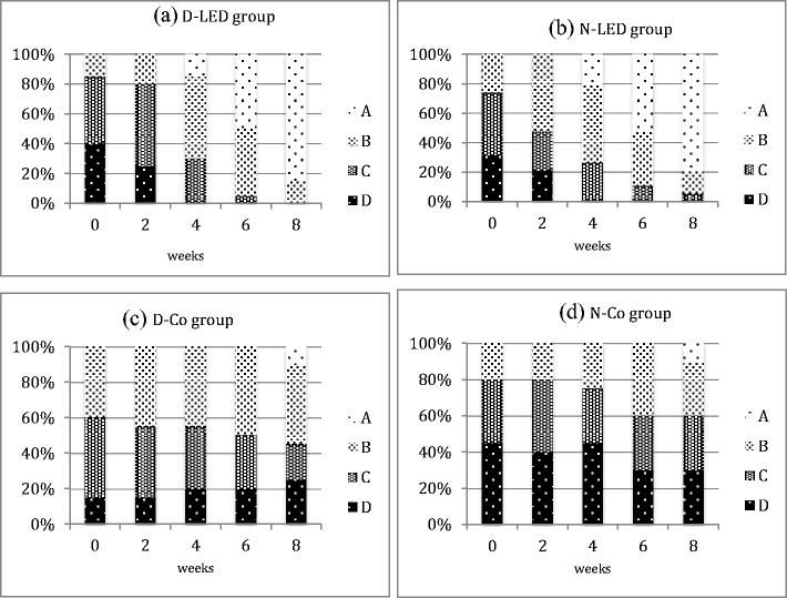
laser (40 mW, 40 sec, 1.6 J, 2.04 J/cm2, 51 mW/cm2, spot  size
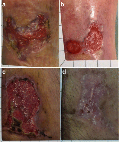
0.79 cm2,   in  non-contact  mode;   power   measured  at   the Technical  University of Cienfuegos)  in the  prodromal stage and  in the stage  of vesicles. In the crust  stage  and  in lesions

infected  secondarily, 40 mW, 2 min, 4.8 J was  used.  All these patients also  received   radiation over  the  vertebrae  C2–C3 (40 mW,  30 sec,  1.2 J), where   the  resident  ganglion of  the virus  is reported to be located  during the latent  periods.11

The control  group was offered indicated treatment with an- tivirals  (acyclovir cream  and  tablets)  and  other  palliative  ther- apies,  such  as an anesthetic cream  and  advice  to avoid  spicy and hot food. The patients were followed  up during 1 year by monthly visits to the clinic. Patients  not willing or able to come to the requested follow-ups were excluded from the study.


Pilot as well as recurrence studies were  approved by the Scientific Committee of the  Medical  University of Cienfue- gos,  and  a  written consent  was  given  by  all  participating patients. All therapies were  free of charge  for the patients.


Results


Pilot study


The outcome of the pilot study is illustrated in Tables 1–3. In  total,  there  were  84 occasions  of recurrence in the  laser group and  114 in the control  group. After day  7 no patients in the laser group had any visible signs of HSV-1 blisters, whereas in the control group 77 patients still had vesicles, 29 had  crust  formation, and  10 had  secondary infections.  This confirms  the curative  effect on the clinical manifestation, and a clear trend  toward reduction of the recurrence frequency.


Recurrence study


To follow  up  on  these  encouraging results,  a new  study was  initiated in  January  2004. Up  until  January  2006, 432 patients had  been  consulting at our  clinic for HSV-1 occur- rence. Out of this group, 322 patients were selected  to be followed  during 5 years in order  to register  the long-term incidence  of recurrence. The study included 189 men and 133 women, ages ranging from 18 to 59. Exclusion  criteria  were inability  to be reached during a 5-year follow-up and having perilabial blisters.  The  latter  exclusion   criterion   was  used to make  the  group more  homogenous. After  a detailed an- amnesis,   the  patients  were  treated daily   until   they  were clinically and subjectively  asymptomatic. Patients  in the prodromal stage  were  treated by 1.6 J per  session,  those  in the blister stage by 4.8 J per session; parameters as in the pilot study.  At all sessions,  the region  at the C2–C3 vertebrae was irradiated by 1.2 J, all in accordance with the experience  from the  pilot  study.  All patients were  checked  and  interviewed annually by receiving  home visits, all performed by the same dentist.  Recurrences  were also recorded if the patient  came to the clinic on his/her own  initiative  for recurrence laser treatment. None of the 438 patients receiving  LLLT in studies

1 and 2 reported any negative side effects, nor did any of the

110  patients  not  included  in  the  study  2.  Checking   the available  literature, no  information about  a possible  differ- ence  in  HSV-1 incidence  in  male  and  female  patients was found.  Clinically, the general  feeling is that females are more likely  to  have   labial  HSV-1  attacks.   This  may,  however, simply be because females in general are more keen to seek dentistry. We therefore  performed an  analysis  by  sex. Our data did not confirm the clinical impression. On the contrary, there  was  a tendency for more  male  relapses,  especially  in years  3 and  5.


Table 1. Laser Group Recurrence Frequency


Once  a month


Every 2–3 months


Every 4–5 months


Every 6 months


Once a year


First time ever


No recurrence


Before treatment                   9                        26                         58                       12                       7                       4                           0

After treatment                     0                          0                          37                       22                     25                       0                         32


Distribution according to the frequency of annual recurrence of the labial herpes,  before and  after receiving  treatment; n = 116.


LOW LEVEL LASER THERAPY OF HSV-1                                                                                                                                       39


Table 2. Recurrence Frequency for the Patients of the Control Group, Traditional Treatment


Once  a month


Every 2–3 months


Every 4–5 months


Every 6 months


Once a year


First time ever


No recurrence


Before treatment                   7                       24                         56                       14                      9                       6                          0

After treatment                     6                       21                         46                       27                    14                       0                          2


Distribution according to the annual recurrence frequency,  before and  after receiving  treatment; n = 116.


Results of the recurrence study


However, the  use  of LLLT to suppress infections  caused

12


Out of the 322 patients, the number of annual recurrences


by HSV 1 and 2 was evaluated by Ferreira et al.


after one to


varied   between 22 and  42, with  the  extreme  exception   of year 3. The increase  in recurrence at year 3 remains to be understood. The  number of relapses   was  as  follows,  with

‘m indicating males  and  ‘f indicating females:  Year 1, 35 (21 m/14 f ); year  2, 42 (23 m/19 f ); year  3, 149 (98 m/51 f ); year  4, 41 (20 m/21 f ); year  5, 22 (10 m/12 f ). Thirty-three patients (19 m/14 f ) did  not have one single recurrence after

5 years  of observation.


Discussion


These two studies  confirm  that  HSV-1 outbreaks can be re- lieved  and  shortened by using  LLLT, and  that  the number of relapses  decreases.  An analysis  of the available  literature veri- fies our positive  clinical observations. In the early literature an in vitro study by Gilioli et al.4 showed a stimulating effect on the virus itself. Tardivo5 observed the behavior of cells infected with HSV-1 under a GaAs  laser, 30 mW. Doses of 4 and  12 J were given. A lesser cytopathic effect was noted in the irradiated cells than  in the control  cells, more evident at 4 J. The two cell lines were incubated for 7 days and frozen, and the released  viruses were  inoculated in a new  culture.  An  absence  of cytopathic effect was noted  in the cells infected with viruses  derived from the cultures that received  12 J initially.

One  of the  first  clinical  reports on  the  use  of LLLT for

HSV-1 appeared in 1983, and  indicated that  647 nm, 50 mW,

3 J/cm2 had  a positive  effect on HSV-1, but  less on HSV-2.6

Ve´lez-Gonza´ lez et al.7   treated 60 patients with  herpes simplex  in the  oral  (HSV-1) or genital  (HSV-2) area.  Three groups in each category  received  (1) 200 mg acyclovir  orally

plus placebo  laser; (2) placebo  acyclovir and HeNe laser light at  8 J/cm2;  or (3) acyclovir  and  HeNe.  Relapses  in the  lips and face were significantly  reduced in the group treated with HeNe   laser  plus  acyclovir,   as  compared with  the  groups treated with acyclovir or HeNe  only. The number of relapses per year before and  after treatment was  5.2/2.8 for group I,

7.83/1.16 for group II, and 7.28/1.28 for group III. There was no significant  difference between the latter groups. However, healing  time  was  shorter  in the  group that  received  a com- bination of treatments. The effects on the HSV-2 groups were lower  for all three  treatment modalities.


five applications. A gradual reduction in replication of HSV 1

as well as 2 was observed, with  68.4% and  57.3% inhibition, respectively, after five applications.

An  important aspect  of  LLLT for  HSV-1  has  been  de- scribed  by Schindl  et al.8, namely,  the possibility  of treating patients with  recurrent herpes  labialis even during the symptom-free period.  Fifty patients with recurrent herpes labialis infections  (at least once a month for > 6 months) were treated with  690 nm,  80 mW  laser,  energy  density  48 J/cm2, in a double-blind study.  Patients  received  daily  irradiations for  2  weeks,  10  sessions  in  all.  The  treatment was  given during a recurrence-free period  and  the  irradiation was  gi- ven at the site of the original  herpes  labialis infection. If both lips  had  been  involved, both  upper and  lower  lips  were treated. Patients  were monitored for 52 weeks. The mean recurrence-free interval  in the laser group was 37.5 weeks (range,   2–52  weeks),   and   in  the  placebo   group  3  weeks

(range,  1–20 weeks).  No side effects were  noted.

LLLT has been used  as a single treatment modality and in combination  with   other   modalities.  Tzonko   et  al.13    used

630 nm, 100 mW/cm2 in a group of 32 patients, and the same treatment for  another group of 32 patients in  combination with the photosensibilizer Granofurin H. Both groups showed

positive  results,  but  the addition of Granofurin H provided the best outcome.  Sperandio et al.14, Marotti  et al.,15  and  de Carvalho  et  al.16    report   that   photoactivated  disinfection (PAD) can be a viable method of treating HSV-1. The blister is dyed with methylene blue, and irradiated with red laser light. Bello-Silva et al.17  used  Er:YAG to open and drain  large blis- ters, and then followed up with 660-nm LLLT. Almeida-Lopes et al.18  have shown that HSV-1 can be treated advantageously by irradiation of the involved lymph  nodes  only.

The  cellular   mechanism  behind   the   reported  result   is partly  unknown. The positive  effects of LLLT on pain19 and inflammation20  are  well  documented and  are  likely  a part of  the  mechanism. However,  the  prolonged  relapse   time cannot  be explained by these factors. From the studies of Eduardo,9 Donnarumma,10 and Almeida-Lopes18 it can be hypothesized that  the main  effect of the laser light is more  a stimulation of the  immune system  than  an inhibitory  effect on the virus.


Table 3. Recurrence Frequency Compared for the Patients of Both Groups


Once  a month


Every 2–3 months


Every 4–5 months


Every 6 months


Once a year


First time ever


No recurrence


Laser group                       0                          0                          37                        22                     25                        0                          32

Control  group                   6                        21                         46                        27                     14                        0                            2


Distribution according to the annual recurrence frequency after receiving  laser or traditional treatment.


40                                                                                                                                                                                    SANCHEZ ET AL.


In this study,  a wavelength of 670 nm in the red part of the spectrum was  used,  and  fairly  low  energies  were  applied. Other wavelengths and generally  higher  energies  are also reported in the  literature. Therefore,  it seems  that  the treat- ment  is  not  wavelength sensitive  or  very  sensitive  to  the amount of energy  applied.

Irradiation over the C2–C3 vertebrae has so far only been reported in an animal  study,11 and  our  study did  not make an effort to establish the specific effect of this additional ir- radiation. The low  energy  used  and  the  rather  weak  pene- tration  of the  670-nm wavelength are both  probably on the outskirts of the actual  therapeutic window.

Given   the   high   number  of  patients  included  in  both studies, and the high level of improvement, no statistical analysis  was  considered.

Taking the lack of side effects and  the prolongation of the relapse  time  into  consideration, LLLT appears to be an  at- tractive  alternative to presently used  treatment modalities.


Conclusions


LLLT appears to be a safe, inexpensive, and  effective treatment modality for the treatment of HSV-1, with  advan- tages over pharmaceutical methods. The main advantage over the latter is the prolongation of the recurrence period.  Further studies on the underlying mechanisms are warranted.


Author  Disclosure Statement


No  conflicting  financial  interests  exist.  The research  was self-funded by the authors and  their  clinic.


References


1. Santana,  J.C. (1985). Atlas of Pathology of the Buccal Complex.

Havana: Editorial  Scientific-Technique.

2. Eversole,  L.R. (1985). Buccal Pathology. Diagnosis and Treat- ment. Havana: Editorial  Scientific-Technique.

3. Harmenberg, J., Oberg, B., and Spruance, S. (2010). Prevention of ulcerative lesions by episodic  treatment of recurrent herpes labialis: A literature review. Acta Derm. Venereol. 90, 122130.

4. Gilioli,  G.,  Taparelli,   F.,  Fornaciari,  A.,  Palmieri,   B., and

Celani,  M. (1985). Studio  ultrastrutturale di colture  cellulari

‘vero infettate  con virus  Herpes  Simplex  e sottoposte all’- azione  Laser [In Italian].  . [Ultrastructural study of cell cul- tures infected with herpes  simplex virus and subjected  to the action  of laser]. Med.  Laser Rep. 3, 28–31.

5. Tardivo,  J.P. (1989). Effect of low power  laser  over  cells in-

fected  by  herpes  simplex  virus  (HSV). Lasers  Surg.  Med. Suppl  1, 31.

6. Landthaler, M., Haina,  D., and  Waidelich,  W. (1983). Be- handlung  von   Zoster,   postzosterischen Schmerzen und Herpes  simplex  recidivans in loco mit  Laser-Licht  [In Ger- man]. [Treatment of herpes  zoster, postherpetic pain, and herpes  simplex  recidivans in loco with  laser light]. Fortschr. Med. 101, 1039–1042.

7. Ve´lez–Gonsalez,  M.,  Urrea–Arbela´ ez,  A.,  Nicholas,   et  al.

(1995). Treatment of relapse  in herpes  simplex  on labial and facial areas  and  of primary herpes  simplex  on genital  areas and  area pudenda with  low power  HeNe-laser or Acyclovir administrated orally.  Effects of low-power light  on biologi- cal systems.  Proc. SPIE 2630, 43–50.

8. Schindl,  A.,  and   Neuman,  R.  (1999).  Low-intensity  laser therapy is an  effective  treatment for recurrent herpes  sim-


plex infection. Results from a randomized double-blind placebo-controlled study.  J. Invest.  Dermatol. 113, 221–223.

9. Eduardo, F de P., Mehnert,  D. U., Monezi,  A. M., Zezell, D.

M., Schubert,  M. M., Eduardo, C de P, and  Marques,  M.M. (2007). In  vitro  effect  of  phototherapy with  low  intensity laser  on  HSV-1  and  epithelial   cells.  Mechanisms for  low- light therapy II. Proc. SPIE. 6428, 642805.

10. Donnarumma, G., De Gregorio,  V., Fusco,  A., et al. (2010).

Inhibition of HSV-1 replication by laser diode-irradiation: possible  mechanism of action.  Int.  J. Immunopathol. Phar- macol. 23, 1167–1176.

11. Perrin, D., Jolivald, J. R., Triki, H., et al. (1997). Effect of laser

irradiation on  latency  of herpes  simplex  virus  in a mouse model.  Pathol.  Biol. (Paris). 45, 24–27.

12. Ferreira,  D de C., Martins,  F. O., and Romanos,  M. T. (2009).

Impacto  do  laser  de  baixa  intensidade na  supressa˜ o de  in- fecc¸ o˜ es pelos v´?rus  Herpes  simplex  1 e 2: estudo in vitro [In Portuguese]. [Impact  of low-intensity laser  on the  suppres- sion of infections  caused  by Herpes  simplex  viruses  1 and  2: in vitro  study].  Rev. Soc. Bras. Med.  Trop. 42, 82–85.

13. Tzonko,  T., Uzunov, T., Uzunov, R., and  Grozdanova, T.

(2004). Application of low-intensity laser in the treatment of Herpes  simplex  recidivans. Eighth  International Conference on  Laser  and  Laser  Information Technologies.   Proc.  SPIE

5449, 466–468.

14. Sperandio, F.F., Marotti,  J., Aranha, A.C., and  Eduardo, C. de P. (2009). Photodynamic therapy for the treatment of recurrent herpes  labialis: preliminary results.  Gen. Dent. 57,

415–419.

15. Marotti,  J., Sperandio, F.F., Fregnani, E.R., Aranha, A.C., de Freitas,  P.M., and  Eduardo, C de  P. (2010). High-intensity laser and photodynamic therapy as a treatment for recurrent herpes  labialis.  Photomed. Laser Surg. 28, 439–444.

16. de Carvalho, R.R., Eduardo, F de P., Ramalho,  K.M., et al. (2010). Effect of laser  phototherapy on recurring herpes  la- bialis prevention: an in vivo study.  Lasers Med. Sci. 25, 397–

402.

17. Bello–Silva, M.S., de Freitas,  P.M., Aranha, A.C., Lage– Marques,  J.L., Simo˜ es, A., and Eduardo, C de P. (2010) Low- and  high-intensity lasers in the treatment of herpes  simplex virus  1 infection.  Photomed. Laser Surg. 28, 135–139.

18. Almeida–Lopes, L., Lopes;  A,  Tune´r,  J., and  Calderhead, R.G. (2005) Infrared diode  laser therapy-induced lymphatic drainage for inflammation in the head  and neck. Laser Ther.

14, 67–74.

19. Fulop, A.M., Dhimmer, S., Deluca, J.R., et al. (2010). A meta- analysis  of the efficacy of laser phototherapy on pain  relief. Clin. J. Pain 26, 729–736.

20. Albertini,  R., Villaverde,  A.B., Aimbire,  F., et al. (2007). Anti-

inflammatory effects of low-level  laser therapy (LLLT) with two  different  red  wavelengths (660 nm  and  684 nm)  in car- rageenan-induced rat paw  edema.  J. Photochem. Photobiol. B. 89, 50–55.


Address  correspondence  to:

Jan Tune´r Swedish Laser Medical  Society Spjutva¨gen 11

772 32 Gra¨ngesberg

Sweden


E-mail: jan.tuner@swipnet.se


Original Source: https://www.slideshare.net/JanTun/the-use-of-low-level-laser-in-the-treatment-of-herpes-simplex

Photobiomodulation therapy as a high potential treatment modality for COVID-19

Sepideh Soheilifar,1 Homa Fathi,2 and Navid Naghdicorresponding author3 - Lasers Med Sci. 2020 Nov 25 : 1–4. (Publication) 4513
This recent study shows promise for treating damage to the lungs from Covid.
View Resource

Considering the pathophysiology of COVID-19 and potential positive effects of PBM in balancing the function of the immune system, this treatment modality could be effective in severe COVID-19 cases with ARDS. COVID-19 mortality is mainly because of cytokine storm in severe cases. PBM has the potential to decrease the level of pro-inflammatory cytokines and improve the balance of IL-10. These effects can balance immune response and decrease the impact of cytokine storm. PBM is mainly local and has very limited adverse side effects . Unlike corticosteroids, it does not cause delayed body response to virus elimination, secondary infection, or longer hospitalization period. This hypothesis is mainly based on theoretical data. The authors suggest that researchers should assess the potentials of this treatment method as it might save the lives of severely affected patients.


Intro: COVID-19 is now a worldwide concern, causing an unprecedented pandemic. The infected cases show different symptoms based on the severity of the disease. In asymptomatic and non-severe symptomatic cases, the host immune system can successfully eliminate the virus and its effects. In severe cases, however, immune system impairment causes cytokine release syndrome which eventually leads to acute respiratory distress syndrome (ARDS). In recent years, photobiomodulation (PBM) has shown promising results in reducing acute pulmonary inflammation. Considering the high potential impact of PBM on immune responses, we hypothesized that using PBM could be an effective treatment modality for ARDS management in COVID-19 patients.

Background: In the final days of 2019, China reported the emergence of an unknown pathogen causing pneumonia-like symptoms in the infected cases in Wuhan, Hubei. On January 7, 2020, Chinese Center for Disease Control and Prevention detected the origin as a novel virus from the Coronaviridae family. World Health Organization (WHO) soon confirmed that human to human transmission of the virus has led to a worldwide “pandemic” [1]. The virus was named severe acute respiratory syndrome coronavirus 2 (SARS-CoV-2) causing Coronavirus disease 2019 (COVID-19). Coronaviridae is a large family of enveloped, positive-sense, single-stranded RNA virus [2]. Based on the genome structure and phylogenetic relationships, this family is further categorized into four groups; Alphacoronavirus, Betacoronavirus, Gammacoronavirus, and Deltacoronavirus. Alpha and Betacoronaviruses are specific to mammals and cause respiratory diseases in humans, namely Severe Acute Respiratory Syndrome coronavirus (SARS-CoV) and the Middle East Respiratory Syndrome coronavirus (MERS-CoV). Delta and Gammacoronaviruses cause infection in both mammals and birds [3]. Genetic analysis revealed that SARS-CoV-2 is most probably in the Betacoronavirus category [4]. The infected cases show different symptoms based on the severity of the disease. In asymptomatic and non-severe symptomatic cases, the host immune system can successfully eliminate the virus and its effects. In severe cases, however, immune system impairment causes cytokine release syndrome which eventually leads to acute respiratory distress syndrome (ARDS) [5]. One of the treatment strategies is to eliminate inflammatory response in the host. Although some medications such as immunosuppressants have this effect, they cause delayed body response to virus elimination [6]. To address this issue, alternative treatment modalities for inflammation elimination are urgently needed. One such potential treatment is photobiomodulation (PBM), also known as low-level laser therapy (LLLT). PBM is an alternative modality for local management of increased inflammation, which has been used from 50 years ago [7]. It is defined as a low power laser or light-emitting diode (LED) in the range of 1–500 mW utilized to promote tissue regeneration and decrease inflammation and pain. A narrow spectral width light in red or near infra-red range (600–1000 nm) with 1–5000 mW/cm2 power density is used in PBM [8]. PBM can change cellular and molecular metabolism, signaling, inflammation, and chemical messenger release. It has shown promising results in reducing acute pulmonary inflammation, as they have a high potential for the local balance of immune responses [9]. Therefore, the objective of this paper is to hypothesize that using PBM could be an effective treatment modality for ARDS management in COVID-19 patients.

Original Source: https://www.ncbi.nlm.nih.gov/pmc/articles/PMC7688201/

Therapeutic outcomes of low-level laser therapy for closed bone fracture in the human wrist and hand.

Chang WD1, Wu JH, Wang HJ, Jiang JA. - Photomed Laser Surg. 2014 Apr;32(4):212-8. doi: 10.1089/pho.2012.3398. Epub 2014 Mar 20. (Publication) 493
View Resource

This test is based on a Microlight ML830 with 90mW of total power. The dosage are very low by contemporary standards.


Intro: The therapeutic outcomes of low-level laser therapy (LLLT) on closed bone fractures (CBFs) in the wrist and hand were investigated in this controlled study.

Background: The therapeutic outcomes of low-level laser therapy (LLLT) on closed bone fractures (CBFs) in the wrist and hand were investigated in this controlled study.

Abstract: Abstract OBJECTIVE: The therapeutic outcomes of low-level laser therapy (LLLT) on closed bone fractures (CBFs) in the wrist and hand were investigated in this controlled study. BACKGROUND DATA: Animal research has confirmed that LLLT increases osteocyte quantity; however, little research has been conducted to determine the effect of LLLT on the treatment of human bone fractures. METHODS: In this study, the therapeutic outcomes of administering 830 nm LLLT to treat CBFs in the wrist or hand were examined. Fifty patients with CBFs in the wrist and hand, who had not received surgical treatment, were recruited and randomly assigned to two groups. The laser group underwent a treatment program in which 830 nm LLLT (average power 60 mW, peak power 8 W, 10 Hz, 600 sec, and 9.7 J/cm(2) per fracture site) was administered five times per week for 2 weeks. Participants in a placebo group received sham laser treatment. The pain, functional disability, grip strength, and radiographic parameters of the participants were evaluated before and after treatment and at a 2-week follow-up. RESULTS: After treatment and at the follow-up, the laser group exhibited significant changes in all of the parameters compared with the baseline (p<0.05). The results of comparing the two groups after treatment and at the follow-up indicated significant between-group differences among all of the parameters (p<0.05). CONCLUSIONS: LLLT can relieve pain and improve the healing process of CBFs in the human wrist and hand.

Methods: Animal research has confirmed that LLLT increases osteocyte quantity; however, little research has been conducted to determine the effect of LLLT on the treatment of human bone fractures.

Results: In this study, the therapeutic outcomes of administering 830 nm LLLT to treat CBFs in the wrist or hand were examined. Fifty patients with CBFs in the wrist and hand, who had not received surgical treatment, were recruited and randomly assigned to two groups. The laser group underwent a treatment program in which 830 nm LLLT (average power 60 mW, peak power 8 W, 10 Hz, 600 sec, and 9.7 J/cm(2) per fracture site) was administered five times per week for 2 weeks. Participants in a placebo group received sham laser treatment. The pain, functional disability, grip strength, and radiographic parameters of the participants were evaluated before and after treatment and at a 2-week follow-up.

Conclusions: After treatment and at the follow-up, the laser group exhibited significant changes in all of the parameters compared with the baseline (p<0.05). The results of comparing the two groups after treatment and at the follow-up indicated significant between-group differences among all of the parameters (p<0.05).

Original Source: http://www.ncbi.nlm.nih.gov/pubmed/24649935

Wounded tiger gets THOR LLLT / Low Level Laser Therapy treatment

- 2012 (Video) 4353
2 videos of a tiger receiving LLLT, one video shows them giving LLLT to the tiger's eye, do NOT point any lights or lasers into your eye, it was also made by thor, so it could be biased
View Resource

Two videos of a tiger recieving LLLT on a wound and on it's eye, tiger does't seem to be in pain, but the videos simply don't show enough to make any real claims.

video length: (0:40) 


Tiger's eye gets THOR LLLT / Low Level Laser Therapy treatment

video length: (0:39)


Original Source: https://www.youtube.com/watch?v=FB5AykoLngo

Effect of 830-nm diode laser irradiation on human sperm motility.

Salman Yazdi R1, Bakhshi S, Jannat Alipoor F, Akhoond MR, Borhani S, Farrahi F, Lotfi Panah M, Sadighi Gilani MA. - Lasers Med Sci. 2014 Jan;29(1):97-104. doi: 10.1007/s10103-013-1276-7. Epub 2013 Feb 14. (Publication) 936
View Resource

This study was done with a Microlight ML830 with a total power of 90mW.


Intro: Sperm motility is known as an effective parameter in male fertility, and it depends on energy consumption. Low-level laser irradiation could increase energy supply to the cell by producing adenosine triphosphate. The purpose of this study is to evaluate how the low-level laser irradiation affects the human sperm motility. Fresh human semen specimens of asthenospermic patients were divided into four equal portions and irradiated by 830-nm GaAlAs laser irradiation with varying doses as: 0 (control), 4, 6 and 10 J/cm(2). At the times of 0, 30, 45 and 60 min following irradiation, sperm motilities are assessed by means of computer-aided sperm analysis in all samples. Two additional tests [HOS and sperm chromatin dispersion (SCD) tests] were also performed on the control and high irradiated groups as well. Sperm motility of the control groups significantly decreased after 30, 45 and 60 min of irradiation, while those of irradiated groups remained constant or slightly increased by passing of time. Significant increases have been observed in doses of 4 and 6 J/cm(2) at the times of 60 and 45 min, respectively. SCD test also revealed a non-significant difference. Our results showed that irradiating human sperms with low-level 830-nm diode laser can improve their progressive motility depending on both laser density and post-exposure time.

Background: Sperm motility is known as an effective parameter in male fertility, and it depends on energy consumption. Low-level laser irradiation could increase energy supply to the cell by producing adenosine triphosphate. The purpose of this study is to evaluate how the low-level laser irradiation affects the human sperm motility. Fresh human semen specimens of asthenospermic patients were divided into four equal portions and irradiated by 830-nm GaAlAs laser irradiation with varying doses as: 0 (control), 4, 6 and 10 J/cm(2). At the times of 0, 30, 45 and 60 min following irradiation, sperm motilities are assessed by means of computer-aided sperm analysis in all samples. Two additional tests [HOS and sperm chromatin dispersion (SCD) tests] were also performed on the control and high irradiated groups as well. Sperm motility of the control groups significantly decreased after 30, 45 and 60 min of irradiation, while those of irradiated groups remained constant or slightly increased by passing of time. Significant increases have been observed in doses of 4 and 6 J/cm(2) at the times of 60 and 45 min, respectively. SCD test also revealed a non-significant difference. Our results showed that irradiating human sperms with low-level 830-nm diode laser can improve their progressive motility depending on both laser density and post-exposure time.

Abstract: Abstract Sperm motility is known as an effective parameter in male fertility, and it depends on energy consumption. Low-level laser irradiation could increase energy supply to the cell by producing adenosine triphosphate. The purpose of this study is to evaluate how the low-level laser irradiation affects the human sperm motility. Fresh human semen specimens of asthenospermic patients were divided into four equal portions and irradiated by 830-nm GaAlAs laser irradiation with varying doses as: 0 (control), 4, 6 and 10 J/cm(2). At the times of 0, 30, 45 and 60 min following irradiation, sperm motilities are assessed by means of computer-aided sperm analysis in all samples. Two additional tests [HOS and sperm chromatin dispersion (SCD) tests] were also performed on the control and high irradiated groups as well. Sperm motility of the control groups significantly decreased after 30, 45 and 60 min of irradiation, while those of irradiated groups remained constant or slightly increased by passing of time. Significant increases have been observed in doses of 4 and 6 J/cm(2) at the times of 60 and 45 min, respectively. SCD test also revealed a non-significant difference. Our results showed that irradiating human sperms with low-level 830-nm diode laser can improve their progressive motility depending on both laser density and post-exposure time.

Original Source: http://www.ncbi.nlm.nih.gov/pubmed/23407899

Low Level Laser Therapy For Treating Back Pain

- 2015 (Video) 4392
This is an Australian news report on LLLT.
View Resource

LLLT also known as cold laser was featured on Channel 7 News with Dr Roberta Chow using low level laser to treat back pain

video length: (2:06) 


Original Source: https://www.youtube.com/watch?v=GPk5HxzF_kY&nohtml5=False

Non-invasive subcutaneous fat reduction: a review.

Kennedy J1, Verne S1, Griffith R1, Falto-Aizpurua L1, Nouri K1. - J Eur Acad Dermatol Venereol. 2015 Sep;29(9):1679-88. doi: 10.1111/jdv.12994. Epub 2015 Feb 9. (Publication) 179
View Resource
Intro: The risks, financial costs and lengthy downtime associated with surgical procedures for fat reduction have led to the development of a number of non-invasive techniques. Non-invasive body contouring now represents the fastest growing area of aesthetic medicine. There are currently four leading non-invasive techniques for reducing localized subcutaneous adipose tissue: low-level laser therapy (LLLT), cryolipolysis, radio frequency (RF) and high-intensity focused ultrasound (HIFU). To review and compare leading techniques and clinical outcomes of non-invasive subcutaneous fat reduction. The terms 'non-invasive', 'low-level laser', 'cryolipolysis', 'ultrasound' and 'radio frequency' were combined with 'lipolysis', 'fat reduction' or 'body contour' during separate searches in the PubMed database. We identified 31 studies (27 prospective clinical studies and four retrospective chart reviews) with a total of 2937 patients that had been treated with LLLT (n = 1114), cryolipolysis (n = 706), HIFU (n = 843) or RF (n = 116) or other techniques (n = 158) for fat reduction or body contouring. A majority of these patients experienced significant and satisfying results without any serious adverse effects. The studies investigating these devices have all varied in treatment regimen, body locations, follow-up times or outcome operationalization. Each technique differs in offered advantages and severity of adverse effects. However, multiple non-invasive devices are safe and effective for circumferential reduction in local fat tissue by 2 cm or more across the abdomen, hips and thighs. Results are consistent and reproducible for each device and none are associated with any serious or permanent adverse effects.

Background: The risks, financial costs and lengthy downtime associated with surgical procedures for fat reduction have led to the development of a number of non-invasive techniques. Non-invasive body contouring now represents the fastest growing area of aesthetic medicine. There are currently four leading non-invasive techniques for reducing localized subcutaneous adipose tissue: low-level laser therapy (LLLT), cryolipolysis, radio frequency (RF) and high-intensity focused ultrasound (HIFU). To review and compare leading techniques and clinical outcomes of non-invasive subcutaneous fat reduction. The terms 'non-invasive', 'low-level laser', 'cryolipolysis', 'ultrasound' and 'radio frequency' were combined with 'lipolysis', 'fat reduction' or 'body contour' during separate searches in the PubMed database. We identified 31 studies (27 prospective clinical studies and four retrospective chart reviews) with a total of 2937 patients that had been treated with LLLT (n = 1114), cryolipolysis (n = 706), HIFU (n = 843) or RF (n = 116) or other techniques (n = 158) for fat reduction or body contouring. A majority of these patients experienced significant and satisfying results without any serious adverse effects. The studies investigating these devices have all varied in treatment regimen, body locations, follow-up times or outcome operationalization. Each technique differs in offered advantages and severity of adverse effects. However, multiple non-invasive devices are safe and effective for circumferential reduction in local fat tissue by 2 cm or more across the abdomen, hips and thighs. Results are consistent and reproducible for each device and none are associated with any serious or permanent adverse effects.

Abstract: Abstract The risks, financial costs and lengthy downtime associated with surgical procedures for fat reduction have led to the development of a number of non-invasive techniques. Non-invasive body contouring now represents the fastest growing area of aesthetic medicine. There are currently four leading non-invasive techniques for reducing localized subcutaneous adipose tissue: low-level laser therapy (LLLT), cryolipolysis, radio frequency (RF) and high-intensity focused ultrasound (HIFU). To review and compare leading techniques and clinical outcomes of non-invasive subcutaneous fat reduction. The terms 'non-invasive', 'low-level laser', 'cryolipolysis', 'ultrasound' and 'radio frequency' were combined with 'lipolysis', 'fat reduction' or 'body contour' during separate searches in the PubMed database. We identified 31 studies (27 prospective clinical studies and four retrospective chart reviews) with a total of 2937 patients that had been treated with LLLT (n = 1114), cryolipolysis (n = 706), HIFU (n = 843) or RF (n = 116) or other techniques (n = 158) for fat reduction or body contouring. A majority of these patients experienced significant and satisfying results without any serious adverse effects. The studies investigating these devices have all varied in treatment regimen, body locations, follow-up times or outcome operationalization. Each technique differs in offered advantages and severity of adverse effects. However, multiple non-invasive devices are safe and effective for circumferential reduction in local fat tissue by 2 cm or more across the abdomen, hips and thighs. Results are consistent and reproducible for each device and none are associated with any serious or permanent adverse effects. © 2015 European Academy of Dermatology and Venereology.

Methods: © 2015 European Academy of Dermatology and Venereology.

Original Source: http://www.ncbi.nlm.nih.gov/pubmed/25664493

Are interventions for accelerating orthodontic tooth movement effective?

Abdallah MN1, Flores-Mir C2. - Evid Based Dent. 2014 Dec;15(4):116-7. doi: 10.1038/sj.ebd.6401064. (Publication) 228
LLLT was less effective than other modalities at accelerating tooth movement.
View Resource
Intro: Pubmed, Embase, Sciences Citation Index, Cochrane Central Register of Controlled Trials (CENTRAL) and grey literature database of SIGLE were searched from January 1, 1990 to August 20, 2011 with no language restrictions.

Background: Pubmed, Embase, Sciences Citation Index, Cochrane Central Register of Controlled Trials (CENTRAL) and grey literature database of SIGLE were searched from January 1, 1990 to August 20, 2011 with no language restrictions.

Abstract: Abstract DATA SOURCES: Pubmed, Embase, Sciences Citation Index, Cochrane Central Register of Controlled Trials (CENTRAL) and grey literature database of SIGLE were searched from January 1, 1990 to August 20, 2011 with no language restrictions. STUDY SELECTION: Randomised controlled trials (RCTs) or quasi-RCTs in which the participants were healthy and received additional interventions to conventional orthodontic treatment for accelerating tooth movements were included. Subjects with defects in oral and maxillofacial regions (ie, cleft lip/palate), dental pathologies and medical conditions were excluded. DATA EXTRACTION AND SYNTHESIS: Studies were selected by two independent reviewers and disagreements were resolved by discussion with a third reviewer. The primary outcomes included accumulative moved distance (AMD) or movement rate (MR) and time required to move the tooth to its destination. Secondary outcomes were pain improvement, anchorage loss, periodontal health, orthodontic caries, pulp vitality and root resorption. The reviewers performed statistical pooling, where possible, according to a priori criteria on the basis of comparability of patient type, treatments and outcomes measured and risk of bias. The reviewers tested for heterogeneity, publication bias and sensitivity. A quality assessment test was conducted to evaluate the method used to measure AMD. RESULTS: The authors selected seven RCTs and two quasi-RCTs, which included a total of 101 patients with an age range of 12-26.3 years. Eight studies compared four intervention methods to no intervention group (control group). From them, four studies assessed low laser therapy (LLL), two evaluated corticotomy (CC), one assessed electrical current therapy (EC) and one evaluated pulsed electromagnetic field (PEF). Another study compared dentoalveolar distraction (DAD) vs periodontal distraction (PDD).Quality assessment scores showed that only two studies were of high quality, five studies were of medium quality, while two studies were of low quality. All studies, except one, compared left and right sides of the same participant (split mouth design), and measured the AMD.The method for measuring AMD was reliable in three studies, relatively reliable in one study and unreliable in four studies. The authors only performed pooled AMD mean meta-analysis for the LLL studies. The meta-analysis showed pooled mean AMD of 0.32 (95% confidence interval (CI), 20.04, 0.68), 0.76 (95% CI, 20.14, 1.65), and 0.73 (95% CI, 20.68, 2.14) for one month, two months and three months, respectively. Two LLL studies showed no differences regarding periodontal health and two LLL studies showed no differences in root resorption between LLL intervention and control groups. Compared to control group, one study reported that CS had significantly higher MR and another study showed that CS exhibited larger AMD for one month, two months, three months and four months. Two studies revealed that CS did not show any difference in the periodontal health status. One study reported the EC showed significantly larger AMD for one month, whereas another study reported that PEF induced larger AMD for five + 0.6 months. DAD showed faster MR and less anchorage loss compared to PDD. Teeth remained vital in both DAD and PDD interventions and one out of six cases presented root resorption in the PDD group. CONCLUSIONS: Among the five interventions corticotomy is effective and safe to accelerate orthodontic tooth movement, low-level laser therapy was unable to accelerate orthodontic tooth movement. The level of evidence does not support whether electrical current and pulsed electromagnetic fields are effective in accelerating orthodontic tooth movement and dentoalveolar or periodontal distraction is promising in accelerating orthodontic tooth movement.

Methods: Randomised controlled trials (RCTs) or quasi-RCTs in which the participants were healthy and received additional interventions to conventional orthodontic treatment for accelerating tooth movements were included. Subjects with defects in oral and maxillofacial regions (ie, cleft lip/palate), dental pathologies and medical conditions were excluded.

Results: Studies were selected by two independent reviewers and disagreements were resolved by discussion with a third reviewer. The primary outcomes included accumulative moved distance (AMD) or movement rate (MR) and time required to move the tooth to its destination. Secondary outcomes were pain improvement, anchorage loss, periodontal health, orthodontic caries, pulp vitality and root resorption. The reviewers performed statistical pooling, where possible, according to a priori criteria on the basis of comparability of patient type, treatments and outcomes measured and risk of bias. The reviewers tested for heterogeneity, publication bias and sensitivity. A quality assessment test was conducted to evaluate the method used to measure AMD.

Conclusions: The authors selected seven RCTs and two quasi-RCTs, which included a total of 101 patients with an age range of 12-26.3 years. Eight studies compared four intervention methods to no intervention group (control group). From them, four studies assessed low laser therapy (LLL), two evaluated corticotomy (CC), one assessed electrical current therapy (EC) and one evaluated pulsed electromagnetic field (PEF). Another study compared dentoalveolar distraction (DAD) vs periodontal distraction (PDD).Quality assessment scores showed that only two studies were of high quality, five studies were of medium quality, while two studies were of low quality. All studies, except one, compared left and right sides of the same participant (split mouth design), and measured the AMD.The method for measuring AMD was reliable in three studies, relatively reliable in one study and unreliable in four studies. The authors only performed pooled AMD mean meta-analysis for the LLL studies. The meta-analysis showed pooled mean AMD of 0.32 (95% confidence interval (CI), 20.04, 0.68), 0.76 (95% CI, 20.14, 1.65), and 0.73 (95% CI, 20.68, 2.14) for one month, two months and three months, respectively. Two LLL studies showed no differences regarding periodontal health and two LLL studies showed no differences in root resorption between LLL intervention and control groups. Compared to control group, one study reported that CS had significantly higher MR and another study showed that CS exhibited larger AMD for one month, two months, three months and four months. Two studies revealed that CS did not show any difference in the periodontal health status. One study reported the EC showed significantly larger AMD for one month, whereas another study reported that PEF induced larger AMD for five + 0.6 months. DAD showed faster MR and less anchorage loss compared to PDD. Teeth remained vital in both DAD and PDD interventions and one out of six cases presented root resorption in the PDD group.

Original Source: http://www.ncbi.nlm.nih.gov/pubmed/25522945

Effect of low level laser therapy on dental pulp during orthodontic movement.

Domínguez A1, Ballesteros RE1, Viáfara JH1, Tamayo OM1. - World J Methodol. 2013 Jun 26;3(2):19-26. doi: 10.5662/wjm.v3.i2.19. eCollection 2013. (Publication) 793
Study based on 100mW at 830nm. Total dosage was 2.2 joules so only a fraction of the typical recommended dosage. Showed no improvement.
View Resource
Intro: To validate the protocol described here to be used in future clinical trials related to the effect of laser therapy on dental pulp.

Background: To validate the protocol described here to be used in future clinical trials related to the effect of laser therapy on dental pulp.

Abstract: Abstract AIM: To validate the protocol described here to be used in future clinical trials related to the effect of laser therapy on dental pulp. METHODS: Histologically treated samples from eight human healthy premolar teeth obtained from the middle root level were distributed in four groups: group 1 (G1) absolute control; group 2 (G2) only laser irradiation; group 3 (G3) exposed only to orthodontics; and group 4 (G4) treated with orthodontics and laser. Laser treatment was performed at 830 nm wavelength, 100 mW (energy 80 J/cm(2), 2.2 J), for 22 s in the vestibular surface and 22 s in the palatal surface, 1 mm away from the dental root mucosa. Three staining methods were performed: hematoxylin-eosin (HE), Masson's Trichrome method and Gomori's method. RESULTS: The pulp histology parameters were evaluated and the results classified in to 3 parts: an inflammatory response, soft tissue response (dental pulp) and hard tissue response (dentin and predentin). There was no inflammation (chronic or acute) in any of the evaluated groups. The zones of pulp necrosis were found in one premolar of G3 and in one of G4; in groups G2 and G4 there was higher angiogenesis than in the other two groups. G4 group presented the highest level of vascularization. A reduced nerve density was observed in G3. A G2 specimen showed increased nerve density. A higher rate of calcification was observed in G1 compared to G2. Denticles, either real or false, were observed in G1, G2 and G3. Sclerosis of dentin and focal dentin loss was observed among all the groups. Secondary dentin was present in one sample in G1 and G2. A necrosis zone was found in one sample of G3 and G4. No differences between groups were observed in the odontoblast irregularity layer but the layer was wider in the group treated with laser only. A notable difference was detected in reduction of the cell-free layer between the groups G1 and G4. The findings in pulp tissue favor its adaptative response against dental movement induced by orthodontics. No definitive conclusions may be derived as this is a pilot study. CONCLUSION: The protocol described here was shown to be an effective method to evaluate changes in dental pulp submitted to low level laser in teeth under orthodontic movement.

Methods: Histologically treated samples from eight human healthy premolar teeth obtained from the middle root level were distributed in four groups: group 1 (G1) absolute control; group 2 (G2) only laser irradiation; group 3 (G3) exposed only to orthodontics; and group 4 (G4) treated with orthodontics and laser. Laser treatment was performed at 830 nm wavelength, 100 mW (energy 80 J/cm(2), 2.2 J), for 22 s in the vestibular surface and 22 s in the palatal surface, 1 mm away from the dental root mucosa. Three staining methods were performed: hematoxylin-eosin (HE), Masson's Trichrome method and Gomori's method.

Results: The pulp histology parameters were evaluated and the results classified in to 3 parts: an inflammatory response, soft tissue response (dental pulp) and hard tissue response (dentin and predentin). There was no inflammation (chronic or acute) in any of the evaluated groups. The zones of pulp necrosis were found in one premolar of G3 and in one of G4; in groups G2 and G4 there was higher angiogenesis than in the other two groups. G4 group presented the highest level of vascularization. A reduced nerve density was observed in G3. A G2 specimen showed increased nerve density. A higher rate of calcification was observed in G1 compared to G2. Denticles, either real or false, were observed in G1, G2 and G3. Sclerosis of dentin and focal dentin loss was observed among all the groups. Secondary dentin was present in one sample in G1 and G2. A necrosis zone was found in one sample of G3 and G4. No differences between groups were observed in the odontoblast irregularity layer but the layer was wider in the group treated with laser only. A notable difference was detected in reduction of the cell-free layer between the groups G1 and G4. The findings in pulp tissue favor its adaptative response against dental movement induced by orthodontics. No definitive conclusions may be derived as this is a pilot study.

Conclusions: The protocol described here was shown to be an effective method to evaluate changes in dental pulp submitted to low level laser in teeth under orthodontic movement.

Original Source: http://www.ncbi.nlm.nih.gov/pubmed/25237620

Efficacy of low-level laser therapy on scar tissue.

Freitas CP1, Melo C, Alexandrino AM, Noites A. - J Cosmet Laser Ther. 2013 Jun;15(3):171-6. doi: 10.3109/14764172.2013.769272. (Publication) 858
View Resource
Intro: Physiotherapy has a very important role in the maintenance of the integumentary system integrity. There is very few evidence in humans. Nevertheless, there are some studies about tissue regeneration using low-level laser therapy (LLLT).

Background: Physiotherapy has a very important role in the maintenance of the integumentary system integrity. There is very few evidence in humans. Nevertheless, there are some studies about tissue regeneration using low-level laser therapy (LLLT).

Abstract: Abstract BACKGROUND: Physiotherapy has a very important role in the maintenance of the integumentary system integrity. There is very few evidence in humans. Nevertheless, there are some studies about tissue regeneration using low-level laser therapy (LLLT). AIM: To analyze the effectiveness of LLLT on scar tissue. METHODS: Seventeen volunteers were stratified by age of their scars, and then randomly assigned to an experimental group (EG) - n = 9 - and a placebo group (PG) - n = 8. Fifteen sessions were conducted to both the groups thrice a week. However, in the PG, the laser device was switched off. Scars' thickness, length, width, macroscopic aspect, pain threshold, pain perception, and itching were measured. RESULTS: After 5 weeks, there were no statistically significant differences in any variable between both the groups. However, analyzing independently each group, EG showed a significant improvement in macroscopic aspect (p = 0.003) using LLLT. Taking into account the scars' age, LLLT showed a tendency to decrease older scars' thickness in EG. CONCLUSION: The intervention with LLLT appears to have a positive effect on the macroscopic scars' appearance, and on old scars' thickness, in the studied sample. However, it cannot be said for sure that LLLT has influence on scar tissue.

Methods: To analyze the effectiveness of LLLT on scar tissue.

Results: Seventeen volunteers were stratified by age of their scars, and then randomly assigned to an experimental group (EG) - n = 9 - and a placebo group (PG) - n = 8. Fifteen sessions were conducted to both the groups thrice a week. However, in the PG, the laser device was switched off. Scars' thickness, length, width, macroscopic aspect, pain threshold, pain perception, and itching were measured.

Conclusions: After 5 weeks, there were no statistically significant differences in any variable between both the groups. However, analyzing independently each group, EG showed a significant improvement in macroscopic aspect (p = 0.003) using LLLT. Taking into account the scars' age, LLLT showed a tendency to decrease older scars' thickness in EG.

Original Source: http://www.ncbi.nlm.nih.gov/pubmed/23607736

Light therapy and supplementary Riboflavin in the SOD1 transgenic mouse model of familial amyotrophic lateral sclerosis (FALS).

Moges H1, Vasconcelos OM, Campbell WW, Borke RC, McCoy JA, Kaczmarczyk L, Feng J, Anders JJ. - Lasers Surg Med. 2009 Jan;41(1):52-9. doi: 10.1002/lsm.20732. (Publication) 2623
810 nm diode laser, 140-mW output power, 1.4 cm(2) spot area, 120 seconds - total dosage 16 joules energy density. Low dosage showed no improvement.
View Resource
Intro: Familial amyotrophic lateral sclerosis (FALS) is a neurodegenerative disease characterized by progressive loss of motor neurons and death. Mitochondrial dysfunction and oxidative stress play an important role in motor neuron loss in ALS. Light therapy (LT) has biomodulatory effects on mitochondria. Riboflavin improves energy efficiency in mitochondria and reduces oxidative injury. The purpose of this study was to examine the synergistic effect of LT and riboflavin on the survival of motor neurons in a mouse model of FALS.

Background: Familial amyotrophic lateral sclerosis (FALS) is a neurodegenerative disease characterized by progressive loss of motor neurons and death. Mitochondrial dysfunction and oxidative stress play an important role in motor neuron loss in ALS. Light therapy (LT) has biomodulatory effects on mitochondria. Riboflavin improves energy efficiency in mitochondria and reduces oxidative injury. The purpose of this study was to examine the synergistic effect of LT and riboflavin on the survival of motor neurons in a mouse model of FALS.

Abstract: Abstract BACKGROUND AND OBJECTIVE: Familial amyotrophic lateral sclerosis (FALS) is a neurodegenerative disease characterized by progressive loss of motor neurons and death. Mitochondrial dysfunction and oxidative stress play an important role in motor neuron loss in ALS. Light therapy (LT) has biomodulatory effects on mitochondria. Riboflavin improves energy efficiency in mitochondria and reduces oxidative injury. The purpose of this study was to examine the synergistic effect of LT and riboflavin on the survival of motor neurons in a mouse model of FALS. STUDY DESIGN/MATERIALS AND METHODS: G93A SOD1 transgenic mice were divided into four groups: Control, Riboflavin, Light, and Riboflavin+Light (combination). Mice were treated from 51 days of age until death. A single set of LT parameters was used: 810 nm diode laser, 140-mW output power, 1.4 cm(2) spot area, 120 seconds treatment duration, and 12 J/cm(2) energy density. Behavioral tests and weight monitoring were done weekly. At end stage of the disease, mice were euthanized, survival data was collected and immunohistochemistry and motor neuron counts were performed. RESULTS: There was no difference in survival between groups. Motor function was not significantly improved with the exception of the rotarod test which showed significant improvement in the Light group in the early stage of the disease. Immunohistochemical expression of the astrocyte marker, glial fibrilary acidic protein, was significantly reduced in the cervical and lumbar enlargements of the spinal cord as a result of LT. There was no difference in the number of motor neurons in the anterior horn of the lumbar enlargement between groups. CONCLUSIONS: The lack of significant improvement in survival and motor performance indicates study interventions were ineffective in altering disease progression in the G93A SOD1 mice. Our findings have potential implications for the conceptual use of light to treat other neurodegenerative diseases that have been linked to mitochondrial dysfunction.

Methods: G93A SOD1 transgenic mice were divided into four groups: Control, Riboflavin, Light, and Riboflavin+Light (combination). Mice were treated from 51 days of age until death. A single set of LT parameters was used: 810 nm diode laser, 140-mW output power, 1.4 cm(2) spot area, 120 seconds treatment duration, and 12 J/cm(2) energy density. Behavioral tests and weight monitoring were done weekly. At end stage of the disease, mice were euthanized, survival data was collected and immunohistochemistry and motor neuron counts were performed.

Results: There was no difference in survival between groups. Motor function was not significantly improved with the exception of the rotarod test which showed significant improvement in the Light group in the early stage of the disease. Immunohistochemical expression of the astrocyte marker, glial fibrilary acidic protein, was significantly reduced in the cervical and lumbar enlargements of the spinal cord as a result of LT. There was no difference in the number of motor neurons in the anterior horn of the lumbar enlargement between groups.

Conclusions: The lack of significant improvement in survival and motor performance indicates study interventions were ineffective in altering disease progression in the G93A SOD1 mice. Our findings have potential implications for the conceptual use of light to treat other neurodegenerative diseases that have been linked to mitochondrial dysfunction.

Original Source: http://www.ncbi.nlm.nih.gov/pubmed/19143012

Design and testing of low intensity laser biostimulator.

Valchinov ES1, Pallikarakis NE. - Biomed Eng Online. 2005 Jan 13;4:5. (Publication) 3675
View Resource
Intro: The non-invasive nature of laser biostimulation has made lasers an attractive alternative in Medical Acupuncture at the last 25 years. However, there is still an uncertainty as to whether they work or their effect is just placebo. Although a plethora of scientific papers published about the topic showing positive clinical results, there is still a lack of objective scientific proofs about the biostimulation effect of lasers in Medical Acupuncture. The objective of this work was to design and build a low cost portable laser device for stimulation of acupuncture points, considered here as small localized biosources (SLB), without stimulating any sensory nerves via shock or heat and to find out a suitable method for objectively evaluating its stimulating effect. The design is aimed for studying SLB potentials provoked by laser stimulus, in search for objective proofs of the biostimulation effect of lasers used in Medical Acupuncture.

Background: The non-invasive nature of laser biostimulation has made lasers an attractive alternative in Medical Acupuncture at the last 25 years. However, there is still an uncertainty as to whether they work or their effect is just placebo. Although a plethora of scientific papers published about the topic showing positive clinical results, there is still a lack of objective scientific proofs about the biostimulation effect of lasers in Medical Acupuncture. The objective of this work was to design and build a low cost portable laser device for stimulation of acupuncture points, considered here as small localized biosources (SLB), without stimulating any sensory nerves via shock or heat and to find out a suitable method for objectively evaluating its stimulating effect. The design is aimed for studying SLB potentials provoked by laser stimulus, in search for objective proofs of the biostimulation effect of lasers used in Medical Acupuncture.

Abstract: Abstract BACKGROUND: The non-invasive nature of laser biostimulation has made lasers an attractive alternative in Medical Acupuncture at the last 25 years. However, there is still an uncertainty as to whether they work or their effect is just placebo. Although a plethora of scientific papers published about the topic showing positive clinical results, there is still a lack of objective scientific proofs about the biostimulation effect of lasers in Medical Acupuncture. The objective of this work was to design and build a low cost portable laser device for stimulation of acupuncture points, considered here as small localized biosources (SLB), without stimulating any sensory nerves via shock or heat and to find out a suitable method for objectively evaluating its stimulating effect. The design is aimed for studying SLB potentials provoked by laser stimulus, in search for objective proofs of the biostimulation effect of lasers used in Medical Acupuncture. METHODS: The proposed biostimulator features two operational modes: program mode and stimulation mode and two output polarization modes: linearly and circularly polarized laser emission. In program mode, different user-defined stimulation protocols can be created and memorized. The laser output can be either continuous or pulse modulated. Each stimulation session consists of a pre-defined number of successive continuous or square pulse modulated sequences of laser emission. The variable parameters of the laser output are: average output power, pulse width, pulse period, and continuous or pulsed sequence duration and repetition period. In stimulation mode the stimulus is automatically applied according to the pre-programmed protocol. The laser source is 30 mW AlGaInP laser diode with an emission wavelength of 685 nm, driven by a highly integrated driver. The optical system designed for beam collimation and polarization change uses single collimating lens with large numerical aperture, linear polarizer and a quarter-wave retardation plate. The proposed method for testing the device efficiency employs a biofeedback from the subject by recording the biopotentials evoked by the laser stimulus at related distant SLB sites. Therefore measuring of SLB biopotentials caused by the stimulus would indicate that a biopotential has been evoked at the irradiated site and has propagated to the measurement sites, rather than being caused by local changes of the electrical skin conductivity. RESULTS: A prototype device was built according to the proposed design using relatively inexpensive and commercially available components. The laser output can be pulse modulated from 0.1 to 1000 Hz with a duty factor from 10 to 90%. The average output power density can be adjusted in the range 24-480 mW/cm2, where the total irradiation is limited to 2 Joule per stimulation session. The device is controlled by an 8-bit RISC Flash microcontroller with internal RAM and EEPROM memory, which allows for a wide range of different stimulation protocols to be implemented and memorized. The integrated laser diode driver with its onboard light power control loop provides safe and consistent laser modulation. The prototype was tested on the right Tri-Heater (TH) acupuncture meridian according to the proposed method. Laser evoked potentials were recorded from most of the easily accessible SLB along the meridian under study. They appear like periodical spikes with a repetition rate from 0.05 to 10 Hz and amplitude range 0.1-1 mV. CONCLUSION: The prototype's specifications were found to be better or comparable to those of other existing devices. It features low component count, small size and low power consumption. Because of the low power levels used the possibility of sensory nerve stimulation via the phenomenon of shock or heat is excluded. Thus senseless optical stimulation is achieved. The optical system presented offers simple and cost effective way for beam collimation and polarization change. The novel method proposed for testing the device efficiency allows for objectively recording of SLB potentials evoked by laser stimulus. Based on the biopotential records obtained with this method, a scientifically based conclusion can be drawn about the effectiveness of the commercially available devices for low-level laser therapy used in Medical Acupuncture. The prototype tests showed that with the biostimulator presented, SLB could be effectively stimulated at low power levels. However more studies are needed to derive a general conclusion about the SLB biostimulation mechanism of lasers and their most effective power and optical settings.

Methods: The proposed biostimulator features two operational modes: program mode and stimulation mode and two output polarization modes: linearly and circularly polarized laser emission. In program mode, different user-defined stimulation protocols can be created and memorized. The laser output can be either continuous or pulse modulated. Each stimulation session consists of a pre-defined number of successive continuous or square pulse modulated sequences of laser emission. The variable parameters of the laser output are: average output power, pulse width, pulse period, and continuous or pulsed sequence duration and repetition period. In stimulation mode the stimulus is automatically applied according to the pre-programmed protocol. The laser source is 30 mW AlGaInP laser diode with an emission wavelength of 685 nm, driven by a highly integrated driver. The optical system designed for beam collimation and polarization change uses single collimating lens with large numerical aperture, linear polarizer and a quarter-wave retardation plate. The proposed method for testing the device efficiency employs a biofeedback from the subject by recording the biopotentials evoked by the laser stimulus at related distant SLB sites. Therefore measuring of SLB biopotentials caused by the stimulus would indicate that a biopotential has been evoked at the irradiated site and has propagated to the measurement sites, rather than being caused by local changes of the electrical skin conductivity.

Results: A prototype device was built according to the proposed design using relatively inexpensive and commercially available components. The laser output can be pulse modulated from 0.1 to 1000 Hz with a duty factor from 10 to 90%. The average output power density can be adjusted in the range 24-480 mW/cm2, where the total irradiation is limited to 2 Joule per stimulation session. The device is controlled by an 8-bit RISC Flash microcontroller with internal RAM and EEPROM memory, which allows for a wide range of different stimulation protocols to be implemented and memorized. The integrated laser diode driver with its onboard light power control loop provides safe and consistent laser modulation. The prototype was tested on the right Tri-Heater (TH) acupuncture meridian according to the proposed method. Laser evoked potentials were recorded from most of the easily accessible SLB along the meridian under study. They appear like periodical spikes with a repetition rate from 0.05 to 10 Hz and amplitude range 0.1-1 mV.

Conclusions: The prototype's specifications were found to be better or comparable to those of other existing devices. It features low component count, small size and low power consumption. Because of the low power levels used the possibility of sensory nerve stimulation via the phenomenon of shock or heat is excluded. Thus senseless optical stimulation is achieved. The optical system presented offers simple and cost effective way for beam collimation and polarization change. The novel method proposed for testing the device efficiency allows for objectively recording of SLB potentials evoked by laser stimulus. Based on the biopotential records obtained with this method, a scientifically based conclusion can be drawn about the effectiveness of the commercially available devices for low-level laser therapy used in Medical Acupuncture. The prototype tests showed that with the biostimulator presented, SLB could be effectively stimulated at low power levels. However more studies are needed to derive a general conclusion about the SLB biostimulation mechanism of lasers and their most effective power and optical settings.

Original Source: http://www.ncbi.nlm.nih.gov/pubmed/15649327

Acute electrophysiological effect of pulsed gallium-arsenide low-energy laser irradiation on isolated frog sciatic nerve.

Cömelekoğlu U1, Bagiş S, Büyükakilli B, Sahin G, Erdoğan C, Kanik A. - Lasers Med Sci. 2002;17(1):62-7. (Publication) 4154
Use of super pulsed laser (904nm) pulsing ar 4 to 128 Hz with dosages of .005 to 2.5 J/cm2 showed little effect on conduction in a frogs sciatic nerve.
View Resource
Intro: We evaluated the acute electrophysiological effects of low-energy pulsed laser irradiation on isolated frog sciatic nerve measured by extracellular recording technique. A pulsed gallium-arsenide (GaAs) laser (wavelength: 904 nm, pulse duration 220 ns, peak power per pulse: 27W, spot size: 0.28 cm(2), total applied energy density: 0.005-2.5J/cm(2)) was used for the experiment. Sixty isolated nerves were divided into six groups (n=10), each of which received a different laser dose. In each group, action potentials were recorded before laser irradiation which served as the control data. The extracellular action potentials were recorded for each combination of 1, 3, 5, 7, 10, 13 and 15 minutes of irradiation time and 4, 8, 16, 32, 64 and 128 repetition frequency by using a BIOPAC MP 100 Acquisition System Version 3.5.7 (Santa Barbara, USA). Action potential amplitude, area, duration and conduction velocity were measured. Statistical evaluation was performed using repeated measures variance analysis by SPSS 9.0. There were no statistically significant differences for action potential amplitude, area and conduction velocity among the laser groups and control data (p>0.05). The study showed that low-energy GaAs irradiation at 4-128 Hz repetition frequencies administered for irradiation times of 1-15 min generates no effect on action potential amplitude, area, duration and conduction velocity in isolated frog sciatic nerve.

Background: We evaluated the acute electrophysiological effects of low-energy pulsed laser irradiation on isolated frog sciatic nerve measured by extracellular recording technique. A pulsed gallium-arsenide (GaAs) laser (wavelength: 904 nm, pulse duration 220 ns, peak power per pulse: 27W, spot size: 0.28 cm(2), total applied energy density: 0.005-2.5J/cm(2)) was used for the experiment. Sixty isolated nerves were divided into six groups (n=10), each of which received a different laser dose. In each group, action potentials were recorded before laser irradiation which served as the control data. The extracellular action potentials were recorded for each combination of 1, 3, 5, 7, 10, 13 and 15 minutes of irradiation time and 4, 8, 16, 32, 64 and 128 repetition frequency by using a BIOPAC MP 100 Acquisition System Version 3.5.7 (Santa Barbara, USA). Action potential amplitude, area, duration and conduction velocity were measured. Statistical evaluation was performed using repeated measures variance analysis by SPSS 9.0. There were no statistically significant differences for action potential amplitude, area and conduction velocity among the laser groups and control data (p>0.05). The study showed that low-energy GaAs irradiation at 4-128 Hz repetition frequencies administered for irradiation times of 1-15 min generates no effect on action potential amplitude, area, duration and conduction velocity in isolated frog sciatic nerve.

Abstract: Abstract We evaluated the acute electrophysiological effects of low-energy pulsed laser irradiation on isolated frog sciatic nerve measured by extracellular recording technique. A pulsed gallium-arsenide (GaAs) laser (wavelength: 904 nm, pulse duration 220 ns, peak power per pulse: 27W, spot size: 0.28 cm(2), total applied energy density: 0.005-2.5J/cm(2)) was used for the experiment. Sixty isolated nerves were divided into six groups (n=10), each of which received a different laser dose. In each group, action potentials were recorded before laser irradiation which served as the control data. The extracellular action potentials were recorded for each combination of 1, 3, 5, 7, 10, 13 and 15 minutes of irradiation time and 4, 8, 16, 32, 64 and 128 repetition frequency by using a BIOPAC MP 100 Acquisition System Version 3.5.7 (Santa Barbara, USA). Action potential amplitude, area, duration and conduction velocity were measured. Statistical evaluation was performed using repeated measures variance analysis by SPSS 9.0. There were no statistically significant differences for action potential amplitude, area and conduction velocity among the laser groups and control data (p>0.05). The study showed that low-energy GaAs irradiation at 4-128 Hz repetition frequencies administered for irradiation times of 1-15 min generates no effect on action potential amplitude, area, duration and conduction velocity in isolated frog sciatic nerve.

Original Source: http://www.ncbi.nlm.nih.gov/pubmed/11845370

North Dallas and Plano Chiropractor Discusses Cold Laser Therapy

- 2011 (Video) 4380
possible bias
View Resource

This video shows North Dallas and Plano chiropractor, Dr. Khayal, demonstrating the use of cold laser therapy, sometimes known as "cold light laser". Premier Health Chiropractic employs cold laser therapy as a part of its patients healing and recovery process, along with other chiropractic technologies such as the ProAdjuster and spinal decompression.

video length: (2:02) 


Original Source: https://www.youtube.com/watch?v=gpdY13Xazgc&nohtml5=False

Interview with Wie Chen

Wie Chen - Phone interview 5/3/18 (Web) 4477
Dr. Wie Chen says to never treat an area with cancer with a non-thermal dosage of LLLT.
View Resource

ColdLasers recently sold a system to Dr. Wie Chen. He has numerous publications, including several with Dr Hamblin and others and is a leading researcher in the use of higher power lasers for cancer treatment. During the interview, he reviewed how he uses high powered laser to thermally destroy cancer followed up by a special program to promote normal healing. This is done by increasing the tissue temperature to around 60 degree C (140 degree F).  This is not using the laser to cut out the cancer but to thermally destroy the damaged cells.

During the interview, I ask if non-thermal dosages of LLLT can help cancer grow. He said that they do see growth in the cancer in tissue sample with non-thermal (low intensity) LLLT.  

We are adding this “non-published” information to the library as a contrary position to the studies that show there is no interaction with cancer and LLLT. We have heard from some manufacturer, that LLLT only grows healthy cells but  Dr Chen’s research conflicts with this information.


Low-level laser therapy as a treatment for chronic pain.

Kingsley JD1, Demchak T2, Mathis R3. - Front Physiol. 2014 Aug 19;5:306. doi: 10.3389/fphys.2014.00306. eCollection 2014. (Publication) 332
View Resource

Chronic pain is defined as pain that persists for greater than 12 weeks (Task-Force, 1994) and currently affects roughly 30% of the population in the United States (Johannes et al., 2010). The most common method for managing chronic pain has traditionally been pharmacological (Nalamachu, 2013). These treatments often include non-steroidal anti-inflammatory drugs (NSAIDS), opioids, acetaminophen, and anticonvulsants (Nalamachu, 2013). Alternative medicine is now also being used more frequently to treat chronic pain and may consist of acupuncture (McKee et al., 2013), Tai Chi (Wang et al., 2010; Wang, 2012), and low-level laser therapy (LLLT) (Enwemeka et al., 2004; Ay et al., 2010). The focus of this manuscript is to highlight the physiological aspects of LLLT, and to discuss its application for those suffering from chronic pain, alone and in combination with exercise. It will also provide justification for the use of LLLT using specific data and case studies from the existing literature which have resulted in positive outcomes for those suffering from chronic pain.

The physiological mechanisms of LLLT are not well-understood and the mechanisms tend to be very broad (Yamamoto et al., 1988; Kudoh et al., 1989; Campana et al., 1993; Sakurai et al., 2000; Chow et al., 2007; Moriyama et al., 2009; Cidral-Filho et al., 2014). One hypothesis is that there may be an increase in nociceptive threshold after LLLT resulting in neural blockade, specifically an inhibition of A and C neural fibers (Kudoh et al., 1989; Chow et al., 2007). This inhibition may be mediated by altering the axonal flow (Chow et al., 2007) or by inhibiting neural enzymes (Kudoh et al., 1989). In addition, data suggests an increase in endorphin production (Yamamoto et al., 1988) and opioid-receptor binding via opioid-containing leukocytes with LLLT (Cidral-Filho et al., 2014). LLLT may also mimic the effects of anti-inflammatory drugs by attenuating levels of prostaglandin-2 (PGE2) (Campana et al., 1993) and inhibiting cyclooxygenase-2 (COX-2) (Sakurai et al., 2000). In addition, data have suggested that LLLT may augment levels of nitric oxide, a powerful vasodilator, which would in turn act to increase blood flow and assist with healing (Samoilova et al., 2008; Moriyama et al., 2009; Cidral-Filho et al., 2014; Mitchell and Mack, 2013). While the mechanisms have not been completely explained, it is clear that LLLT may have an analgesic effect.

Studies have demonstrated that LLLT may have positive effects on symptomology associated with chronic pain (Fulop et al., 2010; Hsieh and Lee, 2013); however this finding is not universal (Ay et al., 2010). A meta-analysis utilizing 52 effect sizes from 22 articles on LLLT and pain from Fulop et al. (2010) demonstrated an overall effect size of 0.84. This would be classified as a large effect size and suggests a strong inclination for the use of LLLT to reduce chronic pain. Twenty-two studies were utilized with doses ranging from 1 to 30 J/cm2. On the other hand, a meta-analysis from Gam et al. (1993) demonstrated no effect of LLLT on musculoskeletal pain but this study was published over 20 years ago when LLLT was just emerging. More recently data from Ay et al. (2010) have reported no difference in chronic pain compared to placebo using twice weekly treatment 5 days a week for 3 weeks. Treatment consisted of a total energy of 40 J/cm2 (850 nm, 100 mV, a treatment spot area of 0.07 cm2, 4 min over each of the four different points). Taken together, it is hard to assess whether LLLT is an effective modality. However, it is clear that LLLT may be effective in treating chronic pain in many individuals and should not be overlooked as a treatment modality.

A systematic review and meta-analysis from 16 randomized control studies on LLLT and neck pain (Chow et al., 2009) interpreted the analysis that LLLT caused an immediate decrease in pain for acute neck pain and up to 22 weeks post in chronic neck pain patients. Recently, in a double blinded placebo control study Leal et al. (2014) reported a decrease pain and increase in function in patients with knee pain.

One issue with these meta-analyses is that participants were grouped together, under the heading of chronic pain. However, chronic pain has different manifestations which inhibit the ability to make general observations. Separate subheadings of chronic pain may include but are not limited to chronic neck pain and lower back pain, myofascial pain syndrome, and fibromyalgia. A meta-analysis by Gross et al. (2013) worked to separate out the effect of LLLT on a variety of different conditions. Based on their review, the effect of LLLT on chronic neck pain has a moderate level of evidence for effectiveness when using 830 or 940 nm but not 632.8 nm. However, it was mentioned that the trials investigating chronic neck pain and LLLT failed to blind participants which may limit the application of the data. The authors also included the effect of LLLT on myofascial pain syndrome and reported that the data are mixed and evidence is lacking. In addition, LLLT treatments have been reported to be effective for decreasing pain and increasing function in other chronic pain pathologies including fibromyalgia syndrome (Gur et al., 2002a,b; Armagan et al., 2006; Moore and Demchak, 2012).

Studies that examine the use of LLLT combined with exercise seem to have merit, as exercise is a staple of rehabilitation. Interestingly, Djavid et al. (2007) and Gur et al. (2003) both combined LLLT with exercise and each reported no additional effect of exercise in patients with chronic lower back pain. Djavid et al. utilized 27 J/cm2 of total energy (810 nm, 50 mW with an aperture of 0.2211 cm2, 8 points total) while Gur et al. utilized 1 J/cm2 (10 W with an aperature of 10.1 cm2, 4 min per point) for each of the 8 points. Matsutani et al. (2007) combined stretching exercise with LLLT (830 nm, 30 mW with an intensity of 3 J/cm2 over 18 tender points) in 20 women with fibromyalgia. There was no additive effect of combining stretching with LLLT in this study. Both groups reported reductions in pain scores and fatigue. Ultimately, the data are scarce and more are needed to truly understand the implications of LLLT when combined with exercise.

What tends to plague research using LLLT as a treatment modality is that there is no standard of care. Studies differ in overall dosage and wavelength which limits the ability to accurately draw conclusions. Currently, there are also no long-term studies that have evaluated LLLT. Pain is a very complex condition that manifests itself in a variety of different forms. Perhaps there is no set standard of care that will encompass everyone's needs. However, it is clear that LLLT may be beneficial for many individuals suffering from pain, regardless of the condition that is causing it.


Abstract: KEYWORDS: analgesia; class IIIb laser; fibromyalgia; myofascial pain syndrome

Original Source: http://www.ncbi.nlm.nih.gov/pubmed/25191273

Systematic review of the literature of low-level laser therapy (LLLT) in the management of neck pain.

Chow RT1, Barnsley L. - Lasers Surg Med. 2005 Jul;37(1):46-52. (Publication) 3574
View Resource
Intro: Low-level laser therapy (LLLT) is widely used in the treatment of musculoskeletal pain. However, there is controversy over its true efficacy. We aimed to determine the efficacy of LLLT in the treatment of neck pain through systematically reviewing the literature.

Background: Low-level laser therapy (LLLT) is widely used in the treatment of musculoskeletal pain. However, there is controversy over its true efficacy. We aimed to determine the efficacy of LLLT in the treatment of neck pain through systematically reviewing the literature.

Abstract: Abstract BACKGROUND AND OBJECTIVES: Low-level laser therapy (LLLT) is widely used in the treatment of musculoskeletal pain. However, there is controversy over its true efficacy. We aimed to determine the efficacy of LLLT in the treatment of neck pain through systematically reviewing the literature. STUDY DESIGN/MATERIALS AND METHODS: A search of computerized bibliographic databases covering medicine, physiotherapy, allied health, complementary medicine, and biological sciences was undertaken undertaken from date of inception until February 2004 for randomized controlled trials of LLLT for neck pain. A comprehensive list of search terms was applied and explicit inclusion criteria were developed a priori. Twenty studies were identified, five of which met the inclusion criteria. RESULTS: Significant positive effects were reported in four of five trials in which infrared wavelengths (lambda = 780, 810-830, 904, 1,064 nm) were used. Heterogeneity in outcome measures, results reporting, doses, and laser parameters precluded formal meta-analysis. Effect sizes could be calculated for only two of the studies. CONCLUSIONS: This review provides limited evidence from one RCT for the use of infrared laser for the treatment of acute neck pain (n = 71) and chronic neck pain from four RCTs (n = 202). Larger studies are required to confirm the positive findings and determine the most effective laser parameters, sites and modes of application. (c) 2005 Wiley-Liss, Inc.

Methods: A search of computerized bibliographic databases covering medicine, physiotherapy, allied health, complementary medicine, and biological sciences was undertaken undertaken from date of inception until February 2004 for randomized controlled trials of LLLT for neck pain. A comprehensive list of search terms was applied and explicit inclusion criteria were developed a priori. Twenty studies were identified, five of which met the inclusion criteria.

Results: Significant positive effects were reported in four of five trials in which infrared wavelengths (lambda = 780, 810-830, 904, 1,064 nm) were used. Heterogeneity in outcome measures, results reporting, doses, and laser parameters precluded formal meta-analysis. Effect sizes could be calculated for only two of the studies.

Conclusions: This review provides limited evidence from one RCT for the use of infrared laser for the treatment of acute neck pain (n = 71) and chronic neck pain from four RCTs (n = 202). Larger studies are required to confirm the positive findings and determine the most effective laser parameters, sites and modes of application.

Original Source: http://www.ncbi.nlm.nih.gov/pubmed/15954117

Interventions for prevention of herpes simplex labialis (cold sores on the lips).

Chi CC1, Wang SH, Delamere FM, Wojnarowska F, Peters MC, Kanjirath PP. - Cochrane Database Syst Rev. 2015 Aug 7;8:CD010095. [Epub ahead of print] (Publication) 40
This study found that oral antiviral agents provide the best results in treating a viral issue.
View Resource
Intro: Herpes simplex labialis (HSL), also known as cold sores, is a common disease of the lips caused by the herpes simplex virus, which is found throughout the world. It presents as a painful vesicular eruption, forming unsightly crusts, which cause cosmetic disfigurement and psychosocial distress. There is no cure available, and it recurs periodically.

Background: Herpes simplex labialis (HSL), also known as cold sores, is a common disease of the lips caused by the herpes simplex virus, which is found throughout the world. It presents as a painful vesicular eruption, forming unsightly crusts, which cause cosmetic disfigurement and psychosocial distress. There is no cure available, and it recurs periodically.

Abstract: Abstract BACKGROUND: Herpes simplex labialis (HSL), also known as cold sores, is a common disease of the lips caused by the herpes simplex virus, which is found throughout the world. It presents as a painful vesicular eruption, forming unsightly crusts, which cause cosmetic disfigurement and psychosocial distress. There is no cure available, and it recurs periodically. OBJECTIVES: To assess the effects of interventions for the prevention of HSL in people of all ages. SEARCH METHODS: We searched the following databases up to 19 May 2015: the Cochrane Skin Group Specialised Register, the Oral Health Group Specialised Register, CENTRAL in the Cochrane Library (Issue 4, 2015), MEDLINE (from 1946), EMBASE (from 1974), LILACS (from 1982), the China National Knowledge Infrastructure (CNKI) database, Airiti Library, and 5 trial registers. To identify further references to relevant randomised controlled trials, we scanned the bibliographies of included studies and published reviews, and we also contacted the original researchers of our included studies. SELECTION CRITERIA: Randomised controlled trials (RCTs) of interventions for preventing HSL in immunocompetent people. DATA COLLECTION AND ANALYSIS: Two authors independently selected trials, extracted data, and assessed the risk of bias. A third author was available for resolving differences of opinion. MAIN RESULTS: This review included 32 RCTs, with a total of 2640 immunocompetent participants, covering 19 treatments. The quality of the body of evidence was low to moderate for most outcomes, but was very low for a few outcomes. Our primary outcomes were 'Incidence of HSL' and 'Adverse effects during use of the preventative intervention'.The evidence for short-term (≤ 1 month) use of oral aciclovir in preventing recurrent HSL was inconsistent across the doses used in the studies: 2 RCTs showed low quality evidence for a reduced recurrence of HSL with aciclovir 400 mg twice daily (risk ratio (RR) 0.26, 95% confidence interval (CI) 0.13 to 0.51; n = 177), while 1 RCT testing aciclovir 800 mg twice daily and 2 RCTs testing 200 mg 5 times daily found no similar preventive effects (RR 1.08, 95% CI 0.62 to 1.87; n = 237; moderate quality evidence and RR 0.46, 95% CI 0.20 to 1.07; n = 66; low quality evidence, respectively). The direction of intervention effect was unrelated to the risk of bias. The evidence from 1 RCT for the effect of short-term use of valaciclovir in reducing recurrence of HSL by clinical evaluation was uncertain (RR 0.55, 95% CI 0.23 to 1.28; n = 125; moderate quality evidence), as was the evidence from 1 RCT testing short-term use of famciclovir.Long-term (> 1 month) use of oral antiviral agents reduced the recurrence of HSL. There was low quality evidence from 1 RCT that long-term use of oral aciclovir reduced clinical recurrences (1.80 versus 0.85 episodes per participant per a 4-month period, P = 0.009) and virological recurrence (1.40 versus 0.40 episodes per participant per a 4-month period, P = 0.003). One RCT found long-term use of valaciclovir effective in reducing the incidence of HSL (with a decrease of 0.09 episodes per participant per month; n = 95). One RCT found that a long-term suppressive regimen of valaciclovir had a lower incidence of HSL than an episodic regimen of valciclovir (difference in means (MD) -0.10 episodes per participant per month, 95% CI -0.16 to -0.05; n = 120).These trials found no increase in adverse events associated with the use of oral antiviral agents (moderate quality evidence).There was no evidence to show that short-term use of topical antiviral agents prevented recurrent HSL. There was moderate quality evidence from 2 RCTs that topical aciclovir 5% cream probably has little effect on preventing recurrence of HSL (pooled RR 0.91, 95% CI 0.48 to 1.72; n = 271). There was moderate quality evidence from a single RCT that topical foscarnet 3% cream has little effect in preventing HSL (RR 1.08, 95% CI 0.82 to 1.40; n = 295).The efficacy of long-term use of topical aciclovir cream was uncertain. One RCT found significantly fewer research-diagnosed recurrences of HSL when on aciclovir cream treatment than on placebo (P < 0.05), but found no significant differences in the mean number of participant-reported recurrences between the 2 groups (P ≥ 0.05). One RCT found no preventive effect of topical application of 1,5-pentanediol gel for 26 weeks (P > 0.05). Another RCT found that the group who used 2-hydroxypropyl-β-cyclo dextrin 20% gel for 6 months had significantly more recurrences than the placebo group (P = 0.003).These studies found no increase in adverse events related to the use of topical antiviral agents.Two RCTs found that the application of sunscreen significantly prevented recurrent HSL induced by experimental ultraviolet light (pooled RR 0.07, 95% CI 0.01 to 0.33; n = 111), but another RCT found that sunscreen did not prevent HSL induced by sunlight (RR 1.13, 95% CI 0.25 to 5.06; n = 51). These RCTs did not report adverse events.There were very few data suggesting that thymopentin, low-level laser therapy, and hypnotherapy are effective in preventing recurrent HSL, with one to two RCTs for each intervention. We failed to find any evidence of efficacy for lysine, LongoVital® supplementation, gamma globulin, herpes simplex virus (HSV) type I subunit vaccine, and yellow fever vaccine in preventing HSL. There were no consistent data supporting the efficacy of levamisole and interferon, which were also associated with an increased risk of adverse effects such as fever. AUTHORS' CONCLUSIONS: The current evidence demonstrates that long-term use of oral antiviral agents can prevent HSL, but the clinical benefit is small. We did not find evidence of an increased risk of adverse events. On the other hand, the evidence on topical antiviral agents and other interventions either showed no efficacy or could not confirm their efficacy in preventing HSL.

Methods: To assess the effects of interventions for the prevention of HSL in people of all ages.

Results: We searched the following databases up to 19 May 2015: the Cochrane Skin Group Specialised Register, the Oral Health Group Specialised Register, CENTRAL in the Cochrane Library (Issue 4, 2015), MEDLINE (from 1946), EMBASE (from 1974), LILACS (from 1982), the China National Knowledge Infrastructure (CNKI) database, Airiti Library, and 5 trial registers. To identify further references to relevant randomised controlled trials, we scanned the bibliographies of included studies and published reviews, and we also contacted the original researchers of our included studies.

Conclusions: The current evidence demonstrates that long-term use of oral antiviral agents can prevent HSL, but the clinical benefit is small. We did not find evidence of an increased risk of adverse events. On the other hand, the evidence on topical antiviral agents and other interventions either showed no efficacy or could not confirm their efficacy in preventing HSL.

Original Source: http://www.ncbi.nlm.nih.gov/pubmed/26252373

Irradiation by gallium-aluminum-arsenate diode laser enhances the induction of nitric oxide by Porphyromonas gingivalis in RAW 264.7 cells.

Ahn KB1, Kang SS, Park OJ, Kim TI. - J Periodontol. 2014 Sep;85(9):1259-65. doi: 10.1902/jop.2014.130744. Epub 2014 Feb 28. (Publication) 523
View Resource
Intro: Low-level laser irradiation promotes cell viability and wound healing in periodontal tissue. However, its effect on periodontal pathogenic bacteria is unknown. The purpose of this study is to investigate the biologic effect of low-level laser irradiation on Porphyromonas gingivalis.

Background: Low-level laser irradiation promotes cell viability and wound healing in periodontal tissue. However, its effect on periodontal pathogenic bacteria is unknown. The purpose of this study is to investigate the biologic effect of low-level laser irradiation on Porphyromonas gingivalis.

Abstract: Abstract BACKGROUND: Low-level laser irradiation promotes cell viability and wound healing in periodontal tissue. However, its effect on periodontal pathogenic bacteria is unknown. The purpose of this study is to investigate the biologic effect of low-level laser irradiation on Porphyromonas gingivalis. METHODS: A murine macrophage cell line (RAW 264.7) was cultured and treated with gallium-aluminum-arsenate (GaAlAs) laser-irradiated P. gingivalis with varying levels of energy fluency. Gene expression of monocyte chemotactic protein-1 (MCP-1), interleukin-6 (IL-6), interferon-β (IFN-β), and inducible nitric oxide synthase (iNOS) was examined by reverse transcription-polymerase chain reaction. Production of iNOS was determined by Western blot analysis, and nitric oxide (NO) release was assessed using Griess reagent. Flow cytometric analysis was performed to determine the activation of Toll-like receptors (TLRs) in response to P. gingivalis. RESULTS: The laser-irradiated P. gingivalis significantly enhanced messenger RNA and protein levels of iNOS in RAW 264.7. Although the laser irradiation on P. gingivalis did not alter the expression level of MCP-1, IL-6, and IFN-β, it showed a noticeable effect on NO production in RAW 264.7. Furthermore, the laser-irradiated P. gingivalis accelerated TLR2 activation, but not TLR4 activation. CONCLUSIONS: This study reveals that GaAlAs laser irradiation on P. gingivalis induced iNOS expression at the transcriptional and translation levels and increased NO release in macrophages. Moreover, it is confirmed that this process was mediated specifically by TLR2 activation. These findings suggest that low-level laser irradiation to periodontal pathogenic bacteria could be detrimental to periodontal treatments.

Methods: A murine macrophage cell line (RAW 264.7) was cultured and treated with gallium-aluminum-arsenate (GaAlAs) laser-irradiated P. gingivalis with varying levels of energy fluency. Gene expression of monocyte chemotactic protein-1 (MCP-1), interleukin-6 (IL-6), interferon-β (IFN-β), and inducible nitric oxide synthase (iNOS) was examined by reverse transcription-polymerase chain reaction. Production of iNOS was determined by Western blot analysis, and nitric oxide (NO) release was assessed using Griess reagent. Flow cytometric analysis was performed to determine the activation of Toll-like receptors (TLRs) in response to P. gingivalis.

Results: The laser-irradiated P. gingivalis significantly enhanced messenger RNA and protein levels of iNOS in RAW 264.7. Although the laser irradiation on P. gingivalis did not alter the expression level of MCP-1, IL-6, and IFN-β, it showed a noticeable effect on NO production in RAW 264.7. Furthermore, the laser-irradiated P. gingivalis accelerated TLR2 activation, but not TLR4 activation.

Conclusions: This study reveals that GaAlAs laser irradiation on P. gingivalis induced iNOS expression at the transcriptional and translation levels and increased NO release in macrophages. Moreover, it is confirmed that this process was mediated specifically by TLR2 activation. These findings suggest that low-level laser irradiation to periodontal pathogenic bacteria could be detrimental to periodontal treatments.

Original Source: http://www.ncbi.nlm.nih.gov/pubmed/24579764

Laser therapy as an effective method for implant surface decontamination: a histomorphometric study in rats.

Salmeron S1, Rezende ML, Consolaro A, Sant'ana AC, Damante CA, Greghi SL, Passanezi E. - J Periodontol. 2013 May;84(5):641-9. doi: 10.1902/jop.2012.120166. Epub 2012 Jun 9. (Publication) 1242
View Resource
Intro: To the best of the authors' knowledge, a standard protocol for treating peri-implantitis is not yet established.

Background: To the best of the authors' knowledge, a standard protocol for treating peri-implantitis is not yet established.

Abstract: Abstract BACKGROUND: To the best of the authors' knowledge, a standard protocol for treating peri-implantitis is not yet established. METHODS: A total of 150 titanium disks with smooth or rough surfaces contaminated with microbial biofilm were implanted subcutaneously in rats after undergoing one of three treatments: 1) low-intensity laser (LIL); 2) antimicrobial photodynamic therapy (aPDT); or 3) toluidine blue O (TBO). Sterile and contaminated disks served as negative (NC) and positive (C) control groups, respectively. After days 7, 28, and 84, tissue inflammation was evaluated microscopically by measuring the density of collagen fibers (degree of fibrosis) and concentration of polymorphonuclear neutrophils. RESULTS: Surface texture did not affect the degree of inflammation, but the area of reactive tissue was significantly greater for rough implants (2.6 ± 3.7 × 10(6) µm(2)) than for smooth ones (1.9 ± 2.6 × 10(6) µm(2); P = 0.0377). Group C presented the lowest and group NC presented the highest degree of fibrosis with significance only after day 7; these groups had the highest and lowest scores, respectively, for degree of inflammation. Group C showed the largest area of reactive tissue (9.11 ± 2.10 × 10(6) µm(2)), but it was not significantly larger than group LIL (P = 0.3031) and group TBO (P = 0.1333). Group aPDT showed the smallest area (4.34 ± 1.49 × 10(6) µm(2)) of reactive tissue among the treatment groups. After day 28, groups LIL, aPDT, TBO, and C resembled group NC in all the studied parameters. CONCLUSION: Group aPDT showed more favorable results in parameter area of reactive tissue than the other methods after day 7, but over longer time periods all methods produced outcomes equivalent to sterile implants.

Methods: A total of 150 titanium disks with smooth or rough surfaces contaminated with microbial biofilm were implanted subcutaneously in rats after undergoing one of three treatments: 1) low-intensity laser (LIL); 2) antimicrobial photodynamic therapy (aPDT); or 3) toluidine blue O (TBO). Sterile and contaminated disks served as negative (NC) and positive (C) control groups, respectively. After days 7, 28, and 84, tissue inflammation was evaluated microscopically by measuring the density of collagen fibers (degree of fibrosis) and concentration of polymorphonuclear neutrophils.

Results: Surface texture did not affect the degree of inflammation, but the area of reactive tissue was significantly greater for rough implants (2.6 ± 3.7 × 10(6) µm(2)) than for smooth ones (1.9 ± 2.6 × 10(6) µm(2); P = 0.0377). Group C presented the lowest and group NC presented the highest degree of fibrosis with significance only after day 7; these groups had the highest and lowest scores, respectively, for degree of inflammation. Group C showed the largest area of reactive tissue (9.11 ± 2.10 × 10(6) µm(2)), but it was not significantly larger than group LIL (P = 0.3031) and group TBO (P = 0.1333). Group aPDT showed the smallest area (4.34 ± 1.49 × 10(6) µm(2)) of reactive tissue among the treatment groups. After day 28, groups LIL, aPDT, TBO, and C resembled group NC in all the studied parameters.

Conclusions: Group aPDT showed more favorable results in parameter area of reactive tissue than the other methods after day 7, but over longer time periods all methods produced outcomes equivalent to sterile implants.

Original Source: http://www.ncbi.nlm.nih.gov/pubmed/22680303

Safe and effective one-session fractional skin resurfacing using a carbon dioxide laser device in super-pulse mode: a clinical and histologic study.

Trelles MA1, Shohat M, Urdiales F. - Aesthetic Plast Surg. 2011 Feb;35(1):31-42. doi: 10.1007/s00266-010-9553-3. Epub 2010 Jul 26. (Publication) 1953
View Resource
Intro: Carbon dioxide (CO(2)) laser ablative fractional resurfacing produces skin damage, with removal of the epidermis and variable portions of the dermis as well as associated residual heating, resulting in new collagen formation and skin tightening. The nonresurfaced epidermis helps tissue to heal rapidly, with short-term postoperative erythema. The results for 40 patients (8 men and 32 women) after a single session of a fractional CO(2) resurfacing mode were studied. The treatments included resurfacing of the full face, periocular upper lip, and residual acne scars. The patients had skin prototypes 2 to 4 and wrinkle degrees 1 to 3. The histologic effects, efficacy, and treatment safety in various clinical conditions and for different phototypes are discussed. The CO(2) laser for fractional treatment is used in super-pulse mode. The beam is split by a lens into several microbeams, and super-pulse repetition is limited by the pulse width. The laser needs a power adaptation to meet the set fluence per microbeam. Laser pulsing can operate repeatedly on the same spot or be moved randomly over the skin, using several passes to achieve a desired residual thermal effect. Low, medium, and high settings are preprogrammed in the device, and they indicate the strength of resurfacing. A single treatment was given with the patient under topical anesthesia. However, the anesthesia was injected on areas of scar tissue. Medium settings (2 Hz, 30 W, 60 mJ) were used, and two passes were made for dark skins and degree 1 wrinkles. High settings (2 Hz, 60 W, 120 mJ) were used, and three passes were made for degree 3 wrinkles and scar tissue. Postoperatively, resurfaced areas were treated with an ointment of gentamycin, Retinol Palmitate, and DL-methionine (Novartis; Farmaceutics, S.A., Barcelona, Spain). Once epithelialization was achieved, antipigment and sun protection agents were recommended. Evaluations were performed 15 days and 2 months after treatment by both patients and clinicians. Treatment improved wrinkle aspect and scar condition, and no patient reported adverse effects or complications, irrespective of skin type, except for plaques of erythema in areas that received extra laser passes, which were not seen at the 2-month assessment. The results evaluated by clinicians were very much in correlation with those of patients. Immediately after treatment, vaporization was produced by stacked pulses, with clear ablation and collateral heat coagulation. An increased number of random pulses removed more epidermis, and with denser pulses per area, a thermal deposit was noted histologically. At 2 months, a thicker, multicelluar epidermis and an evident increase in collagen were observed. Fractional CO(2) laser permits a variety of resurfacing settings that obtain safe, effective skin rejuvenation and correct scar tissue in a single treatment.

Background: Carbon dioxide (CO(2)) laser ablative fractional resurfacing produces skin damage, with removal of the epidermis and variable portions of the dermis as well as associated residual heating, resulting in new collagen formation and skin tightening. The nonresurfaced epidermis helps tissue to heal rapidly, with short-term postoperative erythema. The results for 40 patients (8 men and 32 women) after a single session of a fractional CO(2) resurfacing mode were studied. The treatments included resurfacing of the full face, periocular upper lip, and residual acne scars. The patients had skin prototypes 2 to 4 and wrinkle degrees 1 to 3. The histologic effects, efficacy, and treatment safety in various clinical conditions and for different phototypes are discussed. The CO(2) laser for fractional treatment is used in super-pulse mode. The beam is split by a lens into several microbeams, and super-pulse repetition is limited by the pulse width. The laser needs a power adaptation to meet the set fluence per microbeam. Laser pulsing can operate repeatedly on the same spot or be moved randomly over the skin, using several passes to achieve a desired residual thermal effect. Low, medium, and high settings are preprogrammed in the device, and they indicate the strength of resurfacing. A single treatment was given with the patient under topical anesthesia. However, the anesthesia was injected on areas of scar tissue. Medium settings (2 Hz, 30 W, 60 mJ) were used, and two passes were made for dark skins and degree 1 wrinkles. High settings (2 Hz, 60 W, 120 mJ) were used, and three passes were made for degree 3 wrinkles and scar tissue. Postoperatively, resurfaced areas were treated with an ointment of gentamycin, Retinol Palmitate, and DL-methionine (Novartis; Farmaceutics, S.A., Barcelona, Spain). Once epithelialization was achieved, antipigment and sun protection agents were recommended. Evaluations were performed 15 days and 2 months after treatment by both patients and clinicians. Treatment improved wrinkle aspect and scar condition, and no patient reported adverse effects or complications, irrespective of skin type, except for plaques of erythema in areas that received extra laser passes, which were not seen at the 2-month assessment. The results evaluated by clinicians were very much in correlation with those of patients. Immediately after treatment, vaporization was produced by stacked pulses, with clear ablation and collateral heat coagulation. An increased number of random pulses removed more epidermis, and with denser pulses per area, a thermal deposit was noted histologically. At 2 months, a thicker, multicelluar epidermis and an evident increase in collagen were observed. Fractional CO(2) laser permits a variety of resurfacing settings that obtain safe, effective skin rejuvenation and correct scar tissue in a single treatment.

Abstract: Abstract Carbon dioxide (CO(2)) laser ablative fractional resurfacing produces skin damage, with removal of the epidermis and variable portions of the dermis as well as associated residual heating, resulting in new collagen formation and skin tightening. The nonresurfaced epidermis helps tissue to heal rapidly, with short-term postoperative erythema. The results for 40 patients (8 men and 32 women) after a single session of a fractional CO(2) resurfacing mode were studied. The treatments included resurfacing of the full face, periocular upper lip, and residual acne scars. The patients had skin prototypes 2 to 4 and wrinkle degrees 1 to 3. The histologic effects, efficacy, and treatment safety in various clinical conditions and for different phototypes are discussed. The CO(2) laser for fractional treatment is used in super-pulse mode. The beam is split by a lens into several microbeams, and super-pulse repetition is limited by the pulse width. The laser needs a power adaptation to meet the set fluence per microbeam. Laser pulsing can operate repeatedly on the same spot or be moved randomly over the skin, using several passes to achieve a desired residual thermal effect. Low, medium, and high settings are preprogrammed in the device, and they indicate the strength of resurfacing. A single treatment was given with the patient under topical anesthesia. However, the anesthesia was injected on areas of scar tissue. Medium settings (2 Hz, 30 W, 60 mJ) were used, and two passes were made for dark skins and degree 1 wrinkles. High settings (2 Hz, 60 W, 120 mJ) were used, and three passes were made for degree 3 wrinkles and scar tissue. Postoperatively, resurfaced areas were treated with an ointment of gentamycin, Retinol Palmitate, and DL-methionine (Novartis; Farmaceutics, S.A., Barcelona, Spain). Once epithelialization was achieved, antipigment and sun protection agents were recommended. Evaluations were performed 15 days and 2 months after treatment by both patients and clinicians. Treatment improved wrinkle aspect and scar condition, and no patient reported adverse effects or complications, irrespective of skin type, except for plaques of erythema in areas that received extra laser passes, which were not seen at the 2-month assessment. The results evaluated by clinicians were very much in correlation with those of patients. Immediately after treatment, vaporization was produced by stacked pulses, with clear ablation and collateral heat coagulation. An increased number of random pulses removed more epidermis, and with denser pulses per area, a thermal deposit was noted histologically. At 2 months, a thicker, multicelluar epidermis and an evident increase in collagen were observed. Fractional CO(2) laser permits a variety of resurfacing settings that obtain safe, effective skin rejuvenation and correct scar tissue in a single treatment.

Original Source: http://www.ncbi.nlm.nih.gov/pubmed/20814788

Effect of low power 655 nm diode laser irradiation on the neuromuscular junctions of the mouse diaphragm.

Nicolau RA1, Martinez MS, Rigau J, Tomàs J. - Lasers Surg Med. 2004;34(3):277-84. (Publication) 3832
655 nm and 1-12 J/cm2 showed no improvement.
View Resource
Intro: Low level laser therapy (LLLT) in specific wavelengths and fluence maintains the electrophysiological activity of injured peripheral nerve in rats, preventing scar formation (at injury site) as well as degenerative changes in the corresponding motor neurons of the spinal cord, thus accelerating regeneration of the injured nerve. We studied the effect of LLLT on the neurotransmitter release in neuromuscular junctions of the mouse diaphragm.

Background: Low level laser therapy (LLLT) in specific wavelengths and fluence maintains the electrophysiological activity of injured peripheral nerve in rats, preventing scar formation (at injury site) as well as degenerative changes in the corresponding motor neurons of the spinal cord, thus accelerating regeneration of the injured nerve. We studied the effect of LLLT on the neurotransmitter release in neuromuscular junctions of the mouse diaphragm.

Abstract: Abstract BACKGROUND AND OBJECTIVES: Low level laser therapy (LLLT) in specific wavelengths and fluence maintains the electrophysiological activity of injured peripheral nerve in rats, preventing scar formation (at injury site) as well as degenerative changes in the corresponding motor neurons of the spinal cord, thus accelerating regeneration of the injured nerve. We studied the effect of LLLT on the neurotransmitter release in neuromuscular junctions of the mouse diaphragm. STUDY DESIGN/MATERIALS AND METHODS: Thirty-nine diaphragm muscles were studied. LLLT with GaAlAs 655 nm (1-12 J/cm(2)) was used. Neurotransmitter release was studied by conventional intracellular recording techniques on curarised or high magnesium media. Quantal content, amplitude, latency and rise time were analysed for end-plate potentials (EPPs). Frequency and amplitude were evaluated for the miniature end-plate potentials (MEPPs). Short-term plasticity of the neurotransmitter release (fast facilitation) was also evaluated by paired pulse stimulation. RESULTS AND CONCLUSIONS: This study showed that LLLT (655 nm) in these doses has no detectable physiological effect on the motor end-plate neurotransmitter release in mice. Copyright 2004 Wiley-Liss, Inc.

Methods: Thirty-nine diaphragm muscles were studied. LLLT with GaAlAs 655 nm (1-12 J/cm(2)) was used. Neurotransmitter release was studied by conventional intracellular recording techniques on curarised or high magnesium media. Quantal content, amplitude, latency and rise time were analysed for end-plate potentials (EPPs). Frequency and amplitude were evaluated for the miniature end-plate potentials (MEPPs). Short-term plasticity of the neurotransmitter release (fast facilitation) was also evaluated by paired pulse stimulation.

Results: This study showed that LLLT (655 nm) in these doses has no detectable physiological effect on the motor end-plate neurotransmitter release in mice.

Conclusions: Copyright 2004 Wiley-Liss, Inc.

Original Source: http://www.ncbi.nlm.nih.gov/pubmed/15022258

The effects of low level laser treatment on recovery of nerve conduction and motor function after compression injury in the rat sciatic nerve.

Khullar SM1, Brodin P, Messelt EB, Haanaes HR. - Eur J Oral Sci. 1995 Oct;103(5):299-305. (Publication) 4272
Total dosage was 54 joules at 830nm. Dosage is extremely small compared to current class 4 dosages for nerve damage.
View Resource
Intro: An animal study is presented examining the effect of low level laser (LLL) treatment on nerve regeneration following axonotmesis. Twenty animals received a standardised injury to the right sciatic nerve using a time, load and length sequence (10 min, 150 N, 5 mm) known to cause extensive axonal degeneration of the rat sciatic nerve. The LLL treatment was administered using a hand-held laser probe in light contact with the skin on the dorsal aspect of the hind leg overlying the site of the axonotmesis injury to the sciatic nerve. A group of 10 animals were treated with 6J of LLL (GaAlAs 830 nm) daily for a period of 28 d. Ten more animals were treated daily with a sham exposure setting and served as controls. Nerve function was assessed by a recognised method of walking tract print analysis; the "Sciatic Functional Index" (SFI), and nerve regeneration was assessed by recording the evoked compound action potentials (cAP) in the common peroneal nerve. At 21 d post-injury, the laser-treated group had a significantly lower median SFI than the sham laser-treated group, indicating that the real laser treatment had improved functional recovery in the nerve. However, no differences were found between the evoked cAP parameters that were measured in the laser-treated and sham laser-treated groups. Histological examination reiterated the lack of difference between the two groups. Consequently, the effects of LLL on recovery must have occurred more peripherally to the point measured.

Background: An animal study is presented examining the effect of low level laser (LLL) treatment on nerve regeneration following axonotmesis. Twenty animals received a standardised injury to the right sciatic nerve using a time, load and length sequence (10 min, 150 N, 5 mm) known to cause extensive axonal degeneration of the rat sciatic nerve. The LLL treatment was administered using a hand-held laser probe in light contact with the skin on the dorsal aspect of the hind leg overlying the site of the axonotmesis injury to the sciatic nerve. A group of 10 animals were treated with 6J of LLL (GaAlAs 830 nm) daily for a period of 28 d. Ten more animals were treated daily with a sham exposure setting and served as controls. Nerve function was assessed by a recognised method of walking tract print analysis; the "Sciatic Functional Index" (SFI), and nerve regeneration was assessed by recording the evoked compound action potentials (cAP) in the common peroneal nerve. At 21 d post-injury, the laser-treated group had a significantly lower median SFI than the sham laser-treated group, indicating that the real laser treatment had improved functional recovery in the nerve. However, no differences were found between the evoked cAP parameters that were measured in the laser-treated and sham laser-treated groups. Histological examination reiterated the lack of difference between the two groups. Consequently, the effects of LLL on recovery must have occurred more peripherally to the point measured.

Abstract: Abstract An animal study is presented examining the effect of low level laser (LLL) treatment on nerve regeneration following axonotmesis. Twenty animals received a standardised injury to the right sciatic nerve using a time, load and length sequence (10 min, 150 N, 5 mm) known to cause extensive axonal degeneration of the rat sciatic nerve. The LLL treatment was administered using a hand-held laser probe in light contact with the skin on the dorsal aspect of the hind leg overlying the site of the axonotmesis injury to the sciatic nerve. A group of 10 animals were treated with 6J of LLL (GaAlAs 830 nm) daily for a period of 28 d. Ten more animals were treated daily with a sham exposure setting and served as controls. Nerve function was assessed by a recognised method of walking tract print analysis; the "Sciatic Functional Index" (SFI), and nerve regeneration was assessed by recording the evoked compound action potentials (cAP) in the common peroneal nerve. At 21 d post-injury, the laser-treated group had a significantly lower median SFI than the sham laser-treated group, indicating that the real laser treatment had improved functional recovery in the nerve. However, no differences were found between the evoked cAP parameters that were measured in the laser-treated and sham laser-treated groups. Histological examination reiterated the lack of difference between the two groups. Consequently, the effects of LLL on recovery must have occurred more peripherally to the point measured.

Original Source: http://www.ncbi.nlm.nih.gov/pubmed/8521121

How Cold Laser Therapy Works

- 2008 (Video) 4397
This is a short video description of LLLT, along with some history of LLLT. The video is produced by the founder of scalar wave lasers, which makes many magical claims.
View Resource

Lasers as a medicinal tool have been researched ever since their discovery forty years ago. LASER is the acronym for Light Amplification by Stimulated Emission of Radiation. Albert Einstein was the first person to discover the presence of laser; however it was Theodre Maiman who invented the first working ruby laser. He managed to produce a red colored laser from ruby crystal which was so intense that it could bore through several layers of stacked razor metals. The laser produced was powerful but short-lived. Since then much research has taken place in this area.
Low level lasers are characterized by low intensity levels and were used by Endre Mester, a Hungarian scientist in his medical research and he presented the paper on utility of Low level lasers in medicine in 1969. This resulted in astounding discoveries -- laser beams relieved patients from pain, hastened recuperation, and drastically cut down marks and blemishes.
LLLT- The Science behind It
The photons, carriers of electromagnetic radiations, an inherent component of all wavelengths of light activate the multifunctional nucleotide, ATP. ATP (Adenosine Triphosphate) contain high energy phosphate bonds which transport energy to cells for biochemical processes including muscular contraction and enzymatic metabolism, thereby restoring the normal enzymatic balance and energy levels. This, in a radical but natural manner, accelerates the healing process.
LLLT is characterized by typical LASER attributes of coherence, polarization and monochromaticity. They are narrow and shiny beams which can penetrate and be easily assimilated by the body. Low level LASERs have a specific wavelength range. The frequency of light is given by the movement of light moving upward and downward. 
How It Works
Low level laser therapy works in a similar manner to the photosynthesis in plants where the sun's energy is used by the plants which initiate crucial cellular processes hastening the cell production and rejuvenating processes of the plant cells. 
In the similar way, the photons of LLT reach the human body. While the body can be compared to the plant, the low level laser light is similar to the sun light. Once absorbed, the LASER activates the cell metabolism and cell reconstruction. The Low Level Laser Therapy rays are capable of boring 3 inches deep into the body. These rays insert bio photons into the damaged and the living cells. These cells start producing ATP improving their function, strengthening the body resistance by producing collagen, enzymes thereby improving the synthesis of various hormones. These substances are basic for the healthy functioning of the body cells. Hence the tissues are healed and pain disappears.
With photons as the driving force, the Low Level Laser is the silent healer of wounds, pains and dermatological disorders. It is established beyond doubt that unlike other drug or therapy, Low Level Laser Therapy has no peripheral or undesirable secondary effect. A laser is critical in revitalizing the impaired or injured cells by improving the resistance or immunity. Low Level Laser Therapy will go a long way in medical history and it has come to a stay.

video length: (4:00) 


Original Source: https://www.youtube.com/watch?v=v9fHMR94t_I

M6 Cold Laser Therapy for Knee Pain

- 2015 (Video) 4375
Advertisement
View Resource

Advertisement that at least shows LLLT being used on patients.

video length: (2:09) 


(LLLT) Low Level Laser Therapy - The Drs TV SHOW

The Doctors - (Video) 4335
TV show excerpt with obvious ad at the end
View Resource

This video is not exactly the autority on LLLT, it does show some results in the use of LLLT in hairloss at (6:24); however it has a shameless hairloss clinic plug at the end, so take it with a grain of salt. 

 video length: (7:55)


Original Source: https://www.youtube.com/watch?v=bm4KhRTsUlY

Shear bond strength of veneering ceramic to zirconia core after different surface treatments.

Kirmali O1, Akin H, Ozdemir AK. - Photomed Laser Surg. 2013 Jun;31(6):261-8. doi: 10.1089/pho.2013.3487. (Publication) 809
View Resource
Intro: The aim of this study was to evaluate the effect of different surface treatments: sandblasting, liners, and different laser irradiations on shear bond strength (SBS) of pre-sintered zirconia to veneer ceramic.

Background: The aim of this study was to evaluate the effect of different surface treatments: sandblasting, liners, and different laser irradiations on shear bond strength (SBS) of pre-sintered zirconia to veneer ceramic.

Abstract: Abstract OBJECTIVE: The aim of this study was to evaluate the effect of different surface treatments: sandblasting, liners, and different laser irradiations on shear bond strength (SBS) of pre-sintered zirconia to veneer ceramic. BACKGROUND DATA: The SBS between veneering porcelain and zirconium oxide (ZrO2) substructure was weak. Various surface treatment methods have been suggested for zirconia to obtain high bond strength to veneering porcelain. There is no study that evaluated the bond strength between veneering porcelain and the different surface treatments on pre-sintered ZrO2 substructure. METHODS: Two hundred specimens with 7 mm diameter and 3 mm height pre-sintered zirconia blocks were fabricated. Specimens were randomly divided into 10 groups (n=20) according to surface treatments applied. Group C, untreated (Control); Group E, erbium:yttrium-aluminum-garnet (Er:YAG) laser irradiated; Group N, neodymium:yttrium-aluminum-garnet (Nd:YAG) laser irradiated; Group SB, sandblasted; Group L, liner applied; Group NL, Nd:YAG laser irradiated+liner applied; Group EL, Er:YAG laser irradiated+liner applied; Group SN, sandblasted+Nd:YAG laser irradiated; Group SE, sandblasted+Er:YAG laser irradiated; and Group SL, sandblasted+liner applied. The disks were then veneered with veneering porcelain. Before the experiment, specimens were steeped in 37°C distilled water for 24 h. All specimens were thermocycled for 5000 cycles between 5°C and 55°C with a 30 sec dwell time. Shear bond strength test was performed at a crosshead speed of 1 mm/min. The fractured specimens were examined under a stereomicroscope to evaluate the fracture pattern. RESULTS: Surface treatments significantly changing the topography of the yttrium-stabilized tetragonal zirconia (Y-TZP) ceramic according to scanning electron microscopic (SEM) images. The highest mean bond strength value was obtained in Group SE, and the lowest bond strength value was observed in NL group. Bond strength values of the other groups were similar to each other. CONCLUSIONS: This study shows that sandblasting+Er:YAG, sandblasting, Er:YAG+liner, sandblasting+Nd:YAG, and Er:YAG laser treatment on pre-sintered ZrO2 substructure increased the bond strength to veneering porcelain, compared with other surface treatments. After different surface treatments, there was no difference in surface roughness of pre-sintered zirconia to compare after sintering; sintering enhanced the bond strength within the limitations of present study.

Methods: The SBS between veneering porcelain and zirconium oxide (ZrO2) substructure was weak. Various surface treatment methods have been suggested for zirconia to obtain high bond strength to veneering porcelain. There is no study that evaluated the bond strength between veneering porcelain and the different surface treatments on pre-sintered ZrO2 substructure.

Results: Two hundred specimens with 7 mm diameter and 3 mm height pre-sintered zirconia blocks were fabricated. Specimens were randomly divided into 10 groups (n=20) according to surface treatments applied. Group C, untreated (Control); Group E, erbium:yttrium-aluminum-garnet (Er:YAG) laser irradiated; Group N, neodymium:yttrium-aluminum-garnet (Nd:YAG) laser irradiated; Group SB, sandblasted; Group L, liner applied; Group NL, Nd:YAG laser irradiated+liner applied; Group EL, Er:YAG laser irradiated+liner applied; Group SN, sandblasted+Nd:YAG laser irradiated; Group SE, sandblasted+Er:YAG laser irradiated; and Group SL, sandblasted+liner applied. The disks were then veneered with veneering porcelain. Before the experiment, specimens were steeped in 37°C distilled water for 24 h. All specimens were thermocycled for 5000 cycles between 5°C and 55°C with a 30 sec dwell time. Shear bond strength test was performed at a crosshead speed of 1 mm/min. The fractured specimens were examined under a stereomicroscope to evaluate the fracture pattern.

Conclusions: Surface treatments significantly changing the topography of the yttrium-stabilized tetragonal zirconia (Y-TZP) ceramic according to scanning electron microscopic (SEM) images. The highest mean bond strength value was obtained in Group SE, and the lowest bond strength value was observed in NL group. Bond strength values of the other groups were similar to each other.

Original Source: http://www.ncbi.nlm.nih.gov/pubmed/23741995

Low-level laser therapy using 80-Hz pulsed infrared diode laser accelerates third-degree burn healing in rat.

Vasheghani MM1, Bayat M, Dadpay M, Habibie M, Rezaei F. - Photomed Laser Surg. 2009 Dec;27(6):959-64. doi: 10.1089/pho.2008.2366. (Publication) 2393
Dosage is less than 1j/cm2. Other research shows positive results for third-degree burns in rats at dosages of 25j/cm2.
View Resource
Intro: To evaluate 80-Hz pulsed infrared diode low-level laser therapy (LLLT) for third-degree burn healing in rats.

Background: To evaluate 80-Hz pulsed infrared diode low-level laser therapy (LLLT) for third-degree burn healing in rats.

Abstract: Abstract OBJECTIVE: To evaluate 80-Hz pulsed infrared diode low-level laser therapy (LLLT) for third-degree burn healing in rats. BACKGROUND DATA: Review of the literature indicates that LLLT has a biostimulatory effect on wound healing, although no clear recommendation can be made yet. METHODS: Forty-eight male rats were divided into groups 1 and 2. On day 0, three third-degree burns were made on the dorsum of each rat using steam. In group 1, the first burn was exposed to an 80-Hz pulsed 890-nm infrared diode laser at 0.396 J/cm2 three times a week. In group 2, the first burn was exposed to the inactivated laser. In both groups, the second burns were treated with topical application of nitrofurazone 0.2%, and third burns did not receive any treatment and were considered as control burns. Burns were clinically examined. The rats were treated for 8, 12, and 20 days in both groups. On days 8, 2, and 20, microbiological samples of burns were collected and analyzed. Data were analyzed using the chi-square method. RESULTS: The paired Student t-test showed that the wound closure rate of laser-treated burns (17.6+/-1.86 days) was significantly longer than that of the control burns (19.6+/-0.81 days) (p=0.007).The number of laser-treated burns that closed was significantly higher than controls. Chi-square tests showed that there was not significant difference between each microorganism (Staphylococcus epidermis, S. aureus and Pseudomonas aeruginosa). CONCLUSION: LLLT using an 80-Hz pulsed infrared diode laser accelerated third-degree burn healing in rat.

Methods: Review of the literature indicates that LLLT has a biostimulatory effect on wound healing, although no clear recommendation can be made yet.

Results: Forty-eight male rats were divided into groups 1 and 2. On day 0, three third-degree burns were made on the dorsum of each rat using steam. In group 1, the first burn was exposed to an 80-Hz pulsed 890-nm infrared diode laser at 0.396 J/cm2 three times a week. In group 2, the first burn was exposed to the inactivated laser. In both groups, the second burns were treated with topical application of nitrofurazone 0.2%, and third burns did not receive any treatment and were considered as control burns. Burns were clinically examined. The rats were treated for 8, 12, and 20 days in both groups. On days 8, 2, and 20, microbiological samples of burns were collected and analyzed. Data were analyzed using the chi-square method.

Conclusions: The paired Student t-test showed that the wound closure rate of laser-treated burns (17.6+/-1.86 days) was significantly longer than that of the control burns (19.6+/-0.81 days) (p=0.007).The number of laser-treated burns that closed was significantly higher than controls. Chi-square tests showed that there was not significant difference between each microorganism (Staphylococcus epidermis, S. aureus and Pseudomonas aeruginosa).

Original Source: http://www.ncbi.nlm.nih.gov/pubmed/19698004

Double-blind randomized controlled trial of low-level laser therapy in carpal tunnel syndrome.

Irvine J1, Chong SL, Amirjani N, Chan KM. - Muscle Nerve. 2004 Aug;30(2):182-7. (Publication) 3759
Dosage of 6J/cm2 at 860nm at the output of the laser (not at depth) showed no significant advantage.
View Resource
Intro: Several studies have suggested that low-level laser therapy (LLLT) is effective in patients with carpal tunnel syndrome (CTS). In a double-blind randomized controlled trial of LLLT, 15 CTS patients, 34 to 67 years of age, were randomly assigned to either the control group (n = 8) or treatment group (n =7). Both groups were treated three times per week for 5 weeks. Those in the treatment group received 860 nm galium/aluminum/arsenide laser at a dosage of 6 J/cm2 over the carpal tunnel, whereas those in the control group were treated with sham laser. The primary outcome measure was the Levine Carpal Tunnel Syndrome Questionnaire, and the secondary outcome measures were electrophysiological data and the Purdue pegboard test. All patients completed the study without adverse effects. There was a significant symptomatic improvement in both the control (P = 0.034) and treatment (P =0.043) groups. However, there was no significant difference in any of the outcome measures between the two groups. Thus, LLLT is no more effective in the reduction of symptoms of CTS than is sham treatment.

Background: Several studies have suggested that low-level laser therapy (LLLT) is effective in patients with carpal tunnel syndrome (CTS). In a double-blind randomized controlled trial of LLLT, 15 CTS patients, 34 to 67 years of age, were randomly assigned to either the control group (n = 8) or treatment group (n =7). Both groups were treated three times per week for 5 weeks. Those in the treatment group received 860 nm galium/aluminum/arsenide laser at a dosage of 6 J/cm2 over the carpal tunnel, whereas those in the control group were treated with sham laser. The primary outcome measure was the Levine Carpal Tunnel Syndrome Questionnaire, and the secondary outcome measures were electrophysiological data and the Purdue pegboard test. All patients completed the study without adverse effects. There was a significant symptomatic improvement in both the control (P = 0.034) and treatment (P =0.043) groups. However, there was no significant difference in any of the outcome measures between the two groups. Thus, LLLT is no more effective in the reduction of symptoms of CTS than is sham treatment.

Abstract: Abstract Several studies have suggested that low-level laser therapy (LLLT) is effective in patients with carpal tunnel syndrome (CTS). In a double-blind randomized controlled trial of LLLT, 15 CTS patients, 34 to 67 years of age, were randomly assigned to either the control group (n = 8) or treatment group (n =7). Both groups were treated three times per week for 5 weeks. Those in the treatment group received 860 nm galium/aluminum/arsenide laser at a dosage of 6 J/cm2 over the carpal tunnel, whereas those in the control group were treated with sham laser. The primary outcome measure was the Levine Carpal Tunnel Syndrome Questionnaire, and the secondary outcome measures were electrophysiological data and the Purdue pegboard test. All patients completed the study without adverse effects. There was a significant symptomatic improvement in both the control (P = 0.034) and treatment (P =0.043) groups. However, there was no significant difference in any of the outcome measures between the two groups. Thus, LLLT is no more effective in the reduction of symptoms of CTS than is sham treatment.

Original Source: http://www.ncbi.nlm.nih.gov/pubmed/15266633

[The use of lasers in surgical orthopedics. A current review].

[Article in German] - Orthopade. 1997 Mar;26(3):267-72. (Publication) 4252
View Resource
Background: In laser-assisted arthroscopic knee surgery, clinical outcome and experimental results are quite different. After laser treatment of local chondromalacia, large cartilage lesions with less tendency towards repair were more often seen than after conventional arthroscopic treatment. Therefore, laser treatment of chondromalacia cannot be recommended. Compared with conventional meniscectomy, laser-assisted meniscal surgery has some advantages, but there is also some risk of inducing gonarthrosis. Some studies show a good hemostatic effect of the laser and the feasibility of precise tissue cutting. On the other hand, laser treatment causes alteration of the tissue. The meniscal tissue becomes stiffer, which may promote the manifestation of gonarthrosis. Percutaneous laser disc decompression has been in successful clinical use since 1986 in the treatment of intervertebral disc prolapses. Studies of multiple orthopedic departments worldwide show a success rate of 75%. To guarantee the success the indications must be observed. The use of lasers in the arthroscopic treatment of outlet impingement syndrome have some advantages, too. The outcome is better than that of other arthroscopic techniques and there are fewer complications because of the hemostatic effect and the improved vision. Laser-assisted capsular shrinkage combined with arthroscopic labrum reattachment allows conventional laser use. Capsular shrinkage can be achieved with low-level laser energy. If this treatment is not successful, other operative techniques can be performed without restrictions.

Abstract: Author information Orthopdische Klinik, Klinik II im Annastift e. V., Medizinische Hochschule Hannover.

Original Source: http://www.ncbi.nlm.nih.gov/pubmed/9198801

Is low-level laser therapy effective in acute or chronic low back pain?

Ay S1, DoÄŸan SK, Evcik D. - Clin Rheumatol. 2010 Aug;29(8):905-10. doi: 10.1007/s10067-010-1460-0. Epub 2010 Apr 23. (Publication) 2101
Dosage not disclosed. Other studies has shown that dosage less than 4 joules/cm2 can take a very large number of treatments to see an improvement.
View Resource
Background: The purpose of this study was to compare the effectiveness of low-level laser therapy (LLLT) on pain and functional capacity in patients with acute and chronic low back pain caused by lumbar disk herniation (LDH). LLLT has been used to treat acute and chronic pain of musculoskeletal system disorders. This study is a randomized, double-blind, placebo-controlled study. Forty patients with acute (26 females/14 males) and 40 patients with chronic (20 females/20 males) low back pain caused by LDH were included in the study. Patients were randomly allocated into four groups. Group 1 (acute LDH, n = 20) received hot-pack + laser therapy; group 2 (chronic LDH, n = 20) received hot-pack + laser therapy; group 3 (acute LDH, n = 20) received hot-pack + placebo laser therapy, and group 4 (chronic LDH, n = 20) received hot-pack + placebo laser therapy, for 15 sessions during 3 weeks. Assessment parameters included pain, patients' global assessment, physician's global assessment, and functional capacity. Pain was evaluated by visual analog scale (VAS). [corrected] Patients' and physician's global assessment were also measured with VAS. Modified Schober test and flexion and lateral flexion measures were used in the evaluation of range of motion (ROM) of lumbar spine. Roland Disability Questionnaire (RDQ) and Modified Oswestry Disability Questionnaire (MODQ) were used in the functional evaluation. Measurements were done before and after 3 weeks of treatment. After the treatment, there were statistically significant improvements in pain severity, patients' and physician's global assessment, ROM, RDQ scores, and MODQ scores in all groups (p < 0.05). However, no significant differences were detected between four treatment groups with respect to all outcome parameters (p > 0.05). There were no differences between laser and placebo laser treatments on pain severity and functional capacity in patients with acute and chronic low back pain caused by LDH.

Abstract: Erratum in Clin Rheumatol. 2010 Aug;29(8):911.

Original Source: http://www.ncbi.nlm.nih.gov/pubmed/20414695

Home Search Introduction

Ken Teegardin - (Website) 4361
View Resource

This tool is a searchable collection of technical publications, books, videos and other resources about the use of lasers and light for PhotoBioModulation (PBM). Enter a keyword above or see some of our favorite queries below. 

Here are some of our favorite queries:


All the resources include links to the original source so we are not making any statement about the use of lasers for treating non-FDA cleared application, we are simple summarizing what others have said.

Where every possible, we have included a link to the orginal publication.

This tool uses a broad match query so:

The results of the search are sorted based on 3 quality factors on a scale of 1 to 10 with 10 being the best score. Originally all the resources were given a 5-5-5 until they could be individually evaluated. These scores are purely opinion and are only used to simplify the rank of the results from more valuable to least valuable. This should not be considered a critique of any work. This system was created to help researchers (including ourselves) find the most usable resources for any cold laser therapy research. The resources are assigned values based on the following 3 factors:

Over the past few years of working with research, we found that a majority of the published resources are lacking in one of these three ranking factors.
The original goal of this research tool was to tie published resources to the protocols in the laser-therapy.us library. This connection allows users to trace each protocol back to a list of resources so the protocol can be researched and improved.

General Comments


POWER
When many of the first research papers were published, the most power laser available for therapy were less than 100mW and many systems had to be pulsed to keep the laser from burning out too quickly. Today, system are available that will deliver up to 60,000mW of continuous output. Because of these power limitation, many early studies were limited to extremely low dosages by today’s standards. It takes a 50mW system 17 minutes to deliver 50 joules at the surface of the skin. If this was spread over a large area of damage or was treating a deeper problem, the actual dosages were much less than 1J/cm2.  Today, we know that these dosages typically produce very little or no results.
WAVELENGTH
About 80% of the resources in this database are in the near infrared wavelength. There is also some interest in the red wavelength (600 to 660nm) . Other wavelengths like blue, purple, and green have very little scientific research behind them and have not gotten much traction in the core therapy market with the exception of some fringe consumer products.
Legal Disclaimer
This research tool is free to use but we make no claims about the accuracy of the information. It is an aggregation of existing published resources and it is up to the user to determine if the source of the resources has any value. The information provided through this web site should not be used for diagnosing or treating a health problem or disease. If you have or suspect you may have a health problem, you should consult your local health care provider.


Biophtonica Introduction

Biophotonica - (Website) 4523
View Resource

Welcome to the BioPhotonica Education Center. There are over 5000 successful studies showing the efficacy of PBM, light therapy and sound therapy. This is a searchable collection of technical publications, books, videos and other resources about the best practices in the industry and about treating a wide variety of problems. All the resources include links to the original source (where available) so we are not making any claims about the use of our technology for treating "non-FDA cleared" applications, we are simply summarizing what the expert are saying about proper application of these technologies.

Enter a keyword above and click on one of the following links to see a set of publications about that subject. HINT: Shorter keywords work better.

Here are some of our favorite queries:

Testimonials

Research Info for other Applications

Autoimmune Research

Contraindications

This tool uses a broad match query so:


Light House Health Introduction

LightHouse - (Website) 4515
View Resource

Welcome to the Lighthouse Health Education Center. There are over 5000 successful studies showing the efficacy of PBM, light therapy and sound therapy. This is a searchable collection of technical publications, books, videos and other resources about the best practices in the industry and about treating a wide variety of problems. All the resources include links to the original source (where available) so we are not making any claims about the use of our technology for treating "non-FDA cleared" applications, we are simply summarizing what the expert are saying about proper application of these technologies.

Enter a keyword above and click on one of the following links to see a set of publications about that subject. HINT: Shorter keywords work better.

Here are some of our favorite queries:

Testimonials

Research Info for other Applications

Autoimmune Research

Contraindications

This tool uses a broad match query so:


The query result(s) can be shared using the following direct link. Anyone who clicks on this link in an email or on a web site will be shown the current results for the query.
https://www.laser-therapy.us/research/index.cfm?researchinput=How